EP2170182B1 - Systems for performing submucosal medical procedures - Google Patents
Systems for performing submucosal medical procedures Download PDFInfo
- Publication number
- EP2170182B1 EP2170182B1 EP08771571.0A EP08771571A EP2170182B1 EP 2170182 B1 EP2170182 B1 EP 2170182B1 EP 08771571 A EP08771571 A EP 08771571A EP 2170182 B1 EP2170182 B1 EP 2170182B1
- Authority
- EP
- European Patent Office
- Prior art keywords
- submucosal
- needle
- lumen
- instrument
- distal end
- Prior art date
- Legal status (The legal status is an assumption and is not a legal conclusion. Google has not performed a legal analysis and makes no representation as to the accuracy of the status listed.)
- Active
Links
Images


Classifications
-
- A—HUMAN NECESSITIES
- A61—MEDICAL OR VETERINARY SCIENCE; HYGIENE
- A61B—DIAGNOSIS; SURGERY; IDENTIFICATION
- A61B17/00—Surgical instruments, devices or methods
- A61B17/32—Surgical cutting instruments
- A61B17/3205—Excision instruments
- A61B17/3207—Atherectomy devices working by cutting or abrading; Similar devices specially adapted for non-vascular obstructions
- A61B17/320783—Atherectomy devices working by cutting or abrading; Similar devices specially adapted for non-vascular obstructions through side-hole, e.g. sliding or rotating cutter inside catheter
-
- A—HUMAN NECESSITIES
- A61—MEDICAL OR VETERINARY SCIENCE; HYGIENE
- A61B—DIAGNOSIS; SURGERY; IDENTIFICATION
- A61B10/00—Instruments for taking body samples for diagnostic purposes; Other methods or instruments for diagnosis, e.g. for vaccination diagnosis, sex determination or ovulation-period determination; Throat striking implements
- A61B10/02—Instruments for taking cell samples or for biopsy
-
- A—HUMAN NECESSITIES
- A61—MEDICAL OR VETERINARY SCIENCE; HYGIENE
- A61B—DIAGNOSIS; SURGERY; IDENTIFICATION
- A61B17/00—Surgical instruments, devices or methods
- A61B17/32—Surgical cutting instruments
- A61B17/320016—Endoscopic cutting instruments, e.g. arthroscopes, resectoscopes
-
- A—HUMAN NECESSITIES
- A61—MEDICAL OR VETERINARY SCIENCE; HYGIENE
- A61B—DIAGNOSIS; SURGERY; IDENTIFICATION
- A61B17/00—Surgical instruments, devices or methods
- A61B17/32—Surgical cutting instruments
- A61B17/3205—Excision instruments
- A61B17/32056—Surgical snare instruments
-
- A—HUMAN NECESSITIES
- A61—MEDICAL OR VETERINARY SCIENCE; HYGIENE
- A61B—DIAGNOSIS; SURGERY; IDENTIFICATION
- A61B17/00—Surgical instruments, devices or methods
- A61B17/34—Trocars; Puncturing needles
- A61B17/3478—Endoscopic needles, e.g. for infusion
-
- A—HUMAN NECESSITIES
- A61—MEDICAL OR VETERINARY SCIENCE; HYGIENE
- A61B—DIAGNOSIS; SURGERY; IDENTIFICATION
- A61B18/00—Surgical instruments, devices or methods for transferring non-mechanical forms of energy to or from the body
- A61B18/04—Surgical instruments, devices or methods for transferring non-mechanical forms of energy to or from the body by heating
- A61B18/12—Surgical instruments, devices or methods for transferring non-mechanical forms of energy to or from the body by heating by passing a current through the tissue to be heated, e.g. high-frequency current
- A61B18/14—Probes or electrodes therefor
- A61B18/1477—Needle-like probes
-
- A—HUMAN NECESSITIES
- A61—MEDICAL OR VETERINARY SCIENCE; HYGIENE
- A61B—DIAGNOSIS; SURGERY; IDENTIFICATION
- A61B18/00—Surgical instruments, devices or methods for transferring non-mechanical forms of energy to or from the body
- A61B18/18—Surgical instruments, devices or methods for transferring non-mechanical forms of energy to or from the body by applying electromagnetic radiation, e.g. microwaves
- A61B18/20—Surgical instruments, devices or methods for transferring non-mechanical forms of energy to or from the body by applying electromagnetic radiation, e.g. microwaves using laser
- A61B18/22—Surgical instruments, devices or methods for transferring non-mechanical forms of energy to or from the body by applying electromagnetic radiation, e.g. microwaves using laser the beam being directed along or through a flexible conduit, e.g. an optical fibre; Couplings or hand-pieces therefor
-
- A—HUMAN NECESSITIES
- A61—MEDICAL OR VETERINARY SCIENCE; HYGIENE
- A61B—DIAGNOSIS; SURGERY; IDENTIFICATION
- A61B10/00—Instruments for taking body samples for diagnostic purposes; Other methods or instruments for diagnosis, e.g. for vaccination diagnosis, sex determination or ovulation-period determination; Throat striking implements
- A61B10/02—Instruments for taking cell samples or for biopsy
- A61B10/0233—Pointed or sharp biopsy instruments
- A61B10/0283—Pointed or sharp biopsy instruments with vacuum aspiration, e.g. caused by retractable plunger or by connected syringe
-
- A—HUMAN NECESSITIES
- A61—MEDICAL OR VETERINARY SCIENCE; HYGIENE
- A61B—DIAGNOSIS; SURGERY; IDENTIFICATION
- A61B17/00—Surgical instruments, devices or methods
- A61B17/22—Implements for squeezing-off ulcers or the like on inner organs of the body; Implements for scraping-out cavities of body organs, e.g. bones; for invasive removal or destruction of calculus using mechanical vibrations; for removing obstructions in blood vessels, not otherwise provided for
- A61B17/22031—Gripping instruments, e.g. forceps, for removing or smashing calculi
-
- A—HUMAN NECESSITIES
- A61—MEDICAL OR VETERINARY SCIENCE; HYGIENE
- A61B—DIAGNOSIS; SURGERY; IDENTIFICATION
- A61B18/00—Surgical instruments, devices or methods for transferring non-mechanical forms of energy to or from the body
- A61B18/04—Surgical instruments, devices or methods for transferring non-mechanical forms of energy to or from the body by heating
- A61B18/12—Surgical instruments, devices or methods for transferring non-mechanical forms of energy to or from the body by heating by passing a current through the tissue to be heated, e.g. high-frequency current
- A61B18/14—Probes or electrodes therefor
- A61B18/1492—Probes or electrodes therefor having a flexible, catheter-like structure, e.g. for heart ablation
-
- A—HUMAN NECESSITIES
- A61—MEDICAL OR VETERINARY SCIENCE; HYGIENE
- A61B—DIAGNOSIS; SURGERY; IDENTIFICATION
- A61B17/00—Surgical instruments, devices or methods
- A61B17/00234—Surgical instruments, devices or methods for minimally invasive surgery
- A61B2017/00238—Type of minimally invasive operation
- A61B2017/00269—Type of minimally invasive operation endoscopic mucosal resection EMR
-
- A—HUMAN NECESSITIES
- A61—MEDICAL OR VETERINARY SCIENCE; HYGIENE
- A61B—DIAGNOSIS; SURGERY; IDENTIFICATION
- A61B17/00—Surgical instruments, devices or methods
- A61B2017/00535—Surgical instruments, devices or methods pneumatically or hydraulically operated
- A61B2017/00539—Surgical instruments, devices or methods pneumatically or hydraulically operated hydraulically
-
- A—HUMAN NECESSITIES
- A61—MEDICAL OR VETERINARY SCIENCE; HYGIENE
- A61B—DIAGNOSIS; SURGERY; IDENTIFICATION
- A61B17/00—Surgical instruments, devices or methods
- A61B17/28—Surgical forceps
- A61B17/29—Forceps for use in minimally invasive surgery
- A61B2017/2926—Details of heads or jaws
- A61B2017/2932—Transmission of forces to jaw members
- A61B2017/2933—Transmission of forces to jaw members camming or guiding means
- A61B2017/2937—Transmission of forces to jaw members camming or guiding means with flexible part
-
- A—HUMAN NECESSITIES
- A61—MEDICAL OR VETERINARY SCIENCE; HYGIENE
- A61B—DIAGNOSIS; SURGERY; IDENTIFICATION
- A61B17/00—Surgical instruments, devices or methods
- A61B17/32—Surgical cutting instruments
- A61B2017/320044—Blunt dissectors
- A61B2017/320048—Balloon dissectors
-
- A—HUMAN NECESSITIES
- A61—MEDICAL OR VETERINARY SCIENCE; HYGIENE
- A61B—DIAGNOSIS; SURGERY; IDENTIFICATION
- A61B17/00—Surgical instruments, devices or methods
- A61B17/32—Surgical cutting instruments
- A61B2017/320052—Guides for cutting instruments
-
- A—HUMAN NECESSITIES
- A61—MEDICAL OR VETERINARY SCIENCE; HYGIENE
- A61B—DIAGNOSIS; SURGERY; IDENTIFICATION
- A61B18/00—Surgical instruments, devices or methods for transferring non-mechanical forms of energy to or from the body
- A61B2018/00053—Mechanical features of the instrument of device
- A61B2018/00273—Anchoring means for temporary attachment of a device to tissue
- A61B2018/00291—Anchoring means for temporary attachment of a device to tissue using suction
-
- A—HUMAN NECESSITIES
- A61—MEDICAL OR VETERINARY SCIENCE; HYGIENE
- A61B—DIAGNOSIS; SURGERY; IDENTIFICATION
- A61B18/00—Surgical instruments, devices or methods for transferring non-mechanical forms of energy to or from the body
- A61B18/04—Surgical instruments, devices or methods for transferring non-mechanical forms of energy to or from the body by heating
- A61B18/12—Surgical instruments, devices or methods for transferring non-mechanical forms of energy to or from the body by heating by passing a current through the tissue to be heated, e.g. high-frequency current
- A61B18/14—Probes or electrodes therefor
- A61B2018/1405—Electrodes having a specific shape
- A61B2018/1407—Loop
-
- A—HUMAN NECESSITIES
- A61—MEDICAL OR VETERINARY SCIENCE; HYGIENE
- A61B—DIAGNOSIS; SURGERY; IDENTIFICATION
- A61B18/00—Surgical instruments, devices or methods for transferring non-mechanical forms of energy to or from the body
- A61B18/04—Surgical instruments, devices or methods for transferring non-mechanical forms of energy to or from the body by heating
- A61B18/12—Surgical instruments, devices or methods for transferring non-mechanical forms of energy to or from the body by heating by passing a current through the tissue to be heated, e.g. high-frequency current
- A61B18/14—Probes or electrodes therefor
- A61B2018/1405—Electrodes having a specific shape
- A61B2018/1425—Needle
Definitions
- the present invention relates to a safe access needle injection instrument for use in performing submucosal medical procedures in a desired area of the digestive tract using an endoscope.
- gastrointestinal endoscopy has for many years focused on diagnostic and therapeutic techniques to observe, modify and remove tissues located in the digestive tract.
- General endoscopic procedural techniques such as visualizing, dilating, cutting and manipulating tissue have been accomplished using flexible devices such as endoscopes, balloons, snares and electrosurgical tools well known in the art.
- Endoscopic Mucosal Resection involves the injection of saline or other biocompatible solution beneath the lesion in an attempt to raise the lesion thereby changing the geometry to make it suitable for resection using conventional snare devices.
- U.S. Patent No. 5,961,526 discloses a coaxial needle and severing snare assembly in which a needle is used to pierce tissue adjacent a target lesion to elevate the lesion with saline. Once the lesion is elevated, the needle is retracted from the tissue and the snare is extended from the needle lumen to surround the lesion. The lesion is then aspirated into an aspiration cylinder adjacent the distal end of the endoscope and the snare is cinched to sever the tissue surrounding the lesion.
- EMR techniques have been shown to be effective in treating some flat neoplastic lesions there are limitations and complications associated with these techniques.
- a major limitation associated with this technique is the size of the lesion that can be resected. Generally, these EMR techniques are suitable only for resecting mucosal lesions which are less than 2 cm in diameter. While larger or irregular shaped lesions may be resected in a piecemeal fashion, this is undesirable since small portions of the lesion may remain.
- Another limitation of these techniques includes uncertainty of the area being resected. Once tissue has been suctioned into a cap ligator or aspiration cylinder, the tissue is directly adjacent the visualization means of the endoscope obscuring the field of view.
- EMR techniques One complication associated with these EMR techniques is in relation to the use of the needle injection system. Manipulating the injection catheter to position the needle through the mucosal layer into the submucosal layer can ultimately result in puncturing the muscular wall of the digestive tract which may lead to infection or peritonitis. Another complication associated with EMR techniques is damage to the underlying muscular layer. Saline and other non-viscous fluids used to elevate the lesion dissipate relatively quickly after injection into the submucosal layer, such that portions of the underlying muscular layer may be included in the suctioned tissue and inadvertently damaged when using the electrosurgical tool for resection.
- ESD Endoscopic Submucosal Dissection
- the physician uses the needle knife to manually cut the submucosal connective tissue binding the mucosal layer to the muscular wall. Once the physician has completed the submucosal dissection, the mucosal layer is free to be removed in one piece. While this procedure allows the physician to resect large, irregular shaped lesions en bloc, it requires a high degree of skill on the part of the physician and is still subject to the complications associated with needle perforations and muscular layer injury.
- U.S. Patent No. 6,098,629 a method of implanting a submucosal esophageal bulking device is disclosed.
- the patent further discloses the use of a blunt dissecting member to create a submucosal pocket.
- the patent discloses the use of a balloon inserted into the submucosal layer to dissect the submucosal tissue when dilated to form a submucosal pocket.
- a submucosal dissection instrument, system and method are disclosed.
- the application further discloses an electrosurgical high frequency knife in combination with a submucosal dissection balloon. Included in the method are the steps of sequentially activating the high frequency knife to create a hole and advancing the balloon assembly into the hole with expansion of the balloon dissecting the connective tissue of the submucosal layer. These steps of the method are repeated until all of the connective tissue beneath the lesion is completely dissected.
- the initial hole through the mucosal layer may be visualized endocopically.
- the balloon assembly is advanced into the submucosal incision hole and expanded to create a cavity, further advancement of the high frequency knife to form a second hole must be conducted without visualization.
- the second hole formation and subsequent holes without visual confirmation of the orientation of the high frequency knife there is a risk of perforating the muscular wall or mucosal layer.
- a safe access needle injection instrument for use in a mammal is known from, for example, WO01/12102 and EP1518507 having the features set out in the pre-characterising clause of claim 1.
- Such an instrument comprises an elongated flexible tubular member with proximal and distal ends and a lumen extending therethrough.
- a tissue holding member is positioned adjacent the distal end of the tubular member.
- a needle member having proximal and distal ends with a lumen extending therethrough is slidably positioned within the lumen of the tubular member.
- the tissue holding member is integrally formed with the tubular member and is in the form of a window member adapted to engage the mucosal tissue within the digestive tract.
- a seal plug is included within the lumen of the tubular member distal to the window member.
- the needle member is coaxially disposed within the lumen of the tubular member.
- the distal end of the needle member is operable from a first position proximal to the window member to a second position within the window member by axially advancing the needle member relative to the tubular member.
- the distal end of the needle is operable from a second position within the window member to a first position proximal to the window member by axially retracting the needle member relative to the tubular member.
- the needle member further includes a stop member positioned adjacent the distal end of the needle member; the stop member is arranged such that when the distal end of the needle member pierces the mucosal layer, the stop member engages the mucosal tissue to thereby limit the depth to which the needle penetrates through the mucosal layer. Once the stop member engages the mucosal tissue it may also seal around the needle such that fluid injected through the lumen of the needle into the submucosal layer does not exit the puncture site of the needle.
- the instrument according to the present invention may be used in a method of creating a safety bleb beneath the mucosal layer in the digestive tract of a mammal.
- the instrument may be used in a method in which the instrument is inserted through a natural orifice into the digestive tract of a mammal, mucosal tissue is engaged with the tissue holding member, the mucosal layer is pierced with the needle member and fluid is injected through the needle member into the submucosal layer.
- the instrument according to the present invention may be used in a safe access dissection system for use in a mammal, together with an injectable dissection material.
- the injectable dissection material may take the form of a solution capable of dissolving the submucosal connective tissue.
- An example of this type of dissolving solution is sodium 2-mercaptoethanesulfanate (MESNA).
- MESNA sodium 2-mercaptoethanesulfanate
- Additional substances which may dissolve the submucosal connective tissue include acids and enzymes such as a peptase enzyme solution, protease / collagenase, papain, chymotrypsin and acetylcysteine.
- the injectable dissection material may take the form of a non-pharmacological agent and provide a pure mechanical dissection of the submucosal tissue.
- the mechanical injectable dissection material includes injectable solutions which solidify upon entering the submucosal space, injectable semisolid gelatins, and injectable gelled microspheres. Solutions which solidify after injection into the submucosal space may be thermosensitive polymer solutions or thermo-reversible polymer gels such as Pluronic 127. Additional injectable solidifying solutions include monomer and polymer solutions like hydrogels and cyanoacrylates which polymerize or crosslink upon contact with tissue or added chemical agents.
- the semisolid gelatins and gelled microspheres may be formed of natural materials such as collagen and alginates or synthetic materials like polyvinylalcohol (PVA), polyvinylpyrolidone (PVP) and acrylate polymers.
- the instrument in accordance with the present invention may be used in a method to create a dissected safety bleb beneath the mucosal layer in the digestive tract of a mammal, in which the instrument is inserted through a natural orifice into the digestive tract of a mammal, the instrument is operated to engage mucosal tissue with the tissue holding member, the mucosal layer is pierced with the needle member, and a dissecting material is injected through the needle member into the submucosal layer where the submucosal connective tissue is dissected, separating the mucosal layer from the muscular layer.
- the method may additionally include the step of removing the dissecting material from the mammal.
- the instrument in accordance with the present invention may be used in combination with a submucosal tunnelling instrument including an elongate tunnelling tubular member having proximal and distal ends and a lumen extending therethrough and a first elongate expandable member located at the distal end of the tunnelling tubular member, the expandable member having proximal and distal ends wherein the proximal end of the first expandable member is connected to the distal end of the tunnelling tubular member and in use, the first expandable member is everted and positioned within the lumen of the tunnelling tubular member; and a submucosal dissecting instrument including an elongate dissection tubular member having proximal and distal ends and a lumen extending therethrough and a second expandable member located at the distal end of the tunnelling tubular member.
- the first expandable member may have a first spiral configuration, in which the distal end of the expandable member is positioned within centre of the rolled spiral shape, and a second extended configuration in which the proximal and distal ends of the expandable member generally take the form of a straight line shape, the expandable member being operable from a first spiral configuration to a second extended configuration.
- the expandable member may also include a retaining member which maintains the shape of the expandable member in its first spiral configuration during delivery and positioning of the submucosal tunnelling instrument.
- the retaining member may take the form of a spiral shaped coil member affixed to the expandable member.
- the spiral shaped coil member may be formed from metals or polymers which may be resilient or non-resilient.
- the first or second expandable member may take the form of a balloon, which may be of the compliant or non-compliant type generally known in the art.
- the balloon may be formed from biocompatible polymer types such as olefins, elastomers, thermoplastic elastomers, vinyls, polyamides, polyimides, polyesters, fluoropolymers, copolymers and blends of any of the aforementioned.
- the first expandable member may take the form of a tubular framework.
- the tubular framework may be constructed in different fashions such as a laser cut tube, braided and non braided mesh tubes.
- the tubular framework may be formed from polymers such as olefins, thermoplastic elastomers, vinyls, polyamides, polyimides, polyesters, fluoropolymers, copolymers and blends of any of the aforementioned or metals such as stainless steel, nitinol and other biocompatible metallic alloys.
- the distal end of the first expandable member may be connected to the distal end of a tether member, such a tether member being slidably disposed with the lumen of the tubular member and having a proximal end which is connected to a handle member.
- the tether member may take the form of a flexible filament which may include a through lumen.
- the handle member may be used to adjust the length of the tether member to thereby control the length of the expandable member that is allowed to exit the lumen of the tubular member.
- the submucosal tunnelling instrument may be operated to create a submucosal tunnel beneath the mucosal layer in the digestive tract of a mammal, in which a safety bleb is created beneath the mucosal layer.
- the submucosal tunnelling instrument has an elongate tunnelling tubular member, and an everted second expandable member located within the distal lumen of the tunnelling tubular member; the submucosal tunnelling instrument is inserted through a natural orifice into the digestive tract of a mammal, an opening is formed in the mucosal layer of the safety bleb, the distal end of the submucosal tunnelling instrument is positioned through the formed opening in the mucosal layer, the submucosal tunnelling instrument is operated to thereby extend and expand the first expandable member from the tunnelling tubular member, thereby to form submucosal tunnel, and the submucosal tunnelling instrument is then removed from the mammal.
- the submucosal dissecting instrument may further include a marker or markers spaced apart known distances on the shaft of the dissection tubular member to visually determine the length to which the distal end of the dissection tubular member has been inserted into a submucosal tunnel.
- the markers may additionally be made of radio-opaque material to thereby be visible under fluoroscopy.
- the system according to the present invention may be used to create a large mucosal layer dissected area in the digestive tract of a mammal.
- an elongate submucosal tunnel is formed beneath the mucosal layer and the submucosal dissecting is inserted through a natural orifice into the digestive tract of a mammal.
- the distal end of the submucosal dissecting instrument is then positioned through an opening in the mucosal layer into an elongate submucosal tunnel, and the submucosal dissecting is operated instrument to thereby dilate the expandable member at the distal end of the dissecting tubular member, thereby forming a large mucosal layer dissected area, followed by removal of the submucosal dissecting instrument from the mammal.
- the submucosal dissection system may include a submucosal tunnelling instrument and a submucosal dissecting instrument which are integrally formed.
- FIG. 1 illustrates an endoscope 2 of the type used in endoscopic procedures and suitable for use with embodiments of the present invention.
- the endoscope 2 has a working channel 4 extending from a proximal portion of the endoscope to the distal end of the endoscope.
- the endoscope 2 also has an insertion section 6 which enters the body of a patient passing through a natural orifice such as the mouth or rectum.
- the insertion section 6 is generally navigated to a position with the digestive tract when performing a submucosal medical procedure.
- Devices for use in performing submucosal medical procedures are preferably delivered through working channel 4 of the endoscope 2; however devices may be delivered along side the insertion section 6 of the endoscope.
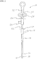
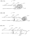
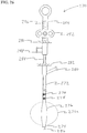
- FIG. 2 illustrates a safe access needle injection instrument 10 which is used to aid the physician in obtaining access to the submucosal layer to perform a submucosal medical procedure.
- the safe access needle injection instrument 10 includes a tubular shaft 12 having a distal end 13.
- the diameter of tubular shaft 12 is generally in the range 1mm to 10mm with a preferred range of 2.0 to 6.0 mm.
- Adjacent distal end 13, a portion of the wall of tubular shaft 12 is removed to form window member 14.
- window member 14 can be formed with a cap element coupled to the distal end of tubular shaft 12.
- needle member 16 Slidably disposed within the lumen of tubular shaft 12 is needle member 16.
- the proximal portion of tubular shaft includes vacuum port 18 which is capable of being coupled to a vacuum source such as a syringe or vacuum pump (not shown).
- Valve assembly 20 provides a releasable seal to tubular shaft 12.
- a handle assembly 21 is connected to tubular shaft 12 through connector tubing 22.
- the handle assembly 21 includes needle fluid port 24 and valve assembly 26 for injecting fluid through and sealing around the proximal portion of needle member 16.
- the proximal portion of needle member 16 is also connected to a needle slide member 28 positioned on handle body 30. The proximal movement of needle slide member 28 on handle body 30 causes needle member 16 to move proximally within tubular shaft 12.
- Distance markers 32 are located on handle body 30 to gauge the movement of needle member 16 within tubular shaft 12.
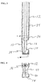
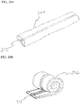
- needle member 16 is positioned within lumen 34 of tubular shaft 12. Located on the exterior of needle member 16 is stop member 36. The distal end 1 3 of tubular shaft 12 is closed with seal plug 38. Also shown is needle member tip 40 and needle lumen 42. Needle lumen 42 communicates needle tip 40 with needle fluid port 24 so that fluid injected through needle port 24 exits the lumen at needle tip 40.
- FIGS. 5A and 5B illustrate the actuation of needle member 16.
- Needle member 16 is shown in detail with needle body 44 connected to needle tip 40. Needle body 44 may be constructed of a separate material as shown or integrally formed with needle tip 40. Needle body 44 may be constructed from flexible tubing having good axial pushability.
- needle member 16 is in a first position in which needle tip 40 is located within lumen 34 proximal to window member 14. This is the preferred position for needle member 16 when tubular shaft 12 is deployed within the body.
- needle member 16 Upon actuation, needle member 16 is moved to a second position in which needle tip 40 is positioned within window member 14.
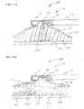
- FIGS. 6A through 6E illustrate the operation of safe access needle injection instrument 10. Insertion section 6 of endoscope 2 is passed through a natural orifice in a patient and positioned at a location in the digestive tract in which to perform a submucosal procedure. Safe access needle injection instrument 10 is deployed through the working channel 4 of endoscope 2. As depicted in FIG. 6A the distal potion of safe access needle injection instrument 10 is positioned within the digestive tract adjacent mucosal layer 46. Beneath the mucosal layer are the submucosal layer 48 and the muscular layer 50. Window member 14 is oriented towards mucosal layer 46. Needle member 16 is located in a first position proximal to window member 14.
- a vacuum source is connected to vacuum port 18, which communicates with lumen 34, and the applied vacuum causes the tissue of the digestive tract to be suctioned into window member 14 as shown in FIG. 6B .
- the actuation of needle member 16 is shown in FIG. 6C as it is moved to its second position. Distal movement of needle member 16 causes needle tip 40 to move distally relative to tubular shaft 12 to thereby pierce the mucosal layer 46 and enter the submucosal layer 48 of the suctioned tissue.
- stop member 36 engages the mucosal layer 46. Stop member 36 prevents further distal movement of needle member 16 and provides a seal around needle tip 40.
- a pressurized fluid source is connected to needle fluid port 24 to deliver fluid through the lumen of needle member 16 to the submucosal layer 48.
- the mucosal layer 46 is elevated forming a submucosal bleb.
- the fluid used to create the bleb may be of any type suitable for the environment such as solutions containing saline, hypertonic solutions of saline-epinephrine, sodium hyaluronate, poly-N-acetylglucosamine, sodium chondroitin sulfate, chitosans or other biocompatible mucopolysaccharides.
- the safe access needle injection instrument 10 may be removed from the safety bleb as illustrated in FIG. 6E .
- a safe access dissection system 100 that includes a safe access needle injection instrument 10 and an injectable dissection material 102.
- the injectable dissection material 102 is a solution capable of dissolving the submucosal connective tissue, such as sodium 2-mercaptoethanesulfanate (MESNA). Additional substances which may dissolve the submucosal connective tissue include acids and enzymes such as a peptase enzyme solution, protease / collagenase, papain, chymotrypsin and acetylcysteine.
- the instrument 10 is used to create a safety bleb beneath the mucosal layer 46 in the digestive tract of a mammal.
- the injectable dissection material 102 may be delivered through needle tip 40 into the submucosal layer 48 as shown in FIG. 11A .
- Material 102 begins to break down the stretched submucosal connective tissue 52. Under the force imparted by the distention of the bleb, the submucosal connective tissue 52 breaks, thereby causing the mucosal layer 46 to become detached from muscular layer 50 in bleb region as shown in FIG. 11B .
- FIGS. 12A and 12B show a safe access dissection system 104 that includes a safe access needle injection instrument 10 and an injectable dissection material 106.
- the injectable dissection material 106 takes the form of a semisolid gelatin capable of mechanically breaking the submucosal connective tissue 52.
- the semisolid gelatin may be formed using biocompatible commercially available gelatins. Generally, these gelatins are in a powdered form and mixed with warm water. Upon cooling, the gelatin forms a semisolid consistency with physical cross links.
- the gelatin material is preferably formed within the barrel of a pressurizable syringe since aspiration of this material is difficult.
- the instrument 10 is used to create a safety bleb beneath the mucosal layer 46 in the digestive tract of a mammal.
- the injectable dissection material 106 may be delivered through needle tip 40 into the submucosal layer 48 as shown in FIG. 12A .
- the mass and semisolid nature of the injectable dissection material 106 begins to apply force to the stretched submucosal connective tissue 52, unlike a saline solution which only permeates the submucosal layer 48.
- the injectable dissection material 106 may also take the form of injectable solutions which solidify upon entering the submucosal space. Solutions which solidify after injection into the submucosal space may be thermosensitive polymer solutions such as Pluronic 127. Additional injectable solidifying solutions include monomer and polymer solutions like hydrogels and cyanoacrylates which polymerize or crosslink upon contact with tissue or added chemical agents.
- FIGS. 13A and 13B show a safe access dissection system 108 that includes a safe access needle injection instrument 10 and an injectable dissection material 110.
- the injectable dissection material 110 takes the form of gelled microspheres dispersed in a solution capable of mechanically breaking the submucosal connective tissue 52.
- the microspheres may be formed using biocompatible natural materials such as collagen and alginates or synthetic materials like polyvinylalcohol (PVA), polyvinylpyrolidone (PVP) and acrylate polymers.
- PVA polyvinylalcohol
- PVP polyvinylpyrolidone
- a safe access needle injection instrument 10 is used to create a safety bleb beneath the mucosal layer 46 in the digestive tract of a mammal.
- the injectable dissection material 110 may be delivered through needle tip 40 into the submucosal layer 48 as shown in FIG. 13A .
- the mass and solid nature of the injectable dissection material 110 begins to apply force to the stretched submucosal connective tissue 52, unlike a saline solution which only permeates the submucosal layer 48.
- the submucosal connective tissue 52 breaks, thereby causing the mucosal layer 46 to become detached from muscular layer 50 in bleb region as shown in FIG. 13B .
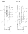
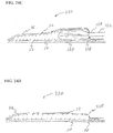
- FIG. 14 illustrates a submucosal tunnelling instrument 120 for performing a submucosal medical procedure.
- the submucosal tunnelling instrument 120 includes a catheter 122 having proximal and distal ends and an expandable member which preferably takes the form of balloon member 124 located adjacent the distal end.
- the proximal end of catheter 122 is attached to connector tubing 126 to access inflation port 128.
- Valve assembly 130 provides a seal for fluid introduced into inflation port 128.
- Tether slide 132 is slidably positioned on handle body 134.
- Handle body 134 includes distance markers 136 to gauge the movement of tether slide 132.
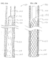
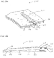
- FIG. 15A A cross sectioned view of the distal end of the submucosal tunnelling instrument 120 is shown in more detail in FIG. 15A .
- Catheter 122 has a distal end 138 and a lumen 123.
- balloon member 124 Located within lumen 123 is balloon member 124.
- the balloon member 124 is preferably non-compliant of the type generally known in the art, but balloon member 124 may be of the compliant or semi-compliant type.
- the balloon member 124 may be formed from biocompatible polymer types such as olefins, elastomers, thermoplastic elastomers, vinyls, polyamides, polyimides, polyesters, fluoropolymers, copolymers and blends of any of these.
- the proximal end 140 of balloon member 124 is attached to the distal end 138 of catheter 122.
- the distal end 142 of balloon member 124 is positioned within the lumen 123 in an everted configuration.
- a tether member 144 is connected to the distal end 142 of balloon member 124.
- Tether member 144 is flexible and preferably takes the form of a filament, as shown; however tether member 144 may take the form of a tube.
- the proximal end of tether member 144 is connected to tether slide 132 through valve assembly 130. Tether member 144 aids in initially positioning balloon member 124 within the lumen 123 of catheter 122.
- the aforementioned embodiment of the submucosal tunnelling instrument include an expandable member which preferably takes the form of an inflatable balloon
- other devices may be suitable for essentially performing the same function.
- the expandable member 124 may take the form of an expandable elongate braid, mesh or tubular framework in which the proximal end of the expandable member is connected to the distal end of the catheter and the distal end of the expandable member is everted and positioned within the lumen of the catheter.
- a stiffened tether member 144 located within the lumen of the catheter 122 may be used as a pusher to push the expandable elongate member from the lumen of the catheter in essentially the same way that the balloon expands from the lumen of the catheter.
- the expandable elongate braid may be formed from resilient materials such as nitinol, spring steels, vitreloy, as well as polymers and composites.
- the expandable member may comprise individual wires to form a braid or mesh configuration.
- the expandable member may be laser cut from a tube forming a tubular framework.
- the expanded diameter is larger than the outer diameter of the catheter.
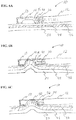
- FIGS. 16A through 16C illustrate various stages of deployment of balloon member 124 from the lumen 123 of catheter 122.
- a fluid filled syringe or other source is connected to inflation port 128.
- Balloon member 124 has an expanded diameter range of about 1 mm to about 30mm and is preferably in the range of 2 mm to 20mm.
- the length of balloon member 124 is as long as necessary to perform a desired submucosal medical procedure. This length can be in the range of 5 mm to 50 cm and preferably in the range of 7 mm to about 10cm.
- balloon member 124 As balloon member 124 expands it extends in a linear fashion moving the distal end 142 of balloon member 124 towards the distal end 138 of catheter 122. As long as pressurized fluid is applied to catheter lumen 123, balloon 124 will extend to its full length. Alternatively, since the distal end 142 of balloon 124 is connected to tether member 144, the amount of linear extension balloon 124 takes may be controlled by the tether slide 132 to define an extension length shorter than the full length of the balloon.
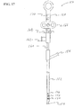
- FIG. 17 illustrates a submucosal tunnelling instrument 150 for performing a submucosal medical procedure.
- the submucosal tunnelling instrument 150 includes a catheter 152 having proximal and distal ends and a balloon member 154 located adjacent the distal end. Positioned on the exterior of catheter 152 adjacent the distal end is a series of markers 156. These markers may be visible under direct visualization of the endoscope and may be additionally visible under fluoroscopy. Adjacent the proximal end of catheter 152 is an auxiliary device port 158. The proximal end of catheter 152 is attached to connector tubing 160 to access inflation port 162. Valve assembly 164 provides a seal for fluid introduced into inflation port 162.
- Tether slide 166 is slidably positioned on handle body 168. Handle body 168 includes distance markers 170 to gauge the movement of tether slide 166.
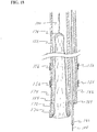
- FIG. 18 A cross sectioned view of the distal end of the submucosal tunnelling instrument 150 is shown in more detail in FIG. 18 .
- Catheter 152 has a distal end 172 and a first lumen 174.
- balloon member 154 Located within first lumen 174 is balloon member 154.
- the balloon member 154 is preferably non-compliant of the type generally known in the art. However, balloon member 154 may be of the compliant or semi-compliant type.
- the balloon member 154 may be formed from biocompatible polymer types such as olefins, elastomers, thermoplastic elastomers, vinyls, polyamides, polyimides, polyesters, fluoropolymers, copolymers and blends of any of the aforementioned.
- the proximal end 176 of balloon member 154 is attached to the distal end 172 of catheter 152.
- the distal end 178 of balloon member 154 is positioned within the first lumen 174 in an everted configuration.
- a tether member 180 is connected to the distal end 178 of balloon member 154.
- Tether member 180 is flexible and preferably takes the form of a filament, as shown; however tether member 180 may take the form of a tube.
- the proximal end of tether member 180 is connected to tether slide 166 through valve assembly 164.
- Tether member 180 aids in initially positioning balloon member 154 within the first lumen 174 of catheter 152.
- Catheter 152 has a second lumen 182 that extends from auxiliary device port 158 to distal end 184.
- Distal end 184 is located proximal to distal end 172 of catheter 152.
- a needle knife 184 Slidably disposed within second lumen 182 is a needle knife 184 that has a knife tip 188.
- Needle knife 184 is preferably of the endoscopic electrosurgical type. However any form of incision device that may be operated to form an incision in tissue such as mechanical cutters, water jets or lasers may be suitable.
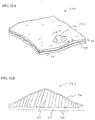
- FIGS. 19A through 19C illustrate a submucosal tunnelling instrument 200 useful in a combination of the present invention.
- Catheter 202 has a distal end 204 which is connected to balloon member 206.
- the proximal end 208 of balloon 206 is connected to distal end 204 of catheter 202.
- Balloon member 206 is rolled into a spiral configuration in which the distal end 210 is located in the centre of the spiral.
- balloon member 206 inflates.
- the inflation of balloon member 206 causes the balloon to unroll from a spiral configuration extending linearly.
- the balloon member 206 may be thermally treated to retain the spiral configuration for delivery through the working channel of an endoscope.
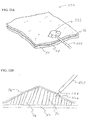
- the balloon member 206 may incorporate a spiral shaped member 212 attached the wall of balloon member 206 as shown in FIGS. 20A and 20B .
- the spiral shaped member may be formed from a resilient filament as shown in an outstretched configuration in FIG 20A .
- the spiral shaped member being formed of a resilient filament and incorporated into the wall of the balloon preferably takes its spiral shape and in doing so forms the balloon member into a spiral shape as shown in FIG. 20B .
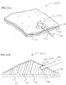
- FIGS. 21A and 21B illustrate a desired region 220 of the digestive tract of a mammal.
- a safe access needle injection instrument according to any of the embodiments previously described may be used to form a safety bleb 222 beneath the mucosal layer 46.
- the safety bleb contains the submucosal connective tissue 52 generally in a stretched condition attached to both the mucosal layer 46 and the muscular layer 50.
- an endoscopic incision tool 224 is positioned adjacent to the safety bleb 222.
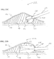
- FIGS. 23A through 24D illustrate the introduction and operation of a submucosal tunneling instrument into submucosal layer 48.
- the distal end 138 of the submucosal tunneling instrument 120 is positioned through the mucosal opening 228. Once the proximal end 140 of balloon 124 is through the mucosal opening 228 the submucosal tunneling instrument 120 may be operated.
- the proximal end 140 of balloon 124 By delivering pressurized fluid through the lumen of catheter 122, the proximal end 140 of balloon 124 inflates to its expanded diameter as depicted in FIG. 23C .
- the expanded diameter of the proximal end 140 of balloon 124 is larger than the diameter of the mucosal opening 228. The larger diameter prevents balloon 124 from pushing the distal end 138 of catheter 122 backwards out of the submucosal layer 48 through mucosal opening 228.
- further inflation extends balloon 124 in a linear fashion within the submucosal layer 48 causing the submucosal connective tissue 52 to break in regions adjacent to the balloon.
- the balloon 124 can only expand by increasing the volume of the surrounding area between the mucosal layer 46 and the muscular layer 50.
- the application of force during expansion of balloon 124 is concentrated on the submucosal connective tissue 52, thereby causing the submucosal connective tissue 52 to break, whereas the force applied to the mucosal layer 46 or the muscular layer 50 by balloon 124 is diluted over a larger portion of balloon 124.
- the force required to break the submucosal connective tissue 52 as applied by balloon 124 is less than the force required to perforate the mucosal layer 46 or muscular layer 50 by balloon 124 thereby minimizing trauma to surrounding tissue.
- 24A illustrates a perspective view of region 220 in the digestive tract having a submucosal tunnel 230 formed by submucosal tunneling instrument 120.
- balloon 124 is fully expanded and generally occupies the majority of the space of submucosal tunnel 230.
- Balloon 124 is then deflated by applying a negative pressure to catheter 122 and retracting tether member 144.
- the distal end 138 of catheter 122 is then removed from mucosal opening 228, leaving submucosal tunnel 230 generally deflated.
- the submucosal connective tissue 52 within the tunnel is broken.
- the submucosal tunnel 230 may provide suitable access to the muscular wall or the placement of an implant device. However to perform other submucosal medical procedures an area larger than the submucosal tunnel 230 may be desired.
- FIGS. 25A through 25D illustrate the formation of an area larger than a submucosal tunnel for performing a submucosal medical procedure according to an embodiment of the present invention.
- a region 220 in the digestive tract is prepared by forming a submucosal tunnel 230.
- a submucosal dissection instrument 240 having a catheter 242 is positioned through mucosal opening 228 into submucosal tunnel 230.
- markers 244 Located on catheter 242 are markers 244 that indicate the insertion depth of catheter distal end 246 within submucosal tunnel 230.
- the submucosal dissection instrument 240 is in a proper position for operation when balloon member 248 including distal end 250 and proximal end 252 are sufficiently located within submucosal tunnel 230, as shown in FIG. 25B .
- balloon member 248 inflates.
- the submucosal connective tissue 52 is broken in the area of the expanded balloon member 248 and the mucosal layer 46 is elevated.
- the elevated mucosal layer 46 forms a large mucosal dissected area 260.
- the balloon member 248 may be deflated and submucosal dissecting instrument 240 removed from the large mucosal dissected area 260.
- the dissected region beneath mucosal layer 46 has transformed in geometry from a high aspect ratio tunnel to a low aspect ratio chamber suitable for performing some submucosal medical procedures.
- the above descriptions of submucosal tunnelling instruments and submucosal dissecting instruments have shown separate instruments to create a submucosal tunnel or large mucosal dissected area. However the two types of instruments may be combined to form a submucosal tunnelling dissecting instrument 270 as illustrated in FIG. 26 .
- the instrument 270 includes a dissection catheter 272 having a distal end 274 and a dissection balloon 276 having an expanded dissection balloon 276a configuration.
- the dissection balloon 276 can be non-compliant as generally known in the art or dissection balloon 276 may be of the compliant or semi-compliant type.
- the dissection balloon 276 may be formed from biocompatible polymer types such as olefins, elastomers, thermoplastic elastomers, vinyls, polyamides, polyimides, polyesters, fluoropolymers, copolymers and blends of any of the aforementioned.
- the dissection catheter 272 has insertion markers 278 positioned along its shaft.
- the proximal end of dissection catheter 272 includes both an inflation port 280 that is in fluid communication with dissection balloon 276, and a valve assembly 282.
- Tunnelling catheter 284 is slidably disposed through valve assembly 282 extending within a lumen of dissection catheter 272.
- the tunnelling catheter distal end 286 may extend beyond the dissection catheter distal end 274.
- Tunnelling catheter 284 includes an inflation port 288 and valve assembly 290.
- a tether slide member 292 is slidably disposed on handle body 294 with distance markers 296.
- FIG. 27 is a detailed cross section of the distal portion of the submucosal tunnelling dissecting instrument 270.
- the distal end 298 and proximal end 300 of dissection balloon 276 are connected to the exterior of dissection catheter 272.
- Inflation lumen 302 connects inflation port 280 with the interior of dissection balloon 276 through inflation aperture 304.
- Tunnelling catheter 284 is slidably disposed within the lumen 306 of dissection catheter 272. Positioned within the lumen 308 of tunnelling catheter 284 there is an everted expandable tunnelling balloon 310.
- tunnelling balloon 310 is preferably non-compliant of the type generally known in the art, however, tunnelling balloon 310 may be of the compliant or semi-compliant type.
- the tunnelling balloon 310 may be formed from biocompatible polymer types such as olefins, elastomers, thermoplastic elastomers, vinyls, polyamides, polyimides, polyesters, fluoropolymers, copolymers and blends of any of these.
- the distal end of tunnelling balloon 310 is connected to a tether member 312 which has a proximal end that is connected to tether slide 292.
- the operation of the submucosal tunnelling dissecting instrument 270 to form a submucosal tunnel and large mucosal dissected area is similar to the operation of the separate instruments.
- the distal end 286 of tunnelling catheter 284 is positioned through a mucosal opening formed in a safety bleb.
- the tunnelling catheter 284 is pressurized with fluid to linearly expand tunnelling balloon 310.
- tunnelling balloon 310 may be deflated and dissection catheter 272 may be advanced through the mucosal opening into the submucosal tunnel.
- the markers 278 may be used to determine the depth in which the dissection catheter 272 has been advanced into the submucosal tunnel.
- dissection catheter 272 Once the dissection catheter 272 has been properly positioned within the submucosal tunnel it may be operated. By applying pressurized fluid to inflation port 280, dissection balloon 276 is dilated to an expanded dissection balloon 276a configuration. During the expansion a large mucosal dissected area is created which is accessible for performing a subsequent submucosal medical procedure.
Landscapes
- Health & Medical Sciences (AREA)
- Life Sciences & Earth Sciences (AREA)
- Surgery (AREA)
- Engineering & Computer Science (AREA)
- Veterinary Medicine (AREA)
- Heart & Thoracic Surgery (AREA)
- Medical Informatics (AREA)
- Molecular Biology (AREA)
- Animal Behavior & Ethology (AREA)
- General Health & Medical Sciences (AREA)
- Public Health (AREA)
- Biomedical Technology (AREA)
- Nuclear Medicine, Radiotherapy & Molecular Imaging (AREA)
- Physics & Mathematics (AREA)
- Pathology (AREA)
- Otolaryngology (AREA)
- Vascular Medicine (AREA)
- Plasma & Fusion (AREA)
- Orthopedic Medicine & Surgery (AREA)
- Optics & Photonics (AREA)
- Electromagnetism (AREA)
- Surgical Instruments (AREA)
- Infusion, Injection, And Reservoir Apparatuses (AREA)
- Media Introduction/Drainage Providing Device (AREA)
Applications Claiming Priority (2)
| Application Number | Priority Date | Filing Date | Title |
|---|---|---|---|
| US11/775,996 US8128592B2 (en) | 2007-07-11 | 2007-07-11 | Methods and systems for performing submucosal medical procedures |
| PCT/US2008/067635 WO2009009274A2 (en) | 2007-07-11 | 2008-06-20 | Methods and systems for performing submucosal medical procedures |
Publications (3)
| Publication Number | Publication Date |
|---|---|
| EP2170182A2 EP2170182A2 (en) | 2010-04-07 |
| EP2170182A4 EP2170182A4 (en) | 2015-04-08 |
| EP2170182B1 true EP2170182B1 (en) | 2018-08-29 |
Family
ID=40229398
Family Applications (1)
| Application Number | Title | Priority Date | Filing Date |
|---|---|---|---|
| EP08771571.0A Active EP2170182B1 (en) | 2007-07-11 | 2008-06-20 | Systems for performing submucosal medical procedures |
Country Status (4)
| Country | Link |
|---|---|
| US (6) | US8128592B2 (enExample) |
| EP (1) | EP2170182B1 (enExample) |
| JP (3) | JP5833308B2 (enExample) |
| WO (1) | WO2009009274A2 (enExample) |
Families Citing this family (105)
| Publication number | Priority date | Publication date | Assignee | Title |
|---|---|---|---|---|
| US6814739B2 (en) | 2001-05-18 | 2004-11-09 | U.S. Endoscopy Group, Inc. | Retrieval device |
| JP5443341B2 (ja) * | 2007-05-25 | 2014-03-19 | クック メディカル テクノロジーズ エルエルシー | 穿孔を閉鎖するための医療器具、システム及び方法 |
| US8591521B2 (en) | 2007-06-08 | 2013-11-26 | United States Endoscopy Group, Inc. | Retrieval device |
| US8929988B2 (en) * | 2007-07-11 | 2015-01-06 | Apollo Endosurgery, Inc. | Methods and systems for submucosal implantation of a device for diagnosis and treatment of a body |
| US8128592B2 (en) | 2007-07-11 | 2012-03-06 | Apollo Endosurgery, Inc. | Methods and systems for performing submucosal medical procedures |
| JP5226792B2 (ja) * | 2007-09-25 | 2013-07-03 | クック メディカル テクノロジーズ エルエルシー | 組織アンカーを使用するための医療器具、装置及び方法 |
| EP2219536B1 (en) * | 2007-10-23 | 2012-12-19 | Boston Scientific Scimed, Inc. | Apparatus for treating tissue |
| WO2009082596A1 (en) * | 2007-12-18 | 2009-07-02 | Wilson-Cook Medical, Inc. | Device and method for placement of tissue anchors |
| JP5580828B2 (ja) * | 2008-10-06 | 2014-08-27 | クック メディカル テクノロジーズ エルエルシー | 組織アンカーを安全に配備するための端部キャップ |
| WO2010068265A1 (en) * | 2008-12-10 | 2010-06-17 | Minimally Invasive Devices, Llc | Systems and methods for optimizing and maintaining visualization of a surgical field during the use of surgical scopes |
| AU2009333028B2 (en) * | 2008-12-31 | 2013-10-17 | Cook Medical Technologies Llc | Medical device with pivotable jaws |
| DE202009018056U1 (de) | 2009-04-16 | 2010-12-30 | Erbe Elektromedizin Gmbh | Endoskopisches Chirurgieinstrument |
| US20100324446A1 (en) * | 2009-06-18 | 2010-12-23 | Vance Products Incorporated, D/B/A Cook Orolgoical Incorporated | Telescoping Biopsy Device |
| US10363087B2 (en) * | 2009-10-12 | 2019-07-30 | Apollo Endosurgery Us, Inc. | Tissue resection device |
| US20110105838A1 (en) * | 2009-10-29 | 2011-05-05 | Roberto Fogel | Suction device for endoscopic instruments and method |
| US9986893B2 (en) | 2009-12-15 | 2018-06-05 | Cornell University | Method and apparatus for manipulating the side wall of a body lumen or body cavity so as to provide increased visualization of the same and/or increased access to the same, and/or for stabilizing instruments relative to the same |
| US11986150B2 (en) | 2009-12-15 | 2024-05-21 | Lumendi Ltd. | Method and apparatus for manipulating the side wall of a body lumen or body cavity so as to provide increased visualization of the same and/or increased access to the same, and/or for stabilizing instruments relative to the same |
| US12121209B2 (en) | 2014-02-11 | 2024-10-22 | Cornell University | Method and apparatus for providing increased visualization and manipulation of a body side wall |
| US8939997B2 (en) | 2010-10-11 | 2015-01-27 | Cook Medical Technologies Llc | Medical devices with detachable pivotable jaws |
| US8545519B2 (en) | 2009-12-22 | 2013-10-01 | Cook Medical Technologies Llc | Medical devices with detachable pivotable jaws |
| US12070224B2 (en) | 2009-12-22 | 2024-08-27 | Cook Medical Technologies Llc | Medical devices with detachable pivotable jaws |
| DK2515770T3 (en) | 2009-12-22 | 2019-02-25 | Cook Medical Technologies Llc | MEDICAL DEVICES WITH REMOVABLE THREADABLE BUYERS |
| US10010336B2 (en) | 2009-12-22 | 2018-07-03 | Cook Medical Technologies, Inc. | Medical devices with detachable pivotable jaws |
| US9078562B2 (en) | 2010-01-11 | 2015-07-14 | Minimally Invasive Devices, Inc. | Systems and methods for optimizing and maintaining visualization of a surgical field during the use of surgical scopes |
| US8430862B2 (en) * | 2010-04-08 | 2013-04-30 | KMG Pharma LLC | Subconjunctival agent delivery apparatus, system and method |
| US8652118B2 (en) * | 2010-04-08 | 2014-02-18 | Kmg Pharma, Llc | Sub-mucosal agent delivery, apparatus, system and method |
| US8574217B2 (en) * | 2010-04-08 | 2013-11-05 | Kmg Pharma, Llc | Sub-mucosal agent delivery method for the eye |
| WO2011130663A2 (en) * | 2010-04-16 | 2011-10-20 | Ascentx Medical, Inc. | Injection apparatus for long distance delivery of soft tissue bulking agents containing microspheres |
| WO2012013246A1 (en) * | 2010-07-30 | 2012-02-02 | Ethicon Endo-Surgery, Inc. | A system and method for submucosal tunneling of the gi tract for the diversion of bodily fluids |
| EP2600759A4 (en) | 2010-08-04 | 2013-08-28 | Minimally Invasive Devices Llc | SYSTEMS AND METHOD FOR OPTIMIZING AND SAVING THE VIEW OF A SURGICAL FIELD DURING THE USE OF SURGICAL ENDOSCOPES |
| SI2627268T1 (sl) | 2010-10-11 | 2017-10-30 | Cook Medical Technologies Llc | Medicinske naprave s snemljivimi vrtljivimi čeljustmi |
| DK2627263T3 (en) | 2010-10-11 | 2017-03-06 | Cook Medical Technologies Llc | MEDICAL DEVICES WITH THIRD Jaws |
| WO2012075487A2 (en) | 2010-12-03 | 2012-06-07 | Minimally Invasive Devices, Llc | Devices, systems, and methods for performing endoscopic surgical procedures |
| US11246653B2 (en) * | 2010-12-07 | 2022-02-15 | Boaz Avitall | Catheter systems for cardiac arrhythmia ablation |
| EP2651316B1 (en) | 2010-12-15 | 2016-07-06 | Cook Medical Technologies LLC | Medical devices with detachable pivotable jaws |
| CN103582463B (zh) | 2011-01-19 | 2018-02-13 | 弗拉克泰尔实验室公司 | 用于组织处理的装置与方法 |
| WO2013001363A1 (en) * | 2011-06-27 | 2013-01-03 | E-Motion Medical Ltd. | Esophageal stimulation devices and methods |
| US9999767B2 (en) | 2011-06-27 | 2018-06-19 | E-Motion Medical, Ltd. | Esophageal stimulation system |
| JP6318088B2 (ja) | 2011-07-26 | 2018-04-25 | アンフォラ メディカル, インコーポレイテッド | 骨盤神経組織を変調するための装置および方法 |
| EP2604202B1 (de) | 2011-12-14 | 2015-04-01 | Erbe Elektromedizin GmbH | Instrument für die Wasserstrahlchirurgie |
| US9408529B2 (en) * | 2012-01-25 | 2016-08-09 | Boston Scientific Scimed, Inc. | Endoscopic instrument having movable distal tool |
| JP5821108B2 (ja) * | 2012-02-02 | 2015-11-24 | 国立大学法人 鹿児島大学 | 切開具 |
| JP6211541B2 (ja) * | 2012-02-09 | 2017-10-11 | ボストン サイエンティフィック サイムド,インコーポレイテッドBoston Scientific Scimed,Inc. | 循環ワイヤを備える切断具 |
| AU2013226062B2 (en) | 2012-02-27 | 2017-10-19 | Fractyl Health, Inc. | Heat ablation systems, devices and methods for the treatment of tissue |
| EP2838598B1 (en) * | 2012-04-19 | 2020-01-15 | Fractyl Laboratories, Inc. | Tissue expansion devices |
| EP2879605A4 (en) | 2012-07-30 | 2016-04-06 | Fractyl Lab Inc | ELECTRICITY CONTROL SYSTEMS, DEVICES AND METHOD FOR TREATMENT OF TISSUE |
| WO2014026055A1 (en) | 2012-08-09 | 2014-02-13 | Fractyl Laboratories Inc. | Ablation systems, devices and methods for the treatment of tissue |
| WO2014055997A1 (en) | 2012-10-05 | 2014-04-10 | Fractyl Laboratories Inc. | Methods, systems and devices for performing multiple treatments on a patient |
| US10384052B2 (en) | 2012-12-24 | 2019-08-20 | E-Motion Medical, Ltd | GI tract stimulation devices and methods |
| JP6180118B2 (ja) * | 2013-01-22 | 2017-08-16 | オリンパス株式会社 | アクセスデバイスおよびアクセスシステム |
| WO2014151824A1 (en) | 2013-03-14 | 2014-09-25 | Minimally Invasive Devices, Inc. | Fluid dispensing control systems and methods |
| US10092312B2 (en) | 2013-05-07 | 2018-10-09 | Auxin Surgery Sa | Device for chemically assisted dissection |
| EP3003461B1 (en) | 2013-06-04 | 2019-05-01 | Fractyl Laboratories, Inc. | Systems and devices for reducing the luminal surface area of the gastrointestinal tract |
| US10070853B2 (en) * | 2013-08-14 | 2018-09-11 | Covidien Lp | Expandable balloon desufflation assembly |
| US9872700B2 (en) | 2013-09-03 | 2018-01-23 | United States Endoscopy Group, Inc. | Endoscopic snare device |
| US9572591B2 (en) | 2013-09-03 | 2017-02-21 | United States Endoscopy Group, Inc. | Endoscopic snare device |
| WO2015038973A1 (en) | 2013-09-12 | 2015-03-19 | Fractyl Laboratories, Inc. | Systems, methods and devices for treatment of target tissue |
| KR101656944B1 (ko) * | 2013-10-16 | 2016-09-19 | 국립암센터 | 내시경용 주사장치 |
| EP3071286B1 (en) | 2013-11-22 | 2024-01-03 | Fractyl Health, Inc. | Systems for the creation of a therapeutic restriction in the gastrointestinal tract |
| US10959774B2 (en) | 2014-03-24 | 2021-03-30 | Fractyl Laboratories, Inc. | Injectate delivery devices, systems and methods |
| WO2015179837A1 (en) | 2014-05-23 | 2015-11-26 | Amphora Medical, Inc. | Methods and devices for treating pelvic conditions |
| US9844641B2 (en) | 2014-07-16 | 2017-12-19 | Fractyl Laboratories, Inc. | Systems, devices and methods for performing medical procedures in the intestine |
| US11185367B2 (en) | 2014-07-16 | 2021-11-30 | Fractyl Health, Inc. | Methods and systems for treating diabetes and related diseases and disorders |
| WO2016011269A1 (en) | 2014-07-16 | 2016-01-21 | Fractyl Laboratories, Inc. | Methods and systems for treating diabetes and related diseases and disorders |
| CN104173118A (zh) * | 2014-08-22 | 2014-12-03 | 山西省肿瘤研究所 | 一种建立小鼠直肠移植瘤的装置 |
| KR102267891B1 (ko) | 2014-12-01 | 2021-06-23 | 삼성전자주식회사 | 냉장고 |
| JP6426032B2 (ja) * | 2015-03-13 | 2018-11-21 | オリンパス株式会社 | 内視鏡用穿刺デバイス |
| GB2551937B (en) | 2015-03-26 | 2020-09-02 | Gyrus Acmi Inc | Biopsy sample retention mechanism |
| JP2018529495A (ja) | 2015-09-28 | 2018-10-11 | ストライカー コーポレイションStryker Corporation | 機械的血栓摘出装置および方法 |
| US10342540B2 (en) * | 2015-10-15 | 2019-07-09 | Boston Scientific Scimed, Inc. | Tissue retraction devices and related methods of use |
| US10548626B2 (en) | 2015-12-15 | 2020-02-04 | Boston Scientific Scimed, Inc. | Endoscopic tissue manipulation tool |
| KR101731894B1 (ko) * | 2016-01-11 | 2017-05-02 | 연세대학교 원주산학협력단 | 위장관 점막하종양 생검용 기구 |
| JP6664008B2 (ja) | 2016-04-08 | 2020-03-13 | セント・ジュード・メディカル,カーディオロジー・ディヴィジョン,インコーポレイテッド | マッピング可変ループ・カテーテル・ハンドル |
| EP3275388A1 (en) | 2016-07-28 | 2018-01-31 | Auxin Surgery | Electro-chemical surgical instrument |
| KR20190104148A (ko) * | 2016-12-09 | 2019-09-06 | 자네타 말라노브스카-스테가 | 브러시 생검 장치, 키트 및 방법 |
| WO2018129551A1 (en) | 2017-01-09 | 2018-07-12 | United States Endoscopy Group, Inc. | Endoscopic snare device |
| USD827819S1 (en) * | 2017-04-07 | 2018-09-04 | St. Jude Medical, Cardiology Division, Inc. | Catheter handle |
| USD827818S1 (en) * | 2017-04-07 | 2018-09-04 | St. Jude Medical, Cardiology Division, Inc. | Catheter handle |
| WO2018204503A1 (en) | 2017-05-03 | 2018-11-08 | Z Surgical Llc | Minimal-access percutaneous and self-retracting surgical system |
| US10857020B2 (en) | 2017-09-14 | 2020-12-08 | Olympus Corporation | Gastrointestinal track constricting method |
| US11648047B2 (en) | 2017-10-06 | 2023-05-16 | Vive Scientific, Llc | System and method to treat obstructive sleep apnea |
| US10561489B2 (en) | 2018-03-05 | 2020-02-18 | Olympus Corporation | Gastrointestinal-tract constricting method |
| US10555801B2 (en) | 2018-03-05 | 2020-02-11 | Olympus Corporation | Gastrointestinal-tract constricting method |
| CN110279931B (zh) * | 2018-03-15 | 2021-12-28 | 王恩长 | 一种多功能球囊导管及系统 |
| US10918454B2 (en) | 2018-04-02 | 2021-02-16 | Olympus Corporation | Gastrointestinal tract constricting method |
| JP6914425B2 (ja) | 2018-04-06 | 2021-08-04 | オリンパス株式会社 | 薬剤供給デバイス |
| EP3781042B1 (en) | 2018-04-18 | 2025-03-05 | C. R. Bard, Inc. | Dual lumen coaxial introducer having integrated tissue marker delivery |
| EP3813686A4 (en) * | 2018-06-05 | 2022-02-23 | Shaare Zedek Scientific Ltd. | Endoscope accessory devices |
| US10576248B2 (en) * | 2018-07-23 | 2020-03-03 | Crossbay Medical, Inc. | Apparatus and method for everting catheter for uterine access for biopsy and cytology |
| CN108938075A (zh) * | 2018-08-13 | 2018-12-07 | 唐丹 | 一种内镜下粘膜剥离术辅助套管 |
| US20200323520A1 (en) * | 2019-04-11 | 2020-10-15 | GW Medical LLC | Small tube tissue biopsy |
| EP3969097A4 (en) * | 2019-05-17 | 2023-07-05 | PAVmed Inc. | CATHETER DEVICE SYSTEM, AND METHOD OF USE |
| CN110495931B (zh) * | 2019-09-09 | 2024-12-03 | 武汉微新坦医疗科技有限公司 | 一种电动式心腔内心肌切割器 |
| CN110559021B (zh) * | 2019-09-20 | 2021-05-18 | 吉林大学 | 一种肿瘤科医生活检用活体取样保护装置 |
| EP4061256B1 (en) * | 2019-11-22 | 2025-07-16 | Microsteer Ltd. | Systems for controllable access of tools to elevated tissues |
| EP4090282A4 (en) | 2020-01-15 | 2024-02-21 | Fractyl Health, Inc. | AUTOMATIC FABRIC TREATMENT DEVICES, SYSTEMS AND METHODS |
| EP4093302B1 (en) | 2020-01-23 | 2024-05-22 | Stryker Corporation | Inverting capture apparatuses having material depots |
| CN111227928A (zh) * | 2020-03-13 | 2020-06-05 | 南微医学科技股份有限公司 | 注射装置、圈套器及医疗设备 |
| CN111588445A (zh) * | 2020-05-07 | 2020-08-28 | 西安国际医学中心有限公司 | 一种带有缝合功能的黏膜下悬雍垂腭咽成形术刀具 |
| EP4243910A4 (en) | 2020-11-16 | 2024-12-25 | Lumendi Ltd. | METHOD AND DEVICE FOR TURNING A HOLLOW SLEEVE AND SUBSEQUENT REVERSING AN INVERTED HOLLOW SLEEVE |
| US12213665B2 (en) * | 2021-02-04 | 2025-02-04 | Olympus Medical Systems Corp. | Methods for closing a wound |
| CN113057728B (zh) * | 2021-02-04 | 2023-02-28 | 安瑞医疗器械(杭州)有限公司 | 一种配合内窥镜使用的高频电器械 |
| WO2022181133A1 (ja) * | 2021-02-25 | 2022-09-01 | 株式会社カネカ | 内視鏡用光照射装置 |
| DE102022107857A1 (de) | 2022-04-01 | 2023-10-05 | Tuebingen Scientific Medical Gmbh | Pneumatische Antriebsvorrichtung zur translatorischen und/oder rotatorischenBewegung |
| CN114712656B (zh) * | 2022-04-01 | 2023-01-31 | 中国医学科学院北京协和医院 | 应用于临床手术的注射取样装置 |
Family Cites Families (198)
| Publication number | Priority date | Publication date | Assignee | Title |
|---|---|---|---|---|
| US3039468A (en) | 1959-01-07 | 1962-06-19 | Joseph L Price | Trocar and method of treating bloat |
| US3980861A (en) | 1973-03-26 | 1976-09-14 | Akio Fukunaga | Electrically heated miniature thermal implement |
| JPS5727445Y2 (enExample) | 1973-06-20 | 1982-06-15 | ||
| US4222380A (en) | 1977-12-02 | 1980-09-16 | Olympus Optical Co., Ltd. | Celiac injector |
| US4418692A (en) | 1978-11-17 | 1983-12-06 | Guay Jean Louis | Device for treating living tissue with an electric current |
| US4271839A (en) * | 1979-07-25 | 1981-06-09 | Thomas J. Fogarty | Dilation catheter method and apparatus |
| US4630609A (en) * | 1981-05-14 | 1986-12-23 | Thomas J. Fogarty | Dilatation catheter method and apparatus |
| US4445892A (en) * | 1982-05-06 | 1984-05-01 | Laserscope, Inc. | Dual balloon catheter device |
| US5370675A (en) | 1992-08-12 | 1994-12-06 | Vidamed, Inc. | Medical probe device and method |
| US4887598A (en) | 1982-09-24 | 1989-12-19 | Berke Joseph J | Manual rotary scalpel structure |
| US4655219A (en) | 1983-07-22 | 1987-04-07 | American Hospital Supply Corporation | Multicomponent flexible grasping device |
| US4863440A (en) * | 1985-12-23 | 1989-09-05 | Thomas J. Fogarty | Pressurized manual advancement dilatation catheter |
| DE3629809A1 (de) | 1986-09-02 | 1988-03-10 | Wolf Gmbh Richard | Koagulationszange |
| US4779611A (en) * | 1987-02-24 | 1988-10-25 | Grooters Ronald K | Disposable surgical scope guide |
| US4907591A (en) | 1988-03-29 | 1990-03-13 | Pfizer Hospital Products Group, Inc. | Surgical instrument for establishing compression anastomosis |
| DE3821544C2 (de) * | 1988-06-25 | 1994-04-28 | H Prof Dr Med Just | Dilatationskatheter |
| US5514091A (en) | 1988-07-22 | 1996-05-07 | Yoon; Inbae | Expandable multifunctional manipulating instruments for various medical procedures |
| US6120437A (en) | 1988-07-22 | 2000-09-19 | Inbae Yoon | Methods for creating spaces at obstructed sites endoscopically and methods therefor |
| US5159925A (en) | 1988-09-09 | 1992-11-03 | Gynelab, Inc. | Cauterizing apparatus and method for laparoscopic cholecystostomy, gallbladder ablation and treatment of benign prostate hypertrophy |
| JP2656955B2 (ja) | 1988-09-14 | 1997-09-24 | オリンパス光学工業株式会社 | 放射線検出治療装置 |
| US5129396A (en) | 1988-11-10 | 1992-07-14 | Arye Rosen | Microwave aided balloon angioplasty with lumen measurement |
| US5632746A (en) | 1989-08-16 | 1997-05-27 | Medtronic, Inc. | Device or apparatus for manipulating matter |
| US5009659A (en) | 1989-10-30 | 1991-04-23 | Schneider (Usa) Inc. | Fiber tip atherectomy catheter |
| US5984939A (en) | 1989-12-05 | 1999-11-16 | Yoon; Inbae | Multifunctional grasping instrument with cutting member and operating channel for use in endoscopic and non-endoscopic procedures |
| US5226908A (en) | 1989-12-05 | 1993-07-13 | Inbae Yoon | Multi-functional instruments and stretchable ligating and occluding devices |
| US5135484A (en) | 1990-05-09 | 1992-08-04 | Pioneering Technologies, Inc. | Method of removing plaque from vessels |
| US5080660A (en) | 1990-05-11 | 1992-01-14 | Applied Urology, Inc. | Electrosurgical electrode |
| US5078716A (en) | 1990-05-11 | 1992-01-07 | Doll Larry F | Electrosurgical apparatus for resecting abnormal protruding growth |
| US5196024A (en) | 1990-07-03 | 1993-03-23 | Cedars-Sinai Medical Center | Balloon catheter with cutting edge |
| US5158543A (en) | 1990-10-30 | 1992-10-27 | Lazarus Harrison M | Laparoscopic surgical system and method |
| FR2668695B1 (fr) | 1990-11-06 | 1995-09-29 | Ethnor | Instrument chirurgical endoscopique pour le deplacement de tissus ou organes. |
| JPH04220245A (ja) * | 1990-12-20 | 1992-08-11 | Sumitomo Bakelite Co Ltd | 組織切除用カッター |
| US5339799A (en) | 1991-04-23 | 1994-08-23 | Olympus Optical Co., Ltd. | Medical system for reproducing a state of contact of the treatment section in the operation unit |
| US5632761A (en) * | 1991-05-29 | 1997-05-27 | Origin Medsystems, Inc. | Inflatable devices for separating layers of tissue, and methods of using |
| US5383889A (en) * | 1991-05-29 | 1995-01-24 | Origin Medsystems, Inc. | Tethered everting balloon retractor for hollow bodies and method of using |
| US5370134A (en) | 1991-05-29 | 1994-12-06 | Orgin Medsystems, Inc. | Method and apparatus for body structure manipulation and dissection |
| US5431173A (en) | 1991-05-29 | 1995-07-11 | Origin Medsystems, Inc. | Method and apparatus for body structure manipulation and dissection |
| US7744617B2 (en) * | 1991-05-29 | 2010-06-29 | Covidien Ag | Method and inflatable chamber apparatus for separating layers of tissue |
| US5171305A (en) * | 1991-10-17 | 1992-12-15 | Imagyn Medical, Inc. | Linear eversion catheter with reinforced inner body extension |
| AU661240B2 (en) * | 1991-10-18 | 1995-07-13 | Imagyn Medical, Inc. | Apparatus and method for independent movement of an instrument within a linear eversion catheter |
| US5395312A (en) | 1991-10-18 | 1995-03-07 | Desai; Ashvin | Surgical tool |
| US5308327A (en) | 1991-11-25 | 1994-05-03 | Advanced Surgical Inc. | Self-deployed inflatable retractor |
| EP0637226A4 (en) | 1992-04-23 | 1995-06-14 | Scimed Life Systems Inc | DEVICE AND METHOD FOR CLOSING VASCULAR POINTS. |
| US5318564A (en) | 1992-05-01 | 1994-06-07 | Hemostatic Surgery Corporation | Bipolar surgical snare and methods of use |
| US5290284A (en) | 1992-05-01 | 1994-03-01 | Adair Edwin Lloyd | Laparoscopic surgical ligation and electrosurgical coagulation and cutting device |
| US5599300A (en) * | 1992-05-11 | 1997-02-04 | Arrow Precision Products, Inc. | Method for electrosurgically obtaining access to the biliary tree with an adjustably positionable needle-knife |
| US6540764B1 (en) | 1992-06-02 | 2003-04-01 | General Surgical Innovations, Inc. | Apparatus and method for dissecting tissue layers |
| US5318543A (en) | 1992-10-08 | 1994-06-07 | Abbott Laboratories | Laparoscopic jejunostomy instrumentation kit |
| EP0683684B1 (en) | 1993-01-07 | 2001-08-08 | Medical Innovations Corporation | Gastrostomy catheter system |
| US5400773A (en) | 1993-01-19 | 1995-03-28 | Loma Linda University Medical Center | Inflatable endoscopic retractor |
| US5620447A (en) * | 1993-01-29 | 1997-04-15 | Smith & Nephew Dyonics Inc. | Surgical instrument |
| US5372601A (en) | 1993-03-30 | 1994-12-13 | Lary; Banning G. | Longitudinal reciprocating incisor |
| US5417697A (en) | 1993-07-07 | 1995-05-23 | Wilk; Peter J. | Polyp retrieval assembly with cauterization loop and suction web |
| US5718703A (en) | 1993-09-17 | 1998-02-17 | Origin Medsystems, Inc. | Method and apparatus for small needle electrocautery |
| EP0722286B1 (en) | 1993-09-20 | 2002-08-21 | Boston Scientific Corporation | Multiple biopsy sampling device |
| US5582609A (en) | 1993-10-14 | 1996-12-10 | Ep Technologies, Inc. | Systems and methods for forming large lesions in body tissue using curvilinear electrode elements |
| US5443474A (en) * | 1994-03-07 | 1995-08-22 | Implemed, Inc. | Meniscectomy knife |
| US5507765A (en) | 1994-04-28 | 1996-04-16 | Mott; James B. | Punch-type surgical instrument for skin incision, set of parts for making such an instrument of selectably variable size, and blade unit for such instrument |
| US5507795A (en) | 1994-04-29 | 1996-04-16 | Devices For Vascular Intervention, Inc. | Catheter with perfusion system |
| US6009877A (en) | 1994-06-24 | 2000-01-04 | Edwards; Stuart D. | Method for treating a sphincter |
| US5690668A (en) * | 1994-06-29 | 1997-11-25 | General Surgical Innovations, Inc. | Extraluminal balloon dissection |
| DE4425195C1 (de) | 1994-07-16 | 1995-11-16 | Osypka Peter | Katheter mit Mehrfachelektrode |
| CA2199864C (en) * | 1994-09-16 | 2006-06-20 | Seth A. Foerster | Methods and devices for defining and marking tissue |
| US5531699A (en) | 1994-09-19 | 1996-07-02 | Abbott Laboratories | Spring-loaded reciprocable stylet holder |
| US5570700A (en) | 1994-10-03 | 1996-11-05 | Vogeler; Douglas M. | Elliptical biopsy punch |
| US5571130A (en) | 1994-10-04 | 1996-11-05 | Advanced Cardiovascular Systems, Inc. | Atherectomy and prostectomy system |
| US5527273A (en) | 1994-10-06 | 1996-06-18 | Misonix, Inc. | Ultrasonic lipectomy probe and method for manufacture |
| US6152920A (en) | 1997-10-10 | 2000-11-28 | Ep Technologies, Inc. | Surgical method and apparatus for positioning a diagnostic or therapeutic element within the body |
| US6071274A (en) | 1996-12-19 | 2000-06-06 | Ep Technologies, Inc. | Loop structures for supporting multiple electrode elements |
| US5836947A (en) | 1994-10-07 | 1998-11-17 | Ep Technologies, Inc. | Flexible structures having movable splines for supporting electrode elements |
| US5885278A (en) | 1994-10-07 | 1999-03-23 | E.P. Technologies, Inc. | Structures for deploying movable electrode elements |
| US5643305A (en) | 1994-11-18 | 1997-07-01 | Al-Tameem; Moshin | Device for excision of a fistula |
| US5562102A (en) * | 1994-11-21 | 1996-10-08 | Taylor; Thomas V. | Multiple biopsy device |
| US5632754A (en) | 1994-12-23 | 1997-05-27 | Devices For Vascular Intervention | Universal catheter with interchangeable work element |
| US6936024B1 (en) | 1995-01-23 | 2005-08-30 | Russell A. Houser | Percutaneous transmyocardial revascularization (PTMR) system |
| JPH08196640A (ja) * | 1995-01-31 | 1996-08-06 | Nippon Zeon Co Ltd | スライディングカテーテルおよびその製造方法 |
| US5591183A (en) * | 1995-04-12 | 1997-01-07 | Origin Medsystems, Inc. | Dissection apparatus |
| AU4632196A (en) * | 1995-04-14 | 1996-10-30 | Schneider (Usa) Inc. | Rolling membrane stent delivery device |
| US5651788A (en) | 1995-05-17 | 1997-07-29 | C.R. Bard, Inc. | Mucosectomy process and device |
| US5628753A (en) | 1995-06-01 | 1997-05-13 | Sandoz Nutrition Ltd. | Gastrostomy tube removal tool |
| US5871475A (en) | 1995-06-05 | 1999-02-16 | Frassica; James J. | Catheter system |
| US5709224A (en) | 1995-06-07 | 1998-01-20 | Radiotherapeutics Corporation | Method and device for permanent vessel occlusion |
| US5702438A (en) | 1995-06-08 | 1997-12-30 | Avitall; Boaz | Expandable recording and ablation catheter system |
| US5713364A (en) | 1995-08-01 | 1998-02-03 | Medispectra, Inc. | Spectral volume microprobe analysis of materials |
| US5556405A (en) | 1995-10-13 | 1996-09-17 | Interventional Technologies Inc. | Universal dilator with reciprocal incisor |
| WO1997017038A1 (en) * | 1995-11-09 | 1997-05-15 | University Of Massachusetts | Tissue re-surfacing with hydrogel-cell compositions |
| US5697944A (en) | 1995-11-15 | 1997-12-16 | Interventional Technologies Inc. | Universal dilator with expandable incisor |
| US5906606A (en) * | 1995-12-04 | 1999-05-25 | Target Therapuetics, Inc. | Braided body balloon catheter |
| US5683388A (en) * | 1996-01-11 | 1997-11-04 | Symbiosis Corporation | Endoscopic bipolar multiple sample bioptome |
| US5800482A (en) * | 1996-03-06 | 1998-09-01 | Cardiac Pathways Corporation | Apparatus and method for linear lesion ablation |
| US6032077A (en) | 1996-03-06 | 2000-02-29 | Cardiac Pathways Corporation | Ablation catheter with electrical coupling via foam drenched with a conductive fluid |
| US6016452A (en) | 1996-03-19 | 2000-01-18 | Kasevich; Raymond S. | Dynamic heating method and radio frequency thermal treatment |
| US5782747A (en) | 1996-04-22 | 1998-07-21 | Zimmon Science Corporation | Spring based multi-purpose medical instrument |
| JPH1014922A (ja) * | 1996-07-03 | 1998-01-20 | Osamu Yoshida | 臓器細切器および臓器細切装置 |
| US5913870A (en) | 1996-08-13 | 1999-06-22 | United States Surgical Corporation | Surgical dissector |
| US6258086B1 (en) | 1996-10-23 | 2001-07-10 | Oratec Interventions, Inc. | Catheter for delivery of energy to a surgical site |
| US5827268A (en) | 1996-10-30 | 1998-10-27 | Hearten Medical, Inc. | Device for the treatment of patent ductus arteriosus and method of using the device |
| EP0842640A1 (de) | 1996-11-13 | 1998-05-20 | Sulzer Osypka GmbH | Herzkatheter mit Elektrode auf aufspannbarer Einrichtung |
| US5931810A (en) * | 1996-12-05 | 1999-08-03 | Comedicus Incorporated | Method for accessing the pericardial space |
| US6332880B1 (en) | 1996-12-19 | 2001-12-25 | Ep Technologies, Inc. | Loop structures for supporting multiple electrode elements |
| US5919190A (en) | 1996-12-20 | 1999-07-06 | Vandusseldorp; Gregg A. | Cutting loop for an electrocautery probe |
| US6045570A (en) | 1997-02-11 | 2000-04-04 | Biointerventional Corporation | Biological sealant mixture and system for use in percutaneous occlusion of puncture sites and tracts in the human body and method |
| US5800449A (en) | 1997-03-11 | 1998-09-01 | Ethicon Endo-Surgery, Inc. | Knife shield for surgical instruments |
| US6416510B1 (en) | 1997-03-13 | 2002-07-09 | Biocardia, Inc. | Drug delivery catheters that attach to tissue and methods for their use |
| US5944730A (en) | 1997-05-19 | 1999-08-31 | Cardio Medical Solutions, Inc. | Device and method for assisting end-to-side anastomosis |
| US6071283A (en) | 1997-06-06 | 2000-06-06 | Medical Scientific, Inc. | Selectively coated electrosurgical instrument |
| JP3758815B2 (ja) | 1997-06-18 | 2006-03-22 | ペンタックス株式会社 | 内視鏡の先端部 |
| US6019743A (en) * | 1997-06-30 | 2000-02-01 | Mcneil-Ppc, Inc. | Flexible applicator for inserting an article into a mammalin body cavity |
| US6652515B1 (en) | 1997-07-08 | 2003-11-25 | Atrionix, Inc. | Tissue ablation device assembly and method for electrically isolating a pulmonary vein ostium from an atrial wall |
| US6117101A (en) | 1997-07-08 | 2000-09-12 | The Regents Of The University Of California | Circumferential ablation device assembly |
| US6324418B1 (en) | 1997-09-29 | 2001-11-27 | Boston Scientific Corporation | Portable tissue spectroscopy apparatus and method |
| US5891141A (en) | 1997-09-02 | 1999-04-06 | Everest Medical Corporation | Bipolar electrosurgical instrument for cutting and sealing tubular tissue structures |
| US5887594A (en) | 1997-09-22 | 1999-03-30 | Beth Israel Deaconess Medical Center Inc. | Methods and devices for gastroesophageal reflux reduction |
| US6494881B1 (en) | 1997-09-30 | 2002-12-17 | Scimed Life Systems, Inc. | Apparatus and method for electrode-surgical tissue removal having a selectively insulated electrode |
| JP3345317B2 (ja) * | 1997-10-09 | 2002-11-18 | 旭光学工業株式会社 | 内視鏡用注射具 |
| US6645200B1 (en) | 1997-10-10 | 2003-11-11 | Scimed Life Systems, Inc. | Method and apparatus for positioning a diagnostic or therapeutic element within the body and tip electrode for use with same |
| US6117148A (en) | 1997-10-17 | 2000-09-12 | Ravo; Biagio | Intraluminal anastomotic device |
| US6063082A (en) | 1997-11-04 | 2000-05-16 | Scimed Life Systems, Inc. | Percutaneous myocardial revascularization basket delivery system and radiofrequency therapeutic device |
| US6695810B2 (en) * | 1997-11-21 | 2004-02-24 | Advanced Interventional Technologies, Inc. | Endolumenal aortic isolation assembly and method |
| DE59909400D1 (de) | 1998-01-26 | 2004-06-09 | Karlsruhe Forschzent | Bipolares koagulations- und schneidgerät für die endoskopische chirurgie |
| JP4157183B2 (ja) * | 1998-02-17 | 2008-09-24 | オリンパス株式会社 | 内視鏡用処置具 |
| US5961526A (en) | 1998-02-18 | 1999-10-05 | Boston Scientific Corporation | Coaxial needle and severing snare |
| US6261241B1 (en) * | 1998-03-03 | 2001-07-17 | Senorx, Inc. | Electrosurgical biopsy device and method |
| US6454727B1 (en) | 1998-03-03 | 2002-09-24 | Senorx, Inc. | Tissue acquisition system and method of use |
| US6279809B1 (en) | 1998-03-10 | 2001-08-28 | Enrico Nicolo | Circular stapler for side to end, side to side and end to side anastomosis |
| JPH11285502A (ja) | 1998-04-03 | 1999-10-19 | Asahi Optical Co Ltd | 内視鏡用高周波処置具 |
| US6997885B2 (en) | 1998-04-08 | 2006-02-14 | Senorx, Inc. | Dilation devices and methods for removing tissue specimens |
| US6354297B1 (en) | 1998-04-16 | 2002-03-12 | The Uniformed Services University Of The Health Sciences | Method and device for destroying fat cells by induction of programmed cell death |
| US6352503B1 (en) | 1998-07-17 | 2002-03-05 | Olympus Optical Co., Ltd. | Endoscopic surgery apparatus |
| US6102887A (en) | 1998-08-11 | 2000-08-15 | Biocardia, Inc. | Catheter drug delivery system and method for use |
| US6936014B2 (en) | 2002-10-16 | 2005-08-30 | Rubicor Medical, Inc. | Devices and methods for performing procedures on a breast |
| US6099518A (en) * | 1998-10-20 | 2000-08-08 | Boston Scientific Corporation | Needle herniorrhaphy devices |
| US6221039B1 (en) | 1998-10-26 | 2001-04-24 | Scimed Life Systems, Inc. | Multi-function surgical instrument |
| US6056763A (en) | 1998-11-18 | 2000-05-02 | Parsons; Lorna | Tongue scraper |
| US6190382B1 (en) | 1998-12-14 | 2001-02-20 | Medwaves, Inc. | Radio-frequency based catheter system for ablation of body tissues |
| US6475226B1 (en) | 1999-02-03 | 2002-11-05 | Scimed Life Systems, Inc. | Percutaneous bypass apparatus and method |
| JP3596340B2 (ja) | 1999-03-18 | 2004-12-02 | 株式会社日立製作所 | 手術用挿入装置 |
| US6098629A (en) | 1999-04-07 | 2000-08-08 | Endonetics, Inc. | Submucosal esophageal bulking device |
| US6743244B2 (en) | 1999-04-16 | 2004-06-01 | Integrated Vascular Interventional Technologies, L.C. | Soft anvil apparatus for cutting anastomosis fenestra |
| US6626921B2 (en) | 1999-04-16 | 2003-09-30 | Integrated Vascular Interventional Technologies, L.C. | Externally positioned anvil apparatus for cutting anastomosis |
| US6623494B1 (en) | 1999-04-16 | 2003-09-23 | Integrated Vascular Interventional Technologies, L.C. (Ivit, Lc) | Methods and systems for intraluminally directed vascular anastomosis |
| US6319230B1 (en) | 1999-05-07 | 2001-11-20 | Scimed Life Systems, Inc. | Lateral needle injection apparatus and method |
| US6860892B1 (en) | 1999-05-28 | 2005-03-01 | General Surgical Innovations, Inc. | Specially shaped balloon device for use in surgery and method of use |
| US7063712B2 (en) | 2001-04-27 | 2006-06-20 | Cardica, Inc. | Anastomosis method |
| US6358197B1 (en) * | 1999-08-13 | 2002-03-19 | Enteric Medical Technologies, Inc. | Apparatus for forming implants in gastrointestinal tract and kit for use therewith |
| US7758521B2 (en) | 1999-10-29 | 2010-07-20 | Medtronic, Inc. | Methods and systems for accessing the pericardial space |
| US6358262B1 (en) | 1999-11-05 | 2002-03-19 | Alcon Universal Ltd. | Lamellar dissecting instrument |
| US6270501B1 (en) | 1999-11-08 | 2001-08-07 | The Regents Of The University Of Michigan | Surgical method and apparatus and cannulated scalpel for use therein |
| DE19958855C2 (de) | 1999-12-07 | 2003-07-03 | Klaus Kasten | Handgerät zur Präparation eines Vollhaut-Transplantats durch Abtragen von Hautgewebeschichten |
| US6193129B1 (en) | 2000-01-24 | 2001-02-27 | Ethicon Endo-Surgery, Inc. | Cutting blade for a surgical anastomosis stapling instrument |
| IL135371A (en) | 2000-03-30 | 2006-10-31 | Roie Medical Technologies Ltd | Resectoscope |
| JP4674975B2 (ja) | 2000-05-26 | 2011-04-20 | オリンパス株式会社 | 内視鏡用フード |
| ES2240470T3 (es) | 2000-06-13 | 2005-10-16 | Atrionix, Inc. | Sonda de ablacion quirurgica que permite realizar una lesion circular. |
| US6921361B2 (en) * | 2000-07-24 | 2005-07-26 | Olympus Corporation | Endoscopic instrument for forming an artificial valve |
| US6736828B1 (en) | 2000-09-29 | 2004-05-18 | Scimed Life Systems, Inc. | Method for performing endoluminal fundoplication and apparatus for use in the method |
| US7033373B2 (en) | 2000-11-03 | 2006-04-25 | Satiety, Inc. | Method and device for use in minimally invasive placement of space-occupying intragastric devices |
| US6419641B1 (en) * | 2000-11-28 | 2002-07-16 | Promex, Llc | Flexible tip medical instrument |
| ATE499054T1 (de) | 2000-12-20 | 2011-03-15 | Fox Hollow Technologies Inc | Verkleinerungskatheter |
| AR031361A1 (es) | 2001-04-26 | 2003-09-24 | Ruben Juan Sferco | Instrumento para la realizacion de colangiografia operatoria |
| US9668690B1 (en) | 2001-05-01 | 2017-06-06 | Intrapace, Inc. | Submucosal gastric implant device and method |
| US6535764B2 (en) * | 2001-05-01 | 2003-03-18 | Intrapace, Inc. | Gastric treatment and diagnosis device and method |
| US6666847B2 (en) * | 2001-05-18 | 2003-12-23 | Us Endoscopy Group, Inc. | Duodenoscope needle |
| JP3722729B2 (ja) | 2001-06-04 | 2005-11-30 | オリンパス株式会社 | 内視鏡用処置装置 |
| US6796963B2 (en) * | 2001-07-10 | 2004-09-28 | Myocardial Therapeutics, Inc. | Flexible tissue injection catheters with controlled depth penetration |
| US6592608B2 (en) | 2001-12-07 | 2003-07-15 | Biopsy Sciences, Llc | Bioabsorbable sealant |
| US6632231B2 (en) | 2001-08-23 | 2003-10-14 | Scimed Life Systems, Inc. | Segmented balloon catheter blade |
| US6623437B2 (en) | 2001-08-28 | 2003-09-23 | Rex Medical, L.P. | Tissue biopsy apparatus |
| US6572615B2 (en) | 2001-09-28 | 2003-06-03 | Ethicon, Inc. | Surgical device for applying radio frequency energy to a portion of a captured vessel |
| US6730097B2 (en) * | 2001-12-07 | 2004-05-04 | William G. Dennis | Surgical snare with steering tether and method of using same |
| EP1329774B1 (en) * | 2002-01-18 | 2006-12-20 | Canon Kabushiki Kaisha | Color toner, and full-color image-forming method |
| US6951566B2 (en) | 2002-01-25 | 2005-10-04 | Scimed Life Systems, Inc. | Reciprocating cutting and dilating balloon |
| US6736822B2 (en) | 2002-02-20 | 2004-05-18 | Mcclellan Scott B. | Device and method for internal ligation of tubular structures |
| US6932833B1 (en) | 2002-04-01 | 2005-08-23 | Bobby W. Presley | Method and barrier for limiting fluid movement through a tissue rent |
| US6852111B1 (en) | 2002-04-11 | 2005-02-08 | David Lieber | Laparoscopic electrotome |
| DE10316839B4 (de) | 2002-04-12 | 2015-04-30 | Olympus Corp. | Inzisionsvorrichtung zur Verwendung mit einem Endoskop |
| US7089045B2 (en) | 2002-08-30 | 2006-08-08 | Biosense Webster, Inc. | Catheter and method for mapping Purkinje fibers |
| US7037344B2 (en) | 2002-11-01 | 2006-05-02 | Valentx, Inc. | Apparatus and methods for treatment of morbid obesity |
| US20040133124A1 (en) * | 2003-01-06 | 2004-07-08 | Cook Incorporated. | Flexible biopsy needle |
| US7169115B2 (en) | 2003-09-29 | 2007-01-30 | Ethicon Endo-Surgery, Inc. | Endoscopic mucosal resection device with overtube and method of use |
| US6994705B2 (en) | 2003-09-29 | 2006-02-07 | Ethicon-Endo Surgery, Inc. | Endoscopic mucosal resection device with conductive tissue stop |
| US20050247320A1 (en) | 2003-10-10 | 2005-11-10 | Stack Richard S | Devices and methods for retaining a gastro-esophageal implant |
| US7252665B2 (en) * | 2003-10-31 | 2007-08-07 | Medtronic, Inc | Ablation of stomach lining to reduce stomach acid secretion |
| JP4422472B2 (ja) * | 2003-12-19 | 2010-02-24 | オリンパス株式会社 | 粘膜下層剥離処置具及びそのシステム |
| US7147635B2 (en) * | 2004-01-29 | 2006-12-12 | Ethicon, Inc. | Bipolar electrosurgical snare |
| US20050209653A1 (en) * | 2004-03-16 | 2005-09-22 | Medtronic, Inc. | Intra-luminal device for gastrointestinal electrical stimulation |
| US8001976B2 (en) | 2004-03-23 | 2011-08-23 | Michael Gertner | Management systems for the surgically treated obese patient |
| US7766908B2 (en) | 2004-03-31 | 2010-08-03 | Hoya Coporation | High-frequency treating instrument for endoscope |
| US8562516B2 (en) | 2004-04-14 | 2013-10-22 | Usgi Medical Inc. | Methods and apparatus for obtaining endoluminal access |
| US7909809B2 (en) | 2004-09-27 | 2011-03-22 | Boston Scientific Scimed, Inc. | Devices and methods for agent-assisted medical procedures |
| US20060095079A1 (en) | 2004-10-29 | 2006-05-04 | Gerber Martin T | Sub-mucosal medical device implantation |
| US8216234B2 (en) | 2004-11-10 | 2012-07-10 | Ethicon Endo-Surgery, Inc. | Tissue resection device |
| JP4874259B2 (ja) * | 2004-11-23 | 2012-02-15 | ヌームアールエックス・インコーポレーテッド | 標的部位にアクセスするための操縦可能な装置 |
| WO2006074060A2 (en) | 2004-12-30 | 2006-07-13 | Cook Incorporated | Inverting occlusion devices and systems |
| US20070149990A1 (en) * | 2005-07-11 | 2007-06-28 | Palmer Erika I | Apparatus and methods of tissue removal within a spine |
| US8641729B2 (en) * | 2005-07-13 | 2014-02-04 | Creighton University | Systems and techniques for minimally invasive gastrointestinal procedures |
| US8221443B2 (en) * | 2006-11-15 | 2012-07-17 | Mayo Foundation For Medical Education And Research | Submucosal endoscopy with mucosal flap methods and kits |
| US8128592B2 (en) | 2007-07-11 | 2012-03-06 | Apollo Endosurgery, Inc. | Methods and systems for performing submucosal medical procedures |
| US8317771B2 (en) | 2007-07-11 | 2012-11-27 | Apollo Endosurgery, Inc. | Methods and systems for performing submucosal medical procedures |
-
2007
- 2007-07-11 US US11/775,996 patent/US8128592B2/en active Active
-
2008
- 2008-06-20 WO PCT/US2008/067635 patent/WO2009009274A2/en not_active Ceased
- 2008-06-20 EP EP08771571.0A patent/EP2170182B1/en active Active
- 2008-06-20 JP JP2010516118A patent/JP5833308B2/ja active Active
-
2012
- 2012-03-06 US US13/413,310 patent/US8906051B2/en active Active
- 2012-03-06 US US13/413,275 patent/US8911467B2/en active Active
-
2014
- 2014-10-24 JP JP2014217483A patent/JP6018155B2/ja active Active
- 2014-12-15 US US14/570,659 patent/US20150157358A1/en not_active Abandoned
-
2016
- 2016-09-27 JP JP2016187999A patent/JP6235672B2/ja active Active
-
2019
- 2019-05-13 US US16/410,316 patent/US11633207B2/en active Active
-
2023
- 2023-04-24 US US18/138,507 patent/US12426917B2/en active Active
Non-Patent Citations (1)
| Title |
|---|
| None * |
Also Published As
| Publication number | Publication date |
|---|---|
| US8906051B2 (en) | 2014-12-09 |
| US12426917B2 (en) | 2025-09-30 |
| WO2009009274A3 (en) | 2009-02-26 |
| JP2015033634A (ja) | 2015-02-19 |
| US20150157358A1 (en) | 2015-06-11 |
| US20230414247A1 (en) | 2023-12-28 |
| US20120226302A1 (en) | 2012-09-06 |
| JP2017018655A (ja) | 2017-01-26 |
| EP2170182A4 (en) | 2015-04-08 |
| US20090018602A1 (en) | 2009-01-15 |
| JP5833308B2 (ja) | 2015-12-16 |
| US20190274721A1 (en) | 2019-09-12 |
| JP2010533036A (ja) | 2010-10-21 |
| EP2170182A2 (en) | 2010-04-07 |
| WO2009009274A2 (en) | 2009-01-15 |
| JP6018155B2 (ja) | 2016-11-02 |
| US8128592B2 (en) | 2012-03-06 |
| US8911467B2 (en) | 2014-12-16 |
| US11633207B2 (en) | 2023-04-25 |
| JP6235672B2 (ja) | 2017-11-22 |
| US20120226300A1 (en) | 2012-09-06 |
Similar Documents
| Publication | Publication Date | Title |
|---|---|---|
| US12426917B2 (en) | Methods and systems for performing submucosal medical procedures | |
| US8929988B2 (en) | Methods and systems for submucosal implantation of a device for diagnosis and treatment of a body | |
| US8317771B2 (en) | Methods and systems for performing submucosal medical procedures | |
| US8491472B2 (en) | Methods and systems for submucosal implantation of a device for diagnosis and treatment with a therapeutic agent | |
| US20100217151A1 (en) | Methods and Systems for Performing Submucosal Medical Procedures | |
| AU2007238753B2 (en) | Apparatus and methods for endoscopic resection of tissue | |
| US9301770B2 (en) | Systems, methods and devices for performing gynecological procedures | |
| US9439705B2 (en) | Inflatable, steerable balloon for elevation of tissue within a body | |
| US10363087B2 (en) | Tissue resection device | |
| Jones et al. | Methods and systems for performing submucosal medical procedures | |
| WO2026033745A1 (ja) | 前立腺治療方法、処置具および膀胱腫瘍治療方法 | |
| Jones et al. | Methods and systems for submucosal implantation of a device for diagnosis and treatment with a therapeutic agent |
Legal Events
| Date | Code | Title | Description |
|---|---|---|---|
| PUAI | Public reference made under article 153(3) epc to a published international application that has entered the european phase |
Free format text: ORIGINAL CODE: 0009012 |
|
| 17P | Request for examination filed |
Effective date: 20100210 |
|
| AK | Designated contracting states |
Kind code of ref document: A2 Designated state(s): AT BE BG CH CY CZ DE DK EE ES FI FR GB GR HR HU IE IS IT LI LT LU LV MC MT NL NO PL PT RO SE SI SK TR |
|
| AX | Request for extension of the european patent |
Extension state: AL BA MK RS |
|
| DAX | Request for extension of the european patent (deleted) | ||
| RAP1 | Party data changed (applicant data changed or rights of an application transferred) |
Owner name: THE BOARD OF REGENTS OF THE UNIVERSITY OF TEXAS SY Owner name: APOLLO ENDOSURGERY, INC. |
|
| RIC1 | Information provided on ipc code assigned before grant |
Ipc: A61B 17/24 20060101AFI20141112BHEP |
|
| A4 | Supplementary search report drawn up and despatched |
Effective date: 20150309 |
|
| RIC1 | Information provided on ipc code assigned before grant |
Ipc: A61B 17/24 20060101AFI20150303BHEP |
|
| GRAP | Despatch of communication of intention to grant a patent |
Free format text: ORIGINAL CODE: EPIDOSNIGR1 |
|
| STAA | Information on the status of an ep patent application or granted ep patent |
Free format text: STATUS: GRANT OF PATENT IS INTENDED |
|
| INTG | Intention to grant announced |
Effective date: 20180323 |
|
| GRAS | Grant fee paid |
Free format text: ORIGINAL CODE: EPIDOSNIGR3 |
|
| GRAA | (expected) grant |
Free format text: ORIGINAL CODE: 0009210 |
|
| STAA | Information on the status of an ep patent application or granted ep patent |
Free format text: STATUS: THE PATENT HAS BEEN GRANTED |
|
| AK | Designated contracting states |
Kind code of ref document: B1 Designated state(s): AT BE BG CH CY CZ DE DK EE ES FI FR GB GR HR HU IE IS IT LI LT LU LV MC MT NL NO PL PT RO SE SI SK TR |
|
| REG | Reference to a national code |
Ref country code: GB Ref legal event code: FG4D |
|
| REG | Reference to a national code |
Ref country code: CH Ref legal event code: EP |
|
| REG | Reference to a national code |
Ref country code: AT Ref legal event code: REF Ref document number: 1034209 Country of ref document: AT Kind code of ref document: T Effective date: 20180915 |
|
| REG | Reference to a national code |
Ref country code: IE Ref legal event code: FG4D |
|
| REG | Reference to a national code |
Ref country code: DE Ref legal event code: R096 Ref document number: 602008056714 Country of ref document: DE |
|
| REG | Reference to a national code |
Ref country code: NL Ref legal event code: MP Effective date: 20180829 |
|
| REG | Reference to a national code |
Ref country code: LT Ref legal event code: MG4D |
|
| PG25 | Lapsed in a contracting state [announced via postgrant information from national office to epo] |
Ref country code: IS Free format text: LAPSE BECAUSE OF FAILURE TO SUBMIT A TRANSLATION OF THE DESCRIPTION OR TO PAY THE FEE WITHIN THE PRESCRIBED TIME-LIMIT Effective date: 20181229 Ref country code: NL Free format text: LAPSE BECAUSE OF FAILURE TO SUBMIT A TRANSLATION OF THE DESCRIPTION OR TO PAY THE FEE WITHIN THE PRESCRIBED TIME-LIMIT Effective date: 20180829 Ref country code: SE Free format text: LAPSE BECAUSE OF FAILURE TO SUBMIT A TRANSLATION OF THE DESCRIPTION OR TO PAY THE FEE WITHIN THE PRESCRIBED TIME-LIMIT Effective date: 20180829 Ref country code: BG Free format text: LAPSE BECAUSE OF FAILURE TO SUBMIT A TRANSLATION OF THE DESCRIPTION OR TO PAY THE FEE WITHIN THE PRESCRIBED TIME-LIMIT Effective date: 20181129 Ref country code: LT Free format text: LAPSE BECAUSE OF FAILURE TO SUBMIT A TRANSLATION OF THE DESCRIPTION OR TO PAY THE FEE WITHIN THE PRESCRIBED TIME-LIMIT Effective date: 20180829 Ref country code: NO Free format text: LAPSE BECAUSE OF FAILURE TO SUBMIT A TRANSLATION OF THE DESCRIPTION OR TO PAY THE FEE WITHIN THE PRESCRIBED TIME-LIMIT Effective date: 20181129 Ref country code: FI Free format text: LAPSE BECAUSE OF FAILURE TO SUBMIT A TRANSLATION OF THE DESCRIPTION OR TO PAY THE FEE WITHIN THE PRESCRIBED TIME-LIMIT Effective date: 20180829 Ref country code: GR Free format text: LAPSE BECAUSE OF FAILURE TO SUBMIT A TRANSLATION OF THE DESCRIPTION OR TO PAY THE FEE WITHIN THE PRESCRIBED TIME-LIMIT Effective date: 20181130 |
|
| REG | Reference to a national code |
Ref country code: AT Ref legal event code: MK05 Ref document number: 1034209 Country of ref document: AT Kind code of ref document: T Effective date: 20180829 |
|
| PG25 | Lapsed in a contracting state [announced via postgrant information from national office to epo] |
Ref country code: HR Free format text: LAPSE BECAUSE OF FAILURE TO SUBMIT A TRANSLATION OF THE DESCRIPTION OR TO PAY THE FEE WITHIN THE PRESCRIBED TIME-LIMIT Effective date: 20180829 Ref country code: LV Free format text: LAPSE BECAUSE OF FAILURE TO SUBMIT A TRANSLATION OF THE DESCRIPTION OR TO PAY THE FEE WITHIN THE PRESCRIBED TIME-LIMIT Effective date: 20180829 Ref country code: ES Free format text: LAPSE BECAUSE OF FAILURE TO SUBMIT A TRANSLATION OF THE DESCRIPTION OR TO PAY THE FEE WITHIN THE PRESCRIBED TIME-LIMIT Effective date: 20180829 |
|
| PG25 | Lapsed in a contracting state [announced via postgrant information from national office to epo] |
Ref country code: RO Free format text: LAPSE BECAUSE OF FAILURE TO SUBMIT A TRANSLATION OF THE DESCRIPTION OR TO PAY THE FEE WITHIN THE PRESCRIBED TIME-LIMIT Effective date: 20180829 Ref country code: IT Free format text: LAPSE BECAUSE OF FAILURE TO SUBMIT A TRANSLATION OF THE DESCRIPTION OR TO PAY THE FEE WITHIN THE PRESCRIBED TIME-LIMIT Effective date: 20180829 Ref country code: CZ Free format text: LAPSE BECAUSE OF FAILURE TO SUBMIT A TRANSLATION OF THE DESCRIPTION OR TO PAY THE FEE WITHIN THE PRESCRIBED TIME-LIMIT Effective date: 20180829 Ref country code: PL Free format text: LAPSE BECAUSE OF FAILURE TO SUBMIT A TRANSLATION OF THE DESCRIPTION OR TO PAY THE FEE WITHIN THE PRESCRIBED TIME-LIMIT Effective date: 20180829 Ref country code: AT Free format text: LAPSE BECAUSE OF FAILURE TO SUBMIT A TRANSLATION OF THE DESCRIPTION OR TO PAY THE FEE WITHIN THE PRESCRIBED TIME-LIMIT Effective date: 20180829 Ref country code: EE Free format text: LAPSE BECAUSE OF FAILURE TO SUBMIT A TRANSLATION OF THE DESCRIPTION OR TO PAY THE FEE WITHIN THE PRESCRIBED TIME-LIMIT Effective date: 20180829 |
|
| PG25 | Lapsed in a contracting state [announced via postgrant information from national office to epo] |
Ref country code: DK Free format text: LAPSE BECAUSE OF FAILURE TO SUBMIT A TRANSLATION OF THE DESCRIPTION OR TO PAY THE FEE WITHIN THE PRESCRIBED TIME-LIMIT Effective date: 20180829 Ref country code: SK Free format text: LAPSE BECAUSE OF FAILURE TO SUBMIT A TRANSLATION OF THE DESCRIPTION OR TO PAY THE FEE WITHIN THE PRESCRIBED TIME-LIMIT Effective date: 20180829 |
|
| REG | Reference to a national code |
Ref country code: DE Ref legal event code: R097 Ref document number: 602008056714 Country of ref document: DE |
|
| PLBE | No opposition filed within time limit |
Free format text: ORIGINAL CODE: 0009261 |
|
| STAA | Information on the status of an ep patent application or granted ep patent |
Free format text: STATUS: NO OPPOSITION FILED WITHIN TIME LIMIT |
|
| 26N | No opposition filed |
Effective date: 20190531 |
|
| PG25 | Lapsed in a contracting state [announced via postgrant information from national office to epo] |
Ref country code: SI Free format text: LAPSE BECAUSE OF FAILURE TO SUBMIT A TRANSLATION OF THE DESCRIPTION OR TO PAY THE FEE WITHIN THE PRESCRIBED TIME-LIMIT Effective date: 20180829 |
|
| PG25 | Lapsed in a contracting state [announced via postgrant information from national office to epo] |
Ref country code: MC Free format text: LAPSE BECAUSE OF FAILURE TO SUBMIT A TRANSLATION OF THE DESCRIPTION OR TO PAY THE FEE WITHIN THE PRESCRIBED TIME-LIMIT Effective date: 20180829 |
|
| REG | Reference to a national code |
Ref country code: CH Ref legal event code: PL |
|
| REG | Reference to a national code |
Ref country code: BE Ref legal event code: MM Effective date: 20190630 |
|
| PG25 | Lapsed in a contracting state [announced via postgrant information from national office to epo] |
Ref country code: TR Free format text: LAPSE BECAUSE OF FAILURE TO SUBMIT A TRANSLATION OF THE DESCRIPTION OR TO PAY THE FEE WITHIN THE PRESCRIBED TIME-LIMIT Effective date: 20180829 |
|
| PG25 | Lapsed in a contracting state [announced via postgrant information from national office to epo] |
Ref country code: IE Free format text: LAPSE BECAUSE OF NON-PAYMENT OF DUE FEES Effective date: 20190620 |
|
| PG25 | Lapsed in a contracting state [announced via postgrant information from national office to epo] |
Ref country code: CH Free format text: LAPSE BECAUSE OF NON-PAYMENT OF DUE FEES Effective date: 20190630 Ref country code: BE Free format text: LAPSE BECAUSE OF NON-PAYMENT OF DUE FEES Effective date: 20190630 Ref country code: LU Free format text: LAPSE BECAUSE OF NON-PAYMENT OF DUE FEES Effective date: 20190620 Ref country code: LI Free format text: LAPSE BECAUSE OF NON-PAYMENT OF DUE FEES Effective date: 20190630 |
|
| PG25 | Lapsed in a contracting state [announced via postgrant information from national office to epo] |
Ref country code: PT Free format text: LAPSE BECAUSE OF FAILURE TO SUBMIT A TRANSLATION OF THE DESCRIPTION OR TO PAY THE FEE WITHIN THE PRESCRIBED TIME-LIMIT Effective date: 20181229 |
|
| PG25 | Lapsed in a contracting state [announced via postgrant information from national office to epo] |
Ref country code: CY Free format text: LAPSE BECAUSE OF FAILURE TO SUBMIT A TRANSLATION OF THE DESCRIPTION OR TO PAY THE FEE WITHIN THE PRESCRIBED TIME-LIMIT Effective date: 20180829 |
|
| PG25 | Lapsed in a contracting state [announced via postgrant information from national office to epo] |
Ref country code: HU Free format text: LAPSE BECAUSE OF FAILURE TO SUBMIT A TRANSLATION OF THE DESCRIPTION OR TO PAY THE FEE WITHIN THE PRESCRIBED TIME-LIMIT; INVALID AB INITIO Effective date: 20080620 Ref country code: MT Free format text: LAPSE BECAUSE OF FAILURE TO SUBMIT A TRANSLATION OF THE DESCRIPTION OR TO PAY THE FEE WITHIN THE PRESCRIBED TIME-LIMIT Effective date: 20180829 |
|
| PGFP | Annual fee paid to national office [announced via postgrant information from national office to epo] |
Ref country code: FR Payment date: 20230626 Year of fee payment: 16 |
|
| REG | Reference to a national code |
Ref country code: DE Ref legal event code: R081 Ref document number: 602008056714 Country of ref document: DE Owner name: MAYO FOUNDATION FOR MEDICAL EDUCATION AND RESE, US Free format text: FORMER OWNERS: APOLLO ENDOSURGERY US, INC., AUSTIN, TX, US; THE BOARD OF REGENTS OF THE UNIVERSITY OF TEXAS SYSTEM, AUSTIN, TEX., US Ref country code: DE Ref legal event code: R081 Ref document number: 602008056714 Country of ref document: DE Owner name: MAYO FOUNDATION FOR MEDICAL EDUCATION AND RESE, US Free format text: FORMER OWNERS: APOLLO ENDOSURGERY, INC., AUSTIN, TEX., US; THE BOARD OF REGENTS OF THE UNIVERSITY OF TEXAS SYSTEM, AUSTIN, TEX., US Ref country code: DE Ref legal event code: R081 Ref document number: 602008056714 Country of ref document: DE Owner name: THE BOARD OF REGENTS OF THE UNIVERSITY OF TEXA, US Free format text: FORMER OWNERS: APOLLO ENDOSURGERY US, INC., AUSTIN, TX, US; THE BOARD OF REGENTS OF THE UNIVERSITY OF TEXAS SYSTEM, AUSTIN, TEX., US Ref country code: DE Ref legal event code: R081 Ref document number: 602008056714 Country of ref document: DE Owner name: BOSTON SCIENTIFIC SCIMED, INC., MAPLE GROVE, US Free format text: FORMER OWNERS: APOLLO ENDOSURGERY US, INC., AUSTIN, TX, US; THE BOARD OF REGENTS OF THE UNIVERSITY OF TEXAS SYSTEM, AUSTIN, TEX., US Ref country code: DE Ref legal event code: R081 Ref document number: 602008056714 Country of ref document: DE Owner name: THE BOARD OF REGENTS OF THE UNIVERSITY OF TEXA, US Free format text: FORMER OWNERS: APOLLO ENDOSURGERY, INC., AUSTIN, TEX., US; THE BOARD OF REGENTS OF THE UNIVERSITY OF TEXAS SYSTEM, AUSTIN, TEX., US Ref country code: DE Ref legal event code: R081 Ref document number: 602008056714 Country of ref document: DE Owner name: BOSTON SCIENTIFIC SCIMED, INC., MAPLE GROVE, US Free format text: FORMER OWNERS: APOLLO ENDOSURGERY, INC., AUSTIN, TEX., US; THE BOARD OF REGENTS OF THE UNIVERSITY OF TEXAS SYSTEM, AUSTIN, TEX., US |
|
| REG | Reference to a national code |
Ref country code: DE Ref legal event code: R081 Ref document number: 602008056714 Country of ref document: DE Owner name: THE BOARD OF REGENTS OF THE UNIVERSITY OF TEXA, US Free format text: FORMER OWNERS: BOSTON SCIENTIFIC SCIMED, INC., MAPLE GROVE, MN, US; THE BOARD OF REGENTS OF THE UNIVERSITY OF TEXAS SYSTEM, AUSTIN, TEX., US Ref country code: DE Ref legal event code: R081 Ref document number: 602008056714 Country of ref document: DE Owner name: MAYO FOUNDATION FOR MEDICAL EDUCATION AND RESE, US Free format text: FORMER OWNERS: BOSTON SCIENTIFIC SCIMED, INC., MAPLE GROVE, MN, US; THE BOARD OF REGENTS OF THE UNIVERSITY OF TEXAS SYSTEM, AUSTIN, TEX., US Ref country code: DE Ref legal event code: R081 Ref document number: 602008056714 Country of ref document: DE Owner name: BOSTON SCIENTIFIC SCIMED, INC., MAPLE GROVE, US Free format text: FORMER OWNERS: BOSTON SCIENTIFIC SCIMED, INC., MAPLE GROVE, MN, US; THE BOARD OF REGENTS OF THE UNIVERSITY OF TEXAS SYSTEM, AUSTIN, TEX., US |
|
| REG | Reference to a national code |
Ref country code: DE Ref legal event code: R082 Ref document number: 602008056714 Country of ref document: DE Representative=s name: VOSSIUS & PARTNER PATENTANWAELTE RECHTSANWAELT, DE |
|
| PG25 | Lapsed in a contracting state [announced via postgrant information from national office to epo] |
Ref country code: FR Free format text: LAPSE BECAUSE OF NON-PAYMENT OF DUE FEES Effective date: 20240630 |
|
| PGFP | Annual fee paid to national office [announced via postgrant information from national office to epo] |
Ref country code: DE Payment date: 20250520 Year of fee payment: 18 |
|
| PGFP | Annual fee paid to national office [announced via postgrant information from national office to epo] |
Ref country code: GB Payment date: 20250520 Year of fee payment: 18 |