EP2098537A2 - Identifikation antigen- oder ligandenspezifischer Bindungsproteine - Google Patents
Identifikation antigen- oder ligandenspezifischer Bindungsproteine Download PDFInfo
- Publication number
- EP2098537A2 EP2098537A2 EP20090003076 EP09003076A EP2098537A2 EP 2098537 A2 EP2098537 A2 EP 2098537A2 EP 20090003076 EP20090003076 EP 20090003076 EP 09003076 A EP09003076 A EP 09003076A EP 2098537 A2 EP2098537 A2 EP 2098537A2
- Authority
- EP
- European Patent Office
- Prior art keywords
- antibody
- cells
- expression
- retroviral
- cell
- Prior art date
- Legal status (The legal status is an assumption and is not a legal conclusion. Google has not performed a legal analysis and makes no representation as to the accuracy of the status listed.)
- Granted
Links
- 239000003446 ligand Substances 0.000 title claims description 25
- 102000014914 Carrier Proteins Human genes 0.000 title abstract description 100
- 108091008324 binding proteins Proteins 0.000 title abstract description 99
- 230000009870 specific binding Effects 0.000 title abstract description 7
- 230000014509 gene expression Effects 0.000 claims abstract description 259
- 238000000034 method Methods 0.000 claims abstract description 156
- 239000012634 fragment Substances 0.000 claims abstract description 123
- 239000000427 antigen Substances 0.000 claims abstract description 103
- 108091007433 antigens Proteins 0.000 claims abstract description 102
- 102000036639 antigens Human genes 0.000 claims abstract description 102
- 241000251539 Vertebrata <Metazoa> Species 0.000 claims abstract description 85
- 238000012216 screening Methods 0.000 claims abstract description 33
- 238000002955 isolation Methods 0.000 claims abstract description 24
- 210000004027 cell Anatomy 0.000 claims description 578
- 230000001177 retroviral effect Effects 0.000 claims description 254
- 241000282414 Homo sapiens Species 0.000 claims description 162
- 108091026890 Coding region Proteins 0.000 claims description 99
- 230000027455 binding Effects 0.000 claims description 83
- 238000010361 transduction Methods 0.000 claims description 83
- 230000026683 transduction Effects 0.000 claims description 77
- 230000006798 recombination Effects 0.000 claims description 67
- 238000005215 recombination Methods 0.000 claims description 66
- 239000005090 green fluorescent protein Substances 0.000 claims description 41
- 108010043121 Green Fluorescent Proteins Proteins 0.000 claims description 36
- 102000004144 Green Fluorescent Proteins Human genes 0.000 claims description 36
- 210000004698 lymphocyte Anatomy 0.000 claims description 36
- 239000002773 nucleotide Substances 0.000 claims description 28
- 125000003729 nucleotide group Chemical group 0.000 claims description 27
- 108090000765 processed proteins & peptides Proteins 0.000 claims description 25
- 108091005957 yellow fluorescent proteins Proteins 0.000 claims description 25
- 102000004196 processed proteins & peptides Human genes 0.000 claims description 24
- 210000001519 tissue Anatomy 0.000 claims description 21
- 239000011230 binding agent Substances 0.000 claims description 20
- 239000003550 marker Substances 0.000 claims description 18
- 229920001184 polypeptide Polymers 0.000 claims description 18
- 208000015181 infectious disease Diseases 0.000 claims description 17
- 241000699670 Mus sp. Species 0.000 claims description 16
- 241000283984 Rodentia Species 0.000 claims description 15
- 102000002663 Surrogate Immunoglobulin Light Chains Human genes 0.000 claims description 14
- 108010018324 Surrogate Immunoglobulin Light Chains Proteins 0.000 claims description 14
- 210000001948 pro-b lymphocyte Anatomy 0.000 claims description 14
- 108091028043 Nucleic acid sequence Proteins 0.000 claims description 12
- 241000894007 species Species 0.000 claims description 11
- 230000003115 biocidal effect Effects 0.000 claims description 10
- 108010054477 Immunoglobulin Fab Fragments Proteins 0.000 claims description 8
- 102000001706 Immunoglobulin Fab Fragments Human genes 0.000 claims description 8
- 238000004091 panning Methods 0.000 claims description 5
- 230000002463 transducing effect Effects 0.000 claims description 5
- 241000251468 Actinopterygii Species 0.000 claims description 4
- 241000124008 Mammalia Species 0.000 claims description 4
- 108091005948 blue fluorescent proteins Proteins 0.000 claims description 4
- 108010054624 red fluorescent protein Proteins 0.000 claims description 4
- 241000283973 Oryctolagus cuniculus Species 0.000 claims description 3
- 241000700159 Rattus Species 0.000 claims description 3
- 210000003705 ribosome Anatomy 0.000 claims description 3
- 241000271566 Aves Species 0.000 claims description 2
- 241000283690 Bos taurus Species 0.000 claims description 2
- 241000700198 Cavia Species 0.000 claims description 2
- 241000938605 Crocodylia Species 0.000 claims description 2
- 241000283086 Equidae Species 0.000 claims description 2
- 102100031573 Hematopoietic progenitor cell antigen CD34 Human genes 0.000 claims description 2
- 101000777663 Homo sapiens Hematopoietic progenitor cell antigen CD34 Proteins 0.000 claims description 2
- 102000007339 Nerve Growth Factor Receptors Human genes 0.000 claims description 2
- 108010032605 Nerve Growth Factor Receptors Proteins 0.000 claims description 2
- 241001494479 Pecora Species 0.000 claims description 2
- 241000282887 Suidae Species 0.000 claims description 2
- 238000000926 separation method Methods 0.000 claims description 2
- 239000002458 cell surface marker Substances 0.000 claims 1
- 108091006047 fluorescent proteins Proteins 0.000 claims 1
- 102000034287 fluorescent proteins Human genes 0.000 claims 1
- 239000013598 vector Substances 0.000 abstract description 139
- 238000012546 transfer Methods 0.000 abstract description 36
- 238000011065 in-situ storage Methods 0.000 abstract description 20
- 238000000338 in vitro Methods 0.000 abstract description 19
- 108090000623 proteins and genes Proteins 0.000 description 146
- 108060003951 Immunoglobulin Proteins 0.000 description 76
- 102000018358 immunoglobulin Human genes 0.000 description 76
- 102100022433 Single-stranded DNA cytosine deaminase Human genes 0.000 description 66
- 101710143275 Single-stranded DNA cytosine deaminase Proteins 0.000 description 66
- 238000010367 cloning Methods 0.000 description 66
- 239000013604 expression vector Substances 0.000 description 62
- 102000003812 Interleukin-15 Human genes 0.000 description 61
- 108090000172 Interleukin-15 Proteins 0.000 description 61
- 108091008146 restriction endonucleases Proteins 0.000 description 60
- 101150008942 J gene Proteins 0.000 description 49
- 102000004169 proteins and genes Human genes 0.000 description 46
- 238000010186 staining Methods 0.000 description 46
- 235000018102 proteins Nutrition 0.000 description 44
- 101150097493 D gene Proteins 0.000 description 41
- 239000002245 particle Substances 0.000 description 41
- 108700026244 Open Reading Frames Proteins 0.000 description 40
- 101150012195 PREB gene Proteins 0.000 description 40
- 210000003719 b-lymphocyte Anatomy 0.000 description 39
- 108020004414 DNA Proteins 0.000 description 38
- 108010048367 enhanced green fluorescent protein Proteins 0.000 description 38
- 101150117115 V gene Proteins 0.000 description 37
- 230000000392 somatic effect Effects 0.000 description 37
- 239000013615 primer Substances 0.000 description 34
- 238000005516 engineering process Methods 0.000 description 32
- 230000008569 process Effects 0.000 description 32
- 238000004806 packaging method and process Methods 0.000 description 31
- 241001529936 Murinae Species 0.000 description 29
- 230000001404 mediated effect Effects 0.000 description 28
- 239000013612 plasmid Substances 0.000 description 28
- 239000012528 membrane Substances 0.000 description 27
- 239000013642 negative control Substances 0.000 description 27
- 239000013641 positive control Substances 0.000 description 27
- 230000009257 reactivity Effects 0.000 description 26
- 241000699666 Mus <mouse, genus> Species 0.000 description 25
- 210000004602 germ cell Anatomy 0.000 description 24
- 108700028146 Genetic Enhancer Elements Proteins 0.000 description 21
- 238000011161 development Methods 0.000 description 21
- 230000018109 developmental process Effects 0.000 description 21
- 238000013459 approach Methods 0.000 description 20
- RXWNCPJZOCPEPQ-NVWDDTSBSA-N puromycin Chemical compound C1=CC(OC)=CC=C1C[C@H](N)C(=O)N[C@H]1[C@@H](O)[C@H](N2C3=NC=NC(=C3N=C2)N(C)C)O[C@@H]1CO RXWNCPJZOCPEPQ-NVWDDTSBSA-N 0.000 description 20
- 239000002299 complementary DNA Substances 0.000 description 19
- 229940072221 immunoglobulins Drugs 0.000 description 19
- 230000035772 mutation Effects 0.000 description 19
- 239000002243 precursor Substances 0.000 description 18
- 230000001225 therapeutic effect Effects 0.000 description 18
- 108020004705 Codon Proteins 0.000 description 17
- 206010069754 Acquired gene mutation Diseases 0.000 description 16
- 230000002068 genetic effect Effects 0.000 description 16
- 238000002823 phage display Methods 0.000 description 16
- 230000037439 somatic mutation Effects 0.000 description 16
- 238000011144 upstream manufacturing Methods 0.000 description 16
- 230000008901 benefit Effects 0.000 description 15
- 239000003623 enhancer Substances 0.000 description 15
- 241000714175 Abelson murine leukemia virus Species 0.000 description 14
- 150000001413 amino acids Chemical group 0.000 description 14
- 241001430294 unidentified retrovirus Species 0.000 description 14
- 102100027203 B-cell antigen receptor complex-associated protein beta chain Human genes 0.000 description 13
- 238000002474 experimental method Methods 0.000 description 13
- 230000002441 reversible effect Effects 0.000 description 13
- 108091093088 Amplicon Proteins 0.000 description 12
- 102000008394 Immunoglobulin Fragments Human genes 0.000 description 12
- 108010021625 Immunoglobulin Fragments Proteins 0.000 description 12
- 238000013461 design Methods 0.000 description 12
- 230000008707 rearrangement Effects 0.000 description 12
- 230000003612 virological effect Effects 0.000 description 12
- 108010008286 DNA nucleotidylexotransferase Proteins 0.000 description 11
- 238000010276 construction Methods 0.000 description 11
- 238000001514 detection method Methods 0.000 description 11
- 210000003527 eukaryotic cell Anatomy 0.000 description 11
- 239000000047 product Substances 0.000 description 11
- 238000001890 transfection Methods 0.000 description 11
- 102100033215 DNA nucleotidylexotransferase Human genes 0.000 description 10
- 241001465754 Metazoa Species 0.000 description 10
- 102000001183 RAG-1 Human genes 0.000 description 10
- 108060006897 RAG1 Proteins 0.000 description 10
- 235000001014 amino acid Nutrition 0.000 description 10
- 238000005304 joining Methods 0.000 description 10
- 229950010131 puromycin Drugs 0.000 description 10
- 238000011830 transgenic mouse model Methods 0.000 description 10
- 108010047041 Complementarity Determining Regions Proteins 0.000 description 9
- 102000004190 Enzymes Human genes 0.000 description 9
- 108090000790 Enzymes Proteins 0.000 description 9
- 108010032099 V(D)J recombination activating protein 2 Proteins 0.000 description 9
- 102100029591 V(D)J recombination-activating protein 2 Human genes 0.000 description 9
- 230000009824 affinity maturation Effects 0.000 description 9
- 230000001419 dependent effect Effects 0.000 description 9
- 229940088598 enzyme Drugs 0.000 description 9
- 230000009261 transgenic effect Effects 0.000 description 9
- 238000012408 PCR amplification Methods 0.000 description 8
- 230000000694 effects Effects 0.000 description 8
- 230000010354 integration Effects 0.000 description 8
- 238000005457 optimization Methods 0.000 description 8
- 230000008685 targeting Effects 0.000 description 8
- 108091032973 (ribonucleotides)n+m Proteins 0.000 description 7
- 108010019476 Immunoglobulin Heavy Chains Proteins 0.000 description 7
- 108700008625 Reporter Genes Proteins 0.000 description 7
- 241000700605 Viruses Species 0.000 description 7
- 230000004186 co-expression Effects 0.000 description 7
- 238000010790 dilution Methods 0.000 description 7
- 239000012895 dilution Substances 0.000 description 7
- 230000004927 fusion Effects 0.000 description 7
- 230000003053 immunization Effects 0.000 description 7
- 238000002649 immunization Methods 0.000 description 7
- 210000004962 mammalian cell Anatomy 0.000 description 7
- 239000000203 mixture Substances 0.000 description 7
- 230000012846 protein folding Effects 0.000 description 7
- 102000005962 receptors Human genes 0.000 description 7
- 108020003175 receptors Proteins 0.000 description 7
- 230000014616 translation Effects 0.000 description 7
- 108700005091 Immunoglobulin Genes Proteins 0.000 description 6
- 102000006496 Immunoglobulin Heavy Chains Human genes 0.000 description 6
- 102000005431 Molecular Chaperones Human genes 0.000 description 6
- 108010006519 Molecular Chaperones Proteins 0.000 description 6
- 241000714177 Murine leukemia virus Species 0.000 description 6
- 241000699660 Mus musculus Species 0.000 description 6
- 102000007056 Recombinant Fusion Proteins Human genes 0.000 description 6
- 108010008281 Recombinant Fusion Proteins Proteins 0.000 description 6
- 238000007792 addition Methods 0.000 description 6
- 238000004458 analytical method Methods 0.000 description 6
- 238000012512 characterization method Methods 0.000 description 6
- 230000008021 deposition Effects 0.000 description 6
- 238000000684 flow cytometry Methods 0.000 description 6
- 230000001939 inductive effect Effects 0.000 description 6
- 238000003780 insertion Methods 0.000 description 6
- 230000037431 insertion Effects 0.000 description 6
- 238000004519 manufacturing process Methods 0.000 description 6
- 150000007523 nucleic acids Chemical group 0.000 description 6
- 238000002360 preparation method Methods 0.000 description 6
- 230000001105 regulatory effect Effects 0.000 description 6
- 230000010076 replication Effects 0.000 description 6
- 230000010473 stable expression Effects 0.000 description 6
- 238000013519 translation Methods 0.000 description 6
- 108700028369 Alleles Proteins 0.000 description 5
- 102000019260 B-Cell Antigen Receptors Human genes 0.000 description 5
- 108010012919 B-Cell Antigen Receptors Proteins 0.000 description 5
- 108091003079 Bovine Serum Albumin Proteins 0.000 description 5
- 102100034349 Integrase Human genes 0.000 description 5
- 230000003321 amplification Effects 0.000 description 5
- 238000003556 assay Methods 0.000 description 5
- 238000004113 cell culture Methods 0.000 description 5
- 230000008859 change Effects 0.000 description 5
- 239000003153 chemical reaction reagent Substances 0.000 description 5
- 238000012790 confirmation Methods 0.000 description 5
- 239000012894 fetal calf serum Substances 0.000 description 5
- 210000000987 immune system Anatomy 0.000 description 5
- 238000001727 in vivo Methods 0.000 description 5
- 238000002703 mutagenesis Methods 0.000 description 5
- 231100000350 mutagenesis Toxicity 0.000 description 5
- 238000003199 nucleic acid amplification method Methods 0.000 description 5
- 230000008520 organization Effects 0.000 description 5
- 238000003757 reverse transcription PCR Methods 0.000 description 5
- 238000002702 ribosome display Methods 0.000 description 5
- 239000013605 shuttle vector Substances 0.000 description 5
- 230000001052 transient effect Effects 0.000 description 5
- 238000001712 DNA sequencing Methods 0.000 description 4
- 238000012413 Fluorescence activated cell sorting analysis Methods 0.000 description 4
- 241000701044 Human gammaherpesvirus 4 Species 0.000 description 4
- 102100029567 Immunoglobulin kappa light chain Human genes 0.000 description 4
- 101710189008 Immunoglobulin kappa light chain Proteins 0.000 description 4
- 102100034353 Integrase Human genes 0.000 description 4
- 238000012300 Sequence Analysis Methods 0.000 description 4
- 239000004098 Tetracycline Substances 0.000 description 4
- 230000002950 deficient Effects 0.000 description 4
- 208000037265 diseases, disorders, signs and symptoms Diseases 0.000 description 4
- 238000012137 double-staining Methods 0.000 description 4
- 239000003814 drug Substances 0.000 description 4
- 108010078428 env Gene Products Proteins 0.000 description 4
- 238000001976 enzyme digestion Methods 0.000 description 4
- 230000006870 function Effects 0.000 description 4
- 230000013595 glycosylation Effects 0.000 description 4
- 238000006206 glycosylation reaction Methods 0.000 description 4
- 238000002372 labelling Methods 0.000 description 4
- 108020004707 nucleic acids Proteins 0.000 description 4
- 102000039446 nucleic acids Human genes 0.000 description 4
- 229940092253 ovalbumin Drugs 0.000 description 4
- 210000003819 peripheral blood mononuclear cell Anatomy 0.000 description 4
- 238000002741 site-directed mutagenesis Methods 0.000 description 4
- 239000006228 supernatant Substances 0.000 description 4
- 229960002180 tetracycline Drugs 0.000 description 4
- 229930101283 tetracycline Natural products 0.000 description 4
- 235000019364 tetracycline Nutrition 0.000 description 4
- 150000003522 tetracyclines Chemical class 0.000 description 4
- -1 threonine amino acid Chemical class 0.000 description 4
- 230000009466 transformation Effects 0.000 description 4
- 238000001262 western blot Methods 0.000 description 4
- 102000040650 (ribonucleotides)n+m Human genes 0.000 description 3
- 239000006144 Dulbecco’s modified Eagle's medium Substances 0.000 description 3
- 101710091045 Envelope protein Proteins 0.000 description 3
- 108700024394 Exon Proteins 0.000 description 3
- 101001033249 Homo sapiens Interleukin-1 beta Proteins 0.000 description 3
- 101001055157 Homo sapiens Interleukin-15 Proteins 0.000 description 3
- 108091029795 Intergenic region Proteins 0.000 description 3
- 108010052285 Membrane Proteins Proteins 0.000 description 3
- 108010076504 Protein Sorting Signals Proteins 0.000 description 3
- 101710188315 Protein X Proteins 0.000 description 3
- 108091008874 T cell receptors Proteins 0.000 description 3
- 102000016266 T-Cell Antigen Receptors Human genes 0.000 description 3
- AYFVYJQAPQTCCC-UHFFFAOYSA-N Threonine Natural products CC(O)C(N)C(O)=O AYFVYJQAPQTCCC-UHFFFAOYSA-N 0.000 description 3
- 239000004473 Threonine Substances 0.000 description 3
- 241000711975 Vesicular stomatitis virus Species 0.000 description 3
- 230000003213 activating effect Effects 0.000 description 3
- 239000003242 anti bacterial agent Substances 0.000 description 3
- 210000000234 capsid Anatomy 0.000 description 3
- 238000005119 centrifugation Methods 0.000 description 3
- 238000006243 chemical reaction Methods 0.000 description 3
- 239000013599 cloning vector Substances 0.000 description 3
- 230000000295 complement effect Effects 0.000 description 3
- 239000012228 culture supernatant Substances 0.000 description 3
- 201000010099 disease Diseases 0.000 description 3
- 230000005014 ectopic expression Effects 0.000 description 3
- 108700004025 env Genes Proteins 0.000 description 3
- 230000001747 exhibiting effect Effects 0.000 description 3
- 230000002349 favourable effect Effects 0.000 description 3
- 108700004026 gag Genes Proteins 0.000 description 3
- 238000012239 gene modification Methods 0.000 description 3
- 238000010353 genetic engineering Methods 0.000 description 3
- 230000005017 genetic modification Effects 0.000 description 3
- 235000013617 genetically modified food Nutrition 0.000 description 3
- 210000001280 germinal center Anatomy 0.000 description 3
- 239000001963 growth medium Substances 0.000 description 3
- 102000056003 human IL15 Human genes 0.000 description 3
- 210000005260 human cell Anatomy 0.000 description 3
- 230000003834 intracellular effect Effects 0.000 description 3
- 230000007246 mechanism Effects 0.000 description 3
- 239000002609 medium Substances 0.000 description 3
- 230000004048 modification Effects 0.000 description 3
- 238000012986 modification Methods 0.000 description 3
- 238000012544 monitoring process Methods 0.000 description 3
- 108700004029 pol Genes Proteins 0.000 description 3
- 230000004481 post-translational protein modification Effects 0.000 description 3
- 230000002062 proliferating effect Effects 0.000 description 3
- 230000003252 repetitive effect Effects 0.000 description 3
- 239000007787 solid Substances 0.000 description 3
- 210000000952 spleen Anatomy 0.000 description 3
- 238000006467 substitution reaction Methods 0.000 description 3
- 230000002459 sustained effect Effects 0.000 description 3
- 230000002103 transcriptional effect Effects 0.000 description 3
- 230000014621 translational initiation Effects 0.000 description 3
- 230000010415 tropism Effects 0.000 description 3
- YBJHBAHKTGYVGT-ZKWXMUAHSA-N (+)-Biotin Chemical compound N1C(=O)N[C@@H]2[C@H](CCCCC(=O)O)SC[C@@H]21 YBJHBAHKTGYVGT-ZKWXMUAHSA-N 0.000 description 2
- SGKRLCUYIXIAHR-AKNGSSGZSA-N (4s,4ar,5s,5ar,6r,12ar)-4-(dimethylamino)-1,5,10,11,12a-pentahydroxy-6-methyl-3,12-dioxo-4a,5,5a,6-tetrahydro-4h-tetracene-2-carboxamide Chemical compound C1=CC=C2[C@H](C)[C@@H]([C@H](O)[C@@H]3[C@](C(O)=C(C(N)=O)C(=O)[C@H]3N(C)C)(O)C3=O)C3=C(O)C2=C1O SGKRLCUYIXIAHR-AKNGSSGZSA-N 0.000 description 2
- AZUYLZMQTIKGSC-UHFFFAOYSA-N 1-[6-[4-(5-chloro-6-methyl-1H-indazol-4-yl)-5-methyl-3-(1-methylindazol-5-yl)pyrazol-1-yl]-2-azaspiro[3.3]heptan-2-yl]prop-2-en-1-one Chemical compound ClC=1C(=C2C=NNC2=CC=1C)C=1C(=NN(C=1C)C1CC2(CN(C2)C(C=C)=O)C1)C=1C=C2C=NN(C2=CC=1)C AZUYLZMQTIKGSC-UHFFFAOYSA-N 0.000 description 2
- 102000000844 Cell Surface Receptors Human genes 0.000 description 2
- 108010001857 Cell Surface Receptors Proteins 0.000 description 2
- 108020004635 Complementary DNA Proteins 0.000 description 2
- 108010014303 DNA-directed DNA polymerase Proteins 0.000 description 2
- 102000016928 DNA-directed DNA polymerase Human genes 0.000 description 2
- 241000196324 Embryophyta Species 0.000 description 2
- 241000713813 Gibbon ape leukemia virus Species 0.000 description 2
- 241000282412 Homo Species 0.000 description 2
- 101001061851 Homo sapiens V(D)J recombination-activating protein 2 Proteins 0.000 description 2
- 102000013463 Immunoglobulin Light Chains Human genes 0.000 description 2
- 108010065825 Immunoglobulin Light Chains Proteins 0.000 description 2
- 102000012745 Immunoglobulin Subunits Human genes 0.000 description 2
- 108010079585 Immunoglobulin Subunits Proteins 0.000 description 2
- 108010067060 Immunoglobulin Variable Region Proteins 0.000 description 2
- 102000017727 Immunoglobulin Variable Region Human genes 0.000 description 2
- 108091092195 Intron Proteins 0.000 description 2
- 108091034117 Oligonucleotide Proteins 0.000 description 2
- 206010035226 Plasma cell myeloma Diseases 0.000 description 2
- 239000004365 Protease Substances 0.000 description 2
- 240000004808 Saccharomyces cerevisiae Species 0.000 description 2
- MTCFGRXMJLQNBG-UHFFFAOYSA-N Serine Natural products OCC(N)C(O)=O MTCFGRXMJLQNBG-UHFFFAOYSA-N 0.000 description 2
- 241000710960 Sindbis virus Species 0.000 description 2
- 108091081024 Start codon Proteins 0.000 description 2
- 108010090804 Streptavidin Proteins 0.000 description 2
- 210000001744 T-lymphocyte Anatomy 0.000 description 2
- 108020005038 Terminator Codon Proteins 0.000 description 2
- 108020005202 Viral DNA Proteins 0.000 description 2
- 108010087302 Viral Structural Proteins Proteins 0.000 description 2
- JLCPHMBAVCMARE-UHFFFAOYSA-N [3-[[3-[[3-[[3-[[3-[[3-[[3-[[3-[[3-[[3-[[3-[[5-(2-amino-6-oxo-1H-purin-9-yl)-3-[[3-[[3-[[3-[[3-[[3-[[5-(2-amino-6-oxo-1H-purin-9-yl)-3-[[5-(2-amino-6-oxo-1H-purin-9-yl)-3-hydroxyoxolan-2-yl]methoxy-hydroxyphosphoryl]oxyoxolan-2-yl]methoxy-hydroxyphosphoryl]oxy-5-(5-methyl-2,4-dioxopyrimidin-1-yl)oxolan-2-yl]methoxy-hydroxyphosphoryl]oxy-5-(6-aminopurin-9-yl)oxolan-2-yl]methoxy-hydroxyphosphoryl]oxy-5-(6-aminopurin-9-yl)oxolan-2-yl]methoxy-hydroxyphosphoryl]oxy-5-(6-aminopurin-9-yl)oxolan-2-yl]methoxy-hydroxyphosphoryl]oxy-5-(6-aminopurin-9-yl)oxolan-2-yl]methoxy-hydroxyphosphoryl]oxyoxolan-2-yl]methoxy-hydroxyphosphoryl]oxy-5-(5-methyl-2,4-dioxopyrimidin-1-yl)oxolan-2-yl]methoxy-hydroxyphosphoryl]oxy-5-(4-amino-2-oxopyrimidin-1-yl)oxolan-2-yl]methoxy-hydroxyphosphoryl]oxy-5-(5-methyl-2,4-dioxopyrimidin-1-yl)oxolan-2-yl]methoxy-hydroxyphosphoryl]oxy-5-(5-methyl-2,4-dioxopyrimidin-1-yl)oxolan-2-yl]methoxy-hydroxyphosphoryl]oxy-5-(6-aminopurin-9-yl)oxolan-2-yl]methoxy-hydroxyphosphoryl]oxy-5-(6-aminopurin-9-yl)oxolan-2-yl]methoxy-hydroxyphosphoryl]oxy-5-(4-amino-2-oxopyrimidin-1-yl)oxolan-2-yl]methoxy-hydroxyphosphoryl]oxy-5-(4-amino-2-oxopyrimidin-1-yl)oxolan-2-yl]methoxy-hydroxyphosphoryl]oxy-5-(4-amino-2-oxopyrimidin-1-yl)oxolan-2-yl]methoxy-hydroxyphosphoryl]oxy-5-(6-aminopurin-9-yl)oxolan-2-yl]methoxy-hydroxyphosphoryl]oxy-5-(4-amino-2-oxopyrimidin-1-yl)oxolan-2-yl]methyl [5-(6-aminopurin-9-yl)-2-(hydroxymethyl)oxolan-3-yl] hydrogen phosphate Polymers Cc1cn(C2CC(OP(O)(=O)OCC3OC(CC3OP(O)(=O)OCC3OC(CC3O)n3cnc4c3nc(N)[nH]c4=O)n3cnc4c3nc(N)[nH]c4=O)C(COP(O)(=O)OC3CC(OC3COP(O)(=O)OC3CC(OC3COP(O)(=O)OC3CC(OC3COP(O)(=O)OC3CC(OC3COP(O)(=O)OC3CC(OC3COP(O)(=O)OC3CC(OC3COP(O)(=O)OC3CC(OC3COP(O)(=O)OC3CC(OC3COP(O)(=O)OC3CC(OC3COP(O)(=O)OC3CC(OC3COP(O)(=O)OC3CC(OC3COP(O)(=O)OC3CC(OC3COP(O)(=O)OC3CC(OC3COP(O)(=O)OC3CC(OC3COP(O)(=O)OC3CC(OC3COP(O)(=O)OC3CC(OC3COP(O)(=O)OC3CC(OC3CO)n3cnc4c(N)ncnc34)n3ccc(N)nc3=O)n3cnc4c(N)ncnc34)n3ccc(N)nc3=O)n3ccc(N)nc3=O)n3ccc(N)nc3=O)n3cnc4c(N)ncnc34)n3cnc4c(N)ncnc34)n3cc(C)c(=O)[nH]c3=O)n3cc(C)c(=O)[nH]c3=O)n3ccc(N)nc3=O)n3cc(C)c(=O)[nH]c3=O)n3cnc4c3nc(N)[nH]c4=O)n3cnc4c(N)ncnc34)n3cnc4c(N)ncnc34)n3cnc4c(N)ncnc34)n3cnc4c(N)ncnc34)O2)c(=O)[nH]c1=O JLCPHMBAVCMARE-UHFFFAOYSA-N 0.000 description 2
- 210000005006 adaptive immune system Anatomy 0.000 description 2
- 238000004873 anchoring Methods 0.000 description 2
- 229940088710 antibiotic agent Drugs 0.000 description 2
- 230000001580 bacterial effect Effects 0.000 description 2
- 230000015572 biosynthetic process Effects 0.000 description 2
- 230000024245 cell differentiation Effects 0.000 description 2
- 210000000170 cell membrane Anatomy 0.000 description 2
- 230000001413 cellular effect Effects 0.000 description 2
- 125000000151 cysteine group Chemical class N[C@@H](CS)C(=O)* 0.000 description 2
- 108010057085 cytokine receptors Proteins 0.000 description 2
- 230000029087 digestion Effects 0.000 description 2
- LOKCTEFSRHRXRJ-UHFFFAOYSA-I dipotassium trisodium dihydrogen phosphate hydrogen phosphate dichloride Chemical compound P(=O)(O)(O)[O-].[K+].P(=O)(O)([O-])[O-].[Na+].[Na+].[Cl-].[K+].[Cl-].[Na+] LOKCTEFSRHRXRJ-UHFFFAOYSA-I 0.000 description 2
- BFMYDTVEBKDAKJ-UHFFFAOYSA-L disodium;(2',7'-dibromo-3',6'-dioxido-3-oxospiro[2-benzofuran-1,9'-xanthene]-4'-yl)mercury;hydrate Chemical compound O.[Na+].[Na+].O1C(=O)C2=CC=CC=C2C21C1=CC(Br)=C([O-])C([Hg])=C1OC1=C2C=C(Br)C([O-])=C1 BFMYDTVEBKDAKJ-UHFFFAOYSA-L 0.000 description 2
- 229960003722 doxycycline Drugs 0.000 description 2
- 229940079593 drug Drugs 0.000 description 2
- MHMNJMPURVTYEJ-UHFFFAOYSA-N fluorescein-5-isothiocyanate Chemical compound O1C(=O)C2=CC(N=C=S)=CC=C2C21C1=CC=C(O)C=C1OC1=CC(O)=CC=C21 MHMNJMPURVTYEJ-UHFFFAOYSA-N 0.000 description 2
- 108020001507 fusion proteins Proteins 0.000 description 2
- 102000037865 fusion proteins Human genes 0.000 description 2
- 238000003209 gene knockout Methods 0.000 description 2
- 210000003958 hematopoietic stem cell Anatomy 0.000 description 2
- 210000005104 human peripheral blood lymphocyte Anatomy 0.000 description 2
- 230000028996 humoral immune response Effects 0.000 description 2
- 210000004408 hybridoma Anatomy 0.000 description 2
- 230000008105 immune reaction Effects 0.000 description 2
- 230000028993 immune response Effects 0.000 description 2
- 230000002163 immunogen Effects 0.000 description 2
- 230000005847 immunogenicity Effects 0.000 description 2
- 230000001976 improved effect Effects 0.000 description 2
- 230000006872 improvement Effects 0.000 description 2
- 238000010348 incorporation Methods 0.000 description 2
- 238000011534 incubation Methods 0.000 description 2
- 230000003993 interaction Effects 0.000 description 2
- 238000011813 knockout mouse model Methods 0.000 description 2
- 208000032839 leukemia Diseases 0.000 description 2
- 210000001165 lymph node Anatomy 0.000 description 2
- 238000012423 maintenance Methods 0.000 description 2
- 230000000813 microbial effect Effects 0.000 description 2
- 201000000050 myeloid neoplasm Diseases 0.000 description 2
- 210000000056 organ Anatomy 0.000 description 2
- 230000002018 overexpression Effects 0.000 description 2
- 239000002953 phosphate buffered saline Substances 0.000 description 2
- 239000013600 plasmid vector Substances 0.000 description 2
- 210000004180 plasmocyte Anatomy 0.000 description 2
- 230000028327 secretion Effects 0.000 description 2
- 238000012163 sequencing technique Methods 0.000 description 2
- 210000002966 serum Anatomy 0.000 description 2
- 210000001082 somatic cell Anatomy 0.000 description 2
- 239000012128 staining reagent Substances 0.000 description 2
- 238000010561 standard procedure Methods 0.000 description 2
- 238000012360 testing method Methods 0.000 description 2
- 238000004448 titration Methods 0.000 description 2
- 238000013518 transcription Methods 0.000 description 2
- 230000035897 transcription Effects 0.000 description 2
- 230000007704 transition Effects 0.000 description 2
- 102000035160 transmembrane proteins Human genes 0.000 description 2
- 108091005703 transmembrane proteins Proteins 0.000 description 2
- 230000032258 transport Effects 0.000 description 2
- 239000013603 viral vector Substances 0.000 description 2
- OOIBFPKQHULHSQ-UHFFFAOYSA-N (3-hydroxy-1-adamantyl) 2-methylprop-2-enoate Chemical compound C1C(C2)CC3CC2(O)CC1(OC(=O)C(=C)C)C3 OOIBFPKQHULHSQ-UHFFFAOYSA-N 0.000 description 1
- BQIMPGFMMOZASS-CLZZGJSISA-N (6r,7r)-7-amino-3-(hydroxymethyl)-8-oxo-5-thia-1-azabicyclo[4.2.0]oct-2-ene-2-carboxylic acid Chemical compound S1CC(CO)=C(C(O)=O)N2C(=O)[C@@H](N)[C@H]21 BQIMPGFMMOZASS-CLZZGJSISA-N 0.000 description 1
- QRBLKGHRWFGINE-UGWAGOLRSA-N 2-[2-[2-[[2-[[4-[[2-[[6-amino-2-[3-amino-1-[(2,3-diamino-3-oxopropyl)amino]-3-oxopropyl]-5-methylpyrimidine-4-carbonyl]amino]-3-[(2r,3s,4s,5s,6s)-3-[(2s,3r,4r,5s)-4-carbamoyl-3,4,5-trihydroxy-6-(hydroxymethyl)oxan-2-yl]oxy-4,5-dihydroxy-6-(hydroxymethyl)- Chemical compound N=1C(C=2SC=C(N=2)C(N)=O)CSC=1CCNC(=O)C(C(C)=O)NC(=O)C(C)C(O)C(C)NC(=O)C(C(O[C@H]1[C@@]([C@@H](O)[C@H](O)[C@H](CO)O1)(C)O[C@H]1[C@@H]([C@](O)([C@@H](O)C(CO)O1)C(N)=O)O)C=1NC=NC=1)NC(=O)C1=NC(C(CC(N)=O)NCC(N)C(N)=O)=NC(N)=C1C QRBLKGHRWFGINE-UGWAGOLRSA-N 0.000 description 1
- 101150112497 26 gene Proteins 0.000 description 1
- 101150110188 30 gene Proteins 0.000 description 1
- 102000007469 Actins Human genes 0.000 description 1
- 108010085238 Actins Proteins 0.000 description 1
- 208000030090 Acute Disease Diseases 0.000 description 1
- 102000002260 Alkaline Phosphatase Human genes 0.000 description 1
- 108020004774 Alkaline Phosphatase Proteins 0.000 description 1
- 102000006306 Antigen Receptors Human genes 0.000 description 1
- 108010083359 Antigen Receptors Proteins 0.000 description 1
- 108010006654 Bleomycin Proteins 0.000 description 1
- GRHCMYGRTZOUBM-UHFFFAOYSA-N CC(C)=C(CC1)CC1=C1CCCCCC1 Chemical compound CC(C)=C(CC1)CC1=C1CCCCCC1 GRHCMYGRTZOUBM-UHFFFAOYSA-N 0.000 description 1
- 241000282832 Camelidae Species 0.000 description 1
- 241000283707 Capra Species 0.000 description 1
- 101710167800 Capsid assembly scaffolding protein Proteins 0.000 description 1
- 101710132601 Capsid protein Proteins 0.000 description 1
- 108010078791 Carrier Proteins Proteins 0.000 description 1
- 102000009410 Chemokine receptor Human genes 0.000 description 1
- 108050000299 Chemokine receptor Proteins 0.000 description 1
- 108091035707 Consensus sequence Proteins 0.000 description 1
- 102000004127 Cytokines Human genes 0.000 description 1
- 102000053602 DNA Human genes 0.000 description 1
- 102000004594 DNA Polymerase I Human genes 0.000 description 1
- 108010017826 DNA Polymerase I Proteins 0.000 description 1
- 108020001019 DNA Primers Proteins 0.000 description 1
- 239000003155 DNA primer Substances 0.000 description 1
- 238000012270 DNA recombination Methods 0.000 description 1
- 241000702421 Dependoparvovirus Species 0.000 description 1
- 235000017274 Diospyros sandwicensis Nutrition 0.000 description 1
- 102100038132 Endogenous retrovirus group K member 6 Pro protein Human genes 0.000 description 1
- 241000283074 Equus asinus Species 0.000 description 1
- 241000588724 Escherichia coli Species 0.000 description 1
- 108060002716 Exonuclease Proteins 0.000 description 1
- 108091006027 G proteins Proteins 0.000 description 1
- 102000030782 GTP binding Human genes 0.000 description 1
- 108091000058 GTP-Binding Proteins 0.000 description 1
- 108090000288 Glycoproteins Proteins 0.000 description 1
- 102000003886 Glycoproteins Human genes 0.000 description 1
- 101000980898 Homo sapiens Cell division cycle-associated protein 4 Proteins 0.000 description 1
- 101000866278 Homo sapiens HLA class II histocompatibility antigen, DO alpha chain Proteins 0.000 description 1
- 101000854886 Homo sapiens Immunoglobulin iota chain Proteins 0.000 description 1
- 101000840267 Homo sapiens Immunoglobulin lambda-like polypeptide 1 Proteins 0.000 description 1
- 108090000144 Human Proteins Proteins 0.000 description 1
- 102000003839 Human Proteins Human genes 0.000 description 1
- GRRNUXAQVGOGFE-UHFFFAOYSA-N Hygromycin-B Natural products OC1C(NC)CC(N)C(O)C1OC1C2OC3(C(C(O)C(O)C(C(N)CO)O3)O)OC2C(O)C(CO)O1 GRRNUXAQVGOGFE-UHFFFAOYSA-N 0.000 description 1
- 102100020744 Immunoglobulin iota chain Human genes 0.000 description 1
- 102000018297 Immunoglobulin subtype Human genes 0.000 description 1
- 108050007411 Immunoglobulin subtype Proteins 0.000 description 1
- 108010061833 Integrases Proteins 0.000 description 1
- 108010002352 Interleukin-1 Proteins 0.000 description 1
- 102000000589 Interleukin-1 Human genes 0.000 description 1
- ZQISRDCJNBUVMM-UHFFFAOYSA-N L-Histidinol Natural products OCC(N)CC1=CN=CN1 ZQISRDCJNBUVMM-UHFFFAOYSA-N 0.000 description 1
- ZDXPYRJPNDTMRX-VKHMYHEASA-N L-glutamine Chemical compound OC(=O)[C@@H](N)CCC(N)=O ZDXPYRJPNDTMRX-VKHMYHEASA-N 0.000 description 1
- 229930182816 L-glutamine Natural products 0.000 description 1
- ZQISRDCJNBUVMM-YFKPBYRVSA-N L-histidinol Chemical compound OC[C@@H](N)CC1=CNC=N1 ZQISRDCJNBUVMM-YFKPBYRVSA-N 0.000 description 1
- OUYCCCASQSFEME-QMMMGPOBSA-N L-tyrosine Chemical compound OC(=O)[C@@H](N)CC1=CC=C(O)C=C1 OUYCCCASQSFEME-QMMMGPOBSA-N 0.000 description 1
- 241000282838 Lama Species 0.000 description 1
- 206010025323 Lymphomas Diseases 0.000 description 1
- 239000004472 Lysine Substances 0.000 description 1
- KDXKERNSBIXSRK-UHFFFAOYSA-N Lysine Natural products NCCCCC(N)C(O)=O KDXKERNSBIXSRK-UHFFFAOYSA-N 0.000 description 1
- 229930193140 Neomycin Natural products 0.000 description 1
- 108090000526 Papain Proteins 0.000 description 1
- 108091005804 Peptidases Proteins 0.000 description 1
- 102000010292 Peptide Elongation Factor 1 Human genes 0.000 description 1
- 108010077524 Peptide Elongation Factor 1 Proteins 0.000 description 1
- LTQCLFMNABRKSH-UHFFFAOYSA-N Phleomycin Natural products N=1C(C=2SC=C(N=2)C(N)=O)CSC=1CCNC(=O)C(C(O)C)NC(=O)C(C)C(O)C(C)NC(=O)C(C(OC1C(C(O)C(O)C(CO)O1)OC1C(C(OC(N)=O)C(O)C(CO)O1)O)C=1NC=NC=1)NC(=O)C1=NC(C(CC(N)=O)NCC(N)C(N)=O)=NC(N)=C1C LTQCLFMNABRKSH-UHFFFAOYSA-N 0.000 description 1
- 108010035235 Phleomycins Proteins 0.000 description 1
- 101710130420 Probable capsid assembly scaffolding protein Proteins 0.000 description 1
- 108010092799 RNA-directed DNA polymerase Proteins 0.000 description 1
- 239000012979 RPMI medium Substances 0.000 description 1
- 108020004511 Recombinant DNA Proteins 0.000 description 1
- 101710204410 Scaffold protein Proteins 0.000 description 1
- 108010003723 Single-Domain Antibodies Proteins 0.000 description 1
- 101710172711 Structural protein Proteins 0.000 description 1
- 108700019146 Transgenes Proteins 0.000 description 1
- 108091005906 Type I transmembrane proteins Proteins 0.000 description 1
- 241000700618 Vaccinia virus Species 0.000 description 1
- 108700005077 Viral Genes Proteins 0.000 description 1
- 108010067390 Viral Proteins Proteins 0.000 description 1
- 239000000370 acceptor Substances 0.000 description 1
- 239000013543 active substance Substances 0.000 description 1
- 238000010171 animal model Methods 0.000 description 1
- 230000003466 anti-cipated effect Effects 0.000 description 1
- 230000002924 anti-infective effect Effects 0.000 description 1
- 230000005875 antibody response Effects 0.000 description 1
- 210000000628 antibody-producing cell Anatomy 0.000 description 1
- 210000000612 antigen-presenting cell Anatomy 0.000 description 1
- 230000000890 antigenic effect Effects 0.000 description 1
- 210000000649 b-lymphocyte subset Anatomy 0.000 description 1
- 230000009286 beneficial effect Effects 0.000 description 1
- 230000002902 bimodal effect Effects 0.000 description 1
- 238000002306 biochemical method Methods 0.000 description 1
- 229940125385 biologic drug Drugs 0.000 description 1
- 229960002685 biotin Drugs 0.000 description 1
- 235000020958 biotin Nutrition 0.000 description 1
- 239000011616 biotin Substances 0.000 description 1
- 229960001561 bleomycin Drugs 0.000 description 1
- OYVAGSVQBOHSSS-UAPAGMARSA-O bleomycin A2 Chemical compound N([C@H](C(=O)N[C@H](C)[C@@H](O)[C@H](C)C(=O)N[C@@H]([C@H](O)C)C(=O)NCCC=1SC=C(N=1)C=1SC=C(N=1)C(=O)NCCC[S+](C)C)[C@@H](O[C@H]1[C@H]([C@@H](O)[C@H](O)[C@H](CO)O1)O[C@@H]1[C@H]([C@@H](OC(N)=O)[C@H](O)[C@@H](CO)O1)O)C=1N=CNC=1)C(=O)C1=NC([C@H](CC(N)=O)NC[C@H](N)C(N)=O)=NC(N)=C1C OYVAGSVQBOHSSS-UAPAGMARSA-O 0.000 description 1
- 210000001185 bone marrow Anatomy 0.000 description 1
- 238000009395 breeding Methods 0.000 description 1
- 230000001488 breeding effect Effects 0.000 description 1
- 210000004899 c-terminal region Anatomy 0.000 description 1
- 125000003178 carboxy group Chemical group [H]OC(*)=O 0.000 description 1
- 230000032823 cell division Effects 0.000 description 1
- 230000010261 cell growth Effects 0.000 description 1
- 239000006285 cell suspension Substances 0.000 description 1
- 230000005101 cell tropism Effects 0.000 description 1
- 150000005829 chemical entities Chemical class 0.000 description 1
- 238000003776 cleavage reaction Methods 0.000 description 1
- 238000012411 cloning technique Methods 0.000 description 1
- 230000001276 controlling effect Effects 0.000 description 1
- 230000008878 coupling Effects 0.000 description 1
- 238000010168 coupling process Methods 0.000 description 1
- 238000005859 coupling reaction Methods 0.000 description 1
- 230000009260 cross reactivity Effects 0.000 description 1
- 235000018417 cysteine Nutrition 0.000 description 1
- 102000003675 cytokine receptors Human genes 0.000 description 1
- 230000000120 cytopathologic effect Effects 0.000 description 1
- 230000003247 decreasing effect Effects 0.000 description 1
- 230000007812 deficiency Effects 0.000 description 1
- 238000012217 deletion Methods 0.000 description 1
- 230000037430 deletion Effects 0.000 description 1
- 239000000032 diagnostic agent Substances 0.000 description 1
- 229940039227 diagnostic agent Drugs 0.000 description 1
- 230000004069 differentiation Effects 0.000 description 1
- 208000035475 disorder Diseases 0.000 description 1
- 238000004520 electroporation Methods 0.000 description 1
- 101150030339 env gene Proteins 0.000 description 1
- 230000002255 enzymatic effect Effects 0.000 description 1
- 238000009585 enzyme analysis Methods 0.000 description 1
- 125000001495 ethyl group Chemical group [H]C([H])([H])C([H])([H])* 0.000 description 1
- 230000003203 everyday effect Effects 0.000 description 1
- 102000013165 exonuclease Human genes 0.000 description 1
- 230000001605 fetal effect Effects 0.000 description 1
- 210000002950 fibroblast Anatomy 0.000 description 1
- 239000012530 fluid Substances 0.000 description 1
- 238000001943 fluorescence-activated cell sorting Methods 0.000 description 1
- 238000003198 gene knock in Methods 0.000 description 1
- 230000030279 gene silencing Effects 0.000 description 1
- 238000010363 gene targeting Methods 0.000 description 1
- 238000001415 gene therapy Methods 0.000 description 1
- 238000013537 high throughput screening Methods 0.000 description 1
- 238000002744 homologous recombination Methods 0.000 description 1
- 230000006801 homologous recombination Effects 0.000 description 1
- 239000005556 hormone Substances 0.000 description 1
- 229940088597 hormone Drugs 0.000 description 1
- 102000044493 human CDCA4 Human genes 0.000 description 1
- 238000011577 humanized mouse model Methods 0.000 description 1
- GRRNUXAQVGOGFE-NZSRVPFOSA-N hygromycin B Chemical compound O[C@@H]1[C@@H](NC)C[C@@H](N)[C@H](O)[C@H]1O[C@H]1[C@H]2O[C@@]3([C@@H]([C@@H](O)[C@@H](O)[C@@H](C(N)CO)O3)O)O[C@H]2[C@@H](O)[C@@H](CO)O1 GRRNUXAQVGOGFE-NZSRVPFOSA-N 0.000 description 1
- 229940097277 hygromycin b Drugs 0.000 description 1
- 239000012642 immune effector Substances 0.000 description 1
- 230000037189 immune system physiology Effects 0.000 description 1
- 102000028557 immunoglobulin binding proteins Human genes 0.000 description 1
- 108091009323 immunoglobulin binding proteins Proteins 0.000 description 1
- 230000016784 immunoglobulin production Effects 0.000 description 1
- 229940121354 immunomodulator Drugs 0.000 description 1
- 230000002458 infectious effect Effects 0.000 description 1
- 208000027866 inflammatory disease Diseases 0.000 description 1
- 239000000543 intermediate Substances 0.000 description 1
- 210000003734 kidney Anatomy 0.000 description 1
- 210000004185 liver Anatomy 0.000 description 1
- 230000007774 longterm Effects 0.000 description 1
- 238000000464 low-speed centrifugation Methods 0.000 description 1
- 230000000527 lymphocytic effect Effects 0.000 description 1
- 210000005210 lymphoid organ Anatomy 0.000 description 1
- 210000003519 mature b lymphocyte Anatomy 0.000 description 1
- 210000001806 memory b lymphocyte Anatomy 0.000 description 1
- 108020004999 messenger RNA Proteins 0.000 description 1
- HPNSFSBZBAHARI-UHFFFAOYSA-N micophenolic acid Natural products OC1=C(CC=C(C)CCC(O)=O)C(OC)=C(C)C2=C1C(=O)OC2 HPNSFSBZBAHARI-UHFFFAOYSA-N 0.000 description 1
- 230000003278 mimic effect Effects 0.000 description 1
- 238000002156 mixing Methods 0.000 description 1
- 238000001823 molecular biology technique Methods 0.000 description 1
- HPNSFSBZBAHARI-RUDMXATFSA-N mycophenolic acid Chemical compound OC1=C(C\C=C(/C)CCC(O)=O)C(OC)=C(C)C2=C1C(=O)OC2 HPNSFSBZBAHARI-RUDMXATFSA-N 0.000 description 1
- 229960000951 mycophenolic acid Drugs 0.000 description 1
- 230000007498 myristoylation Effects 0.000 description 1
- 229960004927 neomycin Drugs 0.000 description 1
- 230000003472 neutralizing effect Effects 0.000 description 1
- 230000005937 nuclear translocation Effects 0.000 description 1
- 229940055729 papain Drugs 0.000 description 1
- 235000019834 papain Nutrition 0.000 description 1
- 230000037361 pathway Effects 0.000 description 1
- 210000005259 peripheral blood Anatomy 0.000 description 1
- 239000011886 peripheral blood Substances 0.000 description 1
- 230000002093 peripheral effect Effects 0.000 description 1
- 238000000053 physical method Methods 0.000 description 1
- 238000003752 polymerase chain reaction Methods 0.000 description 1
- 210000005211 primary lymphoid organ Anatomy 0.000 description 1
- 238000012545 processing Methods 0.000 description 1
- 230000009465 prokaryotic expression Effects 0.000 description 1
- 230000001737 promoting effect Effects 0.000 description 1
- 230000001915 proofreading effect Effects 0.000 description 1
- 235000019419 proteases Nutrition 0.000 description 1
- 238000001243 protein synthesis Methods 0.000 description 1
- 238000003751 purification from natural source Methods 0.000 description 1
- 238000003908 quality control method Methods 0.000 description 1
- 238000003259 recombinant expression Methods 0.000 description 1
- 238000011084 recovery Methods 0.000 description 1
- 230000003362 replicative effect Effects 0.000 description 1
- 230000004044 response Effects 0.000 description 1
- 238000010839 reverse transcription Methods 0.000 description 1
- 230000007017 scission Effects 0.000 description 1
- 210000005212 secondary lymphoid organ Anatomy 0.000 description 1
- 238000007860 single-cell PCR Methods 0.000 description 1
- 239000000243 solution Substances 0.000 description 1
- 230000003393 splenic effect Effects 0.000 description 1
- 238000003153 stable transfection Methods 0.000 description 1
- 239000000126 substance Substances 0.000 description 1
- 239000000758 substrate Substances 0.000 description 1
- 230000004083 survival effect Effects 0.000 description 1
- 238000003786 synthesis reaction Methods 0.000 description 1
- 229940124597 therapeutic agent Drugs 0.000 description 1
- 125000003396 thiol group Chemical group [H]S* 0.000 description 1
- 210000001541 thymus gland Anatomy 0.000 description 1
- 239000003104 tissue culture media Substances 0.000 description 1
- 231100000331 toxic Toxicity 0.000 description 1
- 230000002588 toxic effect Effects 0.000 description 1
- 238000003151 transfection method Methods 0.000 description 1
- 230000001131 transforming effect Effects 0.000 description 1
- 230000010474 transient expression Effects 0.000 description 1
- 238000002054 transplantation Methods 0.000 description 1
- 230000001960 triggered effect Effects 0.000 description 1
- OUYCCCASQSFEME-UHFFFAOYSA-N tyrosine Natural products OC(=O)C(N)CC1=CC=C(O)C=C1 OUYCCCASQSFEME-UHFFFAOYSA-N 0.000 description 1
- 241001515965 unidentified phage Species 0.000 description 1
- 238000005406 washing Methods 0.000 description 1
Images


Classifications
-
- G—PHYSICS
- G01—MEASURING; TESTING
- G01N—INVESTIGATING OR ANALYSING MATERIALS BY DETERMINING THEIR CHEMICAL OR PHYSICAL PROPERTIES
- G01N33/00—Investigating or analysing materials by specific methods not covered by groups G01N1/00 - G01N31/00
- G01N33/48—Biological material, e.g. blood, urine; Haemocytometers
- G01N33/50—Chemical analysis of biological material, e.g. blood, urine; Testing involving biospecific ligand binding methods; Immunological testing
- G01N33/68—Chemical analysis of biological material, e.g. blood, urine; Testing involving biospecific ligand binding methods; Immunological testing involving proteins, peptides or amino acids
- G01N33/6854—Immunoglobulins
-
- C—CHEMISTRY; METALLURGY
- C07—ORGANIC CHEMISTRY
- C07K—PEPTIDES
- C07K14/00—Peptides having more than 20 amino acids; Gastrins; Somatostatins; Melanotropins; Derivatives thereof
- C07K14/435—Peptides having more than 20 amino acids; Gastrins; Somatostatins; Melanotropins; Derivatives thereof from animals; from humans
- C07K14/43504—Peptides having more than 20 amino acids; Gastrins; Somatostatins; Melanotropins; Derivatives thereof from animals; from humans from invertebrates
- C07K14/43595—Peptides having more than 20 amino acids; Gastrins; Somatostatins; Melanotropins; Derivatives thereof from animals; from humans from invertebrates from coelenteratae, e.g. medusae
-
- C—CHEMISTRY; METALLURGY
- C07—ORGANIC CHEMISTRY
- C07K—PEPTIDES
- C07K16/00—Immunoglobulins [IGs], e.g. monoclonal or polyclonal antibodies
-
- C—CHEMISTRY; METALLURGY
- C07—ORGANIC CHEMISTRY
- C07K—PEPTIDES
- C07K16/00—Immunoglobulins [IGs], e.g. monoclonal or polyclonal antibodies
- C07K16/18—Immunoglobulins [IGs], e.g. monoclonal or polyclonal antibodies against material from animals or humans
-
- C—CHEMISTRY; METALLURGY
- C12—BIOCHEMISTRY; BEER; SPIRITS; WINE; VINEGAR; MICROBIOLOGY; ENZYMOLOGY; MUTATION OR GENETIC ENGINEERING
- C12N—MICROORGANISMS OR ENZYMES; COMPOSITIONS THEREOF; PROPAGATING, PRESERVING, OR MAINTAINING MICROORGANISMS; MUTATION OR GENETIC ENGINEERING; CULTURE MEDIA
- C12N15/00—Mutation or genetic engineering; DNA or RNA concerning genetic engineering, vectors, e.g. plasmids, or their isolation, preparation or purification; Use of hosts therefor
- C12N15/09—Recombinant DNA-technology
- C12N15/10—Processes for the isolation, preparation or purification of DNA or RNA
- C12N15/1034—Isolating an individual clone by screening libraries
- C12N15/1037—Screening libraries presented on the surface of microorganisms, e.g. phage display, E. coli display
-
- C—CHEMISTRY; METALLURGY
- C12—BIOCHEMISTRY; BEER; SPIRITS; WINE; VINEGAR; MICROBIOLOGY; ENZYMOLOGY; MUTATION OR GENETIC ENGINEERING
- C12N—MICROORGANISMS OR ENZYMES; COMPOSITIONS THEREOF; PROPAGATING, PRESERVING, OR MAINTAINING MICROORGANISMS; MUTATION OR GENETIC ENGINEERING; CULTURE MEDIA
- C12N5/00—Undifferentiated human, animal or plant cells, e.g. cell lines; Tissues; Cultivation or maintenance thereof; Culture media therefor
- C12N5/06—Animal cells or tissues; Human cells or tissues
- C12N5/0602—Vertebrate cells
- C12N5/0634—Cells from the blood or the immune system
- C12N5/0635—B lymphocytes
-
- C—CHEMISTRY; METALLURGY
- C12—BIOCHEMISTRY; BEER; SPIRITS; WINE; VINEGAR; MICROBIOLOGY; ENZYMOLOGY; MUTATION OR GENETIC ENGINEERING
- C12N—MICROORGANISMS OR ENZYMES; COMPOSITIONS THEREOF; PROPAGATING, PRESERVING, OR MAINTAINING MICROORGANISMS; MUTATION OR GENETIC ENGINEERING; CULTURE MEDIA
- C12N9/00—Enzymes; Proenzymes; Compositions thereof; Processes for preparing, activating, inhibiting, separating or purifying enzymes
- C12N9/14—Hydrolases (3)
- C12N9/78—Hydrolases (3) acting on carbon to nitrogen bonds other than peptide bonds (3.5)
-
- C—CHEMISTRY; METALLURGY
- C12—BIOCHEMISTRY; BEER; SPIRITS; WINE; VINEGAR; MICROBIOLOGY; ENZYMOLOGY; MUTATION OR GENETIC ENGINEERING
- C12Q—MEASURING OR TESTING PROCESSES INVOLVING ENZYMES, NUCLEIC ACIDS OR MICROORGANISMS; COMPOSITIONS OR TEST PAPERS THEREFOR; PROCESSES OF PREPARING SUCH COMPOSITIONS; CONDITION-RESPONSIVE CONTROL IN MICROBIOLOGICAL OR ENZYMOLOGICAL PROCESSES
- C12Q1/00—Measuring or testing processes involving enzymes, nucleic acids or microorganisms; Compositions therefor; Processes of preparing such compositions
- C12Q1/68—Measuring or testing processes involving enzymes, nucleic acids or microorganisms; Compositions therefor; Processes of preparing such compositions involving nucleic acids
- C12Q1/6897—Measuring or testing processes involving enzymes, nucleic acids or microorganisms; Compositions therefor; Processes of preparing such compositions involving nucleic acids involving reporter genes operably linked to promoters
-
- G—PHYSICS
- G01—MEASURING; TESTING
- G01N—INVESTIGATING OR ANALYSING MATERIALS BY DETERMINING THEIR CHEMICAL OR PHYSICAL PROPERTIES
- G01N33/00—Investigating or analysing materials by specific methods not covered by groups G01N1/00 - G01N31/00
- G01N33/48—Biological material, e.g. blood, urine; Haemocytometers
- G01N33/50—Chemical analysis of biological material, e.g. blood, urine; Testing involving biospecific ligand binding methods; Immunological testing
- G01N33/53—Immunoassay; Biospecific binding assay; Materials therefor
- G01N33/569—Immunoassay; Biospecific binding assay; Materials therefor for microorganisms, e.g. protozoa, bacteria, viruses
- G01N33/56966—Animal cells
- G01N33/56972—White blood cells
-
- C—CHEMISTRY; METALLURGY
- C07—ORGANIC CHEMISTRY
- C07K—PEPTIDES
- C07K2317/00—Immunoglobulins specific features
- C07K2317/20—Immunoglobulins specific features characterized by taxonomic origin
- C07K2317/21—Immunoglobulins specific features characterized by taxonomic origin from primates, e.g. man
-
- C—CHEMISTRY; METALLURGY
- C40—COMBINATORIAL TECHNOLOGY
- C40B—COMBINATORIAL CHEMISTRY; LIBRARIES, e.g. CHEMICAL LIBRARIES
- C40B30/00—Methods of screening libraries
- C40B30/04—Methods of screening libraries by measuring the ability to specifically bind a target molecule, e.g. antibody-antigen binding, receptor-ligand binding
Definitions
- the present invention discloses novel methods for the generation, expression and screening of diverse collections of binding proteins in vertebrate cells in vitro, allowing the identification and isolation of ligand- or antigen-reactive binding proteins.
- the present invention relates to methods for the retroviral expression, isolation and identification of at least one nucleotide sequence encoding a binding protein such as an antibody or fragment thereof specific for a desired antigen or ligand.
- Display technologies have played an important role in the isolation of specific high-affinity binding proteins for diagnostic and therapeutic applications in a vast number of disorders and diseases. These technologies extend into the broad field of antibody engineering, synthetic enzymes, proteomics, and cell-free protein synthesis.
- Biomolecular display technologies which allow the construction of a large pool of modularly coded biomolecules, their display for property selection, and rapid characterisation (decoding) of their structures, are particularly useful for accessing and analyzing protein diversity on a large scale.
- Recently, in vitro display technologies have come to prominence due to the isolation of antibodies by phage display, ribosome display and microbial display, which have now become mainstream antibody and protein engineering platforms.
- microbial expression and display systems suffer from limitations in particular for the expression of large, dimeric vertebrate proteins, like antibodies. This is due to the general inability to express full-length antibodies in such expression systems, which requires the display of engineered antibody fragments, but also due to the lack of glycosylation, absence of chaperone proteins, lack of subcellular compartments and eukaryotic cell specific protein trafficking, that individually and collectively result in protein folding artefacts in microbially expressed mammalian proteins. Recently, in vitro display methods have also been developed employing eukaryotic host cells, including yeast, plants and mammalian cells.
- Yeast and plant cell expression systems also suffer from a lack of glycosylation and specific vertebrate and mammalian cell-specific chaperones, so that the same limitations with regard to protein folding apply for the expression of vertebrate proteins in such systems.
- Expression, proper protein folding and posttranslational modification of large recombinant proteins, like antibodies, can only be expected to occur with reasonable efficiency and quality in vertebrate expression systems, ideally expressing proteins in the phylogenetically most closely related cell system.
- therapeutically interesting proteins like antibodies from rodents or humans, are ideally expressed in rodent or human cells, and it is not surprising that only expression systems from such species are approved by regulatory authorities for the production of clinically-grade full-length therapeutic antibodies.
- vertebrate and mammalian cell based expression systems are laborious, require long-time frames to establish stably producing cell lines and clones, and an efficient and controlled genetic modification of such cells is often not trivial and therefore makes these systems less attractive for screening and display methods.
- DNA transfection methods cannot be controlled for the number of DNA constructs that are either transiently or stably incorporated into transfected cells, which precludes clonal expression of protein libraries and therefore a clean gene to phenotype screen.
- the alternative viral systems either lack a proper control of clonal expression, a stable maintenance of the genetic constructs, and/or suffer from the fact that such systems often cause cytopathic effects in the target cells (e.g. vaccinia virus expression), such that protein clones either cannot be displayed and/or sequentially enriched for a particular phenotype, like e.g. specific binding to an antigen.
- target cells e.g. vaccinia virus expression
- the method according to the invention utilises stable retroviral expression of binding proteins such as, in particular, antibodies in mammalian cells, in particular B lymphocytic cell lines, such that stable and preferably clonal expression of antibody proteins is achieved in the presence of proper glycosylation, chaperone proteins and protein trafficking, ensuring proper protein folding and allowing efficient and, if desired, repeated screening for antigen-binding antibody clones. Since the preferred embodiment of the method according to the invention is based on the retroviral expression of antibodies or fragments thereof in precursor lymphocytes the technology disclosed herein is termed 'Retrocyte Display' (for retro viral preB lympho cyte display ).
- the present invention generally relates to the provision of therapeutic or diagnostic antibodies or fragments thereof. In particular, it relates to the identification and selection of antigen-reactive antibodies with fully human amino acid sequences that are of interest for therapeutic applications.
- the embodiments of the invention involve retroviral expression vectors enabling the expression of diverse collections of binding proteins, including antibodies or fragments thereof, in vertebrate, preferably mammalian, cells and methods for the efficient isolation of ligand- or antigen-reactive molecules.
- the present invention provides novel methods for the generation of diverse collections of binding proteins, such as antibodies or fragments thereof, by three alternative methods.
- diverse collections of binding proteins, including antibodies or fragments thereof can also be generated by any combination of the above-mentioned methods.
- said binding proteins or antibodies or fragments thereof are displayed on the surface of precursor lymphocytes.
- the present invention particularly provides methods allowing the stable, and optionally clonal, expression of diverse collections of binding proteins, preferably antibodies, in vertebrate cells using retroviral transduction, which greatly facilitates the amplification, isolation, and cloning of binding protein encoding genes, in comparison to alternative, plasmid-based or non-integrating virus-based vertebrate expression systems known in the art.
- retroviral transduction of murine precursor lymphocytes that are incapable of expressing endogenous antibodies is disclosed, such that only heterologous, recombinant antibodies are expressed in the host cells as membrane-bound antibodies.
- the invention illustrates how cells that express ligand- or antigen-reactive binding proteins, such as antibodies or fragments thereof, can be isolated and optionally expanded in vitro, in order to iteratively enrich for a population of antigen-reactive binder cells, from which genes encoding antigen- or ligand-reactive binding proteins can subsequently be cloned and sequenced by standard molecular biology procedures known in the art ( Fig. 1 ) .
- Retroviral transduction protocols which optionally allow (i) delivery of single binding protein encoding constructs into single target cells, in order to ensure clonal expression of binding proteins in the host cells; (ii) shuffling of at least one expression construct encoding a first polypeptide chain with at least one expression construct encoding a second polypeptide chain, thereby generating a functional multimeric binding protein (e.g.
- retroviral expression vectors and their utilization are disclosed, wherein said vectors contain cis -regulatory genetic elements targeting somatic hypermutation to protein encoding sequences, preferably via an activation-induced cytidine deaminase (AID) pathway (Papavasiliou & Schatz, 2002), or by using other enzymes targeting somatic mutations to binding protein encoding sequences.
- AID activation-induced cytidine deaminase pathway
- V(D)J recombination in situ For the generation of diverse collections of binding proteins, preferably antibodies or fragments thereof, by V(D)J recombination in situ , retroviral vector constructs and their utilization are disclosed, wherein said constructs contain variable (V), optionally diversity (D), and joining (J) gene segments arranged in "quasi-germline" configuration allowing assembly of coding regions for immunoglobulin or immunoglobulin-like binding proteins via recombination activating gene (RAG)-mediated rearrangement of the gene segments by the process known as V(D)J recombination (Granch et al., 1998).
- V variable
- D optionally diversity
- J joining gene segments arranged in "quasi-germline” configuration allowing assembly of coding regions for immunoglobulin or immunoglobulin-like binding proteins via recombination activating gene (RAG)-mediated rearrangement of the gene segments by the process known as V(D)J recombination (Granch e
- the present invention further illustrates how retrovirally transduced cells stably expressing diverse collections of recombinant binding proteins are subsequently labelled by binding to at least one ligand or antigen of interest, and how cells binding to the aforementioned ligand or antigen of interest are detected by appropriate secondary reagents.
- Methods for the specific labelling of ligand- or antigen-reactive cells and their enrichment or isolation, preferably by high-speed fluorescent activated cell sorting (FACS), are described.
- antigen-reactive cells may optionally be isolated and again expanded in tissue culture, such that optionally iterative cycles of antigen labelling, antigen-directed enrichment, and expansion of ligand or antigen-reactive cells can be performed, until subcloning of the cells is performed allowing the identification of the nucleotide coding region for antigen-reactive antibodies by standard PCR cloning methods ( Fig. 1 ).
- the methods disclosed herein allow the expression of diverse collections of antibody chains or fragments thereof from at least one vector construct, which optionally can give rise to collections of diverse binding proteins upon transfer and expression into vertebrate cells in situ.
- Expression of antibody chains in vertebrate cells is preferably mediated by retroviral transduction.
- a first aspect of the present invention refers to a method for the isolation and identification of at least one nucleotide sequence encoding an antibody or fragment thereof specific for a desired antigen or ligand, comprising the steps of:
- step (d) may be preceded by a step of expanding the enriched vertebrate host cells in tissue culture.
- step (c) may be followed by a step of expanding the enriched vertebrate host cells in tissue culture, after which step (c) is repeated at least once before step (d) is carried out.
- retroviral transduction is performed at a multiplicity of infection (MOI) of equal to or less than 0.1.
- MOI multiplicity of infection
- An antibody according to a method of the present invention is preferably a full-length antibody.
- a fragment of an antibody may be selected from the group consisting of: a heavy chain, a light chain, a single V H domain, a single V L domain, a scFv fragment, a Fab fragment, and a F(ab')2 fragment.
- the antibody or fragment(s) thereof may have a naturally occurring amino acid sequence, an artificially engineered amino acid sequence or a combination thereof.
- the method of the present invention is used preferably for the isolation and identification of at least one nucleotide sequence encoding an antibody chain, it would be apparent to a person skilled in the art that the method of the present invention can also be used for the isolation and identification of at least one nucleotide sequence encoding any monomeric or multimeric cell surface receptor belonging to the Ig-superfamily, and any functional fragment thereof, or a monomeric or multimeric cell surface receptor belonging to the TNF ⁇ -receptor superfamily, or any fragment thereof.
- the binding protein is a full-length antibody
- the full-length antibody is selected from the group consisting of a fully human antibody, a humanized antibody, in which CDR regions of a non-human antibody or antibodies have been grafted onto a human antibody framework, and a chimeric antibody, in which variable region domains from one vertebrate species are combined with constant region domains of another vertebrate species, with the constant domain of the chimeric antibody preferably being derived from a human antibody or antibodies.
- the vertebrate host cells may be derived from a group of species comprising cartilaginous fish, bony fish, amphibians, reptilia, birds and mammals.
- the group of species of mammals may include pigs, sheep, cattle, horses and rodents.
- the group of rodents may further comprise mice, rats, rabbits and guinea pigs.
- the vertebrate host cell species is mouse ( Mus musculus ).
- the vertebrate host cells for use in a method of the present invention can be derived from any vertebrate organ, but are preferably derived from lymphocyte lineage cells.
- the preferred lymphocytes for use in the present invention are of the B cell lineage, because these cells express antibody-specific chaperone proteins, and because accessory molecules, like Ig ⁇ and Ig ⁇ required to mediate cell surface anchoring of antibodies are expressed in these cells.
- the B cells are precursor B lymphocytes, as preB cells can be found that do not express any endogenous antibody chains.
- the preferred lymphocytes as utilised in the present invention are unable to express endogenous antibody polypeptides including components of the so-called surrogate light chain, encoded by the genes lambda-5, VpreB1 and VpreB2. Therefore, the preferred lymphocytes express accessory membrane proteins facilitating membrane deposition of antibody molecules, such as the B cell specific Ig ⁇ and Ig ⁇ molecules, but they lack expression of any endogenous antibody polypeptide or surrogate light chain component.
- Ig ⁇ and Ig ⁇ molecules ectopically by methods known in the art, e.g. stable transfection with expression vectors for these proteins.
- antibody molecules are anchored to the cell membrane of lymphocytes via endogenously expressed Ig ⁇ and Ig ⁇ proteins, which are naturally expressed in murine pre-B lymphocytes.
- the methods disclosed herein include procedures allowing the isolation of cells displaying desired binding characteristics for a ligand or antigen of interest and the isolation of genes encoding a desired binding protein of interest.
- the preferred method of retroviral expression of an antibody in vertebrate cells disclosed herein allows for stable and preferably clonal expression of antibodies, which greatly facilitates the amplification, isolation, and cloning of antibody encoding genes, in comparison to alternative, plasmid-based or non-integrating virus-based vertebrate expression systems known in the art.
- the disclosed methods allow for efficient generation of diverse collections of binding proteins in vitro by either:
- the at least one nucleotide sequence is a plurality of nucleotide sequences that comprise an antibody heavy chain sequence and multiple antibody light chain sequences, or - in the alternative - comprise an antibody light chain sequence and multiple antibody heavy chain sequences.
- the antibody or fragment thereof comprises a variable binding domain encoded by the at least one retroviral expression construct enabling V(D)J recombination in order to generate a coding sequence for a variable binding domain upon retroviral transduction or
- step (b) of the above method is performed under mutagenizing conditions, preferably via the expression of activation induced cytidine deaminase (AID) which is either endogenously or ectopically expressed, wherein the ectopic expression of AID is performed under inducible conditions.
- AID activation induced cytidine deaminase
- the at least one retroviral expression construct encoding said antibody or fragment thereof contains a combination of cis-regulatory promoter and enhancer elements allowing the targeting of AID mediated somatic mutation to a variable binding domain encoded by the expression construct, wherein the promoter and enhancer elements are selected from the group consisting of
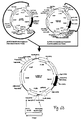
- Fig.1 This figure illustrates the principle of 'Retrocyte Display' allowing the identification and isolation of a binding protein such as an antibody, specific for a desired antigen or ligand.
- a retroviral expression construct that can give rise to expression of a diverse collection of binding proteins is stably transduced into suitable vertebrate host cells ("selector cells"). This is accomplished by transfecting at least one retroviral vector encoding at least one binding protein into retroviral packaging cells (step 1), which may either constitutively or transiently express retroviral proteins Gag, Pol and Env.
- Packaging cells transfected with the at least one retroviral binding protein construct will then produce recombinant retroviral particles within 24-72 hours post transfection, containing the at least one retroviral expression construct.
- the resulting retroviral particles accumulate in the cell culture supernatant of the retroviral packaging cells, and can be used to transduce suitable vertebrate host cells ("selector cells") (step 2), which then express the binding protein.
- the binding proteins such as antibodies or fragments thereof are expressed on the cell surface of the "selector' cells" and the cells then are labelled with a desired antigen or ligand (step 3).
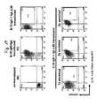
- Antigen- or ligand binding cells are then preferably analyzed by fluorescent activated cell sorting (FACS) and cells that exhibit specific antigen binding, are separated from the non-binding cell population preferably by preparative, high-speed FACS (step 4).
- Antigen-or ligand reactive cells may optionally be expanded in tissue culture again, and due to the stable expression phenotype of retrovirally transduced cells, cycles of antigen-directed cell sorting and tissue culture expansion may be repeated, up to the point that a detectable antigen- or ligand reactive cell population is obtained.
- This antigen- or ligand reactive cell population may be subjected to a final, preferable, single-cell sorting step, or may directly be used for cloning of binding protein encoding genes on a population basis.
- step 5 the coding regions of relevant binding domains are cloned from the antigen- or ligand-selected cell pools or cell clones, by RT-PCR or genomic PCR using primer pairs binding to sequences specific for the binding protein library and/or specific for other vector sequences, by standard methods known in the art. Cloned and sequenced coding regions for binding proteins may then optionally be expressed as recombinant proteins in any expression system of choice for further functional characterization and to confirm antigen- or ligand binding specificity (step 6).
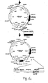
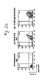
- Fig.2 (a) This figure illustrates the schematic structure of antibodies or immunoglobulins and fragments thereof, which are the preferred binding proteins according to the disclosed invention.
- Fig. 2a shows the schematic structure of an IgG antibody (left), which is characterized by a characteristic Y-shaped structure and is composed of two identical immunoglobulin (Ig) heavy and light chains, comprising four (V H -C H 1-C H 2-C H 3) and two immunoglobulin domains (V L -C L ), respectively.
- the V-domains are the highly variable antigen binding regions of IgH and IgL chains, whereas the C H and C L domains represent the constant region domains.
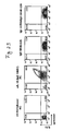
- variable region domains of IgH chains are encoded by V, D and J gene segments, whereas the variable region domains of IgL chains are encoded by only V and J gene segments, which need to be assembled from germline immunoglobulin gene loci ( Figs. 2b) and 2c ) during early B lymphopoiesis, by the process known as V(D)J recombination.
- Antibody IgH and IgL chains are covalently held together by disulphide bridges, which couple the identical IgH chains together at a location close to the flexible hinge region, i.e. between the C H 1 and C H 2 domains, whereas additional disulphide bridges between the C H 1 and C L domains, as depicted, are covalently coupling IgH and IgL chains ( Fig. 2a left).
- Fab fragments are univalent fragments of full-length antibodies only containing V H -C H 1/V L -C L domains coupled by a natural disulphide bridge, which can either be derived by enzymatic papain cleavage from full-length antibodies, or which can be expressed as recombinant proteins by expressing C H 2-C H 3 deleted IgH chains together with IgL chains.
- Additional fragments of fully human antibodies are single chain variable domain fragments (scFv-fragments), which only comprise the variable region domains of IgH and IgL chains that are coupled by a synthetic linker or an artificial disulphide bridge.
- the expression of either full-length antibodies, or antibody fragments, as the depicted Fab and scFv fragments may also be expressed as binding proteins in order to realize the invention.
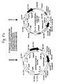
- Fig. 2b schematically depicts the process of V(D)J recombination occurring on a germline IgH chain allele, resulting in the assembly of the coding regions of antibody V H domains.
- the variable domains of IgH chains in vertebrate species are encoded by a multitude of V, D and J gene segments, which are separated in germline configuration.
- V(D)J recombination occurring during early B lymphopoiesis one selected V, D and J gene segment is site-specifically rearranged to generate a unique coding region for an antibody V H domain.
- V(D)J recombination in the IgH chain locus is an ordered process and starts with rearrangement of a selected D to a selected J gene segment, usually on both IgH chain alleles. Only after D to J gene rearrangement, one selected V region is site-specifically joined to the already assembled DJ region, thereby generating a V-D-J ORF encoding the V H domain.
- the process of V(D)J recombination is dependent on the expression of precursor lymphocyte specific recombination activating genes (RAG) 1 and 2.
- Fig. 2c schematically depicts the process of V(D)J recombination occurring on a germline IgL chain allele, resulting in the assembly of the coding regions of antibody V L domains.
- the variable domains of Ig L chains in vertebrate species are encoded only by V and J gene segments, which are separated in germline configuration, similar to the gene segments in the IgH chain locus.
- the generation of an antibody V L domains requires only one site-specific V(D)J recombination event, as depicted.
- Fig. 3 This figure schematically illustrates the principle of stable genetic modification of target cells for the expression of a binding protein of interest (BPOI) such as an antibody (alternatively labelled "X”) by retroviral transduction.
- BPOI binding protein of interest
- X antibody
- Fig. 3a depicts the schematic organization of a wild-type retroviral genome (upper left), in which the genes for the structural and functional proteins Gag, Pol and Env are located in between so-called 5' and 3' long-terminal repeat (LTR) sequences flanking the retroviral genome.
- the 5'LTRs are important for the expression of the retroviral genes and also for the replication of the retroviral genome in the infected host cell.
- Another important region in the retroviral genome is the ⁇ (Psi) packaging signal, which is required for the packaging of the retroviral RNA during replication and/or production of retroviral particles.
- the gag, pol and env genes may be removed from a wild-type retroviral genome, so that only 5' and 3' LTRs and then ⁇ (Psi) packaging signal remains.
- ⁇ Psi packaging signal
- MCS multiple cloning site
- a recombinant protein e.g. a binding protein of interest (BPOI) "X"
- BPOI binding protein of interest
- ORF open reading frame
- expression of a gene of interest may optionally be driven by an additional heterologous promoter (Prom.), and optional addition of a marker gene, e.g. downstream of the 5'LTR promoter and ⁇ packaging signal, as depicted here, may allow selection and/or tracking of retrovirally transduced constructs.
- Fig. 3b schematically illustrates the procedure of retroviral transduction of target cells resulting in the stable expression of a BPOI-"X" such as an antibody.
- a recombinant retroviral construct containing an expression cassette for a BPOI-"X" is transiently transfected into a retroviral packaging cell line (PCL), expressing structural and functional retroviral proteins Gag, Pol and Env of a wild-type retrovirus (left).
- a retroviral PCL can be generated by either stably or transiently transfecting expression constructs for the Gag, Pol and Env proteins into a suitable and easy to transfect cell line (e.g.
- retroviral particles are replication incompetent, because they lack the genes for the functional retroviral Gag, Pol and Env proteins and therefore, they can deliver their genetic payload into a target cell only once, a process that is called retroviral transduction, or single round infection.
- RNA of a recombinant retrovirus is introduced into the target cells, where it is reverse transcribed into cDNA, which is then stably integrated into the target cell genome.
- a gene of interest like the BPOI-"X
- FIG. 4 This figure shows the schematic design of preferred types of retroviral expression constructs that can be used to realize the invention.
- the drawing depicts the schematic design of retroviral vectors contained in a standard DNA cloning plasmid backbone (closed black line); the relevant genes and regions for the retroviral genome are highlighted.
- One preferred vector generation depicted in panel (a), whose detailed cloning is described in Figs 5 and 6 , and provided in Example 1, contains the cDNA coding regions for human Ig ⁇ 1 H chains and Ig ⁇ L chains driven by a strong constitutive CMV promoter (Prom) and flanked up- and downstream by the Ig ⁇ intron enhancer ( ⁇ iE) and 3' ⁇ enhancer (3' ⁇ E) elements, promoting somatic hypermutation to the V coding regions of the IgH and IgL chains.
- Prom a strong constitutive CMV promoter
- ⁇ iE Ig ⁇ intron enhancer
- 3' ⁇ enhancer 3' ⁇ enhancer
- the retroviral IgH and IgL chain expression constructs additionally contain open reading frames for the antibiotic resistance markers hygromycinB (hygro R ) and puromycin (puro R ), respectively, allowing the selection of stable integration of the IgH and IgL chain constructs applying respective antibiotic drug selection to cultures of retrovirally transduced vertebrate cells.
- hygro R antibiotic resistance markers
- puro R puromycin
- convenient, unique restriction enzyme sites are highlighted, allowing the straightforward replacement of V coding regions with HindIII and Eco47III, or the replacement of the entire IgH and IgL chain coding regions by using the restriction enzymes HindIII and NotI. This way, from one existing IgH or IgL chain expression construct different V regions and even entire collections of V regions can easily be cloned into the disclosed expression vectors.
- V(D)J recombination active cells in situ , e.g. in precursor lymphocytes expressing endogenous RAG1 and RAG2 proteins. Because the process of V(D)J recombination is not precise, a diverse collection of variable coding region sequences may result from one individual retroviral vector within a given set of V, D and J gene segments for IgH, or a given set of V and J gene segments for IgL.
- V, optionally D and J gene segments The diversity in the joining of V, optionally D and J gene segments is due to a combination of exonuclease activity, TdT mediated N-region addition, and P nucleotide generation, which may all contribute individually or jointly to coding joint diversification.
- V, D and J gene segments have been cloned in a fashion that different V, D and J gene segment family members can be easily replaced by unique restriction enzyme sites, a limited number of constructs generated and introduced into V(D)J recombination competent host cells, can result,in an enormous diversity of in situ generated binding protein diversity.
- a primary in situ generated collection of diverse binding proteins can optionally further be mutagenized by an AID-dependent somatic hypermutation process.
- V(D)J recombination activity e.g. precursor lymphocytes
- AID mediated somatic hypermutation is active, or can be activated.
- This panel schematically depicts yet another design of retroviral constructs that can be used to realize the invention.
- the expression of the IgH and IgL coding regions is driven by the 5'LTR promoter of the retroviral backbone and the expression of IgH and IgL chains is coupled to the expression of GFP and YFP autofluorescence markers, respectively, allowing the tracking and isolation of IgH and IgL expressing cells simply by analyzing the transduced cells for green and yellow fluorescence.
- These constructs are very useful for controlling the multiplicity of infection of "selector cells" without further labelling procedures.
- Fig. 5a-e illustrates the cloning strategy for the construction of a retroviral IgH (human Ig ⁇ 1 isotype) expression vector, disclosed in detail in Example 1, and in the basic design provided in Fig. 4(a) .
- the cloning of expression constructs for both membrane bound IgG as well as secreted IgG is depicted, as detailed in Example 1 - unique restriction enzyme sites in the plasmid maps are provided for general reference purposes.
- the final retroviral Ig ⁇ 1 H chain expression construct as disclosed in Fig.
- any other V H domain coding region, or a collection (library) of diverse V H domain coding regions can be introduced into the vectors using the unique HindIII and Eco47III restriction enzyme sites, by replacing the existing V H region with said any other V H domain coding regions.
- Fig. 5a depicts a first preparatory cloning step, in which an Eco47III restriction site (circled) is removed from the commercially available pLHCX vector backbone by site-directed mutagenesis, as described in Example 1. This generates the retroviral vector backbone pLHCXm1, in which the Eco47III restriction enzyme site can later be re-introduced for the cloning and replacement of V H domain coding regions.
- Fig. 5a further illustrates, how cloned fragments of the human ⁇ 1 constant region genes, either with, or without membrane spanning exons M1/M2 are cloned into the pLHCXm1 backbone using unique HindIII and ClaI restriction enzyme sites present in the MCS of pLHCXm1. The fragments were designed to contain additional flanking Eco47III and NotI restriction enzyme sites for later cloning purposes, as detailed in Example 1. Fig.
- FIG. 5b shows the plasmids maps of the cloning intermediates without V H domain coding regions, and it is shown, how a particular V H coding region flanked by HindIII and Eco47III sites is cloned into the constructs. These constructs, which are thus generated, are depicted in Fig. 5c , and would in principle be sufficient to confer the expression of human Ig ⁇ 1 H chains in any recipient cell line.
- the possibility to additionally mutagenize V H coding regions in an AID-dependent manner is an aspect of this invention and two additional cloning steps are disclosed, in which the core ⁇ iE element with additional flanking sequences is cloned into a unique BglII site, upstream of the CMV promoter of the expression cassette ( Fig. 5c bottom and Fig. 5d ) and in which the 3' ⁇ E element with some flanking DNA sequence is cloned into the unique ClaI site downstream of the expression cassette for human Ig ⁇ 1 H chains.
- These constructs correspond to the schematic plasmid maps that have already been disclosed in Fig. 4a , but here with precise restriction enzyme maps and drawn to scale.
- Fig. 6a-d illustrates the detailed cloning strategy provided in Example 1, for the construction of retroviral IgL (human Ig ⁇ L isotype) expression construct, whose basic design was already provided in Fig. 4(b) .
- retroviral IgL human Ig ⁇ L isotype
- any other V L domain coding region, or a collection (library) of diverse V L domain coding regions can be introduced into the vectors using the unique HindIII and Eco47III restriction enzyme sites, by replacing the existing V L region with said any other V L domain coding region(s).
- the cloning strategy for the retroviral IgL chain expression vectors required preparatory cloning steps, in order to generate a modified retroviral vector backbone, into which the desired elements could be cloned using convenient restriction enzyme sites as depicted.
- a first step from commercial plasmid pLPCX an undesired Eco47III site was removed from the ⁇ (Psi) packaging signal by site-directed mutagenesis as described in Example 2, resulting in modified plasmid pLPCXm1 ( Fig. 6a ).
- a novel pLPCXm2 backbone was generated by ligating a large, AscI-BlpI digested fragment from commercial plasmid pLHCX with an AscI-Ncol fragment from pLPCXm1 ( Fig. 6b ).
- the non-compatible BlpI and NcoI DNA ends needed to be filled up with nucleotides using Klenow fragment as described in Example 1.
- the constant region for a human ⁇ L chain (C ⁇ ) has been inserted via HindIII and ClaI as shown ( Fig. 6b ).
- the human C ⁇ fragment was further flanked by Eco47III and NotI sites to facilitate additional cloning procedures.
- one selected human V ⁇ element was cloned into the construct via unique HindIII and Eco47III sites ( Fig. 6c ). This construct would in principle be sufficient to confer the expression of human Ig ⁇ L chains in any recipient cell line.
- This construct would in principle be sufficient to confer the expression of human Ig ⁇ L chains in any recipient cell line.
- the IgH chain expression constructs Fig.
- Fig. 7 This figure illustrates the cloning strategy for a retroviral expression construct for activation induced cytidine deaminase (AID).
- AID activation induced cytidine deaminase
- the commercial pLPCX retroviral vector backbone was used and a specific RT-PCR-fragment from mouse splenic cDNA containing the AID coding region was cloned into the unique XhoI restriction site of the pLPCX vector using compatible XhoI restriction enzyme sites inserted into the PCR amplication primers, as described in Example 2.
- FIG. 8 This figure (8a and continued onto 8b) illustrates the detailed cloning strategy, also provided in Example 2, for a retroviral reporter constructs with and without Ig ⁇ L chain enhancer elements, allowing the identification and quantitation of somatic mutations by reversion of a defined EGFP stop mutation.
- Fig. 9 This figure provides an experimental proof-of-concept that the disclosed retroviral vectors allow AID mediated somatic mutation of sequences, like preferably antibody V coding regions, cloned downstream of the V-promoter elements.
- Panel (a) shows an analysis of AID expression by Western-blotting of five selected FA-12 A-MuLV transformed cell clones that had been stably transfected with a retroviral AID expression construct, whose cloning was depicted in Fig. 7 .
- the Western-blot analysis shows a distinct AID-specific signal of ca. 25kD in FA-12 transfectant clones 1 through 4, but not in transfectant 5 and also not in the non-transfected negative control (NC).
- Transfectant 3 was used for further testing of retroviral reporter vectors for AID-mediated somatic hypermutation (SHM), which is depicted in panel (b): Here the retroviral reporter constructs of Fig. 8 (once with and once without Ig ⁇ enhancer elements) were retrovirally transduced into AID expressing and AID non-expressing FA-12 transfectants 3 and 5, respectively. As expected, only when reporter constructs containing the enhancer elements were transduced into AID-expressing FA-12 transfectant clone 3, was it possible to detect green revertant transductants at a 0.2% frequency 10 days post transduction.
- SHM somatic hypermutation
- Fig. 10 This figure illustrates the sequence of the EGFP coding region with an engineered stop mutation that was used to clone an EGFP reporter construct for quantitating somatic hypermutation.
- panel (a) it is shown, which of the four nucleotides have been mutated in codon 107 and 108 of the EGFP open reading frame, thereby generating a stop-codon in codon 107 and generating a lysine to threonine amino acid change in codon 108.
- These four nucleotide changes additionally resulted in the introduction of unique SpeI restriction enzyme site, as indicated that could be used as a diagnostic marker for stop codon reversions upon somatic hypermutation.
- the G-nucleotide of the TAG stop codon is embedded in a so-called RGYW sequence motif, which is known to be a hotspot for somatic hypermutation.
- RGYW sequence motif which is known to be a hotspot for somatic hypermutation.
- sequence analysis revealed that the G nucleotide in the original TAG stop-codon had been mutated to a C nucleotide, resulting in a TAC codon, thereby confirming the restriction enzyme analysis, and demonstrating that AID-mediated somatic mutation had been targeted to the G in the RGYW motif.
- FIG. 11(a) and (b) Illustration of the detailed cloning strategy of a V(D)J recombination competent retroviral IgH chain expression vector, as disclosed in detail in Example 4.
- Fig. 12 Proof-of-concept that retroviral constructs requiring V(D)J recombination of V, D and J gene segments in "quasi-germline" configuration can give rise to productively rearranged heavy chain expression constructs and Ig+ cells upon transduction into RAG1/RAG2 positive precursor lymphocytes.
- Panel (a) contain data showing the generation of surface immunoglobulin positive cells (0.04%, upper right quadrant of left FACS plot) after transduction of a V(D)J recombination competent retroviral expression vector (detailed description of cloning, see Fig. 11 ) into A-MuLV transformed preB cell line 230-238.
- the immunoglobulin expression is coupled to EGFP expression using constructs as schematically illustrated in Fig. 4c . Therefore, immunoglobulin expressing cells can only be generated in the population of green (i.e. stably transduced) cells.
- the right staining panel shows re-analysis of surface immunoglobulin expression after a single round of FACS enrichment and expansion of the rare (0.04%) surface immunoglobulin cells for 8 days in tissue culture. After this one round of enrichment, the combined frequency of immunoglobulin positive cells had increased to 17.8% (as expected detectable in the green, i.e. the stably transduced population) from which PCR amplicons have been obtained and sequenced. (b).
- this panel shows a DNA sequence (clone 225, with amino acid translation on top) obtained from a PCR amplicon derived from surface immunoglobulin cells after one round of enrichment that had been transduced with "quasi-germline" V(D)J recombination competent retroviral vectors.
- sequences of the coding regions of the V, D and J gene segments are provided in (b) at the top, also with amino acid translation on top of the V and J gene segments, as the D segment sequence can be read in three different reading frames, depending on the junctional diversity after V(D)J recombination.
- Intervening sequences between the V, D and J gene segments in "quasi-germline" configuration are depicted with dots.
- the sequence of recovered clone 225 clearly represents a bona fide V(D)J rearrangement event, with typical features of nucleotide loss and TdT catalyzed N-sequence additions clearly detectable at the coding joints between the assembled V, D and J gene segments (all intervening sequences had been lost from clone 225).
- the sequence of clone 225 exhibited an open reading frame and, apart from the aforementioned variations at the coding junctions, did not contain additional somatic mutations in the V, D and J sequences.
- Fig. 13 Data showing the testing of a panel of different A-MuLV transformed murine preB cell lines for the susceptibility to ecotropic MLV-derived vector gene transfer.
- 1x10 5 cells were transduced with a MOI of 0.5 using a vector preparation having packaged the reporter gene EGFP encompassing transfer vector LEGFP-N1.
- Transduction was carried out as detailed in Example 5.
- Two days post transduction, gene transfer was detected by expression of EGFP using FACS. Except for preB cell line 18/81, all other tested A-MuLV transformed preB cell lines were susceptible for transduction at frequencies ranging between 40-60% under the applied conditions, and can, in principle, be used for the current invention.
- Untreated naive target cells served as negative controls and showed no green fluorescence (not shown).
- Fig. 14 Characterization of a panel of murine preB cell lines for intracellular expression of endogenous IgM heavy chains (cy- ⁇ H), in order to identify cells devoid of endogenous murine antibody expression that can be used as selector cells for retrocyte display.
- Cells were permeabilised and stained using anti-murine IgM heavy chain antibodies coupled to FITC (FL1). Untreated cells served as negative controls.
- the experiment shows that cell lines FA-12, 1624-5, 1624-6, 18/81-c18-11, and 40E1 had practically undetectable endogenous antibody expression, and can thus be used in a method of the present invention.
- Fig. 15 Illustration of the complexity of retroviral expression vectors following the design disclosed in Fig. 4(c) and the experimental principle for the generation of a IgH and IgL chain shuffled antibody library.
- Both vectors harbour the packaging sequence Psi ( ⁇ ), flanking long terminal repeats (LTR) and an internal ribosome entry signal (IRES).
- the IRES Parallel to the expression of an antibody polypeptide chain mediated by the viral promoter in the 5'LTR, the IRES enables the coupled expression of the reporter gene gfp and yfp , respectively. Upon viral gene transfer into selector cells, this allows for the convenient detection and enrichment of successfully transduced and immunoglobulin chain expressing cells using FACS.
- the TgL(245)-LIB-IRES-YFP library is transferred followed by expansion and enrichment of cells expressing YFP and human light chains employing FACS.
- the resultant population constitutes a fully human antibody displaying a defined human antibody library expressed by 1624-5 cells, containing a complexity of maximally 159'250 clones.
- Fig. 16 This figure shows how a two-step transduction with IgH-IRES-GFP and IgL-IRES-YFP libraries has been performed at conditions ensuring a transduction, resulting in clonal expression of polypeptide chains in the vast majority of the transduced cells.
- cells displaying gene transfer efficiencies lower than 10% were enriched using FACS sorting four days post infection.
- Cells were expanded for six days and subjected to a second transduction employing vector particles having packaged the light chain coding regions of antibodies at a dilution of 1:5 as described above.
- GFP-positive cells selected for heavy chain expression were infected with vector particles transducing the IgL-LIB-IRES-YFP library and vice versa.
- transduced cells expressing GFP and YFP were enriched using FACS.
- Fig. 17 Titration of IL-15 staining with a population of preB cells expressing an anti-IL-15 reference antibody by FACS, in order to define optimal conditions allowing optimal IL-15 antigen staining conditions for Retrocyte Display experiments.
- the staining procedure included a titration of the IL-15 antigen in the range of 2.5 ⁇ g/ml-0.1 ⁇ g/ml, at two different concentrations of a polyclonal, biotinylated anti-IL-15 secondary antibody, as indicated, which was detected with streptavidin-PE conjugate by FACS. Surface Ig+ cells were counterstained with an anti-Ig ⁇ L chain-APC antibody.
- optimal IL-15 staining is accomplished at a concentration of 0.1 or 0.5 ⁇ g/ml IL-15 antigen, and using 3 ⁇ g/ml of the secondary, polyclonal anti-IL-15 antibody.
- the FACS profile of the NC cells shows that approximately 50% of the Ab-library transduced cells are surface-Ig+, as detected by the anti-Ig ⁇ L chain-APC staining. However, none of the surface-Ig+ cells displays binding to IL-15. In contrast, the PC cells, in which more than 90% of the cells expressed surface Ig, a specific IL-15-antigen binding is apparent by a specific signal on the x-axis.
- Fig. 19 Proof of concept for the enrichment of a IL-15-reactive cell population by Retrocyte Display from a diverse antibody library, as dislosed in detail in Example 7.
- the top panel shows FACS stainings for GFP/YFP expression (y-axis), indicative of the frequency of Ig-retrovector transduced cells, and IL-15/anti-IL-15-bio (x-axis), indicative of specific IL-15 staining.
- the top-right panel shows the same two-colour FACS staining of a population of cells that have been transduced with a single IgH chain encoding retroviral vector encoding the IgH chain of the reference anti-IL15 antibody in combination with a diverse, >7x10 4 different Ig ⁇ L chain library.
- This IgL chain shuffled library therefore contains potentially >7x10 4 different antibodies, and expectedly, even by very narrow gating for antibody-expressing and IL-15 reactive cells, as indicated in the top-right FACS-profile, very few IL-15 reactive cells could be detected (here 2.42%, due to the gating close to the negative population, as indicated).
- the enriched population was expanded in tissue culture, and the identical staining procedure and FACS-sorting was repeated three times, as shown for the three FACS stainings under identical conditions in the lower three FACS panels.
- consecutive enrichment/cell expansion cycles resulted in a population of cells that was almost 100% positive for antibody expression and even more positive for IL-15-reactivity than the original PC cell line.
- This data shows clearly that by repeated FACS sorting and expansion a highly antigen-reactive cell population can be successfully enriched to an essentially 100% antigen-reactive cell population from almost undetectable antigen-reactive cell populations using three consecutive rounds of Retrocyte Display.
- Fig. 20 This figure illustrates and confirms the specific IL-15 antigen reactivity of 4 representatives of 24 individual cell clones established after single cell sorting from a 3 times IL-15 antigen enriched cell population, as described in Fig. 19 .
- the 4 selected cell clones are designated clone F, H, V and W, and all show specific IL-15 reactivity on GFP/YFP positive cells, indicative of the stably transduced, Ig encoding retroviral vectors.
- higher GFP/YFP expressing cells, expressing higher antibody levels showed higher IL-15 specificity, leading to characteristic diagonal staining signals in the Ig/IL-15 double stainings.
- Fig. 21 This figure provides a second proof of concept for successful Retrocyte Display enrichments of antigen-reactive cells by illustrating the successful enrichment of IL-L ⁇ antigen-reactive cells to an essentially 40% antigen-reactive cell population using three consecutive rounds of retrocyte display cell enrichment/tissue culture expansion, starting from a minimally IL-lbeta-reactive cell population in the initial cell population. Double stainings for GFP/YFP expression (indicative of antibody expression) and IL-1 ⁇ reactivity are provided.
- the bottom panels show the FACS stainings for antibody expression and IL-1 ⁇ reactivity of an antibody library generated by shuffling of a diverse IgL chain library of >1.2x10 5 individual IgL chain clones against the IgH chain of the SK48-E26 reference antibody, before (0x enriched) and after 1, 2 and 3 Retrocyte Display enrichment rounds, as indicated and as disclosed in detail in Example 8.
- Fig. 22 This figure shows confirmation of IL-1 ⁇ antigen reactivity of a novel antibody identified by Retrocyte Display, as disclosed in detail in Example 8. From the 3x enriched cell population, shown in Fig. 21 , 24 individual cell clones have been established by single cell sorting. From these 24 cell clones, 12 clones harboured a novel IgL chain, termed LCB24, as disclosed in Example 8.
- the IL-1 ⁇ specificity of the novel LCB24 Ig ⁇ L chain co-expressed with the IgH chain of the IL-1 ⁇ specific reference antibody SK48-E26 was analyzed by FACS upon re-transduction of the cloned and sequence characterized IgL and IgH chain retroviral expression vectors into the original selector cell line.
- the FACS stainings show analysis of antibody expression (via GFP/YFP) and IL-1 ⁇ reactivity by two colour FACS, as indicated.
- NC negative control, left
- a clear IL-1 ⁇ specific staining is detected in positive control cells expressing IgH and IgL chains of reference antibody SK48-E26 (middle).
- a similar, IL-1 ⁇ specific, signal is detectable in antibody expressing selector cells transduced with the SK48-E26 reference antibody IgH chain vector and the novel, fully human LCB24 Ig ⁇ L chain, cloned from IL-1 ⁇ specific Retrocyte Display cell clones (right).
- Fig. 23 Confirmation of lack of cross-reactivity to IL-15 of novel antibody encoded by LCB24 IgkL chain/SK48-E25 IgH chain.
- the two right FACS stainings show no IL-15 reactivity on antibody expressing cells either encoding the novel antibody composed of SK48-E26 IgH and LCB24 IgL chain, or on cells expressing the original SK48-E26 IgH/IgL combination.
- Fig. 24 This figure illustrates the successful enrichment of streptavidin-APC-Cy7 antigen-reactive cells by three consecutive rounds of Retrocyte Display cell enrichment/tissue culture expansion, from an antibody library generated by shuffling of a diverse IgL with a diverse IgH chain library as disclosed in Example 9. Streptavidin-APC-Cy7 reactive cells were enriched by three consecutive rounds of high-speed cell sorting, followed by cell culture expansion, as indicated. The binding-specificity of antibody expressing cells for the streptavidin-APC-Cy7 antigen is demonstrated by analyzing FACS profiles of the sequentially enriched cell populations in the presence (lower panel) and absence (top panel) of the antigen. This demonstrates a proof of concept for the efficient enrichment of antigen-specific by Retrocyte Display in the absence of any reference antibody that could be used for chain shuffling approaches.
- Fig. 25 The data presented in this figure provide evidence for the specificity of the 3 times Retrocyte Display enriched cell population disclosed in Fig, 24 for specific reactivity to the Cy7 fluorochrome of the strepatavidin-APC-Cy7 tandem dye.
- non-transduced selector cells, unenriched cells expressing an IgH/IgL chain library combination and a 3 times strepatavidin-APC-Cy7 enriched cell population were analyzed by FACS for antibody expression (indicated by GFP/YFP fluorescence) and reactivity to different streptavidin-fluorochrome conjugates, as indicated.
- streptavidin-APC-Cy7 enriched cell population only bound to streptavidin-APC-Cy7, but not to streptavidin-APC or streptavidin-APC-Cy5.5, and non-specific staining of strepatavidin-APC-Cy7 was also not detectable to either the selector cells or the selector cells expressing a diverse antibody library.
- This provides proof of concept for the efficient and highly specific Retrocyte Display enrichment of specific antibodies from cells expressing a diverse antibody library, without the need of antibody IgH or IgL chains from antigen-specific reference antibodies.
- Fig. 26 Two novel human antibodies identified by Retrocyte Display, sharing the same IgH chain show specific binding to antigen streptavidin-APC-Cy7. As disclosed in Example 9, two different IgH chain sequences (HC49 and HC58) and two different IgL chain sequences (LC4 and LC10) could be identified from single-sorted cell clones after three rounds of Retrocyte Display enrichment. In this figure, all possible pairings of IgL chains LC4 and LC10 with HC49 and HC58 were examined for reactivity to the target antigen streptavidin-APC-Cy7.
- Affinity maturation A highly regulated immunological process of antigen-driven improvement of the binding specificities of antibodies produced by antigen-stimulated B lymphocytes, mostly occurring in germinal centers. The process is caused by somatic hypermutation largely targeted to the coding regions for the variable domains of antibodies coupled with the selective expansion and survival of B lymphocytes generating higher affinity antibodies.
- Antibody This term describes an immunoglobulin whether natural or partly or wholly synthetically produced. The term also covers any polypeptide or protein comprising an antibody antigen-binding site, like heavy chain only antibodies from for example camels or lamas.
- a full-length antibody comprises two identical heavy (H) chains and two identical light (L) chains. In its monomeric form, two IgH and two IgL chains assemble into a symmetric Y shaped disulphide linked antibody molecule that has two binding domains formed by the combination of the variable regions of IgH and IgL chains.
- Antibodies can be isolated or obtained by purification from natural source, or else obtained by genetic engineering, recombinant expression or by chemical synthesis, and they can then contain amino acids not encoded by germline immunoglobulin genes.
- a fully human antibody comprises human heavy and light chains i.e. variable and constant domains from the human species.
- a chimeric antibody comprises variable region domains from one vertebrate species combined with constant region domains of another vertebrate species. The constant domains of a chimeric antibody are usually derived from a human antibody or antibodies.
- Humanised antibodies can be produced by grafting CDRs of non-human antibodies onto framework regions of IgH and IgL variable domains of human origin.
- Antibody fragment It has been shown that fragments of a whole antibody can perform the function of binding antigens.
- binding fragments are (i) the Fab fragment consisting of VL, VH, CL and CHl domains; (ii) the Fd fragment consisting of the VH and CHl domains; (iii) the Fv fragment consisting of the VL and VH domains of a single antibody; (iv) the dAb fragment, which consists of a VH or a VL domain; (v) isolated CDR regions; (vi) F(ab')2 fragments, a bivalent fragment comprising two linked Fab fragments; (vii) single chain Fv molecules (scFv), wherein a V H domain and a V L domain are linked by a peptide linker which allows the two domains to associate to form an antigen binding site; (viii) bispecific single chain Fv dimmers; and (ix) "diabodies", multivalent or multispecific fragments constructed by gene fusion.
- Fv, scFv or diabody molecules may be stabilized by the incorporation of disulphide bridges linking the V H and V L domains.
- Minibodies comprising an scFv joined to a CH3 domain may also be made.
- Other examples of binding fragments are Fab', which differs from Fab fragments by the addition of a few residues at the carboxyl terminus of the heavy chain CHl domain, including one or more cysteines from the antibody hinge region, and Fab' - SH, which is a Fab' fragment in which the cysteine residue(s) of the constant domains bear a free thiol group.
- a heavy or a light chain may also be considered to be an antibody fragment.
- all the above antibody fragments display at least one function of the whole native antibody from which said fragments are derived and are thus termed 'functional' fragments.
- Antigen Any biomolecule or chemical entity that can be bound by the variable domains of immunoglobulins (or antibodies).
- Binding protein This term defines one protein of a pair of molecules that bind one another.
- the binding partner of a binding protein is usually referred to as a ligand.
- the proteins of a binding pair may be naturally derived or wholly or partially synthetically produced.
- One protein of the pair of molecules has an area on its surface, or a cavity, which binds to and is therefore complementary to a particular spatial and polar organization of the other protein of the pair of molecules.
- types of binding pairs are antigen-antibody, biotin-avidin, hormone-hormone receptor, receptor-ligand, enzyme-substrate.
- the present invention is preferably concerned with antigen-antibody type reactions.
- Complementary determining region This term refers to the hypervariable regions of the heavy and light chains of an immunoglobulin. CDRs are the regions in the three dimensional structure of an immunoglobulin that directly establish contact to antigen. An antibody typically contains 3 heavy chain CDRs and 3 light chain CDRs. The CDRs are usually the most diverse parts of antigen receptors.
- Ig-like domains A structural moiety of a biomolecule that is characterized by a particular three dimensional structure (e.g. variable or constant region domains of immunoglobulins that are structurally related, such as Ig-like domains that can be found in many molecules of the immune system, which belong to the so-called Ig-superfamily).
- Germinal center A distinct histological structure in peripheral lymphoid organs (e.g. lymph nodes or spleen) where cognate interactions between antigen presenting cells and between different lymphocyte populations occur, resulting in the proliferative expansion of antigen-reactive lymphocytes, as well as affinity maturation and class switch recombination of antibodies produced by antigen reactive B lymphocytes.
- lymphoid organs e.g. lymph nodes or spleen
- Germline configuration The unrearranged configuration of genes and gene loci, as they are inherited from the parents, and as they will be passed on to further generations through the germline.
- DNA recombination events occurring in somatic cells like e.g. V(D)J recombination in lymphocytes, lead to the reshuffling or loss of genetic information on certain gene loci and therefore to a change of the genes from the germline configuration.
- PreB lymphocyte A precursor B lymphocyte is characterized by the expression of particular precursor B cell specific genes, like e.g. the ⁇ 5 and V preB1 and V preB2 genes, and the expression of precursor lymphoid specific factors involved in V(D)J recombination (e.g. RAG-1, RAG-2).
- precursor B lymphocytes are characterized by the presence of DJ H on both heavy chain alleles or at least one V H DJ H rearrangement on at least one immunoglobulin heavy chain allele, while the light chain gene loci are still in unrearranged, germline configuration, such that the preB cells cannot express complete antibodies.
- Primary lymphoid organs Organs, in which lymphocytes develop from hematopoietic stem cells, in mice and humans e.g. the bone marrow, the thymus, and during fetal life, the liver.
- Quadrature configuration The artificial arrangement of V, optionally D, and J gene segments with flanking recombination signal sequences cloned from germline immunoglobulin gene loci into artificial genetic constructs, such that the arrangement of the V, optionally D, and J gene segments in such artificial genetic constructs still allows site-specific recombination of the gene segments into a variable coding region by the process of V(D)J recombination.
- Somatic mutation A process in somatic cells resulting in the introduction of point mutations into specific regions of the genome. When this occurs at a high frequency (>10 -4 mutations per basepair per cell division) it is known as somatic hypermutation.
- V(D)J recombination This is the process for generating antibody and T-cell receptor diversity and is the method by which functional antibody genes are created. It involves the rearrangement of many gene segments that code for the heavy and light chain proteins of immunoglobulins, and it only occurs in lymphocytes.
- Transfecting/Transfection In the context of eukaryotic cells this is the process of introducing nucleic acid sequences into eukaryotic cells, usually associated with using chemical and/or physical methods.
- Transforming/Transformation In the context of eukaryotic cells this is the process of immortalizing a cell for the establishment of a continuously proliferating cell line.
- Transducing The process of delivering DNA into vertebrate cells via the production of recombinant viruses.
- a packaging cell line expressing structural proteins for viral particles is transfected with a recombinant viral DNA construct comprising the regulatory elements for packaging of the viral DNA construct into the viral structural proteins.
- recombinant viruses are produced that can be used to infect (mammalian) target cells leading to the introduction of the genetic information cloned into the recombinant viral genome.
- Vector/Construct An artificially generated nucleic acid sequence which can be used to shuttle nucleic acid elements between different organisms and species, and which can further be used to propagate, amplify and maintain genomic information.
- Antibodies, or immunoglobulins are the most widespread class of binding proteins that have proven to be particular useful for therapeutic and for diagnostic applications. Therapeutic antibodies have developed into the commercially most successful class of biologic drugs and there is continued interest in novel and powerful methods to develop antibody-based therapeutics (Baker, 2005).
- Antibodies consist of two identical heavy (H) chain and light (L) chain glycoproteins that are covalently linked via disulphide bonds ( Fig. 2a ).
- Each immunoglobulin heavy (IgH) and light chain (IgL) polypeptide comprises an N-terminal variable domain that varies between different antibodies and a C-terminal constant region, that is identical between different antibodies belonging to the same immunoglobulin subtype (isotype) ( Fig. 2a ).
- the combination of IgH and IgL chain variable domains creates the antigen binding pocket of an antibody and determines its specificity, whereas the constant regions determines the immune effector function of an antibody.
- V H and V L domains are encoded by a multitude of gene segments, that are designated V (variable), D (diversity), and J (joining) gene segments.
- V variable
- D diversity
- J joining
- the rearrangement of gene segments is mediated by recombination-activating gene (RAG) 1 and 2 products. Due to the multitude of V, D, and J gene segments, and imprecision in gene segment joining, an enormous repertoire of different V region specificities can be generated by the millions of B lymphocytes produced by the immune system every day (Granch et al., 1998). Because the immunoglobulin heavy chain gene locus contain V, D and J gene segments, the coding region for a V H domain of an antibody requires two sequential V(D)J rearrangement events, whereas the immunoglobulin gene locus lacks D gene segments and the V L coding region is generated by one V to J-rearrangement event ( Fig 2c ).
- the junctional diversity that is generated by V(D)J recombination in the CDR3 region of IgH chains is greater than the CDR3 junctional diversity that is generated by only one rearrangement event for the IgL chains.
- the enzyme terminal deoxynucleotidyltransferase (TdT) is expressed, that is able to add non-templated nucleotides at the D to J and V to D junctions (so-called N-sequence diversity), additionally diversifying the IgH chain CDR3 repertoire.
- both IgH and IgL chain repertoires can further be diversified by the process of somatic hypermutation that is triggered in mature B cells during the course of a T cell dependent immune reaction (Papavasiliou & Schatz, 2002).
- somatic mutations are specifically targeted to the V H and V L coding regions, and are mediated by the B lineage specific enzyme activation-induced cytidine deaminase (abbreviated AID, see: Papavasiliou & Schatz, 2002).
- AID activation-induced cytidine deaminase
- cells expressing higher affinity antibody mutants against the immunogen are positively selected in the course of an immunization mostly occurring in germinal centers, and resulting in an enrichment of cells producing higher affinity antibodies.
- These antibodies now also accumulate mutations in CDRs 1 and 2, a process, which is referred to as affinity maturation of the antibody repertoire.
- the AID-mediated diversification and specific targeting to the V H and V L coding regions is significantly increased by the presence of cis -regulatory genetic elements or motifs, in particular enhancer elements of the IgH and IgL chain gene locus, located in the proximity of the rearranged V H and V L coding regions (Bachl & Olsson, 1999).
- monoclonal antibodies directed against a molecule or epitope of interest are generated by immunization of small laboratory, or farm animals, e.g. mice, rats, rabbits, or goats and donkeys, respectively. After repeated immunizations, animals are either bled for the isolation of polyclonal antibodies from their blood serum or, for the generation of monoclonal antibodies, the animals are sacrificed in order to isolate lymphocytes from secondary lymphoid organs, like lymph nodes or the spleen.
- Isolated lymphocytes are fused to immortal myeloma cells for the generation of hybridomas, which are subsequently subcloned and screened for the secretion of monoclonal antibodies exhibiting desired functional properties, like e.g. binding to a particular antigen or target.
- HAMA human-anti-mouse antibody response
- Chimeric antibodies are generated by fusing the variable binding domains of a rodent antibody to the constant regions of a human antibody, using standard genetic engineering and cloning techniques.
- Humanized antibodies in contrast, are generated by only transferring the complementarity determining regions (CDRs) of a variable domain from a rodent antibody to the variable region framework of a human antibody, which is also done by standard molecular biology techniques.
- HACA human anti-chimeric antibody
- transgenic mouse technology can deliver high affinity antibodies, due to the natural affinity maturation occurring in these mice upon in vivo immunization, and it has been demonstrated that the affinity profile of human antibodies towards a given antigen derived from human transgenic mice can be comparable to that of wild-type mice.
- affinity profile of human antibodies towards a given antigen derived from human transgenic mice can be comparable to that of wild-type mice.
- disadvantages associated with the human immunoglobulin transgenic animals are:
- phage-display selected antibody fragments have mediocre affinity and/or may carry structural artefacts.
- phage display selected binders are re-engineered and expressed as full-length antibodies in vertebrate cells, it may happen that phage selected antibodies are either poorly or not at all expressible, or that they exhibit altered binding characteristics.
- retroviral expression systems in this method is preferred, because gene transfer of single copies of expression constructs can be transferred into individual preB cells (Kitamura et al., 1995; Stitz J et al., 2005), and also, because there is complete freedom of choice, as to which antibody expression constructs are being used for engraftment for the mice (e.g. antigen pre-selected antibody libraries, antibody libraries from diseased patients, or individual antibody clones). Therefore, a particular advantage of this technology is its flexibility that it can be applied to de novo development of antibodies, as well as to the optimization of existing therapeutic antibody candidates.
- One in vitro system is based on the isolation of antigen-enriched memory B cells from human patients with a particular disease that can be isolated and then be immortalized by Epstein-Barr Virus (EBV) transformation in vitro , followed by the screening for antigen-reactive EBV lines ( Lanzavecchia, WO 04/76677 A2 ).
- EBV Epstein-Barr Virus
- EP 1 041 143 A An alternative method for the identification of biologically active peptides and nucleic acids has been proposed by Jensen et al (EP 1 041 143 A ).
- the preferred method described in EP 1 041 143 A comprises an initial screening procedure in which a large number of retroviral vectors can be introduced into cells such that the individual cell can express a number of different RNAs or peptides.
- the cells that show a phenotypic change are subsequently isolated and the retroviral DNA in that clone can be isolated by PCR.
- This PCR product can then be used to re-transfect viral packaging cells to create further retroviral vectors.
- These retroviral vectors can then be used for the transduction of different cells and finally after a second cloning procedure the active substance can be identified.
- this method results in an indirect change in the phenotype of a cell by the importation of biologically active peptides or nucleic acids. This is in contrast to the method of the present invention whereby the retrovirally transduced constructs directly encode the binding proteins, preferably antibodies, to which the screening is directed. It should also be noted that the peptides and nucleic acids described in EP 1 041 143 A differ greatly in size to the antibodies or antibody fragments identified by methods of the present invention.
- a further method for retrovirus-based genomic screening is set out in WO 03/083075 A2 (Bremel et al ) .
- This method relates to the expression and screening of genomic DNA sequences encoding uncharacterised genes and proteins.
- a process is described in which a cell line is transduced with a retroviral expression construct such that a genomic DNA virus is inserted into the genome of the cell line as a provirus, and then the expression of polypeptides from the provirus is analysed directly.
- Such a method does not provide the opportunity for enrichment of the cell line, nor for the isolation and identification of the expressed polypeptides before analysis is performed, which would detract from the high-throughput screening technology developed by Bremel and co-workers.
- a recently published patent application (WO 08/055795 A1 ) from Beerli and co-workers describes a screening platform for the isolation of human antibodies, which utilises a Sindbis virus expression system.
- An essential feature of this platform is the generation of starting library where B cells specific for an antigen of interest are directly isolated from peripheral blood mononuclear cells (PBMCs) of human donors.
- PBMCs peripheral blood mononuclear cells
- Recombinant, antigen-reactive scFv libraries are generated from this pool of B cells and screened by mammalian cell surface display by using a Sindbis virus expression system. Similar to phage display, one of the drawbacks to this system is that the scFvs of interest need to be re-engineered and expressed as full-length IgGs in vertebrate cells. Such a process can be associated with a loss in affinity of the antibody of interest on conversion since these antibodies may not express well in vertebrate cells and/or may exhibit altered binding characteristics.
- the invention disclosed herein comprises a unique and extremely powerful combination of methods for the development and optimization of binding proteins, preferably antibodies, or fragments thereof.
- the main advantages of the invention disclosed herein are complete flexibility in terms of optimization and de novo development of antibodies, and the speed to identify specific binders in short periods of time.
- the invention is realized in vitro, there is no limitation with regard to the development of antibodies against antigens, which are highly conserved across species, or that may be toxic in experimental animals.
- the binding proteins in particular antibodies, can be expressed as full-length antibodies, in a vertebrate cell, and preferably in a B lymphocyte environment, i.e. the natural host cell of antibodies, ensuring most natural and proper protein folding, correct posttranslational modification, and a quality control for heavy and light chain pairing.
- the key advantages of the disclosed invention are the complete flexibility with regard to development of antibodies against any desired target, the possibility to affinity optimize existing antibodies, a complete freedom of choice as to which type of antibody is expressed in the system (antigen-enriched, synthetic, from patients, under conditions of IgH and IgL chain shuffling, etc.).
- 'Retrocyte Display' In comparison to other eukaryotic cell-based expression systems involving either plasmid based expression constructs or non-integrating viral vectors, key advantages of the disclosed invention of 'Retrocyte Display' are that stable, sustained and clonal expression can be achieved by use of retroviral gene transfer technology.
- the stable, sustained and clonal expression of recombinant antibodies in the target cells allows repetitive enrichment cycles of antigen- or ligand specific cells, including the possibility to isolate and to expand monoclonal cells for identification of the antibody genes.
- the invention disclosed herein additionally allows the additional generation of genetic diversity upon retroviral transduction into vertebrate host cells in situ using single retroviral constructs by either exploiting the lymphocyte specific mechanism of V(D)J recombination, or exploiting the process of somatic hypermutation for further mutagenesis of binding proteins.
- the method of retrocyte display disclosed herein provides unique, novel and powerful solutions for many evident limitations that pre-existing technologies suffer from.
- the invention disclosed herein is broadly applicable to the expression, screening and identification of binding proteins specifically binding to a ligand or antigen of interest. While the invention can be performed with any binding protein, including but not limited to monomeric, homo- or hetero-multimeric membrane bound receptors, like T cell receptors, cytokine, or chemokine receptors, but also with other scaffold proteins, the preferred binding proteins according to the invention are full-length antibodies, with fully human antibodies being particularly preferred.
- any (functional) fragment of an antibody including, but not limited to single chain Fv fragments (scF v ), Fab fragments, F(ab')2, single V H or V L domains, single heavy or light chains or any combination thereof, with any naturally occurring or artificially engineered modification may be used to realize the invention.
- the invention is particularly applicable to any kind of artificially engineered or designed modifications of antibody binding regions, e.g. those generated by site-, or region-directed mutagenesis, fusion of naturally occurring sequences from different antibodies, randomization of CDR sequences, DNA shuffling, error-prone PCR, just to name a few methods by way of illustration.
- a preferred method for the expression of binding proteins according to the invention is to use retroviral vector-mediated transduction of vertebrate host cells.
- retrovirus vectors have been investigated for many years in the field of gene therapy.
- AAV adeno-associated virus
- Perabo et al. (WO 03/054197 A2 ) have inserted randomised sequences encoding targeting peptides into the viral capsid gene, at a site critical for binding to the primary cellular receptor, and produced AAV libraries that displayed the peptides in the context of the viral capsid.
- the selective pressure provided by the culture environment drove the selection by means of the ability of the viral clones to accomplish every step in the infection process, namely binding, uptake, uncoating, nuclear translocation, replication, and gene expression.
- vectors were generated that efficiently transduced leukemia cells. Whilst such a technique may be useful to generate viral mutants that infect target cells previously resistant to infection by wild-type AAV, it does not provide for the generation of diverse collections of binding proteins in vitro.
- binding proteins have several key advantages over any other methods known in the art for the expression of recombinant proteins in eukaryotic and/or vertebrate host cells.
- expression of recombinant binding proteins in vertebrate host cells may also be achieved by alternative methods, like, for instance, but not limited to, transient or stable DNA transfection, RNA transfection, or by transfer of DNA-based viral vectors, like adeno-viral or poxvirus-based vectors - albeit none of the aforementioned alternative methods allows for an easily controllable stable and clonal expression of binding proteins in vertebrate host cells.
- Preferred vertebrate host cells for the realization of the invention are cells of the B lymphocyte lineage, in particular precursor B lymphocytes, which often lack endogenous antibody expression, but which express favourable accessory proteins, like e.g. chaperones for proper protein folding and antibody assembly, or accessory membrane proteins facilitating membrane deposition of antibody molecules, like e.g. the B cell specific Ig ⁇ or Ig ⁇ proteins.
- retroviral vectors that are relatively small (maximal size of recombinant DNA to be incorporated: 8-10 kB) and that can be cloned and manipulated by standard molecular biology methods as plasmid vectors, from which the retroviral RNA genome can be transcribed.
- a wild-type retroviral genome only contains three genes, gag, pol and env, which encode the nuclear core proteins, a retroviral integrase, protease, RNAse, and a reverse transcriptase, and envelope proteins, respectively ( Fig. 3a ).
- the retroviral genome contains cis -regulatory sequences, like the Psi ( ⁇ ) sequence required for packaging of the retroviral RNA genome into virus particles, a polyA signal for retroviral transcript termination, and lastly, so-called 5'- and 3'-long-terminal repeats (LTRs) containing promoter elements and signals for retroviral integration into the host cell genome ( Fig. 3a ).
- LTRs 5'- and 3'-long-terminal repeats
- the gag, pol and env coding regions of a wild-type retrovirus are replaced by any expression cassette for a gene of interest ( Fig. 3a ), including relevant cis -regulatory elements, like promoters or enhancers.
- a plasmid vector containing a retroviral genome needs to be transiently or stably transfected into a so-called retroviral packaging cell line (PCL), expressing the viral structural proteins encoded by gag, pol and env in trans in a transient or stable fashion, and therefore allowing the packaging of the recombinant viral genome (the transfer vector) into replication incompetent retroviral particles ( Fig. 3b ).
- PCL retroviral packaging cell line
- Fig. 3b retroviral packaging cell line
- These retroviral particles allow for a single-round infection (transduction) of target cells ( Fig. 3b ).
- the entry of the retroviral particle into target cells is mediated by a specific interaction of the Env protein with a specific receptor on the target cell.
- the nature of the Env protein determines the tropism of the retroviral particles to specific host cells expressing the cognate receptor.
- Ecotropic retroviruses are restricted to rodent cells, amphotropic retroviruses may infect various species including rodent and human cells and pantropic retroviruses may infect any replicating cell with a cell membrane, as the cell entry occurs via structures present on all eukaryotic cell membranes.
- Retroviral vector particles with a variety of different tropisms can also be generated using heterologous envelope proteins of other viruses such as gibbon ape leukemia virus (GaLV), vesicular stomatitis virus (VSV) or HIV and SIV or even cellular membrane proteins, just to name a few by way of illustration - a technique known as "pseudotyping".
- GaLV gibbon ape leukemia virus
- VSV vesicular stomatitis virus
- HIV and SIV even cellular membrane proteins
- a retrovirus can deliver the viral genome into the host cell, where the viral proteins mediate reverse transcription of the genome into cDNA and eventually its stable integration into the host cell genome, allowing stable expression of the delivered genes ( Fig. 3b ).
- ecotropic MLV particles are used to mediate gene transfer into murine B cells.
- any infectious retroviral vector pseudotyped with any other envelope or transmembrane protein can be employed to realize the invention, provided that it mediates transduction in any appropriate target selector cell independent from their parental donor species, cell type or their expression of a cognate receptor mediating vector cell entry.
- vector-containing retroviral particles containing transcripts of recombinant retroviral genomes, or transfer vectors
- transfer vectors can be harvested from the cell culture supernatant of packaging cells either stably or transiently expressing transfer vectors ( Fig. 3b ). This can be carried out in a broad range of protocols and variations thereof, known to a person skilled in the art.
- Preferred embodiments of this invention include: 1) preparation of cell-free retroviral particle containing supernatants using either passage through an appropriate filter or a centrifugation step that separates packaging cells from vector particles.
- retroviral particle preparations are subsequently used to transduce vertebrate host cells by co-incubation for a variable time frame or by performing a so-called "spin infection".
- a target cell suspension is mixed with retroviral particle containing medium and is subjected to low-speed centrifugation ( Fig. 3b ).
- co-cultivation of target cells with packaging cells enabling cell-to-cell contact or separation of both cell populations by a membrane which allows the passage of retroviral particles but not of the packaging cells, can be performed to enable transduction of target cells.
- a preferred embodiment of the method is to use B-lymphocyte lineage cells from rodents that do not express endogenous murine immunoglobulin proteins, and that can be transduced with retroviruses of ecotropic host range.
- Cells of the B lymphocyte lineage have the advantage that they already express B cell specific Ig ⁇ and Ig ⁇ proteins that are favourable for cell surface expression and anchoring of membrane bound, full-length immunoglobulins.
- immunoglobulin negative plasma cell-derived cells like e.g. myeloma cells, as for instance, but not limited to Sp2/0, NSO, X63 and Ag8653 generally lack the accessory Ig ⁇ and Ig ⁇ proteins for membrane immunoglobulin deposition.
- the method may still be applied, if expression of the Ig ⁇ and Ig ⁇ proteins is conferred upon transfection or transduction of expression vectors for Ig ⁇ and Ig ⁇ , a standard procedure for any person skilled in the art.
- the method may be realized with any vertebrate host cell line, provided that retroviral particles with appropriate tropism are produced, that are able to transduce said vertebrate host cell line.
- retroviral particles for instance, but not limited to particles pseudotyped with the G protein of VSV
- pantropic retroviral particles for instance, but not limited to particles pseudotyped with the G protein of VSV
- Preferred cells of the B-lymphocyte lineage are for instance, but not limited to precursor B-, B-leukemia or B-lymphoma cells, from any vertebrate species, but also primary precursor B cells that can be grown in tissue culture for long periods of time.
- Precursor B lymphocytes would represent ideal host cells for retroviral expression of immunoglobulins, as the majority of such cell lines, do not express endogenous immunoglobulin proteins.
- murine preB cell lines can easily be obtained from any mouse strain by transformation with Abelson-murine leukemia virus (A-MuLV).
- preB cells express the preB cell specific proteins VpreB and ⁇ 5, which together form the so-called surrogate light chain, which, in the absence of conventional light chains, can form preB cell receptor complexes of immunoglobulin heavy and surrogate light chain.
- preB cells are preferred that lack expression of surrogate light chain components, comprising the gene products of the ⁇ 5, or VpreBl, or VpreB2 genes either as single, double or triple-gene knockouts.
- surrogate light chain can bind to heterologous heavy chains it is expected that surrogate light chain expression may interfere at a varying degree with the screening of IgH/IgL pairs, but due to generally low expression levels of surrogate light chain proteins in preB cells, the method may yet be realized using wild-type preB cells, expressing surrogate light chain components.
- any vertebrate cell line expressing Ig ⁇ and Ig ⁇ , and not expressing endogenous immunoglobulin proteins may be used as target host cells for the method, with surrogate light chain deficient preB cells being the preferred host cells for realizing the invention.
- binding proteins to be expressed, screened and identified are full-length antibodies, and by amino acid sequence, fully human immunoglobulins.
- any binding protein capable of cell expression in vertebrate cells may be subjected to screening and selection for specific ligand or antigen binding according to the disclosed method.
- binding proteins may include fragments of antibodies from any vertebrate species, like e.g. single chain Fv, Fab fragments ( Fig. 2a ) or single V H or V L domains, or a heavy or light chain, preferably expressed in a way that deposition on the cell surface membrane is enabled. This could be achieved e.g.
- Retroviral expression of immunoglobulin heavy and light chains is preferably achieved by sequential transduction of separate retroviral expression constructs for heavy and light chains.
- the invention can also be realized by performing a co-transduction of the target cells, in which separate retroviral constructs for IgH and IgL chains are being used.
- the separate expression of IgH and IgL chains from different retroviral vectors offers the advantage, that collections of retroviral vectors encoding a diverse collection of immunoglobulin heavy chains can randomly be combined with collections of retroviral expression vectors encoding a diverse collection of immunoglobulin light chains.
- This so-called heavy and light chain shuffling can create a large degree of diversity of different immunoglobulin binding specificities, even when the total number of heavy and light chain collections are limited (e.g. 10 4 different heavy chains, randomly combined with 10 4 different light chains theoretically results in 10 8 different antibody specificities).
- Shuffling of collections of IgH and IgL chain vectors is preferably performed with a one sided shuffling, meaning that one polypeptide chain of an antibody is a single construct encoding a single antibody chain.
- retroviral IgH and IgL chain expression can also be achieved, if both proteins are encoded on the same retroviral backbone (see below).
- the heavy and light chain expression is conferred by cloning of heavy and light chain cDNAs into an empty retroviral vector, where expression is driven by the promoter activity of the 5'LTR and proper RNA processing is mediated by the 3'LTR sequences ( Fig. 3a ).
- the heavy chain constructs should preferably contain their endogenous membrane-spanning coding region, in order to allow optimal membrane deposition of the recombinant immunoglobulins.
- membrane spanning domains of other transmembrane proteins may be fused to the constant regions of antibodies, in order to assure surface deposition of the expressed modified immunoglobulins.
- different transmembrane regions of membrane bound proteins may be advantageous for cell surface expression of the binders.
- these biomolecules may alternatively also be expressed as soluble, secreted proteins, such that detection of the antibody is performed in fluid phase.
- soluble, secreted proteins such that detection of the antibody is performed in fluid phase.
- Such a form of expression could be advantageous, if the screening of single producer clones and binders involves an assay requiring soluble antibodies, or if the assay is performed in semi-solid media with an assay allowing the quantitation of expression levels and binding specificities on single cell clones.
- Expression vectors for recombinant immunoglobulins may be employed coding for all known immunoglobulin heavy and light chain isotypes, which in the case of fully human antibodies, allows the expression of IgM, IgD, IgG 1 , IgG 2 , IgG 3 , IgG 4 , IgA 1 , IgA 2 and IgE antibodies, either containing Ig ⁇ or Ig ⁇ light chains.
- retroviral expression vectors for human heavy and light chains it is preferred that only the variable coding region of a human heavy and light chain shall be replaced using unique restriction enzymes, like e.g., but not limited to HindIII and Eco47III, as depicted in the schematic drawing of retroviral antibody expression vectors (4a and 4b).
- variable coding regions in retroviral expression vectors, either with V-region libraries or individual V-region coding regions, in-frame to the constant coding regions for immunoglobulin heavy and light chains.
- Such a scheme of only exchanging variable region domains, either aiming at generating expression vectors encoding a single specificity, or aiming at generating a collection of binding proteins will be favourable.
- full-length antibodies may be expressed that contain variable region domains and constant region domains derived from different species (chimeric antibodies).
- the simplest retroviral expression vector for binding proteins could be constructed by insertion of a cDNA coding region for the binding protein or gene of interest into an "empty" retroviral expression vector backbone ( Fig. 3a ). Even in the absence of any selection marker and/or screening marker (e.g. enhanced green fluorescent protein, EGFP) allowing the direct detection of transduced cells, the invention could be realized, because cells stably expressing binding proteins from the retroviral vectors can be identified and isolated based on the stable expression of the binding proteins, either in secreted or in membrane-bound form. However, various features included in the retroviral expression vectors are preferred.
- the first one is a strong constitutive or an inducible promoter element driving the expression of the recombinant binding proteins, which are placed directly upstream of the coding cDNA regions ( Fig. 4a, b ).
- promoters may be, for instance, but not limited to, constitutive promoters, as the immediate early CMV promoter, ⁇ -actin promoter, EF-1 ⁇ promoter, or inducible promoters, like tetracycline- or any other antibiotic-inducible promoter, that may either upregulate or downregulate expression by addition or removal of tetracycline or other antibiotics and derivatives thereof, like doxycycline.
- inducible promoter elements in the retroviral expression constructs is another preferred embodiment, because it is known that in some retroviral vector backbones either 5'LTR promoters or even strong constitutive promoters can be silenced.
- marker genes in the retroviral expression constructs, which subsequently allow the selection and/or monitoring of stable retroviral transduction of host cells without detection of the recombinant binding proteins ( Fig.4a, b ). Selection and/ or screening markers are particularly useful for the preferred two-step retroviral transduction protocol, involving the sequential transduction of immunoglobulin heavy and light chain retroviral expression vectors.
- a vertebrate host cell is first transduced with at least one retroviral expression construct encoding a first immunoglobulin polypeptide chain or chains, and after the first at least one polypeptide chain is stably expressed, a second transduction with at least one retroviral expression construct encoding the corresponding other immunoglobulin polypeptide chain or chains, then allowing generation of a complete antibody or collection of antibodies.
- a selection or screening marker is used for the selection or screening for a successful first transduction event, it is very useful to optimize the co-transduction frequencies of at least two retroviral expression constructs encoding separate chains of a multimeric binding protein, like antibodies. The use of selection and/ or screening markers is therefore strongly preferred.
- Selection markers conferring resistance to antibiotics useful for the selection of mammalian cells, include, but are not limited to, e.g. genes for puromycin, neomycin, hygromcin B, mycophenolic acid, histidinol, bleomycin, and phleomycin resistance.
- genes for puromycin, neomycin, hygromcin B, mycophenolic acid, histidinol, bleomycin, and phleomycin resistance For the expression of multimeric proteins, like antibodies, encoded by separate retroviral constructs, it is preferable that expression of different polypeptide chains are linked to different selection markers, thereby allowing separate selection for the stable transduction of corresponding expression constructs.
- Marker genes allowing monitoring of retroviral transduction into host cells include, but are not limited to genes, conferring auto-fluorescence to transduced cells, like e.g., but not limited to green fluorescent protein (GFP), enhanced green fluorescent protein (EGFP), yellow fluorescent protein (YFP), blue fluorescent protein (BFP) and red fluorescent protein (RFP).
- GFP green fluorescent protein
- EGFP enhanced green fluorescent protein
- YFP yellow fluorescent protein
- BFP blue fluorescent protein
- RFP red fluorescent protein
- cell surface markers could be used such as CD7 or truncated variants thereof, CD34 or truncated variants thereof, or low affinity nerve growth factor receptor.
- antibiotic selection markers fluorescence markers or cell surface markers
- IVS internal ribosomal entry sequences
- a person skilled in the art may also realize the invention by expressing a selection and/or marker gene from a separate expression cassette contained in the retroviral construct, driven by an additional promoter element.
- multimeric proteins like immunoglobulins, from separate retroviral vectors, it is preferred that different binding protein chains are linked to different selection and/or screening markers, thereby allowing separate monitoring for the stable transduction of the different expression constructs.
- any selection or screening marker gene can also be cloned downstream of the 5'LTR and downstream of the 5'LTR and ⁇ packaging signal, such that its expression is driven by the 5'LTR promoter (see Figs. 3 and 4 ).
- a preferred embodiment of the invention is to express recombinant antibodies or fragments thereof in cells of the B-lymphocyte lineage, preferably in preB lymphocytes. It is therefore further preferred to drive the expression of recombinant antibodies by promoter and enhancer combinations that are known to confer high-level expression selectively in B-lineage cells.
- promoter/enhancer combinations can be e.g., but not limited to, immunoglobulin ⁇ light chain promoter, ⁇ -intron and 3' ⁇ enhancer combinations, or immunoglobulin heavy chain, heavy chain intron and 3' ⁇ enhancer combinations.
- the combination of immunoglobulin ⁇ light chain promoter, ⁇ -intron and 3' ⁇ enhancer combinations is preferred ( Fig.
- any person skilled in the art may appreciate that expression of a particular recombinant antibody in retroviral vectors for realizing the invention may be effected by any combination of cis -regulatory promoter/enhancer elements and coding regions that allows expression the antibody in the desired vertebrate host cell either on the cell surface membrane, or in secreted form.
- the invention may also be realized, if the expression of different protein chains of multimeric binding proteins is linked on the same retroviral expression construct.
- this may be accomplished by, but not limited to, expression of the heavy and light chain from one promoter and separating the coding regions for heavy and light chains by IRES sequences.
- IRES sequences it is preferred to clone the heavy chain directly downstream of the promoter and the light chain downstream of the IRES, because it is known that a gene following the IRES is often expressed at somewhat lower levels than the gene upstream of the IRES.
- light chains are the smaller molecules, it is anticipated that a better stoichiometric expression of heavy and light chains expressed via IRES linkage is achieved, if light chain expression is controlled via IRES.
- co-expression of two chains of a dimeric binding protein such as an antibody may be achieved by cloning two separate expression cassettes into a single retroviral backbone, such that the expression of each individual binding protein chain is separately controlled.
- retroviral vectors harbouring two binding protein coding regions, e.g., heavy and light chain of immunoglobulins
- this method allows for a single retroviral gene transfer of binding protein pairs into target cells, which allows for facilitated control of clonal expression of dimeric binding proteins and reduces the time-frame for the generation of a binder expressing cell population, in comparison to a two-step retroviral transduction protocol.
- the coding regions for immunoglobulin heavy and light chains can be cloned into the retroviral expression vectors in different contexts.
- the immunoglobulin heavy and light chain coding regions are cloned into retroviral expression constructs as contiguous cDNA sequences, including leader sequences required for proper surface expression and/or secretion. Examples of the basic design for such expression vectors with enhancer elements are depicted in Fig. 4a .
- the heavy chains encode human ⁇ 1 heavy chain isotypes and the light chains ⁇ light chain isotypes, however, it is to be understood that any other heavy and light chain isotypes of human or other vertebrate species may be employed in order to realize the invention.
- the restriction enzyme site introduced at the borders of V H -C ⁇ l and V L -CK is an Eco47III site ( Fig.
- retroviral expression vectors may be employed containing the coding regions in genomic configuration, with the typical exon-intron structure found for immunoglobulin heavy and light chains in the germline.
- retroviral vectors will be transcribed into mRNA upon retroviral particle packaging, such an organization of expression constructs requires that the transcriptional organization of the coding regions runs in opposite direction to the transcriptional orientation of the 5'LTR of the retroviral genome, because otherwise the retroviral transfer vector would already be spliced, upon which the exon-intron structure would be lost before transduction and stable integration of the recombinant construct into the target cells.
- these constructs offer the functionality that antibodies may be expressed as either membrane bound or as secreted antibodies, depending on the nature of the target cell for transduction, and the ability of the target cell to either terminate transcription at the internal stop codon for secreted antibodies, or by alternative splicing of a splice donor upstream of the stop codon for secreted antibodies, to splice acceptors of the membrane spanning exons of membrane bound immunoglobulin.
- a preferred aspect of the invention is the generation and utilization of retroviral expression constructs for human antibodies or any heterologous antibody or fragment thereof, in which the variable coding region of the heavy and/or the light chain still needs to be assembled in the target cells from V, optionally D, and J gene segments in "quasi-germline" configuration by the process of V(D)J recombination. Illustrations of the basic design of such expression vectors are depicted in Fig. 4 b , which still share the feature of the non-rearrangeable constructs that "germline" V-D-J or V-J cassettes for heavy and light chains can be replaced by unique restriction enzyme sites, including preferably Eco47III at the 3' border of the J-element coding region.
- V, D and J-elements contained in such vectors are flanked by conserved recombination signal sequences (RSSs) known to be recognition motifs for the recombination activating genes (RAG) 1 and 2.
- RASs conserved recombination signal sequences
- RAG1 and RAG2 Upon co-expression of RAG1 and RAG2 in any vertebrate cell, such vectors will site-specifically recombine V, optionally D and J gene segments in order to generate the V H and V L regions encoding the variable domains of antibody heavy and light chains, respectively.
- the expression of RAG-1 and RAG-2 genes, and thus V(D)J recombination activity is normally restricted to early precursor lymphocytes. Therefore, the preferred use of precursor lymphocyte to realize the invention automatically provides the activity for V(D)J recombination.
- any somatic vertebrate cell line can be rendered proficient for V(D)J recombination, and any person skilled in the art may therefore also realize this aspect of the invention with any non-precursor lymphocyte cell line, by conferring ectopic expression of RAG-1 and RAG-2.
- RAG-1 or RAG-2 deficient cell lines may be employed in which the RAG-1 or RAG-2 deficiency is complemented by overexpression of the corresponding RAG gene or a fragment thereof.
- V(D)J rearrangeable constructs have the advantage that from a single retroviral expression construct that is stably transduced into a vertebrate host cell a diverse repertoire of antibody specificities can be generated via RAG-1 and RAG-2 mediated V(D)J recombination.
- V, D and J gene elements involves a great degree of imprecision that contributes significantly to the diverse amino acid sequences found in V H and V L complementarity determining region (CDR) 3, it is preferred to employ a collection of V-D-J-C ⁇ 1 and V-J-C ⁇ retroviral construct, representing several V-region families, D and J elements, in order to increase the variability already at the level of germline gene segment sequences provided.
- retroviral construct allowing the somatic assembly of V, optionally D, and J gene segments mediated by the process of V(D)J recombination allow the generation of a large diversity of variable domain binding regions upon transduction into precursor lymphocytes in situ, so that diverse collection of IgH and IgL chains can be generated from single or limited numbers of constructs.
- TdT lymphocyte specifically expressed gene terminal deoxynucleotidyl transferase
- V(D)J rearrangeable retroviral constructs containing more than one V, or D or J gene segment, such that by the process of V(D)J recombination different V, D and J gene segments may be used in different rearranged clones from the same construct.
- V(D)J recombination different V, D and J gene segments may be used in different rearranged clones from the same construct.
- the incorporation of a multitude of different V, D and J gene segments into such constructs is only restricted by the total capacity of retroviral vectors accepting DNA, which is reported to be maximally in the range of 8-10 kilobases.
- V(D)J recombination competent retroviral constructs ( Fig. 4a,b lower panels) for the expression of heterologous antibodies or fragments thereof is an aspect of the current invention, it is clear that the generation of a diverse repertoire via this approach is mainly restricted to the generation of diversity in the CDR3 regions of immunoglobulin heavy and light chains, very similar to the characteristic of a primary antibody repertoire generated during early B lymphopoiesis.
- Somatic hypermutation is known to be strongly enhanced by the enzyme activation induced cytidine deaminase (AID).
- AID activation induced cytidine deaminase
- High-level somatic hypermutation additionally depends on the presence of cis -regulatory enhancer elements from the immunoglobulin gene locus, and a beneficial effect has most clearly been described for combinations of the Ig ⁇ intron and 3' ⁇ enhancer elements.
- An aspect of the current invention is therefore the employment of retroviral expression constructs containing these cis -regulatory elements to retrovirally express such immunoglobulin expression constructs in target cells endogenously or ectopically expressing the AID enzyme, either constitutively or inducibly, by methods known in the art.
- the specific selection of higher affinity antibody binders to desired antigens can be accomplished by Retrocyte Display via increased binding to desired antigens of choice detected by standard FACS based technology, followed by high-speed preparative cell sorting of strong antigen binders. Strong binders can thus be selectively isolated, and the antibody genes encoded by the retroviral vectors can be re-isolated, cloned and sequenced from selected cells or cell clones by standard molecular biology methods, known in the art, including, but not limited to genomic and RT-PCR.
- the final cell sorting step is performed as a single cell sort, allowing the clonal isolation and final expansion of antigen reactive cell clones, which facilitates cloning and sequence determination of the coding region of cognate IgH and IgL chain pairs from selected binders.
- FACS-enriched cells can be expanded in culture and can optionally again be subjected to antigen binding and repeated high-speed cell sorting of highly reactive cells, a process that can optionally be applied repeatedly, until desired staining intensity and hence expected binding specificity for a desired antigen is achieved ( Fig. 1 ).
- This selective enrichment and in vitro expansion of antigen reactive cells mimic the selective outgrowth of higher affinity binders occurring in T cell dependent immune reactions.
- high-speed cell sorter assisted enrichment of antigen-reactive cells is only a preferred method of realizing the invention, but that other ways of selecting and isolating cells for antigen reactivity, like for instance, but not limited to, panning methods, where cells are bound to immobilized antigens on a solid support, may also be applied.
- it is possible to enrich antigen-reactive cells by micromanipulative approaches e.g., but not limited to, growing cells under limiting dilution conditions in microtiter plates or as cell clones in semi-solid medium, which allows specific antigen-staining and/or labelling of cell clones and their identification by microscope assisted ways followed by manual and/or roboter assisted picking of antigen reactive clones.
- a further embodiment of the invention is to perform repetitive cycles of antigen-selection/FACS-sorting/expansion of antigen-reactive cells in the presence of mutagenizing conditions, specifically targeting mutations to the coding regions of variable antibody binding domains.
- higher affinity mutants which are generated in situ, are generated in each round of cell amplification.
- higher affinity mutants can selectively be enriched and expanded.
- a high mutation rate targeted to antibody variable region domains can be achieved by overexpressing the AID enzyme in the antibody expressing cells, in particular, when the expression constructs contain cis -regulatory promoter and enhancer elements, including, but not limited to immunoglobulin ⁇ intron and 3' ⁇ enhancer elements, that are known to confer AID mediated somatic hypermutation to antibody variable regions ( Fig 4a and b ).
- one aspect of the invention utilises AID expression vectors, in which AID expression can be induced and again switched off using inducible promoters, e.g., but not limited to, tetracycline and/or doxycycline inducible promoter systems (Gossen & Bujard, 1992), in which the expression of a gene of interest is controlled by a minimal CMV promoter flanked by tandem repeats of the prokaryotic tet-operon, and which can be induced or suppressed for expression using a HSV-VP16-Tet-repressor fusion protein, whose binding to the tet-operon is allosterically controlled by tetracycline or tetracycline derivatives.
- inducible promoters e.g., but not limited to, tetracycline and/or doxycycline inducible promoter systems (Gossen & Bujard, 1992)
- inducible promoters e.g., but not limited
- the invention can be realized with retroviral expression vectors for binding proteins of different design (compare e.g. Figs 4a-c ).
- retroviral expression vectors for binding proteins of different design see e.g. Figs 4a-c .
- the detailed cloning strategy for retroviral expression vectors is described herein allowing the expression of fully human IgG 1 / ⁇ L antibodies, and the selection for the stable maintenance of these vectors in target cells using antibiotic resistance markers.
- the primer Seq-ID2 was used as a reverse primer for PCR amplification of the secreted version of human IgG 1 together with Seq-ID1, and contained a unique NotI site (underlined) for cloning purposes.
- the primer Seq-ID3 was used as a reverse primer for PCR amplification of the membrane, bound version of human IgG 1 together with Seq-ID1, and contained a unique NotI site (underlined) for cloning purposes.
- the resulting PCR-products of ca. 1.0kb for the secreted version of human C ⁇ 1 and of ca. 1.2 kb for the membrane bound version of human C ⁇ 1 were digested with HindIII and ClaI restriction enzymes and were in parallel directionally cloned into the compatible restriction enzyme sites pLHCX-ml, resulting in plasmids pLHCX-m1-C ⁇ 1-s and pLHCX-m1-C ⁇ 1-m, respectively (see also Fig. 5b ).
- V H chain regions could then be cloned in-frame to the coding regions for secreted or membrane-bound human C ⁇ 1 using unique restriction enzymes HindIII and Eco47III, flanking V H region fragments ( Fig. 5b ). This combination of restriction enzymes is only very rarely found in human V H coding regions of all 7 human V-gene segment families.
- a human V H coding region from a previously identified fully human antibody, specific for NIP-Ovalbumin was inserted into the constructs pLHCX-m1-C ⁇ 1s and pLHCX-ml-C ⁇ 1m as a HindIII-Eco47III fragment resulting in plasmids pLHCX-m1-VHC ⁇ 1s and pLHCX-m1-VHC ⁇ 1m, respectively ( Fig. 5c ).
- the V H coding region for the NIP-Ovalbumin specific human antibody including leader sequence and 5'-HindIII and 3'Eco47III cloning sites is provided in Seq-ID4. It should be noted that two additional C nucleotides had been added upstream of the start-ATG for improved translation (approximation of a Kozak-consensus sequence):
- HindIII and Eco47III restriction enzyme sites for cloning are underlined in Seq-ID4.
- the start ATG of the leader sequence starts at position 9.
- the basic retroviral human Ig heavy chain expression vectors pLHCXml-VHCglm and pLHCXm1-VHCg1s ( Fig.5c ) have additionally been modified to contain ⁇ intron and 3' ⁇ enhancer elements in the following way.
- the sequence of the murine ⁇ intron enhancer ( ⁇ iE) is located within a ca. 2.3kb long intergenic region located between the Jx5 element and the constant ⁇ coding region, whose sequence can de derived from NCBI-Genbank entry V00777.
- the core ⁇ iE comprises only about 0.5kb within this intergenic region and its sequence can be derived from NCBI Genbank entry X00268.
- the entire 2.3kb fragment from J ⁇ 5 to C ⁇ , including the ⁇ iE region, contains an internal BglII site, precluding the use of this restriction enzyme for cloning of a PCR amplified genomic fragment into the pLHCX-m1-VHC ⁇ 1-s and pLHCX-ml-VHC ⁇ l-m vectors.
- the ca. 0.8kb fragment core ⁇ iE containing genomic PCR fragment was also cloned as a BamHI digested PCR fragment both into the unique BglII restriction enzyme site of vectors pLHCX-m1-VHC ⁇ 1-s and pLHCX-m1-VHC ⁇ 1-m (not shown here).
- the sequence of the murine 3' ⁇ enhancer element deposited can be retrieved under NCBI-Genbank reference number X15878, and is contained in an 808bp gene sequence located ca. 8.7 kb downstream of the constant ⁇ coding region in the mouse genome.
- the murine 3' ⁇ enhancer does not contain an internal ClaI site and was therefore PCR-amplified from mouse genomic DNA using forward and reverse PCR primers Seq-ID10 and Seq-ID11, respectively, containing additional ClaI restriction enzyme sites for cloning into the unique ClaI site of retroviral vectors pLHCX-ml-VHC ⁇ 1s-3' ⁇ E and pLHCX-m1-VHC ⁇ 1m-3' ⁇ E ( Fig. 5d ).
- Both vectors additionally contain ⁇ iE and 3' ⁇ E cis regulatory elements upstream and downstream of the Ig ⁇ 1 H chain expression cassette, conferring somatic hypermutation to the V H regions of the expressed Ig ⁇ 1 H chains.
- retroviral human immunoglobulin light chain expression vectors allowing antibiotic selection for retroviral integration
- the commercially available retroviral vector pLPCX (BD-Clontech, Mountain View, CA) has been used ( Fig 6a ).
- This vector contains an antibiotic selection marker conferring puromycin resistance, driven by the 5'LTR promoter of the retroviral backbone.
- pLPCX contains two Eco47III sites and a MCS with more restriction enzyme sites, but lacks the convenient unique BglII site upstream of the CMV promoter ( Fig. 6a ).
- the mutated vector was designated pLPCX-m1 ( Fig. 6a ).
- an Ascl-NcoI fragment from pLPCX-m1 in which the NcoI digested DNA end had been filled-in by Klenow enzyme, was cloned into an AscI-BlpI digested pLHCX backbone, in which the BlpI digested DNA end had been filled in by Klenow enzyme ( Fig. 6b ), thereby generating a vector designated pLPCX-m2, in which essentially only the hygromycinB gene of pLHCX had been replaced by the puromycin resistance marker of pLPCX ( Fig. 6b ).
- the constant ⁇ light chain coding region was PCR cloned from human peripheral blood lymphocyte cDNA using forward and reverse primers Seq-ID12 and Seq-ID13, containing HindIII and ClaI restriction enzyme sites, respectively for directional cloning into pLPCX-m2 ( Fig. 6b ).
- the forward primer Seq-ID12 additionally contained an Eco47III site allowing in-frame fusion of V L coding regions to the constant ⁇ light chain coding region, only resulting in one conserved threonine to serine amino acid substitution at the first position of the human constant ⁇ light chain.
- the reverse primer contained an additional internal NotI site to facilitate later cloning procedures, like e.g. the exchange of the constant ⁇ coding region.
- a human V ⁇ coding region from a previously identified fully human antibody, specific for NIP-Ovalbumin was inserted into the construct pLPCX-m2-C ⁇ as a HindIII-Eco47III fragment ( Fig. 6c ).
- the V ⁇ coding region for the NIP-Ovalbumin specific human antibody including leader sequence and 5'-HindIII and 3'Eco47III cloning sites is provided in Seq-ID14.
- the mouse ⁇ iE was inserted into the unique BglII site in pLPCX-m2-V ⁇ -C ⁇ , upstream of the CMV promoter, either as a ca. 2.3 kb genomic, BamHI digested PCR fragment amplified with primer pairs Seq-ID5 and Seq-ID6 (see above), or as a ca.
- the murine 3' ⁇ E was inserted as a ClaI digested genomic PCR fragment amplified with primer pairs Seq-ID10 and Seq-ID11 into the unique ClaI restriction site downstream of the ⁇ light chain coding region in order to generate the retroviral expression vector pLPCX-m2-V ⁇ -C ⁇ -- ⁇ iE-3' ⁇ E ( Fig. 6d ).
- this vector Like the IgH chain expression vector containing ⁇ iE and 3' ⁇ E elements, this vector now contains all cis -regulatory elements required to confer somatic hypermutation to any V ⁇ coding region cloned into the construct (see below).
- AID activated B cell specific protein activation induced cytidine deaminase
- the murine AID cDNA was PCR amplified using high-fidelity Pfx-polymerase (Invitrogen, Carlsbad, CA) from total mouse spleen cDNA according to the instruction of the manufacturer, using forward and reverse PCR primers Seq-ID15 and Seq-ID16, containing additional XhoI cloning sites for ligation of the PCR amplified fragment into compatible vectors.
- the forward primer contained additional two C nucleotides (highlighted in italics) downstream of the XhoI site and upstream of the start ATG codon of the murine AID ORF, in order to approximate a Kozak translational initiation sequence and thereby ensuring proper translation of the cloned cDNA.
- the resulting 620 bp RT-PCR product was digested with XhoI and was ligated into XhoI digested and alkaline phosphatase treated pLPCX, from BD-Clontech (Mountain View, CA). Ligation products containing the insert in correct orientation were determined by diagnostic restriction enzyme digestion. A clone with correct restriction enzyme pattern containing the murine AID cDNA insert in correct orientation was verified by DNA sequencing and was designated pLPCX-mAID and ( Fig. 7 ).
- the sequence of the murine AID cDNA cloned corresponded exactly to the published murine AID cDNA ORF provided in NCBI-Genbank entry AF132979.
- PvuI linearised pLPCX-mAID construct was transfected into 5x10 6 FA-12 Abelson transformed preB cells resuspended in 800 ⁇ l plain RPMI medium by electroporation at 300V, 960 ⁇ F at ambient temperature.
- Transfected cells were resuspended in 20ml growth medium containing FCS and were plated into ten 96 well plates at 200 ⁇ l/well. 48 hours post transfection, stably transfected cells were selected by adding 2 ⁇ g/ml puromycin antibiotic to the growth medium.
- a specific AID protein band was detectable in ca. 80% of the analyzed FA-12-AID transfected cell clones and displayed an apparent molecular weight of 25kD, as expected. From this it was concluded that several cell lines were obtained constitutively over-expressing the murine AID protein.
- the stop mutation was introduced at codon 107 of the EGFP ORF changing a tyrosine codon to a TAG stop codon.
- codon 108 was modified, thereby generating a novel diagnostic SpeI restriction site within in the mutated EGFP sequence, such that upon reversion of the stop-mutation in codon 107 the SpeI site would be destroyed, thereby facilitating the identification and characterization of revertants.
- the sequence modification introduced into the EGFP ORF is depicted in Fig. 10a , the entire mutated EGFP ORF is provided in Fig. 10b .
- the reporter construct for demonstrating somatic hypermutation was constructed as follows:
- the EGFP ORF was PCR amplified from plasmid pIRES-EGFP (BD-Clontech, Mountain View, CA) as a template with high-fidelity Pfx-Polymerase (Invitrogen, Carlsbad, CA) and forward primers Seq-ID17 and Seq-ID18, each containing additional HindIII and
- the forward primer contained additional two C nucleotides upstream of the ATG start-codon, highlighted in italics, which approximates a Kozak translation initiation consensus sequence and ensures proper translational initiation at the correct ATG start codon.
- the Pfx amplified EGFP PCR fragment of 737 bp was directly cloned into the pCR4-Topo vector, which is part of a Zero-Blunt PCR cloning kit (Invitrogen, Carlsbad, CA) resulting in the pCR4-Topo-EGFP vector ( Fig. 11 ).
- a sequence-verified clone of pCR4-Topo-EGFP was mutated at codons 107 and 108 of the EGFP ORF as depicted in Fig. 10 using a QuikchangeTM kit (Stratagene, La Jolla, CA) according to the manufacturer's instructions using specific primer pairs conferring the desired mutations, thereby generating plasmid pCR4-Topo-EGFPmut.
- the sequence verified, mutated EGFP ORF of pCR4-Topo-EGFPmut was recovered from the plasmid by double restriction enzyme digestion using restriction enzymes HindIII and Eco47III.
- the digested fragment was ligated into HindIII and Eco47III double digested plasmid pLHCXm1-VHC ⁇ -s- ⁇ iE-3' ⁇ E ( Fig. 5e ) and into HindIII and Eco47III double digested plasmid pLHCXm1-VHC ⁇ -s ( Fig. 5c ), not containing enhancer elements.
- Both plasmids were transduced into puromycin-resistant FA-12 AID transfectant clones 3 (AID expressing) and 5 (no AID expression) as control.
- Transduced cells were cultured under 2mg/ml hygromycin B selection beginning 24 hours after transduction, and cells were analyzed for emergence of green auto-fluorescent cells after 6, 8 and 10 days of culture. Only in the experiment, in which a mutated EGFP reporter construct was expressed in the context with ⁇ iE and 3' ⁇ E (i.e.
- the estimated mutation rate was in the range of ca. 3x10 -5 mutations/bp/generation. This value is still lower as somatic hypermutation rates that have been reported in vivo which can reach rates of up to 10 -4 or even 10 -3 /bp/generation. Nevertheless, the detected mutation rate was still significantly higher than the background mutation rate reported in vertebrate cells that is estimated to be in the range of 10 -8 mutations/bp/generation.
- pVPack-GP an expression construct harboring gag and pol genes of MLV
- pVPack-Eco an expression construct encompassing the env gene of ecotropic MLV, both STRATAGENE
- vector particle-containing supernatants were collected from transient packaging cells and centrifuged at 3,000rpm to remove contaminating cells.
- 1.5x10 6 1624-5 murine B cells were suspended in 1ml of media supplemented with different quantities of vector particles (diluted 1:1; 1:5; 1:20; 1:50; 1:100; 1:200) having packaged the heavy or light chain-encoding regions of antibodies.
- Transduction was performed by centrifugation in 1.5ml-Eppendorf cups at 3.300rpm and 30°C for 3 hours. Unused supernatants were stored at -80°C for utilization at a later time-point.
- Chain shuffling or guided evolution approach Detection and iterative enrichment of antigen specific human antibody expressing cells by high-speed cell sorting, cloning of the variable region coding regions from antigen selected cells and confirmation of antigen-specificity
- a cell library was generated expressing a library of human Ig ⁇ L chains with a complexity of approximately 1.2 x 10 5 in combination with the heavy chain of reference antibody SK48-E26 ( Young et al., WO 95/07997 Al ) directed against the target antigen human IL-1 ⁇ .
- the retroviral vector backbone harbouring these chains is depicted in more detail in Figures 4c and 11 (see also Example 4).
- 3 x 10 6 1624-5 A-MuLV transformed 1624-5 preB cells were transduced with the SK48-E26 IgH chain-encoding transfer vector harbouring particles at a MOI less than 0.1.
- GFP-positive cells were enriched by standard high speed cell sorting using a FACSAria from BD. Sorted cells were expanded in tissue culture in a humidified incubator for five days. Following expansion, the cell population was transduced with particles having packaged the
- Ig ⁇ L chain library at a MOI of 1.5 by standard spin infection as described above (Example 5), and cells were allowed to recover from transduction for two days in tissue culture. Following the two days recovery and expansion period, 5 x 10 5 cells co-expressing GFP and YFP, and thus harbouring at least one heavy and one light chain construct, were enriched using preparative cell sorting using a FACSAria from BD. The cell population now enriched for co-expression of IgH and IgL chain constructs was expanded for another four days in tissue culture.
- genomic DNA of 24 cell clones was isolated and served as a template for standard genomic PCR employing oligonucleotides Seq-ID31 and Seq-ID32, specifically binding up- and down-stream of the variable region of human light chains encoded in the retroviral light chain library, respectively.
- PCR amplicons of expected size were obtained from each analyzed cell clone, and the PCR amplicons were directly subjected to DNA sequence analysis.
- LCB24 an identical, but novel Ig ⁇ L chain, termed LCB24, and, as expected also harboured the IgH chain of SK48-E26, as determined separately.
- a selected PCR amplicon containing the amplified novel LCB24 Ig ⁇ L chain was digested with the restriction enzymes HindIII and Eco47III flanking the variable coding region ( Fig. 4c ), and the fragment was cloned into a retroviral Ig ⁇ L chain expression vector with compatible restriction enzyme sites allowing the in-frame fusion of the LCB24 V L coding region to the constant human kappa light chain coding region.
- the resultant vector encoded a novel, fully human Ig ⁇ L chain.
- L chain was transduced together with the SK48E26 IgH chain of the IL-1 ⁇ reference antibody SK48-E26 into 1624-5 cells.
- GFP+/YFP+ cells were enriched by high-speed cell sorting using a FACSAria from BD.
- the resulting, Ig expressing cells were first tested for their ability to bind IL-1 ⁇ as described. As expected their reactivity mediated by display of LCB24 together with the heavy chain of SK48-E26 was confirmed ( Fig. 22 ).
- the cells expressing the LCB24 IgL/SK48-E26 IgH combination were assayed for IL-15 reactivity, as described before. As depicted in Fig. 23 , no reactivity for IL-15 could be detected for the novel IgL LCB24/HC SK48-E26 antibody, indicating the target antigen-specificity of the novel antibody.
- Further controls included a cell line expressing an anti-IL-15 specific reference antibody (as positive control) and the original SK48-E26 IL-1 ⁇ antibody. While the anti-IL-15 antibody expressing cells, as expected, showed specific staining to IL-15, no reactivity was detected for the SK48-E26 IL-1 ⁇ antibody or for cells ( Fig. 23 ).
- a cell library was generated expressing a library of heavy chains with a complexity of approximately 6.5 x 10 5 (coupled to GFP) using a MOI of approximately 0.1.
- the retroviral vector backbone harbouring these chains is depicted in Figs. 4c and 11 in more detail (see also Example 4).
- 3x10 6 1624-5 A-MuLV transformed 1624-5 preB cells were transduced with the above-mentioned IgH chain-library encoding transfer vector harbouring particles at a MOI less than 0.1.
- Two days after transduction, GFP-positive cells were enriched by standard high speed cell sorting using a FACSAria from BD.
- GFP+ cells After sorting of GFP+ cells, the cells were expanded in tissue culture for two additional days. After expansion, the GFP+ cell population was transduced with particles having packaged a light chain library consisting of 245 fully sequence characterized light chains at a MOI >1 as described before. Two days post transduction, GFP+/YFP+ double transduced cells were enriched by high speed cell sorting and the cell population, now harbouring both IgH and IgL chain libraries in the majority of the cells were again expanded for three days in tissue culture.
- the resultant cell population was again stained to assess binding of the target antigen SAV-APC-Cy7 and analysed using flow cytometry. As depicted in Fig. 24 , the bulk population obtained showed binding to SAV-APC-Cy7 indicating the successful selection of antibodies with antigen-specificity to SAV-APC-Cy7.
- the three-times enriched library expressing cells were also stained with the antigens SAV-APC and SAV-PerCP-Cy5.5 ( Fig. 25 ).
- Genomic DNA of the 3-times enriched cell population was isolated and served as a template for standard genomic PCR employing oligonucleotides Seq-ID31 (see above) and Seq-ID33 specifically binding up- and down-stream of the coding regions of human light and heavy chains encoded in the retroviral libraries.
- PCR amplicons for the heavy chains and light chains of expected size were obtained and were separately subcloned into a standard PCR-fragment cloning vector pSC-B (Stratagene), as recommended by the manufacturer.
- pSC-B plasmid clones harbouring the cloned heavy and light chain regions were isolated from 10 bacterial clones each resulting from the IgH PCR fragment subcloning and the IgL PCR fragment subcloning, which were all subjected to DNA sequence analysis.
- DNA sequencing revealed two different IgH chain sequences termed HC49, HC58 and two different IgL chain sequences termed LC4 and LC10.
- V H and V L coding regions DNA fragments containing the V H and V L coding regions were isolated from sequence verified clones by HindIII and Eco47III digestion, as these restriction enzyme sites flank the variable regions (see Fig. 4c ) of the respective V H and V L regions of HC49, HC58, LC4 and LC10.
- the isolated V H and V L regions of HC49, HC58, LC4 and LC10 were cloned into a retroviral recipient vector harbouring the constant regions of a human Ig ⁇ 1H chain (expression IRES coupled to GFP) and a human Ig ⁇ L chain (expression IRES coupled to YFP), respectively, as described above.
- retroviral expression vector constructs were generated encoding fully human IgH and IgL chains for the novel HC49 and HC58 IgH chains, LC4 and LC10 IgL chains.
- GFP+/YFP+ cells were enriched using flow cytometry as before.
- Resultant cells were first tested for their ability to bind SAV-APC-Cy7 as described. As depicted in Fig. 26 , reactivity of cells expressing the antibodies HC49/LC4 and HC/LC10 did not show significant binding activity against SAV-APC-Cy7.
Priority Applications (3)
| Application Number | Priority Date | Filing Date | Title |
|---|---|---|---|
| EP12163003.2A EP2489674B1 (de) | 2008-03-05 | 2009-03-04 | Identifikation antigen- oder ligandenspezifischer Bindungsproteine |
| SI200931117T SI2098537T1 (sl) | 2008-03-05 | 2009-03-04 | Identifikacija na antigen ali na ligand specifičnih veznih proteinov |
| EP20090003076 EP2098537B1 (de) | 2008-03-05 | 2009-03-04 | Identifikation antigen- oder ligandenspezifischer Bindungsproteine |
Applications Claiming Priority (3)
| Application Number | Priority Date | Filing Date | Title |
|---|---|---|---|
| EP20080004096 EP2098536A1 (de) | 2008-03-05 | 2008-03-05 | Isolation und Identifikation antigen- oder ligandenspezifischer Bindungsproteine |
| US12588608P | 2008-04-29 | 2008-04-29 | |
| EP20090003076 EP2098537B1 (de) | 2008-03-05 | 2009-03-04 | Identifikation antigen- oder ligandenspezifischer Bindungsproteine |
Related Child Applications (3)
| Application Number | Title | Priority Date | Filing Date |
|---|---|---|---|
| EP12163003.2A Division EP2489674B1 (de) | 2008-03-05 | 2009-03-04 | Identifikation antigen- oder ligandenspezifischer Bindungsproteine |
| EP12163003.2A Previously-Filed-Application EP2489674B1 (de) | 2008-03-05 | 2009-03-04 | Identifikation antigen- oder ligandenspezifischer Bindungsproteine |
| EP12163003.2A Division-Into EP2489674B1 (de) | 2008-03-05 | 2009-03-04 | Identifikation antigen- oder ligandenspezifischer Bindungsproteine |
Publications (3)
| Publication Number | Publication Date |
|---|---|
| EP2098537A2 true EP2098537A2 (de) | 2009-09-09 |
| EP2098537A3 EP2098537A3 (de) | 2009-11-11 |
| EP2098537B1 EP2098537B1 (de) | 2014-10-29 |
Family
ID=39764879
Family Applications (3)
| Application Number | Title | Priority Date | Filing Date |
|---|---|---|---|
| EP20080004096 Ceased EP2098536A1 (de) | 2008-03-05 | 2008-03-05 | Isolation und Identifikation antigen- oder ligandenspezifischer Bindungsproteine |
| EP12163003.2A Active EP2489674B1 (de) | 2008-03-05 | 2009-03-04 | Identifikation antigen- oder ligandenspezifischer Bindungsproteine |
| EP20090003076 Active EP2098537B1 (de) | 2008-03-05 | 2009-03-04 | Identifikation antigen- oder ligandenspezifischer Bindungsproteine |
Family Applications Before (2)
| Application Number | Title | Priority Date | Filing Date |
|---|---|---|---|
| EP20080004096 Ceased EP2098536A1 (de) | 2008-03-05 | 2008-03-05 | Isolation und Identifikation antigen- oder ligandenspezifischer Bindungsproteine |
| EP12163003.2A Active EP2489674B1 (de) | 2008-03-05 | 2009-03-04 | Identifikation antigen- oder ligandenspezifischer Bindungsproteine |
Country Status (19)
| Country | Link |
|---|---|
| US (4) | US8748353B2 (de) |
| EP (3) | EP2098536A1 (de) |
| JP (2) | JP5707650B2 (de) |
| KR (1) | KR101764426B1 (de) |
| CN (2) | CN104805121B (de) |
| AU (1) | AU2009221239B2 (de) |
| BR (1) | BRPI0907864B1 (de) |
| CA (1) | CA2715341C (de) |
| DK (1) | DK2098537T3 (de) |
| ES (1) | ES2528753T3 (de) |
| HK (2) | HK1149280A1 (de) |
| IL (2) | IL207573A (de) |
| MX (2) | MX2010009585A (de) |
| NZ (1) | NZ587512A (de) |
| PT (1) | PT2098537E (de) |
| SG (2) | SG188841A1 (de) |
| SI (1) | SI2098537T1 (de) |
| WO (1) | WO2009109368A1 (de) |
| ZA (1) | ZA201005882B (de) |
Cited By (1)
| Publication number | Priority date | Publication date | Assignee | Title |
|---|---|---|---|---|
| WO2021150631A1 (en) * | 2020-01-21 | 2021-07-29 | Synthego Corporation | Devices and methods for transfection and for generation of clonal populations of cells |
Families Citing this family (9)
| Publication number | Priority date | Publication date | Assignee | Title |
|---|---|---|---|---|
| EP2098536A1 (de) | 2008-03-05 | 2009-09-09 | 4-Antibody AG | Isolation und Identifikation antigen- oder ligandenspezifischer Bindungsproteine |
| CA2757178C (en) * | 2009-04-03 | 2020-05-19 | Medical Research Council | Mutants of activation-induced cytidine deaminase (aid) and methods of use |
| WO2012158948A1 (en) | 2011-05-17 | 2012-11-22 | The Rockefeller University | Human immunodeficiency virus neutralizing antibodies adn methods of use thereof |
| KR20140107295A (ko) * | 2011-12-22 | 2014-09-04 | 에프. 호프만-라 로슈 아게 | 진핵 세포용 전장 항체 표시 시스템 및 그것의 용도 |
| ES2529419T3 (es) * | 2012-01-12 | 2015-02-19 | Adnagen Gmbh | Método para la cuantificación, caracterización genética cualitativa y caracterización de la expresión génica de células predeterminadas |
| DK2692865T3 (en) | 2012-07-30 | 2015-02-16 | Nbe Therapeutics Llc | Transponeringsmedieret identification of specific binding proteins, or functional |
| TWI619727B (zh) * | 2013-02-27 | 2018-04-01 | 中央研究院 | 抗體之原位親和力成熟作用 |
| JP6962563B2 (ja) * | 2015-05-12 | 2021-11-05 | 株式会社オンチップ・バイオテクノロジーズ | 単一粒子解析方法およびその解析のためのシステム |
| GB201603372D0 (en) * | 2016-02-26 | 2016-04-13 | Ucl Business Plc | Cell |
Citations (18)
| Publication number | Priority date | Publication date | Assignee | Title |
|---|---|---|---|---|
| WO1989012823A1 (en) | 1988-06-14 | 1989-12-28 | Medical Biology Institute | Human immune system in non-human animal |
| WO1990013660A2 (en) | 1989-05-09 | 1990-11-15 | Cetus Corporation | Human monoclonal antibodies to sero-specific determinants of gram-negative bacteria |
| WO1992001047A1 (en) | 1990-07-10 | 1992-01-23 | Cambridge Antibody Technology Limited | Methods for producing members of specific binding pairs |
| WO1992002551A1 (en) | 1990-08-02 | 1992-02-20 | B.R. Centre Limited | Methods for the production of proteins with a desired function |
| WO1995001997A1 (en) | 1993-07-09 | 1995-01-19 | Smithkline Beecham Corporation | RECOMBINANT AND HUMANIZED IL-1β ANTIBODIES FOR TREATMENT OF IL-1 MEDIATED INFLAMMATORY DISORDERS IN MAN |
| WO1995007997A1 (en) | 1993-09-17 | 1995-03-23 | Won Cheol Shin | New antifungal antibiotic, and new bacillus sp. microorganism which can produce the above antibiotic and its manufacturing method |
| WO1998024893A2 (en) | 1996-12-03 | 1998-06-11 | Abgenix, Inc. | TRANSGENIC MAMMALS HAVING HUMAN IG LOCI INCLUDING PLURAL VH AND Vλ REGIONS AND ANTIBODIES PRODUCED THEREFROM |
| EP1041143A2 (de) | 1995-06-02 | 2000-10-04 | M & E Biotech A/S | Verfahren zur identifikation von biologish activen Peptiden und Nukleinsäuren |
| WO2002066630A1 (en) | 2001-02-16 | 2002-08-29 | Regeneron Pharmaceuticals, Inc. | Methods of modifying eukaryotic cells |
| WO2002102855A2 (en) | 2000-11-17 | 2002-12-27 | University Of Rochester | In vitro methods of producing and identifying immunoglobulin molecules in eukaryotic cells |
| WO2003017935A2 (en) | 2001-08-23 | 2003-03-06 | Genmab A/S | Human antibodies specific for interleukin 15 (il-15) |
| WO2003054197A2 (de) | 2001-12-21 | 2003-07-03 | Medigene Ag | A librairy of modified structural genes or capsid modified particles useful for the identification of viral clones with desired cell tropism |
| WO2003068819A1 (en) | 2001-12-22 | 2003-08-21 | 4-Antibody Ag | Method for the generation of genetically modified vertebrate precursor lymphocytes and use thereof for the production of heterologous binding proteins |
| WO2003083075A2 (en) | 2002-03-28 | 2003-10-09 | Gala Design, Inc. | Retrovirus-based genomic screening |
| WO2004051266A1 (ja) | 2002-11-14 | 2004-06-17 | Atsushi Muraguchi | 抗原特異的リンパ球検出用マイクロウェルアレイチップ、抗原特異的リンパ球の検出法及び製造方法、並びに抗原特異的リンパ球抗原受容体遺伝子のクローニング方法 |
| WO2004076677A2 (en) | 2003-02-26 | 2004-09-10 | Institute For Research In Biomedicine | Monoclonal antibody production by ebv transformation of b cells |
| WO2004106377A1 (en) | 2003-05-30 | 2004-12-09 | Celltech R & D Limited | Methods for producing antibodies |
| WO2008055795A1 (en) | 2006-11-07 | 2008-05-15 | Cytos Biotechnology Ag | Selection of human monoclonal antibodies by mammalian cell display |
Family Cites Families (64)
| Publication number | Priority date | Publication date | Assignee | Title |
|---|---|---|---|---|
| GB8308235D0 (en) | 1983-03-25 | 1983-05-05 | Celltech Ltd | Polypeptides |
| US4816567A (en) | 1983-04-08 | 1989-03-28 | Genentech, Inc. | Recombinant immunoglobin preparations |
| US4713332A (en) | 1984-01-13 | 1987-12-15 | The Ontario Cancer Institute | T cell specific CDNA clone |
| WO1985003947A1 (en) | 1984-03-01 | 1985-09-12 | The Board Of Trustees Of The Leland Stanford Jr. U | T-cell receptor-specific for antigen polypeptides and related polynucleotides |
| GB8607679D0 (en) | 1986-03-27 | 1986-04-30 | Winter G P | Recombinant dna product |
| ATE144791T1 (de) | 1987-06-23 | 1996-11-15 | Univ Leland Stanford Junior | Innerhalb des alpha-locus gelegenes t-zell- rezeptor-gen und dna-konstruktionen |
| US6544771B1 (en) | 1987-12-11 | 2003-04-08 | Cell Genesys, Inc. | Retroviral gene therapy vectors and therapeutic methods based thereon |
| US5223409A (en) | 1988-09-02 | 1993-06-29 | Protein Engineering Corp. | Directed evolution of novel binding proteins |
| KR0184860B1 (ko) | 1988-11-11 | 1999-04-01 | 메디칼 리써어치 카운실 | 단일영역 리간드와 이를 포함하는 수용체 및 이들의 제조방법과 이용(법) |
| US6969586B1 (en) | 1989-05-16 | 2005-11-29 | Scripps Research Institute | Method for tapping the immunological repertoire |
| US6291160B1 (en) | 1989-05-16 | 2001-09-18 | Scripps Research Institute | Method for producing polymers having a preselected activity |
| US6680192B1 (en) | 1989-05-16 | 2004-01-20 | Scripps Research Institute | Method for producing polymers having a preselected activity |
| US6291159B1 (en) | 1989-05-16 | 2001-09-18 | Scripps Research Institute | Method for producing polymers having a preselected activity |
| US6291161B1 (en) | 1989-05-16 | 2001-09-18 | Scripps Research Institute | Method for tapping the immunological repertiore |
| US6291158B1 (en) | 1989-05-16 | 2001-09-18 | Scripps Research Institute | Method for tapping the immunological repertoire |
| US5780225A (en) | 1990-01-12 | 1998-07-14 | Stratagene | Method for generating libaries of antibody genes comprising amplification of diverse antibody DNAs and methods for using these libraries for the production of diverse antigen combining molecules |
| AU7247191A (en) | 1990-01-11 | 1991-08-05 | Molecular Affinities Corporation | Production of antibodies using gene libraries |
| US5871974A (en) | 1990-09-28 | 1999-02-16 | Ixsys Inc. | Surface expression libraries of heteromeric receptors |
| US5753432A (en) | 1990-10-19 | 1998-05-19 | Board Of Trustees Of The University Of Illinois | Genes and genetic elements associated with control of neoplastic transformation in mammalian cells |
| ATE243747T1 (de) | 1991-07-15 | 2003-07-15 | Wellcome Found | Herstellung von antikörpern |
| US6319707B1 (en) | 1991-08-12 | 2001-11-20 | Fred Hutchinson Cancer Research Center Board Regents Of The University Of Washington | Cap-independent multicistronic retroviral vectors |
| DK0605522T3 (da) | 1991-09-23 | 2000-01-17 | Medical Res Council | Fremgangsmåde til fremstilling af humaniserede antistoffer |
| US5866344A (en) | 1991-11-15 | 1999-02-02 | Board Of Regents, The University Of Texas System | Antibody selection methods using cell surface expressed libraries |
| US5348867A (en) | 1991-11-15 | 1994-09-20 | George Georgiou | Expression of proteins on bacterial surface |
| US6051427A (en) | 1993-06-11 | 2000-04-18 | Cell Genesys, Inc. | Method for production of high titer virus and high efficiency retroviral mediated transduction of mammalian cells |
| US6506604B2 (en) | 1993-06-11 | 2003-01-14 | Cell Genesys, Inc. | Method for production of high titer virus and high efficiency retroviral mediated transduction of mammalian cells |
| US5834256A (en) | 1993-06-11 | 1998-11-10 | Cell Genesys, Inc. | Method for production of high titer virus and high efficiency retroviral mediated transduction of mammalian cells |
| DE4406512C1 (de) | 1994-02-28 | 1995-02-16 | Gsf Forschungszentrum Umwelt | Integrationsvektoren zur Herstellung von Genen, die rekombinante Antikörper codieren |
| US20010053523A1 (en) | 1995-06-02 | 2001-12-20 | M&E Biotech A/S. | Method for identification of biologically active peptides and nucleic acids |
| US6537776B1 (en) | 1999-06-14 | 2003-03-25 | Diversa Corporation | Synthetic ligation reassembly in directed evolution |
| EP0934953A2 (de) | 1997-12-03 | 1999-08-11 | Boehringer Mannheim Corporation | Komplexspezifische Antikörper, Verfahren zur Herstellung und deren Verwendungen |
| US6759243B2 (en) | 1998-01-20 | 2004-07-06 | Board Of Trustees Of The University Of Illinois | High affinity TCR proteins and methods |
| DE19816129A1 (de) | 1998-04-09 | 1999-10-14 | Dolores Schendel | T-Zellrezeptor-Expressionskassette |
| CA2675802A1 (en) | 1998-10-09 | 2000-04-20 | Medical Research Council | Use of an immunoglobulin-producing cell line to gererate diversity in a gene or gene product |
| US8288322B2 (en) | 2000-04-17 | 2012-10-16 | Dyax Corp. | Methods of constructing libraries comprising displayed and/or expressed members of a diverse family of peptides, polypeptides or proteins and the novel libraries |
| EP1188825A1 (de) | 2000-09-18 | 2002-03-20 | Universiteit Leiden | T-Zell-Rezeptor-Transfer in eine Kandidateffektorzelle oder eine Vorläuferzelle davon |
| EP1830190A3 (de) | 2000-11-17 | 2008-07-09 | University Of Rochester | In-vitro-Verfahren zur Herstellung und Identifizierung von Immunoglobulin-Molekülen in eukaryontischen Zellen |
| US20050196755A1 (en) | 2000-11-17 | 2005-09-08 | Maurice Zauderer | In vitro methods of producing and identifying immunoglobulin molecules in eukaryotic cells |
| CN1408874A (zh) * | 2001-09-25 | 2003-04-09 | 韩泽广 | 原核细胞体内抗体文库构建、抗体的筛选和优化及用途 |
| EP1438400B1 (de) | 2001-10-01 | 2009-06-17 | Dyax Corp. | Mehrkettige eukaryontische display-vektoren und deren verwendungen |
| US20040038304A1 (en) | 2002-03-28 | 2004-02-26 | Gala Design, Inc. | Antibody libraries |
| DK1506288T3 (da) | 2002-05-10 | 2013-07-22 | Medical Res Council | Aktiveringsinduceret deaminase (aid) |
| ATE386810T1 (de) | 2002-06-14 | 2008-03-15 | Dyax Corp | Rekombination von nukleinsäurebibliotheksmitgliedern |
| ES2368733T3 (es) | 2002-07-18 | 2011-11-21 | Merus B.V. | Producción recombinante de mezclas de anticuerpos. |
| AU2003276403B2 (en) | 2002-11-09 | 2010-04-15 | Adaptimmune Limited | T cell receptor display |
| CA2527694C (en) | 2003-05-30 | 2015-07-14 | Hendricus Renerus Jacobus Mattheus Hoogenboom | Fab library for the preparation of anti vegf and anti rabies virus fabs |
| EP1737971B1 (de) | 2004-01-20 | 2017-08-16 | Merus N.V. | Gemische bindender proteine |
| EP1784506B1 (de) | 2004-07-21 | 2010-08-25 | Dana-Farber Cancer Institute, Inc. | Lentivirusvektoren und deren verwendung |
| GB0506912D0 (en) | 2005-04-05 | 2005-05-11 | Celltech R&D Ltd | Biological products |
| PL2327763T3 (pl) | 2005-08-05 | 2018-08-31 | Helmholtz Zentrum München Deutsches Forschungszentrum Für Gesundheit Und Umwelt Gmbh | Generowanie komórek T specyficznych względem antygenu |
| KR101030612B1 (ko) | 2005-10-21 | 2011-04-20 | 에프. 호프만-라 로슈 아게 | 폴리펩타이드의 재조합 발현 방법 |
| EP1979378B1 (de) | 2005-12-20 | 2012-07-25 | MorphoSys AG | Neuartige sammlung von hcdr3-regionen und verwendungen davon |
| WO2007137616A1 (en) | 2006-05-30 | 2007-12-06 | Millegen | Highly diversified antibody libraries |
| EP1975239A1 (de) | 2006-09-27 | 2008-10-01 | Bundesrepublik Deutschland, letztvertreten durch den Präsidenten des Paul-Ehrlich-Instituts Prof. Dr. Johannes Löwer | Pseudotyping von retroviralen Vektoren, Methoden zu deren Produktion und deren Verwendung für zielgerichteten Gen Transfer und screening Methoden |
| EP2126105A4 (de) | 2007-02-20 | 2010-11-03 | Anaptysbio Inc | Somatische hyperpermutationssysteme |
| JP5941616B2 (ja) | 2007-05-25 | 2016-06-29 | シムフォゲン・アクティーゼルスカブSymphogen A/S | 組み換えポリクローナルタンパク質を製造する方法 |
| EP2193146B1 (de) | 2007-09-14 | 2016-05-25 | Adimab, LLC | Synthetische antikörperbibliotheken mit rationalem design und ihre verwendung |
| US8637435B2 (en) | 2007-11-16 | 2014-01-28 | Merck Sharp & Dohme Corp. | Eukaryotic cell display systems |
| FR2924440B1 (fr) | 2007-12-04 | 2015-01-09 | Pf Medicament | Nouveau procede de generation et de criblage d'une banque d'anticorps |
| EP2098536A1 (de) | 2008-03-05 | 2009-09-09 | 4-Antibody AG | Isolation und Identifikation antigen- oder ligandenspezifischer Bindungsproteine |
| EP2271758B1 (de) | 2008-04-14 | 2018-09-12 | Innovative Targeting Solutions Inc. | Erzeugung von sequenzdiversität bei immunglobulinen |
| CA2736277C (en) | 2008-09-10 | 2016-06-21 | Philochem Ag | Display library for antibody selection |
| CN102282256B (zh) | 2008-12-05 | 2014-09-17 | 凯奥目生物科学株式会社 | 针对在细胞表面表达的蛋白质的抗体制作方法 |
| MX2015000979A (es) | 2012-07-27 | 2015-11-23 | Univ Illinois | Ingenieria de receptores de celulas t. |
-
2008
- 2008-03-05 EP EP20080004096 patent/EP2098536A1/de not_active Ceased
-
2009
- 2009-03-04 US US12/397,957 patent/US8748353B2/en active Active
- 2009-03-04 SG SG2013015805A patent/SG188841A1/en unknown
- 2009-03-04 ES ES09003076.8T patent/ES2528753T3/es active Active
- 2009-03-04 NZ NZ587512A patent/NZ587512A/en not_active IP Right Cessation
- 2009-03-04 DK DK09003076.8T patent/DK2098537T3/en active
- 2009-03-04 CA CA2715341A patent/CA2715341C/en not_active Expired - Fee Related
- 2009-03-04 PT PT09003076T patent/PT2098537E/pt unknown
- 2009-03-04 JP JP2010549052A patent/JP5707650B2/ja not_active Expired - Fee Related
- 2009-03-04 CN CN201510043628.4A patent/CN104805121B/zh active Active
- 2009-03-04 WO PCT/EP2009/001525 patent/WO2009109368A1/en active Application Filing
- 2009-03-04 CN CN200980107592.4A patent/CN101970482B/zh not_active Expired - Fee Related
- 2009-03-04 AU AU2009221239A patent/AU2009221239B2/en not_active Ceased
- 2009-03-04 BR BRPI0907864-9A patent/BRPI0907864B1/pt not_active IP Right Cessation
- 2009-03-04 EP EP12163003.2A patent/EP2489674B1/de active Active
- 2009-03-04 MX MX2010009585A patent/MX2010009585A/es active IP Right Grant
- 2009-03-04 EP EP20090003076 patent/EP2098537B1/de active Active
- 2009-03-04 KR KR1020107020411A patent/KR101764426B1/ko active IP Right Grant
- 2009-03-04 SG SG10201503155VA patent/SG10201503155VA/en unknown
- 2009-03-04 MX MX2014015500A patent/MX355993B/es unknown
- 2009-03-04 SI SI200931117T patent/SI2098537T1/sl unknown
-
2010
- 2010-08-12 IL IL207573A patent/IL207573A/en active IP Right Grant
- 2010-08-17 ZA ZA2010/05882A patent/ZA201005882B/en unknown
-
2011
- 2011-04-11 HK HK11103625.4A patent/HK1149280A1/xx not_active IP Right Cessation
- 2011-09-29 US US13/248,487 patent/US8716194B2/en active Active
-
2014
- 2014-04-15 US US14/252,809 patent/US9593327B2/en active Active
- 2014-12-11 JP JP2014250886A patent/JP6019540B2/ja not_active Expired - Fee Related
-
2015
- 2015-09-03 IL IL241093A patent/IL241093A0/en unknown
-
2016
- 2016-01-27 HK HK16100876.1A patent/HK1213012A1/zh unknown
-
2017
- 2017-01-27 US US15/418,192 patent/US10502745B2/en active Active
Patent Citations (18)
| Publication number | Priority date | Publication date | Assignee | Title |
|---|---|---|---|---|
| WO1989012823A1 (en) | 1988-06-14 | 1989-12-28 | Medical Biology Institute | Human immune system in non-human animal |
| WO1990013660A2 (en) | 1989-05-09 | 1990-11-15 | Cetus Corporation | Human monoclonal antibodies to sero-specific determinants of gram-negative bacteria |
| WO1992001047A1 (en) | 1990-07-10 | 1992-01-23 | Cambridge Antibody Technology Limited | Methods for producing members of specific binding pairs |
| WO1992002551A1 (en) | 1990-08-02 | 1992-02-20 | B.R. Centre Limited | Methods for the production of proteins with a desired function |
| WO1995001997A1 (en) | 1993-07-09 | 1995-01-19 | Smithkline Beecham Corporation | RECOMBINANT AND HUMANIZED IL-1β ANTIBODIES FOR TREATMENT OF IL-1 MEDIATED INFLAMMATORY DISORDERS IN MAN |
| WO1995007997A1 (en) | 1993-09-17 | 1995-03-23 | Won Cheol Shin | New antifungal antibiotic, and new bacillus sp. microorganism which can produce the above antibiotic and its manufacturing method |
| EP1041143A2 (de) | 1995-06-02 | 2000-10-04 | M & E Biotech A/S | Verfahren zur identifikation von biologish activen Peptiden und Nukleinsäuren |
| WO1998024893A2 (en) | 1996-12-03 | 1998-06-11 | Abgenix, Inc. | TRANSGENIC MAMMALS HAVING HUMAN IG LOCI INCLUDING PLURAL VH AND Vλ REGIONS AND ANTIBODIES PRODUCED THEREFROM |
| WO2002102855A2 (en) | 2000-11-17 | 2002-12-27 | University Of Rochester | In vitro methods of producing and identifying immunoglobulin molecules in eukaryotic cells |
| WO2002066630A1 (en) | 2001-02-16 | 2002-08-29 | Regeneron Pharmaceuticals, Inc. | Methods of modifying eukaryotic cells |
| WO2003017935A2 (en) | 2001-08-23 | 2003-03-06 | Genmab A/S | Human antibodies specific for interleukin 15 (il-15) |
| WO2003054197A2 (de) | 2001-12-21 | 2003-07-03 | Medigene Ag | A librairy of modified structural genes or capsid modified particles useful for the identification of viral clones with desired cell tropism |
| WO2003068819A1 (en) | 2001-12-22 | 2003-08-21 | 4-Antibody Ag | Method for the generation of genetically modified vertebrate precursor lymphocytes and use thereof for the production of heterologous binding proteins |
| WO2003083075A2 (en) | 2002-03-28 | 2003-10-09 | Gala Design, Inc. | Retrovirus-based genomic screening |
| WO2004051266A1 (ja) | 2002-11-14 | 2004-06-17 | Atsushi Muraguchi | 抗原特異的リンパ球検出用マイクロウェルアレイチップ、抗原特異的リンパ球の検出法及び製造方法、並びに抗原特異的リンパ球抗原受容体遺伝子のクローニング方法 |
| WO2004076677A2 (en) | 2003-02-26 | 2004-09-10 | Institute For Research In Biomedicine | Monoclonal antibody production by ebv transformation of b cells |
| WO2004106377A1 (en) | 2003-05-30 | 2004-12-09 | Celltech R & D Limited | Methods for producing antibodies |
| WO2008055795A1 (en) | 2006-11-07 | 2008-05-15 | Cytos Biotechnology Ag | Selection of human monoclonal antibodies by mammalian cell display |
Non-Patent Citations (32)
Cited By (1)
| Publication number | Priority date | Publication date | Assignee | Title |
|---|---|---|---|---|
| WO2021150631A1 (en) * | 2020-01-21 | 2021-07-29 | Synthego Corporation | Devices and methods for transfection and for generation of clonal populations of cells |
Also Published As
Similar Documents
| Publication | Publication Date | Title |
|---|---|---|
| US10502745B2 (en) | Identification of antigen- or ligand-specific binding proteins | |
| JP6043431B2 (ja) | 特異的な結合タンパク質または機能性タンパク質の遺伝子転位を利用した同定 | |
| AU2014274595B2 (en) | Identification of antigen- or ligand-specific binding proteins | |
| US10995134B2 (en) | Method for development of monoclonal antibodies | |
| BR112015000836B1 (pt) | Método para identificar um polipeptídeo tendo uma funcionalidade ou especificidade de ligação desejadas, biblioteca de moléculas de polinucleotídeo, método para gerar uma biblioteca de polinucleotídeos e método para gerar uma população de células hospedeiras |
Legal Events
| Date | Code | Title | Description |
|---|---|---|---|
| PUAI | Public reference made under article 153(3) epc to a published international application that has entered the european phase |
Free format text: ORIGINAL CODE: 0009012 |
|
| AK | Designated contracting states |
Kind code of ref document: A2 Designated state(s): AT BE BG CH CY CZ DE DK EE ES FI FR GB GR HR HU IE IS IT LI LT LU LV MC MK MT NL NO PL PT RO SE SI SK TR |
|
| AX | Request for extension of the european patent |
Extension state: AL BA RS |
|
| PUAL | Search report despatched |
Free format text: ORIGINAL CODE: 0009013 |
|
| AK | Designated contracting states |
Kind code of ref document: A3 Designated state(s): AT BE BG CH CY CZ DE DK EE ES FI FR GB GR HR HU IE IS IT LI LT LU LV MC MK MT NL NO PL PT RO SE SI SK TR |
|
| AX | Request for extension of the european patent |
Extension state: AL BA RS |
|
| 17P | Request for examination filed |
Effective date: 20100122 |
|
| 17Q | First examination report despatched |
Effective date: 20100407 |
|
| AKX | Designation fees paid |
Designated state(s): AT BE BG CH CY CZ DE DK EE ES FI FR GB GR HR HU IE IS IT LI LT LU LV MC MK MT NL NO PL PT RO SE SI SK TR |
|
| GRAP | Despatch of communication of intention to grant a patent |
Free format text: ORIGINAL CODE: EPIDOSNIGR1 |
|
| INTG | Intention to grant announced |
Effective date: 20140724 |
|
| RIN1 | Information on inventor provided before grant (corrected) |
Inventor name: STITZ, JOERN Inventor name: GRAWUNDER, ULF |
|
| GRAS | Grant fee paid |
Free format text: ORIGINAL CODE: EPIDOSNIGR3 |
|
| RAP1 | Party data changed (applicant data changed or rights of an application transferred) |
Owner name: 4-ANTIBODY AG |
|
| GRAA | (expected) grant |
Free format text: ORIGINAL CODE: 0009210 |
|
| AK | Designated contracting states |
Kind code of ref document: B1 Designated state(s): AT BE BG CH CY CZ DE DK EE ES FI FR GB GR HR HU IE IS IT LI LT LU LV MC MK MT NL NO PL PT RO SE SI SK TR |
|
| REG | Reference to a national code |
Ref country code: GB Ref legal event code: FG4D |
|
| REG | Reference to a national code |
Ref country code: CH Ref legal event code: EP |
|
| REG | Reference to a national code |
Ref country code: AT Ref legal event code: REF Ref document number: 693554 Country of ref document: AT Kind code of ref document: T Effective date: 20141115 |
|
| REG | Reference to a national code |
Ref country code: IE Ref legal event code: FG4D |
|
| REG | Reference to a national code |
Ref country code: DE Ref legal event code: R096 Ref document number: 602009027401 Country of ref document: DE Effective date: 20141211 |
|
| REG | Reference to a national code |
Ref country code: DK Ref legal event code: T3 Effective date: 20150122 |
|
| REG | Reference to a national code |
Ref country code: PT Ref legal event code: SC4A Free format text: AVAILABILITY OF NATIONAL TRANSLATION Effective date: 20150128 |
|
| REG | Reference to a national code |
Ref country code: SE Ref legal event code: TRGR |
|
| REG | Reference to a national code |
Ref country code: NL Ref legal event code: T3 |
|
| REG | Reference to a national code |
Ref country code: ES Ref legal event code: FG2A Ref document number: 2528753 Country of ref document: ES Kind code of ref document: T3 Effective date: 20150212 |
|
| REG | Reference to a national code |
Ref country code: AT Ref legal event code: MK05 Ref document number: 693554 Country of ref document: AT Kind code of ref document: T Effective date: 20141029 |
|
| PG25 | Lapsed in a contracting state [announced via postgrant information from national office to epo] |
Ref country code: NO Free format text: LAPSE BECAUSE OF FAILURE TO SUBMIT A TRANSLATION OF THE DESCRIPTION OR TO PAY THE FEE WITHIN THE PRESCRIBED TIME-LIMIT Effective date: 20150129 |
|
| PG25 | Lapsed in a contracting state [announced via postgrant information from national office to epo] |
Ref country code: CY Free format text: LAPSE BECAUSE OF FAILURE TO SUBMIT A TRANSLATION OF THE DESCRIPTION OR TO PAY THE FEE WITHIN THE PRESCRIBED TIME-LIMIT Effective date: 20141029 Ref country code: AT Free format text: LAPSE BECAUSE OF FAILURE TO SUBMIT A TRANSLATION OF THE DESCRIPTION OR TO PAY THE FEE WITHIN THE PRESCRIBED TIME-LIMIT Effective date: 20141029 Ref country code: HR Free format text: LAPSE BECAUSE OF FAILURE TO SUBMIT A TRANSLATION OF THE DESCRIPTION OR TO PAY THE FEE WITHIN THE PRESCRIBED TIME-LIMIT Effective date: 20141029 Ref country code: PL Free format text: LAPSE BECAUSE OF FAILURE TO SUBMIT A TRANSLATION OF THE DESCRIPTION OR TO PAY THE FEE WITHIN THE PRESCRIBED TIME-LIMIT Effective date: 20141029 Ref country code: GR Free format text: LAPSE BECAUSE OF FAILURE TO SUBMIT A TRANSLATION OF THE DESCRIPTION OR TO PAY THE FEE WITHIN THE PRESCRIBED TIME-LIMIT Effective date: 20150130 |
|
| REG | Reference to a national code |
Ref country code: DE Ref legal event code: R097 Ref document number: 602009027401 Country of ref document: DE |
|
| PG25 | Lapsed in a contracting state [announced via postgrant information from national office to epo] |
Ref country code: EE Free format text: LAPSE BECAUSE OF FAILURE TO SUBMIT A TRANSLATION OF THE DESCRIPTION OR TO PAY THE FEE WITHIN THE PRESCRIBED TIME-LIMIT Effective date: 20141029 Ref country code: SK Free format text: LAPSE BECAUSE OF FAILURE TO SUBMIT A TRANSLATION OF THE DESCRIPTION OR TO PAY THE FEE WITHIN THE PRESCRIBED TIME-LIMIT Effective date: 20141029 Ref country code: RO Free format text: LAPSE BECAUSE OF FAILURE TO SUBMIT A TRANSLATION OF THE DESCRIPTION OR TO PAY THE FEE WITHIN THE PRESCRIBED TIME-LIMIT Effective date: 20141029 Ref country code: CZ Free format text: LAPSE BECAUSE OF FAILURE TO SUBMIT A TRANSLATION OF THE DESCRIPTION OR TO PAY THE FEE WITHIN THE PRESCRIBED TIME-LIMIT Effective date: 20141029 |
|
| PLBE | No opposition filed within time limit |
Free format text: ORIGINAL CODE: 0009261 |
|
| STAA | Information on the status of an ep patent application or granted ep patent |
Free format text: STATUS: NO OPPOSITION FILED WITHIN TIME LIMIT |
|
| 26N | No opposition filed |
Effective date: 20150730 |
|
| REG | Reference to a national code |
Ref country code: HU Ref legal event code: AG4A Ref document number: E024214 Country of ref document: HU |
|
| REG | Reference to a national code |
Ref country code: FR Ref legal event code: PLFP Year of fee payment: 8 |
|
| PG25 | Lapsed in a contracting state [announced via postgrant information from national office to epo] |
Ref country code: MT Free format text: LAPSE BECAUSE OF FAILURE TO SUBMIT A TRANSLATION OF THE DESCRIPTION OR TO PAY THE FEE WITHIN THE PRESCRIBED TIME-LIMIT Effective date: 20141029 |
|
| REG | Reference to a national code |
Ref country code: DE Ref legal event code: R081 Ref document number: 602009027401 Country of ref document: DE Owner name: AGENUS INC. (N.D.GES. DES STAATES DELAWARE), L, US Free format text: FORMER OWNER: 4-ANTIBODY AG, BASEL, CH |
|
| REG | Reference to a national code |
Ref country code: FR Ref legal event code: PLFP Year of fee payment: 9 |
|
| REG | Reference to a national code |
Ref country code: CH Ref legal event code: PUE Owner name: AGENUS INC., US Free format text: FORMER OWNER: 4-ANTIBODY AG, CH |
|
| REG | Reference to a national code |
Ref country code: FR Ref legal event code: TP Owner name: AGENUS INC., US Effective date: 20170330 |
|
| PG25 | Lapsed in a contracting state [announced via postgrant information from national office to epo] |
Ref country code: BG Free format text: LAPSE BECAUSE OF FAILURE TO SUBMIT A TRANSLATION OF THE DESCRIPTION OR TO PAY THE FEE WITHIN THE PRESCRIBED TIME-LIMIT Effective date: 20141029 |
|
| REG | Reference to a national code |
Ref country code: GB Ref legal event code: 732E Free format text: REGISTERED BETWEEN 20170525 AND 20170531 |
|
| REG | Reference to a national code |
Ref country code: NL Ref legal event code: PD Owner name: AGENUS INC.; US Free format text: DETAILS ASSIGNMENT: CHANGE OF OWNER(S), ASSIGNMENT; FORMER OWNER NAME: 4-ANTIBODY AG Effective date: 20170523 |
|
| PG25 | Lapsed in a contracting state [announced via postgrant information from national office to epo] |
Ref country code: TR Free format text: LAPSE BECAUSE OF FAILURE TO SUBMIT A TRANSLATION OF THE DESCRIPTION OR TO PAY THE FEE WITHIN THE PRESCRIBED TIME-LIMIT Effective date: 20141029 |
|
| PG25 | Lapsed in a contracting state [announced via postgrant information from national office to epo] |
Ref country code: BE Free format text: LAPSE BECAUSE OF FAILURE TO SUBMIT A TRANSLATION OF THE DESCRIPTION OR TO PAY THE FEE WITHIN THE PRESCRIBED TIME-LIMIT Effective date: 20141029 |
|
| REG | Reference to a national code |
Ref country code: FR Ref legal event code: PLFP Year of fee payment: 10 |
|
| PGFP | Annual fee paid to national office [announced via postgrant information from national office to epo] |
Ref country code: FI Payment date: 20190327 Year of fee payment: 11 Ref country code: LT Payment date: 20190219 Year of fee payment: 11 Ref country code: MC Payment date: 20190221 Year of fee payment: 11 Ref country code: DE Payment date: 20190327 Year of fee payment: 11 Ref country code: IS Payment date: 20190219 Year of fee payment: 11 Ref country code: IE Payment date: 20190327 Year of fee payment: 11 Ref country code: LU Payment date: 20190327 Year of fee payment: 11 Ref country code: FR Payment date: 20190325 Year of fee payment: 11 Ref country code: IT Payment date: 20190322 Year of fee payment: 11 |
|
| PGFP | Annual fee paid to national office [announced via postgrant information from national office to epo] |
Ref country code: LV Payment date: 20190228 Year of fee payment: 11 Ref country code: DK Payment date: 20190327 Year of fee payment: 11 Ref country code: HU Payment date: 20190226 Year of fee payment: 11 Ref country code: SE Payment date: 20190326 Year of fee payment: 11 Ref country code: SI Payment date: 20190220 Year of fee payment: 11 Ref country code: NL Payment date: 20190326 Year of fee payment: 11 |
|
| PGFP | Annual fee paid to national office [announced via postgrant information from national office to epo] |
Ref country code: MK Payment date: 20190221 Year of fee payment: 11 |
|
| PGFP | Annual fee paid to national office [announced via postgrant information from national office to epo] |
Ref country code: ES Payment date: 20190401 Year of fee payment: 11 Ref country code: PT Payment date: 20190221 Year of fee payment: 11 |
|
| PGFP | Annual fee paid to national office [announced via postgrant information from national office to epo] |
Ref country code: CH Payment date: 20190404 Year of fee payment: 11 |
|
| PGFP | Annual fee paid to national office [announced via postgrant information from national office to epo] |
Ref country code: GB Payment date: 20190404 Year of fee payment: 11 |
|
| REG | Reference to a national code |
Ref country code: DE Ref legal event code: R119 Ref document number: 602009027401 Country of ref document: DE |
|
| REG | Reference to a national code |
Ref country code: FI Ref legal event code: MAE |
|
| PG25 | Lapsed in a contracting state [announced via postgrant information from national office to epo] |
Ref country code: MC Free format text: LAPSE BECAUSE OF NON-PAYMENT OF DUE FEES Effective date: 20200331 Ref country code: FI Free format text: LAPSE BECAUSE OF NON-PAYMENT OF DUE FEES Effective date: 20200304 |
|
| REG | Reference to a national code |
Ref country code: CH Ref legal event code: PL |
|
| REG | Reference to a national code |
Ref country code: DK Ref legal event code: EBP Effective date: 20200331 |
|
| REG | Reference to a national code |
Ref country code: NL Ref legal event code: MM Effective date: 20200401 |
|
| PG25 | Lapsed in a contracting state [announced via postgrant information from national office to epo] |
Ref country code: LV Free format text: LAPSE BECAUSE OF NON-PAYMENT OF DUE FEES Effective date: 20200304 |
|
| REG | Reference to a national code |
Ref country code: LT Ref legal event code: MM4D Effective date: 20200304 |
|
| PG25 | Lapsed in a contracting state [announced via postgrant information from national office to epo] |
Ref country code: NL Free format text: LAPSE BECAUSE OF NON-PAYMENT OF DUE FEES Effective date: 20200401 Ref country code: LU Free format text: LAPSE BECAUSE OF NON-PAYMENT OF DUE FEES Effective date: 20200304 |
|
| REG | Reference to a national code |
Ref country code: SI Ref legal event code: KO00 Effective date: 20201116 |
|
| PG25 | Lapsed in a contracting state [announced via postgrant information from national office to epo] |
Ref country code: HU Free format text: LAPSE BECAUSE OF NON-PAYMENT OF DUE FEES Effective date: 20200305 Ref country code: DE Free format text: LAPSE BECAUSE OF NON-PAYMENT OF DUE FEES Effective date: 20201001 Ref country code: PT Free format text: LAPSE BECAUSE OF NON-PAYMENT OF DUE FEES Effective date: 20201008 Ref country code: CH Free format text: LAPSE BECAUSE OF NON-PAYMENT OF DUE FEES Effective date: 20200331 Ref country code: IE Free format text: LAPSE BECAUSE OF NON-PAYMENT OF DUE FEES Effective date: 20200304 Ref country code: LI Free format text: LAPSE BECAUSE OF NON-PAYMENT OF DUE FEES Effective date: 20200331 Ref country code: LT Free format text: LAPSE BECAUSE OF NON-PAYMENT OF DUE FEES Effective date: 20200304 Ref country code: FR Free format text: LAPSE BECAUSE OF NON-PAYMENT OF DUE FEES Effective date: 20200331 Ref country code: SE Free format text: LAPSE BECAUSE OF NON-PAYMENT OF DUE FEES Effective date: 20200305 |
|
| PG25 | Lapsed in a contracting state [announced via postgrant information from national office to epo] |
Ref country code: SI Free format text: LAPSE BECAUSE OF NON-PAYMENT OF DUE FEES Effective date: 20200305 |
|
| GBPC | Gb: european patent ceased through non-payment of renewal fee |
Effective date: 20200304 |
|
| PG25 | Lapsed in a contracting state [announced via postgrant information from national office to epo] |
Ref country code: GB Free format text: LAPSE BECAUSE OF NON-PAYMENT OF DUE FEES Effective date: 20200304 Ref country code: DK Free format text: LAPSE BECAUSE OF NON-PAYMENT OF DUE FEES Effective date: 20200331 |
|
| REG | Reference to a national code |
Ref country code: ES Ref legal event code: FD2A Effective date: 20210727 |
|
| PG25 | Lapsed in a contracting state [announced via postgrant information from national office to epo] |
Ref country code: IT Free format text: LAPSE BECAUSE OF NON-PAYMENT OF DUE FEES Effective date: 20200304 |
|
| PG25 | Lapsed in a contracting state [announced via postgrant information from national office to epo] |
Ref country code: ES Free format text: LAPSE BECAUSE OF NON-PAYMENT OF DUE FEES Effective date: 20200305 |