EP2035548B1 - Embryo quality assessment based on blastomere division and movement - Google Patents
Embryo quality assessment based on blastomere division and movement Download PDFInfo
- Publication number
- EP2035548B1 EP2035548B1 EP07722668A EP07722668A EP2035548B1 EP 2035548 B1 EP2035548 B1 EP 2035548B1 EP 07722668 A EP07722668 A EP 07722668A EP 07722668 A EP07722668 A EP 07722668A EP 2035548 B1 EP2035548 B1 EP 2035548B1
- Authority
- EP
- European Patent Office
- Prior art keywords
- embryo
- embryos
- cell
- peak
- period
- Prior art date
- Legal status (The legal status is an assumption and is not a legal conclusion. Google has not performed a legal analysis and makes no representation as to the accuracy of the status listed.)
- Revoked
Links
Images


Classifications
-
- C—CHEMISTRY; METALLURGY
- C12—BIOCHEMISTRY; BEER; SPIRITS; WINE; VINEGAR; MICROBIOLOGY; ENZYMOLOGY; MUTATION OR GENETIC ENGINEERING
- C12Q—MEASURING OR TESTING PROCESSES INVOLVING ENZYMES, NUCLEIC ACIDS OR MICROORGANISMS; COMPOSITIONS OR TEST PAPERS THEREFOR; PROCESSES OF PREPARING SUCH COMPOSITIONS; CONDITION-RESPONSIVE CONTROL IN MICROBIOLOGICAL OR ENZYMOLOGICAL PROCESSES
- C12Q1/00—Measuring or testing processes involving enzymes, nucleic acids or microorganisms; Compositions therefor; Processes of preparing such compositions
- C12Q1/02—Measuring or testing processes involving enzymes, nucleic acids or microorganisms; Compositions therefor; Processes of preparing such compositions involving viable microorganisms
-
- C—CHEMISTRY; METALLURGY
- C12—BIOCHEMISTRY; BEER; SPIRITS; WINE; VINEGAR; MICROBIOLOGY; ENZYMOLOGY; MUTATION OR GENETIC ENGINEERING
- C12N—MICROORGANISMS OR ENZYMES; COMPOSITIONS THEREOF; PROPAGATING, PRESERVING, OR MAINTAINING MICROORGANISMS; MUTATION OR GENETIC ENGINEERING; CULTURE MEDIA
- C12N5/00—Undifferentiated human, animal or plant cells, e.g. cell lines; Tissues; Cultivation or maintenance thereof; Culture media therefor
- C12N5/06—Animal cells or tissues; Human cells or tissues
- C12N5/0602—Vertebrate cells
- C12N5/0603—Embryonic cells ; Embryoid bodies
- C12N5/0604—Whole embryos; Culture medium therefor
-
- A—HUMAN NECESSITIES
- A61—MEDICAL OR VETERINARY SCIENCE; HYGIENE
- A61B—DIAGNOSIS; SURGERY; IDENTIFICATION
- A61B17/00—Surgical instruments, devices or methods
- A61B17/42—Gynaecological or obstetrical instruments or methods
- A61B17/425—Gynaecological or obstetrical instruments or methods for reproduction or fertilisation
- A61B17/435—Gynaecological or obstetrical instruments or methods for reproduction or fertilisation for embryo or ova transplantation
-
- C—CHEMISTRY; METALLURGY
- C12—BIOCHEMISTRY; BEER; SPIRITS; WINE; VINEGAR; MICROBIOLOGY; ENZYMOLOGY; MUTATION OR GENETIC ENGINEERING
- C12M—APPARATUS FOR ENZYMOLOGY OR MICROBIOLOGY; APPARATUS FOR CULTURING MICROORGANISMS FOR PRODUCING BIOMASS, FOR GROWING CELLS OR FOR OBTAINING FERMENTATION OR METABOLIC PRODUCTS, i.e. BIOREACTORS OR FERMENTERS
- C12M21/00—Bioreactors or fermenters specially adapted for specific uses
- C12M21/06—Bioreactors or fermenters specially adapted for specific uses for in vitro fertilization
-
- C—CHEMISTRY; METALLURGY
- C12—BIOCHEMISTRY; BEER; SPIRITS; WINE; VINEGAR; MICROBIOLOGY; ENZYMOLOGY; MUTATION OR GENETIC ENGINEERING
- C12M—APPARATUS FOR ENZYMOLOGY OR MICROBIOLOGY; APPARATUS FOR CULTURING MICROORGANISMS FOR PRODUCING BIOMASS, FOR GROWING CELLS OR FOR OBTAINING FERMENTATION OR METABOLIC PRODUCTS, i.e. BIOREACTORS OR FERMENTERS
- C12M41/00—Means for regulation, monitoring, measurement or control, e.g. flow regulation
- C12M41/46—Means for regulation, monitoring, measurement or control, e.g. flow regulation of cellular or enzymatic activity or functionality, e.g. cell viability
-
- C—CHEMISTRY; METALLURGY
- C12—BIOCHEMISTRY; BEER; SPIRITS; WINE; VINEGAR; MICROBIOLOGY; ENZYMOLOGY; MUTATION OR GENETIC ENGINEERING
- C12M—APPARATUS FOR ENZYMOLOGY OR MICROBIOLOGY; APPARATUS FOR CULTURING MICROORGANISMS FOR PRODUCING BIOMASS, FOR GROWING CELLS OR FOR OBTAINING FERMENTATION OR METABOLIC PRODUCTS, i.e. BIOREACTORS OR FERMENTERS
- C12M41/00—Means for regulation, monitoring, measurement or control, e.g. flow regulation
- C12M41/48—Automatic or computerized control
-
- G—PHYSICS
- G01—MEASURING; TESTING
- G01N—INVESTIGATING OR ANALYSING MATERIALS BY DETERMINING THEIR CHEMICAL OR PHYSICAL PROPERTIES
- G01N33/00—Investigating or analysing materials by specific methods not covered by groups G01N1/00 - G01N31/00
- G01N33/48—Biological material, e.g. blood, urine; Haemocytometers
- G01N33/50—Chemical analysis of biological material, e.g. blood, urine; Testing involving biospecific ligand binding methods; Immunological testing
- G01N33/68—Chemical analysis of biological material, e.g. blood, urine; Testing involving biospecific ligand binding methods; Immunological testing involving proteins, peptides or amino acids
- G01N33/689—Chemical analysis of biological material, e.g. blood, urine; Testing involving biospecific ligand binding methods; Immunological testing involving proteins, peptides or amino acids related to pregnancy or the gonads
-
- G—PHYSICS
- G01—MEASURING; TESTING
- G01N—INVESTIGATING OR ANALYSING MATERIALS BY DETERMINING THEIR CHEMICAL OR PHYSICAL PROPERTIES
- G01N2800/00—Detection or diagnosis of diseases
- G01N2800/38—Pediatrics
- G01N2800/385—Congenital anomalies
Definitions
- the present invention relates to a method for selecting embryos for in vitro fertilization based on the timing, and extent of observed cell divisions and associated cellular movement.
- Infertility affects more than 80 million people worldwide. It is estimated that 10% of all couples experience primary or secondary infertility (Vayena et al. 2001).
- In vitro fertilization (IVF) is an elective medical treatment that may provide a couple who has been otherwise unable to conceive a chance to establish a pregnancy. It Is a process in which eggs (oocytes) are taken from a woman's ovaries and then fertilized with sperm in the laboratory. The embryos created in this process are then placed into the uterus for potential implantation. To avoid multiple pregnancies and multiple births only a few embryos are transferred (normally less than four and ideally only one (Bhattacharya et al. 2004)).
- time-lapse image acquisition during embryo development This has mainly been done by placing a research microscope inside an incubator or building an "incubator stage” onto a microscope stage with automated image acquisition.
- the "incubator” maintain acceptable temperature (37 °C), humidity (> 90%) and gas composition (5% CO2 and in some cases reduced oxygen concentration).
- Manual assessment of time-lapse images has yielded important information about timing and time interval between onset of consecutive cell divisions (Grisart et al. 1994, Holm et al. 1998, Majerus et al. 2000, Holm et al. 2002, Holm et al. 2003, Lequarre et al. 2003, Motosugi et al. 2005).
- Tokura et al. (1993) discloses that in mouse embryos cultured in vitro showing a developmental block mitochondrial translocation was inhibited after aggregation in the early two-cell stage
- the present invention relates to a method to facilitate the selection of optimal embryos to be implanted after in vitro fertilization (IVF) based on the timing, duration, spatial distribution, and extent of observed cell divisions and associated cellular and organelle movement.
- IVF in vitro fertilization
- the invention relates to a method for determining embryo quality comprising monitoring the embryo for a time period before the embryo undergoes compaction, said time period having a length sufficient to comprise at least one cell division period and at least a part of an inter-division period, and determining the extent of cellular movement during the at least part of an inter-division period thereby obtaining an embryo quality measure.
- the obtained embryo quality measure may then be used for identifying and selecting embryos suitable of transplantation into the uterus of a female in order to provide a pregnancy and live-born baby.
- the invention relates to a method for selecting an embryo suitable for transplantation, said method comprising monitoring the embryo as defined above obtaining an embryo quality measure, and selecting the embryo having the highest embryo quality measure.
- the invention may be applied in a system having means for carrying out the methods described above.
- Said system may be any suitable system, such as a computer comprising computer code portions constituting means for executing the methods as described above.
- the system may further comprise means for acquiring images of the embryo at different time intervals, such as the system described in WO2007/042044 .
- the invention may be applied in a data carrier comprising computer code portions constituting means for executing the methods as described above.
- Cell division period the period of time from the first observation of indentations In the cell membrane (indicating onset of cytoplasmic division) to the cytoplasmic cell division is complete so that the cytoplasm of the ensuing daughter cells is segregated in two separate cells.
- Inter-division period the period of time from end of one cell division period to the onset of the subsequent cell division period.
- Cellular movement Movement of the center of the cell and the outer cell membrane. Internal movement of organelles within the cell is NOT cellular movement. The outer cell membrane is a dynamic structure, so the cell boundary will continually change position slightly. However, these slight fluctuations are not considered cellular movement. Cellular movement is when the center of gravity for the cell and its position with respect to other cells change as well as when cells divide. Cellular movement can be quantified by calculating the difference between two consecutive digital images of the moving cell. An example of such quantification is described in detail in WO 2007/042044 . However, other methods to determine movement of the cellular center of gravity, and or position of the cytoplasm membrane may be envisioned e.g. by using FertiMorph software (ImageHouse Medical, Copenhagen, Denmark) to semiautomatically outline the boundary of each blastomere in consecutive optical transects through an embryo.
- FertiMorph software ImageHouse Medical, Copenhagen, Denmark
- Organelle movement Movement of internal organelles and organelle membranes within the embryo which may be visible by microscopy. Organelle movement is not Cellular movement in the context of this application.
- Movement spatial rearrangement of objects. Movements are characterized and/or quantified and/or described by many different parameters including but restricted to: extent of movement, area and/or volume involved in movement, rotation, translation vectors, orientation of movement, speed of movement, resizing, inflation/deflation etc. Different measurements of cellular or organelle movement may thus be used for different purposes some of these reflect the extent or magnitude of movement, some the spatial distribution of moving objects, some the trajectories or volumes being afflicted by the movement.
- embryo In some cases the term "embryo" is used to describe a fertilized oocyte after implantation in the uterus until 8 weeks after fertilization at which stage it becomes a foetus. According to this definition the the fertilized oocyte is often called a pre-embryo until implantation occurs. However, throughout this patent application we will use a broader definition of the term embryo, which includes the pre-embryo phase. It thus encompasses all developmental stages from the fertilization of the oocyte through morula, blastocyst stages hatching and implantation.
- Embryo quality is a measure of the ability of said embryo to successfully implant and develop in the uterus after transfer. Embryos of high quality will successfully implant and develop in the uterus after transfer whereas low quality embryos will not.
- Embryo viability is a measure of the ability of said embryo to successfully implant and develop in the uterus after transfer. Embryos of high viability will successfully implant and develop in the uterus after transfer whereas low viability embryos will not. Viability and quality are used interchangeably in this document
- Embryo quality (or viability) measurement is a parameter intended to reflect the quality (or viability) of an embryo such that embryos with high values of the quality parameter have a high probability of being of high quality (or viability), and low probability of being low quality (or viability). Whereas embryos with an associated low value for the quality (or viability) parameter only have a low probability of having a high quality (or viability) and a high probability of being low quality (or viability)
- An embryo is approximately spherical and is composed of one or more cells (blastomeres) surrounded by a gelatine-like shell, the acellular matrix known as the zona pellucida.
- the zona pellucida performs a variety of functions until the embryo hatches, and is a good landmark for embryo evaluation.
- the zona is spherical and translucent, and should be clearly distinguishable from cellular debris.
- An embryo is formed when an oocyte is fertilized by fusion or injection of a sperm cell (spermatozoa).
- spermatozoa a sperm cell
- the term is traditionally used also after hatching (i.e. rupture of zona pelucida) and the ensuing implantation.
- the fertilized oocyte is traditionally called an embryo for the first 8 weeks. After that (i.e. after eight weeks and when all major organs have been formed) it is called a foetus. However the distinction between embryo and foetus is not generally well defined.
- blastomere numbers increase geometrically (1-2-4-8-16- etc.). Synchronous cell division is generally maintained to the 16-cell stage in embryos. After that, cell division becomes asynchronous and finally individual cells possess their own cell cycle.
- bovine embryos The blastomeres composing the embryo should be easily identifiable until at least the 16-cell stages as spherical cells. At about the 32-cell stage (morula stage), embryos undergo compaction, as inter-cell adhesion occur when adhesion proteins are expressed. As a result, individual cells in the embryo are difficult to evaluate an enumerate beyond this stage. For human embryos compaction occurs somewhat earlier and individual blastomeres can not readily be identified at the 16 cell stage.
- Human embryos produced during infertility treatment are usually transferred to the recipient before the morula stage, whereas other mammalian embryos often are cultured experimentally to a further development stage (expanded blastocysts) before transfer to the recipient or discharge. In some cases human embryos are also cultivated to the blastocyst stage before transfer. This is preferably done when many good quality embryos are available or prolonged incubation is necessary to await the result of a preimplantation genetic diagnosis (PGD).
- PPD preimplantation genetic diagnosis
- embryo is used in the following to denote each of the stages fertilized oocyte, zygote, 2-cell, 4-cell, 8-cell, 16-cell, morula, blastocyst, expanded blastocyst and hatched blastocyst, as well as all stages in between (e.g. 3 - cell or 5-cell)
- the present invention provides an embryo quality measurement [See definition of embryo quality measurement above] being based on one or more determinations of the embryo, such as determining the extent of cellular movement in at least two inter-division periods thereby obtaining an embryo quality measure.
- the invention relies on the observation that the cell positions are usually relatively stationary between cell divisions (i.e. little cellular movement), except for a short time interval around each cell division, where the division of one cell into two leads to brief but considerable rearrangement of the dividing cells as well as the surrounding cells (i.e. pronounced cellular movement).
- time-lapse images may be analyzed by difference imaging (see WO 2007/042044 ).
- difference imaging see WO 2007/042044 .
- the resulting difference images can be used to quantify the amount of change occurring between consecutive frames in an image series.
- the application may be applied to analysis of difference Image data, where the changing positions of the cell boundaries (i.e. cell membranes) as a consequence of cellular movement causes a range parameters derived from the difference image to rise temporarily (see WO 2007/042044 ). These parameters include (but are not restricted to) a rise in the mean absolute intensity or variance. Cell divisions and their duration and related cellular re-arrangement can thus be detected by temporary change, an increase or a decrease, in standard deviation for all pixels in the difference image or any other of the derived parameters for "blastomere activity" listed in WO 2007/042044 . However the selection criteria may also be applied to visual observations and analysis of time-lapse images and other temporally resolved data (e.g. excretion or uptake of metabolites, changes in physical or chemical appearance, diffraction, scatter, absorption etc.) related to embryo development that are not related to blastomere activity as defined in WO 2007/042044 .
- time-lapse images and other temporally resolved data e.g. excretion or
- peaks or valleys are derived parameter values.
- peaks or valleys frequently denote cell division events
- the timing and duration of these events as well as the parameter values observed during and between the events are used to characterize the embryo, and to evaluate its development potential.
- the shape of each peak also provides additional information as may the size of the peak in general.
- a peak may also denote an abrupt collapse of a blastomer and concurrent cell death.
- the baseline of most parameters are usually not affected by cell division whereas cell lysis is frequently accompanied by a marked change in the baseline value (for most parameters in a decrease following lysis.)
- the embryo quality measure comprises information about cellular and organelle movement during at least one cell division, and/or at least a part of one inter-division period such as i) the duration of the at least one cell division period; and/or ii) determining the extent and/or spatial distribution of cellular or organelle movement during the cell division period; and/or iii) determining duration of an inter-division period; and/or iv) determining the extent and/or spatial distribution of cellular or organelle movement during the inter-division period.
- the embryo quality measure may comprise information of two or more of the determinations described herein, such as three or more of the determinations described herein.
- the embryo quality measure may comprise information of all the determinations described herein.
- the embryo quality measure comprises information about the length of the cell division period and the length of the interdivision period, or the embryo quality measure comprises information comprises information about the movement in the cell division period and the movement in the interdivision period.
- the embryo quality measure may comprise information about the length of a period and the movement in the same period.
- the embryo quality measure is based on the following observations:
- a neural network or other quantitative pattern recognition algorithms may be used to evaluate the complex cell motility patterns described above. Such a network may be used to find the best quality embryos for transfer in IVF treatments.
- Example 6 describes an approach to derive key parameters for embryo development from "Blastomere activity” (see WO 2007/042044 ) during embryo development, and subsequently evaluate the derived parameters using different mathematical models (linear, Princepal component analysis, Markov models etc.)
- a final analysis step could include a comparison of the made observations with similar observations of embryos of different quality and development competence, as well as comparing parameter values for a given embryo with other quantitative measurements made on the same embryo. This may include a comparison with online measurements such as blastomer motility, respiration rate, amino acid uptake etc. A combined dataset of blastomer motility analysis, respiration rates and other quantitative parameters are likely to improve embryo selection and reliably enable embryologist to choose the best embryos for transfer.
- the method may be combined with other measurements in order to evaluate the embryo in question, and may be used for selection of competent embryos for transfer to the recipient.
- respiration rate amino acid uptake
- motility analysis blastomer motility
- morphology blastomere size
- blastomere granulation fragmentation
- blastomere color polar body orientation
- nucleation spindle formation and integrity
- numerous other qualitative measurements may be selected from the group of respiration rate, amino acid uptake, motility analysis, blastomer motility, morphology, blastomere size, blastomere granulation, fragmentation, blastomere color, polar body orientation, nucleation, spindle formation and integrity, and numerous other qualitative measurements.
- the respiration measurement may be conducted as described in PCT publication no. WO 2004/056265 .
- the observations may be conducted during cultivation of the cell population, such as wherein the cell population is positioned in a culture medium.
- Means for culturing cell population are known in the art. An example of culturing an embryo is described in PCT publication no. WO 2004/056265 .
- the application further relates to a data carrier comprising a computer program directly loadable in the memory of a digital processing device and comprising computer code portions constituting means for executing the method of the invention as described above.
- the data carrier may be a magnetic or optical disk or in the shape of an electronic card of the type EEPROM or Flash, and designed to be loaded into existing digital processing means.
- the present invention further provides a method for selecting an embryo for transplantation.
- the method implies that the embryo has been monitored for determining a change in the embryo as described above in order to determine when cell divisions have occurred and optionally whether cell death has occurred as well as the quality of cell divisions and overall quality of embryo. It is preferred to select an embryo having substantially synchronous cell division giving rise to sharp derived parameters for the difference images, and more preferred to select an embryo having no cell death.
- the selection or identifying method may be combined with other measurements as described above in order to evaluate the quality of the embryo.
- the important criteria in a morphological evaluation of embryos are: (1) shape of the embryo including number of blastomers and degree of fragmentation; (2) presence and quality of a zona pellucida; (3) size; (4) colour and texture; (5) knowledge of the age of the embryo in relation to its developmental stage, and (6) blastomere membrane integrity.
- the transplantation may then be conducted by any suitable method known to the skilled person.
- Bovine immature cumulus-oocyte complexes were aspirated from slaughterhouse-derived ovaries, selected and matured for 24 h in four-well dishes (Nunc, Roskilde, Denmark). Each well contained 400 ⁇ L of bicarbonate buffered TCM-199 medium (Gibco BRL, Paisley, UK) supplemented with 15% cattle serum (CS; Danish Veterinary Institute, Frederiksberg, Denmark), 10 IU/mL eCG and 5 IU/mL hCG (Suigonan Vet; Intervet Scandinavia, Skovlunde, Denmark), The embryos were matured under mineral oil at 38.5 °C in 5% CO2 in humidified air.
- COCs cumulus-oocyte complexes
- Fertilization was performed in modified Tyrode's medium using frozen-thawed, Percoll-selected sperm. After 22 h, cumulus cells were removed by vortexing and presumptive zygotes were transferred to 400 ⁇ L of culture medium, composed of synthetic oviduct fluid medium with aminoacids, citrate and inositol (SOFaaci) supplemented with antibiotics (Gentamycin sulfate, 10mg/ml) and 5% CS and incubated at 38.5 °C in 5% CO2, 5% 02, 90% N2 atmosphere with maximum humidity.
- SOFaaci synthetic oviduct fluid medium with aminoacids, citrate and inositol
- the incubator system has been described in detail earlier and has proved suitable for in-vitro embryo culture (Holm et al. 1998). Briefly, the 4-well culture dish was placed on the microscopic stage (MultiControl 2000 Scanning stage,Marchante, Germany) of an inverted Nikon TMD microscope (Diaphot, DFA A/S, Copenhagen, Denmark). A black plexiglas incubator box regulated by an air temperature controller (Air-ThermTM, World Precision Instruments, Aston, UK) was fitted around the stage. A plastic cover with open bottom was placed over the culture dish and the humidified gas-mixture was lead into this semi-closed culture chamber after having passed through a gas washing bottle placed inside the incubator box.
- An air temperature controller Air-ThermTM, World Precision Instruments, Aston, UK
- This culture box has previously been proved useful for in-vitro embryo culture (Holm et al. 1998, 2003), providing stable temperature and humidity conditions. Our weekly routine in vitro embryo production during the experimental served as controls for the integrity of the basic culture system.
- the time-lapse recording was directed by an image analysis software (ImagePro TM , Unit One, Birker ⁇ d, Denmark), which controlled both the movements of the scanning stage in the x-, y- and z-axes, the operation of the connected highly light sensitive video camera (WAT-902H, Watec, DFA A/S, Copenhagen, Denmark), as well as the recording and storage of time-lapse sequences on the computer hard disc.
- Time-lapse Images of each embryo were sequentially recorded in minimal light at intervals of 30 min. throughout the 7 day culture period. Between recordings the embryo were moved out of the light field.
- the automated computer based analysis consisted of computing the standard deviation of the differences image which is calculated as the difference between two consecutive frames. To avoid alignment artifacts and other problems the following elaborate procedure was used:
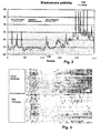
- Some of the observed activity is due to asynchronous cell division (e.g. 2 ⁇ 3 ⁇ 4 ⁇ 5 ⁇ 6 ⁇ 7 ⁇ 8) and fragmentation as opposed to synchronous cell divisions (e.g. 2 ⁇ 4, 4 ⁇ 8) observed for high quality embryos.
- the blastomere activity of 41 embryos is displayed as a pseudo-gel-image in Fig. 1 where motility peaks are indicated by dark bands and inactivity is white.
- Embryos that develop to blastocysts such as the left panel in Fig. 5 have uniformly distributed blastomere activity. Embryos that do not have uniformly distributed blastomere activity such as the right panel in Fig. 5 never develops into a blastocyst.
- the amount of cellular movement in different time intervals is a good indicator of embryo quality.
- a quality related parameter can be calculated from a ratio of average movement in different time-segments and/or a ratio of standard deviations in different time-segments Embryo selection procedures can be established based on the value of these parameters.
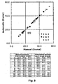
- Fig. 8 shows the excellent correspondence between automatic and manual determination of onset of cell division. Very early onset of the first cell division is an indication of high embryo quality. Very late onset of first (and subsequent cell divisions) indicates low quality embryos. However, for the majority of the embryos, the exact onset of the first cell division alone does not provide a clear indication of embryo quality as is shown in Fig. 8 below. While the average onset of cell divisions was delayed for the bad embryos, the large inherent standard deviation makes the absolute values a poor selection criteria except in extreme cases. (e.g. first division before 30 hours signifies a good embryo. First division after 35 hours signifies a bad embryo but the vast majority of the bovine embryos investigated have intermediate division times that are not easily interpreted.
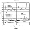
- a typical time series of blastomere activities consist of a few measurements every hour during incubation (e.g. approximately 150 data points for each embryo measured during the first 2 to 3 days which is the diagnostically interesting time window). Most statistical methods have difficulties with analysing data with such a high dimension. Thus, it is important to find robust methods for reducing the dimensions by extracting derived parameters. To achieve this, the blastomere activity was divided into three intervals: 0-32, 32-52 and 52-72 hours after image acquisition was started ( Fig. 9 ). Within each of these intervals three peaks were found using the following method: The first peak was the highest blastomere activity. The second peak was the highest activity value that was at least 3.5h before or after the first peak.
- Statistical models of embryo quality can be developed based on the above derived parameters. If each embryo has be evaluated according to the final development a number of different statistical methods exists for analysis the relation between the derived parameters and the final development. These methods includes: linear and non-linear models, Bayesians network, neural networks, hidden Markov models, nearest neighbours, principal component analysis and others.
- Fig. 12 below shows an example of a Principal Component Analysis (PCA) of the data.
- PCA Principal Component Analysis
- the statistical model can be evaluated and/or extended as new data are generated. To facilitate this it is important to find a robust data structure and set of derived parameters.
- parameter 39 baseline value of blastomere activity in the third time segment (76 to 96 hrs after fertilization) can to some extend to sort out abnormal and non-viable embryos. Based on this single parameter it is thus possible to automatically select embryos of good quality with with 72 % accuracy.
- a typical time series of blastomere activities consist of a few measurements every hour during incubation (e.g. approximately 150 data points for each embryo measured measured during the first 2 to 3 days which is the diagnostically interesting time window). Most statistical methods have difficulties with analysing data with such a high dimension. Thus, it is important to find robust methods for reducing the dimensions by extracting derived parameters. To achieve this, the blastomere activity was divided into three intervals: 0-32, 32-52 and 52-72 hours after image acquisition was started ( Figure 9 ). The three time intervals was selected to reflect three developmental stages for bovine embryos. Segment 1: initial cell divisions from 1-cell to 8-cells. Segment 2: resting stage with relatively little activity and movements. It is believed the embryonic genome is activated at this stage.
- Segment 3 Resuming cell division an developing into a morula. It is often impossible to count individual blastomeres at this stage, but the time-lapse images reveal that cell division has resumed.
- the first peak was the highest blastomere activity.
- the second peak was the highest activity value that was at least 3.5h before or after the first peak.
- the third peak was the highest activity that was at least 3.5h from both the first and second peak.
- Peak 1 Peak 1
- value 2 Peak 1 time 3 Peak 1 mean 4 Valley 1
- Peak shape which reflects the duration or synchrony of the mayor cell division event.
- I sharp peak in blastomere activity i.e. a fast synchronized cell division
- Peak mean divided by peak value will always be ⁇ 1, with a value close to one indicating a broad peak and a value close to 0 a very sharp peak.
- the parameter set of 21 parameters shown above is used for a fast analysis as it only include information that can be gained from the first segment i.e. 32 hours of incubation.
- the small set contain important information that can me used to classify embryos in viable and not viable. However, if data for the following two time intervals is available then the analysis can be repeated for the two following segments. We do not calculate the ratios (i.e. shape characteristics and interval between peaks) for the following segments but only the peaks and valleys (i.e. 15 parameters per segment) Finally the global average value, the global StDev and the global Minimum and maximum are included in the full parameter set of 59 parameters shown below:
- Statistical models of embryo quality can be developed based on the above derived parameters. If each embryo has be evaluated according to the final development a number of different statistical methods exists for analysis the relation between the derived parameters and the final development. These methods includes but are not limited to: linear and non-linear models, Bayesians network, neural networks, hidden Markov models, nearest neighbours, principal component analysis and others. Figure 11 below shows an example of a Principal Component Analysis (PCA) of the data.
- PCA Principal Component Analysis
- Example 7 An example of the use of a linear model is shown in Example 7.
- the statistical model can be evaluated and/or extended as new data are generated. To facilitate this it is important to find a robust data structure and set of derived parameters.
- parameter 39 baseline value of blastomere activity in the third time segment (76 to 96 hrs after fertilization) can to some extend to sort out abnormal and non-viable embryos. Based on this single parameter it is thus possible to automatically select embryos of good quality with with 72 % accuracy.
- Bovine immature cumulus-oocyte complexes were aspirated from slaughterhouse-derived ovaries, matured for 24h before fertilization for 22h. Cumulus cells were then removed and presumptive zygotes were transferred and cultured in synthetic oviduct fluid medium. Time-lapse images were acquired inside an incubator box fitted onto an inverted Nikon microscope stage mounted with a sensitive video camera.
- the fully automated image analysis procedure generated a quantitative measure of cell blastomere activity based on the observed movement between consecutive images in the time-lapse series.
- the correlation between blastomere activity and cell division was confirmed by comparing automated and manual analysis of the time-lapse image series.
- Pronounced peaks in blastomere activity were found to be associated with cell-divisions.
- the exact onset and duration of cell-divisions could be quantified based on position, shape and size of the recorded peaks.
- the blastomere activity pattern of a given embryo could thus be reduced to a set of key parameters corresponding to peak height, position and width for prominent peaks as well as similar parameters describing the blastomere activity level between peaks.
- a total of 55 parameters for each embryo was used in a simple linear model to classify the embryo as "viable” or "non-viable".
- the model was trained on a subset of the observed embryo patterns and evaluated on a different independent subset.
- the same time-lapse series of images was evaluated by a skilled embryologist attempting to predict whether the embryo would develop to an expanded blastocyst or not.
Landscapes
- Health & Medical Sciences (AREA)
- Life Sciences & Earth Sciences (AREA)
- Engineering & Computer Science (AREA)
- Chemical & Material Sciences (AREA)
- Organic Chemistry (AREA)
- Zoology (AREA)
- Wood Science & Technology (AREA)
- Bioinformatics & Cheminformatics (AREA)
- Biomedical Technology (AREA)
- Genetics & Genomics (AREA)
- Biotechnology (AREA)
- General Health & Medical Sciences (AREA)
- Biochemistry (AREA)
- Microbiology (AREA)
- General Engineering & Computer Science (AREA)
- Analytical Chemistry (AREA)
- Molecular Biology (AREA)
- Reproductive Health (AREA)
- Gynecology & Obstetrics (AREA)
- Cell Biology (AREA)
- Sustainable Development (AREA)
- Immunology (AREA)
- Proteomics, Peptides & Aminoacids (AREA)
- Hematology (AREA)
- Developmental Biology & Embryology (AREA)
- Urology & Nephrology (AREA)
- Physics & Mathematics (AREA)
- Pregnancy & Childbirth (AREA)
- Surgery (AREA)
- Computer Hardware Design (AREA)
- Food Science & Technology (AREA)
- General Physics & Mathematics (AREA)
- Pathology (AREA)
- Medicinal Chemistry (AREA)
- Animal Behavior & Ethology (AREA)
- Veterinary Medicine (AREA)
- Transplantation (AREA)
- Heart & Thoracic Surgery (AREA)
- Medical Informatics (AREA)
- Biophysics (AREA)
Priority Applications (1)
| Application Number | Priority Date | Filing Date | Title |
|---|---|---|---|
| EP10172399A EP2282210A1 (en) | 2006-06-16 | 2007-06-15 | Embryo quality assessment based on blastomere division and movement |
Applications Claiming Priority (5)
| Application Number | Priority Date | Filing Date | Title |
|---|---|---|---|
| US81411506P | 2006-06-16 | 2006-06-16 | |
| DKPA200600821 | 2006-06-16 | ||
| PCT/DK2006/000581 WO2007042044A1 (en) | 2005-10-14 | 2006-10-16 | Determination of a change in a cell population |
| DKPA200700571 | 2007-04-19 | ||
| PCT/DK2007/000291 WO2007144001A2 (en) | 2006-06-16 | 2007-06-15 | Embryo quality assessment based on blastomere division and movement |
Publications (2)
| Publication Number | Publication Date |
|---|---|
| EP2035548A2 EP2035548A2 (en) | 2009-03-18 |
| EP2035548B1 true EP2035548B1 (en) | 2010-08-11 |
Family
ID=43414894
Family Applications (2)
| Application Number | Title | Priority Date | Filing Date |
|---|---|---|---|
| EP07722668A Revoked EP2035548B1 (en) | 2006-06-16 | 2007-06-15 | Embryo quality assessment based on blastomere division and movement |
| EP10172399A Withdrawn EP2282210A1 (en) | 2006-06-16 | 2007-06-15 | Embryo quality assessment based on blastomere division and movement |
Family Applications After (1)
| Application Number | Title | Priority Date | Filing Date |
|---|---|---|---|
| EP10172399A Withdrawn EP2282210A1 (en) | 2006-06-16 | 2007-06-15 | Embryo quality assessment based on blastomere division and movement |
Country Status (8)
Cited By (1)
| Publication number | Priority date | Publication date | Assignee | Title |
|---|---|---|---|---|
| RU2441243C1 (ru) * | 2010-11-12 | 2012-01-27 | Федеральное государственное учреждение "Ивановский научно-исследовательский институт материнства и детства имени В.Н. Городкова" Министерства здравоохранения и социального развития Российской Федерации | Способ прогнозирования качества эмбриона при проведении эко у женщин с бесплодием |
Families Citing this family (40)
| Publication number | Priority date | Publication date | Assignee | Title |
|---|---|---|---|---|
| CN102254150A (zh) | 2005-10-14 | 2011-11-23 | 尤尼森斯繁殖技术公司 | 细胞群的变化的测定 |
| DE602008005746D1 (de) | 2007-06-29 | 2011-05-05 | Unisense Fertilitech As | Vorrichtung, system und verfahren zur beobachtung und/oder kultivierung mikroskopischer objekte |
| GB0719037D0 (en) | 2007-09-28 | 2007-11-07 | Vitrolife Sweden Ab | Sampling needle |
| JP2011526782A (ja) * | 2008-07-05 | 2011-10-20 | ウニセンス フェルティリテック アー/エス | 1対1識別システム |
| CN104293646A (zh) | 2009-08-22 | 2015-01-21 | 里兰斯坦福初级大学理事会 | 成像并评估胚胎、卵母细胞和干细胞 |
| GB201006046D0 (en) * | 2010-04-12 | 2010-05-26 | Ge Healthcare Uk Ltd | System and method for determining motion of a biological object |
| JP5807288B2 (ja) * | 2010-06-30 | 2015-11-10 | 大日本印刷株式会社 | 体外培養による胚の製造方法、並びに胚を選別するための方法、装置、及びシステム |
| JP5962828B2 (ja) * | 2010-06-30 | 2016-08-03 | 大日本印刷株式会社 | 体外培養による胚の製造方法、並びに胚を選別するための方法、装置、及びシステム |
| JP6007182B2 (ja) * | 2010-09-27 | 2016-10-12 | プロジェニー, インコーポレイテッド | 胚、卵母細胞および幹細胞の自動撮像および評価のための装置、手段およびシステム |
| GR1007617B (el) * | 2011-01-24 | 2012-06-20 | Ιδρυμα Τεχνολογιας Και Ερευνας - Ιτε, | Χρηση μη-γραμμικων απεικονιστικων τεχνικων για την αξιολογηση της ποιοτητας εμβρυων προεμφυτευτικου σταδιου και την προωθηση επιτυχους εγκυμοσυνης |
| ES2657927T3 (es) * | 2011-02-23 | 2018-03-07 | The Board Of Trustees Of The Leland Stanford Junior University | Métodos de detección de aneuploidía en embriones humanos |
| CN104232566B (zh) * | 2011-05-31 | 2016-11-16 | 尤尼森斯繁殖技术公司 | 基于卵裂球的卵裂和形态的胚胎质量评估 |
| EP2726864A1 (en) * | 2011-07-02 | 2014-05-07 | Unisense Fertilitech A/S | Adaptive embryo selection criteria optimized through iterative customization and collaboration |
| CN104508123B (zh) | 2012-05-31 | 2017-08-22 | 尤尼森斯繁殖技术公司 | 基于胚泡发育的胚胎质量评估 |
| CA2875038A1 (en) | 2012-05-31 | 2013-12-05 | Auxogyn, Inc. | In vitro embryo blastocyst prediction methods |
| WO2014001312A1 (en) * | 2012-06-25 | 2014-01-03 | Unisense Fertilitech A/S | Method and apparatus |
| CN110688985A (zh) * | 2012-08-30 | 2020-01-14 | 尤尼森斯繁殖技术公司 | 体外培养胚胎的自动化监控 |
| US10241108B2 (en) | 2013-02-01 | 2019-03-26 | Ares Trading S.A. | Abnormal syngamy phenotypes observed with time lapse imaging for early identification of embryos with lower development potential |
| ES2981412T3 (es) | 2013-02-28 | 2024-10-08 | Ares Trading Sa | Equipo, método y sistema para el seguimiento automatizado no invasivo de la actividad celular |
| JP2014193145A (ja) * | 2013-03-01 | 2014-10-09 | Naoki Yamashita | 胚盤胞培養液中のノルエピネフリン量によるヒト胚盤胞の評価方法 |
| US20160138104A1 (en) * | 2013-06-18 | 2016-05-19 | Universite De Montpellier | Methods for determining the quality of an embryo |
| EP3120297B1 (en) | 2014-03-20 | 2020-12-02 | Ares Trading S.A. | Quantitative measurement of human blastocyst and morula morphology developmental kinetics |
| CN104718298A (zh) * | 2014-06-19 | 2015-06-17 | 湖南光琇高新生命科技有限公司 | 一种根据分裂行为对体外受精治疗胚胎进行评估的等级分类方法 |
| US10510143B1 (en) | 2015-09-21 | 2019-12-17 | Ares Trading S.A. | Systems and methods for generating a mask for automated assessment of embryo quality |
| US11494578B1 (en) | 2015-09-21 | 2022-11-08 | Ares Trading S.A. | Systems and methods for automated assessment of embryo quality using image based features |
| JP2018014991A (ja) * | 2016-07-13 | 2018-02-01 | ソニー株式会社 | 情報処理装置、情報処理方法及び情報処理システム |
| JP2018022216A (ja) * | 2016-08-01 | 2018-02-08 | ソニー株式会社 | 情報処理装置、情報処理方法、及びプログラム |
| US11282201B2 (en) | 2016-11-02 | 2022-03-22 | Sony Corporation | Information processing device, information processing method and information processing system |
| JP6977293B2 (ja) * | 2017-03-31 | 2021-12-08 | ソニーグループ株式会社 | 情報処理装置、情報処理方法、プログラム及び観察システム |
| JP7100431B2 (ja) * | 2017-07-14 | 2022-07-13 | 株式会社ニコン | 受精卵判定装置、受精卵判定システム、及び受精卵判定プログラム |
| DE112018004960T5 (de) | 2017-10-26 | 2020-07-23 | Sony Corporation | Informationsverarbeitungseinrichtung, informationsverarbeitungsverfahren, programm und beobachtungssystem |
| JP6732722B2 (ja) * | 2017-12-11 | 2020-08-05 | 憲隆 福永 | 胚選抜システム |
| JP2020036580A (ja) * | 2018-06-20 | 2020-03-12 | Jcrファーマ株式会社 | 初期胚を選別するための解析ソフトウエア及び装置 |
| GB201810634D0 (en) * | 2018-06-28 | 2018-08-15 | Vitrolife As | Methods and apparatus for assessing embryo development |
| CN109214437A (zh) * | 2018-08-22 | 2019-01-15 | 湖南自兴智慧医疗科技有限公司 | 一种基于机器学习的ivf-et早孕胚胎发育预测系统 |
| JP7535572B2 (ja) * | 2019-09-06 | 2024-08-16 | ザ ブリガム アンド ウィメンズ ホスピタル インコーポレイテッド | 生殖補助手順に用いられる品質保証の評価指標の自動評価 |
| CN116323915A (zh) * | 2020-08-03 | 2023-06-23 | 爱慕基因系统有限公司 | 基于实时视频的胚胎评价 |
| JP2024054578A (ja) | 2022-10-05 | 2024-04-17 | 株式会社アステック | タイムラプス撮影機能付き培養装置および培養方法 |
| CN116091421A (zh) * | 2022-12-16 | 2023-05-09 | 中山大学 | 一种体外受精胚胎卵裂球图像自动分割及面积计算的方法 |
| CN116758539B (zh) * | 2023-08-17 | 2023-10-31 | 武汉互创联合科技有限公司 | 一种基于数据增强的胚胎图像卵裂球识别方法 |
Family Cites Families (28)
| Publication number | Priority date | Publication date | Assignee | Title |
|---|---|---|---|---|
| US3618734A (en) * | 1969-06-10 | 1971-11-09 | Res Foundation Of Children S H | Specimen incubator |
| DE2940446C2 (de) * | 1979-10-05 | 1982-07-08 | B. Braun Melsungen Ag, 3508 Melsungen | Züchtung von tierischen Zellen in Suspensions- und Monolayerkulturen in Fermentationsgefäßen |
| US4724543A (en) * | 1985-09-10 | 1988-02-09 | Beckman Research Institute, City Of Hope | Method and apparatus for automatic digital image analysis |
| JP2559760B2 (ja) * | 1987-08-31 | 1996-12-04 | 株式会社日立製作所 | 細胞搬送方法 |
| US5196168A (en) * | 1991-12-19 | 1993-03-23 | Eastman Kodak Company | Incubator with positioning device for slide elements |
| AU658079B2 (en) * | 1992-09-28 | 1995-03-30 | Becton Dickinson & Company | Cell culture insert |
| US5763279A (en) * | 1993-09-09 | 1998-06-09 | Synthecon, Inc. | Gas permeable bioreactor and method of use |
| US5968340A (en) * | 1997-04-07 | 1999-10-19 | Marine Biological Laboratory | Polarographic self-referencing probe and method for using |
| US6228636B1 (en) * | 1998-09-21 | 2001-05-08 | Matsushita Electric Industrial Co., Ltd. | Incubator |
| DE19903506C2 (de) * | 1999-01-29 | 2002-04-04 | Inst Chemo Biosensorik | Verfahren, Gefäß und Vorrichtung zur Überwachung der Stoffwechselaktivität von Zellkulturen in flüssigen Medien |
| US6391577B1 (en) * | 1999-03-03 | 2002-05-21 | Susan R. Mikkelsen | Rapid electrochemical assay for antibiotic and cytotoxic drug susceptibility in microorganisms |
| JP3442357B2 (ja) * | 2000-08-25 | 2003-09-02 | 株式会社日立製作所 | 両生類卵母細胞試料導入装置、両生類卵母細胞試料導入システム、両生類卵母細胞試料導入方法、両生類卵母細胞の製造方法、両生類卵母細胞及びそれを販売又は譲渡する方法、スクリーニング用のセンサーとして用いる方法、容器、及び解析方法 |
| US6434320B1 (en) * | 2000-10-13 | 2002-08-13 | Comtrak Technologies, Llc | Method of searching recorded digital video for areas of activity |
| US6555365B2 (en) * | 2000-12-07 | 2003-04-29 | Biocrystal, Ltd. | Microincubator comprising a cell culture apparatus and a transparent heater |
| US7015031B2 (en) * | 2002-01-24 | 2006-03-21 | Genx International, Inc. | Biological specimen-culturing system and method with onboard specimen development sensors |
| EP1486067B1 (en) * | 2002-02-13 | 2015-04-15 | Reify Corporation | Method and apparatus for acquisition, compression, and characterization of spatiotemporal signals |
| TWI249574B (en) * | 2002-11-19 | 2006-02-21 | Sanyo Electric Co | Preserving device |
| US20060201883A1 (en) * | 2003-02-19 | 2006-09-14 | Hofmeister William H | Polymer matrixes having nanoscale channels and uses |
| US7372985B2 (en) * | 2003-08-15 | 2008-05-13 | Massachusetts Institute Of Technology | Systems and methods for volumetric tissue scanning microscopy |
| US7239719B2 (en) * | 2003-08-22 | 2007-07-03 | Bbn Technologies Corp. | Automatic target detection and motion analysis from image data |
| EP1695080A1 (en) * | 2003-08-28 | 2006-08-30 | Coopersurgical, Inc. | Method for determining embryo quality |
| US20070015289A1 (en) * | 2003-09-19 | 2007-01-18 | Kao H P | Dispenser array spotting |
| JP3950972B2 (ja) * | 2003-11-14 | 2007-08-01 | 国立大学法人名古屋大学 | 生物試料の生物発光測定装置 |
| CA2549928A1 (en) * | 2003-12-19 | 2005-06-30 | University Of Waterloo | Cultured cell and method and apparatus for cell culture |
| US7336401B2 (en) * | 2003-12-19 | 2008-02-26 | Xerox Corporation | Systems and methods for estimating an image marking process using event mapping of scanned image attributes |
| JP4333426B2 (ja) * | 2004-03-19 | 2009-09-16 | ソニー株式会社 | 化合物半導体の製造方法、及び半導体装置の製造方法 |
| CN102254150A (zh) * | 2005-10-14 | 2011-11-23 | 尤尼森斯繁殖技术公司 | 细胞群的变化的测定 |
| US20080056952A1 (en) * | 2006-08-25 | 2008-03-06 | Angros Lee H | Analytic plates with markable portions and methods of use |
-
2007
- 2007-06-15 EP EP07722668A patent/EP2035548B1/en not_active Revoked
- 2007-06-15 DE DE602007008419T patent/DE602007008419D1/de active Active
- 2007-06-15 DK DK07722668.6T patent/DK2035548T3/da active
- 2007-06-15 CN CN201210428780.0A patent/CN103074410B/zh not_active Expired - Fee Related
- 2007-06-15 WO PCT/DK2007/000291 patent/WO2007144001A2/en active Application Filing
- 2007-06-15 CN CN200780027901.8A patent/CN101495619B/zh not_active Expired - Fee Related
- 2007-06-15 JP JP2009514641A patent/JP5731748B2/ja active Active
- 2007-06-15 EP EP10172399A patent/EP2282210A1/en not_active Withdrawn
- 2007-06-15 AT AT07722668T patent/ATE477499T1/de not_active IP Right Cessation
- 2007-06-15 US US12/304,905 patent/US20100041090A1/en not_active Abandoned
-
2012
- 2012-01-31 US US13/362,895 patent/US20120309043A1/en not_active Abandoned
- 2012-07-31 US US13/562,989 patent/US20130102837A1/en not_active Abandoned
- 2012-10-01 US US13/632,505 patent/US20130225917A1/en not_active Abandoned
-
2013
- 2013-07-05 JP JP2013141334A patent/JP5732110B2/ja active Active
Cited By (1)
| Publication number | Priority date | Publication date | Assignee | Title |
|---|---|---|---|---|
| RU2441243C1 (ru) * | 2010-11-12 | 2012-01-27 | Федеральное государственное учреждение "Ивановский научно-исследовательский институт материнства и детства имени В.Н. Городкова" Министерства здравоохранения и социального развития Российской Федерации | Способ прогнозирования качества эмбриона при проведении эко у женщин с бесплодием |
Also Published As
| Publication number | Publication date |
|---|---|
| US20120309043A1 (en) | 2012-12-06 |
| US20100041090A1 (en) | 2010-02-18 |
| DK2035548T3 (da) | 2010-11-22 |
| US20130102837A1 (en) | 2013-04-25 |
| ATE477499T1 (de) | 2010-08-15 |
| DE602007008419D1 (de) | 2010-09-23 |
| US20130225917A1 (en) | 2013-08-29 |
| WO2007144001A3 (en) | 2008-06-05 |
| WO2007144001A2 (en) | 2007-12-21 |
| JP5732110B2 (ja) | 2015-06-10 |
| CN101495619B (zh) | 2014-01-08 |
| JP5731748B2 (ja) | 2015-06-10 |
| CN101495619A (zh) | 2009-07-29 |
| CN103074410A (zh) | 2013-05-01 |
| CN103074410B (zh) | 2016-12-07 |
| EP2035548A2 (en) | 2009-03-18 |
| JP2013198503A (ja) | 2013-10-03 |
| EP2282210A1 (en) | 2011-02-09 |
| JP2009539387A (ja) | 2009-11-19 |
Similar Documents
| Publication | Publication Date | Title |
|---|---|---|
| EP2035548B1 (en) | Embryo quality assessment based on blastomere division and movement | |
| EP2279868A1 (en) | Printing-fluid container | |
| EP1949297B1 (en) | Determination of a change in a cell population | |
| CN102576027B (zh) | 成像并评估胚胎、卵母细胞和干细胞 | |
| Sugimura et al. | Selection of viable in vitro-fertilized bovine embryos using time-lapse monitoring in microwell culture dishes | |
| US20150346187A1 (en) | In Vitro Embryo Blastocyst Prediction Methods | |
| EP2951574B1 (en) | Measuring embryo development and implantation potential with timing and first cytokinesis phenotype parameters | |
| Arav et al. | Prediction of embryonic developmental competence by time-lapse observation and ‘shortest-half’analysis | |
| US10942170B2 (en) | Quantitative measurement of human blastocyst and morula morphology developmental kinetics | |
| CN103757087B (zh) | 基于卵裂球分裂和运动的胚胎质量评估 | |
| ES2353622T3 (es) | Evaluación de la calidad del embrión basada en la división y el movimiento del blastómero. | |
| HK1184829A (en) | Embryo quality assessment based on blastomere division and movement | |
| WO2016036697A1 (en) | Methods of detecting embryo mosaicism | |
| Fynewever et al. | Inhibited embryo development at specific cell-stage is dependent on type of nanoparticles used for in vitro tagging | |
| Hnida et al. | Morphometric analysis of human embryos | |
| GB2513908A (en) | Method |
Legal Events
| Date | Code | Title | Description |
|---|---|---|---|
| PUAI | Public reference made under article 153(3) epc to a published international application that has entered the european phase |
Free format text: ORIGINAL CODE: 0009012 |
|
| 17P | Request for examination filed |
Effective date: 20090116 |
|
| AK | Designated contracting states |
Kind code of ref document: A2 Designated state(s): AT BE BG CH CY CZ DE DK EE ES FI FR GB GR HU IE IS IT LI LT LU LV MC MT NL PL PT RO SE SI SK TR |
|
| AX | Request for extension of the european patent |
Extension state: AL BA HR MK RS |
|
| 17Q | First examination report despatched |
Effective date: 20090520 |
|
| DAX | Request for extension of the european patent (deleted) | ||
| GRAP | Despatch of communication of intention to grant a patent |
Free format text: ORIGINAL CODE: EPIDOSNIGR1 |
|
| RIC1 | Information provided on ipc code assigned before grant |
Ipc: G01N 33/68 20060101AFI20100104BHEP |
|
| GRAS | Grant fee paid |
Free format text: ORIGINAL CODE: EPIDOSNIGR3 |
|
| GRAA | (expected) grant |
Free format text: ORIGINAL CODE: 0009210 |
|
| AK | Designated contracting states |
Kind code of ref document: B1 Designated state(s): AT BE BG CH CY CZ DE DK EE ES FI FR GB GR HU IE IS IT LI LT LU LV MC MT NL PL PT RO SE SI SK TR |
|
| REG | Reference to a national code |
Ref country code: GB Ref legal event code: FG4D |
|
| REG | Reference to a national code |
Ref country code: CH Ref legal event code: EP |
|
| REG | Reference to a national code |
Ref country code: IE Ref legal event code: FG4D |
|
| REF | Corresponds to: |
Ref document number: 602007008419 Country of ref document: DE Date of ref document: 20100923 Kind code of ref document: P |
|
| REG | Reference to a national code |
Ref country code: DK Ref legal event code: T3 |
|
| REG | Reference to a national code |
Ref country code: NL Ref legal event code: T3 |
|
| REG | Reference to a national code |
Ref country code: SE Ref legal event code: TRGR Ref country code: CH Ref legal event code: PCOW Free format text: UNISENSE FERTILITECH A/S;TUEAGER 1;8200 AARHUS N (DK) Ref country code: CH Ref legal event code: NV Representative=s name: ANDRE ROLAND S.A. |
|
| LTIE | Lt: invalidation of european patent or patent extension |
Effective date: 20100811 |
|
| PG25 | Lapsed in a contracting state [announced via postgrant information from national office to epo] |
Ref country code: AT Free format text: LAPSE BECAUSE OF FAILURE TO SUBMIT A TRANSLATION OF THE DESCRIPTION OR TO PAY THE FEE WITHIN THE PRESCRIBED TIME-LIMIT Effective date: 20100811 Ref country code: LT Free format text: LAPSE BECAUSE OF FAILURE TO SUBMIT A TRANSLATION OF THE DESCRIPTION OR TO PAY THE FEE WITHIN THE PRESCRIBED TIME-LIMIT Effective date: 20100811 |
|
| REG | Reference to a national code |
Ref country code: FR Ref legal event code: CA |
|
| PG25 | Lapsed in a contracting state [announced via postgrant information from national office to epo] |
Ref country code: PT Free format text: LAPSE BECAUSE OF FAILURE TO SUBMIT A TRANSLATION OF THE DESCRIPTION OR TO PAY THE FEE WITHIN THE PRESCRIBED TIME-LIMIT Effective date: 20101213 Ref country code: SI Free format text: LAPSE BECAUSE OF FAILURE TO SUBMIT A TRANSLATION OF THE DESCRIPTION OR TO PAY THE FEE WITHIN THE PRESCRIBED TIME-LIMIT Effective date: 20100811 Ref country code: CY Free format text: LAPSE BECAUSE OF FAILURE TO SUBMIT A TRANSLATION OF THE DESCRIPTION OR TO PAY THE FEE WITHIN THE PRESCRIBED TIME-LIMIT Effective date: 20100811 Ref country code: BG Free format text: LAPSE BECAUSE OF FAILURE TO SUBMIT A TRANSLATION OF THE DESCRIPTION OR TO PAY THE FEE WITHIN THE PRESCRIBED TIME-LIMIT Effective date: 20101111 Ref country code: IS Free format text: LAPSE BECAUSE OF FAILURE TO SUBMIT A TRANSLATION OF THE DESCRIPTION OR TO PAY THE FEE WITHIN THE PRESCRIBED TIME-LIMIT Effective date: 20101211 Ref country code: PL Free format text: LAPSE BECAUSE OF FAILURE TO SUBMIT A TRANSLATION OF THE DESCRIPTION OR TO PAY THE FEE WITHIN THE PRESCRIBED TIME-LIMIT Effective date: 20100811 |
|
| REG | Reference to a national code |
Ref country code: ES Ref legal event code: FG2A Effective date: 20110221 |
|
| PG25 | Lapsed in a contracting state [announced via postgrant information from national office to epo] |
Ref country code: LV Free format text: LAPSE BECAUSE OF FAILURE TO SUBMIT A TRANSLATION OF THE DESCRIPTION OR TO PAY THE FEE WITHIN THE PRESCRIBED TIME-LIMIT Effective date: 20100811 Ref country code: GR Free format text: LAPSE BECAUSE OF FAILURE TO SUBMIT A TRANSLATION OF THE DESCRIPTION OR TO PAY THE FEE WITHIN THE PRESCRIBED TIME-LIMIT Effective date: 20101112 |
|
| PLBI | Opposition filed |
Free format text: ORIGINAL CODE: 0009260 |
|
| PG25 | Lapsed in a contracting state [announced via postgrant information from national office to epo] |
Ref country code: CZ Free format text: LAPSE BECAUSE OF FAILURE TO SUBMIT A TRANSLATION OF THE DESCRIPTION OR TO PAY THE FEE WITHIN THE PRESCRIBED TIME-LIMIT Effective date: 20100811 Ref country code: EE Free format text: LAPSE BECAUSE OF FAILURE TO SUBMIT A TRANSLATION OF THE DESCRIPTION OR TO PAY THE FEE WITHIN THE PRESCRIBED TIME-LIMIT Effective date: 20100811 Ref country code: RO Free format text: LAPSE BECAUSE OF FAILURE TO SUBMIT A TRANSLATION OF THE DESCRIPTION OR TO PAY THE FEE WITHIN THE PRESCRIBED TIME-LIMIT Effective date: 20100811 Ref country code: SK Free format text: LAPSE BECAUSE OF FAILURE TO SUBMIT A TRANSLATION OF THE DESCRIPTION OR TO PAY THE FEE WITHIN THE PRESCRIBED TIME-LIMIT Effective date: 20100811 |
|
| PLAX | Notice of opposition and request to file observation + time limit sent |
Free format text: ORIGINAL CODE: EPIDOSNOBS2 |
|
| 26 | Opposition filed |
Opponent name: OLSWANG LLP Effective date: 20110510 |
|
| REG | Reference to a national code |
Ref country code: DE Ref legal event code: R026 Ref document number: 602007008419 Country of ref document: DE Effective date: 20110510 |
|
| PLAF | Information modified related to communication of a notice of opposition and request to file observations + time limit |
Free format text: ORIGINAL CODE: EPIDOSCOBS2 |
|
| PG25 | Lapsed in a contracting state [announced via postgrant information from national office to epo] |
Ref country code: MT Free format text: LAPSE BECAUSE OF FAILURE TO SUBMIT A TRANSLATION OF THE DESCRIPTION OR TO PAY THE FEE WITHIN THE PRESCRIBED TIME-LIMIT Effective date: 20100811 |
|
| PLBB | Reply of patent proprietor to notice(s) of opposition received |
Free format text: ORIGINAL CODE: EPIDOSNOBS3 |
|
| REG | Reference to a national code |
Ref country code: CH Ref legal event code: PCAR Free format text: ANDRE ROLAND S.A.;CASE POSTALE 5107;1002 LAUSANNE (CH) |
|
| RDAF | Communication despatched that patent is revoked |
Free format text: ORIGINAL CODE: EPIDOSNREV1 |
|
| APBM | Appeal reference recorded |
Free format text: ORIGINAL CODE: EPIDOSNREFNO |
|
| APBP | Date of receipt of notice of appeal recorded |
Free format text: ORIGINAL CODE: EPIDOSNNOA2O |
|
| APAH | Appeal reference modified |
Free format text: ORIGINAL CODE: EPIDOSCREFNO |
|
| PG25 | Lapsed in a contracting state [announced via postgrant information from national office to epo] |
Ref country code: MC Free format text: LAPSE BECAUSE OF NON-PAYMENT OF DUE FEES Effective date: 20110630 |
|
| PG25 | Lapsed in a contracting state [announced via postgrant information from national office to epo] |
Ref country code: LU Free format text: LAPSE BECAUSE OF NON-PAYMENT OF DUE FEES Effective date: 20110615 |
|
| APBQ | Date of receipt of statement of grounds of appeal recorded |
Free format text: ORIGINAL CODE: EPIDOSNNOA3O |
|
| PG25 | Lapsed in a contracting state [announced via postgrant information from national office to epo] |
Ref country code: HU Free format text: LAPSE BECAUSE OF FAILURE TO SUBMIT A TRANSLATION OF THE DESCRIPTION OR TO PAY THE FEE WITHIN THE PRESCRIBED TIME-LIMIT Effective date: 20100811 |
|
| PGFP | Annual fee paid to national office [announced via postgrant information from national office to epo] |
Ref country code: IE Payment date: 20140610 Year of fee payment: 8 Ref country code: GB Payment date: 20140617 Year of fee payment: 8 |
|
| PGFP | Annual fee paid to national office [announced via postgrant information from national office to epo] |
Ref country code: CH Payment date: 20140612 Year of fee payment: 8 Ref country code: DE Payment date: 20140611 Year of fee payment: 8 Ref country code: SE Payment date: 20140611 Year of fee payment: 8 Ref country code: FI Payment date: 20140610 Year of fee payment: 8 Ref country code: IT Payment date: 20140612 Year of fee payment: 8 Ref country code: ES Payment date: 20140513 Year of fee payment: 8 Ref country code: TR Payment date: 20140529 Year of fee payment: 8 |
|
| PGFP | Annual fee paid to national office [announced via postgrant information from national office to epo] |
Ref country code: DK Payment date: 20140610 Year of fee payment: 8 |
|
| PGFP | Annual fee paid to national office [announced via postgrant information from national office to epo] |
Ref country code: BE Payment date: 20140611 Year of fee payment: 8 Ref country code: NL Payment date: 20140610 Year of fee payment: 8 |
|
| PGFP | Annual fee paid to national office [announced via postgrant information from national office to epo] |
Ref country code: FR Payment date: 20140609 Year of fee payment: 8 |
|
| REG | Reference to a national code |
Ref country code: DE Ref legal event code: R119 Ref document number: 602007008419 Country of ref document: DE |
|
| REG | Reference to a national code |
Ref country code: DK Ref legal event code: EBP Effective date: 20150630 |
|
| PG25 | Lapsed in a contracting state [announced via postgrant information from national office to epo] |
Ref country code: FI Free format text: LAPSE BECAUSE OF NON-PAYMENT OF DUE FEES Effective date: 20150615 Ref country code: IT Free format text: LAPSE BECAUSE OF NON-PAYMENT OF DUE FEES Effective date: 20150615 |
|
| REG | Reference to a national code |
Ref country code: CH Ref legal event code: PL |
|
| REG | Reference to a national code |
Ref country code: SE Ref legal event code: EUG |
|
| GBPC | Gb: european patent ceased through non-payment of renewal fee |
Effective date: 20150615 |
|
| PG25 | Lapsed in a contracting state [announced via postgrant information from national office to epo] |
Ref country code: SE Free format text: LAPSE BECAUSE OF NON-PAYMENT OF DUE FEES Effective date: 20150616 |
|
| REG | Reference to a national code |
Ref country code: NL Ref legal event code: MM Effective date: 20150701 |
|
| REG | Reference to a national code |
Ref country code: IE Ref legal event code: MM4A |
|
| REG | Reference to a national code |
Ref country code: FR Ref legal event code: ST Effective date: 20160229 |
|
| PG25 | Lapsed in a contracting state [announced via postgrant information from national office to epo] |
Ref country code: NL Free format text: LAPSE BECAUSE OF NON-PAYMENT OF DUE FEES Effective date: 20150701 Ref country code: GB Free format text: LAPSE BECAUSE OF NON-PAYMENT OF DUE FEES Effective date: 20150615 Ref country code: DE Free format text: LAPSE BECAUSE OF NON-PAYMENT OF DUE FEES Effective date: 20160101 Ref country code: LI Free format text: LAPSE BECAUSE OF NON-PAYMENT OF DUE FEES Effective date: 20150630 Ref country code: CH Free format text: LAPSE BECAUSE OF NON-PAYMENT OF DUE FEES Effective date: 20150630 Ref country code: IE Free format text: LAPSE BECAUSE OF NON-PAYMENT OF DUE FEES Effective date: 20150615 |
|
| PG25 | Lapsed in a contracting state [announced via postgrant information from national office to epo] |
Ref country code: FR Free format text: LAPSE BECAUSE OF NON-PAYMENT OF DUE FEES Effective date: 20150630 |
|
| PG25 | Lapsed in a contracting state [announced via postgrant information from national office to epo] |
Ref country code: DK Free format text: LAPSE BECAUSE OF NON-PAYMENT OF DUE FEES Effective date: 20150630 |
|
| PG25 | Lapsed in a contracting state [announced via postgrant information from national office to epo] |
Ref country code: ES Free format text: LAPSE BECAUSE OF NON-PAYMENT OF DUE FEES Effective date: 20150616 |
|
| PG25 | Lapsed in a contracting state [announced via postgrant information from national office to epo] |
Ref country code: BE Free format text: LAPSE BECAUSE OF NON-PAYMENT OF DUE FEES Effective date: 20150630 |
|
| APAH | Appeal reference modified |
Free format text: ORIGINAL CODE: EPIDOSCREFNO |
|
| PG25 | Lapsed in a contracting state [announced via postgrant information from national office to epo] |
Ref country code: TR Free format text: LAPSE BECAUSE OF NON-PAYMENT OF DUE FEES Effective date: 20150615 |
|
| APBY | Invitation to file observations in appeal sent |
Free format text: ORIGINAL CODE: EPIDOSNOBA2O |
|
| REG | Reference to a national code |
Ref country code: DE Ref legal event code: R064 Ref document number: 602007008419 Country of ref document: DE Ref country code: DE Ref legal event code: R103 Ref document number: 602007008419 Country of ref document: DE |
|
| APBU | Appeal procedure closed |
Free format text: ORIGINAL CODE: EPIDOSNNOA9O |
|
| RDAG | Patent revoked |
Free format text: ORIGINAL CODE: 0009271 |
|
| STAA | Information on the status of an ep patent application or granted ep patent |
Free format text: STATUS: PATENT REVOKED |
|
| 27W | Patent revoked |
Effective date: 20171023 |
|
| REG | Reference to a national code |
Ref country code: SE Ref legal event code: ECNC |