EP1864112B1 - Device and method for hilbert phase imaging - Google Patents
Device and method for hilbert phase imaging Download PDFInfo
- Publication number
- EP1864112B1 EP1864112B1 EP20060739540 EP06739540A EP1864112B1 EP 1864112 B1 EP1864112 B1 EP 1864112B1 EP 20060739540 EP20060739540 EP 20060739540 EP 06739540 A EP06739540 A EP 06739540A EP 1864112 B1 EP1864112 B1 EP 1864112B1
- Authority
- EP
- European Patent Office
- Prior art keywords
- image
- phase
- optical
- path
- sample
- Prior art date
- Legal status (The legal status is an assumption and is not a legal conclusion. Google has not performed a legal analysis and makes no representation as to the accuracy of the status listed.)
- Expired - Lifetime
Links
Images




Classifications
-
- H—ELECTRICITY
- H10—SEMICONDUCTOR DEVICES; ELECTRIC SOLID-STATE DEVICES NOT OTHERWISE PROVIDED FOR
- H10F—INORGANIC SEMICONDUCTOR DEVICES SENSITIVE TO INFRARED RADIATION, LIGHT, ELECTROMAGNETIC RADIATION OF SHORTER WAVELENGTH OR CORPUSCULAR RADIATION
- H10F39/00—Integrated devices, or assemblies of multiple devices, comprising at least one element covered by group H10F30/00, e.g. radiation detectors comprising photodiode arrays
- H10F39/80—Constructional details of image sensors
-
- G—PHYSICS
- G01—MEASURING; TESTING
- G01N—INVESTIGATING OR ANALYSING MATERIALS BY DETERMINING THEIR CHEMICAL OR PHYSICAL PROPERTIES
- G01N15/00—Investigating characteristics of particles; Investigating permeability, pore-volume or surface-area of porous materials
- G01N15/10—Investigating individual particles
-
- G—PHYSICS
- G01—MEASURING; TESTING
- G01N—INVESTIGATING OR ANALYSING MATERIALS BY DETERMINING THEIR CHEMICAL OR PHYSICAL PROPERTIES
- G01N15/00—Investigating characteristics of particles; Investigating permeability, pore-volume or surface-area of porous materials
- G01N15/10—Investigating individual particles
- G01N15/14—Optical investigation techniques, e.g. flow cytometry
- G01N15/1429—Signal processing
- G01N15/1433—Signal processing using image recognition
-
- G—PHYSICS
- G01—MEASURING; TESTING
- G01N—INVESTIGATING OR ANALYSING MATERIALS BY DETERMINING THEIR CHEMICAL OR PHYSICAL PROPERTIES
- G01N21/00—Investigating or analysing materials by the use of optical means, i.e. using sub-millimetre waves, infrared, visible or ultraviolet light
- G01N21/17—Systems in which incident light is modified in accordance with the properties of the material investigated
- G01N21/41—Refractivity; Phase-affecting properties, e.g. optical path length
- G01N21/45—Refractivity; Phase-affecting properties, e.g. optical path length using interferometric methods; using Schlieren methods
- G01N21/453—Holographic interferometry
-
- G—PHYSICS
- G01—MEASURING; TESTING
- G01N—INVESTIGATING OR ANALYSING MATERIALS BY DETERMINING THEIR CHEMICAL OR PHYSICAL PROPERTIES
- G01N33/00—Investigating or analysing materials by specific methods not covered by groups G01N1/00 - G01N31/00
- G01N33/48—Biological material, e.g. blood, urine; Haemocytometers
- G01N33/483—Physical analysis of biological material
- G01N33/487—Physical analysis of biological material of liquid biological material
- G01N33/49—Blood
-
- G—PHYSICS
- G06—COMPUTING OR CALCULATING; COUNTING
- G06V—IMAGE OR VIDEO RECOGNITION OR UNDERSTANDING
- G06V20/00—Scenes; Scene-specific elements
- G06V20/60—Type of objects
- G06V20/69—Microscopic objects, e.g. biological cells or cellular parts
- G06V20/693—Acquisition
-
- G—PHYSICS
- G01—MEASURING; TESTING
- G01N—INVESTIGATING OR ANALYSING MATERIALS BY DETERMINING THEIR CHEMICAL OR PHYSICAL PROPERTIES
- G01N15/00—Investigating characteristics of particles; Investigating permeability, pore-volume or surface-area of porous materials
- G01N15/01—Investigating characteristics of particles; Investigating permeability, pore-volume or surface-area of porous materials specially adapted for biological cells, e.g. blood cells
- G01N2015/012—Red blood cells
-
- G—PHYSICS
- G01—MEASURING; TESTING
- G01N—INVESTIGATING OR ANALYSING MATERIALS BY DETERMINING THEIR CHEMICAL OR PHYSICAL PROPERTIES
- G01N15/00—Investigating characteristics of particles; Investigating permeability, pore-volume or surface-area of porous materials
- G01N15/01—Investigating characteristics of particles; Investigating permeability, pore-volume or surface-area of porous materials specially adapted for biological cells, e.g. blood cells
- G01N2015/016—White blood cells
-
- G—PHYSICS
- G01—MEASURING; TESTING
- G01N—INVESTIGATING OR ANALYSING MATERIALS BY DETERMINING THEIR CHEMICAL OR PHYSICAL PROPERTIES
- G01N15/00—Investigating characteristics of particles; Investigating permeability, pore-volume or surface-area of porous materials
- G01N15/10—Investigating individual particles
- G01N15/14—Optical investigation techniques, e.g. flow cytometry
- G01N15/1434—Optical arrangements
- G01N2015/1447—Spatial selection
- G01N2015/145—Spatial selection by pattern of light, e.g. fringe pattern
-
- G—PHYSICS
- G01—MEASURING; TESTING
- G01N—INVESTIGATING OR ANALYSING MATERIALS BY DETERMINING THEIR CHEMICAL OR PHYSICAL PROPERTIES
- G01N15/00—Investigating characteristics of particles; Investigating permeability, pore-volume or surface-area of porous materials
- G01N15/10—Investigating individual particles
- G01N15/14—Optical investigation techniques, e.g. flow cytometry
- G01N15/1434—Optical arrangements
- G01N2015/1454—Optical arrangements using phase shift or interference, e.g. for improving contrast
-
- G—PHYSICS
- G01—MEASURING; TESTING
- G01N—INVESTIGATING OR ANALYSING MATERIALS BY DETERMINING THEIR CHEMICAL OR PHYSICAL PROPERTIES
- G01N15/00—Investigating characteristics of particles; Investigating permeability, pore-volume or surface-area of porous materials
- G01N15/10—Investigating individual particles
- G01N15/14—Optical investigation techniques, e.g. flow cytometry
- G01N2015/1497—Particle shape
Definitions
- Optical microscopy has been a commonly used method of investigation in medicine and biology. Numerous biological samples, including live cells, are quite transparent under visible light illumination and behave essentially as phase objects. Techniques such as phase contrast and Nomarski microscopy provide contrast of nearly invisible samples by transforming the phase information into intensity distribution and thus reveal structural details of biological systems. However, the information obtained with these techniques about the phase shift associated with the illuminating field is only qualitative. Retrieving quantitative phase information from transparent objects with high accuracy and low noise allows for measurements in the biological investigation of structure and dynamics. Both interferometric and non-interferometric techniques have been proposed for quantitative phase imaging of biological samples. Also Fourier phase microscopy (FPM) has been developed as an extremely low-noise phase imaging method. Due to the sub-nanometer phase stability over extended periods of time, FPM is suitable for investigating dynamics in biological systems on time scales from seconds to a cell lifetime.
- FPM Fourier phase microscopy
- US 5, 194, 918 discloses providing images of surfaces of samples with a correlation microscope by transforming interference signals using a Hilbert transform.
- the microscope comprises a light source 1, and a CCD camera 23 which detects the interfering signals from the reference mirror (signal B) and that from the object (signal A) i.e. two paths.
- Images of the interfering signals are acquired by a frame grabber and the digital signals from the CCD detector are processed by Hilbert transform which introduces a phase shift of "pi./2" to an input signal.
- a Hilbert filter is used a fast alternate algorithm for extracting the correlation envelope from the interference fringes to determine phase data.
- the complex analytic signal formalism of time-varying fields has found broad applications in optics.
- the Hilbert transform relationship between the real and imaginary part of a complex analytic signal has been used to retrieve phase shifts from single temporal interferograms.
- the present invention relates to systems and methods for quantitative phase imaging, referred to as Hilbert phase microscopy (HPM), which allows the retrieval of a full field phase image from a single spatial interferogram.
- HPM Hilbert phase microscopy
- HPM single-shot phase imaging is limited in frame acquisition rate only by the recording device such as an imaging sensor.
- imaging sensors include digital imaging detectors such as charge coupled devices (CCD) or a CMOS imaging array. This contrasts with phase-shifting techniques, in which multiple recordings are required for retrieving a single phase image.
- HPM provides for phase unwrapping, which enables the study of phase objects much larger than the wavelength of light.
- the imaging device preferably has at least 200,000 pixels that can collect at least 10 frames per second and preferably over 100 frames per second.
- the light from a single light source is split along a reference path and a sample path.
- the light along the sample path is directed through the sample or object being measured and the light along the reference path is modulated by a modulating element such that when the light from the sample is combined with the modulated reference light that an interference pattern is produced that is detected by the imaging sensor.
- the modulating element can be a rotating mirror or a movable lens, for example.
- Preferred embodiments of the invention can include fiber optics to couple light onto the object such as tissue to be imaged. Lasers or other highly coherent light sources of different wavelengths can be used.
- a computer or other data processor or image processor can be connected to the output of the imaging device for processing of the image data.
- Preferred embodiments of the invention can include configurations of Hilbert phase imaging according to the invention in which the optical geometry is set up for transmissive or reflective imaging.
- an inverted microscope geometry can be used with a beam splitter used to combine the reference and sample images.
- a reflective measurement can be performed by attaching a reflective material, such as polystyrene beads to a cell membrane. Coherent light can then be reflected off this material to obtain an interferogram. This can be used to measure mechanical properties of the membrane such as the shear modulus or the bending modulus.
- the procedures described herein can used in-vitro on human or mammalian tissue or fluid or in-vivo on the human eye of other tissues, for example.
- the invention provides for non-biological applications as well as biological applications; for instance the invention can provide for studying the phase profile of an optical fiber and/or other transparent or semi-transparent objects or materials including crystalline structures.
- FIG. 1 A preferred embodiment of the invention is illustrated in FIG. 1 .
- a HeNe laser is used as a light source 10 for an imaging Mach-Zender interferometer 20.
- a first beam splitter 12 splits the beam from the light source 10 to form two arms of the interferometer, the arms comprising a reference beam 14 and a sample beam 16, respectively.
- a mirror 18 directs the sample beam 16 onto a sample or object 25.
- a second mirror 30 directs the reference field onto a second beam splitter 32.
- the orientation of the reference field 40 is adjustable, for example, by rotatable movement of mirror 30 in order to tilt reference field 40.
- An image sensor 42 such as a CCD, can be positioned in the common Fourier plane of the lenses 26, 28 where the exact (magnified) replica of the sample field 44 is formed.
- the reference field 40 which is directed onto the CCD image sensor 42 by the beam splitter 32, is slightly tilted with respect to the sample beam 44 in order to create a uniform fringe structure oriented at 45° with respect to the x and y axes of the CCD image sensor 42.
- the CCD used in this embodiment (C770, Hamamatsu Photonics) has an acquisition rate of 291 frames/s at the full resolution of 480x640 pixels. Higher resolutions and acquisition rates can also be used.
- Image data is sent from the sensor 42 to the processor or computer 120 for analysis and display.
- I R and I s are, respectively, the reference and sample irradiance distributions
- q is the spatial frequency of the fringes
- ⁇ is the spatially varying phase associated with the object 25, the quantity of interest.
- Eq. (1) is analogous to describing the temporal interference in Michelson and other interferometers, in which q corresponds to the frequency shift introduced by an acousto-optic modulator or a moving mirror.
- z (x) exhibits rapid phase modulation, with frequency q, and thus ⁇ is strongly wrapped.
- q is higher than the spatial frequency content of the object, the unwrapping procedure works efficiently.
- the invention provides for an apparatus and method for retrieving the phase profile of an optical fiber having a fiber core with a diameter of 100 ⁇ m and a refractive index of 1.457, while the cladding has an outer diameter of 110 ⁇ m and a refractive index of 1.452.
- the fiber is immersed in glycerol to better mimic a phase object, in this example.
- the transmission intensity image of this sample ( FIG. 2a ) shows low contrast, which is an indication of the transparency of the sample.
- FIGS. 2b -d represent intermediate steps in the phase reconstruction sequence and correspond to the rectangular area shown in FIG. 2a .
- This region encompasses the glycerol/cladding and cladding/core interfaces.
- the interferogram recorded by the CCD ( FIG. 2b ) is Fourier transformed and high-pass filtered, such that the sinusoidal signal is obtained ( FIG. 2c ).
- the 2D Fourier transform is computed and the negative spatial frequencies are suppressed.
- a complex 2D signal is obtained that uniquely provides information about the phase of the object, as described in Eq. 2.
- the strongly wrapped and unwrapped phase images, respectively, are shown in FIGS. 2d and 2e .
- the quantitative phase image of the optical fiber is obtained by subtracting the linear phase and is depicted in FIG.
- FIG. 2f while a cross-section is shown in FIG. 2g .
- the continuous line represents the modeled fit, with the refractive index of glycerol as the variable parameter.
- a preferred embodiment of the invention uses HPM for biological measurements, such as, for example, quantifying parameters for phase-images of tissue or body fluids such as red blood cells from whole blood smears.
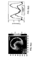
- FIG. 2h shows an example of such an image, in which the individual cells and agglomeration of cells are easily identifiable. Red blood cells lack nuclei and major organelles can be modeled as optically homogeneous objects.
- the phase information from the HPM images can be transformed into thickness information, which directly provides parameters such as cell shape and volume.
- the data were recorded in 10.3ms and the sample was prepared by sandwiching a droplet of whole blood between two cover slips.
- HPM can provide quantitative phase images in transparent samples.
- this method can measure phase objects with phase profiles much higher than the wavelength of the illuminating light. This important feature is due to the high spatial modulation imposed on the image, which creates well defined wrapping points on the phase image, thus facilitating the unwrapping procedure.
- the ability of HPM to obtain quantitative phase images from single-shot measurements allows, therefore, monitoring fast dynamic processes in transparent or transmissive systems.
- a further preferred embodiment of the invention provides for studying rapid processes in transparent media, such as, for example, analyzing the evaporation of micron-size liquid droplets.
- FIG. 3a shows the FPM image of such water droplets sprayed onto a microscope slide. The z-axis information indicates that the thickness of these droplets is significantly smaller than their transverse size.
- a series of 333 phase images were recorded at time intervals of 10.3ms. Since each phase image is obtained from one CCD recording, it is not necessary to eliminate noise between the two interferometer arms, which provides a significant advantage over phase-shifting techniques. The noise between successive frames does not obscure the phase images, which can be conveniently displayed by referencing each image to a fixed point in the field of view.
- FIG. 4 an example of the disclosure uses the principle of Hilbert phase microscopy in an inverted geometry to provide a high-speed and high-sensitivity quantitative phase microscope 60.
- the inverted geometry is particularly suitable for live cell investigation.
- the potential of the method for quantitative biological microscopy has been demonstrated by quantifying red blood cell shape and fluctuations with nanometer path-length sensitivity at the millisecond time scale.
- the CCD used (C7770, Hamamatsu Photonics) has an acquisition rate of 291 frames/s at the full resolution of 640 x 480 pixels and the CCD 100 can be connected to and controlled by computer 120.
- the focal distance, f between the focus of the reference arm objective 3 and the tube lens 90 is 250mm.
- the new HPM microscope is particularly suited for the quantitative investigation of live cells.
- RBCs red blood cells
- FIG. 5 shows a quantitative phase image of live blood cells; both isolated and agglomerated erythrocytes are easily identifiable.
- a white blood cell (WBC) is also present in the field of view.
- WBC white blood cell
- the phase information associated with the RBCs can be easily translated into a nanometer scale image of the cell topography.
- the volume of individual cells can be evaluated; in FIG. 5 , the measured volumes (units of in femtoliters) are displayed below individual red blood cells.
- each phase image was referenced to the average value across the area in the field of view containing no cells, denoted by R.
- R the average value across the area in the field of view containing no cells.
- sets of 1000 images were recorded, acquired at 10.3ms each and noise analysis was performed on a second empty region in the field of view.
- the spatial standard deviation, ⁇ s of the pathlength fluctuation across this area (indicated in FIG. 5a as O) has a certain fluctuation in time and is characterized in turn by a temporal average ⁇ s >.
- the time-dependence of ⁇ s is plotted in FIG.
- Biological structures such as living cells are predominantly transparent under bright field illumination.
- Phase contrast (PC) and differential interference contrast (DIC) microscopy have been used extensively to infer morphometric features of cells without the need for exogenous contrast agents.
- PC Phase contrast
- DIC differential interference contrast
- These techniques transfer the information encoded in the phase of the imaging field into the intensity distribution of the final image.
- the optical phase shift through a given sample can be regarded as a powerful endogenous contrast agent, as it contains information about both the thickness and refractive index of the sample.
- mature erythrocytes red blood cells, or RBCs
- RBCs represent a very particular type of structure in that they lack nuclei and major organelles.
Landscapes
- Health & Medical Sciences (AREA)
- Life Sciences & Earth Sciences (AREA)
- Engineering & Computer Science (AREA)
- Chemical & Material Sciences (AREA)
- Physics & Mathematics (AREA)
- General Health & Medical Sciences (AREA)
- General Physics & Mathematics (AREA)
- Biomedical Technology (AREA)
- Immunology (AREA)
- Pathology (AREA)
- Biochemistry (AREA)
- Analytical Chemistry (AREA)
- Hematology (AREA)
- Molecular Biology (AREA)
- Dispersion Chemistry (AREA)
- Food Science & Technology (AREA)
- Biophysics (AREA)
- Urology & Nephrology (AREA)
- Ecology (AREA)
- Medicinal Chemistry (AREA)
- Signal Processing (AREA)
- Theoretical Computer Science (AREA)
- Multimedia (AREA)
- Investigating Or Analysing Materials By Optical Means (AREA)
- Length Measuring Devices By Optical Means (AREA)
- Microscoopes, Condenser (AREA)
- Instruments For Measurement Of Length By Optical Means (AREA)
- Computer Vision & Pattern Recognition (AREA)
- Image Processing (AREA)
- Image Analysis (AREA)
- Investigating Or Analysing Biological Materials (AREA)
Priority Applications (1)
| Application Number | Priority Date | Filing Date | Title |
|---|---|---|---|
| EP10184686A EP2375239A3 (en) | 2005-03-25 | 2006-03-24 | System and method for Hilbert phase imaging |
Applications Claiming Priority (2)
| Application Number | Priority Date | Filing Date | Title |
|---|---|---|---|
| US66511805P | 2005-03-25 | 2005-03-25 | |
| PCT/US2006/010821 WO2006104899A2 (en) | 2005-03-25 | 2006-03-24 | System and method for hilbert phase imaging |
Related Child Applications (1)
| Application Number | Title | Priority Date | Filing Date |
|---|---|---|---|
| EP10184686A Division-Into EP2375239A3 (en) | 2005-03-25 | 2006-03-24 | System and method for Hilbert phase imaging |
Publications (2)
| Publication Number | Publication Date |
|---|---|
| EP1864112A2 EP1864112A2 (en) | 2007-12-12 |
| EP1864112B1 true EP1864112B1 (en) | 2015-05-06 |
Family
ID=36888741
Family Applications (2)
| Application Number | Title | Priority Date | Filing Date |
|---|---|---|---|
| EP20060739540 Expired - Lifetime EP1864112B1 (en) | 2005-03-25 | 2006-03-24 | Device and method for hilbert phase imaging |
| EP10184686A Withdrawn EP2375239A3 (en) | 2005-03-25 | 2006-03-24 | System and method for Hilbert phase imaging |
Family Applications After (1)
| Application Number | Title | Priority Date | Filing Date |
|---|---|---|---|
| EP10184686A Withdrawn EP2375239A3 (en) | 2005-03-25 | 2006-03-24 | System and method for Hilbert phase imaging |
Country Status (5)
| Country | Link |
|---|---|
| US (2) | US8772693B2 (https=) |
| EP (2) | EP1864112B1 (https=) |
| JP (3) | JP2008534929A (https=) |
| CN (2) | CN102539382B (https=) |
| WO (1) | WO2006104899A2 (https=) |
Families Citing this family (47)
| Publication number | Priority date | Publication date | Assignee | Title |
|---|---|---|---|---|
| US11243494B2 (en) | 2002-07-31 | 2022-02-08 | Abs Global, Inc. | Multiple laminar flow-based particle and cellular separation with laser steering |
| EP1864112B1 (en) | 2005-03-25 | 2015-05-06 | Massachusetts Institute of Technology | Device and method for hilbert phase imaging |
| FR2902877B1 (fr) * | 2006-06-22 | 2008-09-12 | Centre Nat Rech Scient | Procede de caracterisation de l'anisotropie d'un milieu diffusant et dispositif pour la mise en oeuvre d'un tel procede |
| US8848199B2 (en) * | 2007-07-10 | 2014-09-30 | Massachusetts Institute Of Technology | Tomographic phase microscopy |
| US7936913B2 (en) * | 2007-08-07 | 2011-05-03 | Nextslide Imaging Llc | Network image review in clinical hematology |
| US8026102B2 (en) * | 2009-01-21 | 2011-09-27 | Blaze Medical Devices, LLC | Apparatus and method to characterize blood and red blood cells via erythrocyte membrane fragility quantification |
| US8599383B2 (en) | 2009-05-06 | 2013-12-03 | The Regents Of The University Of California | Optical cytometry |
| US20120225475A1 (en) * | 2010-11-16 | 2012-09-06 | 1087 Systems, Inc. | Cytometry system with quantum cascade laser source, acoustic detector, and micro-fluidic cell handling system configured for inspection of individual cells |
| US10908066B2 (en) | 2010-11-16 | 2021-02-02 | 1087 Systems, Inc. | Use of vibrational spectroscopy for microfluidic liquid measurement |
| EP2668465A1 (en) * | 2011-01-25 | 2013-12-04 | Massachusetts Institute Of Technology | Single-shot full-field reflection phase microscopy |
| WO2012123850A1 (en) * | 2011-03-15 | 2012-09-20 | Koninklijke Philips Electronics N.V. | Medical imaging device for providing an image representation supporting in positioning an intervention device |
| EP2739937B1 (en) | 2011-08-02 | 2023-03-01 | The Regents of The University of California | Rapid, massively parallel single-cell drug response measurements via live cell interferometry |
| WO2013086527A1 (en) * | 2011-12-09 | 2013-06-13 | Massachusetts Institute Of Technology | Systems and methods self-referenced quantitative phase microscopy |
| US8934103B2 (en) | 2011-12-22 | 2015-01-13 | General Electric Company | Quantitative phase microscopy for label-free high-contrast cell imaging |
| CN102879399A (zh) * | 2012-09-25 | 2013-01-16 | 西安交通大学 | 红细胞—固体壁面高速碰撞显微可视化实验装置 |
| EP2912512A4 (en) * | 2012-10-29 | 2016-07-13 | Gen Electric | Quantitative Phase Microscopy for Labeling with High Contrast |
| JP6801846B2 (ja) | 2013-02-05 | 2020-12-16 | マサチューセッツ インスティテュート オブ テクノロジー | 3dホログラフィックイメージングフローサイトメトリ |
| WO2014190303A1 (en) | 2013-05-24 | 2014-11-27 | The Regents Of The University Of California | Identifying desirable t lymphocytes by change in mass responses |
| US8961904B2 (en) | 2013-07-16 | 2015-02-24 | Premium Genetics (Uk) Ltd. | Microfluidic chip |
| DE102014200911A1 (de) * | 2013-10-09 | 2015-04-09 | Siemens Aktiengesellschaft | In-Vitro-Verfahren zum markierungsfreien Bestimmen eines Zelltyps einer Zelle |
| US11796449B2 (en) | 2013-10-30 | 2023-10-24 | Abs Global, Inc. | Microfluidic system and method with focused energy apparatus |
| EP2998776A1 (en) * | 2014-09-22 | 2016-03-23 | Nanolive SA | Tomographic phase microscope |
| US10180388B2 (en) | 2015-02-19 | 2019-01-15 | 1087 Systems, Inc. | Scanning infrared measurement system |
| US10337851B2 (en) * | 2015-04-02 | 2019-07-02 | Ramot At Tel-Aviv University Ltd. | Fast phase processing of off-axis interferograms |
| US10365465B2 (en) | 2015-05-04 | 2019-07-30 | Versitech Limited | Apparatus and method for quantitative phase-gradient chirped-wavelength-encoded optical imaging |
| CN105071209A (zh) * | 2015-07-21 | 2015-11-18 | 中国工程物理研究院激光聚变研究中心 | 一种超短脉冲激光系统压缩器调试方法 |
| KR20170096719A (ko) | 2016-02-17 | 2017-08-25 | 한국전자통신연구원 | 이미지 처리 장치 및 그 처리 방법 |
| FR3050038B1 (fr) * | 2016-04-06 | 2018-05-18 | Lltech Management | Procede et dispositif de microscopie interferentielle plein champ en lumiere incoherente |
| EP3504573A4 (en) * | 2016-08-28 | 2020-07-29 | Augmentiqs Medical Ltd. | HISTOLOGICAL EXAMINATION SYSTEM OF TISSUE SAMPLES |
| KR101862977B1 (ko) * | 2017-01-19 | 2018-05-30 | 연세대학교 산학협력단 | 오프축 디지털 홀로그래피의 확장성을 이용한 대면적 측정 시스템 |
| CN106871811B (zh) * | 2017-01-21 | 2019-02-05 | 西安交通大学 | 基于变角度无透镜傅里叶数字全息的物体三维形貌测量装置及方法 |
| JP2018139532A (ja) * | 2017-02-28 | 2018-09-13 | 株式会社島津製作所 | 細胞観察装置 |
| WO2018235476A1 (ja) * | 2017-06-22 | 2018-12-27 | ソニー株式会社 | 情報処理装置、情報処理方法、及びプログラム |
| EP3683611B1 (en) * | 2017-09-12 | 2025-09-24 | Nikon Corporation | Microscope and observation method |
| JP6798625B2 (ja) * | 2017-11-14 | 2020-12-09 | 株式会社ニコン | 定量位相画像生成方法、定量位相画像生成装置およびプログラム |
| US11331670B2 (en) | 2018-05-23 | 2022-05-17 | Abs Global, Inc. | Systems and methods for particle focusing in microchannels |
| EP3899489B1 (en) | 2018-12-21 | 2025-04-09 | ABS Global, Inc. | System and methods for sub-population identification |
| EP3955735B1 (en) | 2019-04-18 | 2024-05-22 | ABS Global, Inc. | System and process for continuous addition of cryoprotectant |
| US11628439B2 (en) | 2020-01-13 | 2023-04-18 | Abs Global, Inc. | Single-sheath microfluidic chip |
| WO2021146619A1 (en) * | 2020-01-16 | 2021-07-22 | The Johns Hopkins University | Snapshot hyperspectral imager for emission and reactions (shear) |
| US12372401B2 (en) | 2020-01-31 | 2025-07-29 | Photothermal Spectroscopy Corp. | Method and apparatus for high performance wide field photothermal imaging and spectroscopy |
| US20230073901A1 (en) * | 2020-02-01 | 2023-03-09 | Arizona Board Of Regents On Behalf Of The University Of Arizona | Systems and methods for performing multiple-wavelength quantitative phase imaging (qpi) |
| WO2022087299A1 (en) | 2020-10-21 | 2022-04-28 | Abs Global, Inc. | Methods and systems for processing genetic samples to determine identity or detect contamination |
| US12135270B2 (en) | 2020-11-23 | 2024-11-05 | Abs Global, Inc. | Modular flow cytometry systems and methods of processing samples |
| CN113960040A (zh) * | 2021-10-29 | 2022-01-21 | 杭州智微信息科技有限公司 | 一种自动检测识别疟原虫的系统及方法 |
| USD1118965S1 (en) | 2021-11-12 | 2026-03-17 | Abs Global, Inc. | Flow cytometry device |
| CN116295108B (zh) * | 2023-05-25 | 2023-08-01 | 中国科学院长春光学精密机械与物理研究所 | 一种矩阵式轮廓测量方法及装置 |
Family Cites Families (27)
| Publication number | Priority date | Publication date | Assignee | Title |
|---|---|---|---|---|
| US4596145A (en) | 1983-09-20 | 1986-06-24 | Smith Stephen W | Acoustic orthoscopic imaging system |
| US4694434A (en) | 1984-06-12 | 1987-09-15 | Von Ramm Olaf T | Three-dimensional imaging system |
| MX9702434A (es) * | 1991-03-07 | 1998-05-31 | Masimo Corp | Aparato de procesamiento de señales. |
| US5194918A (en) * | 1991-05-14 | 1993-03-16 | The Board Of Trustees Of The Leland Stanford Junior University | Method of providing images of surfaces with a correlation microscope by transforming interference signals |
| JPH07318806A (ja) | 1994-05-30 | 1995-12-08 | Sony Corp | 位相差顕微鏡装置 |
| US5747810A (en) | 1995-05-15 | 1998-05-05 | Univ. Of Pennsylvania | Simultaneous absorption and diffusion tomography system and method using direct reconstruction of scattered radiation |
| US6525821B1 (en) * | 1997-06-11 | 2003-02-25 | Ut-Battelle, L.L.C. | Acquisition and replay systems for direct-to-digital holography and holovision |
| US5963310A (en) * | 1997-06-11 | 1999-10-05 | The United States Of America As Represented By The Administrator Of The National Aeronautics And Space Administration | Surface imaging skin friction instrument and method |
| JPH11230833A (ja) * | 1998-02-17 | 1999-08-27 | Ricoh Co Ltd | 位相分布の測定方法及び装置 |
| US6549801B1 (en) * | 1998-06-11 | 2003-04-15 | The Regents Of The University Of California | Phase-resolved optical coherence tomography and optical doppler tomography for imaging fluid flow in tissue with fast scanning speed and high velocity sensitivity |
| US6262818B1 (en) | 1998-10-07 | 2001-07-17 | Institute Of Applied Optics, Swiss Federal Institute Of Technology | Method for simultaneous amplitude and quantitative phase contrast imaging by numerical reconstruction of digital holograms |
| US7224464B2 (en) | 1998-11-04 | 2007-05-29 | Manning Christopher J | Fourier-transform spectrometers |
| US6271923B1 (en) * | 1999-05-05 | 2001-08-07 | Zygo Corporation | Interferometry system having a dynamic beam steering assembly for measuring angle and distance |
| US6456380B1 (en) * | 1999-05-19 | 2002-09-24 | Nippon Telegraph And Telephone Corporation | Method and apparatus for measuring waveform of optical signal |
| US6611339B1 (en) | 2000-06-09 | 2003-08-26 | Massachusetts Institute Of Technology | Phase dispersive tomography |
| US6665456B2 (en) | 2001-01-12 | 2003-12-16 | Board Of Regents, The University Of Texas System | Method and apparatus for differential phase optical coherence tomography |
| JP3871309B2 (ja) * | 2001-01-31 | 2007-01-24 | フジノン株式会社 | 位相シフト縞解析方法およびこれを用いた装置 |
| US7365858B2 (en) * | 2001-12-18 | 2008-04-29 | Massachusetts Institute Of Technology | Systems and methods for phase measurements |
| US7557929B2 (en) * | 2001-12-18 | 2009-07-07 | Massachusetts Institute Of Technology | Systems and methods for phase measurements |
| US7385707B2 (en) * | 2002-03-14 | 2008-06-10 | Taylor Hobson Limited | Surface profiling apparatus |
| US6868347B2 (en) | 2002-03-19 | 2005-03-15 | The Regents Of The University Of California | System for real time, non-invasive metrology of microfluidic chips |
| CN1156101C (zh) * | 2002-04-17 | 2004-06-30 | 华东师范大学 | 单光子路由操控装置 |
| US6999178B2 (en) * | 2003-08-26 | 2006-02-14 | Ut-Battelle Llc | Spatial-heterodyne interferometry for reflection and transmission (SHIRT) measurements |
| US7289253B2 (en) * | 2004-11-13 | 2007-10-30 | Third Dimension Ip Llc | System and methods for shearless hologram acquisition |
| US7586618B2 (en) * | 2005-02-28 | 2009-09-08 | The Board Of Trustees Of The University Of Illinois | Distinguishing non-resonant four-wave-mixing noise in coherent stokes and anti-stokes Raman scattering |
| EP1864112B1 (en) | 2005-03-25 | 2015-05-06 | Massachusetts Institute of Technology | Device and method for hilbert phase imaging |
| US8428331B2 (en) * | 2006-08-07 | 2013-04-23 | Northeastern University | Phase subtraction cell counting method |
-
2006
- 2006-03-24 EP EP20060739540 patent/EP1864112B1/en not_active Expired - Lifetime
- 2006-03-24 US US11/389,670 patent/US8772693B2/en active Active
- 2006-03-24 WO PCT/US2006/010821 patent/WO2006104899A2/en not_active Ceased
- 2006-03-24 JP JP2008503231A patent/JP2008534929A/ja active Pending
- 2006-03-24 CN CN201110374950.7A patent/CN102539382B/zh not_active Expired - Fee Related
- 2006-03-24 EP EP10184686A patent/EP2375239A3/en not_active Withdrawn
- 2006-03-24 CN CN2006800097231A patent/CN101147052B/zh not_active Expired - Fee Related
-
2012
- 2012-04-16 JP JP2012093061A patent/JP2012211902A/ja active Pending
-
2014
- 2014-06-10 US US14/301,187 patent/US10256262B2/en not_active Expired - Lifetime
-
2015
- 2015-10-26 JP JP2015209575A patent/JP6130464B2/ja not_active Expired - Lifetime
Also Published As
| Publication number | Publication date |
|---|---|
| US10256262B2 (en) | 2019-04-09 |
| WO2006104899A3 (en) | 2007-01-25 |
| JP2016029388A (ja) | 2016-03-03 |
| EP1864112A2 (en) | 2007-12-12 |
| CN101147052A (zh) | 2008-03-19 |
| CN102539382A (zh) | 2012-07-04 |
| JP2012211902A (ja) | 2012-11-01 |
| JP6130464B2 (ja) | 2017-05-17 |
| US20140361148A1 (en) | 2014-12-11 |
| US20060291712A1 (en) | 2006-12-28 |
| US8772693B2 (en) | 2014-07-08 |
| CN102539382B (zh) | 2016-04-13 |
| EP2375239A2 (en) | 2011-10-12 |
| WO2006104899A9 (en) | 2006-12-14 |
| WO2006104899A2 (en) | 2006-10-05 |
| CN101147052B (zh) | 2012-01-11 |
| JP2008534929A (ja) | 2008-08-28 |
| EP2375239A3 (en) | 2012-01-18 |
Similar Documents
| Publication | Publication Date | Title |
|---|---|---|
| EP1864112B1 (en) | Device and method for hilbert phase imaging | |
| Popescu | Quantitative phase imaging of nanoscale cell structure and dynamics | |
| Popescu et al. | Erythrocyte structure and dynamics quantified by Hilbert phase microscopy | |
| Kim | Digital holographic microscopy | |
| US9528817B2 (en) | Systems and methods for phase measurements | |
| US7365858B2 (en) | Systems and methods for phase measurements | |
| JP5416883B2 (ja) | 位相測定用システムと方法 | |
| US20090290156A1 (en) | Spatial light interference microscopy and fourier transform light scattering for cell and tissue characterization | |
| Schürmann et al. | Refractive index measurements of single, spherical cells using digital holographic microscopy | |
| JP6001566B2 (ja) | 赤血球に関連付けられた物理パラメータの決定方法及び装置 | |
| JP2014508922A (ja) | シングルショットの全視野反射位相顕微鏡法 | |
| Strasser et al. | Direct measurement of individual optical forces in ensembles of trapped particles | |
| Kumar et al. | Role of arbitrary intensity profile laser beam in trapping of RBC for phase-imaging | |
| Kumar et al. | Interferometry for topographical diagnostics of RBCs in optical tweezers | |
| Liu et al. | Typical Applications of Computational Phase Imaging | |
| Yamauchi et al. | Conference 9336: Quantitative Phase Imaging | |
| Mir | Quantitative phase imaging for blood cytometry | |
| Wang | Quantitative light imaging of intracelluar transport | |
| Popescu et al. | Nanometer fluctuations of erythrocytes imaged by Hilbert phase microscopy | |
| Rinehart | Development of Quantitative Phase Imaging for Temporal and Spectral Analysis of Dynamic Microscopic Samples | |
| Radak | Brief description of optical microscopy and study of Hilbert phase microscopy (HPM) and its applications in various experiments and recover phase noise Hologram interference scheme based on Hilbert conversion under quantitative interference microscopy |
Legal Events
| Date | Code | Title | Description |
|---|---|---|---|
| PUAI | Public reference made under article 153(3) epc to a published international application that has entered the european phase |
Free format text: ORIGINAL CODE: 0009012 |
|
| 17P | Request for examination filed |
Effective date: 20070928 |
|
| AK | Designated contracting states |
Kind code of ref document: A2 Designated state(s): AT BE BG CH CY CZ DE DK EE ES FI FR GB GR HU IE IS IT LI LT LU LV MC NL PL PT RO SE SI SK TR |
|
| 17Q | First examination report despatched |
Effective date: 20080122 |
|
| DAX | Request for extension of the european patent (deleted) | ||
| GRAP | Despatch of communication of intention to grant a patent |
Free format text: ORIGINAL CODE: EPIDOSNIGR1 |
|
| INTG | Intention to grant announced |
Effective date: 20141120 |
|
| RIN1 | Information on inventor provided before grant (corrected) |
Inventor name: DASARI, RAMACHANDRA Inventor name: POPESCU, GABRIEL Inventor name: FELD, MICHAEL Inventor name: IKEDA, TAKAHIRO |
|
| GRAS | Grant fee paid |
Free format text: ORIGINAL CODE: EPIDOSNIGR3 |
|
| GRAA | (expected) grant |
Free format text: ORIGINAL CODE: 0009210 |
|
| AK | Designated contracting states |
Kind code of ref document: B1 Designated state(s): AT BE BG CH CY CZ DE DK EE ES FI FR GB GR HU IE IS IT LI LT LU LV MC NL PL PT RO SE SI SK TR |
|
| REG | Reference to a national code |
Ref country code: GB Ref legal event code: FG4D |
|
| REG | Reference to a national code |
Ref country code: CH Ref legal event code: EP |
|
| REG | Reference to a national code |
Ref country code: IE Ref legal event code: FG4D |
|
| REG | Reference to a national code |
Ref country code: AT Ref legal event code: REF Ref document number: 726047 Country of ref document: AT Kind code of ref document: T Effective date: 20150615 |
|
| REG | Reference to a national code |
Ref country code: DE Ref legal event code: R096 Ref document number: 602006045370 Country of ref document: DE Effective date: 20150618 |
|
| REG | Reference to a national code |
Ref country code: SE Ref legal event code: TRGR |
|
| REG | Reference to a national code |
Ref country code: CH Ref legal event code: NV Representative=s name: RENTSCH PARTNER AG, CH |
|
| REG | Reference to a national code |
Ref country code: AT Ref legal event code: MK05 Ref document number: 726047 Country of ref document: AT Kind code of ref document: T Effective date: 20150506 |
|
| REG | Reference to a national code |
Ref country code: NL Ref legal event code: MP Effective date: 20150506 |
|
| REG | Reference to a national code |
Ref country code: LT Ref legal event code: MG4D |
|
| PG25 | Lapsed in a contracting state [announced via postgrant information from national office to epo] |
Ref country code: ES Free format text: LAPSE BECAUSE OF FAILURE TO SUBMIT A TRANSLATION OF THE DESCRIPTION OR TO PAY THE FEE WITHIN THE PRESCRIBED TIME-LIMIT Effective date: 20150506 Ref country code: LT Free format text: LAPSE BECAUSE OF FAILURE TO SUBMIT A TRANSLATION OF THE DESCRIPTION OR TO PAY THE FEE WITHIN THE PRESCRIBED TIME-LIMIT Effective date: 20150506 Ref country code: PT Free format text: LAPSE BECAUSE OF FAILURE TO SUBMIT A TRANSLATION OF THE DESCRIPTION OR TO PAY THE FEE WITHIN THE PRESCRIBED TIME-LIMIT Effective date: 20150907 Ref country code: FI Free format text: LAPSE BECAUSE OF FAILURE TO SUBMIT A TRANSLATION OF THE DESCRIPTION OR TO PAY THE FEE WITHIN THE PRESCRIBED TIME-LIMIT Effective date: 20150506 |
|
| PG25 | Lapsed in a contracting state [announced via postgrant information from national office to epo] |
Ref country code: BG Free format text: LAPSE BECAUSE OF FAILURE TO SUBMIT A TRANSLATION OF THE DESCRIPTION OR TO PAY THE FEE WITHIN THE PRESCRIBED TIME-LIMIT Effective date: 20150806 Ref country code: IS Free format text: LAPSE BECAUSE OF FAILURE TO SUBMIT A TRANSLATION OF THE DESCRIPTION OR TO PAY THE FEE WITHIN THE PRESCRIBED TIME-LIMIT Effective date: 20150906 Ref country code: LV Free format text: LAPSE BECAUSE OF FAILURE TO SUBMIT A TRANSLATION OF THE DESCRIPTION OR TO PAY THE FEE WITHIN THE PRESCRIBED TIME-LIMIT Effective date: 20150506 Ref country code: AT Free format text: LAPSE BECAUSE OF FAILURE TO SUBMIT A TRANSLATION OF THE DESCRIPTION OR TO PAY THE FEE WITHIN THE PRESCRIBED TIME-LIMIT Effective date: 20150506 Ref country code: GR Free format text: LAPSE BECAUSE OF FAILURE TO SUBMIT A TRANSLATION OF THE DESCRIPTION OR TO PAY THE FEE WITHIN THE PRESCRIBED TIME-LIMIT Effective date: 20150807 |
|
| PG25 | Lapsed in a contracting state [announced via postgrant information from national office to epo] |
Ref country code: DK Free format text: LAPSE BECAUSE OF FAILURE TO SUBMIT A TRANSLATION OF THE DESCRIPTION OR TO PAY THE FEE WITHIN THE PRESCRIBED TIME-LIMIT Effective date: 20150506 Ref country code: EE Free format text: LAPSE BECAUSE OF FAILURE TO SUBMIT A TRANSLATION OF THE DESCRIPTION OR TO PAY THE FEE WITHIN THE PRESCRIBED TIME-LIMIT Effective date: 20150506 |
|
| REG | Reference to a national code |
Ref country code: DE Ref legal event code: R097 Ref document number: 602006045370 Country of ref document: DE |
|
| PG25 | Lapsed in a contracting state [announced via postgrant information from national office to epo] |
Ref country code: CZ Free format text: LAPSE BECAUSE OF FAILURE TO SUBMIT A TRANSLATION OF THE DESCRIPTION OR TO PAY THE FEE WITHIN THE PRESCRIBED TIME-LIMIT Effective date: 20150506 Ref country code: SK Free format text: LAPSE BECAUSE OF FAILURE TO SUBMIT A TRANSLATION OF THE DESCRIPTION OR TO PAY THE FEE WITHIN THE PRESCRIBED TIME-LIMIT Effective date: 20150506 Ref country code: PL Free format text: LAPSE BECAUSE OF FAILURE TO SUBMIT A TRANSLATION OF THE DESCRIPTION OR TO PAY THE FEE WITHIN THE PRESCRIBED TIME-LIMIT Effective date: 20150506 Ref country code: RO Free format text: LAPSE BECAUSE OF NON-PAYMENT OF DUE FEES Effective date: 20150506 |
|
| PLBE | No opposition filed within time limit |
Free format text: ORIGINAL CODE: 0009261 |
|
| STAA | Information on the status of an ep patent application or granted ep patent |
Free format text: STATUS: NO OPPOSITION FILED WITHIN TIME LIMIT |
|
| REG | Reference to a national code |
Ref country code: FR Ref legal event code: PLFP Year of fee payment: 11 |
|
| 26N | No opposition filed |
Effective date: 20160209 |
|
| PG25 | Lapsed in a contracting state [announced via postgrant information from national office to epo] |
Ref country code: IT Free format text: LAPSE BECAUSE OF FAILURE TO SUBMIT A TRANSLATION OF THE DESCRIPTION OR TO PAY THE FEE WITHIN THE PRESCRIBED TIME-LIMIT Effective date: 20150506 |
|
| PG25 | Lapsed in a contracting state [announced via postgrant information from national office to epo] |
Ref country code: SI Free format text: LAPSE BECAUSE OF FAILURE TO SUBMIT A TRANSLATION OF THE DESCRIPTION OR TO PAY THE FEE WITHIN THE PRESCRIBED TIME-LIMIT Effective date: 20150506 |
|
| PG25 | Lapsed in a contracting state [announced via postgrant information from national office to epo] |
Ref country code: BE Free format text: LAPSE BECAUSE OF FAILURE TO SUBMIT A TRANSLATION OF THE DESCRIPTION OR TO PAY THE FEE WITHIN THE PRESCRIBED TIME-LIMIT Effective date: 20150506 |
|
| PG25 | Lapsed in a contracting state [announced via postgrant information from national office to epo] |
Ref country code: LU Free format text: LAPSE BECAUSE OF FAILURE TO SUBMIT A TRANSLATION OF THE DESCRIPTION OR TO PAY THE FEE WITHIN THE PRESCRIBED TIME-LIMIT Effective date: 20160324 Ref country code: MC Free format text: LAPSE BECAUSE OF FAILURE TO SUBMIT A TRANSLATION OF THE DESCRIPTION OR TO PAY THE FEE WITHIN THE PRESCRIBED TIME-LIMIT Effective date: 20150506 |
|
| GBPC | Gb: european patent ceased through non-payment of renewal fee |
Effective date: 20160324 |
|
| REG | Reference to a national code |
Ref country code: IE Ref legal event code: MM4A |
|
| PG25 | Lapsed in a contracting state [announced via postgrant information from national office to epo] |
Ref country code: GB Free format text: LAPSE BECAUSE OF NON-PAYMENT OF DUE FEES Effective date: 20160324 Ref country code: IE Free format text: LAPSE BECAUSE OF NON-PAYMENT OF DUE FEES Effective date: 20160324 |
|
| REG | Reference to a national code |
Ref country code: FR Ref legal event code: PLFP Year of fee payment: 12 |
|
| PGFP | Annual fee paid to national office [announced via postgrant information from national office to epo] |
Ref country code: FR Payment date: 20170327 Year of fee payment: 12 Ref country code: SE Payment date: 20170329 Year of fee payment: 12 Ref country code: CH Payment date: 20170327 Year of fee payment: 12 |
|
| PG25 | Lapsed in a contracting state [announced via postgrant information from national office to epo] |
Ref country code: NL Free format text: LAPSE BECAUSE OF FAILURE TO SUBMIT A TRANSLATION OF THE DESCRIPTION OR TO PAY THE FEE WITHIN THE PRESCRIBED TIME-LIMIT Effective date: 20150506 |
|
| PGFP | Annual fee paid to national office [announced via postgrant information from national office to epo] |
Ref country code: DE Payment date: 20170329 Year of fee payment: 12 |
|
| REG | Reference to a national code |
Ref country code: CH Ref legal event code: PCAR Free format text: NEW ADDRESS: BELLERIVESTRASSE 203 POSTFACH, 8034 ZUERICH (CH) |
|
| PG25 | Lapsed in a contracting state [announced via postgrant information from national office to epo] |
Ref country code: HU Free format text: LAPSE BECAUSE OF FAILURE TO SUBMIT A TRANSLATION OF THE DESCRIPTION OR TO PAY THE FEE WITHIN THE PRESCRIBED TIME-LIMIT; INVALID AB INITIO Effective date: 20060324 Ref country code: CY Free format text: LAPSE BECAUSE OF FAILURE TO SUBMIT A TRANSLATION OF THE DESCRIPTION OR TO PAY THE FEE WITHIN THE PRESCRIBED TIME-LIMIT Effective date: 20150506 |
|
| PG25 | Lapsed in a contracting state [announced via postgrant information from national office to epo] |
Ref country code: TR Free format text: LAPSE BECAUSE OF FAILURE TO SUBMIT A TRANSLATION OF THE DESCRIPTION OR TO PAY THE FEE WITHIN THE PRESCRIBED TIME-LIMIT Effective date: 20150506 |
|
| REG | Reference to a national code |
Ref country code: DE Ref legal event code: R119 Ref document number: 602006045370 Country of ref document: DE |
|
| PG25 | Lapsed in a contracting state [announced via postgrant information from national office to epo] |
Ref country code: SE Free format text: LAPSE BECAUSE OF NON-PAYMENT OF DUE FEES Effective date: 20180325 |
|
| REG | Reference to a national code |
Ref country code: CH Ref legal event code: PL |
|
| PG25 | Lapsed in a contracting state [announced via postgrant information from national office to epo] |
Ref country code: DE Free format text: LAPSE BECAUSE OF NON-PAYMENT OF DUE FEES Effective date: 20181002 |
|
| PG25 | Lapsed in a contracting state [announced via postgrant information from national office to epo] |
Ref country code: CH Free format text: LAPSE BECAUSE OF NON-PAYMENT OF DUE FEES Effective date: 20180331 Ref country code: LI Free format text: LAPSE BECAUSE OF NON-PAYMENT OF DUE FEES Effective date: 20180331 |
|
| PG25 | Lapsed in a contracting state [announced via postgrant information from national office to epo] |
Ref country code: FR Free format text: LAPSE BECAUSE OF NON-PAYMENT OF DUE FEES Effective date: 20180331 |