EP1797198B1 - Verbessertes elektrophoretisches trennverfahren für die analyse der genexpression - Google Patents
Verbessertes elektrophoretisches trennverfahren für die analyse der genexpression Download PDFInfo
- Publication number
- EP1797198B1 EP1797198B1 EP05796264A EP05796264A EP1797198B1 EP 1797198 B1 EP1797198 B1 EP 1797198B1 EP 05796264 A EP05796264 A EP 05796264A EP 05796264 A EP05796264 A EP 05796264A EP 1797198 B1 EP1797198 B1 EP 1797198B1
- Authority
- EP
- European Patent Office
- Prior art keywords
- cdna
- populations
- different
- separation
- mmol
- Prior art date
- Legal status (The legal status is an assumption and is not a legal conclusion. Google has not performed a legal analysis and makes no representation as to the accuracy of the status listed.)
- Expired - Lifetime
Links
Images
Classifications
-
- C—CHEMISTRY; METALLURGY
- C12—BIOCHEMISTRY; BEER; SPIRITS; WINE; VINEGAR; MICROBIOLOGY; ENZYMOLOGY; MUTATION OR GENETIC ENGINEERING
- C12Q—MEASURING OR TESTING PROCESSES INVOLVING ENZYMES, NUCLEIC ACIDS OR MICROORGANISMS; COMPOSITIONS OR TEST PAPERS THEREFOR; PROCESSES OF PREPARING SUCH COMPOSITIONS; CONDITION-RESPONSIVE CONTROL IN MICROBIOLOGICAL OR ENZYMOLOGICAL PROCESSES
- C12Q1/00—Measuring or testing processes involving enzymes, nucleic acids or microorganisms; Compositions therefor; Processes of preparing such compositions
- C12Q1/68—Measuring or testing processes involving enzymes, nucleic acids or microorganisms; Compositions therefor; Processes of preparing such compositions involving nucleic acids
- C12Q1/6813—Hybridisation assays
- C12Q1/6816—Hybridisation assays characterised by the detection means
-
- C—CHEMISTRY; METALLURGY
- C12—BIOCHEMISTRY; BEER; SPIRITS; WINE; VINEGAR; MICROBIOLOGY; ENZYMOLOGY; MUTATION OR GENETIC ENGINEERING
- C12Q—MEASURING OR TESTING PROCESSES INVOLVING ENZYMES, NUCLEIC ACIDS OR MICROORGANISMS; COMPOSITIONS OR TEST PAPERS THEREFOR; PROCESSES OF PREPARING SUCH COMPOSITIONS; CONDITION-RESPONSIVE CONTROL IN MICROBIOLOGICAL OR ENZYMOLOGICAL PROCESSES
- C12Q1/00—Measuring or testing processes involving enzymes, nucleic acids or microorganisms; Compositions therefor; Processes of preparing such compositions
- C12Q1/68—Measuring or testing processes involving enzymes, nucleic acids or microorganisms; Compositions therefor; Processes of preparing such compositions involving nucleic acids
- C12Q1/6844—Nucleic acid amplification reactions
- C12Q1/6851—Quantitative amplification
Definitions
- the present invention relates to an improved method for the quantitative or qualitative analysis of the gene expression of a biological material.
- the present invention is therefore based on the technical problem of providing a method for the quantitative or qualitative analysis of the gene expression of a biological material, which allows the most cost-effective and easy to carry out gene expression studies, especially on complex biological materials whose sequence information is not available or incomplete.
- the present invention solves the underlying technical problem by providing a method for the quantitative and / or qualitative analysis of the gene expression of a biological material, wherein obtained in a first step a) from the biological material RNA, in a second step b) from the RNA obtained in a third step c) in a separation system, a multidimensional separation of the at least one cDNA population and in a fourth step d) a qualitative and / or quantitative evaluation of the obtained, multidimensionally separated spots of at least one cDNA population is performed.
- the total RNA is isolated and precipitated from the biological material, double-stranded cDNA is prepared from the total RNA, the cDNA is subjected to restriction endonuclease restriction digestion, the so fragmented cDNA precipitated and a suppression PCR for amplification the 3-ends of the cDNA fragments and optionally a specific amplification of subpopulations of the amplified cDNA fragments is performed.
- the resulting multidimensionally separated spots of the at least one cDNA population are detected and evaluated qualitatively and quantitatively.
- the invention relates to a method for quantitatively or qualitatively analyzing the gene expression of a biological material, wherein in a first method step a) at least two different total RNAs or at least two different mRNA populations are obtained from the biological material, in a second method step b) at least two different populations of double-stranded cDNA are obtained from the at least two different RNAs, in particular at least two different total RNAs or the at least two different mRNA populations, in particular by reverse transcription, these are optionally amplified, during this process step b) the obtained at least two different cDNA populations are each labeled differently, in a third process step c) in a separation system a multidimensional division of the at least two different cDNA populations fürg and in a fourth process step d) a qualitative and / or quantitative evaluation of the resulting multidimensionally separated spots of the at least
- RNA is understood as meaning both mRNA and total RNA.
- the invention therefore sees as starting material both the use of mRNA, in particular at least one mRNA population, as well as total RNA. In the case in which mRNA or an mRNA population is used, this generally results from total RNA after an isolation step has been carried out.
- RNA, mRNA, DNA, cDNA are always understood as meaning the corresponding DNA or RNA molecules, unless stated otherwise.
- the invention therefore provides in a first step to provide RNA, that is to say either total RNA or at least one mRNA population of a biological material, from which subsequently, in particular by means of reverse transcription, at least one cDNA population is produced, this cDNA It is envisaged, following the reverse transcription, ie following the cDNA synthesis, to carry out an amplification of the cDNA obtained, for example by means of PCR.
- primers can be used which have no specificity for particular genes or particular nucleotide sequences, that is, are sequence, gene and / or organism-nonspecific.
- the primers can be labeled, in particular fluorescently labeled.
- DNA-cleaving enzymes for example restriction endonucleases, preferably sequence-specific restriction endonucleases
- the molecules of the cDNA population obtained, ie the cDNA fragments are cleaved and thus above all the average molecular weight of the cDNA population is determined.
- a separation system in particular an electrophoretic separation system, preferably a gel electrophoretic separation system, the obtained at least one cDNA population in at least two different dimensions, for example two or three dimensions, so separated that from at least one cDNA population a multidimensional Pattern at spatially separated spots, arises, which can be fed to a qualitative and / or quantitative evaluation.
- the present method is advantageous in that, regardless of the principle of the hybridization of complementary DNA to specific gene probes used in conventional microarray technologies, a high-resolution separation of double-stranded DNA is made possible.
- the present method allows and serves for the analysis, also identification, of biological materials whose sequence information is not or not completely available, in particular gene expression analyzes, without these analyzes requiring knowledge of sequence data of the biological materials to be examined or of the specific genes.
- the nucleic acids of the cDNA population are separated by means of the inventively provided, for example two-dimensional, gel system and thus displayed in the form of complex spot patterns.
- a biological material is understood as meaning a material which contains genetic information and can reproduce itself or be reproduced in a biological system.
- biological materials are organisms, viruses, cells, yeast, bacteria, cell systems, tissues or the like.
- the present invention provides biological material as a starting material, which is unknown in terms of its genetic information, that is, for which there is no sequence information of the genetic material or only in incomplete fragments.
- a livestock such as goat, sheep, horse, dog, pig or the like is considered as biological material, for example.
- the invention also covers plants, in particular useful plants such as rye, barley, triticale, maize, wheat, oats, millet, sugar cane, fodder beet, sugar beet, rice and the like.
- a spot is understood to mean a spatially focused local collection of cDNA molecules obtained after carrying out the separation of the cDNA population, which behave identically in the multidimensional field and accordingly have substantially the same position in the separation system.
- the invention provides that the RNA obtained from the biological material in the first method step, that is to say total RNA or at least one mRNA population, is marked during or after the reverse transcription into a population of double-stranded cDNA, wherein the label, for example, a radioactive label or a Can be fluorescent label.
- the label for example, a radioactive label or a Can be fluorescent label.
- the invention also provides for a marking by means of, in particular more, especially two or three, intercalating dyes, for example Cyber Green, SYBR-Green or SYBR-Gold.
- the multidimensional separation of at least one cDNA population is a two-dimensional separation, wherein at least one cDNA population is separated in one dimension according to their molecular weight, wherein in a further preferred embodiment, a high-resolution DNA gel electrophoresis, for example using a polyacrylamide gel used becomes.
- the separation of the at least one cDNA population in a further dimension is carried out according to its GC content, wherein in a further preferred embodiment the separation according to the GC content is effected in a denaturing gradient gel electrophoresis.
- the separation according to the GC content can also be carried out by means of a temperature gradient gel electrophoresis.
- a two-dimensional high resolution separation of cDNA molecules from complex populations or mixtures wherein the separation is performed in one dimension by molecular weight and in a second dimension by GC content.
- the quantitative and / or qualitative evaluation of the spot pattern obtained after the multi-dimensional separation in the separation system can be carried out, for example, by means of commercially available scanners.
- the invention also provides that several different total RNAs or mRNA populations, for example two different mRNA populations, are simultaneously transcribed to several different cDNA populations using the method according to the invention, amplified and then together in a separation system of a subjected to multidimensional separation.
- several different total RNAs or mRNA populations for example two different mRNA populations
- amplified and then together in a separation system of a subjected to multidimensional separation it is advantageous to mark the different cDNA populations obtained differently.
- the present method is particularly suitable for use in analytics, diagnostics, in the field of drug discovery and basic research.
- the present method is advantageously usable in the field of plant breeding or veterinary medicine. In all these areas, it is essential to obtain data and knowledge about the gene expression of biological materials, that is, to obtain information about, for example, the location- or time- or development-specific activity of genes of the biological material, in particular of the organism, without that Sequence information about the target organisms and / or the target genes are known.
- the method according to the invention enables a fast, efficient analysis and identification of expressed genes of a biological material, without knowing or using sequence information on this biological material in advance Depending on the resolution of the separation system used, for example a two-dimensional gel, it is certainly possible to obtain patterns of up to a thousand different spots per separation system, which permit unambiguous characterization and analysis of gene expression.
- the present method also ensures the provision of sequence information through subsequent sequence analysis.
- the method according to the invention can advantageously be used to identify potential virulence factors in microorganisms, for example yeasts, in particular Candida, preferably Candida albicans, preferably in the context of gene expression studies.
- yeasts for example yeasts, in particular Candida, preferably Candida albicans, preferably in the context of gene expression studies.
- Important characteristics of meat quality in pigs are essentially defined by the composition and structural characteristics of the muscle fibers in muscle tissue.
- the expression Favorable properties are very much determined by genetic, predisposing factors. To identify such factors, both muscle tissue from a high meat quality pig and a tissue sample from a low fiber quality pig are taken. In each case, the total RNA is isolated from both samples, and then the mRNA contained therein is rewritten by means of reverse transcription in the presence of differently labeled primers or nucleotides into double-stranded cDNA (ds-cDNA). This ds cDNA is cut with sequence-specific restriction endonucleases to produce ds DNA fragments of defined length.
- the differently labeled ds-cDNA pools are pooled and separated by means of multi-dimensional gel electrophoresis simultaneously with high resolution.
- the first dimension the different fragments in the polyacrylamide gel are separated according to their respective molecular weight.
- the fragments are then further separated in the second dimension according to their respective GC content.
- the DNA fragments migrate along a continuous gradient of varying hybridization stringency, continuously increasing the formamide and urea concentration in the gel.
- the complex spot patterns are visualized by fluorescence scanners and then analyzed quantitatively and qualitatively. Spots that are detectable only in samples of high meat quality and thus potential markers are excised from the gel and identified by DNA sequencing.
- Candida albicans causes systemic infections associated with a high mortality rate.
- gene expression studies are performed on clinical isolates of C. albicans under various conditions. For this purpose, total RNA or mRNA is isolated from C. albicans cells and then the corresponding cDNA is prepared by reverse transcription in a manner known per se. The cDNA thus prepared is digested by means of restriction endonucleases and then ligated defined adapters to the thus fragmented cDNA.
- This adapter-ligated cDNA can then be amplified by PCR and resolved by two-dimensional DNA gel electrophoresis.
- the spot patterns obtained in this way can then be qualitatively and quantitatively evaluated and the individual spots can be punched out, amplified by PCR and then sequenced.
- the individual experimental steps are listed in detail below:
- RNA samples For the isolation of the total RNA, 11 ml of YPD medium with Candida albicans cells (clinical isolate SC5314) are inoculated for the preculture and shaken overnight at 30 ° C. Subsequently, 50 ml of YPD medium are inoculated with overnight culture to an OD600 between 0.15-0.20. This culture is grown to an OD of 0.8 - 1.0 at 30 ° C in a shaker (180 rpm). For the preparation of cell beads, the cell suspension is centrifuged off for 4 minutes at 3000 ⁇ g, the pellet is resuspended in the remaining 1.0-1.5 ml of medium and pipetted dropwise into a 50 ml tube filled with liquid nitrogen. Frozen samples can be stored at -80 ° C until isolated.
- RNA from C. albicans as well as from eukaryotic cells a RNeasy-Midi-Kit (Qiagen) can be used. However, the cells are not broken up with the help of glass beads, but, for example, with a ball mill from Retsch.
- the sample is vortexed for 1 min, that means vigorously shaken.
- the solid components of the lysate are first centrifuged off for 5 min at 3500 xg and the supernatant with 1 volume of 70% ethanol and mixed well. This solution is then added completely in 4 ml steps to the RNeasy Midi column and centrifuged for 5 min at 3500 xg.
- RNA bound on the membrane is first washed with 4 ml of RW1 buffer, then twice with 2.5 ml of RPE buffer.
- RW1 buffer 4 ml of RW1 buffer
- RPE buffer 2.5 ml of RPE buffer.
- 250 ⁇ l of RNase-free water are added by pipette onto the membrane, the column is incubated for 1 min at room temperature and then Centrifuged at 3500 xg for 3 min. These steps are repeated once, resulting in a final volume of 500 ⁇ l.
- RNA For the precipitation of the total RNA, in each case 250 ⁇ l of the eluate are transferred into two 1.5 ml reaction vessels and transferred by addition of 25 ⁇ l 3 mol / l sodium acetate, pH 5.3 and 625 ⁇ l 100% ethanol (-20 ° C.) Night at -20 ° C like. By centrifuging for 30 minutes at 4 ° C., 13,000 ⁇ g, the precipitated nucleic acids are pelleted and then washed twice with 1 ml of 70% EtOH-DEPC (-20 ° C.) to remove the remaining salt residues.
- the RNA is taken up in 10-50 ⁇ l of DEPC-H 2 O and a concentration and purity determination is carried out by means of UV spectroscopy.
- RNA 0.5-1.0 ⁇ g polyA + or total RNA
- ODD_T_all primer alternatively: a 10 ⁇ mol / l mix of ODD-TA, ODD-TG and ODD-TC
- the batch is first incubated for 2 min at 72 ° C. for denaturing the RNA secondary structure and then for 2 min on ice. Then 2 ⁇ l 5x first strand buffer (250 mmol / l Tris, pH 8.3, 30 mmol / l MgCl 2 , 375 mmol / l KCl), 1 ⁇ l 20 mmol / l DTT, 1 ⁇ l 10 mmol / l dNTP mix and Add 1 ⁇ l of PowerScript reverse transcriptase and mix components by gently pipetting up and down. The reverse transcriptase reaction is carried out for 1 h at 42 ° C and stopped by transfer to ice. This first-strand cDNA can now either be used directly in the second-strand synthesis or stored at -20 ° C for up to three months.
- first strand cDNA can now either be used directly in the second-strand synthesis or stored at -20 ° C for up to three months.
- the second-strand synthesis is carried out by the use of the BD Advantage TM 2 polymerase mix in the Long Distance (LD) PCR.
- This mixture is the BD TITANIUM TM Taq DNA Polymerase in combination with BD TaqStart TM Antibody and a small amount of proofreading polymerase.
- the purification of ds-cDNAs is carried out by means of column purification (eg QIAGEN QIAquick PCR Purification Kits)
- the elution is carried out by adding 90 ⁇ l of EB buffer (10 mmol / l Tris-Cl, pH 8.5).
- 90 ⁇ l of the eluate are mixed in a 120 ⁇ l digest with 12 ⁇ l 10x buffer 1 (100 mmol / l Bis Tris Propane-HCl, pH 7.0, 100 mmol / l MgCl 2 , 10 mmol / l dithiothreitol) and 12 ⁇ l 10 mg / ml BSA and incubated after addition of 30 U Rsal 2 h at 37 ° C. After a further addition of 15 U Rsal and incubation for 1 h, the cut ds cDNA can be precipitated.
- 10x buffer 1 100 mmol / l Bis Tris Propane-HCl, pH 7.0, 100 mmol / l MgCl 2 , 10 mmol / l dithiothreitol
- the precipitation of the cDNA is carried out by adding 0.1 volume of 3 mol / l sodium acetate, pH 5.3 and 2.5 volume 100% EtOH directly to the restriction digestion and incubation for 2 h at -20 ° C.
- Pelleting is carried out by centrifugation (13,000 xg) for 25 minutes in the refrigerated centrifuge at 4 ° C. After washing in 70% ethanol [-20 ° C], the pellet can be taken up in 10 ⁇ l of molecular biological water. Of these, 5 ⁇ l are used for the adapter ligation, the rest frozen at -20 ° C.
- 10 ⁇ T4 ligase buffer 500 mmol / l Tris-HCl, pH 7.5, 100 mmol / l MgCl 2 , 100 mmol / l dithiothreitol 10 mmol / l ATP; 250 ⁇ g / ml BSA
- the activity of the ligase is stopped the next day by heat inactivation (10 min at 65 ° C).
- the 10 ⁇ l ligation mixture is made up to 100 ⁇ l with 90 ⁇ l of water and purified by means of columns (QIAGEN PCR Purification Kit Protocol).
- the so-called suppression PCR with the primers ODD_T_all and primer AP1 serves primarily to amplify the 3 'ends of the cDNA.
- Steps 2 and 3 are repeated 19 times (total number of cycles: 20).
- the amplified cDNA fragments from the adapter PCR II are then separated in high resolution by means of two-dimensional gel electrophoresis.
- the samples are first applied in the first dimension in a non-denaturing polyacrylamide gel.
- Composition of the gel for the first dimension (separation by molecular weight): 8% acrylamide (ratio acrylamide: bisacrylamide 37.5: 1) in 1xTAE (40 mmol / l Tris; 20 mmol / l glacial acetic acid, 1 mmol / l EDTA, pH 8 , 0).
- the samples are electrophoresed at constant voltage at 200 volts for 2-4 hours.
- the lane of the first dimension is excised and placed on a polyacrylamide gel containing a concentration gradient of urea and formamide for separation in the second dimension.
- the polyacrylamide gel of the second dimension can either be a constant concentration or likewise have a concentration gradient of acrylamide.
- Composition of the gel for the second dimension 8% acrylamide (ratio acrylamide: bisacrylamide 37.5: 1) in 1xTAE (40 mmol / l Tris, 20 mmol / l glacial acetic acid, 1 mmol / l EDTA, pH 8.0); continuous gradient of 0.7 mol / l urea, 4% formamide above, to 2.8 mol / l urea, 16% formamide below.
- the fragments are electrophoresed in this second dimension at constant temperature at 60 ° C for 15 hours at constant voltage at 100 volts.
- the 2D gel is stained using SYBR gold to visualize the DNA spots.
- SYBR gold When using fluorescently labeled primer 1 and primer 2 in the adapter PCR II, a staining of the 2D gel by means of SYBR gold is omitted.
- the detection of the DNA spots is carried out by means of commercially available fluorescence documentation systems.
Landscapes
- Chemical & Material Sciences (AREA)
- Life Sciences & Earth Sciences (AREA)
- Organic Chemistry (AREA)
- Engineering & Computer Science (AREA)
- Zoology (AREA)
- Wood Science & Technology (AREA)
- Proteomics, Peptides & Aminoacids (AREA)
- Health & Medical Sciences (AREA)
- Biophysics (AREA)
- General Engineering & Computer Science (AREA)
- Immunology (AREA)
- Microbiology (AREA)
- Molecular Biology (AREA)
- Analytical Chemistry (AREA)
- Physics & Mathematics (AREA)
- Genetics & Genomics (AREA)
- Biochemistry (AREA)
- Bioinformatics & Cheminformatics (AREA)
- Biotechnology (AREA)
- General Health & Medical Sciences (AREA)
- Chemical Kinetics & Catalysis (AREA)
- Measuring Or Testing Involving Enzymes Or Micro-Organisms (AREA)
Description
- Die vorliegende Erfindung betrifft ein verbessertes Verfahren zur quantitativen oder qualitativen Analyse der Genexpression eines biologischen Materials.
- Der umfassenden funktionellen Charakterisierung von Genen kommt heute im Bereich der Biowissenschaften sowie der Medizin aber auch der Lebensmitteltechnologie eine zentrale Rolle zu. Von großer Bedeutung sind dabei sogenannte Genexpressionstudien, das heißt qualitative und quantitative Analysen zur Bestimmung der Genaktivitäten eines Organismus. Gegenwärtig werden Genexpressionsanalysen mittels der sogenannten Microarray-Technologie durchgeführt. Verfahren, die auf der Microarray-Technologie basieren, weisen jedoch gravierende Nachteile auf. Microarrays umfassen in der Regel bis zu mehrere tausend verschiedene DNA-Fragmente, die als spezifische Sonden fungieren. Die Herstellung dieser Sonden ist äußerst aufwendig und damit sehr kostenintensiv. Weiterhin wird für die Generierung dieser Sonden die Sequenzinformation der Gene, die analysiert werden sollen, vorausgesetzt. Damit beschränken sich genomweite Expressionsstudien bisher auf vollständig sequenzierte Organismen. An vielen wirtschaftlich relevanten und komplexen Organismen wie Nutzpflanzen oder Nutztieren können keine umfassenden Genexpressionsstudien durchgeführt werden, da bisher vollständige Genomsequenzen von nur wenigen ausgewählten höheren Modellorganismen wie dem Menschen oder der Maus vorliegen. Erkennbar ist daher ein wachsender Bedarf an vorteilhaften alternativen Verfahren zu der etablierten Microarray-Technologie, die die vorgenannten Nachteile nicht aufweisen.
- Aus der Publikation Suzuki et al., Nucleic Acids Research, 1996,24(2):289-294, ist ein Verfahren zur Quantifizierung von markierter doppelsträngiger cDNA, welche aus der Gesamt-RNA eines Gewebes erzeugt wird, bekannt. Dazu wird die doppelsträngige cDNA markiert, einem Restriktionsverdau unterzogen, in einer ersten Dimension elektrophoretisch nach der Länge aufgetrennt und nach einem weiteren Restriktionsverdau in einer zweiten Dimension ebenfalls elektrophoretisch nach der Länge aufgetrennt. Dieses Verfahren ist auch als RLCS (restriction landmark cDNA scanning) bekannt. Entsprechende Verfahren sind aus Mayumi-Matsuda et al., Biochimica et Biophysica Acta, 1999,1489:293-302, aus der
EP 0 872 559 A1 und aus derUS 6,670,123 B1 bekannt. Aus derUS 2003/0017490 A1 ist ein Verfahren zur Analyse von cDNA-Populationen bekannt, welche nach mehrmaligem Restriktionsverdau einer cDNA erhalten werden. Nach spezifischer Amplifikation der cDNA-Fragmente findet mindestens ein weiterer Restriktionsverdau statt, um Affinitäts-Tags von den amplifizierten Fragmenten wieder abzutrennen. Diese doppelstängigen Fragmente werden dann elektrophoretisch in der ersten Dimension nach dem Molekulargewicht und anschließend in der zweiten Dimension entlang eines denaturierenden Gradienten aufgetrennt. - Aus der
US 2003/0186296 A1 ist eine zweifarbige (Mehrkanal)analyse der Genexpression von Zellen bekannt. Dazu werden einzelsträngige Nucleinsäurefragmente an einen zweidimensionalen Träger hybridisiert; doppelsträngige Komplexe werden zuvor abgetrennt, um die Hybridisierung nicht zu stören. - Der vorliegenden Erfindung liegt daher das technische Problem zugrunde, ein Verfahren zur quantitativen oder qualitativen Analyse der Genexpression eines biologischen Materials bereitzustellen, welches möglichst kostengünstige und einfach durchzuführende Genexpressionsstudien ermöglicht, insbesondere auch an komplexen biologischen Materialien, deren Sequenzinformation bisher nicht oder nur unvollständig vorliegen.
- Die vorliegende Erfindung löst das ihr zugrundeliegende technische Problem durch die Bereitstellung eines Verfahrens zur quantitativen und/oder qualitativen Analyse der Genexpression eines biologischen Materials, wobei in einem ersten Verfahrensschritt a) aus dem biologischen Material RNA gewonnen, in einem zweiten Verfahrensschritt b) aus der RNA mindestens eine Population markierter doppelsträngiger cDNA erhalten, in einem dritten Verfahrensschritt c) in einem Trennsystem eine mehrdimensionale Auftrennung der mindestens einen cDNA-Population durchgeführt und in einem vierten Verfahrensschritt d) eine qualitative und/oder quantitative Auswertung der erhaltenen, mehrdimensional aufgetrennten Spots der mindestens einen cDNA-Population durchgeführt wird.
- Erfindungsgemäß ist dazu vorgesehen, dass die Gesamt-RNA aus dem biologischen Material isoliert und gefällt wird, aus der Gesamt-RNA doppelsträngige cDNA hergestellt wird, die cDNA mit Restriktionsendonuklease einem Restriktionsverdau unterzogen wird, die so fragmentierte cDNA gefällt und eine Supressions-PCR zur Amplifizierung der 3-Enden der cDNA-Fragmente und gegebenenfalls ein spezifisches Amplifizieren von Subpopulationen der amplifizierten cDNA-Fragmente durchgeführt wird. Erfindungsgemäß findet ein zweidimensionales Auftrennen der erhaltenen cDNA-Population statt, wobei die cDNA-Population in der ersten Dimension nach ihrem Molekulargewicht aufgetrennt und in der zweiten Dimension nach ihrem GC-Gehalt aufgetrennt wird. Erfindungsgemäß werden die erhaltenen, mehrdimensional aufgetrennten Spots der mindestens einen cDNA-Population detektiert und qualitativ und quantitativ ausgewertet. Die Erfindung betrifft in einer bevorzugten Ausführung ein Verfahren zur quantitativen oder qualitativen Analyse der Genexpression eines biologischen Materials, wobei in einem ersten Verfahrensschritt a) aus dem biologischen Material mindestens zwei verschiedene Gesamt-RNA's oder mindestens zwei verschiedene mRNA-Populationen gewonnen, in einem zweiten Verfahrensschritt b) aus den mindestens zwei verschiedenen RNA's, insbesondere mindestens zwei verschiedenen Gesamt-RNA's oder den mindestens zwei verschiedenen mRNA-Populationen, insbesondere durch reverse Transkription, mindestens zwei verschiedene Populationen doppelsträngiger cDNA erhalten werden, diese gegebenenfalls amplifiziert werden, wobei während dieses Verfahrensschritts b) die erhaltenen mindestens zwei verschiedenen cDNA-Populationen jeweils unterschiedlich markiert werden, in einem dritten Verfahrensschritt c) in einem Trennsystem eine mehrdimensionale Aufteilung der mindestens zwei verschiedenen cDNA-Populationen durchgeführt und in einem vierten Verfahrensschritt d) eine qualitative und/oder quantitative Auswertung der erhaltenen, mehrdimensional aufgetrennten Spots der mindestens zwei cDNA-Populationen durchgeführt wird, wobei die mindestens zwei verschiedenen cDNA-Populationen gemeinsam in einem Trennsystem aufgetrennt werden.
- Im Zusammenhang mit der vorliegenden Erfindung wird unter RNA sowohl mRNA als auch Gesamt-RNA verstanden. Die Erfindung sieht daher als Ausgangsmaterial sowohl die Verwendung von mRNA, insbesondere mindestens einer mRNA-Population, als auch Gesamt-RNA vor. In dem Fall, in dem mRNA oder eine mRNA-Population eingesetzt wird, stammt diese in der Regel, nach Durchführung eines Isolierungsschrittes, aus Gesamt-RNA.
- Im Zusammenhang mit der vorliegenden Erfindung werden unter den Begriffen RNA, mRNA, DNA, cDNA immer, wenn nicht anders angegeben, die entsprechenden DNA- oder RNA-Moleküle verstanden.
- Die Erfindung sieht daher in einem ersten Schritt vor, RNA, das heißt entweder Gesamt-RNA oder zumindest eine mRNA-Population eines biologischen Materials, bereitzustellen, aus der anschließend, insbesondere mittels reverser Transkription, mindestens eine cDNA-Population hergestellt wird, wobei diese cDNA-Population gleichzeitig mit der reversen Transkription oder anschließend markiert wirdEs ist vorgesehen, im Anschluss an die reverse Transkription, also im Anschluss an die cDNA-Synthese, eine Amplifikation der erhaltenen cDNA durchzuführen, zum Beispiel mittels PCR. Sowohl für die cDNA-Synthese als auch für die gegebenenfalls stattfindende cDNA-Amplifikation können in bevorzugter Ausführungsform Primer verwendet werden, die keine Spezifität für bestimmte Gene oder bestimmte Nucleotidsequenzen aufweisen, das heißt, Sequenz-, Gen- und/oder Organismen-unspezifisch sind. Die Primer können dabei markiert, insbesondere fluoreszenzmarkiert sein.
- Mithilfe von DNA spaltenden Enzymen, zum Beispiel Restriktionsendonukleasen, vorzugsweise sequenzspezifischen Restriktionsendonukleasen, werden die Moleküle der erhaltenen cDNA-Population, das heißt die cDNA-Fragmente, geschnitten und damit vor allem das durchschnittliche Molekulargewicht der cDNA-Population festgelegt.
- Im Anschluss daran wird in einem Trennsystem, insbesondere einem elektrophoretischen Trennsystem, vorzugsweise einem gelelelektrophoretischen Trennsystem, die erhaltene mindestens eine cDNA-Population in mindestens zwei verschiedenen Dimensionen, zum Beispiel zwei oder drei Dimensionen, so aufgetrennt, dass aus mindestens einer cDNA-Population ein mehrdimensionales Muster an räumlich voneinander getrennten Spots, entsteht, welches einer qualitativen und/oder quantitativen Auswertung zugeführt werden kann. Das vorliegende Verfahren ist vorteilhaft insofern, als dass unabhängig von dem Prinzip der in den herkömmlichen Microarray-Technologien eingesetzten Hybridisierung komplementärer DNA an spezifische Gensonden eine hochauflösende Auftrennung doppelsträngiger DNA ermöglicht wird. Das vorliegende Verfahren ermöglicht und dient der Analyse, auch Identifikation, von biologischen Materialien, deren Sequenzinformationen bisher nicht oder nicht vollständig vorliegen, insbesondere also Genexpressionsanalysen, ohne dass diese Analysen Kenntnisse von Sequenzdaten der zu untersuchenden biologischen Materialien oder der spezifischen Gene voraussetzt. Erfindungsgemäß werden die Nucleinsäuren der cDNA-Population mittels des erfindungsgemäß vorgesehenen, zum Beispiel zweidimensionalen, Gelsystems aufgetrennt und so in Form komplexer Spotmuster dargestellt. Durch qualitative und quantitative Auswertung können so verschiedene Spotmuster und damit verschiedene DNA-Proben beziehungsweise mRNA-Populationen miteinander verglichen und identifiziert werden, ohne dass vorab Sequenzinformationen bekannt sein müssten. Erfindungsgemäß ist es darüber hinaus auch möglich, einzelne Spots aus dem Trennsystem zu isolieren und einer Sequenzierung zu unterziehen.
- Im Zusammenhang mit der vorliegenden Erfindung wird unter einem biologischen Material ein Material verstanden, das genetische Informationen enthält und sich selbst reproduzieren oder in einem biologischen System reproduziert werden kann. Beispiele für solche biologische Materialien sind Organismen, Viren, Zellen, Hefe, Bakterien, Zellsysteme, Gewebe oder dergleichen. In besonders bevorzugter Ausführungsform sieht die vorliegende Erfindung biologisches Material als Ausgangsmaterial vor, welches hinsichtlich seiner genetischen Information unbekannt ist, das heißt für die keine Sequenzinformationen des genetischen Materials vorliegt oder nur in unvollständigen Bruchstücken. In besonders bevorzugter Ausführungsform wird als biologisches Material zum Beispiel ein Nutztier angesehen, wie Ziege, Schaf, Pferd, Hund, Schwein oder dergleichen. Selbstverständlich erfasst die Erfindung auch Pflanzen insbesondere Nutzpflanzen wie Roggen, Gerste, Triticale, Mais, Weizen, Hafer, Hirse, Zuckerrohr, Futterrübe, Zuckerrübe, Reis und dergleichen.
- Im Zusammenhang mit der vorliegenden Erfindung wird unter einem Spot eine nach Durchführung der Auftrennung der cDNA-Population erhaltene räumlich fokussierte lokale Ansammlung von cDNA-Molekülen verstanden, die sich im mehrdimensionalen Feld gleich verhalten haben und dementsprechend im Wesentlichen die gleiche Position im Trennsystem aufweisen.
- Die Erfindung sieht in einer weiteren bevorzugten Ausführungsform vor, dass die in dem ersten Verfahrensschritt aus dem biologischen Material gewonnene RNA, das heißt Gesamt-RNA oder mindestens eine mRNA-Population, während oder nach der reversen Transkription in eine Population doppelsträngiger cDNA markiert wird, wobei die Markierung beispielsweise eine radioaktive Markierung oder eine Fluoreszenz-Markierung sein kann. Die Erfindung sieht auch eine Markierung mittels, besonders mehrerer, vor allem zwei oder drei, interkalierender Farbstoffe, zum Beispiel Cyber Green, SYBR-Green oder SYBR-Gold, vor.
- Die mehrdimensionale Auftrennung mindestens einer cDNA-Population ist eine zweidimensionale Auftrennung, wobei mindestens eine cDNA-Population in einer Dimension nach ihrem Molekulargewicht aufgetrennt wird, wobei dazu in einer weiteren bevorzugten Ausführungsform eine hochauflösende DNA-Gelelektrophorese, zum Beispiel unter Einsatz von einem Polyacrylamidgel, eingesetzt wird.
- Die Auftrennung der mindestens einen cDNA-Population in einer weiteren Dimension wird nach ihrem GC-Gehalt durchgeführt wird,
wobei in einer weiteren bevorzugten Ausführungsform die Auftrennung nach dem GC-Gehalt in einer denaturierenden Gradientengelelektrophorese geschieht. - In einer besonders bevorzugten Ausführungsform der vorliegenden Erfindung kann die Auftrennung nach dem GC-Gehalt auch mittels einer Temperaturgradienten-Gelelektrophorese durchgeführt werden.
- In einer besonders bevorzugten Ausführungsform ist eine zweidimensionale hochauflösende Auftrennung von cDNA-Molekülen aus komplexen Populationen oder Mischungen vorgesehen, wobei die Auftrennung in einer Dimension nach dem Molekulargewicht und in einer zweiten Dimension nach dem GC-Gehalt durchgeführt wird.
- Die quantitative und/oder qualitative Auswertung des nach der mehrdimensionalen Auftrennung in dem Trennsystem erhaltenen Spotmusters kann beispielsweise mittels handelsüblicher Scanner erfolgen.
- In einer bevorzugten Ausführungsform der Erfindung ist vorgesehen, einzelne oder mehrere der erhaltenen Spots mittels üblicher Verfahren aus dem Trennsystem zu isolieren und anschließend einer Sequenzanalyse mittels üblicher Methoden zuzuführen.
- Die Erfindung sieht in bevorzugter Ausführungsform selbstverständlich auch vor, dass mittels der erfindungsgemäßen Verfahren mehrere verschiedene Gesamt-RNA's oder mRNA-Populationen, zum Beispiel zwei verschiedene mRNA-Populationen gleichzeitig zu mehreren verschiedenen cDNA-Populationen revers transkribiert, amplifiziert und anschließend gemeinsam in einem Trennsystem einer mehrdimensionalen Auftrennung unterzogen werden. In dieser Ausführungsform ist es vorteilhaft, die verschiedenen erhaltenen cDNA-Populationen unterschiedlich zu markieren.
- Das vorliegende Verfahren eignet sich insbesondere zum Einsatz in der Analytik, der Diagnostik, im Bereich der Medikamentenentwicklung (drug discovery) und der Grundlagenforschung. Darüber hinaus ist das vorliegende Verfahren vorteilhafter Weise im Bereich der Pflanzenzüchtung oder der Veterinärmedizin einsetzbar. In all den genannten Bereichen ist es wesentlich, Daten und Erkenntnisse über die Genexpression biologischer Materialien zu erhalten, das heißt Erkenntnisse über beispielsweise die Orts-, oder Zeit-, oder Entwicklungs-spezifische Aktivität von Genen des biologischen Materials, insbesondere des Organismuses zu erhalten, ohne dass dazu Sequenzinformationen über die Zielorganismen und/oder die Zielgene bekannt sind. Das erfindungsgemäße Verfahren ermöglicht eine schnelle, effiziente Analyse und Identifikation exprimierter Gene eines biologischen Materials, ohne vorab Sequenzinformationen zu diesem biologischen Material zu kennen oder einzusetzen, wobei sich je nach Auflösung des eingesetzten Trennsystems, beispielsweise eines zweidimensionalen Gels, durchaus Muster bis zu tausend verschiedenen Spots pro Trennsystem erzielen lassen, die eine eindeutige Charakterisierung und Analyse einer Genexpression ermöglichen. Das vorliegende Verfahren gewährleistet auch die Bereitstellung von Sequenzinformationen durch anschließende Sequenzanalyse.
- Das erfindungsgemäße Verfahren kann in vorteilhafter Weise zur Identifizierung potentieller Virulenzfaktoren in Mikroorganismen, zum Beispiel Hefen, insbesondere Candida, vorzugsweise Candida albicans eingesetzt werden, vorzugsweise im Rahmen von GenExpressionsstudien.
- Weitere vorteilhafte Ausgestaltungen ergeben sich aus den Unteransprüchen.
- Die Erfindung wird anhand der folgenden Ausführungsbeispiele näher erläutert.
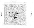
- Die dazugehörige Figur zeigt:
- Das Ergebnis einer zweidimensionalen DNA-Gelelektrophorese, nämlich ein repräsentatives Spotmuster für cDNA's aus Candida albicans.
- Wichtige Eigenschaften der Fleischqualität im Schwein definieren sich im wesentlichen über die Zusammensetzung und Strukturcharakteristika der Muskelfasern im Muskelgewebe. Die Ausprägung günstiger Eigenschaften wird dabei sehr stark von genetischen, prädisponierenden Faktoren bestimmt. Zur Identifizierung solcher Faktoren wird sowohl Muskelgewebe von einem Schwein mit hoher Fleischqualität als auch eine Gewebeprobe eines Schweins mit geringer Faserqualität genommen. Von beiden Proben wird jeweils die Gesamt-RNA isoliert und anschließend die darin enthaltene mRNA mittels reverser Transkription in Anwesenheit unterschiedlich markierter Primer bzw. Nukleotide in doppelsträngige cDNA (ds-cDNA) umgeschrieben. Diese ds-cDNA wird mit sequenzspezifischen Restriktionsendonukleasen geschnitten, um ds-DNA-Fragmente definierter Länge herzustellen. Die unterschiedlich markierten ds-cDNA-Pools werden vereinigt und mittels der mehrdimensionalen Gelelektrophorese simultan hochauflösend aufgetrennt. In der ersten Dimension werden dabei die unterschiedlichen Fragmente im Polyacrylamidgel nach ihrem jeweiligen Molekulargewicht aufgetrennt. Rechtwinklig zur Auftrennung in der ersten Dimension werden die Fragmente anschließend in der zweiten Dimension nach ihrem jeweiligen GC-Gehalt weiter separiert. Dabei wandern die DNA-Fragmente entlang eines kontinuierlichen Gradienten variierender Hybridisierungsstringenz, wobei die Formamid- und Harnstoffkonzentration im Gel kontinuierlich erhöht wird. Nach Auftrennung der markierten DNA-Fragmente in zwei Dimensionen werden die komplexen Spotmuster mittels Fluoreszenzscannern visualisiert und anschließend quantitativ und qualitativ analysiert. Spots, die ausschließlich in Proben mit hoher Fleischqualität zu detektieren sind und damit potentielle Marker darstellen, werden aus dem Gel ausgeschnitten und mittels DNA-Sequenzierung identifiziert.
- Humanpathogene Hefen haben spezifische Virulenzmechanismen entwickelt, um im entsprechenden Wirt Gewebe besiedeln zu können und damit in der Lage sind, Infektionen hervorzurufen. So verursacht zum Beispiel Candida albicans systemische Infektionen, die mit einer hohen Mortalitätsrate verbunden sind. Zur Identifizierung potentieller Virulenzfaktoren in C. albicans werden GenexpressionsStudien an klinischen Isolaten von C. albicans unter verschiedenen Bedingungen durchgeführt. Dazu wird aus C. albicans-Zellen Gesamt-RNA beziehungsweise mRNA isoliert und anschließend die entsprechende cDNA mittels reverser Transkription in an sich bekannter Weise hergestellt. Die so hergestellte cDNA wird mittels Restriktionsendonukleasen verdaut und anschließend definierte Adaptoren an die so fragmentierte cDNA ligiert. Diese adaptorligierte cDNA kann dann mittels PCR amplifiziert und mittels zweidimensionaler DNA-Gelelektrophorese aufgetrennt werden. Die so erhaltenen Spotmuster können dann qualitativ und quantitativ ausgewertet werden und die einzelnen Spots ausgestochen, mittels PCR amplifiziert und anschließend sequenziert werden. Die einzelnen experimentellen Schritte sind im folgendem detailliert aufgelistet:
- Für die Isolierung der Gesamt-RNA wird für die Vorkultur 11 ml YPD - Medium mit Candida albicans Zellen (klinisches Isolat SC5314) angeimpft und über Nacht bei 30°C geschüttelt. Anschließend werden 50 ml YPD-Medium mit Übernachtkultur zu einer OD600 zwischen 0,15-0,20 angeimpft. Diese Kultur wird bis zu einer OD von 0,8 - 1,0 bei 30°C im Schüttler (180 Upm) wachsen gelassen. Für die Herstellung von Zellperlen wird die Zellsuspension 4 min bei 3000 xg abzentrifugiert, das Pellet im restlichen 1,0-1,5 ml Medium resuspendiert und tropfenweise in ein mit flüssigem Stickstoff befülltes 50 ml Röhrchen pipettiert. Die so eingefrorenen Proben können bei -80°C bis zur Isolation aufbewahrt werden.
- Für die Isolierung der Gesamt-RNA aus C. albicans sowie auch aus eukaryontischen Zellen kann ein RNeasy-Midi-Kit (Qiagen) verwendet werden. Die Zellen werden allerdings nicht mit Hilfe von Glasperlen, sondern zum Beispiel mit einer Kugelmühle der Firma Retsch aufgeschlossen.
- Dabei werden die erhaltenen Zellperlen in zuvor mit flüssigem Stickstoff gekühlten Teflonbehältern mit einer Wolframkarbid-Kugel zu feinem Pulver zermahlen (shaking frequency = 30/sec, Zeit = 2 min) und anschließend in den Lyse-Puffer (Puffer RLT + 0,01 Vol% Mercaptoethanol) des Kits überführt. Zur Homogenisierung wird die Probe 1 min gevortext, dass heißt heftig geschüttelt. Anschließend werden die festen Bestandteile des Lysats zunächst 5 min bei 3500 xg abzentrifugiert und der Überstand mit 1 Volumen 70 % Ethanol versetzt und gut gemischt. Diese Lösung wird dann in 4 ml-Schritten komplett auf die RNeasy Midi-Säule gegeben und jeweils 5 min bei 3500 xg zentrifugiert. Anschließend wird die auf der Membran gebundene RNA nach Herstellerangaben zuerst mit 4 ml RW1-Puffer, dann zweimal mit jeweils 2,5 ml RPE-Puffer gewaschen. Zur Eluation der RNA wird auf die Membran 250 µl RNase-freies Wasser hinzupipettiert, die Säule 1 min bei Raumtemperatur inkubiert und anschließend 3 min bei 3500 xg zentrifugiert. Diese Schritte werden einmal wiederholt, so dass ein Endvolumen von 500 µl entsteht.
- Für die Fällung der Gesamt-RNA werden jeweils 250 µl des Eluats in zwei 1,5 ml Reaktionsgefäße überführt und durch Zugabe von 25 µl 3 mol/l Natriumacetat, pH 5,3 und 625 µl 100% Ethanol (-20°C) über Nacht bei -20°C gefällt. Durch die 30 minütige Zentrifugation bei 4°C, 13000 xg werden die ausgefallenen Nukleinsäuren pelletiert und zur Entfernung der noch vorhandenen Salzreste anschließend zweimal mit 1 ml 70% EtOH-DEPC (-20°C) gewaschen.
- Nach dem Trocknen des Gesamt - RNA - Niederschlags bei 37°C wird die RNA in 10-50 µl DEPC-H2O aufgenommen und eine Konzentrations- und Reinheitsbestimmung mittels der UV-Spektroskopie durchgeführt.
- Die Synthese von Volllängen ds-cDNA aus 0,5 - 1 µg polyA+ beziehungsweise Gesamt-RNA erfolgt nach einem modifizierten Protokoll vom SMART cDNA Library Construction Kit (BD Bioscience).
- Verwendete Primer (jeweils 10 mol/l):
- SMART IV Oligonucleotid:
5'-AAGCAGTGGTATCAACGCAGAGTGGCCATTACGGCCGGG-3' (SEQ ID Nr. 1) - ODD_T_all:
5'-GCGAGTCGACCGTTTTTTTTTTTTT-3' (SEQ ID Nr. 2) - ODD-TA:
5'-GCGAGTCGACCGTTTTTTTTTTTTTA-3' (SEQ ID Nr. 3) - ODD-TG:
5'-GCGAGTCGACCGTTTTTTTTTTTTTG-3' (SEQ ID Nr. 4) - ODD-TC:
5'-GCGAGTCGACCGTTTTTTTTTTTTTC-3' (SEQ ID Nr. 5) - A2-Sau3a:
5'-AAGCAGTGGTATCAACGCAGAGT-3' (SEQ ID Nr. 6) - Für die Erststrang cDNA Synthese werden in einem RNase-freiem Reaktionsgefäß 1-3 µl RNA [0,5 - 1,0 µg polyA+ bzw. Gesamt - RNA] mit jeweils 1 µl 10 µmol/l SMART IV Oligonucleotid- und ODD_T_all-Primer (alternativ: ein 10 µmol/l Mix aus ODD-TA, ODD-TG und ODD-TC) versetzt und bis zu einem Gesamtvolumen von 5 µl mit DEPC-H2O aufgefüllt.
- Der Ansatz wird für die Denaturierung der RNA - Sekundärstruktur zuerst 2 min bei 72°C und dann 2 min auf Eis inkubiert. Anschließend werden 2 µl 5x Erststrang Puffer (250 mmol/l Tris, pH 8,3; 30 mmol/l MgCl2; 375 mmol/l KCl), 1 µl 20 mmol/l DTT, 1 µl 10 mmol/l dNTP Mix und 1 µl PowerScript Reverse Transkriptase hinzugegeben und die Komponenten durch vorsichtiges Auf- und Abpipettieren gemischt. Die Reverse Transkriptase Reaktion erfolgt für 1 h bei 42°C und wird durch Überführung auf Eis gestoppt. Diese Erststrang-cDNA kann nun entweder direkt in die Zweitstrangsynthese eingesetzt oder bei -20°C bis zu drei Monaten gelagert werden.
- Die Zweitstrangsynthese erfolgt durch die Verwendung des BD AdvantageTM 2 Polymerase Mixes in der Long Distance (LD) - PCR. Bei dieser Mischung handelt es sich um die BD TITANIUM™ Taq DNA Polymerase in Kombination mit BD TaqStart™ Antikörper und einer geringen Menge einer proofreading Polymerase.
- Im 100 µl Gesamtvolumen werden 2 µl Erststrang-cDNA, 80 µl molekularbiologisches H2O, 10 µl 10x Advantage 2 PCR Puffer (400 mmol/l Tricine-KOH, pH8,7; 150 mmol/l KOAc; 35 mmol/l Mg(OAc)2; 37,5 µg/ml BSA; 0,05% Tween 20; 0,05% Nonidet-P40), 2 µl 50 x dNTP Mix (jeweils 10 mmol/l), 2 µl A2-Sau3A-Primer, 2 µl ODD_T_all-Primer (alternativ: ODD-TA/TC/TG -Primer-Mix) und 2 µl 50x Advantage™ 2 Polymerase Mix zusammen gegeben, kurz gevortext, dass heißt heftig geschüttelt, und anschließend eine "Hot-Start-PCR" mit zwei Zyklen durchgeführt.
- PCR-Programm:
- 95°C - 0:20 min
- 95°C - 0:05 min
- 68°C - 6:00 min
- 95°C - 0:05 min
- 68°C - 6:00 min
- Die Aufreinigung von ds-cDNAs erfolgt mittels Säulenaufreinigung (z.B. QIAGEN QIAquick PCR Purification Kits. Die Eluation erfolgt durch Zugabe von 90 µl EB-Puffer (10mmol/l Tris-Cl, pH8,5).
- 90 µl des Eluats werden im 120 µl Verdau-Ansatz mit 12 µl 10x Puffer 1 (100 mmol/l Bis Tris Propane-HCl, pH 7,0; 100 mmol/l MgCl2; 10 mmol/l Dithiothreitol) und 12 µl 10 mg/ml BSA versetzt und nach Zugabe von 30 U Rsal 2 h bei 37°C inkubiert. Nach einer weiteren Zugabe von 15 U Rsal und Inkubation für 1 h kann die geschnittene ds-cDNA gefällt werden.
- Die Fällung der cDNA erfolgt durch Zugabe von 0,1 Volumen 3 mol/l Natriumacetat, pH 5,3 und 2,5 Volumen 100% EtOH direkt zum Restriktionsverdau und einer Inkubation für 2 h bei -20°C. Die Pelletierung erfolgt durch eine 25 minütige Zentrifugation (13000 xg) in der Kühlzentrifuge bei 4°C. Nach dem Waschen im 70% Ethanol [-20°C] kann das Pellet in 10 µl molekularbiologischem Wasser aufgenommen werden. Davon werden 5 µl für die Adaptorligation eingesetzt, der Rest bei -20°C eingefroren.
- Verwendete Oligonukleotide:
- Long Oligo RsaI [100µmol/l]:
GCGTGAAGACGACAGAAAGGGCGTGGTGCGGAGGGCGGT (SEQ ID Nr. 7) - Short Oligo RsaI [100µmol/l]:
ACCGCCCTCCGC (SEQ ID Nr. 8) - Für die Herstellung eines 20 µmol/l Adapter-Stocks (ODD_Adapter_Rsal) werden jeweils 20 µl des Long Oligo Rsal- und des Short Oligo Rsal-Primers in 100 µl Gesamtvolumen 50 mmol/l Tris-HCL, pH 7,5; 10 mmol/l MgCl2; 10 mmol/l Dithiothreitol; 1 mmol/l ATP; 25 µg/ml BSA versetzt. Durch eine 3 min Denaturierung bei 95°C für 3 min und anschließende langsame Abkühlung (ca. 8 h) bei RT hybridisieren die komplementären Nukleotidsequenzen zu doppelsträngigen Adaptoren, die für die Ligation von Rsal-verdauter ds-cDNA eingesetzt werden können.
- 5 µl der nach dem Rsal-Verdau gefällter ds-cDNA werden für die Adapterligation eingesetzt.
- Für die Ligation werden 1 µl des 20 µmol/l Adapter-Stocklösung [ODD_Adapter_Rsal], 1 µl 10x T4-Ligase_Puffer (500 mmol/l Tris-HCL, pH 7,5; 100 mmol/l MgCl2; 100 mmol/l Dithiothreitol; 10 mmol/l ATP; 250 µg/ml BSA), 5 µl ds-cDNA und 200 U T4-DNA-Ligase in 10 µl Gesamtvolumen zusammengemischt und über Nacht bei 16°C inkubiert.
- Die Aktivität der Ligase wird am nächsten Tag durch Hitzeinaktivierung (10 min. bei 65°C) gestoppt.
- Anschließend wird der 10 µl Ligationsansatz mit 90 µl Wasser auf 100 µl aufgefüllt und mittels Säulen (QIAGEN PCR Purification Kit - Protokoll) aufgereinigt.
- Die sogenannte Suppressions-PCR mit den Primern ODD_T_all und Primer AP1 dient vor allem zur Amplifikation der 3' Enden der cDNA.
- Zur Amplifizierung der Adapter-ligierten DNA wird folgende PCR durchgeführt:
- 10 µl des Eluats der Adapter-ligierten ds-cDNA werden mit 2,5 µl 10x PCR-Puffer (200 mmol/l Tris-HCl, pH 8,4 ; 500 mmol/l KCl), 0,6 µl 50 mmol/l MgCl2, 0,5 µl 5 mmol/l dNTPs, 0,25 µl 10 µmol/l AP1-Primer
- (5'-TGTAGCGTGAAGACGACAGAA-3') (SEQ ID Nr. 9),
- 0,25 µl ODD_T_all-Primer
- (5'-GCGAGTCGACCGTTTTTTTTTTTTT-3') (SEQ ID Nr. 2),
- 10,65 µl molekularbiologisches Wasser sowie 2 U Taq-Polymerase vermischt und anschließend mit folgendem PCR-Programm amplifiziert:
- PCR-Programm:
- 1. 95°C - 0:05 min
- 2. 65°C - 0:30 min
- 3. 72°C - 1:00 min
- Schritte 2 und 3 werden 19 mal wiederholt (Gesamtzyklenzahl: 20).
- Die so amplifizierte cDNA kann anschließend in einer zweiten PCR (Adapter-PCR II) spezifisch amplifiziert werden, wobei sogenannte anchored Primer Verwendung finden
- Primer 1:
AGGGCGTGGTGCGGAGGGCGGTCCNN (SEQ ID Nr. 10),
wobei N die Nucleotide A, G, C oder T repräsentieren kann und damit 16 Primer 1-Varianten möglich sind; sowie - Primer 2:
5'-GCGAGTCGACCGTTTTTTTTTTTTTVN-3' (SEQ ID Nr. 11),
wobei V die Nucleotide A, G, oder C und N die Nucleotide A, G, C oder T repräsentieren können und somit 12 Primer 2-Varianten möglich sind). - Für die spezifische Amplifizierung von Subpopulationen von amplifizierten cDNA-Fragmenten aus der Adapter-PCRI wird folgender Ansatz gewählt:
- Die amplifizierten cDNA-Fragmente aus der Adapter-PCR I werden 1:30 mit Wasser verdünnt. 10 µl dieser Verdünnung werden mit 10 µl 10x PCR-Puffer (200 mmol/l Tris-HCl, pH 8,4 ; 500 mmol/l KCl), 3 µl 50 mmol/l MgCl2, 4 µl 5 mmol/l dNTPs, 2,5 µl 10 µmol/l Primer 1
(5'- GGGCGTGGTGCGGAGGGCGGTCCNN-3') (SEQ ID Nr. 10), - 2,5 µl 10 µmol/l Primer 2
(5'- GCGAGTCGACCGTTTTTTTTTTTTTVN -3') (SEQ ID Nr. 11), - 65,5 µl molekularbiologisches Wasser sowie 20 U Taq-Polymerase vermischt und anschließend mit folgendem PCR-Programm amplifiziert:
- PCR-Programm:
- 1. 95°C - 0:05 min
- 2. 65°C - 0:30 min
- 3. 72°C - 1:00 min
- Schritte 2 und 3 werden 20 mal wiederholt (Gesamtzyklenzahl: 21).
- Die amplifizierten cDNA-Fragmente aus der Adapter-PCR II werden anschließend mittels zweidimensionaler Gelelektrophorese hochauflösend aufgetrennt. Dazu werden die Proben zunächst in der ersten Dimension in einem nicht-denaturierenden Polyacrylamidgel aufgetragen. Zusammensetzung des Gels für die erste Dimension (Auftrennung nach Molekulargewicht): 8% Acrylamid (Verhältnis Acrylamid : Bisacrylamid 37,5 : 1) in 1xTAE (40 mmol/l Tris; 20 mmol/l Eisessig; 1 mmol/l EDTA, pH 8,0). Die Proben werden zwischen 2-4 Stunden bei konstanter Spannung bei 200 Volt elektrophoretisiert. Anschließend wird die Spur der ersten Dimension ausgeschnitten und zur Auftrennung in der zweiten Dimension auf ein Polyacrylamidgel, das einen Konzentrationsgradient an Harnstoff und Formamid enthält, aufgelegt. Das Polyacrylamidgel der zweiten Dimension kann dabei entweder eine konstante Konzentration oder ebenfalls einen Konzentrationsgradienten an Acrylamid aufweisen. Zusammensetzung des Gels für die zweite Dimension: 8 % Acrylamid (Verhältnis Acrylamid : Bisacrylamid 37,5 : 1) in 1xTAE (40 mmol/l Tris; 20 mmol/l Eisessig; 1 mmol/l EDTA, pH 8,0); kontinuierlicher Gradient von 0,7 mol/l Harnstoff, 4 % Formamid oben, bis 2,8 mol/l Harnstoff, 16 % Formamid unten. Die Fragmente werden in dieser zweiten Dimension bei konstanter Temperatur bei 60°C für 15 Stunden bei konstanter Spannung bei 100 Volt elektrophoretisiert.
- Das 2D-Gel wird zur Visualisierung der DNA-Spots mittels SYBR-Gold gefärbt. Bei Verwendung fluoreszenzmarkierter Primer 1 und Primer 2 in der Adapter-PCR II entfällt eine Färbung des 2D-Gels mittels SYBR-Gold. Die Detektion der DNA-Spots erfolgt mittels handelsüblicher Fluoreszenzdokumentationssystemen.
- Die Figur zeigt ein repräsentatives Spotmuster für cDNAs aus C. albicans nach zweidimensionaler Auftrennung und Färbung mittels SYBR-Gold.
- 1. Dimension: 8% TAE-Polyacrylamidgel (Auftrennung nach Molekulargewicht)
- 2. Dimension: Denaturierende Gradientengelelektrophorese 8% Acrylamid, kontinuierlicher Denaturierungsgradient von 4% Formamid + 0,7 mol/l Harnstoff bis 16% Formamid + 2,8 mol/l Harnstoff (Auftrennung nach GC-Gehalt)
-
- <110> Fraunhofer-Gesellschaft zur Förderung der angewandten Forschung e.V.
- <120> Verbessertes elektrophoretisches Trennverfahren für die Analyse der Genexpression
- <130> 26293 WO
- <150>
DE102004048334.5-41
<151> 2004-10-04 - <160> 11
- <170> PatentIn version 3.3
- <210> 1
<211> 39
<212> DNA
<213> Artificial - <220>
<223> Candida albicans - <400> 1

- <210> 2
<211> 25
<212> DNA
<213> Artificial - <220>
<223> Candida albicans - <400> 2

- <210> 3
<211> 26
<212> DNA
<213> Artificial - <220>
<223> Candida albicans - <400> 3

- <210> 4
<211> 26
<212> DNA
<213> Artificial - <220>
<223> Candida albicans - <400> 4

- <210> 5
<211> 26
<212> DNA
<213> Artificial - <220>
<223> Candida albicans - <400> 5

- <210> 6
<211> 23
<212> DNA
<213> Artificial - <220>
<223> Candida albicans - <400> 6

- <210> 7
<211> 39
<212> DNA
<213> Artificial - <220>
<223> Candida Albicans - <400> 7

- <210> 8
<211> 12
<212> DNA
<213> Artificial - <220>
<223> Candida albicans - <400> 8

- <210> 9
<211> 21
<212> DNA
<213> Artificial - <220>
<223> Candida albicans - <400> 9

- <210> 10
<211> 26
<212> DNA
<213> Artificial - <220>
<223> Candida albicans - <220>
<221> misc_feature
<222> (25)..(26)
<223> n is a, c, g, or t - <400> 10

- <210> 11
<211> 27
<212> DNA
<213> Artificial - <220>
<223> Candida albicans, v is a, g or c - <220>
<221> misc_feature
<222> (27)..(27)
<223> n is a, c, g, or t - <400> 11

Claims (6)
- Verfahren zur quantitativen oder qualitativen Analyse der Genexpression eines biologischen Materials, enthaltend die Schritte:Isolieren und Fällen der Gesamt-RNA aus dem biologischen Material;Herstellen von doppelsträngiger cDNA aus der Gesamt-RNA;Restriktionsverdauen der cDNA mit Restriktionsendonuclease;Fällen der fragmentierten cDNA;Durchführen einer Suppressions-PCR zur Amplifizierung der 3'-Enden der cDNA-Fragmente und gegebenenfalls spezifisches Amplifizieren von Subpopulationen der amplifizierten cDNA-Fragmente;zweidimensionales Auftrennen der erhaltenen cDNA-Population, wobei die cDNA-Population in der ersten Dimension nach ihrem Molekulargewicht und in der zweiten Dimension nach ihrem GC-Gehalt aufgetrennt wird; undDetektieren und qualitatives und/oder quantitatives Auswerten der erhaltenen, mehrdimensional aufgetrennten Spots der mindestens einen cDNA-Population.
- Verfahren nach Anspruch 1, wobei die Auftrennung nach dem Molekulargewicht mittels DNA-Gelelektrophoresesystem erfolgt.
- Verfahren nach Anspruch 1 oder 2, wobei die Auftrennung nach dem GC-Gehalt in einer denaturierenden Gradientengelelektrophorese erfolgt.
- Verfahren nach einem der vorhergehenden Ansprüche, wobei in einem weiteren Schritt eine Sequenzierung von DNA-Molekülen aus mindestens einem der in dem Trennsystem erhaltenen und daraus isolierten cDNA-Spot erfolgt.
- Verfahren nach einem der vorhergehenden Ansprüche, wobei die doppelsträngige cDNA-Population mit interkalierenden Farbstoffen oder radioaktiv- oder fluoreszenz-markiert ist.
- Verfahren nach einem der vorhergehenden Ansprüche, wobei
aus dem biologischen Material mindestens zwei verschiedene Gesamt-RNA's oder mindestens zwei verschiedene mRNA-Populationen gewonnen;
aus den mindestens zwei verschiedenen RNA's, insbesondere mindestens zwei verschiedenen Gesamt-RNA's oder den mindestens zwei verschiedenen mRNA-Populationen, insbesondere durch reverse Transkription, mindestens zwei verschiedene Populationen doppelsträngiger cDNA erhalten werden, wobei während dieses Verfahrensschritts die mindestens zwei verschiedenen Populationen doppelsträngiger cDNA's jeweils unterschiedlich markiert werden;
in einem Trennsystem eine gemeinsame mehrdimensionale Aufteilung der mindestens zwei verschiedenen cDNA-Populationen durchgeführt wird; und
eine qualitative und/oder quantitative Auswertung der erhaltenen, mehrdimensional aufgetrennten Spots der mindestens zwei cDNA-Populationen durchgeführt wird.
Applications Claiming Priority (2)
| Application Number | Priority Date | Filing Date | Title |
|---|---|---|---|
| DE102004048334A DE102004048334A1 (de) | 2004-10-01 | 2004-10-01 | Verbessertes elektrophoretisches Trennverfahren für die Analyse der Genexpression |
| PCT/EP2005/010566 WO2006037563A1 (de) | 2004-10-01 | 2005-09-30 | Verbessertes elektrophoretisches trennverfahren für die analyse der genexpression |
Publications (2)
| Publication Number | Publication Date |
|---|---|
| EP1797198A1 EP1797198A1 (de) | 2007-06-20 |
| EP1797198B1 true EP1797198B1 (de) | 2009-11-11 |
Family
ID=35654255
Family Applications (1)
| Application Number | Title | Priority Date | Filing Date |
|---|---|---|---|
| EP05796264A Expired - Lifetime EP1797198B1 (de) | 2004-10-01 | 2005-09-30 | Verbessertes elektrophoretisches trennverfahren für die analyse der genexpression |
Country Status (7)
| Country | Link |
|---|---|
| US (1) | US20080261209A1 (de) |
| EP (1) | EP1797198B1 (de) |
| JP (1) | JP2008514213A (de) |
| AT (1) | ATE448329T1 (de) |
| AU (1) | AU2005291445B2 (de) |
| DE (2) | DE102004048334A1 (de) |
| WO (1) | WO2006037563A1 (de) |
Families Citing this family (1)
| Publication number | Priority date | Publication date | Assignee | Title |
|---|---|---|---|---|
| DE102009058298B4 (de) | 2009-12-01 | 2014-10-30 | Hölle & Hüttner AG | Genexpressionsanalyse |
Family Cites Families (8)
| Publication number | Priority date | Publication date | Assignee | Title |
|---|---|---|---|---|
| NL8801147A (nl) * | 1988-05-02 | 1989-12-01 | Tno | Werkwijze voor het detecteren van genetische variatie, en daarvoor geschikte kit. |
| US5925525A (en) * | 1989-06-07 | 1999-07-20 | Affymetrix, Inc. | Method of identifying nucleotide differences |
| US5316935A (en) * | 1992-04-06 | 1994-05-31 | California Institute Of Technology | Subtilisin variants suitable for hydrolysis and synthesis in organic media |
| RU2111254C1 (ru) * | 1994-07-11 | 1998-05-20 | Институт молекулярной биологии им.В.А.Энгельгардта РАН | СПОСОБ ДЕТЕКЦИИ ДИФФЕРЕНЦИАЛЬНО ЭКСПРЕССИРУЮЩИХСЯ МАТРИЧНЫХ РНК И КЛОНИРОВАНИЯ СООТВЕТСТВУЮЩИХ ИМ ФРАГМЕНТОВ кДНК |
| WO1997017466A1 (en) * | 1995-11-09 | 1997-05-15 | Shionogi & Co., Ltd. | Method for the simultaneous detection of polygenes |
| AU2264197A (en) * | 1996-02-09 | 1997-08-28 | Government Of The United States Of America, As Represented By The Secretary Of The Department Of Health And Human Services, The | Restriction display (rd-pcr) of differentially expressed mrnas |
| US6670123B1 (en) * | 2000-11-03 | 2003-12-30 | New York Blood Center, Inc. | Method for detecting hematopoietic stem cells |
| AU2003233219A1 (en) * | 2002-04-30 | 2003-11-17 | Board Of Regents, The University Of Texas System | Ssh based methods for identifying and isolating unique nucleic acid sequences |
-
2004
- 2004-10-01 DE DE102004048334A patent/DE102004048334A1/de not_active Ceased
-
2005
- 2005-09-30 JP JP2007533949A patent/JP2008514213A/ja active Pending
- 2005-09-30 EP EP05796264A patent/EP1797198B1/de not_active Expired - Lifetime
- 2005-09-30 WO PCT/EP2005/010566 patent/WO2006037563A1/de not_active Ceased
- 2005-09-30 AU AU2005291445A patent/AU2005291445B2/en not_active Ceased
- 2005-09-30 US US11/664,403 patent/US20080261209A1/en not_active Abandoned
- 2005-09-30 DE DE502005008501T patent/DE502005008501D1/de not_active Expired - Lifetime
- 2005-09-30 AT AT05796264T patent/ATE448329T1/de not_active IP Right Cessation
Also Published As
| Publication number | Publication date |
|---|---|
| ATE448329T1 (de) | 2009-11-15 |
| JP2008514213A (ja) | 2008-05-08 |
| US20080261209A1 (en) | 2008-10-23 |
| AU2005291445A1 (en) | 2006-04-13 |
| WO2006037563A8 (de) | 2006-06-15 |
| AU2005291445B2 (en) | 2010-06-24 |
| EP1797198A1 (de) | 2007-06-20 |
| WO2006037563A1 (de) | 2006-04-13 |
| DE102004048334A1 (de) | 2006-04-06 |
| DE502005008501D1 (de) | 2009-12-24 |
Similar Documents
| Publication | Publication Date | Title |
|---|---|---|
| EP0743367B2 (de) | Verfahren zur Genexpressionsanalyse | |
| DE69734063T2 (de) | Herstellung und verwendung von normalisierten dna-bibliotheken | |
| DE3855064T2 (de) | Selektive Amplifikation von Oligonukleotiden-Zielsequenzen | |
| DE69230873T3 (de) | Selektive Restriktionsfragmentenamplifikation: generelles Verfahren für DNS-Fingerprinting | |
| DE69128077T2 (de) | Nuklein-säure amplifikation mit dns abhängigen rns polymerasen aktivität von rns-replikasen | |
| DE69825240T2 (de) | Verfahren zur subtraktiven hybridisierung und differenziellen analyse | |
| DE69332665T2 (de) | Methode um mrna zu klonieren | |
| DE69535428T2 (de) | Verfahren zum Auffinden von differentiel exprimierte Gene | |
| DE69607830T2 (de) | Verfahren zur isothermer amplifikation von nukleinsäure molekülen | |
| DE69011101T2 (de) | Methoden zur in vitro-dna-amplifikation, zum genomischen klonieren und zur genkartierung. | |
| DE69900592T2 (de) | Verfahren zur unspezifischen amplifizierung einer nukleinsäure | |
| EP3041951B1 (de) | Chromosomenkonformationserfassungsverfahren mit auswahl- und anreicherungsschritten | |
| DE69605803T2 (de) | Nachweis von fehlpaarungen durch spaltung mit resolvase auf einem festträger | |
| CN113528612B (zh) | 用于检测染色质开放位点间染色质相互作用的NicE-C技术 | |
| DE112006002652B4 (de) | Verfahren zur isothermen Amplifikation von Nukleinsäuren und Verfahren zum Nachweisen von Nukleinsäuren durch gleichzeitige isotherme Amplifikation von Nukleinsäuren und Signalsonde | |
| US6706476B1 (en) | Process for amplifying and labeling single stranded cDNA by 5′ ligated adaptor mediated amplification | |
| DE4428651C1 (de) | Verfahren zur Herstellung und Amplifikation von Nukleinsäuren | |
| DE102011114984B3 (de) | Sequenzspezifische Analyse von Nukleinsäuren | |
| DE60009530T2 (de) | Genetische toxizitätsmarker, herstellung und verwendung | |
| DE69737327T2 (de) | Verfahren zur identifizierung von essentiellen genen für das wachstum eines organismus | |
| EP2471955B1 (de) | Organismusspezifisches hybridisierbares Nucleinsäuremolekül | |
| EP1797198A1 (de) | Verbessertes elektrophoretisches trennverfahren für die analyse der genexpression | |
| DE69914942T2 (de) | Kennzeichnung einer substanz mittels eines reportergens und sequenzen, die für die in vitro expression des reportergens notwendig sind | |
| EP1430301B1 (de) | Verfahren zur herstellung einer normalisierten genbank aus nukleinsäure-exktrakten von bodenproben und deren verwendung | |
| DE69937453T2 (de) | Verfahren zur isolierung und charakterisierung potentieller funktionen ausgehend von einer biologischen probe, welche nukleinsäuren enthält |
Legal Events
| Date | Code | Title | Description |
|---|---|---|---|
| PUAI | Public reference made under article 153(3) epc to a published international application that has entered the european phase |
Free format text: ORIGINAL CODE: 0009012 |
|
| 17P | Request for examination filed |
Effective date: 20070502 |
|
| AK | Designated contracting states |
Kind code of ref document: A1 Designated state(s): AT BE BG CH CY CZ DE DK EE ES FI FR GB GR HU IE IS IT LI LT LU LV MC NL PL PT RO SE SI SK TR |
|
| 17Q | First examination report despatched |
Effective date: 20070829 |
|
| DAX | Request for extension of the european patent (deleted) | ||
| GRAP | Despatch of communication of intention to grant a patent |
Free format text: ORIGINAL CODE: EPIDOSNIGR1 |
|
| GRAS | Grant fee paid |
Free format text: ORIGINAL CODE: EPIDOSNIGR3 |
|
| GRAA | (expected) grant |
Free format text: ORIGINAL CODE: 0009210 |
|
| AK | Designated contracting states |
Kind code of ref document: B1 Designated state(s): AT BE BG CH CY CZ DE DK EE ES FI FR GB GR HU IE IS IT LI LT LU LV MC NL PL PT RO SE SI SK TR |
|
| REG | Reference to a national code |
Ref country code: GB Ref legal event code: FG4D Free format text: NOT ENGLISH |
|
| REG | Reference to a national code |
Ref country code: CH Ref legal event code: EP |
|
| REG | Reference to a national code |
Ref country code: IE Ref legal event code: FG4D |
|
| REF | Corresponds to: |
Ref document number: 502005008501 Country of ref document: DE Date of ref document: 20091224 Kind code of ref document: P |
|
| NLV1 | Nl: lapsed or annulled due to failure to fulfill the requirements of art. 29p and 29m of the patents act | ||
| LTIE | Lt: invalidation of european patent or patent extension |
Effective date: 20091111 |
|
| PG25 | Lapsed in a contracting state [announced via postgrant information from national office to epo] |
Ref country code: ES Free format text: LAPSE BECAUSE OF FAILURE TO SUBMIT A TRANSLATION OF THE DESCRIPTION OR TO PAY THE FEE WITHIN THE PRESCRIBED TIME-LIMIT Effective date: 20100222 Ref country code: FI Free format text: LAPSE BECAUSE OF FAILURE TO SUBMIT A TRANSLATION OF THE DESCRIPTION OR TO PAY THE FEE WITHIN THE PRESCRIBED TIME-LIMIT Effective date: 20091111 Ref country code: SE Free format text: LAPSE BECAUSE OF FAILURE TO SUBMIT A TRANSLATION OF THE DESCRIPTION OR TO PAY THE FEE WITHIN THE PRESCRIBED TIME-LIMIT Effective date: 20091111 Ref country code: LT Free format text: LAPSE BECAUSE OF FAILURE TO SUBMIT A TRANSLATION OF THE DESCRIPTION OR TO PAY THE FEE WITHIN THE PRESCRIBED TIME-LIMIT Effective date: 20091111 Ref country code: PT Free format text: LAPSE BECAUSE OF FAILURE TO SUBMIT A TRANSLATION OF THE DESCRIPTION OR TO PAY THE FEE WITHIN THE PRESCRIBED TIME-LIMIT Effective date: 20100311 Ref country code: IS Free format text: LAPSE BECAUSE OF FAILURE TO SUBMIT A TRANSLATION OF THE DESCRIPTION OR TO PAY THE FEE WITHIN THE PRESCRIBED TIME-LIMIT Effective date: 20100311 |
|
| PG25 | Lapsed in a contracting state [announced via postgrant information from national office to epo] |
Ref country code: LV Free format text: LAPSE BECAUSE OF FAILURE TO SUBMIT A TRANSLATION OF THE DESCRIPTION OR TO PAY THE FEE WITHIN THE PRESCRIBED TIME-LIMIT Effective date: 20091111 Ref country code: PL Free format text: LAPSE BECAUSE OF FAILURE TO SUBMIT A TRANSLATION OF THE DESCRIPTION OR TO PAY THE FEE WITHIN THE PRESCRIBED TIME-LIMIT Effective date: 20091111 Ref country code: SI Free format text: LAPSE BECAUSE OF FAILURE TO SUBMIT A TRANSLATION OF THE DESCRIPTION OR TO PAY THE FEE WITHIN THE PRESCRIBED TIME-LIMIT Effective date: 20091111 Ref country code: CY Free format text: LAPSE BECAUSE OF FAILURE TO SUBMIT A TRANSLATION OF THE DESCRIPTION OR TO PAY THE FEE WITHIN THE PRESCRIBED TIME-LIMIT Effective date: 20091111 |
|
| REG | Reference to a national code |
Ref country code: IE Ref legal event code: FD4D |
|
| PG25 | Lapsed in a contracting state [announced via postgrant information from national office to epo] |
Ref country code: DK Free format text: LAPSE BECAUSE OF FAILURE TO SUBMIT A TRANSLATION OF THE DESCRIPTION OR TO PAY THE FEE WITHIN THE PRESCRIBED TIME-LIMIT Effective date: 20091111 Ref country code: RO Free format text: LAPSE BECAUSE OF FAILURE TO SUBMIT A TRANSLATION OF THE DESCRIPTION OR TO PAY THE FEE WITHIN THE PRESCRIBED TIME-LIMIT Effective date: 20091111 Ref country code: BG Free format text: LAPSE BECAUSE OF FAILURE TO SUBMIT A TRANSLATION OF THE DESCRIPTION OR TO PAY THE FEE WITHIN THE PRESCRIBED TIME-LIMIT Effective date: 20100211 Ref country code: IE Free format text: LAPSE BECAUSE OF FAILURE TO SUBMIT A TRANSLATION OF THE DESCRIPTION OR TO PAY THE FEE WITHIN THE PRESCRIBED TIME-LIMIT Effective date: 20091111 Ref country code: EE Free format text: LAPSE BECAUSE OF FAILURE TO SUBMIT A TRANSLATION OF THE DESCRIPTION OR TO PAY THE FEE WITHIN THE PRESCRIBED TIME-LIMIT Effective date: 20091111 |
|
| PG25 | Lapsed in a contracting state [announced via postgrant information from national office to epo] |
Ref country code: SK Free format text: LAPSE BECAUSE OF FAILURE TO SUBMIT A TRANSLATION OF THE DESCRIPTION OR TO PAY THE FEE WITHIN THE PRESCRIBED TIME-LIMIT Effective date: 20091111 Ref country code: CZ Free format text: LAPSE BECAUSE OF FAILURE TO SUBMIT A TRANSLATION OF THE DESCRIPTION OR TO PAY THE FEE WITHIN THE PRESCRIBED TIME-LIMIT Effective date: 20091111 |
|
| PLBE | No opposition filed within time limit |
Free format text: ORIGINAL CODE: 0009261 |
|
| STAA | Information on the status of an ep patent application or granted ep patent |
Free format text: STATUS: NO OPPOSITION FILED WITHIN TIME LIMIT |
|
| 26N | No opposition filed |
Effective date: 20100812 |
|
| PG25 | Lapsed in a contracting state [announced via postgrant information from national office to epo] |
Ref country code: GR Free format text: LAPSE BECAUSE OF FAILURE TO SUBMIT A TRANSLATION OF THE DESCRIPTION OR TO PAY THE FEE WITHIN THE PRESCRIBED TIME-LIMIT Effective date: 20100212 |
|
| BERE | Be: lapsed |
Owner name: FRAUNHOFER-GESELLSCHAFT ZUR FORDERUNG DER ANGEWAN Effective date: 20100930 |
|
| PG25 | Lapsed in a contracting state [announced via postgrant information from national office to epo] |
Ref country code: IT Free format text: LAPSE BECAUSE OF FAILURE TO SUBMIT A TRANSLATION OF THE DESCRIPTION OR TO PAY THE FEE WITHIN THE PRESCRIBED TIME-LIMIT Effective date: 20091111 |
|
| PG25 | Lapsed in a contracting state [announced via postgrant information from national office to epo] |
Ref country code: MC Free format text: LAPSE BECAUSE OF NON-PAYMENT OF DUE FEES Effective date: 20100930 |
|
| REG | Reference to a national code |
Ref country code: CH Ref legal event code: PL |
|
| PG25 | Lapsed in a contracting state [announced via postgrant information from national office to epo] |
Ref country code: LI Free format text: LAPSE BECAUSE OF NON-PAYMENT OF DUE FEES Effective date: 20100930 Ref country code: CH Free format text: LAPSE BECAUSE OF NON-PAYMENT OF DUE FEES Effective date: 20100930 Ref country code: BE Free format text: LAPSE BECAUSE OF NON-PAYMENT OF DUE FEES Effective date: 20100930 |
|
| PG25 | Lapsed in a contracting state [announced via postgrant information from national office to epo] |
Ref country code: AT Free format text: LAPSE BECAUSE OF NON-PAYMENT OF DUE FEES Effective date: 20100930 |
|
| PG25 | Lapsed in a contracting state [announced via postgrant information from national office to epo] |
Ref country code: HU Free format text: LAPSE BECAUSE OF FAILURE TO SUBMIT A TRANSLATION OF THE DESCRIPTION OR TO PAY THE FEE WITHIN THE PRESCRIBED TIME-LIMIT Effective date: 20100512 Ref country code: NL Free format text: LAPSE BECAUSE OF FAILURE TO SUBMIT A TRANSLATION OF THE DESCRIPTION OR TO PAY THE FEE WITHIN THE PRESCRIBED TIME-LIMIT Effective date: 20091111 Ref country code: LU Free format text: LAPSE BECAUSE OF NON-PAYMENT OF DUE FEES Effective date: 20100930 |
|
| PG25 | Lapsed in a contracting state [announced via postgrant information from national office to epo] |
Ref country code: TR Free format text: LAPSE BECAUSE OF FAILURE TO SUBMIT A TRANSLATION OF THE DESCRIPTION OR TO PAY THE FEE WITHIN THE PRESCRIBED TIME-LIMIT Effective date: 20091111 |
|
| PGFP | Annual fee paid to national office [announced via postgrant information from national office to epo] |
Ref country code: DE Payment date: 20140922 Year of fee payment: 10 |
|
| PGFP | Annual fee paid to national office [announced via postgrant information from national office to epo] |
Ref country code: GB Payment date: 20140923 Year of fee payment: 10 |
|
| PGFP | Annual fee paid to national office [announced via postgrant information from national office to epo] |
Ref country code: FR Payment date: 20140917 Year of fee payment: 10 |
|
| REG | Reference to a national code |
Ref country code: DE Ref legal event code: R119 Ref document number: 502005008501 Country of ref document: DE |
|
| GBPC | Gb: european patent ceased through non-payment of renewal fee |
Effective date: 20150930 |
|
| REG | Reference to a national code |
Ref country code: FR Ref legal event code: ST Effective date: 20160531 |
|
| PG25 | Lapsed in a contracting state [announced via postgrant information from national office to epo] |
Ref country code: DE Free format text: LAPSE BECAUSE OF NON-PAYMENT OF DUE FEES Effective date: 20160401 Ref country code: GB Free format text: LAPSE BECAUSE OF NON-PAYMENT OF DUE FEES Effective date: 20150930 |
|
| PG25 | Lapsed in a contracting state [announced via postgrant information from national office to epo] |
Ref country code: FR Free format text: LAPSE BECAUSE OF NON-PAYMENT OF DUE FEES Effective date: 20150930 |