EP1740954B1 - Massenspektrometrie von antikörperkonjugaten - Google Patents
Massenspektrometrie von antikörperkonjugaten Download PDFInfo
- Publication number
- EP1740954B1 EP1740954B1 EP05733039.1A EP05733039A EP1740954B1 EP 1740954 B1 EP1740954 B1 EP 1740954B1 EP 05733039 A EP05733039 A EP 05733039A EP 1740954 B1 EP1740954 B1 EP 1740954B1
- Authority
- EP
- European Patent Office
- Prior art keywords
- antibody
- genbank accession
- drug
- trastuzumab
- mmae
- Prior art date
- Legal status (The legal status is an assumption and is not a legal conclusion. Google has not performed a legal analysis and makes no representation as to the accuracy of the status listed.)
- Active
Links
- 238000004949 mass spectrometry Methods 0.000 title claims description 69
- 229940127121 immunoconjugate Drugs 0.000 title description 15
- 239000003814 drug Substances 0.000 claims description 246
- 229940079593 drug Drugs 0.000 claims description 243
- 238000000034 method Methods 0.000 claims description 192
- 229940049595 antibody-drug conjugate Drugs 0.000 claims description 157
- 239000000611 antibody drug conjugate Substances 0.000 claims description 155
- -1 polyethylene Polymers 0.000 claims description 86
- 108090000623 proteins and genes Proteins 0.000 claims description 85
- IEDXPSOJFSVCKU-HOKPPMCLSA-N [4-[[(2S)-5-(carbamoylamino)-2-[[(2S)-2-[6-(2,5-dioxopyrrolidin-1-yl)hexanoylamino]-3-methylbutanoyl]amino]pentanoyl]amino]phenyl]methyl N-[(2S)-1-[[(2S)-1-[[(3R,4S,5S)-1-[(2S)-2-[(1R,2R)-3-[[(1S,2R)-1-hydroxy-1-phenylpropan-2-yl]amino]-1-methoxy-2-methyl-3-oxopropyl]pyrrolidin-1-yl]-3-methoxy-5-methyl-1-oxoheptan-4-yl]-methylamino]-3-methyl-1-oxobutan-2-yl]amino]-3-methyl-1-oxobutan-2-yl]-N-methylcarbamate Chemical compound CC[C@H](C)[C@@H]([C@@H](CC(=O)N1CCC[C@H]1[C@H](OC)[C@@H](C)C(=O)N[C@H](C)[C@@H](O)c1ccccc1)OC)N(C)C(=O)[C@@H](NC(=O)[C@H](C(C)C)N(C)C(=O)OCc1ccc(NC(=O)[C@H](CCCNC(N)=O)NC(=O)[C@@H](NC(=O)CCCCCN2C(=O)CCC2=O)C(C)C)cc1)C(C)C IEDXPSOJFSVCKU-HOKPPMCLSA-N 0.000 claims description 84
- 239000012634 fragment Substances 0.000 claims description 83
- 239000000523 sample Substances 0.000 claims description 76
- 239000012528 membrane Substances 0.000 claims description 75
- 210000002381 plasma Anatomy 0.000 claims description 75
- 239000000427 antigen Substances 0.000 claims description 73
- 108091007433 antigens Proteins 0.000 claims description 70
- 102000036639 antigens Human genes 0.000 claims description 70
- 102000004169 proteins and genes Human genes 0.000 claims description 70
- 206010028980 Neoplasm Diseases 0.000 claims description 55
- 150000001875 compounds Chemical class 0.000 claims description 55
- 238000004458 analytical method Methods 0.000 claims description 51
- 239000003153 chemical reaction reagent Substances 0.000 claims description 49
- 108090000765 processed proteins & peptides Proteins 0.000 claims description 45
- 241000282414 Homo sapiens Species 0.000 claims description 38
- 239000000203 mixture Substances 0.000 claims description 36
- 102000004196 processed proteins & peptides Human genes 0.000 claims description 33
- 238000000926 separation method Methods 0.000 claims description 29
- WEVYAHXRMPXWCK-UHFFFAOYSA-N Acetonitrile Chemical compound CC#N WEVYAHXRMPXWCK-UHFFFAOYSA-N 0.000 claims description 28
- 239000012472 biological sample Substances 0.000 claims description 28
- 238000001877 single-ion monitoring Methods 0.000 claims description 25
- MFRNYXJJRJQHNW-DEMKXPNLSA-N (2s)-2-[[(2r,3r)-3-methoxy-3-[(2s)-1-[(3r,4s,5s)-3-methoxy-5-methyl-4-[methyl-[(2s)-3-methyl-2-[[(2s)-3-methyl-2-(methylamino)butanoyl]amino]butanoyl]amino]heptanoyl]pyrrolidin-2-yl]-2-methylpropanoyl]amino]-3-phenylpropanoic acid Chemical compound CN[C@@H](C(C)C)C(=O)N[C@@H](C(C)C)C(=O)N(C)[C@@H]([C@@H](C)CC)[C@H](OC)CC(=O)N1CCC[C@H]1[C@H](OC)[C@@H](C)C(=O)N[C@H](C(O)=O)CC1=CC=CC=C1 MFRNYXJJRJQHNW-DEMKXPNLSA-N 0.000 claims description 24
- 239000000470 constituent Substances 0.000 claims description 24
- 239000002207 metabolite Substances 0.000 claims description 23
- 108010021625 Immunoglobulin Fragments Proteins 0.000 claims description 20
- 102000008394 Immunoglobulin Fragments Human genes 0.000 claims description 20
- 101001012157 Homo sapiens Receptor tyrosine-protein kinase erbB-2 Proteins 0.000 claims description 19
- 102100030086 Receptor tyrosine-protein kinase erbB-2 Human genes 0.000 claims description 19
- 125000000524 functional group Chemical group 0.000 claims description 19
- 108060003951 Immunoglobulin Proteins 0.000 claims description 18
- 210000004369 blood Anatomy 0.000 claims description 18
- 239000008280 blood Substances 0.000 claims description 18
- 102000018358 immunoglobulin Human genes 0.000 claims description 18
- 102100026711 Metalloreductase STEAP2 Human genes 0.000 claims description 16
- 102000005962 receptors Human genes 0.000 claims description 16
- 108020003175 receptors Proteins 0.000 claims description 16
- 230000000694 effects Effects 0.000 claims description 15
- 101000628535 Homo sapiens Metalloreductase STEAP2 Proteins 0.000 claims description 14
- 241000124008 Mammalia Species 0.000 claims description 14
- 229910052717 sulfur Inorganic materials 0.000 claims description 14
- 102100032780 Semaphorin-5B Human genes 0.000 claims description 12
- 210000003719 b-lymphocyte Anatomy 0.000 claims description 12
- 238000004587 chromatography analysis Methods 0.000 claims description 12
- 238000010828 elution Methods 0.000 claims description 12
- 210000001519 tissue Anatomy 0.000 claims description 11
- 101000654679 Homo sapiens Semaphorin-5B Proteins 0.000 claims description 10
- 102100029690 Tumor necrosis factor receptor superfamily member 13C Human genes 0.000 claims description 9
- 108010044540 auristatin Proteins 0.000 claims description 9
- 230000006870 function Effects 0.000 claims description 9
- 102100027203 B-cell antigen receptor complex-associated protein beta chain Human genes 0.000 claims description 8
- 108010023729 Complement 3d Receptors Proteins 0.000 claims description 8
- 102000011412 Complement 3d Receptors Human genes 0.000 claims description 8
- 101000835745 Homo sapiens Teratocarcinoma-derived growth factor 1 Proteins 0.000 claims description 8
- ZMXDDKWLCZADIW-UHFFFAOYSA-N N,N-Dimethylformamide Chemical compound CN(C)C=O ZMXDDKWLCZADIW-UHFFFAOYSA-N 0.000 claims description 8
- 102100026404 Teratocarcinoma-derived growth factor 1 Human genes 0.000 claims description 8
- 238000012545 processing Methods 0.000 claims description 8
- 210000002966 serum Anatomy 0.000 claims description 7
- JJAHTWIKCUJRDK-UHFFFAOYSA-N succinimidyl 4-(N-maleimidomethyl)cyclohexane-1-carboxylate Chemical compound C1CC(CN2C(C=CC2=O)=O)CCC1C(=O)ON1C(=O)CCC1=O JJAHTWIKCUJRDK-UHFFFAOYSA-N 0.000 claims description 7
- 125000004434 sulfur atom Chemical group 0.000 claims description 7
- 210000002700 urine Anatomy 0.000 claims description 7
- 102100025218 B-cell differentiation antigen CD72 Human genes 0.000 claims description 6
- 102100024220 CD180 antigen Human genes 0.000 claims description 6
- 108010001445 CD79 Antigens Proteins 0.000 claims description 6
- 102000000796 CD79 Antigens Human genes 0.000 claims description 6
- 102000000844 Cell Surface Receptors Human genes 0.000 claims description 6
- 108010001857 Cell Surface Receptors Proteins 0.000 claims description 6
- 102100031511 Fc receptor-like protein 2 Human genes 0.000 claims description 6
- 101000914491 Homo sapiens B-cell antigen receptor complex-associated protein beta chain Proteins 0.000 claims description 6
- 101000980829 Homo sapiens CD180 antigen Proteins 0.000 claims description 6
- 101000844504 Homo sapiens Transient receptor potential cation channel subfamily M member 4 Proteins 0.000 claims description 6
- 239000004793 Polystyrene Substances 0.000 claims description 6
- 102100038437 Sodium-dependent phosphate transport protein 2B Human genes 0.000 claims description 6
- 230000037396 body weight Effects 0.000 claims description 6
- 229920002223 polystyrene Polymers 0.000 claims description 6
- 241000282693 Cercopithecidae Species 0.000 claims description 5
- 102000052922 Large Neutral Amino Acid-Transporter 1 Human genes 0.000 claims description 5
- 102100036735 Prostate stem cell antigen Human genes 0.000 claims description 5
- 108091006232 SLC7A5 Proteins 0.000 claims description 5
- 210000000941 bile Anatomy 0.000 claims description 5
- 229920001577 copolymer Polymers 0.000 claims description 5
- 238000011161 development Methods 0.000 claims description 5
- 230000018109 developmental process Effects 0.000 claims description 5
- 230000004069 differentiation Effects 0.000 claims description 5
- 239000012530 fluid Substances 0.000 claims description 5
- 101710129514 B-cell differentiation antigen CD72 Proteins 0.000 claims description 4
- 102100032312 Brevican core protein Human genes 0.000 claims description 4
- 102100032768 Complement receptor type 2 Human genes 0.000 claims description 4
- 102100031517 Fc receptor-like protein 1 Human genes 0.000 claims description 4
- 101000846911 Homo sapiens Fc receptor-like protein 2 Proteins 0.000 claims description 4
- 101000623901 Homo sapiens Mucin-16 Proteins 0.000 claims description 4
- 101000604039 Homo sapiens Sodium-dependent phosphate transport protein 2B Proteins 0.000 claims description 4
- 108010006444 Leucine-Rich Repeat Proteins Proteins 0.000 claims description 4
- 108090000015 Mesothelin Proteins 0.000 claims description 4
- 102000003735 Mesothelin Human genes 0.000 claims description 4
- 102100023123 Mucin-16 Human genes 0.000 claims description 4
- 102100037603 P2X purinoceptor 5 Human genes 0.000 claims description 4
- 102100031228 Transient receptor potential cation channel subfamily M member 4 Human genes 0.000 claims description 4
- 101710178300 Tumor necrosis factor receptor superfamily member 13C Proteins 0.000 claims description 4
- 239000003638 chemical reducing agent Substances 0.000 claims description 4
- 239000002299 complementary DNA Substances 0.000 claims description 4
- 210000003608 fece Anatomy 0.000 claims description 4
- 210000004901 leucine-rich repeat Anatomy 0.000 claims description 4
- 229920000642 polymer Polymers 0.000 claims description 4
- 210000002307 prostate Anatomy 0.000 claims description 4
- 230000002441 reversible effect Effects 0.000 claims description 4
- 230000005945 translocation Effects 0.000 claims description 4
- 125000004169 (C1-C6) alkyl group Chemical group 0.000 claims description 3
- 108091008875 B cell receptors Proteins 0.000 claims description 3
- 108010046304 B-Cell Activation Factor Receptor Proteins 0.000 claims description 3
- 102100027052 Bone morphogenetic protein receptor type-1B Human genes 0.000 claims description 3
- 102100025473 Carcinoembryonic antigen-related cell adhesion molecule 6 Human genes 0.000 claims description 3
- DGAQECJNVWCQMB-PUAWFVPOSA-M Ilexoside XXIX Chemical compound C[C@@H]1CC[C@@]2(CC[C@@]3(C(=CC[C@H]4[C@]3(CC[C@@H]5[C@@]4(CC[C@@H](C5(C)C)OS(=O)(=O)[O-])C)C)[C@@H]2[C@]1(C)O)C)C(=O)O[C@H]6[C@@H]([C@H]([C@@H]([C@H](O6)CO)O)O)O.[Na+] DGAQECJNVWCQMB-PUAWFVPOSA-M 0.000 claims description 3
- 108010029485 Protein Isoforms Proteins 0.000 claims description 3
- 102000001708 Protein Isoforms Human genes 0.000 claims description 3
- 239000003463 adsorbent Substances 0.000 claims description 3
- 238000004113 cell culture Methods 0.000 claims description 3
- 239000013592 cell lysate Substances 0.000 claims description 3
- 238000005119 centrifugation Methods 0.000 claims description 3
- 230000001419 dependent effect Effects 0.000 claims description 3
- 230000003301 hydrolyzing effect Effects 0.000 claims description 3
- 210000004698 lymphocyte Anatomy 0.000 claims description 3
- 230000005012 migration Effects 0.000 claims description 3
- 238000013508 migration Methods 0.000 claims description 3
- 239000011734 sodium Substances 0.000 claims description 3
- 229910052708 sodium Inorganic materials 0.000 claims description 3
- 208000030507 AIDS Diseases 0.000 claims description 2
- 230000024704 B cell apoptotic process Effects 0.000 claims description 2
- 102100027205 B-cell antigen receptor complex-associated protein alpha chain Human genes 0.000 claims description 2
- 101710166261 B-cell antigen receptor complex-associated protein beta chain Proteins 0.000 claims description 2
- 102100038080 B-cell receptor CD22 Human genes 0.000 claims description 2
- 230000003844 B-cell-activation Effects 0.000 claims description 2
- 102000007350 Bone Morphogenetic Proteins Human genes 0.000 claims description 2
- 108010007726 Bone Morphogenetic Proteins Proteins 0.000 claims description 2
- 108010085074 Brevican Proteins 0.000 claims description 2
- 208000011691 Burkitt lymphomas Diseases 0.000 claims description 2
- 102100031658 C-X-C chemokine receptor type 5 Human genes 0.000 claims description 2
- 108010008955 Chemokine CXCL13 Proteins 0.000 claims description 2
- 102000006574 Chemokine CXCL13 Human genes 0.000 claims description 2
- 101100004180 Chironomus tentans BR3 gene Proteins 0.000 claims description 2
- 206010053567 Coagulopathies Diseases 0.000 claims description 2
- 102100030886 Complement receptor type 1 Human genes 0.000 claims description 2
- 239000004375 Dextrin Substances 0.000 claims description 2
- 229920001353 Dextrin Polymers 0.000 claims description 2
- 102100026245 E3 ubiquitin-protein ligase RNF43 Human genes 0.000 claims description 2
- 108050009340 Endothelin Proteins 0.000 claims description 2
- 102000002045 Endothelin Human genes 0.000 claims description 2
- 101710120224 Fc receptor-like protein 1 Proteins 0.000 claims description 2
- 102100031507 Fc receptor-like protein 5 Human genes 0.000 claims description 2
- 101150051800 Fcrl1 gene Proteins 0.000 claims description 2
- 102000003688 G-Protein-Coupled Receptors Human genes 0.000 claims description 2
- 108090000045 G-Protein-Coupled Receptors Proteins 0.000 claims description 2
- 208000031886 HIV Infections Diseases 0.000 claims description 2
- 102100031546 HLA class II histocompatibility antigen, DO beta chain Human genes 0.000 claims description 2
- 102000018713 Histocompatibility Antigens Class II Human genes 0.000 claims description 2
- 108010027412 Histocompatibility Antigens Class II Proteins 0.000 claims description 2
- 101000914489 Homo sapiens B-cell antigen receptor complex-associated protein alpha chain Proteins 0.000 claims description 2
- 101000884305 Homo sapiens B-cell receptor CD22 Proteins 0.000 claims description 2
- 101000984546 Homo sapiens Bone morphogenetic protein receptor type-1B Proteins 0.000 claims description 2
- 101000922405 Homo sapiens C-X-C chemokine receptor type 5 Proteins 0.000 claims description 2
- 101000727061 Homo sapiens Complement receptor type 1 Proteins 0.000 claims description 2
- 101000941929 Homo sapiens Complement receptor type 2 Proteins 0.000 claims description 2
- 101000692702 Homo sapiens E3 ubiquitin-protein ligase RNF43 Proteins 0.000 claims description 2
- 101100119857 Homo sapiens FCRL2 gene Proteins 0.000 claims description 2
- 101000846908 Homo sapiens Fc receptor-like protein 5 Proteins 0.000 claims description 2
- 101000866281 Homo sapiens HLA class II histocompatibility antigen, DO beta chain Proteins 0.000 claims description 2
- 101000576802 Homo sapiens Mesothelin Proteins 0.000 claims description 2
- 101001024605 Homo sapiens Next to BRCA1 gene 1 protein Proteins 0.000 claims description 2
- 101000883798 Homo sapiens Probable ATP-dependent RNA helicase DDX53 Proteins 0.000 claims description 2
- 101100425948 Homo sapiens TNFRSF13C gene Proteins 0.000 claims description 2
- 206010020853 Hypertonic bladder Diseases 0.000 claims description 2
- 102000004310 Ion Channels Human genes 0.000 claims description 2
- 108090000862 Ion Channels Proteins 0.000 claims description 2
- 102000004086 Ligand-Gated Ion Channels Human genes 0.000 claims description 2
- 108090000543 Ligand-Gated Ion Channels Proteins 0.000 claims description 2
- 206010025323 Lymphomas Diseases 0.000 claims description 2
- 102000018697 Membrane Proteins Human genes 0.000 claims description 2
- 108010052285 Membrane Proteins Proteins 0.000 claims description 2
- 102100025096 Mesothelin Human genes 0.000 claims description 2
- 101710147242 Metalloreductase STEAP2 Proteins 0.000 claims description 2
- 101000623899 Mus musculus Mucin-13 Proteins 0.000 claims description 2
- 101100042271 Mus musculus Sema3b gene Proteins 0.000 claims description 2
- 101710189969 P2X purinoceptor 5 Proteins 0.000 claims description 2
- 108010092528 Phosphate Transport Proteins Proteins 0.000 claims description 2
- 102000016462 Phosphate Transport Proteins Human genes 0.000 claims description 2
- 102000004160 Phosphoric Monoester Hydrolases Human genes 0.000 claims description 2
- 108090000608 Phosphoric Monoester Hydrolases Proteins 0.000 claims description 2
- 206010035226 Plasma cell myeloma Diseases 0.000 claims description 2
- 102100038236 Probable ATP-dependent RNA helicase DDX53 Human genes 0.000 claims description 2
- 206010060862 Prostate cancer Diseases 0.000 claims description 2
- 208000000236 Prostatic Neoplasms Diseases 0.000 claims description 2
- 108010080192 Purinergic Receptors Proteins 0.000 claims description 2
- 108050003452 SH2 domains Proteins 0.000 claims description 2
- 102000014400 SH2 domains Human genes 0.000 claims description 2
- 108091006576 SLC34A2 Proteins 0.000 claims description 2
- 101710199399 Semaphorin-5B Proteins 0.000 claims description 2
- 101100523267 Staphylococcus aureus qacC gene Proteins 0.000 claims description 2
- 210000001744 T-lymphocyte Anatomy 0.000 claims description 2
- 102000003618 TRPM4 Human genes 0.000 claims description 2
- 101710098080 Teratocarcinoma-derived growth factor Proteins 0.000 claims description 2
- 210000000601 blood cell Anatomy 0.000 claims description 2
- 229940112869 bone morphogenetic protein Drugs 0.000 claims description 2
- 230000011712 cell development Effects 0.000 claims description 2
- 230000035602 clotting Effects 0.000 claims description 2
- 230000007123 defense Effects 0.000 claims description 2
- 230000007812 deficiency Effects 0.000 claims description 2
- 230000003831 deregulation Effects 0.000 claims description 2
- 235000019425 dextrin Nutrition 0.000 claims description 2
- 230000009266 disease activity Effects 0.000 claims description 2
- ZUBDGKVDJUIMQQ-UBFCDGJISA-N endothelin-1 Chemical compound C([C@@H](C(=O)N[C@@H](CC(C)C)C(=O)N[C@@H](CC(O)=O)C(=O)N[C@@H]([C@@H](C)CC)C(=O)N[C@@H]([C@@H](C)CC)C(=O)N[C@@H](CC=1C2=CC=CC=C2NC=1)C(O)=O)NC(=O)[C@H]1NC(=O)[C@H](CC=2C=CC=CC=2)NC(=O)[C@@H](CC=2C=CC(O)=CC=2)NC(=O)[C@H](C(C)C)NC(=O)[C@H]2CSSC[C@@H](C(N[C@H](CO)C(=O)N[C@@H](CO)C(=O)N[C@H](CC(C)C)C(=O)N[C@@H](CCSC)C(=O)N[C@H](CC(O)=O)C(=O)N[C@@H](CCCCN)C(=O)N[C@@H](CCC(O)=O)C(=O)N2)=O)NC(=O)[C@@H](CO)NC(=O)[C@H](N)CSSC1)C1=CNC=N1 ZUBDGKVDJUIMQQ-UBFCDGJISA-N 0.000 claims description 2
- 230000003100 immobilizing effect Effects 0.000 claims description 2
- 230000001939 inductive effect Effects 0.000 claims description 2
- 208000032839 leukemia Diseases 0.000 claims description 2
- 230000036210 malignancy Effects 0.000 claims description 2
- 201000000050 myeloid neoplasm Diseases 0.000 claims description 2
- 230000004766 neurogenesis Effects 0.000 claims description 2
- 210000000056 organ Anatomy 0.000 claims description 2
- 230000007310 pathophysiology Effects 0.000 claims description 2
- 229920002401 polyacrylamide Polymers 0.000 claims description 2
- 230000005062 synaptic transmission Effects 0.000 claims description 2
- 201000000596 systemic lupus erythematosus Diseases 0.000 claims description 2
- NQPDZGIKBAWPEJ-UHFFFAOYSA-N valeric acid Chemical compound CCCCC(O)=O NQPDZGIKBAWPEJ-UHFFFAOYSA-N 0.000 claims description 2
- VYPSYNLAJGMNEJ-UHFFFAOYSA-N Silicium dioxide Chemical compound O=[Si]=O VYPSYNLAJGMNEJ-UHFFFAOYSA-N 0.000 claims 3
- ZHNUHDYFZUAESO-UHFFFAOYSA-N Formamide Chemical compound NC=O ZHNUHDYFZUAESO-UHFFFAOYSA-N 0.000 claims 2
- 101001136592 Homo sapiens Prostate stem cell antigen Proteins 0.000 claims 2
- 238000004891 communication Methods 0.000 claims 2
- 239000006166 lysate Substances 0.000 claims 2
- 229920002818 (Hydroxyethyl)methacrylate Polymers 0.000 claims 1
- 229920000936 Agarose Polymers 0.000 claims 1
- 108091006146 Channels Proteins 0.000 claims 1
- 101000934359 Homo sapiens B-cell differentiation antigen CD72 Proteins 0.000 claims 1
- WOBHKFSMXKNTIM-UHFFFAOYSA-N Hydroxyethyl methacrylate Chemical compound CC(=C)C(=O)OCCO WOBHKFSMXKNTIM-UHFFFAOYSA-N 0.000 claims 1
- 239000004952 Polyamide Substances 0.000 claims 1
- 239000004698 Polyethylene Substances 0.000 claims 1
- XBDQKXXYIPTUBI-UHFFFAOYSA-N Propionic acid Chemical compound CCC(O)=O XBDQKXXYIPTUBI-UHFFFAOYSA-N 0.000 claims 1
- PZBFGYYEXUXCOF-UHFFFAOYSA-N TCEP Chemical compound OC(=O)CCP(CCC(O)=O)CCC(O)=O PZBFGYYEXUXCOF-UHFFFAOYSA-N 0.000 claims 1
- 229920002678 cellulose Polymers 0.000 claims 1
- 239000001913 cellulose Substances 0.000 claims 1
- 239000005289 controlled pore glass Substances 0.000 claims 1
- 239000011521 glass Substances 0.000 claims 1
- 229920000578 graft copolymer Polymers 0.000 claims 1
- 229920000058 polyacrylate Polymers 0.000 claims 1
- 229920002647 polyamide Polymers 0.000 claims 1
- 229920000573 polyethylene Polymers 0.000 claims 1
- 239000000741 silica gel Substances 0.000 claims 1
- 229910002027 silica gel Inorganic materials 0.000 claims 1
- 239000000377 silicon dioxide Substances 0.000 claims 1
- 229960001866 silicon dioxide Drugs 0.000 claims 1
- 150000003573 thiols Chemical class 0.000 claims 1
- 238000011068 loading method Methods 0.000 description 83
- 125000005647 linker group Chemical group 0.000 description 65
- 235000018102 proteins Nutrition 0.000 description 64
- 229960000575 trastuzumab Drugs 0.000 description 59
- 238000004811 liquid chromatography Methods 0.000 description 51
- 150000002500 ions Chemical class 0.000 description 49
- 238000004895 liquid chromatography mass spectrometry Methods 0.000 description 44
- 210000004027 cell Anatomy 0.000 description 37
- 201000011510 cancer Diseases 0.000 description 33
- 238000002360 preparation method Methods 0.000 description 33
- 241000700159 Rattus Species 0.000 description 32
- 125000003275 alpha amino acid group Chemical group 0.000 description 32
- 230000021615 conjugation Effects 0.000 description 31
- 230000002829 reductive effect Effects 0.000 description 30
- 238000011282 treatment Methods 0.000 description 30
- 230000027455 binding Effects 0.000 description 29
- 229920001184 polypeptide Polymers 0.000 description 26
- 239000000243 solution Substances 0.000 description 25
- 241000894007 species Species 0.000 description 24
- 238000000132 electrospray ionisation Methods 0.000 description 23
- 125000000623 heterocyclic group Chemical group 0.000 description 23
- 229960005558 mertansine Drugs 0.000 description 23
- 239000000562 conjugate Substances 0.000 description 22
- 125000006850 spacer group Chemical group 0.000 description 19
- 238000012512 characterization method Methods 0.000 description 18
- 238000001514 detection method Methods 0.000 description 18
- LOKCTEFSRHRXRJ-UHFFFAOYSA-I dipotassium trisodium dihydrogen phosphate hydrogen phosphate dichloride Chemical compound P(=O)(O)(O)[O-].[K+].P(=O)(O)([O-])[O-].[Na+].[Na+].[Cl-].[K+].[Cl-].[Na+] LOKCTEFSRHRXRJ-UHFFFAOYSA-I 0.000 description 18
- 150000002148 esters Chemical class 0.000 description 18
- 239000002953 phosphate buffered saline Substances 0.000 description 18
- 238000002330 electrospray ionisation mass spectrometry Methods 0.000 description 17
- 238000001819 mass spectrum Methods 0.000 description 17
- 241000282567 Macaca fascicularis Species 0.000 description 16
- ANZJBCHSOXCCRQ-FKUXLPTCSA-N mertansine Chemical compound CO[C@@H]([C@@]1(O)C[C@H](OC(=O)N1)[C@@H](C)[C@@H]1O[C@@]1(C)[C@@H](OC(=O)[C@H](C)N(C)C(=O)CCS)CC(=O)N1C)\C=C\C=C(C)\CC2=CC(OC)=C(Cl)C1=C2 ANZJBCHSOXCCRQ-FKUXLPTCSA-N 0.000 description 16
- 230000009467 reduction Effects 0.000 description 16
- DJQYYYCQOZMCRC-UHFFFAOYSA-N 2-aminopropane-1,3-dithiol Chemical compound SCC(N)CS DJQYYYCQOZMCRC-UHFFFAOYSA-N 0.000 description 15
- 238000011088 calibration curve Methods 0.000 description 15
- 230000022811 deglycosylation Effects 0.000 description 15
- BDAGIHXWWSANSR-UHFFFAOYSA-N methanoic acid Natural products OC=O BDAGIHXWWSANSR-UHFFFAOYSA-N 0.000 description 15
- 125000003396 thiol group Chemical class [H]S* 0.000 description 15
- DTQVDTLACAAQTR-UHFFFAOYSA-N Trifluoroacetic acid Chemical compound OC(=O)C(F)(F)F DTQVDTLACAAQTR-UHFFFAOYSA-N 0.000 description 14
- 229940024606 amino acid Drugs 0.000 description 14
- 235000001014 amino acid Nutrition 0.000 description 14
- 150000001413 amino acids Chemical class 0.000 description 14
- 238000006243 chemical reaction Methods 0.000 description 14
- 239000000872 buffer Substances 0.000 description 13
- 238000004128 high performance liquid chromatography Methods 0.000 description 13
- 238000001727 in vivo Methods 0.000 description 13
- 239000012071 phase Substances 0.000 description 13
- 238000001228 spectrum Methods 0.000 description 13
- 239000000126 substance Substances 0.000 description 13
- 241001465754 Metazoa Species 0.000 description 12
- 238000009826 distribution Methods 0.000 description 12
- 239000007850 fluorescent dye Substances 0.000 description 12
- XLYOFNOQVPJJNP-UHFFFAOYSA-N water Substances O XLYOFNOQVPJJNP-UHFFFAOYSA-N 0.000 description 12
- 125000004209 (C1-C8) alkyl group Chemical group 0.000 description 11
- IAZDPXIOMUYVGZ-UHFFFAOYSA-N Dimethylsulphoxide Chemical compound CS(C)=O IAZDPXIOMUYVGZ-UHFFFAOYSA-N 0.000 description 11
- DHMQDGOQFOQNFH-UHFFFAOYSA-N Glycine Chemical compound NCC(O)=O DHMQDGOQFOQNFH-UHFFFAOYSA-N 0.000 description 11
- 241001529936 Murinae Species 0.000 description 11
- 238000002347 injection Methods 0.000 description 11
- 239000007924 injection Substances 0.000 description 11
- 238000002414 normal-phase solid-phase extraction Methods 0.000 description 11
- 239000002904 solvent Substances 0.000 description 11
- HZAXFHJVJLSVMW-UHFFFAOYSA-N 2-Aminoethan-1-ol Chemical compound NCCO HZAXFHJVJLSVMW-UHFFFAOYSA-N 0.000 description 10
- 239000002253 acid Substances 0.000 description 10
- VHJLVAABSRFDPM-QWWZWVQMSA-N dithiothreitol Chemical compound SC[C@@H](O)[C@H](O)CS VHJLVAABSRFDPM-QWWZWVQMSA-N 0.000 description 10
- 125000002496 methyl group Chemical group [H]C([H])([H])* 0.000 description 10
- 230000000269 nucleophilic effect Effects 0.000 description 10
- 230000035945 sensitivity Effects 0.000 description 10
- 102000004190 Enzymes Human genes 0.000 description 9
- 108090000790 Enzymes Proteins 0.000 description 9
- KDXKERNSBIXSRK-UHFFFAOYSA-N Lysine Natural products NCCCCC(N)C(O)=O KDXKERNSBIXSRK-UHFFFAOYSA-N 0.000 description 9
- PEEHTFAAVSWFBL-UHFFFAOYSA-N Maleimide Chemical compound O=C1NC(=O)C=C1 PEEHTFAAVSWFBL-UHFFFAOYSA-N 0.000 description 9
- OKKJLVBELUTLKV-UHFFFAOYSA-N Methanol Chemical compound OC OKKJLVBELUTLKV-UHFFFAOYSA-N 0.000 description 9
- 125000003118 aryl group Chemical group 0.000 description 9
- 238000003776 cleavage reaction Methods 0.000 description 9
- 231100000433 cytotoxic Toxicity 0.000 description 9
- 230000001472 cytotoxic effect Effects 0.000 description 9
- 229940088598 enzyme Drugs 0.000 description 9
- 230000003993 interaction Effects 0.000 description 9
- 239000003446 ligand Substances 0.000 description 9
- 235000018977 lysine Nutrition 0.000 description 9
- 239000011159 matrix material Substances 0.000 description 9
- 230000007017 scission Effects 0.000 description 9
- 150000003384 small molecules Chemical class 0.000 description 9
- 210000004881 tumor cell Anatomy 0.000 description 9
- 238000004780 2D liquid chromatography Methods 0.000 description 8
- 206010009944 Colon cancer Diseases 0.000 description 8
- 108010016626 Dipeptides Proteins 0.000 description 8
- 238000002965 ELISA Methods 0.000 description 8
- XEKOWRVHYACXOJ-UHFFFAOYSA-N Ethyl acetate Chemical compound CCOC(C)=O XEKOWRVHYACXOJ-UHFFFAOYSA-N 0.000 description 8
- 239000004472 Lysine Substances 0.000 description 8
- 125000003178 carboxy group Chemical group [H]OC(*)=O 0.000 description 8
- 239000003795 chemical substances by application Substances 0.000 description 8
- 210000000349 chromosome Anatomy 0.000 description 8
- 229940022353 herceptin Drugs 0.000 description 8
- 125000005842 heteroatom Chemical group 0.000 description 8
- 230000008569 process Effects 0.000 description 8
- 238000004007 reversed phase HPLC Methods 0.000 description 8
- 150000003839 salts Chemical class 0.000 description 8
- 239000000758 substrate Substances 0.000 description 8
- OSWFIVFLDKOXQC-UHFFFAOYSA-N 4-(3-methoxyphenyl)aniline Chemical compound COC1=CC=CC(C=2C=CC(N)=CC=2)=C1 OSWFIVFLDKOXQC-UHFFFAOYSA-N 0.000 description 7
- 239000002202 Polyethylene glycol Substances 0.000 description 7
- RWRDLPDLKQPQOW-UHFFFAOYSA-N Pyrrolidine Chemical group C1CCNC1 RWRDLPDLKQPQOW-UHFFFAOYSA-N 0.000 description 7
- 238000003556 assay Methods 0.000 description 7
- 229960002173 citrulline Drugs 0.000 description 7
- 235000019253 formic acid Nutrition 0.000 description 7
- 239000000543 intermediate Substances 0.000 description 7
- 230000001988 toxicity Effects 0.000 description 7
- 231100000419 toxicity Toxicity 0.000 description 7
- YBJHBAHKTGYVGT-ZKWXMUAHSA-N (+)-Biotin Chemical compound N1C(=O)N[C@@H]2[C@H](CCCCC(=O)O)SC[C@@H]21 YBJHBAHKTGYVGT-ZKWXMUAHSA-N 0.000 description 6
- QWPXBEHQFHACTK-KZVYIGENSA-N (10e,12e)-86-chloro-12,14,4-trihydroxy-85,14-dimethoxy-33,2,7,10-tetramethyl-15,16-dihydro-14h-7-aza-1(6,4)-oxazina-3(2,3)-oxirana-8(1,3)-benzenacyclotetradecaphane-10,12-dien-6-one Chemical compound CN1C(=O)CC(O)C2(C)OC2C(C)C(OC(=O)N2)CC2(O)C(OC)\C=C\C=C(C)\CC2=CC(OC)=C(Cl)C1=C2 QWPXBEHQFHACTK-KZVYIGENSA-N 0.000 description 6
- IJGRMHOSHXDMSA-UHFFFAOYSA-N Atomic nitrogen Chemical compound N#N IJGRMHOSHXDMSA-UHFFFAOYSA-N 0.000 description 6
- VYZAMTAEIAYCRO-UHFFFAOYSA-N Chromium Chemical compound [Cr] VYZAMTAEIAYCRO-UHFFFAOYSA-N 0.000 description 6
- 239000004471 Glycine Substances 0.000 description 6
- KDXKERNSBIXSRK-YFKPBYRVSA-N L-lysine Chemical compound NCCCC[C@H](N)C(O)=O KDXKERNSBIXSRK-YFKPBYRVSA-N 0.000 description 6
- 208000015914 Non-Hodgkin lymphomas Diseases 0.000 description 6
- 102000035195 Peptidases Human genes 0.000 description 6
- 108091005804 Peptidases Proteins 0.000 description 6
- 125000000539 amino acid group Chemical group 0.000 description 6
- 125000003277 amino group Chemical group 0.000 description 6
- 229910052799 carbon Inorganic materials 0.000 description 6
- 125000004432 carbon atom Chemical group C* 0.000 description 6
- 230000000875 corresponding effect Effects 0.000 description 6
- 229930188854 dolastatin Natural products 0.000 description 6
- 238000002866 fluorescence resonance energy transfer Methods 0.000 description 6
- 239000007789 gas Substances 0.000 description 6
- 210000004408 hybridoma Anatomy 0.000 description 6
- 125000004435 hydrogen atom Chemical group [H]* 0.000 description 6
- RAXXELZNTBOGNW-UHFFFAOYSA-N imidazole Chemical group C1=CNC=N1 RAXXELZNTBOGNW-UHFFFAOYSA-N 0.000 description 6
- 125000003588 lysine group Chemical group [H]N([H])C([H])([H])C([H])([H])C([H])([H])C([H])([H])C([H])(N([H])[H])C(*)=O 0.000 description 6
- 125000004123 n-propyl group Chemical group [H]C([H])([H])C([H])([H])C([H])([H])* 0.000 description 6
- 229910052757 nitrogen Inorganic materials 0.000 description 6
- 229910052760 oxygen Inorganic materials 0.000 description 6
- 238000011160 research Methods 0.000 description 6
- 238000004366 reverse phase liquid chromatography Methods 0.000 description 6
- 238000000672 surface-enhanced laser desorption--ionisation Methods 0.000 description 6
- WGTODYJZXSJIAG-UHFFFAOYSA-N tetramethylrhodamine chloride Chemical compound [Cl-].C=12C=CC(N(C)C)=CC2=[O+]C2=CC(N(C)C)=CC=C2C=1C1=CC=CC=C1C(O)=O WGTODYJZXSJIAG-UHFFFAOYSA-N 0.000 description 6
- 230000001225 therapeutic effect Effects 0.000 description 6
- PBVAJRFEEOIAGW-UHFFFAOYSA-N 3-[bis(2-carboxyethyl)phosphanyl]propanoic acid;hydrochloride Chemical compound Cl.OC(=O)CCP(CCC(O)=O)CCC(O)=O PBVAJRFEEOIAGW-UHFFFAOYSA-N 0.000 description 5
- UHOVQNZJYSORNB-UHFFFAOYSA-N Benzene Chemical compound C1=CC=CC=C1 UHOVQNZJYSORNB-UHFFFAOYSA-N 0.000 description 5
- 0 C*Cc1c[n]c2ccccc12 Chemical compound C*Cc1c[n]c2ccccc12 0.000 description 5
- YMWUJEATGCHHMB-UHFFFAOYSA-N Dichloromethane Chemical compound ClCCl YMWUJEATGCHHMB-UHFFFAOYSA-N 0.000 description 5
- QOSSAOTZNIDXMA-UHFFFAOYSA-N Dicylcohexylcarbodiimide Chemical compound C1CCCCC1N=C=NC1CCCCC1 QOSSAOTZNIDXMA-UHFFFAOYSA-N 0.000 description 5
- XTHFKEDIFFGKHM-UHFFFAOYSA-N Dimethoxyethane Chemical compound COCCOC XTHFKEDIFFGKHM-UHFFFAOYSA-N 0.000 description 5
- XUJNEKJLAYXESH-REOHCLBHSA-N L-Cysteine Chemical compound SC[C@H](N)C(O)=O XUJNEKJLAYXESH-REOHCLBHSA-N 0.000 description 5
- QWPXBEHQFHACTK-UHFFFAOYSA-N Maytansinol Natural products CN1C(=O)CC(O)C2(C)OC2C(C)C(OC(=O)N2)CC2(O)C(OC)C=CC=C(C)CC2=CC(OC)=C(Cl)C1=C2 QWPXBEHQFHACTK-UHFFFAOYSA-N 0.000 description 5
- 239000004365 Protease Substances 0.000 description 5
- 238000013459 approach Methods 0.000 description 5
- 125000004429 atom Chemical group 0.000 description 5
- 230000015572 biosynthetic process Effects 0.000 description 5
- 238000007385 chemical modification Methods 0.000 description 5
- 238000005859 coupling reaction Methods 0.000 description 5
- 235000018417 cysteine Nutrition 0.000 description 5
- XUJNEKJLAYXESH-UHFFFAOYSA-N cysteine Natural products SCC(N)C(O)=O XUJNEKJLAYXESH-UHFFFAOYSA-N 0.000 description 5
- 239000000975 dye Substances 0.000 description 5
- 238000005516 engineering process Methods 0.000 description 5
- 230000002255 enzymatic effect Effects 0.000 description 5
- 125000001495 ethyl group Chemical group [H]C([H])([H])C([H])([H])* 0.000 description 5
- 229910052736 halogen Inorganic materials 0.000 description 5
- 238000011534 incubation Methods 0.000 description 5
- 238000002955 isolation Methods 0.000 description 5
- 125000001449 isopropyl group Chemical group [H]C([H])([H])C([H])(*)C([H])([H])[H] 0.000 description 5
- 229920002521 macromolecule Polymers 0.000 description 5
- 230000004060 metabolic process Effects 0.000 description 5
- 230000004048 modification Effects 0.000 description 5
- 238000012986 modification Methods 0.000 description 5
- 125000004108 n-butyl group Chemical group [H]C([H])([H])C([H])([H])C([H])([H])C([H])([H])* 0.000 description 5
- 229920001223 polyethylene glycol Polymers 0.000 description 5
- 230000009145 protein modification Effects 0.000 description 5
- 125000006413 ring segment Chemical group 0.000 description 5
- 125000002914 sec-butyl group Chemical group [H]C([H])([H])C([H])([H])C([H])(*)C([H])([H])[H] 0.000 description 5
- 238000012453 sprague-dawley rat model Methods 0.000 description 5
- 125000000999 tert-butyl group Chemical group [H]C([H])([H])C(*)(C([H])([H])[H])C([H])([H])[H] 0.000 description 5
- 238000012360 testing method Methods 0.000 description 5
- 125000006736 (C6-C20) aryl group Chemical group 0.000 description 4
- MYRTYDVEIRVNKP-UHFFFAOYSA-N 1,2-Divinylbenzene Chemical compound C=CC1=CC=CC=C1C=C MYRTYDVEIRVNKP-UHFFFAOYSA-N 0.000 description 4
- VHYFNPMBLIVWCW-UHFFFAOYSA-N 4-Dimethylaminopyridine Chemical compound CN(C)C1=CC=NC=C1 VHYFNPMBLIVWCW-UHFFFAOYSA-N 0.000 description 4
- 102000002260 Alkaline Phosphatase Human genes 0.000 description 4
- 108020004774 Alkaline Phosphatase Proteins 0.000 description 4
- CIWBSHSKHKDKBQ-JLAZNSOCSA-N Ascorbic acid Chemical compound OC[C@H](O)[C@H]1OC(=O)C(O)=C1O CIWBSHSKHKDKBQ-JLAZNSOCSA-N 0.000 description 4
- 108091003079 Bovine Serum Albumin Proteins 0.000 description 4
- 206010055113 Breast cancer metastatic Diseases 0.000 description 4
- AOJJSUZBOXZQNB-TZSSRYMLSA-N Doxorubicin Chemical compound O([C@H]1C[C@@](O)(CC=2C(O)=C3C(=O)C=4C=CC=C(C=4C(=O)C3=C(O)C=21)OC)C(=O)CO)[C@H]1C[C@H](N)[C@H](O)[C@H](C)O1 AOJJSUZBOXZQNB-TZSSRYMLSA-N 0.000 description 4
- GKQLYSROISKDLL-UHFFFAOYSA-N EEDQ Chemical compound C1=CC=C2N(C(=O)OCC)C(OCC)C=CC2=C1 GKQLYSROISKDLL-UHFFFAOYSA-N 0.000 description 4
- 102100031968 Ephrin type-B receptor 2 Human genes 0.000 description 4
- 108010001336 Horseradish Peroxidase Proteins 0.000 description 4
- WHUUTDBJXJRKMK-VKHMYHEASA-N L-glutamic acid Chemical compound OC(=O)[C@@H](N)CCC(O)=O WHUUTDBJXJRKMK-VKHMYHEASA-N 0.000 description 4
- 241000699666 Mus <mouse, genus> Species 0.000 description 4
- NQTADLQHYWFPDB-UHFFFAOYSA-N N-Hydroxysuccinimide Chemical class ON1C(=O)CCC1=O NQTADLQHYWFPDB-UHFFFAOYSA-N 0.000 description 4
- RHGKLRLOHDJJDR-UHFFFAOYSA-N Ndelta-carbamoyl-DL-ornithine Natural products OC(=O)C(N)CCCNC(N)=O RHGKLRLOHDJJDR-UHFFFAOYSA-N 0.000 description 4
- 108700020796 Oncogene Proteins 0.000 description 4
- KAESVJOAVNADME-UHFFFAOYSA-N Pyrrole Chemical group C=1C=CNC=1 KAESVJOAVNADME-UHFFFAOYSA-N 0.000 description 4
- 108020004511 Recombinant DNA Proteins 0.000 description 4
- YTPLMLYBLZKORZ-UHFFFAOYSA-N Thiophene Chemical group C=1C=CSC=1 YTPLMLYBLZKORZ-UHFFFAOYSA-N 0.000 description 4
- 238000002835 absorbance Methods 0.000 description 4
- 238000007792 addition Methods 0.000 description 4
- 150000001299 aldehydes Chemical class 0.000 description 4
- 239000002246 antineoplastic agent Substances 0.000 description 4
- 230000004071 biological effect Effects 0.000 description 4
- 229940098773 bovine serum albumin Drugs 0.000 description 4
- 229930195731 calicheamicin Natural products 0.000 description 4
- 230000001413 cellular effect Effects 0.000 description 4
- 238000004925 denaturation Methods 0.000 description 4
- 230000036425 denaturation Effects 0.000 description 4
- 239000000284 extract Substances 0.000 description 4
- BTCSSZJGUNDROE-UHFFFAOYSA-N gamma-aminobutyric acid Chemical compound NCCCC(O)=O BTCSSZJGUNDROE-UHFFFAOYSA-N 0.000 description 4
- 239000008241 heterogeneous mixture Substances 0.000 description 4
- 229910052739 hydrogen Inorganic materials 0.000 description 4
- 239000001257 hydrogen Substances 0.000 description 4
- 238000006460 hydrolysis reaction Methods 0.000 description 4
- 230000002209 hydrophobic effect Effects 0.000 description 4
- 230000002998 immunogenetic effect Effects 0.000 description 4
- 238000001802 infusion Methods 0.000 description 4
- 125000000959 isobutyl group Chemical group [H]C([H])([H])C([H])(C([H])([H])[H])C([H])([H])* 0.000 description 4
- 150000002540 isothiocyanates Chemical class 0.000 description 4
- 238000002372 labelling Methods 0.000 description 4
- 150000002605 large molecules Chemical class 0.000 description 4
- WKPWGQKGSOKKOO-RSFHAFMBSA-N maytansine Chemical class CO[C@@H]([C@@]1(O)C[C@](OC(=O)N1)([C@H]([C@@H]1O[C@@]1(C)[C@@H](OC(=O)[C@H](C)N(C)C(C)=O)CC(=O)N1C)C)[H])\C=C\C=C(C)\CC2=CC(OC)=C(Cl)C1=C2 WKPWGQKGSOKKOO-RSFHAFMBSA-N 0.000 description 4
- 238000005259 measurement Methods 0.000 description 4
- 230000002503 metabolic effect Effects 0.000 description 4
- 239000003094 microcapsule Substances 0.000 description 4
- 238000011275 oncology therapy Methods 0.000 description 4
- 230000036961 partial effect Effects 0.000 description 4
- 238000005191 phase separation Methods 0.000 description 4
- 229910052698 phosphorus Inorganic materials 0.000 description 4
- 235000019419 proteases Nutrition 0.000 description 4
- 238000010791 quenching Methods 0.000 description 4
- 230000000171 quenching effect Effects 0.000 description 4
- 239000011347 resin Substances 0.000 description 4
- 229920005989 resin Polymers 0.000 description 4
- 238000001542 size-exclusion chromatography Methods 0.000 description 4
- 235000000346 sugar Nutrition 0.000 description 4
- 238000003786 synthesis reaction Methods 0.000 description 4
- 239000003053 toxin Substances 0.000 description 4
- 231100000765 toxin Toxicity 0.000 description 4
- 108700012359 toxins Proteins 0.000 description 4
- 239000003981 vehicle Substances 0.000 description 4
- 239000002699 waste material Substances 0.000 description 4
- OGNSCSPNOLGXSM-UHFFFAOYSA-N (+/-)-DABA Natural products NCCC(N)C(O)=O OGNSCSPNOLGXSM-UHFFFAOYSA-N 0.000 description 3
- BQWBEDSJTMWJAE-UHFFFAOYSA-N (2,5-dioxopyrrolidin-1-yl) 4-[(2-iodoacetyl)amino]benzoate Chemical compound C1=CC(NC(=O)CI)=CC=C1C(=O)ON1C(=O)CCC1=O BQWBEDSJTMWJAE-UHFFFAOYSA-N 0.000 description 3
- BDITVJHHJUTHBB-DNIGJBFUSA-N (2s)-2-[[(2r,3r)-3-[(2s)-1-[(3r,4s,5s)-4-[[(2s)-2-[[(2s)-2-[[4-[[(2s)-5-(carbamoylamino)-2-[[(2s)-2-[6-(2,5-dioxopyrrol-1-yl)hexanoylamino]-3-methylbutanoyl]amino]pentanoyl]amino]phenyl]methoxycarbonyl-methylamino]-3-methylbutanoyl]amino]-3-methylbutanoyl Chemical compound C([C@H](NC(=O)[C@H](C)[C@@H](OC)[C@@H]1CCCN1C(=O)C[C@H]([C@H]([C@@H](C)CC)N(C)C(=O)[C@@H](NC(=O)[C@H](C(C)C)N(C)C(=O)OCC=1C=CC(NC(=O)[C@H](CCCNC(N)=O)NC(=O)[C@@H](NC(=O)CCCCCN2C(C=CC2=O)=O)C(C)C)=CC=1)C(C)C)OC)C(O)=O)C1=CC=CC=C1 BDITVJHHJUTHBB-DNIGJBFUSA-N 0.000 description 3
- WOWDZACBATWTAU-FEFUEGSOSA-N (2s)-2-[[(2s)-2-(dimethylamino)-3-methylbutanoyl]amino]-n-[(3r,4s,5s)-1-[(2s)-2-[(1r,2r)-3-[[(1s,2r)-1-hydroxy-1-phenylpropan-2-yl]amino]-1-methoxy-2-methyl-3-oxopropyl]pyrrolidin-1-yl]-3-methoxy-5-methyl-1-oxoheptan-4-yl]-n,3-dimethylbutanamide Chemical compound CC(C)[C@H](N(C)C)C(=O)N[C@@H](C(C)C)C(=O)N(C)[C@@H]([C@@H](C)CC)[C@H](OC)CC(=O)N1CCC[C@H]1[C@H](OC)[C@@H](C)C(=O)N[C@H](C)[C@@H](O)C1=CC=CC=C1 WOWDZACBATWTAU-FEFUEGSOSA-N 0.000 description 3
- AGGWFDNPHKLBBV-YUMQZZPRSA-N (2s)-2-[[(2s)-2-amino-3-methylbutanoyl]amino]-5-(carbamoylamino)pentanoic acid Chemical compound CC(C)[C@H](N)C(=O)N[C@H](C(O)=O)CCCNC(N)=O AGGWFDNPHKLBBV-YUMQZZPRSA-N 0.000 description 3
- BDNKZNFMNDZQMI-UHFFFAOYSA-N 1,3-diisopropylcarbodiimide Chemical compound CC(C)N=C=NC(C)C BDNKZNFMNDZQMI-UHFFFAOYSA-N 0.000 description 3
- ASOKPJOREAFHNY-UHFFFAOYSA-N 1-Hydroxybenzotriazole Chemical compound C1=CC=C2N(O)N=NC2=C1 ASOKPJOREAFHNY-UHFFFAOYSA-N 0.000 description 3
- KIUMMUBSPKGMOY-UHFFFAOYSA-N 3,3'-Dithiobis(6-nitrobenzoic acid) Chemical compound C1=C([N+]([O-])=O)C(C(=O)O)=CC(SSC=2C=C(C(=CC=2)[N+]([O-])=O)C(O)=O)=C1 KIUMMUBSPKGMOY-UHFFFAOYSA-N 0.000 description 3
- STQGQHZAVUOBTE-UHFFFAOYSA-N 7-Cyan-hept-2t-en-4,6-diinsaeure Natural products C1=2C(O)=C3C(=O)C=4C(OC)=CC=CC=4C(=O)C3=C(O)C=2CC(O)(C(C)=O)CC1OC1CC(N)C(O)C(C)O1 STQGQHZAVUOBTE-UHFFFAOYSA-N 0.000 description 3
- WVDDGKGOMKODPV-UHFFFAOYSA-N Benzyl alcohol Chemical compound OCC1=CC=CC=C1 WVDDGKGOMKODPV-UHFFFAOYSA-N 0.000 description 3
- FERIUCNNQQJTOY-UHFFFAOYSA-N Butyric acid Natural products CCCC(O)=O FERIUCNNQQJTOY-UHFFFAOYSA-N 0.000 description 3
- OKTJSMMVPCPJKN-UHFFFAOYSA-N Carbon Chemical group [C] OKTJSMMVPCPJKN-UHFFFAOYSA-N 0.000 description 3
- KRKNYBCHXYNGOX-UHFFFAOYSA-K Citrate Chemical compound [O-]C(=O)CC(O)(CC([O-])=O)C([O-])=O KRKNYBCHXYNGOX-UHFFFAOYSA-K 0.000 description 3
- 208000001333 Colorectal Neoplasms Diseases 0.000 description 3
- 238000012286 ELISA Assay Methods 0.000 description 3
- LFQSCWFLJHTTHZ-UHFFFAOYSA-N Ethanol Chemical compound CCO LFQSCWFLJHTTHZ-UHFFFAOYSA-N 0.000 description 3
- YLQBMQCUIZJEEH-UHFFFAOYSA-N Furan Chemical group C=1C=COC=1 YLQBMQCUIZJEEH-UHFFFAOYSA-N 0.000 description 3
- 101001064462 Homo sapiens Ephrin type-B receptor 2 Proteins 0.000 description 3
- OAKJQQAXSVQMHS-UHFFFAOYSA-N Hydrazine Chemical group NN OAKJQQAXSVQMHS-UHFFFAOYSA-N 0.000 description 3
- 102000006496 Immunoglobulin Heavy Chains Human genes 0.000 description 3
- 108010019476 Immunoglobulin Heavy Chains Proteins 0.000 description 3
- SIKJAQJRHWYJAI-UHFFFAOYSA-N Indole Chemical group C1=CC=C2NC=CC2=C1 SIKJAQJRHWYJAI-UHFFFAOYSA-N 0.000 description 3
- RHGKLRLOHDJJDR-BYPYZUCNSA-N L-citrulline Chemical group NC(=O)NCCC[C@H]([NH3+])C([O-])=O RHGKLRLOHDJJDR-BYPYZUCNSA-N 0.000 description 3
- FBOZXECLQNJBKD-ZDUSSCGKSA-N L-methotrexate Chemical compound C=1N=C2N=C(N)N=C(N)C2=NC=1CN(C)C1=CC=C(C(=O)N[C@@H](CCC(O)=O)C(O)=O)C=C1 FBOZXECLQNJBKD-ZDUSSCGKSA-N 0.000 description 3
- 229930126263 Maytansine Natural products 0.000 description 3
- 241000699660 Mus musculus Species 0.000 description 3
- JGFZNNIVVJXRND-UHFFFAOYSA-N N,N-Diisopropylethylamine (DIPEA) Chemical compound CCN(C(C)C)C(C)C JGFZNNIVVJXRND-UHFFFAOYSA-N 0.000 description 3
- QPCDCPDFJACHGM-UHFFFAOYSA-N N,N-bis{2-[bis(carboxymethyl)amino]ethyl}glycine Chemical compound OC(=O)CN(CC(O)=O)CCN(CC(=O)O)CCN(CC(O)=O)CC(O)=O QPCDCPDFJACHGM-UHFFFAOYSA-N 0.000 description 3
- 241000283973 Oryctolagus cuniculus Species 0.000 description 3
- 229910019142 PO4 Inorganic materials 0.000 description 3
- 229930012538 Paclitaxel Natural products 0.000 description 3
- 102000007079 Peptide Fragments Human genes 0.000 description 3
- 108010033276 Peptide Fragments Proteins 0.000 description 3
- NINIDFKCEFEMDL-UHFFFAOYSA-N Sulfur Chemical compound [S] NINIDFKCEFEMDL-UHFFFAOYSA-N 0.000 description 3
- 102000004142 Trypsin Human genes 0.000 description 3
- 108090000631 Trypsin Proteins 0.000 description 3
- KZSNJWFQEVHDMF-UHFFFAOYSA-N Valine Chemical compound CC(C)C(N)C(O)=O KZSNJWFQEVHDMF-UHFFFAOYSA-N 0.000 description 3
- NLMBVBUNULOTNS-HOKPPMCLSA-N [4-[[(2s)-5-(carbamoylamino)-2-[[(2s)-2-[6-(2,5-dioxopyrrol-1-yl)hexanoylamino]-3-methylbutanoyl]amino]pentanoyl]amino]phenyl]methyl n-[(2s)-1-[[(2s)-1-[[(3r,4s,5s)-1-[(2s)-2-[(1r,2r)-3-[[(1s,2r)-1-hydroxy-1-phenylpropan-2-yl]amino]-1-methoxy-2-methyl-3-o Chemical compound C1([C@H](O)[C@@H](C)NC(=O)[C@H](C)[C@@H](OC)[C@@H]2CCCN2C(=O)C[C@H]([C@H]([C@@H](C)CC)N(C)C(=O)[C@@H](NC(=O)[C@H](C(C)C)N(C)C(=O)OCC=2C=CC(NC(=O)[C@H](CCCNC(N)=O)NC(=O)[C@@H](NC(=O)CCCCCN3C(C=CC3=O)=O)C(C)C)=CC=2)C(C)C)OC)=CC=CC=C1 NLMBVBUNULOTNS-HOKPPMCLSA-N 0.000 description 3
- 125000000217 alkyl group Chemical group 0.000 description 3
- 125000002947 alkylene group Chemical group 0.000 description 3
- 150000001408 amides Chemical class 0.000 description 3
- 150000001412 amines Chemical class 0.000 description 3
- 239000012491 analyte Substances 0.000 description 3
- 230000009830 antibody antigen interaction Effects 0.000 description 3
- 239000011324 bead Substances 0.000 description 3
- 229960002685 biotin Drugs 0.000 description 3
- 235000020958 biotin Nutrition 0.000 description 3
- 239000011616 biotin Substances 0.000 description 3
- 230000000903 blocking effect Effects 0.000 description 3
- 210000004899 c-terminal region Anatomy 0.000 description 3
- HXCHCVDVKSCDHU-LULTVBGHSA-N calicheamicin Chemical compound C1[C@H](OC)[C@@H](NCC)CO[C@H]1O[C@H]1[C@H](O[C@@H]2C\3=C(NC(=O)OC)C(=O)C[C@](C/3=C/CSSSC)(O)C#C\C=C/C#C2)O[C@H](C)[C@@H](NO[C@@H]2O[C@H](C)[C@@H](SC(=O)C=3C(=C(OC)C(O[C@H]4[C@@H]([C@H](OC)[C@@H](O)[C@H](C)O4)O)=C(I)C=3C)OC)[C@@H](O)C2)[C@@H]1O HXCHCVDVKSCDHU-LULTVBGHSA-N 0.000 description 3
- 125000004452 carbocyclyl group Chemical group 0.000 description 3
- 150000001720 carbohydrates Chemical class 0.000 description 3
- 235000014633 carbohydrates Nutrition 0.000 description 3
- 150000001721 carbon Chemical group 0.000 description 3
- 150000004649 carbonic acid derivatives Chemical class 0.000 description 3
- 239000000969 carrier Substances 0.000 description 3
- 239000006143 cell culture medium Substances 0.000 description 3
- 230000008859 change Effects 0.000 description 3
- 150000001793 charged compounds Chemical class 0.000 description 3
- 238000000451 chemical ionisation Methods 0.000 description 3
- 239000003593 chromogenic compound Substances 0.000 description 3
- 235000013477 citrulline Nutrition 0.000 description 3
- 230000008878 coupling Effects 0.000 description 3
- 238000010168 coupling process Methods 0.000 description 3
- 125000000113 cyclohexyl group Chemical group [H]C1([H])C([H])([H])C([H])([H])C([H])(*)C([H])([H])C1([H])[H] 0.000 description 3
- 229940127089 cytotoxic agent Drugs 0.000 description 3
- 239000002254 cytotoxic agent Substances 0.000 description 3
- STQGQHZAVUOBTE-VGBVRHCVSA-N daunorubicin Chemical compound O([C@H]1C[C@@](O)(CC=2C(O)=C3C(=O)C=4C=CC=C(C=4C(=O)C3=C(O)C=21)OC)C(C)=O)[C@H]1C[C@H](N)[C@H](O)[C@H](C)O1 STQGQHZAVUOBTE-VGBVRHCVSA-N 0.000 description 3
- 238000012217 deletion Methods 0.000 description 3
- 230000037430 deletion Effects 0.000 description 3
- 238000001212 derivatisation Methods 0.000 description 3
- 238000013461 design Methods 0.000 description 3
- 238000003745 diagnosis Methods 0.000 description 3
- 230000029087 digestion Effects 0.000 description 3
- BGRWYRAHAFMIBJ-UHFFFAOYSA-N diisopropylcarbodiimide Natural products CC(C)NC(=O)NC(C)C BGRWYRAHAFMIBJ-UHFFFAOYSA-N 0.000 description 3
- 239000000539 dimer Substances 0.000 description 3
- 208000037265 diseases, disorders, signs and symptoms Diseases 0.000 description 3
- 125000002228 disulfide group Chemical group 0.000 description 3
- AMRJKAQTDDKMCE-UHFFFAOYSA-N dolastatin Chemical compound CC(C)C(N(C)C)C(=O)NC(C(C)C)C(=O)N(C)C(C(C)C)C(OC)CC(=O)N1CCCC1C(OC)C(C)C(=O)NC(C=1SC=CN=1)CC1=CC=CC=C1 AMRJKAQTDDKMCE-UHFFFAOYSA-N 0.000 description 3
- 238000001962 electrophoresis Methods 0.000 description 3
- 235000019439 ethyl acetate Nutrition 0.000 description 3
- 238000009472 formulation Methods 0.000 description 3
- 230000004927 fusion Effects 0.000 description 3
- 229960003692 gamma aminobutyric acid Drugs 0.000 description 3
- 230000014509 gene expression Effects 0.000 description 3
- 230000007062 hydrolysis Effects 0.000 description 3
- 125000002887 hydroxy group Chemical group [H]O* 0.000 description 3
- 229940072221 immunoglobulins Drugs 0.000 description 3
- 239000012948 isocyanate Substances 0.000 description 3
- 150000002513 isocyanates Chemical class 0.000 description 3
- 230000000670 limiting effect Effects 0.000 description 3
- 239000007791 liquid phase Substances 0.000 description 3
- 239000000463 material Substances 0.000 description 3
- 230000007246 mechanism Effects 0.000 description 3
- 238000011140 membrane chromatography Methods 0.000 description 3
- 229960000485 methotrexate Drugs 0.000 description 3
- 230000000813 microbial effect Effects 0.000 description 3
- 239000003068 molecular probe Substances 0.000 description 3
- 125000000740 n-pentyl group Chemical group [H]C([H])([H])C([H])([H])C([H])([H])C([H])([H])C([H])([H])* 0.000 description 3
- 125000000449 nitro group Chemical group [O-][N+](*)=O 0.000 description 3
- 125000000538 pentafluorophenyl group Chemical group FC1=C(F)C(F)=C(*)C(F)=C1F 0.000 description 3
- 102000013415 peroxidase activity proteins Human genes 0.000 description 3
- 108040007629 peroxidase activity proteins Proteins 0.000 description 3
- NBIIXXVUZAFLBC-UHFFFAOYSA-K phosphate Chemical compound [O-]P([O-])([O-])=O NBIIXXVUZAFLBC-UHFFFAOYSA-K 0.000 description 3
- 239000010452 phosphate Substances 0.000 description 3
- 239000002243 precursor Substances 0.000 description 3
- 238000000746 purification Methods 0.000 description 3
- 125000000714 pyrimidinyl group Chemical group 0.000 description 3
- 150000003254 radicals Chemical group 0.000 description 3
- 230000009257 reactivity Effects 0.000 description 3
- 239000001488 sodium phosphate Substances 0.000 description 3
- 229910000162 sodium phosphate Inorganic materials 0.000 description 3
- 239000012453 solvate Substances 0.000 description 3
- 230000003595 spectral effect Effects 0.000 description 3
- 125000001424 substituent group Chemical group 0.000 description 3
- KDYFGRWQOYBRFD-UHFFFAOYSA-L succinate(2-) Chemical compound [O-]C(=O)CCC([O-])=O KDYFGRWQOYBRFD-UHFFFAOYSA-L 0.000 description 3
- KZNICNPSHKQLFF-UHFFFAOYSA-N succinimide Chemical group O=C1CCC(=O)N1 KZNICNPSHKQLFF-UHFFFAOYSA-N 0.000 description 3
- 239000011593 sulfur Substances 0.000 description 3
- 238000001308 synthesis method Methods 0.000 description 3
- 238000011830 transgenic mouse model Methods 0.000 description 3
- 229960001612 trastuzumab emtansine Drugs 0.000 description 3
- RYFMWSXOAZQYPI-UHFFFAOYSA-K trisodium phosphate Chemical compound [Na+].[Na+].[Na+].[O-]P([O-])([O-])=O RYFMWSXOAZQYPI-UHFFFAOYSA-K 0.000 description 3
- 239000012588 trypsin Substances 0.000 description 3
- 230000003612 virological effect Effects 0.000 description 3
- GEYOCULIXLDCMW-UHFFFAOYSA-N 1,2-phenylenediamine Chemical compound NC1=CC=CC=C1N GEYOCULIXLDCMW-UHFFFAOYSA-N 0.000 description 2
- XQUPVDVFXZDTLT-UHFFFAOYSA-N 1-[4-[[4-(2,5-dioxopyrrol-1-yl)phenyl]methyl]phenyl]pyrrole-2,5-dione Chemical compound O=C1C=CC(=O)N1C(C=C1)=CC=C1CC1=CC=C(N2C(C=CC2=O)=O)C=C1 XQUPVDVFXZDTLT-UHFFFAOYSA-N 0.000 description 2
- IEJPPSMHUUQABK-UHFFFAOYSA-N 2,4-diphenyl-4h-1,3-oxazol-5-one Chemical compound O=C1OC(C=2C=CC=CC=2)=NC1C1=CC=CC=C1 IEJPPSMHUUQABK-UHFFFAOYSA-N 0.000 description 2
- 125000004398 2-methyl-2-butyl group Chemical group CC(C)(CC)* 0.000 description 2
- 125000004918 2-methyl-2-pentyl group Chemical group CC(C)(CCC)* 0.000 description 2
- 125000004922 2-methyl-3-pentyl group Chemical group CC(C)C(CC)* 0.000 description 2
- NDMPLJNOPCLANR-UHFFFAOYSA-N 3,4-dihydroxy-15-(4-hydroxy-18-methoxycarbonyl-5,18-seco-ibogamin-18-yl)-16-methoxy-1-methyl-6,7-didehydro-aspidospermidine-3-carboxylic acid methyl ester Natural products C1C(CC)(O)CC(CC2(C(=O)OC)C=3C(=CC4=C(C56C(C(C(O)C7(CC)C=CCN(C67)CC5)(O)C(=O)OC)N4C)C=3)OC)CN1CCC1=C2NC2=CC=CC=C12 NDMPLJNOPCLANR-UHFFFAOYSA-N 0.000 description 2
- FPQQSJJWHUJYPU-UHFFFAOYSA-N 3-(dimethylamino)propyliminomethylidene-ethylazanium;chloride Chemical compound Cl.CCN=C=NCCCN(C)C FPQQSJJWHUJYPU-UHFFFAOYSA-N 0.000 description 2
- 125000004917 3-methyl-2-butyl group Chemical group CC(C(C)*)C 0.000 description 2
- 125000004919 3-methyl-2-pentyl group Chemical group CC(C(C)*)CC 0.000 description 2
- 125000004921 3-methyl-3-pentyl group Chemical group CC(CC)(CC)* 0.000 description 2
- SFYDWLYPIXHPML-UHFFFAOYSA-N 3-nitro-1-(2,4,6-trimethylphenyl)sulfonyl-1,2,4-triazole Chemical compound CC1=CC(C)=CC(C)=C1S(=O)(=O)N1N=C([N+]([O-])=O)N=C1 SFYDWLYPIXHPML-UHFFFAOYSA-N 0.000 description 2
- 125000004920 4-methyl-2-pentyl group Chemical group CC(CC(C)*)C 0.000 description 2
- HBAQYPYDRFILMT-UHFFFAOYSA-N 8-[3-(1-cyclopropylpyrazol-4-yl)-1H-pyrazolo[4,3-d]pyrimidin-5-yl]-3-methyl-3,8-diazabicyclo[3.2.1]octan-2-one Chemical class C1(CC1)N1N=CC(=C1)C1=NNC2=C1N=C(N=C2)N1C2C(N(CC1CC2)C)=O HBAQYPYDRFILMT-UHFFFAOYSA-N 0.000 description 2
- UJOBWOGCFQCDNV-UHFFFAOYSA-N 9H-carbazole Chemical compound C1=CC=C2C3=CC=CC=C3NC2=C1 UJOBWOGCFQCDNV-UHFFFAOYSA-N 0.000 description 2
- QTBSBXVTEAMEQO-UHFFFAOYSA-M Acetate Chemical compound CC([O-])=O QTBSBXVTEAMEQO-UHFFFAOYSA-M 0.000 description 2
- QTBSBXVTEAMEQO-UHFFFAOYSA-N Acetic acid Chemical compound CC(O)=O QTBSBXVTEAMEQO-UHFFFAOYSA-N 0.000 description 2
- 208000031261 Acute myeloid leukaemia Diseases 0.000 description 2
- OMNVYXHOSHNURL-WPRPVWTQSA-N Ala-Phe Chemical compound C[C@H](N)C(=O)N[C@H](C(O)=O)CC1=CC=CC=C1 OMNVYXHOSHNURL-WPRPVWTQSA-N 0.000 description 2
- 108010088751 Albumins Proteins 0.000 description 2
- 102000009027 Albumins Human genes 0.000 description 2
- QGZKDVFQNNGYKY-UHFFFAOYSA-O Ammonium Chemical compound [NH4+] QGZKDVFQNNGYKY-UHFFFAOYSA-O 0.000 description 2
- USFZMSVCRYTOJT-UHFFFAOYSA-N Ammonium acetate Chemical compound N.CC(O)=O USFZMSVCRYTOJT-UHFFFAOYSA-N 0.000 description 2
- 239000005695 Ammonium acetate Substances 0.000 description 2
- 239000004475 Arginine Substances 0.000 description 2
- DCXYFEDJOCDNAF-UHFFFAOYSA-N Asparagine Natural products OC(=O)C(N)CC(N)=O DCXYFEDJOCDNAF-UHFFFAOYSA-N 0.000 description 2
- 108090001008 Avidin Proteins 0.000 description 2
- KXDHJXZQYSOELW-UHFFFAOYSA-M Carbamate Chemical compound NC([O-])=O KXDHJXZQYSOELW-UHFFFAOYSA-M 0.000 description 2
- 102000004225 Cathepsin B Human genes 0.000 description 2
- 108090000712 Cathepsin B Proteins 0.000 description 2
- 108010047041 Complementarity Determining Regions Proteins 0.000 description 2
- WQZGKKKJIJFFOK-QTVWNMPRSA-N D-mannopyranose Chemical compound OC[C@H]1OC(O)[C@@H](O)[C@@H](O)[C@@H]1O WQZGKKKJIJFFOK-QTVWNMPRSA-N 0.000 description 2
- 108020004414 DNA Proteins 0.000 description 2
- 239000012625 DNA intercalator Substances 0.000 description 2
- 102100020743 Dipeptidase 1 Human genes 0.000 description 2
- KCXVZYZYPLLWCC-UHFFFAOYSA-N EDTA Chemical compound OC(=O)CN(CC(O)=O)CCN(CC(O)=O)CC(O)=O KCXVZYZYPLLWCC-UHFFFAOYSA-N 0.000 description 2
- 102000009024 Epidermal Growth Factor Human genes 0.000 description 2
- 229930189413 Esperamicin Natural products 0.000 description 2
- 108010087819 Fc receptors Proteins 0.000 description 2
- 102000009109 Fc receptors Human genes 0.000 description 2
- 239000007821 HATU Substances 0.000 description 2
- 101000914326 Homo sapiens Carcinoembryonic antigen-related cell adhesion molecule 6 Proteins 0.000 description 2
- 102100026120 IgG receptor FcRn large subunit p51 Human genes 0.000 description 2
- 101710177940 IgG receptor FcRn large subunit p51 Proteins 0.000 description 2
- 108010054477 Immunoglobulin Fab Fragments Proteins 0.000 description 2
- 102000001706 Immunoglobulin Fab Fragments Human genes 0.000 description 2
- 102000013463 Immunoglobulin Light Chains Human genes 0.000 description 2
- 108010065825 Immunoglobulin Light Chains Proteins 0.000 description 2
- 108010067060 Immunoglobulin Variable Region Proteins 0.000 description 2
- 102000017727 Immunoglobulin Variable Region Human genes 0.000 description 2
- KFZMGEQAYNKOFK-UHFFFAOYSA-N Isopropanol Chemical compound CC(C)O KFZMGEQAYNKOFK-UHFFFAOYSA-N 0.000 description 2
- ODKSFYDXXFIFQN-BYPYZUCNSA-P L-argininium(2+) Chemical compound NC(=[NH2+])NCCC[C@H]([NH3+])C(O)=O ODKSFYDXXFIFQN-BYPYZUCNSA-P 0.000 description 2
- DCXYFEDJOCDNAF-REOHCLBHSA-N L-asparagine Chemical compound OC(=O)[C@@H](N)CC(N)=O DCXYFEDJOCDNAF-REOHCLBHSA-N 0.000 description 2
- ZDXPYRJPNDTMRX-VKHMYHEASA-N L-glutamine Chemical compound OC(=O)[C@@H](N)CCC(N)=O ZDXPYRJPNDTMRX-VKHMYHEASA-N 0.000 description 2
- HNDVDQJCIGZPNO-YFKPBYRVSA-N L-histidine Chemical compound OC(=O)[C@@H](N)CC1=CN=CN1 HNDVDQJCIGZPNO-YFKPBYRVSA-N 0.000 description 2
- AGPKZVBTJJNPAG-WHFBIAKZSA-N L-isoleucine Chemical compound CC[C@H](C)[C@H](N)C(O)=O AGPKZVBTJJNPAG-WHFBIAKZSA-N 0.000 description 2
- FFEARJCKVFRZRR-BYPYZUCNSA-N L-methionine Chemical compound CSCC[C@H](N)C(O)=O FFEARJCKVFRZRR-BYPYZUCNSA-N 0.000 description 2
- KZSNJWFQEVHDMF-BYPYZUCNSA-N L-valine Chemical compound CC(C)[C@H](N)C(O)=O KZSNJWFQEVHDMF-BYPYZUCNSA-N 0.000 description 2
- 108010000817 Leuprolide Proteins 0.000 description 2
- YNAVUWVOSKDBBP-UHFFFAOYSA-N Morpholine Chemical group C1COCCN1 YNAVUWVOSKDBBP-UHFFFAOYSA-N 0.000 description 2
- NWIBSHFKIJFRCO-WUDYKRTCSA-N Mytomycin Chemical compound C1N2C(C(C(C)=C(N)C3=O)=O)=C3[C@@H](COC(N)=O)[C@@]2(OC)[C@@H]2[C@H]1N2 NWIBSHFKIJFRCO-WUDYKRTCSA-N 0.000 description 2
- HDFGOPSGAURCEO-UHFFFAOYSA-N N-ethylmaleimide Chemical compound CCN1C(=O)C=CC1=O HDFGOPSGAURCEO-UHFFFAOYSA-N 0.000 description 2
- AKCRVYNORCOYQT-YFKPBYRVSA-N N-methyl-L-valine Chemical compound CN[C@@H](C(C)C)C(O)=O AKCRVYNORCOYQT-YFKPBYRVSA-N 0.000 description 2
- UFWIBTONFRDIAS-UHFFFAOYSA-N Naphthalene Chemical class C1=CC=CC2=CC=CC=C21 UFWIBTONFRDIAS-UHFFFAOYSA-N 0.000 description 2
- 102000004316 Oxidoreductases Human genes 0.000 description 2
- 108090000854 Oxidoreductases Proteins 0.000 description 2
- ISWSIDIOOBJBQZ-UHFFFAOYSA-N Phenol Natural products OC1=CC=CC=C1 ISWSIDIOOBJBQZ-UHFFFAOYSA-N 0.000 description 2
- GLUUGHFHXGJENI-UHFFFAOYSA-N Piperazine Chemical group C1CNCCN1 GLUUGHFHXGJENI-UHFFFAOYSA-N 0.000 description 2
- NQRYJNQNLNOLGT-UHFFFAOYSA-N Piperidine Chemical group C1CCNCC1 NQRYJNQNLNOLGT-UHFFFAOYSA-N 0.000 description 2
- 229920001213 Polysorbate 20 Polymers 0.000 description 2
- WTKZEGDFNFYCGP-UHFFFAOYSA-N Pyrazole Chemical group C=1C=NNC=1 WTKZEGDFNFYCGP-UHFFFAOYSA-N 0.000 description 2
- JUJWROOIHBZHMG-UHFFFAOYSA-N Pyridine Chemical group C1=CC=NC=C1 JUJWROOIHBZHMG-UHFFFAOYSA-N 0.000 description 2
- 229910006069 SO3H Inorganic materials 0.000 description 2
- 229920005654 Sephadex Polymers 0.000 description 2
- 239000012507 Sephadex™ Substances 0.000 description 2
- PXIPVTKHYLBLMZ-UHFFFAOYSA-N Sodium azide Chemical compound [Na+].[N-]=[N+]=[N-] PXIPVTKHYLBLMZ-UHFFFAOYSA-N 0.000 description 2
- FAPWRFPIFSIZLT-UHFFFAOYSA-M Sodium chloride Chemical compound [Na+].[Cl-] FAPWRFPIFSIZLT-UHFFFAOYSA-M 0.000 description 2
- WYURNTSHIVDZCO-UHFFFAOYSA-N Tetrahydrofuran Chemical group C1CCOC1 WYURNTSHIVDZCO-UHFFFAOYSA-N 0.000 description 2
- 108010009583 Transforming Growth Factors Proteins 0.000 description 2
- 102000009618 Transforming Growth Factors Human genes 0.000 description 2
- 102000004243 Tubulin Human genes 0.000 description 2
- 108090000704 Tubulin Proteins 0.000 description 2
- 101800003344 Vaccinia growth factor Proteins 0.000 description 2
- 125000002777 acetyl group Chemical group [H]C([H])([H])C(*)=O 0.000 description 2
- 230000021736 acetylation Effects 0.000 description 2
- 238000006640 acetylation reaction Methods 0.000 description 2
- 230000009471 action Effects 0.000 description 2
- 239000008186 active pharmaceutical agent Substances 0.000 description 2
- 230000002411 adverse Effects 0.000 description 2
- 238000001042 affinity chromatography Methods 0.000 description 2
- 230000002776 aggregation Effects 0.000 description 2
- 238000004220 aggregation Methods 0.000 description 2
- 101150115889 al gene Proteins 0.000 description 2
- 108010011559 alanylphenylalanine Proteins 0.000 description 2
- 230000009435 amidation Effects 0.000 description 2
- 238000007112 amidation reaction Methods 0.000 description 2
- 235000019257 ammonium acetate Nutrition 0.000 description 2
- 229940043376 ammonium acetate Drugs 0.000 description 2
- 150000008064 anhydrides Chemical class 0.000 description 2
- MWPLVEDNUUSJAV-UHFFFAOYSA-N anthracene Chemical class C1=CC=CC2=CC3=CC=CC=C3C=C21 MWPLVEDNUUSJAV-UHFFFAOYSA-N 0.000 description 2
- 230000001093 anti-cancer Effects 0.000 description 2
- 238000009175 antibody therapy Methods 0.000 description 2
- 230000010056 antibody-dependent cellular cytotoxicity Effects 0.000 description 2
- 230000000890 antigenic effect Effects 0.000 description 2
- 239000007864 aqueous solution Substances 0.000 description 2
- ODKSFYDXXFIFQN-UHFFFAOYSA-N arginine Natural products OC(=O)C(N)CCCNC(N)=N ODKSFYDXXFIFQN-UHFFFAOYSA-N 0.000 description 2
- 235000009697 arginine Nutrition 0.000 description 2
- 235000010323 ascorbic acid Nutrition 0.000 description 2
- 239000011668 ascorbic acid Substances 0.000 description 2
- 235000009582 asparagine Nutrition 0.000 description 2
- 229960001230 asparagine Drugs 0.000 description 2
- 235000003704 aspartic acid Nutrition 0.000 description 2
- QVGXLLKOCUKJST-UHFFFAOYSA-N atomic oxygen Chemical compound [O] QVGXLLKOCUKJST-UHFFFAOYSA-N 0.000 description 2
- HONIICLYMWZJFZ-UHFFFAOYSA-N azetidine Chemical compound C1CNC1 HONIICLYMWZJFZ-UHFFFAOYSA-N 0.000 description 2
- 125000004069 aziridinyl group Chemical group 0.000 description 2
- WPYMKLBDIGXBTP-UHFFFAOYSA-N benzoic acid Chemical compound OC(=O)C1=CC=CC=C1 WPYMKLBDIGXBTP-UHFFFAOYSA-N 0.000 description 2
- 108010005774 beta-Galactosidase Proteins 0.000 description 2
- OQFSQFPPLPISGP-UHFFFAOYSA-N beta-carboxyaspartic acid Natural products OC(=O)C(N)C(C(O)=O)C(O)=O OQFSQFPPLPISGP-UHFFFAOYSA-N 0.000 description 2
- 230000001588 bifunctional effect Effects 0.000 description 2
- ZADPBFCGQRWHPN-UHFFFAOYSA-N boronic acid Chemical compound OBO ZADPBFCGQRWHPN-UHFFFAOYSA-N 0.000 description 2
- 244000309464 bull Species 0.000 description 2
- 150000001718 carbodiimides Chemical class 0.000 description 2
- 125000000837 carbohydrate group Chemical group 0.000 description 2
- 125000002915 carbonyl group Chemical group [*:2]C([*:1])=O 0.000 description 2
- 150000001735 carboxylic acids Chemical class 0.000 description 2
- 230000015556 catabolic process Effects 0.000 description 2
- YCIMNLLNPGFGHC-UHFFFAOYSA-N catechol Chemical compound OC1=CC=CC=C1O YCIMNLLNPGFGHC-UHFFFAOYSA-N 0.000 description 2
- 150000001768 cations Chemical class 0.000 description 2
- 238000002512 chemotherapy Methods 0.000 description 2
- 238000013375 chromatographic separation Methods 0.000 description 2
- 238000010367 cloning Methods 0.000 description 2
- 238000010668 complexation reaction Methods 0.000 description 2
- 238000004590 computer program Methods 0.000 description 2
- 230000001268 conjugating effect Effects 0.000 description 2
- 210000004748 cultured cell Anatomy 0.000 description 2
- 125000004122 cyclic group Chemical group 0.000 description 2
- 125000001995 cyclobutyl group Chemical group [H]C1([H])C([H])([H])C([H])(*)C1([H])[H] 0.000 description 2
- 125000000582 cycloheptyl group Chemical group [H]C1([H])C([H])([H])C([H])([H])C([H])([H])C([H])(*)C([H])([H])C1([H])[H] 0.000 description 2
- 125000000640 cyclooctyl group Chemical group [H]C1([H])C([H])([H])C([H])([H])C([H])([H])C([H])(*)C([H])([H])C([H])([H])C1([H])[H] 0.000 description 2
- 125000001511 cyclopentyl group Chemical group [H]C1([H])C([H])([H])C([H])([H])C([H])(*)C1([H])[H] 0.000 description 2
- 125000001559 cyclopropyl group Chemical group [H]C1([H])C([H])([H])C1([H])* 0.000 description 2
- 230000001085 cytostatic effect Effects 0.000 description 2
- 231100000599 cytotoxic agent Toxicity 0.000 description 2
- 238000013480 data collection Methods 0.000 description 2
- 229960000975 daunorubicin Drugs 0.000 description 2
- 238000006731 degradation reaction Methods 0.000 description 2
- 239000003405 delayed action preparation Substances 0.000 description 2
- 238000003795 desorption Methods 0.000 description 2
- 125000000664 diazo group Chemical group [N-]=[N+]=[*] 0.000 description 2
- FAMRKDQNMBBFBR-BQYQJAHWSA-N diethyl azodicarboxylate Substances CCOC(=O)\N=N\C(=O)OCC FAMRKDQNMBBFBR-BQYQJAHWSA-N 0.000 description 2
- HPNMFZURTQLUMO-UHFFFAOYSA-N diethylamine Chemical compound CCNCC HPNMFZURTQLUMO-UHFFFAOYSA-N 0.000 description 2
- 229960005156 digoxin Drugs 0.000 description 2
- 239000003085 diluting agent Substances 0.000 description 2
- ZUOUZKKEUPVFJK-UHFFFAOYSA-N diphenyl Chemical class C1=CC=CC=C1C1=CC=CC=C1 ZUOUZKKEUPVFJK-UHFFFAOYSA-N 0.000 description 2
- 208000035475 disorder Diseases 0.000 description 2
- 229940116441 divinylbenzene Drugs 0.000 description 2
- 239000003534 dna topoisomerase inhibitor Substances 0.000 description 2
- 229960004679 doxorubicin Drugs 0.000 description 2
- 239000012636 effector Substances 0.000 description 2
- 230000008030 elimination Effects 0.000 description 2
- 238000003379 elimination reaction Methods 0.000 description 2
- 239000012156 elution solvent Substances 0.000 description 2
- 230000029142 excretion Effects 0.000 description 2
- 239000013604 expression vector Substances 0.000 description 2
- 238000000605 extraction Methods 0.000 description 2
- 238000000855 fermentation Methods 0.000 description 2
- 230000004151 fermentation Effects 0.000 description 2
- GNBHRKFJIUUOQI-UHFFFAOYSA-N fluorescein Chemical compound O1C(=O)C2=CC=CC=C2C21C1=CC=C(O)C=C1OC1=CC(O)=CC=C21 GNBHRKFJIUUOQI-UHFFFAOYSA-N 0.000 description 2
- 125000002485 formyl group Chemical group [H]C(*)=O 0.000 description 2
- 229960002989 glutamic acid Drugs 0.000 description 2
- ZDXPYRJPNDTMRX-UHFFFAOYSA-N glutamine Natural products OC(=O)C(N)CCC(N)=O ZDXPYRJPNDTMRX-UHFFFAOYSA-N 0.000 description 2
- 235000004554 glutamine Nutrition 0.000 description 2
- 230000013595 glycosylation Effects 0.000 description 2
- 238000006206 glycosylation reaction Methods 0.000 description 2
- XKUKSGPZAADMRA-UHFFFAOYSA-N glycyl-glycyl-glycine Chemical compound NCC(=O)NCC(=O)NCC(O)=O XKUKSGPZAADMRA-UHFFFAOYSA-N 0.000 description 2
- HNDVDQJCIGZPNO-UHFFFAOYSA-N histidine Natural products OC(=O)C(N)CC1=CN=CN1 HNDVDQJCIGZPNO-UHFFFAOYSA-N 0.000 description 2
- 235000014304 histidine Nutrition 0.000 description 2
- 150000002430 hydrocarbons Chemical group 0.000 description 2
- NPZTUJOABDZTLV-UHFFFAOYSA-N hydroxybenzotriazole Substances O=C1C=CC=C2NNN=C12 NPZTUJOABDZTLV-UHFFFAOYSA-N 0.000 description 2
- 230000001900 immune effect Effects 0.000 description 2
- 230000002163 immunogen Effects 0.000 description 2
- 238000001114 immunoprecipitation Methods 0.000 description 2
- 238000011065 in-situ storage Methods 0.000 description 2
- 239000003112 inhibitor Substances 0.000 description 2
- 230000005764 inhibitory process Effects 0.000 description 2
- NOESYZHRGYRDHS-UHFFFAOYSA-N insulin Chemical compound N1C(=O)C(NC(=O)C(CCC(N)=O)NC(=O)C(CCC(O)=O)NC(=O)C(C(C)C)NC(=O)C(NC(=O)CN)C(C)CC)CSSCC(C(NC(CO)C(=O)NC(CC(C)C)C(=O)NC(CC=2C=CC(O)=CC=2)C(=O)NC(CCC(N)=O)C(=O)NC(CC(C)C)C(=O)NC(CCC(O)=O)C(=O)NC(CC(N)=O)C(=O)NC(CC=2C=CC(O)=CC=2)C(=O)NC(CSSCC(NC(=O)C(C(C)C)NC(=O)C(CC(C)C)NC(=O)C(CC=2C=CC(O)=CC=2)NC(=O)C(CC(C)C)NC(=O)C(C)NC(=O)C(CCC(O)=O)NC(=O)C(C(C)C)NC(=O)C(CC(C)C)NC(=O)C(CC=2NC=NC=2)NC(=O)C(CO)NC(=O)CNC2=O)C(=O)NCC(=O)NC(CCC(O)=O)C(=O)NC(CCCNC(N)=N)C(=O)NCC(=O)NC(CC=3C=CC=CC=3)C(=O)NC(CC=3C=CC=CC=3)C(=O)NC(CC=3C=CC(O)=CC=3)C(=O)NC(C(C)O)C(=O)N3C(CCC3)C(=O)NC(CCCCN)C(=O)NC(C)C(O)=O)C(=O)NC(CC(N)=O)C(O)=O)=O)NC(=O)C(C(C)CC)NC(=O)C(CO)NC(=O)C(C(C)O)NC(=O)C1CSSCC2NC(=O)C(CC(C)C)NC(=O)C(NC(=O)C(CCC(N)=O)NC(=O)C(CC(N)=O)NC(=O)C(NC(=O)C(N)CC=1C=CC=CC=1)C(C)C)CC1=CN=CN1 NOESYZHRGYRDHS-UHFFFAOYSA-N 0.000 description 2
- 238000001990 intravenous administration Methods 0.000 description 2
- 125000000904 isoindolyl group Chemical group C=1(NC=C2C=CC=CC12)* 0.000 description 2
- 229960000310 isoleucine Drugs 0.000 description 2
- AGPKZVBTJJNPAG-UHFFFAOYSA-N isoleucine Natural products CCC(C)C(N)C(O)=O AGPKZVBTJJNPAG-UHFFFAOYSA-N 0.000 description 2
- 125000002183 isoquinolinyl group Chemical group C1(=NC=CC2=CC=CC=C12)* 0.000 description 2
- 125000000842 isoxazolyl group Chemical group 0.000 description 2
- 230000003902 lesion Effects 0.000 description 2
- RGLRXNKKBLIBQS-XNHQSDQCSA-N leuprolide acetate Chemical compound CC(O)=O.CCNC(=O)[C@@H]1CCCN1C(=O)[C@H](CCCNC(N)=N)NC(=O)[C@H](CC(C)C)NC(=O)[C@@H](CC(C)C)NC(=O)[C@@H](NC(=O)[C@H](CO)NC(=O)[C@H](CC=1C2=CC=CC=C2NC=1)NC(=O)[C@H](CC=1N=CNC=1)NC(=O)[C@H]1NC(=O)CC1)CC1=CC=C(O)C=C1 RGLRXNKKBLIBQS-XNHQSDQCSA-N 0.000 description 2
- 108020001756 ligand binding domains Proteins 0.000 description 2
- 239000007788 liquid Substances 0.000 description 2
- 238000001294 liquid chromatography-tandem mass spectrometry Methods 0.000 description 2
- RLSSMJSEOOYNOY-UHFFFAOYSA-N m-cresol Chemical compound CC1=CC=CC(O)=C1 RLSSMJSEOOYNOY-UHFFFAOYSA-N 0.000 description 2
- 238000004519 manufacturing process Methods 0.000 description 2
- 238000000816 matrix-assisted laser desorption--ionisation Methods 0.000 description 2
- GLVAUDGFNGKCSF-UHFFFAOYSA-N mercaptopurine Chemical compound S=C1NC=NC2=C1NC=N2 GLVAUDGFNGKCSF-UHFFFAOYSA-N 0.000 description 2
- 206010061289 metastatic neoplasm Diseases 0.000 description 2
- 229930182817 methionine Natural products 0.000 description 2
- 239000004005 microsphere Substances 0.000 description 2
- 230000011278 mitosis Effects 0.000 description 2
- 239000003607 modifier Substances 0.000 description 2
- 125000001620 monocyclic carbocycle group Chemical group 0.000 description 2
- 125000001280 n-hexyl group Chemical group C(CCCCC)* 0.000 description 2
- ZHCAAFJSYLFLPX-UHFFFAOYSA-N nitrocyclohexatriene Chemical group [O-][N+](=O)C1=CC=C=C[CH]1 ZHCAAFJSYLFLPX-UHFFFAOYSA-N 0.000 description 2
- QJGQUHMNIGDVPM-UHFFFAOYSA-N nitrogen group Chemical group [N] QJGQUHMNIGDVPM-UHFFFAOYSA-N 0.000 description 2
- 230000009871 nonspecific binding Effects 0.000 description 2
- 231100000252 nontoxic Toxicity 0.000 description 2
- 230000003000 nontoxic effect Effects 0.000 description 2
- 108020004707 nucleic acids Proteins 0.000 description 2
- 150000007523 nucleic acids Chemical class 0.000 description 2
- 102000039446 nucleic acids Human genes 0.000 description 2
- 239000002773 nucleotide Substances 0.000 description 2
- 125000003729 nucleotide group Chemical group 0.000 description 2
- 150000002894 organic compounds Chemical class 0.000 description 2
- 239000003960 organic solvent Substances 0.000 description 2
- 230000003647 oxidation Effects 0.000 description 2
- 238000007254 oxidation reaction Methods 0.000 description 2
- 150000002923 oximes Chemical class 0.000 description 2
- 239000001301 oxygen Substances 0.000 description 2
- 125000000636 p-nitrophenyl group Chemical group [H]C1=C([H])C(=C([H])C([H])=C1*)[N+]([O-])=O 0.000 description 2
- 229960001592 paclitaxel Drugs 0.000 description 2
- AQIXEPGDORPWBJ-UHFFFAOYSA-N pentan-3-ol Chemical compound CCC(O)CC AQIXEPGDORPWBJ-UHFFFAOYSA-N 0.000 description 2
- 125000003538 pentan-3-yl group Chemical group [H]C([H])([H])C([H])([H])C([H])(*)C([H])([H])C([H])([H])[H] 0.000 description 2
- 239000008194 pharmaceutical composition Substances 0.000 description 2
- 239000000546 pharmaceutical excipient Substances 0.000 description 2
- 125000001997 phenyl group Chemical group [H]C1=C([H])C([H])=C(*)C([H])=C1[H] 0.000 description 2
- YJGVMLPVUAXIQN-XVVDYKMHSA-N podophyllotoxin Chemical compound COC1=C(OC)C(OC)=CC([C@@H]2C3=CC=4OCOC=4C=C3[C@H](O)[C@@H]3[C@@H]2C(OC3)=O)=C1 YJGVMLPVUAXIQN-XVVDYKMHSA-N 0.000 description 2
- 239000000256 polyoxyethylene sorbitan monolaurate Substances 0.000 description 2
- 235000010486 polyoxyethylene sorbitan monolaurate Nutrition 0.000 description 2
- 125000002924 primary amino group Chemical group [H]N([H])* 0.000 description 2
- 230000035755 proliferation Effects 0.000 description 2
- QELSKZZBTMNZEB-UHFFFAOYSA-N propylparaben Chemical compound CCCOC(=O)C1=CC=C(O)C=C1 QELSKZZBTMNZEB-UHFFFAOYSA-N 0.000 description 2
- 238000000159 protein binding assay Methods 0.000 description 2
- 239000012460 protein solution Substances 0.000 description 2
- 125000003373 pyrazinyl group Chemical group 0.000 description 2
- 125000002943 quinolinyl group Chemical group N1=C(C=CC2=CC=CC=C12)* 0.000 description 2
- 238000003259 recombinant expression Methods 0.000 description 2
- GHMLBKRAJCXXBS-UHFFFAOYSA-N resorcinol Chemical compound OC1=CC=CC(O)=C1 GHMLBKRAJCXXBS-UHFFFAOYSA-N 0.000 description 2
- 230000004044 response Effects 0.000 description 2
- 238000012552 review Methods 0.000 description 2
- PYWVYCXTNDRMGF-UHFFFAOYSA-N rhodamine B Chemical compound [Cl-].C=12C=CC(=[N+](CC)CC)C=C2OC2=CC(N(CC)CC)=CC=C2C=1C1=CC=CC=C1C(O)=O PYWVYCXTNDRMGF-UHFFFAOYSA-N 0.000 description 2
- 229960004641 rituximab Drugs 0.000 description 2
- 239000012723 sample buffer Substances 0.000 description 2
- 125000000467 secondary amino group Chemical group [H]N([*:1])[*:2] 0.000 description 2
- 238000002098 selective ion monitoring Methods 0.000 description 2
- 238000012163 sequencing technique Methods 0.000 description 2
- 239000007790 solid phase Substances 0.000 description 2
- 239000003381 stabilizer Substances 0.000 description 2
- 238000006467 substitution reaction Methods 0.000 description 2
- YBBRCQOCSYXUOC-UHFFFAOYSA-N sulfuryl dichloride Chemical compound ClS(Cl)(=O)=O YBBRCQOCSYXUOC-UHFFFAOYSA-N 0.000 description 2
- 238000002198 surface plasmon resonance spectroscopy Methods 0.000 description 2
- 238000004885 tandem mass spectrometry Methods 0.000 description 2
- 230000008685 targeting Effects 0.000 description 2
- RCINICONZNJXQF-MZXODVADSA-N taxol Chemical compound O([C@@H]1[C@@]2(C[C@@H](C(C)=C(C2(C)C)[C@H](C([C@]2(C)[C@@H](O)C[C@H]3OC[C@]3([C@H]21)OC(C)=O)=O)OC(=O)C)OC(=O)[C@H](O)[C@@H](NC(=O)C=1C=CC=CC=1)C=1C=CC=CC=1)O)C(=O)C1=CC=CC=C1 RCINICONZNJXQF-MZXODVADSA-N 0.000 description 2
- 125000003507 tetrahydrothiofenyl group Chemical group 0.000 description 2
- WROMPOXWARCANT-UHFFFAOYSA-N tfa trifluoroacetic acid Chemical compound OC(=O)C(F)(F)F.OC(=O)C(F)(F)F WROMPOXWARCANT-UHFFFAOYSA-N 0.000 description 2
- 125000001544 thienyl group Chemical group 0.000 description 2
- 238000004809 thin layer chromatography Methods 0.000 description 2
- 150000003568 thioethers Chemical class 0.000 description 2
- ATGUDZODTABURZ-UHFFFAOYSA-N thiolan-2-ylideneazanium;chloride Chemical compound Cl.N=C1CCCS1 ATGUDZODTABURZ-UHFFFAOYSA-N 0.000 description 2
- 229940044693 topoisomerase inhibitor Drugs 0.000 description 2
- VZCYOOQTPOCHFL-UHFFFAOYSA-N trans-butenedioic acid Natural products OC(=O)C=CC(O)=O VZCYOOQTPOCHFL-UHFFFAOYSA-N 0.000 description 2
- 238000012546 transfer Methods 0.000 description 2
- 125000002221 trityl group Chemical group [H]C1=C([H])C([H])=C([H])C([H])=C1C([*])(C1=C(C(=C(C(=C1[H])[H])[H])[H])[H])C1=C([H])C([H])=C([H])C([H])=C1[H] 0.000 description 2
- 238000000870 ultraviolet spectroscopy Methods 0.000 description 2
- 239000004474 valine Substances 0.000 description 2
- OGWKCGZFUXNPDA-XQKSVPLYSA-N vincristine Chemical compound C([N@]1C[C@@H](C[C@]2(C(=O)OC)C=3C(=CC4=C([C@]56[C@H]([C@@]([C@H](OC(C)=O)[C@]7(CC)C=CCN([C@H]67)CC5)(O)C(=O)OC)N4C=O)C=3)OC)C[C@@](C1)(O)CC)CC1=C2NC2=CC=CC=C12 OGWKCGZFUXNPDA-XQKSVPLYSA-N 0.000 description 2
- 229960004528 vincristine Drugs 0.000 description 2
- OGWKCGZFUXNPDA-UHFFFAOYSA-N vincristine Natural products C1C(CC)(O)CC(CC2(C(=O)OC)C=3C(=CC4=C(C56C(C(C(OC(C)=O)C7(CC)C=CCN(C67)CC5)(O)C(=O)OC)N4C=O)C=3)OC)CN1CCC1=C2NC2=CC=CC=C12 OGWKCGZFUXNPDA-UHFFFAOYSA-N 0.000 description 2
- UGGWPQSBPIFKDZ-KOTLKJBCSA-N vindesine Chemical compound C([C@@H](C[C@]1(C(=O)OC)C=2C(=CC3=C([C@]45[C@H]([C@@]([C@H](O)[C@]6(CC)C=CCN([C@H]56)CC4)(O)C(N)=O)N3C)C=2)OC)C[C@@](C2)(O)CC)N2CCC2=C1N=C1[C]2C=CC=C1 UGGWPQSBPIFKDZ-KOTLKJBCSA-N 0.000 description 2
- 229960004355 vindesine Drugs 0.000 description 2
- 238000005406 washing Methods 0.000 description 2
- AIFRHYZBTHREPW-UHFFFAOYSA-N β-carboline Chemical compound N1=CC=C2C3=CC=CC=C3NC2=C1 AIFRHYZBTHREPW-UHFFFAOYSA-N 0.000 description 2
- HDTRYLNUVZCQOY-UHFFFAOYSA-N α-D-glucopyranosyl-α-D-glucopyranoside Natural products OC1C(O)C(O)C(CO)OC1OC1C(O)C(O)C(O)C(CO)O1 HDTRYLNUVZCQOY-UHFFFAOYSA-N 0.000 description 1
- DLNKOYKMWOXYQA-VXNVDRBHSA-N (+)-norephedrine Chemical group C[C@@H](N)[C@@H](O)C1=CC=CC=C1 DLNKOYKMWOXYQA-VXNVDRBHSA-N 0.000 description 1
- FLCQLSRLQIPNLM-UHFFFAOYSA-N (2,5-dioxopyrrolidin-1-yl) 2-acetylsulfanylacetate Chemical compound CC(=O)SCC(=O)ON1C(=O)CCC1=O FLCQLSRLQIPNLM-UHFFFAOYSA-N 0.000 description 1
- JKHVDAUOODACDU-UHFFFAOYSA-N (2,5-dioxopyrrolidin-1-yl) 3-(2,5-dioxopyrrol-1-yl)propanoate Chemical compound O=C1CCC(=O)N1OC(=O)CCN1C(=O)C=CC1=O JKHVDAUOODACDU-UHFFFAOYSA-N 0.000 description 1
- PVGATNRYUYNBHO-UHFFFAOYSA-N (2,5-dioxopyrrolidin-1-yl) 4-(2,5-dioxopyrrol-1-yl)butanoate Chemical compound O=C1CCC(=O)N1OC(=O)CCCN1C(=O)C=CC1=O PVGATNRYUYNBHO-UHFFFAOYSA-N 0.000 description 1
- PMJWDPGOWBRILU-UHFFFAOYSA-N (2,5-dioxopyrrolidin-1-yl) 4-[4-(2,5-dioxopyrrol-1-yl)phenyl]butanoate Chemical compound O=C1CCC(=O)N1OC(=O)CCCC(C=C1)=CC=C1N1C(=O)C=CC1=O PMJWDPGOWBRILU-UHFFFAOYSA-N 0.000 description 1
- VLARLSIGSPVYHX-UHFFFAOYSA-N (2,5-dioxopyrrolidin-1-yl) 6-(2,5-dioxopyrrol-1-yl)hexanoate Chemical compound O=C1CCC(=O)N1OC(=O)CCCCCN1C(=O)C=CC1=O VLARLSIGSPVYHX-UHFFFAOYSA-N 0.000 description 1
- WCMOHMXWOOBVMZ-UHFFFAOYSA-N (2,5-dioxopyrrolidin-1-yl) 6-[3-(2,5-dioxopyrrol-1-yl)propanoylamino]hexanoate Chemical compound O=C1CCC(=O)N1OC(=O)CCCCCNC(=O)CCN1C(=O)C=CC1=O WCMOHMXWOOBVMZ-UHFFFAOYSA-N 0.000 description 1
- IHVODYOQUSEYJJ-UHFFFAOYSA-N (2,5-dioxopyrrolidin-1-yl) 6-[[4-[(2,5-dioxopyrrol-1-yl)methyl]cyclohexanecarbonyl]amino]hexanoate Chemical compound O=C1CCC(=O)N1OC(=O)CCCCCNC(=O)C(CC1)CCC1CN1C(=O)C=CC1=O IHVODYOQUSEYJJ-UHFFFAOYSA-N 0.000 description 1
- BSPMWFRGZQDRIU-UHFFFAOYSA-N (2-amino-1h-imidazol-5-yl)methanol Chemical class NC1=NC(CO)=CN1 BSPMWFRGZQDRIU-UHFFFAOYSA-N 0.000 description 1
- MTCFGRXMJLQNBG-REOHCLBHSA-N (2S)-2-Amino-3-hydroxypropansäure Chemical compound OC[C@H](N)C(O)=O MTCFGRXMJLQNBG-REOHCLBHSA-N 0.000 description 1
- MCEHFIXEKNKSRW-LBPRGKRZSA-N (2s)-2-[[3,5-dichloro-4-[(2,4-diaminopteridin-6-yl)methyl-methylamino]benzoyl]amino]pentanedioic acid Chemical compound C=1N=C2N=C(N)N=C(N)C2=NC=1CN(C)C1=C(Cl)C=C(C(=O)N[C@@H](CCC(O)=O)C(O)=O)C=C1Cl MCEHFIXEKNKSRW-LBPRGKRZSA-N 0.000 description 1
- XMQUEQJCYRFIQS-YFKPBYRVSA-N (2s)-2-amino-5-ethoxy-5-oxopentanoic acid Chemical compound CCOC(=O)CC[C@H](N)C(O)=O XMQUEQJCYRFIQS-YFKPBYRVSA-N 0.000 description 1
- GHCBYDBRCQYPPL-XKSSXDPKSA-N (3r,4s,5s)-3-methoxy-5-methyl-4-(methylamino)heptanoic acid Chemical compound CC[C@H](C)[C@H](NC)[C@H](OC)CC(O)=O GHCBYDBRCQYPPL-XKSSXDPKSA-N 0.000 description 1
- LTDQGCFMTVHZKP-UHFFFAOYSA-N (4-bromophenyl)-(4,6-dimethoxy-3-methyl-1-benzofuran-2-yl)methanone Chemical compound O1C2=CC(OC)=CC(OC)=C2C(C)=C1C(=O)C1=CC=C(Br)C=C1 LTDQGCFMTVHZKP-UHFFFAOYSA-N 0.000 description 1
- 125000006702 (C1-C18) alkyl group Chemical group 0.000 description 1
- 125000003837 (C1-C20) alkyl group Chemical group 0.000 description 1
- GHOKWGTUZJEAQD-ZETCQYMHSA-N (D)-(+)-Pantothenic acid Chemical compound OCC(C)(C)[C@@H](O)C(=O)NCCC(O)=O GHOKWGTUZJEAQD-ZETCQYMHSA-N 0.000 description 1
- VYEWZWBILJHHCU-OMQUDAQFSA-N (e)-n-[(2s,3r,4r,5r,6r)-2-[(2r,3r,4s,5s,6s)-3-acetamido-5-amino-4-hydroxy-6-(hydroxymethyl)oxan-2-yl]oxy-6-[2-[(2r,3s,4r,5r)-5-(2,4-dioxopyrimidin-1-yl)-3,4-dihydroxyoxolan-2-yl]-2-hydroxyethyl]-4,5-dihydroxyoxan-3-yl]-5-methylhex-2-enamide Chemical compound N1([C@@H]2O[C@@H]([C@H]([C@H]2O)O)C(O)C[C@@H]2[C@H](O)[C@H](O)[C@H]([C@@H](O2)O[C@@H]2[C@@H]([C@@H](O)[C@H](N)[C@@H](CO)O2)NC(C)=O)NC(=O)/C=C/CC(C)C)C=CC(=O)NC1=O VYEWZWBILJHHCU-OMQUDAQFSA-N 0.000 description 1
- 125000003088 (fluoren-9-ylmethoxy)carbonyl group Chemical group 0.000 description 1
- VHJLVAABSRFDPM-UHFFFAOYSA-N 1,4-dithiothreitol Chemical compound SCC(O)C(O)CS VHJLVAABSRFDPM-UHFFFAOYSA-N 0.000 description 1
- LMDZBCPBFSXMTL-UHFFFAOYSA-N 1-Ethyl-3-(3-dimethylaminopropyl)carbodiimide Substances CCN=C=NCCCN(C)C LMDZBCPBFSXMTL-UHFFFAOYSA-N 0.000 description 1
- SGVWDRVQIYUSRA-UHFFFAOYSA-N 1-[2-[2-(2,5-dioxopyrrol-1-yl)ethyldisulfanyl]ethyl]pyrrole-2,5-dione Chemical compound O=C1C=CC(=O)N1CCSSCCN1C(=O)C=CC1=O SGVWDRVQIYUSRA-UHFFFAOYSA-N 0.000 description 1
- DIYPCWKHSODVAP-UHFFFAOYSA-N 1-[3-(2,5-dioxopyrrol-1-yl)benzoyl]oxy-2,5-dioxopyrrolidine-3-sulfonic acid Chemical compound O=C1C(S(=O)(=O)O)CC(=O)N1OC(=O)C1=CC=CC(N2C(C=CC2=O)=O)=C1 DIYPCWKHSODVAP-UHFFFAOYSA-N 0.000 description 1
- CULQNACJHGHAER-UHFFFAOYSA-N 1-[4-[(2-iodoacetyl)amino]benzoyl]oxy-2,5-dioxopyrrolidine-3-sulfonic acid Chemical compound O=C1C(S(=O)(=O)O)CC(=O)N1OC(=O)C1=CC=C(NC(=O)CI)C=C1 CULQNACJHGHAER-UHFFFAOYSA-N 0.000 description 1
- XXJGBENTLXFVFI-UHFFFAOYSA-N 1-amino-methylene Chemical compound N[CH2] XXJGBENTLXFVFI-UHFFFAOYSA-N 0.000 description 1
- NFGXHKASABOEEW-UHFFFAOYSA-N 1-methylethyl 11-methoxy-3,7,11-trimethyl-2,4-dodecadienoate Chemical compound COC(C)(C)CCCC(C)CC=CC(C)=CC(=O)OC(C)C NFGXHKASABOEEW-UHFFFAOYSA-N 0.000 description 1
- 125000001462 1-pyrrolyl group Chemical group [*]N1C([H])=C([H])C([H])=C1[H] 0.000 description 1
- BAXOFTOLAUCFNW-UHFFFAOYSA-N 1H-indazole Chemical group C1=CC=C2C=NNC2=C1 BAXOFTOLAUCFNW-UHFFFAOYSA-N 0.000 description 1
- 125000005955 1H-indazolyl group Chemical group 0.000 description 1
- HRBGUGQWTMBDTR-UHFFFAOYSA-N 2,3,4-tri(propan-2-yl)benzenesulfonyl chloride Chemical compound CC(C)C1=CC=C(S(Cl)(=O)=O)C(C(C)C)=C1C(C)C HRBGUGQWTMBDTR-UHFFFAOYSA-N 0.000 description 1
- KEQTWHPMSVAFDA-UHFFFAOYSA-N 2,3-dihydro-1h-pyrazole Chemical group C1NNC=C1 KEQTWHPMSVAFDA-UHFFFAOYSA-N 0.000 description 1
- KGLPWQKSKUVKMJ-UHFFFAOYSA-N 2,3-dihydrophthalazine-1,4-dione Chemical class C1=CC=C2C(=O)NNC(=O)C2=C1 KGLPWQKSKUVKMJ-UHFFFAOYSA-N 0.000 description 1
- FFMBYMANYCDCMK-UHFFFAOYSA-N 2,5-dihydro-1h-imidazole Chemical group C1NCN=C1 FFMBYMANYCDCMK-UHFFFAOYSA-N 0.000 description 1
- WXTMDXOMEHJXQO-UHFFFAOYSA-N 2,5-dihydroxybenzoic acid Chemical compound OC(=O)C1=CC(O)=CC=C1O WXTMDXOMEHJXQO-UHFFFAOYSA-N 0.000 description 1
- 150000003923 2,5-pyrrolediones Chemical class 0.000 description 1
- UFCRQKWENZPCAD-UHFFFAOYSA-N 2-(2-aminophenyl)propanamide Chemical class NC(=O)C(C)C1=CC=CC=C1N UFCRQKWENZPCAD-UHFFFAOYSA-N 0.000 description 1
- QEVGZEDELICMKH-UHFFFAOYSA-L 2-(carboxylatomethoxy)acetate Chemical compound [O-]C(=O)COCC([O-])=O QEVGZEDELICMKH-UHFFFAOYSA-L 0.000 description 1
- WAMWSIDTKSNDCU-UHFFFAOYSA-N 2-azaniumyl-2-cyclohexylacetate Chemical compound OC(=O)C(N)C1CCCCC1 WAMWSIDTKSNDCU-UHFFFAOYSA-N 0.000 description 1
- BMUXBWLKTHLRQC-UHFFFAOYSA-N 2-azanylethanoic acid Chemical compound NCC(O)=O.NCC(O)=O.NCC(O)=O BMUXBWLKTHLRQC-UHFFFAOYSA-N 0.000 description 1
- JUIKUQOUMZUFQT-UHFFFAOYSA-N 2-bromoacetamide Chemical compound NC(=O)CBr JUIKUQOUMZUFQT-UHFFFAOYSA-N 0.000 description 1
- NEAQRZUHTPSBBM-UHFFFAOYSA-N 2-hydroxy-3,3-dimethyl-7-nitro-4h-isoquinolin-1-one Chemical class C1=C([N+]([O-])=O)C=C2C(=O)N(O)C(C)(C)CC2=C1 NEAQRZUHTPSBBM-UHFFFAOYSA-N 0.000 description 1
- 125000004493 2-methylbut-1-yl group Chemical group CC(C*)CC 0.000 description 1
- 125000006325 2-propenyl amino group Chemical group [H]C([H])=C([H])C([H])([H])N([H])* 0.000 description 1
- 125000004105 2-pyridyl group Chemical group N1=C([*])C([H])=C([H])C([H])=C1[H] 0.000 description 1
- RSEBUVRVKCANEP-UHFFFAOYSA-N 2-pyrroline Chemical group C1CC=CN1 RSEBUVRVKCANEP-UHFFFAOYSA-N 0.000 description 1
- KGIGUEBEKRSTEW-UHFFFAOYSA-N 2-vinylpyridine Chemical compound C=CC1=CC=CC=N1 KGIGUEBEKRSTEW-UHFFFAOYSA-N 0.000 description 1
- FEWJPZIEWOKRBE-UHFFFAOYSA-M 3-carboxy-2,3-dihydroxypropanoate Chemical compound OC(=O)C(O)C(O)C([O-])=O FEWJPZIEWOKRBE-UHFFFAOYSA-M 0.000 description 1
- 125000003349 3-pyridyl group Chemical group N1=C([H])C([*])=C([H])C([H])=C1[H] 0.000 description 1
- JVQIKJMSUIMUDI-UHFFFAOYSA-N 3-pyrroline Chemical group C1NCC=C1 JVQIKJMSUIMUDI-UHFFFAOYSA-N 0.000 description 1
- QXZBMSIDSOZZHK-DOPDSADYSA-N 31362-50-2 Chemical compound C([C@@H](C(=O)N[C@@H](CC(C)C)C(=O)N[C@@H](CCSC)C(N)=O)NC(=O)CNC(=O)[C@@H](NC(=O)[C@H](C)NC(=O)[C@H](CC=1C2=CC=CC=C2NC=1)NC(=O)[C@H](CCC(N)=O)NC(=O)[C@H](CC(N)=O)NC(=O)CNC(=O)[C@H](CC(C)C)NC(=O)[C@H](CCCNC(N)=N)NC(=O)[C@H](CCC(N)=O)NC(=O)[C@H]1NC(=O)CC1)C(C)C)C1=CNC=N1 QXZBMSIDSOZZHK-DOPDSADYSA-N 0.000 description 1
- UPMLOUAZCHDJJD-UHFFFAOYSA-N 4,4'-Diphenylmethane Diisocyanate Chemical compound C1=CC(N=C=O)=CC=C1CC1=CC=C(N=C=O)C=C1 UPMLOUAZCHDJJD-UHFFFAOYSA-N 0.000 description 1
- MCGBIXXDQFWVDW-UHFFFAOYSA-N 4,5-dihydro-1h-pyrazole Chemical group C1CC=NN1 MCGBIXXDQFWVDW-UHFFFAOYSA-N 0.000 description 1
- OBWSOTREAMFOCQ-UHFFFAOYSA-N 4-(4-amino-3,5-dimethylphenyl)-2,6-dimethylaniline;hydrochloride Chemical compound Cl.CC1=C(N)C(C)=CC(C=2C=C(C)C(N)=C(C)C=2)=C1 OBWSOTREAMFOCQ-UHFFFAOYSA-N 0.000 description 1
- XZKIHKMTEMTJQX-UHFFFAOYSA-N 4-Nitrophenyl Phosphate Chemical compound OP(O)(=O)OC1=CC=C([N+]([O-])=O)C=C1 XZKIHKMTEMTJQX-UHFFFAOYSA-N 0.000 description 1
- ZMRMMAOBSFSXLN-UHFFFAOYSA-N 4-[4-(2,5-dioxopyrrol-1-yl)phenyl]butanehydrazide Chemical compound C1=CC(CCCC(=O)NN)=CC=C1N1C(=O)C=CC1=O ZMRMMAOBSFSXLN-UHFFFAOYSA-N 0.000 description 1
- NCQDNRMSPJIZKR-UHFFFAOYSA-N 4-[[1-(2,5-dioxopyrrolidin-1-yl)-2h-pyridin-2-yl]sulfanyl]pentanoic acid Chemical compound OC(=O)CCC(C)SC1C=CC=CN1N1C(=O)CCC1=O NCQDNRMSPJIZKR-UHFFFAOYSA-N 0.000 description 1
- WCVPFJVXEXJFLB-UHFFFAOYSA-N 4-aminobutanamide Chemical class NCCCC(N)=O WCVPFJVXEXJFLB-UHFFFAOYSA-N 0.000 description 1
- TVZGACDUOSZQKY-LBPRGKRZSA-N 4-aminofolic acid Chemical compound C1=NC2=NC(N)=NC(N)=C2N=C1CNC1=CC=C(C(=O)N[C@@H](CCC(O)=O)C(O)=O)C=C1 TVZGACDUOSZQKY-LBPRGKRZSA-N 0.000 description 1
- 229960000549 4-dimethylaminophenol Drugs 0.000 description 1
- 125000003143 4-hydroxybenzyl group Chemical group [H]C([*])([H])C1=C([H])C([H])=C(O[H])C([H])=C1[H] 0.000 description 1
- 125000005986 4-piperidonyl group Chemical group 0.000 description 1
- 125000000339 4-pyridyl group Chemical group N1=C([H])C([H])=C([*])C([H])=C1[H] 0.000 description 1
- KDDQRKBRJSGMQE-UHFFFAOYSA-N 4-thiazolyl Chemical group [C]1=CSC=N1 KDDQRKBRJSGMQE-UHFFFAOYSA-N 0.000 description 1
- 125000002471 4H-quinolizinyl group Chemical group C=1(C=CCN2C=CC=CC12)* 0.000 description 1
- 125000004938 5-pyridyl group Chemical group N1=CC=CC(=C1)* 0.000 description 1
- CWDWFSXUQODZGW-UHFFFAOYSA-N 5-thiazolyl Chemical group [C]1=CN=CS1 CWDWFSXUQODZGW-UHFFFAOYSA-N 0.000 description 1
- UGJOTJHSQWBROP-AXJCKIDNSA-N 6-(2,5-dioxopyrrol-1-yl)-n-[(2s)-1-[[(2s)-1-[[(3r,4s,5s)-1-[(2s)-2-[(1r,2r)-3-[[(1s,2r)-1-hydroxy-1-phenylpropan-2-yl]amino]-1-methoxy-2-methyl-3-oxopropyl]pyrrolidin-1-yl]-3-methoxy-5-methyl-1-oxoheptan-4-yl]-methylamino]-3-methyl-1-oxobutan-2-yl]amino]- Chemical compound C1([C@H](O)[C@@H](C)NC(=O)[C@H](C)[C@@H](OC)[C@@H]2CCCN2C(=O)C[C@H]([C@H]([C@@H](C)CC)N(C)C(=O)[C@@H](NC(=O)[C@H](C(C)C)N(C)C(=O)CCCCCN2C(C=CC2=O)=O)C(C)C)OC)=CC=CC=C1 UGJOTJHSQWBROP-AXJCKIDNSA-N 0.000 description 1
- 102100031126 6-phosphogluconolactonase Human genes 0.000 description 1
- 108010029731 6-phosphogluconolactonase Proteins 0.000 description 1
- 125000004939 6-pyridyl group Chemical group N1=CC=CC=C1* 0.000 description 1
- 206010069754 Acquired gene mutation Diseases 0.000 description 1
- 108010083359 Antigen Receptors Proteins 0.000 description 1
- 102000006306 Antigen Receptors Human genes 0.000 description 1
- 108010083590 Apoproteins Proteins 0.000 description 1
- 102000006410 Apoproteins Human genes 0.000 description 1
- 208000023275 Autoimmune disease Diseases 0.000 description 1
- 238000012935 Averaging Methods 0.000 description 1
- 208000010839 B-cell chronic lymphocytic leukemia Diseases 0.000 description 1
- 108090000363 Bacterial Luciferases Proteins 0.000 description 1
- 231100000699 Bacterial toxin Toxicity 0.000 description 1
- 102100026189 Beta-galactosidase Human genes 0.000 description 1
- 108010006654 Bleomycin Proteins 0.000 description 1
- 108010051479 Bombesin Proteins 0.000 description 1
- 101710140080 Brevican core protein Proteins 0.000 description 1
- CPELXLSAUQHCOX-UHFFFAOYSA-M Bromide Chemical compound [Br-] CPELXLSAUQHCOX-UHFFFAOYSA-M 0.000 description 1
- XNATXHBONYNKIJ-UHFFFAOYSA-N CC1NCNC1C1CCCC1 Chemical compound CC1NCNC1C1CCCC1 XNATXHBONYNKIJ-UHFFFAOYSA-N 0.000 description 1
- 229940124292 CD20 monoclonal antibody Drugs 0.000 description 1
- KLWPJMFMVPTNCC-UHFFFAOYSA-N Camptothecin Natural products CCC1(O)C(=O)OCC2=C1C=C3C4Nc5ccccc5C=C4CN3C2=O KLWPJMFMVPTNCC-UHFFFAOYSA-N 0.000 description 1
- BVKZGUZCCUSVTD-UHFFFAOYSA-L Carbonate Chemical compound [O-]C([O-])=O BVKZGUZCCUSVTD-UHFFFAOYSA-L 0.000 description 1
- 201000009030 Carcinoma Diseases 0.000 description 1
- 102000003902 Cathepsin C Human genes 0.000 description 1
- 108090000267 Cathepsin C Proteins 0.000 description 1
- 102000003908 Cathepsin D Human genes 0.000 description 1
- 108090000258 Cathepsin D Proteins 0.000 description 1
- 241000700199 Cavia porcellus Species 0.000 description 1
- VEXZGXHMUGYJMC-UHFFFAOYSA-M Chloride anion Chemical compound [Cl-] VEXZGXHMUGYJMC-UHFFFAOYSA-M 0.000 description 1
- 239000004971 Cross linker Substances 0.000 description 1
- UHDGCWIWMRVCDJ-CCXZUQQUSA-N Cytarabine Chemical compound O=C1N=C(N)C=CN1[C@H]1[C@@H](O)[C@H](O)[C@@H](CO)O1 UHDGCWIWMRVCDJ-CCXZUQQUSA-N 0.000 description 1
- FBPFZTCFMRRESA-FSIIMWSLSA-N D-Glucitol Natural products OC[C@H](O)[C@H](O)[C@@H](O)[C@H](O)CO FBPFZTCFMRRESA-FSIIMWSLSA-N 0.000 description 1
- IGXWBGJHJZYPQS-SSDOTTSWSA-N D-Luciferin Chemical compound OC(=O)[C@H]1CSC(C=2SC3=CC=C(O)C=C3N=2)=N1 IGXWBGJHJZYPQS-SSDOTTSWSA-N 0.000 description 1
- FBPFZTCFMRRESA-KVTDHHQDSA-N D-Mannitol Chemical compound OC[C@@H](O)[C@@H](O)[C@H](O)[C@H](O)CO FBPFZTCFMRRESA-KVTDHHQDSA-N 0.000 description 1
- DSLZVSRJTYRBFB-LLEIAEIESA-N D-glucaric acid Chemical compound OC(=O)[C@@H](O)[C@@H](O)[C@H](O)[C@@H](O)C(O)=O DSLZVSRJTYRBFB-LLEIAEIESA-N 0.000 description 1
- FBPFZTCFMRRESA-JGWLITMVSA-N D-glucitol Chemical compound OC[C@H](O)[C@@H](O)[C@H](O)[C@H](O)CO FBPFZTCFMRRESA-JGWLITMVSA-N 0.000 description 1
- RGHNJXZEOKUKBD-SQOUGZDYSA-M D-gluconate Chemical compound OC[C@@H](O)[C@@H](O)[C@H](O)[C@@H](O)C([O-])=O RGHNJXZEOKUKBD-SQOUGZDYSA-M 0.000 description 1
- AEMOLEFTQBMNLQ-AQKNRBDQSA-N D-glucopyranuronic acid Chemical compound OC1O[C@H](C(O)=O)[C@@H](O)[C@H](O)[C@H]1O AEMOLEFTQBMNLQ-AQKNRBDQSA-N 0.000 description 1
- 239000012623 DNA damaging agent Substances 0.000 description 1
- 230000004568 DNA-binding Effects 0.000 description 1
- 108010092160 Dactinomycin Proteins 0.000 description 1
- WEAHRLBPCANXCN-UHFFFAOYSA-N Daunomycin Natural products CCC1(O)CC(OC2CC(N)C(O)C(C)O2)c3cc4C(=O)c5c(OC)cccc5C(=O)c4c(O)c3C1 WEAHRLBPCANXCN-UHFFFAOYSA-N 0.000 description 1
- 206010011906 Death Diseases 0.000 description 1
- CYCGRDQQIOGCKX-UHFFFAOYSA-N Dehydro-luciferin Natural products OC(=O)C1=CSC(C=2SC3=CC(O)=CC=C3N=2)=N1 CYCGRDQQIOGCKX-UHFFFAOYSA-N 0.000 description 1
- LTMHDMANZUZIPE-AMTYYWEZSA-N Digoxin Natural products O([C@H]1[C@H](C)O[C@H](O[C@@H]2C[C@@H]3[C@@](C)([C@@H]4[C@H]([C@]5(O)[C@](C)([C@H](O)C4)[C@H](C4=CC(=O)OC4)CC5)CC3)CC2)C[C@@H]1O)[C@H]1O[C@H](C)[C@@H](O[C@H]2O[C@@H](C)[C@H](O)[C@@H](O)C2)[C@@H](O)C1 LTMHDMANZUZIPE-AMTYYWEZSA-N 0.000 description 1
- BWGNESOTFCXPMA-UHFFFAOYSA-N Dihydrogen disulfide Chemical compound SS BWGNESOTFCXPMA-UHFFFAOYSA-N 0.000 description 1
- RWSOTUBLDIXVET-UHFFFAOYSA-N Dihydrogen sulfide Chemical class S RWSOTUBLDIXVET-UHFFFAOYSA-N 0.000 description 1
- 102000016607 Diphtheria Toxin Human genes 0.000 description 1
- 108010053187 Diphtheria Toxin Proteins 0.000 description 1
- 102000018386 EGF Family of Proteins Human genes 0.000 description 1
- 108010066486 EGF Family of Proteins Proteins 0.000 description 1
- 102000001301 EGF receptor Human genes 0.000 description 1
- 108060006698 EGF receptor Proteins 0.000 description 1
- 241000854350 Enicospilus group Species 0.000 description 1
- 101710181478 Envelope glycoprotein GP350 Proteins 0.000 description 1
- 102000050554 Eph Family Receptors Human genes 0.000 description 1
- 108091008815 Eph receptors Proteins 0.000 description 1
- 101800003838 Epidermal growth factor Proteins 0.000 description 1
- 239000004593 Epoxy Substances 0.000 description 1
- 229910052693 Europium Inorganic materials 0.000 description 1
- 108010007457 Extracellular Signal-Regulated MAP Kinases Proteins 0.000 description 1
- 108090000331 Firefly luciferases Proteins 0.000 description 1
- BJGNCJDXODQBOB-UHFFFAOYSA-N Fivefly Luciferin Natural products OC(=O)C1CSC(C=2SC3=CC(O)=CC=C3N=2)=N1 BJGNCJDXODQBOB-UHFFFAOYSA-N 0.000 description 1
- GHASVSINZRGABV-UHFFFAOYSA-N Fluorouracil Chemical compound FC1=CNC(=O)NC1=O GHASVSINZRGABV-UHFFFAOYSA-N 0.000 description 1
- BDAGIHXWWSANSR-UHFFFAOYSA-M Formate Chemical compound [O-]C=O BDAGIHXWWSANSR-UHFFFAOYSA-M 0.000 description 1
- 108010015133 Galactose oxidase Proteins 0.000 description 1
- 240000005702 Galium aparine Species 0.000 description 1
- 235000014820 Galium aparine Nutrition 0.000 description 1
- 102400000921 Gastrin Human genes 0.000 description 1
- 108090001053 Gastrin releasing peptide Proteins 0.000 description 1
- 102000004862 Gastrin releasing peptide Human genes 0.000 description 1
- 102100036519 Gastrin-releasing peptide Human genes 0.000 description 1
- 108010052343 Gastrins Proteins 0.000 description 1
- 108010010803 Gelatin Proteins 0.000 description 1
- JRZJKWGQFNTSRN-UHFFFAOYSA-N Geldanamycin Natural products C1C(C)CC(OC)C(O)C(C)C=C(C)C(OC(N)=O)C(OC)CCC=C(C)C(=O)NC2=CC(=O)C(OC)=C1C2=O JRZJKWGQFNTSRN-UHFFFAOYSA-N 0.000 description 1
- 108010073178 Glucan 1,4-alpha-Glucosidase Proteins 0.000 description 1
- 102100022624 Glucoamylase Human genes 0.000 description 1
- WQZGKKKJIJFFOK-GASJEMHNSA-N Glucose Natural products OC[C@H]1OC(O)[C@H](O)[C@@H](O)[C@@H]1O WQZGKKKJIJFFOK-GASJEMHNSA-N 0.000 description 1
- 239000004366 Glucose oxidase Substances 0.000 description 1
- 108010015776 Glucose oxidase Proteins 0.000 description 1
- 108010018962 Glucosephosphate Dehydrogenase Proteins 0.000 description 1
- 102100041003 Glutamate carboxypeptidase 2 Human genes 0.000 description 1
- WHUUTDBJXJRKMK-UHFFFAOYSA-N Glutamic acid Natural products OC(=O)C(N)CCC(O)=O WHUUTDBJXJRKMK-UHFFFAOYSA-N 0.000 description 1
- 102000003886 Glycoproteins Human genes 0.000 description 1
- 108090000288 Glycoproteins Proteins 0.000 description 1
- 229920002907 Guar gum Polymers 0.000 description 1
- 208000002250 Hematologic Neoplasms Diseases 0.000 description 1
- 208000032843 Hemorrhage Diseases 0.000 description 1
- HTTJABKRGRZYRN-UHFFFAOYSA-N Heparin Chemical compound OC1C(NC(=O)C)C(O)OC(COS(O)(=O)=O)C1OC1C(OS(O)(=O)=O)C(O)C(OC2C(C(OS(O)(=O)=O)C(OC3C(C(O)C(O)C(O3)C(O)=O)OS(O)(=O)=O)C(CO)O2)NS(O)(=O)=O)C(C(O)=O)O1 HTTJABKRGRZYRN-UHFFFAOYSA-N 0.000 description 1
- 102100028999 High mobility group protein HMGI-C Human genes 0.000 description 1
- 208000017604 Hodgkin disease Diseases 0.000 description 1
- 208000010747 Hodgkins lymphoma Diseases 0.000 description 1
- 101000731086 Homo sapiens Brevican core protein Proteins 0.000 description 1
- 101000932213 Homo sapiens Dipeptidase 1 Proteins 0.000 description 1
- 101000892862 Homo sapiens Glutamate carboxypeptidase 2 Proteins 0.000 description 1
- 101000986379 Homo sapiens High mobility group protein HMGI-C Proteins 0.000 description 1
- 101001044893 Homo sapiens Interleukin-20 receptor subunit alpha Proteins 0.000 description 1
- 101000628547 Homo sapiens Metalloreductase STEAP1 Proteins 0.000 description 1
- 101000934338 Homo sapiens Myeloid cell surface antigen CD33 Proteins 0.000 description 1
- 101000851376 Homo sapiens Tumor necrosis factor receptor superfamily member 8 Proteins 0.000 description 1
- AVXURJPOCDRRFD-UHFFFAOYSA-N Hydroxylamine Chemical compound ON AVXURJPOCDRRFD-UHFFFAOYSA-N 0.000 description 1
- WRYCSMQKUKOKBP-UHFFFAOYSA-N Imidazolidine Chemical group C1CNCN1 WRYCSMQKUKOKBP-UHFFFAOYSA-N 0.000 description 1
- 102000009786 Immunoglobulin Constant Regions Human genes 0.000 description 1
- 108010009817 Immunoglobulin Constant Regions Proteins 0.000 description 1
- 108090001061 Insulin Proteins 0.000 description 1
- 102000004877 Insulin Human genes 0.000 description 1
- 108090000723 Insulin-Like Growth Factor I Proteins 0.000 description 1
- 238000012695 Interfacial polymerization Methods 0.000 description 1
- 108010002350 Interleukin-2 Proteins 0.000 description 1
- 102000000588 Interleukin-2 Human genes 0.000 description 1
- 102100022706 Interleukin-20 receptor subunit alpha Human genes 0.000 description 1
- 102000004889 Interleukin-6 Human genes 0.000 description 1
- 108090001005 Interleukin-6 Proteins 0.000 description 1
- AHLPHDHHMVZTML-BYPYZUCNSA-N L-Ornithine Chemical compound NCCC[C@H](N)C(O)=O AHLPHDHHMVZTML-BYPYZUCNSA-N 0.000 description 1
- FADYJNXDPBKVCA-UHFFFAOYSA-N L-Phenylalanyl-L-lysin Natural products NCCCCC(C(O)=O)NC(=O)C(N)CC1=CC=CC=C1 FADYJNXDPBKVCA-UHFFFAOYSA-N 0.000 description 1
- ONIBWKKTOPOVIA-BYPYZUCNSA-N L-Proline Chemical compound OC(=O)[C@@H]1CCCN1 ONIBWKKTOPOVIA-BYPYZUCNSA-N 0.000 description 1
- QNAYBMKLOCPYGJ-REOHCLBHSA-N L-alanine Chemical compound C[C@H](N)C(O)=O QNAYBMKLOCPYGJ-REOHCLBHSA-N 0.000 description 1
- ZGUNAGUHMKGQNY-ZETCQYMHSA-N L-alpha-phenylglycine zwitterion Chemical compound OC(=O)[C@@H](N)C1=CC=CC=C1 ZGUNAGUHMKGQNY-ZETCQYMHSA-N 0.000 description 1
- CKLJMWTZIZZHCS-REOHCLBHSA-N L-aspartic acid Chemical compound OC(=O)[C@@H](N)CC(O)=O CKLJMWTZIZZHCS-REOHCLBHSA-N 0.000 description 1
- ROHFNLRQFUQHCH-YFKPBYRVSA-N L-leucine Chemical compound CC(C)C[C@H](N)C(O)=O ROHFNLRQFUQHCH-YFKPBYRVSA-N 0.000 description 1
- COLNVLDHVKWLRT-QMMMGPOBSA-N L-phenylalanine Chemical compound OC(=O)[C@@H](N)CC1=CC=CC=C1 COLNVLDHVKWLRT-QMMMGPOBSA-N 0.000 description 1
- FEWJPZIEWOKRBE-JCYAYHJZSA-L L-tartrate(2-) Chemical compound [O-]C(=O)[C@H](O)[C@@H](O)C([O-])=O FEWJPZIEWOKRBE-JCYAYHJZSA-L 0.000 description 1
- AYFVYJQAPQTCCC-GBXIJSLDSA-N L-threonine Chemical compound C[C@@H](O)[C@H](N)C(O)=O AYFVYJQAPQTCCC-GBXIJSLDSA-N 0.000 description 1
- QIVBCDIJIAJPQS-VIFPVBQESA-N L-tryptophane Chemical compound C1=CC=C2C(C[C@H](N)C(O)=O)=CNC2=C1 QIVBCDIJIAJPQS-VIFPVBQESA-N 0.000 description 1
- OUYCCCASQSFEME-QMMMGPOBSA-N L-tyrosine Chemical compound OC(=O)[C@@H](N)CC1=CC=C(O)C=C1 OUYCCCASQSFEME-QMMMGPOBSA-N 0.000 description 1
- 102000007330 LDL Lipoproteins Human genes 0.000 description 1
- 108010007622 LDL Lipoproteins Proteins 0.000 description 1
- JVTAAEKCZFNVCJ-UHFFFAOYSA-M Lactate Chemical compound CC(O)C([O-])=O JVTAAEKCZFNVCJ-UHFFFAOYSA-M 0.000 description 1
- 102100038609 Lactoperoxidase Human genes 0.000 description 1
- 108010023244 Lactoperoxidase Proteins 0.000 description 1
- 108090001090 Lectins Proteins 0.000 description 1
- 102000004856 Lectins Human genes 0.000 description 1
- 206010024264 Lethargy Diseases 0.000 description 1
- ROHFNLRQFUQHCH-UHFFFAOYSA-N Leucine Natural products CC(C)CC(N)C(O)=O ROHFNLRQFUQHCH-UHFFFAOYSA-N 0.000 description 1
- LPGWZGMPDKDHEP-HLTPFJCJSA-N Leurosine Chemical compound C([C@]1([C@@H]2O1)CC)N(CCC=1C3=CC=CC=C3NC=11)C[C@H]2C[C@]1(C(=O)OC)C1=CC([C@]23[C@H]([C@@]([C@H](OC(C)=O)[C@]4(CC)C=CCN([C@H]34)CC2)(O)C(=O)OC)N2C)=C2C=C1OC LPGWZGMPDKDHEP-HLTPFJCJSA-N 0.000 description 1
- LPGWZGMPDKDHEP-GKWAKPNHSA-N Leurosine Natural products O=C(O[C@H]1[C@](O)(C(=O)OC)[C@@H]2N(C)c3c(cc(c(OC)c3)[C@]3(C(=O)OC)c4[nH]c5c(c4CCN4C[C@]6(CC)O[C@@H]6[C@H](C3)C4)cccc5)[C@@]32[C@H]2[C@@]1(CC)C=CCN2CC3)C LPGWZGMPDKDHEP-GKWAKPNHSA-N 0.000 description 1
- 206010024612 Lipoma Diseases 0.000 description 1
- WHXSMMKQMYFTQS-UHFFFAOYSA-N Lithium Chemical compound [Li] WHXSMMKQMYFTQS-UHFFFAOYSA-N 0.000 description 1
- 108060001084 Luciferase Proteins 0.000 description 1
- DDWFXDSYGUXRAY-UHFFFAOYSA-N Luciferin Natural products CCc1c(C)c(CC2NC(=O)C(=C2C=C)C)[nH]c1Cc3[nH]c4C(=C5/NC(CC(=O)O)C(C)C5CC(=O)O)CC(=O)c4c3C DDWFXDSYGUXRAY-UHFFFAOYSA-N 0.000 description 1
- 239000004907 Macro-emulsion Substances 0.000 description 1
- 102000013460 Malate Dehydrogenase Human genes 0.000 description 1
- 108010026217 Malate Dehydrogenase Proteins 0.000 description 1
- OFOBLEOULBTSOW-UHFFFAOYSA-L Malonate Chemical compound [O-]C(=O)CC([O-])=O OFOBLEOULBTSOW-UHFFFAOYSA-L 0.000 description 1
- 229930195725 Mannitol Natural products 0.000 description 1
- 241001441512 Maytenus serrata Species 0.000 description 1
- 102100026712 Metalloreductase STEAP1 Human genes 0.000 description 1
- AFVFQIVMOAPDHO-UHFFFAOYSA-N Methanesulfonic acid Chemical compound CS(O)(=O)=O AFVFQIVMOAPDHO-UHFFFAOYSA-N 0.000 description 1
- 102000029749 Microtubule Human genes 0.000 description 1
- 108091022875 Microtubule Proteins 0.000 description 1
- 229930192392 Mitomycin Natural products 0.000 description 1
- HYFMSAFINFJTFH-UHFFFAOYSA-N Mitomycin-A Natural products O=C1C(OC)=C(C)C(=O)C2=C1C(COC(N)=O)C1(OC)N2CC2NC21 HYFMSAFINFJTFH-UHFFFAOYSA-N 0.000 description 1
- 102000016943 Muramidase Human genes 0.000 description 1
- 108010014251 Muramidase Proteins 0.000 description 1
- 208000033776 Myeloid Acute Leukemia Diseases 0.000 description 1
- 102100025243 Myeloid cell surface antigen CD33 Human genes 0.000 description 1
- FONIWJIDLJEJTL-UHFFFAOYSA-N N(8)-acetylspermidine Chemical compound CC(=O)NCCCCNCCCN FONIWJIDLJEJTL-UHFFFAOYSA-N 0.000 description 1
- APGLTERDKORUHK-LURJTMIESA-N N,N-dimethyl-L-Valine Chemical group CC(C)[C@H](N(C)C)C(O)=O APGLTERDKORUHK-LURJTMIESA-N 0.000 description 1
- 108010062010 N-Acetylmuramoyl-L-alanine Amidase Proteins 0.000 description 1
- ZDZOTLJHXYCWBA-VCVYQWHSSA-N N-debenzoyl-N-(tert-butoxycarbonyl)-10-deacetyltaxol Chemical compound O([C@H]1[C@H]2[C@@](C([C@H](O)C3=C(C)[C@@H](OC(=O)[C@H](O)[C@@H](NC(=O)OC(C)(C)C)C=4C=CC=CC=4)C[C@]1(O)C3(C)C)=O)(C)[C@@H](O)C[C@H]1OC[C@]12OC(=O)C)C(=O)C1=CC=CC=C1 ZDZOTLJHXYCWBA-VCVYQWHSSA-N 0.000 description 1
- GHAZCVNUKKZTLG-UHFFFAOYSA-N N-ethyl-succinimide Natural products CCN1C(=O)CCC1=O GHAZCVNUKKZTLG-UHFFFAOYSA-N 0.000 description 1
- GDFAOVXKHJXLEI-VKHMYHEASA-N N-methyl-L-alanine Chemical class C[NH2+][C@@H](C)C([O-])=O GDFAOVXKHJXLEI-VKHMYHEASA-N 0.000 description 1
- 229910002651 NO3 Inorganic materials 0.000 description 1
- 238000011887 Necropsy Methods 0.000 description 1
- NHNBFGGVMKEFGY-UHFFFAOYSA-N Nitrate Chemical compound [O-][N+]([O-])=O NHNBFGGVMKEFGY-UHFFFAOYSA-N 0.000 description 1
- AHLPHDHHMVZTML-UHFFFAOYSA-N Orn-delta-NH2 Natural products NCCCC(N)C(O)=O AHLPHDHHMVZTML-UHFFFAOYSA-N 0.000 description 1
- UTJLXEIPEHZYQJ-UHFFFAOYSA-N Ornithine Natural products OC(=O)C(C)CCCN UTJLXEIPEHZYQJ-UHFFFAOYSA-N 0.000 description 1
- 101710160107 Outer membrane protein A Proteins 0.000 description 1
- 206010033128 Ovarian cancer Diseases 0.000 description 1
- 206010061535 Ovarian neoplasm Diseases 0.000 description 1
- MUBZPKHOEPUJKR-UHFFFAOYSA-N Oxalic acid Chemical compound OC(=O)C(O)=O MUBZPKHOEPUJKR-UHFFFAOYSA-N 0.000 description 1
- 229910018828 PO3H2 Inorganic materials 0.000 description 1
- 102000057297 Pepsin A Human genes 0.000 description 1
- 108090000284 Pepsin A Proteins 0.000 description 1
- 108010055817 Peptide-N4-(N-acetyl-beta-glucosaminyl) Asparagine Amidase Proteins 0.000 description 1
- 102000000447 Peptide-N4-(N-acetyl-beta-glucosaminyl) Asparagine Amidase Human genes 0.000 description 1
- 244000025272 Persea americana Species 0.000 description 1
- 235000008673 Persea americana Nutrition 0.000 description 1
- 206010057249 Phagocytosis Diseases 0.000 description 1
- FADYJNXDPBKVCA-STQMWFEESA-N Phe-Lys Chemical compound NCCCC[C@@H](C(O)=O)NC(=O)[C@@H](N)CC1=CC=CC=C1 FADYJNXDPBKVCA-STQMWFEESA-N 0.000 description 1
- PCNDJXKNXGMECE-UHFFFAOYSA-N Phenazine Natural products C1=CC=CC2=NC3=CC=CC=C3N=C21 PCNDJXKNXGMECE-UHFFFAOYSA-N 0.000 description 1
- 108010004729 Phycoerythrin Proteins 0.000 description 1
- 241001315609 Pittosporum crassifolium Species 0.000 description 1
- 231100000742 Plant toxin Toxicity 0.000 description 1
- 108010038512 Platelet-Derived Growth Factor Proteins 0.000 description 1
- 102000010780 Platelet-Derived Growth Factor Human genes 0.000 description 1
- 241000276498 Pollachius virens Species 0.000 description 1
- 241000288906 Primates Species 0.000 description 1
- ONIBWKKTOPOVIA-UHFFFAOYSA-N Proline Natural products OC(=O)C1CCCN1 ONIBWKKTOPOVIA-UHFFFAOYSA-N 0.000 description 1
- XBDQKXXYIPTUBI-UHFFFAOYSA-M Propionate Chemical compound CCC([O-])=O XBDQKXXYIPTUBI-UHFFFAOYSA-M 0.000 description 1
- 101710120463 Prostate stem cell antigen Proteins 0.000 description 1
- KYQCOXFCLRTKLS-UHFFFAOYSA-N Pyrazine Natural products C1=CN=CC=N1 KYQCOXFCLRTKLS-UHFFFAOYSA-N 0.000 description 1
- 102100029986 Receptor tyrosine-protein kinase erbB-3 Human genes 0.000 description 1
- 101710100969 Receptor tyrosine-protein kinase erbB-3 Proteins 0.000 description 1
- 102100029981 Receptor tyrosine-protein kinase erbB-4 Human genes 0.000 description 1
- 101710100963 Receptor tyrosine-protein kinase erbB-4 Proteins 0.000 description 1
- 108010039491 Ricin Proteins 0.000 description 1
- 101710109488 Salt stress-induced protein Proteins 0.000 description 1
- 206010039491 Sarcoma Diseases 0.000 description 1
- MTCFGRXMJLQNBG-UHFFFAOYSA-N Serine Natural products OCC(N)C(O)=O MTCFGRXMJLQNBG-UHFFFAOYSA-N 0.000 description 1
- 108010071390 Serum Albumin Proteins 0.000 description 1
- 102000007562 Serum Albumin Human genes 0.000 description 1
- PMZURENOXWZQFD-UHFFFAOYSA-L Sodium Sulfate Chemical compound [Na+].[Na+].[O-]S([O-])(=O)=O PMZURENOXWZQFD-UHFFFAOYSA-L 0.000 description 1
- 102000013275 Somatomedins Human genes 0.000 description 1
- CZMRCDWAGMRECN-UGDNZRGBSA-N Sucrose Chemical compound O[C@H]1[C@H](O)[C@@H](CO)O[C@@]1(CO)O[C@@H]1[C@H](O)[C@@H](O)[C@H](O)[C@@H](CO)O1 CZMRCDWAGMRECN-UGDNZRGBSA-N 0.000 description 1
- 229930006000 Sucrose Natural products 0.000 description 1
- QAOWNCQODCNURD-UHFFFAOYSA-L Sulfate Chemical compound [O-]S([O-])(=O)=O QAOWNCQODCNURD-UHFFFAOYSA-L 0.000 description 1
- 108010008038 Synthetic Vaccines Proteins 0.000 description 1
- 239000012317 TBTU Substances 0.000 description 1
- 229920002253 Tannate Polymers 0.000 description 1
- WDLRUFUQRNWCPK-UHFFFAOYSA-N Tetraxetan Chemical compound OC(=O)CN1CCN(CC(O)=O)CCN(CC(O)=O)CCN(CC(O)=O)CC1 WDLRUFUQRNWCPK-UHFFFAOYSA-N 0.000 description 1
- FZWLAAWBMGSTSO-UHFFFAOYSA-N Thiazole Chemical group C1=CSC=N1 FZWLAAWBMGSTSO-UHFFFAOYSA-N 0.000 description 1
- AYFVYJQAPQTCCC-UHFFFAOYSA-N Threonine Natural products CC(O)C(N)C(O)=O AYFVYJQAPQTCCC-UHFFFAOYSA-N 0.000 description 1
- 239000004473 Threonine Substances 0.000 description 1
- 101710183280 Topoisomerase Proteins 0.000 description 1
- 102000004338 Transferrin Human genes 0.000 description 1
- 108090000901 Transferrin Proteins 0.000 description 1
- 108090001012 Transforming Growth Factor beta Proteins 0.000 description 1
- 102000004887 Transforming Growth Factor beta Human genes 0.000 description 1
- 101800004564 Transforming growth factor alpha Proteins 0.000 description 1
- 102400001320 Transforming growth factor alpha Human genes 0.000 description 1
- 108700019146 Transgenes Proteins 0.000 description 1
- HDTRYLNUVZCQOY-WSWWMNSNSA-N Trehalose Natural products O[C@@H]1[C@@H](O)[C@@H](O)[C@@H](CO)O[C@@H]1O[C@@H]1[C@H](O)[C@@H](O)[C@@H](O)[C@@H](CO)O1 HDTRYLNUVZCQOY-WSWWMNSNSA-N 0.000 description 1
- QIVBCDIJIAJPQS-UHFFFAOYSA-N Tryptophan Natural products C1=CC=C2C(CC(N)C(O)=O)=CNC2=C1 QIVBCDIJIAJPQS-UHFFFAOYSA-N 0.000 description 1
- 102100036857 Tumor necrosis factor receptor superfamily member 8 Human genes 0.000 description 1
- YJQCOFNZVFGCAF-UHFFFAOYSA-N Tunicamycin II Natural products O1C(CC(O)C2C(C(O)C(O2)N2C(NC(=O)C=C2)=O)O)C(O)C(O)C(NC(=O)C=CCCCCCCCCC(C)C)C1OC1OC(CO)C(O)C(O)C1NC(C)=O YJQCOFNZVFGCAF-UHFFFAOYSA-N 0.000 description 1
- 108010051765 Type I Bone Morphogenetic Protein Receptors Proteins 0.000 description 1
- 108010092464 Urate Oxidase Proteins 0.000 description 1
- 108010046334 Urease Proteins 0.000 description 1
- JXLYSJRDGCGARV-WWYNWVTFSA-N Vinblastine Natural products O=C(O[C@H]1[C@](O)(C(=O)OC)[C@@H]2N(C)c3c(cc(c(OC)c3)[C@]3(C(=O)OC)c4[nH]c5c(c4CCN4C[C@](O)(CC)C[C@H](C3)C4)cccc5)[C@@]32[C@H]2[C@@]1(CC)C=CCN2CC3)C JXLYSJRDGCGARV-WWYNWVTFSA-N 0.000 description 1
- 241000863480 Vinca Species 0.000 description 1
- 108010093894 Xanthine oxidase Proteins 0.000 description 1
- 102100033220 Xanthine oxidase Human genes 0.000 description 1
- BBTPHKWZMDGVTM-UHFFFAOYSA-N [N]CCCl Chemical compound [N]CCCl BBTPHKWZMDGVTM-UHFFFAOYSA-N 0.000 description 1
- CLZISMQKJZCZDN-UHFFFAOYSA-N [benzotriazol-1-yloxy(dimethylamino)methylidene]-dimethylazanium Chemical compound C1=CC=C2N(OC(N(C)C)=[N+](C)C)N=NC2=C1 CLZISMQKJZCZDN-UHFFFAOYSA-N 0.000 description 1
- 238000010521 absorption reaction Methods 0.000 description 1
- 150000001241 acetals Chemical class 0.000 description 1
- 150000007513 acids Chemical class 0.000 description 1
- 125000000641 acridinyl group Chemical group C1(=CC=CC2=NC3=CC=CC=C3C=C12)* 0.000 description 1
- 229930183665 actinomycin Natural products 0.000 description 1
- 230000003213 activating effect Effects 0.000 description 1
- 230000004913 activation Effects 0.000 description 1
- 239000012190 activator Substances 0.000 description 1
- 238000001261 affinity purification Methods 0.000 description 1
- 229960003767 alanine Drugs 0.000 description 1
- 235000004279 alanine Nutrition 0.000 description 1
- 150000001298 alcohols Chemical class 0.000 description 1
- HAXFWIACAGNFHA-UHFFFAOYSA-N aldrithiol Chemical compound C=1C=CC=NC=1SSC1=CC=CC=N1 HAXFWIACAGNFHA-UHFFFAOYSA-N 0.000 description 1
- 150000001336 alkenes Chemical class 0.000 description 1
- 125000004450 alkenylene group Chemical group 0.000 description 1
- 125000003545 alkoxy group Chemical group 0.000 description 1
- 239000002168 alkylating agent Substances 0.000 description 1
- 229940100198 alkylating agent Drugs 0.000 description 1
- 125000004419 alkynylene group Chemical group 0.000 description 1
- SHGAZHPCJJPHSC-YCNIQYBTSA-N all-trans-retinoic acid Chemical compound OC(=O)\C=C(/C)\C=C\C=C(/C)\C=C\C1=C(C)CCCC1(C)C SHGAZHPCJJPHSC-YCNIQYBTSA-N 0.000 description 1
- 150000001361 allenes Chemical class 0.000 description 1
- HDTRYLNUVZCQOY-LIZSDCNHSA-N alpha,alpha-trehalose Chemical compound O[C@@H]1[C@@H](O)[C@H](O)[C@@H](CO)O[C@@H]1O[C@@H]1[C@H](O)[C@@H](O)[C@H](O)[C@@H](CO)O1 HDTRYLNUVZCQOY-LIZSDCNHSA-N 0.000 description 1
- 230000004075 alteration Effects 0.000 description 1
- 125000003368 amide group Chemical group 0.000 description 1
- 150000001409 amidines Chemical class 0.000 description 1
- 229960003896 aminopterin Drugs 0.000 description 1
- 230000002491 angiogenic effect Effects 0.000 description 1
- SMWDFEZZVXVKRB-UHFFFAOYSA-N anhydrous quinoline Natural products N1=CC=CC2=CC=CC=C21 SMWDFEZZVXVKRB-UHFFFAOYSA-N 0.000 description 1
- 238000005349 anion exchange Methods 0.000 description 1
- 229940045799 anthracyclines and related substance Drugs 0.000 description 1
- 230000002494 anti-cea effect Effects 0.000 description 1
- 230000000843 anti-fungal effect Effects 0.000 description 1
- 230000000340 anti-metabolite Effects 0.000 description 1
- 238000011230 antibody-based therapy Methods 0.000 description 1
- 238000011319 anticancer therapy Methods 0.000 description 1
- 229940100197 antimetabolite Drugs 0.000 description 1
- 239000002256 antimetabolite Substances 0.000 description 1
- 229940041181 antineoplastic drug Drugs 0.000 description 1
- 229940045696 antineoplastic drug podophyllotoxin derivative Drugs 0.000 description 1
- 239000003963 antioxidant agent Substances 0.000 description 1
- 235000006708 antioxidants Nutrition 0.000 description 1
- 150000001491 aromatic compounds Chemical class 0.000 description 1
- 125000004391 aryl sulfonyl group Chemical group 0.000 description 1
- 229940072107 ascorbate Drugs 0.000 description 1
- 229960005070 ascorbic acid Drugs 0.000 description 1
- 230000001363 autoimmune Effects 0.000 description 1
- 229940120638 avastin Drugs 0.000 description 1
- 150000001540 azides Chemical class 0.000 description 1
- 125000000751 azo group Chemical group [*]N=N[*] 0.000 description 1
- 125000004931 azocinyl group Chemical group N1=C(C=CC=CC=C1)* 0.000 description 1
- 125000005337 azoxy group Chemical group [N+]([O-])(=N*)* 0.000 description 1
- 239000000688 bacterial toxin Substances 0.000 description 1
- 150000007514 bases Chemical class 0.000 description 1
- 230000008901 benefit Effects 0.000 description 1
- 229960000686 benzalkonium chloride Drugs 0.000 description 1
- 150000001555 benzenes Chemical class 0.000 description 1
- 229940077388 benzenesulfonate Drugs 0.000 description 1
- SRSXLGNVWSONIS-UHFFFAOYSA-M benzenesulfonate Chemical compound [O-]S(=O)(=O)C1=CC=CC=C1 SRSXLGNVWSONIS-UHFFFAOYSA-M 0.000 description 1
- UREZNYTWGJKWBI-UHFFFAOYSA-M benzethonium chloride Chemical compound [Cl-].C1=CC(C(C)(C)CC(C)(C)C)=CC=C1OCCOCC[N+](C)(C)CC1=CC=CC=C1 UREZNYTWGJKWBI-UHFFFAOYSA-M 0.000 description 1
- 229960001950 benzethonium chloride Drugs 0.000 description 1
- 125000003785 benzimidazolyl group Chemical group N1=C(NC2=C1C=CC=C2)* 0.000 description 1
- 125000004603 benzisoxazolyl group Chemical group O1N=C(C2=C1C=CC=C2)* 0.000 description 1
- 125000000499 benzofuranyl group Chemical group O1C(=CC2=C1C=CC=C2)* 0.000 description 1
- 125000003354 benzotriazolyl group Chemical group N1N=NC2=C1C=CC=C2* 0.000 description 1
- 125000004935 benzoxazolinyl group Chemical group O1C(=NC2=C1C=CC=C2)* 0.000 description 1
- 235000019445 benzyl alcohol Nutrition 0.000 description 1
- 125000001797 benzyl group Chemical group [H]C1=C([H])C([H])=C(C([H])=C1[H])C([H])([H])* 0.000 description 1
- CADWTSSKOVRVJC-UHFFFAOYSA-N benzyl(dimethyl)azanium;chloride Chemical compound [Cl-].C[NH+](C)CC1=CC=CC=C1 CADWTSSKOVRVJC-UHFFFAOYSA-N 0.000 description 1
- WQZGKKKJIJFFOK-FPRJBGLDSA-N beta-D-galactose Chemical compound OC[C@H]1O[C@@H](O)[C@H](O)[C@@H](O)[C@H]1O WQZGKKKJIJFFOK-FPRJBGLDSA-N 0.000 description 1
- WQZGKKKJIJFFOK-VFUOTHLCSA-N beta-D-glucose Chemical compound OC[C@H]1O[C@@H](O)[C@H](O)[C@@H](O)[C@@H]1O WQZGKKKJIJFFOK-VFUOTHLCSA-N 0.000 description 1
- 102000005936 beta-Galactosidase Human genes 0.000 description 1
- 125000002619 bicyclic group Chemical group 0.000 description 1
- 230000008033 biological extinction Effects 0.000 description 1
- 239000013060 biological fluid Substances 0.000 description 1
- 229960000074 biopharmaceutical Drugs 0.000 description 1
- 229920001222 biopolymer Polymers 0.000 description 1
- 230000036983 biotransformation Effects 0.000 description 1
- 235000010290 biphenyl Nutrition 0.000 description 1
- 239000004305 biphenyl Chemical class 0.000 description 1
- ACBQROXDOHKANW-UHFFFAOYSA-N bis(4-nitrophenyl) carbonate Chemical compound C1=CC([N+](=O)[O-])=CC=C1OC(=O)OC1=CC=C([N+]([O-])=O)C=C1 ACBQROXDOHKANW-UHFFFAOYSA-N 0.000 description 1
- HUTDDBSSHVOYJR-UHFFFAOYSA-H bis[(2-oxo-1,3,2$l^{5},4$l^{2}-dioxaphosphaplumbetan-2-yl)oxy]lead Chemical compound [Pb+2].[Pb+2].[Pb+2].[O-]P([O-])([O-])=O.[O-]P([O-])([O-])=O HUTDDBSSHVOYJR-UHFFFAOYSA-H 0.000 description 1
- 238000004061 bleaching Methods 0.000 description 1
- 230000000740 bleeding effect Effects 0.000 description 1
- OYVAGSVQBOHSSS-UAPAGMARSA-O bleomycin A2 Chemical class N([C@H](C(=O)N[C@H](C)[C@@H](O)[C@H](C)C(=O)N[C@@H]([C@H](O)C)C(=O)NCCC=1SC=C(N=1)C=1SC=C(N=1)C(=O)NCCC[S+](C)C)[C@@H](O[C@H]1[C@H]([C@@H](O)[C@H](O)[C@H](CO)O1)O[C@@H]1[C@H]([C@@H](OC(N)=O)[C@H](O)[C@@H](CO)O1)O)C=1N=CNC=1)C(=O)C1=NC([C@H](CC(N)=O)NC[C@H](N)C(N)=O)=NC(N)=C1C OYVAGSVQBOHSSS-UAPAGMARSA-O 0.000 description 1
- 239000010836 blood and blood product Substances 0.000 description 1
- 238000004820 blood count Methods 0.000 description 1
- 229940125691 blood product Drugs 0.000 description 1
- 210000001124 body fluid Anatomy 0.000 description 1
- 239000010839 body fluid Substances 0.000 description 1
- 238000009835 boiling Methods 0.000 description 1
- 238000006664 bond formation reaction Methods 0.000 description 1
- 229910021538 borax Inorganic materials 0.000 description 1
- DQXBYHZEEUGOBF-UHFFFAOYSA-N but-3-enoic acid;ethene Chemical compound C=C.OC(=O)CC=C DQXBYHZEEUGOBF-UHFFFAOYSA-N 0.000 description 1
- SNCZNSNPXMPCGN-UHFFFAOYSA-N butanediamide Chemical compound NC(=O)CCC(N)=O SNCZNSNPXMPCGN-UHFFFAOYSA-N 0.000 description 1
- LRHPLDYGYMQRHN-UHFFFAOYSA-N butyl alcohol Substances CCCCO LRHPLDYGYMQRHN-UHFFFAOYSA-N 0.000 description 1
- 125000000484 butyl group Chemical group [H]C([*])([H])C([H])([H])C([H])([H])C([H])([H])[H] 0.000 description 1
- 238000004364 calculation method Methods 0.000 description 1
- 229940112129 campath Drugs 0.000 description 1
- VSJKWCGYPAHWDS-FQEVSTJZSA-N camptothecin Chemical compound C1=CC=C2C=C(CN3C4=CC5=C(C3=O)COC(=O)[C@]5(O)CC)C4=NC2=C1 VSJKWCGYPAHWDS-FQEVSTJZSA-N 0.000 description 1
- 229940127093 camptothecin Drugs 0.000 description 1
- 229950007296 cantuzumab mertansine Drugs 0.000 description 1
- 150000004657 carbamic acid derivatives Chemical class 0.000 description 1
- 235000013877 carbamide Nutrition 0.000 description 1
- 125000000609 carbazolyl group Chemical group C1(=CC=CC=2C3=CC=CC=C3NC12)* 0.000 description 1
- 125000002837 carbocyclic group Chemical group 0.000 description 1
- 125000005587 carbonate group Chemical group 0.000 description 1
- 150000001732 carboxylic acid derivatives Chemical group 0.000 description 1
- XREUEWVEMYWFFA-CSKJXFQVSA-N carminomycin Chemical compound C1[C@H](N)[C@H](O)[C@H](C)O[C@H]1O[C@@H]1C2=C(O)C(C(=O)C3=C(O)C=CC=C3C3=O)=C3C(O)=C2C[C@@](O)(C(C)=O)C1 XREUEWVEMYWFFA-CSKJXFQVSA-N 0.000 description 1
- 229930188550 carminomycin Natural products 0.000 description 1
- XREUEWVEMYWFFA-UHFFFAOYSA-N carminomycin I Natural products C1C(N)C(O)C(C)OC1OC1C2=C(O)C(C(=O)C3=C(O)C=CC=C3C3=O)=C3C(O)=C2CC(O)(C(C)=O)C1 XREUEWVEMYWFFA-UHFFFAOYSA-N 0.000 description 1
- 229950001725 carubicin Drugs 0.000 description 1
- 238000005341 cation exchange Methods 0.000 description 1
- 238000005277 cation exchange chromatography Methods 0.000 description 1
- 238000000423 cell based assay Methods 0.000 description 1
- 230000030833 cell death Effects 0.000 description 1
- 230000024245 cell differentiation Effects 0.000 description 1
- 230000007910 cell fusion Effects 0.000 description 1
- 230000004663 cell proliferation Effects 0.000 description 1
- 230000015861 cell surface binding Effects 0.000 description 1
- 210000003169 central nervous system Anatomy 0.000 description 1
- 210000001175 cerebrospinal fluid Anatomy 0.000 description 1
- 229960005395 cetuximab Drugs 0.000 description 1
- 239000002738 chelating agent Substances 0.000 description 1
- AOXOCDRNSPFDPE-UKEONUMOSA-N chembl413654 Chemical compound C([C@H](C(=O)NCC(=O)N[C@H](CC=1C2=CC=CC=C2NC=1)C(=O)N[C@H](CCSC)C(=O)N[C@H](CC(O)=O)C(=O)N[C@H](CC=1C=CC=CC=1)C(N)=O)NC(=O)[C@@H](C)NC(=O)[C@@H](CCC(O)=O)NC(=O)[C@@H](CCC(O)=O)NC(=O)[C@@H](CCC(O)=O)NC(=O)[C@H](CCC(O)=O)NC(=O)[C@H](CCC(O)=O)NC(=O)[C@H](CC(C)C)NC(=O)[C@H](CC=1C2=CC=CC=C2NC=1)NC(=O)[C@H]1N(CCC1)C(=O)CNC(=O)[C@@H](N)CCC(O)=O)C1=CC=C(O)C=C1 AOXOCDRNSPFDPE-UKEONUMOSA-N 0.000 description 1
- 238000001311 chemical methods and process Methods 0.000 description 1
- 238000009104 chemotherapy regimen Methods 0.000 description 1
- 150000001805 chlorine compounds Chemical class 0.000 description 1
- AOGYCOYQMAVAFD-UHFFFAOYSA-N chlorocarbonic acid Chemical group OC(Cl)=O AOGYCOYQMAVAFD-UHFFFAOYSA-N 0.000 description 1
- 125000003016 chromanyl group Chemical group O1C(CCC2=CC=CC=C12)* 0.000 description 1
- 125000004230 chromenyl group Chemical group O1C(C=CC2=CC=CC=C12)* 0.000 description 1
- 125000000259 cinnolinyl group Chemical group N1=NC(=CC2=CC=CC=C12)* 0.000 description 1
- 229940001468 citrate Drugs 0.000 description 1
- 238000005354 coacervation Methods 0.000 description 1
- 239000011248 coating agent Substances 0.000 description 1
- 238000000576 coating method Methods 0.000 description 1
- 210000001072 colon Anatomy 0.000 description 1
- 238000012875 competitive assay Methods 0.000 description 1
- 230000000295 complement effect Effects 0.000 description 1
- 230000004540 complement-dependent cytotoxicity Effects 0.000 description 1
- 230000001010 compromised effect Effects 0.000 description 1
- 238000012790 confirmation Methods 0.000 description 1
- 238000010276 construction Methods 0.000 description 1
- 230000001276 controlling effect Effects 0.000 description 1
- 230000002596 correlated effect Effects 0.000 description 1
- 238000004132 cross linking Methods 0.000 description 1
- 239000003431 cross linking reagent Substances 0.000 description 1
- 150000003983 crown ethers Chemical class 0.000 description 1
- 150000001913 cyanates Chemical class 0.000 description 1
- 125000004093 cyano group Chemical group *C#N 0.000 description 1
- HPXRVTGHNJAIIH-UHFFFAOYSA-N cyclohexanol Chemical compound OC1CCCCC1 HPXRVTGHNJAIIH-UHFFFAOYSA-N 0.000 description 1
- 125000000151 cysteine group Chemical group N[C@@H](CS)C(=O)* 0.000 description 1
- 239000000824 cytostatic agent Substances 0.000 description 1
- 230000006378 damage Effects 0.000 description 1
- 125000001295 dansyl group Chemical group [H]C1=C([H])C(N(C([H])([H])[H])C([H])([H])[H])=C2C([H])=C([H])C([H])=C(C2=C1[H])S(*)(=O)=O 0.000 description 1
- 238000007405 data analysis Methods 0.000 description 1
- 230000006240 deamidation Effects 0.000 description 1
- 125000004856 decahydroquinolinyl group Chemical group N1(CCCC2CCCCC12)* 0.000 description 1
- 230000007423 decrease Effects 0.000 description 1
- 230000003247 decreasing effect Effects 0.000 description 1
- 239000003599 detergent Substances 0.000 description 1
- 238000002405 diagnostic procedure Methods 0.000 description 1
- 238000000502 dialysis Methods 0.000 description 1
- 239000012954 diazonium Substances 0.000 description 1
- IJGRMHOSHXDMSA-UHFFFAOYSA-O diazynium Chemical compound [NH+]#N IJGRMHOSHXDMSA-UHFFFAOYSA-O 0.000 description 1
- LTMHDMANZUZIPE-PUGKRICDSA-N digoxin Chemical compound C1[C@H](O)[C@H](O)[C@@H](C)O[C@H]1O[C@@H]1[C@@H](C)O[C@@H](O[C@@H]2[C@H](O[C@@H](O[C@@H]3C[C@@H]4[C@]([C@@H]5[C@H]([C@]6(CC[C@@H]([C@@]6(C)[C@H](O)C5)C=5COC(=O)C=5)O)CC4)(C)CC3)C[C@@H]2O)C)C[C@@H]1O LTMHDMANZUZIPE-PUGKRICDSA-N 0.000 description 1
- LTMHDMANZUZIPE-UHFFFAOYSA-N digoxine Natural products C1C(O)C(O)C(C)OC1OC1C(C)OC(OC2C(OC(OC3CC4C(C5C(C6(CCC(C6(C)C(O)C5)C=5COC(=O)C=5)O)CC4)(C)CC3)CC2O)C)CC1O LTMHDMANZUZIPE-UHFFFAOYSA-N 0.000 description 1
- 239000003166 dihydrofolate reductase inhibitor Substances 0.000 description 1
- 230000003292 diminished effect Effects 0.000 description 1
- 150000002016 disaccharides Chemical class 0.000 description 1
- 201000010099 disease Diseases 0.000 description 1
- 230000006806 disease prevention Effects 0.000 description 1
- 238000010494 dissociation reaction Methods 0.000 description 1
- 230000005593 dissociations Effects 0.000 description 1
- 150000002019 disulfides Chemical class 0.000 description 1
- VSJKWCGYPAHWDS-UHFFFAOYSA-N dl-camptothecin Natural products C1=CC=C2C=C(CN3C4=CC5=C(C3=O)COC(=O)C5(O)CC)C4=NC2=C1 VSJKWCGYPAHWDS-UHFFFAOYSA-N 0.000 description 1
- DLNKOYKMWOXYQA-UHFFFAOYSA-N dl-pseudophenylpropanolamine Natural products CC(N)C(O)C1=CC=CC=C1 DLNKOYKMWOXYQA-UHFFFAOYSA-N 0.000 description 1
- 230000003828 downregulation Effects 0.000 description 1
- 238000012377 drug delivery Methods 0.000 description 1
- 238000009510 drug design Methods 0.000 description 1
- 238000009509 drug development Methods 0.000 description 1
- 230000036267 drug metabolism Effects 0.000 description 1
- 238000001035 drying Methods 0.000 description 1
- 230000009977 dual effect Effects 0.000 description 1
- 239000012039 electrophile Substances 0.000 description 1
- 150000002081 enamines Chemical class 0.000 description 1
- 238000006911 enzymatic reaction Methods 0.000 description 1
- 239000002532 enzyme inhibitor Substances 0.000 description 1
- 229940116977 epidermal growth factor Drugs 0.000 description 1
- 102000052116 epidermal growth factor receptor activity proteins Human genes 0.000 description 1
- 108700015053 epidermal growth factor receptor activity proteins Proteins 0.000 description 1
- YJGVMLPVUAXIQN-UHFFFAOYSA-N epipodophyllotoxin Natural products COC1=C(OC)C(OC)=CC(C2C3=CC=4OCOC=4C=C3C(O)C3C2C(OC3)=O)=C1 YJGVMLPVUAXIQN-UHFFFAOYSA-N 0.000 description 1
- 229940082789 erbitux Drugs 0.000 description 1
- LJQQFQHBKUKHIS-WJHRIEJJSA-N esperamicin Chemical compound O1CC(NC(C)C)C(OC)CC1OC1C(O)C(NOC2OC(C)C(SC)C(O)C2)C(C)OC1OC1C(\C2=C/CSSSC)=C(NC(=O)OC)C(=O)C(OC3OC(C)C(O)C(OC(=O)C=4C(=CC(OC)=C(OC)C=4)NC(=O)C(=C)OC)C3)C2(O)C#C\C=C/C#C1 LJQQFQHBKUKHIS-WJHRIEJJSA-N 0.000 description 1
- 230000032050 esterification Effects 0.000 description 1
- 238000005886 esterification reaction Methods 0.000 description 1
- CCIVGXIOQKPBKL-UHFFFAOYSA-M ethanesulfonate Chemical compound CCS([O-])(=O)=O CCIVGXIOQKPBKL-UHFFFAOYSA-M 0.000 description 1
- 125000001033 ether group Chemical group 0.000 description 1
- 150000002170 ethers Chemical class 0.000 description 1
- FAMRKDQNMBBFBR-UHFFFAOYSA-N ethyl n-ethoxycarbonyliminocarbamate Chemical compound CCOC(=O)N=NC(=O)OCC FAMRKDQNMBBFBR-UHFFFAOYSA-N 0.000 description 1
- 125000000816 ethylene group Chemical group [H]C([H])([*:1])C([H])([H])[*:2] 0.000 description 1
- 239000005038 ethylene vinyl acetate Substances 0.000 description 1
- 125000002534 ethynyl group Chemical class [H]C#C* 0.000 description 1
- VJJPUSNTGOMMGY-MRVIYFEKSA-N etoposide Chemical compound COC1=C(O)C(OC)=CC([C@@H]2C3=CC=4OCOC=4C=C3[C@@H](O[C@H]3[C@@H]([C@@H](O)[C@@H]4O[C@H](C)OC[C@H]4O3)O)[C@@H]3[C@@H]2C(OC3)=O)=C1 VJJPUSNTGOMMGY-MRVIYFEKSA-N 0.000 description 1
- 229960005420 etoposide Drugs 0.000 description 1
- LIQODXNTTZAGID-OCBXBXKTSA-N etoposide phosphate Chemical compound COC1=C(OP(O)(O)=O)C(OC)=CC([C@@H]2C3=CC=4OCOC=4C=C3[C@@H](O[C@H]3[C@@H]([C@@H](O)[C@@H]4O[C@H](C)OC[C@H]4O3)O)[C@@H]3[C@@H]2C(OC3)=O)=C1 LIQODXNTTZAGID-OCBXBXKTSA-N 0.000 description 1
- 229960000752 etoposide phosphate Drugs 0.000 description 1
- OGPBJKLSAFTDLK-UHFFFAOYSA-N europium atom Chemical compound [Eu] OGPBJKLSAFTDLK-UHFFFAOYSA-N 0.000 description 1
- 238000001704 evaporation Methods 0.000 description 1
- 230000008020 evaporation Effects 0.000 description 1
- 238000002474 experimental method Methods 0.000 description 1
- 238000010265 fast atom bombardment Methods 0.000 description 1
- 238000004401 flow injection analysis Methods 0.000 description 1
- 238000001506 fluorescence spectroscopy Methods 0.000 description 1
- 229960002949 fluorouracil Drugs 0.000 description 1
- 230000022244 formylation Effects 0.000 description 1
- 238000006170 formylation reaction Methods 0.000 description 1
- 238000013467 fragmentation Methods 0.000 description 1
- 238000006062 fragmentation reaction Methods 0.000 description 1
- 229940050411 fumarate Drugs 0.000 description 1
- VZCYOOQTPOCHFL-OWOJBTEDSA-L fumarate(2-) Chemical compound [O-]C(=O)\C=C\C([O-])=O VZCYOOQTPOCHFL-OWOJBTEDSA-L 0.000 description 1
- 125000003838 furazanyl group Chemical group 0.000 description 1
- 125000002541 furyl group Chemical group 0.000 description 1
- 108020001507 fusion proteins Proteins 0.000 description 1
- 102000037865 fusion proteins Human genes 0.000 description 1
- 230000002496 gastric effect Effects 0.000 description 1
- PUBCCFNQJQKCNC-XKNFJVFFSA-N gastrin-releasingpeptide Chemical compound C([C@@H](C(=O)N[C@@H](CC(C)C)C(=O)N[C@@H](CCSC)C(N)=O)NC(=O)CNC(=O)[C@@H](NC(=O)[C@H](C)NC(=O)[C@H](CC=1C2=CC=CC=C2NC=1)NC(=O)[C@H](CC=1N=CNC=1)NC(=O)[C@H](CC(N)=O)NC(=O)CNC(=O)[C@H](CCCN=C(N)N)NC(=O)[C@H]1N(CCC1)C(=O)[C@H](CC=1C=CC(O)=CC=1)NC(=O)[C@H](CCSC)NC(=O)[C@H](CCCCN)NC(=O)[C@@H](NC(=O)[C@H](CC(C)C)NC(=O)[C@@H](NC(=O)[C@@H](NC(=O)CNC(=O)CNC(=O)CNC(=O)[C@H](C)NC(=O)[C@H]1N(CCC1)C(=O)[C@H](CC(C)C)NC(=O)[C@H]1N(CCC1)C(=O)[C@@H](N)C(C)C)[C@@H](C)O)C(C)C)[C@@H](C)O)C(C)C)C1=CNC=N1 PUBCCFNQJQKCNC-XKNFJVFFSA-N 0.000 description 1
- 210000005095 gastrointestinal system Anatomy 0.000 description 1
- 239000000499 gel Substances 0.000 description 1
- 239000008273 gelatin Substances 0.000 description 1
- 229920000159 gelatin Polymers 0.000 description 1
- 235000019322 gelatine Nutrition 0.000 description 1
- 235000011852 gelatine desserts Nutrition 0.000 description 1
- QTQAWLPCGQOSGP-GBTDJJJQSA-N geldanamycin Chemical compound N1C(=O)\C(C)=C/C=C\[C@@H](OC)[C@H](OC(N)=O)\C(C)=C/[C@@H](C)[C@@H](O)[C@H](OC)C[C@@H](C)CC2=C(OC)C(=O)C=C1C2=O QTQAWLPCGQOSGP-GBTDJJJQSA-N 0.000 description 1
- 238000002523 gelfiltration Methods 0.000 description 1
- 229960003297 gemtuzumab ozogamicin Drugs 0.000 description 1
- 239000003168 generic drug Substances 0.000 description 1
- 229940050410 gluconate Drugs 0.000 description 1
- 239000008103 glucose Substances 0.000 description 1
- 229940116332 glucose oxidase Drugs 0.000 description 1
- 235000019420 glucose oxidase Nutrition 0.000 description 1
- 229940097042 glucuronate Drugs 0.000 description 1
- 229930195712 glutamate Natural products 0.000 description 1
- 229940049906 glutamate Drugs 0.000 description 1
- 235000013922 glutamic acid Nutrition 0.000 description 1
- 239000004220 glutamic acid Substances 0.000 description 1
- 230000036252 glycation Effects 0.000 description 1
- 125000003630 glycyl group Chemical group [H]N([H])C([H])([H])C(*)=O 0.000 description 1
- 108010067216 glycyl-glycyl-glycine Proteins 0.000 description 1
- 238000009499 grossing Methods 0.000 description 1
- 239000000665 guar gum Substances 0.000 description 1
- 235000010417 guar gum Nutrition 0.000 description 1
- 229960002154 guar gum Drugs 0.000 description 1
- 229940093915 gynecological organic acid Drugs 0.000 description 1
- 230000003394 haemopoietic effect Effects 0.000 description 1
- 150000004820 halides Chemical class 0.000 description 1
- 125000005179 haloacetyl group Chemical group 0.000 description 1
- 125000001475 halogen functional group Chemical group 0.000 description 1
- 150000002367 halogens Chemical group 0.000 description 1
- 201000010536 head and neck cancer Diseases 0.000 description 1
- 208000014829 head and neck neoplasm Diseases 0.000 description 1
- 230000036541 health Effects 0.000 description 1
- 229960002897 heparin Drugs 0.000 description 1
- 229920000669 heparin Polymers 0.000 description 1
- 125000001072 heteroaryl group Chemical group 0.000 description 1
- 150000002391 heterocyclic compounds Chemical class 0.000 description 1
- ALBYIUDWACNRRB-UHFFFAOYSA-N hexanamide Chemical compound CCCCCC(N)=O ALBYIUDWACNRRB-UHFFFAOYSA-N 0.000 description 1
- 229940042795 hydrazides for tuberculosis treatment Drugs 0.000 description 1
- 150000002429 hydrazines Chemical class 0.000 description 1
- 150000007857 hydrazones Chemical class 0.000 description 1
- 239000000017 hydrogel Substances 0.000 description 1
- XMBWDFGMSWQBCA-UHFFFAOYSA-N hydrogen iodide Chemical compound I XMBWDFGMSWQBCA-UHFFFAOYSA-N 0.000 description 1
- QAOWNCQODCNURD-UHFFFAOYSA-M hydrogensulfate Chemical compound OS([O-])(=O)=O QAOWNCQODCNURD-UHFFFAOYSA-M 0.000 description 1
- 229920001477 hydrophilic polymer Polymers 0.000 description 1
- 229920001600 hydrophobic polymer Polymers 0.000 description 1
- 150000002443 hydroxylamines Chemical class 0.000 description 1
- 229940031574 hydroxymethyl cellulose Drugs 0.000 description 1
- 229920003063 hydroxymethyl cellulose Polymers 0.000 description 1
- 150000002463 imidates Chemical class 0.000 description 1
- 125000002632 imidazolidinyl group Chemical group 0.000 description 1
- MTNDZQHUAFNZQY-UHFFFAOYSA-N imidazoline Chemical group C1CN=CN1 MTNDZQHUAFNZQY-UHFFFAOYSA-N 0.000 description 1
- 125000002636 imidazolinyl group Chemical group 0.000 description 1
- 125000002883 imidazolyl group Chemical group 0.000 description 1
- 150000003949 imides Chemical class 0.000 description 1
- 150000002466 imines Chemical class 0.000 description 1
- 210000000987 immune system Anatomy 0.000 description 1
- 238000003018 immunoassay Methods 0.000 description 1
- 238000003312 immunocapture Methods 0.000 description 1
- 239000002596 immunotoxin Substances 0.000 description 1
- 230000006872 improvement Effects 0.000 description 1
- 239000012535 impurity Substances 0.000 description 1
- 238000000338 in vitro Methods 0.000 description 1
- PZOUSPYUWWUPPK-UHFFFAOYSA-N indole Chemical group CC1=CC=CC2=C1C=CN2 PZOUSPYUWWUPPK-UHFFFAOYSA-N 0.000 description 1
- RKJUIXBNRJVNHR-UHFFFAOYSA-N indolenine Chemical group C1=CC=C2CC=NC2=C1 RKJUIXBNRJVNHR-UHFFFAOYSA-N 0.000 description 1
- 125000004926 indolenyl group Chemical group 0.000 description 1
- 125000003387 indolinyl group Chemical group N1(CCC2=CC=CC=C12)* 0.000 description 1
- 125000003406 indolizinyl group Chemical group C=1(C=CN2C=CC=CC12)* 0.000 description 1
- 125000001041 indolyl group Chemical group 0.000 description 1
- 239000000411 inducer Substances 0.000 description 1
- 230000002401 inhibitory effect Effects 0.000 description 1
- 238000003780 insertion Methods 0.000 description 1
- 230000037431 insertion Effects 0.000 description 1
- 238000007689 inspection Methods 0.000 description 1
- 229940125396 insulin Drugs 0.000 description 1
- 230000002452 interceptive effect Effects 0.000 description 1
- 230000007154 intracellular accumulation Effects 0.000 description 1
- 238000007918 intramuscular administration Methods 0.000 description 1
- 238000007913 intrathecal administration Methods 0.000 description 1
- PGLTVOMIXTUURA-UHFFFAOYSA-N iodoacetamide Chemical compound NC(=O)CI PGLTVOMIXTUURA-UHFFFAOYSA-N 0.000 description 1
- 238000005040 ion trap Methods 0.000 description 1
- 125000004936 isatinoyl group Chemical group N1(C(=O)C(=O)C2=CC=CC=C12)C(=O)* 0.000 description 1
- AWJUIBRHMBBTKR-UHFFFAOYSA-N iso-quinoline Natural products C1=NC=CC2=CC=CC=C21 AWJUIBRHMBBTKR-UHFFFAOYSA-N 0.000 description 1
- 125000001977 isobenzofuranyl group Chemical group C=1(OC=C2C=CC=CC12)* 0.000 description 1
- 125000003384 isochromanyl group Chemical group C1(OCCC2=CC=CC=C12)* 0.000 description 1
- 238000001155 isoelectric focusing Methods 0.000 description 1
- GWVMLCQWXVFZCN-UHFFFAOYSA-N isoindoline Chemical group C1=CC=C2CNCC2=C1 GWVMLCQWXVFZCN-UHFFFAOYSA-N 0.000 description 1
- 125000004594 isoindolinyl group Chemical group C1(NCC2=CC=CC=C12)* 0.000 description 1
- 125000000741 isoleucyl group Chemical group [H]N([H])C(C(C([H])([H])[H])C([H])([H])C([H])([H])[H])C(=O)O* 0.000 description 1
- TWBYWOBDOCUKOW-UHFFFAOYSA-M isonicotinate Chemical compound [O-]C(=O)C1=CC=NC=C1 TWBYWOBDOCUKOW-UHFFFAOYSA-M 0.000 description 1
- 150000002527 isonitriles Chemical class 0.000 description 1
- ZLTPDFXIESTBQG-UHFFFAOYSA-N isothiazole Chemical group C=1C=NSC=1 ZLTPDFXIESTBQG-UHFFFAOYSA-N 0.000 description 1
- 125000001786 isothiazolyl group Chemical group 0.000 description 1
- 230000000155 isotopic effect Effects 0.000 description 1
- 150000002576 ketones Chemical class 0.000 description 1
- 229950000518 labetuzumab Drugs 0.000 description 1
- 238000011005 laboratory method Methods 0.000 description 1
- 229940057428 lactoperoxidase Drugs 0.000 description 1
- 239000002523 lectin Substances 0.000 description 1
- 229960004338 leuprorelin Drugs 0.000 description 1
- 150000002632 lipids Chemical class 0.000 description 1
- 239000002502 liposome Substances 0.000 description 1
- 229910052744 lithium Inorganic materials 0.000 description 1
- 210000004185 liver Anatomy 0.000 description 1
- 210000004072 lung Anatomy 0.000 description 1
- 208000020816 lung neoplasm Diseases 0.000 description 1
- 229940087857 lupron Drugs 0.000 description 1
- 210000002751 lymph Anatomy 0.000 description 1
- 239000012931 lyophilized formulation Substances 0.000 description 1
- 239000004325 lysozyme Substances 0.000 description 1
- 235000010335 lysozyme Nutrition 0.000 description 1
- 229960000274 lysozyme Drugs 0.000 description 1
- 230000014759 maintenance of location Effects 0.000 description 1
- VZCYOOQTPOCHFL-UPHRSURJSA-N maleic acid Chemical compound OC(=O)\C=C/C(O)=O VZCYOOQTPOCHFL-UPHRSURJSA-N 0.000 description 1
- 125000005439 maleimidyl group Chemical group C1(C=CC(N1*)=O)=O 0.000 description 1
- 239000000594 mannitol Substances 0.000 description 1
- 235000010355 mannitol Nutrition 0.000 description 1
- 229960004961 mechlorethamine Drugs 0.000 description 1
- SGDBTWWWUNNDEQ-LBPRGKRZSA-N melphalan Chemical compound OC(=O)[C@@H](N)CC1=CC=C(N(CCCl)CCCl)C=C1 SGDBTWWWUNNDEQ-LBPRGKRZSA-N 0.000 description 1
- 229960001924 melphalan Drugs 0.000 description 1
- 230000008018 melting Effects 0.000 description 1
- 238000002844 melting Methods 0.000 description 1
- 229960001428 mercaptopurine Drugs 0.000 description 1
- 229910052751 metal Inorganic materials 0.000 description 1
- 239000002184 metal Chemical class 0.000 description 1
- 229910021645 metal ion Inorganic materials 0.000 description 1
- TWXDDNPPQUTEOV-FVGYRXGTSA-N methamphetamine hydrochloride Chemical compound Cl.CN[C@@H](C)CC1=CC=CC=C1 TWXDDNPPQUTEOV-FVGYRXGTSA-N 0.000 description 1
- FBOZXECLQNJBKD-UHFFFAOYSA-N methotrexate Chemical compound C=1N=C2N=C(N)N=C(N)C2=NC=1CN(C)C1=CC=C(C(=O)NC(CCC(O)=O)C(O)=O)C=C1 FBOZXECLQNJBKD-UHFFFAOYSA-N 0.000 description 1
- 235000010270 methyl p-hydroxybenzoate Nutrition 0.000 description 1
- 239000004292 methyl p-hydroxybenzoate Substances 0.000 description 1
- 229960002216 methylparaben Drugs 0.000 description 1
- 239000004530 micro-emulsion Substances 0.000 description 1
- 108010029942 microperoxidase Proteins 0.000 description 1
- 210000004688 microtubule Anatomy 0.000 description 1
- 230000008880 microtubule cytoskeleton organization Effects 0.000 description 1
- 231100000324 minimal toxicity Toxicity 0.000 description 1
- 229960004857 mitomycin Drugs 0.000 description 1
- HYFMSAFINFJTFH-NGSRAFSJSA-N mitomycin A Chemical compound O=C1C(OC)=C(C)C(=O)C2=C1[C@@H](COC(N)=O)[C@]1(OC)N2C[C@@H]2N[C@@H]21 HYFMSAFINFJTFH-NGSRAFSJSA-N 0.000 description 1
- 238000012544 monitoring process Methods 0.000 description 1
- 229940125645 monoclonal antibody drug Drugs 0.000 description 1
- 150000002772 monosaccharides Chemical class 0.000 description 1
- 125000002757 morpholinyl group Chemical group 0.000 description 1
- 230000035772 mutation Effects 0.000 description 1
- QAPPRYMOAGJPPE-UHFFFAOYSA-N n-(2,5-dioxopyrrolidin-1-yl)-2-iodoacetamide Chemical compound ICC(=O)NN1C(=O)CCC1=O QAPPRYMOAGJPPE-UHFFFAOYSA-N 0.000 description 1
- AMKBTTRWLGVRER-OFVILXPXSA-N n-[(2s)-1-[[(2s)-5-(carbamoylamino)-1-[4-(hydroxymethyl)anilino]-1-oxopentan-2-yl]amino]-3-methyl-1-oxobutan-2-yl]-6-(2,5-dioxopyrrol-1-yl)hexanamide Chemical compound N([C@@H](C(C)C)C(=O)N[C@@H](CCCNC(N)=O)C(=O)NC=1C=CC(CO)=CC=1)C(=O)CCCCCN1C(=O)C=CC1=O AMKBTTRWLGVRER-OFVILXPXSA-N 0.000 description 1
- UPSFMJHZUCSEHU-JYGUBCOQSA-N n-[(2s,3r,4r,5s,6r)-2-[(2r,3s,4r,5r,6s)-5-acetamido-4-hydroxy-2-(hydroxymethyl)-6-(4-methyl-2-oxochromen-7-yl)oxyoxan-3-yl]oxy-4,5-dihydroxy-6-(hydroxymethyl)oxan-3-yl]acetamide Chemical compound CC(=O)N[C@@H]1[C@@H](O)[C@H](O)[C@@H](CO)O[C@H]1O[C@H]1[C@H](O)[C@@H](NC(C)=O)[C@H](OC=2C=C3OC(=O)C=C(C)C3=CC=2)O[C@@H]1CO UPSFMJHZUCSEHU-JYGUBCOQSA-N 0.000 description 1
- YOHYSYJDKVYCJI-UHFFFAOYSA-N n-[3-[[6-[3-(trifluoromethyl)anilino]pyrimidin-4-yl]amino]phenyl]cyclopropanecarboxamide Chemical compound FC(F)(F)C1=CC=CC(NC=2N=CN=C(NC=3C=C(NC(=O)C4CC4)C=CC=3)C=2)=C1 YOHYSYJDKVYCJI-UHFFFAOYSA-N 0.000 description 1
- OWIUPIRUAQMTTK-UHFFFAOYSA-M n-aminocarbamate Chemical compound NNC([O-])=O OWIUPIRUAQMTTK-UHFFFAOYSA-M 0.000 description 1
- 239000002088 nanocapsule Substances 0.000 description 1
- 239000002105 nanoparticle Substances 0.000 description 1
- 125000004593 naphthyridinyl group Chemical group N1=C(C=CC2=CC=CN=C12)* 0.000 description 1
- 229930014626 natural product Natural products 0.000 description 1
- 239000006199 nebulizer Substances 0.000 description 1
- 150000002825 nitriles Chemical class 0.000 description 1
- 125000004433 nitrogen atom Chemical group N* 0.000 description 1
- 150000002832 nitroso derivatives Chemical class 0.000 description 1
- 239000002736 nonionic surfactant Substances 0.000 description 1
- 239000002777 nucleoside Substances 0.000 description 1
- 125000003835 nucleoside group Chemical group 0.000 description 1
- 125000004930 octahydroisoquinolinyl group Chemical group C1(NCCC2CCCC=C12)* 0.000 description 1
- 229940049964 oleate Drugs 0.000 description 1
- ZQPPMHVWECSIRJ-KTKRTIGZSA-M oleate Chemical compound CCCCCCCC\C=C/CCCCCCCC([O-])=O ZQPPMHVWECSIRJ-KTKRTIGZSA-M 0.000 description 1
- 230000003287 optical effect Effects 0.000 description 1
- 229950007283 oregovomab Drugs 0.000 description 1
- 150000007524 organic acids Chemical class 0.000 description 1
- 235000005985 organic acids Nutrition 0.000 description 1
- 229960003104 ornithine Drugs 0.000 description 1
- 150000002905 orthoesters Chemical class 0.000 description 1
- 230000002018 overexpression Effects 0.000 description 1
- 125000000160 oxazolidinyl group Chemical group 0.000 description 1
- 125000002971 oxazolyl group Chemical group 0.000 description 1
- 125000004095 oxindolyl group Chemical group N1(C(CC2=CC=CC=C12)=O)* 0.000 description 1
- LXCFILQKKLGQFO-UHFFFAOYSA-N p-hydroxybenzoic acid methyl ester Natural products COC(=O)C1=CC=C(O)C=C1 LXCFILQKKLGQFO-UHFFFAOYSA-N 0.000 description 1
- 229940014662 pantothenate Drugs 0.000 description 1
- 235000019161 pantothenic acid Nutrition 0.000 description 1
- 239000011713 pantothenic acid Substances 0.000 description 1
- 238000007911 parenteral administration Methods 0.000 description 1
- 239000002245 particle Substances 0.000 description 1
- 230000007170 pathology Effects 0.000 description 1
- 230000006320 pegylation Effects 0.000 description 1
- 229960003330 pentetic acid Drugs 0.000 description 1
- 229940111202 pepsin Drugs 0.000 description 1
- 229940124633 peptidic drug Drugs 0.000 description 1
- 230000000737 periodic effect Effects 0.000 description 1
- 230000035699 permeability Effects 0.000 description 1
- 238000002823 phage display Methods 0.000 description 1
- 230000008782 phagocytosis Effects 0.000 description 1
- 125000004934 phenanthridinyl group Chemical group C1(=CC=CC2=NC=C3C=CC=CC3=C12)* 0.000 description 1
- 125000004625 phenanthrolinyl group Chemical group N1=C(C=CC2=CC=C3C=CC=NC3=C12)* 0.000 description 1
- 125000001791 phenazinyl group Chemical group C1(=CC=CC2=NC3=CC=CC=C3N=C12)* 0.000 description 1
- 150000002989 phenols Chemical class 0.000 description 1
- 125000001484 phenothiazinyl group Chemical group C1(=CC=CC=2SC3=CC=CC=C3NC12)* 0.000 description 1
- 125000004932 phenoxathinyl group Chemical group 0.000 description 1
- 125000001644 phenoxazinyl group Chemical group C1(=CC=CC=2OC3=CC=CC=C3NC12)* 0.000 description 1
- COLNVLDHVKWLRT-UHFFFAOYSA-N phenylalanine Natural products OC(=O)C(N)CC1=CC=CC=C1 COLNVLDHVKWLRT-UHFFFAOYSA-N 0.000 description 1
- 125000000843 phenylene group Chemical group C1(=C(C=CC=C1)*)* 0.000 description 1
- 150000008300 phosphoramidites Chemical class 0.000 description 1
- 230000026731 phosphorylation Effects 0.000 description 1
- 238000006366 phosphorylation reaction Methods 0.000 description 1
- 125000004592 phthalazinyl group Chemical group C1(=NN=CC2=CC=CC=C12)* 0.000 description 1
- 230000000704 physical effect Effects 0.000 description 1
- 125000004193 piperazinyl group Chemical group 0.000 description 1
- 125000000587 piperidin-1-yl group Chemical group [H]C1([H])N(*)C([H])([H])C([H])([H])C([H])([H])C1([H])[H] 0.000 description 1
- 125000003386 piperidinyl group Chemical group 0.000 description 1
- 125000005936 piperidyl group Chemical group 0.000 description 1
- 239000003123 plant toxin Substances 0.000 description 1
- 230000036470 plasma concentration Effects 0.000 description 1
- 229960001237 podophyllotoxin Drugs 0.000 description 1
- YVCVYCSAAZQOJI-UHFFFAOYSA-N podophyllotoxin Natural products COC1=C(O)C(OC)=CC(C2C3=CC=4OCOC=4C=C3C(O)C3C2C(OC3)=O)=C1 YVCVYCSAAZQOJI-UHFFFAOYSA-N 0.000 description 1
- 239000003600 podophyllotoxin derivative Substances 0.000 description 1
- 229920001200 poly(ethylene-vinyl acetate) Polymers 0.000 description 1
- 229920000747 poly(lactic acid) Polymers 0.000 description 1
- 229920001481 poly(stearyl methacrylate) Polymers 0.000 description 1
- 229920000728 polyester Polymers 0.000 description 1
- 229920002338 polyhydroxyethylmethacrylate Polymers 0.000 description 1
- 238000006116 polymerization reaction Methods 0.000 description 1
- 229920002451 polyvinyl alcohol Polymers 0.000 description 1
- 229920000036 polyvinylpyrrolidone Polymers 0.000 description 1
- 239000001267 polyvinylpyrrolidone Substances 0.000 description 1
- 235000013855 polyvinylpyrrolidone Nutrition 0.000 description 1
- 230000003389 potentiating effect Effects 0.000 description 1
- 238000001556 precipitation Methods 0.000 description 1
- 230000002028 premature Effects 0.000 description 1
- 239000003755 preservative agent Substances 0.000 description 1
- 230000002265 prevention Effects 0.000 description 1
- VVWRJUBEIPHGQF-MDZDMXLPSA-N propan-2-yl (ne)-n-propan-2-yloxycarbonyliminocarbamate Chemical compound CC(C)OC(=O)\N=N\C(=O)OC(C)C VVWRJUBEIPHGQF-MDZDMXLPSA-N 0.000 description 1
- VVWRJUBEIPHGQF-UHFFFAOYSA-N propan-2-yl n-propan-2-yloxycarbonyliminocarbamate Chemical compound CC(C)OC(=O)N=NC(=O)OC(C)C VVWRJUBEIPHGQF-UHFFFAOYSA-N 0.000 description 1
- 125000001436 propyl group Chemical group [H]C([*])([H])C([H])([H])C([H])([H])[H] 0.000 description 1
- 235000010232 propyl p-hydroxybenzoate Nutrition 0.000 description 1
- 239000004405 propyl p-hydroxybenzoate Substances 0.000 description 1
- 229960003415 propylparaben Drugs 0.000 description 1
- 208000023958 prostate neoplasm Diseases 0.000 description 1
- 235000019833 protease Nutrition 0.000 description 1
- 125000006239 protecting group Chemical group 0.000 description 1
- 230000006337 proteolytic cleavage Effects 0.000 description 1
- 229940024999 proteolytic enzymes for treatment of wounds and ulcers Drugs 0.000 description 1
- CPNGPNLZQNNVQM-UHFFFAOYSA-N pteridine Chemical compound N1=CN=CC2=NC=CN=C21 CPNGPNLZQNNVQM-UHFFFAOYSA-N 0.000 description 1
- 125000001042 pteridinyl group Chemical group N1=C(N=CC2=NC=CN=C12)* 0.000 description 1
- 125000000561 purinyl group Chemical group N1=C(N=C2N=CNC2=C1)* 0.000 description 1
- 125000004309 pyranyl group Chemical group O1C(C=CC=C1)* 0.000 description 1
- 125000004307 pyrazin-2-yl group Chemical group [H]C1=C([H])N=C(*)C([H])=N1 0.000 description 1
- 125000004944 pyrazin-3-yl group Chemical group [H]C1=C([H])N=C(*)C([H])=N1 0.000 description 1
- 125000003072 pyrazolidinyl group Chemical group 0.000 description 1
- DNXIASIHZYFFRO-UHFFFAOYSA-N pyrazoline Chemical group C1CN=NC1 DNXIASIHZYFFRO-UHFFFAOYSA-N 0.000 description 1
- 125000002755 pyrazolinyl group Chemical group 0.000 description 1
- 125000003226 pyrazolyl group Chemical group 0.000 description 1
- 125000002206 pyridazin-3-yl group Chemical group [H]C1=C([H])C([H])=C(*)N=N1 0.000 description 1
- 125000004940 pyridazin-4-yl group Chemical group N1=NC=C(C=C1)* 0.000 description 1
- 125000004941 pyridazin-5-yl group Chemical group N1=NC=CC(=C1)* 0.000 description 1
- 125000004942 pyridazin-6-yl group Chemical group N1=NC=CC=C1* 0.000 description 1
- PBMFSQRYOILNGV-UHFFFAOYSA-N pyridazine Chemical group C1=CC=NN=C1 PBMFSQRYOILNGV-UHFFFAOYSA-N 0.000 description 1
- 125000002098 pyridazinyl group Chemical group 0.000 description 1
- UMJSCPRVCHMLSP-UHFFFAOYSA-N pyridine Natural products COC1=CC=CN=C1 UMJSCPRVCHMLSP-UHFFFAOYSA-N 0.000 description 1
- 125000004076 pyridyl group Chemical group 0.000 description 1
- 125000000246 pyrimidin-2-yl group Chemical group [H]C1=NC(*)=NC([H])=C1[H] 0.000 description 1
- 125000004527 pyrimidin-4-yl group Chemical group N1=CN=C(C=C1)* 0.000 description 1
- 125000004528 pyrimidin-5-yl group Chemical group N1=CN=CC(=C1)* 0.000 description 1
- 125000004943 pyrimidin-6-yl group Chemical group N1=CN=CC=C1* 0.000 description 1
- 125000000719 pyrrolidinyl group Chemical group 0.000 description 1
- 125000001422 pyrrolinyl group Chemical group 0.000 description 1
- 125000000168 pyrrolyl group Chemical group 0.000 description 1
- 125000002294 quinazolinyl group Chemical group N1=C(N=CC2=CC=CC=C12)* 0.000 description 1
- 125000001567 quinoxalinyl group Chemical group N1=C(C=NC2=CC=CC=C12)* 0.000 description 1
- 125000004621 quinuclidinyl group Chemical group N12C(CC(CC1)CC2)* 0.000 description 1
- 238000000163 radioactive labelling Methods 0.000 description 1
- 229910052761 rare earth metal Inorganic materials 0.000 description 1
- 150000002910 rare earth metals Chemical class 0.000 description 1
- 239000011541 reaction mixture Substances 0.000 description 1
- 230000035484 reaction time Effects 0.000 description 1
- 238000006722 reduction reaction Methods 0.000 description 1
- 230000003578 releasing effect Effects 0.000 description 1
- 125000006853 reporter group Chemical group 0.000 description 1
- 230000000717 retained effect Effects 0.000 description 1
- 229930002330 retinoic acid Natural products 0.000 description 1
- 238000007363 ring formation reaction Methods 0.000 description 1
- YGSDEFSMJLZEOE-UHFFFAOYSA-M salicylate Chemical compound OC1=CC=CC=C1C([O-])=O YGSDEFSMJLZEOE-UHFFFAOYSA-M 0.000 description 1
- 229960001860 salicylate Drugs 0.000 description 1
- 210000003296 saliva Anatomy 0.000 description 1
- 239000012266 salt solution Substances 0.000 description 1
- 238000005070 sampling Methods 0.000 description 1
- 229920006395 saturated elastomer Polymers 0.000 description 1
- 238000012216 screening Methods 0.000 description 1
- 238000004062 sedimentation Methods 0.000 description 1
- 150000003349 semicarbazides Chemical class 0.000 description 1
- 229940126586 small molecule drug Drugs 0.000 description 1
- 239000000878 small molecule-drug conjugate Substances 0.000 description 1
- 239000011780 sodium chloride Substances 0.000 description 1
- 229910052938 sodium sulfate Inorganic materials 0.000 description 1
- 235000011152 sodium sulphate Nutrition 0.000 description 1
- 235000010339 sodium tetraborate Nutrition 0.000 description 1
- MKNJJMHQBYVHRS-UHFFFAOYSA-M sodium;1-[11-(2,5-dioxopyrrol-1-yl)undecanoyloxy]-2,5-dioxopyrrolidine-3-sulfonate Chemical compound [Na+].O=C1C(S(=O)(=O)[O-])CC(=O)N1OC(=O)CCCCCCCCCCN1C(=O)C=CC1=O MKNJJMHQBYVHRS-UHFFFAOYSA-M 0.000 description 1
- ULARYIUTHAWJMU-UHFFFAOYSA-M sodium;1-[4-(2,5-dioxopyrrol-1-yl)butanoyloxy]-2,5-dioxopyrrolidine-3-sulfonate Chemical compound [Na+].O=C1C(S(=O)(=O)[O-])CC(=O)N1OC(=O)CCCN1C(=O)C=CC1=O ULARYIUTHAWJMU-UHFFFAOYSA-M 0.000 description 1
- VUFNRPJNRFOTGK-UHFFFAOYSA-M sodium;1-[4-[(2,5-dioxopyrrol-1-yl)methyl]cyclohexanecarbonyl]oxy-2,5-dioxopyrrolidine-3-sulfonate Chemical compound [Na+].O=C1C(S(=O)(=O)[O-])CC(=O)N1OC(=O)C1CCC(CN2C(C=CC2=O)=O)CC1 VUFNRPJNRFOTGK-UHFFFAOYSA-M 0.000 description 1
- MIDXXTLMKGZDPV-UHFFFAOYSA-M sodium;1-[6-(2,5-dioxopyrrol-1-yl)hexanoyloxy]-2,5-dioxopyrrolidine-3-sulfonate Chemical compound [Na+].O=C1C(S(=O)(=O)[O-])CC(=O)N1OC(=O)CCCCCN1C(=O)C=CC1=O MIDXXTLMKGZDPV-UHFFFAOYSA-M 0.000 description 1
- AGDSCTQQXMDDCV-UHFFFAOYSA-M sodium;2-iodoacetate Chemical compound [Na+].[O-]C(=O)CI AGDSCTQQXMDDCV-UHFFFAOYSA-M 0.000 description 1
- 239000007787 solid Substances 0.000 description 1
- 230000037439 somatic mutation Effects 0.000 description 1
- 239000000600 sorbitol Substances 0.000 description 1
- 238000001179 sorption measurement Methods 0.000 description 1
- 238000013223 sprague-dawley female rat Methods 0.000 description 1
- 238000010186 staining Methods 0.000 description 1
- SFVFIFLLYFPGHH-UHFFFAOYSA-M stearalkonium chloride Chemical compound [Cl-].CCCCCCCCCCCCCCCCCC[N+](C)(C)CC1=CC=CC=C1 SFVFIFLLYFPGHH-UHFFFAOYSA-M 0.000 description 1
- 238000003860 storage Methods 0.000 description 1
- 238000007920 subcutaneous administration Methods 0.000 description 1
- 125000000547 substituted alkyl group Chemical group 0.000 description 1
- 125000003107 substituted aryl group Chemical group 0.000 description 1
- 229960002317 succinimide Drugs 0.000 description 1
- 239000005720 sucrose Substances 0.000 description 1
- 150000008163 sugars Chemical class 0.000 description 1
- 150000003450 sulfenic acids Chemical class 0.000 description 1
- 150000003455 sulfinic acids Chemical class 0.000 description 1
- LSNNMFCWUKXFEE-UHFFFAOYSA-L sulfite Chemical class [O-]S([O-])=O LSNNMFCWUKXFEE-UHFFFAOYSA-L 0.000 description 1
- 125000001273 sulfonato group Chemical group [O-]S(*)(=O)=O 0.000 description 1
- 150000003457 sulfones Chemical class 0.000 description 1
- 150000003460 sulfonic acids Chemical class 0.000 description 1
- 150000003462 sulfoxides Chemical class 0.000 description 1
- 150000003467 sulfuric acid derivatives Chemical class 0.000 description 1
- 238000013268 sustained release Methods 0.000 description 1
- 239000012730 sustained-release form Substances 0.000 description 1
- 210000004243 sweat Anatomy 0.000 description 1
- 238000007910 systemic administration Methods 0.000 description 1
- 230000009885 systemic effect Effects 0.000 description 1
- 108700003774 talisomycin Proteins 0.000 description 1
- 229940095064 tartrate Drugs 0.000 description 1
- 150000004579 taxol derivatives Chemical class 0.000 description 1
- 229940063683 taxotere Drugs 0.000 description 1
- 125000003718 tetrahydrofuranyl group Chemical group 0.000 description 1
- 125000003039 tetrahydroisoquinolinyl group Chemical group C1(NCCC2=CC=CC=C12)* 0.000 description 1
- 125000001412 tetrahydropyranyl group Chemical group 0.000 description 1
- 125000005942 tetrahydropyridyl group Chemical group 0.000 description 1
- 125000000147 tetrahydroquinolinyl group Chemical group N1(CCCC2=CC=CC=C12)* 0.000 description 1
- 125000003831 tetrazolyl group Chemical group 0.000 description 1
- MPLHNVLQVRSVEE-UHFFFAOYSA-N texas red Chemical compound [O-]S(=O)(=O)C1=CC(S(Cl)(=O)=O)=CC=C1C(C1=CC=2CCCN3CCCC(C=23)=C1O1)=C2C1=C(CCC1)C3=[N+]1CCCC3=C2 MPLHNVLQVRSVEE-UHFFFAOYSA-N 0.000 description 1
- ZRKFYGHZFMAOKI-QMGMOQQFSA-N tgfbeta Chemical compound C([C@H](NC(=O)[C@H](C(C)C)NC(=O)CNC(=O)[C@H](CCC(O)=O)NC(=O)[C@H](CCCNC(N)=N)NC(=O)[C@H](CC(N)=O)NC(=O)[C@H](CC(C)C)NC(=O)[C@H]([C@@H](C)O)NC(=O)[C@H](CCC(O)=O)NC(=O)[C@H]([C@@H](C)O)NC(=O)[C@H](CC(C)C)NC(=O)CNC(=O)[C@H](C)NC(=O)[C@H](CO)NC(=O)[C@H](CCC(N)=O)NC(=O)[C@@H](NC(=O)[C@H](C)NC(=O)[C@H](C)NC(=O)[C@@H](NC(=O)[C@H](CC(C)C)NC(=O)[C@@H](N)CCSC)C(C)C)[C@@H](C)CC)C(=O)N[C@@H]([C@@H](C)O)C(=O)N[C@@H](C(C)C)C(=O)N[C@@H](CC=1C=CC=CC=1)C(=O)N[C@@H](C)C(=O)N1[C@@H](CCC1)C(=O)N[C@@H]([C@@H](C)O)C(=O)N[C@@H](CC(N)=O)C(=O)N[C@@H](CCC(O)=O)C(=O)N[C@@H](C)C(=O)N[C@@H](CC=1C=CC=CC=1)C(=O)N[C@@H](CCCNC(N)=N)C(=O)N[C@@H](C)C(=O)N[C@@H](CC(C)C)C(=O)N1[C@@H](CCC1)C(=O)N1[C@@H](CCC1)C(=O)N[C@@H](CCCNC(N)=N)C(=O)N[C@@H](CCC(O)=O)C(=O)N[C@@H](CCCNC(N)=N)C(=O)N[C@@H](CO)C(=O)N[C@@H](CCCNC(N)=N)C(=O)N[C@@H](CC(C)C)C(=O)N[C@@H](CC(C)C)C(O)=O)C1=CC=C(O)C=C1 ZRKFYGHZFMAOKI-QMGMOQQFSA-N 0.000 description 1
- 231100001274 therapeutic index Toxicity 0.000 description 1
- 238000002560 therapeutic procedure Methods 0.000 description 1
- 125000004927 thianaphthalenyl group Chemical group S1C(C=CC2=CC=CC=C12)* 0.000 description 1
- 125000004627 thianthrenyl group Chemical group C1(=CC=CC=2SC3=CC=CC=C3SC12)* 0.000 description 1
- 125000000335 thiazolyl group Chemical group 0.000 description 1
- SRVJKTDHMYAMHA-WUXMJOGZSA-N thioacetazone Chemical compound CC(=O)NC1=CC=C(\C=N\NC(N)=S)C=C1 SRVJKTDHMYAMHA-WUXMJOGZSA-N 0.000 description 1
- 150000003567 thiocyanates Chemical class 0.000 description 1
- 229930192474 thiophene Chemical group 0.000 description 1
- 239000003734 thymidylate synthase inhibitor Substances 0.000 description 1
- JOXIMZWYDAKGHI-UHFFFAOYSA-N toluene-4-sulfonic acid Chemical compound CC1=CC=C(S(O)(=O)=O)C=C1 JOXIMZWYDAKGHI-UHFFFAOYSA-N 0.000 description 1
- 125000002088 tosyl group Chemical group [H]C1=C([H])C(=C([H])C([H])=C1C([H])([H])[H])S(*)(=O)=O 0.000 description 1
- 231100000331 toxic Toxicity 0.000 description 1
- 230000002588 toxic effect Effects 0.000 description 1
- 239000012581 transferrin Substances 0.000 description 1
- 230000001052 transient effect Effects 0.000 description 1
- 229960001727 tretinoin Drugs 0.000 description 1
- YWYZEGXAUVWDED-UHFFFAOYSA-N triammonium citrate Chemical compound [NH4+].[NH4+].[NH4+].[O-]C(=O)CC(O)(CC([O-])=O)C([O-])=O YWYZEGXAUVWDED-UHFFFAOYSA-N 0.000 description 1
- PYHOFAHZHOBVGV-UHFFFAOYSA-N triazane Chemical compound NNN PYHOFAHZHOBVGV-UHFFFAOYSA-N 0.000 description 1
- 125000004306 triazinyl group Chemical group 0.000 description 1
- BSVBQGMMJUBVOD-UHFFFAOYSA-N trisodium borate Chemical compound [Na+].[Na+].[Na+].[O-]B([O-])[O-] BSVBQGMMJUBVOD-UHFFFAOYSA-N 0.000 description 1
- 230000004614 tumor growth Effects 0.000 description 1
- MEYZYGMYMLNUHJ-UHFFFAOYSA-N tunicamycin Natural products CC(C)CCCCCCCCCC=CC(=O)NC1C(O)C(O)C(CC(O)C2OC(C(O)C2O)N3C=CC(=O)NC3=O)OC1OC4OC(CO)C(O)C(O)C4NC(=O)C MEYZYGMYMLNUHJ-UHFFFAOYSA-N 0.000 description 1
- OUYCCCASQSFEME-UHFFFAOYSA-N tyrosine Natural products OC(=O)C(N)CC1=CC=C(O)C=C1 OUYCCCASQSFEME-UHFFFAOYSA-N 0.000 description 1
- 238000000108 ultra-filtration Methods 0.000 description 1
- 150000003672 ureas Chemical class 0.000 description 1
- VBEQCZHXXJYVRD-GACYYNSASA-N uroanthelone Chemical compound C([C@@H](C(=O)N[C@H](C(=O)N[C@@H](CS)C(=O)N[C@@H](CC(N)=O)C(=O)N[C@@H](CS)C(=O)N[C@H](C(=O)N[C@@H]([C@@H](C)CC)C(=O)NCC(=O)N[C@@H](CC=1C=CC(O)=CC=1)C(=O)N[C@@H](CO)C(=O)NCC(=O)N[C@@H](CC(O)=O)C(=O)N[C@@H](CCCNC(N)=N)C(=O)N[C@@H](CS)C(=O)N[C@@H](CCC(N)=O)C(=O)N[C@@H]([C@@H](C)O)C(=O)N[C@@H](CCCNC(N)=N)C(=O)N[C@@H](CC(O)=O)C(=O)N[C@@H](CC(C)C)C(=O)N[C@@H](CCCNC(N)=N)C(=O)N[C@@H](CC=1C2=CC=CC=C2NC=1)C(=O)N[C@@H](CC=1C2=CC=CC=C2NC=1)C(=O)N[C@@H](CCC(O)=O)C(=O)N[C@@H](CC(C)C)C(=O)N[C@@H](CCCNC(N)=N)C(O)=O)C(C)C)[C@@H](C)O)NC(=O)[C@H](CO)NC(=O)[C@H](CC(O)=O)NC(=O)[C@H](CC(C)C)NC(=O)[C@H](CO)NC(=O)[C@H](CCC(O)=O)NC(=O)[C@@H](NC(=O)[C@H](CC=1NC=NC=1)NC(=O)[C@H](CCSC)NC(=O)[C@H](CS)NC(=O)[C@@H](NC(=O)CNC(=O)CNC(=O)[C@H](CC(N)=O)NC(=O)[C@H](CC(C)C)NC(=O)[C@H](CS)NC(=O)[C@H](CC=1C=CC(O)=CC=1)NC(=O)CNC(=O)[C@H](CC(O)=O)NC(=O)[C@H](CC=1C=CC(O)=CC=1)NC(=O)[C@H](CO)NC(=O)[C@H](CO)NC(=O)[C@H]1N(CCC1)C(=O)[C@H](CS)NC(=O)CNC(=O)[C@H]1N(CCC1)C(=O)[C@H](CC=1C=CC(O)=CC=1)NC(=O)[C@H](CO)NC(=O)[C@@H](N)CC(N)=O)C(C)C)[C@@H](C)CC)C1=CC=C(O)C=C1 VBEQCZHXXJYVRD-GACYYNSASA-N 0.000 description 1
- 125000005500 uronium group Chemical group 0.000 description 1
- HOFQVRTUGATRFI-XQKSVPLYSA-N vinblastine Chemical compound C([C@@H](C[C@]1(C(=O)OC)C=2C(=CC3=C([C@]45[C@H]([C@@]([C@H](OC(C)=O)[C@]6(CC)C=CCN([C@H]56)CC4)(O)C(=O)OC)N3C)C=2)OC)C[C@@](C2)(O)CC)N2CCC2=C1N=C1[C]2C=CC=C1 HOFQVRTUGATRFI-XQKSVPLYSA-N 0.000 description 1
- 229960003048 vinblastine Drugs 0.000 description 1
- 239000002023 wood Substances 0.000 description 1
- 125000001834 xanthenyl group Chemical group C1=CC=CC=2OC3=CC=CC=C3C(C12)* 0.000 description 1
- 229910052727 yttrium Inorganic materials 0.000 description 1
- 125000004933 β-carbolinyl group Chemical group C1(=NC=CC=2C3=CC=CC=C3NC12)* 0.000 description 1
Images
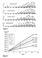


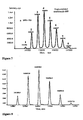


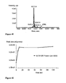
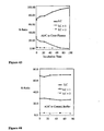
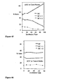
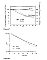
Classifications
-
- C—CHEMISTRY; METALLURGY
- C07—ORGANIC CHEMISTRY
- C07K—PEPTIDES
- C07K16/00—Immunoglobulins [IGs], e.g. monoclonal or polyclonal antibodies
- C07K16/18—Immunoglobulins [IGs], e.g. monoclonal or polyclonal antibodies against material from animals or humans
- C07K16/32—Immunoglobulins [IGs], e.g. monoclonal or polyclonal antibodies against material from animals or humans against translation products of oncogenes
-
- C—CHEMISTRY; METALLURGY
- C07—ORGANIC CHEMISTRY
- C07K—PEPTIDES
- C07K16/00—Immunoglobulins [IGs], e.g. monoclonal or polyclonal antibodies
- C07K16/18—Immunoglobulins [IGs], e.g. monoclonal or polyclonal antibodies against material from animals or humans
- C07K16/28—Immunoglobulins [IGs], e.g. monoclonal or polyclonal antibodies against material from animals or humans against receptors, cell surface antigens or cell surface determinants
- C07K16/2866—Immunoglobulins [IGs], e.g. monoclonal or polyclonal antibodies against material from animals or humans against receptors, cell surface antigens or cell surface determinants against receptors for cytokines, lymphokines, interferons
-
- G—PHYSICS
- G01—MEASURING; TESTING
- G01N—INVESTIGATING OR ANALYSING MATERIALS BY DETERMINING THEIR CHEMICAL OR PHYSICAL PROPERTIES
- G01N33/00—Investigating or analysing materials by specific methods not covered by groups G01N1/00 - G01N31/00
- G01N33/48—Biological material, e.g. blood, urine; Haemocytometers
- G01N33/50—Chemical analysis of biological material, e.g. blood, urine; Testing involving biospecific ligand binding methods; Immunological testing
- G01N33/68—Chemical analysis of biological material, e.g. blood, urine; Testing involving biospecific ligand binding methods; Immunological testing involving proteins, peptides or amino acids
- G01N33/6803—General methods of protein analysis not limited to specific proteins or families of proteins
- G01N33/6848—Methods of protein analysis involving mass spectrometry
-
- G—PHYSICS
- G01—MEASURING; TESTING
- G01N—INVESTIGATING OR ANALYSING MATERIALS BY DETERMINING THEIR CHEMICAL OR PHYSICAL PROPERTIES
- G01N33/00—Investigating or analysing materials by specific methods not covered by groups G01N1/00 - G01N31/00
- G01N33/48—Biological material, e.g. blood, urine; Haemocytometers
- G01N33/50—Chemical analysis of biological material, e.g. blood, urine; Testing involving biospecific ligand binding methods; Immunological testing
- G01N33/68—Chemical analysis of biological material, e.g. blood, urine; Testing involving biospecific ligand binding methods; Immunological testing involving proteins, peptides or amino acids
- G01N33/6854—Immunoglobulins
-
- A—HUMAN NECESSITIES
- A61—MEDICAL OR VETERINARY SCIENCE; HYGIENE
- A61K—PREPARATIONS FOR MEDICAL, DENTAL OR TOILETRY PURPOSES
- A61K39/00—Medicinal preparations containing antigens or antibodies
- A61K2039/505—Medicinal preparations containing antigens or antibodies comprising antibodies
-
- Y—GENERAL TAGGING OF NEW TECHNOLOGICAL DEVELOPMENTS; GENERAL TAGGING OF CROSS-SECTIONAL TECHNOLOGIES SPANNING OVER SEVERAL SECTIONS OF THE IPC; TECHNICAL SUBJECTS COVERED BY FORMER USPC CROSS-REFERENCE ART COLLECTIONS [XRACs] AND DIGESTS
- Y10—TECHNICAL SUBJECTS COVERED BY FORMER USPC
- Y10S—TECHNICAL SUBJECTS COVERED BY FORMER USPC CROSS-REFERENCE ART COLLECTIONS [XRACs] AND DIGESTS
- Y10S436/00—Chemistry: analytical and immunological testing
- Y10S436/825—Pretreatment for removal of interfering factors from sample
-
- Y—GENERAL TAGGING OF NEW TECHNOLOGICAL DEVELOPMENTS; GENERAL TAGGING OF CROSS-SECTIONAL TECHNOLOGIES SPANNING OVER SEVERAL SECTIONS OF THE IPC; TECHNICAL SUBJECTS COVERED BY FORMER USPC CROSS-REFERENCE ART COLLECTIONS [XRACs] AND DIGESTS
- Y10—TECHNICAL SUBJECTS COVERED BY FORMER USPC
- Y10T—TECHNICAL SUBJECTS COVERED BY FORMER US CLASSIFICATION
- Y10T436/00—Chemistry: analytical and immunological testing
- Y10T436/24—Nuclear magnetic resonance, electron spin resonance or other spin effects or mass spectrometry
Definitions
- the invention relates generally to methods to detect, analyze, screen, characterize, and quantitate antibody conjugate compounds, including antibody-drug conjugates, and their fragments and metabolites, by mass spectrometry.
- the invention also relates to methods to prepare mass spectrometric samples for pharmacokinetic studies.
- Targeted anti-cancer therapeutics are designed to reduce nonspecific toxicities and increase efficacy relative to conventional cancer chemotherapy.
- This approach is embodied by the powerful targeting ability of monoclonal antibodies to specifically deliver highly potent, conjugated small molecule therapeutics to a cancer cell.
- the pharmacokinetics and biodistribution of an antibody play a major role in determining whether its use in the clinic will be successful.
- the antibody must be capable of being delivered to the site of action and be retained there for the length of time necessary to achieve its purpose.
- To evaluate properties such as pharmacokinetics and toxicity of these antibody-drug conjugates it is useful to be able to characterize and quantitate them from plasma, urine, and other biological samples. Additionally, the ability to quantitate the free drug (not conjugated to the antibody) in the method from the same sample and the same chromatographic injection would also be useful.
- EI electron impact
- CI chemical ionization
- DCI desorption chemical ionization
- FAB fast atom bombardment
- ESI electrospray ionization
- MALDI matrix-assisted laser desorption/ionization
- MS/MS tandem mass spectrometry
- LC/MS is used for characterization of antibodies (Martin et al (1997) Cancer Chemother. Pharmacol. 40:189-201 ; WO 03/046571 ; WO 03/046572 ), and ELISA is used for quantitation in biological matrices ( Murray et al (2001) J. Imm. Methods 255:41-56 ; Kirchner et al (2004) Clin. Pharmacokinetics 43(2):83-95 ).
- ELISA assays typically are sensitive and amenable to high-throughput screens.
- Electrospray ionization provides for the atmospheric pressure ionization (API) of a liquid sample.
- the electrospray process creates highly-charged droplets that, under evaporation, create ions representative of the species contained in the solution.
- An ion-sampling orifice of a mass spectrometer may be used to sample these gas phase ions for mass analysis.
- the response for an analyte measured by the mass spectrometer detector is dependent on the concentration of the analyte in the fluid and independent of the fluid flow rate.
- HERCEPTIN® is a recombinant DNA-derived humanized monoclonal antibody that selectively binds to the extracellular domain (ECD) of the human epidermal growth factor receptor2 protein, HER2 (ErbB2) ( US 5821337 ; US 6054297 ; US 6407213 ; US 6639055 ; Coussens et al (1985) Science 230:1132-9 ; Slamon, et al (1989) Science 244:707-12 ).
- Trastuzumab is an IgG1 kappa antibody that contains human framework regions with the complementarity-determining regions (cdr) of a murine antibody (4D5) that binds to HER2.
- Trastuzumab binds to the HER2 antigen and thus inhibits the proliferation of human tumor cells that overexpress HER2 ( Hudziak RM, et al (1989) Mol Cell Biol 9:1165-72 ; Lewis GD, et al (1993) Cancer Immunol Immunother; 37:255-63 ; Baselga J, et al (1998) Cancer Res. 58:2825-2831 ).
- HERCEPTIN® as a single agent is indicated for the treatment of patients with metastatic breast cancer whose tumors overexpress the HER2 protein and who have received one or more chemotherapy regimens for their metastatic disease.
- HERCEPTIN® in combination with paclitaxel is indicated for treatment of patients with metastatic breast cancer whose tumors overexpress the HER2 protein and who have not received chemotherapy for their metastatic disease.
- HERCEPTIN® is clinically active in patients with ErbB2-overexpressing metastatic breast cancers that have received extensive prior anti-cancer therapy ( Baselga et al (1996) J. Clin. Oncol. 14:737-744 ).
- the aim of antibody therapy and diagnosis is to exploit the combination of high specificity and affinity of the antibody-antigen interaction, to enable detection and/or treatment of a particular lesion or disorder.
- the antibody is used alone, or is conjugated, i.e. loaded, with another moiety such as a detection label, pharmacokinetic modifier, radioisotope, toxin, or drug.
- ADC antibody-drug conjugates
- immunoconjugates for the local delivery of cytotoxic or cytostatic agents to kill or inhibit tumor cells in the treatment of cancer
- Toxins used in antibody-toxin conjugates include bacterial toxins such as diphtheria toxin, plant toxins such as ricin, small molecule toxins such as geldanamycin ( Mandler et al (2000) Jour. of the Nat. Cancer Inst.
- toxins and drugs may exert their cytotoxic and cytostatic effects by mechanisms including tubulin binding, DNA binding, or topoisomerase inhibition. Some cytotoxic drugs tend to be inactive or less active when conjugated to large antibodies or protein receptor ligands.
- Exemplary antibody-drug conjugates include MYLOTARGTM (gemtuzumab ozogamicin, Wyeth Pharmaceuticals), a hu CD33 antibody linked to calicheamicin, which was approved in 2000 for the treatment of acute myeloid leukemia by injection ( Drugs of the Future (2000) 25(7):686 ; US Patent Nos. 4970198 ; 5079233 ; 5585089 ; 5606040 ; 5693762 ; 5739116 ; 5767285 ; 5773001 ).
- Cantuzumab mertansine (Immunogen, Inc.), an antibody-drug conjugate composed of the huC242 antibody linked via the disulfide linker SPP to the maytansinoid drug moiety DM1, is advancing into Phase II trials for the treatment of cancers that express CanAg, such as colon, pancreatic, gastric, and others.
- MLN-2704 (Millennium Pharm, BZL Biologics, Immunogen Inc.), an antibody-drug conjugate composed of the anti-prostate specific membrane antigen (PSMA) monoclonal antibody also linked to DM1, is under development for the potential treatment of prostate tumors.
- PSMA anti-prostate specific membrane antigen
- auristatin peptides auristatin E (AE) and monomethylauristatin (MMAE) synthetic analogs of dolastatin, were conjugated to chimeric monoclonal antibodies cBR96 (specific to Lewis Y on carcinomas) and cAC10 (specific to CD30 on hematological malignancies), and are under therapeutic development ( Doronina et al (2003) Nature Biotechnology 21(7):778-784 ; Francisco et al (2003) Blood 102:1458-1465 ).
- the above-mentioned antibody-drug conjugates (ADC) which are approved or under development for therapeutic use are heterogeneous mixtures where the process of covalent attachment of the drug moiety to the antibody is largely uncontrolled and the resulting conjugation products are incompletely characterized.
- the drug loading is a statistical average for the collection of ADC molecules in a composition or formulation. Because of the heterogeneous nature of antibody-drug conjugate compositions, pharmacokinetic samples collected from biological sources after administration are difficult to evaluate. ELISA assays are limited to detection of antibody-antigen binding ( DiJoseph et al (2004) Blood 103:1807-1814 ). UV spectroscopy can measure the total absorbance of certain fluorescent or UV-active drug moieties or metabolites, but cannot distinguish between free drug and antibody-drug conjugate.
- An aspect of the invention includes methods to detect, screen, and quantitate antibody conjugate compounds and compositions, antibodies, and fragments and metabolites thereof, by affinity separation, chromatography, and mass spectrometry as defined in the claims.
- Mass spectrometry is single ion monitoring (SIM) mass spectrometry (MS). The practice or technique of SIM is sometimes referred to as selected ion monitoring or selective ion monitoring.
- An aspect of the invention includes a method for detecting antibody-drug conjugate compounds comprising
- the invention relates to methods for screening a mixture of antibody-drug conjugate compounds to determine the relative clearance of the compounds, or fragments or metabolites thereof, in a biological source, such as a mammal, tissue, or cell culture.
- the invention relates to methods for detecting by mass spectrometry, compounds of a heterogeneous mixture of antibody-drug conjugate compounds having Formula I, wherein the mixture includes compounds having more than one drug loading value, p, where p may be 1, 2, 3, 4, 5, 6, 7, or 8; the method comprises performing mass spectrometry on a sample comprising the heterogeneous mixture of antibody-drug conjugate compounds and detecting more than one compound of the mixture, or fragments or metabolites thereof.
- the invention relates to methods to quantitate levels of both free drug and antibody-drug conjugates in plasma by LC/MS with a single analysis.
- the invention relates to methods for studying in vivo metabolism of antibody-drug conjugates.
- the invention relates to methods of pharmacokinetic analysis of antibody-drug conjugates after administration to mammals.
- the invention relates to methods of patient profiling by measuring susceptibility and response to antibody-drug conjugate therapeutic treatment.
- the invention diagnostic methods of diagnosing the presence or absence of a condition or disease characterized by the over-expression of a protein or antigen by administration of an antibody-drug conjugate to a mammal, tissue, or cell culture.
- the invention includes Immunoaffinity Membrane/LC/MS methods having immunoaffinity membrane (IAM) selection and reverse phase liquid chromatography (LC) front-end steps coupled with a back-end mass spectrometry (MS) detection step to isolate, cleanup, and detect antibody-drug conjugates in plasma as defined in the claims.
- IAM immunoaffinity membrane
- MS mass spectrometry
- SIM results for antibody conjugates and antibodies analyzed by the methods of the present invention are surprising and unexpected because SIM has not been recognized as useful for such large molecules.
- the methods of the present invention combine high resolution of ions and stability of the ion envelope by control of the declustering potential, pH, and the mobile phase.
- selection of high mass ions, rather than the most abundant, highest peak, ion, for SIM avoids matrix interference typical at the lower mass range.
- An antibody is a protein generated by the immune system that is capable of recognizing and binding to a specific antigen.
- a target antigen generally has numerous binding sites, also called epitopes, recognized by CDRs on multiple antibodies. Each antibody that specifically binds to a different epitope has a different structure. Thus, one antigen may have more than one corresponding antibody.
- antibody also refers to a full-length immunoglobulin molecule or an immunologically active portion of a full-length immunoglobulin molecule, i.e ., a molecule that contains an antigen binding site that immunospecifically binds an antigen of a target of interest or part thereof, such targets including but not limited to, cancer cell or cells that produce autoimmune antibodies associated with an autoimmune disease.
- the immunoglobulin disclosed herein can be of any type (e.g., IgG, IgE, IgM, IgD, and IgA), class ( e.g., IgG1, IgG2, IgG3, IgG4, IgA1 and IgA2) or subclass of immunoglobulin molecule.
- the immunoglobulins can be derived from any species. In one aspect, however, the immunoglobulin is of human, murine, or rabbit origin.
- Antibody fragments comprise a portion of a full length antibody, generally the antigen binding or variable region thereof.
- Examples of antibody fragments include Fab, Fab', F(ab') 2 , and Fv fragments; diabodies; linear antibodies; fragments produced by a Fab expression library, anti-idiotypic (anti-Id) antibodies, CDR (complementary determining region), ECD (extracellular domain), and epitope-binding fragments of any of the above which immunospecifically bind to cancer cell antigens, viral antigens or microbial antigens, single-chain antibody molecules; and multispecific antibodies formed from antibody fragments.
- an “intact antibody” herein is one comprising a VL and VH domains, as well as complete light and heavy chain constant domains.
- the term "monoclonal antibody” as used herein refers to an antibody obtained from a population of substantially homogeneous antibodies, i.e. , the individual antibodies comprising the population are identical except for possible naturally occurring mutations that may be present in minor amounts. Monoclonal antibodies are highly specific, being directed against a single antigenic site. Furthermore, in contrast to polyclonal antibody preparations which include different antibodies directed against different determinants (epitopes), each monoclonal antibody is directed against a single determinant on the antigen. In addition to their specificity, the monoclonal antibodies are advantageous in that they may be synthesized uncontaminated by other antibodies.
- the modifier "monoclonal” indicates the character of the antibody as being obtained from a substantially homogeneous population of antibodies, and is not to be construed as requiring production of the antibody by any particular method.
- the monoclonal antibodies to be used in accordance with the present invention may be made by the hybridoma method first described by Kohler et al (1975) Nature 256:495 , or may be made by recombinant DNA methods (see, US 4816567 ).
- the “monoclonal antibodies” may also be isolated from phage antibody libraries using the techniques described in Clackson et al (1991) Nature, 352:624-628 ; Marks et al (1991) J. Mol. Biol., 222:581-597 ; for example.
- the monoclonal antibodies herein specifically include "chimeric" antibodies in which a portion of the heavy and/or light chain is identical with or homologous to corresponding sequences in antibodies derived from a particular species or belonging to a particular antibody class or subclass, while the remainder of the chain(s) is identical with or homologous to corresponding sequences in antibodies derived from another species or belonging to another antibody class or subclass, as well as fragments of such antibodies, so long as they exhibit the desired biological activity ( US 4816567 ; and Morrison et al (1984) Proc. Natl. Acad. Sci. USA, 81:6851-6855 ).
- Chimeric antibodies of interest herein include "primatized" antibodies comprising variable domain antigen-binding sequences derived from a non-human primate (e.g., Old World Monkey, Ape etc) and human constant region sequences.
- Antibody fragments comprise a portion of an intact antibody, e.g. comprising the antigen-binding or variable region thereof.
- antibody fragments include Fab, Fab', F(ab')2, and Fv fragments; diabodies; linear antibodies; single-chain antibody molecules; and multispecific antibodies formed from antibody fragment(s).
- an “intact” antibody is one which comprises an antigen-binding variable region as well as a light chain constant domain (CL) and heavy chain constant domains, CH1, CH2 and CH3.
- the constant domains may be native sequence constant domains (e.g ., human native sequence constant domains) or amino acid sequence variant thereof.
- the intact antibody may have one or more "effector functions" which refer to those biological activities attributable to the Fc region (a native sequence Fc region or amino acid sequence variant Fc region) of an antibody.
- effector functions include Clq binding; complement dependent cytotoxicity; Fc receptor binding; antibody-dependent cell-mediated cytotoxicity (ADCC); phagocytosis; down regulation of cell surface receptors (e.g ., B cell receptor; BCR), etc.
- intact antibodies can be assigned to different "classes.” There are five major classes of intact antibodies: IgA, IgD, IgE, IgG, and IgM, and several of these may be further divided into “subclasses” (isotypes), e.g., IgG1, IgG2, IgG3, IgG4, IgA, and IgA2.
- the heavy-chain constant domains that correspond to the different classes of antibodies are called a, ⁇ , ⁇ , ⁇ , and ⁇ , respectively.
- the subunit structures and three-dimensional configurations of different classes of immunoglobulins are well known.
- amino acid sequence variant refers to polypeptides having amino acid sequences that differ to some extent from a native sequence polypeptide. Ordinarily, amino acid sequence variants will possess at least about 70% sequence identity with at least one receptor binding domain of a native antibody or with at least one ligand binding domain of a native receptor, and preferably, they will be at least about 80%, more preferably, at least about 90% homologous by sequence with such receptor or ligand binding domains. The amino acid sequence variants possess substitutions, deletions, and/or insertions at certain positions within the amino acid sequence of the native amino acid sequence. Amino acids are designated by the conventional names, one-letter and three-letter codes.
- Sequence identity is defined as the percentage of residues in the amino acid sequence variant that are identical after aligning the sequences and introducing gaps, if necessary, to achieve the maximum percent sequence identity. Methods and computer programs for the alignment are well known in the art. One such computer program is "Align 2,” authored by Genentech, Inc., which was filed with user documentation in the United States Copyright Office, Washington, DC 20559, on December 10,1991.
- Useful monoclonal antibodies are homogeneous populations of antibodies to a particular antigenic determinant (e.g ., a cancer cell antigen, a viral antigen, a microbial antigen, a protein, a peptide, a carbohydrate, a chemical, nucleic acid, or fragments thereof).
- a monoclonal antibody (MAb) to an antigen-of-interest can be prepared by using any technique known in the art which provides for the production of antibody molecules by continuous cell lines in culture.
- Such antibodies may be of any immunoglobulin class including IgG, IgM, IgE, IgA, and IgD and any subclass thereof.
- the hybridoma producing the MAbs of use in this invention may be cultivated in vitro or in vivo.
- Useful monoclonal antibodies include, but are not limited to, human monoclonal antibodies, humanized monoclonal antibodies, antibody fragments, or chimeric human-mouse (or other species) monoclonal antibodies.
- Human monoclonal antibodies may be made by any of numerous techniques known in the art (e.g ., Teng et al, 1983, Proc. Natl. Acad. Sci. U.S.A. 80,7308-7312 ; Kozbor et al, 1983, Immununology Today 4, 72-79 ; and Olsson et al, 1982, Meth. Enzymol. 92, 3-16 ).
- the antibody can also be a bispecific antibody.
- Bispecific antibodies may have a hybrid immunoglobulin heavy chain with a first binding specificity in one arm, and a hybrid immunoglobulin heavy chain-light chain pair (providing a second binding specificity) in the other arm.
- This asymmetric structure facilitates the separation of the desired bispecific compound from unwanted immunoglobulin chain combinations, as the presence of an immunoglobulin light chain in only one half of the bispecific molecule provides for a facile way of separation ( WO 94/04690 ; Suresh et al, Methods in Enzymology, 1986, 121:210 ; Rodrigues et al, 1993, J.
- bispecific antibodies can be prepared for conjugation as ADC in the treatment or prevention of disease as defined herein.
- antibody variable domains with the desired binding specificities are fused to immunoglobulin constant domain sequences.
- the fusion may be with an immunoglobulin heavy chain constant domain, comprising at least part of the hinge, C H 2, and C H 3 regions.
- the first heavy-chain constant region (C H 1) may contain the site necessary for light chain binding, present in at least one of the fusions.
- Nucleic acids with sequences encoding the immunoglobulin heavy chain fusions and, if desired, the immunoglobulin light chain are inserted into separate expression vectors, and are co-transfected into a suitable host organism.
- Hybrid or bifunctional antibodies can be derived either biologically, i.e., by cell fusion techniques, or chemically, especially with cross-linking agents or disulfide-bridge forming reagents, and may comprise whole antibodies or fragments thereof ( EP 105360 ; WO 83/03679 ; EP 217577 ).
- the antibody can be a functionally active fragment, derivative or analog of an antibody that immunospecifically binds to cancer cell antigens, viral antigens, or microbial antigens or other antibodies bound to tumor cells or matrix.
- “functionally active” means that the fragment, derivative or analog is able to elicit anti-anti-idiotype antibodies that recognize the same antigen that the antibody from which the fragment, derivative or analog is derived recognized.
- the antigenicity of the idiotype of the immunoglobulin molecule can be enhanced by deletion of framework and CDR sequences that are C-terminal to the CDR sequence that specifically recognizes the antigen.
- synthetic peptides containing the CDR sequences can be used in binding assays with the antigen by any binding assay method known in the art (e.g., the BIA core assay) (See, for e.g., Kabat et al, 1991, Sequences of Proteins of Immunological Interest, Fifth Edition, National Institute of Health, Bethesda, Md ; Kabat E et al, 1980, J. of Immunology 125(3):961-969 ).
- Other useful antibodies include fragments of antibodies such as, but not limited to, F(ab')2 fragments, which contain the variable region, the light chain constant region and the CH1 domain of the heavy chain can be produced by pepsin digestion of the antibody molecule, and Fab fragments, which can be generated by reducing the disulfide bridges of the F(ab')2 fragments.
- Other useful antibodies are heavy chain and light chain dimers of antibodies, or any minimal fragment thereof such as Fvs or single chain antibodies (SCAs) (e.g., as described in US 4946778 ; Bird, (1988), Science 242:423-42 ; Huston et al, (1988) Proc. Natl. Acad. Sci. USA 85:5879-5883 ; and Ward et al, (1989) Nature 334:544-54 ), or any other molecule with the same specificity as the antibody.
- SCAs single chain antibodies
- recombinant antibodies such as chimeric and humanized monoclonal antibodies, comprising both human and non-human portions, which can be made using standard recombinant DNA techniques, are useful antibodies.
- a chimeric antibody is a molecule in which different portions are derived from different animal species, such as those having a variable region derived from a murine monoclonal and human immunoglobulin constant regions ( US 4816567 ; and US 4816397 ).
- Chimeric and humanized monoclonal antibodies can be produced by recombinant DNA techniques known in the art, for example using methods described in WO 87/02671 ; EP 184,187 ; EP 171496 ; EP 173494 ; WO 86/01533 ; US 4816567 ; EP 12023 ; Berter et al, (1988) Science 240:1041-1043 ; Liu et al, (1987) Proc. Natl. Acad. Sci. USA 84:3439-3443 ; Liu et al, (1987) J. ImmunoL 139:3521-3526 ; Sun et al, (1987) Proc. Natl. Acad. Sci.
- Completely human antibodies can be produced using transgenic mice that are incapable of expressing endogenous immunoglobulin heavy and light chains genes, but which can express human heavy and light chain genes.
- the transgenic mice are immunized in the normal fashion with a selected antigen, e.g., all or a portion of a polypeptide of the invention.
- Monoclonal antibodies directed against the antigen can be obtained using conventional hybridoma technology.
- the human immunoglobulin transgenes harbored by the transgenic mice rearrange during B cell differentiation, and subsequently undergo class switching and somatic mutation. Thus, using such a technique, it is possible to produce therapeutically useful IgG, IgA, IgM and IgE antibodies.
- Completely human antibodies that recognize a selected epitope can be generated using a technique referred to as "guided selection.”
- a selected non-human monoclonal antibody e.g., a mouse antibody
- Human antibodies can also be produced using various techniques known in the art, including phage display libraries ( Hoogenboom and Winter, J. Mol. BioL, 227:381 (1991 ); Marks et al, J. Mol. Biol., 222:581 (1991 )).
- the antibody may be a fusion protein of an antibody, or a functionally active fragment thereof, for example in which the antibody is fused via a covalent bond (e.g., a peptide bond), at either the N-terminus or the C-terminus to an amino acid sequence of another protein (or portion thereof, such as at least 10, 20 or 50 amino acid portion of the protein) that is not the antibody.
- a covalent bond e.g., a peptide bond
- the antibody or fragment thereof may be covalently linked to the other protein at the N-terminus of the constant domain.
- Antibodies include analogs and derivatives that are either modified, i.e ., by the covalent attachment of any type of molecule as long as such covalent attachment permits the antibody to retain its antigen binding immunospecificity.
- the derivatives and analogs of the antibodies include those that have been further modified, e.g., by glycosylation, acetylation, pegylation, phosphorylation, amidation, derivatization by known protecting/blocking groups, proteolytic cleavage, linkage to a cellular antibody unit or other protein, etc.
- analog or derivative can contain one or more unnatural amino acids.
- the antibodies in ADC include antibodies having modifications (e.g., substitutions, deletions or additions) in amino acid residues that interact with Fc receptors.
- antibodies include antibodies having modifications in amino acid residues identified as involved in the interaction between the anti-Fc domain and the FcRn receptor (see: WO 97/34631 ).
- Antibodies immunospecific for a cancer cell antigen can be obtained commercially, for example, from Genentech, Inc. (South San Francisco, CA) or produced by any method known to one of skill in the art such as, e.g., chemical synthesis or recombinant expression techniques.
- the nucleotide sequence encoding antibodies immunospecific for a cancer cell antigen can be obtained, e.g., from the GenBank database or a database like it, the literature publications, or by routine cloning and sequencing.
- receptor includes any peptide, protein, glycoprotein, polycarbohydrate, or lipid that is uniquely expressed or overexpressed on the surface of a cell, and is exposed on the surface of a cell in a manner that will allow interaction with a circulating targeting agent, such as an antibody-drug conjugate.
- Cells bearing receptors include tumor cells.
- patient includes human and veterinary subjects.
- mammal for purposes of administration of, or contact with, antibody-drug conjugates refers to any animal classified as a mammal, including human, domestic and farm animals, nonhuman primates, and any other animal that has mammalian tissue.
- Bio sample means (i) blood, bile, urine, or feces; (ii) tissue extract; and (iii) cell culture media, cell lysate, or cell extract.
- Bio source means (i) mammals such as a mouse, a rat, a rabbit, a dog, a monkey, or a human; (ii) mammalian tissue; and (iii) cultured cells.
- label means any moiety which can be covalently attached to an antibody and that functions to: (i) provide a detectable signal; (ii) interact with a second label to modify the detectable signal provided by the first or second label, e.g. FRET (fluorescence resonance energy transfer); (iii) stabilize interactions or increase affinity of binding, with antigen or ligand; (iv) affect mobility, e.g. electrophoretic mobility, or cell-permeability, by charge, hydrophobicity, shape, or other physical parameters, or (v) provide a capture moiety, to modulate ligand affinity, antibody/antigen binding, or ionic complexation.
- FRET fluorescence resonance energy transfer
- Alkyl is a C 1 -C 18 hydrocarbon moiety containing normal, secondary, tertiary or cyclic carbon atoms.
- alkyl radicals include C 1 -C 8 hydrocarbon moieties such as: methyl (Me, -CH 3 ), ethyl (Et,-CH 2 CH 3 ), 1-propyl (n-Pr, n-propyl, -CH 2 CH 2 CH 3 ), 2-propyl (i-Pr, i-propyl, -CH(CH 3 ) 2 ), 1-butyl (n-Bu, n-butyl, -CH 2 CH 2 CH 2 CH 3 ), 2-methyl-1-propyl (i-Bu, i-butyl, -CH 2 CH(CH 3 ) 2 ), 2-butyl (s-Bu, s-butyl,-CH(CH 3 )CH 2 CH 3 ), 2-methyl-2-propyl ( t -Bu, t -butyl,
- 2-methyl-2-butyl (-C(CH 3 ) 2 CH 2 CH 3 ), 3-methyl-2-butyl (-CH(CH 3 )CH(CH 3 ) 2 ), 3-methyl-l-butyl (-CH 2 CH 2 CH(CH 3 ) 2 ), 2-methyl-1-butyl (-CH 2 CH(CH 3 )CH 2 CH 3 ), 1-hexyl (-CH 2 CH 2 CH 2 CH 2 CH 2 CH 3 ), 2-hexyl (-CH(CH 3 )CH 2 CH 2 CH 2 CH 3 ), 3-hexyl (-CH(CH 2 CH 3 )(CH 2 CH 2 CH 3 )), 2-methyl-2-pentyl (-C(CH 3 ) 2 CH 2 CH 2 CH 3 ).
- Aryl means a monovalent aromatic hydrocarbon radical of 6-20 carbon atoms derived by the removal of one hydrogen atom from a single carbon atom of a parent aromatic ring system. Some aryl groups are represented in the exemplary structures as "Ar”. Typical aryl groups include, but are not limited to, radicals derived from benzene, substituted benzene, naphthalene, anthracene, biphenyl.
- Substituted alkyl and “substituted aryl” mean alkyl and aryl, respectively, in which one or more hydrogen atoms are each independently replaced with a substituent.
- Heteroaryl “heterocyclyl”, and “heterocycle” all refer to a ring system in which one or more ring atoms is a heteroatom, e.g. nitrogen, oxygen, and sulfur.
- the heterocycle radical comprises 1 to 20 carbon atoms and 1 to 5 heteroatoms selected from N, O, P, and S.
- a heterocycle may be a monocycle having 3 to 7 ring members (2 to 6 carbon atoms and 1 to 3 heteroatoms selected from N, O, P, and S) or a bicycle having 7 to 10 ring members (4 to 9 carbon atoms and 1 to 3 heteroatoms selected from N, O, P, and S), for example: a bicyclo [4,5], [5,5], [5,6], or [6,6] system.
- Heterocycles are described in Paquette, Leo A.; "Principles of Modern Heterocyclic Chemistry” (W.A. Benjamin, New York, 1968), particularly Chapters 1,3,4, 6,7, and 9 ; “ The Chemistry of Heterocyclic Compounds, A series of Monographs” (John Wiley & Sons, New York, 1950 to present), in particular Volumes 13, 14, 16, 19, and 28 ; and J. Am. Chem. Soc. (1960) 82:5566 .
- heterocycles include by way of example and not limitation pyridyl, dihydroypyridyl, tetrahydropyridyl (piperidyl), thiazolyl, tetrahydrothiophenyl, sulfur oxidized tetrahydrothiophenyl, pyrimidinyl, furanyl, thienyl, pyrrolyl, pyrazolyl, imidazolyl, tetrazolyl, benzofuranyl, thianaphthalenyl, indolyl, indolenyl, quinolinyl, isoquinolinyl, benzimidazolyl, piperidinyl, 4-piperidonyl, pyrrolidinyl, 2-pyrrolidonyl, pyrrolinyl, tetrahydrofuranyl, bis-tetrahydrofuranyl, tetrahydropyranyl, bis-tetrahydropyranyl, tetra
- carbon bonded heterocycles are bonded at position 2, 3, 4, 5, or 6 of a pyridine, position 3, 4, 5, or 6 of a pyridazine, position 2, 4, 5, or 6 of a pyrimidine, position 2, 3, 5, or 6 of a pyrazine, position 2, 3, 4, or 5 of a furan, tetrahydrofuran, thiofuran, thiophene, pyrrole or tetrahydropyrrole, position 2, 4, or 5 of an oxazole, imidazole or thiazole, position 3, 4, or 5 of an isoxazole, pyrazole, or isothiazole, position 2 or 3 of an aziridine, position 2, 3, or 4 of an azetidine, position 2, 3, 4, 5, 6, 7, or 8 of a quinoline or position 1, 3, 4, 5, 6, 7, or 8 of an isoquinoline.
- carbon bonded heterocycles include 2-pyridyl, 3-pyridyl, 4-pyridyl, 5-pyridyl, 6-pyridyl, 3-pyridazinyl, 4-pyridazinyl, 5-pyridazinyl, 6-pyridazinyl, 2-pyrimidinyl, 4-pyrimidinyl, 5-pyrimidinyl, 6-pyrimidinyl, 2-pyrazinyl, 3-pyrazinyl, 5-pyrazinyl, 6-pyrazinyl, 2-thiazolyl, 4-thiazolyl, or 5-thiazolyl.
- nitrogen bonded heterocycles are bonded at position 1 of an aziridine, azetidine, pyrrole, pyrrolidine, 2-pyrroline, 3-pyrroline, imidazole, imidazolidine, 2-imidazoline, 3-imidazoline, pyrazole, pyrazoline, 2-pyrazoline, 3-pyrazoline, piperidine, piperazine, indole, indoline, 1H- indazole, position 2 of a isoindole, or isoindoline, position 4 of a morpholine, and position 9 of a carbazole, or ⁇ -carboline.
- nitrogen bonded heterocycles include 1-aziridyl, 1-azetedyl, 1-pyrrolyl, 1-imidazolyl, 1-pyrazolyl, and 1-piperidinyl.
- Carbocycle and “carbocyclyl” mean a saturated or unsaturated ring having 3 to 7 carbon atoms as a monocycle or 7 to 12 carbon atoms as a bicycle.
- Monocyclic carbocycles have 3 to 6 ring atoms, still more typically 5 or 6 ring atoms.
- Bicyclic carbocycles have 7 to 12 ring atoms, e.g. arranged as a bicyclo [4,5], [5,5], [5,6] or [6,6] system, or 9 or 10 ring atoms arranged as a bicyclo [5,6] or [6,6] system.
- Examples of monocyclic carbocycles include cyclopropyl, cyclobutyl, cyclopentyl, 1-cyclopent-1-enyl, 1-cyclopent-2-enyl, 1-cyclopent-3-enyl, cyclohexyl, 1-cyclohex-1-enyl, 1-cyclohex-2-enyl, 1-cyclohex-3-enyl, cycloheptyl, and cyclooctyl.
- Reactive functional groups include, but are not limited to, olefins, acetylenes, alcohols, phenols, ethers, oxides, halides, aldehydes, ketones, carboxylic acids, esters, carbonates, amides, cyanates, isocyanates, thiocyanates, isothiocyanates, amines, hydrazines, hydrazones, hydrazides, diazo, diazonium, nitro, nitriles, mercaptans (thiols), sulfides, disulfides, sulfoxides, sulfones, sulfonic acids, sulfinic acids, acetals, ketals, anhydrides, sulfates, sulfenic acids, isonitriles, amidines, imides, imidates, nitrones, hydroxylamines, oximes, hydroxamic acids, thiohydroxamic acids, allene
- Exemplary reactive functional groups include N-hydroxysuccinimide (NHS)esters, para-nitrophenyl (PNP) carbonates, pentafluorophenyl (PFP) carbonates, and maleimides.
- NHS N-hydroxysuccinimide
- PNP para-nitrophenyl
- PFP pentafluorophenyl
- Linker means a chemical moiety comprising a covalent bond or a chain of atoms that covalently attaches an antibody to a drug moiety.
- a linker is specified as L.
- Linkers include a divalent radical such as an alkylene, an aryldiyl, a heteroaryldiyl, moieties such as: -(CR 2 ) n O(CR 2 ) n -, repeating units of alkyloxy (e.g. polyethylenoxy, PEG, polymethyleneoxy) and allylamino (e.g. polyethyleneamino, Jeffamine TM ); and diacid ester and amides including succinate, succinamide, diglycolate, malonate, and caproamide.
- chiral refers to molecules which have the property of non-superimposability of the mirror image partner, while the term “achiral” refers to molecules which are superimposable on their mirror image partner.
- stereoisomers refers to compounds which have identical chemical constitution, but differ with regard to the arrangement of the atoms or groups in space.
- Diastereomer refers to a stereoisomer with two or more centers of chirality and whose molecules are not mirror images of one another. Diastereomers have different physical properties, e.g. melting points, boiling points, spectral properties, and reactivities. Mixtures of diastereomers may separate under high resolution analytical procedures such as electrophoresis and chromatography.
- Enantiomers refer to two stereoisomers of a compound which are non-superimposable mirror images of one another.
- d and 1 or (+) and (-) are employed to designate the sign of rotation of plane-polarized light by the compound, with (-) or 1 meaning that the compound is levorotatory.
- a compound prefixed with (+) or d is dextrorotatory.
- these stereoisomers are identical except that they are mirror images of one another.
- a specific stereoisomer may also be referred to as an enantiomer, and a mixture of such isomers is often called an enantiomeric mixture.
- a 50:50 mixture of enantiomers is referred to as a racemic mixture or a racemate, which may occur where there has been no stereoselection or stereospecificity in a chemical reaction or process.
- racemic mixture and racemate refer to an equimolar mixture of two enantiomeric species, devoid of optical activity.
- phrases "pharmaceutically acceptable salt,” as used herein, refers to pharmaceutically acceptable organic or inorganic salts of an ADC.
- Exemplary salts include, but are not limited, to sulfate, citrate, acetate, oxalate, chloride, bromide, iodide, nitrate, bisulfate, phosphate, acid phosphate, isonicotinate, lactate, salicylate, acid citrate, tartrate, oleate, tannate, pantothenate, bitartrate, ascorbate, succinate, maleate, gentisinate, fumarate, gluconate, glucuronate, saccharate, formate, benzoate, glutamate, methanesulfonate, ethanesulfonate, benzenesulfonate, p -toluenesulfonate, and pamoate ( i.e., 1,1'-methylene bis -(2-hydroxy-3-
- a pharmaceutically acceptable salt may involve the inclusion of another molecule such as an acetate ion, a succinate ion or other counterion.
- the counterion may be any organic or inorganic moiety that stabilizes the charge on the parent compound.
- a pharmaceutically acceptable salt may have more than one charged atom in its structure. Instances where multiple charged atoms are part of the pharmaceutically acceptable salt can have multiple counter ions. Hence, a pharmaceutically acceptable salt can have one or more charged atoms and/or one or more counterion.
- “Pharmaceutically acceptable solvate” refers to an association of one or more solvent molecules and an ADC.
- solvents that form pharmaceutically acceptable solvates include, but are not limited to, water, isopropanol, ethanol, methanol, DMSO, ethyl acetate, acetic acid, and ethanolamine.
- the antibody unit (Ab-) of Formula I includes within its scope any unit of an antibody (Ab) that binds or reactively associates or complexes with a receptor, antigen or other receptive moiety associated with a given target-cell population.
- An antibody can be any protein or protein-like molecule that binds to, complexes with, or reacts with a moiety of a cell population sought to be therapeutically or otherwise biologically modified.
- the antibody unit acts to deliver the Drug unit to the particular target cell population with which the antibody unit reacts.
- Such antibodies include, but are not limited to, large molecular weight proteins such as, full-length antibodies and antibody fragments.
- ADC antibody-drug conjugates
- Antibodies which comprise Ab in Formula I antibody-drug conjugates (ADC) and which may be useful in the treatment of cancer include, but are not limited to, antibodies against tumor-associated antigens (TAA).
- TAA tumor-associated antigens
- TAA tumor-associated antigens
- TAA tumor-associated antigens
- TAA tumor-associated antigens
- TAA tumor-associated antigens
- researchers have sought to identify transmembrane or otherwise tumor-associated polypeptides that are specifically expressed on the surface of one or more particular type(s) of cancer cell as compared to on one or more normal non-cancerous cell(s). Often, such tumor-associated polypeptides are more abundantly expressed on the surface of the cancer cells as compared to on the surface of the non-cancerous cells. The identification of such tumor-associated cell surface antigen polypeptides has given rise to the ability to specifically target cancer cells for destruction via antibody-based therapies.
- TAA examples include, but are not limited to, TAA (1)-(35) listed below.
- TAA (1)-(35) listed below.
- information relating to these antigens, all of which are known in the art, is listed below and includes names, alternative names, Genbank accession numbers and primary reference(s).
- Tumor-associated antigens targeted by antibodies include all amino acid sequence variants and isoforms possessing at least about 70%, 80%, 85%, 90%, or 95% sequence identity relative to the sequences identified in the cited references, or which exhibit substantially the same biological properties or characteristics as a TAA having a sequence found in the cited references.
- a TAA having a variant sequence generally is able to bind specifically to an antibody that binds specifically to the TAA with the corresponding sequence listed.
- Herceptin® full length, humanized antiHER2 (MW 145167)
- Herceptin F(ab')2 derived from antiHER2 enzymatically (MW 100000)
- 4D5 full-length, murine antiHER2, from hybridoma
- rhu4D5 transiently expressed, full-length humanized antibody
- rhuFab4D5 recombinant humanized Fab (MW 47738)
- 4D5Fc8 full-length, murine antiHER2, with mutated FcRn binding domain.
- the antibody of the antibody-drug conjugates (ADC) for use in the invention may specifically bind to a receptor encoded by an ErbB gene.
- the antibody may bind specifically to an ErbB receptor selected from EGFR, HER2, HER3 and HER4.
- the ADC may specifically bind to the extracellular domain of the HER2 receptor and inhibit the growth of tumor cells which overexpress HER2 receptor.
- HERCEPTIN® selectively binds to the extracellular domain (ECD) of the human epidermal growth factor receptor2 protein, HER2 (ErbB2) ( US 5821337 ; US 6054297 ; US 6407213 ; US 6639055 ; Coussens et al (1985) Science 230:1132-9 ; Slamon, et al (1989) Science 244:707-12 ).
- Trastuzumab is an IgG1 kappa antibody that contains human framework regions with the complementarity-determining regions (cdr) of a murine antibody (4D5) that binds to HER2.
- trastuzumab binds to the HER2 antigen and thus inhibits the proliferation of human tumor cells that overexpress HER2 ( Hudziak RM, et al (1989) Mol Cell Biol 9:1165-72 ; Lewis GD, et al (1993) Cancer Immunol Immunother; 37:255-63 ; Baselga J, et al (1998) Cancer Res. 58:2825-2831 ).
- the antibody of the ADC may be a monoclonal antibody, e.g. a murine monoclonal antibody, a chimeric antibody, or a humanized antibody.
- a humanized antibody may be huMAb4D5-1, huMAb4D5-2, huMAb4D5-3, huMAb4D5-4, huMAb4D5-5, huMAb4D5-6, huMAb4D5-7 or huMAb4D5-8 (Trastuzumab).
- the antibody may be an antibody fragment, e.g. a Fab fragment
- Antibodies immunospecific for a cancer cell antigen can be obtained commercially or produced by any method known to one of skill in the art such as, e.g., recombinant expression techniques.
- the nucleotide sequence encoding antibodies immunospecific for a cancer cell antigen can be obtained, e.g., from the GenBank database or a database like it, the literature publications, or by routine cloning and sequencing.
- antibodies available for the treatment of cancer include, but are not limited to, humanized anti-HER2 monoclonal antibody for the treatment of patients with metastatic breast cancer; RITUXAN® (rituximab; Genentech) which is a chimeric anti-CD20 monoclonal antibody for the treatment of patients with non-Hodgkin's lymphoma; OvaRex (AltaRex Corporation, MA) which is a murine antibody for the treatment of ovarian cancer; Panorex (Glaxo Wellcome, NC) which is a murine IgG 2a antibody for the treatment of colorectal cancer; Cetuximab Erbitux (Imclone Systems Inc., NY) which is an anti-EGFR IgG chimeric antibody for the treatment of epidermal growth factor positive cancers, such as head and neck cancer; Vitaxin (MedImmune, Inc., MD) which is a humanized antibody for the treatment of sarcoma; Campath I/H (Leukosite,
- the antibodies for use in the invention may be conjugated with any label moiety which can be covalently attached to the antibody through a reactive functional group, such as a cysteine thiol or lysine amino ( Singh et al (2002) Anal. Biochem. 304:147-15 ; Harlow E. and Lane, D. (1999) Using Antibodies: A Laboratory Manual, Cold Springs Harbor Laboratory Press, Cold Spring Harbor, NY ; Lundblad R.L. (1991) Chemical Reagents for Protein Modification, 2nd ed. CRC Press, Boca Raton, FL ).
- the attached label may function to: (i) provide a detectable signal; (ii) interact with a second label to modify the detectable signal provided by the first or second label, e.g.
- FRET fluorescence resonance energy transfer
- stabilize interactions or increase affinity of binding, with antigen or ligand
- affect mobility e.g. electrophoretic mobility or cell-permeability, by charge, hydrophobicity, shape, or other physical parameters, or
- provide a capture moiety to modulate ligand affinity, antibody/antigen binding, or ionic complexation.
- the labelled antibodies may also be used as an affinity purification agent
- the labelled antibody is immobilized on a solid phase such a Sephadex resin or filter paper, using methods well known in the art.
- the immobilized antibody is contacted with a sample containing the antigen to be purified, and thereafter the support is washed with a suitable solvent that will remove substantially all the material in the sample except the antigen to be purified, which is bound to the immobilized polypeptide variant Finally, the support is washed with another suitable solvent, such as glycine buffer, pH 5.0, that will release the antigen from the polypeptide variant.
- the polypeptide variant may also be useful in diagnostic assays, e.g., for detecting expression of an antigen of interest in specific cells, tissues, or serum.
- the antibody will typically be labeled with a detectable moiety.
- a detectable moiety Numerous labels are available which can be generally grouped into the following categories:
- enzyme-substrate combinations include, for example:
- the label is indirectly conjugated with the polypeptide variant.
- the polypeptide variant can be conjugated with biotin and any of the categories of labels mentioned above can be conjugated with avidin, or vice versa. Biotin binds selectively to avidin and thus, the label can be conjugated with the polypeptide variant in this indirect manner.
- the polypeptide variant is conjugated with a small hapten ( e . g ., digoxin) and one of the different types of labels mentioned above is conjugated with an anti-hapten polypeptide variant (e.g ., anti-digoxin antibody).
- a small hapten e. g ., digoxin
- an anti-hapten polypeptide variant e.g ., anti-digoxin antibody
- the polypeptide variant may be employed in any known assay method, such as competitive binding assays, direct and indirect sandwich assays, and immunoprecipitation assays ( Zola, (1987) Monoclonal Antibodies: A Manual of Techniques, pp.147-158, CRC press, Inc. ).
- the polypeptide variant may also be used for in vivo diagnostic assays.
- the polypeptide variant is labeled with a radionuclide (such as 111 In, 99 Tc, 14 C, 131 I, 125 I, 3 H, 32 P or 35 S) so that the antigen or cells expressing it can be localized using immunoscintiography.
- a radionuclide such as 111 In, 99 Tc, 14 C, 131 I, 125 I, 3 H, 32 P or 35 S
- a detection label may be useful for localizing, visualizing, and quantitating a binding or recognition event.
- the labelled antibodies of the invention can detect cell-surface receptors.
- Another use for detectably labelled antibodies is a method of bead-based immunocapture comprising conjugating a bead with a fluorescent labelled antibody and detecting a fluorescence signal upon binding of a ligand. Similar binding detection methodologies utilize the surface plasmon resonance (SPR) effect to measure and detect antibody-antigen interactions.
- SPR surface plasmon resonance
- Detection labels that are generally applicable for staining or labelling antibodies may have the following properties: (i) the labelled antibody should produce a high signal with low background so that small quantities of biopolymers can be sensitively detected in both cell-free and cell-based assays; and (ii) the labelled antibody should be photostable so that the fluorescent signal may be observed, monitored and recorded without significant photo bleaching.
- the labels preferably (iii) have good water-solubility to achieve effective conjugate concentration and detection sensitivity and (iv) are non-toxic to living cells so as not to disrupt the normal metabolic processes of the cells or cause premature cell death.
- Label moieties which provide a detectable signal include fluorescent dyes, chemiluminescent dyes (risegs et al, "Synthesis of Functionalised Fluorescent Dyes and Their Coupling to Amines and Amino Acids," J. Chem. Soc., Perkin-Trans. 1:1051-1058 (1997 ).
- FRET fluorescence resonance energy transfer
- Reporter groups are typically fluorescent dyes that are excited by light at a certain wavelength and transfer energy to an acceptor, or quencher, group, with the appropriate Stokes shift for emission at maximal brightness.
- Fluorescent dyes include molecules with extended aromaticity, such as fluorescein and rhodamine, and their derivatives.
- the fluorescent reporter may be partially or significantly quenched by the quencher moiety in an intact peptide. Upon cleavage of the peptide by a peptidase or protease, a detectable increase in fluorescence may be measured ( Knight, C. (1995) "Fluorimetric Assays of Proteolytic Enzymes", Methods in Enzymology, Academic Press, 248:18-34 ).
- Labelling reagents typically bear reactive functionality which may react (i) directly with a reactive functional group, e.g. cysteine thiol, of an antibody to form the labelled antibody, (ii) with a linker reagent to form a linker label intermediate, or (iii) with a linker antibody to form the labelled antibody.
- Reactive functionality of labelling reagents include: maleimide, haloacetyl, iodoacetamide succinimidyl ester (e.g.
- An exemplary reactive functional group is N-hydroxysuccinimidyl ester (NHS) of a carboxyl group substituent of a detectable label, e.g. biotin or a fluorescent dye.
- the NHS ester of the label may be preformed, isolated, purified, and/or characterized, or it may be formed in situ and reacted with a nucleophilic group of an antibody.
- the carboxyl form of the label is activated by reacting with some combination of a carbodiimide reagent, e.g. dicyclohexylcarbodiimide, diisopropylcarbodiimide, or a uronium reagent, e.g.
- TSTU O-(N-Succinimidyl)-N,N,N',N'-tetramethyluronium tetrafluoroborate
- HBTU O-benzotriazol-1-yl
- HATU O-(7-azabenzotriazol-1-yl)-N,N,N',N'-tetramethyluronium hexafluorophosphate
- an activator such as 1-hydroxybenzotriazole (HOBt)
- HOBt 1-hydroxybenzotriazole
- N-hydroxysuccinimide N-hydroxysuccinimide
- the label and the antibody may be coupled by in situ activation of the label and reaction with the antibody to form the label-antibody conjugate in one step.
- Other activating and coupling reagents include TBTU (2-(1H-benzotriazo-1-yl)-1-1,3,3-tetramethyluronium hexafluorophosphate), TFFH (N,N',N",N'"-tetramethyluronium 2-fluoro-hexafluorophosphate), PyBOP (benzotriazole-1-yl-oxy-tris-pyrrolidino-phosphonium hexafluorophosphate, EEDQ (2-ethoxy-1-ethoxycarbonyl-1,2-dihydro-quinoline), DCC (dicyclohexylcarbodiimide); DIPCDI (diisopropylcarbodiimide), MSNT (1-(mesitylene-2-sulfonyl)-3-nitro-1H-1,2,4-triazole
- TAMRA tetramethylrhodamine
- one or more reactive amino groups, e.g. lysine, of an antibody displaces the hydroxysuccinimide group of a NHS active ester of a fluorescent dye, such as TAMRA, resulting in the conjugation of one or more TAMRA dye moieties to the antibody.
- a fluorescent dye such as TAMRA
- Certain fluorescent dyes in proximity to other fluorescent dyes may undergo self quenching. Self quenching effects are noted where the brightness (molecular extinction coefficient) as measured by fluorescence or UV absorbance is not linearly correlated to the number of dye moieties in the molecule.
- Figure 8 shows a deconvolution spectra of a trastuzumab-rhodamine conjugate after deglycosylation.
- Figure 9 shows the charged ions (m/z) prior to deconvolution of the spectra of trastuzumab-rhodamine conjugate after deglycosylation of Figure 8 , which are used for quantitation.
- the separation, detection, and quantitation methods of the invention can characterize dye loading and product distributions ( Figure 39 ) whereas fluorescence or UV spectroscopy cannot.
- the drug moiety (D) of the Formula I antibody-drug conjugates includes any compound, moiety or group which has a cytotoxic or cytostatic effect.
- Drug moieties include chemotherapeutic agents, which may function as microtubulin inhibitors, mitosis inhibitors, topoisomerase inhibitors, or DNA intercalators.
- the drug moieties in the Formula I antibody-drug conjugates may have other mechanisms of action, and are not limited to any such mechanisms.
- Exemplary drug moieties D in Formula I antibody-drug conjugate compounds are cytotoxic drugs, particularly those which are used for cancer therapy.
- Such drugs include, in general, DNA damaging agents, anti-metabolites, natural products and their analogs.
- Exemplary classes of cytotoxic agents include the enzyme inhibitors such as dihydrofolate reductase inhibitors, and thymidylate synthase inhibitors, DNA intercalators, DNA cleavers, topoisomerase inhibitors, the anthracycline family of drugs, the vinca drugs, the mitomycins, the bleomycins, the cytotoxic nucleosides, the pteridine family of drugs, diynenes, the podophyllotoxins, dolastatins, maytansinoids, differentiation inducers, and taxols.
- enzyme inhibitors such as dihydrofolate reductase inhibitors, and thymidylate synthase inhibitors
- DNA intercalators DNA cleavers, topoisome
- Exemplary drug moieties include, but are not limited to: methotrexate, methopterin, dichloromethotrexate, 5-fluorouracil, 6-mercaptopurine, cytosine arabinoside, melphalan, leurosine, leurosideine, actinomycin, daunorubicin, doxorubicin, mitomycin C, mitomycin A, carminomycin, aminopterin, tallysomycin, podophyllotoxin and podophyllotoxin derivatives such as etoposide or etoposide phosphate, vinblastine, vincristine, vindesine, taxol, taxotere retinoic acid, butyric acid, N 8 -acetyl spermidine, camptothecin, calicheamicin, esperamicin, ene-diynes, and their derivatives and analogues.
- the drug moiety (D) of the antibody drug conjugates (ADC) of Formula I include maytansinoids having the structure: where the wavy line indicates the covalent attachment of the sulfur atom of D to a linker (L) of an antibody drug conjugate (ADC).
- R may independently be H or a C 1 -C 6 alkyl.
- the alkylene chain attaching the amide group to the sulfur atom may be methanyl, ethanyl, or propyl, i.e. m is 1,2, or 3.
- Maytansine compounds inhibit cell proliferation by inhibiting the formation of microtubules during mitosis through inhibition of polymerization of the microtubulin protein, tubulin ( Remillard et al (1975) Science 189:1002-1005 ; US 5208020 ). Maytansine was isolated from the east African shrub Maytenus serrata and shown to be 100- to 1000-fold more cytotoxic than conventional cancer chemotherapeutic agents like methotrexate, daunorubicin, and vincristine ( US 3896111 ). Subsequently, it was discovered that some microbes also produce maytansinoids, such as maytansinol and C-3 esters of maytansinol ( US 4151042 ).
- Maytansine and maytansinoids are highly cytotoxic but their clinical use in cancer therapy has been greatly limited by their severe systemic side-effects primarily attributed to their poor selectivity for tumors. Clinical trials with maytansine had been discontinued due to serious adverse effects on the central nervous system and gastrointestinal system ( Issel et al, (1978) Can. Treatment. Rev. 5:199-207 ).
- Maytansinoid drug moieties are attractive drug moieties in antibody drug conjugates because they are: (i) relatively accessible to prepare by fermentation or chemical modification, derivatization of fermentation products, (ii) amenable to derivatization with functional groups suitable for conjugation through the non-disulfide linkers to antibodies, (iii) stable in plasma, and (iv) effective against a variety of tumor cell lines.
- the maytansinoid drug moiety (D) will have the following stereochemistry:
- the drug moiety (D) of the antibody drug conjugates (ADC) of Formula I also include dolastatins and their peptidic analogs and derivatives, the auristatins ( US Patent Nos. 5635483 ; 5780588 ). Dolastatins and auristatins have been shown to interfere with microtubule dynamics, GTP hydrolysis, and nuclear and cellular division ( Woyke et al (2001) Antimicrob. Agents and Chemother. 45(12):3580-3584 ) and have anticancer ( US 5663149 ) and antifungal activity ( Pettit et al (1998) Antimicrob. Agents Chemother. 42:2961-2965 ).
- the dolastatin or auristatin drug moiety may be attached to the antibody through the N (amino) terminus or the C (carboxyl) terminus of the peptidic drug moiety ( WO 02/088172 ).
- Variants of auristatin E are disclosed in US 5767237 ; US 6124431 .
- Embodiments of drug moiety (D) of the antibody-drug conjugates (ADC) of Formula I include the N-terminus linked monomethylauristatin drug moieties D E and D F , disclosed in "Monomethylvaline Compounds Capable of Conjugation to Ligands", US Ser. No. 10/983,340, filed Nov. 5, 2004 , and having the structures: wherein the wavy line of D E and D F indicates the covalent attachment site to A, W, or Y of the Linker, and independently at each location:
- MMAE and MMAF immunoconjugates are disclosed in Senter et al, Proceedings of the American Association for Cancer Research, Volume 45, Abstract Number 623, presented March 28, 2004 .
- peptide-based drug moieties can be prepared by forming a peptide bond between two or more amino acids and/or peptide fragments.
- Such peptide bonds can be prepared, for example, according to the liquid phase synthesis method ( see E. Schröder and K. Lübke, "The Peptides", volume 1, pp 76-136,1965, Academic Press ) that is well known in the field of peptide chemistry.
- the auristatin/dolastatin drug moieties may be prepared according to the methods of: US 5635483 ; US 5780588 ; Pettit et al (1989) J. Am. Chem. Soc.
- Another useful class of drug moiety for Formula I compounds is the ene-diyne family of calicheamicins ( US5053394 ; US4970198 ; US5079233 ; US5773001 ; US5606040 ; US5739116 ; US5264586 ; US5384412 ) and esperamicins ( US5877296 ).
- a “Linker” (L) is a bifunctional or multifunctional moiety which can be used to link one or more Drug moieties (D) and an antibody unit (Ab) to form antibody drug conjugates (ADC) of Formula I.
- Antibody drug conjugates (ADC) can be conveniently prepared using a Linker having reactive functionality for binding to the Drug and to the Antibody.
- Various functional groups of an antibody (Ab) including a cysteine thiol, a lysine amino, an aspartic acid carboxylic acid, or a sugar hydroxyl group, can form a bond with a functional group of a drug moiety or drug-linker reagent.
- a Linker has an electrophilic group that is reactive with a nucleophilic group, such as cysteine, present on an antibody.
- a cysteine thiol of the antibody is reactive with an electrophilic group on a Linker and forms a covalent bond to a Linker.
- Useful electrophilic groups include, but are not limited to, maleimide and haloacetamide groups.
- Cysteine thiols of antibodies may react with electrophilic functional groups such as maleimide or ⁇ -halo carbonyl, of drug moiety-linkers, or linker reagents, according to the conjugation method at page 766 of Klussman, et al (2004), Bioconjugate Chemistry 15(4):765-773 , and according to the protocol of Example 4.
- electrophilic functional groups such as maleimide or ⁇ -halo carbonyl
- linker L of an ADC has the formula: -A a -W w -Y y - wherein:
- Exemplary embodiments include where D is a Drug unit (moiety) having a nitrogen atom that can form a bond with the Spacer unit when y is 1 or 2, with the C-terminal carboxyl group of an Amino Acid unit when y is 0, with the carboxyl group of a Stretcher unit when w and y are each 0, and with the carboxyl group of a Drug unit when a, w, and y are each 0.
- D is a Drug unit (moiety) having a nitrogen atom that can form a bond with the Spacer unit when y is 1 or 2, with the C-terminal carboxyl group of an Amino Acid unit when y is 0, with the carboxyl group of a Stretcher unit when w and y are each 0, and with the carboxyl group of a Drug unit when a, w, and y are each 0.
- D is a Drug unit (moiety) having a nitrogen atom that can form a bond with the Spacer unit when y is 1 or 2, with the C
- the Stretcher unit (A), when present, is capable of linking an antibody unit to an amino acid unit (W).
- an antibody (Ab) has a free cysteine thiol group that can form a bond with an electrophilic functional group of a Stretcher Unit.
- Stretcher units of this embodiment are depicted within the square brackets of Formulas IIIa and IIIb, wherein Ab, W, Y, D, w and y are as defined above, and R 17 is a divalent radical selected from (CH 2 ) r , C 3 -C 8 carbocyclyl, O-(CH 2 ) r , aryl, (CH 2 ) r -aryl, -aryl-(CH 2 ) r -, (CH 2 ) r -(C 3 -C 8 carbocyclyl), (C 3 -C 8 carbocyclyl)-(CH 2 ) r , C 3 -C 8 heterocyclyl, (CH 2 ) r -(C 3 -C 8 heterocyclyl), -(C 3 -C 8 heterocyclyl)-(CH 2 ) r -, -(CH 2 ) r C(O)NR b (CH 2 ) r -, -(CH 2 CH 2
- An illustrative Stretcher unit is that of Formula IIIa, and is derived from maleimido-caproyl (MC) wherein R 17 is -(CH 2 ) 5 -:
- An illustrative Stretcher unit is that of Formula IIIIa, and is derived from maleimido-propanoyl (MP) wherein R 17 is -(CH 2 ) 2 -:
- Stretcher unit is that of Formula IIIa wherein R 17 is -(CH 2 CH 2 O) r -CH 2 - and r is 2:
- Stretcher unit is that of Formula IIIa wherein R 17 is -(CH 2 ) r C(O)NR b (CH 2 CH 2 O) r -CH 2 - where R b is H and each r is 2:
- Another illustrative Stretcher unit is that of Formula IIIb wherein R 17 is -(CH 2 ) 5 -:
- the Stretcher unit is linked to the Antibody unit via a disulfide bond between a sulfur atom of the Antibody unit and a sulfur atom of the Stretcher unit
- a representative Stretcher unit of this embodiment is depicted within the square brackets of Formula IV, wherein R 17 , Ab-, -W-, -Y-, -D, w and y are as defined above.
- the reactive group of the Stretcher contains a thiol-reactive functional group that can form a bond with a free cysteine thiol of an antibody.
- thiol-reactive functional groups include, but are not limited to, maleimide, ⁇ -haloacetyl, activated esters such as succinimide esters, 4-nitrophenyl esters, pentafluorophenyl esters, tetrafluorophenyl esters, anhydrides, acid chlorides, sulfonyl chlorides, isocyanates and isothiocyanates.
- Representative Stretcher units of this embodiment are depicted within the square brackets of Formulas Va and Vb, wherein -R 17 -, Ab-, -W-, -Y-, -D, w and y are as defined above;
- the linker may be a dendritic type linker for covalent attachment of more than one drug moiety through a branching, multifunctional linker moiety to an antibody ( Sun et al (2002) Bioorganic & Medicinal Chemistry Letters 12:2213-2215 ; Sun et al (2003) Bioorganic & Medicinal Chemistry 11:1761-1768 ).
- Dendritic linkers can increase the molar ratio of drug to antibody, i.e. loading, which is related to the potency of the ADC.
- an antibody bears only one reactive cysteine thiol group, a multitude of drug moieties may be attached through a dendritic linker.
- the linker may comprise amino acid residues.
- the Amino Acid unit (-W w -) when present, links the antibody (Ab) to the drug moiety (D) in an antibody-drug conjugate (ADC) of the invention.
- Amino acid unit -W w - is a dipeptide, tripeptide, tetrapeptide, pentapeptide, hexapeptide, heptapeptide, octapeptide, nonapeptide, decapeptide, undecapeptide or dodecapeptide unit.
- Amino acid residues which comprise the Amino Acid unit include those occurring naturally, as well as minor amino acids and non-naturally occurring amino acid analogs, such as citrulline.
- R 19 When R 19 is other than hydrogen, the carbon atom to which R 19 is attached is chiral. Each carbon atom to which R 19 is attached independently in the (S) or (R) configuration, or a racemic mixture. Amino acid units may thus be enantiomerically pure, racemic, or diastereomeric.
- w may be 1, 2 or 3, to form single amino acid, dipeptide, and tripeptide amino acid units, respectively.
- Amino acid units W are selected from natural and non-natural amino acids.
- the side chain-bearing carbon may be in either D or L (R or S) configuration.
- Amino acid unit Z may be alanine, 2-amino-2-cyclohexylacetic acid, 2-amino-2-phenylacetic acid, arginine, asparagine, aspartic acid, cysteine, glutamine, glutamic acid, glycine, histidine, isoleucine, leucine, lysine, methionine, phenylalanine, proline, serine, threonine, tryptophan, tyrosine, valine, ⁇ -aminobutyric acid, ⁇ , ⁇ -dimethyl ⁇ -aminobutyric acid, ⁇ , ⁇ -dimethyl ⁇ -aminobutyric acid, ornithine, and citrulline (Cit).
- Amino acid unit Z optionally includes protected forms of amino acids where reactive functionality of the side chains are protected.
- Protected amino acid reagents and intermediates are well known, including lysine-protected with acetyl, formyl, triphenylmethyl (trityl), and monomethoxytrityl (MMT).
- Other protected amino acid units include arginine-protected tosyl or nitro group, ornithine-protected with acetyl or formyl groups.
- the Amino Acid unit can be enzymatically cleaved by one or more enzymes, including a tumor-associated protease, to liberate the Drug moiety (-D), which in one embodiment is protonated in vivo upon release to provide a Drug (D).
- -D Drug moiety
- Useful -W w - units can be designed and optimized in their selectivity for enzymatic cleavage by a particular enzymes, such as cathepsin B, C and D, or a plasmin protease.
- Exemplary -W w - Amino Acid units include a dipeptide, a tripeptide, a tetrapeptide or a pentapeptide.
- Exemplary dipeptides include: valine-citrulline (vc or val-cit), alanine-phenylalanine (af or ala-phe).
- Exemplary tripeptides include: glycine-valine-citrulline (gly-val-cit) and glycine-glycine-glycine (gly-gly-gly).
- a non self-immolative Spacer unit is one in which part or all of the Spacer unit remains bound to the Drug moiety after cleavage, particularly enzymatic, of an Amino Acid unit from the antibody drug conjugate or the Drug moiety-linker.
- an ADC containing a glycine-glycine Spacer unit or a glycine Spacer unit undergoes enzymatic cleavage via a tumor-cell associated-protease, a cancer-cell-associated protease or a lymphocyte-associated protease, a glycine-glycine-Drug moiety or a glycine-Drug moiety is cleaved from Ab-A a -Ww-.
- an independent hydrolysis reaction takes place within the target cell, cleaving the glycine-Drug moiety bond and liberating the Drug.
- -Y y - is a p-aminobenzylcarbamoyl (PAB) unit (see Schemes 2 and 3) whose phenylene portion is substituted with Q m wherein Q is -C 1 -C 8 alkyl, -O-(C 1 -C 8 alkyl), -halogen,- nitro or - cyano; and m is an integer ranging from 0-4.
- PAB p-aminobenzylcarbamoyl
- Exemplary embodiments of a non self-immolative Spacer unit are: -Gly-Gly- ; -Gly- ; -Ala-Phe- ; -Val-Cit-.
- an ADC containing a self-immolative Spacer unit can release -D.
- - Y- is a PAB (para-aminobenzyloxycarbonyl) group that is linked to -W w - via the amino nitrogen atom of the PAB group, and connected directly to -D via a carbonate, carbamate or ether group, where the ADC has exemplary formula X: wherein Q is -C 1 -C 8 alkyl, -O-(C 1 -C 8 alkyl), -halogen, -nitro or -cyano; m is an integer ranging from 0-4; and p ranges from 1 to 4.
- Self-immolative spacers include, but are not limited to, PAB ( Car1 et al (1981) J. Med. Chem. 24:479-480 ; Chakravarty et al (1983) J. Med. Chem. 26:638-644 ; US 6214345 ), aromatic compounds that are electronically similar to the PAB group such as 2-aminoimidazol-5-methanol derivatives ( Hay et al. (1999) Bioorg. Med. Chem. Lett. 9:2237 ) and ortho or para-aminobenzylacetals.
- PAB Car1 et al (1981) J. Med. Chem. 24:479-480 ; Chakravarty et al (1983) J. Med. Chem. 26:638-644 ; US 6214345
- aromatic compounds that are electronically similar to the PAB group such as 2-aminoimidazol-5-methanol derivatives ( Hay et al. (1999) Bioorg. Med. Chem. Lett. 9:22
- Spacers can be used that undergo cyclization upon amide bond hydrolysis, such as substituted and unsubstituted 4-aminobutyric acid amides ( Rodrigues et al, (1995) Chemistry Biology, 2:223 ), appropriately substituted bicyclo[2.2.1] and bicyclo[2.2.2] ring systems ( Storm, et al, (1972) J. Amer. Chem. Soc., 94:5815 ) and 2-aminophenylpropionic acid amides ( Amsberry, et al, (1990) J. Org. Chem., 55:5867 ). Elimination of amine-containing drugs that are substituted at glycine ( Kingsbury, et al, (1984) J. Med. Chem., 27:1447 ) are also examples of self-immolative spacer useful in ADCs.
- Spacer units also include represented by Formulas XI and XII :
- Embodiments of the Formula I antibody-drug conjugate compounds include XIIIa (val-cit), XIIIb (MC-val-cit), XIIIc (MC-val-cit-PAB):
- Formula Ia antibody-drug conjugate compounds include XIVa-e: where X is: Y is: and R is independently H or C 1 -C 6 alkyl; and n is 1 to 12.
- exemplary antibody-drug conjugates have structures, where an antibody, e.g. trastuzumab (Tr), is linked through an amino group to a linker, and p is 1 to about 8:
- Exemplary antibody drug conjugates where DM1 is linked through a BMPEO linker to a thiol group of trastuzumab have the structure: where Tr is trastuzumab; n is 0, 1, or 2; and p is 1, 2, 3, 4, 5, 6, 7 or 8.
- the Antibody Drug Conjugates (ADC) of Formula I can be made using the synthetic procedures outlined below.
- ADC can be conveniently prepared using a linker reagent or drug-linker reagent having reactive functionality for binding to the Drug and to the Antibody.
- a linker reagent has an electrophilic group that is reactive with a nucleophilic group present on an antibody.
- Useful nucleophilic groups on an antibody include but are not limited to, sulfhydryl, hydroxyl and amino groups.
- the heteroatom of the nucleophilic group of an antibody is reactive to an electrophilic group on a linker reagent and forms a covalent bond to a linker.
- Useful electrophilic groups include, but are not limited to, maleimide and haloacetamide groups. The electrophilic group provides a convenient site for Antibody attachment.
- a linker reagent or drug-linker reagent has a reactive nucleophilic functional group which is reactive with an electrophile present on an antibody to form a covalent bond.
- Useful electrophilic groups on an antibody include, but are not limited to, aldehyde and ketone carbonyl groups.
- Useful nucleophilic groups on a linker include, but are not limited to, hydrazide, oxime, amino, hydrazine, thiosemicarbazone, hydrazine carboxylate, and arylhydrazide.
- the electrophilic group on an antibody provides a convenient site for attachment to a linker.
- Carboxylic acid functional groups and chloroformate functional groups are also useful reactive sites for a linker because they can react with secondary amino groups of a Drug to form an amide linkage.
- a reactive site is a carbonate functional group on a linker, such as but not limited to p-nitrophenyl carbonate, which can react with an amino group of a Drug, such as but not limited to N-methyl valine, to form a carbamate linkage.
- peptide-based Drugs can be prepared by forming a peptide bond between two or more amino acids and/or peptide fragments. Such peptide bonds can be prepared, for example, according to the liquid phase synthesis method ( see E. Schröder and K. Lüke, (1965) "The Peptides", volume 1, pp 76-136,, Academic Press ) that is well known in the field of peptide chemistry.
- An antibody unit can form a bond to either a linker, a Stretcher unit, an Amino Acid unit, a Spacer Unit, or a Drug moiety directly.
- An antibody unit can form a bond to a linker unit via a heteroatom of the antibody.
- Heteroatoms that may be present on an antibody unit include sulfur (in one embodiment, from a sulfhydryl group of an antibody), oxygen (in one embodiment, from a carbonyl, carboxyl or hydroxyl group of an antibody) and nitrogen (in one embodiment, from a primary or secondary amino group of an antibody). These heteroatoms can be present on the antibody in the antibody's natural state, for example a naturally occurring antibody, or can be introduced into the antibody via chemical modification.
- the antibody has one or more lysine residues that can be chemically modified to introduce one or more sulfhydryl groups.
- the antibody unit bonds to the Linker unit via the sulfhydryl group's sulfur atom.
- the reagents that can be used to modify lysines include, but are not limited to, N-succinimidyl S-acetylthioacetate (SATA) and 2-Iminothiolane hydrochloride (Traut's Reagent).
- the antibody can have one or more carbohydrate groups that can be chemically modified to have one or more sulfhydryl groups.
- the antibody unit bonds to the linker, such as the Stretcher Unit, via the sulfhydryl group's sulfur atom.
- the antibody can have one or more carbohydrate groups that can be oxidized to provide an aldehyde (-CHO) group (see for example, Laguzza, et al (1989) J. Med. Chem. 32(3):548-55 ).
- the corresponding aldehyde can form a bond with a Reactive Site on a Stretcher.
- Reactive sites on a Stretcher that can react with a carbonyl group on an antibody include, but are not limited to, hydrazine and hydroxylamine.
- Other protocols for the modification of proteins for the attachment or association of Drug Units are described in Coligan et al, "Current Protocols in Protein Science", vol. 2, John Wiley & Sons (2002 ).
- peptide-type linkers can be prepared by forming a peptide bond between two or more amino acids and/or peptide fragments.
- Such peptide bonds can be prepared, for example, according to the liquid phase synthesis method (see E. Schröder and K. Lübke, "The Peptides", volume 1, pp 76-136, (1965), Academic Press ) that is well known in the field of peptide chemistry.
- the linker may be substituted with groups which modulated solubility or reactivity.
- a charged substituent such as sulfonate (-SO 3 - ) or ammonium, may increase water solubility of the reagent and facilitate the coupling reaction of the linker reagent with the antibody or the drug moiety, or facilitate the coupling reaction of Ab-L (antibody-linker) with D, or D-L (drug linker reagent) with Ab, depending on the synthetic route employed to prepare the ADC.
- the compounds of the invention expressly contemplate, but are not limited to, ADC prepared with cross-linker reagents: BMPEO, BMPS, EMCS, GMBS, HBVS, LC-SMCC, MBS, MPBH, SBAP, SIA, SIAB, SMCC, SMPB, SMPH, sulfo-EMCS, sulfo-GMBS, sulfo-KMUS, sulfo-MBS, sulfo-SIAB, sulfo-SMCC, and sulfo-SMPB, and SVSB (succinimidyl-(4-vinylsulfone)benzoate), and including bis-maleimide reagents: DTME, BMB, BMDB, BMH, BMOE, BM(PEO) 3 , and BM(PEO) 4 , which are commercially available from Pierce Biotechnology, Inc., Customer Service Department, P.O.
- Bis-maleimide reagents allow the attachment of the thiol group of a cysteine residue of an antibody to a thiol-containing drug moiety, label, or linker intermediate, in a sequential or concurrent fashion.
- Other functional groups besides maleimide, which are reactive with a thiol group of an antibody, drug moiety, label, or linker intermediate include iodoacetamide, bromoacetamide, vinyl pyridine, disulfide, pyridyl disulfide, isocyanate, and isothiocyanate.
- Useful linker reagents can also be obtained via other commercial sources, such as Molecular Biosciences Inc.(Boulder, CO), or synthesized in accordance with procedures described in Toki et al (2002) J. Org., Chem. 67:1866-1872 ; US 6214345 to Firestone et al ; WO 02/088172 ; US 2003130189 ; US2003096743 ; WO 03/026577 ; WO 03/043583 ; and WO 04/032828 .
- valine-citrulline (val-cit or vc) dipeptide linker reagent having a maleimide Stretcher Unit and a para-aminobenzyloxycarbonyl (PAB) self-immolative Spacer Unit has the structure: where Q is -C 1 -C 8 alkyl, -O-(C 1 -C 8 alkyl), -halogen, -nitro or -cyano; and m is an integer ranging from 0-4.
- An exemplary phe-lys(Mtr) dipeptide linker reagent having a maleimide Stretcher unit and a PAB Spacer unit can be prepared according to Dubowchik, et al. (1997) Tetrahedron Letters, 38:5257-60 , and has the structure: where Mtr is mono-4-methoxytrityl, Q is -C 1 -C 8 alkyl, -O-(C 1 -C 8 alkyl), -halogen, -nitro or -cyano; and m is an integer ranging from 0-4.
- Exemplary drug-linker reagents include: Maleimidocaproyl-valine-citrulline-p-hydroxymethylaminobenzyloxycarbonyl-MMAF (MC-val-cit-PAB-MMAF): Maleimidocaproyl-valine-citrulline-p-hydroxymethylaminobenzyloxycarbonyl-MMAE (MC-val-cit-PAB-MMAE): Maleimidocaproyl-MMAF (MC-MMAF): and Maleimidocaproyl-MMAE (MC-MMAE): and can be prepared according to: Doronina et al (2003) Nature Biotechnology 21(7):778-784 ; Francisco et al (2003) Blood 102:1458-1465 ; and "Monomethylvaline Compounds Capable of Conjugation to Ligands", US Ser. No. 10/983,340, filed Nov. 5, 2004 .
- Antibody-drug conjugate compounds may be synthesized by reducing the interchain disulfide bonds of the antibody followed by conjugation of the maleimide drugs to cysteine thiol groups of the reduced antibody via thioether linkages. As a result, these antibody conjugates are held together by non-covalent van der Waal's interactions and will separate into light and heavy chains after disulfide bond reduction, and under denaturing conditions (reverse phase chromatography). Antibodies which are conjugated through amino groups, such as lysine residues, do not necessitate reduction and cleavage of interchain disulfide bonds.
- the antibody conjugates were initially characterized by coupling a reverse phase HPLC method to (PLRP-S 2.0 x 50mm, 8 ⁇ m 1000 ⁇ column) an API 3000 mass spectrometer.
- the drug loading value is represented by p, the average number of drug moieties per antibody in a molecule of Formula I.
- the average number of drugs per antibody in preparations of ADC from conjugation reactions may be characterized by the methods of the present invention, i.e. LC/MS.
- the average number of drug per antibody in preparation of ADC from conjugation reaction may be characterized by the methods of this invention, i.e. LC/MS, and indirectly by cathepsin B cleavage followed by ELISA assay to measure the levels of free drug as indicated in Sanderson et al (2005) Clinical Cancer Res. 11:843-852 ). Even though both methods would provide an average number of drug per antibody, the LC/MS method provides additional information about the distribution of p (drug) per intact antibody, as well as on light and heavy chain fragments. This important distribution parameter may be determined by methods of the present invention with the separation of the individual molecules of an ADC composition and their characterization and quantitation.
- Separation of the constituents of the sample occurs both at the separation media step of the method and during the mass spectrometry step.
- the high resolution of the separation media step of the methods of the invention provides separation, purification, and quantitation of separated sample constituents from complex, heterogeneous ADC samples.
- the high resolution and accuracy of the mass spectrometric step of the methods of the invention provides detection and quantitation of the separated sample constituents.
- the methods of the invention can determine the amount of bound drug per antibody (loading) of ADC and the distribution of drug moieties on fragments such as heavy chain and light chain, and even to locate covalently attached drug moieties in sub-fragment loci of the antibody, or at particular amino acid residues.
- p may be limited by the number of attachment sites on the antibody.
- an antibody may have only one or several cysteine thiol groups, or may have only one or several sufficiently reactive thiol groups through which a linker may be attached.
- Less reactive amino acid residues such as lysine may be more numerous in the antibody to be conjugated, but may be unreactive and unavailable for reaction with the drug moiety or drug-linker reagent
- Higher drug loading, e.g. p >5 may cause aggregation, insolubility, toxicity, lack of efficacy, or loss of cellular permeability of certain antibody-drug conjugates.
- an antibody may contain, for example, many lysine residues that do not react with an amine-reactive drug-linker intermediate or linker reagent. Only the most reactive lysine groups may react with an amine-reactive linker reagent. Also, only the most reactive cysteine thiol groups may react with a thiol-reactive linker reagent. Generally, antibodies do not contain many, if any, free and reactive cysteine thiol groups which may be linked to a drug moiety.
- cysteine thiol residues in the antibodies of the compounds of the invention exist as disulfide bridges and must be reduced with a reducing agent such as dithiothreitol (DTT) or tris(2-carboxyethyl)phosphine hydrochloride (TCEP), under partial or total reducing conditions. Additionally, the antibody may be subjected to denaturing, or partially denaturing, conditions to reveal reactive nucleophilic groups such as lysine or cysteine.
- DTT dithiothreitol
- TCEP tris(2-carboxyethyl)phosphine hydrochloride
- the loading (drug/antibody ratio) of an ADC may be controlled by several parameters, including: (i) limiting the molar excess of drug-linker intermediate or linker reagent relative to antibody, (ii) limiting the conjugation reaction time or temperature, and (iii) partial or limiting reductive conditions for cysteine thiol modification.
- the resulting product is a mixture of ADC compounds with a distribution of drug moieties attached to an antibody, e.g. 1, 2, 3, etc.
- Liquid chromatography methods such as polymeric reverse phase (PLRP) and hydrophobic interaction (HIC) may separate compounds in the mixture by drug loading value.
- Preparations of ADC with a single drug loading value (p) may be isolated (" Effect of drug loading on the pharmacology, pharmacokinetics, and toxicity of an anti-CD30 antibody-drug conjugate", Hamblett, KJ., et al, Abstract No.
- ADCs may still be heterogeneous mixtures because the drug moieties may be attached, via the linker, at different sites on the antibody.
- the antibody drug conjugates (ADC) for use in the invention may be contacted with, or administered to, biological sources by any route appropriate to the condition to be treated.
- the ADC will typically be administered to a mammal parenterally, i.e. infusion, subcutaneous, intramuscular, intravenous, intradermal, intrathecal and epidural.
- the biological sources that may be contacted, i.e. administered, with Formula I ADC include: (i) mammals such as a mouse, a rat, a rabbit, a dog, a monkey, or a human; (ii) mammalian tissue; and (iii) cultured cells.
- Biological samples are collected from the biological source once, or at timed, periodic, or random intervals.
- Biological samples include: (i) blood, bile, urine, or feces; (ii) tissue extracts; and (iii) cell culture media, cell lysates, or cell extracts.
- compositions of therapeutic antibody drug conjugates (ADC) for use in the invention are typically prepared for parenteral administration, i.e. bolus, intraveneous, intratumor injection with a pharmaceutically acceptable parenteral vehicle and in a unit dosage injectable form.
- An antibody-drug conjugate (ADC) having the desired degree of purity is optionally mixed with pharmaceutically acceptable diluents, carriers, excipients or stabilizers ( Remington's Pharmaceutical Sciences (1980) 16th edition, Osol, A. Ed .), in the form of a lyophilized formulation or an aqueous solution.
- Acceptable diluents, carriers, excipients, and stabilizers are nontoxic to biological source recipients at the dosages and concentrations employed, and include buffers such as phosphate, citrate, and other organic acids; antioxidants including ascorbic acid and methionine; preservatives (such as octadecyldimethylbenzyl ammonium chloride; hexamethonium chloride; benzalkonium chloride, benzethonium chloride; phenol, butyl or benzyl alcohol; alkyl parabens such as methyl or propyl paraben; catechol; resorcinol; cyclohexanol; 3-pentanol; and m-cresol); low molecular weight (less than about 10 residues) polypeptides; proteins, such as serum albumin, gelatin, or immunoglobulins; hydrophilic polymers such as polyvinylpyrrolidone; amino acids such as glycine, glutamine, as
- Zn-protein complexes Zn-protein complexes
- non-ionic surfactants such as TWEENTM, PLURONICSTM or polyethylene glycol (PEG).
- TWEENTM TWEENTM
- PLURONICSTM polyethylene glycol
- PEG polyethylene glycol
- the active pharmaceutical ingredients may also be entrapped in microcapsules prepared, for example, by coacervation techniques or by interfacial polymerization, for example, hydroxymethylcellulose or gelatin-microcapsules and poly-(methylmethacylate) microcapsules, respectively, in colloidal drug delivery systems (for example, liposomes, albumin microspheres, microemulsions, nano-particles and nanocapsules) or in macroemulsions.
- colloidal drug delivery systems for example, liposomes, albumin microspheres, microemulsions, nano-particles and nanocapsules
- Sustained-release preparations may be prepared. Suitable examples of sustained-release preparations include semipermeable matrices of solid hydrophobic polymers containing the ADC, which matrices are in the form of shaped articles, e.g. films, or microcapsules.
- sustained-release matrices include polyesters, hydrogels (for example, poly(2-hydroxyethyl-methacrylate), or poly(vinylalcohol)), polylactides ( US 3773919 ), copolymers of L-glutamic acid and gamma-ethyl-L-glutamate, non-degradable ethylene-vinyl acetate, degradable lactic acid-glycolic acid copolymers such as the LUPRON DEPOT TM (injectable microspheres composed of lactic acid-glycolic acid copolymer and leuprolide acetate), and poly-D-(-)-3-hydroxybutyric acid.
- polyesters for example, poly(2-hydroxyethyl-methacrylate), or poly(vinylalcohol)
- polylactides US 3773919
- copolymers of L-glutamic acid and gamma-ethyl-L-glutamate non-degradable ethylene-vinyl
- novel and unobvious compounds produced by a process comprising contacting a compound of this invention with a mammal for a period of time sufficient to yield a metabolic product thereof are described.
- the methods also include detection and characterization of metabolites of antibodies and antibody-drug conjugates which are separated sample constituents.
- Metabolite products typically may be identified by administering the antibody-drug conjugate mixture in a detectable dose (e.g. greater than about 0.5 mg/kg) to an animal such as rat, mouse, guinea pig, monkey, or to man, allowing sufficient time for metabolism to occur (typically about 30 seconds to 30 hours) and isolating its metabolized products from processing the urine, blood or other biological samples.
- a detectable dose e.g. greater than about 0.5 mg/kg
- an animal such as rat, mouse, guinea pig, monkey, or to man
- the metabolite structures are determined by the mass spectrometric methods of the invention.
- Figure 41 represents a small protein (8178 MW) captured and isolated by an anti-auristatin affinity membrane. As the pharmacokinetic profile shows in Figure 42 , the concentration of this protein increases over time until it reaches a plateau after 10 hours and maintains its concentration for the duration of the study (5 days).
- PK pharmacokinetic
- AUC area under the curve
- Bioavailability is the extent to which the administered compound reaches general circulation from the administered dose form, usually expressed as a percentage of the administered dose.
- the half-life of a compound is the time required for 50% of the peak plasma concentration of the compound to be removed by excretion or biotransformation (metabolism).
- the therapeutic index expresses the selectivity of the compound between the desired therapeutic activity and the undesired toxic side effects.
- the pharmacokinetic measurements from the methods of the invention elucidate the absorption, distribution, metabolism, and excretion (ADME) of antibodies and antibody-drug conjugates (ADC).
- a biological sample comprising the antibody-drug conjugate compound having the Formula I, or fragment or metabolite thereof is collected.
- the biological sample may be collected by any means, including withdrawing a fluid by syringe or cannula.
- the biological sample may be blood or blood products such as serum, plasma or the like, cerebrospinal fluid or other body fluid, e.g. saliva, urine, lymph, bile, feces, sweat, or breath vapor.
- Processing biological samples serves to remove impurities and reduce sample heterogeneity which may hinder separation of the sample constituents, or obscure data collection or analysis. Alternatively, or in addition to, processing simplifies sample handling, preserves from degradation, minimizes sample volume, or selects for the sample constituents (analytes) of interest in the mass spectrometric analysis. Alternatively, or in addition to, processing converts biological samples into metabolites, fragments, or derivatives which are of interest in determining drug metabolism or pharmacokinetic effects.
- Processing biological samples to form analysis samples may also be conducted by: formulating, immobilizing, centrifugation, isolating, digesting, inducing or preventing blood cell clotting, hydrolyzing, or purifying.
- Immunoaffinity membrane (IAM) selection chromatography using immobilized antigen) selectively isolated antibody-drug conjugates from plasma (Examples 1, 2, 3). Immunoaffinity techniques offer high specificity due to the strong non-covalent antigen-antibody interaction.
- the antigen is immobilized onto a solid phase support, the sample from the biological source containing the protein is added, the non-specific proteins, e.g. albumin, are washed away, and the protein of interest is eluted.
- Exemplary immunoaffinity membrane selection media include: POROS® AL-50 resin, Monolith CIM (Convection Interactive Media®, BIA Separations, Slovenia) epoxy disks and EMPORE® affinity membranes (3M, St. Paul, MN).
- a PROSPEKT-2® instrument Chroaffinity Specialties, Inc., Brockville, Ontario was used for automated column washing and switching prior to introduction into the mass spectrometer (Example 1).
- Figure 28 shows the multiwell, vacuum manifold method of immunoaffinity membrane (IAM) selection binding for analysis sample formation for LC/MS analysis (Example 2).
- An antigen specific for the antibody of the administered antibody-drug conjugate is covalently attached to a membrane in each of the vessels that compose the multiwell plate.
- Plasma or serum samples from the mammal (biological source) that received the antibody-drug conjugate composition are applied by manual pipetting or automated robotic dispensing. Sample constituents specific for the membrane bound antigen are allowed to bind. The membrane is washed to elute non-specific proteins and other non-specific sample constituents. Bound antibodies may be deglycosylated on the membrane, e.g. with PNGaseF.
- the bound sample constituents may be eluted into a sample plate, with segregated receiving vessels or wells.
- the eluted samples may then be addressed by manual pipetting or by robotic transfer and separated by reverse phase chromatography and the separated sample constituents are analyzed by mass spectrometry.
- the biological sample may be digested with trypsin digestion.
- samples may be reduced with DTT, S-carboxymethylated with sodium iodoacetate, and then digested with trypsin.
- Digested samples may be processed by methods including: (i) reverse phase HPLC, e.g. Nucleosil C18 column; (ii) size-exclusion chromatography (SEC), e.g. TSK 3000SWxL column; or (iii) boronate affinity chromatography using a TSK Boronate column.
- reverse phase HPLC e.g. Nucleosil C18 column
- SEC size-exclusion chromatography
- TSK 3000SWxL column e.g. TSK 3000SWxL
- boronate affinity chromatography using a TSK Boronate column boronate affinity chromatography using a TSK Boronate column.
- the biological sample may be applied to a separation media to effect separation of more than one sample constituent.
- Separation methods include affinity, chromatography, and electrophoresis methods.
- Affinity methods include affinity chromatography, antibody/antigen immunoaffinity, immunoaffinity chromatography, adsorption, immunoadsorption, and immobilized affinity matrices.
- Chromatography methods include HPLC, hydrophobic interaction (HIC), anion exchange, cation exchange, reverse-phase, normal phase, ion-pair reverse-phase, thin-layer, and size-exclusion.
- Electrophoretic methods include single dimensional, slab gel, capillary, polyacrylamide, denaturing, native, free solution, paper, 2-dimensional, isoelectric focusing, and gradient voltage.
- Other separation methods include: dialysis, centrifugation, magnetic, magnetic beads, immunomagnetic ( WO 2003087772 ), sedimentation, floatation, precipitation, immunoprecipitation, and gel filtration.
- Separation methods may effect separation of the constituents of the biological sample by one or more physico-chemical properties including, but not limited to, elution time, hydrophobicity, hydrophilicity, migration time, rate, velocity, chromatographic retention time, solubility, molecular volume or size, net charge, charge state, ionic charge, isoelectric point, dissociation constant (pKa), antibody affinity, electrophoretic mobility, ionization potential, dipole moment, hydrogen-bonding capability, and ion mobility in gas phase.
- physico-chemical properties including, but not limited to, elution time, hydrophobicity, hydrophilicity, migration time, rate, velocity, chromatographic retention time, solubility, molecular volume or size, net charge, charge state, ionic charge, isoelectric point, dissociation constant (pKa), antibody affinity, electrophoretic mobility, ionization potential, dipole moment, hydrogen-bonding capability, and ion mobility in gas phase.
- Antibody-drug conjugates may be enzymatically deglycosylated with PNGaseF prior to analysis.
- a reversed phase column (PLRP-S 2.0 x 50 mm, 8 ⁇ m, 4000 ⁇ ) coupled to an API 3000 mass spectrometer was employed ( Mann et al "A Novel Approach to Characterization of Trastuzumab-DM1 Conjugates using LC-MS for Confirmation of Statistically Calculated Distributions", 51st Conference on Mass Spectrometry and Allied Topics, American Society for Mass Spectrometry, Montreal, Québec, June 8-12, 2003 ). Trastuzumab-DM1 samples were analyzed ( Figures 6 and 7 ).
- the reduction procedure involved reacting the conjugated antibody with 1,4-dithiothreitol (DTT) at 37 °C for 30 minutes ( Figures 13 and 14 ). Although the DM1 is also cleaved during the reduction, a 116 dalton linker fragment remains attached at the conjugation site. Trastuzumab-DM1 conjugate lots and their corresponding precursors (Trastuzumab-SPP linker conjugates) were evaluated by analyzing intact and reduced samples with LC/MS. The number of DM1or linker moieties on each light (0, 1, or 2) and heavy (0, 1, 2, 3, 4, or 5) chain can differ and each antibody has a pair of light and heavy chains allowing for many possible combinations.
- DTT 1,4-dithiothreitol
- An exemplary reverse phase chromatography adsorbent is a highly cross-linked polystyrene particle such as PLRP-S (Polymer Laboratories, Amherst, MA).
- the biological sample may be purified by cation-exchange chromatography (IEC) on a Dionex Pro Pac WCX-10 column. Fractions were collected, concentrated with Centricon-30.
- IEC cation-exchange chromatography
- the methods of the invention are appropriate for the analysis of antibody mixtures derived from biological samples where different chemical constituents of the mixture are first isolated, separated, or partially separated by one or more processes including affinity or chromatography which cause the constituents to elute sequentially or in a batch wise manner, or to be directly detected by mass spectrometry.
- Various structural features and properties of antibodies can be elucidated from mass spectrometry analysis including: fragmentation, deamidation, glycation, oxidation, partial sequence information, e.g. N-terminal and C-terminal, dimer and aggregation states.
- One or more chemical constituents in the biological sample can be characterized in a highly specific manner by measurement of its accurate mass since the administered antibody-drug conjugate is of known sequence, structure, and molecular weight.
- mass spectrometry systems capable of high mass accuracy, high sensitivity, and high resolution are known in the art and can be employed in the methods of the invention.
- the mass analyzers of such mass spectrometers include, but are not limited to, quadrupole (Q), time of flight (TOF), ion trap, magnetic sector or FT-ICR or combinations thereof.
- the ion source of the mass spectrometer should yield mainly sample molecular ions, or pseudo-molecular ions, and certain characterizable fragment ions. Examples of such ion sources include atmospheric pressure ionization sources, e.g. electrospray ionization (ESI) and Matrix Assisted Laser Desorption Ionization (MALDI).
- ESI electrospray ionization
- MALDI Matrix Assisted Laser Desorption Ionization
- ESI and MALDI are the two most commonly employed methods to ionize proteins for mass spectrometric analysis.
- ESI and APCI are the most commonly used ion source techniques for LC/MS ( Lee, M. "LC/MS Applications in Drug Development” (2002) J. Wiley & Sons, New York ).
- SELDI Surface Enhanced Laser Desorption Ionization
- US 6020208 US 6020208
- SELDI is an example of a surface-based ionization technique that allows for high-throughput mass spectrometry ( US 6020208 ).
- SELDI is used to analyze complex mixtures of proteins and other biomolecules.
- SELDI employs a chemically reactive surface such as a "protein chip" to interact with analytes, e.g., proteins, in solution.
- analytes e.g., proteins
- Such surfaces selectively interact with analytes and immobilize them thereon.
- the analytes of the invention can be partially purified on the chip and then quickly analyzed in the mass spectrometer. By providing different reactive moieties at different sites on a substrate surface, throughput may be increased.
- mass spectrometers can sample and record the whole mass spectrum simultaneously and with a frequency that allows enough spectra to be acquired for a plurality of constituents in the mixture to ensure that the mass spectrometric signal intensity or peak area is quantitatively representative. This will also ensure that the elution times observed for all the masses would not be modified or distorted by the mass analyzer and it would help ensure that quantitative measurements are not compromised by the need to measure abundances of transient signals.
- Quantitation of the antibody species was accomplished by single ion monitoring (SIM) using an ion from each species of interest (LC and LC+1D). This enabled comparison of relative amounts of each conjugate species in plasma. Linear curves in solution and plasma are shown in Figures 16 and 17 for both the unmodified (LC) and conjugated (LC+D) light chain of the antibody, respectively.
- Figure 18 shows a calibration curve for free drug MMAE in rat plasma, with an LOQ of 0.1 ng/ml.
- the LC/LC/MS antibody method was combined with the small molecule SPE method on the PROSPEKT-2 instrument.
- the combined method performs affinity isolation of the antibody conjugate, online SPE extraction of the free drug, reverse phase separation of the small molecule, followed by reversed phase separation of the conjugated antibody.
- the waste stream from the immunoaffinity column (which contains the free drug) is directed through the C18 SPE cartridge before it is discarded.
- the SPE cartridge is eluted while the plasma is washed from the affinity column and finally the affinity column is eluted. Two data files are thus generated from one plasma injection and provide valuable quantitation data for the conjugated antibody species in vivo.
- Masses of relatively high molecular weight compounds such as antibodies can be detected at mass-to-charge ratios (m/z) that are easily determined by most mass spectrometers (typical m/z ranges of up to 2000 to 3000).
- Electrospray ionization mass spectrometry ESI-MS in particular, is suited for charged, polar or basic compounds and for analyzing multiply charged compounds with excellent detection limits. ESI thus allows detection and characterization of large biomolecules, such as antibodies and antibody-drug conjugates with molecular weight (MW) of 150,000 or higher. With high-mass ions, a series of multiply charged molecular ions are typically observed.
- Electrospray ionization is compatible with liquid separation methods (front end), as well as MS/MS methods (back end) (" Electrospray Ionization Mass Spectrometry: Fundamentals, Instrumentation, and Applications", Cole, R.B., Ed. (1997) Wiley, New York ).
- a dilute solution of a peptide, protein, or other biomolecule may be introduced to the mass spectrometer system by syringe pump, flow injection or LC/MS.
- ESI-MS data may be acquired by averaging a number of scans together and smoothing the data to provide good peak intensity and shape.
- m/z peaks are observed in the ESI spectra for macromolecules which represent a single molecular weight.
- the calculation of the molecular weight can be accomplished either by solving two equations with two unknowns or by purchasing deconvolution software from a vendor which can automatically calculate the mass of the macromolecule.
- ESI of proteins produce multiply charged ions with the number of charges tending to increase as the molecular weight increases.
- the number of charges on a given ionic species may be determined by methods such as: (i) comparing two charge states that differ by one charge and solving simultaneous equations; (ii) looking for species that have the same charge but different adduct masses; and (iii) examining the mass-to-charge ratios for resolved isotopic clusters.
- the methods of ESI and ESI-MS and parameters needed to conduct these methods are well known in the art. The gentleness of the electrospray ionization process allows intact antibody conjugates to be directly detected by mass spectrometry.
- a Q1 mass spectrum of the protein, antibody, antibody fragment or antibody-conjugates (large molecules) is run as part of the method.
- a suitable quality Q1 mass spectrum of a large molecule can be obtained as follows:
- ions were selected from a wide range of m/z and plotted concentration versus peak intensity ( Figure 2 ). Linearity may be obtained over a wide range of m/z.
- Figure 2 shows the charged states of an anti-HER2 antibody, trastuzumab (HERCEPTIN ® ), obtained from a Q1 scan of a light chain of the antibody.
- HERCEPTIN ® trastuzumab
- Figure 3 shows ESI-MS of glycosylated trastuzumab and spectral deconvolution.
- Figure 4 shows calibration curves for quantitation by ESI-MS of: intact trastuzumab ( ⁇ Her2) and deglycosylated intact trastuzumab.
- Figure 5 shows calibration curves for quantitation by ESI-MS of the light chain (LC) and heavy chain (HC) of reduced trastuzumab.
- FIG. 6 shows ESI-MS of an antibody-conjugate, trastuzumab-SPP-DM1, after deglycosylation with PNGaseF and hydrolytic cleavage of DM1.
- Figure 7 shows the deconvolution spectrum of the raw data of Figure 6 .
- the charged ion range for quantitation is determined.
- a region of the spectrum is selected that does not have matrix interference. This is determined experimentally for each protein and by running a Q1 spectrum ( Figure 21 ), and selecting a charged ion such as 2251.9 where the region of spectrum is free of matrix interference.
- deconvolution tables are used to determine the exact mass to charge ratio (m/z) for each species to quantitated.
- Deconvolution software applications such as AnalystTM (Applied Biosystems, Foster City, CA) are commercially available and/or provided with mass spectrometers.
- Deconvolution software generally provides the user with a table of deconvoluted masses as well as a sub-table of m/z ions used to calculate these masses, from which the ions selected for SIM method can be extracted.
- the dwell time for 1-10 ions is between 150 to 200 micro-seconds each. If more than ten ions are being monitored then the dwell time should be adjusted to 75 to 100.
- Figure 22 shows chromatograms of rat plasma and solution samples isolated by immunoaffinity membrane (IAM) selection, reduction and denaturation of trastuzumab-MC-vc-PAB-MMAE.
- Figure 23 shows single ion monitoring (SIM) of deglycosylated, reduced fragments of trastuzumab-MC-vc-PAB-MMAE in rat plasma: (top) light chain; (middle) light chain with one MMAE; (bottom) heavy chain with three MMAE.
- IAM immunoaffinity membrane
- Figure 24 shows a calibration curve for the 2251 m/z ion of the light chain with one MMAE drug moiety (LC+1 MMAE) of trastuzumab-MC-vc-PAB-MMAE in rat plasma, with an LOQ of 149 ng/ml.
- the SIM method may be used to prepare a calibration curve in the sample matrix to quantitate the desired species.
- the DP may be adjusted for each ion and test samples may be run to experimentally determine this variable for each species, such as shown in Figure 1 .
- a good DP starting point is 50 to 150 volts in increments of 10 voltage units.
- the lower limit of quantitation (LOQ) of LC+1 MMAE of the trastuzumab conjugate in Figure 10 is 140 ng/ml.
- Figure 19 shows: (top) a total ion chromatogram of reduced and denatured trastuzumab-MC-vc-PAB-MMAE; (middle) mass spectra of light chain with one MMAE; and (bottom) mass spectra of heavy chain with three MMAE.
- Figure 20 shows the deconvoluted mass spectra of fragments of reduced trastuzumab-MC-vc-PAB-MMAE: (top) LC+1 MMAE and (bottom) HC+3 MMAE.
- Figure 25 shows LC analysis of two preparations of the anti-EphB2R antibody drug conjugate, 2H9-MC-vc-PAB-MMAE, with assignments of fragments; LC + 0, LC + 1, HC + 0, HC + 1, HC + 2, HC + 3, HC + 4.
- 2H9 is an IgG1 antibody with strong affinity for the ephrin B receptor. 2H9 also has 5 disulfide groups, with a potential for up to 10 reactive cysteine thiol groups after reduction.
- Figure 26 shows LC/MS analysis of two preparations of 2H9-MC-vc-PAB-MMAE, with Q1 data collection for characterization.
- Table 1 compiles the integrated areas of the fragments of the two preparations of ADC, 2H9-MC-vc-PAB-MMAE, and establishes the drug loading on the light chains and heavy chains, and therefore total drug loading per antibody.
- Total average drug loading per 2H9 was calculated from the sum of the two HC and two LC fragments.
- Total average drug loading per 2H9 for the ADC preparation at the top of Figures 25 and 26 was 3.5 MMAE/2H9.
- Total average drug loading per 2H9 for the ADC preparation at the bottom of Figures 25 and 26 was 5.1 MMAE/2H9.
- Table 1 compiles the integrated areas of the fragments of the two preparations of ADC, 2H9-MC-vc-PAB-MMAE, and establishes the drug loading on the light chains and heavy chains, and therefore total drug loading per antibody.
- Total average drug loading per 2H9 was calculated from the sum of the two HC and two LC fragments.
- trastuzumab-MC-vc-PAB-MMAE A similar LC/MS identification and characterization of the antibody-drug conjugate, trastuzumab-MC-vc-PAB-MMAE was conducted.
- One preparation gave a light chain loading of 1.0 (MMAF/LC) and a heavy chain loading of 2.9 (MMAE/HC), therefore a total drug loading of 7.8 (MMAE/trastuzumab).
- Another preparation gave a light chain loading of 1.0 (MMAF/LC) and a heavy chain loading of 1.6 (MMAE/HC), therefore a total drug loading of 5.1 (MMAE/trastuzumab).
- Figures 28-31 show the results from pharmacokinetic (PK) analysis of plasma samples by LC/MS. Sprague-Dawley rats were dosed with either of two preparations of trastuzumab-MC-vc-PAB-MMAE loaded with an average of 8.7 or 5.3 MMAE. drug moieties per antibody, trastuzumab.
- PK pharmacokinetic
- Figures 28 and 29 show PK analysis of LC/MS samples from plasma from Sprague-Dawley rats dosed with: trastuzumab-MC-vc-PAB-MMAE (8.7 MMAE/trastuzumab), 2 mg MMAE/kg ( Figure 28 ); and trastuzumab-MC-vc-PAB-MMAE (5.3 MMAE/trastuzumab), 2 mg MMAE/kg ( Figure 29 ). The concentrations of the various heavy and light chains, with and without conjugated drug moieties were calculated.
- Figure 30 shows the % ratio plot of the level of MMAE conjugation of LC (light chain) and HC (heavy chain) fragments of trastuzumab-MC-vc-PAB-MMAE (5.3 MMAE/trastuzumab).
- Figure 31 shows a comparison of drug (MMAE) to antibody (trastuzumab) ratio changes over time for two preparations of trastuzumab-MC-vc-PAB-MMAE.
- One preparation had a drug to antibody ratio of 8.7 MMAE/trastuzumab, and the other was the preparation shown in Figure 31 which had a ratio of 5.3.
- Figures 32-36 show the results of a comparison pharmacokinetic study of the drug loading effect.
- a single lot of antibody-drug conjugate trastuzumab-MC-vc-PAB-MMAF was prepared. Peaks from the chromatogram were identified by MS that indicated drug loadings of 2, 4, and 6 MMAF per antibody, trastuzumab ( Figure 32).
- Figure 32 shows the hydrophobic interaction chromatograms (HIC) of: (top) crude mixture of trastuzumab-MC-vc-MMAF with drug loading of 0, 2, 4, 6; (second from top) trastuzumab-MC-vc-MMAF with drug loading of 6; (middle) trastuzumab-MC-vc-MMAF with drug loading of 4; (second from bottom) trastuzumab-MC-vc-MMAF with drug loading of 2; (bottom) trastuzumab.
- HIC hydrophobic interaction chromatograms
- Figure 33 shows LC/MS analysis after reduction and denaturation of: (top) trastuzumab-MC-vc-PAB-MMAF with drug loading of 2; (middle) trastuzumab-MC-vc-PAB-MMAF with drug loading of 4; (bottom) trastuzumab-MC-vc-PAB-MMAF with drug loading of 6.
- the sample with drug loading of 2 (top) showed 53.1% LC and 46.9% LC+1 of light chain fragments and 51% HC and 49.0% HC+1 of heavy chain fragments.
- Total areas for LC, LC+1, HC, and HC+1 gave a calculated total drug loading of 1.9 MMAF/Ab.
- the sample with drug loading of 4 (middle) showed 60.1% LC and 39.9 LC+1 of light chain fragments and 49.1% HC+1, 48.1% HC+2, 2.8% HC+3 of heavy chain fragments.
- Total areas for LC, LC+1, HC+1, HC+2, and HC+3 gave a calculated total drug loading of 3.9 MMAF/Ab.
- the sample with drug loading of 6 bottom) showed 55.2% LC and 44.9% LC+1 of light chain fragments and 7.6% HC+1, 46.7% HC+2, 45.7% HC+3 of heavy chain fragments.
- FIG. 34 shows MS analysis with characterization by mass of fragments with 0, 1, 2, and 3 MMAF drug moieties on the heavy chain of: (top) trastuzumab-MC-vc-PAB-MMAF with drug loading of 2; (middle) trastuzumab-MC-vc-PAB-MMAF with drug loading of 4; (bottom) trastuzumab-MC-vc-PAB-MMAF with drug loading of 6.
- Free drug, MMAF was detected in plasma from the rats dosed with trastuzumab-MC-vc-PAB-MMAF with drug loading of 2 (51.73 gm ADC/kg); trastuzumab-MC-vc-PAB-MMAF with drug loading of 4 (26.12 mg ADC/kg); and trastuzumab-MC-vc-PAB-MMAF with drug loading of 6 (17.59 mg ADC/kg).
- free MMAF was 1.80 ng/ml in rats receiving trastuzumab-MC-vc-PAB-MMAF with drug loading of 2.
- free MMAF was 9.09 ng/ml in rats receiving trastuzumab-MC-vc-PAB-MMAF with drug loading of 4.
- free MMAF was 50.81 ng/ml in rats receiving trastuzumab-MC-vc-PAB-MMAF with drug loading of 6.
- the presence of free drug in plasma is consistent with the measured decrease in the drug/Ab ratio over time detected in plasma from the rats dosed with the three trastuzumab-MC-vc-PAB-MMAF conjugates with 2, 4, and 6 drug loadings (Example 9).
- Figure 35 shows a comparison of drug (MMAF) to antibody (trastuzumab) ratio changes over time for cleared trastuzumab-MC-vc-MMAF with drug loading of 2; trastuzumab-MC-vc-PAB-MMAF with drug loading of 4; and trastuzumab-MC-vc-PAB-MMAF with drug loading of 6 from rat plasma samples analyzed by immunoaffinity membrane selection /LC/MS (Example 2).
- Figure 36 shows in further detail the changes of drug to antibody ratio by a comparison of the conjugation levels of light and heavy chain over time for trastuzumab-MC-vc-PAB-MMAF with drug loadings of 2, 4 and 6 from rat plasma samples analyzed by immunoaffinity membrane selection membrane/LC/MS.
- Figures 37 and 38 show the results of a comparison pharmacokinetic study of the differences between the drug moieties MMAE and MMAF in an antibody-drug conjugate, with similar drug loadings. Rats were dosed with Vehicle Control; 20.2 mg ADC/kg trastuzumab-MC-vc-PAB-MMAE with average drug loading of 5.3; and 26.12 mg/kg trastuzumab-MC-vc-PAB-MMAF with drug loading of 4. The dosages essentially normalize the amount of administered drug.
- LC/MS analysis at day 4 showed that the cleared trastuzumab-MC-vc-PAB-MMAE plasma samples showed the heavy chain fragments had essentially no drug remaining conjugated, and the light chain fragments had 0.9 MMAF per LC.
- LC/MS analysis at day 4 showed that the trastuzumab-MC-vc-PAB-MMAF plasma samples showed the heavy chain fragments had 0.9 MMAF per HC, and the light chain fragments had 0.4 MMAF per LC.
- Figure 37 shows a comparison of the change to drug/antibody ratios over time in the drug to LC (light chain) and drug to HC (heavy chain) ratio changes over time for trastuzumab-MC-vc-PAB-MMAE with average drug loading of 5.3 and trastuzumab-MC-vc-PAB-MMAF with drug loading of 4.
- the graph shows that relative rates of degradation of the fragment groups are: HC-MMAE > HC-MMAF> LC-MMAF> LC-MMAE.
- Figure 38 shows the relative rate of drug loss from the LC (light chain) and HC (heavy chain) of trastuzumab-MC-vc-PAB-MMAE samples with average drug loading of 5.3; and trastuzumab-MC-vc-PAB-MMAF with drug loading of 4.
- FIG. 40 shows deconvoluted mass spectrometry analysis plotting intensity in counts per second (cps) versus atomic mass units (amu) of the samples. Naked 4D5 Fab (top left) shows primarily a single mass. After reduction of disulfide group, naked 4D5 separates into light chain and heavy chain (bottom left).
- a preparation of the antibody-drug conjugate, 4D5 Fab-SPP-DM1 shows a distribution of species, with 0, 1, 2, 3, and 4 DM1 drug moieties (top right).
- FIG. 41 shows mass spectrometry analysis plotting intensity in counts per second (cps) versus atomic mass units (amu) of a metabolite of trastuzumab-MC-MMAF with a mass of about 8178 amu, captured on an anti-auristatin antibody affinity membrane from an in vivo plasma sample from cynomolgus monkey.
- Figure 42 shows the peak area of the 8178 mass unit (+5 ion of 1636 m/z) metabolite captured on an anti-auristatin antibody affinity membrane from an in vivo plasma sample from cynomolgus monkey, collected at time points up to 120 hours after administration of trastuzumab-MC-MMAF.
- Figure 43 shows a plot of light chain fragments detected by mass spectrometry from plasma samples collected at time points (hours) after administration of trastuzumab-SMCC-DM1 to cynomolgus monkey.
- the light chain fragments are characterized by their mass as conjugated to 0, 1, and 2 DM1 drug moieties, and assigned a percent ratio to total light chain fragments from trastuzumab-SMCC-DM1.
- Figure 44 shows a plot of light chain fragments detected by mass spectrometry from samples of trastuzumab-SMCC-DM1 in buffer (PBS and 0.5% BSA), at incubation time points (hours).
- the light chain fragments are characterized by their mass as conjugated to 0, 1, and 2 DM1 drug moieties, and assigned a percent ratio to total light chain fragments from trastuzumab-SMCC-DM1.
- Figure 45 shows a plot of heavy chain fragments detected by mass spectrometry from plasma samples collected at time points (hours) after administration of trastuzumab-SMCC-DM1 to cynomolgus monkey.
- the heavy chain fragments are characterized by their mass as conjugated to 0, 1, and 2 DM1 drug moieties, and assigned a percent ratio to total heavy chain fragments from trastuzumab-SMCC-DM1.
- Figure 46 shows a plot of heavy chain fragments detected by mass spectrometry from samples of trastuzumab-SMCC-DM1 in buffer, at incubation time points (hours).
- the heavy chain fragments are characterized by their mass as conjugated to 0, 1, and 2 DM1 drug moieties, and assigned a percent ratio to total heavy chain fragments from trastuzumab-SMCC-DM1.
- Figure 47 shows a plot of the antibody remaining conjugated to a drug moiety in: (top lines) buffer samples of the antibody drug conjugate, trastuzumab-MC-MMAF; and (bottom lines) plasma samples collected from cynomolgus monkeys after administration of trastuzumab-MC-MMAF, at time points up to 70 hours.
- the fraction of antibody conjugated to a drug moiety was measured by the LC/MS method (solid lines) and by a double ELISA test (dotted lines).
- the LC/MS and ELISA methods correlate well for both the buffer and in vivo, pharmacokinetic samples.
- Figure 48 shows a plot of quantitation in ⁇ g/mL of a Fab antibody drug conjugate, 4D5 Fab-MC-vc-PAB-MMAE in plasma samples collected from cynomolgus monkeys after administration of trastuzumab-MC-MMAF, at time points up to 60 hours by the LC/MS (dotted line) and ELISA test (solid line). A comparison of the results is provided in Table 2.
- HPLC grade acetonitrile was purchased from Burdick and Jackson (Muskegon, MI).
- Formic acid (FA) was purchased from Mallinckrodt (Phillipsburg, NJ).
- Trifluoroacetic acid (TFA) and 1,4-dithio-DL-threitol (DTT) was purchased from Avocado Research Chemicals Ltd. (Ward Hill, MA).
- PNGaseF enzyme was purchased from Prozyme (San Leandro, CA).
- PLRP-S polymer columns were purchased from Polymer Laboratories (Amherst, MA).
- An API 3000 mass spectrometer was used for the embodiments of the invention described herein and was purchased from Applied Biosystems (Foster City, CA).
- a CTC HTS-PAL autosampler was purchased from Leap Technologies (Carrboro, NJ).
- Shimadzu high performance liquid chromatography (HPLC) pumps (LC-10AD) and system controller (SCL-10A) were purchased from Shimadzu Corporation (Columbia, MD).
- a Keystone Scientific hot-pocket column heater was purchased from Thermo (Waltham, MA).
- a switching valve was purchased from Valco Instruments Co. Inc. (Houston, TX).
- a reverse phase HPLC method was used for reduced antibody detection using a PLRP-S 8 ⁇ , 1000 ⁇ , 2.0 x 50 mm cross-linked polystyrene/divinyl-benzene column.
- Mobile phase A and B consisted of 0.05% TFA in water and acetonitrile, respectively. The column temperature was heated to 70°C.
- a 9-minute gradient is used for standards and samples analysis: (0.0-1.0 min, 500 ⁇ L/min, 0% B; 1.0-1.1 min, 500-250 ⁇ l/min, 0-30% B; 1.0-1.1 min, 250 ⁇ l/min, 30% B; 1.1-1.5 min, 250 ⁇ l/min, 30% B; 1.5-5.5 min, 250 ⁇ l/min, 30-50% B; 5.5-6.5 min, 250 ⁇ l/min, 50-80% B; 6.5-7.5 min, 250 ⁇ l/min, 80% B; 7.5-7.6 min, 250 ⁇ l/min, 0% B; 7.6-9.0, 250 ⁇ l/min, 0% B).
- Injection volume for this method is 100-300 ⁇ l because the samples were dilute.
- Injection volumes for characterization may be 10-20 ⁇ l whereas injection volumes for membrane samples may be 200-500 ⁇ l.
- a switching valve Valco Instruments, Co, Inc., Houston, TX is used to divert sample buffers to waste (0.0-1.8 minutes).
- the light chain (LC), and heavy chain (HC) of the antibody elutes at approximately 4.41 and 4.76 minutes, respectively.
- a reverse phase HPLC method was used for intact antibody detection using a PLRP-S 8 ⁇ , 4000 ⁇ , 2.0 x 50 mm cross-linked polystyrene/divinyl-benzene column.
- Mobile phase A and B are 0.1% FA in water and acetonitrile, respectively.
- the column temperature was heated to 70 °C.
- An 8-minute gradient is used for standards and samples analysis: (0.0-2.5 min, 500 ⁇ L/min, 0% B; 2.5-3.5 min, 500 ⁇ l/min, 50% B; 35-5.0 min, 500 ⁇ l/min, 50-%100 B; 5.0-6.4 min, 500 ⁇ l/min, 100% B; 6.4-6.5 min, 500 ⁇ l/min, 100-0% B; 6.5-8.0,500 ⁇ l/min, 0% B).
- Injection volume for this method is 10 ⁇ l.
- a switching valve diverts sample buffers to waste (0.0-2.5 minutes). The intact antibody elutes at approximately 3.5 minutes.
- the Q1 scan mode (1200-2500 m/z for small protein, and 1800-3000 m/z for large proteins) was used to acquire data on the API 3000 for the full scan mode.
- the declustering potential (DP) was ramped from 30 to 120 or 70 to 250 volts depending on the flow rate solutions at the source.
- the following additional parameters were used: nebulizer gas (NEB) was 12.0, curtain gas (CUR) was 10.0, the ionspray voltage (IS) was 5500, temperature (TEM) was 300-500 depending on the column flow rate, focusing potential (FP) was 400V, entrance potential was 10.0V, and the deflector (DF) was -100V.
- SIM Single ion monitoring
- a column switching, immunoaffinity/LC/MS method was used for analysis of ADC and quantitating the small molecule drug moiety and metabolites.
- a PROSPEKT-2 ® instrument Chromatographic Specialties, Inc., Brockville, Ontario) was used for automated column washing and switching prior to introduction into the mass spectrometer.
- the immunoaffinity/LC/MS method performs affinity isolation of the antibody conjugate, online SPE extraction of the free drug, reverse phase separation of the small molecule, followed by reversed phase separation of the conjugated antibody.
- the waste stream from the immunoaffinity column (which contains the free drug) is directed through the C18 SPE cartridge before it is discarded.
- the SPE cartridge is eluted while the plasma is washed from the affinity column and finally the affinity column is eluted.
- Two data files are generated from one plasma injection and provide valuable quantitation data for the conjugated antibody species in vivo.
- the LC/MS/MS method employs the PROSPEKT-2 instrument for online SPE (solid phase extraction), reverse phase chromatography (SYNERGI C12) for chromatographic separation, and an API 3000 mass spectrometer for detection.
- IAM immunoaffinity membrane
- reverse phase HPLC method An immunoaffinity membrane (IAM) selection and reverse phase HPLC method was developed to characterize and quantitate biological samples ( Mann, E. and Kadkhodayan, M. "Antibody Isolation and Quantitation using LC/MS and a Novel 96-Well Immunoaffinity Membrane", 52nd Conference on Mass Spectrometry and Allied Topics, American Society for Mass Spectrometry, Nashville, TN, May 23-27, 2004 ).
- a commercially available affinity membrane was used in a 96-well format, which allows simultaneous cleanup of 96 samples.
- a vacuum manifold equipped with a pressure gauge (Aldrich) was used for membrane loading, washes and elution.
- the pressure gauge was kept at 20 psi for all washes and at 5 psi for the critical steps, such as immobilization of the antigen, application of plasma samples and protein elution.
- Biological samples such as plasma were delivered to each well, incubated, washed, and eluted from the membrane with 0.2% trifluoroacetic acid (TFA) into a 2-ml square well 96-well plate which was directly placed into the autosampler for sample analysis by LC/MS.
- TFA trifluoroacetic acid
- An API 3000 triple quadrapole mass spectrometer equipped with a reversed phase column (PLRP-S, 2.0 x 50 mm, 8u, 1000A) was used for LC/MS sample analysis.
- Antigen immobilization and antibody capture conditions were evaluated and adjusted (buffer choice, concentration, and pH).
- the immobilization chemistry involving stable bond formation between azolactone functional groups on the EMPORE membrane and amine groups on the antibody antigen (Ab) is below.
- ECD extracellular domain
- HER2 human epidermal growth factor receptor2 protein
- ErbB2 human epidermal growth factor receptor2 protein
- the number and sequence of washes (PBS, dilute tween20 detergent, and Millipore TM water) were optimized to determine the most efficient cleanup strategy.
- the optimal elution volume needs to be optimized for each antigen-antibody system.
- the EMPORE membranes are normally reusable, e.g. up to 30 times without adverse effects.
- An azalactone affinity membrane was prepared utilizing the optimized conditions and tested for linearity and whole plate variability.
- the whole plate variability experiment resulted in 6.0% CV for the entire plate and 2.5 to 8.0% CV for the following rows and columns representing the 96 wells (8 x 12) in Figure 11 . The area under the curve counts thus show acceptable precision across the entire plate.
- Certain antibodies are glycosylated with one or more sugar residues.
- the monoclonal antibody analyzed in Figure 12 shows spectra of the glycosylated antibody before (top) and after deglycosylation (bottom). Heterogeneity of the charged ions is greatly diminished by sugar removal resulting in greater accuracy and sensitivity.
- On-membrane enzymatic reactions can be conducted, such as deglycosylation using PNGaseF ( Figure 12 ).
- the NANOMATE 100 instrument system is a commercially available nanospray interface that utilizes an ESI chip containing 100 individual nozzles for automated nanospray infusion into the mass spectrometer, resulting in a low and controlled flow rate with higher sensitivity (lower LOQ), reduced sample consumption, automation, and elimination of sample carryover benefits.
- the sample is aspirated through conductive pipette tips and delivered to the back of a nozzle on the chip. High voltage is applied to the pipette, forming a 100 nl/min electrospray plume.
- the NANOMATE settings were 5-mL sample aspiration followed by a 3-mL air-gap to provide 0.2 minutes of baseline prior to the sample peak. Gas pressure used was 0.4 psi and the applied voltage for nanospray was 1.6 kV in the positive mode. One-minute infusion time was found to have optimum S/N (signal to noise) ratio.
- Ionization conditions for the intact and reduced antibodies were optimized by investigating various parameters such as acid percentage, ion pair agent concentration and organic solvent choice.
- the DP voltage was an important parameter in obtaining high quality data.
- characterization of various antibodies revealed information about carbohydrate distribution.
- Calibration curves obtained by nanoelectrospray flow rates (100 nl/min) showed a 40-fold increase in sensitivity when compared to conventional LC/MS flow rates (300 ⁇ l/min) with % CV's ranging from 5 to 15%.
- the linear range of this method was 4 to 300 pg/ ⁇ l with absolute sensitivity of 400 femtograms (2.7 attomoles) using a 1-minute infusion time.
- Trastuzumab dissolved in 500mM sodium borate and 500 mM sodium chloride at pH 8.0 is treated with an excess of 100mM dithiothreitol (DTI). After incubation at 37 °C for about 30 minutes, the buffer is exchanged by elution over Sephadex G25 resin and eluted with PBS with 1mM DTPA. The thiol/Ab value is checked by determining the reduced antibody concentration from the absorbance at 280 nm of the solution and the thiol concentration by reaction with DTNB (Aldrich, Milwaukee, WI) and determination of the absorbance at 412 nm. The reduced antibody dissolved in PBS is chilled on ice.
- DTI dithiothreitol
- the drug linker reagent maleimidocaproyl-monomethyl auristatin E (MMAE), i.e. MC-MMAE, dissolved in DMSO, is diluted in acetonitrile and water at known concentration, and added to the chilled reduced antibody trastuzumab in PBS. After about one hour, an excess of maleimide is added to quench the reaction and cap any unreacted antibody thiol groups.
- the reaction mixture is concentrated by centrifugal ultrafiltration and trastuzumab-MC-MMAE is purified and desalted by elution through G25 resin in PBS, filtered through 0.2 ⁇ m filters under sterile conditions, and frozen for storage.
- trastuzumab-MC-MMAF was prepared by conjugation of trastuzumab and MC-MMAF following the procedure of Example 4.
- trastuzumab-MC-val-cit-PAB-MMAE was prepared by conjugation of trastuzumab and MC-val-cit-PAB-MMAE following the procedure of Example 4.
- Trastuzumab-MC-val-cit-PAB-MMAF was prepared by conjugation of trastuzumab and MC-val-cit-PAB-MMAF ("Monomethylvaline Compounds Capable of Conjugation to Ligands", US Ser. No. 10/983,340, filed Nov. 5, 2004 ) and following the procedure of Example 4.
- Groups 2 and 4 (840 ⁇ g MMAE/m 2 ), and Groups 3 and 5 (2105 ⁇ g MMAE/m 2 ).
- Group 6 received the highest dose of 4209 ⁇ g MMAE/m 2 .
- Group Sample Administered mg ADC/kg ⁇ g MMAE/m 2 * MMAE/T r N/sex 1 Vehicle (PBS) 0 0 0 6/F 2 Tr-MC-val-cit-PAB-MMAE 5.0 841 8.7 6/F 3 Tr-MC-val-cit-PAB-MMAE 12.5 2103 8.7 6/F 4 Tr-MC-val-cit-PAB-MMAE 8.06 840 5.3 6/F 5 Tr-MC-val-cit-PAB-MMAE 20.2 2105 5.3 6/F 6 Tr-MC-val-cit-PAB-MMAE 40.4 4209 5.3 6/F 7 Free MMAE 0.206 840 NA 6/F * Body surface area calculated using MW 718 MMAE
- the dose solutions were administered by a single intravenous bolus tail-vein injection on Study Day 1 at a dose volume of 10 ml/kg. All dose solutions were within 10% of the intended concentration. Body weights of the animals were measured pre-dose on Study Day 1 and daily thereafter. Whole blood was collected into EDTA containing tubes for hematology parameters and complete blood counts. Whole blood was collected into serum separator tubes for clinical chemistry parameters. Blood samples were collected pre-dose on Study Day 4, and on Study Days 3 and 5. Whole blood was also collected into lithium heparin containing tubes and the plasma was frozen at-70 °C for later analysis.
- Sprague-Dawley rats (75-80 grams each) were each administered with an HPLC isolated form of the antibody-drug conjugate trastuzumab-MC-vc-PAB-MMAF (Groups 1, 2, 3), or naked, unconjugated antibody trastuzumab (Group 4).
- Group 1 rats received the HPLC purified, 2 drug/Ab conjugate.
- Group 1 rats received the HPLC purified, 4 drug/Ab conjugate.
- Group 1 rats received the HPLC purified, 6 drug/Ab conjugate.
- Each rat received a single bolus injection at time 0.
- Tr-MC-val-cit-PAB-MMAF 2.0 2 4 2 Tr-MC-val-cit-PAB-MMAF 2.0 4 4 3 Tr-MC-val-cit-PAB-MMAF 2.0 6 4 4 Tr (trastuzumab, rhu4D5) 2.0 0 4
- Plasma samples were analyzed by the immunoaffinity membrane selection and reverse phase HPLC method of Example 2. The pharmacokinetic results are plotted in Figures 35 and 36 .
- cynomolgus monkeys were treated with ADC followed by analysis of plasma samples, as well as liver enzyme measurements, and inspection and analysis of the effects on various organs. Gross observations included changes in body weights and signs of lesions and bleeding. Clinical pathology parameters (serum chemistry and hematology), histopathology, and necropsy were conducted on dosed animals.
Landscapes
- Health & Medical Sciences (AREA)
- Life Sciences & Earth Sciences (AREA)
- Chemical & Material Sciences (AREA)
- Molecular Biology (AREA)
- Immunology (AREA)
- Engineering & Computer Science (AREA)
- Medicinal Chemistry (AREA)
- Urology & Nephrology (AREA)
- Biomedical Technology (AREA)
- General Health & Medical Sciences (AREA)
- Proteomics, Peptides & Aminoacids (AREA)
- Organic Chemistry (AREA)
- Physics & Mathematics (AREA)
- Biochemistry (AREA)
- Hematology (AREA)
- Biophysics (AREA)
- Bioinformatics & Cheminformatics (AREA)
- Food Science & Technology (AREA)
- Biotechnology (AREA)
- Cell Biology (AREA)
- Bioinformatics & Computational Biology (AREA)
- Microbiology (AREA)
- Genetics & Genomics (AREA)
- Pathology (AREA)
- Analytical Chemistry (AREA)
- General Physics & Mathematics (AREA)
- Spectroscopy & Molecular Physics (AREA)
- Oncology (AREA)
- Peptides Or Proteins (AREA)
- Medicines Containing Antibodies Or Antigens For Use As Internal Diagnostic Agents (AREA)
- Other Investigation Or Analysis Of Materials By Electrical Means (AREA)
Claims (40)
- Verfahren zur Detektion von Antikörper-Arzneimittel-Konjugatverbindungen, das Folgendes umfasst:(i) das Prozessieren einer biologischen Probe, die aus einer zuvor mit einer Antikörper-Arzneimittel-Konjugatverbindung mit der Formel I:
Ab-(L-D)p I
kontaktierten biologischen Quelle gewonnen wurde, mittels Immunaffinitätsmembranselektion, worin:Ab ein Antikörper ist, der an ein oder mehrere tumorassoziierte Antigene oder Zelloberflächenrezeptoren bindet;D eine aus einem Maytansinoid und einem Auristatin ausgewählte Arzneimittelgruppierung ist;L ein kovalent an Ab und kovalent an D gebundener Linker ist, worin L aus einem aus N-Succinimidyl-4-(2-pyridylthio)propanoat (SPDP), Succinimidyl-4-(N-maleinimidomethyl)cyclohexan-1-carboxylat (SMCC) und N-Succinimidyl-4-(2-pyridylthio)pentanoat (SPP) ausgewählten Linkerreagens gebildet ist oder L aus Maleinimidocaproyl (MC), Maleinimidopropanoyl (MP) und Maleinimidocaproyl-Valin-Citrullin-Paraaminobenzyloxycarbonyl (MC-vc-PAB) ausgewählt ist; undp = 1, 2, 3, 4, 5, 6, 7 oder 8 ist;worin eine Analyseprobe durch Kontaktieren der biologischen Probe mit der Immunaffinitätsmembran, die ein immobilisiertes Antigen umfasst, und Elution der Analyseprobe gebildet wird;(ii) das Aufbringen der Analyseprobe auf ein Trennmedium, um Abtrennung von mehr als einem Probenbestandteil zu bewirken, worin ein abgetrennter Probenbestandteil ein Antikörperfragment einer Antikörper-Arzneimittel-Konjugatverbindung mit der Formel I oder ein Stoffwechselprodukt davon umfasst und wobei p = 0, 1, 2, 3, 4, 5, 6, 7 oder 8 ist; und(iii) das Ermitteln der Masse oder des Masse-zu-Ladung-Verhältnisses und das Quantifizieren eines oder mehrerer abgetrennter Probenbestandteile, wobei es sich um ein Antikörperfragment einer Antikörper-Arzneimittel-Konjugatverbindung mit der Formel I oder ein Stoffwechselprodukt davon handelt und wobei p = 0, 1, 2, 3, 4, 5, 6, 7 oder 8 ist, durch Einzelionenüberwachungs- (SIM-) Massenspektrometrie. - Verfahren nach Anspruch 1, das des Weiteren das ein- oder mehrmalige Wiederholen der Schritte (i) und (ii) umfasst.
- Verfahren nach Anspruch 1, worin die biologische Quelle aus einem Säugetier, einem Gewebe und einer Zellkultur ausgewählt ist.
- Verfahren nach Anspruch 1, worin die biologische Probe Blut, Galle, Urin oder Fäkalien ist.
- Verfahren nach Anspruch 4, worin die biologische Probe Blut ist und das Blut prozessiert wird, um Plasma oder Serum zu bilden.
- Verfahren nach Anspruch 3, worin das Säugetier aus einer Maus, einer Ratte, einem Hund, einem Affen und einem Menschen ausgewählt ist.
- Verfahren nach Anspruch 1, worin die biologische Probe durch Formulieren, Immobilisieren, Zentrifugation, Isolieren, Verdauen, Auslösen und Unterbinden von Blutzellengerinnung, Hydrolysieren oder Reinigen der Analyseprobe weiter prozessiert wird.
- Verfahren nach Anspruch 1, worin die Analyseprobe ein Zelllysat, ein Gewebelysat oder ein Organlysat ist.
- Verfahren nach Anspruch 1, worin die Analyseprobe denaturiert ist.
- Verfahren nach Anspruch 9, worin die Analyseprobe durch ein aus Formamid, Dimethylformamid und Acetonitril ausgewähltes Denaturierungsreagens denaturiert wird.
- Verfahren nach Anspruch 1, worin die Analyseprobe mit einem Reduktionsmittel versetzt wird.
- Verfahren nach Anspruch 11, worin das Reduktionsmittel DTT oder TCEP ist.
- Verfahren nach Anspruch 1, worin die Immunaffinitätsmembran aus Folgendem ausgewählt ist:(i) Polystyrol, porösem Glas, Glas, Kieselgel, Siliciumdioxid, Polyacrylamid, Polyacrylat, Hydroxyethylmethacrylat, Polyamid, Polyethylen, Polyethylenoxy, Agarose, Dextran, Cellulose sowie Copolymeren, Gemischen und Pfropfpolymeren darin.
- Verfahren nach Anspruch 1, worin die Immunaffinitätsmembran funktionelle Gruppen von Azolactam oder einen porösen Polymermonolith umfasst.
- Verfahren nach Anspruch 1, worin die Immunaffinitätsmembran zumindest einen Durchflusskanal in Fluidkommunikation mit einem Sammelbecken umfasst.
- Verfahren nach Anspruch 15, worin die Immunaffinitätsmembran in einem Durchflussgefäß konfiguriert ist, worin die Probe aus der biologischen Quelle an einem Ende oder in einer Öffnung eingebracht und eine Probe am anderen Ende oder aus der anderen Öffnung eluiert wird.
- Verfahren nach Anspruch 16, worin die Immunaffinitätsmembran in einer Vielzahl von Durchflussgefäßen jeweils in Kommunikation mit einem eigenen Sammelbecken verteilt wird.
- Verfahren nach Anspruch 17, worin die Gefäße und Becken in einem 96-Well-Mikrotiter-Format von 12 Spalten x 8 Reihen oder einem 384-Well-Mikrotiter-Format von 24 Spalten x 16 Reihen konfiguriert sind.
- Verfahren nach Anspruch 1, das des Weiteren den Schritt des Versetzens der Analyseprobe mit einem Deglykosylierungsreagens umfasst.
- Verfahren nach Anspruch 19, worin das Deglykosylierungsreagens PNGaseF ist.
- Verfahren nach Anspruch 1, worin die Analyseprobe periodisch auf das Trennmedium aufgebracht wird oder kontinuierlich auf das Trennmedium aufgebracht wird.
- Verfahren nach Anspruch 1, worin das Trennmedium ein Chromatographieträger ist.
- Verfahren nach Anspruch 22, worin der Chromatographieträger ein Umkehrphasenadsorptionsmittel ist.
- Verfahren nach Anspruch 23, worin die Umkehrphase Polystyrol oder ein Pfropfpolymer oder Copolymer von Polystyrol ist.
- Verfahren nach Anspruch 22, worin ein Ausfluss aus dem Chromatographieträger periodisch mittels Massenspektrometrie analysiert wird, um das Masse-zu-Ladung-Verhältnis von mehr als einem der abgetrennten, freigegebenen Bestandteile zu ermitteln.
- Verfahren nach Anspruch 1, worin ein Probenbestandteil ein Schwerketten- oder Leichtkettenantikörperfragment umfasst.
- Verfahren nach Anspruch 26, worin das Schwerketten- oder Leichtkettenantikörperfragment weiters eine oder mehrere Arzneimittelgruppierungen umfasst.
- Verfahren nach Anspruch 1, worin Ab ein Antikörperfragment ist.
- Verfahren nach Anspruch 28, worin das Antikörperfragment aus einem Fab-, einem Fab'-, einem F(ab')2-, einem Fv-Fragment, einem Diabody, einem linearen Antikörper und einem einkettigen Antikörpermolekül ausgewählt ist.
- Verfahren nach Anspruch 1, worin Ab aus der aus den humanisierten Antikörpern huMAb4D5-1, huMAb4D5-2, huMAb4D5-3, huMAb4D5-4, huMAb4D5-5, huMAb4D5-6, huMAb4D5-7 und huMAb4D5-8 bestehenden Gruppe ausgewählt ist.
- Verfahren nach Anspruch 30, worin Ab der humanisierte Antikörper huMAb4D5-8 ist.
- Verfahren nach Anspruch 1, worin Ab ein Anti-ErbB2-Antikörper ist.
- Verfahren nach Anspruch 32, worin Ab ein Anti-ErbB2-Antikörper ist, der an das 4D5-Epitop bindet.
- Verfahren nach Anspruch 1, worin die Antikörper-Arzneimittel-Konjugatverbindung mit der Formel I oder ein Antikörperfragment oder Stoffwechselprodukt davon an ein tumorassoziiertes Antigen oder einen Zelloberflächenrezeptor in der biologischen Quelle bindet.
- Verfahren nach Anspruch 1, worin der Antikörper der Antikörper-Arzneimittel-Konjugatverbindung an ein oder mehrere aus (1) bis (35) ausgewählte tumorassoziierte Antigene oder Zelloberflächenrezeptoren bindet:(1) BMPR1B (morphogenetischer Knochenproteinrezeptor-Typ 1B, Genbank-Zugangsnr. NM_001203);(2) E16 (LAT1, SLC7A5, Genbank-Zugangsnr. NM_003486);(3) STEAP1 (Sechs-Transmembran-Epithelantigen der Prostata, Genbank-Zugangsnr. NM_012449);(4) 0772P (CA125, MUC16, Genbank-Zugangsnr. AF361486);(5) MPF (MPF, MSLN, SMR, Megakaryozyten-potenzierender Faktor, Mesothelin, Genbank-Zugangsnr. NM_005823);(6) Napi3b (NAPI-3B, NPTIIb, SLC34A2, Gelöststoffträgerfamilie 34 (Natriumphosphat), Mitglied 2, natriumabhängiger Phosphattransporter 3b vom Typ II, Genbank-Zugangsnr. NM_006424);(7) Sema 5b (FLJ10372, KIAA1445, Mm.42015, SEMA5B, SEMAG, Semaphorin 5b Hlog, Sema-Domäne, sieben Thrombospondin-Wiederholungen (Typ 1 und Typ-1-ähnlich), Transmembrandomäne (TM) und kurze Zytoplasmadomäne, (Semaphorin) 5B, Genbank-Zugangsnr. AB040878);(8) PSCA hlg (2700050C12Rik, C530008016Rik, RIKEN-cDNA 2700050C12, RIKEN-cDNA-2700050C12-Gen, Genbank-Zugangsnr. AY358628);(9) ETBR (Endothelin-Typ-B-Rezeptor, Genbank-Zugangsnr. AY275463);(10) MSG783 (RNF124, hypothetisches Protein FLJ0315, Genbank-Zugangsnr. NM_017763);(11) STEAP2 (HGNC_8639, IPCA-1, PCANAP1, STAMP1, STEAP2, STMP, Prostatakrebs-assoziiertes Gen 1, Prostatakrebs-assoziiertes Protein 1, Sechs-Transmembran-Epithelantigen der Prostata 2, Sechs-Transmembran-Prostataprotein, Genbank-Zugangsnr. AF455138);(12) TrpM4 (BR22450, FLJ20041, TRPM4, TRPM4B, vorübergehender Rezeptorpotential-Kationenkanal, Unterfamilie M, Mitglied 4, Genbank-Zugangsnr. NM_017636);(13) CRIPTO (CR, CR1, CRGF, CRIPTO, TDGF1, Teratokarzinom-abgeleiteter Wachstumsfaktor, Genbank-Zugangsnr. NP_003203 oder NM_003212);(14) CD21 (CR2 (Komplementrezeptor 2) oder C3DR (C3d/Epstein-Barr-Virusrezeptor) oder Hs.73792, Genbank-Zugangsnr. M26004);(15) CD79b (CD79B, CD79β, IGb (Immunglobulin-assoziiert beta), B29, Genbank-Zugangsnr. NM_000626);(16) FcRH2 (IFGP4, IRTA4, SPAP1A (SH2-Domäne-enthaltendes Phosphatase-Ankerprotein 1a), SPAP1B, SPAP1C, Genbank-Zugangsnr. NM_030764);(17) HER2 (Genbank-Zugangsnr. M11730);(18) NCA (Genbank-Zugangsnr. M18728);(19) MDP (Genbank-Zugangsnr. BC017023);(20) IL20Rα (Genbank-Zugangsnr. AF184971);(21) Brevican (Genbank-Zugangsnr. AF229053);(22) EphB2R (Genbank-Zugangsnr. NM_004442);(23) ASLG659 (Genbank-Zugangsnr. AX092328);(24) PSCA (Genbank-Zugangsnr. AJ297436);(25) GEDA (Genbank-Zugangsnr. AY260763);(26) BAFF-R (B-Zellen-Aktivierungsfaktorrezeptor, BLyS-Rezeptor 3, BR3, NP_443177.1); und(27) CD22 (B-Zellen-Rezeptor CD22, B-Isoform, NP-001762.1);(28) CD79a (CD79A, CD79α, Immunglobulin-assoziiert alpha, ein B-Zellenspezifisches Protein, das mit Igβ (CD79B) kovalent wechselwirkt und einen Komplex an der Oberfläche mit IgM-Molekülen bildet, ein an der Differenzierung von B-Zellen beteiligtes Signal überträgt, Genbank-Zugangsnr. NAP_001774.1);(29) CXCR5 (Burkitt-Lymphom-Rezeptor 1, ein G-Protein-gebundener Rezeptor, der durch das Chemokin CXCL13 aktiviert wird, bei der Lymphozytenwanderung und der humoralen Abwehr wirkt, eine Rolle bei HIV-2-Infektion und womöglich der Entwicklung von AIDS, Lymphom, Myelom und Leukämie spielt, Genbank-Zugangsnr. NAP_001707.1);(30) HLA-DOB (Beta-Untereinheit von MHC-Klasse-II-Molekül (la-Antigen), die Peptide bindet und sie CD4+-T-Lymphozyten präsentiert, Genbank-Zugangsnr. NAP_002111.1);(31) P2X5 (purinerger Rezeptor P2X Liganden-Gated-lonenkanal 5, ein durch extrazelluläres ATP gesteuerter Ionenkanal, kann an Synapsenübertragung und Nervenbildung beteiligt sein, ein Defekt kann zur Pathophysiologie idiopathischer Detrusorinstabilität beitragen, Genbank-Zugangsnr. NP_002552.2);(32) CD72 (B-Zellen-Differenzierungsantigen CD72, Lyb-2, Genbank-Zugangsnr. NAP_001773.1);(33) LY64 (Lymphozytenantigen 64 (RP105), Membranprotein vom Typ I der Leucin-reichen Wiederholungs- (LRR-) Familie, reguliert Aktivierung und Apoptose von B-Zellen, Verlust der Funktion hängt mit erhöhter Krankheitsaktivität bei Patienten mit systemischem Lupus erythematodes zusammen, Genbank-Zugangsnr. NAP_005573.1);(34) FcRH1 (Fc-Rezeptor-ähnliches Protein 1, ein mutmaßlicher Rezeptor für die Immunglobulin-Fc-Domäne, der C2-Typ-Ig-ähnliche und ITAM-Domäne enthält, könnte eine Rolle bei der Differenzierung von B-Lymphozyten spielen, Genbank-Zugangsnr. NP_443170.1); und(35) IRTA2 (Immunglobulin-Superfamilie-Rezeptortranslokation-assoziiert 2, ein mutmaßlicher Immunrezeptor mit möglichen Rollen bei der Entwicklung von B-Zellen und Lymphombildung; Deregulierung des Gens durch Translozierung tritt bei manchen B-Zell-Malignitäten auf, Genbank-Zugangsnr. NP_112571.1).
- Verfahren nach Anspruch 1, worin die Antikörper-Arzneimittel-Konjugatverbindung einem Säugetier in einer Dosis von 0,1 bis 10 mg/kg Körpergewicht verabreicht wurde.
- Verfahren nach Anspruch 1, worin L kovalent an ein Amino, Carboxyl oder Thiol von Ab gebunden ist.
- Verfahren nach Anspruch 1, worin D ein Maytansinoid mit der folgenden Struktur ist:

- Verfahren nach Anspruch 38, worin D DM1 mit der folgenden Struktur ist:

- Verfahren nach Anspruch 1, worin D ein Auristatin ist, das Folgendes ist:(i) MMAE mit der folgenden Struktur:
 (ii) MMAF mit der folgenden Struktur:
Applications Claiming Priority (3)
| Application Number | Priority Date | Filing Date | Title |
|---|---|---|---|
| US56053004P | 2004-04-07 | 2004-04-07 | |
| US65402005P | 2005-02-17 | 2005-02-17 | |
| PCT/US2005/011675 WO2005101017A1 (en) | 2004-04-07 | 2005-04-06 | Mass spectrometry of antibody conjugates |
Publications (2)
| Publication Number | Publication Date |
|---|---|
| EP1740954A1 EP1740954A1 (de) | 2007-01-10 |
| EP1740954B1 true EP1740954B1 (de) | 2015-08-19 |
Family
ID=34965151
Family Applications (1)
| Application Number | Title | Priority Date | Filing Date |
|---|---|---|---|
| EP05733039.1A Active EP1740954B1 (de) | 2004-04-07 | 2005-04-06 | Massenspektrometrie von antikörperkonjugaten |
Country Status (6)
| Country | Link |
|---|---|
| US (1) | US7662936B2 (de) |
| EP (1) | EP1740954B1 (de) |
| JP (1) | JP4861308B2 (de) |
| CA (1) | CA2559870A1 (de) |
| ES (1) | ES2549077T3 (de) |
| WO (1) | WO2005101017A1 (de) |
Cited By (3)
| Publication number | Priority date | Publication date | Assignee | Title |
|---|---|---|---|---|
| CN111239238A (zh) * | 2020-02-03 | 2020-06-05 | 华南农业大学 | 一种组织样品快速质谱成像方法 |
| WO2021155219A1 (en) * | 2020-01-31 | 2021-08-05 | Regeneron Pharmaceuticals, Inc. | High confidence compound identification by liquid chromatography-mass spectrometry |
| US11860156B2 (en) | 2016-05-27 | 2024-01-02 | Genentech, Inc. | Bioanalytical analysis of site-specific antibody drug conjugates |
Families Citing this family (84)
| Publication number | Priority date | Publication date | Assignee | Title |
|---|---|---|---|---|
| US6136311A (en) | 1996-05-06 | 2000-10-24 | Cornell Research Foundation, Inc. | Treatment and diagnosis of cancer |
| US20050276812A1 (en) | 2004-06-01 | 2005-12-15 | Genentech, Inc. | Antibody-drug conjugates and methods |
| CN107213469A (zh) * | 2003-11-06 | 2017-09-29 | 西雅图基因公司 | 能够与配体偶联的单甲基缬氨酸化合物 |
| US7541330B2 (en) * | 2004-06-15 | 2009-06-02 | Kosan Biosciences Incorporated | Conjugates with reduced adverse systemic effects |
| JP2008519863A (ja) * | 2004-11-12 | 2008-06-12 | シアトル ジェネティクス インコーポレイティッド | N末端にアミノ安息香酸単位を有するオーリスタチン |
| AU2006210837B2 (en) | 2005-01-31 | 2012-07-12 | Genentech, Inc. | Anti-EphB2 antibodies and methods using same |
| CA2614436C (en) | 2005-07-07 | 2016-05-17 | Seattle Genetics, Inc. | Monomethylvaline compounds having phenylalanine side-chain modifications at the c-terminus |
| US8871720B2 (en) * | 2005-07-07 | 2014-10-28 | Seattle Genetics, Inc. | Monomethylvaline compounds having phenylalanine carboxy modifications at the C-terminus |
| US8945361B2 (en) | 2005-09-20 | 2015-02-03 | ProteinSimple | Electrophoresis standards, methods and kits |
| EP1942944A2 (de) * | 2005-10-31 | 2008-07-16 | Genentech, Inc. | Makrozyklische depsipeptid-antikörper-wirkstoff-konjugate und entsprechende verfahren |
| US7750116B1 (en) | 2006-02-18 | 2010-07-06 | Seattle Genetics, Inc. | Antibody drug conjugate metabolites |
| EP1996716B1 (de) | 2006-03-20 | 2011-05-11 | The Regents of the University of California | Manipulierte anti-prostatastammzellenantigen (psca)-antikörper für krebs-targeting |
| ATE479770T1 (de) * | 2006-05-02 | 2010-09-15 | Genentech Inc | Mikrowellengestützte deglycosylierung von proteinen zur molekulargewichtsbestimmung durch massenspektrometrie |
| US7943387B2 (en) * | 2006-12-18 | 2011-05-17 | Covalx Ag | Direct mass spectrometric analysis of aggregates of therapeutic proteins |
| EP2097756B1 (de) * | 2006-12-21 | 2011-10-19 | Novartis AG | Antikörperquantifizierung |
| WO2008113758A1 (en) * | 2007-03-16 | 2008-09-25 | Covalx Ag | Direct mass spectrometric analysis of drug candidates targeting protein complexes |
| WO2008150261A1 (en) * | 2007-06-04 | 2008-12-11 | Wyeth | Detection and quantitation of calicheamicin |
| AU2008276128B2 (en) | 2007-07-16 | 2013-10-10 | Genentech, Inc. | Humanized anti-CD79b antibodies and immunoconjugates and methods of use |
| ES2381788T3 (es) | 2007-07-16 | 2012-05-31 | Genentech, Inc. | Anticuerpos anti-CD79b e inmunoconjugados y métodos de uso |
| US20090035848A1 (en) * | 2007-08-03 | 2009-02-05 | Robert Hickey | Moving bed biofilm reactor (mbbr) system for conversion of syngas components to liquid products |
| EP2197491A4 (de) | 2007-09-04 | 2011-01-12 | Univ California | Hochaffine anti-prostata-stammzell-antigen-(psca-) antikörper gegen krebs und zum nachweis von krebs |
| EP2209498A2 (de) * | 2007-10-03 | 2010-07-28 | Cornell University | Behandlung von proliferativen erkrankungen mit psma-antikörpern |
| MX2010005377A (es) * | 2007-11-22 | 2010-06-25 | Symphogen As | Metodo para caracterizacion de proteina policlonal recombinante. |
| JP5315025B2 (ja) * | 2007-11-30 | 2013-10-16 | 株式会社日立ハイテクノロジーズ | スペクトル解析および表示 |
| IL287292B (en) | 2008-01-31 | 2022-09-01 | Genentech Inc | and fusion antibody-drug-cd79b engineered antibodies cysteine- |
| WO2009114763A2 (en) * | 2008-03-13 | 2009-09-17 | Perkinelmer Las, Inc. | Enzymatic substrates for multiple detection systems |
| EP2277044B1 (de) * | 2008-05-13 | 2015-06-17 | Genentech, Inc. | Analyse von antikörper-arzneimittel konjugaten mittels perlenbasierter affinitätserfassung und massenspektrometrie |
| US20100069616A1 (en) * | 2008-08-06 | 2010-03-18 | The Regents Of The University Of California | Engineered antibody-nanoparticle conjugates |
| JP5906090B2 (ja) | 2009-02-17 | 2016-04-20 | コーネル・リサーチ・ファンデーション・インコーポレイテッドCornell Research Foundation, Incorporated | 癌の診断のための方法およびキットならびに治療価の推定 |
| US9238878B2 (en) * | 2009-02-17 | 2016-01-19 | Redwood Bioscience, Inc. | Aldehyde-tagged protein-based drug carriers and methods of use |
| NZ623607A (en) | 2009-10-23 | 2016-07-29 | Millennium Pharm Inc | Anti-gcc antibody molecules and related compositions and methods |
| BR112012012887A2 (pt) | 2009-12-02 | 2017-05-02 | Imaginab Inc | minicorpo e cys-diabody (cysdb) codificados por sequência de nucleótidos, respectivo uso e métodos de diagnósticos e de tratamento de câncer associado com a expressão de psma num sujeito. |
| KR20130135028A (ko) * | 2010-05-17 | 2013-12-10 | 보드 오브 리전츠, 더 유니버시티 오브 텍사스 시스템 | 동물로부터 단일클론 항체의 고속 분리 |
| MA34881B1 (fr) | 2010-12-20 | 2014-02-01 | Genentech Inc | Anticorps et immunoconjugués anti-mésothéline |
| JP6162606B2 (ja) | 2011-01-14 | 2017-07-12 | レッドウッド バイオサイエンス, インコーポレイテッド | アルデヒド−タグ付き免疫グロブリンポリペプチド及びその使用方法 |
| WO2012143495A2 (de) * | 2011-04-21 | 2012-10-26 | Bayer Intellectual Property Gmbh | Neue binder-wirkstoff konjugate (adcs) und ihre verwendung |
| US8679767B2 (en) | 2011-05-12 | 2014-03-25 | Genentech, Inc. | Multiple reaction monitoring LC-MS/MS method to detect therapeutic antibodies in animal samples using framework signature peptides |
| AU2015224535B2 (en) * | 2011-06-21 | 2017-07-20 | Immunogen, Inc. | Novel maytansinoid derivatives with peptide linker and conjugates thereof |
| AU2012272930B2 (en) | 2011-06-21 | 2015-06-11 | Immunogen, Inc. | Novel maytansinoid derivatives with peptide linker and conjugates thereof |
| US9958455B2 (en) * | 2011-09-29 | 2018-05-01 | Seattle Genetics, Inc. | Intact mass determination of protein conjugated agent compounds |
| JP5827545B2 (ja) * | 2011-10-28 | 2015-12-02 | 株式会社日立ハイテクノロジーズ | 分析方法および分析装置 |
| JP6124439B2 (ja) * | 2011-12-20 | 2017-05-10 | Jcrファーマ株式会社 | ホルミルグリシン残基の分析法 |
| EP2822597A1 (de) * | 2012-03-09 | 2015-01-14 | UCL Business Plc. | Chemische modifikation von antikörpern |
| AR090549A1 (es) | 2012-03-30 | 2014-11-19 | Genentech Inc | Anticuerpos anti-lgr5 e inmunoconjugados |
| WO2013155634A1 (en) * | 2012-04-19 | 2013-10-24 | Jiaqi Wu | Dual wavelength isoelectric focusing for determining drug load in antibody drug conjugates |
| MX2014012889A (es) | 2012-05-01 | 2014-11-14 | Genentech Inc | Anticuerpos e inmunoconjugados anti-pmel17. |
| US9353150B2 (en) | 2012-12-04 | 2016-05-31 | Massachusetts Institute Of Technology | Substituted pyrazino[1′,2′:1 ,5]pyrrolo[2,3-b]-indole-1,4-diones for cancer treatment |
| SG10201705150RA (en) | 2012-12-21 | 2017-07-28 | Bioalliance Cv | Hydrophilic self-immolative linkers and conjugates thereof |
| WO2014121031A1 (en) * | 2013-01-31 | 2014-08-07 | Excelimmune, Inc. | Characterization of antibody mixtures by mass spectrometry |
| TW201525142A (zh) | 2013-03-15 | 2015-07-01 | Perkinelmer Health Sci Inc | 關於用於溶酶體儲積症之測試的化合物及方法 |
| KR20150132864A (ko) * | 2013-03-15 | 2015-11-26 | 애브비 인코포레이티드 | 항체 약물 접합체(adc) 정제 |
| KR102305226B1 (ko) * | 2013-03-15 | 2021-09-29 | 아비에 도이치란트 게엠베하 운트 콤파니 카게 | 항-egfr 항체 약물 접합체 제형 |
| CN105723213B (zh) | 2013-08-29 | 2019-09-13 | 圣母大学 | 高灵敏度电喷射接口 |
| CN105518027A (zh) | 2013-09-17 | 2016-04-20 | 豪夫迈·罗氏有限公司 | 使用抗lgr5抗体的方法 |
| CN104974252B (zh) * | 2014-04-01 | 2020-04-24 | 三生国健药业(上海)股份有限公司 | 一种抑制肿瘤生长的抗体-小分子药物偶联物及其制备方法和用途 |
| AU2015240454B2 (en) | 2014-04-04 | 2019-08-22 | Mayo Foundation For Medical Education And Research | Isotyping immunoglobulins using accurate molecular mass |
| WO2015185487A1 (en) * | 2014-06-04 | 2015-12-10 | Roche Diagnostics Gmbh | Method and device for separating metabolites or stereoisomers by ion mobility spectrometry |
| RU2017101331A (ru) * | 2014-06-20 | 2018-07-23 | Абгеномикс Интернэшнл Инк. | Конъюгаты антитело к her2 - лекарственное средство |
| CN106661124A (zh) | 2014-06-20 | 2017-05-10 | 荷商台医(有限合伙)公司 | 抗叶酸受体α(FRA)抗体‑药物缀合物及其使用方法 |
| SG11201701128YA (en) * | 2014-09-12 | 2017-03-30 | Genentech Inc | Cysteine engineered antibodies and conjugates |
| RS60349B8 (sr) | 2014-09-23 | 2022-10-31 | Hoffmann La Roche | Postupak upotrebe anti-cd79b imunokonjugata |
| CN107709983B (zh) | 2015-06-10 | 2020-05-22 | 真空紫外线分析学股份有限公司 | 使用真空紫外光谱和气相色谱对复杂样品进行详细的批量分类分析的方法 |
| CN104897826B (zh) * | 2015-06-16 | 2016-06-22 | 广西师范大学 | 一种用于细胞原位检测小分子化合物与靶蛋白相互作用的方法 |
| AU2016304764C1 (en) | 2015-08-07 | 2023-06-01 | Imaginab, Inc. | Antigen binding constructs to target molecules |
| EP3353200A4 (de) | 2015-09-24 | 2019-06-26 | Mayo Foundation for Medical Education and Research | Identifizierung von freien leichten ketten aus immunglobulin durch massenspektrometrie |
| CN106729743B (zh) | 2015-11-23 | 2021-09-21 | 四川科伦博泰生物医药股份有限公司 | 抗ErbB2抗体-药物偶联物及其组合物、制备方法和应用 |
| US10472422B2 (en) | 2016-01-08 | 2019-11-12 | Abgenomics International Inc. | Tetravalent anti-PSGL-1 antibodies and uses thereof |
| CN109477819A (zh) * | 2016-03-02 | 2019-03-15 | 沃特世科技公司 | 通过质谱法鉴定和定量抗体药物缀合物中的缀合肽 |
| WO2017165734A1 (en) | 2016-03-25 | 2017-09-28 | Genentech, Inc. | Multiplexed total antibody and antibody-conjugated drug quantification assay |
| EP3448885A4 (de) | 2016-04-26 | 2020-01-08 | R.P. Scherer Technologies, LLC | Antikörperkonjugate und verfahren zur herstellung und verwendung davon |
| WO2017197045A1 (en) | 2016-05-11 | 2017-11-16 | Movassaghi Mohammad | Convergent and enantioselective total synthesis of communesin analogs |
| AU2017325022B2 (en) | 2016-09-07 | 2022-10-13 | Mayo Foundation For Medical Education And Research | Identification and monitoring of cleaved immunoglobulins by molecular mass |
| US20180128832A1 (en) * | 2016-11-09 | 2018-05-10 | Richard-Allan Scientific Company | Proteogenomic analysis system and methods |
| WO2018147960A1 (en) | 2017-02-08 | 2018-08-16 | Imaginab, Inc. | Extension sequences for diabodies |
| US11932650B2 (en) | 2017-05-11 | 2024-03-19 | Massachusetts Institute Of Technology | Potent agelastatin derivatives as modulators for cancer invasion and metastasis |
| US11946937B2 (en) | 2017-09-13 | 2024-04-02 | Mayo Foundation For Medical Education And Research | Identification and monitoring of apoptosis inhibitor of macrophage |
| MX2020003214A (es) * | 2017-09-20 | 2020-07-28 | Mersana Therapeutics Inc | Composiciones y metodos para predecir la respuesta a la terapia dirigida al napi2b. |
| US10640508B2 (en) | 2017-10-13 | 2020-05-05 | Massachusetts Institute Of Technology | Diazene directed modular synthesis of compounds with quaternary carbon centers |
| WO2019077113A1 (en) * | 2017-10-20 | 2019-04-25 | F. Hoffmann-La Roche Ag | COPY PROTECTION FOR ANTIBODIES |
| EA202192036A1 (ru) * | 2019-01-25 | 2021-10-18 | Ридженерон Фармасьютикалз, Инк. | Онлайн хроматография и масс-спектрометр с ионизацией электрораспылением |
| US11535634B2 (en) | 2019-06-05 | 2022-12-27 | Massachusetts Institute Of Technology | Compounds, conjugates, and compositions of epipolythiodiketopiperazines and polythiodiketopiperazines and uses thereof |
| EP4034883A1 (de) | 2019-09-27 | 2022-08-03 | Regeneron Pharmaceuticals, Inc. | Hydrophobe interaktionschromatografie-gekoppelte native massenspektrometrie für die antikörperanalyse |
| US11918936B2 (en) | 2020-01-17 | 2024-03-05 | Waters Technologies Corporation | Performance and dynamic range for oligonucleotide bioanalysis through reduction of non specific binding |
| GB202110404D0 (en) * | 2021-07-20 | 2021-09-01 | Ucl Business Ltd | Methods and reagents for the detection of analytes |
Family Cites Families (12)
| Publication number | Priority date | Publication date | Assignee | Title |
|---|---|---|---|---|
| US6020208A (en) * | 1994-05-27 | 2000-02-01 | Baylor College Of Medicine | Systems for surface-enhanced affinity capture for desorption and detection of analytes |
| US5773001A (en) * | 1994-06-03 | 1998-06-30 | American Cyanamid Company | Conjugates of methyltrithio antitumor agents and intermediates for their synthesis |
| US20030027216A1 (en) * | 2001-07-02 | 2003-02-06 | Kiernan Urban A. | Analysis of proteins from biological fluids using mass spectrometric immunoassay |
| EP1105517B2 (de) * | 1998-08-25 | 2009-09-30 | University Of Washington | Schnelle quantitative analyse von proteinen oder proteinfunktionen in komplexen gemischen |
| DK2283866T3 (en) * | 1999-06-25 | 2015-05-18 | Genentech Inc | METHODS OF TREATMENT USING ANTI-ERBB ANTIBODY-MAYTANSINOID CONJUGATES |
| GB0006141D0 (en) * | 2000-03-14 | 2000-05-03 | Brax Group Ltd | Mass labels |
| WO2003000176A2 (en) * | 2001-06-20 | 2003-01-03 | Molecular Staging, Inc. | Conjugates of reduced antibodies and biomolecules |
| AU2002336842A1 (en) | 2001-11-23 | 2003-06-10 | Syn.X Pharma, Inc. | Hp and apolipoprotein biopolymer markers predictive of alzheimers disease |
| US7179606B2 (en) | 2001-11-23 | 2007-02-20 | Syn X Pharma, Inc. | IG heavy chain, IG kappa, IG lambda biopolymer markers predictive of Alzheimer's disease |
| WO2003089928A1 (en) * | 2002-04-19 | 2003-10-30 | Pfizer Products Inc. | Test for measurement of therapeutic drug levels |
| GB0212522D0 (en) * | 2002-05-31 | 2002-07-10 | Shimadzu Res Lab Europe Ltd | Selective binding and analysis of macromolecules |
| WO2005073732A2 (en) * | 2004-01-23 | 2005-08-11 | Amgen Inc. | Lc/ms method of analyzing high molecular weight proteins |
-
2005
- 2005-04-06 WO PCT/US2005/011675 patent/WO2005101017A1/en active Application Filing
- 2005-04-06 ES ES05733039.1T patent/ES2549077T3/es active Active
- 2005-04-06 JP JP2007507477A patent/JP4861308B2/ja active Active
- 2005-04-06 US US11/101,018 patent/US7662936B2/en active Active
- 2005-04-06 EP EP05733039.1A patent/EP1740954B1/de active Active
- 2005-04-06 CA CA002559870A patent/CA2559870A1/en not_active Abandoned
Cited By (5)
| Publication number | Priority date | Publication date | Assignee | Title |
|---|---|---|---|---|
| US11860156B2 (en) | 2016-05-27 | 2024-01-02 | Genentech, Inc. | Bioanalytical analysis of site-specific antibody drug conjugates |
| WO2021155219A1 (en) * | 2020-01-31 | 2021-08-05 | Regeneron Pharmaceuticals, Inc. | High confidence compound identification by liquid chromatography-mass spectrometry |
| US11371971B2 (en) | 2020-01-31 | 2022-06-28 | Regeneron Pharmaceuticals, Inc. | High confidence compound identification by liquid chromatography-mass spectrometry |
| US11959897B2 (en) | 2020-01-31 | 2024-04-16 | Regeneron Pharmaceuticals, Inc. | High confidence compound identification by liquid chromatography-mass spectrometry |
| CN111239238A (zh) * | 2020-02-03 | 2020-06-05 | 华南农业大学 | 一种组织样品快速质谱成像方法 |
Also Published As
| Publication number | Publication date |
|---|---|
| EP1740954A1 (de) | 2007-01-10 |
| WO2005101017A1 (en) | 2005-10-27 |
| CA2559870A1 (en) | 2005-10-27 |
| JP2007532882A (ja) | 2007-11-15 |
| ES2549077T3 (es) | 2015-10-22 |
| US7662936B2 (en) | 2010-02-16 |
| US20050232929A1 (en) | 2005-10-20 |
| JP4861308B2 (ja) | 2012-01-25 |
Similar Documents
| Publication | Publication Date | Title |
|---|---|---|
| EP1740954B1 (de) | Massenspektrometrie von antikörperkonjugaten | |
| EP2277044B1 (de) | Analyse von antikörper-arzneimittel konjugaten mittels perlenbasierter affinitätserfassung und massenspektrometrie | |
| US20230099149A1 (en) | Treatment of cancer | |
| US7947839B2 (en) | Heterocyclic-substituted bis-1,8 naphthalimide compounds, antibody drug conjugates, and methods of use | |
| JP5158804B2 (ja) | 複素環置換ビス−1,8−ナフタルイミド化合物、抗体薬物コンジュゲート、および使用方法 | |
| JP4993645B2 (ja) | 抗体薬剤結合体および方法 | |
| US20210011000A1 (en) | Bioanalytical analysis of site-specific antibody drug conjugates | |
| JP2007527421A (ja) | 複素環式自壊的リンカーおよび結合体 | |
| CA2543888A1 (en) | Monomethylvaline compounds capable of conjugation to ligands | |
| AU2007248253B2 (en) | Microwave assisted deglycosylation of proteins for molecular weight determination by mass spectrometry | |
| US20200363425A1 (en) | Acid-mediated assay for analyzing ligand-drug conjugates |
Legal Events
| Date | Code | Title | Description |
|---|---|---|---|
| PUAI | Public reference made under article 153(3) epc to a published international application that has entered the european phase |
Free format text: ORIGINAL CODE: 0009012 |
|
| 17P | Request for examination filed |
Effective date: 20061030 |
|
| AK | Designated contracting states |
Kind code of ref document: A1 Designated state(s): AT BE BG CH CY CZ DE DK EE ES FI FR GB GR HU IE IS IT LI LT LU MC NL PL PT RO SE SI SK TR |
|
| RIN1 | Information on inventor provided before grant (corrected) |
Inventor name: KADKHODAYAN, MIRYAM Inventor name: MANN, EMILY |
|
| RIN1 | Information on inventor provided before grant (corrected) |
Inventor name: KADKHODAYAN, MIRYAM Inventor name: MANN, EMILY |
|
| DAX | Request for extension of the european patent (deleted) | ||
| RAP1 | Party data changed (applicant data changed or rights of an application transferred) |
Owner name: GENENTECH, INC. |
|
| 17Q | First examination report despatched |
Effective date: 20100727 |
|
| GRAP | Despatch of communication of intention to grant a patent |
Free format text: ORIGINAL CODE: EPIDOSNIGR1 |
|
| INTG | Intention to grant announced |
Effective date: 20140919 |
|
| RAP1 | Party data changed (applicant data changed or rights of an application transferred) |
Owner name: GENENTECH, INC. |
|
| GRAP | Despatch of communication of intention to grant a patent |
Free format text: ORIGINAL CODE: EPIDOSNIGR1 |
|
| INTG | Intention to grant announced |
Effective date: 20150428 |
|
| GRAS | Grant fee paid |
Free format text: ORIGINAL CODE: EPIDOSNIGR3 |
|
| GRAA | (expected) grant |
Free format text: ORIGINAL CODE: 0009210 |
|
| AK | Designated contracting states |
Kind code of ref document: B1 Designated state(s): AT BE BG CH CY CZ DE DK EE ES FI FR GB GR HU IE IS IT LI LT LU MC NL PL PT RO SE SI SK TR |
|
| REG | Reference to a national code |
Ref country code: GB Ref legal event code: FG4D |
|
| REG | Reference to a national code |
Ref country code: CH Ref legal event code: EP |
|
| REG | Reference to a national code |
Ref country code: IE Ref legal event code: FG4D |
|
| REG | Reference to a national code |
Ref country code: AT Ref legal event code: REF Ref document number: 744177 Country of ref document: AT Kind code of ref document: T Effective date: 20150915 |
|
| REG | Reference to a national code |
Ref country code: DE Ref legal event code: R096 Ref document number: 602005047282 Country of ref document: DE |
|
| REG | Reference to a national code |
Ref country code: CH Ref legal event code: NV Representative=s name: F. HOFFMANN-LA ROCHE AG, CH |
|
| REG | Reference to a national code |
Ref country code: AT Ref legal event code: MK05 Ref document number: 744177 Country of ref document: AT Kind code of ref document: T Effective date: 20150819 |
|
| REG | Reference to a national code |
Ref country code: LT Ref legal event code: MG4D |
|
| REG | Reference to a national code |
Ref country code: NL Ref legal event code: MP Effective date: 20150819 |
|
| PG25 | Lapsed in a contracting state [announced via postgrant information from national office to epo] |
Ref country code: GR Free format text: LAPSE BECAUSE OF FAILURE TO SUBMIT A TRANSLATION OF THE DESCRIPTION OR TO PAY THE FEE WITHIN THE PRESCRIBED TIME-LIMIT Effective date: 20151120 Ref country code: LT Free format text: LAPSE BECAUSE OF FAILURE TO SUBMIT A TRANSLATION OF THE DESCRIPTION OR TO PAY THE FEE WITHIN THE PRESCRIBED TIME-LIMIT Effective date: 20150819 Ref country code: FI Free format text: LAPSE BECAUSE OF FAILURE TO SUBMIT A TRANSLATION OF THE DESCRIPTION OR TO PAY THE FEE WITHIN THE PRESCRIBED TIME-LIMIT Effective date: 20150819 |
|
| PG25 | Lapsed in a contracting state [announced via postgrant information from national office to epo] |
Ref country code: PT Free format text: LAPSE BECAUSE OF FAILURE TO SUBMIT A TRANSLATION OF THE DESCRIPTION OR TO PAY THE FEE WITHIN THE PRESCRIBED TIME-LIMIT Effective date: 20151221 Ref country code: PL Free format text: LAPSE BECAUSE OF FAILURE TO SUBMIT A TRANSLATION OF THE DESCRIPTION OR TO PAY THE FEE WITHIN THE PRESCRIBED TIME-LIMIT Effective date: 20150819 Ref country code: IS Free format text: LAPSE BECAUSE OF FAILURE TO SUBMIT A TRANSLATION OF THE DESCRIPTION OR TO PAY THE FEE WITHIN THE PRESCRIBED TIME-LIMIT Effective date: 20151219 Ref country code: AT Free format text: LAPSE BECAUSE OF FAILURE TO SUBMIT A TRANSLATION OF THE DESCRIPTION OR TO PAY THE FEE WITHIN THE PRESCRIBED TIME-LIMIT Effective date: 20150819 Ref country code: SE Free format text: LAPSE BECAUSE OF FAILURE TO SUBMIT A TRANSLATION OF THE DESCRIPTION OR TO PAY THE FEE WITHIN THE PRESCRIBED TIME-LIMIT Effective date: 20150819 |
|
| PG25 | Lapsed in a contracting state [announced via postgrant information from national office to epo] |
Ref country code: NL Free format text: LAPSE BECAUSE OF FAILURE TO SUBMIT A TRANSLATION OF THE DESCRIPTION OR TO PAY THE FEE WITHIN THE PRESCRIBED TIME-LIMIT Effective date: 20150819 |
|
| REG | Reference to a national code |
Ref country code: FR Ref legal event code: PLFP Year of fee payment: 12 |
|
| PG25 | Lapsed in a contracting state [announced via postgrant information from national office to epo] |
Ref country code: SK Free format text: LAPSE BECAUSE OF FAILURE TO SUBMIT A TRANSLATION OF THE DESCRIPTION OR TO PAY THE FEE WITHIN THE PRESCRIBED TIME-LIMIT Effective date: 20150819 Ref country code: CZ Free format text: LAPSE BECAUSE OF FAILURE TO SUBMIT A TRANSLATION OF THE DESCRIPTION OR TO PAY THE FEE WITHIN THE PRESCRIBED TIME-LIMIT Effective date: 20150819 Ref country code: EE Free format text: LAPSE BECAUSE OF FAILURE TO SUBMIT A TRANSLATION OF THE DESCRIPTION OR TO PAY THE FEE WITHIN THE PRESCRIBED TIME-LIMIT Effective date: 20150819 Ref country code: DK Free format text: LAPSE BECAUSE OF FAILURE TO SUBMIT A TRANSLATION OF THE DESCRIPTION OR TO PAY THE FEE WITHIN THE PRESCRIBED TIME-LIMIT Effective date: 20150819 |
|
| REG | Reference to a national code |
Ref country code: DE Ref legal event code: R097 Ref document number: 602005047282 Country of ref document: DE |
|
| PG25 | Lapsed in a contracting state [announced via postgrant information from national office to epo] |
Ref country code: RO Free format text: LAPSE BECAUSE OF FAILURE TO SUBMIT A TRANSLATION OF THE DESCRIPTION OR TO PAY THE FEE WITHIN THE PRESCRIBED TIME-LIMIT Effective date: 20150819 |
|
| PLBE | No opposition filed within time limit |
Free format text: ORIGINAL CODE: 0009261 |
|
| STAA | Information on the status of an ep patent application or granted ep patent |
Free format text: STATUS: NO OPPOSITION FILED WITHIN TIME LIMIT |
|
| 26N | No opposition filed |
Effective date: 20160520 |
|
| PG25 | Lapsed in a contracting state [announced via postgrant information from national office to epo] |
Ref country code: SI Free format text: LAPSE BECAUSE OF FAILURE TO SUBMIT A TRANSLATION OF THE DESCRIPTION OR TO PAY THE FEE WITHIN THE PRESCRIBED TIME-LIMIT Effective date: 20150819 Ref country code: BE Free format text: LAPSE BECAUSE OF NON-PAYMENT OF DUE FEES Effective date: 20160430 |
|
| PG25 | Lapsed in a contracting state [announced via postgrant information from national office to epo] |
Ref country code: LU Free format text: LAPSE BECAUSE OF FAILURE TO SUBMIT A TRANSLATION OF THE DESCRIPTION OR TO PAY THE FEE WITHIN THE PRESCRIBED TIME-LIMIT Effective date: 20160406 Ref country code: BE Free format text: LAPSE BECAUSE OF FAILURE TO SUBMIT A TRANSLATION OF THE DESCRIPTION OR TO PAY THE FEE WITHIN THE PRESCRIBED TIME-LIMIT Effective date: 20150819 |
|
| REG | Reference to a national code |
Ref country code: IE Ref legal event code: MM4A |
|
| REG | Reference to a national code |
Ref country code: FR Ref legal event code: PLFP Year of fee payment: 13 |
|
| PG25 | Lapsed in a contracting state [announced via postgrant information from national office to epo] |
Ref country code: IE Free format text: LAPSE BECAUSE OF NON-PAYMENT OF DUE FEES Effective date: 20160406 |
|
| REG | Reference to a national code |
Ref country code: FR Ref legal event code: PLFP Year of fee payment: 14 |
|
| PG25 | Lapsed in a contracting state [announced via postgrant information from national office to epo] |
Ref country code: HU Free format text: LAPSE BECAUSE OF FAILURE TO SUBMIT A TRANSLATION OF THE DESCRIPTION OR TO PAY THE FEE WITHIN THE PRESCRIBED TIME-LIMIT; INVALID AB INITIO Effective date: 20050406 Ref country code: CY Free format text: LAPSE BECAUSE OF FAILURE TO SUBMIT A TRANSLATION OF THE DESCRIPTION OR TO PAY THE FEE WITHIN THE PRESCRIBED TIME-LIMIT Effective date: 20150819 |
|
| PG25 | Lapsed in a contracting state [announced via postgrant information from national office to epo] |
Ref country code: MC Free format text: LAPSE BECAUSE OF FAILURE TO SUBMIT A TRANSLATION OF THE DESCRIPTION OR TO PAY THE FEE WITHIN THE PRESCRIBED TIME-LIMIT Effective date: 20150819 Ref country code: TR Free format text: LAPSE BECAUSE OF FAILURE TO SUBMIT A TRANSLATION OF THE DESCRIPTION OR TO PAY THE FEE WITHIN THE PRESCRIBED TIME-LIMIT Effective date: 20150819 |
|
| PG25 | Lapsed in a contracting state [announced via postgrant information from national office to epo] |
Ref country code: BG Free format text: LAPSE BECAUSE OF FAILURE TO SUBMIT A TRANSLATION OF THE DESCRIPTION OR TO PAY THE FEE WITHIN THE PRESCRIBED TIME-LIMIT Effective date: 20150819 |
|
| PGFP | Annual fee paid to national office [announced via postgrant information from national office to epo] |
Ref country code: FR Payment date: 20230320 Year of fee payment: 19 |
|
| PGFP | Annual fee paid to national office [announced via postgrant information from national office to epo] |
Ref country code: IT Payment date: 20230419 Year of fee payment: 19 Ref country code: ES Payment date: 20230509 Year of fee payment: 19 Ref country code: DE Payment date: 20230320 Year of fee payment: 19 Ref country code: CH Payment date: 20230502 Year of fee payment: 19 |
|
| PGFP | Annual fee paid to national office [announced via postgrant information from national office to epo] |
Ref country code: GB Payment date: 20240320 Year of fee payment: 20 |