EP1577391A1 - Neutrokin Alpha - Google Patents
Neutrokin Alpha Download PDFInfo
- Publication number
- EP1577391A1 EP1577391A1 EP05012261A EP05012261A EP1577391A1 EP 1577391 A1 EP1577391 A1 EP 1577391A1 EP 05012261 A EP05012261 A EP 05012261A EP 05012261 A EP05012261 A EP 05012261A EP 1577391 A1 EP1577391 A1 EP 1577391A1
- Authority
- EP
- European Patent Office
- Prior art keywords
- neutrokine
- polypeptide
- polynucleotide
- amino acid
- level
- Prior art date
- Legal status (The legal status is an assumption and is not a legal conclusion. Google has not performed a legal analysis and makes no representation as to the accuracy of the status listed.)
- Withdrawn
Links
Images


Classifications
-
- C—CHEMISTRY; METALLURGY
- C12—BIOCHEMISTRY; BEER; SPIRITS; WINE; VINEGAR; MICROBIOLOGY; ENZYMOLOGY; MUTATION OR GENETIC ENGINEERING
- C12N—MICROORGANISMS OR ENZYMES; COMPOSITIONS THEREOF; PROPAGATING, PRESERVING, OR MAINTAINING MICROORGANISMS; MUTATION OR GENETIC ENGINEERING; CULTURE MEDIA
- C12N15/00—Mutation or genetic engineering; DNA or RNA concerning genetic engineering, vectors, e.g. plasmids, or their isolation, preparation or purification; Use of hosts therefor
- C12N15/09—Recombinant DNA-technology
- C12N15/11—DNA or RNA fragments; Modified forms thereof; Non-coding nucleic acids having a biological activity
-
- C—CHEMISTRY; METALLURGY
- C07—ORGANIC CHEMISTRY
- C07K—PEPTIDES
- C07K14/00—Peptides having more than 20 amino acids; Gastrins; Somatostatins; Melanotropins; Derivatives thereof
- C07K14/435—Peptides having more than 20 amino acids; Gastrins; Somatostatins; Melanotropins; Derivatives thereof from animals; from humans
- C07K14/52—Cytokines; Lymphokines; Interferons
-
- A—HUMAN NECESSITIES
- A61—MEDICAL OR VETERINARY SCIENCE; HYGIENE
- A61P—SPECIFIC THERAPEUTIC ACTIVITY OF CHEMICAL COMPOUNDS OR MEDICINAL PREPARATIONS
- A61P11/00—Drugs for disorders of the respiratory system
- A61P11/06—Antiasthmatics
-
- A—HUMAN NECESSITIES
- A61—MEDICAL OR VETERINARY SCIENCE; HYGIENE
- A61P—SPECIFIC THERAPEUTIC ACTIVITY OF CHEMICAL COMPOUNDS OR MEDICINAL PREPARATIONS
- A61P29/00—Non-central analgesic, antipyretic or antiinflammatory agents, e.g. antirheumatic agents; Non-steroidal antiinflammatory drugs [NSAID]
-
- A—HUMAN NECESSITIES
- A61—MEDICAL OR VETERINARY SCIENCE; HYGIENE
- A61P—SPECIFIC THERAPEUTIC ACTIVITY OF CHEMICAL COMPOUNDS OR MEDICINAL PREPARATIONS
- A61P35/00—Antineoplastic agents
-
- A—HUMAN NECESSITIES
- A61—MEDICAL OR VETERINARY SCIENCE; HYGIENE
- A61P—SPECIFIC THERAPEUTIC ACTIVITY OF CHEMICAL COMPOUNDS OR MEDICINAL PREPARATIONS
- A61P37/00—Drugs for immunological or allergic disorders
-
- A—HUMAN NECESSITIES
- A61—MEDICAL OR VETERINARY SCIENCE; HYGIENE
- A61P—SPECIFIC THERAPEUTIC ACTIVITY OF CHEMICAL COMPOUNDS OR MEDICINAL PREPARATIONS
- A61P37/00—Drugs for immunological or allergic disorders
- A61P37/02—Immunomodulators
- A61P37/06—Immunosuppressants, e.g. drugs for graft rejection
-
- A—HUMAN NECESSITIES
- A61—MEDICAL OR VETERINARY SCIENCE; HYGIENE
- A61P—SPECIFIC THERAPEUTIC ACTIVITY OF CHEMICAL COMPOUNDS OR MEDICINAL PREPARATIONS
- A61P37/00—Drugs for immunological or allergic disorders
- A61P37/08—Antiallergic agents
-
- A—HUMAN NECESSITIES
- A61—MEDICAL OR VETERINARY SCIENCE; HYGIENE
- A61K—PREPARATIONS FOR MEDICAL, DENTAL OR TOILETRY PURPOSES
- A61K39/00—Medicinal preparations containing antigens or antibodies
- A61K2039/505—Medicinal preparations containing antigens or antibodies comprising antibodies
-
- A—HUMAN NECESSITIES
- A61—MEDICAL OR VETERINARY SCIENCE; HYGIENE
- A61K—PREPARATIONS FOR MEDICAL, DENTAL OR TOILETRY PURPOSES
- A61K38/00—Medicinal preparations containing peptides
-
- Y—GENERAL TAGGING OF NEW TECHNOLOGICAL DEVELOPMENTS; GENERAL TAGGING OF CROSS-SECTIONAL TECHNOLOGIES SPANNING OVER SEVERAL SECTIONS OF THE IPC; TECHNICAL SUBJECTS COVERED BY FORMER USPC CROSS-REFERENCE ART COLLECTIONS [XRACs] AND DIGESTS
- Y02—TECHNOLOGIES OR APPLICATIONS FOR MITIGATION OR ADAPTATION AGAINST CLIMATE CHANGE
- Y02A—TECHNOLOGIES FOR ADAPTATION TO CLIMATE CHANGE
- Y02A50/00—TECHNOLOGIES FOR ADAPTATION TO CLIMATE CHANGE in human health protection, e.g. against extreme weather
- Y02A50/30—Against vector-borne diseases, e.g. mosquito-borne, fly-borne, tick-borne or waterborne diseases whose impact is exacerbated by climate change
Definitions
- the present invention relates to a novel cytokine expressed by neutrophils which has therefore been designated Neutrokine ⁇ protein ("Neutrokine ⁇ ").
- Neutrokine ⁇ a novel cytokine expressed by neutrophils which has therefore been designated Neutrokine ⁇ protein ("Neutrokine ⁇ ").
- isolated nucleic acid molecules are provided encoding the Neutrokine ⁇ protein.
- Neutrokine ⁇ polypeptides are also provided, as are vectors, host cells and recombinant methods for producing the same.
- TNF- ⁇ and TNF- ⁇ , or lymphotoxin are related members of a broad class of polypeptide mediators, which includes the interferons, interleukins and growth factors, collectively called cytokines (Beutler, B. and Cerami, A., Annu. Ret,. Immunol., 7 :625-655 (1989)).
- cytokine receptors Sequence analysis of cytokine receptors has defined several subfamilies of membrane proteins (1) the Ig superfamily, (2) the hematopoietin (cytokine receptor superfamily and (3) the tumor necrosis factor (TNF)/nerve growth factor (NGF) receptor superfamily (for review of TNF superfamily see, Gruss and Dower, Blood 85(12) :3378-3404 (1995) and Aggarwal and Natarajan, Eur. Cytokine Netw., 7(2) :93-124 (1996)).
- TNF/NGF receptor superfamily contains at least 10 difference proteins. Gruss and Dower, supra . Ligands for these receptors have been identified and belong to at least two cytokine superfamilies. Gruss and Dower, supra .
- Tumor necrosis factor (a mixture of TNF- ⁇ and TNF- ⁇ ) was originally discovered as a result of its anti-tumor activity, however, now it is recognized as a pleiotropic cytokine capable of numerous biological activities including apoptosis of some transformed cell lines, mediation of cell activation and proliferation and also as playing important roles in immune regulation and inflammation.
- TNF-ligand superfamily known members of the TNF-ligand superfamily include TNF- ⁇ , TNF- ⁇ (lymphotoxin- ⁇ ), LT- ⁇ , OX40L, Fas ligand, CD30L, CD27L, CD40L and 4-IBBL.
- the ligands of the TNF ligand superfamily are acidic, TNF-like molecules with approximately 20% sequence homology in the extracellular domains (range, 12%-36%) and exist mainly as membrane-bound forms with the biologically active form being a trimeric/multimeric complex. Soluble forms of the TNF ligand superfamily have only been identified so far for TNF, LT ⁇ , and Fas ligand (for a general review, see Gruss, H.
- Tumor necrosis factor-alpha (TNF ⁇ ; also termed cachectin; hereinafter "TNF") is secreted primarily by monocytes and macrophages in response to endotoxin or other stimuli as a soluble homotrimer of 17 kD protein subunits (Smith, R.A. et al., J. Biol. Chem. 262 :6951-6954 (1987)). A membrane-bound 26 kD precursor form of TNF has also been described (Kriegler, M. et al., Cell 53 :45-53 (1988)).
- TNF is a regulatory cytokine with pleiotropic biological activities. These activities include: inhibition of lipoprotein lipase synthesis ("cachectin” activity) (Beutler, B. et al., Nature 316 :552 (1985)), activation of polymorphonuclear leukocytes (Klebanoff, S.J. et al., J. Immunol. 136 :4220 (1986); Perussia, B., et al., J. Immunol. 138 :765 (1987)), inhibition of cell growth or stimulation of cell growth (Vilcek, J. et al., J. Exp. Med. 163 :632 (1986); Sugarman, B. J.
- MHC major histocompatibility complex
- TNF is noted for its pro-inflammatory actions which result in tissue injury, such as induction of procoagulant activity on vascular endothelial cells (Pober, J.S. et al., J. Immunol. 136 :1680 (1986)), increased adherence of neutrophils and lymphocytes (Pober, J.S. et al., J. Immunol. 138 :3319 (1987)), and stimulation of the release of platelet activating factor from macrophages, neutrophils and vascular endothelial cells (Camussi, G. et al., J. Exp. Med. 166 :1390 (1987)).
- Cachexia The extensive wasting which results is known as "cachexia” (Kern, K. A. et al. J. Parent. Enter. Nutr. 12 :286-298 (1988)). Cachexia includes progressive weight loss, anorexia, and persistent erosion of body mass in response to a malignant growth. The cachectic state is thus associated with significant morbidity and is responsible for the majority of cancer mortality. A number of studies have suggested that TNF is an important mediator of the cachexia in cancer, infectious pathology, and in other catabolic states.
- TNF is thought to play a central role in the pathophysiological consequences of Gram-negative sepsis and endotoxic shock (Michie, H.R et al., Br. J. Surg. 76 :670-671 (1989); Debets, J. M. H. et al., Second Vienna Shock Forum , p.463-466 (1989); Simpson, S. Q. et al., Crit. Care Clin. 5 :27-47 (1989)), including fever, malaise, anorexia, and cachexia.
- Endotoxin is a potent monocyte/macrophage activator which stimulates production and secretion of TNF (Kornbluth, S.K. et al., J. Immunol.
- TNF could mimic many biological effects of endotoxin, it was concluded to be a central mediator responsible for the clinical manifestations of endotoxin-related illness. TNF and other monocyte-derived cytokines mediate the metabolic and neurohormonal responses to endotoxin (Michie, H.R et al., N. Eng. J. Med. 318 :1481-1486 (1988)). Endotoxin administration to human volunteers produces acute illness with flu-like symptoms including fever, tachycardia, increased metabolic rate and stress hormone release (Revhaug, A. et al., Arch. Surg. 123 :162-170 (1988)).
- Elevated levels of circulating TNF have also been found in patients suffering from Gram-negative sepsis (Waage, A. et al., Lancet 1 :355-357 (1987); Hammerle, A.F. et al., Second Vienna Shock Forum p. 715-718 (1989); Debets, J. M. H. et al., Crit. Care Med. 17 :489-497 (1989); Calandra, T. et al., J. Infec. Dis. 161 :982-987 (1990)).
- Passive immunotherapy directed at neutralizing TNF may have a beneficial effect in Gram-negative sepsis and endotoxemia, based on the increased TNF production and elevated TNF levels in these pathology states, as discussed above.
- Antibodies to a "modulator" material which was characterized as cachectin (later found to be identical to TNF) were disclosed by Cerami et al . (EPO Patent Publication 0,212,489, March 4, 1987). Such antibodies were said to be useful in diagnostic immunoassays and in therapy of shock in bacterial infections. Rubin et al .
- EPO Patent Publication 0,218,868, April 22, 1987 disclosed monoclonal antibodies to human TNF, the hybridomas secreting such antibodies, methods of producing such antibodies, and the use of such antibodies in immunoassay of TNF.
- Yone et al . disclosed anti-TNF antibodies, including mAbs, and their utility in immunoassay diagnosis of pathologies, in particular Kawasaki's pathology and bacterial infection.
- Kawasaki's pathology infantile acute febrile mucocutaneous lymph node syndrome; Kawasaki, T., Allergy 16 :178 (1967); Kawasaki, T., Shonica (Pediatrics) 26 :935 (1985)) were said to contain elevated TNF levels which were related to progress of the pathology (Yone et al., supra ).
- TNF neutralizing antibodies Some of these mAbs were used to map epitopes of human TNF and develop enzyme immunoassays (Fendly et al., supra ; Hirai et al., supra; Moller et al., supra ) and to assist in the purification of recombinant TNF (Bringman et al., supra ). However, these studies do not provide a basis for producing TNF neutralizing antibodies that can be used for in vivo diagnostic or therapeutic uses in humans, due to immunogenicity, lack of specificity and/or pharmaceutical suitability.
- Neutralizing antisera or mAbs to TNF have been shown in mammals other than man to abrogate adverse physiological changes and prevent death after lethal challenge in experimental endotoxemia and bacteremia. This effect has been demonstrated, e.g., in rodent lethality assays and in primate pathology model systems (Mathison, J.C. et al., J. Clin. Invest. 81 :1925-1937 (1988); Beutler, B. et al., Science 229 :869-871 (1985); Tracey, K. J. et al., Nature 330 :662-664 (1987); Shimamoto, Y. et al., Immunol. Lett.
- Apoptosis plays a critical role in the destruction of immune thymocytes that recognize self antigens. Failure of this normal elimination process may play a role in autoimmune diseases (Gammon et al., Immunology Today 12 :193 (1991)).
- Fas/CD23 a cell surface antigen that mediates apoptosis and is involved in clonal deletion of T-cells. Fas is expressed in activated T-cells, B-cells, neutrophils and in thymus, liver, heart and lung and ovary in adult mice (Watanabe-Fukunaga et al., J. Immunol. 148 :1274 (1992)) in addition to activated T-cells, B-cells, neutorophils. In experiments where a monoclonal Ab is cross-linked to Fas, apoptosis is induced (Yonehara et al., J. Exp.
- Fas antigen is a cell surface protein of relative MW of 45 Kd.
- Both human and murine genes for Fas have been cloned by Watanabe-Fukunaga et al., (J. Immunol. 148 :1274 (1992)) and Itoh et al. ( Cell 66 :233 (1991)).
- the proteins encoded by these genes are both transmembrane proteins with structural homology to the Nerve Growth Factor/Tumor Necrosis Factor receptor superfamily, which includes two TNF receptors, the low affinity Nerve Growth Factor receptor and CD40, CD27, CD30, and OX40.
- Fas ligand has been described (Suda et al., Cell 75 :1169 (1993)).
- the amino acid sequence indicates that Fas ligand is a type II transmembrane protein belonging to the TNF family.
- the Fas ligand polypeptide comprises three main domains: a short intracellular domain at the amino terminal end and a longer extracellular domain at the carboxy terminal end, connected by a hydrophobic transmembrane domain.
- Fas ligand is expressed in splenocytes and thymocytes, consistent with T-cell mediated cytotoxicity.
- the purified Fas ligand has a MW of 40 kD.
- Fas/Fas ligand interactions are required for apoptosis following the activation of T-cells (Ju et al., Nature 373 :444 (1995); Brunner et al., Nature 373 :441 (1995)).
- Activation of T-cells induces both proteins on the cell surface.
- Subsequent interaction between the ligand and receptor results in apoptosis of the cells. This supports the possible regulatory role for apoptosis induced by Fas/Fas ligand interaction during normal immune responses.
- cytokines similar to TNF that are involved in pathological conditions.
- Such novel cytokines could be used to make novel antibodies or other antagonists that bind these TNF-like cytokines for therapy of disorders related to TNF-like cytokines.
- the present invention provides isolated nucleic acid molecules comprising a polynucleotide encoding a cytokine that is structurally similar to TNF and related cytokines and is believed to have similar biological effects and activities.
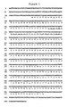
- This cytokine is named Neutrokine ⁇ and the invention includes Neutrokine ⁇ polypeptides having at least a portion of the amino acid sequence in FIG. 1 (SEQ ID NO:2) or amino acid sequence encoded by the cDNA clone deposited in a bacterial host as ATCC Deposit on October 22, 1996.
- soluble forms of Neutrokine ⁇ include all or a portion of the extracellular domain cleaved from the transmembrane domain and a polypeptide comprising the complete Neutrokine ⁇ polypeptide lacking the transmembrane domain, i.e., the extracellular domain linked to the intracellular domain.
- one aspect of the invention provides an isolated nucleic acid molecule comprising a polynucleotide having a nucleotide sequence selected from the group consisting of: (a) a nucleotide sequence encoding a full-length Neutrokine ⁇ polypeptide having the complete amino acid sequence in Figure 1 (SEQ ID NO:2) or as encoded by the cDNA clone contained in the ATCC Deposit of October 22, 1996; (b) a nucleotide sequence encoding the predicted extracellular domain of the Neutrokine ⁇ polypeptide having the amino acid sequence at positions 73 to 285 in Figure 1 (SEQ ID NO:2) or as encoded by the cDNA clone contained in the ATCC Deposit of October 22, 1996; (c) a nucleotide sequence encoding a polypeptide comprising the Neutrokine ⁇ intracellular domain (amino acid residues from about 1 to about 46 in FIG.
- nucleic acid molecules that comprise a polynucleotide having a nucleotide sequence at least 90% identical, and more preferably at least 95%, 96%, 97%, 98% or 99% identical, to any of the nucleotide sequences in (a), (b), (c), (d), (e) or (f) above, or a polynucleotide which hybridizes under stringent hybridization conditions to a polynucleotide in (a), (b), (c), (d), (e) or (f) above.
- This polynucleotide which hybridizes does not hybridize under stringent hybridization conditions to a polynucleotide having a nucleotide sequence consisting of only A residues or of only T residues.
- An additional nucleic acid embodiment of the invention relates to an isolated nucleic acid molecule comprising a polynucleotide which encodes the amino acid sequence of an epitope-bearing portion of a Neutrokine ⁇ polypeptide having an amino acid sequence in (a), (b), (c), (d) or (e) above.
- the present invention also relates to recombinant vectors, which include the isolated nucleic acid molecules of the present invention, and to host cells containing the recombinant vectors, as well as to methods of making such vectors and host cells and for using them for production of Neutrokine ⁇ polypeptides or peptides by recombinant techniques.
- the invention further provides an isolated Neutrokine ⁇ polypeptide comprising an amino acid sequence selected from the group consisting of: (a) the amino acid sequence of the full-length Neutrokine ⁇ polypeptide having the complete amino acid sequence shown in Figure 1 (SEQ ID NO:2) or as encoded by the cDNA clone contained in the ATCC Deposit of October 22, 1996; (b) the amino acid sequence of the predicted extracellular domain of the Neutrokine ⁇ polypeptide having the amino acid sequence at positions 73 to 285 in Figure 1 (SEQ ID NO:2) or as encoded by the cDNA clone contained in the ATCC Deposit of October 22, 1996; (c) the amino acid sequence of the Neutrokine ⁇ intracellular domain (amino acid residues from about 1 to about 46 in FIG.
- polypeptides of the present invention also include polypeptides having an amino acid sequence with at least 90% similarity, and more preferably at least 95% similarity to those described in (a), (b), (c), (d) or (e) above, as well as polypeptides having an amino acid sequence at least 80% identical, more preferably at least 90% identical, and still more preferably 95%, 96%, 97%, 98% or 99% identical to those above.
- An additional embodiment of this aspect of the invention relates to a peptide or polypeptide which has the amino acid sequence of an epitope-bearing portion of a Neutrokine ⁇ polypeptide having an amino acid sequence described in (a), (b), (c), (d) or (e) above.
- Peptides or polypeptides having the amino acid sequence of an epitope-bearing portion of a Neutrokine ⁇ polypeptide of the invention include portions of such polypeptides with at least six or seven, preferably at least nine, and more preferably at least about 30 amino acids to about 50 amino acids, although epitope-bearing polypeptides of any length up to and including the entire amino acid sequence of a polypeptide of the invention described above also are included in the invention.
- the invention provides an isolated antibody that binds specifically to an polypeptide having an amino acid sequence described in (a), (b), (c), (d) or (e) above.
- the invention further provides methods for isolating antibodies that bind specifically to an Neutrokine ⁇ polypeptide having an amino acid sequence as described herein. Such antibodies are useful diagnostically or therapeutically as described below.
- the invention also provides for pharmaceutical compositions comprising soluble Neutrokine ⁇ polypeptides, particularly human Neutrokine ⁇ polypeptides, which may be employed, for instance, to treat tumor and tumor metastasis, infections by bacteria, viruses and other parasites, immunodeficiencies, inflammatory diseases, lymphadenopathy, autoimmune diseases, graft versus host disease and to stimulate peripheral tolerance, destroy some transformed cell lines, mediate cell activation and proliferation, and are functionally linked as primary mediators of immune regulation and inflammatory responses.
- soluble Neutrokine ⁇ polypeptides particularly human Neutrokine ⁇ polypeptides
- compositions comprising an Neutrokine ⁇ polynucleotide or an Neutrokine ⁇ polypeptide for administration to cells in vitro, to cells ex vivo and to cells in vivo, or to a multicellular organism.
- the compositions comprise an Neutrokine ⁇ polynucleotide for expression of an Neutrokine ⁇ polypeptide in a host organism for treatment of disease.
- Particularly preferred in this regard is expression in a human patient for treatment of a dysfunction associated with aberrant endogenous activity of an Neutrokine ⁇ gene.
- the present invention also provides a screening method for identifying compounds capable of enhancing or inhibiting a cellular response induced by Neutrokine ⁇ which involves contacting cells which express Neutrokine a with the candidate compound, assaying a cellular response, and comparing the cellular response to a standard cellular response, the standard being assayed when contact is made in absence of the candidate compound; whereby, an increased cellular response over the standard indicates that the compound is an agonist and a decreased cellular response over the standard indicates that the compound is an antagonist.
- a method for identifying Neutrokine ⁇ receptors is - provided, as well as a screening assay for agonists and antagonists using such receptors.
- This assay involves determining the effect a candidate compound has on Neutrokine ⁇ binding to the Neutrokine ⁇ receptor.
- the method involves contacting a Neutrokine ⁇ receptor with an Neutrokine ⁇ polypeptide and a candidate compound and determining whether Neutrokine a polypeptide binding to the Neutrokine ⁇ receptor is increased or decreased due to the presence of the candidate compound.
- the antagonists may be employed to prevent septic shock, inflammation, cerebral malaria, activation of the HIV virus, graft-host rejection, bone resorption, rheumatoid arthritis and cachexia (wasting or malnutrition)
- Neutrokine ⁇ is expressed not only in neutrophils, but also in kidney, lung, peripheral leukocyte, bone marrow, T cell lymphoma, B cell lymphoma, activated T cells, stomach cancer, smooth muscle, macrophages, cord blood tissue.
- Neutrokine ⁇ gene expression can be detected in certain tissues (e.g., bone marrow) or bodily fluids (e.g., serum, plasma, urine, synovial fluid or spinal fluid) taken from an individual having such a disorder, relative to a "standard" Neutrokine ⁇ gene expression level, i.e., the Neutrokine ⁇ expression level in tissue or bodily fluids from an individual not having the disorder.
- tissues e.g., bone marrow
- bodily fluids e.g., serum, plasma, urine, synovial fluid or spinal fluid
- the invention provides a diagnostic method useful during diagnosis of a disorder, which involves: (a) assaying Neutrokine ⁇ gene expression level in cells or body fluid of an individual; (b) comparing the Neutrokine a gene expression level with a standard Neutrokine ⁇ gene expression level, whereby an increase or decrease in the assayed Neutrokine ⁇ gene expression level compared to the standard expression level is indicative of a disorder.
- An additional aspect of the invention is related to a method for treating an individual in need of an increased level of Neutrokine ⁇ activity in the body comprising administering to such an individual a composition comprising a therapeutically effective amount of an isolated Neutrokine ⁇ polypeptide of the invention or an agonist thereof.
- a still further aspect of the invention is related to a method for treating an individual in need of a decreased level of Neutrokine ⁇ activity in the body comprising, administering to such an individual a composition comprising a therapeutically effective amount of an Neutrokine ⁇ antagonist.
- Preferred antagonists for use in the present invention are Neutrokine ⁇ -specific antibodies.
- the present invention provides isolated nucleic acid molecules comprising a polynucleotide encoding Neutrokine ⁇ polypeptide having the amino acid sequence shown in Figure 1 (SEQ ID NO:2), which was determined by sequencing a cloned cDNA Neutrokine ⁇ .
- the nucleotide sequence shown in Figure 1 (SEQ ID NO:1) was obtained by sequencing the HNEDU15 clone, which was deposited on October 22, 1996 at the American Type Culture Collection, 12301 Park Lawn Drive, Rockville, Maryland.
- the deposited clone is contained in the pBluescript SK(-) plasmid (Stratagene, La Jolla, CA).
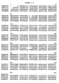
- the Neutrokine ⁇ protein of the present invention shares sequence homology with the translation product of the human mRNAs for TNF- ⁇ , TNF- ⁇ and Fas ligand (Figure 2).
- TNF- ⁇ is thought to be an important cytokine that plays a role in cytotoxicity, necrosis, apoptosis, costimulation, proliferation, lymph node formation, immunoglobulin class switch, differentiation, antiviral activity, regulation of adhesion molecules and other cytokines and growth factors.
- nucleotide sequences determined by sequencing a DNA molecule herein were determined using an automated DNA sequencer (such as the Model 373 from Applied Biosystems, Inc., Foster City, CA), and all amino acid sequences of polypeptides encoded by DNA molecules determined herein were predicted by translation of a DNA sequence determined as above. Therefore, as is known in the art for any DNA sequence determined by this automated approach, any nucleotide sequence determined herein may contain some errors. Nucleotide sequences determined by automation are typically at least about 90% identical, more typically at least about 95% to at least about 99.9% identical to the actual nucleotide sequence of the sequenced DNA molecule. The actual sequence can be more precisely determined by other approaches including manual DNA sequencing methods well known in the art.
- a single insertion or deletion in a determined nucleotide sequence compared to the actual sequence will cause a frame shift in translation of the nucleotide sequence such that the predicted amino acid sequence encoded by a determined nucleotide sequence will be completely different from the amino acid sequence actually encoded by the sequenced DNA molecule, beginning at the point of such an insertion or deletion.
- nucleotide sequence of a nucleic acid molecule or polynucleotide is intended, for a DNA molecule or polynucleotide, a sequence of deoxyribonucleotides, and for an RNA molecule or polynucleotide, the corresponding sequence of ribonucleotides (A, G, C and U), where each thymidine deoxyribonucleotide (T) in the specified deoxyribonucleotide sequence is replaced by the ribonucleotide uridine (U).
- nucleic acid molecule of the present invention encoding a Neutrokine ⁇ polypeptide may be obtained using standard cloning and screening procedures, such as those for cloning cDNAs using mRNA as starting material.
- nucleic acid molecule described in Figure 1 SEQ ID NO:1 was discovered in a cDNA library derived from neutrophils. Expressed sequence tags corresponding to a portion of the Neutrokine ⁇ cDNA were also found in neutrophil
- the Neutrokine ⁇ gene contains an open reading frame encoding a protein of about 285 amino acid residues, an intracellular domain of about 46 amino acids (amino acid residues from about 1 to about 46 in Figure. 1 (SEQ ID NO:2)), a transmembrane domain of about 26 amino acids (amino acid residues from about 47 to about 72 in FIG. 1 (SEQ ID NO:2)), an extracellular domain of about 213 amino acids (amino acid residues from about 73 to about 285 in Figure 1 (SEQ ID NO:2)); and a deduced molecular weight of about 31 kDa.
- the Neutrokine ⁇ protein shown in Figure 1 (SEQ ID NO: 2) is about 20% similar and about 10 % identical to human TNF- ⁇ which can be accessed on GenBank as Accession No. 339764.
- the actual complete Neutrokine ⁇ polypeptide encoded by the deposited cDNA which comprises about 285 amino acids, may be somewhat shorter.
- the determined Neutrokine ⁇ coding sequence contains a second methionine codon which may serve as an alternative start codon for translation of the open reading frame, at nucleotide positions 210-213 in Figure 1 (SEQ ID NO:1).
- the actual open reading frame may be anywhere in the range of ⁇ 20 amino acids, more likely in the range of ⁇ 10 amino acids, of that predicted from either the first or second methionine codon from the N-terminus shown in Figure 1 (SEQ ID NO:1).
- the exact "address" of the extracelluar, intracelluar and transmembrane domains of the Neutrokine ⁇ polypeptide may differ slightly.
- the exact location of the Neutrokine ⁇ extracellular domain in Figure 1 may vary slightly (e.g., the address may "shift" by about 1 to about 20 residues, more likely about 1 to about 5 residues) depending on the criteria used to define the domain.
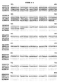
- the ends of the transmembrane domain and the beginning of the extracellular domain were predicted on the basis of the identification of the hydrophobic amino acid sequence in the above indicated positions, as shown in Figure 3.
- the invention further provides polypeptides having various residues deleted from the N-terminus of the complete polypeptide, including polypeptides lacking one or more amino acids from the N-terminus of the extracellular domain described herein, which constitute soluble forms of the extracellular domain of the Neutrokine ⁇ protein.
- nucleic acid molecules of the present invention may be in the form of RNA, such as mRNA, or in the form of DNA, including, for instance, cDNA and genomic DNA obtained by cloning or produced synthetically.
- the DNA may be double-stranded or single-stranded.
- Single-stranded DNA or RNA may be the coding strand, also known as the sense strand, or it may be the non-coding strand, also referred to as the anti-sense strand.
- isolated nucleic acid molecule(s) is intended a nucleic acid molecule, DNA or RNA, which has been removed from its native environment
- recombinant DNA molecules contained in a vector are considered isolated for the purposes of the present invention.
- Further examples of isolated DNA molecules include recombinant DNA molecules maintained in heterologous host cells or purified (partially or substantially) DNA molecules in solution.
- Isolated RNA molecules include in vivo or in vitro RNA transcripts of the DNA molecules of the present invention. Isolated nucleic acid molecules according to the present invention further include such molecules produced synthetically.
- Isolated nucleic acid molecules of the present invention include DNA molecules comprising an open reading frame (ORF) with an initiation codon at positions 147-149 of the nucleotide sequence shown in Figure 1 (SEQ ID NO:1).
- isolated nucleic acid molecules of the invention include DNA molecules which comprise a sequence substantially different from those described above but which, due to the degeneracy of the genetic code, still encode the Neutrokine ⁇ protein.
- the genetic code is well known in the art. Thus, it would be routine for one skilled in the art to generate the degenerate variants described above.
- the invention provides isolated nucleic acid molecules encoding the Neutrokine ⁇ polypeptide having an amino acid sequence encoded by the cDNA contained in the plasmid deposited on October 22, 1996.
- this nucleic acid molecule will comprise a sequence encoding the extracellular domain of the polypeptide encoded by the above-described deposited cDNA clone.
- the invention further provides an isolated nucleic acid molecule having the nucleotide sequence shown in Figure 1 (SEQ ID NO:1) or the nucleotide sequence of the Neutrokine ⁇ cDNA contained in the above-described deposited clone, or a nucleic acid molecule having a sequence complementary to one of the above sequences.
- isolated molecules particularly DNA molecules, are useful as probes for gene mapping, by in situ hybridization with chromosomes, and for detecting expression of the Neutrokine ⁇ gene in human tissue, for instance, by Northern blot analysis.
- the present invention is further directed to nucleic acid molecules encoding portions of the nucleotide sequences described herein as well as to fragments of the isolated nucleic acid molecules described herein.
- the invention provides a polynucleotide having a nucleotide sequence representing the portion of SEQ ID NO:1 which consists of positions 1-1001 of SEQ ID NO:1.
- the invention includes a polynucleotide comprising a sequence at least 95% identical to any portion of at least about 30 contiguous nucleotides, preferably at least about 50 nucleotides, of the sequence from nucleotide 1 to nucleotide 809 in Figure 1 (SEQ ID NO:1).
- a fragment of an isolated nucleic acid molecule having the nucleotide sequence of the deposited cDNA or the nucleotide sequence shown in Figure 1 is intended fragments at least about 15 nt, and more preferably at least about 20 nt, still more preferably at least about 30 nt, and even more preferably, at least about 40 nt in length which are useful as diagnostic probes and primers as discussed herein.
- larger fragments 50-300 nt in length are also useful according to the present invention as are fragments corresponding to most, if not all, of the nucleotide sequence of the deposited cDNA or as shown in Figure 1 (SEQ ID NO:1).
- nucleic acid fragments of the present invention include nucleic acid molecules encoding epitope-bearing portions of the Neutrokine ⁇ polypeptide as identified in Figure 1 and described in more detail below.
- the invention provides an isolated nucleic acid molecule comprising a polynucleotide which hybridizes under stringent hybridization conditions to a portion of the polynucleotide in a nucleic acid molecule of the invention described above, for instance, the cDNA clone contained in the ATCC Deposit of October 22, 1996.
- stringent hybridization conditions is intended overnight incubation at 42° C in a solution comprising: 50% formamide, 5x SSC (150 mM NaCl, 15 mM trisodium citrate), 50 mM sodium phosphate (pH 7.6), 5x Denhardt's solution, 10% dextran sulfate, and 20 ⁇ g/ml denatured, sheared salmon sperm DNA, followed by washing the filters in 0.1 x SSC at about 65° C.
- a polynucleotide which hybridizes to a "portion" of a polynucleotide is intended a polynucleotide (either DNA or RNA) hybridizing to at least about 15 nucleotides (nt), and more preferably at least about 20 nt, still more preferably at least about 30 nt, and even more preferably about 30-70 (e.g., 50) nt of the reference polynucleotide. These are useful as diagnostic probes and primers as discussed above and in more detail below.
- a portion of a polynucleotide of "at least 20 nt in length,” for example, is intended 20 or more contiguous nucleotides from the nucleotide sequence of the reference polynucleotide (e.g., the deposited cDNA or the nucleotide sequence as shown in Figure 1 (SEQ ID NO:1)).
- a polynucleotide which hybridizes only to a poly A sequence such as the 3' terminal poly(A) tract of the Neutrokine ⁇ cDNA shown in Figure 1 (SEQ ID NO:1)
- a complementary stretch of T (or U) residues would not be included in a polynucleotide of the invention used to hybridize to a portion of a nucleic acid of the invention, since such a polynucleotide would hybridize to any nucleic acid molecule containing a poly (A) stretch or the complement thereof (e.g., practically any double-stranded cDNA clone).
- nucleic acid molecules of the present invention which encode a Neutrokine ⁇ polypeptide may include, but are not limited to those encoding the amino acid sequence of the extracellular domain of the polypeptide, by itself; and the coding sequence for the extracellular domain of the polypeptide and additional sequences, such as those encoding the intracellular and transmembrane domain sequences, or a pre-, or pro- or prepro- protein sequence; the coding sequence of the extracellular domain of the polypeptide, with or without the aforementioned additional coding sequences.
- nucleic acids of the invention are the above protein sequences together with additional, non-coding sequences, including for example, but not limited to introns and non-coding 5' and 3' sequences, such as the transcribed, non-translated sequences that play a role in transcription, mRNA processing, including splicing and polyadenylation signals, for example - ribosome binding and stability of mRNA; an additional coding sequence which codes for additional amino acids, such as those which provide additional functionalities.
- additional, non-coding sequences including for example, but not limited to introns and non-coding 5' and 3' sequences, such as the transcribed, non-translated sequences that play a role in transcription, mRNA processing, including splicing and polyadenylation signals, for example - ribosome binding and stability of mRNA; an additional coding sequence which codes for additional amino acids, such as those which provide additional functionalities.
- the sequence encoding the polypeptide may be fused to a marker sequence, such as a sequence encoding a peptide which facilitates purification of the fused polypeptide.
- the marker amino acid sequence is a hexa-histidine peptide, such as the tag provided in a pQE vector (QIAGEN, Inc., 9259 Eton Avenue, Chatsworth, CA, 91311), among others, many of which are commercially available.
- hexa-histidine provides for convenient purification of the fusion protein.
- the "HA” tag is another peptide useful for purification which corresponds to an epitope derived from the influenza hemagglutinin protein, which has been described by Wilson et al., Cell 37: 767 (1984).
- other such fusion proteins include the Neutrokine ⁇ fused to Fc at the N- or C-terminus.
- the present invention further relates to variants of the nucleic acid molecules of the present invention, which encode portions, analogs or derivatives of the Neutrokine ⁇ protein.
- Variants may occur naturally, such as a natural allelic variant.
- allelic variant is intended one of several alternate forms of a gene occupying a given locus on a chromosome of an organism. Genes II, Lewin, B., ed., John Wiley & Sons, New York (1985). Non-naturally occurring variants may be produced using art-known mutagenesis techniques.
- variants include those produced by nucleotide substitutions, deletions or additions.
- the substitutions, deletions or additions may involve one or more nucleotides.
- the variants may be altered in coding regions, non-coding regions, or both. Alterations in the coding regions may produce conservative or non-conservative amino acid substitutions, deletions or additions. Especially preferred among these are silent substitutions, additions and deletions, which do not alter the properties and activities of the Neutrokine ⁇ protein or portions thereof. Also especially preferred in this regard are conservative substitutions.
- nucleic acid molecules encoding the extracellular domain of the protein having the amino acid sequence shown in Figure 1 (SEQ ID NO:2) or the extracellular domain of the Neutrokine ⁇ amino acid sequence encoded by the deposited cDNA clone.
- inventions include an isolated nucleic acid molecule comprising a polynucleotide having a nucleotide sequence at least 90% identical, and more preferably at least 95%, 96%, 97%, 98% or 99% identical to a polynucleotide selected from the group consisting of: (a) a nucleotide sequence encoding the Neutrokine ⁇ polypeptide having the complete amino acid sequence in Figure 1 (SEQ ID NO:2); (b) a nucleotide sequence encoding the predicted extracellular domain of the Neutrokine ⁇ polypeptide having the amino acid sequence at positions 73-285 in Figure 1 (SEQ ID NO:2); (c) a nucleotide sequence encoding the Neutrokine ⁇ polypeptide having the complete amino acid sequence encoded by the cDNA clone contained in the ATCC of October 22, 1996; (d) a nucleotide sequence encoding the extracellular domain of the Neutrokine ⁇ polypeptide having the amino
- nucleotide sequence of the polynucleotide is identical to the reference sequence except that the polynucleotide sequence may include up to five point mutations per each 100 nucleotides of the reference nucleotide sequence encoding the Neutrokine ⁇ polypeptide.
- a polynucleotide having a nucleotide sequence at least 95% identical to a reference nucleotide sequence up to 5% of the nucleotides in the reference sequence may be deleted or substituted with another nucleotide, or a number of nucleotides up to 5% of the total nucleotides in the reference sequence may be inserted into the reference sequence.
- These mutations of the reference sequence may occur at the 5' or 3' terminal positions of the reference nucleotide sequence or anywhere between those terminal positions, interspersed either individually among nucleotides in the reference sequence or in one or more contiguous groups within the reference sequence.
- nucleic acid molecule is at least 90%, 95%, 96%, 97%, 98% or 99% identical to, for instance, the nucleotide sequence shown in Figure 1 or to the nucleotides sequence of the deposited cDNA clone can be determined conventionally using known computer programs such as the Bestfit program (Wisconsin Sequence Analysis Package, Version 8 for Unix, Genetics Computer Group, University Research Park, 575 Science Drive, Madison, WI 53711). Bestfit uses the local homology algorithm of Smith and Waterman, Advances in Applied Mathematics 2 :482-489 (1981), to find the best segment of homology between two sequences.
- Bestfit program Wiconsin Sequence Analysis Package, Version 8 for Unix, Genetics Computer Group, University Research Park, 575 Science Drive, Madison, WI 53711. Bestfit uses the local homology algorithm of Smith and Waterman, Advances in Applied Mathematics 2 :482-489 (1981), to find the best segment of homology between two sequences.
- the parameters are set, of course, such that the percentage of identity is calculated over the full length of the reference nucleotide sequence and that gaps in homology of up to 5% of the total number of nucleotides in the reference sequence are allowed.
- the present application is directed to nucleic acid molecules at least 90%, 95%, 96%, 97%, 98% or 99% identical to the nucleic acid sequence shown in Figure 1 (SEQ ID NO:1) or to the nucleic acid sequence of the deposited cDNA, irrespective of whether they encode a polypeptide having Neutrokine ⁇ activity. This is because even where a particular nucleic acid molecule does not encode a polypeptide having Neutrokine ⁇ activity, one of skill in the art would still know how to use the nucleic acid molecule, for instance, as a hybridization probe or a polymerase chain reaction (PCR) primer.
- PCR polymerase chain reaction
- nucleic acid molecules of the present invention that do not encode a polypeptide having Neutrokine ⁇ activity include, inter alia, (1) isolating the Neutrokine ⁇ gene or allelic variants thereof in a cDNA library; (2) in situ hybridization (e.g., "FISH") to metaphase chromosomal spreads to provide precise chromosomal location of the Neutrokine ⁇ gene, as described in Verma et al., Human Chromosomes: A Manual of Basic Techniques, Pergamon Press, New York (1988); and Northern Blot analysis for detecting Neutrokine ⁇ mRNA expression in specific tissues.
- FISH in situ hybridization
- nucleic acid molecules having sequences at least 90%, 95%, 96%, 97%, 98% or 99% identical to the nucleic acid sequence shown in Figure 1 (SEQ ID NO:1) or to the nucleic acid sequence of the deposited cDNA which do, in fact, encode a polypeptide having Neutrokine ⁇ protein activity.
- a polypeptide having Neutrokine ⁇ activity is intended polypeptides exhibiting activity similar, but not necessarily identical, to an activity of the extracellular domain or of the full-length Neutrokine ⁇ protein of the invention, as measured in a particular biological assay.
- the Neutrokine ⁇ protein of the present invention modulates cell proliferation, cytotoxicity and cell death.
- An in vitro cell proliferation, cytotoxicity and cell death assay for measuring the effect of a protein on certain cells can be performed by using reagents well known and commonly available in the art for detecting cell replication and/or death.
- numerous such assays for TNF-related protein activities are described in the various references in the Background section of this disclosure, above. Briefly, such an assay involves collecting human or animal (e.g., mouse) cells and mixing with (1) transfected host cell-supernatant containing Neutrokine ⁇ protein (or a candidate polypeptide) or (2) nontransfected host cell-supernatant control, and measuring the effect on cell numbers or viability after incubation of certain period of time.
- Such cell proliferation modulation activities as can be measure in this type of assay are useful for treating tumor, tumor metastasis, infections, autoimmune diseases inflammation and other immune-related diseases.
- Neutrokine ⁇ modulates cell proliferation and differentiation in a dose-dependent manner in the above-described assay.
- a polypeptide having Neutrokine ⁇ protein activity includes polypeptides that also exhibit any of the same cell modulatory (particularly immunomodulatory) activities in the above-described assays in a dose-dependent manner.
- a polypeptide having Neutrokine ⁇ protein activity will exhibit substantially similar dose-dependence in a given activity as compared to the Neutrokine ⁇ protein (i.e., the candidate polypeptide will exhibit greater activity or not more than about 25-fold less and, preferably, not more than about tenfold less activity relative to the reference Neutrokine ⁇ protein).
- Neutrokine ⁇ Like other members of TNF family, Neutrokine ⁇ exhibits activity on leukocytes including for example monocytes, lymphocytes and neutrophils. For this reason Neutrokine ⁇ is active in directing the proliferation, differentiation and migration of these cell types. Such activity is useful for immune enhancement or suppression, myeloprotection, stem cell mobilization, acute and chronic inflammatory control and treatment of leukemia. Assays for measuring such activity are known in the art. For example, see Peters et al., Immun. Today 17 :273 (1996); Young et al., J. Exp. Med. 182 :1111 (1995); Caux et al., Nature 390:258 (1992); and Santiago-Schwarz et al., Adv. Exp. Med. Biol. 378:7 (1995)."]
- nucleic acid molecules having a sequence at least 90%, 95%, 96%, 97%, 98%, or 99% identical to the nucleic acid sequence of the deposited cDNA or the nucleic acid sequence shown in Figure 1 (SEQ ID NO:1) will encode a polypeptide "having Neutrokine ⁇ protein activity.”
- degenerate variants of these nucleotide sequences all encode the same polypeptide, this will be clear to the skilled artisan even without performing the above described comparison assay.
- nucleic acid molecules that are not degenerate variants, a reasonable number will also encode a polypeptide having Neutrokine ⁇ protein activity. This is because the skilled artisan is fully aware of amino acid substitutions that are either less likely or not likely to significantly effect protein function (e.g., replacing one aliphatic amino acid with a second aliphatic amino acid), as further described below.
- the present invention also relates to vectors which include the isolated DNA molecules of the present invention, host cells which are genetically engineered with the recombinant vectors, and the production of Neutrokine ⁇ polypeptides or fragments thereof by recombinant techniques.
- the vector may be, for example, a phage, plasmid, viral or retroviral vector. Retroviral vectors may be replication competent or replication defective. In the latter case, viral propagation generally will occur only in complementing host cells.
- the polynucleotides may be joined to a vector containing a selectable marker for propagation in a host.
- a plasmid vector is introduced in a precipitate, such as a calcium phosphate precipitate, or in a complex with a charged lipid. If the vector is a virus, it may be packaged in vitro using an appropriate packaging cell line and then transduced into host cells.
- the DNA insert should be operatively linked to an appropriate promoter, such as the phage lambda PL promoter, the E. coli lac, trp, phoA and tac promoters, the SV40 early and late promoters and promoters of retroviral LTRs, to name a few. Other suitable promoters will be known to the skilled artisan.
- the expression constructs will further contain sites for transcription initiation, termination and, in the transcribed region, a ribosome binding site for translation.
- the coding portion of the extracellular domain of the transcripts expressed by the constructs will preferably include a translation initiating at the beginning and a termination codon (UAA, UGA or UAG) appropriately positioned at the end of the polypeptide to be translated.
- the expression vectors will preferably include at least one selectable marker.
- markers include dihydrofolate reductase, G418 or neomycin resistance for eukaryotic cell culture and tetracycline, kanamycin or ampicillin resistance genes for culturing in E. coli and other bacteria.
- Representative examples of appropriate hosts include, but are not limited to, bacterial cells, such as E. coli, Streptomyces and Salmonella typhimurium cells; fungal cells, such as yeast cells; insect cells such as Drosophila S2 and Spodoptera Sf9 cells; animal cells such as CHO, COS, 293 and Bowes melanoma cells; and plant cells. Appropriate culture mediums and conditions for the above-described host cells are known in the art.
- vectors preferred for use in bacteria include pQE70, pQE60 and pQE-9, available from QIAGEN, Inc., supra ; pBS vectors, Phagescript vectors, Bluescript vectors, pNH8A, pNH16a, pNH18A, pNH46A, available from Stratagene; and ptrc99a, pKK223-3, pKK233-3, pDR540, pRIT5 available from Pharmacia.
- preferred eukaryotic vectors are pWLNEO, pSV2CAT, pOG44, pXT1 and pSG available from Stratagene; and pSVK3, pBPV, pMSG and pSVL available from Pharmacia.
- Other suitable vectors will be readily apparent to the skilled artisan.
- Introduction of the construct into the host cell can be effected by calcium phosphate transfection, DEAE-dextran mediated transfection, cationic lipid-mediated transfection, electroporation, transduction, infection or other methods. Such methods are described in many standard laboratory manuals, such as Davis et al., Basic Methods In Molecular Biology (1986).
- the polypeptide may be expressed in a modified form, such as a fusion protein, and may include not only secretion signals, but also additional heterologous functional regions. For instance, a region of additional amino acids, particularly charged amino acids, may be added to the N-terminus of the polypeptide to improve stability and persistence in the host cell, during purification, or during subsequent handling and storage. Also, peptide moieties may be added to the polypeptide to facilitate purification. Such regions may be removed prior to final preparation of the polypeptide. The addition of peptide moieties to polypeptides to engender secretion or excretion, to improve stability and to facilitate purification, among others, are familiar and routine techniques in the art.
- a preferred fusion protein comprises a heterologous region from immunoglobulin that is useful to stabilize and purify proteins.
- EP-A-O 464 533 (Canadian counterpart 2045869) discloses fusion proteins comprising various portions of constant region of immunoglobulin molecules together with another human protein or part thereof.
- the Fc part in a fusion protein is thoroughly advantageous for use in therapy and diagnosis and thus results, for example, in improved pharmacokinetic properties (EP-A 0232 262).
- Fc portion proves to be a hindrance to use in therapy and diagnosis, for example when the fusion protein is to be used as antigen for immunizations.
- human proteins, such as hIL-5 has been fused with Fc portions for the purpose of high-throughput screening assays to identify antagonists of hIL-5. See, D. Bennett et al., J. Molecular Recognition 8 :52-58 (1995) and K. Johanson et al., J. Biol. Chem. 270 :9459-9471 (1995).
- the Neutrokine ⁇ protein can be recovered and purified from recombinant cell cultures by well-known methods including ammonium sulfate or ethanol precipitation, acid extraction, anion or cation exchange chromatography, phosphocellulose chromatography, hydrophobic interaction chromatography, affinity chromatography, hydroxylapatite chromatography and lectin chromatography. Most preferably, high performance liquid chromatography ("HPLC") is employed for purification.
- Polypeptides of the present invention include naturally purified products, products of chemical synthetic procedures, and products produced by recombinant techniques from a prokaryotic or eukaryotic host, including, for example, bacterial, yeast, higher plant, insect and mammalian cells.
- polypeptides of the present invention may be glycosylated or may be non-glycosylated.
- polypeptides of the invention may also include an initial modified methionine residue, in some cases as a result of host-mediated processes.
- the invention further provides an isolated Neutrokine ⁇ polypeptide having the amino acid sequence encoded by the deposited cDNA, or the amino acid sequence in Figure 1 (SEQ ID NO:2), or a peptide or polypeptide comprising a portion of the above polypeptides.
- Neutrokine ⁇ polypeptides protein engineering may be employed.
- Recombinant DNA technology known to those skilled in the art can be used to create novel mutant proteins or "muteins including single or multiple amino acid substitutions, deletions, additions or fusion proteins.
- Such modified polypeptides can show, e.g., enhanced activity or increased stability.
- they may be purified in higher yields and show better solubility than the corresponding natural polypeptide, at least under certain purification and storage conditions.
- deletions of N-terminal amino acids up to the Gly (G) residue at position 191 in Figure 1 may retain some biological activity such as cytotoxicity to appropriate target cells.
- Polypeptides having further N-terminal deletions including the Gly(G)residue would not be expected to retain such biological activities because it is known that this residue in TNF-related polypeptides is in the beginning of the conserved domain required for biological activities.
- deletion of one or more amino acids from the N-terminus of a protein results in modification of loss of one or more biological functions of the protein, other biological activities may still be retained.
- the ability of the shortened protein to induce and/or bind to antibodies which recognize the complete or extracellular domain of the protein generally will be retained when less than the majority of the residues of the complete or extracellular domain of the protein are removed from the N-terminus. Whether a particular polypeptide lacking N-terminal residues of a complete protein retains such immunologic activities can readily be determined by routine methods described herein and otherwise known in the art.
- the present invention further provides polypeptides having one or more residues from the amino terminus of the amino acid sequence of the Neutrokine ⁇ shown in Figure 1(SEQ ID NO:2), up to the Gly191 residue from the amino terminus, and polynucleotides encoding such polypeptides.
- the present invention provides polypeptides having the amino acid sequence of residues n-190 of SEQ ID NO:2, where n is an integer in the range of 2-190 and 191 is the position of the first residue from the N-terminus of the complete Neutrokine ⁇ polypeptide (shown in SEQ ID NO:2) believed to be required for activity of the Neutrokine ⁇ protein.
- the invention provides polynucleotides encoding polypeptides having the amino acid sequence of residues 2-285, 3-285, 4-285, 5-285, 6-285, 7-285, 8-285, 9-285, 10-285, 11-285, 12-285, 13-285, 14-285, 15-285, 16-285, 17-285, 18-285, 19-285, 20-285, 21-285, 22-285, 23-285, 24-285, 25-285, 26-285, 27-285, 28-285, 29-285, 30-285, 31-285, 32-285, 33-285, 34-285, 35-285, 36-285, 37-285, 38-285, 39-285, 40-285, 41-285, 42-285, 43-285, 44-285, 45-285, 46-285, 47-285, 48-285, 49-285, 50-285, 51-285, 52-285, 53-285, 54-285, 55-285, 56-285, 57-285, 58-285,
- C-terminal deletion muteins are known. For instance, Interferon gamma shows up to ten times higher activities by deleting 8-10 amino acid residues from the carboxy terminus of the protein (Döbeli et al., J. Biotechnology 7 :199-216 (1988). Since the present protein is a member of the TNF polypeptide family, deletions of C-terminal amino acids up to the Leu at position 284 are expected to retain most if not all biological activity such as receptor binding and modulation of cell replication.
- Polypeptides having deletions of up to about 10 additional C -terminal residues also may retain some activity such as receptor binding, although such polypeptides would lack a portion of the conserved TNF domain beginning at about Leu284.
- deletion of one or more amino acids from the C-terminus of a protein results in modification of loss of one or more biological functions of the protein, other biological activities may still be retained.
- the ability of the shortened protein to induce and/or bind to antibodies which recognize the complete or mature protein generally will be retained when less than the majority of the residues of the complete or mature protein are removed from the C-terminus. Whether a particular polypeptide lacking C-terminal residues of a complete protein retains such immunologic activities can readily be determined by routine methods described herein and otherwise known in the art.
- the present invention further provides polypeptides having one or more residues from the carboxy terminus of the amino acid sequence of the Neutrokine ⁇ shown in Figure 1( SEQ ID NO:2), up to the Gly274 residue from the carboxy terminus, and polynucleotides encoding such polypeptides.
- the present invention provides polypeptides having the amino acid sequence of residues 1-m of the amino acid sequence in SEQ ID NO:2, where m is any integer in the range of 274 to 284.
- the invention provides polynucleotides encoding polypeptides having the amino acid sequence of residues 1-274, 1-275, 1-276, 1-277, 1-278, 1-279, 1-280, 1-281, 1-282, 1-283 and 1-284 of SEQ ID NO:2. Polynucleotides encoding these polypeptides also are provided.
- polypeptides having one or more amino acids deleted from both the amino and the carboxyl termini which may be described generally as having residues n-m of SEQ ID NO:2, where n and m are integers as described above.
- Polynucleotides encoding all of the above deletion polypeptides also are provided.
- the invention further includes variations of the Neutrokine ⁇ polypeptide which show substantial Neutrokine ⁇ polypeptide activity or which include regions of Neutrokine ⁇ protein such as the protein portions discussed below.
- Such mutants include deletions, insertions, inversions, repeats, and type substitutions selected according to general rules known in the art so as have little effect on activity.
- guidance concerning how to make phenotypically silent amino acid substitutions is provided in Bowie, J. U. et al., "Deciphering the Message in Protein Sequences: Tolerance to Amino Acid Substitutions, " Science 247 :1306-1310 (1990), wherein the authors indicate that there are two main approaches for studying the tolerance of an amino acid sequence to change.
- the first method relies on the process of evolution, in which mutations are either accepted or rejected by natural selection.
- the second approach uses genetic engineering to introduce amino acid changes at specific positions of a cloned gene and selections or screens to identify sequences that maintain functionality.
- conservative substitutions are the replacements, one for another, among the aliphatic amino acids Ala, Val, Leu and Ile; interchange of the hydroxyl residues Ser and Thr, exchange of the acidic residues Asp and Glu, substitution between the amide residues Asn and Gln, exchange of the basic residues Lys and Arg and replacements among the aromatic residues Phe, Tyr.
- the fragment, derivative or analog of the polypeptide of Figure 1 may be (i) one in which one or more of the amino acid residues are substituted with a conserved or non-conserved amino acid residue (preferably a conserved amino acid residue) and such substituted amino acid residue may or may not be one encoded by the genetic code, or (ii) one in which one or more of the amino acid residues includes a substituent group, or (iii) one in which the extracellular domain of the polypeptide is fused with another compound, such as a compound to increase the half-life of the polypeptide (for example, polyethylene glycol), or (iv) one in which the additional amino acids are fused to the extracellular domain of the polypeptide, such as an IgG Fc fusion region peptide or leader or secretory sequence or a sequence which is employed for purification of the extracellular domain of the polypeptide or a proprotein sequence.
- a conserved or non-conserved amino acid residue preferably a conserved amino acid residue
- the Neutrokine ⁇ of the present invention may include one or more amino acid substitutions, deletions or additions, either from natural mutations or human manipulation. As indicated, changes are preferably of a minor nature, such as conservative amino acid substitutions that do not significantly affect the folding or activity of the protein (see Table 1). Conservative Amino Acid Substitutions. Aromatic Phenylalanine Tryptophan Tyrosine Hydrophobic Leucine Isoleucine Valine Polar Glutamine Asparagine Basic Arginine Lysine Histidine Acidic Aspartic Acid Glutamic Acid Small Alanine Serine Threonine Methionine Glycine
- Amino acids in the Neutrokine ⁇ protein of the present invention that are essential for function can be identified by methods known in the art, such as site-directed mutagenesis or alanine-scanning mutagenesis (Cunningham and Wells, Science 244 :1081-1085 (1989). The latter procedure introduces single alanine mutations at every residue in the molecule. The resulting mutant molecules are then tested for biological activity such as receptor binding or in vitro or in vitro proliferative activity.
- Neutrokine ⁇ is a member of the TNF-related protein family, to modulate rather than completely eliminate biological activities of Neutrokine ⁇ , preferably mutations are made in sequences encoding amino acids in the TNF conserved domain, i.e., in positions 191-284 of Figure 1 (SEQ ID NO:2), more preferably in residues within this region which are not conserved in all members of the TGF family.
- polypeptides of the present invention include Neutrokine ⁇ mutants.
- Neutrokine ⁇ mutants are comprised of the full-length or preferably the extracellular domain of the Neutrokine ⁇ amino acid sequence shown in Figure 1 (SEQ ID NO:2).
- isolated polynucleotides comprising nucleic acid sequences which encode the above Neutrokine ⁇ mutants.
- polypeptides of the present invention are preferably provided in an isolated form, and preferably are substantially purified.
- a recombinantly produced version of the Neutrokine ⁇ polypeptide can be substantially purified by the one-step method described in Smith and Johnson, Gene 67:31-40 (1988).
- polypeptides of the present invention include the complete polypeptide encoded by the deposited cDNA including the intracellular, transmembrane and extracellular domains of the polypeptide encoded by the deposited cDNA, the extracellular domain minus the intracellular and transmembrane domains of the protein, the complete polypeptide of Figure 1 (SEQ ID NO:2), the extracellular domain of Figure 1 (SEQ ID NO:2) minus the intracellular and transmembrane domains, as well as polypeptides which have at least 90% similarity, more preferably at least 95% similarity, and still more preferably at least 96%, 97%, 98% or 99% similarity to those described above.
- polypeptides of the present invention include polypeptides at least 80% identical, more preferably at least 90% or 95% identical, still more preferably at least 96%, 97%, 98% or 99% identical to the polypeptide encoded by the deposited cDNA or to the polypeptide of Figure 1 (SEQ ID NO:2), and also include portions of such polypeptides with at least 30 amino acids and more preferably at least 50 amino acids.
- % similarity for two polypeptides is intended a similarity score produced by comparing the amino acid sequences of the two polypeptides using the Bestfit program (Wisconsin Sequence Analysis Package, Version 8 for Unix, Genetics Computer Group, University Research Park, 575 Science Drive, Madison, WI 53711) and the default settings for determining similarity. Bestfit uses the local homology algorithm of Smith and Waterman (Advances in Applied Mathematics 2:482-489, 1981) to find the best segment of similarity between two sequences.
- polypeptide having an amino acid sequence at least, for example, 95% "identical" to a reference amino acid sequence of a Neutrokine ⁇ polypeptide is intended that the amino acid sequence of the polypeptide is identical to the reference sequence except that the polypeptide sequence may include up to five amino acid alterations per each 100 amino acids of the reference amino acid of the Neutrokine ⁇ polypeptide.
- up to 5% of the amino acid residues in the reference sequence may be deleted or substituted with another amino acid, or a number of amino acids up to 5% of the total amino acid residues in the reference sequence may be inserted into the reference sequence.
- These alterations of the reference sequence may occur at the amino or carboxy terminal positions of the reference amino acid sequence or anywhere between those terminal positions, interspersed either individually among residues in the reference sequence or in one or more contiguous groups within the reference sequence.
- any particular polypeptide is at least 90%, 95%, 96%, 97%, 98% or 99% identical to, for instance, the amino acid sequence shown in Figure 1 (SEQ ID NO:2) or to the amino acid sequence encoded by deposited cDNA clone can be determined conventionally using known computer programs such the Bestfit program (Wisconsin Sequence Analysis Package, Version 8 for Unix, Genetics Computer Group, University Research Park, 575 Science Drive, Madison, WI 53711).
- the parameters are set, of course, such that the percentage of identity is calculated over the full length of the reference amino acid sequence and that gaps in homology of up to 5% of the total number of amino acid residues in the reference sequence are allowed.
- polypeptide of the present invention could be used as a molecular weight marker on SDS-PAGE gels or on molecular sieve gel filtration columns using methods well known to those of skill in the art.
- polypeptides of the present invention can also be used to raise polyclonal and monoclonal antibodies, which are useful in assays for detecting Neutrokine ⁇ protein expression as described below or as agonists and antagonists capable of enhancing or inhibiting Neutrokine ⁇ protein function.
- polypeptides can be used in the yeast two-hybrid system to "capture" Neutrokine ⁇ protein binding proteins which are also candidate agonists and antagonists according to the present invention.
- the yeast two hybrid system is described in Fields and Song, Nature 340:245-246 (1989).
- the invention provides a peptide or polypeptide comprising an epitope-bearing portion of a polypeptide of the invention.
- the epitope of this polypeptide portion is an immunogenic or antigenic epitope of a polypeptide of the invention.
- An "immunogenic epitope" is defined as a part of a protein that elicits an antibody response when the whole protein is the immunogen.
- a region of a protein molecule to which an antibody can bind is defined as an "antigenic epitope.”
- the number of immunogenic epitopes of a protein generally is less than the number of antigenic epitopes. See, for instance, Geysen et al., Proc. Natl. Acad. Sci. USA 81 :3998- 4002 (1983).
- peptides or polypeptides bearing an antigenic epitope i.e., that contain a region of a protein molecule to which an antibody can bind
- relatively short synthetic peptides that mimic part of a protein sequence are routinely capable of eliciting an antiserum that reacts with the partially mimicked protein. See, for instance, Sutcliffe, J. G., Shinnick, T. M., Green, N. and Learner, R. A. (1983) "Antibodies that react with predetermined sites on proteins", Science, 219 :660-666.
- Peptides capable of eliciting protein-reactive sera are frequently represented in the primary sequence of a protein, can be characterized by a set of simple chemical rules, and are confined neither to immunodominant regions of intact proteins (i.e., immunogenic epitopes) nor to the amino or carboxyl terminals.
- Antigenic epitope-bearing peptides and polypeptides of the invention are therefore useful to raise antibodies, including monoclonal antibodies, that bind specifically to a polypeptide of the invention. See, for instance, Wilson et al., Cell 37 :767-778 (1984) at 777.
- Antigenic epitope-bearing peptides and polypeptides of the invention preferably contain a sequence of at least seven, more preferably at least nine and most preferably between about 15 to about 30 amino acids contained within the amino acid sequence of a polypeptide of the invention.
- Non-limiting examples of antigenic polypeptides or peptides that can be used to generate Neutrokine specific antibodies include: a polypeptide comprising amino acid residues from about Phe 115 to about Leu 147 in Figure 1 (SEQ ID NO:2); a polypeptide comprising amino acid residues from about Ile 150 to about Tyr 163 in Figure 1 (SEQ ID NO:2); a polypeptide comprising amino acid residues from about Ser 171 to about Phe 194 in Figure 1 (SEQ ID NO:2); a polypeptide comprising amino acid residues from about Glu 223 to about Tyr 247 in Figure 1 (SEQ ID NO:2); a polypeptide comprising amino acid residues from about Ser 271 to about Phe 278 in Figure 1 (SEQ ID NO:2); These polypeptide fragments have been determined to bear antigenic epitopes of the Neutrokine ⁇ protein by the analysis of the Jameson-Wolf antigenic index, as shown in Figure 3, above.
- the epitope-bearing peptides and polypeptides of the invention may be produced by any conventional means. See, e.g., Houghten, R. A. (1985) General method for the rapid solid-phase synthesis of large numbers of peptides: specificity of antigen-antibody interaction at the level of individual amino acids. Proc. Natl. Acad. Sci. USA 82 :5131-5135; this "Simultaneous Multiple Peptide Synthesis (SMPS)" process is further described in U.S. Patent No. 4,631,211 to Houghten et al. (1986).
- SMPS Simultaneous Multiple Peptide Synthesis
- Epitope-bearing peptides and polypeptides of the invention are used to induce antibodies according to methods well known in the art. See, for instance, Sutcliffe et al., supra; Wilson et al., supra; Chow, M. et al., Proc. Natl. Acad. Sci. USA 82:910-914; and Bittle, F. J. et al., J. Gen. Virol. 66:2347-2354 (1985).
- Immunogenic epitope-bearing peptides of the invention i.e., those parts of a protein that elicit an antibody response when the whole protein is the immunogen, are identified according to methods known in the art.
- U.S. Patent No. 5,194,392 to Geysen (1990) describes a general method of detecting or determining the sequence of monomers (amino acids or other compounds) which is a topological equivalent of the epitope (i.e., a "mimotope") which is complementary to a particular paratope (antigen binding site) of an antibody of interest. More generally, U.S. Patent No. 4,433,092 to Geysen (1989) describes a method of detecting or determining a sequence of monomers which is a topographical equivalent of a ligand which is complementary to the ligand binding site of a particular receptor of interest. Similarly, U.S.
- Patent No. 5,480,971 to Houghten, R. A. et al. (1996) on Peralkylated Oligopeptide Mixtures discloses linear C1-C7-alkyl peralkylated oligopeptides and sets and libraries of such peptides, as well as methods for using such oligopeptide sets and libraries for determining the sequence of a peralkylated oligopeptide that preferentially binds to an acceptor molecule of interest.
- non-peptide analogs of the epitope-bearing peptides of the invention also can be made routinely by these methods.
- Neutrokine ⁇ polypeptides of the present invention and the epitope-bearing fragments thereof described above can be combined with parts of the constant domain of immunoglobulins (IgG), resulting in chimeric polypeptides.
- IgG immunoglobulins
- These fusion proteins facilitate purification and show an increased half-life in vivo. This has been shown, e.g., for chimeric proteins consisting of the first two domains of the human CD4-polypeptide and various domains of the constant regions of the heavy or light chains of mammalian immunoglobulins (EP A 394,827; Traunecker et al., Nature 331:84-86 (1988)).
- Fusion proteins that have a disulfide-linked dimeric structure due to the IgG part can also be more efficient in binding and neutralizing other molecules than the monomeric Neutrokine ⁇ protein or protein fragment alone (Fountoulakis et al., J. Biochem. 270 :3958-3964 (1995)).
- Neutrokine ⁇ is expressed in various tissues and particularly in neutrophils.
- substantially altered (increased or decreased) levels of Neutrokine ⁇ gene expression can be detected in immune system tissue or other cells or bodily fluids (e.g., sera, plasma, urine, synovial fluid or spinal fluid) taken from an individual having such a disorder, relative to a "standard" Neutrokine ⁇ gene expression level, that is, the Neutrokine ⁇ expression level in immune system tissues or bodily fluids from an individual not having the immune system disorder.
- the invention provides a diagnostic method useful during diagnosis of an system disorder, which involves measuring the expression level of the gene encoding the Neutrokine ⁇ protein in immune system tissue or other cells or body fluid from an individual and comparing the measured gene expression level with a standard Neutrokine ⁇ gene expression level, whereby an increase or decrease in the gene expression level compared to the standard is indicative of an immune system disorder.
- tissue in mammals with cancer of the immune express significantly enhanced or reduced levels of the Neutrokine ⁇ protein and mRNA encoding the Neutrokine ⁇ protein when compared to a corresponding "standard” level.
- enhanced or depressed levels of the Neutrokine ⁇ protein can be detected in certain body fluids (e.g., sera, plasma, urine, and spinal fluid) from mammals with such a cancer when compared to sera from mammals of the same species not having the cancer.
- the invention provides a diagnostic method useful during diagnosis of a immune system disorder, including cancers of this system, which involves measuring the expression level of the gene encoding the Neutrokine ⁇ protein in immune system tissue or other cells or body fluid from an individual and comparing the measured gene expression level with a standard Neutrokine ⁇ gene expression level, whereby an increase or decrease in the gene expression level compared to the standard is indicative of an immune system disorder.
- the present invention is useful as a prognostic indicator, whereby patients exhibiting enhanced or depressed Neutrokine ⁇ gene expression will experience a worse clinical outcome relative to patients expressing the gene at a level nearer the standard level.
- test the expression level of the gene encoding the Neutrokine ⁇ protein is intended qualitatively or quantitatively measuring or estimating the level of the Neutrokine ⁇ protein or the level of the mRNA encoding the Neutrokine ⁇ protein in a first biological sample either directly (e.g., by determining or estimating absolute protein level or mRNA level) or relatively (e.g., by comparing to the Neutrokine ⁇ protein level or mRNA level in a second biological sample).
- the Neutrokine ⁇ protein level or mRNA level in the first biological sample is measured or estimated and compared to a standard Neutrokine ⁇ protein level or mRNA level, the standard being taken from a second biological sample obtained from an individual not having the disorder or being determined by averaging levels from a population of individuals not having a disorder of the immune system.
- a standard Neutrokine ⁇ protein level or mRNA level is known, it can be used repeatedly as a standard for comparison.
- biological sample any biological sample obtained from an individual, body fluid, cell line, tissue culture, or other source which contains Neutrokine ⁇ protein or mRNA.
- biological samples include body fluids (such as sera, plasma, urine, synovial fluid and spinal fluid) which contain free extracellular domains of the Neutrokine ⁇ protein, immune system tissue, and other tissue sources found to express complete or free extracellular domain of the Neutrokine ⁇ or a Neutrokine ⁇ receptor.
- body fluids such as sera, plasma, urine, synovial fluid and spinal fluid
- tissue sources found to express complete or free extracellular domain of the Neutrokine ⁇ or a Neutrokine ⁇ receptor.
- the present invention is useful for diagnosis or treatment of various immune system-related disorders in mammals, preferably humans.
- disorders include but are not limited to tumors and tumor metastasis, infections by bacteria, viruses and other parasites, immunodeficiencies, inflammatory diseases, lymphadenopathy, autoimmune diseases, and graft versus host disease.
- Total cellular RNA can be isolated from a biological sample using any suitable technique such as the single-step guanidinium-thiocyanate-phenolchloroform method described in Chomczynski and Sacchi, Anal. Biochem. 162 :156-159 (1987). Levels of mRNA encoding the Neutrokine ⁇ protein are then assayed using any appropriate method. These include Northern blot analysis, S 1 nuclease mapping, the polymerase chain reaction (PCR), reverse transcription in combination with the polymerase chain reaction (RT-PCR), and reverse transcription in combination with the ligase chain reaction (RT-LCR).
- PCR polymerase chain reaction
- RT-PCR reverse transcription in combination with the polymerase chain reaction
- RT-LCR reverse transcription in combination with the ligase chain reaction
- Neutrokine ⁇ protein levels in a biological sample can occur using antibody-based techniques.
- Neutrokine ⁇ protein expression in tissues can be studied with classical immunohistological methods (Jalkanen, M., et al., J. Cell. Biol. 101 :976-985 (1985); Jalkanen, M., et al., J. Cell. Biol. 105 :3087-3096 (1987)).
- Other antibody-based methods useful for detecting Neutrokine ⁇ protein gene expression include immunoassays, such as the enzyme linked immunosorbent assay (ELISA) and the radioimmunoassay (RIA).
- ELISA enzyme linked immunosorbent assay
- RIA radioimmunoassay
- Suitable antibody assay labels include enzyme labels, such as, glucose oxidase, and radioisotopes, such as iodine ( 125 I, 121 I), carbon ( 14 C), sulfur ( 35 S), tritium ( 3 H), indium ( 112 In), and technetium ( 99m Tc), and fluorescent labels, such as fluorescein and rhodamine, and biotin.
- enzyme labels such as, glucose oxidase, and radioisotopes, such as iodine ( 125 I, 121 I), carbon ( 14 C), sulfur ( 35 S), tritium ( 3 H), indium ( 112 In), and technetium ( 99m Tc)
- fluorescent labels such as fluorescein and rhodamine, and biotin.
- Neutrokine ⁇ protein can also be detected in vivo by imaging.
- Antibody labels or markers for in vivo imaging of Neutrokine ⁇ protein include those detectable by X-radiography, NMR or ESR.
- suitable labels include radioisotopes such as barium or cesium, which emit detectable radiation but are not overtly harmful to the subject.
- Suitable markers for NMR and ESR include those with a detectable characteristic spin, such as deuterium, which may be incorporated into the antibody by labeling of nutrients for the relevant hybridoma.
- a Neutrokine ⁇ protein-specific antibody or antibody fragment which has been labeled with an appropriate detectable imaging moiety such as a radioisotope (for example, 131 I, 112 In, 99m Tc), a radio-opaque substance, or a material detectable by nuclear magnetic resonance, is introduced (for example, parenterally, subcutaneously or intraperitoneally) into the mammal to be examined for immune system disorder.
- a radioisotope for example, 131 I, 112 In, 99m Tc
- a radio-opaque substance for example, parenterally, subcutaneously or intraperitoneally
- the quantity of radioactivity injected will normally range from about 5 to 20 millicuries of 99m Tc.
- the labeled antibody or antibody fragment will then preferentially accumulate at the location of cells which contain Neutrokine ⁇ protein.
- In vivo tumor imaging is described in S.W. Burchiel et al., "Immunopharmacokinetics of Radiolabeled Antibodies and Their Fragments" (Chapter 13 in Tumor Imaging: The Radiochemical Detection of Cancer , S.W. Burchiel and B. A. Rhodes, eds., Masson Publishing Inc. (1982)).
- Neutrokine ⁇ -protein specific antibodies for use in the present invention can be raised against the intact Neutrokine ⁇ protein or an antigenic polypeptide fragment thereof, which may be presented together with a carrier protein, such as an albumin, to an animal system (such as rabbit or mouse) or, if it is long enough (at least about 25 amino acids), without a carrier.
- a carrier protein such as an albumin
- antibody As used herein, the term "antibody” (Ab) or “monoclonal antibody” (Mab) is meant to include intact molecules as well as antibody fragments (such as, for example, Fab and F(ab')2 fragments) which are capable of specifically binding to Neutrokine ⁇ protein.
- Fab and F(ab')2 fragments lack the Fc fragment of intact antibody, clear more rapidly from the circulation, and may have less non-specific tissue binding of an intact antibody (Wahl et al., J. Nucl. Med. 24 :316-325 (1983)). Thus, these fragments are preferred.
- the antibodies of the present invention may be prepared by any of a variety of methods.
- cells expressing the Neutrokine ⁇ protein or an antigenic fragment thereof can be administered to an animal in order to induce the production of sera containing polyclonal antibodies.
- a preparation of Neutrokine ⁇ protein is prepared and purified to render it substantially free of natural contaminants. Such a preparation is then introduced into an animal in order to produce polyclonal antisera of greater specific activity.
- the antibodies of the present invention are monoclonal antibodies (or Neutrokine ⁇ protein binding fragments thereof).
- monoclonal antibodies can be prepared using hybridoma technology (Köhler et al., Nature 256 :495 (1975); Köhler et al., Eur. J. Immunol. 6 :511 (1976); Köhler et al., Eur. J. Immunol. 6:292 (1976); Hammerling et al., in: Monoclonal Antibodies and T-Cell Hybridomas, Elsevier, N.Y., (1981) pp. 563-681 ).
- such procedures involve immunizing an animal (preferably a mouse) with a Neutrokine ⁇ protein antigen or, more preferably, with a Neutrokine ⁇ protein-expressing cell.
- Suitable cells can be recognized by their capacity to bind anti-Neutrokine ⁇ protein antibody.
- Such cells may be cultured in any suitable tissue culture medium; however, it is preferable to culture cells in Earle's modified Eagle's medium supplemented with 10% fetal bovine serum (inactivated at about 56° C), and supplemented with about 10 g/l of nonessential amino acids, about 1,000 U/ml of penicillin, and about 100 ⁇ g/ml of streptomycin.
- the splenocytes of such mice are extracted and fused with a suitable myeloma cell line.
- a suitable myeloma cell line may be employed in accordance with the present invention; however, it is preferable to employ the parent myeloma cell line (SP20), available from the American Type Culture Collection, Rockville, Maryland.
- SP20 myeloma cell line
- the resulting hybridoma cells are selectively maintained in HAT medium, and then cloned by limiting dilution as described by Wands et al. (Gastroenterology 80:225-232 (1981)).
- the hybridoma cells obtained through such a selection are then assayed to identify clones which secrete antibodies capable of binding the Neutrokine ⁇ protein antigen.
- additional antibodies capable of binding to the Neutrokine ⁇ protein antigen may be produced in a two-step procedure through the use of anti-idiotypic antibodies.
- Such a method makes use of the fact that antibodies are themselves antigens, and that, therefore, it is possible to obtain an antibody which binds to a second antibody.
- Neutrokine ⁇ -protein specific antibodies are used to immunize an animal, preferably a mouse.
- the splenocytes of such an animal are then used to produce hybridoma cells, and the hybridoma cells are screened to identify clones which produce an antibody whose ability to bind to the Neutrokine ⁇ protein-specific antibody can be blocked by the Neutrokine ⁇ protein antigen.
- Such antibodies comprise anti-idiotypic antibodies to the Neutrokine ⁇ protein-specific antibody and can be used to immunize an animal to induce formation of further Neutrokine ⁇ protein-specific antibodies.
- Fab and F(ab')2 and other fragments of the antibodies of the present invention may be used according to the methods disclosed herein.
- Such fragments are typically produced by proteolytic cleavage, using enzymes such as papain (to produce Fab fragments) or pepsin (to produce F(ab')2 fragments).
- enzymes such as papain (to produce Fab fragments) or pepsin (to produce F(ab')2 fragments).
- Neutrokine ⁇ protein-binding fragments can be produced through the application of recombinant DNA technology or through synthetic chemistry.
- chimeric monoclonal antibodies can be produced using genetic constructs derived from hybridoma cells producing the monoclonal antibodies described above. Methods for producing chimeric antibodies are known in the art. See, for review, Morrison, Science 229 :1202 (1985); Oi et al., BioTechniques 4 :214 (1986); Cabilly et al., U.S. Patent No.
- Neutrokine ⁇ polynucleotides and polypeptides are useful for diagnosis of conditions involving abnormally high or low expression of Neutrokine ⁇ activities.
- the extracellular domain of the protein may be released in soluble form from the cells which express Neutrokine ⁇ by proteolytic cleavage and therefore, when Neutrokine ⁇ protein (particularly a soluble form of the extracellular domain) is added from an exogenous source to cells, tissues or the body of an individual, the protein will exert its modulating activities on any of its target cells of that individual.
- cells expressing this type II transmembrane protein may be added to cells, tissues or the body of an individual whereby the added cells will bind to cells expressing receptor for Neutrokine ⁇ whereby the cells expressing Neutrokine ⁇ can cause actions (e.g., cytotoxicity) on the receptor-bearing target cells.
- the invention also provides a method of treatment of an individual in need of an increased level of Neutrokine ⁇ activity comprising administering to such an individual a pharmaceutical composition comprising an amount of an isolated Neutrokine ⁇ polypeptide of the invention, effective to increase the Neutrokine ⁇ activity level in such an individual.
- Neutrokine ⁇ belongs to the TNF superfamily, it also should also modulate angiogenesis. In addition, since Neutrokine ⁇ inhibits immune cell functions, it will have a wide range of anti-inflammatory activities.
- Neutrokine ⁇ may be employed as an anti-neovascularizing agent to treat solid tumors by stimulating the invasion and activation of host defense cells, e.g., cytotoxic T cells and macrophages and by inhibiting the angiogenesis of tumors.
- host defense cells e.g., cytotoxic T cells and macrophages
- Those of skill in the art will recognize other non-cancer indications where blood vessel proliferation is not wanted. They may also be employed to enhance host defenses against resistant chronic and acute infections, for example, myobacterial infections via the attraction and activation of microbicidal leukocytes.
- Neutrokine ⁇ may also be employed to inhibit T-cell proliferation by the inhibition of IL-2 biosynthesis for the treatment of T-cell mediated auto-immune diseases and lymphocytic leukemias.
- Neutrokine ⁇ may also be employed to stimulate wound healing, both via the recruitment of debris clearing and connective tissue promoting inflammatory cells.
- Neutrokine ⁇ may also be employed to treat other fibrotic disorders, including liver cirrhosis, osteoarthritis and pulmonary fibrosis.
- Neutrokine ⁇ also increases the presence of eosinophils which have the distinctive function of killing the larvae of parasites that invade tissues, as in schistosomiasis, trichinosis and ascariasis.
- Neutrokine ⁇ may also be employed to treat sepsis.
- the Neutrokine ⁇ polypeptide composition (preferably containing a polypeptide which is a soluble form of the extracellular domain) will be formulated and dosed in a fashion consistent with good medical practice, taking into account the clinical condition of the individual patient (especially the side effects of treatment with Neutrokine ⁇ polypeptide alone), the site of delivery of the Neutrokine ⁇ polypeptide composition, the method of administration, the scheduling of administration, and other factors known to practitioners.
- the "effective amount" of Neutrokine ⁇ polypeptide for purposes herein is thus determined by such considerations.
- the total pharmaceutically effective amount of Neutrokine ⁇ polypeptide administered parenterally per dose will be in the range of about 1 ⁇ g/kg/day to 10 mg/kg/day of patient body weight, although, as noted above, this will be subject to therapeutic discretion. More preferably, this dose is at least 0.01 mg/kg/day, and most preferably for humans between about 0.01 and 1 mg/kg/day for the hormone.
- the Neutrokine ⁇ polypeptide is typically administered at a dose rate of about 1 ⁇ g/kg/hour to about 50 ⁇ g/kg/hour, either by 1-4 injections per day or by continuous subcutaneous infusions, for example, using a mini-pump. An intravenous bag solution may also be employed. The length of treatment needed to observe changes and the interval following treatment for responses to occur appears to vary depending on the desired effect.
- compositions containing the Neutrokine ⁇ of the invention may be administered orally, rectally, parenterally, intracistemally, intravaginally, intraperitoneally, topically (as by powders, ointments, drops or transdermal patch), bucally, or as an oral or nasal spray.
- pharmaceutically acceptable carrier is meant a non-toxic solid, semisolid or liquid filler, diluent, encapsulating material or formulation auxiliary of any type.
- parenteral refers to modes of administration which include intravenous, intramuscular, intraperitoneal, intrasternal, subcutaneous and intraarticular injection and infusion.
- the Neutrokine ⁇ polypeptide is also suitably administered by sustained-release systems.
- sustained-release compositions include semi-permeable polymer matrices in the form of shaped articles, e.g., films, or mirocapsules.
- Sustained-release matrices include polylactides (U.S. Pat. No. 3,773,919, EP 58,481), copolymers of L-glutamic acid and gamma-ethyl-L-glutamate (Sidman, U. et al., Biopolymers 22 :547-556 (1983)), poly (2- hydroxyethyl methacrylate) (R. Langer et al., J. Biomed. Mater.
- Sustained-release Neutrokine ⁇ polypeptide compositions also include liposomally entrapped Neutrokine ⁇ polypeptide. Liposomes containing Neutrokine ⁇ polypeptide are prepared by methods known per se: DE 3,218,121; Epstein et al., Proc. Natl. Acad. Sci. (USA) 82:3688-3692 (1985); Hwang et al., Proc. Natl.
- the liposomes are of the small (about 200-800 Angstroms) unilamellar type in which the lipid content is greater than about 30 mol. percent cholesterol, the selected proportion being adjusted for the optimal Neutrokine ⁇ polypeptide therapy.
- the Neutrokine ⁇ polypeptide is formulated generally by mixing it at the desired degree of purity, in a unit dosage injectable form (solution, suspension, or emulsion), with a pharmaceutically acceptable carrier, i.e., one that is non-toxic to recipients at the dosages and concentrations employed and is compatible with other ingredients of the formulation.
- a pharmaceutically acceptable carrier i.e., one that is non-toxic to recipients at the dosages and concentrations employed and is compatible with other ingredients of the formulation.
- the formulation preferably does not include oxidizing agents and other compounds that are known to be deleterious to polypeptides.
- the formulations are prepared by contacting the Neutrokine ⁇ polypeptide uniformly and intimately with liquid carriers or finely divided solid carriers or both. Then, if necessary, the product is shaped into the desired formulation.
- the carrier is a parenteral carrier, more preferably a solution that is isotonic with the blood of the recipient. Examples of such carrier vehicles include water, saline, Ringer's solution, and dextrose solution. Non-aqueous vehicles such as fixed oils and ethyl oleate are also useful herein, as well as liposomes.
- the carrier suitably contains minor amounts of additives such as substances that enhance isotonicity and chemical stability.
- additives such as substances that enhance isotonicity and chemical stability.
- Such materials are non-toxic to recipients at the dosages and concentrations employed, and include buffers such as phosphate, citrate, succinate, acetic acid, and other organic acids or their salts; antioxidants such as ascorbic acid; low molecular weight (less than about ten residues) polypeptides, e.g., polyarginine or tripeptides; proteins, such as serum albumin, gelatin, or immunoglobulins; hydrophilic polymers such as polyvinylpyrrolidone; amino acids, such as glycine, glutamic acid, aspartic acid, or arginine; monosaccharides, disaccharides, and other carbohydrates including cellulose or its derivatives, glucose, manose, or dextrins; chelating agents such as EDTA; sugar alcohols such as mannitol or sorbi
- the Neutrokine ⁇ polypeptide is typically formulated in such vehicles at a concentration of about 0.1 mg/ml to 100 mg/ml, preferably 1-10 mg/ml, at a pH of about 3 to 8. It will be understood that the use of certain of the foregoing excipients, carriers, or stabilizers will result in the formation of Neutrokine ⁇ polypeptide salts.
- Neutrokine ⁇ polypeptide to be used for therapeutic administration must be sterile. Sterility is readily accomplished by filtration through sterile filtration membranes (e.g., 0.2 micron membranes). Therapeutic Neutrokine ⁇ polypeptide compositions generally are placed into a container having a sterile access port, for example, an intravenous solution bag or vial having a stopper pierceable by a hypodermic injection needle.
- a sterile access port for example, an intravenous solution bag or vial having a stopper pierceable by a hypodermic injection needle.
- Neutrokine ⁇ polypeptide ordinarily will be stored in unit or multidose containers, for example, sealed ampoules or vials, as an aqueous solution or as a lyophilized formulation for reconstitution.
- a lyophilized formulation 10-ml vials are filled with 5 ml of sterile-filtered 1% (w/v) aqueous Neutrokine ⁇ polypeptide solution, and the resulting mixture is lyophilized.
- the infusion solution is prepared by reconstituting the lyophilized Neutrokine ⁇ polypeptide using bacteriostatic Water-for-Injection.
- the invention also provides a pharmaceutical pack or kit comprising one or more containers filled with one or more of the ingredients of the pharmaceutical compositions of the invention.
- a pharmaceutical pack or kit comprising one or more containers filled with one or more of the ingredients of the pharmaceutical compositions of the invention.
- Associated with such container(s) can be a notice in the form prescribed by a governmental agency regulating the manufacture, use or sale of pharmaceuticals or biological products, which notice reflects approval by the agency of manufacture, use or sale for human administration.
- the polypeptides of the present invention may be employed in conjunction with other therapeutic compounds.
- the invention also provides a method of screening compounds to identify those which enhance or block the action of Neutrokine ⁇ on cells, such as its interaction with Neutrokine ⁇ binding molecules such as receptor molecules.
- An agonist is a compound which increases the natural biological functions of Neutrokine ⁇ or which functions in a manner similar to Neutrokine while antagonists decrease or eliminate such functions.
- the invention provides a method for identifying a receptor protein or other ligand-binding protein which binds specifically to a Neutrokine ⁇ polypeptide.
- a cellular compartment such as a membrane or a preparation thereof, may be prepared from a cell that expresses a molecule that binds Neutrokine ⁇ . The preparation is incubated with labeled Neutrokine ⁇ and complexes of Neutrokine ⁇ bound to the receptor or other binding protein are isolated and characterized according to routine methods known in the art.
- the Neutrokine ⁇ polypeptide may be bound to a solid support so that binding molecules solubilized from cells are bound to the column and then eluted and characterized according to routine methods.
- a cellular compartment such as a membrane or a preparation thereof, may be prepared from a cell that expresses a molecule that binds Neutrokine ⁇ such as a molecule of a signaling or regulatory pathway modulated by Neutrokine ⁇
- the preparation is incubated with labeled Neutrokine ⁇ in the absence or the presence of a candidate molecule which may be a Neutrokine ⁇ agonist or antagonist.
- the ability of the candidate molecule to bind the binding molecule is reflected in decreased binding of the labeled ligand.
- Molecules which bind gratuitously, i.e., without inducing the effects ofNeutrokine ⁇ on binding the Neutrokine ⁇ binding molecule, are most likely to be good antagonists. Molecules that bind well and elicit effects that are the same as or closely related to Neutrokine ⁇ are agonists.
- Neutrokine ⁇ -like effects of potential agonists and antagonists may by measured, for instance, by determining activity of a second messenger system following interaction of the candidate molecule with a cell or appropriate cell preparation, and comparing the effect with that of Neutrokine ⁇ or molecules that elicit the same effects as Neutrokine ⁇ .
- Second messenger systems that may be useful in this regard include but are not limited to AMP guanylate cyclase, ion channel or phosphoinositide hydrolysis second messenger systems.
- Neutrokine ⁇ antagonists are a competitive assay that combines Neutrokine ⁇ and a potential antagonist with membrane-bound receptor molecules or recombinant Neutrokine ⁇ receptor molecules under appropriate conditions for a competitive inhibition assay.
- Neutrokine ⁇ can be labeled, such as by radioactivity, such that the number of Neutrokine ⁇ molecules bound to a receptor molecule can be determined accurately to assess the effectiveness of the potential antagonist.
- Potential antagonists include small organic molecules, peptides, polypeptides and antibodies that bind to a polypeptide of the invention and thereby inhibit or extinguish its activity. Potential antagonists also may be small organic molecules, a peptide, a polypeptide such as a closely related protein or antibody that binds the same sites on a binding molecule, such as a receptor molecule, without inducing Neutrokine ⁇ induced activities, thereby preventing the action of Neutrokine ⁇ by excluding Neutrokine ⁇ from binding.
- Antisense molecules can be used to control gene expression through antisense DNA or RNA or through triple-helix formation. Antisense techniques are discussed, for example, in Okano, J. Neurochem. 56: 560 (1991); "Oligodeoxynucleotides as Antisense Inhibitors of Gene Expression, CRC Press, Boca Raton, FL (1988). Triple helix formation is discussed in, for instance Lee et al., Nucleic Acids Research 6: 3073 (1979); Cooney et al., Science 241: 456 (1988); and Dervan et al., Science 251: 1360 (1991). The methods are based on binding of a polynucleotide to a complementary DNA or RNA.
- the 5' coding portion of a polynucleotide that encodes the extracellular domain of the polypeptide of the present invention may be used to design an antisense RNA oligonucleotide of from about 10 to 40 base pairs in length.
- a DNA oligonucleotide is designed to be complementary to a region of the gene involved in transcription thereby preventing transcription and the production of Neutrokine ⁇ .
- the antisense RNA oligonucleotide hybridizes to the mRNA in vivo and blocks translation of the mRNA molecule into Neutrokine ⁇ polypeptide.
- the oligonucleotides described above can also be delivered to cells such that the antisense RNA or DNA may be expressed in vivo to inhibit production of Neutrokine ⁇ .
- the agonists and antagonists may be employed in a composition with a pharmaceutically acceptable carrier, e.g., as described above.
- the antagonists may be employed for instance to inhibit Neutrokine ⁇ the chemotaxis and activation of macrophages and their precursors, and of neutrophils, basophils, B lymphocytes and some T-cell subsets, e.g., activated and CD8 cytotoxic T cells and natural killer cells, in certain auto-immune and chronic inflammatory and infective diseases.
- auto-immune diseases include multiple sclerosis, and insulin-dependent diabetes.
- the antagonists may also be employed to treat infectious diseases including silicosis, sarcoidosis, idiopathic pulmonary fibrosis by preventing the recruitment and activation of mononuclear phagocytes.
- the antagonists may also be employed to treat idiopathic hyper-eosinophilic syndrome by preventing eosinophil production and migration. Endotoxic shock may also be treated by the antagonists by preventing the migration of macrophages and their production of the human chemokine polypeptides of the present invention.
- the antagonists may also be employed for treating atherosclerosis, by preventing monocyte infiltration in the artery wall.
- the antagonists may also be employed to treat histamine-mediated allergic reactions and immunological disorders including late phase allergic reactions, chronic urticaria, and atopic dermatitis by inhibiting chemokine-induced mast cell and basophil degranulation and release of histamine.
- IgE-mediated allergic reactions such as allergic asthma, rhinitis, and eczema may also be treated.
- the antagonists may also be employed to treat chronic and acute inflammation by preventing the attraction of monocytes to a wound area. They may also be employed to regulate normal pulmonary macrophage populations, since chronic and acute inflammatory pulmonary diseases are associated with sequestration of mononuclear phagocytes in the lung.
- Antagonists may also be employed to treat rheumatoid arthritis by preventing the attraction of monocytes into synovial fluid in the joints of patients. Monocyte influx and activation plays a significant role in the pathogenesis of both degenerative and inflammatory arthropathies.
- the antagonists may be employed to interfere with the deleterious cascades attributed primarily to IL-1 and TNF, which prevents the biosynthesis of other inflammatory cytokines. In this way, the antagonists may be employed to prevent inflammation.
- the antagonists may also be employed to inhibit prostaglandin-independent fever induced by chemokines.
- the antagonists may also be employed to treat cases of bone marrow failure, for example, aplastic anemia and myelodysplastic syndrome.
- the antagonists may also be employed to treat asthma and allergy by preventing eosinophil accumulation in the lung.
- the antagonists may also be employed to treat subepithelial basement membrane fibrosis which is a prominent feature of the asthmatic lung.
- Antibodies against Neutrokine ⁇ may be employed to bind to and inhibit Neutrokine ⁇ activity to treat ARDS, by preventing infiltration of neutrophils into the lung after injury.
- the antagonists may be employed in a composition with a pharmaceutically acceptable carrier, e.g., as hereinafter described.
- the nucleic acid molecules of the present invention are also valuable for chromosome identification.
- the sequence is specifically targeted to and can hybridize with a particular location on an individual human chromosome.
- Few chromosome marking reagents based on actual sequence data (repeat polymorphisms) are presently available for marking chromosomal location.
- the mapping of DNAs to chromosomes according to the present invention is an important first step in correlating those sequences with genes associated with disease.
- the cDNA herein disclosed is used to clone genomic DNA of a Neutrokine ⁇ protein gene. This can be accomplished using a variety of well known techniques and libraries, which generally are available commercially. The genomic DNA then is used for in situ chromosome mapping using well known techniques for this purpose.
- sequences can be mapped to chromosomes by preparing PCR primers (preferably 15-25 bp) from the cDNA. Computer analysis of the 3' untranslated region of the gene is used to rapidly select primers that do not span more than one exon in the genomic DNA, thus complicating the amplification process. These primers are then used for PCR screening of somatic cell hybrids containing individual human chromosomes. Fluorescence in situ hybridization ("FISH") of a cDNA clone to a metaphase chromosomal spread can be used to provide a precise chromosomal location in one step. This technique can be used with probes from the cDNA as short as 50 or 60 bp. For a review of this technique, see Verma et al., Human Chromosomes: A Manual Of Basic Techniques, Pergamon Press, New York (1988).
- FISH Fluorescence in situ hybridization
- Example 1a Expression and Purification of "His-tagged" Neutrokine ⁇ in E. coli
- the bacterial expression vector pQE9 (pD10) is used for bacterial expression in this example. (QIAGEN, Inc., supra ).
- pQE9 encodes ampicillin antibiotic resistance ("Ampr") and contains a bacterial origin of replication ("ori"), an IPTG inducible promoter, a ribosome binding site (“RBS”), six codons encoding histidine residues that allow affinity purification using nickel-nitrilo-tri-acetic acid (“Ni-NTA”) affinity resin sold by QIAGEN, Inc., supra , and suitable single restriction enzyme cleavage sites. These elements are arranged such that an inserted DNA fragment encoding a polypeptide expresses that polypeptide with the six His residues (i.e., a "6 X His tag”) covalently linked to the amino terminus of that polypeptide.
- the DNA sequence encoding the desired portion of the Neutrokine ⁇ protein comprising the extracellular domain sequence is amplified from the deposited cDNA clone using PCR oligonucleotide primers which anneal to the amino terminal sequences of the desired portion of the Neutrokine ⁇ protein and to sequences in the deposited construct 3' to the cDNA coding sequence. Additional nucleotides containing restriction sites to facilitate cloning in the pQE9 vector are added to the 5' and 3' primer sequences, respectively.
- the 5' primer has the sequence 5' GTG GGATCCA GCCTCCGGGCAGAGCTG 3' (SEQ ID NO:10) containing the underlined BamH I restriction site followed by 18 nucleotides of the amino terminal coding sequence of the extracellular domain of the Neutrokine ⁇ sequence in Figure 1.
- the point in the protein coding sequence where the 5' primer begins may be varied to amplify a DNA segment encoding any desired portion of the complete Neutrokine ⁇ protein shorter or longer than the extracellular domain of the form.
- the 3' primer has the sequence 5'GTG AAGCTT TTATTACAGCAGTTTCAATGCACC3' (SEQ ID NO:11) containing the underlined Hind III restriction site followed by two stop codons and 18 nucleotides complementary to the 3' end of the coding sequence of the Neutrokine ⁇ DNA sequence in Figure 1.
- the amplified Neutrokine ⁇ DNA fragment and the vector pQE9 are digested with BamH I and Hind III and the digested DNAs are then ligated together. Insertion of the Neutrokine ⁇ DNA into the restricted pQE9 vector places the Neutrokine ⁇ protein coding region downstream from the IPTG-inducible promoter and in-frame with an initiating AUG and the six histidine codons.
- E. coli strain M15/rep4 containing multiple copies of the plasmid pREP4, which expresses the lac repressor and confers kanamycin resistance ("Kan r "), is used in carrying out the illustrative example described herein.
- This strain which is only one of many that are suitable for expressing Neutrokine ⁇ protein, is available commercially from QIAGEN, Inc., supra .
- Transformants are identified by their ability to grow on LB plates in the presence of ampicillin and kanamycin. Plasmid DNA is isolated from resistant colonies and the identity of the cloned DNA confirmed by restriction analysis, PCR and DNA sequencing. Clones containing the desired constructs are grown overnight ("O/N") in liquid culture in LB media supplemented with both ampicillin (100 ⁇ g/ml) and kanamycin (25 ⁇ g/ml). The O/N culture is used to inoculate a large culture, at a dilution of approximately 1:25 to 1:250. The cells are grown to an optical density at 600 nm ("OD600”) of between 0.4 and 0.6.
- O/N optical density at 600 nm
- IPTG Isopropyl- ⁇ -D-thiogalactopyranoside
- Ni-NTA nickel-nitrilo-tri-acetic acid
- the column is first washed with 10 volumes of 6 M guanidine-HCl, pH 8, then washed with 10 volumes of 6 M guanidine-HCl pH 6, and finally the Neutrokine ⁇ is eluted with 6 M guanidine-HCl, pH 5.
- the purified protein is then renatured by dialyzing it against phosphate-buffered saline (PBS) or 50 mM Na-acetate, pH 6 buffer plus 200 mM NaCl.
- PBS phosphate-buffered saline
- the protein can be successfully refolded while immobilized on the Ni-NTA column.
- the recommended conditions are as follows: renature using a linear 6M-1M urea gradient in 500 mM NaCl, 20% glycerol, 20 mM Tris/HCl pH 7.4, containing protease inhibitors.
- the renaturation should be performed over a period of 1.5 hours or more.
- the proteins can be eluted by the addition of 250 mM immidazole. Immidazole is removed by a final dialyzing step against PBS or 50 mM sodium acetate pH 6 buffer plus 200 mM NaCl.
- the purified protein is stored at 4° C or frozen at -80°C
- Example 1b Expression and Purification of Neutrokine ⁇ in E. coli
- the bacterial expression vector pQE60 is used for bacterial expression in this example. (QIAGEN, Inc., 9259 Eton Avenue, Chatsworth, CA, 91311). pQE60 encodes ampicillin antibiotic resistance ("Ampr”) and contains a bacterial origin of replication ("ori"), an IPTG inducible promoter, a ribosome binding site (“RBS”), six codons encoding histidine residues that allow affinity purification using nickel-nitrilo-tri-acetic acid (“Ni-NTA”) affinity resin sold by QIAGEN, Inc., supra , and suitable single restriction enzyme cleavage sites.
- Amr ampicillin antibiotic resistance
- ori bacterial origin of replication
- RBS ribosome binding site
- 6 six codons encoding histidine residues that allow affinity purification using nickel-nitrilo-tri-acetic acid (“Ni-NTA”) affinity resin sold by QIAGEN, Inc., supra , and suitable single restriction enzyme cleavage sites.
- a DNA fragment encoding a polypeptide may be inserted in such as way as to produce that polypeptide with the six His residues (i.e., a "6 X His tag") covalently linked to the carboxyl terminus of that polypeptide.
- the polypeptide coding sequence is inserted such that translation of the six His codons is prevented and, therefore, the polypeptide is produced with no 6 X His tag.
- the DNA sequence encoding the desired portion of the Neutrokine ⁇ protein comprising the extracellular domain sequence is amplified from the deposited cDNA clone using PCR oligonucleotide primers which anneal to the amino terminal sequences of the desired portion of the Neutrokine ⁇ protein and to sequences in the deposited construct 3' to the cDNA coding sequence. Additional nucleotides containing restriction sites to facilitate cloning in the pQE60 vector are added to the 5' and 3' sequences, respectively.
- the 5' primer has the sequence 5' GTG TCATGA GCCTCCGGGCAGAGCTG 3' (SEQ ID NO:12) containing the underlined BspH restriction site followed by 17 nucleotides of the amino terminal coding sequence of the extracellular domain of the Neutrokine ⁇ sequence in Figure 1.
- SEQ ID NO:12 sequence 5' GTG TCATGA GCCTCCGGGCAGAGCTG 3' (SEQ ID NO:12) containing the underlined BspH restriction site followed by 17 nucleotides of the amino terminal coding sequence of the extracellular domain of the Neutrokine ⁇ sequence in Figure 1.
- the point in the protein coding sequence where the 5' primer begins may be varied to amplify a desired portion of the complete protein shorter or longer than the extracellular domain of the form.
- the 3' primer has the sequence 5'GTG AAGCTT TTATTACAGCAGTTTCAATGCACC 3' (SEQ ID NO:13) containing the underlined Hind III restriction site followed by two stop codons and 18 nucleotides complementary to the 3' end of the coding sequence in the Neutrokine ⁇ DNA sequence in Figure 1.
- the amplified Neutrokine ⁇ DNA fragments and the vector pQE60 are digested with BspH I and Hind III and the digested DNAs are then ligated together. Insertion of the Neutrokine ⁇ DNA into the restricted pQE60 vector places the Neutrokine ⁇ protein coding region including its associated stop codon downstream from the IPTG-inducible promoter and in-frame with an initiating AUG. The associated stop codon prevents translation of the six histidine codons downstream of the insertion point.
- E. coli strain M15/rep4 containing multiple copies of the plasmid pREP4, which expresses the lac repressor and confers kanamycin resistance ("Kanr"), is used in carrying out the illustrative example described herein.
- This strain which is only one of many that are suitable for expressing Neutrokine ⁇ protein, is available commercially from QIAGEN, Inc., supra .
- Transformants are identified by their ability to grow on LB plates in the presence of ampicillin and kanamycin. Plasmid DNA is isolated from resistant colonies and the identity of the cloned DNA confirmed by restriction analysis, PCR and DNA sequencing.
- Clones containing the desired constructs are grown overnight ("O/N") in liquid culture in LB media supplemented with both ampicillin (100 ⁇ g/ml) and kanamycin (25 ⁇ g/ml).
- the O/N culture is used to inoculate a large culture, at a dilution of approximately 1:25 to 1:250.
- the cells are grown to an optical density at 600 nm ("OD600") of between 0.4 and 0.6. isopropyl-b-D-thiogalactopyranoside (“IPTG”) is then added to a final concentration of 1 mM to induce transcription from the lac repressor sensitive promoter, by inactivating the lacI repressor.
- IPTG isopropyl-b-D-thiogalactopyranoside
- the cells are then stirred for 3-4 hours at 4° C in 6M guanidine-HCl, pH 8.
- the cell debris is removed by centrifugation, and the supernatant containing the Neutrokine ⁇ is dialyzed against 50 mM Na-acetate buffer pH 6, supplemented with 200 mM NaCl.
- the protein can be successfully refolded by dialyzing it against 500 mM NaCl, 20% glycerol, 25 mM Tris/HCl pH 7.4, containing protease inhibitors.
- the protein can be purified by ion exchange, hydrophobic interaction and size exclusion chromatography.
- an affinity chromatography step such as an antibody column can be used to obtain pure Neutrokine ⁇ protein.
- the purified protein is stored at 4° C or frozen at -80° C.
- the plasmid shuttle vector pA2 GP is used to insert the cloned DNA encoding the extracellular domain of the protein, lacking its naturally associated intracellular and transmembrane sequences, into a baculovirus to express the extracellular domain of the Neutrokine ⁇ protein, using a baculovirus leader and standard methods as described in Summers et al., A Manual of Methods for Baculovirus Vectors and Insect Cell Culture Procedures, Texas Agricultural Experimental Station Bulletin No. 1555 (1987).
- This expression vector contains the strong polyhedrin promoter of the Autographa californica nuclear polyhedrosis virus (AcMNPV) followed by the secretory signal peptide (leader) of the baculovirus gp67 protein and convenient restriction sites such as BamHI, Xba I and Asp718.
- the polyadenylation site of the simian virus 40 ("SV40") is used for efficient polyadenylation.
- the plasmid contains the beta-galactosidase gene from E. coli under control of a weak Drosophila promoter in the same orientation, followed by the polyadenylation signal of the polyhedrin gene.
- the inserted genes are flanked on both sides by viral sequences for cell-mediated homologous recombination with wild-type viral DNA to generate viable virus that expresses the cloned polynucleotide.
- baculovirus vectors could be used in place of the vector above, such as pAc373, pVL941 and pAcIM1, as one skilled in the art would readily appreciate, as long as the construct provides appropriately located signals for transcription, translation, secretion and the like, including a signal peptide and an in-frame AUG as required.
- Such vectors are described, for instance, in Luckow et al., Virology 170 :31-39 (1989).
- the cDNA sequence encoding the extracellular domain of the Neutrokine ⁇ protein in the deposited clone, lacking the AUG initiation codon and the naturally associated intracellular and transmembrane domain sequences shown in Figure 1 is amplified using PCR oligonucleotide primers corresponding to the 5' and 3' sequences of the gene.
- the 5' primer has the sequence 5' GTG GGATCC CCGGGCAGAGCTGCAGGGC 3' (SEQ ID NO:14) containing the underlined BamH I restriction enzyme site followed by 18 nucleotides of the sequence of the extracellular domain of the Neutrokine ⁇ protein shown in Figure 1, beginning with the indicated N-terminus of the extracellular domain of the protein.
- the 3' primer has the sequence 5' GTG GGATCC TTATTACAGCAGTTTCAATGCACC 3' (SEQ ID NO:15) containing the underlined Bam HI restriction site followed by two stop codons and 18 nucleotides complementary to the 3' coding sequence in Figure 1.
- the amplified fragment is isolated from a 1% agarose gel using a commercially available kit ("Geneclean,” BIO 101 Inc., La Jolla, Ca.). The fragment then is digested with BamH I and again is purified on a 1% agarose gel. This fragment is designated herein F1.
- the plasmid is digested with the restriction enzymes Bam HI and optionally, can be dephosphorylated using calf intestinal phosphatase, using routine procedures known in the art.
- the DNA is then isolated from a 1% agarose gel using a commercially available kit ("Geneclean" BIO 101 Inc., La Jolla, Ca.). This vector DNA is designated herein "V1".
- Fragment F 1 and the dephosphorylated plasmid V 1 are ligated together with T4 DNA ligase.
- E. coli HB101 or other suitable E. coli hosts such as XL-1 Blue (Statagene Cloning Systems, La Jolla, CA) cells are transformed with the ligation mixture and spread on culture plates.
- Bacteria are identified that contain the plasmid with the human Neutrokine ⁇ gene by digesting DNA from individual colonies using Bam HI and then analyzing the digestion product by gel electrophoresis. The sequence of the cloned fragment is confirmed by DNA sequencing. This plasmid is designated herein pA2GP Neutrokine ⁇ .
- plasmid pA2GP Neutrokine ⁇ Five ⁇ g of the plasmid pA2GP Neutrokine ⁇ is co-transfected with 1.0 ⁇ g of a commercially available linearized baculovirus DNA ("BaculoGoldTM baculovirus DNA", Pharmingen, San Diego, CA), using the lipofection method described by Felgner et al., Proc. Natl. Acad. Sci. USA 84: 7413-7417 (1987).
- BaculoGoldTM virus DNA and 5 ⁇ g of the plasmid pA2GP Neutrokine ⁇ are mixed in a sterile well of a microtiter plate containing 50 ⁇ l of serum-free Grace's medium (Life Technologies Inc., Gaithersburg, MD).
- plaque assay After four days the supernatant is collected and a plaque assay is performed, as described by Summers and Smith, supra .
- An agarose gel with "Blue Gal” (Life Technologies Inc., Gaithersburg) is used to allow easy identification and isolation of gal-expressing clones, which produce blue-stained plaques.
- a detailed description of a "plaque assay” of this type can also be found in the user's guide for insect cell culture and baculovirology distributed by Life Technologies Inc., Gaithersburg, page 9-10). After appropriate incubation, blue stained plaques are picked with the tip of a micropipettor (e.g., Eppendorf).
- the agar containing the recombinant viruses is then resuspended in a microcentrifuge tube containing 200 ⁇ l of Grace's medium and the suspension containing the recombinant baculovirus is used to infect Sf9 cells seeded in 35 mm dishes. Four days later the supernatants of these culture dishes are harvested and then they are stored at 4° C.
- the recombinant virus is called V-Neutrokine ⁇ .
- Sf9 cells are grown in Grace's medium supplemented with 10% heat-inactivated FBS.
- the cells are infected with the recombinant baculovirus V-Neutrokine ⁇ at a multiplicity of infection ("MOI") of about 2.
- MOI multiplicity of infection
- the medium is removed and is replaced with SF900 II medium minus methionine and cysteine (available from Life Technologies Inc., Rockville, MD). After 42 hours, 5 ⁇ Ci of 35 S-methionine and 5 ⁇ Ci 35 S-cysteine (available from Amersham) are added.
- the cells are further incubated for 16 hours and then are harvested by centrifugation.
- the proteins in the supernatant as well as the intracellular proteins are analyzed by SDS-PAGE followed by autoradiography (if radiolabeled).
- Microsequencing of the amino acid sequence of the amino terminus of purified protein may be used to determine the amino terminal sequence of the extracellular domain of the protein and thus the cleavage point and length of the secretory signal peptide.
- a typical mammalian expression vector contains the promoter element, which mediates the initiation of transcription of mRNA, the protein coding sequence, and signals required for the termination of transcription and polyadenylation of the transcript. Additional elements include enhancers, Kozak sequences and intervening sequences flanked by donor and acceptor sites for RNA splicing. Highly efficient transcription can be achieved with the early and late promoters from SV40, the long terminal repeats (LTRs) from Retroviruses, e.g., RSV, HTLVI, HIVI and the early promoter of the cytomegalovirus (CMV). However, cellular elements can also be used (e.g., the human actin promoter).
- Suitable expression vectors for use in practicing the present invention include, for example, vectors such as pSVL and pMSG (Pharmacia, Uppsala, Sweden), pRSVcat (ATCC 37152), pSV2dhfr (ATCC 37146) and pBC12MI (ATCC 67109).
- Mammalian host cells that could be used include, human Hela, 293, H9 and Jurkat cells, mouse NIH3T3 and C127 cells, Cos 1, Cos 7 and CV1, quail QC1-3 cells, mouse L cells and Chinese hamster ovary (CHO) cells.
- the gene can be expressed in stable cell lines that contain the gene integrated into a chromosome.
- a selectable marker such as dhfr, gpt, neomycin, hygromycin allows the identification and isolation of the transfected cells.
- the transfected gene can also be amplified to express large amounts of the encoded protein.
- the DHFR (dihydrofolate reductase) marker is useful to develop cell lines that carry several hundred or even several thousand copies of the gene of interest.
- Another useful selection marker is the enzyme glutamine synthase (GS) (Murphy et al., Biochem J. 227 :277-279 (1991); Bebbington et al., Bio / Technology 10 :169-175 (1992)). Using these markers, the mammalian cells are grown in selective medium and the cells with the highest resistance are selected. These cell lines contain the amplified gene(s) integrated into a chromosome. Chinese hamster ovary (CHO) and NSO cells are often used for the production of proteins.
- the expression vectors pC 1 and pC4 contain the strong promoter (LTR) of the Rous Sarcoma Virus (Cullen et al., Molecular and Cellular Biology, 438-447 (March, 1985)) plus a fragment of the CMV-enhancer (Boshart et al., Cell 41:521-530 (1985)). Multiple cloning sites, e.g., with the restriction enzyme cleavage sites BamHI, XbaI and Asp718, facilitate the cloning of the gene of interest.
- the vectors contain in addition the 3' intron, the polyadenylation and termination signal of the rat preproinsulin gene.
- the expression plasmid, pNeutrokine ⁇ HA is made by cloning a portion of the deposited cDNA encoding the extracellular domain of the Neutrokine ⁇ protein into the expression vector pcDNAI/Amp or pcDNAIII (which can be obtained from Invitrogen, Inc.). To produce a soluble, secreted form of the polypeptide, the extracellular domain is fused to the secretory leader sequence of the human IL-6 gene.
- the expression vector pcDNAI/amp contains: (1) an E. coli origin of replication effective for propagation in E. coli and other prokaryotic cells; (2) an ampicillin resistance gene for selection of plasmid-containing prokaryotic cells; (3) an SV40 origin of replication for propagation in eukaryotic cells; (4) a CMV promoter, a polylinker, an SV40 intron; (5) several codons encoding a hemagglutinin fragment (i.e., an "HA" tag to facilitate purification) followed by a termination codon and polyadenylation signal arranged so that a cDNA can be conveniently placed under expression control of the CMV promoter and operably linked to the SV40 intron and the polyadenylation signal by means of restriction sites in the polylinker.
- an E. coli origin of replication effective for propagation in E. coli and other prokaryotic cells
- an ampicillin resistance gene for selection of plasmid-containing prokaryotic cells
- the HA tag corresponds to an epitope derived from the influenza hemagglutinin protein described by Wilson et al., Cell 37 : 767 (1984).
- the fusion of the HA tag to the target protein allows easy detection and recovery of the recombinant protein with an antibody that recognizes the HA epitope.
- pcDNAIII contains, in addition, the selectable neomycin marker.
- a DNA fragment encoding the extracellular domain of the Neutrokine ⁇ polypeptide is cloned into the polylinker region of the vector so that recombinant protein expression is directed by the CMV promoter.
- the plasmid construction strategy is as follows.
- the Neutrokine ⁇ cDNA of the deposited clone is amplified using primers that contain convenient restriction sites, much as described above for construction of vectors for expression of Neutrokine ⁇ in E. coli .
- Suitable primers include the following, which are used in this example.
- the 5' primer containing the underlined Bam HI site, a Kozak sequence, an AUG start codon, a sequence encoding the secretory leader peptide from the human IL-6 gene, and 18 nucleotides of the 5' coding region of the extracellular domain of Neutrokine ⁇ protein, has the following sequence: 5' GCGGGATCC GCCACC ATGAACTCCTTCTCCACAAGCGCCTTCG GTCCAGTTGCCTTCTCCCTGGGGCTGCTCCTGGTGTTGCCTGCTG CCTTCCCTGCCCCAGTTGTGAGACAAGGGGACCTGGCCAGC3' (SEQ ID NO:16).
- the 3' primer containing the underlined Bam HI restriction site and 18 of nucleotides complementary to the 3' coding sequence immediately before the stop codon, has the following sequence: 5' GTG GGATCC TTACAGCAGTTTCAATGCACC 3' (SEQ ID NO:17).
- the PCR amplified DNA fragment and the vector, pcDNAI/Amp, are digested with Bam HI and then ligated.
- the ligation mixture is transformed into E. coli strain SURE (available from Stratagene Cloning Systems, 11099 North Torrey Pines Road, La Jolla, CA 92037), and the transformed culture is plated on ampicillin media plates which then are incubated to allow growth of ampicillin resistant colonies. Plasmid DNA is isolated from resistant colonies and examined by restriction analysis or other means for the presence of the fragment encoding the Neutrokine ⁇ extracellular domain.
- COS cells are transfected with an expression vector, as described above, using DEAE-DEXTRAN, as described, for instance, in Sambrook et al., Molecular Cloning: a Laboratory Manual , Cold Spring Laboratory Press, Cold Spring Harbor, New York (1989). Cells are incubated under conditions for expression of Neutrokine ⁇ by the vector.
- Neutrokine ⁇ HA fusion protein is detected by radiolabeling and immunoprecipitation, using methods described in, for example Harlow et al., Antibodies: A Laboratory Manual, 2nd Ed.; Cold Spring Harbor Laboratory Press, Cold Spring Harbor, New York (1988). To this end, two days after transfection, the cells are labeled by incubation in media containing 35 S-cysteine for 8 hours. The cells and the media are collected, and the cells are washed and the lysed with detergent-containing RIPA buffer: 150 mM NaCl, 1% NP-40, 0.1% SDS, 1% NP-40, 0.5% DOC, 50 mM TRIS, pH 7.5, as described by Wilson et al. cited above.
- Proteins are precipitated from the cell lysate and from the culture media using an HA-specific monoclonal antibody. The precipitated proteins then are analyzed by SDS-PAGE and autoradiography. An expression product of the expected size is seen in the cell lysate, which is not seen in negative controls.
- the vector pC4 is used for the expression of Neutrokine ⁇ protein.
- Plasmid pC4 is a derivative of the plasmid pSV2-dhfr (ATCC Accession No. 37146).
- To produce a soluble, secreted form of the Neutrokine ⁇ polypeptide the portion of the deposited cDNA encoding the extracellular domain is fused to the secretory leader sequence of the human IL-6 gene.
- the vector plasmid contains the mouse DHFR gene under control of the SV40 early promoter.
- Chinese hamster ovary- or other cells lacking dihydrofolate activity that are transfected with these plasmids can be selected by growing the cells in a selective medium (alpha minus MEM, Life Technologies) supplemented with the chemotherapeutic agent methotrexate.
- a selective medium alpha minus MEM, Life Technologies
- methotrexate MTX
- Plasmid pC4 contains for expressing the gene of interest the strong promoter of the long terminal repeat (LTR) of the Rouse Sarcoma Virus (Cullen, et al., Molecular and Cellular Biology, March 1985:438-447) plus a fragment isolated from the enhancer of the immediate early gene of human cytomegalovirus (CMV) (Boshart et al., Cell 41 :521-530 (1985)). Downstream of the promoter are the following single restriction enzyme cleavage sites that allow the integration of the genes: BamHI, Xba I, and Asp718. Behind these cloning sites the plasmid contains the 3' intron and polyadenylation site of the rat preproinsulin gene.
- LTR long terminal repeat
- CMV cytomegalovirus
- high efficiency promoters can also be used for the expression, e.g., the human ⁇ -actin promoter, the SV40 early or late promoters or the long terminal repeats from other retroviruses, e.g., HIV and HTLVI.
- Clontech's Tet-Off and Tet-On gene expression systems and similar systems can be used to express the Neutrokine ⁇ in a regulated way in mammalian cells (Gossen, M., & Bujard, H. 1992, Proc. Natl. Acad. Sci. USA 89: 5547-5551).
- Other signals e.g., from the human growth hormone or globin genes can be used as well.
- Stable cell lines carrying a gene of interest integrated into the chromosomes can also be selected upon co-transfection with a selectable marker such as gpt, G418 or hygromycin. It is advantageous to use more than one selectable marker in the beginning, e.g., G418 plus methotrexate.
- the plasmid pC4 is digested with the restriction enzymes Bam HI and then dephosphorylated using calf intestinal phosphates by procedures known in the art.
- the vector is then isolated from a 1% agarose gel.
- the DNA sequence encoding the extracellular domain of the Neutrokine ⁇ protein is amplified using PCR oligonucleotide primers corresponding to the 5' and 3' sequences of the gene.
- the 5' primer containing the underlined Bam HI site, a Kozak sequence, an AUG start codon, a sequence encoding the secretory leader peptide from the human IL-6 gene, and 18 nucleotides of the 5' coding region of the extracellular domain of Neutrokine ⁇ protein, has the following sequence: 5' GCGGGATCC GCCACC ATGAACTCCTTCTCCACAAGCGCCTTCG GTCCAGTTGCCTTCTCCCTGGGGCTGCTCCTGGTGTTGCCTGCTG CCTTCCCTGCCCCAGTTGTGAGACAAGGGGACCTGGCCAGC3' (SEQ ID NO:16).
- the 3' primer containing the underlined Bam HI and 18 of nucleotides complementary to the 3' coding sequence immediately before the stop codon, has the following sequence: 5' GTG GGATCC TTACAGCAGTTTCAATGCACC 3' (SEQ ID NO:17).
- the amplified fragment is digested with the endonucleases Bam HI and then purified again on a 1% agarose gel.
- the isolated fragment and the dephosphorylated vector are then ligated with T4 DNA ligase.
- E. coli HB101 or XL-1 Blue cells are then transformed and bacteria are identified that contain the fragment inserted into plasmid pC4 using, for instance, restriction enzyme analysis.
- Chinese hamster ovary cells lacking an active DHFR gene are used for transfection.
- Five ⁇ g of the expression plasmid pC4 is cotransfected with 0.5 ⁇ g of the plasmid pSVneo using lipofectin (Felgner et al., supra ).
- the plasmid pSV2-neo contains a dominant selectable marker, the neo gene from Tn5 encoding an enzyme that confers resistance to a group of antibiotics including G418.
- the cells are seeded in alpha minus MEM supplemented with 1 mg/ml G418.
- the cells are trypsinized and seeded in hybridoma cloning plates (Greiner, Germany) in alpha minus MEM supplemented with 10, 25, or 50 ng/ml of metothrexate plus 1 mg/ml G418. After about 10-14 days single clones are trypsinized and then seeded in 6-well petri dishes or 10 ml flasks using different concentrations of methotrexate (50 nM, 100 nM, 200 nM, 400 nM, 800 nM).
- methotrexate 50 nM, 100 nM, 200 nM, 400 nM, 800 nM.
- Clones growing at the highest concentrations of methotrexate are then transferred to new 6-well plates containing even higher concentrations of methotrexate (1 ⁇ M, 2 ⁇ M, 5 ⁇ M, 10 mM, 20 mM). The same procedure is repeated until clones are obtained which grow at a concentration of 100 - 200 ⁇ M.
- Expression of the desired gene product is analyzed, for instance, by SDS-PAGE and Western blot or by reversed phase HPLC analysis.
- Northern blot analysis is carried out to examine Neutrokine ⁇ gene expression in human tissues, using methods described by, among others, Sambrook et al ., cited above.
- a cDNA probe containing the entire nucleotide sequence of the Neutrokine ⁇ protein (SEQ ID NO:1) is labeled with 32 P using the redi primeTM DNA labeling system (Amersham Life Science), according to manufacturer's instructions. After labeling, the probe is purified using a CHROMA SPIN-100TM column (Clontech Laboratories, Inc.), according to manufacturer's protocol number PT1200-1. The purified labeled probe is then used to examine various human tissues for Neutrokine ⁇ mRNA.
- MTN Multiple Tissue Northern
- H human tissues
- IM human immune system tissues
Landscapes
- Health & Medical Sciences (AREA)
- Life Sciences & Earth Sciences (AREA)
- Chemical & Material Sciences (AREA)
- Organic Chemistry (AREA)
- Engineering & Computer Science (AREA)
- General Health & Medical Sciences (AREA)
- Bioinformatics & Cheminformatics (AREA)
- Medicinal Chemistry (AREA)
- Nuclear Medicine, Radiotherapy & Molecular Imaging (AREA)
- Animal Behavior & Ethology (AREA)
- Pharmacology & Pharmacy (AREA)
- Public Health (AREA)
- Veterinary Medicine (AREA)
- General Chemical & Material Sciences (AREA)
- Chemical Kinetics & Catalysis (AREA)
- Immunology (AREA)
- Genetics & Genomics (AREA)
- Zoology (AREA)
- Pulmonology (AREA)
- Biomedical Technology (AREA)
- Molecular Biology (AREA)
- Biotechnology (AREA)
- General Engineering & Computer Science (AREA)
- Biochemistry (AREA)
- Biophysics (AREA)
- Wood Science & Technology (AREA)
- Rheumatology (AREA)
- Microbiology (AREA)
- Pain & Pain Management (AREA)
- Physics & Mathematics (AREA)
- Transplantation (AREA)
- Gastroenterology & Hepatology (AREA)
- Plant Pathology (AREA)
- Proteomics, Peptides & Aminoacids (AREA)
- Toxicology (AREA)
- Peptides Or Proteins (AREA)
- Medicines That Contain Protein Lipid Enzymes And Other Medicines (AREA)
- Preparation Of Compounds By Using Micro-Organisms (AREA)
- Nitrogen Condensed Heterocyclic Rings (AREA)
- Saccharide Compounds (AREA)
Priority Applications (1)
| Application Number | Priority Date | Filing Date | Title |
|---|---|---|---|
| EP10156941A EP2230307A1 (de) | 1996-10-25 | 1996-10-25 | Neutrokin Alpha |
Applications Claiming Priority (2)
| Application Number | Priority Date | Filing Date | Title |
|---|---|---|---|
| EP96939612A EP0939804B2 (de) | 1996-10-25 | 1996-10-25 | NEUTROKIN alpha |
| PCT/US1996/017957 WO1998018921A1 (en) | 1996-10-25 | 1996-10-25 | NEUTROKINE $g(a) |
Related Parent Applications (1)
| Application Number | Title | Priority Date | Filing Date |
|---|---|---|---|
| EP96939612A Division EP0939804B2 (de) | 1996-10-25 | 1996-10-25 | NEUTROKIN alpha |
Publications (1)
| Publication Number | Publication Date |
|---|---|
| EP1577391A1 true EP1577391A1 (de) | 2005-09-21 |
Family
ID=22256104
Family Applications (3)
| Application Number | Title | Priority Date | Filing Date |
|---|---|---|---|
| EP05012261A Withdrawn EP1577391A1 (de) | 1996-10-25 | 1996-10-25 | Neutrokin Alpha |
| EP10156941A Withdrawn EP2230307A1 (de) | 1996-10-25 | 1996-10-25 | Neutrokin Alpha |
| EP96939612A Expired - Lifetime EP0939804B2 (de) | 1996-10-25 | 1996-10-25 | NEUTROKIN alpha |
Family Applications After (2)
| Application Number | Title | Priority Date | Filing Date |
|---|---|---|---|
| EP10156941A Withdrawn EP2230307A1 (de) | 1996-10-25 | 1996-10-25 | Neutrokin Alpha |
| EP96939612A Expired - Lifetime EP0939804B2 (de) | 1996-10-25 | 1996-10-25 | NEUTROKIN alpha |
Country Status (14)
| Country | Link |
|---|---|
| EP (3) | EP1577391A1 (de) |
| JP (1) | JP2001503263A (de) |
| KR (2) | KR20050004269A (de) |
| CN (1) | CN1539970A (de) |
| AT (1) | ATE302272T1 (de) |
| AU (1) | AU731553B2 (de) |
| BR (1) | BR9612752A (de) |
| CA (2) | CA2266439C (de) |
| DE (1) | DE69635088T3 (de) |
| DK (1) | DK0939804T4 (de) |
| ES (1) | ES2248823T5 (de) |
| NZ (1) | NZ334309A (de) |
| PT (1) | PT939804E (de) |
| WO (1) | WO1998018921A1 (de) |
Cited By (7)
| Publication number | Priority date | Publication date | Assignee | Title |
|---|---|---|---|---|
| US7605236B2 (en) | 2000-06-16 | 2009-10-20 | Human Genome Sciences, Inc. | Antibodies that immunospecifically bind to B lymphocyte stimulator protein |
| US7879328B2 (en) | 2000-06-16 | 2011-02-01 | Human Genome Sciences, Inc. | Antibodies that immunospecifically bind to B lymphocyte stimulator |
| US8062906B2 (en) | 2000-08-18 | 2011-11-22 | Human Genome Sciences, Inc. | B-lymphocyte stimulator binding polypeptides and methods based thereon |
| US8071092B1 (en) | 1996-10-25 | 2011-12-06 | Human Genome Sciences, Inc. | Methods of inhibiting B lymphocytes using antibodies to Neutrokine-alpha |
| US8211649B2 (en) | 2006-03-31 | 2012-07-03 | Human Genome Sciences, Inc. | Methods of diagnosing and prognosing hodgkin's lymphoma |
| US8212004B2 (en) | 1999-03-02 | 2012-07-03 | Human Genome Sciences, Inc. | Neutrokine-alpha fusion proteins |
| US9168286B2 (en) | 2005-10-13 | 2015-10-27 | Human Genome Sciences, Inc. | Methods and compositions for use in treatment of patients with autoantibody positive disease |
Families Citing this family (94)
| Publication number | Priority date | Publication date | Assignee | Title |
|---|---|---|---|---|
| US7888466B2 (en) | 1996-01-11 | 2011-02-15 | Human Genome Sciences, Inc. | Human G-protein chemokine receptor HSATU68 |
| US6713061B1 (en) | 1996-03-12 | 2004-03-30 | Human Genome Sciences, Inc. | Death domain containing receptors |
| WO1997033904A1 (en) | 1996-03-12 | 1997-09-18 | Human Genome Sciences, Inc. | Death domain containing receptors |
| US7357927B2 (en) | 1996-03-12 | 2008-04-15 | Human Genome Sciences, Inc. | Death domain containing receptors |
| US7964190B2 (en) | 1996-03-22 | 2011-06-21 | Human Genome Sciences, Inc. | Methods and compositions for decreasing T-cell activity |
| US6635743B1 (en) | 1996-03-22 | 2003-10-21 | Human Genome Sciences, Inc. | Apoptosis inducing molecule II and methods of use |
| US6689579B1 (en) | 1996-10-25 | 2004-02-10 | Human Genome Sciences, Inc. | Polynucleotides encoding neutrokine-α |
| WO1998027114A2 (en) * | 1996-12-17 | 1998-06-25 | Schering Corporation | Mammalian cell surface antigens; related reagents |
| CA2232743A1 (en) * | 1997-04-02 | 1998-10-02 | Smithkline Beecham Corporation | A tnf homologue, tl5 |
| CA2292835A1 (en) * | 1997-06-06 | 1998-12-10 | Regeneron Pharmaceuticals, Inc. | Ntn-2 member of tnf ligand family |
| WO1998055620A1 (en) * | 1997-06-06 | 1998-12-10 | Regeneron Pharmaceuticals, Inc. | Ntn-2 member of tnf ligand family |
| AU9376498A (en) * | 1997-09-05 | 1999-03-22 | University Of Washington | Tumor necrosis factor family receptors and ligands, encoding nucleic acids and related binding agents |
| EA200000311A1 (ru) * | 1997-09-12 | 2000-10-30 | Апотек Р Энд Д Са | Новый белок иммунной системы - кау |
| JP2002503444A (ja) * | 1997-11-26 | 2002-02-05 | イーライ・リリー・アンド・カンパニー | リガンドファミリー遺伝子 |
| US6297367B1 (en) | 1997-12-30 | 2001-10-02 | Chiron Corporation | Polynucleotide encoding TNFL1 |
| AU2093499A (en) * | 1997-12-30 | 1999-07-19 | Chiron Corporation | Members of tnf and tnfr families |
| US7488590B2 (en) | 1998-10-23 | 2009-02-10 | Amgen Inc. | Modified peptides as therapeutic agents |
| GB9828628D0 (en) * | 1998-12-23 | 1999-02-17 | Glaxo Group Ltd | Novel ligand |
| US7833529B1 (en) | 1999-01-07 | 2010-11-16 | Zymogenetics, Inc. | Methods for inhibiting B lymphocyte proliferation with soluble ztnf4 receptor |
| NZ512753A (en) | 1999-01-07 | 2003-10-31 | Zymogenetics Inc | Soluble receptor BR43x2 related proteins useful for inhibiting ztnf4 |
| US20030095967A1 (en) | 1999-01-25 | 2003-05-22 | Mackay Fabienne | BAFF, inhibitors thereof and their use in the modulation of B-cell response and treatment of autoimmune disorders |
| HUP0105283A3 (en) * | 1999-01-25 | 2004-08-30 | Biogen Idec Ma Inc Cambridge | Baff, inhibitors thereof and their use in the modulation of b-cell response |
| US6475986B1 (en) * | 1999-02-02 | 2002-11-05 | Research Development Foundation | Uses of THANK, a TNF homologue that activates apoptosis |
| JP2003526330A (ja) * | 1999-02-23 | 2003-09-09 | ヒューマン ジノーム サイエンシーズ, インコーポレイテッド | ニュートロカイン−αおよびニュートロカイン−αスプライス改変体 |
| CN100439499C (zh) * | 1999-02-23 | 2008-12-03 | 人体基因组科学有限公司 | Neutrokine-α和Neutrokine-α剪接变体 |
| EP2357192A1 (de) | 1999-02-26 | 2011-08-17 | Human Genome Sciences, Inc. | Menschliches alpha-Endokin und Verfahren zu seiner Verwendung |
| IL145676A0 (en) | 1999-04-12 | 2002-06-30 | Genentech Inc | Tumor necrosis factor homologs and nucleic acids encoding the same |
| US20030022233A1 (en) * | 1999-04-30 | 2003-01-30 | Raymond G. Goodwin | Methods of use of the taci/taci-l interaction |
| WO2000068378A1 (en) * | 1999-05-06 | 2000-11-16 | National Jewish Medical And Research Center | Tall-1 nucleic acid molecules, proteins, receptors and methods of use thereof |
| JP5118796B2 (ja) | 1999-06-28 | 2013-01-16 | ジェネンテック, インコーポレイテッド | 二価の金属イオンを利用したApo−2リガンドの製造方法 |
| PL211786B1 (pl) | 1999-08-17 | 2012-06-29 | Apoxis Sa | Zastosowanie przeciwciała oraz polipeptydu |
| UA74798C2 (uk) | 1999-10-06 | 2006-02-15 | Байоджен Айдек Ма Інк. | Спосіб лікування раку у ссавця за допомогою поліпептиду, що протидіє взаємодії april з його рецепторами |
| CA2897626C (en) | 2000-02-11 | 2020-03-24 | Biogen Idec Ma Inc. | Heterologous polypeptide of the tnf family |
| ATE511857T1 (de) | 2000-02-16 | 2011-06-15 | Genentech Inc | Anti-april monoklonaler antikörper und deren verwendung zur behandlung von immunerkrankungen oder krebs |
| US6969519B2 (en) | 2000-03-10 | 2005-11-29 | Human Genome Sciences, Inc. | Methods of using an agonistic antibody human tumor necrosis factor receptor (TR17) |
| NZ521540A (en) | 2000-04-11 | 2004-09-24 | Genentech Inc | Multivalent antibodies and uses therefor |
| CA2405563A1 (en) | 2000-04-12 | 2001-10-25 | Human Genome Sciences, Inc. | Albumin fusion proteins |
| US20020086018A1 (en) | 2000-05-12 | 2002-07-04 | Theill Lars Eyde | Methods and compositions of matter concerning APRIL/G70, BCMA, BLYS/AGP-3, and TACI |
| EP1294949A4 (de) | 2000-06-15 | 2004-08-25 | Human Genome Sciences Inc | Menschlicher tumornekrosefaktor delta und epsilon |
| US7220840B2 (en) | 2000-06-16 | 2007-05-22 | Human Genome Sciences, Inc. | Antibodies that immunospecifically bind to B lymphocyte stimulator protein |
| WO2002009755A2 (en) | 2000-07-27 | 2002-02-07 | Genentech, Inc. | Apo-2l receptor agonist and cpt-11 synergism |
| AU2001288260A1 (en) * | 2000-08-15 | 2002-03-13 | Human Genome Sciences, Inc. | Neutrokine-alpha and neutrokine-alpha splice variant |
| UA83458C2 (uk) | 2000-09-18 | 2008-07-25 | Байоджен Айдек Ма Інк. | Виділений поліпептид baff-r (рецептор фактора активації в-клітин сімейства tnf) |
| AU2002238052A1 (en) | 2001-02-20 | 2002-09-04 | Zymogenetics, Inc. | Antibodies that bind both bcma and taci |
| ES2387546T3 (es) | 2001-05-11 | 2012-09-25 | Amgen Inc. | Péptidos y moléculas relacionadas que se unen a TALL-1 |
| EA007275B1 (ru) | 2001-05-24 | 2006-08-25 | Займодженетикс, Инк. | Слитые белки taci-иммуноглобулина |
| US7064189B2 (en) | 2001-05-25 | 2006-06-20 | Human Genome Sciences, Inc. | Antibodies that immunospecifically bind to trail receptors |
| EP1409544B1 (de) | 2001-07-03 | 2009-06-17 | Genentech, Inc. | Humane dr4-antikörper und deren anwendungen |
| AR035119A1 (es) | 2001-08-16 | 2004-04-14 | Lilly Co Eli | Anticuerpos humanos antagonistas anti-htnfsf13b |
| JP2005508162A (ja) | 2001-10-02 | 2005-03-31 | ジェネンテック・インコーポレーテッド | Apo−2リガンド変異体とその使用法 |
| EP1507793A4 (de) * | 2001-10-17 | 2006-02-01 | Human Genome Sciences Inc | Neutrokin-alpha und neutrokin-alpha-spleissvariante |
| ES2351786T3 (es) | 2001-11-13 | 2011-02-10 | Genentech, Inc. | Formulaciones con ligando apo2/trail y usos de las mismas. |
| US7842668B1 (en) | 2001-11-13 | 2010-11-30 | Genentech, Inc. | Apo-2 ligand/trail formulations |
| US7741285B2 (en) | 2001-11-13 | 2010-06-22 | Genentech, Inc. | APO-2 ligand/trail formulations |
| ES2500918T3 (es) | 2001-12-21 | 2014-10-01 | Human Genome Sciences, Inc. | Proteínas de fusión de albúmina e interferón beta |
| CA2489348A1 (en) | 2002-06-24 | 2003-12-31 | Genentech, Inc. | Apo-2 ligand/trail variants and uses thereof |
| US20050249671A9 (en) * | 2002-12-23 | 2005-11-10 | David Parmelee | Neutrokine-alpha conjugate, neutrokine-alpha complex, and uses thereof |
| AU2004233164B2 (en) | 2003-03-28 | 2009-10-08 | Biogen Ma Inc. | Truncated BAFF receptors |
| KR20060027801A (ko) | 2003-06-05 | 2006-03-28 | 제넨테크, 인크. | B 세포 장애에 대한 조합 요법 |
| US20050255560A1 (en) * | 2003-11-21 | 2005-11-17 | Zeren Gao | Ztnf11, a tumor necrosis factor |
| EP1709072A1 (de) * | 2004-01-29 | 2006-10-11 | Genentech, Inc. | Varianten der extrazellulären domäne von bcma und verwendungen dafür |
| EP1773866B1 (de) | 2004-07-02 | 2013-06-19 | Bio-Layer Pty Limited | Verwendung von metallkomplexen |
| RU2431676C2 (ru) | 2004-08-06 | 2011-10-20 | Дженентек, Инк. | Анализы и способы с применением биомаркеров |
| CA2575755C (en) | 2004-08-06 | 2014-04-08 | Genentech, Inc. | Assays and methods using biomarkers |
| ATE535611T1 (de) | 2004-09-15 | 2011-12-15 | Kowa Co | Verfahren zum screening des baff-suppressors oder inhibitors |
| BRPI0516297A (pt) | 2004-10-05 | 2008-09-02 | Genentech Inc | métodos de tratamento de vasculite e artigos de fabricação |
| EP2332563A3 (de) | 2004-10-13 | 2013-03-13 | The Washington University | Verwendung von BAFF zur Behandlung von Sepsis |
| JO3000B1 (ar) | 2004-10-20 | 2016-09-05 | Genentech Inc | مركبات أجسام مضادة . |
| WO2006067210A1 (en) | 2004-12-23 | 2006-06-29 | Laboratoires Serono S.A. | Bcma polypeptides and uses thereof |
| US8029783B2 (en) | 2005-02-02 | 2011-10-04 | Genentech, Inc. | DR5 antibodies and articles of manufacture containing same |
| AU2006278227B2 (en) | 2005-08-09 | 2011-10-20 | Ares Trading S.A. | Methods for the treatment and prevention of abnormal cell proliferation using TACI-fusion molecules |
| CA2618765A1 (en) | 2005-08-09 | 2007-02-15 | Zymogenetics, Inc. | Methods for treating b-cell malignancies using taci-ig fusion molecule |
| ES2380119T3 (es) | 2005-08-16 | 2012-05-08 | Genentech, Inc. | Sensibilidad apoptótica a APO2L/TRAIL analizando la expresión de GalNac-T14 en células/tejidos |
| AU2006344395B2 (en) | 2005-10-13 | 2013-05-02 | Human Genome Sciences, Inc. | Methods and compositions for use in treatment of patients with autoantibody positive diseases |
| MY149159A (en) | 2005-11-15 | 2013-07-31 | Hoffmann La Roche | Method for treating joint damage |
| AU2006318539B2 (en) | 2005-11-23 | 2012-09-13 | Genentech, Inc. | Methods and compositions related to B cell assays |
| KR20090016707A (ko) | 2006-05-15 | 2009-02-17 | 아레스 트레이딩 에스.에이. | Taciig 융합 분자를 사용하여 자가면역 질병을 치료하는 방법 |
| EA027693B1 (ru) | 2008-09-26 | 2017-08-31 | Токаджен Инк. | Рекомбинантный репликационно-компетентный ретровирус с цитозиндезаминазной активностью для генной терапии нарушений клеточной пролиферации |
| KR20110089185A (ko) | 2008-11-25 | 2011-08-04 | 바이오겐 아이덱 엠에이 인코포레이티드 | 신경계 세포의 생존을 촉진하기 위한 dr6 및 p75 길항제의 용도 |
| WO2010075249A2 (en) | 2008-12-22 | 2010-07-01 | Genentech, Inc. | A method for treating rheumatoid arthritis with b-cell antagonists |
| CN102597268B (zh) | 2009-09-03 | 2017-09-22 | 弗·哈夫曼-拉罗切有限公司 | 用于治疗、诊断和监控类风湿性关节炎的方法 |
| NZ611269A (en) | 2010-11-19 | 2015-03-27 | Eisai R&D Man Co Ltd | Neutralizing anti-ccl20 antibodies |
| WO2012118750A2 (en) | 2011-02-28 | 2012-09-07 | Genentech, Inc. | Biological markers and methods for predicting response to b-cell antagonists |
| BR112014018471A2 (pt) | 2012-01-31 | 2017-07-04 | Genentech Inc | anticorpos anti-ige m1' e métodos para o seu uso |
| CN103421113B (zh) * | 2012-05-22 | 2018-01-19 | 武汉华鑫康源生物医药有限公司 | 抗BLyS抗体 |
| ES2786051T3 (es) | 2012-10-25 | 2020-10-08 | Tocagen Inc | Vector retroviral con casete de mini-promotor |
| JOP20140087B1 (ar) | 2013-03-13 | 2021-08-17 | Amgen Inc | بروتينات مخصصة ل baff و b7rp1 وإستخداماتها |
| US9458246B2 (en) | 2013-03-13 | 2016-10-04 | Amgen Inc. | Proteins specific for BAFF and B7RP1 |
| DE102014018663A1 (de) | 2014-12-13 | 2015-03-05 | Heinz Kiefer | Nachweis von vermehrungsfähigen Zellen zur Überprüfung von Arzneimittel-Wirkstoffangaben bei Zulassungsverfahren |
| AR104368A1 (es) | 2015-04-03 | 2017-07-19 | Lilly Co Eli | Anticuerpos biespecíficos anti-cd20- / anti-baff |
| PE20230494A1 (es) | 2020-05-08 | 2023-03-23 | Alpine Immune Sciences Inc | Proteinas inmunomoduladoras inhibidoras de april y baff y metodos de uso de las mismas |
| IL308336A (en) | 2021-05-07 | 2024-01-01 | Alpine Immune Sciences Inc | Methods of dosing and treatment with TACI-FC modulatory fusion protein |
| CN113238061B (zh) * | 2021-07-09 | 2021-10-01 | 中南大学湘雅医院 | 以cd180阴性b细胞指示重症肌无力病情的试剂盒及应用 |
| WO2024077018A2 (en) | 2022-10-04 | 2024-04-11 | Alpine Immune Sciences, Inc. | Methods and uses of taci-fc fusion immunomodulatory protein |
Family Cites Families (28)
| Publication number | Priority date | Publication date | Assignee | Title |
|---|---|---|---|---|
| US3773919A (en) | 1969-10-23 | 1973-11-20 | Du Pont | Polylactide-drug mixtures |
| US4263428A (en) | 1978-03-24 | 1981-04-21 | The Regents Of The University Of California | Bis-anthracycline nucleic acid function inhibitors and improved method for administering the same |
| ATE12348T1 (de) | 1980-11-10 | 1985-04-15 | Gersonde Klaus Prof Dr | Verfahren zur herstellung von lipid-vesikeln durch ultraschallbehandlung, anwendung des verfahrens und vorrichtung zur durchfuehrung des verfahrens. |
| IE52535B1 (en) | 1981-02-16 | 1987-12-09 | Ici Plc | Continuous release pharmaceutical compositions |
| US4433092A (en) | 1981-03-09 | 1984-02-21 | Champion Spark Plug Company | Green ceramic of lead-free glass, conductive carbon, silicone resin and AlPO4, useful, after firing, as an electrical resistor |
| US4485045A (en) | 1981-07-06 | 1984-11-27 | Research Corporation | Synthetic phosphatidyl cholines useful in forming liposomes |
| JPS58118008A (ja) | 1982-01-06 | 1983-07-13 | Nec Corp | デ−タ処理装置 |
| DE3374837D1 (en) | 1982-02-17 | 1988-01-21 | Ciba Geigy Ag | Lipids in the aqueous phase |
| DE3218121A1 (de) | 1982-05-14 | 1983-11-17 | Leskovar, Peter, Dr.-Ing., 8000 München | Arzneimittel zur tumorbehandlung |
| EP0102324A3 (de) | 1982-07-29 | 1984-11-07 | Ciba-Geigy Ag | Lipide und Tenside in wässriger Phase |
| US4816567A (en) | 1983-04-08 | 1989-03-28 | Genentech, Inc. | Recombinant immunoglobin preparations |
| US4544545A (en) | 1983-06-20 | 1985-10-01 | Trustees University Of Massachusetts | Liposomes containing modified cholesterol for organ targeting |
| HUT35524A (en) | 1983-08-02 | 1985-07-29 | Hoechst Ag | Process for preparing pharmaceutical compositions containing regulatory /regulative/ peptides providing for the retarded release of the active substance |
| DE3486459D1 (de) | 1983-09-26 | 1997-12-11 | Udo Dr Med Ehrenfeld | Mittel und Erzeugnis für die Diagnose und Therapie von Tumoren sowie zur Behandlung von Schwächen der zelligen und humoralen Immunabwehr |
| DE3474511D1 (en) | 1983-11-01 | 1988-11-17 | Terumo Corp | Pharmaceutical composition containing urokinase |
| CA1255586A (en) | 1984-07-24 | 1989-06-13 | Hendrik M. Geysen | Method of determining mimotopes |
| JPS6147500A (ja) | 1984-08-15 | 1986-03-07 | Res Dev Corp Of Japan | キメラモノクロ−ナル抗体及びその製造法 |
| EP0173494A3 (de) | 1984-08-27 | 1987-11-25 | The Board Of Trustees Of The Leland Stanford Junior University | Chimäre Rezeptoren durch Verbindung und Expression von DNS |
| GB8422238D0 (en) | 1984-09-03 | 1984-10-10 | Neuberger M S | Chimeric proteins |
| US4631211A (en) | 1985-03-25 | 1986-12-23 | Scripps Clinic & Research Foundation | Means for sequential solid phase organic synthesis and methods using the same |
| EP0232262A4 (de) | 1985-08-15 | 1989-09-19 | Stauffer Chemical Co | Tryptophan erzeugender mikroorganismus. |
| ATE114673T1 (de) | 1985-08-16 | 1994-12-15 | Univ Rockefeller | Modulator der anabolischen aktivität und seine verwendungen. |
| US4870163A (en) | 1985-08-29 | 1989-09-26 | New York Blood Center, Inc. | Preparation of pure human tumor necrosis factor and hybridomas producing monoclonal antibodies to human tumor necrosis factor |
| JPS63501765A (ja) | 1985-11-01 | 1988-07-21 | インタ−ナショナル、ジェネティック、エンジニアリング インコ−ポレ−テッド | 抗体遺伝子のモジュ−ル組立体、それにより産生された抗体及び用途 |
| US5075236A (en) | 1987-04-24 | 1991-12-24 | Teijin Limited | Method of detecting kawasaki disease using anti-tumor necrosis antibody |
| EP0394827A1 (de) | 1989-04-26 | 1990-10-31 | F. Hoffmann-La Roche Ag | Chimärische CD4-Immunoglobulin-Polypeptide |
| DE59109032D1 (de) | 1990-06-28 | 1998-09-03 | Hoechst Ag | Fusionsproteine mit immunglobulinanteilen, ihre Herstellung und Verwendung |
| US5480971A (en) | 1993-06-17 | 1996-01-02 | Houghten Pharmaceuticals, Inc. | Peralkylated oligopeptide mixtures |
-
1996
- 1996-10-25 DK DK96939612.6T patent/DK0939804T4/da active
- 1996-10-25 EP EP05012261A patent/EP1577391A1/de not_active Withdrawn
- 1996-10-25 PT PT96939612T patent/PT939804E/pt unknown
- 1996-10-25 CA CA002266439A patent/CA2266439C/en not_active Expired - Lifetime
- 1996-10-25 NZ NZ334309A patent/NZ334309A/en not_active IP Right Cessation
- 1996-10-25 WO PCT/US1996/017957 patent/WO1998018921A1/en active IP Right Grant
- 1996-10-25 EP EP10156941A patent/EP2230307A1/de not_active Withdrawn
- 1996-10-25 EP EP96939612A patent/EP0939804B2/de not_active Expired - Lifetime
- 1996-10-25 JP JP52041198A patent/JP2001503263A/ja not_active Withdrawn
- 1996-10-25 AU AU76745/96A patent/AU731553B2/en not_active Expired
- 1996-10-25 DE DE69635088T patent/DE69635088T3/de not_active Expired - Lifetime
- 1996-10-25 CA CA002665133A patent/CA2665133A1/en not_active Abandoned
- 1996-10-25 KR KR10-2004-7019418A patent/KR20050004269A/ko not_active Application Discontinuation
- 1996-10-25 CN CNA2004100055320A patent/CN1539970A/zh active Pending
- 1996-10-25 KR KR1019990703614A patent/KR20000052796A/ko active Search and Examination
- 1996-10-25 BR BR9612752-0A patent/BR9612752A/pt not_active Application Discontinuation
- 1996-10-25 ES ES96939612T patent/ES2248823T5/es not_active Expired - Lifetime
- 1996-10-25 AT AT96939612T patent/ATE302272T1/de active
Non-Patent Citations (4)
| Title |
|---|
| MOORE P A ET AL: "BLyS: member of the tumor necrosis factor family and B lymphocyte stimulator", SCIENCE, AMERICAN ASSOCIATION FOR THE ADVANCEMENT OF SCIENCE,, US, vol. 285, no. 5425, 9 July 1999 (1999-07-09), pages 260 - 263, XP002142252, ISSN: 0036-8075 * |
| NARDELLI B ET AL: "B lymphocyte stimulator (BLyS): A therapeutic trichotomy for the treatment of B lymphocyte diseases", LEUKEMIA AND LYMPHOMA, HARWOOD ACADEMIC PUBLISHERS, CHUR, CH, vol. 43, no. 7, 2002, pages 1367 - 1373, XP002973536, ISSN: 1042-8194 * |
| NOVAK A J ET AL: "Expression of BLyS and its receptors in B-cell non-Hodgkin lymphoma: Correlation with disease activity and patient outcome", BLOOD 15 OCT 2004 UNITED STATES, vol. 104, no. 8, 15 October 2004 (2004-10-15), pages 2247 - 2253, XP002338181, ISSN: 0006-4971 * |
| STOHL W: "A THERAPEUTIC ROLE FOR BLYS ANTAGONISTS", LUPUS, BASINGSTOKE, GB, vol. 13, no. 5, 2004, pages 317 - 322, XP008044502, ISSN: 0961-2033 * |
Cited By (12)
| Publication number | Priority date | Publication date | Assignee | Title |
|---|---|---|---|---|
| US8071092B1 (en) | 1996-10-25 | 2011-12-06 | Human Genome Sciences, Inc. | Methods of inhibiting B lymphocytes using antibodies to Neutrokine-alpha |
| US8173122B2 (en) | 1996-10-25 | 2012-05-08 | Human Genome Sciences, Inc. | Methods of treatment using antibodies to neutrokine-alpha |
| US8231873B2 (en) | 1996-10-25 | 2012-07-31 | Human Genome Sciences, Inc. | Methods of treatment using antibodies to Neutrokine-alpha |
| US8303951B2 (en) | 1996-10-25 | 2012-11-06 | Human Genome Sciences, Inc. | Neutrokine-alpha antibodies and methods of use thereof |
| US8212004B2 (en) | 1999-03-02 | 2012-07-03 | Human Genome Sciences, Inc. | Neutrokine-alpha fusion proteins |
| US7605236B2 (en) | 2000-06-16 | 2009-10-20 | Human Genome Sciences, Inc. | Antibodies that immunospecifically bind to B lymphocyte stimulator protein |
| US7879328B2 (en) | 2000-06-16 | 2011-02-01 | Human Genome Sciences, Inc. | Antibodies that immunospecifically bind to B lymphocyte stimulator |
| US8101181B2 (en) | 2000-06-16 | 2012-01-24 | Human Genome Sciences, Inc. | Antibodies that immunospecifically bind to B lymphocyte stimulator protein |
| US9187548B2 (en) | 2000-06-16 | 2015-11-17 | Human Genome Sciences, Inc. | Antibodies that immunospecifically bind to B lymphocyte stimulator protein |
| US8062906B2 (en) | 2000-08-18 | 2011-11-22 | Human Genome Sciences, Inc. | B-lymphocyte stimulator binding polypeptides and methods based thereon |
| US9168286B2 (en) | 2005-10-13 | 2015-10-27 | Human Genome Sciences, Inc. | Methods and compositions for use in treatment of patients with autoantibody positive disease |
| US8211649B2 (en) | 2006-03-31 | 2012-07-03 | Human Genome Sciences, Inc. | Methods of diagnosing and prognosing hodgkin's lymphoma |
Also Published As
| Publication number | Publication date |
|---|---|
| ES2248823T5 (es) | 2011-11-03 |
| DE69635088D1 (de) | 2005-09-22 |
| KR20050004269A (ko) | 2005-01-12 |
| EP0939804A1 (de) | 1999-09-08 |
| EP0939804B2 (de) | 2011-06-15 |
| EP0939804B1 (de) | 2005-08-17 |
| CN1539970A (zh) | 2004-10-27 |
| KR20000052796A (ko) | 2000-08-25 |
| PT939804E (pt) | 2005-11-30 |
| AU7674596A (en) | 1998-05-22 |
| DE69635088T3 (de) | 2012-01-26 |
| CA2665133A1 (en) | 1998-05-07 |
| EP2230307A1 (de) | 2010-09-22 |
| AU731553B2 (en) | 2001-04-05 |
| NZ334309A (en) | 2000-08-25 |
| DE69635088T2 (de) | 2006-06-14 |
| JP2001503263A (ja) | 2001-03-13 |
| BR9612752A (pt) | 2000-01-18 |
| CA2266439A1 (en) | 1998-05-07 |
| ATE302272T1 (de) | 2005-09-15 |
| WO1998018921A1 (en) | 1998-05-07 |
| DK0939804T3 (da) | 2005-12-19 |
| DK0939804T4 (da) | 2011-07-18 |
| ES2248823T3 (es) | 2006-03-16 |
| CA2266439C (en) | 2009-06-16 |
Similar Documents
| Publication | Publication Date | Title |
|---|---|---|
| EP0939804B1 (de) | NEUTROKIN alpha | |
| US6689579B1 (en) | Polynucleotides encoding neutrokine-α | |
| EP2025682A2 (de) | Humanes Endokinalpha | |
| JP2001510035A (ja) | インターロイキン−20 | |
| US7320869B2 (en) | Antibodies to Chemokine Alpha-5 | |
| US6521742B2 (en) | Human endokine alpha | |
| US7217529B2 (en) | Antibodies to chemokine Alpha-6 | |
| AU779750B2 (en) | Neutrokine alpha | |
| JP2004129667A (ja) | ニュートロカインα | |
| MXPA99002973A (en) | NEUTROKINE&agr; | |
| JP2009136294A (ja) | ニュートロカインα | |
| JP2012044999A (ja) | ニュートロカインα | |
| JP2007222174A (ja) | ニュートロカインα | |
| JP2004041216A (ja) | ヒトエンドカインα | |
| NZ514393A (en) | Use of antibodies that bind to the endokine alpha polypeptide so as to decrease the level of endokine alpha activity in situ |
Legal Events
| Date | Code | Title | Description |
|---|---|---|---|
| PUAI | Public reference made under article 153(3) epc to a published international application that has entered the european phase |
Free format text: ORIGINAL CODE: 0009012 |
|
| 17P | Request for examination filed |
Effective date: 20050607 |
|
| AC | Divisional application: reference to earlier application |
Ref document number: 0939804 Country of ref document: EP Kind code of ref document: P |
|
| AK | Designated contracting states |
Kind code of ref document: A1 Designated state(s): AT BE CH DE DK ES FI FR GB GR IE IT LI LU MC NL PT SE |
|
| REG | Reference to a national code |
Ref country code: HK Ref legal event code: DE Ref document number: 1081233 Country of ref document: HK |
|
| AKX | Designation fees paid |
Designated state(s): AT BE CH DE DK ES FI FR GB GR IE IT LI LU MC NL PT SE |
|
| 17Q | First examination report despatched |
Effective date: 20080701 |
|
| STAA | Information on the status of an ep patent application or granted ep patent |
Free format text: STATUS: THE APPLICATION IS DEEMED TO BE WITHDRAWN |
|
| 18D | Application deemed to be withdrawn |
Effective date: 20110217 |
|
| REG | Reference to a national code |
Ref country code: HK Ref legal event code: WD Ref document number: 1081233 Country of ref document: HK |