EP1429650B1 - Alternativ gesplicter zirkulierender gewebefaktor - Google Patents
Alternativ gesplicter zirkulierender gewebefaktor Download PDFInfo
- Publication number
- EP1429650B1 EP1429650B1 EP02797784A EP02797784A EP1429650B1 EP 1429650 B1 EP1429650 B1 EP 1429650B1 EP 02797784 A EP02797784 A EP 02797784A EP 02797784 A EP02797784 A EP 02797784A EP 1429650 B1 EP1429650 B1 EP 1429650B1
- Authority
- EP
- European Patent Office
- Prior art keywords
- tissue factor
- htf
- alt
- protein
- antibody
- Prior art date
- Legal status (The legal status is an assumption and is not a legal conclusion. Google has not performed a legal analysis and makes no representation as to the accuracy of the status listed.)
- Expired - Lifetime
Links
- 108010000499 Thromboplastin Proteins 0.000 title claims description 152
- 102000002262 Thromboplastin Human genes 0.000 title claims description 152
- 101000635804 Homo sapiens Tissue factor Proteins 0.000 claims abstract description 32
- 150000001413 amino acids Chemical class 0.000 claims abstract description 25
- 108090000765 processed proteins & peptides Proteins 0.000 claims abstract description 25
- 108020004999 messenger RNA Proteins 0.000 claims abstract description 23
- 239000012528 membrane Substances 0.000 claims abstract description 20
- 108090000623 proteins and genes Proteins 0.000 claims description 70
- 102000004169 proteins and genes Human genes 0.000 claims description 58
- 238000000034 method Methods 0.000 claims description 48
- 230000000694 effects Effects 0.000 claims description 36
- 230000014509 gene expression Effects 0.000 claims description 30
- 125000003729 nucleotide group Chemical group 0.000 claims description 20
- 239000002773 nucleotide Substances 0.000 claims description 18
- 102000004196 processed proteins & peptides Human genes 0.000 claims description 13
- 239000003814 drug Substances 0.000 claims description 12
- 239000003112 inhibitor Substances 0.000 claims description 11
- 108091032973 (ribonucleotides)n+m Proteins 0.000 claims description 10
- 230000000692 anti-sense effect Effects 0.000 claims description 9
- 239000012634 fragment Substances 0.000 claims description 8
- 238000012216 screening Methods 0.000 claims description 7
- 238000000338 in vitro Methods 0.000 claims description 6
- 239000013598 vector Substances 0.000 claims description 6
- 102000040650 (ribonucleotides)n+m Human genes 0.000 claims description 5
- 125000003275 alpha amino acid group Chemical group 0.000 claims description 4
- 230000000295 complement effect Effects 0.000 claims description 4
- 238000006467 substitution reaction Methods 0.000 claims description 4
- 238000004519 manufacturing process Methods 0.000 claims description 3
- 238000003018 immunoassay Methods 0.000 claims description 2
- 229920001184 polypeptide Polymers 0.000 claims description 2
- 230000001580 bacterial effect Effects 0.000 claims 1
- 210000004027 cell Anatomy 0.000 abstract description 36
- 150000001875 compounds Chemical class 0.000 abstract description 21
- 239000002299 complementary DNA Substances 0.000 abstract description 14
- 230000035602 clotting Effects 0.000 abstract description 11
- 206010053567 Coagulopathies Diseases 0.000 abstract description 10
- 108700026244 Open Reading Frames Proteins 0.000 abstract description 10
- 208000037265 diseases, disorders, signs and symptoms Diseases 0.000 abstract description 9
- 241000894006 Bacteria Species 0.000 abstract description 7
- 238000011282 treatment Methods 0.000 abstract description 7
- 210000004899 c-terminal region Anatomy 0.000 abstract description 5
- 230000001732 thrombotic effect Effects 0.000 abstract description 4
- 208000024172 Cardiovascular disease Diseases 0.000 abstract description 2
- 230000002159 abnormal effect Effects 0.000 abstract description 2
- 208000037819 metastatic cancer Diseases 0.000 abstract description 2
- 208000011575 metastatic malignant neoplasm Diseases 0.000 abstract description 2
- 235000018102 proteins Nutrition 0.000 description 55
- 230000027455 binding Effects 0.000 description 33
- 241000282414 Homo sapiens Species 0.000 description 28
- 235000001014 amino acid Nutrition 0.000 description 23
- 210000004369 blood Anatomy 0.000 description 20
- 239000008280 blood Substances 0.000 description 20
- 108091034117 Oligonucleotide Proteins 0.000 description 18
- 210000002381 plasma Anatomy 0.000 description 18
- 208000007536 Thrombosis Diseases 0.000 description 17
- 239000000758 substrate Substances 0.000 description 17
- 230000015572 biosynthetic process Effects 0.000 description 16
- 108020004414 DNA Proteins 0.000 description 15
- 230000003993 interaction Effects 0.000 description 15
- 150000003904 phospholipids Chemical class 0.000 description 15
- 239000000523 sample Substances 0.000 description 15
- 238000003556 assay Methods 0.000 description 14
- JLCPHMBAVCMARE-UHFFFAOYSA-N [3-[[3-[[3-[[3-[[3-[[3-[[3-[[3-[[3-[[3-[[3-[[5-(2-amino-6-oxo-1H-purin-9-yl)-3-[[3-[[3-[[3-[[3-[[3-[[5-(2-amino-6-oxo-1H-purin-9-yl)-3-[[5-(2-amino-6-oxo-1H-purin-9-yl)-3-hydroxyoxolan-2-yl]methoxy-hydroxyphosphoryl]oxyoxolan-2-yl]methoxy-hydroxyphosphoryl]oxy-5-(5-methyl-2,4-dioxopyrimidin-1-yl)oxolan-2-yl]methoxy-hydroxyphosphoryl]oxy-5-(6-aminopurin-9-yl)oxolan-2-yl]methoxy-hydroxyphosphoryl]oxy-5-(6-aminopurin-9-yl)oxolan-2-yl]methoxy-hydroxyphosphoryl]oxy-5-(6-aminopurin-9-yl)oxolan-2-yl]methoxy-hydroxyphosphoryl]oxy-5-(6-aminopurin-9-yl)oxolan-2-yl]methoxy-hydroxyphosphoryl]oxyoxolan-2-yl]methoxy-hydroxyphosphoryl]oxy-5-(5-methyl-2,4-dioxopyrimidin-1-yl)oxolan-2-yl]methoxy-hydroxyphosphoryl]oxy-5-(4-amino-2-oxopyrimidin-1-yl)oxolan-2-yl]methoxy-hydroxyphosphoryl]oxy-5-(5-methyl-2,4-dioxopyrimidin-1-yl)oxolan-2-yl]methoxy-hydroxyphosphoryl]oxy-5-(5-methyl-2,4-dioxopyrimidin-1-yl)oxolan-2-yl]methoxy-hydroxyphosphoryl]oxy-5-(6-aminopurin-9-yl)oxolan-2-yl]methoxy-hydroxyphosphoryl]oxy-5-(6-aminopurin-9-yl)oxolan-2-yl]methoxy-hydroxyphosphoryl]oxy-5-(4-amino-2-oxopyrimidin-1-yl)oxolan-2-yl]methoxy-hydroxyphosphoryl]oxy-5-(4-amino-2-oxopyrimidin-1-yl)oxolan-2-yl]methoxy-hydroxyphosphoryl]oxy-5-(4-amino-2-oxopyrimidin-1-yl)oxolan-2-yl]methoxy-hydroxyphosphoryl]oxy-5-(6-aminopurin-9-yl)oxolan-2-yl]methoxy-hydroxyphosphoryl]oxy-5-(4-amino-2-oxopyrimidin-1-yl)oxolan-2-yl]methyl [5-(6-aminopurin-9-yl)-2-(hydroxymethyl)oxolan-3-yl] hydrogen phosphate Polymers Cc1cn(C2CC(OP(O)(=O)OCC3OC(CC3OP(O)(=O)OCC3OC(CC3O)n3cnc4c3nc(N)[nH]c4=O)n3cnc4c3nc(N)[nH]c4=O)C(COP(O)(=O)OC3CC(OC3COP(O)(=O)OC3CC(OC3COP(O)(=O)OC3CC(OC3COP(O)(=O)OC3CC(OC3COP(O)(=O)OC3CC(OC3COP(O)(=O)OC3CC(OC3COP(O)(=O)OC3CC(OC3COP(O)(=O)OC3CC(OC3COP(O)(=O)OC3CC(OC3COP(O)(=O)OC3CC(OC3COP(O)(=O)OC3CC(OC3COP(O)(=O)OC3CC(OC3COP(O)(=O)OC3CC(OC3COP(O)(=O)OC3CC(OC3COP(O)(=O)OC3CC(OC3COP(O)(=O)OC3CC(OC3COP(O)(=O)OC3CC(OC3CO)n3cnc4c(N)ncnc34)n3ccc(N)nc3=O)n3cnc4c(N)ncnc34)n3ccc(N)nc3=O)n3ccc(N)nc3=O)n3ccc(N)nc3=O)n3cnc4c(N)ncnc34)n3cnc4c(N)ncnc34)n3cc(C)c(=O)[nH]c3=O)n3cc(C)c(=O)[nH]c3=O)n3ccc(N)nc3=O)n3cc(C)c(=O)[nH]c3=O)n3cnc4c3nc(N)[nH]c4=O)n3cnc4c(N)ncnc34)n3cnc4c(N)ncnc34)n3cnc4c(N)ncnc34)n3cnc4c(N)ncnc34)O2)c(=O)[nH]c1=O JLCPHMBAVCMARE-UHFFFAOYSA-N 0.000 description 13
- 241001465754 Metazoa Species 0.000 description 12
- 238000004458 analytical method Methods 0.000 description 12
- 230000008569 process Effects 0.000 description 12
- 206010047249 Venous thrombosis Diseases 0.000 description 11
- 206010028980 Neoplasm Diseases 0.000 description 10
- 238000009396 hybridization Methods 0.000 description 10
- 239000003446 ligand Substances 0.000 description 10
- 239000000203 mixture Substances 0.000 description 10
- 150000007523 nucleic acids Chemical group 0.000 description 10
- 206010051055 Deep vein thrombosis Diseases 0.000 description 9
- 238000001727 in vivo Methods 0.000 description 9
- 239000012071 phase Substances 0.000 description 9
- 108010074860 Factor Xa Proteins 0.000 description 8
- 101150025711 TF gene Proteins 0.000 description 8
- 230000015271 coagulation Effects 0.000 description 8
- 238000005345 coagulation Methods 0.000 description 8
- 230000007246 mechanism Effects 0.000 description 8
- 108020004707 nucleic acids Proteins 0.000 description 8
- 102000039446 nucleic acids Human genes 0.000 description 8
- 238000003786 synthesis reaction Methods 0.000 description 8
- BWGVNKXGVNDBDI-UHFFFAOYSA-N Fibrin monomer Chemical group CNC(=O)CNC(=O)CN BWGVNKXGVNDBDI-UHFFFAOYSA-N 0.000 description 7
- 108091005804 Peptidases Proteins 0.000 description 7
- 239000004365 Protease Substances 0.000 description 7
- 102100037486 Reverse transcriptase/ribonuclease H Human genes 0.000 description 7
- 239000003153 chemical reaction reagent Substances 0.000 description 7
- 239000013078 crystal Substances 0.000 description 7
- 238000013461 design Methods 0.000 description 7
- 229940079593 drug Drugs 0.000 description 7
- 230000005764 inhibitory process Effects 0.000 description 7
- 239000002502 liposome Substances 0.000 description 7
- 239000000463 material Substances 0.000 description 7
- 230000001394 metastastic effect Effects 0.000 description 7
- 206010061289 metastatic neoplasm Diseases 0.000 description 7
- 239000004005 microsphere Substances 0.000 description 7
- 239000000725 suspension Substances 0.000 description 7
- 238000012360 testing method Methods 0.000 description 7
- 108020005029 5' Flanking Region Proteins 0.000 description 6
- 108020005544 Antisense RNA Proteins 0.000 description 6
- 102000008186 Collagen Human genes 0.000 description 6
- 108010035532 Collagen Proteins 0.000 description 6
- 102000009123 Fibrin Human genes 0.000 description 6
- 108010073385 Fibrin Proteins 0.000 description 6
- 241000282412 Homo Species 0.000 description 6
- 206010027476 Metastases Diseases 0.000 description 6
- 108091028043 Nucleic acid sequence Proteins 0.000 description 6
- 108090000190 Thrombin Proteins 0.000 description 6
- 238000007792 addition Methods 0.000 description 6
- 201000011510 cancer Diseases 0.000 description 6
- 230000003197 catalytic effect Effects 0.000 description 6
- 229920001436 collagen Polymers 0.000 description 6
- 239000003184 complementary RNA Substances 0.000 description 6
- 230000000875 corresponding effect Effects 0.000 description 6
- 239000013604 expression vector Substances 0.000 description 6
- 229950003499 fibrin Drugs 0.000 description 6
- 230000009401 metastasis Effects 0.000 description 6
- 210000004623 platelet-rich plasma Anatomy 0.000 description 6
- 239000011541 reaction mixture Substances 0.000 description 6
- 102000005962 receptors Human genes 0.000 description 6
- 108020003175 receptors Proteins 0.000 description 6
- 229960004072 thrombin Drugs 0.000 description 6
- 210000001519 tissue Anatomy 0.000 description 6
- 239000003656 tris buffered saline Substances 0.000 description 6
- UXVMQQNJUSDDNG-UHFFFAOYSA-L Calcium chloride Chemical compound [Cl-].[Cl-].[Ca+2] UXVMQQNJUSDDNG-UHFFFAOYSA-L 0.000 description 5
- KDXKERNSBIXSRK-UHFFFAOYSA-N Lysine Natural products NCCCCC(N)C(O)=O KDXKERNSBIXSRK-UHFFFAOYSA-N 0.000 description 5
- 239000002671 adjuvant Substances 0.000 description 5
- 238000013459 approach Methods 0.000 description 5
- 239000001110 calcium chloride Substances 0.000 description 5
- 229910001628 calcium chloride Inorganic materials 0.000 description 5
- 238000006243 chemical reaction Methods 0.000 description 5
- 208000035475 disorder Diseases 0.000 description 5
- 230000002255 enzymatic effect Effects 0.000 description 5
- 238000002474 experimental method Methods 0.000 description 5
- 238000011534 incubation Methods 0.000 description 5
- 150000002632 lipids Chemical class 0.000 description 5
- 229920002521 macromolecule Polymers 0.000 description 5
- 238000003032 molecular docking Methods 0.000 description 5
- 239000008188 pellet Substances 0.000 description 5
- 239000013615 primer Substances 0.000 description 5
- 239000000047 product Substances 0.000 description 5
- 230000001105 regulatory effect Effects 0.000 description 5
- 239000007858 starting material Substances 0.000 description 5
- 239000000126 substance Substances 0.000 description 5
- 102000015081 Blood Coagulation Factors Human genes 0.000 description 4
- 108010039209 Blood Coagulation Factors Proteins 0.000 description 4
- BHPQYMZQTOCNFJ-UHFFFAOYSA-N Calcium cation Chemical compound [Ca+2] BHPQYMZQTOCNFJ-UHFFFAOYSA-N 0.000 description 4
- 108010054265 Factor VIIa Proteins 0.000 description 4
- 238000005481 NMR spectroscopy Methods 0.000 description 4
- 108010073929 Vascular Endothelial Growth Factor A Proteins 0.000 description 4
- 102000005789 Vascular Endothelial Growth Factors Human genes 0.000 description 4
- 108010019530 Vascular Endothelial Growth Factors Proteins 0.000 description 4
- 125000000539 amino acid group Chemical group 0.000 description 4
- 238000010171 animal model Methods 0.000 description 4
- 239000000427 antigen Substances 0.000 description 4
- 102000036639 antigens Human genes 0.000 description 4
- 108091007433 antigens Proteins 0.000 description 4
- 239000003114 blood coagulation factor Substances 0.000 description 4
- 239000011575 calcium Substances 0.000 description 4
- 238000009510 drug design Methods 0.000 description 4
- 230000023597 hemostasis Effects 0.000 description 4
- 230000003053 immunization Effects 0.000 description 4
- 230000002163 immunogen Effects 0.000 description 4
- 230000002401 inhibitory effect Effects 0.000 description 4
- 230000003902 lesion Effects 0.000 description 4
- 230000001404 mediated effect Effects 0.000 description 4
- 238000000329 molecular dynamics simulation Methods 0.000 description 4
- 238000000302 molecular modelling Methods 0.000 description 4
- 230000003204 osmotic effect Effects 0.000 description 4
- 239000013610 patient sample Substances 0.000 description 4
- 229920000642 polymer Polymers 0.000 description 4
- 230000002797 proteolythic effect Effects 0.000 description 4
- 238000011160 research Methods 0.000 description 4
- 238000003757 reverse transcription PCR Methods 0.000 description 4
- 230000035939 shock Effects 0.000 description 4
- 241000894007 species Species 0.000 description 4
- 238000010561 standard procedure Methods 0.000 description 4
- 210000004881 tumor cell Anatomy 0.000 description 4
- QTBSBXVTEAMEQO-UHFFFAOYSA-N Acetic acid Chemical compound CC(O)=O QTBSBXVTEAMEQO-UHFFFAOYSA-N 0.000 description 3
- CSCPPACGZOOCGX-UHFFFAOYSA-N Acetone Chemical compound CC(C)=O CSCPPACGZOOCGX-UHFFFAOYSA-N 0.000 description 3
- KCXVZYZYPLLWCC-UHFFFAOYSA-N EDTA Chemical compound OC(=O)CN(CC(O)=O)CCN(CC(O)=O)CC(O)=O KCXVZYZYPLLWCC-UHFFFAOYSA-N 0.000 description 3
- 108010054964 H-hexahydrotyrosyl-alanyl-arginine-4-nitroanilide Proteins 0.000 description 3
- OFOBLEOULBTSOW-UHFFFAOYSA-N Malonic acid Chemical compound OC(=O)CC(O)=O OFOBLEOULBTSOW-UHFFFAOYSA-N 0.000 description 3
- 241000283973 Oryctolagus cuniculus Species 0.000 description 3
- MUBZPKHOEPUJKR-UHFFFAOYSA-N Oxalic acid Chemical compound OC(=O)C(O)=O MUBZPKHOEPUJKR-UHFFFAOYSA-N 0.000 description 3
- KWYUFKZDYYNOTN-UHFFFAOYSA-M Potassium hydroxide Chemical compound [OH-].[K+] KWYUFKZDYYNOTN-UHFFFAOYSA-M 0.000 description 3
- HEMHJVSKTPXQMS-UHFFFAOYSA-M Sodium hydroxide Chemical compound [OH-].[Na+] HEMHJVSKTPXQMS-UHFFFAOYSA-M 0.000 description 3
- 102000003790 Thrombin receptors Human genes 0.000 description 3
- 108090000166 Thrombin receptors Proteins 0.000 description 3
- 239000002253 acid Substances 0.000 description 3
- 150000007513 acids Chemical class 0.000 description 3
- 230000004913 activation Effects 0.000 description 3
- 230000033115 angiogenesis Effects 0.000 description 3
- 239000003146 anticoagulant agent Substances 0.000 description 3
- 239000008346 aqueous phase Substances 0.000 description 3
- 230000008901 benefit Effects 0.000 description 3
- 230000000903 blocking effect Effects 0.000 description 3
- 239000000872 buffer Substances 0.000 description 3
- 229910001424 calcium ion Inorganic materials 0.000 description 3
- 125000003178 carboxy group Chemical group [H]OC(*)=O 0.000 description 3
- 238000012512 characterization method Methods 0.000 description 3
- 208000029078 coronary artery disease Diseases 0.000 description 3
- 230000006378 damage Effects 0.000 description 3
- 238000001514 detection method Methods 0.000 description 3
- 239000003599 detergent Substances 0.000 description 3
- 239000003937 drug carrier Substances 0.000 description 3
- 238000001415 gene therapy Methods 0.000 description 3
- 210000003714 granulocyte Anatomy 0.000 description 3
- 230000012010 growth Effects 0.000 description 3
- 238000002955 isolation Methods 0.000 description 3
- 210000004072 lung Anatomy 0.000 description 3
- 201000001441 melanoma Diseases 0.000 description 3
- 239000011859 microparticle Substances 0.000 description 3
- 238000000324 molecular mechanic Methods 0.000 description 3
- 210000001616 monocyte Anatomy 0.000 description 3
- HEGSGKPQLMEBJL-UHFFFAOYSA-N n-octyl beta-D-glucopyranoside Natural products CCCCCCCCOC1OC(CO)C(O)C(O)C1O HEGSGKPQLMEBJL-UHFFFAOYSA-N 0.000 description 3
- HEGSGKPQLMEBJL-RKQHYHRCSA-N octyl beta-D-glucopyranoside Chemical compound CCCCCCCCO[C@@H]1O[C@H](CO)[C@@H](O)[C@H](O)[C@H]1O HEGSGKPQLMEBJL-RKQHYHRCSA-N 0.000 description 3
- 238000003752 polymerase chain reaction Methods 0.000 description 3
- 125000002924 primary amino group Chemical group [H]N([H])* 0.000 description 3
- 239000003805 procoagulant Substances 0.000 description 3
- 238000004869 quantum mechanical method Methods 0.000 description 3
- 238000012163 sequencing technique Methods 0.000 description 3
- 239000006228 supernatant Substances 0.000 description 3
- 238000013518 transcription Methods 0.000 description 3
- 230000035897 transcription Effects 0.000 description 3
- 238000012800 visualization Methods 0.000 description 3
- XLYOFNOQVPJJNP-UHFFFAOYSA-N water Substances O XLYOFNOQVPJJNP-UHFFFAOYSA-N 0.000 description 3
- TZCPCKNHXULUIY-RGULYWFUSA-N 1,2-distearoyl-sn-glycero-3-phosphoserine Chemical compound CCCCCCCCCCCCCCCCCC(=O)OC[C@H](COP(O)(=O)OC[C@H](N)C(O)=O)OC(=O)CCCCCCCCCCCCCCCCC TZCPCKNHXULUIY-RGULYWFUSA-N 0.000 description 2
- 108020000948 Antisense Oligonucleotides Proteins 0.000 description 2
- 201000001320 Atherosclerosis Diseases 0.000 description 2
- OYPRJOBELJOOCE-UHFFFAOYSA-N Calcium Chemical compound [Ca] OYPRJOBELJOOCE-UHFFFAOYSA-N 0.000 description 2
- 108020004705 Codon Proteins 0.000 description 2
- 102100026816 DNA-dependent metalloprotease SPRTN Human genes 0.000 description 2
- 101710175461 DNA-dependent metalloprotease SPRTN Proteins 0.000 description 2
- 229940123900 Direct thrombin inhibitor Drugs 0.000 description 2
- 102100037642 Elongation factor G, mitochondrial Human genes 0.000 description 2
- 102000004190 Enzymes Human genes 0.000 description 2
- 108090000790 Enzymes Proteins 0.000 description 2
- 108010049003 Fibrinogen Proteins 0.000 description 2
- 102000008946 Fibrinogen Human genes 0.000 description 2
- VZCYOOQTPOCHFL-OWOJBTEDSA-N Fumaric acid Chemical compound OC(=O)\C=C\C(O)=O VZCYOOQTPOCHFL-OWOJBTEDSA-N 0.000 description 2
- ZWZWYGMENQVNFU-UHFFFAOYSA-N Glycerophosphorylserin Natural products OC(=O)C(N)COP(O)(=O)OCC(O)CO ZWZWYGMENQVNFU-UHFFFAOYSA-N 0.000 description 2
- AEMRFAOFKBGASW-UHFFFAOYSA-N Glycolic acid Chemical compound OCC(O)=O AEMRFAOFKBGASW-UHFFFAOYSA-N 0.000 description 2
- ZRALSGWEFCBTJO-UHFFFAOYSA-N Guanidine Chemical compound NC(N)=N ZRALSGWEFCBTJO-UHFFFAOYSA-N 0.000 description 2
- 101000743338 Haemophilus phage HP1 (strain HP1c1) Probable head completion/stabilization protein Proteins 0.000 description 2
- 101000976889 Haemophilus phage HP1 (strain HP1c1) Uncharacterized 19.2 kDa protein in cox-rep intergenic region Proteins 0.000 description 2
- 101000786896 Haemophilus phage HP1 (strain HP1c1) Uncharacterized 19.2 kDa protein in rep-hol intergenic region Proteins 0.000 description 2
- 101000880344 Homo sapiens Elongation factor G, mitochondrial Proteins 0.000 description 2
- 101001028836 Homo sapiens M-phase-specific PLK1-interacting protein Proteins 0.000 description 2
- VEXZGXHMUGYJMC-UHFFFAOYSA-N Hydrochloric acid Chemical compound Cl VEXZGXHMUGYJMC-UHFFFAOYSA-N 0.000 description 2
- 102000017727 Immunoglobulin Variable Region Human genes 0.000 description 2
- 108010067060 Immunoglobulin Variable Region Proteins 0.000 description 2
- 102100037185 M-phase-specific PLK1-interacting protein Human genes 0.000 description 2
- 108020005187 Oligonucleotide Probes Proteins 0.000 description 2
- 101000805098 Orgyia pseudotsugata multicapsid polyhedrosis virus Uncharacterized 73.1 kDa protein Proteins 0.000 description 2
- NBIIXXVUZAFLBC-UHFFFAOYSA-N Phosphoric acid Chemical compound OP(O)(O)=O NBIIXXVUZAFLBC-UHFFFAOYSA-N 0.000 description 2
- 101800004937 Protein C Proteins 0.000 description 2
- 102000017975 Protein C Human genes 0.000 description 2
- LCTONWCANYUPML-UHFFFAOYSA-N Pyruvic acid Chemical compound CC(=O)C(O)=O LCTONWCANYUPML-UHFFFAOYSA-N 0.000 description 2
- 102000007056 Recombinant Fusion Proteins Human genes 0.000 description 2
- 108010008281 Recombinant Fusion Proteins Proteins 0.000 description 2
- 101000748505 Rhodobacter capsulatus Uncharacterized 16.1 kDa protein in hypE 3'region Proteins 0.000 description 2
- 101800001700 Saposin-D Proteins 0.000 description 2
- FAPWRFPIFSIZLT-UHFFFAOYSA-M Sodium chloride Chemical compound [Na+].[Cl-] FAPWRFPIFSIZLT-UHFFFAOYSA-M 0.000 description 2
- QAOWNCQODCNURD-UHFFFAOYSA-N Sulfuric acid Chemical compound OS(O)(=O)=O QAOWNCQODCNURD-UHFFFAOYSA-N 0.000 description 2
- 208000024248 Vascular System injury Diseases 0.000 description 2
- 208000012339 Vascular injury Diseases 0.000 description 2
- 230000004075 alteration Effects 0.000 description 2
- 239000005557 antagonist Substances 0.000 description 2
- 230000002785 anti-thrombosis Effects 0.000 description 2
- 239000000074 antisense oligonucleotide Substances 0.000 description 2
- 238000012230 antisense oligonucleotides Methods 0.000 description 2
- 239000011324 bead Substances 0.000 description 2
- 230000023555 blood coagulation Effects 0.000 description 2
- 210000004204 blood vessel Anatomy 0.000 description 2
- 230000037396 body weight Effects 0.000 description 2
- 210000004556 brain Anatomy 0.000 description 2
- 239000007975 buffered saline Substances 0.000 description 2
- 229910052791 calcium Inorganic materials 0.000 description 2
- 238000006555 catalytic reaction Methods 0.000 description 2
- 238000005119 centrifugation Methods 0.000 description 2
- 239000003795 chemical substances by application Substances 0.000 description 2
- 230000000052 comparative effect Effects 0.000 description 2
- 238000005094 computer simulation Methods 0.000 description 2
- 230000002596 correlated effect Effects 0.000 description 2
- 230000001419 dependent effect Effects 0.000 description 2
- 230000008021 deposition Effects 0.000 description 2
- 238000011161 development Methods 0.000 description 2
- 238000000502 dialysis Methods 0.000 description 2
- XBDQKXXYIPTUBI-UHFFFAOYSA-N dimethylselenoniopropionate Natural products CCC(O)=O XBDQKXXYIPTUBI-UHFFFAOYSA-N 0.000 description 2
- 201000010099 disease Diseases 0.000 description 2
- 238000009826 distribution Methods 0.000 description 2
- 210000003038 endothelium Anatomy 0.000 description 2
- 238000005516 engineering process Methods 0.000 description 2
- 229940088598 enzyme Drugs 0.000 description 2
- 229940012413 factor vii Drugs 0.000 description 2
- 229940012414 factor viia Drugs 0.000 description 2
- 229940012952 fibrinogen Drugs 0.000 description 2
- 239000012530 fluid Substances 0.000 description 2
- 230000037433 frameshift Effects 0.000 description 2
- 150000003278 haem Chemical class 0.000 description 2
- 238000013537 high throughput screening Methods 0.000 description 2
- 238000000703 high-speed centrifugation Methods 0.000 description 2
- 210000005260 human cell Anatomy 0.000 description 2
- 210000004408 hybridoma Anatomy 0.000 description 2
- 230000002209 hydrophobic effect Effects 0.000 description 2
- 125000001165 hydrophobic group Chemical group 0.000 description 2
- 238000002649 immunization Methods 0.000 description 2
- 238000003364 immunohistochemistry Methods 0.000 description 2
- 230000000977 initiatory effect Effects 0.000 description 2
- 208000014674 injury Diseases 0.000 description 2
- 238000003780 insertion Methods 0.000 description 2
- 230000037431 insertion Effects 0.000 description 2
- 239000000138 intercalating agent Substances 0.000 description 2
- 210000004731 jugular vein Anatomy 0.000 description 2
- JVTAAEKCZFNVCJ-UHFFFAOYSA-N lactic acid Chemical compound CC(O)C(O)=O JVTAAEKCZFNVCJ-UHFFFAOYSA-N 0.000 description 2
- 239000007788 liquid Substances 0.000 description 2
- 230000036210 malignancy Effects 0.000 description 2
- 238000005259 measurement Methods 0.000 description 2
- BDAGIHXWWSANSR-UHFFFAOYSA-N methanoic acid Natural products OC=O BDAGIHXWWSANSR-UHFFFAOYSA-N 0.000 description 2
- 230000004048 modification Effects 0.000 description 2
- 238000012986 modification Methods 0.000 description 2
- -1 mono- Chemical class 0.000 description 2
- 230000000869 mutational effect Effects 0.000 description 2
- 208000010125 myocardial infarction Diseases 0.000 description 2
- 239000002751 oligonucleotide probe Substances 0.000 description 2
- 239000002245 particle Substances 0.000 description 2
- 230000001575 pathological effect Effects 0.000 description 2
- VLTRZXGMWDSKGL-UHFFFAOYSA-N perchloric acid Chemical compound OCl(=O)(=O)=O VLTRZXGMWDSKGL-UHFFFAOYSA-N 0.000 description 2
- WTJKGGKOPKCXLL-RRHRGVEJSA-N phosphatidylcholine Chemical compound CCCCCCCCCCCCCCCC(=O)OC[C@H](COP([O-])(=O)OCC[N+](C)(C)C)OC(=O)CCCCCCCC=CCCCCCCCC WTJKGGKOPKCXLL-RRHRGVEJSA-N 0.000 description 2
- 238000002360 preparation method Methods 0.000 description 2
- 230000002265 prevention Effects 0.000 description 2
- 230000004952 protein activity Effects 0.000 description 2
- 229960000856 protein c Drugs 0.000 description 2
- 230000002285 radioactive effect Effects 0.000 description 2
- 230000004044 response Effects 0.000 description 2
- 230000002441 reversible effect Effects 0.000 description 2
- 238000012552 review Methods 0.000 description 2
- 150000003839 salts Chemical class 0.000 description 2
- 150000003384 small molecules Chemical class 0.000 description 2
- 239000011780 sodium chloride Substances 0.000 description 2
- 108010068698 spleen exonuclease Proteins 0.000 description 2
- 230000001225 therapeutic effect Effects 0.000 description 2
- 238000002560 therapeutic procedure Methods 0.000 description 2
- ZMZDMBWJUHKJPS-UHFFFAOYSA-N thiocyanic acid Chemical compound SC#N ZMZDMBWJUHKJPS-UHFFFAOYSA-N 0.000 description 2
- 239000003868 thrombin inhibitor Substances 0.000 description 2
- VZCYOOQTPOCHFL-UHFFFAOYSA-N trans-butenedioic acid Natural products OC(=O)C=CC(O)=O VZCYOOQTPOCHFL-UHFFFAOYSA-N 0.000 description 2
- 238000001890 transfection Methods 0.000 description 2
- 230000004565 tumor cell growth Effects 0.000 description 2
- 230000004614 tumor growth Effects 0.000 description 2
- 230000005428 wave function Effects 0.000 description 2
- PGOHTUIFYSHAQG-LJSDBVFPSA-N (2S)-6-amino-2-[[(2S)-5-amino-2-[[(2S)-2-[[(2S)-2-[[(2S)-2-[[(2S)-4-amino-2-[[(2S)-2-[[(2S)-2-[[(2S)-2-[[(2S)-2-[[(2S)-5-amino-2-[[(2S)-5-amino-2-[[(2S)-2-[[(2S)-2-[[(2S)-2-[[(2S,3R)-2-[[(2S)-5-amino-2-[[(2S)-2-[[(2S)-2-[[(2S,3R)-2-[[(2S)-2-[[(2S)-2-[[(2S)-2-[[(2S)-2-[[(2S)-5-amino-2-[[(2S)-1-[(2S,3R)-2-[[(2S)-2-[[(2S)-2-[[(2R)-2-[[(2S)-2-[[(2S)-2-[[2-[[(2S)-2-[[(2S)-2-[[(2S)-2-[[(2S)-1-[(2S)-2-[[(2S)-2-[[(2S)-2-[[(2S)-2-amino-4-methylsulfanylbutanoyl]amino]-3-(1H-indol-3-yl)propanoyl]amino]-5-carbamimidamidopentanoyl]amino]propanoyl]pyrrolidine-2-carbonyl]amino]-3-methylbutanoyl]amino]-4-methylpentanoyl]amino]-4-methylpentanoyl]amino]acetyl]amino]-3-hydroxypropanoyl]amino]-4-methylpentanoyl]amino]-3-sulfanylpropanoyl]amino]-4-methylsulfanylbutanoyl]amino]-5-carbamimidamidopentanoyl]amino]-3-hydroxybutanoyl]pyrrolidine-2-carbonyl]amino]-5-oxopentanoyl]amino]-3-hydroxypropanoyl]amino]-3-hydroxypropanoyl]amino]-3-(1H-imidazol-5-yl)propanoyl]amino]-4-methylpentanoyl]amino]-3-hydroxybutanoyl]amino]-3-(1H-indol-3-yl)propanoyl]amino]-5-carbamimidamidopentanoyl]amino]-5-oxopentanoyl]amino]-3-hydroxybutanoyl]amino]-3-hydroxypropanoyl]amino]-3-carboxypropanoyl]amino]-3-hydroxypropanoyl]amino]-5-oxopentanoyl]amino]-5-oxopentanoyl]amino]-3-phenylpropanoyl]amino]-5-carbamimidamidopentanoyl]amino]-3-methylbutanoyl]amino]-4-methylpentanoyl]amino]-4-oxobutanoyl]amino]-5-carbamimidamidopentanoyl]amino]-3-(1H-indol-3-yl)propanoyl]amino]-4-carboxybutanoyl]amino]-5-oxopentanoyl]amino]hexanoic acid Chemical compound CSCC[C@H](N)C(=O)N[C@@H](Cc1c[nH]c2ccccc12)C(=O)N[C@@H](CCCNC(N)=N)C(=O)N[C@@H](C)C(=O)N1CCC[C@H]1C(=O)N[C@@H](C(C)C)C(=O)N[C@@H](CC(C)C)C(=O)N[C@@H](CC(C)C)C(=O)NCC(=O)N[C@@H](CO)C(=O)N[C@@H](CC(C)C)C(=O)N[C@@H](CS)C(=O)N[C@@H](CCSC)C(=O)N[C@@H](CCCNC(N)=N)C(=O)N[C@@H]([C@@H](C)O)C(=O)N1CCC[C@H]1C(=O)N[C@@H](CCC(N)=O)C(=O)N[C@@H](CO)C(=O)N[C@@H](CO)C(=O)N[C@@H](Cc1cnc[nH]1)C(=O)N[C@@H](CC(C)C)C(=O)N[C@@H]([C@@H](C)O)C(=O)N[C@@H](Cc1c[nH]c2ccccc12)C(=O)N[C@@H](CCCNC(N)=N)C(=O)N[C@@H](CCC(N)=O)C(=O)N[C@@H]([C@@H](C)O)C(=O)N[C@@H](CO)C(=O)N[C@@H](CC(O)=O)C(=O)N[C@@H](CO)C(=O)N[C@@H](CCC(N)=O)C(=O)N[C@@H](CCC(N)=O)C(=O)N[C@@H](Cc1ccccc1)C(=O)N[C@@H](CCCNC(N)=N)C(=O)N[C@@H](C(C)C)C(=O)N[C@@H](CC(C)C)C(=O)N[C@@H](CC(N)=O)C(=O)N[C@@H](CCCNC(N)=N)C(=O)N[C@@H](Cc1c[nH]c2ccccc12)C(=O)N[C@@H](CCC(O)=O)C(=O)N[C@@H](CCC(N)=O)C(=O)N[C@@H](CCCCN)C(O)=O PGOHTUIFYSHAQG-LJSDBVFPSA-N 0.000 description 1
- LJCBAPRMNYSDOP-LVCYMWGESA-N (2s)-3-(7-carbamimidoylnaphthalen-2-yl)-2-[4-[(3s)-1-ethanimidoylpyrrolidin-3-yl]oxyphenyl]propanoic acid;hydron;chloride;pentahydrate Chemical compound O.O.O.O.O.Cl.C1N(C(=N)C)CC[C@@H]1OC1=CC=C([C@H](CC=2C=C3C=C(C=CC3=CC=2)C(N)=N)C(O)=O)C=C1 LJCBAPRMNYSDOP-LVCYMWGESA-N 0.000 description 1
- HOURAEXAZWBEAW-UHFFFAOYSA-N 1-aminopropane-1,1,3-tricarboxylic acid Chemical compound OC(=O)C(C(O)=O)(N)CCC(O)=O HOURAEXAZWBEAW-UHFFFAOYSA-N 0.000 description 1
- JKMHFZQWWAIEOD-UHFFFAOYSA-N 2-[4-(2-hydroxyethyl)piperazin-1-yl]ethanesulfonic acid Chemical compound OCC[NH+]1CCN(CCS([O-])(=O)=O)CC1 JKMHFZQWWAIEOD-UHFFFAOYSA-N 0.000 description 1
- KMEMIMRPZGDOMG-UHFFFAOYSA-N 2-cyanoethoxyphosphonamidous acid Chemical compound NP(O)OCCC#N KMEMIMRPZGDOMG-UHFFFAOYSA-N 0.000 description 1
- BMYNFMYTOJXKLE-UHFFFAOYSA-N 3-azaniumyl-2-hydroxypropanoate Chemical compound NCC(O)C(O)=O BMYNFMYTOJXKLE-UHFFFAOYSA-N 0.000 description 1
- OSWFIVFLDKOXQC-UHFFFAOYSA-N 4-(3-methoxyphenyl)aniline Chemical compound COC1=CC=CC(C=2C=CC(N)=CC=2)=C1 OSWFIVFLDKOXQC-UHFFFAOYSA-N 0.000 description 1
- 108010042708 Acetylmuramyl-Alanyl-Isoglutamine Proteins 0.000 description 1
- 208000004476 Acute Coronary Syndrome Diseases 0.000 description 1
- 229920000936 Agarose Polymers 0.000 description 1
- VHUUQVKOLVNVRT-UHFFFAOYSA-N Ammonium hydroxide Chemical compound [NH4+].[OH-] VHUUQVKOLVNVRT-UHFFFAOYSA-N 0.000 description 1
- 241000024188 Andala Species 0.000 description 1
- 206010002388 Angina unstable Diseases 0.000 description 1
- 208000003343 Antiphospholipid Syndrome Diseases 0.000 description 1
- 206010003162 Arterial injury Diseases 0.000 description 1
- 239000005552 B01AC04 - Clopidogrel Substances 0.000 description 1
- 102000014914 Carrier Proteins Human genes 0.000 description 1
- 108010078791 Carrier Proteins Proteins 0.000 description 1
- 108010038061 Chymotrypsinogen Proteins 0.000 description 1
- 208000005443 Circulating Neoplastic Cells Diseases 0.000 description 1
- 102100023804 Coagulation factor VII Human genes 0.000 description 1
- 102100029117 Coagulation factor X Human genes 0.000 description 1
- 229920000742 Cotton Polymers 0.000 description 1
- 102000053602 DNA Human genes 0.000 description 1
- 239000003155 DNA primer Substances 0.000 description 1
- 206010061818 Disease progression Diseases 0.000 description 1
- 238000002965 ELISA Methods 0.000 description 1
- 108010042407 Endonucleases Proteins 0.000 description 1
- 102000004533 Endonucleases Human genes 0.000 description 1
- 241000588724 Escherichia coli Species 0.000 description 1
- 108010023321 Factor VII Proteins 0.000 description 1
- 229940082863 Factor VIIa inhibitor Drugs 0.000 description 1
- 108010014173 Factor X Proteins 0.000 description 1
- 102000002090 Fibronectin type III Human genes 0.000 description 1
- 108050009401 Fibronectin type III Proteins 0.000 description 1
- 201000008808 Fibrosarcoma Diseases 0.000 description 1
- 208000031886 HIV Infections Diseases 0.000 description 1
- 208000031220 Hemophilia Diseases 0.000 description 1
- 208000009292 Hemophilia A Diseases 0.000 description 1
- 208000032843 Hemorrhage Diseases 0.000 description 1
- 102000007625 Hirudins Human genes 0.000 description 1
- 108010007267 Hirudins Proteins 0.000 description 1
- 101001018097 Homo sapiens L-selectin Proteins 0.000 description 1
- 206010061218 Inflammation Diseases 0.000 description 1
- 102100025306 Integrin alpha-IIb Human genes 0.000 description 1
- 101710149643 Integrin alpha-IIb Proteins 0.000 description 1
- 102100033467 L-selectin Human genes 0.000 description 1
- 108091036060 Linker DNA Proteins 0.000 description 1
- 241000124008 Mammalia Species 0.000 description 1
- 102000018697 Membrane Proteins Human genes 0.000 description 1
- 108010052285 Membrane Proteins Proteins 0.000 description 1
- 206010027280 Meningococcal sepsis Diseases 0.000 description 1
- 206010027480 Metastatic malignant melanoma Diseases 0.000 description 1
- 241001529936 Murinae Species 0.000 description 1
- 101000635833 Mus musculus Tissue factor Proteins 0.000 description 1
- 241000699670 Mus sp. Species 0.000 description 1
- CHJJGSNFBQVOTG-UHFFFAOYSA-N N-methyl-guanidine Natural products CNC(N)=N CHJJGSNFBQVOTG-UHFFFAOYSA-N 0.000 description 1
- 241000244206 Nematoda Species 0.000 description 1
- 101100205189 Neurospora crassa (strain ATCC 24698 / 74-OR23-1A / CBS 708.71 / DSM 1257 / FGSC 987) leu-5 gene Proteins 0.000 description 1
- GRYLNZFGIOXLOG-UHFFFAOYSA-N Nitric acid Chemical compound O[N+]([O-])=O GRYLNZFGIOXLOG-UHFFFAOYSA-N 0.000 description 1
- 238000012408 PCR amplification Methods 0.000 description 1
- 229910019142 PO4 Inorganic materials 0.000 description 1
- 229930040373 Paraformaldehyde Natural products 0.000 description 1
- 108010033276 Peptide Fragments Proteins 0.000 description 1
- 102000007079 Peptide Fragments Human genes 0.000 description 1
- 229940122985 Peptide agonist Drugs 0.000 description 1
- 206010057249 Phagocytosis Diseases 0.000 description 1
- 108010001014 Plasminogen Activators Proteins 0.000 description 1
- 102000001938 Plasminogen Activators Human genes 0.000 description 1
- 108010094028 Prothrombin Proteins 0.000 description 1
- 102100027378 Prothrombin Human genes 0.000 description 1
- 101000781681 Protobothrops flavoviridis Disintegrin triflavin Proteins 0.000 description 1
- 238000004617 QSAR study Methods 0.000 description 1
- 238000012228 RNA interference-mediated gene silencing Methods 0.000 description 1
- 206010039491 Sarcoma Diseases 0.000 description 1
- 206010040047 Sepsis Diseases 0.000 description 1
- 238000012300 Sequence Analysis Methods 0.000 description 1
- 108010022999 Serine Proteases Proteins 0.000 description 1
- 102000012479 Serine Proteases Human genes 0.000 description 1
- 229920002472 Starch Polymers 0.000 description 1
- 108091081024 Start codon Proteins 0.000 description 1
- 101100226845 Strongylocentrotus purpuratus EGF2 gene Proteins 0.000 description 1
- 208000003028 Stuttering Diseases 0.000 description 1
- KDYFGRWQOYBRFD-UHFFFAOYSA-N Succinic acid Natural products OC(=O)CCC(O)=O KDYFGRWQOYBRFD-UHFFFAOYSA-N 0.000 description 1
- 108020005038 Terminator Codon Proteins 0.000 description 1
- RYYWUUFWQRZTIU-UHFFFAOYSA-N Thiophosphoric acid Chemical class OP(O)(S)=O RYYWUUFWQRZTIU-UHFFFAOYSA-N 0.000 description 1
- 239000013504 Triton X-100 Substances 0.000 description 1
- 229920004890 Triton X-100 Polymers 0.000 description 1
- 208000035896 Twin-reversed arterial perfusion sequence Diseases 0.000 description 1
- 208000007814 Unstable Angina Diseases 0.000 description 1
- 102000003990 Urokinase-type plasminogen activator Human genes 0.000 description 1
- 108090000435 Urokinase-type plasminogen activator Proteins 0.000 description 1
- 208000027418 Wounds and injury Diseases 0.000 description 1
- 238000005263 ab initio calculation Methods 0.000 description 1
- 235000011054 acetic acid Nutrition 0.000 description 1
- 125000000641 acridinyl group Chemical class C1(=CC=CC2=NC3=CC=CC=C3C=C12)* 0.000 description 1
- 239000011149 active material Substances 0.000 description 1
- 230000001464 adherent effect Effects 0.000 description 1
- 230000002411 adverse Effects 0.000 description 1
- 238000001042 affinity chromatography Methods 0.000 description 1
- 230000008860 allosteric change Effects 0.000 description 1
- 230000003281 allosteric effect Effects 0.000 description 1
- 150000001370 alpha-amino acid derivatives Chemical class 0.000 description 1
- 235000008206 alpha-amino acids Nutrition 0.000 description 1
- 229910000147 aluminium phosphate Inorganic materials 0.000 description 1
- 150000001412 amines Chemical class 0.000 description 1
- 239000000908 ammonium hydroxide Substances 0.000 description 1
- 229940127219 anticoagulant drug Drugs 0.000 description 1
- 239000002506 anticoagulant protein Substances 0.000 description 1
- 229960004676 antithrombotic agent Drugs 0.000 description 1
- 239000008365 aqueous carrier Substances 0.000 description 1
- 239000012736 aqueous medium Substances 0.000 description 1
- 239000007864 aqueous solution Substances 0.000 description 1
- 150000004982 aromatic amines Chemical class 0.000 description 1
- 230000009286 beneficial effect Effects 0.000 description 1
- 239000011230 binding agent Substances 0.000 description 1
- 238000004166 bioassay Methods 0.000 description 1
- 238000012742 biochemical analysis Methods 0.000 description 1
- 239000012620 biological material Substances 0.000 description 1
- 230000031018 biological processes and functions Effects 0.000 description 1
- 238000006664 bond formation reaction Methods 0.000 description 1
- 210000001185 bone marrow Anatomy 0.000 description 1
- KDYFGRWQOYBRFD-NUQCWPJISA-N butanedioic acid Chemical compound O[14C](=O)CC[14C](O)=O KDYFGRWQOYBRFD-NUQCWPJISA-N 0.000 description 1
- 238000004364 calculation method Methods 0.000 description 1
- 235000014633 carbohydrates Nutrition 0.000 description 1
- 150000001720 carbohydrates Chemical class 0.000 description 1
- 239000000969 carrier Substances 0.000 description 1
- 230000015556 catabolic process Effects 0.000 description 1
- 239000003054 catalyst Substances 0.000 description 1
- 125000002091 cationic group Chemical group 0.000 description 1
- 230000004709 cell invasion Effects 0.000 description 1
- 229920002678 cellulose Polymers 0.000 description 1
- 239000001913 cellulose Substances 0.000 description 1
- 230000008859 change Effects 0.000 description 1
- 238000007385 chemical modification Methods 0.000 description 1
- 239000003593 chromogenic compound Substances 0.000 description 1
- 238000010367 cloning Methods 0.000 description 1
- 229960003009 clopidogrel Drugs 0.000 description 1
- GKTWGGQPFAXNFI-HNNXBMFYSA-N clopidogrel Chemical compound C1([C@H](N2CC=3C=CSC=3CC2)C(=O)OC)=CC=CC=C1Cl GKTWGGQPFAXNFI-HNNXBMFYSA-N 0.000 description 1
- 229940105756 coagulation factor x Drugs 0.000 description 1
- 239000002131 composite material Substances 0.000 description 1
- 238000010205 computational analysis Methods 0.000 description 1
- 210000002808 connective tissue Anatomy 0.000 description 1
- 238000013270 controlled release Methods 0.000 description 1
- 230000008878 coupling Effects 0.000 description 1
- 238000010168 coupling process Methods 0.000 description 1
- 238000005859 coupling reaction Methods 0.000 description 1
- 239000006071 cream Substances 0.000 description 1
- 238000012926 crystallographic analysis Methods 0.000 description 1
- 238000002447 crystallographic data Methods 0.000 description 1
- 125000006317 cyclopropyl amino group Chemical group 0.000 description 1
- 235000018417 cysteine Nutrition 0.000 description 1
- XUJNEKJLAYXESH-UHFFFAOYSA-N cysteine Natural products SCC(N)C(O)=O XUJNEKJLAYXESH-UHFFFAOYSA-N 0.000 description 1
- 125000000151 cysteine group Chemical group N[C@@H](CS)C(=O)* 0.000 description 1
- 102000003675 cytokine receptors Human genes 0.000 description 1
- 108010057085 cytokine receptors Proteins 0.000 description 1
- 230000001086 cytosolic effect Effects 0.000 description 1
- 230000003247 decreasing effect Effects 0.000 description 1
- 230000007812 deficiency Effects 0.000 description 1
- 238000006731 degradation reaction Methods 0.000 description 1
- 238000012217 deletion Methods 0.000 description 1
- 230000037430 deletion Effects 0.000 description 1
- 238000003745 diagnosis Methods 0.000 description 1
- SWSQBOPZIKWTGO-UHFFFAOYSA-N dimethylaminoamidine Natural products CN(C)C(N)=N SWSQBOPZIKWTGO-UHFFFAOYSA-N 0.000 description 1
- LOKCTEFSRHRXRJ-UHFFFAOYSA-I dipotassium trisodium dihydrogen phosphate hydrogen phosphate dichloride Chemical compound P(=O)(O)(O)[O-].[K+].P(=O)(O)([O-])[O-].[Na+].[Na+].[Cl-].[K+].[Cl-].[Na+] LOKCTEFSRHRXRJ-UHFFFAOYSA-I 0.000 description 1
- 230000005750 disease progression Effects 0.000 description 1
- 208000009190 disseminated intravascular coagulation Diseases 0.000 description 1
- 231100000673 dose–response relationship Toxicity 0.000 description 1
- 229940000406 drug candidate Drugs 0.000 description 1
- 238000007876 drug discovery Methods 0.000 description 1
- 239000003596 drug target Substances 0.000 description 1
- 238000010218 electron microscopic analysis Methods 0.000 description 1
- 238000005421 electrostatic potential Methods 0.000 description 1
- 238000005538 encapsulation Methods 0.000 description 1
- 210000002889 endothelial cell Anatomy 0.000 description 1
- 239000003623 enhancer Substances 0.000 description 1
- 230000006862 enzymatic digestion Effects 0.000 description 1
- 150000002169 ethanolamines Chemical class 0.000 description 1
- 238000011156 evaluation Methods 0.000 description 1
- 239000000284 extract Substances 0.000 description 1
- 238000013213 extrapolation Methods 0.000 description 1
- 230000020764 fibrinolysis Effects 0.000 description 1
- 235000019253 formic acid Nutrition 0.000 description 1
- 239000001530 fumaric acid Substances 0.000 description 1
- 230000004927 fusion Effects 0.000 description 1
- 102000037865 fusion proteins Human genes 0.000 description 1
- 108020001507 fusion proteins Proteins 0.000 description 1
- 210000001035 gastrointestinal tract Anatomy 0.000 description 1
- 239000000499 gel Substances 0.000 description 1
- 230000009368 gene silencing by RNA Effects 0.000 description 1
- 230000002068 genetic effect Effects 0.000 description 1
- 239000011521 glass Substances 0.000 description 1
- PCHJSUWPFVWCPO-UHFFFAOYSA-N gold Chemical compound [Au] PCHJSUWPFVWCPO-UHFFFAOYSA-N 0.000 description 1
- 239000010931 gold Substances 0.000 description 1
- 229910052737 gold Inorganic materials 0.000 description 1
- 230000036541 health Effects 0.000 description 1
- 230000002439 hemostatic effect Effects 0.000 description 1
- WQPDUTSPKFMPDP-OUMQNGNKSA-N hirudin Chemical compound C([C@@H](C(=O)N[C@@H](CCC(O)=O)C(=O)N[C@@H](CCC(O)=O)C(=O)N[C@@H]([C@@H](C)CC)C(=O)N1[C@@H](CCC1)C(=O)N[C@@H](CCC(O)=O)C(=O)N[C@@H](CCC(O)=O)C(=O)N[C@@H](CC=1C=CC(OS(O)(=O)=O)=CC=1)C(=O)N[C@@H](CC(C)C)C(=O)N[C@@H](CCC(N)=O)C(O)=O)NC(=O)[C@H](CC(O)=O)NC(=O)CNC(=O)[C@H](CC(O)=O)NC(=O)[C@H](CC(N)=O)NC(=O)[C@H](CC=1NC=NC=1)NC(=O)[C@H](CO)NC(=O)[C@H](CCC(N)=O)NC(=O)[C@H]1N(CCC1)C(=O)[C@H](CCCCN)NC(=O)[C@H]1N(CCC1)C(=O)[C@@H](NC(=O)CNC(=O)[C@H](CCC(O)=O)NC(=O)CNC(=O)[C@@H](NC(=O)[C@@H](NC(=O)[C@H]1NC(=O)[C@H](CCC(N)=O)NC(=O)[C@H](CC(N)=O)NC(=O)[C@H](CCCCN)NC(=O)[C@H](CCC(O)=O)NC(=O)CNC(=O)[C@H](CC(O)=O)NC(=O)[C@H](CO)NC(=O)CNC(=O)[C@H](CC(C)C)NC(=O)[C@H]([C@@H](C)CC)NC(=O)[C@@H]2CSSC[C@@H](C(=O)N[C@@H](CCC(O)=O)C(=O)NCC(=O)N[C@@H](CO)C(=O)N[C@@H](CC(N)=O)C(=O)N[C@H](C(=O)N[C@H](C(NCC(=O)N[C@@H](CCC(N)=O)C(=O)NCC(=O)N[C@@H](CC(N)=O)C(=O)N[C@@H](CCCCN)C(=O)N2)=O)CSSC1)C(C)C)NC(=O)[C@H](CC(C)C)NC(=O)[C@H]1NC(=O)[C@H](CC(C)C)NC(=O)[C@H](CC(N)=O)NC(=O)[C@H](CCC(N)=O)NC(=O)CNC(=O)[C@H](CO)NC(=O)[C@H](CCC(O)=O)NC(=O)[C@H]([C@@H](C)O)NC(=O)[C@@H](NC(=O)[C@H](CC(O)=O)NC(=O)[C@@H](NC(=O)[C@H](CC=2C=CC(O)=CC=2)NC(=O)[C@@H](NC(=O)[C@@H](N)C(C)C)C(C)C)[C@@H](C)O)CSSC1)C(C)C)[C@@H](C)O)[C@@H](C)O)C1=CC=CC=C1 WQPDUTSPKFMPDP-OUMQNGNKSA-N 0.000 description 1
- 229940006607 hirudin Drugs 0.000 description 1
- 125000002887 hydroxy group Chemical group [H]O* 0.000 description 1
- 238000003384 imaging method Methods 0.000 description 1
- 230000003100 immobilizing effect Effects 0.000 description 1
- 230000001900 immune effect Effects 0.000 description 1
- 230000028993 immune response Effects 0.000 description 1
- 230000037451 immune surveillance Effects 0.000 description 1
- 238000003312 immunocapture Methods 0.000 description 1
- 230000001771 impaired effect Effects 0.000 description 1
- 208000027866 inflammatory disease Diseases 0.000 description 1
- 230000004054 inflammatory process Effects 0.000 description 1
- 239000003999 initiator Substances 0.000 description 1
- 150000007529 inorganic bases Chemical class 0.000 description 1
- 230000002452 interceptive effect Effects 0.000 description 1
- 201000004332 intermediate coronary syndrome Diseases 0.000 description 1
- 230000010039 intracellular degradation Effects 0.000 description 1
- 230000003834 intracellular effect Effects 0.000 description 1
- 230000004068 intracellular signaling Effects 0.000 description 1
- 238000005304 joining Methods 0.000 description 1
- 210000002510 keratinocyte Anatomy 0.000 description 1
- 108010045069 keyhole-limpet hemocyanin Proteins 0.000 description 1
- 238000011813 knockout mouse model Methods 0.000 description 1
- 238000002372 labelling Methods 0.000 description 1
- 239000004310 lactic acid Substances 0.000 description 1
- 235000014655 lactic acid Nutrition 0.000 description 1
- 210000000265 leukocyte Anatomy 0.000 description 1
- 230000000670 limiting effect Effects 0.000 description 1
- 230000033001 locomotion Effects 0.000 description 1
- 229940127215 low-molecular weight heparin Drugs 0.000 description 1
- 239000000314 lubricant Substances 0.000 description 1
- 206010025135 lupus erythematosus Diseases 0.000 description 1
- 125000003588 lysine group Chemical group [H]N([H])C([H])([H])C([H])([H])C([H])([H])C([H])([H])C([H])(N([H])[H])C(*)=O 0.000 description 1
- 210000002540 macrophage Anatomy 0.000 description 1
- VZCYOOQTPOCHFL-UPHRSURJSA-N maleic acid Chemical compound OC(=O)\C=C/C(O)=O VZCYOOQTPOCHFL-UPHRSURJSA-N 0.000 description 1
- 239000011976 maleic acid Substances 0.000 description 1
- 230000003211 malignant effect Effects 0.000 description 1
- 210000004962 mammalian cell Anatomy 0.000 description 1
- 238000012067 mathematical method Methods 0.000 description 1
- 238000010297 mechanical methods and process Methods 0.000 description 1
- 238000002844 melting Methods 0.000 description 1
- 230000008018 melting Effects 0.000 description 1
- 208000021039 metastatic melanoma Diseases 0.000 description 1
- 230000011987 methylation Effects 0.000 description 1
- 238000007069 methylation reaction Methods 0.000 description 1
- YACKEPLHDIMKIO-UHFFFAOYSA-N methylphosphonic acid Chemical compound CP(O)(O)=O YACKEPLHDIMKIO-UHFFFAOYSA-N 0.000 description 1
- 150000007522 mineralic acids Chemical class 0.000 description 1
- 238000002156 mixing Methods 0.000 description 1
- 238000010369 molecular cloning Methods 0.000 description 1
- 230000004001 molecular interaction Effects 0.000 description 1
- 238000010172 mouse model Methods 0.000 description 1
- 229940126619 mouse monoclonal antibody Drugs 0.000 description 1
- 230000005405 multipole Effects 0.000 description 1
- BSOQXXWZTUDTEL-ZUYCGGNHSA-N muramyl dipeptide Chemical compound OC(=O)CC[C@H](C(N)=O)NC(=O)[C@H](C)NC(=O)[C@@H](C)O[C@H]1[C@H](O)[C@@H](CO)O[C@@H](O)[C@@H]1NC(C)=O BSOQXXWZTUDTEL-ZUYCGGNHSA-N 0.000 description 1
- 229930014626 natural product Natural products 0.000 description 1
- 210000000440 neutrophil Anatomy 0.000 description 1
- 229910017604 nitric acid Inorganic materials 0.000 description 1
- 230000000683 nonmetastatic effect Effects 0.000 description 1
- 229940046166 oligodeoxynucleotide Drugs 0.000 description 1
- 238000005457 optimization Methods 0.000 description 1
- 150000007524 organic acids Chemical class 0.000 description 1
- 235000005985 organic acids Nutrition 0.000 description 1
- 150000007530 organic bases Chemical class 0.000 description 1
- 235000006408 oxalic acid Nutrition 0.000 description 1
- 229920002866 paraformaldehyde Polymers 0.000 description 1
- 238000007911 parenteral administration Methods 0.000 description 1
- 230000036961 partial effect Effects 0.000 description 1
- 230000037361 pathway Effects 0.000 description 1
- RGSFGYAAUTVSQA-UHFFFAOYSA-N pentamethylene Natural products C1CCCC1 RGSFGYAAUTVSQA-UHFFFAOYSA-N 0.000 description 1
- 230000010412 perfusion Effects 0.000 description 1
- 230000009984 peri-natal effect Effects 0.000 description 1
- 230000008782 phagocytosis Effects 0.000 description 1
- 239000008194 pharmaceutical composition Substances 0.000 description 1
- 238000011458 pharmacological treatment Methods 0.000 description 1
- 239000010452 phosphate Substances 0.000 description 1
- 239000002953 phosphate buffered saline Substances 0.000 description 1
- 125000002467 phosphate group Chemical group [H]OP(=O)(O[H])O[*] 0.000 description 1
- 150000008298 phosphoramidates Chemical class 0.000 description 1
- PTMHPRAIXMAOOB-UHFFFAOYSA-N phosphoramidic acid Chemical group NP(O)(O)=O PTMHPRAIXMAOOB-UHFFFAOYSA-N 0.000 description 1
- 210000002826 placenta Anatomy 0.000 description 1
- 239000013612 plasmid Substances 0.000 description 1
- 229940127126 plasminogen activator Drugs 0.000 description 1
- 238000005381 potential energy Methods 0.000 description 1
- 239000002987 primer (paints) Substances 0.000 description 1
- 230000002947 procoagulating effect Effects 0.000 description 1
- 230000002062 proliferating effect Effects 0.000 description 1
- 230000001902 propagating effect Effects 0.000 description 1
- 235000019260 propionic acid Nutrition 0.000 description 1
- 230000006916 protein interaction Effects 0.000 description 1
- 230000004850 protein–protein interaction Effects 0.000 description 1
- 230000006337 proteolytic cleavage Effects 0.000 description 1
- 229940039716 prothrombin Drugs 0.000 description 1
- 229940107700 pyruvic acid Drugs 0.000 description 1
- 230000005610 quantum mechanics Effects 0.000 description 1
- IUVKMZGDUIUOCP-BTNSXGMBSA-N quinbolone Chemical compound O([C@H]1CC[C@H]2[C@H]3[C@@H]([C@]4(C=CC(=O)C=C4CC3)C)CC[C@@]21C)C1=CCCC1 IUVKMZGDUIUOCP-BTNSXGMBSA-N 0.000 description 1
- 238000003259 recombinant expression Methods 0.000 description 1
- 238000010188 recombinant method Methods 0.000 description 1
- 230000006798 recombination Effects 0.000 description 1
- 238000005215 recombination Methods 0.000 description 1
- 230000007115 recruitment Effects 0.000 description 1
- 230000009467 reduction Effects 0.000 description 1
- 230000002829 reductive effect Effects 0.000 description 1
- 238000004153 renaturation Methods 0.000 description 1
- 230000008439 repair process Effects 0.000 description 1
- 239000011347 resin Substances 0.000 description 1
- 229920005989 resin Polymers 0.000 description 1
- 108091008146 restriction endonucleases Proteins 0.000 description 1
- 239000011369 resultant mixture Substances 0.000 description 1
- 230000001177 retroviral effect Effects 0.000 description 1
- 102200156953 rs121964883 Human genes 0.000 description 1
- 102200139521 rs1419548558 Human genes 0.000 description 1
- 210000002966 serum Anatomy 0.000 description 1
- 238000010008 shearing Methods 0.000 description 1
- 208000007056 sickle cell anemia Diseases 0.000 description 1
- 230000011664 signaling Effects 0.000 description 1
- 210000000329 smooth muscle myocyte Anatomy 0.000 description 1
- 238000010532 solid phase synthesis reaction Methods 0.000 description 1
- 239000000243 solution Substances 0.000 description 1
- 238000010186 staining Methods 0.000 description 1
- 238000012289 standard assay Methods 0.000 description 1
- 235000019698 starch Nutrition 0.000 description 1
- 239000008223 sterile water Substances 0.000 description 1
- 235000000346 sugar Nutrition 0.000 description 1
- 150000008163 sugars Chemical class 0.000 description 1
- DHCDFWKWKRSZHF-UHFFFAOYSA-N sulfurothioic S-acid Chemical compound OS(O)(=O)=S DHCDFWKWKRSZHF-UHFFFAOYSA-N 0.000 description 1
- 239000013589 supplement Substances 0.000 description 1
- 239000004094 surface-active agent Substances 0.000 description 1
- 238000001356 surgical procedure Methods 0.000 description 1
- 238000010189 synthetic method Methods 0.000 description 1
- 230000008685 targeting Effects 0.000 description 1
- 230000002123 temporal effect Effects 0.000 description 1
- 229940124597 therapeutic agent Drugs 0.000 description 1
- RYYWUUFWQRZTIU-UHFFFAOYSA-K thiophosphate Chemical compound [O-]P([O-])([O-])=S RYYWUUFWQRZTIU-UHFFFAOYSA-K 0.000 description 1
- 230000002885 thrombogenetic effect Effects 0.000 description 1
- 230000000699 topical effect Effects 0.000 description 1
- 230000002103 transcriptional effect Effects 0.000 description 1
- 125000005270 trialkylamine group Chemical group 0.000 description 1
- 230000005747 tumor angiogenesis Effects 0.000 description 1
- 238000005199 ultracentrifugation Methods 0.000 description 1
- 230000003827 upregulation Effects 0.000 description 1
- 229960005356 urokinase Drugs 0.000 description 1
- 239000003981 vehicle Substances 0.000 description 1
- 230000029812 viral genome replication Effects 0.000 description 1
- 239000013603 viral vector Substances 0.000 description 1
- 230000003612 virological effect Effects 0.000 description 1
- 238000001262 western blot Methods 0.000 description 1
- 238000002424 x-ray crystallography Methods 0.000 description 1
Images
Classifications
-
- C—CHEMISTRY; METALLURGY
- C07—ORGANIC CHEMISTRY
- C07K—PEPTIDES
- C07K14/00—Peptides having more than 20 amino acids; Gastrins; Somatostatins; Melanotropins; Derivatives thereof
- C07K14/435—Peptides having more than 20 amino acids; Gastrins; Somatostatins; Melanotropins; Derivatives thereof from animals; from humans
- C07K14/705—Receptors; Cell surface antigens; Cell surface determinants
- C07K14/70596—Molecules with a "CD"-designation not provided for elsewhere
-
- C—CHEMISTRY; METALLURGY
- C07—ORGANIC CHEMISTRY
- C07K—PEPTIDES
- C07K14/00—Peptides having more than 20 amino acids; Gastrins; Somatostatins; Melanotropins; Derivatives thereof
- C07K14/435—Peptides having more than 20 amino acids; Gastrins; Somatostatins; Melanotropins; Derivatives thereof from animals; from humans
- C07K14/475—Growth factors; Growth regulators
-
- A—HUMAN NECESSITIES
- A61—MEDICAL OR VETERINARY SCIENCE; HYGIENE
- A61K—PREPARATIONS FOR MEDICAL, DENTAL OR TOILETRY PURPOSES
- A61K38/00—Medicinal preparations containing peptides
Definitions
- the present invention is generally in the field of diagnostic and therapeutic reagents, and especially relates to a naturally occurring, circulating soluble alternatively spliced form of human tissue factor (alt-hTF) implicated in thrombotic conditions.
- alt-hTF human tissue factor
- Tissue factor is the primary initiator of blood coagulation.
- TF Tissue factor
- formation of a TF:FVIIa complex leads to the generation of FXa, thrombin and the deposition of fibrin to limit hemorrhage.
- FXa FXa
- thrombin thrombin
- fibrin fibrin to limit hemorrhage.
- TF initiates life-threatening intravascular thrombosis in sepsis, atherosclerosis and cancer. More recently, TF has been proposed to play a role in other biological processes, including tumor-associated angiogenesis, metastasis and inflammation.
- TF is used commercially in diagnostic clotting assays. It has always been thought that TF is present in only very small amounts on the surface of cells in the body and not in a circulating form.
- thromboplastin an extract of human brain, placenta or rabbit brain and lung that was previously acetone extracted. The material was lyophilized and resuspended in buffered saline. Until cloned, the structure, exact molecular weight, role of carbohydrate, and relationship with other proteins in humans and other species were not known.
- tissue factor (263 amino acids in length; GenBank accession number J02931) or human tissue factor modified to yield a truncated or soluble tissue factor (between 218 and 243 amino acids in length) could be expressed in bacteria and mammalian cells.
- This tissue factor is used in commercial clotting assays.
- the soluble tissue factor has the advantage that it is easier to produce, purify and resuspend, as compared to the membrane bound form.
- tissue factor In blood vessels of healthy humans, tissue factor is found primarily in the adventitia and thus physically separated from coagulation factors, which mainly circulate in an inactive form. Following injury, TF is exposed to blood and initiates the coagulation cascade. The resulting fibrin formation is essential for the initial repair of vessel damage to minimize blood-loss. Therefore, TF may be considered to form a hemostatic sheath around blood vessels essential for hemostasis and appears to be essential for life inasmuch as no TF deficiency has been reported and TF knockout mice do not survive beyond the perinatal period.
- TF also plays a crucial role in pathological situations such as coronary artery disease or deep vein thrombosis (DVT).
- atherosclerosis is the underlying process leading to pathological disturbances of the arterial wall.
- the mechanism of venous thrombosis is poorly understood but perhaps blood-borne TF is involved, as reported by Giesen et al., Proc. Natl. Acad. Sci. USA 1999; 96: 2311-2315 .
- Atheromae contain TF as judged by direct bioassay of excised lesions and by immunohistochemistry. Monocytes/macrophages are generally believed to be the major source of this TF although smooth muscle cells near experimental arterial injury contain TF.
- TF Upon plaque rupture, TF is exposed to flowing blood thereby allowing circulating factor VII/VIIa to complex with TF.
- This complex is the catalyst that initiates blood coagulation and thrombosis.
- the deposition of platelets on a TF-coated disc has been reported to inhibit this surface-bound TF, thus implicating circulating TF as necessary for thrombus propagation ( Hathcock and Nemerson, Abstract OC2404, Thrombosis and Haemostasis, Supplement, 2001 ).
- soluble hTF may play a role in these disorders as well as normal coagulation, although its role is not clear. It is also not clear what form this protein may have, or its source.
- Previously reported functional mutants of human tissue factor have been truncation mutants, typically 1, 2, or 3 - 218/219, or mutants engineered to contain amino acid substitutions.
- One commercially available diagnostic reagent is truncated at residue 243.
- alt-hTF A previously unknown circulating form of soluble human tissue factor has been identified. This form of human tissue factor appears to be the result of alternative splicing and is therefore referred to as "alt-hTF.”
- alt-hTF mRNA was detected in a cell line, HL-60.
- the cDNA region encoding the entire open reading frame of alt-hTF was cloned.
- the sequence encoding the alt-hTF mature peptide was expressed in bacteria.
- Alt-hTF consists of the first 166 amino acids of membrane bound TF, and a 40 amino acid C-terminal region unique to alt-hTF.
- Alt-hTF is used as a diagnostic and is also a target for compounds to inhibit clotting and to treat disorders associated with elevated TF. It is also useful as a target for antibodies selectively reactive with alt-hTF, to remove it from the circulation for treatment of clotting or other disorders associated with elevated or abnormal levels of TF, including thrombotic conditions, cardiovascular disorders, DVT, DIC, and possibly metastatic cancers.
- a first aspect of the invention provides an alternatively spliced form of human tissue factor according to Claim 1.
- a second aspect of the invention provides a human tissue factor protein according to Claim 2.
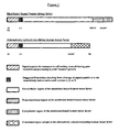
- Membrane bound human tissue factor is a polymer of 263 amino acids. At one end of the polymer is an amino (“NH 2 ”) group commonly referred to in the art as the amino (or “N”)-terminus. At the other end of the polymer is a carboxyl (“COOH”) group commonly referred to in the art as the carboxyl (or “C”)-terminus. The region between the N- and C-termini is commonly referred to as the "internal” region.
- the schematic structure of the TF internal region is shown in Figure 1 . Truncated soluble versions of membrane bound TF are typically from 1-3 (i.e., amino acids 1, 2 or 3) to 218-219, although a truncated version may include additional residues of the transmembrane region from 219-241.
- the extracellular domain of TF is composed of two fibronectin type III domains, both of which contribute to binding of coagulation factor VIIa. All four VIIa structural domains (alpha-carboxyglutamic acid (Gla), EGF1, EGF2 and protease) make TF contacts and, therefore, the total TF-VIIa intermolecular contact area is large (1800 ⁇ 2 ). The dimensions of the entire TF:VIIa complex are about 115 ⁇ in length and 40-50 ⁇ in diameter.
- the contacts provided by the VIIa light chain, particularly the EGF1 domain, are the main contributors to the binding energy (approximately 6.6-9.6 kcal/mol) and also to the contact area (1340 ⁇ 2 of a total of 1810 ⁇ 2 ).
- the contacts made by the VIIa protease domain are less important with respect to energetic contribution (2.3-3.1 kcal/mol), but have a critical role in transmitting allosteric changes within the VII protease domain leading to enhanced catalytic activity.
- the contact residues are mainly located on two adjacent alpha-helical stretches in the VIIa protease domain: VIIa Arg 134 (chymotrypsinogen numbering scheme used throughout) contacts TF-Asp 44 and TF-Trp 45, and VIIa-Met 164 of the adjacent alpha-helix contacts TF-Phe 76 and TF-Tyr 94.
- VIIa Arg 134 chymotrypsinogen numbering scheme used throughout
- VIIa-Met 164 of the adjacent alpha-helix contacts TF-Phe 76 and TF-Tyr 94.
- the VIIa active site is located about 80 ⁇ above the membrane surface.
- Factors VII, IX, X, protein C and prothrombin are anchored to the phospholipid surface via their Gla domains, which contains several Ca 2+ -binding sites.
- the VIIa-GIa domain has three hydrophobic residues at positions 4, 5 and 8 (Phe 4, Leu 5, Leu 8).
- Studies with coagulation factors IX, X and protein C suggest that occupancy of the Ca 2+ -binding sites induces the surface exposure of these residues (in IX the residues are at position 6, 9 and 10) which then engage in hydrophobic interactions with the phospholipid layer.
- these amino acids may anchor VIIa to membranes by insertion into the outer phospholipid layer.
- Mutational changes at TF residues Lys 165 and Lys 166 revealed another region of TF that is important for enzymatic activity of the TF:VIIa complex. These two lysine residues are part of a surface region that interacts with substrates and is located outside the TF-VIIa interface area. The main region is composed of 7 residues (Tyr 157, Lys 159, Ser 163, Gly 164, Lys 165, Lys 166 and Tyr 185) forming a continuous surface patch of about 500 ⁇ 2 .
- This substrate recognition region which may further extend to the VIIa-Gla domain, contacts the Gla domains of substrates X and IX.
- This TF-substrate contact site may serve to properly align X and IX with respect to TF:VIIa complex allowing the formation of productive ternary TF:VIIa:substrate complexes.
- anti-TF antibodies that bind to this region potently inhibit activation of substrates X and IX.
- VIIa protease domain Another important interaction site with substrate is located in the VIIa protease domain centered around the inserted N-terminus. This exosite extends to the area of the Ca 2+ -loop, a region that is topologically similar to the fibrinogen-binding exosite of thrombin.
- the crystal structure of the E76-peptide:VIIa complex reveals that the peptide binds near the Ca 2+ -loop and thus only occupies a portion of the identified macromolecular substrate exosite on VIIa.
- the TF-mediated enhancement of VIIa proteolytic activity is the aggregate of TF's interaction with substrate, the immobilization of VIIa onto the cell surface and the proper positioning of the VIIa protease domain.
- TF also induces allosteric effects at the VIIa catalytic center that contribute to an increase in VIIa enzymatic activity.
- the overall conformation of the S1 recognition pocket as well as the position of the catalytic triad residues (His 57, Asp 102, Ser 195) in VIIa is very similar to other serine proteases, such as IXa and Xa.
- the Xa inhibitor, DX-9065a which selectively binds to the Xa active site, demonstrates that differences in the S2-S4 region can be exploited to design enzyme-specific reversible inhibitors.
- the binding of VII to TF and its subsequent conversion to VIIa is believed to constitute the initial proteolytic process in coagulation.
- human tissue factor As described herein, a new form of human tissue factor has been discovered. This form of human tissue factor circulates in plasma. Sequence analysis indicates that it is a soluble form of human tissue factor and apparently results from alternative splicing of the primary RNA transcript.
- SEQ ID NO:1 is the carboxyl-terminus of naturally occurring circulating alternatively spliced human tissue factor, amino acid residues 167 to 206, referred to herein as "alt-hTF.” This 40 amino acid sequence is unique to alt-hTF.
- Y S T S L E L W Y L W S S S L S S S W L Y L Y T S V E R Q E W G R A G R R T P H (SEQ ID NO:1)
- SEQ ID NO:2 is the cDNA sequence encoding amino acid residues 167 to 206 of alt-hTF. The stop codon is underlined.
- SEQ ID NO:1 and SEQ ID NO:2 are useful in screening of patient samples for the presence of the normal alt-hTF protein, using hybridization assays of patient samples, including blood and tissues, as well as using the methods and reagents described in the examples. Screening can also be accomplished using antibodies, typically labeled with a fluorescent, radioactive, or enzymatic label, or by isolation of target cells and screening for clotting activity, as described in the examples. Typically, one would screen for expression on either a qualitative or quantitative basis, and for expression of functional alt-hTF.
- Reaction conditions for hybridization of an oligonucleotide probe or primer to a nucleic acid sequence vary from oligonucleotide to oligonucleotide, depending on factors such as oligonucleotide length, the number of G and C nucleotides, and the composition of the buffer utilized in the hybridization reaction.
- Moderately stringent hybridization conditions are generally understood by those skilled in the art as conditions approximately 25°C below the melting temperature of a perfectly base-paired double-stranded DNA. Higher specificity is generally achieved by employing incubation conditions having higher temperatures, in other words more stringent conditions. In general, the longer the sequence or higher the G and C content, the higher the temperature and/or salt concentration required.
- the preferred size of a hybridization probe is from 10 nucleotides to 100,000 nucleotides in length. Below 10 nucleotides, hybridized systems are not stable and will begin to denature above 20°C. Above 100,000 nucleotides, one finds that hybridization (renaturation) becomes a much slower and incomplete process, as described in greater detail in the text MOLECULAR GENETICS, Stent, G.S. and R. Calender, pp. 213-219 (1971 ). Ideally, the probe should be from 20 to 10,000 nucleotides. Smaller nucleotide sequences (20-100) lend themselves to production by automated organic synthetic techniques. Sequences from 100-10,000 nucleotides can be obtained from appropriate restriction endonuclease treatments. The labeling of the smaller probes with the relatively bulky chemiluminescent moieties may in some cases interfere with the hybridization process.
- Antibodies to alt-hTF can also be generated that are useful in detection, characterization or isolation of alt-hTF, as well as for modifying alt-hTF protein activity, in most cases, through inhibition of clotting. Antibodies are generated by standard techniques, using alt-hTF. Since the proteins exhibit high evolutionary conservation, it may be advantageous to generate antibodies to the protein of a different species of origin than the species in which the antibodies are to be tested or utilized, looking for those antibodies that are immunoreactive with the most evolutionarily conserved regions.
- Antibodies are typically generated by immunization of an animal using an adjuvant such as Freund's adjuvant in combination with an immunogenic amount of the protein administered over a period of weeks in two to three week intervals, then isolated from the serum, or used to make hybridomas that express the antibodies in culture. Because the methods for immunizing animals yield antibody that is not of human origin, the antibodies could elicit an adverse effect if administered to humans. Methods for "humanizing" antibodies, or generating less immunogenic fragments of non-human antibodies, are well known. A humanized antibody is one in which only the antigen-recognized sites, or complementarity-determining hypervariable regions (CDRs) are of non-human origin, whereas all framework regions (FR) of variable domains are products of human genes. These "humanized” antibodies present a lesser xenographic rejection stimulus when introduced to a human recipient.
- an adjuvant such as Freund's adjuvant
- variable region DNA of a selected animal recombinant anti-idiotypic ScFv is sequenced by the method of Clackson, T., et al., 1991 Nature, 352:624-688 .
- animal CDRs are distinguished from animal framework regions (FR) based on locations of the CDRs in known sequences of animal variable genes. Kabat, H.A., et al., Sequences of Proteins of Immunological Interest, 4th Ed. (U.S. Dept.
- the CDRs are grafted onto human heavy chain variable region framework by the use of synthetic oligonucleotides and polymerase chain reaction (PCR) recombination. Codons for the animal heavy chain CDRs, as well as the available human heavy chain variable region framework, are built in four (each 100 bases long) oligonucleotides. Using PCR, a grafted DNA sequence of 400 bases is formed that encodes for the recombinant animal CDR/human heavy chain FR protection.
- PCR polymerase chain reaction
- the immunogenic stimulus presented by the monoclonal antibodies so produced may be further decreased by the use of Pharmacia's (Pharmacia LKB Biotechnology, Sweden) "Recombinant Phage Antibody System” (RPAS), which generates a single-chain Fv fragment (ScFv) that incorporates the complete antigen-binding domain of the antibody.
- RPAS Recombinant Phage Antibody System
- ScFv single-chain Fv fragment
- antibody variable heavy and light chain genes are separately amplified from the hybridoma mRNA and cloned into an expression vector.
- the heavy and light chain domains are co-expressed on the same polypeptide chain after joining with a short linker DNA that codes for a flexible peptide.
- ScFv single-chain Fv fragment
- the antibodies can be formulated in standard pharmaceutical carriers for administration to patients in need thereof. These include saline, phosphate buffered saline, and other aqueous carriers, and liposomes, polymeric microspheres and other controlled release delivery devices, as are well known in the art.
- the antibodies can also be administered with adjuvant, such as muramyl dipeptide or other materials approved for use in humans (Freund's adjuvant can be used for administration of antibody to animals).
- Antibodies can also be immobilized as discussed below.
- the levels of alt-hTF can be measured in plasma using a simple antibody immunoassay, or the mRNA can be measured using standard RT-PCR or other nucleotide based diagnostic assays.
- an antibody that differentiates between native membrane bound TF or a truncated form thereof, and the alt-hTF This can be obtained by immunizing with the alt-hTF, alone or conjugated to a carrier, with or without adjuvant, then removing antibody that is produced that binds to the membrane bound TF using for example a column having membrane bound TF immobilized thereon.
- a fragment of the alt-hTF derived from the last forty amino acids of the protein is used for immunization to generate alt-hTF antibodies.
- TF is a membrane-anchored cell-surface protein that initiates coagulation when blood contacts damaged tissue.
- membrane bound TF is rapidly covered by several layers of adherent platelets and fibrin and thus is physically separated from the site of vascular injury.
- thrombogenesis has been emphasized.
- a specific anti-TF antibody inhibited thrombus formation.
- a collagen-coated cotton thread is inserted into the jugular vein and thrombus growth on the cotton-thread is monitored by measurement of the incorporated radioactive fibrinogen.
- granulocytes were associated with thrombi in damaged jugular veins and were shown to be the source of thrombus associated TF activity.
- Plasma TF levels were quantified by ELISA and reported to be increased in patients with unstable angina ( Lee, et al., Exp. Mol. Med. 1999;31: 159-164 ) myocardial infarction ( Soejima, et al., J. Am. Coll Cardiol 1999;34: 983-988 ) trauma- and septic- patients ( Gando, et al., Thromb Haemost 1998;79: 1111-1115 ), patients with disseminated intravascular coagulation ( Shimura, et al., Amer. J. Hematol. 1997;55: 169-174 ), antiphospholipid syndrome ( Atsumi, et al., Thromb. Haem.
- u-PAR urokinase receptor
- VIIai Functionally inactivated VIIa failed to upregulate u-PAR expression in SW979 cells, indicating that the proteolytic activity of VIIa was vital for this response.
- Xa treatment of SW979 cells with Xa did not upregulate expression of the u-PAR gene and they suggest that the TF- VIIa complex itself, rather than TF-initiated thrombin generation, is responsible for this effect.
- This mechanism differs from the one proposed by Fischer, et al., where downstream substrates of the TF-VIIa complex, which include Xa and thrombin, were deemed to be essential.
- the TF-VIIa complex initiates coagulation and intracellular signaling. It also alters gene expression and promotes metastasis in murine models.
- Various mechanisms, all involving active VIIa, have been proposed to account for these varied responses. Accordingly, one should be able to treat clotting or the disorders discussed above, by blocking the activity, or removal of, the alt-hTF, using standard techniques to obtain suitable antibodies or other compounds specific for the alt-hTF, or removal techniques such as a column or filter having immobilized therein on antibody specifically immunoreactive with the alt-hTF.
- alt-hTF is useful as a target for compounds that turn on, or off, or otherwise regulate clotting or other disease processes mediated by alt-hTF.
- the assays described in the examples clearly provide routine methodology by which a compound can be tested.
- the in vitro studies of compounds that appear to inhibit alt-hTF are then confirmed by animal testing.
- Assays for testing compounds for useful activity can be based solely on interaction with the alt-hTF protein.
- the assays can be based on interaction with the gene sequence encoding the alt-hTF protein, or the regulatory sequences directing expression of the alt-hTF protein.
- antisense that binds to the regulatory sequences, and/or to the protein encoding sequences can be synthesized using standard oligonucleotide synthetic chemistry.
- the antisense can be stabilized for pharmaceutical use using standard methodology (encapsulation in a liposome or microsphere; introduction of modified nucleotides that are resistant to degradation or groups which increase resistance to endonucleases, such as phosphorothiodates and methylation), then screened initially for alteration of alt-hTF activity in transfected or naturally occurring cells which express alt-hTF, then in vivo in laboratory animals.
- the antisense would inhibit expression.
- sequences which block those sequences which "turn off' synthesis can also be targeted.
- the alt-hTF protein for study can be isolated from either naturally occurring cells or cells which have been genetically engineered to express the alt-hTF protein, as described in the examples above.
- Medicinal chemistry is an interdisciplinary approach used to design small molecules, such as organic chemicals or peptides, for use as therapeutic agents.
- Medicinal chemists use a variety of technology platforms to discover and design drugs. These include combinatorial chemistry, computational chemistry, molecular modeling, high-throughput screening (HTS), enzymology, and pharmacology. The goal is to identify portions of a molecule responsible for particular activities, such as receptor binding or protein interaction. These properties can then be exploited to rationally design more effective drugs. Based on the structure and properties of a lead drug candidate, combinatorial chemists synthesize a series of closely related analogs.
- Computational chemistry tools are then used to simulate the interactions of structural elements with macro-molecules, such as receptors, in order to correlate structure with activity.
- Computational chemistry tools include tools for 3-D structure analysis, quantitative structure-activity relationship analysis, and comparative molecular field analysis, among others.
- Several companies market software and services to help speed drug discovery and lead optimization programs. For example, Tripos Inc., St. Louis, produces a variety of "chemically intelligent" modeling and analysis tools through its discovery software program. Bio Balance, New York, is an example of a company that does computer modeling of proteins for drug design.
- Molecular modeling applications use falls into two broad categories: interactive visualization and computational analyses.
- the latter involves objective, computational analysis and is based upon known biophysical features of the molecule and established mathematical concepts that describe those features.
- These two approaches to modeling can be used alone or collectively to computationally derive a structure.
- these tools also can be used to reconstruct best-fit models from known structures when researchers make theoretical substitutions, insertions, or deletions in the composition of the macromolecule.
- Three of the most prominent uses of modern molecular modeling applications are structure analysis, homology modeling, and docking. Structure analysis centers on computational visualization of a molecule, provided its 3-D atomic coordinates have been elucidated, usually by X-ray crystallography or nuclear magnetic resonance (NMR).
- This information usually resides in major, world-accessible databases including the Brookhaven Protein Data Bank for protein structures, the Nucleic Acids DataBase at Rutgers University for DNA structures, and the Cambridge Crystallographic Data Centre (CCDC) for small molecule (nonprotein/DNA/RNA) structures.
- CCDC Cambridge Crystallographic Data Centre
- investigators may dissect the intricate features of a molecule's structure or examine potential structural changes due to changes in the atomic or molecular composition of the molecule or macromolecule.
- Three-dimensional structural analyses give the ability to examine the spatial, electrostatic, hydrophilic/hydrophobic, potential bonding, or the relationships of the substitute residue with neighboring residues on the same or separate chains.
- Homology modeling has been very important these last few years, as researchers in academia and the pharmaceutical industry seek model structures for proteins whose crystal structures have not yet been solved. Homology modeling is also referred to as "comparative modeling” and "knowledge-based modeling.” It is essentially the theoretical creation of a structure using structural elements borrowed from another protein within the same protein family (usually based on primary sequence and/or secondary structure features) whose crystal structure is known. The process involves alignment of the two sequences, usually performed by any of a number of bioinformatics tools. The result of this alignment is then fed into a homology modeling application, which uses the known crystal structure and the alignment to construct a "draft" (preliminary) structure for the "structureless" protein.
- Docking modeling is used to better understand and model novel protein-protein and protein-ligand interactions (that is, receptor and ligand binding). This provides an avenue to examine and model receptor sites and assess potential ligands (drugs) abd receptor-ligand associations. These techniques allow one to examine binding specificity and decipher the details of the atomic interactions involved in molecular recognition and catalysis.
- Some of the more widely used docking programs include AutoDock (Oxford Molecular Group), DOCK (Molecular Design Institute-UC San Francisco), FTDOCK (Biomolecular Modeling Laboratory), INSIGHT II (MSI), SYBYL/FLEXIDOCK (Tripos) and MidasPlus (Computer Graphics Laboratory, UC San Francisco).
- Modeling programs that utilize molecular dynamics function include HyperChem (Hypercube), INSIGHT II/Discover (MSI), AMBER, CAChe (OMG), SYBYL (Tripos), Alchemy 2000 (Tripos), Spartan (Wavefunction).
- Molecular mechanics emphasizes the potential energy of the molecule as a function of its component atoms, bonds and their angles, and charges-in general, the entire macromolecular environment. This approach attempts to calculate an energy potential for the entire molecule.
- Modeling programs that utilize molecular mechanical methods include HyperChem (Hypercube), SCULPT (ISI), SYBYL (Tripos), INSIGHT II (MSI), AMBER, CAChe (OMG).
- Quantum mechanics methods of "structure” determination are based upon the electronic makeup of a molecule. Electron distribution is defined by one of many quantum theories; the most widely known and used is the molecular orbital theory. This and other theories provide the basis for mathematically determining a number of physicochemical parameters (electric multipole moments of a molecule, electron density distribution, electron affinities, nuclear atomic charge, electrostatic potentials, heats of formation, ionization energies, etc.) that may be utilized to construct a model structure. Quantum mechanisms offer advantages over the other methods in that it can be used to examine molecules at various electronic (“energy”) states or during chemical bond formation and breakage. In essence, quantum mechanical methods are better at predicting chemical reality.
- Modeling with quantum mechanical methods is best for detailed analyses of molecule surface electrostatics, and thus protein-protein interactions and active/binding site interpretations (structure-function relationships, mechanistics of catalysis and/or binding).
- Modeling programs that utilize quantum mechanical methods include Cerius 2 , INSIGHT II (MSI), HyperChem (Hypercube), and SYBYL (Tripos).
- Many modeling applications make use of several mathematical methods; for example, Mac/PC/UNIX SPARTAN (Wavefunction), SYBYL (Tripos), and INSIGHT II (MSI) use a combination of molecular mechanics, quantum mechanical, and/or molecular dynamics methods.
- Multi-routine programs such as Cerius 2 (Tripos), INSIGHT II (MSI), Look/GeneMine (MAG), MidasPlus (Computer Graphics Lab), and SYBYL (Tripos) are especially important to researchers performing homology modeling and docking, where various kinds of computational routines are utilized in the model-building process.
- Such a process incorporates all physicochemical properties into the computational equation to derive the best thermodynamically stable structure, a structure that should depict a functional molecule.
- Computer modeling technology allows visualization of the three-dimensional atomic structure of a selected molecule and the rational design of new compounds that will interact with the molecule.
- the three-dimensional construct typically depends on data from x-ray crystallographic analyses or NMR imaging of the selected molecule.
- the molecular dynamics require force field data.
- the computer graphics systems enable prediction of how a new compound will link to the target molecule and allow experimental manipulation of the structures of the compound and target molecule to perfect binding specificity. Prediction of what the molecule-compound interaction will be when small changes are made in one or both requires molecular mechanics software and computationally intensive computers, usually coupled with user-friendly, menu-driven interfaces between the molecular design program and the user.
- Nucleic acid molecules containing the 5' regulatory sequences of the TF gene can be used to regulate or inhibit gene expression in vivo .
- Vectors including both plasmid and eukaryotic viral vectors, may be used to express a particular recombinant 5' flanking region-gene construct in cells depending on the preference and judgment of the skilled practitioner (see, e.g., Sambrook et al., Chapter 16).
- a number of viral and nonviral vectors have been developed that enable the introduction of nucleic acid sequences in vivo (see, e.g., Mulligan, 1993 Science, 260, 926-932 ; United States Patent No. 4,980,286 ; United States Patent No. 4,868,116 ; incorporated herein by reference).
- nucleic acid is encapsulated in cationic liposomes that can be injected intravenously into a mammal.
- This system has been used to introduce DNA into the cells of multiple tissues of adult mice, including endothelium and bone marrow (see, e.g., Zhu et al., 1993 Science 261, 209-211 .
- the 5' flanking sequences of the TF gene can also be used to inhibit the expression of alt-hTF.
- an antisense RNA of all or a portion of the 5' flanking region of the TF gene can be used to inhibit expression of alt-hTF in vivo .
- Expression vectors e.g., retroviral expression vectors
- U.S. Patent No. 4,868,116 U.S. Patent No. 4,980,286
- DNA containing all or a portion of the sequence of the 5' flanking region of the TF gene can be inserted into an appropriate expression vector so that upon passage into the cell, the transcription of the inserted DNA yields an antisense RNA transcript that is complementary to the mRNA transcript of the alt-hTF protein normally found in the cell.
- This antisense RNA transcript of the inserted DNA can then base-pair with the normal mRNA transcript found in the cell and thereby prevent the mRNA from being translated. It is of course necessary to select sequences that are downstream from the transcriptional start sites for the TF gene to ensure that the antisense RNA contains sequences complementary to those present in the mRNA.
- Antisense RNA can be also generated in vitro, and then inserted into cells. Oligonucleotides can be synthesized on an automated synthesizer (e.g., Model 8700 automated synthesizer of Milligen-Biosearch, Burlington, MA or ABI Model 380B). In addition, antisense deoxyoligonucleotides have been shown to be effective in inhibiting gene transcription and viral replication (see e.g., Zamecnik et al., 1978 Proc. Natl. Acad. Sci. USA 75, 280-284 ; Zamecnik et al., 1986 Proc. Natl. Acad. Sci., 83, 4143-4146 ; Wickstrom et al., 1988 Proc. Natl.
- antisense oligonucleotides Inhibition of expression of a gene by antisense oligonucleotides is also possible if the antisense oligonucleotides contain modified nucleotides (see, e.g., Offensperger et. al., 1993 EMBO J. 12, 1257-1262 (antisense phosphorothioate oligodeoxynucleotides); Rosenberg et al., PCT WO 93/01286 (synthesis of sulfurthioate oligonucleotides); Agrawal et al., 1988 Proc. Natl. Acad. Sci.
- Inhibition of expression of a gene can also be achieved by using small double-stranded RNA molecules (21-25 base pairs in length) by means of a process known as RNA interference (see, e.g., Sharp, Genes & Development, 2001, 15:485-490 ).
- Double-stranded RNA molecules can be synthesized in vitro and then introduced into living cells (see, e.g., Donze et al., 2002 Nucleic Acid Research, 30:e46 ) or synthesized from a DNA template that was stably incorporated into cells (see, e.g., Sui et al., 2002 Proc. Natl. Acad. Sci. USA 99:5515-5520 ).
- Double-stranded RNA molecules have been shown to inhibit HIV-1 infection (see Novina et al., 2002, Nature Medicine, 8:681-686 ) and expression of the full-length tissue factor (see Holen et al., Nucleic Acid Research 2002, 30:1757-1766 ).
- double-stranded RNA molecules containing the region unique to alt-hTF mRNA, i.e. the site of splicing of exon 4 to exon 6 may be used to selectively inhibit expression of alt-hTF protein in vivo.
- sequences of the 5' flanking region of the TF gene can also be used in triple helix (triplex) gene therapy. Oligonucleotides complementary to gene promoter sequences on one of the strands of the DNA have been shown to bind promoter and regulatory sequences to form local triple nucleic acid helices which block transcription of the gene (see, e.g., 1989 Maher et al., Science 245, 725-730 ; Orson et al., 1991 Nucl. Acids Res. 19, 3435-3441 ; Postal et al., 1991 Proc. Natl. Acad. Sci.
- oligonucleotides should generally be greater than 14 nucleotides in length to ensure target sequence specificity (see, e.g., Maher et al., 1989; Grigoriev et al., 1992). Also, many cells avidly take up oligonucleotides that are less than 50 nucleotides in length (see e.g., Orson et al., 1991; Holt et al., 1988 Mol. Cell. Biol. 8, 963-973 ; Wickstrom et al., 1988 Proc. Natl. Acad. Sci. USA 85, 1028-1032 ).
- a free amine can be introduced to a 3' terminal hydroxyl group of oligonucleotides without loss of sequence binding specificity (Orson et al., 1991). Furthermore, more stable triplexes are formed if any cytosines that may be present in the oligonucleotide are methylated, and also if an intercalating agent, such as an acridine derivative, is covalently attached to a 5' terminal phosphate (e.g., via a pentamethylene bridge); again without loss of sequence specificity (Maher et al., 1989; Grigoriev et al., 1992).
- an intercalating agent such as an acridine derivative
- oligonucleotides Methods to produce or synthesize oligonucleotides are well known in the art and synthetic oligonucleotides are now commercially available. Such methods can range from standard enzymatic digestion followed by nucleotide fragment isolation (see e.g., Sambrook et al., Chapters 5, 6) to purely synthetic methods, for example, by the cyanoethyl phosphoramidite method using a Milligen or Beckman System 1Plus DNA synthesizer (see also, Ikuta et al., in Ann. Rev. Biochem.
- DNA sequences of the 5' flanking region of the TF gene described herein can be used to design and construct oligonucleotides including a DNA sequence consisting essentially of at least 15 consecutive nucleotides, with or without base modifications or intercalating agent derivatives, for use in forming triple helices specifically within the 5' flanking region of the TF gene in order to inhibit expression of the gene.
- enhancers or multiple copies of the regulatory sequences may be advantageous to insert enhancers or multiple copies of the regulatory sequences into an expression system to facilitate screening of methods and reagents for manipulation of expression.
- Molecules with a given function can be selected for from a complex mixture of random molecules in what has been referred to as "in vitro genetics" ( Szostak, TIBS 19:89, 1992 ).
- in vitro genetics Szostak, TIBS 19:89, 1992 .
- One synthesizes a large pool of molecules bearing random and defined sequences and subjects that complex mixture for example, approximately 10 15 individual sequences in 100 ⁇ g of a 100 nucleotide RNA, to some selection and enrichment process.
- Ellington and Szostak (1990) estimated that 1 in 10 10 RNA molecules folded in such a way as to bind a given ligand. DNA molecules with such ligand-binding behavior have been isolated (Ellington and Szostak, 1992; Bock et al, 1992).
- Compounds that are effective for blocking binding of the alt-hTF can also consist of fragments of the alt-hTF protein, expressed recombinantly and cleaved by enzymatic digest or expressed from a sequence encoding a peptide of less than the full length alt-hTF protein. These will typically be soluble proteins, i.e., not including the transmembrane and cytoplasmic regions, although smaller portions determined in the assays described above to inhibit or compete for binding to the alt-hTF protein can also be utilized. It is a routine matter to make appropriate alt-hTF protein fragments, test for binding, and then utilize them. The preferred fragments are of human origin, in order to minimize potential immunological response.
- the peptides can be as short as five to eight amino acids in length and are easily prepared by standard techniques. They can also be modified to increase in vivo half-life, by chemical modification of the amino acids or by attachment to a carrier molecule or inert substrate. Based on studies with other peptide fragments blocking alt-hTF binding, the IC 50 , the dose of peptide required to inhibit binding by 50%, ranges from about 50 ⁇ M to about 300 ⁇ M, depending on the peptides. These ranges are well within the effective concentrations for the in vivo administration of peptides, based on comparison with the RGD-containing peptides, described, for example, in U.S. Patent No. 4,792,525 to Ruoslaghti, et al. , used in vivo to alter cell attachment and phagocytosis.
- the peptides can also be conjugated to a carrier protein such as keyhole limpet hemocyanin by its N-terminal cysteine by standard procedures such as the commercial Imject kit from Pierce Chemicals or expressed as a fusion protein, which may have increased efficacy.
- a carrier protein such as keyhole limpet hemocyanin by its N-terminal cysteine by standard procedures such as the commercial Imject kit from Pierce Chemicals or expressed as a fusion protein, which may have increased efficacy.
- the peptides can be prepared by proteolytic cleavage of the alt-hTF protein, or, preferably, by synthetic means. These methods are known to those skilled in the art. An example is the solid phase synthesis described by J. Merrifield, 1964 J. Am. Chem. Soc. 85, 2149 , used in U.S. Patent No. 4,792,525 , and described in U.S. Patent No.
- the peptide can also be administered as a pharmaceutically acceptable acid- or base- addition salt, formed by reaction with inorganic acids such as hydrochloric acid, hydrobromic acid, perchloric acid, nitric acid, thiocyanic acid, sulfuric acid, and phosphoric acid, and organic acids such as formic acid, acetic acid, propionic acid, glycolic acid, lactic acid, pyruvic acid, oxalic acid, malonic acid, succinic acid, maleic acid, and fumaric acid, or by reaction with an inorganic base such as sodium hydroxide, ammonium hydroxide, potassium hydroxide, and organic bases such as mono-, di-, trialkyl and aryl amines and substituted ethanolamines.
- inorganic acids such as hydrochloric acid, hydrobromic acid, perchloric acid, nitric acid, thiocyanic acid, sulfuric acid, and phosphoric acid
- organic acids such as formic acid, acetic acid, propionic acid
- Peptides containing cyclopropyl amino acids, or amino acids derivatized in a similar fashion can also be used. These peptides retain their original activity but have increased half-lives in vivo. Methods known for modifying amino acids, and their use, are known to those skilled in the art, for example, as described in U.S. Patent No. 4,629,784 to Stammer .
- Peptides are generally active when administered parenterally in amounts above about 1 ⁇ g/kg of body weight. Based on extrapolation from other proteins, for treatment of most inflammatory disorders, the dosage range will be between 0.1 to 70 mg/kg of body weight. This dosage will be dependent, in part, on whether one or more peptides are administered.
- the peptide sequences present in the C-terminal region of alt-hTF should be the most optimal. Because this sequence has not been described in other proteins, this would be a unique target to inhibit binding alt-hTF to platelets, thereby uniquely inhibiting the activity of alt-hTF.
- binding activity Compounds that alter alt-hTF protein activity and/or binding (referred to generally herein as "binding activity") are preferably administered in a pharmaceutically acceptable vehicle.
- suitable pharmaceutical vehicles are known to those skilled in the art.
- the compound will usually be dissolved or suspended in sterile water or saline.
- the compound will be incorporated into an inert carrier in tablet, liquid, or capsular form.
- Suitable carriers may be starches or sugars and include lubricants, flavorings, binders, and other materials of the same nature.
- the compounds can also be administered locally by topical application of a solution, cream, gel, or polymeric material (for example, Pluronic TM , BASF).
- the compound may be administered in liposomes or microspheres (or microparticles).
- Methods for preparing liposomes and microspheres for administration to a patient are known to those skilled in the art.
- U.S. Patent No. 4,789,734 describes methods for encapsulating biological materials in liposomes. Essentially, the material is dissolved in an aqueous solution, the appropriate phospholipids and lipids added, along with surfactants if required, and the material dialyzed or sonicated, as necessary.
- a review of known methods is by G. Gregoriadis, Chapter 14. "Liposomes", Drug Carriers in Biology and Medicine pp. 287-341 (Academic Press, 1979 ).
- Microspheres formed of polymers or proteins are well known to those skilled in the art, and can be tailored for passage through the gastrointestinal tract directly into the bloodstream. Alternatively, the compound can be incorporated and the microspheres, or composite of microspheres, implanted for slow release over a period of time, ranging from days to months. See, for example, U.S. Patent No. 4,906,474 , 4,925,673 , and 3,625,214 .
- the antibodies to alt-hTF protein can be used to remove alt-hTF from patient blood, by immobilizing the alt-hTF antibodies on a suitable substrate, such as the cellulose membrane of a dialysis unit, using conventional coupling, for example, using carboimide. The patient's blood is then dialyzed through the unit.
- tissue factor that appears to circulate in human plasma in a latent form has been identified.
- this protein Upon shear-induced binding to platelets, this protein exhibits enzymatic activity and, therefore, may serve as a potential trigger for initiating or propagating thrombosis.
- the activity Following high-speed centrifugation of already platelet-poor plasma, the activity remained in the bulk aqueous phase indicating that blood-borne TF is soluble in an aqueous medium.
- alt-hTF is extracted from bacteria simply by osmotic shock.
- Citrated blood was processed as follows. After centrifugation at 200xG for 15 minutes, the cell pellet was discarded and the supernatant was mixed 1:1 with Tris-buffered saline (TBS) and is referred to as the "starting material.” Nine mL of the starting material was removed and centrifuged at 260,000xG for 4 hours. Following high-speed centrifugation, 4 distinct layers were evident and their volume fractions were estimated: an upper lipid phase (17% of volume), a bulk-aqueous phase (52%), a lower viscous phase (23%) and a pellet (3%). Samples from the starting material and each of the three upper phases were extracted and assayed for TF activity using a standardized protocol. In one experiment, the pellet was resuspended in 4.5mL of TBS and also assayed for TF activity.
- TBS Tris-buffered saline
- agrose beads covalently linked to a rabbit antihuman TF antibody (0.5 mg antibody per mL of agarose).
- the beads were washed 5 times with hepes-buffered saline (HBS) and the bound antigen was eluted with 6M guanidine dissolved in HBS.
- HBS hepes-buffered saline
- the eluted protein was collected and incubated with phospholipids (30% phosphatidyl serine, 70% phosphatidyl choline; 75 ⁇ M total) and n-octyl- ⁇ -D-glucopyranoside.
- the samples were dialyzed to remove the detergent and allow the formation of lipid vesicles in the presence of the eluted protein.
- the dialyzed samples were supplemented with calcium (5mM), coagulation factor VIIa (1 nM) and coagulation factor X (150 nM), and the rate of appearance of FXa was measured using a standard assay.
- the results are shown in Figure 2 .
- the TF activity associated with the starting material was 158.5 pM-FXa/min.
- TF activity measured in each phase was 45.7 pM-FXa/min for the lipid phase, 138.9 pM-FXa/min for the bulk aqueous phase, 554.7 pM-FXa/min for the lower viscous phase, and 40 pM-FXa/min for the resuspended pellet. Background levels were approximately 20 pM-FXa/min.
- Example 2 Identification of alt-hTF mRNA in a human cell line.
- TF mRNA was examined in a human cell line HL-60 using RT-PCR.
- a pair of primers designated "Forward” and “Reverse” yielded two bands.
- the size of the larger band was approximately 400 base pairs - as expected from the TF cDNA sequence.
- the size of the smaller band was approximately 240 base pairs.
- the smaller band was subcloned and sequenced. Sequencing results revealed a previously unknown splicing variant of TF mRNA in which exon 5 is absent and exon 4 is thus fused directly with exon 6. Of note, such a fusion creates a frameshift in the TF open reading frame.
- the 770 base pair product was subcloned and sequenced.
- the results of sequencing confirmed the existence of the TF mRNA spieces encoding an open reading frame with the initiation codon corresponding to bases 112-114, and a termination codon corresponding to bases 986-988 of the published TF cDNA sequence; GenBank accession number J02931.
- the alternatively spliced hTF mRNA lacks exon 5 and thus contains a frameshift.
- the open reading frame of the alt-hTF mRNA encodes a human tissue factor variant whose mature peptide comprises 206 amino acids.
- Amino acids 1-166 are identical to those of the known membrane bound human TF; however, the remaining 40 amino acids at the carboxy-terminus diverge from the known TF amino acid sequence.
- the carboxy-terminus of the alternatively spliced hTF contains a region with potential transmembrane properties.
- Example 3 Expression of alt-hTF in various tissues.
- the lengths of reported sequences of the three clones derived from a human lung cDNA library are 663, 780, and 747 base pairs, respectively. None of the three sequences encode a significantly larger non-interrupted stretch of amino acids in any of the three 5'-3' frames. Although all three sequences contain a region corresponding to the site of alternative splicing, i.e., TCA GGA AAG AAA TAT TCT (SG KK YS) (SEQ ID NO:3), in all three sequences this region is not in frame with the sequence encoding human tissue factor protein.
- the three clones corresponding to the above lung ESTs were obtained from the I.M.A.G.E. consortium and fully sequenced. None of the clones contained the complete alt-hTF open reading frame. However, sequencing results revealed that, like in membrane-bound TF mRNA, the long 3'-untranslated end of the asHTF mRNA is entirely encoded by exon 6.
- the length of the reported sequence of a clone derived from a primary human keratinocyte cDNA library is 356 base pairs. This sequence encodes a stretch of amino acids 72 through 166 of the membrane bound human tissue factor fused to the first 23 amino acids of the 40 amino acid carboxyl-terminus unique to the alternatively spliced hTF. This sequence is, therefore, a partial (incomplete) cDNA encoding an alternatively spliced human tissue factor molecule.
- start plasma Frozen human plasma (300 mL) was thawed on ice and centrifuged at 3,000xG for 90 minutes. The supernatant (referred to as "start plasma") was passed at 4°C through a column loaded with Affigel-coupled antibodies to the last 10 amino acids at the carboxyl-terminus of the alt-hTF protein. A flow-through sample was collected after 250 mL of start plasma was passed through the column.
- Samples described above were tested for tissue factor activity using a Xa chromogenic assay in a platelet-shear system.
- Freshly drawn citrated blood was centrifuged at 135xG for 20 minutes at room temperature to prepare platelet rich plasma (PRP). Platelets were then pelleted by centrifuging PRP at 850xG for 12 minutes, washed twice in CGSa, and resuspended at 3x10 8 platelets/mL in modified Tyrode's Buffer.
- 75 ⁇ L of the test samples i.e. start plasma and plasma-flow-through
- 75 ⁇ L of the test samples i.e. start plasma and plasma-flow-through
- the plasma-platelet suspension was loaded into collagen-coated wells.
- Example 5 Expression of the alt-hTF protein in bacteria.
- the region encoding the entire mature peptide of the alternatively spliced hTF variant was amplified using RT-PCR and subcloned into pBAD/gIIIA expression vector (Invitrogen Corporation). The sequence of this construct was verified, and the recombinant alternatively spliced hTF protein was produced in E. coli . At the N-terminus of the expressed protein, three additional amino acids (i.e. Thr, Met, and Ala) were present due to the structure of the multiple cloning site in pBAD/gIIIA expression vector.
- the expressed protein was isolated from bacteria by osmotic shock, and the resultant osmotic shock fluid was concentrated via centrifugation in Centricon TM filter devices (Millipore). Presence of the desired protein in the concentrated osmotic shock fluid was verified by Western immunoblotting, and protein concentration was analysed using Bradford protein microassay.
- Three samples containing the recombinant alt-hTF protein were then prepared: a sample of the protein dissolved in HBS, a sample of the relipidated protein, and a sample of the protein incubated with phospholipid vesicles.
- Relipidation of the recombinant protein was carried out as follows. 35 ⁇ g of protein in TBS were combined with n-octyl- ⁇ -D-glucopyranoside (final concentration - 125 mM) and a phospholipid mixture (PS:PC 30:70, final concentration - 75 ⁇ M) to the final volume of 0.5 mL.
- the sample was placed on orbital mixer (set to slow) for 30 min at RT, transferred into a 0.5 mL dialysis cassette and dialyzed overnight versus 2 L of TBS.
- Incubation of the recombinant protein with phospholipid vesicles was carried out as follows. 35 ⁇ g of protein in HBS were combined with a phospholipid vesicle preparation (PS:PC 30:70, final concentration - 75 ⁇ M) that was extruded to yield vesicles with the diameter of 100 nm. This mixture was incubated overnight at room temperature.
- Example 6 Presence of the alt-TF protein in ex-vivo thrombi.
Landscapes
- Health & Medical Sciences (AREA)
- Chemical & Material Sciences (AREA)
- Life Sciences & Earth Sciences (AREA)
- Organic Chemistry (AREA)
- Medicinal Chemistry (AREA)
- Toxicology (AREA)
- Biochemistry (AREA)
- Biophysics (AREA)
- General Health & Medical Sciences (AREA)
- Genetics & Genomics (AREA)
- Zoology (AREA)
- Molecular Biology (AREA)
- Proteomics, Peptides & Aminoacids (AREA)
- Gastroenterology & Hepatology (AREA)
- Cell Biology (AREA)
- Immunology (AREA)
- Peptides Or Proteins (AREA)
- External Artificial Organs (AREA)
- Materials For Medical Uses (AREA)
- Medicines Containing Material From Animals Or Micro-Organisms (AREA)
- Medicines That Contain Protein Lipid Enzymes And Other Medicines (AREA)
Claims (21)
- Alternativ gespleißte Form des humanen Gewebefaktors, die Y S T S L E L W Y L W S S S L S S S W L Y L Y T S V E R Q E W G R A G R R T P H (SEQ ID NO:1) umfasst, oder die alternativ gespleißte Form des humanen Gewebefaktors, die konservative Aminosäuresubstitutionen aufweist, mit humaner Gewebefaktor-Aktivität.
- Gewebefaktor nach Anspruch 1, der ferner die ersten 166 Aminosäuren des membrangebundenen humanen Gewebefaktors umfasst.
- Humanes Gewebefaktorprotein, das die Sequenz Serin-Serin-Serin-Leucin-Serin-Serin-Serin umfasst, wobei das Protein in der Lage ist, einen Antikörper zu generieren, der zwischen dem nativen membrangebundenen Gewebefaktor und einer alternativ gespleißten Form des humanen Gewebefaktors, wie sie im Anspruch 1 definiert ist, unterscheidet.
- Nucleotidmolekül, das für irgendeines der Gewebefaktorproteine der Ansprüche 1-3 codiert.
- Nucleotidmolekül nach Anspruch 4 in einem Vektor für die Expression des Gewebefaktorproteins.
- Nucleotidmolekül nach Anspruch 5 in einem Vektor für die Expression in einem Wirtsbakterium.
- Nucleotidmolekül nach Anspruch 5 in einem Vektor für die Expression in einem eukaryotischen System.
- Doppelsträngiges RNA-Molekül, das den Bereich einer für das Polypeptid der SEQ ID NO:1 codierenden mRNA umfasst.
- Verfahren zur Erzeugung irgendeines der Gewebefaktorproteine der Ansprüche 1-3, das das Exprimieren des Nucleotidmoleküls nach Anspruch 5 in einem geeigneten Wirt umfasst.
- In-vitro-Verfahren zum Screenen auf Erkrankungen, die mit dem Gewebefaktor assoziiert sind, das das Messen der Spiegel eines alternativ gespleißten Gewebefaktors, wie er in irgendeinem der Ansprüche 1 bis 3 definiert ist, im Plasma oder Gewebe umfasst.
- Verfahren nach Anspruch 10, wobei die Spiegel mittels eines Immunoassays mit einem Antikörper, der spezifisch mit dem alternativ gespleißten Gewebefaktor reagiert, bestimmt werden.
- Hemmer der Gewebefaktorproteine nach irgendeinem der Ansprüche 1-3, der nicht mit dem membrangebundenen humanen Gewebefaktor reagiert, wobei der Hemmer ein Antisensemolekül, das zu einer für ein Gewebefaktorprotein nach einem der Ansprüche 1 bis 3 codierenden mRNA komplementär ist, ein Fragment der alternativ gespleißten Form des humanen Gewebefaktors oder ein Antikörper gegen die alternativ gespleißte Form des humanen Gewebefaktorproteins nach irgendeinem der Ansprüche 1 bis 3 ist.
- Antikörper, wie er im Anspruch 12 definiert ist, der spezifisch mit den Gewebefaktorproteinen nach irgendeinem der Ansprüche 1-3 immunreaktiv ist.
- Antikörper nach Anspruch 13, konjugiert mit einer nachweisbaren Markierung.
- Antikörper nach Anspruch 13, in einer Säule oder auf einem Filter immobilisiert.
- Hemmer gemäß Anspruch 12 für den Einsatz in der Medizin.
- Antikörper gemäß Anspruch 13 für den Einsatz in der Medizin.
- Verwendung eines Hemmers gemäß Anspruch 12 bei der Herstellung eines Medikaments zur Behandlung einer mit dem Gewebefaktor assoziierten Erkrankung.
- Verwendung eines Antikörpers gemäß Anspruch 13 bei der Herstellung eines Medikaments für die Entfernung des alternativ gespleißten Gewebefaktors.
- Hemmer gemäß Anspruch 12 für den Einsatz in der Behandlung einer mit dem Gewebefaktor assoziierten Erkrankung.
- Antikörper gemäß Anspruch 13 für die Entfernung des alternativ gespleißten Gewebefaktors.
Applications Claiming Priority (3)
| Application Number | Priority Date | Filing Date | Title |
|---|---|---|---|
| US31624401P | 2001-08-30 | 2001-08-30 | |
| US316244P | 2001-08-30 | ||
| PCT/US2002/027533 WO2003020111A2 (en) | 2001-08-30 | 2002-08-29 | Alternatively spliced circulating tissue factor |
Publications (3)
| Publication Number | Publication Date |
|---|---|
| EP1429650A2 EP1429650A2 (de) | 2004-06-23 |
| EP1429650A4 EP1429650A4 (de) | 2005-11-23 |
| EP1429650B1 true EP1429650B1 (de) | 2009-06-24 |
Family
ID=23228197
Family Applications (1)
| Application Number | Title | Priority Date | Filing Date |
|---|---|---|---|
| EP02797784A Expired - Lifetime EP1429650B1 (de) | 2001-08-30 | 2002-08-29 | Alternativ gesplicter zirkulierender gewebefaktor |
Country Status (6)
| Country | Link |
|---|---|
| US (1) | US7045350B2 (de) |
| EP (1) | EP1429650B1 (de) |
| AT (1) | ATE434404T1 (de) |
| AU (1) | AU2002361471A1 (de) |
| DE (1) | DE60232733D1 (de) |
| WO (1) | WO2003020111A2 (de) |
Families Citing this family (13)
| Publication number | Priority date | Publication date | Assignee | Title |
|---|---|---|---|---|
| EP1554381B1 (de) * | 2002-02-07 | 2009-11-25 | siRNAsense AS | Post-transcriptional silencing der gewebefaktor expression durch kurze interferierende rnas |
| US7425328B2 (en) | 2003-04-22 | 2008-09-16 | Purdue Pharma L.P. | Tissue factor antibodies and uses thereof |
| WO2008054822A2 (en) * | 2006-11-03 | 2008-05-08 | Angiogen Llc | Compositions comprising alternatively spliced tissue factor (as tf) and methods for use |
| US20090042869A1 (en) * | 2007-06-20 | 2009-02-12 | University Of Utah Research Foundation | SIGNAL-DEPENDENT SPLICING OF TISSUE FACTOR PRE-mRNA IN PLATELET CELLS |
| UA109633C2 (uk) | 2008-12-09 | 2015-09-25 | Антитіло людини проти тканинного фактора | |
| WO2011144953A1 (en) * | 2010-05-18 | 2011-11-24 | Diagon Kft. | Procedure for producing recombinant human tissue factor |
| BR112012031727B1 (pt) | 2010-06-15 | 2022-03-29 | Genmab A/S | Conjugado de droga-anticorpo, composição farmacêutica, e, uso do conjugado de droga- anticorpo |
| US9555109B2 (en) | 2012-01-20 | 2017-01-31 | University Of Cincinnati | Method of inhibiting cell proliferation induced by alternatively spliced tissue factor by administering a monoclonal antibody |
| US10204181B1 (en) * | 2015-07-10 | 2019-02-12 | Omnisent LLC | Systems and methods for modeling quantum structure and behavior |
| US10637583B2 (en) * | 2015-07-10 | 2020-04-28 | Omnisent, LLC | Systems and methods for modeling quantum entanglement and performing quantum communication |
| TWI841554B (zh) | 2018-03-21 | 2024-05-11 | 丹麥商珍美寶股份有限公司 | 以鉑為主之劑與抗組織因子抗體-藥物共軛物的組合治療癌症之方法 |
| IL278522B2 (en) | 2018-05-07 | 2025-10-01 | Genmab As | Methods for treating cancer with a combination of anti-PD-1 antibody and always an anti-tissue factor antibody drug |
| TWI844571B (zh) | 2018-10-30 | 2024-06-11 | 丹麥商珍美寶股份有限公司 | 使用抗血管內皮生長因子(vegf)抗體與抗組織因子(tf)抗體-藥物共軛體之組合以治療癌症之方法 |
Family Cites Families (24)
| Publication number | Priority date | Publication date | Assignee | Title |
|---|---|---|---|---|
| US3625214A (en) | 1970-05-18 | 1971-12-07 | Alza Corp | Drug-delivery device |
| US4244946A (en) | 1979-06-11 | 1981-01-13 | The Salk Institute For Biological Studies | Water-soluble peptides affecting gonadal function |
| US4305872A (en) | 1979-10-19 | 1981-12-15 | Kenneth Wingrove | Polypeptide derivatives |
| US4316891A (en) | 1980-06-14 | 1982-02-23 | The Salk Institute For Biological Studies | Extended N-terminal somatostatin |
| US4792525A (en) | 1982-08-04 | 1988-12-20 | La Jolla Cancer Research Foundation | Tetrapeptide |
| US4906474A (en) | 1983-03-22 | 1990-03-06 | Massachusetts Institute Of Technology | Bioerodible polyanhydrides for controlled drug delivery |
| US4629784A (en) | 1983-08-16 | 1986-12-16 | The University Of Georgia Research Foundation, Inc. | Synthesis of cyclopropane amino acids |
| WO1987000201A1 (en) | 1985-07-05 | 1987-01-15 | Whitehead Institute For Biomedical Research | Epithelial cells expressing foreign genetic material |
| US4980286A (en) | 1985-07-05 | 1990-12-25 | Whitehead Institute For Biomedical Research | In vivo introduction and expression of foreign genetic material in epithelial cells |
| US4789734A (en) | 1985-08-06 | 1988-12-06 | La Jolla Cancer Research Foundation | Vitronectin specific cell receptor derived from mammalian mesenchymal tissue |
| CH671155A5 (de) | 1986-08-18 | 1989-08-15 | Clinical Technologies Ass | |
| HUT52546A (en) * | 1988-06-17 | 1990-07-28 | Sinai School Medicine | Process for cloning and expressing of human tissue factor |
| CA1340977C (en) | 1988-11-15 | 2000-04-25 | Monty Krieger | Scavenger receptor protein and antibody thereto |
| US5211947A (en) | 1988-12-16 | 1993-05-18 | Schering Corporation | Method for lowering blood cholesterol levels with granulocyte-macrophage colony stimulating factor |
| JPH03290184A (ja) | 1990-04-06 | 1991-12-19 | Chugai Pharmaceut Co Ltd | スカベンジャーレセプター産生動物細胞 |
| JP3594318B2 (ja) | 1990-08-27 | 2004-11-24 | 中外製薬株式会社 | 抗ヒトスカベンジャーレセプター抗体 |
| US5346991A (en) | 1991-06-13 | 1994-09-13 | Genentech, Inc. | Tissue factor mutants useful for the treatment of myocardial infarction and coagulopathic disorders |
| JPH07501204A (ja) | 1991-06-28 | 1995-02-09 | マサチューセッツ インスティテュート オブ テクノロジー | 局所的オリゴヌクレオチド療法 |
| WO1993019166A1 (en) | 1992-03-23 | 1993-09-30 | The University Of North Carolina At Chapel Hill | Small animal model for studying cholesterol metabolism |
| US5585479A (en) | 1992-07-24 | 1996-12-17 | The United States Of America As Represented By The Secretary Of The Navy | Antisense oligonucleotides directed against human ELAM-I RNA |
| US5746223A (en) | 1996-10-11 | 1998-05-05 | Williams; Kevin Jon | Method of forcing the reverse transport of cholesterol from a body part to the liver while avoiding harmful disruptions of hepatic cholesterol homeostasis |
| US6429289B1 (en) | 1994-06-23 | 2002-08-06 | Massachusetts Institute Of Technology | Class BI and CI scavenger receptors |
| US5652224A (en) | 1995-02-24 | 1997-07-29 | The Trustees Of The University Of Pennsylvania | Methods and compositions for gene therapy for the treatment of defects in lipoprotein metabolism |
| US5925333A (en) | 1995-11-15 | 1999-07-20 | Massachusetts Institute Of Technology | Methods for modulation of lipid uptake |
-
2002
- 2002-08-29 DE DE60232733T patent/DE60232733D1/de not_active Expired - Lifetime
- 2002-08-29 WO PCT/US2002/027533 patent/WO2003020111A2/en not_active Ceased
- 2002-08-29 AU AU2002361471A patent/AU2002361471A1/en not_active Abandoned
- 2002-08-29 AT AT02797784T patent/ATE434404T1/de not_active IP Right Cessation
- 2002-08-29 EP EP02797784A patent/EP1429650B1/de not_active Expired - Lifetime
- 2002-08-29 US US10/230,839 patent/US7045350B2/en not_active Expired - Lifetime
Also Published As
| Publication number | Publication date |
|---|---|
| AU2002361471A1 (en) | 2003-03-18 |
| EP1429650A4 (de) | 2005-11-23 |
| US20030049784A1 (en) | 2003-03-13 |
| ATE434404T1 (de) | 2009-07-15 |
| DE60232733D1 (de) | 2009-08-06 |
| US7045350B2 (en) | 2006-05-16 |
| EP1429650A2 (de) | 2004-06-23 |
| WO2003020111A2 (en) | 2003-03-13 |
| WO2003020111A3 (en) | 2004-01-29 |
Similar Documents
| Publication | Publication Date | Title |
|---|---|---|
| EP0777731B1 (de) | Klonierung und regulation eines rezeptors fur endothelialzell-protein c/ aktiviertes protein c | |
| EP1429650B1 (de) | Alternativ gesplicter zirkulierender gewebefaktor | |
| JP2003517304A (ja) | クニッツ型プロテアーゼインヒビターポリヌクレオチド、ポリペプチド、および抗体 | |
| US20080254506A1 (en) | Claudin polypeptides | |
| WO1996005303A9 (en) | Cloning and regulation of an endothelial cell protein c/activated protein c receptor | |
| JP2002517185A (ja) | 前立腺癌および結腸癌の診断および治療に有用な腫瘍抗原 | |
| AU2001284977A1 (en) | Claudin polypeptides | |
| JP2003524413A (ja) | プラスミノーゲン様ポリヌクレオチド、ポリペプチド、および抗体 | |
| US5843884A (en) | C9 complement inhibitor | |
| JPH11508135A (ja) | ヒトedg−2受容体ホモログ | |
| US7166568B1 (en) | Compositions and methods to inhibit formation of the C5b-9 complex of complement | |
| JP2003512856A (ja) | ウテログロビン様ポリヌクレオチド、ポリペプチド、および抗体 | |
| JP2003507036A (ja) | レチノイドレセプター相互作用ポリヌクレオチド、ポリペプチド、および抗体 | |
| JP2003508036A (ja) | アトラクチン様ポリヌクレオチド、ポリペプチド、および抗体 | |
| JPH11505412A (ja) | ホスホリパーゼcホモログ | |
| US7186802B2 (en) | Claudin polypeptides | |
| JP4777785B2 (ja) | ケモカインレセプターccr5のn末端領域に対する抗体酵素 | |
| JP2003523760A (ja) | ヒトポリヌクレオチド、ポリペプチド、および抗体 | |
| JPH11501817A (ja) | ヒト臍静脈内皮細胞で発現されるヒアルロン酸レセプタ | |
| JP2003515325A (ja) | 4ジスルフィドコアドメイン含有(fdcd)ポリヌクレオチド、ポリペプチド、および抗体 | |
| CA2485648A1 (en) | Claudin polypeptides, polynucleotides, and methods of making and use thereof | |
| JP2003514519A (ja) | クリングルドメイン含有ポリヌクレオチド、ポリペプチド、および抗体 | |
| JP2002238578A (ja) | ラットedg7受容体 | |
| JP2003529357A (ja) | ヒトポリヌクレオチド、ポリペプチド、および抗体 | |
| AU2006200669A1 (en) | Claudin polypeptides |
Legal Events
| Date | Code | Title | Description |
|---|---|---|---|
| PUAI | Public reference made under article 153(3) epc to a published international application that has entered the european phase |
Free format text: ORIGINAL CODE: 0009012 |
|
| 17P | Request for examination filed |
Effective date: 20040329 |
|
| AK | Designated contracting states |
Kind code of ref document: A2 Designated state(s): AT BE BG CH CY CZ DE DK EE ES FI FR GB GR IE IT LI LU MC NL PT SE SK TR |
|
| AX | Request for extension of the european patent |
Extension state: AL LT LV MK RO SI |
|
| A4 | Supplementary search report drawn up and despatched |
Effective date: 20051010 |
|
| GRAP | Despatch of communication of intention to grant a patent |
Free format text: ORIGINAL CODE: EPIDOSNIGR1 |
|
| GRAS | Grant fee paid |
Free format text: ORIGINAL CODE: EPIDOSNIGR3 |
|
| GRAA | (expected) grant |
Free format text: ORIGINAL CODE: 0009210 |
|
| AK | Designated contracting states |
Kind code of ref document: B1 Designated state(s): AT BE BG CH CY CZ DE DK EE ES FI FR GB GR IE IT LI LU MC NL PT SE SK TR |
|
| REG | Reference to a national code |
Ref country code: GB Ref legal event code: FG4D |
|
| REG | Reference to a national code |
Ref country code: CH Ref legal event code: EP |
|
| REG | Reference to a national code |
Ref country code: IE Ref legal event code: FG4D |
|
| REF | Corresponds to: |
Ref document number: 60232733 Country of ref document: DE Date of ref document: 20090806 Kind code of ref document: P |
|
| REG | Reference to a national code |
Ref country code: CH Ref legal event code: NV Representative=s name: KIRKER & CIE S.A. |
|
| PG25 | Lapsed in a contracting state [announced via postgrant information from national office to epo] |
Ref country code: FI Free format text: LAPSE BECAUSE OF FAILURE TO SUBMIT A TRANSLATION OF THE DESCRIPTION OR TO PAY THE FEE WITHIN THE PRESCRIBED TIME-LIMIT Effective date: 20090624 Ref country code: AT Free format text: LAPSE BECAUSE OF FAILURE TO SUBMIT A TRANSLATION OF THE DESCRIPTION OR TO PAY THE FEE WITHIN THE PRESCRIBED TIME-LIMIT Effective date: 20090624 |
|
| PG25 | Lapsed in a contracting state [announced via postgrant information from national office to epo] |
Ref country code: SE Free format text: LAPSE BECAUSE OF FAILURE TO SUBMIT A TRANSLATION OF THE DESCRIPTION OR TO PAY THE FEE WITHIN THE PRESCRIBED TIME-LIMIT Effective date: 20090924 |
|
| NLV1 | Nl: lapsed or annulled due to failure to fulfill the requirements of art. 29p and 29m of the patents act | ||
| PG25 | Lapsed in a contracting state [announced via postgrant information from national office to epo] |
Ref country code: EE Free format text: LAPSE BECAUSE OF FAILURE TO SUBMIT A TRANSLATION OF THE DESCRIPTION OR TO PAY THE FEE WITHIN THE PRESCRIBED TIME-LIMIT Effective date: 20090624 Ref country code: CZ Free format text: LAPSE BECAUSE OF FAILURE TO SUBMIT A TRANSLATION OF THE DESCRIPTION OR TO PAY THE FEE WITHIN THE PRESCRIBED TIME-LIMIT Effective date: 20090624 Ref country code: ES Free format text: LAPSE BECAUSE OF FAILURE TO SUBMIT A TRANSLATION OF THE DESCRIPTION OR TO PAY THE FEE WITHIN THE PRESCRIBED TIME-LIMIT Effective date: 20091005 |
|
| PG25 | Lapsed in a contracting state [announced via postgrant information from national office to epo] |
Ref country code: NL Free format text: LAPSE BECAUSE OF FAILURE TO SUBMIT A TRANSLATION OF THE DESCRIPTION OR TO PAY THE FEE WITHIN THE PRESCRIBED TIME-LIMIT Effective date: 20090624 Ref country code: SK Free format text: LAPSE BECAUSE OF FAILURE TO SUBMIT A TRANSLATION OF THE DESCRIPTION OR TO PAY THE FEE WITHIN THE PRESCRIBED TIME-LIMIT Effective date: 20090624 Ref country code: BE Free format text: LAPSE BECAUSE OF FAILURE TO SUBMIT A TRANSLATION OF THE DESCRIPTION OR TO PAY THE FEE WITHIN THE PRESCRIBED TIME-LIMIT Effective date: 20090624 |
|
| PG25 | Lapsed in a contracting state [announced via postgrant information from national office to epo] |
Ref country code: MC Free format text: LAPSE BECAUSE OF NON-PAYMENT OF DUE FEES Effective date: 20090831 Ref country code: PT Free format text: LAPSE BECAUSE OF FAILURE TO SUBMIT A TRANSLATION OF THE DESCRIPTION OR TO PAY THE FEE WITHIN THE PRESCRIBED TIME-LIMIT Effective date: 20091024 Ref country code: BG Free format text: LAPSE BECAUSE OF FAILURE TO SUBMIT A TRANSLATION OF THE DESCRIPTION OR TO PAY THE FEE WITHIN THE PRESCRIBED TIME-LIMIT Effective date: 20090924 |
|
| PG25 | Lapsed in a contracting state [announced via postgrant information from national office to epo] |
Ref country code: DK Free format text: LAPSE BECAUSE OF FAILURE TO SUBMIT A TRANSLATION OF THE DESCRIPTION OR TO PAY THE FEE WITHIN THE PRESCRIBED TIME-LIMIT Effective date: 20090624 |
|
| PLBE | No opposition filed within time limit |
Free format text: ORIGINAL CODE: 0009261 |
|
| STAA | Information on the status of an ep patent application or granted ep patent |
Free format text: STATUS: NO OPPOSITION FILED WITHIN TIME LIMIT |
|
| 26N | No opposition filed |
Effective date: 20100325 |
|
| PG25 | Lapsed in a contracting state [announced via postgrant information from national office to epo] |
Ref country code: IE Free format text: LAPSE BECAUSE OF NON-PAYMENT OF DUE FEES Effective date: 20090829 |
|
| PG25 | Lapsed in a contracting state [announced via postgrant information from national office to epo] |
Ref country code: GR Free format text: LAPSE BECAUSE OF FAILURE TO SUBMIT A TRANSLATION OF THE DESCRIPTION OR TO PAY THE FEE WITHIN THE PRESCRIBED TIME-LIMIT Effective date: 20090925 |
|
| PG25 | Lapsed in a contracting state [announced via postgrant information from national office to epo] |
Ref country code: IT Free format text: LAPSE BECAUSE OF FAILURE TO SUBMIT A TRANSLATION OF THE DESCRIPTION OR TO PAY THE FEE WITHIN THE PRESCRIBED TIME-LIMIT Effective date: 20090624 |
|
| REG | Reference to a national code |
Ref country code: CH Ref legal event code: PFA Owner name: MOUNT SINAI SCHOOL OF MEDICINE Free format text: MOUNT SINAI SCHOOL OF MEDICINE OF NEW YORK UNIVERSITY#ONE GUSTAVE LEVY PLACE#NEW YORK, NY 10029-6574 (US) -TRANSFER TO- MOUNT SINAI SCHOOL OF MEDICINE#ONE GUSTAVE LEVY PLACE#NEW YORK, NY 10029-6574 (US) |
|
| PG25 | Lapsed in a contracting state [announced via postgrant information from national office to epo] |
Ref country code: LU Free format text: LAPSE BECAUSE OF NON-PAYMENT OF DUE FEES Effective date: 20090829 |
|
| PG25 | Lapsed in a contracting state [announced via postgrant information from national office to epo] |
Ref country code: TR Free format text: LAPSE BECAUSE OF FAILURE TO SUBMIT A TRANSLATION OF THE DESCRIPTION OR TO PAY THE FEE WITHIN THE PRESCRIBED TIME-LIMIT Effective date: 20090624 |
|
| PG25 | Lapsed in a contracting state [announced via postgrant information from national office to epo] |
Ref country code: CY Free format text: LAPSE BECAUSE OF FAILURE TO SUBMIT A TRANSLATION OF THE DESCRIPTION OR TO PAY THE FEE WITHIN THE PRESCRIBED TIME-LIMIT Effective date: 20090624 |
|
| PGFP | Annual fee paid to national office [announced via postgrant information from national office to epo] |
Ref country code: CH Payment date: 20140827 Year of fee payment: 13 Ref country code: DE Payment date: 20140827 Year of fee payment: 13 |
|
| PGFP | Annual fee paid to national office [announced via postgrant information from national office to epo] |
Ref country code: FR Payment date: 20140818 Year of fee payment: 13 Ref country code: GB Payment date: 20140827 Year of fee payment: 13 |
|
| REG | Reference to a national code |
Ref country code: DE Ref legal event code: R119 Ref document number: 60232733 Country of ref document: DE |
|
| REG | Reference to a national code |
Ref country code: CH Ref legal event code: PL |
|
| GBPC | Gb: european patent ceased through non-payment of renewal fee |
Effective date: 20150829 |
|
| PG25 | Lapsed in a contracting state [announced via postgrant information from national office to epo] |
Ref country code: LI Free format text: LAPSE BECAUSE OF NON-PAYMENT OF DUE FEES Effective date: 20150831 Ref country code: CH Free format text: LAPSE BECAUSE OF NON-PAYMENT OF DUE FEES Effective date: 20150831 |
|
| REG | Reference to a national code |
Ref country code: FR Ref legal event code: ST Effective date: 20160429 |
|
| PG25 | Lapsed in a contracting state [announced via postgrant information from national office to epo] |
Ref country code: DE Free format text: LAPSE BECAUSE OF NON-PAYMENT OF DUE FEES Effective date: 20160301 Ref country code: GB Free format text: LAPSE BECAUSE OF NON-PAYMENT OF DUE FEES Effective date: 20150829 |
|
| PG25 | Lapsed in a contracting state [announced via postgrant information from national office to epo] |
Ref country code: FR Free format text: LAPSE BECAUSE OF NON-PAYMENT OF DUE FEES Effective date: 20150831 |