EP1257822B1 - Her-2 binding antagonists - Google Patents
Her-2 binding antagonists Download PDFInfo
- Publication number
- EP1257822B1 EP1257822B1 EP01912833A EP01912833A EP1257822B1 EP 1257822 B1 EP1257822 B1 EP 1257822B1 EP 01912833 A EP01912833 A EP 01912833A EP 01912833 A EP01912833 A EP 01912833A EP 1257822 B1 EP1257822 B1 EP 1257822B1
- Authority
- EP
- European Patent Office
- Prior art keywords
- sequence
- xaa
- polypeptide
- seq
- ecdiiia
- Prior art date
- Legal status (The legal status is an assumption and is not a legal conclusion. Google has not performed a legal analysis and makes no representation as to the accuracy of the status listed.)
- Expired - Lifetime
Links
Images


Classifications
-
- C—CHEMISTRY; METALLURGY
- C07—ORGANIC CHEMISTRY
- C07K—PEPTIDES
- C07K16/00—Immunoglobulins [IGs], e.g. monoclonal or polyclonal antibodies
- C07K16/18—Immunoglobulins [IGs], e.g. monoclonal or polyclonal antibodies against material from animals or humans
- C07K16/32—Immunoglobulins [IGs], e.g. monoclonal or polyclonal antibodies against material from animals or humans against translation products of oncogenes
-
- A—HUMAN NECESSITIES
- A61—MEDICAL OR VETERINARY SCIENCE; HYGIENE
- A61P—SPECIFIC THERAPEUTIC ACTIVITY OF CHEMICAL COMPOUNDS OR MEDICINAL PREPARATIONS
- A61P1/00—Drugs for disorders of the alimentary tract or the digestive system
-
- A—HUMAN NECESSITIES
- A61—MEDICAL OR VETERINARY SCIENCE; HYGIENE
- A61P—SPECIFIC THERAPEUTIC ACTIVITY OF CHEMICAL COMPOUNDS OR MEDICINAL PREPARATIONS
- A61P11/00—Drugs for disorders of the respiratory system
-
- A—HUMAN NECESSITIES
- A61—MEDICAL OR VETERINARY SCIENCE; HYGIENE
- A61P—SPECIFIC THERAPEUTIC ACTIVITY OF CHEMICAL COMPOUNDS OR MEDICINAL PREPARATIONS
- A61P15/00—Drugs for genital or sexual disorders; Contraceptives
-
- A—HUMAN NECESSITIES
- A61—MEDICAL OR VETERINARY SCIENCE; HYGIENE
- A61P—SPECIFIC THERAPEUTIC ACTIVITY OF CHEMICAL COMPOUNDS OR MEDICINAL PREPARATIONS
- A61P35/00—Antineoplastic agents
-
- Y—GENERAL TAGGING OF NEW TECHNOLOGICAL DEVELOPMENTS; GENERAL TAGGING OF CROSS-SECTIONAL TECHNOLOGIES SPANNING OVER SEVERAL SECTIONS OF THE IPC; TECHNICAL SUBJECTS COVERED BY FORMER USPC CROSS-REFERENCE ART COLLECTIONS [XRACs] AND DIGESTS
- Y10—TECHNICAL SUBJECTS COVERED BY FORMER USPC
- Y10T—TECHNICAL SUBJECTS COVERED BY FORMER US CLASSIFICATION
- Y10T436/00—Chemistry: analytical and immunological testing
- Y10T436/14—Heterocyclic carbon compound [i.e., O, S, N, Se, Te, as only ring hetero atom]
- Y10T436/142222—Hetero-O [e.g., ascorbic acid, etc.]
- Y10T436/143333—Saccharide [e.g., DNA, etc.]
Definitions
- the present invention provides a HER-2 binding antagonist. Specifically, intron retention has generated a novel HER-2 antagonist polypeptide that binds to the HER-2 receptor.
- the HER-2/neu (erbB-2) oncogene encodes a receptor-like tyrosine kinase (RTK) that has been extensively investigated because of its role in several human carcinomas ( Hynes and Stern, Biochim. et Biophys. Acta 1198:165-184, 1994 ; and Dougall et al., Oncogene 9:2109-2123, 1994 ) and in mammalian development ( Lee et al., Nature 378:394-398, 1995 ).
- RTK receptor-like tyrosine kinase
- the sequence of the HER-2 protein was determined from a cDNA that was cloned by homology to the epidermal growth factor receptor (EGFR) mRNA from placenta ( Coussens et al., Science 230:1132-1139, 1985 ) and from a gastric carcinoma cell line ( Yamamoto et al., Nature 319:230-234, 1986 ).
- EGFR epidermal growth factor receptor
- the HER-2 mRNA was shown to be about 4.5 kb ( Coussens et al., Science 230:1132-1139, 1985 ; and Yamamoto et al., Nature 319:230-234, 1986 ) and encodes a transmembrane glycoprotein of 185 kDa in normal and malignant human tissues (p185HER-2) ( Hynes and Stem, Biochim. et Biophys. Acta 1198:165-184, 1994 ; and Dougall et al., Oncogene 9:2109-2123, 1994 ).
- P185HER-2 consists of a large extracellular domain, a transmembrane segment, and an intracellular domain with tyrosine kinase activity ( Hynes and Stem, Biochim. et Biophys. Acta 1198:165-184, 1994 ; and Dougall et al., Oncogene 9:2109-2123, 1994 ).
- p185HER-2 Overexpression of p185HER-2 causes phenotypic transformation of cultured cells ( DiFiore et al., Science 237:178-182, 1987 ; and Hudziak et al., Proc. Natl. Acad. Sci. USA 84:7159-7163, 1987 ) and has been associated with aggressive clinical progression of breast and ovarian cancer ( Slamon et al., Science 235:177-182, 1987 ; and Slamon et al., Science 244:707-712, 1989 ).
- p185HER-2 is highly homologous to the EGFR. However, a ligand that directly binds with high affinity to p185HER-2 has not yet been identified.
- the signaling activity of HER-2 may be mediated through heterodimerization with other ligand-binding members of the EGFR family ( Carraway and Cantley, Cell 78:5-8, 1994 ; Earp et al., Breast Cancer Res. Treat. 35:115-132, 1995 ; Qian et al., Oncogene 10:211-219, 1995 ); and Tzahar et al., EMBOJ. 19,4938-4950,1997
- a truncated extracellular domain of HER-2 is also the product of a 2.3 kb alternative transcript generated by use of a polyadenylation signal within an intron ( Scott et al., Mol. Cell. Biol. 13:2247-2257, 1993 ).
- the alternative transcript was first identified in the gastric carcinoma cell line, MKN7 ( Yamamoto et al., Nature 319:230-234, 1986 ; and Scott et al., Mol. Cell. Biol. 13:2247-2257, 1993 ) and the truncated receptor was located within the perinuclear cytoplasm rather than secreted from these tumor cells ( Scott et al., Mol. Cell. Biol. 13:2247-2257, 1993 ).
- a truncated extracellular domain of the EGFR generated by alternative splicing ( Petch et al., Mol. Cell. Biol. 10:2973-2982, 1990 ) is secreted, exhibits ligand-binding, and dimerization properties ( Basu et al., Mol. Cell. Biol. 9:671-677, 1989 ), and may have a dominant negative effect on receptor function ( Basu et al., Mol. Cell. Biol. 9:671-677, 1989 ; and Flickinger et al., Mol. Cell. Biol. 12:883-893, 1992 ).
- the present invention provides an isolated polypeptide according to claim 1 having from about 50 to 79 amino acids taken from the sequence of SEQ ID NO. 1, wherein the polypeptide binds to the extracellular domain ECD of HER-2 at an affinity of at least 10 8 .
- the isolated polypeptide is from about 69 to 79 amino acids in length.
- the isolated polypeptide binds to a site on the ECD of HER-2 that is different from the site of binding of Herceptin® (a marketed humanized monoclonal antibody that is used for the treatment of cancer and that binds to the ECD or HER-2).
- the present invention further provides an isolated and glycosylated polypeptide according to claim 5, having from about 80 to 419 amino acids taken from the sequence of SEQ ID NO. 2, wherein the C terminal 79 amino acids are present, and wherein at least three N-linked glycosylation sites are present.
- the isolated polypeptide is from about 350 to 419 amino acids in length and four N-linked glycosylation sites are present.
- the isolated polypeptide binds to a site on the ECD of HER-2 that is different from the site of binding of Herceptin (a marketed humanized monoclonal antibody that is used for the treatment of cancer and that binds to the BCD or HER-2).
- the present invention further provides an isolated DNA sequence that codes for a polypeptide according to the claimed invention.
- the present invention further provides a transfected cell comprising an expression vector having a DNA sequences that codes for a polypeptide according to the claimed invention.
- the present invention describes a method for treating a solid tumor characterized by overexpression of HER-2, comprising administering an agent that binds to the extracellular domain (ECD) of HER-2, wherein the agent is selected from the group consisting of (a) an isolated polypeptide having from about 50 to 79 amino acids taken from the sequence of SEQ ID NO. 1, wherein the polypeptide binds to the extracellular domain ECD of HER-2 at an affinity of at least 10 8 , (b) an isolated and glycosylated polypeptide having from about 80 to 419 amino acids taken from the sequence of SEQ ID NO.
- ECD extracellular domain
- the solid tumor that overexpresses HER-2 is selected from the group consisting of breast cancer, small cell lung carcinoma, ovarian cancer and colon cancer.
- the agent is the isolated polypeptide having from about 50 to 79 amino acids taken from the sequence of SEQ ID NO. 1.
- the agent is a combination of the isolated polypeptide having from about 50 to 79 amino acids taken from the sequence of SEQ ID NO. 1 and the monoclonal antibody that binds to the ECD of HER-2.
- the present invention further provides a pharmaceutical composition
- a pharmaceutical composition comprising to a polypeptide according to the claimed invention and a pharmaceutically acceptable carrier.
- the agent is a combination of the isolated polypeptide and the monoclonal antibody that binds to the ECD of HER-2.
- the present invention further describes a method for targeting a therapeutic agent to solid tumor tissue, wherein the solid tumor tissue is characterized by overexpression of HER-2, comprising attaching the therapeutic agent to an isolated polypeptide having from about 50 to 79 amino acids taken from the sequence of SEQ ID NO. 1, wherein the polypeptide binds to the extracellular domain ECD of HER-2 at an affinity of at least 10 8 .
- the isolated polypeptide is from about 69 to 79 amino acids in length.
- the isolated polypeptide binds to a site on the ECD of HER-2 that is different from the site of binding of Herceptin® (a marketed humanized monoclonal antibody that is used for the treatment of cancer and that binds to the ECD or HER-2).
- the present invention further provides a method for determining the prognosis of tumor treatment in a patient for a tumor that overexpresses HER-2 according to the claim 15.
- the method for determining the prognosis of tumor treatment further comprises measuring the amount of p185HER-2 ECD in the bodily fluid, and determining a ratio between the amount of p68HER-2 and p185HER-2.
- the present invention further provides a method for cancer prognosis or diagnosis in a patient according to the claim 18.
- the sequence identity assay is selected from the group consisting of DNA sequencing, PCR assays, ELISA immunologic assays, immunoassays, hybridization assays, and combinations thereof.
- An anti-p68HER-2 antibody-based assay wherein the assay is selected from the group consisting of ELISA, immunoprecipitation, immunohistocytochemistry, and Western analysis.
- the present invention further provides for the above-mentioned cancer prognostic or diagnostic methods further comprising measuring the amount of p185HER-2 ECD in the bodily fluid sample.
- the present invention further provides for the above-mentioned cancers prognostic or diagnostic methods further comprising measuring the amount of p185HER-2 ECD in the bodily fluid sample, and determining a ratio between the amount of p185HER-2 ECD and a particular p68HER-2 ECDIIIa variant
- the present invention further provides for antibodies specific for a polypeptide according to the invention.
- the present invention is based upon the initial discovery of an alternative HER-2 mRNA of 4.8 kb with a 274 bp insert identified as intron 8.
- the retained intron is in-frame and encodes 79 amino acids [SEQ ID NO. 1] followed by a stop codon at nucleotide 236.
- the alternative mRNA predicts a truncated HER-2 protein that lacks the transmembrane and intracellular domains and contains 419 amino acids [SEQ ID NO. 2]; 340 residues that are identical to the N-terminus of p185HER-2 and 79 unique residues at the C-terminus [SEQ ID NO. 1].
- a 68 kDa protein product was identified [SEQ ID NO.2].
- This 68 kDa protein is the product of an alternative HER-2 transcript, and is found in cell extracts and in extracellular media from several cell lines. Expression of the alternative transcript was highest in a nontransfected human embryonic kidney cell line.
- the ECDIIIa protein was found to be 68 kDa which is the approximate size expected of the protein encoded by the alternative transcript if the five N-linked glycosylation sites found in subdomains I and II of p185HER-2 are glycosylated ( Stem et al., Mol. Cell. Biol. 6:1729-1740, 1986 ).
- HER-2 specifically binds to p185HER-2.
- the association with p 185HER-2 may be conferred by the novel proline rich ECDIIIa domain rather than the N-terminal subdomains I and II of p68HER-2.
- the HER-2 ECD generated by in vitro deletion mutagenesis, also contains subdomains I and II, it does not associate with the extracellular domain of p185HER-2 unless engineered to enhance their proximity ( Tzahar et al., EMBO J. 16:4938-4950, 1997 ; O'Rourke et al., Proc. Natl. Acad. Sci.
- ECDIIIa peptide binds with high affinity (nM concentrations) to p185HER-2 and to transfected 17-3-1 cells that overexpress p185HER-2 ( Figure 5 ).
- Preferential binding of the ECDIIIa domain peptide to 17-3-1 cells indicates that secreted p68HER-2 interacts with the extracellular region of p185HER-2 at the cell surface. Therefore, p68HER-2 and fragments thereof appear to be a naturally occurring HER-2 binding protein, encoded by the HER-2 gene.
- p68HER-2 In contrast to EGFR family ligands ( Groenen et al., Growth Factors 11:235-257, 1994 ), p68HER-2 lacks an EGF homology domain and contains the first 340 amino acids of the receptor itself, p185HER.
- binding is tightly coupled to stimulation of receptor dimerization and tyrosine phosphorylation ( Hynes and Stem, Biochim. et Biophys. Acta 1198:165-184,1994 ; Dougall et al., Oncogene 9:2109-2123, 1994 ; and Groenen et al., Growth Factors 11:235-257, 1994 ). Although they bind, neither p68HER-2 nor the ECDIIIa peptide was found to activate p185HER-2.
- Activation was assessed in two different cell lines that differ in the extent of p185HER-2 tyrosine phosphorylation, transfected 17-3-1 cells as well as SKOV-3 ovarian carcinoma cells. Furthermore in vitro self-phosphorylation activity, which is enhanced in dimeric forms of p185HER-2 ( Dougall et al., Oncogene 9:2109-2123, 1994 ; and Lin et al., J. Cell. Biochem. 49, 290-295, 1992 ), was not stimulated by p68HER-2 or ECDIIIa.
- the Argos protein which is an extracellular inhibitor of the Drosophila EGF receptor and the only known antagonist of class I RTKs, did not simulate tyrosine phosphorylation of the receptor ( Schweitzer et al., Nature 376:699-702, 1995 ).
- Angiopoietin-2 a natural antagonist for the Tie 2 RTK, bound the endothelial receptor but failed to activate it ( Maisonpierre et al., Science 277:55-60, 1997 ).
- HER-2 ECD when engineered to enhance its binding to RTKs, prevented the formation of productive dimers required for transphosphorylation and receptor activation thereby having a dominant negative effect ( O'Rourke et al., Proc. Natl. Acad Sci. USA 94:3250-3255, 1997 ).
- soluble p68HER-2 In contrast to the HER-2 ECD, soluble p68HER-2 exhibited strong binding to p 185HER-2, yet also contains subdomain I and II of the ECD.
- subdomain I may be the low affinity, promiscuous ligand binding site required for recruitment of p185HER-2 into heteromeric complexes ( Tzahar et al., EMBO J. 16:4938-4950, 1997 ), p68HER-2 could block this site and thereby obstruct recruitment of p185HER-2 into dimers. Alternatively, p68HER-2 could compete with an uncharacterized ligand for binding to p185HER-2.
- the tissue-specific expression of p68HER-2 in human fetal liver and kidney may function to modulate the extent to which p185HER-2 is occupied during development of these organs.
- p68HER-2 is the first example of a naturally occurring p185HER-2 binding protein that may prevent activation of p185HER-2.
- the present invention further provides a pharmaceutical composition
- a pharmaceutical composition comprising the polypeptide according the claimed invention and pharmaceutically acceptable carrier.
- the agent is a combination of the isolated polypeptide having from about 50 to 79 amino acids taken from the sequence of SEQ ID NO. 1 and the monoclonal antibody that binds to the ECD of HER-2.
- inventive pharmaceutical composition can be administered to a patient either by itself (complex or combination) or in pharmaceutical compositions where it is mixed with suitable carriers and excipients.
- Inventive polypeptide can be administered parenterally, such as by intravenous injection or infusion, intraperitoneal injection, subcutaneous injection, or intramuscular injection.
- Inventive polypeptide can be administered orally or rectally through appropriate formulation with carriers and excipients to form tablets, pills, capsules, liquids, gels, syrups, slurries, suspensions and the like.
- Inventive polypeptide can be administered topically, such as by skin patch, to achieve consistent systemic levels of active agent.
- Inventive polypeptide is formulated into topical creams, skin or mucosal patch, liquids or gels suitable to topical application to skin or mucosal membrane surfaces.
- Inventive polypeptide can be administered by inhaler to the respiratory tract for local or systemic treatment of cancers characterized by overexpressing HER-2.
- inventive polypeptide suitable for use with the present invention can be determined by those skilled in the art from this disclosure.
- Inventive polypeptide will contain an effective dosage (depending upon the route of administration and pharmacokinetics of the active agent) of inventive polypeptide and suitable pharmaceutical carriers and excipients, which are suitable for the particular route of administration of the formulation (i.e., oral, parenteral, topical or by inhalation).
- the active inventive polypeptide is mixed into the pharmaceutical formulation by means of mixing, dissolving, granulating, dragee-making, emulsifying, encapsulating, entrapping or lyophilizing processes.
- the pharmaceutical formulations for parenteral administration include aqueous solutions of the inventive polypeptide in water-soluble form.
- suspensions of the inventive polypeptide may be prepared as oily injection suspensions.
- Suitable lipophilic solvents or vehicles include fatty oils such as sesame oil, or synthetic fatty acid esters, such as ethyl oleate or triglycerides, or liposomes.
- Aqueous injection suspensions may contain substances which increase the viscosity of the suspension, such as sodium carboxymethyl cellulose, sorbitol, or dextran.
- the suspension may optionally contain stabilizers or agents to increase the solubility of the complex or combination to allow for more concentrated solutions.
- compositions for oral administration can be obtained by combining the active compound with solid excipients, such as sugars (e.g. , lactose, sucrose, mannitol or sorbitol), cellulose preparations (e.g. , starch, methyl cellulose, hydroxypropylmethyl cellulose, and sodium carboxymethyl cellulose), gelaten, gums, or polyvinylpyrrolidone.
- solid excipients such as sugars (e.g. , lactose, sucrose, mannitol or sorbitol), cellulose preparations (e.g. , starch, methyl cellulose, hydroxypropylmethyl cellulose, and sodium carboxymethyl cellulose), gelaten, gums, or polyvinylpyrrolidone.
- a desintegrating agent may be added, and a stabilizer may be added.
- Polypeptide synthesis is done by a group of standard procedures for polypeptide synthesis by sequential amino acids building through peptide synthesis equipment, following manufacturer's instructions for synthesizing peptides.
- shorter polypeptides of less than 100 amino acids, are best suited for the method of synthesis through sequential amino acid building of polypeptides.
- heterologous polypeptides can be expressed by transformed cells using standard recombinant DNA techniques to transform either prokaryotic or eukaryotic cells, provide appropriate growth media for their expression, and then purify the inventive polypeptide either from the media or from intracellular contents depending upon the type of cell used and its expression characteristics.
- the present invention describes a method for treating a solid tumor characterized by overexpression of HER-2, or HER-2 variants (see Example 8) comprising administering an agent that binds to the extracellular domain (ECD) of HER-2, wherein the agent is selected from the group consisting of (a) an isolated polypeptide having from about 50 to 79 amino acids taken from the sequence of SEQ ID NO. 1, wherein the polypeptide binds to the extracellular domain ECD of HER-2 at an affinity of at least 10 8 , (b) an isolated and glycosylated polypeptide having from about 300 to 419 amino acids taken from the sequence of SEQ ID NO.
- ECD extracellular domain
- the solid tumor that overexpresses HER-2 is selected from the group consisting of breast cancer, small cell lung carcinoma, ovarian cancer, prostate cancer, gastric carcinoma, cervical cancer, esophageal carcinoma, and colon cancer.
- the agent is the isolated polypeptide having from about 50 to 79 amino acids taken from the sequence of SEQ ID NO. 1.
- the agent is a combination of the isolated polypeptide having from about 50 to 79 amino acids taken from the sequence of SEQ ID NO. 1 and the monoclonal antibody that binds to the ECD of HER-2.
- the p68HER-2 polypeptide described herein was found to bind to HER-2 and prevent signal transduction through the kinase domain. Without being bound by theory, the unique ECDIIIa domain mediates specific binding to p185HER-2 and the resulting interaction with p68ECDIIIa prevents p185HER-2 dimerization and subsequent signal transduction. Therefore, p68HER-2 functions as a HER-2 antagonist to prevent signal transduction by preventing dimerization as a necessary prerequisite for signal transduction. Thus, the mechanism of p68HER-2 as a HER-2 antagonist is different from the mechanism of binding agents, such as the 79 amino acid polypeptide described herein or a monoclonal antibody that binds to the EDC of HER-2.
- the inventive method provides that p68HER-2 inhibits tumor cell growth in tumors that overexpress HER-2 by providing a selective pressure for such tumor cells.
- the HER-2 antagonists that are binding agents also inhibit tumor cell growth in tumors that overexpress HER-2 by providing selective pressure to such cells to prevent ligand binding to the ECD of HER-2 and prevent signal transduction even before potential dimerization.
- the present invention further describes a method for targeting a therapeutic agent to solid tumor tissue, wherein the solid tumor tissue is characterized by overexpression of HER-2, comprising attaching the therapeutic agent to an isolated polypeptide having from about 50 to 79 amino acids taken from the sequence of SEQ ID NO. 1, wherein the polypeptide binds to the extracellular domain ECD of HER-2 at an affinity of at least 10 8 .
- the isolated polypeptide is from about 69 to 79 amino acids in length.
- the isolated polypeptide binds to a site on the ECD of HER-2 that is different from the site of binding of Herceptin® (a marketed humanized monoclonal antibody that is used for the treatment of cancer and that binds to the ECD or HER-2).
- the 79 amino acid polypeptide [SEQ ID NO. 1] exhibited surprising high affinity binding properties to the ECD of HER-2. Moreover, the site of such binding is different and unaffected by the site of binding of a marketed humanized monoclonal antibody (Herceptin®). Therefore, the high binding affinity enables the 79 amino acid polypeptide to function as a targeting molecule to tumor cells expressing HER-2.
- the p68HER-2 ECDIIIa variant 3 ( see TABLE 1, below) glycosylated polypeptide was expressed and used as an antigen for antibody production.
- antibody specific for p68HER-2 was prepared by injecting rabbits with purified polyhistidine-tagged ECDIIIa variant 3 peptide, which is the same as the intron encoded novel C-terminus or p68HER-2, the domain that binds with high affinity to p185HER-2.
- the isolated polyclonal antibody detected pM quantities of ECDIIIa peptide or of p68HER-2 with high specificity (see Figures 3 and 5 ).
- an antibody specific for p68HER-2 is useful as a diagnostic agent for detecting p68HER-2 in bodily fluids and tumor tissues using diagnostic techniques, such as ELISA, immunoprecipitations, immunohistochemistry or Western analysis.
- Antibodies that specifically recognize one or more epitopes of ECDIIIa, or epitopes of p68HER-2, or peptide fragments, and thus distinguish among ECDIIIa variants (see TABLE 1, below) are also encompassed by the invention.
- Such antibodies include but are not limited to polyclonal antibodies, monoclonal antibodies (mAbs), humanized or chimeric antibodies, single-chain antibodies, Fab fragments, F(ab').sub.2 fragments, fragments produced by a Fab expression library, anti-idiotypic (anti-Id) antibodies, and epitope-binding fragments of any of the above.
- the antibodies of the invention may be used, for example, in the detection of a particular p68HER-2 ECDIIIa variant in a biological sample and may, therefore, be utilized as part of a diagnostic or prognostic technique whereby patients or tissue samples may be tested for the presence of particular variants, or for abnormal amounts particular variants.
- Such antibodies may also be utilized in conjunction with, for example, compound screening schemes for the evaluation of the effect of test compounds on expression and/or activity of particular p69HER-2 variants. Additionally, such antibodies can be used in conjunction with the cancer treatment methods described herein.
- various host animals may be immunized by injection with e.g. , polyhistidine-tagged ECDIIIa variant polypeptides, truncated ECDIIIa variant polypeptides, functional equivalents of the ECDIIIa variants or mutants of the ECDIIIa region.
- host animals may include but are not limited to rabbits, mice, hamsters and rats, to name but a few.
- adjuvants may be used to increase the immunological response, depending on the host species, including but not limited to Freund's (complete and incomplete), mineral gels such as aluminum hydroxide, surface active substances such as lysolecithin, pluronic polyols, polyanions, peptides, oil emulsions, keyhole limpet hemocyanin, dinitrophenol, and potentially useful human adjuvants such as BCG (bacille Calmette-Guerin) and Corynebacterium parvum.
- BCG Bacille Calmette-Guerin
- Corynebacterium parvum bacille Calmette-Guerin
- Polyclonal antibodies are heterogeneous populations of antibody molecules derived from the sera of the immunized animals.
- Monoclonal antibodies which are homogeneous populations of antibodies to a particular antigen, may be obtained by any technique that provides for the production of antibody molecules by continuous cell lines in culture. These include, but are not limited to, the hybridoma technique of Kohler and Milstein, (Nature 256:495-497, 1975 ; and U.S. Pat. 4,376,110 ), the human B-cell hybridoma technique ( Kosbor et al., Immunology Today 4:72, 1983 ; Cole et al., Proc. Natl. Acad. Sci.
- Such antibodies may be of any immunoglobulin class including IgG, IgM, IgE, IgA, IgD and any subclass thereof.
- Hybridomas producing mAb may be cultivated in vitro or in vivo. Production of high titers of mAbs in vivo makes this the presently preferred method of production.
- chimeric antibodies Morrison et al., Proc. Natl. Acad Sci. USA, 81:6851-6855, 1984 ; Neuberger et al., Nature, 312:604-608, 1984 ; Takeda et al., Nature, 314: 452-454, 1985 ) by splicing the genes from a mouse antibody molecule of appropriate antigen specificity together with genes from a human antibody molecule of appropriate biological activity can be used.
- a chimeric antibody is a molecule in which different portions are derived from different animal species, such as those having a variable region derived from a murine mAb and a human immunoglobulin constant region (humanized).
- Single-chain antibodies are formed by linking the heavy and light chain fragments of the Fv region via an amino acid bridge, resulting in a single chain polypeptide.
- Antibody fragments that recognize specific epitopes may be generated by known techniques.
- such fragments include but are not limited to: the F(ab').sub.2 fragments which can be produced by pepsin digestion of the antibody molecule and the Fab fragments which can be generated by reducing the disulfide bridges of the F(ab').sub.2 fragments.
- Fab expression libraries may be constructed-( Huse et al., Science; 246:1275-1281, 1989 ) to allow rapid and easy identification of monoclonal Fab fragments with the desired specificity.
- Antibodies to particular ECDIIIa variants can, in turn, be utilized to generate anti-idiotype antibodies that "mimic" the ECDIIIa variant, using techniques well known to those skilled in the art. ( Greenspan & Bona, FASEB J 7 (5):437-444, 1993 ; and Nissinoff, J. Immunol. 147:2429-2438, 1991 ).
- antibodies which bind to an ECDIIIa variant and competitively inhibit the binding of p68HER-2 to HER-2 receptor can be used to generate anti-idiotypes that "mimic" the ECDIIIa variant and, therefore, bind and neutralize HER-2 receptor.
- Such neutralizing anti-idiotypes or Fab fragments of such anti-idiotypes can be used in cancer therapeutic regimens.
- antibodies to particular ECDIIIa variants that can act as agonists or antagonists of the ECDIIIa variant activity can be generated. Such antibodies will bind to the ECDIIIa variant and modulate the activity of p68HER-2 vis-à-vis p185HER-2 receptor-mediated signal transduction. Such antibodies may be particularly useful for treating particular cancers and/or modulating tumor differentiation.
- the present invention further provides a method for determining the prognosis of tumor treatment for a tumor that overexpresses HER-2, comprising: (a) obtaining a bodily fluid, wherein the bodily fluid is selected from the group consisting of blood, serum, urine, lymph, saliva, tumor tissue, and combinations thereof; and (b) measuring the amount of p68HER-2 expressed using an anti-p68HER-2 antibody-based assay, wherein the assay is selected from the group consisting of ELISA, immunoprecipitation, immunohistocytochemistry, and Western analysis.
- the method for determining the prognosis of tumor treatment further comprises measuring the amount of p185HER-2 ECD in the bodily fluid, and determining a ratio between the amount of p68HER-2 and p185HER-2.
- Example 11 shows that the human sequence of intron 8 is both proline-rich and polymorphic. Sequencing of genomic DNA from fifteen different individuals resulted in the identification of 10 variable sequence regions within Her-2 Intron 8. See SEQ ID NO: 10; Figure 8 , and Table 1. Figure 8 shows the most common nucleotide and corresponding polypeptide sequences of intron 8. This region contains 10 different polymorphisms (marked by the letters W (2x), Y (3x), R, N, M, and S (2x) in SEQ ID NO: 10; or marked by an "X” in Figure 8 ) that result in nonconservative amino acid substitutions (see legend to TABLE 1).
- the polymorphism (G ⁇ C) at nucleotide position 161 would result in a substitution of Arginine (R) for Proline (P) at amino acid residue #54 of SEQ ID NO: 1, or residue #394 of SEQ ID NO:2.
- the N-terminal Glycine (G), designated as position 1 in Figure 8 or SEQ ID NO: 10, corresponds to amino acid residue 341 in the "herstatin" sequence ( Doherty et al., Proc. Natl. Acad. Sci. USA 96:10,869-10,874, 1999 ).
- the nucleotide sequence shown in Figure 1(A) is a polymorphic form that differs at amino acid residues #6 and #73 from the most commonly detected sequence shown here in Figure 8 .
- an individual may, inter alia, be genetically heterozygous for two variants, homozygous for a given variant, or homozygous for a double variant. Both tumor progression and optimal treatment may vary depending upon the particular variants represented in a given individual.
- ECDIIIa-containing polypeptides can bind tightly to, and thus antagonize the HER-2 receptor.
- Such a specific, high-affinity interaction is dependent upon particular primary, secondary and tertiary structure of the ECDIIIa-containing polypeptide.
- the ECDIIIa region is proline-rich, and it is well known in the art that nonconservative substitution of proline residues, or other residues within a proline-rich sequence, in a given protein can have profound effects on its secondary and tertiary structure.
- the polymorphisms of the present invention are likely to embody significant structural, biochemical and biological differences relative to the most common polypeptide structure (shown in Figure 8 ).
- Structural differences among ECDIIIa variant proteins may include for example, differences in size, electronegativity, or antigenicity. Differences in biological properties among ECDIIIa variants might be seen e.g., in the relative degree of cellular secretion, the nature and/or extent of modulation of the HER-2 receptor, pharmacokinetics (e.g ., serum half-life, elimination profile), resistance to proteolysis, N-linked glycosylation patterns, etc. These biological differences, in turn, would be expected to alter tumor progression and thus optimal treatment protocols.
- an individual contains a particular ECDIIIa variant or variants may, in itself, be prognostic of particular cancer susceptibility.
- the apparent genetic heterogeneity of ECDIIIa region means that the nature of the particular ECDIIIa variation carried by an individual may have to be ascertained using sequence identity assays prior to attempting genetic diagnosis of the patient.
- the analysis can be carried out on any genomic DNA derived from bodily fluids of the patient, typically a blood sample from an adult or child, but alternatively may be serum, urine, lymph, saliva, tumor tissue, placental tissue, umbilical cord tissue, amniotic fluid, and chorionic villi samples. It is expected that standard genetic diagnostic methods, such as hybridization or amplification assays, can be used. Either DNA or RNA, may, for example, be used in hybridization or amplification assays of biological samples to detect particular ECDIIIa variant sequences.
- sequence identity assays may include, but are not limited to, Southern or Northern analyses, single-stranded conformational polymorphism analysis, in situ hybridization assays, and polymerase chain reaction ("PCR") analyses. Such analyses may reveal both quantitative and qualitative aspects of ECDIIIa variant sequence expression. Such aspects may include, for example, point mutations, and/or activation or inactivation of gene expression. Standard in situ hybridization techniques may be used to provide information regarding which cells within a given tissue express a particular ECDIIIa variant sequence.
- diagnostic methods for the detection of ECDIIIa variant nucleic acid molecules involve contacting and incubating nucleic acids, derived from cell types or tissues being analyzed, with one or more labeled nucleic acid reagents, or probes, specific for particular ECDIIIa variants. More preferably, PCR, or reverse transcription PCR, can be utilized to identify nucleotide variation within the ECDIIIa domain. PCR reaction conditions should be chosen which optimize amplified product yield and specificity, and, additionally, produce amplified products of lengths that may be resolved utilizing standard gel electrophoresis techniques.
- reaction conditions are well known to those of skill in the art, and important reaction parameters include, for example, length and nucleotide sequence of oligonucleotide primers, and annealing and elongation step temperatures and reaction times.
- the PCR products can be analyzed by methods such as heteroduplex detection, cleavage of RNA-DNA hybrids using Rnase A, single-stranded conformational polymorphisms, and denaturing gradient gel electrophoresis.
- RFLP restriction fragment length polymorphism
- ECDIIIa variants can also be analyzed at the expression level using sequence identity assays with bodily fluids derived from the patient, typically a blood sample from an adult or child, but may include serum, urine, lymph, saliva, tumor tissue, placental or umbilical cord cells, amniotic fluid, and chorionic villi samples.
- sequence identity assays for analyzing expression include, but are not limited to, mRNA-based methods, such as Northern blots and in situ hybridization (using a nucleic acid probe derived from the relevant cDNA), and quantitative PCR (as described by St-Jacques et al., Endocrinology 134:2645-2657, 1994 ).
- Polypeptide-based methods including the use of antibodies specific for the ECDIIIa variant of interest, as discussed above, could also be used. These techniques permit quantitation of the amount of expression of a given ECDIIIa variant, at least relative to positive and negative controls.
- a battery of monoclonal antibodies, specific for different ECDIIIa eptitopes or variants could be used for rapidly screening cells or tissue samples to detect those expressing particular ECDIIIa variants, or for quantifying the level of ECDIIIa variant polypeptides.
- Preferred diagnostic methods for the quantitative or qualitative detection of ECDIIIa variant peptide molecules may involve, for example, immunoassays wherein particular ECDIIIa-containing peptides are detected by their interaction with anti-ECDIIIa variant specific antibodies. This can be accomplished for example, by immunofluorescence techniques employing a fluorescently labeled antibody coupled with light microscopic, flow cytometric, or fluorometric detection.
- the antibodies (or fragments thereof) useful in the present invention may, additionally, be employed histologically, as in immunofluorescence or immunoelectron microscopy, for in situ detection of ECDIIIa-containing peptides. Through the use of such procedures, it is possible to determine not only the presence of particular ECDIIIa-containing polypeptides, but also their distribution in the examined tissue.
- Immunoassays for ECDIIIa variant polypeptides preferably comprise incubating a biological sample, such as the above-named bodily fluids, which have been incubated in the presence of a detectably labeled antibody capable of identifying ECDIIIa-containing peptides, and detecting bound antibody by any of a number of techniques well known in the art.
- the biological sample may be brought in contact with and immobilized onto a solid phase support or carrier such as nitrocellulose, or other solid support that is capable of immobilizing soluble proteins, cells, or cell particles.
- the support may then be washed with suitable buffers followed by treatment with the detectably labeled anti-ECDIIIa variant specific antibody.
- the solid phase support may then be washed with the buffer a second time to remove unbound antibody.
- the amount of bound label on the solid support may then be detected by conventional means.
- anti-ECDIIIa variant specific antibodies can be detectably labeled by linking the same to an enzyme for use in an enzyme immunoassay or Enzyme Linked Immunosorbent Assay ("ELISA").
- ELISA Enzyme Linked Immunosorbent Assay
- the enzyme which is bound to the antibody will react with an appropriate substrate, preferably, a chromogenic substrate, in such a manner as to produce a chemical moiety which can be detected, for example, by spectrophotometric, fluorimetirc or by visual means.
- Enzymes which can be used to detectably label the antibody include, but are not limited to, malate dehydrogenase, staphylococcal nuclease, delta-5-steroid isomerase, yeast alcohol dehydrogenase, alpha-glycerophosphate, dehydrogenase, triose phosphate isomerase, horseradish peroxidase, alkaline phosphatase, asparaginase, glucose oxidase, beta-galactosidase, ribonuclease, urease, catalase, glucose-6-phosphate dehydrogenase, glucoamylase and acetylcholinesterase.
- the detection can be accomplished by calorimetric methods that employ a chromogenic substrate for the enzyme. Detection may also be accomplished visually by comparison of the extent of enzymatic reaction with appropriate standards. Detection may also be accomplished using any of a variety of other immunoassays. For example, by radioactively labeling the antibodies or antibody fragments, it is possible to detect ECDIIIa-containing peptides through the use of a radioimmunoassay (RIA). The radioactive isotope can be detected by such means as the use of a gamma counter or a scintillation counter or by autoradiography.
- RIA radioimmunoassay
- fluorescent labeling compounds fluorescein isothiocyanate, rhodamine, phycoerythrin, phycocyanin, allophycocyanin, o-phthaldehyde and fluorescamine.
- the antibody can also be detectably labeled using fluorescence emitting metals such as 152 Eu, or others of the lanthanide series. These metals can be attached to the antibody using such metal chelating groups as diethylenetriaminepentacetic acid (DTPA) or ethylenediaminetetraacetic acid (EDTA).
- DTPA diethylenetriaminepentacetic acid
- EDTA ethylenediaminetetraacetic acid
- the antibody also can be detectably labeled by coupling it to a chemiluminescent compound.
- the presence of the chemiluminescent-tagged antibody is then determined by detecting the presence of luminescence that arises during the course of a chemical reaction.
- particularly useful chemiluminescent labeling compounds are luminol, isoluminol, theromatic acridinium ester, imidazole, acridinium salt and oxalate ester.
- a bioluminescent compound may be used to label the antibody of the present invention. Bioluminescence is a type of chemiluminescence found in biological systems in which a catalytic protein increases the efficiency of the chemiluminescent reaction. The presence of a bioluminescent protein is determined by detecting the presence of luminescence.
- Important bioluminescent compounds for purposes of labeling are luciferin, luciferase and aequorin.
- binding activity of a given lot of anti-ECDIIIa-variant specific antibody may be determined according to well-known methods. Those skilled in the art will be able to determine operative and optimal assay conditions for each determination by employing routine experimentation.
- the present invention including the unexpected discovery of a plurality of variable sequence positions within the proline-rich ECDIIIa region, along with antibodies specific for particular ECDIIIa variants, provides for valuable prognostic and diagnostic information and assays.
- the present invention further proivides a method for determining the prognosis of tumor treatment in a patient for a tumor that overexpresses HER-2, comprising: (a) obtaining a bodily fluid sample from the patient, wherein the bodily fluid is selected from the group consisting of blood, serum, urine, lymph, saliva, tumor tissue, placental tissue, umbilical cord tissue, amniotic fluid, chorionic villi tissue, and combinations thereof; and (b) measuring the amount of p68HER-2 expressed using an anti-p68HER-2 antibody-based assay, wherein the assay is selected from the group consisting of ELISA, immunoprecipitation, immunohistocytochemistry, and Western analysis.
- the method for determining the prognosis of tumor treatment further comprises measuring the amount of p185HER-2 ECD in the bodily fluid, and determining a ratio between the amount of p68HER-2 and p185HER-2. The higher the ratio of p68HER-2:p185HER-2, the better the treatment prognosis.
- the method for determining the prognosis of tumor treatment further comprises determining which particular ECDIIIa variants are present and optimizing tumor treatment in view of particular biochemical and biological properties among ECDIIIa protein variants.
- the present invention further provides a method for cancer prognosis or diagnosis in a patient according to claim 18.
- the sequence identity assay is selected from the group consisting of DNA sequencing, PCR assays, ELISA immunologic assays, immunoassays, hybridization assays, and combinations thereof.
- an antibody of claim 14 is use in an anti-p68HER-2 antibody-based assay, wherein the assay is selected from the group consisting of ELISA, immunoprecipitation, immunohistocytochemistry, and Western analysis; and (c) correlating the presence or amount of the p68HER-2 ECDIIIa variant to cancer treatment and diagnosis using an historical database.
- the present invention further provides for the above-mentioned cancer prognostic or diagnostic methods further comprising measuring the amount of p185HER-2 ECD in the bodily fluid sample.
- the present invention further provides for the above-mentioned cancer prognostic or diagnostic methods further comprising measuring the amount of p185HER-2 ECD in the bodily fluid sample, and determining a ratio between the amount of p185HER-2 ECD and a particular p68HER-2 ECDIIIa variant.
- the present invention further provides for antibodies specific for A polypeptide according to the claimed invention
- P68HER-2 as a Therapeutic Agent .
- p68HER-2 or ECDIIIa peptide inhibits the growth of tumor cells that overexpress HER-2 by binding to p185HER-2 at the cells surface.
- This hypothesis was examined by testing anchorage independent growth of cells in the presence or absence of p68HER-2 using cells that depend on p185HER-2 overexpression for their malignant growth, yet have little or no detectable p68HER-2.
- Anchorage independent growth of cells in soft agar was used as a predictive model for tumor cytotoxicity. This is a common and predictive procedure to examine transforming activity and reflects the tumorigenic and oncogenic potential of cells ( DiFore et al., Science 237:178-182, 1987 ; Hudziak et al., Proc. Natl. Acad. Sci. USA 84:7159-7163, 1987 ; and Baasner et al., Oncogene 13:901-911, 1996 ).
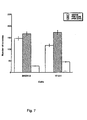
- p68HER-2 The effects of p68HER-2 on anchorage independent growth in soft agar was determined using SKOV-3 carcinoma cells and HER-2 transfected 17-3-1 cells, which are both tumorigenic and overexpress p185HER-2.
- the cells were suspended in media supplemented with fetal calf serum in the presence or absence of p68HER-2 and incubated for 21 days in a humidified incubator.
- Anchorage independent growth was quantitated by counting the number of colonies that contained more than 50 cells.
- Figure 7 shows that in the presence of p68HER-2, anchorage independent growth of both SKOV-3 cells and 17-3-1 cells was inhibited several fold. Accordingly, these data show that p68HER-2 is not just cytostatic, but cytotoxic and possibly apoptotic.
- This example provides the results from an experiment to investigate HER-2 mRNA diversity within the extracellular domain (ECD) coding sequence using polymerase chain reaction (PCR).
- a cDNA library from SKOV-3 cells American Type Culture Collection (Rockville, MD) maintained in DMEM, supplemented with 10% fetal bovine serum and 0.05% gentamycin), an ovarian carcinoma cell line in which the HER-2 gene is amplified eight times ( Tyson et al., Am. J. Obstet. Gynecol. 165:640-646, 1991 ) was examined using a forward primer specific for exon 1 ( Tal et al., Mol. Cell. Biol.
- SKOV-3 cDNA library was provided by Origene Technologies, Inc. (Rockville, MD), and was prepared from RNA extracted from SKOV-3 cells. RNA was extracted from SKOV-3 cells grown to 80% confluence on 15 cm plates with TriReagent (Molecular Research Center, Inc., Cincinnati, OH), according to the manufacturer's protocol, to obtain total RNA.
- RNA was resuspended in 10mM Tris-EDTA, pH 8.0, for reverse transcription and cDNA library construction, or in RNA hybridization buffer (80% formamide, 40mM PIPES, 4 mM NaCl, 1mM EDTA, pH 7.5) for ribonuclease protection assay (RPA). RNA concentrations were determined spectrophotometrically at OD 260 . Poly A + mRNA was selected from total RNA using a mRNA extraction kit (Oligotex, Qiagen).
- Nucleic acids were fixed to membranes by UV crosslinking in a UV-Stratalinker (Stratagene, Inc., La Jolla, CA), and the membranes were blocked in hybridization buffer (50% formamide, 5X SSC, 1% SDS, 10 mg/ml herring sperm DNA) at 42 °C for 2 h.
- hybridization buffer 50% formamide, 5X SSC, 1% SDS, 10 mg/ml herring sperm DNA
- the membranes were hybridized at 42 °C for 16 h in hybridization buffer with 10 7 cpm of a 220 bp Kpn-HincII fragment from ECDIIIa cDNA labelled with ( ⁇ - 32 P)dCTP (NEN Life Sciences) using a Random Prime DNA Labelling Kit (Boehringer Mannheim).
- Templates were amplified in a Perkin Elmer GeneAmp PCR System 2400 (Perkin Elmer Cetus, Emeryville, CA) using the Expand High Fidelity PCR System (Boerhinger Mannheim) with 1X High Fidelity PCR buffer containing 2.5 mM MgCl 2 , 5 ⁇ M of each primer, and 200 ⁇ M dNTPs. All primers were obtained from GIBCO BRL (Life Technologies). Numbering of nucleotide and amino acid residues is according to the HER-2 cDNA sequence reported by Coussens et al. ( Coussens et al., Science 230:1132-1139, 1985 ).
- the HER-2 extracellular domain was targeted for amplification from an SKOV-3 cDNA library (Origene Technologies, Inc.) using a forward primer (A) identical to nucleotides (nt) 142-161 of HER-2 cDNA (5'-TGAGCACC ATG GAGCTGGC-3' [SEQ ID NO 3]), which spans the initiation codon (underlined) and a reverse primer (B) (5'-TCCGGCAGAAATGCCAGGCTCC-3' [SEQ ID NO 4]), which is complementary to HER-2 exon sequence at nt 1265-1286. Cycling parameters were: 94 °C, 30 sec; 58 °C, 45 sec; 68 °C, 3 min, for 30 cycles.
- the region spanning the alternative sequence (denoted ECDIIIa) from genomic DNA was amplified using a forward primer (C) (5'-AACACAGCGGTGTGAGAAGTGC-3' [SEQ ID NO 5]) identical to HER-2 exon-specific sequence at nt 1131-1152 and the reverse primer (B) [SEQ ID NO. 4] on DNA prepared as described ( Bond et al., FEBS Letters 367:61-66, 1995 ) with cycling parameters: 94 °C, 30 sec; 62 °C, 30 sec; 72 °C, 60 sec, for 25 cycles.
- C forward primer
- B reverse primer
- RT-PCR Reverse transcriptase-polymerase chain reaction
- Amplification of the ECDIIIa insert and adjacent 3' HER-2 exon-specific sequence was with a forward primer (E) (5'-TCTGGGTACCCACTCACTGC-3' [SEQ ID NO 7]) which is identical to the 5'ECDIIIa-specific sequence and contains a Kpn 1 restriction site and a reverse primer (F) (5'-T TCA CACTGGCACGTCCAGACC-3' [SEQ ID NO 8]) which is complementary to HER-2 exon sequence at nt 3898-3919 and spans the termination codon (underlined). Cycling parameters were: 94 °C, 30 sec; 60 °C, 40 sec; 68 °C, 5 min, for 30 cycles.
- the PCR product was subcloned and the nucleotide sequence was determined.
- This example provides the results from experiments characterizing ECDIIIa as contiguous with HER-2 exons in the genome.
- a forward primer identical to nucleotides 763-785
- a reverse primer complementary to nucleotides 1265-1286 of the HER-2 cDNA
- the amplification product was anticipated to span exon 5 ( Tal et al., Mol. Cell. Biol. 7:2597-2601, 1987 ) to an exon which is immediately 3' of the ECDIIIa sequence. Intron number and sizes were estimated based on PCR product sizes, restriction digest analysis, and partial sequence analysis of amplification products.
- HER-2 exon-specific primers that directly flank the insert to determine the sequences immediately flanking the ECDIIIa sequence.
- a -430 bp product was amplified from normal human genomic DNA and from genomic DNA extracted from carcinoma cell lines SKOV-3, SKBR-3 and BT474, all of which have HER-2 gene amplification ( Kraus et al., EMBO J. 6:605-610, 1987 ) and were found to express ECDIIIa in their cDNA.
- the identities of the PCR products as HER-2 were verified by Southern blot analysis using the procedure described in Example 1.
- ECDIIIa is the only retained intron within the coding sequence of HER-2 mRNA.
- RT-PCR reverse transcriptase-polymerase chain reaction
- Amplification of the 3'HER-2 coding sequence was then performed using a forward primer identical to 5' ECDIIIa sequence and a reverse primer complementary to 3'HER-2 cDNA sequence at nucleotides 3898-3919, which spans the p185HER-2 termination codon.
- a product of 2.9 kb was amplified, which is the size expected from the HER-2 cDNA if no additional introns were retained.
- This example illustrates the expression of a protein containing an ECDIIIa sequence.
- the ECDIIIa sequence was expressed as a polyhistidine-tagged peptide in bacteria, purified the peptide by nickel-affinity chromatography, and raised antisera against the purified peptide.
- the bacterial expression vector was prepared by amplifying the ECDIIIa sequence from the SKOV-3 cDNA library using primer E and a reverse primer complementary to the 3' end of the ECDIIIa insert sequence.
- the reverse primer contained a Bam H1 restriction site sequence, and was identical to that used for template construction in the RPA (described in examples 1 and 2).
- the PCR amplification product of ⁇ 280 bp was digested with Kpn 1 and Bam H1, gel purified (Qiaex II, Qiagen, Chatsworth, CA), and cloned into the pET30a vector, which encodes a six histidine tag at the amino-terminus of the expressed protein (Novagen, Madison, WI).
- the resulting expression vector, pET-ECDIIIa was used for transformation of bacterial strain BL21.
- BL21 cells transformed with the pET-ECDIIIa expression vector were grown in LB broth with 30 ⁇ g/ml Kanamycin for 4 h at 37 °C. Expression was induced with 0.1 mM IPTG for 3 h and the harvested cells were lysed by sonication, and then centrifuged at 39,000 x g for 20 min. The supernatant was absorbed onto Ni-NTA agarose (Qiagen), by shaking for 60 min at room temperature. The resin was washed with ten volumes of wash buffer (10 mM Tris pH 7.9 and 300 mM NaCl), followed by ten volumes of wash buffer with 50 mM imidazole. The his-tagged ECDIIIa protein was eluted in wash buffer with 250 mM imidazole. The his-tagged protein, which was estimated to be approximately 90% pure by Coomassie Blue staining of gels, was used to generate and characterize antibodies.
- anti-ECDIIIa antisera were produced by Cocalico Biologicals, Inc. (Reamstown, PA) by injection of two rabbits with purified polyhistidine-tagged ECDIIIa peptide (described below).
- Polyclonal anti-neu (N) was produced against a peptide identical to amino acid residues 151-165 of p185HER-2 ( Lin and Clinton, Oncogene 6:639-643, 1991 ).
- Polyclonal anti-neu (C) was made against a peptide identical to the last 15 residues of the carboxy-terminus of p185HER-2 ( Lin et al., Mol. Cell. Endocrin. 69:111-119, 1990 ).
- Antisera from two immunized rabbits were characterized and found to contain antibodies of high titer that reacted with the purified ECDIIIa peptide.
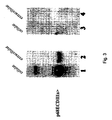
- a Western blot analysis examined whether SKBR-3 cells, which expressed the alternative sequence in its cDNA, produced a protein that reacts with anti-ECDIIIa antibody.
- a 68 kDa protein from the cell extract and from the extracellular media reacted with anti-ECDIIIa antibody from two different rabbits diluted at least 20,000 fold, but not with preimmune sera.
- Inspection of the cDNA sequence of the alternative transcript ( Figure 1 ) predicted a secreted protein product of 65-70 kDa if all 5 consensus N-linked glycosylation sites in the N-terminal p185HER-2 sequence were glycosylated ( Stem et al., Mol. Cell. Biol. 6:1729-1740, 1986 ).
- the 68 kDa ECDIIIa protein [SEQ ID NO. 2] is the translation product of the alternative HER-2 mRNA, then its N-terminal residues should be identical to the N-terminal 340 residues of p185HER-2. Therefore, cell extract from SKBR-3 cells was immunoprecipitated with anti-peptide antibody against an N-terminal sequence of HER-2, anti-neu (N) ( Lin and Clinton, Oncogene 6:639-643, 1991 ) or with anti-ECDIIIa, and the immune complexes were examined by Western blot analysis with both antibodies.
- the immune complexes were bound to Protein G Sepharose (Pharmacia) by incubation for 1 h at 4 °C with shaking, collected by centrifugation, and washed four times with M-RIPA.
- the proteins were released from the immune complex by incubation at 95° C for 2 min in SDS-PAGE sample buffer and resolved by SDS-PAGE in 7.5% gels (Mini-Protean II electrophoresis cell, Bio-Rad).
- the blots were then incubated with primary antibody, washed twice for 15 min, and four times for 5 min with TBS-Tween (Tris-buffered saline containing 0.05% Tween), and then incubated for 40 min with goat anti-rabbit secondary antibody, conjugated to horseradish peroxidase (Bio-Rad), diluted 1: 10, 000 in TBS-Tween. After incubation with secondary antibody, the membranes were washed as described above and reacted with chemiluminescent reagent (Pierce) and then were exposed to Kodak X-OMAT BLU film.
- TBS-Tween Tris-buffered saline containing 0.05% Tween
- p68HER-2 was detected when anti-ECDIIIa was used for immunoprecipitation and for Western blot analysis.
- anti-ECDIIIa was used for immunoprecipitation and anti-neu (N) was the probe in the Western blot, a 68kDa protein was detected, indicating that p68ECDIIIa contained the N-terminal sequence of p185HER-2.
- anti-neu (N) precipitated p68HER-2, which was detected by probing with anti-ECDIIIa antibody.
- HEK293 cells derived from normal human embryonic kidney cells, expressed the highest levels of p68ECDIIIa in the cell extract and in the extracellular media, at about 5 to 10-fold higher amounts than SKBR-3 cells.
- the HEK293 cells contained about 20 fold lower amounts of p185HER-2.
- the relative proportion of p68HER-2 to p185HER-2 was at least 100 fold greater in HEK293 cells than in the three carcinoma cell lines studied. Reactivity with p68HER-2 as well as with a protein of ⁇ 120 kDa, particularly apparent in the HEK293 extracts, was blocked by preincubation of the antisera with purified ECDIIIa peptide demonstrating sequence-specific reactivity. The larger protein may be a dimer of p68HER-2. Therefore, p68HER-2 was expressed and secreted from several carcinoma cell lines and is at 5-10 fold elevated levels in HEK293.
- This example illustrates expression of an alternative HER-2 transcript containing the ECDIIIa intron sequence.
- Results of the RT-PCR analysis indicated that the ECDIIIa sequence was inserted into an otherwise normal-sized HER-2 mRNA. These data suggest an alternative transcript of ⁇ 4.8 kb.
- Northern blot analysis was conducted using an ECDIIIa-specific probe. Briefly, a template for antisense RNA probe synthesis was constructed from SKOV-3 cDNA by PCR amplification of a 389 bp sequence spanning the entire ECDIIIa insert sequence and containing adjacent 5'HER-2 exon sequence. The PCR was done using the forward primer C [SEQ ID NO.
- telomere 5 that is identical to HER-2 cDNA sequence at nt 1131-1152 and a reverse primer (5'-GCACGGATCCATAGCAGACTGAG GAGG-3' [SEQ ID NO. 9]) which contains a 3' Bam H1 restriction endonuclease site and is complementary to the sequence spanning the 3' splice site of the ECDIIIa sequence.
- the PCR product was then digested with Bam H1, liberating a 375 bp fragment, which was cloned into pBluescript SK (Stratagene).
- the plasmid was sequenced by the Vollum Institute Core Sequencing Facility (Portland, OR) with m13 forward and reverse primers.
- RNA probe complimentary to the entire ECDIIIa sequence and to 87 nt of HER-2 exon sequence 5' to the insert was transcribed from 1 ⁇ g of linearized template using ( ⁇ - 32 P) CTP, T7 RNA polymerase, and the T7/SP6 Riboprobe Synthesis System (Promega, Madison, WI). This probe was expected to protect a 370 nt fragment when hybridized with mRNA containing ECDIIIa and adjacent HER-2 exon sequence, and to protect an 87 nt fragment when hybridized with fully spliced HER-2 mRNA.
- RNA hybrids 30 ⁇ g of RNA were hybridized with approximately 50,000 cpm of antisense RNA probe at 48 °C for 16 h.
- RNA hybrids were digested for 30 min at 37 °C with 40 ⁇ g/ml RNaseA (Boerhinger Mannheim) and 2 ⁇ g/ml RNase T1 (Life Technologies) in a solution of 250 mM NaCl, 5 mM EDTA, and 10 mM Tris pH 7.5. Proteinase K 1100 ⁇ g) (Life Technologies) in 20 ⁇ l 10°% SDS was added to stop the digestion.
- Samples were extracted with acid phenol (pH 4.5; Life Technologies) and chloroform, precipitated with two volumes of 100% ethanol, and suspended in 5 ⁇ l of RPA sample buffer (88% formamide, 10 mM EDTA pH 8.0, 1 mg/ml xylene cyanol, and 1 mg/ml bromophenol blue). Samples were denatured at 95° C for 10 min and electrophoresed on a 5% polyacrylamide/urea gel in TBE (89 mM Tris, 89 mM borate, 2 mM EDTA pH 8.3). Gels were dried under vacuum and subjected to phosphorimager analysis for quantitation of the protected fragments (IP Lab Gel, Molecular Dynamics).
- RNA from ovarian (SKOV-3) and breast (SKBR-3 and BT474) carcinoma cell lines which contained detectable levels of p68ECDIIIa, and a control cell line, 17-3-1, stably transfected with HER-2 cDNA, were hybridized with an antisense 32 P-labeled RNA probe which spanned the entire ECDIIIa (intron 8) sequence and 5' HER-2 exon sequence flanking intron 8.
- ECDIIIa intron 8
- an 87 nucleotide protected fragment was detected in all cells and is the size expected for the fully-spliced HER-2 message which is overexpressed by more than 100 fold in these carcinoma cell lines compared to normal control cell lines ( Kraus et al., EMBD J. 6:605-610, 1987 ).
- the amounts of each protected fragment were quantitated and normalized for size to estimate the relative abundance of the alternative transcript, expressed as a percentage of the p185HER-2 mRNA.
- the alternative HER-2 mRNA with the ECDIIIa insert was at 4.2% the level of the fully spliced transcript in SKOV-3; 5.4% in SKBR-3, and 0.8% in BT474 cells.
- the 4.8 kb transcript likely corresponded to the full length 4.5 kb transcript with the 274bp insert and the 2.6 kb transcript may have corresponded to a previously described 2.3 kb alternative transcript ( Yamamoto et al., Nature 319:230-234, 1986 ; and Scott et al., Mol. Cell. Biol. 13:2247-2257, 1993 ) with the 274bp ECDIIIa insert.
- ECDIIIa-containing alternative transcript Several cell lines were also investigated for the ECDIIIa-containing alternative transcript by Northern blot analysis.
- the 4.8 kb alternative transcript was detected in the human embryonic kidney cell line, HEK-293 ( Figure 2 ).
- the ECDIIIa sequence was detected by RT-PCR analysis of SKBR-3, BT474, and SKOV-3 carcinoma cell lines, which all contain HER-2 gene amplification, an ECDIIIa-containing alternative transcript could not be detected by Northern analysis of these cells. Therefore, the more sensitive ribonuclease protection assay (RPA) was employed using an antisense probe which spanned the entire ECDIIIa sequence and 5' HER-2 exon sequence flanking the ECDIIIa sequence.
- RPA ribonuclease protection assay
- the alternative HER-2 mRNA with the ECDIIIa insert was detected at less than 5% of the fully spliced transcript in SKOV-3, SKBR-3, and BT474 cells.
- This example illustrates expression of a protein containing the ECDIIIa sequence.
- the ECDIIIa sequence as a polyhistidine-tagged peptide in bacteria, was expressed and purified by nickel-affinity chromatography, and raised antisera against the purified peptide.
- the larger protein of ⁇ 125 kDa detected in some cell extracts may be an aggregate of p68HER-2.
- the cDNA sequence of the alternative transcript ( Figure 1 ) predicts a secreted protein product of 65-70 kDa if all 5 consensus N-linked glycosylation sites in the N-terminal p185HER-2 sequence are glycosylated ( Stern et al., Mol. Cell. Biol. 6:1729-1740, 1986 ).
- Several other cell lines were examined for expression of p68ECDIIIa The carcinoma cell lines which contained ECDIIIa sequence in their cDNA (BT474, SKOV-3, SKBR-3) also had detectable levels of p68HER-2.
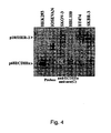
- This example illustrates the expression of p68HER-2 relative to p185HER-2 was markedly reduced in carcinoma cell lines in which the HER-2 gene is amplified. Because the p68HER-2 mRNA was expressed at very low levels relative to the p185HER-2 mRNA in carcinoma cell lines with HER-2 gene amplification, the relative proportions of p68HER-2 and p185HER-2 proteins in several cell lines were examined with and without HER-2 gene amplification. Western blots were prepared and probed with both antisera specific for p68HER-2 and for p185HER-2. Figure 4 shows that p185HER-2 was readily detected in the carcinoma cells lines that have their HER-2 gene amplified about 8 times ( Kraus et al., EMBO J.
- p68HER-2 was the only HER-2 protein detected in the HEK-293, IOSEVAN, and HBL100 nontumorigenic cells, although p185HER-2 was expressed at very low levels in these cells ( Kraus et al., EMBO J. 6:605-610, 1987 ) and was detected in overexposed blots.
- p68HER-2 was low in proportion to p185HER-2 in carcinoma cells with HER-2 gene amplification and suggests that a mechanism may exist to maintain low levels of p68HER-2 when p185HER-2 is overexpressed.
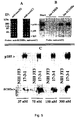
- p68HER-2 and the ECDIIIa peptide specifically bind to p185HER-2. Because p68HER-2 is secreted and contains subdomains I and II identical to p185HER-2, in addition to a novel sequence, the possibility that p68HER-2 may interact with p185HER-2 was investigated. Antipeptide antibody against the N-terminus of p185HER-2 and p68HER-2, anti-neu (N), or antibody specific for p185HER-2, anti-neu(C), were used for immunoprecipitations of SKBR-3 carcinoma cells, which express low levels of p68HER-2 and overexpress p185HER-2.
- the immunoprecipitated material was prepared as a Western blot and probed with both anti-ECDIIIa specific for p68HER-2 and with anti-neu(C).
- Anti-neu (N) immunoprecipitated both p68HER-2 and p185HER-2 ( Figure 5A ).
- antibodies specific for the C-terminus of p185HER-2 immunoprecipitated p185HER-2 and coprecipitated p68HER-2 ( Figure 5A ), suggesting an interaction between the two proteins.
- the immobilized peptides were incubated with protein extracts prepared from HER-2 transfected 3T3 cells (17-3-1). Following extensive washes, the bound proteins were eluted and prepared as a Western blot which was probed with an antibody specific for p185HER-2. Equal amounts of His-tagged ECDIIIa peptide and control peptide were bound to the resin as confirmed by elution with 1M imidazole and Coomassie staining of the eluted material in SDS-gels. While no p185HER-2 was retained by resin without peptide or with control peptide, p185HER-2 was selectively retained by the ECDIIIa peptide ( Figure 5B ).
- ECDIIIa domain bound to p185HER-2 in a pulldown assay, the question of whether the ECDIIIa domain preferentially binds to cells that overexpress p185HER-2 was examined. This was investigated using monolayer cultures of 17-3-1 cells transfected with HER-2 compared to the parental 3T3 cells. The cells were incubated with different concentrations of the His-ECDIIIa peptide, washed, and extracted in denaturing buffer with protease inhibitors. To detect any bound peptide, the cell extracts were examined by Western blot analysis using antibodies specific for ECDIIIa.
- ECDIIIa peptide treated cells were reacted as a Western blot with antibodies specific for p185HER-2, demonstrating the overexpression of p185HER-2 in the transfected 17-3-1 cells.
- the ECDIIIa peptide preferentially bound to intact 17-3-1 cells at nM concentrations ( Figure 5C ) whereas little or no peptide was found to bind to equivalent amounts of parental 3T3 cells suggesting a specific interaction with the extracellular domain of p185HER-2.
- Tyrosine phosphorylation of RTKs is the initial indication of ligand activation and signal transduction.
- Tyrosine phosphorylation in 17-3-1 cells treated with different amounts of the purified ECDIIIa peptide, with conditioned media (CM) from HEK293 cells that contained high levels of p68HER-2 ( Figure 2A), or with control, conditioned media from SKOV-3 cells that had no detectable p68HER-2 were examined.
- intron 8 is polymorphic within that portion of intron 8 that serves as in-frame (with the extracellular domain of p185HER-2) coding sequence when intron 8 is alternatively retained in mRNA.
- Intron 8 of the human HER-2 gene is alternatively retained in mRNA, and encodes a novel 79-residue domain at the C-terminus of a part of the extracellular domain of p185HER-2.
- the product, "herstatin,” of the alternative transcript with the retained intron functions as an autoinhibitor of the HER-2 oncogene.
- the intron 8 encoded domain, alone, was shown to bind with nM affinity to p185HER-2. ( Doherty et al., Proc. Natl. Acad. Sci. USA 96:10,869-10,874, 1999 ).
- the polymorphism (G ⁇ C) at nucleotide position 161 would result in a substitution of Arginine (R) for Proline (P) at amino acid residue #54 of SEQ ID NO:1, or residue #394 of SEQ ID NO:2.
- the N-terminal Glycine (G), designated as position 1 in Figure 8 or SEQ ID NO: 10, corresponds to amino acid residue #341 in the "herstatin" sequence ( Doherty et al., Proc. Natl. Acad. Sci. USA 96:10,869-10,874, 1999 ).
- the nucleotide sequence shown in Figure 1(A) is a polymorphic form that differs at amino acid residues #6 and #73 from the most commonly detected sequence shown here in Figure 8 .
- Sequence variants 1-11 are listed, showing the base changes at particular "X" positions relative to that found in the most common DNA sequence shown in Figure 8 .
- the numbers in parenthesis after each X correspond to the nucleotide position in the DNA sequence shown in Figure 8 or SEQ ID NO:10.
- the DNA sequence variants listed here correspond to the variable amino acid positions ("Xaa”) of SEQ ID NO:1 as follows: X(4) to Xaa(2); X(14) to Xaa(5); X(17) to Xaa(6); X(47) to Xaa(16); X(54) to Xaa(18); X(62) to Xaa(21); X(106) to Xaa(36); X(161) to Xaa(54); X(191) to Xaa(64); X(217) to Xaa(73); and to the variable amino acid positions of SEQ ID NO:2 as follows: X(4) toXaa(342); X(14) to Xaa(345); X(17) to Xaa(346); X(47) to Xaa(356); X(54) to Xaa(358); X(62) to Xaa(361); X
- variable amino acid positions in SEQ ID NO:1 are: Variant 1, Xaa(2)(Thr ⁇ Ser); Variant 2, Xaa(5) (Leu ⁇ Pro); Variant 3, Xaa(6) (Pro ⁇ Leu); Variant 4, Xaa(16) (Leu ⁇ Gln); Variant 5, Xaa(18) (Met ⁇ Leu); Variant 6, Xaa(21) (Gly ⁇ Asp, Alu or Val); Variant 7, Xaa(36) (Leu ⁇ Ile); Variant 8, Xaa(54) (Pro ⁇ Arg); Variant 9, Xaa(64) (Pro ⁇ Leu); Variant 10, Xaa(73) (Asp ⁇ Asn), and Variant 11, Xaa(6) (Pro ⁇ Leu) and Xaa(73) (Asp ⁇ Asn).
- the same substitutions apply to the corresponding variable amino acid positions in SEQ ID NO:2.
- polymorphic sites were identified within that portion of intron 8 that serves as in-frame (with the extracellular domain of p185HER-2) coding sequence when intron 8 is alternatively retained in mRNA (i.e., four polymorphic sites within the sequence region encompassed by SEQ ID NO:10, or within that encompassed by the sequence region of Figure 8 ).
- Two of these polymorphic sites correspond in position to those (variants 3 and 10, respectivley) disclosed above in Example 11, whereas the other two (variants 13 and 14) represent additional polymorphic sites (Table II).
- an additional polymorphic site (variant 16) was identified in a region of intron 8 that remains as “non-coding” sequence when intron 8 is alternatively retained in mRNA.
- This "non-coding" intron 8 polymorphic site is located 3', or downstream from that portion of intron 8 that contains the other polymorphic sites shown in this Example and Example 11, and that serves as in-frame (with the extracellular domain of p185HER-2) coding sequence when intron 8 is alternatively retained in mRNA.
- Polymorphisms in the nucleotide and deduced amino acid sequence of intron 8 in the HER-2 gene were identified by sequencing genomic DNA (using blood samples) from 215 individuals corresponding to 75 African Americans (Black), 135 Caucasians (White), one Asian American (Asian) and 4 Hispanics.
- the N-terminal Glycine (G or Gly) designated as position 1 in Figure 8 or SEQ ID NO:1 or SEQ ID NO:10 corresponds to amino acid residue #341 in the "herstatin" sequence of SEQ ID NO:2 or SEQ ID NO:13.
- Table II designates the nucleotide substitutions and the two amino acid residue substitutions in the coding sequence of intron 8 and a third nucleotide substitution in a non coding sequence of intron 8 using numbering corresponding to the entire "herstatin" protein sequence (SEQ ID NO:2 or SEQ ID NO:13): Table II N Black 75 White 135 Asian 1 Hispanic/Latino 4 Total 215 Herstatin Polymorphism Distributions Among Blacks Arg357Cys (C1081T) Prostate Cases Controls Other Cancers wt (%) 24 (96) 32 (89) 13 (93) het (%) 1 (4) 2 (6) 1 (7) mut (%) 0 (0) 2 (6) 0 (0) total 25 36 14 Arg371IIe (G1124T) Prostate Cases Controls Other Cancers wt (%) 24 (96) 36 (100) 14 (100) het (%) 1(4) 0(0) 0 (0) mut (%) 0 (0) 0 (0) (0)
- This table shows the distribution of three additional (relative to those identified in Example 11) polymorphic regions in HER-2 intron 8 of the DNA from African American individuals. Amino acids position designations correspond to amino acid positions in the "Herstatin" sequence (SEQ ID NO:2 or SEQ ID NO:13).
- Table III illustrates that the sequence data revealed polymorphisms at nucleotide positions #17 and #217 (also corresponding to nucleotide positions of the sequence region shown in Figure 8 or SEQ ID NO:10).
- the polymorphism at position #17 (variant 12) corresponds to variant 3 of Table I (Example 11).
- the polymorphism at position #217 (variant 15) corresponds (at least at the protein level) to variant 10 of Table I (Example 11) (see SEQ ID NO:12 and SEQ ID NO:13).
- intron 8 contains three polymorphic sites (corresponding to variants 13, 14 and 16) in addition to those disclosed in Example 11, above.
- Two of these (variants 13 and 14) are located at nucleotide positions #49 and #92 of SEQ ID NO:11 (also corresponding to nucleotide positions #49 and #92 of SEQ ID NO: 10 (or Figure 8 ).
- the third (variant 16) is located at a nucleotide position #259 of SEQ ID NO:11 [also corresponding to nucleotide position #259 relative to the sequence region of SEQ ID NO:10 (or to position #264 of the sequence shown in Figure 1 , panel A)].
- the polymorphism corresponding to variant 16 is located 19 nucleotide positions 3' (downstream) from that portion of intron 8 that contains the other polymorphic sites shown in this Example and Example 11 ( i.e., that portion represented by SEQ ID NO:10), and that serves as in-frame (with the extracellular domain of p 185HER-2) "coding" sequence when intron 8 is alternatively retained in mRNA.
- polymorphisms Two of these polymorphisms result in nonconservative amino acid substitutions (see Table II and Table III, and legend of Table III; also see SEQ ID NO:12 and SEQ ID NO:13).
- the polymorphism (C ⁇ T) found at the nucleotide position corresponding to nucleotide #49 of SEQ ID NO:11 [or to position # 49 of SEQ ID NO:10 or Figure 8 ] i.e., the polymorphism at position X(49) of Table 2
- Arg Arginine
- Cysteine Cysteine
- SEQ ID NO:11, SEQ ID NO:12 and SEQ ID NO:13 show the four variant amino acid positions described in this example, along with those of Example 11 that are also shown in SEQ ID NO:1, SEQ ID NO:2 and SEQ ID NO:10.
- Table III designates (in addition to variants 12 and 15, which correspond to variants 3 and 10, respectivley of Table I) the nucleotide substitutions and the corresponding two additional (relative to those of Table I of Example 11) amino acid residue substitutions ( i.e. , variants 13 and 14) in the "coding" sequence of intron 8, along with the third nucleotide substitution in the 3' "non-coding" region of intron 8.
- the numbers in parenthesis after each X (polymorphic position) refer to nucleotide positions of SEQ ID NO:11 [or, as in Table I, correspond to (or are relative to, in the case of X(259) the nucleotide positions in the DNA sequences shown in Figure 8 or SEQ ID NO:10].
- N-terminal Glycine designated as position 1 in SEQ ID NO:11, Figure 8 , SEQ ID NO:1 or SEQ ID NO: 10, corresponds to amino acid residue #341 in the "herstatin" sequence of SEQ ID NO:2.
- Variant 12 T Variant 13 T Variant 14 T Variant 15 A Variant 16 T Table III . Sequence variants in the intron-8 encoded domain found in human tissues (based on 215 different individuals). Sequence variants 12-16 are listed.
- the numbers in parenthesis after each X refer to nucleotide positions of SEQ ID NO:11 [or to positions that correspond to, or are relative to (in the case of X(259)) the nucleotide positions in the DNA sequences shown in Figure 8 or SEQ ID NO:10].
- the DNA sequence variants listed here and in SEQ ID NO:11 correspond to variable amino acid positions shown in SEQ ID NO:12 [and also correspond to variable amino acid positions ("Xaa") of SEQ ID NO:1 or SEQ ID NO:10 as follows: X(17) to Xaa(6); X(49) to Xaa(17); X(92) to Xaa(31); X(217) to Xaa(73)].
- the DNA sequence variant X(259) occurs in an untranslated region, and therefore does not alter the amino acid sequence of herstatin.
- the variants of this table correspond to variable amino acid positions of SEQ ID NO:13 and SEQ ID NO:2 as follows: X(17) to Xaa(346); X(49) to Xaa(357); X(92) to Xaa(371); X(217) to Xaa(413).
- variable amino acid positions in SEQ ID NO:11 and SEQ ID NO:12 are: Variant 12, Xaa(6)(Pro ⁇ Leu); Variant 13, Xaa(17) (Arg ⁇ Cys); Variant 14, Xaa(31) (Arg ⁇ Ile); Variant 15, Xaa(73) (Asp ⁇ Asn).
- Variant 16, X(259) is in an untranslated region and does not code for an amino acid alteration, but instead alters only the nucleotide sequence at nucleotide position 259 ( i.e., C ⁇ T). The same substitutions apply to the corresponding variable amino acid positions in SEQ ID NO: 13.
- SKOV3 ovarian carcinoma cells Two additional polymorphisms were found in a cell line derived from human ovarian cancer (SKOV3). These two polymorphisms result in nonconservative amino acid substitutions.
- One polymorphism is a substitution (C-T) at nucleotide #17 in the intron 8 sequence and nucleotide # 1037 in the "herstatin” sequence resulting in a substitution of leucine for proline at amino acid residue #6 in the intron 8 sequence and at amino acid residue # 346 in the "herstatin' sequence (i.e., of SEQ ID NO:2 or SEQ ID NO:13).
- the second polymorphism found in the SKOV3 ovarian carcinoma cells line is a substitution (G-A) at nucleotide #217 in the intron 8 sequence and nucleotide #1237 in the "herstatin” sequence resulting in a substitution of Asparagine for Aspartic Acid at amino acid residue # 73 in the intron 8 sequence and amino acid #413 in the "herstatin” sequence (i.e., of SEQ ID NO:2 or SEQ ID NO: 13).
- G-A substitution of Asparagine for Aspartic Acid at amino acid residue # 73 in the intron 8 sequence and amino acid #413 in the "herstatin” sequence
- Examples 11 and 12 of the present invention disclose 13 polymorphic positions in intron 8 of the Her-2 gene.
- Example 12 involved a relatively large DNA sample size, and indicated that the five polymorphic sites identified (three of which are distinct from the ten polymorphic sites identified in Example 11) are unique to African Americans (Black).
- intron 8 that serves as in-frame (with the extracellular domain of p185HER-2) coding sequence when intron 8 is alternatively retained in mRNA.
- the polymorphism corresponding to variant 16 is located in a region of intron 8 that remains as "non-coding" sequence when intron 8 is alternatively retained in mRNA.
- This "non-coding" intron 8 polymorphic site is located 19 nucleotide positions 3', or downstream from that portion of intron 8 that contains the other polymorphic sites, and that serves as in-frame (with the extracellular domain of p185HER-2) coding sequence when intron 8 is alternatively retained in mRNA.
- HER-2 intron 8 polymorphisms provide for novel DNA and protein sequences, novel pharmaceutical compositions for treating solid tumors that overexpress HER-2, and monoclonal antibodies that bind to ECDIIIa variants corresponding to SEQ ID NO:1, SEQ ID NO:2, SEQ ID NO:12 or SEQ ID NO:13. These HER-2 intron 8 polymorphisms also provide for prognostic and diagnostic assays for the treatment and prevention of cancer.
Landscapes
- Health & Medical Sciences (AREA)
- Chemical & Material Sciences (AREA)
- Organic Chemistry (AREA)
- Medicinal Chemistry (AREA)
- Life Sciences & Earth Sciences (AREA)
- General Health & Medical Sciences (AREA)
- Animal Behavior & Ethology (AREA)
- Pharmacology & Pharmacy (AREA)
- Public Health (AREA)
- Chemical Kinetics & Catalysis (AREA)
- Nuclear Medicine, Radiotherapy & Molecular Imaging (AREA)
- General Chemical & Material Sciences (AREA)
- Veterinary Medicine (AREA)
- Engineering & Computer Science (AREA)
- Bioinformatics & Cheminformatics (AREA)
- Genetics & Genomics (AREA)
- Proteomics, Peptides & Aminoacids (AREA)
- Molecular Biology (AREA)
- Biophysics (AREA)
- Biochemistry (AREA)
- Immunology (AREA)
- Oncology (AREA)
- Reproductive Health (AREA)
- Endocrinology (AREA)
- Pulmonology (AREA)
- Peptides Or Proteins (AREA)
- Medicines Containing Antibodies Or Antigens For Use As Internal Diagnostic Agents (AREA)
- Medicines That Contain Protein Lipid Enzymes And Other Medicines (AREA)
- Pharmaceuticals Containing Other Organic And Inorganic Compounds (AREA)
- Acyclic And Carbocyclic Compounds In Medicinal Compositions (AREA)
- Measuring Or Testing Involving Enzymes Or Micro-Organisms (AREA)
- Micro-Organisms Or Cultivation Processes Thereof (AREA)
Applications Claiming Priority (3)
| Application Number | Priority Date | Filing Date | Title |
|---|---|---|---|
| US506079 | 2000-02-16 | ||
| US09/506,079 US7625859B1 (en) | 2000-02-16 | 2000-02-16 | HER-2 binding antagonists |
| PCT/US2001/005327 WO2001061356A1 (en) | 2000-02-16 | 2001-02-16 | Her-2 binding antagonists |
Publications (3)
| Publication Number | Publication Date |
|---|---|
| EP1257822A1 EP1257822A1 (en) | 2002-11-20 |
| EP1257822A4 EP1257822A4 (en) | 2004-09-08 |
| EP1257822B1 true EP1257822B1 (en) | 2012-01-18 |
Family
ID=24013084
Family Applications (1)
| Application Number | Title | Priority Date | Filing Date |
|---|---|---|---|
| EP01912833A Expired - Lifetime EP1257822B1 (en) | 2000-02-16 | 2001-02-16 | Her-2 binding antagonists |
Country Status (7)
Families Citing this family (16)
| Publication number | Priority date | Publication date | Assignee | Title |
|---|---|---|---|---|
| US6541214B1 (en) | 1998-11-13 | 2003-04-01 | Oregon Heath Science University | N-terminally truncated HER-2/neu protein as a cancer prognostic indicator |
| US7393823B1 (en) | 1999-01-20 | 2008-07-01 | Oregon Health And Science University | HER-2 binding antagonists |
| US7396810B1 (en) * | 2000-08-14 | 2008-07-08 | Oregon Health Sciences University | Compositions and methods for treating cancer by modulating HER-2 and EGF receptors |
| US7625859B1 (en) | 2000-02-16 | 2009-12-01 | Oregon Health & Science University | HER-2 binding antagonists |
| DE60028970T2 (de) | 1999-07-02 | 2007-02-15 | Genentech Inc., San Francisco | An her2 bindende peptidverbindungen |
| EP1583504A4 (en) * | 2002-12-26 | 2008-03-05 | Cemines Llc | METHODS AND COMPOSITIONS FOR THE DIAGNOSIS, PROGNOSIS, AND TREATMENT OF CANCER |
| EP2463299A3 (en) | 2003-03-05 | 2012-10-17 | Dendreon Corporation | Compositions and methods employing alternative reading frame polypeptides for the treatment of cancer and infectious disease |
| TWI472339B (zh) * | 2008-01-30 | 2015-02-11 | Genentech Inc | 包含結合至her2結構域ii之抗體及其酸性變異體的組合物 |
| JP5718319B2 (ja) * | 2009-05-14 | 2015-05-13 | ネステク ソシエテ アノニム | Her2標的化療法に対する乳癌細胞の感受性を決定するためのバイオマーカー |
| US9427458B2 (en) | 2010-07-22 | 2016-08-30 | Consejo Nacional De Investigaciones Cientificas Y Tecnicas, Conicet | Methods for inhibiting cancer cell proliferation |
| KR101258144B1 (ko) | 2011-07-22 | 2013-04-30 | 한양대학교 산학협력단 | Ecm1 항체를 함유하는 항암용 조성물 |
| CN105985435B (zh) * | 2015-01-30 | 2019-10-15 | 嘉和生物药业有限公司 | 全人源her2抗体的突变抗体及其编码基因和应用 |
| HUE051246T2 (hu) | 2015-05-30 | 2021-03-01 | Molecular Templates Inc | Deimmunizált shigatoxin alegység vázak és azokat tartalmazó, sejthez irányító molekulák |
| AU2019257343A1 (en) | 2018-04-17 | 2020-09-10 | Molecular Templates, Inc. | HER2-targeting molecules comprising de-immunized, Shiga toxin A Subunit scaffolds |
| WO2023168426A1 (en) | 2022-03-03 | 2023-09-07 | Enosi Therapeutics Corporation | Compositions and cells containing mixtures of oligo-trap fusion proteins (ofps) and uses thereof |
| WO2024186690A2 (en) | 2023-03-03 | 2024-09-12 | Enosi Therapeutics Corporation | Oligo-trap fusion proteins (ofps) and uses thereof |
Family Cites Families (145)
| Publication number | Priority date | Publication date | Assignee | Title |
|---|---|---|---|---|
| US633169A (en) * | 1899-06-17 | 1899-09-19 | Alexander Hazard | Bottle-stopper. |
| US4376110A (en) | 1980-08-04 | 1983-03-08 | Hybritech, Incorporated | Immunometric assays using monoclonal antibodies |
| DE3485712D1 (de) | 1983-03-11 | 1992-06-17 | Sloan Kettering Inst Cancer | Monoklonale antikoerper gegen menschliche nierenkrebsantigene und verfahren. |
| US4933294A (en) | 1984-01-30 | 1990-06-12 | Icrf Patents Limited | Method of detecting truncated epidermal growth factor receptors |
| US6054561A (en) * | 1984-02-08 | 2000-04-25 | Chiron Corporation | Antigen-binding sites of antibody molecules specific for cancer antigens |
| US4959314A (en) | 1984-11-09 | 1990-09-25 | Cetus Corporation | Cysteine-depleted muteins of biologically active proteins |
| US4777127A (en) | 1985-09-30 | 1988-10-11 | Labsystems Oy | Human retrovirus-related products and methods of diagnosing and treating conditions associated with said retrovirus |
| US7838216B1 (en) | 1986-03-05 | 2010-11-23 | The United States Of America, As Represented By The Department Of Health And Human Services | Human gene related to but distinct from EGF receptor gene |
| US4933284A (en) * | 1986-05-07 | 1990-06-12 | Uop | Regenerable dialkylaminoalkyl cellulose support matrix for immobilizing biologically active materials |
| US5401638A (en) | 1986-06-04 | 1995-03-28 | Oncogene Science, Inc. | Detection and quantification of neu related proteins in the biological fluids of humans |
| US4946778A (en) | 1987-09-21 | 1990-08-07 | Genex Corporation | Single polypeptide chain binding molecules |
| GB8702816D0 (en) | 1987-02-07 | 1987-03-11 | Al Sumidaie A M K | Obtaining retrovirus-containing fraction |
| US5219740A (en) | 1987-02-13 | 1993-06-15 | Fred Hutchinson Cancer Research Center | Retroviral gene transfer into diploid fibroblasts for gene therapy |
| JP3040121B2 (ja) | 1988-01-12 | 2000-05-08 | ジェネンテク,インコーポレイテッド | 増殖因子レセプターの機能を阻害することにより腫瘍細胞を処置する方法 |
| US5720937A (en) | 1988-01-12 | 1998-02-24 | Genentech, Inc. | In vivo tumor detection assay |
| US5206152A (en) | 1988-04-08 | 1993-04-27 | Arch Development Corporation | Cloning and expression of early growth regulatory protein genes |
| ATE130873T1 (de) | 1988-04-18 | 1995-12-15 | Applied Biotechnology Inc | Nachweis der expression von neu genen und produkten. |
| JPH03503868A (ja) | 1988-04-29 | 1991-08-29 | フエル,バリイ・エム | 強化熱可塑性樹脂ハニカム構造体 |
| US5422120A (en) | 1988-05-30 | 1995-06-06 | Depotech Corporation | Heterovesicular liposomes |
| AP129A (en) | 1988-06-03 | 1991-04-17 | Smithkline Biologicals S A | Expression of retrovirus gag protein eukaryotic cells |
| WO1990007936A1 (en) | 1989-01-23 | 1990-07-26 | Chiron Corporation | Recombinant therapies for infection and hyperproliferative disorders |
| US5703055A (en) | 1989-03-21 | 1997-12-30 | Wisconsin Alumni Research Foundation | Generation of antibodies through lipid mediated DNA delivery |
| ATE277193T1 (de) | 1989-03-21 | 2004-10-15 | Vical Inc | Expression von exogenen polynukleotidsequenzen in wirbeltieren |
| US6207645B1 (en) * | 1989-03-21 | 2001-03-27 | The Immune Response Corporation | Vaccination and methods against diseases resulting from pathogenic responses by specific T cell populations |
| DE69031120T2 (de) | 1989-05-19 | 1998-01-15 | Genentech, Inc., South San Francisco, Calif. | Her2 extrazellulare domäne |
| US5705157A (en) | 1989-07-27 | 1998-01-06 | The Trustees Of The University Of Pennsylvania | Methods of treating cancerous cells with anti-receptor antibodies |
| DE69033842T2 (de) | 1989-08-04 | 2002-06-20 | Schering Ag | Äusserliche domäne von c-erbb-2:gp75 |
| CA2066053C (en) | 1989-08-18 | 2001-12-11 | Harry E. Gruber | Recombinant retroviruses delivering vector constructs to target cells |
| US5585362A (en) | 1989-08-22 | 1996-12-17 | The Regents Of The University Of Michigan | Adenovirus vectors for gene therapy |
| JP3208427B2 (ja) | 1989-09-29 | 2001-09-10 | オー・エス・アイ・ファーマシューテイカルズ・インコーポレイテッド | ヒトの生物学的流体中のneu関連タンパク質の検出及び定量 |
| US5149855A (en) * | 1989-12-26 | 1992-09-22 | Mitsubishi Rayon Co., Ltd. | Process for racemizing optically active carboxylic acid esters |
| AU7316391A (en) | 1990-01-25 | 1991-08-21 | State of Oregon acting by and through the The State Board of Higher Education on Behalf of the Oregon Health Sciences University, The | Detection of a characteristic antigen from carcinoma in body fluids |
| ZA911974B (en) | 1990-03-21 | 1994-08-22 | Res Dev Foundation | Heterovesicular liposomes |
| JPH0776238B2 (ja) * | 1990-04-06 | 1995-08-16 | ザ・トラスティーズ・オブ・ザ・ユニバーシティ・オブ・ペンシルバニア | neu遺伝子産物のためのリガンド |
| EP0459942B1 (de) | 1990-05-10 | 1995-02-22 | Ciba-Geigy Ag | Tinten |
| US5578482A (en) | 1990-05-25 | 1996-11-26 | Georgetown University | Ligand growth factors that bind to the erbB-2 receptor protein and induce cellular responses |
| US5874528A (en) | 1990-05-25 | 1999-02-23 | Georgetown University | Binding peptides which interact with ligand growth factors of the epidermal growth factor receptor and erbB-2 receptor |
| US5149655A (en) | 1990-06-21 | 1992-09-22 | Agracetus, Inc. | Apparatus for genetic transformation |
| DK0563278T3 (da) | 1990-12-20 | 2000-08-21 | Dana Farber Cancer Inst Inc | Regulering af geneekspression med ioniserende stråling |
| US5571894A (en) * | 1991-02-05 | 1996-11-05 | Ciba-Geigy Corporation | Recombinant antibodies specific for a growth factor receptor |
| DE69232718T2 (de) | 1991-02-22 | 2003-04-03 | American Cyanamid Co., Madison | Identifizierung eines menschlichen rezeptor-tyrosinkinasegens |
| IL101943A0 (en) | 1991-05-24 | 1992-12-30 | Genentech Inc | Structure,production and use of heregulin |
| US5580858A (en) * | 1991-06-10 | 1996-12-03 | Alberta Research Council | Immunosuppressive and tolerogenic modified Lewisx compounds |
| DE69233421T2 (de) | 1991-06-21 | 2006-02-23 | The Walter And Eliza Hall Institute Of Medical Research, Parkville | Neuartige rezeptor-typ tyrosinkinase und deren verwendung |
| JP3534749B2 (ja) | 1991-08-20 | 2004-06-07 | アメリカ合衆国 | アデノウイルスが介在する胃腸管への遺伝子の輸送 |
| ATE176328T1 (de) | 1991-08-22 | 1999-02-15 | Becton Dickinson Co | Methoden und zusammensetzungen zur krebstherapie und zur vorhersagbarkeit der reaktionen auf diese behandlung |
| WO1993010218A1 (en) | 1991-11-14 | 1993-05-27 | The United States Government As Represented By The Secretary Of The Department Of Health And Human Services | Vectors including foreign genes and negative selective markers |
| GB9125623D0 (en) | 1991-12-02 | 1992-01-29 | Dynal As | Cell modification |
| KR940703857A (ko) | 1992-01-09 | 1994-12-12 | 티모 에스코 | 타이, 새로운 내피 세포 수용체 티로신 키나제(tie, a novel endothelial cell receptor tyrosine kinase) |
| US6331302B1 (en) | 1992-01-22 | 2001-12-18 | Genentech, Inc. | Protein tyrosine kinase agonist antibodies |
| JPH07503372A (ja) | 1992-01-23 | 1995-04-13 | バイカル・インコーポレイテッド | 生体外遺伝子導入 |
| JPH05271294A (ja) * | 1992-01-24 | 1993-10-19 | Japan Found Cancer Res | ヒトプロヒビチンおよびそれをコードするdna |
| US5861301A (en) | 1992-02-20 | 1999-01-19 | American Cayanamid Company | Recombinant kinase insert domain containing receptor and gene encoding same |
| FR2688514A1 (fr) | 1992-03-16 | 1993-09-17 | Centre Nat Rech Scient | Adenovirus recombinants defectifs exprimant des cytokines et medicaments antitumoraux les contenant. |
| WO1993025234A1 (en) | 1992-06-08 | 1993-12-23 | The Regents Of The University Of California | Methods and compositions for targeting specific tissue |
| CA2137361A1 (en) | 1992-06-10 | 1993-12-23 | W. French Anderson | Vector particles resistant to inactivation by human serum |
| GB2269175A (en) | 1992-07-31 | 1994-02-02 | Imperial College | Retroviral vectors |
| US5811098A (en) | 1992-11-24 | 1998-09-22 | Bristol-Myers Squibb Company | Antibodies to HER4, human receptor tyrosine kinase |
| US5635483A (en) | 1992-12-03 | 1997-06-03 | Arizona Board Of Regents Acting On Behalf Of Arizona State University | Tumor inhibiting tetrapeptide bearing modified phenethyl amides |
| EP0911413A3 (en) | 1992-12-03 | 2000-11-15 | Genzyme Corporation | Minimal adenovirus-based gene therapy vector |
| US5801005A (en) | 1993-03-17 | 1998-09-01 | University Of Washington | Immune reactivity to HER-2/neu protein for diagnosis of malignancies in which the HER-2/neu oncogene is associated |
| CA2158347A1 (en) | 1993-03-26 | 1994-10-13 | Renato Baserga | Method of inhibiting the proliferation and causing the differentiation of cells with igf-1 receptor antisense oligonucleotides |
| US6340674B1 (en) | 1993-03-26 | 2002-01-22 | Thomas Jefferson University | Method of inhibiting the proliferation and causing the differentiation of cells with IGF-1 receptor antisense oligonucleotides |
| ES2188612T3 (es) | 1993-04-22 | 2003-07-01 | Skyepharma Inc | Liposomas multivesiculares de ciclodextrina para encapsular compuestos farmacologicos y metodos para su uso. |
| EP0705344B8 (en) | 1993-06-24 | 2006-05-10 | Advec Inc. | Adenovirus vectors for gene therapy |
| US6015686A (en) | 1993-09-15 | 2000-01-18 | Chiron Viagene, Inc. | Eukaryotic layered vector initiation systems |
| DE69433461T2 (de) | 1993-09-15 | 2004-11-18 | Chiron Corp. (N.D.Ges.D. Staates Delaware), Emeryville | Rekombinanter Alphavirus Vektor |
| BR9407956A (pt) | 1993-10-25 | 1996-11-26 | Canji Inc | Composiçao farmacêutica vetor de express o de adenovirus recombinante e kit par reduzir a proliferaçao de células tumorais |
| PL314485A1 (en) | 1993-11-16 | 1996-09-16 | Depotech Corp | Bubbles with controllable release of active substances |
| US6696548B2 (en) | 1993-12-03 | 2004-02-24 | St. Jude Children's Research Hospital | Antibodies for recognition of alk protein tyrosine/kinase receptor |
| AU2096895A (en) | 1994-03-07 | 1995-09-25 | Sugen, Incorporated | Receptor tyrosine kinase inhibitors for inhibiting cell proliferative disorders and compositions thereof |
| US6045797A (en) | 1994-03-14 | 2000-04-04 | New York University Medical Center | Treatment or diagnosis of diseases or conditions associated with a BLM domain |
| WO1995025166A1 (en) | 1994-03-14 | 1995-09-21 | New York University Medical Center | Methods for treatment or diagnosis of diseases or conditions associated with abnormal signal transduction |
| EP0758196A4 (en) | 1994-05-05 | 1999-05-06 | Univ Pennsylvania | COMPOSITIONS AND METHODS FOR TREATING TUMORS |
| ES2297831T3 (es) | 1994-05-09 | 2008-05-01 | Oxford Biomedica (Uk) Limited | Vectores retroviricos que presentan una tasa de recombinacion reducida. |
| US5770567A (en) | 1994-11-14 | 1998-06-23 | Genentech, Inc. | Sensory and motor neuron derived factor (SMDF) |
| HRP950558A2 (en) | 1994-11-15 | 1997-12-31 | Scott M. Wilhelm | Substituted 4-biarylbutric or biarylpentanoic acids and derivatives as matrix metalloprotease inhibitors |
| AU4594996A (en) | 1994-11-30 | 1996-06-19 | Chiron Viagene, Inc. | Recombinant alphavirus vectors |
| US5783404A (en) | 1995-04-13 | 1998-07-21 | Amgen Inc. | Methods and compositions for determining HER-2/neu expression using monoclonal antibodies |
| US5663144A (en) | 1995-05-03 | 1997-09-02 | The Trustees Of The University Of Pennsylvania | Compounds that bind to p185 and methods of using the same |
| US6008373A (en) * | 1995-06-07 | 1999-12-28 | Carnegie Mellon University | Fluorescent labeling complexes with large stokes shift formed by coupling together cyanine and other fluorochromes capable of resonance energy transfer |
| US6685940B2 (en) | 1995-07-27 | 2004-02-03 | Genentech, Inc. | Protein formulation |
| US6267958B1 (en) | 1995-07-27 | 2001-07-31 | Genentech, Inc. | Protein formulation |
| WO1997018241A1 (en) | 1995-11-14 | 1997-05-22 | Thomas Jefferson University | Inducing resistance to tumor growth with soluble igf-1 receptor |
| US5783186A (en) | 1995-12-05 | 1998-07-21 | Amgen Inc. | Antibody-induced apoptosis |
| WO1997042338A1 (en) | 1996-05-06 | 1997-11-13 | Chiron Corporation | Crossless retroviral vectors |
| US6413730B1 (en) | 1996-07-05 | 2002-07-02 | Mount Sinai Hospital Corporation | Method for identifying compounds that inhibit or enhance activation of a transmembrane ligand for a receptor tyrosine kinase |
| HRP970371A2 (en) | 1996-07-13 | 1998-08-31 | Kathryn Jane Smith | Heterocyclic compounds |
| US5878712A (en) * | 1996-09-20 | 1999-03-09 | Fev Motorentechnik Gmbh & Co. Kg | System for the direct injection of fuel in internal-combustion engines |
| WO1998013071A1 (en) | 1996-09-24 | 1998-04-02 | Merck & Co., Inc. | Gene therapy for inhibition of angiogenesis |
| US5910583A (en) | 1996-11-04 | 1999-06-08 | Duke University | Antisense oligonucleotides against ERBB-2 |
| US7125969B1 (en) | 1996-11-27 | 2006-10-24 | The Regents Of The University Of California | ETS-related gene overexpressed in human breast and epithelial cancers |
| US20020172965A1 (en) * | 1996-12-13 | 2002-11-21 | Arcaris, Inc. | Methods for measuring relative amounts of nucleic acids in a complex mixture and retrieval of specific sequences therefrom |
| US20020137890A1 (en) | 1997-03-31 | 2002-09-26 | Genentech, Inc. | Secreted and transmembrane polypeptides and nucleic acids encoding the same |
| WO1998058053A1 (en) | 1997-06-18 | 1998-12-23 | Merck & Co., Inc. | Human receptor tyrosine kinase, kdr |
| US5877171A (en) * | 1997-07-28 | 1999-03-02 | Mcleod; Malcolm N. | Method of treating pre-menstrual syndrome using chromium |
| JP4475803B2 (ja) | 1997-10-15 | 2010-06-09 | アボット・ラボラトリーズ | E型肝炎ウイルスを検出するための方法および組成物 |
| DK1510579T3 (da) | 1998-02-04 | 2009-11-30 | Genentech Inc | Anvendelse af heregulin som en epitelcellevækstfaktor |
| US6417168B1 (en) | 1998-03-04 | 2002-07-09 | The Trustees Of The University Of Pennsylvania | Compositions and methods of treating tumors |
| SI1308456T1 (sl) | 1998-05-06 | 2008-02-29 | Genentech Inc | Ciscenje protiteles z ionsko izmenjevalno kromatografijo |
| US7241862B2 (en) | 1998-05-15 | 2007-07-10 | Genentech, Inc. | Polypeptides that induce cell proliferation or induce fetal hemoglobin |
| DE69930662T2 (de) | 1998-06-11 | 2006-12-28 | Astrazeneca Ab | Humane rezeptortyrosinkinase |
| US7173005B2 (en) | 1998-09-02 | 2007-02-06 | Antyra Inc. | Insulin and IGF-1 receptor agonists and antagonists |
| US6875741B2 (en) | 1998-09-02 | 2005-04-05 | Renuka Pillutla | Insulin and IGF-1 receptor agonists and antagonists |
| IL142684A0 (en) | 1998-11-07 | 2002-03-10 | Genentech Inc | Method for enhancing proliferation of inner ear hair cells using ligands for her2 and/or her3 receptors |
| US6541214B1 (en) | 1998-11-13 | 2003-04-01 | Oregon Heath Science University | N-terminally truncated HER-2/neu protein as a cancer prognostic indicator |
| US6337338B1 (en) | 1998-12-15 | 2002-01-08 | Telik, Inc. | Heteroaryl-aryl ureas as IGF-1 receptor antagonists |
| US7393823B1 (en) | 1999-01-20 | 2008-07-01 | Oregon Health And Science University | HER-2 binding antagonists |
| US7625859B1 (en) | 2000-02-16 | 2009-12-01 | Oregon Health & Science University | HER-2 binding antagonists |
| US7396810B1 (en) | 2000-08-14 | 2008-07-08 | Oregon Health Sciences University | Compositions and methods for treating cancer by modulating HER-2 and EGF receptors |
| AU5003200A (en) | 1999-05-14 | 2000-12-05 | United States Of America As Represented By The Department Of Veterans Affairs, The | Isolation and characterization of epidermal growth factor related protein |
| DE60028970T2 (de) | 1999-07-02 | 2007-02-15 | Genentech Inc., San Francisco | An her2 bindende peptidverbindungen |
| AU8010000A (en) | 1999-10-15 | 2001-04-30 | Human Genome Sciences, Inc. | Protein tyrosine kinase receptor polynucleotides, polypeptides, and antibodies |
| EP1245236A4 (en) * | 1999-12-28 | 2004-09-22 | Sumitomo Pharma | PROPHYLACTICS AND THERAPEUTICS FOR TREATING CHRONIC HEPATITIS |
| BR0111355A (pt) | 2000-05-19 | 2003-04-29 | Genentech Inc | Método para o aumento da probabilidade de eficácia de um tratamento contra o câncer com antagonistas de erbb, método para o aumento da probabilidade de eficácia de um anticorpo anti-her2 no tratamento de câncer, embalagem farmacêutica, método para a identificação de um paciente disposto a responder favoravelmente a um antagonista de erbb para o tratamento de câncer e usos |
| TWI317285B (en) | 2000-07-28 | 2009-11-21 | Dainippon Sumitomo Pharma Co | New use and kit for remedies for cancer |
| AU9500201A (en) | 2000-08-09 | 2002-02-18 | Imclone Systems Inc | Treatment of hyperproliferative diseases with epidermal growth factor receptor antagonists |
| WO2002012335A2 (en) | 2000-08-10 | 2002-02-14 | Pharmacia Corporation | A NEW EGF MOTIF REPEAT PROTEIN OBTAINED FROM A cDNA LIBRARY FROM HS-5 STROMAL CELL LINE |
| US6818424B2 (en) * | 2000-09-01 | 2004-11-16 | E. I. Du Pont De Nemours And Company | Production of cyclic terpenoids |
| US6686940B2 (en) * | 2000-11-16 | 2004-02-03 | Minolta Co., Ltd. | Reversible image display medium |
| EP1228766A1 (en) | 2001-01-31 | 2002-08-07 | Max-Planck-Gesellschaft zur Förderung der Wissenschaften e.V. | PYK2 phosphorylation by HER3 induces tumor invasion |
| US20020104642A1 (en) * | 2001-02-05 | 2002-08-08 | Garner Scott D. | Heat pipe system for heat reduction in automobiles |
| US7213546B2 (en) * | 2001-03-21 | 2007-05-08 | Steven Vermeer | Engine airflow management system |
| AU2002251367A1 (en) | 2001-05-03 | 2002-11-18 | Oxford Glycosciences (Uk) Ltd | Proteins and genes for diagnosis and treatment of erbb2-related cancer |
| US20030124513A1 (en) | 2001-05-29 | 2003-07-03 | Mcswiggen James | Enzymatic nucleic acid treatment of diseases or conditions related to levels of HIV |
| WO2003011897A1 (en) | 2001-07-27 | 2003-02-13 | The Regents Of The University Of California | Modulation of heregulin and her3 interaction |
| EP1436423A4 (en) | 2001-09-19 | 2005-04-13 | Intergenetics Inc | GENETIC ANALYSIS TO STRATIFY CANCER RISK |
| US20050123896A1 (en) | 2001-10-25 | 2005-06-09 | Benz Christopher C. | Screening system for modulators of her2 mediated transcription and her2 modulators identified thereby |
| US7200845B2 (en) * | 2001-12-03 | 2007-04-03 | Hewlett-Packard Development Company, L.P. | System and method for high availability firmware load |
| WO2003060071A2 (en) | 2001-12-21 | 2003-07-24 | Human Genome Sciences, Inc. | Albumin fusion proteins |
| JP2005525317A (ja) | 2002-01-18 | 2005-08-25 | カイロン コーポレイション | Her2レセプタータンパク質の過剰発現によって特徴づけられる癌のための組み合わせil−2/抗her2抗体治療 |
| WO2003087131A2 (en) | 2002-04-10 | 2003-10-23 | Genentech, Inc | Anti-her2 antibody variants |
| HUP0600340A3 (en) | 2002-07-15 | 2011-06-28 | Genentech Inc | Methods for identifying tumors that are responsive to treatment with anti-erbb2 antibodies |
| WO2004041065A2 (en) | 2002-10-30 | 2004-05-21 | The United States Of America, As Represented By The Secretary, Department Of Health And Human Services | METHODS FOR PROPHYLAXIS AND TREATMENT OF HER-2/neu TUMORS |
| EP1418527A1 (en) * | 2002-11-08 | 2004-05-12 | Motorola, Inc. | Multiple SIM card holding apparatus |
| TW200420565A (en) | 2002-12-13 | 2004-10-16 | Bristol Myers Squibb Co | C-6 modified indazolylpyrrolotriazines |
| SE0203747D0 (sv) | 2002-12-18 | 2002-12-18 | Karolinska Innovations Ab | New use |
| SE0203746D0 (sv) | 2002-12-18 | 2002-12-18 | Karolinska Innovations Ab | New compounds |
| US7009714B2 (en) * | 2003-02-26 | 2006-03-07 | Hitachi High-Technologies Corporation | Method of dry etching a sample and dry etching system |
| US20050239088A1 (en) | 2003-05-16 | 2005-10-27 | Shepard H M | Intron fusion proteins, and methods of identifying and using same |
| CN100392074C (zh) | 2003-10-15 | 2008-06-04 | 上海海欣生物技术有限公司 | 树突状细胞肿瘤疫苗及其制法和用途 |
| US20050272637A1 (en) | 2004-04-22 | 2005-12-08 | Oregon Health & Science University | Compositions and methods for modulating signaling mediated by IGF-1 receptor and erbB receptors |
| CA2583230A1 (en) | 2004-10-05 | 2006-04-20 | Oregon Health And Science University | Compositions and methods for treating disease |
| US7251756B2 (en) * | 2005-04-26 | 2007-07-31 | International Business Machines Corporation | Method and apparatus for increasing fuse programming yield through preferred use of duplicate data |
| US20130313493A1 (en) * | 2012-05-24 | 2013-11-28 | Sabic Innovative Plastics Ip B.V. | Flame retardant polycarbonate compositions, methods of manufacture thereof and articles comprising the same |
-
2000
- 2000-02-16 US US09/506,079 patent/US7625859B1/en not_active Expired - Fee Related
-
2001
- 2001-02-16 AT AT01912833T patent/ATE542138T1/de active
- 2001-02-16 JP JP2001560693A patent/JP2004501605A/ja active Pending
- 2001-02-16 CA CA002400473A patent/CA2400473A1/en not_active Abandoned
- 2001-02-16 US US10/204,102 patent/US7935784B2/en not_active Expired - Fee Related
- 2001-02-16 WO PCT/US2001/005327 patent/WO2001061356A1/en active IP Right Grant
- 2001-02-16 EP EP01912833A patent/EP1257822B1/en not_active Expired - Lifetime
- 2001-02-16 AU AU4157501A patent/AU4157501A/xx not_active Withdrawn
-
2009
- 2009-12-01 US US12/628,272 patent/US20100267027A1/en not_active Abandoned
-
2011
- 2011-03-14 US US13/047,619 patent/US20120045755A1/en not_active Abandoned
Also Published As
| Publication number | Publication date |
|---|---|
| US7935784B2 (en) | 2011-05-03 |
| WO2001061356A9 (en) | 2002-10-31 |
| US20120045755A1 (en) | 2012-02-23 |
| ATE542138T1 (de) | 2012-02-15 |
| EP1257822A1 (en) | 2002-11-20 |
| US20100267027A1 (en) | 2010-10-21 |
| US20080219997A1 (en) | 2008-09-11 |
| CA2400473A1 (en) | 2001-08-23 |
| JP2004501605A (ja) | 2004-01-22 |
| EP1257822A4 (en) | 2004-09-08 |
| US7625859B1 (en) | 2009-12-01 |
| WO2001061356A1 (en) | 2001-08-23 |
| AU4157501A (en) | 2001-08-27 |
Similar Documents
| Publication | Publication Date | Title |
|---|---|---|
| US20120045755A1 (en) | Her-2 binding antagonists | |
| US7939080B2 (en) | HER-2 binding antagonists | |
| US20100119521A1 (en) | Compositions and methods for treating cancer by modulating her-2 and egf receptors | |
| AU2001286483A1 (en) | Expression of herstatin, an alternative HER-2/NEU product, in cells that express either p185HER-2 or the EGF receptor inhibits receptor activity and cell growth | |
| EP1414477B1 (en) | Repeat sequences of the ca125 gene and their use for diagnostic interventions | |
| Scott et al. | A truncated intracellular HER2/neu receptor produced by alternative RNA processing affects growth of human carcinoma cells | |
| US5773579A (en) | Lung cancer marker | |
| US20090035819A1 (en) | CA125 gene and its use for diagnostic and therapeutic interventions | |
| EP2340845B1 (en) | Repeat sequences of the CA125 gene and their use for diagnostic and therapeutic interventions | |
| WO1997035978A1 (en) | Partial intron sequence of von hippel-lindau (vhl) disease gene and its use in diagnosis of disease | |
| US5710014A (en) | Cloned cDNA for human procathepsin l. | |
| AU2001241575B2 (en) | HER-2 binding antagonists | |
| AU2001241575A1 (en) | HER-2 binding antagonists | |
| WO2007050798A2 (en) | Lrrtm1 compositions and methods of their use for the diagnosis and treatment of cancer | |
| AU3708501A (en) | Partial intron sequence of von hippel-lindau (VHL) disease gene and its use in diagnosis of disease |
Legal Events
| Date | Code | Title | Description |
|---|---|---|---|
| PUAI | Public reference made under article 153(3) epc to a published international application that has entered the european phase |
Free format text: ORIGINAL CODE: 0009012 |
|
| 17P | Request for examination filed |
Effective date: 20020910 |
|
| AK | Designated contracting states |
Kind code of ref document: A1 Designated state(s): AT BE CH CY DE DK ES FI FR GB GR IE IT LI LU MC NL PT SE TR |
|
| AX | Request for extension of the european patent |
Free format text: AL;LT;LV;MK;RO;SI |
|
| A4 | Supplementary search report drawn up and despatched |
Effective date: 20040716 |
|
| DA4 | Supplementary search report drawn up and despatched (deleted) | ||
| 16A | New documents despatched to applicant after publication of the search report |
Effective date: 20041207 |
|
| RA4 | Supplementary search report drawn up and despatched (corrected) |
Effective date: 20041207 |
|
| 17Q | First examination report despatched |
Effective date: 20050808 |
|
| GRAP | Despatch of communication of intention to grant a patent |
Free format text: ORIGINAL CODE: EPIDOSNIGR1 |
|
| GRAS | Grant fee paid |
Free format text: ORIGINAL CODE: EPIDOSNIGR3 |
|
| GRAA | (expected) grant |
Free format text: ORIGINAL CODE: 0009210 |
|
| AK | Designated contracting states |
Kind code of ref document: B1 Designated state(s): AT BE CH CY DE DK ES FI FR GB GR IE IT LI LU MC NL PT SE TR |
|
| REG | Reference to a national code |
Ref country code: GB Ref legal event code: FG4D |
|
| REG | Reference to a national code |
Ref country code: CH Ref legal event code: EP |
|
| REG | Reference to a national code |
Ref country code: AT Ref legal event code: REF Ref document number: 542138 Country of ref document: AT Kind code of ref document: T Effective date: 20120215 Ref country code: IE Ref legal event code: FG4D |
|
| REG | Reference to a national code |
Ref country code: DE Ref legal event code: R096 Ref document number: 60145986 Country of ref document: DE Effective date: 20120315 |
|
| REG | Reference to a national code |
Ref country code: NL Ref legal event code: VDEP Effective date: 20120118 |
|
| PG25 | Lapsed in a contracting state [announced via postgrant information from national office to epo] |
Ref country code: NL Free format text: LAPSE BECAUSE OF FAILURE TO SUBMIT A TRANSLATION OF THE DESCRIPTION OR TO PAY THE FEE WITHIN THE PRESCRIBED TIME-LIMIT Effective date: 20120118 Ref country code: BE Free format text: LAPSE BECAUSE OF FAILURE TO SUBMIT A TRANSLATION OF THE DESCRIPTION OR TO PAY THE FEE WITHIN THE PRESCRIBED TIME-LIMIT Effective date: 20120118 |
|
| PG25 | Lapsed in a contracting state [announced via postgrant information from national office to epo] |
Ref country code: FI Free format text: LAPSE BECAUSE OF FAILURE TO SUBMIT A TRANSLATION OF THE DESCRIPTION OR TO PAY THE FEE WITHIN THE PRESCRIBED TIME-LIMIT Effective date: 20120118 Ref country code: PT Free format text: LAPSE BECAUSE OF FAILURE TO SUBMIT A TRANSLATION OF THE DESCRIPTION OR TO PAY THE FEE WITHIN THE PRESCRIBED TIME-LIMIT Effective date: 20120518 Ref country code: GR Free format text: LAPSE BECAUSE OF FAILURE TO SUBMIT A TRANSLATION OF THE DESCRIPTION OR TO PAY THE FEE WITHIN THE PRESCRIBED TIME-LIMIT Effective date: 20120419 |
|
| REG | Reference to a national code |
Ref country code: AT Ref legal event code: MK05 Ref document number: 542138 Country of ref document: AT Kind code of ref document: T Effective date: 20120118 |
|
| PG25 | Lapsed in a contracting state [announced via postgrant information from national office to epo] |
Ref country code: CY Free format text: LAPSE BECAUSE OF FAILURE TO SUBMIT A TRANSLATION OF THE DESCRIPTION OR TO PAY THE FEE WITHIN THE PRESCRIBED TIME-LIMIT Effective date: 20120118 Ref country code: MC Free format text: LAPSE BECAUSE OF NON-PAYMENT OF DUE FEES Effective date: 20120229 |
|
| REG | Reference to a national code |
Ref country code: CH Ref legal event code: PL |
|
| PG25 | Lapsed in a contracting state [announced via postgrant information from national office to epo] |
Ref country code: CH Free format text: LAPSE BECAUSE OF NON-PAYMENT OF DUE FEES Effective date: 20120229 Ref country code: LI Free format text: LAPSE BECAUSE OF NON-PAYMENT OF DUE FEES Effective date: 20120229 Ref country code: SE Free format text: LAPSE BECAUSE OF FAILURE TO SUBMIT A TRANSLATION OF THE DESCRIPTION OR TO PAY THE FEE WITHIN THE PRESCRIBED TIME-LIMIT Effective date: 20120118 Ref country code: DK Free format text: LAPSE BECAUSE OF FAILURE TO SUBMIT A TRANSLATION OF THE DESCRIPTION OR TO PAY THE FEE WITHIN THE PRESCRIBED TIME-LIMIT Effective date: 20120118 |
|
| REG | Reference to a national code |
Ref country code: IE Ref legal event code: MM4A |
|
| PLBE | No opposition filed within time limit |
Free format text: ORIGINAL CODE: 0009261 |
|
| REG | Reference to a national code |
Ref country code: FR Ref legal event code: ST Effective date: 20121031 |
|
| STAA | Information on the status of an ep patent application or granted ep patent |
Free format text: STATUS: NO OPPOSITION FILED WITHIN TIME LIMIT |
|
| PG25 | Lapsed in a contracting state [announced via postgrant information from national office to epo] |
Ref country code: IT Free format text: LAPSE BECAUSE OF FAILURE TO SUBMIT A TRANSLATION OF THE DESCRIPTION OR TO PAY THE FEE WITHIN THE PRESCRIBED TIME-LIMIT Effective date: 20120118 |
|
| REG | Reference to a national code |
Ref country code: DE Ref legal event code: R119 Ref document number: 60145986 Country of ref document: DE Effective date: 20120901 |
|
| 26N | No opposition filed |
Effective date: 20121019 |
|
| GBPC | Gb: european patent ceased through non-payment of renewal fee |
Effective date: 20120418 |
|
| PG25 | Lapsed in a contracting state [announced via postgrant information from national office to epo] |
Ref country code: IE Free format text: LAPSE BECAUSE OF NON-PAYMENT OF DUE FEES Effective date: 20120216 Ref country code: AT Free format text: LAPSE BECAUSE OF FAILURE TO SUBMIT A TRANSLATION OF THE DESCRIPTION OR TO PAY THE FEE WITHIN THE PRESCRIBED TIME-LIMIT Effective date: 20120118 Ref country code: GB Free format text: LAPSE BECAUSE OF NON-PAYMENT OF DUE FEES Effective date: 20120418 Ref country code: FR Free format text: LAPSE BECAUSE OF NON-PAYMENT OF DUE FEES Effective date: 20120319 |
|
| PG25 | Lapsed in a contracting state [announced via postgrant information from national office to epo] |
Ref country code: ES Free format text: LAPSE BECAUSE OF FAILURE TO SUBMIT A TRANSLATION OF THE DESCRIPTION OR TO PAY THE FEE WITHIN THE PRESCRIBED TIME-LIMIT Effective date: 20120429 |
|
| PG25 | Lapsed in a contracting state [announced via postgrant information from national office to epo] |
Ref country code: DE Free format text: LAPSE BECAUSE OF NON-PAYMENT OF DUE FEES Effective date: 20120901 |
|
| PG25 | Lapsed in a contracting state [announced via postgrant information from national office to epo] |
Ref country code: TR Free format text: LAPSE BECAUSE OF FAILURE TO SUBMIT A TRANSLATION OF THE DESCRIPTION OR TO PAY THE FEE WITHIN THE PRESCRIBED TIME-LIMIT Effective date: 20120118 |
|
| PG25 | Lapsed in a contracting state [announced via postgrant information from national office to epo] |
Ref country code: LU Free format text: LAPSE BECAUSE OF NON-PAYMENT OF DUE FEES Effective date: 20120216 |