EP1234539A2 - Einrichtung zur Verwendung bei der Ligatur von intramuralen Arterien in Hohlorganen - Google Patents
Einrichtung zur Verwendung bei der Ligatur von intramuralen Arterien in Hohlorganen Download PDFInfo
- Publication number
- EP1234539A2 EP1234539A2 EP02002315A EP02002315A EP1234539A2 EP 1234539 A2 EP1234539 A2 EP 1234539A2 EP 02002315 A EP02002315 A EP 02002315A EP 02002315 A EP02002315 A EP 02002315A EP 1234539 A2 EP1234539 A2 EP 1234539A2
- Authority
- EP
- European Patent Office
- Prior art keywords
- tube
- proximal
- light
- distal
- area
- Prior art date
- Legal status (The legal status is an assumption and is not a legal conclusion. Google has not performed a legal analysis and makes no representation as to the accuracy of the status listed.)
- Withdrawn
Links
- 210000001367 artery Anatomy 0.000 title claims abstract description 27
- 238000002604 ultrasonography Methods 0.000 claims abstract description 24
- 239000000523 sample Substances 0.000 claims abstract description 21
- 229920000515 polycarbonate Polymers 0.000 claims abstract description 5
- 210000000664 rectum Anatomy 0.000 claims abstract description 5
- 238000005286 illumination Methods 0.000 claims abstract description 4
- 239000004417 polycarbonate Substances 0.000 claims abstract description 4
- 229920002492 poly(sulfone) Polymers 0.000 claims abstract description 3
- 230000003287 optical effect Effects 0.000 claims description 7
- 210000000056 organ Anatomy 0.000 claims description 6
- 239000000463 material Substances 0.000 claims description 5
- 230000005855 radiation Effects 0.000 claims description 5
- 238000004659 sterilization and disinfection Methods 0.000 claims description 5
- 230000001954 sterilising effect Effects 0.000 claims description 4
- 238000011156 evaluation Methods 0.000 description 6
- 210000000436 anus Anatomy 0.000 description 3
- 238000004140 cleaning Methods 0.000 description 2
- 230000004313 glare Effects 0.000 description 2
- 210000004877 mucosa Anatomy 0.000 description 2
- 229920001296 polysiloxane Polymers 0.000 description 2
- 230000000007 visual effect Effects 0.000 description 2
- 238000012800 visualization Methods 0.000 description 2
- UXUFTKZYJYGMGO-CMCWBKRRSA-N (2s,3s,4r,5r)-5-[6-amino-2-[2-[4-[3-(2-aminoethylamino)-3-oxopropyl]phenyl]ethylamino]purin-9-yl]-n-ethyl-3,4-dihydroxyoxolane-2-carboxamide Chemical compound O[C@@H]1[C@H](O)[C@@H](C(=O)NCC)O[C@H]1N1C2=NC(NCCC=3C=CC(CCC(=O)NCCN)=CC=3)=NC(N)=C2N=C1 UXUFTKZYJYGMGO-CMCWBKRRSA-N 0.000 description 1
- 239000004425 Makrolon Substances 0.000 description 1
- 230000005540 biological transmission Effects 0.000 description 1
- 210000001520 comb Anatomy 0.000 description 1
- 238000006073 displacement reaction Methods 0.000 description 1
- 238000005516 engineering process Methods 0.000 description 1
- 230000004438 eyesight Effects 0.000 description 1
- 230000004807 localization Effects 0.000 description 1
- 238000000034 method Methods 0.000 description 1
- 238000012986 modification Methods 0.000 description 1
- 230000004048 modification Effects 0.000 description 1
- 239000013307 optical fiber Substances 0.000 description 1
- 230000005236 sound signal Effects 0.000 description 1
Images

Classifications
-
- A—HUMAN NECESSITIES
- A61—MEDICAL OR VETERINARY SCIENCE; HYGIENE
- A61B—DIAGNOSIS; SURGERY; IDENTIFICATION
- A61B17/00—Surgical instruments, devices or methods
- A61B17/34—Trocars; Puncturing needles
- A61B17/3417—Details of tips or shafts, e.g. grooves, expandable, bendable; Multiple coaxial sliding cannulas, e.g. for dilating
- A61B17/3421—Cannulas
-
- A—HUMAN NECESSITIES
- A61—MEDICAL OR VETERINARY SCIENCE; HYGIENE
- A61B—DIAGNOSIS; SURGERY; IDENTIFICATION
- A61B1/00—Instruments for performing medical examinations of the interior of cavities or tubes of the body by visual or photographical inspection, e.g. endoscopes; Illuminating arrangements therefor
- A61B1/06—Instruments for performing medical examinations of the interior of cavities or tubes of the body by visual or photographical inspection, e.g. endoscopes; Illuminating arrangements therefor with illuminating arrangements
- A61B1/0661—Endoscope light sources
- A61B1/0669—Endoscope light sources at proximal end of an endoscope
-
- A—HUMAN NECESSITIES
- A61—MEDICAL OR VETERINARY SCIENCE; HYGIENE
- A61B—DIAGNOSIS; SURGERY; IDENTIFICATION
- A61B1/00—Instruments for performing medical examinations of the interior of cavities or tubes of the body by visual or photographical inspection, e.g. endoscopes; Illuminating arrangements therefor
- A61B1/06—Instruments for performing medical examinations of the interior of cavities or tubes of the body by visual or photographical inspection, e.g. endoscopes; Illuminating arrangements therefor with illuminating arrangements
- A61B1/0661—Endoscope light sources
- A61B1/0684—Endoscope light sources using light emitting diodes [LED]
-
- A—HUMAN NECESSITIES
- A61—MEDICAL OR VETERINARY SCIENCE; HYGIENE
- A61B—DIAGNOSIS; SURGERY; IDENTIFICATION
- A61B1/00—Instruments for performing medical examinations of the interior of cavities or tubes of the body by visual or photographical inspection, e.g. endoscopes; Illuminating arrangements therefor
- A61B1/31—Instruments for performing medical examinations of the interior of cavities or tubes of the body by visual or photographical inspection, e.g. endoscopes; Illuminating arrangements therefor for the rectum, e.g. proctoscopes, sigmoidoscopes, colonoscopes
-
- A—HUMAN NECESSITIES
- A61—MEDICAL OR VETERINARY SCIENCE; HYGIENE
- A61B—DIAGNOSIS; SURGERY; IDENTIFICATION
- A61B17/00—Surgical instruments, devices or methods
- A61B17/04—Surgical instruments, devices or methods for suturing wounds; Holders or packages for needles or suture materials
- A61B17/06—Needles ; Sutures; Needle-suture combinations; Holders or packages for needles or suture materials
- A61B17/062—Needle manipulators
-
- A—HUMAN NECESSITIES
- A61—MEDICAL OR VETERINARY SCIENCE; HYGIENE
- A61B—DIAGNOSIS; SURGERY; IDENTIFICATION
- A61B17/00—Surgical instruments, devices or methods
- A61B17/12—Surgical instruments, devices or methods for ligaturing or otherwise compressing tubular parts of the body, e.g. blood vessels or umbilical cord
- A61B17/12009—Implements for ligaturing other than by clamps or clips, e.g. using a loop with a slip knot
-
- A—HUMAN NECESSITIES
- A61—MEDICAL OR VETERINARY SCIENCE; HYGIENE
- A61B—DIAGNOSIS; SURGERY; IDENTIFICATION
- A61B8/00—Diagnosis using ultrasonic, sonic or infrasonic waves
- A61B8/12—Diagnosis using ultrasonic, sonic or infrasonic waves in body cavities or body tracts, e.g. by using catheters
-
- A—HUMAN NECESSITIES
- A61—MEDICAL OR VETERINARY SCIENCE; HYGIENE
- A61B—DIAGNOSIS; SURGERY; IDENTIFICATION
- A61B17/00—Surgical instruments, devices or methods
- A61B17/34—Trocars; Puncturing needles
- A61B17/3417—Details of tips or shafts, e.g. grooves, expandable, bendable; Multiple coaxial sliding cannulas, e.g. for dilating
- A61B17/3421—Cannulas
- A61B2017/3445—Cannulas used as instrument channel for multiple instruments
-
- A—HUMAN NECESSITIES
- A61—MEDICAL OR VETERINARY SCIENCE; HYGIENE
- A61B—DIAGNOSIS; SURGERY; IDENTIFICATION
- A61B17/00—Surgical instruments, devices or methods
- A61B17/34—Trocars; Puncturing needles
- A61B17/3417—Details of tips or shafts, e.g. grooves, expandable, bendable; Multiple coaxial sliding cannulas, e.g. for dilating
- A61B17/3421—Cannulas
- A61B2017/345—Cannulas for introduction into a natural body opening
- A61B2017/3452—Cannulas for introduction into a natural body opening for the rectum, e.g. for hemorrhoid surgery
-
- A—HUMAN NECESSITIES
- A61—MEDICAL OR VETERINARY SCIENCE; HYGIENE
- A61B—DIAGNOSIS; SURGERY; IDENTIFICATION
- A61B90/00—Instruments, implements or accessories specially adapted for surgery or diagnosis and not covered by any of the groups A61B1/00 - A61B50/00, e.g. for luxation treatment or for protecting wound edges
- A61B90/30—Devices for illuminating a surgical field, the devices having an interrelation with other surgical devices or with a surgical procedure
- A61B2090/306—Devices for illuminating a surgical field, the devices having an interrelation with other surgical devices or with a surgical procedure using optical fibres
-
- A—HUMAN NECESSITIES
- A61—MEDICAL OR VETERINARY SCIENCE; HYGIENE
- A61B—DIAGNOSIS; SURGERY; IDENTIFICATION
- A61B90/00—Instruments, implements or accessories specially adapted for surgery or diagnosis and not covered by any of the groups A61B1/00 - A61B50/00, e.g. for luxation treatment or for protecting wound edges
- A61B90/36—Image-producing devices or illumination devices not otherwise provided for
- A61B90/37—Surgical systems with images on a monitor during operation
- A61B2090/378—Surgical systems with images on a monitor during operation using ultrasound
Definitions
- the invention relates to a device for use in ligating intramural Arteries in hollow organs, which have a handle and one in the hollow organ, in particular the rectum to be inserted tube, the device a proximal area in which the handle is arranged, and one has a distal area in which an ultrasound probe for localization an artery and a treatment opening are arranged, and wherein an illumination device is provided.
- Such a device also referred to as an ultrasound proctoscope, is out the US-5,570,692 A known.
- the treatment opening which is a ligature of the intramural artery.
- the distal end of the tube is closed by a removable cap.
- In this cap is one towards incandescent lamp arranged towards the proximal end. To replace The cap can be unscrewed from the light bulb.
- a disadvantage of this known device is that it in the direction light emerging to the proximal end to a glare for the person to be treated Person is coming. Furthermore, the cleaning and sterilization of the facility only possible to a limited extent and relatively complex. For this, the cap from the tube be unscrewed and the light bulb and a silicone seal removed become. As a result, these non-heat-resistant parts can only be chemically disinfected, which is not a real sterilization. The facility on the other hand is not steam sterilizable. At most, a gas sterilization are carried out, which are comparatively due to their potential hazard is rarely used and is often prohibited. For cleaning and disinfection disassembled individual parts, in particular the silicone seal, can also be easily lost.
- the one received by the ultrasound probe is reflected Signal evaluated and converted into an acoustic signal. If from If an artery is detected in the ultrasound probe, this can be done by the treating person Person using a loudspeaker or headphones as Sound signal are perceived.
- the disadvantage here is that background noise can affect the location of an artery.
- the object of the invention is an improved device of the aforementioned Provide a type that is easy to set up and operate.
- this is achieved in that the lighting device a light guide means by means of the light from the proximal is directed to the distal area of the device.
- This can make it an easy built ultrasound proctoscope are provided, in which also a Glare to the treating person can be practically eliminated.
- the ultrasound proctoscope can be constructed simply and inexpensively be designed as a single-use device, which according to the Operation is disposed of.
- a device according to the invention can also be designed to be steam sterilizable several times, for example five to ten times.
- the light guide a light guide, from the distal end of which light to the treatment opening is emitted.
- the radiation is preferably from proximal to directed distally, for particularly good illumination of the treatment area conveniently at an angle to the longitudinal axis of the tube.
- the Light guide means from the tube made of a translucent material formed in which by its proximal end face of at least three LEDs light is irradiated and directed into the distal area of the tube.
- the light is guided under - at least partially - total reflection at the outer and inner surface of the tube.
- light in particular are easily guided to the distal area of the tube. It is just as little as with the stand the technology, including a light bulb in the distal area of the tube of these leading power supply lines required.
- the device is at the beginning mentioned type provided that in the area facing the user proximal side of the device an optical display for visualization the location of an artery is provided.
- This visual display is preferred in addition to the acoustic display. Through this visual display locating an artery is made easier, especially with existing ones Background noise.
- the optical display according to the invention is located thereby directly in the patient's field of vision.
- the optical one Display designed in the form of a light-emitting diode bar.
- the device Preferably in the area of the proximal side facing the user the device provided an on / off switch for the ultrasound probe.
- This The switch is therefore in the sterilized area so that the ultrasound probe, and in particular their acoustic display, by the treating person during treatment can be switched on / off as needed.
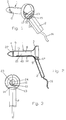
- a device corresponding to that shown in FIGS. 1 to 3 Embodiment includes an in the anus of the person to be treated tube 1 to be inserted and a handle 2.
- the tube 1 is on its the proximal end facing the treating person is open and at his distal end closed, it funnel-shaped towards its proximal end widens and widens in the area of its distal end to a rounded Tapered tip.
- the facility consists of a left and a right Half shell that is welded or glued together. With each of the two Half shells are the corresponding half shells of the tube in one piece with the Half-shell of the handle formed.
- ultrasound probe 6 radiating forward (distal) and one distal in front of the ultrasound probe lying treatment opening provided in the tube jacket 40.
- a white light LED 5 is arranged in the proximal area of the device. This LED 5 is in front of the proximal end of a along the inner surface 8 of the Tube 1 extending light guide 10 arranged by means of the light from the proximal Area 3 is directed to the distal area 4 of the device.
- the outer surface 8 of the tube 1 is attached to the light guide by means of clips 11. From the distal end of the light guide 10, light is inter alia towards the treatment opening 7 emitted.
- distal end of the light guide 10 be beveled accordingly.
- the radiation the light is directed from proximal to distal.
- this radiation can be angular be aligned to the longitudinal axis of the tube.
- a circuit board 12 On the proximal side of the proctoscope facing the patient a circuit board 12 is fixed, which in addition to the white light LED 5 the on / off switch 13 facing the user and an optical display in the form of a light-emitting diode bar 14, of four individual here LEDs 15 is formed.
- the power supply to the ultrasound probe 6 and the transmission of the signal of the Ultrasound probe takes place via electrical lines 17 which are located within a groove the inner lateral surface 8 of the tube 1. These electrical wires 17 and those connected to the on / off switch 13 and the light-emitting diode bar 14 electrical lines are connected via a connecting cable 16 to a Control and evaluation unit 18 connected (see FIG. 10).
- the power supply of the Control and evaluation unit 18 can take place via a power supply unit 19 or by means of Batteries.
- the control and evaluation unit comprises the evaluation of the ultrasonic signals a mini computer. An acoustic indicator is on a headphone 20 issued.
- the mini computer can also take on other tasks, such as storing data to document arterial flow before and after the ligature (documentation of the operation), possibly also with indication of angular positions of the arteries.
- This distal wall 22 has a shoulder with a toothed guide surface 23.
- This guide surface 23 extends essentially in the direction of the longitudinal extent of the treatment opening 7, but is just trained.
- In the wall 22 there is also a parallel to the guide surface 23 extending groove 24 is provided.
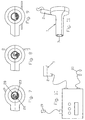
- a needle holder for use with this proctoscope is shown in Figs. 5 and 6 shown.
- a corresponding groove 24 is formed at the point where the circular needle 28 is to be placed in the needle holder 25 .
- a gear wheel 26 is fixed, which has a circular toothed cross section Neck of the needle holder forms.
- the artery is ligated to the 7 inserted into the proctoscope, the pin 27 protrudes into the guide groove 24 and the gear 26 with the toothed guide surface 23 combs.
- the needle holder 25 is about the longitudinal axis of the Gear 26 and the pin 27 rotated, as shown in FIGS. 8 and 9 is.
- FIG. 4 A somewhat modified embodiment of the invention is shown in Fig. 4, the electrical parts (ultrasound probe, white light LED, light emitting diode bar, On / off switch and its wiring) omitted for the sake of simplicity are.
- the handle 2 is on a connector 29 set.
- the tube consists of two or weldable half shells.
- a light guide 10 runs in a groove on the inner surface of the tube and is with clips 11 on tube 1 attached.
- the connection of the tube 1 with the connector 29 is carried out according to Art a bayonet lock, i.e. the tube is inserted into the connector 29 and then twisted about its longitudinal axis.
- Tube 1 from its proximal end is one over the outer surface of tube 1 protruding ring flange 30 is provided. Contacts can be arranged on this be the electrical contact between those running in the tube electrical lines for the ultrasound probe and the corresponding through the connector 29 and the handle 2 produce further lines.
- the handle 2 with the connector 29 and the electrical parts provided therein can be steam sterilized and thus be designed to be recyclable during the Tube 1 and the parts installed in it (light guide 10, ultrasound probe and connecting lines) Display one-off parts.
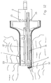
- FIGS. 11 and 12 Another embodiment of the invention is shown in FIGS. 11 and 12.
- This embodiment of the invention corresponds to that shown in FIGS. 1 to 3 with the difference that the distal end of the tube 1 is open. This open The distal end here forms the treatment opening 7.
- Such an ultrasound proctoscope can be used especially for performing a rubber band ligature become.
- the treatment process is shown schematically in FIG. 12.
- An artery is located using the proctoscope inserted into the anus.
- the ultrasound probe is emitted obliquely to the front.
- On Instrument 31 for setting a ligature by means of a rubber band 32 is shown in introduced the tube 1.
- This instrument is only indicated schematically in FIG. 12. It includes a suction tube for aspirating the mucosa 33, which the contains ligating artery.
- the artery is previously attached to the instrument 31 loaded rubber band 32 tied.
- Such instruments for setting of rubber band ligatures are known and do not form the subject of these Invention and are therefore not
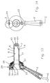
- a further, particularly preferred embodiment of the invention is in the 13 to 15 are shown.
- the handle 2 is on the from the two parts 34, 35 existing connector 29 set.
- the tube 1 is removable with connected to the connector 29.
- the part 35 has an external thread 36 on which a union nut 37 can be screwed, which has an annular flange 38 overlaps on the outside and near the distal end of the tube 1.
- the treating person facing proximal side of the proctoscope optical display in the form of a light-emitting diode bar 14 for visualization locating an artery as well as an on / off switch 13 provided for the ultrasonic probe.
- the tube 1 consists of a translucent material and forms a light guide, by means of the light from the proximal to the distal area of the proctoscope is directed.
- the tube 1 consists of a translucent material and forms a light guide, by means of the light from the proximal to the distal area of the proctoscope is directed.
- three white light LEDs 5 light is transmitted through the proximal End face 39 of the tube jacket 40 irradiated. These three LED 5 are in the circumferential direction of the tube jacket spaced 120 ° apart. through
- the light radiated into the tube jacket becomes total reflection in the distal Headed area 4 of the facility.
- the inner surface 8 of the tube is roughened, so that at this point, more light emerges, which illuminates the treatment opening.
- a region 42 is roughened on the distal end wall 43. It this makes the guide groove 24 particularly recognizable, into which the tip a needle holder (here without gear).
- This embodiment of the invention can in turn be used once Part or be designed as a steam-sterilizable part.
- entire proctoscope can be steam sterilized or only parts of it, for example the connector 29 with the handle 2 and the union nut 37 and the parts installed in the connector.
- Suitable materials from which the tube can be produced are in particular Polycarbonate or polysulfone.
- polycarbonate is among the material names "APEC” or “MAKROLON” available.
- the ring flange 38 can contact points be provided for contacting these lines 17.
- the groove in the ring flange 38 can simultaneously serve as a centering groove for tube 1.
Landscapes
- Health & Medical Sciences (AREA)
- Life Sciences & Earth Sciences (AREA)
- Surgery (AREA)
- Engineering & Computer Science (AREA)
- General Health & Medical Sciences (AREA)
- Biomedical Technology (AREA)
- Heart & Thoracic Surgery (AREA)
- Medical Informatics (AREA)
- Molecular Biology (AREA)
- Animal Behavior & Ethology (AREA)
- Nuclear Medicine, Radiotherapy & Molecular Imaging (AREA)
- Public Health (AREA)
- Veterinary Medicine (AREA)
- Pathology (AREA)
- Physics & Mathematics (AREA)
- Biophysics (AREA)
- Optics & Photonics (AREA)
- Radiology & Medical Imaging (AREA)
- Reproductive Health (AREA)
- Vascular Medicine (AREA)
- Microelectronics & Electronic Packaging (AREA)
- Surgical Instruments (AREA)
Abstract
Description
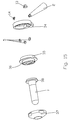
- Fig. 1
- eine perspektivische Darstellung eines ersten Ausführungsbeispiels der Erfindung;
- Fig. 2
- einen Schnitt durch die vertikale Längsmittelebene der Einrichtung von Fig. 1;
- Fig. 3
- eine Ansicht auf die der behandelnden Person zugewandte proximale Seite der Einrichtung von Fig. 1;
- Fig. 4
- eine perspektivische Darstellung von nach Art einer Explosionsdarstellung auseinandergezogenen Teilen einer modifizierten Ausführungsform der Erfindung (ohne elektrische Teile);
- Fig. 5
- eine perspektivische Darstellung eines Nadelhalters mit einer eingelegten Nadel zur Verwendung mit dem Proktoskop entsprechend Fig. 1 oder Fig. 4;
- Fig. 6
- ein vergrößertes Detail B von Fig. 5;
- Fig. 7
- bis Fig. 9 Arbeitsschritte bei der Ligatur einer Arterie (Querschnitte durch den Tubus);
- Fig.10
- eine schematische Darstellung der an eine Steuer- und Auswerteinheit angeschlossenen Einrichtung;
- Fig.11
- eine perspektivische Darstellung eines weiteren Ausführungsbeispiels der Erfindung;
- Fig.12
- einen Schnitt durch eine Längsmittelebene des Ausführungsbeispiels gemäß Fig. 11, wobei in schematischer Weise das Setzen einer Gummibandligatur dargestellt ist;
- Fig.13
- einen Längsmittelschnitt entlang der Linie A-A von Fig. 14 einer weiteren Ausführungsform der Erfindung;
- Fig.14
- eine Ansicht auf die der behandelnden Person zugewandten proximalen Seite der Einrichtung von Fig. 13 und
- Fig.15
- eine perspektivische Darstellung nach Art einer Explosionsdarstellung der Einrichtung entsprechend Fig. 13 (ohne Verbindungsleitungen zur Verdrahtung der elektrischen Teile).
- 1
- Tubus
- 2
- Handgriff
- 3
- proximaler Bereich
- 4
- distaler Bereich
- 5
- LED
- 6
- Ultraschallsonde
- 7
- Behandlungsöffnung
- 8
- innere Mantelfläche
- 9
- äußere Mantelfläche
- 10
- Lichtleiter
- 11
- Clips-Teil
- 12
- Leiterplatte
- 13
- Ein/Aus-Schalter
- 14
- Leuchtdiodenbalken
- 15
- Leuchtdiode
- 16
- Anschlußkabel
- 17
- elektrische Leitung
- 18
- Steuer- und Auswerteinheit
- 19
- Netzteil
- 20
- Kopfhörer
- 21
- Innenraum
- 22
- Wandung
- 23
- gezahnte Führungsfläche
- 24
- Führungsnut
- 25
- Nadelhalter
- 26
- Zahnrad
- 27
- Stift
- 28
- Rundnadel
- 29
- Anschlußstück
- 30
- Ringflansch
- 31
- Instrument
- 32
- Gummiband
- 33
- Mukosa
- 34
- Teil
- 35
- Teil
- 36
- Außengewinde
- 37
- Überwurfmutter
- 38
- Ringflansch
- 39
- Stirnfläche
- 40
- Tubusmantel
- 41
- Bereich
- 42
- Bereich
- 43
- Stirnwandung
Claims (15)
- Einrichtung zur Verwendung bei der Ligatur von intramuralen Arterien in Hohlorganen, welche einen Handgriff (2) und einen in das Hohlorgan, insbesondere das Rektum einzuführenden Tubus (1) umfaßt, wobei die Einrichtung einen proximalen Bereich (3), in dem der Handgriff (2) angeordnet ist, und einen distalen Bereich (4) aufweist, in dem eine Ultraschallsonde (6) für die Lokalisation einer Arterie und eine Behandlungsöffnung (7) angeordnet sind, und wobei eine Beleuchtungseinrichtung vorgesehen ist, dadurch gekennzeichnet, daß die Beleuchtungseinrichtung ein Lichtleitmittel (10; 40) umfaßt, mittels dem Licht vom proximalen zum distalen Bereich der Einrichtung geleitet wird.
- Einrichtung nach Anspruch 1, dadurch gekennzeichnet, daß das Lichtleitmittel ein vom proximalen zum distalen Bereich der Einrichtung verlaufender Lichtleiter (10) ist, dessen proximales Ende vor einem im proximalen Bereich (3) der Einrichtung angeordneten lichterzeugenden Element, vorzugsweise einer LED (5), liegt und von dessen distalem Ende Licht zumindest teilweise in Richtung zur Behandlungsöffnung (7) abgestrahlt wird, wobei die Abstrahlung von proximal nach distal gerichtet ist, vorzugsweise winkelig zur Längsachse des Tubus (1).
- Einrichtung nach Anspruch 2, dadurch gekennzeichnet, daß der Tubus (1) und der Handgriff (2) aus linken und rechten Halbschalen bestehen, wobei die jeweilige Halbschale des Handgriffs (2) einstückig mit der jeweils entsprechenden Halbschale des Tubus (1) ausgebildet ist.
- Einrichtung nach Anspruch 1, dadurch gekennzeichnet, daß das Lichtleitmittel von dem aus einem lichtdurchlässigen Material bestehenden Tubusmantel (40) gebildet wird, in welchen durch dessen proximale Stirnfläche (39) von mindestens drei in Umfangsrichtung der ringförmig ausgebildeten proximalen Stirnfläche (39) des Tubusmantels (40) voneinander beabstandeten LEDs (5) Licht eingestrahlt wird und in den distalen Bereich (4) der Einrichtung geleitet wird.
- Einrichtung nach Anspruch 4, dadurch gekennzeichnet, daß der Tubus (1) aus Polycarbonat oder Polysulfon besteht.
- Einrichtung nach Anspruch 4 oder Anspruch 5, dadurch gekennzeichnet, daß die Lichtleitung im Tubus (1) mittels Totalreflektion erfolgt.
- Einrichtung nach Anspruch 6, dadurch gekennzeichnet, daß die innere Mantelfläche (8) des Tubus in einem der Behandlungsöffnung (7) gegenüberliegenden Bereich (41) und/oder die Innenseite der distalen Stirnwandung (43) des Tubus (1) in einem Bereich (42) aufgerauht ist.
- Einrichtung nach einem der Ansprüche 1 bis 2 oder einem der Ansprüche 4 bis 7, dadurch gekennzeichnet, daß der Handgriff (2) an einem Anschlußstück (29) festgelegt ist und der Tubus (1) abnehmbar mit dem Anschlußstück (29) verbunden ist, vorzugsweise mittels eines Bajonettverschlusses oder mittels einer Überwurfmutter (37), die auf ein Außengewinde (36) des Anschlußstücks (29) aufschraubbar ist und einen Ringflansch (38) an der Außenseite des Tubus (1) übergreift.
- Einrichtung nach einem der Ansprüche 1 bis 8, wobei der Tubus (1) auf seiner distalen Seite geschlossen ist, eine den zur proximalen Seite offenen Innenraum (21) abschließende Wandung (22) vorgesehen ist und die Behandlungsöffnung (7) im Tubusmantel (40) angeordnet ist, dadurch gekennzeichnet, daß in der distalen Wandung (22) eine Schulter mit einer gezahnten Führungsfläche (23) vorgesehen ist, welche auf die ein im Querschnitt kreisförmiger oder teilkreisförmiger gezahnter Ansatz, vorzugsweise ein Zahnrad (26), eines Nadelhalters (25) aufsetzbar ist.
- Einrichtung nach Anspruch 9, dadurch gekennzeichnet, daß sich die Führungsfläche im wesentlichen in der Richtung der Längsausdehnung der Behandlungsöffnung (7) erstreckt, vorzugsweise aber eben ausgebildet ist.
- Einrichtung nach einem der Ansprüche 1 bis 10, dadurch gekennzeichnet, daß der Tubus (1) an seinem distalen Ende offen ist, wobei dieses offene distale Ende die Behandlungsöffnung (7) bildet.
- Einrichtung zur Verwendung bei der Ligatur von intramuralen Arterien in Hohlorganen, welche einen Handgriff (2) und einen in das Hohlorgan, insbesondere das Rektum einzuführenden Tubus (1) umfaßt, wobei die Einrichtung einen proximalen Bereich (3), in dem der Handgriff (2) angeordnet ist, und einen distalen Bereich (4) aufweist, in dem eine Ultraschallsonde (6) für die Lokalisation einer Arterie und eine Behandlungsöffnung (7) angeordnet sind, und wobei eine Beleuchtungseinrichtung vorgesehen ist, insbesondere nach einem der Ansprüche 1 bis 18, dadurch gekennzeichnet, daß im Bereich der der behandelnden Person zugewandten proximalen Seite der Einrichtung eine optische Anzeige zur Darstellung der Ortung einer Arterie vorgesehen ist.
- Einrichtung nach Anspruch 12, dadurch gekennzeichnet, daß die optische Anzeige von einem Leuchtdiodenbalken (14) gebildet wird.
- Einrichtung nach einem der Ansprüche 1 bis 13, dadurch gekennzeichnet, daß die Einrichtung, vorzugsweise im Bereich der der behandelnden Person zugewandten proximalen Seite, einen Ein/Aus-Schalter (13) für die Ultraschallsonde aufweist.
- Einrichtung nach einem der Ansprüche 1 bis 14, dadurch gekennzeichnet, daß die Einrichtung zumindest teilweise mehrfach heißdampfsterilisierbar ist.
Priority Applications (1)
| Application Number | Priority Date | Filing Date | Title |
|---|---|---|---|
| DE20220338U DE20220338U1 (de) | 2001-02-15 | 2002-01-31 | Einrichtung zur Verwendung bei der Ligatur von intramuralen Arterien in Hohlorganen |
Applications Claiming Priority (2)
| Application Number | Priority Date | Filing Date | Title |
|---|---|---|---|
| AT2352001 | 2001-02-15 | ||
| AT2352001 | 2001-02-15 |
Publications (2)
| Publication Number | Publication Date |
|---|---|
| EP1234539A2 true EP1234539A2 (de) | 2002-08-28 |
| EP1234539A3 EP1234539A3 (de) | 2002-09-11 |
Family
ID=3669993
Family Applications (1)
| Application Number | Title | Priority Date | Filing Date |
|---|---|---|---|
| EP02002315A Withdrawn EP1234539A3 (de) | 2001-02-15 | 2002-01-31 | Einrichtung zur Verwendung bei der Ligatur von intramuralen Arterien in Hohlorganen |
Country Status (1)
| Country | Link |
|---|---|
| EP (1) | EP1234539A3 (de) |
Cited By (15)
| Publication number | Priority date | Publication date | Assignee | Title |
|---|---|---|---|---|
| WO2004064624A1 (en) * | 2003-01-21 | 2004-08-05 | Metech S.R.L. | A retractor for operations on the arteria haemorroidalis |
| WO2006033122A1 (en) * | 2004-09-21 | 2006-03-30 | Sapi Med S.P.A. | Medical device for precision surgery |
| EP1683473A1 (de) * | 2005-01-25 | 2006-07-26 | AMI Agency for Medical Innovations GmbH | Einrichtung zur Verwendung bei der Behandlung eines Hämorrhoidenprolaps |
| WO2006116779A1 (de) | 2005-05-04 | 2006-11-09 | Ami Agency For Medical Innovations Gmbh | Einrichtung zur verwendung bei der ligatur von intramuralen arterien |
| RU2308873C2 (ru) * | 2003-01-21 | 2007-10-27 | МЕТЕК С.р.Л. | Ретрактор для хирургических операций на артерии прямой кишки |
| WO2008128261A1 (de) | 2007-04-23 | 2008-10-30 | Ami Agency For Medical Innovations Gmbh | Einrichtung zur verwendung bei der behandlung eines hämorrhoidenprolaps |
| EP2179700A1 (de) * | 2003-09-30 | 2010-04-28 | Ethicon Endo-Surgery, Inc. | Verschlungene Schutzhülle in einer Dichtungsanordnung für Trokar |
| WO2011050705A1 (zh) * | 2009-10-26 | 2011-05-05 | 北京中法派尔特医疗设备有限公司 | 一种肛肠手术器械及扩肛器 |
| CN103142284A (zh) * | 2013-03-05 | 2013-06-12 | 常州贺利氏微创医疗器械有限公司 | 带光源的多环痔疮套扎器 |
| CN104207808A (zh) * | 2014-09-05 | 2014-12-17 | 北京中法派尔特医疗设备有限公司 | 一种缝扎器 |
| GB2518343A (en) * | 2013-06-28 | 2015-03-25 | Rocket Medical Plc | Improved medical device |
| CN105079948A (zh) * | 2015-06-30 | 2015-11-25 | 江汉大学 | 一种用于实验动物的肛门直肠扩张装置及系统 |
| RU2580903C1 (ru) * | 2015-01-22 | 2016-04-10 | Акционерное общество "Научно-производственная фирма "БИОСС" (АО "НПФ "БИОСС") | Проктоскоп |
| WO2016118041A1 (ru) * | 2015-01-22 | 2016-07-28 | Акционерное Общество "Научно-Производственная Фирма "Биосс" | Проктоскоп |
| RU2715448C2 (ru) * | 2015-06-17 | 2020-02-28 | Франческо СИАС | Вращающийся аноскоп |
Citations (1)
| Publication number | Priority date | Publication date | Assignee | Title |
|---|---|---|---|---|
| US5570692A (en) | 1995-05-19 | 1996-11-05 | Hayashi Denki Co. Ltd. | Ultrasonic doppler blood flow detector for hemorrhoid artery ligation |
Family Cites Families (3)
| Publication number | Priority date | Publication date | Assignee | Title |
|---|---|---|---|---|
| US3581738A (en) * | 1968-11-12 | 1971-06-01 | Welch Allyn Inc | Disposable illuminating endoscope and method of manufacture |
| DE3717607A1 (de) * | 1987-05-25 | 1988-12-08 | Elke Technik Fritz Kerner Gmbh | Spekulum |
| US6132379A (en) * | 1998-11-04 | 2000-10-17 | Patacsil; Estelito G. | Method and apparatus for ultrasound guided intravenous cannulation |
-
2002
- 2002-01-31 EP EP02002315A patent/EP1234539A3/de not_active Withdrawn
Patent Citations (1)
| Publication number | Priority date | Publication date | Assignee | Title |
|---|---|---|---|---|
| US5570692A (en) | 1995-05-19 | 1996-11-05 | Hayashi Denki Co. Ltd. | Ultrasonic doppler blood flow detector for hemorrhoid artery ligation |
Cited By (27)
| Publication number | Priority date | Publication date | Assignee | Title |
|---|---|---|---|---|
| AU2003205634B2 (en) * | 2003-01-21 | 2009-05-28 | Thd S.P.A. | Retractor for surgical operations on the arteria haemorroidalis |
| JP2006512977A (ja) * | 2003-01-21 | 2006-04-20 | エムイーテック エス.アール.エル. | 痔核動脈の外科手術用レトラクター |
| WO2004064624A1 (en) * | 2003-01-21 | 2004-08-05 | Metech S.R.L. | A retractor for operations on the arteria haemorroidalis |
| RU2308873C2 (ru) * | 2003-01-21 | 2007-10-27 | МЕТЕК С.р.Л. | Ретрактор для хирургических операций на артерии прямой кишки |
| CN100553552C (zh) * | 2003-01-21 | 2009-10-28 | Thd股份公司 | 用于在直肠动脉上外科手术的牵开器 |
| US7452329B2 (en) | 2003-01-21 | 2008-11-18 | Thd S.P.A. | Retractor for operations on the arteria haemorroidalis |
| EP2179700A1 (de) * | 2003-09-30 | 2010-04-28 | Ethicon Endo-Surgery, Inc. | Verschlungene Schutzhülle in einer Dichtungsanordnung für Trokar |
| WO2006033122A1 (en) * | 2004-09-21 | 2006-03-30 | Sapi Med S.P.A. | Medical device for precision surgery |
| EP1683473A1 (de) * | 2005-01-25 | 2006-07-26 | AMI Agency for Medical Innovations GmbH | Einrichtung zur Verwendung bei der Behandlung eines Hämorrhoidenprolaps |
| US7695432B2 (en) | 2005-01-25 | 2010-04-13 | Ami Agency For Medical Innovations Gmbh | Instrument for use in the treatment of prolapsed hemorrhoids |
| WO2006116779A1 (de) | 2005-05-04 | 2006-11-09 | Ami Agency For Medical Innovations Gmbh | Einrichtung zur verwendung bei der ligatur von intramuralen arterien |
| WO2008128261A1 (de) | 2007-04-23 | 2008-10-30 | Ami Agency For Medical Innovations Gmbh | Einrichtung zur verwendung bei der behandlung eines hämorrhoidenprolaps |
| US8394012B2 (en) | 2007-04-23 | 2013-03-12 | A.M.I. Agency For Medical Innovations Gmbh | Device for use for the treatment of hemorrhoid prolapse |
| WO2011050705A1 (zh) * | 2009-10-26 | 2011-05-05 | 北京中法派尔特医疗设备有限公司 | 一种肛肠手术器械及扩肛器 |
| CN103142284A (zh) * | 2013-03-05 | 2013-06-12 | 常州贺利氏微创医疗器械有限公司 | 带光源的多环痔疮套扎器 |
| CN103142284B (zh) * | 2013-03-05 | 2015-10-28 | 常州贺利氏微创医疗器械有限公司 | 带光源的多环痔疮套扎器 |
| GB2518343A (en) * | 2013-06-28 | 2015-03-25 | Rocket Medical Plc | Improved medical device |
| CN104207808A (zh) * | 2014-09-05 | 2014-12-17 | 北京中法派尔特医疗设备有限公司 | 一种缝扎器 |
| WO2016118041A1 (ru) * | 2015-01-22 | 2016-07-28 | Акционерное Общество "Научно-Производственная Фирма "Биосс" | Проктоскоп |
| RU2580903C1 (ru) * | 2015-01-22 | 2016-04-10 | Акционерное общество "Научно-производственная фирма "БИОСС" (АО "НПФ "БИОСС") | Проктоскоп |
| CN107205634A (zh) * | 2015-01-22 | 2017-09-26 | 博尔斯研究生产股份公司 | 直肠镜 |
| AU2015378687A8 (en) * | 2015-01-22 | 2018-03-29 | Ao Npf "Bioss" | Proctoscope |
| AU2015378687B2 (en) * | 2015-01-22 | 2018-03-29 | Ao Npf "Bioss" | Proctoscope |
| EP3248537A4 (de) * | 2015-01-22 | 2018-09-12 | Ao Npf "Bioss" | Proktoskop |
| CN107205634B (zh) * | 2015-01-22 | 2019-08-30 | 博尔斯研究生产股份公司 | 直肠镜 |
| RU2715448C2 (ru) * | 2015-06-17 | 2020-02-28 | Франческо СИАС | Вращающийся аноскоп |
| CN105079948A (zh) * | 2015-06-30 | 2015-11-25 | 江汉大学 | 一种用于实验动物的肛门直肠扩张装置及系统 |
Also Published As
| Publication number | Publication date |
|---|---|
| EP1234539A3 (de) | 2002-09-11 |
Similar Documents
| Publication | Publication Date | Title |
|---|---|---|
| EP1234539A2 (de) | Einrichtung zur Verwendung bei der Ligatur von intramuralen Arterien in Hohlorganen | |
| DE3727190C2 (de) | Führungsrohr zum subkutanen Einführen in den Körper eines Patienten | |
| DE69734978T2 (de) | Optische biopsiezangen | |
| DE69330169T2 (de) | Visuell gesteuerter trokar | |
| EP0093927B1 (de) | Gerät zur Spektrenmessung in der Blutbahn | |
| DE4035146A1 (de) | Instrument zum penetrieren von koerpergewebe | |
| DE3429945C2 (de) | ||
| DE2636510A1 (de) | Endoskop, insbesondere rektoskop | |
| DE19827255A1 (de) | Endoskop | |
| EP0516582A1 (de) | Punktiergerät für Blutgefässe | |
| DE19929314A1 (de) | Endoskop für Ultraschalluntersuchungen und damit verbundene chirurgische Behandlungen | |
| DE2057219A1 (de) | Spekulum | |
| DE3309097A1 (de) | Vorrichtung zur untersuchung des inneren von koerperhoehlen mit ultraschallwellen | |
| DE20207260U1 (de) | Beleuchteter zahnärztlicher oder chirurgischer Spiegel | |
| US7060028B2 (en) | Endoilluminator | |
| DE4303756A1 (en) | Clinical monitoring instrument incorporating optical fibre bundle - illuminates interior or organ by repetitive total internal reflection between both walls of hollow circular transparent cone | |
| EP0871403B1 (de) | Vorrichtung zur durchführung von manipulationen im menschlichen körper und insbesondere im uterus | |
| DE1566116A1 (de) | Sterilisierbare Beleuchtung fuer chirurgische Instrumente | |
| DE3707403C2 (de) | Medizinisches Resektionsgerät | |
| JPH0654862A (ja) | 照明付き外科用カニユーレ | |
| DE20220338U1 (de) | Einrichtung zur Verwendung bei der Ligatur von intramuralen Arterien in Hohlorganen | |
| EP0269048B1 (de) | Stethoskop | |
| JP2000245740A (ja) | 内視鏡用処置具 | |
| CA2176565C (en) | Visually directed trocar and method | |
| DE3717607C2 (de) |
Legal Events
| Date | Code | Title | Description |
|---|---|---|---|
| PUAI | Public reference made under article 153(3) epc to a published international application that has entered the european phase |
Free format text: ORIGINAL CODE: 0009012 |
|
| PUAL | Search report despatched |
Free format text: ORIGINAL CODE: 0009013 |
|
| AK | Designated contracting states |
Kind code of ref document: A2 Designated state(s): AT BE CH CY DE DK ES FI FR GB GR IE IT LI LU MC NL PT SE TR |
|
| AX | Request for extension of the european patent |
Free format text: AL;LT;LV;MK;RO;SI |
|
| AK | Designated contracting states |
Kind code of ref document: A3 Designated state(s): AT BE CH CY DE DK ES FI FR GB GR IE IT LI LU MC NL PT SE TR |
|
| AX | Request for extension of the european patent |
Free format text: AL;LT;LV;MK;RO;SI |
|
| AKX | Designation fees paid | ||
| REG | Reference to a national code |
Ref country code: DE Ref legal event code: 8566 |
|
| STAA | Information on the status of an ep patent application or granted ep patent |
Free format text: STATUS: THE APPLICATION IS DEEMED TO BE WITHDRAWN |
|
| 18D | Application deemed to be withdrawn |
Effective date: 20030312 |