EP1230552B1 - Elisa für vegf - Google Patents
Elisa für vegf Download PDFInfo
- Publication number
- EP1230552B1 EP1230552B1 EP00978702A EP00978702A EP1230552B1 EP 1230552 B1 EP1230552 B1 EP 1230552B1 EP 00978702 A EP00978702 A EP 00978702A EP 00978702 A EP00978702 A EP 00978702A EP 1230552 B1 EP1230552 B1 EP 1230552B1
- Authority
- EP
- European Patent Office
- Prior art keywords
- vegf
- antibody
- immobilized
- monoclonal
- mab
- Prior art date
- Legal status (The legal status is an assumption and is not a legal conclusion. Google has not performed a legal analysis and makes no representation as to the accuracy of the status listed.)
- Expired - Lifetime
Links
Images


Classifications
-
- G—PHYSICS
- G01—MEASURING; TESTING
- G01N—INVESTIGATING OR ANALYSING MATERIALS BY DETERMINING THEIR CHEMICAL OR PHYSICAL PROPERTIES
- G01N33/00—Investigating or analysing materials by specific methods not covered by groups G01N1/00 - G01N31/00
- G01N33/48—Biological material, e.g. blood, urine; Haemocytometers
- G01N33/50—Chemical analysis of biological material, e.g. blood, urine; Testing involving biospecific ligand binding methods; Immunological testing
- G01N33/53—Immunoassay; Biospecific binding assay; Materials therefor
- G01N33/543—Immunoassay; Biospecific binding assay; Materials therefor with an insoluble carrier for immobilising immunochemicals
-
- G—PHYSICS
- G01—MEASURING; TESTING
- G01N—INVESTIGATING OR ANALYSING MATERIALS BY DETERMINING THEIR CHEMICAL OR PHYSICAL PROPERTIES
- G01N33/00—Investigating or analysing materials by specific methods not covered by groups G01N1/00 - G01N31/00
- G01N33/48—Biological material, e.g. blood, urine; Haemocytometers
- G01N33/50—Chemical analysis of biological material, e.g. blood, urine; Testing involving biospecific ligand binding methods; Immunological testing
- G01N33/68—Chemical analysis of biological material, e.g. blood, urine; Testing involving biospecific ligand binding methods; Immunological testing involving proteins, peptides or amino acids
- G01N33/6863—Cytokines, i.e. immune system proteins modifying a biological response such as cell growth proliferation or differentiation, e.g. TNF, CNF, GM-CSF, lymphotoxin, MIF or their receptors
-
- G—PHYSICS
- G01—MEASURING; TESTING
- G01N—INVESTIGATING OR ANALYSING MATERIALS BY DETERMINING THEIR CHEMICAL OR PHYSICAL PROPERTIES
- G01N33/00—Investigating or analysing materials by specific methods not covered by groups G01N1/00 - G01N31/00
- G01N33/48—Biological material, e.g. blood, urine; Haemocytometers
- G01N33/50—Chemical analysis of biological material, e.g. blood, urine; Testing involving biospecific ligand binding methods; Immunological testing
- G01N33/68—Chemical analysis of biological material, e.g. blood, urine; Testing involving biospecific ligand binding methods; Immunological testing involving proteins, peptides or amino acids
- G01N33/6872—Intracellular protein regulatory factors and their receptors, e.g. including ion channels
-
- G—PHYSICS
- G01—MEASURING; TESTING
- G01N—INVESTIGATING OR ANALYSING MATERIALS BY DETERMINING THEIR CHEMICAL OR PHYSICAL PROPERTIES
- G01N2333/00—Assays involving biological materials from specific organisms or of a specific nature
- G01N2333/435—Assays involving biological materials from specific organisms or of a specific nature from animals; from humans
- G01N2333/475—Assays involving growth factors
-
- Y—GENERAL TAGGING OF NEW TECHNOLOGICAL DEVELOPMENTS; GENERAL TAGGING OF CROSS-SECTIONAL TECHNOLOGIES SPANNING OVER SEVERAL SECTIONS OF THE IPC; TECHNICAL SUBJECTS COVERED BY FORMER USPC CROSS-REFERENCE ART COLLECTIONS [XRACs] AND DIGESTS
- Y10—TECHNICAL SUBJECTS COVERED BY FORMER USPC
- Y10S—TECHNICAL SUBJECTS COVERED BY FORMER USPC CROSS-REFERENCE ART COLLECTIONS [XRACs] AND DIGESTS
- Y10S435/00—Chemistry: molecular biology and microbiology
- Y10S435/975—Kit
-
- Y—GENERAL TAGGING OF NEW TECHNOLOGICAL DEVELOPMENTS; GENERAL TAGGING OF CROSS-SECTIONAL TECHNOLOGIES SPANNING OVER SEVERAL SECTIONS OF THE IPC; TECHNICAL SUBJECTS COVERED BY FORMER USPC CROSS-REFERENCE ART COLLECTIONS [XRACs] AND DIGESTS
- Y10—TECHNICAL SUBJECTS COVERED BY FORMER USPC
- Y10S—TECHNICAL SUBJECTS COVERED BY FORMER USPC CROSS-REFERENCE ART COLLECTIONS [XRACs] AND DIGESTS
- Y10S436/00—Chemistry: analytical and immunological testing
- Y10S436/807—Apparatus included in process claim, e.g. physical support structures
- Y10S436/809—Multifield plates or multicontainer arrays
-
- Y—GENERAL TAGGING OF NEW TECHNOLOGICAL DEVELOPMENTS; GENERAL TAGGING OF CROSS-SECTIONAL TECHNOLOGIES SPANNING OVER SEVERAL SECTIONS OF THE IPC; TECHNICAL SUBJECTS COVERED BY FORMER USPC CROSS-REFERENCE ART COLLECTIONS [XRACs] AND DIGESTS
- Y10—TECHNICAL SUBJECTS COVERED BY FORMER USPC
- Y10S—TECHNICAL SUBJECTS COVERED BY FORMER USPC CROSS-REFERENCE ART COLLECTIONS [XRACs] AND DIGESTS
- Y10S436/00—Chemistry: analytical and immunological testing
- Y10S436/811—Test for named disease, body condition or organ function
- Y10S436/813—Cancer
Definitions
- This invention relates to immunoassays for detecting VEGF that can be used as diagnostic and prognostic methods for patients with cancer, cardiovascular, or other pathologies.
- angiogenesis is implicated in the pathogenesis of a variety of disorders. These include solid tumors, intra-ocular neovascular syndromes such as proliferative retinopathies or age-related macular degeneration (AMD), rheumatoid arthritis, and psoriasis (Folkman et al. J. Biol. Chem . 267:10931-10934 (1992); Klagsbrun et al. Annu. Rev. Physiol . 53:217-239(1991); and Garner A. Vascular diseases. In: Pathobiology of ocular disease. A dynamic approach . Garner A. Klintworth GK. Eds.
- the search for positive regulators of angiogenesis has yielded many candidates, including aFGF, bFGF, TGF- ⁇ , TGF- ⁇ , HGF, TNF- ⁇ , angiogenin, IL-8, etc. (Folkman et al., supra , and Klagsbrun et al., supra ).
- the negative regulators so far identified include thrombospondin (Good et al. Proc. Natl. Acad. Sci. USA. 87:6624-6628 (1990)), the 16-kilodalton N-terminal fragment of prolactin (Clapp et al.
- VEGF vascular endothelial growth factor
- VEGF has been shown to be a key mediator of ncovascularization associated with tumors and intra-ocular disorders (Ferrara et al., supra ).
- the VEGF mRNA is overexpressed by the majority of human tumors examined (Berkman et al. J Clin Invest 91:153-159 (1993); Brown et al. Human Pathol ., 26:86-91 (1995); Brown et al. Cancer Res. 53:4727-4735 (1993); Mattern et al. Brit. J. Cancer. 73:931-934 (1996); and Dvorak et al. Am J. Pathol . 146:1029-1039 (1995)).
- VEGF vascular endothelial growth factor
- VEGF is a heparin binding growth factor with a molecular weight of 45 kD (Plouet et al. EMBO J. 8:3801 (1989); Neufeld et al. Prog. Growth Factor Res. 5:89 (1994)). It is a dimeric glycoprotein consisting of two identical subunits. Although VEGF is encoded from a single gene, at least five isoforms exist in vivo due to alternative mRNA splicing. These isoforms, VEGF121, VEGF145, VEGF165, VEGF189, and VEGF206, contain 121, 145, 165, 189, and 206 amino acids, respectively (Leung et al.
- VEGF isoforms show differing affinities for heparin; VEGF121 binds heparin weakly, while VEGF165, VEGF189, and VEGF206 bind heparin with increasing affinity. VEGF121 and VEGF165 are secreted and both isoforms are found in the circulation.
- VEGF189 and VEGF206 are found mostly associated with heparin sulfate containing proteoglycans in the extracellular matrix (Houck et al. J. Biol. Chem . 267:26031 (1992); Park et al. Mol. Biol. Cell 4:1317 (1993)). Of the five isoforms, VEGF165 is the most abundantly expressed variant in the majority of cells and tissues.
- VEGFR-1 FLT-1
- VEGFR-2 KDR/FLK-1
- VEGFR-3 which are all signaling tyrosine kinases
- Neuropilin-1 and Neuropilin-2 which are both accessory-isoform-specific receptors that bind selectively to VEGF165 (de Vries et al. Science 255:989 (1992); Terman et al. Biochem. Biophys. Res. Commun . 187:1579 (1992); Millauer et al. Cell 72:835 (1993); Neufeld et al., supra ).
- the various roles of these receptors in VEGF biology are under active investigation by numerous groups.
- VEGF is produced by tissues and docs not have to enter the circulation to exert its biological effect, but rather acts locally as a paracrine regulator. This raises the question of the significance of circulating VEGF and what role it plays in normal biology or pathology.
- VEGF levels have been used to correlate levels of circulating VEGF with tumor burden and have suggested VEGF levels as a potential prognostic marker (Ferrari and Scagliotti Eur. J.
- VEGF 165/165 can be proteolytically cleaved into three other forms: a 165/110 heterodimer, a 110/110 homodimer, and a 55-amino-acid C-terminal fragment (Keyt et al. J. Biol. Chem . 271:7788-7795 (1996); Keck et al. Arch. Biochem. Biophys. 344:103-113 (1997)).
- VEGF endogenous vascular endogenous vascular endogenous vascular endogenous vascular endogenous vascular endogenous vascular endogenous vascular endogenous vascular endogenous vascular endogenous vascular endogenous vascular endogenous vascular endogenous vascular endogenous vascular endogenous vascular endogenous vascular endogenous vascular endogenous vascular endogenous vascular endogenous vascular endogenous vascular endogenous . Y- et al. Clin. Chem . 38:71 (1992); Kondo et al. Biochim. Biophys. Acta 1221:211 (1994); Baker et al. Obstet. Gynecol . 86:815 (1995); Hanatani et al. Biosci. Biotechnol, Biochem . 59:1958 (1995); Leith and Michelson Cell Prolif .
- ELISAs enzyme-linked immunosorbent assays
- An ELISA kit for VEGF detection is commercially available from R&D Systems (Abingdon. U.K.).
- the R&D VEGF ELISA kit has been used in sandwich assays wherein a monoclonal antibody is used to capture the target VEGF antigen and a polyclonal antibody is used to detect the VEGF. Webb et al. supra (1998). It is not clear whether the detection results using the R&D ELISA kit are influenced by the presence of protcolytical processes. or degradation of VEGF, or by interference of other scrum proteins. Obermair et al., supra (1998).
- a multi-site antibody-sandwich ELISA method and kits for VEGF as antigen were developed to detect VEGF in biological samples and used as a diagnostic/prognostic index.
- the present assay has high sensitivity and is capable of detecting most of the isoforms of endogenous VEGF in circulation.
- the invention provides a method for detecting VEGF in a biological sample, preferably from vascular, diabetic, or cancer patients, comprising the steps of:
- the capture reagents are immobilized in a weight ratio of about 0.8:1 to 1.2:1 of monoclonal to polyclonal antibody. More preferably, the weight ratio is about 1:1 of monoclonal to polyclonal antibody.
- the invention provides an immunoassay kit for detecting VEGF in a biological sample, the kit comprising:
- the assay herein is unique in that it uses a polyclonal/monoclonal antibody mixture as the capture reagents, and the capture monoclonal antibody binds to the C-terminal portion of VEGF.
- Most of the previously disclosed VEGF ELISAs are based on either a dual monoclonal antibody pair for capture/detection, or a monoclonal antibody as capture reagent and a polyclonal antibody for detection. If a polyclonal antibody is used alone as the capture antibody, all sensitivity of the assay is lost.
- the detection antibody of the invention binds to the biologically active regions of VEGF. i.e., the binding domains for the KDR and FLT1 receptors of VEGF, which ensures that the detected VEGF molecules are free from being blocked by, for example, soluble VEGF receptors in the circulation.
- the assay described herein provides a more accurate measurement of circulating VEGF molecules that are most likely biologically active.
- VEGF refers to the 165-amino acid vascular endothelial cell growth factor, and related 121-, 145-, 189-, and 206-amino acid vascular endothelial cell growth factors, as described by Leung et al . Science 246:1306 (1989), Houck et al . Mol. Endocrin . 5:1806 (1991), and Neufeld et al ., supra , together with the naturally occurring allelic and processed forms of those growth factors.
- detecting is used in the broadest sense to include both qualitative and quantitative measurements of a target molecule.
- the detecting method as described herein is used to identify the mere presence of VEGF in a biological sample.
- the method is used to test whether VEGF in a sample is at a detectable level.
- the method can be used to quantify the amount of VEGF in a sample and further to compare the VEGF levels from different samples.
- biological sample refers to a body sample from any animal, but preferably is from a mammal, more preferably from a human. Most preferably, such biological sample is from vascular, diabetic, or cancer patients.
- samples include biological fluids such as serum, plasma, vitreous fluid, lymph fluid, synovial fluid, follicular fluid, seminal fluid, amniotic fluid, milk, whole blood, urine, ccrebro-spinal fluid, saliva, sputum, tcars, perspiration, mucus, and tissue culture medium, as well as tissue extracts such as homogenized tissue, and cellular extracts.
- tissue extracts such as homogenized tissue, and cellular extracts.
- the preferred biological sample herein is serum, plasma or urine.
- capture reagent refers to a reagent capable of binding and capturing a target molecule in a sample such that under suitable condition, the capture reagent-target molecule complex can be separated from the rest of the sample.
- the capture reagent is immobilized or immobilizable.
- the capture reagent is preferably an antibody or a mixture of different antibodies against a target antigen.
- detectable antibody refers to an antibody that is capable of being detected either directly through a label amplified by a detection means, or indirectly through, e.g., another antibody that is labeled.
- the antibody is typically conjugated to a moiety that is detectable by some means.
- the preferred detectable antibody is biotinylated antibody.
- detection means refers to a moiety or technique used to detect the presence of the detectable antibody in the ELISA herein and includes detection agents that amplify the immobilized label such as label captured onto a microtiter plate.
- the defection means is a fluorimetric detection agent such as avidin or streptavidin.
- antibody is used in the broadest sense and includes monoclonal antibodies (including agonist, antagonist, and neutralizing antibodies), polyclonal antibodies, multivalent antibodies, multispecific antibodies, and antibody fragments so long as they exhibit the desired binding specificity.
- the term "monoclonal antibody” as used herein refers to an antibody obtained from a population of substantially homogeneous antibodies, i.e., the individual antibodies comprising the population are identical except for possible naturally-occurring mutations that may be present in minor amounts. Monoclonal antibodies are highly specific, being directed against a single antigenic site. Furthermore, in contrast to conventional (polyclonal) antibody preparations that typically include different antibodies directed against different determinants (epitopes), each monoclonal antibody is directed against a single determinant on the antigen.
- the modifier "monoclonal” indicates the character of the antibody as being obtained from a substantially homogeneous population of antibodies, and is not to be construed as requiring production of the antibody by any particular method.
- the monoclonal antibodies to be used in accordance with the present invention may be made by the hyhridoma method first described by Kohler et al. Nature 256:495 (1975), or may be made by recombinant DNA methods (see, e . g ., U.S. Patent No. 4,816,567).
- the "monoclonal antibodies” may also be isolated from phage antibody libraries using the techniques described in Clackson et al. Nature 352:624-628 (1991) and Marks et al. J. Mol. Biol . 222:581-597 (1991), for example.
- the monoclonal antibodies herein specifically include "chimeric" antibodies (immunoglobulins) in which a portion of the heavy and/or light chain is identical with or homologous to corresponding sequences in antibodies derived from a particular species or belonging to a particular antibody class or subclass, while the remainder of the chain(s) is identical with or homologous to corresponding sequences in antibodies derived from another species or belonging to another antibody class or subclass, as well as fragments of such antibodies, so long as they exhibit the desired biological activity (U.S. Patent No. 4,816,567; and Morrison et al. Proc. Natl. Acad. Sci. USA 81:6851-6855 (1984)).
- chimeric antibodies immunoglobulins in which a portion of the heavy and/or light chain is identical with or homologous to corresponding sequences in antibodies derived from a particular species or belonging to a particular antibody class or subclass, while the remainder of the chain(s) is identical with or homologous to corresponding sequences in antibodies
- Humanized forms of non-human (e . g ., murine) antibodies are chimeric antibodies that contain minimal sequence derived from non-human immunoglobulin.
- humanized antibodies are human immunoglobulins (recipient antibody) in which hypervariable region residues of the recipient are replaced by hypervariable region residues from a non-human species (donor antibody) such as mouse, rat, rabbit or nonhuman primate having the desired specificity, affinity, and capacity.
- donor antibody such as mouse, rat, rabbit or nonhuman primate having the desired specificity, affinity, and capacity.
- framework region (FR) residues of the human immunoglobulin are replaced by corresponding non-human residues.
- humanized antibodies may comprise residues that are not found in the recipient antibody or in the donor antibody. These modifications are made to further refine antibody performance.
- the humanized antibody will comprise substantially all of at least one, and typically two, variable domains, in which all or substantially all of the hypervariable regions correspond to those of a non-human immunoglobulin and all or substantially all of the FRs are those of a human immunoglobulin sequence.
- the humanized antibody optionally also will comprise at least a portion of an immunoglobulin constant region (Fc), typically that of a human immunoglobulin.
- Fc immunoglobulin constant region
- mammal for purposes of treatment refers to any animal classified as a mammal, including humans, domestic, and farm animals, and zoo, sports, or pet animals, such as dogs, horses, cats, sheep, pigs, cows, etc. Preferably, the mammal is human.
- cancer refers to or describe the physiological condition in mammals that is typically characterized by unregulated cell growth.
- cancer include but are not limited to, carcinoma including adenocarcinoma, lymphoma, blastoma, melanoma, sarcoma, and leukemia. More particular examples of such cancers include squamous cell cancer, small-cell lung cancer, non-small cell lung cancer, gastrointestinal cancer.
- Hodgkin's and non-Hodgkin's lymphoma pancreatic cancer, glioblastoma, cervical cancer, ovarian cancer, liver cancer such as hepatic carcinoma and hepatoma, bladder cancer, breast cancer, colon cancer, coloreetal cancer, endometrial carcinoma, salivary gland carcinoma, kidney cancer such as renal cell carcinoma and Wilms' tumors, basal cell careinoma, melanoma, prostate cancer, vulval cancer, thyroid cancer, testicular cancer, esophageal cancer, and various types of head and neck cancer.
- the preferred cancers for treatment herein are breast, colon, lung, and melanoma.
- vascular and cardiac are used interchangeably and describe patients with indications that stimulate angiogenesis and/or cardiovascularization, and those that inhibit angiogenesis and/or cardiovascularization.
- disorders include, for example, arterial disease, such as atherosclerosis, hypertension, inflammatory vasculitis, Reynaud's disease and Reynaud's phenomenon, ancurysms, and aricrial restenosis: venous and lymphatic disorders such as thrombophlebitis, lymphangitis, and lymphedema; and other vascular disorders such as peripheral vascular disease, cancer such as vascular tumors, e .
- hemangioma (capiliary and cavernous), glomus tumors, telangiectasia, bacillary angiomatosis, hemangioendothelioma, angiosarcoma, haemangiopericytoma, Kaposi's sarcoma, lymphangioma, and lymphangiosarcoma, tumor angiogenesis, trauma such as wounds, bums, and other injured tissue, implant fixation, scarring, ischemia reperfusion injury, rheumatoid arthritis, cerebrovascular disease, renal diseases such as acute renal failure, and osteoporosis.
- CHF congestive heart failure
- diabetes refers to a progressive disease of carbohydrate metabolism involving inadequate production or utilization of insulin and is characterized by hyperglycemia and glycosuria. This term includes all forms of diabetes, such as type I and type II diabetes and insulin-resistant diabetes, such as Mendenhall's Syndrome. Werner Syndrome, leprechaunism, lipoatrophic diabetes, and other lipoatrophies.
- affinity purified refers to purifying a substance by eluting it through an affinity chromatography column.
- the assay described herein is a multi-site immunoassay utilizing the following steps.
- the biological sample is contacted and incubated with the immobilized capture (or coat) reagents, which are an anti-VEGF monoclonal antibody and a polyclonal antibody directed against VEGF.
- the immobilized capture (or coat) reagents which are an anti-VEGF monoclonal antibody and a polyclonal antibody directed against VEGF.
- These antibodies may be from any species, but preferably the monoclonal antibody is a murine or rat monoclonal antibody, more preferably murine, and most preferably MAb 3.5F8 (Rodrigeuz et al., supra (1998)), and the polyclonal antibody is an anti-rabbit or anti-goat antibody, more preferably anti-rabbit.
- the polyclonal antibody is preferably affinity purified, to decrease background.
- the immobilized monoclonal antibody is a murine monoclonal antibody, most preferably MAb 3.5F8, and the immobilized polyclonal antibody is an affinity-purified rabbit antibody.
- the immobilized capture reagents are mixed together before they are immobilized. Immobilization conventionally is accomplished by insolubilizing the capture reagents either before the assay procedure, as by adsorption to a water-insoluble matrix or surface (U.S. Pat. No.
- non-covalent or covalent coupling for example, using glutaraldehyde orcarbodiimide cross-linking, with or without prior activation of the support with, e.g., nitric acid and a reducing agent as described in U.S. Pat. No. 3.645.852 or in Rotmans et al. J. Immunol. Methods 57:87-98 (1983)), or afterward. e.g., by immunoprecipitation.
- the solid phase used for immobilization may be any inert support or carrier that is essentially water insoluble and useful in immunomctric assays, including supports in the form of, e.g., surfaces, particles, porous matrices, etc.
- supports in the form of, e.g., surfaces, particles, porous matrices, etc.
- commonly used supports include small sheets, Sephadex, polyvinyl chloride, plastic beads, and assay plates or test tubes manufactured from polyethylene, polypropylene, polystyrene, and the like including 96-well microtiter plates, as well as particulate materials such as filter paper, agarose, cross-linked dextran, and other polysaccharides.
- reactive water-insoluble matrices such as cyanogen bromide-activated carbohydrates and the reactive substrates described in U.S. Pat.
- Nos. 3,969,287; 3,691,016; 4,195,128; 4,247,642; 4,229,537; and 4,330,440 are suitably employed for capture reagent immobilization.
- the immobilized capture reagents are coated on a microtiter plate, and in particular the preferred solid phase used is a multi-well microtiter plate that can be used to analyze several samples at one time. The most preferred is a microtest 96-well ELISA plate such as that sold as Nune Maxisorb or Immulon.
- the solid phase is coated with the pre-mixcd capture reagents as defined above, which may be linked by a non-covalent or covalent interaction or physical linkage as desired. Techniques for attachment include those described in U.S. Pat. No. 4,376,110 and the references cited therein. If covalent, the plate or other solid phase is incubated with a cross-linking agent together with the capture reagent under conditions well known in the art such as for 1 hour at room temperature.
- cross-linking agents for attaching the pre-mixed capture reagents to the solid phase substrate include, e . g ., 1,1-bis(diazoacetyl)-2-phenylethane, glutaraldehyde, N-hydroxysuccinimide esters, for example, esters with 4-azidosalicylic acid, homobifunctional imidoesters, including disuccinimidyl esters such as 3,3'-dithiobis(succinimidylpropionate), and bifunctional maleimides such as bis-N-maleimido-1,8-octane.
- Derivatizing agents such as methyl-3-[(p-azidophenyl)dithio]propioimidate yield photoactivatable intermediates capable of forming cross-links in the presence of light.
- 96-well plates are utilized, they are preferably coated with the mixture of capture reagents (typically diluted in a buffer such as 0.05 M sodium carbonate by incubation for at least about 10 hours, more preferably at least overnight, at temperatures of about 4-20°C. more preferably about 4-8°C, and at a pH of about 8-12. more preferably about 9-10. and most preferably about 9.6. If shorter coating times (1-2 hours) are desired, one can use 96-well plates with nitrocellulose filter bottoms (Millipore MULTISCREENTM) or coat at 37°C. The plates may he stacked and coated long in advance of the assay itself, and then the assay can be carried out simultaneously on several samples in a manual, semi-automatic, or automatic fashion, such as by using robotics.
- a buffer such as 0.05 M sodium carbonate
- the coated plates are then typically treated with a blocking agent that binds non-speeifically to and saturates the binding sites to prevent unwanted binding of the free ligand to the excess sites on the wells of the plate.
- a blocking agent that binds non-speeifically to and saturates the binding sites to prevent unwanted binding of the free ligand to the excess sites on the wells of the plate.
- appropriate blocking agents include, e.g., gelatin, bovine serum albumin. egg albumin, casein, and non-fat milk.
- the blocking treatment typically takes place under conditions of ambient temperatures for about 1-4 hours, preferably about 1.5 to 3 hours.
- the VEGF standard purified VEGF
- the preferred dilution rate is about 5-15%, preferably about 10%, by volume.
- Buffers that may be used for dilution for this purpose include (a) PBS containing 0.5% BSA. 0.05% TWEEN 20TM detergent (P20), 0.05% PROCLINTM 300 antibiotic, 5 mM EDTA, 0.25% Chaps surfactant, 0.2% beta-gamma globulin, and 0.35M NaCl; (b) PBS containing 0.5% BSA, 0.05% P20, and 0.05% PROCLINTM 300.
- Buffer (c) is the preferred buffer for the assay herein since it has the best differentiation between each standard as well as the biggest signal-to-noise ratio.
- PROCLINTM 300 acts as a preservative, and TWEEN 20TM acts as a detergent to eliminate non-specific binding.
- the added EDTA and salt of buffer (c) act to decrease the background over the other buffers, including buffer (b).
- the weight ratio of the capture reagents is preferably about 0.8:1 to about 1.2:1, more preferably about 1:1.
- the amount of capture reagents employed is sufficiently large to give a good signal in comparison with the VEGF standards, but not in molar excess compared to the maximum expected endogenous VEGF level in the sample.
- the amount of biological sample added be such that the immobilized capture reagents are in molar excess of the maximum molar concentration of free VEGF anticipated in the biological sample after appropriate dilution of the sample. This anticipated level depends mainly on any known correlation between the concentration levels of the free VEGF in the particular biological sample being analyzed with the clinical condition of the patient.
- cancer patients may have a maximum expected concentration of free VEGF in their serum that is quite high, whereas a normal child or adult will be expected to have a much lower level of free VEGF in their serum based on what is known in the literature.
- the concentration of the capture reagents will generally he determined by the concentration range of interest of the VEGF taking any necessary dilution of the biological sample into account, the final concentration of the capture reagents will normally be determined empirically to maximize the sensitivity of the assay over the range of interest.
- the molar excess is suitably less than about ten-fold of the maximum expected molar concentration of free VEGF in the biological sample after any appropriate dilution of the sample.
- the amount of monoclonal antibodies immobilized is about 0.4 ⁇ g/ml and the amount of polyclonal antibodies immobilized is about 0.4 ⁇ g/ml.
- the conditions for incubation of sample and immobilized capture reagent are selected to maximize sensitivity of the assay and to minimize dissociation.
- the incubation is accomplished at fairly constant temperatures, ranging from about 0°C to about 40°C, preferably from about 36 to 38°C to obtain a less variable, lower coefficient of variant (CV) than at, e.g. room temperature.
- the time for incubation depends primarily on the temperature, being generally no greater than about 10 hours to avoid an insensitive assay.
- the incubation time is from about 0.5 to 3 hours, and more preferably 1.5-3 hours at 36-38°C to maximize binding of free VEGF to capture reagents.
- the duration of incubation may be longer if a protease inhibitor is added to prevent proteases in the biological fluid from degrading the VEGF.
- the pH of the incubation mixture will ordinarily be in the range of about 6-9.5, preferably in the range of about 6-7, more preferably about 6.0 to 6.5, and most preferably the pH of the assay (ELISA) diluent is 6.35 ⁇ 0.1.
- Acidic pH such as pH 4-5 decreased recovery of VEGF.
- the pH of the incubation buffer is chosen to maintain a significant level of specific binding of the capture reagents to the VEGF being captured.
- Various buffers may be employed to achieve and maintain the desired pH during this step, including borate, phosphate, carbonate. Tris-HCl or Tris-phosphate, acetate, barbital, and the like.
- the particular buffer employed is not critical to the invention, but in individual assays one buffer may be preferred over another.
- the biological sample is separated (preferably by washing) from the immobilized capture reagents to remove uncaptured VEGF.
- the solution used for washing is generally a buffer ("washing buffer") with a pH determined using the considerations and buffers described above for the incubation step, with a preferable pH range of about 6-9.
- the washing may be done three or more times.
- the temperature of washing is generally from refrigerator to moderate temperatures, with a constant temperature maintained during the assay period, typically from about 0-40°C. more preferably about 4-30°C.
- the wash buffer can be placed in ice at 4 °C in a reservoir before the washing, and a plate washer can be utilized for this step.
- a cross-linking agent or other suitable agent may also be added at this stage to allow the now-bound VEGF to be covalently attached to the capture reagents if there is any concern that the captured VEGF may dissociate to some extent in the subsequent steps.
- the immobilized capture reagents are contacted with detectable antibodies, preferably at a temperature of about 20-40°C, more preferably about 36-38°C, with the exact temperature and time for contacting the two being dependent primarily on the detection means employed.
- detectable antibodies preferably at a temperature of about 20-40°C, more preferably about 36-38°C, with the exact temperature and time for contacting the two being dependent primarily on the detection means employed.
- the contacting is carried out overnight (e.g., about 15-17 hours or more) to amplify the signal to the maximum.
- the detectable antibody may he a polyclonal or monoclonal antibody, preferably it is a monoclonal antibody, more preferably murinc, and most preferably MAb A4.6.1.
- the preferred detectable antibody is directly detectable, and preferably has a fluorimetric label.
- the fluorimetric label has greater sensitivity to the assay compared to the conventional colorimetric label.
- the delectable antibody is biotinylated and the detection means is avidin or streptavidin- ⁇ -galactosidase and MUG.
- an antibody with respect to the maximum concentration of free VEGF expected (as described above) is added to the plate after it is washed.
- This antibody (which is directly or indirectly detectable) is preferably a polyclonal antibody, although any antibody can he employed.
- the affinity of the antibody must be sufficiently high that small amounts of the free VEGF can be detected, but not so high that it causes the VEGF to be pulled from the capture reagents.
- the level of free VEGF that is now hound to the capture reagents is measured using a detection means for the detectable antibody.
- the measuring step preferably comprises comparing the reaction that occurs as a result of the above three steps with a standard curve to determine the level of VEGF compared to a normal individual.
- the antibodies used as the coat or detectable antibodies may be obtained from any convenient vertebrate source, such as murine, primate, lagomorpha, goat, rabbit, rat, chicken, bovine, ovine, equine, canine, feline, or porcine.
- Chimeric or humanized antibodies may also be employed, as described, e.g., in U.S. Pat. No. 4.816.567; Morrison et al. Proc. Natl. Acad. Sci. USA 81:6851 (1984); Neuberger et al. Nature 312 : 604 (1984); Takeda et al. Nature 314 :452 (1985); and WO 98/45331 published October 15, 1998, as well as in those additional references set forth above.
- Animals may be immunized against the immunogenic conjugates or derivatives by combining 1 mg or 1 ⁇ g of conjugate (for rabbits or mice, respectively) with 3 volumes of Freund's complete adjuvant and injecting the solution intradermally at multiple sites.
- the animals are boosted with 1/5 to 1/10 the original amount of conjugate in Freund's incomplete adjuvant by subcutaneous injection at multiple sites.
- 7 to 14 days later animals are bled and the serum is assayed for anti-VEGF titer. Animals are boosted until the titer plateaus.
- the animal is boosted with the conjugate of VEGF. but conjugated to a different protein and/or through a different cross-linking agent.
- Conjugates also can be made in recombinant cell culture as protein fusions. Also, aggregating agents such as alum are used to enhance the immune response. Methods for the production of polyclonal antibodies are described in numerous immunology textbooks, such as Davis et al. Microbiology, 3rd Edition. (Harper & Row. New York, New York. 1980).
- Monoclonal antibodies are prepared by recovering spleen cells from immunized animals and immortalizing the cells in conventional fashion, e.g. by fusion with myeloma cells or by Epstein-Barr virus transformation, and screening for clones expressing the desired antibody. See, e.g., Kohler and Milstein Eur. J. Immunol . 6 :511 (1976). Monoclonal antibodies, or the antigen-binding region of a monoclonal antibody, such as Fab or (Fab), fragments, may alternatively be produced by recombinant methods.
- Suitable antibodies include those already utilized in known RIAs for the protein in question, e.g., those antibodies directed against VEGF as described in the references given in the introduction herein.
- the anti body added to the immobilized capture reagents will be either directly labeled, or detected indirectly by addition, after washing off of excess first antibody, of a molar excess of a second, labeled antibody directed against IgG of the animal species of the first antibody.
- indirect assay, labeled antiscra against the first antibody are added to the sample so as to produce the labeled antibody in situ .
- the label used for either the first or second antibody is any detectable functionality that docs not interfere with the binding of free VEGF to the antibody.
- suitable labels are those numerous labels known for use in immunoassay, including moieties that may be detected directly, such as fluorochrome, chemiluminseent, and radioactive labels, as well as moieties, such as enzymes, that must be reacted or derivatized to be detected.
- radioisotopes 32 P, 14 C, 125 I, 3 H, and 131 I examples include the radioisotopes 32 P, 14 C, 125 I, 3 H, and 131 I, fluorophores such as rare earth chelates or fluorescein and its derivatives, rhodamine and its derivatives, dansyl, umbelliferone, luceriferascs, e.g., firefly luciferase and bacterial luciferase (U.S. Pat. No. 4,737.456), luciferin.
- fluorophores such as rare earth chelates or fluorescein and its derivatives, rhodamine and its derivatives, dansyl, umbelliferone, luceriferascs, e.g., firefly luciferase and bacterial luciferase (U.S. Pat. No. 4,737.456), luciferin.
- 2,3-dihydrophthalazinediones horseradish peroxidase (HRP), alkaline phosphalase, ⁇ -galactosidase, glucoamylase, lysozyme, saccharide, oxidases, e.g., glucose oxidase, galactose oxidase, and glucose-6-phosphate dehydrogenase, heterocyclic oxidases such as uricase and xanthine oxidase, coupled with an enzyme that employs hydrogen peroxide to oxidize a dye precursor such as HRP.
- HRP horseradish peroxidase
- alkaline phosphalase alkaline phosphalase
- ⁇ -galactosidase e.g., ⁇ -galactosidase
- glucoamylase glucoamylase
- lysozyme saccharide
- oxidases e.g., glucose oxida
- lactoperoxidase or microperoxidase, biotin/avidin, biotin/streptavidin, biotin/Streptavidin- ⁇ -galactosidase with MUG, spin labels, bacteriophage labels, stable free radicals, and the like.
- fluorimetric detection is preferred.
- coupling agents such as dialdehydes, carbodiimides, dimaleimides, bis-imidates, bis-diazotized benzidine, and the like may be used to tag the antibodies with the above-described fluorescent, chemiluminescent, and enzyme labels. See, for example, U.S. Pat. Nos. 3,940,475 (fluorimetry) and 3,645,090 (enzymes); Hunter et al. Nature 144:945 (1962); David et al. Biochemistry 13:1014-1021 (1974); Pain et al. J. Immunol. Methods 40:219-230 (1981); and Nygren J. Histochem.
- Preferred labels herein are fluorescent to increase amplification and sensitivity to 8 pg/ml, more preferably biotin with streptavidin- ⁇ -galactosidase and MUG for amplifying the signal.
- the amount of bound antibody is determined by removing excess unbound labeled antibody through washing and then measuring the amount of the attached label using a detection method appropriate to the label, and correlating the measured amount with the amount of free VEGF in the biological sample.
- the amount of color developed and measured will be a direct measurement of the amount of VEGF present.
- HRP is the label
- the color is detected using the substrate OPD at 490 nm absorbance.
- color or chemiluminiscence is developed and measured by incubating the immobilized capture reagent with a substrate of the enzyme. Then the amount of free VEGF concentration is calculated by comparing with the color or chemituminescence generated by the standard VEGF run in parallel.
- kits are packaged combinations including the basic elements of:
- the kit further comprises a solid support for the capture reagents, which may be provided as a separate element or on which the capture reagents are already immobilized.
- the capture antibodies in the kit may be immobilized on a solid support, or they may be immobilized on such support that is included with the kit or provided separately from the kit.
- the capture reagents are coated on a microtiter plate.
- the detection reagent may be labeled antibodies detected directly or unlabeled antibodies that are detected by labeled antibodies directed against the unlabeled antibodies raised in a different species.
- the kit will ordinarily include substrates and cofactors required by the enzyme, and where the label is a fluorophore, a dye precursor that provides the detectable chromophore.
- the detection reagent is unlabeled, the kit may further comprise a detection means for the detectable antibodies, such as the labeled antibodies directed to the unlabeled antibodies, preferably in a fluorimetric-detected format.
- the weight ratio of monoclonal antibody to polyclonal antibody in the kit is about 1:1
- the detectable antibody is a biotinylated murine monoclonal antibody
- the monoclonal antibody is murine or rat, more preferably murine, and most preferably MAb 3.5F8
- the polyclonal antibody is affinity purified, and more preferably from goat or rabbit, most preferably rabbit
- the amount of murine monoclonal antibodies is 0.4 ⁇ g/ml and the amount of rabbit polyclonal antibodies is 0.4 ⁇ g/ml.
- the capture reagents are immobilized in this kit.
- the detectable antibody is MAb A4.6.1.
- the kit also typically contains instructions for carrying out the assay, and/or VEGF as an antigen standard (e.g., purified VEGF, preferably recombinantly produced VEGF), as well as other additives such as stabilizers, washing and incubation buffers, and the like.
- an antigen standard e.g., purified VEGF, preferably recombinantly produced VEGF
- other additives such as stabilizers, washing and incubation buffers, and the like.
- VEGF vascular endothelial growth factor
- Examples of standards for VEGF are recombinant human VEGF produced in mammalian cells available from Genentech. Inc., South San Francisco. California.
- the components of the kit will be provided in predetermined ratios, with the relative amounts of the various reagents suitably varied to provide for concentrations in solution of the reagents that substantially maximize the sensitivity of the assay.
- the reagents may be provided as dry powders, usually lyophilized, including excipients, which on dissolution will provide for a reagent solution having the appropriate concentration for combining with the sample to be tested.
- rhVEGF recombinant human VEGF165 expressed in Eschcrichia coli (Genentech, South San Francisco, CA) was used as the standard and for the controls (prepared in ELISA diluent as defined below and stored at -70°C).
- Streptavidin- ⁇ -gafactosidase Streptavidin- ⁇ -gafactosidase (Strep- ⁇ -gal) was purchased from Boehringer Mannheim. W. Germany; MUG was purchased from Sigma, St. Louis, MO. Dimethylsulfoxide (DMSO) was purchased from Sigma.
- Antibodies against rhVEGF165 were prepared as described in Kim et al., Growth Factors, 7:53 (1992). Briefly, BALB/c mice were hyperimmunized intraperitoneally with a 10 mg dose of rhVEGF165 conjugated to keyhole limpet hemocyanin. Spleen cells were fused with a mouse myeloma line and culture supernatants from wells containing hybridomas were screened for the presence of MAbs to rhVEGF165 by an ELISA. Positive hybridomas were cloned twice using the limiting dilution technique. The monoclonal antibodies used in this ELISA have been characterized in Kim et al., supra (1992). One of the capture antibodies, MAb 3.5F8, is thought to bind near the heparin binding domain, amino acid residues 111-165, with a K d of 13 pM. Rodriguez et al., supra (1998).
- the rabbit polyclonal antibody (PAb) used as the other coat antibody was generated by injecting VEGF into a rabbit using a standard protocol, and purified by passing it through an affinity column to which VEGF was coupled to capture the polyclonal antibody, thus removing the immunoglobulins from the sample.
- the molecules that are not the desired antibody were washed off and the bound antibody was eluted with 0.2 M glycine, pH 2, then the pH was brought to neutral prior to dialysis overnight in PBS at 4°C, and the elutent containing the antibody was used for the multi-site assay.
- the detection antibody, MAb A4.6.1, binds rhVEGF165 with a Kd of 86 pM.
- MAb A4.6.1 binds rhVEGF165 with a Kd of 86 pM.
- Several lines of evidence suggest that this MAb binds rhVEGF near the KDR receptor binding region (Kim et al., supra (1992)).
- the MAb A4.6.1 was biotinylated with biotinylamidocaproic acid-N-hydroxysuccinimide ester (Biotin-X-NHS) (Research Organics. Cleveland, OH) according to the following protocol.
- the MAb A4.6.1 was dialyzed against 100 mM NaHCO 3 , pH 8.5 overnight at 2-8°C.
- a total of 60 ⁇ l of a 5-mg/ml solution of Biolin-X-NHS in DMSO was added to the MAb (adjusted after dialysis to a concentration of 2-10 mg/ml) using a 1:10 (w/w) ratio of Biotin:MAb.
- the coated plates were washed 3 times with 400 ⁇ l ELISA wash buffer (PBS containing 0.05% TWEEN-20TM detergent) using a BIOTEK EL304TM platewasher (Biotek Instruments, Winooski, VT), and blocked with ELISA blocking diluent at 200 ⁇ l/well (PBS containing 0.5% BSA, 0.05% TWEEN-20TM, and 0.05% PROCLINTM 300 antibiotic, pH 7.2) for 1-3 hours at ambient temperature with agitation. After blocking, the plates were washed again 3 times with 400 ⁇ l ELISA wash buffer.
- 400 ⁇ l ELISA wash buffer PBS containing 0.05% TWEEN-20TM detergent
- BIOTEK EL304TM platewasher Biotek Instruments, Winooski, VT
- rhVEGF165 100 ⁇ l/well of standards, samples, or controls were added to duplicate wells and incubated for 1.5-2 hours at 37°C with agitation.
- the standard curve was prepared in ELISA diluent (PBS containing 0.5% BSA, 0.05% TWEEN-20TM, 0.05% PROCLINTM 300. 5 mM EDTA. and 0.35 M NaCl, pH 6.3 ⁇ 0.1).
- the standard curve was 128 pg/ml diluted serially 1:2 to 2 pg/ml.
- the plates were washed six times with 400 ⁇ l ELISA wash buffer, and 100 ⁇ l/well of MAb A4.6.1-Biotin, freshly diluted 1:200 to its optimal concentration in ELISA diluent, was added to the plates. After a 1.5-2-hour incubation at 37°C with agitation, the plates were washed six times as described above and 100 ⁇ l/well of strep- ⁇ -gal, diluted 1:40K in ELISA diluent, was added to the plates.
- the fluorescent unit (FSU) of the well contents was read on a CYTOFLUOR 4000TM fluorescent plate reader (PerSeptive Biosyslems, Framingham, MA) using 360 nm excitation and 460 nm emission filters.
- a four-parameter curve fit program was used to generate a standard curve, from which sample and control concentrations were interpolated. FSU readings were stable for at least 30 minutes at room temperature after 150 ⁇ l glycine was added.
- rhVEGF165 The ability to accurately measure VEGF in human plasma was assessed using several approaches. The effect of plasma on the assay sensitivity and performance was evaluated using rhVEGF165. Known amounts of rhVEGF165 were added to individual human plasma samples and the percent recovery determined as follows: (1) the amount of endogenous VEGF in the sample, determined from a parallel sample, was subtracted from the total amount of VEGF measured in the sample. (2) the 'recovered'VEGF value was then divided by the amount of VEGF added to the sample and multiplied by 100. The dilution linearity of rhVEGF165 added into individual human plasmas was also evaluated.
- each plasma sample was diluted 1:10 in ELISA diluent followed by serial 1:2 dilutions in ELISA diluent.
- High and low matrix (standard) controls were prepared in neat human EDTA plasma (frozen). They were diluted 1/10 in ELISA diluent for a final concentration of 10% plasma.
- Endogenous VEGF levels were measured in individual human plasma samples. Blood from normal healthy individuals was drawn into 15% K3 EDTA Vacutainer tubes (Becton Dickenson. San Jose, CA). The tubes were centrifuged at 2000 ⁇ g for 20 min and the plasma was collected. Plasma samples were diluted 1:10 in ELISA diluent for use in the assay. The dilution linearity of endogenous VEGF in selected samples was also evaluated as described above.
- VEGF analyte
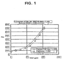
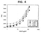
- the procedures for determining coat maximization were the same as described above in the Methods section except that the concentration of either the PAb or the MAb 3.5F8 coat was varied. Specifically, for Fig. 4 the concentration of MAb 3.5F8 was varied from 0.4 to 4 ⁇ g/ml while keeping constant the concentration of polyclonal antibody (at 1 ⁇ g/ml). and for Fig. 5 and Table 5 the concentration of polyclonal antibody was varied from 0.4 to 4 ⁇ g/ml while keeping constant the concentration of MAb 3.5F8 (at 0.4 ⁇ g/ml).
- the concentrations of MAb 3.5F8 and of the PAb at the constant concentration of the other coat antibody were essentially the same in VEGF quantitation for the low and high controt
- the upper limit of coat concentration e.g., 0.4 ⁇ g/ml
- the concentration of 1 ⁇ g/ml of MAb 3.5F8 and PAb increased the amount of VEGF measured in each case, such a concentration also gave higher background.
- the results show that the preferred concentrations for both capture reagents is about 0.4 ⁇ g/ml.
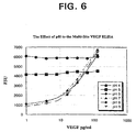
- a pH profile was performed to determine whether changing pH of the assay buffer would increase or decrease recovery of VEGF in normal human EDTA plasma. Changing the pH could dissociate binding proteins or other complexes, if any, which would interfere with the MAb A4.6.1 detection.
- the procedure for examining the pH profile of the assay was the same as described in the Methods section above except that for the sample incubation and biotin incubation, the assay buffer was adjusted using NaOH or HCl, resulting in assay buffers ranging from pH 4 to 9. A standard curve, a low and high matrix, and four normal human EDTA plasma samples were evaluated from dilutions performed using these varying-pH assay buffers.
- results in Fig. 6 and Table 6 show that there was no recovery of VEGF at pH 4 and 5.
- pH 6-9 revealed good VEGF plasma recovery with the assay control within an acceptable range.
- the preferred assay buffer is one with a pH of about 6.35 ⁇ 0.1, which results in maximal VEGF binding and is appropriate for all dilution steps of the assay.
- rhVEGF Approximately 85 pg/ml rhVEGF was spiked into neat human EDTA plasma and serially diluted to 1/10, 1/20, 1/40, and 1/80 and analyzed. The results, in Table 7 and Fig. 7, show that rhVEGF spiked in EDTA plasma showed linear correlation to expected concentration, with a coefficient correlation of 0.996. The percent difference between dilution-corrected concentration values determined for successive serial dilutions did not exceed a mean of 19% ⁇ 7.5, as shown in Table 7.
- Endogenous VEGF levels were measured in freshly-collected plasma from several normal healthy individuals.
- the individual human EDTA plasma samples were spiked with lowest, low, mid, and high concentrations of rhVEGF so as to fall within the assay range of the standard curve. Endogenous VEGF concentrations were determined and subtracted from the measured concentration to obtain comparison to the targeted spike. Results in Table 8 show that mean % recoveries were 99%, 113%, 106%, and 118% for the high, mid, low, and lower spikes, respectively.
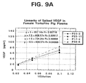
- Rat EDTA plasma (2 male pools, 1 individual) was tested for linearity of dilution. Neat plasma samples were spiked with low (20 pg/ml), mid (44 pg/ml), and high (98 pg/ml) concentrations of rhVEGF and were serially diluted 1/10, 1/20, 1/40, 1/80 in ELISA diluent. Results in Table 10 and Figs. 8A-8C show that normal rat plasma samples dilute linearly following a minimum 1/20 dilution in the assay range of 8-128 pg/ml.
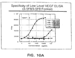
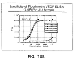
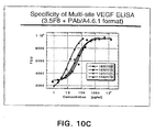
- FIG. 10A, 10B, and 10C show a comparison of the single-site, two-site, and multi-site ELISAs for VEGF, respectively. It can be seen by comparing these graphs that the multi-site assay herein is capable of capturing more VEGF variants.
- the multi-site ELISA herein using PAb and MAb 3.5F8 as coat antibodies was compared to an ELISA using only MAb 3.5F8 or PAb as coat antibody for evaluating the amount of VEGF in normal human samples.
- the results, set forth in Table 12, show that the amount of VEGF detected in pg/ml was much higher for the multi-site assay than for the assay with PAb alone or MAb 3.5F8 alone.
- Plasma and serum samples from normal human donors were analyzed by the two-site ELISA with MAb 3.5F8 as capture antibody and PAb A4.6.1 as detection antibody and by the multi-site assay herein using the PAb and MAbs and procedures noted in the Methods.
- the results, summarized in Figures 11A and 11B for plasma and serum respectively, indicate that the multi-site assay herein detects more VEGF in both types of samples than the two-site assay.
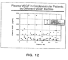
- Figure 12 shows the amounts of plasma VEGF in cardiac patients using all three assays
- Figure 13 and Table 13 summarize the amounts of VEGF in normal and cardiac patients using the two-site and multi-site assays by the mean amount of VEGF, standard deviation, % CV and s.e.m.
- the results indicate that the multi-site assay herein detects more VEGF in both types of samples than the two-site assay.
- TABLE 13 Sensitivity of Two-Site and Multi-Site Assays to VEGF in Normal and Cardiac Patients Normal Donors Cardiac Patients Two-site Multi-site Two-site Multi-site mean pg/ml 32.61 192.40 37.54 279.23 s.d. 13.05 67.66 23.89 156.69 % CV 40.02 35.17 63.65 56.11 s.e.m. (Standard error mean) 1.84 9.56 5.34 35
- Serum samples from non-small cell lung carcinoma patients were analyzed by the two-site ELISA with MAb 3.5F8 as capture antibody and MAb A4.6.1 as detection antibody and by the multi-site assay herein using the PAb and MAbs and procedures noted in the Methods.
- the results, shown in Figure 14, indicate that the multi-site assay herein detects more VEGF in lung cancer samples than the two-site assay.
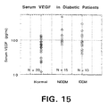
- Serum VEGF levels in normal humans and in patients with NIDDM (Type I diabetes) and IDDM (Type II diabetes) were measured using the two-site ELISA (MAb 3.5F8 as coat and MAb A4.6.1 as detection agent) described above.
- Figure 15 shows that the levels of scrum VEGF in NIDDM and IDDM patients were higher than in normal patients using this assay. Since the multi-site assay detects more VEGF than the two-site assay in other diseased patients, it would be expected that the multi-site assay herein would be suitable fordctccting elevated levels of VEGF in diabetic patients.
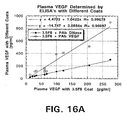
- the two-site and muiti-site ELISAs were carried out as described above for normal human plasma samples.
- a multi-site was carried out using PAb to DNase rather than PAb to VEGF as coat reagent. All were at the 0.4 ⁇ g/ml concentration.
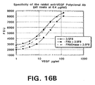
- Figure 16 and Table 14 show that the VEGF detected by multi-site VEGF assay is specific. Results from the ELISA using PAb to DNase plus MAb 3.5F8 show almost identical results as the ELISA using MAb 3.5F8 alone as capture reagent, with a slope of 1.04 (Fig. 16A).
- TYPE OF ASSAY Immunoassay Fluorimetric ELISA: mixture of a murine anti-VEGF monoclonal antibody (MAb 3.5F8) and a rabbit affinity-purified polyclonal antibody for capture and an anti-VEGF monoclonal antibody (MAb A4.6.1) for detection.
- Standard curve will be diluted in ELISA diluent: PBS/0.5%BSA/0.05%Polysorbate20/0.05% PROCLINTM 300/5mM EDTA/0.35M NaCl, pH 6.35 ⁇ 0.1 SPECIES QUALIFIED Human, Rat, England Pig BIOLOGICAL MATRIX Serum, EDTA Plasma.
- ELISA diluent Dilution buffer ASSAY RANGE 1-128 pg/ml in ELISA diluent QUANTITATIVE RANGE IN BIOLOGICAL MATRIX 80 pg/ml to 1280 pg/ml for human serum and EDTA plasma (1/10 minimum dilution) 240 pg/ml to 850 pg/ml for England Pig EDTA Plasma (1/20 minimum dilution) Minimum Quantifiable Concentration Endogenous values as low as 120 pg/ml will be reported for information only with a %CV of approximately 60% in this range.
- Mean % recoveries in the quantitative range were: Low Mid High 82% 91% 99% 4 female and 4 male Yorkshire pig EDTA Plasmas were spiked with high, mid and low concentrations of rhVEGF. rhVEGF quantitation below 12 pg/ml (endogenous VEGF measured) will be reported for information only (mean % recovery below 12 pg/ml is approximately 60%). Mean % recoveries in the quantitative range were: Low Mid High n/a 86% 107% SPECIFICITY rhVEGF. IGF, TNF NGF, hGH, IFN, IIbIIIa, rhuMAb VEGF, anti-VEGF MAb 3.5F8 were spiked into human EDTA plasma.
- rhVEGF LINEARITY AND INTERFERENCE rhVEGF was spiked into 6 different normal human EDTA plasma samples. Samples were serially diluted 1 ⁇ 2 to cover the range of the standard curve. 4 dilutions were made from each sample, rhVEGF values obtained were plotted against I/dilution and the correlation coefficient (R 2 ) of a linear regression analysis was calculated. Samples were linear across the range of the standard curve. n Mean R 2 SD 6 0.996 0.005 rhVEGF was spiked in 3 rat EDTA plasma samples at high, mid and low concentration. Samples were linear across the high and mid quantitative range.
- n Mean R 2 SD Spike 3 0.998 0.002 High 3 0.998 0.003 Mid 3 0.967 0.034 Low rhVEGF was spiked into 8 EDTA pig plasma samples and serially diluted 1 ⁇ 2 to cover the range of the standard curve. 4 dilutions were made from each sample. Samples were linear across the range of the standard curve.
- the fluorometric substrate strep- ⁇ -gal/MUG
- strep- ⁇ -gal/MUG is preferred for use in the detection system so that the ELISA can detect endogenous VEGF levels in normal individuals.
- the use of this substrate and the determination of the best ELISA diluent resulted in much lower background absorbance, which was preferred to achieve the increase in the assay sensitivity.
- the multi-site ELISA described herein is highly specific due to the choice of antibodies used for capture and detection.
- One of the coat antibodies, MAb 3.5F8. binds near the heparin binding region of VEGF (residues 111-165) and the other coat antibody, the rabbit polyclonal antibody binds VEGF.
- the detection antibody, MAb A4.6.1 binds in the KDR receptor binding region (residues 1-110) of the molecule, yielding a specific ELISA for VEGF.
- antibodies are raised in animals against human VEGF, with the C-terminal specific antibody being a monoclonal antibody and the whole-VEGF-specific antibody being a polyclonal antibody, preferably affinity purified. These two antibodies are used as coat antibodies (immobilized capture reagents) on a solid support such as microtiter plates.
- the antibody used for detection can be either polyclonal antibodies or monoclonal antibodies provided they are specific for the KDR and FLT1 binding domain regions of human VEGF.
Landscapes
- Health & Medical Sciences (AREA)
- Life Sciences & Earth Sciences (AREA)
- Engineering & Computer Science (AREA)
- Immunology (AREA)
- Molecular Biology (AREA)
- Chemical & Material Sciences (AREA)
- Urology & Nephrology (AREA)
- Biomedical Technology (AREA)
- Hematology (AREA)
- Cell Biology (AREA)
- Analytical Chemistry (AREA)
- General Physics & Mathematics (AREA)
- Pathology (AREA)
- Food Science & Technology (AREA)
- Medicinal Chemistry (AREA)
- Physics & Mathematics (AREA)
- Biotechnology (AREA)
- Biochemistry (AREA)
- General Health & Medical Sciences (AREA)
- Microbiology (AREA)
- Proteomics, Peptides & Aminoacids (AREA)
- Peptides Or Proteins (AREA)
- Preparation Of Compounds By Using Micro-Organisms (AREA)
- Medicines That Contain Protein Lipid Enzymes And Other Medicines (AREA)
- Medicines Containing Material From Animals Or Micro-Organisms (AREA)
- Investigating Or Analysing Biological Materials (AREA)
- Investigating Or Analysing Materials By The Use Of Chemical Reactions (AREA)
Claims (26)
- Verfahren zur Detektion von Gefäßendothelwachstumsfaktor (VEGF) in einer biologischen Probe, die folgenden Schritte umfassend:(a) das Kontaktieren und Inkubieren der biologischen Probe mit vorgemischten Einfangreagenzien, die an einem festen Träger immobilisiert sind, worin die Einfangreagenzien polyklonale und monoklonale Antikörper gegen menschlichen VEGF sind, wobei sich der monoklonale Antikörper spezifisch an den C-Terminus (Reste 111-165) von menschlichem VEGF bindet;(b) das Trennen der biologischen Probe von den immobilisierten Einfangreagenzien;(c) das Kontaktieren der immobilisierten Einfangreagenzien mit einem nachweisbaren Antikörper, der sich an die KDR- und FLT1-Rezeptor-Bindungsdomänen von VEGF bindet; und(d) das Messen der Konzentration an VEGF, der an die Einfangreagenzien gebunden ist, unter Verwendung eines Detektionsmittels für den nachweisbaren Antikörper.
- Verfahren nach Anspruch 1, worin die biologische Probe von einem menschlichen Probanden stammt.
- Verfahren nach Anspruch 2, worin der menschliche Proband ein Patient mit Gefäßerkrankungen, Diabetes oder Krebs ist und der Messschritt (d) weiters einen Vergleich mit einer Standardkurve umfasst, um die Konzentration an VEGF im Vergleich zu einer normalen Person zu bestimmen.
- Verfahren nach Anspruch 1, worin die biologische Probe Plasma, Serum oder Urin ist.
- Verfahren nach Anspruch 1, worin die Einfangreagenzien in einem Gewichtsverhältnis von etwa 0,8:1 bis 1,2:1 von monoklonalem Antikörper zu polyklonalem Antikörper immobilisiert werden.
- Verfahren nach Anspruch 5, worin das Gewichtsverhältnis von monoklonalem Antikörper zu polyklonalem Antikörper etwa 1:1 1 beträgt.
- Verfahren nach Anspruch 6, worin die Menge an immobilisierten monoklonalen Antikörpern etwa 0,4 µg/ml und die Menge an immobilisierten polyklonalen Antikörpern etwa 0,4 µg/ml beträgt.
- Verfahren nach Anspruch 1, worin die immobilisierten Einfangreagenzien auf eine Mikrotiterplatte beschichtet werden.
- Verfahren nach Anspruch 1, worin der nachweisbare Antikörper direkt nachweisbar ist.
- Verfahren nach Anspruch 9, worin der nachweisbare Antikörper durch ein fluorimetrisches Reagens amplifiziert wird.
- Verfahren nach Anspruch 10, worin der nachweisbare Antikörper biotinyliert ist und das Detektionsmittel Avidin- oder Streptavidin-β-Galactosidase und 4-Methylumbelliferyl-β-Galactosid ist.
- Verfahren nach Anspruch 1, worin der immobilisierte monoklonale Antikörper von Maus und der immobilisierte polyklonale Antikörper von Kaninchen oder Ziege abstammt.
- Verfahren nach Anspruch 1, worin der immobilisierte polyklonale Antikörper affinitätsgereinigt ist.
- Verfahren nach Anspruch 1, worin der nachweisbare Antikörper ein monoklonaler Antikörper ist.
- Verfahren nach Anspruch 14, worin der nachweisbare Antikörper ein monoklonaler Maus-Antikörper ist.
- Verfahren nach Anspruch 15, worin der immobilisierte monoklonale Antikörper MAb 3.5F8 und der nachweisbare Antikörper MAb A4.6.1 ist.
- Immuntestset zur Detektion von Gefäßendothelwachstumsfaktor (VEGF) in einer biologischen Probe, umfassend:(a) als Einfangreagenzien polyklonale und monoklonale Antikörper gegen menschlichen VEGF, vorgemischt in einem Gewichtsverhältnis von etwa 0,8:1 bis 1,2:1 von monoklonalem zu polyklonalem Antikörper, worin sich der monoklonale Antikörper spezifisch an den C-Terminus (Reste 111-165) von menschlichem VEGF bindet; und(b) als Detektionsreagens einen nachweisbaren Antikörper, der sich an die KDR- und FLT1-Rezeptor-Bindungsdomänen von VEGF bindet.
- Set nach Anspruch 17, weiters umfassend einen festen Träger für die Einfangreagenzien.
- Set nach Anspruch 18, worin die Einfangreagenzien an dem festen Träger immobilisiert sind.
- Set nach Anspruch 19, worin die Einfangreagenzien auf eine Mikrotiterplatte beschichtet sind.
- Set nach Anspruch 17, weiters umfassend ein Detektionsmittel für die nachweisbaren Antikörper.
- Set nach Anspruch 21, worin das Detektionsmittel fluorimetrisch ist.
- Set nach Anspruch 17, weiters umfassend gereinigten VEGF als einen Antigen-Standard.
- Set nach Anspruch 17, worin das Gewichtsverhältnis von monoklonalem Antikörper zu polyklonalem Antikörper etwa 1:1 beträgt, der monoklonale Antikörper von Maus abstammt, der polyklonale Antikörper affinitätsgereinigt ist und die Menge an monoklonalen Antikörpern 0,4 µg/ml und die Menge an polyklonalen Antikörpern 0,4 µg/ml beträgt.
- Set nach Anspruch 24, worin die Einfangreagenzien immobilisiert sind und der polyklonale Antikörper ein Kaninchen- oder Ziegen-Antikörper ist.
- Set nach Anspruch 25, worin der nachweisbare Antikörper monoklonaler Maus-Antikörper MAb A4.6.1 und der monoklonale Einfangreagens-Antikörper MAb 3.5F8 ist.
Applications Claiming Priority (3)
| Application Number | Priority Date | Filing Date | Title |
|---|---|---|---|
| US16573699P | 1999-11-16 | 1999-11-16 | |
| US165736P | 1999-11-16 | ||
| PCT/US2000/031427 WO2001036972A2 (en) | 1999-11-16 | 2000-11-15 | Elisa for vegf |
Publications (2)
| Publication Number | Publication Date |
|---|---|
| EP1230552A2 EP1230552A2 (de) | 2002-08-14 |
| EP1230552B1 true EP1230552B1 (de) | 2006-01-11 |
Family
ID=22600234
Family Applications (1)
| Application Number | Title | Priority Date | Filing Date |
|---|---|---|---|
| EP00978702A Expired - Lifetime EP1230552B1 (de) | 1999-11-16 | 2000-11-15 | Elisa für vegf |
Country Status (14)
| Country | Link |
|---|---|
| US (2) | US6855508B2 (de) |
| EP (1) | EP1230552B1 (de) |
| JP (1) | JP4620312B2 (de) |
| KR (1) | KR100749982B1 (de) |
| CN (1) | CN100399030C (de) |
| AT (1) | ATE315789T1 (de) |
| AU (1) | AU774575C (de) |
| CA (1) | CA2387390C (de) |
| DE (1) | DE60025526T2 (de) |
| ES (1) | ES2256066T3 (de) |
| IL (2) | IL149171A0 (de) |
| NZ (1) | NZ518363A (de) |
| WO (1) | WO2001036972A2 (de) |
| ZA (1) | ZA200202942B (de) |
Families Citing this family (50)
| Publication number | Priority date | Publication date | Assignee | Title |
|---|---|---|---|---|
| DE60025526T2 (de) * | 1999-11-16 | 2006-08-24 | Genentech, Inc., South San Francisco | Elisa für vegf |
| WO2002033422A2 (en) * | 2000-10-20 | 2002-04-25 | Board Of Regents, The University Of Texas System | Methods for detecting retinopathy of prematurity |
| US20050009110A1 (en) * | 2003-07-08 | 2005-01-13 | Xiao-Jia Chang | Methods of producing antibodies for diagnostics and therapeutics |
| US7758859B2 (en) | 2003-08-01 | 2010-07-20 | Genentech, Inc. | Anti-VEGF antibodies |
| US20050106667A1 (en) | 2003-08-01 | 2005-05-19 | Genentech, Inc | Binding polypeptides with restricted diversity sequences |
| AU2004261980A1 (en) * | 2003-08-01 | 2005-02-10 | Genentech, Inc. | Antibody CDR polypeptide sequences with restricted diversity |
| US7514223B2 (en) | 2004-05-15 | 2009-04-07 | Genentech, Inc. | Cross-screening system and methods for detecting a molecule having binding affinity for a target molecule |
| CA2511269A1 (en) * | 2004-07-07 | 2006-01-07 | F. Hoffmann-La Roche Ag | Multimarker panel based on p1gf for diabetes type 1 and 2 |
| CN1316249C (zh) * | 2004-08-25 | 2007-05-16 | 北京健平九星生物医药科技有限公司 | 一种酶联检测试剂盒及制备方法 |
| US7595149B1 (en) | 2004-11-08 | 2009-09-29 | University Of Kentucky Research Foundation | Methods for cancer detection |
| DK2457929T3 (en) * | 2006-10-04 | 2016-09-05 | Genentech Inc | Elisa vegf |
| AU2013221919B2 (en) * | 2006-10-04 | 2016-04-14 | Genentech, Inc. | ELISA for VEGF |
| WO2008079322A1 (en) * | 2006-12-22 | 2008-07-03 | Beckman Coulter, Inc. | Methods, kits and materials for diagnosing disease states by measuring isoforms or proforms of myeloperoxidase |
| US20080160638A1 (en) * | 2006-12-27 | 2008-07-03 | David Lederman | Functionalized Microcantilever Sensor and Associated Method For Detection of Targeted Analytes |
| DE102007038730A1 (de) * | 2007-08-16 | 2009-02-19 | Carl Zeiss Meditec Ag | Nachweis des menschlichen Vascular Endothelial Growth Factor |
| TWI468417B (zh) | 2007-11-30 | 2015-01-11 | Genentech Inc | 抗-vegf抗體 |
| CA2716522A1 (en) * | 2008-03-05 | 2009-10-15 | Singulex, Inc. | Methods and compositions for highly sensitive detection of molecules |
| US7772006B2 (en) | 2008-08-21 | 2010-08-10 | Paul Tornambe | Agent detection and/or quantitation in a biological fluid |
| CN102144161B (zh) | 2008-09-04 | 2016-02-17 | 贝克曼考尔特公司 | 泛激酶活化与信号通路的评估 |
| US20110294683A1 (en) * | 2008-10-30 | 2011-12-01 | Centre de Recherche Public de la Sant'e | Biomarkers |
| US8349325B2 (en) * | 2008-12-23 | 2013-01-08 | Abbott Laboratories | Soluble FMS-like tyrosine kinase-1 (sFLT-1) antibody and related composition, kit, methods of using, and materials and method for making |
| EP2488661B1 (de) | 2009-10-14 | 2014-12-24 | Oberbauer, Rainer | Prüfung auf Risiko einer akuten Nierenverletzung |
| WO2011057347A1 (en) | 2009-11-12 | 2011-05-19 | Tgr Biosciences Pty Ltd | Analyte detection |
| US20120004132A1 (en) * | 2010-07-02 | 2012-01-05 | Affymetrix, Inc. | Detection of Nucleic Acids and Proteins |
| CA2805618A1 (en) | 2010-07-19 | 2012-01-26 | F. Hoffmann-La Roche Ag | Method to identify a patient with an increased likelihood of responding to an anti-cancer therapy |
| KR20130126576A (ko) | 2010-07-19 | 2013-11-20 | 에프. 호프만-라 로슈 아게 | 항암요법에 반응할 가능성이 증가된 환자를 확인하는 방법 |
| JP5805190B2 (ja) | 2010-08-19 | 2015-11-04 | エフ.ホフマン−ラ ロシュ アーゲーF. Hoffmann−La Roche Aktiengesellschaft | 療法用モノクローナル抗体に結合する抗体の測定のためのアッセイ |
| WO2012030738A2 (en) | 2010-08-30 | 2012-03-08 | Beckman Coulter, Inc. | Complex phosphoprotein activation profiles |
| CA2822530C (en) * | 2011-01-21 | 2023-02-21 | Basilea Pharmaceutica Ag | Glu-tubulin as a biomarker for furazanobenzimidazoles |
| WO2012113718A1 (en) | 2011-02-21 | 2012-08-30 | Roche Diagnostics Gmbh | A diagnostic method for type ii diabetes |
| EP2786148A4 (de) * | 2011-03-31 | 2015-12-16 | Tgr Biosciences Pty Ltd | Nachweis von analyten mit einem einschritt-immuntest |
| CN102323421A (zh) * | 2011-05-24 | 2012-01-18 | 北京健平九星生物医药科技有限公司 | 一种酶联检测试剂盒及其制备方法 |
| FR2976076B1 (fr) * | 2011-05-31 | 2015-02-27 | Imabiotech | Procede de detection et de quantification d'une molecule cible dans un tissu |
| CN102435743B (zh) * | 2011-09-15 | 2014-10-15 | 北京健平九星生物医药科技有限公司 | 一种酶联检测试剂盒及其制备方法 |
| US20130149386A1 (en) * | 2011-12-07 | 2013-06-13 | Region Nordjylland | Cd36 as biomarker for steatosis |
| CA2853917A1 (en) * | 2011-12-19 | 2013-06-27 | F. Hoffmann-La Roche Ag | Method for the detection of free binding partner of a multispecific binder |
| WO2013113663A1 (en) | 2012-02-01 | 2013-08-08 | F. Hoffmann-La Roche Ag | Method for the detection of a binding partner of a multispecific binder |
| ES2627482T3 (es) | 2012-07-13 | 2017-07-28 | F. Hoffmann-La Roche Ag | Procedimiento para la detección de una proteína de unión multiespecífica |
| EP2954332A1 (de) | 2013-02-06 | 2015-12-16 | Pieris AG | Neuartige lipocalinmuteintests zur messung der hepcidinkonzentration |
| CN106153935B (zh) * | 2015-03-26 | 2018-05-08 | 广州瑞博奥生物科技有限公司 | 一种定量检测CD79α的酶联免疫试剂盒 |
| CN106153934B (zh) * | 2015-03-26 | 2018-06-19 | 广州瑞博奥生物科技有限公司 | 一种高效定量检测高尔基体73的试剂盒 |
| CN106153933B (zh) * | 2015-03-26 | 2018-09-04 | 广州瑞博奥生物科技有限公司 | 一种高效定量检测c反应蛋白的免疫学试剂盒 |
| PE20181363A1 (es) | 2015-09-23 | 2018-08-27 | Genentech Inc | Variantes optimizadas de anticuerpos anti-vegf |
| CN105353136A (zh) * | 2015-12-01 | 2016-02-24 | 北京健平九星生物医药科技有限公司 | 基于量子点CdSe检测人血清中VEGF浓度的试剂盒及其使用方法 |
| EP3485281A1 (de) | 2016-07-15 | 2019-05-22 | H. Hoffnabb-La Roche Ag | Verfahren und mittel zur erkennung der stufe des gesamten vegf-a |
| WO2018093856A1 (en) * | 2016-11-16 | 2018-05-24 | Gupte Sachin A | Inhibitors of glucose-6-phosphate dehydrogenase for treating cardiovascular and pulmonary conditions |
| CN107677808A (zh) * | 2017-08-04 | 2018-02-09 | 苏州浩欧博生物医药有限公司 | 一种甲状腺球蛋白的定量检测试剂盒及其制备方法和检测方法 |
| CN111808191A (zh) * | 2020-05-11 | 2020-10-23 | 廊坊天光生物技术有限公司 | 一种用于检测血清中vegf含量的抗体对及其用途 |
| JP7485273B2 (ja) * | 2020-06-25 | 2024-05-16 | 国立大学法人 鹿児島大学 | 抗vegf-a抗体による癌に対する治療応答性を予測又は決定するための方法 |
| CN119846234B (zh) * | 2025-01-23 | 2025-10-10 | 上海触研医学技术有限公司 | 检测生物样品中人vegf水平的试剂盒 |
Family Cites Families (18)
| Publication number | Priority date | Publication date | Assignee | Title |
|---|---|---|---|---|
| SE337223B (de) | 1967-05-23 | 1971-08-02 | Pharmacia Ab | |
| US3720760A (en) | 1968-09-06 | 1973-03-13 | Pharmacia Ab | Method for determining the presence of reagin-immunoglobulins(reagin-ig)directed against certain allergens,in aqueous samples |
| US3691016A (en) | 1970-04-17 | 1972-09-12 | Monsanto Co | Process for the preparation of insoluble enzymes |
| CA1023287A (en) | 1972-12-08 | 1977-12-27 | Boehringer Mannheim G.M.B.H. | Process for the preparation of carrier-bound proteins |
| US4195128A (en) | 1976-05-03 | 1980-03-25 | Bayer Aktiengesellschaft | Polymeric carrier bound ligands |
| US4330440A (en) | 1977-02-08 | 1982-05-18 | Development Finance Corporation Of New Zealand | Activated matrix and method of activation |
| CA1093991A (en) | 1977-02-17 | 1981-01-20 | Hideo Hirohara | Enzyme immobilization with pullulan gel |
| US4229537A (en) | 1978-02-09 | 1980-10-21 | New York University | Preparation of trichloro-s-triazine activated supports for coupling ligands |
| US4376110A (en) | 1980-08-04 | 1983-03-08 | Hybritech, Incorporated | Immunometric assays using monoclonal antibodies |
| IT8423926U1 (it) * | 1984-11-29 | 1986-05-29 | Ire Ind Riunite Eurodomestici S P A | Disposizione per rimuovere detersivi e prodotti di lavaggio in genere dagli scomparti dei cassetti di macchine lavabiancheria domestiche |
| US5620845A (en) | 1988-06-06 | 1997-04-15 | Ampcor, Inc. | Immunoassay diagnostic kit |
| US5219739A (en) | 1989-07-27 | 1993-06-15 | Scios Nova Inc. | DNA sequences encoding bVEGF120 and hVEGF121 and methods for the production of bovine and human vascular endothelial cell growth factors, bVEGF120 and hVEGF121 |
| CA2072758A1 (en) | 1990-09-14 | 1992-03-15 | Kenneth Francis Buechler | Antibodies to complexes of ligand receptors and ligands and their utility in ligand-receptor assays |
| US5434087A (en) | 1993-02-24 | 1995-07-18 | Abbott Laboratories | Folate immunoassay utilizing folate binding protein in a multiclonal antibody format |
| EP0751992B1 (de) * | 1994-03-08 | 2005-11-09 | Human Genome Sciences, Inc. | Vaskularer endothelialer wachstumsfaktor 2 |
| JPH08245424A (ja) * | 1995-03-06 | 1996-09-24 | Toagosei Co Ltd | 糖尿病性腎症診断剤 |
| JP3591264B2 (ja) * | 1997-12-24 | 2004-11-17 | 富士レビオ株式会社 | Vegf121特異的モノクローナル抗体及び測定方法 |
| DE60025526T2 (de) * | 1999-11-16 | 2006-08-24 | Genentech, Inc., South San Francisco | Elisa für vegf |
-
2000
- 2000-11-15 DE DE60025526T patent/DE60025526T2/de not_active Expired - Lifetime
- 2000-11-15 CN CNB008157553A patent/CN100399030C/zh not_active Expired - Lifetime
- 2000-11-15 AU AU16134/01A patent/AU774575C/en not_active Expired
- 2000-11-15 JP JP2001538809A patent/JP4620312B2/ja not_active Expired - Lifetime
- 2000-11-15 WO PCT/US2000/031427 patent/WO2001036972A2/en not_active Ceased
- 2000-11-15 AT AT00978702T patent/ATE315789T1/de active
- 2000-11-15 IL IL14917100A patent/IL149171A0/xx active IP Right Grant
- 2000-11-15 NZ NZ518363A patent/NZ518363A/xx not_active IP Right Cessation
- 2000-11-15 ES ES00978702T patent/ES2256066T3/es not_active Expired - Lifetime
- 2000-11-15 KR KR1020027006227A patent/KR100749982B1/ko not_active Expired - Lifetime
- 2000-11-15 EP EP00978702A patent/EP1230552B1/de not_active Expired - Lifetime
- 2000-11-15 CA CA002387390A patent/CA2387390C/en not_active Expired - Lifetime
-
2002
- 2002-04-15 ZA ZA200202942A patent/ZA200202942B/xx unknown
- 2002-04-16 IL IL149171A patent/IL149171A/en unknown
- 2002-08-05 US US10/211,282 patent/US6855508B2/en not_active Expired - Lifetime
-
2004
- 2004-11-01 US US10/978,701 patent/US7541160B2/en not_active Expired - Fee Related
Also Published As
| Publication number | Publication date |
|---|---|
| ZA200202942B (en) | 2003-06-25 |
| CA2387390C (en) | 2009-01-06 |
| CA2387390A1 (en) | 2001-05-25 |
| US7541160B2 (en) | 2009-06-02 |
| IL149171A0 (en) | 2002-11-10 |
| DE60025526T2 (de) | 2006-08-24 |
| AU774575B2 (en) | 2004-07-01 |
| EP1230552A2 (de) | 2002-08-14 |
| AU1613401A (en) | 2001-05-30 |
| ATE315789T1 (de) | 2006-02-15 |
| KR20020070434A (ko) | 2002-09-09 |
| US20070202558A1 (en) | 2007-08-30 |
| AU774575C (en) | 2005-09-08 |
| CN100399030C (zh) | 2008-07-02 |
| US20030044865A1 (en) | 2003-03-06 |
| WO2001036972A2 (en) | 2001-05-25 |
| CN1420987A (zh) | 2003-05-28 |
| ES2256066T3 (es) | 2006-07-16 |
| KR100749982B1 (ko) | 2007-08-16 |
| JP4620312B2 (ja) | 2011-01-26 |
| US6855508B2 (en) | 2005-02-15 |
| IL149171A (en) | 2008-06-05 |
| WO2001036972A3 (en) | 2002-01-03 |
| JP2003515136A (ja) | 2003-04-22 |
| DE60025526D1 (de) | 2006-04-06 |
| NZ518363A (en) | 2003-01-31 |
Similar Documents
| Publication | Publication Date | Title |
|---|---|---|
| EP1230552B1 (de) | Elisa für vegf | |
| US9625462B2 (en) | Elisa for VEGF | |
| HK1232236B (en) | Elisa for vegf | |
| HK1130090B (en) | Elisa for vegf | |
| HK1168361B (en) | Elisa for vegf | |
| AU2013221919A1 (en) | ELISA for VEGF |
Legal Events
| Date | Code | Title | Description |
|---|---|---|---|
| PUAI | Public reference made under article 153(3) epc to a published international application that has entered the european phase |
Free format text: ORIGINAL CODE: 0009012 |
|
| 17P | Request for examination filed |
Effective date: 20020506 |
|
| AK | Designated contracting states |
Kind code of ref document: A2 Designated state(s): AT BE CH CY DE DK ES FI FR GB GR IE IT LI LU MC NL PT SE TR |
|
| AX | Request for extension of the european patent |
Free format text: AL;LT;LV;MK;RO;SI |
|
| GRAP | Despatch of communication of intention to grant a patent |
Free format text: ORIGINAL CODE: EPIDOSNIGR1 |
|
| GRAS | Grant fee paid |
Free format text: ORIGINAL CODE: EPIDOSNIGR3 |
|
| GRAA | (expected) grant |
Free format text: ORIGINAL CODE: 0009210 |
|
| AK | Designated contracting states |
Kind code of ref document: B1 Designated state(s): AT BE CH CY DE DK ES FI FR GB GR IE IT LI LU MC NL PT SE TR |
|
| PG25 | Lapsed in a contracting state [announced via postgrant information from national office to epo] |
Ref country code: IT Free format text: LAPSE BECAUSE OF FAILURE TO SUBMIT A TRANSLATION OF THE DESCRIPTION OR TO PAY THE FEE WITHIN THE PRESCRIBED TIME-LIMIT;WARNING: LAPSES OF ITALIAN PATENTS WITH EFFECTIVE DATE BEFORE 2007 MAY HAVE OCCURRED AT ANY TIME BEFORE 2007. THE CORRECT EFFECTIVE DATE MAY BE DIFFERENT FROM THE ONE RECORDED. Effective date: 20060111 Ref country code: FI Free format text: LAPSE BECAUSE OF FAILURE TO SUBMIT A TRANSLATION OF THE DESCRIPTION OR TO PAY THE FEE WITHIN THE PRESCRIBED TIME-LIMIT Effective date: 20060111 |
|
| REG | Reference to a national code |
Ref country code: CH Ref legal event code: EP |
|
| REG | Reference to a national code |
Ref country code: IE Ref legal event code: FG4D |
|
| REF | Corresponds to: |
Ref document number: 60025526 Country of ref document: DE Date of ref document: 20060406 Kind code of ref document: P |
|
| PG25 | Lapsed in a contracting state [announced via postgrant information from national office to epo] |
Ref country code: SE Free format text: LAPSE BECAUSE OF FAILURE TO SUBMIT A TRANSLATION OF THE DESCRIPTION OR TO PAY THE FEE WITHIN THE PRESCRIBED TIME-LIMIT Effective date: 20060411 Ref country code: DK Free format text: LAPSE BECAUSE OF FAILURE TO SUBMIT A TRANSLATION OF THE DESCRIPTION OR TO PAY THE FEE WITHIN THE PRESCRIBED TIME-LIMIT Effective date: 20060411 |
|
| PG25 | Lapsed in a contracting state [announced via postgrant information from national office to epo] |
Ref country code: PT Free format text: LAPSE BECAUSE OF FAILURE TO SUBMIT A TRANSLATION OF THE DESCRIPTION OR TO PAY THE FEE WITHIN THE PRESCRIBED TIME-LIMIT Effective date: 20060612 |
|
| REG | Reference to a national code |
Ref country code: ES Ref legal event code: FG2A Ref document number: 2256066 Country of ref document: ES Kind code of ref document: T3 |
|
| ET | Fr: translation filed | ||
| PLBE | No opposition filed within time limit |
Free format text: ORIGINAL CODE: 0009261 |
|
| STAA | Information on the status of an ep patent application or granted ep patent |
Free format text: STATUS: NO OPPOSITION FILED WITHIN TIME LIMIT |
|
| 26N | No opposition filed |
Effective date: 20061012 |
|
| PG25 | Lapsed in a contracting state [announced via postgrant information from national office to epo] |
Ref country code: GR Free format text: LAPSE BECAUSE OF FAILURE TO SUBMIT A TRANSLATION OF THE DESCRIPTION OR TO PAY THE FEE WITHIN THE PRESCRIBED TIME-LIMIT Effective date: 20060412 |
|
| PG25 | Lapsed in a contracting state [announced via postgrant information from national office to epo] |
Ref country code: TR Free format text: LAPSE BECAUSE OF FAILURE TO SUBMIT A TRANSLATION OF THE DESCRIPTION OR TO PAY THE FEE WITHIN THE PRESCRIBED TIME-LIMIT Effective date: 20060111 |
|
| PG25 | Lapsed in a contracting state [announced via postgrant information from national office to epo] |
Ref country code: CY Free format text: LAPSE BECAUSE OF FAILURE TO SUBMIT A TRANSLATION OF THE DESCRIPTION OR TO PAY THE FEE WITHIN THE PRESCRIBED TIME-LIMIT Effective date: 20060111 |
|
| REG | Reference to a national code |
Ref country code: FR Ref legal event code: PLFP Year of fee payment: 16 |
|
| REG | Reference to a national code |
Ref country code: FR Ref legal event code: PLFP Year of fee payment: 17 |
|
| REG | Reference to a national code |
Ref country code: FR Ref legal event code: PLFP Year of fee payment: 18 |
|
| REG | Reference to a national code |
Ref country code: FR Ref legal event code: PLFP Year of fee payment: 19 |
|
| PGFP | Annual fee paid to national office [announced via postgrant information from national office to epo] |
Ref country code: LU Payment date: 20190918 Year of fee payment: 20 |
|
| PGFP | Annual fee paid to national office [announced via postgrant information from national office to epo] |
Ref country code: IE Payment date: 20191029 Year of fee payment: 20 Ref country code: DE Payment date: 20191017 Year of fee payment: 20 Ref country code: NL Payment date: 20191030 Year of fee payment: 20 Ref country code: MC Payment date: 20191029 Year of fee payment: 20 |
|
| PGFP | Annual fee paid to national office [announced via postgrant information from national office to epo] |
Ref country code: ES Payment date: 20191202 Year of fee payment: 20 Ref country code: IT Payment date: 20191113 Year of fee payment: 20 Ref country code: BE Payment date: 20191021 Year of fee payment: 20 Ref country code: FR Payment date: 20191029 Year of fee payment: 20 |
|
| PGFP | Annual fee paid to national office [announced via postgrant information from national office to epo] |
Ref country code: AT Payment date: 20191028 Year of fee payment: 20 Ref country code: CH Payment date: 20191028 Year of fee payment: 20 |
|
| PGFP | Annual fee paid to national office [announced via postgrant information from national office to epo] |
Ref country code: GB Payment date: 20191029 Year of fee payment: 20 |
|
| REG | Reference to a national code |
Ref country code: DE Ref legal event code: R071 Ref document number: 60025526 Country of ref document: DE |
|
| REG | Reference to a national code |
Ref country code: NL Ref legal event code: MK Effective date: 20201114 |
|
| REG | Reference to a national code |
Ref country code: CH Ref legal event code: PL |
|
| REG | Reference to a national code |
Ref country code: GB Ref legal event code: PE20 Expiry date: 20201114 |
|
| REG | Reference to a national code |
Ref country code: IE Ref legal event code: MK9A |
|
| REG | Reference to a national code |
Ref country code: AT Ref legal event code: MK07 Ref document number: 315789 Country of ref document: AT Kind code of ref document: T Effective date: 20201115 Ref country code: BE Ref legal event code: MK Effective date: 20201115 |
|
| PG25 | Lapsed in a contracting state [announced via postgrant information from national office to epo] |
Ref country code: IE Free format text: LAPSE BECAUSE OF EXPIRATION OF PROTECTION Effective date: 20201115 Ref country code: GB Free format text: LAPSE BECAUSE OF EXPIRATION OF PROTECTION Effective date: 20201114 |
|
| REG | Reference to a national code |
Ref country code: ES Ref legal event code: FD2A Effective date: 20211229 |
|
| PG25 | Lapsed in a contracting state [announced via postgrant information from national office to epo] |
Ref country code: ES Free format text: LAPSE BECAUSE OF EXPIRATION OF PROTECTION Effective date: 20201116 |