EP1227355A2 - Microscope pour observation à grand angle, notamment de chirurgie ophthalmologique - Google Patents
Microscope pour observation à grand angle, notamment de chirurgie ophthalmologique Download PDFInfo
- Publication number
- EP1227355A2 EP1227355A2 EP01121232A EP01121232A EP1227355A2 EP 1227355 A2 EP1227355 A2 EP 1227355A2 EP 01121232 A EP01121232 A EP 01121232A EP 01121232 A EP01121232 A EP 01121232A EP 1227355 A2 EP1227355 A2 EP 1227355A2
- Authority
- EP
- European Patent Office
- Prior art keywords
- microscope
- microscope according
- beam path
- optics
- eye
- Prior art date
- Legal status (The legal status is an assumption and is not a legal conclusion. Google has not performed a legal analysis and makes no representation as to the accuracy of the status listed.)
- Granted
Links
Images


Classifications
-
- A—HUMAN NECESSITIES
- A61—MEDICAL OR VETERINARY SCIENCE; HYGIENE
- A61B—DIAGNOSIS; SURGERY; IDENTIFICATION
- A61B3/00—Apparatus for testing the eyes; Instruments for examining the eyes
- A61B3/10—Objective types, i.e. instruments for examining the eyes independent of the patients' perceptions or reactions
- A61B3/13—Ophthalmic microscopes
-
- G—PHYSICS
- G02—OPTICS
- G02B—OPTICAL ELEMENTS, SYSTEMS OR APPARATUS
- G02B17/00—Systems with reflecting surfaces, with or without refracting elements
- G02B17/02—Catoptric systems, e.g. image erecting and reversing system
- G02B17/04—Catoptric systems, e.g. image erecting and reversing system using prisms only
-
- G—PHYSICS
- G02—OPTICS
- G02B—OPTICAL ELEMENTS, SYSTEMS OR APPARATUS
- G02B21/00—Microscopes
- G02B21/0004—Microscopes specially adapted for specific applications
- G02B21/0012—Surgical microscopes
-
- A—HUMAN NECESSITIES
- A61—MEDICAL OR VETERINARY SCIENCE; HYGIENE
- A61B—DIAGNOSIS; SURGERY; IDENTIFICATION
- A61B3/00—Apparatus for testing the eyes; Instruments for examining the eyes
- A61B3/10—Objective types, i.e. instruments for examining the eyes independent of the patients' perceptions or reactions
- A61B3/12—Objective types, i.e. instruments for examining the eyes independent of the patients' perceptions or reactions for looking at the eye fundus, e.g. ophthalmoscopes
- A61B3/125—Objective types, i.e. instruments for examining the eyes independent of the patients' perceptions or reactions for looking at the eye fundus, e.g. ophthalmoscopes with contact lenses
Definitions
- the invention relates to a microscope for wide-angle viewing of an eye with one between the lens and the eye to be treated, an inverted image design optics to observe the Fundus, especially for eye surgery, and with an in the beam path of the microscope, preferably insertable or retractable device for image reversal and erection.
- the invention has therefore set itself the task of a microscope at the outset specified type so that its height also then does not need to be significantly increased if it is optional with a Device for image reversal and erection can be operated.
- the object is achieved in that the device for Image reversal and erection from a low building reflective system, in particular a prism system that carried by a holder attached to the microscope and so in the Beam path of the microscope between the lens and the one to be treated Eye can be inserted or swiveled in that the prism system immediately in front of the lens, at a distance from the eye.
- the arrangement according to the invention makes the between the lens and to take advantage of the space available to be treated, so that overall the height of the microscope is retained even when in the eyepiece a correct and upright image is generated.
- the Optics for observing the fundus can be attached to the holder attached to the prism system as well as directly on the eye be designed to be attachable. This way, immediately after Swinging in or pushing the device in the right direction and upright image, which is not only by another actuation must be produced, the otherwise required hand or foot operation this is no longer necessary, which is of great advantage especially in eye surgery.
- the device for image reversal and erection can be in the area between the lens and the eye. Essential But it is easier if the holder is at the bottom of the
- Microscope arranged on this pivot axis is rotatable, so that only Few components are required to get the device out of a ready position into the beam path of the microscope.
- the prism system is best in a closed housing arranged, which is provided with openings for the beam path. Between the prism system and the lens can be one after the Pushing or swiveling the prism system into the beam path of the microscope the imaging optics immediately adjacent to the objective Adjustment of the beam path should be provided, preferably in the Objectively adjacent opening in the housing. Otherwise it is useful if the pivot axis for the holder is approximately horizontal the microscope is provided.
- the optics for (wide-angle) observation of the fundus can be off a lens system arranged movably along the beam path.
- the distance of this lens system to the eye can be changed during the Work of the surgeon can be left unchanged by this if at Focus in the beam path between the optics to observe the Fundus and the prism system along the beam path and movable optics are provided relative to the prism system; all that is required is a single optical lens, which in turn can be focused is.
- the optics for observing the fundus by means of a the first spindle drive fastened to the holder can be moved along the beam path be easiest in such a way that the optics on a traverse is held, which on one attached to the holder and to the first Spindle drive extending parallel guide pin longitudinally is guided, a first knob for the first on the guide pin Spindle drive is mounted.
- the entire adjustment mechanism for the optics is open connected in this way to the holder and thus also to the prism system and always remains precisely adjusted.
- optics can be used to adjust the intermediate observation to be moved if this is by means of a on the guide pin attached second spindle drive is movable along the beam path, a second rotary knob is used for this.
- the prism system can, for example, as a reflection prism according to Uppendahl or Schmidtpechan.
- the knobs for the spindle drives can be operated by hand; in but they can advantageously be actuated by means of an electric drive, such a drive, for example, preferably on the Has located electric motor, the output via a flexible Shaft can be rotatably coupled with the rotary knob, so that the circuit if necessary, be carried out by a foot-operated switch, so that the surgeon keeps his hands free when refocusing must become.
- an electric drive such as a drive, for example, preferably on the Has located electric motor
- the output via a flexible Shaft can be rotatably coupled with the rotary knob, so that the circuit if necessary, be carried out by a foot-operated switch, so that the surgeon keeps his hands free when refocusing must become.
- the arrangement according to the invention of the device for image inversion and Erection is therefore quite universal on microscopes different designs can be used; existing microscopes can be used without much effort retrofitted accordingly and thus the requirements be better adapted during the operative care.
- the device can also move out of the beam path very quickly Microscope can be removed again without the surgeon doing his job interrupt in the eye of a patient or an assistant should be consulted.
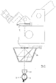
- a microscope according to the invention is initially located accordingly Fig.1-10 an eyepiece 1, which is pivotable about an axis 2, so that, for example during an eye operation, the surgeon uses the eyepiece 1 can optimally adapt his posture during surgery. Furthermore is on the microscope an adjustment 3 to change the magnification intended.
- a lens 4 first enables observation of the Front section of the eye 5 on one eye 6.
- an optics 8 for observing the fundus 9 held in the Beam path 10 of the microscope can be swiveled in and by means of a (first) Spindle drive 11 is movable in the direction of the beam path 10.
- This look 8 here a simple observation lens, is attached to a holder 12, which can be pivoted about a pivot axis 13 which is fixed at the attachment 7 is.
- the optics 8 is attached to a (first) crossbar 14, which is on a guide pin 15 can be moved parallel to the beam path 10 below the objective 4 is led.
- the cross member 14 is by a driver, not shown, which engages in the threaded spindle 17 moves.
- the guide pin 15 and the threaded spindle 17 are on the one hand in a common on which Holder 12 locked bearing piece 18 and on the other hand in a (first) connecting tab 19 attached.
- the threaded spindle 17 is rotatable about its axis stored.
- a (first) rotary knob 20 provided, by means of which the spindle output 11 can be set in motion and the optics 8 can be moved along the beam path 10.
- the optics 8 is on attached to the retaining pin 22 which is resiliently held in a guide 21.
- the complete spindle drive can be separated from the holder 12 on the bearing piece 18, so that it can be sterilized.
- a device connected to the optics 8 by the common holder 12 23 for image reversal and erection consists of a porroprism system 2.
- the bearing piece 18 from the holder 12 is advantageous detachable.
- An opening 26, 27 each in the housing 25 permit the passage of the beam path 10 through the prism system 24.
- the entrance of the Beam path 10 is connected upstream in the prism system 24 in the Objective 4 adjacent opening 26 an imaging optics 28 for adaptation of the beam path considerably lengthened because of the prism system 24 10th
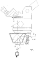
- the housing 25 is shaped somewhat differently, otherwise the arrangement does not differ from that of FIG. 1.
- the spindle drive 11 is with an electric motor drive equipped.
- an electric motor 29 is provided on the extension 7, from the output of which can be connected via a suitable coupling 30 flexible shaft 31 and a belt drive 32, the threaded spindle 17 in Rotation can be offset.
- the electric motor can also be used on others Point of the system. Accordingly, it is sufficient that Switch electric motor 29 from a foot switch to optics 8 to move along the beam path 10; for example, an operator accordingly focus without using his surgical tools to hand and thus have to interrupt the ongoing operation.
- the invention can also be used when instead of non-contact observation rather, as in the arrangements of FIGS. 1-4 by means of the optics 8 5,6 to an aspherical eye 6 putting on optics 33 is used, which is similar to that Optics 8 first an inverted and inverted image of the fundus 9 supplies, which in turn can be used by the prism system 24 is made.
- optics 34 which can be displaced along the beam path 10 Adaptation of the intermediate image according to Fig. 7-10. More like that Way as the optics 8 for observing the fundus 9 is for the Optics 34 - here a simple lens - a (second) spindle drive 35 installed, a further guide pin 36 and a threaded spindle 37 on each side of the first connecting link facing away from the first spindle drive 11 19 attached and at each other end of one (Second) connecting tab 38 are held together.
- the optics 34 is held in a (second) traverse 39, which is exactly the same as the first traverse 14 contains a driver which of the rotating threaded spindle 37 is moved in the direction of the beam path 10 if a corresponding on the threaded spindle 37 held (second) knob 40 operated (Fig.7.8).
- the spindle drive 35 can also according to Fig.9,10 in a similar manner to the spindle drive 11 by means of the electric drive 29-32 are operated.
- FIG. 14 shows the beam path below the Microscope, this beam path through the use of four Prisms as shown in Figs. 15, 16 have been improved. In particular this results in an enlargement of the stereoscopic basis, whereby aberrations also disappear.
- the advantage of this arrangement is in that no shadowing can occur, and therefore a better one stereoscopic vision is guaranteed.
- the prisms used are equally strong, with the base of the prisms 40 below and 41 is facing each other, while those closest to the lens 4 Prisms 42 and 43 have their base directed outwards. With The stereoscopic width shown by arrow B is thereby essential improved.
- the prisms have e.g. with a lens focal length of 200 mm advantageous 5 pdpt (prism dioptrine).
Landscapes
- Physics & Mathematics (AREA)
- Health & Medical Sciences (AREA)
- Life Sciences & Earth Sciences (AREA)
- Surgery (AREA)
- Optics & Photonics (AREA)
- General Physics & Mathematics (AREA)
- General Health & Medical Sciences (AREA)
- Engineering & Computer Science (AREA)
- Veterinary Medicine (AREA)
- Molecular Biology (AREA)
- Heart & Thoracic Surgery (AREA)
- Animal Behavior & Ethology (AREA)
- Biomedical Technology (AREA)
- Public Health (AREA)
- Medical Informatics (AREA)
- Chemical & Material Sciences (AREA)
- Analytical Chemistry (AREA)
- Ophthalmology & Optometry (AREA)
- Biophysics (AREA)
- Eye Examination Apparatus (AREA)
- Microscoopes, Condenser (AREA)
Applications Claiming Priority (2)
| Application Number | Priority Date | Filing Date | Title |
|---|---|---|---|
| DE20021955U | 2000-12-23 | ||
| DE20021955U DE20021955U1 (de) | 2000-12-23 | 2000-12-23 | Mikroskop zur Weitwinkelbeobachtung, insbesondere für Augenoperationen |
Publications (4)
| Publication Number | Publication Date |
|---|---|
| EP1227355A2 true EP1227355A2 (fr) | 2002-07-31 |
| EP1227355A3 EP1227355A3 (fr) | 2003-11-12 |
| EP1227355B1 EP1227355B1 (fr) | 2008-06-11 |
| EP1227355B2 EP1227355B2 (fr) | 2011-07-27 |
Family
ID=7950607
Family Applications (1)
| Application Number | Title | Priority Date | Filing Date |
|---|---|---|---|
| EP01121232A Expired - Lifetime EP1227355B2 (fr) | 2000-12-23 | 2001-09-05 | Microscope pour observation à grand angle, notamment de chirurgie ophthalmologique |
Country Status (5)
| Country | Link |
|---|---|
| US (2) | US6788455B2 (fr) |
| EP (1) | EP1227355B2 (fr) |
| JP (1) | JP4154148B2 (fr) |
| DE (2) | DE20021955U1 (fr) |
| ES (1) | ES2307560T5 (fr) |
Cited By (5)
| Publication number | Priority date | Publication date | Assignee | Title |
|---|---|---|---|---|
| EP1450193A2 (fr) * | 2003-01-30 | 2004-08-25 | Kabushiki Kaisha TOPCON | Microscope chirurgical |
| EP1498761A1 (fr) * | 2003-07-17 | 2005-01-19 | Leica Microsystems (Schweiz) AG | Microscope stéreoscopique |
| DE102004043998A1 (de) * | 2004-09-11 | 2006-03-16 | Carl Zeiss Meditec Ag | Ophthalmologisches Gerät, insbesondere Spaltlampe, mit Stereobasis-Wechselvorrichtung |
| DE202009014603U1 (de) | 2009-10-29 | 2011-03-10 | Möller-Wedel GmbH | Modul zur stereoskopischen Weitwinkel-Fundusbeobachtung für ein ophthalmologisches Operationsmikroskop |
| EP2921099A1 (fr) | 2014-03-18 | 2015-09-23 | Dieter Mann GmbH | Adaptateur d'ophtalmoscopie pour microscope opératoire |
Families Citing this family (23)
| Publication number | Priority date | Publication date | Assignee | Title |
|---|---|---|---|---|
| EP1320779B1 (fr) * | 2000-09-26 | 2004-12-08 | Carl Zeiss | Systeme de redressement de l'image, module d'ophtalmoscopie additionnel et microscope d'operation |
| DE10140402B4 (de) * | 2000-09-26 | 2012-08-30 | Carl Zeiss Meditec Ag | Bildumkehrsystem, Ophthalmoskopie-Vorsatzmodul und Operationsmikroskop |
| DE20021955U1 (de) † | 2000-12-23 | 2001-03-15 | Oculus Optikgeraete Gmbh | Mikroskop zur Weitwinkelbeobachtung, insbesondere für Augenoperationen |
| JP4068371B2 (ja) * | 2001-06-13 | 2008-03-26 | 株式会社トプコン | 手術用顕微鏡 |
| CN100388044C (zh) * | 2002-03-26 | 2008-05-14 | 株式会社拓普康 | 手术用显微镜 |
| DE20215635U1 (de) * | 2002-10-11 | 2002-12-05 | Oculus Optikgeraete Gmbh | Optische Vorrichtung zur lösbaren Befestigung an einem Mikroskop |
| JP4417036B2 (ja) * | 2003-06-09 | 2010-02-17 | 株式会社トプコン | 眼科用手術顕微鏡 |
| JP2005034285A (ja) * | 2003-07-18 | 2005-02-10 | Topcon Corp | 手術用顕微鏡及び観察プリズム |
| DE102004050893B4 (de) * | 2003-10-31 | 2015-05-21 | Carl Zeiss Meditec Ag | Tubus mit zwei umschaltbaren Planoptikelementen zur wahlweisen Strahlengangvertauschung und Bildumkehr für ein Mikroskop sowie Mikroskop |
| DE102005040834A1 (de) * | 2005-08-25 | 2007-03-08 | Carl Zeiss Jena Gmbh | Einrichtung zum Wechseln von Objektiven an optischen Geräten, insbesondere an Mikroskopen |
| GB0608258D0 (en) * | 2006-04-26 | 2006-06-07 | Perkinelmer Singapore Pte Ltd | Spectroscopy using attenuated total internal reflectance (ATR) |
| US7903331B2 (en) * | 2006-07-31 | 2011-03-08 | Volk Optical, Inc. | Flexible positioner and ophthalmic microscope incorporating the same |
| DE102006047459A1 (de) * | 2006-10-07 | 2008-04-10 | Carl Zeiss Surgical Gmbh | Ophthalmo-Operationsmikroskopsystem |
| US7940479B2 (en) * | 2007-04-02 | 2011-05-10 | Volk Optical, Inc. | Positioners and microscopes incorporating the same |
| JP5030669B2 (ja) * | 2007-05-31 | 2012-09-19 | 興和株式会社 | レンズ支持装置、眼底画像取得装置、及び眼底画像取得システム |
| DE102008011608A1 (de) * | 2008-02-28 | 2009-09-03 | Carl Zeiss Surgical Gmbh | Vorsatzeinrichtung für eine optische Beobachtungseinrichtung |
| JP2010000110A (ja) * | 2008-06-18 | 2010-01-07 | Topcon Corp | 二眼式ステレオビデオ顕微鏡装置 |
| DE102009018114A1 (de) | 2009-04-20 | 2011-01-05 | Dieter Mann Gmbh | Weitwinkelbeobachtung am Operationsmikroskop |
| DE202011110431U1 (de) | 2011-04-18 | 2014-01-07 | Leica Microsystems (Schweiz) Ag | Operationsmikroskopsystem |
| DE102013219379B3 (de) | 2013-09-26 | 2015-03-12 | Carl Zeiss Meditec Ag | Optisches Abbildungssystem |
| DE102013219383B3 (de) | 2013-09-26 | 2015-03-12 | Carl Zeiss Meditec Ag | Optisches Abbildungssystem |
| DE102017105580A1 (de) * | 2016-11-04 | 2018-05-09 | Carl Zeiss Meditec Ag | Operationsmikroskop |
| CN110068920A (zh) * | 2019-05-29 | 2019-07-30 | 苏州四海通仪器有限公司 | 一种用于显微镜的非接触广角倒像装置及显微镜系统 |
Citations (10)
| Publication number | Priority date | Publication date | Assignee | Title |
|---|---|---|---|---|
| DE8902035U1 (fr) * | 1989-02-21 | 1989-03-30 | J.D. Moeller Optische Werke Gmbh, 2000 Wedel, De | |
| US5009487A (en) † | 1988-07-30 | 1991-04-23 | Oculus Optikgeraete Gmbh | Prism system for a stereoscopic microscope |
| WO1991015150A1 (fr) * | 1990-03-29 | 1991-10-17 | K.U. Leuven Research & Development | Dispositif d'observation de l'×il, comprenant des moyens de renversement d'image |
| US5282085A (en) * | 1991-05-18 | 1994-01-25 | Oculus Optikgeraete Gmbh | Stereoscopic microscope including a field-magnifying lens in front of the objective lens |
| US5321447A (en) * | 1991-05-04 | 1994-06-14 | Carl-Zeiss-Stiftung | Ophthalmoscopic attachment for a surgical microscope |
| DE9415219U1 (de) * | 1994-09-22 | 1994-11-24 | Oculus Optikgeraete Gmbh | Vorsatzeinrichtung für ein Mikroskop |
| US5986801A (en) † | 1996-11-08 | 1999-11-16 | Volk; Donald A. | Image reinverter for stereo microscope |
| DE20021955U1 (de) † | 2000-12-23 | 2001-03-15 | Oculus Optikgeraete Gmbh | Mikroskop zur Weitwinkelbeobachtung, insbesondere für Augenoperationen |
| EP1191381A2 (fr) * | 2000-09-26 | 2002-03-27 | Carl Zeiss | Dispositif stéréoscopique pour microscopie |
| WO2002027379A2 (fr) † | 2000-09-26 | 2002-04-04 | Carl Zeiss | Systeme de redressement de l'image, module d'ophtalmoscopie additionnel et microscope d'operation |
Family Cites Families (12)
| Publication number | Priority date | Publication date | Assignee | Title |
|---|---|---|---|---|
| US4015898A (en) * | 1975-04-14 | 1977-04-05 | Kurt Ernest Schirmer | Upright wide angle stereo ophthalmoscope |
| DE3215566A1 (de) * | 1982-04-26 | 1983-10-27 | Ernst Leitz Wetzlar Gmbh, 6330 Wetzlar | Antrieb zur scharfstellung eines mikroskopes |
| DE3217776C2 (de) * | 1982-05-12 | 1985-01-31 | Fa. Carl Zeiss, 7920 Heidenheim | Stereomikroskop |
| DE3539009A1 (de) * | 1985-11-02 | 1987-05-07 | Moeller J D Optik | Vorsatz fuer ein stereoskopisches operationsmikroskop fuer die augenchirurgie |
| US5200773A (en) * | 1989-10-27 | 1993-04-06 | Volk Donald A | Diagnostic indirect ophthalmoscopy contact lens system |
| US5438456A (en) * | 1991-03-14 | 1995-08-01 | Grinblat; Avi | Optical stereoscopic microscope system |
| US5526074A (en) * | 1994-10-31 | 1996-06-11 | Volk; Donald A. | Full field reinverting indirect contact ophthalmoscope |
| DE19541237B4 (de) * | 1994-11-12 | 2006-04-13 | Carl Zeiss | Pankratisches Vergrößerungssystem |
| DE19524475C1 (de) * | 1995-07-10 | 1996-11-14 | Fraunhofer Ges Forschung | Optische Zentriervorrichtung zum lagegenauen Bestücken eines Bauelements in Oberflächenmontagetechnik sowie deren Verwendung zur Montage von Laserdioden |
| US5793524A (en) * | 1997-08-04 | 1998-08-11 | Luloh; K. Peter | Device for non-contact wide-angle viewing of fundus during vitrectomy |
| DE29905969U1 (de) * | 1999-04-08 | 1999-07-08 | Oculus Optikgeraete Gmbh | Stereoskopisches Mikroskop |
| DE20017891U1 (de) * | 2000-10-18 | 2001-02-08 | Oculus Optikgeraete Gmbh | Mikroskop zur kontaktfreien Weitwinkel-Beobachtung |
-
2000
- 2000-12-23 DE DE20021955U patent/DE20021955U1/de not_active Expired - Lifetime
-
2001
- 2001-09-05 EP EP01121232A patent/EP1227355B2/fr not_active Expired - Lifetime
- 2001-09-05 ES ES01121232T patent/ES2307560T5/es not_active Expired - Lifetime
- 2001-09-05 DE DE50114022T patent/DE50114022D1/de not_active Expired - Lifetime
- 2001-12-17 US US10/023,783 patent/US6788455B2/en not_active Expired - Lifetime
- 2001-12-20 JP JP2001388087A patent/JP4154148B2/ja not_active Expired - Lifetime
-
2004
- 2004-06-01 US US10/858,413 patent/US6967774B2/en not_active Expired - Lifetime
Patent Citations (10)
| Publication number | Priority date | Publication date | Assignee | Title |
|---|---|---|---|---|
| US5009487A (en) † | 1988-07-30 | 1991-04-23 | Oculus Optikgeraete Gmbh | Prism system for a stereoscopic microscope |
| DE8902035U1 (fr) * | 1989-02-21 | 1989-03-30 | J.D. Moeller Optische Werke Gmbh, 2000 Wedel, De | |
| WO1991015150A1 (fr) * | 1990-03-29 | 1991-10-17 | K.U. Leuven Research & Development | Dispositif d'observation de l'×il, comprenant des moyens de renversement d'image |
| US5321447A (en) * | 1991-05-04 | 1994-06-14 | Carl-Zeiss-Stiftung | Ophthalmoscopic attachment for a surgical microscope |
| US5282085A (en) * | 1991-05-18 | 1994-01-25 | Oculus Optikgeraete Gmbh | Stereoscopic microscope including a field-magnifying lens in front of the objective lens |
| DE9415219U1 (de) * | 1994-09-22 | 1994-11-24 | Oculus Optikgeraete Gmbh | Vorsatzeinrichtung für ein Mikroskop |
| US5986801A (en) † | 1996-11-08 | 1999-11-16 | Volk; Donald A. | Image reinverter for stereo microscope |
| EP1191381A2 (fr) * | 2000-09-26 | 2002-03-27 | Carl Zeiss | Dispositif stéréoscopique pour microscopie |
| WO2002027379A2 (fr) † | 2000-09-26 | 2002-04-04 | Carl Zeiss | Systeme de redressement de l'image, module d'ophtalmoscopie additionnel et microscope d'operation |
| DE20021955U1 (de) † | 2000-12-23 | 2001-03-15 | Oculus Optikgeraete Gmbh | Mikroskop zur Weitwinkelbeobachtung, insbesondere für Augenoperationen |
Cited By (8)
| Publication number | Priority date | Publication date | Assignee | Title |
|---|---|---|---|---|
| EP1450193A2 (fr) * | 2003-01-30 | 2004-08-25 | Kabushiki Kaisha TOPCON | Microscope chirurgical |
| EP1450193A3 (fr) * | 2003-01-30 | 2004-09-08 | Kabushiki Kaisha TOPCON | Microscope chirurgical |
| CN100346751C (zh) * | 2003-01-30 | 2007-11-07 | 拓普康株式会社 | 手术用显微镜装置 |
| EP1498761A1 (fr) * | 2003-07-17 | 2005-01-19 | Leica Microsystems (Schweiz) AG | Microscope stéreoscopique |
| DE102004043998A1 (de) * | 2004-09-11 | 2006-03-16 | Carl Zeiss Meditec Ag | Ophthalmologisches Gerät, insbesondere Spaltlampe, mit Stereobasis-Wechselvorrichtung |
| DE202009014603U1 (de) | 2009-10-29 | 2011-03-10 | Möller-Wedel GmbH | Modul zur stereoskopischen Weitwinkel-Fundusbeobachtung für ein ophthalmologisches Operationsmikroskop |
| EP2316330A1 (fr) | 2009-10-29 | 2011-05-04 | Möller-Wedel GmbH | Module d'observation ophtalmoscopique à grand angle stéréoscopique pour un microscope d'opération ophtalmologique |
| EP2921099A1 (fr) | 2014-03-18 | 2015-09-23 | Dieter Mann GmbH | Adaptateur d'ophtalmoscopie pour microscope opératoire |
Also Published As
| Publication number | Publication date |
|---|---|
| JP2002253575A (ja) | 2002-09-10 |
| EP1227355B2 (fr) | 2011-07-27 |
| DE20021955U1 (de) | 2001-03-15 |
| US20040218266A1 (en) | 2004-11-04 |
| DE50114022D1 (de) | 2008-07-24 |
| EP1227355A3 (fr) | 2003-11-12 |
| JP4154148B2 (ja) | 2008-09-24 |
| ES2307560T3 (es) | 2008-12-01 |
| ES2307560T5 (es) | 2011-11-16 |
| US6788455B2 (en) | 2004-09-07 |
| US6967774B2 (en) | 2005-11-22 |
| US20020118448A1 (en) | 2002-08-29 |
| EP1227355B1 (fr) | 2008-06-11 |
Similar Documents
| Publication | Publication Date | Title |
|---|---|---|
| EP1227355B1 (fr) | Microscope pour observation à grand angle, notamment de chirurgie ophthalmologique | |
| DE10226874B4 (de) | Operationsmikroskop | |
| DE19609034C2 (de) | Vorrichtung zur Führung chirurgischer Instrumente für die endoskopische Chirurgie | |
| DE4233274B4 (de) | Optisches Augenbehandlungsgerät | |
| EP1326117B2 (fr) | Adaptateur ophtalmoscopique et microscope chirurgical | |
| DE3713512C2 (de) | Laserhandstück | |
| DE102004050893B4 (de) | Tubus mit zwei umschaltbaren Planoptikelementen zur wahlweisen Strahlengangvertauschung und Bildumkehr für ein Mikroskop sowie Mikroskop | |
| DE3105018A1 (de) | Operationsmikroskop | |
| EP1199591B1 (fr) | Microscope pour observation à grand angle sans contact | |
| EP0193818A1 (fr) | Stéréomicroscope pour opérations | |
| DE102009018114A1 (de) | Weitwinkelbeobachtung am Operationsmikroskop | |
| EP1109046A1 (fr) | Dispositif d'éclairage pour un microscope opératoire | |
| DE19728035B4 (de) | Beobachtungsvorrichtung mit Schrägbeleuchtung | |
| DE102006038911A1 (de) | Ophthalmoskopie-Vorsatzmodul und Operationsmikroskop mit Ophthalmoskopie-Vorsatzzmodul | |
| CH687424A5 (de) | Operationsmikroskop. | |
| EP2316330B1 (fr) | Module d'observation ophtalmoscopique à grand angle stéréoscopique pour un microscope d'opération ophtalmologique | |
| WO2015036097A1 (fr) | Endoscope à direction d'observation réglable | |
| EP1291696A1 (fr) | Configuration de prismes pour une illumination normale et oblique dans un stéréomicroscope chirurgical | |
| EP1410754B1 (fr) | Microscope chirurgical à dispositif d'illumination | |
| EP1006390B1 (fr) | Ensemble microscope-endoscope | |
| DD157757B1 (de) | Vorrichtung zur spaltbeleuchtung | |
| EP2090913A2 (fr) | Tubes pour un dispositif d'observation | |
| DE2801441C3 (de) | Endoskop | |
| DE102012216470B4 (de) | Spaltleuchte für ein Operationsmikroskop und Operationsmikroskop | |
| DE102020100677B3 (de) | Optisches Beobachtungsinstrument |
Legal Events
| Date | Code | Title | Description |
|---|---|---|---|
| PUAI | Public reference made under article 153(3) epc to a published international application that has entered the european phase |
Free format text: ORIGINAL CODE: 0009012 |
|
| AK | Designated contracting states |
Kind code of ref document: A2 Designated state(s): AT BE CH CY DE DK ES FI FR GB GR IE IT LI LU MC NL PT SE TR |
|
| AX | Request for extension of the european patent |
Free format text: AL;LT;LV;MK;RO;SI |
|
| PUAL | Search report despatched |
Free format text: ORIGINAL CODE: 0009013 |
|
| AK | Designated contracting states |
Kind code of ref document: A3 Designated state(s): AT BE CH CY DE DK ES FI FR GB GR IE IT LI LU MC NL PT SE TR |
|
| AX | Request for extension of the european patent |
Extension state: AL LT LV MK RO SI |
|
| RIC1 | Information provided on ipc code assigned before grant |
Ipc: 7A 61B 3/13 B Ipc: 7G 02B 21/00 A |
|
| 17P | Request for examination filed |
Effective date: 20040115 |
|
| AKX | Designation fees paid |
Designated state(s): DE ES FR GB |
|
| 17Q | First examination report despatched |
Effective date: 20070608 |
|
| GRAP | Despatch of communication of intention to grant a patent |
Free format text: ORIGINAL CODE: EPIDOSNIGR1 |
|
| GRAS | Grant fee paid |
Free format text: ORIGINAL CODE: EPIDOSNIGR3 |
|
| GRAA | (expected) grant |
Free format text: ORIGINAL CODE: 0009210 |
|
| AK | Designated contracting states |
Kind code of ref document: B1 Designated state(s): DE ES FR GB |
|
| REG | Reference to a national code |
Ref country code: GB Ref legal event code: FG4D Free format text: NOT ENGLISH |
|
| REF | Corresponds to: |
Ref document number: 50114022 Country of ref document: DE Date of ref document: 20080724 Kind code of ref document: P |
|
| REG | Reference to a national code |
Ref country code: ES Ref legal event code: FG2A Ref document number: 2307560 Country of ref document: ES Kind code of ref document: T3 |
|
| PLBI | Opposition filed |
Free format text: ORIGINAL CODE: 0009260 |
|
| PLAX | Notice of opposition and request to file observation + time limit sent |
Free format text: ORIGINAL CODE: EPIDOSNOBS2 |
|
| 26 | Opposition filed |
Opponent name: CARL ZEISS SURGICAL GMBH Effective date: 20090311 |
|
| PLAB | Opposition data, opponent's data or that of the opponent's representative modified |
Free format text: ORIGINAL CODE: 0009299OPPO |
|
| PLBB | Reply of patent proprietor to notice(s) of opposition received |
Free format text: ORIGINAL CODE: EPIDOSNOBS3 |
|
| PUAH | Patent maintained in amended form |
Free format text: ORIGINAL CODE: 0009272 |
|
| STAA | Information on the status of an ep patent application or granted ep patent |
Free format text: STATUS: PATENT MAINTAINED AS AMENDED |
|
| 27A | Patent maintained in amended form |
Effective date: 20110727 |
|
| AK | Designated contracting states |
Kind code of ref document: B2 Designated state(s): DE ES FR GB |
|
| REG | Reference to a national code |
Ref country code: DE Ref legal event code: R102 Ref document number: 50114022 Country of ref document: DE |
|
| REG | Reference to a national code |
Ref country code: DE Ref legal event code: R102 Ref document number: 50114022 Country of ref document: DE Effective date: 20110727 |
|
| REG | Reference to a national code |
Ref country code: ES Ref legal event code: DC2A Ref document number: 2307560 Country of ref document: ES Kind code of ref document: T5 Effective date: 20111116 |
|
| REG | Reference to a national code |
Ref country code: FR Ref legal event code: PLFP Year of fee payment: 16 |
|
| REG | Reference to a national code |
Ref country code: FR Ref legal event code: PLFP Year of fee payment: 17 |
|
| REG | Reference to a national code |
Ref country code: FR Ref legal event code: PLFP Year of fee payment: 18 |
|
| PGFP | Annual fee paid to national office [announced via postgrant information from national office to epo] |
Ref country code: GB Payment date: 20200923 Year of fee payment: 20 Ref country code: FR Payment date: 20200922 Year of fee payment: 20 |
|
| PGFP | Annual fee paid to national office [announced via postgrant information from national office to epo] |
Ref country code: ES Payment date: 20201016 Year of fee payment: 20 Ref country code: DE Payment date: 20201127 Year of fee payment: 20 |
|
| REG | Reference to a national code |
Ref country code: DE Ref legal event code: R071 Ref document number: 50114022 Country of ref document: DE |
|
| REG | Reference to a national code |
Ref country code: GB Ref legal event code: PE20 Expiry date: 20210904 |
|
| PG25 | Lapsed in a contracting state [announced via postgrant information from national office to epo] |
Ref country code: GB Free format text: LAPSE BECAUSE OF EXPIRATION OF PROTECTION Effective date: 20210904 |
|
| REG | Reference to a national code |
Ref country code: ES Ref legal event code: FD2A Effective date: 20220126 |
|
| PG25 | Lapsed in a contracting state [announced via postgrant information from national office to epo] |
Ref country code: ES Free format text: LAPSE BECAUSE OF EXPIRATION OF PROTECTION Effective date: 20210906 |