EP1181552B1 - Verfahren zur diagnose tse-induzierter veränderungen in geweben mittels infrarotspektroskopie - Google Patents
Verfahren zur diagnose tse-induzierter veränderungen in geweben mittels infrarotspektroskopie Download PDFInfo
- Publication number
- EP1181552B1 EP1181552B1 EP00941900A EP00941900A EP1181552B1 EP 1181552 B1 EP1181552 B1 EP 1181552B1 EP 00941900 A EP00941900 A EP 00941900A EP 00941900 A EP00941900 A EP 00941900A EP 1181552 B1 EP1181552 B1 EP 1181552B1
- Authority
- EP
- European Patent Office
- Prior art keywords
- infrared
- tse
- spectra
- tissue
- sample
- Prior art date
- Legal status (The legal status is an assumption and is not a legal conclusion. Google has not performed a legal analysis and makes no representation as to the accuracy of the status listed.)
- Expired - Lifetime
Links
Images



Classifications
-
- G—PHYSICS
- G01—MEASURING; TESTING
- G01N—INVESTIGATING OR ANALYSING MATERIALS BY DETERMINING THEIR CHEMICAL OR PHYSICAL PROPERTIES
- G01N21/00—Investigating or analysing materials by the use of optical means, i.e. using sub-millimetre waves, infrared, visible or ultraviolet light
- G01N21/17—Systems in which incident light is modified in accordance with the properties of the material investigated
- G01N21/25—Colour; Spectral properties, i.e. comparison of effect of material on the light at two or more different wavelengths or wavelength bands
- G01N21/31—Investigating relative effect of material at wavelengths characteristic of specific elements or molecules, e.g. atomic absorption spectrometry
- G01N21/35—Investigating relative effect of material at wavelengths characteristic of specific elements or molecules, e.g. atomic absorption spectrometry using infrared light
- G01N21/359—Investigating relative effect of material at wavelengths characteristic of specific elements or molecules, e.g. atomic absorption spectrometry using infrared light using near infrared light
-
- G—PHYSICS
- G01—MEASURING; TESTING
- G01N—INVESTIGATING OR ANALYSING MATERIALS BY DETERMINING THEIR CHEMICAL OR PHYSICAL PROPERTIES
- G01N21/00—Investigating or analysing materials by the use of optical means, i.e. using sub-millimetre waves, infrared, visible or ultraviolet light
- G01N21/17—Systems in which incident light is modified in accordance with the properties of the material investigated
- G01N21/25—Colour; Spectral properties, i.e. comparison of effect of material on the light at two or more different wavelengths or wavelength bands
- G01N21/31—Investigating relative effect of material at wavelengths characteristic of specific elements or molecules, e.g. atomic absorption spectrometry
- G01N2021/3196—Correlating located peaks in spectrum with reference data, e.g. fingerprint data
-
- G—PHYSICS
- G01—MEASURING; TESTING
- G01N—INVESTIGATING OR ANALYSING MATERIALS BY DETERMINING THEIR CHEMICAL OR PHYSICAL PROPERTIES
- G01N21/00—Investigating or analysing materials by the use of optical means, i.e. using sub-millimetre waves, infrared, visible or ultraviolet light
- G01N21/17—Systems in which incident light is modified in accordance with the properties of the material investigated
- G01N21/25—Colour; Spectral properties, i.e. comparison of effect of material on the light at two or more different wavelengths or wavelength bands
- G01N21/31—Investigating relative effect of material at wavelengths characteristic of specific elements or molecules, e.g. atomic absorption spectrometry
- G01N21/35—Investigating relative effect of material at wavelengths characteristic of specific elements or molecules, e.g. atomic absorption spectrometry using infrared light
-
- Y—GENERAL TAGGING OF NEW TECHNOLOGICAL DEVELOPMENTS; GENERAL TAGGING OF CROSS-SECTIONAL TECHNOLOGIES SPANNING OVER SEVERAL SECTIONS OF THE IPC; TECHNICAL SUBJECTS COVERED BY FORMER USPC CROSS-REFERENCE ART COLLECTIONS [XRACs] AND DIGESTS
- Y10—TECHNICAL SUBJECTS COVERED BY FORMER USPC
- Y10S—TECHNICAL SUBJECTS COVERED BY FORMER USPC CROSS-REFERENCE ART COLLECTIONS [XRACs] AND DIGESTS
- Y10S436/00—Chemistry: analytical and immunological testing
- Y10S436/811—Test for named disease, body condition or organ function
Definitions
- the invention relates to a method for fast Identification of transmissible spongiform encephalopathies (TSE) induced pathological changes in animal or human tissues with infrared spectroscopy (IR spectroscopy).
- TSE transmissible spongiform encephalopathies
- IR spectroscopy infrared spectroscopy
- Transmissible spongiform encephalopathies are transferable neurodegenerative diseases of the central nervous system (CNS) fatal to many mammals as well as affect people.
- CNS central nervous system
- TSE transmissible spongiform encephalopathies
- BSE bovine spongiform encephalopathy
- CWD chronic exhaustion
- TSE transferable Encephalopathy
- FSE feline spongiform Encephalopathy
- a TSE a) can definitely be determined by the histological Evidence of characteristic spongiform (spongy) changes associated with gliosis in brain tissue, b) by the immunological Detection of deposits of the pathological prion protein (PrP) using Western blot technique, histo blot technique and immunohistochemistry, c) by electron microscopy Detection of scrapie-associated (PrP) fibrils (SAF) and d) by detection of the infectious TSE agent using transmission experiments in animal experiments diagnose.
- PrP pathological prion protein
- SAF scrapie-associated fibrils
- TSE diseases in farm animals is because of the potential portability through consumption of meat of animals of great interest. So there is e.g. the suspicion that the consumption of BSE contaminated Beef a new variant of CJD at Humans (nvCJD) can cause. In terms of consumer protection and curbing the spread of the epidemic Therefore, some states currently run official agencies Monitoring the degree of infection of cattle populations with BSE. In this regard, be on Slaughterhouses routinely inspect slaughterhouses Cattle aimed, of which the further usability of the slaughtered goods.
- test systems are currently under development to enable sensitive and rapid screening of large numbers of samples for pathological prion protein and thus TSE diagnostics on an industrial scale.
- These include a capillary electrophoresis immunoassay with fluorescence-labeled peptides (Schmerr & Jenny (1998) Electrophoresis 19: 409-419) and an immunological detection system called Delfia with fluorescent lanthanide chelates (Safar et al. (1998) Nature Medicine 4: 1157-1165).
- the tissue sample from the medulla oblongata of slaughtered cattle is homogenized and treated with the enzyme Proteinase K.
- the pathological prion protein that may remain after the treatment is marked with the monoclonal antibody 6H4 (manufactured by Prionics) and then stained in a Western blot. According to the manufacturer, this process takes up to 12 hours from the time the sample is obtained until a final result is obtained.
- the invention is based on the object of a method for quick, reliable and economical proof Develop TSE-induced changes in tissues.
- the inventive method is also intended to Effective routine slaughterhouse conditions be applicable.
- the object of the invention is therefore to create a method for the diagnosis of TSE-induced pathological Changes in tissues, the changes being caused by Scrapie, BSE or another TSE group associated disease form.
- the method on which the invention is based is essentially based on measurements of the infrared spectra of the pathologically modified tissue. It is already known from a number of publications and patent applications that disease-specific changes can be reflected in the infrared spectrum of the tissue (US Patent 5,168,162 (Wong &Rigas; US Patent 5,038,039; Wong, Rigas; Lasch & Naumann (1998) Cell. Mol. Biol. 44: 189-202; Lasch et al. (1998) Proc. SPIE 3257: 187-198; Choo et.al. (1996) Biophys. J. 71: 1672-1679) no published data available.
- the procedure is for TSE diagnostics using infrared spectroscopy a comparison of spectra of the object to be examined Tissue material with corresponding spectra of Fabrics of known origin in the sense of a reference procedure necessary. Practical implementation of the procedure therefore requires the existence of a validated Reference database of IR spectra from healthy or pathological tissue samples were obtained. The creation The reference database is standardized for a Diagnostics only required once.
- the comparison of spectra of unknown samples with the reference database is then preferably carried out using techniques the computer-aided pattern recognition process, such as e.g. multivariate statistics, artificial neural networks, genetic algorithms etc.
- Infrared light is applied to the samples to record the spectra steered and the spectral characteristics of the emerging Radiation, i.e. after interaction of light registered with the tissue.
- the use is advantageous microspectrometric techniques when minimizing the required sample quantities are aimed for.
- An infrared microscope can also be used Thin sections also have spatially resolved spectral information be obtained, which makes the process much more specific and make it more sensitive. In perspective would be a proof by infrared light guide as Endoscope conceivable for the diagnosis of TSE directly in the infected organism.
- the new method for diagnosis allows TSE-induced Changes in tissue statements within less than a minute after receiving the sample. So that is it both the immunological detection of the prion protein and also superior to the immunohistological diagnosis that deliver results only after up to 12 hours.
- a routine application of the method according to the invention for meat control in slaughterhouses practically no intermediate storage of the slaughtered goods until the diagnosis is received.
- the speed of the Diagnosis represents an economic advantage over known ones Procedures represent, since storage time of the slaughtered goods and thus minimizes space and energy costs for cooling become. In addition, the freshness of the Meat at the time of final consumption.
- the method according to the invention can be used without inclusion highly specialized experts (e.g. from histologists) be performed since the classification of the IR spectra by known, for the purposes of TSE diagnostics optimized procedures of computer-aided Pattern recognition takes place.
- highly specialized experts e.g. from histologists
- the evaluation of the spectra based on strictly mathematical criteria leads simultaneously to a high degree of diagnosis without subjective Experience knowledge comes along and thus inevitable, bypasses human misjudgments.
- IR microscopic version for spatially resolved Analysis of tissue thin sections lies in the combination specific, spectrally resolved structure information and the high spatial resolution that results from this becomes.
- a IR microscope the involvement of individual neurons on The course of the disease can be recorded and examined.
- the very high diagnostic sensitivity results from the Fact that practically no averaging of characteristics sick and healthy cells, like others, non-spatially resolved methods is necessarily the case.
- This particular embodiment of the method requires meanwhile a relatively long time for data acquisition, which depends on the size of the tissue arial examined and the spatial resolution takes 1 to 6 hours, and It is therefore less suitable for routine checks.
- tissue samples are taken from the organism post mortem . It can be both animal and human organisms.
- TSE transmissible spongiform encephalopathy
- Organs are the central nervous system, the peripheral one Nervous system, organs of the lymphatic system, the Digestive system, endocrine system, cardiovascular System and the respiratory system.
- the preferred nerve system is the central nervous system and the peripheral nervous system, in particular Medulla oblongata as well as pons of the brain are beneficial.
- the preparation of the tissue sample depends on the particular one Embodiment of the method.
- tissue samples small pieces of tissue removed.
- the native samples will be e.g. placed in commercially available IR cells.
- a homogenate of the tissue material is prepared in H 2 O and aliquots are placed in IR cuvettes.
- aliquots of this suspension are dried as transparent films on IR-transparent sample holders, whereby reduced pressures have proven to be advantageous in terms of accelerating the drying process (Helm et al. (1991) J. Gen. Microbiol. 137: 69-79.
- the method can in principle also apply to the living organism, by inserting the light guide minimally invasively into the tissue and the infrared spectrum is recorded there directly.
- the implementation of this embodiment is currently still to the further development of infrared light guide technology bound because the currently available Light guide spectral sensitivities still too low have and are also too inflexible and too large.
- the quantities of substance required to record the IR spectra and their areal extension can be very be kept small. Depending on the given conditions (e.g. spectroscopy with or without beam focusing or use of an IR microscope) can substance amounts in the range from ⁇ g to ng.
- the diameters of the irradiated sample areas vary accordingly between 1-3 mm and 10-30 ⁇ m. The lower limit corresponds approximately to the size of one or fewer cells (e.g. neurons).
- Infrared spectra of the tissue samples are used according to the method measured, which were produced in one of the ways described were.
- the spectra are recorded here preferably with a Fourier transform infrared spectrometer, which compared to conventionally working, disperse Devices has a number of known advantages, only the speed of data acquisition and the higher sensitivity should be called.
- the use of a conventional, disperse IR spectrometer is also possible in principle, but leads to a loss in speed of the procedure.
- each of them per se can be used for spectra measurement known IR spectroscopic measuring arrangements used are weakened (e.g. in transmission / absorption Total reflection, direct or diffuse reflection). Especially Transmission / absorption spectroscopy has proven itself.
- the infrared spectrum is typically recorded in the spectral range of the so-called middle infrared between 500 and 4000 cm -1 .
- Narrower spectral ranges also in the near infrared between 4000 and 10000 cm -1 also lead to a successful diagnosis if it has previously been ensured that the spectra of the infected and healthy tissue samples have characteristic variances in the spectral range detected.
- the selection of one or more suitable spectral ranges can e.g. by visual inspection of the spectra (Selection of the areas with the strongest and most characteristic Changes compared to the control group) or by a multivariate method known per se for the selection of spectral characteristics.
- the physical measurement parameters such as spectral resolution or number of averaged spectra etc.
- Important when specifying the parameters for spectra acquisition and the sample preparation is only that for all measurements, especially for the control measurements on tissue samples from uninfected animals, identical Parameters can be selected.
- the procedure requires a one-off Creation of a reference spectra database. For this be Spectra of samples from TSE-infected organisms and measured from samples from TSE-free individuals.
- Sample preparation and spectra recording are done in carried out analogously to the unknown samples. It is crucial that all parameters for the reference and sample measurements are chosen identically.
- the spectrum of the sample to be examined is compared with the Spectra of the reference database compared. This is done the classification of the spectrum preferably using one of the known methods for pattern recognition, for example with multivariate algorithms Statistics, artificial neural networks or genetic Algorithms. In this step the spectrum is in mind a two-tier problem as healthy or infected with TSE classified.
- the sample which in this case is a slide applied thin section of tissue is in the beam path an infrared microscope.
- the spectra recording in the infrared microscopic measuring arrangement either in transmission or in direct reflection.
- the spatial resolution achieved here can be determined by the step distance of the individual measuring points be determined.
- the use is extremely advantageous of a computer controlled x / y table that automated Spectra measurements according to an arbitrarily determinable Allows grid with defined step distances. such x / y tables are now standard equipment modern IR microscope.
- the result of a spatially resolved measurement is one Infrared spectrum series, with each spectrum one pixel represented on the fictitious grid of the thin tissue section. In this way, IR data are obtained that the Completely cover the selected section of the thin section.
- the site-specific information about the spatial Spread of the TSE in tissue is obtained by each the spectra of a mapping data set with the reference database is balanced and so either as healthy or classified as infected.
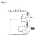
- Example 1 10 S and N samples, which were obtained in the same manner as in Example 1, were homogenized in H 2 O (10 ⁇ l H 2 O per mg tissue material). Aliquots of 35 ⁇ l of the suspensions were placed on a PC-controlled multi-sample carrier, which is also suitable for measuring microbial samples (Helm et al. (1991) J. Gen. Microbiol. 137: 69-79; Helm et al. (1991) J. Microbiol. Meth. 14: 127-142; Naumann (1998) Proc. SPIE 3257: 245-257), applied from ZnSe and dried according to the information described in the literature.
- Infrared spectra of the films obtained in this way were recorded in transmission and subjected to a hierarchical classification, based on second derivatives of the spectra in the spectral range between 1100 and 1000 cm -1 .
- Figure 3 shows the dendrogram of the classification of these spectra calculated according to the so-called Ward's algorithm.
- the spectra of the infected animals (S-1 to S-10) could be perfectly separated from those of the healthy animals (N-1 to N-10).
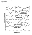
- cryosections were made from CNS samples of N and S animals, which were obtained as explained above, and were processed using the known methods of FT-IR mapping (Diem et al. (1999) Appl. Spectroscopy 53: 148A-160A; Lasch & Naumann (1998) Cell. Mol. Biol. 44: 189-202; Choo et al. (1996) Biophys. J. 71: 1672-1679) and infrared imaging (Lasch & Naumann (1998) Cell. Mol. Biol. 44: 189-202; Lasch et al. (1998) Proc. SPIE 3257: 187-198).
- FIG. 4A shows a dendrogram, for the calculation of which after data compression by means of main component analysis, the first three main components between 1450 and 950 cm -1 (approx. 500 data points) were used.
- the four main classes can be assigned to the four histologically defined cerebellar structures stratum molecular, stratum ganglionare, stratum granulosum and substantia alba .
- vector-normalized second derivatives of spectra of samples of scrapie-infected animals (dashed lines) are compared to those of healthy animals (solid lines).
- the lower part (b) shows the difference spectra between vector-normalized S and N spectra from a) for the respective tissue structures. All spectra used for this comparison are mean values of spectra of a spectra class (see FIG. 4A). They are labeled with the name of their cerebellar layer and the number of their spectral class. The characteristic spectral differences observed for the individual tissue classes are suitable for a reliable diagnosis of the disease-associated pathogenesis process.
Landscapes
- Physics & Mathematics (AREA)
- Spectroscopy & Molecular Physics (AREA)
- Health & Medical Sciences (AREA)
- Life Sciences & Earth Sciences (AREA)
- Chemical & Material Sciences (AREA)
- Analytical Chemistry (AREA)
- Biochemistry (AREA)
- General Health & Medical Sciences (AREA)
- General Physics & Mathematics (AREA)
- Immunology (AREA)
- Pathology (AREA)
- Investigating Or Analysing Materials By Optical Means (AREA)
- Investigating Or Analysing Biological Materials (AREA)
- Measurement Of The Respiration, Hearing Ability, Form, And Blood Characteristics Of Living Organisms (AREA)
- Measuring And Recording Apparatus For Diagnosis (AREA)
Description
Claims (10)
- Verfahren zur Diagnose von TSE-induzierten pathologischen Veränderungen in Geweben, wobei die Veränderungen durch Scrapie, BSE oder durch eine andere dem TSE-Formenkreis zugehörige Krankheitsform hervorgerufen werden, dadurch gekennzeichnet, daß(a) Infrarotstrahlung auf eine durch TSE pathologisch veränderte Gewebeprobe gelenkt wird und die spektralen Charakteristika der Infrarotstrahlung nach Wechselwirkung mit der Probe registriert werden und(b) die so erhaltenen Infrarotspektren mit einer Referenzdatenbank, welche Infrarotspektren von TSE-infizierten Geweben und von nicht-infizierten Geweben enthält, verglichen und klassifiziert werden.
- Verfahren nach Anspruche 1, dadurch gekennzeichnet, daß die Gewebeprobe dem zentralen Nervensystem, dem peripheren Nervensystem oder Organen des lymphatischen Systems, des Verdauungssystems, des endokrinen Systems, des kardiovaskulären Systes oder des respiratorischem Systems entstammt.
- Verfahren nach mindestens einem der Ansprüche 1 und 2, dadurch gekennzeichnet, daß das Infrarotspektrum des Gewebes entweder in einer oder mehreren Regionen des mittleren Infrarotbereichs von 500 bis 4000 cm-1 oder des nahen Infrarotbereichs von 4000 bis 10000 cm-1 oder in beiden Regionen gemessen wird.
- Verfahren nach mindestens einem der Ansprüche 1 bis 3, dadurch gekennzeichnet, daß das Infrarotspektrum des Gewebes im spektralen Bereich von 1000 bis 1300 cm -1 des mittleren Infrarots erfaßt wird.
- Verfahren nach mindestens einem der Ansprüche 1 bis 4, dadurch gekennzeichnet, daß die Wechselwirkung der Infrarotstrahlung mit der Probe und die Detektion der charakteristisch veränderten Strahlung in einer Transmissions/Absorptionsanordnung, einer Anordnung zur Messung der abgeschwächten Totalreflexion, einer Anordnung zur Messung der direkten oder diffusen Reflexion oder mittels IR-Lichtleitertechnik erfolgt.
- Verfahren nach mindestens einem der Ansprüche 1 bis 5, dadurch gekennzeichnet, daß der Vergleich des Infrarotspektrums der zu untersuchenden Probe mit den Infrarotspektren der Referenzdatenbank mittels einer oder mehrerer Methoden der Mustererkennung, vorzugsweise mittels Algorithmen der multivariaten Statistik oder künstlicher neuronaler Netze, erfolgt, wobei die dem Vergleich zugrundeliegenden spektralen Bereiche mit Verfahren zur Extraktion optimaler spektraler Merkmale, etwa mit genetischen Algorithmen, ermittelt werden.
- Verfahren nach mindestens einem der Ansprüche 1 bis 6, dadurch gekennzeichnet, daß die Messung des Infrarotspektrums mit einer infrarotmikroskopischen Meßanordnung an einem Gewebedünnschnitt in Transmission oder in direkter Reflexion durchgeführt wird.
- Verfahren nach Anspruch 7, dadurch gekennzeichnet, daß ortsaufgelöst, d.h. in Abhängigkeit von der Gewebestelle, an welcher der Infrarotstrahl durch die Probe geleitet wird, Infrarotspektren gemessen werden.
- Verfahren nach mindestens einem der Ansprüche 7 und 8, dadurch gekennzeichnet, daß jedes der ortsabhängig registrierten Infrarotspektren mit der Referenzdatenbank verglichen wird und somit ortsspezifische Informationen über die Krankheitsausbreitung im Gewebe erhalten werden.
- Verfahren nach mindestens einem der Ansprüche 7 bis 9, dadurch gekennzeichnet, daß die Referenzdatenbank Referenzspektren von TSE-infizierten Geweben und von nicht-infizierten Geweben jeweils aller im Gewebeschnitt mittels Infrarotspektroskopie unterscheidbarer Strukturen enthält.
Applications Claiming Priority (3)
| Application Number | Priority Date | Filing Date | Title |
|---|---|---|---|
| DE19923811A DE19923811C1 (de) | 1999-05-20 | 1999-05-20 | Verfahren zur Diagnose TSE-induzierter Veränderungen in Geweben mittels Infrarotspektroskopie |
| DE19923811 | 1999-05-20 | ||
| PCT/DE2000/001404 WO2000072007A2 (de) | 1999-05-20 | 2000-05-03 | Verfahren zur diagnose tse-induzierter veränderungen in geweben mittels infrarotspektroskopie |
Publications (2)
| Publication Number | Publication Date |
|---|---|
| EP1181552A2 EP1181552A2 (de) | 2002-02-27 |
| EP1181552B1 true EP1181552B1 (de) | 2003-04-09 |
Family
ID=7909061
Family Applications (1)
| Application Number | Title | Priority Date | Filing Date |
|---|---|---|---|
| EP00941900A Expired - Lifetime EP1181552B1 (de) | 1999-05-20 | 2000-05-03 | Verfahren zur diagnose tse-induzierter veränderungen in geweben mittels infrarotspektroskopie |
Country Status (10)
| Country | Link |
|---|---|
| US (1) | US6777241B1 (de) |
| EP (1) | EP1181552B1 (de) |
| JP (1) | JP3569231B2 (de) |
| AT (1) | ATE237134T1 (de) |
| AU (1) | AU5671500A (de) |
| DE (3) | DE19923811C1 (de) |
| DK (1) | DK1181552T3 (de) |
| ES (1) | ES2193967T3 (de) |
| PT (1) | PT1181552E (de) |
| WO (1) | WO2000072007A2 (de) |
Cited By (1)
| Publication number | Priority date | Publication date | Assignee | Title |
|---|---|---|---|---|
| DE102008035532A1 (de) | 2008-07-29 | 2010-02-11 | Carl Zeiss Ag | Verfahren zur Bestimmung der Reflexionscharakteristik einer Substanz |
Families Citing this family (13)
| Publication number | Priority date | Publication date | Assignee | Title |
|---|---|---|---|---|
| DE10109901B4 (de) * | 2001-02-22 | 2004-02-05 | Bundesrepublik Deutschland vertreten durch den Bundesminister für Gesundheit, dieses vertreten durch das Robert-Koch-Institut, vertreten durch seinen Leiter | Verfahren zum Nachweis von TSE-induzierten Veränderungen in menschlichen und tierischen Körperflüssigkeiten |
| DE10127500A1 (de) * | 2001-06-06 | 2002-12-12 | Simon Kammermeier | Verfahren zur Früherkennung von BSE bei Rindern |
| US20040171026A1 (en) * | 2001-09-05 | 2004-09-02 | Hochstrasser Denis Francois | Diagnostic method for transmissible spongiform encephalopathies |
| GB2379737A (en) * | 2001-09-05 | 2003-03-19 | Univ Geneve | Diagnostic method for spongiform encephalopathy disease |
| GB0323451D0 (en) * | 2003-10-07 | 2003-11-05 | Imp College Innovations Ltd | Methods for analysis of spectral data and their applications |
| US20050136487A1 (en) * | 2003-10-27 | 2005-06-23 | Meyer Donald W. | Transmissible spongiform encephalopathy detection in cervids, sheep and goats |
| ES2246113B1 (es) * | 2003-10-31 | 2007-04-01 | Consejo Sup. De Invest. Cientificas | Procedimiento de deteccion ante-mortem de proteinas prionicas por espectroscopia infrarroja y su uso en el diagnostico de encefalopatias espongiformes transmisibles. |
| DE102004061064A1 (de) * | 2004-12-18 | 2006-06-29 | Roche Diagnostics Gmbh | Verfahren und Vorrichtung zur spektroskopischen Untersuchung von Körperflüssigkeiten und Gewebeproben hinsichtlich eines erhöhten Alzheimerverdachts |
| JP2007139465A (ja) * | 2005-11-15 | 2007-06-07 | Yanmar Co Ltd | 残留農薬判定装置 |
| JP2007292639A (ja) * | 2006-04-26 | 2007-11-08 | Dainippon Ink & Chem Inc | スペクトル分析装置およびそのプログラム |
| JP5047962B2 (ja) * | 2006-07-06 | 2012-10-10 | 株式会社疲労科学研究所 | 近赤外光を用いたガン、全身性エリテマトーデス(sle)又は抗リン脂質抗体症候群に関する検査・診断装置の作動方法 |
| US8655807B2 (en) * | 2010-04-05 | 2014-02-18 | Applied Research Associates, Inc. | Methods for forming recognition algorithms for laser-induced breakdown spectroscopy |
| US9532732B2 (en) * | 2010-05-03 | 2017-01-03 | Emovi Inc. | Method and system for knee joint evaluation and diagnostic aid in normal and pathologic state |
Family Cites Families (6)
| Publication number | Priority date | Publication date | Assignee | Title |
|---|---|---|---|---|
| CA2008831C (en) * | 1990-01-29 | 1996-03-26 | Patrick T.T. Wong | Method of detecting the presence of anomalies in biological tissues and cells in natural and cultured form by infrared spectroscopy |
| US5168162A (en) * | 1991-02-04 | 1992-12-01 | Cornell Research Foundation, Inc. | Method of detecting the presence of anomalies in exfoliated cells using infrared spectroscopy |
| EP0644412A3 (de) | 1993-09-17 | 1995-08-09 | Boehringer Mannheim Gmbh | Verfahren zur Analyse klinisch relevanter Flüssigkeiten und Suspensionen. |
| US6031232A (en) * | 1995-11-13 | 2000-02-29 | Bio-Rad Laboratories, Inc. | Method for the detection of malignant and premalignant stages of cervical cancer |
| DE19841217B4 (de) * | 1997-10-27 | 2005-06-16 | Applied Photonics Worldwide, Inc., Reno | Gerät und Verfahren zur spektroskopischen Analyse von menschlichem oder tierischem Gewebe oder Körperfluiden |
| US6399314B1 (en) * | 1999-12-29 | 2002-06-04 | American Cyanamid Company | Methods of detection of amyloidogenic proteins |
-
1999
- 1999-05-20 DE DE19923811A patent/DE19923811C1/de not_active Expired - Fee Related
-
2000
- 2000-03-05 US US10/009,226 patent/US6777241B1/en not_active Expired - Fee Related
- 2000-05-03 PT PT00941900T patent/PT1181552E/pt unknown
- 2000-05-03 AT AT00941900T patent/ATE237134T1/de not_active IP Right Cessation
- 2000-05-03 DE DE20023042U patent/DE20023042U1/de not_active Expired - Lifetime
- 2000-05-03 DE DE50001719T patent/DE50001719D1/de not_active Expired - Fee Related
- 2000-05-03 WO PCT/DE2000/001404 patent/WO2000072007A2/de not_active Ceased
- 2000-05-03 AU AU56715/00A patent/AU5671500A/en not_active Abandoned
- 2000-05-03 EP EP00941900A patent/EP1181552B1/de not_active Expired - Lifetime
- 2000-05-03 DK DK00941900T patent/DK1181552T3/da active
- 2000-05-03 JP JP2000620345A patent/JP3569231B2/ja not_active Expired - Fee Related
- 2000-05-03 ES ES00941900T patent/ES2193967T3/es not_active Expired - Lifetime
Cited By (1)
| Publication number | Priority date | Publication date | Assignee | Title |
|---|---|---|---|---|
| DE102008035532A1 (de) | 2008-07-29 | 2010-02-11 | Carl Zeiss Ag | Verfahren zur Bestimmung der Reflexionscharakteristik einer Substanz |
Also Published As
| Publication number | Publication date |
|---|---|
| DE20023042U1 (de) | 2002-10-31 |
| AU5671500A (en) | 2000-12-12 |
| WO2000072007A2 (de) | 2000-11-30 |
| WO2000072007A3 (de) | 2001-04-26 |
| DE19923811C1 (de) | 2000-12-07 |
| ES2193967T3 (es) | 2003-11-16 |
| DE50001719D1 (de) | 2003-05-15 |
| JP3569231B2 (ja) | 2004-09-22 |
| JP2003500648A (ja) | 2003-01-07 |
| EP1181552A2 (de) | 2002-02-27 |
| US6777241B1 (en) | 2004-08-17 |
| DK1181552T3 (da) | 2003-08-04 |
| PT1181552E (pt) | 2003-08-29 |
| ATE237134T1 (de) | 2003-04-15 |
Similar Documents
| Publication | Publication Date | Title |
|---|---|---|
| DE102016004051B3 (de) | Verfahren und Vorrichtung zur optischen in-ovo Geschlechtsbestimmung von befruchteten und bebrüteten Vogeleiern | |
| DE19923811C1 (de) | Verfahren zur Diagnose TSE-induzierter Veränderungen in Geweben mittels Infrarotspektroskopie | |
| EP3164696B1 (de) | Verfahren und vorrichtung zur ramanspektroskopischen in-ovo geschlechtsbestimmung von befruchteten und bebrüteten vogeleiern | |
| DE60026287T2 (de) | Nmr-verfahren zur bestimmung des risikos zur entwicklung von diabetes type 2 | |
| DE68925586T2 (de) | Verfahren für laserinduzierte fluoreszenz von gewebe | |
| DE69301310T2 (de) | Verfahren zur bestimmung von harnstoff in milch | |
| WO1999015903A1 (de) | Verfahren zur messung der assoziation von teilstrukturen pathologischer proteinablagerungen | |
| EP0644412A2 (de) | Verfahren zur Analyse klinisch relevanter Flüssigkeiten und Suspensionen | |
| DE112011100038T5 (de) | Verfahren zur Entwicklung von Erkennungsalgorithmen für die laserinduzierte PlasmaEmissionsspektroskopie | |
| DE2935812A1 (de) | Verfahren zur werkstoffpruefung | |
| DE102007013107A1 (de) | Verfahren zur Bestimmung des Gschlechts von Vögeln | |
| DE69713498T2 (de) | Methode zum nachweis übertragbarer spongiformer enzephalopathien | |
| DE112005001530B4 (de) | Verfahren zur Spektralidentifikation von Mikroorganismen | |
| DE102012205686B3 (de) | Verfahren zur Substanzidentifizierung und zugehöriges Computerprogrammprodukt | |
| DE3304795A1 (de) | Verfahren zur differentiellen bestimmung der entwicklungsstufen neutrophiler, granulozytischer zellen und anderer leukozyten mittels metachromatischer frabstoffadsorption und einer fluoreszenzlicht-emissionseinrichtung | |
| WO2003098174A1 (de) | Verfahren und vorrichtung zur spektral differenzierenden, bildgebenden messung von fluoreszenzlicht | |
| DE10315877B4 (de) | Krankheitsverlaufkontrolle | |
| EP1362231B1 (de) | Verfahren zum nachweis von tse-induzierten veränderungen im menschlichen und tierischen körper | |
| DE102020002256A1 (de) | Prozesssteuerung/-regelung auf Basis einer spektroskopischen Bestimmung unbestimmter Substanzkonzentrationen | |
| DE10152677A1 (de) | Antikörper zum spezifischen Nachweis von pathogenen Prionen humanen Ursprungs und damit durchgeführten Nachweisverfahren | |
| DE102020105123B3 (de) | Verfahren zum spektrometrischen Charakterisieren von Mikroorganismen | |
| EP0457789A1 (de) | Verfahren zur schnellen prüfung der wirksamkeit von agenzien auf mikroorganismen. | |
| WO2001023894A1 (de) | Quantitative analyse und typisierung subzellulärer partikel | |
| DE4414883C2 (de) | Verfahren zur Bestimmung der Vitalität oder der Schädigung oder der Umweltbeeinflussung von pflanzlichem Material | |
| DE10305768A1 (de) | Verfahren zum Färben von lipophilen Zellbestandteilen einer biologischen Probe |
Legal Events
| Date | Code | Title | Description |
|---|---|---|---|
| PUAI | Public reference made under article 153(3) epc to a published international application that has entered the european phase |
Free format text: ORIGINAL CODE: 0009012 |
|
| 17P | Request for examination filed |
Effective date: 20010919 |
|
| AK | Designated contracting states |
Kind code of ref document: A2 Designated state(s): AT BE CH CY DE DK ES FI FR GB GR IE IT LI LU MC NL PT SE |
|
| AX | Request for extension of the european patent |
Free format text: AL;LT;LV;MK;RO;SI |
|
| GRAG | Despatch of communication of intention to grant |
Free format text: ORIGINAL CODE: EPIDOS AGRA |
|
| 17Q | First examination report despatched |
Effective date: 20020628 |
|
| GRAG | Despatch of communication of intention to grant |
Free format text: ORIGINAL CODE: EPIDOS AGRA |
|
| GRAH | Despatch of communication of intention to grant a patent |
Free format text: ORIGINAL CODE: EPIDOS IGRA |
|
| GRAH | Despatch of communication of intention to grant a patent |
Free format text: ORIGINAL CODE: EPIDOS IGRA |
|
| GRAA | (expected) grant |
Free format text: ORIGINAL CODE: 0009210 |
|
| AK | Designated contracting states |
Designated state(s): AT BE CH CY DE DK ES FI FR GB GR IE IT LI LU MC NL PT SE |
|
| REG | Reference to a national code |
Ref country code: GB Ref legal event code: FG4D Free format text: NOT ENGLISH |
|
| REG | Reference to a national code |
Ref country code: CH Ref legal event code: EP Ref country code: CH Ref legal event code: NV Representative=s name: ANDRE ROLAND CONSEILS EN PROPRIETE INTELLECTUELLE |
|
| GBT | Gb: translation of ep patent filed (gb section 77(6)(a)/1977) |
Effective date: 20030414 |
|
| REG | Reference to a national code |
Ref country code: IE Ref legal event code: FG4D Free format text: GERMAN |
|
| REG | Reference to a national code |
Ref country code: GR Ref legal event code: EP Ref document number: 20030401431 Country of ref document: GR |
|
| REG | Reference to a national code |
Ref country code: SE Ref legal event code: TRGR |
|
| REG | Reference to a national code |
Ref country code: DK Ref legal event code: T3 |
|
| LTIE | Lt: invalidation of european patent or patent extension |
Effective date: 20030409 |
|
| ET | Fr: translation filed | ||
| REG | Reference to a national code |
Ref country code: ES Ref legal event code: FG2A Ref document number: 2193967 Country of ref document: ES Kind code of ref document: T3 |
|
| PLBE | No opposition filed within time limit |
Free format text: ORIGINAL CODE: 0009261 |
|
| STAA | Information on the status of an ep patent application or granted ep patent |
Free format text: STATUS: NO OPPOSITION FILED WITHIN TIME LIMIT |
|
| PGFP | Annual fee paid to national office [announced via postgrant information from national office to epo] |
Ref country code: AT Payment date: 20040317 Year of fee payment: 5 Ref country code: BE Payment date: 20040317 Year of fee payment: 5 Ref country code: NL Payment date: 20040317 Year of fee payment: 5 |
|
| PGFP | Annual fee paid to national office [announced via postgrant information from national office to epo] |
Ref country code: CH Payment date: 20040318 Year of fee payment: 5 Ref country code: MC Payment date: 20040318 Year of fee payment: 5 Ref country code: SE Payment date: 20040318 Year of fee payment: 5 |
|
| PGFP | Annual fee paid to national office [announced via postgrant information from national office to epo] |
Ref country code: DK Payment date: 20040319 Year of fee payment: 5 |
|
| 26N | No opposition filed |
Effective date: 20040112 |
|
| PGFP | Annual fee paid to national office [announced via postgrant information from national office to epo] |
Ref country code: PT Payment date: 20040401 Year of fee payment: 5 |
|
| PGFP | Annual fee paid to national office [announced via postgrant information from national office to epo] |
Ref country code: CY Payment date: 20040428 Year of fee payment: 5 |
|
| PGFP | Annual fee paid to national office [announced via postgrant information from national office to epo] |
Ref country code: FI Payment date: 20040521 Year of fee payment: 5 |
|
| PGFP | Annual fee paid to national office [announced via postgrant information from national office to epo] |
Ref country code: LU Payment date: 20040525 Year of fee payment: 5 |
|
| PGFP | Annual fee paid to national office [announced via postgrant information from national office to epo] |
Ref country code: GR Payment date: 20040526 Year of fee payment: 5 |
|
| PG25 | Lapsed in a contracting state [announced via postgrant information from national office to epo] |
Ref country code: IT Free format text: LAPSE BECAUSE OF NON-PAYMENT OF DUE FEES;WARNING: LAPSES OF ITALIAN PATENTS WITH EFFECTIVE DATE BEFORE 2007 MAY HAVE OCCURRED AT ANY TIME BEFORE 2007. THE CORRECT EFFECTIVE DATE MAY BE DIFFERENT FROM THE ONE RECORDED. Effective date: 20050503 Ref country code: AT Free format text: LAPSE BECAUSE OF NON-PAYMENT OF DUE FEES Effective date: 20050503 Ref country code: CY Free format text: LAPSE BECAUSE OF NON-PAYMENT OF DUE FEES Effective date: 20050503 Ref country code: LU Free format text: LAPSE BECAUSE OF NON-PAYMENT OF DUE FEES Effective date: 20050503 |
|
| PG25 | Lapsed in a contracting state [announced via postgrant information from national office to epo] |
Ref country code: SE Free format text: LAPSE BECAUSE OF NON-PAYMENT OF DUE FEES Effective date: 20050504 |
|
| PG25 | Lapsed in a contracting state [announced via postgrant information from national office to epo] |
Ref country code: FI Free format text: LAPSE BECAUSE OF NON-PAYMENT OF DUE FEES Effective date: 20050506 |
|
| PG25 | Lapsed in a contracting state [announced via postgrant information from national office to epo] |
Ref country code: LI Free format text: LAPSE BECAUSE OF NON-PAYMENT OF DUE FEES Effective date: 20050531 Ref country code: MC Free format text: LAPSE BECAUSE OF NON-PAYMENT OF DUE FEES Effective date: 20050531 Ref country code: BE Free format text: LAPSE BECAUSE OF NON-PAYMENT OF DUE FEES Effective date: 20050531 Ref country code: DK Free format text: LAPSE BECAUSE OF NON-PAYMENT OF DUE FEES Effective date: 20050531 Ref country code: CH Free format text: LAPSE BECAUSE OF NON-PAYMENT OF DUE FEES Effective date: 20050531 |
|
| PG25 | Lapsed in a contracting state [announced via postgrant information from national office to epo] |
Ref country code: PT Free format text: LAPSE BECAUSE OF NON-PAYMENT OF DUE FEES Effective date: 20051103 |
|
| BERE | Be: lapsed |
Owner name: *ROBERT-KOCH-INSTITUT Effective date: 20050531 |
|
| PG25 | Lapsed in a contracting state [announced via postgrant information from national office to epo] |
Ref country code: NL Free format text: LAPSE BECAUSE OF NON-PAYMENT OF DUE FEES Effective date: 20051201 |
|
| PG25 | Lapsed in a contracting state [announced via postgrant information from national office to epo] |
Ref country code: GR Free format text: LAPSE BECAUSE OF NON-PAYMENT OF DUE FEES Effective date: 20051205 |
|
| REG | Reference to a national code |
Ref country code: CH Ref legal event code: PL |
|
| EUG | Se: european patent has lapsed | ||
| NLV4 | Nl: lapsed or anulled due to non-payment of the annual fee |
Effective date: 20051201 |
|
| REG | Reference to a national code |
Ref country code: DK Ref legal event code: EBP |
|
| PGFP | Annual fee paid to national office [announced via postgrant information from national office to epo] |
Ref country code: ES Payment date: 20060526 Year of fee payment: 7 |
|
| PGFP | Annual fee paid to national office [announced via postgrant information from national office to epo] |
Ref country code: IE Payment date: 20060530 Year of fee payment: 7 |
|
| BERE | Be: lapsed |
Owner name: *ROBERT-KOCH-INSTITUT Effective date: 20050531 |
|
| REG | Reference to a national code |
Ref country code: IE Ref legal event code: MM4A |
|
| PG25 | Lapsed in a contracting state [announced via postgrant information from national office to epo] |
Ref country code: IE Free format text: LAPSE BECAUSE OF NON-PAYMENT OF DUE FEES Effective date: 20070503 |
|
| PGFP | Annual fee paid to national office [announced via postgrant information from national office to epo] |
Ref country code: DE Payment date: 20080318 Year of fee payment: 9 |
|
| REG | Reference to a national code |
Ref country code: ES Ref legal event code: FD2A Effective date: 20070504 |
|
| PG25 | Lapsed in a contracting state [announced via postgrant information from national office to epo] |
Ref country code: ES Free format text: LAPSE BECAUSE OF NON-PAYMENT OF DUE FEES Effective date: 20070504 |
|
| PGFP | Annual fee paid to national office [announced via postgrant information from national office to epo] |
Ref country code: GB Payment date: 20080522 Year of fee payment: 9 |
|
| GBPC | Gb: european patent ceased through non-payment of renewal fee |
Effective date: 20090503 |
|
| REG | Reference to a national code |
Ref country code: FR Ref legal event code: ST Effective date: 20100129 |
|
| PG25 | Lapsed in a contracting state [announced via postgrant information from national office to epo] |
Ref country code: FR Free format text: LAPSE BECAUSE OF NON-PAYMENT OF DUE FEES Effective date: 20090602 |
|
| PGFP | Annual fee paid to national office [announced via postgrant information from national office to epo] |
Ref country code: FR Payment date: 20080519 Year of fee payment: 9 |
|
| PG25 | Lapsed in a contracting state [announced via postgrant information from national office to epo] |
Ref country code: GB Free format text: LAPSE BECAUSE OF NON-PAYMENT OF DUE FEES Effective date: 20090503 |
|
| PG25 | Lapsed in a contracting state [announced via postgrant information from national office to epo] |
Ref country code: DE Free format text: LAPSE BECAUSE OF NON-PAYMENT OF DUE FEES Effective date: 20091201 |