EP1098554A2 - Radiographische/Fluoroskopische Röntgenstrahlungsvorrichtung, Röntgenstrahlungs-Abbildungsverfahren und radiographisches/fluoroskopisches Röntgenstrahlungsverfahren - Google Patents
Radiographische/Fluoroskopische Röntgenstrahlungsvorrichtung, Röntgenstrahlungs-Abbildungsverfahren und radiographisches/fluoroskopisches Röntgenstrahlungsverfahren Download PDFInfo
- Publication number
- EP1098554A2 EP1098554A2 EP00309913A EP00309913A EP1098554A2 EP 1098554 A2 EP1098554 A2 EP 1098554A2 EP 00309913 A EP00309913 A EP 00309913A EP 00309913 A EP00309913 A EP 00309913A EP 1098554 A2 EP1098554 A2 EP 1098554A2
- Authority
- EP
- European Patent Office
- Prior art keywords
- radiographic
- ray
- focus size
- fluoroscopic
- focus
- Prior art date
- Legal status (The legal status is an assumption and is not a legal conclusion. Google has not performed a legal analysis and makes no representation as to the accuracy of the status listed.)
- Withdrawn
Links
- 238000000034 method Methods 0.000 title claims description 39
- 230000033001 locomotion Effects 0.000 claims abstract description 31
- 230000001678 irradiating effect Effects 0.000 claims description 48
- 238000002601 radiography Methods 0.000 claims description 15
- 238000003384 imaging method Methods 0.000 description 5
- 210000002784 stomach Anatomy 0.000 description 4
- 230000015572 biosynthetic process Effects 0.000 description 3
- 238000002594 fluoroscopy Methods 0.000 description 3
- 230000002496 gastric effect Effects 0.000 description 3
- 229910052788 barium Inorganic materials 0.000 description 2
- DSAJWYNOEDNPEQ-UHFFFAOYSA-N barium atom Chemical compound [Ba] DSAJWYNOEDNPEQ-UHFFFAOYSA-N 0.000 description 2
- 238000010586 diagram Methods 0.000 description 2
- 210000002429 large intestine Anatomy 0.000 description 2
- 238000004088 simulation Methods 0.000 description 2
- 240000007509 Phytolacca dioica Species 0.000 description 1
- 239000002872 contrast media Substances 0.000 description 1
- 230000002035 prolonged effect Effects 0.000 description 1
Images


Classifications
-
- G—PHYSICS
- G03—PHOTOGRAPHY; CINEMATOGRAPHY; ANALOGOUS TECHNIQUES USING WAVES OTHER THAN OPTICAL WAVES; ELECTROGRAPHY; HOLOGRAPHY
- G03B—APPARATUS OR ARRANGEMENTS FOR TAKING PHOTOGRAPHS OR FOR PROJECTING OR VIEWING THEM; APPARATUS OR ARRANGEMENTS EMPLOYING ANALOGOUS TECHNIQUES USING WAVES OTHER THAN OPTICAL WAVES; ACCESSORIES THEREFOR
- G03B42/00—Obtaining records using waves other than optical waves; Visualisation of such records by using optical means
- G03B42/02—Obtaining records using waves other than optical waves; Visualisation of such records by using optical means using X-rays
-
- A—HUMAN NECESSITIES
- A61—MEDICAL OR VETERINARY SCIENCE; HYGIENE
- A61B—DIAGNOSIS; SURGERY; IDENTIFICATION
- A61B6/00—Apparatus or devices for radiation diagnosis; Apparatus or devices for radiation diagnosis combined with radiation therapy equipment
- A61B6/54—Control of apparatus or devices for radiation diagnosis
- A61B6/542—Control of apparatus or devices for radiation diagnosis involving control of exposure
-
- A—HUMAN NECESSITIES
- A61—MEDICAL OR VETERINARY SCIENCE; HYGIENE
- A61B—DIAGNOSIS; SURGERY; IDENTIFICATION
- A61B6/00—Apparatus or devices for radiation diagnosis; Apparatus or devices for radiation diagnosis combined with radiation therapy equipment
-
- H—ELECTRICITY
- H05—ELECTRIC TECHNIQUES NOT OTHERWISE PROVIDED FOR
- H05G—X-RAY TECHNIQUE
- H05G1/00—X-ray apparatus involving X-ray tubes; Circuits therefor
- H05G1/08—Electrical details
- H05G1/26—Measuring, controlling or protecting
- H05G1/30—Controlling
- H05G1/46—Combined control of different quantities, e.g. exposure time as well as voltage or current
-
- H—ELECTRICITY
- H05—ELECTRIC TECHNIQUES NOT OTHERWISE PROVIDED FOR
- H05G—X-RAY TECHNIQUE
- H05G1/00—X-ray apparatus involving X-ray tubes; Circuits therefor
- H05G1/08—Electrical details
- H05G1/58—Switching arrangements for changing-over from one mode of operation to another, e.g. from radioscopy to radiography, from radioscopy to irradiation or from one tube voltage to another
Definitions
- the present invention relates to an X-ray radiographic/fluoroscopic apparatus, X-ray radiographic method and X-ray radiographic/fluoroscopic method, and more particularly to an X-ray radiographic/fluoroscopic apparatus, X-ray radiographic method and X-ray radiographic/fluoroscopic method that can consistently produce X-ray radiographic images with high quality regardless of the magnitude of body thickness or motion of an examined site.
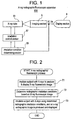
- Figure 1 illustrates the configuration of an example of a conventional X-ray radiographic/fluoroscopic apparatus.
- the X-ray radiographic/fluoroscopic apparatus 500 comprises an X-ray tube 51 in which a single fluoroscopic focus size or a single radiographic focus size is set, an irradiation condition determining section 55 for determining fluoroscopic irradiation conditions and radiographic irradiation conditions (such as the tube voltage and tube current of the X-ray tube 51, exposure dose and radiographic irradiation time), an irradiation control section 57 for controlling the operation of the X-ray tube 51, an imaging section 8 comprising an image intensifier or TV camera or the like for capturing an X-ray fluoroscopic image or X-ray radiographic image of a subject and outputting a fluoroscopic video signal Vf or radiographic video signal Vs, and a display section 9 for displaying an X-ray fluoroscopic image or X-ray radiographic image based on the fluoroscopic video signal Vf or radiographic video signal Vs.
- an irradiation condition determining section 55 for determining fluoroscopic ir
- Figure 2 is a flow chart illustrating an X-ray radiographic/fluoroscopic process by the X-ray radiographic/fluoroscopic apparatus 500.
- Step T1 a subject is irradiated with X-rays based on predefined fluoroscopic irradiation conditions, and an X-ray fluoroscopic image is produced and displayed.
- the fluoroscopic focus size is 0.3 mm, for example, and the input power rating of the X-ray tube 51 in this case is on the order of 10 kW (or less), for example.
- body thickness is calculated based on the X-ray fluoroscopic image to determine radiographic irradiation conditions from the body thickness.
- the radiographic focus size is 0.6 mm, for example, and the input power rating of the X-ray tube 51 in this case is on the order of 30 ⁇ 40 kW, for example.
- Step T3 the subject is irradiated with X-rays using the determined radiographic irradiation conditions, and an X-ray radiographic image is produced and displayed.
- the focus size is preferably made small for improved resolution.
- the focus size is small, the input power rating of the X-ray tube 51 is reduced, and hence the radiographic irradiation time (imaging time) required for the same exposure dose is prolonged, leading to a problem that the image is subject to blurring in radiographing an examined site having a large body thickness or large motion.
- the gastric wall is radiographed with a contrast material such as barium applied, blurring gets worse due to vibration of the X-ray tube 51 or the gastric wall and rundown of the barium from the gastric wall etc.
- the present invention seeks to provide an X-ray radiographic/fluoroscopic apparatus, X-ray radiographic method and X-ray radiographic/fluoroscopic method that can consistently produce X-ray radiographic images with high quality regardless of the magnitude of the body thickness or motion of an examined site.
- an X-ray radiographic/fluoroscopic apparatus comprising: X-ray emitting means having a function of emitting X-rays with a focus selected from at least one fluoroscopic focus size and a plurality of radiographic focus sizes; X-ray fluoroscopic image producing means for producing an X-ray fluoroscopic image by irradiating a subject with X-rays with said fluoroscopic focus size selected; radiographic focus size selecting means for selecting a relatively large radiographic focus size in radiographing an examined site having a relatively large body thickness, and selecting a relatively small radiographic focus size in radiographing an examined site having a relatively small body thickness; and X-ray radiographic image producing means for producing an X-ray radiographic image by irradiating the subject with X-rays equivalent to radiographic irradiation conditions determined based on said X-ray fluoroscopic image with said selected radiographic focus size.
- an X-ray radiographic/fluoroscopic apparatus comprising: X-ray emitting means having a function of emitting X-rays with a focus selected from at least one fluoroscopic focus size and a plurality of radiographic focus sizes; X-ray fluoroscopic image producing means for producing an X-ray fluoroscopic image by irradiating a subject with X-rays with said fluoroscopic focus size selected; radiographic focus size selecting means for selecting a relatively large radiographic focus size in radiographing an examined site having a relatively large motion, and selecting a relatively small radiographic focus size in radiographing an examined site having a relatively small motion; and X-ray radiographic image producing means for producing an X-ray radiographic image by irradiating the subject with X-rays equivalent to radiographic irradiation conditions determined based on said X-ray fluoroscopic image with said selected radiographic focus size.
- an X-ray radiographic/fluoroscopic apparatus comprising: X-ray emitting means having a function of emitting X-rays with a focus selected from at least one fluoroscopic focus size and a plurality of radiographic focus sizes; examined site input means for receiving an examined site from an operator; radiographic irradiation time threshold defining means for defining a radiographic irradiation time threshold that is an upper limit of a radiographic irradiation time corresponding to said examined site; X-ray fluoroscopic image producing means for producing an X-ray fluoroscopic image by irradiating a subject with X-rays with said fluoroscopic focus size selected; radiographic focus size selecting means for selecting a relatively large radiographic focus size if a radiographic irradiation time determined based on said X-ray fluoroscopic image is longer than said radiographic irradiation time threshold, and selecting a relatively small radiographic focus size if said determined radiographic irradiation
- an X-ray radiographic/fluoroscopic apparatus comprising: X-ray emitting means having a function of emitting X-rays with a focus selected from at least one fluoroscopic focus size and a plurality of radiographic focus sizes; X-ray fluoroscopic image producing means for producing an X-ray fluoroscopic image by irradiating a subject with X-rays with said fluoroscopic focus size selected; radiographic irradiation time threshold input means for receiving from an operator a radiographic irradiation time threshold that is an upper limit of a radiographic irradiation time corresponding to an examined site; radiographic focus size selecting means for selecting a relatively large radiographic focus size if a radiographic irradiation time determined based on said X-ray fluoroscopic image is longer than said radiographic irradiation time threshold, and selecting a relatively small radiographic focus size if said determined radiographic irradiation time is shorter than said radiographic irradiation
- an X-ray radiographic/fluoroscopic apparatus comprising: X-ray emitting means having a function of emitting X-rays with a focus selected from at least one fluoroscopic focus size and a plurality of radiographic focus sizes; examined site input means for receiving an examined site from an operator; X-ray fluoroscopic image producing means for producing an X-ray fluoroscopic image by irradiating a subject with X-rays with said fluoroscopic focus size selected; radiographic irradiation condition determining means for determining radiographic irradiation conditions based on said X-ray fluoroscopic image; radiographic focus size selecting means for selecting a radiographic focus size for use in X-ray radiography based on said examined site and said radiographic irradiation conditions; and X-ray radiographic image producing means for producing an X-ray radiographic image by irradiating the subject with X-rays equivalent to said radiographic irradiation conditions with said selected radiographic
- the X-ray emitting means may have a plurality of fluoroscopic focus sizes, preferably three or more.
- an X-ray radiographic method comprising the step of producing an X-ray radiographic image by irradiating a subject with X-rays with a relatively large radiographic focus size in radiographing an examined site having a relatively large body thickness, and by irradiating the subject with X-rays with a relatively small radiographic focus size in radiographing an examined site having a relatively small body thickness.
- an X-ray radiographic method comprising the step of producing an X-ray radiographic image by irradiating a subject with X-rays with a relatively large radiographic focus size in radiographing an examined site having a relatively large motion, and by irradiating the subject with X-rays with a relatively small radiographic focus size in radiographing an examined site having a relatively small motion.
- the radiographic focus size for use in the radiography may be selected from among three or more radiographic focus sizes.
- an X-ray radiographic/fluoroscopic method comprising the steps of: producing an X-ray fluoroscopic image by irradiating a subject with X-rays with a fluoroscopic focus size; selecting a relatively large radiographic focus size from among a plurality of radiographic focus sizes in radiographing an examined site having a relatively large body thickness, and selecting a relatively small radiographic focus size in radiographing an examined site having a relatively small body thickness; and producing an X-ray radiographic image by irradiating the subject with X-rays equivalent to radiographic irradiation conditions determined based on said X-ray fluoroscopic image with said selected radiographic focus size.
- an X-ray radiographic/fluoroscopic method comprising the steps of: producing an X-ray fluoroscopic image by irradiating a subject with X-rays with a fluoroscopic focus size; selecting a relatively large radiographic focus size from among a plurality of radiographic focus sizes in radiographing an examined site having a relatively large motion, and selecting a relatively small radiographic focus size in radiographing an examined site having a relatively small motion; and producing an X-ray radiographic image by irradiating the subject with X-rays equivalent to radiographic irradiation conditions determined based on said X-ray fluoroscopic image with said selected radiographic focus size.
- an X-ray radiographic/fluoroscopic method comprising the steps of: producing an X-ray fluoroscopic image by irradiating a subject with X-rays with a fluoroscopic focus size; receiving an examined site from an operator to define a radiographic irradiation time threshold that is an upper limit of a radiographic irradiation time corresponding to said examined site; selecting a relatively large radiographic focus size from among a plurality of radiographic focus sizes if a radiographic irradiation time determined based on said X-ray fluoroscopic image is longer than said radiographic irradiation time threshold, and selecting a relatively small radiographic focus size if said determined radiographic irradiation time is shorter than said radiographic irradiation time threshold; and producing an X-ray radiographic image by irradiating the subject with X-rays equivalent to radiographic irradiation conditions determined based on said X-ray fluoroscopic image with said selected
- an X-ray radiographic/fluoroscopic method comprising the steps of: producing an X-ray fluoroscopic image by irradiating a subject with X-rays with a fluoroscopic focus size; receiving from an operator a radiographic irradiation time threshold that is an upper limit of a radiographic irradiation time corresponding to an examined site; selecting a relatively large radiographic focus size from among a plurality of radiographic focus sizes if a radiographic irradiation time determined based on said X-ray fluoroscopic image is longer than said radiographic irradiation time threshold, and selecting a relatively small radiographic focus size if said determined radiographic irradiation time is shorter than said radiographic irradiation time threshold; and producing an X-ray radiographic image by irradiating the subject with X-rays equivalent to radiographic irradiation conditions determined based on said X-ray fluoroscopic image with said selected radiographic focus size.
- an X-ray radiographic/fluoroscopic method comprising the steps of: producing an X-ray fluoroscopic image by irradiating a subject with X-rays with a fluoroscopic focus size; determining radiographic irradiation conditions based on said X-ray fluoroscopic image; selecting a radiographic focus size for use in X-ray radiography from among a plurality of radiographic focus sizes based on an examined site and said radiographic irradiation conditions; and producing an X-ray radiographic image by irradiating the subject with X-rays equivalent to said radiographic irradiation conditions with said selected radiographic focus size.
- the radiographic focus size for use in the radiography may be selected from among three or more radiographic focus sizes.
- the radiography when an examined site having a relatively large body thickness is radiographed, the radiography is performed with a relatively large radiographic focus size selected, whereby the X-ray irradiation strength is enhanced and the radiographic irradiation time reduced to reduce blurring of the examined site.
- the radiography is performed with a relatively small radiographic focus size selected, whereby the resolution of the X-ray radiographic image is improved.
- X-ray radiographic/fluoroscopic apparatus of the second aspect X-ray radiographic method of the seventh aspect and X-ray radiographic/fluoroscopic method of the ninth aspect
- the radiography is performed with a relatively large radiographic focus size selected, whereby the X-ray irradiation strength is enhanced and the radiographic irradiation time reduced to reduce blurring of the examined site.
- the radiography is performed with a relatively small radiographic focus size selected, whereby the resolution of the X-ray radiographic image is improved.
- X-ray radiographic images can be consistently produced with high quality regardless of the magnitude of motion of the examined site.
- a shorter radiographic irradiation time threshold is defined for an examined site having a larger degree of body thickness or motion
- a relatively large radiographic focus size is selected and blurring of the examined site can be reduced.
- a relatively small radiographic focus size is selected and an X-ray radiographic image can be produced with high resolution.
- the operator can finely specify a radiographic irradiation time threshold in conformity with image quality preferences or clinical interest while viewing an X-ray fluoroscopic image.
- X-ray radiographic/fluoroscopic apparatus of the fifth aspect and X-ray radiographic/fluoroscopic method of the twelfth aspect since the radiographic focus size for use in X-ray radiography is selected based on an examined site and radiographic irradiation conditions, X-ray radiographic images can be consistently produced with high quality regardless of the magnitude of body thickness or motion of the examined site.
- the fluoroscopic focus size for use in fluoroscopy can be selected from among a plurality of fluoroscopic focus sizes, image quality of the X-ray fluoroscopic image can be improved.
- radiographic focus size for use in radiography can be selected from among three or more radiographic focus sizes, a radiographic focus size suitable for radiography of the examined site can be more finely selected.
- an advantage resulting from magnification of the radiographic focus size i.e., reduction of blurring by reducing the irradiation time
- an advantage resulting from reduction of the radiographic focus size i.e., improvement of resolution by suppressing penumbra formation
- Figure 1 illustrates the configuration of an example of a conventional X-ray radiographic/fluoroscopic apparatus.
- Figure 2 is a flow chart illustrating an X-ray radiographic/fluoroscopic process by the X-ray radiographic/fluoroscopic apparatus shown in Figure 1.
- Figure 3 illustrates the configuration of an X-ray radiographic/fluoroscopic apparatus in accordance with a first embodiment of the present invention.
- Figure 4 illustrates the content of the radiographic irradiation time threshold table in the X-ray radiographic/fluoroscopic apparatus of Figure 3.
- Figure 5 is a flow chart illustrating an X-ray radiographic/fluoroscopic process by the X-ray radiographic/fluoroscopic apparatus of Figure 3.
- Figure 6 is a characteristic diagram illustrating the result of simulation of spatial frequency - MTF value characteristics.
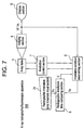
- Figure 7 illustrates the configuration of an X-ray radiographic/fluoroscopic apparatus in accordance with a second embodiment of the present invention.
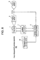
- Figure 8 illustrates the configuration of an X-ray radiographic/fluoroscopic apparatus in accordance with a third embodiment of the present invention.
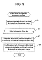
- Figure 9 is a flow chart of an X-ray radiographic/fluoroscopic process by the X-ray radiographic/fluoroscopic apparatus of Figure 8.
- Figure 3 illustrates the configuration of an X-ray radiographic/fluoroscopic apparatus in accordance with a first embodiment of the present invention.
- the X-ray radiographic/fluoroscopic apparatus 100 comprises an X-ray tube 1 having a function of emitting X-rays with a focus selected from a single fluoroscopic focus size and a plurality of radiographic focus sizes, an examined site input section 2 for receiving an examined site from a human operator (physician or technician), a radiographic irradiation time threshold table 3 for previously storing an upper limit of the radiographic irradiation time corresponding to each examined site, i.e., a radiographic irradiation time threshold Th, a radiographic irradiation time threshold readout section 4 for reading the radiographic irradiation time threshold Th corresponding to the examined site received from the operator out of the radiographic irradiation time threshold table 3, an irradiation condition determining section 5 for determining fluoroscopic irradiation conditions and radiographic irradiation conditions (such as the tube voltage, tube current, exposure dose, and radiographic irradiation time Ts), a radiographic ir
- the fluoroscopic focus size is 0.3 mm, for example, and the input power rating of the X-ray tube 1 in this case is on the order of 10 kW (or less), for example.
- the radiographic focus size is, for example, selected from between 0.6 mm and 1.25 mm. If the former is selected, the input power rating of the X-ray tube 1 is 34 kW, the tube voltage rating is 70 kV, and the tube current rating is 140 mA, for example. If the latter is selected, the input power rating is 76 kW, the tube voltage rating is 70 kV, and the tube current rating is 560 mA, for example.
- Figure 4 illustrates the content of the radiographic irradiation time threshold table 3.
- a radiographic irradiation time threshold Th is stored for each examined site.
- a relatively short radiographic irradiation time threshold Th is stored for an examined site in which a combined value of the degree of (standard) body thickness and vibration is large, and a relatively long radiographic irradiation time threshold Th is stored for an examined site in which the combined value is small.
- the radiographic irradiation time threshold Th is 5 ms for the "Stomach”, which has a relatively large combined value
- the radiographic irradiation time threshold Th is 10 ms for the "Small & large intestines", which have a relatively small combined value.
- the vibration speed of the "Stomach” is on the order of 28 mm/sec, for example.
- Figure 5 is a flow chart illustrating an X-ray radiographic/fluoroscopic process by the X-ray radiographic/fluoroscopic apparatus 100.
- Step S1 the operator inputs an examined site using the examined site input section 2.
- Step S2 a radiographic irradiation time threshold Th corresponding to the examined site is read out of the radiographic irradiation time threshold table 3.
- Th 5 ms is read out.
- Step S3 the subject is irradiated with X-rays based on predefined fluoroscopic irradiation conditions, and an X-ray fluoroscopic image is produced and displayed.
- the body thickness is calculated based on the X-ray fluoroscopic image to determine radiographic irradiation conditions (which can properly set image brightness on the image plane) when the X-rays are emitted with a small radiographic focus size selected.
- the operator may manually specify the radiographic irradiation conditions while viewing the X-ray fluoroscopic image.
- the radiographic irradiation conditions are, for example, an exposure dose of 0.14 mAs and a radiographic irradiation time Ts of 10 ms.
- Step S5 a small radiographic focus size is selected.
- a radiographic focus size of 0.6 mm is selected.
- Step S6 the radiographic irradiation time threshold Th read at Step S2 and the radiographic irradiation time Ts determined at Step S4 are compared, and if Th ⁇ Ts, the process goes to Step S7; otherwise (i.e., if Th ⁇ Ts) to Step S8.
- Step S7 a larger radiographic focus size is selected.
- a radiographic focus size of 1.25 mm is selected.
- Step S8 the subject is irradiated with X-rays equivalent to the radiographic irradiation conditions (i.e., X-rays of identical exposure dose) with the radiographic focus size selected at Step S5 or S7, and an X-ray radiographic image is produced and displayed.
- X-rays equivalent to the radiographic irradiation conditions may be emitted within a radiographic irradiation time on the order of Ts/4.
- Figure 6 is a characteristic diagram illustrating the result of simulation of spatial frequency - MTF (modulation transfer function) value characteristics when two examined sites having constant vibration and different body thicknesses are irradiated with X-rays with large and small radiographic focus sizes.
- the characteristic curve ⁇ 1 indicated by a dotted line is obtained on the assumption that an examined site having a small body thickness on the order of 20 cm, for example, is radiographed with a small radiographic focus size (0.6 mm).
- the characteristic curve ⁇ 2 indicated by a solid line is obtained on the assumption that an examined site having a small body thickness is radiographed with a large radiographic focus size (1.25 mm).
- the resolution is better for the characteristic curve ⁇ 1, which has a higher spatial frequency with respect to the same MTF value. That is, when an examined site having a small body thickness is to be radiographed, it is preferred that a small radiographic focus size be selected.
- the characteristic curve ⁇ 1 indicated by a dot-dash line is obtained on the assumption that an examined site having a large body thickness on the order of 30 cm, for example, is radiographed with a large radiographic focus size.
- the characteristic curve ⁇ 2 indicated by a double-dot-dash line is obtained when a subject having a large body thickness is radiographed with a small radiographic focus size.
- the resolution is better for the characteristic curve ⁇ 1, which has a higher spatial frequency with respect to the same MTF value. That is, when an examined site having a large body thickness is to be radiographed, it is preferred that a large radiographic focus size be selected.
- the resolution can be improved by selecting the large radiographic focus size for the examined site having a large motion to reduce the radiographic irradiation time; and the resolution can be improved by selecting the small radiographic focus size for the examined site having a small motion to suppress penumbra formation.
- a shorter radiographic irradiation time threshold Th is predefined when the combined value of the degree of body thickness and motion is large. If the radiographic irradiation time Ts is longer than the radiographic irradiation time threshold Th, a larger radiographic focus size is selected, and thus the radiographic irradiation time can be reduced and blurring of the examined site can be mitigated. If the radiographic irradiation time Ts is shorter than the radiographic irradiation time threshold Th, a small radiographic focus size is selected, and thus penumbra formation can be suppressed and resolution of the X-ray radiographic image can be improved.
- Figure 7 illustrates the configuration of an X-ray radiographic/fluoroscopic apparatus in accordance with a second embodiment of the present invention.
- the X-ray radiographic/fluoroscopic apparatus 200 comprises a radiographic irradiation time threshold input section 21 for receiving a radiographic irradiation time threshold Th from the operator, an irradiation condition determining section 5 for determining fluoroscopic irradiation conditions and radiographic irradiation conditions (such as the tube voltage, tube current, exposure dose and radiographic irradiation time Ts), and a radiographic irradiation time comparing section 6 for comparing the radiographic irradiation time threshold Th and the radiographic irradiation time Ts and sending the result C of the comparison to the irradiation condition determining section 5.
- the X-ray tube 1, irradiation control section 7, imaging section 8 and display section 9 are the same components as those of the X-ray radiographic/fluoroscopic apparatus 100 of the first embodiment.
- the configuration can be simplified. Moreover, the operator can finely specify the radiographic irradiation time threshold Th in conformity with image quality preferences or clinical interest while viewing an X-ray fluoroscopic image.
- Figure 8 illustrates the configuration of an X-ray radiographic/fluoroscopic apparatus in accordance with a third embodiment of the present invention.
- the X-ray radiographic/fluoroscopic apparatus 300 comprises a radiographic focus size input section 31 for receiving a radiographic focus size from the operator, and an irradiation condition determining section 5 for determining fluoroscopic irradiation conditions and radiographic irradiation conditions (such as the tube voltage, tube current, exposure dose and radiographic irradiation time Ts).
- the X-ray tube 1, irradiation control section 7, imaging section 8 and display section 9 are the same components as those of the X-ray radiographic/fluoroscopic apparatus 100 of the first embodiment.
- Figure 9 is a flow chart illustrating an X-ray radiographic/fluoroscopic process by the X-ray radiographic/fluoroscopic apparatus 300.
- Step S3 the subject is irradiated with X-rays based on predefined fluoroscopic irradiation conditions, and an X-ray fluoroscopic image is produced and displayed.
- Step SS4 the operator selects a radiographic focus size using the radiographic focus size input section 31 while viewing the X-ray fluoroscopic image.
- the selection criterion is any one of (1) - (3) as follows:
- Step S4' radiographic irradiation conditions are determined in conformity with the selected radiographic focus size.
- Step S8' the subject is irradiated with X-rays using the determined radiographic irradiation conditions, and an X-ray radiographic image is produced and displayed.
- the configuration can be further simplified. Moreover, the operator can finely select a radiographic focus size in conformity with image quality preferences or clinical interest while viewing an X-ray fluoroscopic image.
Landscapes
- Health & Medical Sciences (AREA)
- Life Sciences & Earth Sciences (AREA)
- Medical Informatics (AREA)
- Engineering & Computer Science (AREA)
- General Health & Medical Sciences (AREA)
- Physics & Mathematics (AREA)
- Radiology & Medical Imaging (AREA)
- Molecular Biology (AREA)
- Biophysics (AREA)
- Nuclear Medicine, Radiotherapy & Molecular Imaging (AREA)
- Optics & Photonics (AREA)
- Pathology (AREA)
- Veterinary Medicine (AREA)
- Biomedical Technology (AREA)
- Heart & Thoracic Surgery (AREA)
- High Energy & Nuclear Physics (AREA)
- Surgery (AREA)
- Animal Behavior & Ethology (AREA)
- Public Health (AREA)
- Toxicology (AREA)
- General Physics & Mathematics (AREA)
- Apparatus For Radiation Diagnosis (AREA)
- Analysing Materials By The Use Of Radiation (AREA)
- X-Ray Techniques (AREA)
Applications Claiming Priority (2)
| Application Number | Priority Date | Filing Date | Title |
|---|---|---|---|
| JP31658299 | 1999-11-08 | ||
| JP31658299A JP2001137223A (ja) | 1999-11-08 | 1999-11-08 | X線透視撮影装置、x線撮影方法およびx線透視撮影方法 |
Publications (2)
| Publication Number | Publication Date |
|---|---|
| EP1098554A2 true EP1098554A2 (de) | 2001-05-09 |
| EP1098554A3 EP1098554A3 (de) | 2003-06-18 |
Family
ID=18078702
Family Applications (1)
| Application Number | Title | Priority Date | Filing Date |
|---|---|---|---|
| EP00309913A Withdrawn EP1098554A3 (de) | 1999-11-08 | 2000-11-08 | Radiographische/Fluoroskopische Röntgenstrahlungsvorrichtung, Röntgenstrahlungs-Abbildungsverfahren und radiographisches/fluoroskopisches Röntgenstrahlungsverfahren |
Country Status (5)
| Country | Link |
|---|---|
| US (1) | US6426996B1 (de) |
| EP (1) | EP1098554A3 (de) |
| JP (1) | JP2001137223A (de) |
| KR (1) | KR100367445B1 (de) |
| CN (1) | CN1295249A (de) |
Cited By (2)
| Publication number | Priority date | Publication date | Assignee | Title |
|---|---|---|---|---|
| WO2019202197A1 (en) * | 2018-04-20 | 2019-10-24 | Outotec (Finland) Oy | X-ray fluorescence analyser, and a method for performing x-ray fluorescence analysis |
| US11680913B2 (en) | 2018-04-20 | 2023-06-20 | Outotec (Finland) Oy | X-ray fluorescence analyzer system and a method for performing X-ray fluorescence analysis of an element of interest in slurry |
Families Citing this family (13)
| Publication number | Priority date | Publication date | Assignee | Title |
|---|---|---|---|---|
| JP2005270201A (ja) * | 2004-03-23 | 2005-10-06 | Fuji Photo Film Co Ltd | X線撮影装置 |
| JP2009077839A (ja) * | 2007-09-26 | 2009-04-16 | Fujifilm Corp | 放射線画像処理装置及び処理方法 |
| JP5433334B2 (ja) * | 2009-07-27 | 2014-03-05 | 株式会社東芝 | X線ct装置 |
| JP5572040B2 (ja) * | 2009-09-28 | 2014-08-13 | 富士フイルム株式会社 | 放射線撮影装置 |
| US9014336B2 (en) * | 2010-12-15 | 2015-04-21 | Koninklijke Philips N.V. | Power supply unit for an X-ray tube |
| JP6050356B2 (ja) * | 2012-07-17 | 2016-12-21 | 株式会社日立製作所 | X線透視撮影装置及びx線透視撮影装置の制御方法 |
| JP5716069B2 (ja) * | 2013-09-30 | 2015-05-13 | 株式会社東芝 | X線ct装置 |
| KR101697665B1 (ko) | 2015-07-09 | 2017-01-19 | (주)디알젬 | X선 투시촬영장치용 필라멘트 구동 회로 |
| US10531855B2 (en) | 2016-12-28 | 2020-01-14 | Canon Medical Systems Corporation | X-ray computed tomography apparatus |
| EP3482688A1 (de) * | 2017-11-08 | 2019-05-15 | Koninklijke Philips N.V. | Vorrichtung und verfahren zur bestimmung von bildparametern zur erzeugung eines röntgenimpulses |
| JP7132092B2 (ja) * | 2018-11-08 | 2022-09-06 | 富士フイルムヘルスケア株式会社 | X線透視撮影装置 |
| EP4218582B1 (de) * | 2020-09-25 | 2025-10-01 | FUJIFILM Corporation | Einstellvorrichtung, einstellverfahren und einstellprogramm |
| CN113707519B (zh) * | 2021-09-17 | 2024-03-12 | 武汉联影医疗科技有限公司 | 一种基于动压滑动轴承的x射线管的运行控制方法和系统 |
Family Cites Families (4)
| Publication number | Priority date | Publication date | Assignee | Title |
|---|---|---|---|---|
| US4685118A (en) * | 1983-11-10 | 1987-08-04 | Picker International, Inc. | X-ray tube electron beam switching and biasing method and apparatus |
| JP3662688B2 (ja) * | 1996-03-08 | 2005-06-22 | 株式会社日立メディコ | X線撮影装置 |
| US6075837A (en) * | 1998-03-19 | 2000-06-13 | Picker International, Inc. | Image minifying radiographic and fluoroscopic x-ray system |
| US6222907B1 (en) * | 1999-07-12 | 2001-04-24 | General Electric Company | Image quality optimization using an X-ray model based optimization |
-
1999
- 1999-11-08 JP JP31658299A patent/JP2001137223A/ja not_active Withdrawn
-
2000
- 2000-10-11 US US09/686,226 patent/US6426996B1/en not_active Expired - Fee Related
- 2000-11-07 KR KR10-2000-0065764A patent/KR100367445B1/ko not_active Expired - Fee Related
- 2000-11-08 EP EP00309913A patent/EP1098554A3/de not_active Withdrawn
- 2000-11-08 CN CN00130928A patent/CN1295249A/zh active Pending
Non-Patent Citations (1)
| Title |
|---|
| No Search * |
Cited By (3)
| Publication number | Priority date | Publication date | Assignee | Title |
|---|---|---|---|---|
| WO2019202197A1 (en) * | 2018-04-20 | 2019-10-24 | Outotec (Finland) Oy | X-ray fluorescence analyser, and a method for performing x-ray fluorescence analysis |
| US11680913B2 (en) | 2018-04-20 | 2023-06-20 | Outotec (Finland) Oy | X-ray fluorescence analyzer system and a method for performing X-ray fluorescence analysis of an element of interest in slurry |
| US11815480B2 (en) | 2018-04-20 | 2023-11-14 | Outotec (Finland) Oy | X-ray fluorescence analyzer and a method for performing an x-ray fluorescence analysis |
Also Published As
| Publication number | Publication date |
|---|---|
| KR100367445B1 (ko) | 2003-01-10 |
| EP1098554A3 (de) | 2003-06-18 |
| KR20010051488A (ko) | 2001-06-25 |
| JP2001137223A (ja) | 2001-05-22 |
| US6426996B1 (en) | 2002-07-30 |
| CN1295249A (zh) | 2001-05-16 |
Similar Documents
| Publication | Publication Date | Title |
|---|---|---|
| EP1098554A2 (de) | Radiographische/Fluoroskopische Röntgenstrahlungsvorrichtung, Röntgenstrahlungs-Abbildungsverfahren und radiographisches/fluoroskopisches Röntgenstrahlungsverfahren | |
| US8175357B2 (en) | X-ray diagnostic apparatus, image processing apparatus, and image processing method | |
| EP1216661A2 (de) | Medizinisches Diagnose-Verfahren und -Gerät zur Steuerung von zwei-Energien Belichtungsverfahren auf Basis von Bildinformationen | |
| EP2047800A1 (de) | Röntgengerät | |
| JP2002158862A (ja) | 医用画像処理方法及び医用画像処理装置 | |
| EP0206156B1 (de) | Bilderzeugungssystem mittels Röntgenstrahlen | |
| JPH10234714A (ja) | X線撮像装置 | |
| US4562464A (en) | X-Ray diagnostic apparatus | |
| JP2001340321A (ja) | X線診断装置 | |
| JP5043403B2 (ja) | X線透視撮影装置 | |
| JP2015009097A (ja) | X線診断装置 | |
| JP2003230555A (ja) | X線透視撮影診断装置 | |
| JPH0654257A (ja) | ディジタル・サブトラクション・アンジアグラフィ装置 | |
| JP2005198975A (ja) | X線診断装置及びその方法 | |
| JP2023130559A (ja) | 移動型放射線撮影装置、動態画像出力方法及びプログラム | |
| JPH1147122A (ja) | X線画像診断装置 | |
| US6510253B1 (en) | Image information network | |
| JP3414440B2 (ja) | X線tv装置 | |
| JPH1094539A (ja) | ステレオ透視対応x線映像装置 | |
| JP3623308B2 (ja) | X線画像診断装置 | |
| JPH11290306A (ja) | X線装置 | |
| JPH09327456A (ja) | X線撮影方法及びx線撮影装置 | |
| JPH04292144A (ja) | X線透視撮影装置 | |
| US20050220274A1 (en) | Workflow improvement | |
| JP2002143137A (ja) | X線透視撮影装置 |
Legal Events
| Date | Code | Title | Description |
|---|---|---|---|
| PUAI | Public reference made under article 153(3) epc to a published international application that has entered the european phase |
Free format text: ORIGINAL CODE: 0009012 |
|
| AK | Designated contracting states |
Kind code of ref document: A2 Designated state(s): AT BE CH CY DE DK ES FI FR GB GR IE IT LI LU MC NL PT SE TR |
|
| AX | Request for extension of the european patent |
Free format text: AL;LT;LV;MK;RO;SI |
|
| PUAL | Search report despatched |
Free format text: ORIGINAL CODE: 0009013 |
|
| AK | Designated contracting states |
Designated state(s): AT BE CH CY DE DK ES FI FR GB GR IE IT LI LU MC NL PT SE TR |
|
| AX | Request for extension of the european patent |
Extension state: AL LT LV MK RO SI |
|
| 17P | Request for examination filed |
Effective date: 20031218 |
|
| AKX | Designation fees paid |
Designated state(s): DE FR GB |
|
| 17Q | First examination report despatched |
Effective date: 20050125 |
|
| STAA | Information on the status of an ep patent application or granted ep patent |
Free format text: STATUS: THE APPLICATION IS DEEMED TO BE WITHDRAWN |
|
| 18D | Application deemed to be withdrawn |
Effective date: 20050805 |