EP0893784A2 - Verfahren und Gerät für Strahlungstomographie - Google Patents
Verfahren und Gerät für Strahlungstomographie Download PDFInfo
- Publication number
- EP0893784A2 EP0893784A2 EP98305869A EP98305869A EP0893784A2 EP 0893784 A2 EP0893784 A2 EP 0893784A2 EP 98305869 A EP98305869 A EP 98305869A EP 98305869 A EP98305869 A EP 98305869A EP 0893784 A2 EP0893784 A2 EP 0893784A2
- Authority
- EP
- European Patent Office
- Prior art keywords
- subject
- projection data
- radiation
- data
- image
- Prior art date
- Legal status (The legal status is an assumption and is not a legal conclusion. Google has not performed a legal analysis and makes no representation as to the accuracy of the status listed.)
- Granted
Links
- 230000005855 radiation Effects 0.000 title claims abstract description 64
- 238000000034 method Methods 0.000 title claims abstract description 26
- 238000003325 tomography Methods 0.000 title claims abstract description 25
- 238000004364 calculation method Methods 0.000 claims abstract description 32
- 230000000747 cardiac effect Effects 0.000 claims description 35
- 238000004519 manufacturing process Methods 0.000 claims description 10
- 238000005259 measurement Methods 0.000 claims description 5
- 238000013213 extrapolation Methods 0.000 claims description 2
- 238000001514 detection method Methods 0.000 description 20
- 238000010586 diagram Methods 0.000 description 12
- 238000003384 imaging method Methods 0.000 description 12
- 230000000737 periodic effect Effects 0.000 description 11
- 230000000241 respiratory effect Effects 0.000 description 11
- 238000004846 x-ray emission Methods 0.000 description 8
- 210000001835 viscera Anatomy 0.000 description 7
- 238000002591 computed tomography Methods 0.000 description 6
- 230000000694 effects Effects 0.000 description 5
- 238000012544 monitoring process Methods 0.000 description 5
- 230000001276 controlling effect Effects 0.000 description 4
- 230000001105 regulatory effect Effects 0.000 description 4
- 210000004072 lung Anatomy 0.000 description 3
- 238000012545 processing Methods 0.000 description 3
- 230000029058 respiratory gaseous exchange Effects 0.000 description 3
- 230000007423 decrease Effects 0.000 description 2
- 210000001015 abdomen Anatomy 0.000 description 1
- 238000013459 approach Methods 0.000 description 1
- 125000004122 cyclic group Chemical group 0.000 description 1
- 238000011156 evaluation Methods 0.000 description 1
- 238000007781 pre-processing Methods 0.000 description 1
- 230000010349 pulsation Effects 0.000 description 1
- 239000004065 semiconductor Substances 0.000 description 1
- 230000001360 synchronised effect Effects 0.000 description 1
- 229910052724 xenon Inorganic materials 0.000 description 1
- FHNFHKCVQCLJFQ-UHFFFAOYSA-N xenon atom Chemical compound [Xe] FHNFHKCVQCLJFQ-UHFFFAOYSA-N 0.000 description 1
Images


Classifications
-
- A—HUMAN NECESSITIES
- A61—MEDICAL OR VETERINARY SCIENCE; HYGIENE
- A61B—DIAGNOSIS; SURGERY; IDENTIFICATION
- A61B6/00—Apparatus or devices for radiation diagnosis; Apparatus or devices for radiation diagnosis combined with radiation therapy equipment
- A61B6/02—Arrangements for diagnosis sequentially in different planes; Stereoscopic radiation diagnosis
- A61B6/03—Computed tomography [CT]
- A61B6/032—Transmission computed tomography [CT]
-
- G—PHYSICS
- G06—COMPUTING; CALCULATING OR COUNTING
- G06T—IMAGE DATA PROCESSING OR GENERATION, IN GENERAL
- G06T11/00—2D [Two Dimensional] image generation
- G06T11/003—Reconstruction from projections, e.g. tomography
- G06T11/005—Specific pre-processing for tomographic reconstruction, e.g. calibration, source positioning, rebinning, scatter correction, retrospective gating
-
- A—HUMAN NECESSITIES
- A61—MEDICAL OR VETERINARY SCIENCE; HYGIENE
- A61B—DIAGNOSIS; SURGERY; IDENTIFICATION
- A61B6/00—Apparatus or devices for radiation diagnosis; Apparatus or devices for radiation diagnosis combined with radiation therapy equipment
- A61B6/54—Control of apparatus or devices for radiation diagnosis
- A61B6/541—Control of apparatus or devices for radiation diagnosis involving acquisition triggered by a physiological signal
-
- G—PHYSICS
- G06—COMPUTING; CALCULATING OR COUNTING
- G06T—IMAGE DATA PROCESSING OR GENERATION, IN GENERAL
- G06T2211/00—Image generation
- G06T2211/40—Computed tomography
- G06T2211/412—Dynamic
-
- Y—GENERAL TAGGING OF NEW TECHNOLOGICAL DEVELOPMENTS; GENERAL TAGGING OF CROSS-SECTIONAL TECHNOLOGIES SPANNING OVER SEVERAL SECTIONS OF THE IPC; TECHNICAL SUBJECTS COVERED BY FORMER USPC CROSS-REFERENCE ART COLLECTIONS [XRACs] AND DIGESTS
- Y10—TECHNICAL SUBJECTS COVERED BY FORMER USPC
- Y10S—TECHNICAL SUBJECTS COVERED BY FORMER USPC CROSS-REFERENCE ART COLLECTIONS [XRACs] AND DIGESTS
- Y10S378/00—X-ray or gamma ray systems or devices
- Y10S378/901—Computer tomography program or processor
Definitions
- the present invention relates to a radiation tomography method and apparatus for sequentially measuring projection data of a subject in a plurality of view directions around the subject by radiations transmitting through a plurality of paths, and producing a tomographic image of the subject based on the projection data.
- Radiation tomography apparatuses include the x-ray CT (computed tomography) apparatus, for example.
- x-rays are employed as radiation.
- the apparatus is configured to scan a subject by a radiation emission / detection apparatus, i.e., an x-ray emission / detection apparatus rotating around the subject to measure x-ray projection data of the subject respectively in a plurality of view directions around the subject, and produce (i.e., reconstruct) a tomographic image based on the projection data.
- a scan can be completed within about 0.8 sec. Accordingly, by scanning the subject synchronously with respiratory monitoring signals from the subject (i.e., patient), and in conformity with a time phase at which the body motion is slow such as the maximum inspiration or expiration time, a tomographic image for the lung field, abdomen etc. which is not much affected by body motion is produced.
- the half-scan technique comprises reconstructing an image from image information acquired by a half rotation of radiation beam generation means.
- the technique comprises: continuously scanning a subject a plurality of times over multiple cardiac strokes while monitoring ECG (electrocardiographic) signals; sorting the acquired projection data by phase based on the ECG signal; and reconstructing a tomographic image of the heart at each phase based on the sorted projection data.
- ECG electrocardiographic
- the half-scan technique cannot provide high enough image quality because of the low accuracy of the acquired data.
- an appropriate estimating calculation e.g., interpolation
- view data which are opposite each other obtained by opposite radiation beams to improve the image quality, but the opposite view data is not acquired with the half-scan technique, and it is impossible to perform such a processing for improving the image quality.
- the heart-gate scan technique requires a scan over multiple cardiac strokes, resulting in an increased scan duration and hence an increased x-ray exposure on the subject.
- the present invention provides a radiation tomography method comprising the steps of: measuring projection data representing a subject by radiation beams in a plurality of view directions around the subject; calculating estimated projection data for each of the plurality of view directions by performing a weighting calculation on data elements of the projection data generated by radiations which transmit through a same path in opposite directions so that a desired time phase for the subject is centrally weighted; and producing a tomographic image of the subject based on the estimated projection data.
- the present invention provides a radiation tomography apparatus comprising: radiation beam generation means for generating a radiation beam; measurement means for sequentially measuring projection data representing a subject by the radiation beam in a plurality of view directions around the subject; estimated projection data calculation means for calculating estimated projection data for each of the plurality of view directions by performing a weighting calculation on data elements of the projection data generated by radiations which transmit through a same path in opposite directions; control means for controlling the weighting calculation so that the weighting calculation is performed centered on a desired time phase for the subject; and image production means for producing a tomographic image of the subject based on the estimated projection data.
- a start time for the weighting calculation is regulated based on a periodic signal from the subject in order to obtain a tomographic image at an appropriate time phase.
- the lung of the subject is the examined object, for example, it is desirable to control the weighting calculation based on the periodic signal generated by respiration.
- the heart of the subject is the examined object, for example, it is desirable to control the weighting calculation based on the periodic signal generated by pulsation of the heart.
- the image information acquisition processing with the measurement weighted based on the ECG signal generated by the subject may be performed a plurality of times to produce tomographic images at a plurality of time phases of a cardiac stroke.
- the present invention provides the radiation tomography apparatus as described regarding the second aspect, wherein the radiation beam generation means generates a parallel beam.
- the present invention provides the radiation tomography apparatus as described regarding the second aspect, wherein the radiation beam generation means generates a fan beam.
- the present invention provides the radiation tomography apparatus as described regarding the second - fourth aspects, wherein the estimated projection data calculation means performs the weighting calculation by linear interpolation / extrapolation according to time phases at which the projection data which are opposite each other are acquired and the desired time phase.
- the present invention provides the radiation tomography apparatus as described regarding the second - fifth aspects, wherein the desired time phase for use in the control means is a time phase of the maximum expiration of the subject.
- the present invention provides the radiation tomography apparatus as described regarding the second - sixth aspects, wherein the desired time phase for use in the control means is defined at a plurality of points within one cardiac stroke of the subject.
- the present invention provides the radiation tomography apparatus as described regarding the second - seventh aspects further comprising: second image production means for producing a tomographic image of the subject based on the projection data for a plurality of views equivalent to those of a half rotation around the subject; and selection means for selecting the image production means or the second image production means to produce one of the tomographic images.
- the present invention provides the radiation tomography apparatus as described regarding the second - eighth aspects, wherein the desired time phase for use in the control means is defined at a plurality of points separated by intervals of a cardiac cycle of the subject, and a plurality of tomographic images produced by the image production means and corresponding to the plurality of time phases which are identical represent different locations in the subject.
- the radiation is preferably x-rays since practical means for generating, detecting and controlling x-rays are most widely available.
- an effective scan time can be made short, because a desired motion phase of the periodically moving internal organ which is examined is regulated to be centrally weighted when performing the weighting calculation on a pair of projection data elements generated by radiations which transmit through the same path in opposite directions. This enables a high image quality to be achieved in an effective scan time shorter than the actual scan time.
- imaging of a tomographic image close to a stationary image of the object internal organ at a desired time phase can be performed by sequentially measuring projection data representing a subject by radiation beams in a plurality of view directions around the subject; calculating estimated projection data by performing a weighting calculation on data elements of the projection data generated by radiations which transmit through the same path in opposite directions so that a desired time phase is centrally weighted; and producing a tomographic image based on the estimated projection data.
- a radiation tomography method and apparatus are realized which provide an effect equivalent to reducing the scan time.
- a full scan ensures sufficient data accuracy and allows the estimating calculation to be performed on the opposite view data, and, moreover, a tomographic image close to a stationary image can be produced by obtaining an effect equivalent to that obtained by defining a short imaging time span.
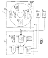
- FIG. 1 is a block diagram of an apparatus in accordance with one embodiment of the present invention.
- Figure 2 is a schematic diagram of a detector array in the apparatus in accordance with one embodiment of the present invention.
- Figure 3 is a schematic diagram of an x-ray emission / detection apparatus in the apparatus in accordance with one embodiment of the present invention.
- Figure 4 is a schematic diagram of the x-ray emission / detection apparatus in the apparatus in accordance with one embodiment of the present invention.
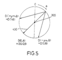
- Figure 5 is a diagram illustrating a relationship between a view angle and view data when a fan-shaped x-ray beam is employed.
- Figure 6 is a diagram illustrating a relationship between a view angle and view data when a fan-shaped x-ray beam is employed.
- Figure 7 is a diagram for explaining a relationship among a weighting factor, a gantry angle and a channel angle.
- Figure 8 is a graphical representation illustrating a profile of a weighting factor for use in an estimating calculation in the apparatus in accordance with one embodiment of the present invention.
- Figure 9 is a flow chart illustrating the operation of the apparatus in accordance with one embodiment of the present invention.
- Figure 10 is a graphical representation showing an example of a respiratory monitoring signal in the apparatus in accordance with one embodiment of the present invention.
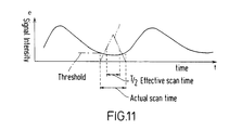
- Figure 11 is a diagram illustrating an example of a relationship between the respiratory monitoring signal and a scan time phase in the apparatus in accordance with one embodiment of the present invention.
- Figure 12 is a diagram illustrating an example of a relationship between an ECG signal and an image reconstruction time phase in the apparatus in accordance with one embodiment of the present invention.
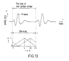
- Figure 13 is a diagram illustrating an example of a relationship between the weighting factor and cardiac strokes in the apparatus in accordance with one embodiment of the present invention.
- Figure 14 is a flow chart illustrating the operation of the apparatus in accordance with one embodiment of the present invention.
- Figure 15 is a diagram illustrating an example of a relationship between the weighting factor and cardiac strokes in the apparatus in accordance with one embodiment of the present invention.
- Figure 1 is a block diagram of an x-ray CT apparatus.
- the apparatus is one embodiment in accordance with the present invention. Its configuration represents one embodiment of the invention. Its operation represents another embodiment of the invention.
- the apparatus is comprised of a scanning gantry 2, an imaging table 4, an operator console 6 and a periodic motion monitor 10.
- the scanning gantry 2 has an x-ray tube 20 as a radiation source.
- X-rays emanating from the x-ray tube 20 are formed into, for example, a fan-shaped x-ray beam (i.e., fan beam) by a collimator 22 and impinge upon a detector array 24.
- the x-ray tube 20 and the collimator 22 are one example of the radiation beam generation means in the present invention.
- the detector array 24 has a plurality of x-ray detection elements arranged in an array along the width of the fan-shaped x-ray beam. The detailed configuration of the detector array 24 will be described later.
- the x-ray tube 20, the collimator 22 and the detector array 24 compose an x-ray emission / detection apparatus. The detailed configuration thereof will be described later.
- the detector array 24 is connected with a data acquisition section 26.
- the data acquisition section 26 collects data detected by the individual x-ray detection elements in the detector array 24.
- Emission of the x-rays from the x-ray tube 20 is governed by an x-ray controller 28.
- an x-ray controller 28 In the drawing, the connection between the x-ray tube 20 and the x-ray controller 28 is not shown.
- the collimator 22 is regulated by a collimator controller 30.
- a collimator controller 30 In the drawing, the connection between the collimator 22 and the collimator controller 30 is not shown.
- the x-ray tube 20 - collimator controller 30 are mounted on a rotating portion 32 in the scanning gantry 2. Rotation of the rotating portion 32 is governed by a rotation controller 34. In the drawing, the connection between the rotating portion 32 and the rotation controller is not shown.
- the imaging table 4 carries a subject (not shown) into /out of an x-ray irradiation space within the scanning gantry 2. The relationship between the subject and the x-ray irradiation space will be described later.
- the periodic motion monitor 10 detects a vital activity signal such as a respiratory signal or an ECG signal from the subject on the imaging table 4.
- the vital activity signal is an example of a periodic signal in the present invention.
- the operator console 6 has a CPU (central processing unit) 60 which is a computer, for example.
- the CPU 60 is connected with a control interface 62.
- the control interface 62 is connected with the scanning gantry 2 and the imaging table 4.
- the CPU 60 controls the scanning gantry 2 and the imaging table 4 via the control interface 62.
- the data acquisition portion 26, the x-ray controller 28, the collimator controller 30 and the rotation controller 34 in the scanning gantry 2 are controlled via the control interface 62.
- the individual connections between these parts and the control interface 62 are not shown.
- the CPU 60 is also connected with a data acquisition buffer 64.
- the data acquisition buffer 64 is connected with the data acquisition portion 26 and the periodic motion monitor 10. The data collected in the data acquisition portion 26 and output signals from the periodic motion monitor 10 are supplied to the data acquisition buffer 64.
- the data acquisition buffer 64 temporarily stores the supplied data.
- the CPU 60 is further connected with a storage device 66.
- the storage device 66 stores various data, reconstructed images, programs etc.
- the CPU 60 is also connected with a display 68 and a operating device 70.
- the display 68 presents a reconstructed image supplied from the CPU 60 and other information.
- the operating device 70 is manipulated by an operator, and is configured to supply various commands and information to the CPU 60.
- the description is exclusively made on an apparatus in which the radiation beam generation means generates a fan beam
- the present invention can easily be implemented by an apparatus in which the radiation beam generation means generates a parallel beam as will described later.
- Figure 2 schematically illustrates the configuration of the detector array 24 which detects a fan beam from the x-ray source.
- the detector array 24 is constituted as a multi-channel x-ray detector in which multiple (e.g., 1,000) x-ray detection elements 24(i) are arranged in an arc shape.
- the x-ray detection elements 24( i ) are solid-state detectors such as scintillation or semiconductor x-ray detectors. It will be understood that ion-chamber type detectors utilizing an ionized gas such as Xe (xenon) gas may also be employed.
- Figure 3 illustrates the interrelationship among the x-ray tube 20, the collimator 22 and the detector array 24 in the x-ray emission / detection apparatus.
- Figure 3(a) is a front view and Fig. 3(b) is a side view.
- x-rays emanating from the x-ray tube 20 are formed into a fan-shaped x-ray beam 40 by the collimator 22 and impinge upon the detector array 24.
- the extent, i.e., the width of the fan-shaped x-ray beam 40 is shown.
- the thickness of the x-ray beam 40 is shown.
- a virtual line 400 passing through a focal point of the x-ray tube 20 and the center of rotation 300 of the scanning gantry 2 is defined as an angular reference axis.
- the angular reference axis 400 extends to reach the center of the detector array 24.
- Each of the angles formed by virtual lines linking the focal point of the x-ray tube 20 with the respective x-ray detection elements 24( i ) and the angular reference axis 400 is referred to as a channel angle ⁇ .
- the channel angle ⁇ is 0 at the central x-ray detection element 24( I /2) in the detector array 24.
- the channel angle ⁇ is + ⁇ m at the x-ray detection element 24(1), the leftmost of the detector elements 24 in the drawing, and is - ⁇ m at the x-ray detection element 24( I ), the rightmost of the detector elements 24 in the drawing. Since the channel number i and the channel angle ⁇ have one-to-one correspondence, the x-ray detection element 24( i ) is expressed as an x-ray detection element 24( ⁇ ) hereinbelow.
- the subject is inserted with the subject's body axis intersecting the fan plane of the x-ray beam 40. This is illustrated in Fig. 4.
- a subject 8 rested on the imaging table 4 is inserted with the subject's body axis intersecting the fan plane of the x-ray beam 40.
- a projection image of the subject 8 sliced by the x-ray beam 40 is projected on the detector array 24.
- the thickness of the x-ray beam 40 at the isocenter of the subject 8 is the slice thickness th for the subject 8.
- the slice thickness th is determined by an x-ray passing aperture through the collimator 22.
- the x-ray emission / detection apparatus consisting of the x-ray tube 20, the collimator 22 and the detector array 24 rotates (i.e., scans) around the body axis of the subject 8 maintaining their interrelationship.
- Projection data representing the subject 8 are acquired at a plurality (e.g., 1,000) of view angles per scan rotation.
- the number of projection data elements per view is equal to the number of channels of the detector array 24 and is 1,000, for example.
- the projection data element from each channel represents the intensity of a transmitting x-ray which impinges upon the channel from the focal point of the x-ray tube 20. Accordingly, the projection data formed by, for example, 1,000 x-rays which travel different paths are acquired.
- Acquisition of the projection data is performed by a system consisting of the detector array 24 - the data acquisition portion 26 - the data acquisition buffer 64.
- the x-ray tube 20, the collimator 22, the detector array 24, the data acquisition portion 26 and the data acquisition buffer 64 are one example of the measuring means in accordance with the present invention.
- a view angle at which the projection data is measured will now be explained with reference to Fig. 5.
- An angle ⁇ formed by the angular reference axis 400 and, for example, a vertical axis at an angular position at which the x-ray emission / detection apparatus is placed by rotation is referred to as the view angle.
- the origin from which the view angle ⁇ is measured may be taken at any appropriate position instead of at the vertical axis.
- the opposite view data for each of the other channels is D(- ⁇ , ⁇ + ⁇ +2 ⁇ ). Since the view data which are opposite each other are acquired from different view angles, i.e., different rotational locations in the scanning gantry 2, they are different in data acquisition time point.
- the CPU 60 finds the respective opposite view data for the projection data acquired in the data acquisition buffer 64.
- the CPU 60 also calculates estimated projection data by a weighting calculation using a pair of view data elements which are opposite each other.
- the CPU 60 is an example of the estimated projection data calculation means in the present invention.
- the CPU 60 is an example of the control means in the present invention.
- This equation means that the weighting calculation on the opposite view data elements which form a pair gives the estimated projection data for use in producing a tomographic image with a desired time phase centrally weighted.
- Figure 7(a) - (d) illustrates how to determine the weighting factor w( ⁇ , ⁇ ) in connection with the gantry rotational angle and the channel angle ⁇ .
- the view data and its opposite view data are weighted, i.e., multiplied by weighting factors, so that they are proportionated according to the closeness from the angular location ⁇ to the radiation source location at the time at which the respective data are generated.
- the weighting factors are normalized so that the sum of the weighting factor of the view data and the weighting factor of its opposite view data would be 1. For example, when the radiation source is placed at an angular location ⁇ as shown in Fig. 7(a), its opposite view appears at the location ⁇ + ⁇ + 2 ⁇ and the weighting factor is expressed as:
- the weighting factor is given as follows:
- the weighting factor ( ⁇ , ⁇ ) for the estimating calculation is given as follows:
- Fig. 8 The variation in the weighting factor w( ⁇ , ⁇ ) during one rotation of the scanning gantry 2, that is, during the view angle varying from 0 to 2 ⁇ is shown in Fig. 8 for the data D(0, ⁇ ) as an example.
- the radiation beam generation means generates a parallel beam.
- the weighting factor is applied to all the beams.
- the estimating calculation of the projection data includes both of interpolative and extrapolative techniques according to a combination of opposite view data.
- the FWHM can be conceptually recognized as corresponding to an effective slice thickness of a helical scan, and it can be used for the evaluation of the image quality by the weighting.
- Figure 9 is a flow chart of the operation of the apparatus.
- the operation of the apparatus is started in response to a command supplied to the CPU 60 by the operator via the operating device 70. Thereafter, the operation of the apparatus proceeds under control of the CPU 60.
- Step 800 respiration monitoring on the subject is started in Step 800.
- Step 802 external signal input is performed in Step 802 in which the respiratory signal is supplied from the periodic motion monitor 10 to the CPU, and is presented on the display 68 via the CPU 60.
- a respiratory signal waveform which represents the time phases of the maximum inspiration and expiration is presented on the display 68 as exemplarily shown in Fig. 10.
- the operator ascertains the respiratory action of the subject 8 based on the presented waveform.
- the operator inputs an appropriate threshold for distinguishing the time phase of the maximum expiration, for example, via the operating device 70.
- an appropriate threshold for distinguishing the time phase of the maximum expiration for example, via the operating device 70.
- a condition of the subject including the internal organs at the maximum expiration is considered to be closest to the stationary condition. It will be understood that a value for distinguishing the time phase of the maximum inspiration may be defined as the threshold.
- a scan start trigger is generated in Step 804.
- Step 806 a scan is started in Step 806, x-rays are irradiated in Step 808 and data acquisition is performed in Step 810.
- the estimated projection data as described hereinabove is calculated using a pair of opposite view data elements in Step 814.
- Step 816 image reconstruction is performed based on the estimated projection data, and the reconstructed image is displayed and stored in Step 818.
- image reconstruction may be performed using data acquired effectively over a half of the actual scan time by the scanning gantry 2 centered on the time phase of T/2 .
- This relationship is exemplarily shown in Fig. 11.
- a time T/2 equal to the FWHM of the weighting factor profile can be regarded as an effective scan time corresponding to an effective scan slice thickness of a helical scan.
- the effective scan time is 0.4 sec.
- a tomographic image can be obtained scarcely affected by respiratory body motion of the subject 8.
- a tomographic image can be obtained at a desired time phase of a cardiac stroke in a similar manner as above.
- the cardiac stroke phase is determined by specifying an appropriate threshold etc. for an ECG signal.
- the above-described embodiment comprises the steps of: controlling a weighting calculation so that a desired time phase during one rotation of the gantry is principally weighted; coinciding the time phase with a time phase at which the motion of the internal organs of an examined object is relatively slow; acquiring image information principally at the time phase at which the motion is relatively slow; and producing an image close to a stationary image of the examined object's internal organs.
- the above image information acquisition procedure may be performed a plurality of times at a predetermined interval and may be used to sequentially observe the motion of the internal organs. That is, a continuous scan is performed over two cardiac strokes, for example, to acquire data during a plurality of rotations.
- data S1 equivalent to those of the first 360° can be used to obtain a tomographic image at one phase by performing image reconstruction from estimated data obtained from a pair of opposite view data elements.
- data S2 - S6 can be subsequently processed in the same way as above appropriately shifting a range covering 360° to obtain images at respective phases.
- the x-ray exposure on the subject is considerably lower than by the conventional heart-gate scan technique since the scan time corresponds to two cardiac strokes at most.
- a tomographic image at each phase can be imaged at each of the plurality of slice locations.
- the R wave (a wave which forms the maximum peak) in the ECG signal can be used as a scan trigger to facilitate aligning of the phases through the plurality of slices.
- Tomographic images for a plurality of slices at an identical phase can be used to construct a reformation image for an arbitrary slice or a three-dimensional image at every phase.
- the scan time Tcan be equalized to a time for one cardiac stroke (i.e., a cycle) which is usually about 1 sec.
- a cycle a time for one cardiac stroke which is usually about 1 sec.
- view data for all phases within one cycle of the cardiac stroke can be acquired by one scan. Since the cardiac stroke phases of the first and last views in the scan are identical, the cardiac stroke phases make one cycle preserving the continuity.
- a reconstructed image can be obtained in which the view data at the phase is given the maximum weight, hence a tomographic image of the heart which is not much affected by motion can be obtained.
- the centrally weighted location by aligning the centrally weighted location with a desired cardiac stroke phase, a tomographic image at the phase can be obtained.
- the image quality at a phase involving a rapid motion would be inevitably degraded.
- the centrally weighted location it is preferred that the R wave of the ECG waveform, for example, be selected as a reference location, because the reference location is distinct.
- the centrally weighted location may be defined by using a relative time within one scan time.
- FIG 14 is a flow chart illustrating the operation of the present apparatus when the heart is imaged as described above.
- a heart rate is first measured in Step 800'.
- the heart rate is measured based on a signal from the periodic motion monitor 10 such as an electrocardiograph.
- the average time for one cardiac stroke etc. is found from the measurement of the heart rate.
- Step 802' an optimum scan time is determined in Step 802'.
- the optimum scan time is determined so as to be equalized to the average time for one cardiac stroke, for example.
- Step 804' a weighting factor and a cardiac stroke phase with which the centrally weighted location is aligned are determined in Step 804'.
- Step 806 a scan is started.
- the scan is performed over the optimum scan time determined in Step 802'.
- scanning of the subject 8 reconstruction of a tomographic image, display of the reconstructed image etc. are performed according to the same procedure as shown by the flow chart in Fig. 9.
- One scan over a time equal to one cardiac stroke time, for example, is thus performed, and a tomographic image of the heart is obtained at the cardiac stroke phase as determined in Step 804'.
- image reconstruction can be performed again on the view data for one scan stored in the storage device 66 with the centrally weighted location shifted, to obtain another tomographic image of the heart at another phase. If necessary, by reconstructing images with the centrally weighted location sequentially shifted, tomographic images of the heart at various phases can be arbitrarily obtained from the view data for one scan. If the view data involving a part having a rapid motion is to be excluded, a segment reconstruction technique may be performed using the view data corresponding to a half scan instead of the above effective scan time-reduced image reconstruction technique in reconstructing an image. That is, according to use, which of the effective scan time-reduced image reconstruction and the segment reconstruction using half-scan view data is to be performed may be selected for the tomographic image to be obtained, and the image reconstruction may be performed according to the selection.
- the heart rate may be measured by a more convenient instrument such as a plusimeter, instead of the electrocardiograph.
- the cardiac stroke phase (the centrally weighted location) cannot be determined based on the R wave of the ECG waveform.
- a suitable image can be produced by obtaining reconstructed images for a number of centrally weighted locations appropriately defined within a scan time and selecting a suitable image.
- the cardiac stroke phases at the beginning of the respective scans become diverse. Accordingly, as shown in Fig. 15, the scans for slices 1, and 2, for example, are started at different cardiac stroke phases. However, either scan time is one cardiac stroke time T.
- centrally weighted locations for the view data acquired by such a multi-slice technique are defined at the same relative time within the respective scan times, tomographic images for the slices are different in cardiac stroke phase, resulting in inconvenience in observing all the slices from first to last.
- the cardiac stroke phase corresponding to the location at the relative time t c for the slice 1 repeatedly appears at every time T, the phase at the time nT after the relative time t c is identical to the phase at the relative time t c . Therefore, a tomographic image at the cardiac stroke phase the same as that of the slice 1 can be obtained based on the centrally weighted location defined at the time point as above.
- the tomographic images for all the slices can be provided with a common cardiac stroke phase.
- the radiation is not limited to x-rays but may be any other type of radiation such as ⁇ -rays.
- x-rays are currently preferred since practical means for generating, detecting and controlling x-rays are most widely available.
Landscapes
- Health & Medical Sciences (AREA)
- Engineering & Computer Science (AREA)
- Life Sciences & Earth Sciences (AREA)
- Physics & Mathematics (AREA)
- Medical Informatics (AREA)
- Theoretical Computer Science (AREA)
- Pathology (AREA)
- Heart & Thoracic Surgery (AREA)
- High Energy & Nuclear Physics (AREA)
- Veterinary Medicine (AREA)
- Nuclear Medicine, Radiotherapy & Molecular Imaging (AREA)
- Optics & Photonics (AREA)
- General Physics & Mathematics (AREA)
- Radiology & Medical Imaging (AREA)
- Biomedical Technology (AREA)
- Biophysics (AREA)
- Molecular Biology (AREA)
- Surgery (AREA)
- Animal Behavior & Ethology (AREA)
- General Health & Medical Sciences (AREA)
- Public Health (AREA)
- Pulmonology (AREA)
- Physiology (AREA)
- Apparatus For Radiation Diagnosis (AREA)
Applications Claiming Priority (6)
| Application Number | Priority Date | Filing Date | Title |
|---|---|---|---|
| JP198362/97 | 1997-07-24 | ||
| JP19836297 | 1997-07-24 | ||
| JP19836297 | 1997-07-24 | ||
| JP11347/98 | 1998-01-23 | ||
| JP1134798 | 1998-01-23 | ||
| JP10011347A JP3124254B2 (ja) | 1997-07-24 | 1998-01-23 | 放射線断層撮影装置 |
Publications (3)
| Publication Number | Publication Date |
|---|---|
| EP0893784A2 true EP0893784A2 (de) | 1999-01-27 |
| EP0893784A3 EP0893784A3 (de) | 2002-12-11 |
| EP0893784B1 EP0893784B1 (de) | 2007-10-10 |
Family
ID=26346759
Family Applications (1)
| Application Number | Title | Priority Date | Filing Date |
|---|---|---|---|
| EP98305869A Expired - Lifetime EP0893784B1 (de) | 1997-07-24 | 1998-07-23 | Verfahren und Gerät für Strahlungstomographie |
Country Status (7)
| Country | Link |
|---|---|
| US (1) | US5991356A (de) |
| EP (1) | EP0893784B1 (de) |
| JP (1) | JP3124254B2 (de) |
| KR (1) | KR100326197B1 (de) |
| CN (1) | CN100473357C (de) |
| DE (1) | DE69838533T2 (de) |
| TW (1) | TW477689B (de) |
Cited By (2)
| Publication number | Priority date | Publication date | Assignee | Title |
|---|---|---|---|---|
| EP1104917A2 (de) | 1999-11-16 | 2001-06-06 | General Electric Company | Adaptives Interpolationsverfahren und -System für Tomographie mit verringerter Anzahl von Datenansichten |
| EP1323378A2 (de) * | 2001-12-07 | 2003-07-02 | Philips Intellectual Property & Standards GmbH | Verfahren zur Rekonstruktion eines 3D-Bilddatensatzes eines Untersuchungsbereiches |
Families Citing this family (44)
| Publication number | Priority date | Publication date | Assignee | Title |
|---|---|---|---|---|
| JP3313611B2 (ja) * | 1997-05-06 | 2002-08-12 | ジーイー横河メディカルシステム株式会社 | 放射線断層撮影方法および装置 |
| US6233478B1 (en) * | 1998-09-28 | 2001-05-15 | Advanced Research & Technology Institute | Apparatus and method for constructing computed tomography image slices of an object undergoing cyclic motion |
| US8788020B2 (en) * | 1998-10-23 | 2014-07-22 | Varian Medical Systems, Inc. | Method and system for radiation application |
| US6937696B1 (en) | 1998-10-23 | 2005-08-30 | Varian Medical Systems Technologies, Inc. | Method and system for predictive physiological gating |
| US6243437B1 (en) * | 1998-11-25 | 2001-06-05 | General Electric Company | Coronary calcification detection using retrospective cardiac gating of imaging system |
| DE19854939C2 (de) * | 1998-11-27 | 2001-11-22 | Siemens Ag | Verfahren und Gerät zur Erzeugung von CT-Bildern |
| US6597803B1 (en) * | 1999-10-29 | 2003-07-22 | Ge Medical Systems Global Technology Company, Llc | Hybrid reconstruction for high pitch multi-slice helical cardiac imaging |
| US6466640B1 (en) * | 1999-11-26 | 2002-10-15 | Kabushiki Kaisha Toshiba | Computed tomography system and method |
| DE19957082B4 (de) * | 1999-11-28 | 2004-08-26 | Siemens Ag | Verfahren zur Untersuchung eines eine periodische Bewegung ausführenden Körperbereichs |
| US6301325B1 (en) * | 1999-12-22 | 2001-10-09 | Ge Medical Systems Global Technology Company Llc | Half-scan algorithm for use with a high speed multi-row fan beam helical detector |
| JP4773667B2 (ja) * | 2000-04-14 | 2011-09-14 | ゼネラル・エレクトリック・カンパニイ | 投影ビューの間の補間を用いた計算機式断層画像の再構成 |
| US6442228B1 (en) * | 2000-04-20 | 2002-08-27 | Ge Medical Systems Global Technology Company, Llc | Data acquisition modifications for improved reconstruction with conventional CT |
| US6539074B1 (en) * | 2000-08-25 | 2003-03-25 | General Electric Company | Reconstruction of multislice tomographic images from four-dimensional data |
| JP3942142B2 (ja) * | 2000-12-15 | 2007-07-11 | ジーイー・メディカル・システムズ・グローバル・テクノロジー・カンパニー・エルエルシー | 放射線断層撮影装置およびその方法 |
| US6463118B2 (en) | 2000-12-29 | 2002-10-08 | Ge Medical Systems Global Technology Company, Llc | Computed tomography (CT) weighting for high quality image recontruction |
| DE10123797B4 (de) * | 2001-05-16 | 2006-10-19 | Siemens Ag | Verfahren zur Herstellung eines Bildes mit einem Computertomographen |
| JP4175791B2 (ja) * | 2001-08-20 | 2008-11-05 | ジーイー・メディカル・システムズ・グローバル・テクノロジー・カンパニー・エルエルシー | 画像生成方法およびx線ct装置 |
| US6901131B2 (en) * | 2001-12-28 | 2005-05-31 | General Electric Company | Methods and apparatus for computed tomography imaging |
| US7054475B2 (en) * | 2001-12-28 | 2006-05-30 | General Electric Company | Apparatus and method for volumetric reconstruction of a cyclically moving object |
| JP4157302B2 (ja) * | 2002-01-10 | 2008-10-01 | 株式会社日立メディコ | X線ct装置 |
| DE10210646A1 (de) * | 2002-03-11 | 2003-10-09 | Siemens Ag | Verfahren zur Bilddarstellung eines in einen Untersuchungsbereich eines Patienten eingebrachten medizinischen Instruments |
| US7013034B2 (en) * | 2002-10-10 | 2006-03-14 | Ge Medical Systems Global Technology Company, Llc | Methods and apparatus for reconstructing an image of an object |
| JP4342274B2 (ja) | 2003-11-04 | 2009-10-14 | ジーイー・メディカル・システムズ・グローバル・テクノロジー・カンパニー・エルエルシー | X線ct装置 |
| JP4201686B2 (ja) * | 2003-11-04 | 2008-12-24 | ジーイー・メディカル・システムズ・グローバル・テクノロジー・カンパニー・エルエルシー | X線ct装置 |
| EP1694211B1 (de) * | 2003-12-08 | 2018-11-21 | Philips Intellectual Property & Standards GmbH | Computertomographie-verfahren für die periodische bewegung von objekten |
| JP4222930B2 (ja) * | 2003-12-10 | 2009-02-12 | ジーイー・メディカル・システムズ・グローバル・テクノロジー・カンパニー・エルエルシー | 3次元逆投影方法および装置並びにx線ct装置 |
| JP2005177203A (ja) * | 2003-12-22 | 2005-07-07 | Ge Medical Systems Global Technology Co Llc | 複数位置のct画像生成方法およびx線ct装置 |
| DE102004006548B4 (de) * | 2004-02-10 | 2006-10-19 | Siemens Ag | Verfahren zur Planung der Strahlentherapie eines Patienten und CT-System hierzu und zur Erstellung von CT-Aufnahmen |
| JP4091008B2 (ja) | 2004-03-09 | 2008-05-28 | ジーイー・メディカル・システムズ・グローバル・テクノロジー・カンパニー・エルエルシー | Ct画像生成方法およびx線ct装置 |
| JP4498023B2 (ja) * | 2004-06-15 | 2010-07-07 | キヤノン株式会社 | X線ct装置 |
| US20060090782A1 (en) * | 2004-11-01 | 2006-05-04 | Paul Bergman | Walking aid device |
| JP5389436B2 (ja) * | 2005-03-17 | 2014-01-15 | コーニンクレッカ フィリップス エヌ ヴェ | 心臓画像の反復的再構成のための方法及びデバイス |
| US7570733B2 (en) * | 2005-06-10 | 2009-08-04 | General Electric Company | Step-and-shoot cardiac CT imaging |
| RU2441587C2 (ru) * | 2006-05-26 | 2012-02-10 | Конинклейке Филипс Электроникс, Н.В. | Визуализация с помощью динамической компьютерной томографии |
| US7470907B2 (en) * | 2006-12-15 | 2008-12-30 | General Electric Company | Cross-slit collimator method and system |
| US8218720B2 (en) * | 2007-03-12 | 2012-07-10 | Varian Medical Systems, Inc. | Method and apparatus to facilitate reconstructing an image using fan-beam data |
| US10667727B2 (en) | 2008-09-05 | 2020-06-02 | Varian Medical Systems, Inc. | Systems and methods for determining a state of a patient |
| JP2011000358A (ja) * | 2009-06-22 | 2011-01-06 | Fujifilm Corp | 放射線撮影装置および方法 |
| US8270561B2 (en) * | 2010-10-13 | 2012-09-18 | Kabushiki Kaisha Toshiba | Motion weighting in computed tomography (CT) with cone angle |
| JP5813957B2 (ja) * | 2011-01-19 | 2015-11-17 | 株式会社東芝 | X線ct装置及び制御プログラム |
| CN103239253B (zh) * | 2012-02-14 | 2015-07-15 | 株式会社东芝 | 医用图像诊断装置 |
| US9047701B2 (en) * | 2012-03-31 | 2015-06-02 | Varian Medical Systems, Inc. | 4D cone beam CT using deformable registration |
| JP7305334B2 (ja) * | 2018-11-13 | 2023-07-10 | キヤノンメディカルシステムズ株式会社 | X線診断システム及び再構成処理システム |
| JP7393647B2 (ja) * | 2020-03-04 | 2023-12-07 | 日本製鉄株式会社 | 温度測定装置及び温度測定方法 |
Family Cites Families (8)
| Publication number | Priority date | Publication date | Assignee | Title |
|---|---|---|---|---|
| DE2939975A1 (de) * | 1979-10-02 | 1981-04-16 | Siemens AG, 1000 Berlin und 8000 München | Roentgenschichtgeraet zur herstellung von transversalschichtbildern |
| JPS62227324A (ja) * | 1986-03-27 | 1987-10-06 | 横河メディカルシステム株式会社 | 放射線断層撮影装置 |
| US5262946A (en) * | 1988-10-20 | 1993-11-16 | Picker International, Inc. | Dynamic volume scanning for CT scanners |
| US4994965A (en) * | 1988-11-23 | 1991-02-19 | General Electric Company | Method for reducing motion induced image artifacts in projection imaging |
| JP2970680B2 (ja) * | 1990-06-14 | 1999-11-02 | 株式会社東芝 | X線ct装置 |
| US5457728A (en) * | 1990-11-14 | 1995-10-10 | Cedars-Sinai Medical Center | Coronary tracking display |
| JP2770935B2 (ja) * | 1991-03-15 | 1998-07-02 | 株式会社日立メディコ | Ct装置 |
| DE19622075C2 (de) * | 1996-05-31 | 1999-10-14 | Siemens Ag | Verfahren und Gerät zur radiologischen Untersuchung von Herzphasen eines Patienten |
-
1998
- 1998-01-23 JP JP10011347A patent/JP3124254B2/ja not_active Expired - Fee Related
- 1998-05-26 US US09/085,523 patent/US5991356A/en not_active Expired - Lifetime
- 1998-07-10 TW TW087111253A patent/TW477689B/zh not_active IP Right Cessation
- 1998-07-23 DE DE69838533T patent/DE69838533T2/de not_active Expired - Fee Related
- 1998-07-23 EP EP98305869A patent/EP0893784B1/de not_active Expired - Lifetime
- 1998-07-24 CN CNB981161758A patent/CN100473357C/zh not_active Expired - Fee Related
- 1998-07-24 KR KR1019980031365A patent/KR100326197B1/ko not_active IP Right Cessation
Non-Patent Citations (1)
| Title |
|---|
| None |
Cited By (4)
| Publication number | Priority date | Publication date | Assignee | Title |
|---|---|---|---|---|
| EP1104917A2 (de) | 1999-11-16 | 2001-06-06 | General Electric Company | Adaptives Interpolationsverfahren und -System für Tomographie mit verringerter Anzahl von Datenansichten |
| EP1104917B1 (de) * | 1999-11-16 | 2010-01-27 | General Electric Company | Adaptives Interpolationsverfahren und -System für Tomographie mit verringerter Anzahl von Datenansichten |
| EP1323378A2 (de) * | 2001-12-07 | 2003-07-02 | Philips Intellectual Property & Standards GmbH | Verfahren zur Rekonstruktion eines 3D-Bilddatensatzes eines Untersuchungsbereiches |
| EP1323378A3 (de) * | 2001-12-07 | 2004-04-21 | Philips Intellectual Property & Standards GmbH | Verfahren zur Rekonstruktion eines 3D-Bilddatensatzes eines Untersuchungsbereiches |
Also Published As
| Publication number | Publication date |
|---|---|
| DE69838533D1 (de) | 2007-11-22 |
| CN100473357C (zh) | 2009-04-01 |
| DE69838533T2 (de) | 2008-07-10 |
| EP0893784B1 (de) | 2007-10-10 |
| EP0893784A3 (de) | 2002-12-11 |
| KR19990014334A (ko) | 1999-02-25 |
| JPH1189830A (ja) | 1999-04-06 |
| KR100326197B1 (ko) | 2002-07-02 |
| JP3124254B2 (ja) | 2001-01-15 |
| US5991356A (en) | 1999-11-23 |
| TW477689B (en) | 2002-03-01 |
| CN1206581A (zh) | 1999-02-03 |
Similar Documents
| Publication | Publication Date | Title |
|---|---|---|
| US5991356A (en) | Radiation tomography method and apparatus | |
| US6252924B1 (en) | Method and apparatus for motion-free cardiac CT imaging | |
| US7054475B2 (en) | Apparatus and method for volumetric reconstruction of a cyclically moving object | |
| US6370217B1 (en) | Volumetric computed tomography system for cardiac imaging | |
| JP4436601B2 (ja) | ゲート式ct画像中の時相整合不良によるアーティファクトを最小にするための方法及び装置 | |
| US6470066B2 (en) | X-ray computerized tomography apparatus, control method therefor and image generating method using the apparatus | |
| JP4606414B2 (ja) | 円錐形状光線束を用いるコンピュータ断層撮影方法 | |
| EP1605826B1 (de) | Computertomographisches bildgebungssystem | |
| US6307910B1 (en) | Methods and apparatus for reduced radiation coronary computed tomography imaging | |
| WO2006090877A1 (ja) | X線ct装置 | |
| US7027855B2 (en) | R-peak detection corrections for improved cardiac gated reconstruction | |
| EP1249790A2 (de) | Verfahren und Vorrichtung zur bewegungsfreien Computertomographie | |
| KR20060135560A (ko) | X선 ct 장치 | |
| JP2000107174A (ja) | 像再構成方法及び測定デ―タ取得方法 | |
| EP1095619A1 (de) | Hybride Rekonstruktion für Hochschrittabstand-, Mehrschnitt und Wendelherzbildgebung | |
| JPH10225452A (ja) | 部分体積アーチファクトを検出する方法およびシステム | |
| US7477771B2 (en) | Method and system for extracting information about the cardiac cycle from CT projection data | |
| JP2005040602A (ja) | 被検体の周期的な運動を行なう部位の検査方法およびこの方法を実施するためのct装置 | |
| JP3636545B2 (ja) | 撮像条件算出方法およびx線ct装置 | |
| JP4266422B2 (ja) | 放射線断層撮影方法および装置 | |
| US7023958B2 (en) | Radiation image-acquiring apparatus, and radiation image-acquiring method | |
| JP2000023969A (ja) | 放射線断層撮影装置 | |
| JP4649150B2 (ja) | 放射線撮像装置及び撮像方法 | |
| JP3784916B2 (ja) | X線ct装置 |
Legal Events
| Date | Code | Title | Description |
|---|---|---|---|
| PUAI | Public reference made under article 153(3) epc to a published international application that has entered the european phase |
Free format text: ORIGINAL CODE: 0009012 |
|
| AK | Designated contracting states |
Kind code of ref document: A2 Designated state(s): AT BE CH CY DE DK ES FI FR GB GR IE IT LI LU MC NL PT SE |
|
| AX | Request for extension of the european patent |
Free format text: AL;LT;LV;MK;RO;SI |
|
| PUAL | Search report despatched |
Free format text: ORIGINAL CODE: 0009013 |
|
| AK | Designated contracting states |
Kind code of ref document: A3 Designated state(s): AT BE CH CY DE DK ES FI FR GB GR IE IT LI LU MC NL PT SE |
|
| AX | Request for extension of the european patent |
Free format text: AL;LT;LV;MK;RO;SI |
|
| 17P | Request for examination filed |
Effective date: 20030611 |
|
| AKX | Designation fees paid |
Designated state(s): DE FR GB |
|
| 17Q | First examination report despatched |
Effective date: 20030811 |
|
| APBN | Date of receipt of notice of appeal recorded |
Free format text: ORIGINAL CODE: EPIDOSNNOA2E |
|
| APBR | Date of receipt of statement of grounds of appeal recorded |
Free format text: ORIGINAL CODE: EPIDOSNNOA3E |
|
| APBV | Interlocutory revision of appeal recorded |
Free format text: ORIGINAL CODE: EPIDOSNIRAPE |
|
| GRAP | Despatch of communication of intention to grant a patent |
Free format text: ORIGINAL CODE: EPIDOSNIGR1 |
|
| GRAS | Grant fee paid |
Free format text: ORIGINAL CODE: EPIDOSNIGR3 |
|
| GRAA | (expected) grant |
Free format text: ORIGINAL CODE: 0009210 |
|
| AK | Designated contracting states |
Kind code of ref document: B1 Designated state(s): DE FR GB |
|
| REG | Reference to a national code |
Ref country code: GB Ref legal event code: FG4D |
|
| REF | Corresponds to: |
Ref document number: 69838533 Country of ref document: DE Date of ref document: 20071122 Kind code of ref document: P |
|
| EN | Fr: translation not filed | ||
| PLBE | No opposition filed within time limit |
Free format text: ORIGINAL CODE: 0009261 |
|
| STAA | Information on the status of an ep patent application or granted ep patent |
Free format text: STATUS: NO OPPOSITION FILED WITHIN TIME LIMIT |
|
| 26N | No opposition filed |
Effective date: 20080711 |
|
| PG25 | Lapsed in a contracting state [announced via postgrant information from national office to epo] |
Ref country code: FR Free format text: LAPSE BECAUSE OF FAILURE TO SUBMIT A TRANSLATION OF THE DESCRIPTION OR TO PAY THE FEE WITHIN THE PRESCRIBED TIME-LIMIT Effective date: 20080725 |
|
| GBPC | Gb: european patent ceased through non-payment of renewal fee |
Effective date: 20080723 |
|
| PG25 | Lapsed in a contracting state [announced via postgrant information from national office to epo] |
Ref country code: GB Free format text: LAPSE BECAUSE OF NON-PAYMENT OF DUE FEES Effective date: 20080723 |
|
| PGFP | Annual fee paid to national office [announced via postgrant information from national office to epo] |
Ref country code: DE Payment date: 20090729 Year of fee payment: 12 |
|
| PG25 | Lapsed in a contracting state [announced via postgrant information from national office to epo] |
Ref country code: DE Free format text: LAPSE BECAUSE OF NON-PAYMENT OF DUE FEES Effective date: 20110201 |
|
| REG | Reference to a national code |
Ref country code: DE Ref legal event code: R119 Ref document number: 69838533 Country of ref document: DE Effective date: 20110201 |