EP0770876B1 - Méthode de screening pour l'identification de ligands des protéines cibles - Google Patents
Méthode de screening pour l'identification de ligands des protéines cibles Download PDFInfo
- Publication number
- EP0770876B1 EP0770876B1 EP96610042A EP96610042A EP0770876B1 EP 0770876 B1 EP0770876 B1 EP 0770876B1 EP 96610042 A EP96610042 A EP 96610042A EP 96610042 A EP96610042 A EP 96610042A EP 0770876 B1 EP0770876 B1 EP 0770876B1
- Authority
- EP
- European Patent Office
- Prior art keywords
- target protein
- test
- ligand
- protein
- binding
- Prior art date
- Legal status (The legal status is an assumption and is not a legal conclusion. Google has not performed a legal analysis and makes no representation as to the accuracy of the status listed.)
- Expired - Lifetime
Links
- 108090000623 proteins and genes Proteins 0.000 title claims abstract description 367
- 102000004169 proteins and genes Human genes 0.000 title claims abstract description 366
- 239000003446 ligand Substances 0.000 title claims abstract description 237
- 238000000034 method Methods 0.000 title claims abstract description 88
- 238000012216 screening Methods 0.000 title claims abstract description 13
- 238000012360 testing method Methods 0.000 claims abstract description 188
- 150000001875 compounds Chemical class 0.000 claims abstract description 84
- 230000007423 decrease Effects 0.000 claims abstract description 8
- 230000027455 binding Effects 0.000 claims description 111
- 238000009739 binding Methods 0.000 claims description 107
- 101000851058 Homo sapiens Neutrophil elastase Proteins 0.000 claims description 38
- 102000052502 human ELANE Human genes 0.000 claims description 38
- 239000000203 mixture Substances 0.000 claims description 37
- 230000017854 proteolysis Effects 0.000 claims description 33
- 108010006519 Molecular Chaperones Proteins 0.000 claims description 30
- 238000011534 incubation Methods 0.000 claims description 25
- 102000005431 Molecular Chaperones Human genes 0.000 claims description 22
- 239000004365 Protease Substances 0.000 claims description 22
- 108010054147 Hemoglobins Proteins 0.000 claims description 21
- 102000001554 Hemoglobins Human genes 0.000 claims description 21
- 102000035195 Peptidases Human genes 0.000 claims description 21
- 108091005804 Peptidases Proteins 0.000 claims description 21
- 108010067770 Endopeptidase K Proteins 0.000 claims description 19
- 108010022394 Threonine synthase Proteins 0.000 claims description 18
- 102000004419 dihydrofolate reductase Human genes 0.000 claims description 18
- 239000007787 solid Substances 0.000 claims description 17
- 239000000758 substrate Substances 0.000 claims description 15
- 102000003846 Carbonic anhydrases Human genes 0.000 claims description 12
- 108090000209 Carbonic anhydrases Proteins 0.000 claims description 12
- -1 cofactor Substances 0.000 claims description 9
- 108090001109 Thermolysin Proteins 0.000 claims description 7
- 239000000523 sample Substances 0.000 claims description 7
- 101710150344 Protein Rev Proteins 0.000 claims description 5
- XSQUKJJJFZCRTK-UHFFFAOYSA-N Urea Chemical compound NC(N)=O XSQUKJJJFZCRTK-UHFFFAOYSA-N 0.000 claims description 5
- 230000015572 biosynthetic process Effects 0.000 claims description 5
- 150000003839 salts Chemical class 0.000 claims description 4
- 239000004202 carbamide Substances 0.000 claims description 3
- 239000003599 detergent Substances 0.000 claims description 3
- 238000007707 calorimetry Methods 0.000 claims description 2
- 238000000978 circular dichroism spectroscopy Methods 0.000 claims description 2
- 238000001506 fluorescence spectroscopy Methods 0.000 claims description 2
- 238000002264 polyacrylamide gel electrophoresis Methods 0.000 claims description 2
- 238000000870 ultraviolet spectroscopy Methods 0.000 claims description 2
- 239000011248 coating agent Substances 0.000 claims 5
- 238000000576 coating method Methods 0.000 claims 5
- 125000000539 amino acid group Chemical group 0.000 claims 1
- ZRALSGWEFCBTJO-UHFFFAOYSA-O guanidinium Chemical compound NC(N)=[NH2+] ZRALSGWEFCBTJO-UHFFFAOYSA-O 0.000 claims 1
- 230000003100 immobilizing effect Effects 0.000 claims 1
- 230000001225 therapeutic effect Effects 0.000 abstract description 2
- 235000018102 proteins Nutrition 0.000 description 80
- 108010016797 Sickle Hemoglobin Proteins 0.000 description 37
- IAZDPXIOMUYVGZ-UHFFFAOYSA-N Dimethylsulphoxide Chemical compound CS(C)=O IAZDPXIOMUYVGZ-UHFFFAOYSA-N 0.000 description 36
- 238000003556 assay Methods 0.000 description 33
- 238000002965 ELISA Methods 0.000 description 30
- 239000000243 solution Substances 0.000 description 27
- 230000000694 effects Effects 0.000 description 22
- 239000011541 reaction mixture Substances 0.000 description 18
- 235000013336 milk Nutrition 0.000 description 15
- 239000008267 milk Substances 0.000 description 15
- 210000004080 milk Anatomy 0.000 description 15
- QKNYBSVHEMOAJP-UHFFFAOYSA-N 2-amino-2-(hydroxymethyl)propane-1,3-diol;hydron;chloride Chemical compound Cl.OCC(N)(CO)CO QKNYBSVHEMOAJP-UHFFFAOYSA-N 0.000 description 14
- 230000029087 digestion Effects 0.000 description 14
- YBYRMVIVWMBXKQ-UHFFFAOYSA-N phenylmethanesulfonyl fluoride Chemical compound FS(=O)(=O)CC1=CC=CC=C1 YBYRMVIVWMBXKQ-UHFFFAOYSA-N 0.000 description 14
- 108090000765 processed proteins & peptides Proteins 0.000 description 14
- 102000002260 Alkaline Phosphatase Human genes 0.000 description 13
- 108020004774 Alkaline Phosphatase Proteins 0.000 description 13
- FAPWRFPIFSIZLT-UHFFFAOYSA-M Sodium chloride Chemical compound [Na+].[Cl-] FAPWRFPIFSIZLT-UHFFFAOYSA-M 0.000 description 13
- 239000008186 active pharmaceutical agent Substances 0.000 description 13
- 238000006243 chemical reaction Methods 0.000 description 13
- 239000000872 buffer Substances 0.000 description 12
- 238000013537 high throughput screening Methods 0.000 description 12
- 235000019419 proteases Nutrition 0.000 description 12
- 235000013861 fat-free Nutrition 0.000 description 11
- 239000003656 tris buffered saline Substances 0.000 description 11
- 108010001478 Bacitracin Proteins 0.000 description 10
- FBOZXECLQNJBKD-ZDUSSCGKSA-N L-methotrexate Chemical compound C=1N=C2N=C(N)N=C(N)C2=NC=1CN(C)C1=CC=C(C(=O)N[C@@H](CCC(O)=O)C(O)=O)C=C1 FBOZXECLQNJBKD-ZDUSSCGKSA-N 0.000 description 10
- 239000000020 Nitrocellulose Substances 0.000 description 10
- 238000013459 approach Methods 0.000 description 10
- 238000001514 detection method Methods 0.000 description 10
- 230000005764 inhibitory process Effects 0.000 description 10
- 229960000485 methotrexate Drugs 0.000 description 10
- 229920001220 nitrocellulos Polymers 0.000 description 10
- 102000004196 processed proteins & peptides Human genes 0.000 description 10
- JIAARYAFYJHUJI-UHFFFAOYSA-L zinc dichloride Chemical compound [Cl-].[Cl-].[Zn+2] JIAARYAFYJHUJI-UHFFFAOYSA-L 0.000 description 10
- PEDCQBHIVMGVHV-UHFFFAOYSA-N Glycerine Chemical compound OCC(O)CO PEDCQBHIVMGVHV-UHFFFAOYSA-N 0.000 description 9
- 238000002835 absorbance Methods 0.000 description 9
- BZKPWHYZMXOIDC-UHFFFAOYSA-N acetazolamide Chemical compound CC(=O)NC1=NN=C(S(N)(=O)=O)S1 BZKPWHYZMXOIDC-UHFFFAOYSA-N 0.000 description 9
- 229960000571 acetazolamide Drugs 0.000 description 9
- 239000003795 chemical substances by application Substances 0.000 description 9
- 239000007790 solid phase Substances 0.000 description 9
- 108020005087 unfolded proteins Proteins 0.000 description 9
- 241000283707 Capra Species 0.000 description 8
- ZBCBWPMODOFKDW-UHFFFAOYSA-N diethanolamine Chemical compound OCCNCCO ZBCBWPMODOFKDW-UHFFFAOYSA-N 0.000 description 8
- 238000010790 dilution Methods 0.000 description 8
- 239000012895 dilution Substances 0.000 description 8
- 238000001879 gelation Methods 0.000 description 8
- 239000003112 inhibitor Substances 0.000 description 8
- 238000000746 purification Methods 0.000 description 8
- 150000001413 amino acids Chemical group 0.000 description 7
- VSGNNIFQASZAOI-UHFFFAOYSA-L calcium acetate Chemical compound [Ca+2].CC([O-])=O.CC([O-])=O VSGNNIFQASZAOI-UHFFFAOYSA-L 0.000 description 7
- 238000011161 development Methods 0.000 description 7
- 238000002474 experimental method Methods 0.000 description 7
- 230000002401 inhibitory effect Effects 0.000 description 7
- 238000005259 measurement Methods 0.000 description 7
- 238000004448 titration Methods 0.000 description 7
- XZKIHKMTEMTJQX-UHFFFAOYSA-N 4-Nitrophenyl Phosphate Chemical compound OP(O)(=O)OC1=CC=C([N+]([O-])=O)C=C1 XZKIHKMTEMTJQX-UHFFFAOYSA-N 0.000 description 6
- 241000283973 Oryctolagus cuniculus Species 0.000 description 6
- 235000001014 amino acid Nutrition 0.000 description 6
- 239000001639 calcium acetate Substances 0.000 description 6
- 235000011092 calcium acetate Nutrition 0.000 description 6
- 229960005147 calcium acetate Drugs 0.000 description 6
- 230000002255 enzymatic effect Effects 0.000 description 6
- 239000012634 fragment Substances 0.000 description 6
- 108010028965 sickle methemoglobin Proteins 0.000 description 6
- 239000011780 sodium chloride Substances 0.000 description 6
- UCRLQOPRDMGYOA-DFTDUNEMSA-L zinc;(4r)-4-[[(2s)-2-[[(4r)-2-[(1s,2s)-1-amino-2-methylbutyl]-4,5-dihydro-1,3-thiazole-4-carbonyl]amino]-4-methylpentanoyl]amino]-5-[[(2s,3s)-1-[[(3s,6r,9s,12r,15s,18r,21s)-3-(2-amino-2-oxoethyl)-18-(3-aminopropyl)-12-benzyl-15-[(2s)-butan-2-yl]-6-(carbox Chemical compound [Zn+2].C1SC([C@@H](N)[C@@H](C)CC)=N[C@@H]1C(=O)N[C@@H](CC(C)C)C(=O)N[C@H](CCC([O-])=O)C(=O)N[C@@H]([C@@H](C)CC)C(=O)N[C@@H]1C(=O)N[C@H](CCCN)C(=O)N[C@@H]([C@@H](C)CC)C(=O)N[C@H](CC=2C=CC=CC=2)C(=O)N[C@@H](CC=2NC=NC=2)C(=O)N[C@H](CC([O-])=O)C(=O)N[C@@H](CC(N)=O)C(=O)NCCCC1 UCRLQOPRDMGYOA-DFTDUNEMSA-L 0.000 description 6
- 229920001213 Polysorbate 20 Polymers 0.000 description 5
- DBMJMQXJHONAFJ-UHFFFAOYSA-M Sodium laurylsulphate Chemical compound [Na+].CCCCCCCCCCCCOS([O-])(=O)=O DBMJMQXJHONAFJ-UHFFFAOYSA-M 0.000 description 5
- 239000011324 bead Substances 0.000 description 5
- 229910021538 borax Inorganic materials 0.000 description 5
- 230000003197 catalytic effect Effects 0.000 description 5
- 239000003814 drug Substances 0.000 description 5
- 229940127121 immunoconjugate Drugs 0.000 description 5
- 230000003993 interaction Effects 0.000 description 5
- 239000000256 polyoxyethylene sorbitan monolaurate Substances 0.000 description 5
- 235000010486 polyoxyethylene sorbitan monolaurate Nutrition 0.000 description 5
- 239000000047 product Substances 0.000 description 5
- 230000004845 protein aggregation Effects 0.000 description 5
- 230000012846 protein folding Effects 0.000 description 5
- 235000010339 sodium tetraborate Nutrition 0.000 description 5
- 239000012085 test solution Substances 0.000 description 5
- BSVBQGMMJUBVOD-UHFFFAOYSA-N trisodium borate Chemical compound [Na+].[Na+].[Na+].[O-]B([O-])[O-] BSVBQGMMJUBVOD-UHFFFAOYSA-N 0.000 description 5
- 239000011592 zinc chloride Substances 0.000 description 5
- XOHUEYCVLUUEJJ-UHFFFAOYSA-I 2,3-Diphosphoglycerate Chemical compound [O-]P(=O)([O-])OC(C(=O)[O-])COP([O-])([O-])=O XOHUEYCVLUUEJJ-UHFFFAOYSA-I 0.000 description 4
- 108091003079 Bovine Serum Albumin Proteins 0.000 description 4
- 102000004190 Enzymes Human genes 0.000 description 4
- 108090000790 Enzymes Proteins 0.000 description 4
- 108010085682 Hemoglobin A Proteins 0.000 description 4
- 102000007513 Hemoglobin A Human genes 0.000 description 4
- XEEYBQQBJWHFJM-UHFFFAOYSA-N Iron Chemical compound [Fe] XEEYBQQBJWHFJM-UHFFFAOYSA-N 0.000 description 4
- XJLXINKUBYWONI-NNYOXOHSSA-N NADP zwitterion Chemical compound NC(=O)C1=CC=C[N+]([C@H]2[C@@H]([C@H](O)[C@@H](COP([O-])(=O)OP(O)(=O)OC[C@@H]3[C@H]([C@@H](OP(O)(O)=O)[C@@H](O3)N3C4=NC=NC(N)=C4N=C3)O)O2)O)=C1 XJLXINKUBYWONI-NNYOXOHSSA-N 0.000 description 4
- 230000002776 aggregation Effects 0.000 description 4
- 238000004220 aggregation Methods 0.000 description 4
- 229960003071 bacitracin Drugs 0.000 description 4
- 229930184125 bacitracin Natural products 0.000 description 4
- CLKOFPXJLQSYAH-ABRJDSQDSA-N bacitracin A Chemical compound C1SC([C@@H](N)[C@@H](C)CC)=N[C@@H]1C(=O)N[C@@H](CC(C)C)C(=O)N[C@H](CCC(O)=O)C(=O)N[C@@H]([C@@H](C)CC)C(=O)N[C@@H]1C(=O)N[C@H](CCCN)C(=O)N[C@@H]([C@@H](C)CC)C(=O)N[C@H](CC=2C=CC=CC=2)C(=O)N[C@@H](CC=2N=CNC=2)C(=O)N[C@H](CC(O)=O)C(=O)N[C@@H](CC(N)=O)C(=O)NCCCC1 CLKOFPXJLQSYAH-ABRJDSQDSA-N 0.000 description 4
- 229940098773 bovine serum albumin Drugs 0.000 description 4
- 239000003398 denaturant Substances 0.000 description 4
- 201000010099 disease Diseases 0.000 description 4
- 208000037265 diseases, disorders, signs and symptoms Diseases 0.000 description 4
- 229940088598 enzyme Drugs 0.000 description 4
- 230000006870 function Effects 0.000 description 4
- 238000001502 gel electrophoresis Methods 0.000 description 4
- 238000000159 protein binding assay Methods 0.000 description 4
- 230000002797 proteolythic effect Effects 0.000 description 4
- 108020003175 receptors Proteins 0.000 description 4
- 102000005962 receptors Human genes 0.000 description 4
- 239000007974 sodium acetate buffer Substances 0.000 description 4
- 239000000126 substance Substances 0.000 description 4
- 239000006228 supernatant Substances 0.000 description 4
- IJWCGVPEDDQUDE-YGJAXBLXSA-N (2s)-2-[[(1s)-2-[[(2s)-5-amino-1,5-dioxo-1-[[(2s)-1-oxopropan-2-yl]amino]pentan-2-yl]amino]-1-[(6s)-2-amino-1,4,5,6-tetrahydropyrimidin-6-yl]-2-oxoethyl]carbamoylamino]-4-methylpentanoic acid Chemical compound O=C[C@H](C)NC(=O)[C@H](CCC(N)=O)NC(=O)[C@@H](NC(=O)N[C@@H](CC(C)C)C(O)=O)[C@@H]1CCN=C(N)N1 IJWCGVPEDDQUDE-YGJAXBLXSA-N 0.000 description 3
- 108010033547 Carbonic Anhydrase I Proteins 0.000 description 3
- 102100025518 Carbonic anhydrase 1 Human genes 0.000 description 3
- XFXPMWWXUTWYJX-UHFFFAOYSA-N Cyanide Chemical compound N#[C-] XFXPMWWXUTWYJX-UHFFFAOYSA-N 0.000 description 3
- IJWCGVPEDDQUDE-UHFFFAOYSA-N Elastatinal Natural products O=CC(C)NC(=O)C(CCC(N)=O)NC(=O)C(NC(=O)NC(CC(C)C)C(O)=O)C1CCN=C(N)N1 IJWCGVPEDDQUDE-UHFFFAOYSA-N 0.000 description 3
- 102000016942 Elastin Human genes 0.000 description 3
- 108010014258 Elastin Proteins 0.000 description 3
- LFQSCWFLJHTTHZ-UHFFFAOYSA-N Ethanol Chemical compound CCO LFQSCWFLJHTTHZ-UHFFFAOYSA-N 0.000 description 3
- 229920005654 Sephadex Polymers 0.000 description 3
- 239000012507 Sephadex™ Substances 0.000 description 3
- 230000009471 action Effects 0.000 description 3
- 230000004071 biological effect Effects 0.000 description 3
- 230000008859 change Effects 0.000 description 3
- OZRNSSUDZOLUSN-LBPRGKRZSA-N dihydrofolic acid Chemical compound N=1C=2C(=O)NC(N)=NC=2NCC=1CNC1=CC=C(C(=O)N[C@@H](CCC(O)=O)C(O)=O)C=C1 OZRNSSUDZOLUSN-LBPRGKRZSA-N 0.000 description 3
- 108010039262 elastatinal Proteins 0.000 description 3
- 229920002549 elastin Polymers 0.000 description 3
- 239000000284 extract Substances 0.000 description 3
- 239000012528 membrane Substances 0.000 description 3
- 229930027945 nicotinamide-adenine dinucleotide Natural products 0.000 description 3
- 230000035790 physiological processes and functions Effects 0.000 description 3
- 229920002401 polyacrylamide Polymers 0.000 description 3
- 238000006116 polymerization reaction Methods 0.000 description 3
- 229920001184 polypeptide Polymers 0.000 description 3
- 230000006432 protein unfolding Effects 0.000 description 3
- 238000000926 separation method Methods 0.000 description 3
- 210000002966 serum Anatomy 0.000 description 3
- 150000003384 small molecules Chemical class 0.000 description 3
- UUWIYANOKPHUQZ-OTKIHZFJSA-N 4-n-(4-bromophenyl)sulfonyl-1-n-[(2s)-3-methyl-1-oxo-1-[(2s)-2-[(1,1,1-trifluoro-4-methyl-2-oxopentan-3-yl)carbamoyl]pyrrolidin-1-yl]butan-2-yl]benzene-1,4-dicarboxamide Chemical compound N([C@@H](C(C)C)C(=O)N1[C@@H](CCC1)C(=O)NC(C(C)C)C(=O)C(F)(F)F)C(=O)C(C=C1)=CC=C1C(=O)NS(=O)(=O)C1=CC=C(Br)C=C1 UUWIYANOKPHUQZ-OTKIHZFJSA-N 0.000 description 2
- BTBUEUYNUDRHOZ-UHFFFAOYSA-N Borate Chemical compound [O-]B([O-])[O-] BTBUEUYNUDRHOZ-UHFFFAOYSA-N 0.000 description 2
- 241000588724 Escherichia coli Species 0.000 description 2
- DHMQDGOQFOQNFH-UHFFFAOYSA-N Glycine Chemical compound NCC(O)=O DHMQDGOQFOQNFH-UHFFFAOYSA-N 0.000 description 2
- 108010004889 Heat-Shock Proteins Proteins 0.000 description 2
- 102000002812 Heat-Shock Proteins Human genes 0.000 description 2
- 108010049328 ICI 200355 Proteins 0.000 description 2
- COLNVLDHVKWLRT-QMMMGPOBSA-N L-phenylalanine Chemical compound OC(=O)[C@@H](N)CC1=CC=CC=C1 COLNVLDHVKWLRT-QMMMGPOBSA-N 0.000 description 2
- QIVBCDIJIAJPQS-VIFPVBQESA-N L-tryptophane Chemical compound C1=CC=C2C(C[C@H](N)C(O)=O)=CNC2=C1 QIVBCDIJIAJPQS-VIFPVBQESA-N 0.000 description 2
- 108010061951 Methemoglobin Proteins 0.000 description 2
- 102000016387 Pancreatic elastase Human genes 0.000 description 2
- 108010067372 Pancreatic elastase Proteins 0.000 description 2
- 102000007079 Peptide Fragments Human genes 0.000 description 2
- 108010033276 Peptide Fragments Proteins 0.000 description 2
- PXIPVTKHYLBLMZ-UHFFFAOYSA-N Sodium azide Chemical compound [Na+].[N-]=[N+]=[N-] PXIPVTKHYLBLMZ-UHFFFAOYSA-N 0.000 description 2
- 239000013504 Triton X-100 Substances 0.000 description 2
- 229920004890 Triton X-100 Polymers 0.000 description 2
- 238000000862 absorption spectrum Methods 0.000 description 2
- 238000004458 analytical method Methods 0.000 description 2
- 238000000149 argon plasma sintering Methods 0.000 description 2
- 150000001540 azides Chemical class 0.000 description 2
- 230000008827 biological function Effects 0.000 description 2
- 230000033228 biological regulation Effects 0.000 description 2
- 210000004369 blood Anatomy 0.000 description 2
- 239000008280 blood Substances 0.000 description 2
- 244000309464 bull Species 0.000 description 2
- 238000005251 capillar electrophoresis Methods 0.000 description 2
- 238000005119 centrifugation Methods 0.000 description 2
- 239000003593 chromogenic compound Substances 0.000 description 2
- 238000003776 cleavage reaction Methods 0.000 description 2
- 238000011109 contamination Methods 0.000 description 2
- 230000000368 destabilizing effect Effects 0.000 description 2
- 239000012470 diluted sample Substances 0.000 description 2
- 238000009826 distribution Methods 0.000 description 2
- 229940079593 drug Drugs 0.000 description 2
- 238000001035 drying Methods 0.000 description 2
- 239000000499 gel Substances 0.000 description 2
- 150000003278 haem Chemical class 0.000 description 2
- 238000012203 high throughput assay Methods 0.000 description 2
- 238000003018 immunoassay Methods 0.000 description 2
- 230000002163 immunogen Effects 0.000 description 2
- 238000000338 in vitro Methods 0.000 description 2
- 229910052742 iron Inorganic materials 0.000 description 2
- 238000002955 isolation Methods 0.000 description 2
- 150000002611 lead compounds Chemical class 0.000 description 2
- 230000014759 maintenance of location Effects 0.000 description 2
- 239000011159 matrix material Substances 0.000 description 2
- 229930014626 natural product Natural products 0.000 description 2
- 239000008363 phosphate buffer Substances 0.000 description 2
- 229920003023 plastic Polymers 0.000 description 2
- 239000004033 plastic Substances 0.000 description 2
- 229920000136 polysorbate Polymers 0.000 description 2
- 230000008569 process Effects 0.000 description 2
- 230000004853 protein function Effects 0.000 description 2
- 238000005070 sampling Methods 0.000 description 2
- 238000003345 scintillation counting Methods 0.000 description 2
- 230000007017 scission Effects 0.000 description 2
- 230000035945 sensitivity Effects 0.000 description 2
- FVAUCKIRQBBSSJ-UHFFFAOYSA-M sodium iodide Inorganic materials [Na+].[I-] FVAUCKIRQBBSSJ-UHFFFAOYSA-M 0.000 description 2
- 239000012064 sodium phosphate buffer Substances 0.000 description 2
- 238000011895 specific detection Methods 0.000 description 2
- 230000003595 spectral effect Effects 0.000 description 2
- 239000011550 stock solution Substances 0.000 description 2
- 238000006467 substitution reaction Methods 0.000 description 2
- XLYOFNOQVPJJNP-UHFFFAOYSA-N water Substances O XLYOFNOQVPJJNP-UHFFFAOYSA-N 0.000 description 2
- FJQZXCPWAGYPSD-UHFFFAOYSA-N 1,3,4,6-tetrachloro-3a,6a-diphenylimidazo[4,5-d]imidazole-2,5-dione Chemical compound ClN1C(=O)N(Cl)C2(C=3C=CC=CC=3)N(Cl)C(=O)N(Cl)C12C1=CC=CC=C1 FJQZXCPWAGYPSD-UHFFFAOYSA-N 0.000 description 1
- 102100024341 10 kDa heat shock protein, mitochondrial Human genes 0.000 description 1
- UAIUNKRWKOVEES-UHFFFAOYSA-N 3,3',5,5'-tetramethylbenzidine Chemical compound CC1=C(N)C(C)=CC(C=2C=C(C)C(N)=C(C)C=2)=C1 UAIUNKRWKOVEES-UHFFFAOYSA-N 0.000 description 1
- QFVHZQCOUORWEI-UHFFFAOYSA-N 4-[(4-anilino-5-sulfonaphthalen-1-yl)diazenyl]-5-hydroxynaphthalene-2,7-disulfonic acid Chemical compound C=12C(O)=CC(S(O)(=O)=O)=CC2=CC(S(O)(=O)=O)=CC=1N=NC(C1=CC=CC(=C11)S(O)(=O)=O)=CC=C1NC1=CC=CC=C1 QFVHZQCOUORWEI-UHFFFAOYSA-N 0.000 description 1
- GVUGADOWXGKRAE-SRVKXCTJSA-N 4-[[(2s)-1-[[(2s)-1-[[(2s)-1-(4-nitroanilino)-1-oxopropan-2-yl]amino]-1-oxopropan-2-yl]amino]-1-oxopropan-2-yl]amino]-4-oxobutanoic acid Chemical compound OC(=O)CCC(=O)N[C@@H](C)C(=O)N[C@@H](C)C(=O)N[C@@H](C)C(=O)NC1=CC=C([N+]([O-])=O)C=C1 GVUGADOWXGKRAE-SRVKXCTJSA-N 0.000 description 1
- 102000005367 Carboxypeptidases Human genes 0.000 description 1
- 108010006303 Carboxypeptidases Proteins 0.000 description 1
- 102000014914 Carrier Proteins Human genes 0.000 description 1
- 108010078791 Carrier Proteins Proteins 0.000 description 1
- 108010059013 Chaperonin 10 Proteins 0.000 description 1
- JZUFKLXOESDKRF-UHFFFAOYSA-N Chlorothiazide Chemical compound C1=C(Cl)C(S(=O)(=O)N)=CC2=C1NCNS2(=O)=O JZUFKLXOESDKRF-UHFFFAOYSA-N 0.000 description 1
- 108090000317 Chymotrypsin Proteins 0.000 description 1
- 206010053567 Coagulopathies Diseases 0.000 description 1
- 108010005843 Cysteine Proteases Proteins 0.000 description 1
- 102000005927 Cysteine Proteases Human genes 0.000 description 1
- 235000009355 Dianthus caryophyllus Nutrition 0.000 description 1
- 240000006497 Dianthus caryophyllus Species 0.000 description 1
- VTLYFUHAOXGGBS-UHFFFAOYSA-N Fe3+ Chemical group [Fe+3] VTLYFUHAOXGGBS-UHFFFAOYSA-N 0.000 description 1
- 108010010803 Gelatin Proteins 0.000 description 1
- 108010051815 Glutamyl endopeptidase Proteins 0.000 description 1
- 239000004471 Glycine Substances 0.000 description 1
- HTTJABKRGRZYRN-UHFFFAOYSA-N Heparin Chemical compound OC1C(NC(=O)C)C(O)OC(COS(O)(=O)=O)C1OC1C(OS(O)(=O)=O)C(O)C(OC2C(C(OS(O)(=O)=O)C(OC3C(C(O)C(O)C(O3)C(O)=O)OS(O)(=O)=O)C(CO)O2)NS(O)(=O)=O)C(C(O)=O)O1 HTTJABKRGRZYRN-UHFFFAOYSA-N 0.000 description 1
- VSNHCAURESNICA-UHFFFAOYSA-N Hydroxyurea Chemical compound NC(=O)NO VSNHCAURESNICA-UHFFFAOYSA-N 0.000 description 1
- 108050004689 Inhibitor of carbonic anhydrases Proteins 0.000 description 1
- 108010092147 MDL 101146 Proteins 0.000 description 1
- 241000124008 Mammalia Species 0.000 description 1
- 108010006035 Metalloproteases Proteins 0.000 description 1
- 102000005741 Metalloproteases Human genes 0.000 description 1
- 241001465754 Metazoa Species 0.000 description 1
- 102000043276 Oncogene Human genes 0.000 description 1
- 108700020796 Oncogene Proteins 0.000 description 1
- 239000002033 PVDF binder Substances 0.000 description 1
- 108090000526 Papain Proteins 0.000 description 1
- 229940124158 Protease/peptidase inhibitor Drugs 0.000 description 1
- 108020004511 Recombinant DNA Proteins 0.000 description 1
- 102000012479 Serine Proteases Human genes 0.000 description 1
- 108010022999 Serine Proteases Proteins 0.000 description 1
- 208000000859 Sickle cell trait Diseases 0.000 description 1
- 108090000787 Subtilisin Proteins 0.000 description 1
- 108091023040 Transcription factor Proteins 0.000 description 1
- 102000040945 Transcription factor Human genes 0.000 description 1
- 108090000631 Trypsin Proteins 0.000 description 1
- 102000004142 Trypsin Human genes 0.000 description 1
- QIVBCDIJIAJPQS-UHFFFAOYSA-N Tryptophan Natural products C1=CC=C2C(CC(N)C(O)=O)=CNC2=C1 QIVBCDIJIAJPQS-UHFFFAOYSA-N 0.000 description 1
- 102000044209 Tumor Suppressor Genes Human genes 0.000 description 1
- 108700025716 Tumor Suppressor Genes Proteins 0.000 description 1
- 241000251539 Vertebrata <Metazoa> Species 0.000 description 1
- 108010067390 Viral Proteins Proteins 0.000 description 1
- 239000002253 acid Substances 0.000 description 1
- 238000010171 animal model Methods 0.000 description 1
- 230000008901 benefit Effects 0.000 description 1
- 238000010256 biochemical assay Methods 0.000 description 1
- 230000008033 biological extinction Effects 0.000 description 1
- 239000001045 blue dye Substances 0.000 description 1
- 210000004027 cell Anatomy 0.000 description 1
- 238000012512 characterization method Methods 0.000 description 1
- 238000001311 chemical methods and process Methods 0.000 description 1
- 239000003153 chemical reaction reagent Substances 0.000 description 1
- 239000003638 chemical reducing agent Substances 0.000 description 1
- 229960002376 chymotrypsin Drugs 0.000 description 1
- 230000035602 clotting Effects 0.000 description 1
- 230000021615 conjugation Effects 0.000 description 1
- 238000010276 construction Methods 0.000 description 1
- 239000012228 culture supernatant Substances 0.000 description 1
- 238000003936 denaturing gel electrophoresis Methods 0.000 description 1
- 230000003292 diminished effect Effects 0.000 description 1
- LOKCTEFSRHRXRJ-UHFFFAOYSA-I dipotassium trisodium dihydrogen phosphate hydrogen phosphate dichloride Chemical compound P(=O)(O)(O)[O-].[K+].P(=O)(O)([O-])[O-].[Na+].[Na+].[Cl-].[K+].[Cl-].[Na+] LOKCTEFSRHRXRJ-UHFFFAOYSA-I 0.000 description 1
- 238000010494 dissociation reaction Methods 0.000 description 1
- 230000005593 dissociations Effects 0.000 description 1
- VHJLVAABSRFDPM-QWWZWVQMSA-N dithiothreitol Chemical compound SC[C@@H](O)[C@H](O)CS VHJLVAABSRFDPM-QWWZWVQMSA-N 0.000 description 1
- 238000007876 drug discovery Methods 0.000 description 1
- 239000000975 dye Substances 0.000 description 1
- 238000010828 elution Methods 0.000 description 1
- 230000007247 enzymatic mechanism Effects 0.000 description 1
- 238000006911 enzymatic reaction Methods 0.000 description 1
- 210000003743 erythrocyte Anatomy 0.000 description 1
- 238000001914 filtration Methods 0.000 description 1
- 150000002224 folic acids Chemical class 0.000 description 1
- 230000002538 fungal effect Effects 0.000 description 1
- 239000000054 fungal extract Substances 0.000 description 1
- 239000008273 gelatin Substances 0.000 description 1
- 229920000159 gelatin Polymers 0.000 description 1
- 235000019322 gelatine Nutrition 0.000 description 1
- 235000011852 gelatine desserts Nutrition 0.000 description 1
- 150000004676 glycans Chemical class 0.000 description 1
- PCHJSUWPFVWCPO-UHFFFAOYSA-N gold Chemical compound [Au] PCHJSUWPFVWCPO-UHFFFAOYSA-N 0.000 description 1
- 239000011544 gradient gel Substances 0.000 description 1
- ZJYYHGLJYGJLLN-UHFFFAOYSA-N guanidinium thiocyanate Chemical compound SC#N.NC(N)=N ZJYYHGLJYGJLLN-UHFFFAOYSA-N 0.000 description 1
- 229920000669 heparin Polymers 0.000 description 1
- 229960002897 heparin Drugs 0.000 description 1
- 238000004128 high performance liquid chromatography Methods 0.000 description 1
- 239000005556 hormone Substances 0.000 description 1
- 229940088597 hormone Drugs 0.000 description 1
- 210000004408 hybridoma Anatomy 0.000 description 1
- 229960002003 hydrochlorothiazide Drugs 0.000 description 1
- 230000007062 hydrolysis Effects 0.000 description 1
- 238000006460 hydrolysis reaction Methods 0.000 description 1
- 125000001165 hydrophobic group Chemical group 0.000 description 1
- 229960001330 hydroxycarbamide Drugs 0.000 description 1
- 230000001900 immune effect Effects 0.000 description 1
- 238000001727 in vivo Methods 0.000 description 1
- 230000003834 intracellular effect Effects 0.000 description 1
- PGLTVOMIXTUURA-UHFFFAOYSA-N iodoacetamide Chemical compound NC(=O)CI PGLTVOMIXTUURA-UHFFFAOYSA-N 0.000 description 1
- 150000002500 ions Chemical class 0.000 description 1
- 230000002427 irreversible effect Effects 0.000 description 1
- 238000002372 labelling Methods 0.000 description 1
- 230000000670 limiting effect Effects 0.000 description 1
- 150000002632 lipids Chemical class 0.000 description 1
- 239000012160 loading buffer Substances 0.000 description 1
- 239000006166 lysate Substances 0.000 description 1
- 229920002521 macromolecule Polymers 0.000 description 1
- 238000004519 manufacturing process Methods 0.000 description 1
- 230000000873 masking effect Effects 0.000 description 1
- XQAMVCHQGHAELT-YPAWHYETSA-N mdl 101,146 Chemical compound N([C@H](C(C)C)C(=O)N1[C@H](CCC1)C(=O)N[C@H](C(C)C)C(=O)C(F)(F)C(F)(F)F)C(=O)C(C=C1)=CC=C1C(=O)N1CCOCC1 XQAMVCHQGHAELT-YPAWHYETSA-N 0.000 description 1
- 230000007246 mechanism Effects 0.000 description 1
- 229910052751 metal Inorganic materials 0.000 description 1
- 239000002184 metal Substances 0.000 description 1
- 150000002739 metals Chemical class 0.000 description 1
- 239000000401 methanolic extract Substances 0.000 description 1
- 238000002156 mixing Methods 0.000 description 1
- 238000012544 monitoring process Methods 0.000 description 1
- 230000010807 negative regulation of binding Effects 0.000 description 1
- 230000023837 negative regulation of proteolysis Effects 0.000 description 1
- 108020004707 nucleic acids Proteins 0.000 description 1
- 102000039446 nucleic acids Human genes 0.000 description 1
- 150000007523 nucleic acids Chemical class 0.000 description 1
- 230000003647 oxidation Effects 0.000 description 1
- 238000007254 oxidation reaction Methods 0.000 description 1
- 238000006213 oxygenation reaction Methods 0.000 description 1
- 235000019834 papain Nutrition 0.000 description 1
- 229940055729 papain Drugs 0.000 description 1
- 230000036961 partial effect Effects 0.000 description 1
- 230000008506 pathogenesis Effects 0.000 description 1
- 230000001991 pathophysiological effect Effects 0.000 description 1
- 239000000137 peptide hydrolase inhibitor Substances 0.000 description 1
- 230000004526 pharmaceutical effect Effects 0.000 description 1
- OAHKWDDSKCRNFE-UHFFFAOYSA-N phenylmethanesulfonyl chloride Chemical compound ClS(=O)(=O)CC1=CC=CC=C1 OAHKWDDSKCRNFE-UHFFFAOYSA-N 0.000 description 1
- 239000002953 phosphate buffered saline Substances 0.000 description 1
- 230000009894 physiological stress Effects 0.000 description 1
- 229920001282 polysaccharide Polymers 0.000 description 1
- 239000005017 polysaccharide Substances 0.000 description 1
- 229920002981 polyvinylidene fluoride Polymers 0.000 description 1
- 230000003449 preventive effect Effects 0.000 description 1
- 238000002731 protein assay Methods 0.000 description 1
- 238000002331 protein detection Methods 0.000 description 1
- 230000006916 protein interaction Effects 0.000 description 1
- 230000026447 protein localization Effects 0.000 description 1
- 239000012857 radioactive material Substances 0.000 description 1
- 238000003127 radioimmunoassay Methods 0.000 description 1
- 230000002829 reductive effect Effects 0.000 description 1
- 238000011160 research Methods 0.000 description 1
- 230000004044 response Effects 0.000 description 1
- 230000002441 reversible effect Effects 0.000 description 1
- 108010006031 sickle deoxyhemoglobin Proteins 0.000 description 1
- 238000003998 size exclusion chromatography high performance liquid chromatography Methods 0.000 description 1
- 238000001542 size-exclusion chromatography Methods 0.000 description 1
- JVBXVOWTABLYPX-UHFFFAOYSA-L sodium dithionite Chemical compound [Na+].[Na+].[O-]S(=O)S([O-])=O JVBXVOWTABLYPX-UHFFFAOYSA-L 0.000 description 1
- 238000002415 sodium dodecyl sulfate polyacrylamide gel electrophoresis Methods 0.000 description 1
- 239000001488 sodium phosphate Substances 0.000 description 1
- 229910000162 sodium phosphate Inorganic materials 0.000 description 1
- 238000001179 sorption measurement Methods 0.000 description 1
- 230000009870 specific binding Effects 0.000 description 1
- 230000006641 stabilisation Effects 0.000 description 1
- 238000011105 stabilization Methods 0.000 description 1
- 230000000087 stabilizing effect Effects 0.000 description 1
- 238000010561 standard procedure Methods 0.000 description 1
- FDDDEECHVMSUSB-UHFFFAOYSA-N sulfanilamide Chemical compound NC1=CC=C(S(N)(=O)=O)C=C1 FDDDEECHVMSUSB-UHFFFAOYSA-N 0.000 description 1
- 229940124530 sulfonamide Drugs 0.000 description 1
- 238000003786 synthesis reaction Methods 0.000 description 1
- 230000009897 systematic effect Effects 0.000 description 1
- 229940126585 therapeutic drug Drugs 0.000 description 1
- 230000007704 transition Effects 0.000 description 1
- RYFMWSXOAZQYPI-UHFFFAOYSA-K trisodium phosphate Chemical compound [Na+].[Na+].[Na+].[O-]P([O-])([O-])=O RYFMWSXOAZQYPI-UHFFFAOYSA-K 0.000 description 1
- 239000012588 trypsin Substances 0.000 description 1
- 235000005074 zinc chloride Nutrition 0.000 description 1
- 229960001939 zinc chloride Drugs 0.000 description 1
Images


Classifications
-
- C—CHEMISTRY; METALLURGY
- C12—BIOCHEMISTRY; BEER; SPIRITS; WINE; VINEGAR; MICROBIOLOGY; ENZYMOLOGY; MUTATION OR GENETIC ENGINEERING
- C12Q—MEASURING OR TESTING PROCESSES INVOLVING ENZYMES, NUCLEIC ACIDS OR MICROORGANISMS; COMPOSITIONS OR TEST PAPERS THEREFOR; PROCESSES OF PREPARING SUCH COMPOSITIONS; CONDITION-RESPONSIVE CONTROL IN MICROBIOLOGICAL OR ENZYMOLOGICAL PROCESSES
- C12Q1/00—Measuring or testing processes involving enzymes, nucleic acids or microorganisms; Compositions therefor; Processes of preparing such compositions
- C12Q1/34—Measuring or testing processes involving enzymes, nucleic acids or microorganisms; Compositions therefor; Processes of preparing such compositions involving hydrolase
- C12Q1/37—Measuring or testing processes involving enzymes, nucleic acids or microorganisms; Compositions therefor; Processes of preparing such compositions involving hydrolase involving peptidase or proteinase
-
- G—PHYSICS
- G01—MEASURING; TESTING
- G01N—INVESTIGATING OR ANALYSING MATERIALS BY DETERMINING THEIR CHEMICAL OR PHYSICAL PROPERTIES
- G01N33/00—Investigating or analysing materials by specific methods not covered by groups G01N1/00 - G01N31/00
- G01N33/48—Biological material, e.g. blood, urine; Haemocytometers
- G01N33/50—Chemical analysis of biological material, e.g. blood, urine; Testing involving biospecific ligand binding methods; Immunological testing
- G01N33/68—Chemical analysis of biological material, e.g. blood, urine; Testing involving biospecific ligand binding methods; Immunological testing involving proteins, peptides or amino acids
Definitions
- This invention pertains to novel methods for high-throughput screening for pharmaceutical compounds, in particular those that bind to proteins involved in pathogenesis of disease or in regulation of a physiological function.
- Pharmaceuticals can be developed from lead compounds that are identified through a random screening process directed towards a target, such as a receptor.
- Large scale screening approaches can be complicated by a number of factors.
- Assays are laborious or expensive to perform. Assays may involve experimental animals, cell lines, or tissue cultures that are difficult or expensive to acquire or maintain. They may require the use of radioactive materials, and thus pose safety and disposal problems. These considerations often place practical limitations on the number of compounds that reasonably can be screened.
- those employing random screening methods are frequently forced to limit their search to those compounds for which some prior knowledge suggests that the compounds are likely to be effective. This strategy limits the range of compounds tested, and many useful drugs may be overlooked.
- biochemical assays may exclude a wide variety of useful chemical compounds, because the interactions between the ligand and the receptor protein are outside the scope of the assay.
- many proteins have multiple functions, whereas most assays are capable of monitoring only one such activity. With such a specific assay, many potential pharmaceuticals may not be detected.
- EP 865502 (prior art in accordance with Article 54(3)(4) EPC) discloses a method of identifying a ligand that binds to a predetermined target protein involving the contacting of test and control combinations with a conformation-sensitive fluorescence probe.
- the present invention provides a method for identifying a ligand that binds a target protein.
- the method is carried out by:
- any method may be used to determine the amount of target protein in folded or unfolded states, including without limitation proteolysis, antibody binding, surface binding, molecular chaperone binding, differential binding to immobilized ligand and differential formation of aggregated protein.
- the target protein is human Hemoglobin S (HbS), and ligands are identified by their ability to reduce the susceptibility of HbS to proteolysis.
- HbS Hemoglobin S
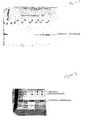
- Figure 1 shows an SDS-polyacrylamide gel profile of carbonic anhydrase after proteolysis in the absence and presence of increasing concentrations of acetazolamide.
- Figure 2 shows an SDS-polyacrylamide gel profile of carbonic anhydrase after proteolysis in the absence and presence of 1.0mM acetazolamide, in the absence and presence of a fungal extract.
- Figure 3 shows a graph representing a titration of the binding of radiolabelled human neutrophil elastase to nitrocellulose filters after proteolysis in the absence and presence of increasing concentrations of elastatinal.
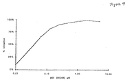
- Figure 4 shows a graph representing a titration of the ELISA detection of human neutrophil elastase after proteolysis in the presence of increasing concentrations of ICI 200,355.
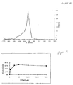
- Figure 5 shows a graph representing the distribution of data for test ligands tested for binding to human neutrophil elastase.
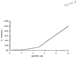
- Figure 6 shows a graph representing the titration of a ligand for human neutrophil elastase.
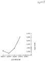
- Figure 7 shows a graph representing the titration of five ligands for their ability to inhibit the enzymatic activity of human neutrophil elastase.
- Figure 8 shows a graph representing a titration of the ELISA detection of human hemoglobin after proteolysis in the presence of increasing concentrations of 2,3-diphosphoglycerate.
- Figure 9 shows a graph representing a titration of the binding of human hemoglobin to nitrocellulose filters after proteolysis in the absence or presence of increasing concentrations of 2,3-diphosphoglycerate.
- Figure 10 shows a graph representing the distribution of binding data for test ligands tested for binding to human hemoglobin S.
- Figure 11 shows a graph representing the titration of a ligand for human hemoglobin.
- Figure 12 shows the structures of compounds identified as ligands for human hemoglobin S (HbS) and their activities in inhibiting HbS gelation relative to tryptophan (Trp).
- HbS human hemoglobin S
- Trp tryptophan
- Figure 13 shows a graph representing the ligand-binding activity for human hemoglobin of Zinc-bacitracin (BacZ), zinc-free bactracin (Bac), zinc-free bacitracin to which an equimolar concentration of ZnCl 2 has been added (Bac+Z), ZnCl 2 , and zinc-bacitracin to which a molar excess of EDTA has been added (BacZ + EDTA).
- ligand refers to an agent that binds a target protein.
- the agent may bind the target protein when the target protein is in its native conformation, or when it is partially or totally unfolded or denatured.
- a ligand is not limited to an agent that binds a recognized functional region of the target protein e.g. the active site of an enzyme, the antigen-combining site of an antibody, the hormone-binding site of a receptor, a cofactor-binding site, and the like.
- a ligand can also be an agent that binds any surface or internal sequences or conformational domains of the target protein. Therefore, the ligands of the present invention encompass agents that in and of themselves may have no apparent biological function, beyond their ability to bind to the target protein in the manner described above.
- test ligand refers to an agent, comprising a compound, molecule or complex, which is being tested for its ability to bind to a target protein.
- Test ligands can be virtually any agent, including without limitation metals, peptides, proteins, lipids, polysaccharides, nucleic acids, small organic molecules, and combinations thereof.
- Complex mixtures of substances such as natural product extracts, which may include more than one test ligand, can also be tested, and the component that binds the target protein can be purified from the mixture in a subsequent step.
- target protein refers to a peptide, protein or protein complex for which identification of a ligand or binding partner is desired.
- Target proteins include without limitation peptides or proteins known or believed to be involved in the etiology of a given disease, condition or pathophysiological state, or in the regulation of physiological function.
- Target proteins may be derived from any living organism, such as a vertebrate, particularly a mammal and even more particularly a human. For use in the present invention, it is not necessary that the protein's biochemical function be specifically identified.
- Target proteins include without limitation receptors, enzymes, oncogene products, tumor suppressor gene products, viral proteins, and transcription factors, either in purified form or as part of a complex mixture of proteins and other compounds.
- target proteins may comprise wild type proteins, or, alternatively, mutant or variant proteins, including those with altered stability, activity, or other variant properties, or hybrid proteins to which foreign amino acid sequences e.g. sequences that facilitate purification have been added.
- test combination refers to the combination of one or more test ligands and a target protein.
- Control combination refers to the target protein in the absence of a test ligand.
- the "folded state" of a protein refers to the native or undenatured form of the protein as it is present in its natural environment, or after isolation or purification, i.e. before exposure to denaturing conditions. This includes native proteins that may be detectably unfolded to differing extents in their natural environment, and whose folding patterns may change during their natural functioning.
- the "unfolded state” refers to a situation in which the polypeptide has lost elements of its secondary and/or tertiary structure that are present in its “folded state.” It will be recognized by those skilled in the art that it is difficult to determine experimentally when a polypeptide has become completely unfolded i.e. has lost all elements of secondary and tertiary structure. Thus, the term “unfolded state” as used herein encompasses partial or total unfolding.
- detectable fraction refers to a quantity that is empirically determined and that will vary depending upon the method used to distinguish folded from unfolded protein. For example, when protease sensitivity is used to monitor folding, conditions are chosen (e.g. by adjusting temperature or adding denaturants) so that approximately 80% of the target protein is digested within a convenient incubation period. Alternatively, when antibodies specific to the folded or unfolded state of a target protein are used as the detection method, conditions are chosen so that a sufficient amount of antibody is bound to give a detectable signal.
- the present invention encompasses high-throughput screening methods for identifying a ligand that binds a target protein. If the target protein to which the test ligand binds is associated with or causative of a disease or condition, the ligand may be useful for diagnosing, preventing or treating the disease or condition.
- a ligand identified by the present method can also be one that is used in a purification or separation method, such as a method that results in purification or separation of the target protein from a mixture.
- the present invention also relates to ligands identified by the present method and their therapeutic uses (for diagnostic, preventive or treatment purposes) and uses in purification and separation methods.
- a ligand for a target protein is identified by its ability to influence the extent of folding or the rate of folding or unfolding of the target protein.
- Experimental conditions are chosen so that the target protein is subjected to unfolding, whether reversible or irreversible. If the test ligand binds to the target protein under these conditions, the relative amount of folded:unfolded target protein or the rate of folding or unfolding of the target protein in the presence of the test ligand will be different, i.e. higher or lower, than that observed in the absence of the test ligand.
- the present method encompasses incubating the target protein in the presence and absence of a test ligand, under conditions in which (in the absence of ligand) the target protein would partially or totally unfold. This is followed by analysis of the absolute or relative amounts of folded vs. unfolded target protein or of the rate of folding or unfolding of the target protein.
- An important feature of the present invention is that it will detect any compound that binds to any sequence or domain of the target protein, not only to sequences or domains that are intimately involved in a biological activity or function.
- the binding sequence, region, or domain may be present on the surface of the target protein when it is in its folded state, or may be buried in the interior of the protein. Some binding sites may only become accessible to ligand binding when the protein is partially or totally unfolded.
- the test ligand is combined with a target protein, and the mixture is maintained under appropriate conditions and for a sufficient time to allow binding of the test ligand to the target protein.
- Experimental conditions are determined empirically for each target protein. When testing test ligands, incubation conditions are chosen so that most ligand:target protein interactions would be expected to proceed to completion. In general, the test ligand is present in molar excess relative to the target protein.
- the target protein can be in a soluble form, or, alternatively, can be bound to a solid phase matrix.
- the matrix may comprise without limitation beads, membrane filters, plastic surfaces, or other suitable solid supports.
- appropriate experimental conditions e.g. temperature, time, pH, salt concentration, and additional components, are chosen so that a detectible fraction of the protein is present in an unfolded form in the absence of test ligand.
- preferred experimental conditions allow a detectable amount of the protein to unfold during a convenient incubation period in the absence of test ligand.
- denaturing conditions may be required, including the use of elevated temperatures, the addition of chaotropes or denaturants such as urea or guanidium salts such as guanidinium thiocyanate, detergents, or combinations thereof.
- introduction of stabilizing or destabilizing amino acid substitutions may be used to manipulate the folded:unfolded ratio of target proteins.
- the time necessary for binding of target protein to ligand will vary depending on the test ligand, target protein and other conditions used. In some cases, binding will occur instantaneously (e.g., essentially simultaneous with combination of test ligand and target protein), while in others, the test ligand-target protein combination is maintained for a longer time e.g. up to 12-16 hours, before binding is detected. When many test ligands are employed, an incubation time is chosen that is sufficient for most protein:ligand interactions.
- Binding of a test ligand to the target protein is assessed by comparing the absolute amount of folded or unfolded target protein in the absence and presence of test ligand, or, alternatively, by determining the ratio of folded:unfolded target protein or the rate of target protein folding or unfolding in the absence and presence of test ligand. If a test ligand binds the target protein (i.e., if the test ligand is a ligand for the target protein), there may be significantly more folded, and less unfolded, target protein (and, thus, a higher ratio of folded to unfolded target protein) than is present in the absence of a test ligand.
- binding of the test ligand may result in significantly less folded, and more unfolded, target protein than is present in the absence of a test ligand.
- binding of the test ligand may cause the rate of target protein folding or unfolding to change significantly.
- determination of the absolute amounts of folded and unfolded target protein, the folded:unfolded ratio, or the rates of folding or unfolding may be carried out using one of the known methods as described below. These methods include without limitation proteolysis of the target protein, binding of the target protein to appropriate surfaces, binding of specific antibodies to the target protein, binding of the target protein to molecular chaperones, binding of the target protein to immobilized ligands, and measurement of aggregation of the target protein. Other physico-chemical techniques may also be used, either alone or in conjunction with the above methods; these include without limitation measurements of circular dichroism, ultraviolet and fluorescence spectroscopy, and calorimetry.
- a preferred embodiment involves measuring the relative proteolysis of a target protein following incubation in the absence and presence of a test ligand.
- each target protein may have unique properties that make a particular detection method most suitable for the purposes of the present invention.
- test ligands identified as "positive" compounds or ligands from among the total compounds screened This threshold is set according to two criteria. First, the number of positive compounds should be manageable in practical terms. Second, the number of positive compounds should reflect ligands with an appreciable affinity towards the target protein. A preferred threshold is achieved when 0.1% to 1% of the total test ligands are shown to be ligands of a given target protein.
- Binding to a given protein is a prerequisite for pharmaceuticals intended to modify directly the action of that protein.
- a test ligand may indicate the potential ability of the test ligand to alter protein function and to be an effective pharmaceutical or lead compound for the development of such a pharmaceutical.
- the ligand may serve as the basis for the construction of hybrid compounds containing an additional component that has the potential to alter the protein's function. In this case, binding of the ligand to the target protein serves to anchor or orient the additional component so as to effectuate its pharmaceutical effects.
- a known compound that inhibits the activity of a family of related enzymes may be rendered specific to one member of the family by conjugation of the known compound to a ligand, identified by the methods of the present invention, that binds specifically to that member at a different site than that recognized by the known compound.
- the present method is based on physico-chemical properties common to most proteins gives it widespread application.
- the present invention can be applied to large-scale systematic high-throughput procedures that allow a cost-effective screening of many thousands of test ligands.
- a ligand Once a ligand has been identified by the methods of the present invention, it can be further analyzed in more detail using known methods specific to the particular target protein used. For example, the ligand can be tested for binding to the target protein directly e.g. by incubating radiolabelled ligand with unlabelled target protein, and then separating protein-bound and unbound ligand.
- the ligand can be tested for its ability to influence, either positively or negatively, a known biological activity of the target protein.
- binding of test ligand to target protein is detected through the use of proteolysis.
- proteolysis This assay is based on the increased susceptibility of unfolded, denatured polypeptides to protease digestion relative to that of folded proteins.
- the test ligand-target protein combination, and a control combination lacking the test ligand are treated with one or more proteases that act preferentially upon unfolded target protein.
- the level of intact i.e. unproteolysed target protein is assessed using one of the methods described below e.g. gel electrophoresis and/or immunoassay.
- test ligand has bound the target protein.
- Proteases useful in practicing the present invention include without limitation trypsin, chymotrypsin, V8 protease, elastase, carboxypeptidase, proteinase K, thermolysin, papain and subtilisin (all of which can be obtained from Sigma Chemical Co., St. Louis, MO).
- the most important criterion in selecting a protease or proteases for use in practicing the present invention is that the protease(s) must be capable of digesting the particular target protein under the chosen incubation conditions, and that this activity be preferentially directed towards the unfolded form of the protein.
- protease particularly proteases with different enzymatic mechanisms of action
- cofactors that are required for the activity of the protease(s) are provided in excess, to avoid false positive results due to test ligands that may sequester these factors.
- a purified target protein is first taken up to a final concentration of 1-100 ⁇ g/ml in a buffer containing 50 mM Tris-HCl, pH 7.5, 10% DMSO, 50 mM Nacl, 10% glycerol, and 1.0 mM DTT.
- Proteases such as, for example, proteinase K or thermolysin (proteases with distinct mechanisms of action), are then added individually to a final concentration of 0.2-10.0 ⁇ g/ml.
- Parallel incubations are performed for different time periods ranging from 5 minutes to one hour, preferably 30 minutes, at 4°C, 15°C, 25°C, and 35°C.
- Reactions are terminated by addition of an appropriate protease inhibitor, such as, for example, phenylmethylsulfonyl chloride (PMSF) to a final concentration of 1mm (for serine proteases), ethylenediaminotetraacetic acid (EDTA) to a final concentration of 20 mM (for metalloproteases), or iodoacetamide (for cysteine proteases).
- PMSF phenylmethylsulfonyl chloride
- EDTA ethylenediaminotetraacetic acid
- metalloproteases for metalloproteases
- iodoacetamide for cysteine proteases
- the above protocol allows the selection of appropriate conditions (e.g., protease concentration and digestion temperature) that result in digestion of approximately 70% of the target protein within a 30 minute incubation period, indicating that a significant degree of unfolding has occurred.
- conditions are chosen so that proteolysis displays a temperature dependence indicative of a cooperative protein unfolding transition.
- additional variables can be adjusted, including, for example, the concentrations of glycerol, salt, reducing agents, BSA or other "carrier proteins," target protein, denaturants and detergents.
- the ligand is included in the reaction mixture at a concentration above the Kd for its binding to the target protein and at least equal to the molar concentration of target protein, and the digestion experiment is repeated. Typically, at least a two-fold increase or decrease in the extent of digestion of the target protein is observed, indicating that binding of a known ligand changes the ratio of folded:unfolded target protein and/or the rate of folding or unfolding.
- test ligands at concentrations ranging from 20 to 200 ⁇ M. Observation of at least a two-fold increase or decrease in the extent of digestion of the target protein signifies a "hit" compound, i.e., a ligand that binds the target protein.
- Preferred conditions are those in which between 0.1% and 1% of test ligands are identified as "hit" compounds using this procedure.
- the relative amount of folded and unfolded target protein in the presence and absence of test ligand is assessed by measuring the relative amount of target protein that binds to an appropriate surface.
- This method takes advantage of the increased propensity of unfolded proteins to adhere to surfaces, which is due to the increased surface area, and decrease in masking of hydrophobic residues, that results from unfolding.
- a test ligand binds a target protein (i.e., is a ligand of the target protein)
- it may stabilize the folded form of the target protein and decrease its binding to a solid surface.
- a ligand may stabilize the unfolded form of the protein and increase its binding to a solid surface.
- the target protein, a test ligand and a surface that preferentially binds unfolded protein are combined and maintained under conditions appropriate for binding of the target protein to a ligand and binding of unfolded target protein to the surface.
- the target protein and test ligand can be pre-incubated in the absence of the surface to allow binding.
- Surfaces suitable for this purpose include without limitation microtiter plates constructed from a variety of treated or untreated plastics, plates treated for tissue culture or for high protein binding, nitrocellulose filters and PVDF filters.
- Determination of the amount of surface-bound target protein or the amount of target protein remaining in solution can be carried out using standard methods known in the art e.g. determination of radioactivity or immunoassay. If significantly more or less target protein is surface bound in the presence of a test ligand than in the absence of the test ligand, the test ligand is a ligand of the target protein. Similarly, the ratio of surface-bound:soluble target protein will be significantly greater or smaller in the presence of a test ligand than in its absence, if a test ligand is a ligand for the target protein.
- the extent to which folded and unfolded target protein are present in the test combination is assessed through the use of antibodies specific for either the unfolded state or the folded state of the protein i.e. denatured-specific ("DS"), or native-specific ("NS") antibodies, respectively. (Breyer, 1989, J. Biol. Chem., 264 (5) :13348-13354).
- DS denatured-specific
- NS native-specific
- Polyclonal and monoclonal DS and NS antibodies specific for particular target proteins can be prepared by methods that are well known in the art (E. Harlow & D. Lane, Antibodies: A Laboratory Manual, Cold Spring Harbor Laboratory, 1988; Zola, Monoclonal Antibodies: A Manual of Techniques, CRC Press, Inc., Boca Raton, Florida,1987).
- animals can be immunized with a peptide from a region of the protein that is buried in the interior of the protein when it is in the native state. If the three-dimensional structure of the protein is unknown, antibodies are prepared against several peptides. Alternatively, fully denatured (i.e., unfolded) target protein is used as an immunogen.
- the resulting antibodies are screened for preferential binding to the denatured state.
- culture supernatants derived from individual cloned hybridomas are screened, and positive clones are used directly as a source of individual DS antibodies.
- polyclonal antibodies an unfractionated antiserum may exhibit preferential binding to the denatured state.
- DS antibodies may be purified from a polyclonal antiserum by selective adsorption techniques well-known in the art.
- NS antibodies intact non-denatured protein, or one or more peptides known to be on the surface of the native protein, may be used as an immunogen.
- the resulting antibodies are screened as above for preferential binding to the native protein and purified for use in the present invention.
- DS or NS antibodies can be utilized to detect a ligand-induced change in the level of folded target protein, unfolded target protein, the folded:unfolded ratio, or the rate of folding or unfolding.
- a test combination containing the DS antibody, the target protein, and the test ligand is exposed to a solid support e.g. a microtiter plate coated with the denatured target protein or a peptide fragment thereof, under conditions appropriate for binding of the target protein with its ligand and binding of the DS antibody to unfolded target protein.
- a control combination which is the same as the test combination except that it does not contain test ligand, is processed in the same manner as the test solution.
- a test combination containing the DS antibody, the test ligand, and the target protein is exposed to a solid support coated with a second antibody, referred to as a solid phase antibody, which cannot bind to the target protein simultaneously with the DS antibody, and is specific for the target protein, but is either specific for the folded state (NS antibody) or unable to differentiate between the native and denatured states ("non-differentiating" or "ND" antibody).
- the resulting test combination or solution is maintained under conditions appropriate for binding of the target protein with a ligand of the target protein and for binding of the antibodies to the proteins they recognize.
- a control combination which is the same as the test solution except that it does not contain test ligand, is processed in the same manner as the test solution.
- denatured (unfolded) target protein binds the DS antibody and is inhibited from binding the solid phase antibody.
- the ability of the test ligand to bind the target protein can be gauged by determining the amount of target protein that binds to the solid phase antibody in the test solution and comparing it with the extent to which target protein binds to the solid phase antibody in the absence of test ligand, which in turn reflects the amount of target protein in the folded state.
- the amount of target protein bound to the plate via the second antibody or remaining in solution can be detected by the methods described below. This approach may be used in a comparable manner with NS antibody as the soluble antibody and DS or ND antibody on the solid phase.
- a test solution containing the target protein and the test ligand is exposed to a solid support e.g. a microtiter plate that has been coated with a DS or NS antibody and maintained under conditions appropriate for binding of target protein to its ligand and for binding of the antibody to target protein.
- the antibody can be present on the surfaces of beads. The ability of the test ligand to bind the target protein is gauged by determining the extent to which target protein remains in solution (unbound to the antibody) or on the solid surface (bound to the antibody), or the ratio of the two, in the presence and in the absence of test ligand.
- the antibody can be present in solution and the target protein can be attached to a solid phase, such as a plate surface or bead surface.
- molecular chaperones are used to assess the relative levels of folded and unfolded protein in a test combination.
- Chaperones encompass known proteins that bind unfolded proteins as part of their normal physiological function. They are generally involved in assembling oligomeric proteins, in ensuring that certain proteins fold correctly, in facilitating protein localization, and in preventing the formation of proteinaceous aggregates during physiological stress (Hardy, 1991, Science, 251 :439-443). These proteins have the ability to interact with many unfolded or partially denatured proteins without specific recognition of defined sequence motifs.
- SecB One molecular chaperone, found in E . coli, is a protein known as SecB.
- SecB has a demonstrated involvement in export of a subset of otherwise unrelated proteins. Competition experiments have shown that SecB binds tightly to all the unfolded proteins tested, including proteins outside of its particular export subset, but does not appear to interact with the folded protein.
- Other chaperones suitable for use in the present invention include without limitation heat shock protein 70s, heat shock protein 90s, GroEI and GroES (Gething et al., Nature 355:33 , 1992).
- a test combination containing the test ligand and the target is exposed to a solid support e.g. microtiter plate or other suitable surface coated with a molecular chaperone, under conditions appropriate for binding of target protein with its ligand and binding of the molecular chaperone to unfolded target protein.
- the unfolded target protein in the solution will have a greater tendency to bind to the molecular chaperone-covered surface relative to the ligand-stabilized folded target protein.
- the ability of the test ligand to bind target protein can be determined by determining the amount of target protein remaining unbound, or the amount bound to the chaperone-coated surface.
- a competition assay for binding to molecular chaperones can be utilized.
- a test combination containing purified target protein, the test ligand, and a molecular chaperone can be exposed to a solid support e.g. a microtiter well coated with denatured (unfolded) target protein, under conditions appropriate for binding target protein with its ligand and binding of the molecular chaperone to unfolded target protein.
- a control combination which is the same as the test combination except that it does not contain test ligand, is processed in the same manner.
- Denatured target protein in solution will bind to the chaperone and thus inhibit its binding to the denatured target protein bound to the support.
- binding of a test ligand to the target protein will result in a difference in the amount of unfolded target protein, and, thus, more or less chaperone will be available to bind to the solid-phase denatured target protein than is the case in the absence of binding of test ligand.
- binding of test ligand can be determined by assessing chaperone bound to the surface or in solution in the test combination and in the control combination and comparing the results. In this assay, the chaperones are generally not provided in excess, so that competition for their binding can be measured.
- a test combination containing the target protein, the test ligand and a molecular chaperone can be exposed to a solid support e.g. a microtiter well that has been coated with antisera or a monoclonal antibody specific for the folded target protein (NS antibody) and unable to bind the target protein bound to the chaperone. Unfolded target protein will bind chaperone in solution and thus be inhibited from binding the solid phase antibody.
- a solid support e.g. a microtiter well that has been coated with antisera or a monoclonal antibody specific for the folded target protein (NS antibody)
- Unfolded target protein will bind chaperone in solution and thus be inhibited from binding the solid phase antibody.
- test ligand is a ligand for the target protein
- more or less target protein will be bound to the antisera or monoclonal antibody bound to the container surface in the test combination than in the control combination, and correspondingly more or less target protein will be present unbound (in solution) in the test combination than in the control combination.
- a known ligand, cofactor, substrate, or analogue thereof of the target protein is used to assay for the presence of folded target protein.
- the ligand, cofactor, substrate, or analogue thereof known to bind to the target protein is immobilized on a solid substrate.
- a solution containing the target protein and test ligand is then added.
- An increase or decrease in the amount of target protein that binds to the immobilized compound relative to an identical assay in the absence of test ligand indicates that the test ligand binds the target protein.
- the amount of target protein bound to the solid substrate can be assessed by sampling the solid substrate or by sampling the solution.
- the amount of unfolded target protein in a test combination is assessed by measuring protein aggregation.
- unfolded protein often forms insoluble aggregates.
- the extent of protein aggregation can be measured by techniques known in the art, including without limitation light scattering, centrifugation, and filtration.
- target protein and test ligand are incubated and the amount of protein aggregation is measured over time or after a fixed incubation time.
- the extent of protein aggregation in the test mixture is compared to the same measurement for a control assay in the absence of test ligand. If a test ligand binds a target protein, the rate of unfolding of target protein will be lower or higher than in the absence of test ligand. For measurements over time, the rate of appearance of aggregated protein will be lower or higher if the test ligand is a ligand for the target protein than if it is not.
- test ligand For measurements at a fixed time, there will be more or less unfolded protein and correspondingly more or less aggregated protein if the test ligand is a ligand for the target protein than if it is not.
- the ability of a test ligand to bind a target protein can be determined by assessing the extent of protein aggregation in the presence and absence of test ligand.
- domain refers to a fragment of a target protein that retains a significant degree of native folded structure after isolation.
- a native protein will be cleaved by a protease into one or more such domains when proteolytic digestion of the native protein is performed at, for example, a lower temperature than that at which complete digestion of the protein occurs.
- one or more individual domains of a target protein can be prepared for use as targets in the assays described above, either by subjecting the intact protein to controlled proteolysis followed by purification of domain-comprising fragments, or by directing the synthesis of such fragments, either in vitro or in vivo, from recombinant DNA molecules encoding domain-comprising fragments of the protein.
- domain-specific detection may be used to quantify folding in a reaction mixture in which the intact protein serves as the target. Methods for domain-specific detection include without limitation the use of domain-specific antibodies and chemical or enzymatic methods which selectively label particular domains. Domain-specific antibodies may be prepared by any method known in the art.
- polyclonal domain-specific antibodies may be raised by using as immunogens either the purified or recombinant domains described above or domain-specific synthetic peptides.
- a panel of monoclonal antibodies may be prepared against the intact protein, and tested for reaction with purified or recombinant domains.
- the embodiments described above require a final step for detecting and/or quantifying the level of target protein or digestion products thereof, or antibodies, in order to quantify the relative amounts of folded and unfolded target protein after exposure to test ligands.
- methods known in the art are used to detect the presence or absence of protein, small peptides or free amino acids. The method used will be determined by the product (proteins, peptides, free amino acids) to be detected.
- techniques for detecting protein size can be used to determine the extent of proteolytic degradation of the target protein e.g. gel electrophoresis, capillary electrophoresis, size exclusion chromatography, high-performance liquid chromatography, and the like.
- gel electrophoresis is used to detect the presence or absence of protein, and can further be used to detect the size of the protein. This latter method is especially useful in conjunction with proteolysis, as the presence of a greater or lesser amount of undigested target protein in the test combination than in the control combination indicates that the test ligand bound to the target protein.
- Example 1 Methotrexate Binding Protects Dihydrofolate Reductase (DHFR) From Proteolytic Digestion by Proteinase K
- DHFR 100 ⁇ g/ml
- Proteinase K 80 ⁇ g/ml
- Tris-HCl pH 7.5 0.1 M
- Methotrexate 10 -10 to 10 -4 M.
- nicotinamide adenine dinucleotide phosphate (NADPH) and dihydrofolate were shown to inhibit proteolysis of DHFR in separate experiments.
- Example 2 Methotrexate, NADPH and Dihydrofolate Binding Protects Dihydrofolate Reductase (DHFR) From Proteolytic Digestion by Proteinase K in the Presence of a Mixture of Amino Acids
- DHFR 2.1 ⁇ g/ml
- Proteinase K 80 ⁇ g/ml
- Tris-HCl pH 7.5
- 10 -5 M of all 20 common amino acids and either 0 or 10 -5 M ligand.
- the ligands used were the inhibitor Methotrexate and the substrates dihydrofolate and NADPH.
- the ELISA analysis showed that methotrexate and the substrates protect DHFR from digestion relative to the absence of ligands that bind to DHFR. Thus, specific binding can be detected in the presence of a complex mixture of compounds that do not bind to the target protein.
- the ELISA analysis revealed that methotrexate inhibits DHFR binding to the Falcon 3072 plate at concentrations of 10 -7 M and above.
- ELISA analysis will reveal target protein in the solution at higher or lower concentration when test ligand-target protein binding has occurred than when it has not.
- ELISA analysis will reveal a higher or lower concentration of target protein in the solution when successful test ligand-target protein binding has occurred.
- Reaction mixtures (0.03 ml total volume) contained 30 ⁇ g/ml HIV Rev protein that had been produced in E. coli, 0.05M Tris-HCl, pH 7.5, 0.01M calcium acetate, 2.5 ⁇ g/ml proteinase K, 10% DMSO, and varying amounts of tRNA as a known ligand.
- the reactions were incubated on ice for 15 minutes. After addition of PMSF and EDTA as described in Example 8 below, samples were prepared for gel electrophoresis and analyzed as described in Example 8.
- Ligand binding to carbonic anhydrase I was tested using proteolysis as a probe of target protein folding, and denaturing gel electrophoresis was used as a method for detection of intact protein remaining after digestion with proteases.
- acetazolamide a known ligand of carbonic anhydrase
- acetazolamide is a known inhibitor of carbonic anhydrase activity
- these experiments make no use of that property, and do not measure the enzymatic activity of the protein.
- the sensitivity of the method to interference by a natural product extract was examined.
- Reaction mixtures contained 13.3 ⁇ g/ml carbonic anhydrase, 0.05 M Tris-HCl pH 7.5, 0.01 M calcium acetate, 2.5 ⁇ g/ml proteinase K, 10% DMSO and acetazolamide (Sigma) in concentrations ranging from 0.0 to 1.0 mM.
- the reactions were incubated at 54°C for 15 minutes, and then chilled on ice.
- Phenyl methyl sulfonyl fluoride (PMSF) was then added from a 20 mM stock solution in ethanol to a final concentration of 1 mM
- EDTA was added from a 0.5M stock solution to a final concentration of 20 mM.
- SDS loading buffer (10% sodium dodecyl sulfate (SDS), 0.5 M Dithiothreitol, 0.4 M Tris-HCl buffer, pH 6.8, 50% Glycerol) was added and samples were heated at 95°C for 3 minutes. Samples were analyzed by SDS-polyacrylamide gel electrophoresis using a 4-15% polyacrylamide (BioRad) gradient gel, which was then stained with Coomassie Blue dye.
- a high throughput assay has been established for carbonic anhydrase I.
- Each reaction mixture in a final volume of 0.05 ml contains: 3.3 ⁇ g carbonic anhydrase, 50 mM Tris-HC1, 50 mM NaCl, 1.0 mM Ca(OAc) 2 , and 0.13 ⁇ g proteinase K, 10% DMSO, and the appropriate test compound at a concentration of 20 ⁇ m.
- Control reactions are identical, except that the test compound is omitted.
- the mixtures are incubated at 20°C for 10 minutes, followed by incubation at 54°C for 30 minutes, after which they are placed on ice.
- Each mixture then receives 200 ⁇ l 50 mM sodium borate buffer, pH 8.5, containing 10 mM EDTA and 1.0 mM PMSF. After 20 minutes incubation on ice, 7.5 ⁇ l of the mixture are added to Dynatech Immulon-4 microplate wells containing 92.5 ⁇ l per well of the borate buffer described above. The plate is then incubated at 4°C overnight to permit carbonic anhydrase binding. Bound carbonic anhydrase is quantified by direct ELISA using HRP-conjugated anti-carbonic anhydrase I antibody at a 1:2,000 dilution (Biodesign, Kennebunk, ME); cat#K90046P) and Pierce (Rockford, IL) Turbo TMB substrate.
- Carbonic anhydrase I inhibitors (obtained from Sigma Chemical Co., St. Louis, MO) were tested in the above assay over a range of inhibitor concentrations.
- Table 2 shows the concentration that caused 50% inhibition of proteolysis in the assay (IC 50 ), the published K d values for the compounds (Matsumoto et al., Chem. Pharm. Bull., 37 : 1913, (1989)), and the ratio of these values for each compound. These data show a positive correlation between the IC 50 and K d values for each inhibitor.
- HNE human neutrophil elastase
- HbA hemoglobin A
- HbS hemoglobin S
- HbS is an intracellular, tetrameric protein that contains a prosthetic group critical to its function. It is known to exist in two conformations with different structural and functional properties.
- HNE is monomeric, lacks a prosthetic group, and is secreted.
- HNE has an enzymatic activity (proteolysis) and does not appear to undergo any global conformational changes.
- proteolysis is used as the probe of target protein folding.
- the two high-throughput modes differ in the methods used for detection of residual target protein following proteolysis.
- the two detection methods are 1) capture of radiolabelled protein on nitrocellulose filters followed by quantitation of bound radioactivity and 2) measurement of protein by enzyme linked immunosorbent assay (ELISA.) Each of these methods was used successfully with both hemoglobin and HNE.
- HNE 0.1 mg HNE (Elastin Products) was labelled by reaction with 125 I-Sodium Iodide (Amersham) in the presence of Iodogen (Pierce) according to manufacturer's protocols (Pierce). Reaction mixtures were prepared in a final volume of 0.05 ml containing radiolabelled HNE (20,000 cpm, corresponding to approximately 10 ⁇ g), 0.025 mg/ml Bovine Serum Albumin, 50 mM Tris-HCl, pH 7.5, 10 mM calcium acetate, 2.5 ⁇ g/ml thermolysin (Boeringer Mannheim), 2.5 ⁇ g/ml proteinase K (Merck), 10% DMSO, and the test compound at a concentration of 200 ⁇ M. Control mixtures were identical, except that the test compound was omitted.
- Reaction mixtures in final volume of 0.05 ml contained 2 ⁇ g/ml HNE, 0.020 mg/ml Bovine Serum Albumin, 50 mM Tris-HCl, pH 7.5, 10 mM calcium acetate, 7.5 ⁇ g/ml thermolysin (Boeringer Mannheim), 7.5 ⁇ g/ml proteinase K (Merck), 10% DMSO, and the test compound at 20 or 200 ⁇ M concentration. Control mixtures were identical except that the test compound was omitted. The mixtures were incubated at 20°C for 15 minutes, then at 63°C, 30 minutes then placed on ice.
- 0.1 ml of rabbit anti HNE antibody (Calbiochem) at a dilution of 1:10,000 in TBST (10 mM Tris-HCl, pH 7.5, 0.15 M NaCl, 0.05% Tween-20) containing 5% nonfat dry milk (Carnation) was then added to each reaction. After 10 minutes incubation at room temperature, the mixtures were transferred to 96-well Immulon-4 plates (Dynatech) that had been coated with HNE by overnight incubation with 0.1 ml per well of 0.2 ⁇ g/ml HNE in 50 mM Sodium Borate buffer, pH 8.5, and 3 mM sodium azide and then washed thoroughly with TBST.
- a known ligand for HNE ICI 200,355
- concentrations ranging from 0.01-10 ⁇ M As shown in Figure 4, inclusion of the ligand caused an inhibition of antibody binding to the plate, indicating an increased level of immunoreactive HNE in the reaction mixtures.
- the hit compounds were assayed for their ability to inhibit the enzymatic activity of HNE. Since compounds identified in the binding assay may bind anywhere on the protein surface, only a small fraction would be expected to inhibit the enzymatic activity of HNE.
- the compounds were tested as inhibitors of the proteolysis of Suc-(Ala) 3 -pNA (Elastin Products Co., Owensville, MI), a chromogenic synthetic substrate, according to the method of Bieth, J, Spiess, B. and Wermuth, C. G. (1974, Biochemical Medicine, 11 :350-357.) Two positive hit compounds and one negative hit compound inhibit the proteolytic activity of HNE significantly in these assays ( Figure 7).
- the compounds were also assayed for potency in inhibiting the catalytic activity of HNE.
- cleavage of the chromogenic substrate methoxysuccinyl-ala-ala-pro-val-p-nitroanilide was monitored.
- 100 ⁇ l reactions were prepared containing the chromogenic substrate (1 mM), 100 nM elastase, 1% DMSO, 100 mM Tris-HCl, pH 7.5, and 0.5 M NaCl. Cleavage was monitored over time by measuring the increase in absorbance of the reaction mixtures at 405 nm.
- HbS or HbA (Sigma) was radiolabelled by reaction with 1 mCi 125 I-Bolton-Hunter reagent (Amersham) in 100 mM sodium borate buffer, pH 8.5, on ice for one hour. Labelling was stopped by addition of borate buffer containing 200 mM glycine. The mixture was then fractionated by size on an execellulose GF-5 column (Pierce) in 50 mM sodium phosphate buffer, pH 7.5, containing 0.25% gelatin.
- reaction mixtures in a final volume of 0.05 ml contained radiolabelled hemoglobin (20,000 CPM), 0.063 mg/ml unlabelled hemoglobin, 0.034 mg/ml Bovine Serum Albumin, 50 mM Tris-HCl, pH 7.5, 10 mM calcium acetate, 2.5 ⁇ g/ml thermolysin (Boeringer Mannheim), 2.5 ⁇ g/ml proteinase K (Merck), 10% DMSO, and test compound. Control mixtures were identical, except that the test compound was omitted. The mixtures were incubated at 20°C for 15 minutes, then 40°C for 30 minutes and then placed on ice.
- Reaction mixtures in a final volume of 0.05 ml contained 0.063 mg/ml Hemoglobin, 0.034 mg/ml Bovine Serum Albumin, 50 mM Tris-HCl, pH 7.5, 10 mM calcium acetate, 7.5 ⁇ g/ml thermolysin (Boeringer Mannheim), 7.5 ⁇ g/ml proteinase K (Merck), 10% DMSO, and the test compound at 20 or 200 ⁇ M concentration. Control reactions were identical, except that the test compound was omitted.
- the mixtures were incubated at 20°C for 15 minutes, then at 44°C for 30 minutes, and then placed on ice. To each mixture was then added 0.05 ml 0.1M sodium borate buffer containing 20mM EDTA and 1mm PMSF. After 10 minutes incubation on ice, the mixtures were transferred to uncoated 96-well Immuulon-4 plates (Dynatech). The plates were then incubated at 4°C overnight to allow binding of the protein to the plate. The plates were washed thoroughly with TBST, and 0.1 ml of rabbit anti-human hemoglobin antibody (Calbiochem) dilute 1:500 was added to each well. The plates were incubated at room temperature for one hour, then thoroughly washed with TBST.
- Calbiochem rabbit anti-human hemoglobin antibody
- Hit compounds showed half maximal effects at concentrations ranging as low as 2.0 ⁇ M (For example, see Figure 11).
- Hemoglobin S is included in its oxidized form, met-hemoglobin (met-HbS), wherein the heme iron is in the ferric state.
- met-hemoglobin met-hemoglobin
- Reaction mixtures (in a final volume of 50 ⁇ l) contained 0.32 mg/ml hemoglobin S, 50 mM Tris-HCl, pH 7.5, 50 mM NaCl, 0.4 mM potassium ferricyanide, 40 ⁇ g/ml proteinase K, 10% DMSO, and the test compound at a concentration of 20 ⁇ M. Control reactions were identical, except that the test compound was omitted.
- the remaining 57 compounds were individually tested for their effects on the visible absorbance spectrum of met-HbS. This was done to identify those compounds whose apparent binding reflects contamination with cyanide or azide ions. Both cyanide and azide test positive using the assay of the present invention, which is presumably due to the fact that each binds with high affinity to the ferric iron atom of met-HbS and thereby increases met-HbS stability substantially. Binding of these ions to HbS also causes characteristic changes in its visible absorbance spectrum. The compounds were also tested for their ability to induce aggregation of met-HbS under the assay conditions used above; this was monitored by measuring changes in light scattering of the HbS solution at 595 nm.
- HbS hemoglobin S
- the reaction mixtures were then subjected to centrifugation at 35,000 rpm for 65 min at 30°C in a Beckmanh SW55Ti rotor.
- the supernatant of each mixture was then sampled in multiplicates and diluted 1:200 in Drabkins solution (Sigma Chemical Co., St. Louis, MO), and the absorbance of the diluted mixture at 540 nm was measured.
- the solubility profiles were plotted using as the x axis the concentration of test ligand (mM) and as the y axis the C SAT (the concentration of HbS in the supernatant in g/dl). (The C SAT at 0 mM ligand should be about 16-18%.)
- the slope of each plot (dy/dx g/dl M) was then obtained, as well as the ratio of each slope to the control compounds (L-Phe and Trp). The slopes are directly proportional to molar effectiveness in inhibiting gelation.
- Figure 12 shows the structures and commercial sources for these compounds and indicates their activities relative to Trp. Thirteen of these compounds exhibited anti-gelation activity at least equal to that of Trp.
- zinc-bacitracin (designated ST56 in Figure 12) is five times as effective as L-trp in inhibiting HbS polymerization.
- Zinc-bacitracin Bacitracin and Zinc Chloride (ZnCl 2 ) were tested individually in the hemoglobin S binding assay described in Example 12 ( Figure 13.) While zinc-free bacitracin is inactive, ZnCl 2 alone has an activity equivalent to that of zinc bacitracin.
- Zn ++ is a ligand of hemoglobin and zinc-free bacitracin is not.
- Zinc-bacitracin complex itself may be a ligand of hemoglobin, or its activity may stem solely from liberation of Zn ++ from the complex.
- the lack of activity in the remaining eight compounds may be due at least in part to their limited solubility at millimolar concentrations that are necessary in the gelation assay.
- the methods of the present invention were successful in identifying a discrete number of HbS ligands from among 40,000 test ligands.
Landscapes
- Life Sciences & Earth Sciences (AREA)
- Health & Medical Sciences (AREA)
- Engineering & Computer Science (AREA)
- Chemical & Material Sciences (AREA)
- Molecular Biology (AREA)
- Immunology (AREA)
- Proteomics, Peptides & Aminoacids (AREA)
- Biomedical Technology (AREA)
- Hematology (AREA)
- Urology & Nephrology (AREA)
- Analytical Chemistry (AREA)
- Biochemistry (AREA)
- Organic Chemistry (AREA)
- Microbiology (AREA)
- General Health & Medical Sciences (AREA)
- Physics & Mathematics (AREA)
- Biotechnology (AREA)
- Medicinal Chemistry (AREA)
- Zoology (AREA)
- General Physics & Mathematics (AREA)
- Pathology (AREA)
- Food Science & Technology (AREA)
- Cell Biology (AREA)
- Wood Science & Technology (AREA)
- Biophysics (AREA)
- Bioinformatics & Cheminformatics (AREA)
- General Engineering & Computer Science (AREA)
- Genetics & Genomics (AREA)
- Investigating Or Analysing Biological Materials (AREA)
- Measuring Or Testing Involving Enzymes Or Micro-Organisms (AREA)
- Peptides Or Proteins (AREA)
- Medicines That Contain Protein Lipid Enzymes And Other Medicines (AREA)
Claims (24)
- Méthode de sélection rapide, à grande échelle, pour identifier un ligand qui se lie à une protéine cible prédéterminée, comprenant les étapes consistant :(a) à sélectionner, en tant que ligands à tester, une pluralité de composés non connus pour se lier à la protéine cible ;(b) à incuber chacun desdits ligands à tester et la protéine cible pour produire une combinaison de test ;(c) à incuber la protéine cible en l'absence d'un ligand à tester pour produire une combinaison témoin ;(d) à traiter les combinaisons de test et témoin pour amener la protéine cible dans la combinaison témoin à se déplier dans une proportion mesurable ;(e) à déterminer dans quelle mesure la protéine cible est présente à l'état plié, à l'état déplié ou les deux dans la combinaison de test et dans la combinaison témoin ;(f) à comparer la détermination faite dans l'étape (e) dans les combinaisons test et témoin, le ligand testé étant, dans le cas où la protéine cible est présente à l'état plié dans une plus grande proportion dans la combinaison de test que dans la combinaison témoin, un ligand qui se lie à la protéine cible ; et(g) à répéter les étapes (b)-(f) avec plusieurs desdits ligands testés jusqu'à ce que l'on identifie un ligand qui se lie à la protéine cible, sous réserve que l'étape de traitement (d) ne comprenne pas de contact desdites combinaisons test et témoin avec une sonde fluorescente sensible à la conformation.
- Méthode selon la revendication 1, dans laquelle l'étape de traitement comprend au moins une modification de la température, une addition de composés dénaturants et des combinaisons de ces opérations.
- Méthode selon la revendication 2, dans laquelle l'étape de traitement comprend l'addition de composés dénaturants, ces derniers étant choisis dans le groupe constitué par l'urée, le guanidium et ses sels, des détergents, et des combinaisons de ces composants.
- Méthode selon la revendication 1 ou 2, dans laquelle l'étape de traitement comprend le fait de soumettre les combinaisons de test et témoin à une augmentation de la température, amenant ainsi une fraction détectable de ladite cible à se trouver sous forme dépliée.
- Méthode de sélection rapide, à grande échelle, pour identifier un ligand qui se lie à une protéine cible prédéterminée, comprenant les étapes consistant :(a) à sélectionner, en tant que ligands à tester, une pluralité de composés non connus pour se lier à la protéine cible ;(b) à incuber chacun desdits ligands à tester et la protéine cible dans des conditions appropriées pour que la protéine cible puisse se déplier dans une proportion mesurable, produisant ainsi une combinaison de test ;(c) à incuber la protéine cible comme dans l'étape (b), mais en l'absence d'un ligand à tester, pour produire une combinaison témoin ;(d) à déterminer dans quelle mesure la protéine cible est présente à l'état plié, à l'état déplié ou les deux dans la combinaison de test et dans la combinaison témoin ;(e) à comparer les déterminations faites dans l'étape (d) dans les combinaisons test et témoin, le ligand testé étant, dans le cas où la protéine cible est présente à l'état plié dans une plus grande proportion dans la combinaison de test que dans la combinaison témoin, un ligand qui se lie à la protéine cible ; et(f) à répéter les étapes (b)-(e) avec plusieurs desdits ligands testés jusqu'à ce que l'on identifie un ligand qui se lieaà la protéine cible ; sous réserve que les étapes d'incubations (b) et (c) ce comprennent pas de contact desdites combinaisons test et témoin avec une sonde fluorescente sensible à la conformation.
- Méthode selon la revendication 5, dans laquelle le ligand se lie à la surface, à des résidus d'acides aminés internes ou à des domaines conformationnels de la protéine cible.
- Méthode selon l'une quelconque des revendications 1-6, dans laquelle l'étape de détermination comprend le fait de soumettre les combinaisons de test et témoin à au moins une protéolyse ; une liaison d'anticorps ; une liaison à une surface ; une liaison moléculaire à une chaperone ; une liaison à un ligand, à un cofacteur ou à un substrat connu, ou à un de leurs analogues, de la protéine cible; une spectroscopie à dichroïsme circulaire; une spectroscopie dans l'ultraviolet ; une spectroscopie en fluorescence ; une calorimétrie ; et leurs combinaisons.
- Méthode selon la revendication 7, dans laquelle l'étape de détermination comprend les étapes consistant :(i) à incuber la protéine cible, chacun desdits ligands à tester et une ou plusieurs protéases qui dégradent de préférence la protéine cible à l'état déplié, la protéine cible étant ainsi dégradée de façon préférentielle dans son état déplié ; et(ii) à mesurer la fraction de la protéine cible dégradée ou la fraction de la protéine cible restant à l'état intact, le ligand testé étant, dans le cas où la fraction de la protéine cible restant à l'état intact dans la combinaison de test est plus grande que celle dans la combinaison témoin, un ligand qui se lie à la protéine cible.
- Méthode selon la revendication 7, dans laquelle l'étape de détermination comprend les étapes consistant :(i) à combiner la protéine cible et chacun desdits ligands à tester ;(ii) à exposer le mélange de la protéine cible et du ligand à tester à une surface qui se lie préférentiellement à une protéine cible dépliée, la protéine cible dépliée se liant préférentiellement à la surface ; et(iii) à déterminer la fraction de la protéine liée à la surface ou la fraction de la protéine cible restant à l'état non lié, le ligand testé étant, dans le cas où la fraction de la protéine cible restant à l'état non lié dans la combinaison de test est plus grande que celle dans la combinaison témoin, un ligand qui se lie à la protéine cible.
- Méthode selon la revendication 7, dans laquelle l'étape de détermination comprend les étapes consistant :(i) à combiner la protéine cible et chacun desdits ligands à tester à une surface sur laquelle un ligand connu de la protéine cible a été immobilisé ; et(ii) à déterminer la fraction de la protéine cible liée à la surface ou la fraction de la protéine cible restant à l'état non lié, le ligand testé étant, dans le cas où la fraction de la protéine cible lié à la surface dans la combinaison de test est plus grande que celle dans la combinaison témoin, un ligand qui se lie à la protéine cible.
- Méthode selon la revendication 7, dans laquelle l'étape de détermination comprend l'utilisation d'anticorps qui distinguent entre la forme pliée et la forme dépliée de la protéine cible.
- Méthode selon la revendication 11, dans laquelle l'étape de détermination comprend les étapes consistant :(i) à revêtir une surface avec un anticorps spécifique dirigé contre l'état dénaturé de la protéine cible ;(ii) à incuber la protéine cible en présence de chacun desdits ligands à la surface ; et(iii) à déterminer la fraction de la protéine cible liée ou non liée à la surface.
- Méthode selon la revendication 11, dans laquelle l'étape de détermination comprend les étapes consistant :(i) à revêtir la surface avec un anticorps capable de se lier à la protéine cible à l'état naturel ou à l'état dénaturé ;(ii) à incuber la protéine cible en présence de chacun desdits ligands à tester et d'un anticorps capable de se lier à la protéine cible à l'état plié ou à l'autre de ses états natif et dénaturé ; et(iii) à déterminer la présence de la protéine cible liée à la surface ou restant non liée.
- Méthode selon la revendication 7, dans laquelle l'étape de détermination comprend la détermination de la quantité de protéine cible liée à une protéine chaperone moléculaire ou non liée à une protéine chaperone moléculaire.
- Méthode selon la revendication 14, dans laquelle l'étape de détermination comprend les étapes consistant :(i) à revêtir une surface avec une protéine chaperone moléculaire ;(ii) à incuber la protéine cible et chacun desdits ligands à tester sur la surface revêtue ; et(iii) à déterminer la quantité de la protéine cible liée à la surface ou restant non liée.
- Méthode selon la revendication 14, dans laquelle l'étape de détermination comprend les étapes consistant :(i) à revêtir une surface avec la protéine cible à l'état dénaturé ;(ii) à incuber une forme purifiée de la protéine cible en présence de chacun desdits ligands à tester et une protéine chaperone moléculaire sur la surface ; et(iii) à déterminer la quantité de l'une ou des deux, de la protéine chaperone moléculaire et de la protéine cible, restant non liée ou liée à la surface.
- Méthode selon la revendication 7, dans laquelle l'étape de détermination comprend la détermination de la formation différentielle de protéine agrégée, en utilisant une méthode choisie dans le groupe constitué par :(i) la mesure de la quantité de protéine agrégée ;(ii) la mesure de la quantité de protéine soluble et la mesure de la vitesse de formation de protéine agrégée.
- Méthode selon la revendication 7, dans laquelle ladite liaison à un ligand, un cofacteur ou un substrat connu, ou à un analogue de ceux-ci, comprend les étapes consistant :le ligand étant l'un quelconque des ligands testés qui provoque une augmentation ou une diminution de ladite fraction dans la combinaison de test par rapport à ladite fraction dans la combinaison témoin.(i) à immobiliser le ligand, cofacteur ou substrat connu, ou l'analogue de ceux-ci, sur un support solide ;(ii) à exposer le support solide aux combinaisons de test et témoin, dans des conditions dans lesquelles la protéine cible à l'état plié se lie au support ; et(iii) à déterminer la fraction de la protéine cible liée au support dans les combinaisons de test et témoin,
- Méthode selon la revendication 8, dans laquelle les protéases de l'étape d'incubation sont choisies dans le groupe constitué par la protéinase K, la thermolysine et leurs combinaisons.
- Méthode selon la revendication 14, dans laquelle l'étape de détermination comprend le fait de soumettre les combinaisons de test et témoin à une dénaturation par électrophorèse sur gel de polyacrylamide.
- Méthode selon la revendication 7, dans laquelle l'étape de détermination comprend les étapes consistant :(i) à revêtir une surface avec des antisérums capables de se lier à la protéine cible à l'état plié ;(ii) à incuber la protéine cible en présence d'une chaperone moléculaire et de chacun desdits ligands à tester et sur la surface ; et(iii) à déterminer la quantité de la protéine cible liée à la surface ou restant non liée.
- Méthode selon l'une quelconque des revendications 1-21, dans laquelle la protéine cible dans la combinaison témoin se déplie de manière irréversible.
- Méthode selon l'une quelconque des revendications 1-21, dans laquelle la protéine cible dans la combinaison témoin se déplie de manière réversible.
- Méthode selon l'une quelconque des revendications 1-23, dans laquelle la protéine cible est choisie dans le groupe constitué par l'anhydrase carbonique, l'élastase neutrophile humaine, l'hémoglobine humaine, la dihydrofolate réductase et la protéine HIV Rev.
Applications Claiming Priority (2)
| Application Number | Priority Date | Filing Date | Title |
|---|---|---|---|
| US54788995A | 1995-10-24 | 1995-10-24 | |
| US547889 | 1995-10-25 |
Publications (2)
| Publication Number | Publication Date |
|---|---|
| EP0770876A1 EP0770876A1 (fr) | 1997-05-02 |
| EP0770876B1 true EP0770876B1 (fr) | 2001-04-11 |
Family
ID=24186560
Family Applications (1)
| Application Number | Title | Priority Date | Filing Date |
|---|---|---|---|
| EP96610042A Expired - Lifetime EP0770876B1 (fr) | 1995-10-25 | 1996-10-17 | Méthode de screening pour l'identification de ligands des protéines cibles |
Country Status (14)
| Country | Link |
|---|---|
| EP (1) | EP0770876B1 (fr) |
| JP (1) | JP2952848B2 (fr) |
| KR (1) | KR0161256B1 (fr) |
| AT (1) | ATE200579T1 (fr) |
| AU (1) | AU698862B2 (fr) |
| BR (1) | BR9604352A (fr) |
| CA (1) | CA2184195C (fr) |
| DE (2) | DE770876T1 (fr) |
| DK (1) | DK0770876T3 (fr) |
| ES (1) | ES2158269T3 (fr) |
| GR (1) | GR3036138T3 (fr) |
| IL (1) | IL119149A (fr) |
| MX (1) | MX9604183A (fr) |
| PT (1) | PT770876E (fr) |
Cited By (3)
| Publication number | Priority date | Publication date | Assignee | Title |
|---|---|---|---|---|
| US7063946B2 (en) | 2001-09-10 | 2006-06-20 | Meso Scale Technologies, Llc. | Methods, reagents, kits and apparatus for protein function analysis |
| US7122382B1 (en) | 1999-05-07 | 2006-10-17 | Riken | Method for detecting proteins under mutual interaction |
| US7122321B2 (en) | 1997-11-12 | 2006-10-17 | Johnson & Johnson Pharmaceutical Research & Development, L.L.C. | High throughput method for functionally classifying proteins identified using a genomics approach |
Families Citing this family (21)
| Publication number | Priority date | Publication date | Assignee | Title |
|---|---|---|---|---|
| US6036920A (en) | 1996-05-09 | 2000-03-14 | 3-Dimensional Pharmaceuticals, Inc. | Microplate thermal shift assay apparatus for ligand development and multi-variable protein chemistry optimization |
| US6453246B1 (en) | 1996-11-04 | 2002-09-17 | 3-Dimensional Pharmaceuticals, Inc. | System, method, and computer program product for representing proximity data in a multi-dimensional space |
| WO1998020437A2 (fr) | 1996-11-04 | 1998-05-14 | 3-Dimensional Pharmaceuticals, Inc. | Systeme, procede et programme produit informatique pour identifier des composes chimiques presentant des proprietes desirees |
| US6571227B1 (en) | 1996-11-04 | 2003-05-27 | 3-Dimensional Pharmaceuticals, Inc. | Method, system and computer program product for non-linear mapping of multi-dimensional data |
| US6569631B1 (en) | 1998-11-12 | 2003-05-27 | 3-Dimensional Pharmaceuticals, Inc. | Microplate thermal shift assay for ligand development using 5-(4″dimethylaminophenyl)-2-(4′-phenyl)oxazole derivative fluorescent dyes |
| GB9904395D0 (en) * | 1999-02-25 | 1999-04-21 | Fluorescience Ltd | Protein assay |
| US6972198B2 (en) * | 1999-02-26 | 2005-12-06 | Cyclacel, Ltd. | Methods and compositions using protein binding partners |
| US6582907B1 (en) * | 1999-12-09 | 2003-06-24 | Pharmacia & Upjohn Company | Use of fluorescence correlation spectroscopy to identify compounds that bind to target species under isothermal denaturing conditions |
| US6376180B1 (en) * | 1999-12-09 | 2002-04-23 | Pharmacia & Upjohn Company | Methods of identifying compounds that bind to target species under isothermal denaturing conditions |
| US7416524B1 (en) | 2000-02-18 | 2008-08-26 | Johnson & Johnson Pharmaceutical Research & Development, L.L.C. | System, method and computer program product for fast and efficient searching of large chemical libraries |
| US6671627B2 (en) | 2000-02-29 | 2003-12-30 | 3-D Pharmaceuticals, Inc. | Method and computer program product for designing combinatorial arrays |
| WO2001071624A1 (fr) | 2000-03-22 | 2001-09-27 | 3-Dimensional Pharmaceuticals, Inc. | Systeme, procede et progiciel pour la representation de relations entre objets dans un espace multidimensionnel |
| WO2001075790A2 (fr) | 2000-04-03 | 2001-10-11 | 3-Dimensional Pharmaceuticals, Inc. | Procede, systeme, et produit informatique permettant de representer un objet dans un espace a plusieurs dimensions |
| KR20010112840A (ko) * | 2000-06-15 | 2001-12-22 | 김유삼 | 기능성 단백질의 패턴분석을 이용한 진단 시스템 |
| WO2002061419A1 (fr) | 2001-01-29 | 2002-08-08 | 3-Dimensional Pharmaceuticals, Inc. | Procede, systeme et progiciel permettant d'analyser les banques combinatoires |
| DE60216487T2 (de) * | 2001-09-03 | 2007-09-13 | Kyowa Hakko Kogyo Co., Ltd. | Verfahren zur bestimmung eines zielproteins eines arzneistoffs |
| EP2124062A1 (fr) | 2005-08-12 | 2009-11-25 | Astellas Pharma Inc. | Procédé servant à identifier une protéine cible d'un agent et procédé servant à cribler un agent thérapeutique pour le diabète utilisant la protéine cible |
| DE102006027259A1 (de) * | 2006-06-09 | 2007-12-13 | Protagen Ag | Protein-Biochip zum differentiellen Screenen von Protein-Protein-Wechselwirkungen |
| WO2018152329A1 (fr) | 2017-02-15 | 2018-08-23 | Ohio State Innovation Foundation | Électrophiles hétéroaromatiques et leurs procédés d'utilisation |
| CN114076826A (zh) * | 2020-08-20 | 2022-02-22 | 中国科学院大连化学物理研究所 | 一种微球辅助的基于蛋白加热沉淀的药物靶蛋白筛选方法 |
| GB202118173D0 (en) * | 2021-12-15 | 2022-01-26 | Medicines Discovery Catpult Ltd | Protein stability assay |
Citations (2)
| Publication number | Priority date | Publication date | Assignee | Title |
|---|---|---|---|---|
| WO1997020925A1 (fr) * | 1995-12-08 | 1997-06-12 | Hybridon, Inc. | Oligonucleotides antisens specifiques du vegf, modifies, destines au traitement des troubles cutanes |
| WO1997042500A1 (fr) * | 1996-05-09 | 1997-11-13 | 3-Dimensional Pharmaceuticals, Inc. | Procede d'analyse et appareil a changement thermique et a microplaque pour l'optimisation de la mise au point de ligands et de la chimie des proteines a variables multiples |
Family Cites Families (9)
| Publication number | Priority date | Publication date | Assignee | Title |
|---|---|---|---|---|
| JPS5231755B2 (fr) * | 1973-01-08 | 1977-08-17 | ||
| US4859609A (en) * | 1986-04-30 | 1989-08-22 | Genentech, Inc. | Novel receptors for efficient determination of ligands and their antagonists or agonists |
| EP0546084A4 (en) * | 1990-08-17 | 1994-10-19 | Univ Boston | Proteases causing abnormal degradation of amyloid -g(b)-protein precursor |
| US5506097A (en) * | 1990-08-24 | 1996-04-09 | President And Fellows Of Harvard College | Method for inhibiting β-protein enzymatic activity |
| WO1993014781A1 (fr) * | 1992-01-24 | 1993-08-05 | The Regents Of The University Of California | Nouveaux peptides et procede pour modifier l'activite de proteines allosteriques |
| CA2115900A1 (fr) * | 1993-02-22 | 1994-08-23 | Gerald W. Becker | Cribles pharmaceutiques et anticorps |
| US5585277A (en) * | 1993-06-21 | 1996-12-17 | Scriptgen Pharmaceuticals, Inc. | Screening method for identifying ligands for target proteins |
| CA2236339A1 (fr) | 1995-12-07 | 1997-06-12 | Robert T. Sauer | Procede de criblage par fluorescence destine a l'identification de ligands |
| US8082993B2 (en) | 2009-03-12 | 2011-12-27 | Halliburton Energy Services, Inc. | One trip gravel pack assembly |
-
1996
- 1996-08-26 CA CA002184195A patent/CA2184195C/fr not_active Expired - Fee Related
- 1996-08-28 IL IL11914996A patent/IL119149A/en not_active IP Right Cessation
- 1996-08-28 AU AU64298/96A patent/AU698862B2/en not_active Ceased
- 1996-09-10 JP JP8239252A patent/JP2952848B2/ja not_active Expired - Fee Related
- 1996-09-17 KR KR1019960040360A patent/KR0161256B1/ko not_active IP Right Cessation
- 1996-09-19 MX MX9604183A patent/MX9604183A/es unknown
- 1996-10-04 BR BR9604352A patent/BR9604352A/pt not_active Application Discontinuation
- 1996-10-17 DE DE0770876T patent/DE770876T1/de active Pending
- 1996-10-17 ES ES96610042T patent/ES2158269T3/es not_active Expired - Lifetime
- 1996-10-17 PT PT96610042T patent/PT770876E/pt unknown
- 1996-10-17 EP EP96610042A patent/EP0770876B1/fr not_active Expired - Lifetime
- 1996-10-17 DK DK96610042T patent/DK0770876T3/da active
- 1996-10-17 DE DE69612457T patent/DE69612457T2/de not_active Expired - Fee Related
- 1996-10-17 AT AT96610042T patent/ATE200579T1/de not_active IP Right Cessation
-
2001
- 2001-06-27 GR GR20010400992T patent/GR3036138T3/el not_active IP Right Cessation
Patent Citations (2)
| Publication number | Priority date | Publication date | Assignee | Title |
|---|---|---|---|---|
| WO1997020925A1 (fr) * | 1995-12-08 | 1997-06-12 | Hybridon, Inc. | Oligonucleotides antisens specifiques du vegf, modifies, destines au traitement des troubles cutanes |
| WO1997042500A1 (fr) * | 1996-05-09 | 1997-11-13 | 3-Dimensional Pharmaceuticals, Inc. | Procede d'analyse et appareil a changement thermique et a microplaque pour l'optimisation de la mise au point de ligands et de la chimie des proteines a variables multiples |
Cited By (3)
| Publication number | Priority date | Publication date | Assignee | Title |
|---|---|---|---|---|
| US7122321B2 (en) | 1997-11-12 | 2006-10-17 | Johnson & Johnson Pharmaceutical Research & Development, L.L.C. | High throughput method for functionally classifying proteins identified using a genomics approach |
| US7122382B1 (en) | 1999-05-07 | 2006-10-17 | Riken | Method for detecting proteins under mutual interaction |
| US7063946B2 (en) | 2001-09-10 | 2006-06-20 | Meso Scale Technologies, Llc. | Methods, reagents, kits and apparatus for protein function analysis |
Also Published As
| Publication number | Publication date |
|---|---|
| DE69612457D1 (de) | 2001-05-17 |
| MX9604183A (es) | 1998-04-30 |
| PT770876E (pt) | 2001-09-28 |
| KR0161256B1 (ko) | 1999-03-30 |
| CA2184195C (fr) | 2002-04-16 |
| AU698862B2 (en) | 1998-11-12 |
| JP2952848B2 (ja) | 1999-09-27 |
| KR970022315A (ko) | 1997-05-28 |
| JPH09178746A (ja) | 1997-07-11 |
| DE69612457T2 (de) | 2001-11-08 |
| ES2158269T3 (es) | 2001-09-01 |
| IL119149A (en) | 2002-03-10 |
| IL119149A0 (en) | 1996-11-14 |
| ATE200579T1 (de) | 2001-04-15 |
| DE770876T1 (de) | 1999-07-22 |
| GR3036138T3 (en) | 2001-09-28 |
| DK0770876T3 (da) | 2001-08-06 |
| BR9604352A (pt) | 1998-06-16 |
| CA2184195A1 (fr) | 1997-04-26 |
| AU6429896A (en) | 1997-05-01 |
| EP0770876A1 (fr) | 1997-05-02 |
Similar Documents
| Publication | Publication Date | Title |
|---|---|---|
| US5679582A (en) | Screening method for identifying ligands for target proteins | |
| EP0770876B1 (fr) | Méthode de screening pour l'identification de ligands des protéines cibles | |
| US5585277A (en) | Screening method for identifying ligands for target proteins | |
| WO1997020952A9 (fr) | Procede de criblage par fluorescence destine a l'identification de ligands | |
| WO1997020952A1 (fr) | Procede de criblage par fluorescence destine a l'identification de ligands | |
| MXPA96004183A (en) | Method of examination to identify paraprotein ligands objet | |
| JP3065667B2 (ja) | トロポニンiおよびtおよびこれらのコンプレックスの新規アッセイ法並びにイムノアッセイ用抗体の選択 | |
| US11927593B2 (en) | High-throughput single molecule protein identification | |
| WO1998019161A1 (fr) | Analyse immunologique en phase solide | |
| JP2002522747A (ja) | 微小管脱重合インヒビターを検出するためのアッセイ | |
| US20080138837A1 (en) | Methods and kits for detecting and measuring ADAMTS13/FXI complexes | |
| US20020055123A1 (en) | Screening method for identifying ligands for target proteins | |
| US20030235873A1 (en) | Process for detecting enzyme activity in an immunoassay | |
| US20210132091A1 (en) | Collagen iv binding assay for the detection of collagen vii | |
| JPH0687795B2 (ja) | マクロ分子ヒドロラ−ゼ用螢光偏光アツセイ、このアツセイに使用する試薬及びこれら試薬の製法 | |
| JP2001281249A (ja) | 特異的IgE抗体測定方法 | |
| MXPA98004456A (en) | Method of analysis based on fluorescence, to identify ligan | |
| JP2006505245A (ja) | 心臓血管疾患リスクの評価に有用なタンパク質および方法 | |
| JP2000111553A (ja) | 正常アグリカン測定法とその応用 | |
| JPH11166931A (ja) | 生体成分の量的あるいは質的異常を検出する方法 | |
| SU1606940A1 (ru) | Способ количественного определени каталитически активных молекул сериновых протеиназ бацилл | |
| US20210405073A1 (en) | Factor xiii immunoassay | |
| EP1217078A1 (fr) | Immunoessai pour l'activité de phosphatase utilisant des anticorps contre des substrats bisphosphorylés | |
| JPH0658940A (ja) | 複合体化したカテプシンGとα−1−抗キモトリプシンの検出方法 | |
| US20050079566A1 (en) | Analytical methods for determination of proteolytic cleavage at specific sites |
Legal Events
| Date | Code | Title | Description |
|---|---|---|---|
| PUAI | Public reference made under article 153(3) epc to a published international application that has entered the european phase |
Free format text: ORIGINAL CODE: 0009012 |
|
| AK | Designated contracting states |
Kind code of ref document: A1 Designated state(s): AT BE CH DE DK ES FI FR GB GR IE IT LI LU MC NL PT SE |
|
| 17P | Request for examination filed |
Effective date: 19971001 |
|
| 17Q | First examination report despatched |
Effective date: 19971113 |
|
| TPAD | Observations filed by third parties |
Free format text: ORIGINAL CODE: EPIDOS TIPA |
|
| ITCL | It: translation for ep claims filed |
Representative=s name: RICCARDI SERGIO & CO. |
|
| EL | Fr: translation of claims filed | ||
| TCNL | Nl: translation of patent claims filed | ||
| DET | De: translation of patent claims | ||
| GRAG | Despatch of communication of intention to grant |
Free format text: ORIGINAL CODE: EPIDOS AGRA |
|
| GRAG | Despatch of communication of intention to grant |
Free format text: ORIGINAL CODE: EPIDOS AGRA |
|
| GRAH | Despatch of communication of intention to grant a patent |
Free format text: ORIGINAL CODE: EPIDOS IGRA |
|
| GRAH | Despatch of communication of intention to grant a patent |
Free format text: ORIGINAL CODE: EPIDOS IGRA |
|
| RAP1 | Party data changed (applicant data changed or rights of an application transferred) |
Owner name: ANADYS PHARMACEUTICALS, INC. |
|
| GRAA | (expected) grant |
Free format text: ORIGINAL CODE: 0009210 |
|
| AK | Designated contracting states |
Kind code of ref document: B1 Designated state(s): AT BE CH DE DK ES FI FR GB GR IE IT LI LU MC NL PT SE |
|
| REF | Corresponds to: |
Ref document number: 200579 Country of ref document: AT Date of ref document: 20010415 Kind code of ref document: T |
|
| REG | Reference to a national code |
Ref country code: CH Ref legal event code: EP |
|
| REG | Reference to a national code |
Ref country code: IE Ref legal event code: FG4D |
|
| REF | Corresponds to: |
Ref document number: 69612457 Country of ref document: DE Date of ref document: 20010517 |
|
| ET | Fr: translation filed | ||
| ITF | It: translation for a ep patent filed | ||
| REG | Reference to a national code |
Ref country code: CH Ref legal event code: NV Representative=s name: BUGNION S.A. |
|
| REG | Reference to a national code |
Ref country code: DK Ref legal event code: T3 |
|
| REG | Reference to a national code |
Ref country code: ES Ref legal event code: FG2A Ref document number: 2158269 Country of ref document: ES Kind code of ref document: T3 |
|
| REG | Reference to a national code |
Ref country code: PT Ref legal event code: SC4A Free format text: AVAILABILITY OF NATIONAL TRANSLATION Effective date: 20010704 |
|
| REG | Reference to a national code |
Ref country code: GB Ref legal event code: IF02 |
|
| PLBE | No opposition filed within time limit |
Free format text: ORIGINAL CODE: 0009261 |
|
| STAA | Information on the status of an ep patent application or granted ep patent |
Free format text: STATUS: NO OPPOSITION FILED WITHIN TIME LIMIT |
|
| 26N | No opposition filed | ||
| PGFP | Annual fee paid to national office [announced via postgrant information from national office to epo] |
Ref country code: NL Payment date: 20041026 Year of fee payment: 9 Ref country code: FI Payment date: 20041026 Year of fee payment: 9 Ref country code: DK Payment date: 20041026 Year of fee payment: 9 |
|
| PGFP | Annual fee paid to national office [announced via postgrant information from national office to epo] |
Ref country code: SE Payment date: 20041027 Year of fee payment: 9 Ref country code: GB Payment date: 20041027 Year of fee payment: 9 |
|
| PGFP | Annual fee paid to national office [announced via postgrant information from national office to epo] |
Ref country code: GR Payment date: 20041101 Year of fee payment: 9 |
|
| PGFP | Annual fee paid to national office [announced via postgrant information from national office to epo] |
Ref country code: DE Payment date: 20041104 Year of fee payment: 9 |
|
| PGFP | Annual fee paid to national office [announced via postgrant information from national office to epo] |
Ref country code: FR Payment date: 20041109 Year of fee payment: 9 |
|
| PGFP | Annual fee paid to national office [announced via postgrant information from national office to epo] |
Ref country code: MC Payment date: 20041110 Year of fee payment: 9 |
|
| PGFP | Annual fee paid to national office [announced via postgrant information from national office to epo] |
Ref country code: IE Payment date: 20041111 Year of fee payment: 9 Ref country code: AT Payment date: 20041111 Year of fee payment: 9 |
|
| PGFP | Annual fee paid to national office [announced via postgrant information from national office to epo] |
Ref country code: LU Payment date: 20041116 Year of fee payment: 9 |
|
| PGFP | Annual fee paid to national office [announced via postgrant information from national office to epo] |
Ref country code: CH Payment date: 20041117 Year of fee payment: 9 |
|
| PGFP | Annual fee paid to national office [announced via postgrant information from national office to epo] |
Ref country code: ES Payment date: 20041129 Year of fee payment: 9 |
|
| PGFP | Annual fee paid to national office [announced via postgrant information from national office to epo] |
Ref country code: PT Payment date: 20041202 Year of fee payment: 9 |
|
| PGFP | Annual fee paid to national office [announced via postgrant information from national office to epo] |
Ref country code: BE Payment date: 20041216 Year of fee payment: 9 |
|
| PG25 | Lapsed in a contracting state [announced via postgrant information from national office to epo] |
Ref country code: IT Free format text: LAPSE BECAUSE OF NON-PAYMENT OF DUE FEES;WARNING: LAPSES OF ITALIAN PATENTS WITH EFFECTIVE DATE BEFORE 2007 MAY HAVE OCCURRED AT ANY TIME BEFORE 2007. THE CORRECT EFFECTIVE DATE MAY BE DIFFERENT FROM THE ONE RECORDED. Effective date: 20051017 Ref country code: IE Free format text: LAPSE BECAUSE OF NON-PAYMENT OF DUE FEES Effective date: 20051017 Ref country code: GB Free format text: LAPSE BECAUSE OF NON-PAYMENT OF DUE FEES Effective date: 20051017 Ref country code: FI Free format text: LAPSE BECAUSE OF NON-PAYMENT OF DUE FEES Effective date: 20051017 Ref country code: AT Free format text: LAPSE BECAUSE OF NON-PAYMENT OF DUE FEES Effective date: 20051017 |
|
| PG25 | Lapsed in a contracting state [announced via postgrant information from national office to epo] |
Ref country code: SE Free format text: LAPSE BECAUSE OF NON-PAYMENT OF DUE FEES Effective date: 20051018 Ref country code: ES Free format text: LAPSE BECAUSE OF NON-PAYMENT OF DUE FEES Effective date: 20051018 |
|
| PG25 | Lapsed in a contracting state [announced via postgrant information from national office to epo] |
Ref country code: MC Free format text: LAPSE BECAUSE OF NON-PAYMENT OF DUE FEES Effective date: 20051031 Ref country code: LU Free format text: LAPSE BECAUSE OF NON-PAYMENT OF DUE FEES Effective date: 20051031 Ref country code: LI Free format text: LAPSE BECAUSE OF NON-PAYMENT OF DUE FEES Effective date: 20051031 Ref country code: DK Free format text: LAPSE BECAUSE OF NON-PAYMENT OF DUE FEES Effective date: 20051031 Ref country code: CH Free format text: LAPSE BECAUSE OF NON-PAYMENT OF DUE FEES Effective date: 20051031 Ref country code: BE Free format text: LAPSE BECAUSE OF NON-PAYMENT OF DUE FEES Effective date: 20051031 |
|
| PG25 | Lapsed in a contracting state [announced via postgrant information from national office to epo] |
Ref country code: PT Free format text: LAPSE BECAUSE OF NON-PAYMENT OF DUE FEES Effective date: 20060417 |
|
| PG25 | Lapsed in a contracting state [announced via postgrant information from national office to epo] |
Ref country code: NL Free format text: LAPSE BECAUSE OF NON-PAYMENT OF DUE FEES Effective date: 20060501 |
|
| PG25 | Lapsed in a contracting state [announced via postgrant information from national office to epo] |
Ref country code: DE Free format text: LAPSE BECAUSE OF NON-PAYMENT OF DUE FEES Effective date: 20060503 |
|
| REG | Reference to a national code |
Ref country code: DK Ref legal event code: EBP |
|
| REG | Reference to a national code |
Ref country code: CH Ref legal event code: PL |
|
| EUG | Se: european patent has lapsed | ||
| GBPC | Gb: european patent ceased through non-payment of renewal fee |
Effective date: 20051017 |
|
| PG25 | Lapsed in a contracting state [announced via postgrant information from national office to epo] |
Ref country code: FR Free format text: LAPSE BECAUSE OF NON-PAYMENT OF DUE FEES Effective date: 20060630 |
|
| REG | Reference to a national code |
Ref country code: PT Ref legal event code: MM4A Effective date: 20060417 |
|
| NLV4 | Nl: lapsed or anulled due to non-payment of the annual fee |
Effective date: 20060501 |
|
| REG | Reference to a national code |
Ref country code: IE Ref legal event code: MM4A |
|
| REG | Reference to a national code |
Ref country code: FR Ref legal event code: ST Effective date: 20060630 |
|
| REG | Reference to a national code |
Ref country code: ES Ref legal event code: FD2A Effective date: 20051018 |
|
| BERE | Be: lapsed |
Owner name: *ANADYS PHARMACEUTICALS INC. Effective date: 20051031 |
|
| PG25 | Lapsed in a contracting state [announced via postgrant information from national office to epo] |
Ref country code: GR Free format text: LAPSE BECAUSE OF NON-PAYMENT OF DUE FEES Effective date: 20010411 |