EP0723398B1 - Superoxiddismutase memetika - Google Patents
Superoxiddismutase memetika Download PDFInfo
- Publication number
- EP0723398B1 EP0723398B1 EP94930729A EP94930729A EP0723398B1 EP 0723398 B1 EP0723398 B1 EP 0723398B1 EP 94930729 A EP94930729 A EP 94930729A EP 94930729 A EP94930729 A EP 94930729A EP 0723398 B1 EP0723398 B1 EP 0723398B1
- Authority
- EP
- European Patent Office
- Prior art keywords
- sod
- mice
- cells
- human
- alkyl group
- Prior art date
- Legal status (The legal status is an assumption and is not a legal conclusion. Google has not performed a legal analysis and makes no representation as to the accuracy of the status listed.)
- Expired - Lifetime
Links
Images


Classifications
-
- C—CHEMISTRY; METALLURGY
- C12—BIOCHEMISTRY; BEER; SPIRITS; WINE; VINEGAR; MICROBIOLOGY; ENZYMOLOGY; MUTATION OR GENETIC ENGINEERING
- C12N—MICROORGANISMS OR ENZYMES; COMPOSITIONS THEREOF; PROPAGATING, PRESERVING, OR MAINTAINING MICROORGANISMS; MUTATION OR GENETIC ENGINEERING; CULTURE MEDIA
- C12N9/00—Enzymes; Proenzymes; Compositions thereof; Processes for preparing, activating, inhibiting, separating or purifying enzymes
- C12N9/0004—Oxidoreductases (1.)
- C12N9/0089—Oxidoreductases (1.) acting on superoxide as acceptor (1.15)
-
- A—HUMAN NECESSITIES
- A61—MEDICAL OR VETERINARY SCIENCE; HYGIENE
- A61K—PREPARATIONS FOR MEDICAL, DENTAL OR TOILETRY PURPOSES
- A61K31/00—Medicinal preparations containing organic active ingredients
- A61K31/33—Heterocyclic compounds
- A61K31/555—Heterocyclic compounds containing heavy metals, e.g. hemin, hematin, melarsoprol
-
- A—HUMAN NECESSITIES
- A61—MEDICAL OR VETERINARY SCIENCE; HYGIENE
- A61K—PREPARATIONS FOR MEDICAL, DENTAL OR TOILETRY PURPOSES
- A61K47/00—Medicinal preparations characterised by the non-active ingredients used, e.g. carriers or inert additives; Targeting or modifying agents chemically bound to the active ingredient
- A61K47/50—Medicinal preparations characterised by the non-active ingredients used, e.g. carriers or inert additives; Targeting or modifying agents chemically bound to the active ingredient the non-active ingredient being chemically bound to the active ingredient, e.g. polymer-drug conjugates
- A61K47/51—Medicinal preparations characterised by the non-active ingredients used, e.g. carriers or inert additives; Targeting or modifying agents chemically bound to the active ingredient the non-active ingredient being chemically bound to the active ingredient, e.g. polymer-drug conjugates the non-active ingredient being a modifying agent
- A61K47/54—Medicinal preparations characterised by the non-active ingredients used, e.g. carriers or inert additives; Targeting or modifying agents chemically bound to the active ingredient the non-active ingredient being chemically bound to the active ingredient, e.g. polymer-drug conjugates the non-active ingredient being a modifying agent the modifying agent being an organic compound
- A61K47/545—Heterocyclic compounds
- A61K47/546—Porphyrines; Porphyrine with an expanded ring system, e.g. texaphyrine
-
- A—HUMAN NECESSITIES
- A61—MEDICAL OR VETERINARY SCIENCE; HYGIENE
- A61K—PREPARATIONS FOR MEDICAL, DENTAL OR TOILETRY PURPOSES
- A61K47/00—Medicinal preparations characterised by the non-active ingredients used, e.g. carriers or inert additives; Targeting or modifying agents chemically bound to the active ingredient
- A61K47/50—Medicinal preparations characterised by the non-active ingredients used, e.g. carriers or inert additives; Targeting or modifying agents chemically bound to the active ingredient the non-active ingredient being chemically bound to the active ingredient, e.g. polymer-drug conjugates
- A61K47/51—Medicinal preparations characterised by the non-active ingredients used, e.g. carriers or inert additives; Targeting or modifying agents chemically bound to the active ingredient the non-active ingredient being chemically bound to the active ingredient, e.g. polymer-drug conjugates the non-active ingredient being a modifying agent
- A61K47/62—Medicinal preparations characterised by the non-active ingredients used, e.g. carriers or inert additives; Targeting or modifying agents chemically bound to the active ingredient the non-active ingredient being chemically bound to the active ingredient, e.g. polymer-drug conjugates the non-active ingredient being a modifying agent the modifying agent being a protein, peptide or polyamino acid
- A61K47/64—Drug-peptide, drug-protein or drug-polyamino acid conjugates, i.e. the modifying agent being a peptide, protein or polyamino acid which is covalently bonded or complexed to a therapeutically active agent
-
- A—HUMAN NECESSITIES
- A61—MEDICAL OR VETERINARY SCIENCE; HYGIENE
- A61K—PREPARATIONS FOR MEDICAL, DENTAL OR TOILETRY PURPOSES
- A61K9/00—Medicinal preparations characterised by special physical form
- A61K9/10—Dispersions; Emulsions
- A61K9/127—Synthetic bilayered vehicles, e.g. liposomes or liposomes with cholesterol as the only non-phosphatidyl surfactant
- A61K9/1271—Non-conventional liposomes, e.g. PEGylated liposomes or liposomes coated or grafted with polymers
- A61K9/1272—Non-conventional liposomes, e.g. PEGylated liposomes or liposomes coated or grafted with polymers comprising non-phosphatidyl surfactants as bilayer-forming substances, e.g. cationic lipids or non-phosphatidyl liposomes coated or grafted with polymers
-
- A—HUMAN NECESSITIES
- A61—MEDICAL OR VETERINARY SCIENCE; HYGIENE
- A61P—SPECIFIC THERAPEUTIC ACTIVITY OF CHEMICAL COMPOUNDS OR MEDICINAL PREPARATIONS
- A61P39/00—General protective or antinoxious agents
- A61P39/06—Free radical scavengers or antioxidants
-
- A—HUMAN NECESSITIES
- A61—MEDICAL OR VETERINARY SCIENCE; HYGIENE
- A61P—SPECIFIC THERAPEUTIC ACTIVITY OF CHEMICAL COMPOUNDS OR MEDICINAL PREPARATIONS
- A61P7/00—Drugs for disorders of the blood or the extracellular fluid
- A61P7/08—Plasma substitutes; Perfusion solutions; Dialytics or haemodialytics; Drugs for electrolytic or acid-base disorders, e.g. hypovolemic shock
-
- C—CHEMISTRY; METALLURGY
- C07—ORGANIC CHEMISTRY
- C07D—HETEROCYCLIC COMPOUNDS
- C07D487/00—Heterocyclic compounds containing nitrogen atoms as the only ring hetero atoms in the condensed system, not provided for by groups C07D451/00 - C07D477/00
- C07D487/22—Heterocyclic compounds containing nitrogen atoms as the only ring hetero atoms in the condensed system, not provided for by groups C07D451/00 - C07D477/00 in which the condensed system contains four or more hetero rings
-
- C—CHEMISTRY; METALLURGY
- C07—ORGANIC CHEMISTRY
- C07K—PEPTIDES
- C07K14/00—Peptides having more than 20 amino acids; Gastrins; Somatostatins; Melanotropins; Derivatives thereof
- C07K14/81—Protease inhibitors
- C07K14/8107—Endopeptidase (E.C. 3.4.21-99) inhibitors
- C07K14/811—Serine protease (E.C. 3.4.21) inhibitors
- C07K14/8121—Serpins
-
- A—HUMAN NECESSITIES
- A61—MEDICAL OR VETERINARY SCIENCE; HYGIENE
- A61K—PREPARATIONS FOR MEDICAL, DENTAL OR TOILETRY PURPOSES
- A61K38/00—Medicinal preparations containing peptides
Definitions
- the present invention relates, in general, to a method of modulating physiological and pathological processes and, in particular, to a method of modulating intra- and extracellular levels of superoxide radicals and thereby processes in which such radicals are a participant.
- Oxygen free radicals are produced as part of the normal metabolism of all cells but also are an important component of the pathogenesis of many disease processes. For example, oxygen free radicals are critical elements of the pathogenesis of diseases of the lung, the central nervous system and skeletal muscle. Oxygen free radicals also play a role in modulating the effects of nitric oxide (NO ⁇ ). In this context, they contribute to the pathogenesis of vascular disorders, inflammatory diseases and the aging process.
- NO ⁇ nitric oxide
- SODs superoxide dismutases
- CuZn SOD dimeric copper- and zinc-containing enzyme
- a second is a tetrameric manganese-containing SOD (Mn SOD) found within mitochondria
- the third is a tetrameric, glycosylated, copper- and zinc-containing enzyme (EC-SOD) found in the extracellular fluids and bound to the extracellular matrix.
- Mn SOD manganese-containing SOD
- EC-SOD tetrameric, glycosylated, copper- and zinc-containing enzyme
- EC-SOD enzyme EC-SOD
- EC-SOD is a tetrameric Cu/Zn-containing glycoprotein with a subunit molecular weight of 30,000 (Marklund, Proc. Natl. Acad. Sci. USA 79:7634 (1982); Tibell et al, Proc. Natl. Acad. Sci. USA 84:6634 (1987); see also USP 5,130,245 and WO 91/04315).
- NO ⁇ functions as a vasoregulator and as a regulator of neurotransmission (Schuman and Madison, Science 254:1503 (1991)). NO ⁇ can, however, be toxic to neurons in some situations (Dawson et al, Proc. Natl. Acad. Sci. USA 88:6368 (1991)).
- O 2 - is known to inactivate NO ⁇ -induced vasorelaxation (Gryglewski et al, Nature 320:454 (1986); Rubanyi and Vanhoutte, Am. J. Physiol. 250:H822 (1986); Rubanyi and Vanhoutte, Am. J. Physiol.
- the invention thus relates to methods of manipulating nitric oxide function that involve the use of extracellular antioxidants. These methods find application in various disease and non-disease states in which oxidative stress plays a role, including inflammation. In a broader sense, the invention relates generally to methods of modulating intra- and extracellular processes in which O 2 - is a participant.
- the present invention relates to a method of modulating intra- or extracellular levels of superoxide radicals. More particularly, the invention relates to a method of modulating normal or pathological processes involving superoxide radicals using, for example, low molecular weight mimetics of SOD.
- the invention provides use of a mimetic of superoxide dismutase of formula or a pharmaceutically acceptable salt thereof, wherein:
- the invention provides a method of protecting plant cells from superoxide radical-induced toxicity comprising contacting said cells with a non-toxic amount, sufficient to effect said protection, of a mimetic of superoxide dismutase of formula or a pharmaceutically acceptable salt thereof, wherein:
- the present invention relates to protecting against the deleterious effects of superoxide radicals and to preventing and treating disease states that involve or result from oxidant stress.
- the invention also relates to modulating biological processes involving superoxide radicals.
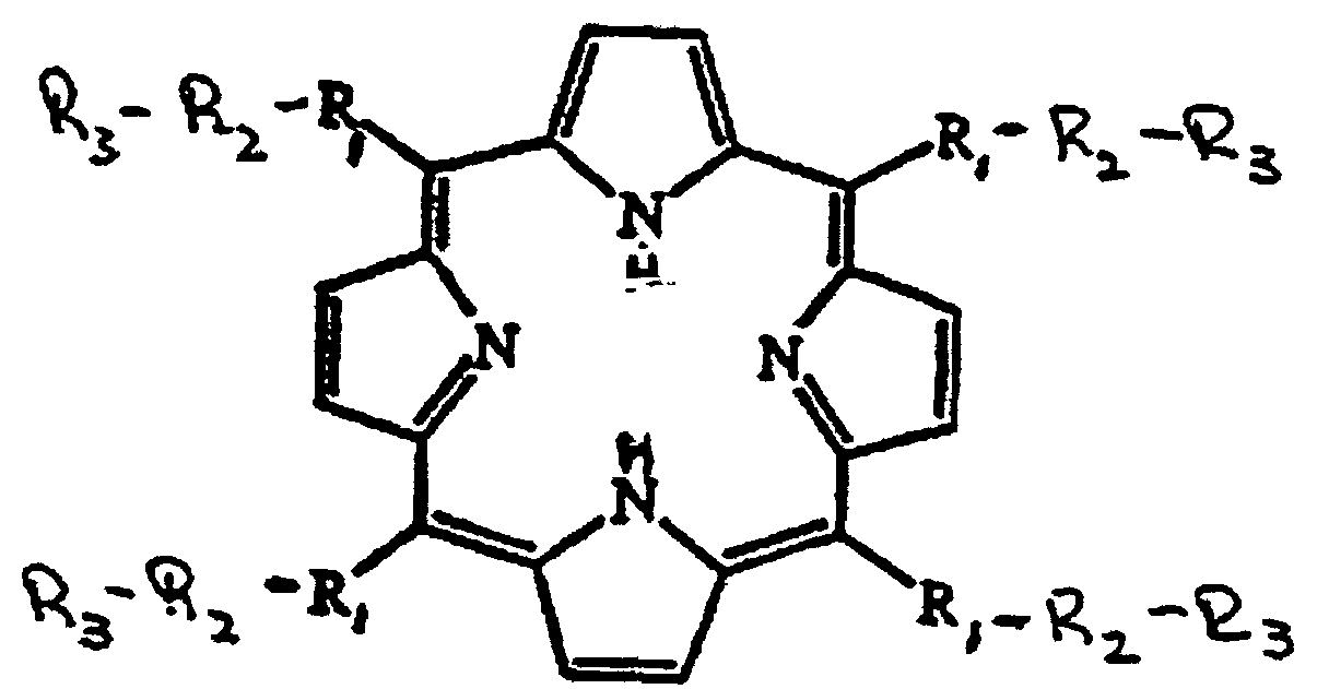
- Mimetics of SOD appropriate for use in the present methods include manganic derivatives of methine substituted porphines, or pharmaceutically acceptable salts thereof.
- Preferred mimetics are of the formula: wherein R 1 , R 2 and R 3 are as defined above.
- R 1 is a bond, wherein X is Cl or Br and Y is a C 1-4 alkyl group;
- R 3 is Y", -OH, -NH 2 , -N + (Y") 3 , -COO-, -COO - , -SO 3 -, -SO 3 - , -C-PO 3 H- or -C-PO 3 H - , wherein Y" is a C 1-4 alkyl group.
- R 1 is a bond, wherein X is Cl or Br and Y is methyl or ethyl
- R 3 is methyl, ethyl, -OH, -NH 2 , -N + (CH 3 ) 3 , -N + (CH 2 CH 3 ) 3 , -COO-, -COO - , -SO 3 -, -SO 3 - , -SO 3 - , -C-PO 3 H- or -C-PO 3 H - .
- R 1 is a bond, wherein Y is alkyl, preferably, C 1-4 alkyl, more preferably methyl or ethyl
- R 3 is C 1-4 alkyl (preferably methyl or ethyl), -OH, -NH-, -N + (CH 3 ) 3 , -N + (CH 2 CH 3 ) 3 , -COO-, -COO - , -SO 3 -, -SO 3 - , -SO 3 - , -C-PO 3 H- or -C-PO 3 H - .
- R 1 is a bond
- Y' is hydrogen or alkyl (preferably C 1-4 alkyl, more preferably methyl or ethyl) and wherein n is 1 to 4 (preferably 1 or 2)
- R 3 is C 1-4 alkyl (preferably methyl or ethyl), -OH, -NH-, -N + (CH 3 ) 3 , -N + (CH 2 CH 3 ) 3 , -COO-, -COO - , -SO 3 -, -SO 3 - , -SO 3 - , -C-PO 3 H- or -C-PO 3 H - .
- one or more of the pyrrole rings of the porphyrin can be substituted with an electron withdrawing group, for example, a NO 2 group, a halogen (eg Cl), or a nitrite.
- an electron withdrawing group for example, a NO 2 group, a halogen (eg Cl), or a nitrite.
- MnTMPyP Mn(III) tetrakis (1-methy-4-pyridyl)porphyrin
- MnTMAP Mn(III) tetrakis (4-trimethyl-aminophenyl)porphyrin
- MnTBAP Mn(III) tetrakis (4-benzoic acid)porphyrin
- Targeted forms of the mimetics can be produced by coupling directly or via a linker to a cell surface or extracellular matrix targeting moiety.
- the targeted mimetics can be used to mimic EC-SOD. Since the sequence of human EC-SOD is known (Hjalmarsson et al, Proc. Natl. Acad. Sci.
- the C-terminal oligopeptide can prepared and attached to the "mimetic core" (eg, a Mn(III)-porphyrin) via, for example, a free amine or carboxy group with a 1-ethyl-3-(3-dimethylaminopropyl)carbodiimide (EDC) coupling reaction (Yamada et al, Biochemistry 20:4836 (1981); Davis and Preston, Anal. Biochem. 116:402 (1981)) (note the R' group in the structures set forth below).
- EDC 1-ethyl-3-(3-dimethylaminopropyl)carbodiimide
- Candidate targeting moieties suitable for attaching to SOD (eg EC-SOD) mimetics to confer GAG binding properties include the following:
- targeting moieties also include heparin binding proteins generally and sugars, including mannose and oligosaccharides.
- Appropriate linkers include -SO 2 -NH-, -PO 3 -NH-, or
- SOD SOD
- EC-SOD SOD mimetics
- Mimetics suitable for use in the present invention can be selected by assaying for SOD activity and stability.
- SOD activity can be monitored in the presence and absence of EDTA using the method of McCord and Fridovich (J. Biol. Chem. 244 (1969)).
- the efficacy of a mimetic can be determined by measuring the effect of the mimetic on the growth of a SOD null E. coli strain versus a wild type strain.
- wild type E. coli (AB1157) and SOD null E. coli . JI132
- Active mimetics can be tested for toxicity in mammalian cell culture by measuring lactate dehydrogenase (LDH) release.
- LDH lactate dehydrogenase
- rat L2 cells a lung type II like cell; (Kaighn and Douglas, J. Cell Biol. 59:160a (1973)
- cells can be seeded at equal densities in 24 well culture dishes and grown to approximately 90% confluence
- SOD mimetics can be added to the cells at log doses (eg micromolar doses in minimal essential medium (MEM)) and incubated for 24 hours.
- MEM minimal essential medium
- Toxicity can be assessed by morphology and by measuring the release of the cytosolic injury marker, LDH (eg on a thermokinetic plate reader), as described by vassault (In: Methods of Enzymatic Analysis, Bergmeyer (ed) pp. 118-26 (1983); oxidation of NADH is measured at 340 nm).
- Efficacy of active mimetics can be assessed by determining their ability to protect mammalian cells against methylviologen (paraquat)-induced toxicity. Specifically, rat L2 cells grown as described above and seeded into 24 well dishes can be pre-incubated with various concentrations of the SOD mimetic and then incubated with a concentration of methylviologen previously shown to produce an LC 75 in control L2 cells.
- Efficacy of the mimetic can be correlated with a decrease in the methylviologen-induced LDH release (St. Clair et al, FEBS Lett. 293:199 (1991)).
- the efficacy of SOD mimetics can be tested in vivo with mouse and/or rat models using both aerosol administration and parental injection.
- male Balb/c mice can be randomized into 4 groups of 8 mice each to form a standard 2X2 contingency statistical model. Animals can be treated with either paraquat (40 mg/kg, ip) or saline and treated with SOD mimetic or vehicle control.
- Lung injury can be assessed 48 hours after paraquat treatment by analysis of bronchoalveolar lavage fluid (BALF) damage parameters (LDH, protein and % PMN) as previously described (Hampson et al, Tox. Appl. Pharm. 98:206 (1989); Day et al, J. Pharm. Methods 24:1 (1990)). Lungs from 2 mice of each group can be instillation fixed with 4% paraformaldehyde and processed for histopathology at the light microscopic level.
- BALF bronchoalveolar lavage fluid
- Mn(III)-porphyrin mimetics suitable for use in the present invention can be effected using art-recognized protocols.
- porphyrin rings with various methine bridge carbon side groups can be purchased commercially and the Mn(III) metal ion inserted into the porphyrin ring by methods including the following: (1) admixture of Mn(II) acetate with porphyrin in the presence of oxygen, under which condition selective stabilization of Mn(III) by the porphyrin causes autooxidation of Mn(II); (2) preparation of Mn(III)(OH) 3 by a modification of the Winkler method (Sastry et al, Anal. Chem.
- Mn(III)-porphyrin complexes can be precipitated from solution with sodium perchlorate, washed and residue perchlorate removed by strong anionic exchange resin.
- Formation of the Mn(III)-porphyrin can be followed spectrophotometrically by monitoring a characteristic Sorét band at 468 nm. Coupling of a binding domain to the "mimetic core" can be carried out as described above.
- the present invention results, at least in part, from the realization that EC-SOD specifically regulates NO ⁇ function.
- the invention is based on the realization that EC-SOD is synthesized by epithelial cells and is primarily localized in the interstitium, on matrix elements and collagen and around smooth muscle cells (particularly lung airways and vasculature).
- NO ⁇ is an intercellular signal and, as such, NO ⁇ must traverse the extracellular matrix to exert its effects.
- NO ⁇ is highly sensitive to inactivation mediated by O 2 - present in the extracellular spaces.
- EC-SOD is thus an enzyme ideally suited to increase the bioavailability of NO ⁇ by preventing its degradation by O 2 -.
- the invention also relates to mimetics of EC-SOD that can be targeted to strategic locations and to the use of such mimetics in manipulating extracellular levels of NO ⁇ .
- the invention is not limited to NO ⁇ manipulation as the sole mechanism of action of the mimetics. Rather, the invention relates to oxygen radical scavenging generally.
- the present invention relates, in a further specific aspect, to inhibiting production of superoxide radicals.
- the SOD mimetics are used to inhibit oxidases, such as xanthine oxidase, that are responsible for production of superoxide radicals (see Example VII).
- oxidases such as xanthine oxidase
- the ability of a mimetic to protect mammalian cells from xanthine/xanthine oxidase-induced injury can be assessed, for example, by growing rat L2 cells in 24-well dishes. Cells can be pre-incubated with various concentrations of an SOD mimetic and then xanthine oxidase (XO) can be added to the culture along with xanthine (X).
- XO xanthine oxidase
- XO/X used in the study can be pre-determined for each cell line by performing a dose-response curve for injury.
- X/XO can be used in an amount that produces approximately an LC 75 in the culture.
- Efficacy of the mimetic can be correlated with a decrease in XO/X-induced LDH release.
- the ability of the mimetics to inhibit the production of such radicals makes possible the use the mimetics as therapeutics for the treatment of gout and reperfusion injuries.
- the mimetics can be used to scavenge hydrogen peroxide, as well as superoxide radicals.
- scavengers of hydrogen peroxide the mimetics serve as mimics of either catalase or peroxidase.
- Appropriate mimetic scavengers can be selected by following absorbance at 240 nm in the presence of hydrogen peroxide (see Beers and Sizer, J. Biol. Chem. 195:133 (1952)). Therapies based on this activity correspond with those noted below.
- the mimetics can be used as catalytic scavengers of superoxide radicals to protect against ischemia reperfusion injuries associated with myocardial infarction, stroke, acute head trauma, organ reperfusion following transplantation, bowel ischemia, pulmonary infarction, surgical occlusion of blood flow, and soft tissue injury.
- the mimetics can further be used to protect against skeletal muscle reperfusion injuries.
- the mimetics can also be used to protect against damage to the eye due to sunlight (and to the skin) as well as glaucoma, and macular degeneration of the eye. Diseases of the bone are also amenable to treatment with the mimetics. Further, connective tissue disorders associated with defects in collagen synthesis or degradation can be expected to be susceptible to treatment with the present mimetics.
- the mimetics can be used as catalytic scavengers of superoxide radicals to increase the very limited storage viability of transplanted hearts, kidneys, skin and other organs and tissues.
- the amount of mimetic to be used in a particular treatment or to be associated with a particular substance can be determined by one skilled in the art.
- molecules (agents) having EC-SOD activity are administered under conditions such that levels of extracellular O 2 - are altered.
- Molecules suitable for use include forms of SOD that bind heparin sulfate or other glycosaminoglycans (GAG), for example, by virtue of the fact that they contain positively charged amino acids near their carboxy terminal end.
- GAG glycosaminoglycans
- the general requirements of mimetics are that they: (a) be stable enough to retain the ligated metal (i.e.
- Mn in the presence of the multiple chelating agents present in living systems, (b) be active enough that reasonable doses can serve to significantly augment the total SOD activity in the extracellular spaces, (c) be able to adhere to the surfaces of cells or extracellular matrix elements (eg collagen) when protection against extracellular sources of O 2 - is needed, and d) be of low toxicity.
- suitable mimetics include nitrogen-containing macro cyclic ligands effective as catalysts for dismutating superoxide, including Mn(III) complexes of porphyrins with bulky cationic substituents on the methine bridge carbons, such as those described above (eg MnTMAP and MnTMPyP). Such complexes are very active and are stable enough to retain full activity in the presence of excess EDTA or in the presence of tissue extracts.
- compositions suitable for use in the present invention can be formulated into pharmaceutical compositions suitable for use in the present invention.
- Such compositions include the active agent together with a pharmaceutically acceptable carrier, excipient or diluent.
- the composition can be present in dosage unit form for example, tablets, capsules or suppositories.
- the composition can also be in the form of a sterile solution suitable for injection or inhalation.
- Compositions can also be in a form suitable for opthalmic use.
- the invention also includes the use of compositions formulated for topical administration, such compositions taking the form, for example, of a lotion, cream, gel or ointment.
- the concentration of active agent to be included in the composition can be selected based on the nature of the agent, the dosage regimen and the result sought.
- the dosage of the composition to be administered can be determined without undue experimentation and will be dependent upon various factors including the nature of the active agent, the route of administration, the patient, and the result sought to be achieved. Suitable doses of mimetics will vary, for example, with the mimetic and with the result sought.
- the results of Faulkner et al (J. Biol. Chem. 269:23471 (1994)) indicate that the in vivo oxidoreductase activity of the mimetics is such that a pharmaceutically effective dose will be low enough to avoid problems of toxicity.
- Doses that can be used include those in the range of 1 to 50 mg/kg.
- diseases or disorders appropriate for treatment using the present invention include diseases of the central nervous system (including AIDS dementia, stroke, amyotrophic lateral sclerosis (ALS), Parkinson's disease and Huntington's disease) and diseases of the musculature (including diaphramic diseases (eg respiratory fatigue in emphysema, bronchitis and cystic fibrosis), cardiac fatigue of congestive heart failure, muscle weakness syndromes associated with myopathies, ALS and multiple sclerosis).
- Many neurologic disorders including stroke, Huntington's disease, Parkinson's disease, ALS, Alzheimer's and AIDS dementia
- NMDA or N-methyl-D-aspartate
- the present invention also relates to treating arthritis, systemic hypertension, atherosclerosis, edema, septic shock, pulmonary hypertension, including primary pulmonary hypertension, impotence, infertility, endometriosis, premature uterine contractions, memory disorders, microbial infections and gout.
- Therapeutic regimens including mode of administration, appropriate for effecting treatment of the conditions described above can be readily determined by one skilled in the art.
- Inflammations particularly inflammations of the lung, are amenable to treatment using the present invention (note particularly the inflammatory based disorders of asthma, ARDS including oxygen toxicity, pneumonia (especially AIDS-related pneumonia), cystic fibrosis, chronic sinusitis and autoimmune diseases (such as rheumatoid arthritis)).
- EC-SOD is localized in the interstitial spaces surrounding airways and vasculature smooth muscle cells.
- EC-SOD and O 2 - mediate the antiinflammatory - proinflammatory balance in the alveolar septum. NO ⁇ released by alveolar septal cells acts to suppress inflammation unless it reacts with O 2 - to form ONOO - .
- EC-SOD By scavenging O 2 - , EC-SOD tips the balance in the alveolar septum against inflammation. Significant amounts of ONOO- will form only when BC-SOD is deficient or when there is greatly increased O 2 - release.
- EC-SOD mimetics such as those described herein, can be used to protect against destruction caused by hyperoxia. Appropriate therapeutic regimens can be readily established by one skilled in the art.
- Figure 24 presents a gene sequence as well as portions of non-coding regions of that sequence of at least 15 bases, preferably, at least 50 bases, more preferably, at least 100 bases and most preferably, at least 500 bases. Such portions, and the complements thereof can be used as probes or primers in protocols including those described in Example VI.
- Screening of subjects for defects in the EC-SOD gene can be done using the approach used to identify mutations on the ⁇ -adrenergic receptor gene (Reihaus et al, Am. J. Respir. Cell. Mol. Biol. 8:334 (1993)). That approach is similar to the one used by Rosen et al (Nature 262:59 (1993)) (see Example VI).
- the EC-SOD expression vector ( Figure 1) was constructed as follows: The entire human EC-SOD cDNA fragment (Hjalmarrson et al, Proc. Natl. Acad. Sci. USA 84:6340 (1987); Hendrickson et al, Genomics 8:736 (1990)) flanked by EcoRI restriction sites was converted with mung bean nuclease to form blunt-ends, ligated to SalI linkers, digested with SalI, and then inserted into the SalI site of the human ⁇ -actin expression vector pH ⁇ APr-1.
- mice Purified DNA at 2.5 ⁇ g/ml in 5 mM Tris-HCl, pH 7.4, 0.1 mM EDTA was injected into the pronuclei of fertilized eggs isolated from mice ((C57BL/6 X C3H)F1 X (C57BL/6 X C3H)F1)((C57BL/6 X C3H)F1 mice were purchased from Charles River).
- Mouse eggs surviving microinjection were then implanted into the oviducts of pseudopregnant foster mothers (CD1) (CD1 mice were purchased from Charles River) following procedures described by Hogan et al (Hogan et al, Manipulating the Mouse Embryo, Cold Spring Harbor; Cold Spring Harbor Laboratory 1986, 32).
- mice carrying the transgene were identified by Southern blot analysis of tail DNA probed with the entire human EC-SOD cDNA. Transgenic founders were found the first litter screened. These mice were bred with (C57BL/6 X C3H)FI to produce offspring for further studies. (In all of the following experiments with the EC-SOD transgenic mice, the nontransgenic mice refer to littermates of the transgenic mice that did not contain the transgene for the human EC-SOD. In experiments in which EC-SOD transgenic mice were not used, wild type (C57BL/6 X C3H)FI were used.)
- Homozygous transgenic mice were produced by an F 1 cross of heterozygous transgenic mice.
- Tail DNA from F 2 mice were isolated and treated with RNAase.
- 10 ⁇ g of DNA from each mouse was cut with PstI and then electrophoresed through a 1.2% agarose gel.
- Southern blot analysis of tail DNA probed with the entire human EC-SOD cDNA was then done.
- the human EC-SOD cDNA did not cross-react with the mouse EC-SOD gene. Band intensity was compared visually to determine which mice were homozygous, heterozygous, or negative for the human EC-SOD transgene.
- SOD activity EC-SOD activity and total SOD activity (CuZn SOD and Mn SOD) remaining after EC-SOD extraction were measured by inhibition of cytochrome C reduction at pH 10, as previously described (Crapo et al, Methods Enzymol. 53:382 (1978)). Total protein was determined by the BCA protein assay (Pierce, Rockford, IL). The SOD activities were then expressed as units/mg total protein.
- mice carrying the human EC-SOD transgene were detected by Southern blot analysis. Northern analysis of various tissues from the F1 of one mouse found to carry the transgene is shown in Figure 2. High levels of message for human EC-SOD were detected in the heart, skeletal muscle, and brain of transgenic mice, with little or no message observed in the lung, liver, and spleen. No message was detectable in nontransgenic littermates.
- Homozygous mice were generated by breeding two heterozygous F1 mice. Homozygous mice were detected by differential band intensities found using Southern blot analysis of equal amounts of PstI digested DNA from the offspring. EC-SOD activity in the mice was found to increase in response to the total copies of the EC-SOD transgene (Table I). EC-SOD activity in tissues of nontransgenic, heterozygous transgenic, and homozygous transgenic mice. Tissues from 3 mice were combined for each measurement. Activity is expressed as Units/g tissue wet weight. Tissue Nontransgenic Heterozygous Homozygous Brain 18 38 50 Heart 35 69 102
- Oxygen exposures 7-8 week old mice were exposed to hyperbaric oxygen five at a time in a small-animal chamber (Bethlehem, Pennsylvania). After flushing the chamber with pure oxygen, compression to 50 meters (6 ATA) was performed within 5 minutes. The oxygen concentration in the chamber was monitored continuously with a Servomex oxygen analyzer (model 572, Sybron, Norwood, Massachusetts) and maintained at ⁇ 99%. The carbon dioxide concentration was analyzed from intermittent samples of chamber gas with an IR detector (IR Industries, Santa Barbara, California) and was not allowed to rise above 0.1%. The chamber temperature was maintained at 25-26°C, but the compression of oxygen in the chamber raised the temperature to 30-32°C transiently, but an environmental control system restored the normal chamber temperature within 3 minutes.
- IR detector IR Industries, Santa Barbara, California
- the exposures lasted 25 to 75 minutes and were followed by decompression for 5 minutes.
- the mice were observed continuously for signs of oxygen toxicity from the beginning of the exposure until 4 hours after removal from the chamber.
- the time to the first generalized convulsion (seizure latency) and the time to death were recorded. These exposure conditions are designed to cause CNS oxygen toxicity without appreciable evidence of pulmonary oxygen toxicity.
- mice were given either i.p. injections of either 0.008 cc/g saline or 400 mg/kg diethyldithiocarbamate dissolved in normal saline (0.008 cc/g). The mice were then exposed to 6 ATA oxygen for 25 mintues as described above.
- mice were given diethyldithiocarbamate and sacrificed one hour later. The brains were removed and assayed for EC-SOD and CuZn SOD activity as described above.
- mice were given either i.p. injections of 0.008 cc/g saline or 180 mg/kg ⁇ -mercaptoethanol (0.008 cc/g). This dose of ⁇ -mercaptoethanol was selected because it contains an equal number of reducing thiols as the dose of diethyldithiocarbamate. The mice were then exposed to 6 ATA oxygen for 30 minutes as described above.
- N- ⁇ -nitro-L-arginine an inhibitor of nitric oxide synthase :
- Ten minutes prior to beginning compression 0.008 cc/g saline or 20 mg/kg (0.008 cc/g) N- ⁇ -nitro-L-arginine dissolved in sterile water was given i.p to the transgenic and nontransgenic mice. Mice were then exposed at 6 ATA oxygen for 25 or 75 minutes as described above.
- Transgenic and nontransgenic mice were subsequently treated with an inhibitor of CuZn SOD, diethyldithiocarbamate, to confirm that the increased sensitivity of transgenic mice to CNS oxygen toxicity was the result of increased SOD activity.
- treatment with 400 mg/kg diethyldithiocarbamate resulted in 80% inhibition of EC-SOD and 60% inhibition of CuZn SOD in the brain. This is consistent with previous findings (Frank et al, Biochem. Pharmacol. 27:251 (1978); Heikkila et al, J. Biol. Chem. 251:2182 (1976)).
- mice were treated with an equimolar amount of reducing thiols in the form of ⁇ -mercaptoethanol and exposed to hyperbaric oxygen.
- Figure 5 shows that ⁇ -mercaptoethanol did not protect against CNS oxygen toxicity.
- nitric oxide is a mediator of CNS oxygen toxicity and EC-SOD is protecting nitric oxide from superoxide mediated inactivation.
- wild-type (C57BL/6 X C3H)F1 mice were treated with an inhibitor of nitric oxide synthase, N- ⁇ -nitro-L-arginine.
- Figure 6 shows the effects of N- ⁇ -nitro-L-arginine on seizure latency in mice.
- mice Young (6-7 week) mice (see Example I) were anesthetized with 60 mg/kg pentobarbital (Nembutal, Abbott Laboratories, Chicago, Illinois). An incision was then made in the scalp and a steel rod 20 cm long, 3 mm in diameter, equilibrated in liquid nitrogen with an 8 cm bath of liquid nitrogen 4 cm from the end of the rod, was placed on the skull over the right cerebral hemisphere for 30 seconds. The skin incision was then sutured.
- pentobarbital Nembutal, Abbott Laboratories, Chicago, Illinois
- Group 3 compared (C57BL/6 X C3H)F1 mice treated with saline to (C57BL/6 X C3H)F1 mice treated with 0.51 ⁇ moles/g Fe 3+ -saturated deferoxamine.
- Group 4 consisted of (C57BL/6 X C3H)F1 mice treated with saline and (C57BL/6 X C3H)F1 mice treated with 0.02 mg/g N- ⁇ -nitro-L-arginine methyl ester.
- Group 5 consisted of (C57BL/6 X C3H)F1 mice treated with saline and (C57BL/6 X C3H)F1 mice treated with 0.02 mg/g N- ⁇ -nitro-L-arginine methyl ester plus 0.05 mg/g L-arginine.
- Group 6 compared edema formation between nontransgenic mice, EC-SOD transgenic mice treated with saline, and EC-SOD transgenic mice treated with 0.02 mg/g N- ⁇ -nitro-L-arginine methyl ester.
- mice were pretreated with i.p. injections of deferoxamine or saline prior to cold-induced injury.
- Table II shows that pretreatment with deferoxamine resulted in 43% less edema formation compared to mice only given saline.
- Mice were then pretreated with i.p. injections of iron-saturated deferoxamine or saline before cold-induced injury to see if the iron chelating properties of this compound were truly necessary for protection against edema formation.
- Table IV shows that, even when deferoxamine was saturated with iron, it was still capable of protecting against edema formation and resulted in 32-48% less edema than that found in saline treated controls.
- the absolute values for the edema index were found to quite variable, however, repeated experiments consistently show the same signficant trends in protection against edema formation in the various treatments examined.
- deferoxamine is capable of protecting against edema formation by a mechanism independent of its ability to scavenge iron. Because deferoxamine is capable of scavenging both the peroxynitrite anion (Radi et al, Arch. Biochem. Biophys. 288(2):481 (1991)) as well as the hydroxyl radical (Hoe et al, Chemico-Biological Interactions 41:75 (1982)), it was hypothesized that it is these properties of deferoxamine that enable it to protect against vasogenic edema.
- N- ⁇ -nitro-L-arginine methyl ester a competitive inhibitor of the enzyme nitric oxide synthase, to determine if this would result in protection against edema formation after a cold-induced injury.
- Table III shows that treatment with N- ⁇ -nitro-L-arginine methyl ester significantly protected mice against edema formation resulting in 37% less edema formation than that occurring in saline treated controls. This protection by N- ⁇ -nitro-L-arginine methyl ester was reversed by simultaneous administration of an excess of L-arginine to the mice (Table III). The effect of inhibition of nitric oxide synthesis on edema formation after cold-induced brain injury.
- Wild-type (C57BL/6 X C3H)F1 mice were treated with the competitive inhibitor of nitric oxide synthase, N- ⁇ -nitro-L-arginine methyl ester (LNAME) to determine what effect nitric oxide had on vasogenic edema. Mice were also given N- ⁇ -nitro-L-arginine methyl ester plus an excess of L-arginine (LNAME + L-Arg) to see if the effects seen with LNAME alone could be reversed. Values are presented as mean ⁇ standard error. Treatment n Edema Index Saline 6 5.77 ⁇ 0.29 LNAME 6 3.65 ⁇ 0.51 Saline 6 6.56 ⁇ 0.21 LNAME + L-Arg 6 6.03 ⁇ 0.71
- EC-SOD transgenic mice were treated with either saline or N- ⁇ -nitro-L-arginine methyl ester to determine if there was an additive effect in preventing edema formation in mice which have both increased levels of EC-SOD as well as the inhibitor of nitric oxide synthase.
- Table IV shows that when EC-SOD transgenic mice were given the inhibitor of nitric oxide synthase, no added protection against edema formation was detected relative to transgenic mice protected only by increased levels of EC-SOD in the brain. Evaluation of the effect of inhibition of nitric oxide synthesis on edema formation in transgenic mice.
- Human lung Five human lung samples were obtained to evaluate the distribution of EC-SOD in human lung tissue. One sample was obtained from a surgical pathology specimen of a right upper lobe resected from a 43 year old white female with a 50 pack year smoking history (equivalent to one pack per day for one year) and a solitary nodule found on chest X-ray. The patient was diagnosed with squamous cell carcinoma. Tissue from a region not involved in the carcinoma from this lobe was used in the studies presented here. A second lung was obtained from a right upper lobe surgical pathology specimen resected from a 51 year old white male with a 60 pack year smoking history found to have an isolated nodule on X-ray.
- the tissues were fixed in 2% paraformaldehyde/0.2% gluteraldehyde in 0.01 M phosphate buffered saline (PBS; 1.2 g NaH 2 PO 4 , 8 g NaCl, 0.2 g KCl, in 1 liter pH 7.3) for 1 hour followed by overnight fixation in 4% paraformaldehyde at 4°C and then in O.C.T. compound.
- PBS phosphate buffered saline
- the tissues were frozen in liquid nitrogen chilled hexane and stored at -70°C until they were sectioned for light microscopic studies.
- lung tissues were processed as in the light microscopic studies up to the equilibration in sucrose. After equilibration in sucrose, the lung tissues were infiltrated with 10% gelatin at 37°C for 10 minutes. The tissue slices, in gelatin, were then solidified on ice, cut into 2 mm/side cubes, and then cryoprotected in 4% polyvinyl alcohol containing 2 M sucrose overnight. These samples were then mounted onto stubs, flash frozen in liquid nitrogen, and then stored in liquid nitrogen until they were sectioned for electron microscopic studies.
- EC-SOD Human recombinant EC-SOD (furnished by S.L. Marklund, Ume ⁇ , Sweden; Tibell et al Proc. Natl. Acad. Sci. USA 84:6634 (1987)) and the 20,000 x g supernatant of a human lung homogenate were denatured in the presence of ⁇ -mercaptoethanol and sodium dodecyl sulfate by boiling for 5 minutes and then electrophoresed through 12% polyacrylamide gel in the presence of sodium dodecyl sulfate. The protein was then electrophoretically transferred to nitrocellulose.
- the blot was then incubated with 4.3 ⁇ g/ml of an IgG purified fraction of rabbit anti-rh-EC-SOD (furnished by S.M. Marklund, Ume ⁇ University Hospital, Ume ⁇ , Sweden) affinity purified with rh-EC-SOD followed by incubation with 125 I-protein A and autoradiography.
- the optimal primary and secondary antibody dilutions were determined empirically and made in PBS with 1% milk plus 1% BSA (milk was not included in the streptavidin solution).
- the slides were developed using diaminobenzidine (10 mg diaminobenzidine, 50 ml 0.05 M Tris-Cl, pH 7.6, 100 ⁇ l 3% H 2 O 2 ) and counterstained with 1% methyl green.
- serial sections were separately labeled with either rabbit anti-rh-EC-SOD (EC-SOD), non-immune rabbit IgG, or rabbit anti-rh-BC-SOD from which EC-SOD binding IgG had been absorbed out (EC-SOD absorbed; see above).
- Electron microscopic immunocytochemistry Ultrathin cryo sections (70 nm) of human lung tissue were immunolabeled with rabbit anti rh-EC-SOD and 10-nm protein A-gold as prevsiously described (Crapo et al, Proc. Natl. Acad. Sci. USA 89:10405 (1992)) (Table VI). Briefly, sections were first incubated three times for five minutes at room temperature in 0.15% glycine in PBS to block aldehyde groups. Nonspecific binding was further blocked by incubation in 1% BSA in PBS for 10 minutes. Primary and secondary antibody dilutions were determined empirically and made in PBS containing 1% BSA.
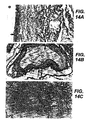
- Light microscopic immunohistochemistry Using an antibody to rh-EC-SOD, this protein was immunolocalized in human lungs. Light microscopic immunohistochemistry revealed with BC-SOD is mainly associated with the connective tissue matrix around vessels and airways in the lung ( Figure 14a and b, Figure 15a, b, and c). EC-SOD was found in close proximity to vascular and airway smooth muscle ( Figure 14a and b, and Figure 15a). EC-SOD was also seen in connective tissue of alveolar septal tips ( Figure 15c) suggesting an affinity of BC-SOD for connective tissue matrix.
- the antibody to EC-SOD was an IgG polyclonal rabbit antibody which was affinity purified using rh-EC-SOD.
- IgG specific for EC-SOD was absorbed out of the antisera using pure rh-EC-SOD bound to cyanogen bromide sepharose.
- Nonimmune rabbit IgG was then added to this absorbed antibody in a sufficient amount to replace the absorbed IgG.
- Labeling lung tissues with this preabsorbed antibody preparation resulted in the absence of labeling in all areas of the lung including the pulmonary vasculature ( Figure 14c). Labeling lung tissue with nonimmune IgG alone also resulted in the absence of labeling in all areas of the lung.
- the controls indicate that the labeling observed with the primary antibody is specific for EC-SOD in the lung.
- Electron microscopic immunocytochemistry A summary of the labeling for EC-SOD in the lung found using electron microscopic immunocytochemistry is summarized in Table VII.
- EC-SOD was mainly associated with extracellular matrix proteins in all regions of the lung.
- a high degree of labeling was seen in areas rich in type I collagen (Figure 16) and in association with other unidentified proteoglycans in extracellular matrix (Figure 17).
- no labeling for EC-SOD was seen in elastin-rich areas (Figure 16).
- a high degree of labeling was observed near the surface of smooth muscle cells and in the connective tissue matrix surrounding smooth muscle cells in vessels ( Figure 17) and airways.
- EC-SOD labeling was absent from the surface of type I and type II cells.
- a moderate, but consistent amount of intracellular EC-SOD was found in type II epithelial cells and in bronchial epithelial cells (Figure 19).
- ( ⁇ ) represents areas where low low amounts of labeling for EC-SOD were inconsistently observed.
- Controls done by absorbing EC-SOD specific antibody out of the primary antibody and replacing this absorbed antibody with nonimmune rabbit IgG resulted in the absence of labeling in all areas of the lung including areas rich in type I collagen as seen in Figure 16c.
- use of nonimmune rabbit IgG instead of the primary antisera also resulted in the absence of labeling in all areas of the lung.
- the lack of labeling with these controls indicates that the labeling observed with the primary antisera is specific for EC-SOD in the lung.
- EC-SOD may have an important function in this location.
- Superoxide is known to rapidly react with nitric oxide and inactivate its properties as a smooth muscle relaxant. Therefore, the presence of EC-SOD along the diffusion path of nitric oxide to smooth muscle cells should increase the half life of this short lived intercellular messenger in this particular region and thus increase its potency as a vasodilator.
- the high labeling for EC-SOD seen around vascular and airway smooth muscle cells indicates a function for EC-SOD as a mediator of nitric oxide activity in the maintenance of low pulmonary vascular pressures and airway resistence.
- EC-SOD In addition to the labeling of EC-SOD in association with smooth muscle cells, EC-SOD also appears to strongly colocalize with type I collagen. Collagen has previously been demonstrated to be susceptible to attack by reactive oxygen species such as the superoxide anion. In addition, the superoxide anion may be capable of activating latent collagenases from polymorphonuclear leukocytes (PMN) which can lead to further collagen degradation. Because collagen fragments have been shown to chemoattract and activate PMN's, any increased produced of superoxide that results in collagen degradation may accelerate inflammatory reactions and tissue destruction through PMN recruitment and activation. Consequently, the association of EC-SOD with collagen may be important in preventing superoxide mediated degradation of collagen and therefore, represent a means of controlling inflammatory responses.
- PMN polymorphonuclear leukocytes
- [ ⁇ - 35 S]dATP ( ⁇ 1400 Ci/mmol), [ ⁇ - 32 P]ATP (3000 Ci/mmol), and [ ⁇ - 35 P]CTP (800 Ci/mmol), were purchased from New England Nuclear.
- Human genomic DNA, T 7 , T 3 , and SP6 RNA polymerase, RNasin, and the pGEM3Zf(+) plasmid were obtained from Promega Biotec.
- Sequenase sequencing kit (V 2.0) was from United States Biochemicals Corporation. Human poly A+ RNA was acquired from Clontech. SeaPlaque GTG agarose was from FMC BioProducts. Restriction enzymes were from New England Biolabs. All other reagents used were molecular biology grade. Oligonucleotides were synthesized using an Applied Biosystems 380B or 392 by the Duke University, Department of Botany DNA synthesis facility. Charged nylon membranes (GeneScreen Plus) were from DuPont.
- RNA Two ⁇ g poly A+ RNA were purified from eight different human tissues. These mRNAs were electrophoresed on a denaturing formaldehyde 1.2% agarose gel and transferred to a charge-modified nylon membrane followed by UV irradiation fixation. The membrane was prehybridized in 50% formamide, 0.25 M NaPO 4 (pH 7.2), 0.25 M NaCl, 1 mM EDTA, 7% SDS, and 5% polyethylene glycol (molecular weight 8000).
- the blot was hybridized in the same buffer overnight at 60°C with 1 X 10 6 cpm/ml of [ 32 P]-labeled human EC-SOD RNA generated by transcription of the full-length cDNA using T 3 RNA polymerase in the presence of [ ⁇ - 32 P]CTP.
- the blot was washed in 0.25 M NaPO 4 (pH 7.2), 1% SDS, and 1mM EDTA at 75°C followed by a second wash using 0.04 M NaPO 4 (pH 7.2), 1% SDS, and 1 mM EDTA at 75°C for 30 minutes. This was followed by exposure to XAR-5 film using a Lightening Plus intensifier screen at -70°C.
- the autoradiogram was scanned using an LKB Ultrascan XL laser densitomer, and the peaks were quantitated by integration using the internal digital integrator of the densitometer or by cutting out the peak from a printer tracing and weighing.
- RNA template was degraded by the addition of RNase H at 55°C for 10 minutes.
- the resulting cDNA was isolated using a GlassMAX DNA (GIBCO BRL) isolation spin cartridge. The purified cDNA was dC-tailed using terminal deoxynucleotidyl transferase (TdT, 0.5 units/ul).
- the final composition of the reaction included 20 mM Tris-HCl (pH 8.4), 50 mM KCl, 2.5 mM MgCl 2 , 100 ⁇ g/ml bovine serum albumin, 400 nM Anchor primer, 200 nM gene-specific primer, and 200 ⁇ M each of dATP, dCTP, dGTP, dTTP.
- amplitaq Perkin Elmer Cetus was added at a final concentration of 0.04 units/ ⁇ l.
- PCR cycling was performed on a Perkin Elmer 9600 for 35 cycles with melting at 94°C for 45 seconds and annealing at 53°C for 15 seconds and extension at 72°C for 90 seconds.
- the full-length EC-SOD cDNA (6 ng) was used as a positive control in the PCR reaction.
- the PCR products were electrophoresed in a 2% SeaPlaque GTG agarose gel, transferred to charged nylon membranes by the method of Southern (Southern, J. Mol. Biol. 98:503 (1975)) using the alkaline transfer protocol (Reed et al, Nuc. Acids Res. 13:7207 (1985)).
- the DNA was fixed to the membrane by baking at 80°C in a vacuum oven for 2 hours.
- the subsequent blot was hybridized to [ 32 P] end-labeled HBC2 (an internal, nested EC-SOD specific primer, 5'-TCCAGCTCCTCCAAGAGAGC-3') overnight at 37°C.
- the blot was washed at increasing stringencies until background hybridization was removed. This was followed by exposure to XAR-5 film using a Lightening Plus intensifier screen at -70°C.
- the blot was hybridized (500 X 10 3 cpm/ml) in 50% formamide, 0.25 M NaPO 4 (pH 7.2), 0.25 M NaCl, 1 mM EDTA, 7% SDS, and 5% polyethylene glycol (molecular weight 8000), at 50°C. Following overnight hybridization, they were washed in 0.25 M NaPO 4 (pH 7.2), 2% SDS, and 1 mM EDTA followed by 0.04 M NaPO 4 (pH 7.2), 1% SDS, and 1 mM EDTA at increasing stringencies until background hybridization was minimized. The blot was exposed to XAR-5 film using a Lightening Plus intensifier screen at -70°C.
- a human adult female leukocyte genomic library constructed in the EMBL-3 vector was obtained from Clontech. Approximately 1 X 10 6 pfu were screened at a density of ⁇ 50,000 pfu/plate using [ 32 P]CTP-labeled human EC-SOD cRNA (1 X 10 6 dpm/ml). The primary through tertiary screens identified approximately 7 unique putative positive plaques. Individual plaques were isolated and lambda DNA purified using LambdaSorb phage adsorbent (Promega Biotec). The size of the insert DNA from each clone was assessed by Sal I restriction endonuclease digestion followed by electrophoresis in 0.7% agarose.
- Oligonucleotides derived from this initial sequencing data were synthesized approximately every 250 base pairs until the complete nucleotide sequence was obtained. Sequencing data were obtained from both strands as shown in Figure 20B except at the 3' portion of the gene where DNA sequence was obtained on one strand only.
- the IntelliGenetics Geneworks program (Version 2.2) was used for organizing the DNA sequence data. Homology searching was performed at the NCBI using the BLAST (Altschul et al, J. Mol. Biol. 215:403 (1990)) network service and the non-redundant nucleotide sequence database (GenBank(77.0) + EMBL(35.0) + EMBLUpdate + GBUdate). Transcriptional factor database searching was performed using both the SIGNAL SCAN 3.0 algorithm (Prestridge et al, CABIOS 9:113 (1993)) as well as the FINDPATTERNS program of the GCG Package (V 7.2) using Release 6.3 of the Transcription Factor Database (Gosh, Nuc. Acids Res.
- poly A(+) mRNA from eight different human tissues was fractionated on a denaturing agarose gel and transferred to a charged nylon membrane. Because a previous paper reported long exposures times in order to identify EC-SOD specific bands during genomic Southern analysis (Hendrickson et al, Genomics 8:736 (1990)), a radiolabeled antisense cRNA probe derived from full-length human BC-SOD cDNA was used (Oury et al, Proc. Nat. Acad. Sci. USA 89:9715 (1992)). A discrete band of approximately 1.4 kb can be seen in all eight human tissues analyzed (Figure 21A).
- skeletal muscle contains an approximate 4.2 kb message, not detected in the other tissues.
- densitometric scanning of the 4.2 and 1.4 kb bands it can be calculated that larger message to make up about 32% of the total skeletal muscle EC-SOD message.
- a very faint band of 2.2 kb can be seen. This band was too weak for quantitation by laser densitometer. Quantitation of these bands was performed by laser densitometry and integration of peaks of autoradiograms obtained in the linear range of exposure ( Figure 21B). After normalizing to the brain, the heart showed the most expression with 10.1 times brain. This was followed by the placenta, pancreas, and lung which gave 13.6, 10.2, and 7.5, respectively.
- mapping of the site of transcription initiation was attempted using the primer extension method.
- a positive signal was not obtained even after long exposure times. This did not seem to be due to technique since it was not possible to get positive signals using RNA generated by in vitro transcription of the EC-SOD cDNA. Whether lack of success using this technique was due to very low abundance of mRNA encoding EC-SOD or some other problem(s) is unclear.
- the EC-SOD gene specific primer EC7 was used for hybridization and reverse transcription of human heart poly A+ mRNA as shown in Figure 22. Half of this reaction was 3' dC tailed using terminal deoxynucleotidyl transferase and the remaining half was not. These templates were then subjected to PCR amplification using the gene specific primers HEC1 + EC7 as well as the anchor primer + EC4. The products of these reactions were fractioned by agarose electrophoresis, transfered to nylon membranes, and probed with the interal nested gene specific primer HEC2.
- FIG. 22A An autoradiogram of this experiment is shown in Figure 22A.
- EC-SOD cDNA As a control template and HEC1 + EC7, a band of 217 bp is expected (lane 3 of Figure 22A). Since the primers HEC1 and EC7 are expected to amplify independant of dC tailing, bands of equal intensity in lanes 4 and 5, which are also of the same size as the EC-SOD control, are seen.
- the anchor primer which hybridizes to the dC tail
- EC4 only one band of ⁇ 190 bp was seen (lane 1). Since the template was not poly C tailed, lane 2 shows no signal as expected.
- the size of the reverse transcribed DNA would correspond to ⁇ 136 bp 5' of the EC4 primer.
- This analysis would predict that there is approximately 6 base pairs of additional 5' sequence on the cDNA clone and that transcription initiation starts about 6 bp upstream of the first intron (indicated by a dashed box).
- eukaryotic transcription initiation usually begins at an adenosine residue, it is expected that it will begin at a G (Breathnach et al, Ann. Rev. Biochem. 50:349 (1981)).
- clone #7 was picked for further analysis.
- Clone #7 is about 18 to 20 kb and contains at least 5000 bp of 5' flanking DNA and at least 4000 bp of 3' flanking DNA. Restriction mapping of clone #7 is shown in Figure 20A. This map is similar to the results obtained with genomic Southern blot analysis data indicating that clone #7 contains the EC-SOD gene.
- FIG. 20B The strategy for sublconing and sequencing clone #7 is shown in Figure 20B.
- Various size continguous and overlapping restriction fragments were subcloned into the plasmid vector pGEM32f(+) ( Figure 20B).
- the DNA inserts were sequenced on both strands using a combination of primer walking and vector specific, universal sequencing primers.
- the 3' half of 7K36 insert was sequenced on one strand only.
- Exon 3 is separated from exon 2 by intron 2, a 3849 bp segment. Exon 3 contains a total of 1336 bp and at 17 bp into this exon starts the beginning of the complete coding sequence for preEC-SOD ( Figure 20D). This includes an 18 amino acid signal peptide that precedes the 222 amino acid mature protein sequence. There are no introns separating the various structural domains of EC-SOD. These domains are shown schematically in Figure 20D and include amino acids 1-95 which contain a glycosylated Asn-89 and show no sequence homology with other proteins. Resides 96-193 show strong homology with CuZn-SOD protein sequences with preservation of critical amino acids important in enzyme catalysis and structure.
- Amino acids 194-222 contain multiple charged resides which have been shown to be important in binding to sulfated proteoglycans. 558 bp of the 5'-flanking region containing putative regulatory elements and 3675 bp of the 3'-flanking region were also sequenced. The exonic DNA sequence data are in agreement with the published cDNA sequence (Hjalmarsson et al, Proc. Natl. Acad. Sci. USA 84:6340 (1987)). The intron-exon boundries are shown in Table VIII and conform to the eukaryotic consensus splice sequence (Senapathy et al, Methods Enzymol. 183:252 (1990)). Both introns split sequences in the 5'-nontranslated region of the EC-SOD gene.
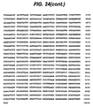
- Figure 24 shows the entire sequence for the human EC-SOD gene. Exonic sequences are shown in boxed uppercase letters while intronic, 5'- and 3'-flanking sequence are shown in lowercase. Exon 3 contains the entire uninterrupted coding region for EC-SOD and the protein sequence is shown using the single letter amino acid code. The 18 amino acid signal peptide and 222 amino acid mature protein sequence are highlighted. The identification of the signal peptide cleavage site is consistent with computer algorithms which predict the site of eukaryotic signal peptide cleavage (Folz et al, Biochem. Biophys. Res. Comm. 146:870 (1987)); Von Heijne, Eur. J. Biochem. 133:17 (1983)).
- Transcriptional factor database searching was used to putatively identify transcriptional regulatory elements. Although almost all eukaryotic promoters utilize a TATA box element to fix the position of transcription initiation, an obvious TATA box cannot be discerned for the EC-SOD gene. Two CAAT box elements were identified. One is in the reverse orientation and located about 20 bp upstream of the first exon, while the second can be found about 335 bp upstream. The putative signal for polyadenylation is shown and the site of poly A adenylation is indicated. Transcriptional factor database searching of the 5'-nontranslated region and first intron identified several potential regulatory elements.
- a cAMP responsive element (CREB) (TGACGT) which is similar to the adenovirus transcription virus (ATF) element can be found starting at 121 bp (Fink et al, Proc. Natl. Acad. Sci. USA 85:6662 (1988); Sassone-Corsi Proc. Natl. Acad. Sci. USA 85:7192 (1988)).
- a half site for the glucocorticoid response element (GRE) (TGTCCT) is located at 370 bp (Karin et al, Nature 308:513 (1984)).
- GRE glucocorticoid response element
- M-CAT skeletal muscle specific trans-activating factor response element
- CATTCCT is found in the reverse orientation beginning at position 238 (Mar et al, Mol.
- a xenobiotic responsive element (XRE) (CACGCW) is found within the first intron at position 1085 bp (Rushmore et al, J. Biol. Chem. 265:14648 (1990)).
- a metal regulatory element (MRE) (TGCRCYC) is found at position 89 (Culotta et al, Mol. Cell. Biol. 9:1376 (1989)).
- Two putative antioxidant response elements (ARE) (RGTGACNNNGC) are found at position 650 and 5022 (Rushmore et al, J. Biol. Chem. 266:11632 (1991)).
- a sis responsive element (SIF) (CCCGTC) important in the induction of the c-fos proto-oncogene is found in the reverse orientation at position 251 (Wagner et al, EMBO J. 9:4477 (1990)).
- TRE TPA responsive element
- the SV40 enhancer region AP4 (CAGCTGTGG) can be found at position 171 (Jones et al, Genes Dev. 2:267 (1988)).
- Genomic DNA will be purified utilizing a Qiagen Blood PCR Kit.
- One ml of blood containing - 10 7 leukocytes/ml will be collected in sodium citrate from each patient or control subject.
- the blood is placed into a QIAGEN-spin column and the leukocytes are entrapped in the resin by brief centrifugation, while erythrocytes and hemoglobin are washed through.
- the leukocytes are lysed by the addition of 0.7 ml of lysis buffer and incubated at room temperature for 30 minutes.
- the DNA is eluted by the addition of 1.2 M KCl, 50 mM MOPS, 15% ethanol, pH 8.3. This typically yields ⁇ 10 ⁇ g of genomic DNA (Reihsaus et al, Am. J. Respir. Cell. Mol. Biol. 8:334 (1993)).
- Sense and antisense oligonucleotide primers (or use primers already obtained from sequencing the genomic DNA) will be designed containing a 3'GC clamp (Sheffeld et al, Proc. Natl., Acad. Sci. USA 86:232 (1989)). These primers will encode the intronless coding region of the EC-SOD gene. A 172 bp region in the 3' untranslated region has been amplified using DNA sequencing primers and human genomic DNA. PCR conditions are as described (Reihause et al, Am. J. Respir. Cell. Mol. Biol.
- SSCP analysis has been used to detect single base pair mismatch (Orita et al, Genomics 5:874 (1989)).
- Temperature-gradient gel electrophoresis (TGGE) will be used to detect differences in mobility (Wartell et al, Nuc. Acids Res. 18:2699 (1990)).
- Samples for TGGE will be prepared by heat denaturing the PCR product at 98°C for 5 min, then renaturing at 50°C for 15 min with the corresponding wild-type DNA derived from PCR of the cloned gene.
- Electrophoresis will be carried out on a 5% acrylamide, 8 M urea gel over a temperature gradient.
- the temperature gradient will be optimized for each of the EC-SOD DNA segments. Typical gradients for the detection of ⁇ 2 -adrenergic receptor mutations were between 35°C to 60°C, and required 4 to 6 hours of run time (Rosen, Nature 262:59 (1993)).
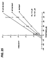
- calf pulmonary artery endothelial cell cultures (CPA-47 (Tissue and Cell 10:535 (1978)) were grown to confluence in Ham's F-12K medium with 10% fetal bovine serum at pH 7.4 and 37°C. Cells were then trypsinized and seeded at equal densities in 24 well plates and grown to 90% confluence. Cells were washed and pre-incubated for 1 hour with 50 ⁇ M of MnTBAP in minimum essential medium (MEM) or MEM only. Varing amounts of xanthine oxidase (XO) plus 200 ⁇ M xanthine (X) were added and allowed to incubate for 24 hours. Cell injury was quantitiated by measuring the release of cellular lactate dehydrogenase (LDH) into the medium. The efficacy of MnTBAP is shown in Figure 26 by the decrease in XO/X-induced LDH release.
- LDH lactate dehydrogenase
- Rat pulmonary epithelial cell cultures (L2 (Kaighn and Douglas J. Cell Biol. 59:160a (1973)) were grown to confluence in Ham's F-12K medium with 10% fetal bovine serum at pH 7.4 and 37°C. Cells were then trypsinized and seeded at equal densities in 24 well plates and grown to 90% confluence. Cells were washed and pre-incubated for 1 hour with 100 ⁇ M of MnTBAP or MnTMPyP in MEM or MEM only. Paraquat (2.5 mM) was added and allowed to incubate for 48 hours. Cell injury was quantitiated by measuring the release of cellular lactate dehydrogenase (LDH) into the medium. Figure 27 shows that MnTPyP (hatched bars) and MnTBAP (grey bars) decrease paraquat-induced LDH release.
- LDH lactate dehydrogenase
- calf pulmonary artery endothelial cell cultures (CPA-47 (Tissue and Cell 10:535 (1987)) were grown to confluence in Ham's F-12K medium with 10% fetal bovine serum at pH 7.4 and 37°C. Cells were then trypsinized and seeded at equal densities in 24 well plates and grown to 90% confluence. Cells were washed and pre-incubated for 1 hour with varing concentrations of MnTBAP in MEM or MEM only. Paraquat (2 mM) was added and allowed to incubate for 24 hours. Cell injury was quantitiated by measuring the release of cellular lactate dehydrogenase (LDH) into the medium. MnTBAP decreases paraquat-induced LDH release in a dose-dependent manner (see Figure 28).
- LDH lactate dehydrogenase
- ZnTBAP does not protect against paraquat-induced injury.
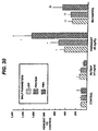
- Calf pulmonary artery endothelial cell cultures CPA-47 were grown to confluence in Ham's F-12K medium with 10% fetal bovine serum at pH 7.4 and 37°C. Cells were then trypsinized and seeded at equal densities in 24 well plates and grown to 90% confluence. Cells were washed and pre-incubated for 1 hour with varing concentrations of ZnTBAP in MEM or MEM only. Paraquat (2 mM) was added and allowed to incubate for 24 hours. Cell injury was quantitiated by measuring the release of cellular lactate dehydrogenase (LDH) into the medium. The results presented in Figure 29 demonstrate that ZnTBAP does not possess SOD-like activity. ZnTBAP can be used as a negative control to show that the redox metal is important in the protection against paraquat toxicity.
- LDH lactate dehydrogenase
- mice were treated with either paraquat (PQ, 45 mg/kg, ip) or saline (10 ml/kg, ip) and exposed to MnTBAP (2.5 mg/ml, nebulized into a 2 L chamber at 2 L/min for 30 minutes twice daily for 2 days) or room air. Mice were killed 48 hours after start of treatment and lung injury was assessed by analysis of bronchoalveolar lavage fluid (BALF).
- BALF damage markers used were lactate dehydrogenase (LDH, as units/L), protein concentration (as mg/dl), and percent of polymorphonuclear leukocytes (PMN).
- LDH lactate dehydrogenase
- PMN percent of polymorphonuclear leukocytes
Landscapes
- Health & Medical Sciences (AREA)
- Chemical & Material Sciences (AREA)
- Life Sciences & Earth Sciences (AREA)
- Organic Chemistry (AREA)
- Medicinal Chemistry (AREA)
- General Health & Medical Sciences (AREA)
- Engineering & Computer Science (AREA)
- Bioinformatics & Cheminformatics (AREA)
- Veterinary Medicine (AREA)
- Public Health (AREA)
- Animal Behavior & Ethology (AREA)
- Pharmacology & Pharmacy (AREA)
- Molecular Biology (AREA)
- Epidemiology (AREA)
- Genetics & Genomics (AREA)
- Proteomics, Peptides & Aminoacids (AREA)
- Biochemistry (AREA)
- Zoology (AREA)
- Wood Science & Technology (AREA)
- Biophysics (AREA)
- Biotechnology (AREA)
- Gastroenterology & Hepatology (AREA)
- Dispersion Chemistry (AREA)
- Microbiology (AREA)
- Chemical Kinetics & Catalysis (AREA)
- General Chemical & Material Sciences (AREA)
- Nuclear Medicine, Radiotherapy & Molecular Imaging (AREA)
- General Engineering & Computer Science (AREA)
- Biomedical Technology (AREA)
- Toxicology (AREA)
- Hematology (AREA)
- Diabetes (AREA)
- Medicines That Contain Protein Lipid Enzymes And Other Medicines (AREA)
- Pharmaceuticals Containing Other Organic And Inorganic Compounds (AREA)
- Enzymes And Modification Thereof (AREA)
- Medicines Containing Material From Animals Or Micro-Organisms (AREA)
- Nitrogen Condensed Heterocyclic Rings (AREA)
- Agricultural Chemicals And Associated Chemicals (AREA)
Claims (6)
- Verwendung eines Nachahmers von Superoxiddismutase mit der Formeloder eines pharmazeutisch verträglichen Salzes davon, worin
R1 eine Bindung,ist, wobei X ein Halogen ist und Y eine Alkylgruppe ist und wobei eine Bindung an R2 an irgendeiner Position bezeichnet und
 eine Bindung an R2 und den Substituenten an irgendeiner Position bezeichnet und
R2 eine Bindung, -(CY'2)n -, -(CY'2-CY'=CY')n -, -(CY'2-CY'2-CH=CH)n -, -(CY'=CY')n -, oderist,
wobei Y' Wasserstoff oder eine Alkylgruppe ist und n 1 bis 8 ist und
R3 -Y", -OH, -NH2, -N+(Y")3, -COOH, -COO-, -SO3H, -SO3 -, -C-PO3H2 oder -C-PO3H- ist, wobei Y" eine Alkylgruppe ist, komplexiert mit Mangan,
bei der Herstellung eines Medikaments zum Schutz von Zellen vor durch Superoxidradikal induzierter Toxizität in einer oder mehreren der folgenden Anwendungen:1. dem Schutz gegen ischämische Reperfusionsverletzungen, die mit Myokardinfarkt, Schlaganfall, akutem Kopftrauma, Organreperfusion nach Transplantation, Darmischämie, Lungeninfarzierung, chirurgischer Okklusion des Blutstroms oder Weichgewebeverletzung einhergehen,2. dem Schutz gegen Skelettreperfusionsverletzungen,3. dem Schutz gegen Schädigung von Haut und Augen durch Sonnenstrahlung,4. dem Schutz gegen Schädigung der Augen durch Glaukom oder Makuladegeneration,5. dem Schutz gegen Knochenerkrankung,6. dem Schutz gegen Bindegewebsstörungen, die mit Störungen der Kollagensynthese oder Degradation einhergehen,7. der Behandlung von Erkrankungen des zentralen Nervensystems,8. der Behandlung von Erkrankungen der Muskulatur,9. der Behandlung von neurologischen Störungen,10. der Behandlung von Arthritis, systemischem Bluthochdruck, Arteriosklerose, Ödem, septischem Schock, pulmonalem Hochdruck, Impotenz, Unfruchtbarkeit, Endometriose, vorzeitigen Gebärmutterkontraktionen, Gedächtnisstörungen, Mikrobeninfektionen und/oder Gicht,11. der Behandlung von Entzündungen und12. der Behandlung von chronischer Sinusitis und/oder Autoimmunerkrankungen. - Verwendung nach Anspruch 1, wobei die Zellen Säugerzellen sind.
- Verwendung nach Anspruch 1 oder Anspruch 2 für die Behandlung von AIDS, Demenz, Schlaganfall, amyotropher Lateralsklerose (ALS), Parkinson'scher Krankheit, Huntington'scher Krankheit, Zwerchfellerkrankungen, einschließlich Atemermüdung bei Emphysem, Bronchitis und zystischer Fibrose, Herzermüdung bei kongestiver Herzinsuffizienz, Muskelschwächesyndromen, die mit Myopathien einhergehen, ALS oder multipler Sklerose, Alzheimer'sche Krankheit, Asthma, ARDS, Pneumonie oder rheumatoider Arthritis.
- Verfahren zum Schutz von Pflanzenzellen vor durch Superoxidradikal induzierter Toxizität, bei dem man die Zellen mit einer nicht toxischen Menge, die ausreicht, um den Schutz zu bewirken, eines Nachahmers von Superoxiddismutase in Kontakt bringt mit der Formeloder eines pharmazeutisch verträglichen Salzes davon,

worin
R1 eine Bindung,ist, wobei X ein Halogen ist und Y eine Alkylgruppe ist und wobei eine Bindung an R2 an irgendeiner Position bezeichnet und
 eine Bindung an R2 und den Substituenten an irgendeiner Position bezeichnet und
R2 eine eindung, -(CY'2)n -, -(CY'2-CY'=CY')n -, -(CY'2-CY'2-CH=CH)n -, -(CY'=CY')n -, oderist,
wobei Y' Wasserstoff oder eine Alkylgruppe ist und n 1 bis 8 ist und
R3 -Y", -OH, -NH2, -N+(Y")3, -COOH, -COO-, -SO3H, -SO3 -, -C-PO3H2 oder -C-PO3H- ist, wobei Y" eine Alkylgruppe ist, komplexiert mit Mangan. - Verwendung eines Nachahmers von Superoxiddismutase mit der Formeloder eines pharmazeutisch verträglichen Salzes davon,

worin
R1 eine Bindung,ist, wobei X ein Halogen ist und Y eine Alkylgruppe ist und wobei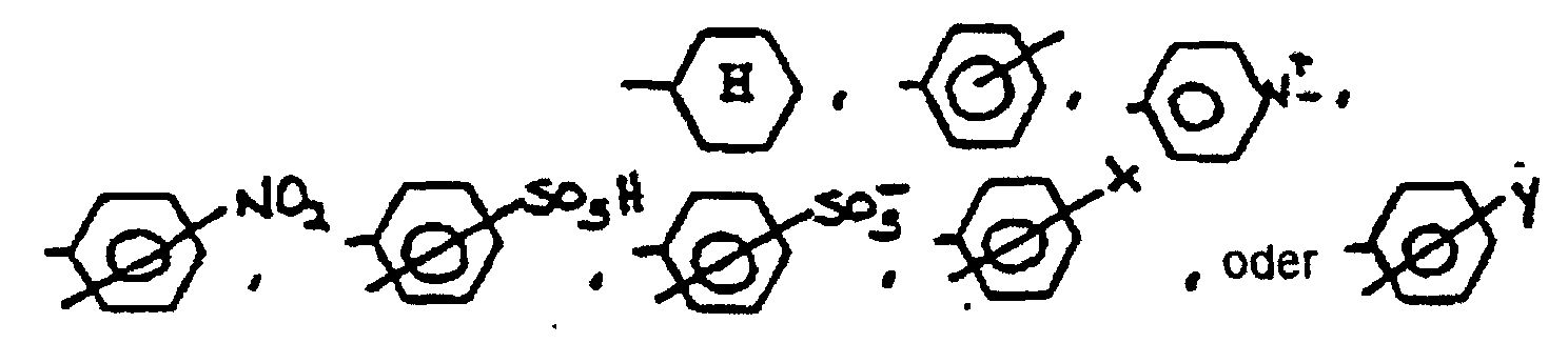 eine Bindung an R2 an irgendeiner Position bezeichnet und
 eine Bindung an R2 und den Substituenten an irgendeiner Position bezeichnet und
R2 eine Bindung, -(CY'2)n -, -(CY'2-CY'=CY')n -, -(CY'2-CY'2-CH=CH)n -, -(CY'=CY')n -, oderist,
wobei Y' Wasserstoff oder eine Alkylgruppe ist und n 1 bis 8 ist und
R3 -Y", -OH, -NH2, -N+(Y")3, -COOH, -COO-, -SO3H, -SO3 -, -C-PO3H2 oder -C-PO3H- ist, wobei Y" eine Alkylgruppe ist, komplexiert mit Mangan,
bei der Herstellung eines Medikaments zur Hemmung eines Schadens aufgrund von Oxidation einer Substanz mit der anschließenden Bildung von O2 - in einer oder mehreren der in Anspruch 1 definierten Anwendungen. - Verwendung eines Nachahmers von Superoxiddismutase, wie er in Anspruch 1 definiert ist, zur Erhöhung der Lagerungsüberlebensfähigkeit von transplantierten Herzen, Nieren, Haut und anderen Organen und Geweben.
Priority Applications (1)
| Application Number | Priority Date | Filing Date | Title |
|---|---|---|---|
| EP04010434A EP1442747A1 (de) | 1993-10-15 | 1994-10-13 | Superoxid Dismutase und ihre Mimetika |
Applications Claiming Priority (3)
| Application Number | Priority Date | Filing Date | Title |
|---|---|---|---|
| US13620793A | 1993-10-15 | 1993-10-15 | |
| US136207 | 1993-10-15 | ||
| PCT/US1994/011558 WO1995010185A1 (en) | 1993-10-15 | 1994-10-13 | Superoxide dismutase and mimetics thereof |
Related Child Applications (1)
| Application Number | Title | Priority Date | Filing Date |
|---|---|---|---|
| EP04010434A Division EP1442747A1 (de) | 1993-10-15 | 1994-10-13 | Superoxid Dismutase und ihre Mimetika |
Publications (3)
| Publication Number | Publication Date |
|---|---|
| EP0723398A1 EP0723398A1 (de) | 1996-07-31 |
| EP0723398A4 EP0723398A4 (de) | 1999-03-24 |
| EP0723398B1 true EP0723398B1 (de) | 2005-03-23 |
Family
ID=22471830
Family Applications (2)
| Application Number | Title | Priority Date | Filing Date |
|---|---|---|---|
| EP04010434A Withdrawn EP1442747A1 (de) | 1993-10-15 | 1994-10-13 | Superoxid Dismutase und ihre Mimetika |
| EP94930729A Expired - Lifetime EP0723398B1 (de) | 1993-10-15 | 1994-10-13 | Superoxiddismutase memetika |
Family Applications Before (1)
| Application Number | Title | Priority Date | Filing Date |
|---|---|---|---|
| EP04010434A Withdrawn EP1442747A1 (de) | 1993-10-15 | 1994-10-13 | Superoxid Dismutase und ihre Mimetika |
Country Status (9)
| Country | Link |
|---|---|
| US (1) | US5747026A (de) |
| EP (2) | EP1442747A1 (de) |
| JP (1) | JPH09505805A (de) |
| AT (1) | ATE291351T1 (de) |
| AU (1) | AU702596B2 (de) |
| CA (1) | CA2174236C (de) |
| DE (1) | DE69434313T2 (de) |
| ES (1) | ES2237753T3 (de) |
| WO (1) | WO1995010185A1 (de) |
Cited By (2)
| Publication number | Priority date | Publication date | Assignee | Title |
|---|---|---|---|---|
| US8975082B2 (en) | 2008-05-13 | 2015-03-10 | University Of Kansas | Metal abstraction peptide (MAP) tag and associated methods |
| US9187735B2 (en) | 2012-06-01 | 2015-11-17 | University Of Kansas | Metal abstraction peptide with superoxide dismutase activity |
Families Citing this family (38)
| Publication number | Priority date | Publication date | Assignee | Title |
|---|---|---|---|---|
| US6127356A (en) | 1993-10-15 | 2000-10-03 | Duke University | Oxidant scavengers |
| US5994339A (en) * | 1993-10-15 | 1999-11-30 | University Of Alabama At Birmingham Research Foundation | Oxidant scavengers |
| US5747026A (en) * | 1993-10-15 | 1998-05-05 | University Of Alabama At Birmingham Research Foundation | Antioxidants |
| WO1996009053A1 (en) * | 1994-09-20 | 1996-03-28 | Duke University | Oxidoreductase activity of manganic porphyrins |
| AU725602B2 (en) * | 1995-06-07 | 2000-10-12 | Duke University | Oxidant scavengers |
| EP0846003A1 (de) * | 1995-08-17 | 1998-06-10 | Monsanto Company | Verfahren zur diagnostischen bildanalyse unter verwendung von biokonjugaten von metallkomplexen von stickstoffhaltigen makrocyclischen liganden |
| CA2183834C (en) * | 1995-08-22 | 2003-09-09 | Hiroshi Maeda | Antihypertensive agents containing pyrazolopyrimidine derivatives |
| KR19990087784A (ko) * | 1996-03-13 | 1999-12-27 | 죤 에이치. 뷰센 | 초과산화물을 불균등화하기 위한 촉매로서 효과적인 질소함유거대고리 리간드의 망간 또는 철착물의 바이오콘쥬게이트 |
| WO1999023097A1 (en) | 1997-11-03 | 1999-05-14 | Duke University | Substituted porphyrins |
| ES2257858T3 (es) | 1998-04-24 | 2006-08-01 | Duke University | Porfirinas sustituidas. |
| US6632808B1 (en) | 1998-08-11 | 2003-10-14 | The United States Of America As Represented By The Department Of Health And Human Services | Inhibitors of amyloid formation |
| GB9817845D0 (en) | 1998-08-17 | 1998-10-14 | Glaxo Group Ltd | Chemical compounds |
| WO2000023568A2 (en) * | 1998-10-06 | 2000-04-27 | Albert Einstein College Of Medicine Of Yeshiva University | Methods and compositions for decreasing mitochondrial overproduction of reactive oxygen species in cells |
| US6210392B1 (en) * | 1999-01-15 | 2001-04-03 | Interventional Technologies, Inc. | Method for treating a wall of a blood vessel |
| US6695830B2 (en) * | 1999-01-15 | 2004-02-24 | Scimed Life Systems, Inc. | Method for delivering medication into an arterial wall for prevention of restenosis |
| ATE312103T1 (de) | 1999-01-25 | 2005-12-15 | Nat Jewish Med & Res Center | Substituierte porphyrine und deren therapeutische verwendungen |
| IT1306716B1 (it) * | 1999-06-21 | 2001-10-02 | Mendes S U R L | Associazione di batteri lattici e suo uso per la prevenzione e/o iltrattamento terapeutico di infezioni e di stati infiammatori. |
| SE9903985D0 (sv) * | 1999-11-03 | 1999-11-03 | Aga Ab | Use of nitric oxide |
| FR2806911B1 (fr) * | 2000-03-28 | 2003-01-10 | Univ Rene Descartes | Utilisation de mimetiques de la sod dans le traitement d'insuffisances hepatocellulaires |
| US6403788B1 (en) | 2000-07-11 | 2002-06-11 | Eukarion, Inc. | Non-genotoxic metalloporphyrins as synthetic catalytic scavengers of reactive oxygen species |
| JP2005524601A (ja) * | 2000-09-15 | 2005-08-18 | ザ スクリップス リサーチ インスティテュート | 抗体による過酸化水素およびスーパーオキシド生成に関する方法および組成物 |
| US20020072512A1 (en) * | 2000-12-08 | 2002-06-13 | Metaphore Pharmaceuticals, Inc | Method of preventing and treating HIV-mediated central nervous system damage |
| DE60233317D1 (de) * | 2001-01-19 | 2009-09-24 | Nat Jewish Med & Res Center | Medikament zum schutz in der radiotherapie |
| EP1353655A2 (de) * | 2001-01-26 | 2003-10-22 | Metaphore Pharmaceuticals Inc. | Verfahren zur behandlung von neurodegenerativen erkrankungen mit pentaaza-makrozyklischen ligandkomplexen |
| EP1439842A4 (de) * | 2001-06-01 | 2009-09-02 | Nat Jewish Med & Res Center | Oxidantienfänger zur behandlung von diabetes oder zur verwendung bei der transplantation oder induktion von immuntoleranz |
| US20040157280A1 (en) * | 2001-09-17 | 2004-08-12 | Paul Wentworth | Antibody mediated ozone generation |
| US20050129680A1 (en) * | 2001-09-17 | 2005-06-16 | Paul Wentworth | Antimicrobial activity of antibodies |
| US20040116350A1 (en) * | 2001-09-17 | 2004-06-17 | Paul Wentworth Jr | Methods and compositions relating to hydrogen peroxide and superoxide production by antibodies |
| AU2003216400A1 (en) | 2002-02-22 | 2003-09-09 | The Curators Of The University Of Missouri | Compounds for treatment of copper overload |
| US7485721B2 (en) * | 2002-06-07 | 2009-02-03 | Duke University | Substituted porphyrins |
| AU2003288058A1 (en) * | 2002-11-14 | 2004-06-03 | Novartis Ag | Antibody- or neutrophil-mediated ozone generation |
| PL2604286T3 (pl) * | 2003-05-01 | 2015-03-31 | Cornell Res Foundation Inc | Sposoby i kompleksy nośnikowe do transportu cząsteczek do komórek |
| BRPI0509359A (pt) | 2004-03-29 | 2007-09-04 | Inotek Pharmaceuticals Corp | compostos de porfirina substituìda por piridila e métodos de empregos destes |
| US8697120B2 (en) * | 2006-05-01 | 2014-04-15 | Johns Hopkins University | Method and use of nano-scale devices for reduction of tissue injury in ischemic and reperfusion injury |
| CN102105146A (zh) | 2008-05-23 | 2011-06-22 | 国立犹太保健医院 | 用于治疗与暴露于烷化物质有关的损伤的方法 |
| JP4891367B2 (ja) | 2009-05-21 | 2012-03-07 | トヨタ自動車株式会社 | 溶媒組成物 |
| US10531655B2 (en) | 2011-12-02 | 2020-01-14 | The Regents Of The University Of California | Reperfusion protection solution and uses thereof |
| CN104190927B (zh) | 2014-08-11 | 2016-05-18 | 苏州大学 | 一种同步送粉空间激光加工与三维成形方法及装置 |
Family Cites Families (21)
| Publication number | Priority date | Publication date | Assignee | Title |
|---|---|---|---|---|
| US4963367A (en) * | 1984-04-27 | 1990-10-16 | Medaphore, Inc. | Drug delivery compositions and methods |
| US4758422A (en) * | 1985-01-04 | 1988-07-19 | Salutar Inc. | Ferrioxamine paramagnetic contrast agents for MR imaging |
| JPS63500175A (ja) * | 1985-05-22 | 1988-01-21 | リポソ−ム テクノロジ−,インコ−ポレイテツド | リポソ−ム吸入法および吸入システム |
| DK402785D0 (da) * | 1985-09-03 | 1985-09-03 | Syn Tek Ab | Fremgangsmaade til fremstilling af et enzym |
| DE3854872D1 (de) * | 1987-03-14 | 1996-02-22 | Boehringer Ingelheim Int | Humane Mangan-Superoxiddismutase (hMn-SOD) |
| US5223538A (en) * | 1987-03-31 | 1993-06-29 | Duke University | Superoxide dismutase mimic |
| US5227405A (en) * | 1987-03-31 | 1993-07-13 | Duke University | Superoxide dismutase mimic |
| US5171680A (en) * | 1988-06-14 | 1992-12-15 | Chiron Corporation | Superoxide dismutase analogs having novel binding properties |
| DE69027383T2 (de) * | 1989-03-06 | 1996-11-14 | Masayasu Inoue | Neue superoxyd-dismutase |
| DK455789D0 (da) * | 1989-09-15 | 1989-09-15 | Symbicom Ab | Polypeptid |
| EP0424033A3 (en) * | 1989-10-19 | 1991-07-31 | Pola Chemical Industries Inc | External skin preparation |
| EP0462836A3 (en) * | 1990-06-20 | 1992-08-26 | Mitsui Toatsu Chemicals, Inc. | Recombinant vector plasmid capable of expressing human manganese superoxide dismutase, and process of producing this enzyme |
| US5202317A (en) * | 1990-09-13 | 1993-04-13 | The Regents Of The University Of California | Synthetic drug molecules that mimic metalloenzymes |
| US5217966A (en) * | 1990-09-13 | 1993-06-08 | The Regents Of The University Of California | Synthetic drug molecules that mimic metalloenzymes |
| AU8947791A (en) * | 1990-11-01 | 1992-05-26 | Scripps Research Institute, The | Glycosaminoglycan-targeted fusion proteins, their design, construction and compositions |
| EP0598753B1 (de) * | 1991-07-19 | 1998-03-18 | Monsanto Company | Mangan-Komplexe mit Stickstoff enthaltenden makrozyklischen Liganden, wirksam als Superoxiddismutasekatalysatoren. |
| US5696109A (en) * | 1992-12-07 | 1997-12-09 | Eukarion, Inc. | Synthetic catalytic free radical scavengers useful as antioxidants for prevention and therapy of disease |
| US5747026A (en) * | 1993-10-15 | 1998-05-05 | University Of Alabama At Birmingham Research Foundation | Antioxidants |
| WO2000023568A2 (en) * | 1998-10-06 | 2000-04-27 | Albert Einstein College Of Medicine Of Yeshiva University | Methods and compositions for decreasing mitochondrial overproduction of reactive oxygen species in cells |
| ATE312103T1 (de) * | 1999-01-25 | 2005-12-15 | Nat Jewish Med & Res Center | Substituierte porphyrine und deren therapeutische verwendungen |
| US6448239B1 (en) * | 1999-06-03 | 2002-09-10 | Trustees Of Princeton University | Peroxynitrite decomposition catalysts and methods of use thereof |
-
1994
- 1994-02-02 US US08/190,504 patent/US5747026A/en not_active Expired - Lifetime
- 1994-10-13 EP EP04010434A patent/EP1442747A1/de not_active Withdrawn
- 1994-10-13 AT AT94930729T patent/ATE291351T1/de not_active IP Right Cessation
- 1994-10-13 EP EP94930729A patent/EP0723398B1/de not_active Expired - Lifetime
- 1994-10-13 AU AU79763/94A patent/AU702596B2/en not_active Expired
- 1994-10-13 ES ES94930729T patent/ES2237753T3/es not_active Expired - Lifetime
- 1994-10-13 CA CA002174236A patent/CA2174236C/en not_active Expired - Lifetime
- 1994-10-13 JP JP7512010A patent/JPH09505805A/ja not_active Ceased
- 1994-10-13 WO PCT/US1994/011558 patent/WO1995010185A1/en not_active Ceased
- 1994-10-13 DE DE69434313T patent/DE69434313T2/de not_active Expired - Lifetime
Cited By (3)
| Publication number | Priority date | Publication date | Assignee | Title |
|---|---|---|---|---|
| US8975082B2 (en) | 2008-05-13 | 2015-03-10 | University Of Kansas | Metal abstraction peptide (MAP) tag and associated methods |
| US9096652B2 (en) | 2008-05-13 | 2015-08-04 | University Of Kansas | Metal abstraction peptide (MAP) tag and associated methods |
| US9187735B2 (en) | 2012-06-01 | 2015-11-17 | University Of Kansas | Metal abstraction peptide with superoxide dismutase activity |
Also Published As
| Publication number | Publication date |
|---|---|
| ATE291351T1 (de) | 2005-04-15 |
| AU7976394A (en) | 1995-05-04 |
| EP0723398A1 (de) | 1996-07-31 |
| EP0723398A4 (de) | 1999-03-24 |
| ES2237753T3 (es) | 2005-08-01 |
| JPH09505805A (ja) | 1997-06-10 |
| EP1442747A1 (de) | 2004-08-04 |
| DE69434313D1 (de) | 2005-04-28 |
| AU702596B2 (en) | 1999-02-25 |
| CA2174236A1 (en) | 1995-04-20 |
| CA2174236C (en) | 2008-02-12 |
| WO1995010185A1 (en) | 1995-04-20 |
| DE69434313T2 (de) | 2006-03-30 |
| US5747026A (en) | 1998-05-05 |
Similar Documents
| Publication | Publication Date | Title |
|---|---|---|
| EP0723398B1 (de) | Superoxiddismutase memetika | |
| US5994339A (en) | Oxidant scavengers | |
| US6583132B1 (en) | Oxidant scavengers | |
| CA2223407C (en) | Oxidant scavengers | |
| Vera et al. | Protective effect of carbon monoxide–releasing compounds in ischemia-induced acute renal failure | |
| Otterbein et al. | Mechanism of hemoglobin-induced protection against endotoxemia in rats: a ferritin-independent pathway | |
| Sheng et al. | Effects of metalloporphyrin catalytic antioxidants in experimental brain ischemia | |
| US20160166649A1 (en) | Use of hepcidin as a regulator of iron homeostasis | |
| Fliser | Perspectives in renal disease progression: the endothelium as a treatment target in chronic kidney disease | |
| EA027336B1 (ru) | Ингибирующие апоптоз фрагменты белка daxx, их пептидомиметики, конъюгаты и композиции и их применение | |
| Heeringa et al. | A protective role for endothelial nitric oxide synthase in glomerulonephritis | |
| US5888982A (en) | Regulation of vascular smooth muscle cell heme oxygenase-1 | |
| Costerousse et al. | Angiotensin I-converting enzyme inhibition but not angiotensin II suppression alters angiotensin I-converting enzyme gene expression in vessels and epithelia | |
| Almeida et al. | The 24‐kDa iron–sulphur subunit of complex I is required for enzyme activity | |
| JPH08283296A (ja) | ヒトアポリポプロテインeアナログ及びヒトスーパーオキシドジスムターゼアナログ | |
| AU769217B2 (en) | Oxidant scavengers | |
| CA2614621A1 (en) | Superoxide dismutase and mimetics thereof | |
| JP2002511764A (ja) | Pakキナーゼ遺伝子およびポリペプチドならびにそれらの使用方法 | |
| WO2009149201A1 (en) | Methods of treatment for pulmonary fibrosis | |
| AU2004201624A1 (en) | Oxidant Scavengers | |
| US20050287132A1 (en) | Biliverdin reductase fragments and variants, and methods of using biliverdin reductase and such fragments and variants | |
| JPH0643341B2 (ja) | 臓器機能改善剤 | |
| US20180221454A1 (en) | Thioredoxin, thioredoxin peptides or derivatives for treatment of age-related hypertension | |
| US20050250725A1 (en) | Regulation of the P21 gene and uses thereof | |
| Youn Cho et al. | 1-Benzylimidazole induces rat hepatic microsomal epoxide hydrolase with the elevation of its mRNA levels |
Legal Events
| Date | Code | Title | Description |
|---|---|---|---|
| PUAI | Public reference made under article 153(3) epc to a published international application that has entered the european phase |
Free format text: ORIGINAL CODE: 0009012 |
|
| 17P | Request for examination filed |
Effective date: 19960418 |
|
| AK | Designated contracting states |
Kind code of ref document: A1 Designated state(s): AT BE CH DE DK ES FR GB GR IE IT LI LU MC NL PT SE |
|
| A4 | Supplementary search report drawn up and despatched |
Effective date: 19990206 |
|
| AK | Designated contracting states |
Kind code of ref document: A4 Designated state(s): AT BE CH DE DK ES FR GB GR IE IT LI LU MC NL PT SE |
|
| 17Q | First examination report despatched |
Effective date: 20000425 |
|
| GRAP | Despatch of communication of intention to grant a patent |
Free format text: ORIGINAL CODE: EPIDOSNIGR1 |
|
| RTI1 | Title (correction) |
Free format text: SUPEROXIDE DISMUTASE MIMETICS |
|
| GRAP | Despatch of communication of intention to grant a patent |
Free format text: ORIGINAL CODE: EPIDOSNIGR1 |
|
| RTI1 | Title (correction) |
Free format text: SUPEROXIDE DISMUTASE MIMETICS |
|
| RTI1 | Title (correction) |
Free format text: SUPEROXIDE DISMUTASE MIMETICS |
|
| GRAS | Grant fee paid |
Free format text: ORIGINAL CODE: EPIDOSNIGR3 |
|
| GRAA | (expected) grant |
Free format text: ORIGINAL CODE: 0009210 |
|
| AK | Designated contracting states |
Kind code of ref document: B1 Designated state(s): AT BE CH DE DK ES FR GB GR IE IT LI LU MC NL PT SE |
|
| PG25 | Lapsed in a contracting state [announced via postgrant information from national office to epo] |
Ref country code: NL Free format text: LAPSE BECAUSE OF FAILURE TO SUBMIT A TRANSLATION OF THE DESCRIPTION OR TO PAY THE FEE WITHIN THE PRESCRIBED TIME-LIMIT Effective date: 20050323 Ref country code: AT Free format text: LAPSE BECAUSE OF FAILURE TO SUBMIT A TRANSLATION OF THE DESCRIPTION OR TO PAY THE FEE WITHIN THE PRESCRIBED TIME-LIMIT Effective date: 20050323 |
|
| REG | Reference to a national code |
Ref country code: GB Ref legal event code: FG4D |
|
| REG | Reference to a national code |
Ref country code: CH Ref legal event code: EP |
|
| REG | Reference to a national code |
Ref country code: IE Ref legal event code: FG4D |
|
| REF | Corresponds to: |
Ref document number: 69434313 Country of ref document: DE Date of ref document: 20050428 Kind code of ref document: P |
|
| PG25 | Lapsed in a contracting state [announced via postgrant information from national office to epo] |
Ref country code: GR Free format text: LAPSE BECAUSE OF FAILURE TO SUBMIT A TRANSLATION OF THE DESCRIPTION OR TO PAY THE FEE WITHIN THE PRESCRIBED TIME-LIMIT Effective date: 20050623 Ref country code: DK Free format text: LAPSE BECAUSE OF FAILURE TO SUBMIT A TRANSLATION OF THE DESCRIPTION OR TO PAY THE FEE WITHIN THE PRESCRIBED TIME-LIMIT Effective date: 20050623 |
|
| REG | Reference to a national code |
Ref country code: CH Ref legal event code: NV Representative=s name: MICHELI & CIE INGENIEURS-CONSEILS |
|
| REG | Reference to a national code |
Ref country code: ES Ref legal event code: FG2A Ref document number: 2237753 Country of ref document: ES Kind code of ref document: T3 |
|
| NLV1 | Nl: lapsed or annulled due to failure to fulfill the requirements of art. 29p and 29m of the patents act | ||
| PG25 | Lapsed in a contracting state [announced via postgrant information from national office to epo] |
Ref country code: PT Free format text: LAPSE BECAUSE OF FAILURE TO SUBMIT A TRANSLATION OF THE DESCRIPTION OR TO PAY THE FEE WITHIN THE PRESCRIBED TIME-LIMIT Effective date: 20050907 |
|
| PG25 | Lapsed in a contracting state [announced via postgrant information from national office to epo] |
Ref country code: MC Free format text: LAPSE BECAUSE OF NON-PAYMENT OF DUE FEES Effective date: 20051031 Ref country code: LU Free format text: LAPSE BECAUSE OF NON-PAYMENT OF DUE FEES Effective date: 20051031 |
|
| PLBE | No opposition filed within time limit |
Free format text: ORIGINAL CODE: 0009261 |
|
| STAA | Information on the status of an ep patent application or granted ep patent |
Free format text: STATUS: NO OPPOSITION FILED WITHIN TIME LIMIT |
|
| 26N | No opposition filed |
Effective date: 20051227 |
|
| ET | Fr: translation filed | ||
| PG25 | Lapsed in a contracting state [announced via postgrant information from national office to epo] |
Ref country code: SE Free format text: LAPSE BECAUSE OF FAILURE TO SUBMIT A TRANSLATION OF THE DESCRIPTION OR TO PAY THE FEE WITHIN THE PRESCRIBED TIME-LIMIT Effective date: 20050623 |
|
| PGFP | Annual fee paid to national office [announced via postgrant information from national office to epo] |
Ref country code: ES Payment date: 20130911 Year of fee payment: 20 |
|
| PGFP | Annual fee paid to national office [announced via postgrant information from national office to epo] |
Ref country code: FR Payment date: 20131009 Year of fee payment: 20 Ref country code: DE Payment date: 20131009 Year of fee payment: 20 Ref country code: CH Payment date: 20131014 Year of fee payment: 20 Ref country code: BE Payment date: 20131014 Year of fee payment: 20 Ref country code: IE Payment date: 20131010 Year of fee payment: 20 Ref country code: GB Payment date: 20131009 Year of fee payment: 20 |
|
| PGFP | Annual fee paid to national office [announced via postgrant information from national office to epo] |
Ref country code: IT Payment date: 20131015 Year of fee payment: 20 |
|
| REG | Reference to a national code |
Ref country code: DE Ref legal event code: R071 Ref document number: 69434313 Country of ref document: DE |
|
| REG | Reference to a national code |
Ref country code: DE Ref legal event code: R071 Ref document number: 69434313 Country of ref document: DE |
|
| REG | Reference to a national code |
Ref country code: CH Ref legal event code: PL |
|
| REG | Reference to a national code |
Ref country code: GB Ref legal event code: PE20 Expiry date: 20141012 |
|
| REG | Reference to a national code |
Ref country code: IE Ref legal event code: MK9A |
|
| REG | Reference to a national code |
Ref country code: ES Ref legal event code: FD2A Effective date: 20150108 |
|
| PG25 | Lapsed in a contracting state [announced via postgrant information from national office to epo] |
Ref country code: GB Free format text: LAPSE BECAUSE OF EXPIRATION OF PROTECTION Effective date: 20141012 Ref country code: IE Free format text: LAPSE BECAUSE OF EXPIRATION OF PROTECTION Effective date: 20141013 Ref country code: ES Free format text: LAPSE BECAUSE OF EXPIRATION OF PROTECTION Effective date: 20141014 |