EP0700430B1 - Verfahren zur selektiven Stimulierung der T-Zellproliferation. - Google Patents
Verfahren zur selektiven Stimulierung der T-Zellproliferation. Download PDFInfo
- Publication number
- EP0700430B1 EP0700430B1 EP94919346A EP94919346A EP0700430B1 EP 0700430 B1 EP0700430 B1 EP 0700430B1 EP 94919346 A EP94919346 A EP 94919346A EP 94919346 A EP94919346 A EP 94919346A EP 0700430 B1 EP0700430 B1 EP 0700430B1
- Authority
- EP
- European Patent Office
- Prior art keywords
- cells
- antibody
- cell
- population
- agent
- Prior art date
- Legal status (The legal status is an assumption and is not a legal conclusion. Google has not performed a legal analysis and makes no representation as to the accuracy of the status listed.)
- Expired - Lifetime
Links
- 210000001744 T-lymphocyte Anatomy 0.000 title claims abstract description 307
- 238000000034 method Methods 0.000 title claims abstract description 112
- 230000004936 stimulating effect Effects 0.000 title claims abstract description 16
- 230000035755 proliferation Effects 0.000 title claims description 20
- 230000004913 activation Effects 0.000 claims abstract description 14
- 230000001939 inductive effect Effects 0.000 claims abstract description 9
- 230000003213 activating effect Effects 0.000 claims abstract description 6
- 210000004027 cell Anatomy 0.000 claims description 195
- 101000914514 Homo sapiens T-cell-specific surface glycoprotein CD28 Proteins 0.000 claims description 74
- 102100027213 T-cell-specific surface glycoprotein CD28 Human genes 0.000 claims description 73
- 108091007433 antigens Proteins 0.000 claims description 64
- 102000036639 antigens Human genes 0.000 claims description 64
- 239000000427 antigen Substances 0.000 claims description 63
- 239000003795 chemical substances by application Substances 0.000 claims description 35
- 239000003446 ligand Substances 0.000 claims description 24
- 230000014509 gene expression Effects 0.000 claims description 22
- 206010028980 Neoplasm Diseases 0.000 claims description 18
- 230000005291 magnetic effect Effects 0.000 claims description 14
- 208000031886 HIV Infections Diseases 0.000 claims description 13
- 208000037357 HIV infectious disease Diseases 0.000 claims description 13
- 208000033519 human immunodeficiency virus infectious disease Diseases 0.000 claims description 13
- 239000011324 bead Substances 0.000 claims description 12
- 239000000203 mixture Substances 0.000 claims description 12
- 238000004519 manufacturing process Methods 0.000 claims description 10
- 230000001965 increasing effect Effects 0.000 claims description 9
- 239000007790 solid phase Substances 0.000 claims description 8
- 230000006052 T cell proliferation Effects 0.000 claims description 7
- 201000011510 cancer Diseases 0.000 claims description 7
- 230000003612 virological effect Effects 0.000 claims description 6
- 230000003247 decreasing effect Effects 0.000 claims description 5
- 230000010076 replication Effects 0.000 claims description 5
- 206010061598 Immunodeficiency Diseases 0.000 claims description 4
- 238000012544 monitoring process Methods 0.000 claims description 4
- 208000029462 Immunodeficiency disease Diseases 0.000 claims description 3
- 230000007813 immunodeficiency Effects 0.000 claims description 3
- 210000003171 tumor-infiltrating lymphocyte Anatomy 0.000 claims description 3
- 229940124522 antiretrovirals Drugs 0.000 claims description 2
- 239000003903 antiretrovirus agent Substances 0.000 claims description 2
- 238000009877 rendering Methods 0.000 claims description 2
- 230000002463 transducing effect Effects 0.000 claims description 2
- 230000007547 defect Effects 0.000 claims 1
- 230000009395 genetic defect Effects 0.000 claims 1
- 102100036011 T-cell surface glycoprotein CD4 Human genes 0.000 description 65
- 230000000638 stimulation Effects 0.000 description 60
- 108010002350 Interleukin-2 Proteins 0.000 description 49
- 102000000588 Interleukin-2 Human genes 0.000 description 49
- 108090000623 proteins and genes Proteins 0.000 description 48
- 235000018102 proteins Nutrition 0.000 description 41
- 102000004169 proteins and genes Human genes 0.000 description 41
- 101000946843 Homo sapiens T-cell surface glycoprotein CD8 alpha chain Proteins 0.000 description 35
- 102100034922 T-cell surface glycoprotein CD8 alpha chain Human genes 0.000 description 35
- 108090000765 processed proteins & peptides Proteins 0.000 description 32
- 238000009739 binding Methods 0.000 description 25
- 210000004408 hybridoma Anatomy 0.000 description 23
- 230000002163 immunogen Effects 0.000 description 23
- 241000725303 Human immunodeficiency virus Species 0.000 description 20
- 241001465754 Metazoa Species 0.000 description 18
- 230000012010 growth Effects 0.000 description 18
- 108091008874 T cell receptors Proteins 0.000 description 17
- 102000016266 T-Cell Antigen Receptors Human genes 0.000 description 17
- 238000000338 in vitro Methods 0.000 description 17
- 238000002474 experimental method Methods 0.000 description 15
- 239000012634 fragment Substances 0.000 description 14
- 108060008682 Tumor Necrosis Factor Proteins 0.000 description 13
- 102000000852 Tumor Necrosis Factor-alpha Human genes 0.000 description 13
- 108091003079 Bovine Serum Albumin Proteins 0.000 description 12
- 102000004127 Cytokines Human genes 0.000 description 12
- 108090000695 Cytokines Proteins 0.000 description 12
- 230000010261 cell growth Effects 0.000 description 12
- 230000003053 immunization Effects 0.000 description 12
- 239000002953 phosphate buffered saline Substances 0.000 description 12
- 210000000265 leukocyte Anatomy 0.000 description 11
- 230000007774 longterm Effects 0.000 description 11
- 210000005259 peripheral blood Anatomy 0.000 description 11
- 239000011886 peripheral blood Substances 0.000 description 11
- 102000004196 processed proteins & peptides Human genes 0.000 description 11
- 102100039620 Granulocyte-macrophage colony-stimulating factor Human genes 0.000 description 10
- 125000000539 amino acid group Chemical group 0.000 description 10
- PGHMRUGBZOYCAA-UHFFFAOYSA-N ionomycin Natural products O1C(CC(O)C(C)C(O)C(C)C=CCC(C)CC(C)C(O)=CC(=O)C(C)CC(C)CC(CCC(O)=O)C)CCC1(C)C1OC(C)(C(C)O)CC1 PGHMRUGBZOYCAA-UHFFFAOYSA-N 0.000 description 10
- PGHMRUGBZOYCAA-ADZNBVRBSA-N ionomycin Chemical compound O1[C@H](C[C@H](O)[C@H](C)[C@H](O)[C@H](C)/C=C/C[C@@H](C)C[C@@H](C)C(/O)=C/C(=O)[C@@H](C)C[C@@H](C)C[C@@H](CCC(O)=O)C)CC[C@@]1(C)[C@@H]1O[C@](C)([C@@H](C)O)CC1 PGHMRUGBZOYCAA-ADZNBVRBSA-N 0.000 description 10
- 239000002609 medium Substances 0.000 description 10
- 238000002360 preparation method Methods 0.000 description 10
- 102000005962 receptors Human genes 0.000 description 10
- 108020003175 receptors Proteins 0.000 description 10
- -1 D-3 phosphoinositides Chemical class 0.000 description 9
- 101000917858 Homo sapiens Low affinity immunoglobulin gamma Fc region receptor III-A Proteins 0.000 description 9
- 101000917839 Homo sapiens Low affinity immunoglobulin gamma Fc region receptor III-B Proteins 0.000 description 9
- 101000946889 Homo sapiens Monocyte differentiation antigen CD14 Proteins 0.000 description 9
- 102100029185 Low affinity immunoglobulin gamma Fc region receptor III-B Human genes 0.000 description 9
- 102100035877 Monocyte differentiation antigen CD14 Human genes 0.000 description 9
- 238000004458 analytical method Methods 0.000 description 9
- 230000007423 decrease Effects 0.000 description 9
- 238000002649 immunization Methods 0.000 description 9
- 208000015181 infectious disease Diseases 0.000 description 9
- 239000004033 plastic Substances 0.000 description 9
- 229920003023 plastic Polymers 0.000 description 9
- 108010017213 Granulocyte-Macrophage Colony-Stimulating Factor Proteins 0.000 description 8
- 102000006354 HLA-DR Antigens Human genes 0.000 description 8
- 108010058597 HLA-DR Antigens Proteins 0.000 description 8
- 239000002671 adjuvant Substances 0.000 description 8
- 150000001413 amino acids Chemical group 0.000 description 8
- 230000000139 costimulatory effect Effects 0.000 description 8
- 239000012894 fetal calf serum Substances 0.000 description 8
- 239000000243 solution Substances 0.000 description 8
- 239000006228 supernatant Substances 0.000 description 8
- 102100022005 B-lymphocyte antigen CD20 Human genes 0.000 description 7
- 239000012981 Hank's balanced salt solution Substances 0.000 description 7
- 101000897405 Homo sapiens B-lymphocyte antigen CD20 Proteins 0.000 description 7
- 101000889276 Homo sapiens Cytotoxic T-lymphocyte protein 4 Proteins 0.000 description 7
- 101001046686 Homo sapiens Integrin alpha-M Proteins 0.000 description 7
- 101000934346 Homo sapiens T-cell surface antigen CD2 Proteins 0.000 description 7
- 102100022338 Integrin alpha-M Human genes 0.000 description 7
- 241000124008 Mammalia Species 0.000 description 7
- 102000007079 Peptide Fragments Human genes 0.000 description 7
- 108010033276 Peptide Fragments Proteins 0.000 description 7
- 206010035226 Plasma cell myeloma Diseases 0.000 description 7
- 102100025237 T-cell surface antigen CD2 Human genes 0.000 description 7
- 210000000612 antigen-presenting cell Anatomy 0.000 description 7
- 210000004698 lymphocyte Anatomy 0.000 description 7
- 230000001404 mediated effect Effects 0.000 description 7
- 201000000050 myeloid neoplasm Diseases 0.000 description 7
- 239000013615 primer Substances 0.000 description 7
- 210000001519 tissue Anatomy 0.000 description 7
- 241000283707 Capra Species 0.000 description 6
- 238000002965 ELISA Methods 0.000 description 6
- 102000004388 Interleukin-4 Human genes 0.000 description 6
- 108090000978 Interleukin-4 Proteins 0.000 description 6
- 241000699666 Mus <mouse, genus> Species 0.000 description 6
- 241000700605 Viruses Species 0.000 description 6
- 239000012190 activator Substances 0.000 description 6
- 239000011248 coating agent Substances 0.000 description 6
- 238000000576 coating method Methods 0.000 description 6
- 125000004122 cyclic group Chemical group 0.000 description 6
- 239000012737 fresh medium Substances 0.000 description 6
- 239000003102 growth factor Substances 0.000 description 6
- 238000009169 immunotherapy Methods 0.000 description 6
- 239000003112 inhibitor Substances 0.000 description 6
- 238000002823 phage display Methods 0.000 description 6
- 230000009257 reactivity Effects 0.000 description 6
- 230000004044 response Effects 0.000 description 6
- 238000012216 screening Methods 0.000 description 6
- 238000012413 Fluorescence activated cell sorting analysis Methods 0.000 description 5
- OUYCCCASQSFEME-QMMMGPOBSA-N L-tyrosine Chemical compound OC(=O)[C@@H](N)CC1=CC=C(O)C=C1 OUYCCCASQSFEME-QMMMGPOBSA-N 0.000 description 5
- 102000018697 Membrane Proteins Human genes 0.000 description 5
- 108010052285 Membrane Proteins Proteins 0.000 description 5
- 108091007960 PI3Ks Proteins 0.000 description 5
- 102000003993 Phosphatidylinositol 3-kinases Human genes 0.000 description 5
- 108090000430 Phosphatidylinositol 3-kinases Proteins 0.000 description 5
- 230000006044 T cell activation Effects 0.000 description 5
- 238000003556 assay Methods 0.000 description 5
- 238000004113 cell culture Methods 0.000 description 5
- 238000004132 cross linking Methods 0.000 description 5
- 239000012228 culture supernatant Substances 0.000 description 5
- 230000000694 effects Effects 0.000 description 5
- 238000005516 engineering process Methods 0.000 description 5
- 230000004927 fusion Effects 0.000 description 5
- 230000028993 immune response Effects 0.000 description 5
- 230000005847 immunogenicity Effects 0.000 description 5
- 230000003834 intracellular effect Effects 0.000 description 5
- 230000000670 limiting effect Effects 0.000 description 5
- 239000000463 material Substances 0.000 description 5
- 210000003819 peripheral blood mononuclear cell Anatomy 0.000 description 5
- 230000026731 phosphorylation Effects 0.000 description 5
- 238000006366 phosphorylation reaction Methods 0.000 description 5
- 230000000284 resting effect Effects 0.000 description 5
- 239000007787 solid Substances 0.000 description 5
- 238000010186 staining Methods 0.000 description 5
- 238000010561 standard procedure Methods 0.000 description 5
- OUYCCCASQSFEME-UHFFFAOYSA-N tyrosine Natural products OC(=O)C(N)CC1=CC=C(O)C=C1 OUYCCCASQSFEME-UHFFFAOYSA-N 0.000 description 5
- 108060003951 Immunoglobulin Proteins 0.000 description 4
- 108010021625 Immunoglobulin Fragments Proteins 0.000 description 4
- 102000008394 Immunoglobulin Fragments Human genes 0.000 description 4
- ZDXPYRJPNDTMRX-VKHMYHEASA-N L-glutamine Chemical compound OC(=O)[C@@H](N)CCC(N)=O ZDXPYRJPNDTMRX-VKHMYHEASA-N 0.000 description 4
- 241000699670 Mus sp. Species 0.000 description 4
- 230000005867 T cell response Effects 0.000 description 4
- 235000001014 amino acid Nutrition 0.000 description 4
- 238000013459 approach Methods 0.000 description 4
- 210000003719 b-lymphocyte Anatomy 0.000 description 4
- 230000008901 benefit Effects 0.000 description 4
- 239000011575 calcium Substances 0.000 description 4
- 229910052791 calcium Inorganic materials 0.000 description 4
- 238000005119 centrifugation Methods 0.000 description 4
- 238000007796 conventional method Methods 0.000 description 4
- 238000000432 density-gradient centrifugation Methods 0.000 description 4
- 230000006870 function Effects 0.000 description 4
- 239000001963 growth medium Substances 0.000 description 4
- 102000043321 human CTLA4 Human genes 0.000 description 4
- 102000018358 immunoglobulin Human genes 0.000 description 4
- 238000001727 in vivo Methods 0.000 description 4
- 238000011534 incubation Methods 0.000 description 4
- 230000003993 interaction Effects 0.000 description 4
- 238000002955 isolation Methods 0.000 description 4
- 239000011777 magnesium Substances 0.000 description 4
- 229910052749 magnesium Inorganic materials 0.000 description 4
- 239000006249 magnetic particle Substances 0.000 description 4
- 239000008188 pellet Substances 0.000 description 4
- 150000003905 phosphatidylinositols Chemical class 0.000 description 4
- 230000003248 secreting effect Effects 0.000 description 4
- 230000028327 secretion Effects 0.000 description 4
- 210000002966 serum Anatomy 0.000 description 4
- 239000000126 substance Substances 0.000 description 4
- HKWJHKSHEWVOSS-OMDJCSNQSA-N 1,2-dihexadecanoyl-sn-glycero-3-phospho-(1D-myo-inositol-3,4-bisphosphate) Chemical compound CCCCCCCCCCCCCCCC(=O)OC[C@@H](OC(=O)CCCCCCCCCCCCCCC)COP(O)(=O)O[C@H]1[C@H](O)[C@@H](O)[C@H](OP(O)(O)=O)[C@@H](OP(O)(O)=O)[C@H]1O HKWJHKSHEWVOSS-OMDJCSNQSA-N 0.000 description 3
- 206010003445 Ascites Diseases 0.000 description 3
- 102100039498 Cytotoxic T-lymphocyte protein 4 Human genes 0.000 description 3
- CEAZRRDELHUEMR-URQXQFDESA-N Gentamicin Chemical compound O1[C@H](C(C)NC)CC[C@@H](N)[C@H]1O[C@H]1[C@H](O)[C@@H](O[C@@H]2[C@@H]([C@@H](NC)[C@@](C)(O)CO2)O)[C@H](N)C[C@@H]1N CEAZRRDELHUEMR-URQXQFDESA-N 0.000 description 3
- 229930182566 Gentamicin Natural products 0.000 description 3
- 108010048209 Human Immunodeficiency Virus Proteins Proteins 0.000 description 3
- 108010054477 Immunoglobulin Fab Fragments Proteins 0.000 description 3
- 102000001706 Immunoglobulin Fab Fragments Human genes 0.000 description 3
- 108010067060 Immunoglobulin Variable Region Proteins 0.000 description 3
- 102000017727 Immunoglobulin Variable Region Human genes 0.000 description 3
- 229930182816 L-glutamine Natural products 0.000 description 3
- 108700018351 Major Histocompatibility Complex Proteins 0.000 description 3
- 241000699660 Mus musculus Species 0.000 description 3
- 241000283973 Oryctolagus cuniculus Species 0.000 description 3
- 239000012980 RPMI-1640 medium Substances 0.000 description 3
- 241000700159 Rattus Species 0.000 description 3
- 108020004511 Recombinant DNA Proteins 0.000 description 3
- 230000000890 antigenic effect Effects 0.000 description 3
- 230000000903 blocking effect Effects 0.000 description 3
- 229940098773 bovine serum albumin Drugs 0.000 description 3
- 230000004663 cell proliferation Effects 0.000 description 3
- 239000002458 cell surface marker Substances 0.000 description 3
- 150000001875 compounds Chemical class 0.000 description 3
- 238000000684 flow cytometry Methods 0.000 description 3
- 229960002518 gentamicin Drugs 0.000 description 3
- 210000005104 human peripheral blood lymphocyte Anatomy 0.000 description 3
- 210000003071 memory t lymphocyte Anatomy 0.000 description 3
- 210000001616 monocyte Anatomy 0.000 description 3
- 238000011580 nude mouse model Methods 0.000 description 3
- 239000002245 particle Substances 0.000 description 3
- 244000052769 pathogen Species 0.000 description 3
- 230000001717 pathogenic effect Effects 0.000 description 3
- 210000005105 peripheral blood lymphocyte Anatomy 0.000 description 3
- 229920001184 polypeptide Polymers 0.000 description 3
- 230000000644 propagated effect Effects 0.000 description 3
- 230000001105 regulatory effect Effects 0.000 description 3
- 238000000926 separation method Methods 0.000 description 3
- 241000894007 species Species 0.000 description 3
- 210000000952 spleen Anatomy 0.000 description 3
- 210000004989 spleen cell Anatomy 0.000 description 3
- 230000020382 suppression by virus of host antigen processing and presentation of peptide antigen via MHC class I Effects 0.000 description 3
- 238000011282 treatment Methods 0.000 description 3
- 210000004881 tumor cell Anatomy 0.000 description 3
- 239000013598 vector Substances 0.000 description 3
- 230000009385 viral infection Effects 0.000 description 3
- 230000003442 weekly effect Effects 0.000 description 3
- LLXVXPPXELIDGQ-UHFFFAOYSA-N (2,5-dioxopyrrolidin-1-yl) 3-(2,5-dioxopyrrol-1-yl)benzoate Chemical compound C=1C=CC(N2C(C=CC2=O)=O)=CC=1C(=O)ON1C(=O)CCC1=O LLXVXPPXELIDGQ-UHFFFAOYSA-N 0.000 description 2
- JWDFQMWEFLOOED-UHFFFAOYSA-N (2,5-dioxopyrrolidin-1-yl) 3-(pyridin-2-yldisulfanyl)propanoate Chemical compound O=C1CCC(=O)N1OC(=O)CCSSC1=CC=CC=N1 JWDFQMWEFLOOED-UHFFFAOYSA-N 0.000 description 2
- BQWBEDSJTMWJAE-UHFFFAOYSA-N (2,5-dioxopyrrolidin-1-yl) 4-[(2-iodoacetyl)amino]benzoate Chemical compound C1=CC(NC(=O)CI)=CC=C1C(=O)ON1C(=O)CCC1=O BQWBEDSJTMWJAE-UHFFFAOYSA-N 0.000 description 2
- PMJWDPGOWBRILU-UHFFFAOYSA-N (2,5-dioxopyrrolidin-1-yl) 4-[4-(2,5-dioxopyrrol-1-yl)phenyl]butanoate Chemical compound O=C1CCC(=O)N1OC(=O)CCCC(C=C1)=CC=C1N1C(=O)C=CC1=O PMJWDPGOWBRILU-UHFFFAOYSA-N 0.000 description 2
- MZOFCQQQCNRIBI-VMXHOPILSA-N (3s)-4-[[(2s)-1-[[(2s)-1-[[(1s)-1-carboxy-2-hydroxyethyl]amino]-4-methyl-1-oxopentan-2-yl]amino]-5-(diaminomethylideneamino)-1-oxopentan-2-yl]amino]-3-[[2-[[(2s)-2,6-diaminohexanoyl]amino]acetyl]amino]-4-oxobutanoic acid Chemical compound OC[C@@H](C(O)=O)NC(=O)[C@H](CC(C)C)NC(=O)[C@H](CCCN=C(N)N)NC(=O)[C@H](CC(O)=O)NC(=O)CNC(=O)[C@@H](N)CCCCN MZOFCQQQCNRIBI-VMXHOPILSA-N 0.000 description 2
- SZPQTEWIRPXBTC-KFOWTEFUSA-N 1,2-dipalmitoyl-sn-glycero-3-phospho-(1'D-myo-inositol-3'-phosphate) Chemical compound CCCCCCCCCCCCCCCC(=O)OC[C@@H](OC(=O)CCCCCCCCCCCCCCC)COP(O)(=O)O[C@H]1[C@H](O)[C@@H](O)[C@H](O)[C@@H](OP(O)(O)=O)[C@H]1O SZPQTEWIRPXBTC-KFOWTEFUSA-N 0.000 description 2
- LMDZBCPBFSXMTL-UHFFFAOYSA-N 1-Ethyl-3-(3-dimethylaminopropyl)carbodiimide Substances CCN=C=NCCCN(C)C LMDZBCPBFSXMTL-UHFFFAOYSA-N 0.000 description 2
- TWJNQYPJQDRXPH-UHFFFAOYSA-N 2-cyanobenzohydrazide Chemical compound NNC(=O)C1=CC=CC=C1C#N TWJNQYPJQDRXPH-UHFFFAOYSA-N 0.000 description 2
- FPQQSJJWHUJYPU-UHFFFAOYSA-N 3-(dimethylamino)propyliminomethylidene-ethylazanium;chloride Chemical compound Cl.CCN=C=NCCCN(C)C FPQQSJJWHUJYPU-UHFFFAOYSA-N 0.000 description 2
- 0 CC1**CC1 Chemical compound CC1**CC1 0.000 description 2
- 102000010910 CD28 Antigens Human genes 0.000 description 2
- 108010062433 CD28 Antigens Proteins 0.000 description 2
- 101100506090 Caenorhabditis elegans hil-2 gene Proteins 0.000 description 2
- OYPRJOBELJOOCE-UHFFFAOYSA-N Calcium Chemical compound [Ca] OYPRJOBELJOOCE-UHFFFAOYSA-N 0.000 description 2
- 102000053642 Catalytic RNA Human genes 0.000 description 2
- 108090000994 Catalytic RNA Proteins 0.000 description 2
- 102000000844 Cell Surface Receptors Human genes 0.000 description 2
- 108010001857 Cell Surface Receptors Proteins 0.000 description 2
- 208000035473 Communicable disease Diseases 0.000 description 2
- 102100032768 Complement receptor type 2 Human genes 0.000 description 2
- 108010047041 Complementarity Determining Regions Proteins 0.000 description 2
- 239000004971 Cross linker Substances 0.000 description 2
- 239000003155 DNA primer Substances 0.000 description 2
- 102000016607 Diphtheria Toxin Human genes 0.000 description 2
- 108010053187 Diphtheria Toxin Proteins 0.000 description 2
- 241000724791 Filamentous phage Species 0.000 description 2
- 241000282412 Homo Species 0.000 description 2
- 101000941929 Homo sapiens Complement receptor type 2 Proteins 0.000 description 2
- 101000746373 Homo sapiens Granulocyte-macrophage colony-stimulating factor Proteins 0.000 description 2
- 101000914484 Homo sapiens T-lymphocyte activation antigen CD80 Proteins 0.000 description 2
- 102100037850 Interferon gamma Human genes 0.000 description 2
- 108010074328 Interferon-gamma Proteins 0.000 description 2
- FYYHWMGAXLPEAU-UHFFFAOYSA-N Magnesium Chemical compound [Mg] FYYHWMGAXLPEAU-UHFFFAOYSA-N 0.000 description 2
- TUNFSRHWOTWDNC-UHFFFAOYSA-N Myristic acid Natural products CCCCCCCCCCCCCC(O)=O TUNFSRHWOTWDNC-UHFFFAOYSA-N 0.000 description 2
- 235000021360 Myristic acid Nutrition 0.000 description 2
- 108010004729 Phycoerythrin Proteins 0.000 description 2
- 239000002202 Polyethylene glycol Substances 0.000 description 2
- 102000003923 Protein Kinase C Human genes 0.000 description 2
- 108090000315 Protein Kinase C Proteins 0.000 description 2
- 108010076504 Protein Sorting Signals Proteins 0.000 description 2
- 102000004022 Protein-Tyrosine Kinases Human genes 0.000 description 2
- 108090000412 Protein-Tyrosine Kinases Proteins 0.000 description 2
- FAPWRFPIFSIZLT-UHFFFAOYSA-M Sodium chloride Chemical compound [Na+].[Cl-] FAPWRFPIFSIZLT-UHFFFAOYSA-M 0.000 description 2
- 102100027222 T-lymphocyte activation antigen CD80 Human genes 0.000 description 2
- 208000036142 Viral infection Diseases 0.000 description 2
- 229960004150 aciclovir Drugs 0.000 description 2
- MKUXAQIIEYXACX-UHFFFAOYSA-N aciclovir Chemical compound N1C(N)=NC(=O)C2=C1N(COCCO)C=N2 MKUXAQIIEYXACX-UHFFFAOYSA-N 0.000 description 2
- 238000013019 agitation Methods 0.000 description 2
- 229940037003 alum Drugs 0.000 description 2
- 230000003698 anagen phase Effects 0.000 description 2
- 230000000692 anti-sense effect Effects 0.000 description 2
- 210000000628 antibody-producing cell Anatomy 0.000 description 2
- 210000004369 blood Anatomy 0.000 description 2
- 239000008280 blood Substances 0.000 description 2
- 210000001185 bone marrow Anatomy 0.000 description 2
- 239000000872 buffer Substances 0.000 description 2
- 125000003178 carboxy group Chemical group [H]OC(*)=O 0.000 description 2
- 239000000969 carrier Substances 0.000 description 2
- 210000004970 cd4 cell Anatomy 0.000 description 2
- 230000001413 cellular effect Effects 0.000 description 2
- 239000003153 chemical reaction reagent Substances 0.000 description 2
- 238000004587 chromatography analysis Methods 0.000 description 2
- 238000010367 cloning Methods 0.000 description 2
- 239000002299 complementary DNA Substances 0.000 description 2
- 230000004940 costimulation Effects 0.000 description 2
- 239000003431 cross linking reagent Substances 0.000 description 2
- 238000012136 culture method Methods 0.000 description 2
- 239000003599 detergent Substances 0.000 description 2
- 238000011161 development Methods 0.000 description 2
- 230000018109 developmental process Effects 0.000 description 2
- 238000010790 dilution Methods 0.000 description 2
- 239000012895 dilution Substances 0.000 description 2
- 238000006471 dimerization reaction Methods 0.000 description 2
- 229940079593 drug Drugs 0.000 description 2
- 239000003814 drug Substances 0.000 description 2
- 210000003743 erythrocyte Anatomy 0.000 description 2
- 239000013604 expression vector Substances 0.000 description 2
- 239000012530 fluid Substances 0.000 description 2
- GNBHRKFJIUUOQI-UHFFFAOYSA-N fluorescein Chemical compound O1C(=O)C2=CC=CC=C2C21C1=CC=C(O)C=C1OC1=CC(O)=CC=C21 GNBHRKFJIUUOQI-UHFFFAOYSA-N 0.000 description 2
- 238000001114 immunoprecipitation Methods 0.000 description 2
- 239000012678 infectious agent Substances 0.000 description 2
- 230000005764 inhibitory process Effects 0.000 description 2
- 210000001165 lymph node Anatomy 0.000 description 2
- 239000006166 lysate Substances 0.000 description 2
- 239000012528 membrane Substances 0.000 description 2
- 244000005700 microbiome Species 0.000 description 2
- 230000002297 mitogenic effect Effects 0.000 description 2
- 238000002156 mixing Methods 0.000 description 2
- 238000011275 oncology therapy Methods 0.000 description 2
- 230000037361 pathway Effects 0.000 description 2
- 239000012071 phase Substances 0.000 description 2
- 229920001308 poly(aminoacid) Polymers 0.000 description 2
- 229920001223 polyethylene glycol Polymers 0.000 description 2
- 125000002924 primary amino group Chemical group [H]N([H])* 0.000 description 2
- XOJVVFBFDXDTEG-UHFFFAOYSA-N pristane Chemical compound CC(C)CCCC(C)CCCC(C)CCCC(C)C XOJVVFBFDXDTEG-UHFFFAOYSA-N 0.000 description 2
- 238000000746 purification Methods 0.000 description 2
- 238000010188 recombinant method Methods 0.000 description 2
- 108091092562 ribozyme Proteins 0.000 description 2
- 230000019491 signal transduction Effects 0.000 description 2
- 230000000392 somatic effect Effects 0.000 description 2
- UCSJYZPVAKXKNQ-HZYVHMACSA-N streptomycin Chemical compound CN[C@H]1[C@H](O)[C@@H](O)[C@H](CO)O[C@H]1O[C@@H]1[C@](C=O)(O)[C@H](C)O[C@H]1O[C@@H]1[C@@H](NC(N)=N)[C@H](O)[C@@H](NC(N)=N)[C@H](O)[C@H]1O UCSJYZPVAKXKNQ-HZYVHMACSA-N 0.000 description 2
- 238000007920 subcutaneous administration Methods 0.000 description 2
- JJAHTWIKCUJRDK-UHFFFAOYSA-N succinimidyl 4-(N-maleimidomethyl)cyclohexane-1-carboxylate Chemical compound C1CC(CN2C(C=CC2=O)=O)CCC1C(=O)ON1C(=O)CCC1=O JJAHTWIKCUJRDK-UHFFFAOYSA-N 0.000 description 2
- 230000002459 sustained effect Effects 0.000 description 2
- 238000013518 transcription Methods 0.000 description 2
- 230000035897 transcription Effects 0.000 description 2
- 230000001052 transient effect Effects 0.000 description 2
- 230000029812 viral genome replication Effects 0.000 description 2
- QGVLYPPODPLXMB-UBTYZVCOSA-N (1aR,1bS,4aR,7aS,7bS,8R,9R,9aS)-4a,7b,9,9a-tetrahydroxy-3-(hydroxymethyl)-1,1,6,8-tetramethyl-1,1a,1b,4,4a,7a,7b,8,9,9a-decahydro-5H-cyclopropa[3,4]benzo[1,2-e]azulen-5-one Chemical compound C1=C(CO)C[C@]2(O)C(=O)C(C)=C[C@H]2[C@@]2(O)[C@H](C)[C@@H](O)[C@@]3(O)C(C)(C)[C@H]3[C@@H]21 QGVLYPPODPLXMB-UBTYZVCOSA-N 0.000 description 1
- QYEAAMBIUQLHFQ-UHFFFAOYSA-N (2,5-dioxopyrrolidin-1-yl) 6-[3-(pyridin-2-yldisulfanyl)propanoylamino]hexanoate Chemical compound O=C1CCC(=O)N1OC(=O)CCCCCNC(=O)CCSSC1=CC=CC=N1 QYEAAMBIUQLHFQ-UHFFFAOYSA-N 0.000 description 1
- 108091032973 (ribonucleotides)n+m Proteins 0.000 description 1
- ZSZXYWFCIKKZBT-IVYVYLGESA-N 1,2-dihexadecanoyl-sn-glycero-3-phospho-(1D-myo-inositol-3,4,5-trisphosphate) Chemical compound CCCCCCCCCCCCCCCC(=O)OC[C@@H](OC(=O)CCCCCCCCCCCCCCC)COP(O)(=O)O[C@@H]1[C@H](O)[C@H](OP(O)(O)=O)[C@@H](OP(O)(O)=O)[C@H](OP(O)(O)=O)[C@H]1O ZSZXYWFCIKKZBT-IVYVYLGESA-N 0.000 description 1
- JKMHFZQWWAIEOD-UHFFFAOYSA-N 2-[4-(2-hydroxyethyl)piperazin-1-yl]ethanesulfonic acid Chemical compound OCC[NH+]1CCN(CCS([O-])(=O)=O)CC1 JKMHFZQWWAIEOD-UHFFFAOYSA-N 0.000 description 1
- QKNYBSVHEMOAJP-UHFFFAOYSA-N 2-amino-2-(hydroxymethyl)propane-1,3-diol;hydron;chloride Chemical compound Cl.OCC(N)(CO)CO QKNYBSVHEMOAJP-UHFFFAOYSA-N 0.000 description 1
- 208000030507 AIDS Diseases 0.000 description 1
- 108010088751 Albumins Proteins 0.000 description 1
- 102000009027 Albumins Human genes 0.000 description 1
- 102000006306 Antigen Receptors Human genes 0.000 description 1
- 108010083359 Antigen Receptors Proteins 0.000 description 1
- 239000004475 Arginine Substances 0.000 description 1
- 108010035053 B7-1 Antigen Proteins 0.000 description 1
- 102000038504 B7-1 Antigen Human genes 0.000 description 1
- 241000894006 Bacteria Species 0.000 description 1
- 102000004506 Blood Proteins Human genes 0.000 description 1
- 108010017384 Blood Proteins Proteins 0.000 description 1
- 241000283690 Bos taurus Species 0.000 description 1
- 210000001266 CD8-positive T-lymphocyte Anatomy 0.000 description 1
- 101100346189 Caenorhabditis elegans mpc-1 gene Proteins 0.000 description 1
- 241000282472 Canis lupus familiaris Species 0.000 description 1
- 102000014914 Carrier Proteins Human genes 0.000 description 1
- 108010078791 Carrier Proteins Proteins 0.000 description 1
- 241000699802 Cricetulus griseus Species 0.000 description 1
- 108020004414 DNA Proteins 0.000 description 1
- 238000001712 DNA sequencing Methods 0.000 description 1
- 230000006820 DNA synthesis Effects 0.000 description 1
- 239000012983 Dulbecco’s minimal essential medium Substances 0.000 description 1
- 239000006144 Dulbecco’s modified Eagle's medium Substances 0.000 description 1
- KCXVZYZYPLLWCC-UHFFFAOYSA-N EDTA Chemical compound OC(=O)CN(CC(O)=O)CCN(CC(O)=O)CC(O)=O KCXVZYZYPLLWCC-UHFFFAOYSA-N 0.000 description 1
- 238000012286 ELISA Assay Methods 0.000 description 1
- 241000588724 Escherichia coli Species 0.000 description 1
- 241000282326 Felis catus Species 0.000 description 1
- 241000233866 Fungi Species 0.000 description 1
- 230000010190 G1 phase Effects 0.000 description 1
- 108700039691 Genetic Promoter Regions Proteins 0.000 description 1
- 102000006395 Globulins Human genes 0.000 description 1
- 108010044091 Globulins Proteins 0.000 description 1
- WQZGKKKJIJFFOK-GASJEMHNSA-N Glucose Natural products OC[C@H]1OC(O)[C@H](O)[C@@H](O)[C@@H]1O WQZGKKKJIJFFOK-GASJEMHNSA-N 0.000 description 1
- 239000007995 HEPES buffer Substances 0.000 description 1
- 108010002459 HIV Integrase Proteins 0.000 description 1
- MCAHMSDENAOJFZ-UHFFFAOYSA-N Herbimycin A Natural products N1C(=O)C(C)=CC=CC(OC)C(OC(N)=O)C(C)=CC(C)C(OC)C(OC)CC(C)C(OC)C2=CC(=O)C=C1C2=O MCAHMSDENAOJFZ-UHFFFAOYSA-N 0.000 description 1
- 101100166600 Homo sapiens CD28 gene Proteins 0.000 description 1
- 101100061678 Homo sapiens CTLA4 gene Proteins 0.000 description 1
- 101000738771 Homo sapiens Receptor-type tyrosine-protein phosphatase C Proteins 0.000 description 1
- 241000700588 Human alphaherpesvirus 1 Species 0.000 description 1
- 229930010555 Inosine Natural products 0.000 description 1
- UGQMRVRMYYASKQ-KQYNXXCUSA-N Inosine Chemical compound O[C@@H]1[C@H](O)[C@@H](CO)O[C@H]1N1C2=NC=NC(O)=C2N=C1 UGQMRVRMYYASKQ-KQYNXXCUSA-N 0.000 description 1
- 102100034343 Integrase Human genes 0.000 description 1
- 108010002616 Interleukin-5 Proteins 0.000 description 1
- 108090001090 Lectins Proteins 0.000 description 1
- 102000004856 Lectins Human genes 0.000 description 1
- 108090001030 Lipoproteins Proteins 0.000 description 1
- 102000004895 Lipoproteins Human genes 0.000 description 1
- 102000008072 Lymphokines Human genes 0.000 description 1
- 108010074338 Lymphokines Proteins 0.000 description 1
- KDXKERNSBIXSRK-UHFFFAOYSA-N Lysine Natural products NCCCCC(N)C(O)=O KDXKERNSBIXSRK-UHFFFAOYSA-N 0.000 description 1
- 239000004472 Lysine Substances 0.000 description 1
- 241001529936 Murinae Species 0.000 description 1
- 108091028043 Nucleic acid sequence Proteins 0.000 description 1
- 108091034117 Oligonucleotide Proteins 0.000 description 1
- 208000001388 Opportunistic Infections Diseases 0.000 description 1
- 108010058846 Ovalbumin Proteins 0.000 description 1
- 238000012408 PCR amplification Methods 0.000 description 1
- 229930040373 Paraformaldehyde Natural products 0.000 description 1
- 229930182555 Penicillin Natural products 0.000 description 1
- JGSARLDLIJGVTE-MBNYWOFBSA-N Penicillin G Chemical compound N([C@H]1[C@H]2SC([C@@H](N2C1=O)C(O)=O)(C)C)C(=O)CC1=CC=CC=C1 JGSARLDLIJGVTE-MBNYWOFBSA-N 0.000 description 1
- 108091005804 Peptidases Proteins 0.000 description 1
- 108010089814 Plant Lectins Proteins 0.000 description 1
- 241000276498 Pollachius virens Species 0.000 description 1
- 108010039918 Polylysine Proteins 0.000 description 1
- XBDQKXXYIPTUBI-UHFFFAOYSA-M Propionate Chemical compound CCC([O-])=O XBDQKXXYIPTUBI-UHFFFAOYSA-M 0.000 description 1
- 239000004365 Protease Substances 0.000 description 1
- 102000009516 Protein Serine-Threonine Kinases Human genes 0.000 description 1
- 108010009341 Protein Serine-Threonine Kinases Proteins 0.000 description 1
- 102000002727 Protein Tyrosine Phosphatase Human genes 0.000 description 1
- 108010092799 RNA-directed DNA polymerase Proteins 0.000 description 1
- 102100037422 Receptor-type tyrosine-protein phosphatase C Human genes 0.000 description 1
- 108091027981 Response element Proteins 0.000 description 1
- 102100037486 Reverse transcriptase/ribonuclease H Human genes 0.000 description 1
- 241000283984 Rodentia Species 0.000 description 1
- 241000220317 Rosa Species 0.000 description 1
- 238000012300 Sequence Analysis Methods 0.000 description 1
- MTCFGRXMJLQNBG-UHFFFAOYSA-N Serine Natural products OCC(N)C(O)=O MTCFGRXMJLQNBG-UHFFFAOYSA-N 0.000 description 1
- 108010071390 Serum Albumin Proteins 0.000 description 1
- 102000007562 Serum Albumin Human genes 0.000 description 1
- 101710172711 Structural protein Proteins 0.000 description 1
- AYFVYJQAPQTCCC-UHFFFAOYSA-N Threonine Natural products CC(O)C(N)C(O)=O AYFVYJQAPQTCCC-UHFFFAOYSA-N 0.000 description 1
- 239000004473 Threonine Substances 0.000 description 1
- 108020004440 Thymidine kinase Proteins 0.000 description 1
- 108010034949 Thyroglobulin Proteins 0.000 description 1
- 102000009843 Thyroglobulin Human genes 0.000 description 1
- 108091027070 Trans-activation response element (TAR) Proteins 0.000 description 1
- 101150117115 V gene Proteins 0.000 description 1
- 108700005077 Viral Genes Proteins 0.000 description 1
- 108010067390 Viral Proteins Proteins 0.000 description 1
- 239000002253 acid Substances 0.000 description 1
- 239000004480 active ingredient Substances 0.000 description 1
- 230000002411 adverse Effects 0.000 description 1
- 238000001042 affinity chromatography Methods 0.000 description 1
- 230000004931 aggregating effect Effects 0.000 description 1
- 238000012870 ammonium sulfate precipitation Methods 0.000 description 1
- 230000003321 amplification Effects 0.000 description 1
- 239000003242 anti bacterial agent Substances 0.000 description 1
- 230000000259 anti-tumor effect Effects 0.000 description 1
- 229940088710 antibiotic agent Drugs 0.000 description 1
- ODKSFYDXXFIFQN-UHFFFAOYSA-N arginine Natural products OC(=O)C(N)CCCNC(N)=N ODKSFYDXXFIFQN-UHFFFAOYSA-N 0.000 description 1
- 239000012298 atmosphere Substances 0.000 description 1
- 230000003305 autocrine Effects 0.000 description 1
- 210000000227 basophil cell of anterior lobe of hypophysis Anatomy 0.000 description 1
- 238000004166 bioassay Methods 0.000 description 1
- 210000001124 body fluid Anatomy 0.000 description 1
- 239000010839 body fluid Substances 0.000 description 1
- 230000037396 body weight Effects 0.000 description 1
- 229960005520 bryostatin Drugs 0.000 description 1
- MJQUEDHRCUIRLF-TVIXENOKSA-N bryostatin 1 Chemical compound C([C@@H]1CC(/[C@@H]([C@@](C(C)(C)/C=C/2)(O)O1)OC(=O)/C=C/C=C/CCC)=C\C(=O)OC)[C@H]([C@@H](C)O)OC(=O)C[C@H](O)C[C@@H](O1)C[C@H](OC(C)=O)C(C)(C)[C@]1(O)C[C@@H]1C\C(=C\C(=O)OC)C[C@H]\2O1 MJQUEDHRCUIRLF-TVIXENOKSA-N 0.000 description 1
- MUIWQCKLQMOUAT-AKUNNTHJSA-N bryostatin 20 Natural products COC(=O)C=C1C[C@@]2(C)C[C@]3(O)O[C@](C)(C[C@@H](O)CC(=O)O[C@](C)(C[C@@]4(C)O[C@](O)(CC5=CC(=O)O[C@]45C)C(C)(C)C=C[C@@](C)(C1)O2)[C@@H](C)O)C[C@H](OC(=O)C(C)(C)C)C3(C)C MUIWQCKLQMOUAT-AKUNNTHJSA-N 0.000 description 1
- 210000004899 c-terminal region Anatomy 0.000 description 1
- 230000009460 calcium influx Effects 0.000 description 1
- 239000003710 calcium ionophore Substances 0.000 description 1
- 150000001720 carbohydrates Chemical class 0.000 description 1
- 235000014633 carbohydrates Nutrition 0.000 description 1
- 230000022131 cell cycle Effects 0.000 description 1
- 230000032823 cell division Effects 0.000 description 1
- 230000003915 cell function Effects 0.000 description 1
- 230000005859 cell recognition Effects 0.000 description 1
- 239000006285 cell suspension Substances 0.000 description 1
- 230000003833 cell viability Effects 0.000 description 1
- 238000012412 chemical coupling Methods 0.000 description 1
- 238000010382 chemical cross-linking Methods 0.000 description 1
- 238000001311 chemical methods and process Methods 0.000 description 1
- 210000004978 chinese hamster ovary cell Anatomy 0.000 description 1
- 210000000349 chromosome Anatomy 0.000 description 1
- 230000009918 complex formation Effects 0.000 description 1
- 239000000356 contaminant Substances 0.000 description 1
- 238000011109 contamination Methods 0.000 description 1
- 125000000151 cysteine group Chemical group N[C@@H](CS)C(=O)* 0.000 description 1
- 210000000805 cytoplasm Anatomy 0.000 description 1
- 238000010908 decantation Methods 0.000 description 1
- 230000002950 deficient Effects 0.000 description 1
- 210000004443 dendritic cell Anatomy 0.000 description 1
- 150000001982 diacylglycerols Chemical class 0.000 description 1
- 230000004069 differentiation Effects 0.000 description 1
- 239000003085 diluting agent Substances 0.000 description 1
- LOKCTEFSRHRXRJ-UHFFFAOYSA-I dipotassium trisodium dihydrogen phosphate hydrogen phosphate dichloride Chemical compound P(=O)(O)(O)[O-].[K+].P(=O)(O)([O-])[O-].[Na+].[Na+].[Cl-].[K+].[Cl-].[Na+] LOKCTEFSRHRXRJ-UHFFFAOYSA-I 0.000 description 1
- 201000010099 disease Diseases 0.000 description 1
- 208000037265 diseases, disorders, signs and symptoms Diseases 0.000 description 1
- 238000009826 distribution Methods 0.000 description 1
- CJAONIOAQZUHPN-KKLWWLSJSA-N ethyl 12-[[2-[(2r,3r)-3-[2-[(12-ethoxy-12-oxododecyl)-methylamino]-2-oxoethoxy]butan-2-yl]oxyacetyl]-methylamino]dodecanoate Chemical compound CCOC(=O)CCCCCCCCCCCN(C)C(=O)CO[C@H](C)[C@@H](C)OCC(=O)N(C)CCCCCCCCCCCC(=O)OCC CJAONIOAQZUHPN-KKLWWLSJSA-N 0.000 description 1
- 210000003527 eukaryotic cell Anatomy 0.000 description 1
- 239000012091 fetal bovine serum Substances 0.000 description 1
- 238000001914 filtration Methods 0.000 description 1
- 108020001507 fusion proteins Proteins 0.000 description 1
- 102000037865 fusion proteins Human genes 0.000 description 1
- 108010074605 gamma-Globulins Proteins 0.000 description 1
- 238000010353 genetic engineering Methods 0.000 description 1
- 239000008103 glucose Substances 0.000 description 1
- ZDXPYRJPNDTMRX-UHFFFAOYSA-N glutamine Natural products OC(=O)C(N)CCC(N)=O ZDXPYRJPNDTMRX-UHFFFAOYSA-N 0.000 description 1
- MCAHMSDENAOJFZ-BVXDHVRPSA-N herbimycin Chemical compound N1C(=O)\C(C)=C\C=C/[C@H](OC)[C@@H](OC(N)=O)\C(C)=C\[C@H](C)[C@@H](OC)[C@@H](OC)C[C@H](C)[C@@H](OC)C2=CC(=O)C=C1C2=O MCAHMSDENAOJFZ-BVXDHVRPSA-N 0.000 description 1
- FUZZWVXGSFPDMH-UHFFFAOYSA-N hexanoic acid Chemical compound CCCCCC(O)=O FUZZWVXGSFPDMH-UHFFFAOYSA-N 0.000 description 1
- 238000004128 high performance liquid chromatography Methods 0.000 description 1
- 230000003100 immobilizing effect Effects 0.000 description 1
- 230000036737 immune function Effects 0.000 description 1
- 238000003018 immunoassay Methods 0.000 description 1
- 230000016784 immunoglobulin production Effects 0.000 description 1
- 229940072221 immunoglobulins Drugs 0.000 description 1
- 230000036046 immunoreaction Effects 0.000 description 1
- 230000006698 induction Effects 0.000 description 1
- 238000001802 infusion Methods 0.000 description 1
- 238000002347 injection Methods 0.000 description 1
- 239000007924 injection Substances 0.000 description 1
- 229960003786 inosine Drugs 0.000 description 1
- CDAISMWEOUEBRE-GPIVLXJGSA-N inositol Chemical group O[C@H]1[C@H](O)[C@@H](O)[C@H](O)[C@H](O)[C@@H]1O CDAISMWEOUEBRE-GPIVLXJGSA-N 0.000 description 1
- 230000004073 interleukin-2 production Effects 0.000 description 1
- 230000004068 intracellular signaling Effects 0.000 description 1
- 238000007912 intraperitoneal administration Methods 0.000 description 1
- 238000004255 ion exchange chromatography Methods 0.000 description 1
- 108010045069 keyhole-limpet hemocyanin Proteins 0.000 description 1
- 238000002372 labelling Methods 0.000 description 1
- 239000002523 lectin Substances 0.000 description 1
- 208000032839 leukemia Diseases 0.000 description 1
- 108700041430 link Proteins 0.000 description 1
- 150000002632 lipids Chemical class 0.000 description 1
- 230000002934 lysing effect Effects 0.000 description 1
- 210000002540 macrophage Anatomy 0.000 description 1
- 238000007885 magnetic separation Methods 0.000 description 1
- 230000014759 maintenance of location Effects 0.000 description 1
- 238000013507 mapping Methods 0.000 description 1
- 239000003550 marker Substances 0.000 description 1
- 230000007246 mechanism Effects 0.000 description 1
- MIKKOBKEXMRYFQ-WZTVWXICSA-N meglumine amidotrizoate Chemical compound C[NH2+]C[C@H](O)[C@@H](O)[C@H](O)[C@H](O)CO.CC(=O)NC1=C(I)C(NC(C)=O)=C(I)C(C([O-])=O)=C1I MIKKOBKEXMRYFQ-WZTVWXICSA-N 0.000 description 1
- 108020004999 messenger RNA Proteins 0.000 description 1
- 230000004060 metabolic process Effects 0.000 description 1
- MYWUZJCMWCOHBA-VIFPVBQESA-N methamphetamine Chemical compound CN[C@@H](C)CC1=CC=CC=C1 MYWUZJCMWCOHBA-VIFPVBQESA-N 0.000 description 1
- 230000003278 mimic effect Effects 0.000 description 1
- 210000005087 mononuclear cell Anatomy 0.000 description 1
- 238000003199 nucleic acid amplification method Methods 0.000 description 1
- 108020004707 nucleic acids Proteins 0.000 description 1
- 102000039446 nucleic acids Human genes 0.000 description 1
- 150000007523 nucleic acids Chemical class 0.000 description 1
- 210000004940 nucleus Anatomy 0.000 description 1
- 229940092253 ovalbumin Drugs 0.000 description 1
- 210000001672 ovary Anatomy 0.000 description 1
- 238000004806 packaging method and process Methods 0.000 description 1
- 229920002866 paraformaldehyde Polymers 0.000 description 1
- 244000045947 parasite Species 0.000 description 1
- 229940049954 penicillin Drugs 0.000 description 1
- 210000004976 peripheral blood cell Anatomy 0.000 description 1
- 235000020030 perry Nutrition 0.000 description 1
- 239000000825 pharmaceutical preparation Substances 0.000 description 1
- 230000006611 pharmacological activation Effects 0.000 description 1
- QGVLYPPODPLXMB-QXYKVGAMSA-N phorbol Natural products C[C@@H]1[C@@H](O)[C@]2(O)[C@H]([C@H]3C=C(CO)C[C@@]4(O)[C@H](C=C(C)C4=O)[C@@]13O)C2(C)C QGVLYPPODPLXMB-QXYKVGAMSA-N 0.000 description 1
- PHEDXBVPIONUQT-RGYGYFBISA-N phorbol 13-acetate 12-myristate Chemical compound C([C@]1(O)C(=O)C(C)=C[C@H]1[C@@]1(O)[C@H](C)[C@H]2OC(=O)CCCCCCCCCCCCC)C(CO)=C[C@H]1[C@H]1[C@]2(OC(C)=O)C1(C)C PHEDXBVPIONUQT-RGYGYFBISA-N 0.000 description 1
- 239000002644 phorbol ester Substances 0.000 description 1
- 239000003726 plant lectin Substances 0.000 description 1
- 229920000724 poly(L-arginine) polymer Polymers 0.000 description 1
- 108010011110 polyarginine Proteins 0.000 description 1
- 229920000656 polylysine Polymers 0.000 description 1
- 229920000642 polymer Polymers 0.000 description 1
- 230000008569 process Effects 0.000 description 1
- 239000000047 product Substances 0.000 description 1
- 230000002062 proliferating effect Effects 0.000 description 1
- 230000002035 prolonged effect Effects 0.000 description 1
- 230000001902 propagating effect Effects 0.000 description 1
- 108020000494 protein-tyrosine phosphatase Proteins 0.000 description 1
- 230000017854 proteolysis Effects 0.000 description 1
- 108010043277 recombinant soluble CD4 Proteins 0.000 description 1
- 230000003362 replicative effect Effects 0.000 description 1
- 230000004043 responsiveness Effects 0.000 description 1
- 108091008146 restriction endonucleases Proteins 0.000 description 1
- 238000002976 reverse transcriptase assay Methods 0.000 description 1
- PYWVYCXTNDRMGF-UHFFFAOYSA-N rhodamine B Chemical compound [Cl-].C=12C=CC(=[N+](CC)CC)C=C2OC2=CC(N(CC)CC)=CC=C2C=1C1=CC=CC=C1C(O)=O PYWVYCXTNDRMGF-UHFFFAOYSA-N 0.000 description 1
- 238000005070 sampling Methods 0.000 description 1
- 239000006152 selective media Substances 0.000 description 1
- 238000012163 sequencing technique Methods 0.000 description 1
- 230000007781 signaling event Effects 0.000 description 1
- 238000004513 sizing Methods 0.000 description 1
- 150000003384 small molecules Chemical class 0.000 description 1
- RDZTWEVXRGYCFV-UHFFFAOYSA-M sodium 2-[4-(2-hydroxyethyl)piperazin-1-yl]ethanesulfonate Chemical compound [Na+].OCCN1CCN(CCS([O-])(=O)=O)CC1 RDZTWEVXRGYCFV-UHFFFAOYSA-M 0.000 description 1
- 239000011780 sodium chloride Substances 0.000 description 1
- 210000001082 somatic cell Anatomy 0.000 description 1
- 238000009987 spinning Methods 0.000 description 1
- 238000003860 storage Methods 0.000 description 1
- 229960005322 streptomycin Drugs 0.000 description 1
- 238000010254 subcutaneous injection Methods 0.000 description 1
- 239000007929 subcutaneous injection Substances 0.000 description 1
- 239000000725 suspension Substances 0.000 description 1
- 238000012360 testing method Methods 0.000 description 1
- 230000001225 therapeutic effect Effects 0.000 description 1
- 238000004809 thin layer chromatography Methods 0.000 description 1
- 229960002175 thyroglobulin Drugs 0.000 description 1
- 231100000331 toxic Toxicity 0.000 description 1
- 230000002588 toxic effect Effects 0.000 description 1
- 239000003053 toxin Substances 0.000 description 1
- 231100000765 toxin Toxicity 0.000 description 1
- 108700012359 toxins Proteins 0.000 description 1
- 238000001890 transfection Methods 0.000 description 1
- 230000009466 transformation Effects 0.000 description 1
- 230000009261 transgenic effect Effects 0.000 description 1
- 230000007704 transition Effects 0.000 description 1
- 230000032258 transport Effects 0.000 description 1
- 230000001960 triggered effect Effects 0.000 description 1
- PZYFJWVGRGEWGO-UHFFFAOYSA-N trisodium;hydrogen peroxide;trioxido(oxo)vanadium Chemical compound [Na+].[Na+].[Na+].OO.OO.OO.[O-][V]([O-])([O-])=O PZYFJWVGRGEWGO-UHFFFAOYSA-N 0.000 description 1
- 239000005483 tyrosine kinase inhibitor Substances 0.000 description 1
- 241001430294 unidentified retrovirus Species 0.000 description 1
- 230000003827 upregulation Effects 0.000 description 1
- 230000035899 viability Effects 0.000 description 1
- 108700026220 vif Genes Proteins 0.000 description 1
- 210000002845 virion Anatomy 0.000 description 1
- 238000005406 washing Methods 0.000 description 1
- XLYOFNOQVPJJNP-UHFFFAOYSA-N water Substances O XLYOFNOQVPJJNP-UHFFFAOYSA-N 0.000 description 1
Images


Classifications
-
- A—HUMAN NECESSITIES
- A61—MEDICAL OR VETERINARY SCIENCE; HYGIENE
- A61K—PREPARATIONS FOR MEDICAL, DENTAL OR TOILETRY PURPOSES
- A61K35/00—Medicinal preparations containing materials or reaction products thereof with undetermined constitution
- A61K35/12—Materials from mammals; Compositions comprising non-specified tissues or cells; Compositions comprising non-embryonic stem cells; Genetically modified cells
- A61K35/14—Blood; Artificial blood
- A61K35/17—Lymphocytes; B-cells; T-cells; Natural killer cells; Interferon-activated or cytokine-activated lymphocytes
-
- A—HUMAN NECESSITIES
- A61—MEDICAL OR VETERINARY SCIENCE; HYGIENE
- A61P—SPECIFIC THERAPEUTIC ACTIVITY OF CHEMICAL COMPOUNDS OR MEDICINAL PREPARATIONS
- A61P31/00—Antiinfectives, i.e. antibiotics, antiseptics, chemotherapeutics
-
- A—HUMAN NECESSITIES
- A61—MEDICAL OR VETERINARY SCIENCE; HYGIENE
- A61P—SPECIFIC THERAPEUTIC ACTIVITY OF CHEMICAL COMPOUNDS OR MEDICINAL PREPARATIONS
- A61P35/00—Antineoplastic agents
-
- C—CHEMISTRY; METALLURGY
- C07—ORGANIC CHEMISTRY
- C07K—PEPTIDES
- C07K14/00—Peptides having more than 20 amino acids; Gastrins; Somatostatins; Melanotropins; Derivatives thereof
- C07K14/435—Peptides having more than 20 amino acids; Gastrins; Somatostatins; Melanotropins; Derivatives thereof from animals; from humans
- C07K14/705—Receptors; Cell surface antigens; Cell surface determinants
- C07K14/70503—Immunoglobulin superfamily
- C07K14/70532—B7 molecules, e.g. CD80, CD86
-
- C—CHEMISTRY; METALLURGY
- C07—ORGANIC CHEMISTRY
- C07K—PEPTIDES
- C07K16/00—Immunoglobulins [IGs], e.g. monoclonal or polyclonal antibodies
- C07K16/18—Immunoglobulins [IGs], e.g. monoclonal or polyclonal antibodies against material from animals or humans
- C07K16/28—Immunoglobulins [IGs], e.g. monoclonal or polyclonal antibodies against material from animals or humans against receptors, cell surface antigens or cell surface determinants
- C07K16/2803—Immunoglobulins [IGs], e.g. monoclonal or polyclonal antibodies against material from animals or humans against receptors, cell surface antigens or cell surface determinants against the immunoglobulin superfamily
- C07K16/2809—Immunoglobulins [IGs], e.g. monoclonal or polyclonal antibodies against material from animals or humans against receptors, cell surface antigens or cell surface determinants against the immunoglobulin superfamily against the T-cell receptor (TcR)-CD3 complex
-
- C—CHEMISTRY; METALLURGY
- C07—ORGANIC CHEMISTRY
- C07K—PEPTIDES
- C07K16/00—Immunoglobulins [IGs], e.g. monoclonal or polyclonal antibodies
- C07K16/18—Immunoglobulins [IGs], e.g. monoclonal or polyclonal antibodies against material from animals or humans
- C07K16/28—Immunoglobulins [IGs], e.g. monoclonal or polyclonal antibodies against material from animals or humans against receptors, cell surface antigens or cell surface determinants
- C07K16/2803—Immunoglobulins [IGs], e.g. monoclonal or polyclonal antibodies against material from animals or humans against receptors, cell surface antigens or cell surface determinants against the immunoglobulin superfamily
- C07K16/2818—Immunoglobulins [IGs], e.g. monoclonal or polyclonal antibodies against material from animals or humans against receptors, cell surface antigens or cell surface determinants against the immunoglobulin superfamily against CD28 or CD152
-
- C—CHEMISTRY; METALLURGY
- C07—ORGANIC CHEMISTRY
- C07K—PEPTIDES
- C07K16/00—Immunoglobulins [IGs], e.g. monoclonal or polyclonal antibodies
- C07K16/18—Immunoglobulins [IGs], e.g. monoclonal or polyclonal antibodies against material from animals or humans
- C07K16/28—Immunoglobulins [IGs], e.g. monoclonal or polyclonal antibodies against material from animals or humans against receptors, cell surface antigens or cell surface determinants
- C07K16/2896—Immunoglobulins [IGs], e.g. monoclonal or polyclonal antibodies against material from animals or humans against receptors, cell surface antigens or cell surface determinants against molecules with a "CD"-designation, not provided for elsewhere
-
- C—CHEMISTRY; METALLURGY
- C12—BIOCHEMISTRY; BEER; SPIRITS; WINE; VINEGAR; MICROBIOLOGY; ENZYMOLOGY; MUTATION OR GENETIC ENGINEERING
- C12N—MICROORGANISMS OR ENZYMES; COMPOSITIONS THEREOF; PROPAGATING, PRESERVING, OR MAINTAINING MICROORGANISMS; MUTATION OR GENETIC ENGINEERING; CULTURE MEDIA
- C12N5/00—Undifferentiated human, animal or plant cells, e.g. cell lines; Tissues; Cultivation or maintenance thereof; Culture media therefor
- C12N5/06—Animal cells or tissues; Human cells or tissues
- C12N5/0602—Vertebrate cells
- C12N5/0634—Cells from the blood or the immune system
- C12N5/0636—T lymphocytes
-
- A—HUMAN NECESSITIES
- A61—MEDICAL OR VETERINARY SCIENCE; HYGIENE
- A61K—PREPARATIONS FOR MEDICAL, DENTAL OR TOILETRY PURPOSES
- A61K38/00—Medicinal preparations containing peptides
-
- A—HUMAN NECESSITIES
- A61—MEDICAL OR VETERINARY SCIENCE; HYGIENE
- A61K—PREPARATIONS FOR MEDICAL, DENTAL OR TOILETRY PURPOSES
- A61K48/00—Medicinal preparations containing genetic material which is inserted into cells of the living body to treat genetic diseases; Gene therapy
-
- C—CHEMISTRY; METALLURGY
- C07—ORGANIC CHEMISTRY
- C07K—PEPTIDES
- C07K2317/00—Immunoglobulins specific features
- C07K2317/30—Immunoglobulins specific features characterized by aspects of specificity or valency
- C07K2317/34—Identification of a linear epitope shorter than 20 amino acid residues or of a conformational epitope defined by amino acid residues
-
- C—CHEMISTRY; METALLURGY
- C07—ORGANIC CHEMISTRY
- C07K—PEPTIDES
- C07K2317/00—Immunoglobulins specific features
- C07K2317/70—Immunoglobulins specific features characterized by effect upon binding to a cell or to an antigen
- C07K2317/74—Inducing cell proliferation
-
- C—CHEMISTRY; METALLURGY
- C12—BIOCHEMISTRY; BEER; SPIRITS; WINE; VINEGAR; MICROBIOLOGY; ENZYMOLOGY; MUTATION OR GENETIC ENGINEERING
- C12N—MICROORGANISMS OR ENZYMES; COMPOSITIONS THEREOF; PROPAGATING, PRESERVING, OR MAINTAINING MICROORGANISMS; MUTATION OR GENETIC ENGINEERING; CULTURE MEDIA
- C12N2501/00—Active agents used in cell culture processes, e.g. differentation
- C12N2501/50—Cell markers; Cell surface determinants
- C12N2501/51—B7 molecules, e.g. CD80, CD86, CD28 (ligand), CD152 (ligand)
-
- C—CHEMISTRY; METALLURGY
- C12—BIOCHEMISTRY; BEER; SPIRITS; WINE; VINEGAR; MICROBIOLOGY; ENZYMOLOGY; MUTATION OR GENETIC ENGINEERING
- C12N—MICROORGANISMS OR ENZYMES; COMPOSITIONS THEREOF; PROPAGATING, PRESERVING, OR MAINTAINING MICROORGANISMS; MUTATION OR GENETIC ENGINEERING; CULTURE MEDIA
- C12N2501/00—Active agents used in cell culture processes, e.g. differentation
- C12N2501/50—Cell markers; Cell surface determinants
- C12N2501/515—CD3, T-cell receptor complex
-
- C—CHEMISTRY; METALLURGY
- C12—BIOCHEMISTRY; BEER; SPIRITS; WINE; VINEGAR; MICROBIOLOGY; ENZYMOLOGY; MUTATION OR GENETIC ENGINEERING
- C12N—MICROORGANISMS OR ENZYMES; COMPOSITIONS THEREOF; PROPAGATING, PRESERVING, OR MAINTAINING MICROORGANISMS; MUTATION OR GENETIC ENGINEERING; CULTURE MEDIA
- C12N2501/00—Active agents used in cell culture processes, e.g. differentation
- C12N2501/50—Cell markers; Cell surface determinants
- C12N2501/599—Cell markers; Cell surface determinants with CD designations not provided for elsewhere
Definitions
- T cell proliferation has been crucial to many of the recent advances in the understanding of T cell recognition of antigen and T cell activation.
- the development of culture methods for the generation of human antigen-specific T cell clones has been useful in defining antigens expressed by pathogens and tumors that are recognized by T cells to establish methods of immunotherapy to treat a variety of human diseases.
- Antigen-specific T cells can be expanded in vitro for use in adoptive cellular immunotherapy in which infusions of such T cells have been shown to have anti-tumor reactivity in a tumor-bearing host.
- Adoptive immunotherapy has also been used to treat viral infections in immunocompromised individuals.
- accessory cells which may carry the virus may result in contamination of the entire T cell population during long-term culture.
- An alternative culture method to clone and expand human T cells in vitro in the absence of exogenous growth factor and accessory cells would be of significant benefit.
- the present invention refers to an in vitro - method for inducing a population of T cells to proliferate, comprising contacting a population of T cells with a solid phase surface having directly immobilized thereon:
- this invention pertains to methods for selectively inducing ex vivo expansion of a population of T cells in the absence of exogenous growth factors, such as lymphokines, and accessory cells.
- T cell proliferation can be induced without the need for antigen, thus providing an expanded T cell population which is polyclonal with respect to antigen reactivity.
- the method provides for sustained proliferation of a selected population of CD4 + or CD8 + T cells over an extended period of time to yield a multi-fold increase in the number of these cells relative to the original T cell population.
- a population of T cells is induced to proliferate by activating the T cells and stimulating an accessory molecule on the surface of the T cells with a ligand which binds the accessory molecule.
- Activation of a population of T cells is accomplished by contacting the T cells with a first agent which stimulates a TCR/CD3 complex-associated signal in the T cells.
- Stimulation of the TCR/CD3 complex-associated signal in a T cell is accomplished either by ligation of the T cell receptor (TCR)/CD3 complex or the CD2 surface protein, or by directly stimulating receptor-coupled signaling pathways.
- TCR T cell receptor
- an anti-CD3 antibody, an anti-CD2 antibody, or a protein kinase C activator in conjunction with a calcium ionophore is used to activate a population of T cells.
- an activated population of T cells is contacted with a second agent which stimulates an accessory molecule on the surface of the T cells.
- a population of CD4 + T cells can be stimulated to proliferate with an anti-CD28 antibody directed to the CD28 molecule on the surface of the T cells.
- Proliferation of a population of CD8 + T cells is accomplished by use of a monoclonal antibody ES5.2D8 which binds to an accessory molecule having a molecular weight of about 27 kD present on activated T cells.
- proliferation of an activated population of T cells can be induced by stimulation of one or more intracellular signals which result from ligation of an accessory molecule, such as CD28.
- the progress of proliferation of the T cells in response to continuing exposure to the ligand or other agent which acts intracellularly to simulate a pathway mediated by the accessory molecule is monitored.
- the T cells are reactivated and restimulated, such as with additional anti-CD3 antibody and a co-stimulatory ligand, to induce further proliferation.
- the rate ofT cell proliferation is monitored by examining cell size.
- T cell proliferation is monitored by assaying for expression of cell surface molecules in response to exposure to the ligand or other agent, such as B7-1 or B7-2.
- the monitoring and restimulation of the T cells can be repeated for sustained proliferation to produce a population of T cells increased in number from about 100- to about 100,000-fold over the original T cell population.
- the method of the invention can be used to expand selected T cell populations for use in treating an infectious disease or cancer.
- the resulting T cell population can be genetically transduced and used for immunotherapy or can be used for in vitro analysis of infectious agents such as HIV.
- Proliferation of a population of CD4 + cells obtained from an individual infected with HIV can be achieved and the cells rendered resistant to HIV infection.
- the expanded T cells are restored to the individual.
- a population of tumor-infiltrating lymphocytes can be obtained from an individual afflicted with cancer and the T cells stimulated to proliferate to sufficient numbers and restored to the individual.
- supernatants from cultures of T cells expanded in accordance with the method of the invention are a rich source of cytokines and can be used to sustain T cells in vivo or ex vivo .
- T cell activation is used herein to define a state in which a T cell response has been initiated or activated by a primary signal, such as through the TCR/CD3 complex, but not necessarily due to interaction with a protein antigen. A T cell is activated if it has received a primary signaling event which initiates an immune response by the T cell.
- T cell activation can be accomplished by stimulating the T cell TCR/CD3 complex or via stimulation of the CD2 surface protein.
- An anti-CD3 monoclonal antibody can be used to activate a population of T cells via the TCR/CD3 complex.
- OKT3 prepared from hybridoma cells obtained from the American Type Culture Collection or monoclonal antibody G19-4 is preferred.
- binding of an anti-CD2 antibody will activate T cells.
- Stimulatory forms of anti-CD2 antibodies are known and available. Stimulation through CD2 with anti-CD2 antibodies is typically accomplished using a combination of at least two different anti-CD2 antibodies.
- Stimulatory combinations of anti-CD2 antibodies which have been described include the following: the T11.3 antibody in combination with the T11.1 or T11.2 antibody (Meuer, S.C. et al. (1984) Cell 36 :897-906) and the 9.6 antibody (which recognizes the same epitope as T11.1) in combination with the 9-1 antibody (Yang, S. Y. et al. (1986) J. Immunol . 137:1097-1100).
- Other antibodies which bind to the same epitopes as any of the above described antibodies can also be used. Additional antibodies, or combinations of antibodies, can be prepared and identified by standard techniques.
- TCR/CD3 complex or CD2 molecule Although stimulation of the TCR/CD3 complex or CD2 molecule is required for delivery of a primary activation signal in a T cell, a number of molecules on the surface of T cells, termed accessory or costimulatory molecules, have been implicated in regulating the transition of a resting T cell to blast transformation, and subsequent proliferation and differentiation. Thus, in addition to the primary activation signal provided through the TCR/CD3 complex, induction of T cell responses requires a second, costimulatory signal.
- costimulatory or accessory molecule, CD28 is believed to initiate or regulate a signal transduction pathway that is distinct from those stimulated by the TCR complex.
- an accessory molecule on the surface of the T cell such as CD28
- stimulation of the accessory molecule CD28 is accomplished by contacting an activated population of T cells with a ligand which binds CD28.
- Activation of the T cells with, for example, an anti-CD3 antibody and stimulation of the CD28 accessory molecule results in selective proliferation of CD4 + T cells.
- An anti-CD28 monoclonal antibody or fragment thereof capable of crosslinking the CD28 molecule, or a natural ligand for CD28 e.g., a member of the B7 family of proteins, such as B7-1(CD80) and B7-2 (CD86)
- a natural ligand for CD28 e.g., a member of the B7 family of proteins, such as B7-1(CD80) and B7-2 (CD86)
- B7-1(CD80) and B7-2 (CD86) Freedman, A.S. et al. (1987) J. Immunol . 137 :3260-3267; Freeman, G.J. et al. (1989) J. Immunol . 143 :2714-2722; Freeman, G.J. et al. (1991) J. Exp . Med . 174 :625-631; Freeman, G.J. et al.
- binding homologues of a natural ligand can also be used in accordance with the invention.
- Ligands useful for stimulating an accessory molecule are, in accordance with the present invention directly immobilized on a solid phase surface.
- Anti-CD28 antibodies or fragments thereof useful in stimulating proliferation of CD4 + T cells include monoclonal antibody 9.3, an IgG2a antibody (Dr.
- a preferred anti-CD28 antibody is monoclonal antibody 9.3 or EX5.3D10.
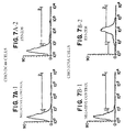
- the EX5.3DI0 monoclonal antibody was derived from immunizing a Balb/c mouse with CHO (Chinese hamster ovary) cells transfected with the human CD28 gene (designated CHO-hh). Hybridomas from the fusion were selected by whole cell ELISA screening against Jurkat (human T leukemia) CD28 tranfectants designated Jurkat #7. Reactivity of the monoclonal antibody EX5.3D10 with CD28 was further confirmed by fluorescent activated cell sorter analysis (FACS) analysis in which it was tested side by side with the monoclonal antibody 9.3 ( Figure 6).
- FACS fluorescent activated cell sorter analysis
- an activated population of CD4 + T cells is stimulated to proliferate by contacting the T cells with an agent which acts intracellularly to stimulate a signal in the T cell mediated by ligation of an accessory molecule, such as CD28.
- an agent which acts intracellularly to stimulate a signal in the T cell mediated by ligation of an accessory molecule, such as CD28.
- agent is intended to encompass chemicals and other pharmaceutical compounds which stimulate a costimulatory or other signal in a T cell without the requirement for an interaction between a T cell surface receptor and a costimulatory molecule or other ligand.
- the agent may act intracellularly to stimulate a signal associated with CD28 ligation.
- the agent is a non-proteinaceous compound.
- Natural ligands for CD28 include members of the B7 family of proteins, such as B7-1(CD80) and B7-2 (CD86).
- CD28 receptor stimulation leads to the production of D-3 phosphoinositides in T cells and that inhibition of the activity of phosphatidylinositol 3-kinase (PI3K) in a T cell can inhibit T cell responses, such as lymphokine production and cellular proliferation.
- PI3K phosphatidylinositol 3-kinase
- Protein tyrosine phosphorylation has also been shown to occur in T cells upon CD28 ligation and it has been demonstrated that a protein tyrosine kinase inhibitor, herbimycin A, can inhibit CD28-induced IL-2 production (Vandenberghe, P. et al. (1992) J. Exp. Med 175 :951-960; Lu, Y. et al. (1992) J. Immunol .
- the CD28 receptor mediated pathway can be stimulated by contacting T cells with an activator of PI3K or an agent which stimulates protein tyrosine phosphorylation in the T cell, or both.
- An activator of PI3K can be identified based upon its ability to stimulate production of at least one D-3 phosphoinositide in a T cell.
- D-3 phosphoinositide is intended to include derivatives of phosphatidylinositol that are phosphorylated at the D-3 position of the inositol ring and encompasses the compounds phosphatidylinositol(3)-monophosphate (PtdIns(3)P), phosphatidylinositol(3,4)-bisphosphate (PtdIns(3,4)P 2 ), and phosphatidylinositol(3,4,5)-trisphosphate (PtdIns(3,4,5)P 3 ).
- PtdIns(3)P phosphatidylinositol(3)-monophosphate
- PtdIns(3,4)P 2 phosphatidylinositol(3,4)P 2
- PtdIns(3,4,5)P 3 phosphatidylinositol(3,4,5)P 3
- D-3 phosphoinositide in the T cell is increased relative to the amount of the D-3 phosphoinositide in the T cell in the absence of the substance
- Production of D-3 phosphoinositides (e.g., PtdIns(3)P, PtdIns(3,4)P 2 and/or PtdIns(3,4,5)P 3 ) in a T cell can be assessed by standard methods, such as high pressure liquid chromatography or thin layer chromatography, as discussed above.
- protein tyrosine phosphorylation can be stimulated in a T cell, for example, by contacting the T cell with an activator of protein tyrosine kinases, such as pervanadate (see O'Shea, J.J. et al. (1992) Proc. Natl. Acad. Sci. USA 89 :10306-103101; and Secrist, J.P. (1993) J. Biol. Chem . 268 :5886-5893).
- the T cell can be contacted with an agent which inhibits the activity of a cellular protein tyrosine phosphatase, such as CD45, to increase the net amount of protein tyrosine phosphorylation in the T cell. Any of these agents can be used to expand an activated population of CD4 + T cells in accordance with the methods described herein.
- an activated population of T cells is stimulated through a 27 kD accessory molecule found on activated T cells and recognized by the monoclonal antibody ES5.2D8.
- a population of CD8 + T cells was preferentially expanded by stimulation with an anti-CD3 monoclonal antibody and the ES5.2D8 monoclonal antibody.
- the monoclonal antibody ES5.2D8 was produced by immunization of mice with activated human blood lymphocytes and boosted with recombinant human CTLA4 protein produced in E. coli .
- the ES5.2D8 monoclonal antibody is of the IgG2b isotype and specifically binds to cells transfected with human CTLA4.
- Hybridomas producing CTLA4-specific antibody were identified by screening by ELISA against human CTLA4 protein as well as by differential FACS against wild type CHO-DG44 cells vs. CHO-105A cells, which are transfected with the human CTLA4 gene.
- the ES5.2D8 clone reacts strongly with both activated human T cells and CHO-105A cells but not with CHO-DCA4 cells, indicating that it does indeed bind to CTLA4.
- Immunoprecipitation of detergent lysates of surface labeled activated human T cells revealed that ES5.2D8 also reacts with a 27 kD cell surface protein (Figure 8).
- a hybridoma which produces the monoclonal antibody ES5.2D8 was deposited on June 4, 1993 with the American Type Culture Collection at ATCC Deposit No. HB11374.
- an antibody such as monoclonal antibody ES5.2D8, or other antibody which recognizes the same 27 kD ligand as ES5.2D8, can be used.
- the epitope recognized by the monoclonal antibody ES5.2D8 was identified by screening a phage display library (PDL).
- Antibodies which bind to the same epitope as the monoclonal antibody ES5.2D8 are within the scope of the invention. Such antibodies can be produced by immunization with a peptide fragment including the epitope or with the native 27 kD antigen.
- epitope refers to the actual structural portion of the antigen that is immunologically bound by an antibody combining site.
- the term is also used interchangeably with "antigenic determinant”.
- Xaa 2 is Cys or Ile
- Xaa 3 is Leu or Arg
- Xaa 4 if present, is Arg or Pro.
- Xaa 1 and Xaa 4 are additional amino acid residues found at either the amino or carboxy side, or both the amino and carboxy sides, of the core epitope in the native 27 kD protein. It will be appreciated by those skilled in the art that in the native protein, additional non-contiguous amino acid residues may also contribute to the conformational epitope recognized by the antibody.
- Synthetic peptides encompassing the epitope can be created which includes other amino acid residues flanking the core seven amino acid residues (i.e., Xaa can alternatively be other amino acid residues than those found in the native protein). These flanking amino acid residues can function to alter the properties of the resulting peptide, for example to increase the solubility, enhance the immunogenicity or promote dimerization of the resultant peptide.
- one or more charged amino acids e.g., lysine, arginine
- cysteine residues can be included to increase the dimerization of the resulting peptide.
- inventions pertain to expansion of a population of CD8 + T cells by use of an agent which acts intracellularly to stimulate a signal in the T cell mediated by ligation of the 27 kD protein.
- agent encompasses chemicals and other pharmaceutical compounds which stimulate a signal in a T cell without the requirement for an interaction between a T cell surface receptor and a ligand. Thus, this agent does not bind to the extracellular portion of the 27 kD protein, but rather mimics or induces an intracellular signal (e.g., second messenger) associated with ligation of the protein by an appropriate ligand.
- the ligands described herein can be used to identify an intracellular signal(s) associated with T cell expansion mediated by contact of the 27 kD protein with an appropriate ligand (as described in the Examples) and examining the resultant intracellular signalling that occurs (e.g., protein tyrosine phosphorylation, calcium influx, activation of serine/threonine and/or tyrosine kinases, phosphatidyl inositol metabolism, etc.).
- An agent which enhances an intracellular signal associated with the 27 kD protein can then be used to expand CD8 + T cells.
- agents e.g., small molecules, drugs, etc.
- T cells are contacted with an antigen in a form suitable to trigger a primary activation signal in the T cell, i.e., the antigen is presented to the T cell such that a signal is triggered in the T cell through the TCR/CD3 complex.
- the antigen can be presented to the T cell by an antigen presenting cell in conjuction with an MHC molecule.
- An antigen presenting cell such as a B cell, macrophage, monocyte, dendritic cell, Langerhan cell, or other cell which can present antigen to a T cell, can be incubated with the T cell in the presence of the antigen (e.g., a soluble antigen) such that the antigen presenting cell presents the antigen to the T cell.
- the antigen e.g., a soluble antigen
- a cell expressing an antigen of interest can be incubated with the T cell.
- a tumor cell expressing tumor-associated antigens can be incubated with a T cell together to induce a tumor-specific response.
- a cell infected with a pathogen e.g., a virus, which presents antigens of the pathogen can be incubated with a T cell.
- a pathogen e.g., a virus
- the cells can be expanded in accordance with the methods of the invention. For example, after antigen specificity has been established, T cells can be expanded by culture with an anti-CD3 antibody and an anti-CD28 antibody according to the methods described herein.
- antibody refers to immunoglobulin molecules and immunologically active portions of immunoglobulin molecules, i.e., molecules that contain an antigen binding site which specifically binds (immunoreacts with) an antigen, such as CD3, CD28.
- an antigen such as CD3, CD28.
- the simplest naturally occurring antibody e.g., IgG
- IgG comprises four polypeptide chains, two heavy (H) chains and two light (L) chains inter-connected by disulfide bonds. It has been shown that the antigen-binding function of an antibody can be performed by fragments of a naturally-occurring antibody. Thus, these antigen-binding fragments are also intended to be designated by the term "antibody”.
- binding fragments encompassed within the term antibody include (i) an Fab fragment consisting of the VL, VH, CL and CH1 domains; (ii) an Fd fragment consisting of the VH and CH1 domains; (iii) an Fv fragment consisting of the VL and VH domains of a single arm of an antibody, (iv) a dAb fragment (Ward et al., (1989) Nature 341 :544-546) which consists of a VH domain; (v) an isolated complementarity determining region (CDR); and (vi) an F(ab') 2 fragment, a bivalent fragment comprising two Fab fragments linked by a disulfide bridge at the hinge region.
- a synthetic linker can be made that enables them to be made as a single protein chain (known as single chain Fv (scFv); Bird et al. (1988) Science 242 :423-426; and Huston et al. (1988) PNAS 85 :5879-5883) by recombinant methods.
- single chain Fv single chain Fv
- Preferred antibody fragments for use in T cell expansion are those which are capable of crosslinking their target antigen, e.g., bivalent fragments such as F(ab') 2 fragments.
- an antibody fragment which does not itself crosslink its target antigen e.g., a Fab fragment
- a secondary antibody which serves to crosslink the antibody fragment, thereby crosslinking the target antigen.
- Antibodies can be fragmented using conventional techniques as described herein and the fragments screened for utility in the same manner as described for whole antibodies.
- An antibody of the invention is further intended to include bispecific and chimeric molecules having a desired binding portion (e.g., CD28).
- a desired binding specificity for an epitope refers to the ability of individual antibodies to specifically immunoreact with a T cell surface molecule, e.g., CD28. That is, it refers to a non-random binding reaction between an antibody molecule and an antigenic determinant of the T cell surface molecule.
- the desired binding specificity is typically determined from the reference point of the ability of the antibody to differentially bind the T cell surface molecule and an unrelated antigen, and therefore distinguish between two different antigens, particularly where the two antigens have unique epitopes.
- An antibody which binds specifically to a particular epitope is referred to as a "specific antibody”.
- Antibody combining site refers to that structural portion of an antibody molecule comprised of a heavy and light chain variable and hypervariable regions that specifically binds (immunoreacts with) antigen.
- the term “immunoreact” or “reactive with” in its various forms is used herein to refer to binding between ah antigenic determinant-containing molecule and a molecule containing an antibody combining site such as a whole antibody molecule or a portion thereof.
- the present invention encompasses that the anti-CD3 antibody is directly immobilized on a solid phase surface (e.g., beads).
- An antibody can be immobilized directly or indirectly by, for example, a secondary antibody, to a solid surface, such as a tissue culture flask or bead.
- a secondary antibody to a solid surface, such as a tissue culture flask or bead.
- the following is a protocol for immobilizing an anti-CD3 antibody on beads. It should be appreciated that the same protocol can be used to immobilize other antibodies or fragments thereof (e.g., an anti-CD28 antibody) to beads.
- a source of T cells is obtained from a subject.
- the term subject is intended to include living organisms in which an immune response can be elicited, e.g., mammals. Examples of subjects include humans, dogs, cats, mice, rats, and transgenic species thereof.
- T cells can be obtained from a number of sources, including peripheral blood leukocytes, bone marrow, lymph node tissue, spleen tissue, and tumors.
- peripheral blood leukocytes are obtained from an individual by leukopheresis.
- a specific subpopulatioh of T cells can be further isolated by positive or negative selection techniques.
- negative selection of a T cell population can be accomplished with a combination of antibodies directed to surface markers unique to the cells negatively selected.
- a preferred method is cell sorting via negative magnetic immunoadherence which utilizes a cocktail of monoclonal antibodies directed to cell surface markers present on the cells negatively selected.
- a monoclonal antibody cocktail typically includes antibodies to CD14, CD20, CD11b, CD16, HLA-DR, and CD8. Additional monoclonal antibody cocktails are provided in Table 1.
- the process of negative selection results in an essentially homogenous population of CD4 + or CD8 + T cells.
- the T cells can be activated as described herein, such as by contact with an anti-CD3 antibody immobilized on a solid phase surface or an anti-CD2 antibody immobilized on a solid phase surface.
- a ligand which binds the accessory molecule is employed.
- a population of CD4 + cells can be contacted with an anti-CD3 antibody and an anti-CD28 antibody, under conditions appropriate for stimulating proliferation of the T cells.
- an anti-CD3 antibody and the monoclonal antibody ES5.2D8 can be used.
- Conditions appropriate for T cell culture include an appropriate media (e.g., Minimal Essential Media or RPMI Media 1640) which may contain factors necessary for proliferation and viability, including animal serum (e.g., fetal bovine serum) and antibiotics (e.g., penicillin streptomycin).
- an appropriate media e.g., Minimal Essential Media or RPMI Media 1640
- animal serum e.g., fetal bovine serum
- antibiotics e.g., penicillin streptomycin
- the T cells are maintained under conditions necessary to support growth, for example an appropriate temperature (e.g., 37°C) and atmosphere (e.g., air plus 5% CO 2 ).
- T cells are maintained in contact with the co-stimulatory ligand throughout the culture term.
- the rate of T cell proliferation is monitored periodically (e.g., daily) by, for example, examining the size or measuring the volume of the T cells, such as with a Coulter Counter.
- a resting T cell has a mean diameter of about 6.8 microns. Following the initial activation and stimulation and in the presence of the stimulating ligand, the T cell mean diameter will increase to over 12 microns by day 4 and begin to decrease by about day 6.
- the T cells When the mean T cell diameter decreases to approximately 8 microns, the T cells are reactivated and restimulated to induce further proliferation of the T cells.
- the rate ofT cell proliferation and time for T cell restimulation can be monitored by assaying for the presence of cell surface molecules, such as B7-1, B7-2, which are induced on activated T cells.
- B7-1, B7-2 cell surface molecules, such as B7-1, B7-2, which are induced on activated T cells.
- B7-1, B7-2 cell surface molecules
- cyclic changes in B7-1 expression can be used as a means of monitoring T cell proliferation; where decreases in the level of B7-1 expression, relative to the level of expression following an initial or previous stimulation or the level of expression in an unstimulated cell, indicates the time for restimulation.
- CD4 + or CD8 + T cells For inducing long term stimulation of a population of CD4 + or CD8 + T cells, it may be necessary to reactivate and restimulate the T cells with an anti-CD3 antibody and an anti-CD28 antibody or monoclonal antibody ES5.2D8 several times to produce a population of CD4 + or CD8 + cells increased in number from about 10- to about 1,000-fold the original T cell population. Using this methodology, it is possible to get increases in a T cell population of from about 100- to about 100,000-fold than the original resting T cell population.
- T cells expanded by the method of the invention secrete high levels of cytokines (e.g., IL-2, IFN ⁇ , IL-4, GM-CSF and TNF ⁇ ) into the culture supernatants.
- cytokines e.g., IL-2, IFN ⁇ , IL-4, GM-CSF and TNF ⁇
- CD4 + T cells expanded by use of anti-CD3 and anti-CD28 costimulation secrete high levels of GM-CSF and TNF ⁇ into the culture medium.
- These cytokines can be purified from the culture supematants or the supernatants can be used directly for maintaining cells in culture.
- the T cells expanded by the method of the invention together with the culture supernatant and cytokines can be administered to support the growth of cells in vivo.
- T cells can be obtained from the individual, expanded in vitro and the resulting T cell population and supernatant, including cytokines such as TNF ⁇ , can be readministered to the patient to augment T cell growth in vivo .
- cytokines such as TNF ⁇
- antibodies used in the methods described herein can be readily obtained from public sources, such as the ATCC, antibodies to T cell surface accessory molecules, the CD3 complex, or CD2 can be produced by standard techniques. Methodologies for generating antibodies for use in the methods of the invention are described in further detail below.
- the Immunogen is used herein to describe a composition containing a peptide or protein as an active ingredient used for the preparation of antibodies against an antigen (e.g., CD3, CD28).
- an antigen e.g., CD3, CD28.
- a peptide or protein is used to induce antibodies it is to be understood that the peptide can be used alone, or linked to a carrier as a conjugate, or as a peptide polymer.
- the immunogen should contain an effective, immunogenic amount of a peptide or protein, optionally as a conjugate linked to a carrier.
- the effective amount of peptide per unit dose depends, among other things, on the species of animal inoculated, the body weight of the animal and the chosen immunization regimen as is well known in the art.
- the immunogen preparation will typically contain peptide concentrations of about 10 micrograms to about 500 milligrams per immunization dose, preferably about 50 micrograms to about 50 milligrams per dose.
- An immunization preparation can also include an adjuvant as part of the diluent. Adjuvants such as complete Freund's adjuvant (CFA), incomplete Freund's adjuvant (IFA) and alum are materials well known in the art, and are available commercially from several sources.
- CFA complete Freund's adjuvant
- IFA incomplete Freund's adjuvant
- alum are materials well known in the art, and are available commercially from several sources.
- peptides can alternatively be employed towards which antibodies can be raised for use in this invention.
- Both soluble and membrane bound forms of the protein or peptide fragments are suitable for use as an immunogen and can also be isolated by immunoaffinity purification as well.
- a purified form of protein such as may be isolated as described above or as known in the art, can itself be directly used as an immunogen, or alternatively, can be linked to a suitable carrier protein by conventional techniques, including by chemical coupling means as well as by genetic engineering using a cloned gene of the protein.
- the purified protein can also be covalently or noncovalently modified with non-proteinaceous materials such as lipids or carbohydrates to enhance immunogenicity or solubility.
- a purified protein can be coupled with or incorporated into a viral particle, a replicating virus, or other microorganism in order to enhance immunogenicity.
- the protein may be, for example, chemically attached to the viral particle or microorganism or an immunogenic portion thereof.
- a purified CD28 protein, or a peptide fragment thereof is conjugated to a carrier which is immunogenic in animals.
- Preferred carriers include proteins such as albumins, serum proteins (e.g., globulins and lipoproteins), and polyamino acids.
- useful proteins include bovine serum albumin, rabbit serum albumin, thyroglobulin, keyhole limpet hemocyanin, egg ovalbumin and bovine gamma-globulins.
- Synthetic polyamino acids such as polylysine or polyarginine are also useful carriers.
- cross-linking agents are heterobifunctional cross-linkers, which can be used to link proteins in a stepwise manner.
- heterobifunctional cross-linkers include succinimidyl 4-(N-maleimidomethyl) cyclohexane- 1-carboxylate (SMCC), m-Maleimidobenzoyl-N- hydroxysuccinimide ester (MBS); N-succinimidyl (4-iodoacetyl) aminobenzoate (SIAB), succinimidyl 4-(p-maleimidophenyl) butyrate (SMPB), 1-ethyl-3-(3-dimethylaminopropyl) carbodiimide hydrochloride (EDC); 4-succinimidyl-oxycarbonyl-a-methyl-a-(2-pyridyldithio)-tolune (SMPT), N-succinimidyl 3-(2-pyridyldithio) propionate (SPDP), succinimidyl 6-[3-(2-pyridyldithio) propionate (SPDP), succinimid
- a protein of interest e.g., CD28
- Various cell lines can be used as immunogens to generate monoclonal antibodies to an antigen including, but not limited, to T cells.
- peripheral blood T cells can be obtained from a subject which constitutively expresses CD28, but can be activated in vitro with anti-CD3 antibodies, PHA or PMA.
- an antigen specific (e.g., alloreactive) T cell clone can be activated to express CD28 by presentation of antigen, together with a costimulatory signal, to the T cell.
- Whole cells that can be used as immunogens to produce CD28 specific antibodies also include recombinant transfectants.
- COS and CHO cells can be reconstituted by transfection with a CD28 cDNA to produce cells expressing CD28 on their surface. These transfectant cells can then be used as immunogens to produce anti-CD28 antibodies.
- transfectant cells are known, particularly eukaryotic cells able to glycosylate the CD28 protein, but any procedure that works to express transfected CD28 genes on the cell surface could be used to produce the whole cell immunogen.
- Xaa 2 is Cys or Ile
- Xaa 3 is Leu or Arg
- Xaa 4 if present, is Arg or Pro.
- a peptide having the amino acid sequence of SEQ ID NO: 5 can be used as an immunogen.
- Xaa 2 is Cys or Ile
- Xaa 3 is Leu or Arg
- Xaa 4 if present, is Arg or Pro.
- a number of other peptide sequences determined by phage display technology as described in Example 3. Examples of these other peptide sequences are shown below: Any of these peptides, or other peptides containing a stretch of seven amino acids bracketed in bold type (representing the epitope bound by the antibody) possibly flanked by alternative amino acid residues, can also be used as immunogens to produce an antibody for use in the methods of the invention and are encompassed by the invention.
- immunogens peptides can be modified to increase solubility and/or enhance immunogenicity as described above.
- Polyclonal antibodies to a purified protein or peptide fragment thereof can generally be raised in animals by multiple subcutaneous (sc) or intraperitoneal (ip) injections of an appropriate immunogen, such as the extracellular domain of the protein, and an adjuvant.
- an appropriate immunogen such as the extracellular domain of the protein, and an adjuvant.
- a polyclonal antisera can be produced, for example, as described in Lindsten, T. et al. (1993) J. Immunol . 151 :3489-3499.
- animals are typically immunized against the immunogenic protein, peptide or derivative by combining about 1 ⁇ g to 1 mg of protein with Freund's complete adjuvant and injecting the solution intradermally at multiple sites.
- the animals are boosted with 1/5 to 1/10 the original amount of immunogen in Freund's complete adjuvant (or other suitable adjuvant) by subcutaneous injection at multiple sites.
- the animals are bled and the serum is assayed for anti-protein or peptide titer (e.g., by ELISA). Animals are boosted until the titer plateaus.
- aggregating agents such as alum can be used to enhance the immune response.
- Such mammalian-produced populations of antibody molecules are referred to as "polyclonal" because the population comprises antibodies with differing immunospecificities and affinities for the antigen.
- the antibody molecules are then collected from the mammal (e.g., from the blood) and isolated by well known techniques, such as protein A chromatography, to obtain the IgG fraction.
- the antibodies may be purified by immunoaffinity chromatography using solid phase-affixed immunogen.
- the antibody is contacted with the solid phase-affixed immunogen for a period of time sufficient for the immunogen to immunoreact with the antibody molecules to form a solid phase-affixed immunocomplex.
- the bound antibodies are separated from the complex by standard techniques.
- monoclonal antibody or “monoclonal antibody composition”, as used herein, refers to a population of antibody molecules that contain only one species of an antigen binding site capable of immunoreacting with a particular epitope of an antigen.
- a monoclonal antibody composition thus typically displays a single binding affinity for a particular protein with which it immunoreacts.
- the monoclonal antibody used in the subject method is further characterized as immunoreacting with a protein derived from humans.
- Monoclonal antibodies useful in the methods of the invention are directed to an epitope of an antigen(s) on T cells, such that complex formation between the antibody and the antigen (also referred to herein as ligation) induces stimulation and T cell expansion.
- a monoclonal antibody to an epitope of an antigen e.g., CD3, CD28
- CD3, CD28 can be prepared by using a technique which provides for the production of antibody molecules by continuous cell lines in culture. These include, but are not limited to, the hybridoma technique originally described by Kohler and Milstein (1975, Nature 256:495-497), and the more recent human B cell hybridoma technique (Kozbor et al. (1983) Immunol.
- the antibody preparation applied in the subject method is a monoclonal antibody produced by a hybridoma cell line.
- Hybridoma fusion techniques were first introduced by Köhler and Milstein (Köhler et al. Nature (1975) 256 :495-97; Brown et al. (1981) J. Immunol . 127 :539-46; Brown et al. (1980) J. Biol. Chem . 255 :4980-83; Yeh et al. (1976) PNAS 76 :2927-31; and Yeh et al. (1982) Int. J. Cancer 29 :269-75).
- the monoclonal antibody compositions of the present invention can be produced by the following method, which comprises the steps of:
- the resulting cells which include the desired hybridomas, are then grown in a selective medium, such as HAT medium, in which unfused parental myeloma or lymphocyte cells eventually die. Only the hybridoma cells survive and can be grown under limiting dilution conditions to obtain isolated clones.
- the supernatants of the hybridomas are screened for the presence of antibody of the desired specificity, e.g., by immunoassay techniques using the antigen that has been used for immunization. Positive clones can then be subcloned under limiting dilution conditions and the monoclonal antibody produced can be isolated.
- a selective medium such as HAT medium
- Hybridomas produced according to these methods can be propagated in vitro or in vivo (in ascites fluid) using techniques known in the art.
- the individual cell line may be propagated in vitro , for example in laboratory culture vessels, and the culture medium containing high concentrations of a single specific monoclonal antibody can be harvested by decantation, filtration or centrifugation.
- the yield of monoclonal antibody can be enhanced by injecting a sample of the hybridoma into a histocompatible animal of the type used to provide the somatic and myeloma cells for the original fusion. Tumors secreting the specific monoclonal antibody produced by the fused cell hybrid develop in the injected animal.
- the body fluids of the animal such as ascites fluid or serum, provide monoclonal antibodies in high concentrations.
- Immunodeficient or nude mice may be used or the hybridoma may be passaged first into irradiated nude mice as a solid subcutaneous tumor, cultured in vitro and then injected intraperitoneally into pristane primed, irradiated nude mice which develop ascites tumors secreting large amounts of specific human monoclonal antibodies.
- DMEM Dulbecco's minimal essential medium
- Monoclonal antibody compositions of the invention can also be produced by other methods well known to those skilled in the art of recombinant DNA technology.
- An alternative method, referred to as the "combinatorial antibody display” method has been developed to identify and isolate antibody fragments having a particular antigen specificity, and can be utilized to produce monoclonal antibodies (for descriptions of combinatorial antibody display see, e.g., Sastry et al. (1989) PNAS 86 :5728; Huse et al. (1989) Science 246 :1275; and Orlandi et al. (1989) PNAS 86 :3833).
- the antibody repertoire of the resulting B-cell pool is cloned.
- Methods are generally known for directly obtaining the DNA sequence of the variable regions of a diverse population of immunoglobulin molecules by using a mixture of oligomer primers and PCR.
- mixed oligonucleotide primers corresponding to the 5' leader (signal peptide) sequences and/or framework 1 (FR1) sequences, as well as primer to a conserved 3' constant region primer can be used for PCR amplification of the heavy and light chain variable regions from a number of murine antibodies (Larrick et al. (1991) Biotechniques 11 :152-156).
- a similar strategy can also be used to amplify human heavy and light chain variable regions from human antibodies (Larrick et al. (1991) Methods: Companion to Methods in Enzymology 2 :106-110).
- RNA is isolated from activated B cells of, for example, peripheral blood cells, bone marrow, or spleen preparations, using standard protocols (e.g., U.S. Patent No. 4,683,202; Orlandi, et al. PNAS (1989) 86 :3833-3837; Sastry et al., PNAS (1989) 86 :5728-5732; and Huse et al. (1989) Science 246 :1275-1281.) First-strand cDNA is synthesized using primers specific for the constant region of the heavy chain(s) and each of the ⁇ and ⁇ light chains, as well as primers for the signal sequence.
- variable region PCR primers the variable regions of both heavy and light chains are amplified, each alone or in combinantion, and ligated into appropriate vectors for further manipulation in generating the display packages.
- Oligonucleotide primers useful in amplification protocols may be unique or degenerate or incorporate inosine at degenerate positions. Restriction endonuclease recognition sequences may also be incorporated into the primers to allow for the cloning of the amplified fragment into a vector in a predetermined reading frame for expression.
- the V-gene library cloned from the immunization-derived antibody repertoire can be expressed by a population of display packages, preferably derived from filamentous phage, to form an antibody display library.
- the display package comprises a system that allows the sampling of very large variegated antibody display libraries, rapid sorting after each affinity separation round, and easy isolation of the antibody gene from purified display packages.
- kits for generating phage display libraries e.g., the Pharmacia Recombinant Phage Antibody System , catalog no. 27-9400-01; and the Stratagene SurfZAP TM phage display kit, catalog no.
- examples of methods and reagents particularly amenable for use in generating a variegated antibody display library can be found in, for example, Ladner et al. U.S. Patent No. 5,223,409; Kang et al. International Publication No. WO 92/18619; Dower et al. International Publication No. WO 91/17271; Winter et al. International Publication WO 92/20791; Markland et al. International Publication No. WO 92/15679; Breitling et al. International Publication WO 93/01288; McCafferty et al. International Publication No. WO 92/01047; Garrard et al.
- the V region domains of heavy and light chains can be expressed on the same polypeptide, joined by a flexible linker to form a single-chain Fv fragment, and the scFV gene subsequently cloned into the desired expression vector or phage genome.
- a flexible linker As generally described in McCafferty et al., Nature (1990) 348 :552-554, complete V H and V L domains of an antibody, joined by a flexible (Gly 4 -Ser) 3 linker can be used to produce a single chain antibody which can render the display package separable based on antigen affinity. Isolated scFV antibodies immunoreactive with the antigen can subsequently be formulated into a pharmaceutical preparation for use in the subject method.
- the antibody library is screened with the protein, or peptide fragment thereof, to identify and isolate packages that express an antibody having specificity for the protein.
- Nucleic acid encoding the selected antibody can be recovered from the display package (e.g., from the phage genome) and subcloned into other expression vectors by standard recombinant DNA techniques.
- Hybridomas useful in the present invention are those characterized as having the capacity to produce a monoclonal antibody which will specifically immunoreact with an antigen of interest (e.g., CD3, CD28).
- an antigen of interest e.g., CD3, CD28.
- Methods for generating hybridomas that produce, e.g., secrete, antibody molecules having a desired immunospecificity, e.g., having the ability to immunoreact with the CD28 antigen, and/or an identifiable epitope of CD28 are well known in the art. Particularly applicable is the hybridoma technology described by Niman et al. (1983) PNAS 80 :4949-4953; and by Galfre et al. (1981) Meth. Enzymol . 73 :3-46.
- the method of this invention can be used to selectively expand a population of CD4 + or CD8 + T cells for use in the treatment of infectious disease, cancer and immunotherapy.
- a population ofT cells which is polyclonal with respect to antigen reactivity, but essentially homogeneous with respect to either CD4 + or CD8 + can be produced.
- the method allows for the expansion of a population of T cells in numbers sufficient to reconstitute an individual's total CD4 + or CD8 + T cell population (the population of lymphocytes in an individual is approximately 10 11) .
- the resulting T cell population can be genetically transduced and used for immunotherapy or can be used in methods of in vitro analyses of infectious agents.
- a population of tumor-infiltrating lymphocytes can be obtained from an individual afflicted with cancer and the T cells stimulated to proliferate to sufficient numbers.
- the resulting T cell population can be genetically transduced to express tumor necrosis factor (TNF) or other factor and restored to the individual.
- TNF tumor necrosis factor
- CD4 + -T cells expanded by the method of the invention is in the treatment of HIV infection in an individual. Prolonged infection with HIV eventually results in a marked decline in the number of CD4 + T lymphocytes. This decline, in turn, causes a profound state of immunodeficiency, rendering the patient susceptible to an array of life threatening opportunistic infections. Replenishing the number of CD4 + T cells to normal levels may be expected to restore immune function to a significant degree.
- the method described herein provides a means for selectively expanding CD4 + T cells to sufficient numbers to reconstitute this population in an HIV infected patient. It may also be necessary to avoid infecting the T cells during long-term stimulation or it may be desirable to render the T cells permanently resistant to HIV infection.
- T cells may be rendered either resistant to HIV infection or incapable of producing virus prior to restoring the T cells to the infected individual.
- one or more anti-retroviral agents can be cultured with CD4 + T cells prior to expansion to inhibit HIV replication or viral production (e.g., drugs that target reverse transcriptase and/or other components of the viral machinery, see e.g., Chow et al. (1993) Nature 361, 650-653).
- T cells can be genetically transduced to produce transdominant inhibitors, which are mutated, nonfunctional forms of normal HIV gene products.
- Transdominant inhibitors function to oligomerize or compete for binding with the wild type HIV proteins.
- transdominant inhibitors have been derived from HIV proteins including tat, rev, and gag. The function of tat is to enhance the transcription of viral genes by binding to the trans activation response element (tar) found in the promoter region of most HIV genes.
- Rev through binding to the rev response element (RRE) found at the 5' end of unspliced HIV transcripts, facilitates the transport of unprocessed mRNA from the nucleus to the cytoplasm for packaging into virions.
- Gag is first synthesized as a single polypeptide and subsequently cleaved by a virus-encoded protease to yield three structural proteins, p15, p17, and p24.
- Transdominant inhibitors derived from these gene products have been demonstrated to inhibit infection of cells cultured with lab pet HIV isolates.
- RevM10 One example of a transdominant inhibitor which appears to act by forming nonfunctional multimers with wild-type Rev.
- RevM10 construct has blocked infection of CEM cells by HTLV-IIIB for up to 28 days (Malim et al. JEM 176:1197, Bevec et al. PNAS 89:9870). In these studies, RevM10 failed to demonstrate adverse effect on normal T cell function as judged by the criteria of growth rate and IL-2 secretion.
- T cells can be transduced to produce molecules known as "molecular decoys" which are binding elements for viral proteins critical to replication or assembly, such as TAR.
- High level retrovirus-mediated expression of TAR in CEM SS cells has been found to effectively block the ARV-2 HIV isolate, as measured by RT assay (Sullenger et al. Cell 63:601). Importantly, it also blocked SIV (SIVmac251) infection, suggesting that inhibition of HIV infection with molecular decoys may be generally applicable to various isolates and thereby alleviate the problem of hypervariability. Further, it has been demonstrated that TAR expression has no discernible effects on cell viability (Sullenger et al. J. Virol . 65:6811).
- T cells can be transduced to produce is a soluble CD4 tagged at the carboxy terminus with a KDEL (lysine-aspartic acid-glutamic acid-leucine) sequence, a signal for ER retention (Buonocore and Rose, PNAS 90:2695)( Nature 345:625).
- KDEL lysine-aspartic acid-glutamic acid-leucine
- the sCD4-KDEL gene expression is driven by the HIV LTR.
- H9 cells transduced with the sCD4-KDEL construct show up regulation of expression of intracellular CD4 upon HIV infection. This strategy effectively blocked production of HIV MN for up to 60 days post infection.
- the proposed advantage of this inhibitor is that the virus should not be able to escape its effect by mutating because CD4 binding is essential for HIV infectivity.
- T cells can also be transduced to express antisense molecules and ribozyme which block viral replication or infection. Viral replication can be inhibited with a variety of antisense strategies.
- One particular ribozyme which cleaves HIV integrase (Sioud and Drlica, PNAS 88:7303), has been developed and may offer an approach to blocking infection as opposed to merely viral production.
- HSV TK herpes simplex virus type 1 thymidine kinase gene
- the methods for stimulating and expanding a population of antigen specific T cells are useful in therapeutic situations where it is desirable to upregulate an immune response (e.g., induce a response or enhance an existing response) upon administration of the T cells to a subject.
- the method can be used to enhance a T cell response against tumor-associated antigens.
- Tumor cells from a subject typically express tumor-associated antigens but may be unable to stimulate a costimulatory signal in T cells (e.g., because they lack expression of costimulatory molecules).
- tumor cells can be contacted with T cells from the subject in vitro and antigen specific T cells expanded according to the method of the invention and the T cells returned to the subject.
- T cells can be stimulated and expanded as described herein to induce or enhance responsiveness to pathogenic agents, such as viruses (e.g., human immunodeficiency virus), bacteria, parasites and fungi.
- Tissue culture flasks were coated with anti-CD3 monoclonal antibody.
- OKT3 prepared from hybridoma cells obtained from the American Type Culture Collection was used in this procedure.
- the optimal concentration to be coated on tissue cultured flasks must be determined experimentally. With OKT3, the optimal concentration was determined to be typically in the range of 0.1 to 10 micrograms per milliliter.
- the antibody was suspended in 0.05 M Tris-HCl, pH 9.0 (Sigma Chemical Co., St. Louis, MO). Coating solution sufficient to cover the bottom of a tissue culture flask was added (Falcon, Nunc or Costar) and incubated overnight at 4° C.
- the flasks were washed three times with phosphate buffered saline without calcium or magnesium (PBS w/o Ca or Mg) and blocking buffer (PBS w/o Ca or Mg plus 5% bovine serum albumin) added to cover the bottom of the flask and were incubated two hours at room temperature. After this incubation, flasks were used directly or frozen for storage, leaving the blocking solution on the flask.
- PBS w/o Ca or Mg phosphate buffered saline without calcium or magnesium
- blocking buffer PBS w/o Ca or Mg plus 5% bovine serum albumin
- PBLs Peripheral Blood Leukocytes
- Samples were obtained by leukopheresis of healthy donors. Using sterile conditions, the leukocytes were transferred to a T800 culture flask. The bag was washed with Hanks balanced salt solution w/o calcium or magnesium (HBSS w/o) (Whittaker Bioproducts, Inc., Walkersville, MD). The cells were diluted with HBSS w/o and mixed well. The cells were then split equally between two 200 milliliter conical-bottom sterile plastic tissue culture tubes. Each tube was brought up to 200 ml with HBSS w/o and spun at 1800 RPM for 12 minutes in a Beckman TJ-6 centrifuge.
- HBSS w/o Hanks balanced salt solution w/o calcium or magnesium
- the supernatant was aspirated and each pellet resuspended in 50 ml HBSS w/o.
- the cells were transferred to two 50 ml conical bottom tubes and spun at 1500 RPM for eight minutes. Again the supernatant was aspirated.
- the cell pellets were resuspended in 50 ml of ACK lysing buffer (Biofluids, Inc., Rockville MD, Catalog #304) at room temperature with gentle mixing for three minutes. The cells were again pelleted by spinning at 1500 RPM for 8 minutes. After aspirating the supernatant, the pellets were combined into one 50 ml tube in 32 ml HBSS w/o.
- ACK lysing buffer Biofluids, Inc., Rockville MD, Catalog #304
- the cell sorting via negative magnetic immunoadherence must be performed at 4° C.
- the washed cell pellets obtained from the PERCOLLTM gradients described above were resuspended in coating medium (RPMI-1640 (BioWhittaker, Walkersville, MD, Catalog # 12-167Y), 3% fetal calf serum (FCS) (or 1% human AB - serum or 0.5% bovine serum albumin) 5 mM EDTA (Quality Biological, Inc., Gaithersburg, MD, Catalog # 14-117-1), 2 mM L-glutamine (BioWhittaker, Walkersville, MD, Catalog # 17-905C), 20 mM HEPES (Bio Whittaker, Walkersville, MD, Catalog # 17-757A), 50 ⁇ g/ml gentamicin (BioWhittaker, Walkersville, MD, Catalog # 17-905C)) to a cell density of 20 x 10 6 per ml.
- coating medium RPMI-1640 (BioWhittaker, Walkersville, MD,
- a cocktail of monoclonal antibodies directed to cell surface markers was added to a final concentration of 1 ⁇ g/ml for each antibody.
- the composition of this cocktail is designed to enrich for either CD4 + or CD28 + T cells.
- the cocktail will typically include antibodies to CD14, CD20, CD11b, CD16, HLA-DR, and (for CD4+ cells only) CD8. (See Table 1 for a list of sorting monoclonal antibody cocktails.)
- the tube containing cells and antibodies was rotated at 4°C for 30-45 minutes. At the end of this incubation, the cells were washed three times with coating medium to remove unbound antibody.
- Magnetic beads coated with goat anti-mouse IgG (Dynabeads M-450, Catalog #11006, P&S Biochemicals, Gaithersburg, MD) and prewashed with coating medium were added at a ratio of three beads per cell. The cells and beads were then rotated for 1-1.5 hours at 4°C. The antibody-coated cells were removed using a magnetic particle concentrator according to the manufacturer's directions (MPC-1, Catalog # 12001, P&S Biochemicals, Gaithersburg, MD). The nonadherent cells were washed out of the coating medium and resuspended in an appropriate culture medium.
- Tissue culture flasks precoated with anti-CD3 monoclonal antibody were thawed and washed three times with PBS.
- the purified T cells were added at a density of 2 X 10 6 /ml.
- Anti-CD28 monoclonal antibody mAb 9.3 (Dr. Jeffery Ledbetter, Bristol Myers Squibb Corporation, Seattle, WA) or EX5.3D10, ATCC Deposit No. HB11373 (Repligen Corporation, Cambridge, MA) was added at a concentration of 1 ⁇ g/ml and cells were cultured at 37° C overnight. The cells were then detached from the flask by forceful pipetting and transferred to a fresh untreated flask at a density of 0.5 x 10 6 /ml.
- the cells were resuspended every other day by forceful pipetting and diluted to 0.5 x 10 6 /ml.
- the mean diameter of the cells was monitored daily with a Coulter Counter 2M interfaced to a Coulter Channelyzer. Resting T cells have a mean diameter of 6.8 microns. With this stimulation protocol, the mean diameter increased to over 12 microns by day 4 and then began to decrease by about day 6. When the mean diameter decreased to about 8 microns, the cells were again stimulated overnight with anti-CD3 and anti-CD28 as above. It was important that the cells not be allowed to return to resting diameter. This cycle was repeated for as long as three months. It can be expected that the time between restimulations will progressively decrease.
- T cells were isolated by negative selection using magnetic immunobeads and monoclonal antibodies as described in the Methods and Materials section above.
- CD4 + cells were further isolated from the T cell population by treating the cells with anti-CD8 monoclonal antibody and removing the CD8 + cells with magnetic immunobeads. Briefly, T cells were obtained from leukopheresis of a normal donor, and purified with FICOLLTM density gradient centrifugation, followed by magnetic immunobead sorting.
- the resulting CD28 + , CD4 + T cells were cultured in defined medium (X-Vivol 10 containing gentamicin and L-glutamine (Whittaker Bioproducts) at an initial density of 2.0 x 10 6 /ml by adding cells to culture dishes containing plastic-adsorbed Goat anti-mouse IgG (Kirkegaard and Perry Laboratories, Gaithersburg, MD) and anti-CD3 mAb G19-4. After 48 hours, the cells were removed and placed in flasks containing either hIL-2 (5%, CalBiochem) or anti-CD28 mAb (500 ng/ml). The cells cultured with IL-2 were fed with fresh IL-2 at 2-day intervals.
- defined medium X-Vivol 10 containing gentamicin and L-glutamine (Whittaker Bioproducts) at an initial density of 2.0 x 10 6 /ml by adding cells to culture dishes containing plastic-adsorbed Goat anti-mouse IgG
- Fresh medium was added to all cultures as required to maintain a cell density of 0.5 x 10 6 /ml. Cells were restimulated at approximately weekly intervals by culture on plastic-adsorbed anti-CD3 mAb for 24 hours, the cells were then removed and placed at 1.0 x 10 6 /ml in fresh medium in flasks containing either IL-2 or anti-CD28 mAb.
- the culture vessel initially contained 50 x 10 6 cells, and the cells were cultured in an optimal amount of mitogenic lectin PHA, or cultured with cyclic stimulation of plastic immobilized anti-CD3 mAb in the presence of interleukin 2 or anti-CD28 mAb 9.3.
- the cells cultured in PHA alone did not proliferate, with all cells dying by about day 20 of culture, demonstrating the functional absence of accessory cells.
- the cells grown in the presence of anti-CD3 with IL-2 or anti-CD28 entered a logarithmic growth phase, with equal rates of growth for the first three weeks of culture.
- CD28 receptor stimulation perhaps by anti-CD28 crosslinking, is able to stimulate the growth of CD4 + T cells in the absence of fetal calf serum or accessory cells, and furthermore, about 10-fold more cells can be obtained using anti-CD28 as opposed to addition of exogenous IL-2.
- CD4 + T cell expansion using anti-CD28 antibody consistently yielded more CD4 + T cells than expansion using IL-2 (e.g., up to 1000-fold more cells).
- This system has the added advantage of not requiring the presence of accessory cells which may be advantageous in clinical situations where accessory cells are limiting or defective.
- Example 2 Long Term Growth of Anti-CD28-Treated T cells In Medium Containing Fetal Calf Serum
- T cells were obtained from leukopheresis of a normal donor, and purified with FICOLLTM density gradient centrifugations, followed by magnetic immunobead sorting.
- the resulting CD28 + , CD4 + T cells were cultured at an initial density of 2.0 x 10 6 /ml in medium (RPMI-1640 containing 10% heat-inactivated fetal calf serum [Hyclone, Logan, Utah] and gentamicin and L-glutamine) by adding cells to culture dishes containing plastic-adsorbed OKT3.
- the cells were removed and placed in flasks containing either hIL-2 (10% final concentration, CalBiochem) or anti-CD28 mAb 9.3 (800 ng/ml).
- the cells were fed with fresh medium as required to maintain a cell density of 0.5 x 10 6 /ml, and restimulated at approximately weekly intervals by culture on plastic adsorbed anti-CD3 mAb for 24 hours.
- the cells entered logarithmic growth phase, with equal rates of growth for the first three weeks of culture.
- the anti-CD3 cultures began to diverge in growth rates during the fourth week of culture, with the IL-2 fed cells entering a plateau phase after a ⁇ 4.0log 10 expansion.
- the cultures grown in the presence of anti-CD28 remained in logarithmic growth until the fifth week of culture, at which time there had been a ⁇ 5.1log 10 expansion.
- CD28 stimulation resulted in a ⁇ 125,000-fold expansion of the initial culture while IL-2 feeding resulted in a 10,000-fold expansion of cells.
- T cells were obtained from leukopheresis of a normal donor, and purified with sequential FICOLLTM and PERCOLLTM density gradient centrifugations, followed by magnetic immunobead sorting.
- the resulting CD28 + , CD4 + T cells were cultured at an initial density of 2.0 x 10 6 /ml by adding cells to culture dishes containing phorbol myristic acid (PMA 3 ng/ml, Sigma) and ionomycin (120 ng/ml, Calbiochem, lot #3710232).
- the cells were diluted to 0.5 x 10 6 /ml and placed in flasks containing either rIL-2 (50 IU/ml, Boerhinger Mannheim, lot #11844900)) or anti-CD28 mAb (1 ug/ml).
- the cells were fed with fresh medium as required to maintain a cell density of 0.5 x 10 6 /ml, and restimulated cyclically at approximately weekly intervals by readdition of PMA and ionomycin.
- Fresh IL-2 was added to the IL-2 containing culture at daily intervals.
- T cells that were purified of accessory cells did not grow in cell numbers in the presence of PMA ("P” in the Figure) and ionomycin ("I” in the Figure), with or without IL-2.
- the cells clumped and enlarged, as indicated by size analysis, indicating the cells had been induced to enter the G1 phase of the cell cycle but did not progress to DNA synthesis and cell division.
- addition of CD28 mAb to PMA plus ionomycin treated cells resulted in logarithmic cell growth.
- anti-CD3 mAb is not required to provide T cell activation.
- other activators of protein kinase C such as bryostatin or diacylglycerol can be used in place of PMA.
- Example 4 Immunophenotype of Cells Cultured with Anti-CD3 Stimulation and Addition of IL-2 or Anti-CD28 mAb
- FIG. 4 demonstrates the selective enrichment of CD4 cells from peripheral blood lymphocytes.
- Mononuclear cells were isolated from blood by FICOLLTM hypaque density gradient centrifugation. The cells were stained with CD4 and CD8 monoclonal antibodies, and analyzed for the percent positive cells on day 0. The cells were then cultured on plastic immobilized anti-CD3 monoclonal antibody G19-4 plus IL-2, or plastic immobilized anti-CD3 monoclonal antibody G19-4 plus anti-CD28 monoclonal antibody 9.3 (0.5 ⁇ g/ml).
- the cells were isolated from culture on day 16, and repeat staining for CD4 and CD8 antigens was done by flow cytometry. Data was gated on the lymphocyte population by forward angle light scatter and side scatter. By this analysis, the % CD4 and CD8 cells were 8.0% and 84.5% in the cells grown in IL-2, and 44.6% and 52.5% in the cells grown in CD28. These results suggest that CD28 expansion favors the CD4 + cell, in contrast to the well-established observation that CD8 + cells predominate in cells grown in IL-2 (for example, see D.A. Cantrell and K.A. Smith, (1983), J. Exp. Med . 158:1895 and Gullberg, M. and K.A. Smith (1986) J. Exp. Med . 163, 270).
- CD4 + T cells were enriched to 98% purity using negative selection with monoclonal antibodies and magnetic immunobeads as described above.
- Fluorescent Activated Cell Sorter (FACS) Analysis was used to examine the phenotype of the T cells cultured with anti-CD3 and anti-CD28. Cells were pelleted by centrifugation and resuspended in PBS/1% BSA. The cells were then washed by repeating this procedure twice. The cells were pelleted and resuspended in 100 ⁇ l of primary antibody solution, vortexed, and kept on ice for one hour.
- FACS Fluorescent Activated Cell Sorter
- the cells After washing twice in PBS/1% BSA, the cells were resuspended in 100 ⁇ l of fluorescein-labeled goat-anti-mouse IgG and incubated for 30 minutes on ice. At the end of this incubation, the cells were washed twice in PBS and resuspended in 500 ⁇ l 1% paraformaldehyde in PBS. The labeled cells were analyzed on an Ortho Cytofluorograph. Cells were stained after isolation, or after 26 days in culture, with phycoerythrin conjugated anti-CD3 (Leu-4), CD4 (Leu-3A), CD8 (OKT8) or with IgG2a control monoclonal antibodies and fluorescence quantified with a flow cytometer.
- the cells were cultured for one month using anti-CD3 and either IL-2 or anti-CD28 to propagate the cells. There was equal expansion of the cells for the first 26 days of the culture (not shown), however, as can be seen in Figure 5, the phenotype of the cells diverged progressively with increasing time in culture so that at day 26 of culture, the predominant cell in anti-CD28 culture was CD4 + while the cells in the IL-2 culture were predominantly CD8 + .
- CD28 receptor stimulation perhaps by crosslinking, is able to selectively expand T cells of the CD4 phenotype while the conventional method of in vitro T cell culture yields cells of the CD8 phenotype. Additional experiments have been conducted with similar results, indicating that CD28 stimulation of initially mixed populations of cells is able to yield cultures containing predominantly or exclusively CD4 T cells, and thus one can expand and "rescue" the CD4 cells that were initially present in limiting amounts.
- cyclic expression of the B7-1 antigen was used to determine the time for T cell restimulation.
- the cells obtained from the experiment shown in Figure 10 were stained with a CTLA-4Ig fusion protein (obtained from Repligen Corporation; see also Linsley P.S. et al. (1991) J. Exp. Med . 174, 561-569) and analyzed by flow cytometry to measure B7-1 receptor expression. It was determined that CD4 + T cells do not initially express the B7-1 receptor, and that with culture, expression is induced. Further, the B7-1 expression was found to be transient, and to be re-induced with repeated anti-CD3 restimulation.
- CD28 + /CD4 + T cells were isolated as described in the previous examples. The cells were stimulated with plastic immobilized anti-CD3 mAb and IL-2 (200 U/ml), or anti-CD3 and anti-CD28 without added lymphokine. The cells were restimulated with anti-CD3 antibody as determined by changes in cell volume as described in Example 5. Cell culture supernatant was removed at the time points indicated and analyzed for IL-2 ( Figure 11), GM-CSF ( Figure 12), and TNF- ⁇ ( Figure 13).
- IL-2 was determined by bioassay on CTLL-2 cells while TNF- ⁇ and GM-CSF were measured by ELISA according to manufacturers instructions (TNF ⁇ , GMCSF:R&D Systems, Minneapolis, MN).
- TNF ⁇ TNF- ⁇
- GM-CSF GM-CSF
- ELISA ELISA according to manufacturers instructions
- anti-CD3 plus anti-CD28 stimulation was shown to cause high levels of IL-4 and IL-5 in culture supernatants after approximately day 10 of culture, although only small amounts of these cytokines were present during the early period of culture.
- the patterns of cytokine secretion with cells expanded by several restimulations according to the protocol described in the examples was compared to cells expanded with anti-CD3 plus IL-2 over three weeks in culture. Cells were changed to fresh medium at each restimulation with anti-CD3 antibody. Stimulations were spaced at ten day intervals. After 24 hours of further culture, an aliquot of cell culture supernatant was removed for assay. ELISA assays for individual cytokines were performed with kits from various suppliers (IL-2:T Cell Diagnostics, Cambridge, MA; IFN- ⁇ Endogen, Inc., Boston, MA; IL-4, TNF ⁇ , GMCSF:R&D Systems, Minneapolis, MN) according to directions supplied with the kits.
- the polyclonality of a population of T cells following stimulation with an anti-CD3 and an anti-CD28 antibody as described in the preceding examples was determined.
- CD28 + /CD4 + T cells were isolated as described in the previous examples.
- the cells were stimulated with plastic immobilized anti-CD3 mAb and anti-CD28 mAb and FACS analysis conducted essentially as described in Example 4 using a panel of anti-TCR antibodies (V ⁇ 5a, V ⁇ 5b, V ⁇ 5c, V ⁇ 6a, V ⁇ 8a, V ⁇ 12a and V ⁇ 2a) obtained from Pharmingen.
- the polyclonality of the T cell population was determined before (Day 1) and after stimulation (Day 24). As shown in Figure 14, the TCR diversity of a population of T cells stimulated through CD28 is maintained at day 24.
- Example 8 Comparison of Cell Surface Staining of T Cells from HIV+ and HIV- Individuals Following Anti-CD28 Stimulation
- CD28 + /CD4 + T cells were obtained as described herein.
- the anti-CD3 mAb was labeled with a first label (e.g., rhodamine) and the appropriate second antibody (e.g., anti-CD28, anti-CD4, anti-CD8) was labeled with a second label (e.g., fluorescein).
- a first label e.g., rhodamine
- anti-CD28, anti-CD4, anti-CD8 was labeled with a second label (e.g., fluorescein).
- T cells were stimulated with plastic immobilized anti-CD3 mAb and anti-CD28 mAb as described herein and the percent of T cells expressing a variety of cell surface markers at differenct stimulations (i.e., S1, S2 and S3) determined by FACS analysis. As shown in Figures 15 and 16, the overall cell surface marker distribution on T cells obtained from HIV seropositive and seronegative individuals is approximately the same throughout the stimulation assay. It is noteworthy that the presence of one cell surface marker, CD45RA, which is a marker for naive T cells, declines over the course of CD28 stimulated T cell expansion. In contrast, the percent of T cells expressing the memory T cell surface marker, CD45RO, increases with CD28 stimulation.
- CD45RA which is a marker for naive T cells
- T cell expansion through CD28 stimulation preferentially expands memory T cells or converts naive T cells to memory T cells.
- the decline in the percent of T cells expressing CD28 is an artifact of the experiment due to the presence of anti-CD28 antibody in the T cell culture throughout the assay. The presence of anti-CD28 antibody prevents staining of the CD28 antigen.
- CD8 + T cells were obtained essentially as described in Example 1.
- a primary anti-CD8 antibody and a labeled appropriate secondary antibody were used in FACS analysis to determine the percent positive cells.
- the CD8 + fraction had increased from approximately 20% to over 40%.
- Another monoclonal antibody ER4.7G11 (referred to as 7G11) was also found to stimulate CD8 + T cells.
- This antibody was raised against recombinant human CTLA4 and has been deposited with the ATCC on June 3, 1994 at Accession No. . This result indicates that binding of either a distinct region of CTLA4 or of a cross-reactive cell surface protein selectively activates CD8 + T cells.
- epitope mapping was performed by phage display library (PDL) screening and was confirmed using synthetic peptides.
- a random 20 amino acid PDL was prepared by cloning a degenerate oligonucleotide into the fUSE5 vector (Scott, J.K. and Smith, G.P. (1990) Science 249 :386-390) as described in Cwirla, S.E. et al. (1990) Proc. Natl. Acad Sci. USA 87 :6378-6382.
- the PDL was used to identify short peptides that specifically bound mAb ES5.2D8 by a micropanning technique described in Jellis, C.L.
- phage clones were purified from the library by virtue of their affinity for immobilized mAb and the random peptide was identified by DNA sequencing. Briefly, mAb ES5.2D8 was coated onto Nunc Maxisorp 96 well plates and incubated with 5 x 10 10 phage representing. 8 x 10 6 different phage displaying random 20 amino acid peptides. Specifically bound phage were eluted, amplified, then incubated with the antibody a second time. After the third round, 7 phage were isolated, and DNA was prepared for sequencing.
Claims (32)
- In vitro-Verfahren zur Induktion der Proliferation einer Population von T-Zellen, umfassend das Inkontaktbringen einer Population von T-Zellen mit einer Festphasenoberfläche, welche darauf direkt immobilisiert enthält:(a) einen ersten Wirkstoff, welcher ein primäres Aktivierungssignal für die T-Zellen bereitstellt und dabei die T-Zellen aktiviert; und(b) einen zweiten Wirkstoff, welcher ein akzessorisches Molekül auf der Oberfläche der T-Zellen stimuliert und dabei die aktivierten T-Zellen stimuliert, wobei der erste und zweite Wirkstoff dabei die T-Zellen zur Proliferation induziert.
- Verfahren nach Anspruch 1 zur Induktion der Proliferation einer Population von CD4+-T-Zellen, weiterhin umfassend das Isolieren einer Population von CD4+-T-Zellen vor Schritt (a).
- Verfahren nach Anspruch 1 oder 2, wobei der erste Wirkstoff ein TCR/CD3-Komplex-assoziiertes Signal in den T-Zellen stimuliert.
- Verfahren nach Anspruch 3, wobei der erste Wirkstoff ein anti-CD3-Antikörper ist.
- Verfahren nach Anspruch 4, wobei der anti-CD3-Antikörper ein monoclonaler Antikörper gegen menschliches CD3 ist.
- Verfahren nach Anspruch 1 oder 2, wobei der erste Wirkstoff ein anti-CD2-Antikörper ist.
- Verfahren nach einem der Ansprüche 1 bis 6, wobei das akzessorische Molekül auf der T-Zelle CD28 ist.
- Verfahren nach Anspruch 7, wobei der zweite Wirkstoff ein anti-CD28-Antikörper ist.
- Verfahren nach Anspruch 8, wobei der anti-CD28-Antikörper ein monoclonaler Antikörper gegen menschliches CD28 ist.
- Verfahren nach Anspruch 8, wobei der anti-CD3-Antikörper OKT3 ist und der anti-CD28-Antikörper 9.3 oder EX5.3D10 ist.
- Verfahren nach Anspruch 7, wobei der zweite Wirkstoff eine stimulierende Form eines natürlichen Liganden für CD28 ist.
- Verfahren nach Anspruch 11, wobei der natürliche Ligand B7-1 oder B7-2 ist.
- Verfahren nach einem der Ansprüche 1 bis 12, weiterhin umfassend: die Überwachung der Proliferation der T-Zellen, sowie die Reaktivierung und Restimulierung der T-Zellen mit dem ersten und zweiten Wirkstoff, wenn die Rate der T-Zellproliferation abnimmt, um eine weitere Proliferation der T-Zellen zu induzieren.
- Verfahren nach Anspruch 13, wobei der Schritt der Überwachung der Proliferation der T-Zellen durch die Untersuchung der Zellgröße oder die Bestimmung des Expressionsniveaus eines Zelloberflächenmoleküls vorgenommen wird und der Schritt der Reaktivierung und Restimulierung eingeleitet wird, wenn die Größe der T-Zellen abgenommen hat oder wenn das Niveau des Zelloberflächenmoleküls gesunken ist.
- Verfahren nach Anspruch 14, wobei das Zelloberflächenmolekül B7-1 ist.
- Verfahren nach einem der Ansprüche 1 bis 15, wobei die Population der T-Zellen von einem mit HIV infizierten Individuum erhältlich ist.
- Verfahren nach Anspruch 16, wobei das Verfahren weiterhin die Verleihung von Resistenz der T-Zellen gegenüber einer HIV-Infektion umfasst.
- Verfahren nach Anspruch 17, wobei die Verleihung von Resistenz der T-Zellen gegenüber einer HIV-Infektion durch das Inkontaktbringen der T-Zellen mit wenigstens einem anti-retroviralen Wirkstoff, der HIV-Replikation oder virale Produktion hemmt, vorgenommen wird.
- Verfahren nach Anspruch 17, wobei die Verleihung von Resistenz der T-Zellen gegenüber einer HIV-Infektion durch die genetische Transduktion der T-Zellen vorgenommen wird, um Moleküle zu produzieren, welche eine HIV-Infektion oder -Replikation hemmen.
- Verfahren nach einem der Ansprüche 1 bis 15, wobei die T-Zellen aus einem Individuum, welches an einer mit einem genetischen Defekt assoziierten Immunschwäche leidet, genetisch transduziert werden, um den Defekt zu korrigieren.
- Verfahren nach Anspruch 1 oder 2, wobei die T-Zellen durch Kontakt mit einem Antigen oder einem Teil davon aktiviert werden.
- Verfahren nach einem der Ansprüche 1 bis 21, weiterhin umfassend die Wiederholung der Schritte (a) bis (b), um eine Population von T-Zellen zu erzeugen, welche 100- bis 100 000-fach größer ist als die ursprüngliche T-Zellpopulation.
- Verfahren nach Anspruch 1 zur Induktion der Proliferation einer Population von CD8+ Zellen.
- Verfahren nach Anspruch 23, wobei das akzessorische Molekül ein Molekulargewicht von etwa 27kD auf aktivierten T-Zellen hat.
- Verfahren nach Anspruch 23 oder 24, weiterhin umfassend die Wiederholung der Schritte (a) bis (b), um eine Population von CD8+-T-Zellen zu erzeugen, welche 100- bis 100 000-fach größer ist als die ursprüngliche T-Zellpopulation.
- Verfahren nach einem der Ansprüche 23 bis 25, wobei der zweite Wirkstoff der monoclonale Antikörper ES5.2D8 ist.
- Verfahren nach einem der Ansprüche 23 bis 26, wobei die Population von CD8+-T-Zellen tumorinfiltrierende Lymphocyten sind, welche von einem unter Krebs leidenden Individuum erhältlich sind.
- Verfahren nach einem der Ansprüche 1 bis 15, wobei die Population von T-Zellen von einem unter Krebs leidenden Individuum erhältlich ist.
- Zusammensetzung, umfassend eine Festphasenoberfläche wie in einem der Ansprüche 1, 3 bis 12, 24 oder 26 definiert.
- Zusammensetzung nach Anspruch 29, welche kovalent daran verknüpft enthält:(a) einen anti-CD3-Antikörper; und(b) einen anti-CD28-Antikörper.
- Zusammensetzung nach einem der Ansprüche 29 oder 30, wobei die Festphasenoberfläche eine Perle oder eine Kulturschalenoberfläche ist.
- Zusammensetzung nach Anspruch 31, wobei die Perle eine magnetische Perle ist.
Priority Applications (1)
| Application Number | Priority Date | Filing Date | Title |
|---|---|---|---|
| EP20050001589 EP1553168A3 (de) | 1993-06-04 | 1994-06-03 | Verfahren zur selektiven Stimulierung der T-Zellproliferation |
Applications Claiming Priority (3)
| Application Number | Priority Date | Filing Date | Title |
|---|---|---|---|
| US7322393A | 1993-06-04 | 1993-06-04 | |
| US73223 | 1993-06-04 | ||
| PCT/US1994/006255 WO1994029436A1 (en) | 1993-06-04 | 1994-06-03 | Methods for selectively stimulating proliferation of t cells |
Related Child Applications (1)
| Application Number | Title | Priority Date | Filing Date |
|---|---|---|---|
| EP20050001589 Division EP1553168A3 (de) | 1993-06-04 | 1994-06-03 | Verfahren zur selektiven Stimulierung der T-Zellproliferation |
Publications (2)
| Publication Number | Publication Date |
|---|---|
| EP0700430A1 EP0700430A1 (de) | 1996-03-13 |
| EP0700430B1 true EP0700430B1 (de) | 2005-04-20 |
Family
ID=22112479
Family Applications (2)
| Application Number | Title | Priority Date | Filing Date |
|---|---|---|---|
| EP20050001589 Withdrawn EP1553168A3 (de) | 1993-06-04 | 1994-06-03 | Verfahren zur selektiven Stimulierung der T-Zellproliferation |
| EP94919346A Expired - Lifetime EP0700430B1 (de) | 1993-06-04 | 1994-06-03 | Verfahren zur selektiven Stimulierung der T-Zellproliferation. |
Family Applications Before (1)
| Application Number | Title | Priority Date | Filing Date |
|---|---|---|---|
| EP20050001589 Withdrawn EP1553168A3 (de) | 1993-06-04 | 1994-06-03 | Verfahren zur selektiven Stimulierung der T-Zellproliferation |
Country Status (11)
| Country | Link |
|---|---|
| EP (2) | EP1553168A3 (de) |
| JP (2) | JPH08511166A (de) |
| AT (1) | ATE293686T1 (de) |
| AU (1) | AU7052194A (de) |
| CA (1) | CA2164226C (de) |
| DE (1) | DE69434342T2 (de) |
| DK (1) | DK0700430T3 (de) |
| ES (1) | ES2240962T3 (de) |
| HK (1) | HK1014734A1 (de) |
| PT (1) | PT700430E (de) |
| WO (1) | WO1994029436A1 (de) |
Cited By (13)
| Publication number | Priority date | Publication date | Assignee | Title |
|---|---|---|---|---|
| WO2015158868A2 (en) | 2014-04-16 | 2015-10-22 | Juno Therapeutics Gmbh | Methods, kits and apparatus for expanding a population of cells |
| WO2016166568A1 (en) | 2015-04-16 | 2016-10-20 | Juno Therapeutics Gmbh | Methods, kits and apparatus for expanding a population of cells |
| WO2017068419A2 (en) | 2015-10-22 | 2017-04-27 | Juno Therapeutics Gmbh | Methods, kits, agents and apparatuses for transduction |
| WO2017068425A1 (en) | 2015-10-22 | 2017-04-27 | Juno Therapeutics Gmbh | Methods for culturing cells and kits and apparatus for same |
| WO2017068421A1 (en) | 2015-10-22 | 2017-04-27 | Juno Therapeutics Gmbh | Methods for culturing cells and kits and apparatus for same |
| WO2018001985A1 (en) | 2016-06-28 | 2018-01-04 | Technische Universitaet Muenchen | Combination of reversible and irreversible cell labeling for analizing receptor-ligand koff rate |
| WO2020089343A1 (en) | 2018-10-31 | 2020-05-07 | Juno Therapeutics Gmbh | Methods for selection and stimulation of cells and apparatus for same |
| WO2021084050A1 (en) | 2019-10-30 | 2021-05-06 | Juno Therapeutics Gmbh | Cell selection and/or stimulation devices and methods of use |
| WO2021163389A1 (en) | 2020-02-12 | 2021-08-19 | Juno Therapeutics, Inc. | Bcma-directed chimeric antigen receptor t cell compositions and methods and uses thereof |
| WO2021163391A1 (en) | 2020-02-12 | 2021-08-19 | Juno Therapeutics, Inc. | Cd19-directed chimeric antigen receptor t cell compositions and methods and uses thereof |
| WO2022234009A2 (en) | 2021-05-06 | 2022-11-10 | Juno Therapeutics Gmbh | Methods for stimulating and transducing t cells |
| WO2023147515A1 (en) | 2022-01-28 | 2023-08-03 | Juno Therapeutics, Inc. | Methods of manufacturing cellular compositions |
| EP4227423A1 (de) | 2022-02-09 | 2023-08-16 | Technische Universität München | Mittel und verfahren zur identifizierung von hochaffinen t-zellen |
Families Citing this family (95)
| Publication number | Priority date | Publication date | Assignee | Title |
|---|---|---|---|---|
| US7479269B2 (en) | 1988-11-23 | 2009-01-20 | Genetics Institute, Llc | Methods for selectively enriching TH1 and TH2 cells |
| US6534055B1 (en) | 1988-11-23 | 2003-03-18 | Genetics Institute, Inc. | Methods for selectively stimulating proliferation of T cells |
| US6685941B1 (en) | 1988-11-23 | 2004-02-03 | The Regents Of The University Of Michigan | Methods of treating autoimmune disease via CTLA-4Ig |
| US6905680B2 (en) | 1988-11-23 | 2005-06-14 | Genetics Institute, Inc. | Methods of treating HIV infected subjects |
| US6887471B1 (en) | 1991-06-27 | 2005-05-03 | Bristol-Myers Squibb Company | Method to inhibit T cell interactions with soluble B7 |
| AU1333195A (en) * | 1994-06-03 | 1996-01-04 | Dana-Farber Cancer Institute | Methods for selectively stimulating proliferation of t cells |
| US7175843B2 (en) | 1994-06-03 | 2007-02-13 | Genetics Institute, Llc | Methods for selectively stimulating proliferation of T cells |
| US6692964B1 (en) | 1995-05-04 | 2004-02-17 | The United States Of America As Represented By The Secretary Of The Navy | Methods for transfecting T cells |
| US7067318B2 (en) | 1995-06-07 | 2006-06-27 | The Regents Of The University Of Michigan | Methods for transfecting T cells |
| AU762356B2 (en) * | 1995-05-04 | 2003-06-26 | Regents Of The University Of Michigan, The | Improved methods for transfecting T cells |
| ATE292689T1 (de) * | 1995-05-04 | 2005-04-15 | Us Navy | Verbesserte verfahren zur transfektion der t- zellen |
| JP2001520509A (ja) * | 1995-07-25 | 2001-10-30 | セルセラピー・インコーポレイテツド | 自己免疫細胞療法:細胞組成物、方法およびヒト疾患の治療への応用 |
| DE19722888A1 (de) * | 1997-05-28 | 1998-12-03 | Thomas Prof Dr Huenig | Human-CD28 spezifische monoklonale Antikörper zur antigenunspezifischen Aktivierung von T-Lymphozyten |
| JP2001512689A (ja) * | 1997-08-11 | 2001-08-28 | カイロン コーポレイション | T細胞を遺伝子改変するための方法 |
| ZA989497B (en) | 1997-10-20 | 2000-04-19 | Roche Diagnostics Gmbh | Positive-negative selection in homologous recombination. |
| AU1395499A (en) * | 1997-11-10 | 1999-05-31 | Arch Development Corporation | Methods for treatment of tumors and tumor cells using (ex vivo) activated t cells |
| FI974321A0 (fi) * | 1997-11-25 | 1997-11-25 | Jenny Ja Antti Wihurin Rahasto | Multipel heparinglykosaminoglykan och en proteoglykan innehaollande dessa |
| PT1210428E (pt) | 1999-08-23 | 2015-07-21 | Genetics Inst Llc | Pd-1, um recetor para b7-4 e suas utilizações |
| US20020019048A1 (en) * | 2000-05-25 | 2002-02-14 | Ronald Berenson | Methods for restoring or enhancing T-cell immune surveillance following naturally or artificially induced immunosuppression |
| TWI322153B (en) | 2000-07-03 | 2010-03-21 | Bristol Myers Squibb Co | Methods for treating rheumatic diseases using a soluble ctla4 molecule |
| US20040022787A1 (en) | 2000-07-03 | 2004-02-05 | Robert Cohen | Methods for treating an autoimmune disease using a soluble CTLA4 molecule and a DMARD or NSAID |
| US6977072B2 (en) | 2000-10-27 | 2005-12-20 | Irx Therapeutics, Inc. | Vaccine immunotherapy for immune suppressed patients |
| US20070025958A1 (en) | 2000-10-27 | 2007-02-01 | Hadden John W | Vaccine immunotherapy |
| CA2442066C (en) | 2001-04-02 | 2005-11-01 | Wyeth | Pd-1, a receptor for b7-4, and uses therefor |
| PL204899B1 (pl) | 2001-05-23 | 2010-02-26 | Bristol Myers Squibb Co | Zastosowanie rozpuszczalnej zmutowanej cząsteczki CTLA4 |
| WO2003004625A1 (en) * | 2001-07-02 | 2003-01-16 | The Government Of The United States Of America As Represented By The Secretary Of Health And Human Services | Methods of generating human cd4+ th2 cells and uses thereof |
| US7718196B2 (en) | 2001-07-02 | 2010-05-18 | The United States Of America, As Represented By The Department Of Health And Human Services | Rapamycin-resistant T cells and therapeutic uses thereof |
| EP2518142B1 (de) | 2001-08-24 | 2015-07-15 | UVic Industry Partnerships Inc. | Proaerolysin enthaltend Proteaseaktivierungssequenzen und Verfahren zur Anwendung bei der Behandlung von Prostatakrebs |
| AU2002336417A1 (en) * | 2001-08-31 | 2003-03-18 | The Government Of The United States Of America As Represented By The Secretary Of The Department Of | Methods of generating human cd4+ th1 cells |
| JP2006524991A (ja) * | 2003-05-08 | 2006-11-09 | エクサイト セラピーズ インコーポレーティッド | 抗原特異的t細胞の作製および単離の方法 |
| WO2005007116A2 (en) * | 2003-07-18 | 2005-01-27 | Valeocyte Therapies Llc | Artificial antigen presenting cell devices |
| SI2573166T1 (sl) * | 2004-02-26 | 2016-09-30 | Immunovative Therapies, Ltd. | Metode za pripravo T-celic za celično terapijo |
| JP5144250B2 (ja) * | 2004-03-01 | 2013-02-13 | イミュノバティブ セラピーズ, リミテッド | 細胞治療処方方法および組成物 |
| CA2612355A1 (en) * | 2005-06-16 | 2006-12-28 | Virxsys Corporation | Antibody complexes |
| AU2008329741B2 (en) | 2007-11-28 | 2013-09-12 | Irx Therapeutics, Inc. | Method of increasing immunological effect |
| NZ587051A (en) | 2008-01-04 | 2012-12-21 | Intellikine Llc | Isoquinolinone derivatives, compositions and methods of inhibiting phosphatidyl inositol-3 kinase (pi3 kinase) |
| US8193182B2 (en) | 2008-01-04 | 2012-06-05 | Intellikine, Inc. | Substituted isoquinolin-1(2H)-ones, and methods of use thereof |
| JP5464641B2 (ja) * | 2008-01-21 | 2014-04-09 | 国立大学法人 東京大学 | 制御性t細胞製造方法及び制御性t細胞増幅装置 |
| WO2009151496A2 (en) * | 2008-03-26 | 2009-12-17 | The Government Of The United States Of America, As Represented By The Secretary, Department Of Health & Human Services, | Induction of proliferation, effector molecule expression, and cytolytic capacity of hiv-specific cd8+ t cells |
| US8703778B2 (en) | 2008-09-26 | 2014-04-22 | Intellikine Llc | Heterocyclic kinase inhibitors |
| AU2009296392B2 (en) | 2008-09-26 | 2016-06-02 | Dana-Farber Cancer Institute, Inc. | Human anti-PD-1, PD-L1, and PD-L2 antibodies and uses therefor |
| CA2760791C (en) | 2009-05-07 | 2017-06-20 | Intellikine, Inc. | Heterocyclic compounds and uses thereof |
| ES2679043T3 (es) | 2009-05-15 | 2018-08-21 | Irx Therapeutics, Inc. | Inmunoterapia de vacuna |
| US8968740B2 (en) | 2009-11-13 | 2015-03-03 | Dana-Farber Cancer Institute, Inc. | Compositions, kits, and methods for the diagnosis, prognosis, monitoring, treatment and modulation of post-transplant lymphoproliferative disorders and hypoxia associated angiogenesis disorders using galectin-1 |
| US9333238B2 (en) | 2009-12-08 | 2016-05-10 | Irx Therapeutics, Inc. | Method of immunotherapy for treament of human papillomavirus infection |
| EP2552947A4 (de) | 2010-03-26 | 2013-11-13 | Dartmouth College | Vista-regulatorisches t-zellen-mediator-protein, vista-bindende wirkstoffe und verwendung davon |
| US20150231215A1 (en) | 2012-06-22 | 2015-08-20 | Randolph J. Noelle | VISTA Antagonist and Methods of Use |
| US10745467B2 (en) | 2010-03-26 | 2020-08-18 | The Trustees Of Dartmouth College | VISTA-Ig for treatment of autoimmune, allergic and inflammatory disorders |
| ES2593256T3 (es) | 2010-05-21 | 2016-12-07 | Infinity Pharmaceuticals, Inc. | Compuestos químicos, composiciones y métodos para las modulaciones de cinasas |
| US8945943B2 (en) | 2010-05-26 | 2015-02-03 | The Board Of Trustees Of The University Of Illinois | Personal glucose meters for detection and quantification of a broad range of analytes |
| WO2012064973A2 (en) | 2010-11-10 | 2012-05-18 | Infinity Pharmaceuticals, Inc. | Heterocyclic compounds and uses thereof |
| CN103648499B (zh) | 2011-01-10 | 2017-02-15 | 无限药品股份有限公司 | 用于制备异喹啉酮的方法及异喹啉酮的固体形式 |
| EP2734520B1 (de) | 2011-07-19 | 2016-09-14 | Infinity Pharmaceuticals, Inc. | Heterocyclische verbindungen und ihre verwendung |
| CA2842190A1 (en) | 2011-07-19 | 2013-01-24 | Infinity Pharmaceuticals Inc. | Heterocyclic compounds and uses thereof |
| WO2013032591A1 (en) | 2011-08-29 | 2013-03-07 | Infinity Pharmaceuticals Inc. | Heterocyclic compounds and uses thereof |
| US8940742B2 (en) | 2012-04-10 | 2015-01-27 | Infinity Pharmaceuticals, Inc. | Heterocyclic compounds and uses thereof |
| US9890215B2 (en) | 2012-06-22 | 2018-02-13 | King's College London | Vista modulators for diagnosis and treatment of cancer |
| AU2013277051B2 (en) | 2012-06-22 | 2018-06-07 | King's College London | Novel VISTA-Ig constructs and the use of VISTA-Ig for treatment of autoimmune, allergic and inflammatory disorders |
| US8828998B2 (en) | 2012-06-25 | 2014-09-09 | Infinity Pharmaceuticals, Inc. | Treatment of lupus, fibrotic conditions, and inflammatory myopathies and other disorders using PI3 kinase inhibitors |
| EP2879710B1 (de) | 2012-08-03 | 2019-11-13 | Dana-Farber Cancer Institute, Inc. | Medizinische verwendungen von immunzellenaktivierungsmodulierenden substanzen und entsprechende screeningverfahren |
| AU2013312211B2 (en) | 2012-09-07 | 2018-03-29 | King's College London | VISTA modulators for diagnosis and treatment of cancer |
| EP2711418B1 (de) | 2012-09-25 | 2017-08-23 | Miltenyi Biotec GmbH | Verfahren zur polyklonalen Stimulation von T-Zellen durch flexible Nanomatricen |
| US9481667B2 (en) | 2013-03-15 | 2016-11-01 | Infinity Pharmaceuticals, Inc. | Salts and solid forms of isoquinolinones and composition comprising and methods of using the same |
| EP3293275B1 (de) | 2013-06-06 | 2021-08-04 | Dana-Farber Cancer Institute, Inc. | Zusammensetzung und verfahren zur identifikation, beurteilung, vorbeugung und behandlung von krebs mit pd-l1-isoformen |
| JP2016523102A (ja) * | 2013-06-24 | 2016-08-08 | アントフロゲネシス コーポレーション | T細胞を増殖させるための方法 |
| EP2824112B1 (de) | 2013-07-10 | 2016-12-21 | Miltenyi Biotec GmbH | Verfahren zur Herbeiführung der Proliferation natürlicher Killerzellen durch mobile Nanomatrices |
| WO2015048312A1 (en) | 2013-09-26 | 2015-04-02 | Costim Pharmaceuticals Inc. | Methods for treating hematologic cancers |
| WO2015051241A1 (en) | 2013-10-04 | 2015-04-09 | Infinity Pharmaceuticals, Inc. | Heterocyclic compounds and uses thereof |
| CA2925944C (en) | 2013-10-04 | 2023-01-10 | Infinity Pharmaceuticals, Inc. | Heterocyclic compounds and uses thereof |
| WO2015097536A2 (en) | 2013-12-24 | 2015-07-02 | Janssen Pharmaceutical Nv | Anti-vista antibodies and fragments |
| US11014987B2 (en) | 2013-12-24 | 2021-05-25 | Janssen Pharmaceutics Nv | Anti-vista antibodies and fragments, uses thereof, and methods of identifying same |
| EP4066834A1 (de) | 2014-03-19 | 2022-10-05 | Infinity Pharmaceuticals, Inc. | Heterocyclische verbindungen zur verwendung in der behandlung von pi3k-gamma-vermittelten erkrankungen |
| WO2015160975A2 (en) | 2014-04-16 | 2015-10-22 | Infinity Pharmaceuticals, Inc. | Combination therapies |
| AU2015274504B2 (en) | 2014-06-11 | 2021-02-04 | Kathy A. Green | Use of VISTA agonists and antagonists to suppress or enhance humoral immunity |
| WO2016054491A1 (en) | 2014-10-03 | 2016-04-07 | Infinity Pharmaceuticals, Inc. | Heterocyclic compounds and uses thereof |
| AU2015357463B2 (en) | 2014-12-05 | 2021-10-07 | Immunext, Inc. | Identification of VSIG8 as the putative vista receptor and its use thereof to produce vista/VSIG8 modulators |
| AU2015360903B2 (en) | 2014-12-08 | 2021-03-25 | Dana-Farber Cancer Institute, Inc. | Methods for upregulating immune responses using combinations of anti-RGMB and anti-PD-1 agents |
| EP3091032A1 (de) | 2015-05-08 | 2016-11-09 | Miltenyi Biotec GmbH | Cd3-spezifischer, humanisierter antikörper oder fragment davon |
| BR112017027870A2 (pt) | 2015-06-24 | 2018-08-28 | Janssen Pharmaceutica Nv | anticorpos e fragmentos anti-vista |
| CN108349985A (zh) | 2015-09-14 | 2018-07-31 | 无限药品股份有限公司 | 异喹啉酮的固体形式、其制备方法、包含其的组合物及其使用方法 |
| US11207393B2 (en) | 2015-10-16 | 2021-12-28 | President And Fellows Of Harvard College | Regulatory T cell PD-1 modulation for regulating T cell effector immune responses |
| US10899836B2 (en) | 2016-02-12 | 2021-01-26 | Janssen Pharmaceutica Nv | Method of identifying anti-VISTA antibodies |
| US10759806B2 (en) | 2016-03-17 | 2020-09-01 | Infinity Pharmaceuticals, Inc. | Isotopologues of isoquinolinone and quinazolinone compounds and uses thereof as PI3K kinase inhibitors |
| EP3433365B1 (de) | 2016-03-21 | 2023-08-02 | Dana-Farber Cancer Institute, Inc. | T-zell-erschöpfungszustandsspezifische genexpressionsregulatoren und verwendungen davon |
| WO2017176932A1 (en) | 2016-04-08 | 2017-10-12 | Emory University | Methods of treating cancer and infectious diseases using cell based therapies |
| CA3020848A1 (en) | 2016-04-15 | 2017-10-19 | Janssen Pharmaceuticals, Inc. | Anti-human vista antibodies and use thereof |
| US10919914B2 (en) | 2016-06-08 | 2021-02-16 | Infinity Pharmaceuticals, Inc. | Heterocyclic compounds and uses thereof |
| WO2017223422A1 (en) | 2016-06-24 | 2017-12-28 | Infinity Pharmaceuticals, Inc. | Combination therapies |
| US10294454B2 (en) | 2016-08-24 | 2019-05-21 | General Electric Company | Methods and kits for cell activation |
| WO2018039637A1 (en) | 2016-08-26 | 2018-03-01 | Juno Therapeutics, Inc. | Methods of enumerating particles present in a cell composition |
| KR102606210B1 (ko) | 2017-04-27 | 2023-11-24 | 주노 테라퓨틱스 게엠베하 | 올리고머 입자 시약 및 이의 사용 방법 |
| WO2019169194A1 (en) | 2018-02-28 | 2019-09-06 | Juno Therapeutics, Inc. | Methods for detecting particles present in a cell composition |
| JP2021533746A (ja) | 2018-08-09 | 2021-12-09 | ジュノー セラピューティクス インコーポレイテッド | 操作された細胞およびその組成物を生成するための方法 |
| EP3847188A1 (de) | 2018-09-03 | 2021-07-14 | Technische Universität München | Doppelpeptid-tag, der reversibilität und flexible funktionalisierung kombiniert |
| WO2022261183A2 (en) | 2021-06-08 | 2022-12-15 | Dana-Farber Cancer Institute, Inc. | Compositions and methods for treating and/or identifying an agent for treating intestinal cancers |
Family Cites Families (4)
| Publication number | Priority date | Publication date | Assignee | Title |
|---|---|---|---|---|
| CA2003455C (en) * | 1988-11-23 | 2000-02-22 | Craig B. Thompson | Immunotherapy involving cd28 stimulation |
| ZA91463B (en) * | 1990-01-25 | 1992-09-30 | Bristol Myers Squibb Co | Method of activating cytolytic activity of lymphocytes using anti-cd28 antibody |
| WO1992000092A1 (en) * | 1990-07-02 | 1992-01-09 | Bristol-Myers Squibb Company | Ligand for cd28 receptor on b cells and methods |
| JPH07508711A (ja) * | 1992-04-07 | 1995-09-28 | ザ リージェンツ オブ ザ ユニバーシティ オブ ミシガン | Cd28経路免疫調節 |
-
1994
- 1994-06-03 AT AT94919346T patent/ATE293686T1/de active
- 1994-06-03 CA CA2164226A patent/CA2164226C/en not_active Expired - Lifetime
- 1994-06-03 PT PT94919346T patent/PT700430E/pt unknown
- 1994-06-03 DK DK94919346T patent/DK0700430T3/da active
- 1994-06-03 EP EP20050001589 patent/EP1553168A3/de not_active Withdrawn
- 1994-06-03 WO PCT/US1994/006255 patent/WO1994029436A1/en active IP Right Grant
- 1994-06-03 AU AU70521/94A patent/AU7052194A/en not_active Abandoned
- 1994-06-03 DE DE69434342T patent/DE69434342T2/de not_active Expired - Lifetime
- 1994-06-03 ES ES94919346T patent/ES2240962T3/es not_active Expired - Lifetime
- 1994-06-03 EP EP94919346A patent/EP0700430B1/de not_active Expired - Lifetime
- 1994-06-03 JP JP7501964A patent/JPH08511166A/ja active Pending
-
1998
- 1998-12-28 HK HK98116086A patent/HK1014734A1/xx not_active IP Right Cessation
-
2004
- 2004-06-16 JP JP2004178583A patent/JP4009618B2/ja not_active Expired - Lifetime
Cited By (17)
| Publication number | Priority date | Publication date | Assignee | Title |
|---|---|---|---|---|
| WO2015158868A2 (en) | 2014-04-16 | 2015-10-22 | Juno Therapeutics Gmbh | Methods, kits and apparatus for expanding a population of cells |
| EP3988920A1 (de) | 2014-04-16 | 2022-04-27 | Juno Therapeutics GmbH | Verfahren, kits und vorrichtung zur erweiterung einer zellpopulation |
| WO2016166568A1 (en) | 2015-04-16 | 2016-10-20 | Juno Therapeutics Gmbh | Methods, kits and apparatus for expanding a population of cells |
| WO2017068419A2 (en) | 2015-10-22 | 2017-04-27 | Juno Therapeutics Gmbh | Methods, kits, agents and apparatuses for transduction |
| WO2017068425A1 (en) | 2015-10-22 | 2017-04-27 | Juno Therapeutics Gmbh | Methods for culturing cells and kits and apparatus for same |
| WO2017068421A1 (en) | 2015-10-22 | 2017-04-27 | Juno Therapeutics Gmbh | Methods for culturing cells and kits and apparatus for same |
| US11913024B2 (en) | 2015-10-22 | 2024-02-27 | Juno Therapeutics Gmbh | Methods for culturing cells and kits and apparatus for same |
| US11320431B2 (en) | 2016-06-28 | 2022-05-03 | Technische Universitaet Muenchen | Combination of reversible and irreversible cell labeling for analizing receptor-ligand koff rate |
| WO2018001985A1 (en) | 2016-06-28 | 2018-01-04 | Technische Universitaet Muenchen | Combination of reversible and irreversible cell labeling for analizing receptor-ligand koff rate |
| WO2020089343A1 (en) | 2018-10-31 | 2020-05-07 | Juno Therapeutics Gmbh | Methods for selection and stimulation of cells and apparatus for same |
| WO2021084050A1 (en) | 2019-10-30 | 2021-05-06 | Juno Therapeutics Gmbh | Cell selection and/or stimulation devices and methods of use |
| WO2021163389A1 (en) | 2020-02-12 | 2021-08-19 | Juno Therapeutics, Inc. | Bcma-directed chimeric antigen receptor t cell compositions and methods and uses thereof |
| WO2021163391A1 (en) | 2020-02-12 | 2021-08-19 | Juno Therapeutics, Inc. | Cd19-directed chimeric antigen receptor t cell compositions and methods and uses thereof |
| WO2022234009A2 (en) | 2021-05-06 | 2022-11-10 | Juno Therapeutics Gmbh | Methods for stimulating and transducing t cells |
| WO2023147515A1 (en) | 2022-01-28 | 2023-08-03 | Juno Therapeutics, Inc. | Methods of manufacturing cellular compositions |
| EP4227423A1 (de) | 2022-02-09 | 2023-08-16 | Technische Universität München | Mittel und verfahren zur identifizierung von hochaffinen t-zellen |
| WO2023152221A1 (en) | 2022-02-09 | 2023-08-17 | Technische Universität München | Means and methods for identifying high affinity t-cells |
Also Published As
| Publication number | Publication date |
|---|---|
| PT700430E (pt) | 2005-07-29 |
| ATE293686T1 (de) | 2005-05-15 |
| DK0700430T3 (da) | 2005-08-15 |
| JPH08511166A (ja) | 1996-11-26 |
| EP0700430A1 (de) | 1996-03-13 |
| HK1014734A1 (en) | 1999-09-30 |
| CA2164226A1 (en) | 1994-12-22 |
| ES2240962T3 (es) | 2005-10-16 |
| JP4009618B2 (ja) | 2007-11-21 |
| CA2164226C (en) | 2011-02-08 |
| DE69434342T2 (de) | 2006-03-16 |
| DE69434342D1 (de) | 2005-05-25 |
| AU7052194A (en) | 1995-01-03 |
| EP1553168A3 (de) | 2011-04-06 |
| JP2004267220A (ja) | 2004-09-30 |
| EP1553168A2 (de) | 2005-07-13 |
| WO1994029436A1 (en) | 1994-12-22 |
Similar Documents
| Publication | Publication Date | Title |
|---|---|---|
| EP0700430B1 (de) | Verfahren zur selektiven Stimulierung der T-Zellproliferation. | |
| US5858358A (en) | Methods for selectively stimulating proliferation of T cells | |
| US7175843B2 (en) | Methods for selectively stimulating proliferation of T cells | |
| US6887466B2 (en) | Methods for selectively stimulating proliferation of T cells | |
| US7144575B2 (en) | Methods for selectively stimulating proliferation of T cells | |
| WO1995033823A1 (en) | Methods for selectively stimulating proliferation of t cells | |
| US7232566B2 (en) | Methods for treating HIV infected subjects | |
| US7479269B2 (en) | Methods for selectively enriching TH1 and TH2 cells | |
| US20020076407A1 (en) | Method for selectively stimulating proliferation of t cells | |
| AU2006200028B2 (en) | Methods for selectively stimulating proliferation of T cells (D3) | |
| AU765876B2 (en) | Methods for selectively stimulating proliferation of T cells | |
| AU2003257877B2 (en) | Methods for selectively stimulating proliferation of T cells (D2) | |
| AU2003257530B2 (en) | Methods for selectively stimulating proliferation of T cells (D1) | |
| AU727438B2 (en) | Methods for selectively stimulating proliferation of T cells |
Legal Events
| Date | Code | Title | Description |
|---|---|---|---|
| PUAI | Public reference made under article 153(3) epc to a published international application that has entered the european phase |
Free format text: ORIGINAL CODE: 0009012 |
|
| 17P | Request for examination filed |
Effective date: 19960104 |
|
| AK | Designated contracting states |
Kind code of ref document: A1 Designated state(s): AT BE CH DE DK ES FR GB GR IE IT LI LU MC NL PT SE |
|
| 17Q | First examination report despatched |
Effective date: 19960925 |
|
| GRAP | Despatch of communication of intention to grant a patent |
Free format text: ORIGINAL CODE: EPIDOSNIGR1 |
|
| GRAS | Grant fee paid |
Free format text: ORIGINAL CODE: EPIDOSNIGR3 |
|
| GRAA | (expected) grant |
Free format text: ORIGINAL CODE: 0009210 |
|
| RIN1 | Information on inventor provided before grant (corrected) |
Inventor name: RENNERT, PAUL, D. Inventor name: GRAY, GARY, S. Inventor name: NABEL, GARY, J. Inventor name: THOMPSON, CRAIG, B. Inventor name: JUNE, CARL, H. |
|
| RAP1 | Party data changed (applicant data changed or rights of an application transferred) |
Owner name: REPLIGEN CORPORATION Owner name: THE REGENTS OF THE UNIVERSITY OF MICHIGAN Owner name: THE UNITED STATES OF AMERICA AS REPRESENTED BY THE |
|
| RIC1 | Information provided on ipc code assigned before grant |
Ipc: 7C 12P 21/08 B Ipc: 7C 12N 5/20 B Ipc: 7A 61K 35/14 B Ipc: 7C 12N 5/08 A |
|
| AK | Designated contracting states |
Kind code of ref document: B1 Designated state(s): AT BE CH DE DK ES FR GB GR IE IT LI LU MC NL PT SE |
|
| REG | Reference to a national code |
Ref country code: GB Ref legal event code: FG4D |
|
| REG | Reference to a national code |
Ref country code: CH Ref legal event code: EP |
|
| REG | Reference to a national code |
Ref country code: IE Ref legal event code: FG4D |
|
| REF | Corresponds to: |
Ref document number: 69434342 Country of ref document: DE Date of ref document: 20050525 Kind code of ref document: P |
|
| REG | Reference to a national code |
Ref country code: SE Ref legal event code: TRGR |
|
| REG | Reference to a national code |
Ref country code: PT Ref legal event code: SC4A Effective date: 20050516 |
|
| REG | Reference to a national code |
Ref country code: DK Ref legal event code: T3 Ref country code: CH Ref legal event code: NV Representative=s name: E. BLUM & CO. PATENTANWAELTE |
|
| REG | Reference to a national code |
Ref country code: GR Ref legal event code: EP Ref document number: 20050402248 Country of ref document: GR |
|
| REG | Reference to a national code |
Ref country code: HK Ref legal event code: GR Ref document number: 1014734 Country of ref document: HK |
|
| REG | Reference to a national code |
Ref country code: ES Ref legal event code: FG2A Ref document number: 2240962 Country of ref document: ES Kind code of ref document: T3 |
|
| PLBE | No opposition filed within time limit |
Free format text: ORIGINAL CODE: 0009261 |
|
| STAA | Information on the status of an ep patent application or granted ep patent |
Free format text: STATUS: NO OPPOSITION FILED WITHIN TIME LIMIT |
|
| ET | Fr: translation filed | ||
| 26N | No opposition filed |
Effective date: 20060123 |
|
| REG | Reference to a national code |
Ref country code: CH Ref legal event code: PFA Owner name: REPLIGEN CORPORATION Free format text: REPLIGEN CORPORATION#ONE KENDALL SQUARE BUILDING 700#CAMBRIDGE, MA 02139 (US) $ THE REGENTS OF THE UNIVERSITY OF MICHIGAN#TECHNOLOGY MANAGEMENT WOLVERINE TOWER OFFICE ROOM 2071, 3003 SOUTH STATE STREET#ANN ARBOR, MICHIGAN 48109-1280 (US) $ THE UNITED STATES OF AMERICA AS REPRESENTED BY THE SECRETARY OF THE NAVY#OFFICE OF NAVAL RESEARCH, CODE 01CC 800 NORTH QUINCY STREET#ARLINGTON, VA 22217-5660 (US) -TRANSFER TO- REPLIGEN CORPORATION#ONE KENDALL SQUARE BUILDING 700#CAMBRIDGE, MA 02139 (US) $ THE REGENTS OF THE UNIVERSITY OF MICHIGAN#TECHNOLOGY MANAGEMENT WOLVERINE TOWER OFFICE ROOM 2071, 3003 SOUTH STATE STREET#ANN ARBOR, MICHIGAN 48109-1280 (US) $ THE UNITED STATES OF AMERICA AS REPRESE |
|
| PGFP | Annual fee paid to national office [announced via postgrant information from national office to epo] |
Ref country code: MC Payment date: 20091210 Year of fee payment: 16 |
|
| PGFP | Annual fee paid to national office [announced via postgrant information from national office to epo] |
Ref country code: PT Payment date: 20091203 Year of fee payment: 16 |
|
| PGFP | Annual fee paid to national office [announced via postgrant information from national office to epo] |
Ref country code: LU Payment date: 20100115 Year of fee payment: 16 |
|
| PGFP | Annual fee paid to national office [announced via postgrant information from national office to epo] |
Ref country code: GR Payment date: 20091210 Year of fee payment: 16 |
|
| REG | Reference to a national code |
Ref country code: PT Ref legal event code: MM4A Free format text: LAPSE DUE TO NON-PAYMENT OF FEES Effective date: 20101203 |
|
| PG25 | Lapsed in a contracting state [announced via postgrant information from national office to epo] |
Ref country code: MC Free format text: LAPSE BECAUSE OF NON-PAYMENT OF DUE FEES Effective date: 20100630 |
|
| PG25 | Lapsed in a contracting state [announced via postgrant information from national office to epo] |
Ref country code: PT Free format text: LAPSE BECAUSE OF NON-PAYMENT OF DUE FEES Effective date: 20101203 |
|
| PG25 | Lapsed in a contracting state [announced via postgrant information from national office to epo] |
Ref country code: GR Free format text: LAPSE BECAUSE OF NON-PAYMENT OF DUE FEES Effective date: 20110104 |
|
| PG25 | Lapsed in a contracting state [announced via postgrant information from national office to epo] |
Ref country code: LU Free format text: LAPSE BECAUSE OF NON-PAYMENT OF DUE FEES Effective date: 20090603 |
|
| PGFP | Annual fee paid to national office [announced via postgrant information from national office to epo] |
Ref country code: ES Payment date: 20120626 Year of fee payment: 19 |
|
| PGFP | Annual fee paid to national office [announced via postgrant information from national office to epo] |
Ref country code: AT Payment date: 20120521 Year of fee payment: 19 |
|
| PGFP | Annual fee paid to national office [announced via postgrant information from national office to epo] |
Ref country code: IE Payment date: 20130625 Year of fee payment: 20 Ref country code: CH Payment date: 20130627 Year of fee payment: 20 Ref country code: DK Payment date: 20130625 Year of fee payment: 20 Ref country code: SE Payment date: 20130627 Year of fee payment: 20 Ref country code: GB Payment date: 20130627 Year of fee payment: 20 Ref country code: DE Payment date: 20130627 Year of fee payment: 20 |
|
| PGFP | Annual fee paid to national office [announced via postgrant information from national office to epo] |
Ref country code: FR Payment date: 20130702 Year of fee payment: 20 Ref country code: IT Payment date: 20130625 Year of fee payment: 20 |
|
| PGFP | Annual fee paid to national office [announced via postgrant information from national office to epo] |
Ref country code: BE Payment date: 20130627 Year of fee payment: 20 |
|
| PGFP | Annual fee paid to national office [announced via postgrant information from national office to epo] |
Ref country code: NL Payment date: 20130626 Year of fee payment: 20 |
|
| REG | Reference to a national code |
Ref country code: DE Ref legal event code: R071 Ref document number: 69434342 Country of ref document: DE |
|
| REG | Reference to a national code |
Ref country code: DE Ref legal event code: R071 Ref document number: 69434342 Country of ref document: DE |
|
| REG | Reference to a national code |
Ref country code: DK Ref legal event code: EUP Effective date: 20140603 |
|
| REG | Reference to a national code |
Ref country code: NL Ref legal event code: V4 Effective date: 20140603 |
|
| REG | Reference to a national code |
Ref country code: CH Ref legal event code: PL |
|
| REG | Reference to a national code |
Ref country code: GB Ref legal event code: PE20 Expiry date: 20140602 |
|
| BE20 | Be: patent expired |
Owner name: REPLIGEN CORP. Effective date: 20140603 Owner name: THE *REGENTS OF THE UNIVERSITY OF MICHIGAN Effective date: 20140603 Owner name: THE *UNITED STATES OF AMERICA AS REPRESENTED BY TH Effective date: 20140603 |
|
| REG | Reference to a national code |
Ref country code: AT Ref legal event code: MK07 Ref document number: 293686 Country of ref document: AT Kind code of ref document: T Effective date: 20140603 |
|
| REG | Reference to a national code |
Ref country code: SE Ref legal event code: EUG |
|
| REG | Reference to a national code |
Ref country code: IE Ref legal event code: MK9A |
|
| PG25 | Lapsed in a contracting state [announced via postgrant information from national office to epo] |
Ref country code: GB Free format text: LAPSE BECAUSE OF EXPIRATION OF PROTECTION Effective date: 20140602 |
|
| PG25 | Lapsed in a contracting state [announced via postgrant information from national office to epo] |
Ref country code: DE Free format text: LAPSE BECAUSE OF EXPIRATION OF PROTECTION Effective date: 20140604 |
|
| REG | Reference to a national code |
Ref country code: ES Ref legal event code: FD2A Effective date: 20140926 |
|
| PG25 | Lapsed in a contracting state [announced via postgrant information from national office to epo] |
Ref country code: IE Free format text: LAPSE BECAUSE OF EXPIRATION OF PROTECTION Effective date: 20140603 |
|
| PG25 | Lapsed in a contracting state [announced via postgrant information from national office to epo] |
Ref country code: ES Free format text: LAPSE BECAUSE OF EXPIRATION OF PROTECTION Effective date: 20140604 |
|
| REG | Reference to a national code |
Ref country code: GR Ref legal event code: ML Ref document number: 20050402248 Country of ref document: GR Effective date: 20110104 |