EP0582244A2 - Hefewirtsstämme mit Defekten in der N-Glycosylierung - Google Patents
Hefewirtsstämme mit Defekten in der N-Glycosylierung Download PDFInfo
- Publication number
- EP0582244A2 EP0582244A2 EP93112338A EP93112338A EP0582244A2 EP 0582244 A2 EP0582244 A2 EP 0582244A2 EP 93112338 A EP93112338 A EP 93112338A EP 93112338 A EP93112338 A EP 93112338A EP 0582244 A2 EP0582244 A2 EP 0582244A2
- Authority
- EP
- European Patent Office
- Prior art keywords
- dsm
- god
- yeast
- glycosylation
- mutants
- Prior art date
- Legal status (The legal status is an assumption and is not a legal conclusion. Google has not performed a legal analysis and makes no representation as to the accuracy of the status listed.)
- Granted
Links
Images


Classifications
-
- C—CHEMISTRY; METALLURGY
- C12—BIOCHEMISTRY; BEER; SPIRITS; WINE; VINEGAR; MICROBIOLOGY; ENZYMOLOGY; MUTATION OR GENETIC ENGINEERING
- C12N—MICROORGANISMS OR ENZYMES; COMPOSITIONS THEREOF; PROPAGATING, PRESERVING, OR MAINTAINING MICROORGANISMS; MUTATION OR GENETIC ENGINEERING; CULTURE MEDIA
- C12N9/00—Enzymes; Proenzymes; Compositions thereof; Processes for preparing, activating, inhibiting, separating or purifying enzymes
- C12N9/0004—Oxidoreductases (1.)
- C12N9/0006—Oxidoreductases (1.) acting on CH-OH groups as donors (1.1)
-
- C—CHEMISTRY; METALLURGY
- C12—BIOCHEMISTRY; BEER; SPIRITS; WINE; VINEGAR; MICROBIOLOGY; ENZYMOLOGY; MUTATION OR GENETIC ENGINEERING
- C12N—MICROORGANISMS OR ENZYMES; COMPOSITIONS THEREOF; PROPAGATING, PRESERVING, OR MAINTAINING MICROORGANISMS; MUTATION OR GENETIC ENGINEERING; CULTURE MEDIA
- C12N1/00—Microorganisms, e.g. protozoa; Compositions thereof; Processes of propagating, maintaining or preserving microorganisms or compositions thereof; Processes of preparing or isolating a composition containing a microorganism; Culture media therefor
- C12N1/14—Fungi; Culture media therefor
- C12N1/16—Yeasts; Culture media therefor
- C12N1/18—Baker's yeast; Brewer's yeast
- C12N1/185—Saccharomyces isolates
-
- C—CHEMISTRY; METALLURGY
- C12—BIOCHEMISTRY; BEER; SPIRITS; WINE; VINEGAR; MICROBIOLOGY; ENZYMOLOGY; MUTATION OR GENETIC ENGINEERING
- C12N—MICROORGANISMS OR ENZYMES; COMPOSITIONS THEREOF; PROPAGATING, PRESERVING, OR MAINTAINING MICROORGANISMS; MUTATION OR GENETIC ENGINEERING; CULTURE MEDIA
- C12N15/00—Mutation or genetic engineering; DNA or RNA concerning genetic engineering, vectors, e.g. plasmids, or their isolation, preparation or purification; Use of hosts therefor
- C12N15/09—Recombinant DNA-technology
- C12N15/63—Introduction of foreign genetic material using vectors; Vectors; Use of hosts therefor; Regulation of expression
- C12N15/79—Vectors or expression systems specially adapted for eukaryotic hosts
- C12N15/80—Vectors or expression systems specially adapted for eukaryotic hosts for fungi
- C12N15/81—Vectors or expression systems specially adapted for eukaryotic hosts for fungi for yeasts
-
- C—CHEMISTRY; METALLURGY
- C12—BIOCHEMISTRY; BEER; SPIRITS; WINE; VINEGAR; MICROBIOLOGY; ENZYMOLOGY; MUTATION OR GENETIC ENGINEERING
- C12P—FERMENTATION OR ENZYME-USING PROCESSES TO SYNTHESISE A DESIRED CHEMICAL COMPOUND OR COMPOSITION OR TO SEPARATE OPTICAL ISOMERS FROM A RACEMIC MIXTURE
- C12P21/00—Preparation of peptides or proteins
- C12P21/005—Glycopeptides, glycoproteins
-
- C—CHEMISTRY; METALLURGY
- C12—BIOCHEMISTRY; BEER; SPIRITS; WINE; VINEGAR; MICROBIOLOGY; ENZYMOLOGY; MUTATION OR GENETIC ENGINEERING
- C12R—INDEXING SCHEME ASSOCIATED WITH SUBCLASSES C12C - C12Q, RELATING TO MICROORGANISMS
- C12R2001/00—Microorganisms ; Processes using microorganisms
- C12R2001/645—Fungi ; Processes using fungi
- C12R2001/85—Saccharomyces
Definitions
- the invention relates to yeast host strains with defects in N-glycosylation and their use for the expression of uniformly glycosylated proteins.
- O-glycosidic carbohydrate structures of yeast proteins consist of an unbranched mannose chain of 1 - 5 mannose residues. O-glycosylation begins in the ER (transfer of the first mannose residue) and ends in the Golgi apparatus.
- N-glycosylation takes place in two steps.
- a "core” unit consisting of N-acetylglucosamine, mannose and glucose is built up on a lipid carrier intermediate and is transferred in the ER to Asn residues of "glycoproteins".
- the sugar structure in the Golgi apparatus is extended ("outer chain” glycosylation).
- the structure of the "outer chain” glycosylation is organism-specific.
- the "outer chain” glycosylation of secreted yeast proteins is of the "high" mannose type, ie it consists of a long polymannose oligosaccharide chain. Proteins secreted heterologously in yeast are also provided with this yeast-specific "outer chain” glycosylation of the "high" mannose type, which is also referred to as hyperglycosylation. It is undesirable in many cases because it creates a heterogeneous protein product (carbohydrate content, molecular weight). Protein purification can also be made more difficult by the heterogeneous carbohydrate content.
- Hyperglycosylation can prevent post-translational processing (eg maturation of a "prepro" protein into the native protein by proteolytic cleavage of the prepro segment) for steric reasons or reduce the cleavage efficiency (Bekkers, ACAPA et al., Biochim. Biophys. Acta 1089 (1991) 345-351). Furthermore, the specific activity (in units / weight unit) of hyperglycosylated enzymes is reduced by the increased carbohydrate content. In addition, the yeast-specific "outer chain” glycosylation is highly immunogenic, which is undesirable for therapeutic use.
- Mutants with a block in the secretion pathway are for Biotechnological purposes (homologous and heterologous secretion of proteins) are understandably not suitable.
- N-glycosylation-defective mutants are conditionally lethal, i.e. they are not viable under normal conditions (cultivation temperature 30 ° C) and have a temperature-sensitive (ts) phenotype, e.g. the mutants alg1, alg2, alg4, bet1, bet2, almost every sec mutants and ypt1 ("cold sensitive").
- ts temperature-sensitive phenotype
- a temperature-sensitive phenotype means that the lethal ts mutation only occurs after a temperature shift from, for example, 26 ° C (permissive growth conditions) to 37 ° C (non-permissive growth conditions). These mutants are understandably less suitable for biotechnological purposes.
- a mutant group which is characterized by a strongly reduced (mnn7, mnn8, mnn10) to almost completely missing "outer chain” glycosylation or modified “core” glycosylation (mnn9) has the following disadvantages: The cell growth is slowed down, the cells are morphological changes and lyse already during cultivation, so that these mutants can only be grown in osmotically stabilized media (addition of approx. 0.5 M KCl or approx. 1 M sorbitol).
- N-glycosylation defects mutants described in the literature have a defect that is only partially pronounced in the cell ("leaky”). This leads to heterogeneous N-glycosylation ("contamination", overlay by wild-type N-glycosylation).
- Yeast strains which are able to express / secrete proteins with shortened or missing "outer chain" N-glycosylation are described, for example, in EP-A 0 344 864.
- the strains described in this application are all based on that of C.E. Ballou described the mnn9 mutation. Mnn9 mutant strains tend to lyse during growth and must therefore be stabilized osmotically (see above).
- the yeast strains described in EP-A 0 314 096 are also based on the mnn9 mutation.
- the strains were improved with regard to sensitivity to lysis in such a way that osmotic stabilizers in the medium can be dispensed with.
- the MNN9 gene was cloned and then strains were constructed in which the MNN9 gene is under the control of an "externally" controllable promoter. This ensures that e.g. there is sufficient active MNN9 gene product for the synthesis of necessary cellular glycosylated host proteins during the cultivation phase of the cells and the mnn9 mutation is only expressed in the actual production phase of the desired N-hypoglycosylated protein.
- yeast secretion mutants (supersecretion mutants) often secrete hypoglycosylated glycoproteins. Such an example is described in EP-A 0 382 332.
- the molecular cause of the ssc1 (pmr1) mutation is most likely based on the inactivation of a D-type ATPase, which is involved in vesicle transport between the ER and Golgi complex. It is believed that destruction of the SSC1 gene affects the cell's sorting mechanism, thereby opening up an alternative secretion pathway bypassing the Golgi compartment, in which the "outer chain” glycosylation normally takes place.
- the object of the present invention was to avoid these disadvantages and to provide good growing yeast host strains for the homologous and heterologous secretion of proteins with as uniform and reduced N-glycosylation as possible (eg with a complete or partial defect in the "outer chain" Glycosylation), since there is a need in biotechnology for well-growing, well-secreting yeast strains for the homologous and heterologous secretion of proteins with uniformly defined N-glycosylation.
- mutant yeasts with defects in the N-glycosylation which are obtainable by [3 H] mannose suicide selection, introduction of one or more selective markers (auxotrophy needs and / or resistances) and selection of those strains which, after transformation with the plasmid YEpL / GOD and cultivation in full medium with 2% yeast extract, 4% bactopeptone, Difco, 0.1 mol / l phosphate buffer pH 7.0, 1% fructose and 6% maltose after 3 - 4 days incubation with shaking produce GOD in an amount of 10 mg / l or more and alleles to Saccharomyces cerevisiae DSM 7042, DSM 7160, DSM 7338 and / or DSM 7340.
- the [3 H] mannose suicide selection is an effective method for the production and isolation of mutant yeasts (Littlwood, BS, In: Methods of Cell Biology, Prescott, DM (ed), Academic Press, New York, (1975) Vol. IX, pp . 273-285).
- tritiated manose which is incorporated into glycoproteins of the cell, for example, causes cell death in some of the yeast cells.
- surviving cells for example, there is a change in carbohydrate metabolism and / or glycoprotein synthesis (Pouyssegur, J., Proc. Natl. Acad. Sci. 77, 2698-2701 (1980); Hirschberg, CB et al., Mol. Cell. Biol.
- the selectable markers can be introduced by crossing the yeast strains to form diploids (isolation of the zygotes by micromanipulation) and, if necessary, subsequent sporulation to haploids (tetrad analysis).
- Suitable selectable markers are, for example, the auxotrophy markers ura3, leu2, trp1, lys2, his3, his4 and ade2 or genes which indicate resistance e.g. against copper (CUP1 gene) or G418 (Tn601 (903) aminoglycoside phosphotransferase gene) (Bitter, G.A. et al., Methods Enzymol. 153 (1987) 516-543).
- the N-glycosylation-defective mutants preferably contain a defect in the NGD29 and / or NGD62 gene.
- the ngd phenotype can be determined by activity staining of invertase using native PAGE gels with sucrose and 2,3,4-trinitrophenyltetrazolium chloride as substrate / glucose reagent.
- Adequate GOD production can be determined by determining the activity of the GOD secreted into the medium after cultivation under standard conditions.
- the strain to be tested (GOD transformant) is incubated, preferably after selective preculturing in full medium, with shaking for 3-4 days.
- Yeast extract, bactopeptone, fructose and maltose are preferably added to the complete medium at neutral pH.
- the glucose oxidase is determined, for example, by the method described in the examples under "General Methods".
- Mutants according to the invention can be determined by a test in which it is analyzed whether the mutants to be tested have a mutation in the same genes as the yeast strains DSM 7042 or DSM 7338 (ngd29) and DSM 7160 or DSM 7340 (ngd62) .
- the strain to be tested is crossed in each case with a yeast strain from group DSM 7042/7338 and group DSM 7160/7340 and the diploid strains obtained are analyzed.
- the mutation to be tested (strain) is allelic with the ngd mutants according to the invention (DSM 7042, DSM 7338, ngd29) and / or (DSM 7160, DSM 7340, ngd62) if the mutations in diploid cells do not compensate each other.
- the mutation to be tested (strain) is not allelic with the ngd mutants according to the invention (DSM 7042, DSM 7338, ngd29) and / or (DSM 7160, ngd62) if the mutations in the diploid cell complement and a wild type phenotype regarding N-glycosylation results.
- Preferred yeast strains used according to the invention are the strains DSM 7042, DSM 7160, DSM 7338 and / or DSM 7340. DSM 7338 and / or DSM 7340 are particularly preferably used.
- Another object of the invention is a process for the production of Saccharomyces mutants with defects in the N-glycosylation by [3 H] mannose suicide selection, introduction of one or more selectable markers (and / or resistance genes) auxotrophy needs and selection of those strains which after transformation with the Plasmid YEpL / GOD and fermentation in full medium with 2% yeast extract, 4% bactopeptone, Difco, 0.1 mol / l phosphate buffer pH 7.0, 1% fructose and 6% maltose after 3-4 days of incubation with shaking GOD in an amount of greater than 10 mg / l produce and alleles to Saccharomyces cerevisiae DSM 7042, DSM 7160, DSM 7338 and / or DSM 7340.
- yeast strains according to the invention for the production of essentially uniformly glycosylated proteins.
- yeast-specific glycoproteins e.g. external invertase, acid phosphatase, endoglucanase and cell wall mannoproteins
- the yeast strains according to the invention are fermented and the desired glycoprotein is isolated and purified from the cells or the culture supernatant by known methods.
- Heterologous proteins are obtained in that yeast strains according to the invention are transformed with a recombinant DNA which contains the gene to be expressed Contains glycoproteins.
- a recombinant DNA which contains the gene to be expressed Contains glycoproteins.
- glucose oxidase, ⁇ 1-microglobulin, erythropoietin and glucoamylase can be produced.
- Another object of the invention is a method for the expression and production of substantially uniformly glycosylated proteins by transforming a yeast mutant according to claim 1 with a DNA coding for the protein, fermentation of the transformants and isolation of the protein from the cells or the culture supernatant.
- Saccharomyces cerevisiae strains were isolated according to the method of Beggs, J.D. (Nature 275 (1978) 104-109; Ito, H. et al., J. Bacteriol. 153 (1983) 163-168 or Delorme, E. (Applied and Environmental Microbiology 55 (1989) 2242-2246) C source for yeast strains expressing glucose oxidase, fructose was used instead of glucose.
- the GOD activity determination was carried out in a volume of 1 ml in oxygen-saturated 0.1 mol / l potassium phosphate buffer, pH 7.0, with 0.18 mol / l glucose, 15 units / ml horseradish peroxidase and 1.75 mmol / l ABTS ® glucose reagent performed at 25 ° C.
- 1 unit (U) GOD activity is defined as the amount of enzyme that oxidizes 1 ⁇ mol glucose per min at 25 ° C.
- the specific activity of the purified A. niger GOD is approx. 230 U / mg protein under these test conditions.
- the protein determination was carried out according to the Microbiuret method (Zamenhof, S. et al., Methods Enzymol. 3 (1957) 696-704) with bovine serum albumin as standard.
- the protein concentration of purified GOD enzymes was determined on the basis of the optical density at 280 nm (1 OD280 ⁇ 1.5 mg / ml of purified GOD)
- the cells from 5 ml growth medium (approx. 0.1-0.2 g yeast, wet weight) were centrifuged off.
- the cell pellet was washed once with 10 mmol / l phosphate buffer, pH 7.0 and then disrupted with glass beads by homogenization with a whirl mix (Ciriacy, M., Mut. Res. 29 (1975) 315-326).
- the cells were then resuspended / extracted in 2 ml of 10 mmol / l phosphate buffer, pH 7.0, the cell debris centrifuged off and the supernatant processed further as a crude extract.
- Soluble samples (medium supernatants and cell lysates) were mixed with 1/5 volume 5xSDS sample buffer (1xSDS sample buffer: 50 mmol / l Tris-HCl, pH 6.8, 1% SDS, 1% mercaptoethanol, 10% glycerol, 0.001% Bromophenol blue) was added and incubated for 5 min at 95 ° C.
- Insoluble proteins the cell debris fraction was extracted with 2 ml of 1xSDS sample buffer and 6-8 mol / l urea, denatured by heating to 95 ° C. for 5 minutes and separated from the insoluble constituents by centrifugation. The proteins were then separated by SDS-PAGE (Laemmli, UK, Nature 227 (1970) 680-685) and stained with Coomassie Brilliant Blue® dye.
- the plasmid YEpL is based on the ⁇ Glucosidase vector YEp / 5C6b3 (Kopetzki et al., Yeast 5 (1989) 11-24; Kopetzki, et al., EP-A 0 323 838).
- the approximately 2.3 kbp EcoRI / PvuII fragment from plasmid pBR322 (origin of plasmid, ampicillin resistance gene) is used for replication of the plasmid in E. coli.
- the vector contains the approximately 2.2 kbp EcoRI fragment from the 2 ⁇ m DNA of the yeast (subcloned from the E. coli / yeast shuttle vector YEp24).
- the vector also contains the URA3 and LEU2d gene for the selection of the plasmid in auxotrophic yeast strains and an ⁇ -glucosidase expression cassette (GLUCPI gene). It consists of the ⁇ -glucosidase promoter, a polylinker (cloning site for genes to be expressed) and the ⁇ -glucosidase determinator.
- the MAL2-8cp gene is present, whose gene product, the MAL2-8cp protein, activates the ⁇ -glucosidase promoter. The ⁇ glucosidase promoter is repressed in the presence of glucose. He dereprimiert after consumption of the Glucose and reaches its maximum activity after induction with maltose.
- the plasmid YEp / 5C6b3 was linearized with XhoI, the 5 'overhanging ends were filled in with Klenow polymerase, the plasmid was cut with MroI and the 8.7 kBp long MroI / XhoI (blunt) vector fragment was isolated.
- the plasmid YEp / 5C6b3 was digested with the restriction endonucleases MroI and ScaI, the ⁇ -glucosidase gene containing 2.5 kBp long MroI / ScaI fragment was isolated and ligated with the 8.7 kBp long MroI / XhoI (blunt) vector fragment.
- the desired plasmid was identified by restriction mapping and designated YEp / KL-6b3.
- MluI linker (5'-GACGCGTC-3 ') was ligated into the SspI restriction endonuclease interface of the 5'-untranslated region of the MAL2-8cp gene. Plasmid construction: YEp / KL-6b3M.
- PCR polymerase chain reaction
- the GLUCPI terminator sequence from the plasmid YEp / KL-6b3M was used using the primer pair (see SEQ ID NO. 3 and SEQ ID NO. 4) amplified and the approximately 860 bp PCR product isolated after agarose gel electrophoresis.
- the approximately 1.27 kBp long PCR product was digested with the restriction endonucleases MroI and MluI, the approximately 0.92 kBp long MroI / MluI-GLUPI promoter / MCS / GLUCPI terminator fragment after agarose gel electrophoresis and isolated in the approximately 8 , 55 kbp MroI / MluI-YEp / KL-6b3M vector fragment ligated.
- the desired plasmid YEp / KL-6b3M-MCS was identified by restriction mapping and the DNA areas synthesized by PCR were checked by DNA sequencing.
- the LEU2d gene was inserted into the plasmid YEp / KL-6b3M-MCS.
- the plasmid YEp / KL-6B3M-MCS was digested with CelII and SnaBI and the 8.4 kBp long CelII / SnaBI-YEp / KL-6b3M-MCS vector fragment was isolated.
- the LEU2d gene was isolated as a 2.32 kbp CelII / SnaBI fragment from the plasmid pADH040-2 (Erhart, E. and Hollenberg, CP, J. Bacteriol.
- the cloning of the glucose oxidase gene used (strain: NRRL-3, ATTC 9029), the subcloning into the pBluescript SK (+) vector, the DNA sequencing and the derivation of the GOD protein sequence are described in the publication by Kriechbaum, M. et al. (FEBS Lett. 255 (1989) 63-66).
- the GOD gene was cloned into pBluescript SK (+) in two sections (SalI restriction fragments).
- the plasmid pSK / GOD-1.8 contains an approximately 1.8 kbp long SalI fragment which codes for the 5 'untranslated region and the N-terminal region of the GOD structural gene up to the SalI site at bp position 164 (bp -Position according to the numbering in Kriechbaum, M. et al.).
- the plasmid pSK / GOD-2.0 contains an approximately 2.0 kbp long SalI fragment which codes for the rest of the GOD structural gene from Bp position 165 to 1853 and the 3 ′ untranslated region downstream of the GOD structural gene.
- the 5'- and 3'-untranslated region of the GOD gene was removed by means of PCR technology, the GOD structural gene was provided with unique restriction endonuclease interfaces at both ends (BglII and PvuII) and also a singular SphI in the C-terminal coding region of the GOD structural gene and NheI interface introduced to obtain a DNA sequence coding for the native GOD protein.
- the GOD structural gene was then assembled in a three-fragment ligation from the two PCR fragments and inserted into the vector YEpL.
- the following primer pair was used to amplify the N-terminal GOD structural gene (see SEQ ID NO. 5 and SEQ ID NO.
- the remaining GOD structural gene was amplified uses the following primer pair (see SEQ ID NO. 7 and SEQ ID NO. 8) and plasmid pSK / GOD-2.0 as template DNA.
- the approximately 220 bp PCR product of the first reaction was cleaved with BglII and SalI and the approximately 130 bp BglII / SalI fragment was isolated.
- the approximately 2.05 kBp long PCR product of the second reaction was digested with SalI and PvuII and the approximately 1.7 kBp long DNA fragment was isolated.
- the PCR fragments were then ligated into the approximately 10.7 kbp BglII / PvuII-YEpL vector fragment (three-fragment ligation).
- the desired plasmid YEpL / GOD (Fig. 3) was identified by restriction mapping and partially sequenced (cloning transitions).
- the plasmid contains a modified GOD gene which codes for a GOD enzyme variant which additionally has 4 histidine residues at the C-terminal.
- YEpL / GOD- (His) 4 was made from plasmid YEpL / GOD.
- the plasmid YEpL / GOD was partially cleaved with SphI and completely with PvuII, the approx. 10.7 kbp long SphI / PvuII fragment was isolated and then extracted from 2 oligonucleotides (see SEQ ID NO. 9 and SEQ ID NO. 10) DNA linker produced by hybridization.
- the desired plasmid YEpL / GOD- (His) 4 was identified by colony hybridization with radioactively labeled primer 10 as a probe and further analyzed by restriction mapping and partial sequencing (C-terminal region of the GOD structural gene).
- the cells are incubated at 28 ° C. with shaking until an OD of 0.6 at 578 nm is reached.
- 106 cells are washed with YEP medium (2% Bacto Peptone, 1% yeast extract, Difco) and resuspended in 0.1 ml YEP with 0.1% glucose.
- 2 mCi [3 H] mannose (specific activity 18.5 Ci / mmol) are added and the culture is incubated at 28 ° C. for 60 minutes.
- the cells are centrifuged off, washed with water and resuspended in YEPD, which contains 25% glycerol, and stored at -70 ° C. for exposure to radioactivity. After approximately 45-50 days, when the survival rate of the cells has dropped to 1.5-0.2%, aliquots of the cells are plated on YEP agar plates with 2% mannose and incubated at 30 ° C.
- Mutants with a defect in protein glycosylation are first selected for their ability not to incorporate [3 H] mannose and to incorporate [3 S] methionine. For this, the cells are allowed to grow on YEPD agar plates, the yeast colonies are replicated on 2 Rotband filters (Schleicher & Schull, Dassel, Germany) and the filters are incubated again on YEPD plates for 6 hours. A filter is then incubated in a solution of YEPD (just enough to wet the filter) containing 0.01 mCi / ml [35S] methionine. The other filter is impregnated with YEP, which contains 0.2 mCi / ml [3 H] mannose, and incubated for 30 minutes. The cells / colonies are fixed on the filter with 5% trichloroacetic acid, washed with water and acetone and analyzed by autoradiography.
- the SUC2 gene from S. cerevisiae codes for 2 differently regulated and compartmentalized invertase forms, i) a glycosylated invertase predominantly secreted into the periplasm and ii) an intracellular somewhat shortened non-glycosylated form (Carlson, M. et al., Mol. Cell. Biol 3 (1983) 439-447).
- the invertase contains 14 potential N-glycosylation sites, of which an average of 9-10 are glycosylated in the secreted form per invertase subunit.
- External wild-type invertase migrates through non-uniform "outer chain" glycosylation as a diffuse band in native gels.
- cytoplasmic, non-glycosylated form gives a sharp band after activity staining.
- a modified N-glycosylation can thus be roughly analyzed via the migration rate and the band sharpness of external, invertase in native gels.
- the yeast strains (X2180-1A wild-type strain and positive clones) were grown in 5 ml of YEPS medium (1% yeast extract, 2% bactopeptone, Difco, and 2% sucrose) overnight, the cells were harvested in the late logarithmic growth phase, once with 20 mmol / l sodium azide washed and digested with glass beads by homogenization on a whirl mix. Cell lysate production, native gel electrophoresis and activity staining of invertase with sucrose and 2,3,4-trinitrophenyltetrazolium chloride as substrate / glucose reagent was carried out according to the method of Ballou CE (Methods Enzymol. 185 (1990) 440-470).
- ngd29 (DSM 7042/7338) and ngd62 (DSM 7160/7340) (ngd for: "N-glycosylation defective"), synthesize a "uniformly glycosylated" dimer compared to the parent strain X2180-1A external invertase (sharp band and increased migration rate in native gels after activity staining).
- the ngd mutant strains were osmotically stable, cultivable at 30 ° C and did not aggregate during cultivation.
- the ngd mutants were analyzed using suitable laboratory strains in accordance with the method described by F. Sherman et al. (Methods in Yeast Genetics: A Laboratory Manual, Cold Spring Harbor Laboratory, Cold Spring Harbor, New York, (1981)) crossed method and diploid strains isolated by micromanipulation. The diploid strains were then sporulated and segregants with suitable auxotrophies (e.g. ura3, leu2) and ngd mutation were isolated.
- suitable auxotrophies e.g. ura3, leu2
- YEPD 1% yeast extract, 2% bactopeptone, Difco, and 2% glucose
- the cells were centrifuged briefly, the medium decanted down to approx. 0.2 ml and the cell pellet resuspended in the residual medium. This cell suspension was applied to a potassium acetate plate (1% potassium acetate, 1.5% agar). After about 5 days the Asci thus obtained were inoculated with an inoculation loop in 0.5 ml of sterile water resuspended, 10 ul of a ⁇ -glucuronidase / arylsulfatase mixture (Boehringer Mannheim) added and incubated for 10 min at room temperature. 10 ml of water were then added, the Ascus suspension was centrifuged off and the supernatant decanted.
- a potassium acetate plate 1% potassium acetate, 1.5% agar
- the spores of several Asci were isolated under the micromanipulator and incubated for 3 days at 30 ° C. on YEPD plates (YEPD with 1.5% agar).
- a stamp plate was placed from the germinated spores, the colonies on minimal synthetic media (0.67% yeast nitrogen base without amino acids, Difco; 2% glucose; 1.5% agar plus additives: 20 mg / l Trp, His, Arg, Met; 30 mg / l Leu, Ile, Lys; 50 mg / l Phe; 100 mg / l Glu, Asp; 400 mg / l Val, Thr, Ser and 20 mg / l adenine and uracil; one of these additives was missing in each Minimal media) stamped and incubated for 3 days at 30 ° C.
- Segregants with ngd29 phenotype which had auxotrophies for uracil and leucine, were as in Example 2.2. described analyzed / isolated. In all tetrads examined, the ngd29 phenotype (as well as the ngd62 phenotype) segregated 2: 2, which suggests a single mutation in a single nuclear locus.
- strains BMY3-9A and N-BMY3-9A (MAT ⁇ leu2-3,112 ura3-52 his3- ⁇ 1 ngd29; DSM 7042 and DSM 7338) and BMY3-9C and N-BMY3-9C ( MAT ⁇ leu2-3, 112, ura3-52 ngd29; DSM 7193 and DSM 7341) obtained.
- strains BMY12-20D and N-BMY12-20D (MAT ⁇ leu 2 ura 3 to 4 ngd62; DSM 7160 and DSM 7340) were obtained in an analogous manner from the crossing JM1935 x ngd62.
- the A. niger GOD is a naturally secreted, glycosylated dimeric enzyme. There are 8 potential N-gycosylation sites (sequons) and 3 cysteine residues per subunit, two of which form a disulfide bridge. GOD expressed in S. cerevisiae wild-type strains is secreted into the medium and is very heterogeneous in terms of molecular weight due to a non-uniform "outer chain" glycosylation (hyperglycosylation) (Frederick, KR et al., J. Biol. Chem. 265 (1990) 3793 -3802; De Baetselier, A.
- A. niger GOD protein consists of 583 amino acids with a potential molecular weight of 63 273 Da (Frederick, K.R. et al., J. Biol. Chem. 265 (1990) 3793-3802).
- the plasmids YEpL / GOD (Example 1.5) and YEp / GOD- (His) 4 (Example 1.6) were in the wild-type strain JM1935 (MAT ⁇ leu2 ura3 his4 MAL4) DSM 7156 BMY3-9A (DSM 7042 and N-BMY3-9A (DSM 7338) (see Example 3) and the transformants on minimal medium agar plates with 1.5% agarose, 0.67% YNB (yeast nitrogen base, salt-vitamin mixture, Difco) 0.5% CAA (casamino acids, protein hydrolyzate, Difco) and 2% fructose selected as a C source (uracil selection).
- the transformants were used to amplify the plasmid copy number (selection for the plasmid-encoded LEU2d allele; Beggs, JD, Nature 275 (1978) 104-109; Erhart, E. and Hollenberg, CPJ, Bacteriol. 156 (1983) 625-635) spread on minimal medium plates without leucine (1.5% agarose, 0.67% YNB, Difco, 60 mg / l adenine and 2% fructose).
- Preculture cultures were carried out in leucine selective medium with 0.67% YNB and 4% fructose in shake flasks at 30 ° C. for 48 hours and used to inoculate expression cultures (inoculum: 1-2%).
- the main culture (1 l shake culture) was at 30 ° C in full medium with 2% yeast extract, 4% bactopeptone, Difco, 0.1 mol / l phosphate buffer, pH 7.0, 1% fructose and 6% maltose for 3-4 days incubated with shaking. Samples were taken after 48 and 72 hours and the cell growth (determination of the optical density at 600 nm, OD600), the GOD activity secreted in the medium and the GOD activity remaining in the cells after cell lysis in the crude extract were determined.
- the GOD (His) 4 enzyme secreted into the medium expressed in the host strains DSM 7042/7338 (ngd29) and DSM 7160/7340 (ngd62) were expressed with the enzyme (purified) and purified GOD expressed in the wild-type strain DSM 7156 A. niger (Boehringer Mannheim, FRG) characterized by SDS-PAGE and subsequent protein staining.
- the GOD-containing medium supernatants from the wild-type strain were concentrated 10-fold by TCA precipitation before electrophoresis.
- Carbohydrate-free GOD (His) 4 enzyme was produced enzymatically with N-Glycosidase F and used as a size standard.
- the sample was diluted to 0.1 ml with 20 mmol / l potassium phosphate buffer, pH 7.2, octylglucoside (final concentration: 0.5%) and 5 units of N-glycosidase F, 1 - 12 hours at 37 ° C incubated and then mixed with 25 ul 5xSDS buffer (see above).
- the GOD enzymes (GOD and GOD- (His4)) expressed in the glycosylation-deficient ngd mutant strains are visible as a dominant, uniform band with a molecular weight of approximately 80 kDa after protein staining in SDS-PAGE gels.
- This experiment shows the lack of "outer chain” glycosylation of the GOD enzymes and indicates a uniform "core” like glycosylation.
- the ngd mutant strains have a mnn9-like phenotype in terms of glycosylation.
- the GOD enzymes expressed in wild-type strains can only be recognized as very diffuse bands that extend over a molecular weight range of approximately 80-200 kDa.
- Glycosylation defective mutants such as e.g. mnn8, mnn9 and mnn10 show an increased resistance to orthovanadate and an increased sensitivity to the antibiotic hygromycin B.
- the resistance / sensitivity phenotype enables a differentiation / grouping of N-glycosylation-defective mutants (Ballou, L. et al., Proc. Natl. Acad. Sci. 88 (1991) 3209-3212).
- the strains to be examined were grown in YEPD medium (5 ml roller culture) overnight and the strains / cultures were adjusted to an optical density (OD600) of exactly 0.05 with YEPD medium. Then 20 ⁇ l of cell suspension were spotted on YEPD agar plates with 2-15 mmol / l sodium orthovanadate or 10-200 ⁇ g / ml hygromycin B. After 2 days of incubation at 30 ° C, the growth of the cell stains was evaluated (see table).
- the ngd mutants show differences in the resistance pattern among themselves, with the mnn9 mutant and with wild-type strains.
- allelic test is used to identify (differentiate) genes and gene defects (mutations). This technique can be used to analyze whether 2 mutants are allelic (have a mutation in the same gene). The ngd mutants were examined among themselves and for the mnn9 mutant for allelia.
- Two haploid mutant strains to be analyzed of different mating types with complementing auxotrophy needs are crossed and the diploid strains are selected on plates with minimal medium.
- the diploidy of the isolated strains is specific due to the presence of a and ⁇ mating type DNA sequences using PCR analysis according to the method of Huxley, C. et al. (Trends Genet. 6 (1190) 236) confirmed.
- Two mutants are allelic, i.e. have a mutation in the same gene if the mutations in the diploid cell do not complement each other.
- Two mutants are not allelic, i.e. have a mutation in 2 different genes if the mutations in the diploid cell complement each other and a wild-type phenotype results.
- BMY3-9C (MAT ⁇ leu2-3, -112 ura3-52 ngd29)
- DSM 7193 BMY8-12A (MAT ⁇ trp1-289 a his3- ⁇ 1 ngd62)
- DSM 7157 BMY13-1C (MAT ⁇ ura3-52 leu2-3, -112 his3- ⁇ 1 mnn9)
- DSM 7159 BMY13-7B (MAT ⁇ leu2-3, -112 his3- ⁇ 1 mnn9)
- DSM 7158 BMY12-20D (MAT ⁇ leu2 ura3 his4 ngd62)
- DSM 7160 N-BMY3-9C (MAT ⁇ leu2-3, -112 ura3-52 ngd29)
- DSM 7341 N-BMY13-1C (MAT ⁇ ura3-52 leu2-3, -112 his3- ⁇ 1 mnn9)
- DSM 7339 N-BMY12-20D (MAT ⁇ leu2 ura3 his4 ngd62
- SC synthetic complete medium (0.67% yeast nitrogen base without amino acids, Difco; 2% glucose; 1.5% agar plus additives: 20 mg / l Trp, His, Arg, Met; 30 mg / l Leu, Ile, Lys ; 50 mg / l Phe; 100 mg / l Glu, Asp; 400 mg / l Val, Thr, Ser and 20 mg / l adenine and uracil; The amino acids Ura, His and Trp were, as in the table in the column Selection the diploid, omitted from the individual minimal media.
- mutants ngd29 and ngd62 differ from one another and are different from mnn9 (not parallel).
- the GOD variant GOD- (His) 4 was isolated from the culture filtrate of BMY3-9A / GOD- (His) 4 cells and BMY12-20D / GOD- (His) 4 cells (hyperglycosylation-defective host strains).
- the culture filtrate was titrated to pH 7.5 with sodium hydroxide solution and applied to an NTA column equilibrated with 10 mmol / l potassium phosphate buffer, pH 7.5 (column volume 25 ml; NTA gel from Diagen, Düsseldorf; Hochuli, E. et al., J. Chromatography 411 (1987) 177-184; Hochuli, E. et al., Biotechnology 6 (1988) 1321-1325).
- the column was washed with 5-10 column volumes of 1 mol / l sodium chloride in 10 mmol / l potassium phosphate buffer, pH 7.5, and with 5-10 column volumes of 10 mmol / l potassium phosphate buffer, pH 7.5.
- the GOD- (His) 4 enzyme was then eluted with 0.1 mol / l imidazole in equilibration buffer, pH 7.5, and the GOD- (His) 4-containing fractions (yellow) against 10 mmol / l potassium phosphate buffer, pH 7, 5, dialyzed.
- the dialysate was then applied to a Q-Sepharose ff column (column volume 12 ml) equilibrated with 25 mmol / l potassium phosphate buffer, pH 7.5 and washed with 5-10 column volumes of equilibration buffer.
- the bound GOD enzymes were eluted through a gradient of 0-1 mol / l KCl in equilibration buffer (approx. 10 column volumes) and the (yellow) fractions containing GOD were combined.
- GOD activity is determined as described in the General Methods section.
- Activity (U / mg protein) spec.
- Activity (U / mg enzyme) GOD (A. niger) 225 195 GOD (WT) 230 69 GOD (ngd29) 228 196 GOD (ngd62) 213 220
- GOD- (His) 4 (WT) 220 68
- the purified GOD enzymes were mixed with 1/5 volume 5xSDS sample buffer (1xSDS sample buffer: 50 mmol / l Tris-HCl, pH 6.8, 1% SDS, 1% mercaptoethanol, 10% glycerol, 0.001% bromophenol blue) and 5 incubated min at 95 ° C.
- the proteins were then separated by SDS-PAGE (Laemmli, UK, Nature 227 (1970) 680-685) and stained with Coomassie Brilliant Blue R dye.
- the carbohydrate content of the GOD enzymes from different organisms or yeast strains was determined based on the method of Ashwell, G. (Enzymol. 3 method (1957) 84).
- 0.5 ml of purified GOD enzyme (concentration 20 - 100 U / ml in H2O) are mixed with 5 ml of anthrone reagent, the solution is incubated for 5 minutes at 25 ° C and then heated in a boiling water bath for 15 minutes. After cooling the sample to 25 ° C, the absorbance at 630 nm is determined against a reagent blank.
- the carbohydrate content of the GOD sample is determined by means of a simultaneously created mannose calibration curve with mannose standard solutions of 5, 25, 75 and 100 ⁇ g / ml mannose.
- Organism / glycosylation Carbohydrate content (%) (based on protein) GOD (A. niger) 13 GOD (WT) 71 GOD (ngd29) 12.5 GOD (ngd62) 13 GOD- (His) 4 (WT) 65 GOD- (His) 4 (ngd29) 11 GOD- (His) 4 (ngd62) 12th A. niger, GOD from A. niger, purity II (Boehringer Mannheim) WT, S. cerevisiae wild type ngd29, S. cerevisiae hyperglycosylation defects ngd29 mutant ngd62, S. cerevisiae hyperglycosylation defects ngd62 mutant
- Ciriacy, M Genetics of alcohol dehydrogenase in Saccharomyces cerevisiae. I. Isolation and genetic analysis of adh mutants. Courage. Res. 29, 315-326 (1975).
- De Baetselier A .; Vasavada, A .; Dohet, P .; Ha-Ti, V .; De Beukelaer, M .; Erpicum, T .; De Clerk, L .; Hanotier, J .; Rosenberg, S .: Fermentation of yeast producing A. niger glucose oxidase: scale up, purification and characterization of the recombinant enzyme. Biotechnology 9, 559-561 (1991).
- M.A Glycosylation of heterologous proteins in Saccharomyces cerevisiae. In: Barr, P.J .; Brake, A.J .; Valenzuela, P. (eds.), Yeast genetic engineering, Butterworths, Stoneham, Mass, pp. 233-246 (1989).
- Mullis, K.B .; Faloona, F.A . Specific synthesis of DNA in vitro via a polymerase-catalyzed chain reaction. Methods Enzymol. 155, 355-350 (1987).
Landscapes
- Life Sciences & Earth Sciences (AREA)
- Chemical & Material Sciences (AREA)
- Health & Medical Sciences (AREA)
- Engineering & Computer Science (AREA)
- Organic Chemistry (AREA)
- Zoology (AREA)
- Wood Science & Technology (AREA)
- Genetics & Genomics (AREA)
- Bioinformatics & Cheminformatics (AREA)
- Biotechnology (AREA)
- Mycology (AREA)
- General Engineering & Computer Science (AREA)
- General Health & Medical Sciences (AREA)
- Biochemistry (AREA)
- Microbiology (AREA)
- Biomedical Technology (AREA)
- Medicinal Chemistry (AREA)
- Molecular Biology (AREA)
- Virology (AREA)
- Tropical Medicine & Parasitology (AREA)
- Botany (AREA)
- Chemical Kinetics & Catalysis (AREA)
- General Chemical & Material Sciences (AREA)
- Physics & Mathematics (AREA)
- Biophysics (AREA)
- Plant Pathology (AREA)
- Micro-Organisms Or Cultivation Processes Thereof (AREA)
- Preparation Of Compounds By Using Micro-Organisms (AREA)
- Saccharide Compounds (AREA)
- Peptides Or Proteins (AREA)
- Polysaccharides And Polysaccharide Derivatives (AREA)
Abstract
Description
- Gegenstand der Erfindung sind Hefewirtsstämme mit Defekten in der N-Glycosylierung sowie deren Verwendung zur Expression von einheitlich glycosylierten Proteinen.
- Ein Protein kann auf 3 Arten posttranslational mit Kohlenhydraten versehen werden. Man unterscheidet zwischen:
- * N-Glycosylierung
- N-glycosidische Bindung der Kohlenhydratkette zu Asn
- * O-Glycosylierung
- O-glycosidische Bindung der Kohlenhydratkette zu Thr oder Ser
- * Glycosyl-phosphatidyl-inositolanker (GPI)
- Bestandteil einiger Membranproteine,
- Der GPI-Anker dient zur Einlagerung in die Phospholipidmembran.
- Die Glycosylierung von Proteinen ist beispielsweise beschrieben in:
- Kukuruzinska, M.A. et al., Ann. Rev. Biochem. 56 (1987) 915-944;
- Paulson, C.P., TIBS 14 (1989) 272-276;
- Warren, C.E., BFE 7 (1990) 392-395;
- Ballou, C.E., In: Strathern, J.N., et al., The Molecular Biology of the Yeast Saccharomyces, Cold Spring Harbor Laboratory, New York, pp. 355-360 (1982).
- Kornfeld, R.; Kornfeld, S., Ann. Rev. Biochem 54 (1985) 631-664;
- Tanner, W.; Lehle, L., Biochim. Biophys. Acta 906 (1987) 81-99;
- Innis, N.A., In: Barr, P.J. et al., Yeast genetic engineering, Butterworths, Stoneham, Mass, pp. 233-246 (1989).
- Die O-glycosidischen Kohlenhydratstrukturen von Hefeproteinen bestehen aus einer unverzweigten Mannosekette von 1 - 5 Mannoseresten. Die O-Glycosylierung beginnt im ER (Transfer des ersten Mannoserestes) und wird im Golgi Apparat beendet.
- Die N-Glycosylierung erfolgt in 2 Schritten. An einem Lipidcarrierintermediat wird eine "core" Einheit aus N-Acetylglucosamin, Mannose und Glucose aufgebaut, die im ER auf Asn-Reste von "Glycoproteinen" übertragen wird. Nach Prozessierung der Protein-gebundenen "core"-Einheit (Abspaltung der Glucosereste und eines spezifischen Mannoserestes im ER) wird im Golgi Apparat die Zuckerstruktur verlängert ("outer chain" Glycosylierung). Die Struktur der "outer chain" Glycosylierung ist Organismus spezifisch.
- Die "outer chain" Glycosylierung von sezernierten Hefeproteinen ist vom "high" Mannose Typ, d. h. besteht aus einer langen Polymannose-Oligosaccharidkette. Heterolog in Hefe sezernierte Proteine werden ebenfalls mit dieser Hefe-spezifischen "outer chain" Glycosylierung vom "high" Mannose Typ versehen, die auch als Hyperglycosylierung bezeichnet wird. Sie ist in vielen Fällen unerwünscht, da dabei ein heterogenes Protein-Produkt (Kohlenhydratanteil, Molekulargewicht) entsteht. Zudem kann die Proteinaufreinigung durch den heterogenen Kohlenhydratanteil erschwert werden. Die Hyperglycosylierung kann die posttranslationale Prozessierung (z.B. Reifung eines "prepro" Proteins zum nativen Protein durch proteolytische Abspaltung des prepro Segments) aus sterischen Gründen verhindern bzw. die Spaltungseffizienz herabsetzen (Bekkers, A.C.A.P.A. et al., Biochim. Biophys. Acta 1089 (1991) 345-351). Weiterhin ist die spezifische Aktivität (in Units/Gewichtseinheit) hyperglycosylierter Enzyme durch den erhöhten Kohlenhydratanteil erniedrigt. Zudem ist die Hefe spezifische "outer chain" Glycosylierung stark immunogen, was bei therapeutischer Anwendung unerwünscht ist.
- Die Glucoseoxidase (GOD) aus Aspergillus niger ist ein natürlich sezerniertes N-glycosyliertes Homodimer (Molekulargewicht/Untereinheit (UE): ca. 80 kDa, Cofaktor: 1 FAD/UE, 1 SS-Brücke/UE). Heterolog in Saccharomyces cerevisiae exprimierte GOD wird sehr effizient ins Medium sezerniert. Das Enzym ist enzymatisch aktiv, jedoch durch eine nicht einheitliche "outer chain" Glycosylierung von bis zu 150 Mannoseresten hinsichtlich des Kohlenhydratanteils und des Molekulargewichts heterogen. Die aus A. niger isolierte GOD besitzt im Gegensatz dazu eine relativ einheitliche Kohlenhydratstruktur ("core" Glycosylierung).
- Kriechbaum, N. et al., FEBS Lett. 255 (1989) 63-66;
- Frederick, K.R. et al., J. Biol. Chem. 265 (1990) 3793-3802;
- De Baetselier, A. et al., Biotechnology 9 (1991) 559-561;
- Whittington, H. et al., Curr. Genet. 18 (1990) 531-536;
- Rosenberg, S., WO 89/12675;
- Auch die N-Glycosylierung sezernierter Hefeproteine ist meist nicht einheitlich. Dies ist beispielsweise für die externe S. cerevisiae Invertase bekannt (Reddy, V.A. et al., J. Biol. Chem. 263 (1988) 6978-6985; Ziegler, F.D. et al., J. Biol. Chem. 263 (1988) 6978-6985). Von den 14 potentiellen Sequons (Sequon, Glycosylierungsstelle, Aminosäure Sequenzmotiv: Asn-X-Ser/Thr) der Invertase werden 13 vollständig oder partiell glycosyliert. Von den 13 genutzten Sequons werden jedoch pro Invertase Untereinheit im Durchschnitt nur 9 - 10 glycosyliert. Ein durch die Proteinsequenz definiertes Sequon kann
- i) immer glycosyliert,
- ii) nie glycosyliert oder
- iii) nur manchmal glycosyliert werden.
- i) nur teilweise Glycosylierung der potentiell genutzten Sequons pro Molekül (z.B. es werden nur 3 von 5 potentiell genutzten Sequons rein zufällig glycosyliert),
- ii) Vorhandensein von kurzen "core"-Oligosacchariden und langen Polymannoseketten (outer chain) und,
- iii) Variation der "outer chain" Kettenlänge.
- Um Glycoproteine mit reduziertem bzw. fehlendem Kohlenhydratanteil, also mit einheitlichem Kohlenhydratanteil zu erhalten, sind folgende Vorgehensweisen bekannt:
- * Expression des Glycoprotein kodierenden Gens in Gegenwart von Inhibitoren der Glycosylierung (z.B. Tunicamycin) bzw. des Vesikeltransports (z.B. Brefeldin A)
- * Enzymatische Deglycosylierung der Proteine in vitro z.B. mit Endo F oder/und Endo H oder/und N-Glycosidase F
- * Entfernung/Veränderung von Glycosylierungsstellen durch Mutagenese auf DNA Ebene
- * Verwendung von glycosylierungsdefekten Wirtsstämmen
- Sekretionsmutanten besitzen einen lokalisierten Block in der Sekretionsmaschinerie, wodurch unvollständig N-glycosylierte Proteine in dem entsprechenden Zellkompartiment akkumulieren.
z.B.: - * sec Mutanten ("secretion defective") von R. Schekman,
- Novick, P. et al., Cell 21 (1980) 205-215;
- Schekman, R. und Novick, P., In: Strathern, J.N. et al., The Molecular Biology of the Yeast Saccharomyces. Cold Spring Harbor Laboratory, New York, pp. 361-398 (1982);
- * bet Mutanten ("blocked early in transport") von S. Ferro-Novick,
- Ferro-Novick, S. und Newman A.P., J. Cell. Biol. 105 (1987) 1587-1594;
- * ypt1 Mutante von D. Gallwitz,
- Schmitt, H.D. et al., Cell 47 (1986) 401-412;
- Schmitt, H.D. et al., Cell 53 (1988) 635-647;
- * sar1 Mutante von M. Muramatsu,
- Nakano, A. und Muramatsu, M., J. Cell. Biol. 109 (1989) 2677-2691.
- N-glycosylierungsdefekte Mutanten verfügen in der Regel über einen funktionsfähigen Sekretionsweg. Defekte N-Glycosylierung kann auf unterschiedlichen Gendefekten beruhen, wie z.B. Mutationen
- * im Kohlenhydrat-aufbauenden (modifizierenden) Enzymsystem,
- * im zellulären Proteintransportsystem ("sorting, targeting").
- * mnn Mutanten ("mannan defective") von C.E. Ballou,
- Ballou, L. et al., J. Biol. Chem. 255 (1980) 5986-5891;
- Ballou, C.E., Methods Enzymol. 185 (1990) 440-470;
- Ballou, C.E., In: Strathern, J.N. et al., The Molecular Biology of the Yeast Saccharomyces. Cold Spring Harbor Laboratory, New York, pp. 355-360 (1982);
- * vrg Mutanten ("vanadat resistant glycosylation") von C.E. Ballou,
- Ballou, L. et al. Proc. Natl. Acad. Sci. 88 (1991) 3209-3212;
- * alg Mutanten ("asparagine-linked glycosylation defective) von R. Robbins,
- Huffaker, T.C. und Robbins, P.W., Proc. Natl. Acad. Sci. 80 (1983) 7466-7470;
- Runge, K.W. und Robbins, P.W., In: Bonventre, P.F. et al., Microbiology, American Society for Microbiology, Washington, D.C. pp. 312-316 (1986);
- * pmr1 (ssc1) Mutante von G. Fink, (D.T. Moir),
- Duncan, M.J. und Smith, R.A., EPA 0211208;
- Fink, G.R., EPA 0382332;
- Rudolph, H.K. et al., Cell 58 (1989) 133-145;
- * erd1 Mutante ("endoplasmatic reticulum retention defective") von H.R.B. Pelham,
- Hardwick, K.G. et al., EMBO J. 9 (1990) 632-630.
- Mutanten mit einem Block im Sekretionsweg sind für biotechnologische Zwecke (homologe und heterologe Sekretion von Proteinen) verständlicherweise nicht geeignet.
- Eine Vielzahl der N-glycosylierungsdefekten Mutanten sind konditional lethal, d.h. sie sind unter normalen Bedingungen (Anzuchttemperatur 30°C) nicht lebensfähig und besitzen einen Temperatur-sensitiven (ts) Phänotyp, wie z.B. die Mutanten alg1, alg2, alg4, bet1, bet2, fast alle sec Mutanten und ypt1 ("cold sensitive").
- Ein Temperatur-sensitiver Phänotyp bedeuted, daß die lethale ts-Mutation erst nach einem Temperatur-Shift von z.B. 26°C (permissive Wachstumsbedingungen) auf 37°C (nicht-permissive Wachstumsbedingungen) zur Ausprägung kommt. Auch diese Mutanten sind verständlicherweise für biotechnologische Zwecke weniger geeignet.
Eine Mutantengruppe, die durch eine stark reduzierte (mnn7, mnn8, mnn10) bis fast vollständig fehlende "outer chain" Glycosylierung bzw. modifizierte "core" Glycosylierung (mnn9) charakterisiert ist, besitzt folgende Nachteile: Das Zellwachstum ist verlangsamt, die Zellen sind morphologisch verändert und lysieren bereits während der Anzucht, so daß diese Mutanten nur in osmotisch stabilisierten Medien angezogen werden können (Zusatz von ca. 0.5 M KCl bzw. ca. 1 M Sorbit). - Zudem weisen einige der in der Literatur beschriebenen N-glycosylierungsdefekten Mutanten (z.B. alg1) einen Defekt auf, der nur teilweise in der Zelle ausgeprägt ist ("leaky"). Dies führt zu einer heterogenen N-Glycosylierung ("Kontamination", Überlagerung durch Wildtyp N-Glycosylierung).
- Hefestämme, die in der Lage sind, Proteine mit verkürzter bzw. fehlender "outer chain" N-Glycosylierung zu exprimieren/sekretieren sind beispielsweise beschrieben in der EP-A 0 344 864. Die in dieser Anmeldung beschriebenen Stämme beruhen alle auf der von C.E. Ballou beschriebenen mnn9 Mutation. Mnn9 Mutantenstämme neigen zur Lyse während des Wachstums und müssen deshalb osmotisch stabilisiert werden (siehe oben).
- Die in der EP-A 0 314 096 beschriebenen Hefestämme basieren ebenfalls auf der mnn9 Mutation. Die Stämme wurden hinsichtlich Lyseempfindlichkeit so verbessert, daß auf osmotische Stabilisatoren im Medium verzichtet werden kann.
- Dazu wurde das MNN9 Gen kloniert und anschließend Stämme konstruiert, in denen des MNN9 Gen unter der Kontrolle eines "von außen" regulierbaren Promotors steht. Dadurch wird erreicht, daß z.B. während der Anzuchtphase der Zellen genügend aktives MNN9 Genprodukt zur Synthese notwendiger zellulärer glycosylierter Wirtsproteine vorhanden ist und nur in der eigentlichen Produktionsphase des gewünschten N-hypoglycosylierten Proteins die mnn9 Mutation zur Ausprägung kommt.
- Bei dieser Methode bestehen jedoch folgende Nachteile:
- * Die Zellkultivierung ist wesentlich komplizierter.
- * Nach Erreichen der gewünschten Zelldichte muß das MNN9 Gen z.B. durch einen Temperatur-Shift der Kultur abgeschaltet werden, bevor die Synthese des gewünschten N-hypoglycosylierten Proteins induziert wird.
- * Nach Abschaltung des MNN9 Gens besitzen die Zellen den mnn9 Phänotyp, d.h. wachsen schlecht usw. (siehe oben).
- * Noch vorhandenes aktives MNN9 Genprodukt kann trotz Abschaltung des MNN9 Gens bei bereits induzierter Produktsynthese zur Synthese geringer Mengen des nicht erwünschten hyperglycosylierten Proteinproduktes führen.
- Es ist weiter bekannt, daß Hefesekretionsmutanten (Supersekretionsmutanten) oft hypoglycosylierte Glycoproteine sezernieren. Ein solches Beispiel ist in der EP-A 0 382 332 beschrieben.
- Die molekulare Ursache der ssc1 (pmr1) Mutation basiert sehr wahrscheinlich auf der Inaktivierung einer D-Typ ATPase, die am Vesikeltransport zwischen ER und Golgi Komplex beteiligt ist. Es wird vermutet, daß durch Zerstörung des SSC1 Gens der Sortierungsmechanismus der Zelle beeinträchtigt wird, wodurch ein alternativer Sekretionsweg unter Umgehung des Golgi Kompartiments, in dem normalerweise die "outer chain" Glycosylierung stattfindet, eröffnet wird.
- Die Nachteile der ssc1-Mutation sind insbesondere:
- * Die ssc1 Mutation verursacht ein Ca-abhängiges Wachstum. Die Mutantenstämme wachsen schlecht bei niedriger Ca-Konzentration.
- * Die erhöhte Sekretion (z.B.: Prochymosin, u-PA und t-PA) ist abhängig vom Genprodukt. Die heterologe Sekretion von α-1-Antitrypsin und die Sekretion homologer Enzyme (Invertase ins Periplasma und alkalische Phosphatase, Proteinase B und Carboxy peptidase Y in die Vakuole) ist nicht erhöht (Moir, D.T., In: Barr, P.J.; Brake, A.J. et al., Yeast genetic engineering, Butterworths, Stoneham, Mass, pp. 215-231 (1989).
- Die Aufgabe der vorliegenden Erfindung war es, diese Nachteile zu vermeiden und gut wachsende Hefewirtsstämme für die homologe und heterologe Sekretion von Proteinen zur Verfügung zu stellen mit möglichst einheitlicher und reduzierter N-Glycosylierung (z.B. mit einem vollständigen oder partiellen Defekt in der "outer chain" Glycosylierung), da in der Biotechnologie ein Bedarf besteht an gut wachsenden, gut sezernierenden Hefestämmen für die homologe und heterologe Sekretion von Proteinen mit einheitlicher definierter N-Glycosylierung.
- Diese Aufgabe wird gelöst durch Hefemutanten mit Defekten in der N-Glycosylierung (ngd-Mutanten, "N-glycosylation defective"), die erhältlich sind durch [³H]-Mannose Suizidselektion, Einführung eines oder mehrerer selektiver Marker (Auxotrophiebedürfnisse und/oder Resistenzen) und Auswahl derjenigen Stämme, welche nach Transformation mit dem Plasmid YEpL/GOD und Anzucht in Vollmedium mit 2% Hefeextrakt, 4% Bactopepton, Difco, 0,1 mol/l Phosphatpuffer pH 7,0, 1% Fructose und 6% Maltose nach 3 - 4 Tagen Inkubation unter Schütteln GOD in einer Menge von 10 mg/l oder mehr produzieren und allel sind zu Saccharomyces cerevisiae DSM 7042, DSM 7160, DSM 7338 und/oder DSM 7340.
- Das Prinzip der [³H]-Mannose Suizidselektion besteht im wesentlichen aus:
- Mutagenese (eines Wildtypstamms, z.B. von X2180-1A; ATCC 26786)
- Inkubation mit [³H]-Mannose
- Anreicherung von hyperglycosylierungsdefekten Mutanten durch Lagerung der Zellen bei tiefen Temperaturen, vorzugsweise bei etwa -80°C bis die Überlebensrate der Zellen auf 2 - 3 Zehnerpotenzen des ursprünglichen Werts abfällt, vorzugsweise beträgt die Lagerungsdauer dazu zwei bis vier Monate.
- Selektion nach Mutanten mit reduzierter N-Glycosylierung anhand von homolog exprimierter Invertase
- Analyse durch Aktivitätsanfärbung und/oder Immunpräzipitation von sezernierter Invertase und Bestimmung des Molekulargewichts der Invertase vorzugsweise durch SDS-PAGE. Aus dem ermittelten Molekulargewicht läßt sich der Umfang der Glycosylierung ermitteln.
- Die [³H]-Mannose Suizidselektion ist eine wirksame Methode zur Herstellung und Isolierung von Hefemutanten (Littlwood, B.S., In: Methods of Cell Biology, Prescott, D.M. (ed), Academic Press, New York, (1975) Vol. IX, pp. 273-285). Bei diesem Verfahren wird durch tritiummarkierte Mannose, welche z.B. in Glycoproteine der Zelle eingebaut wird, bei einem Teil der Hefezellen der Zelltod verursacht. Bei überlebenden Zellen erfolgt beispielsweise eine Änderung des Kohlenhydratmetabolismus und/oder der Glycoproteinsynthese (Pouyssegur, J., Proc. Natl. Acad. Sci. 77, 2698-2701 (1980); Hirschberg, C.B. et al., Mol. Cell. Biol. 1 (1981), 902-909; Huffaker, T.C. und Robbins, P.W., J. Biol. Chem. 257 (1982), 3202-3210). Um Mutanten zu erhalten, die einen Defekt in der "outer chain" Glycosylierung von Hefemannoproteinen enthalten, wurde die [³H]-Mannose-Suizidselektion gewählt. Dabei wurde von der Annahme ausgegangen, daß glycosylierungsdefekte Mutanten weniger radioaktive Mannose als Wildtypzellen einbauen und deshalb eine erhöhte Resistenz gegenüber der Exposition von [³H]-Mannose zeigen. Um dabei Unspezifitäten, die auf dem Metabolismus von Mannose beruhen, zu vermeiden, wird vorzugsweise an der Position zwei mit Tritium derivatisierte Mannose verwendet.
- Die selektierbaren Marker (Auxotrophiemarker und/oder Resistenzen) können durch Kreuzung der Hefestämme zu Diploiden (Isolierung der Zygoten durch Mikromanipulation) und ggf. anschließende Sporulation zu Haploiden (Tetradenanalyse) eingeführt werden. Geeignete selektierbare Marker sind beispielsweise die Auxotrophiemarker ura3, leu2, trp1, lys2, his3, his4 und ade2 oder Gene, die eine Resistenz z.B. gegen Kupfer (CUP1 Gen) oder G418 (Tn601(903) Aminoglycosid Phosphotransferase Gen) bewirken (Bitter, G.A. et al., Methods Enzymol. 153 (1987) 516-543).
- Vorzugsweise enthalten die N-glycosylierungsdefekten Mutanten einen Defekt im NGD29 und/oder NGD62 Gen.
- Der ngd-Phänotyp kann durch Aktivitätsanfärbung von Invertase mittels nativer PAGE-Gele mit Saccharose und 2,3,4-Trinitrophenyltetrazoliumchlorid als Substrat/Glucosereagenz ermittelt werden.
- Die ausreichende GOD-Produktion kann bestimmt werden durch Aktivitätsbestimmung der ins Medium sezernierten GOD nach Anzucht unter Standardbedingungen. Dazu wird der zu testende Stamm (GOD Transformante) vorzugsweise nach einer selektiven Vorkulturführung in Vollmedium 3 - 4 Tage unter Schütteln inkubiert. Dem Vollmedium werden vorzugsweise Hefeextrakt, Bactopepton, Fructose und Maltose bei neutralem pH-Wert zugesetzt.
- Die Bestimmung der Glucoseoxidase erfolgt beispielsweise nach dem in den Beispielen unter "Allgemeine Methoden" beschriebenen Verfahren.
- Erfindungsgemäße Mutanten (allele Mutanten) können durch einen Test ermittelt werden, bei dem analysiert wird, ob die zu prüfenden Mutanten in den gleichen Genen eine Mutation aufweisen wie die Hefestämme DSM 7042 oder DSM 7338 (ngd29) und DSM 7160 oder DSM 7340 (ngd62).
- Dazu wird der zu prüfende Stamm jeweils mit einem Hefestamm der Gruppe DSM 7042/7338 und der Gruppe DSM 7160/7340 gekreuzt und die dabei erhaltenen diploiden Stämme analysiert.
- Die zu prüfende Mutation (Stamm) ist mit den erfindungsgemäßen ngd-Mutanten (DSM 7042, DSM 7338, ngd29) und/oder (DSM 7160, DSM 7340, ngd62) allel, wenn sich die Mutationen in diploiden Zellen nicht kompensieren.
- Die zu prüfende Mutation (Stamm) ist mit den erfindungsgemäßen ngd-Mutanten (DSM 7042, DSM 7338, ngd29) und/oder (DSM 7160, ngd62) nicht allel, wenn sich die Mutationen in der diploiden Zelle komplementieren und ein Wildtypphänotyp hinsichtlich N-Glycosylierung resultiert.
- Bevorzugte erfindungsgemäß verwendete Hefestämme sind die Stämme DSM 7042, DSM 7160, DSM 7338 und/oder DSM 7340. Besonders bevorzugt werden DSM 7338 und/oder DSM 7340 verwendet.
- Ein weiterer Gegenstand der Erfindung ist ein Verfahren zur Herstellung von Saccharomyces Mutanten mit Defekten in der N-Glycosylierung durch [³H]-Mannosesuizidselektion, Einführung eines oder mehrerer selektierbarer Marker (und/oder Resistenzgene) Auxotrophiebedürfnisse und Auswahl derjenigen Stämme, welche nach Transformation mit dem Plasmid YEpL/GOD und Fermentation in Vollmedium mit 2% Hefeextrakt, 4% Bactopepton, Difco, 0,1 mol/l Phosphatpuffer pH 7,0, 1% Fructose und 6% Maltose nach 3 - 4 Tagen Inkubation unter Schütteln GOD in einer Menge von größer 10 mg/l produzieren und allel sind zu Saccharomyces cerevisiae DSM 7042, DSM 7160, DSM 7338 und/oder DSM 7340.
- Ein weiterer Gegenstand der Erfindung ist die Verwendung der erfindungsgemäßen Hefestämme zur Herstellung von im wesentlichen einheitlich glycosylierten Proteinen. Zur Herstellung von hefeeigenen Glycoproteinen (z.B. externe Invertase, saure Phosphatase, Endoglucanase und Zellwand Mannoproteine) werden die erfindungsgemäßen Hefestämme fermentiert und das gewünschte Glycoprotein aus den Zellen oder dem Kulturüberstand nach bekannten Verfahren isoliert und gereinigt.
- Heterologe Proteine werden dadurch erhalten, daß erfindungsgemäße Hefestämme mit einer rekombinanten DNA transformiert werden, welche das Gen des zu exprimierenden Glycoproteins enthält. Auf diese Weise können beispielsweise Glucoseoxidase, α1-Mikroglobulin, Erythropoietin und Glucoamylase hergestellt werden.
- Ein weiterer Gegenstand der Erfindung ist ein Verfahren zur Expression und Herstellung von im wesentlichen einheitlich glycosylierten Proteinen durch Transformation einer Hefe-Mutante nach Anspruch 1 mit einer für das Protein codierenden DNA, Fermentation der Transformanten und Isolierung des Proteins aus den Zellen oder dem Kulturüberstand.
- Für Patentzwecke wurden bei der Deutschen Sammlung für Mikroorganismen (DSM), Mascheroder Weg 1 B, D-3300 Braunschweig, hinterlegt:

- Die nachfolgenden Beispiele erläutern die Erfindung weiter.
- Zur Manipulation von DNA wurden Standardmethoden benutzt, wie sie bei Maniatis, T. et al., In: Molecular cloning: A laboratory manual. Cold Spring Harbor Laboratory Press, Cold Spring Harbor, New York, (1989), beschrieben sind. Die verwendeten molekularbiologischen Reagenzien wurden nach den Angaben des Herstellers eingesetzt.
- Saccharomyces cerevisiae Stämme wurden entsprechend der Methode von Beggs, J.D. (Nature 275 (1978) 104-109; Ito, H. et al., J. Bacteriol. 153 (1983) 163-168 oder Delorme, E. (Applied and Environmental Microbiology 55 (1989) 2242-2246) transformiert. Als C-Quelle für Glucoseoxidase exprimierende Hefestämme wurde Fructose anstelle von Glucose verwendet.
- Die GOD Aktivitätsbestimmung wurde in einem Volumen von 1 ml in mit Sauerstoff gesättigtem 0,1 mol/l Kaliumphosphatpuffer, pH 7,0, mit 0,18 mol/l Glucose, 15 Units/ml Meerrettich Peroxidase und 1,75 mmol/l ABTS® Glucosereagenz bei 25°C durchgeführt. Die Reaktion wurde durch Zugabe von Glucoseoxidase (10 µl GOD-haltige Probe verdünnt auf 5 - 20 mU/ml) gestartet und die Absorptionsänderung/min (ΔA/min) bei 405 nm (ε₄₀₅ = 36,8 [mmol⁻¹ x 1 x cm-¹]) bestimmt. 1 Unit (U) GOD Aktivität ist definiert als die Menge Enzym, die 1 µmol Glucose pro min bei 25°C oxidiert. Die spezifische Aktivität der gereinigten A. niger GOD beträgt unter diesen Testbedingungen ca. 230 U/mg Protein.
- Die Proteinbestimmung wurde nach der Microbiuret-Methode (Zamenhof, S. et al., Methods Enzymol. 3 (1957) 696-704) mit Rinderserumalbumin als Standard durchgeführt.
- Die Proteinkonzentration gereinigter GOD Enzyme wurde anhand der optischen Dichte bei 280 nm ermittelt (1 OD₂₈₀ ≙ 1,5 mg/ml gereinigter GOD)
- Die Zellen aus 5 ml Anzuchtmedium (ca. 0,1 - 0,2 g Hefe, Naßgewicht) wurden abzentrifugiert. Das Zellpellet wurde einmal mit 10 mmol/l Phosphatpuffer, pH 7,0, gewaschen und anschließend mit Glasperlen durch Homogenisieren mit einem Whirlmix aufgeschloseen (Ciriacy, M., Mut. Res. 29 (1975) 315-326). Danach wurden die Zellen in 2 ml 10 mmol/l Phosphatpuffer, pH 7,0, resuspendiert/extrahiert, die Zelltrümmer abzentrifugiert und der Überstand als Rohextrakt weiterverarbeitet.
- Lösliche Proben (Mediumüberstände und Zell-Lysate) wurden mit 1/5 Volumen 5xSDS-Probenpuffer (1xSDS-Probenpuffer: 50 mmol/l Tris-HCl, pH 6,8, 1% SDS, 1% Mercaptoethanol, 10% Glycerin, 0,001% Bromphenolblau) versetzt und 5 min bei 95°C inkubiert. Nicht lösliche Proteine der Zelltrümmerfraktion wurden mit 2 ml 1xSDS-Probenpuffer und 6 - 8 mol/l Harnstoff extrahiert, durch 5 minütiges Erhitzen auf 95°C denaturiert und durch Zentrifugation von den unlöslichen Bestandteilen abgetrennt. Danach wurden die Proteine durch SDS-PAGE aufgetrennt (Laemmli, U.K., Nature 227 (1970) 680-685) und mit Coomassie Brilliant Blue® Farbstoff angefärbt.
- Das Plasmid YEpL basiert auf dem αGlucosidase Vektor YEp/5C6b3 (Kopetzki et al., Yeast 5 (1989) 11-24; Kopetzki, et al., EP-A 0 323 838). Zur Replikation des Plasmids in E. coli dient das ca. 2,3 kBp lange EcoRI/PvuII-Fragment aus dem Plasmid pBR322 (Plasmidursprung, Ampicillinresistenzgen). Zur Replikation in Hefe enthält der Vektor das ca. 2,2 kBp lange EcoRI-Fragment aus der 2µm DNA der Hefe (subkloniert aus dem E. coli/Hefe-Shuttlevektor YEp24). Weiterhin enthält der Vektor das URA3 und LEU2d Gen zur Selektion des Plasmides in auxotrophen Hefestämmen und eine αGlucosidase-Expressionskassette (GLUCPI-Gen). Sie besteht aus dem αGlucosidasepromotor, einem Polylinker (Klonierungstelle für zu exprimierende Gene) und dem αGlucosidaseterminator. Zudem ist das MAL2-8cp Gen vorhanden, dessen Genprodukt, das MAL2-8cp-Protein, den αGlucosidasepromotor aktiviert. Der αGlucosidasepromotor ist in Gegenwart von Glucose reprimiert. Er dereprimiert nach Verbrauch der Glucose und erreicht seine maximale Aktivität nach Induktion mit Maltose.
- Die nicht benötigte ca. 1,4 kBp lange DNA Sequenz zwischen dem αGlucosidaseterminator und dem MAL2-8cp Promotor wurde aus dem Plasmid YEp/5C6b3 (Fig. 1) deletiert.
- Dazu wurde das Plasmid YEp/5C6b3 mit XhoI linearisiert, die 5'-überhängenden Enden mit Klenow Polymerase aufgefüllt, das Plasmid mit MroI nachgeschnitten und das 8,7 kBp lange MroI/XhoI(blunt)-Vektorfragment isoliert. In einem zweiten Ansatz wurde das Plasmid YEp/5C6b3 mit den Restriktionsendonukleasen MroI und ScaI verdaut, das αGlucosidasegen enthaltende 2,5 kBp lange MroI/ScaI-Fragment isoliert und mit dem 8,7 kBp langen MroI/XhoI(blunt)-Vektorfragment ligiert. Das gewünschte Plasmid wurde durch Restriktionskartierung identifiziert und mit YEp/KL-6b3 bezeichnet.
- In die SspI Restriktionsendonukleaseschnittstelle der 5'-nichttranslatierten Region des MAL2-8cp Gens wurde ein MluI-Linker (5'-GACGCGTC-3') ligiert. Plasmidkonstruktion: YEp/KL-6b3M.
- Durch "polymerase chain reaktion" (PCR)-Technik (Mullis, K.B. und Faloona, F.A., Methods in Enzymol. 155 (1987) 335-350) wurde das Strukturgen der αGlucosidase entfernt und durch einen DNA-Linker ("multi cloning site", MCS) ersetzt.

Dazu wurde die GLUCPI-Promotorsequenz aus dem Plasmid YEp/KL-6b3M mittels PCR unter Verwendung des Primerpaares (siehe SEQ ID NO. 1 und SEQ ID NO. 2)
amplifiziert und das ca. 410 Bp lange PCR Produkt nach Agarosegelelektrophorese isoliert. - In einer zweiten PCR Reaktion wurde die GLUCPI-Terminatorsequenz aus dem Plasmid YEp/KL-6b3M unter Verwendung des Primerpaares (siehe SEQ ID NO. 3 und SEQ ID NO. 4)

amplifiziert und das ca. 860 Bp lange PCR Produkt nach Agarosegelelektrophorese isoliert. - Danach wurden äquimolare Mengen (jeweils ca. 50 pg) der isolierten PCR-Fragmente in PCR Reaktionsmix vereinigt, 5 min bei 95°C zur Denaturierung der ds-DNA inkubiert, das Reaktionsgemisch auf 60°C zum "Annealing" der komplementären MCS-haltigen singulären DNA Stränge abgekühlt, die Hybridisierungsprodukte mit Taq-Polymerase in ds-DNA überführt und in einer dritten PCR Reaktion unter Verwendung des Primerpaares (siehe SEQ ID NO. 1 und SEQ ID NO. 4)

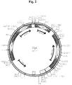
amplifiziert. Danach wurde das ca. 1,27 kBp lange PCR Produkt mit den Restriktionsendonukleasen MroI und MluI verdaut, das ca. 0,92 kBp lange MroI/MluI-GLUPI-Promotor/MCS/GLUCPI-Terminatorfragment nach Agarosegelelektrophorese isoliert und in das ca. 8,55 kBp lange MroI/MluI-YEp/KL-6b3M Vektorfragment ligiert. Das gewünschte Plasmid YEp/KL-6b3M-MCS wurde mittels Restriktionskartierung identifiziert und die mittels PCR synthetisierten DNA-Bereiche durch DNA Sequenzierung überprüft. - In der folgenden Plasmidkonstruktion wurde das LEU2d Gen in das Plasmid YEp/KL-6b3M-MCS insertiert. Dazu wurde das Plasmid YEp/KL-6B3M-MCS mit CelII und SnaBI verdaut und das 8,4 kBp lange CelII/SnaBI-YEp/KL-6b3M-MCS Vektorfragment isoliert. Das LEU2d Gen wurde als ca. 2,32 kBp langes CelII/SnaBI-Fragment aus dem Plasmid pADH040-2 (Erhart, E. und Hollenberg, C.P., J. Bacteriol. 156 (1983) 625-635) isoliert und mit dem 8,4 kBp langen CelII/SnaBI-YEp/KL-6b3M-MCS Vektorfragment ligiert. Die gewünschte Plasmidkonstruktion YEpL (DSM 7038) wurde durch Restriktionskartierung identifiziert (Fig. 2).
- Die Klonierung des verwendeten Glucoseoxidase Gens (Stamm: NRRL-3, ATTC 9029), die Subklonierung in den pBluescript SK(+) Vektor, die DNA Sequenzierung und die Ableitung der GOD Proteinsequenz sind in der Publikation von Kriechbaum, M. et al. (FEBS Lett. 255 (1989) 63-66) beschrieben. Das GOD Gen wurde in 2 Teilbereichen (SalI Restriktionsfragmente) in pBluescript SK(+) kloniert.
- Das Plasmid pSK/GOD-1.8 enthält ein ca. 1,8 kBp langes SalI-Fragment, das für die 5'- nichttranslatierte Region und den N-terminalen Bereich des GOD Strukturgens bis zur SalI-Schnittstelle an Bp-Position 164 kodiert (Bp-Position entsprechend der Numerierung bei Kriechbaum, M. et al.). Das Plasmid pSK/GOD-2.0 enthält ein ca. 2,0 kBp langes SalI-Fragment, das für den Rest des GOD Strukturgens von Bp-Position 165 bis 1853 sowie die 3'- nichttranslatierte Region stromabwärts des GOD Strukturgens kodiert.
- Mittels PCR Technik wurde die 5'- und 3'- nichttranslatierte Region des GOD Gens entfernt, das GOD Strukturgen an beiden Enden mit singulären Restriktionsendonukleaseschnittstellen versehen (BglII bzw. PvuII) und zudem in der C-terminalen kodierenden Region des GOD Strukturgens eine singuläre SphI und NheI Schnittstelle eingeführt unter Erhalt einer für das native GOD Protein kodierenden DNA-Sequenz. Danach wurde das GOD Strukturgen in einer Dreifragmentligation aus den beiden PCR Fragmenten zusammengesetzt und in den Vektor YEpL inseriert. Zur Amplifikation des N-terminalen GOD Strukturgens wurde das folgende Primerpaar verwendet (siehe SEQ ID NO. 5 und SEQ ID NO. 6) und Plasmid pSK/GOD-1.8 als Template DNA.

Zur Amplifikation des restlichen GOD Strukturgens wurde das folgende Primerpaar verwendet (siehe SEQ ID NO. 7 und SEQ ID NO. 8) und Plasmid pSK/GOD-2.0 als Template DNA.
Das ca. 220 Bp lange PCR Produkt der ersten Reaktion wurde mit BglII und SalI nachgespalten und das ca. 130 Bp lange BglII/SalI-Fragment isoliert. Das ca. 2,05 kBp lange PCR Produkt der zweiten Reaktion wurde mit SalI und PvuII verdaut das ca. 1,7 kBp lange DNA Fragment isoliert. Danach wurden die PCR Fragmente in das ca. 10,7 kBp lange BglII/PvuII-YEpL Vektorfragment ligiert (Dreifragmentligation). Das gewünschte Plasmid YEpL/GOD (Fig. 3) wurde durch Restriktionskartierung identifiziert und partiell sequenziert (Klonierungsübergänge). - Das Plasmid enthält ein modifiziertes GOD Gen, das für eine GOD Enzymvariante kodiert, die C-terminal zusätzlich 4 Histidinreste besitzt. YEpL/GOD-(His)4 wurde aus dem Plasmid YEpL/GOD hergestellt.
- Dazu wurde das Plasmid YEpL/GOD partiell mit SphI und vollständig mit PvuII gespalten, das ca. 10,7 kBp lange SphI/PvuII-Fragment isoliert und mit dem folgenden, aus 2 Oligonukleotiden (siehe SEQ ID NO. 9 und SEQ ID NO. 10) durch Hybridisierung hergestellten, DNA Linker ligiert.

Das gewünschte Plasmid YEpL/GOD-(His)4 wurde durch Koloniehybridisierung mit radioaktiv markiertem Primer 10 als Sonde idendifiziert und durch Restriktionskartierung und partielle Sequenzierung (C-terminale Bereich des GOD Strukturgens) weiter analysiert. -
- Mutagenese (Ausgangsstamm: X2180-1A, Genotyp: a SUC2 mal mel gal2 CUP1; ATCC 26786)
- Inkubation mit [³H]-Mannose
- Anreicherung von hyperglycosylierungsdefekten Mutanten durch Lagerung der Zellen bei -80°C bis die Überlebensrate der Zellen auf 2 - 3 Zehnerpotenzen abfällt (2 - 4 Monate)
- Die Zellen werden bei 28°C unter Schütteln inkubiert bis eine OD von 0,6 bei 578 nm erreicht ist. 10⁶ Zellen werden mit YEP-Medium (2% Bacto Pepton, 1% Hefeextrakt, Difco) gewaschen und in 0,1 ml YEP mit 0,1% Glucose resuspendiert. 2 mCi [³H]-Mannose (spezifische Aktivität 18,5 Ci/mmol) werden zugegeben und die Kultur bei 28°C für 60 Minuten inkubiert. Die Zellen werden abzentrifugiert, mit Wasser gewaschen und in YEPD, welches 25% Glyzerin enthält, resuspendiert und bei -70°C zur Einwirkung der Radioaktivität gelagert.
Nach ca. 45 - 50 Tagen, wenn die Überlebensrate der Zellen auf 1,5 - 0,2% abgefallen ist, werden Aliquote der Zellen auf YEP Agarplatten mit 2% Mannose ausplattiert und bei 30°C inkubiert. - Mutanten mit einem Defekt in der Proteinglycosylierung werden zunächst auf ihre Fähigkeit selektioniert [³H]-Mannose nicht einzubauen und [³⁵S]-Methionin einzubauen. Dazu läßt man die Zellen auf YEPD-Agar-Platten wachsen, repliziert die Hefekolonien auf 2 Rotband Filter (Schleicher & Schüll, Dassel, Deutschland) und inkubiert die Filter nochmals auf YEPD-Platten für 6 Stunden. Ein Filter wird dann in einer Lösung aus YEPD (eine zur Benetzung des Filters gerade ausreichende Menge), welche 0,01 mCi/ml [³⁵S]-Methionin enthält, inkubiert. Der andere Filter wird mit YEP, welches 0,2 mCi/ml [³H]-Mannose enthält, getränkt und 30 Minuten inkubiert. Die Zellen/Kolonien werden mit 5% Trichloressigsäure auf dem Filter fixiert, mit Wasser und Aceton gewaschen und durch Autoradiographie analysiert.
- Das SUC2 Gen aus S. cerevisiae kodiert für 2 unterschiedlich regulierte und kompartimentierte Invertase Formen, i) eine glycosylierte vorwiegend ins Periplasma sezernierte Invertase und ii) eine intrazelluläre etwas verkürzte nicht glycosylierte Form (Carlson, M. et al., Mol. Cell. Biol. 3 (1983) 439-447). Die Invertase enthält 14 potentielle N-Glycosylierungsstellen, von denen im Durchschnitt 9 - 10 bei der sezernierten Form pro Invertase Untereinheit glycosyliert werden. Externe Wildtyp Invertase wandert durch nicht einheitliche "outer chain" Glycosylierung als diffuse Bande in nativen Gelen. Die cytoplasmatische, nicht glycosylierte Form ergibt im Gegensatz dazu eine scharfe Bande nach Aktivitätsanfärbung. Eine veränderte N-Glycosylierung läßt sich somit über die Wanderungsgeschwindigkeit und die Bandenschärfe von externer, Invertase in nativen Gelen grob analysieren.
- Die Hefestämme (X2180-1A Wildtypstamm und positive Klone) wurden in 5 ml YEPS-Medium (1% Hefeextrakt, 2% Bactopepton, Difco, und 2% Saccharose) über Nacht angezogen, die Zellen in der spätlogarithmischen Wachstumsphase geerntet, einmal mit 20 mmol/l Natriumazid gewaschen und mit Glasperlen durch Homogenisieren auf einem Whirlmix aufgeschlossen. Die Zelllysat-Herstellung, die native Gelelektrophorese und Aktivitätsanfärbung von Invertase mit Saccharose und 2,3,4-Trinitrophenyltetrazoliumchlorid als Substrat/Glucosereagenz wurde entsprechend der Methode von Ballou C.E. (Methods Enzymol. 185 (1990) 440-470) durchgeführt.
- Die positiven Klone lassen sich anhand der Invertase Aktivitätsanfärbung in 4 Klassen einteilen:
- 1. Mutanten mit Wildtyp Invertase Mobilität.
- 2. Mutanten, die weder nichtglycosylierte noch glycosylierte Invertase synthetisieren.
- 3. Mutanten mit Defekten in der "outer chain" Glycosylierung (distinktes oligomeres Bandenmuster aus 3 - 4 Banden).
- 4. Mutanten, die zu einer ausgeprägten Unterglycosylierung von Invertase führen (größere Mobilität als Wildtyp Invertase)
- Mutantenstämme der Klasse 4, im weiteren mit ngd29 (DSM 7042/7338) und ngd62 (DSM 7160/7340) bezeichnet (ngd für: "N-glycosylation defective"), synthetisieren im Vergleich zum Ausgangsstamm X2180-1A eine "einheitlich glycosylierte" dimere externe Invertase (scharfe Bande und erhöhte Wanderungsgeschwindigkeit in nativen Gelen nach Aktivitätsanfärbung). Die ngd-Mutantenstämme waren osmotisch stabil, bei 30°C kultivierbar und aggregierten nicht während der Anzucht.
- Zur Einführung eines oder mehrerer durch Transformation komplementierbarer Auxotrophien wurden die ngd-Mutanten mit geeigneten Laborstämmen entsprechend der bei F. Sherman et al. (Methods in Yeast Genetics: A Laboratory Manual, Cold Spring Harbor Laboratory, Cold Spring Harbor, New York, (1981)) beschriebenen Methode gekreuzt und diploide Stämme durch Mikromanipulation isoliert. Anschließend wurden die diploiden Stämme sporuliert und Segreganten mit geeigneten Auxotrophien (z.B. ura3, leu2) und ngd-Mutation isoliert.
- Dazu wurde die ngd29-Mutante zusammen mit dem Stamm DBY746 (MATα ura3-52 leu2-3,-112 trp1-289a his3-Δ1; [DSM 4316] äquivalent mit ATCC 44733) und die ngd62 Mutante mit dem Stamm JM1935 (MATα ura3 leu2 his4, DSM 7156) 6 Stunden bei 30°C in YEPD (1% Hefeextrakt, 2% Bactopepton, Difco, und 2% Glucose) inkubiert. Anschließend wurden mit Hilfe eines Mikromanipulators (Modell: nach de Fonbrune der Firma Bachhofer, Reutlingen) Zygoten isoliert und in 5 ml YEPD über Nacht angezogen. Die Zellen wurden kurz abzentrifugiert, das Medium bis auf ca. 0,2 ml dekantiert und das Zellpellet im Restmedium resuspendiert. Diese Zellsuspension wurde auf eine Kaliumacetat-Platte (1% Kaliumacetat, 1,5% Agar) ausgebracht. Nach ca. 5 Tagen wurden die so erhaltenen Asci mit einer Impföse in 0,5 ml sterilem Wasser resuspendiert, 10 µl eines β-Glucuronidase/Arylsulfatase-Gemisches (Boehringer Mannheim) zugegeben und 10 min bei Zimmertemperatur inkubiert. Anschließend wurden 10 ml Wasser zugegeben, die Ascus-Suspension abzentrifugiert und der Überstand dekantiert. Unter dem Mikromanipulator wurden die Sporen mehrerer Asci isoliert und auf YEPD-Platten (YEPD mit 1,5% Agar) 3 Tage bei 30°C inkubiert. Von ausgekeimten Sporen wurde eine Stempelplatte angelegt, die Kolonien auf synthetischen Minimalmedien (0,67% Yeast Nitrogen Base ohne Aminosäuren, Difco; 2% Glucose; 1,5% Agar plus Zusätze: 20 mg/l Trp, His, Arg, Met; 30 mg/l Leu, Ile, Lys; 50 mg/l Phe; 100 mg/l Glu, Asp; 400 mg/l Val, Thr, Ser sowie 20 mg/l Adenin und Uracil; jeweils einer dieser Zusätze fehlte in den einzelnen Minimalmedien) gestempelt und 3 Tage bei 30°C inkubiert. Segreganten mit ngd29-Phänotyp, die Auxotrophien für Uracil und Leucin aufwiesen, wurden wie in Beispiel 2.2. beschrieben analysiert/isoliert. In allen untersuchten Tetraden segregierte der ngd29-Phänotyp (ebenso wie der ngd62 Phänotyp) 2:2, was für eine einzelne Mutation in einem einzelnen nuklären Locus spricht.
- Aus der hier beschriebenen Kreuzung DBY746 x ngd29 wurden die Stämme BMY3-9A und N-BMY3-9A (MATα leu2-3,112 ura3-52 his3-Δ1 ngd29; DSM 7042 und DSM 7338) und BMY3-9C und N-BMY3-9C (MATα leu2-3, 112, ura3-52 ngd29; DSM 7193 und DSM 7341) erhalten.
- Auf analoge Art und Weise wurde aus der Kreuzung JM1935 x ngd62 die Stämme BMY12-20D und N-BMY12-20D (MATα leu 2 ura 3 his 4 ngd62; DSM 7160 und DSM 7340) erhalten.
- Die GOD aus A. niger ist ein natürlicherweise sezerniertes, glycosyliertes dimeres Enzym. Pro Untereineinheit sind 8 potentielle N-Gycosylierungsstellen (Sequons) und 3 Cysteinreste vorhanden, von denen zwei eine Disulfidbrücke ausbilden. In S. cerevisiae Wildtypstämmen exprimierte GOD wird ins Medium sezerniert und ist hinsichtlich des Molekulargewichts auf Grund einer nicht einheitlichen "outer chain" Glycosylierung (Hyperglycosylierung) sehr heterogen (Frederick, K.R. et al., J. Biol. Chem. 265 (1990) 3793-3802; De Baetselier, A. et al., Biotechnology 9 (1991) 559-561; Whittington, H. et al., Curr. Genet. 18 (1990) 531-536). Das prozessierte (Abspaltung einer 22 Aminosäuren langen Signalsequenz) A. niger GOD Protein besteht aus 583 Aminosäuren mit einem potentiellen Molekulargewicht von 63 273 Da (Frederick, K.R. et al., J. Biol. Chem. 265 (1990) 3793-3802).
- Die Plasmide YEpL/GOD (Beispiel 1.5) und YEp/GOD-(His)₄ (Beispiel 1.6) wurden in den Wildtypstamm JM1935 (MATα leu2 ura3 his4 MAL4) DSM 7156 BMY3-9A (DSM 7042 und N-BMY3-9A (DSM 7338) (siehe Beispiel 3) transformiert und die Transformanten auf Minimalmediumagarplatten mit 1,5% Agarose, 0,67% YNB (Yeast Nitrogen Base, Salz-Vitamin-Gemisch, Difco) 0,5 % CAA (Casaminosäuren, Proteinhydrolysat, Difco) und 2% Fructose als C-Quelle selektioniert (Uracil-Selektion).
- Zur Amplifikation der Plasmidkopienzahl (Selektion auf das Plasmid-kodierte LEU2d-Allel; Beggs, J.D., Nature 275 (1978) 104-109; Erhart, E. und Hollenberg, C.P. J., Bacteriol. 156 (1983) 625-635) wurden die Transformanten auf Minimalmediumplatten ohne Leucin ausgestrichen (1,5% Agarose, 0,67% YNB , Difco, 60 mg/l Adenin und 2% Fructose).
- Vorkulturanzuchten wurden in Leucin Selektivmedium mit 0,67% YNB und 4% Fructose in Schüttelkolben bei 30°C für 48 Std. durchgeführt und zum Animpfen von Expressionskulturen (Inoculum: 1 - 2%) verwendet. Die Hauptkultur (1 l Schüttelkultur) wurde bei 30°C in Vollmedium mit 2% Hefeextract, 4% Bactopepton, Difco, 0,1 mol/l Phosphatpuffer, pH 7,0, 1% Fructose und 6% Maltose für 3 - 4 Tage unter Schütteln inkubiert. Nach 48 und 72 Std. wurden Proben entnommen und das Zellwachstum (Bestimmung der optischen Dichte bei 600 nm, OD₆₀₀), die ins Medium sekretierte GOD Aktivität und die in den Zellen verbleibende GOD Aktivität nach Zell-Lyse im Rohextrakt bestimmt.


- Hinsichtlich Expression und Sekretion wurden für die GOD und GOD-(His)₄ Variante keine signifikanten Unterschiede gefunden.
- Das in den glycosylierungsdefekten Wirtsstämmen DSM 7042/7338 (ngd29) und DSM 7160/7340 (ngd62) exprimierte (ins Medium sezernierte) GOD-(His)₄ Enzym wurden mit dem in dem wildtypstamm DSM 7156 exprimierten (sezernierten) Enzym und gereinigter GOD aus A. niger (Boehringer Mannheim, BRD) durch SDS-PAGE und anschließende Proteinanfärbung näher charakterisiert. Die GOD-haltigen Mediumüberstände aus dem Wildtypstamm wurden vor der Elektrophorese 10fach durch TCA-Fällung konzentriert. Kohlenhydratfreies GOD-(His)₄ Enzym wurde enzymatisch mit N-Glycosidase F hergestellt und als Größenstandard verwendet.
- Die Deglycosylierung wurde in Anlehnung an die von Haselbeck, A. und Hösel, W. publizierte Methode (Topics in Biochemistry 8 (1988) 1-4) durchgeführt. 0,1 ml GOD-(His)₄-haltiger Mediumüberstand wurde mit Trichloressigsäure (Endkonzentration: 10%) gefällt, die präzipitierten Proteine abzentrifugiert, das Proteinpellet mit 70% Ethanol gewaschen, im Vakuum getrocknet, in 10 µl 20 mmol/l Kaliumphosphatpuffer, pH 7,2, mit 1% SDS aufgenommen und 3 min auf 95°C erhitzt. Nach Abkühlung auf Raumtemperatur wurde die Probe mit 20 mmol/l Kaliumphosphatpuffer, pH 7,2, Octylglucosid (Endkonzentration: 0,5%) und 5 Units N-Glycosidase F auf 0,1 ml verdünnt, 1 - 12 Std. bei 37°C inkubiert und anschließend mit 25 µl 5xSDS-Puffer (siehe oben) versetzt.
- Die in den glycosylierungsdefizienten ngd-Mutantenstämmen exprimierten GOD Enzyme (GOD und GOD-(His₄)) sind als dominante einheitliche Bande mit einem Molekulargewicht von ca. 80 kDa nach Proteinanfärbung in SDS-PAGE Gelen sichtbar. Dieses Experiment zeigt die fehlende "outer chain" Glycosylierung der GOD Enzyme und deutet auf eine einheitliche "core" ähnliche Glycosylierung hin. Die ngd-Mutantenstämme weisen hinsichtlich der Glycosylierung einen mnn9 ähnlichen Phänotyp auf. Im Gegensatz dazu sind die in Wildtypstämmen exprimierten GOD Enzyme nur als sehr diffuse Banden zu erkennen, die sich über einen Molekulargewichtsbereich von ca. 80 - 200 kDa erstrecken.
- Glycosylierungsdefekte Mutanten wie z.B. mnn8, mnn9 und mnn10 zeigen eine erhöhte Orthovanadat Resistenz und eine erhöhte Sensitivität gegenüber dem Antibiotikum Hygromycin B. Der Resistenz-/Sensitivitätsphänotyp ermöglicht eine Unterscheidung/Gruppierung von N-glycosylierungsdefekten Mutanten (Ballou, L. et al., Proc. Natl. Acad. Sci. 88 (1991) 3209-3212).
- Die zu untersuchenden Stämme wurden in YEPD Medium (5 ml Rollerkultur) über Nacht angezogen und die Stämme/Kulturen mit YEPD Medium auf eine optische Dichte (OD₆₀₀) von exakt 0,05 eingestellt. Danach wurden jeweils 20 µl Zellsuspension auf YEPD-Agarplatten mit 2 - 15 mmol/l Natriumorthovanadat oder 10 - 200 µg/ml Hygromycin B gespottet. Nach 2 Tagen Inkubation bei 30°C wurde das Wachstum der Zellflecken ausgewertet (siehe Tabelle).
-
Stamm Orthovanadat Resistenz mmol/l Hygromycin Resistenz (µg/ml) 2 3 4 5 6 7 10 15 10 50 100 200 DBY 746 (Wildtyp) + + + + - - - - + + - - X2180-1A (Wildtyp) + + + + - - - - + + - - LB347-1C (mnn9)1) + + + + + + + + - - - - BMY3-9A (ngd29) + + + + + - - - + ± - - BMY12-20D(ngd62) + + + ± - - - - + ± - - N-BMY3-9A(ngd29) + + + + + - - - + ± - - N-BMY12-20D(ngd62) + + + ± - - - - + ± - - + Wachstum
± sehr langsames Wachstum
- kein Wachstum1) J. Biol. Chem. 259 (1984) 3805-3811 - Die ngd-Mutanten zeigen Unterschiede hinsichtlich des Resistenzmusters untereinander, zur mnn9 Mutante und zu Wildtypstämmen auf.
- Ein Allelietest dient zur Identifizierung (Unterscheidung) von Genen und Gendefekten (Mutationen). Mit dieser Technik läßt sich analysieren, ob 2 Mutanten allel sind (im gleichen Gen eine Mutation aufweisen). Die ngd-Mutanten wurden untereinander und zur mnn9 Mutante auf Allelie untersucht.
- Die Allelieteste wurden mittels genetischer Standardtechniken durchgeführt (siehe: Sherman, F.; Fink, G.R.; Hicks, J.B., Methods in Yeast Genetics: A Laboratory Manual. Cold Spring Harbor Laboratory, Cold Spring Harbor, New York, (1981); Guthrie, C. und Fink, G.R. (eds.), Guide to Yeast Genetics and Molecular Biology. Methods Enzymol. 194 (1991)).
- Zwei zu analysierende haploide Mutantenstämme unterschiedlichen Paarungstyps mit sich komplementierenden Auxotrophiebedürfnissen werden gekreuzt und die diploiden Stämme auf Platten mit Minimalmedium selektioniert. Die Diploidie der isolierten Stämme wird durch das Vorhandensein von a und α Paarungstyp spezifischen DNA Sequenzen mittels PCR Analyse entsprechend der Methode von Huxley, C. et al. (Trends Genet. 6 (1190) 236) bestätigt.
- Zwei Mutanten sind allel, d.h. besitzen eine Mutation im gleichen Gen, wenn sich die Mutationen in der diploiden Zelle nicht komplementieren.
- Zwei Mutanten sind nicht allel, d.h. besitzen eine Mutation in 2 unterschiedlichen Genen, wenn sich die Mutationen in der diploiden Zelle komplementieren und ein Wildtyp Phänotyp resultiert.
-
BMY3-9C (MATα leu2-3,-112 ura3-52 ngd29) DSM 7193 BMY8-12A (MATα trp1-289a his3-Δ1 ngd62) DSM 7157 BMY13-1C (MATα ura3-52 leu2-3,-112 his3-Δ1 mnn9) DSM 7159 BMY13-7B (MATα leu2-3,-112 his3-Δ1 mnn9) DSM 7158 BMY12-20D(MATα leu2 ura3 his4 ngd62) DSM 7160 N-BMY3-9C (MATα leu2-3,-112 ura3-52 ngd29) DSM 7341 N-BMY13-1C (MATα ura3-52 leu2-3,-112 his3-Δ1 mnn9) DSM 7339 N-BMY12-20D(MATα leu2 ura3 his4 ngd62) DSM 7340 Kreuzungpartner Phänotyp der Haploiden Selektion Diploide Phänotyp der Diploiden MATα MATα BMY3-9C x BMY8-12A ngd29xngd62 his leu Wildtyp BMY3-9C x BMY13-7B ngd29xmnn9 his ura Wildtyp BMY13-1C x BMY8-12A mnn9xngd62 trp ura Wildtyp BMY12-20D x BMY13-7B ngd62xmnn9 his Wildtyp N-BMY3-9C x BMY8-12A ngd29xngd62 his leu Wildtyp N-BMY3-9C x BMY13-7B ngd29xmnn9 his ura Wildtyp N-BMY13-1C x BMY8-12A mnn9xngd62 trp ura Wildtyp N-BMY12-20D x BMY13-7B ngd62xmnn9 his Wildtyp - SC = synthetisches Komplettmedium (0,67% Yeast Nitrogen Base ohne Aminosäuren, Difco; 2% Glucose; 1,5% Agar plus Zusätze: 20 mg/l Trp, His, Arg, Met; 30 mg/l Leu, Ile, Lys; 50 mg/l Phe; 100 mg/l Glu, Asp; 400 mg/l Val, Thr, Ser sowie 20 mg/l Adenin und Uracil; Die Aminosäuren Ura, His und Trp wurden, wie in der Tabelle in der Spalte Selektion der Diploide, angegeben in den einzelnen Minimalmedien weggelassen.
- Die Mutanten ngd29 und ngd62 unterscheiden sich untereinander und sind unterschiedlich zu mnn9 (nichtallel).
- Mit diesem Isolierverfahren wurde die GOD Variante GOD-(His)4 aus dem Kulturfiltrat von BMY3-9A/GOD-(His)4-Zellen und BMY12-20D/GOD-(His)4-Zellen (hyperglycosylierungsdefekte Wirtsstämme) isoliert.
- Das Kulturfiltrat wurde mit Natronlauge auf pH 7,5 titriert und auf eine mit 10 mmol/l Kaliumphosphatpuffer, pH 7,5, äquilibrierte NTA-Säule aufgetragen (Säulenvolumen 25 ml; NTA-Gel der Firma Diagen, Düsseldorf; Hochuli, E. et al., J. Chromatography 411 (1987) 177-184; Hochuli, E. et al., Biotechnology 6 (1988) 1321-1325). Die Säule wurde mit 5 - 10 Säulenvolumen 1 mol/l Natriumchlorid in 10 mmol/l Kaliumphosphatpuffer, pH 7,5, und mit 5 - 10 Säulenvolumen 10 mmol/l Kaliumphosphatpuffer, pH 7,5, nachgewaschen. Danach wurde das GOD-(His)₄ Enzym mit 0,1 mol/l Imidazol in Äquilibrierungspuffer, pH 7,5, eluiert und die GOD-(His)₄ haltigen Fraktionen (gelb) gegen 10 mmol/l Kaliumphosphatpuffer, pH 7,5, dialysiert.
- Nach diesem Verfahren wurde native GOD und hyperglycosylierte GOD aufgereinigt.
- 100 ml sterilfiltriertes Kulturfiltrat wurden mit 33 g festem Ammoniumsulfat (AS-Sättigungskonzentration 55%) unter langsamen Rühren versetzt, die präzipitierten Proteine nach 1 - 2 Std. Inkubation bei Raumtemperatur abzentrifugiert, in 25 ml 25 mmol/l Kaliumphosphatpuffer, pH 7,5, gelöst und gegen den gleichen Puffer dialysiert (4 X 10 l, 24 Std., 4°C).
- Danach wurde das Dialysat auf eine mit 25 mmol/l Kaliumphosphat-Puffer, pH 7,5, äquilibrierte Q-Sepharose ff Säule (Säulenvolumen 12 ml) aufgetragen und mit 5 - 10 Säulenvolumen Äquilibrierungspuffer nachgewaschen. Die gebundenen GOD Enzyme wurden durch einen Gradienten von 0 - 1 mol/l KCl in Äquilibrierungspuffer (ca. 10 Säulenvolumen) eluiert und die GOD-haltigen (gelben) Fraktionen vereinigt.
- Die Bestimmung der GOD Aktivität erfolgt, wie in dem Abschnitt allgemeine Methoden beschrieben.
-
Enzym Organismus/ Glycosylierung spez. Aktivität (U/mg Protein) spez. Aktivität (U/mg Enzym) GOD (A. niger) 225 195 GOD (WT) 230 69 GOD (ngd29) 228 196 GOD (ngd62) 213 220 GOD-(His)4 (WT) 220 68 GOD-(His)4 (ngd29) 223 200 GOD-(His)4 (ngd62) 230 225 A. niger, GOD aus A. niger, Reinheit II (Boehringer Mannheim)
WT, S. cerevisiae Wildtyp
ngd29, S. cerevisiae hyperglycosylierungsdefekte ngd29 Mutante
ngd62, S. cerevisiae hyperglycosylierungsdefekte ngd62 Mutante - Die gereinigten GOD Enzyme wurden mit 1/5 Volumen 5xSDS-Probenpuffer (1xSDS-Probenpuffer: 50 mmol/l Tris-HCl, pH 6,8, 1% SDS, 1% Mercaptoethanol, 10% Glycerin, 0,001% Bromphenolblau) versetzt und 5 min bei 95°C inkubiert. Danach wurden die Proteine durch SDS-PAGE aufgetrennt (Laemmli, U.K., Nature 227 (1970) 680-685) und mit Coomassie Brilliant Blue R Farbstoff angefärbt.
-
Enzym Organismus/ Glycosylierung Molekulargewicht/Untereinheit (kDa) GOD (A. niger) ca. 80 GOD (WT) 80 - 140 GOD (ngd29) ca. 80 GOD (ngd62) ca. 80 GOD-(His)4 (WT) 80 - 140 GOD-(His)4 (ngd29) ca. 80 GOD-(His)4 (ngd62) ca. 80 A. niger, GOD aus A. niger, Reinheit II (Boehringer Mannheim)
WT, S. cerevisiae Wildtyp
ngd29, S. cerevisiae hyperglycosylierungsdefekte ngd29 Mutante
ngd62, S. cerevisiae hyperglycosylierungsdefekte ngd62 Mutante - Der Kohlenhydratanteil der GOD Enzyme aus unterschiedlichen Organismen bzw. Hefestämmen wurde in Anlehnung an die Methode von Ashwell, G. (Methode Enzymol. 3 (1957) 84) bestimmt.
- Hierzu werden 0,5 ml gereinigtes GOD Enzym (Konzentration 20 - 100 U/ml in H₂O) mit 5 ml Anthron-Reagenz gemischt, die Lösung 5 Minuten bei 25°C inkubiert und danach 15 Minuten im siedenden Wasserbad erhitzt. Nach Abkühlung der Probe auf 25°C wird die Extinktion bei 630 nm gegen einen Reagentienleerwert bestimmt. Mittels einer simultan erstellten Mannose-Eichkurve mit Mannose-Standardlösungen von 5, 25, 75 und 100 µg/ml Mannose wird der Kohlenhydratanteil der GOD Probe ermittelt.
- 66 ml konzentrierte Schwefelsäure werden vorsichtig mit 34 ml Wasser verdünnt. Nach Abkühlung auf 80°C werden 50 mg Anthron und 1 g Thioharnstoff in der Schwefelsäure gelöst. Das Anthron-Reagenz ist zwei Wochen bei 4°C haltbar.
-
Enzym Organismus/ Glycosylierung Kohlenhydratanteil (%) (bezogen auf Protein) GOD (A. niger) 13 GOD (WT) 71 GOD (ngd29) 12,5 GOD (ngd62) 13 GOD-(His)4 (WT) 65 GOD-(His)4 (ngd29) 11 GOD-(His)4 (ngd62) 12 A. niger, GOD aus A. niger, Reinheit II (Boehringer Mannheim)
WT, S. cerevisiae Wildtyp
ngd29, S. cerevisiae hyperglycosylierungsdefekte ngd29 Mutante
ngd62, S. cerevisiae hyperglycosylierungsdefekte ngd62 Mutante - Bekkers, A.C.A.P.A.; Franken, P.A.F.; Van den Bergh, C.J.; Verbakel, J.M.A.; Verheij, H.M.; De Haas,G.H.: The use of genetic engineering to obtain efficient production of porcine pancreatic phospholipase A2 by Saccharomyces cerevisiae. Biochim. Biophys. Acta 1089, 345-351 (1991).
- Ballou, L.; Cohen, R.E.; Ballou,C.E.: Saccharomyces cerevisiae mutants that make mannoproteins with a truncated carbohydrate outer chain. J. Biol. Chem. 255, 5986-5991 (1980).
- Ballou, C.E.: Yeast cell wall and cell surface. In: Strathern, J.N.; Jones, E.W.; Broach, J.R. (eds.), The Molecular Biology of the Yeast Saccharomyces, Metabolism and Gene Expression, Cold Spring Harbor Laboratory, New York, pp. 335-360 (1982).
- Ballou, L.; Alvarado, E.; Tsai, P.; Dell, A; Ballou, C.E.: Protein glycosylation defects in the Saccharomyces cerevisiae mnn7 mutant class. J. Biol. Chem. 264, 11857-11864 (1989).
- Ballou, C.E.: Isolation characterization, and properties of Saccharomyces cerevisiae mmn mutants with nonconditional protein glycosylation defects. Methods Enzymol. 185, 440-470 (1990).
- Ballou, L.; Hitzeman, R.A.; Lewis, M.S.; Ballou, C.S.: Vanadate-resistant yeast mutants are defective in protein glycosylation. Proc. Natl. Acad. Sci. 88, 3209-3212 (1991).
- Beggs, J.D.: Transformation of yeast by a replicating hybrid plasmid. Nature 275, 104-109 (1978).
- Carlson, M.; Taussig, R.; Kustu, S.; Botstein, D.: The secreted form of invertase in Saccharomyces cerevisiae is synthesized from mRNA encoding a signal sequence. Mol. Cell. Biol. 3, 439-447 (1983).
- Ciriacy, M.: Genetics of alcohol dehydrogenase in Saccharomyces cerevisiae. I. Isolation and genetic analysis of adh-mutants. Mut. Res. 29, 315-326 (1975).
- De Baetselier, A.; Vasavada, A.; Dohet, P.; Ha-Ti, V.; De Beukelaer, M.; Erpicum, T.; De Clerk, L.; Hanotier, J.; Rosenberg, S.: Fermentation of yeast producing A. niger glucose oxidase: scale up, purification and characterization of the recombinant enzyme. Biotechnology 9, 559-561 (1991).
- Delorme, E.: Transformation of Saccharomyces cerevisiae by electroporation. Applied and Environmental Microbiology 55, 2242- 2246 (1989).
- Dijken, J.P. van; Veenhuis M.: Cytochemical localization of glucose oxidase in peroxisomes of Aspergillus niger. European J. Appl. Microbiol. Biotechnol. 9, 275 - 283 (1980)
Erhart, E.; Hollenberg, C.P.: The presence of a defective LEU2 gene on 2µDNA recombinant plasmids of Saccharomyces cerevisiae is responsible for curing and high copy number. J. Bacteriol. 156, 625-635 (1983). - Frederick, K.R.; Tung, J.; Emerick, R.S.; Masiarz, F.R.; Chamberlain, S.H.; Vasavada, A.; Rosenberg, S.; Chakraborty, S.; Schopter, L.M.; Massey, N.: Glucose oxidase from Aspergillus niger. J. Biol. Chem. 265, 3793-3802 (1990).
- Hadwick, K.G.; Lewis, M.J.; Semenza, J.; Dean, N.; Pelham, H.R.B.: ERD1, a yeast gene required for the retention of luminal endoplasmic reticulum proteins, affects glycoprotein processing in the Golgi apparatus. EMBO J. 9, 623-630 (1990).
- Haselbeck, A.; Hösel, W.: Studies on the effect of the incubation conditions, various detergents and protein concentration on the enzymatic activity of N-glycosidase F (glycopeptidase F) and endoglycosidase F. Topics in Biochemistry 8, 1- 4 (1988).
- Hochuli, E.; Doebeli, H.; Schacher, A.: New metal chelate adsorbent selective for proteins and peptides containing neighbouring histidine residues. J. Chromatography 411, 177-184 (1987).
- Hochuli, E.; Bannwarth, W.; Doebeli, H.; Genz, R.; Stueber, D.: Genetic approach to facilitate purification of recombinant proteins with a novel metal chelate adsobent. Biotechnology 6, 1321-1325 (1988).
- Huffaker, T.C.; Robbins, P.W.: Yeast mutants deficient in protein glycosylation. Proc. Natl. Acad. Sci. 80, 7466-7470 (1983).
- Innis, M.A.: Glycosylation of heterologous proteins in Saccharomyces cerevisiae. In: Barr, P.J.; Brake, A.J.; Valenzuela, P. (eds.), Yeast genetic engineering, Butterworths, Stoneham, Mass, pp. 233-246 (1989).
- Ito, H.; Jukuda, A.; Murata, K.; Kimura, A: Transformation of intact yeast cells treated with alkali cations. J. Bacteriol. 153, 163-168 (1983).
- Kopetzki, E.; Buckel, P.; Schumacher, G.: Cloning and characterization of baker's yeast alpha glucosidase: overexpression in a yeast strain devoid of vacuolar proteinases. Yeast 5, 11-24 (1989).
- Kornfeld, R.; Kornfeld, S.: Assembly of asparagine-linked oligosaccharides. Ann. Rev. Biochem. 54, 631-664 (1985).
- Kukuruzinska, M.A.; Bergh, M.L.E.; Jackson, B.J.: Protein glycosylation in yeast. Ann. Rev. Biochem. 56, 915-944 (1987).
- Kriechbaum, M.; Heilmann, H,J.; Wientjes, F.J.; Hahn, M.; Jany, K.-D.; Gassen, H.G.; Sharif, F.; Alaeddinoglu, G.: Clonig and DNA sequence analysis of the glucose oxidase gene from Aspergillus niger NRRL-3. FEBS Lett. 255, 63-66 (1989).
- Laemmli, U.K.: Cleavage of structural proteins during the assembly of the head of bacteriophage T4. Nature 227, 680-685 (1970).
- Maniatis, T. et al., In: Molecular cloning: A laboratory manual. Cold Spring Harbor Laboratory Press, Cold Spring Harbor, New York, (1989).
- Moir, D.T.: Yeast mutants with increased secretion efficiency. In: Barr, P.J.; Brake, A.J.; Valenzuela, P. (eds.), Yeast genetic engineering, Butterworths, Stoneham, Mass, pp. 215-231 (1989).
- Mullis, K.B.; Faloona, F.A.: Specific synthesis of DNA in vitro via a polymerase-catalyzed chain reaction. Methods Enzymol. 155, 355-350 (1987).
- Nakano, A.; Muramatsu, M.: A novel GTP-binding protein, Sarp1, is involved in transport from the endoplasmic reticulum to the Golgi apparatus. J. Cell. Biol. 109, 2677-2691 (1989).
- Newman, A.P.; Ferro-Novick, S.: Characterization of new mutants in the early part of the yeast secretory pathway isolated by [3H]mannose suicide selection. J. Cell. Biol. 105, 1587-1594 (1987).
- Novick, P.; Field, C.; Schekman, R.: Identification of 23 complementation groups required for post-translational events in the yeast secretory pathway. Cell 21, 205-215 (1980).
- Paulson, C.P.: Glycoproteins: what are the sugar chains for? TIBS 14, 272-276 (1989).
- Rudolph, H.K., Antebi, A.; Fink, G.R.; Buckley, C.M.; Dorman, T.E.; LeVitre, J.; Davidow, L.S.; Mao, J.; Moir, D.T.: The yeast secretory pathway is perturbed by mutations in PMR1, a member of a Ca2+ ATPase family. Cell 58, 133-145 (1989).
- Reddy, V.A.; Johnson, R.S.; Biemann, K.; Williams, R.S.; Ziegler, F.D.; Trimble, R.B.; Maley, F.: Characterization of glycosylation sites in yeast external invertase. I. N-linked oligosaccharide content of the individual sequons. J. Biol. Chem. 263, 6978-6985 (1988).
- Runge, K.W.; Robbins, P.W.: Saccharomyces cerevisiae mutants in the early stages of protein glycosylation. In: Bonventre, P.F.; Morello, J.A.M.; Silver, S.D.; Wu, H.C. (eds.), Microbiology-1986. American Society for Microbiology, Washington, D.C. pp. 312-316 (1986).
- Schekman, R.; Novick, P.: The secretory process and yeast cell-surface assembly. In: Strathern, J.N.; Jones, E.W.; Broach, J.R. (eds.), The Molecular Biology of the Yeast Saccharomyces, Metabolism and Gene Expression, Cold Spring Harbor Laboratory, New York, pp. 361-398 (1982).
- Schmitt, H.D.; Wagner, P.; Pfaff, E.; Gallwitz, D.: The ras-related YPT1 gene product in yeast: a GTP-binding protein that might be involved in microtubule organization. Cell 47, 401-412 (1986).
- Schmitt, H.D.; Puzichia, M.; Gallwitz, D.: Study of a temperature-sensitive mutant of the ras-related YPT1 gene product in yeast suggests a role in the regulation of intracellular calcium. Cell 53, 635-647 (1988).
- Sherman, F.; Fink, G.R.; Hicks, J.B.: Methods in Yeast Genetics: A Laboratory Manual, Cold Spring Harbor Laboratory, Cold Spring Harbor, New York, (1981).
- Tanner, W.; Lehle, L.: Protein glycosylation in yeast. Biochim. Biophys. Acta 906, 81-99 (1987).
- Warren, C.E.: Glycosylation- considerations for protein engineering. BFE 7, 392-395 (1990).
- Whittington, H.; Kerry-Williams, S.; Bidgood, K.; Dodsworth, N. Peberdy, J.; Dobson, M.; Hinchliffe, E.; Ballance, D.J.: Expression of the Aspergillus niger glucose oxidase gene in A. niger, A. nidulans and Saccharomyces cerevisiae. Curr. Genet. 18, 531-536 (1990).
- Zamenhof, S.: Preparation and assay of deoxyribonucleic acid from animal tissue. Methods Enzymol. 3, 696-704 (1957).
- Ziegler, F.D.; Maley, F.; Trimble, R.B.: Characterization of glycosylation sites in yeast external invertase. II. Location of the endo-betta-N-acetylglucosaminidase H-resistant sequons. J. Biol. Chem. 263, 6978-6985 (1988).


Ein Hefestamm wie z. B. X2180-1A (ATCC 26786) wird in YEPD-Medium (2% Bacto Pepton, 1% Hefeextrakt, Difco, und 4% Glucose) angezogen, in der logarithmischen Wachstumsphase (davon ca. 5 x 10⁸ Zellen) geerntet, mit 0,1 mol/l Natriumphosphat, pH 7, gewaschen und in 1,5 ml 0,1 mol/l Natriumphosphat, pH 7, resuspendiert. Die Zellen werden mutagenisiert durch Zusatz von 0,1 ml Ethylmethansulfonat für eine Stunde bei 25°C. 0,2 ml der so behandelten Zellen werden mit 10 ml Natriumthiosulfat (5% w/v) für 10 Minuten inkubiert, 3 x mit 0,1 mol/l Natriumphosphat, pH 7,0, gewaschen und in YEPD-Medium (2% Bacto Pepton, 1% Hefeextrakt, Difco und 4% Glucose) resuspendiert.
Claims (7)
- Hefe-Mutanten mit Defekten in der N-Glycosylierung, erhältlich durch [³H]-Mannose Suizidselektion, Einführung einer oder mehrerer selektiver Marker (Auxotrophien und/oder Resistenzen), Auswahl derjenigen Stämme, welche nach Transformation mit dem Plasmid YEpL/GOD und Fermentation in Vollmedium mit 2% Hefeextrakt, 4% Bactopepton, Difco, 0,1 mol/l Phosphatpuffer, pH 7,0, 1% Fructose und 6% Maltose nach 3 - 4 Tagen Inkubation unter Schütteln in das Medium 10 mg/l GOD oder mehr sezernieren und allel sind zu Saccharomyces cerevisiae DSM 7042, DSM 7338, DSM 7160 und/oder DSM 7340.
- Hefe-Mutanten DSM 7042, DSM 7338, DSM 7160 und DSM 7340.
- Verfahren zur Herstellung von Hefe-Mutanten gemäß den Ansprüchen 1 und 2 durch [³H]-Mannose Suizidselektion, Einführung einer oder mehrerer selektiver Marker (Auxotrophien und/oder Resistenzen), Auswahl derjenigen Stämme, welche nach Transformation mit dem Plasmid YEpL/GOD und Fermentation in Vollmedium mit 2% Hefeextrakt, 4% Bactopepton, Difco, 0,1 mol/l Phosphatpuffer pH 7,0, 1% Fructose und 6% Maltose nach 3 - 4 Tagen Inkubation unter Schütteln in das Medium 10 mg/l GOD oder mehr sezernieren und allel sind zu Saccharomyces cerevisiae DSM 7042, DSM 7338, DSM 7160 und/oder DSM 7340.
- Verfahren nach Anspruch 3 zur Herstellung von Saccharomyces cerevisiae DSM 7042, DSM 7338, DSM 7160 und/oder DSM 7340.
- Verwendung der Hefe-Mutanten nach Anspruch 1 oder 2 als Wirtsorganismus zur homologen und heterologen Expression von Proteinen.
- Hypoglycosylierte rekombinante Proteine, erhältlich durch Transformation einer Hefemutante nach Anspruch 1 oder 2 mit einer rekombinanten DNA, welche für das gewünschte Protein codiert, Fermentation der Zellen und Isolierung des gewünschten Proteins aus den Zellen oder dem Kulturüberstand.
- Verfahren zur Expression, Verfahren zur Herstellung von im wesentlichen einheitlich glycosylierten Proteinen durch Transformation einer für das Protein codierenden DNA von Hefe-Mutanten nach Anspruch 1 oder 2, Fermentation und Isolierung des Proteins aus den Zellen oder dem Kulturüberstand.
Applications Claiming Priority (4)
| Application Number | Priority Date | Filing Date | Title |
|---|---|---|---|
| DE4226094 | 1992-08-07 | ||
| DE4226094 | 1992-08-07 | ||
| DE4301932A DE4301932A1 (de) | 1992-08-07 | 1993-01-25 | Hefewirtsstämme mit Defekten in der N-Glycosylierung |
| DE4301932 | 1993-01-25 |
Publications (3)
| Publication Number | Publication Date |
|---|---|
| EP0582244A2 true EP0582244A2 (de) | 1994-02-09 |
| EP0582244A3 EP0582244A3 (de) | 1995-03-15 |
| EP0582244B1 EP0582244B1 (de) | 2001-10-31 |
Family
ID=25917284
Family Applications (1)
| Application Number | Title | Priority Date | Filing Date |
|---|---|---|---|
| EP93112338A Expired - Lifetime EP0582244B1 (de) | 1992-08-07 | 1993-08-02 | Hefewirtsstämme mit Defekten in der N-Glycosylierung |
Country Status (12)
| Country | Link |
|---|---|
| US (2) | US5705616A (de) |
| EP (1) | EP0582244B1 (de) |
| JP (2) | JP2675511B2 (de) |
| KR (1) | KR0140973B1 (de) |
| AT (1) | ATE207955T1 (de) |
| AU (1) | AU657230B2 (de) |
| CA (1) | CA2103522C (de) |
| DE (2) | DE4301932A1 (de) |
| FI (1) | FI110006B (de) |
| IL (1) | IL106589A (de) |
| NO (1) | NO312246B1 (de) |
| NZ (1) | NZ248323A (de) |
Cited By (4)
| Publication number | Priority date | Publication date | Assignee | Title |
|---|---|---|---|---|
| WO1997049794A1 (fr) * | 1996-06-26 | 1997-12-31 | Societe D'applications De Recherches Et De Conseils Oenologiques (Sarco) | Produit de stabilisation proteique des vins |
| WO2002000856A2 (en) * | 2000-06-30 | 2002-01-03 | Flanders Interuniversity Institute For Biotechnology (Vib) | Protein glycosylation modification in pichia pastoris |
| US7507573B2 (en) | 2003-11-14 | 2009-03-24 | Vib, Vzw | Modification of protein glycosylation in methylotrophic yeast |
| EP3438254A4 (de) * | 2016-03-30 | 2019-10-16 | Kaneka Corporation | Polypeptid mit endonukleaseaktivität und herstellungsverfahren dafür |
Families Citing this family (6)
| Publication number | Priority date | Publication date | Assignee | Title |
|---|---|---|---|---|
| US6566122B1 (en) | 2000-06-16 | 2003-05-20 | Academia Sinica | Super-secreting saccharomyces cerevisiae strains |
| KR100473658B1 (ko) * | 2002-10-25 | 2005-03-10 | 드림바이오젠 주식회사 | 위치 선택적 수식법에 의한 혼성형 단백질 치료제 개발을 위한 형질전환 효모 시스템 |
| JP2009509970A (ja) * | 2005-09-22 | 2009-03-12 | プロサイ インコーポレイテッド | 酵母突然変異体において産生されるグリコシル化ポリペプチドおよびその使用方法 |
| KR101460561B1 (ko) * | 2012-11-20 | 2014-11-13 | 한국식품연구원 | 사카로마이세스 세레비지애 113-8의 대량 생산방법 |
| CN107988177B (zh) * | 2016-10-26 | 2021-08-20 | 潍坊康地恩生物科技有限公司 | 葡萄糖氧化酶突变体 |
| CN108118037B (zh) * | 2016-11-28 | 2021-08-31 | 青岛蔚蓝生物集团有限公司 | 一种耐热性提高的葡萄糖氧化酶突变体 |
Citations (3)
| Publication number | Priority date | Publication date | Assignee | Title |
|---|---|---|---|---|
| EP0314096A2 (de) * | 1987-10-29 | 1989-05-03 | Zymogenetics, Inc. | Verfahren zur Regulierung der Proteinglycosylierung |
| WO1989012675A1 (en) * | 1988-06-21 | 1989-12-28 | Chiron Corporation | Production of glucose oxidase in recombinant systems |
| EP0415565A2 (de) * | 1989-07-31 | 1991-03-06 | Merck & Co. Inc. | Verstärkte Expression von heterologen Proteinen in rekombinanten Wirten in einem wachstumsfaktorfreien Medium |
Family Cites Families (4)
| Publication number | Priority date | Publication date | Assignee | Title |
|---|---|---|---|---|
| US5135854A (en) * | 1987-10-29 | 1992-08-04 | Zymogenetics, Inc. | Methods of regulating protein glycosylation |
| NZ229260A (en) * | 1988-06-03 | 1994-02-25 | Merck & Co Inc | Hepatitis b virus, expression cassette for pre-s domain, host cells and |
| EP0382332A3 (de) * | 1989-01-06 | 1990-11-14 | Collaborative Research Inc. | Mutanten von Saccharomyces cerevisiae mit stark vermehrter Sekretion |
| DE4301904A1 (de) * | 1992-08-07 | 1994-02-10 | Boehringer Mannheim Gmbh | Hypoglycosylierte recombinante Glucoseoxidase |
-
1993
- 1993-01-25 DE DE4301932A patent/DE4301932A1/de not_active Withdrawn
- 1993-08-02 AT AT93112338T patent/ATE207955T1/de not_active IP Right Cessation
- 1993-08-02 AU AU44354/93A patent/AU657230B2/en not_active Ceased
- 1993-08-02 DE DE59310233T patent/DE59310233D1/de not_active Expired - Lifetime
- 1993-08-02 EP EP93112338A patent/EP0582244B1/de not_active Expired - Lifetime
- 1993-08-04 IL IL10658993A patent/IL106589A/xx not_active IP Right Cessation
- 1993-08-04 NZ NZ248323A patent/NZ248323A/en unknown
- 1993-08-06 FI FI933487A patent/FI110006B/fi not_active IP Right Cessation
- 1993-08-06 KR KR1019930015252A patent/KR0140973B1/ko not_active IP Right Cessation
- 1993-08-06 CA CA002103522A patent/CA2103522C/en not_active Expired - Lifetime
- 1993-08-06 NO NO19932811A patent/NO312246B1/no unknown
- 1993-08-06 JP JP5196388A patent/JP2675511B2/ja not_active Expired - Fee Related
-
1995
- 1995-06-05 US US08/461,593 patent/US5705616A/en not_active Expired - Lifetime
-
1996
- 1996-05-31 US US08/651,323 patent/US5798226A/en not_active Expired - Lifetime
-
1997
- 1997-04-09 JP JP9090937A patent/JP2984229B2/ja not_active Expired - Lifetime
Patent Citations (3)
| Publication number | Priority date | Publication date | Assignee | Title |
|---|---|---|---|---|
| EP0314096A2 (de) * | 1987-10-29 | 1989-05-03 | Zymogenetics, Inc. | Verfahren zur Regulierung der Proteinglycosylierung |
| WO1989012675A1 (en) * | 1988-06-21 | 1989-12-28 | Chiron Corporation | Production of glucose oxidase in recombinant systems |
| EP0415565A2 (de) * | 1989-07-31 | 1991-03-06 | Merck & Co. Inc. | Verstärkte Expression von heterologen Proteinen in rekombinanten Wirten in einem wachstumsfaktorfreien Medium |
Non-Patent Citations (3)
| Title |
|---|
| JOURNAL OF BIOLOGICAL CHEMISTRY., Bd.257, Nr.6, 1982, BALTIMORE US Seiten 3203 - 3210 HUFFAKER T.C. ET AL. 'Temperature-sensitive yeast mutant...' * |
| MOLECULAR AND CELLULAR BIOLOGY, Bd.1, Nr.10, 1981, WASHINGTON Seiten 902 - 909 HIRSCHBERG C.B. ET AL. 'SELECTION OF MUTANT...' * |
| YEAST, Bd.8, Nr.7, Juli 1992, CHICHESTER UK Seiten 535 - 547 NAGASU T ET AL 'ISOLATION OF NEW TEMPERATURE-SENSITIVE MUTANTS OF SACCHAROMYCES-CEREVISIAE DEFICIENT IN MANNOSE OUTER CHAIN ELONGATION' * |
Cited By (7)
| Publication number | Priority date | Publication date | Assignee | Title |
|---|---|---|---|---|
| WO1997049794A1 (fr) * | 1996-06-26 | 1997-12-31 | Societe D'applications De Recherches Et De Conseils Oenologiques (Sarco) | Produit de stabilisation proteique des vins |
| FR2750434A1 (fr) * | 1996-06-26 | 1998-01-02 | Applic De Rech Et De Conseils | Produit de stabilisation proteique des vins |
| WO2002000856A2 (en) * | 2000-06-30 | 2002-01-03 | Flanders Interuniversity Institute For Biotechnology (Vib) | Protein glycosylation modification in pichia pastoris |
| WO2002000856A3 (en) * | 2000-06-30 | 2003-01-03 | Flanders Interuniversity Inst | Protein glycosylation modification in pichia pastoris |
| US7507573B2 (en) | 2003-11-14 | 2009-03-24 | Vib, Vzw | Modification of protein glycosylation in methylotrophic yeast |
| US8058053B2 (en) | 2003-11-14 | 2011-11-15 | Vib, Vzw | Modification of protein glycosylation in methylotrophic yeast |
| EP3438254A4 (de) * | 2016-03-30 | 2019-10-16 | Kaneka Corporation | Polypeptid mit endonukleaseaktivität und herstellungsverfahren dafür |
Also Published As
| Publication number | Publication date |
|---|---|
| IL106589A0 (en) | 1993-12-08 |
| EP0582244A3 (de) | 1995-03-15 |
| NZ248323A (en) | 1994-12-22 |
| KR0140973B1 (ko) | 1998-06-15 |
| CA2103522A1 (en) | 1994-02-08 |
| DE4301932A1 (de) | 1994-02-10 |
| ATE207955T1 (de) | 2001-11-15 |
| FI933487A (fi) | 1994-02-08 |
| CA2103522C (en) | 1998-07-07 |
| FI110006B (fi) | 2002-11-15 |
| JP2675511B2 (ja) | 1997-11-12 |
| NO932811L (no) | 1994-02-08 |
| NO932811D0 (no) | 1993-08-06 |
| AU657230B2 (en) | 1995-03-02 |
| FI933487A0 (fi) | 1993-08-06 |
| AU4435493A (en) | 1994-02-24 |
| IL106589A (en) | 2005-07-25 |
| JPH1052277A (ja) | 1998-02-24 |
| JP2984229B2 (ja) | 1999-11-29 |
| US5798226A (en) | 1998-08-25 |
| DE59310233D1 (de) | 2001-12-06 |
| JPH06296482A (ja) | 1994-10-25 |
| NO312246B1 (no) | 2002-04-15 |
| US5705616A (en) | 1998-01-06 |
| KR940004051A (ko) | 1994-03-14 |
| EP0582244B1 (de) | 2001-10-31 |
Similar Documents
| Publication | Publication Date | Title |
|---|---|---|
| DE4244915C2 (de) | DNA-Moleküle codierend Proteine, die an der O-Glykosylierung von Proteinen beteiligt sind | |
| DE60032370T2 (de) | Hefevarianten und verfahren zur herstellung von glykoprotein enthaltenden zuckerketten vom säugetiertyp | |
| EP0654081B1 (de) | Hypoglycosylierte recombinante glucoseoxidase | |
| DD267995A5 (de) | Verfahren zum Herstellen von Plasmid Pxx-11 | |
| EP0582244B1 (de) | Hefewirtsstämme mit Defekten in der N-Glycosylierung | |
| EP0327797A1 (de) | Verfahren zur Herstellung von Proteinen oder proteinhaltigen Genprodukten | |
| Trézéguet et al. | A single base pair dominates over the novel identity of an Escherichia coli tyrosine tRNA in Saccharomyces cerevisiae | |
| DE69333304T2 (de) | Erhöhte produktion von sekretierten proteinen durch rekombinante eukaryotische zellen | |
| AU683061B2 (en) | Yeast strain and methods for expressing heterologous proteins in yeast | |
| EP1472355A1 (de) | Verfahren zur herstellung von zymosterol und/oder dessen biosynthetischen zwischen- und/oder folgeprodukten in transgenen organismen | |
| DE69917294T2 (de) | Verfahren zur herstellung von heterologen polypeptiden in transformierten hefezellen | |
| JPH06277086A (ja) | 哺乳類の高マンノース型糖蛋白質糖鎖の酵母による製造法 | |
| JPH09266792A (ja) | 酵母のマンノース−1−リン酸転移を正に制御する遺伝子ならびにこの遺伝子の欠損変異株を利用する高マンノース型中性糖鎖の製造方法 | |
| EP1606390A1 (de) | Verfahren zur herstellung von ergosta-5,7-dienol und/oder dessen biosynthetischen zwischen- und/oder folgeprodukten in transgenen organismen | |
| DE60013829T2 (de) | Pichia methanolica glyceraldehyd-3-phosphat-dehydrogenase-1-promoter und -terminator | |
| EP0323838B1 (de) | Expressionsvektor und Verfahren zur regulierbaren Herstellung von Proteinen in Eukaryonten | |
| KR20040004089A (ko) | 당외쇄 합성 결손 한세눌라 폴리모르파 변이주 및 그를이용한 재조합 당단백질의 제조 방법 | |
| DE19546680A1 (de) | Modifikation der Glykosylierung in Pilzen durch heterologe Glykosyltransferasen |
Legal Events
| Date | Code | Title | Description |
|---|---|---|---|
| PUAI | Public reference made under article 153(3) epc to a published international application that has entered the european phase |
Free format text: ORIGINAL CODE: 0009012 |
|
| 17P | Request for examination filed |
Effective date: 19930802 |
|
| AK | Designated contracting states |
Kind code of ref document: A2 Designated state(s): AT BE CH DE DK ES FR GB GR IE IT LI LU NL PT SE |
|
| PUAL | Search report despatched |
Free format text: ORIGINAL CODE: 0009013 |
|
| AK | Designated contracting states |
Kind code of ref document: A3 Designated state(s): AT BE CH DE DK ES FR GB GR IE IT LI LU NL PT SE |
|
| RAP3 | Party data changed (applicant data changed or rights of an application transferred) |
Owner name: ROCHE DIAGNOSTICS GMBH |
|
| 17Q | First examination report despatched |
Effective date: 19990223 |
|
| GRAG | Despatch of communication of intention to grant |
Free format text: ORIGINAL CODE: EPIDOS AGRA |
|
| GRAG | Despatch of communication of intention to grant |
Free format text: ORIGINAL CODE: EPIDOS AGRA |
|
| GRAG | Despatch of communication of intention to grant |
Free format text: ORIGINAL CODE: EPIDOS AGRA |
|
| GRAG | Despatch of communication of intention to grant |
Free format text: ORIGINAL CODE: EPIDOS AGRA |
|
| GRAH | Despatch of communication of intention to grant a patent |
Free format text: ORIGINAL CODE: EPIDOS IGRA |
|
| GRAH | Despatch of communication of intention to grant a patent |
Free format text: ORIGINAL CODE: EPIDOS IGRA |
|
| GRAA | (expected) grant |
Free format text: ORIGINAL CODE: 0009210 |
|
| AK | Designated contracting states |
Kind code of ref document: B1 Designated state(s): AT BE CH DE DK ES FR GB GR IE IT LI LU NL PT SE |
|
| PG25 | Lapsed in a contracting state [announced via postgrant information from national office to epo] |
Ref country code: NL Free format text: LAPSE BECAUSE OF FAILURE TO SUBMIT A TRANSLATION OF THE DESCRIPTION OR TO PAY THE FEE WITHIN THE PRESCRIBED TIME-LIMIT Effective date: 20011031 Ref country code: IT Free format text: LAPSE BECAUSE OF FAILURE TO SUBMIT A TRANSLATION OF THE DESCRIPTION OR TO PAY THE FEE WITHIN THE PRESCRIBED TIME-LIMIT;WARNING: LAPSES OF ITALIAN PATENTS WITH EFFECTIVE DATE BEFORE 2007 MAY HAVE OCCURRED AT ANY TIME BEFORE 2007. THE CORRECT EFFECTIVE DATE MAY BE DIFFERENT FROM THE ONE RECORDED. Effective date: 20011031 Ref country code: IE Free format text: LAPSE BECAUSE OF FAILURE TO SUBMIT A TRANSLATION OF THE DESCRIPTION OR TO PAY THE FEE WITHIN THE PRESCRIBED TIME-LIMIT Effective date: 20011031 |
|
| REF | Corresponds to: |
Ref document number: 207955 Country of ref document: AT Date of ref document: 20011115 Kind code of ref document: T |
|
| REG | Reference to a national code |
Ref country code: CH Ref legal event code: EP |
|
| REF | Corresponds to: |
Ref document number: 59310233 Country of ref document: DE Date of ref document: 20011206 |
|
| REG | Reference to a national code |
Ref country code: IE Ref legal event code: FG4D Free format text: GERMAN |
|
| REG | Reference to a national code |
Ref country code: CH Ref legal event code: NV Representative=s name: F. HOFFMANN-LA ROCHE AG |
|
| REG | Reference to a national code |
Ref country code: GB Ref legal event code: IF02 |
|
| PG25 | Lapsed in a contracting state [announced via postgrant information from national office to epo] |
Ref country code: SE Free format text: LAPSE BECAUSE OF FAILURE TO SUBMIT A TRANSLATION OF THE DESCRIPTION OR TO PAY THE FEE WITHIN THE PRESCRIBED TIME-LIMIT Effective date: 20020131 Ref country code: PT Free format text: LAPSE BECAUSE OF FAILURE TO SUBMIT A TRANSLATION OF THE DESCRIPTION OR TO PAY THE FEE WITHIN THE PRESCRIBED TIME-LIMIT Effective date: 20020131 Ref country code: DK Free format text: LAPSE BECAUSE OF FAILURE TO SUBMIT A TRANSLATION OF THE DESCRIPTION OR TO PAY THE FEE WITHIN THE PRESCRIBED TIME-LIMIT Effective date: 20020131 |
|
| PG25 | Lapsed in a contracting state [announced via postgrant information from national office to epo] |
Ref country code: GR Free format text: LAPSE BECAUSE OF FAILURE TO SUBMIT A TRANSLATION OF THE DESCRIPTION OR TO PAY THE FEE WITHIN THE PRESCRIBED TIME-LIMIT Effective date: 20020201 |
|
| GBT | Gb: translation of ep patent filed (gb section 77(6)(a)/1977) |
Effective date: 20020110 |
|
| ET | Fr: translation filed | ||
| NLV1 | Nl: lapsed or annulled due to failure to fulfill the requirements of art. 29p and 29m of the patents act | ||
| PG25 | Lapsed in a contracting state [announced via postgrant information from national office to epo] |
Ref country code: ES Free format text: LAPSE BECAUSE OF FAILURE TO SUBMIT A TRANSLATION OF THE DESCRIPTION OR TO PAY THE FEE WITHIN THE PRESCRIBED TIME-LIMIT Effective date: 20020430 |
|
| REG | Reference to a national code |
Ref country code: IE Ref legal event code: FD4D |
|
| PG25 | Lapsed in a contracting state [announced via postgrant information from national office to epo] |
Ref country code: LU Free format text: LAPSE BECAUSE OF NON-PAYMENT OF DUE FEES Effective date: 20020802 Ref country code: AT Free format text: LAPSE BECAUSE OF NON-PAYMENT OF DUE FEES Effective date: 20020802 |
|
| PG25 | Lapsed in a contracting state [announced via postgrant information from national office to epo] |
Ref country code: BE Free format text: LAPSE BECAUSE OF NON-PAYMENT OF DUE FEES Effective date: 20020831 |
|
| PLBE | No opposition filed within time limit |
Free format text: ORIGINAL CODE: 0009261 |
|
| STAA | Information on the status of an ep patent application or granted ep patent |
Free format text: STATUS: NO OPPOSITION FILED WITHIN TIME LIMIT |
|
| 26N | No opposition filed | ||
| BERE | Be: lapsed |
Owner name: *ROCHE DIAGNOSTICS G.M.B.H. Effective date: 20020831 |
|
| PGFP | Annual fee paid to national office [announced via postgrant information from national office to epo] |
Ref country code: CH Payment date: 20110729 Year of fee payment: 19 |
|
| PGFP | Annual fee paid to national office [announced via postgrant information from national office to epo] |
Ref country code: GB Payment date: 20110722 Year of fee payment: 19 Ref country code: DE Payment date: 20110831 Year of fee payment: 19 Ref country code: FR Payment date: 20110811 Year of fee payment: 19 |
|
| REG | Reference to a national code |
Ref country code: CH Ref legal event code: PL |
|
| GBPC | Gb: european patent ceased through non-payment of renewal fee |
Effective date: 20120802 |
|
| PG25 | Lapsed in a contracting state [announced via postgrant information from national office to epo] |
Ref country code: CH Free format text: LAPSE BECAUSE OF NON-PAYMENT OF DUE FEES Effective date: 20120831 Ref country code: LI Free format text: LAPSE BECAUSE OF NON-PAYMENT OF DUE FEES Effective date: 20120831 |
|
| REG | Reference to a national code |
Ref country code: FR Ref legal event code: ST Effective date: 20130430 |
|
| PG25 | Lapsed in a contracting state [announced via postgrant information from national office to epo] |
Ref country code: GB Free format text: LAPSE BECAUSE OF NON-PAYMENT OF DUE FEES Effective date: 20120802 Ref country code: DE Free format text: LAPSE BECAUSE OF NON-PAYMENT OF DUE FEES Effective date: 20130301 |
|
| PG25 | Lapsed in a contracting state [announced via postgrant information from national office to epo] |
Ref country code: FR Free format text: LAPSE BECAUSE OF NON-PAYMENT OF DUE FEES Effective date: 20120831 |
|
| REG | Reference to a national code |
Ref country code: DE Ref legal event code: R119 Ref document number: 59310233 Country of ref document: DE Effective date: 20130301 |