EP0491014B1 - PEPTIDES DE L'ANTIGENE Sm-D ET LEUR UTILISATION NOTAMMENT POUR LE DIAGNOSTIC DU LUPUS ERYTHEMATEUX DISSEMINE - Google Patents
PEPTIDES DE L'ANTIGENE Sm-D ET LEUR UTILISATION NOTAMMENT POUR LE DIAGNOSTIC DU LUPUS ERYTHEMATEUX DISSEMINE Download PDFInfo
- Publication number
- EP0491014B1 EP0491014B1 EP91911095A EP91911095A EP0491014B1 EP 0491014 B1 EP0491014 B1 EP 0491014B1 EP 91911095 A EP91911095 A EP 91911095A EP 91911095 A EP91911095 A EP 91911095A EP 0491014 B1 EP0491014 B1 EP 0491014B1
- Authority
- EP
- European Patent Office
- Prior art keywords
- peptide
- antibodies
- peptides
- lupus erythematosus
- systemic lupus
- Prior art date
- Legal status (The legal status is an assumption and is not a legal conclusion. Google has not performed a legal analysis and makes no representation as to the accuracy of the status listed.)
- Expired - Lifetime
Links
- 108090000765 processed proteins & peptides Proteins 0.000 title claims abstract description 163
- 102000004196 processed proteins & peptides Human genes 0.000 title claims abstract description 102
- 201000000596 systemic lupus erythematosus Diseases 0.000 title claims abstract description 23
- 239000000427 antigen Substances 0.000 title claims description 16
- 108091007433 antigens Proteins 0.000 title claims description 16
- 102000036639 antigens Human genes 0.000 title claims description 16
- 238000003745 diagnosis Methods 0.000 title claims description 8
- 229920001184 polypeptide Polymers 0.000 claims abstract description 32
- 150000001413 amino acids Chemical class 0.000 claims abstract description 27
- 239000012472 biological sample Substances 0.000 claims abstract description 4
- 101710171854 Small nuclear ribonucleoprotein-associated protein N Proteins 0.000 claims description 25
- 102100034803 Small nuclear ribonucleoprotein-associated protein N Human genes 0.000 claims description 25
- 239000000203 mixture Substances 0.000 claims description 14
- 210000002966 serum Anatomy 0.000 claims description 14
- 238000000034 method Methods 0.000 claims description 12
- 230000000890 antigenic effect Effects 0.000 claims description 9
- 230000003302 anti-idiotype Effects 0.000 claims description 8
- 230000002163 immunogen Effects 0.000 claims description 8
- 239000013060 biological fluid Substances 0.000 claims description 7
- 238000000338 in vitro Methods 0.000 claims description 7
- 125000002887 hydroxy group Chemical group [H]O* 0.000 claims description 6
- 230000001900 immune effect Effects 0.000 claims description 6
- 238000004519 manufacturing process Methods 0.000 claims description 5
- 230000015572 biosynthetic process Effects 0.000 claims description 4
- 230000008105 immune reaction Effects 0.000 claims description 3
- 238000001727 in vivo Methods 0.000 claims description 2
- 239000003153 chemical reaction reagent Substances 0.000 claims 2
- 230000002349 favourable effect Effects 0.000 claims 1
- 230000036449 good health Effects 0.000 claims 1
- 239000000126 substance Substances 0.000 claims 1
- 238000002405 diagnostic procedure Methods 0.000 abstract description 3
- 235000001014 amino acid Nutrition 0.000 description 24
- 229940024606 amino acid Drugs 0.000 description 22
- KPYXMALABCDPGN-HYOZMBHHSA-N (4s)-5-[[(2s)-6-amino-1-[[(2s,3s)-1-[[(2s)-1-[[(2s)-1-[[(2s)-1-[[(2s)-1-[[(2r)-1-[[2-[[2-[[(1s)-3-amino-1-carboxy-3-oxopropyl]amino]-2-oxoethyl]amino]-2-oxoethyl]amino]-1-oxo-3-sulfanylpropan-2-yl]amino]-4-methyl-1-oxopentan-2-yl]amino]-1-oxopropan-2-yl]a Chemical compound NC(=O)C[C@@H](C(O)=O)NC(=O)CNC(=O)CNC(=O)[C@H](CS)NC(=O)[C@H](CC(C)C)NC(=O)[C@H](C)NC(=O)[C@H](C(C)C)NC(=O)[C@H](CC(C)C)NC(=O)[C@H]([C@@H](C)CC)NC(=O)[C@H](CCCCN)NC(=O)[C@H](CCC(O)=O)NC(=O)[C@@H](NC(=O)[C@H](CC(O)=O)NC(=O)CNC(=O)[C@@H](N)CCCCN)CC1=CC=C(O)C=C1 KPYXMALABCDPGN-HYOZMBHHSA-N 0.000 description 18
- 108090000623 proteins and genes Proteins 0.000 description 17
- 238000002965 ELISA Methods 0.000 description 16
- 235000018102 proteins Nutrition 0.000 description 16
- 102000004169 proteins and genes Human genes 0.000 description 16
- 241000283973 Oryctolagus cuniculus Species 0.000 description 11
- 238000011534 incubation Methods 0.000 description 11
- 238000012360 testing method Methods 0.000 description 9
- 206010039073 rheumatoid arthritis Diseases 0.000 description 8
- 208000023275 Autoimmune disease Diseases 0.000 description 6
- 102000004389 Ribonucleoproteins Human genes 0.000 description 6
- 108010081734 Ribonucleoproteins Proteins 0.000 description 6
- 239000002299 complementary DNA Substances 0.000 description 6
- 238000003119 immunoblot Methods 0.000 description 6
- 206010025135 lupus erythematosus Diseases 0.000 description 6
- 102000013415 peroxidase activity proteins Human genes 0.000 description 6
- 108040007629 peroxidase activity proteins Proteins 0.000 description 6
- YBJHBAHKTGYVGT-ZKWXMUAHSA-N (+)-Biotin Chemical compound N1C(=O)N[C@@H]2[C@H](CCCCC(=O)O)SC[C@@H]21 YBJHBAHKTGYVGT-ZKWXMUAHSA-N 0.000 description 5
- FFEARJCKVFRZRR-BYPYZUCNSA-N L-methionine Chemical compound CSCC[C@H](N)C(O)=O FFEARJCKVFRZRR-BYPYZUCNSA-N 0.000 description 5
- 239000003550 marker Substances 0.000 description 5
- 229930182817 methionine Natural products 0.000 description 5
- 239000000243 solution Substances 0.000 description 5
- 239000000758 substrate Substances 0.000 description 5
- 239000011324 bead Substances 0.000 description 4
- 238000006243 chemical reaction Methods 0.000 description 4
- 238000009833 condensation Methods 0.000 description 4
- 230000005494 condensation Effects 0.000 description 4
- HNDVDQJCIGZPNO-UHFFFAOYSA-N histidine Natural products OC(=O)C(N)CC1=CN=CN1 HNDVDQJCIGZPNO-UHFFFAOYSA-N 0.000 description 4
- 238000002347 injection Methods 0.000 description 4
- 239000007924 injection Substances 0.000 description 4
- 239000012528 membrane Substances 0.000 description 4
- 238000005497 microtitration Methods 0.000 description 4
- 239000011347 resin Substances 0.000 description 4
- 229920005989 resin Polymers 0.000 description 4
- -1 tetanus toxoid Proteins 0.000 description 4
- QTBSBXVTEAMEQO-UHFFFAOYSA-N Acetic acid Chemical compound CC(O)=O QTBSBXVTEAMEQO-UHFFFAOYSA-N 0.000 description 3
- 239000004475 Arginine Substances 0.000 description 3
- 108091003079 Bovine Serum Albumin Proteins 0.000 description 3
- 108020004414 DNA Proteins 0.000 description 3
- QMMFVYPAHWMCMS-UHFFFAOYSA-N Dimethyl sulfide Chemical compound CSC QMMFVYPAHWMCMS-UHFFFAOYSA-N 0.000 description 3
- 102000004190 Enzymes Human genes 0.000 description 3
- 108090000790 Enzymes Proteins 0.000 description 3
- 241000282412 Homo Species 0.000 description 3
- WHUUTDBJXJRKMK-VKHMYHEASA-N L-glutamic acid Chemical compound OC(=O)[C@@H](N)CCC(O)=O WHUUTDBJXJRKMK-VKHMYHEASA-N 0.000 description 3
- OUYCCCASQSFEME-QMMMGPOBSA-N L-tyrosine Chemical compound OC(=O)[C@@H](N)CC1=CC=C(O)C=C1 OUYCCCASQSFEME-QMMMGPOBSA-N 0.000 description 3
- 239000004472 Lysine Substances 0.000 description 3
- KDXKERNSBIXSRK-UHFFFAOYSA-N Lysine Natural products NCCCCC(N)C(O)=O KDXKERNSBIXSRK-UHFFFAOYSA-N 0.000 description 3
- 208000025747 Rheumatic disease Diseases 0.000 description 3
- AYFVYJQAPQTCCC-UHFFFAOYSA-N THREONINE Chemical compound CC(O)C(N)C(O)=O AYFVYJQAPQTCCC-UHFFFAOYSA-N 0.000 description 3
- 125000000539 amino acid group Chemical group 0.000 description 3
- 238000004458 analytical method Methods 0.000 description 3
- ODKSFYDXXFIFQN-UHFFFAOYSA-N arginine Natural products OC(=O)C(N)CCCNC(N)=N ODKSFYDXXFIFQN-UHFFFAOYSA-N 0.000 description 3
- 230000001363 autoimmune Effects 0.000 description 3
- 229940098773 bovine serum albumin Drugs 0.000 description 3
- 238000010367 cloning Methods 0.000 description 3
- 230000008878 coupling Effects 0.000 description 3
- 238000010168 coupling process Methods 0.000 description 3
- 238000005859 coupling reaction Methods 0.000 description 3
- 238000010511 deprotection reaction Methods 0.000 description 3
- 238000001514 detection method Methods 0.000 description 3
- 201000010099 disease Diseases 0.000 description 3
- 208000037265 diseases, disorders, signs and symptoms Diseases 0.000 description 3
- 239000012634 fragment Substances 0.000 description 3
- 229940072221 immunoglobulins Drugs 0.000 description 3
- 239000002245 particle Substances 0.000 description 3
- 238000010647 peptide synthesis reaction Methods 0.000 description 3
- 238000002360 preparation method Methods 0.000 description 3
- 238000003786 synthesis reaction Methods 0.000 description 3
- OUYCCCASQSFEME-UHFFFAOYSA-N tyrosine Natural products OC(=O)C(N)CC1=CC=C(O)C=C1 OUYCCCASQSFEME-UHFFFAOYSA-N 0.000 description 3
- 229960005486 vaccine Drugs 0.000 description 3
- 108010088751 Albumins Proteins 0.000 description 2
- 102000009027 Albumins Human genes 0.000 description 2
- DCXYFEDJOCDNAF-UHFFFAOYSA-N Asparagine Natural products OC(=O)C(N)CC(N)=O DCXYFEDJOCDNAF-UHFFFAOYSA-N 0.000 description 2
- 108091028026 C-DNA Proteins 0.000 description 2
- 241000283707 Capra Species 0.000 description 2
- BVKZGUZCCUSVTD-UHFFFAOYSA-L Carbonate Chemical compound [O-]C([O-])=O BVKZGUZCCUSVTD-UHFFFAOYSA-L 0.000 description 2
- KRHYYFGTRYWZRS-UHFFFAOYSA-N Fluorane Chemical compound F KRHYYFGTRYWZRS-UHFFFAOYSA-N 0.000 description 2
- 241001415961 Gaviidae Species 0.000 description 2
- WHUUTDBJXJRKMK-UHFFFAOYSA-N Glutamic acid Natural products OC(=O)C(N)CCC(O)=O WHUUTDBJXJRKMK-UHFFFAOYSA-N 0.000 description 2
- DHMQDGOQFOQNFH-UHFFFAOYSA-N Glycine Chemical compound NCC(O)=O DHMQDGOQFOQNFH-UHFFFAOYSA-N 0.000 description 2
- 108010033040 Histones Proteins 0.000 description 2
- 208000003456 Juvenile Arthritis Diseases 0.000 description 2
- ONIBWKKTOPOVIA-BYPYZUCNSA-N L-Proline Chemical compound OC(=O)[C@@H]1CCCN1 ONIBWKKTOPOVIA-BYPYZUCNSA-N 0.000 description 2
- QNAYBMKLOCPYGJ-REOHCLBHSA-N L-alanine Chemical compound C[C@H](N)C(O)=O QNAYBMKLOCPYGJ-REOHCLBHSA-N 0.000 description 2
- DCXYFEDJOCDNAF-REOHCLBHSA-N L-asparagine Chemical compound OC(=O)[C@@H](N)CC(N)=O DCXYFEDJOCDNAF-REOHCLBHSA-N 0.000 description 2
- CKLJMWTZIZZHCS-REOHCLBHSA-N L-aspartic acid Chemical compound OC(=O)[C@@H](N)CC(O)=O CKLJMWTZIZZHCS-REOHCLBHSA-N 0.000 description 2
- AGPKZVBTJJNPAG-WHFBIAKZSA-N L-isoleucine Chemical compound CC[C@H](C)[C@H](N)C(O)=O AGPKZVBTJJNPAG-WHFBIAKZSA-N 0.000 description 2
- ROHFNLRQFUQHCH-YFKPBYRVSA-N L-leucine Chemical compound CC(C)C[C@H](N)C(O)=O ROHFNLRQFUQHCH-YFKPBYRVSA-N 0.000 description 2
- QIVBCDIJIAJPQS-VIFPVBQESA-N L-tryptophane Chemical compound C1=CC=C2C(C[C@H](N)C(O)=O)=CNC2=C1 QIVBCDIJIAJPQS-VIFPVBQESA-N 0.000 description 2
- KZSNJWFQEVHDMF-BYPYZUCNSA-N L-valine Chemical compound CC(C)[C@H](N)C(O)=O KZSNJWFQEVHDMF-BYPYZUCNSA-N 0.000 description 2
- ROHFNLRQFUQHCH-UHFFFAOYSA-N Leucine Natural products CC(C)CC(N)C(O)=O ROHFNLRQFUQHCH-UHFFFAOYSA-N 0.000 description 2
- 241001465754 Metazoa Species 0.000 description 2
- 208000003250 Mixed connective tissue disease Diseases 0.000 description 2
- 241000699666 Mus <mouse, genus> Species 0.000 description 2
- 239000000020 Nitrocellulose Substances 0.000 description 2
- 229920001213 Polysorbate 20 Polymers 0.000 description 2
- ONIBWKKTOPOVIA-UHFFFAOYSA-N Proline Natural products OC(=O)C1CCCN1 ONIBWKKTOPOVIA-UHFFFAOYSA-N 0.000 description 2
- 241000219061 Rheum Species 0.000 description 2
- 206010039710 Scleroderma Diseases 0.000 description 2
- MTCFGRXMJLQNBG-UHFFFAOYSA-N Serine Natural products OCC(N)C(O)=O MTCFGRXMJLQNBG-UHFFFAOYSA-N 0.000 description 2
- 208000021386 Sjogren Syndrome Diseases 0.000 description 2
- 102000039471 Small Nuclear RNA Human genes 0.000 description 2
- 108010003165 Small Nuclear Ribonucleoproteins Proteins 0.000 description 2
- 102000004598 Small Nuclear Ribonucleoproteins Human genes 0.000 description 2
- 239000004473 Threonine Substances 0.000 description 2
- QIVBCDIJIAJPQS-UHFFFAOYSA-N Tryptophan Natural products C1=CC=C2C(CC(N)C(O)=O)=CNC2=C1 QIVBCDIJIAJPQS-UHFFFAOYSA-N 0.000 description 2
- 108010091281 U1 Small Nuclear Ribonucleoprotein Proteins 0.000 description 2
- 102000018165 U1 Small Nuclear Ribonucleoprotein Human genes 0.000 description 2
- DRTQHJPVMGBUCF-XVFCMESISA-N Uridine Chemical compound O[C@@H]1[C@H](O)[C@@H](CO)O[C@H]1N1C(=O)NC(=O)C=C1 DRTQHJPVMGBUCF-XVFCMESISA-N 0.000 description 2
- KZSNJWFQEVHDMF-UHFFFAOYSA-N Valine Natural products CC(C)C(N)C(O)=O KZSNJWFQEVHDMF-UHFFFAOYSA-N 0.000 description 2
- 239000002253 acid Substances 0.000 description 2
- 235000004279 alanine Nutrition 0.000 description 2
- 125000003275 alpha amino acid group Chemical group 0.000 description 2
- 150000001412 amines Chemical group 0.000 description 2
- 230000003172 anti-dna Effects 0.000 description 2
- 230000002788 anti-peptide Effects 0.000 description 2
- 235000009582 asparagine Nutrition 0.000 description 2
- 229960001230 asparagine Drugs 0.000 description 2
- 235000020958 biotin Nutrition 0.000 description 2
- 239000011616 biotin Substances 0.000 description 2
- 229960002685 biotin Drugs 0.000 description 2
- 210000004027 cell Anatomy 0.000 description 2
- 239000003795 chemical substances by application Substances 0.000 description 2
- 238000003776 cleavage reaction Methods 0.000 description 2
- 238000007796 conventional method Methods 0.000 description 2
- XUJNEKJLAYXESH-UHFFFAOYSA-N cysteine Natural products SCC(N)C(O)=O XUJNEKJLAYXESH-UHFFFAOYSA-N 0.000 description 2
- 238000000151 deposition Methods 0.000 description 2
- 235000013922 glutamic acid Nutrition 0.000 description 2
- 239000004220 glutamic acid Substances 0.000 description 2
- ZDXPYRJPNDTMRX-UHFFFAOYSA-N glutamine Natural products OC(=O)C(N)CCC(N)=O ZDXPYRJPNDTMRX-UHFFFAOYSA-N 0.000 description 2
- 210000004408 hybridoma Anatomy 0.000 description 2
- AGPKZVBTJJNPAG-UHFFFAOYSA-N isoleucine Natural products CCC(C)C(N)C(O)=O AGPKZVBTJJNPAG-UHFFFAOYSA-N 0.000 description 2
- 229960000310 isoleucine Drugs 0.000 description 2
- 238000010369 molecular cloning Methods 0.000 description 2
- 229940126619 mouse monoclonal antibody Drugs 0.000 description 2
- 229920001220 nitrocellulos Polymers 0.000 description 2
- 230000003287 optical effect Effects 0.000 description 2
- 229920002401 polyacrylamide Polymers 0.000 description 2
- 208000005987 polymyositis Diseases 0.000 description 2
- 235000010486 polyoxyethylene sorbitan monolaurate Nutrition 0.000 description 2
- 239000000256 polyoxyethylene sorbitan monolaurate Substances 0.000 description 2
- 230000009257 reactivity Effects 0.000 description 2
- 230000004044 response Effects 0.000 description 2
- 239000000523 sample Substances 0.000 description 2
- 201000000306 sarcoidosis Diseases 0.000 description 2
- 230000007017 scission Effects 0.000 description 2
- 108091029842 small nuclear ribonucleic acid Proteins 0.000 description 2
- 239000007790 solid phase Substances 0.000 description 2
- 238000001179 sorption measurement Methods 0.000 description 2
- 241000894007 species Species 0.000 description 2
- 230000009885 systemic effect Effects 0.000 description 2
- 229960004799 tryptophan Drugs 0.000 description 2
- 239000004474 valine Substances 0.000 description 2
- MTCFGRXMJLQNBG-REOHCLBHSA-N (2S)-2-Amino-3-hydroxypropansäure Chemical compound OC[C@H](N)C(O)=O MTCFGRXMJLQNBG-REOHCLBHSA-N 0.000 description 1
- 108091032973 (ribonucleotides)n+m Proteins 0.000 description 1
- IVNJKQPHHPMONX-WCCKRBBISA-N 2-aminoacetic acid;(2s)-2-amino-5-(diaminomethylideneamino)pentanoic acid Chemical group NCC(O)=O.OC(=O)[C@@H](N)CCCNC(N)=N IVNJKQPHHPMONX-WCCKRBBISA-N 0.000 description 1
- PCDWFBFHIIKIPM-UHFFFAOYSA-N 3-ethyl-2h-1,3-benzothiazole-2-sulfonic acid Chemical compound C1=CC=C2N(CC)C(S(O)(=O)=O)SC2=C1 PCDWFBFHIIKIPM-UHFFFAOYSA-N 0.000 description 1
- 241000283690 Bos taurus Species 0.000 description 1
- OKTJSMMVPCPJKN-UHFFFAOYSA-N Carbon Chemical compound [C] OKTJSMMVPCPJKN-UHFFFAOYSA-N 0.000 description 1
- CKLJMWTZIZZHCS-UWTATZPHSA-N D-aspartic acid Chemical compound OC(=O)[C@H](N)CC(O)=O CKLJMWTZIZZHCS-UWTATZPHSA-N 0.000 description 1
- 235000000638 D-biotin Nutrition 0.000 description 1
- 239000011665 D-biotin Substances 0.000 description 1
- 102000053602 DNA Human genes 0.000 description 1
- 238000009007 Diagnostic Kit Methods 0.000 description 1
- 239000004471 Glycine Substances 0.000 description 1
- 102000017286 Histone H2A Human genes 0.000 description 1
- 108050005231 Histone H2A Proteins 0.000 description 1
- 101000665150 Homo sapiens Small nuclear ribonucleoprotein Sm D1 Proteins 0.000 description 1
- 101000657580 Homo sapiens Small nuclear ribonucleoprotein-associated protein N Proteins 0.000 description 1
- 206010020751 Hypersensitivity Diseases 0.000 description 1
- 206010059176 Juvenile idiopathic arthritis Diseases 0.000 description 1
- ODKSFYDXXFIFQN-BYPYZUCNSA-P L-argininium(2+) Chemical compound NC(=[NH2+])NCCC[C@H]([NH3+])C(O)=O ODKSFYDXXFIFQN-BYPYZUCNSA-P 0.000 description 1
- HNDVDQJCIGZPNO-YFKPBYRVSA-N L-histidine Chemical compound OC(=O)[C@@H](N)CC1=CN=CN1 HNDVDQJCIGZPNO-YFKPBYRVSA-N 0.000 description 1
- COLNVLDHVKWLRT-QMMMGPOBSA-N L-phenylalanine Chemical compound OC(=O)[C@@H](N)CC1=CC=CC=C1 COLNVLDHVKWLRT-QMMMGPOBSA-N 0.000 description 1
- AYFVYJQAPQTCCC-GBXIJSLDSA-N L-threonine Chemical compound C[C@@H](O)[C@H](N)C(O)=O AYFVYJQAPQTCCC-GBXIJSLDSA-N 0.000 description 1
- 241001529936 Murinae Species 0.000 description 1
- 241000699670 Mus sp. Species 0.000 description 1
- 108010051791 Nuclear Antigens Proteins 0.000 description 1
- 102000019040 Nuclear Antigens Human genes 0.000 description 1
- 108091028043 Nucleic acid sequence Proteins 0.000 description 1
- 101800001386 Peptide II Proteins 0.000 description 1
- 101800005164 Peptide V Proteins 0.000 description 1
- 206010035226 Plasma cell myeloma Diseases 0.000 description 1
- 108020004511 Recombinant DNA Proteins 0.000 description 1
- FAPWRFPIFSIZLT-UHFFFAOYSA-M Sodium chloride Chemical compound [Na+].[Cl-] FAPWRFPIFSIZLT-UHFFFAOYSA-M 0.000 description 1
- 108010090804 Streptavidin Proteins 0.000 description 1
- 239000006035 Tryptophane Substances 0.000 description 1
- 108091026838 U1 spliceosomal RNA Proteins 0.000 description 1
- 108010072724 U2 Small Nuclear Ribonucleoprotein Proteins 0.000 description 1
- 102000006986 U2 Small Nuclear Ribonucleoprotein Human genes 0.000 description 1
- 108091026828 U2 spliceosomal RNA Proteins 0.000 description 1
- 241000219995 Wisteria Species 0.000 description 1
- 238000010521 absorption reaction Methods 0.000 description 1
- 150000007513 acids Chemical class 0.000 description 1
- 230000009471 action Effects 0.000 description 1
- 239000008186 active pharmaceutical agent Substances 0.000 description 1
- 239000002671 adjuvant Substances 0.000 description 1
- 238000001042 affinity chromatography Methods 0.000 description 1
- 208000026935 allergic disease Diseases 0.000 description 1
- 230000007815 allergy Effects 0.000 description 1
- 125000003277 amino group Chemical group 0.000 description 1
- 230000003460 anti-nuclear Effects 0.000 description 1
- 206010003246 arthritis Diseases 0.000 description 1
- 235000003704 aspartic acid Nutrition 0.000 description 1
- 230000006472 autoimmune response Effects 0.000 description 1
- 230000005784 autoimmunity Effects 0.000 description 1
- OHDRQQURAXLVGJ-HLVWOLMTSA-N azane;(2e)-3-ethyl-2-[(e)-(3-ethyl-6-sulfo-1,3-benzothiazol-2-ylidene)hydrazinylidene]-1,3-benzothiazole-6-sulfonic acid Chemical compound [NH4+].[NH4+].S/1C2=CC(S([O-])(=O)=O)=CC=C2N(CC)C\1=N/N=C1/SC2=CC(S([O-])(=O)=O)=CC=C2N1CC OHDRQQURAXLVGJ-HLVWOLMTSA-N 0.000 description 1
- 150000007932 benzotriazole esters Chemical class 0.000 description 1
- 125000001797 benzyl group Chemical group [H]C1=C([H])C([H])=C(C([H])=C1[H])C([H])([H])* 0.000 description 1
- DRTQHJPVMGBUCF-PSQAKQOGSA-N beta-L-uridine Natural products O[C@H]1[C@@H](O)[C@H](CO)O[C@@H]1N1C(=O)NC(=O)C=C1 DRTQHJPVMGBUCF-PSQAKQOGSA-N 0.000 description 1
- OQFSQFPPLPISGP-UHFFFAOYSA-N beta-carboxyaspartic acid Natural products OC(=O)C(N)C(C(O)=O)C(O)=O OQFSQFPPLPISGP-UHFFFAOYSA-N 0.000 description 1
- 210000004369 blood Anatomy 0.000 description 1
- 239000008280 blood Substances 0.000 description 1
- 210000004899 c-terminal region Anatomy 0.000 description 1
- 229910052799 carbon Inorganic materials 0.000 description 1
- 125000003178 carboxy group Chemical group [H]OC(*)=O 0.000 description 1
- 230000007910 cell fusion Effects 0.000 description 1
- 230000001413 cellular effect Effects 0.000 description 1
- 238000012512 characterization method Methods 0.000 description 1
- 238000004587 chromatography analysis Methods 0.000 description 1
- 230000001684 chronic effect Effects 0.000 description 1
- 210000002808 connective tissue Anatomy 0.000 description 1
- 208000018631 connective tissue disease Diseases 0.000 description 1
- 239000012043 crude product Substances 0.000 description 1
- 125000000113 cyclohexyl group Chemical group [H]C1([H])C([H])([H])C([H])([H])C([H])(*)C([H])([H])C1([H])[H] 0.000 description 1
- 235000018417 cysteine Nutrition 0.000 description 1
- 238000012217 deletion Methods 0.000 description 1
- 230000037430 deletion Effects 0.000 description 1
- 229940079593 drug Drugs 0.000 description 1
- 239000003814 drug Substances 0.000 description 1
- 230000000694 effects Effects 0.000 description 1
- 238000006911 enzymatic reaction Methods 0.000 description 1
- GNBHRKFJIUUOQI-UHFFFAOYSA-N fluorescein Chemical compound O1C(=O)C2=CC=CC=C2C21C1=CC=C(O)C=C1OC1=CC(O)=CC=C21 GNBHRKFJIUUOQI-UHFFFAOYSA-N 0.000 description 1
- 238000004108 freeze drying Methods 0.000 description 1
- 239000000499 gel Substances 0.000 description 1
- 238000004128 high performance liquid chromatography Methods 0.000 description 1
- 230000007062 hydrolysis Effects 0.000 description 1
- 238000006460 hydrolysis reaction Methods 0.000 description 1
- 230000003053 immunization Effects 0.000 description 1
- 238000002649 immunization Methods 0.000 description 1
- 238000010166 immunofluorescence Methods 0.000 description 1
- 230000001939 inductive effect Effects 0.000 description 1
- 238000003780 insertion Methods 0.000 description 1
- 230000037431 insertion Effects 0.000 description 1
- 239000007788 liquid Substances 0.000 description 1
- 238000004949 mass spectrometry Methods 0.000 description 1
- 239000000463 material Substances 0.000 description 1
- 238000012544 monitoring process Methods 0.000 description 1
- 201000000050 myeloid neoplasm Diseases 0.000 description 1
- FEMOMIGRRWSMCU-UHFFFAOYSA-N ninhydrin Chemical compound C1=CC=C2C(=O)C(O)(O)C(=O)C2=C1 FEMOMIGRRWSMCU-UHFFFAOYSA-N 0.000 description 1
- 231100000252 nontoxic Toxicity 0.000 description 1
- 230000003000 nontoxic effect Effects 0.000 description 1
- 230000008506 pathogenesis Effects 0.000 description 1
- 230000007170 pathology Effects 0.000 description 1
- 239000008194 pharmaceutical composition Substances 0.000 description 1
- COLNVLDHVKWLRT-UHFFFAOYSA-N phenylalanine Natural products OC(=O)C(N)CC1=CC=CC=C1 COLNVLDHVKWLRT-UHFFFAOYSA-N 0.000 description 1
- 239000002244 precipitate Substances 0.000 description 1
- 230000001376 precipitating effect Effects 0.000 description 1
- 238000000746 purification Methods 0.000 description 1
- 230000005855 radiation Effects 0.000 description 1
- 238000003156 radioimmunoprecipitation Methods 0.000 description 1
- 238000011160 research Methods 0.000 description 1
- 238000009738 saturating Methods 0.000 description 1
- 230000035945 sensitivity Effects 0.000 description 1
- 238000012163 sequencing technique Methods 0.000 description 1
- 230000000405 serological effect Effects 0.000 description 1
- 238000010532 solid phase synthesis reaction Methods 0.000 description 1
- 210000004989 spleen cell Anatomy 0.000 description 1
- 239000007929 subcutaneous injection Substances 0.000 description 1
- 238000010254 subcutaneous injection Methods 0.000 description 1
- 238000006467 substitution reaction Methods 0.000 description 1
- 150000003462 sulfoxides Chemical class 0.000 description 1
- 239000006228 supernatant Substances 0.000 description 1
- 239000000725 suspension Substances 0.000 description 1
- 230000009392 systemic autoimmunity Effects 0.000 description 1
- 229960000814 tetanus toxoid Drugs 0.000 description 1
- 125000003698 tetramethyl group Chemical group [H]C([H])([H])* 0.000 description 1
- DRTQHJPVMGBUCF-UHFFFAOYSA-N uracil arabinoside Natural products OC1C(O)C(CO)OC1N1C(=O)NC(=O)C=C1 DRTQHJPVMGBUCF-UHFFFAOYSA-N 0.000 description 1
- 229940045145 uridine Drugs 0.000 description 1
Images

Classifications
-
- G—PHYSICS
- G01—MEASURING; TESTING
- G01N—INVESTIGATING OR ANALYSING MATERIALS BY DETERMINING THEIR CHEMICAL OR PHYSICAL PROPERTIES
- G01N33/00—Investigating or analysing materials by specific methods not covered by groups G01N1/00 - G01N31/00
- G01N33/48—Biological material, e.g. blood, urine; Haemocytometers
- G01N33/50—Chemical analysis of biological material, e.g. blood, urine; Testing involving biospecific ligand binding methods; Immunological testing
- G01N33/53—Immunoassay; Biospecific binding assay; Materials therefor
- G01N33/564—Immunoassay; Biospecific binding assay; Materials therefor for pre-existing immune complex or autoimmune disease, i.e. systemic lupus erythematosus, rheumatoid arthritis, multiple sclerosis, rheumatoid factors or complement components C1-C9
-
- C—CHEMISTRY; METALLURGY
- C07—ORGANIC CHEMISTRY
- C07K—PEPTIDES
- C07K14/00—Peptides having more than 20 amino acids; Gastrins; Somatostatins; Melanotropins; Derivatives thereof
- C07K14/435—Peptides having more than 20 amino acids; Gastrins; Somatostatins; Melanotropins; Derivatives thereof from animals; from humans
- C07K14/46—Peptides having more than 20 amino acids; Gastrins; Somatostatins; Melanotropins; Derivatives thereof from animals; from humans from vertebrates
- C07K14/47—Peptides having more than 20 amino acids; Gastrins; Somatostatins; Melanotropins; Derivatives thereof from animals; from humans from vertebrates from mammals
- C07K14/4701—Peptides having more than 20 amino acids; Gastrins; Somatostatins; Melanotropins; Derivatives thereof from animals; from humans from vertebrates from mammals not used
- C07K14/4713—Autoimmune diseases, e.g. Insulin-dependent diabetes mellitus, multiple sclerosis, rheumathoid arthritis, systemic lupus erythematosus; Autoantigens
-
- C—CHEMISTRY; METALLURGY
- C07—ORGANIC CHEMISTRY
- C07K—PEPTIDES
- C07K16/00—Immunoglobulins [IGs], e.g. monoclonal or polyclonal antibodies
- C07K16/18—Immunoglobulins [IGs], e.g. monoclonal or polyclonal antibodies against material from animals or humans
-
- C—CHEMISTRY; METALLURGY
- C07—ORGANIC CHEMISTRY
- C07K—PEPTIDES
- C07K16/00—Immunoglobulins [IGs], e.g. monoclonal or polyclonal antibodies
- C07K16/42—Immunoglobulins [IGs], e.g. monoclonal or polyclonal antibodies against immunoglobulins
- C07K16/4208—Immunoglobulins [IGs], e.g. monoclonal or polyclonal antibodies against immunoglobulins against an idiotypic determinant on Ig
- C07K16/4241—Immunoglobulins [IGs], e.g. monoclonal or polyclonal antibodies against immunoglobulins against an idiotypic determinant on Ig against anti-human or anti-animal Ig
-
- A—HUMAN NECESSITIES
- A61—MEDICAL OR VETERINARY SCIENCE; HYGIENE
- A61K—PREPARATIONS FOR MEDICAL, DENTAL OR TOILETRY PURPOSES
- A61K39/00—Medicinal preparations containing antigens or antibodies
-
- G—PHYSICS
- G01—MEASURING; TESTING
- G01N—INVESTIGATING OR ANALYSING MATERIALS BY DETERMINING THEIR CHEMICAL OR PHYSICAL PROPERTIES
- G01N2800/00—Detection or diagnosis of diseases
- G01N2800/10—Musculoskeletal or connective tissue disorders
- G01N2800/101—Diffuse connective tissue disease, e.g. Sjögren, Wegener's granulomatosis
- G01N2800/104—Lupus erythematosus [SLE]
-
- Y—GENERAL TAGGING OF NEW TECHNOLOGICAL DEVELOPMENTS; GENERAL TAGGING OF CROSS-SECTIONAL TECHNOLOGIES SPANNING OVER SEVERAL SECTIONS OF THE IPC; TECHNICAL SUBJECTS COVERED BY FORMER USPC CROSS-REFERENCE ART COLLECTIONS [XRACs] AND DIGESTS
- Y10—TECHNICAL SUBJECTS COVERED BY FORMER USPC
- Y10S—TECHNICAL SUBJECTS COVERED BY FORMER USPC CROSS-REFERENCE ART COLLECTIONS [XRACs] AND DIGESTS
- Y10S435/00—Chemistry: molecular biology and microbiology
- Y10S435/975—Kit
-
- Y—GENERAL TAGGING OF NEW TECHNOLOGICAL DEVELOPMENTS; GENERAL TAGGING OF CROSS-SECTIONAL TECHNOLOGIES SPANNING OVER SEVERAL SECTIONS OF THE IPC; TECHNICAL SUBJECTS COVERED BY FORMER USPC CROSS-REFERENCE ART COLLECTIONS [XRACs] AND DIGESTS
- Y10—TECHNICAL SUBJECTS COVERED BY FORMER USPC
- Y10S—TECHNICAL SUBJECTS COVERED BY FORMER USPC CROSS-REFERENCE ART COLLECTIONS [XRACs] AND DIGESTS
- Y10S530/00—Chemistry: natural resins or derivatives; peptides or proteins; lignins or reaction products thereof
- Y10S530/81—Carrier - bound or immobilized peptides or proteins and the preparation thereof, e.g. biological cell or cell fragment as carrier
- Y10S530/812—Peptides or proteins is immobilized on, or in, an organic carrier
Definitions
- the subject of the present invention is peptides capable of being recognized by the antibodies present in biological fluids, in particular the sera of patients or of animals suffering from systemic Lupus Erythematosus (LED).
- LED systemic Lupus Erythematosus
- the invention also relates to the applications of these peptides and of the compositions containing them for the in vitro diagnosis in humans of LED, as well as their use in the constitution of diagnostic kits or "kits”.
- the invention further relates to the applications of these peptides to the production of immunogenic compositions and vaccine compositions against this disease.
- the invention relates to the antibodies capable of being induced in vivo by these immunogenic peptides or rendered immunogenic and the application of these antibodies and of the compositions containing them for the in vitro diagnosis in humans suffering from SLE, as well as to production of drugs against this disease.
- the presence of autoantibodies against cellular components is the general characteristic of autoimmune diseases such as systemic Lupus Erythemateus (SLE), Scleroderma (Scl), Polymyositis and Connective Tissue Disease (MTCD) (Morrow and Isenberg, 1987 ; Tan et al., 1988).
- SLE systemic Lupus Erythemateus
- Scl Scleroderma
- MTCD Connective Tissue Disease
- those reacting with the Sm antigen represent a very useful marker because they are present in 20 to 30% of subjects suffering from SLE and very rarely in subjects suffering from other autoimmune diseases systemic connective tissues.
- the Sm antigen is associated with a particular class of ribonucleoproteins called RNP; these ribonucleoproteins contain five species of RNA rich in uridine called U1, U2, U4, U5 and U6 (Lerner and Steitz, 1979; Brunel et al., 1985), in each of which seven proteins have been identified. These proteins were named, according to their electrophoretic mobility on polyacrylamide gels, band B '(29 Kd), band B (28 Kd), band D (16 Kd), band D' (15.5 Kd), band E (12 Kd), F band (11 Kd) and G band (9 Kd).
- the U1-RNA particle contains three unique polypeptides of 70 Kd, 34 Kd (A) and 22 Kd (C) respectively.
- the U2-RNP species contains two unique polypeptides called A '(33 Kd) and B' '(28.5 Kd).
- Anti-RNP or anti UI-RNP antibodies only precipitate the components of the U1 particle, while anti-Sm antibodies react with particles U1, U2, U4, U5 and U6.
- Patent application No. 295,719 further describes a method for detecting LEDs using the cloned Sm-D antigen.
- the invention relates to peptides capable of reacting with antibodies against the Sm-D polypeptide present in a biological sample of a subject suffering from Disseminated Lupus Erythematosus, these peptides corresponding to a part of the sequence of the Sm-D polypeptide or to a variant of this sequence.
- the peptide groups of 1 to 5 amino acids, possibly contained in X and / or Z are such that their presence does not essentially modify the immunological properties, if necessary immunogenic, of the peptides which lack them, but can increase them.
- Preferred peptides according to the invention are the peptides corresponding to the sequences of formulas (I), and, (IV), in which X represents an NH 2 group and Z represents an OH group or, insofar as the immunological properties of the peptide are not essentially modified but optionally increased, X and Z each represent a group of 1 to 5 amino acids.
- the peptide I corresponding to formula (I) in which X is a free NH 2 group and Z is a free OH group, corresponds to residues 1 to 20 of the Sm-D polypeptide shown in FIG. 1.
- Z represents a group of 1 to 5 amino acids
- peptide I extended at its C-terminal end by the 1 to 5 corresponding amino acids in the Sm-D polypeptide of FIG. 1
- Z is advantageously chosen from the following groups of 1 to 5 amino acids: N, NG, NGT, NGTQ, NGTQV.
- Preferred peptides corresponding to formula (IV) in which X and Z each represent a group of 1 to 5 amino acids are, the peptide IV extended on either side by the 1 to 5 corresponding amino acids in the Sm-D polypeptide of FIG. 1.
- X is advantageously chosen from the groups of 1 to 5 following amino acids: V, AV, KAV, LKAV, HLKAV; and Z is advantageously chosen from the following groups of 1 to 5 amino acids: F, FI, FIL, FILP, FILPD.
- Reference peptides II, III and V to VII which are not part of the invention correspond respectively to residues 17 to 35 (peptide II); 33-51 (peptide III); 64 to 84 (peptide V); 77 to 96 (peptide VI) and 97 to 119 (peptide VII) of the Sm-D polypeptide shown in FIG. 1.
- X is advantageously chosen from the following groups of 1 to 5 amino acids: A, VA, AVA, EAVA, REAVA.
- variants is meant the sequences of the above peptides modified by insertion and / or deletion and / or substitution of one or more amino acids, provided that the antigenic or immunogenic properties of said sequences are not modified; it is possible in particular to use a portion of the peptides previously described insofar as this smaller sequence retains the immunological properties, where appropriate immunogenic, with respect to antibodies reacting with the Sm-D polypeptide.
- variants is also meant the sequences in which the peptide bond (-CO-NH-) is replaced by the structures -CO-N (CH 3 ) -, -CH 2 -CH 2 -, - CO-CH 2 -, or again, the sequences in which the peptide backbone has one or more intercalated groups such as the groups -CH 2 -, -NH-, -O-.
- the present invention also encompasses peptides in which the amino acids having an asymmetric carbon are in D or L form.
- the invention also relates to the conjugates obtained by coupling the peptides of the invention with optionally physiologically acceptable and non-toxic carrier molecules; as carrier molecules, mention may be made of natural proteins such as tetanus toxoid, albumin or albumin serums.
- the peptides of the invention have antigenic properties and can therefore be used in diagnostic methods for determining or monitoring patients suffering from SLE.
- the invention therefore also relates to the compositions containing the preceding peptides or a mixture of these peptides or else these peptides conjugated to a carrier molecule, and capable of being recognized by the autoantibodies present in the serum or any other biological fluid of patients suffering from SLE.
- the detection of the peptide-antibody complex in vitro is carried out by immunoenzymatic tests of the ELISA type, of immunofluorescence, radioimmunological or radioimmunoprecipitation, or of immunoblot (dot-blot).
- the invention relates to cold peptides which are not marked or marked with the aid of a suitable marker which may be biotin or its derivatives, an enzyme such as peroxidase, a fluorescent marker such as fluorescein, a radiation marker, etc.
- a suitable marker which may be biotin or its derivatives, an enzyme such as peroxidase, a fluorescent marker such as fluorescein, a radiation marker, etc.
- a mixture of peptides is used which recognizes, on the one hand, antibodies present in the serum of patients suffering from SLE and, on the other hand, antibodies present in the sera of patients suffering from SID another autoimmune disease.
- the invention also relates to the antibodies formed against the peptides of the invention.
- Antibodies which can be polyclonal, or monoclonal and produced then by any hybridoma prepared according to conventional methods of cell fusion between spleen cells activated in vitro by the antigen or from an animal immunized against one of the peptides of the invention and cells of a myeloma cell line .
- the antibodies prepared from these peptides constitute probes also very specific for the Sm-D antigen of SLE.
- the antibodies formed against the peptides of the invention and the autoantibodies of patients reacting with said peptides and obtained after affinity chromatography can be used to prepare anti-idiotype antibodies constituting in part an exact copy of the initial antigenic peptide and therefore capable of binding to the autoantibodies observed in patients with SLE.
- the present invention therefore relates to these anti-idiotypic antibodies and the compositions containing them as well as their application for the in vitro diagnosis in humans of the presence of autoantibodies observed in the case of LED.
- the invention relates to immunogenic compositions for the production of vaccines, the active principle of which consists of at least one anti-idiotype peptide or antibody according to the invention, optionally conjugated to a carrier molecule, inducing the production of antibodies against the said molecules.
- peptides and which are able to interfere with the pathology and / or clinical manifestations of LED consist of solutions or suspensions which are injectable or which can be administered by other routes which can be administered to doses between 10 ⁇ g / kg and 100 mg / kg of peptide according to the invention.
- the peptides according to the invention can be prepared by conventional techniques of solid phase peptide synthesis, either by successive condensation of the amino acid residues in the required order, or by condensation of the amino acid residues on a previously formed fragment. and already containing several amino acids in the appropriate order or by condensation of several previously prepared fragments, taking care to protect beforehand all the reactive functions carried by the amino acid residues or the fragments, except the engaged amine and carboxyl functions in the peptide bond formed during condensation.
- a PAM resin is used; the amine function of the added amino acid is protected by a terbutyloxycarbonyl group (Boc), the side chains of the trifunctional amino acids are protected for example by the following groups: cyclohexyl for glutamic acid and aspartic acid, benzyl for threonine and serine, 2-chlorobenzyloxycarbonyl for lysine, 2,6-dichlorobenzyl for tyrosine, p-toluenesulfonyl for arginine and histidine.
- Boc terbutyloxycarbonyl group
- Methionine is introduced in the form of terbutyloxycarbonyl (O) sulfoxide methionine which is reduced to methionine during the final cleavage of the peptide from the resin by the action of anhydrous hydrofluoric acid.
- the amino acid bocs are coupled in the form of a benzotriazole ester except Boc histidine which is introduced in the form of a symmetrical anhydrous.
- the total coupling time is approximately 45 minutes; a ninhydrin test is used to check that the coupling has been carried out correctly, which, if necessary, can be doubled.
- the resin is washed and dried.
- a strong or weak treatment with fluororyric acid is used for the deprotection and the cleavage of the peptide from the resin.
- the crude product is dissolved in 10% acetic acid and purified by medium pressure chromatography.
- the peptides are then characterized by high pressure liquid chromatography as well as by analysis of their amino acid composition by mass spectrometry.
- the enzymatic reaction is revealed by adding a peroxidase substrate such as 2,2'-azinobis (3-ethylbenzthiazoline sulfonate) (ABTS) for one hour at 37 ° Celsius.
- a peroxidase substrate such as 2,2'-azinobis (3-ethylbenzthiazoline sulfonate) (ABTS)
- ABTS 2,2'-azinobis (3-ethylbenzthiazoline sulfonate)
- OD optical density
- the revelation can also be done in a single step after incubation of the serum by adding a conjugate anti human IGg labeled with peroxidase (30 min at 37 ° Celsius) then the substrate (TMB 3, 3 ', 5' tetra methyl benzidine) for 15 min at 37 ° Celsius).
- the reaction is blocked by adding 2M HCl and the OD is read at 450 nm.
- mice monoclonal antibodies reacting in immunoblot with some of the bands of the Sm antigen were tested with peptides I, II, III, IV, V, VI and VII.
- the antibodies of clones 2-73 and 7-13 reacted with peptide VII in ELISA.
- the other three antibodies did not react with any of the seven peptides.
- Peptides I and IV are rarely recognized by antibodies in the sera of patients suffering from a rheumatic disease other than SLE. 6% of the sera RA patients reacted with one of peptides I (OD value between 0.30 and 0.39) and IV (OD value between 0.30 and 0.41). Only 6% of sera from patients with Scl reacted with peptide I (OD value between 0.30 and 0.36) and none with peptide IV.
- the results for peptides I and IV are reported in Table I below.
- the patient sera were diluted 1/1000 and brought into contact with 1 ⁇ M of peptide I and 2 ⁇ M of peptide IV adsorbed on a microtiter plate. The values are expressed as a percentage of positive sera compared to the total number of sera tested. A serum is considered positive when the OD value is greater than or equal to 0.3 (which corresponds to the average OD value for the 53 sera from healthy individuals added by twice the standard deviation) after one hour of incubation of the human anti-IgG conjugated to the enzyme with the substrate of said enzyme.
- Antisera against peptides I, II, III, IV, V, VI and VII are obtained in rabbits.
- the rabbit receives 100 ⁇ g of peptide in a saline solution in the presence of Freund's adjuvant (V / V).
- Series of subcutaneous injections are administered twice a month for four months using two rabbits per peptide.
- the blood of rabbits is regularly taken one week after each injection and the level of serum antibodies is measured by ELISA.
- the binding of rabbit antibodies to the 7 peptides is measured by ELISA according to a method identical to that described above (see EXAMPLE II point 2), except for the immunological reaction which is revealed by the addition of rabbit anti-immunoglobulins prepared in goats and conjugated with peroxidase.
- the supernatants of the hybridoma cultures are diluted 1/50 in a PBS-T-BSA buffer and brought into contact with the peptides.
- the reaction is revealed by successive addition of mouse anti-immunoglobulins prepared in rabbits and rabbit anti-immunoglobulins prepared in goats and conjugated to peroxidase as above.
- the method implemented uses algorithms based on certain parameters such as hydrophilicity and the mobility of short segments of the primary structure of proteins (Van Regenmortel and Daney de Marcillac, 1988).
- a murine monoclonal antibody recognizes the 13,000 molecular weight polypeptide of the Sm small nuclear ribonucleoprotein complex. J. Immunol. 135: 428.
Description
- La présente invention a pour objet des peptides susceptibles d'être reconnus par les anticorps présents dans des fluides biologiques, notamment des sérums de patients ou d'animaux atteints de Lupus Erythémateux disséminé (LED).
- L' invention concerne également les applications de ces peptides et des compositions les contenant pour le diagnostic in vitro chez l'homme du LED, ainsi que leur utilisation à la constitution de trousses ou "kits" de diagnostic.
- L'invention concerne en outre les applications de ces peptides à la production de compositions immunogènes et de compositions vaccinantes contre cette maladie.
- L'invention concerne enfin les anticorps susceptibles d'être induits in vivo par ces peptides immunogènes ou rendus immunogènes et l'application de ces anticorps et des compositions les contenant pour le diagnostic in vitro chez l'homme atteint de LED, ainsi qu'à la production de médicaments contre cette maladie.
- La présence d'autoanticorps dirigés contre les composants cellulaires est la caractéristique générale des maladies autoimmunes telles que le Lupus Erythémateus disséminé (LED), la Sclérodermie (Scl), la Polymyosite et la maladie du tissu connectif (MTCD) (Morrow et Isenberg, 1987; Tan et al., 1988). Parmi les nombreux types d'autoanticorps identifiés dans ces maladies, ceux réagissant avec l'antigène Sm représentent un marqueur très utile car ils sont présents chez 20 à 30 % des sujets atteints de LED et très rarement chez les sujets atteints d'autres maladies autoimmunes systémiques des tissus connectifs.
- L'antigène Sm est associé à une classe particulière de ribonucléoprotéines dénommées RNP; ces ribonucléoprotéines contiennent cinq espèces d'ARN riches en uridine dénommés U1, U2, U4, U5 et U6 (Lerner et Steitz, 1979; Brunel et al., 1985), dans chacune desquelles ont été identifiées sept protéines. Ces protéines ont été dénommées, en fonction de leur mobilité électrophorétique sur gels de polyacrylamide, bande B' (29 Kd), bande B (28 Kd), bande D (16 Kd), bande D' (15,5 Kd), bande E (12 Kd), bande F (11 Kd) et bande G (9 Kd). En plus de ces protéines qui constituent le noyau commun, la particule U1-RNA contient trois polypeptides uniques respectivement de 70 Kd, 34 Kd (A) et 22 Kd (C). L'espèce U2-RNP contient deux polypeptides uniques dénommés A' (33 Kd) et B'' (28,5 Kd). Les anticorps anti-RNP ou anti UI-RNP précipitent uniquement les composants de la particule U1, alors que les anticorps anti-Sm réagissent avec les particules U1, U2, U4, U5 et U6.
- En outre, des études en immunoblot ont montré que les anticorps anti-U1-RNP réagissent avec les polypeptides A, C et 70 Kd, alors que les anticorps anti-Sm réagissent avec les polypeptides B', B et D; les bandes E, F et G sont également parfois reconnues par les anticorps anti-Sm (Pettersson et al., 1984; Reichlin et Harley; 1987; Hoch, 1989; Combe et al. 1989).
- Le séquençage de plusieurs polypeptides des ribonucléoprotéines a été obtenu par des techniques d'ADN recombinant (Theissen et al., 1986; Habets et al., 1987; Stanford et al., 1987; Sillekens et al. 1987, 1989; Yamamoto et al., 1988; Rokeach et al. 1989). La demande de brevet européen N° 295 719 décrit le clonage d'un ADN codant pour l'antigène Sm-D (Rokeach et al., 1988); ce gène code pour un polypeptide de 119 aminoacides contenant plusieurs régions basiques, et les auteurs pensent qu'un motif Glycine-Arginine répété neuf fois et localisé à l'extrémité C-terminale constituerait le déterminant antigénique de l'antigène Sm-D. Cette séquence déduite de l'ADN isolé présente peu de similarité avec les autres polypeptides séquencés précédemment.
- La demande de brevet N° 295 719 décrit en outre une méthode de détection du LED en utilisant l'antigène Sm-D cloné.
- Les travaux effectués par la demanderesse sur la séquence du polypeptide Sm-D de 119 aminoacides, lui ont permis de constater que certaines séquences peptidiques sélectionnées à partir du dit polypeptide Sm-D présentent un intérêt particulier pour la détection du LED. La demanderesse a remarqué que certains peptides issus du polypeptide Sm-D sont reconnus de manière tout à fait spécifique par des anticorps présents chez les sujets atteint de LED. Lesdits peptides n'étant pas reconnus par les anticorps présents chez les patients atteints de maladies autoimmunes autres que le LED.
- Ces observations montrent l'intérêt de ces peptides pour le diagnostic in vitro du LED.
- La figure 1 représente la séquence peptidique du polypeptide Sm-D. Les correspondances entre les acides aminés et leur code à une lettre sont les suivantes :
- A
- alanine
- C
- cystéine
- D
- acide aspartique
- E
- acide glutamique
- F
- phénylalanine
- G
- glycine
- H
- histidine
- I
- isoleucine
- K
- lysine
- L
- leucine
- M
- méthionine
- N
- asparagine
- P
- proline
- Q
- glutamine
- R
- arginine
- S
- sérine
- T
- thréonine
- V
- valine
- W
- tryptophane
- Y
- tyrosine
- L'invention concerne des peptides, capables de réagir avec des anticorps contre le polypeptide Sm-D présent dans un échantillon biologique d'un sujet atteint de Lupus Erythemateux Disséminé, ces peptides correspondant à une partie de la séquence du polypeptide Sm-D ou à une variante de cette séquence.
- Les peptides préférés selon l'invention sont constitués par l'une des séquences de formules :
XMKLVRFLMKLSHETVTIELKZ (I)
XKMTLKNREPVQLETLSIRGNRIRYZ (IV)
Dans les formules précédentes : - les groupes X représentent, soit un groupe NH2 libre, soit un groupe peptidique comprenant de 1 à 5 aminoacides, et
- les groupes Z représentent, soit un groupe OH libre, soit un groupe peptidique de 1 à 5 aminoacides.
- Les groupes peptidiques de 1 à 5 aminoacides, éventuellement contenus dans X et/ou Z sont tels que leur présence ne modifie pas essentiellement les propriétés immunologiques, le cas échéant immunogènes, des peptides qui en sont dépourvus, mais peut les accroître.
- Des peptides préférés selon l'invention sont les peptides répondant aux séquences de formules (I), et, (IV), dans lesquelles X représente un groupe NH2 et Z représente un groupe OH ou, dans la mesure où les propriétés immunologiques du peptide ne s'en trouvent pas essentiellement modifiées mais éventuellement augmentées, X et Z représentent chacun un groupe de 1 à 5 aminoacides.
- Le peptide I répondant à la formule (I) dans laquelle X est un groupe NH2 libre et Z un groupe OH libre, correspond aux résidus 1 à 20 du polypeptide Sm-D représenté à la figure 1.
- Des peptides préférés répondant à la formule (I) dans laquelle Z représente un groupe de 1 à 5 aminoacides sont, le peptide I prolongé à son extrémité C-terminale par les 1 à 5 aminoacides correspondants dans le polypeptide Sm-D de la figure 1. Ainsi, Z est avantageusement choisi parmi les groupes de 1 à 5 aminoacides suivants : N, NG, NGT, NGTQ, NGTQV.
- Le peptide IV répondant à la formule (IV) dans laquelle X est un groupe NH2 libre et Z un groupe OH libre, correspond aux résidus 44 à 67 du polypeptide Sm-D représenté à la figure 1.
- Des peptides préférés répondant à la formule (IV) dans laquelle X et Z représentent chacun un groupe de 1 à 5 aminoacides sont, le peptide IV prolongé de part et d'autre par les 1 à 5 aminoacides correspondants dans le polypeptide Sm-D de la figure 1. Ainsi X est avantageusement choisi parmi les groupes de 1 à 5 aminoacides suivants : V, AV, KAV, LKAV, HLKAV;
et Z est avantageusement choisi parmi les groupes de 1 à 5 aminoacides suivants : F, FI, FIL, FILP, FILPD. - Des peptides de référence II, III et V à VII qui ne font pas partie de l'invention correspondent respectivement aux résidus 17 à 35 (peptide II) ; 33 à 51 (peptide III) ; 64 à 84 (peptide V) ; 77 à 96 (peptide VI) et 97 à 119 (peptide VII) du polypeptide Sm-D représenté à la figure 1. Ainsi X est avantageusement choisi parmi les groupes de 1 à 5 aminoacides suivants : A, VA, AVA, EAVA, REAVA.
- On entend par variantes, les séquences des peptides précités modifiées par insertion et/ou délétion et/ou substitution d'un ou plusieurs aminoacides, pour autant que les propriétés antigéniques ou immunogènes desdites séquences ne s'en trouvent pas modifiées; on peut notamment utiliser une partie des peptides précédemment décrit dans la mesure où cette séquence de plus petite taille conserve les propriétés immunologiques, le cas échéant immunogènes, vis à vis des anticorps réagissant avec le polypeptide Sm-D.
- On entend aussi par variantes les séquences dans lesquelles la liaison peptidique (-CO-NH-) est remplacée par les structures -CO-N(CH3)-, -CH2-CH2-, - CO-CH2-, ou encore, les séquences dans lesquelles le squelette peptidique présente un ou plusieurs groupes intercalés tels que les groupes -CH2-, -NH-, -O-. La présente invention englobe également les peptides dans lesquels les aminoacides présentant un carbone asymétrique sont sous forme D ou L.
- L'invention concerne également les conjugués obtenus par couplage des peptides de l'invention avec des molécules porteuses éventuellement physiologiquement acceptables et non toxiques; à titre de molécules porteuses on peut citer des protéines naturelles comme l'anatoxine tétanique, l'albumine ou des sérums albumines.
- Les peptides de l'invention possèdent des propriétés antigèniques et peuvent donc être utilisés dans des procédés de diagnostic pour la détermination ou le suivi de patients atteints de LED. L'invention concerne donc également les compositions contenant les peptides précédents ou un mélange de ces peptides ou encore ces peptides conjugués à une molécule porteuse, et susceptibles d'être reconnus par les autoanticorps présents dans le sérum ou tout autre fluide biologique de patients atteints de LED. La détection du complexe peptide-anticorps in vitro est effectuée par des tests immunoenzymatiques du type ELISA, d'immunofluorescence, radioimmunologiques ou de radioimmunoprécipitation, ou d'immunotransfert (Immunoblot ou de dot-blot).
- Pour la mise en oeuvre de ces tests, l'invention concerne les peptides froids non marqués ou marqués à l'aide d'un marqueur adéquat qui peut être de la biotine ou ses dérivés, une enzyme comme la peroxydase, un marqueur fluorescent comme la fluorescéine, un marqueur radiactif, etc...
- De tels tests comprennent par exemple les étapes suivantes :
- dépôt d'une quantité déterminée d'une composition contenant un peptide ou un conjugué d'un peptide selon l'invention, dans les puits d'une plaque de microtitration ou sur un autre support tel que des billes ou des membranes de nitrocellulose, par exemple,
- dépôt dans les puits du liquide biologique à tester ou incubation de celui-ci avec les billes ou la membrane, en présence d'agents saturant ou après saturation préalable des supports activés,
- après incubation et rinçage des microplaques ou des billes, dépôt dans les puits ou incubation avec les billes d'un système de révélation du complexe peptide-anticorps éventuellement formé.
- Dans un autre mode de réalisation de ce type de test, on met en oeuvre un mélange de peptides reconnaissant d'une part des anticorps présents dans le sérum de patients atteints de LED et d'autre part des anticorps présents dans des sérums de patients atteints d'une autre maladie autoimmune.
- L'invention concerne encore les anticorps formés contre les peptides de l'invention. Anticorps qui peuvent être polyclonaux, ou monoclonaux et produits alors par tout hybridome préparé selon les méthodes classiques de fusion cellulaire entre des cellules spléniques activées in vitro par l'antigène ou provenant d'un animal immunisé contre l'un des peptides de l'invention et des cellules d'une lignée de cellule myélomateuse.
- En raison de la spécificité des peptides de l'invention vis à vis des anticorps des patients atteints de LED, les anticorps préparés à partir de ces peptides constituent des sondes également très spécifiques de l'antigène Sm-D du LED.
- Par ailleurs, les anticorps formés contre les peptides de l'invention et les autoanticorps des patients réagissant avec lesdits peptides et obtenus après chromatographie d'affinité peuvent être utilisés pour préparer des anticorps anti-idiotypes constituant en partie une copie exacte du peptide antigènique initial et donc capable de se lier aux autoanticorps observés chez les patients atteints de LED.
- La présente invention concerne donc ces anticorps anti-idiotypes et les compositions les contenant ainsi que leur application pour le diagnostic in vitro chez l'homme de la présence d'autoanticorps observés dans le cas de LED.
- L'invention concerne enfin des compositions immunogènes pour la production de vaccins dont le principe actif est constitué par au moins un peptide ou un anticorps anti-idiotype selon l'invention, éventuellement conjugué à une molécule porteuse, induisant la production d'anticorps contre lesdits peptides et qui sont capables d'interférer avec la pathologie et/ou les manifestations cliniques du LED. Les compositions pharmaceutiques selon l'invention, utilisables comme vaccin sont constituées par des solutions ou suspensions injectables ou administrables par d'autres voies pouvant être administrées à des doses situées entre 10 µg/Kg et 100 mg/Kg de peptide selon l'invention.
- D'autres caractéristiques et avantages de l'invention apparaîtront à la lecture des exemples qui suivent et qui sont illustrés par les figures en annexe, étant entendu que ces exemples ne sauraient être interprétés comme tendant à réduire la portée des revendications.
- Les peptides selon l'invention peuvent être préparés par les techniques classiques de synthèse peptidique en phase solide, soit par condensation successive des résidus d'acides aminés dans l'ordre requis, soit par condensation des résidus d'acides aminés sur un fragment préalablement formé et contenant déjà plusieurs aminoacides dans l'ordre approprié ou encore par condensation de plusieurs fragments préalablement préparés, en prenant soin de protéger au préalable toutes les fonctions réactives portées par les résidus d'acides aminés ou les fragments, exceptées les fonctions amine et carboxyle engagées dans la liaison peptidique formée lors de la condensation.
- Selon un mode de préparation préféré des peptides de l'invention, on utilise une résine PAM; la fonction amine de l'acide aminé ajouté est protégée par un groupe terbutyloxycarbonyle (Boc), les chaînes latérales des acides aminés trifonctionnels sont protégées par exemple par les groupes suivants : le cyclohexyle pour l'acide glutamique et l'acide aspartique, le benzyle pour la thréonine et la sérine, le 2-chlorobenzyloxycarbonyle pour la lysine, le 2,6-dichlorobenzyle pour la tyrosine, le p-toluènesulfonyle pour l'arginine et l'histidine. La methionine est introduite sous forme de terbutyloxycarbonyle (O) sulfoxyde methionine qui est réduite en méthionine lors du clivage final du peptide de la résine par action de l'acide fluorhydrique anhydre. Les bocs acides aminés sont couplés sous forme d'ester de benzotriazole sauf Boc histidine qui est introduit sous forme d'anhydre symetrique.
- Le temps de couplage total est d'environ 45 minutes; un test à la ninhydrine est utilisé pour vérifier la bonne réalisation du couplage lequel, si nécessaire, peut être doublé.
- A la fin de la synthèse et après la dernière étape de déprotection, la résine est lavée et sèchée. Selon la présence ou l'absence de (O)méthionine dans la séquence un traitement fort ou faible à l'acide fluorydrique est utilisé pour la déprotection et le clivage du peptide de la résine.
- Après lyophilisation le produit brut est dissout dans l'acide acétique 10 % et purifié par chromatographie moyenne pression. Les peptides sont alors caractérisés par chromatographie en phase liquide à haute pression ainsi que par l'analyse de leur composition en aminoacides par spectrométrie de masse.
-
- a - 165 patients atteints d'un lupus érythémateux disséminé (LED) actif ont été testés et comparés à ceux de 32 patients atteints de Sclerodermi (Scl), de 14 patients atteints de maladie mixte du tissu connectif (MTCD), de 2 atteints de Polymyosite, de 13 patients atteints de Sarcoidose, de 5 patients atteints du syndrome de Sjogren, de 35 patients atteints d'arthrite rhumatoïde (RA) et de 86 patients atteints d'arthrite chronique juvénile (JCA).
- b - 53 sérums de volontaires sains ont été utilisés comme témoins.
- c - 18 sérums humains et 5 anticorps monoclonaux de souris reconnaissant en immunoblot les bandes de l'antigène Sm ont également été utilisés. Ces 5 anticorps proviennent respectivement du clone H126 (décrit par Reuter et Lührmann en 1986) réagissant avec les bandes B', B et D, du clone Y12 (décrit par Pettersson et al. en 1984) réagissant avec les bandes B', B, D et E, du clone H57 (décrit par R. Lührmann) réagissant avec les bandes B1 et B, et des clones 2-73 (anti-U1 RNP) et 7-13 (anti-Sm) (décrits par Billings et al. en 1982 et 1985) qui lient le polypeptide D.
- d - les peptides I, II, III, IV, V, VI et VII ont été synthétisés selon la technique décrite à l'exemple I, leur degré de pureté est d'au moins 85 %.
- La réaction immunologique entre les peptides I, II, III, IV, V, VI et VII et les anticorps des différents sérums a été mesurée par ELISA selon le protocole suivant :
- 1 à 2 µM de chaque peptide dilué dans une solution 0,05 M de tampon carbonate à pH 9,6 sont déposés dans les puits d'une plaque de microtitration, à une température de 37° Celsius; les plaques sont alors misent à incuber 1 heure à 37° Celsius avec une solution à 10mg/ml de sérum albumine bovine (BSA) en tampon PBS pH 7,4 contenant 0,05 % de Tween 20 (PBS-T-BSA);
- après trois lavages avec le tampon PBS-T, le sérum du patient dilué au 1/1000 dans le tampon PBS-T-BSA, est ajouté dans les puits et laissé incuber 1 heure à 37° Celsius;
- après plusieurs lavages, la réaction est détectée par incubations successives d'une heure à 37° Celsius avec un conjugué de biotine spécifique de l'IgG humaine, puis avec un conjugué streptavidine marqué à la peroxydase également pendant 1 heure à 37° Celsius.
- La révélation de la réaction enzymatique est effectuée par addition d'un substrat de la peroxydase tel que le 2,2'-azinobis(3-ethylbenzthiazoline sulfonate) (ABTS) pendant une heure à 37° Celsius. La valeur de la densité optique (DO) est mesurée au spectrophotomètre à 405 nm. La révélation peut également se faire en une seule étape après incubation du sérum en ajoutant un conjugué anti IGg humaine marqué à la peroxydase (30 mn à 37° Celsius) puis le substrat (TMB 3, 3', 5' tétra méthyl benzidine) pour 15 mn à 37° Celsius). La réaction est bloquée par addition d'HCl 2M et la DO est lue à 450 nm.
- Afin de déterminer le seuil de positivité du test, une série de 53 sérums d'individus en bonne santé, dilués au 1/1000 ont été testés avec 1 uM et 2 uM des peptides I et IV disposés dans les puits de microplaques; l'absorption moyenne à 405 nm est de 0,10 avec une déviation standard de 0,084. Un sérum est donc considéré comme positif lorsque la valeur de sa DO est supérieure à la moyenne additionnée de deux fois la déviation standard, c'est-à-dire 0,3 unité de DO.
- En utilisant ce seuil, 1,8 % des sérums d'individus normaux ont été trouvés positifs avec les deux peptides (Figure 2). En mettant le seuil à DO moyenne plus 5 déviations standards le test devient totalement spécifique pour le LED.
- Parmi ces sérums, 12 soit 67 %, ont réagi avec le peptide I (DO comprise entre 0,30 et 0,81; valeur moyenne de la DO 0,47 et valeur de la déviation standard 0,15), 16 soit 89 %, ont réagi avec le peptide IV (DO comprise entre 0,30 et 1,20; valeur moyenne de la DO 0,57 et valeur de la déviation standard 0,25) et 6 soit 33 %, ont réagi avec le peptide VII (DO comprise entre 0,30 et 0,73; valeur moyenne de la DO 0,46 et valeur de la déviation standard 0,16).
- Tous les sérums ayant réagi avec le peptide I et/ou le peptide VII ont également réagi avec le peptide IV. Aucun des sérums n'a réagi avec les peptides II, III, V et VI.
- Il apparaît donc que la majeure partie des sérums réagissant en immunoblot avec la bande D peuvent être identifiés par leur réaction en ELISA avec le seul peptide IV.
- En outre, les cinq anticorps monoclonaux de souris réagissant en immunoblot avec certaines des bandes de l'antigène Sm ont été testés avec les peptides I, II, III, IV, V, VI et VII.
- Les anticorps des clones 2-73 et 7-13 ont réagi avec avec le peptide VII en ELISA. Les trois autres anticorps n'ont réagi avec aucun des sept peptides.
- Parmi les 165 patients atteints de LED, 59 % possèdent des anticorps pouvant réagir avec le peptide I (valeur de la DO comprise entre 0,30 et 1,67; valeur moyenne de la DO 0,77), 37 % d'entre eux possèdent des anticorps réagissant avec le peptide IV (valeur de la DO comprise entre 0,30 et 0,58; valeur moyenne de la DO 0,58), deux sérums hors des 165 testés ont réagi avec le peptide VII, et aucun sérum n'a réagi avec les peptides II, III, V et VI (Figure 2, tableau I).
- Les peptides I et IV sont rarement reconnus par les anticorps des sérums de patients atteints d'une autre maladie rhumatismale que le LED. 6 % des sérums de patients atteints de RA ont réagi avec l'un des peptides I (valeur de la DO comprise entre 0,30 et 0,39) et IV (valeur de la DO comprise entre 0,30 et 0,41). Seulement 6 % des sérums de patients atteints de Scl ont réagi avec le peptide I (valeur de la DO comprise entre 0,30 et 0,36) et aucun avec le peptide IV.
- Les résultats concernant les peptides I et IV sont rapportés dans le tableau I ci-dessous. Les sérums des patients ont été dilués au 1/1000 et mis en contact avec 1 uM du peptide I et 2 uM du peptide IV adsorbés sur une plaque de microtitration. Les valeurs sont exprimées en pourcentage de sérums positifs par rapport au nombre total de sérums testés. Un sérum est considéré positif lorsque la valeur de la DO est supérieure ou égale à 0,3 (ce qui correspond à la valeur moyenne de la DO pour les 53 sérums d'individus en bonne santé ajouté de de deux fois la déviation standard) après une heure d'incubation de l'anti-IgG humaine conjugué à l'enzyme avec le substrat de ladite enzyme.
TABLEAU I type de sérum nombre de sérums peptide I peptide IV LED 165 58,8 % 37,0 % Scl 32 6,3 0 MCTD 14 0 0 Polmyosite 2 0 0 Sarcoidose 13 0 0 Syndrome de Sjögren 5 0 0 RA 35 5,7 5,7 JCA 86 1,2 1,2 Normal 53 1,8 1,8 - Ces résultats sont également rapportés à la figure 2 en annexe selon une autre présentation.
- Dans la mesure où tous les sérums positifs avec le peptide IV sont également positifs avec le peptide I en ELISA, l'utilisation du peptide I en ELISA permet d'identifier environ 60 % des sérums de patients atteints de Lupus Erythemateux Disséminé. La figure 3 en annexe montre la liaison des autoanticorps présents dans le sérum de patients atteints de LED avec les peptides I et IV.
- Des antisérums contre les peptides I, II, III, IV, V, VI et VII sont obtenus chez le lapin. A chaque injection le lapin recoit 100 ug de peptide dans une solution saline en présence de l'adjuvant de Freund (V/V). Des séries d'injections sous cutanés sont administrées deux fois par mois pendant quatre mois en utilisant deux lapins par peptide.
- Après trois injections, le sang des lapins est régulièrement prélevé une semaine après chaque injection et le taux d'anticorps sériques est mesuré en ELISA.
- La liaison des anticorps de lapin avec les 7 peptides est mesurée en ELISA selon une méthode identique à celle décrite précédemment (voir EXEMPLE II point 2), excepté en ce qui concerne la réaction immunologique qui est révélée par addition d'anti-immunoglobulines de lapin préparées chez la chèvre et conjuguées à de la peroxydase.
- En outre, pour tester les anticorps monoclonaux, les surnageants des cultures d'hybridomes sont dilués au 1/50 dans un tampon PBS-T-BSA et mis en contact avec les peptides. La réaction est révélée par addition successive d'anti-immunoglobulines de souris préparées chez le lapin et d'anti-immunoglobulines de lapin préparées chez la chèvre et conjuguées à la peroxydase comme précédemment.
- Une forte réponse est obtenue pour les peptides I, II, III, IV, V et VI après trois injections aux lapins (la valeur de la DO à 405 nm est supérieure à 2,0 après 60 minutes d'incubation du substrat avec les antisérums dilués au 1/5000 et 1/10000.
- Ces résultats indiquent que l'incapacité des peptides II, III, V et VI à réagir avec le sérum des patients, voir tableau I ci-dessus, n'est pas due à une mauvaise adsorption de ces peptides sur la plaque ELISA. Seul le peptide VII donne une réponse plus modérée chez les lapins (la valeur de la DO est de 2,0 après 60 minutes d'incubation avec les antisérums dilués au 1/250 et 1/500). Cette réactivité relativement faible de l'antisérum avec le peptide VII n'est pas due à une mauvaise adsorption du peptide sur la plaque ELISA puisque les anticorps monoclonaux 2-73 et 7-13 ont réagi fortement avec ce peptide, (voir EXEMPLE II point 3).
- Les sérums des patients atteints de LED ont également été testés pour déterminer la présence d'anticorps réagissant en ELISA avec de l'ADN natifs double brin. Aucune corrélation n'a été mise en évidence entre la présence d'anticorps anti-ADN et la présence d'anticorps réagissant avec les peptides I ou IV.
- La méthode mise en oeuvre utilise des algorithmes basés sur certains paramètres comme l'hydrophilie et la mobilité de courts segments de la structure primaire des protéines (Van Regenmortel et Daney de Marcillac, 1988).
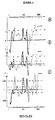
- De manière surprenante les résultats obtenus n'indiquent pas que le peptide I, s'étendant des résidus 1 à 20 du polypeptide Sm-D, comprend un épitope déterminant du polypeptide. Ces résultat sont rapportés à la figure 4.
- Les légendes des figures illustrant la description qui précèdent sont les suivantes :
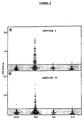
- La figure 1 représente la séquence déduite du polypeptide Sm-D selon le c-DNA.
- La figure 2 représente la liaison en ELISA des peptides I et IV. L'activité des IgG anti-peptide est mesurée sur 53 sérums de sujets en bonne santé (NHS), 165 sérums de patients atteints de Lupus Erythémateux disséminé (LED), 32 sérums de patients atteints de Sclerodermie (Scl) et 35 sérums de patients atteints d'Arthrite Rhumatoïde (RA). Tous les sérums ont été dilués au 1/1000 et le niveau d'anticorps est exprimé en unité de densité optique (OD) à 405 nm après hydrolyse du substrat pendant 60 minutes.
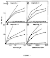
- La figure 3 représente la liaison en ELISA de 6 sérums de patients atteints de Lupus Erythémateux disséminé (l,n,s,m,r,s), et de 1 sérum de sujet en bonne santé (u), avec le peptide I (cadres A et C) et le peptide IV (cadres B et D). En A et B, les sérums sont dilués au 1/1000 et mis en contact avec diverses concentrations du peptide I; en B et D, diverses concentrations des sérums sont mises en contact respectivement avec 1 et 2 µM des peptides I et IV.
- La figure 4 représente les profiles de prédiction antigènique du polypeptide Sm-D construits selon les échelles suivantes :
- . en A : selon Parker et al. (1986),
- . en B : selon Hopp et Woods (1981),
- . en C : selon Karplus et Schultz (1985),
- L'ensemble des résultats énoncés dans les exemples précédents indique clairement que les peptides issus de l'antigène Sm-D sont particulièrement utiles pour la mise en oeuvre d'un test de diagnostic quantitatif d'une grande sensibilité du LED.
- Les observations recueillies sur le peptide I montrent que 59% des patients atteints de LED possèdent des anticorps du type IgG reconnaissant le peptide, alors que 6% des sérums de patients atteints d'une autre maladie autoimmune et moins de 4 % des sérums de sujets normaux réagissent avec le peptide I en ELISA. Le peptide I, éventuellement utilisé avec le peptide IV, constitue donc une sonde particulièrement efficace pour le diagnostic du Lupus Erythémateux Disséminé.
- Ce pourcentage de près de 60% est supérieur à ce qui a été décrit antérieurement avec des protéines purifiées.
- Billings, P.B., Allen, R.W., Jensen, F.C. and Hoch, S.O. 1982. Anti-RNP monoclonal antibodies derived from a mouse strain with lupus-like autoimmunity. J. Immunol. 128: 1176.
- Billings, P.B., Barton, J.R. and Hoch, S.O. 1985. A murine monoclonal antibody recognizes the 13,000 molecular weight polypeptide of the Sm small nuclear ribonucleoprotein complex. J. Immunol. 135: 428.
- Bringmann, P. and Lührmann, R. 1986. Purification of the individual snRNPs U1, U2, U5 and U4/U6 from HeLa cells and characterization of their protein constituents. The Embo J. 5: 3509.
- Brunel, C., Sri-Widada, J. and Jeanteur, P. 1985. snRNP's and scRNP's in eukaryotic cells. In Progr. Mol. Subcell. Biol. Vol.9. F.E. Hahn, D.J. Kopecko, W.E.G. Müller, eds. Springer Verlag (Berlin), p. 1-52.
- Combe, B., Rucheton, M., Graafland, H., Lussiez, V., Brunel, C. and Sany, J. 1989. Clinical significance of anti-RNP and anti-Sm autoantibodies as determined by immunoblotting and immunoprecipitation in sera from patients with connective tissue diseases. Clin. Exp. Immunol. 75: 18.
- Eisenberg, R.A., Dyer, K., Craven, S.Y., Fuller. C.R. and Yount, W.J. 1985. Subclass restriction and polyclonality of the systemic lupus erythematosus marker antibody anti-Sm. J. Clin. Invest. 75: 1270.
- Habets, W.J., Sillekens, P.T.G., Hoet, M.H., Schalken, J.A., Roebroek, A.J.M., Leunissen, J.A.M., Van de Ven, W.J.M. and Van Venrooij, W.J. 1987. Analysis of a cDNA clone expressing a human autoimmune antigen : Full-length sequence of the U2 small nuclear RNA-associated B'' antigen. Proc. Natl. Acad. Sci. USA 84: 2421.
- Hardin, J.A. 1986. The lupus autoantigens and the pathogenesis of systemic lupus erythematosus. Arthritis Rheum. 29: 457.
- Hoch, S.O. 1989. Application of protein blotting to the study of autoimmune disease. InProtein Blotting. Methodology, research and diagnostic applications. B.A. Baldo, E.R. Tovey, S. Karger AG, Basel, p. 140-164.
- Hopp, T.P. and Woods, K.R. 1981. Prediction of protein antigenic determinants from amino acid sequences. Proc. Natl. Acad. Sci. USA 78: 3824.
- Kaiser, E., Colescott, R.L., Bossinger, C.D. and Cook, P.I. 1970. Color test for detection of free terminal amino groups in the solid phase synthesis of peptides. Anal. Biochem. 34: 595.
- Karplus, P.A. and Schulz, G.E. 1985. Prediction of chain flexibility in proteins. Naturwissenschaften 72:212.
- Lerner, M.R. and Steitz, J.A., 1979. Antibodies to small nuclear RNAs complexed with proteins are produced by patients with systemic lupus erythematosus. Proc. Natl. Acad. Sci. USA 76: 5495.
- McCarty, G.A., Rice, J.R., Bembe, M.L. and Pisetsky. D.S. 1982. Independent expression of autoantibodies in systemic lupus erythematosus. J. Rheum 9: 691.
- Merrifield, R.B. 1963. Solid phase peptide synthesis. I. The synthesis of a tetrapeptide. J. Amer. Chem. Soc. 85: 2149.
- Morrow, J. and Isenberg, D.A., 1987. Systemic Lupus Erythematosus. In Autoimmune Rheumatic Diseases, chap. 3. Blackwell Scientific Publ., pp. 48.
- Muller, S., Plaué, S., Couppez, M. and Van Regenmortel, M.H.V. 1986. Comparison of different methods for localizing antigenic regions in histone H2A. Molec. Immunol. 23: 593.
- Muller, S., Bonnier. D., Thiry, M. and Van Regenmortel, M.H.V. 1989. Reactivity in systemic lupus erythematosus with synthetic core histone peptides. Int. Arch. Allergy Appl. Immunol. 89: 288.
- Muller, S., Briand, J.P. and Van Regenmortel, M.H.V., 1988. presence of antibodies to ubiquitin during the autoimmune response associated with systemic lupus erythematosus. Proc. Natl. Acad. Sci. USA 85: 8176.
- Parker, J.M.R., Guo, D. and Hodges, R.S., 1986. New hydrophilicity scale derived from high-performance liquid chromatography peptide retention data : Correlation of predicted surface residues witn antigenicity and X-ray-derived accessible sites. Biochemistry 25: 5425.
- Pettersson, I., Hinterberger, M., Mimori, T., Gottlieb, E. and Steitz, J.A., 1984. The structure of mammalian small nuclear ribonucleoproteins. J. Biol. Chem. 259: 5907.
- Plaué, S. and Briand, J.P., 1988. Solid-phase peptide synthesis. In Synthetic Polypeptides as Antigens In the series Laboratory Techniques in Biochemisty and Molecular Biology, vol. 19. R.H. Burdon and P.H. Van Knippenberg, eds.Elsevier, Amsterdam, p. 41-94.
- Plaué, S., Muller, S. and Van Regenmortel, M.H.V. 1989. A branched, synthetic octapeptide of ubiquinated histone H2A as target of autoantibodies. J. Exp. Med. 169: 1607.
- Reichlin, M. and Harley, J.B. 1987. ANA subsets in systemic lupus erythematosus. In Systemic Lupus Erythematosus. J.S. Smolen, C.C. Zielinski, eds. Springer Verlag (Berlin), p. 105-123.
- Reuter. R. and Lührmann. R. 1986. Immunization of mice with purified U1 small nuclear ribonucleoprotein (RNP) induces a pattern of antibody specificities characteristic of the anti-Sm and anti-RNP autoimmune response of patients with lupus erythematosus, as measured by monoclonal antibodies. Proc. Natl. Acad. Sci. USA 83: 8689.
- Rokeach, L.A., Haselby, J.A. and Hoch, S.O. 1988. Molecular cloning of a cDNA encoding the human Sm-D autoantigen. Proc. Natl. Acad. Sci. USA 85: 4832.
- Rokeach, L.A., Jannatipour, M., Haselby, J.A. and Hoch, S.O. 1989. Primary structure of a human small nuclear ribonucleoprotein polypeptide as deduced by cDNA analysis. J. Biol. Chem. 264: 5024.
- Sillekens, P.T.G., Habets W.J., Beijer, R.P. and Van Venrooij, W.J. 1987. cDNA cloning of the human U1 snRNA-associated A protein : extensive homology between U1 and U2 snRNP-specific proteins. The EMBO J. 6: 3841.
- Sillekens, P.T.G., Beijer, R.P., Habets, W.J. and Van Venrooij, W.J. 1989. Molecular cloning of the cDNA for the human U2 snRNA-specific A' protein. Nucl. Acids Res. 17: 1893.
- Stanford, D.R., Rohleder, A., Neiswanger, K. and Wieben, E.D. 1987. DNA sequence of a human Sm autoimmune antigen. J. Biol. Chem. 262: 9931.
- Tam, J.P., Heath, W.F. and Merrifield, R.B. 1983. SN2 deprotection of synthetic peptides with a low concentration of HF in dimethyl sulfide : evidence and application in peptide synthesis. J. Amer. Chem. Soc. 105: 6442.
- Tan, E.M., Chan, E.K.L., Sullivan, K.F. and Rubin, R.L. 1988. Antinuclear antibodies (ANAs) : Diagnostically specific immune markers and clues toward the understanding of systemic autoimmunity. Clin. Immunol. Immunopath. 47: 121.
- Tan, E.M. and Kunkel, H.G. 1966. Characteristics of a soluble nuclear antigen precipitating with sera of patients with systemic lupus erythematosus. J. Immunol. 96: 464.
- Theissen. H., Etzerodt, M., Reuter, R., Schneider, C.. Lottspeich, F., Argos, P., Lührmann, R. and Philipson, L. 1986. Cloning of the human cDNA for the U1 RNA-assosiated 70K protein. The EMBO J. 5: 3209.
- Tuaillon, N., Muller, S., Pasquali, J.L., Bordigoni, P., Youinou, P., and Van Regenmortel, M.H.V. 1990. Antibodies from patients with rheumatoid arthritis and juvenile chronic arthritis analyzed with core histone synthetic peptides. Int. Arch. All. Appl. Immunol. (in press).
- Van Regenmortel, M.H.V. and Daney de Marcillac,. G. 1988. An assesment of prediction methods for locating continuous epitopes in proteins. Immunol. Lett. 17: 95.
- Williams D.G., Charles, P.J. and Maini, R.N., 1988. Preparative isolation of p67, A, B, B' and D from nRNP/Sm and Sm antigens by reverse-phase chromatography. J. Immunol. Meth. 113: 25.
- Yamamoto, K., Miura, H., Moroi, Y., Yoshinoya, S., Goto, M., Nishioka, K. and Miyamoto, T. 1988. Isolation and characterization of a complementary DNA expressing human U1 small nuclear ribonucleoprotein C Polypeptide. J. Immunol. 140: 311.
Claims (7)
- Peptide capable de réagir avec des anticorps contre le polypeptide Sm-D présents dans un échantillon biologique d'un sujet atteint de Lupus Erythémateux Disséminé, ce peptide étant choisi parmi(1) un peptide de la séquence :
XMKLVRFLMKLSHETVTIELKZ (I)
dans laquelle,X représente un groupe NH2 libre, et Z représente soit un groupe OH libre soit un groupe peptidique choisi parmi les groupes de 1 à 5 aminoacides suivants : N, NG, NGT, NGTQ, NGTQV;et (2) un peptide de la séquence :
XKMTLKNREPVQLETLSIRGNRIRYZ (IV)
dans laquelle X représente, soit un groupe NH2 libre soit un groupe peptidique choisi parmi les groupes de 1 à 5 aminoacides suivants : V, AV, KAV, LKAV, KLKAV;
et Z représente, soit un groupe OH libre soit un groupe peptidique choisi parmi les groupes de 1 à 5 aminoacides suivants : F, FI, FIL, FILP, FILPD. - Anticorps dirigés contre l'antigène Sm-D, caractérisés en ce qu'ils sont reconnus par les peptides selon la revendication 1.
- Anticorps anti-idiotypes formés contre un anticorps selon la revendication 2, caractérisés en ce qu'ils reconnaissent, de manière spécifique, les autoanticorps présents dans des fluides biologiques, notamment des sérums, de patients atteints de Lupus Erythémateux Disséminé.
- Composition antigénique contenant au moins un peptide selon la revendication 1, caractérisée en ce qu'elle réagit immunologiquement avec les autoanticorps présents dans des fluides biologiques, notamment des sérums de patients atteints de Lupus Erythémateux Disséminé.
- Composition immunogène contenant au moins un peptide selon la revendication 1, ou un conjugué de ce peptide avec une molécule porteuse, caractérisée en ce qu'elle induit la production d'anticorps capables de reconnaître l'antigène Sm-D.
- Procédé de diagnostic in vivo du Lupus Erythémateux Disséminé dans un fluide biologique, caractérisé en ce qu'il comprend au moins les étapes suivantes :- la mise en contact de ce fluide biologique avec au moins un peptide selon la revendication 1, ou un conjugué de ce peptide avec une molécule porteuse, ou encore un anticorps antiidiotype selon la revendication 3, dans des conditions permettant la formation d'un complexe immunologique;- la détection de la présente d'un complexe immunologique antigène-anticorps ou d'un complexe anticorps-anticorps anti-idiotype, par des méthodes physiques ou chimiques, dans ledit fluide biologique.
- Kit pour le diagnostic in vitro du Lupus Erythémateux Disséminé, caractérisé en ce qu'il comprend:- au moins un peptide selon la revendication 1, ou un conjugué de ce peptide avec une molécule porteuse, ou encore un anticorps anti-idiotype selon la revendication 3;- des réactifs pour la constitution d'un milieu propice à la réaction immunologique entre les peptides ou les anticorps anti-idiotypes et les autoanticorps éventuellement présents dans un échantillon biologique;- un ou plusieurs réactifs éventuellement marqués aptes à réagir avec le ou les peptides ou les anticorps anti-idiotype pour la détection du complexe immunologique formé;- le cas échéant, un milieu biologique de référence, tel qu'un sérum d'un patient d'un sujet en bonne santé.
Applications Claiming Priority (3)
| Application Number | Priority Date | Filing Date | Title |
|---|---|---|---|
| FR9007029A FR2663034B1 (fr) | 1990-06-06 | 1990-06-06 | Peptides de l'antigene sm-d et leur utilisation notamment pour le diagnostic du lupus erythemateux dissemine. |
| FR9007029 | 1990-06-06 | ||
| PCT/FR1991/000445 WO1991018920A1 (fr) | 1990-06-06 | 1991-06-06 | PEPTIDES DE L'ANTIGENE Sm-D ET LEUR UTILISATION NOTAMMENT POUR LE DIAGNOSTIC DU LUPUS ERYTHEMATEUX DISSEMINE |
Publications (2)
| Publication Number | Publication Date |
|---|---|
| EP0491014A1 EP0491014A1 (fr) | 1992-06-24 |
| EP0491014B1 true EP0491014B1 (fr) | 1996-09-04 |
Family
ID=9397322
Family Applications (1)
| Application Number | Title | Priority Date | Filing Date |
|---|---|---|---|
| EP91911095A Expired - Lifetime EP0491014B1 (fr) | 1990-06-06 | 1991-06-06 | PEPTIDES DE L'ANTIGENE Sm-D ET LEUR UTILISATION NOTAMMENT POUR LE DIAGNOSTIC DU LUPUS ERYTHEMATEUX DISSEMINE |
Country Status (8)
| Country | Link |
|---|---|
| US (1) | US5407833A (fr) |
| EP (1) | EP0491014B1 (fr) |
| JP (1) | JPH05501267A (fr) |
| AT (1) | ATE142224T1 (fr) |
| CA (1) | CA2064576A1 (fr) |
| DE (1) | DE69121861D1 (fr) |
| FR (1) | FR2663034B1 (fr) |
| WO (1) | WO1991018920A1 (fr) |
Families Citing this family (8)
| Publication number | Priority date | Publication date | Assignee | Title |
|---|---|---|---|---|
| FR2707011A1 (en) * | 1993-06-25 | 1994-12-30 | Pasteur Sanofi Diagnostics | Immunological process for the detection and assay of antibodies directed against poly(ADP-ribose) polymerase, application to diagnosis, kit for its implementation |
| US7888458B1 (en) * | 1993-11-30 | 2011-02-15 | John B. Harley | Diagnostics and therapy of epstein-barr virus in autoimmune disorders |
| US6280944B1 (en) | 1994-05-25 | 2001-08-28 | Oklahoma Medical Research Foundation | Assay for pathogenicity of anti-DNA antibodies |
| EP0776906A3 (fr) * | 1995-10-19 | 1998-02-25 | Imtec Immundiagnostika Gmbh | Peptides de l'antigène Sm-D et leur utilisation notamment pour le diagnostic du lupus erythémateux disséminé |
| DE19619418C1 (de) * | 1995-10-19 | 1997-07-24 | Imtec Immundiagnostika Gmbh | Peptide des Antigens Sm-D und ihre Verwendung, insbesondere für die Diagnostik des SLE |
| US7273613B1 (en) | 1997-01-13 | 2007-09-25 | The Board of Regents, The University of Oklahoma | Diagnostics and therapy of Epstein-Barr virus in autoimmune disorders |
| ES2276474T3 (es) * | 1997-08-29 | 2007-06-16 | Innogenetics N.V. | Peptidos metilados, homologos del smd, que reaccionan con los anticuerpos de los sueros de seres vivos afectados con lupus eritematoso sistemico. |
| US6627607B2 (en) * | 2001-04-30 | 2003-09-30 | Syn X Pharma, Inc. | Biopolymer marker indicative of disease state having a molecular weight of 1845 daltons |
Family Cites Families (4)
| Publication number | Priority date | Publication date | Assignee | Title |
|---|---|---|---|---|
| US4654419A (en) * | 1984-08-08 | 1987-03-31 | Scripps Clinic And Research Foundation | Synthetic polypeptides and antibodies related to epstein-barr virus nuclear antigen |
| US4784942A (en) * | 1984-11-05 | 1988-11-15 | The Board Of Regents For The University Of Oklahoma | Monoclonal antibodies against autoimmune RNA proteins |
| US4629783A (en) * | 1985-04-29 | 1986-12-16 | Genetic Systems Corporation | Synthetic antigen for the detection of AIDS-related disease |
| JPH01257263A (ja) * | 1987-06-19 | 1989-10-13 | Agouron Inst:The | Sm−d抗原、sm−d抗原のクローニングとsm−d抗原使用による播種性エリテマトーデの検出 |
-
1990
- 1990-06-06 FR FR9007029A patent/FR2663034B1/fr not_active Expired - Fee Related
-
1991
- 1991-06-06 AT AT91911095T patent/ATE142224T1/de not_active IP Right Cessation
- 1991-06-06 US US07/828,853 patent/US5407833A/en not_active Expired - Fee Related
- 1991-06-06 DE DE69121861T patent/DE69121861D1/de not_active Expired - Lifetime
- 1991-06-06 CA CA002064576A patent/CA2064576A1/fr not_active Abandoned
- 1991-06-06 JP JP3510553A patent/JPH05501267A/ja active Pending
- 1991-06-06 WO PCT/FR1991/000445 patent/WO1991018920A1/fr active IP Right Grant
- 1991-06-06 EP EP91911095A patent/EP0491014B1/fr not_active Expired - Lifetime
Also Published As
| Publication number | Publication date |
|---|---|
| DE69121861D1 (de) | 1996-10-10 |
| US5407833A (en) | 1995-04-18 |
| FR2663034A1 (fr) | 1991-12-13 |
| FR2663034B1 (fr) | 1995-04-21 |
| WO1991018920A1 (fr) | 1991-12-12 |
| ATE142224T1 (de) | 1996-09-15 |
| EP0491014A1 (fr) | 1992-06-24 |
| JPH05501267A (ja) | 1993-03-11 |
| CA2064576A1 (fr) | 1991-12-07 |
Similar Documents
| Publication | Publication Date | Title |
|---|---|---|
| EP0423301B1 (fr) | Peptides synthetiques du conjugue de l'ubiquitine et de l'histone h2a | |
| JPH11507822A (ja) | aPL免疫応答性ペプチド、その結合体およびaPL抗体媒介病理のための処置方法 | |
| JP2009505049A (ja) | 関節リウマチにおいて形成される自己抗体を検出するための方法 | |
| JP2000512981A (ja) | aPL免疫応答性ペプチド、その結合体およびaPL抗体媒介病理のための処置方法 | |
| EP0929669B1 (fr) | Antigenes derives des filaggrines et leur utilisation pour le diagnostic de la polyarthrite rhumatoide | |
| JPS61501912A (ja) | 合成ポリペプチドにより誘発されたヒトインタ−ロイキン−2に対する抗体 | |
| US11098082B2 (en) | Antigenic peptides and uses thereof for diagnosing and treating autism | |
| US8323656B2 (en) | Antigen determinant of rheumatoid arthritis-specific autoantibody and use thereof | |
| KR19990087777A (ko) | 류마토이드 관절염 병원 관련 관절염성 펩티드에 대항하는 면역보호 유도에 유용한 백신 조성물과 방법 | |
| EP0491014B1 (fr) | PEPTIDES DE L'ANTIGENE Sm-D ET LEUR UTILISATION NOTAMMENT POUR LE DIAGNOSTIC DU LUPUS ERYTHEMATEUX DISSEMINE | |
| JP2000502070A (ja) | ヘリコバクター・ピロリによる感染の治療および診断 | |
| EP0750508B1 (fr) | RETROPEPTIDES, ANTICORPS DIRIGES CONTRE CES DERNIERS, ET LEURS UTILISATIONS POUR LA VACCINATION ET LE DIAGNOSTIC $i(IN VITRO) | |
| CN112457392B (zh) | 一种可溶性st2蛋白抗原决定簇多肽及其应用 | |
| EP3294901B1 (fr) | Auto-antigènes pour le diagnostic de la polyarthrite rhumatoïde | |
| CA2316269A1 (fr) | Epitopes peptidiques reconnus par des auto-anticorps antifilaggrine presents dans le serum des patients atteints de polyarthrite rhumatoide | |
| EP2026830A2 (fr) | Peptides modulateurs de l'activation des macrophages, utilisables pour le traitement de la polyarthrite rhumatoïde | |
| FR2682113A1 (fr) | Peptides issus de la proteine a de la ribonucleoproteine snrnp-u1 et leur utilisation pour le diagnostic de la connectivite mixte. | |
| Dyrberg et al. | Immunological cross-reactivity between mimicking epitopes on a virus protein and a human autoantigen depends on a single amino acid residue | |
| JP2744001B2 (ja) | 膵臓チモーゲンの遊離活性化ペプチドの免疫分析による診断、厳密な予測および監視 | |
| EP2086996B1 (fr) | Marqueur specifique d'une degradation oxydative de tissus contenant du collagene de type iii, moyens, methodes et kits de diagnostic, de suivi ou de pronostic des pathologies ciblees par ce marqueur | |
| EP3611183B1 (fr) | Épitope peptidique reconnaissant spécifiquement un auto-anticorps dirigé contre l'aquaporine 5, et son utilisation | |
| EP0494825A1 (fr) | Peptides synthétiques dérivant de l'antigène HBc du virus de l'hépatite B | |
| FR2826124A1 (fr) | Procede de detection d'auto-anticorps specifiques de la polyarthrite rhumatoide | |
| CA2459947A1 (fr) | Compositions et methodes pour le dosage de l'aa4rp |
Legal Events
| Date | Code | Title | Description |
|---|---|---|---|
| PUAI | Public reference made under article 153(3) epc to a published international application that has entered the european phase |
Free format text: ORIGINAL CODE: 0009012 |
|
| AK | Designated contracting states |
Kind code of ref document: A1 Designated state(s): AT BE CH DE DK ES FR GB GR IT LI LU NL SE |
|
| 17P | Request for examination filed |
Effective date: 19920612 |
|
| RIN1 | Information on inventor provided before grant (corrected) |
Inventor name: WEBER, JEAN-CHRISTOPHE Inventor name: BARAKAT, SAMIRA Inventor name: BRIAND, JEAN-PAUL Inventor name: MULLER, SYLVIANE Inventor name: VAN REGENMORTEL, MARC H.V. |
|
| RAP1 | Party data changed (applicant data changed or rights of an application transferred) |
Owner name: PASTEUR SANOFI DIAGNOSTICS |
|
| 17Q | First examination report despatched |
Effective date: 19940812 |
|
| GRAH | Despatch of communication of intention to grant a patent |
Free format text: ORIGINAL CODE: EPIDOS IGRA |
|
| GRAH | Despatch of communication of intention to grant a patent |
Free format text: ORIGINAL CODE: EPIDOS IGRA |
|
| GRAA | (expected) grant |
Free format text: ORIGINAL CODE: 0009210 |
|
| AK | Designated contracting states |
Kind code of ref document: B1 Designated state(s): AT BE CH DE DK ES FR GB GR IT LI LU NL SE |
|
| PG25 | Lapsed in a contracting state [announced via postgrant information from national office to epo] |
Ref country code: NL Free format text: LAPSE BECAUSE OF FAILURE TO SUBMIT A TRANSLATION OF THE DESCRIPTION OR TO PAY THE FEE WITHIN THE PRESCRIBED TIME-LIMIT Effective date: 19960904 Ref country code: GR Free format text: LAPSE BECAUSE OF FAILURE TO SUBMIT A TRANSLATION OF THE DESCRIPTION OR TO PAY THE FEE WITHIN THE PRESCRIBED TIME-LIMIT Effective date: 19960904 Ref country code: GB Effective date: 19960904 Ref country code: ES Free format text: THE PATENT HAS BEEN ANNULLED BY A DECISION OF A NATIONAL AUTHORITY Effective date: 19960904 Ref country code: DK Effective date: 19960904 Ref country code: AT Effective date: 19960904 |
|
| REF | Corresponds to: |
Ref document number: 142224 Country of ref document: AT Date of ref document: 19960915 Kind code of ref document: T |
|
| ITF | It: translation for a ep patent filed |
Owner name: JACOBACCI & PERANI S.P.A. |
|
| REF | Corresponds to: |
Ref document number: 69121861 Country of ref document: DE Date of ref document: 19961010 |
|
| PG25 | Lapsed in a contracting state [announced via postgrant information from national office to epo] |
Ref country code: SE Effective date: 19961204 |
|
| PG25 | Lapsed in a contracting state [announced via postgrant information from national office to epo] |
Ref country code: DE Effective date: 19961205 |
|
| NLV1 | Nl: lapsed or annulled due to failure to fulfill the requirements of art. 29p and 29m of the patents act | ||
| GBV | Gb: ep patent (uk) treated as always having been void in accordance with gb section 77(7)/1977 [no translation filed] |
Effective date: 19960904 |
|
| PG25 | Lapsed in a contracting state [announced via postgrant information from national office to epo] |
Ref country code: LU Free format text: LAPSE BECAUSE OF NON-PAYMENT OF DUE FEES Effective date: 19970630 Ref country code: LI Free format text: LAPSE BECAUSE OF NON-PAYMENT OF DUE FEES Effective date: 19970630 Ref country code: CH Free format text: LAPSE BECAUSE OF NON-PAYMENT OF DUE FEES Effective date: 19970630 Ref country code: BE Effective date: 19970630 |
|
| PLBE | No opposition filed within time limit |
Free format text: ORIGINAL CODE: 0009261 |
|
| STAA | Information on the status of an ep patent application or granted ep patent |
Free format text: STATUS: NO OPPOSITION FILED WITHIN TIME LIMIT |
|
| 26N | No opposition filed | ||
| BERE | Be: lapsed |
Owner name: PASTEUR SANOFI DIAGNOSTICS Effective date: 19970630 |
|
| REG | Reference to a national code |
Ref country code: CH Ref legal event code: PL |
|
| PG25 | Lapsed in a contracting state [announced via postgrant information from national office to epo] |
Ref country code: FR Free format text: LAPSE BECAUSE OF NON-PAYMENT OF DUE FEES Effective date: 19980227 |
|
| REG | Reference to a national code |
Ref country code: FR Ref legal event code: ST |
|
| REG | Reference to a national code |
Ref country code: FR Ref legal event code: ST |
|
| PG25 | Lapsed in a contracting state [announced via postgrant information from national office to epo] |
Ref country code: IT Free format text: LAPSE BECAUSE OF NON-PAYMENT OF DUE FEES Effective date: 20050606 |