EP0414223A2 - Méthode pour la mesure d'une matière d'activité immunologique et appareil adapté à ce procédé - Google Patents
Méthode pour la mesure d'une matière d'activité immunologique et appareil adapté à ce procédé Download PDFInfo
- Publication number
- EP0414223A2 EP0414223A2 EP90116090A EP90116090A EP0414223A2 EP 0414223 A2 EP0414223 A2 EP 0414223A2 EP 90116090 A EP90116090 A EP 90116090A EP 90116090 A EP90116090 A EP 90116090A EP 0414223 A2 EP0414223 A2 EP 0414223A2
- Authority
- EP
- European Patent Office
- Prior art keywords
- measuring
- dispersion
- fine particles
- cell
- specimen
- Prior art date
- Legal status (The legal status is an assumption and is not a legal conclusion. Google has not performed a legal analysis and makes no representation as to the accuracy of the status listed.)
- Granted
Links
- 238000000034 method Methods 0.000 title claims abstract description 137
- 239000011149 active material Substances 0.000 title claims abstract description 50
- 239000010419 fine particle Substances 0.000 claims abstract description 88
- 239000006185 dispersion Substances 0.000 claims abstract description 78
- 239000000463 material Substances 0.000 claims abstract description 58
- 239000011541 reaction mixture Substances 0.000 claims abstract description 54
- 239000007787 solid Substances 0.000 claims abstract description 52
- 239000007788 liquid Substances 0.000 claims abstract description 29
- 230000003100 immobilizing effect Effects 0.000 claims abstract description 10
- 239000003153 chemical reaction reagent Substances 0.000 claims description 128
- 238000003756 stirring Methods 0.000 claims description 102
- 238000006243 chemical reaction Methods 0.000 claims description 65
- 230000004520 agglutination Effects 0.000 claims description 53
- 239000002612 dispersion medium Substances 0.000 claims description 52
- 238000005259 measurement Methods 0.000 claims description 45
- 230000003287 optical effect Effects 0.000 claims description 44
- 239000002245 particle Substances 0.000 claims description 44
- 230000008569 process Effects 0.000 claims description 32
- 239000002609 medium Substances 0.000 claims description 22
- 239000000203 mixture Substances 0.000 claims description 10
- 238000007865 diluting Methods 0.000 claims description 5
- 239000003085 diluting agent Substances 0.000 claims description 5
- 210000004027 cell Anatomy 0.000 description 138
- 239000004816 latex Substances 0.000 description 68
- 229920000126 latex Polymers 0.000 description 68
- 239000000725 suspension Substances 0.000 description 34
- 239000000243 solution Substances 0.000 description 27
- 238000002474 experimental method Methods 0.000 description 21
- 206010001497 Agitation Diseases 0.000 description 20
- 238000013019 agitation Methods 0.000 description 20
- 102000011022 Chorionic Gonadotropin Human genes 0.000 description 18
- 108010062540 Chorionic Gonadotropin Proteins 0.000 description 18
- 229940084986 human chorionic gonadotropin Drugs 0.000 description 18
- 230000035945 sensitivity Effects 0.000 description 17
- IJGRMHOSHXDMSA-UHFFFAOYSA-N Atomic nitrogen Chemical compound N#N IJGRMHOSHXDMSA-UHFFFAOYSA-N 0.000 description 16
- 230000000052 comparative effect Effects 0.000 description 14
- 238000010790 dilution Methods 0.000 description 14
- 239000012895 dilution Substances 0.000 description 14
- 239000004793 Polystyrene Substances 0.000 description 13
- 229920002223 polystyrene Polymers 0.000 description 13
- 210000002966 serum Anatomy 0.000 description 13
- XLYOFNOQVPJJNP-UHFFFAOYSA-N water Substances O XLYOFNOQVPJJNP-UHFFFAOYSA-N 0.000 description 13
- 108091003079 Bovine Serum Albumin Proteins 0.000 description 12
- 229940098773 bovine serum albumin Drugs 0.000 description 12
- 150000001718 carbodiimides Chemical class 0.000 description 11
- 239000011521 glass Substances 0.000 description 11
- 238000012545 processing Methods 0.000 description 11
- CZMRCDWAGMRECN-UGDNZRGBSA-N Sucrose Chemical compound O[C@H]1[C@H](O)[C@@H](CO)O[C@@]1(CO)O[C@@H]1[C@H](O)[C@@H](O)[C@H](O)[C@@H](CO)O1 CZMRCDWAGMRECN-UGDNZRGBSA-N 0.000 description 10
- 229930006000 Sucrose Natural products 0.000 description 10
- 238000011156 evaluation Methods 0.000 description 10
- 230000002829 reductive effect Effects 0.000 description 10
- 239000005720 sucrose Substances 0.000 description 10
- 239000000872 buffer Substances 0.000 description 9
- 239000008363 phosphate buffer Substances 0.000 description 9
- 239000012086 standard solution Substances 0.000 description 9
- 238000003860 storage Methods 0.000 description 9
- 229920002684 Sepharose Polymers 0.000 description 8
- 239000000427 antigen Substances 0.000 description 8
- 102000036639 antigens Human genes 0.000 description 8
- 108091007433 antigens Proteins 0.000 description 8
- 239000007900 aqueous suspension Substances 0.000 description 8
- 230000008859 change Effects 0.000 description 8
- 238000004587 chromatography analysis Methods 0.000 description 8
- 230000018044 dehydration Effects 0.000 description 8
- 238000006297 dehydration reaction Methods 0.000 description 8
- 239000002270 dispersing agent Substances 0.000 description 8
- 229910052757 nitrogen Inorganic materials 0.000 description 8
- 238000002360 preparation method Methods 0.000 description 8
- 229920003051 synthetic elastomer Polymers 0.000 description 8
- 239000005061 synthetic rubber Substances 0.000 description 8
- 238000005406 washing Methods 0.000 description 8
- 239000000126 substance Substances 0.000 description 7
- -1 methacrylate ester Chemical class 0.000 description 6
- 229910019142 PO4 Inorganic materials 0.000 description 5
- 238000010521 absorption reaction Methods 0.000 description 5
- NBIIXXVUZAFLBC-UHFFFAOYSA-K phosphate Chemical compound [O-]P([O-])([O-])=O NBIIXXVUZAFLBC-UHFFFAOYSA-K 0.000 description 5
- 239000010452 phosphate Substances 0.000 description 5
- 239000002504 physiological saline solution Substances 0.000 description 5
- VYPSYNLAJGMNEJ-UHFFFAOYSA-N Silicium dioxide Chemical compound O=[Si]=O VYPSYNLAJGMNEJ-UHFFFAOYSA-N 0.000 description 4
- 238000002835 absorbance Methods 0.000 description 4
- 229920001577 copolymer Polymers 0.000 description 4
- 230000007423 decrease Effects 0.000 description 4
- 230000000694 effects Effects 0.000 description 4
- 230000006870 function Effects 0.000 description 4
- 239000012046 mixed solvent Substances 0.000 description 4
- 102000004169 proteins and genes Human genes 0.000 description 4
- 108090000623 proteins and genes Proteins 0.000 description 4
- 241000283973 Oryctolagus cuniculus Species 0.000 description 3
- 238000004458 analytical method Methods 0.000 description 3
- 239000003795 chemical substances by application Substances 0.000 description 3
- 238000009833 condensation Methods 0.000 description 3
- 230000005494 condensation Effects 0.000 description 3
- 238000001035 drying Methods 0.000 description 3
- 238000004108 freeze drying Methods 0.000 description 3
- 230000001900 immune effect Effects 0.000 description 3
- 239000010954 inorganic particle Substances 0.000 description 3
- 230000001678 irradiating effect Effects 0.000 description 3
- 238000004519 manufacturing process Methods 0.000 description 3
- 239000011146 organic particle Substances 0.000 description 3
- 229920000139 polyethylene terephthalate Polymers 0.000 description 3
- 239000005020 polyethylene terephthalate Substances 0.000 description 3
- 239000004094 surface-active agent Substances 0.000 description 3
- 238000012546 transfer Methods 0.000 description 3
- 239000004925 Acrylic resin Substances 0.000 description 2
- 229920000178 Acrylic resin Polymers 0.000 description 2
- 241000894006 Bacteria Species 0.000 description 2
- 241000283707 Capra Species 0.000 description 2
- 108010022366 Carcinoembryonic Antigen Proteins 0.000 description 2
- 102100025475 Carcinoembryonic antigen-related cell adhesion molecule 5 Human genes 0.000 description 2
- 108090000790 Enzymes Proteins 0.000 description 2
- 102000004190 Enzymes Human genes 0.000 description 2
- 102000012673 Follicle Stimulating Hormone Human genes 0.000 description 2
- 108010079345 Follicle Stimulating Hormone Proteins 0.000 description 2
- 102000009151 Luteinizing Hormone Human genes 0.000 description 2
- 108010073521 Luteinizing Hormone Proteins 0.000 description 2
- 102100035703 Prostatic acid phosphatase Human genes 0.000 description 2
- PPBRXRYQALVLMV-UHFFFAOYSA-N Styrene Chemical compound C=CC1=CC=CC=C1 PPBRXRYQALVLMV-UHFFFAOYSA-N 0.000 description 2
- DPXJVFZANSGRMM-UHFFFAOYSA-N acetic acid;2,3,4,5,6-pentahydroxyhexanal;sodium Chemical compound [Na].CC(O)=O.OCC(O)C(O)C(O)C(O)C=O DPXJVFZANSGRMM-UHFFFAOYSA-N 0.000 description 2
- 150000001298 alcohols Chemical class 0.000 description 2
- 125000003277 amino group Chemical group 0.000 description 2
- 230000008901 benefit Effects 0.000 description 2
- 239000001768 carboxy methyl cellulose Substances 0.000 description 2
- 230000001427 coherent effect Effects 0.000 description 2
- 150000001875 compounds Chemical class 0.000 description 2
- 229940028334 follicle stimulating hormone Drugs 0.000 description 2
- 239000007789 gas Substances 0.000 description 2
- 229910052736 halogen Inorganic materials 0.000 description 2
- 150000002367 halogens Chemical class 0.000 description 2
- NOESYZHRGYRDHS-UHFFFAOYSA-N insulin Chemical compound N1C(=O)C(NC(=O)C(CCC(N)=O)NC(=O)C(CCC(O)=O)NC(=O)C(C(C)C)NC(=O)C(NC(=O)CN)C(C)CC)CSSCC(C(NC(CO)C(=O)NC(CC(C)C)C(=O)NC(CC=2C=CC(O)=CC=2)C(=O)NC(CCC(N)=O)C(=O)NC(CC(C)C)C(=O)NC(CCC(O)=O)C(=O)NC(CC(N)=O)C(=O)NC(CC=2C=CC(O)=CC=2)C(=O)NC(CSSCC(NC(=O)C(C(C)C)NC(=O)C(CC(C)C)NC(=O)C(CC=2C=CC(O)=CC=2)NC(=O)C(CC(C)C)NC(=O)C(C)NC(=O)C(CCC(O)=O)NC(=O)C(C(C)C)NC(=O)C(CC(C)C)NC(=O)C(CC=2NC=NC=2)NC(=O)C(CO)NC(=O)CNC2=O)C(=O)NCC(=O)NC(CCC(O)=O)C(=O)NC(CCCNC(N)=N)C(=O)NCC(=O)NC(CC=3C=CC=CC=3)C(=O)NC(CC=3C=CC=CC=3)C(=O)NC(CC=3C=CC(O)=CC=3)C(=O)NC(C(C)O)C(=O)N3C(CCC3)C(=O)NC(CCCCN)C(=O)NC(C)C(O)=O)C(=O)NC(CC(N)=O)C(O)=O)=O)NC(=O)C(C(C)CC)NC(=O)C(CO)NC(=O)C(C(C)O)NC(=O)C1CSSCC2NC(=O)C(CC(C)C)NC(=O)C(NC(=O)C(CCC(N)=O)NC(=O)C(CC(N)=O)NC(=O)C(NC(=O)C(N)CC=1C=CC=CC=1)C(C)C)CC1=CN=CN1 NOESYZHRGYRDHS-UHFFFAOYSA-N 0.000 description 2
- 238000011835 investigation Methods 0.000 description 2
- 150000002576 ketones Chemical class 0.000 description 2
- 230000007774 longterm Effects 0.000 description 2
- 229940040129 luteinizing hormone Drugs 0.000 description 2
- 239000003960 organic solvent Substances 0.000 description 2
- 239000006179 pH buffering agent Substances 0.000 description 2
- 229920003229 poly(methyl methacrylate) Polymers 0.000 description 2
- 239000004926 polymethyl methacrylate Substances 0.000 description 2
- 108010043671 prostatic acid phosphatase Proteins 0.000 description 2
- LOUPRKONTZGTKE-LHHVKLHASA-N quinidine Chemical compound C([C@H]([C@H](C1)C=C)C2)C[N@@]1[C@H]2[C@@H](O)C1=CC=NC2=CC=C(OC)C=C21 LOUPRKONTZGTKE-LHHVKLHASA-N 0.000 description 2
- 230000009257 reactivity Effects 0.000 description 2
- 238000011160 research Methods 0.000 description 2
- 239000004065 semiconductor Substances 0.000 description 2
- 235000019812 sodium carboxymethyl cellulose Nutrition 0.000 description 2
- 229920001027 sodium carboxymethylcellulose Polymers 0.000 description 2
- 238000001179 sorption measurement Methods 0.000 description 2
- 238000012360 testing method Methods 0.000 description 2
- ZFXYFBGIUFBOJW-UHFFFAOYSA-N theophylline Chemical compound O=C1N(C)C(=O)N(C)C2=C1NC=N2 ZFXYFBGIUFBOJW-UHFFFAOYSA-N 0.000 description 2
- WFKWXMTUELFFGS-UHFFFAOYSA-N tungsten Chemical compound [W] WFKWXMTUELFFGS-UHFFFAOYSA-N 0.000 description 2
- 229910052721 tungsten Inorganic materials 0.000 description 2
- 239000010937 tungsten Substances 0.000 description 2
- 229920003169 water-soluble polymer Polymers 0.000 description 2
- OTLLEIBWKHEHGU-UHFFFAOYSA-N 2-[5-[[5-(6-aminopurin-9-yl)-3,4-dihydroxyoxolan-2-yl]methoxy]-3,4-dihydroxy-6-(hydroxymethyl)oxan-2-yl]oxy-3,5-dihydroxy-4-phosphonooxyhexanedioic acid Chemical compound C1=NC=2C(N)=NC=NC=2N1C(C(C1O)O)OC1COC1C(CO)OC(OC(C(O)C(OP(O)(O)=O)C(O)C(O)=O)C(O)=O)C(O)C1O OTLLEIBWKHEHGU-UHFFFAOYSA-N 0.000 description 1
- NLHHRLWOUZZQLW-UHFFFAOYSA-N Acrylonitrile Chemical compound C=CC#N NLHHRLWOUZZQLW-UHFFFAOYSA-N 0.000 description 1
- 102000004506 Blood Proteins Human genes 0.000 description 1
- 108010017384 Blood Proteins Proteins 0.000 description 1
- 241000193155 Clostridium botulinum Species 0.000 description 1
- 241000186227 Corynebacterium diphtheriae Species 0.000 description 1
- LTMHDMANZUZIPE-AMTYYWEZSA-N Digoxin Natural products O([C@H]1[C@H](C)O[C@H](O[C@@H]2C[C@@H]3[C@@](C)([C@@H]4[C@H]([C@]5(O)[C@](C)([C@H](O)C4)[C@H](C4=CC(=O)OC4)CC5)CC3)CC2)C[C@@H]1O)[C@H]1O[C@H](C)[C@@H](O[C@H]2O[C@@H](C)[C@H](O)[C@@H](O)C2)[C@@H](O)C1 LTMHDMANZUZIPE-AMTYYWEZSA-N 0.000 description 1
- 102000008857 Ferritin Human genes 0.000 description 1
- 108050000784 Ferritin Proteins 0.000 description 1
- 238000008416 Ferritin Methods 0.000 description 1
- 108010010803 Gelatin Proteins 0.000 description 1
- CEAZRRDELHUEMR-URQXQFDESA-N Gentamicin Chemical compound O1[C@H](C(C)NC)CC[C@@H](N)[C@H]1O[C@H]1[C@H](O)[C@@H](O[C@@H]2[C@@H]([C@@H](NC)[C@@](C)(O)CO2)O)[C@H](N)C[C@@H]1N CEAZRRDELHUEMR-URQXQFDESA-N 0.000 description 1
- 229930182566 Gentamicin Natural products 0.000 description 1
- 229920000663 Hydroxyethyl cellulose Polymers 0.000 description 1
- 239000004354 Hydroxyethyl cellulose Substances 0.000 description 1
- 108060003951 Immunoglobulin Proteins 0.000 description 1
- 102000004877 Insulin Human genes 0.000 description 1
- 108090001061 Insulin Proteins 0.000 description 1
- 241000204031 Mycoplasma Species 0.000 description 1
- 206010028980 Neoplasm Diseases 0.000 description 1
- 206010029719 Nonspecific reaction Diseases 0.000 description 1
- CXOFVDLJLONNDW-UHFFFAOYSA-N Phenytoin Chemical compound N1C(=O)NC(=O)C1(C=1C=CC=CC=1)C1=CC=CC=C1 CXOFVDLJLONNDW-UHFFFAOYSA-N 0.000 description 1
- 241000224016 Plasmodium Species 0.000 description 1
- 229920001744 Polyaldehyde Polymers 0.000 description 1
- 239000004698 Polyethylene Substances 0.000 description 1
- 229920001213 Polysorbate 20 Polymers 0.000 description 1
- 239000004372 Polyvinyl alcohol Substances 0.000 description 1
- 229920002125 Sokalan® Polymers 0.000 description 1
- 241001147693 Staphylococcus sp. Species 0.000 description 1
- 241000194022 Streptococcus sp. Species 0.000 description 1
- 229920007962 Styrene Methyl Methacrylate Polymers 0.000 description 1
- 241000223997 Toxoplasma gondii Species 0.000 description 1
- 241000589884 Treponema pallidum Species 0.000 description 1
- 241000224526 Trichomonas Species 0.000 description 1
- 241000223104 Trypanosoma Species 0.000 description 1
- XTXRWKRVRITETP-UHFFFAOYSA-N Vinyl acetate Chemical compound CC(=O)OC=C XTXRWKRVRITETP-UHFFFAOYSA-N 0.000 description 1
- BZHJMEDXRYGGRV-UHFFFAOYSA-N Vinyl chloride Chemical compound ClC=C BZHJMEDXRYGGRV-UHFFFAOYSA-N 0.000 description 1
- 125000005396 acrylic acid ester group Chemical group 0.000 description 1
- 239000000654 additive Substances 0.000 description 1
- 230000004931 aggregating effect Effects 0.000 description 1
- 125000003172 aldehyde group Chemical group 0.000 description 1
- 102000013529 alpha-Fetoproteins Human genes 0.000 description 1
- 108010026331 alpha-Fetoproteins Proteins 0.000 description 1
- PNEYBMLMFCGWSK-UHFFFAOYSA-N aluminium oxide Inorganic materials [O-2].[O-2].[O-2].[Al+3].[Al+3] PNEYBMLMFCGWSK-UHFFFAOYSA-N 0.000 description 1
- 239000003945 anionic surfactant Substances 0.000 description 1
- 239000003242 anti bacterial agent Substances 0.000 description 1
- 230000001088 anti-asthma Effects 0.000 description 1
- 230000003556 anti-epileptic effect Effects 0.000 description 1
- 239000000924 antiasthmatic agent Substances 0.000 description 1
- 229940088710 antibiotic agent Drugs 0.000 description 1
- 239000001961 anticonvulsive agent Substances 0.000 description 1
- 229960003965 antiepileptics Drugs 0.000 description 1
- 238000003556 assay Methods 0.000 description 1
- 239000000440 bentonite Substances 0.000 description 1
- 229910000278 bentonite Inorganic materials 0.000 description 1
- SVPXDRXYRYOSEX-UHFFFAOYSA-N bentoquatam Chemical compound O.O=[Si]=O.O=[Al]O[Al]=O SVPXDRXYRYOSEX-UHFFFAOYSA-N 0.000 description 1
- 230000005540 biological transmission Effects 0.000 description 1
- 125000003917 carbamoyl group Chemical group [H]N([H])C(*)=O 0.000 description 1
- 125000003178 carboxy group Chemical group [H]OC(*)=O 0.000 description 1
- 239000002327 cardiovascular agent Substances 0.000 description 1
- 229940125692 cardiovascular agent Drugs 0.000 description 1
- 229960005091 chloramphenicol Drugs 0.000 description 1
- WIIZWVCIJKGZOK-RKDXNWHRSA-N chloramphenicol Chemical compound ClC(Cl)C(=O)N[C@H](CO)[C@H](O)C1=CC=C([N+]([O-])=O)C=C1 WIIZWVCIJKGZOK-RKDXNWHRSA-N 0.000 description 1
- LOUPRKONTZGTKE-UHFFFAOYSA-N cinchonine Natural products C1C(C(C2)C=C)CCN2C1C(O)C1=CC=NC2=CC=C(OC)C=C21 LOUPRKONTZGTKE-UHFFFAOYSA-N 0.000 description 1
- 238000007796 conventional method Methods 0.000 description 1
- 238000005138 cryopreservation Methods 0.000 description 1
- ATDGTVJJHBUTRL-UHFFFAOYSA-N cyanogen bromide Chemical compound BrC#N ATDGTVJJHBUTRL-UHFFFAOYSA-N 0.000 description 1
- 230000003247 decreasing effect Effects 0.000 description 1
- 238000002405 diagnostic procedure Methods 0.000 description 1
- LTMHDMANZUZIPE-PUGKRICDSA-N digoxin Chemical compound C1[C@H](O)[C@H](O)[C@@H](C)O[C@H]1O[C@@H]1[C@@H](C)O[C@@H](O[C@@H]2[C@H](O[C@@H](O[C@@H]3C[C@@H]4[C@]([C@@H]5[C@H]([C@]6(CC[C@@H]([C@@]6(C)[C@H](O)C5)C=5COC(=O)C=5)O)CC4)(C)CC3)C[C@@H]2O)C)C[C@@H]1O LTMHDMANZUZIPE-PUGKRICDSA-N 0.000 description 1
- 229960005156 digoxin Drugs 0.000 description 1
- LTMHDMANZUZIPE-UHFFFAOYSA-N digoxine Natural products C1C(O)C(O)C(C)OC1OC1C(C)OC(OC2C(OC(OC3CC4C(C5C(C6(CCC(C6(C)C(O)C5)C=5COC(=O)C=5)O)CC4)(C)CC3)CC2O)C)CC1O LTMHDMANZUZIPE-UHFFFAOYSA-N 0.000 description 1
- 229940079593 drug Drugs 0.000 description 1
- 239000003814 drug Substances 0.000 description 1
- 125000003700 epoxy group Chemical group 0.000 description 1
- 210000003743 erythrocyte Anatomy 0.000 description 1
- 229940011871 estrogen Drugs 0.000 description 1
- 239000000262 estrogen Substances 0.000 description 1
- 239000002095 exotoxin Substances 0.000 description 1
- 231100000776 exotoxin Toxicity 0.000 description 1
- 239000012530 fluid Substances 0.000 description 1
- 125000000524 functional group Chemical group 0.000 description 1
- 229920000159 gelatin Polymers 0.000 description 1
- 239000008273 gelatin Substances 0.000 description 1
- 235000019322 gelatine Nutrition 0.000 description 1
- 235000011852 gelatine desserts Nutrition 0.000 description 1
- 229920001519 homopolymer Polymers 0.000 description 1
- 229940088597 hormone Drugs 0.000 description 1
- 239000005556 hormone Substances 0.000 description 1
- 125000002887 hydroxy group Chemical group [H]O* 0.000 description 1
- 235000019447 hydroxyethyl cellulose Nutrition 0.000 description 1
- 238000003018 immunoassay Methods 0.000 description 1
- 102000018358 immunoglobulin Human genes 0.000 description 1
- 229940072221 immunoglobulins Drugs 0.000 description 1
- 230000006698 induction Effects 0.000 description 1
- 230000002401 inhibitory effect Effects 0.000 description 1
- 229940125396 insulin Drugs 0.000 description 1
- ADFPJHOAARPYLP-UHFFFAOYSA-N methyl 2-methylprop-2-enoate;styrene Chemical compound COC(=O)C(C)=C.C=CC1=CC=CC=C1 ADFPJHOAARPYLP-UHFFFAOYSA-N 0.000 description 1
- 238000002156 mixing Methods 0.000 description 1
- 239000000178 monomer Substances 0.000 description 1
- 239000002736 nonionic surfactant Substances 0.000 description 1
- 229960002695 phenobarbital Drugs 0.000 description 1
- DDBREPKUVSBGFI-UHFFFAOYSA-N phenobarbital Chemical compound C=1C=CC=CC=1C1(CC)C(=O)NC(=O)NC1=O DDBREPKUVSBGFI-UHFFFAOYSA-N 0.000 description 1
- 229960002036 phenytoin Drugs 0.000 description 1
- 229920003023 plastic Polymers 0.000 description 1
- 239000004033 plastic Substances 0.000 description 1
- 229920002492 poly(sulfone) Polymers 0.000 description 1
- 229920002401 polyacrylamide Polymers 0.000 description 1
- 239000004584 polyacrylic acid Substances 0.000 description 1
- 239000004417 polycarbonate Substances 0.000 description 1
- 229920000515 polycarbonate Polymers 0.000 description 1
- 229920000573 polyethylene Polymers 0.000 description 1
- 239000000256 polyoxyethylene sorbitan monolaurate Substances 0.000 description 1
- 235000010486 polyoxyethylene sorbitan monolaurate Nutrition 0.000 description 1
- 229920002451 polyvinyl alcohol Polymers 0.000 description 1
- 235000019422 polyvinyl alcohol Nutrition 0.000 description 1
- 239000004800 polyvinyl chloride Substances 0.000 description 1
- 229920000915 polyvinyl chloride Polymers 0.000 description 1
- 229960001404 quinidine Drugs 0.000 description 1
- 230000009467 reduction Effects 0.000 description 1
- 239000000377 silicon dioxide Substances 0.000 description 1
- 239000002904 solvent Substances 0.000 description 1
- 230000002269 spontaneous effect Effects 0.000 description 1
- 229920003048 styrene butadiene rubber Polymers 0.000 description 1
- IMCGHZIGRANKHV-AJNGGQMLSA-N tert-butyl (3s,5s)-2-oxo-5-[(2s,4s)-5-oxo-4-propan-2-yloxolan-2-yl]-3-propan-2-ylpyrrolidine-1-carboxylate Chemical compound O1C(=O)[C@H](C(C)C)C[C@H]1[C@H]1N(C(=O)OC(C)(C)C)C(=O)[C@H](C(C)C)C1 IMCGHZIGRANKHV-AJNGGQMLSA-N 0.000 description 1
- 229960000278 theophylline Drugs 0.000 description 1
- 229940117958 vinyl acetate Drugs 0.000 description 1
- 125000000391 vinyl group Chemical group [H]C([*])=C([H])[H] 0.000 description 1
- 229920002554 vinyl polymer Polymers 0.000 description 1
- KAKZBPTYRLMSJV-UHFFFAOYSA-N vinyl-ethylene Natural products C=CC=C KAKZBPTYRLMSJV-UHFFFAOYSA-N 0.000 description 1
- 230000009385 viral infection Effects 0.000 description 1
Images


Classifications
-
- G—PHYSICS
- G01—MEASURING; TESTING
- G01N—INVESTIGATING OR ANALYSING MATERIALS BY DETERMINING THEIR CHEMICAL OR PHYSICAL PROPERTIES
- G01N33/00—Investigating or analysing materials by specific methods not covered by groups G01N1/00 - G01N31/00
- G01N33/48—Biological material, e.g. blood, urine; Haemocytometers
- G01N33/50—Chemical analysis of biological material, e.g. blood, urine; Testing involving biospecific ligand binding methods; Immunological testing
- G01N33/53—Immunoassay; Biospecific binding assay; Materials therefor
- G01N33/543—Immunoassay; Biospecific binding assay; Materials therefor with an insoluble carrier for immobilising immunochemicals
- G01N33/54313—Immunoassay; Biospecific binding assay; Materials therefor with an insoluble carrier for immobilising immunochemicals the carrier being characterised by its particulate form
-
- G—PHYSICS
- G01—MEASURING; TESTING
- G01N—INVESTIGATING OR ANALYSING MATERIALS BY DETERMINING THEIR CHEMICAL OR PHYSICAL PROPERTIES
- G01N21/00—Investigating or analysing materials by the use of optical means, i.e. using sub-millimetre waves, infrared, visible or ultraviolet light
- G01N21/75—Systems in which material is subjected to a chemical reaction, the progress or the result of the reaction being investigated
- G01N21/77—Systems in which material is subjected to a chemical reaction, the progress or the result of the reaction being investigated by observing the effect on a chemical indicator
- G01N21/82—Systems in which material is subjected to a chemical reaction, the progress or the result of the reaction being investigated by observing the effect on a chemical indicator producing a precipitate or turbidity
-
- G—PHYSICS
- G01—MEASURING; TESTING
- G01N—INVESTIGATING OR ANALYSING MATERIALS BY DETERMINING THEIR CHEMICAL OR PHYSICAL PROPERTIES
- G01N15/00—Investigating characteristics of particles; Investigating permeability, pore-volume or surface-area of porous materials
- G01N15/02—Investigating particle size or size distribution
- G01N15/0205—Investigating particle size or size distribution by optical means
-
- Y—GENERAL TAGGING OF NEW TECHNOLOGICAL DEVELOPMENTS; GENERAL TAGGING OF CROSS-SECTIONAL TECHNOLOGIES SPANNING OVER SEVERAL SECTIONS OF THE IPC; TECHNICAL SUBJECTS COVERED BY FORMER USPC CROSS-REFERENCE ART COLLECTIONS [XRACs] AND DIGESTS
- Y10—TECHNICAL SUBJECTS COVERED BY FORMER USPC
- Y10S—TECHNICAL SUBJECTS COVERED BY FORMER USPC CROSS-REFERENCE ART COLLECTIONS [XRACs] AND DIGESTS
- Y10S436/00—Chemistry: analytical and immunological testing
- Y10S436/805—Optical property
-
- Y—GENERAL TAGGING OF NEW TECHNOLOGICAL DEVELOPMENTS; GENERAL TAGGING OF CROSS-SECTIONAL TECHNOLOGIES SPANNING OVER SEVERAL SECTIONS OF THE IPC; TECHNICAL SUBJECTS COVERED BY FORMER USPC CROSS-REFERENCE ART COLLECTIONS [XRACs] AND DIGESTS
- Y10—TECHNICAL SUBJECTS COVERED BY FORMER USPC
- Y10S—TECHNICAL SUBJECTS COVERED BY FORMER USPC CROSS-REFERENCE ART COLLECTIONS [XRACs] AND DIGESTS
- Y10S436/00—Chemistry: analytical and immunological testing
- Y10S436/811—Test for named disease, body condition or organ function
- Y10S436/813—Cancer
-
- Y—GENERAL TAGGING OF NEW TECHNOLOGICAL DEVELOPMENTS; GENERAL TAGGING OF CROSS-SECTIONAL TECHNOLOGIES SPANNING OVER SEVERAL SECTIONS OF THE IPC; TECHNICAL SUBJECTS COVERED BY FORMER USPC CROSS-REFERENCE ART COLLECTIONS [XRACs] AND DIGESTS
- Y10—TECHNICAL SUBJECTS COVERED BY FORMER USPC
- Y10S—TECHNICAL SUBJECTS COVERED BY FORMER USPC CROSS-REFERENCE ART COLLECTIONS [XRACs] AND DIGESTS
- Y10S436/00—Chemistry: analytical and immunological testing
- Y10S436/811—Test for named disease, body condition or organ function
- Y10S436/814—Pregnancy
Definitions
- the present invention relates to a method and an apparatus for measuring an immunologically active material such as antigen or antibody contained in a specimen with the use of dehydrated solid fine particles (hereinafter referred to as "dry solid fine particles"). More particularly, the present invention relates a method and an apparatus for. optically measuring said immunologically active material wherein dry solid fine particles having an immunologically active material immobilized on their surfaces are used and the agglutination degree of a product resulted as a result of antigen-antibody reaction is optically measured.
- LAIA method A latex agglutination immunoassay method (LAIA method) was developed by J. M. Singer et al [see, Am. J. Med., 21888 (1956)].
- LAIA method a dispersion (latex reagent) obtained by dispensing an immunologically active material such as antibody being disposed on fine particles of polystyrene in a liquid medium such as water is effected with a material having a selective reactivity such as antibody to said immunologically active material to cause an agglutinated body and the agglutinated state of the resultant is observed by eyes to thereby recognize the presence of the material to be observed. Since then, there have been made various studies on this method. Although quantitative determination is difficult, said method of recognizing the presence of an objective material by observing the agglutinated state of such agglutinated body by eyes has been widely used since the method is simple and provides a result for a short period of time.
- A. Fature et al proposed a method of optically observing a change in the turbidity caused by agglutination reaction and performing quantitative determination of an objective material based on the dynamic analysis [see, Protides Biol Fluids, Proc. Colloq., 2589 (1972)].
- This method is however problematic that the values obtained will be greately varied because of unstableness of a latex reagent to be used and the method is not sufficient in the measuring sensitivity.
- the latex reagent used is of a state that solid fine particles are dispersed in a liquid dispersing medium and it is substantially unstable.
- the present invention makes it an object to eliminate the foregoing problems in the prior arts and to provide an improved immunologically measuring method which excels in the reproducibility of a measured value and makes it possible to quantitatively measure an immunologically active material contained in a specimen with a high accuracy.
- Another object of the present invention is to provide an improved immunologically measuring method which makes it possible to quantitatively measure an immunologically active material such as antigen, antibody, etc. contained in a specimen with an improved accuracy by utilizing antigen-antibody reaction wherein a specific dehydrated immune reagent is used.
- a further object of the present invention is to provide an improved immunologically measuring method which makes it possible to quantitatively measure an immunologically active material such as antigen, antibody, etc. contained in a specimen with an improved accuracy wherein a specific dehydrated immune reagent is used and the stirring upon preparing a dispersion of fine particles of said reagent by subjecting said fine particles to redispersion in a dispersing medium is properly controlled by optically observing the dispersed state of said fine particles, whereby causing agglutination reaction in a desirable state, and providing marked improvements in reproducibility and reliability of data obtained.
- an immunologically active material such as antigen, antibody, etc. contained in a specimen with an improved accuracy
- a specific dehydrated immune reagent is used and the stirring upon preparing a dispersion of fine particles of said reagent by subjecting said fine particles to redispersion in a dispersing medium is properly controlled by optically observing the dispersed state of said fine particles, whereby causing agglutination reaction in
- a further object of the present invention is to provide an improved immunologically measuring method which makes it possible to quantitatively measure an immunologically active material such as antigen, antibody, etc. contained in a specimen with an improved accuracy within a short period of time.
- a still further object of the present invention is to provide an apparatus suitable for practicing the foregoing immunologically measuring method.
- the present inventors have intensively investigated the problems concerning the conventional measuring methods with dried reagents. Consequently it has been elucidated that the deviation of measured values by the conventional methods is caused by the change in the redispersed state, along with the decreased measuring sensitivity because the binding between solid fine particles and immunologically active materials is apparently damaged by long-term stirring for redispersion and too strong agitating power.
- the present inventors have furthermore carried out the investigations on the basis of the above results. It has been demonstrated that an extremely great effect would be brought about on highly sensitive and stable measurement, by stirring a dispersion medium while optically measuring the dispersion state during the step of redispersion of dried reagents, followed by transfer to next process at the time when an appropriate dispersion is achieved.
- the present invention has been achieved as a result of further investigations based on the findings described above.
- the present invention includes a method for measuring an immunological by active material (hereinafter referred to as immunologically measuring method”), which covers the two embodiments described below, and two apparatuses suitable for practicing each of the two embodiment.
- the first embodiment of the immunologically measuring method relates to a method of optically measuring a degree of agglutination of a reaction mixture, produced by chemically or physically binding onto the surfaces of dehydrated solid particles a material immunologically active to a material to be measured in a sample, and reacting in a liquid medium the sample with the bound immunologically active material, which comprises the steps of:
- the second embodiment of the immunologically measuring method relates to a method of optically measuring a degree of agglutination of a reaction mixture, produced by chemically or physically binding a material immunologically active to a material to be measured in a sample onto the surfaces of dried solid fine particles, and reacting in a liquid medium and sample with the bound immunologically active material, which comprises the steps of:
- a typical example of the apparatus suitable for practicing the first embodiment of the immunologically measuring method according to the present invention is an apparatus for optically measuring a degree of agglutination of a reaction mixture, produced by chemically or physically immobilizing a material immunologically active to a material to be measured in a sample onto the surfaces of dried solid fine particles, and reacting in a liquid medium the sample with immobilized immunologically active material, comprising a means to fix the measuring cell; a means to introduce the dispersion medium into the measuring cell; a means to introduce the sample into the measuring cell; a means to stir the contents in the measuring cell; a means to optically measure the degree of agglutination of the contents in the measuring cell; a means to determine a dispersion state of the dried reagent fine particles in the dispersion medium, based on the optically measured data of the mixture of dried reagent fine particles, the dispersion medium and the sample, and to control continuation and termination of the stirring.
- a typical example of an apparatus suitable for practicing the second embodiment of the immunologically measuring method according to the present invention is an appartus for optically measuring a degree of agglutination of a reaction mixture, produced by chemically or physically immobilizing a material immunologically active to a material to be measured in a sample onto the surfaces of dried solid fine particles, and reacting in a liquid medium the sample with the immobilized immunologically active material
- said apparatus comprises a means to fix the reaction cell; a means to introduce a dispersion medium into the reaction cell; a means to introduce the sample in the reaction cell; a means to stir the contents in the reaction cell; a means to control continuation or termination of the stirring, based on the optically measured data obtained from the degree of agglutination of the dried reagent fine particles in the dispersion medium in the reaction cell; a means to fix a measuring cell to measure the degree of agglutination of the reaction mixture; a means to flow the reaction mixture into the measuring cell; a means to optically
- immunologically active materials in a sample such as antigen and antibody
- a sample such as antigen and antibody
- the dried immunoreagent is used, so that the following merits may be obtained concerning reagent storage, compared with the storage of conventional reagents dispersed in water. That is, spontaneous agglutination over time as is observed in the case of reagents dispersed in water may not occur because the present reagent is in dry state; temperature control in storage of such reagent may be relaxed, (on the other hand, the conventional reagents cannot be frozen, special care should be taken of their storage.); the present dried reagent may be stored for a long term owing to its stability.
- the dry immunoreagent with the advantages described above is used and a state of a dispersion medium containing the fine particles of the reagent and a sample is optically measured during a stirring process of the dispersion medium; furthermore by controlling the stirring competence, the subsequent agglutination is smoothly facilitated, whereby the reproducibility and reliability of the data to be obtained may be greatly enhanced.
- the process for stirring the reagent may be controlled to a minimum agitation time, so that the decrease in the sensitivity may be avoided and the measuring time may be shortened as well.
- the solid fine particles to be used in the invention include particles from organisms, inorganic particles and organic particles.
- the particles from organisms include, for example, bacteria through dispersion treatment into erythrocytes, including Staphylococcus sp., Streptococcus sp ., etc..
- the inorganic particles include, for example, silica, alumina, bentonite, etc..
- the organic particles include, for example, particles of homopolymer and/or copolymer of vinyl monomers such as styrene, vinylchloride, acrylonitrile, vinylacetate, acrylic acid ester, methacrylate ester, etc.; and particles of butadiene copolymer such as styrene-butadiene copolymer, methylmethacrylate-butadiene copolymer, etc..
- the size of any particle of particles from organisms, inorganic particles, and organic particles may preferably be in the range of 0.05 to 5 ⁇ m and more preferably in the range of 0.1 to 2 I lm.
- the dry reagent of a particle size less than 0.05 ⁇ m is hard to be dispersed, while the stability of a dispersed reagent of a particle size of more than 10 nm will be reduced.
- the immunologically active material immobilized onto the surfaces of the solid particles include immunoglobulins such as IgG, IgM, IgE, etc.; plasma proteins such as complements, CRP, ferritin, a1- microglobulin and 82-microglobulin and their respective antibodies; tumor markers such as a-fetoprotein, carcinoembryonic antigen (CEA), prostatic acid phosphatase (PAP), CA19-9, CA-125, etc.
- immunoglobulins such as IgG, IgM, IgE, etc.
- plasma proteins such as complements, CRP, ferritin, a1- microglobulin and 82-microglobulin and their respective antibodies
- tumor markers such as a-fetoprotein, carcinoembryonic antigen (CEA), prostatic acid phosphatase (PAP), CA19-9, CA-125, etc.
- hormones such as luteinizing hormone (LH), follicle stimulating hormone (FSH), human chorionic gonadotropin (hCG), estrogen, insulin, etc.; and their respective antibodies; substances associated with virus infection such as HBV related antigens (HBs, HBe and HBC), HIV, ATL, etc. and their respective antibodies; bacteria such as Corynebacterium diphtheriae, Clostridium botulinum , mycoplasma, Treponema pallidum , etc. and their respective antibodies; protozoa such as Toxoplasma gondii, Trichomonas leichmainiae, Trypanosoma, Plasmodium, etc.
- drugs including antiepileptics such as phenytoin and phenobarbital, cardiovascular agents such as quinidine and digoxin, antiasthmatics such as theophylline, antibiotics such as chloramphenicol, gentamycin, etc. and their respective antibodies; enzymes and exotoxin (for example, streptolysine O), and their respective antibodies.
- drugs including antiepileptics such as phenytoin and phenobarbital, cardiovascular agents such as quinidine and digoxin, antiasthmatics such as theophylline, antibiotics such as chloramphenicol, gentamycin, etc. and their respective antibodies; enzymes and exotoxin (for example, streptolysine O), and their respective antibodies.
- a substance which can induce antigen-antibody reaction with a material to be measured in a sample should be appropriately selected among them, depending on the sample type for use.
- hCG antibody hCG antibody, CRP antibody .8 2 -microglobulin antibody or a-fetoprotein may specifically be preferable.
- the technique for immobilizing immunologically active materials onto the surfaces of the solid fine particles may utilize physical adsorption or chemical bonding, but chemical bonding may be preferable in the present invention. That is, according to the present invention, the dry solid fine particles whoses surfaces being immobilized with immunologically active materials are dispersed together with a sample as an agglutination factor, by strong stirring. Therefore, the immunologically active materials may occasionally be released from the solid particles, if they are immobilized by the immobilization technique utilizing physical adsorption with weak bonding strength.
- the immobilization technique by chemical bonding is carried out by the known method comprising chemically binding the protein which is contained as a component in an immunologically active material, to an antibody (see Immobilization Enzymes, ed.
- an immunologically active material may be immobilized to the solid particles where a functional group such as amino group and carboxyl group is present (see Japanese Patent Publication No. 12966/1978 or Japanese Unexamined Patent Publication No. 52620/1978).
- an immunologically active material may be immobilized through covalent bonding to the latex particles containing carbamoyl group or amino group.
- an immunologically active material may be immobilized through covalent bonding to the solid particles containing hydroxyl group.
- An immunologically active material may be reacted directly with the solid particles containing epoxy group or aldehyde group and immobilized through covalent bonding thereto.
- Any bonding reaction described above for immobilizing an immunologically active material to solid fine particles may preferably be carried out in water or a mixed solvent of water with an organic solvent compatible with water, including alcohols, ketones, etc..
- buffers such as phosphate buffer-physiological saline, Tris-HCFI buffer, inactive proteins such as bovine serum albumin, etc., surfactants and the like, to the reaction system.
- the reaction solution preferably has pH of 6-10, more preferably pH of 7-9.
- the concentration of particles in the reaction solution may be 0.01-2.0 wt%, generally.
- a dried immunoreagent may be obtained by removing a dispersion medium to be used as a dispersant of the particles to which is bound the immunologically active material.
- the removal of the dispersion medium is advantageous to carry out the removal of the dispersion medium at 60 C or less, preferably at 30 * C or less.
- the specifically preferable embodiment for removal of the dispersion medium is exemplified by its removal by freeze-drying, so that the sensitivity of the immunoreagent may be maintained thereby constantly high.
- the introduction of the dried immunoreagent into a measuring cell may be carried out by a process comprising placing a given amount of the dispersion of particles to which are bound immunologically active materials, and subsequently drying the contents in the cell in the aforementioned manner in order to remove the dispersion medium, or by placing in a measuring cell, a given amount of the dried immunoreagent after removal of the dispersion medium.
- the measuring cell there may be used those made of materials such as transparent glass or plastics (for example, polystyrene, polymethylmethacrylate, polyvinyl chloride, polycarbonate, polysulfone).
- transparent glass or plastics for example, polystyrene, polymethylmethacrylate, polyvinyl chloride, polycarbonate, polysulfone.
- the dispersion medium for dispersing a dried immunoreagent in the measuring cell there may be used water or a mixed solvent comprising water and an organic solvent compatible with water such as alcohols, ketones, etc..
- water or a mixed solvent comprising water and an organic solvent compatible with water such as alcohols, ketones, etc.
- an organic solvent compatible with water such as alcohols, ketones, etc.
- To the dispersion medium may be added also pH buffering agents, proteins, surfactants, water-soluble polymer compounds, etc., optically.
- pH buffering agents may be added in order to adjust the solution to optimum pH for the reaction; for example, phosphate buffer and Tris-HCI buffer may be used. Proteins may be added for the purpose of preventing nonspecific reactions; for example, bovine serum albumin, gelatin, etc. may be used.
- Surfactants and water-soluble polymer compounds are effective as an auxiliary dispersant of a dried immunoreagent, for example, nonionic surfactants such as Tween 20, anionic surfactants, polyvinylalcohol, polyacrylamide, polyacrylic acid, hydroxyethylcellulose, etc. may be used. However, there additives may be used in the range without inhibiting the antigen-antibody agglutination.
- the dried immunoreagent may be optionally diluted with a dispersion medium, depending on the measuring subject.
- concentration of the solid may vary, depending on the type and size of a measuring cell to be used; generally, the concentration should be adjusted preferably to the range of 0.01-5 wt%, more preferably to the range of 0.05-2 wt%.
- a dispersion medium and a dried immunoreagent there may be selected appropriately a method comprising injecting a given amount of the dispersion medium in a measuring cell or a reaction cell containing the sample and the dried immunoreagent, and inserting a stirring device therein for stirring, or a method comprising shaking the measuring cell.
- the ultrasonic wave to be used for ultrasonic stirring may generally have a frequency of 15 kHz to 50 kHz, depending on the type and size of a measuring cell or a reaction cell.
- the degree of dispersion of an immunoreagent in a dispersion medium may be optically measured by using an optically measuring means, and there may be appropriately employed, for example, the method for measuring the intensity of transmitted light, the method for measuring the intensity of scattered light, the method for measuring each intensity of transmitted light and scattered light in combination.
- the intensity of light transmitting through a measuring cell or a reaction cell reduces as the dispersion is promoted, and it remains almost constant after a uniform state of dispersion is achieved.
- the present inventors have invented a method for determining a preferable dispersion state, on the basis of the results of the experiments described below.
- the method for determining the state is practiced by confirming that the relation between A and Ao is in the range defined in the following formula; A/Ao ⁇ 1.1,
- the experiments 1-(1) and 1-(2) are associated with the first embodiment of the immunologically measuring method according to the present invention; the experiments 2-(1) and 2-(2) are associated with the second embodiment of the immunologically measuring method according to the present invention.
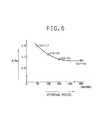
- Example 1-(1) A part of the CRP sensitized latex prepared in the same manner as in Example 1-(1) was adjusted of its solid concentration at 0.2% wt with phosphate buffer-physiological saline (referred to as PBS), pH 7.2, to which had been added 1% wt bovine serum albumin and 5% wt sucrose, and the resulting solution was placed in a glass optical cell (light pass length; 2mm). Then, the index Ao was determined according to the method described above to be 1.02 (wavelength for measurement; 633 nm). After adding PBS to the CRP-detecting dried reagent fine particles which were obtained also in the same method as in Example 1-(1), to adjust their solid concentration at 0.2% wt, CRP standard serum (2 mg/dl) was added. The resulting solution was subjected to ultrasonic agitation, to determine the index A according to the method described above. After termination of agitation, the CRP concentration was measured by the rate method described hereinafter. The results are shown in Table 1 and Fig. 5.
- the first embodiment of the immunologically measuring method of the present invention has been achieved based on the findings described hereinabove.
- an immunologically active material is physically and/or chemically immobilized to solid fine particles in water or a mixed solvent principally composed of water, so that the dispersant composition of the sensitized reagent latex suspension, after immobilization and before drying process, may be made identical to a dispersant composition to be used in redispersion of the dried reagent fine particles and then the value of Ao may be determdined on the basis of the results of optical measurement.
- the stirring process including the dried reagent and a sample according to the present invention
- the dispersion state was examined by the optical measurement described above while practicing the comparison between the obtained results of measurement of the dispersion state and the optical data demonstrating the predetermined dispersion state. Based on such results, the stirring process may be continued or terminated, or the stirring strength may be controlled.
- the immunologically active material reacts with the material to be measured, to induce antigen-antibody reaction and facilitate the agglutination, depending on the concentration of the material to be measured in the sample.
- stirring may be done by inserting a stirring device in a measuring cell or by shaking a measuring cell, as long as the agglutinated masses do not dissociate.
- the method for measuring the agglutination state by irradiating light to the reaction mixture in the measuring cell includes, for example, the method for measuring the intensity of transmitted light, the method for measuring the intensity of scattered light, the method in combination of these methods, and the method for measuring the density of integrating spheres.
- the concentration of the material to be measured in the sample is calculated through data processing of the measured data by using, for example, known methods such as the rate assay method, and the end point technique (Immunological Procedure VIII, Bunko-do 2401 (1979)).
- Example 2-(1) A part of the CRP sensitized latex prepared in the same manner as in Example 2-(1) was adjusted of its solid concentration at 0.2%wt with phosphate buffer-physiological saline (referred to as PBS), pH 7.5, to which were added 1%wt bovine serum albumin and 5%wt sucrose, and the resulting solution was placed in a glass optical cell (light pass length; 2 mm). Then, the index Ao was determined according to the method described above to be 2.75 (wavelength for measurement; 633 nm).
- PBS phosphate buffer-physiological saline
- the second embodiment of the immunologically measuring method of the present invention has been achieved based on the findings described hereinabove.
- an immunological active material is chemically or/and physically immobilized to solid fine particles in water or mixed solvent principally composed of water, so that the dispersant composition of the sensitized reagent latex suspension, after immobilization and before drying process, may be made identical to that of a dispersant to be used in redispersion of the dried reagent particles, and then the value of Ao may be determined on a basis of the results of optical measurement.
- the dispersion state is examined by the optical measurement described above while practicing the comparison between the results of measurement of the dispersion state obtained and the optical data demonstrating the predetermined dispersion states. Based on such results, the stirring process may be continued or terminated or the stirring competence may be controlled.
- the material to be measured reacts with the immunologically active material to induce antigen-antibody reaction to facilitate the agglutination, depending on the concentration of the material to be measured in the sample.
- the stirring may be done by inserting a stirring device in a measuring cell or by shaking a measuring cell, as long as the agglutinated masses do not dissociate.
- the reaction mixture withd agglutination facilitated in the reaction cell is diluted with the above dilution solution in a dilution cell.
- the concentration of the mixture should be adjusted to a concentration capable of transferring the agglutinated masses one by one in the subsequent process to introduce the resulting diluted reaction mixture into a flow cell.
- the agglutination state of the diluted reaction mixture is determined by sequentially measuring optical reactions caused by agglutinated masses being introduced one by one into the flow cell.
- optical reactions caused by agglutinated masses being introduced one by one into the flow cell.
- There may be preferably used, for example, flow cytometers of orthogonal optical axis-type and identical axis-type as disclosed in Diagnostic Test 30 , (11) 1259.
- the concentration of the material to be measured in the sample is calculated from the comparison of the agglutination sate of the diluted reaction mixture comprising the sample and the reagent, with a standard analytic curve which is preliminary prepared and which represents the relation between the concentration of the material to be measured and the agglutination state of the diluted reaction mixture after completion of reaction.
- the first embodiment and the second embodiment of the immunologically measuring method according to the present invention may be practiced by using an appropriate apparatus independently.
- the apparatus to be used for practicing the first embodiment of the immunologically measuring method according to the present invention is required at least to have the following constitution. That is, the apparatus has at least a means to fix a measuring cell, a means to introduce a dispersion medium into the measuring cell, a means to introduce a sample into the measuring cell, a means to stir the contents of the measuring cell with a stirring competence adjustable and a means to optically measure a degree of agglutination in the measuring cell. Furthermore, a means to automatically dilute a sample and a means to detect the presence of excess antigen (prozone phenomenon) may be added.
- FIG. 1 there is shown a representative apparatus suitable for practicing the first embodiment of the immunologically measuring method according to the present invention.
- numeral reference 2 stands for an optical cell made of acrylic resin or quartz glass which contains dehydrated latex reagent.
- Numeral reference 12 stands for a bar code of the optical data for dispersing said latex reagent into a dispersing medium which is disposed on the upper exterior of said optical cell 2.
- the optical cell 2 is placed in a constant temperature vessel 10 which is capable of serving as a holder therefor.
- the vessel 10 is equipped with a stirring means 11 including an ultrasonic vibrator capable of providing a vibration stirring function and a shaking means capable of providing a shaking stirring function.
- the optical data of the bar code 12 disposed on the exterior of the optical cell 2 is read by a bar code reading device 13.
- Numeral reference 8 stands for a reservoir containing a dispersing medium.
- the reservoir 8 is placed in a constant temperature vessel 7.
- a predetermined amount of the dispersing medium contained in the vessel 7 is introduced through a transporting pipe 19 equipped with a liquid supplying pump 17 into the optical cell 2.
- the dehydrated latex reagent and the dispersing medium contained in the optical cell 2 placed in the constant temperature vessel 10 are stirred by actuating the ultrasonic vibrator.
- Numeral reference 1 stands for a light source for radiating light for optical measurement.
- Numeral reference 6 stands for a half mirror. Beam of light from the light source 1 is supplied into the optical cell 2.
- the light source 1 in the case of radiating coherent light, there is used either He-Ne gas laser (wavelength: 632.8 nm) or semiconductor laser (wavelength: 780 nm or 830 nm). Other than these, it is possible use a tungsten lamp or a halogen lamp. In this case, an appropriate wavelength is selected by a monochrometer or a filter.
- the beam of light supplied into the optical cell 2 is dispersed or absorbed, and light transmitted through the cell is detected by a photomultiplier 3 and light scattered through the cell is detected by a photomultiplier 4. Variation in the light quantity for the light source is detected by a photomultiplier 5, and the signal detected by the photomultiplier 5 is transmitted to the data processing device 14. Likewise, the signal detected by the photomultiplier 3 and the signal detected by the photomultiplier 4 are transmitted to the data processing device 14.
- These signals transmitted to the data processing device 14 are entered through a A/D conversion circuit into a comparison circuit wherein they are compared with the optical data concerning the dispersion of the latex reagent from a memory circuit.
- the compared signal is transmitted to a control device 15 for the ultrasonic vibrator in the stirring means 11 to demand termination or continuation of the ultrasonic vibration stirring or to control the competence of the ultrasonic vibration stirring.
- a specimen containing a material to be measured which is contained in a container 9 is introduced through a transporting pipe 19 equipped with a liquid supplying pump 18 into the optical cell 2.
- the contents in the optical cell 2 is shake-stirred by actuating the shaking means in the stirring means 11 for a predetermined period of time (for example, for 3 to 5 seconds) and subjected to the optical measurement in the same manner as in the above case.
- the same optical measurement is again performed.
- the signals resulted by the twice optical measurements are transmitted to the data processing device in the same way as in the above case, wherein they are entered through the A/D conversion circuit into a measuring and computing circuit wherein they are computatively processed based on the analytic curve data previously inputted thereinto, to thereby obtain concentration data which are digitally indicated on a display 16.
- the apparatus to be used for practicing the second embodiment of the immunologically measuring method according to the present invention is required at least to have the following constitution. That is, the apparatus has at least a means to fix a reaction cell, a means to introduce a dispersion medium into the reaction cell, a means to introduce a sample into the reaction cell, a means to stir the contents of the reaction cell, a means to control continuation and termination of stirring, based on the optically measured data obtained from a dispersion state of the dried reagent particles in a dispersion medium, a means to fix a measuring cell to measure the degree of agglutination of a reaction mixture, a means to introduce the reaction mixture into the measuring cell, and a means to optically measure the degree of agglutination of individual aggregating particles of the reaction mixture poured into the measuring cell.
- the apparatus may be provided with a means to dilute the reaction mixture with a dilution solution before introducing the reaction mixture into the measuring cell.
- a means to detect a sample and a means to detect the presence of excess antigen (prozone phenomenon) may be added to such apparatus.
- FIG. 2 A specific example of a preferable apparatus is shown in FIG. 2.
- numeral reference 2 stands for an optical cell (reaction cell) made of acrylic resin or quartz glass which contains dehydrated latex reagent.
- Numeral reference 12 stands for a bar code which is disposed on the upper exterior of the reaction cell 2.
- the bar code 12 contains a code concerning the optical data for dispersing said latex reagent into a dispersing medium and a code for calling data of the reference standard analytic curve from a memory circuit.
- the reaction cell 2 is placed in a constant temperature vessel 10 which is capable of serving as a holder therefor.
- the vessel 10 is equipped with a stirring means 11 including an ultrasonic vibrator capable of providing a vibration stirring function and a shaking means capable of providing a shake-stirring function.
- Numeral reference 8 stands for a reservoir containing a dispersing medium.
- the reservoir 8 is placed in a constant temperature vessel 7.
- a predetermined amount of the dispersing medium contained in the vessel 7 is introduced through a transporting pipe 29 equipped with a liquid supplying pump 17 into the reaction cell 2.
- the dehydrated latex reagent and the dispersing medium contained in the reaction cell 2 placed in the constant temperature vessel 10 are stirred by actuating the ultrasonic vibrator.
- Numeral reference 1 stands for a light source for radiating light for optical measurement.
- Numeral reference 6 stands for a half mirror.
- Beam of light from the light source 1 is supplied into the reaction cell 2.
- the light source 1 in the case of radiating coherent light, there is used either He-Ne gas laser (wavelength: 632.8 nm) or semiconductor laser (wavelength: 780 nm or 830 nm). Other than these, it is possible use a tungsten lamp or a halogen lamp. In this case, an appropriate wavelength is selected by a monochrometer or a filter.
- the beam of light supplied into the reaction cell 2 is dispersed or absorbed, and light transmitted through the cell is detected by a photomultiplier 3 and light scattered through the cell is detected by a photomultiplier 4.
- Variation in the light quantity for the light source 1 is detected by a photomultiplier 5, and the signal detected by the photomultiplier 5 is transmitted to the data processing device 14. Likewise, the signal detected by the photomultiplier 3 and the signal detected by the photomultiplier 4 are transmitted to the data processing device 14.
- These signals transmitted to the data processing device 14 are entered through a A/D conversion circuit into a comparison circuit wherein they are compared with the optical data concerning the dispersion of the latex reagent from a memory circuit.
- the compared signal is transmitted to a control device 15 for the ultrasonic vibrator in the stirring means 11 to demand termination or continuation of the ultrasonic vibration stirring or to control the competence of the ultrasonic vibration stirring.
- a specimen containing a material to be measured which is contained in a container 9 is introduced through a transporting pipe 29 equipped with a liquid supplying pump 18 into the reaction cell 2.
- the contents in the reaction cell 2 is shake-stirred by actuating the shaking means in the stirring means 11 for a predetermined period of time (for example, for 3 to 5 seconds) to cause agglutination reaction.
- the reaction mixture caused in the reaction cell is sent to a dilution cell 20 placed in a constant temperature vessel 10 through a transporting pipe 29 equipped with a liquid supplying pump 19, in accordance with the conditions under which the foregoing reference standard analytic curve.
- a predetermined amount of a diluent contained in a reservoir 22 placed in a constant temperature vessel 21 is supplied into the dilution cell 20 through a transporting pipe 29 equipped with a liquid supplying pump 23.
- the reaction mixture and the diluent thus introduced into the dilution cell 20 are uniformly mixed by stirring them by a stirring means 24.
- the reaction mixture is diluted to a predetermined dilution degree.
- the admixture of the reaction mixture with the diluent in the dilution cell 20 may be performed by stirring them using the foregoing ultrasonic vibration stirring means or shake-stirring means. In this case, such stirring means is provided to the constant temperature vessel 10 (not shown).
- the reaction mixture thus diluted in the dilution cell is sent a flow cell 26 through a transporting pipe 29 equipped with liquid supplying pump 25.
- the diluted reaction mixture is flown such that each of the aggregates of the reaction mixture individually passes through the flow cell 26 and side-scattered light caused by radiating laser beam from a laser beam source when each of the aggregates passes through the flow cell 26 can be detected by a photomultiplier 28.
- the signals detected by the photomultiplier are transmitted to the data processing device 14, wherein they are entered through the A/D conversion circuit into a measuring and computing circuit wherein they are computatively processed based on the analytic curve data previously inputted thereinto, to thereby obtain concentration data which are digitally indicated on a display 16.
- Examples 1-(1) to 1-(4) described hereinafter are associated with the first embodiment of the present invention; Examples 2-(1) to 2-(4) described hereinafter are associated with the second embodiment of the present invention.
- PBS phosphate buffer-physiological saline
- PBS was added to 1.2 mg of the dried reagent fine particles placed in a glass optical cell (light pass length; 2 mm) to a final concentration of 2% as the solid reagent.
- the stirring process was terminated when the A determined in the above manner satisfied the formula A/Ao ⁇ 1.1 (A ⁇ 1.1) and the change in absorbanceAA was measured, 20 seconds and 200 seconds after the termination, by irradiating the light of 633 nm in wavelength.
- the measurement was repeated continuously ten times.
- the reagent lots, A, B and C, prepared independently on different days were used to carry out the measurement ten times each.
- Example 1-(1) The same procedure as in Example 1-(1) was performed to examine within-reproducibility and reproducibility among lots, except that the time for dispersion by ultrasonic bibration stirring was made constant during the dispersion process.
- Example 1-(1) and Comparative Example 1-(1) The results of the tests to examine within-reproducibility of CRP, carried out in Example 1-(1) and Comparative Example 1-(1), are shown in Table 2.
- the results of the tests to examine reproducibility within production lots of the reagent is shown in Table 3.
- Example 2 The results shown in Table 2 indicate that data deviation is smaller in Example 1-(1) where the stirring period of time for the latex reagent is made adjustable under control than in each case of fixed stirring period of time of 100, 200 or 300 seconds.
- Comparative Example 1 the change in absorbance, AA, becomes smaller as the agitation period gets longer; in other words, the sensitivity is lowered.
- bovine serum albumin sucrose and sodium carboxymethylcellulose, to final concentrations of 1%, 3% and 2%, respectively.
- PBS was also added to redisperse the latex to prepare a hCG antibody sensitized latex suspension.
- Example 1-(2) The same procedure as in Example 1-(2) was performed to calculate coefficients of variation in order to examine within-reproducibility (repetition of the measurement ten time), except that the period of time for ultrasonic vibration stirring was made constant at 100 seconds or 200 seconds.
- bovine serum albumin and sucrose were added to the sensitized latex after centrifuge and washing to final concentrations of 1% and 3%, respectively.
- PBS of pH 7.2 was also added to prepare an AFP antibody sensitized latex suspension.
- AFP standard solution 150 ug/ml
- the AFP standard solution was obtained by diluting Standard AFP Serum manufactured by Kyowa Yuka K.K. to the predetermined concentration with Tris-HCI buffer.
- the contents in the cell were immediately subjected to ultrasonic agitation, which was terminated at the time when the index A satisfied the formula; A/Ao ⁇ 1.1, [wherein the index Ao of 0.2% AFP sensitized latex suspension was preliminary measured to be 0.98 at a wavelength 633 nm:]; that is, A5 1.08.
- the stirring period of time during the process was 160 seconds.
- the change in absorbance, AA, 20 seconds and 200 seconds after the termination, was measured with the irradiation of light of 633 nm in wavelength. In order to examine within-reproducibility, the measurement was repeated ten times in total.
- Example 1-(3) The same procedure as in Example 1-(3) was performed to calculate coefficients of variation in order to examine within-reproducibility (repetition of the measurement ten times), except that the period of time for ultrasonic vibration stirring was made constant at 100 seconds or 200 seconds.
- bovine serum albumin and sucrose were added to the sensitized latex after centrifuge and washing to final concentrations of 1% and 3%, respectively.
- PBS of pH 7.2 was also added to prepare a p 2 - microblobulin antibody sensitized latex suspension.
- the ⁇ 2 -microglobulin standard solution was obtained by diluting Standard ⁇ 2 -microglobulin Serum manufactured by Kyowa Yuka K.K. to the predetermined concentration with Tris-HCI buffer.
- the contents in the cell were immediately subjected to ultrasonic agitation, which was terminated at the time when the index A satisfied the formula; A/Ao ⁇ 1.1, [wherein the absorption index Ao of 0.2% ⁇ 2 -microglobulin sensitized latex suspension was preliminary measured to be 1.04 at a wavelength 633 nm]; that is, A ⁇ 1.14.
- the stirring period of time during the process was 190 seconds.
- the change in absorbance, AA, 20 seconds and 200 seconds after the termination, was measured with the irradiation of light of 633 nm in wavelength. In order to examine within-reproducibility, the measurement was repeated ten times in total. As in Example 1-(1), the coefficient of variation (C.V.) was calculated in percentage (%).
- Example 1-(4) The same procedure as in Example 1-(4) was performed to calculate coefficients of variation in order to examine within-reproducibility (repetition of the measurement ten times), except that the time for ultrasonic vibration stirring was made constant at 100 seconds or 20 seconds.
- the 10 ml of an aqueous suspension of 10% carboxylated polystyrene of 0.71 nm in particle size (trade name: G0701; manufactured by Nippon Synthetic Rubber K.K.) was added 25 ml of an aqueous 1% 1-cyclohexyl-3-[2-morpholyl-(4)-ethyl] carbodiimide metho-p-toluenesulfonate (referred to as carbodiimide Ts, hereinafter) solution as a condensation agent and 20 ml of the antibody of the IgG fraction, and the resulting solution was then stirred at room temperature for three hours to obtain sensitized latex.
- carbodiimide Ts 1-cyclohexyl-3-[2-morpholyl-(4)-ethyl] carbodiimide metho-p-toluenesulfonate
- phosphate buffer-physiological saline of pH 7.2 (referred to as PBS hereinafter), which had been adjusted to contain 1% by weight of bovine serum albumin and 3% by weight of sucrose, was added to prepare a CRP antibody sensitized latex suspension.
- PBS was added to 1.2 mg of the dried reagent particles in a glass optical cell (light pass length; 2 mm) so that the concentration of the reagent as solid might be 0.2% by weight.
- the stirring process was terminated when the A satisfied the formula A/Ao5l.1 (A ⁇ 3.03), and 300 seconds later, the reaction mixture was diluted 500 fold with PBS and mixed together in a dilution cell (20- ml cell made of polyethylene terephalate).
- the diluted reaction mixture was injected into a flow cell and then, laser beam of 488 nm in wavelength irradiated the cell to measure the agglutination state of the particles in the diluted reaction mixture.
- the data were compared with the pre-measured data from a standard analytic curve, to determine the CRP concentration in a CRP sample.
- The. procedure was repeated ten times to examine the reproducibility.
- Example 2-(1) The same procedure as in Example 2-(1) was performed to examine reproducibility, except that the period of time for dispersion by ultrasonic vibration stirring was made constant during the dispersion process.
- Example 2-(1) The results obtained in Example 2-(1) and Comparative Example 2-(1) are shown in Table 6.
- the results obtained indicate that the measured values of the CRP concentration obtained by examining a dispersion state of the dried reagent and terminating the stirring when the dispersion state reached the predetermined state, approximate the real value (5.0 u.g/ml) with the smallest deviation.
- the period of time with respect to stirring treatment is fixed at 15 seconds, the dried reagent won't disperse sufficiently so that the reagent particles do not disperse singly; in other words, the particles are already present in agglutination state before reaction with CRP, which increase an apparent measured value.
- the stirring period fixed at 30 sec is almost identical to the stirring period of time as in Example 2-(1 but the deviation of the measured values of the CRP concentration is larger because the dispersion state is changeable on occasion.
- the stirring period is fixed at 300 sec, the dried reagent may disperse sufficiently, but the sensitivity gets lowered due to possible decrease in the antibody activity so that the measured values of CRP are smaller than the real value.
- bovine serum albumin sucrose and sodium carboxymethylcellulose, to final concentrations of 1%, 3% and 2%, respectively.
- PBS was also added to redisperse the latex to prepare a hCG antibody sensitized latex suspension.
- the hCG standard solution which was obtained by adjusting Standard manufactured by Nippon Chemical Research to a concentration of 10 IU/ml, was added into the cell.
- the contents in the cell were immediately subjected to ultrasonic agitation, which was terminated at the time when the index A satisfied the formula; A/Ao ⁇ 1.1, [wherein the absorption index Ao of 0.2% hCg sensitized latex suspension was preliminary measured to be 2.81 at a wavelength 633 nm]; that is, M 3.09.
- the stirring period of time during the process was 40 seconds. 300 seconds after the termination, the reaction mixture was diluted 500 fold with PBS and mixed together in a dilution cell (20-ml cell made of polyethylene terephthalate).
- the diluted reaction mixture was introduced into a flow cell, and then laser beam of 488 nm in wavelength irradiated the cell to measure the agglutination state of the particles in the diluted reaction mixture.
- the data were compared with the premeasured data from a standard analytic curve, to determine the hCG concentration in a hCG sample. The procedure was repeated ten times to examine the reproducibility.
- Example 2-(2) In order to examine reproducibility, the same procedure as in Example 2-(2) was performed to calculate coefficients of variation, except that the period of time for dispersion by ultrasonic vibration stirring was made constant (45 sec, 300 sec) during the dispersion process.
- bovine serum albumin and sucrose were added to the sensitized latex, after centrifuge and washing, to final concentrations of 1% and 3%, respectively.
- PBS, pH 7.2 was also added to prepare an AFP antibody sensitized latex suspension.
- AFP standard solution 50 ⁇ g/ml was added into the cell.
- the AFP standard solution was obtained by diluting Standard AFP Serum manufactured by Kyowa Yuka K.K. to a predetermined concentration with Tris-HCI buffer.
- the contents in the cell were immediately subjected to ultrasonic agitation, which was terminated at the time when the absorption index A satisfied the formula; A/Ao- ⁇ 1.1, [wherein the absorption index Ao of 0.2% AFP sensitized latex suspension was preliminary measured to be 2.71 at a wavelength 633 nm]; that is, AS2.98.
- the stirring period of time during the process was 30 seconds.
- the reaction mixture was diluted 500 fold with PBS and mixed together in a dilution cell (20-ml cell made of polyethylene terephthalate).
- the diluted reaction mixture was introduced into a flow cell, and then laser beam of 488 nm in wavelength irradiated the cell to measure the agglutination state of the particles in the diluted reaction mixture.
- the data were compared with the premeasured data from a standard analytic curve, to determine the AFP concentration in an AFP sample. The procedure was repeated ten times to examine the reproducibility.
- Example 2-(3) In order to examine reproducibility (repetition of the measurement ten times), the same procedure as in Example 2-(3) was performed to calculate coefficients of variation, except that the time for dispersion by ultrasonics was made constant at 30 sec or 300 sec during the dispersion process.
- bovine serum albumin and sucrose were added to the sensitized latex, after centrifuge and washing, to final concentrations of 1%, 3%, respectively.
- PBS of pH 5.5 was also added to prepare a .62-microglobulin antibody sensitized latex suspension.
- MG sample 100 ⁇ l of the 62-microglobulin standard solution, of 5 ag/ml (referred to as MG sample) was added into the cell (The MG sample was obtained by diluting Standard ⁇ 2 -microglobulin Serum manufactured by Kyowa Yuka K.K. to the predetermined concentration with Tris-HCI buffer.) The contents in the cell were immediately subjected to ultrasonic agitation, which was terminated at the time when the abosorption index A satisfied the formula; A/Ao ⁇ 1.1, [wherein the absorption index Ao of 0.2% ⁇ 2 - microglobulin sensitized latex suspension was preliminary measured to be 2.80 at a wavelength 633 nm]; that is, A ⁇ 3.06.
- the stirring period of time during the process was 40 seconds. Subsequently 300 seconds after the termination, the reaction mixture was diluted 500 fold with PBS and mixed together in a dilution cell (20-ml cell made of polyethylene terephthalate). The diluted reaction mixture was introduced into a flow cell, and then laser beam of 488 nm in wavelength irradiated the cell to measure the agglutination state of the particles in the diluted reaction mixture. The data were compared with the premeasured data from a standard curve, to determine the 6 2 -microglobulin concentration in a MG sample. The procedure was repeated ten times to examine the reproducibility.
- Example 4 The same procedure as in Example 4 was performed to calculate coefficients of variation in order to examine reproducibility (repetition of the measurement ten times), except that the stirring period of time for dispersion by ultrasonic vibration stirring was made constant at 35 sec or 300 sec during the dispersion process.
- a method and an apparatus for measuring an immunologically active material by physically or chemically immobilizing material immunologically active to a material to be measured of a specimen to the dehydrated solid fine particles, providing a desirable dispersion comprising said immunologically active material immobilized to said solid fine particles and said specimen in a liquid medium, reacting them to cause a reaction mixture in an agglutinated state and optically measuring said agglutinated state of the reaction mixture to thereby quantitatively determine the content of said material to be measured with an improved accuracy.
Landscapes
- Health & Medical Sciences (AREA)
- Immunology (AREA)
- Life Sciences & Earth Sciences (AREA)
- Engineering & Computer Science (AREA)
- Chemical & Material Sciences (AREA)
- Physics & Mathematics (AREA)
- Biomedical Technology (AREA)
- Pathology (AREA)
- Analytical Chemistry (AREA)
- Hematology (AREA)
- General Physics & Mathematics (AREA)
- Molecular Biology (AREA)
- Urology & Nephrology (AREA)
- General Health & Medical Sciences (AREA)
- Biochemistry (AREA)
- Cell Biology (AREA)
- Medicinal Chemistry (AREA)
- Food Science & Technology (AREA)
- Microbiology (AREA)
- Biotechnology (AREA)
- Chemical Kinetics & Catalysis (AREA)
- Plasma & Fusion (AREA)
- Medicines Containing Antibodies Or Antigens For Use As Internal Diagnostic Agents (AREA)
- Measuring Or Testing Involving Enzymes Or Micro-Organisms (AREA)
Applications Claiming Priority (6)
| Application Number | Priority Date | Filing Date | Title |
|---|---|---|---|
| JP21488989 | 1989-08-23 | ||
| JP214889/89 | 1989-08-23 | ||
| JP184527/90 | 1990-07-12 | ||
| JP18452790A JPH03156371A (ja) | 1989-08-23 | 1990-07-12 | 免疫的に活性な物質の測定方法および測定装置 |
| JP185681/90 | 1990-07-13 | ||
| JP18568190A JPH0472568A (ja) | 1990-07-13 | 1990-07-13 | 免疫的に活性な物質の測定方法および測定装置 |
Publications (3)
| Publication Number | Publication Date |
|---|---|
| EP0414223A2 true EP0414223A2 (fr) | 1991-02-27 |
| EP0414223A3 EP0414223A3 (en) | 1992-11-25 |
| EP0414223B1 EP0414223B1 (fr) | 1996-06-26 |
Family
ID=27325438
Family Applications (1)
| Application Number | Title | Priority Date | Filing Date |
|---|---|---|---|
| EP90116090A Expired - Lifetime EP0414223B1 (fr) | 1989-08-23 | 1990-08-22 | Méthode pour la mesure d'une matière d'activité immunologique et appareil adapté à ce procédé |
Country Status (6)
| Country | Link |
|---|---|
| US (1) | US5679581A (fr) |
| EP (1) | EP0414223B1 (fr) |
| AT (1) | ATE139845T1 (fr) |
| AU (1) | AU643455B2 (fr) |
| CA (1) | CA2023803C (fr) |
| DE (1) | DE69027560T2 (fr) |
Cited By (5)
| Publication number | Priority date | Publication date | Assignee | Title |
|---|---|---|---|---|
| WO1994020856A1 (fr) * | 1993-03-09 | 1994-09-15 | Middlesex Sciences, Inc. | Microparticules macromoleculaires et procedes de preparation |
| US5554730A (en) * | 1993-03-09 | 1996-09-10 | Middlesex Sciences, Inc. | Method and kit for making a polysaccharide-protein conjugate |
| GB2426050A (en) * | 2005-05-09 | 2006-11-15 | Orion Diagnostica Oy | Method of measuring binding rate using sonication |
| EP2836820A1 (fr) * | 2013-01-14 | 2015-02-18 | Alcor Scientific, Inc. | Appareil, procédé et système pour la détermination du taux d'agrégation de globules rouges |
| US9279800B2 (en) | 2012-01-13 | 2016-03-08 | Alcor Scientific, Inc. | Apparatus, method, system for the determination of the aggregation rate of red blood cells |
Families Citing this family (15)
| Publication number | Priority date | Publication date | Assignee | Title |
|---|---|---|---|---|
| ATE139341T1 (de) * | 1989-08-23 | 1996-06-15 | Canon Kk | Methode zur messung eines immunologisch aktiven materials und dazu geeignete vorrichtung |
| DE69624920T2 (de) | 1996-09-27 | 2003-09-18 | Inverness Medical Switzerland Gmbh, Zug | Testreagentien und Testvorrichtungen |
| JP3704035B2 (ja) * | 2000-08-31 | 2005-10-05 | 株式会社日立製作所 | 自動分析装置 |
| EP1256793A1 (fr) * | 2001-05-11 | 2002-11-13 | WTW Wissenschaftlich-Technische Werkstätten GmbH & Co. KG | Dispositif pour la mesure optique dans un milieu |
| GB0229747D0 (en) * | 2002-12-20 | 2003-01-29 | Axis Shield Asa | Assay |
| JP4632400B2 (ja) * | 2003-12-16 | 2011-02-16 | キヤノン株式会社 | 細胞培養用基板、その製造方法、それを用いた細胞スクリーニング法 |
| US7419820B2 (en) * | 2003-12-16 | 2008-09-02 | Canon Kabushiki Kaisha | Transfer sheet for transferring biologically active substance to culture plate |
| JP4541731B2 (ja) * | 2004-03-12 | 2010-09-08 | キヤノン株式会社 | 核酸検出方法 |
| JP4515162B2 (ja) * | 2004-06-15 | 2010-07-28 | キヤノン株式会社 | Dnaインターカレート物質類の吸着用構造物 |
| JP4147235B2 (ja) * | 2004-09-27 | 2008-09-10 | キヤノン株式会社 | 吐出用液体、吐出方法、液滴化方法、液体吐出カートリッジ及び吐出装置 |
| JP4689340B2 (ja) * | 2005-05-02 | 2011-05-25 | キヤノン株式会社 | 吐出用液体医薬組成物 |
| JP2007010413A (ja) * | 2005-06-29 | 2007-01-18 | Canon Inc | 核酸ハイブリッドの融点測定方法及びそのための装置 |
| US7642086B2 (en) * | 2005-08-09 | 2010-01-05 | Canon Kabushiki Kaisha | Labeled probe bound object, method for producing the same and method for using the same |
| RU2350957C1 (ru) * | 2007-12-28 | 2009-03-27 | Институт аналитического приборостроения Российской академии наук | Устройство для иммунологических исследований биологических жидкостей |
| WO2010074326A1 (fr) | 2008-12-25 | 2010-07-01 | Canon Kabushiki Kaisha | Sonde pour échantillon biologique, procédé de marquage et procédé de criblage utilisant la sonde |
Citations (3)
| Publication number | Priority date | Publication date | Assignee | Title |
|---|---|---|---|---|
| EP0123403A2 (fr) * | 1983-03-15 | 1984-10-31 | Boots-Celltech Diagnostics Limited | Examen de liaison en phase hétérogène |
| EP0193208A2 (fr) * | 1985-02-28 | 1986-09-03 | TECHNICON INSTRUMENTS CORPORATION(a Delaware corporation) | Lyophilisation de particules revêtues de réactifs |
| EP0368624A2 (fr) * | 1988-11-09 | 1990-05-16 | Biotrack, Inc. | Procédé et dispositif pour stabiliser et solubiliser des réactifs au latex |
Family Cites Families (8)
| Publication number | Priority date | Publication date | Assignee | Title |
|---|---|---|---|---|
| CH625626A5 (en) * | 1976-03-25 | 1981-09-30 | Hoffmann La Roche | Process for the preparation of stable immunological reagents |
| JPS5854009B2 (ja) * | 1976-07-22 | 1983-12-02 | 日本ユニカ−株式会社 | 段階的に収縮する樹脂管の製造方法 |
| US4092114A (en) * | 1976-10-20 | 1978-05-30 | Fisher Scientific Company | Indirect latex test for determination of immunoglobulins |
| US4205954A (en) * | 1978-05-26 | 1980-06-03 | Warner-Lambert Company | Kinetic latex agglutinometry |
| US4419453A (en) * | 1981-09-28 | 1983-12-06 | The Dow Chemical Company | Immunological agglutination assays with dyed or colored latex and kits |
| JPS5873866A (ja) * | 1981-10-28 | 1983-05-04 | Toray Ind Inc | 免疫学的検査方法 |
| EP0189389B1 (fr) * | 1985-01-24 | 1989-04-05 | Institut International De Pathologie Cellulaire Et Moleculaire | Procédé de dosage immunologique d'anticorps de diverses classes dans un échantillon liquide |
| JPS6246262A (ja) * | 1985-08-23 | 1987-02-28 | Eiken Kagaku Kk | 妊娠診断用乾燥試薬 |
-
1990
- 1990-08-22 DE DE69027560T patent/DE69027560T2/de not_active Expired - Fee Related
- 1990-08-22 AU AU61232/90A patent/AU643455B2/en not_active Ceased
- 1990-08-22 AT AT90116090T patent/ATE139845T1/de not_active IP Right Cessation
- 1990-08-22 CA CA002023803A patent/CA2023803C/fr not_active Expired - Lifetime
- 1990-08-22 EP EP90116090A patent/EP0414223B1/fr not_active Expired - Lifetime
-
1995
- 1995-01-13 US US08/372,877 patent/US5679581A/en not_active Expired - Lifetime
Patent Citations (3)
| Publication number | Priority date | Publication date | Assignee | Title |
|---|---|---|---|---|
| EP0123403A2 (fr) * | 1983-03-15 | 1984-10-31 | Boots-Celltech Diagnostics Limited | Examen de liaison en phase hétérogène |
| EP0193208A2 (fr) * | 1985-02-28 | 1986-09-03 | TECHNICON INSTRUMENTS CORPORATION(a Delaware corporation) | Lyophilisation de particules revêtues de réactifs |
| EP0368624A2 (fr) * | 1988-11-09 | 1990-05-16 | Biotrack, Inc. | Procédé et dispositif pour stabiliser et solubiliser des réactifs au latex |
Cited By (11)
| Publication number | Priority date | Publication date | Assignee | Title |
|---|---|---|---|---|
| WO1994020856A1 (fr) * | 1993-03-09 | 1994-09-15 | Middlesex Sciences, Inc. | Microparticules macromoleculaires et procedes de preparation |
| US5554730A (en) * | 1993-03-09 | 1996-09-10 | Middlesex Sciences, Inc. | Method and kit for making a polysaccharide-protein conjugate |
| US5578709A (en) * | 1993-03-09 | 1996-11-26 | Middlesex Sciences, Inc. | Macromolecular microparticles and methods of production |
| EP0809110A1 (fr) * | 1993-03-09 | 1997-11-26 | Middlesex Sciences, Inc. | Microparticules macromoléculaires et procédés de préparation |
| GB2426050A (en) * | 2005-05-09 | 2006-11-15 | Orion Diagnostica Oy | Method of measuring binding rate using sonication |
| US8133693B2 (en) | 2005-05-09 | 2012-03-13 | Orion Diagnostica Oy | Measurement of binding rate of a binding substance and an analyte |
| US9279800B2 (en) | 2012-01-13 | 2016-03-08 | Alcor Scientific, Inc. | Apparatus, method, system for the determination of the aggregation rate of red blood cells |
| US10488396B2 (en) | 2012-01-13 | 2019-11-26 | Alcor Scientific, Inc. | Apparatus, method, system for the determination of the aggregation rate of red blood cells |
| US11016081B2 (en) | 2012-01-13 | 2021-05-25 | Alcor Scientific Inc. | Apparatus, method, system for the determination of the aggregation rate of red blood cells |
| EP2836820A1 (fr) * | 2013-01-14 | 2015-02-18 | Alcor Scientific, Inc. | Appareil, procédé et système pour la détermination du taux d'agrégation de globules rouges |
| EP2836820A4 (fr) * | 2013-01-14 | 2015-12-16 | Alcor Scient Inc | Appareil, procédé et système pour la détermination du taux d'agrégation de globules rouges |
Also Published As
| Publication number | Publication date |
|---|---|
| DE69027560D1 (de) | 1996-08-01 |
| US5679581A (en) | 1997-10-21 |
| AU643455B2 (en) | 1993-11-18 |
| CA2023803C (fr) | 1999-05-18 |
| DE69027560T2 (de) | 1997-01-09 |
| EP0414223A3 (en) | 1992-11-25 |
| ATE139845T1 (de) | 1996-07-15 |
| CA2023803A1 (fr) | 1991-02-24 |
| AU6123290A (en) | 1991-02-28 |
| EP0414223B1 (fr) | 1996-06-26 |
Similar Documents
| Publication | Publication Date | Title |
|---|---|---|
| EP0414223B1 (fr) | Méthode pour la mesure d'une matière d'activité immunologique et appareil adapté à ce procédé | |
| EP0414224B1 (fr) | Méthode pour la mesure d'une matière d'activité immunologique et appareil adapté à ce procédé | |
| AU614109B2 (en) | Test method and reagent kit therefor | |
| JPH0692969B2 (ja) | 免疫的測定方法 | |
| JPS60259965A (ja) | 免疫学的反応用装置 | |
| CA1140850A (fr) | Compose fixateur pour fixer les aggregats formes par agglutination | |
| US5175112A (en) | Submicron particles, preparation and utilization in immunodiagnosis | |
| Gella et al. | Latex agglutination procedures in immunodiagnosis | |
| US5043289A (en) | Method and device for assaying immunologically reactive substances of clinical interest | |
| EP0466170B1 (fr) | Réactif de détection | |
| EP0269526B1 (fr) | Procédé pour détermination quantitative d'antigènes et d'anticorps | |
| JPH0472567A (ja) | 免疫的に活性な物質の測定方法および測定装置 | |
| US4792527A (en) | Method of assaying biologically active substances and labelling agents therefor | |
| JP2813894B2 (ja) | 免疫的に活性な物質の測定方法および測定装置 | |
| JPH0468588B2 (fr) | ||
| JPH0472568A (ja) | 免疫的に活性な物質の測定方法および測定装置 | |
| JPH0472569A (ja) | 免疫的に活性な物質の測定装置 | |
| JPH03156371A (ja) | 免疫的に活性な物質の測定方法および測定装置 | |
| JPH0619350B2 (ja) | 体液成分分析方法およびその装置 | |
| JPH0511788B2 (fr) | ||
| JPH04344465A (ja) | 免疫測定法 | |
| JP4288672B2 (ja) | 粒子凝集と希釈による親和性物質の測定方法 | |
| JPH07111434B2 (ja) | 抗体又は抗原を固定化した不溶性担体粒子の製造方法 | |
| JPH0472565A (ja) | 検出試薬 | |
| JPH07119766B2 (ja) | 体液中の血清タンパク質の定量的測定法およびそれに用いられる試薬 |
Legal Events
| Date | Code | Title | Description |
|---|---|---|---|
| PUAI | Public reference made under article 153(3) epc to a published international application that has entered the european phase |
Free format text: ORIGINAL CODE: 0009012 |
|
| 17P | Request for examination filed |
Effective date: 19901221 |
|
| AK | Designated contracting states |
Kind code of ref document: A2 Designated state(s): AT BE CH DE DK ES FR GB GR IT LI LU NL SE |
|
| PUAL | Search report despatched |
Free format text: ORIGINAL CODE: 0009013 |
|
| AK | Designated contracting states |
Kind code of ref document: A3 Designated state(s): AT BE CH DE DK ES FR GB GR IT LI LU NL SE |
|
| 17Q | First examination report despatched |
Effective date: 19940721 |
|
| GRAA | (expected) grant |
Free format text: ORIGINAL CODE: 0009210 |
|
| AK | Designated contracting states |
Kind code of ref document: B1 Designated state(s): AT BE CH DE DK ES FR GB GR IT LI LU NL SE |
|
| PG25 | Lapsed in a contracting state [announced via postgrant information from national office to epo] |
Ref country code: LI Free format text: LAPSE BECAUSE OF FAILURE TO SUBMIT A TRANSLATION OF THE DESCRIPTION OR TO PAY THE FEE WITHIN THE PRESCRIBED TIME-LIMIT Effective date: 19960626 Ref country code: IT Free format text: LAPSE BECAUSE OF FAILURE TO SUBMIT A TRANSLATION OF THE DESCRIPTION OR TO PAY THE FEE WITHIN THE PRESCRIBED TIME-LIMIT;WARNING: LAPSES OF ITALIAN PATENTS WITH EFFECTIVE DATE BEFORE 2007 MAY HAVE OCCURRED AT ANY TIME BEFORE 2007. THE CORRECT EFFECTIVE DATE MAY BE DIFFERENT FROM THE ONE RECORDED. Effective date: 19960626 Ref country code: NL Free format text: LAPSE BECAUSE OF FAILURE TO SUBMIT A TRANSLATION OF THE DESCRIPTION OR TO PAY THE FEE WITHIN THE PRESCRIBED TIME-LIMIT Effective date: 19960626 Ref country code: GR Free format text: LAPSE BECAUSE OF FAILURE TO SUBMIT A TRANSLATION OF THE DESCRIPTION OR TO PAY THE FEE WITHIN THE PRESCRIBED TIME-LIMIT Effective date: 19960626 Ref country code: ES Free format text: THE PATENT HAS BEEN ANNULLED BY A DECISION OF A NATIONAL AUTHORITY Effective date: 19960626 Ref country code: AT Effective date: 19960626 Ref country code: CH Free format text: LAPSE BECAUSE OF FAILURE TO SUBMIT A TRANSLATION OF THE DESCRIPTION OR TO PAY THE FEE WITHIN THE PRESCRIBED TIME-LIMIT Effective date: 19960626 Ref country code: BE Effective date: 19960626 Ref country code: DK Effective date: 19960626 |
|
| REF | Corresponds to: |
Ref document number: 139845 Country of ref document: AT Date of ref document: 19960715 Kind code of ref document: T |
|
| REF | Corresponds to: |
Ref document number: 69027560 Country of ref document: DE Date of ref document: 19960801 |
|
| GRAH | Despatch of communication of intention to grant a patent |
Free format text: ORIGINAL CODE: EPIDOS IGRA |
|
| ET | Fr: translation filed | ||
| PG25 | Lapsed in a contracting state [announced via postgrant information from national office to epo] |
Ref country code: LU Free format text: LAPSE BECAUSE OF NON-PAYMENT OF DUE FEES Effective date: 19960831 |
|
| PG25 | Lapsed in a contracting state [announced via postgrant information from national office to epo] |
Ref country code: SE Effective date: 19960926 |
|
| NLV1 | Nl: lapsed or annulled due to failure to fulfill the requirements of art. 29p and 29m of the patents act | ||
| REG | Reference to a national code |
Ref country code: CH Ref legal event code: PL |
|
| PLBE | No opposition filed within time limit |
Free format text: ORIGINAL CODE: 0009261 |
|
| STAA | Information on the status of an ep patent application or granted ep patent |
Free format text: STATUS: NO OPPOSITION FILED WITHIN TIME LIMIT |
|
| 26N | No opposition filed | ||
| REG | Reference to a national code |
Ref country code: GB Ref legal event code: IF02 |
|
| PGFP | Annual fee paid to national office [announced via postgrant information from national office to epo] |
Ref country code: DE Payment date: 20080831 Year of fee payment: 19 |
|
| PGFP | Annual fee paid to national office [announced via postgrant information from national office to epo] |
Ref country code: FR Payment date: 20080821 Year of fee payment: 19 |
|
| PGFP | Annual fee paid to national office [announced via postgrant information from national office to epo] |
Ref country code: GB Payment date: 20080814 Year of fee payment: 19 |
|
| GBPC | Gb: european patent ceased through non-payment of renewal fee |
Effective date: 20090822 |
|
| REG | Reference to a national code |
Ref country code: FR Ref legal event code: ST Effective date: 20100430 |
|
| PG25 | Lapsed in a contracting state [announced via postgrant information from national office to epo] |
Ref country code: FR Free format text: LAPSE BECAUSE OF NON-PAYMENT OF DUE FEES Effective date: 20090831 Ref country code: DE Free format text: LAPSE BECAUSE OF NON-PAYMENT OF DUE FEES Effective date: 20100302 |
|
| PG25 | Lapsed in a contracting state [announced via postgrant information from national office to epo] |
Ref country code: GB Free format text: LAPSE BECAUSE OF NON-PAYMENT OF DUE FEES Effective date: 20090822 |