EP0330120B1 - Fluorescence detection type electrophoresis apparatus - Google Patents
Fluorescence detection type electrophoresis apparatus Download PDFInfo
- Publication number
- EP0330120B1 EP0330120B1 EP89102908A EP89102908A EP0330120B1 EP 0330120 B1 EP0330120 B1 EP 0330120B1 EP 89102908 A EP89102908 A EP 89102908A EP 89102908 A EP89102908 A EP 89102908A EP 0330120 B1 EP0330120 B1 EP 0330120B1
- Authority
- EP
- European Patent Office
- Prior art keywords
- gel
- concentration
- migration
- polyacrylamide
- dna
- Prior art date
- Legal status (The legal status is an assumption and is not a legal conclusion. Google has not performed a legal analysis and makes no representation as to the accuracy of the status listed.)
- Expired - Lifetime
Links
Images


Classifications
-
- G—PHYSICS
- G01—MEASURING; TESTING
- G01N—INVESTIGATING OR ANALYSING MATERIALS BY DETERMINING THEIR CHEMICAL OR PHYSICAL PROPERTIES
- G01N27/00—Investigating or analysing materials by the use of electric, electrochemical, or magnetic means
- G01N27/26—Investigating or analysing materials by the use of electric, electrochemical, or magnetic means by investigating electrochemical variables; by using electrolysis or electrophoresis
- G01N27/416—Systems
- G01N27/447—Systems using electrophoresis
- G01N27/44704—Details; Accessories
- G01N27/44717—Arrangements for investigating the separated zones, e.g. localising zones
- G01N27/44721—Arrangements for investigating the separated zones, e.g. localising zones by optical means
Definitions
- the present invention relates to an apparatus for determining base sequences of DNA or RNA. More specifically, the invention relates to a fluorescence detection type electrophoresis apparatus adapted to shortening the measuring time.
- base sequences of DNA'S have been determined by labelling a DNA fragment with a radioactive element, transferring a pattern onto a photograph through autoradiography, the pattern being subjected to the electrophoresis gel separation depending upon the length thereof, and reading the DNA band pattern involving, however, laborious work and time.
- a method therefore, has been developed for determining the base sequence by separating and detecting DNA fragments in real time by using fluorescence label instead of using radioactive label.
- the real time detection method using fluorescence label and the fluorescence detection type electrophoresis apparatus used for this method have been disclosed, for example, in Journal of Biochemical and Biophysical Methods, 13, 1986, pp. 315-323 , Nature, Vol. 321, 1986, pp. 674-679 , and Science, Vol. 238, 1987, pp. 336-341.
- the object of the present invention is to provide a fluorescence detection type electrophoresis apparatus which is capable of carrying out the measurement within short periods of time overcoming difficulties involved in the above-mentioned prior art in determining the base sequences of DNA or RNA.
- the object of the invention is achieved by optimizing various conditions in the electrophoresis gel migration.
- the object of the invention is achieved by selecting the polyacrylamide concentration of gel used for the electrophoresis separation device to be 2 to 6% (hereinafter, the polyacrylamide concentration is represented by the percentage of weight/volume (g/ml) of the total monomer concentration) with the proviso that the range of 5 to 6% is excluded.
- the fluorescence detection type electrophoresis apparatus of the present invention comprises an electrophoresis separation device for electrophoresis- separating a DNA or RNA sample labelled with fluorescence, an excitation light source for exciting the sample, and detection means for detecting the fluorescence emitted by the sample that is excited, in order to determine the base sequences of the sample, wherein use is made of a gel plate having 2 to 6% of a polyacrylamide concentration as the electrophoresis separation device, wherein the range of 5 to 6% is excluded.
- the electrophoresis separation device is the one in which DNA or RNA fragments are allowed to undergo electrophoresis to effect the electrophoresis separation depending upon the length of fragments, and includes at least, for example, an electrophoresis gel sandwitched between two pieces of transparent plates (quartz plate, etc.) and means for applying an electric field in the direction of migration.
- the reason why the conventional fluorescence detection type electrophoresis apparatus requires a migration time of as long as 5 to 10 hours is attributed to that details of the electrophoresis gel migration phonomenon have not yet been clarified, that the real time detection method requires a long migration lane since the position resolution of fluorescence detection is poorer than the position resolution at the time of reading the band by visually observing the autoradiogram, and that in spite of this fact the measurement is taken using the electrophoresis separation device having an acrylamide concentration which is usually as high as about 8% just like in the autoradiography.
- the present invention was accomplished based upon the above-mentioned discovery by the present inventors.
- the time t shortens with the increase in the intensity of electric field resulting, however, in the generation of Joule's heat. Therefore, the temperature of the gel plate increases to make it difficult to separate the DNA band. Measurement can be taken within short periods of time if the DNA band is discriminated under the conditions of an electric field intensity and a temperature that do not hinder the measurement and if the gel concentration and the length l of migration lane are so selected that the time t is minimized.
- the present invention employs the art of the conventional fluorescence detection type electrophoresis apparatus with the exception that the gel plate in the electrophoresis separation device has a polyacrylamide concentration of 2 to 6%, wherein the range of from 5 to 6% is excluded.
- Fig. 1 illustrates a fluorescence detection type electrophoresis apparatus according to the present invention.
- a laser beam 1 for excitation emitted from a laser source 31 passes through a lens 2 and enters into an electrophoresis gel 4 from the side surface thereof.
- the electrophoresis gel 4 is held by quartz plates 3.
- a sample 32 such as DNA labelled with fluorescence starts migration from the upper end of the gel 4 and proceeds toward the lower end of the gel 4 while undergoing electrophoresis separation.
- the laser beam 1 falls on a place separated by a predetermined distance away from a point where migration started, and the fluorescence from the fluorescence-labelled DNA that passes therethrough is collected by a lens 6 equipped with a filter 5, amplified through an image amplifier 7, permitted to pass through a relay lens 8, and is detected by a Vidicon camera 9 (trade name of RCA).
- the signals that are obtained are processed by a computer 10 and are outputted
- the laser beam may be incident on the front surface while scanning over a predetermined lane instead of falling on the side surface.
- the migration time varies depending upon the distance l from the point where migration started to a portion irradiated with the laser beam, the concentration C of polyacrylamide, and the voltage V applied across the upper end and the lower end of the gel plate (or electric field intensity E in the gel).
- the concentration of polyacrylamide of the gel plate can be easily learned from the amount of background fluorescence when irradiated with the laser beam or from the migration time of the DNA fragment.
- Fig. 4 shows relationships between the concentration of polyacrylamide in the gel and the migration time (details of Fig. 4 will be described later). The concentration of polyacrylamide of the gel can be learned from the migration time.
- the gel having a polyacrylamide concentration of, for example, 6% is prepared to measure the amount of background fluorescence and, then, a newly prepared gel is measured for its amount of background fluorescence and is compared with the case of 6%, so that the polyacrylamide concentration of the gel can be learned.
- reference numeral 30 denotes an electrophoresis separation device (here, however, means for applying an electric field is a widely known one and is not diagrammed), and 33 denotes means for detecting fluorescence.
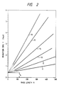
- Fig. 2 shows relationships between the base length (represented by the number of N of bases) of DNA fragments in the gels having various polyacrylamide concentrations and the migration time t.
- the curves 11, 12, 13, 14, 15 and 16 represent the cases where the gels have polyacrylamide concentrations of 2, 3, 4, 5, 6 and 8%. From the fact that the migration speed of the DNA fragment varies in proportion to the electric field intensity E and from the profiles of the curves of Fig.
- the migration time t0 becomes equal to l ⁇ g(T)/E0 in the equation (3) irrespective of the concentration C.
- Fig. 3 shows practically measured dependency of the band interval upon the concentration for various base lengths.
- the migration distance is 22 cm.
- the curves 17, 18, 19 and 20 represent the cases where the DNA fragments have base lengths (represented by the number N of bases) of 100, 200, 300 and 400.
- the base length is as short as about 100
- the band internval d increases with the increase in the gel concentration C.
- the base length is 300 or 400, however, the band interval becomes nearly constant provided the gel concentration C is greater than 4%.
- the DNA band width ⁇ varies nearly in proportion to ⁇ l without almost depending upon the gel concentration.
- Fig. 4 is a diagram showing the dependency of migration time of various base lengths upon the concentration of polyacrylamide in the gel, and wherein curves 17, 18, 19 and 20 represent the cases where the DNA fragments have base lengths (represented by the number N of bases) of 100, 200, 300 and 400. It will be recognized from Fig. 4 that the migration time increases nearly in proportion to the square power of the concentration C.
- the migration time t is a function of the length l of migration lane and the gel concentration C.
- ⁇ 0(C, T) can be found using ⁇ 0bs / ⁇ l from the measured band interval ⁇ 0bs when the length of migration lane is l0.
- Table 1 shows polyacrylamide concentrations C that minimize the migration time required for discriminating the neighboring bands, the lengths l of migration lane and the migration times t when the gel plate has a thickness of 0.3 mm and the electric field intensity is 50 V/cm for various base lengths.
- the gel concentrations should preferably be 4.3 to 6.2 with the range of from 5 to 6% being excluded, 3.2 to 4.3, and 2.6 to 3.2%.
- up to 300 bases can be measured in about 1.5 hours.
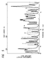
- Fig. 5 shows the results of measurement using the gel having a polyacrylamide concentration of 3% and a migration lane of a length of 22 cm. The separation is not sufficient since the length of migration lane is shorter than 28 cm. It will, however, be comprehended that up to about 300 bases can be identified. The migration time of DNA fragment of a base length of 300 was 77 minutes.
- the spectrum 56 is the one obtained from a DNA fragment group in which a base of nucleic acid at the terminal is guanine
- the spectrum 57 is the one obtained from a DNA group in which a base of nucleic acid at the terminal is thymine.
- the gels having various polyacrylamide concentrations were prepared in the same manner as in the prior art. Described below is a procedure for preparing a gel having a polyacrylamide concentration of 6% (not according to the present invention).
- Water is added to a mixture consisting of 1.14 g of an acrylamide monomer, 0.06 g of an N,N′-methylenebisacrylamide (the total amount with the acrylamide monomer is 1.2 g), 0.16 g of trishydroxyaminomethane, 0.08 g of boric acid, 0.014 g of EDTA ⁇ 2Na and 6.3 g of urea, such that the total amount is 20 ml (i.e., the total amount of acrylamide monomer and N,N′-methylenebisacrylamide is 0.06 g in 1 ml).
- the DNA fragments that had hitherto been measured requiring a time of 5 to 10 hours can now be measured requiring a time of a little more than one hour.
- the measuring time required by the fluorescence detection type electrophoresis apparatus can be strikingly shortened.
- the light scattered by the gel and background fluorescence from impureties and/or gel imposes lower limit for the detection.
- the gel having a low concentration however, the background fluorescence decreases, too, and the detection can be realized maintaining high sensitivity.
Applications Claiming Priority (2)
| Application Number | Priority Date | Filing Date | Title |
|---|---|---|---|
| JP63039385A JP2804038B2 (ja) | 1988-02-24 | 1988-02-24 | 塩基配列決定方法 |
| JP39385/88 | 1988-02-24 |
Publications (3)
| Publication Number | Publication Date |
|---|---|
| EP0330120A2 EP0330120A2 (en) | 1989-08-30 |
| EP0330120A3 EP0330120A3 (en) | 1990-09-12 |
| EP0330120B1 true EP0330120B1 (en) | 1995-06-28 |
Family
ID=12551541
Family Applications (1)
| Application Number | Title | Priority Date | Filing Date |
|---|---|---|---|
| EP89102908A Expired - Lifetime EP0330120B1 (en) | 1988-02-24 | 1989-02-20 | Fluorescence detection type electrophoresis apparatus |
Country Status (5)
| Country | Link |
|---|---|
| US (1) | US4971677A (ja) |
| EP (1) | EP0330120B1 (ja) |
| JP (1) | JP2804038B2 (ja) |
| CN (1) | CN1019860B (ja) |
| DE (1) | DE68923193T2 (ja) |
Families Citing this family (20)
| Publication number | Priority date | Publication date | Assignee | Title |
|---|---|---|---|---|
| US5230781A (en) * | 1984-03-29 | 1993-07-27 | Li-Cor, Inc. | Sequencing near infrared and infrared fluorescence labeled DNA for detecting using laser diodes |
| US5360523A (en) * | 1984-03-29 | 1994-11-01 | Li-Cor, Inc. | DNA sequencing |
| US5246866A (en) * | 1987-12-23 | 1993-09-21 | Hitachi Software Engineering Co., Ltd. | Method for transcription of a DNA sequence |
| JPH083481B2 (ja) * | 1989-06-07 | 1996-01-17 | 日立ソフトウェアエンジニアリング株式会社 | 蛍光式電気泳動パターン読み取り装置 |
| JP2814408B2 (ja) * | 1990-05-22 | 1998-10-22 | 日立ソフトウェアエンジニアリング 株式会社 | 蛍光パターン読み取り装置および蛍光パターン読み取り方法 |
| JPH0743353B2 (ja) * | 1990-05-31 | 1995-05-15 | 株式会社島津製作所 | 蛍光検出型ゲル電気泳動装置 |
| US5098536A (en) * | 1991-02-01 | 1992-03-24 | Beckman Instruments, Inc. | Method of improving signal-to-noise in electropherogram |
| JP2785530B2 (ja) * | 1991-09-13 | 1998-08-13 | 株式会社日立製作所 | 電気泳動装置 |
| US5137609A (en) * | 1992-01-31 | 1992-08-11 | Biometric Imaging Inc. | Differential separation assay |
| US5424841A (en) * | 1993-05-28 | 1995-06-13 | Molecular Dynamics | Apparatus for measuring spatial distribution of fluorescence on a substrate |
| JP3340544B2 (ja) * | 1993-12-24 | 2002-11-05 | 株式会社日立製作所 | 分別採取装置及び分別採取方法 |
| US5507934A (en) * | 1994-11-01 | 1996-04-16 | Visible Genetics Inc. | Apparatus for preparing gels for use in electrophoretic separations and similar applications |
| US5627022A (en) * | 1994-11-01 | 1997-05-06 | Visible Genetics Inc. | Microgels for use in medical diagnosis and holders useful in fabricating same |
| WO1996023213A1 (en) * | 1995-01-23 | 1996-08-01 | Murray Anthony J | Analysis of biological molecules |
| US5582705A (en) * | 1995-05-19 | 1996-12-10 | Iowa State University Research Foundation, Inc. | Multiplexed capillary electrophoresis system |
| US5717602A (en) * | 1996-02-05 | 1998-02-10 | Kenning; Gregory G. | Automated electrophoresis and analysis system |
| US6436641B1 (en) * | 2000-04-17 | 2002-08-20 | Visible Genetics Inc. | Method and apparatus for DNA sequencing |
| KR20030031306A (ko) * | 2001-10-13 | 2003-04-21 | 주식회사 커벡스 | 전기영동의 겔영상을 획득하기 위한 촬영장치 |
| CN1854716B (zh) * | 2005-04-18 | 2012-03-21 | 微奥基因科技常州有限公司 | 集成有可变换光源的电泳分离与分析装置及其使用 |
| CN101916029B (zh) * | 2010-07-26 | 2011-12-28 | 亚亚科技股份有限公司 | 高效能的荧光摄影光源装置 |
Family Cites Families (9)
| Publication number | Priority date | Publication date | Assignee | Title |
|---|---|---|---|---|
| US4123343A (en) * | 1977-06-14 | 1978-10-31 | American Home Products Corporation | Purification of glycoproteins and immunization therewith |
| US4405720A (en) * | 1981-03-04 | 1983-09-20 | The United States Of America As Represented By The Department Of Health And Human Services | Silver stains for protein in gels |
| US4555490A (en) * | 1984-06-08 | 1985-11-26 | The United States Of America As Represented By The Department Of Health And Human Services | Rapid visualization system for gel electrophoresis |
| JPS6162843A (ja) * | 1984-08-13 | 1986-03-31 | Hitachi Ltd | 螢光検出型電気泳動装置 |
| JPS61173158A (ja) * | 1985-01-02 | 1986-08-04 | カリフオルニア・インステイテユ−ト・オブ・テクノロジ− | Dna配列決定法 |
| US4668667A (en) * | 1985-08-01 | 1987-05-26 | Norwich Eaton Pharmaceuticals, Inc. | Acylphosphorotriamides useful as lipid-altering agents |
| US4855225A (en) * | 1986-02-07 | 1989-08-08 | Applied Biosystems, Inc. | Method of detecting electrophoretically separated oligonucleotides |
| DE3752148T2 (de) * | 1987-06-09 | 1998-09-17 | Perkin Elmer Corp | Echtzeitabtastvorrichtung in einem Elektrophoreseapparat zur DNS-Sequenzbestimmung |
| JP2550106B2 (ja) * | 1987-10-30 | 1996-11-06 | 株式会社日立製作所 | 光分散検出型電気泳動装置 |
-
1988
- 1988-02-24 JP JP63039385A patent/JP2804038B2/ja not_active Expired - Fee Related
-
1989
- 1989-02-15 US US07/310,645 patent/US4971677A/en not_active Expired - Lifetime
- 1989-02-20 DE DE68923193T patent/DE68923193T2/de not_active Expired - Lifetime
- 1989-02-20 EP EP89102908A patent/EP0330120B1/en not_active Expired - Lifetime
- 1989-02-23 CN CN89101129A patent/CN1019860B/zh not_active Expired
Also Published As
| Publication number | Publication date |
|---|---|
| JP2804038B2 (ja) | 1998-09-24 |
| CN1019860B (zh) | 1992-12-30 |
| US4971677A (en) | 1990-11-20 |
| DE68923193T2 (de) | 1996-02-08 |
| JPH01214752A (ja) | 1989-08-29 |
| CN1036639A (zh) | 1989-10-25 |
| EP0330120A3 (en) | 1990-09-12 |
| EP0330120A2 (en) | 1989-08-30 |
| DE68923193D1 (de) | 1995-08-03 |
Similar Documents
| Publication | Publication Date | Title |
|---|---|---|
| EP0330120B1 (en) | Fluorescence detection type electrophoresis apparatus | |
| Chen et al. | Low-cost, high-sensitivity laser-induced fluorescence detection for DNA sequencing by capillary gel electrophoresis | |
| Karger et al. | Multiwavelength fluorescence detection for DNA sequencing using capillary electrophoresis | |
| US6488832B2 (en) | Array based electrophoretic system for the analysis of multiple biological samples | |
| EP0628164B1 (en) | Capillary array confocal fluorescence scanner and method | |
| JP2853745B2 (ja) | 光検出電気泳動装置 | |
| US6132578A (en) | Method and apparatus for electrophoresis separation and detection | |
| US6290831B1 (en) | Electrophoretic system for real time detection of multiple electrophoresed biopolymers | |
| JPH01112147A (ja) | 核酸の塩基配列決定方法 | |
| Tseng et al. | Analysis of large-volume DNA markers and polymerase chain reaction products by capillary electrophoresis in the presence of electroosmotic flow | |
| Naimski et al. | Quantitative fluorescenct analysis of different conformational forms of DNA bound to the dye, 4′, 6-diamidine-2-phenylindole, and sepasated by gel electrophoresis | |
| JPS613043A (ja) | 核酸塩基配列決定装置 | |
| JP2840586B2 (ja) | 光検出電気泳動装置 | |
| JP2853706B2 (ja) | 塩基配列決定方法 | |
| EP0275440A2 (en) | Photo detection system for DNA base analysis | |
| JP2914333B2 (ja) | 塩基配列決定方法 | |
| US6793790B1 (en) | Sample collection system for gel electrophoresis | |
| Doktycz et al. | Electrophoresis and detection of tin‐labeled DNAs on open‐faced gels | |
| JPH01209351A (ja) | 塩基配列決定装置 | |
| JPH05322770A (ja) | 多標識系電気泳動法 | |
| Dovichi | Capillary electrophoresis for DNA sequencing | |
| JPH04244954A (ja) | 塩基配列決定装置 | |
| EP0979878A2 (en) | Method for detecting oligonucleotides and determining base sequence of nucleic acids | |
| Zhang et al. | DNA SEQUENCING BY CAPILLARY GEL ELECTROPHORESIS AND LASER-INDUCED FLUORFSCIIVCE DETECTION | |
| Swerdlow | Capillary gel electrophoresis for DNA sequencing |
Legal Events
| Date | Code | Title | Description |
|---|---|---|---|
| PUAI | Public reference made under article 153(3) epc to a published international application that has entered the european phase |
Free format text: ORIGINAL CODE: 0009012 |
|
| AK | Designated contracting states |
Kind code of ref document: A2 Designated state(s): DE GB |
|
| PUAL | Search report despatched |
Free format text: ORIGINAL CODE: 0009013 |
|
| AK | Designated contracting states |
Kind code of ref document: A3 Designated state(s): DE GB |
|
| 17P | Request for examination filed |
Effective date: 19901212 |
|
| 17Q | First examination report despatched |
Effective date: 19920805 |
|
| GRAA | (expected) grant |
Free format text: ORIGINAL CODE: 0009210 |
|
| AK | Designated contracting states |
Kind code of ref document: B1 Designated state(s): DE GB |
|
| REF | Corresponds to: |
Ref document number: 68923193 Country of ref document: DE Date of ref document: 19950803 |
|
| PLBE | No opposition filed within time limit |
Free format text: ORIGINAL CODE: 0009261 |
|
| STAA | Information on the status of an ep patent application or granted ep patent |
Free format text: STATUS: NO OPPOSITION FILED WITHIN TIME LIMIT |
|
| 26N | No opposition filed | ||
| REG | Reference to a national code |
Ref country code: GB Ref legal event code: IF02 |
|
| PGFP | Annual fee paid to national office [announced via postgrant information from national office to epo] |
Ref country code: GB Payment date: 20080114 Year of fee payment: 20 |
|
| PGFP | Annual fee paid to national office [announced via postgrant information from national office to epo] |
Ref country code: DE Payment date: 20080307 Year of fee payment: 20 |
|
| REG | Reference to a national code |
Ref country code: GB Ref legal event code: PE20 Expiry date: 20090219 |
|
| PG25 | Lapsed in a contracting state [announced via postgrant information from national office to epo] |
Ref country code: GB Free format text: LAPSE BECAUSE OF EXPIRATION OF PROTECTION Effective date: 20090219 |