EP0247906B1 - Für Antitumor-Polypeptide kodierende DNS, die Polypeptide und diese Polypeptide enthaltenden Antitumor-Wirkstoffe - Google Patents
Für Antitumor-Polypeptide kodierende DNS, die Polypeptide und diese Polypeptide enthaltenden Antitumor-Wirkstoffe Download PDFInfo
- Publication number
- EP0247906B1 EP0247906B1 EP87400261A EP87400261A EP0247906B1 EP 0247906 B1 EP0247906 B1 EP 0247906B1 EP 87400261 A EP87400261 A EP 87400261A EP 87400261 A EP87400261 A EP 87400261A EP 0247906 B1 EP0247906 B1 EP 0247906B1
- Authority
- EP
- European Patent Office
- Prior art keywords
- dna
- amino acid
- tnf
- leu
- ala
- Prior art date
- Legal status (The legal status is an assumption and is not a legal conclusion. Google has not performed a legal analysis and makes no representation as to the accuracy of the status listed.)
- Expired - Lifetime
Links
- 108090000765 processed proteins & peptides Proteins 0.000 title claims abstract description 86
- 229920001184 polypeptide Polymers 0.000 title claims abstract description 76
- 102000004196 processed proteins & peptides Human genes 0.000 title claims abstract description 76
- 230000000259 anti-tumor effect Effects 0.000 title abstract description 48
- 108020004414 DNA Proteins 0.000 claims abstract description 95
- 238000000034 method Methods 0.000 claims abstract description 26
- 230000008569 process Effects 0.000 claims abstract description 9
- 239000002246 antineoplastic agent Substances 0.000 claims abstract description 4
- 125000003275 alpha amino acid group Chemical group 0.000 claims abstract 10
- 239000013612 plasmid Substances 0.000 claims description 26
- 150000001413 amino acids Chemical class 0.000 claims description 24
- 108091008146 restriction endonucleases Proteins 0.000 claims description 19
- 238000012258 culturing Methods 0.000 claims description 13
- 238000003776 cleavage reaction Methods 0.000 claims description 9
- 230000007017 scission Effects 0.000 claims description 9
- 108091081024 Start codon Proteins 0.000 claims description 4
- 108020004705 Codon Proteins 0.000 claims description 3
- 238000004519 manufacturing process Methods 0.000 claims description 3
- 244000005700 microbiome Species 0.000 claims 2
- 238000002360 preparation method Methods 0.000 abstract description 8
- 125000000539 amino acid group Chemical group 0.000 abstract description 6
- 229910052739 hydrogen Inorganic materials 0.000 abstract 1
- 239000001257 hydrogen Substances 0.000 abstract 1
- 125000004435 hydrogen atom Chemical group [H]* 0.000 abstract 1
- 210000004027 cell Anatomy 0.000 description 73
- FAPWRFPIFSIZLT-UHFFFAOYSA-M Sodium chloride Chemical compound [Na+].[Cl-] FAPWRFPIFSIZLT-UHFFFAOYSA-M 0.000 description 54
- 108090000623 proteins and genes Proteins 0.000 description 50
- 239000000243 solution Substances 0.000 description 49
- 101000611183 Homo sapiens Tumor necrosis factor Proteins 0.000 description 31
- 239000012634 fragment Substances 0.000 description 29
- 239000011780 sodium chloride Substances 0.000 description 29
- 241000588724 Escherichia coli Species 0.000 description 26
- 102000004169 proteins and genes Human genes 0.000 description 21
- 238000005119 centrifugation Methods 0.000 description 20
- 239000000203 mixture Substances 0.000 description 20
- 235000018102 proteins Nutrition 0.000 description 20
- 230000000694 effects Effects 0.000 description 19
- 239000002299 complementary DNA Substances 0.000 description 18
- 239000002609 medium Substances 0.000 description 16
- 239000007853 buffer solution Substances 0.000 description 15
- 108020004999 messenger RNA Proteins 0.000 description 14
- QKNYBSVHEMOAJP-UHFFFAOYSA-N 2-amino-2-(hydroxymethyl)propane-1,3-diol;hydron;chloride Chemical compound Cl.OCC(N)(CO)CO QKNYBSVHEMOAJP-UHFFFAOYSA-N 0.000 description 13
- 239000006228 supernatant Substances 0.000 description 13
- LFQSCWFLJHTTHZ-UHFFFAOYSA-N Ethanol Chemical compound CCO LFQSCWFLJHTTHZ-UHFFFAOYSA-N 0.000 description 12
- 235000001014 amino acid Nutrition 0.000 description 11
- 231100000433 cytotoxic Toxicity 0.000 description 11
- 230000001472 cytotoxic effect Effects 0.000 description 11
- 238000010828 elution Methods 0.000 description 11
- 239000000523 sample Substances 0.000 description 11
- HEDRZPFGACZZDS-UHFFFAOYSA-N Chloroform Chemical compound ClC(Cl)Cl HEDRZPFGACZZDS-UHFFFAOYSA-N 0.000 description 10
- 239000000020 Nitrocellulose Substances 0.000 description 10
- IQFYYKKMVGJFEH-XLPZGREQSA-N Thymidine Chemical compound O=C1NC(=O)C(C)=CN1[C@@H]1O[C@H](CO)[C@@H](O)C1 IQFYYKKMVGJFEH-XLPZGREQSA-N 0.000 description 10
- 230000003013 cytotoxicity Effects 0.000 description 10
- 231100000135 cytotoxicity Toxicity 0.000 description 10
- BPHPUYQFMNQIOC-NXRLNHOXSA-N isopropyl beta-D-thiogalactopyranoside Chemical compound CC(C)S[C@@H]1O[C@H](CO)[C@H](O)[C@H](O)[C@H]1O BPHPUYQFMNQIOC-NXRLNHOXSA-N 0.000 description 10
- 229920001220 nitrocellulos Polymers 0.000 description 10
- 238000004587 chromatography analysis Methods 0.000 description 9
- 108091032973 (ribonucleotides)n+m Proteins 0.000 description 8
- 239000013600 plasmid vector Substances 0.000 description 8
- 239000002244 precipitate Substances 0.000 description 7
- 238000000746 purification Methods 0.000 description 7
- WEVYAHXRMPXWCK-UHFFFAOYSA-N Acetonitrile Chemical compound CC#N WEVYAHXRMPXWCK-UHFFFAOYSA-N 0.000 description 6
- 229920001817 Agar Polymers 0.000 description 6
- KFZMGEQAYNKOFK-UHFFFAOYSA-N Isopropanol Chemical compound CC(C)O KFZMGEQAYNKOFK-UHFFFAOYSA-N 0.000 description 6
- BFNBIHQBYMNNAN-UHFFFAOYSA-N ammonium sulfate Chemical compound N.N.OS(O)(=O)=O BFNBIHQBYMNNAN-UHFFFAOYSA-N 0.000 description 6
- 229910052921 ammonium sulfate Inorganic materials 0.000 description 6
- 235000011130 ammonium sulphate Nutrition 0.000 description 6
- 238000006243 chemical reaction Methods 0.000 description 6
- 238000000502 dialysis Methods 0.000 description 6
- 238000001962 electrophoresis Methods 0.000 description 6
- 238000002523 gelfiltration Methods 0.000 description 6
- 230000002068 genetic effect Effects 0.000 description 6
- 239000012071 phase Substances 0.000 description 6
- KCXVZYZYPLLWCC-UHFFFAOYSA-N EDTA Chemical compound OC(=O)CN(CC(O)=O)CCN(CC(O)=O)CC(O)=O KCXVZYZYPLLWCC-UHFFFAOYSA-N 0.000 description 5
- 102000004160 Phosphoric Monoester Hydrolases Human genes 0.000 description 5
- 108090000608 Phosphoric Monoester Hydrolases Proteins 0.000 description 5
- 108091034057 RNA (poly(A)) Proteins 0.000 description 5
- 229960000723 ampicillin Drugs 0.000 description 5
- AVKUERGKIZMTKX-NJBDSQKTSA-N ampicillin Chemical compound C1([C@@H](N)C(=O)N[C@H]2[C@H]3SC([C@@H](N3C2=O)C(O)=O)(C)C)=CC=CC=C1 AVKUERGKIZMTKX-NJBDSQKTSA-N 0.000 description 5
- 150000001450 anions Chemical class 0.000 description 5
- 238000000605 extraction Methods 0.000 description 5
- 230000002209 hydrophobic effect Effects 0.000 description 5
- 239000008055 phosphate buffer solution Substances 0.000 description 5
- XLYOFNOQVPJJNP-UHFFFAOYSA-N water Substances O XLYOFNOQVPJJNP-UHFFFAOYSA-N 0.000 description 5
- 235000014469 Bacillus subtilis Nutrition 0.000 description 4
- 206010009944 Colon cancer Diseases 0.000 description 4
- 108010008286 DNA nucleotidylexotransferase Proteins 0.000 description 4
- 102100033215 DNA nucleotidylexotransferase Human genes 0.000 description 4
- 239000007836 KH2PO4 Substances 0.000 description 4
- GUBGYTABKSRVRQ-QKKXKWKRSA-N Lactose Natural products OC[C@H]1O[C@@H](O[C@H]2[C@H](O)[C@@H](O)C(O)O[C@@H]2CO)[C@H](O)[C@@H](O)[C@H]1O GUBGYTABKSRVRQ-QKKXKWKRSA-N 0.000 description 4
- ISWSIDIOOBJBQZ-UHFFFAOYSA-N Phenol Chemical compound OC1=CC=CC=C1 ISWSIDIOOBJBQZ-UHFFFAOYSA-N 0.000 description 4
- 239000012980 RPMI-1640 medium Substances 0.000 description 4
- 240000004808 Saccharomyces cerevisiae Species 0.000 description 4
- 229920005654 Sephadex Polymers 0.000 description 4
- 239000012507 Sephadex™ Substances 0.000 description 4
- 230000000295 complement effect Effects 0.000 description 4
- 239000013078 crystal Substances 0.000 description 4
- RGWHQCVHVJXOKC-SHYZEUOFSA-J dCTP(4-) Chemical compound O=C1N=C(N)C=CN1[C@@H]1O[C@H](COP([O-])(=O)OP([O-])(=O)OP([O-])([O-])=O)[C@@H](O)C1 RGWHQCVHVJXOKC-SHYZEUOFSA-J 0.000 description 4
- BNIILDVGGAEEIG-UHFFFAOYSA-L disodium hydrogen phosphate Chemical compound [Na+].[Na+].OP([O-])([O-])=O BNIILDVGGAEEIG-UHFFFAOYSA-L 0.000 description 4
- 229910000397 disodium phosphate Inorganic materials 0.000 description 4
- 235000019800 disodium phosphate Nutrition 0.000 description 4
- 239000000284 extract Substances 0.000 description 4
- PHTQWCKDNZKARW-UHFFFAOYSA-N isoamylol Chemical compound CC(C)CCO PHTQWCKDNZKARW-UHFFFAOYSA-N 0.000 description 4
- 239000008101 lactose Substances 0.000 description 4
- 229910000402 monopotassium phosphate Inorganic materials 0.000 description 4
- 235000019796 monopotassium phosphate Nutrition 0.000 description 4
- 230000017074 necrotic cell death Effects 0.000 description 4
- GNSKLFRGEWLPPA-UHFFFAOYSA-M potassium dihydrogen phosphate Chemical compound [K+].OP(O)([O-])=O GNSKLFRGEWLPPA-UHFFFAOYSA-M 0.000 description 4
- 238000002415 sodium dodecyl sulfate polyacrylamide gel electrophoresis Methods 0.000 description 4
- 238000001179 sorption measurement Methods 0.000 description 4
- 238000010186 staining Methods 0.000 description 4
- 230000001629 suppression Effects 0.000 description 4
- 239000000725 suspension Substances 0.000 description 4
- 239000013598 vector Substances 0.000 description 4
- 238000010792 warming Methods 0.000 description 4
- 102000004594 DNA Polymerase I Human genes 0.000 description 3
- 108010017826 DNA Polymerase I Proteins 0.000 description 3
- 102100034343 Integrase Human genes 0.000 description 3
- 241000699670 Mus sp. Species 0.000 description 3
- 125000001429 N-terminal alpha-amino-acid group Chemical group 0.000 description 3
- 108010092799 RNA-directed DNA polymerase Proteins 0.000 description 3
- 229920002684 Sepharose Polymers 0.000 description 3
- HEMHJVSKTPXQMS-UHFFFAOYSA-M Sodium hydroxide Chemical compound [OH-].[Na+] HEMHJVSKTPXQMS-UHFFFAOYSA-M 0.000 description 3
- 239000008272 agar Substances 0.000 description 3
- 238000005571 anion exchange chromatography Methods 0.000 description 3
- 125000004122 cyclic group Chemical group 0.000 description 3
- SUYVUBYJARFZHO-RRKCRQDMSA-N dATP Chemical compound C1=NC=2C(N)=NC=NC=2N1[C@H]1C[C@H](O)[C@@H](COP(O)(=O)OP(O)(=O)OP(O)(O)=O)O1 SUYVUBYJARFZHO-RRKCRQDMSA-N 0.000 description 3
- SUYVUBYJARFZHO-UHFFFAOYSA-N dATP Natural products C1=NC=2C(N)=NC=NC=2N1C1CC(O)C(COP(O)(=O)OP(O)(=O)OP(O)(O)=O)O1 SUYVUBYJARFZHO-UHFFFAOYSA-N 0.000 description 3
- HAAZLUGHYHWQIW-KVQBGUIXSA-N dGTP Chemical compound C1=NC=2C(=O)NC(N)=NC=2N1[C@H]1C[C@H](O)[C@@H](COP(O)(=O)OP(O)(=O)OP(O)(O)=O)O1 HAAZLUGHYHWQIW-KVQBGUIXSA-N 0.000 description 3
- NHVNXKFIZYSCEB-XLPZGREQSA-N dTTP Chemical compound O=C1NC(=O)C(C)=CN1[C@@H]1O[C@H](COP(O)(=O)OP(O)(=O)OP(O)(O)=O)[C@@H](O)C1 NHVNXKFIZYSCEB-XLPZGREQSA-N 0.000 description 3
- 238000010790 dilution Methods 0.000 description 3
- 239000012895 dilution Substances 0.000 description 3
- 230000001747 exhibiting effect Effects 0.000 description 3
- 239000000499 gel Substances 0.000 description 3
- 238000009396 hybridization Methods 0.000 description 3
- 238000011081 inoculation Methods 0.000 description 3
- 238000004255 ion exchange chromatography Methods 0.000 description 3
- 239000007758 minimum essential medium Substances 0.000 description 3
- 229920002401 polyacrylamide Polymers 0.000 description 3
- 238000011084 recovery Methods 0.000 description 3
- 238000012216 screening Methods 0.000 description 3
- 230000035945 sensitivity Effects 0.000 description 3
- 230000004083 survival effect Effects 0.000 description 3
- 230000002195 synergetic effect Effects 0.000 description 3
- 238000012360 testing method Methods 0.000 description 3
- 201000009030 Carcinoma Diseases 0.000 description 2
- 102000053602 DNA Human genes 0.000 description 2
- 102000012410 DNA Ligases Human genes 0.000 description 2
- 108010061982 DNA Ligases Proteins 0.000 description 2
- 239000003298 DNA probe Substances 0.000 description 2
- 208000001382 Experimental Melanoma Diseases 0.000 description 2
- ZHNUHDYFZUAESO-UHFFFAOYSA-N Formamide Chemical compound NC=O ZHNUHDYFZUAESO-UHFFFAOYSA-N 0.000 description 2
- DHMQDGOQFOQNFH-UHFFFAOYSA-N Glycine Chemical compound NCC(O)=O DHMQDGOQFOQNFH-UHFFFAOYSA-N 0.000 description 2
- 206010058467 Lung neoplasm malignant Diseases 0.000 description 2
- TWRXJAOTZQYOKJ-UHFFFAOYSA-L Magnesium chloride Chemical compound [Mg+2].[Cl-].[Cl-] TWRXJAOTZQYOKJ-UHFFFAOYSA-L 0.000 description 2
- 206010027476 Metastases Diseases 0.000 description 2
- 108020004511 Recombinant DNA Proteins 0.000 description 2
- DTQVDTLACAAQTR-UHFFFAOYSA-N Trifluoroacetic acid Chemical compound OC(=O)C(F)(F)F DTQVDTLACAAQTR-UHFFFAOYSA-N 0.000 description 2
- 108090000631 Trypsin Proteins 0.000 description 2
- 102000004142 Trypsin Human genes 0.000 description 2
- 230000002378 acidificating effect Effects 0.000 description 2
- RJURFGZVJUQBHK-UHFFFAOYSA-N actinomycin D Natural products CC1OC(=O)C(C(C)C)N(C)C(=O)CN(C)C(=O)C2CCCN2C(=O)C(C(C)C)NC(=O)C1NC(=O)C1=C(N)C(=O)C(C)=C2OC(C(C)=CC=C3C(=O)NC4C(=O)NC(C(N5CCCC5C(=O)N(C)CC(=O)N(C)C(C(C)C)C(=O)OC4C)=O)C(C)C)=C3N=C21 RJURFGZVJUQBHK-UHFFFAOYSA-N 0.000 description 2
- 230000009471 action Effects 0.000 description 2
- 102000004139 alpha-Amylases Human genes 0.000 description 2
- 108090000637 alpha-Amylases Proteins 0.000 description 2
- 229940024171 alpha-amylase Drugs 0.000 description 2
- 239000007864 aqueous solution Substances 0.000 description 2
- 238000000376 autoradiography Methods 0.000 description 2
- 108010005774 beta-Galactosidase Proteins 0.000 description 2
- AIYUHDOJVYHVIT-UHFFFAOYSA-M caesium chloride Chemical compound [Cl-].[Cs+] AIYUHDOJVYHVIT-UHFFFAOYSA-M 0.000 description 2
- 244000309466 calf Species 0.000 description 2
- 239000003795 chemical substances by application Substances 0.000 description 2
- 238000000354 decomposition reaction Methods 0.000 description 2
- ZMMJGEGLRURXTF-UHFFFAOYSA-N ethidium bromide Chemical compound [Br-].C12=CC(N)=CC=C2C2=CC=C(N)C=C2[N+](CC)=C1C1=CC=CC=C1 ZMMJGEGLRURXTF-UHFFFAOYSA-N 0.000 description 2
- 229960005542 ethidium bromide Drugs 0.000 description 2
- 238000002474 experimental method Methods 0.000 description 2
- 238000000855 fermentation Methods 0.000 description 2
- 230000004151 fermentation Effects 0.000 description 2
- UYTPUPDQBNUYGX-UHFFFAOYSA-N guanine Chemical compound O=C1NC(N)=NC2=C1N=CN2 UYTPUPDQBNUYGX-UHFFFAOYSA-N 0.000 description 2
- 206010073071 hepatocellular carcinoma Diseases 0.000 description 2
- 238000004128 high performance liquid chromatography Methods 0.000 description 2
- 230000001965 increasing effect Effects 0.000 description 2
- 238000011534 incubation Methods 0.000 description 2
- 230000006698 induction Effects 0.000 description 2
- 230000001939 inductive effect Effects 0.000 description 2
- 230000003902 lesion Effects 0.000 description 2
- 239000007788 liquid Substances 0.000 description 2
- 238000004811 liquid chromatography Methods 0.000 description 2
- 201000005296 lung carcinoma Diseases 0.000 description 2
- RLSSMJSEOOYNOY-UHFFFAOYSA-N m-cresol Chemical compound CC1=CC=CC(O)=C1 RLSSMJSEOOYNOY-UHFFFAOYSA-N 0.000 description 2
- 239000012528 membrane Substances 0.000 description 2
- 230000009401 metastasis Effects 0.000 description 2
- WSFSSNUMVMOOMR-NJFSPNSNSA-N methanone Chemical compound O=[14CH2] WSFSSNUMVMOOMR-NJFSPNSNSA-N 0.000 description 2
- 238000002156 mixing Methods 0.000 description 2
- 239000002808 molecular sieve Substances 0.000 description 2
- 208000017708 myomatous neoplasm Diseases 0.000 description 2
- 150000002989 phenols Chemical class 0.000 description 2
- 108091033319 polynucleotide Proteins 0.000 description 2
- 102000040430 polynucleotide Human genes 0.000 description 2
- 239000002157 polynucleotide Substances 0.000 description 2
- 239000000047 product Substances 0.000 description 2
- 239000012521 purified sample Substances 0.000 description 2
- 239000011541 reaction mixture Substances 0.000 description 2
- 238000011160 research Methods 0.000 description 2
- 238000005185 salting out Methods 0.000 description 2
- 238000012163 sequencing technique Methods 0.000 description 2
- URGAHOPLAPQHLN-UHFFFAOYSA-N sodium aluminosilicate Chemical compound [Na+].[Al+3].[O-][Si]([O-])=O.[O-][Si]([O-])=O URGAHOPLAPQHLN-UHFFFAOYSA-N 0.000 description 2
- 239000001509 sodium citrate Substances 0.000 description 2
- NLJMYIDDQXHKNR-UHFFFAOYSA-K sodium citrate Chemical compound O.O.[Na+].[Na+].[Na+].[O-]C(=O)CC(O)(CC([O-])=O)C([O-])=O NLJMYIDDQXHKNR-UHFFFAOYSA-K 0.000 description 2
- 238000003756 stirring Methods 0.000 description 2
- 210000003699 striated muscle Anatomy 0.000 description 2
- 239000000126 substance Substances 0.000 description 2
- 210000001541 thymus gland Anatomy 0.000 description 2
- 239000012588 trypsin Substances 0.000 description 2
- 238000000108 ultra-filtration Methods 0.000 description 2
- 239000011534 wash buffer Substances 0.000 description 2
- 238000005406 washing Methods 0.000 description 2
- 102000040650 (ribonucleotides)n+m Human genes 0.000 description 1
- PQMRRAQXKWFYQN-UHFFFAOYSA-N 1-phenyl-2-sulfanylideneimidazolidin-4-one Chemical compound S=C1NC(=O)CN1C1=CC=CC=C1 PQMRRAQXKWFYQN-UHFFFAOYSA-N 0.000 description 1
- PHEDXBVPIONUQT-BJBOKCJXSA-N 12-Tetradecanoylphorbol 13-acetate Natural products O=C(O[C@H]1[C@@H](C)[C@@]2(O)[C@H]3[C@@](O)(C(=O)C(C)=C3)CC(CO)=C[C@H]2[C@H]2C(C)(C)[C@]12OC(=O)C)CCCCCCCCCCCCC PHEDXBVPIONUQT-BJBOKCJXSA-N 0.000 description 1
- NHBKXEKEPDILRR-UHFFFAOYSA-N 2,3-bis(butanoylsulfanyl)propyl butanoate Chemical compound CCCC(=O)OCC(SC(=O)CCC)CSC(=O)CCC NHBKXEKEPDILRR-UHFFFAOYSA-N 0.000 description 1
- 206010000871 Acute monocytic leukaemia Diseases 0.000 description 1
- 102000007698 Alcohol dehydrogenase Human genes 0.000 description 1
- 108010021809 Alcohol dehydrogenase Proteins 0.000 description 1
- 239000004475 Arginine Substances 0.000 description 1
- 108091003079 Bovine Serum Albumin Proteins 0.000 description 1
- OYPRJOBELJOOCE-UHFFFAOYSA-N Calcium Chemical compound [Ca] OYPRJOBELJOOCE-UHFFFAOYSA-N 0.000 description 1
- 108020003215 DNA Probes Proteins 0.000 description 1
- 108010014303 DNA-directed DNA polymerase Proteins 0.000 description 1
- 102000016928 DNA-directed DNA polymerase Human genes 0.000 description 1
- 108010092160 Dactinomycin Proteins 0.000 description 1
- 102000016911 Deoxyribonucleases Human genes 0.000 description 1
- 108010053770 Deoxyribonucleases Proteins 0.000 description 1
- 108010067770 Endopeptidase K Proteins 0.000 description 1
- 102000004190 Enzymes Human genes 0.000 description 1
- 108090000790 Enzymes Proteins 0.000 description 1
- 108700039691 Genetic Promoter Regions Proteins 0.000 description 1
- 239000004471 Glycine Substances 0.000 description 1
- QNAYBMKLOCPYGJ-REOHCLBHSA-N L-alanine Chemical compound C[C@H](N)C(O)=O QNAYBMKLOCPYGJ-REOHCLBHSA-N 0.000 description 1
- ODKSFYDXXFIFQN-BYPYZUCNSA-P L-argininium(2+) Chemical compound NC(=[NH2+])NCCC[C@H]([NH3+])C(O)=O ODKSFYDXXFIFQN-BYPYZUCNSA-P 0.000 description 1
- HNDVDQJCIGZPNO-YFKPBYRVSA-N L-histidine Chemical compound OC(=O)[C@@H](N)CC1=CN=CN1 HNDVDQJCIGZPNO-YFKPBYRVSA-N 0.000 description 1
- FFEARJCKVFRZRR-BYPYZUCNSA-N L-methionine Chemical compound CSCC[C@H](N)C(O)=O FFEARJCKVFRZRR-BYPYZUCNSA-N 0.000 description 1
- KZSNJWFQEVHDMF-BYPYZUCNSA-N L-valine Chemical compound CC(C)[C@H](N)C(O)=O KZSNJWFQEVHDMF-BYPYZUCNSA-N 0.000 description 1
- 208000035489 Monocytic Acute Leukemia Diseases 0.000 description 1
- 102000016943 Muramidase Human genes 0.000 description 1
- 108010014251 Muramidase Proteins 0.000 description 1
- 108010062010 N-Acetylmuramoyl-L-alanine Amidase Proteins 0.000 description 1
- 241000283973 Oryctolagus cuniculus Species 0.000 description 1
- 229910019142 PO4 Inorganic materials 0.000 description 1
- 108091005804 Peptidases Proteins 0.000 description 1
- 239000004365 Protease Substances 0.000 description 1
- 102000001253 Protein Kinase Human genes 0.000 description 1
- 108010076504 Protein Sorting Signals Proteins 0.000 description 1
- 239000013614 RNA sample Substances 0.000 description 1
- 102100037486 Reverse transcriptase/ribonuclease H Human genes 0.000 description 1
- 239000012506 Sephacryl® Substances 0.000 description 1
- BQCADISMDOOEFD-UHFFFAOYSA-N Silver Chemical compound [Ag] BQCADISMDOOEFD-UHFFFAOYSA-N 0.000 description 1
- 239000004809 Teflon Substances 0.000 description 1
- 229920006362 Teflon® Polymers 0.000 description 1
- KZSNJWFQEVHDMF-UHFFFAOYSA-N Valine Natural products CC(C)C(N)C(O)=O KZSNJWFQEVHDMF-UHFFFAOYSA-N 0.000 description 1
- 238000010521 absorption reaction Methods 0.000 description 1
- RJURFGZVJUQBHK-IIXSONLDSA-N actinomycin D Chemical compound C[C@H]1OC(=O)[C@H](C(C)C)N(C)C(=O)CN(C)C(=O)[C@@H]2CCCN2C(=O)[C@@H](C(C)C)NC(=O)[C@H]1NC(=O)C1=C(N)C(=O)C(C)=C2OC(C(C)=CC=C3C(=O)N[C@@H]4C(=O)N[C@@H](C(N5CCC[C@H]5C(=O)N(C)CC(=O)N(C)[C@@H](C(C)C)C(=O)O[C@@H]4C)=O)C(C)C)=C3N=C21 RJURFGZVJUQBHK-IIXSONLDSA-N 0.000 description 1
- 239000013543 active substance Substances 0.000 description 1
- 238000001042 affinity chromatography Methods 0.000 description 1
- 235000004279 alanine Nutrition 0.000 description 1
- 230000001476 alcoholic effect Effects 0.000 description 1
- 238000004458 analytical method Methods 0.000 description 1
- 210000004102 animal cell Anatomy 0.000 description 1
- 238000005349 anion exchange Methods 0.000 description 1
- 238000000137 annealing Methods 0.000 description 1
- ODKSFYDXXFIFQN-UHFFFAOYSA-N arginine Natural products OC(=O)C(N)CCCNC(N)=N ODKSFYDXXFIFQN-UHFFFAOYSA-N 0.000 description 1
- 238000003556 assay Methods 0.000 description 1
- 238000010923 batch production Methods 0.000 description 1
- 102000005936 beta-Galactosidase Human genes 0.000 description 1
- 239000000872 buffer Substances 0.000 description 1
- 239000008366 buffered solution Substances 0.000 description 1
- 235000014121 butter Nutrition 0.000 description 1
- 239000011575 calcium Substances 0.000 description 1
- 229910052791 calcium Inorganic materials 0.000 description 1
- 239000000969 carrier Substances 0.000 description 1
- 230000015556 catabolic process Effects 0.000 description 1
- 238000004113 cell culture Methods 0.000 description 1
- 210000002421 cell wall Anatomy 0.000 description 1
- 230000001413 cellular effect Effects 0.000 description 1
- 229960005091 chloramphenicol Drugs 0.000 description 1
- WIIZWVCIJKGZOK-RKDXNWHRSA-N chloramphenicol Chemical compound ClC(Cl)C(=O)N[C@H](CO)[C@H](O)C1=CC=C([N+]([O-])=O)C=C1 WIIZWVCIJKGZOK-RKDXNWHRSA-N 0.000 description 1
- BKHZIBWEHPHYAI-UHFFFAOYSA-N chloroform;3-methylbutan-1-ol Chemical compound ClC(Cl)Cl.CC(C)CCO BKHZIBWEHPHYAI-UHFFFAOYSA-N 0.000 description 1
- 238000010367 cloning Methods 0.000 description 1
- 238000004440 column chromatography Methods 0.000 description 1
- 239000002131 composite material Substances 0.000 description 1
- 238000007796 conventional method Methods 0.000 description 1
- 238000003235 crystal violet staining Methods 0.000 description 1
- 108091092330 cytoplasmic RNA Proteins 0.000 description 1
- 229960000640 dactinomycin Drugs 0.000 description 1
- 238000006731 degradation reaction Methods 0.000 description 1
- 238000001514 detection method Methods 0.000 description 1
- -1 diethylamino, aminoethyl Chemical group 0.000 description 1
- 229940088598 enzyme Drugs 0.000 description 1
- 238000012869 ethanol precipitation Methods 0.000 description 1
- 210000003527 eukaryotic cell Anatomy 0.000 description 1
- 239000012894 fetal calf serum Substances 0.000 description 1
- 238000005194 fractionation Methods 0.000 description 1
- 238000007710 freezing Methods 0.000 description 1
- 229960004198 guanidine Drugs 0.000 description 1
- PJJJBBJSCAKJQF-UHFFFAOYSA-N guanidinium chloride Chemical compound [Cl-].NC(N)=[NH2+] PJJJBBJSCAKJQF-UHFFFAOYSA-N 0.000 description 1
- HNDVDQJCIGZPNO-UHFFFAOYSA-N histidine Natural products OC(=O)C(N)CC1=CN=CN1 HNDVDQJCIGZPNO-UHFFFAOYSA-N 0.000 description 1
- 102000057041 human TNF Human genes 0.000 description 1
- 210000005260 human cell Anatomy 0.000 description 1
- 230000007062 hydrolysis Effects 0.000 description 1
- 238000006460 hydrolysis reaction Methods 0.000 description 1
- 239000000815 hypotonic solution Substances 0.000 description 1
- 238000005470 impregnation Methods 0.000 description 1
- 238000000338 in vitro Methods 0.000 description 1
- 230000005918 in vitro anti-tumor Effects 0.000 description 1
- 238000001727 in vivo Methods 0.000 description 1
- 230000005917 in vivo anti-tumor Effects 0.000 description 1
- 150000002500 ions Chemical class 0.000 description 1
- 229960000274 lysozyme Drugs 0.000 description 1
- 239000004325 lysozyme Substances 0.000 description 1
- 235000010335 lysozyme Nutrition 0.000 description 1
- 229910001629 magnesium chloride Inorganic materials 0.000 description 1
- 230000014759 maintenance of location Effects 0.000 description 1
- 239000000463 material Substances 0.000 description 1
- 229930182817 methionine Natural products 0.000 description 1
- 125000002496 methyl group Chemical group [H]C([H])([H])* 0.000 description 1
- 239000012046 mixed solvent Substances 0.000 description 1
- 238000010369 molecular cloning Methods 0.000 description 1
- 210000004897 n-terminal region Anatomy 0.000 description 1
- 108020004707 nucleic acids Proteins 0.000 description 1
- 102000039446 nucleic acids Human genes 0.000 description 1
- 150000007523 nucleic acids Chemical class 0.000 description 1
- PHEDXBVPIONUQT-RGYGYFBISA-N phorbol 13-acetate 12-myristate Chemical compound C([C@]1(O)C(=O)C(C)=C[C@H]1[C@@]1(O)[C@H](C)[C@H]2OC(=O)CCCCCCCCCCCCC)C(CO)=C[C@H]1[C@H]1[C@]2(OC(C)=O)C1(C)C PHEDXBVPIONUQT-RGYGYFBISA-N 0.000 description 1
- NBIIXXVUZAFLBC-UHFFFAOYSA-K phosphate Chemical compound [O-]P([O-])([O-])=O NBIIXXVUZAFLBC-UHFFFAOYSA-K 0.000 description 1
- 239000010452 phosphate Substances 0.000 description 1
- 239000008363 phosphate buffer Substances 0.000 description 1
- 229920003023 plastic Polymers 0.000 description 1
- 239000004033 plastic Substances 0.000 description 1
- 108060006633 protein kinase Proteins 0.000 description 1
- 230000008439 repair process Effects 0.000 description 1
- 229920006395 saturated elastomer Polymers 0.000 description 1
- 239000012047 saturated solution Substances 0.000 description 1
- 229910052709 silver Inorganic materials 0.000 description 1
- 239000004332 silver Substances 0.000 description 1
- BAZAXWOYCMUHIX-UHFFFAOYSA-M sodium perchlorate Chemical compound [Na+].[O-]Cl(=O)(=O)=O BAZAXWOYCMUHIX-UHFFFAOYSA-M 0.000 description 1
- 229910001488 sodium perchlorate Inorganic materials 0.000 description 1
- 239000007787 solid Substances 0.000 description 1
- 238000010532 solid phase synthesis reaction Methods 0.000 description 1
- 239000002904 solvent Substances 0.000 description 1
- 238000007447 staining method Methods 0.000 description 1
- 238000003860 storage Methods 0.000 description 1
- 239000004094 surface-active agent Substances 0.000 description 1
- 230000001988 toxicity Effects 0.000 description 1
- 231100000419 toxicity Toxicity 0.000 description 1
- 230000009466 transformation Effects 0.000 description 1
- 238000013519 translation Methods 0.000 description 1
- 210000004881 tumor cell Anatomy 0.000 description 1
- 238000011144 upstream manufacturing Methods 0.000 description 1
- 239000004474 valine Substances 0.000 description 1
Images


Classifications
-
- C—CHEMISTRY; METALLURGY
- C07—ORGANIC CHEMISTRY
- C07K—PEPTIDES
- C07K14/00—Peptides having more than 20 amino acids; Gastrins; Somatostatins; Melanotropins; Derivatives thereof
- C07K14/435—Peptides having more than 20 amino acids; Gastrins; Somatostatins; Melanotropins; Derivatives thereof from animals; from humans
- C07K14/52—Cytokines; Lymphokines; Interferons
- C07K14/525—Tumour necrosis factor [TNF]
-
- A—HUMAN NECESSITIES
- A61—MEDICAL OR VETERINARY SCIENCE; HYGIENE
- A61K—PREPARATIONS FOR MEDICAL, DENTAL OR TOILETRY PURPOSES
- A61K38/00—Medicinal preparations containing peptides
Definitions
- This invention relates to DNAs. More particularly, it is concerned with DNAs coding for anti-tumor polypeptides, plasmids possessing them, such polypeptides and processes for their preparation, and anti-tumor agents comprising said polypeptides.
- TNF is a human anti-tumor polypeptide which is cytotoxic to mousefibroblast L-929, and which is obtained from human cell HL-60 (ATCC 240), as described in "The Journal of Biol. Chem.”, 260 , pp. 2345-2354, 1985. Most of the amino acid sequence of this polypeptide has been elucidated.
- Other polypeptides named TNF are known to be produced by E. coli which has been transformed with a certain recombinant plasmid (see “Nature”, 312 , pp. 724-729, Dec. 20/27, 1984. "Nature", 313, pp. 803-806, Feb. 28, 1985, and "Science", 228 , pp. 149-154, Apr. 12, 1985).
- the base sequence of the cloned DNA suggests that one latter polypeptides produced by the transformed E. coli are essentially the same as the TNF described in "The Journal of Biol. Chem.”, 260 referred to above : the only apparent difference is that the TNF described in "Nature”, 313 does not have the two N-terminal amino acids of said TNF, that is, valine and arginine.
- Fig. 1 is a graph showing the NaCl concentration which allows the anti-tumor polypeptide produced in Example 1 to elute in the course of its purification by the second FPLC of the mixture containing said anti-tumor polypeptide which is separated from THP-1 cells.
- Fig. 2 is a graph showing the elution pattern of TNF-1 in reverse phase FPLC.
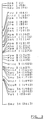
- Fig. 3 shows a restriction enzyme map of the genome gene of the above anti-tumor polypeptide of example 1.
- Fig. 4 represents a partial base sequence of said gene.
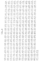
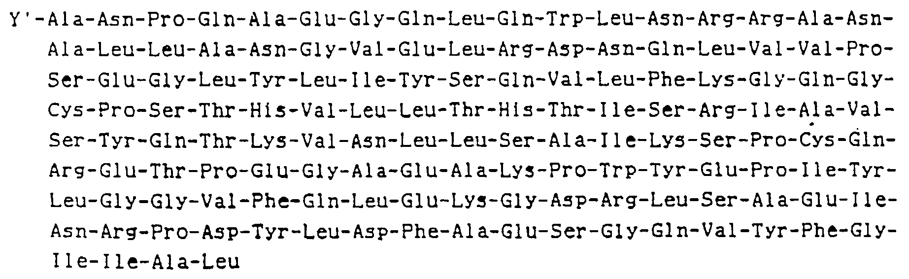
- Fig. 5 shows the base sequence of the anti-tumor polypeptide gene possessed by p12 TNF x/p and the amino acid sequence of the polypeptide coded for said base sequence.
- Fig. 6 shows the base sequence of the antitumor polypeptide gene possessed by pUC540 TNF x/p and the amino acid sequence of the polypeptide coded for said base sequence.
- Fig. 7 shows the Xho-PstI fragment of the above genome gene.
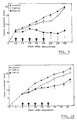
- Fig. 8 is a graph exhibiting in vitro anti-tumor activity of some of the polypeptides of the present invention as compared with the prior art TNF.
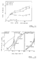
- Figs. 9-11 are graphs exhibiting in vivo anti-tumor activity of one of the polypeptides of the present invention as compared with the prior art TNF.
- Fig. 12 is a graph exhibiting the synergistic anti-tumor activity of the combined use of the anti-tumor polypeptides having the sequence and the corresponding other anti-tumor polypeptides without the sequence both being within the scope of the present invention.
- DNAs coding for the following amino acid sequence wherein Y is a peptide selected from the group consisting of the following ones:
- the portion from the Ala located downstream of the Y to the last Leu is the same as the amino acid sequence of the hitherto known fourth exon of TNF except that the fourth exon lacks the guanine consisting of the first Ala.
- the DNAs of the present invention may be synthesized chemically on the basis of processes described in "Nucleic Acids Res.”, 10 , pp. 7439-7448 (1981), “Biochemistry", 17 , pp. 1257-1267 (1978) etc.
- a process for preparing DNAs of the present invention starting with the genome DNA of human acute monocytic leukemia cell THP-1 will be detailed in examples given later.
- the base sequence of the nineteenth amino acid alanine is GCG
- the base sequence may be cleaved just before the base sequence being the same as that of the fourth exon of TNF by the use of restriction enzyme NruI (TCGCGA) to introduce some other desired base sequences. This introduction is very useful.
- the latter are incorporated into an appropriate vector DNA in an expressible manner, and the thus-obtained recombinant DNA is used to transform host organisms including animal cells, yeasts, B. subtilis, E. coll. and the like to induce the expression.
- the DNA of the present invention is incorporated downstream of the Shine-Dalgarno sequence (hereunder referred to as the SD sequence) of a vector DNA possessing a promotor sequence (being usually downstream of an operator sequence) and the SD sequence which is located downstream of the promotor sequence.
- the DNA of the present invention is incorporated into a vector DNA, and then a promotor sequence (usually together with an operator sequence) and the SD sequence are inserted upstream thereof.
- Example 1 The case where E. coli is used as the host will be illustrated in Example 1.
- the genetic information of the DNAs of the present invention can be expressed as described hereunder.
- Plasmid vector pMA56 with a promotor for alcohol dehydrogenase (ADHI) incorporated therein (“Nature", 298 , pp. 347-350, 1982) has an EcoRI site downstream of the promotor.
- the DNA of the present invention may be recovered as BamHI/PstI fragment from, for example, pUC540 TNF 21/22, pUC540 TNF 69/70, pUC540 TNF 72/73, or pUC540AMCT-1 as described in Example 2 or 3, and then may be inserted into pMA56 at the EcoRI site downstream of the ADHI promotor thereof using EcoRI/BamHI linker and PstI/EcoRI linker to be controlled by the ADHI promotor, thereby expressing the genetic information in yeast.
- ADHI alcohol dehydrogenase
- the DNA of the present invention may be recovered as a BamHI/PstI fragment from, for example, pUC540 TNF 21/22, pUC540 TNF 69/70, pUC540 TNF 72/73, or pUC540AMCT-1 as described in Example 2 or 3, and then may be inserted into pMA56 at its XhoI site downstream of the PHO5 promotor thereof using BamHI/XhoI linker and PstI/XhoI linker to be controlled by PHO5 promotor, thereby making the expression of the genetic information possible in yeast.
- B. subtilis may also be employed as the host as follows to express the genetic information of DNAs of the present invention.
- pTUB285 having ⁇ -amylase promotor which is originally possessed by B. subtilis Marburs strain ("Gene", 34 , p. 148, 1985) has a HincII site downstream of the promotor and a signal peptide.
- the DNA of the present invention may be recoverd as BamHI/PstI fragment from, for example, pUC540 TNF 21/22, pUC540 TNF 69/70, pUC540 TNF 72/73, or pUC540AMCT-1 as described in Example 2 or 3, and then may be inserted into pTUB285 at its HincII site using HincII/BamHI linker and HincII/PstI linker to be controlled by the ⁇ -amylase promotor to express the genetic information in B. subtilis.
- the anti-tumor polypeptide produced by the thus-transformed host cells may be separated and purified as follows :
- the host cells are collected by, for example, centrifugation, and then crushed by treatment with ultrasonic waves or lysozyme.
- a hypotonic solution is used, and in some cases coexistence of a surfactant such as SDS or guanidine HCl may produce a better result.
- the crushed cell-containing solution is subjected to centrifugation to provide a supernatant.
- the thus-prepared supernatant containing the anti-tumor polypeptide may be purified according to any conventional method of purifying proteins. That is, the supernatant may be subjected to purification by ion exchange chromatography using a basic anion exchanger, salting out, dialysis, gel filtration, hydrophobic chromatography, high performance molecular sieve chromatography, electrophoresis, etc., in the given order or by any desired combination of these methods.
- the basic anion exchanger is preferred to be DEAE-Sephadex A-25 or A-50.
- Sepharose CL-6B, or DEAE-Sephamil all made by Pharmacia AB
- any other diethylamino, aminoethyl, or quaternary-aminoethyl group-containing anion exchanger may be used.
- Preferable embodiments of the buffer solution available for use include Tris-HCl and phosphate buffer solutions at pH 6.6-9.0.
- any of these buffer solutions may be used at a low concentration of about 0.05 M to dilute the culture containing the anti-tumor polypeptide to a saline concentration of 0.1 M or less, and then the resulting solution is contacted with an anion exchanger which adsorbs the anti-tumor polypeptide.
- the elution of the anti-tumor polypeptide is carried out with a saline solution containing 0.1-0.2 M of NaCl or KCl .
- the anti-tumor polypeptide is eluted at a saline concentration of about 0.2.
- the contact with the anion exchanger is preferably conducted by a column process, but a batch process may be employed if the contact is conducted on a large scale.
- the solution is preferably pre-treated with an ultrafiltration membrane for removal of lower molecular materials, thereby improving the purification efficiency.
- the solution resulting from the anion exchange chromatography is subjected to dialysis and concentration followed by gel filtration.
- Embodiments of carriers for the gel filtration include Sephadex G-75 and G-100 (manufactured by Pharmacia AB), Sephacryl S-200 (manufactured by Pharmacia AB), Biogel P-100 (manufacturec by Biorad), and Toyo Pearl HW-50 and HW-55 (manufactured by Toyo Soda Corp.).
- the buffer solution intended for use in the gel filtration may be a Tris-HCl or phosphate buffer solution. To prevent adsorption it is desired that 0.2-0.5 M of a saline such as NaCl be added to the solution.
- the anti-tumor polypeptide active solution may be purified by hydrophobic chromatography.
- Butyl-Toyo Pearl 650 or the like may be used as the carrier, and a saline such as ammonium sulfate or NaCl is employed to elute the anti-tumor polypeptide.
- the anti-tumor polypeptide-containing solution purified by gel filtration or hydrophobic chromatography is then subjected to fast protein exchange chromatography using a Pharmacia FPLC (Fast Protein, Peptide, Polynucleotide, Liquid Chromatography) system to provide a purified sample.
- Pharmacia FPLC Fluorescence Chromatography
- the conditions for the fast protein anion exchange chromatography are the same as for the ion exchange chromatography using a carrier such as DESE-Sepharose mentioned previously.
- any of the polypeptides of the present invention may be purified in the same manner as described above. Namely, a solution containing crushed cells which contain said polypeptide is treated by ion exchange chromatography using a basic ion exchanger, salting out, dialysis, gel filtration, hydrophobic chromatography, high performance molecular sieve chromatography, electrophoresis, etc. in the order given here or by any desired combination of these methods.
- polypeptides of the present invention are of course highly cytotoxic to L-929 cells which have been observed to be sensitive to the hitherto known TNF.
- the polypeptides of the present invention are believed to be remarkably cytotoxic even to T-24 cells to which the prior art TNF has been reported to be thoroughly insensible ("Science", 230 , pp. 943-945, issued on Nov. 22, 1985). This cytotoxicity is believed to increase if the proportion of the number of the net basic amino acid residues to the number of all the amino acid residues constituting Y, but excluding the initiation codon Met, is more than about 14.3 %.
- the number of the net basic amino acid residues is calculated by subtracting the number of acidic amino acid residues from the number of all the basic amino acid residues, and histidine is not deemed to be a basic amino acid.
- the cytotoxicity is believed to increase more if said proportion is about 20-50 %.
- some of the polypeptides of the present invention have been observed to be remarkably cytotoxic to primary culture cells obtained from metastasis lesions of patients suffering from striated muscle tumors originating in ductus Mulleri and reported to be resistant to all chemotherapic agents.
- polypeptides of the present invention may be qualitatively and quantitatively analyzed as follows :
- L-929 cells ("Proc. Natl. Acad. Sci. U.S.A.”, 72 , pp. 3666-3670, 1983) are cultured in Eagles' Minimum Essential Medium (hereunder reffered to only as MEM) with 5 % of fetal calf serum (hereunder referred to only as FCS) added thereto until 100 ⁇ l of the medium contains 8 x 10⁴ cells, and then the cells are grown in a flat-bottomed plate having 96 wells. The growth conditions are 2 hours at 37°C in the presence of 5 % CO2, and 100 % H2O, and the procedures may be the same as for the conventional cell culture.
- MEM Eagles' Minimum Essential Medium
- FCS fetal calf serum
- Actinomycin D is then added to the medium to a final concentration of 1 ⁇ g/ml , and the volume of the culture solution is adjusted to 150 ⁇ l. Immediately thereafter 50 ⁇ l of the sample diluted appropriately with MEM medium is added to the culture solution. Here, ED50 may be determined by adjusting the dilution appropriately.
- the L-929 cells having a final volume of 200 ⁇ l are cultured for an additional 18 hours under the same conditions as described above. In order to determine the cell necrosis activity, first the whole medium is removed followed by addition of 2 % of a methyl alcoholic solution containing 0.2 % of crystal violet for fixation staining.
- Crystal violet stains all the eukaryotic cells, but does not stain those cells left in the bottom of the flask as the result of necrosis, so the cell necrosis activity may be determined directly.
- the staining degree is measured on the basis of adsorption at OD 590nm , and is compared with that of a control to determine the cell necrosis activity, This activity is defined as follows.
- the dilution of the sample which allows the survival of 50 % of L-929 cells (N) is determined.
- Rabbit TNS is used as the control. and its activity n (units/ml ) is determined using 2.4 x 10⁶ units/mg/ml of human TNF.
- the dilution which provides ED50 ofrabbit TNS is determined.
- the activity of the sample (units/ml ) is calculated by the equation N/C x n.
- cytotoxicity to A549 lung carcinoma
- LS174T colon carcinoma
- WiDr colon carcinoma
- the subject toxicity is determined by the crystal violet (0.2 %) staining method 24 hours after addition of polypeptides of the present invention or on the basis of the degree of suppression on intake of 3H-thymidine.
- THP-1 cells were suspended in the medium in such a manner that the cell content became 2 x 10⁵ ml .
- the resulting suspension was cultured at 37°C for 4 days, and the resulting culture solution was subjected to centrifugation to collect THP-1 cells aseptically.
- These cells were moved to 200l of a serum-free RPMI-1640 medium placed in another culture tank followed by addition of 100 ng/ml of TPA thereto.
- the solution was cultured under aseptic conditions at 37°C for 5 days with gentle stirring (induction).
- the thus-prepared culture solution was subjected to centrifugation to separate and remove the cells, thereby collecting a supernatant having 100 units/ml of anti-tumor polypeptide activity.
- This supernatant was concentrated ten times with an ultrafiltration membrane (HVLP OHV20 manufactured by Millipore Corp.).
- Solid ammonium sulfate (65 % saturation) was added to the resulting concentrated solution and dissolved therein to precipitate proteins.
- the precipitate was collected by centrifugation (at 1000 r.p.m. for 20 minutes), and then dissolved in a small quantity of 0.05 M Tris-HCl buffer solution pH 7.7).
- the resulting solution was dialyzed against the same type buffer solution (5°C, 24 hrs.). The same quantity of the same type buffer solution was added to the inner solution which was then charged into a DEAE-Toyo Pearl M650 column (5 x 40 cm) previously equilibrated with the same type buffer solution. The column was washed with 1.0 l of the same type buffer solution followed by elution with the same type buffer solution containing 0.2 M of NaCl.
- a 40 % saturated solution of ammonium sulfate was added to and dissolved in the dialysis inner solution which was then subjected to centrifugation to remove the insolubles, and then subjected to hydrophobic chromatography at a rate of 2.0 ml/min. using a Butyl-Toyo Pearl 650S columm (2.5 x 30 cm) previously equilibrated with a 0.05 M Tris-HCl buffer solution containing ammonium sulfate at 40 % saturation. Then, anti-tumor polypeptide active fractions were collected and dialyzed against a 0.05 M Tris-HCl buffer solution (pH 7.8).
- the dialysis inner solution was charged into a Mono QHR 5/5 column (fast protein anion exchange column manufactured by Pharmacia AB) previously equilibrated with 50 mM of Tris-HCl buffer solution (pH 8.5), washed with the same type buffer solution, and then subjected to gradient elution where the NaCl concentration was successively increased to 0.1, 0.15, 0.2, and 0.3 M to elute anti-tumor polypeptide active substances.
- the anti-tumor polypeptide fractions were eluted with 0.2 M of NaCl and then purified to a specific activity of 6.25 x 10⁶ units/mg protein. The fractions were purified 5 to 15 times in this step, and the recovery was 80 % or more.
- Fig. 1 The elution pattern observed in this second FPLC is shown in Fig. 1 of the drawings.
- the vertical axis represents absorption at 280 nm (%), while the horizontal axis represents elution time (min.).
- the anti-tumor polypeptide active portions were eluted with 0.1 M of NaCl , and this result agreed well with the peak at 280 nm.
- fractions corresponding to the three peaks eluted with a retention time of 35, 36 and 37.8 minutes, respectively (hereunder, those fractions corresponding to the three peaks are referred to only as TNF-1, TNF-2 and TNF-3, respectively).
- the respective fractions were subjected to chromatography again under the same conditions as described above for further purification. All of the fractions were proved to be simple proteins by the procedures given below.
- TNF-1, TNF-2 and TNF-3 are all simple substances in view of their behavior in reverse phase FPLC.
- SDS-polyacryl amide gel electrophoresis (hereunder referred to as SDS-PAGE). That is, using a Slab electrophoresis unit manufactured by Biorad Corp. (Protein. 16 cm), the sample was charged into 15.0 % polyacryl amide gel containing 0.1 % of SDS, and the electrophoresis was conducted at a constant current of 20 mA. Then the detection of proteins was attempted by silver impregnation. In each case, only a single band was detected at the position of 17.4 Kd. and no other protein band was found. Accordingly, TNF-1, TNF-2 and TNF-3 all proved to be single proteins in view of the behavior in SDS-PAGE. All the isoelectric points (pI) of these protein samples were determined to be 5.7 according to the polyacryl amide gel isoelectric electrophoresis using Ampholine polyacryl amide gel manufactured by LKB Picassoer AB.
- the amino acid sequence of these three anti-tumor polypeptides were determined by analysis of about 10 ⁇ g of each of them starting with the N-termini using an amino acid sequence analyzer (Model 470A) manufactured by Applied Biosystems Inc.
- the N-terminal amino acid sequence of TNF-1, TNF-2 and TNF-3 were found to be as follows :
- the fourth amino acid represented by X in the above N-terminal amino acid sequence is an amino acid which cannot be identified with any of the gas phase amino acid sequencer now available : it is certain that it is not Ser, and possibly it is Cys which is an amino acid not detectable by any prior art method.
- THP-1 cells were cultured in a 10 % FBS-containing RPM-1-1640 medium at 37°C in the presence of 5 % CO2. When the number of cell reached 1 x 10⁶/ml, 100 ⁇ m/ml of 12-tetradecanoylphorbol-13-acetate (hereunder referred to only as TPA) was added to the medium, and the culturing was continued. The cells collected 8 and 70 hours after addition of TPA were employed for extraction of mRNAs.
- TPA 12-tetradecanoylphorbol-13-acetate
- the extraction of the mRNAs from the cells was conducted as follows : The cells were collected by centrifugation, and washed once with PBS (-) (0.8 % NaCl + 0.02 % KCl + 0.02 % KH2PO4 + 0.115 % Na2HPO4). The collected cells were well suspended in 50 ml of a buffer solution for extraction of RNAs followed by addition of Nonident-P40 to obtain a final concentration of 0.5 % and treatment with a Teflon homogenizer at 10 strokes to crush the cells. Thereafter, the homogenate was subjected to centrifugation at 10000 g at 4°C for 1 min. to obtain cell extracts in the supernatant.
- poly (A) RNA is referred to as mRNA.
- a cDNA library was prepared in two ways using the mRNA obtained as in the above manner.
- oligo (dT) complementary to the 3'-poly (A) sequence of mRNA was annealed with mRNA to prepare a primer for reverse transcriptase. Then, the primer was subjected to the reaction of the reverse transcriptase in the presence of dATP, dGTP, dCTP, and dTTP to synthesize a cDNA complementary to the mRNA. Thereafter, the thus-obtained mRNA/cDNA hybrid was nicked with RNaseH at the mRNA region and the mRNA was replaced by DNA polymerase I and E. coli DNA lygase to synthesize double-strand DNA.
- the 3'-end of the thus-obtained double-strand DNA was labeled with terminal deoxynucleotidyl transferase to add 10-20 dC tails thereto.
- plasmid vector pBR322 replicable in E. coli was treated with restriction enzyme PstI to prepare linear plasmid DNA. This was then labeled at the 3'-end with terminal deoxynucleotidyl transferase to add 10-20 dC tails thereto.
- the dC-tailed plasmid vector and the dC-tailed double-strand DNA were annealed and then transferred to E. coli by transformation with calcium to provide the transformed strain as a cDNA library.
- double-strand DNA with oligo (dT) tails complementary to the poly (A) sequence of mRNA was annealed and reacted with reverse transcriptase in the presence of dATP, dGTP, dCTP, and dTTP to synthesize the complementary cDNA.
- this newly-synthesized cDNA was labeled with terminal deoxynucleotidyl transferase to add dC-tails thereto followed by annealing with the previously dC-tailed plasmid vector and ligation to prepare a plasmid containing mRNA/cDNA hybrid.
- this plasmid was treated with RNaseH, DNA polymerase I and E. coli DNA ligase to replace the mRNA by DNA.
- RNaseH RNaseH
- DNA polymerase I E. coli DNA ligase
- E. coli DNA ligase E. coli DNA ligase
- the cDNA library obtained in the above-described manner was grown on a nitrocellulose filter and then in a medium containing chloramphenicol ("Gene", 10 , pp. 63-67, 1980) to increase the number of plasmids.
- the nitrocellulose filter on which the cDNA library had grown was immersed in a 0.5 N NaOH solution to break the E. coli cell walls as well as to separate the double-strand of the plasmid DNA into two single-strands which were then immersed into a 1 M Tris-HCl solution (pH 7.5) and allowed to stand at room temperature for 10 minutes.
- the nitrocellulose filter was immersed into a 0.5 M Tris-HCl (pH 7.5)/1.5 M NaCl solution at room temperature for 10 minutes and then allowed to be air-dried. After being dried well, the nitrocellulose filter was treated at 80°C for 2 hours.
- the 5'-end of the synthesized DNA harboring 23 bases was labeled with ⁇ 32PATP, T4DNA kinase, and this labeled 5'-end was used thereafter as the DNA probe in screening for cDNA clones.
- the nitrocellulose filter treated at 80°C was hybridized in six volumes of NET (1 x NET, 0.15 M NaCl, 0.015 M Tris-HCl at pH 7.5, 1 mM EDTA, 250 ⁇ g/ml of E. coli tRNA and 0.5 % of NP-40) at 42°C overnight and then washed with six volumes of SSC (1 x SSC, 0.15 M NaCl and 0.015 M sodium citrate) at 0°C.
- the nitrocellulose filter was further washed twice with two volumes of SSC each time at 0°C for 5 minutes and air-dried followed by autoradiography.
- the clones found to be positive by autoradiography were subjected to the Maxam-Gilbert base sequencing method to determine their base sequences to narrow the positive clone candidates.
- THP-1 cells (3 x 10⁹) were cultured in a medium containing 100 ng/ml of TPA for 8 hours and then suspended in 100 ml of 150 mM NaCl + 100 mM EDTA solution followed by addition of 10 ml of 10 M sodium perchlorate and 10 ml of 10 % SDS. Next, 12 ml of 5 M NaCl were added to the mixture followed by warming at 60°C for 15 minutes. An equal volume of a chloroform-isoamyl alcohol (24:1) mixture was added to the resulting solution which was then mixed gently. The mixture was placed in a Hitachi quick-freezing centrifuge for centrifugation at 10000 r.p.m. for 10 minutes to obtain a supernatant.
- the DNA solution treated with the protease was gently mixed with a water-saturated phenol + m-cresol + isoamyl alcohol (100 : 14 : 0.1) mixture and then subjected to centrifugation at 300 r.p.m. at normal temperature for 10 minutes to separate the supernatant. An equal volume of isopropanol was added to she supernatant, and the resulting precipitate was rolled around a Pasteur pipette. The DNA was washed with 70 % ethanol and dissolved in 100 ml of TSE.
- TSE was added to the DNA solution to a final concentration of 800 ⁇ g/ml followed by addition of 0.95 g/ml of CsCl and further of one tenth volume of an ethidium bromide solution (5 mg/ml) to produce a homogenous solution which was then subjected to centrifugation with a Beckman type-60 rotor at 45000 r.p.m. at 20°C for 48 hours for purification of the DNA by the density-gradient method. After the centrifugation was completed, the DNA was recovered with a Pasteur pipette while pursueing the DNA band by irradiation of UV at 360 nm. Next, CsCe saturated isopropyl alcohol was added to the DNA solution and mixed therewith several times. This mixing was repeated ten times to remove the ethidium bromide.
- the thus-prepared DNA solution was dialyzed against 2 l of TSE at 4°C for twenty-four hours to obtain the DNA.
- the DNA concentration was 650 ⁇ g/ml and 18 mg of the DNA was obtained.
- the DNA obtained in the manner described above was subdivided into 15 ⁇ g portions, and subjected to the action of several kinds of restriction enzymes for its complete decomposition.
- the lengths of the anti-tumor polypeptide gene fragments obtained by the action of the enzymes were analyzed by the Southern methed. Namely, 50 units each of ApaI (GGGCCC), XhoI (CTCGAG), BamHI (GGATCC), EcoRI(GAATTC), SstI (GAGCTC) and KpnI (GCTACC) were employed, and the DAN was warmed in an appropriately-buffered solution having an appropriate saline concentration at 37°C overnight.
- Two hundred ng of the cDNA obtained in the above-described screening step (6) were dissolved in 30 ⁇ l of a reaction solution [50 mM Tris-HCl (pH 7.5) + 10 mM MgCl2 + 10 mM DTT] and 20 ⁇ C of ⁇ -32PdCTP and 5 ⁇ M each of dATP, dGTP, and dTTP were added to the resulting solution followed by addition of 12.5 pg of DNase and 10 units of DNA polymerase. Reaction was carried out at 20°C for one hour.
- a reaction solution [50 mM Tris-HCl (pH 7.5) + 10 mM MgCl2 + 10 mM DTT] and 20 ⁇ C of ⁇ -32PdCTP and 5 ⁇ M each of dATP, dGTP, and dTTP were added to the resulting solution followed by addition of 12.5 pg of DNase and 10 units of DNA polymerase. Reaction was carried out at 20°
- the nitrocellulose filter with the DNA adsorbed thereon was uniformly immersed into 1 ml of a solution containing 50 % formaldehyde, 5 x SSC (0.15 M NaCl + 0.015 sodium citrate), 5 x FBP, 1 % glycine, a 20 mM phosphate buffer solution (pH 6.8) and 100 ⁇ g/ml of calf thymus degenerated DNA, and was then sealed in a plastic bag followed by warming at 42°C overnight.
- the filter was immersed into 1 ml of a solution containing 50 % formaldehyde, 5 x SSC, 1 x FBP, a 20 mM phosphate butter solution (pH 6.8), and 100 ⁇ g/ml of calf thymus degenerated DNA with 2 x 10⁷ c.p.m. of the probe added thereto and warmed at 42°C overnight.
- the nitrocellulose filter was transferred to a 2 x SSC solution for one hour washing at 68°C to prevent adsorption of non-specific DNA probes. The washing was repeated with 0.1 x SSC for 5 minutes, and the filter was air-dried. DNA fragments which specifically hybridize with the probe were detected by exposure of Kodak X-ray film for twenty-four hours.
- the resulting DNA fragments were partioned by 1.5 % agar gel, and an agar gel portion of around 2.6 kb was cut off.
- Recovery of the DNA from the agar was conducted as follows : First, the agar was added to 15 ml of a solution prepared by adding 22.5 g of KI to 15 ml of a 10 mM phosphate buffer solution (pH 7.0), and the mixture was warmed to 60°C to dissolve the agar.
- the DNA-containing solution was adsorbed on "Biogel HTP" manufactured by Biorad Corp., washed well with a 10 mM phosphate buffer solution, and then the DNA was eluted with a 1 M phosphate buffer solution and 0.5 % SDS followed by dialysis against ISE at 4°C for twenty four hours.
- Terminal deoxynucleotidyl transferase was used to add dCTP tails to the obtained DNA.
- pNF was cleaved with restriction enzyme KpnI, and thus the cyclic double-stranded DNA was made into linear double-stranded DNA having KpnI cleavage sites at its ends.
- the two 3'-ends were tailed with dCTP.
- the two tailed ones were annealed with each other to form a cyclic chimera. This chimera was incorporated into E. coli RRI to prepare a library comprising 2 x 10⁴ independent colonies.
- the above-mentioned probe was employed to choose the above genome library, thereby providing a clone.
- the restriction enzyme map of the obtained clone and its partial base sequence are shown in Figs. 3 and 4, respectively.
- the restriction enzyme Xhol / PstI fragment of the genome gene obtained in step (8) above (811 base DNA fragment of from the 340th to 1150th bases of the partial base sequence of the TNF genome DNA shown in Fig. 4 was inserted into plasmid vector pUC12 (manufactured by Pharmacia AB) at its restriction enzyme SalI/PstI site to form plasmind pUC12 TNF x/p.
- This plasmid has the promotor region, operator region and SD sequence of lactose operon region, and further possesses, downstream of those regions, a synkaryon gene consisting of 45 bp involing the 5'-terminal region of a ⁇ -galactosidase gene and the genome gene attached thereto.
- the protein expressed by E. coli incorporating this plasmid therein is a composite protein comprising the base sequence of the N-terminal region of ⁇ -galactosidase genome and the DNA fragment (Fig. 5).
- the 811 bp restriction enzyme XhoI/PstI fragment of the qenome gene was inserted into plasmid vector pUC540 having Tac promotor and SD sequence at the restriction enzyme SalI/PstI site to prepare plasmid pUC540 TNF x/p.
- Plasmid vector pUC540 is prepared by cloning the EcoI/BamHI fragment of plasmid pDR540 which has a Tac promotor (commercially available from Pharmacia AB) to the EcoRI/BamHI site of plasmid vector pUC8 (commercially available from the same company).
- Plasmid pUC540 TNF x/p has a restriction enzyme BamHI site downstream of the SD sequence. So,if an exogenous gene is inserted at this BamHI site, its expression is made possible only by addition of isopropyl ⁇ -D-thiogalactopyranoside ( hereunder preferred to as IPTG).
- E. coli JM 103 incorporating the above plasmid p12 TNF x/p or pUC540 TNF x/p therein was pre-cultured in a 1 x YT medium containing 50 ⁇ g/ml of amplicillin(0.8 % bactotryton + 0.5 % bactoyeasts + 0.5 % NaCl) at 37°C, and then transferred in a proportion of 1 % to a 500 ml Sakaguchi flask containing 100 ml of 1 x YT medium with 50 ⁇ g/ml of ampicillin incorporated therein followed by culturing at 37°C in the same manner as described above. When the OD660 reached 0.3.
- Plasmid pUC540 TNF x/p has the restriction enzyme BamHI site downstream of the SD sequence. Accordingly, if an exogenous gene is inserted at this BamHI site, the gene can be expressed only by addition of IPTG thereto.
- the genome gene shown in Fig. 3 was cleaved with XhoI and PatI, and the XhoI/PstI fragment shown in Fig. 7 was recovered. Then, this fragment was cleaved with HincII fragment to recover 294 bp XHoI-HincII fragment and 521 bp HincII/PatI fragment. The 294 bp fragment was partially cleaved with DdeI to recover 206 bp DdeI/HincII fragment.
- E. coli JM103 with pUC540AMCT-1 incorporated therein (deposited with the Fermentation Research Institute (FRI) in Japan as No. 8630 since Jan. 31, 1986), which corresponds to one of the DNAs of the present invention where Y is Met-Val-Lys-Ser-Cys-Thr-Arg-Thr-Pro-Ser-Arg-Lys-Pro-Val-Ala-His-Val-Val, was pre-cultured in a 1 x YT medium containing 50 ⁇ g/ml of ampicillin (0.8 % bactotrypton + 0.5 % bactoyeast extracts + 0.5 % NaCl) at 37°C, and then transferred in a proportion of 1 % to a 500 ml of a Sakaguchi flask containing 100 ml of a 1 x YT medium with 50 ⁇ g/ml of ampicillin added thereto.
- FPI Fermentation Research Institute
- pUC540AMCT-1 was added to the culture in a quantity of 18.7 units/ml, 62.5 units/ml, 187.5 units/ml, 625 units/ml and 1875 units/ml, respectively, and the culture was continued for an additional twenty-four hours.
- 1 ⁇ Ci/ml of 3H-thymidine was added to the culture followed by culturing for an additional nine hours.
- Table 9 The results are shown in Table 9.
- Each of pUC540 TNF 21/22, pUC540 TNF 69/70 and pUC540 TNF 72/73 prepared in Example 2 is a recombinant TNF which is combined with the BamHI downstream region of a Tac promotor, and which has the initiation codon ATG immediately after the restriction enzyme BamHI cleavage point, the first codon of the second amino acid which follows the ATG being G.
- E. coli JM103 with pUC540 TNF Nco21/22, 69/70 or 72/73 incorporated therein (deposited with the Fermentation Research Institute (FRI) in Japan as Nos. 8628, 8629, and 8627, respectively, since Jan.
- 1 x 10⁴/well of T-24 cells were suspended in a mixture of RPMI1640 and 10 w/w % FCS, and then grown in a Linbro 96-well microtiter plate followed by culturing at 37°C for 48 hours in the presence of 0.5 % CO2. Thereafter, pUC540 TNF Nco21/22 was added to the culture in a quantity of 62.5 units/ml, 625 units/ml and 1250 units/ml, and the culturing was continued for an additional twenty-four hours. Next, 1 ⁇ Cl/ml of 3H-thymidine was added to the culture followed by nine more hours of culturing.
- cytotoxicity of some of the anti-tumor active polypeptides of the present invention on WiDr cells was studied. 4 x 10³/well of WiDr cells were used, and the incubation time with the polypeptides was 48 hours. Each of the polypeptides was added in proportions of 103, 104, 5 x 10⁴ and 3 x 10⁵ units/ml. The results are shown in Fig. 8 wherein the symbols stands for :
- the procedures used were substantially the same as in the case of L-929 cells, 1.0 x 10⁴ well (4.0 x 10⁴/well only in the case of T-24) were used, and the respective TNFs were added to the assay well in a proportion of 4 % of the 8 ⁇ l final volume.
- the incubation was conducted at 37°C in the presence of CO2.
- 0.2 % of crystal violet was used for staining, and the staining degree was measured on the basis of adsorption at OD595 to calculate the survival ratios.
- mice were used for the respective TNFs purified with an imnuno-column.
- LPS was used as the control. They were administered it according to the following schedule to evaluate the activity against B-16 melanoma.
- TABLE 13 Days after inoculation of 3 x 10⁵ cells/id TNF-G/head TNF-S/head units LPS units LPS 12 600 317 pg 275 25 pg 14 600 317 pg 275 25 pg 16 500 1.9 ng 500 125 pg 18 500 1.9 ng 500 125 pg 20 270 1.0 ng 270 146 pg The results are shown in Fig. 9.
- mice Ten 8-week old C3H/HE mice were used for the respective TNFs purified with an immuno-column.
- LPS was used as the control. They were administered it according to the following schedule to evaluate the activity against MH134 Hepatoma.
- Table 14 Days after inoculation of 2 x 10³ cells/id TNF-G/head TNF-S/head units LPS units LPS 6 180 95 pg 80 9.5 pg 8 180 95 pg 80 9.5 pg 10 200 740 pg 200 50 pg 12 200 740 pg 200 50 pg 14 120 444 pg 120 31 pg The results are shown in Fig. 10.
- mice Three 8 week old BALB/C-nuSlc mice were used for the respective TNFs purified with an immuno-column. LPS was used as the control. They were administered it according to the following schedule to evaluate the activity against A549. Table 15 Days after inoculation of 5 x 10⁶ cells/id TNF-G/head TNF-S/head units LPS units LPS 11 650 313 pg 650 500 pg 12 650 313 pg 650 500 pg 13 650 313 pg 650 500 pg 14 650 313 pg 650 500 pg 15 650 313 pg 650 500 pg The results are shown in Fig. 11.
- Each of the crushed E. coli cells containing the respective polypeptides prepared in the manner of the above-described examples was mixed in a volumetric ratio of 1:1, and the TNF activity was determined in a manner similar to that described in Example 1.
- the polypeptides used are listed below.
- the novel DNAs synthesized according to the present invention can express the novel anti-tumor polypeptides which are cytotoxic to human tumor cells, but not to normal cells. Furthermore, the present polypeptides are very cytotoxic even to T-24 cells to which the prior art TNF is reported to be entirely insensitive. The present invention provides also those polypeptides which are remarkably cytotoxic to primary culture cells obtained from metastasis lesions of patients suffering from striated muscle tumors originating in ductus Mulleri and reported to be resistant to all chemotherapic agents.
Landscapes
- Chemical & Material Sciences (AREA)
- Health & Medical Sciences (AREA)
- Life Sciences & Earth Sciences (AREA)
- Organic Chemistry (AREA)
- General Health & Medical Sciences (AREA)
- Molecular Biology (AREA)
- Biochemistry (AREA)
- Biophysics (AREA)
- Zoology (AREA)
- Genetics & Genomics (AREA)
- Medicinal Chemistry (AREA)
- Gastroenterology & Hepatology (AREA)
- Proteomics, Peptides & Aminoacids (AREA)
- Toxicology (AREA)
- Medicines That Contain Protein Lipid Enzymes And Other Medicines (AREA)
- Peptides Or Proteins (AREA)
- Preparation Of Compounds By Using Micro-Organisms (AREA)
- Medicines Containing Material From Animals Or Micro-Organisms (AREA)
Claims (9)
- Polypeptid, dargestellt durch die folgende Aminosäuresequenz:worin Y' ausgewählt ist aus der folgenden Gruppe:


- Verfahren zur Herstellung eines Polypeptides, dargestellt durch die folgende Aminosäuresequenz:worin Y' ausgewählt ist aus der folgenden Gruppe:
 gekennzeichnet durch Kultur eines Mikroorganismus, der ein Plasmid enthält, das geeignet ist in dem Wirt-Mikroorganismus zu wachsen und darin DNA einzubauen, die die Aminosäuresequenz codiert, die aufgebaut ist durch Zusatz von Met am Ende der vorstehenden mit Y' beginnenden Aminosäuresequenz.
- Antitumormittel, umfassend ein Polypeptid, dargestellt durch die folgende Aminosäuresequenz:worin Y' ausgewählt ist aus der folgenden Gruppe:
- DNA, dadurch gekennzeichnet, daß sie für die folgende Aminosäuresequenz codiert:worin Y ein Peptid ist, ausgewählt aus der folgenden Gruppe:


- DNA nach Anspruch 4, worin die Basen der Aminosäure vor Ala im Anschluß an Y TC sind, die Basen von Ala GCG sind und die erste Base der Aminosäure nach Ala A ist.
- Plasmid, das DNA-Codierungen für die folgende Aminosäuresequenz enthält:worin Y ein Peptid ist, ausgewählt aus der folgenden Gruppe:


- Verfahren zur Herstellung von DNA, die für die folgende Aminosäuresequenz codiert:
 worin Y ein Peptid ist, ausgewählt aus der folgenden Gruppe:
 gekennzeichnet durch Kombinieren des N-Endes der DNA, die codiert für:
 worin X' ein Peptid ist, ausgewählt aus der folgenden Gruppe:
 mit DNA, codierend für Met-Arg-Ile-Arg.
- Verfahren nach Anspruch 7, worin ein rekombinanter TNF, der mit der BamHI-Downstream-Region eines Tac-Promotors kombiniert ist, der das Initiations-Codon ATG unmittelbar nach dem Spaltungspunkt des Restriktions-Enzyms BamHI hat, wobei das erste Codon der zweiten Aminosäure, die auf das ATG folgt, G ist, und der eine Aminosäuresequenz hat, dargestellt durchworin X' ein Peptid ist, ausgewählt aus der folgenden Gruppe:
 gespalten wird mit dem Restriktions-Enzym NcoI, wobei die Spaltungsstellen der jeweiligen Teile repariert werden und zwischen die Teile doppelsträngige DNA eingeführt wird.
- Verfahren nach Anspruch 8, worin die einzuführende doppelsträngige DNAist.

Applications Claiming Priority (6)
| Application Number | Priority Date | Filing Date | Title |
|---|---|---|---|
| JP21302/86 | 1986-02-04 | ||
| JP2130286 | 1986-02-04 | ||
| JP24220/86 | 1986-02-07 | ||
| JP2422086 | 1986-02-07 | ||
| JP169522/86 | 1986-07-17 | ||
| JP61169522A JP2544114B2 (ja) | 1985-11-15 | 1986-07-17 | 新規dna及びそれを含有する新規プラスミド |
Publications (3)
| Publication Number | Publication Date |
|---|---|
| EP0247906A2 EP0247906A2 (de) | 1987-12-02 |
| EP0247906A3 EP0247906A3 (en) | 1988-12-07 |
| EP0247906B1 true EP0247906B1 (de) | 1994-12-28 |
Family
ID=27283374
Family Applications (1)
| Application Number | Title | Priority Date | Filing Date |
|---|---|---|---|
| EP87400261A Expired - Lifetime EP0247906B1 (de) | 1986-02-04 | 1987-02-04 | Für Antitumor-Polypeptide kodierende DNS, die Polypeptide und diese Polypeptide enthaltenden Antitumor-Wirkstoffe |
Country Status (5)
| Country | Link |
|---|---|
| US (1) | US5081021A (de) |
| EP (1) | EP0247906B1 (de) |
| AT (1) | ATE116367T1 (de) |
| CA (1) | CA1340998C (de) |
| DE (1) | DE3750915T2 (de) |
Families Citing this family (23)
| Publication number | Priority date | Publication date | Assignee | Title |
|---|---|---|---|---|
| AU1346488A (en) * | 1987-02-26 | 1988-09-26 | Cetus Corporation | Arginine-depleted human tumor necrosis factor |
| US5997859A (en) * | 1988-03-21 | 1999-12-07 | Chiron Corporation | Method for treating a metastatic carcinoma using a conditionally lethal gene |
| US5716826A (en) * | 1988-03-21 | 1998-02-10 | Chiron Viagene, Inc. | Recombinant retroviruses |
| US6133029A (en) | 1988-03-21 | 2000-10-17 | Chiron Corporation | Replication defective viral vectors for infecting human cells |
| US6569679B1 (en) | 1988-03-21 | 2003-05-27 | Chiron Corporation | Producer cell that generates adenoviral vectors encoding a cytokine and a conditionally lethal gene |
| JP2828988B2 (ja) * | 1988-04-03 | 1998-11-25 | 源一郎 杣 | 新規dnaとその生産方法、それを有する新規プラスミド、新規ポリペプチドとその生産方法、及び該ポリペプチドからなる新規抗腫瘍剤 |
| DE3841755A1 (de) * | 1988-12-12 | 1990-06-13 | Basf Ag | Neue tnf-peptide |
| DE3841763A1 (de) * | 1988-12-12 | 1990-06-13 | Basf Ag | Neue tnf-peptide |
| DE3843534A1 (de) * | 1988-12-23 | 1990-07-12 | Basf Ag | Neue tnf-polypeptide |
| JP3203599B2 (ja) * | 1989-10-24 | 2001-08-27 | カイロン コーポレイション | 感染性タンパク質デリバリーシステム |
| DK53291D0 (da) * | 1991-03-25 | 1991-03-25 | Carlbiotech Ltd As | Smaa peptider og peptidrelaterede stoffer samt farmaceutiske praeparater indeholdende saadanne forbindelser |
| WO1995014091A2 (en) | 1993-11-18 | 1995-05-26 | Chiron Viagene, Inc. | Compositions and methods for utilizing conditionally lethal genes |
| US5888814A (en) * | 1994-06-06 | 1999-03-30 | Chiron Corporation | Recombinant host cells encoding TNF proteins |
| CN1049663C (zh) * | 1994-09-01 | 2000-02-23 | 中国科学院上海生物工程研究中心 | 突变的人肿瘤坏死因子 |
| US7101974B2 (en) | 2000-03-02 | 2006-09-05 | Xencor | TNF-αvariants |
| US7056695B2 (en) | 2000-03-02 | 2006-06-06 | Xencor | TNF-α variants |
| US7244823B2 (en) * | 2000-03-02 | 2007-07-17 | Xencor | TNF-alpha variants proteins for the treatment of TNF-alpha related disorders |
| US7446174B2 (en) | 2001-03-02 | 2008-11-04 | Xencor, Inc. | Protein based TNF-α variants for the treatment of TNF-α related disorders |
| US20070172449A1 (en) * | 2000-03-02 | 2007-07-26 | Xencor, Inc. | TNF-alpha VARIANT FORMULATIONS FOR THE TREATMENT OF TNF-alpha RELATED DISORDERS |
| US7662367B2 (en) * | 2000-03-02 | 2010-02-16 | Xencor, Inc. | Pharmaceutical compositions for the treatment of TNF-α related disorders |
| US7687461B2 (en) * | 2000-03-02 | 2010-03-30 | Xencor, Inc. | Treatment of TNF-α related disorders with TNF-α variant proteins |
| US7285269B2 (en) * | 2002-12-02 | 2007-10-23 | Amgen Fremont, Inc. | Antibodies directed to tumor necrosis factor |
| US7964470B2 (en) * | 2006-03-01 | 2011-06-21 | Taiwan Semiconductor Manufacturing Company, Ltd. | Flexible processing method for metal-insulator-metal capacitor formation |
Citations (6)
| Publication number | Priority date | Publication date | Assignee | Title |
|---|---|---|---|---|
| WO1986004606A1 (en) * | 1985-02-07 | 1986-08-14 | Cetus Corporation | Cysteine-depleted muteins of biologically active human tumor necrosis factor proteins |
| EP0205038A1 (de) * | 1985-05-29 | 1986-12-17 | Suntory Limited | Polypeptid, Verfahren zu dessen Herstellung, Mikroorganismus und pharmazeutische Verwendung |
| EP0211321A2 (de) * | 1985-07-23 | 1987-02-25 | Mochida Pharmaceutical Co., Ltd. | Aminosäurezusammensetzung, Verfahren zu deren Herstellung und diese enthaltende medizinische Zusammensetzung |
| EP0220482A1 (de) * | 1985-09-30 | 1987-05-06 | Suntory Limited | Plasmid und seine Verwendung |
| EP0251037A2 (de) * | 1986-06-20 | 1988-01-07 | Dainippon Pharmaceutical Co., Ltd. | Polypeptid-Mutanten des menschlichen TNF und für diese Mutanten codierende DNS |
| EP0368367A1 (de) * | 1984-12-21 | 1990-05-16 | Biogen, Inc. | Reinigung, Herstellung und Verwendung von Tumornekrosefaktoren |
Family Cites Families (7)
| Publication number | Priority date | Publication date | Assignee | Title |
|---|---|---|---|---|
| US4769326A (en) * | 1980-02-29 | 1988-09-06 | The Regents Of The University Of California | Expression linkers |
| US4532207A (en) * | 1982-03-19 | 1985-07-30 | G. D. Searle & Co. | Process for the preparation of polypeptides utilizing a charged amino acid polymer and exopeptidase |
| EP0155549B1 (de) * | 1984-03-06 | 1991-03-20 | Dainippon Pharmaceutical Co., Ltd. | DNS den menschlichen Tumornekrosisfaktor kodierend und das menschliche Tumornekronisfaktor-Polypeptid |
| GR851626B (de) * | 1984-07-05 | 1985-11-26 | Genentech Inc | |
| US4677063A (en) * | 1985-05-02 | 1987-06-30 | Cetus Corporation | Human tumor necrosis factor |
| US4677069A (en) * | 1984-12-18 | 1987-06-30 | Cornell Research Foundation, Inc. | Clam derived proteinases |
| JPH064675B2 (ja) * | 1985-07-29 | 1994-01-19 | 伝一 水野 | 抗腫瘍性ポリペプチド |
-
1987
- 1987-02-04 EP EP87400261A patent/EP0247906B1/de not_active Expired - Lifetime
- 1987-02-04 AT AT87400261T patent/ATE116367T1/de not_active IP Right Cessation
- 1987-02-04 DE DE3750915T patent/DE3750915T2/de not_active Expired - Fee Related
- 1987-02-04 CA CA000528947A patent/CA1340998C/en not_active Expired - Fee Related
- 1987-02-04 US US07/010,692 patent/US5081021A/en not_active Expired - Fee Related
Patent Citations (6)
| Publication number | Priority date | Publication date | Assignee | Title |
|---|---|---|---|---|
| EP0368367A1 (de) * | 1984-12-21 | 1990-05-16 | Biogen, Inc. | Reinigung, Herstellung und Verwendung von Tumornekrosefaktoren |
| WO1986004606A1 (en) * | 1985-02-07 | 1986-08-14 | Cetus Corporation | Cysteine-depleted muteins of biologically active human tumor necrosis factor proteins |
| EP0205038A1 (de) * | 1985-05-29 | 1986-12-17 | Suntory Limited | Polypeptid, Verfahren zu dessen Herstellung, Mikroorganismus und pharmazeutische Verwendung |
| EP0211321A2 (de) * | 1985-07-23 | 1987-02-25 | Mochida Pharmaceutical Co., Ltd. | Aminosäurezusammensetzung, Verfahren zu deren Herstellung und diese enthaltende medizinische Zusammensetzung |
| EP0220482A1 (de) * | 1985-09-30 | 1987-05-06 | Suntory Limited | Plasmid und seine Verwendung |
| EP0251037A2 (de) * | 1986-06-20 | 1988-01-07 | Dainippon Pharmaceutical Co., Ltd. | Polypeptid-Mutanten des menschlichen TNF und für diese Mutanten codierende DNS |
Also Published As
| Publication number | Publication date |
|---|---|
| CA1340998C (en) | 2000-05-23 |
| DE3750915T2 (de) | 1995-08-24 |
| US5081021A (en) | 1992-01-14 |
| EP0247906A3 (en) | 1988-12-07 |
| DE3750915D1 (de) | 1995-02-09 |
| EP0247906A2 (de) | 1987-12-02 |
| ATE116367T1 (de) | 1995-01-15 |
Similar Documents
| Publication | Publication Date | Title |
|---|---|---|
| EP0247906B1 (de) | Für Antitumor-Polypeptide kodierende DNS, die Polypeptide und diese Polypeptide enthaltenden Antitumor-Wirkstoffe | |
| KR940010865B1 (ko) | 종양 괴사 인자의 생산 방법 및 종양 괴사 인자의 수율을 증가시키는 방법 | |
| US4414150A (en) | Hybrid human leukocyte interferons | |
| EP0285448B1 (de) | Leukämie hemmender Faktor | |
| EP0043980B1 (de) | Reifes menschliches Leukozyteninterferon A, Verfahren zu seiner mikrobiellen Produktion, Zwischenprodukte dazu und es enthaltende Zusammensetzungen. | |
| US6482613B1 (en) | Microbial production of mature human leukocyte interferons | |
| KR910000460B1 (ko) | 인간의 콜로니 자극인자(CSFs)에 대한 유전자 암호화 | |
| JPS60252496A (ja) | 新規なヒト生理活性ポリペプチド | |
| JPH05279398A (ja) | ヒト抗トロンビンiii | |
| IE51850B1 (en) | Hybrid human leukocyte interferons | |
| HU202287B (en) | Process for producing human immune interferon | |
| WO1988001297A1 (en) | Expression of g-csf and muteins thereof | |
| PL149079B1 (en) | Method of obtaining polypeptides of ifn-beta type | |
| JPH07250690A (ja) | ウマガンマーインターフェロンをコードするdna を含有する宿主生物 | |
| EP0357067B1 (de) | Rekombinanter natürlicher Killerzellen-Aktivator | |
| HK187896A (en) | Genetic sequences, type i interferon peptide coded by them, and these organisms producing the same | |
| JPH0866194A (ja) | ヒト腫瘍壊死因子ミューテインをコードする遺伝子 | |
| EP0594311B1 (de) | Herstellung und Benützung eines Metastasehemmendes Proteines. | |
| EP0153961B1 (de) | Rekombinante materialien und verfahren zur herstellung von menschliche konnektive gewebe aktivierende peptide iii und deren analoge | |
| EP0367575A1 (de) | Menschliches Lymphotoxin | |
| EP0703242A1 (de) | Inhibierender faktor der proliferation von tumorzellen menschlicher herkunft | |
| Tymms et al. | Structure-function studies of interferon-α based on random mutagenesis and expression in vitro | |
| JP2544114B2 (ja) | 新規dna及びそれを含有する新規プラスミド | |
| EP0341100A2 (de) | Thymosin und TNF-kodierende DNA | |
| US5807711A (en) | Parenchymal hepatocyte growth factors |
Legal Events
| Date | Code | Title | Description |
|---|---|---|---|
| PUAI | Public reference made under article 153(3) epc to a published international application that has entered the european phase |
Free format text: ORIGINAL CODE: 0009012 |
|
| AK | Designated contracting states |
Kind code of ref document: A2 Designated state(s): AT BE CH DE ES FR GB GR IT LI LU NL SE |
|
| PUAL | Search report despatched |
Free format text: ORIGINAL CODE: 0009013 |
|
| AK | Designated contracting states |
Kind code of ref document: A3 Designated state(s): AT BE CH DE ES FR GB GR IT LI LU NL SE |
|
| 17P | Request for examination filed |
Effective date: 19890606 |
|
| 17Q | First examination report despatched |
Effective date: 19910612 |
|
| GRAA | (expected) grant |
Free format text: ORIGINAL CODE: 0009210 |
|
| AK | Designated contracting states |
Kind code of ref document: B1 Designated state(s): AT BE CH DE ES FR GB GR IT LI LU NL SE |
|
| PG25 | Lapsed in a contracting state [announced via postgrant information from national office to epo] |
Ref country code: NL Effective date: 19941228 Ref country code: GR Free format text: LAPSE BECAUSE OF FAILURE TO SUBMIT A TRANSLATION OF THE DESCRIPTION OR TO PAY THE FEE WITHIN THE PRESCRIBED TIME-LIMIT Effective date: 19941228 Ref country code: IT Free format text: LAPSE BECAUSE OF FAILURE TO SUBMIT A TRANSLATION OF THE DESCRIPTION OR TO PAY THE FEE WITHIN THE PRE;WARNING: LAPSES OF ITALIAN PATENTS WITH EFFECTIVE DATE BEFORE 2007 MAY HAVE OCCURRED AT ANY TIME BEFORE 2007. THE CORRECT EFFECTIVE DATE MAY BE DIFFERENT FROM THE ONE RECORDED.SCRIBED TIME-LIMIT Effective date: 19941228 |
|
| REF | Corresponds to: |
Ref document number: 116367 Country of ref document: AT Date of ref document: 19950115 Kind code of ref document: T |
|
| REF | Corresponds to: |
Ref document number: 3750915 Country of ref document: DE Date of ref document: 19950209 |
|
| PG25 | Lapsed in a contracting state [announced via postgrant information from national office to epo] |
Ref country code: LU Free format text: LAPSE BECAUSE OF NON-PAYMENT OF DUE FEES Effective date: 19950228 |
|
| PGFP | Annual fee paid to national office [announced via postgrant information from national office to epo] |
Ref country code: AT Payment date: 19950302 Year of fee payment: 9 |
|
| PG25 | Lapsed in a contracting state [announced via postgrant information from national office to epo] |
Ref country code: SE Effective date: 19950328 |
|
| PGFP | Annual fee paid to national office [announced via postgrant information from national office to epo] |
Ref country code: BE Payment date: 19950330 Year of fee payment: 9 |
|
| PG25 | Lapsed in a contracting state [announced via postgrant information from national office to epo] |
Ref country code: ES Free format text: LAPSE BECAUSE OF FAILURE TO SUBMIT A TRANSLATION OF THE DESCRIPTION OR TO PAY THE FEE WITHIN THE PRESCRIBED TIME-LIMIT Effective date: 19950408 |
|
| ET | Fr: translation filed | ||
| NLV1 | Nl: lapsed or annulled due to failure to fulfill the requirements of art. 29p and 29m of the patents act | ||
| PLBE | No opposition filed within time limit |
Free format text: ORIGINAL CODE: 0009261 |
|
| STAA | Information on the status of an ep patent application or granted ep patent |
Free format text: STATUS: NO OPPOSITION FILED WITHIN TIME LIMIT |
|
| 26N | No opposition filed | ||
| PG25 | Lapsed in a contracting state [announced via postgrant information from national office to epo] |
Ref country code: AT Effective date: 19960204 |
|
| PG25 | Lapsed in a contracting state [announced via postgrant information from national office to epo] |
Ref country code: BE Effective date: 19960228 |
|
| BERE | Be: lapsed |
Owner name: SOMA GEN-ICHIRO Effective date: 19960228 Owner name: MIZUNO DEN'ICHI Effective date: 19960228 |
|
| REG | Reference to a national code |
Ref country code: GB Ref legal event code: IF02 |
|
| PGFP | Annual fee paid to national office [announced via postgrant information from national office to epo] |
Ref country code: DE Payment date: 20031223 Year of fee payment: 18 |
|
| PGFP | Annual fee paid to national office [announced via postgrant information from national office to epo] |
Ref country code: CH Payment date: 20040223 Year of fee payment: 18 |
|
| PGFP | Annual fee paid to national office [announced via postgrant information from national office to epo] |
Ref country code: GB Payment date: 20050125 Year of fee payment: 19 |
|
| PGFP | Annual fee paid to national office [announced via postgrant information from national office to epo] |
Ref country code: FR Payment date: 20050216 Year of fee payment: 19 |
|
| PG25 | Lapsed in a contracting state [announced via postgrant information from national office to epo] |
Ref country code: CH Free format text: LAPSE BECAUSE OF NON-PAYMENT OF DUE FEES Effective date: 20050228 Ref country code: LI Free format text: LAPSE BECAUSE OF NON-PAYMENT OF DUE FEES Effective date: 20050228 |
|
| PG25 | Lapsed in a contracting state [announced via postgrant information from national office to epo] |
Ref country code: DE Free format text: LAPSE BECAUSE OF NON-PAYMENT OF DUE FEES Effective date: 20050901 |
|
| REG | Reference to a national code |
Ref country code: CH Ref legal event code: PL |
|
| PG25 | Lapsed in a contracting state [announced via postgrant information from national office to epo] |
Ref country code: GB Free format text: LAPSE BECAUSE OF NON-PAYMENT OF DUE FEES Effective date: 20060204 |
|
| GBPC | Gb: european patent ceased through non-payment of renewal fee |
Effective date: 20060204 |
|
| REG | Reference to a national code |
Ref country code: FR Ref legal event code: ST Effective date: 20061031 |
|
| PG25 | Lapsed in a contracting state [announced via postgrant information from national office to epo] |
Ref country code: FR Free format text: LAPSE BECAUSE OF NON-PAYMENT OF DUE FEES Effective date: 20060228 |