EP0106339A2 - Procédé pour la détermination quantitative simultanée de cellules et réactif utilisé à cette fin - Google Patents
Procédé pour la détermination quantitative simultanée de cellules et réactif utilisé à cette fin Download PDFInfo
- Publication number
- EP0106339A2 EP0106339A2 EP83110273A EP83110273A EP0106339A2 EP 0106339 A2 EP0106339 A2 EP 0106339A2 EP 83110273 A EP83110273 A EP 83110273A EP 83110273 A EP83110273 A EP 83110273A EP 0106339 A2 EP0106339 A2 EP 0106339A2
- Authority
- EP
- European Patent Office
- Prior art keywords
- dyes
- cells
- dye
- cell
- rna
- Prior art date
- Legal status (The legal status is an assumption and is not a legal conclusion. Google has not performed a legal analysis and makes no representation as to the accuracy of the status listed.)
- Withdrawn
Links
Images


Classifications
-
- G—PHYSICS
- G01—MEASURING; TESTING
- G01N—INVESTIGATING OR ANALYSING MATERIALS BY DETERMINING THEIR CHEMICAL OR PHYSICAL PROPERTIES
- G01N21/00—Investigating or analysing materials by the use of optical means, i.e. using sub-millimetre waves, infrared, visible or ultraviolet light
- G01N21/62—Systems in which the material investigated is excited whereby it emits light or causes a change in wavelength of the incident light
- G01N21/63—Systems in which the material investigated is excited whereby it emits light or causes a change in wavelength of the incident light optically excited
- G01N21/64—Fluorescence; Phosphorescence
- G01N21/6428—Measuring fluorescence of fluorescent products of reactions or of fluorochrome labelled reactive substances, e.g. measuring quenching effects, using measuring "optrodes"
-
- G—PHYSICS
- G01—MEASURING; TESTING
- G01N—INVESTIGATING OR ANALYSING MATERIALS BY DETERMINING THEIR CHEMICAL OR PHYSICAL PROPERTIES
- G01N33/00—Investigating or analysing materials by specific methods not covered by groups G01N1/00 - G01N31/00
- G01N33/48—Biological material, e.g. blood, urine; Haemocytometers
- G01N33/50—Chemical analysis of biological material, e.g. blood, urine; Testing involving biospecific ligand binding methods; Immunological testing
- G01N33/5005—Chemical analysis of biological material, e.g. blood, urine; Testing involving biospecific ligand binding methods; Immunological testing involving human or animal cells
-
- Y—GENERAL TAGGING OF NEW TECHNOLOGICAL DEVELOPMENTS; GENERAL TAGGING OF CROSS-SECTIONAL TECHNOLOGIES SPANNING OVER SEVERAL SECTIONS OF THE IPC; TECHNICAL SUBJECTS COVERED BY FORMER USPC CROSS-REFERENCE ART COLLECTIONS [XRACs] AND DIGESTS
- Y10—TECHNICAL SUBJECTS COVERED BY FORMER USPC
- Y10S—TECHNICAL SUBJECTS COVERED BY FORMER USPC CROSS-REFERENCE ART COLLECTIONS [XRACs] AND DIGESTS
- Y10S436/00—Chemistry: analytical and immunological testing
- Y10S436/80—Fluorescent dyes, e.g. rhodamine
-
- Y—GENERAL TAGGING OF NEW TECHNOLOGICAL DEVELOPMENTS; GENERAL TAGGING OF CROSS-SECTIONAL TECHNOLOGIES SPANNING OVER SEVERAL SECTIONS OF THE IPC; TECHNICAL SUBJECTS COVERED BY FORMER USPC CROSS-REFERENCE ART COLLECTIONS [XRACs] AND DIGESTS
- Y10—TECHNICAL SUBJECTS COVERED BY FORMER USPC
- Y10T—TECHNICAL SUBJECTS COVERED BY FORMER US CLASSIFICATION
- Y10T436/00—Chemistry: analytical and immunological testing
- Y10T436/10—Composition for standardization, calibration, simulation, stabilization, preparation or preservation; processes of use in preparation for chemical testing
- Y10T436/101666—Particle count or volume standard or control [e.g., platelet count standards, etc.]
Definitions
- the invention relates to a method for the simultaneous quantitative determination of the cells and a reagent suitable for its implementation.
- Blood cell counting is one of the most frequently arranged laboratory tests in the clinic and practice area. It is estimated that ten thousand such determinations are carried out every day in the Federal Republic of Germany alone. The determination is of particular importance in the context of accident medicine, intensive care medicine and for the operating room.
- Reticulocyte and platelet counting and the differential blood count in particular are time-consuming or methodologically difficult.
- the time required for a quantitative determination of the blood cells can be measured in hours.
- the same also applies to the determination of other cells that are present as individual cells, for example by mechanical or chemical disintegration of tissue.
- This object is achieved according to the invention by a method for the simultaneous quantitative determination of cells, which is characterized in that it incubates the sample to be determined with a fluorescent dye that stains at least one property of the blood cells, and then the volume and fluorescence of the cells measures at least one wavelength simultaneously.
- the method of the invention is based on the fact that properties of the cells are stained by dyes and measured simultaneously with the cell volume, for example in a flow cytometer.
- each individual cell is detected by type and quantity and can be assigned to a specific blood cell type depending on the determined cell volume and fluorescence, since each of the cell types is determined by specific values of Fluorescence and cell volume is characterized. In this way it will be e.g. B. possible to create a complete blood count within a few minutes with minimal personnel expenditure.
- the properties of the cells stained by the dyes used according to the invention are inventory or functional properties.
- Stock properties are those that result from cellular synthesis, such as., B. DNA, RNA, proteins and lipids, while functional properties are the result of metabolic processes such as B. transmembrane potential and intracellular pH.
- DNS / RNA dyes which are particularly suitable for this preferred embodiment of the invention are acridine orange (AO), quinacrine (quinacrine) and pyronine Y, the former being particularly preferred.
- AO acridine orange
- quinacrine quinacrine
- pyronine Y the former being particularly preferred.
- From the group of membrane potential-sensitive dyes 3,3'-dihexyl-oxa-carboxyanin (DiOC6 (3)) is preferred.
- the preferred substances can be excited in the same spectral range with the same light source and are therefore suitable for producing a premixed reagent which can be added to the cell samples.
- dyes from the group mentioned above which are suitable in the context of the invention are fluororeszamine (Fluram), l-anilinonaphtaline-8-sulfonic acid (ANS) and o-phthalaldehyde for staining the cell protein, 4-aminoacridine for staining the lipids, N. - (3-Fluo-anthyl) maleimide for coloring free SH groups, fluorescein diacetate (FDA) for coloring enzyme activities (in the mentioned case of ester activity) and 1; 4-di-acetoxy-2,3-dicyano-benzene (ADB) for staining the intracellular pH.
- FDA fluorescein diacetate
- ADB 4-di-acetoxy-2,3-dicyano-benzene
- the volume or the size distribution of the cells is determined either by light scattering (J. Histochem. Cytochem. 27, 359-365 (1979)) or by measuring the electrical resistance change when passing through a narrow passage (Coulter method, cf. e.g. "Flow cytometry and sorting, by Melamed, Mullaney and Mendelsohn, John Wiley and sons, Inc. 1979, pages 61 to 101).
- the Coulter method is preferred. It relies on the blood sample to pass through a short, small diameter opening and the change in electrical resistance is measured at this point, the particle resistance being different from that of the electrolyte.
- the change in voltage that occurs when a cell passes through the opening through which a constant electrical current is applied between two electrodes, is directly proportional to the particle volume.
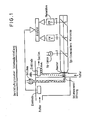
- Incubation with the dye can be carried out conveniently at room temperature within a few minutes. In general, 1 to 10, preferably 2 to 6 minutes of standing at room temperature are sufficient, calculated from the addition of a solution of the dyes to the blood sample to be examined, which has been suitably diluted with isotonic saline. After the incubation, the sample to be examined is placed in a suitable apparatus, for example a commercially available flow cytometer, which must be set up in such a way that the cell volume and the fluorescence can be determined at the same time and thus a corresponding fluorescence measurement can also be assigned to the volume measured for each particle .
- a suitable apparatus for example a commercially available flow cytometer, which must be set up in such a way that the cell volume and the fluorescence can be determined at the same time and thus a corresponding fluorescence measurement can also be assigned to the volume measured for each particle .
- the electrical method is preferred because the experimentally determined dye contents of the individual cells of each blood cell type can be expressed as dye concentrations, since absolute volumes are measured. As a result, the measured properties of the cell types can be directly compared with one another in a normalized manner. However, this is not possible with the light scattering method, since the light scattering depends not only on the cell volume and shape, but also on the nature of the cell surface and interior.
- acridine orange (AO) in combination with DiOC6 use is made of the fact that all blood cells, except the erythrocytes, are well stained by AO, that the erythrocytes are also well stained by DiOC6, but at the same time the coloration of the remaining cells is improved.
- the measurement is carried out in the presence of a fluorescent, in particular a monodisperse calibration phase colored with a fluorescent dye.
- a fluorescent in particular a monodisperse calibration phase colored with a fluorescent dye.
- concentration of the added fluorescent calibration phase is known, it is possible to determine the absolute concentrations of the various cells in the blood.
- Monodisperse latex particles with a diameter of 1 to 10 ⁇ m, particularly preferably between 4 and 6 ⁇ m, are preferably used as the calibration phase.
- other fluorescent particles of known uniform size in the blood cell range and concentration can also be used.
- Blood is mixed with physiological saline and the dye as well as with the particles of the calibration phase.
- the stained blood cells are measured in a suitable device for three to five minutes of staining time, for example using a commercially available flow cytometer at a rate of about 2000 cells per second over a period of about 5 to 15 minutes. In this way, all types of blood cells can be quantified in sufficient quantities.
- tissue cells are first zoned by disintegrating the corresponding tissue. B. released by cutting with a "tissue chopper"! The further procedure then corresponds to that described above for blood cells.
- the method of the invention also makes it possible to measure not only one but several fluorescences. For example, in the preferred embodiment of the method using AO and DiOC6, both the yellow fluorescence of native DNA / RNA (spiraled form) and the red fluorescence of the uncoiled form can be! be determined. This makes it possible to additionally make a statement about the functional state of the blood cells. In this embodiment, the method of the invention therefore enables not only a quantitative one. tive blood cell counting, but also to make statements about the functional state of certain individual cell types, which is not possible with known methods.
- the dyes used according to the invention can be determined both with optical systems which are equipped with mercury or xenon lamps and with those which are provided with laser devices.
- Another object of the invention is a reagent for performing the method according to the invention, which is characterized in that it is a fluo Resence dye from the group DNA / RNA dyes, cell protein dyes, lipid dyes, enzyme dyes, membrane potential sensitive dyes, intracellular pH dyes, SH group dyes and additionally contains a monodisperse calibration phase.
- a preferred reagent according to the invention contains a DNA / RNA dye and a membrane potential sensitive dye.
- a reagent which contains acridine orange, 3,3'-dihexyloxy carbocyanine, monodisperse latex particles with a diameter of 1 to 10 ⁇ m and a solvent is particularly preferred.
- Suitable solvents for the reagent according to the invention are those which can bring the chosen dye and the monodisperse phase into solution in sufficient concentration without attacking, for example dissolving, the particles of the calibration phase.
- Preferred solvents are dimethyl sulfoxide, dimethylformamide and alkanols.
- the method not only enables a shortening of the measuring times and an expansion of the informative value, but can also be used in a particularly large measuring range, which logs about 2.5 decades, i.e. H. is about 1: 500.
- this advantage can be seen from the following: If one proceeds from the normal concentrations of the different blood cells, which are 5 x 10 6 for erythrocytes, 3 x 10⁵ for thrombocytes, and 5 x 10 3 per mm 3 for leukocytes recognizable that a wide measuring range is absolutely necessary if all these blood cells are to be determined simultaneously. So far, it has been difficult to determine platelet concentrations below 5 to -7 x 10 5 per mm 2 . According to the invention, this lower limit is reduced to 1 ⁇ 10 3 per mm 3 . This is important because the particularly critical areas are in the range of 100,000 and 30,000 per mm 3 . In the former case this is the pathological area, in the latter case the acutely dangerous area.
- Another possible application of the method according to the invention is to investigate the effect of medication on individual cells, for example the effect of cytostatic medication on tumor cells.
- This enables pre-testing, particularly for drugs with high toxicity, to determine whether their effectiveness in a particular individual justifies the acceptance of toxic side effects or not.
- single cells can be obtained mechanically from tumor tissue, tested in a suitable nutrient medium, for example in haparinized patient blood plasma, in the presence of the medicament to be examined and then quantitatively by the method according to the invention determine which proportion of the tumor cells by; the drug was killed and what percentage is still alive.
- the method of the invention can thus be used to find out the most effective of a number of possible medicaments.
- AO / DiOC6 are excited between 418 and 500 nm.
- the emitted fluorescence light is collected in two-parameter measurements (volume against fluorescence 1) between 500 and 700 nm through the phototube 1 (PM1).
- volume against fluorescence 1 between 500 and 700 nm through the phototube 1 (PM1).
- the yellow light between 500 and 530 nm and the red light between 550 and 700 nm are measured by PM1 and PM2.
- a cylindrical measuring opening with a diameter of 50 ⁇ m and a length of 50 ⁇ m was used.
- the flow cytometer fluid system was filled with TBS buffer at 25 ° C.
- the cell volume was measured at an electrical current of 0.385 mA.
- the fluorescence was excited with an HBO-100 Hg high pressure lamp.
- the logaritmically amplified fluorescence and volume signals of the cells were either stored in a multi-channel analyzer as two-parameter histograms or written ON-LINE on a magnetic tape. The curves were evaluated graphically and arithmetically by magnetic tape.
- 2a shows a graphical representation of cell volume plotted against fluorescence.
- the volume and fluorescence pulses of the FLUVO-METRICELL flow cytometer were logarithmized using 2.5 decades of logarithmic amplifiers and then counted according to their maximum amplitude in the 64 x 64 matrix of a multi-channel analyzer.
- the channel contents of the matrix were logarithmic (3 decades amplitude log) and normalized to the value denoted by (M).
- the maximum channel content (M) was divided into 20 equal parts (5% steps). Each channel content received a number between 1 and 10 according to its relative frequency. Channel contents with an amplitude greater than 50% of the maximum value were identified by (*).
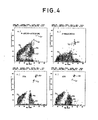
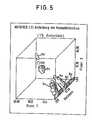
- Example 2 a determination was carried out on a blood sample stained with AO / DiOC6, but recording two different fluorescences, namely the yellow (fluorescence 1; transmembrane potential in the AO / DiOC6 staining) and the red (fluorescence 2; Relaxed RNA / DNS).
- the acquisition of these three parameters enables the measurement data shown in FIG. 5 to be displayed in cloud form.
- Each scale in Figs. 5 and 6 spans 2.5 logarithmic decades.
- the three measuring pulses of each individual cell were logarithmized for the representation in FIG. 5 and transferred ON-LINE to magnetic tape.
- the values were classified in a 32 x 32 x 32 matrix and displayed using a cloud program (. Cytometry f 1,222 -228 (1980)).
- the presence of cells and calibration particles is shown by a contour line at 1% of the maximum channel content.
- the individual cell types and the calibration particles are well distinguished from one another.
- Fig. 6 shows the yellow (Fluor. 1) against the red (Fluor. 2) fluorescence from 3-parameter measurements in which 1/500 diluted human blood was stained with DiOC6 (a) and with AO / DiOC6 simultaneously (b) was.
- DiOC6 DiOC6
- b AO / DiOC6 simultaneously
- the additional AO staining gives the part of curve 6b marked with Th + Re a clear red shift.
- the cell volumes against fluorescence are 1 histograms of the red-shifted (I) and the unchanged (II) particles drawn (c, d), the comparison with FIG.
- RNA-containing red cells correspond to the thrombocytes (Th), the reticulocytes (Re) and the granulocytes (Gr), while the erythrocytes (Er) , the calibration particles (St) and the lymphocytes (Ly) contain no RNA and therefore have retained their yellow fluorescence.
- the double staining also shows that the reticulocytes have a higher membrane potential than the erythrocytes. This can be seen from FIGS. 6a and 6b.
- the cells lying directly above the erythrocyte cluster correspond to the zone labeled Th + Re in FIG. 6a. This zone is made quantitative by the additional AO staining (6b).
- RNA RNA shifted to red, which means that the cells colored yellow by DiOC6 contain RNA. These cells correspond to thrombocytes and reticulocytes in the volume / fluorescence histogram. This shows that the cells overlying the erythrocytes with DiOC6 stained are reticulocytes.
- Fresh sterile material of a lymph node metastasis from a breast cancer is mechanically cut into pieces with a tissue chopper, which are sieved through a sieve with a 60 ⁇ m mesh size.
- the cells obtained are cultivated for seven days in heparinized patient blood plasma as a culture medium in microtiter plates in the presence or absence of various cytostatics.
- the cell suspension is then washed and stained with 1,4-diacetoxy-2,3-dicyano-benzene (ADB) and propidium iodide (PI) for five minutes.
- ADB shows the activity of cytoplasmic esterase and the intracellular pH of the living cells, PI stains the DNA of dead cells.
- the cell volume and the blue and green fluorescence of the stained cells are measured simultaneously in a flow cytometer as described in Example 1. Before the measurement, fluorescent monodisperse latex particles of 6 ⁇ m are added to the cell suspension as a concentration and fluorescence standard. Cell volume and the fluorescence signals of each cell, which correspond to pH, esterase activity and DNA, are measured simultaneously.
- the number of tumor cells or inflammatory cells living or surviving in a culture is then calculated from their ratio to the standardized number of calibration particles.
- the results of the various cytostatics examined and the untreated controls are summarized in the resistance diagram shown in FIG. 7.
- the abscissa of the chart identifies the individual drugs and the controls on which The ordinate is the number of tumor cells or inflammatory cells, based on 100% of the controls. It can be seen that a reduction in tumor cells was achieved with cytostatics Nos. 1, 4, 5 and 7, while the other drugs had no effect. The effect on inflammatory cells, which allows the less toxic medication to be recognized better, is recorded separately.
- the ratio of remaining inflammatory cells to tumor cells after incubation is also shown in the figure as a therapeutic index.
Landscapes
- Health & Medical Sciences (AREA)
- Life Sciences & Earth Sciences (AREA)
- Immunology (AREA)
- Chemical & Material Sciences (AREA)
- Physics & Mathematics (AREA)
- Engineering & Computer Science (AREA)
- Hematology (AREA)
- Biomedical Technology (AREA)
- Analytical Chemistry (AREA)
- Biochemistry (AREA)
- General Health & Medical Sciences (AREA)
- General Physics & Mathematics (AREA)
- Pathology (AREA)
- Urology & Nephrology (AREA)
- Molecular Biology (AREA)
- Nuclear Medicine, Radiotherapy & Molecular Imaging (AREA)
- Biotechnology (AREA)
- Cell Biology (AREA)
- Chemical Kinetics & Catalysis (AREA)
- Microbiology (AREA)
- Optics & Photonics (AREA)
- Tropical Medicine & Parasitology (AREA)
- Food Science & Technology (AREA)
- Medicinal Chemistry (AREA)
- Investigating Or Analysing Biological Materials (AREA)
- Sampling And Sample Adjustment (AREA)
Applications Claiming Priority (2)
| Application Number | Priority Date | Filing Date | Title |
|---|---|---|---|
| DE3238353 | 1982-10-15 | ||
| DE19823238353 DE3238353A1 (de) | 1982-10-15 | 1982-10-15 | Verfahren zur simultanen quantitativen bestimmung der blutzellen und reagenz hierfuer |
Publications (2)
| Publication Number | Publication Date |
|---|---|
| EP0106339A2 true EP0106339A2 (fr) | 1984-04-25 |
| EP0106339A3 EP0106339A3 (fr) | 1985-05-22 |
Family
ID=6175858
Family Applications (1)
| Application Number | Title | Priority Date | Filing Date |
|---|---|---|---|
| EP83110273A Withdrawn EP0106339A3 (fr) | 1982-10-15 | 1983-10-14 | Procédé pour la détermination quantitative simultanée de cellules et réactif utilisé à cette fin |
Country Status (5)
| Country | Link |
|---|---|
| US (1) | US4751188A (fr) |
| EP (1) | EP0106339A3 (fr) |
| JP (1) | JPS6022661A (fr) |
| CA (1) | CA1219791A (fr) |
| DE (1) | DE3238353A1 (fr) |
Cited By (7)
| Publication number | Priority date | Publication date | Assignee | Title |
|---|---|---|---|---|
| EP0266194A3 (en) * | 1986-10-31 | 1988-08-03 | Smithkline Beckman Corporation | Viable cell labelling |
| EP0259834A3 (en) * | 1986-09-10 | 1988-12-07 | Toa Medical Electronics Co., Ltd. | Method of classifying leucocytes by flow cytometry |
| EP0259833A3 (en) * | 1986-09-10 | 1988-12-07 | Toa Medical Electronics Co., Ltd. | Reagent and method for classifying leukocytes by flow cytometry |
| EP0268766A3 (en) * | 1986-11-27 | 1988-12-14 | Toa Medical Electronics Co., Ltd. | Method of classifying leukocytes by flow cytometry and reagents used in the method |
| EP0708334A3 (fr) * | 1994-10-20 | 1996-09-18 | Toa Medical Electronics | Réactif et méthode pour l'analyse des composants solides dans l'urine |
| US5665328A (en) * | 1988-05-02 | 1997-09-09 | Phanos Technologies, Inc. | Compounds, compositions and methods for binding bio-affecting substances to surface membranes of bio-particles |
| EP2630492A4 (fr) * | 2010-10-21 | 2014-04-16 | Nexcelom Bioscience Llc | Billes de référence et de focalisation internes utilisées dans la cytométrie en image |
Families Citing this family (62)
| Publication number | Priority date | Publication date | Assignee | Title |
|---|---|---|---|---|
| US4857451A (en) * | 1984-12-24 | 1989-08-15 | Flow Cytometry Standards Corporation | Method of compensating and calibrating a flow cytometer, and microbead standards kit therefor |
| US5073497A (en) * | 1989-06-30 | 1991-12-17 | Caribbean Microparticles Corporation | Microbead reference standard and method of adjusting a flow cytometer to obtain reproducible results using the microbeads |
| US5084394A (en) * | 1984-12-24 | 1992-01-28 | Vogt Robert F | Method for corrective calibration of a flow cytometry using a mixture of fluorescent microbeads and cells |
| US5089416A (en) * | 1984-12-24 | 1992-02-18 | Caribbean Microparticles Corporation | Method of use of non-fluorescent particles to determine fluorescence threshold of a flow cytometer relative to the autofluorescence of samples |
| US5314824A (en) * | 1984-12-24 | 1994-05-24 | Caribbean Microparticles Corporation | Method of setting up a flow cytometer |
| US5073498A (en) * | 1984-12-24 | 1991-12-17 | Caribbean Microparticles Corporation | Fluorescent alignment microbeads with broad excitation and emission spectra and its use |
| US5380663A (en) * | 1984-12-24 | 1995-01-10 | Caribbean Microparticles Corporation | Automated system for performance analysis and fluorescence quantitation of samples |
| US4867908A (en) * | 1986-08-29 | 1989-09-19 | Becton, Dickinson And Company | Method and materials for calibrating flow cytometers and other analysis instruments |
| US5175109A (en) * | 1986-09-10 | 1992-12-29 | Toa Medical Electronics Co., Ltd. | Reagent for classifying leukocytes by flow cytometry |
| US5179026A (en) * | 1986-11-27 | 1993-01-12 | Toa Medical Electronics Co., Ltd. | Method of classifying leukocytes by flow cytometry and reagents used in the method |
| US4848349A (en) * | 1987-04-29 | 1989-07-18 | Sherman Igor A | Substance and method for measuring hepatic blood flow |
| JPH0782009B2 (ja) * | 1988-03-24 | 1995-09-06 | 工業技術院長 | 細胞融合方法及び装置 |
| GB8810241D0 (en) * | 1988-04-29 | 1988-06-02 | Am Int | Drop-on-demand printhead |
| US5256532A (en) * | 1988-05-02 | 1993-10-26 | Zynaxis Technologies, Inc. | Methods, reagents and test kits for determination of subpopulations of biological entities |
| US5252487A (en) * | 1989-05-19 | 1993-10-12 | Cell Analysis Systems, Inc. | Method and apparatus for determining the amount of oncogene protein product in a cell sample |
| DE4019025C2 (de) * | 1990-06-14 | 1994-09-22 | Triton Technology Inc | Verfahren zur Messung der Durchblutung von Organ- und/oder Gewebeproben |
| IE76732B1 (en) * | 1990-08-07 | 1997-11-05 | Becton Dickinson Co | One step test for absolute counts |
| JP2927979B2 (ja) * | 1991-02-22 | 1999-07-28 | シスメックス株式会社 | フローサイトメトリーによる赤芽球の分類方法 |
| DE69326731T2 (de) * | 1992-09-04 | 2000-05-18 | Becton Dickinson And Co., Franklin Lakes | Kontroll-Teilchen für die Zellzählung und Instrumentenlinearität |
| US5334509A (en) * | 1992-10-22 | 1994-08-02 | Riordan Neil H | Method for detecting intestinal pathogen dientamoeba fragilis |
| DE69327775T2 (de) * | 1992-11-19 | 2000-06-21 | Sysmex Corp., Kobe | Verfahren zur Vorbehandlung für Blutanalyse |
| JP2680931B2 (ja) * | 1993-11-12 | 1997-11-19 | ベクトン・ディッキンソン・アンド・カンパニー | 希少細胞の絶対数を数える方法 |
| US5879900A (en) * | 1994-12-15 | 1999-03-09 | Abbott Laboratories | Method for simultaneous analysis of cell viability, nucleated red blood cells and white blood cell differentials |
| US6159748A (en) * | 1995-03-13 | 2000-12-12 | Affinitech, Ltd | Evaluation of autoimmune diseases using a multiple parameter latex bead suspension and flow cytometry |
| US5837547A (en) * | 1995-12-27 | 1998-11-17 | Caribbean Microparticles Corporation | Flow cytometer calibration method |
| US5804389A (en) * | 1995-12-29 | 1998-09-08 | Phanos Technologies, Inc. | Method for detecting abnormal epithelial cell shedding |
| US5776711A (en) * | 1996-11-12 | 1998-07-07 | The Regents Of The University Of California | Simultaneous human ABO and RH(D) blood typing or antibody screening by flow cytometry |
| AU3146800A (en) * | 1999-03-05 | 2000-09-28 | De Danske Kvaegavlsforeninger | Determination of sperm concentration and viability for artificial insemination |
| FR2883971B1 (fr) * | 2005-03-31 | 2007-11-16 | C2 Diagnostics Sa | Dispositif optique d'analyse sanguine, appareil d'analyse equipe d'un tel dispositif |
| US7738094B2 (en) * | 2007-01-26 | 2010-06-15 | Becton, Dickinson And Company | Method, system, and compositions for cell counting and analysis |
| US8062222B2 (en) * | 2007-06-13 | 2011-11-22 | Litron Laboratories, Ltd. | Method for measuring in vivo hematotoxicity with an emphasis on radiation exposure assessment |
| CN102087197B (zh) * | 2009-12-08 | 2014-06-18 | 龚维燕 | 全功能血液分析仪器中库尔特微孔的共轴照明方法及其分析仪器 |
| US9528160B2 (en) | 2008-11-07 | 2016-12-27 | Adaptive Biotechnolgies Corp. | Rare clonotypes and uses thereof |
| US9506119B2 (en) | 2008-11-07 | 2016-11-29 | Adaptive Biotechnologies Corp. | Method of sequence determination using sequence tags |
| EP2364368B1 (fr) | 2008-11-07 | 2014-01-15 | Sequenta, Inc. | Procédés de surveillance de maladies par analyse de séquence |
| US9365901B2 (en) | 2008-11-07 | 2016-06-14 | Adaptive Biotechnologies Corp. | Monitoring immunoglobulin heavy chain evolution in B-cell acute lymphoblastic leukemia |
| US8628927B2 (en) | 2008-11-07 | 2014-01-14 | Sequenta, Inc. | Monitoring health and disease status using clonotype profiles |
| US8748103B2 (en) | 2008-11-07 | 2014-06-10 | Sequenta, Inc. | Monitoring health and disease status using clonotype profiles |
| ES2726702T3 (es) | 2009-01-15 | 2019-10-08 | Adaptive Biotechnologies Corp | Perfilado de la inmunidad adaptativa y métodos para la generación de anticuerpos monoclonales |
| WO2010151416A1 (fr) | 2009-06-25 | 2010-12-29 | Fred Hutchinson Cancer Research Center | Procédé de mesure de l'immunité adaptative |
| US10385475B2 (en) | 2011-09-12 | 2019-08-20 | Adaptive Biotechnologies Corp. | Random array sequencing of low-complexity libraries |
| EP2768982A4 (fr) | 2011-10-21 | 2015-06-03 | Adaptive Biotechnologies Corp | Quantification de génomes de cellules immunitaires adaptatives dans un mélange complexe de cellules |
| US9523682B2 (en) | 2011-11-16 | 2016-12-20 | Becton, Dickinson And Company | Methods and systems for detecting an analyte in a sample |
| US9824179B2 (en) | 2011-12-09 | 2017-11-21 | Adaptive Biotechnologies Corp. | Diagnosis of lymphoid malignancies and minimal residual disease detection |
| US9499865B2 (en) | 2011-12-13 | 2016-11-22 | Adaptive Biotechnologies Corp. | Detection and measurement of tissue-infiltrating lymphocytes |
| EP3372694A1 (fr) | 2012-03-05 | 2018-09-12 | Adaptive Biotechnologies Corporation | Détermination de chaînes de récepteur immunitaire appariées de sous-unités à adaptation de fréquence |
| HUE029357T2 (en) | 2012-05-08 | 2017-02-28 | Adaptive Biotechnologies Corp | Preparations and devices for measuring and calibrating amplification distortion in multiplex PCR reactions |
| EP3330384B1 (fr) | 2012-10-01 | 2019-09-25 | Adaptive Biotechnologies Corporation | Évaluation de l'immunocompétence par la diversité adaptative du récepteur immunitaire et la caractérisation de la clonalité |
| WO2015160439A2 (fr) | 2014-04-17 | 2015-10-22 | Adaptive Biotechnologies Corporation | Quantification de génomes de cellules de l'immunité acquise dans un mélange complexe de cellules |
| ES2692407T3 (es) | 2013-01-11 | 2018-12-03 | Becton, Dickinson And Company | Dispositivo de ensayo de punto de cuidado de bajo coste |
| US9708657B2 (en) | 2013-07-01 | 2017-07-18 | Adaptive Biotechnologies Corp. | Method for generating clonotype profiles using sequence tags |
| EP3066190B1 (fr) | 2013-11-06 | 2020-12-30 | Becton, Dickinson and Company | Dispositifs microfluidiques et procédés d'utilisation de ces dispositifs |
| JP6518245B2 (ja) | 2013-11-13 | 2019-05-22 | ベクトン・ディキンソン・アンド・カンパニーBecton, Dickinson And Company | 光学撮像システム及びそれを用いた方法 |
| WO2015134787A2 (fr) | 2014-03-05 | 2015-09-11 | Adaptive Biotechnologies Corporation | Procédés dans lesquels on utilise des molécules synthétiques contenant des randomères |
| US10066265B2 (en) | 2014-04-01 | 2018-09-04 | Adaptive Biotechnologies Corp. | Determining antigen-specific t-cells |
| CA2935312C (fr) | 2014-10-14 | 2022-11-22 | Becton, Dickinson And Company | Prise en charge d'echantillons de sang au moyen de plastique a alveoles ouverts |
| BR122020024283B1 (pt) | 2014-10-14 | 2023-02-23 | Becton, Dickinson And Company | Dispositivo de transferência de sangue adaptado para receber uma amostra de sangue |
| CA2966201A1 (fr) | 2014-10-29 | 2016-05-06 | Adaptive Biotechnologies Corp. | Detection simultanee hautement multiplexee d'acides nucleiques codant pour des heterodimeres de recepteurs de l'immunite adaptative apparies a partir de nombreux echantillons |
| US10246701B2 (en) | 2014-11-14 | 2019-04-02 | Adaptive Biotechnologies Corp. | Multiplexed digital quantitation of rearranged lymphoid receptors in a complex mixture |
| CN106604780B (zh) | 2015-03-10 | 2019-06-25 | 贝克顿·迪金森公司 | 生物流体微样本管理装置 |
| ES2857873T3 (es) | 2015-09-01 | 2021-09-29 | Becton Dickinson Co | Dispositivo de filtración en profundidad para separar fases de muestras |
| WO2021222594A1 (fr) * | 2020-04-30 | 2021-11-04 | Daxor Corp. | Mesure du volume sanguin avec un colorant fluorescent |
Family Cites Families (17)
| Publication number | Priority date | Publication date | Assignee | Title |
|---|---|---|---|---|
| US3560754A (en) * | 1965-11-17 | 1971-02-02 | Ibm | Photoelectric particle separator using time delay |
| US3497690A (en) * | 1967-09-21 | 1970-02-24 | Bausch & Lomb | Method and apparatus for classifying biological cells by measuring the size and fluorescent response thereof |
| US3675768A (en) * | 1969-03-17 | 1972-07-11 | Gildardo Legorreta Sanchez | Method and apparatus for classifying and segregating particles with electrical and optical means |
| US3770349A (en) * | 1969-03-17 | 1973-11-06 | Sanchez G Legorreta | Method and apparatus for automatically classifying complex, microscopic particles such as human cells |
| US3657537A (en) * | 1970-04-03 | 1972-04-18 | Bausch & Lomb | Computerized slit-scan cyto-fluorometer for automated cell recognition |
| US3684377A (en) * | 1970-07-13 | 1972-08-15 | Bio Physics Systems Inc | Method for analysis of blood by optical analysis of living cells |
| BE793185A (fr) * | 1971-12-23 | 1973-04-16 | Atomic Energy Commission | Appareil pour analyser et trier rapidement des particules telles que des cellules biologiques |
| US3824402A (en) * | 1973-06-04 | 1974-07-16 | Energy Commission | Dual parameter flow photometric apparatus and method |
| JPS5415185A (en) * | 1977-07-06 | 1979-02-03 | Nippon Telegr & Teleph Corp <Ntt> | Forming of sheath of submarine coaxial cable |
| US4299726A (en) * | 1979-05-07 | 1981-11-10 | Coulter Electronics, Inc. | Process for preparing whole blood reference controls having long term stability, preconditioning diluent and media therefor |
| US4284412A (en) * | 1979-07-13 | 1981-08-18 | Ortho Diagnostics, Inc. | Method and apparatus for automated identification and enumeration of specified blood cell subclasses |
| US4284355A (en) * | 1979-10-29 | 1981-08-18 | Ortho Diagnostics, Inc. | Automated method for cell volume determination |
| US4400370A (en) * | 1980-03-12 | 1983-08-23 | Lawrence Kass | Metachromatic dye sorption means for differential determination of leukocytes |
| US4338024A (en) * | 1980-05-02 | 1982-07-06 | International Remote Imaging Systems, Inc. | Flow analyzer and system for analysis of fluids with particles |
| EP0068404B1 (fr) * | 1981-06-24 | 1985-10-02 | Becton Dickinson and Company | Analysateur pour la détermination simultanée de volume et de caractéristiques d'émission de lumière des particules |
| US4559309A (en) * | 1982-09-01 | 1985-12-17 | Memorial Sloan Kettering Cancer Center | Flow cytometry-fluorescence measurements for characterizing sperm |
| US4584277A (en) * | 1983-04-05 | 1986-04-22 | Syntex (U.S.A.) Inc. | Fluorescent multiparameter particle analysis |
-
1982
- 1982-10-15 DE DE19823238353 patent/DE3238353A1/de not_active Withdrawn
-
1983
- 1983-10-13 US US06/541,562 patent/US4751188A/en not_active Expired - Fee Related
- 1983-10-14 EP EP83110273A patent/EP0106339A3/fr not_active Withdrawn
- 1983-10-14 JP JP58191063A patent/JPS6022661A/ja active Pending
- 1983-10-14 CA CA000438982A patent/CA1219791A/fr not_active Expired
Cited By (10)
| Publication number | Priority date | Publication date | Assignee | Title |
|---|---|---|---|---|
| EP0259834A3 (en) * | 1986-09-10 | 1988-12-07 | Toa Medical Electronics Co., Ltd. | Method of classifying leucocytes by flow cytometry |
| EP0259833A3 (en) * | 1986-09-10 | 1988-12-07 | Toa Medical Electronics Co., Ltd. | Reagent and method for classifying leukocytes by flow cytometry |
| US4933293A (en) * | 1986-09-10 | 1990-06-12 | Toa Medical Electronics Co., Ltd. | Method of classifying leukocytes by flow cytometry and reagents used in the method |
| EP0266194A3 (en) * | 1986-10-31 | 1988-08-03 | Smithkline Beckman Corporation | Viable cell labelling |
| EP0268766A3 (en) * | 1986-11-27 | 1988-12-14 | Toa Medical Electronics Co., Ltd. | Method of classifying leukocytes by flow cytometry and reagents used in the method |
| US5665328A (en) * | 1988-05-02 | 1997-09-09 | Phanos Technologies, Inc. | Compounds, compositions and methods for binding bio-affecting substances to surface membranes of bio-particles |
| EP0708334A3 (fr) * | 1994-10-20 | 1996-09-18 | Toa Medical Electronics | Réactif et méthode pour l'analyse des composants solides dans l'urine |
| US5891733A (en) * | 1994-10-20 | 1999-04-06 | Toa Medical Electronics Co., Ltd. | Reagent for analyzing solid components in urine and method for analyzing solid components by employing the same |
| EP1089078A1 (fr) * | 1994-10-20 | 2001-04-04 | Sysmex Corporation | Réactif et méthode pour l'analyse des composants solides dans l'urine |
| EP2630492A4 (fr) * | 2010-10-21 | 2014-04-16 | Nexcelom Bioscience Llc | Billes de référence et de focalisation internes utilisées dans la cytométrie en image |
Also Published As
| Publication number | Publication date |
|---|---|
| EP0106339A3 (fr) | 1985-05-22 |
| DE3238353A1 (de) | 1984-04-19 |
| JPS6022661A (ja) | 1985-02-05 |
| US4751188A (en) | 1988-06-14 |
| CA1219791A (fr) | 1987-03-31 |
Similar Documents
| Publication | Publication Date | Title |
|---|---|---|
| EP0106339A2 (fr) | Procédé pour la détermination quantitative simultanée de cellules et réactif utilisé à cette fin | |
| DE69120026T2 (de) | Verfahren zur Leukozytenklassifizierung unter Verwendung der Duchflusszytometrie | |
| DE69223931T2 (de) | Reagenszusammensetzungen und ihre Verwendung bei der Identifizierung und Charakterisierung von Retikulozyten in Vollblut | |
| DE2953524C2 (fr) | ||
| DE3109252C2 (de) | Verfahren und Zusammensetzung zur Bestimmung einzelner Leukozyten durch metachromatische Farbstoffdifferentialabsorption | |
| DE69223930T2 (de) | Reagenzzusammensetzungen und ihre Verwendung bei der Identifizierung und Charakterisierung von Retikulozyten in Vollblut | |
| DE69838723T2 (de) | Erythroblasten-diagnostische Durchflusszytometrie-Methode und Reagenzien | |
| DE69118617T2 (de) | Einschrittest für absolute Zellzahlen | |
| DE69632998T2 (de) | Hochempfindliches, genaues und präzises automatisiertes Messverfahren und Vorrichtung zur Identifizierung und Quantifizierung von Blutplättchen und zur Bestimmung des Blutplättchenaktivitätszustands unter Verwendung von Ganzblutproben | |
| DE69329274T2 (de) | Hämatologisches Kontrollprodukt mit Leukozytenanalogen; und Methoden zu ihrer Herstellung und Anwendung | |
| DE2451409C2 (de) | Verfahren zur Bestimmung spezieller weißer Blutkörperchen | |
| DE3586159T2 (de) | Zusammensetzung und verfahren zur differenzierung von weissen blutzellen. | |
| DE69211434T2 (de) | Verfahren zur Probevorbereitung zur Klassifizierung und Zählung der Leukozyten | |
| DE2134910C2 (de) | Verfahren zur Blutanalyse | |
| EP1262776B1 (fr) | Procédé pour la détermination quantitative de cellules tumorales épithéliales vivantes dans un fluide corporel | |
| DE69213315T2 (de) | Reagenzien und Verfahren zur Zellanalyse im Harn | |
| DE3751859T2 (de) | Durchflusszytometrie-Verfahren zur Klassifikation von Leukozyten und Reagenzien dafür | |
| DE68918004T2 (de) | Verfahren zur Analyse von Zellbestandteilen in einer Flüssigkeit. | |
| DE69221086T2 (de) | Reagenzzusammensetzungen und ihre Verwendung zur Erzeugung von kugelförmigen roten Blutzellen | |
| DE69328296T2 (de) | Verfahren und materialien zur bestimmung der teilchenzahl in einem durchflusszytometer | |
| DE69022819T2 (de) | Verbindungen und Reagenzien zur Bestimmung von Retikulozyten. | |
| DE3390432T1 (de) | Verfahren zur volumetrischen Differenzierung von Leukozyten | |
| DE3208629A1 (de) | Metachromatisches farbstoffabsorptionsverfahren zur differentiellen bestimmung der entwicklungsstufen von neutrophilen und granulozytischen zellen sowie anderen leukozyten | |
| DE69326731T2 (de) | Kontroll-Teilchen für die Zellzählung und Instrumentenlinearität | |
| DE3026185C2 (de) | Zusammensetzung, geeignet zur Untersuchung biologischer Gewebe oder Flüssigkeiten und Verfahren zu deren Anwendung |
Legal Events
| Date | Code | Title | Description |
|---|---|---|---|
| PUAI | Public reference made under article 153(3) epc to a published international application that has entered the european phase |
Free format text: ORIGINAL CODE: 0009012 |
|
| AK | Designated contracting states |
Designated state(s): AT BE CH DE FR GB IT LI LU NL SE |
|
| PUAL | Search report despatched |
Free format text: ORIGINAL CODE: 0009013 |
|
| AK | Designated contracting states |
Designated state(s): AT BE CH DE FR GB IT LI LU NL SE |
|
| 17P | Request for examination filed |
Effective date: 19850708 |
|
| 17Q | First examination report despatched |
Effective date: 19870723 |
|
| STAA | Information on the status of an ep patent application or granted ep patent |
Free format text: STATUS: THE APPLICATION IS DEEMED TO BE WITHDRAWN |
|
| 18D | Application deemed to be withdrawn |
Effective date: 19880203 |
|
| RIN1 | Information on inventor provided before grant (corrected) |
Inventor name: VALET, GUENTHER, PROF.DR. |