EP0103477A2 - Procédé de transfert d'un échantillon tissulaire d'un support sur la lamelle d'un microscope - Google Patents
Procédé de transfert d'un échantillon tissulaire d'un support sur la lamelle d'un microscope Download PDFInfo
- Publication number
- EP0103477A2 EP0103477A2 EP83305306A EP83305306A EP0103477A2 EP 0103477 A2 EP0103477 A2 EP 0103477A2 EP 83305306 A EP83305306 A EP 83305306A EP 83305306 A EP83305306 A EP 83305306A EP 0103477 A2 EP0103477 A2 EP 0103477A2
- Authority
- EP
- European Patent Office
- Prior art keywords
- layer
- tissue section
- substrate
- process according
- tissue
- Prior art date
- Legal status (The legal status is an assumption and is not a legal conclusion. Google has not performed a legal analysis and makes no representation as to the accuracy of the status listed.)
- Granted
Links
- 238000000034 method Methods 0.000 title claims description 37
- 239000010410 layer Substances 0.000 claims abstract description 66
- 239000000758 substrate Substances 0.000 claims abstract description 22
- 239000004820 Pressure-sensitive adhesive Substances 0.000 claims abstract description 11
- 238000009792 diffusion process Methods 0.000 claims description 9
- 239000002904 solvent Substances 0.000 claims description 9
- 238000010186 staining Methods 0.000 claims description 3
- 238000009499 grossing Methods 0.000 claims description 2
- 238000013007 heat curing Methods 0.000 claims 1
- 238000003825 pressing Methods 0.000 claims 1
- 239000012790 adhesive layer Substances 0.000 abstract description 10
- 239000000463 material Substances 0.000 abstract description 3
- 239000012188 paraffin wax Substances 0.000 description 10
- CTQNGGLPUBDAKN-UHFFFAOYSA-N O-Xylene Chemical compound CC1=CC=CC=C1C CTQNGGLPUBDAKN-UHFFFAOYSA-N 0.000 description 7
- 239000012120 mounting media Substances 0.000 description 7
- 239000008096 xylene Substances 0.000 description 7
- 239000002390 adhesive tape Substances 0.000 description 5
- HEDRZPFGACZZDS-UHFFFAOYSA-N Chloroform Chemical compound ClC(Cl)Cl HEDRZPFGACZZDS-UHFFFAOYSA-N 0.000 description 4
- 229920001651 Cyanoacrylate Polymers 0.000 description 4
- LFQSCWFLJHTTHZ-UHFFFAOYSA-N Ethanol Chemical compound CCO LFQSCWFLJHTTHZ-UHFFFAOYSA-N 0.000 description 4
- MWCLLHOVUTZFKS-UHFFFAOYSA-N Methyl cyanoacrylate Chemical compound COC(=O)C(=C)C#N MWCLLHOVUTZFKS-UHFFFAOYSA-N 0.000 description 4
- 239000000853 adhesive Substances 0.000 description 4
- 230000001070 adhesive effect Effects 0.000 description 4
- 239000011521 glass Substances 0.000 description 4
- 229920000642 polymer Polymers 0.000 description 4
- YXFVVABEGXRONW-UHFFFAOYSA-N Toluene Chemical compound CC1=CC=CC=C1 YXFVVABEGXRONW-UHFFFAOYSA-N 0.000 description 3
- 235000019441 ethanol Nutrition 0.000 description 3
- 239000000203 mixture Substances 0.000 description 3
- WYURNTSHIVDZCO-UHFFFAOYSA-N Tetrahydrofuran Chemical compound C1CCOC1 WYURNTSHIVDZCO-UHFFFAOYSA-N 0.000 description 2
- -1 acryl Chemical group 0.000 description 2
- IISBACLAFKSPIT-UHFFFAOYSA-N bisphenol A Chemical compound C=1C=C(O)C=CC=1C(C)(C)C1=CC=C(O)C=C1 IISBACLAFKSPIT-UHFFFAOYSA-N 0.000 description 2
- 239000003153 chemical reaction reagent Substances 0.000 description 2
- 238000007796 conventional method Methods 0.000 description 2
- 238000005538 encapsulation Methods 0.000 description 2
- 239000010408 film Substances 0.000 description 2
- 230000008014 freezing Effects 0.000 description 2
- 238000007710 freezing Methods 0.000 description 2
- 125000005641 methacryl group Chemical group 0.000 description 2
- QPJVMBTYPHYUOC-UHFFFAOYSA-N methyl benzoate Chemical compound COC(=O)C1=CC=CC=C1 QPJVMBTYPHYUOC-UHFFFAOYSA-N 0.000 description 2
- 239000000178 monomer Substances 0.000 description 2
- 239000007787 solid Substances 0.000 description 2
- 125000006850 spacer group Chemical group 0.000 description 2
- XLYOFNOQVPJJNP-UHFFFAOYSA-N water Substances O XLYOFNOQVPJJNP-UHFFFAOYSA-N 0.000 description 2
- 102000009027 Albumins Human genes 0.000 description 1
- 108010088751 Albumins Proteins 0.000 description 1
- 241001465754 Metazoa Species 0.000 description 1
- 241000078511 Microtome Species 0.000 description 1
- 239000004830 Super Glue Substances 0.000 description 1
- 125000003118 aryl group Chemical group 0.000 description 1
- 239000004568 cement Substances 0.000 description 1
- 230000018044 dehydration Effects 0.000 description 1
- 238000006297 dehydration reaction Methods 0.000 description 1
- 238000001035 drying Methods 0.000 description 1
- 238000001493 electron microscopy Methods 0.000 description 1
- UHESRSKEBRADOO-UHFFFAOYSA-N ethyl carbamate;prop-2-enoic acid Chemical compound OC(=O)C=C.CCOC(N)=O UHESRSKEBRADOO-UHFFFAOYSA-N 0.000 description 1
- 239000012634 fragment Substances 0.000 description 1
- 238000007654 immersion Methods 0.000 description 1
- 230000008595 infiltration Effects 0.000 description 1
- 238000001764 infiltration Methods 0.000 description 1
- 239000003999 initiator Substances 0.000 description 1
- 239000007788 liquid Substances 0.000 description 1
- 239000002609 medium Substances 0.000 description 1
- 229940095102 methyl benzoate Drugs 0.000 description 1
- 229920005615 natural polymer Polymers 0.000 description 1
- 230000003287 optical effect Effects 0.000 description 1
- 230000000149 penetrating effect Effects 0.000 description 1
- 230000035515 penetration Effects 0.000 description 1
- 239000011148 porous material Substances 0.000 description 1
- KCTAWXVAICEBSD-UHFFFAOYSA-N prop-2-enoyloxy prop-2-eneperoxoate Chemical compound C=CC(=O)OOOC(=O)C=C KCTAWXVAICEBSD-UHFFFAOYSA-N 0.000 description 1
- 229920005989 resin Polymers 0.000 description 1
- 239000011347 resin Substances 0.000 description 1
- 229920002379 silicone rubber Polymers 0.000 description 1
- 239000004945 silicone rubber Substances 0.000 description 1
- 238000007447 staining method Methods 0.000 description 1
- 239000000126 substance Substances 0.000 description 1
- YLQBMQCUIZJEEH-UHFFFAOYSA-N tetrahydrofuran Natural products C=1C=COC=1 YLQBMQCUIZJEEH-UHFFFAOYSA-N 0.000 description 1
- 239000010409 thin film Substances 0.000 description 1
Images


Classifications
-
- G—PHYSICS
- G01—MEASURING; TESTING
- G01N—INVESTIGATING OR ANALYSING MATERIALS BY DETERMINING THEIR CHEMICAL OR PHYSICAL PROPERTIES
- G01N1/00—Sampling; Preparing specimens for investigation
- G01N1/28—Preparing specimens for investigation including physical details of (bio-)chemical methods covered elsewhere, e.g. G01N33/50, C12Q
Definitions
- This invention relates to a method for transferring thin sections of tissue specimens, prior to staining and mounting processes, from a support to a microscope slide.
- Very thin slices of animal and plant tissues are prepared for many different kinds of microscopic studies by sectioning using various kinds of microtomes. While the tissue may be cut fresh, the soft and compliant nature of most fresh tissue makes the cutting of undistorted thin sections very difficult. Often, the tissue is cut on a freezing microtome or in a cryostat at temperatures below 0°C. (32 0 F.,), the hardness of the frozen water within the tissue allowing sections as thin as a few micrometers to be cut relatively easily. As these frozen sections are brittle and friable, they are difficult to handle and process further. To simplify sectioning of tissue, a number of procedures have been developed which produce a block of supported tissue which has superior sectioning properties and produces high quality, relatively easy-to- handle tissue sections. Such procedures typically involve:
- a process for transferring a thin section of a tissue specimen, supported on a substrate by a first pressure-sensitive adhesive layer, from said substrate and mounting it on a microscope slide having at least a portion of one surface coated with a second pressure sensitive adhesive layer, said second layer being polymerisable comprising the steps of:
- a tissue specimen supported on, for example, a flexible piece of pressure-sensitive adhesive tape is brought into contact with a microscope slide over whose contacting surface a film of pressure-sensitive adhesive, capable of further polymerisation, has been layered.
- Techniques by which flexible tape can be applied to such specimens to aid sectioning have been described, for example, in the above- identified publications by Palmgren, Beckel, Gowers et al and Wedeen et al, respectively.
- the layer is then polymerised, usually within about 1 to 3 minutes, and then the tape may, for example, be submerged in a solvent which weakens the supporting tape backing and/or supporting-tape adhesive-layer which are washed or peeled away, to fully expose the tissue specimen.
- the polymerisable layer and its bonds to the section have sufficient strength after curing to allow peeling of the insoluble tape backing without damaging the section and to thereafter firmly support the specimen on the microscope slide.
- the polymerisable layer preferably has only curable components whose respective diffusion coefficients, before and during cure, are sufficiently low that the diffusion thereof into the specimen during the curing process is minimal, and/or a viscosity sufficiently large that flow into the specimen on application of finger-pressure is minimal, that is less than 1 micrometer in 30 minutes.
- the layer forms a positive bond between the microscope slide and specimen section, to permit ease of handling during subsequent processing of the supported section.
- the substantially entire interior structure of the tissue section remains exposable to processing.
- the polymerised bonding layer is not affected, in any way, by such processing.
- the polymerised layer preferably has a refractive index matched to the refractive index of the unstained tissue, e.g., from about 1.53 to 1.57, and to the mounting medium conventionally used to envelop and to attach the coverslip over the specimen at the completion of the processing.
- the specimen is stained during processing to highlight particular components thereof.
- the respective refractive indices of the various components of the completed microscope slide are substantially the same, there is no optical interference introduced to degrade the image in the subsequent microscopic examination of the tissue.
- the present invention provides a method for positively attaching a tissue to a microscope slide by a pressure-sensitive polymerisable layer, the curable components of such layer prior to and during polymerisation penetrating only very minimally into such specimen and, when polymerised, having a compatible index of refraction and neither affecting'nor being affected by any subsequent chemical or physical treatment of the supported tissue.
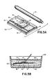
- a thin tissue section 9 surrounded and infiltrated with paraffin 10 is supported on and adhered to the surface of a flexible tape or substrate 12, by a pressure-sensitive adhesive layer 14.
- Section 9 is located within a window or opening in a spacer 13 adhered to layer 14.
- spacer 13 facilitates the cutting of a thin section 9 by a microtome.
- a microscope slide 16 which supports on its upper surface a pressure-sensitive adhesive layer 18.
- Layer 18 can be formed of, for example, a heat-curable polymerisable material or a photopolymerisable material.
- layer 18 is photopolymerisable, e.g. by exposure to U.V. light of wavelengths in the range of 320 nm-390 nm.
- Figure 2 further illustrates the inversion of tape 12 so as to juxtapose specimen 9 and layer 18.
- Specimen9 and layer 18 are subsequently contacted.
- a slight pressure is applied, e.g. manually.
- such pressure can be applied progressively along the specimen 9, to ensure a smooth contact thereof with layer 18.
- Layer 18 preferably is formed to have the characteristics of:
- the laminate 20 is then immersed is a solvent solution 22, such as xylene, contained in beaker 24.
- a solvent solution 22 such as xylene
- Such immersion serves to rapidly soften-the layer 14 on tape 12 and allows for a gentle manual peeling of tape 12 from over specimen 9 while laminate 20 is immersed in beaker 24.
- Exposed layer 14 and paraffin 10 dissolve as, or within on minute after, tape 12 is peeled away.
- the specimen section 9 is now fully exposed.
- the solvent solution 22 may be selected, so as to be effective to dissolve both substrate 12 and adhesive layer 14, so as to avoid the need of peeling substrate 12, as described.
- the layer 18 on slide 16 consists of a mixture of very high molecular weight reactive oligomers (typically, each with two terminal acryl or methacryl groups).
- the high molecular weight ensures a high viscosity and a low diffusion coefficient. If the molecular weight is too high, there will be insufficient tack.
- the acryl or methacryl groups permit radical-initiated polymerisation to crosslink the oligomers into a highly insoluble polymer.
- the mixture is composed of one oligomer with a cured refractive index of greater than 1.560 (e.g. an epoxy-acrylate oligomer with many bisphenol A or other high aromatic residues to raise the refractive index) and another with a cured refractive index less than 1.530 (e.g. a urethane acrylate oligomer).
- cured resins must be extremely resistant to xylene, alcohol and water. By mixing them in appropriate proportions, any desired cured refractive index, between 1.530 and 1.560, can be imparted to layer 18, when polymerised.
- the mounting medium for encapsulating specimen section 9 should preferably have a refractive index near 1.55, and layer 18, when cured, a refractive index of between 1.545 and 1.555.
- specimen section 9 can be processed for storing and permanent encapsulation, according to conventional histological techniques.
- specimen section 9 is securely and permanently bound to slide 16 by now-polymerised layer 18, which neither affects nor is affected by exposure to such techniques.
- encapsulation process may be manually effected, for example, as fully described in McManus and Mowry above.
- a number of tissue specimen sections 9, each supported on an individual slide 16 may be processed in automated fashion, for example, as clearly shown and described in an AUTOTECHNICON system as marketed by Technicon Instruments Corporation, of Tarrytown, New York, and described in Technical Publication No. TA 1-0225-10, June 1977, pages 3-10-3-13.
- Figure 4 illustrates a completely mounted specimen section, ready for microscopic viewing.
- Figure 4A illustrates a specimen section 9 mounted by conventional techniques; such specimen is covered with a mounting medium 26 and a coverslip 28 is located on-such medium and over such specimen.
- Figure 4B shows the envelopment of a specimen 9 in a U.V. curable mounting medium 27, as results from the technique disclosed in U.S. Patent 4,120,991.
- a coverslip is not provided. Rather the upper surface of mounting medium 27 is formed so as to be optically flat.
- the adhesive tape used to capture the sections must at least have an adhesive layer which is soluble in a solvent, such as xylene.
- a solvent such as xylene.
- a silicone rubber adhesive as used by Wedeen and Jernow referenced above is satisfactory for frozen sections, and an adhesive tape, like Permacel No. 925 is satisfactory for paraffin sectioning.
Landscapes
- Physics & Mathematics (AREA)
- Health & Medical Sciences (AREA)
- Life Sciences & Earth Sciences (AREA)
- Chemical & Material Sciences (AREA)
- Analytical Chemistry (AREA)
- Biochemistry (AREA)
- General Health & Medical Sciences (AREA)
- General Physics & Mathematics (AREA)
- Immunology (AREA)
- Pathology (AREA)
- Sampling And Sample Adjustment (AREA)
- Microscoopes, Condenser (AREA)
Applications Claiming Priority (2)
| Application Number | Priority Date | Filing Date | Title |
|---|---|---|---|
| US417307 | 1982-09-13 | ||
| US06/417,307 US4545831A (en) | 1982-09-13 | 1982-09-13 | Method for transferring a thin tissue section |
Publications (3)
| Publication Number | Publication Date |
|---|---|
| EP0103477A2 true EP0103477A2 (fr) | 1984-03-21 |
| EP0103477A3 EP0103477A3 (en) | 1984-05-02 |
| EP0103477B1 EP0103477B1 (fr) | 1987-01-21 |
Family
ID=23653424
Family Applications (1)
| Application Number | Title | Priority Date | Filing Date |
|---|---|---|---|
| EP83305306A Expired EP0103477B1 (fr) | 1982-09-13 | 1983-09-12 | Procédé de transfert d'un échantillon tissulaire d'un support sur la lamelle d'un microscope |
Country Status (7)
| Country | Link |
|---|---|
| US (1) | US4545831A (fr) |
| EP (1) | EP0103477B1 (fr) |
| JP (1) | JPS59116531A (fr) |
| AU (1) | AU554525B2 (fr) |
| CA (1) | CA1215508A (fr) |
| DE (1) | DE3369391D1 (fr) |
| ES (1) | ES525551A0 (fr) |
Cited By (4)
| Publication number | Priority date | Publication date | Assignee | Title |
|---|---|---|---|---|
| US6646238B1 (en) | 1999-05-07 | 2003-11-11 | Evotec Oai Ag | Method and device for a accomodating samples on cryosubstrates |
| EP1015914A4 (fr) * | 1997-09-04 | 2007-05-02 | Andrew E Lorincz | Lame porte-objet |
| CN104596815A (zh) * | 2014-12-17 | 2015-05-06 | 西南林业大学 | 一种竹材滑走切片染色制作方法 |
| WO2015193469A1 (fr) * | 2014-06-19 | 2015-12-23 | Medizinische Hochschule Hannover | Milieu d'inclusion pour échantillons biologiques et procédé pour préparer des échantillons biologiques inclus et leur utilisation |
Families Citing this family (71)
| Publication number | Priority date | Publication date | Assignee | Title |
|---|---|---|---|---|
| IL74774A (en) * | 1984-04-23 | 1988-05-31 | Abbott Lab | Method for the preparation of immunocytochemical slides with a polylysine solution |
| DE3515160A1 (de) * | 1985-04-26 | 1986-11-06 | Klaus J. Dr.med. 7800 Freiburg Bross | Verfahren zur herstellung von objekttraegern mit abgegrenzten reaktionsfeldern und die dabei erhaltenen objekttraeger |
| US4839194A (en) * | 1985-07-05 | 1989-06-13 | Bone Diagnostic Center | Methods of preparing tissue samples |
| JPH0240032B2 (ja) * | 1985-07-05 | 1990-09-10 | Kawaso Texel Kk | Seramitsukurainaa |
| JPS6238408A (ja) * | 1985-08-13 | 1987-02-19 | Fuji Photo Film Co Ltd | 顕微鏡用カバ−フイルム |
| JPS6257216U (fr) * | 1985-09-27 | 1987-04-09 | ||
| JPS6348137U (fr) * | 1986-09-17 | 1988-04-01 | ||
| US4752347A (en) * | 1986-10-03 | 1988-06-21 | Rada David C | Apparatus for preparing tissue sections |
| US4695339A (en) * | 1986-10-03 | 1987-09-22 | Rada David C | Method for preparing tissue sections |
| EP0471483A1 (fr) * | 1990-08-03 | 1992-02-19 | Canon Kabushiki Kaisha | Méthode de réformation de la surface, procédé de production d'une planche d'impression, planche d'impression et procédé d'imprimer |
| JPH04120438A (ja) * | 1990-09-12 | 1992-04-21 | Japan Menburen Technol Kk | プレパラート用封入剤とその製法ならびにプレパラートの製法 |
| US6251516B1 (en) * | 1994-03-01 | 2001-06-26 | The United States Of America As Represented By The Department Of Health And Human Services | Isolation of cellular material under microscopic visualization |
| US6251467B1 (en) | 1994-03-01 | 2001-06-26 | The United States Of America As Represented By The Department Of Health And Human Services | Isolation of cellular material under microscopic visualization |
| US5843657A (en) * | 1994-03-01 | 1998-12-01 | The United States Of America As Represented By The Department Of Health And Human Services | Isolation of cellular material under microscopic visualization |
| US5843644A (en) * | 1994-03-01 | 1998-12-01 | The United States Of America As Represented By The Secretary Of The Department Of Health And Human Services | Isolation of cellular material under microscopic visualization using an adhesive/extraction reagent tipped probe |
| WO1996040506A1 (fr) * | 1995-06-07 | 1996-12-19 | Jacques Michael Casparian | Procede d'optimisation tissulaire en vue d'examens histopathologiques |
| US5628197A (en) * | 1995-09-21 | 1997-05-13 | Rada; David C. | Tissue freezing apparatus |
| US5817032A (en) | 1996-05-14 | 1998-10-06 | Biopath Automation Llc. | Means and method for harvesting and handling tissue samples for biopsy analysis |
| US5776298A (en) * | 1996-07-26 | 1998-07-07 | Franks; James W. | Tissue preparation apparatus and method |
| US6087134A (en) * | 1997-01-14 | 2000-07-11 | Applied Imaging Corporation | Method for analyzing DNA from a rare cell in a cell population |
| US6094923A (en) * | 1997-05-12 | 2000-08-01 | Rada; David C. | Tissue freezing apparatus |
| US5829256A (en) * | 1997-05-12 | 1998-11-03 | Rada; David C. | Specimen freezing apparatus |
| US6793890B2 (en) | 1997-08-20 | 2004-09-21 | The University Of Miami | Rapid tissue processor |
| KR20010023070A (ko) * | 1997-08-20 | 2001-03-26 | 더 유니버시티 오브 마이애미 | 고품질의 연속적인 재료 처리, 조직 고정-탈수-지방제거-함침 방법 |
| US6567214B2 (en) | 1997-09-04 | 2003-05-20 | Andrew E. Lorincz | Microscope slide having culture media and method for use |
| US6239906B1 (en) | 1997-09-04 | 2001-05-29 | Andrew E. Lorincz | Flexible microscope slide |
| AU5920899A (en) | 1998-09-14 | 2000-04-03 | Lucid, Inc. | Imaging of surgical biopsies |
| US20070166834A1 (en) * | 1998-10-05 | 2007-07-19 | Biopath Automation, L.L.C. | Apparatus and method for harvesting and handling tissue samples for biopsy analysis |
| US6743601B1 (en) | 1998-12-10 | 2004-06-01 | The United States Of America As Represented By The Department Of Health And Human Services | Non-contact laser capture microdissection |
| ES2520140T3 (es) * | 1999-02-17 | 2014-11-11 | Lucid, Inc. | Portador de muestras de tejido |
| WO2000049392A1 (fr) | 1999-02-17 | 2000-08-24 | Lucid, Inc. | Cassette destinee a faciliter le sectionnement optique d'un prelevement de tissu retenu |
| US6289682B1 (en) | 1999-08-25 | 2001-09-18 | David C. Rada | Specimen preparation apparatus |
| US6737160B1 (en) * | 1999-12-20 | 2004-05-18 | The Regents Of The University Of California | Adhesive microstructure and method of forming same |
| US8815385B2 (en) * | 1999-12-20 | 2014-08-26 | The Regents Of The University Of California | Controlling peel strength of micron-scale structures |
| US7335271B2 (en) * | 2002-01-02 | 2008-02-26 | Lewis & Clark College | Adhesive microstructure and method of forming same |
| US6872439B2 (en) * | 2002-05-13 | 2005-03-29 | The Regents Of The University Of California | Adhesive microstructure and method of forming same |
| EP1545775B1 (fr) * | 2002-09-26 | 2010-06-16 | BioPath Automation, L.L.C. | Cassette et ensemble d'incorporation, dispositifs de stadification, et procedes de manipulation d'echantillons de tissu |
| JP4105164B2 (ja) * | 2002-09-26 | 2008-06-25 | バイオパス・オートメーション・エル・エル・シー | 組織標本のハンドリングおよび包埋を自動化する機器および方法 |
| US7179424B2 (en) * | 2002-09-26 | 2007-02-20 | Biopath Automation, L.L.C. | Cassette for handling and holding tissue samples during processing, embedding and microtome procedures, and methods therefor |
| US7709047B2 (en) * | 2003-01-24 | 2010-05-04 | The United States Of America As Represented By The Secretary Of The Department Of Health And Human Services | Target activated microtransfer |
| US7175723B2 (en) * | 2003-10-03 | 2007-02-13 | The Regents Of The University Of California | Structure having nano-fibers on annular curved surface, method of making same and method of using same to adhere to a surface |
| US20050119640A1 (en) * | 2003-10-03 | 2005-06-02 | The Regents Of The University Of California | Surgical instrument for adhering to tissues |
| ATE442576T1 (de) * | 2003-10-24 | 2009-09-15 | Univ Miami | Vereinfachte gewebeverarbeitung |
| US8012693B2 (en) | 2003-12-16 | 2011-09-06 | 3M Innovative Properties Company | Analysis of chemically crosslinked cellular samples |
| WO2005068137A1 (fr) * | 2004-01-05 | 2005-07-28 | Lewis & Clark College | Structure adhesive autonettoyante et procedes |
| US7677289B2 (en) | 2004-07-08 | 2010-03-16 | President And Fellows Of Harvard College | Methods and apparatuses for the automated production, collection, handling, and imaging of large numbers of serial tissue sections |
| WO2006060149A2 (fr) | 2004-11-10 | 2006-06-08 | The Regents Of The University Of California | Adhesif nanostructure a commutation active |
| US7799423B2 (en) * | 2004-11-19 | 2010-09-21 | The Regents Of The University Of California | Nanostructured friction enhancement using fabricated microstructure |
| US7476982B2 (en) * | 2005-02-28 | 2009-01-13 | Regents Of The University Of California | Fabricated adhesive microstructures for making an electrical connection |
| US7257953B2 (en) * | 2005-04-21 | 2007-08-21 | Rada David C | Apparatus and method for preparing frozen tissue specimens |
| JP2008547005A (ja) * | 2005-06-16 | 2008-12-25 | スリーエム イノベイティブ プロパティズ カンパニー | マススペクトルを用いた化学的に架橋された細胞サンプルの分類方法 |
| US20070116612A1 (en) * | 2005-11-02 | 2007-05-24 | Biopath Automation, L.L.C. | Prefix tissue cassette |
| US7709087B2 (en) * | 2005-11-18 | 2010-05-04 | The Regents Of The University Of California | Compliant base to increase contact for micro- or nano-fibers |
| WO2008024885A2 (fr) * | 2006-08-23 | 2008-02-28 | The Regents Of The University Of California | Fixations symétriques en forme de spatule pour une adhérence améliorée de microfibres et nanofibres |
| WO2008066846A2 (fr) * | 2006-11-28 | 2008-06-05 | President And Fellows Of Harvard College | Procédé et appareil permettant d'obtenir et de traiter des tranches minces de tissu |
| US8383067B2 (en) * | 2006-12-12 | 2013-02-26 | Biopath Automation, L.L.C. | Biopsy support with sectionable resilient cellular material |
| JP5164003B2 (ja) * | 2007-02-19 | 2013-03-13 | 忠文 川本 | 薄切片標本の保存方法 |
| WO2008112145A1 (fr) * | 2007-03-09 | 2008-09-18 | Quickmbed, Inc. | Dispositif de support et d'orientation d'un échantillon tissulaire |
| US20090298172A1 (en) * | 2008-05-28 | 2009-12-03 | Steven Paul Wheeler | Histological specimen treatment apparatus and method |
| BRPI0923630A2 (pt) * | 2008-12-30 | 2016-01-19 | Biopath Automation Llc | sistemas e métodos de processamento e incorporação de amostras de tecido para biópsia durante processo de histopalogia e de realização de pelo menos parte do mesmo. |
| BRPI1006958B1 (pt) * | 2009-01-22 | 2019-11-19 | Biopath Automation Llc | dispositivos de suporte e orientação de amostras de tecido histológico e métodos de preparação de uma ou mais amostras alongadas de tecido de biópsia para exame histológico |
| EP2614376B1 (fr) | 2010-09-07 | 2022-11-02 | President and Fellows of Harvard College | Procédés et systèmes pour collecter des sections de tissu |
| EP2817609B1 (fr) | 2012-02-26 | 2021-04-07 | Caliber Imaging & Diagnostics Inc. | Platine d'échantillon de tissu pour un microscope à sectionnement optique |
| US10365189B2 (en) | 2015-05-07 | 2019-07-30 | Steven Wheeler | Histological specimen treatment |
| US10571368B2 (en) | 2015-06-30 | 2020-02-25 | Clarapath, Inc. | Automated system and method for advancing tape to transport cut tissue sections |
| US10473557B2 (en) | 2015-06-30 | 2019-11-12 | Clarapath, Inc. | Method, system, and device for automating transfer of tape to microtome sections |
| US10724929B2 (en) | 2016-05-13 | 2020-07-28 | Clarapath, Inc. | Automated tissue sectioning and storage system |
| RU2690816C1 (ru) * | 2018-03-22 | 2019-06-05 | Российская Федерация, от имени которой выступает Федеральное государственное казенное учреждение "Войсковая часть 68240" | Способ получения наноразмерных ворсистых материалов |
| US20240077712A1 (en) * | 2019-10-16 | 2024-03-07 | Hitachi High-Tech Corporation | Microscope Slide and Method for Selecting the Same |
| US20240118295A1 (en) * | 2021-02-09 | 2024-04-11 | Agilent Technologies, Inc. | Apparatus and methods for transferring a tissue section |
| WO2023172513A1 (fr) * | 2022-03-07 | 2023-09-14 | Trustees Of Boston University | Procédé et dispositif d'imagerie de haute qualité de sections de tissu soumises à une inclusion |
Family Cites Families (9)
| Publication number | Priority date | Publication date | Assignee | Title |
|---|---|---|---|---|
| US3324014A (en) * | 1962-12-03 | 1967-06-06 | United Carr Inc | Method for making flush metallic patterns |
| US3450613A (en) * | 1964-03-09 | 1969-06-17 | Bausch & Lomb | Epoxy adhesive containing acrylic acid-epoxy reaction products and photosensitizers |
| AT318253B (de) * | 1972-08-24 | 1974-10-10 | Zeiss Carl Fa | Verfahren und Vorrichtung zum Schneiden von dünnen Präparatscheiben |
| US4269139A (en) * | 1975-12-19 | 1981-05-26 | Technicon Instruments Corporation | Transfer apparatus |
| US4120991A (en) * | 1976-12-10 | 1978-10-17 | Technicon Instruments Corporation | Process for mounting tissue sections with an U.V. light curable mounting medium |
| JPS555481A (en) * | 1978-06-28 | 1980-01-16 | Nippon Denso Co Ltd | Ignition controller for internal combustion engine |
| EP0012776B1 (fr) * | 1978-12-21 | 1982-12-15 | Firma Carl Freudenberg | Procédé de consolidation de nappes non-tissées |
| US4287255A (en) * | 1979-09-06 | 1981-09-01 | Avery International Corporation | Reinforced adhesive tapes |
| US4320157A (en) * | 1980-08-08 | 1982-03-16 | Hagens Gunther Von | Method for preserving large sections of biological tissue with polymers |
-
1982
- 1982-09-13 US US06/417,307 patent/US4545831A/en not_active Expired - Fee Related
-
1983
- 1983-08-26 CA CA000435452A patent/CA1215508A/fr not_active Expired
- 1983-09-05 AU AU18697/83A patent/AU554525B2/en not_active Ceased
- 1983-09-12 EP EP83305306A patent/EP0103477B1/fr not_active Expired
- 1983-09-12 ES ES525551A patent/ES525551A0/es active Granted
- 1983-09-12 DE DE8383305306T patent/DE3369391D1/de not_active Expired
- 1983-09-13 JP JP58167695A patent/JPS59116531A/ja active Granted
Cited By (5)
| Publication number | Priority date | Publication date | Assignee | Title |
|---|---|---|---|---|
| EP1015914A4 (fr) * | 1997-09-04 | 2007-05-02 | Andrew E Lorincz | Lame porte-objet |
| US6646238B1 (en) | 1999-05-07 | 2003-11-11 | Evotec Oai Ag | Method and device for a accomodating samples on cryosubstrates |
| WO2015193469A1 (fr) * | 2014-06-19 | 2015-12-23 | Medizinische Hochschule Hannover | Milieu d'inclusion pour échantillons biologiques et procédé pour préparer des échantillons biologiques inclus et leur utilisation |
| US10401266B2 (en) | 2014-06-19 | 2019-09-03 | Laser Zentrum Hannover E.V. | Embedding medium for biological samples, method for producing embedded biological samples, and use thereof |
| CN104596815A (zh) * | 2014-12-17 | 2015-05-06 | 西南林业大学 | 一种竹材滑走切片染色制作方法 |
Also Published As
| Publication number | Publication date |
|---|---|
| DE3369391D1 (en) | 1987-02-26 |
| AU1869783A (en) | 1984-03-22 |
| AU554525B2 (en) | 1986-08-21 |
| US4545831A (en) | 1985-10-08 |
| ES8407212A1 (es) | 1984-08-16 |
| EP0103477A3 (en) | 1984-05-02 |
| JPH0414295B2 (fr) | 1992-03-12 |
| EP0103477B1 (fr) | 1987-01-21 |
| CA1215508A (fr) | 1986-12-23 |
| ES525551A0 (es) | 1984-08-16 |
| JPS59116531A (ja) | 1984-07-05 |
Similar Documents
| Publication | Publication Date | Title |
|---|---|---|
| EP0103477B1 (fr) | Procédé de transfert d'un échantillon tissulaire d'un support sur la lamelle d'un microscope | |
| CA1338542C (fr) | Composition adhesive pour le montage de specimens | |
| Chinsamy et al. | Preparation of fossil bone for histological examination | |
| EP0807807B1 (fr) | Methode de support fixe d'echantillon biopsique, agent de support fixe et cassette d'inclusion | |
| US4320157A (en) | Method for preserving large sections of biological tissue with polymers | |
| US20100047860A1 (en) | Device for storing specimen slice and instrument for microscopic observation provided with the same | |
| JPH0346560A (ja) | 細胞組織塊の調製 | |
| JPH0519684B2 (fr) | ||
| JP2008233080A (ja) | 薄切片標本の保存方法 | |
| GB2523774A (en) | Microscope slide | |
| Gerrits et al. | Glycol methacrylate embedding for light microscopy: basic principles and trouble-shooting | |
| CN113405880A (zh) | 一种病理标本封固液及其制备、封固方法 | |
| US3770477A (en) | Histological slide | |
| King et al. | A simple device to help re-embed thick plastic sections | |
| CN100542826C (zh) | 一种修补组织芯片中缺失的组织样本的方法 | |
| Dickinson et al. | The identification of sporopollenin in sections of resin-embedded tissues by controlled acetolysis | |
| Nakamura | Resin-reinforcing technique for sectioning gallstones | |
| WO2019111427A1 (fr) | Dispositif d'encapsulation de tranche et son procédé de fabrication et son utilisation | |
| Hipp et al. | Method for histological preparation of bone sections containing titanium implants | |
| Webster et al. | Histological Techniques for Porous, Absorbable, Polymeric Scaffolds, Used in Tissue Engineering | |
| WO2015171909A1 (fr) | Compositions et procédés pour stabiliser un tissu pour un sectionnement histologique | |
| US4818623A (en) | Slide glass | |
| JP2949638B2 (ja) | 顕微鏡検査用標本キット | |
| EP4621471A1 (fr) | Couvre-objet automatique et procédé d'application automatique d'un couvre-objet | |
| Chien et al. | Processing cell cultures, cytospins, smears and epoxy, paraffin or frozen sections on glass or polystyrene supporting substrates for TEM: a review |
Legal Events
| Date | Code | Title | Description |
|---|---|---|---|
| PUAI | Public reference made under article 153(3) epc to a published international application that has entered the european phase |
Free format text: ORIGINAL CODE: 0009012 |
|
| PUAL | Search report despatched |
Free format text: ORIGINAL CODE: 0009013 |
|
| AK | Designated contracting states |
Designated state(s): BE CH DE FR GB IT LI NL SE |
|
| AK | Designated contracting states |
Designated state(s): BE CH DE FR GB IT LI NL SE |
|
| 17P | Request for examination filed |
Effective date: 19840926 |
|
| GRAA | (expected) grant |
Free format text: ORIGINAL CODE: 0009210 |
|
| AK | Designated contracting states |
Kind code of ref document: B1 Designated state(s): BE CH DE FR GB IT LI NL SE |
|
| ITF | It: translation for a ep patent filed | ||
| REF | Corresponds to: |
Ref document number: 3369391 Country of ref document: DE Date of ref document: 19870226 |
|
| ET | Fr: translation filed | ||
| PLBE | No opposition filed within time limit |
Free format text: ORIGINAL CODE: 0009261 |
|
| STAA | Information on the status of an ep patent application or granted ep patent |
Free format text: STATUS: NO OPPOSITION FILED WITHIN TIME LIMIT |
|
| 26N | No opposition filed | ||
| ITTA | It: last paid annual fee | ||
| PGFP | Annual fee paid to national office [announced via postgrant information from national office to epo] |
Ref country code: CH Payment date: 19920731 Year of fee payment: 10 |
|
| PGFP | Annual fee paid to national office [announced via postgrant information from national office to epo] |
Ref country code: GB Payment date: 19920804 Year of fee payment: 10 |
|
| PGFP | Annual fee paid to national office [announced via postgrant information from national office to epo] |
Ref country code: FR Payment date: 19920818 Year of fee payment: 10 |
|
| PGFP | Annual fee paid to national office [announced via postgrant information from national office to epo] |
Ref country code: SE Payment date: 19920820 Year of fee payment: 10 |
|
| PGFP | Annual fee paid to national office [announced via postgrant information from national office to epo] |
Ref country code: DE Payment date: 19920909 Year of fee payment: 10 Ref country code: BE Payment date: 19920909 Year of fee payment: 10 |
|
| PGFP | Annual fee paid to national office [announced via postgrant information from national office to epo] |
Ref country code: NL Payment date: 19920930 Year of fee payment: 10 |
|
| PG25 | Lapsed in a contracting state [announced via postgrant information from national office to epo] |
Ref country code: GB Effective date: 19930912 |
|
| PG25 | Lapsed in a contracting state [announced via postgrant information from national office to epo] |
Ref country code: SE Effective date: 19930913 |
|
| PG25 | Lapsed in a contracting state [announced via postgrant information from national office to epo] |
Ref country code: LI Effective date: 19930930 Ref country code: CH Effective date: 19930930 Ref country code: BE Effective date: 19930930 |
|
| BERE | Be: lapsed |
Owner name: MOUNT SINAI SCHOOL OF MEDICINE OF THE CITY UNIVER Effective date: 19930930 |
|
| PG25 | Lapsed in a contracting state [announced via postgrant information from national office to epo] |
Ref country code: NL Effective date: 19940401 |
|
| GBPC | Gb: european patent ceased through non-payment of renewal fee |
Effective date: 19930912 |
|
| NLV4 | Nl: lapsed or anulled due to non-payment of the annual fee | ||
| PG25 | Lapsed in a contracting state [announced via postgrant information from national office to epo] |
Ref country code: FR Free format text: LAPSE BECAUSE OF NON-PAYMENT OF DUE FEES Effective date: 19940531 |
|
| REG | Reference to a national code |
Ref country code: CH Ref legal event code: PL |
|
| PG25 | Lapsed in a contracting state [announced via postgrant information from national office to epo] |
Ref country code: DE Effective date: 19940601 |
|
| REG | Reference to a national code |
Ref country code: FR Ref legal event code: ST |
|
| EUG | Se: european patent has lapsed |
Ref document number: 83305306.9 Effective date: 19940410 |