EP0100976B1 - Strahlungsbild Wiedergabeeinrichtung - Google Patents
Strahlungsbild Wiedergabeeinrichtung Download PDFInfo
- Publication number
- EP0100976B1 EP0100976B1 EP83107508A EP83107508A EP0100976B1 EP 0100976 B1 EP0100976 B1 EP 0100976B1 EP 83107508 A EP83107508 A EP 83107508A EP 83107508 A EP83107508 A EP 83107508A EP 0100976 B1 EP0100976 B1 EP 0100976B1
- Authority
- EP
- European Patent Office
- Prior art keywords
- image
- recording medium
- processing
- radiation image
- exposure
- Prior art date
- Legal status (The legal status is an assumption and is not a legal conclusion. Google has not performed a legal analysis and makes no representation as to the accuracy of the status listed.)
- Expired
Links
- 230000005855 radiation Effects 0.000 title claims description 47
- 238000012545 processing Methods 0.000 claims description 69
- 238000000034 method Methods 0.000 claims description 27
- 230000001944 accentuation Effects 0.000 claims description 21
- 238000003325 tomography Methods 0.000 claims description 14
- 230000006870 function Effects 0.000 claims description 13
- 230000004044 response Effects 0.000 claims description 3
- 238000012546 transfer Methods 0.000 claims description 3
- OAICVXFJPJFONN-UHFFFAOYSA-N Phosphorus Chemical compound [P] OAICVXFJPJFONN-UHFFFAOYSA-N 0.000 description 6
- 230000003287 optical effect Effects 0.000 description 6
- 210000000038 chest Anatomy 0.000 description 3
- 230000007423 decrease Effects 0.000 description 3
- 210000004072 lung Anatomy 0.000 description 3
- 210000000988 bone and bone Anatomy 0.000 description 2
- 239000002872 contrast media Substances 0.000 description 2
- 238000003745 diagnosis Methods 0.000 description 2
- 230000004936 stimulating effect Effects 0.000 description 2
- WXOMTJVVIMOXJL-BOBFKVMVSA-A O.O.O.O.O.O.O.O.O.O.O.O.O.O.O.O.O.O.O.O.O.O.O[Al](O)O.O[Al](O)O.O[Al](O)O.O[Al](O)O.O[Al](O)O.O[Al](O)O.O[Al](O)O.O[Al](O)O.O[Al](O)OS(=O)(=O)OC[C@H]1O[C@@H](O[C@]2(COS(=O)(=O)O[Al](O)O)O[C@H](OS(=O)(=O)O[Al](O)O)[C@@H](OS(=O)(=O)O[Al](O)O)[C@@H]2OS(=O)(=O)O[Al](O)O)[C@H](OS(=O)(=O)O[Al](O)O)[C@@H](OS(=O)(=O)O[Al](O)O)[C@@H]1OS(=O)(=O)O[Al](O)O Chemical compound O.O.O.O.O.O.O.O.O.O.O.O.O.O.O.O.O.O.O.O.O.O.O[Al](O)O.O[Al](O)O.O[Al](O)O.O[Al](O)O.O[Al](O)O.O[Al](O)O.O[Al](O)O.O[Al](O)O.O[Al](O)OS(=O)(=O)OC[C@H]1O[C@@H](O[C@]2(COS(=O)(=O)O[Al](O)O)O[C@H](OS(=O)(=O)O[Al](O)O)[C@@H](OS(=O)(=O)O[Al](O)O)[C@@H]2OS(=O)(=O)O[Al](O)O)[C@H](OS(=O)(=O)O[Al](O)O)[C@@H](OS(=O)(=O)O[Al](O)O)[C@@H]1OS(=O)(=O)O[Al](O)O WXOMTJVVIMOXJL-BOBFKVMVSA-A 0.000 description 1
- 239000008280 blood Substances 0.000 description 1
- 210000004369 blood Anatomy 0.000 description 1
- 210000000481 breast Anatomy 0.000 description 1
- 238000004891 communication Methods 0.000 description 1
- 230000000593 degrading effect Effects 0.000 description 1
- 230000002542 deteriorative effect Effects 0.000 description 1
- 238000010586 diagram Methods 0.000 description 1
- 239000000839 emulsion Substances 0.000 description 1
- 239000012530 fluid Substances 0.000 description 1
- 210000003128 head Anatomy 0.000 description 1
- 238000012423 maintenance Methods 0.000 description 1
- 238000012986 modification Methods 0.000 description 1
- 230000004048 modification Effects 0.000 description 1
- 210000003739 neck Anatomy 0.000 description 1
- 230000001737 promoting effect Effects 0.000 description 1
- 230000000007 visual effect Effects 0.000 description 1
Images


Classifications
-
- H—ELECTRICITY
- H04—ELECTRIC COMMUNICATION TECHNIQUE
- H04N—PICTORIAL COMMUNICATION, e.g. TELEVISION
- H04N1/00—Scanning, transmission or reproduction of documents or the like, e.g. facsimile transmission; Details thereof
- H04N1/46—Colour picture communication systems
- H04N1/56—Processing of colour picture signals
- H04N1/60—Colour correction or control
-
- G—PHYSICS
- G01—MEASURING; TESTING
- G01T—MEASUREMENT OF NUCLEAR OR X-RADIATION
- G01T1/00—Measuring X-radiation, gamma radiation, corpuscular radiation, or cosmic radiation
- G01T1/16—Measuring radiation intensity
- G01T1/20—Measuring radiation intensity with scintillation detectors
- G01T1/2012—Measuring radiation intensity with scintillation detectors using stimulable phosphors, e.g. stimulable phosphor sheets
-
- G—PHYSICS
- G03—PHOTOGRAPHY; CINEMATOGRAPHY; ANALOGOUS TECHNIQUES USING WAVES OTHER THAN OPTICAL WAVES; ELECTROGRAPHY; HOLOGRAPHY
- G03B—APPARATUS OR ARRANGEMENTS FOR TAKING PHOTOGRAPHS OR FOR PROJECTING OR VIEWING THEM; APPARATUS OR ARRANGEMENTS EMPLOYING ANALOGOUS TECHNIQUES USING WAVES OTHER THAN OPTICAL WAVES; ACCESSORIES THEREFOR
- G03B42/00—Obtaining records using waves other than optical waves; Visualisation of such records by using optical means
- G03B42/02—Obtaining records using waves other than optical waves; Visualisation of such records by using optical means using X-rays
-
- H—ELECTRICITY
- H04—ELECTRIC COMMUNICATION TECHNIQUE
- H04N—PICTORIAL COMMUNICATION, e.g. TELEVISION
- H04N1/00—Scanning, transmission or reproduction of documents or the like, e.g. facsimile transmission; Details thereof
- H04N1/46—Colour picture communication systems
- H04N1/56—Processing of colour picture signals
- H04N1/60—Colour correction or control
- H04N1/6083—Colour correction or control controlled by factors external to the apparatus
Definitions
- the present invention relates to a radiation image reproducing apparatus which reads a radiation image of an object out of a first recording medium, processes the image, and records the processed image on a second recording medium as a visible image, the apparatus comprising processing means for reading the radiation image out of the first recording medium and performing gradation and spatial frequency processing on the radiation image.
- a radiation photographing system which uses a stimulable phosphor sheet as a recording medium, as disclosed in US-A-3,859,527, for example.
- the recording medium is exposed to a radiation transmitted through an object to store a radiation image thereof. Afterwards, the recording medium is stimulated by stimulating rays so that the radiation image may be read photoelectrically to be recorded on another recording medium as a visible image of the object.
- the stimulable phosphor sheet When the stimulable phosphor sheet is exposed to an imagewise radiation of an object, information on the object and exposure is entered into the image reproducing apparatus to be stored in a file. Some of the information is read out of the file in the event of the subsequent reproduction of the object's image to be recorded as visible information on a hard copy of the reproduced image.
- the visible object and exposure information may be utilized for diagnoses by a doctor, for example. Such information may be typified by identification (ID) data on a patient or like object and exposure conditions which include exposed object's part and exposing method.
- a radiation image reproducing apparatus as mentioned at the beginning is known from EP-A-31952.
- gradation and spatial frequency processing on the radiation image is performed on the basis of a pair of functions selected from a number of predetermined gradation and spatial frequency transfer functions. This image processing is carried out to make the reproduced image appear easy to see for a specific application.
- Gradation processing includes a video signal processing which controls a relationship between an optical density of a reproduced image on a recording medium and a level of a recording signal to be recorded on the recording medium, as taught in documents US-A-4,276,473, 4,310,886 and 4,302,672.
- the curvature of a gradation curve or the gradient of a line approximating the curve as well as the level thereof are made variable to match them to specific properties of an image to be processed. Supposing that an object is the thorax of a human body, the contrast may be lowered in the region of the heart and raised in the region of the lungs in order to greatly improve the diagnostic performance for the lung region without deteriorating that for the heart region.
- the contrast of the backbone may be lowered and that of the heart and lung regions raised for the purpose of promoting the ease of visual analysis of the thorax.
- the configuration, either concave or convex, and gradient of the previously mentioned optical density to signal level curve, the density level and the like have to be selected out of ten different types, for example, depending upon the diagnostic purpose of a radiation image.
- the spatial frequency accentuation or emphasis is an image processing for accentuating a video signal in a specific spatial frequency range when reproducing a radiation image, as described in U.S. Patents 4,315,318 and 4,387,428, for example.
- a video signal is accentuated in a very low spatial frequency range with high spatial frequency components less accentuated, the resulting image will accompany a minimum of noise components to facilitate interpretation of the image.
- a plurality of stimulable phosphor sheets may be laid one upon another and exposed to a radiation at the same time, as is sometimes the case with multi-layer tomography.
- the amount of absorbed radiation energy progressively decreases from the top layer toward the bottom layer, degrading the image quality and thereby the contrast accordingly.
- input means for receiving input data indicative of which method out of a number of predetermined methods of exposing the object, and which part to be exposed, out of a number of predetermined parts of the object, had been selected,
- a radiation image reproducing apparatus of the present invention basically comprises a reading unit 10, a processing unit 12, a recording unit 14, a file 16, a terminal unit 18 and a console 20.
- the image of an object is stored as a latent image 22 in an input recording medium 24, which includes a stimulable phosphor sheet, by a radiation which is transmitted through the object in an exposure room or site.
- the reading unit 10 exposes the recording medium 24 to stimulating rays, photoelectrically reads the resulting light, and then enters the object's image into the apparatus in the form of video signals.
- the recording medium 24 is encased in a cassette and a label 26 bearing identification data particular to the medium 24 is adhered to a part of the medium 24.
- the terminal unit 18 serves as an input device for entering into the apparatus the identification data particular to the recording medium 24 (number of photosensitive sheet), identification data particular to an object (patient's name, sex, etc) and exposure data (exposed object's part, exposing method, etc).
- the terminal unit 18 is usually located in the vicinity of a radiation photographing device and connected to the processing unit 12 by a line 28, which may be a communication line or a bus.
- the console 20, although resembling the terminal unit 18, is located near the processing unit 12 and serves two different functions: a function of backing up the terminal unit 18 as an auxiliary input device for receiving data which are not entered through the terminal unit 18, and a system control function for entering various commands related with the maintenance and operation of the apparatus.
- the situations where data are not supplied through the terminal unit 18 may occur when the exposure room furnished with a photographing device is not provided with the terminal unit 18, when the terminal unit, if furnished with, was not used for some reason, or when the exposure mode is of multi-layer tomography as will be described.
- the processing unit 12 is employed for processing various factors associated with images such as gradation and spatial frequency, while effecting a total control over the entire apparatus at the same time.
- the file 16 is a mass storage typified by a floppy disc to store various data provided through the terminal unit 18 or the console 20 as well as image signals supplied through the reading unit 10.
- the recording unit 14 constitutes an image output device which subjects, for example, a laser beam to intensity modulation by imuge signals and other data signals output from the process 12, so that the data may be recorded as visible data into an output recording medium 100 such as photo film.
- the recording medium 100 may comprise a transparent film sheet to one side of which a photosensitive emulsion is applied.
- the data supplied through the terminal unit 18 or the console 20 is stored in the file 16.
- the medium identification data on the label 26 is read thereoutof either optically or magnetically, being used to read corresponding data out of the file 16.
- the video signal representing the object's radiation image read by the reader 10 is transferred by the processor 12 to the recorder 14 to thereby be recorded into the output recording medium 100 as a reproduced image of the object.
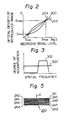
- the gradation processing is a kind of image processings which controls a curve that represents a relation between record signal levels for recording the reproduced image 102 on the output recording medium 100 and optical densities of the image 102.
- Examples of the characteristic curve are shown in Fig. 2 in which the abscissa indicates record signal levels S in logarithmic scale and the ordinate, optical densities D in linear scale.
- the S-D curves of Fig. 2 commonly have a generally upward and rightward inclination which is generally accepted to be desirable in the art, as taught in U.S. Patent 4,276,473, for example.
- various parameters are selected which include the configuration of the S-D curve, such as a straight line exemplified by a line 200 in Fig. 2, a generally convex function exemplified by a curve 202 or a generally concave function exemplified by a curve 204, the gradient or curvature of a curve, etc., as well as the value of the density D for a record signal level S.
- one curve may be selected out of ten different curves, for example.
- the spatial frequency processing is an image processing adapted to accentuate or emphasize a video signal within a specific spatial frequency range, as already described.
- the control parameters for this processing are the spatial frequency characteristic and the degree of accentuation. Examples of the selective accentuation are shown in Fig. 3, in which the abscissa indicates spatial frequencies (/mm) of video signals contained in the image 102 in logarithmic or linear scale and the ordinate, degrees of accentuation.
- Curve 300 shows that the video signal is accentuated substantially evenly over the entire spatial frequency range, while curve 302 shows that the video signal is accentuated in a high spatial frequency range but not in a low spatial frequency range.
- ten different curves are available for the spatial frequency accentuation and ten different values for the accentuation degree, by way of example. Desired ones of such parameters will be selected in conformity to a purpose of diagnosis.
- the embodiment shown and described allows image processings to be performed by the processor 12 in 1,000 different kinds of combinations: ten kinds of selective gradation processing and, concerning the spatial frequency processing, ten kinds of selective spatial frequency curves and ten kinds of selective degrees of accentuation.
- the parameters for the gradation processing and spatial frequency accentuation are selectively specified through the terminal unit 18 or the console 20 by entering data which show at least a patient's exposed portion and an exposing method. Based on the specific parameters, the gradation and spatial frequency accentuation are controlled under image processing conditions which suit a desired diagnostic purpose.
- FIG. 4 there is shown an example of a key arrangement on the operating section of the terminal unit 18 or that of the console 20.
- the operating section includes keys other than the illustrated, such as an image processing start key, they are not directly relevant with the understanding of the present invention and will not be described herein.
- the patient's parts selectable for exposure in accordance with this embodiment are the head, neck, thorax, breast, venter and limbs.
- a gradation and a spatial frequency to be accentuated will be specified automatically.
- What may be specified concerning the exposing method is an exposure with or without a contrast medium and, if without the contrast medium, whether the object is a blood or fluid vessel, which is generally a narrow tube, or an alimentary canal, which is generally a broad tube.
- Image processing conditions are specified as the patient's part to be exposed and exposing method.
- Optional items to be specified are special accentuation of a bone of a soft portion, and application of tomography or enlargement. These items will be specified if necessary and are not essential. In view of the fact that bones show a low transmissivity to a radiation and soft portions a high transmissivity, either one of them may be specified to accentuate a desired portion in a reproduced image, and this will specify a condition for spatial frequency accentuation. In the case of tomography, whether the number of desired layers is one or more may be specified and, if more than one, a multi-layer tomography key in a mode command section will be operated, as will be described later. When enlargement is selected, other spatial frequency range and accentuating degree than ordinary ones will be specified for image processings in order to prevent a decrease in the contrast of the resulting image.
- a normal processing, a subtraction, a mufti-layer tomography and a multiple exposure are available as selective processing modes.
- the multi-layer tomography key will be manipulated.
- use may be made of nine stimulable phosphor sheets as shown in Fig. 5, i.e. a recording medium 240 having recording layers 241-249.
- An imagewise radiation from an object is incident on all the sheets 241-249 at a time. Therefore, supposing a radiation directed as indicated by an arrow A, the absorbed radiation energy becomes progressively smaller from the top layer to the bottom layer.
- the reading unit 10 will read all the sheets one by one. Because the radiation energy distribution absorbed in the individual layers progressively decreases as mentioned, the image quality and thereby the contrast will appear progressively degraded toward the bottom sheet for common image processing conditions when reproduced by the recording unit 14.
- the sheets 241-249 are divided into three groups, i.e., an upper group comprising the sheets 241-243, an intermediate group comprising the sheets 244-246 and a lower group comprising the sheets 247-249.
- the processor 12 is connected to a storage 17 which may be a read only memory (ROM) which stores a table for specifying certain conditions for a gradation processing and a spatial frequency accentuating processing on the basis of the previously mentioned processing conditions such as the method of exposure, exposed part and special accentuation as well as the selected mode.
- a table may be stored in a magnetic disc in the file 16.
- exposure conditions may additionally include a radiation amount, an X-ray tube voltage and a radiation time.
- the terminal unit 18 will be operated to enter object data such as the number of a recording medium 24 used, patient's name, sex, chart number (patient code) and data of birth, as well as management data including the data on exposure, code of an exposure room and X-ray tube voltage.
- object data such as the number of a recording medium 24 used, patient's name, sex, chart number (patient code) and data of birth
- management data including the data on exposure, code of an exposure room and X-ray tube voltage.
- An arrangement may be made such that a number assigned to the recording medium 24 is read out of the label 26 by an optical or magnetic device installed in a radiation photographing system.
- a similar reading device may be employed to read object's data out of, for example, the patient's identification (ID) card or chart.
- ID patient's identification
- the management data may be one which is loaded in the photographing device. The previously mentioned exposure conditions will be entered through the keys of the terminal unit 18 to be stored in the file 16.
- All the recording media 24 exposed in various exposure rooms are conveyed to an image processing room or station in which the reading unit 10 is located. While reading an image 22 out of a recording medium 24, the reader 10 reads the number assigned to the medium 24 out of the label 26 optically or magnetically.
- the processor 12 searches for specific data stored in the file 16 indexed by the number or identification data particular to the medium 24. These data are those entered before through the terminal unit 18 or the console 20.
- the processor 12 is supplied with conditions or parameters for processing the gradation and spatial frequency accentuation from the table of the ROM, on the basis of the exposure conditions which are included in the data read out of the file 16. Image processings are performed in accordance with the parameters.
- the processed image is recorded by the recorder 14 on the recording medium 100 as a reproduced image 102.
- the sheets or recording layers 241-249 of a recording medium 240 are sequentially loaded in the reader 10 to have their radiation images entered in succession.
- the processor 12 uses a number assigned to the medium 240 as an index, the processor 12 searches for relevant data stored in the file 16 and thereby detects that the medium 240 was exposed in the multi-layer tomography mode. Then, the processor 12 processes the images in the medium 240 by modifying the usual conditions to match them to the upper sheet group 241-243, intermediate sheet group 244-246 and lower sheet group 247-249.
- the processor 12 may be loaded in the reader 10 in a random order in which case the processor 12 will process the images setting up conditions suitable for the individual sheets. Whle the processor 12 has been described as storing image processing conditions optimum for various exposure methods and parts to be exposed, it may be constructed to compute optimum image processing conditions every time data representing an exposure method and exposed part are entered. That is, the gist is that the processor 12 is furnished with means for obtaining optimum processing conditions, whether it may be a table or calculation.
- the present invention provides a radiation image reproducing apparatus which facilitates operationability, eliminates the need for expert knowledge for image processings, and reproduces an image undergone a gradation processing and a spatial frequency accentuating processing suitable for a specific diagnostic purpose if only the data indicative of at least exposed part and an exposing method are entered. Further, in the case of multi-layer tomography, adequate image processings are achievable for each of different layers of a radiation image recording medium.
Landscapes
- Engineering & Computer Science (AREA)
- General Physics & Mathematics (AREA)
- Signal Processing (AREA)
- Physics & Mathematics (AREA)
- Multimedia (AREA)
- Life Sciences & Earth Sciences (AREA)
- Spectroscopy & Molecular Physics (AREA)
- Molecular Biology (AREA)
- High Energy & Nuclear Physics (AREA)
- Health & Medical Sciences (AREA)
- Apparatus For Radiation Diagnosis (AREA)
- Analysing Materials By The Use Of Radiation (AREA)
- Radiography Using Non-Light Waves (AREA)
- Image Processing (AREA)
Claims (5)
Applications Claiming Priority (2)
| Application Number | Priority Date | Filing Date | Title |
|---|---|---|---|
| JP57137346A JPS5928144A (ja) | 1982-08-09 | 1982-08-09 | 放射線画像再生装置 |
| JP137346/82 | 1982-08-09 |
Publications (2)
| Publication Number | Publication Date |
|---|---|
| EP0100976A1 EP0100976A1 (de) | 1984-02-22 |
| EP0100976B1 true EP0100976B1 (de) | 1986-11-05 |
Family
ID=15196490
Family Applications (1)
| Application Number | Title | Priority Date | Filing Date |
|---|---|---|---|
| EP83107508A Expired EP0100976B1 (de) | 1982-08-09 | 1983-07-29 | Strahlungsbild Wiedergabeeinrichtung |
Country Status (4)
| Country | Link |
|---|---|
| US (1) | US4611247A (de) |
| EP (1) | EP0100976B1 (de) |
| JP (1) | JPS5928144A (de) |
| DE (2) | DE100976T1 (de) |
Families Citing this family (36)
| Publication number | Priority date | Publication date | Assignee | Title |
|---|---|---|---|---|
| JPS55103706U (de) * | 1979-01-09 | 1980-07-19 | ||
| DE3279930D1 (en) * | 1981-10-26 | 1989-10-12 | Fuji Photo Film Co Ltd | Data processing system for radiation image reproducing apparatus |
| IL70214A (en) * | 1983-11-13 | 1987-10-20 | Elscint Ltd | Image contrast enhancement arrangement |
| JPS60151786A (ja) * | 1984-01-19 | 1985-08-09 | Fuji Photo Film Co Ltd | 放射線画像情報読取階調処理方法および装置 |
| JPH0614168B2 (ja) * | 1984-03-07 | 1994-02-23 | 富士写真フイルム株式会社 | 放射線画像の周波数処理方法および装置 |
| JPS6168031A (ja) * | 1984-09-12 | 1986-04-08 | 富士写真フイルム株式会社 | 放射線画像情報読取装置 |
| DE3586025D1 (de) * | 1984-09-12 | 1992-06-17 | Fuji Photo Film Co Ltd | Strahlungsbildlesevorrichtung und vorrichtung zur bezeichnung der wiedergabebedingungen. |
| JPS6171035A (ja) * | 1984-09-13 | 1986-04-11 | 富士写真フイルム株式会社 | 放射線画像情報再生処理条件指示装置 |
| EP0181518B1 (de) * | 1984-10-16 | 1989-01-18 | Fuji Photo Film Co., Ltd. | Verfahren und Vorrichtung zur Aufzeichnung und Auslesen eines Strahlungsbildes |
| JPS6194035A (ja) * | 1984-10-16 | 1986-05-12 | Fuji Photo Film Co Ltd | 被写体デ−タ出力機能を備えた放射線画像情報記録読取装置 |
| JPS6254247A (ja) * | 1985-09-03 | 1987-03-09 | Fuji Photo Film Co Ltd | 放射線画像情報記録読取装置 |
| JPS62116239A (ja) * | 1985-11-15 | 1987-05-27 | Konishiroku Photo Ind Co Ltd | X線画像処理方法及び装置 |
| JPS636672A (ja) * | 1986-06-27 | 1988-01-12 | Fuji Photo Film Co Ltd | 医用画像蓄積再生方法 |
| US5231572A (en) * | 1986-10-20 | 1993-07-27 | Fuji Photo Film Co., Ltd. | Radiation image storage and reproduction system |
| JP2509815B2 (ja) * | 1986-11-25 | 1996-06-26 | コニカ株式会社 | 放射線画像情報読取表示装置 |
| US6538831B1 (en) * | 1987-09-11 | 2003-03-25 | Hitachi Medical Corporation | Method and apparatus for using recording and reading medical information and image information with digital audiotape |
| JP2779496B2 (ja) * | 1988-03-14 | 1998-07-23 | 富士写真フイルム株式会社 | 放射線画像読取表示装置 |
| JPH0214376A (ja) * | 1988-03-18 | 1990-01-18 | Fuji Photo Film Co Ltd | 放射線画像読取装置 |
| JPH01274740A (ja) * | 1988-04-26 | 1989-11-02 | Toshiba Corp | 放射線透視撮影装置 |
| US5250933A (en) * | 1989-03-02 | 1993-10-05 | Hewlett-Packard Company | Method and apparatus for the simultaneous display of one or more selected images |
| US5564012A (en) * | 1989-03-29 | 1996-10-08 | Fuji Photo Film Co., Ltd. | Support apparatus for use with radiation image information processing system |
| US5651362A (en) * | 1989-03-29 | 1997-07-29 | Fuji Photo Film Co., Ltd. | Support apparatus for use with radiation image information processing system |
| EP0427358B1 (de) * | 1989-11-08 | 1996-03-27 | George S. Allen | Mechanischer Arm für ein interaktives, bildgesteuertes, chirurgisches System |
| US5341228A (en) * | 1990-12-04 | 1994-08-23 | Research Corporation Technologies | Method and apparatus for halftone rendering of a gray scale image using a blue noise mask |
| JP2694582B2 (ja) * | 1991-06-25 | 1997-12-24 | 富士写真フイルム株式会社 | 放射線画像読取条件及び/又は画像処理条件決定方法 |
| US5301671A (en) * | 1991-09-17 | 1994-04-12 | The United States Of America As Represented By The Department Of Health And Human Services | Two- and three-dimensional autoradiographic imaging utilizing charge coupled devices |
| US5732221A (en) * | 1992-03-27 | 1998-03-24 | Documation, Inc. | Electronic documentation system for generating written reports |
| DE69416581T2 (de) * | 1994-04-22 | 1999-08-26 | Agfa-Gevaert N.V. | Verfahren um Papierbilder von Röntgenaufnahmen zu Erzeugen |
| JP3707823B2 (ja) | 1995-03-27 | 2005-10-19 | 富士写真フイルム株式会社 | 画像処理方法および装置 |
| FR2771831B1 (fr) * | 1997-11-28 | 2002-10-31 | Eastman Kodak Co | Procede d'edition automatique pour unite d'imagerie medicale numerique |
| DE10202515B4 (de) * | 2002-01-23 | 2004-08-12 | Holberg, Christof, Dr. | Verfahren, Vorrichtung und Computerprogrammprodukt zum Erstellen eines individuellen Modells eines Kieferknochens |
| US20040186370A1 (en) * | 2003-02-10 | 2004-09-23 | Konica Minolta Holdings, Inc. | Medical image processing system, medical image pickup system and method of administrating medical images |
| JP2005055273A (ja) * | 2003-08-04 | 2005-03-03 | Shimadzu Corp | X線透視装置 |
| JP4608927B2 (ja) * | 2004-03-31 | 2011-01-12 | コニカミノルタエムジー株式会社 | 画像処理方法及び画像処理装置並びに画像処理プログラム |
| JP4690204B2 (ja) * | 2006-01-16 | 2011-06-01 | 富士フイルム株式会社 | 画像再生装置およびそのプログラム |
| CN113454511B (zh) * | 2018-12-21 | 2023-11-14 | 斯科皮奥实验室有限公司 | 显微图像的压缩获取 |
Family Cites Families (18)
| Publication number | Priority date | Publication date | Assignee | Title |
|---|---|---|---|---|
| US31847A (en) * | 1861-03-26 | Jesse young | ||
| USRE31847E (en) | 1973-01-02 | 1985-03-12 | Eastman Kodak Company | Apparatus and method for producing images corresponding to patterns of high energy radiation |
| US3859527A (en) * | 1973-01-02 | 1975-01-07 | Eastman Kodak Co | Apparatus and method for producing images corresponding to patterns of high energy radiation |
| US3988602A (en) * | 1975-02-03 | 1976-10-26 | Goodyear Aerospace Corporation | Method and apparatus for enhancing data |
| US4032784A (en) * | 1975-08-04 | 1977-06-28 | The Gerber Scientific Instrument Company | Method and apparatus for examining a body by a beam of x-rays or other penetrating radiation |
| JPS5512429A (en) * | 1978-07-12 | 1980-01-29 | Fuji Photo Film Co Ltd | Radioactive image reader |
| JPS5548674A (en) * | 1978-10-05 | 1980-04-07 | Fuji Photo Film Co Ltd | Reading device for radiation picture information |
| US4315318A (en) * | 1978-12-26 | 1982-02-09 | Fuji Photo Film Co., Ltd. | Method and apparatus for processing a radiation image |
| JPS5588740A (en) * | 1978-12-26 | 1980-07-04 | Fuji Photo Film Co Ltd | Method of treating gradation of radiation picture of breast and its device |
| JPS55116340A (en) * | 1979-02-28 | 1980-09-06 | Fuji Photo Film Co Ltd | Method and device for processing gradation of radiation picture |
| JPS55116339A (en) * | 1979-02-28 | 1980-09-06 | Fuji Photo Film Co Ltd | Method and device for processing gradation of radiation picture of chest |
| JPS55146447A (en) * | 1979-05-01 | 1980-11-14 | Fuji Photo Film Co Ltd | Radiation image converting panel |
| JPS56104645A (en) * | 1979-12-25 | 1981-08-20 | Fuji Photo Film Co Ltd | Radiation picture treating method and its device |
| JPS5691735A (en) * | 1979-12-25 | 1981-07-24 | Fuji Photo Film Co Ltd | Method and apparatus for treating xxray image |
| JPS571878A (en) * | 1980-06-02 | 1982-01-07 | Matsushita Electric Ind Co Ltd | Electromagnetic proportional control valve |
| CA1192674A (en) * | 1981-10-16 | 1985-08-27 | Hisatoyo Kato | Radiation image recording and read-out system |
| JPS5914842A (ja) * | 1982-07-19 | 1984-01-25 | 富士写真フイルム株式会社 | 任意断層撮影方法および装置 |
| US4482924A (en) * | 1982-09-29 | 1984-11-13 | Eastman Kodak Company | Video player, film medium, and photographic printer for automatic cropping |
-
1982
- 1982-08-09 JP JP57137346A patent/JPS5928144A/ja active Pending
-
1983
- 1983-07-29 EP EP83107508A patent/EP0100976B1/de not_active Expired
- 1983-07-29 DE DE198383107508T patent/DE100976T1/de active Pending
- 1983-07-29 DE DE8383107508T patent/DE3367469D1/de not_active Expired
- 1983-08-08 US US06/521,507 patent/US4611247A/en not_active Expired - Lifetime
Also Published As
| Publication number | Publication date |
|---|---|
| US4611247A (en) | 1986-09-09 |
| EP0100976A1 (de) | 1984-02-22 |
| JPS5928144A (ja) | 1984-02-14 |
| DE100976T1 (de) | 1984-08-02 |
| DE3367469D1 (en) | 1986-12-11 |
Similar Documents
| Publication | Publication Date | Title |
|---|---|---|
| EP0100976B1 (de) | Strahlungsbild Wiedergabeeinrichtung | |
| US4317179A (en) | Method and apparatus for processing a radiographic image | |
| US6542771B2 (en) | Method of and system for detecting prospective abnormal shadow and method of reproducing radiation image | |
| US4340911A (en) | Image gradation processing method and apparatus for mammogram copying system | |
| EP0507485B1 (de) | Methode und Apparat für die Bildverarbeitung von radiografisch hergestellten Bildmustern | |
| EP0032237A1 (de) | Verfahren und Vorrichtung zur Bearbeitung eines Strahlungsbildes | |
| US5551428A (en) | Automatic routing to selected destinations of storage phosphor images | |
| US5369572A (en) | Radiographic image processing method wherein small variation of density is selectively made clear | |
| US5231572A (en) | Radiation image storage and reproduction system | |
| US4591922A (en) | Radiation image reproducing apparatus | |
| EP0490532A2 (de) | Gerät und Verfahren zum Verarbeiten von Röntgenbilddaten | |
| US4306290A (en) | Image gradation processing method and apparatus for radiographic image copying system | |
| US5714764A (en) | Method for detecting prospective abnormal patterns | |
| EP0599098B1 (de) | Mehrere Versionen eines Speicherleuchtstoffbildes | |
| JPH08317908A (ja) | 任意に配置し得るテキスト欄をもつ医学的画像の再生または表示方法およびシステム | |
| JPH0775635A (ja) | 有効に付与された放射線量を制御する方法ならびに装置 | |
| US5751787A (en) | Materials and methods for improved radiography | |
| JP2002008006A (ja) | 画像処理条件決定方法および装置 | |
| JPH06282632A (ja) | 放射線画像に実行された画像処理を評価する方法 | |
| JPS63103225A (ja) | 放射線画像の再生保存システム | |
| Balter | On the work of the radiologist—separation of image capture from image display | |
| JPS5928146A (ja) | 放射線画像再生装置 | |
| EP0527524B1 (de) | Verfahren zur Qualitätsverbesserung von Bildern | |
| EP4375922A1 (de) | Vorrichtung und verfahren zur wirbelkörpererkennung in medizinischen bildern | |
| JPH0516859B2 (de) |
Legal Events
| Date | Code | Title | Description |
|---|---|---|---|
| PUAI | Public reference made under article 153(3) epc to a published international application that has entered the european phase |
Free format text: ORIGINAL CODE: 0009012 |
|
| AK | Designated contracting states |
Designated state(s): DE FR NL |
|
| 17P | Request for examination filed |
Effective date: 19840524 |
|
| DET | De: translation of patent claims | ||
| EL | Fr: translation of claims filed | ||
| GRAA | (expected) grant |
Free format text: ORIGINAL CODE: 0009210 |
|
| AK | Designated contracting states |
Kind code of ref document: B1 Designated state(s): DE FR NL |
|
| REF | Corresponds to: |
Ref document number: 3367469 Country of ref document: DE Date of ref document: 19861211 |
|
| ET | Fr: translation filed | ||
| PLBE | No opposition filed within time limit |
Free format text: ORIGINAL CODE: 0009261 |
|
| STAA | Information on the status of an ep patent application or granted ep patent |
Free format text: STATUS: NO OPPOSITION FILED WITHIN TIME LIMIT |
|
| 26N | No opposition filed | ||
| PGFP | Annual fee paid to national office [announced via postgrant information from national office to epo] |
Ref country code: FR Payment date: 20020719 Year of fee payment: 20 |
|
| PGFP | Annual fee paid to national office [announced via postgrant information from national office to epo] |
Ref country code: NL Payment date: 20020723 Year of fee payment: 20 |
|
| PGFP | Annual fee paid to national office [announced via postgrant information from national office to epo] |
Ref country code: DE Payment date: 20020828 Year of fee payment: 20 |
|
| PG25 | Lapsed in a contracting state [announced via postgrant information from national office to epo] |
Ref country code: NL Free format text: LAPSE BECAUSE OF EXPIRATION OF PROTECTION Effective date: 20030729 |
|
| NLV7 | Nl: ceased due to reaching the maximum lifetime of a patent |
Effective date: 20030729 |