WO2022163845A1 - 検査システム、検査方法、プログラム、およびコンピュータが読み取り可能な記憶媒体 - Google Patents
検査システム、検査方法、プログラム、およびコンピュータが読み取り可能な記憶媒体 Download PDFInfo
- Publication number
- WO2022163845A1 WO2022163845A1 PCT/JP2022/003467 JP2022003467W WO2022163845A1 WO 2022163845 A1 WO2022163845 A1 WO 2022163845A1 JP 2022003467 W JP2022003467 W JP 2022003467W WO 2022163845 A1 WO2022163845 A1 WO 2022163845A1
- Authority
- WO
- WIPO (PCT)
- Prior art keywords
- antibody
- target
- target microorganism
- microorganisms
- specimen
- Prior art date
- Legal status (The legal status is an assumption and is not a legal conclusion. Google has not performed a legal analysis and makes no representation as to the accuracy of the status listed.)
- Ceased
Links
Images





Classifications
-
- C—CHEMISTRY; METALLURGY
- C12—BIOCHEMISTRY; BEER; SPIRITS; WINE; VINEGAR; MICROBIOLOGY; ENZYMOLOGY; MUTATION OR GENETIC ENGINEERING
- C12M—APPARATUS FOR ENZYMOLOGY OR MICROBIOLOGY; APPARATUS FOR CULTURING MICROORGANISMS FOR PRODUCING BIOMASS, FOR GROWING CELLS OR FOR OBTAINING FERMENTATION OR METABOLIC PRODUCTS, i.e. BIOREACTORS OR FERMENTERS
- C12M1/00—Apparatus for enzymology or microbiology
- C12M1/34—Measuring or testing with condition measuring or sensing means, e.g. colony counters
-
- G—PHYSICS
- G01—MEASURING; TESTING
- G01N—INVESTIGATING OR ANALYSING MATERIALS BY DETERMINING THEIR CHEMICAL OR PHYSICAL PROPERTIES
- G01N21/00—Investigating or analysing materials by the use of optical means, i.e. using sub-millimetre waves, infrared, visible or ultraviolet light
- G01N21/62—Systems in which the material investigated is excited whereby it emits light or causes a change in wavelength of the incident light
- G01N21/63—Systems in which the material investigated is excited whereby it emits light or causes a change in wavelength of the incident light optically excited
- G01N21/64—Fluorescence; Phosphorescence
-
- G—PHYSICS
- G01—MEASURING; TESTING
- G01N—INVESTIGATING OR ANALYSING MATERIALS BY DETERMINING THEIR CHEMICAL OR PHYSICAL PROPERTIES
- G01N33/00—Investigating or analysing materials by specific methods not covered by groups G01N1/00 - G01N31/00
- G01N33/48—Biological material, e.g. blood, urine; Haemocytometers
- G01N33/50—Chemical analysis of biological material, e.g. blood, urine; Testing involving biospecific ligand binding methods; Immunological testing
- G01N33/53—Immunoassay; Biospecific binding assay; Materials therefor
-
- G—PHYSICS
- G01—MEASURING; TESTING
- G01N—INVESTIGATING OR ANALYSING MATERIALS BY DETERMINING THEIR CHEMICAL OR PHYSICAL PROPERTIES
- G01N33/00—Investigating or analysing materials by specific methods not covered by groups G01N1/00 - G01N31/00
- G01N33/48—Biological material, e.g. blood, urine; Haemocytometers
- G01N33/50—Chemical analysis of biological material, e.g. blood, urine; Testing involving biospecific ligand binding methods; Immunological testing
- G01N33/53—Immunoassay; Biospecific binding assay; Materials therefor
- G01N33/569—Immunoassay; Biospecific binding assay; Materials therefor for microorganisms, e.g. protozoa, bacteria, viruses
Definitions
- the present invention relates to an inspection system, inspection method, program, and computer-readable storage medium, and more particularly to an inspection system, inspection method, program, and computer-readable storage medium for inspecting microorganisms.
- the present invention has been made in view of such circumstances, and aims to provide an inspection system, an inspection method, a program, and a computer-readable storage medium that can quickly and accurately inspect microorganisms. aim.
- the measuring apparatus is a test system for testing microorganisms, wherein target microorganisms among microorganisms in a predetermined sample are reacted with antibodies that produce antibody reactions.
- the apparatus is characterized by comprising a fluorescence processing unit that performs fluorescence processing on the specimen.
- the fluorescence processing unit performs fluorescence processing at a predetermined wavelength to fluoresce the target microorganism among the microorganisms in the predetermined sample and the antibody that causes the target microorganism to react with the antibody that causes the antibody reaction. Since it was decided to have a It is possible to distinguish target microorganisms from other microorganisms by, for example, fluorescing the microorganisms. As a result, it is possible to quickly test microorganisms, for example, it is possible to test for target microorganisms without depending on the culture method.
- the antibody is an antibody that causes an antibody reaction with the target microorganism, it can be reliably reacted with the target microorganism, and the fluorescence of the target microorganism can be reliably suppressed to accurately detect the microorganism. inspection can be performed.
- the fluorescence processing unit further performs fluorescence processing to fluoresce the sample before the antibody reacts with the target microorganism, the target microorganism in the sample before the target microorganism reacts with the antibody It is possible to fluoresce, including the target microorganism, and to grasp the change before and after the antibody reaction of the target microorganism.
- the fluorescence processing unit further removes the target microorganism and the target in the specimen after reacting any one of the target microorganism and other microorganisms other than the target microorganism with the antibody.
- the fluorescence treatment is performed so as not to cause any one of the microorganisms other than the microorganism to fluoresce, and to cause the other of the target microorganism and the other microorganism other than the target microorganism to fluoresce.
- neither the target microorganism nor the other microorganism other than the target microorganism does not cause fluorescence in the sample after the target microorganism reacts with the antibody, and the target microorganism and the other microorganism other than the target microorganism
- Fluorescence treatment can be performed to generate fluorescence in either one of the other microorganisms, and changes in the target microorganism before and after the antibody reaction can be grasped.
- the fluorescence processing unit does not cause the target microorganisms to emit fluorescence in the specimen after the reaction between the target microorganisms and the antibody, and causes fluorescence to be emitted to microorganisms other than the target microorganisms. If fluorescence treatment is performed to cause fluorescence, it is possible to prevent the target microorganism from generating fluorescence in the sample after the target microorganism reacts with the antibody, and the change in the target microorganism before and after the antibody reaction can be grasped.
- the apparatus has an image processing unit that performs image processing of the fluorescent sample, it becomes possible to grasp changes in target microorganisms after antibody reaction from images.
- An image processing unit that performs image processing of the fluorescent specimen before the reaction between the target microorganism and the antibody and image processing of the fluorescent specimen after the target microorganism and the antibody react. Then, it becomes possible to grasp the change before and after the antibody reaction of the target microorganism by the image.
- the image processing unit determines whether or not the image of the fluorescent specimen is included in the range of the reference data of the microorganism, and determines the range of the reference data of the microorganism in the image of the fluorescent specimen. If the image contained in is specified, it is possible to extract the image of the microorganism after the antibody reaction of the target microorganism from which foreign substances other than the microorganism have been removed, and the microorganism can be inspected with higher accuracy. .
- the image processing unit compares the reference data of the microorganism with the image of the fluorescent specimen before the reaction and the image of the fluorescent specimen after the reaction, and corresponds to the reference data of the microorganism. If the image of the fluorescent specimen before the reaction and the image of the fluorescent specimen after the reaction are specified, the image of the microorganism from which foreign substances other than the microorganism have been removed before and after the antibody reaction of the target microorganism Images can be extracted, and microorganisms can be inspected with higher accuracy. If the reference data for the microorganisms includes the reference data for the target microorganisms, microorganisms other than the target microorganisms can be excluded from the microorganisms, and the microorganisms can be tested more accurately. can be done.
- target microorganism count counting unit for counting the number of the target microorganism, more specifically, the fluorescent specimen before the antibody reacts and the target microorganism reacted with the antibody.
- the number of target microorganisms can be counted by providing a target microorganism count counting unit for counting the number of target microorganisms from the later fluorescent sample.
- the target microorganism number counting unit counts the number of microorganisms counted from the fluorescent specimen after the reaction between the target microorganism and the antibody from the number of microorganisms counted from the fluorescent specimen before the antibody reacts.
- the number of target microorganisms can be counted by subtracting the .
- the target microorganism count counting unit detects the target from an image of the fluorescent specimen before reaction with the antibody and an image of the fluorescent specimen after reaction between the target microorganism and the antibody. You can count the number of microorganisms that For example, the target microorganism number counting unit determines whether the target microorganism reacts with the antibody based on the number of microorganisms counted from the image of the fluorescent specimen image processed by the image processing unit before the antibody reacts. The number of target organisms can be counted by subtracting the number of organisms counted from the image of the fluorescent specimen after irradiating.
- the target microorganism identification unit for identifying the type of target microorganism, the type of target microorganism can be identified. More specifically, the number of the target microorganisms in the fluorescent sample before the reaction with the antibody counted by the target microorganism counting unit and the fluorescence after the reaction between the target microorganism and the antibody after the target microorganism and the antibody react with the number of the target microorganism in the fluorescent specimen before the antibody reacts, and and a target microorganism identification unit that identifies the type of the target microorganism by assuming that the target microorganism has generated the antibody reaction when the number of the target microorganism in the fluorescent sample has decreased. can be done.
- the target microorganism identification unit determines the number of the target microorganisms in the image of the fluorescent sample before the antibody reacts, which is counted by the target microorganism count counting unit, and the number of the target microorganisms and the antibody reacts. comparing the number of the target microorganisms in the image of the fluorescent specimen after the reaction with the target microorganisms, and comparing the number of the target microorganisms in the image of the fluorescent specimen before the reaction with the antibody. When the number of the target microorganisms in the image of the fluorescent specimen after the reaction with the antibody decreases, the target microorganism is considered to have caused the antibody reaction, and the type of the target microorganism can be specified.
- An image capturing unit having a predetermined lens is provided, and the image capturing unit can capture an image of the specimen through the predetermined lens.
- the image capturing unit has a plurality of lenses with different magnifications, and is configured to change the magnification of the lenses to a predetermined value by changing the selection and/or combination of the plurality of lenses with different magnifications. Then, the magnification of the image can be appropriately changed.
- It has a predetermined stage on which the specimen is placed, and a position control unit that controls the position of the stage, and the position control unit controls the position of the stage to control the If the focal position is controlled, the focal position can be controlled.
- the position control unit changes the position of the fluorescent sample, and the image processing unit captures a plurality of images of the fluorescent sample, aggregates the captured images, and performs image processing. As a result, even if the concentration of the target microorganisms in the sample is uneven, it is possible to prevent the measurement accuracy from deteriorating.
- the fluorescence processing unit performs the fluorescence processing on the target microorganisms in the specimen by fluorescent staining using a predetermined reagent, the target microorganisms are dyed to reliably distinguish them from other microorganisms. can be done.
- the target microorganisms include at least one of bacteria and viruses, and at least one mode of a bacteria inspection mode for inspecting the bacteria and a virus inspection mode for inspecting the viruses is provided. For example, at least one of a bacterial test and a viral test can be performed.
- the fluorescence processing unit has a band-pass filter that limits the range of wavelengths of light to a predetermined range, it is possible to more accurately inspect target microorganisms. If the specimen is filtered using a predetermined filter medium, and a filtering unit for removing foreign matter from the specimen is provided, the foreign matter is removed from the specimen, and target microorganisms can be tested with higher accuracy. .
- the measurement method according to the present invention is an inspection method for inspecting microorganisms, which comprises a target microorganism among microorganisms in a predetermined sample and an antibody reaction with the target microorganism.
- the method is characterized by comprising a fluorescence treatment step of performing a fluorescence treatment at a predetermined wavelength to fluoresce the specimen reacted with the generated antibody.
- the fluorescence treatment step may be a step of performing fluorescence treatment to fluoresce the specimen before the target microorganism reacts with the antibody.
- the target microorganism and the target in the specimen after reacting any one of the target microorganism and other microorganisms other than the target microorganism with the antibody A step of performing a fluorescence treatment in which either one of the microorganisms other than the target microorganism does not fluoresce, and the other of the target microorganism and the other microorganism other than the target microorganism fluoresces. can be done.
- the target microorganism in the specimen after the reaction between the target microorganism and the antibody, the target microorganism does not emit fluorescence, and microorganisms other than the target microorganism emit fluorescence. It can be a step of performing a fluorescence treatment that causes the reaction to occur.
- the antibody can be an antibody that does not cause the target microorganism to fluoresce when an antibody reaction is caused in the target microorganism.
- fluorescent staining is performed using a predetermined reagent to fluoresce the microorganisms in the specimen, and the reagent causes the target microorganisms to react with the antibody. It can be a reagent that does not cause the target microorganism to fluoresce.
- the reagent can be a fluorescein-based compound and/or a propidium-based compound. Said reagent can be FDA (fluorescein diacetate) and/or PI (propidium iodide).
- a program according to the present invention causes a computer of an inspection system for inspecting microorganisms to react with a target microorganism in a predetermined sample and an antibody that produces an antibody reaction with the target microorganism. It is characterized by functioning as a fluorescence processing unit that performs fluorescence processing on the sample that has been processed using a predetermined wavelength.
- a computer-readable storage medium stores the above program.
- microorganisms can be tested quickly and accurately.
- FIG. 2 is a diagram schematically showing the configuration of a specimen according to the embodiment of the present invention
- FIG. It is a figure showing the whole inspection system composition concerning the embodiment of the present invention.
- FIG. 4 is a diagram schematically showing a state in which foreign matter is removed from the same sample;
- FIG. 4 is a diagram schematically showing a state in which the same specimen reacts with an antibody.
- FIG. 10 is a diagram showing an image of a sample before antibody reaction;
- FIG. 10 is a diagram showing an image of a specimen after antibody reaction; It is a flow chart for explaining the inspection method of the present invention.
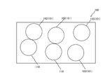
- FIG. 1 is a diagram schematically showing the configuration of a sample according to an embodiment of the present invention
- FIG. 2 is a diagram showing the overall configuration of an inspection system according to an embodiment of the present invention
- FIG. 3 is an embodiment of the present invention.
- Another diagram showing the overall configuration of the inspection system according to FIG. 4 is a diagram showing the configuration of the computer of the inspection system
- FIG. 7 is a diagram showing the configuration of the fluorescence processing unit and the image capturing unit of the inspection system
- FIG. 8 is an image of the specimen before antibody reaction.
- FIG. 9 shows an image of the sample after antibody reaction.
- the inspection system 1 is an apparatus for measuring target microorganisms 102 among microorganisms 101 in a predetermined specimen 100 (sample 100 to be inspected) shown in FIG. is.
- the target microorganisms 102 can include, for example, bacteria and viruses.
- Bacteria can include various types of bacteria such as general bacteria and E. coli, and viruses include various types of viruses such as influenza viruses and coronaviruses. be able to. As shown in FIG.
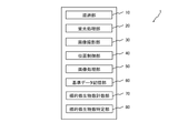
- the inspection system 1 includes a filtering unit 10, a fluorescence processing unit 20, an image capturing unit 30, a position control unit 40, an image processing unit 50, a reference data storage unit 60, a target microorganism counting unit 70, a target It has a microorganism identification unit 80 .
- the inspection system 1 has a bacteria inspection mode 2 for inspecting bacteria and a virus inspection mode 3 for inspecting viruses.
- the inspection system 1 has a general configuration as a computer. As shown in FIG. 4, a central processing unit (CPU, GPU, DSP) 1B, storage devices (ROM, RAM, hard disk, cache memory) 1C, input device (keyboard, touch panel, mouse) 1D, display device (liquid crystal display) 1E, and the like.
- the storage device 1C functions as a computer-readable storage medium.
- the functional units 10 , 20 , 30 , 40 , 50 , 60 , 70 , 80 can mutually generate and input/output signal information by the computer functions of the inspection system 1 .
- the filtering unit 10 has a predetermined filtering filter. That is, the filtering unit 10 can filter the specimen 100 using the filtration filter and remove the foreign matter 100' from the specimen 100 as shown in FIG.
- the filtration filter can be a syringe filter.
- the fluorescence processing unit 20 can perform fluorescence processing to fluoresce the microorganisms 101 in the specimen 100 at a predetermined wavelength. That is, the fluorescence processing unit 20 can perform the fluorescence processing to fluoresce the specimen 100 before the target microorganism 102 reacts with the predetermined antibody 110 at a predetermined wavelength. In addition, as shown in FIG. 6, the fluorescence processing unit 20 can perform fluorescence processing at a predetermined wavelength to fluoresce the specimen 100 after reacting the target microorganism 102 with the predetermined antibody 110 .
- the fluorescence processing unit 20 does not cause the target microorganisms 102 to emit fluorescence at a predetermined wavelength in the sample 100 after the target microorganisms 102 and the antibody 110 have reacted with each other. Fluorescence treatment may be performed to cause other microorganisms 103 to emit fluorescence.
- the antibody 110 is a glycoprotein molecule produced by B cells among lymphocytes, and has the function of recognizing and binding to a molecule (antigen) such as a specific protein.
- Antibodies 110 mainly exist in blood and body fluids, and recognize and bind to, for example, bacteria, viruses, and cells infected with microorganisms that have invaded the body as antigens.
- phagocytic cells such as white blood cells and microphages recognize and phagocytize the complex of the antigen and the antibody 110 and work to remove it from the body, and immune cells such as lymphocytes bind to become immune. It is something that provokes a reaction.
- the predetermined antibody 110 of the present invention is an antibody that causes an antibody response to the target microorganism 102 .
- the predetermined antibody 110 is an antibody that does not cause the target microorganism 102 to fluoresce at a predetermined wavelength when the target microorganism 102 is caused to react with an antibody.
- the fluorescence processing unit 20 can detect the target microorganism 102 at a predetermined wavelength in the specimen 100 after the target microorganism 102 and the antibody 110 have reacted. Fluorescence treatment may be performed so that the microbes 103 other than the target microbes 102 do not fluoresce, and the microbes 103 other than the target microbes 102 fluoresce.
- the antibody 110 is an antibody that causes an antibody reaction in the target microorganism 102, and an antibody that does not cause the target microorganism 102 to fluoresce at a predetermined wavelength when the target microorganism 102 produces an antibody reaction. Whether or not it is is confirmed in advance by various tests.
- the antibody 110 includes various antibodies such as an antibody that causes an antibody reaction against E. coli, an antibody that causes an antibody reaction against general bacteria, an antibody that causes an antibody reaction against influenza virus, an antibody that causes an antibody reaction against coronavirus, and so on. can be used.
- the fluorescence processing unit 20 performs fluorescent staining using a predetermined reagent to fluoresce the microorganisms 101 in the specimen 100 before antibody reaction and fluorescence treatment to fluoresce the microorganisms 101 in the specimen 100 after antibody reaction. It can be carried out.
- This reagent can be a reagent that does not cause the target microorganism 102 to fluoresce at a predetermined wavelength when the antibody 110 causes the target microorganism 102 to elicit an antibody response. That is, the fluorescence processing unit 20 performs fluorescent staining using a predetermined reagent to fluoresce the microorganisms 101 in the specimen 100 at a predetermined wavelength.
- a reagent used for fluorescent staining can be a fluorescein-based compound and/or a propidium-based compound. More specifically, the reagent used for fluorescent staining can be FDA (Fluorescein Diacetate) and/or PI (Propidium Iodide). According to the present inventors, FDA (Fluorescein Diacetate) and PI (Propidium Iodide) are targeted at predetermined wavelengths when the antibody 110 causes the target microorganism 102 to generate an antibody response. It has been clarified that the reagent does not cause the microorganism 102 to fluoresce.
- the fluorescence processing section 20 has an excitation light irradiation section 21 and a wavelength measurement section 22, as shown in FIG.
- the excitation light irradiation unit 21 can irradiate the specimen 100 with excitation light having a predetermined wavelength.
- the wavelength measurement unit 22 can measure the wavelength of fluorescence from the specimen 100 that has absorbed the excitation light. More specifically, the fluorescence processing unit 20 can measure the wavelength of fluorescence from the specimen 100 before antibody reaction and the wavelength of fluorescence from the specimen 100 after antibody reaction.
- the light emitted by the excitation light irradiation unit 21 is 488 nm
- the wavelength measured by the wavelength measurement unit 22 is around 512 nm in bacteria inspection mode 2
- the fluorescence processing unit 20 further has a bandpass filter 23 that limits the wavelength range of light to a predetermined range. That is, the band-pass filter 23 has a wavelength range of the excitation light irradiated onto the specimen 100 by the excitation light irradiation unit 21, a wavelength range of fluorescence from the specimen 100 before antibody reaction measured by the wavelength measurement unit 22, and The wavelength range of fluorescence from the specimen 100 after antibody reaction can be restricted to a predetermined range.
- the light irradiated by the excitation light irradiation unit 21 is 488 ⁇ 10 nm by the band-pass filter 23, the wavelength measured by the wavelength measurement unit 22 is 512 ⁇ 10 nm by the band-pass filter 23 in the bacteria inspection mode 2, and the virus inspection Mode 3 is limited to 512 ⁇ 10 nm or 620 ⁇ 10 nm, respectively.
- the image photographing unit 30 has an optical lens 31 and a photographing device 32 in more detail than the predetermined lens.
- the image capturing unit 30 can magnify and capture an image of the specimen 100 including fluorescent spots via a predetermined optical lens 31 and an image capturing device 32 .
- the image capturing unit 30 has a plurality of optical lenses 31 with different magnifications. That is, the image capturing unit 30 can change the magnification of the optical lens 31 to a predetermined value by selecting and/or changing the combination of the plurality of optical lenses 31 having different magnifications.
- the optical lens 31 is an optical element for refracting light to diverge or converge, and a convex lens and/or a concave lens can be used.
- the imaging device 32 has functions of a CCD camera and a CMOS camera, and can capture an image of the specimen 100 .
- the target microorganism 102 is a bacterium
- the size of the bacterium is about 1 to 5 ⁇ m.
- magnification is set to 50 times or more to take an image. Note that the magnification of the image may be arbitrarily set by the function of a computer such as a CPU of the inspection system 1 .
- the image capturing unit 30 can capture an image of the fluorescent sample 100 by placing the sample 100 on a predetermined stage 33 (mounting table 33 on which the sample 100 is placed). That is, the image capturing unit 30 performs first capturing of the surface of the membrane filter A capturing the foreign matter 100' containing the microorganism 101, and second capturing of the surface of the slide glass B onto which the sample 100 is directly dropped. A photograph can be taken (the second photographing is carried out with the cover glass placed on the slide glass B). The first imaging and the second imaging can be performed with the membrane filter A and the slide glass B placed on the stage 33 .
- the position control unit 40 can control the position of the stage 33 on which the specimen 100 is placed.
- the position control unit 40 can control the movement of the stage 33 in the vertical direction and the horizontal direction.
- the position control unit 40 can control the focus position in the image capturing unit 30 by controlling the vertical movement of the stage 33 . That is, the position control unit 40 controls the movement of the stage 33 in the vertical direction so that the focal positions in the first imaging and the second imaging are the surface positions of the membrane filter A and the surface positions of the slide glass B, for example. can do.
- the position control unit 40 can horizontally change the position of the fluorescent specimen 100 before antibody reaction and the position of the fluorescent specimen 100 after antibody reaction. While changing the position of the sample 100 in the horizontal direction by the position control unit 40, the image capturing unit 30 captures a plurality of images of the fluorescent sample 100 before the antibody reaction and the fluorescent sample 100 after the antibody reaction. You can shoot.
- the image processing unit 50 can perform image processing of the fluorescent specimen 100 photographed by the image photographing unit 30 . That is, the image processing unit 30 performs image processing of the fluorescent specimen 100 before the antibody reaction between the target microorganism 102 and the antibody 110 and processing of the fluorescent specimen 100 after the antibody reaction between the target microorganism 102 and the antibody 110. Image processing can be performed.
- the image processing unit 50 can perform image processing based on reference data of the microorganism 101 . That is, the image processing unit 50 determines whether or not the image of the fluorescent specimen 100 before the antibody reaction and the fluorescent specimen 100 after the antibody reaction are included in the reference data range of the microorganism 101. 8 and 9, an image portion 104a included in the reference data range of the microorganism 101 in the image of the fluorescent specimen 100 before antibody reaction and the fluorescent specimen 100 after antibody reaction. of the image, the image portion 104b included in the reference data range of the microorganism 101 is specified. Further, the image processing section 50 can extract the specified image portions 104a and 104b.
- the image portion 104a specified and extracted from the image of the fluorescent specimen 100 before antibody reaction includes the image of the target microorganism 102 and the image of the other microorganism 103, as shown in FIG. 9, the image portion 104b specified and extracted from the image of the fluorescent specimen 100 after antibody reaction does not contain the target microorganism 102 because it reacts with the antibody. Only images of other microorganisms 103 will be included.
- the number N1 of microorganisms 101 is counted from the image portion 104a identified and extracted from the image of the fluorescent specimen 100 before antibody reaction, and the number N1 of microorganisms 101 is identified and extracted from the image of the fluorescent specimen 100 after antibody reaction.
- the number N2 of microorganisms 101 is counted from the image portion 104b, and as shown in Equation 1, the number N2 of microorganisms 101 counted from the image portion 104b is subtracted from the number N1 of microorganisms 101 counted from the image portion 104a. ), the number N of the target microorganisms 102 can be counted.
- N N1-N2
- the reference data of the microorganism 101 is the data of the target microorganism 102 and the data of the other microorganism 103 so that both the target microorganism 102 and the other microorganism 103 are extracted by the image processing unit 50. Both are included and set.
- the reference data of the microorganism 101 are the size of the target microorganism 102 and the other microorganism 103, the shape of the target microorganism 102 and the other microorganism 103, the fluorescence of the target microorganism 102 and the other microorganism 103.
- the image processing unit 50 uses the image of the fluorescent specimen 100 before antibody reaction and the image of the fluorescent specimen 100 after antibody reaction as a reference data, and the corresponding size and Images of the microorganisms 102 and 103 in shape, and images of fluorescent portions of the microorganisms 102 and 103 having shades corresponding to reference data can be identified and extracted.
- the reference data can include, for example, general bacteria data, E. coli data, virus data, and the like.
- the image processing unit 50 generates an image of the fluorescent specimen 100 before the antibody reaction, which is obtained by changing the position of the specimen 100 photographed by the image photographing unit 30, in the horizontal direction, and an image of the fluorescent specimen 100 after the antibody reaction. images can be aggregated and processed.
- the reference data storage unit 60 can store reference data for the microorganism 101 .
- the reference data is based on at least one of the size of the microorganisms 101 and 102, the shape of the microorganisms 101 and 102, and the intensity of the fluorescence of the microorganisms 101 and 102.
- the reference data can include data on various bacteria such as data on general bacteria and data on E. coli.
- the reference data can include virus data and the like, and the virus data can include coronavirus and influenza virus data.
- the target microorganism count counting unit 70 can count the number of target microorganisms 102 . That is, the target microorganism count counting unit 70 generates an image of the fluorescent sample 100 before the reaction of the antibody 110 image-processed by the image processing unit 50 and a fluorescent sample after the reaction between the target microorganism 102 and the antibody 110. The number N of target microorganisms 102 from 100 images can be counted.
- the target microorganism number counting unit 70 counts the number N1 of the microorganisms 101 from the image portion 104a specified and extracted from the image of the fluorescent specimen 100 before the antibody reaction, and the fluorescent specimen 100 after the antibody reaction.
- the number N2 of microorganisms 101 is counted from the image portion 104b specified and extracted from the image of , and as shown in Equation 1, the number N1 of microorganisms 101 counted from the image portion 104b is calculated from the number N1 of microorganisms 101 counted from the image portion 104b.
- the number N of target microorganisms 102 can be counted by subtracting (subtracting) N2.
- the number of target microorganisms 102 counted by the target microorganism number counting unit 70 is based on the image of the fluorescent specimen 100 before the reaction of the antibody 110, which has been image-processed by the image processing unit 50, and the target microorganism 102 and the antibody. This is done based on the number of fluorescent spots appearing in the image of the fluorescent specimen 100 after the reaction with 110 .
- the target microorganism identifying unit 80 can identify the type of target microorganism 102 . That is, the target microorganism identification unit 80 determines the number of target microorganisms 102 in the image of the fluorescent specimen 100 before the antibody 110 reacts, counted by the target microorganism number counting unit 70, and the number of target microorganisms 102 and the antibody. The number of target microorganisms 102 in the image of the fluorescent specimen 100 after the reaction is compared with the number of target microorganisms 102 in the image of the fluorescent specimen 100 before the antibody 110 reacts with the target microorganisms.
- the target microorganism 102 When the number of target microorganisms 102 in the image of the fluorescent specimen 100 after the reaction between 102 and the antibody 110 decreases, the target microorganism 102 is considered to have generated an antibody reaction, and the type of the target microorganism 102 is determined. can be specified.
- each functional unit of the inspection system 1 described above can function by executing a predetermined program 200 .
- the program 200 causes the computer of the inspection system 1 to perform the fluorescence processing section 20, the image capturing section 30, the position control section 40, the image processing section 50, the reference data storage section 60, the target microorganism counting section 70, and the target microorganism identifying section. 80.
- the program 200 is stored in the storage device 1C of the inspection system 1. FIG.
- step 1 the sample 100 is adjusted.
- the object to be inspected such as food, hands, mouth, floor, etc.
- the swab that has been wiped off is washed away with purified water.
- the rinsed purified water is filtered by the filtering unit 10 to remove the foreign matter 100', and the specimen 100 is prepared.
- the prepared specimen 100 is divided into at least two test tubes (eg, 1 ml) to perform fluorescence treatment in the fluorescence treatment step of step 2 before and after antibody reaction. .
- the antibody 110 is added to one sample 100 out of the separately prepared samples 100 to generate an antibody reaction (the other sample 100 out of the separately adjusted samples 100 is not supplied with the antibody 110).
- the antibody 110 can be an antibody 110 that does not cause the target microbe 102 to fluoresce when the target microbe 102 provokes an antibody response.
- a reagent for fluorescent staining is added to each of the separately prepared specimens 100 .
- the reagent can be a reagent that does not cause the target microorganism 102 to fluoresce when the antibody 110 causes the target microorganism 102 to react with an antibody.
- the reagent can be a fluorescein-based compound and/or a propidium-based compound. More specifically, the reagent can be FDA (fluorescein diacetate) and/or PI (propidium iodide).
- each of the specimens 100 divided and adjusted in step 1 is subjected to fluorescence processing as a fluorescence processing step by the fluorescence processing unit 20 .
- This fluorescence treatment is performed by setting the wavelengths of the excitation light irradiation unit 21 and the wavelength measurement unit 22 to predetermined values. Also, the wavelength range of the band-pass filter 23 is set to a predetermined value.
- Specimen 100 before antibody reaction in step 2 (other specimen 100 without antibody 110 in step 1) and specimen 100 after antibody reaction (specimen 100 in which antibody 110 was added in step 1 to cause antibody reaction) ) can fluoresce in a predetermined wavelength range.
- step 3 the image capturing unit 30 captures an image while placing the specimen 100 fluorescently processed in step 2 on the stage 33 .
- the image capturing unit 30 sets the focal position by moving the stage 33 vertically by the position control unit 40 while setting a predetermined magnification. Further, the image capturing unit 30 moves the stage 33 in the horizontal direction by the position control unit 40, and captures a plurality of images of the fluorescent specimen 100 before antibody reaction and a plurality of images of the fluorescent specimen 100 after antibody reaction. . It is preferable that the image capturing unit 30 scans the entire surface of the membrane filter A and the entire surface of the slide glass B using the position control unit 40 .
- step 4 the image processing unit 50 processes the image captured in step 3.
- FIG. This image processing is performed based on reference data of the microorganism 101 . That is, the image processing unit 50 determines whether or not the image of the fluorescent specimen 100 before the antibody reaction and the fluorescent specimen 100 after the antibody reaction are included in the reference data range of the microorganism 101. Then, image portions 104a and 104b included in the reference data range of the microorganism 101 in the image of the fluorescent specimen 100 before the antibody reaction and the fluorescent specimen 100 after the antibody reaction were identified and identified. Image portions 104a, 104b are extracted. This identification and extraction of the image portions 104a, 104b is based on the horizontal images taken in step 3 (the data of the multiple images are averaged).
- the target microorganism counting unit 70 counts the number of target microorganisms 102 . That is, the target microorganism count counting unit 70 generates an image of the fluorescent sample 100 before the reaction of the antibody 110 image-processed by the image processing unit 50 and a fluorescent sample after the reaction between the target microorganism 102 and the antibody 110. From the 100 images, the number N of target microorganisms 102 is counted according to Equation 1.
- the target microorganism identifying unit 80 identifies the type of target microorganism 102 . That is, the target microorganism identification unit 80 determines the number of target microorganisms 102 in the image of the fluorescent specimen 100 before the antibody 110 reacts, counted by the target microorganism number counting unit 70, and the number of target microorganisms 102 and the antibody. The number of target microorganisms 102 in the image of the fluorescent specimen 100 after the reaction is compared with the number of target microorganisms 102 in the image of the fluorescent specimen 100 before the antibody 110 reacts with the target microorganisms.
- the target microorganism 102 When the number of target microorganisms 102 in the image of the fluorescent specimen 100 after the reaction between 102 and the antibody 110 decreases, the target microorganism 102 is considered to have generated an antibody reaction, and the type of the target microorganism 102 is determined. identify.
- the inspection system 1 reacts the target microorganism 102 among the microorganisms 101 in the predetermined specimen 100 with the antibody that causes the target microorganism 102 to react with the antibody.
- the fluorescence processing unit 20 performs fluorescence processing to fluoresce the sample 100
- the inspection method causes an antibody reaction between the target microorganism 102 among the microorganisms 101 in the predetermined sample 100 and the target microorganism 102. Since it has a fluorescence treatment step of performing fluorescence treatment at a predetermined wavelength to fluoresce the specimen 100 reacted with the antibody 110, the target microorganism 102 among the microorganisms 100 in the specimen 100 is reacted with the antibody.
- the target microorganisms 102 By performing fluorescence treatment to fluoresce the specimen 100 at a predetermined wavelength, the target microorganisms 102 can be distinguished from the other microorganisms 103 by, for example, causing the other microorganisms 103 to fluoresce while not fluorescing the target microorganisms 102 in the specimen 100. It becomes possible to As a result, the target microorganism 102 can be tested without depending on the culture method, and the microorganism 102 can be quickly tested.
- the antibody 110 is the antibody 100 that causes an antibody reaction with the target microorganism 102, it can be reliably reacted with the target microorganism 102, and the fluorescence of the target microorganism 102 can be reliably suppressed. Then, the microorganism 102 can be inspected with high accuracy.
- the fluorescence processing step further includes the target microorganism 102 and the antibody 100. Since the fluorescence treatment is performed to fluoresce the specimen 100 before the reaction with the target microorganism 102, the target microorganism 102 can also be fluorescent in the specimen 100 before the target microorganism 102 reacts with the antibody. It becomes possible to grasp the change before and after the antibody reaction of the target microorganism 102 .
- the fluorescence processing unit 20 does not cause the target microorganism 102 to emit fluorescence in the sample 100 after the target microorganism 102 and the antibody 110 have been further reacted, and the other microorganisms 103 other than the target microorganism 102 are detected.
- the fluorescence treatment step does not cause the target microorganism 102 to emit fluorescence in the specimen 100 after further reacting the target microorganism 102 and the antibody 110, Since the fluorescent treatment is performed to cause the microorganisms 103 other than the target microorganisms 102 to emit fluorescence, the target microorganisms 102 in the specimen 100 after the target microorganisms 102 have reacted with antibodies are subjected to fluorescence. Therefore, it is possible to grasp the change before and after the antibody reaction of the target microorganism 102 .
- the antibody 110 is an antibody 110 that does not cause the target microorganism 102 to fluoresce when causing the target microorganism 102 to generate an antibody reaction.
- the target microbe 102 may not fluoresce when the target microbe 102 is activated.
- the fluorescence processing unit 20 and the fluorescence processing step perform fluorescence processing to fluoresce the microorganisms 101 in the specimen 100 by fluorescence staining using a predetermined reagent, and the reagent reacts with the target microorganisms 102 by the antibody 110. Since it was decided to use a reagent that does not cause fluorescence in the target microorganism 102 when causing can do.
- the image processing unit 50 compares the data serving as the reference of the microorganism 101 with the image of the fluorescent specimen 100 before the reaction and the image of the fluorescent specimen 100 after the reaction to obtain the reference of the microorganism 101. Since the image of the fluorescent specimen 100 before the reaction corresponding to the data and the image of the fluorescent specimen 100 after the reaction were identified and extracted, the identified images were extracted. It is possible to extract an image of the microorganism 101 from which the foreign matter 100' other than the 101 is removed and the inspection noise is removed, and the target microorganism 102 can be inspected with higher accuracy.
- the specimen 100 may contain far more foreign matter 100' than the microorganisms 101, and eliminating the noise in the inspection by removing the foreign matter 100' leads to a significant improvement in the accuracy of the inspection. (If the test noise is not removed by removing the foreign object 100', the decrease in the fluorescence score of the target microorganism 102 due to the antibody reaction may fall within the margin of error).
- the image capturing unit 30 has a plurality of optical lenses 31 with different magnifications, and by changing the selection and/or combination of the plurality of optical lenses 31 with different magnifications, the magnification of the optical lenses 31 can be changed to a predetermined value. Since the configuration is such that the magnification of the image can be appropriately changed.
- the position control unit 40 controls the movement of the stage 33.
- the focal position in the image capturing section 30 is controlled, so that the focal position can be controlled.
- the position control unit 40 changes the position of the fluorescent specimen 100 before the reaction and the position of the fluorescent specimen 100 after the reaction
- the image processing unit 50 controls the position of the fluorescent specimen 100 before the reaction.
- a plurality of images of the specimen 100 that was fluorescent after the reaction and the image of the specimen 100 after the reaction were taken, and the images that were taken were aggregated and subjected to image processing. Even if the concentration of the microorganisms 102 to be measured has unevenness, it is possible to suppress the deterioration of the measurement accuracy.
- the fluorescence processing unit 20 performs fluorescent processing to fluoresce the microorganisms 101 in the specimen 100 before the reaction and the microorganisms 101 in the specimen 100 after the reaction by fluorescent staining using a predetermined reagent. Fluorescent treatment can be performed.
- the target microorganism 102 includes at least one of bacteria and viruses, and has a bacteria inspection mode 2 for inspecting bacteria and a virus inspection mode 3 for inspecting viruses. Inspection and virus inspection can be performed.
- the fluorescence processing unit 20 has a band-pass filter 23 that limits the range of wavelengths of light to a predetermined range, the wavelengths that cause noise in the inspection are removed, and the target microorganisms 102 are detected with greater accuracy. can be inspected well.
- the filtering unit 10 removes the foreign matter 100' from the specimen 100, the physically large foreign matter 100 can be removed from the specimen 100 before the fluorescence treatment. ' can be removed to reduce the noise of the test, and the target microorganism 102 can be tested more accurately.
- the present invention is not limited to the above-described embodiments and can be applied and modified in various ways. That is, for example, in the above-described embodiment, the inspection system 1 has both the bacteria inspection mode 2 and the virus inspection mode 3, but the desired effect can be obtained even if only one mode is used. can play. In other words, if the inspection system 1 has at least one mode of the bacteria inspection mode 2 and the virus inspection mode 2, the desired effects can be obtained.
- the fluorescence processing unit 20, the image processing unit 50, the target microorganism counting unit 70, and the target microorganism identifying unit 80 perform various processes before and after the antibody reaction of the target microorganism 102, Although counting and identification are performed, the necessary effects can also be achieved by performing only various treatments, counting, and identification after antibody reaction.
- the fluorescence processing unit 20, the image processing unit 50, the target microorganism number counting unit 70, and the target microorganism identification unit 80 do not only perform various processing, counting, and identification after antibody reaction, but rather Of course, it is a more preferable embodiment to perform various processing, counting, and identification both before and after the reaction of 102 antibodies.
- the fluorescence processing unit 20 and the fluorescence processing step cause the target microorganisms 102 in the specimen 100 after the reaction between the target microorganisms 102 and the antibodies 110 to generate fluorescence.
- the fluorescence treatment is performed to cause the microorganisms 103 other than the target microorganisms 102 to emit fluorescence.
- the microorganism 103 other than the target microorganism 102 does not emit fluorescence, and the target microorganism 102 emits fluorescence.
- a desired effect can also be obtained by performing a fluorescent treatment.
- the fluorescence processing unit 20 and the fluorescence processing step are performed in the specimen 100 after the antibody 110 is reacted with either the target microorganism 102 or the other microorganism 103 other than the target microorganism 102.
- 102 and other microorganisms 103 other than the target microorganism 102 are not caused to fluoresce, and the other of the target microorganism 102 and the other microorganisms 103 other than the target microorganism 102 is caused to fluoresce.
- a desired effect can also be obtained by performing a fluorescent treatment.
- the target microorganism count counting unit 70 includes an image of the fluorescent specimen 100 before the antibody 110 reacts, which has been image-processed by the image processing unit 50, and the target microorganism 102 and the antibody.
- the number of target microorganisms 102 is counted from the image of the fluorescent specimen 100 after the reaction with 110, it is possible to count the number of target microorganisms 102 by another method without using the image. also has the desired effect.
- the target microorganism identification unit 80 counts the number of target microorganisms 102 in the image of the fluorescent specimen 100 before the antibody 110 reacts with the target microorganism counted by the target microorganism counting unit 70, and The number of target microorganisms 102 in the image of the fluorescent specimen 100 after the reaction between the target microorganism 102 and the antibody 110 is compared with the number of target microorganisms 102 in the image of the fluorescent specimen 100 before the antibody 110 reacts.
- the target microorganisms 102 react with antibodies.
- the type of the target microorganism 102 is identified as having been generated, the desired effect can also be achieved by identifying the type of the target microorganism 102 by another method without using the image.
Landscapes
- Health & Medical Sciences (AREA)
- Life Sciences & Earth Sciences (AREA)
- Engineering & Computer Science (AREA)
- Chemical & Material Sciences (AREA)
- Immunology (AREA)
- Biomedical Technology (AREA)
- General Health & Medical Sciences (AREA)
- Hematology (AREA)
- Analytical Chemistry (AREA)
- Molecular Biology (AREA)
- Urology & Nephrology (AREA)
- Biotechnology (AREA)
- Biochemistry (AREA)
- Microbiology (AREA)
- Pathology (AREA)
- Medicinal Chemistry (AREA)
- Physics & Mathematics (AREA)
- Bioinformatics & Cheminformatics (AREA)
- General Physics & Mathematics (AREA)
- Wood Science & Technology (AREA)
- Cell Biology (AREA)
- Food Science & Technology (AREA)
- Zoology (AREA)
- Organic Chemistry (AREA)
- Sustainable Development (AREA)
- General Engineering & Computer Science (AREA)
- Genetics & Genomics (AREA)
- Tropical Medicine & Parasitology (AREA)
- Virology (AREA)
- Nuclear Medicine, Radiotherapy & Molecular Imaging (AREA)
- Investigating, Analyzing Materials By Fluorescence Or Luminescence (AREA)
- Apparatus Associated With Microorganisms And Enzymes (AREA)
Priority Applications (1)
| Application Number | Priority Date | Filing Date | Title |
|---|---|---|---|
| JP2022524067A JP7286070B2 (ja) | 2021-01-31 | 2022-01-30 | 検査システム、検査方法、プログラム、およびコンピュータが読み取り可能な記憶媒体 |
Applications Claiming Priority (2)
| Application Number | Priority Date | Filing Date | Title |
|---|---|---|---|
| JP2021-014047 | 2021-01-31 | ||
| JP2021014047 | 2021-01-31 |
Publications (1)
| Publication Number | Publication Date |
|---|---|
| WO2022163845A1 true WO2022163845A1 (ja) | 2022-08-04 |
Family
ID=82654717
Family Applications (1)
| Application Number | Title | Priority Date | Filing Date |
|---|---|---|---|
| PCT/JP2022/003467 Ceased WO2022163845A1 (ja) | 2021-01-31 | 2022-01-30 | 検査システム、検査方法、プログラム、およびコンピュータが読み取り可能な記憶媒体 |
Country Status (2)
| Country | Link |
|---|---|
| JP (1) | JP7286070B2 (https=) |
| WO (1) | WO2022163845A1 (https=) |
Cited By (1)
| Publication number | Priority date | Publication date | Assignee | Title |
|---|---|---|---|---|
| WO2024134734A1 (ja) * | 2022-12-19 | 2024-06-27 | 株式会社バランス・イースト | 試薬、測定方法、および測定装置 |
Citations (8)
| Publication number | Priority date | Publication date | Assignee | Title |
|---|---|---|---|---|
| JPS57103055A (en) * | 1980-10-30 | 1982-06-26 | Miles Lab | Analysis of homogeneous peculiar bond and apparatus used therefor |
| JPH07258296A (ja) * | 1994-03-28 | 1995-10-09 | Science & Tech Agency | 抗cdc2キナーゼリン酸化ビメンチンモノクローナル抗体 |
| JP2008541022A (ja) * | 2005-05-02 | 2008-11-20 | テラインベンション ゲーエムベーハー | 分析物を検出するためのeu/tbキレートに基づく蛍光分光法 |
| JP2011515656A (ja) * | 2008-02-14 | 2011-05-19 | スリーエム イノベイティブ プロパティズ カンパニー | 微生物を検出する方法及び組成物 |
| JP2015505248A (ja) * | 2012-01-18 | 2015-02-19 | サントル ナシオナル ドゥ ラ ルシェルシェサイアンティフィク(セエヌエールエス) | 生菌を特異的に標識する方法 |
| JP2017506912A (ja) * | 2014-02-21 | 2017-03-16 | メルク パテント ゲゼルシャフト ミット ベシュレンクテル ハフツングMerck Patent Gesellschaft mit beschraenkter Haftung | 蛍光ベースの検出方法によるサンプル中の微生物を検出する方法 |
| JP2017511691A (ja) * | 2014-02-06 | 2017-04-27 | アルサニス・バイオサイエンスズ・ゲゼルシャフト・ミト・ベシュレンクテル・ハフツング | 大腸菌特異的抗体配列 |
| JP2018514193A (ja) * | 2015-03-17 | 2018-06-07 | エイジェンシー フォー サイエンス,テクノロジー アンド リサーチ | 血清型交差反応性デング熱中和抗体及びその使用 |
-
2022
- 2022-01-30 JP JP2022524067A patent/JP7286070B2/ja active Active
- 2022-01-30 WO PCT/JP2022/003467 patent/WO2022163845A1/ja not_active Ceased
Patent Citations (8)
| Publication number | Priority date | Publication date | Assignee | Title |
|---|---|---|---|---|
| JPS57103055A (en) * | 1980-10-30 | 1982-06-26 | Miles Lab | Analysis of homogeneous peculiar bond and apparatus used therefor |
| JPH07258296A (ja) * | 1994-03-28 | 1995-10-09 | Science & Tech Agency | 抗cdc2キナーゼリン酸化ビメンチンモノクローナル抗体 |
| JP2008541022A (ja) * | 2005-05-02 | 2008-11-20 | テラインベンション ゲーエムベーハー | 分析物を検出するためのeu/tbキレートに基づく蛍光分光法 |
| JP2011515656A (ja) * | 2008-02-14 | 2011-05-19 | スリーエム イノベイティブ プロパティズ カンパニー | 微生物を検出する方法及び組成物 |
| JP2015505248A (ja) * | 2012-01-18 | 2015-02-19 | サントル ナシオナル ドゥ ラ ルシェルシェサイアンティフィク(セエヌエールエス) | 生菌を特異的に標識する方法 |
| JP2017511691A (ja) * | 2014-02-06 | 2017-04-27 | アルサニス・バイオサイエンスズ・ゲゼルシャフト・ミト・ベシュレンクテル・ハフツング | 大腸菌特異的抗体配列 |
| JP2017506912A (ja) * | 2014-02-21 | 2017-03-16 | メルク パテント ゲゼルシャフト ミット ベシュレンクテル ハフツングMerck Patent Gesellschaft mit beschraenkter Haftung | 蛍光ベースの検出方法によるサンプル中の微生物を検出する方法 |
| JP2018514193A (ja) * | 2015-03-17 | 2018-06-07 | エイジェンシー フォー サイエンス,テクノロジー アンド リサーチ | 血清型交差反応性デング熱中和抗体及びその使用 |
Cited By (3)
| Publication number | Priority date | Publication date | Assignee | Title |
|---|---|---|---|---|
| WO2024134734A1 (ja) * | 2022-12-19 | 2024-06-27 | 株式会社バランス・イースト | 試薬、測定方法、および測定装置 |
| JPWO2024134734A1 (https=) * | 2022-12-19 | 2024-06-27 | ||
| JP7624676B2 (ja) | 2022-12-19 | 2025-01-31 | 株式会社バランス・イースト | 試薬、測定方法、および測定装置 |
Also Published As
| Publication number | Publication date |
|---|---|
| JP7286070B2 (ja) | 2023-06-05 |
| JPWO2022163845A1 (https=) | 2022-08-04 |
Similar Documents
| Publication | Publication Date | Title |
|---|---|---|
| JP3700035B2 (ja) | 稀少出現ほ乳類細胞を検出しカウントするための装置及び方法 | |
| JP3290786B2 (ja) | 粒子分析装置 | |
| US6122396A (en) | Method of and apparatus for automating detection of microorganisms | |
| JP7624676B2 (ja) | 試薬、測定方法、および測定装置 | |
| Aslan et al. | A label-free optical biosensor-based point-of-care test for the rapid detection of Monkeypox virus | |
| WO2014156797A1 (ja) | 検出装置および検出方法 | |
| EP2577268A2 (en) | Method and system for the detection of biological objects | |
| JPH02504076A (ja) | 微生物の検出計数装置および方法 | |
| CN111220590A (zh) | 病原微生物药敏的快速检测仪器及检测方法 | |
| JP2013094064A (ja) | 微生物の検査方法 | |
| JP7286070B2 (ja) | 検査システム、検査方法、プログラム、およびコンピュータが読み取り可能な記憶媒体 | |
| CA2454294A1 (en) | Microbe inspection equipment and method | |
| CN107179306A (zh) | 一种基于深度神经网络的ccd生物芯片扫描仪 | |
| WO2003067230A1 (fr) | Procede et dispositif de mesure d'une image fluorescente | |
| WO2003100086A1 (fr) | Procede et dispositif de comptage de cellules vivantes | |
| JPWO2004022774A1 (ja) | 微生物または細胞の検出方法 | |
| WO2002064818A1 (en) | Method and apparatus for immediately discriminating microorganism | |
| JP4487985B2 (ja) | 微生物計量装置 | |
| JPH02170053A (ja) | 微生物の検出方法及び装置 | |
| JP4967280B2 (ja) | 微生物計数装置 | |
| CN209961688U (zh) | 病原微生物药敏的快速检测仪器 | |
| McCall et al. | Evaluation of a miniature microscope objective designed for fluorescence array microscopy detection of Mycobacterium tuberculosis | |
| JP7705922B2 (ja) | ウィルス性粒子測定方法及びウィルス性粒子測定装置 | |
| JP4068419B2 (ja) | 微生物検出方法及び装置 | |
| JP2004184379A (ja) | マイクロアレイの読取方法 |
Legal Events
| Date | Code | Title | Description |
|---|---|---|---|
| ENP | Entry into the national phase |
Ref document number: 2022524067 Country of ref document: JP Kind code of ref document: A |
|
| 121 | Ep: the epo has been informed by wipo that ep was designated in this application |
Ref document number: 22746064 Country of ref document: EP Kind code of ref document: A1 |
|
| NENP | Non-entry into the national phase |
Ref country code: DE |
|
| 122 | Ep: pct application non-entry in european phase |
Ref document number: 22746064 Country of ref document: EP Kind code of ref document: A1 |