WO2020241876A1 - 効率的なpprタンパク質の作製方法及びその利用 - Google Patents
効率的なpprタンパク質の作製方法及びその利用 Download PDFInfo
- Publication number
- WO2020241876A1 WO2020241876A1 PCT/JP2020/021472 JP2020021472W WO2020241876A1 WO 2020241876 A1 WO2020241876 A1 WO 2020241876A1 JP 2020021472 W JP2020021472 W JP 2020021472W WO 2020241876 A1 WO2020241876 A1 WO 2020241876A1
- Authority
- WO
- WIPO (PCT)
- Prior art keywords
- sequence
- amino acid
- ppr
- amino acids
- seq
- Prior art date
- Legal status (The legal status is an assumption and is not a legal conclusion. Google has not performed a legal analysis and makes no representation as to the accuracy of the status listed.)
- Ceased
Links
Images


Classifications
-
- C—CHEMISTRY; METALLURGY
- C07—ORGANIC CHEMISTRY
- C07K—PEPTIDES
- C07K14/00—Peptides having more than 20 amino acids; Gastrins; Somatostatins; Melanotropins; Derivatives thereof
-
- C—CHEMISTRY; METALLURGY
- C07—ORGANIC CHEMISTRY
- C07K—PEPTIDES
- C07K14/00—Peptides having more than 20 amino acids; Gastrins; Somatostatins; Melanotropins; Derivatives thereof
- C07K14/435—Peptides having more than 20 amino acids; Gastrins; Somatostatins; Melanotropins; Derivatives thereof from animals; from humans
- C07K14/46—Peptides having more than 20 amino acids; Gastrins; Somatostatins; Melanotropins; Derivatives thereof from animals; from humans from vertebrates
-
- C—CHEMISTRY; METALLURGY
- C12—BIOCHEMISTRY; BEER; SPIRITS; WINE; VINEGAR; MICROBIOLOGY; ENZYMOLOGY; MUTATION OR GENETIC ENGINEERING
- C12N—MICROORGANISMS OR ENZYMES; COMPOSITIONS THEREOF; PROPAGATING, PRESERVING, OR MAINTAINING MICROORGANISMS; MUTATION OR GENETIC ENGINEERING; CULTURE MEDIA
- C12N15/00—Mutation or genetic engineering; DNA or RNA concerning genetic engineering, vectors, e.g. plasmids, or their isolation, preparation or purification; Use of hosts therefor
- C12N15/09—Recombinant DNA-technology
- C12N15/11—DNA or RNA fragments; Modified forms thereof; Non-coding nucleic acids having a biological activity
- C12N15/113—Non-coding nucleic acids modulating the expression of genes, e.g. antisense oligonucleotides; Antisense DNA or RNA; Triplex- forming oligonucleotides; Catalytic nucleic acids, e.g. ribozymes; Nucleic acids used in co-suppression or gene silencing
-
- C—CHEMISTRY; METALLURGY
- C12—BIOCHEMISTRY; BEER; SPIRITS; WINE; VINEGAR; MICROBIOLOGY; ENZYMOLOGY; MUTATION OR GENETIC ENGINEERING
- C12N—MICROORGANISMS OR ENZYMES; COMPOSITIONS THEREOF; PROPAGATING, PRESERVING, OR MAINTAINING MICROORGANISMS; MUTATION OR GENETIC ENGINEERING; CULTURE MEDIA
- C12N15/00—Mutation or genetic engineering; DNA or RNA concerning genetic engineering, vectors, e.g. plasmids, or their isolation, preparation or purification; Use of hosts therefor
- C12N15/09—Recombinant DNA-technology
- C12N15/63—Introduction of foreign genetic material using vectors; Vectors; Use of hosts therefor; Regulation of expression
-
- C—CHEMISTRY; METALLURGY
- C12—BIOCHEMISTRY; BEER; SPIRITS; WINE; VINEGAR; MICROBIOLOGY; ENZYMOLOGY; MUTATION OR GENETIC ENGINEERING
- C12N—MICROORGANISMS OR ENZYMES; COMPOSITIONS THEREOF; PROPAGATING, PRESERVING, OR MAINTAINING MICROORGANISMS; MUTATION OR GENETIC ENGINEERING; CULTURE MEDIA
- C12N5/00—Undifferentiated human, animal or plant cells, e.g. cell lines; Tissues; Cultivation or maintenance thereof; Culture media therefor
- C12N5/10—Cells modified by introduction of foreign genetic material
-
- C—CHEMISTRY; METALLURGY
- C12—BIOCHEMISTRY; BEER; SPIRITS; WINE; VINEGAR; MICROBIOLOGY; ENZYMOLOGY; MUTATION OR GENETIC ENGINEERING
- C12Q—MEASURING OR TESTING PROCESSES INVOLVING ENZYMES, NUCLEIC ACIDS OR MICROORGANISMS; COMPOSITIONS OR TEST PAPERS THEREFOR; PROCESSES OF PREPARING SUCH COMPOSITIONS; CONDITION-RESPONSIVE CONTROL IN MICROBIOLOGICAL OR ENZYMOLOGICAL PROCESSES
- C12Q1/00—Measuring or testing processes involving enzymes, nucleic acids or microorganisms; Compositions therefor; Processes of preparing such compositions
- C12Q1/68—Measuring or testing processes involving enzymes, nucleic acids or microorganisms; Compositions therefor; Processes of preparing such compositions involving nucleic acids
-
- G—PHYSICS
- G01—MEASURING; TESTING
- G01N—INVESTIGATING OR ANALYSING MATERIALS BY DETERMINING THEIR CHEMICAL OR PHYSICAL PROPERTIES
- G01N33/00—Investigating or analysing materials by specific methods not covered by groups G01N1/00 - G01N31/00
- G01N33/48—Biological material, e.g. blood, urine; Haemocytometers
- G01N33/50—Chemical analysis of biological material, e.g. blood, urine; Testing involving biospecific ligand binding methods; Immunological testing
- G01N33/53—Immunoassay; Biospecific binding assay; Materials therefor
- G01N33/5308—Immunoassay; Biospecific binding assay; Materials therefor for analytes not provided for elsewhere, e.g. nucleic acids, uric acid, worms, mites
-
- C—CHEMISTRY; METALLURGY
- C07—ORGANIC CHEMISTRY
- C07K—PEPTIDES
- C07K2319/00—Fusion polypeptide
-
- C—CHEMISTRY; METALLURGY
- C07—ORGANIC CHEMISTRY
- C07K—PEPTIDES
- C07K2319/00—Fusion polypeptide
- C07K2319/01—Fusion polypeptide containing a localisation/targetting motif
- C07K2319/02—Fusion polypeptide containing a localisation/targetting motif containing a signal sequence
-
- C—CHEMISTRY; METALLURGY
- C07—ORGANIC CHEMISTRY
- C07K—PEPTIDES
- C07K2319/00—Fusion polypeptide
- C07K2319/01—Fusion polypeptide containing a localisation/targetting motif
- C07K2319/09—Fusion polypeptide containing a localisation/targetting motif containing a nuclear localisation signal
-
- C—CHEMISTRY; METALLURGY
- C07—ORGANIC CHEMISTRY
- C07K—PEPTIDES
- C07K2319/00—Fusion polypeptide
- C07K2319/60—Fusion polypeptide containing spectroscopic/fluorescent detection, e.g. green fluorescent protein [GFP]
-
- C—CHEMISTRY; METALLURGY
- C07—ORGANIC CHEMISTRY
- C07K—PEPTIDES
- C07K2319/00—Fusion polypeptide
- C07K2319/85—Fusion polypeptide containing an RNA binding domain
Definitions
- the present invention relates to a nucleic acid manipulation technique using a protein capable of binding to a target nucleic acid.
- the present invention is useful in a wide range of fields such as medical treatment (drug discovery support, treatment), agriculture (agriculture, fishery and livestock product production, breeding), and chemistry (biological substance production).
- PPR protein is a protein containing repeating PPR motifs consisting of about 35 amino acids in length, and one PPR motif can specifically bind to one base.
- the combination of the 1st, 4th, and ii (two before the next motif) amino acid in the PPR motif determines which of adenine, cytosine, guanine, and uracil (or thymine) binds (patent literature). 1, 2).
- Non-Patent Documents 1 to 6 Since the PPR motif recognizes and binds to one base with one motif, for example, when designing a PPR protein that binds to a nucleic acid having a length of 18 bases in a sequence-specific manner, 18 PPR motifs are linked. So far, the production of artificial PPR proteins in which 7 to 14 PPR motifs are linked has been reported (Non-Patent Documents 1 to 6).
- RNA-binding specificity landscapes of designer pentatricopeptide repeat proteins elucidate principals of PPR-RNA interactions.
- a PPR protein with high performance is required in order to specifically bind to the target RNA molecule in the cell and to perform the desired operation.
- a PPR protein that links more motifs than the conventional 7 to 14 and binds to a long base sequence is required. Be done. For example, since the human genome has 6 billion bases and is composed of 4 bases (A, C, G, T or U), it is necessary to specify at least 17 base sequences from the sequence of the sequences. The sequence of bases is required (because 4 16 is 4 billion and 417 is 16 billion).
- the present invention provides the following as novel PPR motifs and the like.
- PPR motifs (A-1) In the PPR motif consisting of the sequence of SEQ ID NO: 9, or the sequence of SEQ ID NO: 9, the amino acid at position 10 is replaced with tyrosine, the amino acid at position 15 is replaced with lysine, and the amino acid at position 16 is replaced. Any substitution selected from the group consisting of substitution of leucine, amino acid at position 17 with glutamic acid, amino acid at position 18 with aspartic acid, and amino acid at position 28 with glutamic acid was performed.
- the amino acid at position 10 is replaced with tyrosine, the amino acid at position 16 is replaced with leucine, and the amino acid at position 17 is used.
- a PPR motif consisting of added sequences and cytosine binding (C-3) SEQ ID NO: 10 has at least 25% sequence identity with the sequence, but the amino acids at positions 1, 3, 4, 14, 18, 19, 26, 30, 33, and 34 are identical. PPR motif that is both present and cytosine-binding; (G-1) In the PPR motif consisting of the sequence of SEQ ID NO: 11, or in the sequence of SEQ ID NO: 11, the amino acid at position 10 is replaced with phenylalanine, the amino acid at position 15 is replaced with aspartic acid, and the amino acid at position 27.
- PPR motif consisting of any of the substituted amino acid sequences selected from the group consisting of the substitution of valine, the substitution of the amino acid at position 28 with serine, and the substitution of the amino acid at position 35 with isoleucine; (G-2) In the sequence of SEQ ID NO: 11, 1 to 1 of amino acids other than amino acids at positions 1, 2, 3, 4, 6, 7, 9, 14, 18, 19, 26, 30, 33, and 34.
- a PPR motif consisting of 21 substituted, deleted, or added sequences and guanine-binding (G-3) SEQ ID NO: 11 with at least 40% sequence identity with the sequence, but positions 1, 2, 3, 4, 6, 7, 9, 14, 18, 19, 26, 30, 33 , And 34 amino acids are identical and guanine-binding PPR motifs; (U-1) In the PPR motif consisting of the sequence of SEQ ID NO: 12, or in the sequence of SEQ ID NO: 12, the amino acid at position 10 is replaced with phenylalanine, the amino acid at position 13 is replaced with serine, and the amino acid at position 15 is replaced.
- Substitution of lysine, amino acid at position 17 with glutamic acid, amino acid at position 20 with leucine, amino acid at position 21 with lysine, amino acid at position 23 with phenylalanine, amino acid at position 24 From the group consisting of substitution of aspartic acid, substitution of amino acid at position 27 with lysine, substitution of amino acid at position 28 with lysine, substitution of amino acid at position 29 with arginine, and substitution of amino acid at position 31 with leucine.
- PPR motif consisting of any of the selected substituted amino acid sequences (U-2) 1-22 amino acids other than those at positions 1, 2, 3, 4, 6, 11, 12, 14, 19, 26, 30, 33, and 34 in the sequence of SEQ ID NO: 12
- PPR motif consisting of sequences substituted, deleted, or added, and uracil-binding
- U-3) SEQ ID NO: 12 has at least 37% sequence identity with the sequence, but positions 1, 2, 3, 4, 6, 11, 12, 14, 19, 26, 30, 33, and A PPR motif in which 34 amino acids are identical and uracil-binding.
- the PPR motif for adenine in the nucleotide sequence is the (A-1), (A-2), or (A-3) PPR motif defined in 1.
- the PPR motif for cytosine in the base sequence is the PPR motif of (C-1), (C-2), or (c-3) defined in 1;
- the PPR motif for guanine in the base sequence is the (G-1), (G-2), or (G-3) PPR motif defined in 1.
- a PPR protein in which the PPR motif for uracil in the base sequence is the PPR motif of (U-1), (U-2), or (U-3) defined in 1.
- the protein according to 4 wherein n is 15 or more.
- the protein according to 4 or 5 wherein the first PPR motif from the N-terminus is one of the following: (1st A-1) PPR motif in which the amino acids at positions 6 and 9 are substituted to satisfy any one of the combinations defined below in the sequence of SEQ ID NO: 402; (1st A-2) (1st In the sequence of A-1), a PPR motif consisting of a sequence in which 1 to 9 amino acids other than the amino acids 1, 4, 6, 9, and 34 are substituted, deleted, or added, and is adenine-binding.
- (1st A-3) (1st A PPR motif that has at least 80% sequence identity with the sequence of A-1), but the amino acids at positions 1, 4, 6, 9, and 34 are identical and adenine-binding
- (1st C-2) (1st In the sequence of C-1), a PPR motif consisting of a sequence in which 1 to 9 amino acids other than the amino acids 1, 4, 6, 9, and 34 are substituted, deleted, or added, and is cytosine-binding.
- (1st C-3) (1st A PPR motif that has at least 80% sequence identity with the sequence of C-1), but the amino acids at positions 1, 4, 6, 9, and 34 are identical and cytosine-binding
- (1st G-2) (1st In the sequence of G-1), a PPR motif consisting of a sequence in which 1 to 9 amino acids other than the amino acids 1, 4, 6, 9, and 34 are substituted, deleted, or added, and is guanine-binding.
- (1st G-3) (1st A PPR motif that has at least 80% sequence identity with the sequence of G-1), but the amino acids at positions 1, 4, 6, 9, and 34 are identical and guanine-binding
- (1st U-1) PPR motif consisting of a sequence in which the amino acids at positions 6 and 9 are substituted so as to satisfy any one of the combinations defined below in the sequence of SEQ ID NO: 405
- (1st U-2) (1st A PPR motif consisting of a sequence in which 1 to 9 amino acids other than the amino acids at positions 1, 4, 6, 9, and 34 are substituted, deleted, or added in the sequence of U-1) and is uracil-binding.
- a method for detecting RNA which comprises using the protein according to any one of 4 to 6.
- a vector comprising the nucleic acid according to 10.
- Cells containing the vector described in 11 (excluding individual humans).
- Methods of producing an organism including the method of operation described in 13.
- PPR motif consisting of the sequence of SEQ ID NO: 9, or the sequence of SEQ ID NO: 9, the amino acid at position 10 is replaced with tyrosine, the amino acid at position 15 is replaced with lysine, and the amino acid at position 16 is replaced. Any substitution selected from the group consisting of substitution of leucine, amino acid at position 17 with glutamic acid, amino acid at position 18 with aspartic acid, and amino acid at position 28 with glutamic acid was performed.
- PPR motif consisting of amino acid sequence (A-2) In the sequence of SEQ ID NO: 9, amino acids other than those at positions 1, 2, 3, 4, 6, 7, 9, 11, 12, 14, 19, 26, 30, 33, and 34 A PPR motif consisting of 1 to 20 substituted, deleted, or added sequences and adenine-binding; (A-3) SEQ ID NO: 9 has at least 42% sequence identity with the sequence, but positions 1, 2, 3, 4, 6, 7, 9, 11, 12, 14, 19, 26, 30 , 33, and 34 amino acids are identical and adenine-binding PPR motifs; (C-1) In the PPR motif consisting of the sequence of SEQ ID NO: 10, or the sequence of SEQ ID NO: 10, the amino acid at position 2 is replaced with serine, the amino acid at position 5 is replaced with isoleucine, and the amino acid at position 7 is replaced.
- a PPR motif consisting of added sequences and cytosine binding (C-3) SEQ ID NO: 10 has at least 25% sequence identity with the sequence, but the amino acids at positions 1, 3, 4, 14, 18, 19, 26, 30, 33, and 34 are identical. PPR motif that is both present and cytosine-binding; (G-1) In the PPR motif consisting of the sequence of SEQ ID NO: 11, or in the sequence of SEQ ID NO: 11, the amino acid at position 10 is replaced with phenylalanine, the amino acid at position 15 is replaced with aspartic acid, and the amino acid at position 27.
- PPR motif consisting of any of the substituted amino acid sequences selected from the group consisting of the substitution of valine, the substitution of the amino acid at position 28 with serine, and the substitution of the amino acid at position 35 with isoleucine; (G-2) In the sequence of SEQ ID NO: 11, 1 to 1 of amino acids other than amino acids at positions 1, 2, 3, 4, 6, 7, 9, 14, 18, 19, 26, 30, 33, and 34.
- a PPR motif consisting of 21 substituted, deleted, or added sequences and guanine-binding (G-3) SEQ ID NO: 11 with at least 40% sequence identity with the sequence, but positions 1, 2, 3, 4, 6, 7, 9, 14, 18, 19, 26, 30, 33 , And 34 amino acids are identical and guanine-binding PPR motifs; (U-1) In the PPR motif consisting of the sequence of SEQ ID NO: 12, or in the sequence of SEQ ID NO: 12, the amino acid at position 10 is replaced with phenylalanine, the amino acid at position 13 is replaced with serine, and the amino acid at position 15 is replaced.
- Substitution of lysine, amino acid at position 17 with glutamic acid, amino acid at position 20 with leucine, amino acid at position 21 with lysine, amino acid at position 23 with phenylalanine, amino acid at position 24 From the group consisting of substitution of aspartic acid, substitution of amino acid at position 27 with lysine, substitution of amino acid at position 28 with lysine, substitution of amino acid at position 29 with arginine, and substitution of amino acid at position 31 with leucine.
- PPR motif consisting of any of the selected substituted amino acid sequences (U-2) 1-22 amino acids other than those at positions 1, 2, 3, 4, 6, 11, 12, 14, 19, 26, 30, 33, and 34 in the sequence of SEQ ID NO: 12
- PPR motif consisting of sequences substituted, deleted, or added, and uracil-binding
- U-3) SEQ ID NO: 12 has at least 37% sequence identity with the sequence, but positions 1, 2, 3, 4, 6, 11, 12, 14, 19, 26, 30, 33, and A PPR motif in which 34 amino acids are identical and uracil-binding.
- the PPR motif for adenine in the nucleotide sequence is the (A-1), (A-2), or (A-3) PPR motif defined in 1.
- the PPR motif for cytosine in the base sequence is the PPR motif of (C-1), (C-2), or (c-3) defined in 1;
- the PPR motif for guanine in the base sequence is the (G-1), (G-2), or (G-3) PPR motif defined in 1.
- a PPR protein in which the PPR motif for uracil in the base sequence is the PPR motif of (U-1), (U-2), or (U-3) defined in 1.
- a method for controlling RNA splicing which comprises using the protein according to 4 or 5.
- a method for detecting RNA which comprises using the protein according to 4 or 5.
- a vector comprising the nucleic acid according to 9.
- a method for detecting or quantifying a protein containing n PPR motifs capable of binding to a target nucleic acid consisting of n base sequences including the following steps: A step of providing a solution containing a candidate protein to an immobilized target nucleic acid to detect or quantify a protein bound to the target nucleic acid. [17] The method of 16, wherein the candidate protein is fused with a labeled protein.
- v2 Motif (Adenine recognition) v2 motif (cytosine recognition) v2 motif (guanine recognition) v2 Motif (Uracil recognition)
- V3.1 introduces the D15K mutation into the adenine recognition motif in v2.
- the first motif is 1st Select X and v2 for the second and subsequent motifs C, v2 G, v2 U, v3.1 Select from A.
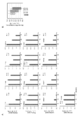
- RNA-binding performance of PPR protein specificity
- PPR proteins for 23 target sequences were prepared using the V2 motif, and all binding combinations were analyzed using RPB-ELISA. Twenty-one PPR proteins were found to have the strongest binding to the target (above). Similarly, RNA binding performance was analyzed using the V3.1 motif (bottom).
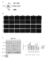
- affinity Detailed analysis of RNA-binding performance of PPR protein (affinity).
- B Correlation between Kd value and signal value obtained in binding experiment with RPB-ELISA.
- the Kd value of RPB-ELISA is 1.0 to 2.0 x 10 7
- the Kd value is 10 -6 to 10 -7 M
- the luminescence value of RPB-ELISA is 2.0 to 4.0 x 10 7
- the Kd value is 10 -7 to 10 -8 M and the emission value in RPB-ELISA is larger than 4.0 x 10 7 , it can be estimated that the Kd value is -10 -8 or less.
- Construction success probability PPR proteins for 72 target sequences were prepared using the v2 motif, and the probability of successful construction was calculated using RPB-ELISA.
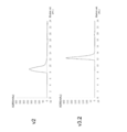
- Each PPR protein was prepared in an E. coli expression system, purified, and separated by gel filtration chromatography. The smaller the elution fraction (Elution vol.), The larger the molecular size. In V2, it was eluted with an elution fraction of 8 to 10 mL, while in v3.2, a peak was observed in the elution fraction of 12 to 14 mL. This suggests that v2 may be aggregated due to the increased protein size, and it was found that the aggregation was improved in v3.2.
- the E value obtained by PF01535 in Pfam and PS51375 in Prosite when the amino acid sequence is analyzed by a protein domain search program on the Web is less than or equal to a predetermined value (desirably).
- a predetermined value refers to a polypeptide composed of 30 to 38 amino acids having the amino acid sequence of E-03).
- the position numbers of the amino acids that make up the PPR motif defined in the present invention are almost synonymous with PF01535, but are subtracted by 2 from the amino acid location of PS51375 (eg, No. 1 of the present invention ⁇ No. 3 of PS51375). Equivalent to.
- the terminal side that is, the -2nd amino acid.
- the conserved amino acid sequence of the PPR motif has low conservativeness at the amino acid level, but the two ⁇ -helices are well conserved on the secondary structure.
- a typical PPR motif is composed of 35 amino acids, but its length is variable from 30 to 38 amino acids.
- the PPR motif referred to in the present invention consists of a polypeptide having a length of 30 to 38 amino acids represented by the formula 1.
- Helix A is a 12 amino acid long part capable of forming an ⁇ -helix structure and is represented by the formula 2.
- a 1 to A 12 independently represent amino acids;
- X is a portion that is absent or consists of 1-9 amino acids in length;
- Helix B is a part of the 11-13 amino acid length that can form an ⁇ -helix structure;
- L is the portion of formula 3 with a length of 2-7 amino acids;
- Equation 3 each amino acid is numbered from the C-terminal side as “i” (-1), “ii” (-2), and However, L iii to L vi i may not exist.
- PPR protein in the present invention refers to a PPR protein having one or more, preferably two or more PPR motifs described above, unless otherwise specified.
- protein in the present specification refers to all substances composed of polypeptides (chains in which a plurality of amino acids are peptide-bonded), and includes those composed of relatively low molecular weight polypeptides.
- amino acid may refer to an ordinary amino acid molecule or an amino acid residue constituting a peptide chain. It is clear to those skilled in the art from the context which one it refers to.
- the binding activity of the target nucleic acid of the PPR motif to a base when it is referred to as specific / specific, the binding activity to any one of the four types of base is the other unless otherwise specified. It means that it is higher than the binding activity to the base.
- Nucleic acid in the present invention refers to RNA or DNA.
- the PPR protein may have specificity for a base in RNA or DNA, but does not bind to a nucleic acid monomer.
- RNA-binding PPR motif the relationship between the combination of the three amino acids 1, 4, and ii and the base that can be bound is as follows (see Patent Document 1 above). (3-1) a combination of three amino acids of A 1, A 4, and L ii is, in turn, valine, in the case of asparagine and aspartic acid, the PPR motif binds strongly to U, then and C, the Next, it has a selective RNA-base binding ability of binding to A or G.
- (3-2) a combination of three amino acids of A 1, A 4, and L ii is in turn valine, threonine, when the asparagine, the PPR motif binds strongly to A, then to G, the following Has a selective RNA-base binding ability that binds to C but not to U.
- (3-3) a combination of three amino acids of A 1, A 4, and L ii is in turn, valine, asparagine, for asparagine, the PPR motifs bind strongly and C, to then A or U It has a selective RNA-base binding ability that it binds to G but does not bind to G.
- (3-4) a combination of three amino acids of A 1, A 4, and L ii is in turn, glutamic acid, glycine, in the case of aspartic acid, the PPR motif binds strongly to G, A, U and C It has a selective RNA base binding ability that does not bind to.
- (3-5) a combination of three amino acids of A 1, A 4, and L ii is in turn, isoleucine, asparagine, for asparagine, the PPR motifs bind strongly and C, then the U, the following Has a selective RNA-base binding ability that binds to A but not to G.
- (3-8) a combination of three amino acids of A 1, A 4, and L ii is in turn, phenylalanine, serine, when asparagine, the PPR motif binds strongly to A, then and C, the following It has a selective RNA base binding ability of binding to G and U.
- (3-9) a combination of three amino acids of A 1, A 4, and L ii is in turn, valine, asparagine, in the case of serine, the PPR motifs bind strongly and C, then binding to U However, it has a selective RNA-base binding ability that it does not bind to A and G.
- the relationship between the combination of the three amino acids 1, 4, and ii and the base that can be bound is as follows (see Patent Document 2 above).
- (2-3) A 1, A 4 , and a combination of three amino acids L ii is, in turn, any amino acid, glycine, when aspartic, the PPR motif is selectively bind to A;
- (2-5) A 1, A 4
- the present invention provides a novel PPR motif.
- the novel PPR motifs provided by the present invention that are adenine-binding are (A-1), (A-2), and (A-3) below: (A-1)
- the amino acid at position 10 is replaced with tyrosine
- the amino acid at position 15 is replaced with lysine
- the amino acid at position 16 is replaced. Any substitution selected from the group consisting of substitution of leucine, amino acid at position 17 with glutamic acid, amino acid at position 18 with aspartic acid, and amino acid at position 28 with glutamic acid was performed.
- PPR motif consisting of amino acid sequence
- A-2 In the sequence of SEQ ID NO: 9, amino acids other than those at positions 1, 2, 3, 4, 6, 7, 9, 11, 12, 14, 19, 26, 30, 33, and 34
- A-3) SEQ ID NO: 9 has at least 42% sequence identity with the sequence, but positions 1, 2, 3, 4, 6, 7, 9, 11, 12, 14, 19, 26, 30 , 33, and 34 amino acids are identical and adenine-binding PPR motifs.
- substitution in (A-1) may be 1, 2 or more, or all of the above.
- positions 1, 2, 3, 4, 6, 7, 9, 11, 12, 14, 19, 26, 30, 33 which are amino acids that can be substituted in the sequence of SEQ ID NO: 9.
- amino acids other than 34 amino acids Preferably, it is an amino acid other than the amino acids at positions 1, 2, 3, 4, 6, 7, 9, 11, 12, 14, 19, 26, 30, 33, and 34, and at positions 5, 8, and 13. , 21, 22, 23, 25, 29, 35, 1 to 11 amino acids other than amino acids, More preferably, it is an amino acid other than the amino acids at positions 1, 2, 3, 4, 6, 7, 9, 11, 12, 14, 19, 26, 30, 33, and 34, and at positions 5, 8, Amino acids other than amino acids 13, 21, 22, 23, 25, 29, 35, and 1 to 7 amino acids other than amino acids at positions 20, 24, 31, and 32. More preferably, it is any of the amino acids at positions 10, 15, 16, 17, 18, and 28.
- (A-3) it has at least 42% sequence identity with the sequence of SEQ ID NO: 9, but at positions 1, 2, 3, 4, 6, 7, 9, 11, 12, 14, 19, 26. , 30, 33, and 34 amino acids are identical
- it has at least 71% sequence identity with the sequence of SEQ ID NO: 9, but at positions 1, 2, 3, 4, 6, 7, 9, 11, 12, 14, 19, 26, 30, 33.
- And 34 amino acids, and the amino acids at positions 5, 8, 13, 21, 22, 23, 25, 29, 35 are identical.
- it has at least 80% sequence identity with the sequence of SEQ ID NO: 9, but at positions 1, 2, 3, 4, 6, 7, 9, 11, 12, 14, 19, 26, 30,
- the amino acids 33 and 34, the amino acids at positions 5, 8, 13, 21, 22, 23, 25, 29, 35, and the amino acids at positions 20, 24, 31, and 32 are the same.
- the amino acid having at least 82% sequence identity with the sequence of SEQ ID NO: 9, but not identical is any of the amino acids at positions 10, 15, 16, 17, 18, and 28.
- novel PPR motifs provided by the present invention that are cytosine-binding are (C-1), (C-2), and (C-3) below: (C-1)
- the amino acid at position 2 is replaced with serine
- the amino acid at position 5 is replaced with isoleucine
- the amino acid at position 7 is replaced.
- a PPR motif consisting of added sequences and cytosine binding (C-3) SEQ ID NO: 10 has at least 25% sequence identity with the sequence, but the amino acids at positions 1, 3, 4, 14, 18, 19, 26, 30, 33, and 34 are identical.
- a PPR motif that is both present and cytosine-binding (C-3) SEQ ID NO: 10 has at least 25% sequence identity with the sequence, but the amino acids at positions 1, 3, 4, 14, 18, 19, 26, 30, 33, and 34 are identical.
- substitution at (C-1) may be 1, 2 or more, or all of the above.
- one of the amino acids other than the amino acids at positions 1, 3, 4, 14, 18, 19, 26, 30, 33, and 34 which are amino acids that can be substituted in the sequence of SEQ ID NO: 10.
- it is an amino acid other than the amino acids at positions 1, 3, 4, 14, 18, 19, 26, 30, 33, and 34, and at positions 6, 9, 11, 12, 17, 20, 21, 23. , 25, 28, and 35 amino acids other than amino acids, 1 to 14 amino acids, More preferably, it is an amino acid other than the amino acids at positions 1, 3, 4, 14, 18, 19, 26, 30, 33, and 34, and at positions 6, 9, 11, 12, 17, 20, 21, Amino acids other than amino acids 23, 25, 28, and 35, and 1 to 10 amino acids other than amino acids at positions 13, 16, 31, and 32. More preferably, it is any of the amino acids at positions 2, 5, 7, 8, 10, 15, 22, 24, 27, and 29.
- (C-3) it has at least 25% sequence identity with the sequence of SEQ ID NO: 10, but the amino acids at positions 1, 3, 4, 14, 18, 19, 26, 30, 33, and 34 To be the same Preferably, it has at least 60% sequence identity with the sequence of SEQ ID NO: 10, but amino acids at positions 1, 3, 4, 14, 18, 19, 26, 30, 33, and 34, and 6, 9. , 11, 12, 17, 20, 21, 23, 25, 28, and 35 amino acids are identical, More preferably, it has at least 71% sequence identity with the sequence of SEQ ID NO: 10, but amino acids at positions 1, 3, 4, 14, 18, 19, 26, 30, 33, and 34, position 6, The amino acids 9, 11, 12, 17, 20, 21, 23, 25, 28, and 35, and the amino acids at positions 13, 16, 31, and 32 are identical. More preferably, amino acids having at least 71% sequence identity with the sequence of SEQ ID NO: 10, but not identical, at positions 2, 5, 7, 8, 10, 15, 22, 24, 27, and 29. It is one of the amino acids.
- novel PPR motifs provided by the present invention that are guanine-binding are (G-1), (G-2), and (G-3) below: (G-1)
- the amino acid at position 10 is replaced with phenylalanine
- the amino acid at position 15 is replaced with aspartic acid
- the amino acid at position 27 is replaced with aspartic acid
- PPR motif consisting of any of the substituted amino acid sequences selected from the group consisting of the substitution of valine, the substitution of the amino acid at position 28 with serine, and the substitution of the amino acid at position 35 with isoleucine; (G-2) In the sequence of SEQ ID NO: 11, 1 to 1 of amino acids other than amino acids at positions 1, 2, 3, 4, 6, 7, 9, 14, 18, 19, 26, 30, 33, and 34.
- a PPR motif consisting of 21 substituted, deleted, or added sequences and guanine-binding; (G-3) SEQ ID NO: 11 with at least 40% sequence identity with the sequence, but positions 1, 2, 3, 4, 6, 7, 9, 14, 18, 19, 26, 30, 33 , And 34 amino acids are identical and guanine-binding PPR motifs.
- substitution with (G-1) may be 1, 2 or more, or all of the above.
- amino acids that can be substituted in the sequence of SEQ ID NO: 11, positions 1, 2, 3, 4, 6, 7, 9, 14, 18, 19, 26, 30, 33, and 1 to 21 amino acids other than 34 amino acids Preferably, it is an amino acid other than the amino acids at positions 1, 2, 3, 4, 6, 7, 9, 14, 18, 19, 26, 30, 33, and 34, and at positions 5, 11, 12, and 17. , 20, 21, 22, 23, and 25 amino acids other than amino acids, 1 to 12 More preferably, it is an amino acid other than the amino acids at positions 1, 2, 3, 4, 6, 7, 9, 14, 18, 19, 26, 30, 33, and 34, and at positions 5, 11, 12, Amino acids other than the amino acids 17, 20, 21, 22, 23, and 25, and 1 to 5 amino acids other than the amino acids at positions 8, 13, 16, 24, 29, 31, and 32. More preferably, it is any of the amino acids at positions 10, 15, 27, 28, and 35.
- (G-3) it has at least 40% sequence identity with the sequence of SEQ ID NO: 11, but at positions 1, 2, 3, 4, 6, 7, 9, 14, 18, 19, 26, 30. , 33, and 34 amino acids are identical
- it has at least 65% sequence identity with the sequence of SEQ ID NO: 11, but at positions 1, 2, 3, 4, 6, 7, 9, 14, 18, 19, 26, 30, 33, and.
- the 34 amino acids and the amino acids at positions 5, 11, 12, 17, 20, 21, 22, 23, and 25 are identical.
- it has at least 85% sequence identity with the sequence of SEQ ID NO: 11, but at positions 1, 2, 3, 4, 6, 7, 9, 14, 18, 19, 26, 30, 33, And 34 amino acids, positions 5, 11, 12, 17, 20, 21, 22, 23, and 25, and positions 8, 13, 16, 24, 29, 31, and 32 are identical.
- the amino acid having at least 85% sequence identity with the sequence of SEQ ID NO: 11, but not identical is any of the amino acids at positions 10, 15, 27, 28, and 35.
- novel PPR motifs provided by the present invention that are uracil-binding are (U-1), (U-2), and (U-3) below: (U-1)
- the amino acid at position 10 is replaced with phenylalanine
- the amino acid at position 13 is replaced with serine
- the amino acid at position 15 is replaced.
- Substitution of lysine, amino acid at position 17 with glutamic acid, amino acid at position 20 with leucine, amino acid at position 21 with lysine, amino acid at position 23 with phenylalanine, amino acid at position 24 From the group consisting of substitution of aspartic acid, substitution of amino acid at position 27 with lysine, substitution of amino acid at position 28 with lysine, substitution of amino acid at position 29 with arginine, and substitution of amino acid at position 31 with leucine.
- a PPR motif consisting of any of the substituted amino acid sequences selected; (U-2) 1-22 amino acids other than those at positions 1, 2, 3, 4, 6, 11, 12, 14, 19, 26, 30, 33, and 34 in the sequence of SEQ ID NO: 12 PPR motif consisting of sequences substituted, deleted, or added, and uracil-binding; (U-3) SEQ ID NO: 12 has at least 37% sequence identity with the sequence, but positions 1, 2, 3, 4, 6, 11, 12, 14, 19, 26, 30, 33, and A PPR motif in which 34 amino acids are identical and uracil-binding.
- substitution with (U-1) may be 1, 2 or more, or all of the above.
- amino acids that can be substituted in the sequence of SEQ ID NO: 12.
- 1 to 22 amino acids other than amino acids Preferably, it is an amino acid other than the amino acids at positions 1, 2, 3, 4, 6, 11, 12, 14, 19, 26, 30, 33, and 34, and at positions 5, 7, 9, 16, 18 , 22, 25, and 35 amino acids other than amino acids, 1 to 14 amino acids, More preferably, it is an amino acid other than the amino acids at positions 1, 2, 3, 4, 6, 11, 12, 14, 19, 26, 30, 33, and 34, and at positions 5, 7, 9, 16, Amino acids other than the amino acids 18, 22, 25, and 35, and 1 to 12 amino acids other than the amino acids at positions 8 and 32. More preferably, it is any of the amino acids at positions 10, 13, 15, 17, 20, 21, 23, 24, 27, 28, 29, and 31.
- (U-3) it has at least 37% sequence identity with the sequence of SEQ ID NO: 12, but at positions 1, 2, 3, 4, 6, 11, 12, 14, 19, 26, 30, 33.
- And 34 amino acids are the same
- it has at least 60% sequence identity with the sequence of SEQ ID NO: 12, but at positions 1, 2, 3, 4, 6, 11, 12, 14, 19, 26, 30, 33, and 34.
- the amino acids and the amino acids at positions 5, 7, 9, 16, 18, 22, 25, and 35 are identical.
- it has at least 65% sequence identity with the sequence of SEQ ID NO: 12, but at positions 1, 2, 3, 4, 6, 11, 12, 14, 19, 26, 30, 33, and 34.
- Amino acids at positions 5, 7, 9, 16, 18, 22, 25, and 35, and amino acids at positions 8 and 32 are identical.
- amino acids that have at least 65% sequence identity with the sequence of SEQ ID NO: 12, but are not identical are at positions 10, 13, 15, 17, 20, 21, 23, 24, 27, 28, 29. , And any of the 31 amino acids.
- PPR motif v2 created by the present inventors A (SEQ ID NO: 9), v2 C (SEQ ID NO: 10), v2 G (SEQ ID NO: 11), v2 U (SEQ ID NO: 12) is the first to be disclosed by the present application and does not exist in nature.
- each of these homologs ((A-1), (A-2), (A-3), (C-1), (C-2), (C-3), (G-1), (above)
- G-2 the embodiments shown as G-2), (G-3), (U-1), (U-2), (U-3) and the preferred cases thereof, SEQ ID NOs: 9-
- SEQ ID NOs: 9- For (aspects consisting of sequences other than 12), at least one of the homologs (whether or not each homolog is disclosed for the first time in the present application and whether it is naturally occurring).
- a combination of 2 or more, for example, 3, 4, 5, 6, 7, 8, 9, 10, 11, 12, 13, 14 is naturally present. It is thought that it will not.
- the number selected is arbitrary when it says "any".
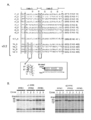
- Figures 1 to 4 show VTN for the PPR motif that recognizes adenine and VSN for the PPR motif that recognizes cytosine by combining the amino acids at positions 1, 4, and ii in the PPR motif sequence of Uracil.
- VTD for the PPR motif that recognizes guanine and VND for the PPR motif that recognizes uracil are collected, and the types and numbers of amino acids that appear at each position are summarized.
- New PPR motif array v2 A (SEQ ID NO: 9), v2 C (SEQ ID NO: 10), v2 G (SEQ ID NO: 11), v2 In U (SEQ ID NO: 12), the amino acids at each position have a high frequency of appearance.
- Figure 6A along with these novel sequences, the combination of amino acids at positions 1, 4, and ii in the dPPR motif sequence was the same as v2, v1 A (SEQ ID NO: 13), v1 C (SEQ ID NO: 14), v1 G (SEQ ID NO: 15), v1 U (SEQ ID NO: 16) is also shown.
- FIG. 6A shows the amino acid sequence of the v3.1 motif.
- V3.1 introduces the D15K mutation into the adenine recognition motif in v2 (SEQ ID NO: 401), and is the same as v2 in other respects.
- v3.1 in the PPR protein, it may be possible to obtain a protein with improved binding force compared to v2.
- the present invention provides a novel PPR protein containing a novel PPR motif.
- the novel PPR proteins provided by the present invention are: A protein containing n PPR motifs capable of binding to a target RNA consisting of n base sequences.
- the PPR motif for adenine in the base sequence is the PPR motif of (A-1), (A-2), or (A-3) described above;
- the PPR motif for cytosine in the nucleotide sequence is the PPR motif of (C-1), (C-2), or (c-3) described above;
- the PPR motif for guanine in the base sequence is the PPR motif of (G-1), (G-2), or (G-3) described above;
- a PPR protein in which the PPR motif for uracil in the base sequence is the PPR motif of (U-1), (U-2), or (U-3) described above.
- Preferred examples of the PPR motif contained in the PPR protein are the above-mentioned (A-1), (A-2), (A-3), (C-1), (C-2), (C-) regarding the PPR motif.
- the description of 3), (G-1), (G-2), (G-3), (U-1), (U-2), or (U-3) applies as is.
- n (representing an integer of 1 or more) is not particularly limited, but can be 10 or more, preferably 12 or more, more preferably 15 or more, and 18 or more. Is more preferable. By increasing the number of motifs, a PPR protein having high binding strength to many targets can be produced.
- the base sequence encoding the existing v1 to v4 motifs (SEQ ID NO: 13 to 16)
- the following sequence can be given as an example of the base sequence encoding the motif utilizing the degeneracy of codons. it can.
- the present invention provides a novel PPR motif and a nucleic acid encoding a novel PPR protein containing the novel PPR motif.
- the base sequence encoding the novel PPR motif has several variations due to codon degeneracy.
- Amino acid sequence v2 of the novel PPR motif of the present invention A (SEQ ID NO: 9), v2 C (SEQ ID NO: 10), v2 G (SEQ ID NO: 11), v2
- a preferred example of the base sequence encoding U (SEQ ID NO: 12) is shown in the table below.
- the amino acid sequence of the PPR motif which has the same amino acid combinations at positions 1, 4, and ii as v2, v1 A (SEQ ID NO: 13), v1 C (SEQ ID NO: 14), v1 G (SEQ ID NO: 15), v1
- the base sequence encoding U (SEQ ID NO: 16) is shown in the table below.
- the base sequence encoding the PPR protein can be composed of any combination of the above sequences.
- An amino acid encoding a protein may be constructed by appropriately combining with a base sequence encoding U (SEQ ID NO: 16).
- Amino acid sequence of the novel PPR motif of the present invention v3.1 A (SEQ ID NO: 401), 1st A (SEQ ID NO: 402), 1st C (SEQ ID NO: 403), 1st G (SEQ ID NO: 404), 1st A preferred example of the base sequence encoding U (SEQ ID NO: 405) is shown in the table below.
- the base sequence encoding the PPR protein can be composed of any combination of the above sequences.
- the base sequence encoding the first PPR motif from the N-terminal is v3.2 above.
- the base sequence encoding the PPR motif after that is v3.1 above as the base sequence encoding the PPR motif for adenine. It is advisable to select A and appropriately combine those selected from the above v2 series as the base sequences encoding the PPR motifs for cytosine, guanine, and uracil.
- Non-Patent Document 6 Coquille et al., 2014 Nat. Commun .; PDB ID: 4PJQ, 4WN4, 4WSL, 4PJR; Non-Patent Document 7: Shen et al., From 2015 Nat.
- the 6th preferably the 6th and 9th amino acids to hydrophilic amino acids (asparagine, aspartic acid, glutamine, glutamic acid, lysine, arginine, serine, threonine), PPR aggregation can be achieved.
- hydrophilic amino acids asparagine, aspartic acid, glutamine, glutamic acid, lysine, arginine, serine, threonine
- a 6 amino acid is a hydrophilic amino acid, preferably A 6 amino acid is asparagine or aspartic acid.
- the A 9 amino acid is a hydrophilic amino acid or glycine, preferably glutamic acid, glutamic acid, lysine, or glycine.
- a 6 amino acid and A 9 amino acid may be combined with any of the following.
- a 6 amino acid is aspartic acid and A 9 amino acid is glutamic acid ⁇ A 6 amino acid is aspartic acid and A 9 amino acid is glutamine ⁇ A 6 amino acid is aspartic acid and A 9 amino acid is Combination that is lysine ⁇ Combination that is A 6 amino acid aspartic acid and A 9 amino acid is glycine
- PPR motifs particularly preferable ones are as follows.
- (1st A-2) (1st In the sequence of A-1), a PPR motif consisting of a sequence in which 1 to 9 amino acids other than the amino acids 1, 4, 6, 9, and 34 are substituted, deleted, or added, and is adenine-binding.
- (1st A-3) (1st A PPR motif that has at least 80% sequence identity with the sequence of A-1), but the amino acids at positions 1, 4, 6, 9, and 34 are identical and adenine-binding
- (1st C-2) (1st In the sequence of C-1), a PPR motif consisting of a sequence in which 1 to 9 amino acids other than the amino acids 1, 4, 6, 9, and 34 are substituted, deleted, or added, and is cytosine-binding.
- (1st C-3) (1st A PPR motif that has at least 80% sequence identity with the sequence of C-1), but the amino acids at positions 1, 4, 6, 9, and 34 are identical and cytosine-binding
- (1st G-2) (1st In the sequence of G-1), a PPR motif consisting of a sequence in which 1 to 9 amino acids other than the amino acids 1, 4, 6, 9, and 34 are substituted, deleted, or added, and is guanine-binding.
- (1st G-3) (1st A PPR motif that has at least 80% sequence identity with the sequence of G-1), but the amino acids at positions 1, 4, 6, 9, and 34 are identical and guanine-binding
- (1st U-1) PPR motif consisting of a sequence in which the amino acids at positions 6 and 9 are substituted so as to satisfy any one of the combinations defined below in the sequence of SEQ ID NO: 405
- (1st U-2) (1st A PPR motif consisting of a sequence in which 1 to 9 amino acids other than the amino acids at positions 1, 4, 6, 9, and 34 are substituted, deleted, or added in the sequence of U-1) and is uracil-binding.
- Figure 6A shows the amino acid sequence of the v3.2 motif as well as the v3.1 motif.
- the first motif is 1st A (SEQ ID NO: 402), 1st C (SEQ ID NO: 403), 1st G (SEQ ID NO: 404), 1st Select U (SEQ ID NO: 405) and v2 for the second and subsequent motifs C, v2 G, v2 U, v3.1 Select from A.
- Intracellular aggregation can be improved by using any of v3.2 as the first PPR motif from the N-terminus in the PPR protein.
- a search / analysis regarding the identity of a base sequence or an amino acid sequence can be performed by an algorithm or program known to those skilled in the art (for example, BLASTN, BLASTP, BLASTX, ClustalW).
- the parameters when using the program can be appropriately set by those skilled in the art, and the default parameters of each program may be used. Specific methods of these analysis methods are also well known to those skilled in the art.
- the identity% value when the identity is expressed in% with respect to the base sequence or the amino acid sequence, it is preferable that the identity% value is high in any case, and specifically, 70% or more. It is preferably 80% or more, more preferably 85% or more, further preferably 90% or more, further preferably 95% or more, and 97.5%. The above is more preferable.
- the number of amino acids to be substituted when referred to as "substitution, deletion, or addition sequence” is the same in any motif or protein unless otherwise specified.
- the motif or protein consisting of an amino acid sequence is not particularly limited as long as it has a desired function, but if the number is about 1 to 9 or 1 to 4, or if it is replaced with an amino acid having similar properties, a larger number may be used. There may be substitutions and the like. Means for preparing polynucleotides or proteins relating to such amino acid sequences are well known to those of skill in the art.
- Amino acids with similar properties refer to amino acids with similar physical properties such as hydropathy, charge, pKa, and solubility, and refer to, for example, the following.
- Hydrophobic (non-polar) amino acids alanine, valine, glycine, isoleucine, leucine, phenylalanine, proline, tryptophan, tyrosine non-hydrophobic amino acids; arginine, asparagine, aspartic acid, glutamic acid, glutamine, lysine, serine, threonine, cysteine, histidine , Methionine; Hydrophilic amino acids; arginine, asparagine, aspartic acid, glutamic acid, glutamine, lysine, serine, threonine; Acidic amino acids: aspartic acid, glutamic acid; Basic amino acids: lysine, arginine, histidine; Neutral amino acids: alanine, asparagine, cysteine, glut
- PPR proteins prepared using the novel PPR motif (SEQ ID NOs: 9-12) of the present invention are not only suitable for making PPR proteins for relatively long target RNAs, but also existing PPRs for the same target RNA ( SEQ ID NOs: 13-16) It may have higher RNA binding performance than PPR proteins prepared using the motif.
- the binding force to the target RNA can be enhanced as compared with the case of using the existing PPR motif.
- the increased binding force can improve the efficiency of intracellular RNA manipulation by the PPR protein.
- intracellular splicing efficiency can be improved by using a PPR protein that has a high binding force to a target (see Example 5).
- the degree to which the binding force is enhanced seems to depend on the sequence and length of the target, but for example, the binding force can be increased to 1.1 times or more, and when specified, 1.3 times or more, 2.0 times or more, 3.0 times or more, It can be 3.6 times or more.
- the binding force to the target sequence can be evaluated by a method using EMSA (Electrophoretic Mobility Shift Assay) or Biacore.
- EMSA is a method that utilizes the property that when a sample in which a protein and a nucleic acid are bound is electrophoresed, the mobility of the nucleic acid molecule changes as compared with the case where the nucleic acid molecule is not bound. Since the intermolecular interaction analysis device represented by Biacore can perform reaction kinetic analysis, detailed protein-nucleic acid binding analysis is possible.
- the binding force to the target sequence can also be evaluated by RPB-ELISA described later.
- the target PPR is the value obtained by subtracting the background signal (the luminescence signal value when the target PPR protein is added without adding the target RNA) from the luminescence amount of the sample to which the target PPR protein and its target RNA are added. And its binding force to the target RNA.
- a PPR protein prepared using the novel PPR motif of the present invention may have a higher ability in specificity for a target sequence than a PPR protein prepared using an existing PPR motif for the same target RNA.
- the specificity for the target RNA can be enhanced as compared with the case where the existing PPR motif is used. If the PPR protein has high specificity for the target RNA, when the target RNA is manipulated intracellularly using the PPR protein, it is possible to avoid unintended effects as a result of binding to the unintended RNA.
- the affinity for the target sequence can be evaluated by a person skilled in the art by a conventional method.
- an appropriate non-target RNA for the target PPR protein is designed, and the binding force (luminescence signal value) in this case is obtained in the same manner, thereby binding signal value to the target sequence / binding signal to the non-target sequence.
- the value (S / N) can be obtained as an index of specificity (affinity) for the target RNA.
- the PPR protein prepared using the novel PPR motif of the present invention may have a high affinity (equilibrium dissociation multiplier, Kd value) for the target RNA.
- the Kd value for the target sequence can be calculated by an existing method such as EMSA.
- EMSA an existing method
- the Kd value in relation to the present invention unless otherwise specified, it refers to a value measured by EMSA under the conditions described in the section of Examples described later.
- the Kd value of the PPR protein prepared using the novel PPR motif of the present invention may depend on the target sequence and length, but if the target sequence length is 18 bases or more, 10 -7 It can be M or less, 10 -8 M or less, or 10 -9 M order. According to the study by the present inventors, the minimum value (high affinity) of the Kd value is 1.95 x 10 -9 under the conditions of the example when the length of the target sequence is 18 bases long, which has been reported so far. It is lower than any of the Kd values (see Table 1) of the designed PPR protein. It is known that the Kd value correlates with the signal value obtained in the binding experiment with RPB-ELISA.
- the Kd value in RPB-ELISA is 1 to 2 x 10 7
- the Kd value is 10 -6 to 10 -7 M
- the luminescence value in RPB-ELISA is.
- 2 to 4 x 10 7 has a Kd value of 10 -7 to 10 -8 M
- RPB-ELISA has a light emission value greater than 4 x 10 7 and a Kd value of ⁇ 10 -8 or less. Can be estimated to be.
- PPR protein construction efficiency By using the novel PPR motif of the present invention, a desired PPR protein can be efficiently constructed.
- the construction efficiency can be calculated by using an existing method and determining the rate at which a PPR protein having a high Kd value can be constructed. Instead of the Kd value, the luminescence signal value by RPB-ELISA may be obtained as described above and calculated in the same manner.
- the Kd value is 10 -6 M or less (RPB-ELISA value is 1 x 10 7).
- PPR protein can be obtained with an efficiency of 50% or more, 60% or more when specified, 70% or more when specified, and 80% or more when specified.
- PPR protein having a Kd value of 10 -7 M or less (RPB-ELISA value of 2 x 10 7 or more) and a target sequence having a length of 18 bases is 50% or more, and 55% or more is specified. It can be obtained with an efficiency of 65% or more when specified, and 75% or more when specified.
- PPR protein having a Kd value of 10 -8 M or less (RPB-ELISA value of 4 x 10 7 or more) and a target sequence having a length of 18 bases is 20% or more, and 25% or more is specified. It can be obtained with an efficiency of 30% or more when specified, and 35% or more when specified.
- the construction efficiency can be calculated using the RPB-ELISA method based on the binding signal value for the target sequence / binding signal value (S / N) for the non-target sequence.
- PPR protein having a target sequence having a length of 18 bases and an S / N of more than 10 are 50% or more, and 55% or more are specified. It can be obtained with an efficiency of 65% or more, and more specifically, 75% or more.
- PPR protein having a target sequence having a length of 18 bases and an S / N of more than 100 is 15% or more, 20% or more when specified, 25% or more when specified, and 30% or more when specified. Can be obtained with efficiency.
- the present invention also provides a method for producing a gene encoding a protein containing n PPR motifs capable of binding to a target nucleic acid consisting of n base sequences, which comprises the following steps: At least 20 polynucleotides, including 4 encoding each of the PPR motifs that are adenine, cytosine, guanine, or uracil or thymine binding, and 16 encoding each of the two conjugates of the PPR motif, are present. From the library of at least 20 ⁇ m types of PPR parts inserted in each of the intermediate vectors Dest-a ...
- n is an integer greater than or equal to m and less than or equal to m ⁇ 2. n can be, for example, 10 to 20.
- the method of the present invention utilizes the Golden Gate reaction.
- the Golden Gate reaction multiple DNA fragments are inserted into the vector using a Type IIS restriction enzyme and T4 DNA ligase. Since the Type IIS restriction enzyme cleaves the outside of the recognition sequence, the attachment end can be set freely. In addition, it is highly efficient because the 4-base protruding end is used for ligation. Furthermore, no recognition sequence remains in the annealed and ligated constructs. Therefore, the polynucleotides encoding the PPR motif can be seamlessly linked (Fig. 5).
- a particularly preferred example of a Type IIS restriction enzyme is BsaI.
- the method of the present invention can efficiently prepare a gene even when there are many repeat sequences by using a parts library appropriately designed in consideration of the characteristics of PPR protein and a Golden Gate reaction. Therefore, this method is useful for producing genes for proteins containing 15 or more PPR motifs capable of binding to a target nucleic acid having a length of 15 bases or more, which increases the number of repeat sequences. If m is set to 10 and a library of 200 types of PPR parts is used and the 10 PPR parts required to create the target gene are selected from the library, 10 to 20 PPR motifs can be obtained. The gene encoding the protein contained can be freely prepared.
- RNA-binding PPR protein having a target sequence having a length of 10 to 20 bases may be described as an example, but this method may be described as a target sequence having another length. It can also be applied to prepare a PPR protein for DNA binding, and can also be applied to prepare a DNA-binding PPR protein.
- a parts library containing one or two sequences encoding the PPR motif is prepared (STEP 1 and STEP 2 in FIG. 5) and used.
- a parts library can be prepared, for example, by inserting a PPR motif sequence into 10 types of intermediate vectors Dest-a, b, c, d, e, f, g, h, i, j.
- the intermediate vector is designed so that Dest-a to Dest-j are seamlessly linked in order by the Golden Gate reaction.
- There are four types of PPR motif sequences to be inserted (A, C, G, U), and 16 types (AA, AC, AG, AU, CA,) that code for each of the two conjugates of the PPR motif.
- the parts library contains at least 200 types of parts.
- select the necessary parts according to the target base sequence Specifically, for example, select one part from each part library of Dest-a, b, c, d, e, f, g, h, i, j, and perform the Golden Gate reaction together with the vector parts. Do (STEP 3 in Figure 5). If you select one that contains one motif in all the intermediate vectors, 10 sequences will be linked, and if you use one that contains two motifs, 20 sequences will be linked. If you want to concatenate 11 to 19 pieces, select the one containing one motif from any Dest-x library.
- the obtained plasmid can be transformed into Escherichia coli and amplified / extracted.
- the present invention provides a method for detecting or quantifying a protein containing n PPR motifs capable of binding to a target nucleic acid consisting of n base sequences, which comprises the following steps: A step of providing a solution containing a candidate protein to an immobilized target nucleic acid to detect or quantify a protein bound to the target nucleic acid.
- the detection / analysis method of the present invention is useful as a method for evaluating the binding performance of a high-throughput PPR protein.
- the detection / analysis method of the present invention is an application of ELISA (Enzyme-Linked ImmunoSorbent Assay) (Fig. 7A), it may be referred to as RPB-ELISA (RNA-protein binding ELISA) method.
- RPB-ELISA RNA-protein binding ELISA
- the method of the present invention is described herein as a method for evaluating RNA-binding PPR protein, but similarly, in order to evaluate the binding performance of DNA-binding PPR protein to target DNA. Can also be applied.
- the step of providing the solution containing the candidate protein to the immobilized target nucleic acid can be carried out by flowing the solution containing the target binding protein through the target nucleic acid molecule immobilized on the plate.
- Immobilization of the target nucleic acid molecule can be achieved by utilizing various existing immobilization methods, for example, by providing a nucleic acid probe containing a biotin-modified target nucleic acid molecule to a well plate coated with streptavidin.
- the candidate protein to be measured can be fused with a labeled protein, for example, an enzyme such as luciferase or a fluorescent protein. Fusion with labeled proteins makes detection and quantification easier.
- a labeled protein for example, an enzyme such as luciferase or a fluorescent protein. Fusion with labeled proteins makes detection and quantification easier.
- the RPB-ELISA method has the advantage that it does not require a special device like Biacore.
- the RPB-ELISA method has high throughput and can evaluate the binding between protein and nucleic acid in a short period of time.
- the RPB-ELISA method is sufficiently detectable at a protein concentration of 6.25 nM or higher under the conditions of Examples, and is similarly detectable with Escherichia coli lysate. Therefore, the nucleic acid-binding protein of interest is purified. It has the advantage that it is not necessary.
- the PPR motif or PPR protein provided by the present invention can link functional regions into a complex.
- proteinaceous functional regions can be linked to form a fusion protein.
- the functional region refers to a portion having a specific biological function in the living body or cell, such as an enzyme function, a catalytic function, an inhibitory function, an enhancing function, or a portion having a function as a label.
- Such regions consist of, for example, proteins, peptides, nucleic acids, bioactive substances, drugs.
- the present invention may be described with respect to the complex by taking a fusion protein as an example, but those skilled in the art will understand the case of a complex other than the fusion protein according to the description. Can be done.
- the functional region is a ribonuclease (RNase).
- RNase ribonuclease
- RNase A eg, bovine pancreatic ribonuclease A: PDB 2AAS
- RNase H ribonuclease
- the functional region is a fluorescent protein.
- fluorescent proteins are mCherry, EGFP, GFP, Sirius, EBFP, ECFP, mTurquoise, TagCFP, AmCyan, mTFP1, MidoriishiCyan, CFP, TurboGFP, AcGFP, TagGFP, Azami-Green, ZsGreen, EmGFP, HyPer, TagYFP, EYFP, Venus, YFP, PhiYFP, PhiYFP-m, TurboYFP, ZsYellow, mBanana, KusabiraOrange, mOrange, TurboRFP, DsRed-Express, DsRed2, TagRFP, DsRed-Monomer, AsRed2, mStrawberry, TurboFP602, mRFP1, JRed, K mRasberry, mPlum, PS-CFP, Dendra2, Kaede, EosFP, KikumeGR.
- a preferred example is
- the functional region is a functional domain that improves the protein expression level from the target mRNA when the target is mRNA (WO2017 / 209122).
- functional domains that increase protein expression from mRNA are, for example, all or functional parts of the functional domains of proteins known to directly or indirectly promote translation of mRNA. Good. More specifically, a domain that induces a ribosome to mRNA, a domain that is involved in initiating or promoting translation of mRNA, a domain that is involved in extracellular transport of mRNA, a domain that is involved in binding to the endoplasmic reticulum membrane, and a small domain.
- It may be a domain containing a cell retention signal (ER retention signal) sequence or a domain containing an endoplasmic reticulum signal sequence. More specifically, the domains that induce ribosomes to the above mRNA are DENR (Density-regulated protein), MCT-1 (Malignant T-cell amplified sequence 1), TPT1 (Translationally-controlled tumor protein), and Lerepo4. It may be a domain containing all or a functional part of a polypeptide selected from the group consisting of (Zinc finger CCCH-domain).
- the domain related to translation initiation or translation promotion of the above-mentioned mRNA may be a domain containing all or a functional part of the polypeptide selected from the group consisting of eIF4E and eIF4G.
- the domain related to the export of the above-mentioned mRNA to the outside of the nucleus may be a domain containing all or a functional part of SLBP (Stem-loop binding protein).
- the domain related to the above-mentioned binding to the endoplasmic reticulum membrane is selected from the group consisting of SEC61B, TRAP-alpha (Translocon associated protein alpha), SR-alpha, Dia1 (Cytochrome b5 peptide 3), and p180. It may be a domain that contains all or a functional portion of the polypeptide.
- the endoplasmic reticulum retention signal (ER retention signal) sequence may be a signal sequence including a KDEL (KEEL) sequence.
- the endoplasmic reticulum signal sequence may be a signal sequence containing MGWSCIILFLVATATGAHS.
- the functional region may be fused to the N-terminal side of the PPR protein, to the C-terminal side, or to both the N-terminal side and the C-terminal side.
- the complex or fusion protein may include multiple functional regions (eg, 2-5).
- the functional region and the PPR protein may be indirectly fused via a linker or the like.
- the present invention also provides the above-mentioned PPR motif, nucleic acid encoding a PPR protein or fusion protein, and a vector containing the nucleic acid (for example, a vector for amplification, an expression vector).
- the vector for amplification can use Escherichia coli or yeast as a host.
- the expression vector means, for example, a vector containing a DNA having a promoter sequence, a DNA encoding a desired protein, and a DNA having a terminator sequence from the upstream, as long as it exhibits a desired function. It does not necessarily have to be arranged in this order.
- various vectors that can be usually used by those skilled in the art can be recombined and used.
- the PPR protein or fusion protein of the present invention can function in cells of eukaryotes (eg, animals, plants, microorganisms (yeast, etc.), prokaryotes).
- the fusion proteins of the invention can function particularly in animal cells (in vitro or in vivo). Examples of animal cells into which the PPR protein or fusion protein of the present invention or a vector expressing the same can be introduced include cells derived from humans, monkeys, pigs, cows, horses, dogs, cats, mice, and rats. Can be done.
- Examples of cultured cells into which the PPR protein or fusion protein of the present invention or a vector expressing the same can be introduced include Chinese hamster ovary (CHO) cells, COS-1 cells, COS-7 cells, and VERO (ATCC).
- CCL-81) cells BHK cells, canine kidney-derived MDCK cells, hamster AV-12-664 cells, HeLa cells, WI38 cells, 293 cells, 293T cells, PER.
- C6 cells can be mentioned, but are not limited to these.
- the PPR protein or fusion protein of the present invention may be able to deliver and function a functional region specific to a nucleic acid sequence in vivo or in a cell.
- Complexes linked with labeled moieties such as GFP can be used to visualize the desired RNA in vivo.
- the PPR protein or fusion protein of the present invention can be modified / destroyed in a nucleic acid sequence-specific manner in the cell or in the living body, and may be imparted with a new function.
- RNA-binding PPR proteins are involved in all RNA processing steps, cleavage, RNA editing, translation, splicing, and RNA stabilization found in organelles. Therefore, the method related to the modification of the PPR protein provided by the present invention, and the PPR motif and the PPR protein provided by the present invention can be expected to be used in various fields as follows.
- LRPPRC Leigh syndrome French Canadian
- LSFC Leigh syndrome French Canadian
- Lee syndrome subacute necrotizing encephalomyelopathy
- the present invention may contribute to the treatment of LSFC (prevention, treatment, suppression of progression).
- Many existing PPR proteins serve to specify the editing site for RNA manipulation (translation of genetic information on RNA; often C ⁇ U).
- This type of PPR protein has an additional motif on the C-terminal side that is suggested to interact with RNA editing enzymes. It can be expected that a PPR protein having such a structure introduces a base polymorphism or treats a disease or condition caused by the base polymorphism.
- stem cells for example, iPS cells
- model cells for evaluation of cosmetics model cells for evaluation of cosmetics
- expression of functional RNA is turned on / for the purpose of elucidating the mechanism of drug discovery and pharmacological tests. Contains cells that can be turned off.
- PPR proteins that specifically binds to a specific RNA associated with a specific disease.
- PPR proteins are introduced into cells using plasmids, viral vectors, mRNAs, and purified proteins, and the PPR proteins bind to the target RNA in the cells, thereby altering the RNA function that causes the disease ( Can be improved).
- means for changing the function include changes in RNA structure due to binding, knockdown due to degradation, changes in splicing reaction due to splicing, and base substitution.
- RNA manipulation and genome editing by PPR protein can improve the breed and breed (genetic improvement of the organism) of the organism more accurately and quickly than the prior art.
- RNA manipulation and genome editing by PPR protein do not convert traits by foreign genes like gene recombination, but are techniques for handling RNA and genome originally possessed by animals and plants, so that mutant selection and return are performed. It can be said that it is close to the traditional breeding method of crossing. Therefore, it is possible to respond reliably and promptly to global food problems and environmental problems.
- Example 1 Establishment of method for producing PPR gene]
- the PPR motif As the PPR motif sequences used in the artificial PPR proteins reported so far, consensus sequences of naturally occurring PPR motif sequences extracted by various methods are used. Among them, the PPR protein prepared by using the motif sequence of dPPR (Non-Patent Documents 2, 3, and 6 above) has a low Kd value (high affinity). This PPR motif array will be referred to as the v1 PPR motif below.
- a consensus sequence was generated using only PPR motifs containing typical 1st, 4th, and iith amino acid combinations that recognize each base.
- the typical amino acid combinations that recognize each base are the combination that recognizes adenine, the first is valine, the fourth is threonine, the ii is aspartic acid, and the first is valine.
- ii-th is aspartic acid
- ii is serine
- guanine is recognized by 1st is valine
- 4th is threonine
- ii is aspartic acid
- uracil is recognized by 1st is valine
- 4th is asparagin Since the ii-th is aspartic acid, a consensus amino acid sequence is extracted from the PPR motif sequence containing the first, fourth, and ii-th combinations thereof, and the sequence is specifically adenine, cytosine, guanine, and uracil, respectively.
- the PPR motif sequence to be recognized was used (Figs. 1 to 4, SEQ ID NOs: 9-12). This will be referred to as the v2 PPR motif below.
- the same 1st, 4th, and iith amino acid combinations were used (SEQ ID NOs: 13-16).
- Cloning is a three-step process. In STEP1, design and preparation of each motif sequence, in STEP2, preparation of a plasmid library in which one or two motifs are cloned, and in STEP3, the target PPR gene is completed by linking the required number of motifs.
- the STEP1 plasmid contains PPR motif sequences that recognize each of A, C, G, and U.
- the DNA fragment containing the PPR motif sequence in the STEP1 plasmid is cloned into an intermediate vector (Dest-x, sequence below).
- the STEP1 plasmid in which only one motif can be inserted is named P1a-vx-X
- the plasmid in which two motifs can be inserted is named P2a-vx-X on the N side and P2b-vx-X on the C side (vx is v1).
- X is A, C, G, U).
- the BsaI restriction enzyme site (BsaI restriction enzyme recognizes and cleaves the GGTCTCnXXXX sequence (SEQ ID NO: 17), and the XXXX portion becomes the protruding end of 4 bases (hereinafter referred to as the tag sequence).
- the base sequence for seamless linkage was designed as follows.
- each motif array For P1a, ggtctca atac (SEQ ID NO: 18), gtgg tgagacc (SEQ ID NO: 19), In the case of P2a, ggtctca atac (SEQ ID NO: 18 above), ggtgtca cata tgagacc (SEQ ID NO: 20), In the case of P2b, ggtctca cata c (SEQ ID NO: 21), gtgg tgagacc (SEQ ID NO: 19 above)
- the nucleotide sequences to which the above sequences were added were prepared by gene synthesis technology and cloned into pUC57-amp.
- Dest-x Dest-a, b, c, d, e, f, g, h, i, j
- base sequence is seamlessly linked from Dest-a to Dest-j in order. Designed.
- Dest-a is gaagacataaactccgtggtcacATACagagaccaaggtctcaGTGGtcacatacatgtcttc (SEQ ID NO: 1)
- Dest-b is gaagacatATACagagaccaaggtctcaGTGGtgacataatgtcttc (SEQ ID NO: 22)
- Dest-c is gaagacatcATACagagaccaaggtctcaGTGGttacatatgtcttc (SEQ ID NO: 23)
- Dest-d is gaagacatacATACagagaccaaggtctcaGTGGttacaatgtcttc (SEQ ID NO: 24)
- Dest-e is gaagacattacATACagagaccaaggtctcaGTGGtgacatgtcttc (SEQ ID NO: 25)
- Dest-f is gaagacattgacATACagagaccaaggtctca
- 0.1 ⁇ L 10x Cut smart buffer (NEB, B7204) and 0.1 ⁇ L BsaI (NEB, R0535S) were added and reacted at 37 ° C. for 60 minutes, 80 ° C. for 10 minutes.
- 2.5 ⁇ L of the reaction solution was transformed into XL1-blue and selected with LB medium containing 30 ⁇ g / ml kanamycin. It was confirmed by sequencing that the target sequence was inserted.
- Dest-j is selected from Dest-a along the target base sequence and cloned into the CAP-x vector (Non-Patent Document 1 above). If you use one motif in all the intermediate vectors, 10 motifs will be connected, and if you use two motifs, 20 motifs will be connected. If you want to connect 11 to 19 motifs, you can make it by using Dest-x at your favorite position with 1 motif. For example, when preparing a PPR sequence of 18 motifs, a plasmid containing 1 motif for Dest-a and Dest-b and 2 motifs for the others is used.
- the intermediate vector used for STEP3 cloning needs to be selected from 3 types of vectors. If the base sequence recognized by the motif located on the C side is adenine, use CAP-A, if it is cytosine, use CAP-C, and if it is guanine or uracil, use CAP-GU. Designed to add the amino acid sequence of MGNSV (SEQ ID NO: 31) to the N side of the PPR repeat and ELTYNTLISGLGKAGRARDPPV (SEQ ID NO: 32) to the C side by cloning into the intermediate vector for STEP3. ..
- pCR8 Fw 5'-TTGATGCCTGGCAGTTCCCT -3'(SEQ ID NO: 33) and pCR8
- the inserted gene region was amplified using a primer of Rv: 5'-CGAACCGAACAGGCTTATGT -3'(SEQ ID NO: 34).
- v1 motif or v2 motif clone 3 types of 18 motifs of PPR protein (PPR1, PPR2, PPR3) (SEQ ID NOs: 35-37, 40-42) each (v1) PPR1, v1 PPR2, v1 PPR3, v2 PPR1, v2 PPR2, v2 Figure 6B shows the results of PPR3).
- PPR1, PPR2, PPR3 SEQ ID NOs: 35-37, 40-42
- EMSA Electrophoretic Mobility Shift Assay
- Biacore Biacore
- Intermolecular interaction analysis equipment such as Biacore can perform detailed protein-nucleic acid binding analysis because it can perform reaction kinetic analysis, but it also requires purified proteins and special equipment. Is. Therefore, we considered a method that has high throughput and can evaluate the binding between protein and nucleic acid in a short period of time.
- ELISA Enzyme-Linked ImmunoSorbent Assay
- the primary antibody is fixed on a well plate, a solution containing the protein to be detected is added thereto, and after washing, the amount of the remaining protein to be analyzed is detected. Therefore, the secondary antibody capable of color development and luminescence detection is possible.
- Fig. 7A we devised a system in which nucleic acid molecules are fixed on a plate, a solution containing the target nucleic acid-binding protein is flowed therein, and the amount of bound protein is quantified (Fig. 7A).
- the method for fixing the nucleic acid molecule to be investigated is to add a nucleic acid probe modified with biotin to its terminal to a well plate coated with streptavidin.
- a nucleic acid probe modified with biotin By fusing the nucleic acid-binding protein to be measured with luciferase or fluorescent protein, detection can be made easier.
- purification of the protein to be measured is not essential, and it is possible to use a crude extract of cells expressing the nucleic acid-binding protein to be measured (cultured animal cells, yeast, Escherichia coli, etc.), and the time required for purification. Can be shortened (Fig. 7B).
- RPB-ELISA RNA-protein binding ELISA
- a recombinant MS2 protein and its binding RNA probe were prepared to establish an experimental system.
- a gene for a protein in which a luciferase protein was fused to the N-terminal side of the MS2 protein and a 6x histidine tag was fused to the C-terminal side was prepared by gene synthesis and cloned into a pET21b vector (NL).
- the RNA probe is a target sequence (RNA) containing an MS2-binding sequence. 4, SEQ ID NO: 64) and non-target sequence (RNA) not included A biotinylated modified version of the 5'end of 51, SEQ ID NO: 247) was synthesized (Greiner).
- the MS2 protein expression plasmid was transformed into Escherichia coli of Rosetta (DE3) strain and cultured overnight at 37 ° C. in 2 mL LB medium containing 100 ⁇ g / mL ampicillin. Then, 2 mL of the culture solution was added to 300 mL LB medium containing 100 ⁇ g / mL ampicillin, and the cells were cultured at 37 ° C. until the OD 600 reached 0.5 to 0.8. After lowering the cultured medium to 15 ° C., IPTG was added to a final concentration of 0.1 mM, and the cells were further cultured for 12 hours.
- Ni-NTA agarose beads (Qiagen, Cat no. 30230) were spun down and the beads were collected.
- 100 ⁇ L of washing buffer was added, and the beads were equilibrated by stirring with a rotator at 4 ° C. for 1 hour. The whole amount of the equilibrated beads was mixed with the protein solution and reacted at 4 ° C. for 1 hour. Then, after centrifuging at 2,000 rpm for 2 minutes to collect the beads, the beads are not coated with 10 ml of washing buffer (20 mM Tris-HCl, pH 8.0, 500 mM NaCl, 0.5% NP-40, 10 mM imidazole). Factors that specifically bind were excluded.
- Elution was performed in 60 ⁇ L of elution buffer (20 mM Tris-HCl, pH 8.0, 500 mM NaCl, 0.5% NP-40, 500 mM imidazole). The degree of purification was confirmed by SDS-PAGE. Dialysis was performed overnight at 4 ° C. with 20 mM Tris-HCl, pH 8.0, 150 mM NaCl, 0.5% NP-40, 1 mM DTT, 1 mM EDTA).
- the amount of luciferase emitted from Escherichia coli lysate and purified MS2 protein solution was measured.
- 40 ⁇ L and 40 ⁇ L of luciferase substrate (Promega, E151A) diluted 2500-fold with luminescent buffer (20 mM Tris-HCl (pH 7.6), 150 mM NaCl, 5 mM MgCl 2 , 0.5% NP-40, 1 mM DTT) Escherichia coli lysate or 40 ⁇ L of purified MS2 protein solution was added to a 96-well white plate and reacted for 5 minutes, and then the amount of luminescence was measured with a plate reader (PerkinElmer, 5103-35).
- the dissolution buffer (20 mM Tris-) was adjusted to 0.01 x 10 8 , 0.02 x 10 8 , 0.09 x 10 8 , 0.38 x 10 8 , 1.50 x 10 8 , 6.00 x 10 8 LU / ⁇ L. Diluted with HCl (pH 7.6), 150 mM NaCl, 5 mM MgCl 2 , 0.5% NP-40, 1 mM DTT, 0.1% BSA).
- a 2.5 pmol biotinylated RNA probe was added to a 96 well streptavidin-coated white plate (Thermo fisher, 15502), reacted at room temperature for 30 minutes and washed with a lysis buffer. Wells with no biotinylated RNA and lysis buffer were also prepared for background measurements (-Probe). Then add blocking buffer (20 mM Tris-HCl (pH 7.6), 150 mM NaCl, 5 mM MgCl 2 , 0.5% NP-40, 1 mM DTT, 1% BSA) and block the plate surface at room temperature for 30 minutes. went.
- blocking buffer (20 mM Tris-HCl (pH 7.6), 150 mM NaCl, 5 mM MgCl 2 , 0.5% NP-40, 1 mM DTT, 1% BSA
- the binding force between MS2 protein and RNA is the value obtained by subtracting the background (the luminescence signal value when PPR protein is added without adding RNA) from the luminescence amount of the sample to which the solution containing each RNA and MS2 protein is added. And said.
- Example 3 RNA-binding performance comparison experiment of 18 motif PPR proteins prepared using existing PPR motif sequence or novel PPR motif sequence
- a recombinant protein was prepared in Escherichia coli and the binding performance was evaluated using RPB-ELISA.
- 5 target sequences (T) for comparison 1 T 2, T 3, T 4, T 5, SEQ ID NOs: 46-50) were set, PPR proteins that bind to each were designed, and genes encoding each were prepared (v1).
- the luciferase protein gene was added to the N-terminal side of the prepared PPR gene, and the histag sequence was added to the C-terminal side, and cloned into the pET21 vector (NL).
- the PPR expression plasmid was transformed into the Rosetta (DE3) strain.
- the E. coli was cultured in 2 mL of 100 ⁇ g / mL ampicillin-containing LB medium at 37 ° C for 12 hours, and when the OD 600 reached 0.5 to 0.8, the culture solution was transferred to an incubator at 15 ° C and allowed to stand for 30 minutes. I let you.
- E. coli pellets by centrifugation at 5,000 x g, 4 ° C., 10 min, 1.5 mL lysis buffer (20 mM Tris-HCl, pH 8.0, 150 mM NaCl, 0.5% NP-40, 1 mM MgCl 2 , 2 mg / mL lysoteam, 1 mM PMSF, 2 ⁇ L of 10 mg / mL DNase) was added and frozen at -80 ° C for 20 minutes. The cells were frozen and crushed while permeating at 25 ° C. for 30 minutes. Subsequently, centrifugation was performed at 3,700 rpm, 4 ° C., for 15 minutes, and a supernatant containing soluble PPR protein (Escherichia coli lysate) was recovered.
- lysis buffer (20 mM Tris-HCl, pH 8.0, 150 mM NaCl, 0.5% NP-40, 1 mM MgCl 2 , 2 mg / mL
- RNA probe in which a 30-base sequence including 18 bases of the target sequence is designed and the 5'end is biotinylated-modified.
- 1, RNA 2, RNA 3, RNA 4, RNA 5, SEQ ID NOs: 61-65) was synthesized (Grainer).
- a 5'terminal biotinylated RNA probe is added to a streptavidin coated plate (Thermo fisher) and reacted at room temperature for 30 minutes to dissolve buffer (20 mM Tris-HCl, pH 7.6, 150 mM NaCl, 5 mM MgCl 2 , 0.5%). Washed with NP-40, 1 mM DTT, 0.1% BSA).
- Wells were also prepared for background measurement with 100 ⁇ L lysis buffer, 1 ⁇ L 100 mM DTT and 1 ⁇ L 40 unit / ⁇ L RNase inhibitor (Takara, 2313A) without RNA. Then, 200 ⁇ L blocking buffer (20 mM Tris-HCl, pH 7.6, 150 mM NaCl, 5 mM MgCl 2 , 0.5% NP-40, 1 mM DTT, 1% BSA) was added, and the plate surface was blocked at room temperature for 30 minutes. Was done.
- luciferase substrate Promega, E151A
- the binding force between PPR and RNA was defined as the value obtained by subtracting the background signal (the luminescence signal value when the PPR protein was added without adding RNA) from the luminescence amount of the sample to which each RNA and PPR protein were added.
- Example 4 Detailed analysis of RNA-binding performance of PPR protein prepared using v2 motif] (Specificity evaluation) 23 target sequences (T) using v2 motif 1-3, T 5 ⁇ T Prepare PPR protein for 24, SEQ ID NOs: 46-48, 51-69) (NL v2 PPR1 ⁇ 3, NL v2 PPR5-24, SEQ ID NOs: 56-58, 70-88) and all binding combinations were analyzed using RPB-ELISA. The experimental method is the same as in Example 3.
- RNA-binding performance of the PPR protein shown in Fig. 9 is shown in the table below numerically (log2 value).
- EMSA was performed to calculate the affinity (Kd value) of each PPR protein for the target RNA.
- Kd value affinity of each PPR protein for the target RNA.
- a streptavidin-binding peptide sequence was added to the N-terminal side of the gene sequence encoding 10 types of PPR protein, and a 6x histag sequence was added to the C-terminal side to construct an Escherichia coli expression plasmid (SBP).
- the rosetta (DE3) strain was transformed into Escherichia coli and cultured overnight at 37 ° C. in 2 mL of LB medium containing 100 ⁇ g / mL ampicillin.
- the target protein was purified by affinity chromatography using an SBP tag. Take 100 ⁇ L of Streptavidine Sepharose High Performance (GE Healthcare, 17511301), collect the beads by spindown, and then use a washing buffer (20 mM Tris-HCl, pH 8.0, 500 mM NaCl, 0.5% NP-40). Equilibrated. The equilibrated beads were gently mixed with the cell extract collected earlier, and permeated at 4 ° C for 10 minutes. Subsequently, the entire amount of this bead solution was applied to the column, and the beads were washed with 10 mL of washing buffer. Elution was performed with elution buffer (20 mM Tris-HCl, pH 8.0, 500 mM NaCl, 2 mM biotin).
- affinity purification using a histidine tag was performed.
- 200 ⁇ L Ni-NTA agarose (Qiagen, 30230) was collected, and after centrifugation, beads were collected.
- the beads were equilibrated by adding 100 ⁇ L of washing buffer and infiltrating at 4 ° C. for 1 hour. The whole amount of the equilibrated beads was mixed with the protein solution eluted from the SBP beads and reacted at 4 ° C. for 1 hour.
- washing buffer (20 mM Tris-HCl, pH 8.0, 500 mM NaCl, 0.5% NP-40, and 10 mM imidazole) to make the beads.
- Factors that bind non-specifically were excluded.
- Elution was performed with 60 ⁇ l of elution buffer (20 mM Tris-HCl, pH 8.0, 500 mM NaCl, 0.5% NP-40, and 500 mM imidazole). The degree of purification was confirmed by SDS-PAGE. Dialysis was performed overnight at 4 degrees with 20 mM Tris-HCl, pH 8.0, 150 mM NaCl, 0.5% NP-40, 1 mM DTT, and 1 mM EDTA).
- the Pierce 660nm Protein Assay Kit (Thermo fisher, 22662) was used to estimate the total protein content after dialysis.
- the sample after dialysis was first subjected to SDS-PAGE on a 10% polyacrylamide gel and stained with CBB.
- the gel image after staining was captured by the ChemiDoc Touch MP imaging system (Biorad). From this gel image, the total band strength and that of the target band were derived.
- the amount of target protein was calculated by multiplying the total amount of protein by the ratio of the target band to all band intensities. Using this value, the molar concentration of purified protein in the dialysis sample was calculated.
- diluted protein solutions 400 nM, 200 nM, 100 nM, 50 nM, 20 nM, 10 nM, 5 nM, 2 nM, 1 nM were prepared. At that time, it was diluted with a binding buffer (20 mM Tris-HCl, pH 8.0, 150 mM NaCl, 0.5% NP-40, 1 mM DTT, 1 mM EDTA).
- RNA probe biotinylated to the 5'side (RNA) 1, RNA 2, RNA 3, RNA 6, RNA 9, RNA 12, RNA 15, RNA 16, RNA 20, RNA 24) was adjusted with a binding buffer to a final concentration of 20 nM.
- RNA samples that had been heat treated at 75 ° C. for 1 minute and then rapidly cooled were used in the following experiments.
- RNA probe solution was mixed with the protein solution of each concentration adjusted above, and a binding reaction was carried out at 25 ° C. for 20 minutes.
- RNA was UV-crosslinked to the membrane using ASTEC Dual UV Transiluminator UVA-15 (Astec, 49909-06).
- the membrane blocking buffer and blocked with (6.7 mM NaH 2 PO 4 ⁇ 2H 2 O, 6.7 mM Na 2 HPO 4 ⁇ 2H 2 O, 125 mM NaCl, 5% SDS).
- 0.5 ⁇ L of Stereptavidine-HRP (Abcam, ab7403) was placed in a blocking buffer in advance, and an antigen-antibody reaction was carried out while permeating for 15 minutes.
- Discard Blocking buffer and wash buffer (0.67 mM NaH 2 PO 4 ⁇ 2H 2 O, 0.67 mM Na 2 HPO 4 ⁇ 2H 2 O, 12.5 mM NaCl, 0.5% SDS) was added to a 20 mL, washed the membrane .. Subsequently, after repeating this washing operation 5 times, 20 ml of equilibration buffer (100 mM Tris-HCl, pH9.5, 100 mM NaCl, 10 mM MgCl 2 ) was added, and the membrane was permeated for 5 minutes. Then, Immunobilon Western Chemiluminisecnet HRP Substrate (Millipore, Cat No.
- the prepared PPR protein was found to have a Kd value of 10 -9 -10 -7 M with respect to the target.
- the Kd value in RPB-ELISA is 1 to 2 x 10 7
- the Kd value is 10 -6 to 10 -7 M
- the luminescence value in RPB-ELISA is 2 to 4 x 10 7
- the Kd value is 10 -7 to 10 -8 M and the emission value in RPB-ELISA is larger than 4 x 10 7 , it can be estimated that the Kd value is -10 -8 or less.
- PPR proteins for 72 target sequences (T1-T3, T6-T76, SEQ ID NOs: 46-48, 51-69, 117-168) were prepared using the v2 motif (NL).
- the experimental method is the same as in Example 3. The results are shown in FIG.
- Kd value is less than or equal to 10 -6 M which (RPB-ELISA value 1 x 10 7 or more) is estimated to the 63 (88%) Yes, Kd value of 10 - There are 57 (79%) estimated to be 7 M or less (RPB-ELISA value 2 x 10 7 or more) and Kd value 10 -8 M or less (RPB-ELISA value 4 x). The number estimated to be 10 7 or more was 43 (40%).
- the value obtained by dividing the target binding signal by the non-target binding signal was used as the value for evaluating the specificity (S / N). 54 (75%) had an S / N higher than 10, and 23 (32%) had an S / N higher than 100. From these facts, it was shown that a sequence-specific RNA-binding protein can be efficiently produced by producing a PPR protein using the v2 motif.
- the target sequences of 68b, SEQ ID NOs: 274, 276, 278, 280, 282, 284, 286, 288, 290, 292, 294, 296, 298) were set.
- Figure 12B shows a plot of the average values of the values of 18 motifs, 15 motifs, and 12 motifs as a box plot. The larger the number of motifs, the higher the binding strength. Comparing the 18 motifs with the 15 motifs, it was found that the 18 motifs can stably produce a protein having a higher binding strength.
- Example 5 Artificial splicing control by PPR protein
- the splicing reporter (RG6) has a gene structure such as exon 1, intron 1, exon 2, intron 2, and exon 3 (Orengo et al., 2006 NAR). Intron 1, exon 2 and intron 2 are inserted with chicken cTNT intron 4, intron 5, and an artificially prepared alternative exon sequence.
- This reporter has two splicing forms, and the amount ratio of mRNA skipped by exon 2 and mRNA not skipped is about 1: 1.
- RFP and GFP genes are encoded in exon 3, but RFP is expressed in mRNA skipped by exon 2 and GFP in mRNA not skipped because the reading frame changes depending on the presence or absence of exon 2. Is expressed.
- the amount of splicing foam in this reporter is known to be regulated by splicing factors that bind to the regions of intron 1, exon 2, and intron 2 (Orengo et al., 2006 NAR). Therefore, we selected a sequence of 18 bases from the regions of intron 1, exon 2, and intron 2, and conducted an experiment to see if the splicing form of the RG-6 reporter could be changed by the PPR protein that binds to them.
- the PPR protein gene was designed with both v1 and v2 motifs (v1) PPRsp1 ⁇ 6, v2 PPRsp1-6, SEQ ID NOs: 331-342). It was cloned into pcDNA3.1 so that a protein fused with a nuclear localization signal on the N-terminal side of the PPR protein gene and a FLAG epitope tag sequence on the C-terminal side was expressed (NLS). v1PPRsp1-6, NLS v2PPRsp1-6, SEQ ID NOs: 343-354). pcDNA3.1 has a CMV promoter and an SV40 poly A signal (terminator), and the PPR protein gene was inserted between them.
- HEK293T cells were seeded in 1 x 10 6 cells / well in a 10 cm dish containing 9 mL DMEM, 1 mL FBS. After culturing for 2 days in a 5% CO 2 environment at 37 ° C, cells were collected. The collected cells were seeded on a PLL-coated 96-well plate at 4 x 10 4 cells / well per well, and cultured in a 37 ° C., 5% CO 2 environment for 1 day. Mix 100 ng PPR expression plasmid DNA, 100 ng RG-6, 0.6 ⁇ L Fugene®-HD (Promega, E2311), 200 ⁇ L Opti-MEM, add to all wells, under 37 ° C, 5% CO 2 environment.
- the reaction mixture was diluted 10-fold and electrophoresed with MultiNA (SHIMADZU, MCE202).
- the band around 114 bp was defined as the exon-skipped RNA band, and the band around 142 bp was defined as the non-skipped RNA band, and the band intensities in each sample were calculated.
- the value obtained by dividing the 114 bp band intensity by the sum of the 114 bp band intensity and the 142 bp band intensity was defined as the splicing ratio.
- Example 6 Control of PPR protein aggregation
- PPR protein using V2 motif base sequence is SEQ ID NO: 457, amino acid sequence is SEQ ID NO: 458
- PPR protein using v3.2 motif base sequence is SEQ ID NO: 459, amino acid sequence is SEQ ID NO: 460
- Escherichia coli Rosetta strain was transformed with the pE-SUMOpro Kan plasmid containing the DNA sequence encoding the PPR protein of interest, cultured at 37 ° C, then lowered to 20 ° C when OD600 reached 0.6, and the final concentration. IPTG was added so as to be 0.5 mM, and the target PPR protein was expressed in Escherichia coli as a SUMO fusion protein. After culturing overnight, the cells were collected by centrifugation and resuspended in Lysis Buffer (50 mM Tris-HCl pH 8.0, 500 mM NaCl).
- Lysis Buffer 50 mM Tris-HCl pH 8.0, 500 mM NaCl
- Escherichia coli is crushed by ultrasonic crushing, and after centrifugation at 17000 g for 30 min, the supernatant fraction is donated to a Ni-Agarose column, the column is washed with a Lysis Buffer containing 20 mM imidazole, and then SUMO is performed with a Lysis Buffer containing 400 mM imidazole.
- the PPR protein intended for fusion was eluted. After elution, the SUMO protein by Ulp1 was separated from the target PPR protein, and at the same time, the protein solution was replaced with an ion exchange Buffer (50 mM Tris-Hcl pH 8.0, 200 mM NaCl) by dialysis.
- an ion exchange Buffer 50 mM Tris-Hcl pH 8.0, 200 mM NaCl
Landscapes
- Health & Medical Sciences (AREA)
- Life Sciences & Earth Sciences (AREA)
- Chemical & Material Sciences (AREA)
- Genetics & Genomics (AREA)
- Engineering & Computer Science (AREA)
- Organic Chemistry (AREA)
- Biomedical Technology (AREA)
- Molecular Biology (AREA)
- Biotechnology (AREA)
- Zoology (AREA)
- Wood Science & Technology (AREA)
- Bioinformatics & Cheminformatics (AREA)
- Biochemistry (AREA)
- General Health & Medical Sciences (AREA)
- General Engineering & Computer Science (AREA)
- Biophysics (AREA)
- Microbiology (AREA)
- Physics & Mathematics (AREA)
- Immunology (AREA)
- Proteomics, Peptides & Aminoacids (AREA)
- Medicinal Chemistry (AREA)
- Plant Pathology (AREA)
- Urology & Nephrology (AREA)
- Gastroenterology & Hepatology (AREA)
- Hematology (AREA)
- Analytical Chemistry (AREA)
- Cell Biology (AREA)
- Food Science & Technology (AREA)
- Tropical Medicine & Parasitology (AREA)
- General Physics & Mathematics (AREA)
- Pathology (AREA)
- Toxicology (AREA)
- Peptides Or Proteins (AREA)
- Measuring Or Testing Involving Enzymes Or Micro-Organisms (AREA)
- Micro-Organisms Or Cultivation Processes Thereof (AREA)
- Preparation Of Compounds By Using Micro-Organisms (AREA)
- Medicines That Contain Protein Lipid Enzymes And Other Medicines (AREA)
- Medicinal Preparation (AREA)
Priority Applications (14)
| Application Number | Priority Date | Filing Date | Title |
|---|---|---|---|
| BR112021023878A BR112021023878A2 (pt) | 2019-05-29 | 2020-05-29 | Motivo ppr, uso do motivo, proteína ppr, método para controlar o entrançamento de rna, método para detectar rna, proteína de fusão, ácido nucleico, vetor, célula, método para manipular rna, e, método para produzir um organismo |
| CA3142299A CA3142299A1 (en) | 2019-05-29 | 2020-05-29 | Efficient method for preparing ppr protein and use of the same |
| AU2020283177A AU2020283177B2 (en) | 2019-05-29 | 2020-05-29 | Efficient method for preparing ppr protein and use of the same |
| US17/614,231 US20220289795A1 (en) | 2019-05-29 | 2020-05-29 | Efficient method for preparing ppr protein and use of the same |
| SG11202113055UA SG11202113055UA (en) | 2019-05-29 | 2020-05-29 | Efficient method for preparing ppr protein and use of the same |
| NZ783345A NZ783345B2 (en) | 2020-05-29 | Efficient method for preparing ppr protein and use of the same | |
| JP2021521907A JP7491515B2 (ja) | 2019-05-29 | 2020-05-29 | 効率的なpprタンパク質の作製方法及びその利用 |
| KR1020217040377A KR20220023985A (ko) | 2019-05-29 | 2020-05-29 | 효율적인 피피알 단백질의 작제방법 및 그의 이용 |
| EP20815305.6A EP3978513A4 (en) | 2019-05-29 | 2020-05-29 | Efficient ppr protein production method and use thereof |
| CN202080039706.2A CN114729017A (zh) | 2019-05-29 | 2020-05-29 | Ppr蛋白的有效制作方法及其应用 |
| IL288426A IL288426A (en) | 2019-05-29 | 2021-11-25 | An effective method for preparing ppr protein and using it |
| JP2023126196A JP7683865B2 (ja) | 2019-05-29 | 2023-08-02 | 効率的なpprタンパク質の作製方法及びその利用 |
| JP2025076735A JP2025107351A (ja) | 2019-05-29 | 2025-05-02 | 効率的なpprタンパク質の作製方法及びその利用 |
| AU2025204992A AU2025204992A1 (en) | 2019-05-29 | 2025-06-30 | Efficient method for preparing ppr protein and use of the same |
Applications Claiming Priority (2)
| Application Number | Priority Date | Filing Date | Title |
|---|---|---|---|
| JP2019-100551 | 2019-05-29 | ||
| JP2019100551 | 2019-05-29 |
Publications (1)
| Publication Number | Publication Date |
|---|---|
| WO2020241876A1 true WO2020241876A1 (ja) | 2020-12-03 |
Family
ID=73553200
Family Applications (1)
| Application Number | Title | Priority Date | Filing Date |
|---|---|---|---|
| PCT/JP2020/021472 Ceased WO2020241876A1 (ja) | 2019-05-29 | 2020-05-29 | 効率的なpprタンパク質の作製方法及びその利用 |
Country Status (11)
| Country | Link |
|---|---|
| US (1) | US20220289795A1 (https=) |
| EP (1) | EP3978513A4 (https=) |
| JP (3) | JP7491515B2 (https=) |
| KR (1) | KR20220023985A (https=) |
| CN (1) | CN114729017A (https=) |
| AU (2) | AU2020283177B2 (https=) |
| BR (1) | BR112021023878A2 (https=) |
| CA (1) | CA3142299A1 (https=) |
| IL (1) | IL288426A (https=) |
| SG (1) | SG11202113055UA (https=) |
| WO (1) | WO2020241876A1 (https=) |
Cited By (1)
| Publication number | Priority date | Publication date | Assignee | Title |
|---|---|---|---|---|
| US12559528B2 (en) | 2019-05-29 | 2026-02-24 | Editforce, Inc. | PPR protein causing less aggregation and use of the same |
Citations (6)
| Publication number | Priority date | Publication date | Assignee | Title |
|---|---|---|---|---|
| WO2011111829A1 (ja) * | 2010-03-11 | 2011-09-15 | 国立大学法人九州大学 | Pprモチーフを利用したrna結合性蛋白質の改変方法 |
| WO2013058404A1 (ja) | 2011-10-21 | 2013-04-25 | 国立大学法人九州大学 | Pprモチーフを利用したrna結合性蛋白質の設計方法及びその利用 |
| WO2014175284A1 (ja) | 2013-04-22 | 2014-10-30 | 国立大学法人九州大学 | Pprモチーフを利用したdna結合性タンパク質およびその利用 |
| WO2017209122A1 (ja) | 2016-06-03 | 2017-12-07 | 国立大学法人九州大学 | 標的mRNAからのタンパク質発現量を向上させるための融合タンパク質 |
| WO2018030488A1 (ja) * | 2016-08-10 | 2018-02-15 | 和光純薬工業株式会社 | Pprモチーフを利用したdna結合性タンパク質およびその利用 |
| WO2019232588A1 (en) * | 2018-06-06 | 2019-12-12 | The University Of Western Australia | Proteins and their use for nucleotide binding |
Family Cites Families (1)
| Publication number | Priority date | Publication date | Assignee | Title |
|---|---|---|---|---|
| CA2452633A1 (en) * | 2001-07-12 | 2003-01-23 | Mcgill University | Nuclear fertility restorer genes and methods of use in plants |
-
2020
- 2020-05-29 CN CN202080039706.2A patent/CN114729017A/zh active Pending
- 2020-05-29 US US17/614,231 patent/US20220289795A1/en active Pending
- 2020-05-29 WO PCT/JP2020/021472 patent/WO2020241876A1/ja not_active Ceased
- 2020-05-29 SG SG11202113055UA patent/SG11202113055UA/en unknown
- 2020-05-29 KR KR1020217040377A patent/KR20220023985A/ko active Pending
- 2020-05-29 BR BR112021023878A patent/BR112021023878A2/pt unknown
- 2020-05-29 CA CA3142299A patent/CA3142299A1/en active Pending
- 2020-05-29 EP EP20815305.6A patent/EP3978513A4/en active Pending
- 2020-05-29 JP JP2021521907A patent/JP7491515B2/ja active Active
- 2020-05-29 AU AU2020283177A patent/AU2020283177B2/en active Active
-
2021
- 2021-11-25 IL IL288426A patent/IL288426A/en unknown
-
2023
- 2023-08-02 JP JP2023126196A patent/JP7683865B2/ja active Active
-
2025
- 2025-05-02 JP JP2025076735A patent/JP2025107351A/ja active Pending
- 2025-06-30 AU AU2025204992A patent/AU2025204992A1/en active Pending
Patent Citations (6)
| Publication number | Priority date | Publication date | Assignee | Title |
|---|---|---|---|---|
| WO2011111829A1 (ja) * | 2010-03-11 | 2011-09-15 | 国立大学法人九州大学 | Pprモチーフを利用したrna結合性蛋白質の改変方法 |
| WO2013058404A1 (ja) | 2011-10-21 | 2013-04-25 | 国立大学法人九州大学 | Pprモチーフを利用したrna結合性蛋白質の設計方法及びその利用 |
| WO2014175284A1 (ja) | 2013-04-22 | 2014-10-30 | 国立大学法人九州大学 | Pprモチーフを利用したdna結合性タンパク質およびその利用 |
| WO2017209122A1 (ja) | 2016-06-03 | 2017-12-07 | 国立大学法人九州大学 | 標的mRNAからのタンパク質発現量を向上させるための融合タンパク質 |
| WO2018030488A1 (ja) * | 2016-08-10 | 2018-02-15 | 和光純薬工業株式会社 | Pprモチーフを利用したdna結合性タンパク質およびその利用 |
| WO2019232588A1 (en) * | 2018-06-06 | 2019-12-12 | The University Of Western Australia | Proteins and their use for nucleotide binding |
Non-Patent Citations (15)
| Title |
|---|
| ALTSCHUL ET AL., J. MOL. BIOL., vol. 215, 1990, pages 403 - 410 |
| COQUILLE, S ET AL.: "An artificial PPR scaffold for programmable RNA recognition", NATURE COMMUNICATIONS, vol. 5, 2014 |
| DATABASE UniProt KB UniProt; 10 April 2019 (2019-04-10), "SubName: Full=Uncharacterized protein {ECO:0000313|EMBL:PIA53975.1};", XP055769830, Database accession no. A0A2G5EDW6 * |
| DATABASE UniProtKB UniProt; 10 April 2019 (2019-04-10), "SubName: Full=Pentatricopeptide repeat {ECO:0000313|EMBL:OVA05176.1};", XP055769857, Database accession no. A0A200Q3Y9 * |
| DATABASE UniProtKB UniProt; 10 April 2019 (2019-04-10), "SubName: Full=Uncharacterized protein {ECO:0000313|EMBL:ERN18345.1};", XP055769838, Database accession no. U5D7X0 * |
| DATABASE UniProtKB UniProt; 16 January 2019 (2019-01-16), "SubName: Full=Uncharacterized protein {ECO:0000313|EMBL:OWM76000.1};", XP055769853, Database accession no. A0A218WUG1 * |
| DATABASE UniProtKB UniProt; 16 January 2019 (2019-01-16), "SubName: Full=Uncharacterized protein {ECO:0000313|EnsemblPlants:GSMUA_Achr8P04810_001};", XP055769851, Database accession no. M0TMN4 * |
| DATABASE UniProtKB UniProt; 8 May 2019 (2019-05-08), "SubName: Full=LOW QUALITY PROTEIN: pentatricopeptide repeat-containing protein At5g61990, mitochondrial-like {ECO:0000313|RefSeq:XP_008790694.3};", XP055769847, Database accession no. A0A2H3XYK4 * |
| GULLY, B.S. ET AL.: "The design and structural characterization of a synthetic pentatricopeptide repeat protein", ACTA CRYST., vol. D71, 2015, pages 196 - 208 |
| JUNJIE YAN, YINYING YAO, SIXING HONG, YAN YANG, CUICUI SHEN, QUNXIA ZHANG, DELIN ZHANG, TINGTING ZOU, PING YIN: "Delineation of pentatricopeptide repeat codes for target RNA prediction", NUCLEIC ACIDS RESEARCH, vol. 47, no. 7, 11 February 2019 (2019-02-11), pages 3728 - 3738, XP055769935, DOI: 10.1093/nar/gkz075 * |
| MIRANDA, R.G. ET AL.: "RNA-binding specificity landscapes of designer pentatricopeptide repeat proteins elucidate principles of PPR-RNA interactions", NUCLEIC ACIDS RESEARCH, vol. 46, no. 5, 2018, pages 2613 - 2623 |
| SHEN, C ET AL.: "Specific RNA Recognition by Designer Pentatricopeptide Repeat Protein", MOLECULAR PLANT, vol. 8, 2015, pages 667 - 670 |
| SHEN, C ET AL.: "Structural basis for specific single-stranded RNA recognition by designer pentatricopeptide repeat proteins", NATURE COMMUNICATIONS, vol. 7, 2016, XP055768010 |
| TRINH, T. ET AL.: "An Escherichia coli strain for the stable propagation of retroviral clones and direct repeat sequences", FOCUS, vol. 16, 1994, pages 78 - 80 |
| YAN, J ET AL.: "Delineation of pentatricopeptide repeat codes for target RNA prediction", NUCLEIC ACIDS RESEARCH, 2019, pages gkz075 |
Cited By (1)
| Publication number | Priority date | Publication date | Assignee | Title |
|---|---|---|---|---|
| US12559528B2 (en) | 2019-05-29 | 2026-02-24 | Editforce, Inc. | PPR protein causing less aggregation and use of the same |
Also Published As
| Publication number | Publication date |
|---|---|
| EP3978513A1 (en) | 2022-04-06 |
| EP3978513A4 (en) | 2023-06-28 |
| AU2025204992A1 (en) | 2025-07-24 |
| JP2025107351A (ja) | 2025-07-17 |
| CN114729017A (zh) | 2022-07-08 |
| JP7491515B2 (ja) | 2024-05-28 |
| JP2023145672A (ja) | 2023-10-11 |
| JP7683865B2 (ja) | 2025-05-27 |
| KR20220023985A (ko) | 2022-03-03 |
| AU2020283177B2 (en) | 2025-05-08 |
| AU2020283177A1 (en) | 2022-01-06 |
| CA3142299A1 (en) | 2020-12-03 |
| BR112021023878A2 (pt) | 2022-01-25 |
| SG11202113055UA (en) | 2021-12-30 |
| IL288426A (en) | 2022-01-01 |
| NZ783345A (en) | 2025-06-27 |
| JPWO2020241876A1 (https=) | 2020-12-03 |
| US20220289795A1 (en) | 2022-09-15 |
Similar Documents
| Publication | Publication Date | Title |
|---|---|---|
| WO2020045281A1 (ja) | 新規ヌクレアーゼドメインおよびその利用 | |
| JP2025107351A (ja) | 効率的なpprタンパク質の作製方法及びその利用 | |
| JP2025181985A (ja) | 凝集の少ないpprタンパク質及びその利用 | |
| Riedelbauch et al. | Recurrent innovation of protein-protein interactions in the Drosophila piRNA pathway | |
| Dubin et al. | A versatile set of tagged expression vectors to monitor protein localisation and function in Dictyostelium | |
| JP5804520B2 (ja) | 核酸構築物、それを用いた複合体の製造方法およびスクリーニング方法 | |
| RU2833901C2 (ru) | Эффективный способ получения белка ppr и его использование | |
| Chatterjee et al. | RNA promotes mitochondrial import of F1-ATP synthase subunit alpha (ATP5A1) | |
| HK40070719A (en) | Efficient ppr protein production method and use thereof | |
| HK40068942A (en) | Efficient ppr protein production method and use thereof | |
| JP2017530726A (ja) | 抗体様タンパク質 | |
| Chen et al. | AP profiling resolves co-translational folding pathway and chaperone interactions in vivo | |
| HK40061571A (en) | Ppr protein with less aggregation and use thereof | |
| HK40070059A (en) | Ppr protein with less aggregation and use thereof | |
| Ughade et al. | High-throughput clone screening and upstream process optimization for recombinant therapeutic peptide expression: biosimilar teriparatide (PTH-34): a case study | |
| JP4478772B2 (ja) | c−Fos蛋白質と複合体を形成する蛋白質、及び、それをコードする核酸、ならびに、それらの利用方法 | |
| Barreto | A genetic screen to isolate “lariat” peptide inhibitors of protein function | |
| Sander | Protein folding in the cell is modulated by the translating ribosome |
Legal Events
| Date | Code | Title | Description |
|---|---|---|---|
| 121 | Ep: the epo has been informed by wipo that ep was designated in this application |
Ref document number: 20815305 Country of ref document: EP Kind code of ref document: A1 |
|
| ENP | Entry into the national phase |
Ref document number: 2021521907 Country of ref document: JP Kind code of ref document: A |
|
| ENP | Entry into the national phase |
Ref document number: 3142299 Country of ref document: CA |
|
| NENP | Non-entry into the national phase |
Ref country code: DE |
|
| REG | Reference to national code |
Ref country code: BR Ref legal event code: B01A Ref document number: 112021023878 Country of ref document: BR |
|
| ENP | Entry into the national phase |
Ref document number: 2020283177 Country of ref document: AU Date of ref document: 20200529 Kind code of ref document: A |
|
| ENP | Entry into the national phase |
Ref document number: 112021023878 Country of ref document: BR Kind code of ref document: A2 Effective date: 20211126 |
|
| ENP | Entry into the national phase |
Ref document number: 2020815305 Country of ref document: EP Effective date: 20220103 |
|
| WWD | Wipo information: divisional of initial pct application |
Ref document number: 821867 Country of ref document: NZ |
|
| WWG | Wipo information: grant in national office |
Ref document number: 783345 Country of ref document: NZ |