WO2018097308A1 - リガンド結合活性が調整可能なリガンド結合分子 - Google Patents
リガンド結合活性が調整可能なリガンド結合分子 Download PDFInfo
- Publication number
- WO2018097308A1 WO2018097308A1 PCT/JP2017/042570 JP2017042570W WO2018097308A1 WO 2018097308 A1 WO2018097308 A1 WO 2018097308A1 JP 2017042570 W JP2017042570 W JP 2017042570W WO 2018097308 A1 WO2018097308 A1 WO 2018097308A1
- Authority
- WO
- WIPO (PCT)
- Prior art keywords
- ligand
- antibody
- protease
- seq
- binding molecule
- Prior art date
- Legal status (The legal status is an assumption and is not a legal conclusion. Google has not performed a legal analysis and makes no representation as to the accuracy of the status listed.)
- Ceased
Links
Images


Classifications
-
- C—CHEMISTRY; METALLURGY
- C07—ORGANIC CHEMISTRY
- C07K—PEPTIDES
- C07K16/00—Immunoglobulins [IG], e.g. monoclonal or polyclonal antibodies
- C07K16/18—Immunoglobulins [IG], e.g. monoclonal or polyclonal antibodies against material from animals or humans
- C07K16/24—Immunoglobulins [IG], e.g. monoclonal or polyclonal antibodies against material from animals or humans against cytokines, lymphokines or interferons
- C07K16/244—Interleukins [IL]
-
- A—HUMAN NECESSITIES
- A61—MEDICAL OR VETERINARY SCIENCE; HYGIENE
- A61K—PREPARATIONS FOR MEDICAL, DENTAL OR TOILETRY PURPOSES
- A61K47/00—Medicinal preparations characterised by the non-active ingredients used, e.g. carriers or inert additives; Targeting or modifying agents chemically bound to the active ingredient
- A61K47/50—Medicinal preparations characterised by the non-active ingredients used, e.g. carriers or inert additives; Targeting or modifying agents chemically bound to the active ingredient the non-active ingredient being chemically bound to the active ingredient, e.g. polymer-drug conjugates
- A61K47/51—Medicinal preparations characterised by the non-active ingredients used, e.g. carriers or inert additives; Targeting or modifying agents chemically bound to the active ingredient the non-active ingredient being chemically bound to the active ingredient, e.g. polymer-drug conjugates the non-active ingredient being a modifying agent
- A61K47/68—Medicinal preparations characterised by the non-active ingredients used, e.g. carriers or inert additives; Targeting or modifying agents chemically bound to the active ingredient the non-active ingredient being chemically bound to the active ingredient, e.g. polymer-drug conjugates the non-active ingredient being a modifying agent the modifying agent being an antibody, an immunoglobulin or a fragment thereof, e.g. an Fc-fragment
- A61K47/6801—Drug-antibody or immunoglobulin conjugates defined by the pharmacologically or therapeutically active agent
- A61K47/6803—Drugs conjugated to an antibody or immunoglobulin, e.g. cisplatin-antibody conjugates
- A61K47/6811—Drugs conjugated to an antibody or immunoglobulin, e.g. cisplatin-antibody conjugates the drug being a protein or peptide, e.g. transferrin or bleomycin
-
- A—HUMAN NECESSITIES
- A61—MEDICAL OR VETERINARY SCIENCE; HYGIENE
- A61K—PREPARATIONS FOR MEDICAL, DENTAL OR TOILETRY PURPOSES
- A61K47/00—Medicinal preparations characterised by the non-active ingredients used, e.g. carriers or inert additives; Targeting or modifying agents chemically bound to the active ingredient
- A61K47/50—Medicinal preparations characterised by the non-active ingredients used, e.g. carriers or inert additives; Targeting or modifying agents chemically bound to the active ingredient the non-active ingredient being chemically bound to the active ingredient, e.g. polymer-drug conjugates
- A61K47/51—Medicinal preparations characterised by the non-active ingredients used, e.g. carriers or inert additives; Targeting or modifying agents chemically bound to the active ingredient the non-active ingredient being chemically bound to the active ingredient, e.g. polymer-drug conjugates the non-active ingredient being a modifying agent
- A61K47/68—Medicinal preparations characterised by the non-active ingredients used, e.g. carriers or inert additives; Targeting or modifying agents chemically bound to the active ingredient the non-active ingredient being chemically bound to the active ingredient, e.g. polymer-drug conjugates the non-active ingredient being a modifying agent the modifying agent being an antibody, an immunoglobulin or a fragment thereof, e.g. an Fc-fragment
- A61K47/6801—Drug-antibody or immunoglobulin conjugates defined by the pharmacologically or therapeutically active agent
- A61K47/6803—Drugs conjugated to an antibody or immunoglobulin, e.g. cisplatin-antibody conjugates
- A61K47/6811—Drugs conjugated to an antibody or immunoglobulin, e.g. cisplatin-antibody conjugates the drug being a protein or peptide, e.g. transferrin or bleomycin
- A61K47/6813—Drugs conjugated to an antibody or immunoglobulin, e.g. cisplatin-antibody conjugates the drug being a protein or peptide, e.g. transferrin or bleomycin the drug being a peptidic cytokine, e.g. an interleukin or interferon
-
- A—HUMAN NECESSITIES
- A61—MEDICAL OR VETERINARY SCIENCE; HYGIENE
- A61P—SPECIFIC THERAPEUTIC ACTIVITY OF CHEMICAL COMPOUNDS OR MEDICINAL PREPARATIONS
- A61P1/00—Drugs for disorders of the alimentary tract or the digestive system
- A61P1/04—Drugs for disorders of the alimentary tract or the digestive system for ulcers, gastritis or reflux esophagitis, e.g. antacids, inhibitors of acid secretion, mucosal protectants
-
- A—HUMAN NECESSITIES
- A61—MEDICAL OR VETERINARY SCIENCE; HYGIENE
- A61P—SPECIFIC THERAPEUTIC ACTIVITY OF CHEMICAL COMPOUNDS OR MEDICINAL PREPARATIONS
- A61P1/00—Drugs for disorders of the alimentary tract or the digestive system
- A61P1/16—Drugs for disorders of the alimentary tract or the digestive system for liver or gallbladder disorders, e.g. hepatoprotective agents, cholagogues, litholytics
-
- A—HUMAN NECESSITIES
- A61—MEDICAL OR VETERINARY SCIENCE; HYGIENE
- A61P—SPECIFIC THERAPEUTIC ACTIVITY OF CHEMICAL COMPOUNDS OR MEDICINAL PREPARATIONS
- A61P11/00—Drugs for disorders of the respiratory system
-
- A—HUMAN NECESSITIES
- A61—MEDICAL OR VETERINARY SCIENCE; HYGIENE
- A61P—SPECIFIC THERAPEUTIC ACTIVITY OF CHEMICAL COMPOUNDS OR MEDICINAL PREPARATIONS
- A61P11/00—Drugs for disorders of the respiratory system
- A61P11/06—Antiasthmatics
-
- A—HUMAN NECESSITIES
- A61—MEDICAL OR VETERINARY SCIENCE; HYGIENE
- A61P—SPECIFIC THERAPEUTIC ACTIVITY OF CHEMICAL COMPOUNDS OR MEDICINAL PREPARATIONS
- A61P13/00—Drugs for disorders of the urinary system
- A61P13/12—Drugs for disorders of the urinary system of the kidneys
-
- A—HUMAN NECESSITIES
- A61—MEDICAL OR VETERINARY SCIENCE; HYGIENE
- A61P—SPECIFIC THERAPEUTIC ACTIVITY OF CHEMICAL COMPOUNDS OR MEDICINAL PREPARATIONS
- A61P17/00—Drugs for dermatological disorders
-
- A—HUMAN NECESSITIES
- A61—MEDICAL OR VETERINARY SCIENCE; HYGIENE
- A61P—SPECIFIC THERAPEUTIC ACTIVITY OF CHEMICAL COMPOUNDS OR MEDICINAL PREPARATIONS
- A61P19/00—Drugs for skeletal disorders
- A61P19/02—Drugs for skeletal disorders for joint disorders, e.g. arthritis, arthrosis
-
- A—HUMAN NECESSITIES
- A61—MEDICAL OR VETERINARY SCIENCE; HYGIENE
- A61P—SPECIFIC THERAPEUTIC ACTIVITY OF CHEMICAL COMPOUNDS OR MEDICINAL PREPARATIONS
- A61P29/00—Non-central analgesic, antipyretic or antiinflammatory agents, e.g. antirheumatic agents; Non-steroidal antiinflammatory drugs [NSAID]
-
- A—HUMAN NECESSITIES
- A61—MEDICAL OR VETERINARY SCIENCE; HYGIENE
- A61P—SPECIFIC THERAPEUTIC ACTIVITY OF CHEMICAL COMPOUNDS OR MEDICINAL PREPARATIONS
- A61P3/00—Drugs for disorders of the metabolism
-
- A—HUMAN NECESSITIES
- A61—MEDICAL OR VETERINARY SCIENCE; HYGIENE
- A61P—SPECIFIC THERAPEUTIC ACTIVITY OF CHEMICAL COMPOUNDS OR MEDICINAL PREPARATIONS
- A61P35/00—Antineoplastic agents
-
- A—HUMAN NECESSITIES
- A61—MEDICAL OR VETERINARY SCIENCE; HYGIENE
- A61P—SPECIFIC THERAPEUTIC ACTIVITY OF CHEMICAL COMPOUNDS OR MEDICINAL PREPARATIONS
- A61P37/00—Drugs for immunological or allergic disorders
- A61P37/02—Immunomodulators
- A61P37/06—Immunosuppressants, e.g. drugs for graft rejection
-
- A—HUMAN NECESSITIES
- A61—MEDICAL OR VETERINARY SCIENCE; HYGIENE
- A61P—SPECIFIC THERAPEUTIC ACTIVITY OF CHEMICAL COMPOUNDS OR MEDICINAL PREPARATIONS
- A61P37/00—Drugs for immunological or allergic disorders
- A61P37/08—Antiallergic agents
-
- A—HUMAN NECESSITIES
- A61—MEDICAL OR VETERINARY SCIENCE; HYGIENE
- A61P—SPECIFIC THERAPEUTIC ACTIVITY OF CHEMICAL COMPOUNDS OR MEDICINAL PREPARATIONS
- A61P9/00—Drugs for disorders of the cardiovascular system
- A61P9/04—Inotropic agents, i.e. stimulants of cardiac contraction; Drugs for heart failure
-
- A—HUMAN NECESSITIES
- A61—MEDICAL OR VETERINARY SCIENCE; HYGIENE
- A61P—SPECIFIC THERAPEUTIC ACTIVITY OF CHEMICAL COMPOUNDS OR MEDICINAL PREPARATIONS
- A61P9/00—Drugs for disorders of the cardiovascular system
- A61P9/10—Drugs for disorders of the cardiovascular system for treating ischaemic or atherosclerotic diseases, e.g. antianginal drugs, coronary vasodilators, drugs for myocardial infarction, retinopathy, cerebrovascula insufficiency, renal arteriosclerosis
-
- C—CHEMISTRY; METALLURGY
- C07—ORGANIC CHEMISTRY
- C07K—PEPTIDES
- C07K14/00—Peptides having more than 20 amino acids; Gastrins; Somatostatins; Melanotropins; Derivatives thereof
- C07K14/435—Peptides having more than 20 amino acids; Gastrins; Somatostatins; Melanotropins; Derivatives thereof from animals; from humans
- C07K14/52—Cytokines; Lymphokines; Interferons
-
- C—CHEMISTRY; METALLURGY
- C07—ORGANIC CHEMISTRY
- C07K—PEPTIDES
- C07K14/00—Peptides having more than 20 amino acids; Gastrins; Somatostatins; Melanotropins; Derivatives thereof
- C07K14/435—Peptides having more than 20 amino acids; Gastrins; Somatostatins; Melanotropins; Derivatives thereof from animals; from humans
- C07K14/52—Cytokines; Lymphokines; Interferons
- C07K14/521—Chemokines
-
- C—CHEMISTRY; METALLURGY
- C07—ORGANIC CHEMISTRY
- C07K—PEPTIDES
- C07K14/00—Peptides having more than 20 amino acids; Gastrins; Somatostatins; Melanotropins; Derivatives thereof
- C07K14/435—Peptides having more than 20 amino acids; Gastrins; Somatostatins; Melanotropins; Derivatives thereof from animals; from humans
- C07K14/705—Receptors; Cell surface antigens; Cell surface determinants
- C07K14/70503—Immunoglobulin superfamily
- C07K14/70521—CD28, CD152
-
- C—CHEMISTRY; METALLURGY
- C07—ORGANIC CHEMISTRY
- C07K—PEPTIDES
- C07K16/00—Immunoglobulins [IG], e.g. monoclonal or polyclonal antibodies
- C07K16/18—Immunoglobulins [IG], e.g. monoclonal or polyclonal antibodies against material from animals or humans
- C07K16/24—Immunoglobulins [IG], e.g. monoclonal or polyclonal antibodies against material from animals or humans against cytokines, lymphokines or interferons
-
- C—CHEMISTRY; METALLURGY
- C07—ORGANIC CHEMISTRY
- C07K—PEPTIDES
- C07K16/00—Immunoglobulins [IG], e.g. monoclonal or polyclonal antibodies
- C07K16/18—Immunoglobulins [IG], e.g. monoclonal or polyclonal antibodies against material from animals or humans
- C07K16/28—Immunoglobulins [IG], e.g. monoclonal or polyclonal antibodies against material from animals or humans against receptors, cell surface antigens or cell surface determinants
- C07K16/2803—Immunoglobulins [IG], e.g. monoclonal or polyclonal antibodies against material from animals or humans against receptors, cell surface antigens or cell surface determinants against the immunoglobulin superfamily
- C07K16/2818—Immunoglobulins [IG], e.g. monoclonal or polyclonal antibodies against material from animals or humans against receptors, cell surface antigens or cell surface determinants against the immunoglobulin superfamily against CD28 or CD152
-
- C—CHEMISTRY; METALLURGY
- C07—ORGANIC CHEMISTRY
- C07K—PEPTIDES
- C07K16/00—Immunoglobulins [IG], e.g. monoclonal or polyclonal antibodies
- C07K16/18—Immunoglobulins [IG], e.g. monoclonal or polyclonal antibodies against material from animals or humans
- C07K16/28—Immunoglobulins [IG], e.g. monoclonal or polyclonal antibodies against material from animals or humans against receptors, cell surface antigens or cell surface determinants
- C07K16/2866—Immunoglobulins [IG], e.g. monoclonal or polyclonal antibodies against material from animals or humans against receptors, cell surface antigens or cell surface determinants against receptors for cytokines, lymphokines, interferons
-
- C—CHEMISTRY; METALLURGY
- C07—ORGANIC CHEMISTRY
- C07K—PEPTIDES
- C07K16/00—Immunoglobulins [IG], e.g. monoclonal or polyclonal antibodies
- C07K16/18—Immunoglobulins [IG], e.g. monoclonal or polyclonal antibodies against material from animals or humans
- C07K16/28—Immunoglobulins [IG], e.g. monoclonal or polyclonal antibodies against material from animals or humans against receptors, cell surface antigens or cell surface determinants
- C07K16/30—Immunoglobulins [IG], e.g. monoclonal or polyclonal antibodies against material from animals or humans against receptors, cell surface antigens or cell surface determinants from tumour cells
-
- C—CHEMISTRY; METALLURGY
- C40—COMBINATORIAL TECHNOLOGY
- C40B—COMBINATORIAL CHEMISTRY; LIBRARIES, e.g. CHEMICAL LIBRARIES
- C40B40/00—Libraries per se, e.g. arrays, mixtures
- C40B40/04—Libraries containing only organic compounds
- C40B40/10—Libraries containing peptides or polypeptides, or derivatives thereof
-
- A—HUMAN NECESSITIES
- A61—MEDICAL OR VETERINARY SCIENCE; HYGIENE
- A61K—PREPARATIONS FOR MEDICAL, DENTAL OR TOILETRY PURPOSES
- A61K39/00—Medicinal preparations containing antigens or antibodies
- A61K2039/505—Medicinal preparations containing antigens or antibodies comprising antibodies
-
- C—CHEMISTRY; METALLURGY
- C07—ORGANIC CHEMISTRY
- C07K—PEPTIDES
- C07K2317/00—Immunoglobulins specific features
- C07K2317/70—Immunoglobulins specific features characterized by effect upon binding to a cell or to an antigen
- C07K2317/76—Antagonist effect on antigen, e.g. neutralization or inhibition of binding
-
- C—CHEMISTRY; METALLURGY
- C07—ORGANIC CHEMISTRY
- C07K—PEPTIDES
- C07K2317/00—Immunoglobulins specific features
- C07K2317/90—Immunoglobulins specific features characterized by (pharmaco)kinetic aspects or by stability of the immunoglobulin
- C07K2317/92—Affinity (KD), association rate (Ka), dissociation rate (Kd) or EC50 value
-
- C—CHEMISTRY; METALLURGY
- C07—ORGANIC CHEMISTRY
- C07K—PEPTIDES
- C07K2319/00—Fusion polypeptide
- C07K2319/50—Fusion polypeptide containing protease site
-
- C—CHEMISTRY; METALLURGY
- C12—BIOCHEMISTRY; BEER; SPIRITS; WINE; VINEGAR; MICROBIOLOGY; ENZYMOLOGY; MUTATION OR GENETIC ENGINEERING
- C12N—MICROORGANISMS OR ENZYMES; COMPOSITIONS THEREOF; PROPAGATING, PRESERVING, OR MAINTAINING MICROORGANISMS; MUTATION OR GENETIC ENGINEERING; CULTURE MEDIA
- C12N15/00—Mutation or genetic engineering; DNA or RNA concerning genetic engineering, vectors, e.g. plasmids, or their isolation, preparation or purification; Use of hosts therefor
- C12N15/09—Recombinant DNA-technology
Definitions
- the present invention includes a ligand-binding molecule having at least one cleavage site, wherein the binding to the ligand is attenuated in a state in which the cleavage site is cleaved, a method for producing the ligand-binding molecule, and a pharmaceutical comprising the ligand-binding molecule A composition is provided.
- Antibodies are attracting attention as pharmaceuticals because of their high stability in plasma and few side effects. Among them, many IgG-type antibody drugs are on the market, and many antibody drugs have been developed (Non-patent Documents 1 and 2).
- Non-patent Document 3 As conventional cancer therapeutic agents using antibody drugs, Rituxan for CD20 antigen, cetuximab for EGFR antigen, Herceptin for HER2 antigen, etc. have been approved (Non-patent Document 3). These antibody molecules bind to antigens expressed in cancer cells and exert damaging activity against cancer cells by ADCC and signal inhibition.
- a method of delivering a ligand to a solid cancer by an immunocytokine in which a biologically active ligand such as cytokine is fused to an antibody molecule that binds to a cancer antigen highly expressed in cancer cells.
- Cytokines delivered to solid tumors by immunocytokines activate immunity to exert antitumor effects. Since cytokines such as IL2, IL12, and TNF are highly toxic, they are expected to increase their effects while reducing side effects by delivering these cytokines locally to the cancer in order to make these cytokines work locally.
- Non-Patent Documents 4, 5, and 6 all of these have problems in that they are not clinically effective when administered systemically, therapeutic window is narrow, the toxicity is high, and systemic administration is not possible, and they are still used as pharmaceuticals Not approved.
- Non-patent Document 7 A major cause of this is that even cytokines that are administered systemically are exposed to the whole body, even if they are immunocytokines, they can act systemically and exert toxicity, or they can only be administered at very low doses to avoid toxicity Is mentioned. There is also a report that the anti-tumor effect did not change between an immunocytokine in which IL2 is fused to an antibody that binds to a cancer antigen and an immunocytokine in which IL2 is fused to an antibody that does not bind to a cancer antigen (Non-patent Document 7).
- a molecule in which a cytokine and a cytokine receptor are bound by a linker that is cleaved by a protease highly expressed in cancer has been reported. Cytokines are inhibited by a cytokine receptor bound by a linker, but when the linker is cleaved by a protease, the cytokine is released from the cytokine receptor and becomes an active form.
- Non-patent Document 8 a molecule that binds TNF-alpha and TNF-R with a linker that can be cleaved with uPA (Non-patent Document 8) has been reported, and a molecule that binds IL2 and IL2R with a linker that can be cleaved with MMP2 (non-patent document) 9) has been reported.
- cytokines have activity even before the linker is cleaved, and the activity is improved only about 10 times by the cleavage of the linker.
- a molecule (non-patent document 9) in which anti-IL2 scFv is bound to IL2 via a linker cleaved by MMP-2 instead of IL2R has been reported.
- the present invention has been made in view of such circumstances, and one of its purposes is to provide a ligand-binding molecule that selectively activates a ligand such as a cytokine or chemokine in a target tissue, and the ligand-binding molecule. It is in providing the pharmaceutical composition containing, the said pharmaceutical composition, and the manufacturing method of the said active ingredient.
- the inventors of the present invention have made extensive studies to achieve the above object, and have created a ligand-binding molecule in which the binding activity to the ligand is attenuated by cleavage of the cleavage site.
- the present inventors find that the ligand-binding molecule or a pharmaceutical composition containing the ligand-binding molecule is useful for treating a disease using the ligand, and administer the ligand-binding molecule. It has been found that the ligand-binding molecule is useful in the treatment of diseases including the above-mentioned diseases and in the manufacture of a medicament for the treatment of diseases.
- the present inventors have completed the present invention by creating a method for producing the ligand-binding molecule.
- a molecule capable of binding to a ligand wherein the molecule is a polypeptide having at least one cleavage site, and the binding to the ligand is attenuated in a state where the molecule is cleaved at at least one cleavage site.
- a ligand-binding molecule (2) The ligand-binding molecule according to (1), wherein the ligand is released from the ligand-binding molecule when the cleavage site is cleaved.
- the protease cleavage sequence is a sequence comprising a sequence selected from the sequences described in SEQ ID NOs: 3, 34, 66, 70, 71, 72, 73, 35, 75, 76, and 345.
- the ligand-binding molecule according to (1) (9) The ligand-binding molecule according to any one of (3) to (8), wherein a first movable linker is further added to one end of the protease cleavage sequence.
- the cleavage site, or the protease cleavage sequence, or the protease cleavage sequence and the first movable linker, or the protease cleavage sequence, the first movable linker, and the second movable linker are located in the antibody constant region.
- the above-mentioned cleavage site, or the above-mentioned protease cleavage sequence, or the protease cleavage sequence and the first movable linker, or the protease cleavage sequence, the first movable linker and the second movable linker are the antibody heavy chain constant region amino acid 118 (EU numbering).
- the cleavage site, or the protease cleavage sequence, or the protease cleavage sequence and the first movable linker, or the protease cleavage sequence, the first movable linker, and the second movable linker are antibody light chain constant region amino acid 108 amino acid (EU numbering)
- the cleavage site, or the protease cleavage sequence, or the protease cleavage sequence and the first movable linker, or the protease cleavage sequence, the first movable linker, and the second movable linker are located in the antibody VH or the antibody VL.
- the cleavage site, or the protease cleavage sequence, or the protease cleavage sequence and the first movable linker, or the protease cleavage sequence, the first movable linker, and the second movable linker are numbered from antibody VH 7th amino acid (Kabat numbering) to number 16 From amino acid (Kabat numbering), amino acid number 40 (Kabat numbering) to amino acid number 47 (Kabat numbering), amino acid number 55 (Kabat numbering) to amino acid number 69 (Kabat numbering), amino acid number 73 (Kabat numbering) to 79 Amino acid (Kabat numbering), amino acid 83 (Kabat numbering) to amino acid 89 (Kabat numbering), amino acid 95 (Kabat numbering) to amino acid 99 (Kabat numbering), and amino acid 101 (Kabat numbering) ) To the 113th amino acid (Kabat numbering), the ligand-binding molecule according to the
- the cleavage site, or the protease cleavage sequence, or the protease cleavage sequence and the first movable linker, or the protease cleavage sequence, the first movable linker, and the second movable linker are antibody VL7 amino acid (Kabat numbering) to 19th From amino acids (Kabat numbering), from amino acid 39 (Kabat numbering) to amino acid 46 (Kabat numbering), from amino acid 49 (Kabat numbering) to amino acid 62 (Kabat numbering), and from amino acid 96 (Kabat numbering)
- the ligand-binding molecule according to (17) which is inserted at an arbitrary position in a sequence selected from the group consisting of amino acids up to amino acid 107 (Kabat numbering).
- the cleavage site, or the protease cleavage sequence, or the protease cleavage sequence and the first movable linker, or the protease cleavage sequence, the first movable linker, and the second movable linker are near the boundary between the antibody constant region and the antibody VH.
- the cleavage site, the protease cleavage sequence, or the protease cleavage sequence and the first movable linker, or the protease cleavage sequence, the first movable linker, and the second movable linker are anti-antibodies from the amino acid (Kabat numbering) of antibody VH109.
- the cleavage site, or the protease cleavage sequence, or the protease cleavage sequence and the first movable linker, or the protease cleavage sequence, the first movable linker, and the second movable linker are antibodies from the amino acid (Kabat numbering) of antibody VL104.
- the ligand-binding molecule according to (20) which is inserted at any position in the sequence up to the amino acid (EU numbering) (Kabat numbering position 113) of the light chain constant region No. 113.
- the antibody VL and the antibody VH in the ligand-binding molecule are associated with each other, and the association is eliminated by cleaving the cleavage site, or the protease cleavage sequence is cleaved by a protease.
- Ligand binding molecule The ligand-binding molecule according to any one of (1) to (24), wherein the ligand is CXCL10, IL12, PD1, or IL6R.
- the ligand is CXCL10, and the ligand-binding molecule includes antibody VH and antibody VL, and the ligand-binding molecule is: (a) H-CDR1 as SEQ ID NO: 374, H-CDR2 as SEQ ID NO: 375, antibody VH containing H-CDR3 as SEQ ID NO: 376, L-CDR1 as SEQ ID NO: 377, SEQ ID NO: An antibody VL comprising L-CDR2 which is 378, L-CDR3 which is SEQ ID NO: 379; or (b) H-CDR1 as SEQ ID NO: 380, H-CDR2 as SEQ ID NO: 381, antibody VH containing H-CDR3 as SEQ ID NO: 382, L-CDR1 as SEQ ID NO: 383, SEQ ID NO: An antibody VL comprising L-CDR2 which is 384, L-CDR3 which is SEQ ID NO: 385; or (c) having antibody VH and antibody VL competing with
- the ligand binding molecule is an antibody heavy chain selected from the sequences represented by SEQ ID NOs: 4 to 14, 23 to 27, 33, 59, 60, 356, and 367, or the sequences represented by SEQ ID NOs: 15 to 22
- the ligand is IL12
- the ligand-binding molecule includes antibody VH and antibody VL
- the ligand-binding molecule is: (a) H-CDR1 as SEQ ID NO: 386, H-CDR2 as SEQ ID NO: 387, antibody VH containing H-CDR3 as SEQ ID NO: 388, L-CDR1 as SEQ ID NO: 389, SEQ ID NO:
- the ligand is PD1, and the ligand binding molecule includes antibody VH and antibody VL, and the ligand binding molecule is: (a) H-CDR1 as SEQ ID NO: 392, H-CDR2 as SEQ ID NO: 393, antibody VH containing H-CDR3 as SEQ ID NO: 394, L-CDR1 as SEQ ID NO: 395, SEQ ID NO: An antibody VL comprising L-CDR2 which is 396, L-CDR3 which is SEQ ID NO: 397; or (b) having antibody VH and antibody VL competing with (a); or (c) The ligand-binding molecule according to (27), which has an antibody VH and an antibody VL that bind to the same epitope as (a).
- the ligand-binding molecule is an antibody comprising an antibody heavy chain selected from the sequences represented by SEQ ID NOs: 304 and 305, or an antibody light chain selected from the sequences represented by SEQ ID NOs: 306 to 315 and 322.
- the ligand is IL-6R (IL-6 receptor), and the ligand-binding molecule includes antibody VH and antibody VL, and the ligand-binding molecule is: (a) H-CDR1 as SEQ ID NO: 398, H-CDR2 as SEQ ID NO: 399, antibody VH containing H-CDR3 as SEQ ID NO: 400, L-CDR1 as SEQ ID NO: 401, SEQ ID NO: Having an antibody VL comprising L-CDR2 which is 402, L-CDR3 which is SEQ ID NO: 403; or (b) having antibody VH and antibody VL competing with (a); or (c) The ligand-binding molecule according to (27), which has an antibody VH and an antibody VL that bind to the same epitope as (a).
- the ligand-binding molecule is an antibody heavy chain selected from the sequences represented by SEQ ID NOs: 153 to 156, 157 to 159, and 404 to 470, or an antibody light chain selected from the sequences represented by SEQ ID NOs: 471 to 535
- the ligand-binding molecule according to (34), which is an antibody comprising (36) The ligand-binding molecule according to any one of (1) to (35), wherein the ligand-binding molecule is an IgG antibody.
- the ligand is CXCL10
- the ligand-binding molecule includes an antibody light chain and an antibody heavy chain
- the antibody light chain or the antibody heavy chain is fused with the ligand (38) to ( 41)
- the ligand is CXCL10, and an antibody light chain contained in the ligand-binding molecule is fused with the ligand, and the ligand-binding molecule is: (a) H-CDR1 as SEQ ID NO: 374, H-CDR2 as SEQ ID NO: 375, antibody heavy chain containing H-CDR3 as SEQ ID NO: 376, L-CDR1 as SEQ ID NO: 377, sequence Having an antibody light chain comprising L-CDR2 being SEQ ID NO: 378, L-CDR3 being SEQ ID NO: 379; or (b) H-CDR1 as SEQ ID NO: 380, H-CDR2 as SEQ ID NO: 381, antibody heavy chain containing H-CDR3 as SEQ ID NO: 382, L-CDR1 as SEQ ID NO: 383, sequence Having an antibody light chain comprising L-CDR2 being SEQ ID NO: 384, L-CDR3 being SEQ ID NO: 385, (42) or the ligand
- the ligand is PD1
- the ligand-binding molecule includes an antibody light chain and an antibody heavy chain, and the antibody light chain or the antibody heavy chain is fused with the ligand.
- 41) The ligand binding molecule according to any one of the above.
- the ligand is PD1, and the antibody light chain has L-CDR1 of SEQ ID NO: 395, L-CDR2 of SEQ ID NO: 396, L-CDR3 of SEQ ID NO: 397,
- the ligand-binding molecule according to any one of (46) to (48), wherein the ligand is PD1 represented by SEQ ID NO: 320.
- the ligand is PD1, the antibody heavy chain contained in the ligand-binding molecule is fused with the ligand, and a series of polypeptides fused with PD1 and the antibody heavy chain are represented by SEQ ID NOs: 323 and 324.
- the ligand-binding molecule according to any one of (46) to (49), comprising a sequence selected from the sequences shown.
- the ligand is PD1, an antibody light chain contained in the ligand-binding molecule is fused with the ligand, and a series of polypeptides in which PD1 and the antibody light chain are fused are SEQ ID NOs: 325 to 334.
- the ligand-binding molecule according to any one of (46) to (49), comprising a sequence selected from the sequences shown.
- the ligand is IL12
- the ligand-binding molecule comprises an antibody light chain and an antibody heavy chain
- the antibody light chain or the antibody heavy chain is fused to the ligand.
- 41) The ligand binding molecule according to any one of the above.
- 53) The ligand-binding molecule according to (52), wherein the cleavage site is contained in the antibody light chain or the antibody heavy chain.
- the ligand is IL12, and the antibody light chain has L-CDR1 of SEQ ID NO: 389, L-CDR2 of SEQ ID NO: 390, L-CDR3 of SEQ ID NO: 391,
- the ligand is IL-6R, the ligand-binding molecule includes an antibody light chain and an antibody heavy chain, and the antibody light chain or the antibody heavy chain is fused to the ligand. (38) To (41).
- the ligand is IL-6R, and the antibody light chain has L-CDR1 of SEQ ID NO: 401, L-CDR2 of SEQ ID NO: 402, L-CDR3 of SEQ ID NO: 403,
- the antibody heavy chain has H-CDR1 as SEQ ID NO: 398, H-CDR2 as SEQ ID NO: 399, and H-CDR3 as SEQ ID NO: 400,
- (58) A complex formed by the ligand and the ligand-binding molecule according to any one of (1) to (36) bound to the ligand.
- (59) A fusion protein in which the ligand and the ligand-binding molecule according to any one of (1) to (36) are fused.
- (60) The fusion protein according to (59), wherein the ligand-binding molecule does not bind to another ligand when the ligand-binding molecule is fused with the ligand.
- (61) The fusion protein according to (59) or (60), wherein the ligand-binding molecule is fused to the ligand via a linker.
- the ligand is CXCL10, and an antibody light chain contained in the ligand-binding molecule is fused with the ligand, and the ligand-binding molecule is: (a) H-CDR1 as SEQ ID NO: 374, H-CDR2 as SEQ ID NO: 375, antibody heavy chain containing H-CDR3 as SEQ ID NO: 376, L-CDR1 as SEQ ID NO: 377, sequence Having an antibody light chain comprising L-CDR2 being SEQ ID NO: 378, L-CDR3 being SEQ ID NO: 379; or (b) H-CDR1 as SEQ ID NO: 380, H-CDR2 as SEQ ID NO: 381, antibody heavy chain containing H-CDR3 as SEQ ID NO: 382, L-CDR1 as SEQ ID NO: 383, sequence Having an antibody light chain comprising L-CDR2 being SEQ ID NO: 384, L-CDR3 being SEQ ID NO: 385, (64) or the ligand
- the ligand is PD1, the ligand-binding molecule includes an antibody light chain and an antibody heavy chain, and the antibody light chain or the antibody heavy chain is fused with the ligand.
- the ligand is PD1, and the antibody light chain has L-CDR1 of SEQ ID NO: 395, L-CDR2 of SEQ ID NO: 396, L-CDR3 of SEQ ID NO: 397,
- the ligand is PD1, the antibody heavy chain contained in the ligand-binding molecule is fused with the ligand, and a series of polypeptides fused with PD1 and the antibody heavy chain are represented by SEQ ID NOs: 323 and 324.
- the ligand is PD1, the antibody light chain contained in the ligand-binding molecule is fused with the ligand, and a series of polypeptides in which PD1 and the antibody light chain are fused are SEQ ID NOs: 325 to 334.
- the fusion protein according to any of (68) to (71) comprising a sequence selected from the sequences shown.
- the ligand is IL12
- the ligand-binding molecule comprises an antibody light chain and an antibody heavy chain
- the antibody light chain or the antibody heavy chain is fused with the ligand from (59)
- ( 63) The fusion protein according to any one of (75)
- the ligand is IL12, and the antibody light chain has L-CDR1 of SEQ ID NO: 389, L-CDR2 of SEQ ID NO: 390, L-CDR3 of SEQ ID NO: 391, The fusion protein according to (74) or (75), wherein the weight chain has H-CDR1 as SEQ ID NO: 386, H-CDR2 as SEQ ID NO: 387, and H-CDR3 as SEQ ID NO: 388.
- the ligand is IL-6R, the ligand-binding molecule includes an antibody light chain and an antibody heavy chain, and the antibody light chain or the antibody heavy chain is fused to the ligand. (59) To (63).
- the ligand is IL-6R, and the antibody light chain has L-CDR1 of SEQ ID NO: 401, L-CDR2 of SEQ ID NO: 402, L-CDR3 of SEQ ID NO: 403, The fusion protein according to (77) or (78), wherein the antibody heavy chain has H-CDR1 as SEQ ID NO: 398, H-CDR2 as SEQ ID NO: 399, and H-CDR3 as SEQ ID NO: 400.
- a pharmaceutical composition comprising the ligand-binding molecule according to any one of (1) to (57).
- a pharmaceutical composition comprising the ligand-binding molecule according to any one of (1) to (37) and a ligand.
- a pharmaceutical composition comprising the complex according to (58).
- a pharmaceutical composition comprising the fusion protein according to any one of (59) to (79).
- a method for producing the ligand-binding molecule according to any one of (1) to (57). which comprises introducing a protease cleavage sequence into a molecule capable of binding to a ligand.
- (87) A polynucleotide encoding the ligand-binding molecule according to any one of (1) to (57).
- (88) A vector comprising the polynucleotide according to (87).
- (89) A host cell comprising the polynucleotide according to (87) or the vector according to (88).
- (90) A method for producing a ligand-binding molecule according to any one of (1) to (57), comprising a step of culturing the host cell according to (89).
- (91) A polynucleotide encoding the fusion protein according to any one of (59) to (79).
- (92) A vector comprising the polynucleotide according to (91).
- a host cell comprising the polynucleotide according to (91) or the vector according to (92).
- FIG. 3 is a diagram showing a fusion protein of an IgG antibody and a ligand containing a ligand-linker-antiligand antibody VH molecule specifically released in a target tissue, and one mode of activation thereof.
- the ligand and the anti-ligand antibody are linked by a linker. It is a figure which shows the IgG antibody which liberates a ligand specifically in a target tissue, and its activation mode.
- An anti-ligand antibody having a protease cleavage sequence inserted near the boundary between VH and CH1 and the ligand are mixed and administered to the individual. It is a figure which shows the IgG antibody which liberates a ligand specifically in a target tissue, and its activation mode.
- An anti-ligand antibody having a protease cleavage sequence inserted near the boundary between VH and CH1 is administered to the individual.
- the administered antibody binds to a ligand originally present in the body, and the subsequent operation is the same as in the activation mode of FIG.
- a band generated near 15 kDa is a band derived from VH
- a band appearing at a position of 25-50 kDa is a band derived from the constant region.
- Continuation of (A), and (B) Protease (MT-SP1) treatment of the antibody molecule created by inserting a protease cleavage sequence into the variable and constant regions of the light chain of MabCXCL10 It is a figure which shows the result evaluated by -PAGE.
- Two new bands are derived from the light chain cleaved by protease treatment. It is a figure which shows the continuation of (B).
- FIG. The insertion site is indicated by [insert]. It is a figure which shows the result of having evaluated the interaction of the antibody molecule and human CXCL10 which were created by inserting the protease cleavage sequence and the movable linker sequence near the boundary between the variable region and the constant region of the heavy chain of MabCXCL10 using Biacore.
- the insertion site is indicated by [insert].
- the amino acid residue indicated by the strikethrough in the “insertion position and modification position” column is removed when the insertion sequence is inserted, that is, the amino acid at the C-terminal side of the insertion sequence is substituted.
- Antibody molecules created by substituting part of the amino acid sequence near the boundary between the variable region and the constant region of MabCXCL10 with a protease cleavage sequence and a movable linker are treated with protease (uPA, MT-SP1) and then run on reduced SDS-PAGE And it is a figure which shows the result of having evaluated the grade of the cutting
- a band generated near 15 kDa is a band derived from VH
- a band appearing at a position of 25-50 kDa is a band derived from the constant region. It is a figure showing luciferase activity (luminescence value). It is a result of SDS-PAGE before and after cleaving the CXCL10-anti-CXCL10 antibody fusion protein with protease. It is a figure showing luciferase activity (luminescence value).
- the Protease (+) lane is a protease-treated antibody
- the protease (-) lane is a protease-untreated antibody.
- a black thick line indicates a protease-treated antibody
- a gray thin line indicates a protease-untreated antibody.
- the X axis represents the measurement time (second), and the measurement start was set to 0 second.
- the Y axis shows the bond.
- each graph indicates the antibody used, and the None graph uses only PBS buffer and no antibody.
- a black thick line indicates a sample that has been subjected to protease treatment, and a gray thin line indicates a sample that has not been subjected to protease treatment.
- the X axis represents the measurement time (second), and the measurement start was set to 0 second.
- the Y axis shows the bond.
- each graph indicates the antibody contained in the sample, and the “antigen and protease” graph includes only PD1 in the sample and does not include the antibody.
- a black thick line indicates a sample that has been subjected to protease treatment, and a gray thin line indicates a sample that has not been subjected to protease treatment.
- the X axis represents the measurement time (second), and the measurement start was set to 0 second.
- the Y axis shows the bond.
- the name of each graph indicates the fusion protein.
- the fusion protein was not used as an evaluation sample, but only the antigen PD1 was used.
- the Protease (+) lane is a protease-treated fusion protein
- the protease (-) lane is a protease-untreated fusion protein.
- the polypeptide in the present invention usually refers to peptides and proteins having a length of about 4 amino acids or more.
- the polypeptide in the present invention is usually a polypeptide comprising an artificially designed sequence, but is not particularly limited, and may be, for example, a biological polypeptide.
- any of natural polypeptide, synthetic polypeptide, recombinant polypeptide, etc. may be sufficient.
- fragments of the above polypeptides are also included in the polypeptides of the present invention.
- a cell-free translation system (Clover Direct (Protein Express) in which a tRNA with a non-natural amino acid linked to a complementary amber suppressor tRNA of the UAG codon (amber codon), which is one of the stop codons, is also suitable. Used.
- the meaning of the term “and / or” used in representing an amino acid modification site includes any combination in which “and” and “or” are appropriately combined.
- “the 37th, 45th, and / or 47th amino acids are substituted” includes the following amino acid modification variations; (a) 37, (b) 45, (c) 47, (d) 37 and 45, (e) 37 and 47, (f) 45 and 47, (g) 37 And 45 and 47.
- an expression representing an amino acid modification an expression in which a one-letter code or a three-letter code of an amino acid before and after modification is written before and after a numeral representing a specific position may be used as appropriate.
- the modification F37V or Phe37Val used for adding an amino acid substitution contained in an antibody variable region represents substitution of Phe at position 37 represented by Kabat numbering to Val. That is, the number represents the position of the amino acid represented by Kabat numbering, and the one-letter code or three-letter code of the amino acid described before is the amino acid before substitution, the one-letter code of the amino acid described thereafter or 3
- the letter code represents the amino acid after substitution.
- the modification of P238A or Pro238Ala used when adding an amino acid substitution to the Fc region contained in the antibody constant region represents substitution of Pro at position 238 represented by EU numbering to Ala. That is, the number represents the position of the amino acid represented by EU numbering, and the one-letter code or three-letter code of the amino acid described before it is the amino acid before substitution, the one-letter code of the amino acid described after that or 3 The letter code represents the amino acid after substitution.
- the present invention relates to a ligand-binding molecule which has a cleavage site and whose binding to the ligand is attenuated in a state where the cleavage site is cleaved.
- the ligand binding molecule of the present invention is a polypeptide and refers to a molecule capable of binding to a ligand.
- the ligand-binding molecule of the present invention is a molecule that can bind to a ligand, particularly a molecule that can bind to a ligand in an uncut state.
- “Coupling” here usually refers to coupling by interaction mainly consisting of non-covalent bonds such as electrostatic force, van der Waals force, and hydrogen bond.
- Preferable examples of the ligand-binding mode of the ligand-binding molecule of the present invention include, but are not limited to, for example, antigen-antibody reaction in which an antigen-binding region, antigen-binding molecule, antibody, antibody fragment and the like bind to an antigen. .
- Bindable to ligand means that the ligand-binding molecule can bind to the ligand even if the ligand-binding molecule and the ligand are separate molecules, and means that the ligand-binding molecule and the ligand are linked by a covalent bond. do not do. For example, the fact that a ligand and a ligand-binding molecule are covalently bonded via a linker does not mean that the ligand can be bound.
- binding to a ligand is attenuated means that the ability to bind is attenuated.
- the ligand-binding molecule may be connected to the ligand via a linker or the like.
- the ligand-binding molecule of the present invention is limited only by binding to a ligand in an uncleaved state, and a molecule having any structure can be used as long as it can bind to a target ligand in an uncleaved state.
- ligand binding molecules include, but are not limited to, antibody heavy chain variable region (VH) and antibody light chain variable region (VL), single domain antibody (sdAb), and cell membrane proteins present in vivo.
- Avimer contains a module called the A domain of about 35 amino acids (International Publications WO2004 / 044011, WO2005 / 040229), Adnectin containing a 10Fn3 domain that binds to the protein in fibronectin, a glycoprotein expressed on the cell membrane (International (Publication WO2002 / 032925), Affibody with IgG binding domain comprising 3 helix bundles consisting of 58 amino acids of ProteinA as scaffold (International Publication WO1995 / 001937), Turn containing 33 amino acid residues and 2 reverses Exposed to the molecular surface of ankyrin repeat (AR) with a structure in which subunits of parallel helices and loops are repeatedly stacked 8 antiparallel strands that are highly conserved in lipocalin molecules such as the domain DARPins (Designed Ankyrin Repeat protein) (International Publication WO2002 / 020565) and Neutrophil Gelatinase-associated Lipocalin (NGAL) Variable loops that
- Recessed leucine-rich repeat (leucine-rich-repeat (LRR)) module of the sphere receptor (variable lymphocyte receptor (VLR)) is a recessed area of a parallel sheet structure inside a horseshoe-shaped structure repeatedly stacked (International Publication WO2008 / 016854).
- antibody is used in the broadest sense and is not limited thereto as long as it exhibits a desired antigen-binding activity, but is not limited to monoclonal antibodies, polyclonal antibodies, multispecific antibodies (eg, Various antibody structures are included, including bispecific antibodies) and antibody fragments.
- a method for producing an antibody having a desired binding activity is known to those skilled in the art.
- a method for producing an antibody that binds to IL-6R (anti-IL-6R antibody) is exemplified.
- Antibodies that bind to antigens other than IL-6R can be appropriately prepared according to the following examples.
- the anti-IL-6R antibody can be obtained as a polyclonal or monoclonal antibody using known means.
- a monoclonal antibody derived from a mammal can be suitably prepared.
- Mammal-derived monoclonal antibodies include those produced by hybridomas and those produced by host cells transformed with expression vectors containing antibody genes by genetic engineering techniques.
- the antibodies mentioned in the present application include “humanized antibodies” and “chimeric antibodies”.
- Monoclonal antibody-producing hybridomas can be prepared, for example, as follows by using known techniques. That is, a mammal is immunized according to a normal immunization method using IL-6R protein as a sensitizing antigen. The resulting immune cells are fused with known parental cells by conventional cell fusion methods. Next, hybridomas that produce anti-IL-6R antibodies can be selected by screening monoclonal antibody-producing cells by conventional screening methods.
- IL-6R protein used as a sensitizing antigen for antibody acquisition can be obtained by expressing the IL-6R gene. That is, an appropriate host cell is transformed by inserting a gene sequence encoding IL-6R into a known expression vector.
- the desired human IL-6R protein is purified from the host cell or culture supernatant by a known method.
- soluble IL-6R is expressed in order to obtain soluble IL-6R from the culture supernatant.
- Purified natural IL-6R protein can also be used as a sensitizing antigen as well.
- the purified IL-6R protein can be used as a sensitizing antigen used for immunization against mammals.
- a partial peptide of IL-6R can also be used as a sensitizing antigen.
- the partial peptide can also be obtained by chemical synthesis from the amino acid sequence of human IL-6R. It can also be obtained by incorporating a part of the IL-6R gene into an expression vector for expression.
- the region and size of IL-6R peptide used as a partial peptide are not particularly limited to a specific embodiment.
- the number of amino acids constituting the peptide to be sensitized antigen is preferably at least 5 or more, for example 6 or more, or 7 or more. More specifically, a peptide having 8 to 50, preferably 10 to 30 residues can be used as a sensitizing antigen.
- a fusion protein obtained by fusing a desired partial polypeptide or peptide of IL-6R protein with a different polypeptide can be used as a sensitizing antigen.
- an antibody Fc fragment or a peptide tag can be suitably used.
- a vector that expresses a fusion protein can be prepared by fusing genes encoding two or more desired polypeptide fragments in-frame and inserting the fusion gene into the expression vector as described above. The method for producing the fusion protein is described in Molecular® Cloning® 2nd® ed.
- the mammal immunized with the sensitizing antigen is not limited to a specific animal, but is preferably selected in consideration of compatibility with the parent cell used for cell fusion.
- rodent animals such as mice, rats, hamsters, rabbits, monkeys and the like are preferably used.
- the above animals are immunized with a sensitizing antigen.
- immunization is performed by administering a sensitizing antigen intraperitoneally or subcutaneously to a mammal.
- a sensitized antigen diluted with PBS (Phosphate-Buffered Saline) or physiological saline at an appropriate dilution ratio is mixed with a normal adjuvant, for example, Freund's complete adjuvant, and emulsified, if desired.
- the sensitizing antigen is administered to the mammal several times every 4 to 21 days.
- an appropriate carrier can be used during immunization with the sensitizing antigen.
- a partial peptide having a low molecular weight when used as a sensitizing antigen, it may be desirable to immunize the sensitizing antigen peptide bound to a carrier protein such as albumin or keyhole limpet hemocyanin.
- a carrier protein such as albumin or keyhole limpet hemocyanin.
- a hybridoma that produces a desired antibody can be prepared as follows using DNA immunization.
- DNA immunization a sensitizing antigen is expressed in vivo in the immunized animal to which the vector DNA constructed in such a manner that the gene encoding the antigen protein can be expressed in the immunized animal.
- This is an immunization method in which immune stimulation is given.
- the following advantages are expected in DNA immunization. -Maintains the structure of membrane proteins such as IL-6R and can be given immune stimulation-No need to purify immune antigens
- DNA expressing IL-6R protein is administered to an immunized animal.
- DNA encoding IL-6R can be synthesized by a known method such as PCR.
- the obtained DNA is inserted into an appropriate expression vector and administered to an immunized animal.
- the expression vector for example, a commercially available expression vector such as pcDNA3.1 can be suitably used.
- a method for administering a vector to a living body a generally used method can be used.
- DNA immunization is performed by introducing gold particles adsorbed with an expression vector into cells of an immunized animal individual using a gene gun.
- the production of an antibody that recognizes IL-6R can also be produced using the method described in International Publication WO 2003/104453.
- immune cells are collected from the mammal and subjected to cell fusion. Spleen cells can be used as preferred immune cells.
- Mammalian myeloma cells are used as the cells fused with the immune cells.
- the myeloma cell is preferably provided with an appropriate selection marker for screening.
- a selectable marker refers to a trait that can (or cannot) survive under certain culture conditions.
- Known selection markers include hypoxanthine-guanine-phosphoribosyltransferase deficiency (hereinafter abbreviated as HGPRT deficiency) or thymidine kinase deficiency (hereinafter abbreviated as TK deficiency).
- HGPRT deficiency hypoxanthine-guanine-phosphoribosyltransferase deficiency
- TK deficiency thymidine kinase deficiency
- Cells having HGPRT or TK deficiency have hypoxanthine-aminopterin-thymidine sensitivity (hereinafter abbreviated as HAT sensitivity).
- HGPRT-deficient or TK-deficient cells can be selected in a medium containing 6 thioguanine, 8 azaguanine (hereinafter abbreviated as 8AG), or 5 'bromodeoxyuridine, respectively.
- 8AG 8 azaguanine
- 5 'bromodeoxyuridine normal cells that incorporate these pyrimidine analogs into DNA die.
- cells deficient in these enzymes that cannot take up these pyrimidine analogs can survive in selective media.
- G418 resistance confers resistance to 2-deoxystreptamine antibiotics (gentamicin analogs) by a neomycin resistance gene.
- gentamicin analogs gentamicin analogs
- myeloma cells suitable for cell fusion are known.
- Examples of such myeloma cells include P3 (P3x63Ag8.653) (J.JImmunol. (1979) 123 (4), 1548-1550), P3x63Ag8U.1 (Current Topics in Microbiology and Immunology (1978) 81, 1- 7), NS-1 (C. Eur. J. Immunol. (1976) 6 (7), 511-519), MPC-11 (Cell (1976) 8 (3), 405-415), SP2 / 0 ( Nature (1978) 276 (5685), 269-270), FO (J. Immunol. Methods (1980) 35 (1-2), 1-21), S194 / 5.XX0.BU.1 (J. Exp. Med. (1978) 148 (1), 313-323), R210 (Nature (1979) 277 (5692), 131-133) and the like can be suitably used.
- P3x63Ag8.653 J.JImmunol. (1979) 123 (4)
- cell fusion between the immune cells and myeloma cells is performed according to a known method such as the method of Köhler and Milstein et al. (Methods Enzymol. (1981) 73, 3-46). More specifically, for example, the cell fusion can be performed in a normal nutrient culture medium in the presence of a cell fusion promoter.
- a cell fusion promoter for example, polyethylene glycol (PEG), Sendai virus (HVJ) or the like is used, and an auxiliary agent such as dimethyl sulfoxide is optionally added to increase the fusion efficiency.
- the usage ratio of immune cells and myeloma cells can be set arbitrarily.
- the number of immune cells is preferably 1 to 10 times that of myeloma cells.
- the culture medium used for the cell fusion for example, RPMI1640 culture medium suitable for the growth of the myeloma cell line, MEM culture medium, and other normal culture liquids used for this type of cell culture are used. Serum replacement fluid such as fetal serum (FCS) can be suitably added.
- FCS fetal serum
- a predetermined amount of the immune cells and myeloma cells are mixed well in the culture solution, and a PEG solution (for example, an average molecular weight of about 1000 to 6000) preheated to about 37 ° C. is usually 30 to 60%. It is added at a concentration of (w / v).
- a desired fused cell is formed by gently mixing the mixture.
- cell fusion agents and the like that are undesirable for the growth of hybridomas can be removed by repeating the operation of adding the appropriate culture solution listed above and removing the supernatant by centrifugation.
- the hybridoma thus obtained can be selected by culturing in a normal selective culture solution, for example, a HAT culture solution (a culture solution containing hypoxanthine, aminopterin and thymidine).
- a HAT culture solution a culture solution containing hypoxanthine, aminopterin and thymidine.
- the culture using the HAT culture solution can be continued for a time sufficient for cells other than the desired hybridoma (non-fused cells) to die (usually, sufficient time is several days to several weeks).
- screening and single cloning of hybridomas producing the desired antibody are performed by the usual limiting dilution method.
- the hybridoma thus obtained can be selected by using a selective culture solution corresponding to the selection marker possessed by the myeloma used for cell fusion.
- a selective culture solution corresponding to the selection marker possessed by the myeloma used for cell fusion.
- cells having HGPRT or TK deficiency can be selected by culturing in a HAT culture solution (a culture solution containing hypoxanthine, aminopterin and thymidine). That is, when HAT-sensitive myeloma cells are used for cell fusion, cells that have succeeded in cell fusion with normal cells can selectively proliferate in the HAT medium.
- the culture using the HAT culture solution is continued for a time sufficient for cells other than the desired hybridoma (non-fusion cells) to die.
- a desired hybridoma can be selected by culturing for several days to several weeks. Subsequently, screening and single cloning of hybridomas producing the desired antibody can be performed by conventional limiting
- Desired antibody screening and single cloning can be suitably performed by a screening method based on a known antigen-antibody reaction.
- a monoclonal antibody that binds to IL-6R can bind to IL-6R expressed on the cell surface.
- Such monoclonal antibodies can be screened, for example, by FACS (fluorescence-activated cell sorting).
- FACS fluorescence-activated cell sorting
- cells expressing IL-6R are prepared.
- Preferred cells for screening are mammalian cells in which IL-6R is forcibly expressed.
- a non-transformed mammalian cell used as a host cell as a control, the binding activity of the antibody to IL-6R on the cell surface can be selectively detected. That is, a hybridoma that produces an IL-6R monoclonal antibody can be obtained by selecting a hybridoma that produces an antibody that does not bind to host cells but binds to IL-6R forced expression cells.
- the binding activity of the antibody to the immobilized IL-6R-expressing cells can be evaluated based on the principle of ELISA.
- IL-6R-expressing cells are immobilized in the well of an ELISA plate.
- the culture supernatant of the hybridoma is brought into contact with the immobilized cells in the well, and an antibody that binds to the immobilized cells is detected.
- the monoclonal antibody is derived from a mouse
- the antibody bound to the cell can be detected by an anti-mouse immunoglobulin antibody.
- a hybridoma that produces a desired antibody having an ability to bind to an antigen selected by these screenings can be cloned by a limiting dilution method or the like.
- the hybridoma producing the monoclonal antibody thus produced can be subcultured in a normal culture solution.
- the hybridoma can also be stored for a long time in liquid nitrogen.
- the hybridoma is cultured according to a usual method, and a desired monoclonal antibody can be obtained from the culture supernatant.
- a hybridoma can be administered to a mammal compatible therewith and allowed to proliferate, and a monoclonal antibody can be obtained from the ascites.
- the former method is suitable for obtaining a highly pure antibody.
- An antibody encoded by an antibody gene cloned from antibody-producing cells such as the hybridoma can also be suitably used.
- An antibody encoded by the gene is expressed by incorporating the cloned antibody gene into a suitable vector and introducing it into a host. Methods for isolation of antibody genes, introduction into vectors, and transformation of host cells are already established by, for example, Vandamme et al. (Eur. J. Biochem. (1990) 192 (3), 767- 775). As described below, methods for producing recombinant antibodies are also known.
- cDNA encoding a variable region (V region) of an anti-IL-6R antibody is obtained from a hybridoma cell that produces the anti-IL-6R antibody.
- V region variable region
- RNA is extracted from the hybridoma.
- the following method can be used. -Guanidine ultracentrifugation (Biochemistry (1979) 18 (24), 5294-5299) -AGPC method (Anal. Biochem. (1987) 162 (1), 156-159)
- Extracted mRNA can be purified using mRNA “Purification” Kit (manufactured by GE Healthcare Bioscience) or the like.
- kits for extracting total mRNA directly from cells such as QuickPrep mRNA Purification Kit (manufactured by GE Healthcare Bioscience) are also commercially available.
- mRNA can be obtained from the hybridoma.
- CDNA encoding the antibody V region can be synthesized from the obtained mRNA using reverse transcriptase.
- cDNA can be synthesized by AMV Reverse Transcriptase First-strand cDNA Synthesis Kit (manufactured by Seikagaku Corporation).
- the desired cDNA fragment is purified from the obtained PCR product and then ligated with vector DNA.
- a desired recombinant vector can be prepared from Escherichia coli that has formed the colony. Then, whether or not the recombinant vector has the target cDNA base sequence is confirmed by a known method such as the dideoxynucleotide chain termination method.
- cDNA is synthesized using RNA extracted from a hybridoma cell as a template to obtain a 5'-RACE cDNA library.
- a commercially available kit such as SMART RACE cDNA amplification kit is appropriately used.
- the antibody gene is amplified by the PCR method using the obtained 5'-RACE ⁇ ⁇ ⁇ ⁇ ⁇ cDNA library as a template.
- Primers for amplifying mouse antibody genes can be designed based on known antibody gene sequences. These primers have different nucleotide sequences for each immunoglobulin subclass. Therefore, it is desirable to determine the subclass in advance using a commercially available kit such as IsoIStrip mouse monoclonal antibody isotyping kit (Roche Diagnostics).
- primers capable of amplifying genes encoding ⁇ 1, ⁇ 2a, ⁇ 2b, ⁇ 3 as heavy chains and ⁇ chain and ⁇ chain as light chains are provided. Can be used.
- a primer that anneals to a portion corresponding to a constant region close to the variable region is generally used as the 3 ′ primer.
- the primer attached to the 5 ′ RACE cDNA library preparation kit is used as the 5 ′ primer.
- an immunoglobulin comprising a combination of a heavy chain and a light chain
- Desired antibodies can be screened using the reconstituted immunoglobulin binding activity to IL-6R as an index.
- the binding of the antibody to IL-6R is more preferably specific.
- Antibodies that bind to IL-6R can be screened, for example, as follows; (1) contacting an antibody containing a V region encoded by cDNA obtained from a hybridoma with IL-6R-expressing cells; (2) a step of detecting binding between an IL-6R-expressing cell and an antibody, and (3) a step of selecting an antibody that binds to the IL-6R-expressing cell.
- a method for detecting the binding between an antibody and IL-6R-expressing cells is known. Specifically, the binding between the antibody and the IL-6R-expressing cell can be detected by a technique such as FACS described above. In order to evaluate the binding activity of the antibody, a fixed specimen of IL-6R-expressing cells can be appropriately used.
- a panning method using a phage vector is also preferably used as an antibody screening method using binding activity as an index.
- an antibody gene is obtained from a polyclonal antibody-expressing cell group as a heavy chain and light chain subclass library
- a screening method using a phage vector is advantageous.
- Genes encoding the variable regions of the heavy chain and the light chain can form a single chain Fv (scFv) by ligating with an appropriate linker sequence.
- scFv single chain Fv
- the phage encoding the antigen can be recovered to recover the DNA encoding scFv having the desired binding activity. By repeating this operation as necessary, scFv having a desired binding activity can be concentrated.
- the cDNA is digested with a restriction enzyme that recognizes restriction enzyme sites inserted at both ends of the cDNA.
- a preferred restriction enzyme recognizes and digests a base sequence that appears infrequently in the base sequence constituting the antibody gene.
- a restriction enzyme that gives a sticky end.
- An antibody expression vector can be obtained by inserting the cDNA encoding the V region of the anti-IL-6R antibody digested as described above into an appropriate expression vector.
- a chimeric antibody is obtained.
- the chimeric antibody means that the origin of the constant region and the variable region are different.
- a heterologous chimeric antibody such as mouse-human
- a human-human homologous chimeric antibody is also included in the chimeric antibody of the present invention.
- a chimeric antibody expression vector can be constructed by inserting the V region gene into an expression vector having a constant region in advance.
- a restriction enzyme recognition sequence for a restriction enzyme that digests the V region gene can be appropriately arranged on the 5 ′ side of an expression vector holding a DNA encoding a desired antibody constant region (C region).
- a chimeric antibody expression vector is constructed by fusing both digested with the same combination of restriction enzymes in-frame.
- the antibody gene is incorporated into an expression vector so that it is expressed under the control of the expression control region.
- An expression control region for expressing an antibody includes, for example, an enhancer and a promoter.
- An appropriate signal sequence can also be added to the amino terminus so that the expressed antibody is secreted extracellularly.
- a peptide having the amino acid sequence MGWSCIILFLVATATGVHS (SEQ ID NO: 536) can be used as the signal sequence, but other suitable signal sequences are added.
- the expressed polypeptide can be cleaved at the carboxyl terminal portion of the sequence, and the cleaved polypeptide can be secreted extracellularly as a mature polypeptide.
- an appropriate host cell is transformed with this expression vector, whereby a recombinant cell expressing a DNA encoding an anti-IL-6R antibody can be obtained.
- Antibody fragment refers to a molecule other than the complete antibody, including a part of the complete antibody that binds to the antigen to which the complete antibody binds.
- Examples of antibody fragments include, but are not limited to, Fv, Fab, Fab ′, Fab′-SH, F (ab ′) 2, diabody, linear antibodies, single chain antibody molecules (eg, scFv) And multispecific antibodies formed from antibody fragments.
- full-length antibody “complete antibody”, and “total antibody” are used interchangeably herein and have a structure substantially similar to or defined herein.
- variable region refers to the heavy or light chain domain of an antibody involved in binding the antibody to an antigen.
- Antibody heavy and light chain variable domains typically have a similar structure, with each domain containing four conserved framework regions (FR) and three complementarity determining regions (CDR). Have (See, for example, Kindt et al. Kuby Immunology, 6th ed., W.H. Freeman and Co., page 91 (2007)).
- One VH or VL domain will be sufficient to confer antigen binding specificity.
- CDR complementarity determining region
- hypervariable loop a structurally defined loop
- antigen contact an antigen Contact residue
- an antibody contains 6 CDRs: 3 in VH (H1, H2, H3) and 3 in VL (L1, L2, L3).
- Exemplary CDRs herein include the following: (a) at amino acid residues 26-32 (L1), 50-52 (L2), 91-96 (L3), 26-32 (H1), 53-55 (H2), and 96-101 (H3)
- the resulting hypervariable loop Chothia and Lesk, J. Mol.
- CDR residues and other residues in the variable domains are numbered herein according to Kabat et al., Supra.
- “Framework” or “FR” refers to variable domain residues other than complementarity determining region (CDR) residues.
- the FR of a variable domain usually consists of four FR domains: FR1, FR2, FR3, and FR4. Accordingly, CDR and FR sequences usually appear in VH (or VL) in the following order: FR1-H1 (L1) -FR2-H2 (L2) -FR3-H3 (L3) -FR4.
- variable region or “constant domain” refers to a part other than the variable region of an antibody.
- an IgG antibody is an approximately 150,000 dalton heterotetrameric glycoprotein composed of two identical light chains and two identical heavy chains that are disulfide bonded, from the N-terminus toward the C-terminus, Each heavy chain has a variable region (VH) ⁇ ⁇ , also called a variable heavy chain domain or heavy chain variable domain, followed by a heavy chain constant region (CH) comprising a CH1 domain, a hinge region, a CH2 domain, and a CH3 domain.
- VH variable region
- CH heavy chain constant region
- each light chain has a variable region (VL) ⁇ ⁇ , also called a variable light chain domain or light chain variable domain, followed by a constant light chain (CL) domain.
- VL variable region
- CL constant light chain
- the light chain of a natural antibody may be assigned to one of two types, called kappa ( ⁇ ) and lambda ( ⁇ ), based on the amino acid sequence of its constant domain.
- Class of an antibody refers to the type of constant domain or constant region provided in the heavy chain of the antibody.
- IgA immunoglobulin
- IgD immunoglobulin D
- IgE immunoglobulin D
- IgG immunoglobulin G
- IgM immunoglobulin M
- Fc region is used herein to define the C-terminal region of an immunoglobulin heavy chain that includes at least a portion of the constant region.
- the term includes native sequence Fc regions and variant Fc regions.
- the heavy chain Fc region extends from Cys226 or from Pro230 to the carboxyl terminus of the heavy chain.
- lysine (Lys447) or glycine-lysine (Gly446-Lys447) at the C-terminal of the Fc region may or may not be present.
- the numbering of amino acid residues in the Fc region or constant region is Kabat et al., Sequences of Proteins of Immunological Interest, 5th Ed. Public Health Service, National Institutes of Health, Bethesda, follow the EU numbering system (also called EU index) described in MD 1991.
- the ligand-binding molecule of the present invention is a polypeptide containing a cleavage site.
- the cleavage site can be cleaved by, for example, an enzyme, can be reduced by a reducing agent, or can be photodegraded.
- the cleavage site may be located anywhere in the polypeptide as long as it can be cleaved to reduce the binding of the ligand binding molecule to the ligand. Further, the cleavage site contained in the polypeptide may be one or plural.
- the ligand-binding molecule of the present invention has weak (ie, attenuated) ligand binding in the cleaved state compared to the uncut state.
- attenuation of the ligand binding can be assessed by the ligand binding activity of the ligand binding molecule.
- Ligand binding molecule and ligand binding activity are evaluated by FACS, ELISA format, BIACORE method using ALPHA screen (Amplified Luminescent Proximity Homogeneous Assay) and surface plasmon resonance (SPR) phenomenon, BLI (Bio-Layer Interferometry) method (Octet) ) And the like (Proc. Natl. Acad. Sci. USA (2006) 103 (11), 4005-4010).
- the ALPHA screen is implemented according to the following principle by ALPHA technology using two beads of donor and acceptor. Only when the molecule bound to the donor bead interacts with the molecule bound to the acceptor bead and the two beads are in close proximity, a luminescent signal is detected.
- a photosensitizer in the donor bead excited by the laser converts ambient oxygen into excited singlet oxygen.
- Singlet oxygen diffuses around the donor bead, and when it reaches the adjacent acceptor bead, it causes a chemiluminescence reaction in the bead, and finally light is emitted.
- the chemiluminescence reaction does not occur because the singlet oxygen produced by the donor bead does not reach the acceptor bead.
- a ligand-binding molecule labeled with biotin is bound to donor beads, and a ligand tagged with glutathione S-transferase (GST) is bound to acceptor beads.
- GST glutathione S-transferase
- the ligand binding molecule and ligand interact to give a signal of 520-620 nm.
- Untagged ligand binding molecules compete with the interaction between the tagged ligand binding molecule and the ligand.
- Relative binding affinity can be determined by quantifying the decrease in fluorescence that results from competition. It is known that a ligand-binding molecule such as an antibody is biotinylated using Sulfo-NHS-biotin or the like.
- a GST fusion ligand is expressed in a cell or the like holding a vector capable of expressing a fusion gene in which a polynucleotide encoding a ligand and a polynucleotide encoding GST are fused in frame
- a method of purification using a glutathione column can be appropriately employed.
- the obtained signal is suitably analyzed by fitting to a one-site competition model using nonlinear regression analysis using software such as GRAPHPAD PRISM (GraphPad, San Diego).
- the Biacore system takes the shift amount, that is, the mass change at the sensor chip surface on the vertical axis, and displays the time change of mass as measurement data (sensorgram).
- Kinetics association rate constant (ka) and dissociation rate constant (kd) are obtained from the sensorgram curve, and dissociation constant (KD) is obtained from the ratio of the constants.
- an inhibition measurement method and an equilibrium value analysis method are also preferably used. Examples of inhibition measurement methods are described in Proc. Natl. Acad. Sci. USA (2006) 103 (11), 4005-4010, and examples of equilibrium analysis methods are described in Methods Enzymol. 2000; 323: 325-40. Yes.
- the ability of the ligand-binding molecule to bind to the ligand is reduced, for example, based on the measurement method described above, the amount of ligand binding per test ligand-binding molecule is 50% compared to the control ligand-binding molecule.
- the amount of ligand binding per test ligand-binding molecule is 50% compared to the control ligand-binding molecule.
- a binding activity index a desired index may be appropriately used.
- a dissociation constant (KD) may be used.
- the dissociation constant (KD) for the ligand of the test ligand binding molecule is larger than the dissociation constant (KD) for the ligand of the control ligand binding molecule, It shows that the binding activity of the test ligand binding molecule to the ligand is weak compared to the control ligand binding molecule.
- the dissociation constant (KD) for the ligand of the test ligand-binding molecule is more than twice the dissociation constant (KD) for the ligand of the control ligand-binding molecule. , Preferably 5 times or more, 10 times or more, particularly preferably 100 times or more.
- the control ligand-binding molecule include an uncleaved ligand-binding molecule.
- the ligand-binding molecule of the present invention releases the ligand from the ligand-binding molecule by cleaving the cleavage site.
- the linker has no cleavage site
- the ligand is released while being connected to the part of the ligand binding molecule through the linker. (See, for example, FIG. 1).
- the ligand is released together with a part of the ligand binding molecule, it can be said that the ligand is released from the ligand binding molecule as long as it is released from the majority of the ligand binding molecule.
- a method for detecting release of a ligand from a ligand-binding molecule by cleavage of a cleavage site there is a method for detecting a ligand with a ligand detection antibody or the like that recognizes the ligand.
- the ligand detection antibody preferably binds to the same epitope as the ligand binding molecule.
- Ligand detection using ligand detection antibodies can be performed by FACS, ELISA format, ALPHA screen (Amplified Luminescent Proximity Homogeneous Assay) and BIACORE method using surface plasmon resonance (SPR) phenomenon, BLI (Bio-Layer Interferometry) method (Octet) Etc.
- FACS Fluorescence Activated Cell Sorting
- the amount of ligand is measured using a ligand detection antibody for a sample containing a ligand-binding molecule and a ligand before or after protease treatment, and the amount of ligand detected in the sample before and after protease treatment is measured. By comparison, release of the ligand can be detected.
- a sample containing protease, ligand-binding molecule and ligand, and a sample containing ligand-binding molecule and ligand without containing protease the amount of ligand was measured using a ligand detection antibody, and a sample with or without protease.
- Ligand release can be detected by comparing the amount of ligand detected in the medium.
- liberation of the ligand can be detected by the method in the examples of the present application.
- the amount of ligand is measured using a ligand detection antibody on the sample containing the fusion protein before or after protease treatment, and then treated with protease.
- Ligand release can be detected by comparing the amount of ligand detected in the samples before and after.
- the amount of ligand is measured using a ligand detection antibody, and the amount of ligand detected in the sample with or without protease is determined. By comparison, release of the ligand can be detected. More specifically, liberation of the ligand can be detected by the method in the examples of the present application.
- the release of the ligand is measured by measuring the biological activity of the ligand in the sample. Can be detected. Specifically, release of the ligand can be detected by measuring and comparing the physiological activity of the ligand with respect to a sample containing the ligand-binding molecule and the ligand before or after the protease treatment.
- the release of the ligand can be detected by measuring and comparing the physiological activity of the ligand with respect to the sample containing the protease, the ligand-binding molecule and the ligand, and the sample containing the protease without the protease and containing the ligand-binding molecule and the ligand.
- release of the ligand is measured by measuring and comparing the biological activity of the ligand against a sample containing the fusion protein before or after protease treatment. It can be detected.
- the liberation of the ligand can be detected by measuring and comparing the physiological activity of the ligand with respect to the sample containing the protease and the fusion protein and the sample containing the fusion protein without the protease.
- the cleavage site contains a protease cleavage sequence and is cleaved by a protease.
- protease refers to enzymes such as endopeptidases or exopeptidases that hydrolyze peptide bonds, usually endopeptidases.
- the protease used in the present invention is limited only by the ability to cleave the protease cleavage sequence, and the type thereof is not particularly limited. In some embodiments, a target tissue specific protease is used.
- Target tissue specific proteases are, for example, (1) a protease expressed at a higher level in a target tissue than in a normal tissue; (2) a protease having higher activity in the target tissue than in normal tissue; (3) a protease expressed at a higher level in target cells than in normal cells; (4) a protease having higher activity in target cells than in normal cells; Can point to either.
- cancer tissue specific proteases or inflammatory tissue specific proteases are used.
- target tissue means a tissue containing at least one target cell.
- the target tissue is cancerous tissue.
- the target tissue is an inflammatory tissue.
- cancer tissue means a tissue containing at least one cancer cell.
- all cell types that contribute to the formation of tumor cells and tumor cells containing endothelial cells such as cancer tissue containing cancer cells and blood vessels.
- a tumor refers to a tumor tissue nest (a foci of tumor tissue).
- tumor is generally used to mean a benign or malignant neoplasm.
- examples of the “inflammatory tissue” include the following. ⁇ Joints in rheumatoid arthritis and osteoarthritis ⁇ Lungs (alveoli) in bronchial asthma and COPD ⁇ Digestive organs in inflammatory bowel disease, Crohn's disease, and ulcerative colitis ⁇ Fibrotic tissue in fibrosis in the liver, kidney, and lung ⁇ Tissue that has undergone rejection in organ transplantation ⁇ Vessels and heart in arteriosclerosis and heart failure (Myocardium) -Visceral fat in metabolic syndrome-Skin tissue in atopic dermatitis and other dermatitis-Spinal nerve in disc herniation and chronic low back pain
- a protease that is specifically expressed or specifically activated in several types of target tissues or a protease (target tissue-specific protease) that is considered to be associated with a disease state of the target tissue is known.
- target tissue-specific protease a protease that is considered to be associated with a disease state of the target tissue.
- International Publication WO2013 / 128194, International Publication WO2010 / 081173, International Publication WO2009 / 025846, etc. disclose proteases that are specifically expressed in cancer tissues.
- proteases that are specifically expressed in the target tissue there are also proteases that are specifically activated in the target tissue.
- proteases may be expressed in an inactive form and then become active.
- active proteases may escape inhibition and be specifically activated.
- the active protease can be measured by using an antibody that recognizes the activated protease (PNAS 2013 Jan 2; 110 (1): 93-98.) Or by labeling the peptide recognized by the protease with a fluorescent label. Although quenched (quenched), it can be measured using a method of emitting light after cleavage (Nat Rev Drug Discov. 2010 Sep; 9 (9): 690-701. Doi: 10.1038 / nrd3053.).
- target tissue-specific protease (i) a protease expressed at a higher level in the target tissue than in the normal tissue, (ii) a protease having higher activity in the target tissue than in normal tissue, (iii) a protease expressed at a higher level in target cells than in normal cells, (iv) a protease having higher activity in target cells than in normal cells, Can point to either.
- proteases include, but are not limited to, cysteine protease (including cathepsin family B, L, S, etc.), aspartyl protease (cathepsin D, E, K, O, etc.), serine protease (Including matriptase (including MT-SP1), cathepsins A and G, thrombin, plasmin, urokinase (uPA), tissue plasminogen activator (tPA), elastase, proteinase 3, thrombin, kallikrein, tryptase, chymase ), Metalloproteases (metalloproteases (MMP1-28), including both membrane-bound (MMP14-17 and MMP24-25) and secreted (MMP1-13 and MMP18-23 and MMP26-28), A disintegration of proteases) Re And metalloproteases (ADAM), metalloproteases with A disintegrin or thrombospondin motif (ADA
- the target tissue-specific protease can refer to a cancer tissue-specific protease or an inflammatory tissue-specific protease.
- cancer tissue-specific protease examples include proteases that are specifically expressed in cancer tissues disclosed in International Publication WO2013 / 128194, International Publication WO2010 / 081173, International Publication WO2009 / 025846, and the like.
- the cancer tissue-specific protease is preferably at least 5 times higher than that in normal tissue, more preferably at least 10 times higher, more preferably at least 100 times higher, and more preferably at least 500 times higher. Preferably, it is most preferably 1000 times higher.
- the cancer tissue-specific protease preferably has an activity in cancer tissue that is 2 times or more higher than that in normal tissue, preferably 3 times or more, 4 times or more, 5 times or more, 10 times or more higher. Is more preferably 100 times or more, particularly preferably 500 times or more, and most preferably 1000 times or more.
- the cancer tissue-specific protease may be bound to the cell membrane of cancer cells, or may be secreted outside the cell membrane without being bound to the cell membrane. If the cancer tissue-specific protease is not bound to the cell membrane of the cancer cell, the cancer tissue-specific protease is present in or near the cancer tissue in order for the cytotoxicity by immune cells to be specific to the cancer cell. It is preferable that In the present specification, “in the vicinity of cancer tissue” means that the cancer tissue-specific protease cleavage sequence is cleaved and has a range in which the effect of reducing the ligand binding activity is exerted. However, a range that does not damage normal cells as much as possible is preferable.
- cancer tissue specific proteases are (i) a protease expressed at a higher level in cancer tissue than in normal tissue, (ii) a protease having higher activity in cancer tissue than in normal tissue, (iii) a protease expressed at a higher level in cancer cells than in normal cells; (iv) a protease having higher activity in cancer cells than in normal cells, One of them.
- One type of cancer tissue-specific protease may be used alone, or two or more types may be combined. The number of types of cancer tissue-specific protease can be appropriately set by those skilled in the art in consideration of the cancer type to be treated.
- proteases serine protease and metalloprotease are preferable, and matriptase (including MT-SP1), urokinase (uPA) and metalloprotease are more preferable as the cancer tissue-specific protease.
- MT-SP1 matriptase
- uPA urokinase
- MMP2 metalloprotease
- MMP9 MMP9
- the inflammatory tissue-specific protease is preferably at least 5 times higher than that in normal tissue, more preferably at least 10 times higher, more preferably at least 100 times higher, more preferably at least 500 times higher. Preferably, it is most preferably 1000 times higher.
- the inflammatory tissue-specific protease preferably has an activity in the inflamed tissue more than twice as high as that in the normal tissue, preferably 3 times or higher, 4 times higher, 5 times higher, 10 times higher. Is more preferably 100 times or more, particularly preferably 500 times or more, and most preferably 1000 times or more.
- the inflammatory tissue-specific protease may be bound to the cell membrane of inflammatory cells, or may be secreted outside the cell membrane without being bound to the cell membrane. If the inflammatory tissue-specific protease is not bound to the cell membrane of the inflammatory cell, the inflammatory tissue-specific protease is present in or near the inflammatory tissue in order for the cytotoxicity by the immune cells to be specific to the inflammatory cell. It is preferable that In the present specification, “in the vicinity of the inflamed tissue” means that the inflamed tissue-specific protease cleaving sequence is cleaved and within a range in which the effect of reducing the ligand binding activity is exerted. However, a range that does not damage normal cells as much as possible is preferable.
- inflammatory tissue-specific proteases are (i) a protease expressed at a higher level in inflamed tissue than in normal tissue; (ii) a protease having higher activity in inflamed tissue than in normal tissue, (iii) a protease expressed at a higher level in inflammatory cells than in normal cells, (iv) a protease having higher activity in inflammatory cells than in normal cells, One of them.
- One type of inflammatory tissue-specific protease may be used alone, or two or more types may be combined. The number of types of inflammatory tissue-specific protease can be appropriately set by those skilled in the art in consideration of the medical condition to be treated.
- the inflammatory tissue-specific protease is preferably a metalloprotease among the proteases exemplified above, and among the metalloproteases, ADAMTS5, MMP2, MMP7, MMP9, and MMP13 are more preferable.
- a protease cleavage sequence is a specific amino acid sequence that is specifically recognized by a target tissue-specific protease when the polypeptide is hydrolyzed by the target tissue-specific protease in aqueous solution.
- the protease cleavage sequence is more specifically expressed in the target tissue / cell to be treated or is more specifically activated in the target tissue / cell to be treated in terms of reducing side effects
- An amino acid sequence that is hydrolyzed with high specificity by a protease is preferred.
- Specific protease cleavage sequences include, for example, proteases that are specifically expressed in the above-described cancer tissues disclosed in International Publication WO2013 / 128194, International Publication WO2010 / 081173, International Publication WO2009 / 025846, etc. And target sequences that are specifically hydrolyzed by inflammatory tissue-specific proteases and the like. Artificially modified sequences such as appropriately introducing amino acid mutations into target sequences that are specifically hydrolyzed by known proteases can also be used.
- the protease cleavage sequence may be one identified by a method known to those skilled in the art as described in Nature Biotechnology 19, 661-667 (2001). Furthermore, a naturally occurring protease cleavage sequence may be used.
- a sequence that undergoes protease cleavage in a protein that changes its molecular form by undergoing protease cleavage such as TGF ⁇ that changes to a latent form by undergoing protease cleavage, can also be used.
- protease cleavage sequences include, but are not limited to, International Publication WO2015 / 116933, International Publication WO2015 / 048329, International Publication WO2016 / 118629, International Publication WO2016 / 179257, International Publication WO2016 / 179285, International Publication WO2016. / 179335, International Publication WO2016 / 179003, International Publication WO2016 / 046778, International Publication WO2016 / 014974, US Patent Publication US2016 / 0289324, United States Patent Publication US2016 / 0311903, PNAS (2000) 97: 7754-7759 ., Biochemical Journal (2010) 426: 219-228., Beilstein J Nanotechnol.
- the protease cleavage sequence is an amino acid sequence that is specifically hydrolyzed by a suitable target tissue-specific protease, as described above.
- the amino acid sequences that are specifically hydrolyzed by the target tissue-specific protease the following amino acid sequences are preferred.
- LSGRSDNH Sequence number: 3, MT-SP1
- PLGLAG SEQ ID NO: 34
- MMP9 VPLSLTMG
- the following sequences can also be used as protease cleavage sequences.
- TSTSGRSANPRG (Sequence number: 66, MT-SP1, can be cut by uPA)
- ISSGLLSGRSDNH (Sequence number: 67, MT-SP1, can be cleaved by uPA)
- AVGLLAPPGGLSGRSDNH (SEQ ID NO: 68, MT-SP1, can be cleaved by uPA)
- GAGVPMSMRGGAG (SEQ ID NO: 69, which can be cleaved by MMP1)
- GAGIPVSLRSGAG (SEQ ID NO: 70, can be cleaved by MMP2)
- GPLGIAGQ (SEQ ID NO: 71, can be cleaved by MMP2)
- GGPLGMLSQS (SEQ ID NO: 72, can be cleaved by MMP2)
- PLGLWA (SEQ ID NO: 73, can be cleaved by MMP2)
- GAGRPFSMIMGAG (SEQ ID NO
- a mobile linker is further added to either or both ends of the protease cleavage sequence.
- the movable linker at one end of the protease cleavage sequence can be referred to as the first movable linker, and the movable linker at the other end can be referred to as the second movable linker.
- the protease cleavage sequence and the mobile linker comprise one of the following formulas: (Protease cleavage sequence) (First movable linker)-(protease cleavage sequence) (Protease cleavage sequence)-(Second movable linker) (First movable linker)-(protease cleavage sequence)-(second movable linker)
- the movable linker in this embodiment is preferably a peptide linker.
- the first movable linker and the second movable linker are each independently and optionally present, and are the same or different movable linkers including at least one flexible amino acid (such as Gly).
- a sufficient number of residues (Arg, Ile, Gln, Glu, Cys, Tyr, Trp, Thr, Val, His, Phe, Pro, Met, Lys such that the protease cleavage sequence provides the desired protease accessibility.
- Gly, Ser, Asp, Asn, Ala, etc. amino acids, particularly Gly, Ser, Asp, Asn, Ala, especially Gly and Ser, especially Gly, etc.).
- a movable linker suitable for use at both ends of a protease cleavage sequence is usually one that improves protease access to the protease cleavage sequence and increases protease cleavage efficiency.
- Suitable mobile linkers are easily selectable, from 1 amino acid (eg Gly) to 20 amino acids, 2 amino acids to 15 amino acids, 4 amino acids to 10 amino acids, 5 amino acids to 9 amino acids, 6 amino acids to 8 amino acids or 7 amino acids From 8 amino acids and 3 amino acids to 12 amino acids and the like, suitable ones from different lengths can be selected.
- the movable linker is a 1 to 7 amino acid peptide linker.
- mobile linkers include, but are not limited to, for example, glycine polymer (G) n, glycine-serine polymers (eg, (GS) n, (GSGGS: SEQ ID NO: 45) n and (GGGS: SEQ ID NO: 36).
- G glycine polymer
- GSGGS glycine-serine polymers
- GGGS SEQ ID NO: 36
- Examples of mobile linkers composed of glycine-serine polymers include, but are not limited to: Ser Gly ⁇ Ser (GS) Ser ⁇ Gly (SG) Gly, Gly, Ser (GGS) Gly, Ser, Gly (GSG) Ser ⁇ Gly ⁇ Gly (SGG) Gly, Ser, Ser (GSS) Ser, Ser, Gly (SSG) Ser, Gly, Ser (SGS) Gly, Gly, Gly, Ser (GGGS, SEQ ID NO: 36) Gly, Gly, Ser, Gly (GGSG, SEQ ID NO: 37) Gly, Ser, Gly, Gly (GSGG, SEQ ID NO: 38) Ser, Gly, Gly, Gly (SGGG, SEQ ID NO: 39) Gly, Ser, Ser, Gly (GSSG, SEQ ID NO: 40) Gly, Gly, Gly, Gly, Ser (GGGGS, SEQ ID NO: 41) Gly, Gly, Gly, Gly, Ser, Gly (GG
- the ligand binding molecule comprises antibody VH and antibody VL.
- ligand binding molecules comprising VH and VL include, but are not limited to, Fv, scFv, Fab, Fab ′, Fab′-SH, F (ab ′) 2, complete antibodies, and the like.
- the ligand binding molecule comprises an Fc region.
- the type is not limited, and Fc regions such as IgG1, IgG2, IgG3, and IgG4 can be used.
- Fc regions such as IgG1, IgG2, IgG3, and IgG4 can be used.
- the ligand binding molecule comprises an antibody constant region.
- the ligand binding molecule is an antibody.
- binding to the ligand is achieved by the variable region.
- the ligand binding molecule is an IgG antibody.
- the type thereof is not limited, and IgG1, IgG2, IgG3, IgG4, etc. can be used. Even when an IgG antibody is used as a ligand-binding molecule, the binding to the ligand is realized by the variable region, and the binding to the ligand can be realized by one or both of the two variable regions of the IgG antibody.
- cleavage of the cleavage site / protease cleavage sequence in the ligand binding molecule disrupts the domain having ligand binding activity in the ligand binding molecule and attenuates binding to the ligand.
- an IgG antibody is used as a ligand-binding molecule
- a cleavage site / protease cleavage sequence is provided in the antibody variable region, and in the cleaved state, a complete antibody variable region cannot be formed, and binding to the ligand is attenuated.
- association in the present specification can be translated into, for example, a state in which two or more polypeptide regions interact.
- a hydrophobic bond, a hydrogen bond, an ionic bond, etc. are formed between polypeptide regions of interest to form an aggregate.
- VH heavy chain variable region
- VL light chain variable region
- VH and VL contained in the ligand binding molecule are associated.
- the association between antibody VH and antibody VL can be eliminated by, for example, cleavage of the cleavage site / protease cleavage sequence.
- the cancellation of the association can be said in other words that all or a part of the interaction state of two or more polypeptide regions is canceled.
- the cancellation of the association between VH and VL may eliminate all of the interaction between VH and VL, or may cancel a part of the interaction between VH and VL.
- the association between the antibody VL or a part thereof and the antibody VH or a part thereof in the ligand-binding molecule is eliminated by cleaving the cleavage site, or the protease cleavage sequence is a protease.
- Ligand-binding molecules that are eliminated by being cleaved at are included.
- the ligand binding molecule comprises antibody VH and antibody VL, and in the state where the cleavage site / protease cleavage sequence of the ligand binding molecule is not cleaved, antibody VH and antibody VL in the ligand binding molecule. And the cleavage of the cleavage site / protease cleavage sequence eliminates the association of antibody VH and antibody VL in the ligand binding molecule.
- the cleavage site / protease cleavage sequence in the ligand binding molecule may be located at any position in the ligand binding molecule as long as it can be cleaved to attenuate the binding of the ligand binding molecule to the ligand.
- the ligand binding molecule comprises antibody VH, antibody VL, and antibody constant region.
- Antibody VH and VL, CH and CL are linked via many amino acid side chains between domains as described by Rothlisberger et al. (J Mol Biol. 2005 Apr 8; 347 (4): 773-89.). It is known to interact. Although it is known that VH-CH1 and VL-CL can form a stable structure as a Fab domain, the amino acid side chain between VH and VL is generally 10 -5 as reported. It is considered that the rate of formation of an association state is small when the VH domain and the VL domain exist only when they interact with dissociation constants ranging from M to 10 ⁇ 8 M.
- the ligand binding molecule comprising antibody VH and antibody VL is provided with a cleavage site / protease cleavage sequence, and prior to cleavage, all heavy chain-light chain interactions are duplicated in the Fab structure. Interaction between a peptide containing VH (or part of VH) and a peptide containing VL (or part of VL) when the cleavage site / protease cleavage sequence is cleaved, whereas it exists between two peptides A ligand-binding molecule was designed that attenuated and eliminated the association of VH and VL.
- the cleavage site / protease cleavage sequence is located within the antibody constant region.
- the cleavage site / protease cleavage sequence is located on the variable region side of the antibody heavy chain constant region at position 140 (EU numbering), preferably at position 122 (EU numbering) in the antibody heavy chain constant region. ) Located on the variable region side from the amino acid.
- the cleavage site / protease cleavage sequence is any position in the sequence from antibody heavy chain constant region amino acid 118 (EU numbering) to antibody heavy chain constant region amino acid 140 (EU numbering). Inserted into.
- the cleavage site / protease cleavage sequence is variable region side, preferably antibody light chain constant, from amino acid number 130 (EU numbering) (Kabat numbering number 130) in the antibody light chain constant region.
- EU numbering amino acid number 130
- No. 113 (EU numbering) Kabat numbering No. 113) in the region is located on the variable region side
- No. 112 EU numbering
- Kabat No. 112 EU numbering
- the cleavage site / protease cleavage sequence ranges from antibody light chain constant region amino acid 108 (EU numbering) (Kabat numbering 108) to antibody light chain constant region amino acid 131 (EU numbering) (Kabat). Inserted at any position in the sequence up to numbering 131)
- the cleavage site / protease cleavage sequence is located in antibody VH or antibody VL.
- the cleavage site / protease cleavage sequence is the antibody constant region side of antibody VH from the 7th (Kabat numbering) amino acid, preferably the antibody constant region side of antibody VH from the 40th (Kabat numbering) amino acid, More preferably, antibody VH from antibody 101 (Kabat numbering) amino acid to the antibody constant region side, more preferably antibody VH from antibody 109 (Kabat numbering) amino acid to the antibody constant region side, antibody VH antibody number 111 (Kabat numbering) ) Located on the antibody constant region side from the amino acid.
- the cleavage site / protease cleavage sequence is the antibody constant region side from the 7th (Kabat numbering) amino acid of antibody VL, preferably the antibody constant region side from the 39th (Kabat numbering) amino acid of antibody VL, More preferably, the antibody constant region side from the 96th (Kabat numbering) amino acid of antibody VL, more preferably the antibody constant region side from the 104th (Kabat numbering) amino acid of antibody VL, and the 105th (Kabat numbering) amino acid of antibody VL. It is located on the antibody constant region side.
- the cleavage site / protease cleavage sequence is inserted at a residue in the antibody VH or antibody VL that forms a loop structure and a residue close to the loop structure.
- the loop structure in antibody VH or antibody VL refers to a part of antibody VH or antibody VL that does not form secondary structures such as ⁇ -helix and ⁇ -seed.
- the positions of the residues forming the loop structure and the residues close to the loop structure are specifically: antibody VH from amino acid 7 (Kabat numbering) to amino acid 16 (Kabat numbering), from amino acid 40 (Kabat numbering) From amino acid 47 (Kabat numbering), amino acid 55 (Kabat numbering) to amino acid 69 (Kabat numbering), amino acid 73 (Kabat numbering) to amino acid 79 (Kabat numbering), amino acid 83 (Kabat numbering) To amino acid No. 89 (Kabat numbering), amino acid No. 95 (Kabat numbering) to amino acid No. 99 (Kabat numbering), amino acid No. 101 (Kabat numbering) to amino acid No.
- the cleavage site / protease cleavage sequence comprises antibody VH from amino acid 7 (Kabat numbering) to amino acid 16 (Kabat numbering), amino acid 40 (Kabat numbering) to amino acid 47 (Kabat).
- the cleavage site / protease cleavage sequence comprises antibody VL from amino acid 7 (Kabat numbering) to amino acid 19 (Kabat numbering), amino acid 39 (Kabat numbering) to amino acid 46 (Kabat).
- the amino acid is inserted at any position in the sequence from amino acid 49 (Kabat numbering) to amino acid 62 (Kabat numbering), amino acid 96 (Kabat numbering) to amino acid 107 (Kabat numbering).
- the cleavage site / protease cleavage sequence is located near the boundary between antibody VH and antibody constant region.
- the vicinity of the boundary between antibody VH and antibody heavy chain constant region can refer to the region between the amino acid of antibody VH from antibody number 101 (Kabat numbering) to the amino acid of antibody heavy chain constant region number 140 (EU numbering), preferably antibody It can refer to between the amino acid of the 109th (Kabat numbering) amino acid of the VH to the amino acid of the antibody heavy chain constant region 122 (EU numbering), or from the amino acid of the 111th antibody (Kabat numbering) of the antibody VH to the antibody heavy chain constant region. It can refer to between the amino acids of No. 122 (EU numbering).
- the vicinity of the boundary between the antibody VH and the antibody light chain constant region is the amino acid number 101 (Kabat numbering) of the antibody VH to the antibody light chain constant region 130.
- EU numbering Kabat numbering No. 130
- EU numbering preferably from the amino acid No. 109 (Kabat numbering) of antibody VH to antibody light chain constant region 113 (EU numbering) (Kabat numbering No. 113)
- the cleavage site / protease cleavage sequence is located near the boundary between antibody VL and antibody constant region.
- the vicinity of the boundary between antibody VL and antibody light chain constant region refers to the region between the amino acid of antibody VL from the 96th (Kabat numbering) amino acid to antibody light chain constant region 130 (EU numbering) (Kabat numbering 130).
- it can refer to the region between the amino acid of antibody No. 104 (Kabat numbering) of the antibody VL and the amino acid of the antibody light chain constant region No. 113 (EU numbering) (Kabat numbering No. 113), or No. 105 of the antibody VL.
- a plurality of cleavage sites / protease cleavage sequences can be provided in the ligand molecule, for example, within antibody constant region, within antibody VH, within antibody VL, near the boundary between antibody VH and antibody constant region, antibody VL and antibody constant region It can be provided at a plurality of locations selected from the vicinity of the boundary. Further, those skilled in the art who have touched the present invention can change the shape of the molecule including the antibody VH, the antibody VL, and the antibody constant region by exchanging the antibody VH and the antibody VL. It does not depart from the scope of the invention.
- ligand is a molecule having biological activity. Molecules with biological activity usually function by interacting with cell surface receptors, thereby biologically stimulating, inhibiting, or otherwise modulating, which normally carry said receptors It appears to be involved in intracellular signaling pathways.
- a ligand includes a desired molecule that exhibits biological activity by interacting with a biomolecule.
- a ligand means not only a molecule that interacts with a receptor, but also a molecule that exerts biological activity by interacting with the molecule.
- a receptor that interacts with the molecule Binding fragments and the like are also included in the ligand.
- a ligand including a ligand binding site of a protein known as a receptor or a site where the receptor interacts with other molecules is included in the ligand in the present invention.
- soluble receptors, soluble fragments of receptors, extracellular domains of transmembrane receptors, polypeptides containing them, and the like are included in the ligand in the present invention.
- the ligands of the present invention can typically exhibit desirable biological activity by binding to one or more binding partners.
- the binding partner of the ligand can be an extracellular, intracellular, or transmembrane protein.
- the binding partner of the ligand is an extracellular protein, such as a soluble receptor.
- the ligand binding partner is a membrane-bound receptor.
- the ligand of the present invention has 10 ⁇ M, 1 ⁇ M, 100 nM, 50 nM, 10 nM, 5 nM, 1 nM, 500 pM, 400 pM, 350 pM, 300 pM, 250 pM, 200 pM, 150 pM, 100 pM, 50 pM, 25 pM, 10 pM, 5 pM, 1 pM, It can specifically bind with a dissociation constant (KD) of 0.5 pM or less than 0.1 pM.
- KD dissociation constant
- molecules having biological activity include, but are not limited to, cytokines, chemokines, polypeptide hormones, growth factors, apoptosis inducers, PAMPs, DAMPs, nucleic acids, or fragments thereof.
- interleukins, interferons, hematopoietic factors, TNF superfamily, chemokines, cell growth factors, TGF- ⁇ family, myokines, adipokines, or neurotrophic factors can be used as ligands.
- the ligand is CXCL10, IL-2, IL-7, IL-12, IL-15, IL-18, IL-21, IFN- ⁇ , IFN- ⁇ , IFN-g, MIG I-TAC, RANTES, MIP-1a, or MIP-1b can be used.
- Chemokines are a family of homogeneous serum proteins between 7-16 kDa and were originally characterized by their ability to induce leukocyte migration. Most chemokines have four characteristic cysteines (Cys) and are classified into CXC, or alpha, CC or beta, C or gamma, and CX3C or delta chemokine classes, depending on the motif represented by the first two cysteines Has been. Two disulfide bonds are formed between the first and third cysteines and between the second and fourth cysteines.
- CXC or alpha subfamily has been classified into two groups: ELR-CXC chemokines and non-ELR-CXC chemokines by the presence of the ELR motif (Glu-Leu-Arg) preceding the first cysteine (eg, Clark-Lewis, supra, and Belperio et al., “CXC Chemokines in Angiogenesis,” J. Leukoc. Biol. 68: 1-8, 2000).
- Interferon-inducible protein-10 (IP-10 or CXCL10) is induced by interferon- ⁇ and TNF- ⁇ and produced by keratinocytes, endothelial cells, fibroblasts and monocytes. IP-10 is thought to play a role in the recruitment of activated T cells to sites of tissue inflammation (Dufour, et al., “IFN-gamma-inducible protein 10 (IP-10; CXCL10) -deficient mice) reveal a role for IP-10 in effector T cell generation and trafficking (IFN ⁇ -inducible protein-10 (IP-10: CXCL10) -deficient mice reveal the role of IP-10 in the generation and transport of effector T cells ), "J Immunol., 168: 3195-204, 2002).
- IP-10 may have a role in hypersensitivity reactions. It may also play a role in the development of inflammatory demyelinating neuropathies (Kieseier, et al., "Chemokines and chemokine receptors in inflammatory demyelinating neuropathies: a central role for IP-10) Chemokines and chemokine receptors in neuropathy: the central role of IP-10), "Brain 125: 823-34, 2002).
- IP-10 can be used for transplantation of stem cells following transplantation (Nagasawa, T., Int. J. Hematol. 72: 408-11, 2000), stem cell mobilization (Gazitt, Y., J. Hematother Stem Cell Res 10: 229-36, 2001; Hattori et al., Blood 97: 3354-59, 2001) and enhanced anti-tumor immunity (Nomura et al., Int. J. Cancer 91: 597-606, 2001; Mach and Dranoff, Curr. Opin. Immunol. 12: 571-75, 2000).
- chemokines is discussed (Bruce, L.
- CXCL10 Biological activities of CXCL10 include, for example, binding to the CXCL10 receptor (CXCR3), CXCL10-induced calcium flux, CXCL10-induced cell chemotaxis, CXCL10 binding to glycosaminoglycans and CXCL10 oligomerization.
- Methods for measuring CXCL10 physiological activity include methods for measuring cell migration activity of CXCL10, Reporter assay using CXCR3 stably expressing cell line (see PLoS One. 2010 Sep 13; 5 (9): e12700.), GPCR signal Examples include PathHunter TM ⁇ -Arrestin recruitment assay using B-Arrestin recruitment induced in the early stage of transmission.
- Interleukin 12 is a heterodimeric cytokine composed of 30 and 40 kD glycosylated polypeptide chains that are disulfide bonded. Cytokines are synthesized and secreted by antigen-presenting cells including dendritic cells, monocytes, macrophages, B cells, Langerhans cells and keratinocytes, and natural killer (NK) cells. IL-12 mediates various biological processes and is referred to as the NK cell stimulating factor (NKSF), T cell stimulating factor, cytotoxic T lymphocyte maturation factor and EBV-transformed B cell line factor. I came.
- NKSF NK cell stimulating factor
- T cell stimulating factor T cell stimulating factor
- cytotoxic T lymphocyte maturation factor cytotoxic T lymphocyte maturation factor
- EBV-transformed B cell line factor EBV-transformed B cell line factor
- Interleukin 12 can bind to IL-12 receptors expressed on the cytoplasmic membrane of cells (eg, T cells, NK cells), thereby altering (eg, initiating, blocking) biological processes.
- IL-12 binding to the IL-12 receptor stimulates proliferation of preactivated T cells and NK cells, and cytotoxic T cells (CTL), NK cells and LAK (lymphokine activation Killer) enhances cytolytic activity of cells, induces the production of ⁇ interferon (IFN ⁇ ) by T cells and NK cells, and of naive Th0 cells (Nive Th0 cell) to Th1 cells producing IFN ⁇ and IL-2 Induces differentiation.
- CTL cytotoxic T cells
- NK cells and LAK lymphokine activation Killer
- IL-12 is absolutely necessary for generating cytolytic cells (eg NK, CTL) and setting cellular immune responses (eg Th1 cell mediated immune responses).
- cytolytic cells eg NK, CTL
- cellular immune responses eg Th1 cell mediated immune responses.
- IL-12 is absolutely important in the generation and regulation of both prophylactic immunity (eg, eradication of infection) and pathological immune responses (eg, autoimmunity).
- Methods of measuring IL12 physiological activity include measurement of IL12 cell proliferation activity, STAT4 reporter assay, cell activation by IL12 (cell surface marker expression, cytokine production, etc.), and method of measuring cell differentiation promotion by IL12, etc. Can be mentioned.
- the protein Programmed Death 1 is an inhibitory member of the CD28 family receptor, and the CD28 family also includes CD28, CTLA-4, ICOS and BTLA.
- PD-1 is expressed on activated B cells, T cells and bone marrow cells (Okazaki et al. (2002) Curr. Opin. Immunol. 14: 391779-82, Bennett et al. (2003) J Immunol 170: 711-8).
- the first members of the family, CD28 and ICOS were discovered by functional effects on elevated T cell proliferation following addition of monoclonal antibodies (Hutloff et al. (1999) Nature 397: 263-266, Hansen et al. (1980) Immunogenics. 10: 247-260).
- PD-1 was discovered by screening for differential expression in apoptotic cells (Ishida et al. (1992) EMBO J. 11: 3887-95).
- Other members of the family, CTLA-4 and BTLA were discovered by screening for differential expression in cytotoxic T lymphocytes and TH1 cells, respectively.
- CD28, ICOS and CTLA-4 all have unpaired cysteine residues, which allow homodimerization.
- PD-1 is thought to exist as a monomer and does not have the unpaired cysteine characteristic of other CD28 family members.
- PD-1 gene is a 55 kDa type I transmembrane protein that is part of the Ig gene superfamily.
- PD-1 contains a membrane proximal immune receptor tyrosine inhibition motif (ITIM) and a membrane tyrosine-based switch motif (ITSM).
- ITIM membrane proximal immune receptor tyrosine inhibition motif
- ITMS membrane tyrosine-based switch motif
- PD-1 is structurally similar to CTLA-4 but lacks the MYPPPY motif (SEQ ID NO: 537) that is important for B7-1 and B7-2 binding.
- Two ligands for PD-1, PD-L1 and PD-L2 have been identified and have been shown to negatively regulate T cell activation when bound to PD-1 (Freeman et al. (2000) J Exp Med 192: 1027-34, Latchman et al.
- Both PD-L1 and PD-L2 are B7 homologs that bind to PD-1 but not to other CD28 family members.
- PD-L1 a ligand of PD-1
- the interaction of PD-1 and PD-L1 results in a decrease in tumor infiltrating lymphocytes, a decrease in T cell receptor-mediated proliferation, and immune evasion by cancerous cells (Dong et al. (2003) J. Mol. Med. 81: 281-7, Blank et al.
- PD-1 is an inhibitory member of the CD28 family expressed on activated B cells, T cells and bone marrow cells.
- PD-1-deficient animals develop various autoimmune phenotypes including autoimmune cardiomyopathy and lupus-like syndrome with arthritis and nephritis (Nishimura et al. (1999) Immunity 11: 141-51, Nishimura et al. (2001). ) Science 291: 319-22).
- PD-1 has been found to play an important role in autoimmune encephalomyelitis, systemic lupus lupus erythematosus, graft-versus-host disease (GVHD), type I diabetes and rheumatoid arthritis (Salama et al.
- the ligand is a cytokine.
- Cytokines are a family of secretory cell signaling proteins involved in immunoregulatory and inflammatory processes, which are secreted by glial cells of the nervous system and by many cells of the immune system. Cytokines can be classified as proteins, peptides or glycoproteins and encompass a large and diverse family of regulators. Cytokines bind to cell surface receptors and induce intracellular signal transduction, which can lead to regulation of enzyme activity, up- or down-regulation of several genes and their transcription factors, or feedback inhibition.
- the cytokines of the invention include immunomodulators such as interleukin (IL) and interferon (IFN).
- IL interleukin
- IFN interferon
- Suitable cytokines can include proteins from one or more of the following types: four ⁇ -helix bundle families (including the IL-2 subfamily, the IFN subfamily, and the IL-10 subfamily); IL-1 family (which includes IL-1 and IL-8), and IL-17 family.
- Cytokines are type 1 cytokines that enhance cellular immune responses (e.g., IFN- ⁇ , TGF- ⁇ , etc.) or type 2 cytokines that favor antibody responses (e.g., IL-4, IL-10, IL-13, etc.) ) Can also be included.
- the ligand is a chemokine.
- Chemokines generally act as chemoattractants that recruit immune effector cells to chemokine expression sites. This is considered beneficial for the expression of specific chemokine genes, for example, with cytokine genes, for the purpose of mobilizing other immune system components to the treatment site.
- chemokines include CXCL10, RANTES, MCAF, MIP1- ⁇ , MIP1- ⁇ .
- cytokines are also known to have chemoattractant effects and can be classified by the term chemokine.
- a variant such as cytokine, chemokine (eg, AnnuAnRev Immunol. 2015; 33: 139-67.) Or a fusion protein (eg, Stem Cells Transl Med) as a ligand in some embodiments of the present invention. . 2015 Jan; 4 (1): 66-73.) Can be used.
- cytokine eg, AnnuAnRev Immunol. 2015; 33: 139-67.
- a fusion protein eg, Stem Cells Transl Med
- the ligand is selected from CXCL10, PD1, IL12, IL6R.
- the CXCL10, PD1, IL12, and IL6R may have the same sequence as the naturally occurring CXCL10, PD1, IL12, and IL6R, and correspond to the naturally occurring CXCL10, PD1, IL12, and IL6R, although the sequences are different. It may be a variant having a physiological activity having a natural ligand.
- a ligand sequence may be artificially added for various purposes.
- a modified version of the ligand is obtained by applying a modification that does not undergo protease cleavage (protease resistance). .
- the ligand binds to an uncleaved ligand binding molecule to inhibit its biological activity.
- the biological activity of the ligand is inhibited include, but are not limited to, for example, binding of an uncleaved ligand binding molecule to a ligand substantially or significantly interferes or competes with the binding of the ligand to its binding partner. The embodiment which does is mentioned.
- an antibody having a ligand neutralizing activity or a fragment thereof is used as the ligand binding molecule, the ligand binding molecule bound to the ligand exerts its neutralizing activity, whereby the biological activity of the ligand can be inhibited.
- the uncleaved ligand-binding molecule can sufficiently neutralize the biological activity of the ligand by binding to the ligand. That is, it is preferred that the biological activity of the ligand bound to the uncleaved ligand binding molecule is lower than the biological activity of the ligand not bound to the uncleaved ligand binding molecule.
- the biological activity of a ligand bound to an uncleaved ligand binding molecule is 90% less than the biological activity of a ligand not bound to an uncleaved ligand binding molecule.
- the binding activity of the cleaved ligand-binding molecule to the ligand is lower than the binding activity of the natural binding partner to the ligand (for example, a natural receptor for the ligand) to the ligand.
- the binding activity between a cleaved ligand-binding molecule and a ligand is 90% or less compared to the amount of binding between a natural binding partner and a ligand in vivo (per unit binding partner).
- binding amount is 6% or less, 5% or less, 4% or less, 3% or less, 2% or less, or 1% or less.
- a binding activity index a desired index may be appropriately used.
- a dissociation constant (KD) may be used.
- the dissociation constant (KD) of the cleaved ligand-binding molecule relative to the ligand of the natural binding partner in vivo is compared with the dissociation constant (KD) of the natural binding partner.
- a large value indicates that the binding activity of the cleaved ligand-binding molecule to the ligand is weak compared to the natural binding partner in vivo.
- the dissociation constant (KD) for the ligand of the cleaved ligand binding molecule is, for example, 1.1 times or more, preferably 1.5 times or more, compared to the dissociation constant (KD) for the natural binding partner ligand in vivo.
- ligand-binding molecule after cleavage has low binding activity to the ligand or has almost no binding activity to the ligand, thereby ensuring that the ligand-binding molecule is released after cleavage; It can be expected to prevent rebinding with another ligand molecule.
- the biological activity of the suppressed ligand is restored after the ligand binding molecule is cleaved.
- the binding of the cleaved ligand binding molecule to the ligand is attenuated, so that the ligand biological activity inhibiting function of the ligand binding molecule is also attenuated.
- One skilled in the art can confirm the biological activity of the ligand by known methods, for example, by detecting the binding between the ligand and its binding partner.
- the uncut ligand binding molecule forms a complex with the ligand by antigen-antibody binding.
- the complex of ligand binding molecule and ligand is formed by non-covalent binding, eg, antigen-antibody binding, between the ligand binding molecule and the ligand.
- the uncleaved ligand binding molecule is fused with a ligand to form a fusion protein, and the ligand binding molecule portion and ligand portion in the fusion protein further interact by antigen-antibody binding. is doing.
- the ligand binding molecule and the ligand can be fused via a linker or not. Even when the ligand binding molecule and the ligand in the fusion protein are fused through or without a linker, there is still a non-covalent bond between the ligand binding molecule portion and the ligand portion.
- the non-covalent bond between the ligand binding molecule portion and the ligand portion is similar to embodiments where the ligand binding molecule and ligand are not fused. Cleavage of the ligand-binding molecule reduces its non-covalent bond. That is, the binding between the ligand binding molecule and the ligand is attenuated.
- the ligand binding molecule and the ligand are fused via a linker.
- linker used when fusing a ligand-binding molecule and a ligand it is disclosed in any peptide linker that can be introduced by genetic engineering or a synthetic compound linker (see, for example, Protein Engineering, 9 (3), 299-305, 1996). However, in this embodiment, a peptide linker is preferable.
- the length of the peptide linker is not particularly limited, and can be appropriately selected by those skilled in the art according to the purpose.
- Synthetic chemical linkers are commonly used for cross-linking peptides such as N-hydroxysuccinimide (NHS), disuccinimidyl suberate (DSS), bis (sulfosuccinimidyl) Suberate (BS3), dithiobis (succinimidyl propionate) (DSP), dithiobis (sulfosuccinimidyl propionate) (DTSSP), ethylene glycol bis (succinimidyl succinate) (EGS), ethylene Glycol bis (sulfosuccinimidyl succinate) (sulfo-EGS), disuccinimidyl tartrate (DST), disulfosuccinimidyl tartrate (sulfo-DST), bis [2- (succinimideoxycarbonyloxy ) Ethyl] sulfone (BSOCOES), bis [2- (sulfosuccinimidooxycarbonyloxy
- the present invention also relates to a pharmaceutical composition (drug) comprising the ligand binding molecule of the present invention and a pharmaceutically acceptable carrier, and a pharmaceutical composition comprising the ligand binding molecule of the present invention, a ligand and a pharmaceutically acceptable carrier ( Drug), a pharmaceutical composition (drug) comprising a fusion protein in which the ligand binding molecule of the present invention and a ligand are fused and a pharmaceutically acceptable carrier.
- treatment is clinical that is intended to alter the natural course of the individual being treated. Means intervention and can be carried out either for prevention or during the course of clinical pathology. Desirable effects of treatment include but are not limited to prevention of disease occurrence or recurrence, reduction of symptoms, attenuation of any direct or indirect pathological effects of disease, prevention of metastasis, progression of disease Includes reduced speed, recovery or alleviation of disease state, and remission or improved prognosis.
- the ligand binding molecules of the invention can control the biological activity of the ligand and are used to delay the onset of the disease or slow the progression of the disease.
- the pharmaceutical composition usually refers to a drug for treatment or prevention of a disease, or examination / diagnosis.
- the term “pharmaceutical composition containing a ligand-binding molecule” can be rephrased as “a method for treating a disease comprising administering a ligand-binding molecule to a treatment subject”. In other words, the use of a ligand binding molecule in the manufacture of a medicament for the treatment of ".
- pharmaceutical composition comprising a ligand binding molecule can also be restated as “use of a ligand binding molecule to treat a disease”.
- composition comprising a ligand-binding molecule and a ligand
- a method of treating a disease comprising administering a ligand-binding molecule and a ligand to a subject to be treated” or “treating a disease”
- pharmaceutical composition comprising a ligand binding molecule and a ligand can also be rephrased as “use of a ligand binding molecule and a ligand to treat a disease”.
- composition comprising a fusion protein can be rephrased as “a method for treating a disease comprising administering a fusion protein to a subject to be treated”, or “manufacturing a medicament for treating a disease”. In other words, “use of fusion protein in”.
- pharmaceutical composition comprising a fusion protein can also be rephrased as “use of a fusion protein to treat a disease”.
- a composition comprising a ligand binding molecule can be administered to an individual.
- the ligand-binding molecule administered to an individual binds to the ligand originally present in the individual in the individual, for example, in blood or tissue, and is further transported in vivo while being bound to the ligand.
- the ligand-binding molecule delivered to the target tissue is cleaved by the target tissue, the binding to the ligand is attenuated, and the ligand bound in the target tissue can be released.
- the released ligand exerts biological activity in the target tissue and can treat a disease caused by the target tissue.
- the ligand binding molecule suppresses the biological activity of the ligand when bound to the ligand and the ligand binding molecule is cleaved specifically in the target tissue, without exerting the biological activity of the ligand during transport, Only after being cleaved at the target tissue, the biological activity of the ligand can be exerted to treat the disease, and systemic side effects can be suppressed.
- a composition comprising a ligand binding molecule and a composition comprising a ligand can be administered to an individual separately or simultaneously.
- a composition comprising both a ligand binding molecule and a ligand can also be administered to an individual.
- the ligand binding molecule and the ligand in the composition may form a complex.
- the ligand binding molecule binds to the ligand administered to the individual and is transported in vivo with the ligand bound.
- the ligand-binding molecule delivered to the target tissue is cleaved by the target tissue, the binding to the ligand is attenuated, and the ligand bound in the target tissue can be released.
- the released ligand exerts biological activity in the target tissue and can treat a disease caused by the target tissue.
- the ligand binding molecule suppresses the biological activity of the ligand when bound to the ligand and the ligand binding molecule is cleaved specifically in the target tissue, without exerting the biological activity of the ligand during transport, Only after being cleaved at the target tissue, the biological activity of the ligand can be exerted to treat the disease, and systemic side effects can be suppressed.
- a ligand-binding molecule administered to an individual can also bind to a ligand that is originally present in the individual in addition to the ligand that is administered to the individual. It can be transported in vivo while bound.
- a fusion protein in which a ligand binding molecule and a ligand are fused can be administered to an individual.
- the ligand binding molecule and the ligand in the fusion protein form a fusion protein with or without a linker, but the non-covalent bond between the ligand binding molecule portion and the ligand portion is still Exists.
- Non-covalent binding to is attenuated and the ligand and a portion of the ligand binding molecule are released from the fusion protein.
- Some of the released ligands and ligand-binding molecules exert the biological activity of the ligand in the target tissue and can treat diseases caused by the target tissue.
- the ligand binding molecule suppresses the biological activity of the ligand when bound to the ligand and the ligand binding molecule is cleaved specifically to the target tissue, the biological activity of the ligand in the fusion protein during delivery is exerted. Without the treatment, the biological activity of the ligand can be exhibited only after being cleaved by the target tissue, the disease can be treated, and systemic side effects can be suppressed.
- the pharmaceutical composition of the present invention can be formulated using methods known to those skilled in the art. For example, it can be used parenterally in the form of sterile solutions with water or other pharmaceutically acceptable liquids, or in the form of suspension injections.
- a pharmacologically acceptable carrier or medium specifically, sterile water or physiological saline, vegetable oil, emulsifier, suspension, surfactant, stabilizer, flavoring agent, excipient, vehicle, preservative Or in combination with binders and the like as appropriate, and can be formulated by mixing in unit dosage forms generally required for accepted pharmaceutical practice.
- the amount of the active ingredient in these preparations is set so as to obtain an appropriate volume within the indicated range.
- a sterile composition for injection can be formulated according to normal pharmaceutical practice using a vehicle such as distilled water for injection.
- a vehicle such as distilled water for injection.
- the aqueous solution for injection include isotonic solutions containing physiological saline, glucose and other adjuvants (for example, D-sorbitol, D-mannose, D-mannitol, sodium chloride).
- Appropriate solubilizers such as alcohol (ethanol, etc.), polyalcohol (propylene glycol, polyethylene glycol, etc.), nonionic surfactants (polysorbate 80 (TM), HCO-50, etc.) can be used in combination.
- oily liquid examples include sesame oil and soybean oil, and benzyl benzoate and / or benzyl alcohol can be used in combination as a solubilizing agent. It can also be formulated with buffers (eg, phosphate buffer and sodium acetate buffer), soothing agents (eg, procaine hydrochloride), stabilizers (eg, benzyl alcohol and phenol), and antioxidants.
- buffers eg, phosphate buffer and sodium acetate buffer
- soothing agents eg, procaine hydrochloride
- stabilizers eg, benzyl alcohol and phenol
- antioxidants antioxidants.
- the prepared injection solution is usually filled into an appropriate ampoule.
- the pharmaceutical composition of the present invention is preferably administered by parenteral administration.
- parenteral administration for example, an injection, nasal administration, pulmonary administration, or transdermal administration composition is administered.
- it can be administered systemically or locally by intravenous injection, intramuscular injection, intraperitoneal injection, subcutaneous injection, and the like.
- the administration method can be appropriately selected depending on the age and symptoms of the patient.
- the dosage of the pharmaceutical composition containing the ligand binding molecule can be set, for example, in the range of 0.0001 mg to 1000 mg per kg of body weight per time. Alternatively, for example, a dose of 0.001 to 100000 mg per patient can be set, but the present invention is not necessarily limited to these values.
- the dose and administration method vary depending on the patient's weight, age, symptoms, etc., but those skilled in the art can set an appropriate dose and administration method in consideration of these conditions.
- the present invention also relates to a method for producing a ligand-binding molecule in which binding to a ligand is attenuated in a cleaved state, or a fusion protein in which the ligand-binding molecule and a ligand are fused.
- a method for producing a ligand binding molecule or fusion protein comprising introducing a protease cleavage sequence into a molecule capable of binding to a ligand.
- Examples of methods for introducing a protease cleavage sequence into a molecule that can bind to a ligand include a method of inserting a protease cleavage sequence into the amino acid sequence of a polypeptide that can bind to a ligand, and an amino acid sequence of a polypeptide that can bind to a ligand. And a method in which a part of is replaced with a protease cleavage sequence.
- An example of a method for obtaining a molecule capable of binding to a ligand is a method for obtaining a ligand binding region having the ability to bind to a ligand.
- the ligand binding region can be obtained, for example, by a method using a known antibody production method.
- the antibody obtained by the production method may be used as it is in the ligand binding region, or only the Fv region may be used in the obtained antibody, and the Fv region is a single chain (also referred to as “sc”). In the case where the antigen can be recognized in the above, only the single chain may be used. Further, a Fab region including an Fv region may be used.
- Humanized antibodies are also referred to as reshaped human antibodies.
- non-human animals for example, humanized antibodies obtained by grafting mouse antibody CDRs to human antibodies are known.
- General genetic recombination techniques for obtaining humanized antibodies are also known.
- Overlap-Extension-PCR is known as a method for transplanting mouse antibody CDRs into human FRs.
- FR amino acid residues can be substituted so that the CDR of the reshaped human antibody forms an appropriate antigen-binding site.
- amino acid sequence mutations can be introduced into FRs by applying the PCR method used for transplantation of mouse CDRs into human FRs.
- Transgenic animals having all repertoires of human antibody genes are used as immunized animals, and a desired human antibody can be obtained by DNA immunization.
- the Fv region of a human antibody is expressed as a single chain antibody (also referred to as “scFv”) on the surface of the phage by the phage display method. Phages expressing scFv that bind to the antigen can be selected. By analyzing the gene of the selected phage, the DNA sequence encoding the Fv region of the human antibody that binds to the antigen can be determined.
- scFv single chain antibody
- the Fv region sequence is fused in-frame with the sequence of the desired human antibody C region, and then inserted into an appropriate expression vector, whereby an expression vector can be prepared.
- the human antibody is obtained by introducing the expression vector into a suitable expression cell as described above and expressing the gene encoding the human antibody.
- a molecule in which a protease cleavage sequence is introduced into a molecule that can bind to a ligand becomes the ligand-binding molecule of the present invention.
- the ligand binding molecules are cleaved by treatment with the protease corresponding to the protease cleavage sequence. For example, whether the protease cleavage sequence was cleaved by contacting a molecule with a protease cleavage sequence introduced into a molecule capable of binding to the ligand with protease and confirming the molecular weight of the product after protease treatment by SDS-PAGE or other electrophoresis. You can check whether or not.
- the present invention also relates to a polynucleotide encoding a ligand-binding molecule whose binding to a ligand is attenuated in a cleaved state, or a polynucleotide encoding a fusion protein in which the ligand-binding molecule and a ligand are fused.
- the polynucleotide in the present invention is usually carried (inserted) in an appropriate vector and introduced into a host cell.
- the vector is not particularly limited as long as it stably holds the inserted nucleic acid.
- the cloning vector is preferably a pBluescript vector (Stratagene), but is commercially available.
- Various vectors can be used.
- An expression vector is particularly useful when a vector is used for the purpose of producing the ligand-binding molecule or fusion protein of the present invention.
- the expression vector is not particularly limited as long as it is a vector that expresses a ligand-binding molecule in vitro, in E. coli, in cultured cells, or in an individual organism.
- a pBEST vector manufactured by Promega
- pET vector manufactured by Invitrogen
- pME18S-FL3 vector GeneBank Accession No. AB009864
- organisms pME18S vector (Mol Cell Biol. 8: 466-472 (1988)) Etc.
- the DNA of the present invention can be inserted into a vector by a conventional method, for example, by a ligase reaction using a restriction enzyme site (Current ⁇ ⁇ ⁇ protocols in Molecular Biology edit. Ausubel et al. (1987) Publish. John Wiley & Sons, Section IV 11.4-11.11.
- the host cell is not particularly limited, and various host cells can be used depending on the purpose.
- Examples of cells for expressing a ligand-binding molecule or fusion protein include bacterial cells (eg, Streptococcus, Staphylococcus, E. coli, Streptomyces, Bacillus subtilis), fungal cells (eg, yeast, Aspergillus), insect cells ( Examples: Drosophila S2, Spodoptera SF9), animal cells (eg, CHO, COS, HeLa, C127, 3T3, BHK, HEK293, Bowes melanoma cells) and plant cells can be exemplified.
- bacterial cells eg, Streptococcus, Staphylococcus, E. coli, Streptomyces, Bacillus subtilis
- fungal cells eg, yeast, Aspergillus
- insect cells Examples: Drosophila S2, Spodoptera SF9
- animal cells eg, CHO, COS, He
- Vector introduction into host cells can be performed by, for example, calcium phosphate precipitation method, electric pulse perforation method (Current protocols in Molecular Biology edit. Ausubel et al. (1987) Publish. John Wiley & Sons.Section 9.1-9.9), lipofectamine method (GIBCO -BRL) and a known method such as a microinjection method.
- an appropriate secretion signal is incorporated into the ligand binding molecule or fusion protein of interest. be able to. These signals may be endogenous to the ligand binding molecule or fusion protein of interest or may be heterologous signals.
- the ligand-binding molecule or the fusion protein is collected when the polypeptide of the present invention is secreted into the medium.
- the ligand-binding molecule or fusion protein of the present invention is produced in a cell, the cell is first lysed, and then the ligand-binding molecule or fusion protein is recovered.
- ammonium sulfate or ethanol precipitation acid extraction, anion or cation exchange chromatography, phosphocellulose chromatography, hydrophobic interaction chromatography, Known methods including affinity chromatography, hydroxylapatite chromatography, and lectin chromatography can be used.
- Example 1 Issues of previously reported immunocytokines and protease-activated cytokines Immunocytokines targeting antigens expressed in cancer tissues have generally been prepared by fusing target cytokines to the ends of IgG or scFv to be targeted (Expert Opin Investig Drugs. 2009 Jul; 18 (7): 991-1000., Curr Opin Immunol. 2016 Jun; 40: 96-102.). Since cytokines such as IL2, IL12, and TNF are highly toxic, it is expected that these cytokines are delivered to the cancer locally by antibodies in order to make these cytokines work locally, reducing side effects and enhancing the effect.
- Non-patent Documents 4, 5, and 6 all of these have problems in that they are not clinically effective when administered systemically, the therapeutic window is narrow, toxicity is high, and systemic administration is not possible.
- the systemically administered cytokine is exposed to the whole body, so it can act systemically and exert toxicity, or it can be administered only at a very low dose to avoid toxicity, Can be mentioned.
- the immunocytokine that binds to the cancer antigen is internalized by the cancer cells in the tumor and disappears, it may be difficult to expose the cytokine locally to the tumor in some cases.
- Non-patent Document 7 There is also a report that the anti-tumor effect did not change between an immunocytokine in which IL2 is fused to an antibody that binds to a cancer antigen and an immunocytokine in which IL2 is fused to an antibody that does not bind to a cancer antigen.
- Non-patent Document 8 molecules that bind TNFalpha and TNFR with a linker that can be cleaved with uPA
- Non-patent Document 9 molecules that bind IL2 and IL2R with a linker that can be cleaved with MMP2
- cytokines have biological activity even before the cleavage of the linker, and the activity is improved only about 10 times by the cleavage of the linker.
- a molecule (non-patent document 9) in which anti-IL2 scFv is bound to IL2 via a linker cleaved by MMP-2 instead of IL2R has been reported.
- IL2 affinity can be obtained by considering that IL2 is released upon linker cleavage. It is natural to use anti-IL2 scFv, which is not strong.
- protease-activated cytokines do not have an Fc region, and are therefore expected to have a short half-life, making it difficult to maintain high exposure. There is no significant difference in the pharmacokinetics of cytokines before and after being activated by protease cleavage (both have a short half-life), making it difficult to widen the therapeutic window.
- Chemokine (Nature Immunology 9, 949-952 (2008)) is a basic protein that expresses its action through a G protein-coupled receptor, and is a group of cytokines. It is. Acts on specific leukocytes expressing the receptor and has the activity (chemotaxis) to migrate leukocytes in the direction of the concentration gradient of the substance (Nat Cell Biol. 2016 Jan; 18 (1): 43-53.) . It is known that chemokines are produced in large quantities in the inflamed part and cause leukocyte migration from inside the blood vessels into the inflamed tissues. By controlling chemokines, leukocyte migration can be controlled, and it can be used for cancer immunotherapy.
- T cells, antigen-presenting cells, M1 macrophages, and the like can be migrated locally in a solid cancer, it is considered possible to cause an antitumor effect.
- Cytokines can exert their effects even when administered systemically, but chemokines cause cells to migrate to tissues with a high concentration due to a concentration gradient, so that even if chemokines are administered systemically, the expected effect cannot be obtained. Therefore, it is considered that cancer immunotherapy (chemokine therapy) by systemic administration of chemokine is not realistic.
- Example 3 Concept of a ligand-binding molecule capable of releasing a target tissue-specific ligand into which a protease cleavage sequence has been introduced
- the known cytokine / chemokine therapy has the following problems. 1. In immunocytokines, even if cytokines are targeted to solid tumors with antibodies, cytokines act systemically, resulting in side effects, or they can only be administered at low doses to avoid side effects, resulting in high exposure in the tumor I can't. 2.
- Cytokines activated by proteases that link cytokine receptors (or antibodies) and cytokines with linkers that can be cleaved by proteases are not sufficiently neutralized, and cytokines have some activity even before protease cleavage. . 3.
- cytokine receptors (or antibodies) can bind to cytokines, thus inhibiting the biological activity of cytokines. 4).
- the half-life of inactive cytokines is short and the residence time in blood is short, so that a large dose is required.
- ligands such as cytokines or chemokines are sufficiently inhibited by ligand-binding molecules (biological activity is minimized) 2. Biological activity of the ligand is restored by cleavage with protease (acts as an active ligand) 3. 3. Ligand binding molecule loses ligand binding activity due to cleavage by protease. Ligand that is activated by protease cleavage has a shorter half-life compared to ligand bound to the ligand binding molecule prior to cleavage by protease.
- a ligand-binding molecule can be prepared by obtaining a binding molecule for the ligand and then inserting a cleavage site into the binding molecule.
- FIGS. 1, 2, and 3 show examples of molecules using an antibody as a molecule that binds to a ligand.
- a neutralizing antibody against the ligand is obtained.
- a protease cleavage sequence is inserted in the vicinity of the boundary between the variable region (VH or VL) and the constant region (CH1 or CL) of the anti-ligand neutralizing antibody. It is confirmed that the anti-ligand antibody retains the ligand binding activity even after the protease cleavage sequence is inserted.
- the ligand-antiligand antibody fusion has an Fc region and thus has a long half-life. Have.
- the systemically administered ligand-antiligand antibody fusion releases the ligand-linker-antiligand antibody VH molecule when the protease cleavage sequence near the boundary between VH and CH1 is cleaved by a protease highly expressed in tumor tissue. . Since VH or VL alone cannot bind to the ligand (both VH and VL are required to bind to the ligand), neutralization of the ligand is released and biological effects can be exerted in the tumor tissue. It is.
- the released ligand-linker-antiligand antibody VH molecule does not have an Fc region, has a low molecular weight, has a very short half-life, and rapidly disappears from the whole body. It is possible to minimize sexual side effects.
- the ligand and the anti-ligand antibody are not bound by a linker as in FIG. 1, and the anti-ligand antibody and the ligand in which a protease cleavage sequence is inserted near the boundary between VH and CH1 are administered in a mixed manner. . If the affinity of the anti-ligand antibody for the ligand is sufficiently strong and the anti-ligand antibody is sufficient relative to the ligand concentration, the biological activity of the ligand is sufficiently inhibited.
- the ligand-antiligand antibody complex has an Fc region and thus has a long half-life. Have.
- the systemically administered ligand-antiligand antibody complex releases the anti-ligand antibody VH molecule when the protease cleavage sequence near the boundary between VH and CH1 is cleaved by a protease highly expressed in tumor tissue. Since VH or VL alone cannot bind to the ligand (both VH and VL are required to bind to the ligand), neutralization of the ligand is released and biological effects can be exerted in the tumor tissue. It is.
- the released ligand molecule also has no Fc region and low molecular weight, so it has a very short half-life and disappears quickly from the whole body, minimizing systemic side effects from the ligand. Is possible.
- an anti-ligand antibody having a protease cleavage sequence inserted near the boundary between VH and CH1 is administered systemically.
- the administered antibody binds to a ligand originally present in the body, and the subsequent steps are the same as described above in FIG.
- an anti-ligand antibody in which a protease cleavage sequence is inserted near the boundary between VH and CH1 it is possible to selectively release the ligand in the tissue expressing the protease and exert its biological action It becomes.
- the cytokine can be selectively acted on in tissues where proteases are expressed. If the ligand is a chemokine, the chemokine is present in the protease-expressing tissue at a high concentration, and the chemokine concentration in the peripheral blood is low, so that cells expressing the chemokine receptor can migrate to the protease-expressing tissue. .
- Example 5 Production and Evaluation of CXCL10 Release Antibody 5-1.
- Introduction of Protease Cleavage Sequence into Anti-CXCL10 Neutralizing Antibody CXCL10 is one of chemokines that have a migrating effect on effector T cells.
- MabCXCL10 dasheavy chain: EEIVH (SEQ ID NO: 1), light chain: EEIVL (SEQ ID NO: 2)), which is a neutralizing antibody against human CXCL10, was prepared by a method known to those skilled in the art, and FreeStyle 293 (Life Technology ) was used for expression and purification by methods known to those skilled in the art.
- the CDR sequences contained in MabCXCL10 are as follows: H-CDR1 (NNGMH, SEQ ID NO: 380), H-CDR2 (VIWFDGMNKFYVDSVKG, SEQ ID NO: 381), H-CDR3 (EGDGSGIYYYGMDV, SEQ ID NO: 382), L-CDR1 (RASQSVSSSYLA, SEQ ID NO: 383), L-CDR2 (GASSRAT, SEQ ID NO: 384), L-CDR3 (QQYGSSPIFT, SEQ ID NO: 385).
- H-CDR1 NGMH, SEQ ID NO: 380
- H-CDR2 VIWFDGMNKFYVDSVKG, SEQ ID NO: 381
- H-CDR3 EGDGSGIYYYYGMDV, SEQ ID NO: 382
- L-CDR1 RASQSVSSSYLA, SEQ ID NO: 383
- L-CDR2 GSSRAT, SEQ
- R PROTEIN A (SURE) (28-4018-60, GE Healthcare) was immobilized on a CM3 sensor chip (BR100536, GE Healthcare) by the amine coupling method using NHS / EDC, and used as a running buffer.
- CM3 sensor chip BR100536, GE Healthcare
- nM human CXCL10 was flowed as an analyte while the antibody was captured, and the binding of the antibody to the antigen at 37 ° C. was evaluated.
- FIG. 4 shows a sensorgram representing the amount of binding over time, taking the difference from the blank using only the running buffer as the analyte.
- the starting time of the horizontal axis is the time when the analyte starts flowing.
- the response (binding amount) at each time when the response at the time when the analyte starts flowing is 0 is the vertical axis.
- Peptide sequence A SEQ ID NO: 3
- uPA urokinase
- MT-SP1 matriptase
- Heavy chain variants EEIVHA (SEQ ID NO: 4), EEIVHB (SEQ ID NO: 5), EEIVHC (SEQ ID NO: 6), EEIVHD (SEQ ID NO: 7), EEIVHE (SEQ ID NO: 8), EEIVHF (SEQ ID NO: 8) : 9), EEIVHG (SEQ ID NO: 10), EEIVHBG (SEQ ID NO: 11), EEIVHCG (SEQ ID NO: 12), EEIVHDG (SEQ ID NO: 13), EEIVHEG (SEQ ID NO: 14), and light chain variants EEIVLA (SEQ ID NO: 15), EEIVLB (SEQ ID NO: 16), EEIVLC (SEQ ID NO: 17), EEIVLD (SEQ ID NO: 18), EEIVLE
- FIG. 6 shows a sensorgram representing the amount of binding over time, taking the difference from the blank using only the running buffer as the analyte.
- the starting time of the horizontal axis is the time when the analyte starts flowing.
- the response (binding amount) at each time when the response at the time when the analyte starts flowing is 0 is the vertical axis.
- EEIVHA / EEIVL, EEIVHE / EEIVL, EEIVHF / EEIVL, EEIVHG / EEIVL, EEIVHEG / EEIVL, and EEIVHBG / EEIVL produced a new band between 25 kDa and 50 kDa by protease treatment.
- EEIVH / EEIVLEG, EEIVH / EEIVLF, and EEIVH / EEIVLG produced bands at 25 kDa or less by protease treatment.
- Expression vectors encoding heavy chain variants EEIVHC002 (SEQ ID NO: 23), EEIVHC003 (SEQ ID NO: 24), EEIVHC004 (SEQ ID NO: 25), EEIVHC005 (SEQ ID NO: 26), EEIVHC006 (SEQ ID NO: 27) was prepared by methods known to those skilled in the art.
- IgG1 antibody in which a protease cleavage sequence is inserted in the vicinity of the boundary between the variable region and the constant region of the heavy chain by combining these modified heavy chain and natural light chain: EEIVHC002 / EEIVL (heavy chain SEQ ID NO: 23, light chain) Chain sequence number: 2), EEIVHC003 / EEIVL (heavy chain sequence number: 24, light chain sequence number: 2), EEIVHC004 / EEIVL (heavy chain sequence number: 25, light chain sequence number: 2), EEIVHC005 / EEIVL (heavy A chain sequence number: 26, light chain sequence number: 2), EEIVHC006 / EEIVL (heavy chain sequence number: 27, light chain sequence number: 2) using FreeStyle 293 (Life Technologies) in a manner known to those skilled in the art It was expressed by sexual expression and purified by a method known to those skilled in the art using protein A.
- FIG. 9 shows a sensorgram representing the amount of binding over time, taking the difference from the blank using only the running buffer as the analyte.
- the starting time of the horizontal axis is the time when the analyte starts flowing.
- the response (binding amount) at each time when the response at the time when the analyte starts flowing is 0 is the vertical axis.
- EEIVHC002 / EEIVL, EEIVHC003 / EEIVL, EEIVHC004 / EEIVL, EEIVHC005 / EEIVL, and EEIVHEC006 / EEIVL produced a new band between 25 kDa and 50 kDa by protease treatment.
- Analyte with antigen / protease treated with 20 nM recombinant human matriptase / ST14 catalytic domain (MT-SP1) (R & D Systems, 3946-SE-010) after binding antibody and human CXCL10 for 20 hours was used.
- the antibody alone was treated with 20 nM recombinant human matriptase / ST14 catalytic domain (MT-SP1) (R & D Systems, 3946-SE-010) for 20 hours.
- an antibody and human CXCL10 were combined.
- CXCL10 was prepared as an antigen-only analyte as a system control for confirming that the response was due to CXCL10 binding.
- Anti-CXCL10 antibody is immobilized on a CM5 sensor chip (BR100530, GE Healthcare) by a method known to those skilled in the art, and 20 mM ACES, 0.05% Tween20, pH 7.4 is used as a running buffer. Analyte with / protease, analyte with / without antigen, and antigen-only analyte were each run, and the binding of anti-CXCL10 antibody and human CXCL10 on the sensor chip at 25 ° C. was evaluated.
- MabCXCL10a (heavy chain: EEIVHa (SEQ ID NO: 65), light chain: EEIVL (SEQ ID NO: 2)) having the same Fab region as antibody MabCXCL10 having no protease cleavage sequence is used to produce antibody EEIVHC006a / EEIVL.
- an analyte with an antigen / protease was prepared, an analyte without an antigen / analyte with a protease, and an analyte with an antigen / without a protease.
- an anti-CXCL10 antibody is immobilized on a CM5 sensor chip (BR100530, GE Healthcare) by a method known to those skilled in the art, 20 mM ACES, 0.05% Tween20, pH 7.4 is used as a running buffer, and an analyte with an antigen / with a protease is used. Analyte with no antigen / protease, analyte with / without antigen, and antigen-only analyte (CXCL10), and evaluate the binding of anti-CXCL10 antibody and human CXCL10 on the sensor chip at 25 ° C did.
- FIG. 11 shows a sensorgram representing the binding amount over time, which is a difference from the flow cell in which the anti-CXCL10 antibody is not immobilized.
- the start time of the vertical axis is the time when the analyte starts to flow.
- the vertical axis represents the response at each time when the response when starting to flow the analyte is 100.
- the heavy chain shown in FIG. Expression vectors encoding heavy chain variants EESVHA009 (SEQ ID NO: 59) and EESVHA012 (SEQ ID NO: 60) were prepared by methods known to those skilled in the art. By combining these heavy chain variants and natural light chains, the following IgG1 antibodies: EESVHA009 / EEIVL (heavy chain SEQ ID NO: 59, light chain SEQ ID NO: 2), EESVHA012 / EEIVL (heavy chain SEQ ID NO: 60, Light chain SEQ ID NO: 2) was expressed by transient expression using FreeStyle 293 (Life Technologies) by a method known to those skilled in the art, and purified by a method known to those skilled in the art using protein A.
- FreeStyle 293 Life Technologies
- FIG. 13 shows the results of evaluation by protease-reduced SDS-PAGE after reaction for 20 hours under conditions of mL, PBS and 37 ° C.
- EESVHA009 / EEIVL and EESVHA012 / EEIVL produced a new band between 25 kDa and 50 kDa by protease treatment. Therefore, it was confirmed that the antibody was cleaved by protease in EESVHA009 / EEIVL and EESVHA012 / EEIVL.
- Example 6 Consideration of a Cleavage Sequence Insertion Permissive Site for Disrupting Antigen Binding Capability by Protease Cleavage Production of an Antibody with a Protease Cleavage Sequence Inserted Immediately Before the Heavy Chain No. 216 Aspartate of Human IgG1 Antibody and In Vitro Functional Evaluation
- a report has been made (International Publication WO2004 / 021861A2). Although experimental data are not described, it is claimed that treatment of a medium containing the corresponding protease after mixing the antibody and antigen releases the antigen from the antigen-antibody complex.
- the 216th amino acid of the heavy chain of the human IgG1 antibody claimed to have inserted the protease cleavage sequence in the report is the Kabat numbering and EU numbering described in Kabat, E. et al. Sequences of Proteins of Immunological Interest 5th edition. It is not aspartic acid in any of the OU numbering systems.
- the amino acid 216 of the heavy chain of human IgG1 antibody is considered to be aspartic acid immediately after cysteine in which a disulfide bond is formed between the heavy chain and the light chain (Nature 344, 667-670). (12 April 1990), Kabat, E. et al. Sequences of Proteins of Immunological Interest 4th edition).
- Example 7 Evaluation of migration activity associated with protease cleavage of anti-CXCL10 neutralizing antibody / CXCL10 complex introduced with protease cleavage sequence Complex formed by CXCL10 neutralizing antibody and CXCL10 introduced with protease cleavage sequence prepared in Example 5 Released CXCL10 by protease cleavage and evaluated whether CXCL10 exhibited cell migration activity.
- the cell migration activity of CXCL10 was prepared by preparing Ba / F3 transfectant cells that express mouse CXCR3 (mCXCR3) (hereinafter referred to as BaF3 / mCXCR3) and HTS Transwell TM -96 Permeable Support with 5.0 ⁇ m Cat. 3387, Corning).
- mouse MT-SP1 (mMT-SP1, Cat. 4735-SE-010, R & D Systems) is terminated after the reaction. Further addition was made to a concentration of 12.5 nM. 235 ⁇ L of each analyte was transferred to the Lower chamber, BaF3 / mCXCR3 cells were seeded at 75 ⁇ L / well at 2.0 ⁇ 10 5 cells / well in the Upper chamber, and reacted for 6 hours. The reaction was carried out under conditions of 5% carbon dioxide and 37 ° C.
- the luminescence intensity reflects the amount of migrated cells, it was found that EEIVHC006a / EEIVL formed a complex with CXCL10 and neutralized the action of CXCL10. On the other hand, when the EEIVHC006a / EEIVL + CXCL10 + protease analyte is added, the luminescence intensity is restored compared to when the EEIVHC006a / EEIVL + CXCL10 analyte is added, and the cells migrate as in the case of the CXCL10 + protease addition. It was shown that.
- Example 8 Evaluation of migration activity associated with protease cleavage of anti-CXCL10 neutralizing antibody-CXCL10 fusion protein into which protease cleavage sequence was introduced 8-1 Production of anti-CXCL10 neutralizing antibody-CXCL10 fusion protein and evaluation of protease cleavage MabCXCL10_G7 (heavy chain: G7H-G1T4 (SEQ ID NO: 368), light chain: G7L-LT0 (SEQ ID NO: SEQ ID NO: 368) 369)), the human CXCL10 mutant hCXCL10R75A (SEQ ID NO: 370) mutated so as to be resistant to protease at the N-terminus of the light chain via a linker sequence comprising a glycine-serine polymer.
- a fusion protein G7H-G1T4 / hCXCL10R75A.G4SGGGG.G7L-LT0 (heavy chain SEQ ID NO: 368, ligand fusion light chain SEQ ID NO: 371) can be obtained by combining a ligand fusion light chain and G7H-G1T4 of MabCXCL10_G7 heavy chain. It was expressed by transient expression using Expi293 (Life Technologies) by a known method, and purified by a method known to those skilled in the art using protein A.
- the CDR sequences of MabCXCL10_G7 are as follows: H-CDR1 (SFSIT, SEQ ID NO: 374), H-CDR2 (EITPMFGIANYAQKFQG, SEQ ID NO: 375), H-CDR3 (DGRFDVSDLLTDKPKVTINYNGMDV, SEQ ID NO: 376), L-CDR1 (SGSSIG SEQ ID NO: 377), L-CDR2 (NNDQRPS, SEQ ID NO: 378), L-CDR3 (ASWDDSLNGRV, SEQ ID NO: 379). It was verified whether this fusion protein was cleaved by protease.
- Human protease urokinase (human uPA, huPA) (R & D Systems, 1310-SE-010) was used as a protease. Cleavage of the fusion protein by protease was assessed by reducing SDS-PAGE. After reacting 30 mg of huPA with 0.1 mg / ml of the fusion protein at 37 ° C. for 1 hour, cleavage of the fusion protein was evaluated by reducing SDS-PAGE. As a result, G7H-G1T4 / hCXCL10R75A.G4SGGGG.G7L-LT0 was not cleaved by protease treatment (FIG. 15).
- a histidine tag was added to the C-terminus of human CXCL10 mutant hCXCL10R75A (SEQ ID NO: 370) mutated to become resistant to protease to produce a histidine-tagged human CXCL10 mutant hCXCL10R75A-His (SEQ ID NO: 373).
- hCXCL10R75A-His (SEQ ID NO: 373) was expressed by transient expression using Expi293 (Life Technologies) by a method known to those skilled in the art, and purified by a method known to those skilled in the art using nickel sepharose.
- Ba / F3 transfectant cells expressing mouse CXCR3 (mCXCR3) were prepared (hereinafter referred to as BaF3 / mCXCR3).
- As an uPA (+) analyte hCXCL10R75A-His 0.15 ⁇ g / mL in 2.0 mL 96-well deep well plate (Cat.
- G7H- fusion protein without protease cleavage sequence G1T4 / hCXCL10R75A.G4SGGGG.G7L-LT0 1.5 ⁇ g / mL was prepared by adding recombinant huPA (Cat. 1310-SE, R & D systems) to a final concentration of 30 nM.
- G7H-G1T4 / hCXCL10R75A.G4SGGGG.G7L-LT0 1.5 ⁇ g / mL contains hCXCL10R75A equivalent to 0.15 ⁇ g / mL.
- hCXCL10R75A-His 0.15 ⁇ g / mL and fusion protein G7H-G1T4 / hCXCL10R75A.G4SGGGG.G7L-LT0 1.5 ⁇ g / mL without protease cleavage sequence were used.
- 235 ⁇ L of each solution to be analyzed was transferred to the Lower chamber, BaF3 / mCXCR3 cells were seeded at 75 ⁇ L / well at 2.0 ⁇ 10 5 cells / well in the Upper chamber, and reacted for 6 hours. The reaction was carried out under conditions of 5% carbon dioxide and 37 ° C.
- G7H-G1T4 / hCXCL10R75A.G4SGGGG.G7L-LT0 which does not have a cleavage sequence, showed no recovery of luminescence intensity even when protease treatment was performed.
- protease urokinase human uPA, huPA
- R & D Systems, 1310-SE-010 Human protease urokinase
- Cleavage of the fusion protein by the protease is assessed by reducing SDS-PAGE. After reacting 0.1 mg / ml of the fusion protein with 30 nM huPA at 37 ° C. for 1 hour, cleavage of the fusion protein is evaluated by reducing SDS-PAGE.
- hCXCL10R75A-His 0.15 ⁇ g / mL prepared in 8-2 in a 2.0 mL 96-well deep well plate (Cat. P-DW-20-CS, Axygen), or protease cleavage sequence Recombinant huPA (Cat. 1310-SE, R & D systems) is added to 1.5 ⁇ g / mL of the fusion protein into which is introduced to prepare a final concentration of 30 nM.
- a fusion protein having a protease cleavage sequence 1.5 ⁇ g / mL contains 0.15 ⁇ g / mL equivalent of hCXCL10R75A.
- the uPA ( ⁇ ) analyte uses hCXCL10R75A-His 0.15 ⁇ g / mL or a fusion protein 1.5 ⁇ g / mL with a protease cleavage sequence.
- 235 ⁇ L of each solution to be analyzed is transferred to the Lower chamber, and BaF3 / mCXCR3 cells are seeded at 75 ⁇ L / well at 2.0 ⁇ 10 5 cells / well in the Upper chamber and allowed to react for 6 hours. The reaction is carried out under conditions of 5% carbon dioxide and 37 ° C. After 6 hours of reaction, 100 ⁇ L of the solution in the Lower chamber is transferred to OptiPlate-96 (Cat.
- Example 9 Preparation of anti-IL-12 neutralizing antibody introduced with protease cleavage sequence and mobile linker sequence and evaluation of IL-12 activation associated with protease cleavage 9-1.
- Preparation of anti-IL-12 neutralizing antibody into which protease cleavage sequence and movable linker sequence are introduced IL-12 is one of the cytokines having immunostimulatory action. IL-12 exerts an antitumor effect by activating immune cells, but at the same time, it has been reported that systemic exposure causes serious side effects (Nat Immunol. 2012 Jul 19; 13 (8): 722-8.).
- Urokinase is located near the boundary between the variable region and the constant region of the heavy chain (UstkH-G1T4, heavy chain SEQ ID NO: 144) of the anti-IL12 antibody having the same variable region as Ustekinumab, a neutralizing antibody against human IL-12 And a peptide sequence A (SEQ ID NO: 3), which is reported to be cleaved by matriptase (MT-SP1), and a sequence containing a movable linker consisting of a glycine-serine polymer, and a modified heavy chain of Ustekinumab UstkH-G1T4CYTM1inP1 (SEQ ID NO: 146) was designed and combined with the light chain of Ustekinumab (UstkL-kT0, SEQ ID NO: 145), UstkH-G1T4CYTM1inP1 / UstkL-kT0 (heavy chain SEQ ID NO: 146, light chain SEQ ID NO
- This Ustekinumab variant UstkH-G1T4CYTM1inP1 / UstkL-kT0 is expressed by transient expression using FreeStyle 293 (Life Technologies) by a method known to those skilled in the art, and purified using a method known to those skilled in the art using protein A. It was.
- H-CDR1 TYWLG, SEQ ID NO: 386
- H-CDR2 ISPVDSDIRYSPSFQG, SEQ ID NO: 387
- H-CDR3 RRPGQGYFDF, SEQ ID NO: 388
- L-CDR1 RASQGISSWLA, SEQ ID NO: 389
- L-CDR2 AASSLQS, SEQ ID NO: 390
- L-CDR3 QQYNIYPYT, SEQ ID NO: 391).
- Protease treatment was performed using Ustekinumab (UstkH-G1T4 / UstkL-kT0) or Ustekinumab variant UstkH-G1T4CYTM1inP1 / UstkL-kT0 at a final concentration of 10.1, 16.9, 9.17 ⁇ M or hMT-SP1, mMT-SP1, or huPA.
- the reaction was performed at 37 ° C. overnight.
- NK92 cells were seeded at 1 ⁇ 10 5 cells / well in a 96-well cell culture plate.
- Antibodies treated with 10 ng / mL IL-12 and protease (UstkH-G1T4 / UstkL-kT0 or UstkH-G1T4CYTM1inP1 / UstkL-kT0, 20, 4, 0.8, 0.16, 0.032, 0.0054, 0.0013 ⁇ g / mL, respectively) Each concentration was added, and IFN- ⁇ production after 48 hours was measured by ELISA.
- FIG. 18 shows the results of measuring the concentration of interferon gamma.
- UstkH-G1T4 / UstkL-kT0 (without protease cleavage sequence) treated with various proteases suppresses interferon gamma production by IL-12 (referred to as neutralization), and IL-12 when the antibody is 0.8 ⁇ g / mL It became the same level as the case where No was added (No IL-12).
- Example 10 Evaluation of an antibody having a protease cleavage sequence introduced into an anti-human CXCL10 neutralizing antibody 10-1.
- Introduction of protease cleavage sequence into anti-human CXCL10 neutralizing antibody MabCXCL10 (heavy chain: EEIVH (SEQ ID NO: 1), light chain: EEIVL (SEQ ID NO: 2)) and MabCXCL10_G7 (heavy chain) that neutralize CXCL10 : G7H-G1T4 (SEQ ID NO: 368) and light chain: G7L-LT0 (SEQ ID NO: 369)) are prepared by methods known to those skilled in the art, and FreeStyle293 cells (Invitrogen) or Expi293 cells (Life technologies) are prepared.
- MabCXCL10 variant EldHA0003 (heavy chain SEQ ID NO: 356, light chain SEQ ID NO: 2) and MabCXCL10_G7 variant G7H.12aa (heavy chain SEQ ID NO: 367, light chain) SEQ ID NO: 369) was expressed by transient expression using FreeStyle293 cell (Invitrogen) or Expi293 cell (Life technologies) by a method known to those skilled in the art, and purified by a method known to those skilled in the art using protein A. .
- the CDR sequences of MRA are as follows: H-CDR1 (SDHAWS, SEQ ID NO: 398), H-CDR2 (YISYSGITTYNPSLKS, SEQ ID NO: 399), H-CDR3 (SLARTTAMDY, SEQ ID NO: 400), L-CDR1 (RASQDISSYLN , SEQ ID NO: 401), L-CDR2 (YTSRLHS, SEQ ID NO: 402), L-CDR3 (QQGNTLPYT, SEQ ID NO: 403).
- Table 4 shows peptide sequences that are known to be cleaved by MMP-2, MMP-7, and MMP-9, and peptide sequences that include a movable linker composed of a glycine-serine polymer in the vicinity of these sequences.
- 4G4S-MEIVHG4SMP2.4G4SG1T4 (SEQ ID NO: 155), MEIVHG4SMP9G4S-MEIVHG4SMP9G4SG1T4 (SEQ ID NO: 156), MEIVHMP2.1-MEIVHMP2.1G1T4 (SEQ ID NO: 157), MEIVHMP2.3-MEIVHMP2.3G1T4 (SEQ ID NO: 158) MEIVHMP7.2-MEIVHMP7.2G1T4 (heavy chain SEQ ID NO: 159) was designed, and expression vectors encoding these modified heavy chains were prepared by methods known to those skilled in the art.
- the MRA variants shown in Table 5 are expressed by transient expression using FreeStyle293 cells (Invitrogen) or Expi293 cells (Life technologies) by methods known to those skilled in the art, Purification was performed by a method known to those skilled in the art using protein A.
- MMP-2 For MMP-2, MEIVHG4SMP2MP9G4S-MEIVHG4SMP2MP9G4SG1T4 / MRAL-k0, MEIVHG4SMP2.2G4S-MEIVHG4SMP2.2G4SG1T4 / MRAL-k0, MEIVHG4SMP2.4G4S-MEIVHG4SMP2.4G4MP1MEHMP4M4MP1ME4 3-MEIVHMP2.3G1T4 / MRAL-k0 cut, MMP-7 with MEIVHMP7.2-MEIVHMP7.2G1T4 / MRAL-k0 cut, MMP-9 with MEIVHG4SMP2MP9G4S-MEIVHG4SMP2MP9G4SG1T4 / MRAL-k0, MEIVHG4MEMP4SG4SMP4 A k0 cleavage was observed.
- Example 12 Evaluation of antibodies introduced with protease cleavage sequences at various positions in the heavy chain 12-1
- uPA urokinase
- MT-SP1 matriptase
- MRA heavy chain variable region are respectively linked to MRA heavy chain constant region (G1T4, SEQ ID NO: 162) to produce MRA heavy chain variants, and expression vectors encoding the corresponding genes are known to those skilled in the art. It was made with.
- the peptide sequence B (SEQ ID NO: 160) was inserted into different positions in the MRA heavy chain constant region (G1T4, SEQ ID NO: 162), respectively, to produce variants of the MRA heavy chain constant region shown in Table 7.
- MRA heavy chain constant region variants are each linked to the MRA heavy chain variable region (MRAH, SEQ ID NO: 161) to produce MRA heavy chain variants, and expression vectors encoding the corresponding genes are known to those skilled in the art.
- Tables 6 and 7 also show the protease cleavage sequence insertion positions in the prepared MRA heavy chain variable region variants and MRA heavy chain constant region variants.
- the insertion position in Table 6 refers to the position adjacent to the constant region of the described position (Kabat numbering) in the antibody heavy chain variable region, and the insertion position in Table 7 refers to the position described in the antibody heavy chain constant region (EU numbering). Next to the variable region side.
- the MRA variant after protease treatment has a heavy chain that is cleaved, compared to the heavy chain of the MRA variant that has not undergone protease treatment (a band that appears near 50 kDa in MT-SP1 (-) lane in the figure) A heavy chain band appears at a lower molecular weight. From this result, it was confirmed that the MRA variant prepared in 12-1 was cleaved by hMT-SP1.
- Example 13 Evaluation of antibodies introduced with protease cleavage sequences at various positions in the light chain 13-1 Production of antibodies with protease cleavage sequences introduced at various positions in the light chain Peptide sequence B (SEQ ID NO: 160) that has been reported to be cleaved by urokinase (uPA) and matriptase (MT-SP1) Were inserted into different positions in the MRA light chain variable region (MRAL, SEQ ID NO: 230), respectively, to produce variants of the MRA light chain variable region shown in Table 9.
- uPA urokinase
- MT-SP1 matriptase
- MRA light chain variable region variants are respectively linked to the MRA light chain constant region (k0, SEQ ID NO: 231) to produce MRA light chain variants, and expression vectors encoding the corresponding genes are known to those skilled in the art. It was made with.
- the peptide sequence B (SEQ ID NO: 160) was inserted into different positions in the MRA light chain constant region (k0, SEQ ID NO: 231), respectively, to produce MRA light chain constant region variants shown in Table 10.
- MRA light chain constant region variants are each linked to the MRA light chain variable region (MRAL, SEQ ID NO: 230) to produce MRA light chain variants, and expression vectors encoding the corresponding genes are known to those skilled in the art. It was produced by the method.
- Tables 9 and 10 also show the protease cleavage sequence insertion positions in the prepared MRA light chain variable region variants and MRA light chain constant region variants.
- the insertion site in Table 9 refers to the constant region side adjacent to the amino acid described in the antibody light chain variable region (Kabat numbering), and the insertion site in Table 10 refers to the amino acid described in the antibody light chain constant region (EU numbering). Next to the variable region side.
- the MRA light chain variant prepared above and the MRA heavy chain are combined, and the MRA variant shown in Table 11 is transiently expressed using FreeStyle293 cell (Invitrogen) or Expi293 cell (Life technologies) in a manner known to those skilled in the art. And purified by a method known to those skilled in the art using protein A.
- the MRA variant cleaved after protease treatment has a lower molecular weight than the light chain of the MRA variant that has not undergone protease treatment (the band filed near 25 kDa of MT-SP1 (-) lane in the figure). A light chain band appears.
- Example 14 Production of anti-human PD1 neutralizing antibody introduced with protease cleavage sequence and evaluation of binding to human PD1 14-1.
- the peptide sequence (SEQ ID NO: 299) reported to be cleaved by matriptase (MT-SP1) is converted into the heavy chain 5C4H-G1T4 of the aforementioned antibody or Inserted into light chain 5C4L-KT0, the heavy chain variants shown in Table 12 and the light chain variants shown in Table 13 were prepared and expressed by methods known to those skilled in the art.
- IgG1 antibodies (Table 14) in which a protease cleavage sequence is introduced by combining the heavy chain variant of Table 12 and the light chain 5C4L-KT0, or the light chain variant of Table 13 and the heavy chain 5C4H-G1T4 are known to those skilled in the art
- the protein was expressed by transient expression using Expi293® (Life Technologies) and purified by a method known to those skilled in the art using protein A.
- As a control antibody not containing the protease cleavage sequence 5C4H-G1T4 / 5C4L-KT0 (heavy chain SEQ ID NO: 297, light chain SEQ ID NO: 298) was expressed and purified.
- Biotinylated Anti-human PD1 Neutralizing Antibody having the same variable region sequence as 5C4H-G1T4 / 5C4L-KT0 was prepared. Specifically, a gene encoding 5C4VH-G1dGSBAP (SEQ ID NO: 317) in which the heavy chain variable region 5C4H (SEQ ID NO: 300) is added with the antibody heavy chain constant region and biotin (AviTag sequence, SEQ ID NO: 316). Fragments were prepared and introduced into animal cell expression vectors by methods known to those skilled in the art.
- the constructed expression vector and a vector expressing light chain 5C4L-KT0B were introduced into FreeStyle293 cells (Invitrogen) using 293fectin (Invitrogen).
- a gene expressing EBNA1 SEQ ID NO: 318) and a gene expressing biotin ligase (BirA, SEQ ID NO: 319) were simultaneously introduced, and biotin was added for the purpose of biotin labeling.
- Cells into which the gene was introduced were cultured at 37 ° C. and 8% CO 2 , and the target biotinylated anti-human PD1 neutralizing antibody (5C4-bio) was secreted into the culture supernatant.
- 5C4-bio was purified from the culture supernatant by a method known to those skilled in the art.
- Samples for binding evaluation, 5C4-bio, and PBS were dispensed into different wells of Tilted bottom (TW384) Micro plates (ForteBio, 18-5076).
- a Streptavidin biosensor (ForteBio, 18-0009) was hydrated with PBS and measured with Octet RED 384 at a setting of 30 ° C. Baseline measurements were performed in wells containing PBS for 30 seconds, after which 5C4-bio was bound to the Streptavidin sensor for 200 seconds. Baseline measurement was performed again in wells containing PBS for 30 seconds, then binding was measured in wells containing samples for binding evaluation for 180 seconds, and dissociation was measured for 180 seconds in wells containing PBS.
- FIG. 26 shows a real-time connection graph showing the state of connection.
- an antibody having a protease cleavage sequence introduced it binds to 5C4-bio when measured with a binding evaluation sample containing a protease-treated antibody, compared to a binding evaluation sample containing a protease-untreated antibody.
- Protein G sensor (ForteBio, 18-0022) was hydrated with PBS and measured with Octet RED 384 at a setting of 30 ° C. Baseline measurements were performed in wells containing PBS for 30 seconds, after which antibody was bound to the Protein G sensor for 200 seconds. Baseline measurements were performed again in wells containing PBS for 30 seconds, then binding was measured for 180 seconds in wells containing human PD1, and dissociation was measured for 180 seconds in wells containing PBS.
- FIG. 28 shows a real-time connection graph showing the state of connection. As shown in FIG. 28, when each antibody into which a protease cleavage sequence was introduced was used, the amount of human PD1 bound to the protease-treated antibody was decreased as compared to the untreated protease antibody.
- FIG. 29 shows a real-time connection graph showing the state of connection.
- the amount of human PD1 bound to 5C4-bio increased in the protease-treated sample compared to the protease-untreated sample. That is, the protease treatment decreased the binding activity of each antibody to PD1, and PD1 was released from the antibody-PD1 complex and released.
- Example 15 Production and Evaluation of Anti-human PD1 Neutralizing Antibody Introduced with Protease Cleavage Sequence and Human PD1 Fusion Protein (Anti-PD1 Neutralizing Antibody-PD1 Fusion Protein) 15-1.
- a human PD1 sequence (SEQ ID NO: 320) was connected through a linker to prepare a PD1 fusion heavy chain (Table 15).
- the human PD1 sequence (SEQ ID NO: 320) is attached to the N-terminus of the light chain or light chain variant of the antibody prepared in Example 14-1 via a movable linker consisting of a glycine-serine polymer (SEQ ID NO: 321). ) To produce a PD1 fusion light chain (Table 16).
- anti-PD1 neutralizing antibody-PD1 fusion proteins combining the PD1 fusion heavy chain and light chain 5C4L-KT0 of Table 15 or the PD1 fusion light chain and heavy chain 5C4H-G1T4 of Table 16: hPD15C4HA12aa-G1T4 / 5C4L-KT0 (PD1 fusion heavy chain SEQ ID NO: 323, light chain SEQ ID NO: 298) hPD15C4HE12aa-G1T4E / 5C4L-KT0 (PD1 fusion heavy chain SEQ ID NO: 324, light chain SEQ ID NO: 298) 5C4H-G1T4 / hPD15C4LH12aa-KT0 (heavy chain SEQ ID NO: 297, light chain SEQ ID NO: 325) 5C4H-G1T4 / hPD15C4LI12aa-KT0 (heavy chain SEQ ID NO: 297, PD1 fusion light chain SEQ ID NO: 326)
- 5C4H-G1T4 / 5C4L-KT0 (heavy chain SEQ ID NO: 297, light chain SEQ ID NO: 298)) was similarly expressed and purified as a control antibody not containing a protease cleavage sequence.
- FIG. 30 shows a real-time connection graph showing the state of connection.
- the ligand-binding molecule of the present invention is transported in vivo with the ligand bound, and is cleaved in the diseased tissue to weaken the binding to the ligand, so that the ligand can be released specifically to the diseased tissue. Can be exposed to.
- the ligand-binding molecule suppresses the biological activity of the ligand during transportation, the risk of the ligand acting systemically is reduced, and is extremely useful in the treatment of diseases.
Landscapes
- Health & Medical Sciences (AREA)
- Chemical & Material Sciences (AREA)
- Organic Chemistry (AREA)
- Life Sciences & Earth Sciences (AREA)
- Medicinal Chemistry (AREA)
- General Health & Medical Sciences (AREA)
- Engineering & Computer Science (AREA)
- Bioinformatics & Cheminformatics (AREA)
- Immunology (AREA)
- Animal Behavior & Ethology (AREA)
- Public Health (AREA)
- Pharmacology & Pharmacy (AREA)
- Veterinary Medicine (AREA)
- Chemical Kinetics & Catalysis (AREA)
- General Chemical & Material Sciences (AREA)
- Molecular Biology (AREA)
- Nuclear Medicine, Radiotherapy & Molecular Imaging (AREA)
- Biochemistry (AREA)
- Genetics & Genomics (AREA)
- Biophysics (AREA)
- Proteomics, Peptides & Aminoacids (AREA)
- Gastroenterology & Hepatology (AREA)
- Zoology (AREA)
- Pulmonology (AREA)
- Toxicology (AREA)
- Cardiology (AREA)
- Cell Biology (AREA)
- Epidemiology (AREA)
- Rheumatology (AREA)
- Heart & Thoracic Surgery (AREA)
- Urology & Nephrology (AREA)
- Orthopedic Medicine & Surgery (AREA)
- Wood Science & Technology (AREA)
- Physical Education & Sports Medicine (AREA)
- Dermatology (AREA)
- Pain & Pain Management (AREA)
- Obesity (AREA)
- Biomedical Technology (AREA)
- Biotechnology (AREA)
- General Engineering & Computer Science (AREA)
Abstract
Description
(1)リガンドに結合可能な分子であって、当該分子は切断サイトを少なくとも一つ有するポリペプチドであり、且つ当該分子が少なくとも一つの切断サイトで切断された状態でリガンドとの結合が減弱される、リガンド結合分子。
(2)前記切断サイトが切断された状態では、前記リガンドが前記リガンド結合分子から遊離する、(1)に記載のリガンド結合分子。
(3)前記切断サイトはプロテアーゼ切断配列を含む、(1)または(2)に記載のリガンド結合分子。
(4)前記プロテアーゼは標的組織特異的プロテアーゼである、(3)に記載のリガンド結合分子。
(5)前記標的組織が癌組織であり、前記標的組織特異的プロテアーゼは癌組織特異的プロテアーゼである、(4)記載のリガンド結合分子。
(6)前記標的組織が炎症組織であり、前記標的組織特異的プロテアーゼは炎症組織特異的プロテアーゼである、(4)記載のリガンド結合分子。
(7)前記プロテアーゼは、マトリプターゼ、ウロキナーゼ(uPA)、およびメタロプロテアーゼから選択される少なくとも一つのプロテアーゼである、(3)から(6)のいずれかに記載のリガンド結合分子。
(8)前記プロテアーゼ切断配列は、配列番号:3、34、66、70、71、72、73、35、75、76、および345に記載の配列から選ばれる配列を含む配列である、(3)に記載のリガンド結合分子。
(9)前記プロテアーゼ切断配列の一端に、第一可動リンカーが更に付加されている、(3)から(8)のいずれかに記載のリガンド結合分子。
(10)前記プロテアーゼ切断配列の他端に、第二可動リンカーが更に付加されている、(9)に記載のリガンド結合分子。
(11)前記第一可動リンカーは、グリシン-セリンポリマーからなる可動リンカーである、(9)に記載のリガンド結合分子。
(12)前記第二可動リンカーは、グリシン-セリンポリマーからなる可動リンカーである、(10)に記載のリガンド結合分子。
(13)前記リガンド結合分子は抗体VHと、抗体VLと、抗体定常領域を含む、(1)から(12)のいずれかに記載のリガンド結合分子。
(14)前記切断サイト、または前記プロテアーゼ切断配列、またはプロテアーゼ切断配列と第一可動リンカー、またはプロテアーゼ切断配列と第一可動リンカーと第二可動リンカーは、前記抗体定常領域内に位置する、(13)に記載のリガンド結合分子。
(15)前記切断サイト、または前記プロテアーゼ切断配列、またはプロテアーゼ切断配列と第一可動リンカー、またはプロテアーゼ切断配列と第一可動リンカーと第二可動リンカーは、抗体重鎖定常領域118番アミノ酸(EUナンバリング)から抗体重鎖定常領域140番アミノ酸(EUナンバリング)までの配列中の任意の位置に挿入される、(14)に記載のリガンド結合分子。
(16)前記切断サイト、または前記プロテアーゼ切断配列、またはプロテアーゼ切断配列と第一可動リンカー、またはプロテアーゼ切断配列と第一可動リンカーと第二可動リンカーは、抗体軽鎖定常領域108番アミノ酸(EUナンバリング)(Kabatナンバリング108番)から抗体軽鎖定常領域131番アミノ酸(EUナンバリング)(Kabatナンバリング131番)までの配列中の任意の位置に挿入される、(14)に記載のリガンド結合分子。
(17)前記切断サイト、または前記プロテアーゼ切断配列、またはプロテアーゼ切断配列と第一可動リンカー、またはプロテアーゼ切断配列と第一可動リンカーと第二可動リンカーは、前記抗体VH内もしくは前記抗体VL内に位置する、(13)に記載のリガンド結合分子。
(18)前記切断サイト、または前記プロテアーゼ切断配列、またはプロテアーゼ切断配列と第一可動リンカー、またはプロテアーゼ切断配列と第一可動リンカーと第二可動リンカーは、抗体VH7番アミノ酸(Kabatナンバリング)から16番アミノ酸(Kabatナンバリング)まで、40番アミノ酸(Kabatナンバリング)から47番アミノ酸(Kabatナンバリング)まで、55番アミノ酸(Kabatナンバリング)から69番アミノ酸(Kabatナンバリング)まで、73番アミノ酸(Kabatナンバリング)から79番アミノ酸(Kabatナンバリング)まで、83番アミノ酸(Kabatナンバリング)から89番アミノ酸(Kabatナンバリング)まで、95番アミノ酸(Kabatナンバリング)から99番アミノ酸(Kabatナンバリング)まで、および101番アミノ酸(Kabatナンバリング)から113番アミノ酸(Kabatナンバリング)までからなる群より選択される配列中の任意位置に挿入される、(17)に記載のリガンド結合分子。
(19)前記切断サイト、または前記プロテアーゼ切断配列、またはプロテアーゼ切断配列と第一可動リンカー、またはプロテアーゼ切断配列と第一可動リンカーと第二可動リンカーは、抗体VL7番アミノ酸(Kabatナンバリング)から19番アミノ酸(Kabatナンバリング)まで、39番アミノ酸(Kabatナンバリング)から46番アミノ酸(Kabatナンバリング)まで、49番アミノ酸(Kabatナンバリング)から62番アミノ酸(Kabatナンバリング)まで、および96番アミノ酸(Kabatナンバリング)から107番アミノ酸(Kabatナンバリング)までからなる群より選択される配列中の任意位置に挿入される、(17)に記載のリガンド結合分子。
(20)前記切断サイト、または前記プロテアーゼ切断配列、またはプロテアーゼ切断配列と第一可動リンカー、またはプロテアーゼ切断配列と第一可動リンカーと第二可動リンカーは、前記抗体定常領域と前記抗体VHの境界付近、または/および前記抗体定常領域と前記抗体VLとの境界付近に位置する、(13)に記載のリガンド結合分子。
(21)前記切断サイト、または前記プロテアーゼ切断配列、またはプロテアーゼ切断配列と第一可動リンカー、またはプロテアーゼ切断配列と第一可動リンカーと第二可動リンカーは、抗体VH109番のアミノ酸(Kabatナンバリング)から抗体重鎖定常領域122番のアミノ酸(EUナンバリング)までの配列中の任意位置に挿入される、(20)に記載のリガンド結合分子。
(22)前記切断サイト、または前記プロテアーゼ切断配列、またはプロテアーゼ切断配列と第一可動リンカー、またはプロテアーゼ切断配列と第一可動リンカーと第二可動リンカーは、抗体VL104番のアミノ酸(Kabatナンバリング)から抗体軽鎖定常領域113番のアミノ酸(EUナンバリング)(Kabatナンバリング113位)までの配列中の任意位置に挿入される、(20)に記載のリガンド結合分子。
(23)前記リガンド結合分子中の前記抗体VLと前記抗体VHは会合しており、当該会合は前記切断サイトが切断されることにより解消される、または前記プロテアーゼ切断配列がプロテアーゼで切断されることにより解消される、(13)から(22)のいずれかに記載のリガンド結合分子。
(24)前記リガンドは生物活性を有する分子であり、前記リガンド結合分子は前記リガンドとの結合で前記リガンドの生物活性を阻害する、(1)から(23)のいずれかに記載のリガンド結合分子。
(25)前記リガンドはサイトカインまたはケモカインである、(1)から(24)のいずれかに記載のリガンド結合分子。
(26)前記リガンドは、インターロイキン、インターフェロン、造血因子、TNFスーパーファミリー、ケモカイン、細胞増殖因子、及びTGF-βファミリーから選ばれるリガンドである、(1)から(24)のいずれかに記載のリガンド結合分子。
(27)前記リガンドはCXCL10、IL12、PD1、またはIL6Rである、(1)から(24)のいずれかに記載のリガンド結合分子。
(28)前記リガンドはCXCL10であり、前記リガンド結合分子は抗体VHと抗体VLを含み、当該リガンド結合分子は:
(a) 配列番号:374となるH-CDR1、配列番号:375となるH-CDR2、配列番号:376となるH-CDR3を含む抗体VH、配列番号:377となるL-CDR1、配列番号:378となるL-CDR2、配列番号:379となるL-CDR3を含む抗体VL有する;または
(b) 配列番号:380となるH-CDR1、配列番号:381となるH-CDR2、配列番号:382となるH-CDR3を含む抗体VH、配列番号:383となるL-CDR1、配列番号:384となるL-CDR2、配列番号:385となるL-CDR3を含む抗体VL有する;または
(c) (a)または(b)と競合する抗体VHと抗体VLを有する;または
(d) (a)または(b)と同一エピトープに結合する抗体VHと抗体VLを有する、(27)に記載のリガンド結合分子。
(29)前記リガンド結合分子は、配列番号:4~14、23~27、33、59、60、356、および367で示す配列から選ばれる抗体重鎖、または配列番号:15~22で示す配列から選ばれる抗体軽鎖を含む抗体である、(28)に記載のリガンド結合分子。
(30)前記リガンドはIL12であり、前記リガンド結合分子は抗体VHと抗体VLを含み、当該リガンド結合分子は:
(a) 配列番号:386となるH-CDR1、配列番号:387となるH-CDR2、配列番号:388となるH-CDR3を含む抗体VH、配列番号:389となるL-CDR1、配列番号:390となるL-CDR2、配列番号:391となるL-CDR3を含む抗体VL有する;または
(b) (a)と競合する抗体VHと抗体VLを有する;または
(c) (a)と同一エピトープに結合する抗体VHと抗体VLを有する、(27)に記載のリガンド結合分子。
(31)前記リガンド結合分子は、配列番号:146で示す抗体重鎖を含む抗体である、(30)に記載のリガンド結合分子。
(32)前記リガンドはPD1であり、前記リガンド結合分子は抗体VHと抗体VLを含み、当該リガンド結合分子は:
(a) 配列番号:392となるH-CDR1、配列番号:393となるH-CDR2、配列番号:394となるH-CDR3を含む抗体VH、配列番号:395となるL-CDR1、配列番号:396となるL-CDR2、配列番号:397となるL-CDR3を含む抗体VL有する;または
(b) (a)と競合する抗体VHと抗体VLを有する;または
(c) (a)と同一エピトープに結合する抗体VHと抗体VLを有する、(27)に記載のリガンド結合分子。
(33)前記リガンド結合分子は、配列番号:304および305で示す配列から選ばれる抗体重鎖、または配列番号:306~315、および322で示す配列から選ばれる抗体軽鎖を含む抗体である、(32)に記載のリガンド結合分子。
(34)前記リガンドはIL-6R(IL-6受容体)であり、前記リガンド結合分子は抗体VHと抗体VLを含み、当該リガンド結合分子は:
(a) 配列番号:398となるH-CDR1、配列番号:399となるH-CDR2、配列番号:400となるH-CDR3を含む抗体VH、配列番号:401となるL-CDR1、配列番号:402となるL-CDR2、配列番号:403となるL-CDR3を含む抗体VL有する;または
(b) (a)と競合する抗体VHと抗体VLを有する;または
(c) (a)と同一エピトープに結合する抗体VHと抗体VLを有する、(27)に記載のリガンド結合分子。
(35)前記リガンド結合分子は、配列番号:153~156、157~159、および404~470で示す配列から選ばれる抗体重鎖、または配列番号:471~535で示す配列から選ばれる抗体軽鎖を含む抗体である、(34)に記載のリガンド結合分子。
(36)前記リガンド結合分子はIgG抗体である、(1)から(35)のいずれかに記載のリガンド結合分子。
(37)前記リガンドと結合している(1)から(36)のいずれかに記載のリガンド結合分子。
(38)前記リガンドと融合されている(1)から(36)のいずれかに記載のリガンド結合分子。
(39)前記リガンド結合分子がリガンドと融合されている状態では更に別のリガンドと結合しない、(38)に記載のリガンド結合分子。
(40)前記リガンド結合分子は、リンカーを介して前記リガンドと融合されている、(38)または(39)に記載のリガンド結合分子。
(41)前記リンカーはプロテアーゼ切断配列を含まない、(40)に記載のリガンド結合分子。
(42)前記リガンドはCXCL10であり、前記リガンド結合分子は抗体軽鎖と抗体重鎖を含んでおり、前記抗体軽鎖または前記抗体重鎖は前記リガンドと融合されている、(38)から(41)のいずれかに記載のリガンド結合分子。
(43)前記切断サイトは前記抗体軽鎖または前記抗体重鎖に含まれる、(42)に記載のリガンド結合分子。
(44)前記リガンドはCXCL10であり、前記リガンド結合分子に含まれる抗体軽鎖が前記リガンドと融合されており、前記リガンド結合分子は:
(a) 配列番号:374となるH-CDR1、配列番号:375となるH-CDR2、配列番号:376となるH-CDR3を含む抗体重鎖と、配列番号:377となるL-CDR1、配列番号:378となるL-CDR2、配列番号:379となるL-CDR3を含む抗体軽鎖を有する;または
(b) 配列番号:380となるH-CDR1、配列番号:381となるH-CDR2、配列番号:382となるH-CDR3を含む抗体重鎖と、配列番号:383となるL-CDR1、配列番号:384となるL-CDR2、配列番号:385となるL-CDR3を含む抗体軽鎖を有する、
(42)または(43)に記載のリガンド結合分子。
(45)前記リガンドは配列番号:370で示されるCXCL10改変体である、(42)から(44)のいずれかに記載のリガンド結合分子。
(46)前記リガンドはPD1であり、前記リガンド結合分子は抗体軽鎖と抗体重鎖を含んでおり、前記抗体軽鎖または前記抗体重鎖は前記リガンドと融合されている、(38)から(41)のいずれかに記載のリガンド結合分子。
(47)前記切断サイトは前記抗体軽鎖または前記抗体重鎖に含まれる、(46)に記載のリガンド結合分子。
(48)前記リガンドはPD1であり、前記抗体軽鎖は配列番号:395となるL-CDR1、配列番号:396となるL-CDR2、配列番号:397となるL-CDR3を有し、前記抗体重鎖は配列番号:392となるH-CDR1、配列番号:393となるH-CDR2、配列番号:394となるH-CDR3を有する、(46)または(47)に記載のリガンド結合分子。
(49)前記リガンドは配列番号:320で示されるPD1である、(46)から(48)のいずれかに記載のリガンド結合分子。
(50)前記リガンドはPD1であり、前記リガンド結合分子に含まれる抗体重鎖が前記リガンドと融合されており、PD1と抗体重鎖が融合された一連のポリペプチドは配列番号:323および324で示す配列から選ばれる配列を含む、(46)から(49)のいずれかに記載のリガンド結合分子。
(51)前記リガンドはPD1であり、前記リガンド結合分子に含まれる抗体軽鎖が前記リガンドと融合されており、PD1と抗体軽鎖が融合された一連のポリペプチドは配列番号:325~334で示す配列から選ばれる配列を含む、(46)から(49)のいずれかに記載のリガンド結合分子。
(52)前記リガンドはIL12であり、前記リガンド結合分子は抗体軽鎖と抗体重鎖を含んでおり、前記抗体軽鎖または前記抗体重鎖は前記リガンドと融合されている、(38)から(41)のいずれかに記載のリガンド結合分子。
(53)前記切断サイトは前記抗体軽鎖または前記抗体重鎖に含まれる、(52)に記載のリガンド結合分子。
(54)前記リガンドはIL12であり、前記抗体軽鎖は配列番号:389となるL-CDR1、配列番号:390となるL-CDR2、配列番号:391となるL-CDR3を有し、前記抗体重鎖は配列番号:386となるH-CDR1、配列番号:387となるH-CDR2、配列番号:388となるH-CDR3を有する、(52)または(53)に記載のリガンド結合分子。
(55)前記リガンドはIL-6Rであり、前記リガンド結合分子は抗体軽鎖と抗体重鎖を含んでおり、前記抗体軽鎖または前記抗体重鎖は前記リガンドと融合されている、(38)から(41)のいずれかに記載のリガンド結合分子。
(56)前記切断サイトは前記抗体軽鎖または前記抗体重鎖に含まれる、(55)に記載のリガンド結合分子。
(57)前記リガンドはIL-6Rであり、前記抗体軽鎖は配列番号:401となるL-CDR1、配列番号:402となるL-CDR2、配列番号:403となるL-CDR3を有し、前記抗体重鎖は配列番号:398となるH-CDR1、配列番号:399となるH-CDR2、配列番号:400となるH-CDR3を有する、(55)または(56)に記載のリガンド結合分子。
(58)前記リガンドと、前記リガンドと結合している(1)から(36)のいずれかに記載のリガンド結合分子とで形成されている複合体。
(59)前記リガンドと(1)から(36)のいずれかに記載のリガンド結合分子が融合されている融合タンパク質。
(60)前記リガンド結合分子がリガンドと融合されている状態では更に別のリガンドと結合しない、(59)に記載の融合タンパク質。
(61)前記リガンド結合分子は、リンカーを介して前記リガンドと融合されている、(59)または(60)に記載の融合タンパク質。
(62)前記リンカーはプロテアーゼ切断配列を含まない、(61)に記載の融合タンパク質。
(63)前記リンカーは、グリシン‐セリンポリマーからなるリンカーである、(61)または(62)に記載の融合タンパク質。
(64)前記リガンドはCXCL10であり、前記リガンド結合分子は抗体軽鎖と抗体重鎖を含んでおり、前記抗体軽鎖または前記抗体重鎖は前記リガンドと融合されている、(59)から(63)のいずれかに記載の融合タンパク質。
(65)前記切断サイトは前記リガンド結合分子の前記抗体軽鎖または前記抗体重鎖に含まれる、(64)に記載の融合タンパク質。
(66)前記リガンドはCXCL10であり、前記リガンド結合分子に含まれる抗体軽鎖が前記リガンドと融合されており、前記リガンド結合分子は:
(a) 配列番号:374となるH-CDR1、配列番号:375となるH-CDR2、配列番号:376となるH-CDR3を含む抗体重鎖と、配列番号:377となるL-CDR1、配列番号:378となるL-CDR2、配列番号:379となるL-CDR3を含む抗体軽鎖を有する;または
(b) 配列番号:380となるH-CDR1、配列番号:381となるH-CDR2、配列番号:382となるH-CDR3を含む抗体重鎖と、配列番号:383となるL-CDR1、配列番号:384となるL-CDR2、配列番号:385となるL-CDR3を含む抗体軽鎖を有する、
(64)または(65)に記載の融合タンパク質。
(67)前記リガンドは配列番号:370で示されるCXCL10改変体である、(64)から(66)のいずれかに記載の融合タンパク質。
(68)前記リガンドはPD1であり、前記リガンド結合分子は抗体軽鎖と抗体重鎖を含んでおり、前記抗体軽鎖または前記抗体重鎖は前記リガンドと融合されている、(59)から(63)のいずれかに記載の融合タンパク質。
(69)前記切断サイトは前記抗体軽鎖または前記抗体重鎖に含まれる、(68)に記載のリガンド結合分子。
(70)前記リガンドはPD1であり、前記抗体軽鎖は配列番号:395となるL-CDR1、配列番号:396となるL-CDR2、配列番号:397となるL-CDR3を有し、前記抗体重鎖は配列番号:392となるH-CDR1、配列番号:393となるH-CDR2、配列番号:394となるH-CDR3を有する、(68)または(69)に記載の融合タンパク質。
(71)前記リガンドは配列番号:320で示されるPD1である、(68)から(70)のいずれかに記載の融合タンパク質。
(72)前記リガンドはPD1であり、前記リガンド結合分子に含まれる抗体重鎖が前記リガンドと融合されており、PD1と抗体重鎖が融合された一連のポリペプチドは配列番号:323および324で示す配列から選ばれる配列を含む、(68)から(71)のいずれかに記載の融合タンパク質。
(73)前記リガンドはPD1であり、前記リガンド結合分子に含まれる抗体軽鎖が前記リガンドと融合されており、PD1と抗体軽鎖が融合された一連のポリペプチドは配列番号:325~334で示す配列から選ばれる配列を含む、(68)から(71)のいずれかに記載の融合タンパク質。
(74)前記リガンドはIL12であり、前記リガンド結合分子は抗体軽鎖と抗体重鎖を含んでおり、前記抗体軽鎖または前記抗体重鎖は前記リガンドと融合されている、(59)から(63)のいずれかに記載の融合タンパク質。
(75)前記切断サイトは前記抗体軽鎖または前記抗体重鎖に含まれる、(74)に記載の融合タンパク質。
(76)前記リガンドはIL12であり、前記抗体軽鎖は配列番号:389となるL-CDR1、配列番号:390となるL-CDR2、配列番号:391となるL-CDR3を有し、前記抗体重鎖は配列番号:386となるH-CDR1、配列番号:387となるH-CDR2、配列番号:388となるH-CDR3を有する、(74)または(75)に記載の融合タンパク質。
(77)前記リガンドはIL-6Rであり、前記リガンド結合分子は抗体軽鎖と抗体重鎖を含んでおり、前記抗体軽鎖または前記抗体重鎖は前記リガンドと融合されている、(59)から(63)のいずれかに記載の融合タンパク質。
(78)前記切断サイトは前記抗体軽鎖または前記抗体重鎖に含まれる、(77)に記載の融合タンパク質。
(79)前記リガンドはIL-6Rであり、前記抗体軽鎖は配列番号:401となるL-CDR1、配列番号:402となるL-CDR2、配列番号:403となるL-CDR3を有し、前記抗体重鎖は配列番号:398となるH-CDR1、配列番号:399となるH-CDR2、配列番号:400となるH-CDR3を有する、(77)または(78)に記載の融合タンパク質。
(80)(1)から(57)のいずれかに記載のリガンド結合分子を含む、医薬組成物。
(81)(1)から(37)のいずれかに記載のリガンド結合分子とリガンドを含む、医薬組成物。
(82)(58)に記載の複合体を含む、医薬組成物。
(83)(59)から(79)のいずれかに記載の融合タンパク質を含む、医薬組成物。
(84)(1)から(57)のいずれかに記載のリガンド結合分子を製造する方法。
(85)リガンドに結合可能な分子中に、プロテアーゼ切断配列を導入することを含む、(84)に記載の製造方法。
(86)プロテアーゼ切断配列を有するリガンド結合分子とそのリガンドを融合させることを含む、(59)から(79)のいずれかに記載の融合タンパク質の製造方法。
(87)(1)から(57)のいずれかに記載のリガンド結合分子をコードするポリヌクレオチド。
(88)(87)に記載のポリヌクレオチドを含むベクター。
(89)(87)に記載のポリヌクレオチドもしくは(88)に記載のベクターを含む宿主細胞。
(90)(89)に記載の宿主細胞を培養する工程を含む、(1)から(57)のいずれかに記載のリガンド結合分子を製造する方法。
(91)(59)から(79)のいずれかに記載の融合タンパク質をコードするポリヌクレオチド。
(92)(91)に記載のポリヌクレオチドを含むベクター。
(93)(91)に記載のポリヌクレオチドもしくは(92)に記載のベクターを含む宿主細胞。
(94)(93)に記載の宿主細胞を培養する工程を含む、(59)から(79)のいずれかに記載の融合タンパク質を製造する方法。
(a) 37番、(b)45番、(c)47番、(d) 37番および45番、(e) 37番および47番、(f) 45番および47番、(g) 37番および45番および47番。
-IL-6Rのような膜蛋白質の構造を維持して免疫刺激が与えられ得る
-免疫抗原を精製する必要が無い
より具体的には、例えば細胞融合促進剤の存在下で通常の栄養培養液中で、前記細胞融合が実施され得る。融合促進剤としては、例えばポリエチレングリコール(PEG)、センダイウイルス(HVJ)等が使用され、更に融合効率を高めるために所望によりジメチルスルホキシド等の補助剤が添加されて使用される。
-グアニジン超遠心法(Biochemistry (1979) 18 (24), 5294-5299)
-AGPC法(Anal. Biochem. (1987) 162 (1), 156-159)
(1)ハイブリドーマから得られたcDNAによってコードされるV領域を含む抗体をIL-6R発現細胞に接触させる工程、
(2)IL-6R発現細胞と抗体との結合を検出する工程、および
(3)IL-6R発現細胞に結合する抗体を選択する工程。
(a) アミノ酸残基26-32 (L1)、50-52 (L2)、91-96 (L3)、26-32 (H1)、53-55 (H2)、および96-101 (H3)のところで生じる超可変ループ (Chothia and Lesk, J. Mol. Biol. 196:901-917 (1987));
(b) アミノ酸残基24-34 (L1)、50-56 (L2)、89-97 (L3)、31-35b (H1)、50-65 (H2)、 および95-102 (H3)のところで生じるCDR (Kabat et al., Sequences of Proteins of Immunological Interest, 5th Ed. Public Health Service, National Institutes of Health, Bethesda, MD (1991));
(c) アミノ酸残基27c-36 (L1)、46-55 (L2)、89-96 (L3)、30-35b (H1)、47-58 (H2)、および93-101 (H3) のところで生じる抗原接触 (MacCallum et al. J. Mol. Biol. 262: 732-745 (1996));ならびに、
(d) HVRアミノ酸残基46-56 (L2)、47-56 (L2)、48-56 (L2)、49-56 (L2)、26-35 (H1)、26-35b (H1)、49-65 (H2)、93-102 (H3)、および94-102 (H3)を含む、(a)、(b)、および/または(c)の組合せ。
別段示さない限り、CDR残基および可変ドメイン中の他の残基(例えば、FR残基)は、本明細書では上記のKabatらにしたがって番号付けされる。
ALPHAスクリーンは、ドナーとアクセプターの2つのビーズを使用するALPHAテクノロジーによって下記の原理に基づいて実施される。ドナービーズに結合した分子が、アクセプタービーズに結合した分子と相互作用し、2つのビーズが近接した状態の時にのみ、発光シグナルが検出される。レーザーによって励起されたドナービーズ内のフォトセンシタイザーは、周辺の酸素を励起状態の一重項酸素に変換する。一重項酸素はドナービーズ周辺に拡散し、近接しているアクセプタービーズに到達するとビーズ内の化学発光反応を引き起こし、最終的に光が放出される。ドナービーズに結合した分子とアクセプタービーズに結合した分子が相互作用しないときは、ドナービーズの産生する一重項酸素がアクセプタービーズに到達しないため、化学発光反応は起きない。
対照のリガンド結合分子としては、例えば、未切断型のリガンド結合分子が挙げられる。
例えば、Octetを用いてリガンドの遊離を検出する場合、リガンドを認識するリガンド検出用抗体をビオチン化し、バイオセンサーと接触させたのちに、サンプル中のリガンドに対する結合を測定することで、リガンドの遊離を検出することができる。具体的には、プロテアーゼ処理前またはプロテアーゼ処理後のリガンド結合分子とリガンドが含まれるサンプルに対して、リガンド検出抗体を用いてリガンドの量を測定し、プロテアーゼ処理前後のサンプル中のリガンド検出量を比較することでリガンドの遊離を検出することができる。また、プロテアーゼとリガンド結合分子とリガンドが含まれるサンプル及び、プロテアーゼを含まないでリガンド結合分子とリガンドが含まれるサンプルに対して、リガンド検出抗体を用いてリガンドの量を測定し、プロテアーゼ有無のサンプル中のリガンド検出量を比較することでリガンドの遊離を検出することができる。より具体的に、本願実施例中の方法によりリガンドの遊離を検出できる。リガンド結合分子がリガンドと融合され融合タンパク質を形成している場合、プロテアーゼ処理前またはプロテアーゼ処理後の融合タンパク質が含まれるサンプルに対して、リガンド検出抗体を用いてリガンドの量を測定し、プロテアーゼ処理前後のサンプル中のリガンド検出量を比較することでリガンドの遊離を検出することができる。また、プロテアーゼと融合タンパク質が含まれるサンプル及び、プロテアーゼを含まないで融合タンパク質が含まれるサンプルに対して、リガンド検出抗体を用いてリガンドの量を測定し、プロテアーゼ有無のサンプル中のリガンド検出量を比較することでリガンドの遊離を検出することができる。より具体的に、本願実施例中の方法によりリガンドの遊離を検出できる。
(1)標的組織にて正常組織より高レベルで発現するプロテアーゼ、
(2)標的組織にて正常組織より高い活性を有するプロテアーゼ、
(3)標的細胞にて正常細胞より高レベルで発現するプロテアーゼ、
(4)標的細胞にて正常細胞より高い活性を有するプロテアーゼ、
のいずれかを指すことができる。より具体的な実施態様において、癌組織特異的プロテアーゼまたは炎症組織特異的プロテアーゼが使用される。
・関節リウマチや変形性関節症における関節
・気管支喘息やCOPDにおける肺(肺胞)
・炎症性腸疾患やクローン病や潰瘍性大腸炎における消化器官
・肝臓、腎臓、肺における線維化症における線維化組織
・臓器移植における拒絶反応が起こっている組織
・動脈硬化や心不全における血管、心臓(心筋)
・メタボリック症候群における内臓脂肪
・アトピー性皮膚炎その他皮膚炎における皮膚組織
・椎間板ヘルニアや慢性腰痛における脊髄神経
活性型プロテアーゼの測定方法は、活性化型のプロテアーゼを認識する抗体を用いる方法(PNAS 2013 Jan 2; 110(1): 93-98.)やプロテアーゼが認識するペプチドを蛍光標識し、切断前は消光(クエンチ)しているが、切断後に発光する方法(Nat Rev Drug Discov. 2010 Sep;9(9):690-701. doi: 10.1038/nrd3053.)を用いて測定されうる。
(i) 標的組織にて正常組織より高レベルで発現するプロテアーゼ、
(ii) 標的組織にて正常組織より高い活性を有するプロテアーゼ、
(iii) 標的細胞にて正常細胞より高レベルで発現するプロテアーゼ、
(iv) 標的細胞にて正常細胞より高い活性を有するプロテアーゼ、
のいずれかを指すことができる。
また、癌組織特異的プロテアーゼは、癌細胞の細胞膜に結合しているものでもよく、細胞膜に結合しておらず細胞外に分泌されるものでもよい。癌組織特異的プロテアーゼが癌細胞の細胞膜に結合していない場合、免疫細胞による細胞傷害が癌細胞に特異的であるためには、癌組織特異的プロテアーゼは癌組織の内部または近傍に存在するものであることが好ましい。本明細書で「癌組織の近傍」とは、癌組織特異的プロテアーゼ切断配列が切断されて、リガンド結合活性の低下効果を発揮する範囲内であることを意味する。ただし、できるだけ正常細胞を傷害しない範囲であることが好ましい。
別の視点から、癌組織特異的プロテアーゼは、
(i) 癌組織にて正常組織より高レベルで発現するプロテアーゼ、
(ii) 癌組織にて正常組織より高い活性を有するプロテアーゼ、
(iii) 癌細胞にて正常細胞より高レベルで発現するプロテアーゼ、
(iv) 癌細胞にて正常細胞より高い活性を有するプロテアーゼ、
のいずれかである。
癌組織特異的プロテアーゼは、1種単独でもよく、2種以上が組み合せられてもよい。癌組織特異的プロテアーゼの種類数は、治療対象の癌種を考慮して、当業者が適宜設定することができる。
また、炎症組織特異的プロテアーゼは、炎症細胞の細胞膜に結合しているものでもよく、細胞膜に結合しておらず細胞外に分泌されるものでもよい。炎症組織特異的プロテアーゼが炎症細胞の細胞膜に結合していない場合、免疫細胞による細胞傷害が炎症細胞に特異的であるためには、炎症組織特異的プロテアーゼは炎症組織の内部または近傍に存在するものであることが好ましい。本明細書で「炎症組織の近傍」とは、炎症組織特異的プロテアーゼ切断配列が切断されて、リガンド結合活性の低下効果を発揮する範囲内であることを意味する。ただし、できるだけ正常細胞を傷害しない範囲であることが好ましい。
別の視点から、炎症組織特異的プロテアーゼは、
(i) 炎症組織にて正常組織より高レベルで発現するプロテアーゼ、
(ii) 炎症組織にて正常組織より高い活性を有するプロテアーゼ、
(iii) 炎症細胞にて正常細胞より高レベルで発現するプロテアーゼ、
(iv) 炎症細胞にて正常細胞より高い活性を有するプロテアーゼ、
のいずれかである。
炎症組織特異的プロテアーゼは、1種単独でもよく、2種以上が組み合せられてもよい。炎症組織特異的プロテアーゼの種類数は、治療対象の病状を考慮して、当業者が適宜設定することができる。
プロテアーゼ切断配列は、副作用低減の点から、治療対象の標的組織/細胞においてより特異的に発現している、もしくは治療対象の標的組織/細胞においてより特異的に活性化されている標的組織特異的プロテアーゼにより、高い特異性で加水分解されるアミノ酸配列であることが好ましい。
具体的なプロテアーゼ切断配列としては、例えば、国際公開WO2013/128194号、国際公開WO2010/081173号、国際公開WO2009/025846号等で開示されている上記で例示した癌組織に特異的に発現するプロテアーゼ、炎症組織特異的プロテアーゼ等によって特異的に加水分解される標的配列が挙げられる。既知のプロテアーゼによって特異的に加水分解される標的配列に、適宜アミノ酸変異を導入する等、人工的に改変した配列も使用できる。また、プロテアーゼ切断配列は、Nature Biotechnology 19, 661 - 667 (2001)に記載のような当業者公知の方法で同定したものを用いてもよい。
更に、天然に存在するプロテアーゼ切断配列を用いても良い。例えば、TGFβがプロテアーゼの切断を受けることで潜在型に変化するような、プロテアーゼの切断を受けることで分子形が変わるタンパク質中のプロテアーゼ切断を受ける配列を使用することもできる。
プロテアーゼ切断配列は、上述のように、好適な標的組織特異的プロテアーゼにより特異的に加水分解されるアミノ酸配列であることがより好ましい。標的組織特異的プロテアーゼにより特異的に加水分解されるアミノ酸配列の中でも、以下のアミノ酸配列が好ましい。
LSGRSDNH(配列番号:3、MT-SP1、uPAにより切断可能)
PLGLAG(配列番号:34、MMP2、MMP9により切断可能)
VPLSLTMG(配列番号:35、MMP7により切断可能)
プロテアーゼ切断配列として、以下の配列を用いることもできる。
TSTSGRSANPRG(配列番号:66、MT-SP1、uPAにより切断可能)
ISSGLLSGRSDNH(配列番号:67、MT-SP1、uPAにより切断可能)
AVGLLAPPGGLSGRSDNH(配列番号:68、MT-SP1、uPAにより切断可能)
GAGVPMSMRGGAG(配列番号:69、MMP1により切断可能)
GAGIPVSLRSGAG(配列番号:70、MMP2により切断可能)
GPLGIAGQ(配列番号:71、MMP2により切断可能)
GGPLGMLSQS(配列番号:72、MMP2により切断可能)
PLGLWA(配列番号:73、MMP2により切断可能)
GAGRPFSMIMGAG(配列番号:74、MMP3により切断可能)
GAGVPLSLTMGAG(配列番号:75、MMP7により切断可能)
GAGVPLSLYSGAG(配列番号:76、MMP9により切断可能)
AANLRN(配列番号:77、MMP11により切断可能)
AQAYVK(配列番号:78、MMP11により切断可能)
AANYMR(配列番号:79、MMP11により切断可能)
AAALTR(配列番号:80、MMP11により切断可能)
AQNLMR(配列番号:81、MMP11により切断可能)
AANYTK(配列番号:82、MMP11により切断可能)
GAGPQGLAGQRGIVAG(配列番号:83、MMP13により切断可能)
PRFKIIGG(配列番号:84、pro-ウロキナーゼにより切断可能)
PRFRIIGG(配列番号:85、pro-ウロキナーゼにより切断可能)
GAGSGRSAG(配列番号:86、uPAにより切断可能)
SGRSA(配列番号:87、uPAにより切断可能)
GSGRSA(配列番号:88、uPAにより切断可能)
SGKSA(配列番号:89、uPAにより切断可能)
SGRSS(配列番号:90、uPAにより切断可能)
SGRRA(配列番号:91、uPAにより切断可能)
SGRNA(配列番号:92、uPAにより切断可能)
SGRKA(配列番号:93、uPAにより切断可能)
QRGRSA(配列番号:94、tPAにより切断可能)
GAGSLLKSRMVPNFNAG(配列番号:95、カテプシンBにより切断可能)
TQGAAA(配列番号:96、カテプシンBにより切断可能)
GAAAAA(配列番号:97、カテプシンBにより切断可能)
GAGAAG(配列番号:98、カテプシンBにより切断可能)
AAAAAG(配列番号:99、カテプシンBにより切断可能)
LCGAAI(配列番号:100、カテプシンBにより切断可能)
FAQALG(配列番号:101、カテプシンBにより切断可能)
LLQANP(配列番号:102、カテプシンBにより切断可能)
LAAANP(配列番号:103、カテプシンBにより切断可能)
LYGAQF(配列番号:104、カテプシンBにより切断可能)
LSQAQG(配列番号:105、カテプシンBにより切断可能)
ASAASG(配列番号:106、カテプシンBにより切断可能)
FLGASL(配列番号:107、カテプシンBにより切断可能)
AYGATG(配列番号:108、カテプシンBにより切断可能)
LAQATG(配列番号:109、カテプシンBにより切断可能)
GAGSGVVIATVIVITAG(配列番号:110、カテプシンLにより切断可能)
APMAEGGG(配列番号:111、メプリンα、メプリンβにより切断可能)
EAQGDKII(配列番号:112、メプリンα、メプリンβにより切断可能)
LAFSDAGP(配列番号:113、メプリンα、メプリンβにより切断可能)
YVADAPK(配列番号:114、メプリンα、メプリンβにより切断可能)
RRRRR(配列番号:115、フーリンにより切断可能)
RRRRRR(配列番号:116、フーリンにより切断可能)
GQSSRHRRAL(配列番号:117、フーリンにより切断可能)
SSRHRRALD(配列番号:118)
RKSSIIIRMRDVVL(配列番号:119、プラスミノーゲン(Plasminogen)により切断可能)
SSSFDKGKYKKGDDA(配列番号:120、Staphylokinaseにより切断可能)
SSSFDKGKYKRGDDA(配列番号:121、Staphylokinaseにより切断可能)
IEGR(配列番号:122、FactorIXaにより切断可能)
IDGR(配列番号:123、FactorIXaにより切断可能)
GGSIDGR(配列番号:124、FactorIXaにより切断可能)
GPQGIAGQ(配列番号:125、Collagenaseにより切断可能)
GPQGLLGA(配列番号:126、Collagenaseにより切断可能)
GIAGQ(配列番号:127、Collagenaseにより切断可能)
GPLGIAG(配列番号:128、Collagenaseにより切断可能)
GPEGLRVG(配列番号:129、Collagenaseにより切断可能)
YGAGLGVV(配列番号:130、Collagenaseにより切断可能)
AGLGVVER(配列番号:131、Collagenaseにより切断可能)
AGLGISST(配列番号:132、Collagenaseにより切断可能)
EPQALAMS(配列番号:133、Collagenaseにより切断可能)
QALAMSAI(配列番号:134、Collagenaseにより切断可能)
AAYHLVSQ(配列番号:135、Collagenaseにより切断可能)
MDAFLESS(配列番号:136、Collagenaseにより切断可能)
ESLPVVAV(配列番号:137、Collagenaseにより切断可能)
SAPAVESE(配列番号:138、Collagenaseにより切断可能)
DVAQFVLT(配列番号:139、Collagenaseにより切断可能)
VAQFVLTE(配列番号:140、Collagenaseにより切断可能)
AQFVLTEG(配列番号:141、Collagenaseにより切断可能)
PVQPIGPQ(配列番号:142、Collagenaseにより切断可能)
LVPRGS(配列番号:143、Thrombinにより切断可能)
TSTSGRSANPRG(配列番号:345)
(プロテアーゼ切断配列)
(第一可動リンカー)-(プロテアーゼ切断配列)
(プロテアーゼ切断配列)-(第二可動リンカー)
(第一可動リンカー)-(プロテアーゼ切断配列)-(第二可動リンカー)
本実施態様における可動リンカーはペプチドリンカーが好ましい。第一可動リンカーと第二可動リンカーは、それぞれ独立かつ任意的に存在し、少なくとも1つのフレキシブルアミノ酸(Glyなど)を含む同一または異なる可動リンカーである。例えば、プロテアーゼ切断配列が所望のプロテアーゼアクセス性を得られるほどの十分な数の残基(Arg、Ile、Gln、Glu、Cys、Tyr、Trp、Thr、Val、His、Phe、Pro、Met、Lys、Gly、Ser、Asp、Asn、Alaなどから任意に選択されるアミノ酸、特にGly、Ser、Asp、Asn、Ala、ことさらGlyおよびSer、特にGlyなど)が含まれる。
このうちグリシンおよびグリシン-セリンポリマーが注目されているが、これらのアミノ酸が比較的構造化されておらず、成分間の中性テザーとして機能しやすいことがその理由である。
グリシン-セリンポリマーからなる可動リンカーの例として、それだけに限定されないが、例えば、
Ser
Gly・Ser(GS)
Ser・Gly(SG)
Gly・Gly・Ser(GGS)
Gly・Ser・Gly(GSG)
Ser・Gly・Gly(SGG)
Gly・Ser・Ser(GSS)
Ser・Ser・Gly(SSG)
Ser・Gly・Ser(SGS)
Gly・Gly・Gly・Ser(GGGS、配列番号:36)
Gly・Gly・Ser・Gly(GGSG、配列番号:37)
Gly・Ser・Gly・Gly(GSGG、配列番号:38)
Ser・Gly・Gly・Gly(SGGG、配列番号:39)
Gly・Ser・Ser・Gly(GSSG、配列番号:40)
Gly・Gly・Gly・Gly・Ser(GGGGS、配列番号:41)
Gly・Gly・Gly・Ser・Gly(GGGSG、配列番号:42)
Gly・Gly・Ser・Gly・Gly(GGSGG、配列番号:43)
Gly・Ser・Gly・Gly・Gly(GSGGG、配列番号:44)
Gly・Ser・Gly・Gly・Ser(GSGGS、配列番号:45)
Ser・Gly・Gly・Gly・Gly(SGGGG、配列番号:46)
Gly・Ser・Ser・Gly・Gly(GSSGG、配列番号:47)
Gly・Ser・Gly・Ser・Gly(GSGSG、配列番号:48)
Ser・Gly・Gly・Ser・Gly(SGGSG、配列番号:49)
Gly・Ser・Ser・Ser・Gly(GSSSG、配列番号:50)
Gly・Gly・Gly・Gly・Gly・Ser(GGGGGS、配列番号:51)
Ser・Gly・Gly・Gly・Gly・Gly(SGGGGG、配列番号:52)
Gly・Gly・Gly・Gly・Gly・Gly・Ser(GGGGGGS、配列番号:53)
Ser・Gly・Gly・Gly・Gly・Gly・Gly(SGGGGGG、配列番号:54)
(Gly・Gly・Gly・Gly・Ser(GGGGS、配列番号:41))n
(Ser・Gly・Gly・Gly・Gly(SGGGG、配列番号:46))n
[nは1以上の整数である]等を挙げることができる。但し、ペプチドリンカーの長さや配列は目的に応じて当業者が適宜選択することができる。
本発明のリガンド結合分子には、リガンド結合分子中の抗体VLもしくはその一部と抗体VHもしくはその一部との会合が、切断サイトが切断されることにより解消される、またはプロテアーゼ切断配列がプロテアーゼで切断されることにより解消されるリガンド結合分子が包含される。
抗体のVHとVL、CHとCLは、Rothlisbergerら(J Mol Biol. 2005 Apr 8;347(4):773-89.)により述べられているようにドメイン間で多くのアミノ酸側鎖を介して相互作用していることが知られている。VH-CH1とVL-CLはFabドメインとして安定した構造を形成することが出来ることが知られているが、報告されている通りVHとVLの間のアミノ酸側鎖は一般的には10-5Mから10-8M範囲の解離定数で相互作用しており、VHドメインとVLドメインのみが存在する場合では会合状態を形成している割合は少ないと考えられる。
いくつかのより具体的な実施態様において、切断サイト/プロテアーゼ切断配列は抗体VHまたは抗体VL中のループ構造を形成している残基及びループ構造に近い残基の位置に挿入される。抗体VHまたは抗体VL中のループ構造とは、抗体VHまたは抗体VLの中で、α-へリックス、β-シード等の二次的構造を形成しない部分を言う。ループ構造を形成している残基及びループ構造に近い残基の位置は、具体的に:抗体VH7番アミノ酸(Kabatナンバリング)から16番アミノ酸(Kabatナンバリング)まで、40番アミノ酸(Kabatナンバリング)から47番アミノ酸(Kabatナンバリング)まで、55番アミノ酸(Kabatナンバリング)から69番アミノ酸(Kabatナンバリング)まで、73番アミノ酸(Kabatナンバリング)から79番アミノ酸(Kabatナンバリング)まで、83番アミノ酸(Kabatナンバリング)から89番アミノ酸(Kabatナンバリング)まで、95番アミノ酸(Kabatナンバリング)から99番アミノ酸(Kabatナンバリング)まで、101番アミノ酸(Kabatナンバリング)から113番アミノ酸(Kabatナンバリング)まで、抗体VL7番アミノ酸(Kabatナンバリング)から19番アミノ酸(Kabatナンバリング)まで、39番アミノ酸(Kabatナンバリング)から46番アミノ酸(Kabatナンバリング)まで、49番アミノ酸(Kabatナンバリング)から62番アミノ酸(Kabatナンバリング)まで、96番アミノ酸(Kabatナンバリング)から107番アミノ酸(Kabatナンバリング)までの範囲を指すことができる。
いくつかのより具体的な実施態様において、切断サイト/プロテアーゼ切断配列は、抗体VH7番アミノ酸(Kabatナンバリング)から16番アミノ酸(Kabatナンバリング)まで、40番アミノ酸(Kabatナンバリング)から47番アミノ酸(Kabatナンバリング)まで、55番アミノ酸(Kabatナンバリング)から69番アミノ酸(Kabatナンバリング)まで、73番アミノ酸(Kabatナンバリング)から79番アミノ酸(Kabatナンバリング)まで、83番アミノ酸(Kabatナンバリング)から89番アミノ酸(Kabatナンバリング)まで、95番アミノ酸(Kabatナンバリング)から99番アミノ酸(Kabatナンバリング)まで、101番アミノ酸(Kabatナンバリング)から113番アミノ酸(Kabatナンバリング)までの配列中の任意位置に挿入される。
いくつかのより具体的な実施態様において、切断サイト/プロテアーゼ切断配列は、抗体VL7番アミノ酸(Kabatナンバリング)から19番アミノ酸(Kabatナンバリング)まで、39番アミノ酸(Kabatナンバリング)から46番アミノ酸(Kabatナンバリング)まで、49番アミノ酸(Kabatナンバリング)から62番アミノ酸(Kabatナンバリング)まで、96番アミノ酸(Kabatナンバリング)から107番アミノ酸(Kabatナンバリング)までの配列中の任意位置に挿入される。
本発明のリガンドは、その結合パートナーに10μM、1μM、100nM、50nM、10nM、5nM、1nM、500pM、400pM、350pM、300pM、250pM、200pM、150pM、100pM、50pM、25pM、10pM、5pM、1pM、0.5pM、または0.1pM以下の解離定数(KD)で特異的に結合することができる。
さらに、CXC又はアルファのサブファミリーは、第1のシステインに先行するELRモチーフ(Glu-Leu-Arg)の存在によって2つの群:ELR-CXCケモカイン及び非ELR-CXCケモカインに分類されている(たとえば、Clark-Lewis, 上記、 及びBelperio et al., "CXC Chemokines in Angiogenesis(脈管形成におけるCXCケモカイン)," J. Leukoc. Biol. 68:1-8, 2000を参照)。
CXCL10の生理活性を測定する方法として、CXCL10の細胞遊走活性を測定する方法、CXCR3安定発現細胞株を用いたReporter assay(PLoS One. 2010 Sep 13;5(9):e12700.参照)、GPCRシグナル伝達初期に誘導されるB-Arrestin recruitmentを利用したPathHunterTM β-Arrestin recruitment assay等が挙げられる。
サイトカインは、免疫調節性および炎症性の過程に関与する分泌型細胞シグナル伝達タンパク質のファミリーであり、これは神経システムのグリア細胞によっておよび免疫システムの多数の細胞によって分泌される。サイトカインは、タンパク質、ペプチドまたは糖タンパク質に分類することができ、大きくて多様な調節因子ファミリーを包含する。サイトカインは細胞表面受容体に結合して細胞内シグナル伝達を誘発し、これによって酵素の活性調節、いくつかの遺伝子およびその転写因子の上方制御もしくは下方制御、またはフィードバック阻害等が起こりうる。
いくつかの実施態様において、本発明のサイトカインは、インターロイキン(IL)およびインターフェロン(IFN)などの免疫調節因子を含む。適したサイトカインは以下のタイプの1つまたは複数由来のタンパク質を含むことができる:4つのα-ヘリックスバンドルファミリー(これはIL-2サブファミリー、IFNサブファミリーおよびIL-10サブファミリーを含む); IL-1ファミリー(これはIL-1およびIL-8を含む)、ならびにIL-17ファミリー。サイトカインは、細胞免疫反応を増強する1型サイトカイン(例えば、IFN-γ、TGF-βなど)、または抗体反応に有利に働く2型サイトカイン(例えば、IL-4、IL-10、IL-13など)に分類されるものを含むこともできる。
本発明の好ましい一実施態様においては、リガンド結合分子とリガンドをリンカーを介して融合させる。リガンド結合分子とリガンドを融合させるとき用いられるリンカーとして、遺伝子工学により導入し得る任意のペプチドリンカー、または合成化合物リンカー(例えば、Protein Engineering, 9 (3), 299-305, 1996参照)に開示されるリンカー等を用いることができるが、本実施態様においてはペプチドリンカーが好ましい。ペプチドリンカーの長さは特に限定されず、目的に応じて当業者が適宜選択することが可能である。例として、それだけに限定されないが、例えば、ペプチドリンカーの場合:
Ser
Gly・Ser(GS)
Ser・Gly(SG)
Gly・Gly・Ser(GGS)
Gly・Ser・Gly(GSG)
Ser・Gly・Gly(SGG)
Gly・Ser・Ser(GSS)
Ser・Ser・Gly(SSG)
Ser・Gly・Ser(SGS)
Gly・Gly・Gly・Ser(GGGS、配列番号:36)
Gly・Gly・Ser・Gly(GGSG、配列番号:37)
Gly・Ser・Gly・Gly(GSGG、配列番号:38)
Ser・Gly・Gly・Gly(SGGG、配列番号:39)
Gly・Ser・Ser・Gly(GSSG、配列番号:40)
Gly・Gly・Gly・Gly・Ser(GGGGS、配列番号:41)
Gly・Gly・Gly・Ser・Gly(GGGSG、配列番号:42)
Gly・Gly・Ser・Gly・Gly(GGSGG、配列番号:43)
Gly・Ser・Gly・Gly・Gly(GSGGG、配列番号:44)
Gly・Ser・Gly・Gly・Ser(GSGGS、配列番号:45)
Ser・Gly・Gly・Gly・Gly(SGGGG、配列番号:46)
Gly・Ser・Ser・Gly・Gly(GSSGG、配列番号:47)
Gly・Ser・Gly・Ser・Gly(GSGSG、配列番号:48)
Ser・Gly・Gly・Ser・Gly(SGGSG、配列番号:49)
Gly・Ser・Ser・Ser・Gly(GSSSG、配列番号:50)
Gly・Gly・Gly・Gly・Gly・Ser(GGGGGS、配列番号:51)
Ser・Gly・Gly・Gly・Gly・Gly(SGGGGG、配列番号:52)
Gly・Gly・Gly・Gly・Gly・Gly・Ser(GGGGGGS、配列番号:53)
Ser・Gly・Gly・Gly・Gly・Gly・Gly(SGGGGGG、配列番号:54)
(Gly・Gly・Gly・Gly・Ser(GGGGS、配列番号:41))n
(Ser・Gly・Gly・Gly・Gly(SGGGG、配列番号:46))n
[nは1以上の整数である]等を挙げることができる。但し、ペプチドリンカーの長さや配列は目的に応じて当業者が適宜選択することができる。
また、本発明において、「リガンド結合分子を含む医薬組成物」との用語は、「リガンド結合分子を治療対象に投与することを含む疾患の治療方法」と言い換えることも可能であるし、「疾患を治療するための医薬の製造におけるリガンド結合分子の使用」と言い換えることも可能である。「リガンド結合分子を含む医薬組成物」との用語を、「疾患を治療するためのリガンド結合分子の使用」と言い換えることも可能である。
「リガンド結合分子とリガンドを含む医薬組成物」との用語は、「リガンド結合分子とリガンドを治療対象に投与することを含む疾患の治療方法」と言い換えることも可能であるし、「疾患を治療するための医薬の製造におけるリガンド結合分子とリガンドの使用」と言い換えることも可能である。「リガンド結合分子とリガンドを含む医薬組成物」との用語を、「疾患を治療するためのリガンド結合分子とリガンドの使用」と言い換えることも可能である。
「融合タンパク質を含む医薬組成物」との用語は、「融合タンパク質を治療対象に投与することを含む疾患の治療方法」と言い換えることも可能であるし、「疾患を治療するための医薬の製造における融合タンパク質の使用」と言い換えることも可能である。「融合タンパク質を含む医薬組成物」との用語を、「疾患を治療するための融合タンパク質の使用」と言い換えることも可能である。
該作製方法により得られた抗体は、そのままリガンド結合領域に用いてもよく、得られた抗体のうちFv領域のみを用いてもよく、該Fv領域が一本鎖(「sc」とも称する。)で抗原を認識し得る場合には、該一本鎖のみを用いてもよい。また、Fv領域を含むFab領域を用いてもよい。
癌組織に発現する抗原を標的としたイムノサイトカインは一般にターゲッティングさせるIgGやscFvの末端に目的のサイトカインを融合させることで作製されてきた(Expert Opin Investig Drugs. 2009 Jul;18(7):991-1000.、Curr Opin Immunol. 2016 Jun;40:96-102.)。IL2やIL12やTNFをはじめとするサイトカインは毒性が強いため、これらのサイトカインを癌局所で働かせるために抗体によって癌局所にデリバリーすることで副作用を軽減しつつ、効果を強めることが期待されているが(非特許文献4、5、6)、これらはいずれも全身投与では臨床で十分な効果を示さない、therapeutic windowが狭い、毒性が強く全身投与ができない、などの課題がある。この大きな原因として、イムノサイトカインであっても全身投与したサイトカインは全身に暴露されてしまうため、全身で作用して毒性を発揮しうる、あるいは毒性を回避するために極めて低い用量でしか投与できない、ことが挙げられる。また、癌抗原に結合するイムノサイトカインは、腫瘍中で癌細胞に内在化されて消失してしまうため、場合によっては腫瘍局所にサイトカインを暴露することが困難となる。癌抗原に結合する抗体にIL2を融合したイムノサイトカインと癌抗原に結合しない抗体にIL2を融合したイムノサイトカインで抗腫瘍効果は変わらなかったという報告もある(非特許文献7)。
この原因として、サイトカインとサイトカインレセプターの親和性が強くないため、プロテアーゼ切断前でもサイトカインがある程度の活性を有してしまうこと、あるいは、プロテアーゼでリンカーが切断された後でも、サイトカインレセプターはサイトカインに結合することができるため、サイトカインの生物活性を阻害してしまうことの2点が挙げられる。
また、これらの報告されているプロテアーゼ活性化サイトカインは上述のIgG-IL2 fusionと異なりFc領域を有さないため半減期が短いと予想され、高い暴露を維持することが難しい。プロテアーゼ切断により活性化される前後のサイトカインの薬物動態に大きな差が無く(共に半減期が短い)、therapeutic windowを広げることが難しい。
ケモカイン(Nature Immunology 9, 949 - 952 (2008))は、Gタンパク質共役受容体を介してその作用を発現する塩基性タンパク質であり、サイトカインの一群である。受容体を発現する特定の白血球に作用し,その物質の濃度勾配の方向に白血球を遊走させる活性(走化性)を持つ(Nat Cell Biol. 2016 Jan;18(1):43-53.)。ケモカインは炎症部で大量に産生され,血管内から炎症組織内への白血球の遊走をもたらすことが知られている。
ケモカインを制御することで、白血球の遊走をコントロールできることから、癌免疫療法に活用することが可能であると考えられる。固形癌の局所にT細胞や抗原提示細胞やM1マクロファージなどを遊走させることができれば、抗腫瘍効果を引き起こすことが可能であると考えられる。サイトカインは全身投与によっても作用を発揮することができるが、ケモカインは濃度勾配によって濃度の高い組織に細胞を遊走させるため、ケモカインを全身投与しても期待する効果は得られない。そのため、ケモカインの全身投与による癌免疫療法(ケモカイン療法)は現実的ではないと考えられる。
実施例1、2に示したように既報のサイトカイン・ケモカイン療法には以下の課題がある。
1.イムノサイトカインにおいては、抗体によりサイトカインを固形癌にターゲッティングしたとしても、サイトカインは全身で作用するため副作用が生じる、あるいは、副作用を回避するため低用量でしか投与できないため、腫瘍中に高い暴露をすることができない。
2.サイトカインレセプター(あるいは抗体)とサイトカインをプロテアーゼで切断出来るリンカーで繋ぐプロテアーゼによって活性化されるサイトカインにおいては、サイトカイン活性の中和が不十分でありプロテアーゼ切断前でもサイトカインがある程度の活性を有してしまう。
3.プロテアーゼによって活性化されるサイトカインにおいては、プロテアーゼでリンカーが切断された後でも、サイトカインレセプター(あるいは抗体)がサイトカインに結合することができるため、サイトカインの生物活性を阻害してしまう。
4.プロテアーゼによって活性化されるサイトカインにおいては、不活性型サイトカインの半減期が短く、血中滞留時間が短いため、必要とする投与量が多い。
1.全身においては、サイトカインあるいはケモカインなどのリガンドがリガンド結合分子によって十分に阻害されている(生物活性が最小化されている)
2.プロテアーゼによる切断によりリガンドの生物活性が回復する(活性型リガンドとなる)
3.プロテアーゼによる切断によりリガンド結合分子がリガンド結合活性を失う
4.プロテアーゼによる切断を受ける前のリガンド結合分子に結合したリガンドと比較して、プロテアーゼによる切断を受けて活性型となったリガンドが短い半減期を有する
図1、2、3ではリガンドへ結合する分子として抗体を用いた分子の例を示している。これらの例では、まずリガンドに対する中和抗体を取得する。続いて、抗リガンド中和抗体の可変領域(VHあるいはVL)と定常領域(CH1あるいはCL)の境界付近にプロテアーゼ切断配列を挿入する。当該抗リガンド抗体がプロテアーゼ切断配列挿入後においてもリガンド結合活性が保持されていることを確認する。リガンドが抗リガンド中和抗体に結合した状態で、プロテアーゼで切断することにより、リガンドが解離することを確認する。解離したリガンドが生物活性を発揮することを確認する。
図1において、リガンドのC末端と抗リガンド抗体のVHのN末端がリンカーを介して結合されており、VHとCH1の境界付近にプロテアーゼ切断配列が挿入されている。抗リガンド抗体のリガンドに対する親和性が十分に強ければ、リガンドの生物活性は十分に阻害されている。このリガンド-抗リガンド抗体融合体を全身投与してもリガンドは中和されているためその生物活性を発揮することは無く、またリガンド-抗リガンド抗体融合体はFc領域を有するため長い半減期を有する。全身投与されたリガンド-抗リガンド抗体融合体は、腫瘍組織で高発現するプロテアーゼによってVHとCH1の境界付近のプロテアーゼ切断配列が切断されると、リガンド-リンカー-抗リガンド抗体のVH分子が遊離する。VHあるいはVL単独ではリガンドに結合することができないため(リガンドへ結合するためにはVHとVL両方が必要)、リガンドの中和は解除され、腫瘍組織において生物学的作用を発揮することが可能である。また、このリリースされたリガンド-リンカー-抗リガンド抗体のVH分子は、Fc領域を有さず、分子量が小さいため、非常に短い半減期を有し、速やかに全身から消失するため、リガンドによる全身性の副作用を最小化することが可能である。
図2においては、リガンドと抗リガンド抗体は図1のようにリンカーで結合されておらず、VHとCH1の境界付近にプロテアーゼ切断配列が挿入された抗リガンド抗体とリガンドが混合して投与される。抗リガンド抗体のリガンドに対する親和性が十分に強く、リガンド濃度に対して抗リガンド抗体が十分あれば、リガンドの生物活性は十分に阻害されている。このリガンド-抗リガンド抗体複合体を全身投与してもリガンドは中和されているためその生物活性を発揮することは無く、またリガンド-抗リガンド抗体複合体はFc領域を有するため長い半減期を有する。全身投与されたリガンド‐抗リガンド抗体複合体は、腫瘍組織で高発現するプロテアーゼによってVHとCH1の境界付近のプロテアーゼ切断配列が切断されると、抗リガンド抗体のVH分子が遊離する。VHあるいはVL単独ではリガンドに結合することができないため(リガンドへ結合するためにはVHとVL両方が必要)、リガンドの中和は解除され、腫瘍組織において生物学的作用を発揮することが可能である。また、このリリースされたリガンド分子は、Fc領域を有さず、分子量が小さいため、非常に短い半減期を有し、速やかに全身から消失するため、リガンドによる全身性の副作用を最小化することが可能である。
図3において、VHとCH1の境界付近にプロテアーゼ切断配列が挿入された抗リガンド抗体が全身投与される。投与された抗体は体内に元々存在するリガンドに結合し、以降は図2の上記記載と同様である。
このようにVHとCH1の境界付近にプロテアーゼ切断配列が挿入された抗リガンド抗体を用いることで、リガンドをプロテアーゼが発現する組織で選択的にリリースし、その生物学的作用を発揮させることが可能となる。リガンドがサイトカインであれば、サイトカインをプロテアーゼが発現する組織で選択的に作用させることができる。リガンドがケモカインであれば、プロテアーゼが発現する組織でケモカインが高濃度で存在し、末梢血でケモカイン濃度が低くなるため、ケモカイン受容体を発現する細胞をプロテアーゼが発現する組織に遊走させることができる。
5-1.抗CXCL10中和抗体へのプロテアーゼ切断配列の導入
CXCL10はエフェクターT細胞に対する遊走作用を持つケモカインの一つである。ヒトCXCL10に対する中和抗体であるMabCXCL10(重鎖:EEIVH(配列番号:1)、軽鎖:EEIVL(配列番号:2))の発現ベクターを当業者公知の方法で調製し、FreeStyle 293(Life Technology)を用いて当業者公知の方法で発現、精製を行った。MabCXCL10に含まれるCDR配列は以下である:H-CDR1(NNGMH、配列番号:380)、H-CDR2(VIWFDGMNKFYVDSVKG、配列番号:381)、H-CDR3(EGDGSGIYYYYGMDV、配列番号:382)、L-CDR1(RASQSVSSSYLA、配列番号:383)、L-CDR2(GASSRAT、配列番号:384)、L-CDR3(QQYGSSPIFT、配列番号:385)。
MabCXCL10とヒトCXCL10( 266-IP-010/CF , R&D Systems )の相互作用をBiacoreを用いて評価した。具体的には、CM3センサーチップ ( BR100536, GE Healthcare )にR PROTEIN A (SURE) (28-4018-60, GE Healthcare ) をNHS・EDCを用いたアミンカップリング法によって固相化し、ランニングバッファーとして20mM ACES, 0.05% Tween20, 200mM NaCl, pH 7.4 を用い、抗体をキャプチャーした状態で1.563 nM のヒトCXCL10をアナライトとして流し、37℃における抗体の抗原に対する結合を評価した。ランニングバッファーのみをアナライトとしたブランクとの差をとった継時的な結合量を表すセンサーグラムを図4に示す。アナライトを流し始める時間を横軸の開始点としている。アナライトを流し始めた時間におけるレスポンスを0としたときの各時間におけるレスポンス(結合量)が縦軸である。図4のセンサーグラムに示すように、MabCXCL10のヒトCXCL10に対する結合が確認された。
MabCXCL10の重鎖あるいは軽鎖の可変領域と定常領域の境界付近にプロテアーゼ切断配列を挿入する検討を行った。癌特異的に発現しているウロキナーゼ ( uPA ) およびマトリプターゼ( MT-SP1 )で切断されることが報告されている配列であるペプチド配列A(配列番号:3)を重鎖または軽鎖の可変領域と定常領域の境界付近の7か所に挿入した図5に示す重鎖および軽鎖を設計した。切断配列挿入に伴う糖鎖付加が起こらないような改変体も設計した。重鎖改変体であるEEIVHA(配列番号:4)、EEIVHB(配列番号:5)、EEIVHC(配列番号:6)、EEIVHD(配列番号:7)、EEIVHE(配列番号:8)、EEIVHF(配列番号:9)、EEIVHG(配列番号:10)、EEIVHBG(配列番号:11)、EEIVHCG(配列番号:12)、EEIVHDG(配列番号:13)、EEIVHEG(配列番号:14)、および軽鎖改変体であるEEIVLA(配列番号:15)、EEIVLB(配列番号:16)、EEIVLC(配列番号:17)、EEIVLD(配列番号:18)、EEIVLE(配列番号:19)、EEIVLF(配列番号:20)、EEIVLG(配列番号:21)、EEIVLEG(配列番号:22)をコードする発現ベクターを当業者公知の方法で作製した。
これらの重鎖改変体と天然型軽鎖を組み合わせて、あるいは、天然型重鎖と軽鎖改変体を組み合わせて、重鎖の可変領域と定常領域の境界付近にプロテアーゼ切断配列を挿入した以下のIgG1抗体:EEIVHA/EEIVL(重鎖配列番号:4、軽鎖配列番号:2)、EEIVHB/EEIVL(重鎖配列番号:5、軽鎖配列番号:2)、EEIVHC/EEIVL(重鎖配列番号:6、軽鎖配列番号:2)、EEIVHD/EEIVL(重鎖配列番号:7、軽鎖配列番号:2)、EEIVHE/EEIVL(重鎖配列番号:8、軽鎖配列番号:2)、EEIVHF/EEIVL(重鎖配列番号:9、軽鎖配列番号:2)、EEIVHG/EEIVL(重鎖配列番号:10、軽鎖配列番号:2)、EEIVHBG/EEIVL(重鎖配列番号:11、軽鎖配列番号:2)、EEIVHCG/EEIVL(重鎖配列番号:12、軽鎖配列番号:2)、EEIVHDG/EEIVL(重鎖配列番号:13、軽鎖配列番号:2)、EEIVHEG/EEIVL(重鎖配列番号:14、軽鎖配列番号:2)、および、軽鎖の可変領域と定常領域の境界付近にプロテアーゼ切断配列を挿入した以下のIgG1抗体:EEIVH/EEIVLA(重鎖配列番号:1、軽鎖配列番号:15)、EEIVH/EEIVLB(重鎖配列番号:1、軽鎖配列番号:16)、EEIVH/EEIVLC(重鎖配列番号:1、軽鎖配列番号:17)、EEIVH/EEIVLD(重鎖配列番号:1、軽鎖配列番号:18)、EEIVH/EEIVLE(重鎖配列番号:1、軽鎖配列番号:19)、EEIVH/EEIVLF(重鎖配列番号:1、軽鎖配列番号:20)、EEIVH/EEIVLG(重鎖配列番号:1、軽鎖配列番号:21)、EEIVH/EEIVLEG(重鎖配列番号:1、軽鎖配列番号:22)を当業者公知の方法でFreeStyle 293 (Life Technologies)を用いた一過性発現により発現し、プロテインAを用いた当業者公知の方法により精製を行った。
5-1で調製した抗体とヒトCXCL10(266-IP-010/CF , R&D Systems)の相互作用をBiacoreを用いて評価した結果を図6に示す。具体的には、CM3センサーチップ ( BR100536, GE Healthcare )にR PROTEIN A (SURE) (28-4018-60, GE Healthcare ) をNHS・EDCを用いたアミンカップリング法によって固相化し、ランニングバッファーとして20mM ACES, 0.05% Tween20, 150mM NaCl, pH 7.4 を用い、抗体をキャプチャーした状態で3.125, 1.563, 0.781 nM のヒトCXCL10をアナライトとして流し、25℃における抗体の抗原に対する結合を評価した。ランニングバッファーのみをアナライトとしたブランクとの差をとった継時的な結合量を表すセンサーグラムを図6に示す。アナライトを流し始める時間を横軸の開始点としている。アナライトを流し始めた時間におけるレスポンスを0としたときの各時間におけるレスポンス(結合量)が縦軸である。図6のセンサーグラムに示すように、いずれの抗体もヒトCXCL10に結合した。すなわち、抗原への結合活性を失わせることなく抗体可変領域と定常領域の境界付近にプロテアーゼ切断配列を挿入することが出来た。
5-1で調製した抗体がプロテアーゼにより切断されるどうかを検証した。プロテアーゼとしてリコンビナントヒトマトリプターゼ/ST14 触媒ドメイン( MT-SP1 ) ( R&D Systems, 3946-SE-010 ) を用い、プロテアーゼ 20nM、抗体 60または100μg/mL、PBS、37℃の条件下で20時間反応させたのちに、プロテアーゼによる切断を還元SDS-PAGEによって評価した結果を図7に示す。その結果、EEIVHA/EEIVL、EEIVHE/EEIVL、EEIVHF/EEIVL、EEIVHG/EEIVL、EEIVHEG/EEIVL、EEIVHBG/EEIVLは、プロテアーゼ処理によって25kDa から 50kDaの間に新たなバンドが生じた。さらに、EEIVH/EEIVLEG、EEIVH/EEIVLF、EEIVH/EEIVLGは、プロテアーゼ処理によって25kDa以下にバンドが生じた。 したがって、EEIVHA/EEIVL、EEIVHE/EEIVL、EEIVHF/EEIVL、EEIVHG/EEIVL、EEIVHEG/EEIVL、EEIVHBG/EEIVL、EEIVH/EEIVLEG、EEIVH/EEIVLF、EEIVH/EEIVLGにおいて、抗体がプロテアーゼによって切断されることが確認された。
5-3でリコンビナントヒトマトリプターゼ/ST14(MT-SP1) 触媒ドメイン ( R&D Systems, 3946-SE-010 )による切断を受けなかったEEIVHC/EEIVLのプロテアーゼ切断配列の近傍に、グリシン-セリンポリマーからなるリンカーを含む配列を挿入する検討を行った。図8に示す5種類の重鎖を設計した。重鎖改変体であるEEIVHC002(配列番号:23)、EEIVHC003(配列番号:24)、EEIVHC004(配列番号:25)、EEIVHC005(配列番号:26)、EEIVHC006(配列番号:27)をコードする発現ベクターを当業者公知の方法で作製した。
これらの重鎖改変体と天然型軽鎖を組み合わせて、重鎖の可変領域と定常領域の境界付近にプロテアーゼ切断配列を挿入した以下のIgG1抗体:EEIVHC002/EEIVL(重鎖配列番号:23、軽鎖配列番号:2)、EEIVHC003/EEIVL(重鎖配列番号:24、軽鎖配列番号:2)、EEIVHC004/EEIVL(重鎖配列番号:25、軽鎖配列番号:2)、EEIVHC005/EEIVL(重鎖配列番号:26、軽鎖配列番号:2)、EEIVHC006/EEIVL(重鎖配列番号:27、軽鎖配列番号:2)を当業者公知の方法でFreeStyle 293 (Life Technologies)を用いた一過性発現により発現し、プロテインAを用いた当業者公知の方法により精製を行った。
5-4で調製した抗体とヒトCXCL10(266-IP-010/CF , R&D Systems)の相互作用をBiacoreを用いて評価した結果を図9に示す。具体的には、CM3センサーチップ ( BR100536, GE Healthcare ) にR PROTEIN A (SURE) (28-4018-60, GE Healthcare ) をNHS・EDCを用いたアミンカップリング法によって固相化し、ランニングバッファーとして20mM ACES, 0.05% Tween20, 300mM NaCl, pH 7.4 を用い、抗体をキャプチャーした状態で6.25, 3.125, 1.563, 0.781 nM のヒトCXCL10をアナライトとして流し、25℃における抗体の抗原に対する結合を評価した。ランニングバッファーのみをアナライトとしたブランクとの差をとった継時的な結合量を表すセンサーグラムを図9に示す。アナライトを流し始める時間を横軸の開始点としている。アナライトを流し始めた時間におけるレスポンスを0としたときの各時間におけるレスポンス(結合量)が縦軸である。図9のセンサーグラムに示すように、いずれの抗体もヒトCXCL10に結合した。すなわち、抗原への結合活性を失わせることなく抗体可変領域と定常領域の境界付近にプロテアーゼ切断配列と可動リンカー配列を挿入することが出来た。
5-5で調製した抗体がプロテアーゼにより切断されるどうかを検証した。プロテアーゼとしてヒトウロキナーゼ( uPA )( R&D Systems, 1310-SE-010 )、リコンビナントヒトマトリプターゼ/ST14 触媒ドメイン( MT-SP1 ) ( R&D Systems, 3946-SE-010 ) を用い、プロテアーゼ12.5nM、抗体 133μg/mL、PBS、37℃の条件下で2、20時間反応させたのちに、プロテアーゼによる切断を還元SDS-PAGEによって評価した結果を図10に示す。その結果、EEIVHC002/EEIVL、EEIVHC003/EEIVL、EEIVHC004/EEIVL、EEIVHC005/EEIVL、EEIVHEC006/EEIVLは、プロテアーゼ処理によって25kDa から 50kDaの間に新たなバンドが生じた。したがって、EEIVHC002/EEIVL、EEIVHC003/EEIVL、EEIVHC004/EEIVL、EEIVHC005/EEIVL、EEIVHEC006/EEIVLにおいて、抗体がプロテアーゼによって切断されることが確認された。
これらの結果から、EEIVHC/EEIVLのようにプロテアーゼ切断サイトのみを可変領域と定常領域の境界付近に導入した場合にはプロテアーゼによる切断を受けない抗体であっても、切断配列近傍に可動リンカー配列を導入することでプロテアーゼによる切断が可能な分子を作成できることが示された。したがって、プロテアーゼ切断配列と可動リンカーを任意に組み合わせることによっても、プロテアーゼによる切断を受ける抗体が作製可能であることが示された。
次に、5-5で調製した抗体に結合したヒトCXCL10がプロテアーゼ処理によって遊離するかどうかをBiacoreを用いて評価した。具体的には、5-5で調製した抗体EEIVHC006a/EEIVL(重鎖配列番号:33、軽鎖配列番号:2)を用いて、抗原あり/プロテアーゼありのアナライト、抗原なし/プロテアーゼありのアナライト、抗原あり/プロテアーゼなしのアナライトをそれぞれ用意した。抗原あり/プロテアーゼありのアナライトは、抗体とヒトCXCL10を結合させたのちに20nM のリコンビナントヒトマトリプターゼ/ST14 触媒ドメイン( MT-SP1)( R&D Systems, 3946-SE-010)で20時間処置したものを用いた。抗原なし/プロテアーゼありのアナライトは、抗体のみを20nM のリコンビナントヒトマトリプターゼ/ST14 触媒ドメイン( MT-SP1)( R&D Systems, 3946-SE-010)で20時間処置したものを用いた。抗原あり/プロテアーゼなしのアナライトは、抗体とヒトCXCL10を結合させたものを用いた。また、CXCL10の結合によるレスポンスであることを確認する系のコントロールとして、CXCL10を抗原のみのアナライトとして用意した。CM5センサーチップ ( BR100530, GE Healthcare ) に抗CXCL10抗体を当業者公知の方法で固相化し、ランニングバッファーとして20mM ACES, 0.05% Tween20, pH 7.4 を用い、抗原あり/プロテアーゼありのアナライト、抗原なし/プロテアーゼありのアナライト、抗原あり/プロテアーゼなしのアナライト、抗原のみアナライトの4種類をそれぞれ流し、25℃におけるセンサーチップ上の抗CXCL10抗体とヒトCXCL10の結合を評価した。
また、プロテアーゼ切断配列を持っていない抗体MabCXCL10と同様のFab領域を持つMabCXCL10a(重鎖:EEIVHa(配列番号:65)、軽鎖:EEIVL(配列番号:2))を用いて、抗体EEIVHC006a/EEIVLと同様に抗原あり/プロテアーゼありのアナライト、抗原なし/プロテアーゼありのアナライト、抗原あり/プロテアーゼなしのアナライトを用意した。同様に、CM5センサーチップ ( BR100530, GE Healthcare ) に抗CXCL10抗体を当業者公知の方法で固相化し、ランニングバッファーとして20mM ACES, 0.05% Tween20, pH 7.4 を用い、抗原あり/プロテアーゼありのアナライト、抗原なし/プロテアーゼありのアナライト、抗原あり/プロテアーゼなしのアナライト、抗原のみアナライト(CXCL10)の4種類をそれぞれ流し、25℃におけるセンサーチップ上の抗CXCL10抗体とヒトCXCL10の結合を評価した。
抗CXCL10抗体を固相化していないフローセルとの差をとった継時的な結合量を表すセンサーグラムを図11に示す。アナライトを流し始める時間を縦軸の開始点としている。縦軸はアナライトを流し始めたときのレスポンスを100とした際の各時間のレスポンスを表す。その結果、図11(A)に示すように切断配列が挿入されていないMabCXCL10aをプロテアーゼ処理してもCXCL10は遊離されないが、図11(B)に示すようにEEIVHC006a/EEIVLをプロテアーゼ処理することでCXCL10が遊離されることが確認された。
MabCXCL10の重鎖の可変領域と定常領域の境界付近のアミノ酸配列の一部をプロテアーゼ切断配列と可動リンカーの配列の一部で置換する検討を行った。癌特異的に発現しているウロキナーゼ ( uPA ) およびマトリプターゼ( MT-SP1 )で切断されることが報告されている配列であるペプチド配列A(配列番号:3)を重鎖の一部のアミノ酸に置換した図12に示す重鎖を設計した。重鎖改変体であるEESVHA009(配列番号:59)、EESVHA012(配列番号:60)をコードする発現ベクターを当業者公知の方法で作製した。
これらの重鎖改変体と天然型軽鎖を組み合わせて、以下のIgG1抗体:EESVHA009/EEIVL(重鎖配列番号:59、軽鎖配列番号:2)、EESVHA012/EEIVL(重鎖配列番号:60、軽鎖配列番号:2)を当業者公知の方法でFreeStyle 293 (Life Technologies)を用いた一過性発現により発現し、プロテインAを用いた当業者公知の方法により精製を行った。
EESVHA009/EEIVL、EESVHA012/EEIVLがプロテアーゼにより切断されるどうかを検証した。プロテアーゼとしてヒトウロキナーゼ( uPA )( R&D Systems, 1310-SE-010 )、リコンビナントヒトマトリプターゼ/ST14 触媒ドメイン( MT-SP1 ) ( R&D Systems, 3946-SE-010 ) を用い、プロテアーゼ12.5nM、抗体 100μg/mL、PBS、37℃の条件下で20時間反応させたのちに、プロテアーゼによる切断を還元SDS-PAGEによって評価した結果を図13に示す。その結果、EESVHA009/EEIVL、EESVHA012/EEIVLは、プロテアーゼ処理によって25kDa から 50kDaの間に新たなバンドが生じた。したがって、EESVHA009/EEIVL、EESVHA012/EEIVLにおいて、抗体がプロテアーゼによって切断されることが確認された。
ヒトIgG1抗体の重鎖216番アスパラギン酸の直前にプロテアーゼ切断配列を挿入した抗体の作成及びインビトロでの機能評価に関する報告がなされている(国際公開WO2004/021861A2)。実験データは未記載であるが、この抗体と抗原を混合した後に対応するプロテアーゼが含まれる培地を処置すると、抗原抗体複合体から抗原が遊離されると主張している。
当該報告でプロテアーゼ切断配列を挿入したと主張されたヒトIgG1抗体の重鎖216番目のアミノ酸は、Kabat, E. et al. Sequences of Proteins of Immunological Interest 5th editionに記載されているKabatナンバリング、EUナンバリング、OUナンバリングのいずれかのナンバリングシステムにおいてもアスパラギン酸ではない。一方で異なる文献を参照すると、ヒトIgG1抗体の重鎖216番アミノ酸は重鎖と軽鎖の間にジスルフィド結合が形成されるシステインの直後のアスパラギン酸であると考えられる(Nature 344, 667 - 670 (12 April 1990), Kabat, E. et al. Sequences of Proteins of Immunological Interest 4th edition)。この216番のアスパラギン酸の直前にプロテアーゼ切断配列を挿入した場合、プロテアーゼによる切断を受けた抗体からはパパインによって抗体のヒンジ領域が切断を受けた場合と同様のFab領域が形成されると考えられる。パパインによる抗体のヒンジ領域の切断によって抗原結合能が失われることは考えにくいと一般的に認識されているため、この216番のアスパラギン酸の直前にプロテアーゼ切断配列を挿入した抗体を対応するプロテアーゼで切断しても、抗原結合能が失われることはないと考えられる。
仮にKabat, E. et al. Sequences of Proteins of Immunological Interest 5th editionに記載されているKabatナンバリングにおけるヒトIgG1抗体の重鎖216番目のアミノ酸(Kabatナンバリング)の直前にプロテアーゼ切断配列が挿入された場合も検討してみる。この部位は重鎖と軽鎖の間にジスルフィド結合が形成される220番目のシステイン(Kabatナンバリング)よりも数アミノ酸N末端側に存在する。そのため、この部位で起こるプロテアーゼによる重鎖の切断がもたらす影響は、重鎖と軽鎖の間に形成されるジスルフィド結合が失われた際にもたらされる影響と同様であると想定される。過去の文献より、重鎖と軽鎖の間のジスルフィド結合を形成できないFab領域であっても抗原結合が失われることは考えにくい(MAbs. 2014 Jan-Feb;6(1):204-18.)。そのため、Kabat, E. et al. Sequences of Proteins of Immunological Interest 5th editionに記載されているKabatナンバリングにおけるヒトIgG1抗体の重鎖216番目のアミノ酸(Kabatナンバリング)の直前にプロテアーゼ切断配列が挿入された場合であっても、プロテアーゼ切断によって抗原結合能が失われることはないと考えられる。
実施例5で作製されたプロテアーゼ切断配列を導入したCXCL10中和抗体とCXCL10が形成する複合体がプロテアーゼ切断によってCXCL10を遊離させ、CXCL10が細胞遊走活性を発揮するかを評価した。
CXCL10の細胞遊走活性は、マウスCXCR3(mCXCR3)を発現するBa/F3トランスフェクタント細胞を作製し(以下BaF3/mCXCR3)、この細胞とHTS TranswellTM-96 Permeable Support with 5.0μm Pore Polycarbonate Membrane (Cat. 3387、Corning) を用いて評価された。アナライトとして、CXCL10+プロテアーゼ、EEIVHC006a/EEIVL+CXCL10、EEIVHC006a/EEIVL+CXCL10+プロテアーゼ、EEIVHC006a/EEIVL+プロテアーゼ、MabCXCL10+CXCL10+プロテアーゼの5種類を準備した。プロテオセーブSS 1.5mLマイクロチューブ(Cat. MS-4265M、住友ベークライト)中で終濃度10 μg/mLの抗体(MabCXCL10かEEIVHC006a/EEIVL)、終濃度100 ng/mL のhCXCL10 (Cat. 300-12、Peprotech)、もしくは抗体とhCXCL10両方を入れ、30分間常温で放置し、プロテアーゼありのアナライトについては前記反応後にmouse MT-SP1 (mMT-SP1 、Cat. 4735-SE-010、R&D Systems)を終濃度12.5 nMになるように、更に加えた。
各アナライト235 μLをLower chamberに移し、Upper chamberにBaF3/mCXCR3細胞を2.0×105 cells/wellとなるように75 μL/wellで播き、6時間反応させた。反応は5%炭酸ガス、37℃条件下で行われた。6時間の反応後、Lower chamber中の溶液100 μLをフルオレッセンスルミネッセンス96ウェルプレート(Cat. 3912、Corning)に移し、CellTiter-GloTM Luminescent Cell Viability Assay溶液(Cat. G7571、Promega)を100 μL加えた。室温で10分反応させた後、SpectraMax M3 マルチモードマイクロプレートリーダー(Molecular Devices)で発光値を測定し、Lower chamberへの細胞の遊走度を評価した。その結果を図14に示す。
CXCL10+プロテアーゼのアナライトと比較して、EEIVHC006a/EEIVL+CXCL10のアナライトを添加した場合では発光強度が減少した。発光強度は遊走した細胞の量を反映することから、EEIVHC006a/EEIVLはCXCL10と複合体を形成し、CXCL10の作用を中和していることが分かった。一方で、EEIVHC006a/EEIVL+CXCL10+プロテアーゼのアナライトを添加した場合、EEIVHC006a/EEIVL+CXCL10のアナライトを添加した場合と比較して発光強度が回復し、CXCL10+プロテアーゼのアナライト添加の場合と同様に細胞を遊走させることが示された。切断配列を有していないMabCXCL10抗体を用いたMabCXCL10+CXCL10+プロテアーゼのアナライト添加の場合、発光強度の回復は見られなかった。この結果から、EEIVHC006a/EEIVLはプロテアーゼによる抗体切断に伴い、CXCL10の中和能が低下することが示された。
8-1 抗CXCL10中和抗体-CXCL10融合タンパク質の作製及びプロテアーゼ切断評価
ヒトCXCL10に対する中和抗体であるMabCXCL10_G7(重鎖:G7H-G1T4(配列番号:368)、軽鎖:G7L-LT0(配列番号:369))の軽鎖を用いて、当該軽鎖のN末端にグリシン-セリンポリマーからなるリンカー配列を介して、プロテアーゼに耐性になるように変異させたヒトCXCL10変異体hCXCL10R75A (配列番号:370)を連結させて、リガンド融合軽鎖hCXCL10R75A.G4SGGGG.G7L-LT0(配列番号:371)を設計した。リガンド融合軽鎖とMabCXCL10_G7重鎖のG7H-G1T4を組み合わせて、融合タンパク質:G7H-G1T4/ hCXCL10R75A.G4SGGGG.G7L-LT0(重鎖配列番号:368、リガンド融合軽鎖配列番号:371)を当業者公知の方法でExpi293 (Life Technologies)を用いた一過性発現により発現し、プロテインAを用いた当業者公知の方法により精製を行った。MabCXCL10_G7のCDR配列は下記通り:H-CDR1(SFSIT、配列番号:374)、H-CDR2(EITPMFGIANYAQKFQG、配列番号:375)、H-CDR3(DGRFDVSDLLTDKPKVTINYNGMDV、配列番号:376)、L-CDR1(SGSSSNIGSNTVN、配列番号:377)、L-CDR2(NNDQRPS、配列番号:378)、L-CDR3(ASWDDSLNGRV、配列番号:379)。
この融合タンパク質がプロテアーゼにより切断されるかを検証した。プロテアーゼとしてヒトに由来するウロキナーゼ(human uPA, huPA)(R&D Systems, 1310-SE-010)を用いた。プロテアーゼによる融合タンパク質の切断は還元SDS-PAGEによって評価した。融合タンパク質0.1 mg/mlに対し30 nM huPAを37℃で1時間反応させた後、還元SDS-PAGEによって融合タンパク質の切断を評価した。その結果、G7H-G1T4/ hCXCL10R75A.G4SGGGG.G7L-LT0はプロテアーゼ処理により切断されなかった (図15)。
プロテアーゼ切断配列を導入した抗CXCL10中和抗体とCXCL10が融合された抗CXCL10中和抗体-CXCL10融合タンパク質がプロテアーゼ切断によってCXCL10を遊離させ、細胞遊走を引き起こすかを評価した。
融合タンパク質から遊離したhCXCL10R75Aの活性比較対象として、hCXCL10R75Aのみの活性を表すhCXCL10R75A-His (配列番号:373)を以下の方法で作製・精製した。プロテアーゼに耐性になるように変異させたヒトCXCL10変異体hCXCL10R75A(配列番号:370)のC末端にヒスチジンタグを付加し、ヒスチジンタグ付加ヒトCXCL10変異体 hCXCL10R75A-His (配列番号:373)を作製した。hCXCL10R75A-His(配列番号:373)を当業者公知の方法でExpi293 (Life Technologies)を用いた一過性発現により発現し、ニッケルセファロースを用いた当業者公知の方法により精製を行った。
細胞遊走活性は、マウスCXCR3(mCXCR3)を発現するBa/F3トランスフェクタント細胞を作製し(以下BaF3/mCXCR3)、この細胞とHTS TranswellTM-96 Permeable Support with 5.0μm Pore Polycarbonate Membrane (Cat. 3387、Corning) を用いて評価された。
uPA(+)のアナライトとして、2.0mL 96ウェルディープウェルプレート(Cat. P-DW-20-C-S、Axygen)中でhCXCL10R75A-His 0.15 μg/mL、プロテアーゼ切断配列を有さない融合タンパク質G7H-G1T4/ hCXCL10R75A.G4SGGGG.G7L-LT0 1.5 μg/mLに対してリコンビナントhuPA (Cat. 1310-SE、R&D systems)を終濃度30 nMになるように加えて準備した。G7H-G1T4/ hCXCL10R75A.G4SGGGG.G7L-LT0 1.5 μg/mLは0.15 μg/mL相当量のhCXCL10R75Aを含む。uPA(-)のアナライトは、hCXCL10R75A-His 0.15 μg/mL、プロテアーゼ切断配列を有さない融合タンパク質G7H-G1T4/ hCXCL10R75A.G4SGGGG.G7L-LT0 1.5 μg/mLを使用した。
分析対象となる各溶液それぞれ235 μLをLower chamberに移し、Upper chamberにBaF3/mCXCR3細胞を2.0×105 cells/wellとなるように75 μL/wellで播き、6時間反応させた。反応は5%炭酸ガス、37℃条件下で行われた。6時間の反応後、Lower chamber中の溶液100 μLをOptiPlate-96(Cat. 6005299、PerkinElmer)に移し、CellTiter-GloTM Luminescent Cell Viability Assay溶液(Cat. G7571、Promega)を100 μL加えた。室温で10分反応させた後、SpectraMax M3 マルチモードマイクロプレートリーダー(Molecular Devices)で発光値を測定し、Lower chamberへの細胞の遊走度を評価した。その結果を図16に示す。CXCL10R75A-Hisを添加した場合と比較して、切断配列を有していないG7H-G1T4/hCXCL10R75A.G4SGGGG.G7L-LT0ではプロテアーゼ処理を行った場合でも発光強度の回復がみられなかった。
8-1で構築されたリガンド融合軽鎖hCXCL10R75A.G4SGGGG.G7L-LT0(配列番号:371)の可変領域と定常領域の境界付近にプロテアーゼ切断配列を含むようにアミノ酸配列を設計する。プロテアーゼ切断配列を導入したリガンド融合軽鎖とMabCXCL10_G7重鎖のG7H-G1T4を組み合わせて、融合タンパク質を当業者公知の方法でExpi293 (Life Technologies)を用いた一過性発現により発現し、プロテインAを用いた当業者公知の方法により精製を行う。
これらの融合タンパク質がプロテアーゼにより切断されるかを検証する。プロテアーゼとしてヒトに由来するウロキナーゼ(human uPA, huPA)(R&D Systems, 1310-SE-010)を用いる。プロテアーゼによる融合タンパク質の切断は還元SDS-PAGEによって評価する。融合タンパク質0.1 mg/mlに対し30 nM huPAを37℃で1時間反応させた後、還元SDS-PAGEによって融合タンパク質の切断を評価する。
プロテアーゼ切断配列を導入した抗CXCL10中和抗体とCXCL10が融合された抗CXCL10中和抗体-CXCL10融合タンパク質がプロテアーゼ切断によってCXCL10を遊離させ、細胞遊走を引き起こすかを評価する。細胞遊走活性は、マウスCXCR3(mCXCR3)を発現するBa/F3トランスフェクタント細胞を作製し(以下BaF3/mCXCR3)、この細胞とHTS TranswellTM-96 Permeable Support with 5.0μm Pore Polycarbonate Membrane (Cat. 3387、Corning) を用いて評価される。
uPA(+)のアナライトとして、2.0mL 96ウェルディープウェルプレート(Cat. P-DW-20-C-S、Axygen)中で8-2で調製されたhCXCL10R75A-His 0.15 μg/mL、あるいはプロテアーゼ切断配列を導入した融合タンパク質1.5 μg/mLに対してリコンビナントhuPA (Cat. 1310-SE、R&D systems)を終濃度30 nMになるように加えて準備する。プロテアーゼ切断配列を有する融合タンパク質 1.5 μg/mLは0.15 μg/mL相当量のhCXCL10R75Aを含む。uPA(-)のアナライトは、hCXCL10R75A-His 0.15 μg/mL、またはプロテアーゼ切断配列を有する融合タンパク質1.5 μg/mLを使用する。
分析対象となる各溶液それぞれ235 μLをLower chamberに移し、Upper chamberにBaF3/mCXCR3細胞を2.0×105 cells/wellとなるように75 μL/wellで播き、6時間反応させる。反応は5%炭酸ガス、37℃条件下で行われる。6時間の反応後、Lower chamber中の溶液100 μLをOptiPlate-96(Cat. 6005299、PerkinElmer)に移し、CellTiter-GloTM Luminescent Cell Viability Assay溶液(Cat. G7571、Promega)を100 μL加える。室温で10分反応させた後、SpectraMax M3 マルチモードマイクロプレートリーダー(Molecular Devices)で発光値を測定し、Lower chamberへの細胞の遊走度を評価する。発光強度で遊走活性の評価が可能である。
9-1. プロテアーゼ切断配列と可動リンカー配列を導入した抗IL-12中和抗体の作製
IL-12は免疫活性化作用を持つサイトカインの一つである。IL-12は免疫細胞を活性化させることで抗腫瘍効果を発揮するが、同時に全身の暴露によって重篤な副作用を引き起こすことも報告されている(Nat Immunol. 2012 Jul 19;13(8):722-8.)。
ヒトIL-12に対する中和抗体であるUstekinumabと同じ可変領域を有する抗IL12抗体の重鎖 (UstkH-G1T4, 重鎖配列番号:144)の可変領域と定常領域の境界付近に、ウロキナーゼ(uPA)およびマトリプターゼ(MT-SP1)で切断されることが報告されているペプチド配列A(配列番号:3)及びグリシン-セリンポリマーからなる可動リンカーを含む配列を挿入し、Ustekinumabの重鎖の改変体UstkH-G1T4CYTM1inP1(配列番号:146)を設計し、Ustekinumabの軽鎖 (UstkL-kT0、配列番号:145)と組み合わせて、Ustekinumabの改変体であるUstkH-G1T4CYTM1inP1/UstkL-kT0(重鎖配列番号:146、軽鎖配列番号:145)をコードする発現ベクターを当業者公知の方法で作製した。このUstekinumabの改変体UstkH-G1T4CYTM1inP1/UstkL-kT0を当業者公知の方法でFreeStyle 293 (Life Technologies)を用いた一過性発現により発現し、プロテインAを用いた当業者公知の方法により精製を行った。本実施例中の抗IL12抗体及びその改変体に含まれるCDR配列は下記通りである:H-CDR1(TYWLG、配列番号:386)、H-CDR2(IMSPVDSDIRYSPSFQG、配列番号:387)、H-CDR3(RRPGQGYFDF、配列番号:388)、L-CDR1(RASQGISSWLA、配列番号:389)、L-CDR2(AASSLQS、配列番号:390)、L-CDR3(QQYNIYPYT、配列番号:391)。
前記9-1で調製された抗体がプロテアーゼにより切断されるどうかを検証した。プロテアーゼとしてヒトに由来するリコンビナントヒトマトリプターゼ/ST14 触媒ドメイン(human MT-SP1, hMT-SP1) (R&D Systems, 3946-SE-010)、マウスに由来するリコンビナントマウスマトリプターゼ/ST14 触媒ドメイン(mouse MT-SP1, mMT-SP1) (R&D Systems, 4735-SE-010)、および、ヒトに由来するウロキナーゼ(human uPA, huPA)(R&D Systems, 1310-SE-010)を用いた。プロテアーゼ処理は、Ustekinumab(UstkH-G1T4/ UstkL-kT0)、またはUstekinumabの改変体UstkH-G1T4CYTM1inP1/UstkL-kT0に対してhMT-SP1、mMT-SP1、あるいは、huPAを最終濃度10.1, 16.9, 9.17 μMとなるようにそれぞれ加え、37℃で一晩反応させることで行った。
プロテアーゼによる抗体の切断は還元SDS-PAGEによって評価した。その結果、UstkH-G1T4/ UstkL-kT0は各プロテアーゼによる切断を受けなかったのに対して、プロテアーゼ切断配列及び可動リンカーを挿入したUstkH-G1T4CYTM1inP1/UstkL-kT0は各プロテアーゼ処理によって25kDa から 50kDaの間に新たなバンドが生じた (図17)。したがって、プロテアーゼ切断配列と可動リンカー配列を導入した抗IL-12中和抗体(UstkH-G1T4CYTM1inP1/UstkL-kT0)がプロテアーゼによって切断されることが確認された。
つぎに、プロテアーゼにより抗体が切断されたとき、IL-12が抗体との複合体から遊離して生理活性を発揮するかについて評価した。IL-12の生理活性をNK92ヒト細胞株のIFN-γ(インターフェロンガンマ、IFN-gとも記載する)産生に基づき評価した。96ウェル細胞培養プレートにNK92細胞を1×10⁵ cells/wellで播種した。10 ng/mLのIL-12とプロテアーゼ処理を行った抗体(UstkH-G1T4/ UstkL-kT0またはUstkH-G1T4CYTM1inP1/UstkL-kT0、それぞれ20、4、0.8、0.16、0.032、0.0054、0.0013 μg/mLの各濃度)を添加し、48時間後のIFN-γ産生量をELISAで測定した。抗体のIL12活性に対する影響を評価するために、抗体を添加せずに、プロテアーゼ処理済みのIL12のみを添加した実験(No Ab)も行われた。図18はインターフェロンガンマの濃度を測定した結果である。各種プロテアーゼで処理したUstkH-G1T4/ UstkL-kT0(プロテアーゼ切断配列を持たない)はIL-12によるインターフェロンガンマの産生を抑制し(中和という)、抗体が0.8μg/mLの場合にIL-12を入れなかった場合(No IL-12)と同程度になった。一方で、各種プロテアーゼで処理したUstkH-G1T4CYTM1inP1/UstkL-kT0(プロテアーゼ切断配列を含む)はいずれの抗体濃度においても、プロテアーゼ切断配列を持たないUstkH-G1T4/ UstkL-kT0を添加した場合に比べてインターフェロンガンマが産生されていた。この結果から、UstkH-G1T4CYTM1inP1/UstkL-kT0はプロテアーゼによる切断に伴いIL-12に対する中和能が減弱されることで、IL-12を細胞に作用させることが確認された。
10-1.抗ヒトCXCL10中和抗体へのプロテアーゼ切断配列の導入
CXCL10を中和する抗体であるMabCXCL10(重鎖:EEIVH(配列番号:1)、軽鎖:EEIVL(配列番号:2))とMabCXCL10_G7(重鎖:G7H-G1T4(配列番号:368)、軽鎖:G7L-LT0(配列番号:369))の発現ベクターを当業者公知の方法で調製し、FreeStyle293細胞 (Invitrogen) もしくはExpi293細胞 (Life technologies)を用いて当業者公知の方法で発現、精製を行った。
MabCXCL10あるいはMabCXCL10_G7の重鎖の可変領域と定常領域の境界付近に、配列番号:345で示す切断配列をそれぞれ挿入し、MabCXCL10改変重鎖EldHA0003-G1T4(配列番号:356)とMabCXCL10_G7改変重鎖G7H.12aa-G1T4(配列番号:367)を作製した。
10-1で調製した抗体がプロテアーゼにより切断されるどうかを検証した。プロテアーゼとしてリコンビナントヒトマトリプターゼ/ST14 触媒ドメイン( human MT-SP1, hMT-SP1 ) ( R&D Systems, 3946-SE-010 ) を用い、プロテアーゼ 10nM、抗体 50μg/mL、PBS、37℃の条件下で20時間反応させたのちに、還元SDS-PAGEに供した。結果は図19A、19Bに示す。MabCXCL10改変体EldHA0003とMabCXCL10_G7改変体G7H.12aaのいずれも、hMT-SP1処理によって37kDa付近に新たなバンドが生じた。即ち、配列番号:345で示すプロテアーゼ切断配列はhMT-SP1によって切断されることが確認された。また、類似の方法で、配列番号:345で示すプロテアーゼ切断配列はhuman uPA, mouse uPAにも切断されることが確認された。
11-1 多様なプロテアーゼの認識配列が導入されたポリペプチドの作製
ヒトIL6Rに対する中和抗体であるMRA(重鎖:MRAH-G1T4(配列番号:147)、軽鎖:MRAL-k0(配列番号:148))の発現ベクターを当業者公知の方法で調製した。MRAのCDR配列は以下である:H-CDR1(SDHAWS、配列番号:398)、H-CDR2(YISYSGITTYNPSLKS、配列番号:399)、H-CDR3(SLARTTAMDY、配列番号:400)、L-CDR1(RASQDISSYLN、配列番号:401)、L-CDR2(YTSRLHS、配列番号:402)、L-CDR3(QQGNTLPYT、配列番号:403)。
MMP-2、MMP-7、MMP-9で切断されることが知られているペプチド配列およびそれらの配列の近傍にグリシン-セリンポリマーからなる可動リンカーを含むペプチド配列を表4に示す。

これらの改変重鎖とMRA軽鎖を組み合わせて、表5に示すMRA改変体を当業者公知の方法でFreeStyle293細胞 (Invitrogen) もしくはExpi293細胞 (Life technologies)を用いた一過性発現により発現し、プロテインAを用いた当業者公知の方法により精製を行った。

11-1で調製したMRA改変体がプロテアーゼにより切断されるどうかを検証した。プロテアーゼとしてリコンビナントヒトMMP-2( R&D Systems, 902-MP-010 )、リコンビナントヒトMMP-7( R&D Systems, 907-MP-010 )、リコンビナントヒトMMP-9( R&D Systems, 911-MP-010 )を用いた。なお、プロテアーゼは1 mM p-aminophenylmercuric acetate (APMA; abcam, ab112146 ) と混ぜ、37℃でそれぞれ1、24時間活性化させてから使用した。プロテアーゼ 50 nM、100 nM、または 500 nM、抗体50μg/mL、assay buffer(MMP Activity Assay Kit(Fluorometric - Green) (ab112146), Component C: Assay Buffer)で実施した。))または 20 mM Tris-HCl, 150 mM NaCl, 5 mM CaCl2, pH 7.2 (以下、Tris)、37℃の条件下で20時間反応させた後に、プロテアーゼによる切断を還元SDS-PAGEによって評価した結果を図20A、20B、図21に示す。MRA改変体抗体は、それぞれ表5に示すプロテアーゼと反応させている。MMP-2ではMEIVHG4SMP2MP9G4S-MEIVHG4SMP2MP9G4SG1T4/MRAL-k0, MEIVHG4SMP2.2G4S-MEIVHG4SMP2.2G4SG1T4/MRAL-k0, MEIVHG4SMP2.4G4S-MEIVHG4SMP2.4G4SG1T4/MRAL-k0,MEIVHMP2.1-MEIVHMP2.1G1T4/MRAL-k0, MEIVHMP2.3-MEIVHMP2.3G1T4/MRAL-k0の切断が、MMP-7ではMEIVHMP7.2-MEIVHMP7.2G1T4/MRAL-k0の切断が、MMP-9ではMEIVHG4SMP2MP9G4S-MEIVHG4SMP2MP9G4SG1T4/MRAL-k0, MEIVHG4SMP9G4S-MEIVHG4SMP9G4SG1T4/MRAL-k0の切断が観察された。
12-1 重鎖の多様な位置にプロテアーゼ切断配列を導入した抗体の作製
ウロキナーゼ ( uPA ) およびマトリプターゼ( MT-SP1 )で切断されることが報告されているペプチド配列B(配列番号:160)をMRA重鎖可変領域(MRAH、配列番号:161)内の異なる位置にそれぞれ挿入し、表6で示すMRA重鎖可変領域の改変体を作製した。これらのMRA重鎖可変領域の改変体をそれぞれMRA重鎖定常領域(G1T4、配列番号:162)と連結させMRA重鎖改変体の作製し、対応する遺伝子コードする発現ベクターを当業者公知の方法で作製した。また、ペプチド配列B(配列番号:160)をMRA重鎖定常領域(G1T4、配列番号:162)内の異なる位置にそれぞれ挿入し、表7に示すMRA重鎖定常領域の改変体を作製した。これらのMRA重鎖定常領域の改変体をそれぞれMRA重鎖可変領域(MRAH、配列番号:161)と連結させMRA重鎖改変体を作製し、対応する遺伝子をコードする発現ベクターを当業者公知の方法で作製した。作製されたMRA重鎖可変領域の改変体およびMRA重鎖定常領域の改変体におけるプロテアーゼ切断配列挿入位置も表6、7に示す。表6における挿入箇所とは、抗体重鎖可変領域中の記載ポジション(Kabatナンバリング)の定常領域側隣を指し、表7における挿入箇所とは、抗体重鎖定常領域中の記載ポジション(EUナンバリング)の可変領域側隣を指す。
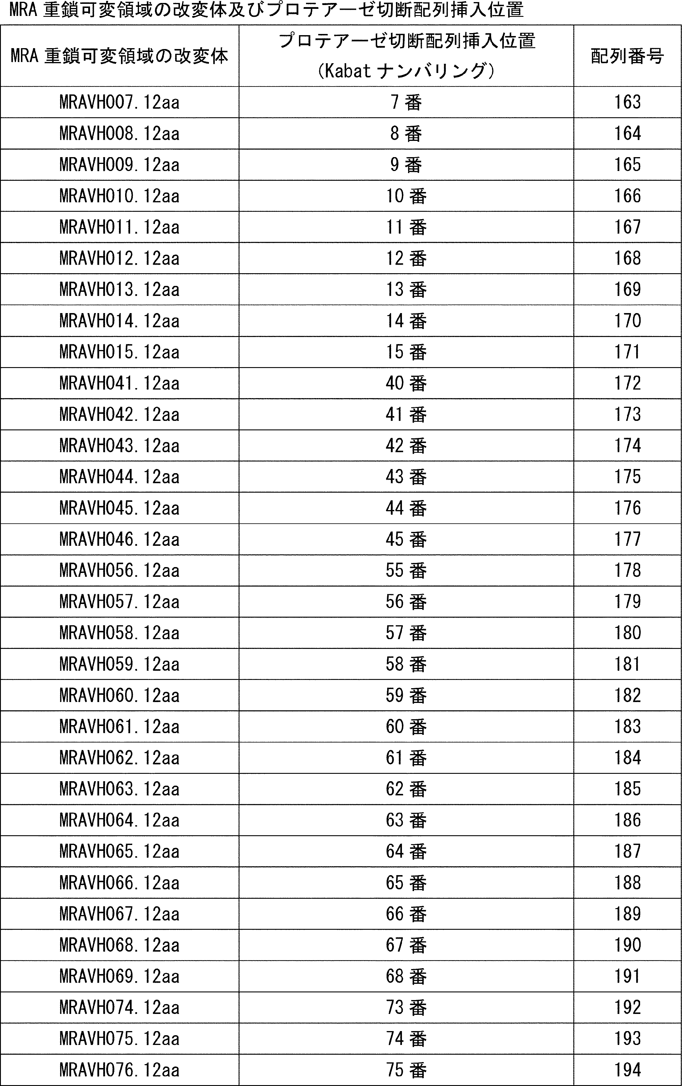

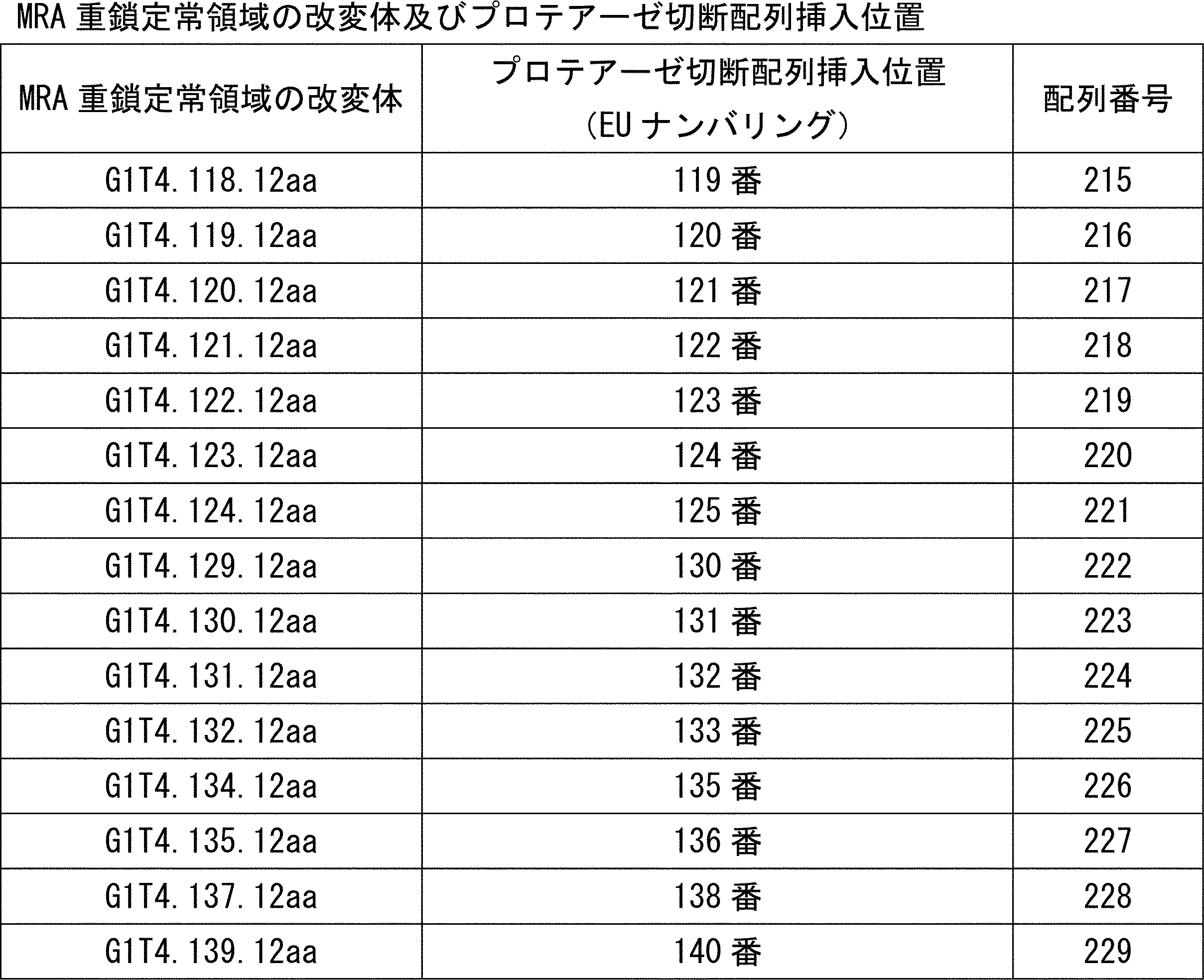
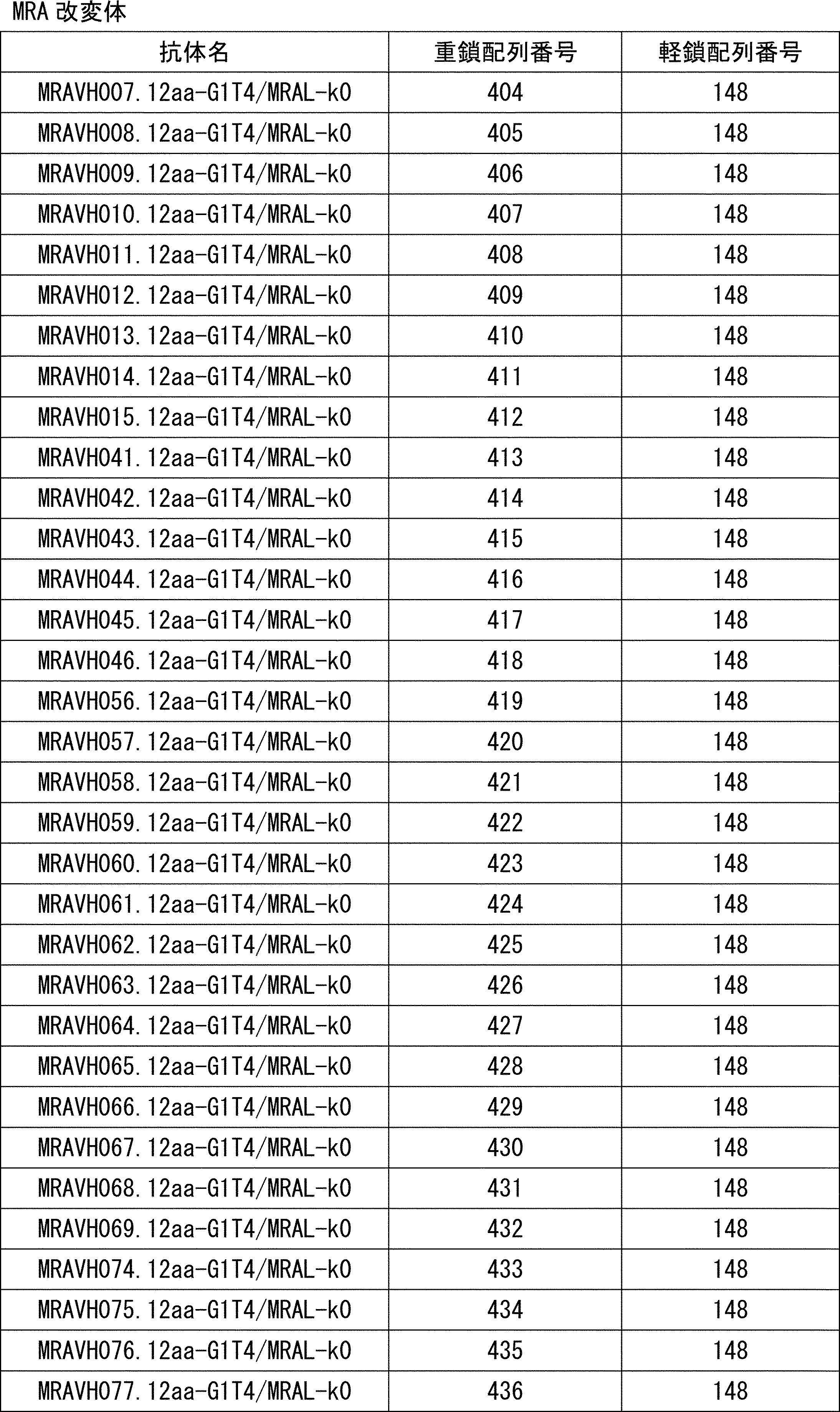
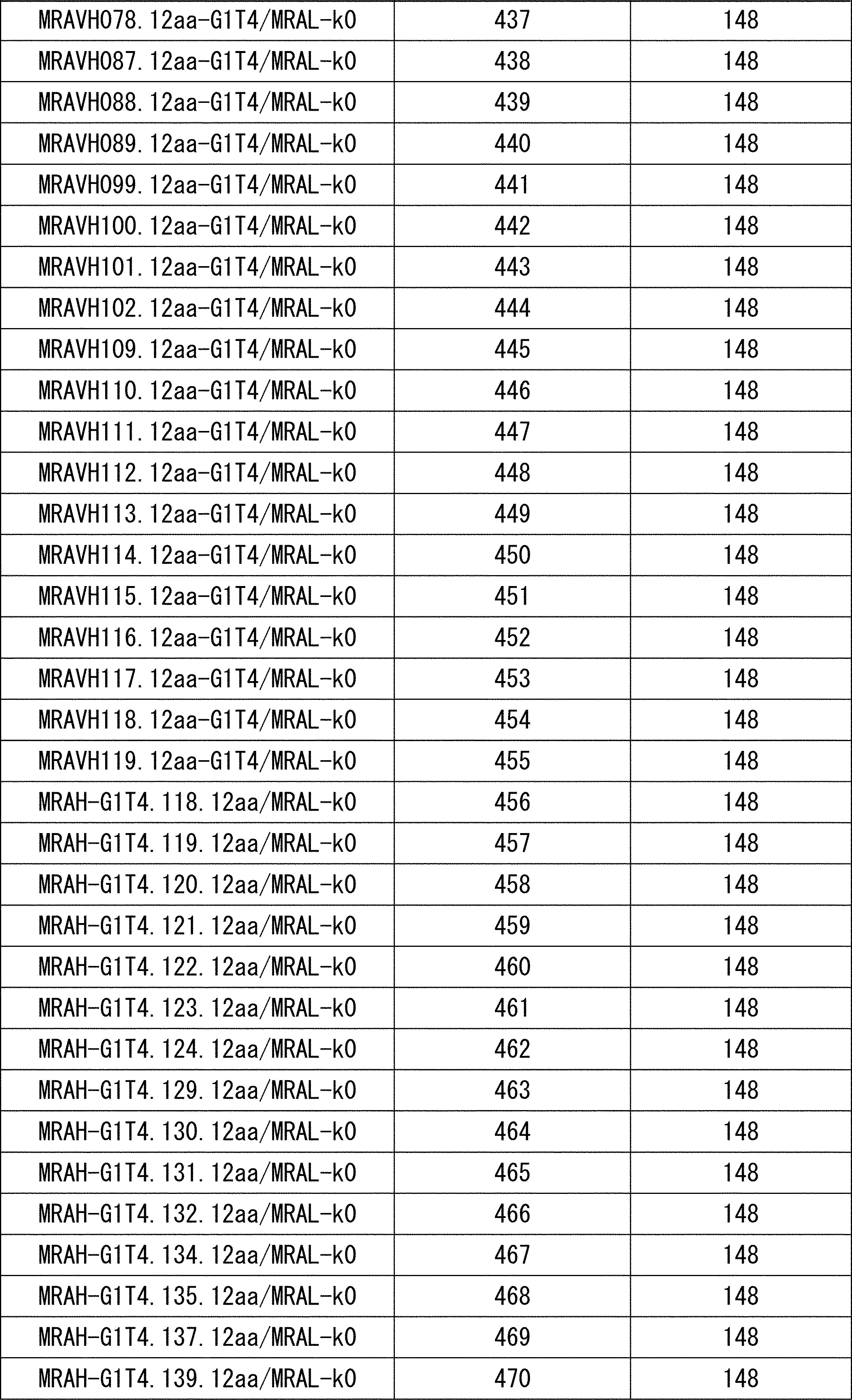
12-1で調製したMRA改変体がプロテアーゼにより切断されるどうかを検証した。プロテアーゼとしてリコンビナントヒトマトリプターゼ/ST14 触媒ドメイン( human MT-SP1, hMT-SP1 ) ( R&D Systems, 3946-SE-010 ) を用い、プロテアーゼ 10nM、抗体 50μg/mL、PBS、37℃の条件下で20時間反応させたのちに、還元SDS-PAGEに供した。結果は図22A、22B、22C、22D、22E、22F、22G、22H、22I、図23A、23B、23Cに示す。プロテアーゼ処理後のMRA改変体は、重鎖が切断され、プロテアーゼ処理を受けていないMRA改変体の重鎖(図中のMT-SP1(-) laneの50kDa付近に出現するバンド)と比べて、より分子量の小さい位置に重鎖バンドが出現している。この結果から、12-1で調製したMRA改変体は、hMT-SP1によって切断されることが確認された。
13-1 軽鎖の多様な位置にプロテアーゼ切断配列を導入した抗体の作製
ウロキナーゼ ( uPA ) およびマトリプターゼ( MT-SP1 )で切断されることが報告されているペプチド配列B(配列番号:160)をMRA軽鎖可変領域(MRAL、配列番号:230)内の異なる位置にそれぞれ挿入し、表9で示すMRA軽鎖可変領域の改変体を作製した。これらのMRA軽鎖可変領域の改変体をそれぞれMRA軽鎖定常領域(k0、配列番号:231)と連結させMRA軽鎖改変体の作製し、対応する遺伝子コードする発現ベクターを当業者公知の方法で作製した。また、ペプチド配列B(配列番号:160)をMRA軽鎖定常領域(k0、配列番号:231)内の異なる位置にそれぞれ挿入し、表10に示すMRA軽鎖定常領域の改変体を作製した。これらのMRA軽鎖定常領域の改変体をそれぞれMRA軽鎖可変領域(MRAL、配列番号:230)と連結させMRA軽鎖改変体を作製し、対応する遺伝子をコードする発現ベクターを当業者公知の方法で作製した。作製されたMRA軽鎖可変領域の改変体およびMRA軽鎖定常領域の改変体におけるプロテアーゼ切断配列挿入位置も表9、10に示す。表9における挿入箇所とは、抗体軽鎖可変領域中の記載アミノ酸(Kabatナンバリング)の定常領域側隣を指し、表10における挿入箇所とは、抗体軽鎖定常領域中の記載アミノ酸(EUナンバリング)の可変領域側隣を指す。
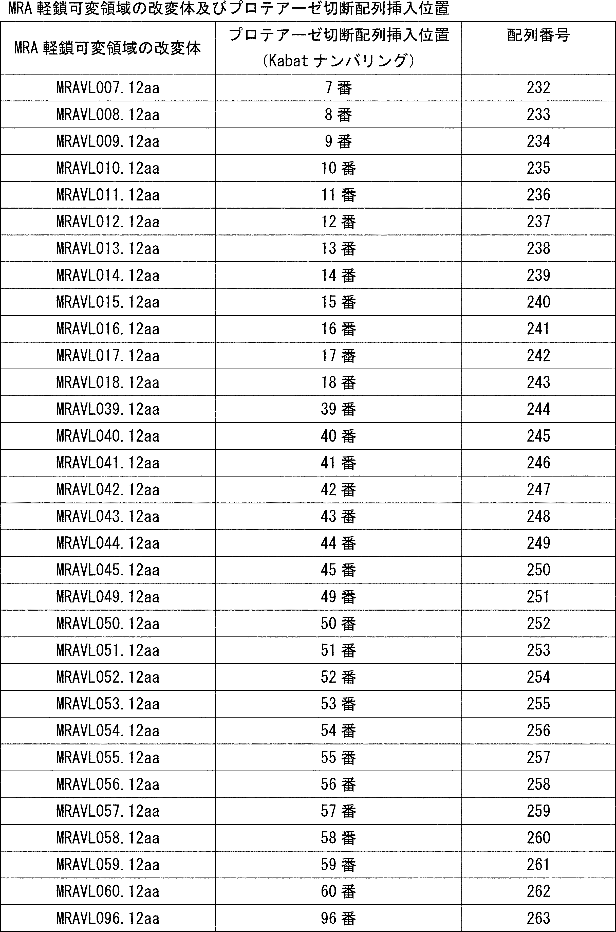
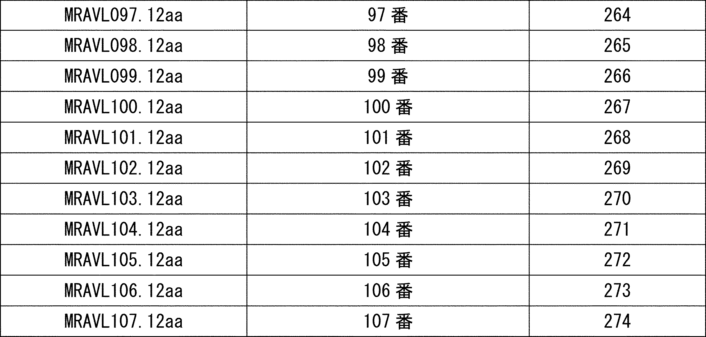
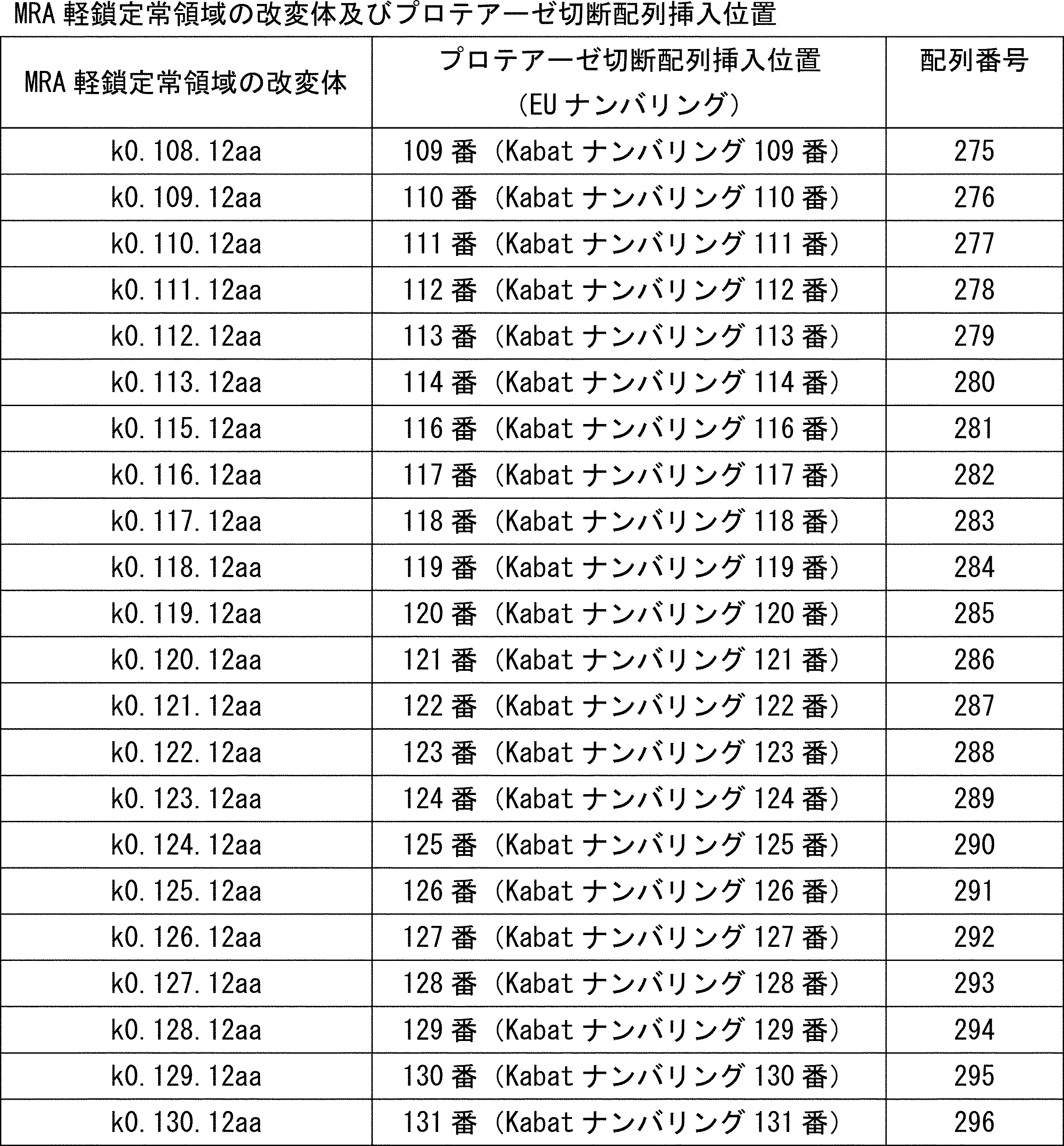

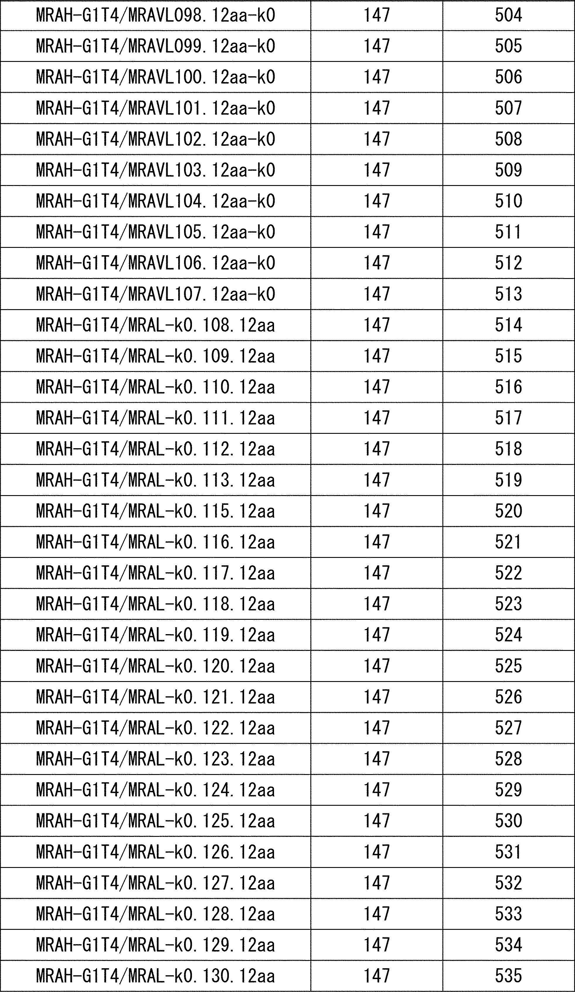
13-1で調製したMRA改変体がプロテアーゼにより切断されるどうかを検証した。プロテアーゼとしてリコンビナントヒトマトリプターゼ/ST14 触媒ドメイン( MT-SP1 ) ( R&D Systems, 3946-SE-010 ) を用い、プロテアーゼ 10nM、抗体 50μg/mL、PBS、37℃の条件下で20時間反応させたのちに、還元SDS-PAGEに供した。結果は図24A、24B、24C、24D、24E、図25A、25Bに示す。プロテアーゼ処理後切断されたMRA改変体は、プロテアーゼ処理を受けていないMRA改変体の軽鎖(図中MT-SP1(-)laneの25kDa付近に出願するバンド)と比べて、より分子量の小さい位置に軽鎖バンドが出現している。
14-1.抗ヒトPD1中和抗体へのプロテアーゼ切断配列の導入
ヒトPD1に対する中和抗体5C4H-G1T4/5C4L-KT0(重鎖5C4H-G1T4、配列番号:297;重鎖可変領域5C4H、配列番号:300;重鎖定常領域G1T4、配列番号:301;軽鎖5C4L-KT0、配列番号:298;軽鎖可変領域5C4L、配列番号:302;軽鎖定常領域KT0、配列番号:303; H-CDR1(NSGMH、配列番号:392)、H-CDR2(VIWYDGSKRYYADSVKG、配列番号:393)、H-CDR3(NDDY、配列番号:394)、L-CDR1(RASQSVSSYLA、配列番号:395)、L-CDR2(DASNRAT、配列番号:396)、L-CDR3(QQSSNWPRT、配列番号:397))の重鎖または軽鎖にプロテアーゼ切断配列を挿入し、プロテアーゼ切断配列が導入された抗体を作製した
まず、癌特異的に発現しているマトリプターゼ(MT-SP1)で切断されることが報告されているペプチド配列(配列番号:299)を前述抗体の重鎖5C4H-G1T4または軽鎖5C4L-KT0に挿入し、表12に示す重鎖改変体と表13に示す軽鎖改変体を作製し、当業者公知の方法で発現させた。
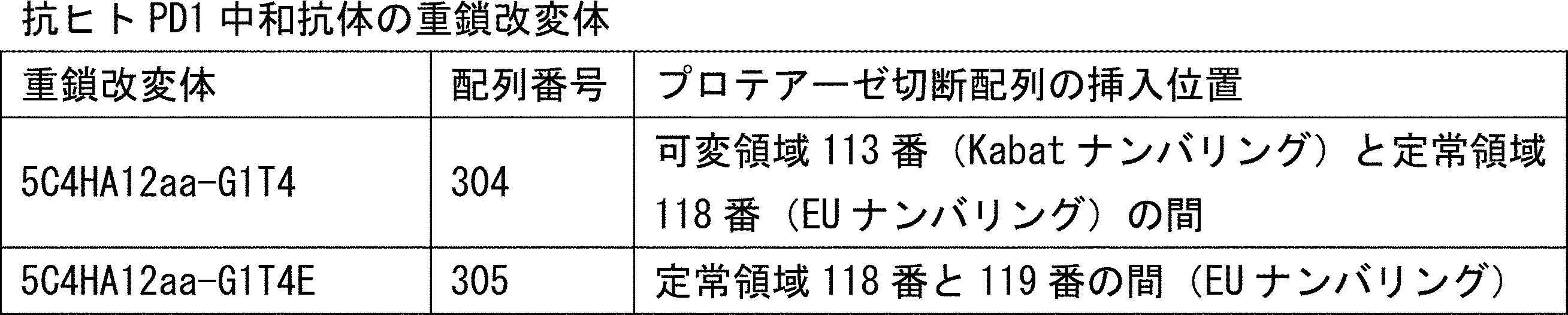

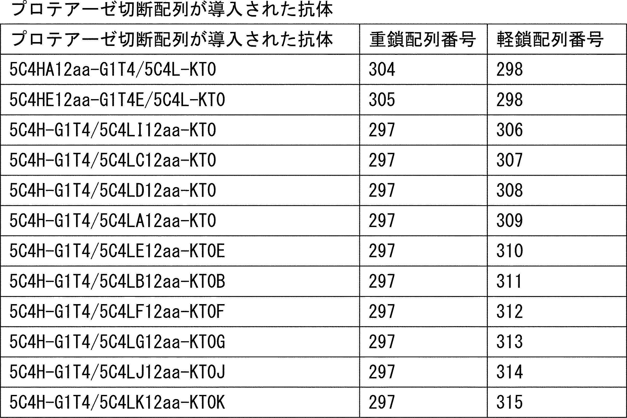
14-2-1 プロテアーゼ処理
プロテアーゼ処理抗体は14-1で調製された抗体(終濃度0.111mg/mL)にPBSで1.8μg/mLに調製したRecombinant Human Matriptase/ST14 Catalytic Domain (hMT-SP1, R&D systems 3946-SE-010) を10μL加えた。プロテアーゼ未処理抗体は14-1で調製された抗体(終濃度0.111mg/mL)にPBSのみを10μL加えた。反応時のサンプル体積は90μLであり、プロテアーゼの終濃度は0.2μg/mLであった。各サンプルを37℃で12時間保温した。
5C4H-G1T4/5C4L-KT0と同じ可変領域の配列を有しているビオチン化抗ヒトPD1中和抗体を調製した。具体的には、重鎖可変領域5C4H(配列番号:300)に抗体の重鎖定常領域とビオチン(AviTag配列、配列番号:316)を付加した5C4VH-G1dGSBAP(配列番号:317)をコードする遺伝子断片を作製し、当業者公知の方法で動物細胞発現用ベクターに導入した。構築された発現ベクターと軽鎖5C4L-KT0B(配列番号:298)を発現するベクターを293フェクチン(Invitrogen)を用いてFreeStyle293細胞 (Invitrogen)に導入した。このときEBNA1(配列番号:318)を発現する遺伝子およびビオチンリガーゼ(BirA、配列番号:319)を発現する遺伝子を同時に導入し、ビオチン標識する目的でビオチンを添加した。遺伝子が導入された細胞を37℃、8% CO2で培養し、目的のビオチン化抗ヒトPD1中和抗体(5C4-bio)を培養上清中に分泌させた。培養上清から当業者公知の方法で5C4-bioを精製した。
14-2-1で調製されたプロテアーゼ処理抗体/プロテアーゼ未処理抗体80μLに、終濃度0.67μMとなるようにヒトPD1を添加し、30分間室温で結合させ、結合評価用サンプルを調製した。抗体と結合しなかったPD1の量を評価することで、抗体のプロテアーゼ処理/未処理時のPD1に対する結合を評価した。
具体的に、抗体と結合しなかったPD1の量を、実施例14-2-2で調製したビオチン化抗ヒトPD1中和抗体 (5C4-bio) を用いてバイオレイヤー干渉法(BLI法)で評価した。
結合評価用サンプル、5C4-bio、PBSをそれぞれTilted bottom (TW384) Micro plates(ForteBio、18-5076)の異なるウェルに分注した。Streptavidinバイオセンサー(ForteBio, 18-0009)をPBSを用いて水和し、30℃の設定でOctet RED 384で測定が行われた。PBSが入ったウェルで30秒間ベースライン測定を実施し、その後200秒間5C4-bioをStreptavidinセンサーに結合させた。再度PBSが入ったウェルで30秒間ベースライン測定を実施し、その後結合評価用サンプルが入ったウェルで180秒間結合を測定し、PBSが入ったウェルで180秒間解離を測定した。結合の様子を示すリアルタイム結合グラフを図26に示す。図26に示す通り、プロテアーゼ切断配列が導入された抗体の場合、プロテアーゼ未処理抗体を含む結合評価用サンプルに比べて、プロテアーゼ処理抗体を含む結合評価用サンプルで測定するときの5C4-bioに結合するヒトPD1の量が多かった。即ち、プテアーゼ切断配列が導入された各抗体は、プロテアーゼ処理によってPD1に対する結合活性が減弱し、PD1が遊離し、5C4-bioに結合した。
14-2-3で用いられた抗体がプロテアーゼ処理で切断されているかSDS-PAGEで確認した。14-2-3で調製されたプロテアーゼ切断抗体/プロテアーゼ未処理抗体のうち、10μLを3.3μLのサンプルバッファーと混合し、95℃5分間保温した。次にMini-PROTEAN TGX gel (4-20% 15well) (Bio-Rad #456-1096)を用いて電気泳動を行い、Sample Blue Safe Stain (novex, LC6065)でタンパク質を染色した。その結果を図27に示す。図27に示すように、プロテアーゼ切断配列を導入した各抗体はプロテアーゼ処理で切断された。
プロテアーゼ切断配列が導入された各抗体のプロテアーゼ処理前後のPD1に対する結合活性を別の方法でも測定した。
14-2-1で調製したプロテアーゼ処理抗体またはプロテアーゼ未処理抗体10μLをPBS 70μLと混合し、PD1結合測定サンプルとした。サンプルのPD1結合評価はバイオレイヤー干渉法(BLI法)で評価した。14-2-1で調製したプロテアーゼ処理抗体/プロテアーゼ未処理抗体とヒトPD1 (250nM) をそれぞれTilted bottom (TW384) Microplates(ForteBio、18-5076)の異なるウェルに分注した。Protein Gセンサー(ForteBio, 18-0022)をPBSを用いて水和し、30℃の設定でOctet RED 384で測定が行われた。PBSが入ったウェルで30秒間ベースライン測定を実施し、その後200秒間抗体をProtein Gセンサーに結合させた。再度PBSが入ったウェルで30秒間ベースライン測定を実施し、その後ヒトPD1が入ったウェルで180秒間結合を測定し、PBSが入ったウェルで180秒間解離を測定した。結合の様子を示すリアルタイム結合グラフを図28に示す。図28に示す通り、プロテアーゼ切断配列が導入された各抗体を使用する場合、プロテアーゼ未処理抗体に比べて、プロテアーゼ処理抗体のヒトPD1結合量が減少した。
14-3-1 リガンド存在下のプロテアーゼ処理
14-1で調製された抗体(終濃度0.100mg/mL)にPBSで6.67μMに調製したヒトPD1を10μL添加し、抗体-PD1複合体液を調製した。プロテアーゼ処理サンプルは、抗体-PD1複合体液にPBSで5.28μg/mLに調製したRecombinant Human Matriptase/ST14 Catalytic Domain (hMT-SP1, R&D systems 3946-SE-010) を10μL加え、プロテアーゼ非処理サンプルはPBSのみを10μL加えた。反応時のプロテアーゼの終濃度は0.528μg/mLであった。各サンプルを37℃で12時間保温した。
抗体と複合体を形成していないPD1の量を実施例14-2-2で調製したビオチン化抗ヒトPD1中和抗体 (5C4-bio) を用いてバイオレイヤー干渉法(BLI法)で評価した。
14-3-1で調製された各サンプル、5C4-bio、PBSをそれぞれTilted bottom (TW384) Micro plates(ForteBio、18-5076)の異なるウェルに分注した。Streptavidinバイオセンサー(ForteBio, 18-0009)をPBSを用いて水和し、30℃の設定でOctet RED 384で測定が行われた。PBSが入ったウェルで30秒間ベースライン測定を実施し、その後200秒間5C4-bioをStreptavidinセンサーに結合させた。再度PBSが入ったウェルで30秒間ベースライン測定を実施し、プロテアーゼ処理サンプルまたはプロテアーゼ未処理サンプルが入ったウェルで180秒間結合を測定し、PBSが入ったウェルで180秒間解離を測定した。結合の様子を示すリアルタイム結合グラフを図29に示す。図29に示す通り、プロテアーゼ切断配列が導入された各抗体の場合、プロテアーゼ未処理サンプルに比べ、プロテアーゼ処理サンプルの方では5C4-bioに結合するヒトPD1の量が増加した。即ち、プロテアーゼ処理により各抗体のPD1に対する結合活性が減弱し、抗体-PD1複合体からPD1が遊離し、リリースされた。
15-1.抗ヒトPD1中和抗体とヒトPD1の融合タンパク質の作製
実施例14-1で作製された抗体の重鎖または重鎖改変体のN末端に、グリシン-セリンポリマー(配列番号:321)からなる可動リンカーを介してヒトPD1配列(配列番号:320)を接続し、PD1融合重鎖を作製した(表15)。

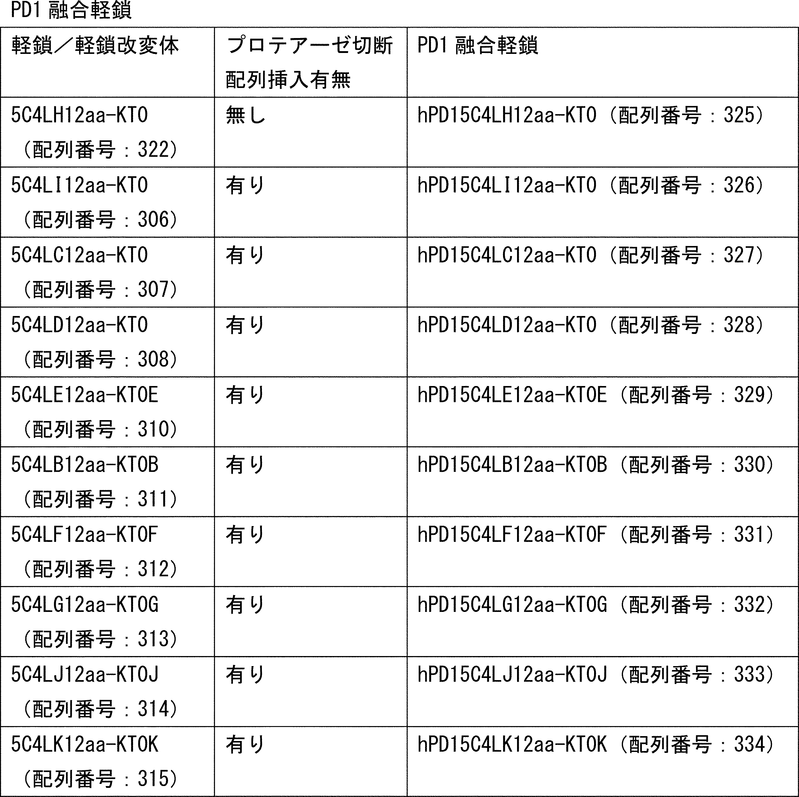
hPD15C4HA12aa-G1T4/5C4L-KT0(PD1融合重鎖配列番号:323、軽鎖配列番号:298)
hPD15C4HE12aa-G1T4E/5C4L-KT0(PD1融合重鎖配列番号:324、軽鎖配列番号:298)
5C4H-G1T4/hPD15C4LH12aa-KT0(重鎖配列番号:297、軽鎖配列番号:325)
5C4H-G1T4/hPD15C4LI12aa-KT0(重鎖配列番号:297、PD1融合軽鎖配列番号:326)
5C4H-G1T4/hPD15C4LC12aa-KT0(重鎖配列番号:297、PD1融合軽鎖配列番号:327)
5C4H-G1T4/hPD15C4LD12aa-KT0(重鎖配列番号:297、PD1融合軽鎖配列番号:328)
5C4H-G1T4/hPD15C4LE12aa-KT0E(重鎖配列番号:297、PD1融合軽鎖配列番号:329)
5C4H-G1T4/hPD15C4LB12aa-KT0B(重鎖配列番号:297、PD1融合軽鎖配列番号:330)
5C4H-G1T4/hPD15C4LF12aa-KT0F(重鎖配列番号:297、PD1融合軽鎖配列番号:331)
5C4H-G1T4/hPD15C4LG12aa-KT0G(重鎖配列番号:297、PD1融合軽鎖配列番号:332)
5C4H-G1T4/hPD15C4LJ12aa-KT0J(重鎖配列番号:297、PD1融合軽鎖配列番号:333)
5C4H-G1T4/hPD15C4LK12aa-KT0K(重鎖配列番号:297、PD1融合軽鎖配列番号:334)
を当業者公知の方法でExpi293 (Life Technologies)を用いた一過性発現により発現し、プロテインAを用いた当業者公知の方法により精製を行った。なおプロテアーゼ切断配列を含まない対照抗体として5C4H-G1T4/5C4L-KT0(重鎖配列番号:297、軽鎖配列番号:298))を同様に発現および精製した。
15-2-1 プロテアーゼ処理
プロテアーゼ処理融合タンパク質は15-1で調製された融合タンパク質30μgにPBSで16.7μg/mLに調製したRecombinant Human Matriptase/ST14 Catalytic Domain (hMT-SP1, R&D systems 3946-SE-010) を4.9μL加えた。プロテアーゼ未処理融合タンパク質は15-1で調製された融合タンパク質30μgにPBSのみを4.9μL加えた。プロテアーゼ処理融合タンパク質またはプロテアーゼ未処理融合タンパク質を37℃で12時間保温した。
プロテアーゼ処理によるPD1遊離を実施例14-2-2で調製したビオチン化抗ヒトPD1中和抗体 (5C4-bio) を用いてバイオレイヤー干渉法(BLI法)で評価した。
15-2-1で調製したプロテアーゼ処理融合タンパク質、プロテアーゼ未処理融合タンパク質、5C4-bio、PBSをそれぞれTilted bottom (TW384) Micro plates(ForteBio、18-5076)の異なるウェルに分注した。Streptavidinバイオセンサー(ForteBio, 18-0009)をPBSを用いて水和し、30℃の設定でOctet RED 384で測定が行われた。PBSが入ったウェルで30秒間ベースライン測定を実施し、その後200秒間5C4-bioをStreptavidinセンサーに結合させた。再度PBSが入ったウェルで30秒間ベースライン測定を実施しその後プロテアーゼ処理融合タンパク質またはプロテアーゼ未処理融合タンパク質が入ったウェルで180秒間結合を測定し、PBSが入ったウェルで180秒間解離を測定した。結合の様子を示すリアルタイム結合グラフを図30に示す。図30に示す通り、プロテアーゼ切断配列が導入された抗体を含む抗体-PD1融合タンパク質の場合、プロテアーゼ未処理サンプルに比べて、プロテアーゼ処理サンプルの方では5C4-bioに結合するヒトPD1の量が増加した。即ち、プロテアーゼ処理により融合タンパク質中の抗体のPD1に対する結合活性が減弱し、PD1が融合タンパク質から遊離した。
15-2-1で調製されたプロテアーゼ処理融合タンパク質がプロテアーゼ処理で切断されているかSDS-PAGEで確認した。15-2-1で調製されたプロテアーゼ処理融合タンパク質またはプロテアーゼ未処理融合タンパク質のうち、10μLを3.3μLのサンプルバッファーと混合し、95℃5分間保温した。次にMini-PROTEAN TGX gel(4-20% 15well) (Bio-Rad #456-1096)を用いて電気泳動を行い、Sample Blue Safe Stain(novex, LC6065)でタンパク質を染色した。その結果を図31に示す。図31に示すように、プロテアーゼ切断配列を導入した抗体を含む融合タンパク質はプロテアーゼ処理で切断された。
Claims (15)
- リガンドに結合可能な分子であって、当該分子は切断サイトを少なくとも一つ有するポリペプチドであり、且つ当該分子が少なくとも一つの切断サイトで切断された状態でリガンドとの結合が減弱される、リガンド結合分子。
- 前記切断サイトはプロテアーゼ切断配列を含む、請求項1に記載のリガンド結合分子。
- 前記プロテアーゼは、標的組織特異的プロテアーゼである、請求項2に記載のリガンド結合分子。
- 前記リガンド結合分子は抗体VHと、抗体VLと、抗体定常領域を含む、請求項1から請求項3のいずれかに記載のリガンド結合分子。
- 前記切断サイトまたは前記プロテアーゼ切断配列は、前記抗体定常領域と前記抗体VHの境界付近、または/および前記抗体定常領域と前記抗体VLとの境界付近に位置する、請求項4に記載のリガンド結合分子。
- 前記リガンド結合分子中の前記抗体VLと前記抗体VHは会合しており、当該会合は前記切断サイトが切断されることにより解消される、または前記プロテアーゼ切断配列がプロテアーゼで切断されることにより解消される、請求項4もしくは請求項5に記載のリガンド結合分子。
- 前記リガンドは生物活性を有する分子であり、前記リガンド結合分子は前記リガンドとの結合で前記リガンドの生物活性を阻害する、請求項1から請求項6のいずれかに記載のリガンド結合分子。
- 前記リガンドはサイトカインまたはケモカインである、請求項1から請求項7のいずれかに記載のリガンド結合分子。
- 前記リガンド結合分子はIgG抗体である、請求項1から請求項8のいずれかに記載のリガンド結合分子。
- 前記リガンドと結合している、請求項1から請求項9のいずれかに記載のリガンド結合分子。
- 前記リガンドと融合されている、請求項1から請求項9のいずれかに記載のリガンド結合分子。
- 請求項1から請求項11のいずれかに記載のリガンド結合分子を含む、医薬組成物。
- 請求項1から請求項10いずれかに記載のリガンド結合分子とリガンドを含む、医薬組成物。
- 請求項1から請求項9のいずれかに記載のリガンド結合分子とリガンドが融合されている融合タンパク質を含む、医薬組成物。
- 請求項1から請求項11のいずれかに記載のリガンド結合分子を製造する方法。
Priority Applications (15)
| Application Number | Priority Date | Filing Date | Title |
|---|---|---|---|
| MX2019005947A MX2019005947A (es) | 2016-11-28 | 2017-11-28 | Molecula de union a ligando que tiene actividad de union a ligando ajustable. |
| AU2017364818A AU2017364818C1 (en) | 2016-11-28 | 2017-11-28 | Ligand-binding molecule having adjustable ligand binding activity |
| KR1020197017817A KR102533814B1 (ko) | 2016-11-28 | 2017-11-28 | 리간드 결합 활성을 조정 가능한 리간드 결합 분자 |
| CA3039316A CA3039316A1 (en) | 2016-11-28 | 2017-11-28 | Ligand-binding molecule having adjustable ligand binding activity |
| RU2019117223A RU2803067C2 (ru) | 2016-11-28 | 2017-11-28 | Лигандсвязывающая молекула с регулируемой лигандсвязывающей активностью |
| EP17872925.7A EP3546480A4 (en) | 2016-11-28 | 2017-11-28 | LIGAND BINDING MOLECULE HAVING ADJUSTABLE LIGAND BINDING ACTIVITY |
| US16/463,218 US12060654B2 (en) | 2016-11-28 | 2017-11-28 | Ligand-binding molecule having adjustable ligand binding activity |
| CN201780084377.1A CN110214151A (zh) | 2016-11-28 | 2017-11-28 | 具有可调节的配体结合活性的配体结合分子 |
| IL266827A IL266827B2 (en) | 2016-11-28 | 2017-11-28 | Ligand binding molecule with tunable ligand binding activity, its fusion protein with a ligand, pharmaceutical compositions containing them, and method for their production |
| BR112019008265A BR112019008265A2 (pt) | 2016-11-28 | 2017-11-28 | molécula de ligação ao ligante cuja atividade de li-gação ao ligante é ajustável |
| JP2018553014A JP7626577B2 (ja) | 2016-11-28 | 2017-11-28 | リガンド結合活性が調整可能なリガンド結合分子 |
| KR1020237015916A KR20230073346A (ko) | 2016-11-28 | 2017-11-28 | 리간드 결합 활성을 조정 가능한 리간드 결합 분자 |
| JP2023038624A JP7671794B2 (ja) | 2016-11-28 | 2023-03-13 | リガンド結合活性が調整可能なリガンド結合分子 |
| US18/656,351 US20250034756A1 (en) | 2016-11-28 | 2024-05-06 | Ligand-binding molecule having adjustable ligand binding activity |
| JP2024226961A JP2025041812A (ja) | 2016-11-28 | 2024-12-24 | リガンド結合活性が調整可能なリガンド結合分子 |
Applications Claiming Priority (2)
| Application Number | Priority Date | Filing Date | Title |
|---|---|---|---|
| JP2016229882 | 2016-11-28 | ||
| JP2016-229882 | 2016-11-28 |
Related Child Applications (2)
| Application Number | Title | Priority Date | Filing Date |
|---|---|---|---|
| US16/463,218 A-371-Of-International US12060654B2 (en) | 2016-11-28 | 2017-11-28 | Ligand-binding molecule having adjustable ligand binding activity |
| US18/656,351 Division US20250034756A1 (en) | 2016-11-28 | 2024-05-06 | Ligand-binding molecule having adjustable ligand binding activity |
Publications (1)
| Publication Number | Publication Date |
|---|---|
| WO2018097308A1 true WO2018097308A1 (ja) | 2018-05-31 |
Family
ID=62195875
Family Applications (1)
| Application Number | Title | Priority Date | Filing Date |
|---|---|---|---|
| PCT/JP2017/042570 Ceased WO2018097308A1 (ja) | 2016-11-28 | 2017-11-28 | リガンド結合活性が調整可能なリガンド結合分子 |
Country Status (12)
| Country | Link |
|---|---|
| US (2) | US12060654B2 (ja) |
| EP (1) | EP3546480A4 (ja) |
| JP (3) | JP7626577B2 (ja) |
| KR (2) | KR20230073346A (ja) |
| CN (1) | CN110214151A (ja) |
| AU (1) | AU2017364818C1 (ja) |
| BR (1) | BR112019008265A2 (ja) |
| CA (1) | CA3039316A1 (ja) |
| IL (1) | IL266827B2 (ja) |
| MX (3) | MX2019005947A (ja) |
| TW (2) | TW202235438A (ja) |
| WO (1) | WO2018097308A1 (ja) |
Cited By (15)
| Publication number | Priority date | Publication date | Assignee | Title |
|---|---|---|---|---|
| WO2019230868A1 (ja) * | 2018-05-30 | 2019-12-05 | 中外製薬株式会社 | 単ドメイン抗体含有リガンド結合分子 |
| WO2020065096A1 (en) * | 2018-09-28 | 2020-04-02 | Pierre Fabre Medicament | New immunocytokines for the treatment of cancer |
| WO2020116498A1 (ja) * | 2018-12-04 | 2020-06-11 | 中外製薬株式会社 | Cxcr3リガンド |
| WO2020246563A1 (ja) | 2019-06-05 | 2020-12-10 | 中外製薬株式会社 | 抗体切断部位結合分子 |
| EP3719035A4 (en) * | 2017-11-28 | 2021-09-01 | Chugai Seiyaku Kabushiki Kaisha | POLYPEPTIDE WITH ANTIGEN-BINDING AREA AND CARRYING SECTION |
| EP3719036A4 (en) * | 2017-11-28 | 2021-09-08 | Chugai Seiyaku Kabushiki Kaisha | LIGANDBINDING MOLECULE WITH ADJUSTABLE LIGANDBINDING ACTIVITY |
| JP2021523741A (ja) * | 2018-05-14 | 2021-09-09 | ウェアウルフ セラピューティクス, インコーポレイテッド | 活性化可能なインターロイキン12ポリペプチド及びその使用方法 |
| US11168139B2 (en) | 2016-11-28 | 2021-11-09 | Chugai Seiyaku Kabushiki Kaisha | Antigen-binding domain, and polypeptide including conveying section |
| JP2021530243A (ja) * | 2018-07-25 | 2021-11-11 | アスクジーン・ファーマ・インコーポレイテッドAskGene Pharma, Inc. | 新規il−21プロドラッグおよびそれを使用する方法 |
| CN114127277A (zh) * | 2019-06-05 | 2022-03-01 | 中外制药株式会社 | 蛋白酶底物和包含蛋白酶切割序列的多肽 |
| US20230069996A1 (en) * | 2020-01-20 | 2023-03-09 | Chugai Seiyaku Kabushiki Kaisha | Ligand-binding fusion proteins |
| WO2023120643A1 (ja) | 2021-12-23 | 2023-06-29 | 中外製薬株式会社 | Cxcr3発現細胞遊走活性が増強したcxcr3リガンド。 |
| US12060654B2 (en) | 2016-11-28 | 2024-08-13 | Chugai Seiyaku Kabushiki Kaisha | Ligand-binding molecule having adjustable ligand binding activity |
| US12077577B2 (en) | 2018-05-30 | 2024-09-03 | Chugai Seiyaku Kabushiki Kaisha | Polypeptide comprising aggrecan binding domain and carrying moiety |
| EP4373862A4 (en) * | 2021-07-19 | 2025-12-31 | Chugai Pharmaceutical Co Ltd | TARGET-SPECIFIC CYTOKINE ADMINISTRATION MEDIATRED BY A PROTEASE USING A FUSION POLYPEPTIDE |
Families Citing this family (6)
| Publication number | Priority date | Publication date | Assignee | Title |
|---|---|---|---|---|
| KR20210081372A (ko) * | 2018-10-22 | 2021-07-01 | 유니버시티 오브 피츠버그 - 오브 더 커먼웰쓰 시스템 오브 하이어 에듀케이션 | 절단 가능한 cxcr3의 활성제 및 이의 사용 방법 |
| CA3141626A1 (en) | 2019-06-12 | 2020-12-17 | AskGene Pharma, Inc. | Novel il-15 prodrugs and methods of use thereof |
| US20220281934A1 (en) * | 2019-08-29 | 2022-09-08 | Antagonis Biotherapeutics Gmbh | T-cell mobilizing cxcl10 mutant with increased glycosaminoglycan binding affinity |
| CA3165927A1 (en) | 2020-01-11 | 2021-07-15 | AskGene Pharma, Inc. | Novel masked cytokines and methods of use thereof |
| EP3889183A1 (en) * | 2020-04-01 | 2021-10-06 | Pierre Fabre Medicament | A protein complex comprising an immunocytokine |
| CN120112547A (zh) | 2022-11-16 | 2025-06-06 | 瑞泽恩制药公司 | 包含膜结合il-12和蛋白酶可裂解接头的嵌合蛋白 |
Citations (42)
| Publication number | Priority date | Publication date | Assignee | Title |
|---|---|---|---|---|
| EP0239400A2 (en) | 1986-03-27 | 1987-09-30 | Medical Research Council | Recombinant antibodies and methods for their production |
| US4816567A (en) | 1983-04-08 | 1989-03-28 | Genentech, Inc. | Recombinant immunoglobin preparations |
| WO1992001047A1 (en) | 1990-07-10 | 1992-01-23 | Cambridge Antibody Technology Limited | Methods for producing members of specific binding pairs |
| WO1992003918A1 (en) | 1990-08-29 | 1992-03-19 | Genpharm International, Inc. | Transgenic non-human animals capable of producing heterologous antibodies |
| WO1992020791A1 (en) | 1990-07-10 | 1992-11-26 | Cambridge Antibody Technology Limited | Methods for producing members of specific binding pairs |
| WO1993006213A1 (en) | 1991-09-23 | 1993-04-01 | Medical Research Council | Production of chimeric antibodies - a combinatorial approach |
| WO1993011236A1 (en) | 1991-12-02 | 1993-06-10 | Medical Research Council | Production of anti-self antibodies from antibody segment repertoires and displayed on phage |
| WO1993012227A1 (en) | 1991-12-17 | 1993-06-24 | Genpharm International, Inc. | Transgenic non-human animals capable of producing heterologous antibodies |
| WO1993019172A1 (en) | 1992-03-24 | 1993-09-30 | Cambridge Antibody Technology Limited | Methods for producing members of specific binding pairs |
| WO1994002602A1 (en) | 1992-07-24 | 1994-02-03 | Cell Genesys, Inc. | Generation of xenogeneic antibodies |
| WO1994025585A1 (en) | 1993-04-26 | 1994-11-10 | Genpharm International, Inc. | Transgenic non-human animals capable of producing heterologous antibodies |
| WO1995001438A1 (en) | 1993-06-30 | 1995-01-12 | Medical Research Council | Sbp members with a chemical moiety covalently bound within the binding site; production and selection thereof |
| WO1995001937A1 (fr) | 1993-07-09 | 1995-01-19 | Association Gradient | Procede de traitement de residus de combustion et installation de mise en ×uvre dudit procede |
| WO1995015388A1 (en) | 1993-12-03 | 1995-06-08 | Medical Research Council | Recombinant binding proteins and peptides |
| WO1996002576A1 (en) | 1994-07-13 | 1996-02-01 | Chugai Seiyaku Kabushiki Kaisha | Reconstituted human antibody against human interleukin-8 |
| WO1996033735A1 (en) | 1995-04-27 | 1996-10-31 | Abgenix, Inc. | Human antibodies derived from immunized xenomice |
| WO1996034096A1 (en) | 1995-04-28 | 1996-10-31 | Abgenix, Inc. | Human antibodies derived from immunized xenomice |
| WO2002020565A2 (en) | 2000-09-08 | 2002-03-14 | Universität Zürich | Collections of repeat proteins comprising repeat modules |
| WO2002032925A2 (en) | 2000-10-16 | 2002-04-25 | Phylos, Inc. | Protein scaffolds for antibody mimics and other binding proteins |
| WO2003000883A1 (fr) | 2001-06-22 | 2003-01-03 | Chugai Seiyaku Kabushiki Kaisha | Inhibiteurs de proliferation cellulaire renfermant un anticorps anti-glypicane 3 |
| WO2003029462A1 (en) | 2001-09-27 | 2003-04-10 | Pieris Proteolab Ag | Muteins of human neutrophil gelatinase-associated lipocalin and related proteins |
| WO2003104453A1 (ja) | 2002-06-05 | 2003-12-18 | 中外製薬株式会社 | 抗体作製方法 |
| WO2004022754A1 (ja) | 2002-09-04 | 2004-03-18 | Chugai Seiyaku Kabushiki Kaisha | MRL/lprマウスを用いた抗体の作製 |
| WO2004021861A2 (en) | 2002-09-03 | 2004-03-18 | Vit Lauermann | Targeted release |
| WO2004044011A2 (en) | 2002-11-06 | 2004-05-27 | Avidia Research Institute | Combinatorial libraries of monomer domains |
| WO2005040229A2 (en) | 2003-10-24 | 2005-05-06 | Avidia, Inc. | Ldl receptor class a and egf domain monomers and multimers |
| WO2006006693A1 (ja) | 2004-07-09 | 2006-01-19 | Chugai Seiyaku Kabushiki Kaisha | 抗グリピカン3抗体 |
| WO2008016854A2 (en) | 2006-08-02 | 2008-02-07 | The Uab Research Foundation | Methods and compositions related to soluble monoclonal variable lymphocyte receptors of defined antigen specificity |
| WO2009025846A2 (en) | 2007-08-22 | 2009-02-26 | The Regents Of The University Of California | Activatable binding polypeptides and methods of identification and use thereof |
| WO2010081173A2 (en) | 2009-01-12 | 2010-07-15 | Cytomx Therapeutics, Llc | Modified antibody compositions, methods of making and using thereof |
| WO2013128194A1 (en) | 2012-02-28 | 2013-09-06 | The University Of Birmingham | Immunotherapeutic molecules and uses |
| JP2013538204A (ja) * | 2010-08-24 | 2013-10-10 | ロシュ グリクアート アーゲー | 活性化可能な二重特異性抗体 |
| WO2015048329A2 (en) | 2013-09-25 | 2015-04-02 | Cytomx Therapeutics, Inc. | Matrix metalloproteinase substrates and other cleavable moieties and methods of use thereof |
| WO2015116933A2 (en) | 2014-01-31 | 2015-08-06 | Cytomx Therapeutics, Inc. | Matriptase and u-plasminogen activator substrates and other cleavable moieties and methods of use thereof |
| WO2016014974A2 (en) | 2014-07-25 | 2016-01-28 | Cytomx Therapeutics, Inc. | Anti-cd3 antibodies, activatable anti-cd3 antibodies, multispecific anti-cd3 antibodies, multispecific activatable anti-cd3 antibodies, and methods of using the same |
| WO2016046778A2 (en) | 2014-09-25 | 2016-03-31 | Amgen Inc | Protease-activatable bispecific proteins |
| WO2016118629A1 (en) | 2015-01-20 | 2016-07-28 | Cytomx Therapeutics, Inc. | Matrix metalloprotease-cleavable and serine protease cleavable substrates and methods of use thereof |
| US20160311903A1 (en) | 2015-03-13 | 2016-10-27 | Cytomx Therapeutics, Inc. | Anti-pdl1 antibodies, activatable anti-pdl1 antibodies, and methods of use thereof |
| WO2016179285A1 (en) | 2015-05-04 | 2016-11-10 | Cytomx Therapeutics, Inc. | Anti-cd166 antibodies, activatable anti-cd166 antibodies, and methods of use thereof |
| WO2016179335A1 (en) | 2015-05-04 | 2016-11-10 | Cytomx Therapeutics, Inc. | Anti-itga3 antibodies, activatable anti-itga3 antibodies, and methods of use thereof |
| WO2016179257A2 (en) | 2015-05-04 | 2016-11-10 | Cytomx Therapeutics, Inc. | Anti-cd71 antibodies, activatable anti-cd71 antibodies, and methods of use thereof |
| WO2016179003A1 (en) | 2015-05-01 | 2016-11-10 | Genentech, Inc. | Masked anti-cd3 antibodies and methods of use |
Family Cites Families (67)
| Publication number | Priority date | Publication date | Assignee | Title |
|---|---|---|---|---|
| AU2003234775A1 (en) | 2002-04-30 | 2003-11-17 | Dnavec Research Inc. | Vector with modified protease-dependent tropism |
| JP2005168328A (ja) | 2003-12-08 | 2005-06-30 | Hokkaido Univ | シグナル伝達活性を有する細胞質蛋白質の活性制御方法 |
| PL1691837T3 (pl) | 2003-12-10 | 2012-11-30 | Squibb & Sons Llc | IP-10 przeciwciała i ich zastosowanie |
| JP5629423B2 (ja) | 2004-04-12 | 2014-11-19 | カタリスト バイオサイエンシーズ, インコーポレイテッド | 野生型及び変異型mt−sp1による、vegf及びvegf受容体の切断 |
| MY157915A (en) | 2004-12-21 | 2016-08-15 | Centocor Inc | Anti-il-12 antibodies, epitopes, compositions, methods and uses. |
| DK2439273T3 (da) | 2005-05-09 | 2019-06-03 | Ono Pharmaceutical Co | Humane monoklonale antistoffer til programmeret død-1(pd-1) og fremgangsmåder til behandling af cancer ved anvendelse af anti-pd-1- antistoffer alene eller i kombination med andre immunterapeutika |
| AU2006284651C1 (en) | 2005-08-31 | 2013-09-12 | The Regents Of The University Of California | Cellular libraries of peptide sequences (CLiPS) and methods of using the same |
| JP2009519011A (ja) | 2005-12-01 | 2009-05-14 | ドマンティス リミテッド | インターロイキン1受容体1型に結合する非競合ドメイン抗体フォーマット |
| EA200801166A1 (ru) | 2005-12-01 | 2008-12-30 | Домантис Лимитед | Форматы конкурентного доменного антитела, которые связываются с рецептором интерлейкина 1 первого типа |
| TWI417301B (zh) | 2006-02-21 | 2013-12-01 | Wyeth Corp | 對抗人類介白素-22(il-22)之抗體及其用途 |
| SG175602A1 (en) | 2006-07-05 | 2011-11-28 | Catalyst Biosciences Inc | Protease screening methods and proteases identified thereby |
| WO2008020079A1 (en) | 2006-08-18 | 2008-02-21 | Ablynx N.V. | Amino acid sequences directed against il-6r and polypeptides comprising the same for the treatment of deseases and disorders associated with il-6-mediated signalling |
| CA2678626A1 (en) | 2007-02-28 | 2008-09-04 | Novimmune S.A. | Anti-ip-10 antibodies and methods of use thereof |
| CA2688433A1 (en) | 2007-06-06 | 2008-12-11 | Domantis Limited | Methods for selecting protease resistant polypeptides |
| EP2158221B1 (en) | 2007-06-21 | 2018-08-29 | MacroGenics, Inc. | Covalent diabodies and uses thereof |
| CA2696263C (en) | 2007-08-15 | 2017-06-13 | Bing Liu | Monospecific and multispecific antibodies and method of use |
| AU2016213702C1 (en) | 2007-08-22 | 2018-11-29 | Cytomx Therapeutics, Inc. | Activatable binding polypeptides and methods of identification and use thereof |
| NZ595461A (en) | 2009-04-10 | 2013-01-25 | Ablynx Nv | Improved amino acid sequences directed against il-6r and polypeptides comprising the same for the treatment of il-6r related diseases and disorders |
| JP2011026294A (ja) | 2009-06-26 | 2011-02-10 | Canon Inc | 化合物 |
| JP5764127B2 (ja) | 2009-08-17 | 2015-08-12 | ロシュ グリクアート アーゲー | 標的化イムノコンジュゲート |
| EP2553101A4 (en) | 2010-04-02 | 2013-09-04 | Univ Rochester | PROTEASE-ACTIVATED CYTOKINES |
| US9193791B2 (en) | 2010-08-03 | 2015-11-24 | City Of Hope | Development of masked therapeutic antibodies to limit off-target effects |
| WO2012028697A1 (en) | 2010-09-01 | 2012-03-08 | Eth Zürich, Institute Of Molecular Biology And Biophysics | Affinity purification system based on donor strand complementation |
| CN103561771B (zh) | 2011-03-17 | 2019-01-04 | 伯明翰大学 | 重新定向的免疫治疗 |
| WO2012158818A2 (en) * | 2011-05-16 | 2012-11-22 | Fabion Pharmaceuticals, Inc. | Multi-specific fab fusion proteins and methods of use |
| TW201817744A (zh) | 2011-09-30 | 2018-05-16 | 日商中外製藥股份有限公司 | 具有促進抗原清除之FcRn結合域的治療性抗原結合分子 |
| US9975962B2 (en) | 2012-03-30 | 2018-05-22 | Bayer Healthcare Llc | Thrombin/plasmin-regulated antibodies that bind TFPI |
| MX357540B (es) | 2012-04-06 | 2018-07-13 | Omeros Corp | Composiciones y metodos para inhibir masp-1 y/o masp-2 y/o masp-3 para el tratamiento de la hemoglobinuria paroxistica nocturna. |
| EP2852617B1 (en) | 2012-05-22 | 2018-12-12 | Life Technologies AS | Recombinant antibody compositions and methods of use thereof |
| EP2863948B1 (en) | 2012-06-22 | 2018-10-24 | Cytomx Therapeutics Inc. | Anti-jagged 1/jagged 2 cross-reactive antibodies, activatable anti-jagged antibodies and methods of use thereof |
| US9487590B2 (en) | 2012-09-25 | 2016-11-08 | Cytomx Therapeutics, Inc. | Activatable antibodies that bind interleukin-6 receptor and methods of use thereof |
| WO2014125955A1 (ja) | 2013-02-15 | 2014-08-21 | 国立大学法人京都工芸繊維大学 | 抗体のリフォールディング方法、リフォールディングされた抗体の製造方法、リフォールディングされた抗体、及びこれらの利用 |
| WO2015066279A2 (en) | 2013-10-30 | 2015-05-07 | Cytomx Therapeutics, Inc. | Activatable antibodies that bind epidermal growth factor receptor and methods of use thereof |
| WO2015089283A1 (en) | 2013-12-11 | 2015-06-18 | Cytomx Therapeutics, Inc. | Antibodies that bind activatable antibodies and methods of use thereof |
| WO2015117930A1 (en) | 2014-02-06 | 2015-08-13 | F. Hoffmann-La Roche Ag | Interleukine 10 immunoconjugates |
| GB201413357D0 (en) | 2014-07-28 | 2014-09-10 | Philogen Spa | Antibodies for treatment and diagnosis |
| MX2017006016A (es) | 2014-11-11 | 2017-06-19 | Amunix Operating Inc | Composiciones conjugadas de xten direccionadas y metodos para producir las mismas. |
| EP3296395B2 (en) | 2015-05-13 | 2024-06-19 | Chugai Seiyaku Kabushiki Kaisha | Multiple antigen binding molecular fusion, pharmaceutical composition, method for identifying linear epitope, and method for preparing multiple antigen binding molecular fusion |
| WO2017025698A1 (en) | 2015-08-11 | 2017-02-16 | Queen Mary University Of London | Bispecific, cleavable antibodies |
| WO2017162587A1 (en) | 2016-03-22 | 2017-09-28 | F. Hoffmann-La Roche Ag | Protease-activated t cell bispecific molecules |
| RU2769282C2 (ru) | 2016-06-20 | 2022-03-30 | Кимаб Лимитед | Анти-PD-L1 и IL-2 цитокины |
| MY200695A (en) | 2016-11-03 | 2024-01-11 | Bristol Myers Squibb Co | Activatable anti-ctla-4 antibodies and uses thereof |
| JP7626577B2 (ja) | 2016-11-28 | 2025-02-07 | 中外製薬株式会社 | リガンド結合活性が調整可能なリガンド結合分子 |
| BR112019008727A2 (pt) | 2016-11-28 | 2019-07-16 | Chugai Seiyaku Kabushiki Kaisha | domínio de ligação ao antígeno, e polipeptídeo incluindo seção de transporte |
| RU2754058C2 (ru) | 2016-12-13 | 2021-08-26 | Астеллас Фарма Инк. | Антитело к cd73 человека |
| JP7249961B2 (ja) | 2017-06-02 | 2023-03-31 | メルク パテント ゲゼルシャフト ミット ベシュレンクテル ハフツング | Adamts5、mmp13およびアグリカンに結合するポリペプチド |
| NZ759601A (en) | 2017-06-02 | 2023-06-30 | Merck Patent Gmbh | Aggrecan binding immunoglobulins |
| JP2020530280A (ja) | 2017-07-03 | 2020-10-22 | トルク セラピューティクス, インコーポレイテッド | 免疫刺激性融合分子およびその使用 |
| US20190038935A1 (en) | 2017-08-07 | 2019-02-07 | Timofey Ignatyev | Exercise game and apparatus employing color-changing base locations |
| CN107602706B (zh) | 2017-10-16 | 2020-12-04 | 湖北大学 | 一种切割效率增强的hrv 3c蛋白酶底物突变体及其应用 |
| JP7482630B2 (ja) | 2017-11-28 | 2024-05-14 | 中外製薬株式会社 | 抗原結合ドメインおよび運搬部分を含むポリペプチド |
| TWI880235B (zh) | 2017-11-28 | 2025-04-11 | 日商中外製藥股份有限公司 | 可調整配體結合活性的配體結合分子 |
| KR102020131B1 (ko) | 2017-12-29 | 2019-09-09 | 박성원 | 광경화성 조성물 및 이를 이용하여 제조된 성형품 |
| EP3762406A2 (en) | 2018-03-09 | 2021-01-13 | Askgene Pharma, Inc. | Cytokine prodrugs |
| AU2019271148B9 (en) | 2018-05-14 | 2025-05-29 | Werewolf Therapeutics, Inc. | Activatable interleukin-2 polypeptides and methods of use thereof |
| WO2019222296A1 (en) | 2018-05-14 | 2019-11-21 | Werewolf Therapeutics, Inc. | Activatable interleukin 12 polypeptides and methods of use thereof |
| US20210253672A1 (en) | 2018-05-30 | 2021-08-19 | Chugai Seiyaku Kabushiki Kaisha | Ligand-binding molecule containing single domain antibody |
| JP7428661B2 (ja) | 2018-05-30 | 2024-02-06 | 中外製薬株式会社 | Il-1r1結合ドメインおよび運搬部分を含むポリペプチド |
| JP7414736B2 (ja) | 2018-05-30 | 2024-01-16 | 中外製薬株式会社 | アグリカン結合ドメインおよび運搬部分を含むポリペプチド |
| US20210355219A1 (en) | 2018-09-21 | 2021-11-18 | Harpoon Therapeutics, Inc. | Conditionally activated target-binding molecules |
| US20220002370A1 (en) | 2018-09-27 | 2022-01-06 | Xilio Development, Inc. | Masked cytokine polypeptides |
| AU2019355971B2 (en) | 2018-10-03 | 2025-05-08 | Xencor, Inc. | IL-12 heterodimeric Fc-fusion proteins |
| CN114127277A (zh) | 2019-06-05 | 2022-03-01 | 中外制药株式会社 | 蛋白酶底物和包含蛋白酶切割序列的多肽 |
| WO2020246563A1 (ja) | 2019-06-05 | 2020-12-10 | 中外製薬株式会社 | 抗体切断部位結合分子 |
| BR112022001320A2 (pt) | 2019-07-25 | 2022-04-12 | Univ Chicago | Composições e métodos compreendendo agentes terapêuticos ativados por protease |
| EP4093875A4 (en) | 2020-01-20 | 2024-02-28 | Chugai Seiyaku Kabushiki Kaisha | Ligand-binding fusion proteins |
| CN117957258A (zh) | 2021-07-19 | 2024-04-30 | 中外制药株式会社 | 利用融合多肽的蛋白酶介导的靶向特异性细胞因子递送 |
-
2017
- 2017-11-28 JP JP2018553014A patent/JP7626577B2/ja active Active
- 2017-11-28 US US16/463,218 patent/US12060654B2/en active Active
- 2017-11-28 TW TW111105054A patent/TW202235438A/zh unknown
- 2017-11-28 MX MX2019005947A patent/MX2019005947A/es unknown
- 2017-11-28 WO PCT/JP2017/042570 patent/WO2018097308A1/ja not_active Ceased
- 2017-11-28 EP EP17872925.7A patent/EP3546480A4/en active Pending
- 2017-11-28 CN CN201780084377.1A patent/CN110214151A/zh active Pending
- 2017-11-28 TW TW106141341A patent/TWI776827B/zh active
- 2017-11-28 KR KR1020237015916A patent/KR20230073346A/ko not_active Ceased
- 2017-11-28 IL IL266827A patent/IL266827B2/en unknown
- 2017-11-28 AU AU2017364818A patent/AU2017364818C1/en active Active
- 2017-11-28 CA CA3039316A patent/CA3039316A1/en active Pending
- 2017-11-28 KR KR1020197017817A patent/KR102533814B1/ko active Active
- 2017-11-28 BR BR112019008265A patent/BR112019008265A2/pt unknown
-
2019
- 2019-05-21 MX MX2024003513A patent/MX2024003513A/es unknown
- 2019-05-21 MX MX2024003516A patent/MX2024003516A/es unknown
-
2023
- 2023-03-13 JP JP2023038624A patent/JP7671794B2/ja active Active
-
2024
- 2024-05-06 US US18/656,351 patent/US20250034756A1/en active Pending
- 2024-12-24 JP JP2024226961A patent/JP2025041812A/ja active Pending
Patent Citations (43)
| Publication number | Priority date | Publication date | Assignee | Title |
|---|---|---|---|---|
| US4816567A (en) | 1983-04-08 | 1989-03-28 | Genentech, Inc. | Recombinant immunoglobin preparations |
| EP0239400A2 (en) | 1986-03-27 | 1987-09-30 | Medical Research Council | Recombinant antibodies and methods for their production |
| WO1992001047A1 (en) | 1990-07-10 | 1992-01-23 | Cambridge Antibody Technology Limited | Methods for producing members of specific binding pairs |
| WO1992020791A1 (en) | 1990-07-10 | 1992-11-26 | Cambridge Antibody Technology Limited | Methods for producing members of specific binding pairs |
| WO1992003918A1 (en) | 1990-08-29 | 1992-03-19 | Genpharm International, Inc. | Transgenic non-human animals capable of producing heterologous antibodies |
| WO1993006213A1 (en) | 1991-09-23 | 1993-04-01 | Medical Research Council | Production of chimeric antibodies - a combinatorial approach |
| WO1993011236A1 (en) | 1991-12-02 | 1993-06-10 | Medical Research Council | Production of anti-self antibodies from antibody segment repertoires and displayed on phage |
| WO1993012227A1 (en) | 1991-12-17 | 1993-06-24 | Genpharm International, Inc. | Transgenic non-human animals capable of producing heterologous antibodies |
| WO1993019172A1 (en) | 1992-03-24 | 1993-09-30 | Cambridge Antibody Technology Limited | Methods for producing members of specific binding pairs |
| WO1994002602A1 (en) | 1992-07-24 | 1994-02-03 | Cell Genesys, Inc. | Generation of xenogeneic antibodies |
| WO1994025585A1 (en) | 1993-04-26 | 1994-11-10 | Genpharm International, Inc. | Transgenic non-human animals capable of producing heterologous antibodies |
| WO1995001438A1 (en) | 1993-06-30 | 1995-01-12 | Medical Research Council | Sbp members with a chemical moiety covalently bound within the binding site; production and selection thereof |
| WO1995001937A1 (fr) | 1993-07-09 | 1995-01-19 | Association Gradient | Procede de traitement de residus de combustion et installation de mise en ×uvre dudit procede |
| WO1995015388A1 (en) | 1993-12-03 | 1995-06-08 | Medical Research Council | Recombinant binding proteins and peptides |
| WO1996002576A1 (en) | 1994-07-13 | 1996-02-01 | Chugai Seiyaku Kabushiki Kaisha | Reconstituted human antibody against human interleukin-8 |
| WO1996033735A1 (en) | 1995-04-27 | 1996-10-31 | Abgenix, Inc. | Human antibodies derived from immunized xenomice |
| WO1996034096A1 (en) | 1995-04-28 | 1996-10-31 | Abgenix, Inc. | Human antibodies derived from immunized xenomice |
| WO2002020565A2 (en) | 2000-09-08 | 2002-03-14 | Universität Zürich | Collections of repeat proteins comprising repeat modules |
| WO2002032925A2 (en) | 2000-10-16 | 2002-04-25 | Phylos, Inc. | Protein scaffolds for antibody mimics and other binding proteins |
| WO2003000883A1 (fr) | 2001-06-22 | 2003-01-03 | Chugai Seiyaku Kabushiki Kaisha | Inhibiteurs de proliferation cellulaire renfermant un anticorps anti-glypicane 3 |
| WO2003029462A1 (en) | 2001-09-27 | 2003-04-10 | Pieris Proteolab Ag | Muteins of human neutrophil gelatinase-associated lipocalin and related proteins |
| WO2003104453A1 (ja) | 2002-06-05 | 2003-12-18 | 中外製薬株式会社 | 抗体作製方法 |
| WO2004021861A2 (en) | 2002-09-03 | 2004-03-18 | Vit Lauermann | Targeted release |
| WO2004022754A1 (ja) | 2002-09-04 | 2004-03-18 | Chugai Seiyaku Kabushiki Kaisha | MRL/lprマウスを用いた抗体の作製 |
| WO2004044011A2 (en) | 2002-11-06 | 2004-05-27 | Avidia Research Institute | Combinatorial libraries of monomer domains |
| WO2005040229A2 (en) | 2003-10-24 | 2005-05-06 | Avidia, Inc. | Ldl receptor class a and egf domain monomers and multimers |
| WO2006006693A1 (ja) | 2004-07-09 | 2006-01-19 | Chugai Seiyaku Kabushiki Kaisha | 抗グリピカン3抗体 |
| WO2008016854A2 (en) | 2006-08-02 | 2008-02-07 | The Uab Research Foundation | Methods and compositions related to soluble monoclonal variable lymphocyte receptors of defined antigen specificity |
| WO2009025846A2 (en) | 2007-08-22 | 2009-02-26 | The Regents Of The University Of California | Activatable binding polypeptides and methods of identification and use thereof |
| WO2010081173A2 (en) | 2009-01-12 | 2010-07-15 | Cytomx Therapeutics, Llc | Modified antibody compositions, methods of making and using thereof |
| JP2013538204A (ja) * | 2010-08-24 | 2013-10-10 | ロシュ グリクアート アーゲー | 活性化可能な二重特異性抗体 |
| WO2013128194A1 (en) | 2012-02-28 | 2013-09-06 | The University Of Birmingham | Immunotherapeutic molecules and uses |
| WO2015048329A2 (en) | 2013-09-25 | 2015-04-02 | Cytomx Therapeutics, Inc. | Matrix metalloproteinase substrates and other cleavable moieties and methods of use thereof |
| WO2015116933A2 (en) | 2014-01-31 | 2015-08-06 | Cytomx Therapeutics, Inc. | Matriptase and u-plasminogen activator substrates and other cleavable moieties and methods of use thereof |
| WO2016014974A2 (en) | 2014-07-25 | 2016-01-28 | Cytomx Therapeutics, Inc. | Anti-cd3 antibodies, activatable anti-cd3 antibodies, multispecific anti-cd3 antibodies, multispecific activatable anti-cd3 antibodies, and methods of using the same |
| WO2016046778A2 (en) | 2014-09-25 | 2016-03-31 | Amgen Inc | Protease-activatable bispecific proteins |
| WO2016118629A1 (en) | 2015-01-20 | 2016-07-28 | Cytomx Therapeutics, Inc. | Matrix metalloprotease-cleavable and serine protease cleavable substrates and methods of use thereof |
| US20160289324A1 (en) | 2015-01-20 | 2016-10-06 | Cytomx Therapeutics, Inc. | Matrix metalloprotease-cleavable and serine protease-cleavable substrates and methods of use thereof |
| US20160311903A1 (en) | 2015-03-13 | 2016-10-27 | Cytomx Therapeutics, Inc. | Anti-pdl1 antibodies, activatable anti-pdl1 antibodies, and methods of use thereof |
| WO2016179003A1 (en) | 2015-05-01 | 2016-11-10 | Genentech, Inc. | Masked anti-cd3 antibodies and methods of use |
| WO2016179285A1 (en) | 2015-05-04 | 2016-11-10 | Cytomx Therapeutics, Inc. | Anti-cd166 antibodies, activatable anti-cd166 antibodies, and methods of use thereof |
| WO2016179335A1 (en) | 2015-05-04 | 2016-11-10 | Cytomx Therapeutics, Inc. | Anti-itga3 antibodies, activatable anti-itga3 antibodies, and methods of use thereof |
| WO2016179257A2 (en) | 2015-05-04 | 2016-11-10 | Cytomx Therapeutics, Inc. | Anti-cd71 antibodies, activatable anti-cd71 antibodies, and methods of use thereof |
Non-Patent Citations (95)
| Title |
|---|
| "Current protocols in Molecular Biology", 1987, JOHN WILEY & SONS |
| "GenBank", Database accession no. AB009864 |
| ANAL. BIOCHEM., vol. 162, no. 1, 1987, pages 156 - 159 |
| ANNU REV IMMUNOL, vol. 33, 2015, pages 139 - 67 |
| ASANO,RYUTARO, KUMAGAI, IZUMI: "Functionalization of Bispecific Therapeutic Antibodies based on Protein Engineering", YAKUGAKU ZASSHI, vol. 135, no. 7, 1 July 2015 (2015-07-01), pages 851 - 856, XP055600128, DOI: 10.1248/yakushi.15-00007-2 * |
| BEILSTEIN J NANOTECHNOL, vol. 7, 2016, pages 364 - 373 |
| BELPERIO ET AL.: "CXC Chemokines in Angiogenesis", J. LEUKOC. BIOL., vol. 68, 1 December 1999 (1999-12-01) |
| BENNETT ET AL., J IMMUNOL, vol. 170, 2003, pages 711 - 8 |
| BIOCHEMICAL JOURNAL, vol. 426, 2010, pages 219 - 228 |
| BIOCHEMISTRY, vol. 18, no. 24, 1979, pages 5294 - 5299 |
| BIOCHIM BIOPHYS ACTA, vol. 1824, no. 1, January 2012 (2012-01-01), pages 133 - 45 |
| BLANK ET AL., CANCER IMMUNOL. IMMUNOTHER., vol. 54, 2005, pages 307 - 314 |
| BROWN ET AL., J. IMMUNOL., vol. 170, 2003, pages 1257 - 66 |
| BRUCE, L. ET AL.: "Radiolabeled Chemokine binding assays", METHODS IN MOLECULAR BIOLOGY, vol. 138, 2000, pages 129 - 134 |
| C. EUR. J. IMMUNOL., vol. 6, no. 7, 1976, pages 511 - 519 |
| CARTER ET AL., EUR J IMMUNOL, vol. 32, 2002, pages 634 - 43 |
| CELL, vol. 8, no. 3, 1976, pages 405 - 415 |
| CHOTHIALESK, J. MOL. BIOL., vol. 196, 1987, pages 901 - 917 |
| CLACKSON ET AL., NATURE, vol. 352, 1991, pages 624 - 628 |
| CURR OPIN IMMUNOL, vol. 40, June 2016 (2016-06-01), pages 96 - 102 |
| CURRENT TOPICS IN MICROBIOLOGY AND IMMUNOLOGY, vol. 81, 1978, pages 1 - 7 |
| DIS MODEL MECH, vol. 7, no. 2, February 2014 (2014-02-01), pages 193 - 203 |
| DONG ET AL., J. MOL. MED., vol. 81, 2003, pages 281 - 7 |
| DONG ET AL., NAT. MED., vol. 8, 2002, pages 787 - 9 |
| DUFOUR ET AL.: "IFN-gamma-inducible protein 10 (IP-10; CXCL10)-deficient mice reveal a role for IP-10 in effector T cell generation and trafficking", J IMMUNOL., vol. 168, 2002, pages 3195 - 204 |
| EXPERT OPIN INVESTIG DRUGS, vol. 18, no. 7, July 2009 (2009-07-01), pages 991 - 1000 |
| FREEMAN ET AL., J EXP MED, vol. 192, 2000, pages 1027 - 34 |
| GAZITT, Y., J. HEMATOTHER STEM CELL RES, vol. 10, 2001, pages 229 - 36 |
| GERSPACH J, NÉMETH J, MÜNKEL S, WAJANT H, PFIZENMAIER K.: ""Target-selective activation of a IFN prodrug by Urokinase-type Plasminogen Activator (uPA) mediated Proteolytic processing at the cell surface"", CANCER IMMUNOL IMMUNOTHER. , vol. 55, no. 12, December 2006 (2006-12-01), pages 1590 - 1600, XP019422516, DOI: 10.1007/s00262-006-0162-6 * |
| GERSPACH JLNEMETH JMUNKEL SWAJANT HPFIZENMAIER K: "Target-selective activation of a TNF prodrug by urokinase-type plasminogen activator (uPA) mediated proteolytic processing at the cell surface", CANCER IMMUNOL IMMUNOTHER, vol. 55, no. 12, December 2006 (2006-12-01), pages 1590 - 600, XP019422516, DOI: doi:10.1007/s00262-006-0162-6 |
| GLADKOV ORAMLAU RSERWATOWSKI PMILANOWSKI JTOMECZKO JKOMAMITSKY PBKRAMER DKRZAKOWSKI MJ: "Cyclophosphamide and tucotuzumab (huKS-IL2) following first-line chemotherapy in responding patients with extensive-disease small-cell lung cancer", ANTICANCER DRUGS, vol. 26, no. 10, November 2015 (2015-11-01), pages 1061 - 8 |
| HANSEN ET AL., IMMUNOGENICS, vol. 10, 1980, pages 247 - 260 |
| HATTORI ET AL., BLOOD, vol. 97, 2001, pages 3354 - 59 |
| HUTLOFF ET AL., NATURE, vol. 397, 1999, pages 263 - 266 |
| ISHIDA ET AL., EMBO J, vol. 11, 1992, pages 3887 - 95 |
| IWAI ET AL., PROC. NATL. ACAD. SCI. USA, vol. 99, 2002, pages 12293 - 7 |
| J INFLAMM (LOND, vol. 7, 2010, pages 45 |
| J. EXP. MED., vol. 148, no. 1, 1978, pages 313 - 323 |
| J. IMMUNOL. METHODS, vol. 35, no. 1-2, 1980, pages 1 - 21 |
| J. IMMUNOL., vol. 123, no. 4, 1979, pages 1548 - 1550 |
| JANICE M REICHERTCLARK J ROSENSWEIGLAURA B FADENMATTHEW C DEWITZ: "Monoclonal antibody successes in the clinic", NAT. BIOTECHNOL., vol. 23, 2005, pages 1073 - 1078, XP002555770, DOI: doi:10.1038/nbt0905-1073 |
| JOHN PUSKAS ET AL.: "Development of an attenuated interleukin-2 fusion protein that can be activated by tumour-expressed proteases", IMMUNOLOGY, vol. 133, no. 2, 23 March 2011 (2011-03-23), pages 206 - 220, XP055073392, DOI: 10.1111/j.1365-2567.2011.03428.x * |
| KIESEIER ET AL.: "Chemokines and chemokine receptors in inflammatory demyelinating neuropathies: a central role for IP-10", BRAIN, vol. 125, 2002, pages 823 - 34 |
| KINDT ET AL.: "Kuby Immunology", 2007, W.H. FREEMAN AND CO., pages: 91 |
| KOHLERMILSTEIN ET AL., METHODS ENZYMOL, vol. 73, 1981, pages 3 - 46 |
| KOHLERMILSTEIN, NATURE, vol. 256, 1975, pages 495 |
| KONISHI ET AL., CLIN. CANCER RES., vol. 10, 2004, pages 5094 - 100 |
| LATCHMAN ET AL., NAT IMMUNOL, vol. 2, 2001, pages 261 - 8 |
| MABS, vol. 6, no. 1, January 2014 (2014-01-01), pages 204 - 18 |
| MACCALLUM ET AL., J. MOL. BIOL., vol. 262, 1996, pages 732 - 745 |
| MACHDRANOFF, CURR. OPIN. IMMUNOL., vol. 12, 2000, pages 571 - 75 |
| MARKS ET AL., J. MOL. BIOL., vol. 222, 1991, pages 581 - 597 |
| METHODS ENZYMOL., vol. 323, 2000, pages 325 - 40 |
| MOL CELL BIOL, vol. 8, 1988, pages 466 - 472 |
| MULLBERG ET AL., J. IMMUNOL., vol. 152, no. 10, 1994, pages 4958 - 4968 |
| N. BIOTECHNOL., vol. 28, no. 5, 2011, pages 253 - 457 |
| NAGASAWA, T., INT. J. HEMATOL., vol. 72, 2000, pages 408 - 11 |
| NAT CELL BIOL, vol. 18, no. 1, January 2016 (2016-01-01), pages 43 - 53 |
| NAT IMMUNOL, vol. 13, no. 8, 19 July 2012 (2012-07-19), pages 722 - 8 |
| NAT REV CANCER, vol. 3, no. 7, July 2003 (2003-07-01), pages 489 - 501 |
| NAT REV DRUG DISCOV, vol. 13, no. 12, December 2014 (2014-12-01), pages 904 - 27 |
| NAT REV DRUG DISCOV, vol. 9, no. 9, September 2010 (2010-09-01), pages 690 - 701 |
| NAT REV IMMUNOL, vol. 6, no. 7, July 2006 (2006-07-01), pages 541 - 50 |
| NATURE BIOTECHNOLOGY, vol. 19, 2001, pages 661 - 667 |
| NATURE IMMUNOLOGY, vol. 9, 2008, pages 949 - 952 |
| NATURE, vol. 276, no. 5685, 1978, pages 269 - 270 |
| NATURE, vol. 277, no. 5692, 1979, pages 131 - 133 |
| NATURE, vol. 344, 12 April 1990 (1990-04-12), pages 667 - 670 |
| NIELSEN ET AL., LUPUS, vol. 13, 2004, pages 510 |
| NISHIMURA ET AL., IMMUNITY, vol. 11, 1999, pages 141 - 51 |
| NISHIMURA ET AL., SCIENCE, vol. 291, 2001, pages 319 - 22 |
| NOMURA ET AL., INT. J. CANCER, vol. 91, 2001, pages 597 - 606 |
| NUCLEIC ACIDS RES., vol. 9.47-9.58, no. 8, 1989, pages 2919 - 2932 |
| OKAZAKI ET AL., CURR. OPIN. IMMUNOL., vol. 14, 2002, pages 391779 - 82 |
| OKAZAKI ET AL., PNAS, vol. 98, 2001, pages 13866 - 71 |
| PAOLONI MMAZCKO CSELTING KLANA SBARBER LPHILLIPS JSKORUPSKI KVAIL DWILSON HBILLER B: "Defining the Pharmacodynamic Profile and Therapeutic Index of NHS-IL12 Immunocytokine in Dogs with Malignant Melanoma", PLOS ONE, vol. 10, no. 6, 19 June 2015 (2015-06-19), pages e0129954 |
| PAPADIA F, BASSO VPATUZZO RMAURICHI ADI FLORIO AZARDI LVENTURA EGONZALEZ-IGLESIAS RLOVATO VGIOVANNONI LTASCIOTTI A: "Isolated limb perfusion with the tumor-targeting human monoclonal antibody-cytokine fusion protein L19-TNF plus melphalan and mild hyperthermia in patients with locally advanced extremity melanoma", J SURG ONCOL, vol. 107, no. 2, February 2013 (2013-02-01), pages 173 - 9, XP055413902, DOI: doi:10.1002/jso.23168 |
| PAUL D. PONATH ET AL.: "Methods in Molecular Biology", vol. 138, 2000, HUMANA PRESS, article "Transwell Chemotaxis", pages: 113 - 120 |
| PAVLOU AKBELSEY MJ.: "The therapeutic antibodies market", EUR. J. PHARM. BIOPHARM., vol. 59, no. 3, 2005, pages 389 - 396, XP025317626, DOI: doi:10.1016/j.ejpb.2004.11.007 |
| PLOS ONE, vol. 5, no. 9, 13 September 2010 (2010-09-13), pages el2700 |
| PNAS, vol. 110, no. 1, 2 January 2013 (2013-01-02), pages 93 - 98 |
| PNAS, vol. 97, 2000, pages 7754 - 7759 |
| PROC. NATL. ACAD. SCI. USA, vol. 103, no. 11, 2006, pages 4005 - 4010 |
| PROC. NATL. ACAD. SCI. USA, vol. 85, no. 23, 1988, pages 8998 - 9002 |
| PROKUNIAALARCON-RIQUELME, HUM MOL GENET, vol. 13, 2004, pages R143 |
| PROTEIN ENGINEERING, vol. 9, no. 3, 1996, pages 299 - 305 |
| PUSKAS JLSKROMBOLAS DSEDLACEK ALORD ESULLIVAN MFRELINGER J: "Development of an attenuated interleukin-2 fusion protein that can be activated by tumour-expressed proteases", IMMUNOLOGY, vol. 133, no. 2, June 2011 (2011-06-01), pages 206 - 20, XP055073392, DOI: doi:10.1111/j.1365-2567.2011.03428.x |
| RAPHAELE, B ET AL.: "Calcium Mobilization", METHODS IN MOLECULAR BIOLOGY, vol. 138, 2000, pages 143 - 148 |
| RESPIR RES, vol. 17, 4 March 2016 (2016-03-04), pages 23 |
| ROTHLISBERGER ET AL., J MOL BIOL., vol. 347, no. 4, 8 April 2005 (2005-04-08), pages 773 - 89 |
| SALAMA ET AL., J EXP MED, vol. 198, 2003, pages 71 - 78 |
| STEM CELLS TRANSL MED, vol. 4, no. 1, January 2015 (2015-01-01), pages 66 - 73 |
| TZENG AKWAN BHOPEL CFNAVARATNA TWITTRUP KD: "Antigen specificity can be irrelevant to immunocytokine efficacy and biodistribution", PROC NATL ACAD SCI U S A., vol. 112, no. 11, 17 March 2015 (2015-03-17), pages 3320 - 5, XP002779698 |
| VANDAMME ET AL., EUR. J. BIOCHEM., vol. 192, no. 3, 1990, pages 767 - 775 |
| WEINER LMSURANARWANG S.: "Monoclonal antibodies: versatile platforms for cancer immunotherapy", NAT. REV. IMMUNOL., vol. 10, no. 5, 2010, pages 317 - 327, XP055217481, DOI: doi:10.1038/nri2744 |
Cited By (26)
| Publication number | Priority date | Publication date | Assignee | Title |
|---|---|---|---|---|
| US12060654B2 (en) | 2016-11-28 | 2024-08-13 | Chugai Seiyaku Kabushiki Kaisha | Ligand-binding molecule having adjustable ligand binding activity |
| US11932697B2 (en) | 2016-11-28 | 2024-03-19 | Chugai Seiyaku Kabushiki Kaisha | Antigen-binding domain, and polypeptide including conveying section |
| US11168139B2 (en) | 2016-11-28 | 2021-11-09 | Chugai Seiyaku Kabushiki Kaisha | Antigen-binding domain, and polypeptide including conveying section |
| US12030955B2 (en) | 2017-11-28 | 2024-07-09 | Chugai Seiyaku Kabushiki Kaisha | Polypeptide including antigen-binding domain and carrying section |
| EP3719035A4 (en) * | 2017-11-28 | 2021-09-01 | Chugai Seiyaku Kabushiki Kaisha | POLYPEPTIDE WITH ANTIGEN-BINDING AREA AND CARRYING SECTION |
| EP3719036A4 (en) * | 2017-11-28 | 2021-09-08 | Chugai Seiyaku Kabushiki Kaisha | LIGANDBINDING MOLECULE WITH ADJUSTABLE LIGANDBINDING ACTIVITY |
| US12195528B2 (en) | 2017-11-28 | 2025-01-14 | Chugai Seiyaku Kabushiki Kaisha | Ligand-binding molecule having adjustable ligand-binding activity |
| JP2021523741A (ja) * | 2018-05-14 | 2021-09-09 | ウェアウルフ セラピューティクス, インコーポレイテッド | 活性化可能なインターロイキン12ポリペプチド及びその使用方法 |
| JP7497340B2 (ja) | 2018-05-14 | 2024-06-10 | ウェアウルフ セラピューティクス, インコーポレイテッド | 活性化可能なインターロイキン12ポリペプチド及びその使用方法 |
| US12077577B2 (en) | 2018-05-30 | 2024-09-03 | Chugai Seiyaku Kabushiki Kaisha | Polypeptide comprising aggrecan binding domain and carrying moiety |
| WO2019230868A1 (ja) * | 2018-05-30 | 2019-12-05 | 中外製薬株式会社 | 単ドメイン抗体含有リガンド結合分子 |
| EP3816182A4 (en) * | 2018-05-30 | 2022-07-13 | Chugai Seiyaku Kabushiki Kaisha | LIGAND BINDING MOLECULE WITH SINGLE DOMAIN ANTIBODY |
| JP2021530243A (ja) * | 2018-07-25 | 2021-11-11 | アスクジーン・ファーマ・インコーポレイテッドAskGene Pharma, Inc. | 新規il−21プロドラッグおよびそれを使用する方法 |
| CN113164558A (zh) * | 2018-09-28 | 2021-07-23 | 皮埃尔法布雷医药公司 | 用于治疗癌症的新型免疫细胞因子 |
| JP2022502443A (ja) * | 2018-09-28 | 2022-01-11 | ピエール、ファーブル、メディカマン | 癌の処置のための新規な免疫サイトカイン |
| WO2020065096A1 (en) * | 2018-09-28 | 2020-04-02 | Pierre Fabre Medicament | New immunocytokines for the treatment of cancer |
| JP7438975B2 (ja) | 2018-12-04 | 2024-02-27 | 中外製薬株式会社 | Cxcr3リガンド |
| JPWO2020116498A1 (ja) * | 2018-12-04 | 2021-10-14 | 中外製薬株式会社 | Cxcr3リガンド |
| WO2020116498A1 (ja) * | 2018-12-04 | 2020-06-11 | 中外製薬株式会社 | Cxcr3リガンド |
| EP3981428A4 (en) * | 2019-06-05 | 2023-07-12 | Chugai Seiyaku Kabushiki Kaisha | Protease substrate, and polypeptide including protease cleavage sequence |
| CN114127277A (zh) * | 2019-06-05 | 2022-03-01 | 中外制药株式会社 | 蛋白酶底物和包含蛋白酶切割序列的多肽 |
| WO2020246563A1 (ja) | 2019-06-05 | 2020-12-10 | 中外製薬株式会社 | 抗体切断部位結合分子 |
| JP2023510994A (ja) * | 2020-01-20 | 2023-03-16 | 中外製薬株式会社 | リガンド結合融合タンパク質 |
| US20230069996A1 (en) * | 2020-01-20 | 2023-03-09 | Chugai Seiyaku Kabushiki Kaisha | Ligand-binding fusion proteins |
| EP4373862A4 (en) * | 2021-07-19 | 2025-12-31 | Chugai Pharmaceutical Co Ltd | TARGET-SPECIFIC CYTOKINE ADMINISTRATION MEDIATRED BY A PROTEASE USING A FUSION POLYPEPTIDE |
| WO2023120643A1 (ja) | 2021-12-23 | 2023-06-29 | 中外製薬株式会社 | Cxcr3発現細胞遊走活性が増強したcxcr3リガンド。 |
Also Published As
| Publication number | Publication date |
|---|---|
| JPWO2018097308A1 (ja) | 2019-10-17 |
| KR102533814B1 (ko) | 2023-05-19 |
| MX2019005947A (es) | 2019-08-26 |
| EP3546480A4 (en) | 2020-07-29 |
| JP2023075256A (ja) | 2023-05-30 |
| IL266827B1 (en) | 2024-12-01 |
| IL266827B2 (en) | 2025-04-01 |
| EP3546480A1 (en) | 2019-10-02 |
| RU2019117223A3 (ja) | 2021-02-18 |
| US12060654B2 (en) | 2024-08-13 |
| KR20230073346A (ko) | 2023-05-25 |
| IL266827A (en) | 2019-08-29 |
| TWI776827B (zh) | 2022-09-11 |
| CA3039316A1 (en) | 2018-05-31 |
| JP7626577B2 (ja) | 2025-02-07 |
| AU2017364818C1 (en) | 2025-05-15 |
| BR112019008265A2 (pt) | 2019-07-09 |
| CN110214151A (zh) | 2019-09-06 |
| AU2017364818A1 (en) | 2019-05-23 |
| JP7671794B2 (ja) | 2025-05-02 |
| KR20190087525A (ko) | 2019-07-24 |
| US20200207846A1 (en) | 2020-07-02 |
| JP2025041812A (ja) | 2025-03-26 |
| TW202235438A (zh) | 2022-09-16 |
| AU2017364818B2 (en) | 2024-11-21 |
| TW201833136A (zh) | 2018-09-16 |
| MX2024003513A (es) | 2024-04-08 |
| RU2019117223A (ru) | 2020-12-28 |
| MX2024003516A (es) | 2024-04-01 |
| US20250034756A1 (en) | 2025-01-30 |
Similar Documents
| Publication | Publication Date | Title |
|---|---|---|
| JP7671794B2 (ja) | リガンド結合活性が調整可能なリガンド結合分子 | |
| JP7850847B2 (ja) | リガンド結合活性が調整可能なリガンド結合分子 | |
| JP2024056935A (ja) | 単ドメイン抗体含有リガンド結合分子 | |
| RU2852787C2 (ru) | Лигандсвязывающая молекула, обладающая регулируемой лигандсвязывающей активностью | |
| RU2803067C2 (ru) | Лигандсвязывающая молекула с регулируемой лигандсвязывающей активностью | |
| RU2854398C1 (ru) | Лигандсвязывающая молекула с регулируемой лигандсвязывающей активностью | |
| HK40007165A (en) | Ligand-binding molecule having adjustable ligand binding activity | |
| HK40029817A (en) | Ligand-binding molecule having adjustable ligand-binding activity |
Legal Events
| Date | Code | Title | Description |
|---|---|---|---|
| 121 | Ep: the epo has been informed by wipo that ep was designated in this application |
Ref document number: 17872925 Country of ref document: EP Kind code of ref document: A1 |
|
| ENP | Entry into the national phase |
Ref document number: 3039316 Country of ref document: CA |
|
| ENP | Entry into the national phase |
Ref document number: 2018553014 Country of ref document: JP Kind code of ref document: A |
|
| WWE | Wipo information: entry into national phase |
Ref document number: 122022023086 Country of ref document: BR |
|
| REG | Reference to national code |
Ref country code: BR Ref legal event code: B01A Ref document number: 112019008265 Country of ref document: BR |
|
| ENP | Entry into the national phase |
Ref document number: 2017364818 Country of ref document: AU Date of ref document: 20171128 Kind code of ref document: A |
|
| NENP | Non-entry into the national phase |
Ref country code: DE |
|
| ENP | Entry into the national phase |
Ref document number: 20197017817 Country of ref document: KR Kind code of ref document: A |
|
| WWE | Wipo information: entry into national phase |
Ref document number: 2017872925 Country of ref document: EP |
|
| ENP | Entry into the national phase |
Ref document number: 112019008265 Country of ref document: BR Kind code of ref document: A2 Effective date: 20190424 |
|
| WWG | Wipo information: grant in national office |
Ref document number: MX/A/2019/005947 Country of ref document: MX |
|
| WWG | Wipo information: grant in national office |
Ref document number: 201947024742 Country of ref document: IN |