WO2017026331A1 - 抗体 - Google Patents
抗体 Download PDFInfo
- Publication number
- WO2017026331A1 WO2017026331A1 PCT/JP2016/072688 JP2016072688W WO2017026331A1 WO 2017026331 A1 WO2017026331 A1 WO 2017026331A1 JP 2016072688 W JP2016072688 W JP 2016072688W WO 2017026331 A1 WO2017026331 A1 WO 2017026331A1
- Authority
- WO
- WIPO (PCT)
- Prior art keywords
- antibody
- cells
- amino acid
- cell
- mmg49
- Prior art date
Links
- 102000006495 integrins Human genes 0.000 claims abstract description 186
- 108010044426 integrins Proteins 0.000 claims abstract description 186
- 125000000539 amino acid group Chemical group 0.000 claims abstract description 117
- 239000008194 pharmaceutical composition Substances 0.000 claims abstract description 48
- 210000004027 cell Anatomy 0.000 claims description 315
- 210000001744 T-lymphocyte Anatomy 0.000 claims description 59
- 108010019670 Chimeric Antigen Receptors Proteins 0.000 claims description 55
- 108091033319 polynucleotide Proteins 0.000 claims description 50
- 102000040430 polynucleotide Human genes 0.000 claims description 50
- 239000002157 polynucleotide Substances 0.000 claims description 49
- 102000036639 antigens Human genes 0.000 claims description 48
- 108091007433 antigens Proteins 0.000 claims description 48
- 239000000427 antigen Substances 0.000 claims description 45
- 230000003213 activating effect Effects 0.000 claims description 6
- FWMNVWWHGCHHJJ-SKKKGAJSSA-N 4-amino-1-[(2r)-6-amino-2-[[(2r)-2-[[(2r)-2-[[(2r)-2-amino-3-phenylpropanoyl]amino]-3-phenylpropanoyl]amino]-4-methylpentanoyl]amino]hexanoyl]piperidine-4-carboxylic acid Chemical compound C([C@H](C(=O)N[C@H](CC(C)C)C(=O)N[C@H](CCCCN)C(=O)N1CCC(N)(CC1)C(O)=O)NC(=O)[C@H](N)CC=1C=CC=CC=1)C1=CC=CC=C1 FWMNVWWHGCHHJJ-SKKKGAJSSA-N 0.000 claims description 4
- 125000003275 alpha amino acid group Chemical group 0.000 claims 8
- 206010035226 Plasma cell myeloma Diseases 0.000 abstract description 102
- 201000000050 myeloid neoplasm Diseases 0.000 abstract description 77
- 239000004480 active ingredient Substances 0.000 abstract description 14
- 238000002560 therapeutic procedure Methods 0.000 abstract 1
- 238000000034 method Methods 0.000 description 87
- 150000001413 amino acids Chemical group 0.000 description 83
- 206010028980 Neoplasm Diseases 0.000 description 58
- 230000027455 binding Effects 0.000 description 48
- 201000010099 disease Diseases 0.000 description 42
- 208000037265 diseases, disorders, signs and symptoms Diseases 0.000 description 42
- 210000004180 plasmocyte Anatomy 0.000 description 40
- 108090000623 proteins and genes Proteins 0.000 description 38
- 102000004169 proteins and genes Human genes 0.000 description 35
- 201000011510 cancer Diseases 0.000 description 33
- 230000000694 effects Effects 0.000 description 33
- 241000699666 Mus <mouse, genus> Species 0.000 description 32
- 235000018102 proteins Nutrition 0.000 description 31
- 238000011282 treatment Methods 0.000 description 28
- 208000034578 Multiple myelomas Diseases 0.000 description 25
- 230000001472 cytotoxic effect Effects 0.000 description 25
- 108091007741 Chimeric antigen receptor T cells Proteins 0.000 description 21
- 238000001943 fluorescence-activated cell sorting Methods 0.000 description 21
- 230000014509 gene expression Effects 0.000 description 21
- 238000012216 screening Methods 0.000 description 19
- 239000000126 substance Substances 0.000 description 19
- 210000001185 bone marrow Anatomy 0.000 description 15
- 230000002265 prevention Effects 0.000 description 15
- 239000013598 vector Substances 0.000 description 14
- 241001465754 Metazoa Species 0.000 description 13
- 241000699670 Mus sp. Species 0.000 description 13
- 230000010056 antibody-dependent cellular cytotoxicity Effects 0.000 description 13
- 239000002299 complementary DNA Substances 0.000 description 13
- 239000013604 expression vector Substances 0.000 description 13
- 230000035772 mutation Effects 0.000 description 13
- 201000005787 hematologic cancer Diseases 0.000 description 12
- 208000024200 hematopoietic and lymphoid system neoplasm Diseases 0.000 description 12
- 210000004408 hybridoma Anatomy 0.000 description 12
- 238000004519 manufacturing process Methods 0.000 description 12
- 102100031585 ADP-ribosyl cyclase/cyclic ADP-ribose hydrolase 1 Human genes 0.000 description 11
- 101000777636 Homo sapiens ADP-ribosyl cyclase/cyclic ADP-ribose hydrolase 1 Proteins 0.000 description 11
- 238000002405 diagnostic procedure Methods 0.000 description 11
- 230000001965 increasing effect Effects 0.000 description 11
- 101001015037 Homo sapiens Integrin beta-7 Proteins 0.000 description 10
- 102100033016 Integrin beta-7 Human genes 0.000 description 10
- 210000000601 blood cell Anatomy 0.000 description 10
- 238000002703 mutagenesis Methods 0.000 description 10
- 231100000350 mutagenesis Toxicity 0.000 description 10
- 239000011886 peripheral blood Substances 0.000 description 10
- 101001053395 Arabidopsis thaliana Acid beta-fructofuranosidase 4, vacuolar Proteins 0.000 description 9
- 238000012413 Fluorescence activated cell sorting analysis Methods 0.000 description 9
- 102100032818 Integrin alpha-4 Human genes 0.000 description 9
- 210000003719 b-lymphocyte Anatomy 0.000 description 9
- 210000005259 peripheral blood Anatomy 0.000 description 9
- 210000000130 stem cell Anatomy 0.000 description 9
- 101000738771 Homo sapiens Receptor-type tyrosine-protein phosphatase C Proteins 0.000 description 8
- 239000004698 Polyethylene Substances 0.000 description 8
- 102100037422 Receptor-type tyrosine-protein phosphatase C Human genes 0.000 description 8
- 238000004458 analytical method Methods 0.000 description 8
- 210000002798 bone marrow cell Anatomy 0.000 description 8
- 230000006378 damage Effects 0.000 description 8
- 230000002950 deficient Effects 0.000 description 8
- 239000012634 fragment Substances 0.000 description 8
- 210000003958 hematopoietic stem cell Anatomy 0.000 description 8
- 230000003834 intracellular effect Effects 0.000 description 8
- 108060003951 Immunoglobulin Proteins 0.000 description 7
- 108010041012 Integrin alpha4 Proteins 0.000 description 7
- 230000002159 abnormal effect Effects 0.000 description 7
- 231100000599 cytotoxic agent Toxicity 0.000 description 7
- 239000002619 cytotoxin Substances 0.000 description 7
- 102000018358 immunoglobulin Human genes 0.000 description 7
- 238000001727 in vivo Methods 0.000 description 7
- 210000000822 natural killer cell Anatomy 0.000 description 7
- 238000006467 substitution reaction Methods 0.000 description 7
- 102000017420 CD3 protein, epsilon/gamma/delta subunit Human genes 0.000 description 6
- 108020004705 Codon Proteins 0.000 description 6
- 108010047041 Complementarity Determining Regions Proteins 0.000 description 6
- 102100031573 Hematopoietic progenitor cell antigen CD34 Human genes 0.000 description 6
- 101000777663 Homo sapiens Hematopoietic progenitor cell antigen CD34 Proteins 0.000 description 6
- 101000914514 Homo sapiens T-cell-specific surface glycoprotein CD28 Proteins 0.000 description 6
- 239000012980 RPMI-1640 medium Substances 0.000 description 6
- 102100027213 T-cell-specific surface glycoprotein CD28 Human genes 0.000 description 6
- 230000004913 activation Effects 0.000 description 6
- 230000000139 costimulatory effect Effects 0.000 description 6
- MHMNJMPURVTYEJ-UHFFFAOYSA-N fluorescein-5-isothiocyanate Chemical compound O1C(=O)C2=CC(N=C=S)=CC=C2C21C1=CC=C(O)C=C1OC1=CC(O)=CC=C21 MHMNJMPURVTYEJ-UHFFFAOYSA-N 0.000 description 6
- 230000009878 intermolecular interaction Effects 0.000 description 6
- 239000002773 nucleotide Substances 0.000 description 6
- 125000003729 nucleotide group Chemical group 0.000 description 6
- 230000002269 spontaneous effect Effects 0.000 description 6
- 238000010186 staining Methods 0.000 description 6
- 239000006228 supernatant Substances 0.000 description 6
- 101710112752 Cytotoxin Proteins 0.000 description 5
- 238000002965 ELISA Methods 0.000 description 5
- 102100037850 Interferon gamma Human genes 0.000 description 5
- 108010074328 Interferon-gamma Proteins 0.000 description 5
- 108010076504 Protein Sorting Signals Proteins 0.000 description 5
- 108091008874 T cell receptors Proteins 0.000 description 5
- 102000016266 T-Cell Antigen Receptors Human genes 0.000 description 5
- 210000004369 blood Anatomy 0.000 description 5
- 239000008280 blood Substances 0.000 description 5
- 230000037396 body weight Effects 0.000 description 5
- -1 busulphalan Chemical compound 0.000 description 5
- 230000008878 coupling Effects 0.000 description 5
- 238000010168 coupling process Methods 0.000 description 5
- 238000005859 coupling reaction Methods 0.000 description 5
- 210000003527 eukaryotic cell Anatomy 0.000 description 5
- 238000002474 experimental method Methods 0.000 description 5
- 239000012091 fetal bovine serum Substances 0.000 description 5
- 230000002489 hematologic effect Effects 0.000 description 5
- 238000003018 immunoassay Methods 0.000 description 5
- 210000003819 peripheral blood mononuclear cell Anatomy 0.000 description 5
- 125000006850 spacer group Chemical group 0.000 description 5
- 230000009870 specific binding Effects 0.000 description 5
- 125000003396 thiol group Chemical group [H]S* 0.000 description 5
- 238000005406 washing Methods 0.000 description 5
- 101100132433 Arabidopsis thaliana VIII-1 gene Proteins 0.000 description 4
- 102100024222 B-lymphocyte antigen CD19 Human genes 0.000 description 4
- 108091003079 Bovine Serum Albumin Proteins 0.000 description 4
- 241000282412 Homo Species 0.000 description 4
- 101000980825 Homo sapiens B-lymphocyte antigen CD19 Proteins 0.000 description 4
- 101001046687 Homo sapiens Integrin alpha-E Proteins 0.000 description 4
- 102100022341 Integrin alpha-E Human genes 0.000 description 4
- 102000000588 Interleukin-2 Human genes 0.000 description 4
- 108010002350 Interleukin-2 Proteins 0.000 description 4
- 101710085938 Matrix protein Proteins 0.000 description 4
- 101710127721 Membrane protein Proteins 0.000 description 4
- 210000000170 cell membrane Anatomy 0.000 description 4
- 238000010367 cloning Methods 0.000 description 4
- 238000003501 co-culture Methods 0.000 description 4
- 238000007796 conventional method Methods 0.000 description 4
- 125000000151 cysteine group Chemical group N[C@@H](CS)C(=O)* 0.000 description 4
- 230000003013 cytotoxicity Effects 0.000 description 4
- 231100000135 cytotoxicity Toxicity 0.000 description 4
- 238000003745 diagnosis Methods 0.000 description 4
- 239000003814 drug Substances 0.000 description 4
- 210000003743 erythrocyte Anatomy 0.000 description 4
- 230000016784 immunoglobulin production Effects 0.000 description 4
- GOTYRUGSSMKFNF-UHFFFAOYSA-N lenalidomide Chemical compound C1C=2C(N)=CC=CC=2C(=O)N1C1CCC(=O)NC1=O GOTYRUGSSMKFNF-UHFFFAOYSA-N 0.000 description 4
- 229960004942 lenalidomide Drugs 0.000 description 4
- 239000002609 medium Substances 0.000 description 4
- 239000000243 solution Substances 0.000 description 4
- 230000001225 therapeutic effect Effects 0.000 description 4
- 210000004881 tumor cell Anatomy 0.000 description 4
- UEJJHQNACJXSKW-UHFFFAOYSA-N 2-(2,6-dioxopiperidin-3-yl)-1H-isoindole-1,3(2H)-dione Chemical compound O=C1C2=CC=CC=C2C(=O)N1C1CCC(=O)NC1=O UEJJHQNACJXSKW-UHFFFAOYSA-N 0.000 description 3
- 241000186361 Actinobacteria <class> Species 0.000 description 3
- 241000588724 Escherichia coli Species 0.000 description 3
- 241000238631 Hexapoda Species 0.000 description 3
- VEXZGXHMUGYJMC-UHFFFAOYSA-N Hydrochloric acid Chemical compound Cl VEXZGXHMUGYJMC-UHFFFAOYSA-N 0.000 description 3
- 108010021625 Immunoglobulin Fragments Proteins 0.000 description 3
- 102000008394 Immunoglobulin Fragments Human genes 0.000 description 3
- 241000283973 Oryctolagus cuniculus Species 0.000 description 3
- 108010033276 Peptide Fragments Proteins 0.000 description 3
- 102000007079 Peptide Fragments Human genes 0.000 description 3
- 239000004365 Protease Substances 0.000 description 3
- 229920004890 Triton X-100 Polymers 0.000 description 3
- 239000013504 Triton X-100 Substances 0.000 description 3
- 241000700605 Viruses Species 0.000 description 3
- 230000009471 action Effects 0.000 description 3
- 235000001014 amino acid Nutrition 0.000 description 3
- 229940024606 amino acid Drugs 0.000 description 3
- 210000000628 antibody-producing cell Anatomy 0.000 description 3
- 230000000903 blocking effect Effects 0.000 description 3
- 229930195731 calicheamicin Natural products 0.000 description 3
- HXCHCVDVKSCDHU-LULTVBGHSA-N calicheamicin Chemical compound C1[C@H](OC)[C@@H](NCC)CO[C@H]1O[C@H]1[C@H](O[C@@H]2C\3=C(NC(=O)OC)C(=O)C[C@](C/3=C/CSSSC)(O)C#C\C=C/C#C2)O[C@H](C)[C@@H](NO[C@@H]2O[C@H](C)[C@@H](SC(=O)C=3C(=C(OC)C(O[C@H]4[C@@H]([C@H](OC)[C@@H](O)[C@H](C)O4)O)=C(I)C=3C)OC)[C@@H](O)C2)[C@@H]1O HXCHCVDVKSCDHU-LULTVBGHSA-N 0.000 description 3
- 238000006243 chemical reaction Methods 0.000 description 3
- 230000000295 complement effect Effects 0.000 description 3
- 150000001875 compounds Chemical class 0.000 description 3
- 238000012790 confirmation Methods 0.000 description 3
- 238000012258 culturing Methods 0.000 description 3
- 231100000433 cytotoxic Toxicity 0.000 description 3
- 230000006334 disulfide bridging Effects 0.000 description 3
- 239000002552 dosage form Substances 0.000 description 3
- 239000000975 dye Substances 0.000 description 3
- 238000000684 flow cytometry Methods 0.000 description 3
- 238000010353 genetic engineering Methods 0.000 description 3
- 238000003384 imaging method Methods 0.000 description 3
- 230000001900 immune effect Effects 0.000 description 3
- 238000000126 in silico method Methods 0.000 description 3
- 238000003780 insertion Methods 0.000 description 3
- 230000037431 insertion Effects 0.000 description 3
- 210000000265 leukocyte Anatomy 0.000 description 3
- 238000001638 lipofection Methods 0.000 description 3
- 210000004962 mammalian cell Anatomy 0.000 description 3
- 150000002696 manganese Chemical class 0.000 description 3
- SGDBTWWWUNNDEQ-LBPRGKRZSA-N melphalan Chemical compound OC(=O)[C@@H](N)CC1=CC=C(N(CCCl)CCCl)C=C1 SGDBTWWWUNNDEQ-LBPRGKRZSA-N 0.000 description 3
- 229960001924 melphalan Drugs 0.000 description 3
- 239000000203 mixture Substances 0.000 description 3
- 210000005087 mononuclear cell Anatomy 0.000 description 3
- 230000001613 neoplastic effect Effects 0.000 description 3
- 210000004976 peripheral blood cell Anatomy 0.000 description 3
- PHEDXBVPIONUQT-RGYGYFBISA-N phorbol 13-acetate 12-myristate Chemical compound C([C@]1(O)C(=O)C(C)=C[C@H]1[C@@]1(O)[C@H](C)[C@H]2OC(=O)CCCCCCCCCCCCC)C(CO)=C[C@H]1[C@H]1[C@]2(OC(C)=O)C1(C)C PHEDXBVPIONUQT-RGYGYFBISA-N 0.000 description 3
- 239000002644 phorbol ester Substances 0.000 description 3
- 125000002924 primary amino group Chemical group [H]N([H])* 0.000 description 3
- 230000008569 process Effects 0.000 description 3
- 210000001236 prokaryotic cell Anatomy 0.000 description 3
- 238000012163 sequencing technique Methods 0.000 description 3
- 239000011734 sodium Substances 0.000 description 3
- 229960003433 thalidomide Drugs 0.000 description 3
- 229940124597 therapeutic agent Drugs 0.000 description 3
- 241001430294 unidentified retrovirus Species 0.000 description 3
- 210000005253 yeast cell Anatomy 0.000 description 3
- 101001053401 Arabidopsis thaliana Acid beta-fructofuranosidase 3, vacuolar Proteins 0.000 description 2
- 101100459319 Arabidopsis thaliana VIII-2 gene Proteins 0.000 description 2
- 108090000695 Cytokines Proteins 0.000 description 2
- 102000004127 Cytokines Human genes 0.000 description 2
- KCXVZYZYPLLWCC-UHFFFAOYSA-N EDTA Chemical compound OC(=O)CN(CC(O)=O)CCN(CC(O)=O)CC(O)=O KCXVZYZYPLLWCC-UHFFFAOYSA-N 0.000 description 2
- DHMQDGOQFOQNFH-UHFFFAOYSA-N Glycine Chemical compound NCC(O)=O DHMQDGOQFOQNFH-UHFFFAOYSA-N 0.000 description 2
- 208000002250 Hematologic Neoplasms Diseases 0.000 description 2
- 101000994375 Homo sapiens Integrin alpha-4 Proteins 0.000 description 2
- 101000946889 Homo sapiens Monocyte differentiation antigen CD14 Proteins 0.000 description 2
- 101000851370 Homo sapiens Tumor necrosis factor receptor superfamily member 9 Proteins 0.000 description 2
- AGPKZVBTJJNPAG-WHFBIAKZSA-N L-isoleucine Chemical compound CC[C@H](C)[C@H](N)C(O)=O AGPKZVBTJJNPAG-WHFBIAKZSA-N 0.000 description 2
- COLNVLDHVKWLRT-QMMMGPOBSA-N L-phenylalanine Chemical compound OC(=O)[C@@H](N)CC1=CC=CC=C1 COLNVLDHVKWLRT-QMMMGPOBSA-N 0.000 description 2
- AYFVYJQAPQTCCC-GBXIJSLDSA-N L-threonine Chemical compound C[C@@H](O)[C@H](N)C(O)=O AYFVYJQAPQTCCC-GBXIJSLDSA-N 0.000 description 2
- QIVBCDIJIAJPQS-VIFPVBQESA-N L-tryptophane Chemical compound C1=CC=C2C(C[C@H](N)C(O)=O)=CNC2=C1 QIVBCDIJIAJPQS-VIFPVBQESA-N 0.000 description 2
- OUYCCCASQSFEME-QMMMGPOBSA-N L-tyrosine Chemical compound OC(=O)[C@@H](N)CC1=CC=C(O)C=C1 OUYCCCASQSFEME-QMMMGPOBSA-N 0.000 description 2
- KZSNJWFQEVHDMF-BYPYZUCNSA-N L-valine Chemical compound CC(C)[C@H](N)C(O)=O KZSNJWFQEVHDMF-BYPYZUCNSA-N 0.000 description 2
- 102000018697 Membrane Proteins Human genes 0.000 description 2
- 108010052285 Membrane Proteins Proteins 0.000 description 2
- 102100035877 Monocyte differentiation antigen CD14 Human genes 0.000 description 2
- NWIBSHFKIJFRCO-WUDYKRTCSA-N Mytomycin Chemical compound C1N2C(C(C(C)=C(N)C3=O)=O)=C3[C@@H](COC(N)=O)[C@@]2(OC)[C@@H]2[C@H]1N2 NWIBSHFKIJFRCO-WUDYKRTCSA-N 0.000 description 2
- 108091028043 Nucleic acid sequence Proteins 0.000 description 2
- 108091005804 Peptidases Proteins 0.000 description 2
- 241000700159 Rattus Species 0.000 description 2
- 102000007056 Recombinant Fusion Proteins Human genes 0.000 description 2
- 108010008281 Recombinant Fusion Proteins Proteins 0.000 description 2
- 102100037486 Reverse transcriptase/ribonuclease H Human genes 0.000 description 2
- AYFVYJQAPQTCCC-UHFFFAOYSA-N Threonine Natural products CC(O)C(N)C(O)=O AYFVYJQAPQTCCC-UHFFFAOYSA-N 0.000 description 2
- 239000004473 Threonine Substances 0.000 description 2
- QIVBCDIJIAJPQS-UHFFFAOYSA-N Tryptophan Natural products C1=CC=C2C(CC(N)C(O)=O)=CNC2=C1 QIVBCDIJIAJPQS-UHFFFAOYSA-N 0.000 description 2
- 102100036856 Tumor necrosis factor receptor superfamily member 9 Human genes 0.000 description 2
- KZSNJWFQEVHDMF-UHFFFAOYSA-N Valine Natural products CC(C)C(N)C(O)=O KZSNJWFQEVHDMF-UHFFFAOYSA-N 0.000 description 2
- 230000005856 abnormality Effects 0.000 description 2
- RJURFGZVJUQBHK-UHFFFAOYSA-N actinomycin D Natural products CC1OC(=O)C(C(C)C)N(C)C(=O)CN(C)C(=O)C2CCCN2C(=O)C(C(C)C)NC(=O)C1NC(=O)C1=C(N)C(=O)C(C)=C2OC(C(C)=CC=C3C(=O)NC4C(=O)NC(C(N5CCCC5C(=O)N(C)CC(=O)N(C)C(C(C)C)C(=O)OC4C)=O)C(C)C)=C3N=C21 RJURFGZVJUQBHK-UHFFFAOYSA-N 0.000 description 2
- 238000003556 assay Methods 0.000 description 2
- 210000001772 blood platelet Anatomy 0.000 description 2
- GXJABQQUPOEUTA-RDJZCZTQSA-N bortezomib Chemical compound C([C@@H](C(=O)N[C@@H](CC(C)C)B(O)O)NC(=O)C=1N=CC=NC=1)C1=CC=CC=C1 GXJABQQUPOEUTA-RDJZCZTQSA-N 0.000 description 2
- 229960001467 bortezomib Drugs 0.000 description 2
- 210000004899 c-terminal region Anatomy 0.000 description 2
- 239000011575 calcium Substances 0.000 description 2
- 230000007910 cell fusion Effects 0.000 description 2
- 230000004663 cell proliferation Effects 0.000 description 2
- 238000005119 centrifugation Methods 0.000 description 2
- 239000003153 chemical reaction reagent Substances 0.000 description 2
- 230000004540 complement-dependent cytotoxicity Effects 0.000 description 2
- 239000012228 culture supernatant Substances 0.000 description 2
- 210000001151 cytotoxic T lymphocyte Anatomy 0.000 description 2
- 238000012217 deletion Methods 0.000 description 2
- 230000037430 deletion Effects 0.000 description 2
- 238000013461 design Methods 0.000 description 2
- 229960003957 dexamethasone Drugs 0.000 description 2
- UREBDLICKHMUKA-CXSFZGCWSA-N dexamethasone Chemical compound C1CC2=CC(=O)C=C[C@]2(C)[C@]2(F)[C@@H]1[C@@H]1C[C@@H](C)[C@@](C(=O)CO)(O)[C@@]1(C)C[C@@H]2O UREBDLICKHMUKA-CXSFZGCWSA-N 0.000 description 2
- 239000012636 effector Substances 0.000 description 2
- 238000001400 expression cloning Methods 0.000 description 2
- 239000012530 fluid Substances 0.000 description 2
- 239000000833 heterodimer Substances 0.000 description 2
- HNDVDQJCIGZPNO-UHFFFAOYSA-N histidine Natural products OC(=O)C(N)CC1=CN=CN1 HNDVDQJCIGZPNO-UHFFFAOYSA-N 0.000 description 2
- 230000003053 immunization Effects 0.000 description 2
- 238000001114 immunoprecipitation Methods 0.000 description 2
- 238000001990 intravenous administration Methods 0.000 description 2
- AGPKZVBTJJNPAG-UHFFFAOYSA-N isoleucine Natural products CCC(C)C(N)C(O)=O AGPKZVBTJJNPAG-UHFFFAOYSA-N 0.000 description 2
- 229960000310 isoleucine Drugs 0.000 description 2
- 230000014759 maintenance of location Effects 0.000 description 2
- 239000011572 manganese Substances 0.000 description 2
- 238000005259 measurement Methods 0.000 description 2
- 239000003607 modifier Substances 0.000 description 2
- 238000007911 parenteral administration Methods 0.000 description 2
- COLNVLDHVKWLRT-UHFFFAOYSA-N phenylalanine Natural products OC(=O)C(N)CC1=CC=CC=C1 COLNVLDHVKWLRT-UHFFFAOYSA-N 0.000 description 2
- BASFCYQUMIYNBI-UHFFFAOYSA-N platinum Chemical compound [Pt] BASFCYQUMIYNBI-UHFFFAOYSA-N 0.000 description 2
- 229920001223 polyethylene glycol Polymers 0.000 description 2
- 229960005205 prednisolone Drugs 0.000 description 2
- OIGNJSKKLXVSLS-VWUMJDOOSA-N prednisolone Chemical compound O=C1C=C[C@]2(C)[C@H]3[C@@H](O)C[C@](C)([C@@](CC4)(O)C(=O)CO)[C@@H]4[C@@H]3CCC2=C1 OIGNJSKKLXVSLS-VWUMJDOOSA-N 0.000 description 2
- 238000002360 preparation method Methods 0.000 description 2
- 230000003449 preventive effect Effects 0.000 description 2
- 239000000047 product Substances 0.000 description 2
- 230000035755 proliferation Effects 0.000 description 2
- XJMOSONTPMZWPB-UHFFFAOYSA-M propidium iodide Chemical compound [I-].[I-].C12=CC(N)=CC=C2C2=CC=C(N)C=C2[N+](CCC[N+](C)(CC)CC)=C1C1=CC=CC=C1 XJMOSONTPMZWPB-UHFFFAOYSA-M 0.000 description 2
- 235000019419 proteases Nutrition 0.000 description 2
- 238000000746 purification Methods 0.000 description 2
- 102000005962 receptors Human genes 0.000 description 2
- 108020003175 receptors Proteins 0.000 description 2
- 230000000717 retained effect Effects 0.000 description 2
- 230000001177 retroviral effect Effects 0.000 description 2
- 102220014598 rs397517195 Human genes 0.000 description 2
- 210000002966 serum Anatomy 0.000 description 2
- 238000002415 sodium dodecyl sulfate polyacrylamide gel electrophoresis Methods 0.000 description 2
- 230000004083 survival effect Effects 0.000 description 2
- 238000012360 testing method Methods 0.000 description 2
- 210000001519 tissue Anatomy 0.000 description 2
- 238000012546 transfer Methods 0.000 description 2
- 238000002054 transplantation Methods 0.000 description 2
- OUYCCCASQSFEME-UHFFFAOYSA-N tyrosine Natural products OC(=O)C(N)CC1=CC=C(O)C=C1 OUYCCCASQSFEME-UHFFFAOYSA-N 0.000 description 2
- 239000004474 valine Substances 0.000 description 2
- 235000014393 valine Nutrition 0.000 description 2
- AQTQHPDCURKLKT-JKDPCDLQSA-N vincristine sulfate Chemical compound OS(O)(=O)=O.C([C@@H](C[C@]1(C(=O)OC)C=2C(=CC3=C([C@]45[C@H]([C@@]([C@H](OC(C)=O)[C@]6(CC)C=CCN([C@H]56)CC4)(O)C(=O)OC)N3C=O)C=2)OC)C[C@@](C2)(O)CC)N2CCC2=C1NC1=CC=CC=C21 AQTQHPDCURKLKT-JKDPCDLQSA-N 0.000 description 2
- 229960002110 vincristine sulfate Drugs 0.000 description 2
- 238000001262 western blot Methods 0.000 description 2
- MTCFGRXMJLQNBG-REOHCLBHSA-N (2S)-2-Amino-3-hydroxypropansäure Chemical compound OC[C@H](N)C(O)=O MTCFGRXMJLQNBG-REOHCLBHSA-N 0.000 description 1
- 108091032973 (ribonucleotides)n+m Proteins 0.000 description 1
- KABRXLINDSPGDF-UHFFFAOYSA-N 7-bromoisoquinoline Chemical compound C1=CN=CC2=CC(Br)=CC=C21 KABRXLINDSPGDF-UHFFFAOYSA-N 0.000 description 1
- 108010083359 Antigen Receptors Proteins 0.000 description 1
- 102000006306 Antigen Receptors Human genes 0.000 description 1
- 239000004475 Arginine Substances 0.000 description 1
- BFYIZQONLCFLEV-DAELLWKTSA-N Aromasine Chemical compound O=C1C=C[C@]2(C)[C@H]3CC[C@](C)(C(CC4)=O)[C@@H]4[C@@H]3CC(=C)C2=C1 BFYIZQONLCFLEV-DAELLWKTSA-N 0.000 description 1
- DCXYFEDJOCDNAF-UHFFFAOYSA-N Asparagine Natural products OC(=O)C(N)CC(N)=O DCXYFEDJOCDNAF-UHFFFAOYSA-N 0.000 description 1
- 208000008720 Bone Marrow Neoplasms Diseases 0.000 description 1
- 206010006002 Bone pain Diseases 0.000 description 1
- 208000011691 Burkitt lymphomas Diseases 0.000 description 1
- 102100036008 CD48 antigen Human genes 0.000 description 1
- 210000001239 CD8-positive, alpha-beta cytotoxic T lymphocyte Anatomy 0.000 description 1
- 101100180402 Caenorhabditis elegans jun-1 gene Proteins 0.000 description 1
- GAGWJHPBXLXJQN-UORFTKCHSA-N Capecitabine Chemical compound C1=C(F)C(NC(=O)OCCCCC)=NC(=O)N1[C@H]1[C@H](O)[C@H](O)[C@@H](C)O1 GAGWJHPBXLXJQN-UORFTKCHSA-N 0.000 description 1
- GAGWJHPBXLXJQN-UHFFFAOYSA-N Capecitabine Natural products C1=C(F)C(NC(=O)OCCCCC)=NC(=O)N1C1C(O)C(O)C(C)O1 GAGWJHPBXLXJQN-UHFFFAOYSA-N 0.000 description 1
- 241000282693 Cercopithecidae Species 0.000 description 1
- PTOAARAWEBMLNO-KVQBGUIXSA-N Cladribine Chemical compound C1=NC=2C(N)=NC(Cl)=NC=2N1[C@H]1C[C@H](O)[C@@H](CO)O1 PTOAARAWEBMLNO-KVQBGUIXSA-N 0.000 description 1
- 108020004635 Complementary DNA Proteins 0.000 description 1
- CMSMOCZEIVJLDB-UHFFFAOYSA-N Cyclophosphamide Chemical compound ClCCN(CCCl)P1(=O)NCCCO1 CMSMOCZEIVJLDB-UHFFFAOYSA-N 0.000 description 1
- UHDGCWIWMRVCDJ-CCXZUQQUSA-N Cytarabine Chemical compound O=C1N=C(N)C=CN1[C@H]1[C@@H](O)[C@H](O)[C@@H](CO)O1 UHDGCWIWMRVCDJ-CCXZUQQUSA-N 0.000 description 1
- 108020004414 DNA Proteins 0.000 description 1
- 102000053602 DNA Human genes 0.000 description 1
- 108010092160 Dactinomycin Proteins 0.000 description 1
- 238000009007 Diagnostic Kit Methods 0.000 description 1
- MWWSFMDVAYGXBV-RUELKSSGSA-N Doxorubicin hydrochloride Chemical compound Cl.O([C@H]1C[C@@](O)(CC=2C(O)=C3C(=O)C=4C=CC=C(C=4C(=O)C3=C(O)C=21)OC)C(=O)CO)[C@H]1C[C@H](N)[C@H](O)[C@H](C)O1 MWWSFMDVAYGXBV-RUELKSSGSA-N 0.000 description 1
- 102220508029 Endogenous retrovirus group K member 6 Rec protein_N36D_mutation Human genes 0.000 description 1
- YQYJSBFKSSDGFO-UHFFFAOYSA-N Epihygromycin Natural products OC1C(O)C(C(=O)C)OC1OC(C(=C1)O)=CC=C1C=C(C)C(=O)NC1C(O)C(O)C2OCOC2C1O YQYJSBFKSSDGFO-UHFFFAOYSA-N 0.000 description 1
- 108010087819 Fc receptors Proteins 0.000 description 1
- 102000009109 Fc receptors Human genes 0.000 description 1
- GHASVSINZRGABV-UHFFFAOYSA-N Fluorouracil Chemical compound FC1=CNC(=O)NC1=O GHASVSINZRGABV-UHFFFAOYSA-N 0.000 description 1
- WHUUTDBJXJRKMK-UHFFFAOYSA-N Glutamic acid Natural products OC(=O)C(N)CCC(O)=O WHUUTDBJXJRKMK-UHFFFAOYSA-N 0.000 description 1
- 239000004471 Glycine Substances 0.000 description 1
- 101000716130 Homo sapiens CD48 antigen Proteins 0.000 description 1
- 101001033280 Homo sapiens Cytokine receptor common subunit beta Proteins 0.000 description 1
- 101001002657 Homo sapiens Interleukin-2 Proteins 0.000 description 1
- 101000917858 Homo sapiens Low affinity immunoglobulin gamma Fc region receptor III-A Proteins 0.000 description 1
- 101000917839 Homo sapiens Low affinity immunoglobulin gamma Fc region receptor III-B Proteins 0.000 description 1
- 208000037147 Hypercalcaemia Diseases 0.000 description 1
- XUJNEKJLAYXESH-REOHCLBHSA-N L-Cysteine Chemical compound SC[C@H](N)C(O)=O XUJNEKJLAYXESH-REOHCLBHSA-N 0.000 description 1
- ONIBWKKTOPOVIA-BYPYZUCNSA-N L-Proline Chemical compound OC(=O)[C@@H]1CCCN1 ONIBWKKTOPOVIA-BYPYZUCNSA-N 0.000 description 1
- QNAYBMKLOCPYGJ-REOHCLBHSA-N L-alanine Chemical compound C[C@H](N)C(O)=O QNAYBMKLOCPYGJ-REOHCLBHSA-N 0.000 description 1
- ODKSFYDXXFIFQN-BYPYZUCNSA-P L-argininium(2+) Chemical compound NC(=[NH2+])NCCC[C@H]([NH3+])C(O)=O ODKSFYDXXFIFQN-BYPYZUCNSA-P 0.000 description 1
- DCXYFEDJOCDNAF-REOHCLBHSA-N L-asparagine Chemical compound OC(=O)[C@@H](N)CC(N)=O DCXYFEDJOCDNAF-REOHCLBHSA-N 0.000 description 1
- CKLJMWTZIZZHCS-REOHCLBHSA-N L-aspartic acid Chemical compound OC(=O)[C@@H](N)CC(O)=O CKLJMWTZIZZHCS-REOHCLBHSA-N 0.000 description 1
- WHUUTDBJXJRKMK-VKHMYHEASA-N L-glutamic acid Chemical compound OC(=O)[C@@H](N)CCC(O)=O WHUUTDBJXJRKMK-VKHMYHEASA-N 0.000 description 1
- ZDXPYRJPNDTMRX-VKHMYHEASA-N L-glutamine Chemical compound OC(=O)[C@@H](N)CCC(N)=O ZDXPYRJPNDTMRX-VKHMYHEASA-N 0.000 description 1
- HNDVDQJCIGZPNO-YFKPBYRVSA-N L-histidine Chemical compound OC(=O)[C@@H](N)CC1=CN=CN1 HNDVDQJCIGZPNO-YFKPBYRVSA-N 0.000 description 1
- ROHFNLRQFUQHCH-YFKPBYRVSA-N L-leucine Chemical compound CC(C)C[C@H](N)C(O)=O ROHFNLRQFUQHCH-YFKPBYRVSA-N 0.000 description 1
- KDXKERNSBIXSRK-YFKPBYRVSA-N L-lysine Chemical compound NCCCC[C@H](N)C(O)=O KDXKERNSBIXSRK-YFKPBYRVSA-N 0.000 description 1
- FFEARJCKVFRZRR-BYPYZUCNSA-N L-methionine Chemical compound CSCC[C@H](N)C(O)=O FFEARJCKVFRZRR-BYPYZUCNSA-N 0.000 description 1
- FBOZXECLQNJBKD-ZDUSSCGKSA-N L-methotrexate Chemical compound C=1N=C2N=C(N)N=C(N)C2=NC=1CN(C)C1=CC=C(C(=O)N[C@@H](CCC(O)=O)C(O)=O)C=C1 FBOZXECLQNJBKD-ZDUSSCGKSA-N 0.000 description 1
- ROHFNLRQFUQHCH-UHFFFAOYSA-N Leucine Natural products CC(C)CC(N)C(O)=O ROHFNLRQFUQHCH-UHFFFAOYSA-N 0.000 description 1
- 239000012097 Lipofectamine 2000 Substances 0.000 description 1
- 102100029185 Low affinity immunoglobulin gamma Fc region receptor III-B Human genes 0.000 description 1
- 108060001084 Luciferase Proteins 0.000 description 1
- 206010025323 Lymphomas Diseases 0.000 description 1
- KDXKERNSBIXSRK-UHFFFAOYSA-N Lysine Natural products NCCCCC(N)C(O)=O KDXKERNSBIXSRK-UHFFFAOYSA-N 0.000 description 1
- 239000004472 Lysine Substances 0.000 description 1
- 241000124008 Mammalia Species 0.000 description 1
- 241001529936 Murinae Species 0.000 description 1
- 101000746372 Mus musculus Granulocyte-macrophage colony-stimulating factor Proteins 0.000 description 1
- 102220500387 Neutral and basic amino acid transport protein rBAT_M41Y_mutation Human genes 0.000 description 1
- 229930012538 Paclitaxel Natural products 0.000 description 1
- 241000282577 Pan troglodytes Species 0.000 description 1
- 108090000526 Papain Proteins 0.000 description 1
- 102000057297 Pepsin A Human genes 0.000 description 1
- 108090000284 Pepsin A Proteins 0.000 description 1
- 208000007452 Plasmacytoma Diseases 0.000 description 1
- 239000002202 Polyethylene glycol Substances 0.000 description 1
- ONIBWKKTOPOVIA-UHFFFAOYSA-N Proline Natural products OC(=O)C1CCCN1 ONIBWKKTOPOVIA-UHFFFAOYSA-N 0.000 description 1
- 229940124158 Protease/peptidase inhibitor Drugs 0.000 description 1
- 229940079156 Proteasome inhibitor Drugs 0.000 description 1
- AHHFEZNOXOZZQA-ZEBDFXRSSA-N Ranimustine Chemical compound CO[C@H]1O[C@H](CNC(=O)N(CCCl)N=O)[C@@H](O)[C@H](O)[C@H]1O AHHFEZNOXOZZQA-ZEBDFXRSSA-N 0.000 description 1
- 108020004511 Recombinant DNA Proteins 0.000 description 1
- 108091028664 Ribonucleotide Proteins 0.000 description 1
- 238000011579 SCID mouse model Methods 0.000 description 1
- MTCFGRXMJLQNBG-UHFFFAOYSA-N Serine Natural products OCC(N)C(O)=O MTCFGRXMJLQNBG-UHFFFAOYSA-N 0.000 description 1
- OCOKWVBYZHBHLU-UHFFFAOYSA-N Sobuzoxane Chemical compound C1C(=O)N(COC(=O)OCC(C)C)C(=O)CN1CCN1CC(=O)N(COC(=O)OCC(C)C)C(=O)C1 OCOKWVBYZHBHLU-UHFFFAOYSA-N 0.000 description 1
- 102220499972 Target of EGR1 protein 1_R35E_mutation Human genes 0.000 description 1
- BPEGJWRSRHCHSN-UHFFFAOYSA-N Temozolomide Chemical compound O=C1N(C)N=NC2=C(C(N)=O)N=CN21 BPEGJWRSRHCHSN-UHFFFAOYSA-N 0.000 description 1
- FOCVUCIESVLUNU-UHFFFAOYSA-N Thiotepa Chemical compound C1CN1P(N1CC1)(=S)N1CC1 FOCVUCIESVLUNU-UHFFFAOYSA-N 0.000 description 1
- 102100022153 Tumor necrosis factor receptor superfamily member 4 Human genes 0.000 description 1
- 101710165473 Tumor necrosis factor receptor superfamily member 4 Proteins 0.000 description 1
- JXLYSJRDGCGARV-WWYNWVTFSA-N Vinblastine Natural products O=C(O[C@H]1[C@](O)(C(=O)OC)[C@@H]2N(C)c3c(cc(c(OC)c3)[C@]3(C(=O)OC)c4[nH]c5c(c4CCN4C[C@](O)(CC)C[C@H](C3)C4)cccc5)[C@@]32[C@H]2[C@@]1(CC)C=CCN2CC3)C JXLYSJRDGCGARV-WWYNWVTFSA-N 0.000 description 1
- 230000002378 acidificating effect Effects 0.000 description 1
- RJURFGZVJUQBHK-IIXSONLDSA-N actinomycin D Chemical compound C[C@H]1OC(=O)[C@H](C(C)C)N(C)C(=O)CN(C)C(=O)[C@@H]2CCCN2C(=O)[C@@H](C(C)C)NC(=O)[C@H]1NC(=O)C1=C(N)C(=O)C(C)=C2OC(C(C)=CC=C3C(=O)N[C@@H]4C(=O)N[C@@H](C(N5CCC[C@H]5C(=O)N(C)CC(=O)N(C)[C@@H](C(C)C)C(=O)O[C@@H]4C)=O)C(C)C)=C3N=C21 RJURFGZVJUQBHK-IIXSONLDSA-N 0.000 description 1
- 235000004279 alanine Nutrition 0.000 description 1
- 229940100198 alkylating agent Drugs 0.000 description 1
- 239000002168 alkylating agent Substances 0.000 description 1
- 125000003277 amino group Chemical group 0.000 description 1
- 238000012197 amplification kit Methods 0.000 description 1
- 229960002550 amrubicin Drugs 0.000 description 1
- VJZITPJGSQKZMX-XDPRQOKASA-N amrubicin Chemical compound O([C@H]1C[C@](CC2=C(O)C=3C(=O)C4=CC=CC=C4C(=O)C=3C(O)=C21)(N)C(=O)C)[C@H]1C[C@H](O)[C@H](O)CO1 VJZITPJGSQKZMX-XDPRQOKASA-N 0.000 description 1
- 229960002932 anastrozole Drugs 0.000 description 1
- YBBLVLTVTVSKRW-UHFFFAOYSA-N anastrozole Chemical compound N#CC(C)(C)C1=CC(C(C)(C#N)C)=CC(CN2N=CN=C2)=C1 YBBLVLTVTVSKRW-UHFFFAOYSA-N 0.000 description 1
- 208000007502 anemia Diseases 0.000 description 1
- 239000003242 anti bacterial agent Substances 0.000 description 1
- 230000001446 anti-myeloma Effects 0.000 description 1
- 230000000259 anti-tumor effect Effects 0.000 description 1
- 229940088710 antibiotic agent Drugs 0.000 description 1
- 238000009175 antibody therapy Methods 0.000 description 1
- 230000006907 apoptotic process Effects 0.000 description 1
- ODKSFYDXXFIFQN-UHFFFAOYSA-N arginine Natural products OC(=O)C(N)CCCNC(N)=N ODKSFYDXXFIFQN-UHFFFAOYSA-N 0.000 description 1
- 239000003886 aromatase inhibitor Substances 0.000 description 1
- 229940046844 aromatase inhibitors Drugs 0.000 description 1
- 125000003118 aryl group Chemical group 0.000 description 1
- 235000009582 asparagine Nutrition 0.000 description 1
- 229960001230 asparagine Drugs 0.000 description 1
- 235000003704 aspartic acid Nutrition 0.000 description 1
- KLNFSAOEKUDMFA-UHFFFAOYSA-N azanide;2-hydroxyacetic acid;platinum(2+) Chemical compound [NH2-].[NH2-].[Pt+2].OCC(O)=O KLNFSAOEKUDMFA-UHFFFAOYSA-N 0.000 description 1
- VSRXQHXAPYXROS-UHFFFAOYSA-N azanide;cyclobutane-1,1-dicarboxylic acid;platinum(2+) Chemical compound [NH2-].[NH2-].[Pt+2].OC(=O)C1(C(O)=O)CCC1 VSRXQHXAPYXROS-UHFFFAOYSA-N 0.000 description 1
- OQFSQFPPLPISGP-UHFFFAOYSA-N beta-carboxyaspartic acid Natural products OC(=O)C(N)C(C(O)=O)C(O)=O OQFSQFPPLPISGP-UHFFFAOYSA-N 0.000 description 1
- 210000000988 bone and bone Anatomy 0.000 description 1
- 201000006491 bone marrow cancer Diseases 0.000 description 1
- 102220395310 c.125T>A Human genes 0.000 description 1
- 238000010804 cDNA synthesis Methods 0.000 description 1
- 238000010805 cDNA synthesis kit Methods 0.000 description 1
- KVUAALJSMIVURS-QNTKWALQSA-L calcium;(2s)-2-[[4-[[(6s)-2-amino-5-formyl-4-oxo-1,6,7,8-tetrahydropteridin-6-yl]methylamino]benzoyl]amino]pentanedioate Chemical compound [Ca+2].C([C@@H]1N(C=O)C=2C(=O)N=C(NC=2NC1)N)NC1=CC=C(C(=O)N[C@@H](CCC([O-])=O)C([O-])=O)C=C1 KVUAALJSMIVURS-QNTKWALQSA-L 0.000 description 1
- 229960004117 capecitabine Drugs 0.000 description 1
- 229960004562 carboplatin Drugs 0.000 description 1
- 125000003178 carboxy group Chemical group [H]OC(*)=O 0.000 description 1
- 239000013592 cell lysate Substances 0.000 description 1
- 239000006285 cell suspension Substances 0.000 description 1
- 239000003638 chemical reducing agent Substances 0.000 description 1
- 239000003795 chemical substances by application Substances 0.000 description 1
- 238000002512 chemotherapy Methods 0.000 description 1
- 229960004316 cisplatin Drugs 0.000 description 1
- DQLATGHUWYMOKM-UHFFFAOYSA-L cisplatin Chemical compound N[Pt](N)(Cl)Cl DQLATGHUWYMOKM-UHFFFAOYSA-L 0.000 description 1
- 229960002436 cladribine Drugs 0.000 description 1
- 239000013599 cloning vector Substances 0.000 description 1
- 238000010276 construction Methods 0.000 description 1
- 239000003246 corticosteroid Substances 0.000 description 1
- 229960001334 corticosteroids Drugs 0.000 description 1
- 229960004397 cyclophosphamide Drugs 0.000 description 1
- 235000018417 cysteine Nutrition 0.000 description 1
- XUJNEKJLAYXESH-UHFFFAOYSA-N cysteine Natural products SCC(N)C(O)=O XUJNEKJLAYXESH-UHFFFAOYSA-N 0.000 description 1
- 229960000684 cytarabine Drugs 0.000 description 1
- 230000001086 cytosolic effect Effects 0.000 description 1
- 238000002784 cytotoxicity assay Methods 0.000 description 1
- 231100000263 cytotoxicity test Toxicity 0.000 description 1
- 229960000640 dactinomycin Drugs 0.000 description 1
- 230000007812 deficiency Effects 0.000 description 1
- 210000004443 dendritic cell Anatomy 0.000 description 1
- 239000005547 deoxyribonucleotide Substances 0.000 description 1
- 125000002637 deoxyribonucleotide group Chemical group 0.000 description 1
- 230000001419 dependent effect Effects 0.000 description 1
- 238000010586 diagram Methods 0.000 description 1
- 239000000539 dimer Substances 0.000 description 1
- 125000002228 disulfide group Chemical group 0.000 description 1
- 239000003534 dna topoisomerase inhibitor Substances 0.000 description 1
- 229960002918 doxorubicin hydrochloride Drugs 0.000 description 1
- 229940079593 drug Drugs 0.000 description 1
- 239000003937 drug carrier Substances 0.000 description 1
- 229960005420 etoposide Drugs 0.000 description 1
- VJJPUSNTGOMMGY-MRVIYFEKSA-N etoposide Chemical compound COC1=C(O)C(OC)=CC([C@@H]2C3=CC=4OCOC=4C=C3[C@@H](O[C@H]3[C@@H]([C@@H](O)[C@@H]4O[C@H](C)OC[C@H]4O3)O)[C@@H]3[C@@H]2C(OC3)=O)=C1 VJJPUSNTGOMMGY-MRVIYFEKSA-N 0.000 description 1
- 230000007717 exclusion Effects 0.000 description 1
- 229960000255 exemestane Drugs 0.000 description 1
- 229960005304 fludarabine phosphate Drugs 0.000 description 1
- 239000007850 fluorescent dye Substances 0.000 description 1
- 229960002949 fluorouracil Drugs 0.000 description 1
- 238000009472 formulation Methods 0.000 description 1
- 125000000524 functional group Chemical group 0.000 description 1
- SDUQYLNIPVEERB-QPPQHZFASA-N gemcitabine Chemical compound O=C1N=C(N)C=CN1[C@H]1C(F)(F)[C@H](O)[C@@H](CO)O1 SDUQYLNIPVEERB-QPPQHZFASA-N 0.000 description 1
- 229960005144 gemcitabine hydrochloride Drugs 0.000 description 1
- 235000013922 glutamic acid Nutrition 0.000 description 1
- 239000004220 glutamic acid Substances 0.000 description 1
- ZDXPYRJPNDTMRX-UHFFFAOYSA-N glutamine Natural products OC(=O)C(N)CCC(N)=O ZDXPYRJPNDTMRX-UHFFFAOYSA-N 0.000 description 1
- 208000025750 heavy chain disease Diseases 0.000 description 1
- 238000011134 hematopoietic stem cell transplantation Methods 0.000 description 1
- 102000055647 human CSF2RB Human genes 0.000 description 1
- 102000053391 human F Human genes 0.000 description 1
- 108700031895 human F Proteins 0.000 description 1
- 102000055277 human IL2 Human genes 0.000 description 1
- 125000002887 hydroxy group Chemical group [H]O* 0.000 description 1
- 230000000148 hypercalcaemia Effects 0.000 description 1
- 208000030915 hypercalcemia disease Diseases 0.000 description 1
- 229960001101 ifosfamide Drugs 0.000 description 1
- HOMGKSMUEGBAAB-UHFFFAOYSA-N ifosfamide Chemical compound ClCCNP1(=O)OCCCN1CCCl HOMGKSMUEGBAAB-UHFFFAOYSA-N 0.000 description 1
- 210000001621 ilium bone Anatomy 0.000 description 1
- 210000002865 immune cell Anatomy 0.000 description 1
- 230000036039 immunity Effects 0.000 description 1
- 238000002649 immunization Methods 0.000 description 1
- 230000005847 immunogenicity Effects 0.000 description 1
- 229940072221 immunoglobulins Drugs 0.000 description 1
- 239000012133 immunoprecipitate Substances 0.000 description 1
- 230000001939 inductive effect Effects 0.000 description 1
- 238000002347 injection Methods 0.000 description 1
- 239000007924 injection Substances 0.000 description 1
- 229960000779 irinotecan hydrochloride Drugs 0.000 description 1
- GURKHSYORGJETM-WAQYZQTGSA-N irinotecan hydrochloride (anhydrous) Chemical compound Cl.C1=C2C(CC)=C3CN(C(C4=C([C@@](C(=O)OC4)(O)CC)C=4)=O)C=4C3=NC2=CC=C1OC(=O)N(CC1)CCC1N1CCCCC1 GURKHSYORGJETM-WAQYZQTGSA-N 0.000 description 1
- 238000002955 isolation Methods 0.000 description 1
- 210000003734 kidney Anatomy 0.000 description 1
- 210000003127 knee Anatomy 0.000 description 1
- 229960003881 letrozole Drugs 0.000 description 1
- HPJKCIUCZWXJDR-UHFFFAOYSA-N letrozole Chemical compound C1=CC(C#N)=CC=C1C(N1N=CN=C1)C1=CC=C(C#N)C=C1 HPJKCIUCZWXJDR-UHFFFAOYSA-N 0.000 description 1
- 208000032839 leukemia Diseases 0.000 description 1
- 210000001165 lymph node Anatomy 0.000 description 1
- 210000004698 lymphocyte Anatomy 0.000 description 1
- 239000006166 lysate Substances 0.000 description 1
- 239000003550 marker Substances 0.000 description 1
- 239000011159 matrix material Substances 0.000 description 1
- 108020004999 messenger RNA Proteins 0.000 description 1
- 229930182817 methionine Natural products 0.000 description 1
- 229960000485 methotrexate Drugs 0.000 description 1
- 231100000782 microtubule inhibitor Toxicity 0.000 description 1
- 229960004857 mitomycin Drugs 0.000 description 1
- ZAHQPTJLOCWVPG-UHFFFAOYSA-N mitoxantrone dihydrochloride Chemical compound Cl.Cl.O=C1C2=C(O)C=CC(O)=C2C(=O)C2=C1C(NCCNCCO)=CC=C2NCCNCCO ZAHQPTJLOCWVPG-UHFFFAOYSA-N 0.000 description 1
- 229960004169 mitoxantrone hydrochloride Drugs 0.000 description 1
- 210000001616 monocyte Anatomy 0.000 description 1
- 229950007221 nedaplatin Drugs 0.000 description 1
- 239000013642 negative control Substances 0.000 description 1
- IXOXBSCIXZEQEQ-UHTZMRCNSA-N nelarabine Chemical compound C1=NC=2C(OC)=NC(N)=NC=2N1[C@@H]1O[C@H](CO)[C@@H](O)[C@@H]1O IXOXBSCIXZEQEQ-UHTZMRCNSA-N 0.000 description 1
- 229960000801 nelarabine Drugs 0.000 description 1
- 210000000440 neutrophil Anatomy 0.000 description 1
- 229960001420 nimustine Drugs 0.000 description 1
- VFEDRRNHLBGPNN-UHFFFAOYSA-N nimustine Chemical compound CC1=NC=C(CNC(=O)N(CCCl)N=O)C(N)=N1 VFEDRRNHLBGPNN-UHFFFAOYSA-N 0.000 description 1
- 238000010899 nucleation Methods 0.000 description 1
- 210000000056 organ Anatomy 0.000 description 1
- 229960001756 oxaliplatin Drugs 0.000 description 1
- DWAFYCQODLXJNR-BNTLRKBRSA-L oxaliplatin Chemical compound O1C(=O)C(=O)O[Pt]11N[C@@H]2CCCC[C@H]2N1 DWAFYCQODLXJNR-BNTLRKBRSA-L 0.000 description 1
- VYNDHICBIRRPFP-UHFFFAOYSA-N pacific blue Chemical compound FC1=C(O)C(F)=C2OC(=O)C(C(=O)O)=CC2=C1 VYNDHICBIRRPFP-UHFFFAOYSA-N 0.000 description 1
- 229960001592 paclitaxel Drugs 0.000 description 1
- 229940055729 papain Drugs 0.000 description 1
- 235000019834 papain Nutrition 0.000 description 1
- 244000052769 pathogen Species 0.000 description 1
- 229960005079 pemetrexed Drugs 0.000 description 1
- 229940111202 pepsin Drugs 0.000 description 1
- 239000000137 peptide hydrolase inhibitor Substances 0.000 description 1
- 150000004633 phorbol derivatives Chemical class 0.000 description 1
- 208000031223 plasma cell leukemia Diseases 0.000 description 1
- 229910052697 platinum Inorganic materials 0.000 description 1
- 239000002243 precursor Substances 0.000 description 1
- 229960004618 prednisone Drugs 0.000 description 1
- XOFYZVNMUHMLCC-ZPOLXVRWSA-N prednisone Chemical compound O=C1C=C[C@]2(C)[C@H]3C(=O)C[C@](C)([C@@](CC4)(O)C(=O)CO)[C@@H]4[C@@H]3CCC2=C1 XOFYZVNMUHMLCC-ZPOLXVRWSA-N 0.000 description 1
- 208000022256 primary systemic amyloidosis Diseases 0.000 description 1
- 108090000765 processed proteins & peptides Proteins 0.000 description 1
- 238000004393 prognosis Methods 0.000 description 1
- 239000003207 proteasome inhibitor Substances 0.000 description 1
- 229960002185 ranimustine Drugs 0.000 description 1
- 108091008146 restriction endonucleases Proteins 0.000 description 1
- 239000002336 ribonucleotide Substances 0.000 description 1
- 125000002652 ribonucleotide group Chemical group 0.000 description 1
- 229950010372 sobuzoxane Drugs 0.000 description 1
- 241000894007 species Species 0.000 description 1
- 238000007447 staining method Methods 0.000 description 1
- 208000024891 symptom Diseases 0.000 description 1
- 230000009885 systemic effect Effects 0.000 description 1
- RCINICONZNJXQF-MZXODVADSA-N taxol Chemical compound O([C@@H]1[C@@]2(C[C@@H](C(C)=C(C2(C)C)[C@H](C([C@]2(C)[C@@H](O)C[C@H]3OC[C@]3([C@H]21)OC(C)=O)=O)OC(=O)C)OC(=O)[C@H](O)[C@@H](NC(=O)C=1C=CC=CC=1)C=1C=CC=CC=1)O)C(=O)C1=CC=CC=C1 RCINICONZNJXQF-MZXODVADSA-N 0.000 description 1
- 229960004964 temozolomide Drugs 0.000 description 1
- 238000010998 test method Methods 0.000 description 1
- ZCTCZKWJFTYNMZ-WKUCUCPSSA-J tetrasodium;(2s)-2-[[4-[2-(2-amino-4-oxo-1,7-dihydropyrrolo[2,3-d]pyrimidin-5-yl)ethyl]benzoyl]amino]pentanedioate;pentahydrate Chemical compound O.O.O.O.O.[Na+].[Na+].[Na+].[Na+].C=1NC=2NC(N)=NC(=O)C=2C=1CCC1=CC=C(C(=O)N[C@@H](CCC([O-])=O)C([O-])=O)C=C1.C=1NC=2NC(N)=NC(=O)C=2C=1CCC1=CC=C(C(=O)N[C@@H](CCC([O-])=O)C([O-])=O)C=C1 ZCTCZKWJFTYNMZ-WKUCUCPSSA-J 0.000 description 1
- 229940126585 therapeutic drug Drugs 0.000 description 1
- 229960001196 thiotepa Drugs 0.000 description 1
- 229940104230 thymidine Drugs 0.000 description 1
- 229940044693 topoisomerase inhibitor Drugs 0.000 description 1
- 229960000303 topotecan Drugs 0.000 description 1
- UCFGDBYHRUNTLO-QHCPKHFHSA-N topotecan Chemical compound C1=C(O)C(CN(C)C)=C2C=C(CN3C4=CC5=C(C3=O)COC(=O)[C@]5(O)CC)C4=NC2=C1 UCFGDBYHRUNTLO-QHCPKHFHSA-N 0.000 description 1
- 231100000331 toxic Toxicity 0.000 description 1
- 230000002588 toxic effect Effects 0.000 description 1
- 102000035160 transmembrane proteins Human genes 0.000 description 1
- 108091005703 transmembrane proteins Proteins 0.000 description 1
- 239000013638 trimer Substances 0.000 description 1
- 241000701161 unidentified adenovirus Species 0.000 description 1
- 210000003462 vein Anatomy 0.000 description 1
- KDQAABAKXDWYSZ-PNYVAJAMSA-N vinblastine sulfate Chemical compound OS(O)(=O)=O.C([C@H](C[C@]1(C(=O)OC)C=2C(=CC3=C([C@]45[C@H]([C@@]([C@H](OC(C)=O)[C@]6(CC)C=CCN([C@H]56)CC4)(O)C(=O)OC)N3C)C=2)OC)C[C@@](C2)(O)CC)N2CCC2=C1NC1=CC=CC=C21 KDQAABAKXDWYSZ-PNYVAJAMSA-N 0.000 description 1
- 229960004982 vinblastine sulfate Drugs 0.000 description 1
- 229960005212 vindesine sulfate Drugs 0.000 description 1
- XLYOFNOQVPJJNP-UHFFFAOYSA-N water Substances O XLYOFNOQVPJJNP-UHFFFAOYSA-N 0.000 description 1
- 108010065816 zeta chain antigen T cell receptor Proteins 0.000 description 1
Images


Classifications
-
- C—CHEMISTRY; METALLURGY
- C07—ORGANIC CHEMISTRY
- C07K—PEPTIDES
- C07K16/00—Immunoglobulins [IGs], e.g. monoclonal or polyclonal antibodies
- C07K16/18—Immunoglobulins [IGs], e.g. monoclonal or polyclonal antibodies against material from animals or humans
- C07K16/28—Immunoglobulins [IGs], e.g. monoclonal or polyclonal antibodies against material from animals or humans against receptors, cell surface antigens or cell surface determinants
- C07K16/2839—Immunoglobulins [IGs], e.g. monoclonal or polyclonal antibodies against material from animals or humans against receptors, cell surface antigens or cell surface determinants against the integrin superfamily
-
- A—HUMAN NECESSITIES
- A61—MEDICAL OR VETERINARY SCIENCE; HYGIENE
- A61K—PREPARATIONS FOR MEDICAL, DENTAL OR TOILETRY PURPOSES
- A61K35/00—Medicinal preparations containing materials or reaction products thereof with undetermined constitution
- A61K35/12—Materials from mammals; Compositions comprising non-specified tissues or cells; Compositions comprising non-embryonic stem cells; Genetically modified cells
- A61K35/14—Blood; Artificial blood
- A61K35/17—Lymphocytes; B-cells; T-cells; Natural killer cells; Interferon-activated or cytokine-activated lymphocytes
-
- A—HUMAN NECESSITIES
- A61—MEDICAL OR VETERINARY SCIENCE; HYGIENE
- A61K—PREPARATIONS FOR MEDICAL, DENTAL OR TOILETRY PURPOSES
- A61K39/00—Medicinal preparations containing antigens or antibodies
- A61K39/0005—Vertebrate antigens
- A61K39/0011—Cancer antigens
-
- A—HUMAN NECESSITIES
- A61—MEDICAL OR VETERINARY SCIENCE; HYGIENE
- A61K—PREPARATIONS FOR MEDICAL, DENTAL OR TOILETRY PURPOSES
- A61K39/00—Medicinal preparations containing antigens or antibodies
- A61K39/395—Antibodies; Immunoglobulins; Immune serum, e.g. antilymphocytic serum
-
- A—HUMAN NECESSITIES
- A61—MEDICAL OR VETERINARY SCIENCE; HYGIENE
- A61K—PREPARATIONS FOR MEDICAL, DENTAL OR TOILETRY PURPOSES
- A61K39/00—Medicinal preparations containing antigens or antibodies
- A61K39/46—Cellular immunotherapy
- A61K39/461—Cellular immunotherapy characterised by the cell type used
- A61K39/4611—T-cells, e.g. tumor infiltrating lymphocytes [TIL], lymphokine-activated killer cells [LAK] or regulatory T cells [Treg]
-
- A—HUMAN NECESSITIES
- A61—MEDICAL OR VETERINARY SCIENCE; HYGIENE
- A61K—PREPARATIONS FOR MEDICAL, DENTAL OR TOILETRY PURPOSES
- A61K39/00—Medicinal preparations containing antigens or antibodies
- A61K39/46—Cellular immunotherapy
- A61K39/463—Cellular immunotherapy characterised by recombinant expression
- A61K39/4631—Chimeric Antigen Receptors [CAR]
-
- A—HUMAN NECESSITIES
- A61—MEDICAL OR VETERINARY SCIENCE; HYGIENE
- A61K—PREPARATIONS FOR MEDICAL, DENTAL OR TOILETRY PURPOSES
- A61K39/00—Medicinal preparations containing antigens or antibodies
- A61K39/46—Cellular immunotherapy
- A61K39/464—Cellular immunotherapy characterised by the antigen targeted or presented
- A61K39/4643—Vertebrate antigens
- A61K39/4644—Cancer antigens
- A61K39/464402—Receptors, cell surface antigens or cell surface determinants
- A61K39/464411—Immunoglobulin superfamily
-
- A—HUMAN NECESSITIES
- A61—MEDICAL OR VETERINARY SCIENCE; HYGIENE
- A61P—SPECIFIC THERAPEUTIC ACTIVITY OF CHEMICAL COMPOUNDS OR MEDICINAL PREPARATIONS
- A61P35/00—Antineoplastic agents
-
- C—CHEMISTRY; METALLURGY
- C07—ORGANIC CHEMISTRY
- C07K—PEPTIDES
- C07K14/00—Peptides having more than 20 amino acids; Gastrins; Somatostatins; Melanotropins; Derivatives thereof
- C07K14/435—Peptides having more than 20 amino acids; Gastrins; Somatostatins; Melanotropins; Derivatives thereof from animals; from humans
- C07K14/705—Receptors; Cell surface antigens; Cell surface determinants
- C07K14/70503—Immunoglobulin superfamily
- C07K14/7051—T-cell receptor (TcR)-CD3 complex
-
- C—CHEMISTRY; METALLURGY
- C07—ORGANIC CHEMISTRY
- C07K—PEPTIDES
- C07K19/00—Hybrid peptides, i.e. peptides covalently bound to nucleic acids, or non-covalently bound protein-protein complexes
-
- C—CHEMISTRY; METALLURGY
- C12—BIOCHEMISTRY; BEER; SPIRITS; WINE; VINEGAR; MICROBIOLOGY; ENZYMOLOGY; MUTATION OR GENETIC ENGINEERING
- C12N—MICROORGANISMS OR ENZYMES; COMPOSITIONS THEREOF; PROPAGATING, PRESERVING, OR MAINTAINING MICROORGANISMS; MUTATION OR GENETIC ENGINEERING; CULTURE MEDIA
- C12N15/00—Mutation or genetic engineering; DNA or RNA concerning genetic engineering, vectors, e.g. plasmids, or their isolation, preparation or purification; Use of hosts therefor
- C12N15/09—Recombinant DNA-technology
-
- C—CHEMISTRY; METALLURGY
- C12—BIOCHEMISTRY; BEER; SPIRITS; WINE; VINEGAR; MICROBIOLOGY; ENZYMOLOGY; MUTATION OR GENETIC ENGINEERING
- C12N—MICROORGANISMS OR ENZYMES; COMPOSITIONS THEREOF; PROPAGATING, PRESERVING, OR MAINTAINING MICROORGANISMS; MUTATION OR GENETIC ENGINEERING; CULTURE MEDIA
- C12N5/00—Undifferentiated human, animal or plant cells, e.g. cell lines; Tissues; Cultivation or maintenance thereof; Culture media therefor
- C12N5/06—Animal cells or tissues; Human cells or tissues
- C12N5/0602—Vertebrate cells
- C12N5/0634—Cells from the blood or the immune system
- C12N5/0636—T lymphocytes
-
- C—CHEMISTRY; METALLURGY
- C12—BIOCHEMISTRY; BEER; SPIRITS; WINE; VINEGAR; MICROBIOLOGY; ENZYMOLOGY; MUTATION OR GENETIC ENGINEERING
- C12N—MICROORGANISMS OR ENZYMES; COMPOSITIONS THEREOF; PROPAGATING, PRESERVING, OR MAINTAINING MICROORGANISMS; MUTATION OR GENETIC ENGINEERING; CULTURE MEDIA
- C12N5/00—Undifferentiated human, animal or plant cells, e.g. cell lines; Tissues; Cultivation or maintenance thereof; Culture media therefor
- C12N5/10—Cells modified by introduction of foreign genetic material
-
- G—PHYSICS
- G01—MEASURING; TESTING
- G01N—INVESTIGATING OR ANALYSING MATERIALS BY DETERMINING THEIR CHEMICAL OR PHYSICAL PROPERTIES
- G01N33/00—Investigating or analysing materials by specific methods not covered by groups G01N1/00 - G01N31/00
- G01N33/48—Biological material, e.g. blood, urine; Haemocytometers
- G01N33/50—Chemical analysis of biological material, e.g. blood, urine; Testing involving biospecific ligand binding methods; Immunological testing
- G01N33/53—Immunoassay; Biospecific binding assay; Materials therefor
- G01N33/574—Immunoassay; Biospecific binding assay; Materials therefor for cancer
- G01N33/57407—Specifically defined cancers
-
- G—PHYSICS
- G01—MEASURING; TESTING
- G01N—INVESTIGATING OR ANALYSING MATERIALS BY DETERMINING THEIR CHEMICAL OR PHYSICAL PROPERTIES
- G01N33/00—Investigating or analysing materials by specific methods not covered by groups G01N1/00 - G01N31/00
- G01N33/48—Biological material, e.g. blood, urine; Haemocytometers
- G01N33/50—Chemical analysis of biological material, e.g. blood, urine; Testing involving biospecific ligand binding methods; Immunological testing
- G01N33/53—Immunoassay; Biospecific binding assay; Materials therefor
- G01N33/574—Immunoassay; Biospecific binding assay; Materials therefor for cancer
- G01N33/57484—Immunoassay; Biospecific binding assay; Materials therefor for cancer involving compounds serving as markers for tumor, cancer, neoplasia, e.g. cellular determinants, receptors, heat shock/stress proteins, A-protein, oligosaccharides, metabolites
-
- G—PHYSICS
- G01—MEASURING; TESTING
- G01N—INVESTIGATING OR ANALYSING MATERIALS BY DETERMINING THEIR CHEMICAL OR PHYSICAL PROPERTIES
- G01N33/00—Investigating or analysing materials by specific methods not covered by groups G01N1/00 - G01N31/00
- G01N33/48—Biological material, e.g. blood, urine; Haemocytometers
- G01N33/50—Chemical analysis of biological material, e.g. blood, urine; Testing involving biospecific ligand binding methods; Immunological testing
- G01N33/53—Immunoassay; Biospecific binding assay; Materials therefor
- G01N33/577—Immunoassay; Biospecific binding assay; Materials therefor involving monoclonal antibodies binding reaction mechanisms characterised by the use of monoclonal antibodies; monoclonal antibodies per se are classified with their corresponding antigens
-
- A—HUMAN NECESSITIES
- A61—MEDICAL OR VETERINARY SCIENCE; HYGIENE
- A61K—PREPARATIONS FOR MEDICAL, DENTAL OR TOILETRY PURPOSES
- A61K39/00—Medicinal preparations containing antigens or antibodies
- A61K2039/505—Medicinal preparations containing antigens or antibodies comprising antibodies
-
- A—HUMAN NECESSITIES
- A61—MEDICAL OR VETERINARY SCIENCE; HYGIENE
- A61K—PREPARATIONS FOR MEDICAL, DENTAL OR TOILETRY PURPOSES
- A61K39/00—Medicinal preparations containing antigens or antibodies
- A61K2039/51—Medicinal preparations containing antigens or antibodies comprising whole cells, viruses or DNA/RNA
- A61K2039/515—Animal cells
- A61K2039/5156—Animal cells expressing foreign proteins
-
- A—HUMAN NECESSITIES
- A61—MEDICAL OR VETERINARY SCIENCE; HYGIENE
- A61K—PREPARATIONS FOR MEDICAL, DENTAL OR TOILETRY PURPOSES
- A61K39/00—Medicinal preparations containing antigens or antibodies
- A61K2039/51—Medicinal preparations containing antigens or antibodies comprising whole cells, viruses or DNA/RNA
- A61K2039/515—Animal cells
- A61K2039/5158—Antigen-pulsed cells, e.g. T-cells
-
- A—HUMAN NECESSITIES
- A61—MEDICAL OR VETERINARY SCIENCE; HYGIENE
- A61K—PREPARATIONS FOR MEDICAL, DENTAL OR TOILETRY PURPOSES
- A61K2239/00—Indexing codes associated with cellular immunotherapy of group A61K39/46
- A61K2239/31—Indexing codes associated with cellular immunotherapy of group A61K39/46 characterized by the route of administration
-
- A—HUMAN NECESSITIES
- A61—MEDICAL OR VETERINARY SCIENCE; HYGIENE
- A61K—PREPARATIONS FOR MEDICAL, DENTAL OR TOILETRY PURPOSES
- A61K2239/00—Indexing codes associated with cellular immunotherapy of group A61K39/46
- A61K2239/38—Indexing codes associated with cellular immunotherapy of group A61K39/46 characterised by the dose, timing or administration schedule
-
- C—CHEMISTRY; METALLURGY
- C07—ORGANIC CHEMISTRY
- C07K—PEPTIDES
- C07K16/00—Immunoglobulins [IGs], e.g. monoclonal or polyclonal antibodies
- C07K16/18—Immunoglobulins [IGs], e.g. monoclonal or polyclonal antibodies against material from animals or humans
- C07K16/28—Immunoglobulins [IGs], e.g. monoclonal or polyclonal antibodies against material from animals or humans against receptors, cell surface antigens or cell surface determinants
- C07K16/30—Immunoglobulins [IGs], e.g. monoclonal or polyclonal antibodies against material from animals or humans against receptors, cell surface antigens or cell surface determinants from tumour cells
-
- C—CHEMISTRY; METALLURGY
- C07—ORGANIC CHEMISTRY
- C07K—PEPTIDES
- C07K2317/00—Immunoglobulins specific features
- C07K2317/30—Immunoglobulins specific features characterized by aspects of specificity or valency
- C07K2317/31—Immunoglobulins specific features characterized by aspects of specificity or valency multispecific
-
- C—CHEMISTRY; METALLURGY
- C07—ORGANIC CHEMISTRY
- C07K—PEPTIDES
- C07K2317/00—Immunoglobulins specific features
- C07K2317/30—Immunoglobulins specific features characterized by aspects of specificity or valency
- C07K2317/34—Identification of a linear epitope shorter than 20 amino acid residues or of a conformational epitope defined by amino acid residues
-
- C—CHEMISTRY; METALLURGY
- C07—ORGANIC CHEMISTRY
- C07K—PEPTIDES
- C07K2317/00—Immunoglobulins specific features
- C07K2317/50—Immunoglobulins specific features characterized by immunoglobulin fragments
- C07K2317/56—Immunoglobulins specific features characterized by immunoglobulin fragments variable (Fv) region, i.e. VH and/or VL
- C07K2317/565—Complementarity determining region [CDR]
-
- C—CHEMISTRY; METALLURGY
- C07—ORGANIC CHEMISTRY
- C07K—PEPTIDES
- C07K2317/00—Immunoglobulins specific features
- C07K2317/60—Immunoglobulins specific features characterized by non-natural combinations of immunoglobulin fragments
- C07K2317/62—Immunoglobulins specific features characterized by non-natural combinations of immunoglobulin fragments comprising only variable region components
- C07K2317/622—Single chain antibody (scFv)
-
- C—CHEMISTRY; METALLURGY
- C07—ORGANIC CHEMISTRY
- C07K—PEPTIDES
- C07K2317/00—Immunoglobulins specific features
- C07K2317/70—Immunoglobulins specific features characterized by effect upon binding to a cell or to an antigen
- C07K2317/73—Inducing cell death, e.g. apoptosis, necrosis or inhibition of cell proliferation
-
- C—CHEMISTRY; METALLURGY
- C07—ORGANIC CHEMISTRY
- C07K—PEPTIDES
- C07K2317/00—Immunoglobulins specific features
- C07K2317/90—Immunoglobulins specific features characterized by (pharmaco)kinetic aspects or by stability of the immunoglobulin
- C07K2317/92—Affinity (KD), association rate (Ka), dissociation rate (Kd) or EC50 value
-
- C—CHEMISTRY; METALLURGY
- C07—ORGANIC CHEMISTRY
- C07K—PEPTIDES
- C07K2319/00—Fusion polypeptide
- C07K2319/01—Fusion polypeptide containing a localisation/targetting motif
- C07K2319/03—Fusion polypeptide containing a localisation/targetting motif containing a transmembrane segment
-
- C—CHEMISTRY; METALLURGY
- C07—ORGANIC CHEMISTRY
- C07K—PEPTIDES
- C07K2319/00—Fusion polypeptide
- C07K2319/33—Fusion polypeptide fusions for targeting to specific cell types, e.g. tissue specific targeting, targeting of a bacterial subspecies
-
- C—CHEMISTRY; METALLURGY
- C12—BIOCHEMISTRY; BEER; SPIRITS; WINE; VINEGAR; MICROBIOLOGY; ENZYMOLOGY; MUTATION OR GENETIC ENGINEERING
- C12N—MICROORGANISMS OR ENZYMES; COMPOSITIONS THEREOF; PROPAGATING, PRESERVING, OR MAINTAINING MICROORGANISMS; MUTATION OR GENETIC ENGINEERING; CULTURE MEDIA
- C12N2510/00—Genetically modified cells
Definitions
- Multiple myeloma which is a representative example of a disease that causes neoplastic growth of plasma cells, accounts for approximately 1% of all cancers and more than 10% of all hematological malignancies.
- Multiple myeloma is a disease in which plasma cells present in the bone marrow become cancerous (resulting in abnormal plasma cells) and proliferate in a monoclonal manner.
- abnormal plasma cells In multiple myeloma, abnormal plasma cells (myeloma cells) spread to the bone marrow throughout the body and proliferate throughout the bone marrow throughout the body. When abnormal plasma cells grow, various symptoms, including bone destruction, appear. From myeloma cells, M protein, which is an abnormal immunoglobulin, is produced, and the blood becomes viscous as the M protein concentration in the blood increases.
- ⁇ M protein does not function as an original antibody that recognizes foreign substances such as pathogens that have invaded the body, thus causing a decrease in immunity. These affect many organs and produce various signs. Typical signs include bone pain and damage, hypercalcemia, kidney damage and failure, and anemia.
- proteasome inhibitors such as thalidomide and its derivative lenalidomide
- chemotherapy such as a combination of melphalan and prednisone, and hematopoietic stem cell transplantation are mainly performed.
- Patent Document 1 discloses a therapeutic agent for multiple myeloma and the like, which contains an anti-human CD48 monoclonal antibody as an active ingredient.
- Integrins mainly form ⁇ - and ⁇ -chain heterodimers in vivo and function as receptors on the cell surface. There are various combinations of ⁇ chains and ⁇ chains of such integrins.
- Non-patent documents 4 to 6 disclose chimeric antigen receptor T cells (CAR-T cells) including an antigen recognition site having affinity for a specific antigen.
- CAR-T cells chimeric antigen receptor T cells
- anti-CS1 antibody has a relatively high specificity to myeloma cells, it cannot be said that the anti-myeloma effect of the antibody alone is high, and the effectiveness of a single agent has not been shown in clinical trials.
- the combination with lenalidomide has been found to increase the anti-tumor effect of anti-CS1 antibodies, and it is likely that this combination will be approved.
- CD38 is expressed on many normal blood cells including CD34-positive hematopoietic progenitor cells, and therefore is a low specificity antigen as a therapeutic target for multiple myeloma. Under such circumstances, it is an object to provide an effective means for the treatment of a disease associated with neoplastic proliferation of plasma cells such as multiple myeloma.
- an MMG49 antibody by screening using as an index the specific binding to myeloma cells and their precursors. It is confirmed that such antibodies bind to a specific region of the human integrin beta 7, CAR-T cells generated with an antigen recognition site of such antibodies is very useful in the treatment of myeloma I found out. Further, epitope MMG49 antibody was also revealed to be present in the region consisting of 20 to 109 amino acid residues of the human integrin beta 7.
- the antibody (I) includes the following antibodies (I-1) to (I-25).
- (I-1) An anti-human integrin beta 7 antibodies, antibodies with epitopes in the region consisting of 20 to 109 amino acid residues of the human integrin beta 7.
- (1-1A) Having an epitope in the region consisting of 33 to 109 amino acid residues of the human integrin beta 7, the antibody according to (I-1).
- (1-1B) Having an epitope in the region consisting of 20 to 90 amino acid residues of the human integrin beta 7, the antibody according to (I-1).
- (1-1C) Having an epitope in the region consisting of 33 to 90 amino acid residues of the human integrin beta 7, the antibody according to (I-1).
- (I-2) In the presence of at least a region consisting of 379 to 721 amino acid residues of the human integrin beta 7, affinity for said epitope increases, the antibody according to (I-1).
- (I-3) In the presence of at least a region consisting of 417 to 721 amino acid residues of the human integrin beta 7, affinity for said epitope increases, the antibody according to (I-2).
- (I-4) In the presence of at least a region consisting of 564 to 721 amino acid residues of the human integrin beta 7, affinity for said epitope increases, the antibody according to (I-2).
- Heavy chain CDR1 having the amino acid sequence set forth in SEQ ID NO: 1
- Heavy chain CDR2 having the amino acid sequence shown in SEQ ID NO: 2 and / or heavy chain CDR3 having the amino acid sequence shown in SEQ ID NO: 3
- And / or a light chain CDR1 having the amino acid sequence shown in SEQ ID NO: 6
- Light chain CDR2 having the amino acid sequence shown in SEQ ID NO: 7 and / or light chain CDR3 having the amino acid sequence shown in SEQ ID NO: 8
- the antibody according to any one of (I-1) to (I-10), comprising a light chain variable region comprising: (I-12)
- (I-13) The antibody according to any one of (I-1) to (I-12), which is Fv, scFv, diabody, triabody, tetrabody, or a combination thereof.
- (I-14) The antibody according to any one of (I-1) to (I-11), comprising a constant region.
- (I-15) The antibody according to any one of (I-1) to (I-12) and (I-14), which is a chimeric antibody.
- (I-16) The antibody according to any one of (I-1) to (I-12) and (I-14), which is a humanized antibody.
- (I-17) The antibody according to any one of (I-1) to (I-12) and (I-14), which is a human antibody.
- (I-21) The antibody according to any one of (I-1) to (I-20), which has cytotoxic activity.
- II-22 The antibody according to (I-21), wherein the cytotoxic activity is ADCC activity and / or CDC activity.
- (I-23) The antibody according to any one of (I-1) to (I-22), which is a multispecific antibody.
- II-24 The antibody according to any one of (I-1) to (I-23), which is formed by binding cytotoxin.
- (I-25) The antibody according to any one of (I-1) to (I-24), which is a monoclonal antibody.
- the polynucleotide (II) includes the polynucleotide shown in the following (II-1).
- (II-1) A polynucleotide having a base sequence encoding the amino acid sequence of the antibody (I).
- the host cell (III) includes the host cells shown in the following (III-1) or (III-2).
- III-1) A host cell carrying a polynucleotide (II).
- III-2) The host cell according to (III-1), which is a eukaryotic cell.
- the chimeric antigen receptor (IV) includes the chimeric antigen receptors shown in the following (IV-1) to (IV-5).
- IV-1) A chimeric antigen receptor having the same epitope as the antibody (I).
- IV-2) The chimeric antigen receptor according to (IV-1), comprising an antigen recognition site of the antibody (I).
- the antigen recognition site is Heavy chain CDR1, having the amino acid sequence set forth in SEQ ID NO: 1, Heavy chain CDR2 having the amino acid sequence shown in SEQ ID NO: 2 and / or heavy chain CDR3 having the amino acid sequence shown in SEQ ID NO: 3 And / or a light chain CDR1 having the amino acid sequence shown in SEQ ID NO: 6, Light chain CDR2 having the amino acid sequence shown in SEQ ID NO: 7 and / or light chain CDR3 having the amino acid sequence shown in SEQ ID NO: 8
- the antigen recognition site is The chimera according to any one of (IV-1) to (IV-3), comprising a heavy chain variable region having the amino acid sequence shown in SEQ ID NO: 4 and / or a light chain variable region having the amino acid sequence shown in SEQ ID NO: 9 Antigen receptor.
- V Polynucleotide Unlike the polynucleotide (II), the polynucleotide (V) includes the polynucleotide shown in the following (V-1) or (V-2).
- V-1) A polynucleotide encoding the amino acid sequence of the chimeric antigen receptor (IV).
- V-2 The polynucleotide according to (V-1), which has the base sequence represented by SEQ ID NO: 22.
- the cell (VI) includes cells shown in any of the following (VI-1) to (VI-4).
- (VI-1) A cell carrying the polynucleotide (V).
- (VI-2) The cell according to (VI-1), which is a eukaryotic cell.
- (VI-3) The cell according to (VI-1) or (VI-2), which is a T cell or NK cell.
- (VI-4) The cell according to any one of (VI-1) to (VI-3), which is a chimeric antigen receptor T cell or a chimeric antigen receptor NK cell.
- the pharmaceutical composition (VII) includes the pharmaceutical compositions shown in the following (VII-1) to (VII-5).
- (VII-1) A pharmaceutical composition comprising the antibody (I) or the cell (VI).
- (VII-2) The pharmaceutical composition according to (VII-1) above, wherein the cell is a chimeric antigen receptor T cell (VI-4).
- (VII-3) The pharmaceutical composition according to (VII-1) or (VII-2), which is used for treatment of cancer.
- VII-4) The pharmaceutical composition according to (VII-3), wherein the cancer is blood cancer.
- (VII-5) The pharmaceutical composition according to (VII-4), wherein the blood cancer is a disease that causes neoplastic growth of plasma cells.
- (VIII) Disease treatment or prevention method includes the following disease treatment or prevention methods (VIII-1) to (VIII-6).
- VIII-1) A method for treating or preventing a disease, comprising a step of administering a therapeutically effective amount of the antibody (I) or the cell (VI) to a subject.
- VIII-2) The method for treatment or prevention according to (VIII-1) above, wherein the cell is a chimeric antigen receptor T cell (VI-4).
- VIII-3) The treatment or prevention method according to (VIII-1) or (VIII-2), wherein the disease is cancer and the subject is a patient suffering from cancer or an animal that is likely to suffer from cancer.
- VIII-4 The treatment or prevention method according to (VII-3), wherein the cancer is blood cancer.
- VIII-5) The treatment or prevention method according to (VIII-4), wherein the blood cancer is a disease that causes neoplastic growth of plasma cells.
- VIII-6) Treatment or prevention of multiple myeloma in which the active human integrin beta 7 targets.
- the use (IX) includes the uses shown in the following (IX-1) to (IX-5).
- (IX-1) Use of the antibody (I) or the cell (VI) for the manufacture of a pharmaceutical composition.
- IX-2) The method for treatment or prevention according to (IX-1) above, wherein the cell is a chimeric antigen receptor T cell (VI-4).
- (IX-3) The use according to (IX-1) or (IX-2), which is used for treatment of cancer.
- (IX-4) The use according to (IX-4), wherein the cancer is blood cancer.
- (IX-5) The use according to (IX-3), wherein the hematological cancer is a disease that causes neoplastic growth of plasma cells.
- (X) Screening method Screening method (X) includes the following screening methods (X-1) to (X-4).
- (X-1) Screening for a candidate substance that specifically binds to human integrin ⁇ 7 and binds to a region consisting of amino acid residues 20 to 109 of human integrin ⁇ 7 from a compound library, or A screening method for an active ingredient of a pharmaceutical composition for prevention.
- (X-2) The screening method according to (X-1), further comprising a step of selecting a substance having cytotoxic activity.
- (X-3) The screening method according to (X-1) or (X-2), wherein the substance to be selected is a monoclonal antibody.
- (X-4) The screening method according to any one of (X-1) to (X-3), wherein the cancer is a hematological cancer.
- (IX-5) The screening method according to (X-4), wherein the blood cancer is a disease that causes neoplastic growth of plasma cells.
- the diagnosis method (XI) includes the following diagnosis methods (XI-1) to (XI-5).
- (XI-1) A method for diagnosing cancer, comprising a step of bringing a sample collected from a subject into contact with the antibody (I).
- (XI-2) The diagnostic method according to (XI-1), wherein the sample collected from the subject is blood or bone marrow fluid.
- (XI-3) The diagnostic method according to (XI-1) or (XI-2), wherein it is judged that the cancer is affected or possibly affected when cells that bind to the antibody (I) are detected.
- (XI-4) The diagnostic method according to (XI-3), wherein the cancer is blood cancer.
- (XI-5) The diagnostic method according to (XI-4), wherein the cell is a plasma cell, and the cancer is a disease that causes neoplastic growth of the plasma cell.
- kit (XII) includes the kits shown in the following (XII-1) to (XII-3).
- (XII-1) A kit for diagnosing cancer comprising the antibody (I).
- (XII-2) The diagnostic method according to (XII-1), wherein the cancer is a hematological cancer.
- (XII-3) The kit according to (XII-2), wherein the cancer is a disease causing neoplastic growth of plasma cells.
- the antibody of the present invention does not recognize normal cells, it is useful as an active ingredient of a pharmaceutical composition. Among them, it is useful as an active ingredient of a therapeutic drug for cancer (for example, blood cancer).
- the antibody of the present invention is useful because a chimeric antigen receptor T cell produced by applying its antigen recognition site to a chimeric antigen receptor can be used as an active ingredient of the pharmaceutical composition as described above. is there.
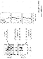
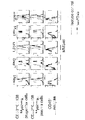
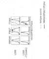
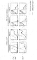
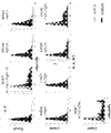
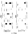
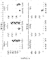
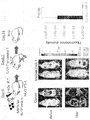
- Example 2 the result of having analyzed the binding of the MMG49 antibody with the bone marrow cell derived from a myeloma patient using FACS.
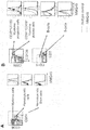
- the figure which shows the identification method of a myeloma progenitor cell fraction (Myeloma progenitor cells), a myeloma plasma cell fraction (Myeloma plasma cells), and a CD45 + white blood cell (CD45 + leukocytes).
- FIG. 1 The figure which shows the identification method of a myeloma progenitor cell fraction (Myeloma progenitor cells), a myeloma plasma cell fraction (Myeloma plasma cells), and a CD45 + white blood cell (CD45 + leukocytes).
- FIG. 2 The figure which shows the identification method of a myeloma progenitor cell fraction (Myeloma progenitor cells), a myeloma plasma cell fraction (Myeloma plasma cells
- Example 2 Results of FACS analysis of binding of MMG49 antibody to myeloma progenitor cell fraction, myeloma plasma cell fraction, and CD45 + leukocyte cells in multiple myeloma patient-derived bone marrow cells (UPN1-5) in Example 2
- FIG. 3 The identification process of the antigen protein which the MMG49 antibody recognizes by the expression cloning method in Example 3 is shown.
- the process of concentrating BaF3 cells bound to MMG49 antibody, which was initially 0.1% or less, by FACS sorting is shown.
- Example 4 shows the results of ITGB7 deficient U266 cells produced using the Crispa-cas9 system, and stained with MMG49 antibody or FIB27 antibody (commercially available anti-integrin beta 7 antibody) and FACS analysis.
- Example 4 a cell lysate derived from MM1s myeloma cells was immunoprecipitated using MMG49 antibody or isotype control antibody, followed by SDS-PAGE, followed by Western blotting with a commercially available anti-integrin ⁇ 7 antibody (Abcam). The figure which shows the result of having performed.
- MMG49 antibody, FIB27 antibody against each cell fraction of healthy human peripheral blood cells in the figure, B cell, T cell, monocyte, neutrophil, erythrocyte, and platelet are shown in order from the left), The figure which shows the result of having analyzed the coupling
- Example 5 the figure which shows the result of carrying out FACS analysis of the coupling
- the left figure shows a method for identifying each cell fraction
- the right figure shows the binding of MMG49 to each fraction.
- A shows a comparison of hematopoietic stem cell and progenitor cell fractions with myeloma cells
- B shows a comparison of B / T lymphocyte fractions with myeloma progenitor and myeloma plasma cell fractions.
- Example 6 the figure which shows the result of having analyzed the coupling
- ITGA4 in the cells shows the results obtained by FACS analysis.
- Example 7 the presence of Ca 2+ / Mg 2+ or Mn 2+, a 20-minute treatment with integrin alpha 4 beta 7 forcibly expressing K562 cells and human normal peripheral blood-derived T cells at 37 °C, MMG49 antibody or isotype
- the figure which shows the result of having dye
- FIG. 1 The figure which shows the presence or absence of the coupling
- FIG. The figure which put together the result shown in FIG. In the graph of the figure, the vertical axis represents the percentage of cells bound by the antibody, the horizontal axis represents the various human / mouse chimeric integrin beta 7 protein.
- Example 9 MMG49 antibody-derived CAR-T cells or T cells (control) into which GFP has been introduced, and K562 cells not expressing integrin ⁇ 7 or K562 cells forcibly expressing integrin ⁇ 4 ⁇ 7
- the amount of IFN- ⁇ produced by co-culture of MMG49 antibody-derived CAR-T cells or GFP-introduced T cells (control) and MMG49 antigen-expressing cells or non-expressing cells in Example 10 by ELISA The figure which shows the result of having quantified.
- the amount of IL2 produced by co-culture of MMG49 antibody-derived CAR-T cells or T cells introduced with GFP and MMG49 antigen-expressing cells or non-expressing cells in Example 10 was quantified by ELISA.
- the y-axis of the graph in a figure means a cytotoxic rate (%).
- Example 12 shows the presence or absence of binding of MMG49 antibody it the construction of human / mouse chimeric integrin beta 7 protein to 293T cells were transiently expressed.
- the MFI on the horizontal axis represents the binding strength to the MMG49 antibody, and the higher the value, the higher the binding strength.
- Myeloma progenitor cells are progenitor cells that have not yet differentiated into myeloma plasma cells. CD38 is strongly expressed, but there is no expression of CD138, a marker specific for mature plasma cells. Characterized. Therefore, myeloma progenitor cells may be referred to as “CD38 ++ CD138 ⁇ cells” or “CD19 ⁇ CD38 ++ CD138 ⁇ cells”.
- Myeloma plasma cells are generally called myeloma cells and are cells that produce M protein, which is an abnormal immunoglobulin.
- CD138 is expressed in addition to the strong expression of CD38. Therefore, myeloma plasma cells may be referred to as “CD38 ++ CD138 + cells” or “CD19 ⁇ CD38 ++ CD138 + cells”.
- Myeloma progenitor cells and myeloma plasma cells also mean tumor progenitor cells and neoplastic plasma cells in diseases that cause neoplastic growth of plasma cells other than multiple myeloma, respectively.
- Hematopoietic progenitor cells are cells that can differentiate into various blood cells. Hematopoietic progenitor cells are characterized by the expression of CD34. Therefore, in the present specification, hematopoietic progenitor cells may be referred to as “CD34 + cells”.
- the antibody (I) is preferably an anti-human integrin ⁇ 7 antibody having an epitope in a region consisting of amino acid residues 20 to 109 of human integrin ⁇ 7 .
- an antibody with an epitope in the region consisting of 20 to 90 amino acid residues of an antibody or human integrin beta 7 with an epitope in the region consisting of 33 to 109 amino acid residues of the human integrin beta 7 Can do.
- Human integrin ⁇ 7 is not particularly limited, and is a transmembrane protein having the amino acid sequence shown in SEQ ID NO: 31, and can be a protein that forms a heterodimer with integrin ⁇ .
- Specific integrin alpha mention may be made of integrin alpha 4 or integrin alpha E.
- the specific amino acid sequence of human integrin ⁇ 7 is, for example, listed in the NCBI database in addition to the amino acid sequence shown in SEQ ID NO: 31, ACCESSION: EAW96675; VERSION: EAW96675.1, GI: 119617081, ACCESSION: NM000889; VERSION : NM000889.2, GI: 540344585, ACCESSION: XM005268851, VERSION: XM005268851.2, GI: 767974096, ACCESSION: XM006719376, VERSION: XM006719376.2, GI: 767974098, ACCESSION: XM005268852, VERSION: XM005268852.3, GI: 767974097 And the like.
- human integrin ⁇ 7 will be based on the amino acid sequence shown in SEQ ID NO: 31, but if it is an amino acid sequence of other human integrin ⁇ 7 , homology with the amino acid sequence shown in SEQ ID NO: 31 was confirmed in silico. Accordingly, those skilled in the art can easily determine which region or site of the human integrin ⁇ 7 amino acid sequence described below corresponds to the region and / or site of human integrin ⁇ 7 described below. it can.
- Region consisting of 1-19 amino acid residues of the human integrin beta 7 is the signal peptide is a peptide fragment not present when functioning as a membrane protein in vivo.
- N-terminal when the human integrin beta 7 to function as a membrane protein is a 20 amino acid residue of the amino acid sequence.
- the region consisting of 20 to 109 amino acid residues of the human integrin beta 7 includes PSI domain.
- the homology between the PSI domain of human integrin ⁇ 7 and the PSI domain of mouse integrin ⁇ 7 is known to be as high as approximately 80% or more, 20-109 containing the PSI domains of human integrin ⁇ 7 and mouse integrin ⁇ 7 comparing the amino acid residues in the region consisting of amino acid residues, as also shown in FIG.
- the epitope of antibody (I) is preferably related to any one or more of these 15 amino acid residues, preferably 2 or more, more preferably 3 or more.
- the epitope of antibody (I) is preferably present in the region consisting of amino acid residues 23 to 109 of human integrin ⁇ 7 and is preferably a region consisting of amino acid residues 23 to 48 or 93 to More preferably, it exists in the region consisting of the 109th amino acid residue.
- the epitope of antibody (I) of another more preferred embodiment is a region consisting of amino acid residues 23 to 48, a region consisting of amino acid residues 93 to 109, or a region consisting of amino acid residues 23 to 48 And a three-dimensional region combining regions consisting of the 93rd to 109th amino acid residues.
- the epitope of antibody (I) may be a linear epitope or a three-dimensional epitope (also referred to as a non-linear epitope).
- a linear epitope is a case where consecutive amino acid residues are epitopes, and a steric epitope is known to those skilled in the art as an epitope composed of non-contiguous amino acid residues.
- the 48th amino acid residue is strongly related as the epitope of antibody (I) or contained in the epitope of antibody (I).
- antibody (I) is an antibody which specifically binds to a region consisting of 20 to 109 amino acid residues of the human integrin beta 7, which specifically bind to inter alia 23-109 th region It is preferable to specifically bind to regions 23 to 48 and / or 93 to 109.
- the property of binding to a region consisting of 20 to 109 amino acid residue of the integrin beta 7 is the epitope of an antibody (I), it is sometimes referred to as affinity for epitopes.
- affinity for epitopes the term “increasing affinity for an epitope” is equivalent to “increasing specific binding ability to an epitope”.
- the affinity for the epitope of the antibody (I) rises in the presence of at least a region consisting of 379 to 722 amino acid residues of the human integrin beta 7 It is preferable that
- At least part of the region consisting of amino acid residues 379 to 722 means that the region may be a region consisting of amino acid residues 379 to 722 or a part thereof. . Specifically as "a part of the region", for example, at least a region consisting of 417 to 722 amino acid residues of the human integrin beta 7, consists of 564-722 amino acid residues of the human integrin beta 7 At least part of the region, at least a region consisting of 379 to 563 amino acid residues of the human integrin beta 7, at least a region consisting of 417 to 563 amino acid residues of the human integrin beta 7, or human at least a region consisting of 379 to 416 amino acid residue of the integrin beta 7 and the like. That is, the affinity of the antibody (I) for the epitope can be increased in the presence of these regions.
- the term "in the presence” is a region consisting of 20 to 109 amino acid residues of the human integrin beta 7, and at least a portion of a region consisting of 379 to 722 amino acid residues of the human integrin beta 7, It can exist in the same molecule, and both regions can exist in separate molecules. Preferably, both regions are present in the same molecule. Note that the term “in the presence” can be read as “by”.
- a chimeric integrin beta 7 proteins for various human / mouse shown in Example 8 comprises a region consisting of 1-109 amino acid residues of the human-derived integrin beta 7, and, integrin from human beta 7
- preparing a chimeric integrin beta 7 protein of human / mouse substituted the region consisting of amino residues residues in the region consisting of 379 to 721 th amino remaining residue of the integrin beta 7 from mouse (# 4961).
- the degree of binding of the antibody (I) is compared with the cell expressing the latter (# 4961) and the cell expressing the former (# 4960), so that the affinity of the antibody (I) for the above epitope Can be confirmed.
- affinity for the epitope of the antibody (I) is preferably shall be increased by activating the human integrin beta 7. Since activated human integrin ⁇ 7 has structural characteristics in the region containing the epitope, it is considered that the affinity of the antibody (I) for the epitope is increased.
- human integrin beta 7 are known.
- cells expressing the human integrin beta 7, for example, plasma cells, NK cells, T cells, B cells, lymphoblasts, derived from Burkitt's lymphoma cells, either blood cells or immune cells such as dendritic cells By causing a phorbol ester such as PMA or a manganese salt to act on cells, human integrin ⁇ 7 expressed in the cells can be activated.
- a phorbol ester such as PMA or a manganese salt
- the human integrin beta 7 be treated such as by manganese salts can be activated.
- Affinity for the epitope of the antibody (I) is to be raised by activating the human integrin beta 7 is by conventional immunoassays described like in the examples below, there those skilled in the art It can be easily confirmed.
- a chimeric integrin beta 7 proteins for various human / mouse shown in Example 8 prepare the cells expressing # 4960 or # 4961 comprises a region consisting of 1-109 amino acid residues, it After being subjected to the integrin ⁇ 7 activation means as shown in Example 7 and then using an immunological measurement means, the antibody to the cells after activation is compared by comparing before and after the activation treatment. An increase in affinity of (I) for the epitope can be confirmed.
- Another aspect of the antibody (I) is characterized in that it has a higher affinity for human integrin ⁇ 7 expressed on cells derived from myeloma than human integrin ⁇ 7 expressed on normal cells. it can be an anti-human integrin beta 7 antibodies.
- the normal cell is not particularly limited as long as it is a cell derived from a healthy person.
- it can be a normal cell derived from blood, and it is preferable to use a normal plasma cell among such normal cells.
- Examples of methods for confirming higher affinity for human integrin ⁇ 7 expressed on myeloma cells than human integrin ⁇ 7 expressed on normal cells include the following examples. Can be easily carried out by those skilled in the art by the conventional immunoassay methods described in 1).
- the “conventional immunoassay” is not particularly limited as long as it is a method using various antibodies regardless of the antigen.
- a flow cytometry method FACS
- cell sorting FACS
- Western blotting ELISA
- immunoprecipitation method SPR method
- QCM method QCM method and the like associated therewith can be mentioned.
- the antibody (I) it is preferable that it has the same epitope as the MMG49 antibody disclosed in Examples described later. Most preferably, it is the same antibody as the MMG49 antibody.
- the following Examples can be referred to for the production method of MMG49 antibody.
- an antibody having an aspect including a heavy chain variable region and / or a light chain variable region is preferable. That is, the antibody (I) can be a heavy chain variable region alone or a light chain variable region alone. Preferably, the antibody includes a heavy chain variable region and a light chain variable region.
- variable region is also called an antigen recognition site and is understood by those skilled in the art as an important site for the antibody to recognize an antigen.
- a variable region has three regions called hypervariable regions (also called complementarity determining regions [CDRs]), and these CDRs are the most important regions most involved in the antigen recognition function of antibodies. It is also known to those skilled in the art.
- the heavy chain variable region included in another embodiment of the antibody (I) includes one or more of heavy chain CDR1, heavy chain CDR2, and heavy chain CDR3. That is, the heavy chain variable region can contain heavy chain CDR1, heavy chain CDR2, or heavy chain CDR3 alone, and preferably contains at least heavy chain CDR3. More preferred is an embodiment comprising a heavy chain CDR1, a heavy chain CDR2, and a heavy chain CDR3 in this order from the amino terminus (N terminus).
- the light chain variable region can also be the same as the heavy chain variable region, and includes, for example, any of light chain CDR1, light chain CDR2, or light chain CDR3, and preferably includes at least light chain CDR3. It is preferable that a light chain CDR1, a light chain CDR2, and a light chain CDR3 are included in this order from the N-terminus of the chain variable region.
- regions other than the respective CDR1 to 3 may be referred to as FR. More specifically, the region between the N-terminus and CDR1 is FR1, the region between CDR1 and CDR2 is FR2, the region between CDR2 and CDR3 is between FR3, and the CDR3 and carboxy terminus (C-terminus). This region is called FR4, and is a name set in each of the heavy chain variable region and the light chain variable region.
- the amino acid sequences of the heavy chain CDR1 to 3 and the light chain CDR1 to 3 are not particularly limited.
- the heavy chain CDR1-3 or light chain CDR1-3 of the MMG49 antibody Heavy chain CDR1, having the amino acid sequence set forth in SEQ ID NO: 1, Heavy chain CDR2 having the amino acid sequence shown in SEQ ID NO: 2, Heavy chain CDR3 having the amino acid sequence shown in SEQ ID NO: 3, A light chain CDR1 having the amino acid sequence shown in SEQ ID NO: 6, A light chain CDR2 having the amino acid sequence set forth in SEQ ID NO: 7, A light chain CDR3 having the amino acid sequence set forth in SEQ ID NO: 8, Etc.
- the heavy chain variable region containing the heavy chain CDRs 1 to 3 for example, a heavy chain variable region having the amino acid sequence shown in SEQ ID NO: 4 which is the heavy chain variable region of the MMG49 antibody can be mentioned.
- a preferred embodiment of the light chain variable region comprising the above light chain CDRs 1 to 3 is, for example, the light chain variable region having the amino acid sequence shown in SEQ ID NO: 9, which is the light chain variable region of the MMG49 antibody.
- amino acid sequences of the MMG49 antibodies shown in SEQ ID NOs: 1 to 4 and 6 to 9 are as shown in Table 1 below.
- the underlined portions provided in the amino acid sequences of the heavy chain and variable region shown in SEQ ID NOs: 4 and 9, respectively, in the table indicate portions located in CDR1, CDR2, and CDR3 in order from the N-terminus.
- antibody (I) is not limited. Specific structures include Fv, scFv, diabody, triabody, tetrabody, and the like, and a structure in which these are appropriately combined can also be used. In addition, these combined structures are sometimes referred to as fragment antibodies. Such a fragment antibody can be an artificially designed recombinant protein containing Fv or can be fused with a biomolecule such as a protein.
- Fv is also called the minimum structural unit of an antibody, and is a structure in which a heavy chain variable region and a light chain variable region are associated by non-covalent intermolecular interaction. Furthermore, a structure in which thiol groups of cysteine residues present in the heavy chain variable region and the light chain variable region are disulfide bonded to each other can also be used.
- ScFv is a structure in which the C-terminus of the heavy chain variable region and the N-terminus of the light chain variable region are connected by a linker, and is also called a single-chain antibody. Further, the C-terminal and N-terminal connected by a linker may be reversed.
- the structure of scFv can also be formed by association such as non-covalent intermolecular interaction like Fv.
- the above-mentioned scFv forms a dimer, a trimer, and a tetramer, respectively.
- Fv non-covalent intermolecular interactions between variable regions It is a structure that is associated in the most structurally stable state.
- the antibody (I) having such various structures is constructed by using conventional genetic engineering means to construct an expression vector, and prokaryotic cells (such as E. coli, actinomycetes, etc.) suitable for the production of the antibody. ), Expression systems employing host cells such as eukaryotic cells (yeast cells, insect cells, mammalian cells, etc.), commonly used cell-free expression systems, etc., can be easily produced by those skilled in the art. it can. The produced antibody can also be obtained in a highly purified state by appropriately subjecting it to a conventional purification step.
- prokaryotic cells such as E. coli, actinomycetes, etc.
- Expression systems employing host cells such as eukaryotic cells (yeast cells, insect cells, mammalian cells, etc.), commonly used cell-free expression systems, etc., can be easily produced by those skilled in the art. it can.
- the produced antibody can also be obtained in a highly purified state by appropriately subjecting it to a conventional purification step.
- a constant region can be contained. It will be understood by those skilled in the art that the constant region includes CH1, CH2, and CH3 if it is a heavy chain constant region and CL if it is a light chain constant region. A region containing CH2 and CH3 may also be called an Fc domain.
- the origin of the specific constant region is not particularly limited.
- animal species that can tolerate mass production such as human origin, mouse origin, rat origin, rabbit origin, monkey origin, chimpanzee origin, animal species closely related to humans, animals that are unlikely to produce immunogenicity even when administered to humans
- constant regions derived from species and the like may be mentioned.
- the antibody (I) when the heavy chain variable region and / or the light chain variable region has a mouse-derived amino acid sequence, the antibody (I) is combined with a chimeric antibody by combining, for example, human-derived constant regions. can do.
- the antibody (I) can be converted into a humanized antibody by substituting the heavy chain FR1-4 and / or the light chain FR1-4 with a human-derived amino acid sequence in the above-described chimeric antibody.
- the antibody (I) is humanized. It can be an antibody.
- human antibody is sometimes referred to as “fully humanized antibody”.
- the structure of the antibody (I) of the embodiment containing a constant region includes a heavy chain having a heavy chain variable region and a heavy chain constant region; and a pair of light chains having a light chain variable region and a light chain constant region, respectively.
- Examples include not only immunoglobulins having the structure of this chain, but also Fab, F (ab ′) 2 , minibody, scFv-Fc and the like. Furthermore, it can also be set as the structure which combined these suitably. In addition, these combined structures are sometimes referred to as fragment antibodies.
- Such a fragment antibody can be an artificially designed recombinant protein containing Fv or can be fused with a biomolecule such as a protein.
- Fab includes a heavy chain fragment comprising CH1 in the heavy chain variable region and the heavy chain constant region, and a light chain comprising the light chain variable region and the light chain constant region, the heavy chain variable region and the light chain variable region, Are associated by the non-covalent intermolecular interaction described above or have a structure formed by bonding by a disulfide bond. Furthermore, CH1 and CL can be formed by disulfide bonding between thiol groups of cysteine residues present in each of them.
- F (ab ′) 2 has a pair of the above-mentioned Fabs, and CH 1 has a structure formed by disulfide bonding between thiol groups of cysteine residues contained therein.
- a minibody has a pair of antibody fragments containing the above scFv and CH3, and such antibody fragments have a structure in which CH3 are associated with each other by non-covalent intermolecular interaction.
- scFv-Fc has a pair of antibody fragments containing the above scFv, CH2, and CH3, and associates with each CH3 by non-covalent intermolecular interaction between CH3 as in the above minibody. It has a structure formed by disulfide bonding between thiol groups of cysteine residues contained.
- the antibody (I) containing constant regions having such various structures is also constructed by using conventional genetic engineering means to construct an expression vector in the same manner as the antibody (I) containing no constant region.
- Those skilled in the art can easily produce a vector by using an expression system that employs a host cell suitable for antibody production.
- the produced antibody can also be obtained in a highly purified state by appropriately subjecting it to a conventional purification step.
- IgG which is an immunoglobulin
- F (ab ′) 2 can also be obtained by decomposing IgG using a protease such as pepsin.
- an immunoglobulin a preferred structure is an immunoglobulin.
- immunoglobulin subtypes are not particularly limited, and examples thereof include IgA, IgD, IgE, IgG, and IgM.
- IgG is preferable.
- IgG2 is preferable among the four subclasses.
- the antibody of a more preferred embodiment is an antibody comprising a heavy chain having the amino acid sequence shown in SEQ ID NO: 5 and / or a light chain having the amino acid sequence shown in SEQ ID NO: 10.
- the most preferred antibody is an antibody comprising a heavy chain having the amino acid sequence shown in SEQ ID NO: 5 and a light chain having the amino acid sequence shown in SEQ ID NO: 10.
- the above-mentioned amino acid sequence can be introduced with mutations depending on the situation.
- Such mutations are preferably not made to the heavy and light chain CDRs. That is, it is preferably applied to the heavy chain FR and the light chain FR, and when the antibody (I) contains a constant region, in addition to the mutation for adjusting ADCC activity or CDC activity shown below, further mutation Can be applied.
- the number of amino acid residues subjected to specific mutagenesis is not particularly limited.
- the identity between the amino acid sequence before mutagenesis and the amino acid sequence after mutagenesis is about 70%, preferably about 75%, more preferably about 80%, more preferably about 85%, more preferably about 90%, More preferably, it is about 95%, more preferably about 96%, more preferably about 97%, more preferably about 98%, and most preferably about 99%.
- such a numerical value shall be obtained by rounding off.
- identity refers to the degree of identical amino acid sequences of two or more comparable amino acid sequences relative to each other. Therefore, it can be said that the higher the identity of two amino acid sequences, the higher the similarity as well as the identity of those sequences.
- the amino acid identity can be calculated using commercially available analysis tools (for example, software such as FASTA, BLAST, PSI-BLAST, SSEARCH, etc.) available through the Internet.
- main initial conditions generally used for BLAST search are as follows. That is, in Advanced BLAST 2.1, blastp is used for the program, Expect value is 10, Filter is OFF, BLOSUM62 is used for Matrix, Gap existence cost, Per residue gap cost, and Lambda ratio are 11, 1, 0.85, respectively.
- the amino acid sequence identity value (%) can be calculated.
- the above-described mutation introduction into the amino acid sequence includes substitution, deletion, insertion and the like.
- Specific mutagenesis is not particularly limited as long as it can be achieved by employing a conventional method. For example, a conservative replacement technique may be employed for replacement.
- conservative substitution technique means a technique in which an amino acid residue is substituted with an amino acid residue having a similar side chain.
- substitution with amino acid residues having basic side chains such as lysine, arginine, histidine and the like is a conservative substitution technique.
- amino acid residues having acidic side chains such as aspartic acid and glutamic acid
- amino acid residues having uncharged polar side chains such as glycine, asparagine, glutamine, serine, threonine, tyrosine and cysteine
- alanine, valine Amino acid residues having non-polar side chains such as leucine, isoleucine, proline, phenylalanine, methionine and tryptophan
- amino acid residues having ⁇ -branched side chains such as threonine, valine and isoleucine
- tyrosine, phenylalanine, tryptophan Substitution with amino acid residues having an aromatic side chain such as histidine is also a conservative substitution technique.
- the antibody (I) can have cytotoxic activity.
- the cytotoxic activity refers to an activity that causes some damage to the bound cells as a result of binding of the antibody to the cells.
- ADCC activity is an abbreviation for antibody-dependent cytotoxicity (antibody-dependent cellular cytotoxicity), and cells having cytotoxic activity such as NK cells expressing a receptor specific for the constant region of an antibody. Is induced in the vicinity of the antibody, and induces damage to the cell to which the antibody binds by the action of such a cell or the like.
- CDC activity is an abbreviation of complement-dependent cytotoxicity (Complement-Dependent Cytotoxicity), in which an antibody recruits complement in the vicinity and the antibody is bound by the action of such complement. It refers to the activity that induces an action that damages cells.
- ADCC activity and CDC activity are Lazar GA et al., Proc Natl Acad Sci USA, 103: 4005-10 (2006), Shields RL et al., J Biol Chem, 276: 6591-604 (2001)) , Moore GL et al., J Immunol, 159: 3613-21 (1997), An Z et al., MAbs, 1: 572-9 (2009) The activity can be adjusted.
- the constant region is a human IgG 1
- S298A, K334A, S298A / K334A by applying a mutation such S298A / E333A / K334A, ADCC activity Can be raised.
- the constant region is a human IgG 1, V234A / G237A, by applying a mutation such H268Q / V309L / A330S / P331S, can be lowered ADCC activity.
- the constant region is human IgG 1
- the activity is increased if mutations such as S267E, H268F, S324T, S267E / H268F, S267E / S324T, H268F / S324T, S267E / H268F / S324T are performed. be able to.
- ADCC activity can be measured according to the method of Brunner KT et al. (Brunner, KT, et al., Immunology, 1968. 14: 181-96).
- myeloma cells are cultured in RPMI1640 medium supplemented with 10% FCS and prepared so that the number of cells is 0.5 ⁇ 10 4 to 1.0 ⁇ 10 4 .
- An appropriate amount of Na 2 51 CrO 4 is added to this, reacted at 37 ° C. for 1 hour, the cells are labeled with 51 Cr, and the washed cells are used as target cells.
- SCID mouse bone marrow cells cultured in RPMI1640 supplemented with 10% FBS, 10 ng / ml mouse GM-CSF, and 40 IU / ml human IL2 can be used.
- the reaction is carried out at 37 ° C. for 4 hours, and after centrifugation, 51 Cr released into the supernatant is measured with a ⁇ -counter.
- ADCC activity can be determined based on the following equation.
- ADCC activity ⁇ ([ 51 Cr release from target cells] ⁇ [Spontaneous 51 Cr release in the absence of antibody]) / ([Maximum 51 Cr release by addition of 1% Triton X-100] ⁇ [Antibody Spontaneous 51 Cr release in the absence of]] ⁇ ⁇ 100
- CDC activity can also be measured according to the method of Brunner KT et al. (Brunner, KT, et al., Immunology, 1968. 14: 181-96).
- myeloma cells as target cells are cultured in RPMI1640 medium supplemented with 10% FCS and prepared so that the number of cells is 0.5 ⁇ 10 4 to 1.0 ⁇ 10 4 .
- An appropriate amount of Na 2 51 CrO 4 is added to this, reacted at 37 ° C. for 1 hour, the cells are labeled with 51 Cr, and the washed cells are used as target cells.
- test antibody or control isotype antibody suspended in RPMI1640 medium supplemented with fetal bovine serum to a 96-well plate to a final concentration of 0.5-50 ⁇ g / mL, then add the target cells and complement. Let react for 1.5 hours. The reaction solution is centrifuged, and 51 Cr released into the supernatant is measured with a ⁇ -counter. CDC activity can be determined based on the following formula.
- CDC activity ⁇ ([ 51 Cr release from target cells] ⁇ [Spontaneous 51 Cr release in the absence of antibody]) / ([Maximum 51 Cr release by addition of 1% Triton X-100] ⁇ [Antibody Spontaneous 51 Cr release in the absence of]] ⁇ ⁇ 100
- An antibody having cytotoxic activity can be obtained, for example, by evaluating the presence or absence of cytotoxic activity using the above-described method and selecting an antibody having the activity.
- a multispecific antibody can be used. That is, it can have an ability to bind with specificity to an antigen other than the region consisting of amino acid residues 20 to 109 of human integrin ⁇ 7 (hereinafter referred to as other antigen).
- an antigen is a region structurally dissimilar consisting 20-109 amino acid residues of the human integrin beta 7. .
- the specific other antigen is not particularly limited.
- CD3, CD16, C1q, Adenovirus knob domain and the like can be mentioned, and at least one of them can be appropriately used as another antigen by appropriately combining them.
- one of the antigens exemplified above is selected as the other antigen. That is, a preferred multispecific antibody is a bispecific antibody.
- Such multispecific antibodies can be easily produced by those skilled in the art by appropriately adopting conventional techniques.
- peptide fragments corresponding to the region consisting of 20 to 109 amino acid residues of the human integrin beta 7, or only a region consisting of 20 to 109 amino acid residues of the human integrin beta 7 was derived from a human, the rest prepare the hybridomas produced using the antibody-producing cells such as B cells obtained cells expressing chimeric integrin beta 7 which was derived from a non-human, such as mouse-derived from immunized animals, other above
- a new hybridoma obtained by separately preparing a hybridoma using antibody-producing cells such as B cells obtained from an animal immunized with an antigen, and fusing these hybridomas together (in the case of creating a bispecific antibody) In some cases, this is also referred to as quadroma.)
- a multispecific antibody is obtained. Door can be.
- bispecific antibodies (1) An antibody having the above F (ab ′) 2 structure is prepared using the region consisting of amino acid residues 20 to 109 of human integrin ⁇ 7 as an epitope: (2) On the other hand, an antibody having a structure of F (ab ′) 2 that specifically binds to another antigen is also prepared: (3) After treating each of the F (ab ′) 2 structure antibodies obtained in (1) and (2) with a reducing agent such as DTT, one of the treated products is further subjected to Elman reagent. Process with: (4) These F (ab ′) 2 structure antibodies after the treatment obtained in (3) are mixed and reacted: Bispecific antibodies can also be prepared by the procedures shown in (1) to (4).
- Bispecific antibodies can also be produced by the procedures shown in (A) to (D).
- a cytotoxin (a substance having cytotoxic activity) can be bound.
- the cytotoxin is not particularly limited as long as it is a substance that causes some damage to the cell, such as killing the cell or suppressing cell proliferation.
- cytotoxins include, for example, alkylating agents such as cyclophosphamide hydrate, ifosfamide, thiotepa, busulphalan, melphalan, nimustine hydrochloride, ranimustine, dacarpazine, temozolomide; methotrexate, pemetrexed sodium hydrate, fluorouracil , Doxyfluridine, capecitabine, tagafur, cytarabine, gemcitabine hydrochloride, fludarabine phosphate ester, nelarabine, cladribine, levofolinate calcium, etc.
- alkylating agents such as cyclophosphamide hydrate, ifosfamide, thiotepa, busulphalan, melphalan, nimustine hydrochloride, ranimustine, dacarpazine, temozolomide; methotrexate, pemetrexed sodium hydrate, fluorouracil ,
- Amrubicin hydrochloride mitoxantrone hydrochloride, mitomycin C, actinomycin D, bleomaciin hydrochloride, Antibiotics such as romasin hydrochloride, dinostatin stimamarer, calicheamicin, microtubule inhibitors such as vincristine sulfate, vinblastine sulfate, vindesine sulfate, paclitaxel; anastrozole, exemestane, letrozole, fadrazole hydrochloride water Aromatase inhibitors such as Japanese; platinum preparations such as cisplatin, carboplatin, nedaplatin, oxaliplatin; topoisomerase inhibitors such as irinotecan hydrochloride hydrate, nogitecan hydrochloride, etoposide, sobuzoxane, corticosteroids such as prednisolone, dexamethasone, thalidomide
- calicheamicin, melphalan, vincristine sulfate, doxorubicin hydrochloride, prednisolone, dexamethasone, thalidomide, lenalidomide, and bortezomib are preferable, and calicheamicin that has a proven record of binding to an antibody is more preferable. It is.
- cytotoxins are all commercially available and can be selected from one or more of the above-mentioned combinations as appropriate.
- the binding mode between the cytotoxin and the above-described antibody is not particularly limited.
- a conventional genetic engineering technique or protein engineering technique those skilled in the art can easily bind the cytotoxin to the above-described antibody. it can. More specifically, a method of binding to a functional group such as an amino group, a thiol group, a guanidyl group, a hydroxyl group or a carboxyl group of the amino acid residue side chain of the antibody (I) through a linker can be mentioned.
- Antibody (I) may be a polyclonal antibody or a monoclonal antibody.
- a monoclonal antibody is preferable.
- monoclonal means obtained from a substantially homogeneous population
- “monoclonal antibody” means an antibody obtained from such a population. That is, it is understood that the individual antibodies contained in such a population are identical except for possible naturally occurring mutations that may be present in trace amounts.
- an antibody specific binding target for example, which in antibody (I) are those present in the region consisting 20-109 amino acid residues of the human integrin beta 7, if a polyclonal antibody , This is a multiple site in the region consisting of amino acid residues 20-109 of human integrin ⁇ 7 , whereas a monoclonal antibody exhibits high specificity in that it is a single site So it is more advantageous.
- “monoclonal” used as a modifier is understood as a product obtained from a substantially uniform population as described above, and should not be interpreted as a modifier for which the production method is specified.
- antibody (I) adopts hybridoma method, recombinant DNA method using host cell (III) holding polynucleotide (II) shown below, isolation from phage library, etc. If it is those skilled in the art, it can manufacture easily.
- the hybridoma method is used after immunizing an animal suitable for antibody production such as mouse, rat, rabbit, etc. with a peptide corresponding to the region consisting of amino acid residues 20 to 109 of human integrin ⁇ 7 and then recovering B cells. And a method for producing the antibody (I) by screening using the function exhibited by the antibody (I) described above as an index.
- immunize an animal (preferably mouse) suitable for antibody production such as mouse, rat, rabbit, etc., and then collect B cells and use it for hybridoma method to demonstrate the above-mentioned antibody (I)
- a method for producing the antibody (I) can be exemplified by screening using the function to be performed as an index.
- the antibody (I) can also be obtained by the method shown in the following screening method (X) using such a function.
- Antibodies (I) have an epitope in the region consisting of 20 to 109 amino acid residues of the human integrin beta 7, to cells expressing the integrin beta 7, not only the ADCC activity and CDC activity above, By combining one or more of apoptosis-inducing activity, survival signal blocking activity, and the like, it is expected to exert cytotoxic activity on such cells. Therefore, the composition containing antibody (I) is useful as pharmaceutical composition (IV) as described in detail below.
- antibody (I) has a region consisting of amino acid residues 20 to 109 of human integrin ⁇ 7 as an epitope, but affinity of antibody (I) for the epitope activates integrin ⁇ 7 To rise. Since active integrin ⁇ 7 is expressed in blood cells such as plasma cells, antibody (I) is used as an active ingredient in pharmaceutical compositions for these cancers (eg, blood cancer). In particular, it is effectively used as a pharmaceutical composition for diseases (eg, myeloma, multiple myeloma, etc.) that cause abnormalities in the above cells.
- diseases eg, myeloma, multiple myeloma, etc.
- Polynucleotide (II) is a polynucleotide having a base sequence encoding the amino acid sequence of the antibody (I).
- the term “polynucleotide” includes, for example, a single-stranded or double-stranded form in which ribonucleotides, deoxyribonucleotides, or any of these nucleotides are appropriately modified by a known method.
- the nucleotide sequence of the polynucleotide (II) can be appropriately determined by those skilled in the art, for example, in silico based on the amino acid sequence of the antibody (I).
- the kind of codon used for determining such a base sequence is not ask
- the specific nucleotide sequence of the polynucleotide (II) is not particularly limited.
- Table 2 below shows the correspondence between each SEQ ID NO indicating an amino acid sequence specified as one of the embodiments of the antibody (I) and a SEQ ID NO indicating a base sequence encoding the amino acid sequence. That is, preferred base sequences possessed by the polynucleotide (II) are those shown in SEQ ID NOs: 11 to 20.
- Polynucleotide (II) may be incorporated in a vector.
- the vector is not particularly limited, and for example, it can be a cloning vector or an expression vector, and its use is not limited.
- it can be a vector for prokaryotic cells such as Escherichia coli and actinomycetes, and a vector for eukaryotic cells such as yeast cells, insect cells, and mammalian cells.
- prokaryotic cells such as Escherichia coli and actinomycetes
- eukaryotic cells such as yeast cells, insect cells, and mammalian cells.
- nucleotide sequence encoding a signal peptide can be appropriately added to the 5 ′ terminal side of the polynucleotide (II) (N terminal side in the above antibody (I)).
- the specific method of using the polynucleotide (II) is not particularly limited. Examples thereof include introduction into the following host cell (III) and use for expression of the antibody (I).
- the host cell (III) is a cell that retains the polynucleotide (II).
- retention refers to maintaining the state in which the polynucleotide (II) is present in the cell, and the state in which the cell does not spontaneously excrete the polynucleotide regardless of whether it is positive or not. It means that.
- the mode in which the host cell (III) holds the polynucleotide (II) is not particularly limited.
- the polynucleotide can be retained in the form of a vector in the cell, or the polynucleotide (II) can be retained in an integrated form in the genome of the cell.
- Specific cell types of the host cell (III) may be eukaryotic cells such as yeast cells, insect cells and mammalian cells, or prokaryotic cells such as Escherichia coli and actinomycetes, and are not particularly limited.
- a chimeric antigen receptor is an artificial T-cell receptor (TCR) -like protein that expresses an antigen recognition site (corresponding to the extracellular domain) expressed on the cell membrane of T cells. It is a protein constructed so that it can be replaced with a desired antigen recognition site, and functions such as cytotoxic activity of T cells themselves can be more effectively exhibited.
- TCR T-cell receptor
- Chimeric antigen receptor (IV) has the same epitope as antibody (I), and more specifically is a protein containing the antigen recognition site of antibody (I). That is, the epitope present in the antigen recognition site contained in the chimeric antigen receptor can be the same as that described in detail for the antibody (I).
- the antigen recognition site of the antibody (I), spacer sequence, transmembrane domain, costimulatory factor, and intracellular domain of TCR are arranged in order from the N-terminus of the chimeric antigen receptor (IV). It is a protein.
- the antigen recognition site of the antibody (I) arranged in the chimeric antigen receptor (IV) can be as described in detail for the antibody (I), specifically, the heavy chain variable region and / or A light chain variable region can be mentioned. Of these, the scFv structure is preferable while having a heavy chain variable region and a light chain variable region.
- a spacer sequence composed of about 10 to 25 amino acid residues can be appropriately provided between the heavy chain variable region and the light chain variable region. More preferably, the number is about 15 to 18.
- Such a spacer sequence may be the same as or different from the above-described spacer sequence arranged in the chimeric antigen receptor (IV).
- the spacer sequence arranged in the chimeric antigen receptor (IV) is not particularly limited.
- it can be composed of about 10 to 25 amino acid residues. More preferably, the number is about 15 to 18.
- the transmembrane domain arranged in the chimeric antigen receptor (IV) is not particularly limited. Specifically, a transmembrane domain derived from a protein such as CD28 or 4-1BB expressed in T cells or the like can be employed while allowing appropriate mutagenesis.
- the costimulatory factor arranged in the chimeric antigen receptor (IV) is not particularly limited as long as it is a costimulatory factor possessed by T cells and the like.
- 4-1BB, OX40, CD28, etc. can be employed while allowing appropriate mutagenesis.
- the intracellular domain of TCR arranged in the chimeric antigen receptor (IV) is not particularly limited.
- an intracellular domain derived from CD3 or the like, also called a TCR ⁇ chain can be employed while allowing appropriate mutagenesis.
- the mutation introduction into CD3 is preferably performed so that ITAM (ImmunoreceptorceptTyrosine-based Activation Motif) is included.
- the chimeric antigen receptor (IV) preferably has the amino acid sequence shown in SEQ ID NO: 21.
- amino acid sequence specifying the above chimeric antigen receptor can be appropriately mutagenized. Moreover, the same can be said for the introduction of mutations into the transmembrane domain, costimulatory factor, and intracellular domain of TCR.
- the number of specific mutation introductions is not particularly limited.
- the identity between the amino acid sequence before mutagenesis and the amino acid sequence after mutagenesis is about 70%, preferably about 75%, more preferably about 80%, more preferably about 85%, more preferably about 90%, More preferably, it is about 95%, more preferably about 96%, more preferably about 97%, more preferably about 98%, and most preferably about 99%.
- such a numerical value shall be obtained by rounding off.
- the above-described mutation introduction into the amino acid sequence includes substitution, deletion, insertion and the like.
- Specific mutagenesis is not particularly limited as long as it can be achieved by employing a conventional method. For example, a conservative replacement technique may be employed for replacement.
- V Polynucleotide
- the polynucleotide (V) is a polynucleotide encoding the amino acid sequence of the chimeric antigen receptor (IV), unlike the polynucleotide (II).
- the base sequence of the polynucleotide (V) can be determined as appropriate based on the amino acid sequence of the chimeric antigen receptor (IV) in silico, for example, in the same manner as the polynucleotide (II).
- the type of codon used for determining the base sequence is not limited. It is preferable to determine the base sequence in consideration of the codon frequency of the cells to be used with the polynucleotide.
- the specific base sequence is not particularly limited.
- a polynucleotide having the base sequence shown in SEQ ID NO: 22 determined based on the amino acid sequence of the chimeric antigen receptor (IV) having the amino acid sequence shown in SEQ ID NO: 21 can be mentioned.
- the base sequence determined based on such an amino acid sequence is not limited to the base sequence shown in SEQ ID NO: 22 in view of the type of codon used.
- a base sequence encoding a signal peptide can be appropriately added to the 5 ′ end side of the polynucleotide (V) (N-terminal side as referred to in the above chimeric antigen receptor (IV)).
- the specific method of using the polynucleotide (V) is not particularly limited. Examples thereof include introduction into the following cells (VI) and use for expression of the chimeric antigen receptor (IV).
- the cell (VI) is a cell that retains the polynucleotide (V), unlike the host cell (III).
- the term “retention” can be the same as the host cell (III).
- the specific cell type can be the same as that of the host cell (III), but preferably has cytotoxic activity.
- T cells, NK cells, K cells and the like can be mentioned, and among them, killer T cells (also referred to as cytotoxic T cells [CTL]) which are a kind of T cells are most preferable.
- CTL cytotoxic T cells
- the antigen recognition site of the antibody (I) constituting the chimeric antigen receptor (IV) is outside the cell.
- the transmembrane domain that is exposed and constitutes the chimeric antigen receptor (IV), the above-mentioned costimulatory factor, or the intracellular domain of TCR is preferably localized in the cell membrane or in the cell.
- Domains localized to these costimulatory factor or a cell membrane or intracellular, antigen recognition site of the antibody (I) is, when bound to a region consisting of 20 to 109 amino acid residues of the human integrin beta 7, the cell Activates a signal that induces cytotoxic activity. Moreover, the affinity of the antibody (I) to a region consisting of 20 to 109 amino acid residues of the human integrin beta 7 is increased by activating the human integrin beta 7. Thus, antibodies (I) exerts attack or cytotoxic activity against the target cells or tissues expressing active integrin beta 7.
- a T cell When the cell that exhibits such a function is a T cell, it is called a chimeric antigen receptor T cell (VI-4).
- VI-4 chimeric antigen receptor T cell
- NK cells cells with the potential to exert cytotoxic activity
- the effect similar to that of a chimeric antigen receptor T cell can be exerted by linking the binding to a region to be activated and the operation of a signal that induces cytotoxic activity in the cell membrane or intracellular domain (this is equivalent to the chimeric antigen receptor T cell).
- Called NK cells Called NK cells.
- the composition comprising cells (VI) in the same manner as the antibody (I) is It can be said that it is useful as a pharmaceutical composition (IV) as described in detail below.
- active integrin ⁇ 7 is expressed in blood cells such as plasma cells, it is used as an active ingredient of a pharmaceutical composition for cancer (eg, blood cancer).
- a pharmaceutical composition for diseases eg, myeloma, multiple myeloma, etc.
- compositions include the antibody (I) or the cell (VI).
- the cell (VI) is preferably a chimeric antigen receptor T cell (VI-4).
- the content of the antibody (I) or the cell (VI) in the pharmaceutical composition (VII) is not particularly limited.
- the amount can be about 0.001 to 10 parts by weight per 100 parts by weight of the pharmaceutical composition.
- cells (VI) it can be about 1 cell / mL to 10 4 cells / mL.
- the administration method of the pharmaceutical composition (VII) is not particularly limited. Since the active ingredient is an antibody or a cell, parenteral administration or parenteral administration is preferred. For example, intravenous administration, transmuscular administration, transdermal administration and the like can be mentioned, and intravenous administration is preferable.
- the dosage form of the pharmaceutical composition (VII) can be prepared with a conventional pharmaceutically acceptable carrier according to the above administration method. Considering the above-mentioned preferable administration method, an injection is preferable.
- the target disease of the pharmaceutical composition (VII) is not particularly limited. Specific examples of target diseases include cancer, blood cancer is preferable, and diseases that cause neoplastic growth of plasma cells are more preferable.
- the term "disease causing plasma cell neoplastic growth" is a disease characterized by neoplastic growth of abnormal plasma cells and an increase in abnormal proteins secreted therefrom. Examples of such diseases include myeloma, multiple myeloma, plasma cell leukemia, plasmacytoma, heavy chain disease, systemic AL amyloidosis and the like.
- the target disease of the pharmaceutical composition (VII) may be other hematological malignancies such as malignant lymphoma and leukemia.
- the administration subject (subject) of the pharmaceutical composition (VII) may be a patient suffering from the above-mentioned disease or an animal that may be affected. “May be affected” can be determined by the diagnostic method (XI) described later.
- the animal can be, for example, a mammal, and is preferably a human.
- the dosage of the pharmaceutical composition (VII) varies depending on the conditions of the subject, such as the degree of the disease, the degree of the desired effect by administration, body weight, sex, age, animal species, etc. and cannot be determined in general. .
- the active ingredient when the active ingredient is antibody (I), it can be usually about 1 ⁇ g / kg (body weight) to 10 g / kg (body weight) per day.
- the active ingredient is cells (VI), it can be usually about 10 4 cells / kg (body weight) to 10 9 cells / kg (body weight).
- the administration schedule of the pharmaceutical composition (VII) is different depending on the conditions such as the degree of the disease to be administered and cannot be determined in a general manner, like the dosage. For example, it is preferably administered once a day to 1 month in the above-mentioned daily dose.
- the method for treating or preventing a disease includes the step of administering a therapeutically effective amount of the antibody (I) or the cell (VI) to a subject. It is a preventive method.
- the cell (VI) is preferably a chimeric antigen receptor T cell (VI-4).
- the subject can be the same as the above-mentioned pharmaceutical composition (VII).
- the therapeutic effect is expected by administering a therapeutically effective amount of the antibody (I) or the cell (VI), and the subject may suffer from the disease. If it is a sex animal, its preventive effect is expected.
- Prevention means that the numerical value measured by a conventional immunological method does not reach the numerical value determined to be affected by the disease, as shown in the diagnostic method (XI) below.
- the disease can be the same as the above pharmaceutical composition (VII), and examples include cancer, and preferable cancers include diseases that cause neoplastic growth of plasma cells (for example, multiple myeloma). it can.
- the therapeutically effective amount can be the same as the dosage of the pharmaceutical composition (VII), and the formulation of the antibody (I) or the cell (VI) is the same as the dosage form of the pharmaceutical composition (VII). The same can be said. Further, the administration method, administration schedule, etc. of the antibody (I) or the cell (VI) can be as described in detail in the pharmaceutical composition (VII).
- the active human integrin beta 7 may include a method for the treatment or prevention of multiple myeloma that target.
- the target include application of the antibody (I) or the cell (VI).
- Use Use is the use of the antibody (I) or the cell (IV) for producing a pharmaceutical composition.
- the pharmaceutical composition can be the same as the pharmaceutical composition (VII).
- the cell (IV) is preferably a chimeric antigen receptor T cell (VI-4).
- diseases for cancer treatment preferably blood cancer, more preferably plasma cell neoplastic growth (eg, myeloma, multiple myeloma, etc.).
- plasma cell neoplastic growth eg, myeloma, multiple myeloma, etc.
- Screening method (X) is a method for screening an active ingredient of a pharmaceutical composition for the treatment or prevention of a disease that causes neoplastic growth of plasma cells, from a compound library to human integrin ⁇ 7 specifically binds to, and including the step of selecting the candidate substance which binds to a region consisting of 20 to 109 amino acid residues of the human integrin beta 7.
- the pharmaceutical composition can be the same as the above-mentioned pharmaceutical composition (VII), and cancer, preferably hematological cancer, more preferably a disease causing neoplastic growth of plasma cells (for example, myeloma, multiple myeloma, etc.)
- cancer preferably hematological cancer, more preferably a disease causing neoplastic growth of plasma cells (for example, myeloma, multiple myeloma, etc.)
- the active ingredient of the pharmaceutical composition for treating or preventing the above include the antibody (I).
- the compound library is not particularly limited, and an existing library can be used.
- An antibody library is preferable, and a hybridoma prepared using antibody-producing cells such as B cells obtained from an animal immunized with a desired antigen is preferably used as the library.
- the desired antigen is preferably a region made of, for example, 20 to 109 amino acid residues of the human integrin beta 7. More preferably activated human integrin beta 7.
- the method for selecting candidate substances is not particularly limited. For example, to screen a candidate substance that specifically binds to human integrin beta 7, and immunological customary that binds to a peptide fragment corresponding to a region consisting of 20 to 109 amino acid residues of the human integrin beta 7 It is possible to adopt a means for confirming and selecting using the automatic measuring means.
- a candidate substance that specifically binds to human integrin ⁇ 7 is selected, and only the region consisting of amino acid residues 20 to 109 of integrin ⁇ 7 detailed in the above antibody (I) is derived from human, Employs a means to identify and select candidate substances that bind to cells expressing chimeric integrin ⁇ 7 that are otherwise derived from non-human sources such as mice. can do. A means for selecting this can also be adopted.
- a candidate substance that specifically binds to human integrin ⁇ 7 is selected, and only the region consisting of amino acid residues 20 to 109 of integrin ⁇ 7 detailed in the above antibody (I) is derived from human. Treating cells expressing chimera-type integrin ⁇ 7 that are otherwise derived from non-human sources, such as mice, with phorbol ester, manganese salt, etc., increase the degree of binding before and after treatment. It is also possible to adopt means for confirming candidate substances and selecting them.
- a candidate substance that specifically binds to human integrin ⁇ 7 is selected, and the region consisting of amino acid residues 111 to 378 of human integrin ⁇ 7 described in detail in antibody (I) above is derived from a mouse. and cells expressing substituted chimeras, a region consisting of 110 to 721 amino acid residues of the human integrin beta 7 was prepared as chimera was replaced with murine origin, to verify the candidate substance having high degree of binding to the former It is also possible to adopt a means for selecting this.
- the screening method (X) may include a step of selecting candidate substances using an indicator that they have cytotoxic activity.
- an indicator that they have cytotoxic activity There are no particular limitations on the cells to be confirmed for specific cytotoxic activity. Examples thereof include blood cells that express active human-derived integrin ⁇ 7 characterized by the PSI domain of human integrin ⁇ 7 described above.
- the step of selecting the candidate substance using an indicator that it has cytotoxic activity may be a step of selecting an antibody having ADCC activity or CDC activity.
- the candidate substance thus selected by the screening method (X) is preferably an antibody, and more preferably a monoclonal antibody. Most preferred is the antibody (I).
- the diagnostic method (XI) is a method for diagnosing cancer, and includes a step of contacting a sample collected from a subject with the antibody (I).
- the subject may be the same as the subject described in detail in the disease treatment or prevention method (VIII).
- the sample collected from the subject can be blood or bone marrow fluid.
- the specific diagnostic method is not particularly limited. For example, when a cell that binds to the antibody (I) is detected, it may be determined that the patient is suffering from or may be affected.
- the degree of binding can be easily determined by those skilled in the art by employing a conventional immunoassay. Depending on the degree measured here, it is possible to determine whether or not the cancer is affected.
- the specific diagnosis of cancer is not particularly limited.
- a cell that binds to the above-described antibody (I) is a plasma cell
- a disease that causes neoplastic proliferation of the plasma cell for example, myeloma
- Kit kit (XII) is a cancer diagnostic kit containing the antibody (I).
- the cancer is not particularly limited and can be as described in detail in the above-mentioned pharmaceutical composition (VII), preferably a hematological cancer, more preferably a disease that causes neoplastic growth of plasma cells (for example, Myeloma, multiple myeloma, etc.).
- a manual can be attached to the kit (XII) as appropriate.
- the method described in detail in the above-described diagnostic method (XI) can be described as a diagnostic criterion for cancer.
- Bone marrow mononuclear cells collected from the iliac bones of myeloma patients who obtained informed consent were suspended in ACK solution (150 mM NH 4 Cl and 10 mM KHCO 3 ), and left for 3 minutes at 4 ° C. to erythrocytes. Was removed. After washing the bone marrow mononuclear cells with PBS supplemented with 2% fetal bovine serum, to prevent nonspecific antibody binding, in PBS containing 10% human AB serum for 20 minutes, Blocking was performed at 4 ° C.
- each antibody labeled with a fluorescent dye was added thereto, stained for 30 minutes at 4 ° C., washed with PBS, and then in PBS containing 1 ⁇ g / ml of propidium iodide (PI). And then subjected to FACS analysis.
- Cell analysis and cell sorting were performed using a FACS Aria cell sorter (Becton DickinsoncytoImmunocytometry System).
- APC-conjugated anti-human CD34 antibody (BD Pharmingen)
- PE-Cy7-conjugated anti-human CD34 antibody (BD Pharmingen)
- APC / Cy7 -conjugated anti-human CD19 antibody (Biolegend)
- FITC -conjugated anti-human CD38 antibody (manufactured by eBioscoiences)
- APC-conjugated anti-human CD138 antibody manufactured by Biolegend
- PE / Cy7-conjugated anti-human CD3 antibody (manufactured by Biolegend)
- FITC-conjugated anti-human CD14 Antibody (BD Pharmingen) / PE / Cy7-conjugated anti-human CD45 antibody (Biolegend).
- Example 1 Preparation of a monoclonal antibody library that binds to myeloma cell lines and does not bind to peripheral blood of healthy individuals In antibody therapy for multiple myeloma, it is necessary to use antibodies that bind to myeloma cells but not normal blood cells. is important. Therefore, such an antibody was identified by the following method. First, 10,000 or more monoclonal antibodies that bind to various myeloma cell lines were prepared using the following procedure.
- human myeloma cell lines (MM.1s cells, RPMI8226 cells, INA6 cells, U266 cells, OPM2 cells, and KMS12BM cells) are used as antigens and immunized twice a week for the footpad of Balb / c mice. did. Thereafter, the lymph nodes below the knee were taken out, a cell suspension was prepared, and the hybridoma was prepared by cell fusion with the SP2 / 0 mouse myeloma cell line. Cell fusion was performed using a method using polyethylene glycol (PEG method). Hybridomas were then selected by culturing the cells in hypoxanthine-aminopterin-thymidine medium (HAT medium) (> 10,000 clones).
- HAT medium hypoxanthine-aminopterin-thymidine medium
- a supernatant containing an antibody that binds to the myeloma cell line used for immunization and does not bind to mononuclear cells derived from healthy human peripheral blood was selected using FACS.
- FACS Fluorescence Activated Cell Sorting
- Example 2 Identification of antibodies that specifically bind to myeloma cells in the bone marrow of human multiple myeloma patients Using the approximately 200 clone candidate antibodies obtained in Example 1 above, staining myeloma cells derived from myeloma patients, Analysis was performed using FACS.
- Each candidate antibody was added to bone marrow cells derived from a patient with multiple myeloma, incubated at 4 ° C for 30 minutes, washed, then added with PE-conjugated anti-mouse IgG antibody as a secondary antibody and further incubated at 4 ° C for 30 minutes. . After washing, the cells were finally stained with APC-conjugated / anti-human / CD138 / antibody, FITC-conjugated / anti-human / CD38, or PE / Cy7-conjugated / anti-human / CD45. As a negative control, a sample to which Isotype control was added instead of the candidate antibody was prepared at the same time.
- the MMG49 antibody was identified as an antibody satisfying the above conditions (FIGS. 1 and 2).
- the Y axis shows the number of cells
- the X axis shows the binding strength of the MMG49 antibody.
- Example 3 Identification of antigen protein to which MMG49 antibody binds The antigen protein to which MMG49 antibody binds was identified by expression cloning method.
- a cDNA library was prepared from MM.1s cells known to bind to the MMG49 antibody using superscript choice system for cDNA synthesis (Invitrogen) and pMXs retrovirus using BstXI adapter (Invitrogen). It was inserted into a virus vector (distributed by Prof. Toshio Kitamura, Institute of Medical Science, University of Tokyo). Infect BaF3 cells with the retrovirus obtained by introducing the thus generated cDNA library into plat-E cells (provided by Prof. Toshio Kitamura) to express a cDNA library derived from MM.1s BaF3 cells were obtained.
- Example 4 Confirmation that the binding antigen of MMG49 antibody by ITGB7-deficient myeloma cells is ITGB7-expressed protein
- An ITGB7-deficient U266 myeloma cell line was prepared using Crispa-Cas9 system.
- a vector was prepared by inserting a double-stranded DNA sequence of an ITGB7-specific target sequence into a PX330 (adgene) vector. It was introduced into U266 cells using Nucleofector (registered trademark) II (Lonza) together with linear hygromycin-resistance gene expression vector (Clontech) as a vector for drug selection. Thereafter, express FIB27 antibody of ITGB7 for clones has increased in medium supplemented with hygromycin; by analyzing with FACS stained and with (anti-integrin beta 7 antibody Biolegend Inc.), identified ITGB7 deficient cells did.
- Example 5 Healthy human peripheral blood and myeloma patients anti-integrin are measured commercial binding patterns MMG49 antibody in each cell fraction of the bone marrow beta 7 antibody; using (FIB27 antibody Biolegend Inc.) and MMG49 antibodies, healthy human peripheral blood and Binding to various cell fractions in bone marrow cells was measured.
- Fc receptor blocking reagent (Miltenyi) was added to block nonspecific antibody binding, and then MMG49 antibody, FIB27 antibody, or Isotype control As a secondary antibody, PE-conjugated / anti-mouse / IgG / antibody was added and further incubated at 4 ° C. for 30 minutes.
- bone marrow cells of myeloma patients were similarly stained with MMG49 antibody, and finally APC-conjugated anti-human CD34 antibody (BD Pharmingen) Alexa647-conjugatged human CD3 (BD Pharmingen), Cy7APC-conjugated anti-CD19 human antibody (BD Pharmingen), PE-Cy7-conjugated anti-CD38 human Staining was performed using an antibody (BD Pharmingen) or FITC-conjugated anti-CD14 human antibody (BD Pharmingen).
- BD Pharmingen APC-conjugated anti-human CD34 antibody
- Alexa647-conjugatged human CD3 BD Pharmingen
- Cy7APC-conjugated anti-CD19 human antibody BD Pharmingen
- PE-Cy7-conjugated anti-CD38 human Staining was performed using an antibody (BD Pharmingen) or FITC-conjugated anti-CD14 human antibody (BD Pharm
- the binding of the MMG49 antibody in each fraction was measured by analyzing using myeloma FACS derived from myeloma patients after staining (FIG. 7). These results indicate that the MMG49 antibody binds strongly to myeloma cells, but hardly binds to all normal blood cells including hematopoietic stem cell and progenitor cell fractions.
- Example 6 Analysis of binding of MMG49 to various cell lines
- the binding of MMG49 antibody and FIB27 antibody in various cell lines was analyzed using FACS.
- the staining method is the same as that for the peripheral blood shown in Example 5 above.
- integrin ⁇ 7 is known to be expressed on the cell surface by forming a heterodimer with integrin ⁇ 4 or integrin ⁇ E , Alexa647-cojugated anti-human CD49d antibody (Biolegend) and APC-conjugated anti These expressions were simultaneously analyzed using FACS using -human CD103 antibody (Biolegend). Note that the CD103 showed the integrin alpha E, it shows the integrin alpha 4 and CD49d. In addition, in the same manner as in Experimental Example 5, the expression levels of integrin ⁇ E and integrin ⁇ 4 in peripheral blood derived from healthy subjects were also examined (FIG. 8).
- ITGA4 was expressed in most myeloma cell lines and ITGAE was not expressed in any cell line.
- FIB27 antibody bound to all myeloma cell lines, the binding of MMG49 antibody did not match the expression level of FIB27 antibody.
- FMG examined the binding of MMG49 antibody and FIB27 antibody to ITGA4-deficient U266 cells prepared using the Crispa-Cas9 system the binding to U266 cells disappeared due to ITGA4 deficiency.
- both MMG49 antibody and FIB27 antibody recognized ⁇ 7 integrin expressed as ⁇ 4 ⁇ 7 integrin.
- Example 7 Analysis of the relationship between integrin activation and MMG49 antibody binding Considering the above-mentioned specific binding mode of MMG49 antibody, it is speculated that MMG49 antibody is activated and recognizes integrin ⁇ 7 whose structure has changed. It was.
- K562 cells forcibly expressing ⁇ 4 ⁇ 7 and human normal peripheral blood CD4 T cells concentrated using CD4 T cell enrichment kit were washed with 5 mM EDTA / HBS, and then 1 mM Ca 2+ / Incubate with MMG49 antibody or FIB27 antibody at room temperature for 30 minutes in 1mM Mg2 + / HBS (low activity buffer) or 2mM Mn2 + / HBS (activation buffer), wash, and PE-conjugated anti as a secondary antibody -mouse IgG antibody was added and further incubated at room temperature for 30 minutes. By analyzing these using FACS, the binding of MMG49 antibody and FIB27 antibody in cells in which integrin ⁇ 4 ⁇ 7 was activated was measured.
- BD pharmingen CD4 T cell enrichment kit
- Example 8 Identification of epitopes essential for recognition of MMG49 antibody
- eight types of human / mouse chimeric integrin ⁇ 7 protein expression as shown in FIG. A vector was prepared.
- Each expression vector was introduced into 293T cells by the lipofection method, and the presence or absence of binding of the MMG49 antibody was analyzed 48 hours later.
- the cells were suspended in PBS supplemented with 1% fetal bovine serum, MMG49 antibody was added, and then allowed to stand at room temperature for 30 minutes. After washing, Alexa488-anti-mouse IgG antibody was added, allowed to stand at room temperature for 30 minutes, and then analyzed by FACS.
- the region consisting of the 110th to 721st amino acid residues is derived from the mouse, and other regions (the 20th to 109th amino acid residues and the 722 to 798th amino acid residues). It has been clarified that the entire region binds strongly to the chimeric integrin ⁇ 7 protein (# 4960), which is a human-derived sequence, in the same manner as the chimeric integrin ⁇ 7 protein (# 4927) (# 4927). Figures 11-13.
- the region consisting of amino acid residues 722 to 798 includes a transmembrane domain (TM) and intracellular domain (cytoplasmic), and that the region consisting of amino acid residues 1 to 19 is a signal peptide. It was shown that an epitope essential for binding to the MMG49 antibody exists in a region consisting of amino acid residues 20 to 109 containing the PSI domain.
- the integrin ⁇ 7 protein (# 4961) has a slightly increased binding power of the MMG49 antibody compared to the chimeric integrin ⁇ 7 protein (# 4960), and the full length of the human integrin ⁇ 7 protein (# 4927) It became clear that it became the completely same binding level as the case.
- the region consisting of the 20th to 109th amino acid residues shown to contain the epitope of the above-mentioned MMG49 antibody and the region corresponding to the signal peptide consisting of the 1st to 19th amino acid residues include the 1st to 378th positions
- the chimeric integrin ⁇ 7 protein (# 4945), whose region consisting of human is derived from human and whose region consisting of amino acid residues from 417 to 798 is derived from human, is the region where the region consisting of amino acid residues from 1 to 563 is mouse derived from, and, chimeric integrin beta 7 protein (# 4946) region consisting 564-798 amino acid residue is derived from a human, and the region consisting of
- the specific binding force, ie affinity, of the MMG49 antibody integrin ⁇ 7 to the region consisting of the 20th to 109th amino acid residues is the amino acids 379 to 721 of human integrin ⁇ 7.
- Example 9 Determination of base sequence of antibody molecule variable region of MMG49 antibody Confirmation of MMG49 antibody subclass using Isotyping kit (Roche) confirmed that it was IgG2a subclass. Furthermore, the base sequence and amino acid sequence of the variable region of the MMG49 antibody were determined.
- the sequencing method was performed using Smarter® RACE® cDNA amplification kit (Clontech). That is, using cDNA prepared from mRNA derived from the hybridoma MMG49 producing the MMG49 antibody as a template, the cDNA fragments of the H chain and ⁇ chain variable region were amplified by PCR reaction, and the nucleotide sequences were decoded. Tables 3 and 4 below show the amino acid sequence, base sequence, and excess variation region (CDR1 to 3) of the decoded heavy chain variable region.
- Tables 3 and 4 below also show the amino acid sequence, base sequence, and excess variable region (CDR1-3) of the decoded L chain ( ⁇ chain) variable region.
- variable region sequence of the isolated MMG49 antibody a chimeric antibody was prepared by binding the variable region cDNA to the constant region sequence of human IgG4 constant region and human IgL kappa chain. Specifically, after inserting each variable region sequence into pFuse-CH-Ig-hG4 and pFuse-CL-Ig-hk (invivogen) using In-Fusion cloning kit (Takara), FreeStyle CHO- The chimerized antibody that was introduced into S cells (Invitrogen) and secreted into the culture supernatant was recovered.
- MM1s cells to which MMG49 antibody binds and KMS12BM cells to which MMG49 antibody does not bind are incubated in a buffer containing MMG49-hIgG4, washed, and then biotinylated anti-human IgG (Rockland) is added as a secondary antibody. After washing again, staining was performed by adding streptavidin-PE (Biolegend), and FACS analysis was performed. As a result, MMG49-hIgG4 showed the same staining pattern as the original MMG49 antibody, suggesting that the obtained variable region sequence was correct (FIG. 14).
- Example 10 Production of chimeric antigen receptor T cell using antibody molecule variable region of MMG49 antibody Chimeric antigen receptor T cell (hereinafter referred to as MMG49 antibody-derived chimeric antigen receptor T cell) using MMG49 antibody molecule variable region sequence.
- the production was performed according to the following procedure with reference to Non-Patent Documents 2 to 4.
- the combined chimeric cDNA was cloned using Zeroblunt PCR cloning kit (Invitrogen), and then sequenced to confirm the nucleotide sequence. Further, the amino acid sequence (SEQ ID NO: 21) confirmed based on the confirmed base sequence and the base sequence (SEQ ID NO: 22) are shown in the sequence listing.
- the amino acid sequence shown in SEQ ID NO: 21 does not contain the Kozak sequence (gaattccacc) shown in SEQ ID NO: 23, but is converted from the disclosed codon (atg) immediately following it into an amino acid sequence.
- the MMG49 antibody-derived chimeric antigen receptor cDNA retroviral vector prepared above was introduced into 293T cells using lipofectamine 2000 (Invitrogen) together with gag / pol and VSV-G envelope expression vector to prepare a retrovirus. The supernatant was collected 48 hours after gene introduction and used as a virus solution.
- human peripheral blood mononuclear cells were added to a 48-well plate coated with anti-CD3 antibody (eBioscience) and cultured for 72 hours. Peripheral blood mononuclear cells were stimulated using X-VIVO15 (Lonza) supplemented with 10% human AB serum and IL-2 (175IU / L). Then, the virus solution prepared above was added to a 48-well plate coated with Retrotronectin (Takara), adsorbed to Retrotronectin by centrifugation at 1700xg for 120 minutes, and then stimulated peripheral blood mononuclear cells (T cells) And the gene was introduced into this.
- Retrotronectin Retrotronectin
- MMG49 antibody-derived chimeric antigen receptor T cells were amplified by continuing the culture in the above-mentioned medium and used for the following studies.
- T cells expressing a CAR construct using the variable region of the MMG49 antibody were stained with PE-anti-human F (ab ') 2 antibody (Jackson Laboratory), and were proportional to the expression of GFP indicating the introduction of the construct.
- PE-anti-human F (ab ') 2 antibody Jackson Laboratory
- the expression of human F (ab ′) 2 was detected (FIG. 16). That is, it was confirmed that the introduced CAR was expressed on the cell surface.
- Example 9 Recognition of ITGB7-expressing tumor cells by MMG49 antibody-derived chimeric antigen receptor T cells and analysis of cytotoxic activity MMG49 antibody-derived chimeric antigen receptor T cells prepared by the method described above or control T cells introduced with GFP alone and integrin ⁇ 7 co-cultured with K562 cells K562 cells or integrin alpha 4 beta 7, which do not express was forcibly expressed, and quantify the amount of cytokine produced. Specifically, 1 ⁇ 10 5 T cells and target cells were added to a 96-well plate. After 24 hours, the supernatant was collected, and the amount of IFN- ⁇ produced was measured by ELISA. The measurement was performed using a Quantikine kit (R & D).
- myeloma cell lines (MM.1s cells, RPMI8226 cells, and JJN3 cells) or MMG49 antibodies bind to MMG49 antibody-derived chimeric antigen receptor T cells or control T cells into which only GFP has been introduced Cells that did not (KMS12BM, Molt4, and Raji cells) were co-cultured and the amount of cytokines produced in the same manner was quantified.
- K562 cells that do not express integrin ⁇ 7 or K562 cells forcibly expressed integrin ⁇ 4 ⁇ 7 as target cells are cultured in RPMI1640 medium supplemented with 10% FCS, and the number of cells is 0.5 to 1.0. x10 4 were prepared.
- the cytotoxic rate (%) was determined based on the following formula (1).
- myeloma cell lines (MM1s cells, RPMI8226 cells, and JJN3 cells) that bind MMG49 antibody to MMG49 antibody-derived chimeric antigen receptor T cells or control T cells into which only GFP has been introduced, or cells that do not bind MMG49 antibody (KMS12BM, Molt4, and Raji cells) were co-cultured and examined in the same manner.
- KMS12BM, Molt4, and Raji cells cells that do not bind MMG49 antibody
- the above results indicate that the MMG49 antibody-derived chimeric antigen receptor T cells can specifically damage cells expressing the antigen recognized by the MMG49 antibody.
- Example 11 Analysis of in vivo bone marrow tumor cell exclusion ability by MMG49 antibody-derived chimeric antigen receptor T cells Using MMG49 antibody-derived chimeric antigen receptor T cells, the therapeutic effect on multiple myeloma in vivo was examined.
- Myeloma cell line MM1s cells (4 ⁇ 10 5 cells) were transplanted into the bone marrow of NOG mice irradiated with 2.4 Gy. Five days later, the mice were divided into an MMG49 antibody-derived chimeric antigen receptor T cell administration group and a control T cell administration group, and 5 ⁇ 10 6 mice were administered intravenously. Seven days later, bone marrow analysis revealed that all mice showed prominent myeloma cell proliferation in the control T cell administration group, whereas MMG49 antibody-derived chimeric antigen receptor T cells. In the treatment group, the tumor had almost completely disappeared. These results indicate that administration of the MMG49 antibody-derived chimeric antigen receptor T cells has the ability to eliminate tumors expressing the MMG49 antigen even in vivo (FIG. 22).
- the myeloma cell line MM1s cells (5 ⁇ 10 6 cells) into which the luciferase gene was introduced were transplanted into the vein of NOG mice irradiated with 2.4 Gy. Five days after transplantation, the degree of tumor cell engraftment was measured using the IVIS imaging system (Perkin Elmer). Thereafter, the mice were divided into an MMG49 antibody-derived chimeric antigen receptor T cell administration group and a control T cell administration group, and 3 ⁇ 10 6 mice were intravenously administered 5 days and 7 days after transplantation. Seven days after the second T cell administration, the tumor volume was measured again using the IVIS imaging system, and clearly all myeloma cells proliferated in the control T cell administration group.
- Example 12 With respect to the epitope of MMG49 antibody, an experiment was conducted to examine the results examined in Example 8 in more detail.
- MMG49 antibody is a region mice consisting of 1-32 second and 91-798 amino acid residue of the integrin beta 7 protein, and, other (33-90 amino acid residues consisting region) (in FIG. 25, Ch5.1) chimeric integrin beta 7 protein is the sequence of human-derived, (in FIG. 11, # 4927) integrin beta 7 full-length protein is derived from human to bind substantially similarly strongly in the case of It became clear (FIG. 25).
- Example 13 Expression of various mutants (R35E / N36D, H38D, M41L / L42Q, and A48V) in which only one or two amino acids of human integrin ⁇ 7 , mouse integrin ⁇ 7 , and human integrin ⁇ 7 are mutated to amino acid sequences derived from mice
- the vector to be introduced was introduced into 293T cells by the lipofection method, and then the experiment was conducted in the same manner as in Example 8.
- FIG. 25 only A48V variant avidity for MMG49 antibody significantly reduced compared to the human integrin beta 7, it became clear that close to a number of mouse mouse integrin beta 7. From this result, 48 amino acid residues of the human integrin beta 7 was found to be included in the epitope of the relevant or MMG49 antibody strongly to an epitope of MMG49 antibody.
Abstract
Description
抗体(I)は、以下の(I-1)~(I-25)に示す抗体を包含する。
抗ヒトインテグリンβ7抗体であって、ヒトインテグリンβ7の20~109番目のアミノ酸残基からなる領域にエピトープを有する抗体。
(1-1A)
ヒトインテグリンβ7の33~109番目のアミノ酸残基からなる領域にエピトープを有する、(I-1)に記載の抗体。
(1-1B)
ヒトインテグリンβ7の20~90番目のアミノ酸残基からなる領域にエピトープを有する、(I-1)に記載の抗体。
(1-1C)
ヒトインテグリンβ7の33~90番目のアミノ酸残基からなる領域にエピトープを有する、(I-1)に記載の抗体。
(I-2)
ヒトインテグリンβ7の379~721番目のアミノ酸残基からなる領域の少なくとも一部の存在下で、前記エピトープへの親和性が上昇する、(I-1)に記載の抗体。
(I-3)
ヒトインテグリンβ7の417~721番目のアミノ酸残基からなる領域の少なくとも一部の存在下で、前記エピトープへの親和性が上昇する、(I-2)に記載の抗体。
(I-4)
ヒトインテグリンβ7の564~721番目のアミノ酸残基からなる領域の少なくとも一部の存在下で、前記エピトープへの親和性が上昇する、(I-2)に記載の抗体。
(I-5)
ヒトインテグリンβ7の379~563番目のアミノ酸残基からなる領域の少なくとも一部の存在下で、前記エピトープへの親和性が上昇する、(I-2)に記載の抗体。
(I-6)
ヒトインテグリンβ7の417~563番目のアミノ酸残基からなる領域の少なくとも一部の存在下で、前記エピトープへの親和性が上昇する、(I-2)に記載の抗体。
(I-7)
ヒトインテグリンβ7の379~416番目のアミノ酸残基からなる領域の少なくとも一部の存在下で、前記エピトープへの親和性が上昇する、(I-2)に記載の抗体。
(I-8)
ヒトインテグリンβ7を活性化することにより、前記エピトープへの親和性が上昇する、(I-1)~(I-7)のいずれか1項に記載の抗体。
(I-9)
抗ヒトインテグリンβ7抗体であって、正常細胞上にて発現するヒトインテグリンβ7よりも、骨髄腫細胞上で発現するヒトインテグリンβ7に対して親和性が高い抗体。
(I-10)
MMG49抗体と同一のエピトープを有する、(I-1)~(I-9)のいずれか1項に記載の抗体。
(I-11)
配列番号1に示すアミノ酸配列を有する重鎖CDR1、
配列番号2に示すアミノ酸配列を有する重鎖CDR2、および/または
配列番号3に示すアミノ酸配列を有する重鎖CDR3
を含む重鎖可変領域、ならびに/あるいは
配列番号6に示すアミノ酸配列を有する軽鎖CDR1、
配列番号7に示すアミノ酸配列を有する軽鎖CDR2、および/または
配列番号8に示すアミノ酸配列を有する軽鎖CDR3
を含む軽鎖可変領域
を含む、(I-1)~(I-10)のいずれか1項に記載の抗体。
(I-12)
配列番号4に示すアミノ酸配列を有する重鎖可変領域および/または
配列番号9に示すアミノ酸配列を有する軽鎖可変領域
を含む、(I-1)~(I-10)のいずれか1項に記載の抗体。
(I-13)
Fv、scFv、ディアボディ(diabody)、トリアボディ(triabody)、テトラボディ(tetrabody)、またはこれらの組み合わせである、(I-1)~(I-12)のいずれか1項に記載の抗体。
(I-14)
定常領域を含む、(I-1)~(I-11)のいずれか1項に記載の抗体。
(I-15)
キメラ抗体である、(I-1)~(I-12)および(I-14)のいずれか1項に記載の抗体。
(I-16)
ヒト化抗体である、(I-1)~(I-12)および(I-14)のいずれか1項に記載の抗体。
(I-17)
ヒト抗体である、(I-1)~(I-12)および(I-14)のいずれか1項に記載の抗体。
(I-18)
イムノグロブリン、Fab、F(ab')2、ミニボディ(minibody)、scFv-Fc、またはこれらの組みあわせである、(I-1)~(I-12)および(I-14)~(I-17)のいずれか1項に記載の抗体。
(I-19)
IgA、IgD、IgE、IgG、またはIgMである、(I-1)~(I-12)および(I-14)~(I-18)のいずれか1項に記載の抗体。
(I-20)
配列番号5に示すアミノ酸配列を有する重鎖および/または配列番号10に示すアミノ酸配列を有する軽鎖を含む、(I-1)~(I-12)および(I-14)~(I-19)のいずれか1項に記載の抗体。
(I-21)
細胞障害活性を有する、(I-1)~(I-20)のいずれか1項に記載の抗体。
(I-22)
細胞障害活性が、ADCC活性および/またはCDC活性である、(I-21)に記載の抗体。
(I-23)
多重特異性抗体である、(I-1)~(I-22)のいずれか1項に記載の抗体。
(I-24)
サイトトキシンが結合してなる、(I-1)~(I-23)のいずれか1項に記載の抗体。
(I-25)
モノクローナル抗体である、(I-1)~(I-24)のいずれか1項に記載の抗体。
ポリヌクレオチド(II)は、以下の(II-1)に示すポリヌクレオチドを包含する。
(II-1)
上記抗体(I)のアミノ酸配列をコードする塩基配列を有するポリヌクレオチド。
宿主細胞(III)は、以下の(III-1)または(III-2)に示す宿主細胞を包含する。
(III-1)
ポリヌクレオチド(II)を保持する宿主細胞。
(III-2)
真核細胞である、(III-1)に記載の宿主細胞。
キメラ抗原受容体(IV)は、以下の(IV-1)~(IV-5)に示すキメラ抗原受容体を包含する。
(IV-1)
上記抗体(I)と同一のエピトープを有するキメラ抗原受容体。
(IV-2)
上記抗体(I)の抗原認識部位を含む、(IV-1)に記載のキメラ抗原受容体。
(IV-3)
抗原認識部位が、
配列番号1に示すアミノ酸配列を有する重鎖CDR1、
配列番号2に示すアミノ酸配列を有する重鎖CDR2、および/または
配列番号3に示すアミノ酸配列を有する重鎖CDR3
を含む重鎖可変領域、ならびに/あるいは
配列番号6に示すアミノ酸配列を有する軽鎖CDR1、
配列番号7に示すアミノ酸配列を有する軽鎖CDR2、および/または
配列番号8に示すアミノ酸配列を有する軽鎖CDR3
を含む軽鎖可変領域
を含む、(IV-1)または(IV-2)に記載のキメラ抗原受容体。
(IV-4)
抗原認識部位が、
配列番号4に示すアミノ酸配列を有する重鎖可変領域および/または
配列番号9に示すアミノ酸配列を有する軽鎖可変領域
を含む、(IV-1)~(IV-3)のいずれかに記載のキメラ抗原受容体。
(IV-5)
配列番号21に示すアミノ酸配列を有する(IV-1)~(IV-4)のいずれか1項に記載のキメラ抗原受容体。
ポリヌクレオチド(V)は、上記ポリヌクレオチド(II)とは異なり、以下の(V-1)または(V-2)に示すポリヌクレオチドを包含する。
(V-1)
上記キメラ抗原受容体(IV)のアミノ酸配列をコードするポリヌクレオチド。
(V-2)
配列番号22に示す塩基配列を有する、(V-1)に記載のポリヌクレオチド。
細胞(VI)は、上記宿主細胞(III)とは異なり、以下の(VI-1)~(VI-4)のいずれかに示す細胞を包含する。
(VI-1)
上記ポリヌクレオチド(V)を保持する細胞。
(VI-2)
真核細胞である、(VI-1)に記載の細胞。
(VI-3)
T細胞またはNK細胞である、(VI-1)または(VI-2)に記載の細胞。
(VI-4)
キメラ抗原受容体T細胞またはキメラ抗原受容体NK細胞である、(VI-1)~(VI-3)のいずれか1項に記載の細胞。
医薬組成物(VII)は、以下の(VII-1)~(VII-5)に示す医薬組成物を包含する。
(VII-1)
上記抗体(I)または上記細胞(VI)を含む医薬組成物。
(VII-2)
上記細胞がキメラ抗原受容体T細胞(VI-4)である、上記(VII-1)に記載の医薬組成物。
(VII-3)
癌の治療用である、(VII-1)または(VII-2)に記載の医薬組成物。
(VII-4)
癌が血液癌である、(VII-3)に記載の医薬組成物。
(VII-5)
血液癌が、形質細胞の腫瘍性増殖をきたす疾患である、(VII-4)に記載の医薬組成物。
疾患の治療または予防方法(VIII)は、以下の(VIII-1)~(VIII-6)に示す疾患の治療または予防方法を包含する。
(VIII-1)
上記抗体(I)または上記細胞(VI)の治療有効量を被験体に投与する工程を含む、疾患の治療または予防方法。
(VIII-2)
上記細胞がキメラ抗原受容体T細胞(VI-4)である、上記(VIII-1)に記載の治療または予防方法。
(VIII-3)
疾患が癌であり、かつ被験体が癌に罹患する患者または癌に罹患する可能性のある動物である、(VIII-1)または(VIII-2)に記載の治療または予防方法。
(VIII-4)
癌が血液癌である、(VII-3)に記載の治療または予防方法。
(VIII-5)
血液癌が、形質細胞の腫瘍性増殖をきたす疾患である、(VIII-4)に記載の治療または予防方法。
(VIII-6)
活性型ヒトインテグリンβ7を標的とした多発性骨髄腫の治療または予防方法。
使用(IX)は、以下の(IX-1)~(IX-5)に示す使用を包含する。
(IX-1)
医薬組成物を製造するための、上記抗体(I)または上記細胞(VI)の使用。
(IX-2)
上記細胞がキメラ抗原受容体T細胞(VI-4)である、上記(IX-1)に記載の治療または予防方法。
(IX-3)
癌の治療用である、(IX-1)または(IX-2)に記載の使用。
(IX-4)
癌が血液癌である、(IX-4)に記載の使用。
(IX-5)
血液癌が、形質細胞の腫瘍性増殖をきたす疾患である、(IX-3)に記載の使用。
スクリーニング方法(X)は、以下の(X-1)~(X-4)に示すスクリーニング方法を包含する。
(X-1)
化合物ライブラリーからヒトインテグリンβ7に特異的に結合し、且つ、ヒトインテグリンβ7の20~109番目のアミノ酸残基からなる領域に結合する候補物質を選別する工程を含む、癌の治療用または予防用の医薬組成物の有効成分のスクリーニング方法。
(X-2)
さらに、細胞障害活性を有する物質を選別する工程を含む、(X-1)に記載のスクリーニング方法。
(X-3)
選別される物質がモノクローナル抗体である、(X-1)または(X-2)に記載のスクリーニング方法。
(X-4)
癌が血液癌である、(X-1)~(X-3)のいずれか1項に記載のスクリーニング方法。(IX-5)
血液癌が、形質細胞の腫瘍性増殖をきたす疾患である、(X-4)に記載のスクリーニング方法。
診断方法(XI)は、以下の(XI-1)~(XI-5)に示す診断方法を包含する。
(XI-1)
被験体から採取したサンプルと、上記抗体(I)とを接触させる工程を含む、癌の診断方法。
(XI-2)
被験体から採取したサンプルが、血液または骨髄液である、(XI-1)に記載の診断方法。
(XI-3)
上記抗体(I)と結合する細胞が検出された場合に癌に罹患した、または罹患する可能性があると判断する(XI-1)または(XI-2)に記載の診断方法。
(XI-4)
癌が血液癌である、(XI-3)に記載の診断方法。
(XI-5)
該細胞が形質細胞であり、かつ該癌が形質細胞の腫瘍性増殖をきたす疾患である、(XI-4)に記載の診断方法。
キット(XII)は、以下の(XII-1)~(XII-3)に示すキットを包含する。
(XII-1)
上記抗体(I)を含む、癌の診断用キット。
(XII-2)
癌が血液癌である、(XII-1)に記載の診断方法。
(XII-3)
癌が、形質細胞の腫瘍性増殖をきたす疾患である、(XII-2)に記載のキット。
抗体(I)とは、好ましくは抗ヒトインテグリンβ7抗体であって、ヒトインテグリンβ7の20~109番目のアミノ酸残基からなる領域にエピトープを有する抗体である。
配列番号1に示すアミノ酸配列を有する重鎖CDR1、
配列番号2に示すアミノ酸配列を有する重鎖CDR2、
配列番号3に示すアミノ酸配列を有する重鎖CDR3、
配列番号6に示すアミノ酸配列を有する軽鎖CDR1、
配列番号7に示すアミノ酸配列を有する軽鎖CDR2、
配列番号8に示すアミノ酸配列を有する軽鎖CDR3、
などが挙げられる。

細胞傷害活性を有する抗体は、例えば上記方法を用いて細胞傷害活性の有無を評価し、当該活性を有する抗体を選抜することによって得ることが出来る。
(1)ヒトインテグリンβ7の20~109番目のアミノ酸残基からなる領域をエピトープとする、上記F(ab')2の構造の抗体を作製する:
(2)一方で、他の抗原に特異的に結合するF(ab')2の構造の抗体も同様に作製する:
(3)(1)および(2)で得られたそれぞれのF(ab')2の構造の抗体をDTTなどの還元剤を用いて処理した後に、どちらか片方の処理物にはさらにエルマン試薬で処理する:
(4)(3)で得られたこれらの処理後のF(ab')2構造の抗体を混合して反応させる:
といった(1)~(4)に示す手順によっても二重特異性抗体を作製することができる。
(B)一方で、他の抗原に特異的に結合する抗体も同様に作製する。
(C)(A)および(B)で得られたそれぞれの可変領域のアミノ酸配列およびこれをコードするポリヌクレオチドの塩基配列を同定する。
(D)(C)にて同定したそれぞれの塩基配列を有するポリヌクレオチドを、要すれば定常部位の塩基配列配列、リンカー配列を有するポリヌクレオチドと共に組みこんだ発現ベクターを作製したのち、これをCHO細胞など抗体産生に適した宿主細胞に導入する。
といった(A)~(D)に示す手順によっても二重特異性抗体を産生させることができる。
ポリヌクレオチド(II)とは、上記抗体(I)のアミノ酸配列をコードする塩基配列を有するポリヌクレオチドである。用語「ポリヌクレオチド」とは、たとえばリボヌクレオチド、デオキシリボヌクレオチド、またはこれらのいずれかのヌクレオチドなどが適宜公知の方法によって修飾されてなる一本鎖または二本鎖の形態を含む。

宿主細胞(III)とは、上記ポリヌクレオチド(II)を保持する細胞である。用語「保持」とは、細胞内に上記ポリヌクレオチド(II)が存在する状態を維持することをいい、当該細胞が自発的に上記ポリヌクレオチドを積極的かどうかに関わらず細胞外に排出しない状態であることを意味する。
キメラ抗原受容体とは、人工のT細胞受容体(TCR)様のタンパク質であり、T細胞の細胞膜上にて発現する(細胞外ドメインに相当する)抗原認識部位を所望の抗原認識部位に置換し、且つT細胞そのものが有する細胞障害活性などの機能を、より効果的に発揮できるように構築されたタンパク質である。
ポリヌクレオチド(V)とは、上記ポリヌクレオチド(II)とは異なり上記キメラ抗原受容体(IV)のアミノ酸配列をコードするポリヌクレオチドである。
細胞(VI)とは、上記宿主細胞(III)とは異なり、上記ポリヌクレオチド(V)を保持する細胞である。用語「保持」とは、上記宿主細胞(III)と同様にすることができる。具体的な細胞の種類も、上記宿主細胞(III)と同様にすることができるが、細胞障害活性を有するが好ましい。例えば、T細胞、NK細胞、K細胞などを挙げることができ、なかでもT細胞の一種であるキラーT細胞(細胞障害性T細胞〔CTL〕ともいう)が最も好ましい。
医薬組成物(VII)とは、上記抗体(I)または上記細胞(VI)を含む。上記細胞(VI)として、キメラ抗原受容体T細胞(VI-4)であることが好ましい。
疾患の治療または予防方法(VIII)とは、上記抗体(I)または上記細胞(VI)の治療有効量を被験体に投与する工程を含む、疾患の治療または予防方法である。細胞(VI)として、は好ましくはキメラ抗原受容体T細胞(VI-4)である。
使用(IX)とは、医薬組成物を製造するための、上記抗体(I)または上記細胞(IV)の使用である。
スクリーニング方法(X)とは形質細胞の腫瘍性増殖をきたす疾患の治療用または予防用の医薬組成物の有効成分のスクリーニング方法であって、化合物ライブラリーからヒトインテグリンβ7に特異的に結合し、且つ、ヒトインテグリンβ7の20~109番目のアミノ酸残基からなる領域に結合する候補物質を選別する工程を含む。
診断方法(XI)とは癌の診断方法であり、被験体から採取したサンプルと、上記抗体(I)とを接触させる工程を含む。
キット(XII)とは、上記抗体(I)を含む、癌の診断用キットである。
以下の実施例において、細胞の選別のために用いたフローサイトメトリー(FACS)は下記の要領で行った。
・APC-conjugated anti-human CD34 antibody(BD Pharmingen社製)・PE-Cy7-conjugated anti-human CD34 antibody(BD Pharmingen社製)・APC/Cy7 -conjugated anti-human CD19 antibody(Biolegend社製)・FITC-conjugated anti-human CD38 antibody(eBioscoiences社製)・APC-conjugated anti-human CD138 antibody(Biolegend社製)・PE/Cy7-conjugated anti-human CD3 antibody(Biolegend社製)・FITC-conjugated anti-human CD14 antibody(BD Pharmingen社製)・PE/Cy7-conjugated anti-human CD45 antibody(Biolegend社製)。
骨髄腫細胞株に結合し健常人末梢血に結合しないモノクローナル抗体ライブラリーの作製 多発性骨髄腫に対する抗体治療においては、骨髄腫細胞には結合するが正常血液細胞には結合しない抗体を用いることが重要である。そこで、このような抗体を以下の方法により同定した。まず、以下の手法を用いて、様々な骨髄腫細胞株に結合するモノクローナル抗体を1万クローン以上作製した。
ヒト多発性骨髄腫患者骨髄において骨髄腫細胞特異的に結合する抗体の同定
上記の実施例1にて得られた約200クローンの候補抗体を用いて、骨髄腫患者由来の骨髄細胞を染色し、FACSを用いて解析した。
MMG49抗体が結合する抗原タンパクの同定
MMG49抗体が結合する抗原タンパクの同定を発現クローニング法によって行った。
ITGB7欠損骨髄腫細胞の作製によるMMG49抗体の結合抗原がITGB7発現タンパク質であることの確認
Crispa-Cas9システムを用いてITGB7欠損U266骨髄腫細胞株を作製した。
健常人末梢血および骨髄腫患者骨髄の各細胞分画におけるMMG49抗体の結合パターンの測定
市販されている抗インテグリンβ7抗体(FIB27抗体;Biolegend社)およびMMG49抗体を用いて、健常人末梢血および骨髄細胞における様々な細胞分画への結合を測定した。
MMG49の各種細胞株への結合の解析
各種細胞株(MM1s細胞、U266細胞、RPMI8226細胞、およびJJN3細胞)におけるMMG49抗体およびFIB27抗体の結合をFACSを用いて解析した。染色の方法は上記実施例5に示す末梢血などの場合と同じである。
インテグリンの活性化とMMG49抗体の結合の関連の解析
上記のMMG49抗体の特異な結合様式から考えて、MMG49抗体は活性化して構造の変化したインテグリンβ7を認識しているのではないかと推測された。
MMG49抗体の認識に必須のエピトープの同定
MMG49抗体が認識しているエピトープを同定するために、オーバーラッピングPCR法を用いて、図11に示すような8種類のヒト/マウスキメラインテグリンβ7タンパク質発現用ベクターを作製した。
MMG49抗体の抗体分子可変部領域の塩基配列の決定
MMG49抗体サブクラスの確認はIsotyping kit(Roche社)を用いて行ったところ、IgG2aサブクラスであることを確認した。さらに、MMG49抗体の可変部領域の塩基配列およびアミノ酸配列を決定した。


MMG49抗体の抗体分子可変部領域を用いたキメラ抗原受容体T細胞の作製
MMG49抗体分子可変部配列を用いたchimeric antigen receptor T cell(以下、MMG49抗体由来キメラ抗原受容体T細胞と呼ぶ。)の作製は、非特許文献2~4などを参照しながら以下の手順で行った。
Jurkat細胞からTrizol(Invitrogen社)を用いてRNAを採取し、さらにSuperscript III cDNA synthesis kit(Invitrogen社)を用いてcDNAを作製した。そして、それを鋳型としてPCRによりCD28およびCD3zのcDNAを増幅し、それぞれ、TA cloning kit(Invitrogen社)を用いてクローニングし、シークエンシングによりこれらの塩基配列を確認した。
オーバーラッピングPCR法を用いて、MMG49抗体由来のVL領域およびVH領域、ならびに上記クローニングしたCD28およびCD3zの各遺伝子断片を結合し、キメラcDNAを作製した。その手順と用いたプライマーを図15に示す。用いたプライマーの塩基配列を以下の表4に示す。

次いで、(2)で結合されたキメラcDNAをEcoRI/SalIの二つの制限酵素で切り出し、MSCV-ires-GFPベクターに挿入した。
次いで、ヒトT細胞へMMG49抗体由来キメラ抗原受容体のcDNAの導入は以下のように行った。
MMG49抗体由来キメラ抗原受容体T細胞によるITGB7発現腫瘍細胞の認識と細胞傷害活性の解析
上述の方法により作製したMMG49抗体由来キメラ抗原受容体T細胞あるいはGFPだけを導入されたコントロールT細胞とインテグリンβ7を発現していないK562細胞あるいはインテグリンα4β7を強制発現させたK562細胞を共培養し、産生されるサイトカインの量を定量した。具体的にはT細胞およびターゲット細胞のそれぞれ1x10⁵個を96ウェルプレートに加えた。24時間後に上清を回収し、ELISAにてIFN-γの産生量を測定した。測定はQuantikine kit(R&D社)を用いて行った。その結果、インテグリンα4β7を強制発現させたK562細胞とMMG49抗体由来キメラ抗原受容体T細胞との共培養においてのみ、control(GFP発現ベクターを導入した刺激後の末梢血単核球を同様に培養して得られるT細胞)よりも高いIFN-γおよびIL2の産生が見られた(図17)。
次に、MMG49抗体由来キメラ抗原受容体T細胞あるいはGFPだけを導入されたコントロールT細胞とMMG49抗体が結合する骨髄腫細胞株(MM.1s細胞、RPMI8226細胞、およびJJN3細胞)あるいはMMG49抗体が結合しない細胞(KMS12BM, Molt4, およびRaji細胞)とを共培養し、同様に産生されるサイトカインの量を定量した。その結果、MMG49抗体が結合する細胞であるMM.1s、RPMI8226細胞、およびJJN3細胞とMMG49抗体由来キメラ抗原受容体T細胞とを共培養したときにのみ、control(GFP発現ベクターを導入した刺激後の末梢血単核球を同様に培養して得られるT細胞)よりも高いIFN-γおよびIL2の産生が見られた(図18および19)。これらの結果は、MMG49抗体由来キメラ抗原受容体T細胞はMMG49抗体が認識する抗原(これをMMG49抗原と呼ぶことがある。)を認識することにより活性化していることを示している。
A: 実験に用いた細胞からの51Crの放出量
B: 抗体の存在しない状態での自発的な51Crの放出量
C: 1%のTriton X-100の添加による最大51Cr放出量
D: 抗体の存在しない状態での自発的51Crの放出量。
MMG49抗体由来キメラ抗原受容体T細胞によるin vivoにおける骨髄腫瘍細胞排除能の解析 MMG49抗体由来キメラ抗原受容体T細胞を用いて、in vivoでの多発性骨髄腫に対する治療効果を調べた。
MMG49抗体のエピトープについて、実施例8にて検討した結果を更に詳細に検討する実験を行った。図25に示すような3種類のヒト/マウスキメラインテグリンβ7タンパク質発現用ベクターを作製し、それぞれの発現ベクターをリポフェクション法により293T細胞に導入し、48時間後にMMG49抗体の結合の有無をFACSにて解析した。
ヒトインテグリンβ7、マウスインテグリンβ7、およびヒトインテグリンβ7の1あるいは2アミノ酸のみをマウス由来のアミノ酸配列に変異させた各種変異体(R35E/N36D、H38D、M41L/L42Q、およびA48V)を発現するベクターをリポフェクション法により293T細胞に導入し、その後は実施例8と同様に実験を行った。その結果、図25に示すように、A48V変異体のみがMMG49抗体に対する結合力がヒトインテグリンβ7と比べて顕著に減少し、マウマウスインテグリンβ7の数値に近くなることが明らかとなった。この結果より、ヒトインテグリンβ7の48番目のアミノ酸残基が、MMG49抗体のエピトープに強く関連するか、またはMMG49抗体のエピトープに含まれることが明らかとなった。
Claims (15)
- 抗ヒトインテグリンβ7抗体であって、ヒトインテグリンβ7の20~109番目のアミノ酸残基からなる領域にエピトープを有する抗体。
- ヒトインテグリンβ7の379~721番目のアミノ酸残基からなる領域の少なくとも一部の存在下で、前記エピトープへの親和性が上昇する、請求項1に記載の抗体。
- ヒトインテグリンβ7を活性化することにより、前記エピトープへの親和性が上昇する、請求項1または2に記載の抗体。
- 配列番号1に示すアミノ酸配列を有する重鎖CDR1、
配列番号2に示すアミノ酸配列を有する重鎖CDR2、および/または
配列番号3に示すアミノ酸配列を有する重鎖CDR3
を含む重鎖可変領域、ならびに/あるいは
配列番号6に示すアミノ酸配列を有する軽鎖CDR1、
配列番号7に示すアミノ酸配列を有する軽鎖CDR2、および/または
配列番号8に示すアミノ酸配列を有する軽鎖CDR3
を含む軽鎖可変領域
を含む、請求項1~3のいずれか1項に記載の抗体。 - 配列番号4に示すアミノ酸配列を有する重鎖可変領域および/または
配列番号9に示すアミノ酸配列を有する軽鎖可変領域
を含む、請求項1~4のいずれか1項に記載の抗体。 - 多重特異性抗体である、請求項1~5のいずれか1項に記載の抗体。
- 請求項1~6のいずれか1項に記載の抗体をコードする塩基配列を有するポリヌクレオチド。
- 請求項7に記載のポリヌクレオチドを保持する宿主細胞。
- 請求項1~6のいずれか1項に記載の抗体の抗原認識部位を含むキメラ抗原受容体。
- 請求項9に記載のキメラ抗原受容体をコードするポリヌクレオチド。
- 配列番号22に示す塩基配列を有する、請求項10に記載のポリヌクレオチド。
- 請求項10または11に記載のポリヌクレオチドを保持する細胞。
- キメラ抗原受容体T細胞である、請求項12に記載の細胞。
- 請求項1~6のいずれか1項に記載の抗体または請求項12に記載の細胞を含む医薬組成物。
- 請求項1~6のいずれか1項に記載の抗体または請求項13に記載のキメラ抗原受容体T細胞を含む医薬組成物。
Priority Applications (17)
| Application Number | Priority Date | Filing Date | Title |
|---|---|---|---|
| KR1020187006417A KR20180030922A (ko) | 2015-08-11 | 2016-08-02 | 항체 |
| JP2017534378A JP6548052B2 (ja) | 2015-08-11 | 2016-08-02 | 抗体 |
| US15/751,574 US10654931B2 (en) | 2015-08-11 | 2016-08-02 | Antibody |
| CA2995046A CA2995046A1 (en) | 2015-08-11 | 2016-08-02 | Anti-human integrin beta-7 antibody |
| EP16835028.8A EP3336184A4 (en) | 2015-08-11 | 2016-08-02 | ANTIBODY |
| CN201680046842.8A CN107922938B (zh) | 2015-08-11 | 2016-08-02 | 抗体 |
| MX2018001668A MX2018001668A (es) | 2015-08-11 | 2016-08-02 | Anticuerpo. |
| IL257368A IL257368B2 (en) | 2015-08-11 | 2016-08-02 | Antibodies against human 7β integrin and their uses |
| SG11201800864XA SG11201800864XA (en) | 2015-08-11 | 2016-08-02 | Antibody |
| AU2016306320A AU2016306320B2 (en) | 2015-08-11 | 2016-08-02 | Antibody |
| IL301753A IL301753A (en) | 2015-08-11 | 2016-08-02 | antibody |
| PH12018500258A PH12018500258A1 (en) | 2015-08-11 | 2018-02-02 | Antibody |
| HK18109155.2A HK1249549A1 (zh) | 2015-08-11 | 2018-07-16 | 抗體 |
| US16/808,451 US20200283533A1 (en) | 2015-08-11 | 2020-03-04 | Antibody |
| US16/905,211 US10988540B2 (en) | 2015-08-11 | 2020-06-18 | Antibody |
| US17/208,014 US20220041735A1 (en) | 2015-08-11 | 2021-03-22 | Antibody |
| AU2022204792A AU2022204792A1 (en) | 2015-08-11 | 2022-07-04 | Antibody |
Applications Claiming Priority (2)
| Application Number | Priority Date | Filing Date | Title |
|---|---|---|---|
| JP2015-159240 | 2015-08-11 | ||
| JP2015159240 | 2015-08-11 |
Related Child Applications (2)
| Application Number | Title | Priority Date | Filing Date |
|---|---|---|---|
| US15/751,574 A-371-Of-International US10654931B2 (en) | 2015-08-11 | 2016-08-02 | Antibody |
| US16/808,451 Continuation US20200283533A1 (en) | 2015-08-11 | 2020-03-04 | Antibody |
Publications (1)
| Publication Number | Publication Date |
|---|---|
| WO2017026331A1 true WO2017026331A1 (ja) | 2017-02-16 |
Family
ID=57983824
Family Applications (1)
| Application Number | Title | Priority Date | Filing Date |
|---|---|---|---|
| PCT/JP2016/072688 WO2017026331A1 (ja) | 2015-08-11 | 2016-08-02 | 抗体 |
Country Status (13)
| Country | Link |
|---|---|
| US (4) | US10654931B2 (ja) |
| EP (1) | EP3336184A4 (ja) |
| JP (4) | JP6548052B2 (ja) |
| KR (1) | KR20180030922A (ja) |
| CN (2) | CN113893341A (ja) |
| AU (2) | AU2016306320B2 (ja) |
| CA (1) | CA2995046A1 (ja) |
| HK (1) | HK1249549A1 (ja) |
| IL (2) | IL301753A (ja) |
| MX (2) | MX2018001668A (ja) |
| PH (1) | PH12018500258A1 (ja) |
| SG (2) | SG11201800864XA (ja) |
| WO (1) | WO2017026331A1 (ja) |
Cited By (4)
| Publication number | Priority date | Publication date | Assignee | Title |
|---|---|---|---|---|
| CN110636849A (zh) * | 2017-03-20 | 2019-12-31 | 希望之城公司 | 用于治疗al淀粉样变的经cs1靶向性嵌合抗原受体修饰的t细胞 |
| JP2021130676A (ja) * | 2015-08-11 | 2021-09-09 | 国立大学法人大阪大学 | 抗体 |
| WO2023013765A1 (ja) * | 2021-08-06 | 2023-02-09 | 大塚製薬株式会社 | キメラ抗原受容体を検出するためのプライマーセット |
| WO2023022235A1 (ja) | 2021-08-20 | 2023-02-23 | 大塚製薬株式会社 | 組み合わせ医薬 |
Families Citing this family (1)
| Publication number | Priority date | Publication date | Assignee | Title |
|---|---|---|---|---|
| CN110577605A (zh) * | 2018-06-11 | 2019-12-17 | 浙江启新生物技术有限公司 | 一种靶向多发性骨髓瘤多种抗原的嵌合抗原受体t(car-t)细胞的构建方法及其应用 |
Citations (6)
| Publication number | Priority date | Publication date | Assignee | Title |
|---|---|---|---|---|
| JPH0686688A (ja) * | 1992-03-07 | 1994-03-29 | Michio Kono | ヒト成熟骨髄腫細胞に対するモノクローナル抗体 |
| JPH06303990A (ja) * | 1993-04-24 | 1994-11-01 | Kanebo Ltd | モノクローナル抗体、それを産生するハイブリドーマおよび該抗体の製造方法 |
| JP2001507210A (ja) * | 1996-08-15 | 2001-06-05 | ミレニアム ファーマシューティカルズ,インコーポレイテツド | α4β7インテグリンと反応するヒト化免疫グロブリン |
| JP2003508355A (ja) * | 1999-08-13 | 2003-03-04 | モレキュラー・ディスカヴァリーズ・エルエルシー | 卵巣癌細胞及び骨髄腫細胞の表面糖タンパク質、それへの抗体及びその使用 |
| CN103374073A (zh) * | 2012-04-26 | 2013-10-30 | 中国科学院上海生命科学研究院 | 识别活化形式整合素α4β7的人源化单克隆抗体 |
| JP2015513394A (ja) * | 2012-02-13 | 2015-05-14 | シアトル チルドレンズ ホスピタル ドゥーイング ビジネ | 二重特異性キメラ抗原受容体およびその治療的使用 |
Family Cites Families (11)
| Publication number | Priority date | Publication date | Assignee | Title |
|---|---|---|---|---|
| SK170497A3 (en) | 1996-04-15 | 1998-09-09 | Icos Corp | Cytoplasmic modulators of integrin regulation/signaling |
| SI1648507T1 (sl) * | 2003-07-24 | 2017-07-31 | Innate Pharma S.A. | Metode in sestavki za povečanje učinkovitosti terapevtskih protiteles z uporabo potenciranja spojin nk celice |
| MEP39508A (en) | 2006-04-21 | 2011-02-10 | Novartis Ag | Antagonist anti-cd40 antibody pharmaceutical compositions |
| DK2982695T3 (da) | 2008-07-09 | 2019-05-13 | Biogen Ma Inc | Sammensætninger, der omfatter antistoffer mod lingo eller fragmenter deraf |
| WO2010107752A2 (en) | 2009-03-20 | 2010-09-23 | Amgen Inc. | Alpha-4-beta-7 heterodimer specific antagonist antibody |
| US9097717B2 (en) | 2009-04-10 | 2015-08-04 | Osaka University | Methods of killing myeloma stem and precursor cells by administering anti-CD48 antibodies |
| WO2012022634A1 (en) * | 2010-08-16 | 2012-02-23 | Institut National De La Sante Et De La Recherche Medicale (Inserm) | Classification, diagnosis and prognosis of multiple myeloma |
| UA116189C2 (uk) * | 2011-05-02 | 2018-02-26 | Мілленніум Фармасьютікалз, Інк. | КОМПОЗИЦІЯ АНТИ-α4β7 АНТИТІЛА |
| RU2015145610A (ru) * | 2013-03-27 | 2017-05-04 | Дженентек, Инк. | Применение биомаркеров для оценки лечения желудочно-кишечных воспалительных расстройств антагонистами бета7 интегрина |
| HRP20220553T1 (hr) * | 2013-07-25 | 2022-06-10 | Cytomx Therapeutics Inc. | Polispecifična protutijela, polispecifična protutijela koja se mogu aktivirati i postupci uporabe navedenih protutijela |
| EP3336184A4 (en) * | 2015-08-11 | 2019-01-23 | Osaka University | ANTIBODY |
-
2016
- 2016-08-02 EP EP16835028.8A patent/EP3336184A4/en active Pending
- 2016-08-02 SG SG11201800864XA patent/SG11201800864XA/en unknown
- 2016-08-02 CN CN202111106559.9A patent/CN113893341A/zh active Pending
- 2016-08-02 WO PCT/JP2016/072688 patent/WO2017026331A1/ja active Application Filing
- 2016-08-02 JP JP2017534378A patent/JP6548052B2/ja active Active
- 2016-08-02 US US15/751,574 patent/US10654931B2/en active Active
- 2016-08-02 CA CA2995046A patent/CA2995046A1/en active Pending
- 2016-08-02 IL IL301753A patent/IL301753A/en unknown
- 2016-08-02 CN CN201680046842.8A patent/CN107922938B/zh active Active
- 2016-08-02 AU AU2016306320A patent/AU2016306320B2/en active Active
- 2016-08-02 KR KR1020187006417A patent/KR20180030922A/ko not_active Application Discontinuation
- 2016-08-02 IL IL257368A patent/IL257368B2/en unknown
- 2016-08-02 SG SG10201913748QA patent/SG10201913748QA/en unknown
- 2016-08-02 MX MX2018001668A patent/MX2018001668A/es unknown
-
2018
- 2018-02-02 PH PH12018500258A patent/PH12018500258A1/en unknown
- 2018-02-08 MX MX2022013566A patent/MX2022013566A/es unknown
- 2018-07-16 HK HK18109155.2A patent/HK1249549A1/zh unknown
-
2019
- 2019-05-30 JP JP2019101116A patent/JP2019201641A/ja not_active Withdrawn
-
2020
- 2020-03-04 US US16/808,451 patent/US20200283533A1/en not_active Abandoned
- 2020-06-18 US US16/905,211 patent/US10988540B2/en active Active
-
2021
- 2021-03-22 US US17/208,014 patent/US20220041735A1/en active Pending
- 2021-05-21 JP JP2021085852A patent/JP2021130676A/ja active Pending
-
2022
- 2022-07-04 AU AU2022204792A patent/AU2022204792A1/en active Pending
-
2023
- 2023-04-21 JP JP2023070426A patent/JP2023093656A/ja active Pending
Patent Citations (6)
| Publication number | Priority date | Publication date | Assignee | Title |
|---|---|---|---|---|
| JPH0686688A (ja) * | 1992-03-07 | 1994-03-29 | Michio Kono | ヒト成熟骨髄腫細胞に対するモノクローナル抗体 |
| JPH06303990A (ja) * | 1993-04-24 | 1994-11-01 | Kanebo Ltd | モノクローナル抗体、それを産生するハイブリドーマおよび該抗体の製造方法 |
| JP2001507210A (ja) * | 1996-08-15 | 2001-06-05 | ミレニアム ファーマシューティカルズ,インコーポレイテツド | α4β7インテグリンと反応するヒト化免疫グロブリン |
| JP2003508355A (ja) * | 1999-08-13 | 2003-03-04 | モレキュラー・ディスカヴァリーズ・エルエルシー | 卵巣癌細胞及び骨髄腫細胞の表面糖タンパク質、それへの抗体及びその使用 |
| JP2015513394A (ja) * | 2012-02-13 | 2015-05-14 | シアトル チルドレンズ ホスピタル ドゥーイング ビジネ | 二重特異性キメラ抗原受容体およびその治療的使用 |
| CN103374073A (zh) * | 2012-04-26 | 2013-10-30 | 中国科学院上海生命科学研究院 | 识别活化形式整合素α4β7的人源化单克隆抗体 |
Non-Patent Citations (4)
| Title |
|---|
| "Anti-Integrin beta 7 antibody [ EP 5948] (ab137058", Retrieved from the Internet <URL:http:// www.abcam.com/integrin-beta-7-antibody-ep5948- ab137058.html> [retrieved on 20161018] * |
| GOTO, T. ET AL.: "A novel membrane antigen selectively expressed on terminally differentiated human B cells", BLOOD, vol. 84, no. 6, 1994, pages 1922 - 1930, XP002912307, ISSN: 0006-4971 * |
| KRISSANSEN, GW. ET AL.: "Immunologic and structural relatedness of the integrin beta 7 complex and the human intraepithelial lymphocyte antigen HML-1", FEBS LETT., vol. 296, no. 1, 1992, pages 25 - 28, ISSN: 0014-5793 * |
| TIDSWELL, M. ET AL.: "Structure-function analysis of the integrin beta 7 subunit: identification of domains involved in adhesion to MAdCAM-1", J. IMMUNOL., vol. 159, no. 3, 1997, pages 1497 - 1505, XP002200150, ISSN: 0022-1767 * |
Cited By (4)
| Publication number | Priority date | Publication date | Assignee | Title |
|---|---|---|---|---|
| JP2021130676A (ja) * | 2015-08-11 | 2021-09-09 | 国立大学法人大阪大学 | 抗体 |
| CN110636849A (zh) * | 2017-03-20 | 2019-12-31 | 希望之城公司 | 用于治疗al淀粉样变的经cs1靶向性嵌合抗原受体修饰的t细胞 |
| WO2023013765A1 (ja) * | 2021-08-06 | 2023-02-09 | 大塚製薬株式会社 | キメラ抗原受容体を検出するためのプライマーセット |
| WO2023022235A1 (ja) | 2021-08-20 | 2023-02-23 | 大塚製薬株式会社 | 組み合わせ医薬 |
Also Published As
| Publication number | Publication date |
|---|---|
| EP3336184A1 (en) | 2018-06-20 |
| AU2016306320B2 (en) | 2022-04-14 |
| AU2016306320A1 (en) | 2018-03-08 |
| KR20180030922A (ko) | 2018-03-26 |
| MX2018001668A (es) | 2018-05-07 |
| HK1249549A1 (zh) | 2018-11-02 |
| US20180230216A1 (en) | 2018-08-16 |
| IL301753A (en) | 2023-05-01 |
| JP2021130676A (ja) | 2021-09-09 |
| US20200283533A1 (en) | 2020-09-10 |
| AU2016306320A2 (en) | 2018-03-22 |
| JP6548052B2 (ja) | 2019-07-24 |
| SG10201913748QA (en) | 2020-03-30 |
| US20220041735A1 (en) | 2022-02-10 |
| EP3336184A4 (en) | 2019-01-23 |
| CA2995046A1 (en) | 2017-02-16 |
| AU2022204792A1 (en) | 2022-09-15 |
| SG11201800864XA (en) | 2018-03-28 |
| CN107922938A (zh) | 2018-04-17 |
| US10654931B2 (en) | 2020-05-19 |
| CN107922938B (zh) | 2021-09-17 |
| JP2019201641A (ja) | 2019-11-28 |
| CN113893341A (zh) | 2022-01-07 |
| IL257368B2 (en) | 2023-09-01 |
| MX2022013566A (es) | 2022-11-30 |
| JP2023093656A (ja) | 2023-07-04 |
| US10988540B2 (en) | 2021-04-27 |
| US20200385474A1 (en) | 2020-12-10 |
| IL257368A (en) | 2018-04-30 |
| JPWO2017026331A1 (ja) | 2018-05-31 |
| IL257368B1 (en) | 2023-05-01 |
| PH12018500258A1 (en) | 2018-08-13 |
Similar Documents
| Publication | Publication Date | Title |
|---|---|---|
| US10988540B2 (en) | Antibody | |
| JP2020514363A (ja) | 腫瘍特異的細胞枯渇のためのFc最適化抗CD25 | |
| AU2016206457A1 (en) | Antibodies and chimeric antigen receptors specific for ROR1 | |
| TW202210521A (zh) | 對cd19具專一性之抗體及嵌合抗原受體 | |
| KR20160006168A (ko) | 인간화 항-cd134(ox40) 항체 및 이의 용도 | |
| US11229700B2 (en) | Trispecific molecule combining specific tumor targeting and local immune checkpoint inhibition | |
| CN108138172B (zh) | 抗体 | |
| ES2737307T3 (es) | Anticuerpos no antagonistas dirigidos contra la cadena alfa del dominio extracelular del receptor de IL7 y uso del mismo en el tratamiento del cáncer | |
| JP2023517252A (ja) | 新規抗lilrb4抗体および派生産物 | |
| JP2023536169A (ja) | 改善された抗原結合受容体 | |
| WO2022082005A1 (en) | Multispecific binding compounds that bind to pd-l1 | |
| CN116997354A (zh) | 用于诊断和治疗用途的针对lilrb1的单克隆抗体 |
Legal Events
| Date | Code | Title | Description |
|---|---|---|---|
| 121 | Ep: the epo has been informed by wipo that ep was designated in this application |
Ref document number: 16835028 Country of ref document: EP Kind code of ref document: A1 |
|
| ENP | Entry into the national phase |
Ref document number: 2017534378 Country of ref document: JP Kind code of ref document: A |
|
| WWE | Wipo information: entry into national phase |
Ref document number: 11201800864X Country of ref document: SG Ref document number: 12018500258 Country of ref document: PH |
|
| WWE | Wipo information: entry into national phase |
Ref document number: 257368 Country of ref document: IL |
|
| ENP | Entry into the national phase |
Ref document number: 2995046 Country of ref document: CA |
|
| WWE | Wipo information: entry into national phase |
Ref document number: MX/A/2018/001668 Country of ref document: MX |
|
| WWE | Wipo information: entry into national phase |
Ref document number: 15751574 Country of ref document: US |
|
| NENP | Non-entry into the national phase |
Ref country code: DE |
|
| ENP | Entry into the national phase |
Ref document number: 20187006417 Country of ref document: KR Kind code of ref document: A |
|
| ENP | Entry into the national phase |
Ref document number: 2016306320 Country of ref document: AU Date of ref document: 20160802 Kind code of ref document: A |
|
| WWE | Wipo information: entry into national phase |
Ref document number: 2016835028 Country of ref document: EP |