WO2016182064A1 - 多重抗原結合分子融合体、医薬組成物、線状エピトープの同定方法、および多重抗原結合分子融合体の製造方法 - Google Patents
多重抗原結合分子融合体、医薬組成物、線状エピトープの同定方法、および多重抗原結合分子融合体の製造方法 Download PDFInfo
- Publication number
- WO2016182064A1 WO2016182064A1 PCT/JP2016/064301 JP2016064301W WO2016182064A1 WO 2016182064 A1 WO2016182064 A1 WO 2016182064A1 JP 2016064301 W JP2016064301 W JP 2016064301W WO 2016182064 A1 WO2016182064 A1 WO 2016182064A1
- Authority
- WO
- WIPO (PCT)
- Prior art keywords
- antigen
- antibody
- binding
- cancer
- region
- Prior art date
- Legal status (The legal status is an assumption and is not a legal conclusion. Google has not performed a legal analysis and makes no representation as to the accuracy of the status listed.)
- Ceased
Links
Images







Classifications
-
- A—HUMAN NECESSITIES
- A61—MEDICAL OR VETERINARY SCIENCE; HYGIENE
- A61K—PREPARATIONS FOR MEDICAL, DENTAL OR TOILETRY PURPOSES
- A61K39/00—Medicinal preparations containing antigens or antibodies
- A61K39/395—Antibodies; Immunoglobulins; Immune serum, e.g. antilymphocytic serum
- A61K39/39533—Antibodies; Immunoglobulins; Immune serum, e.g. antilymphocytic serum against materials from animals
- A61K39/39558—Antibodies; Immunoglobulins; Immune serum, e.g. antilymphocytic serum against materials from animals against tumor tissues, cells, antigens
-
- A—HUMAN NECESSITIES
- A61—MEDICAL OR VETERINARY SCIENCE; HYGIENE
- A61K—PREPARATIONS FOR MEDICAL, DENTAL OR TOILETRY PURPOSES
- A61K39/00—Medicinal preparations containing antigens or antibodies
- A61K39/395—Antibodies; Immunoglobulins; Immune serum, e.g. antilymphocytic serum
-
- A—HUMAN NECESSITIES
- A61—MEDICAL OR VETERINARY SCIENCE; HYGIENE
- A61K—PREPARATIONS FOR MEDICAL, DENTAL OR TOILETRY PURPOSES
- A61K47/00—Medicinal preparations characterised by the non-active ingredients used, e.g. carriers or inert additives; Targeting or modifying agents chemically bound to the active ingredient
- A61K47/30—Macromolecular organic or inorganic compounds, e.g. inorganic polyphosphates
- A61K47/42—Proteins; Polypeptides; Degradation products thereof; Derivatives thereof, e.g. albumin, gelatin or zein
-
- A—HUMAN NECESSITIES
- A61—MEDICAL OR VETERINARY SCIENCE; HYGIENE
- A61K—PREPARATIONS FOR MEDICAL, DENTAL OR TOILETRY PURPOSES
- A61K47/00—Medicinal preparations characterised by the non-active ingredients used, e.g. carriers or inert additives; Targeting or modifying agents chemically bound to the active ingredient
- A61K47/50—Medicinal preparations characterised by the non-active ingredients used, e.g. carriers or inert additives; Targeting or modifying agents chemically bound to the active ingredient the non-active ingredient being chemically bound to the active ingredient, e.g. polymer-drug conjugates
-
- A—HUMAN NECESSITIES
- A61—MEDICAL OR VETERINARY SCIENCE; HYGIENE
- A61K—PREPARATIONS FOR MEDICAL, DENTAL OR TOILETRY PURPOSES
- A61K47/00—Medicinal preparations characterised by the non-active ingredients used, e.g. carriers or inert additives; Targeting or modifying agents chemically bound to the active ingredient
- A61K47/50—Medicinal preparations characterised by the non-active ingredients used, e.g. carriers or inert additives; Targeting or modifying agents chemically bound to the active ingredient the non-active ingredient being chemically bound to the active ingredient, e.g. polymer-drug conjugates
- A61K47/51—Medicinal preparations characterised by the non-active ingredients used, e.g. carriers or inert additives; Targeting or modifying agents chemically bound to the active ingredient the non-active ingredient being chemically bound to the active ingredient, e.g. polymer-drug conjugates the non-active ingredient being a modifying agent
- A61K47/68—Medicinal preparations characterised by the non-active ingredients used, e.g. carriers or inert additives; Targeting or modifying agents chemically bound to the active ingredient the non-active ingredient being chemically bound to the active ingredient, e.g. polymer-drug conjugates the non-active ingredient being a modifying agent the modifying agent being an antibody, an immunoglobulin or a fragment thereof, e.g. an Fc-fragment
- A61K47/6835—Medicinal preparations characterised by the non-active ingredients used, e.g. carriers or inert additives; Targeting or modifying agents chemically bound to the active ingredient the non-active ingredient being chemically bound to the active ingredient, e.g. polymer-drug conjugates the non-active ingredient being a modifying agent the modifying agent being an antibody, an immunoglobulin or a fragment thereof, e.g. an Fc-fragment the modifying agent being an antibody or an immunoglobulin bearing at least one antigen-binding site
- A61K47/6851—Medicinal preparations characterised by the non-active ingredients used, e.g. carriers or inert additives; Targeting or modifying agents chemically bound to the active ingredient the non-active ingredient being chemically bound to the active ingredient, e.g. polymer-drug conjugates the non-active ingredient being a modifying agent the modifying agent being an antibody, an immunoglobulin or a fragment thereof, e.g. an Fc-fragment the modifying agent being an antibody or an immunoglobulin bearing at least one antigen-binding site the antibody targeting a determinant of a tumour cell
-
- A—HUMAN NECESSITIES
- A61—MEDICAL OR VETERINARY SCIENCE; HYGIENE
- A61K—PREPARATIONS FOR MEDICAL, DENTAL OR TOILETRY PURPOSES
- A61K51/00—Preparations containing radioactive substances for use in therapy or testing in vivo
- A61K51/02—Preparations containing radioactive substances for use in therapy or testing in vivo characterised by the carrier, i.e. characterised by the agent or material covalently linked or complexing the radioactive nucleus
- A61K51/04—Organic compounds
- A61K51/08—Peptides, e.g. proteins, carriers being peptides, polyamino acids, proteins
- A61K51/10—Antibodies or immunoglobulins; Fragments thereof, the carrier being an antibody, an immunoglobulin or a fragment thereof, e.g. a camelised human single domain antibody or the Fc fragment of an antibody
- A61K51/1027—Antibodies or immunoglobulins; Fragments thereof, the carrier being an antibody, an immunoglobulin or a fragment thereof, e.g. a camelised human single domain antibody or the Fc fragment of an antibody against receptors, cell-surface antigens or cell-surface determinants
- A61K51/1039—Antibodies or immunoglobulins; Fragments thereof, the carrier being an antibody, an immunoglobulin or a fragment thereof, e.g. a camelised human single domain antibody or the Fc fragment of an antibody against receptors, cell-surface antigens or cell-surface determinants against T-cell receptors
- A61K51/1042—Antibodies or immunoglobulins; Fragments thereof, the carrier being an antibody, an immunoglobulin or a fragment thereof, e.g. a camelised human single domain antibody or the Fc fragment of an antibody against receptors, cell-surface antigens or cell-surface determinants against T-cell receptors against T-cell receptor (TcR)-CD3 complex
-
- A—HUMAN NECESSITIES
- A61—MEDICAL OR VETERINARY SCIENCE; HYGIENE
- A61P—SPECIFIC THERAPEUTIC ACTIVITY OF CHEMICAL COMPOUNDS OR MEDICINAL PREPARATIONS
- A61P35/00—Antineoplastic agents
-
- C—CHEMISTRY; METALLURGY
- C07—ORGANIC CHEMISTRY
- C07K—PEPTIDES
- C07K14/00—Peptides having more than 20 amino acids; Gastrins; Somatostatins; Melanotropins; Derivatives thereof
-
- C—CHEMISTRY; METALLURGY
- C07—ORGANIC CHEMISTRY
- C07K—PEPTIDES
- C07K16/00—Immunoglobulins [IG], e.g. monoclonal or polyclonal antibodies
- C07K16/18—Immunoglobulins [IG], e.g. monoclonal or polyclonal antibodies against material from animals or humans
- C07K16/28—Immunoglobulins [IG], e.g. monoclonal or polyclonal antibodies against material from animals or humans against receptors, cell surface antigens or cell surface determinants
-
- C—CHEMISTRY; METALLURGY
- C07—ORGANIC CHEMISTRY
- C07K—PEPTIDES
- C07K16/00—Immunoglobulins [IG], e.g. monoclonal or polyclonal antibodies
- C07K16/18—Immunoglobulins [IG], e.g. monoclonal or polyclonal antibodies against material from animals or humans
- C07K16/32—Immunoglobulins [IG], e.g. monoclonal or polyclonal antibodies against material from animals or humans against translation products of oncogenes
-
- C—CHEMISTRY; METALLURGY
- C07—ORGANIC CHEMISTRY
- C07K—PEPTIDES
- C07K19/00—Hybrid peptides, i.e. peptides covalently bound to nucleic acids, or non-covalently bound protein-protein complexes
-
- C—CHEMISTRY; METALLURGY
- C07—ORGANIC CHEMISTRY
- C07K—PEPTIDES
- C07K7/00—Peptides having 5 to 20 amino acids in a fully defined sequence; Derivatives thereof
-
- C—CHEMISTRY; METALLURGY
- C12—BIOCHEMISTRY; BEER; SPIRITS; WINE; VINEGAR; MICROBIOLOGY; ENZYMOLOGY; MUTATION OR GENETIC ENGINEERING
- C12N—MICROORGANISMS OR ENZYMES; COMPOSITIONS THEREOF; PROPAGATING, PRESERVING, OR MAINTAINING MICROORGANISMS; MUTATION OR GENETIC ENGINEERING; CULTURE MEDIA
- C12N15/00—Mutation or genetic engineering; DNA or RNA concerning genetic engineering, vectors, e.g. plasmids, or their isolation, preparation or purification; Use of hosts therefor
- C12N15/09—Recombinant DNA-technology
-
- C—CHEMISTRY; METALLURGY
- C12—BIOCHEMISTRY; BEER; SPIRITS; WINE; VINEGAR; MICROBIOLOGY; ENZYMOLOGY; MUTATION OR GENETIC ENGINEERING
- C12P—FERMENTATION OR ENZYME-USING PROCESSES TO SYNTHESISE A DESIRED CHEMICAL COMPOUND OR COMPOSITION OR TO SEPARATE OPTICAL ISOMERS FROM A RACEMIC MIXTURE
- C12P21/00—Preparation of peptides or proteins
Definitions
- the present invention relates to a multiple antigen-binding molecule fusion, a pharmaceutical composition, a method for identifying a linear epitope, and a method for producing a multiple antigen-binding molecule fusion.
- Antibodies are attracting attention as pharmaceuticals because of their high stability in plasma and few side effects. Among them, many IgG-type antibody drugs are on the market, and many antibody drugs have been developed (Non-patent Documents 1 and 2).
- Non-patent Document 3 Rituxan (registered trademark) for CD20 antigen, cetuximab for EGFR antigen, Herceptin (registered trademark) for HER2 antigen, etc. have been approved as cancer therapeutic agents using antibody drugs.
- These antibody molecules bind to antigens expressed in cancer cells and exert damaging activity against cancer cells by ADCC and the like. It is known that the cytotoxic activity by ADCC or the like depends on the number of antigens expressed in the target cells of the therapeutic antibody (Non-patent Document 4). Therefore, the expression level of the target antigen is high. From the viewpoint of the effect of the antibody for use, it is preferable.
- the antigen targeted by the therapeutic antibody as a cancer therapeutic agent is specifically expressed in cancer cells.
- an antibody molecule against EpCAM antigen known as a cancer antigen was considered promising as a cancer therapeutic agent, but EpCAM antigen is also known to be expressed in the pancreas. It has been reported that by administering an anti-EpCAM antibody, side effects of pancreatitis are observed due to cytotoxic activity against the pancreas (Non-patent Document 5).
- Non-patent Document 6 In response to the success of antibody drugs that exert cytotoxic activity due to ADCC activity, enhancement of ADCC activity by removing the fucose of the N-type sugar chain in the Fc region of natural human IgG1 (Non-patent Document 6), natural human A second generation improved antibody molecule that exhibits strong cytotoxic activity by enhancing ADCC activity by enhancing the binding to Fc ⁇ receptor IIIa by amino acid substitution in the Fc region of IgG1 (Non-patent Document 7) has been reported. ing.
- ADC Antibody Drug Conjugate
- a drug having potent cytotoxic activity is conjugated with an antibody as an antibody drug that exerts a cytotoxic activity on cancer cells by a mechanism other than the ADCC activity mediated by NK cells described above
- improved antibody molecules that exhibit stronger cytotoxic activity such as low molecular weight antibodies (Non-Patent Document 9) that exert cytotoxic activity against cancer cells by recruiting T cells to cancer cells, have also been reported. Yes.
- Such antibody molecules that exhibit stronger cytotoxic activity can also exert cytotoxic activity against cancer cells that do not express much antigen, but against normal tissues with less antigen expression. Also exerts cytotoxic activity.
- cetuximab which is a natural human IgG1 against EGFR
- EGFR-BiTE a bispecific antibody against CD3 and EGFR
- cetuximab which is a natural human IgG1 against EGFR
- EGFR-BiTE a bispecific antibody against CD3 and EGFR
- bivatuzumab mertansine an ADC that binds mertansine to an antibody against CD44v6 that is highly expressed in cancer cells, is also expressed in normal tissues. It is recognized (Non-Patent Document 11).
- the target antigen when using an antibody capable of exerting a strong cytotoxic activity against cancer cells with low antigen expression, the target antigen must be expressed in a cancer-specific manner, Herceptin (registered trademark) target antigen HER2 and cetuximab target antigen EGFR are expressed in normal tissues, but the number of cancer antigens that are extremely cancer-specific is limited. it is conceivable that. Therefore, although the cytotoxic activity against cancer can be enhanced, side effects due to the cytotoxic action on normal tissues can be problematic.
- Non-patent Document 13 iplimumab, which enhances tumor immunity by inhibiting CTLA4, which contributes to immunosuppression in cancer, prolongs overall survival against metastatic melanoma (non-patented) Reference 12).
- CTLA4 which contributes to immunosuppression in cancer
- iplimumab inhibits CTLA4 systemically, it enhances tumor immunity, while it causes serious side effects like autoimmune diseases due to systemic immunity activation.
- Non-patent Document 14 antibody drugs that exhibit therapeutic effects by inhibiting inflammatory cytokines in inflammatory and autoimmune diseases are known as antibody drugs against diseases other than cancer.
- Remicade (registered trademark) and Humira (registered trademark) targeting TNF and Actemra (registered trademark) targeting IL-6 receptor exert high therapeutic effects on rheumatoid arthritis
- side effects of infectious diseases are observed by neutralizing these cytokines systemically (Non-patent Document 15).
- Non-Patent Document 16 there is almost no report on a technique that enables an antibody drug to specifically act on a target tissue in order to solve the above-mentioned side effects. For example, regarding a lesion site such as a cancer tissue or an inflammatory tissue, a pH-dependent antibody utilizing the fact that the pH in these target tissues is an acidic condition has been reported (Patent Documents 1 and 2).
- Patent Documents 3 and 4 a method for producing an antibody that exhibits antigen-binding activity for the first time by being cleaved by a protease expressed at a lesion site such as cancer tissue or inflammatory tissue has been reported (Patent Documents 3 and 4).
- a method for producing an antibody that exhibits antigen-binding activity for the first time by being cleaved by a protease expressed at a lesion site such as cancer tissue or inflammatory tissue has been reported (Patent Documents 3 and 4).
- an artificial peptide having binding activity to the antigen-binding site of an antibody against a target antigen is fused to the antibody via a linker that can be cleaved by a protease. It becomes possible to bind to the target antigen by being released from.
- the present invention recognizes cancer antigen and CD3, a derivative for producing a multiantigen-binding molecule (also referred to as “multispecific antigen-binding molecule”) with reduced side effects for general use (in the present specification, And a pharmaceutical composition containing the multiple antigen-binding molecule fusion, and a method for identifying a linear epitope useful for the multiple antigen-binding molecule fusion. And a method for producing a multiple antigen-binding molecule fusion.
- the inventors of the present invention have made extensive studies to achieve the above object. As a result, multiple antigen binding that recognizes cancer antigen and CD3, in which the binding activity to CD3 is inhibited by a polypeptide having a specific amino acid sequence. Created a molecular fusion.
- the polypeptide is bound to a multiple antigen-binding molecule by a specific linker, and the linker is cleaved by a cancer tissue-specific protease, whereby a multiple antigen-binding molecule fusion that exhibits an activity specifically in cancer tissue Created.
- the present inventors have completed the present invention by creating a method for identifying a linear epitope useful for a multiple antigen-binding molecule fusion and a method for producing the multiple antigen-binding molecule fusion.
- the present invention provides the following.
- the multiple antigen-binding molecule ( ⁇ ) is an antibody or an antibody fragment having at least two Fv regions, and the cancer antigen-binding region and the immune cell antigen-binding region are formed by separate Fv regions.
- the multiple antigen-binding molecule fusion according to any one of [1] to [3].
- the antibody or the antibody fragment having at least two Fv regions further has an Fc region, and the Fc region is modified so as to lack a function of recognizing an Fc ⁇ receptor, [4] or [5 ] The multiple antigen binding molecule fusion described in the above.
- the cancer tissue-specific protease-cleavable linker ( ⁇ ) is fused to the heavy chain N terminus or light chain N terminus of the Fv region forming the immune cell antigen binding region.
- the multiple antigen-binding molecule fusion according to any one of [6].
- the cancer tissue-specific protease-cleavable linker ( ⁇ ) and the masking molecule ( ⁇ ) are linear fusion polypeptides, and the cancer tissue-specific protease-cleavable linker ( ⁇ ) is the immune cell.
- the fusion polypeptide When fused to the heavy chain N-terminus of the Fv region forming the antigen binding region, the fusion polypeptide has 11 to 65 amino acids, and the cancer tissue-specific protease-cleavable linker ( ⁇ ) Is fused to the light chain N-terminus of the Fv region forming the immune cell antigen-binding region, the fusion polypeptide has 16 to 65 amino acids, and the multiple antigen according to [7] Binding molecule fusion.
- [9] The multiple antigen-binding molecule fusion according to any one of [1] to [8], wherein the target sequence is the amino acid sequence PLGLAG (SEQ ID NO: 9).
- a pharmaceutical composition comprising the multiple antigen-binding molecule fusion according to any one of [1] to [9] and a pharmaceutically acceptable carrier.
- the pharmaceutical composition according to [10] which is for cancer treatment.
- a method for treating cancer comprising a step of administering the pharmaceutical composition according to [10] or [11] to a patient.
- the immune cell antigen A method for identifying a linear epitope, comprising identifying a linear epitope recognized by a binding region.
- a linear epitope is specific to a cancer tissue that expresses the cancer antigen in a multiple antigen-binding molecule ( ⁇ ) having a cancer antigen binding region that recognizes a cancer antigen and an immune cell antigen binding region that recognizes an immune cell antigen
- a method for producing a multi-antigen-binding molecule fusion comprising a step of expressing a fusion protein fused via a cancer tissue-specific protease-cleavable linker ( ⁇ ) having a region that can be cleaved by a protease that is expressed in a localized manner.
- [15] The multiple antigen-binding molecule fusion according to any one of [1] to [9] or the pharmaceutical composition according to [10] or [11] for use in the treatment of cancer.
- a method for producing an anticancer agent comprising a step of using the multiple antigen-binding molecule fusion according to any one of [1] to [9] or the pharmaceutical composition according to [10] or [11].
- a derivative for universally preparing a multi-antigen-binding molecule that recognizes a cancer antigen and CD3 and has reduced side effects. It is possible to provide a pharmaceutical composition containing a binding molecule fusion, a method for identifying a linear epitope useful for the multiple antigen binding molecule fusion, and a method for producing a multiple antigen binding molecule fusion.
- the alignment of the amino acid sequences of human CD3 ⁇ (SEQ ID NO: 37) and cynomolgus monkey CD3 ⁇ (SEQ ID NO: 38) is shown.
- the whole conceptual diagram of one embodiment of the multiple antigen binding molecule fusion of this invention is shown.
- the partial conceptual diagram of other embodiment of the multiple antigen binding molecule fusion of this invention is shown.
- the upper figure shows the heavy chain N-terminal fusion, and the lower figure shows the light chain N-terminal fusion.
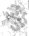
- the antibody is indicated by a thin line, and the epitope peptide is indicated by a thick line. Further, with respect to the 5 amino acid residues from the N-terminal of the epitope peptide, each residue is indicated by a single letter of amino acid and a serial number from the N-terminal side.
- the electron density map is shown by mesh display. It is the figure which surface-displayed the model at the time of connecting the epitope core sequence and the heavy chain N terminal of the variable region of the said antibody by Gly linker. In the figure, the heavy chain of the antibody is shown in black, the light chain in gray, and the linker in white bold line.
- (D) shows a model when connected by a linker consisting of 16 amino acids.
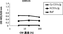
- CD3 ⁇ binding activity of an antibody with a peptide added to the heavy chain N-terminus and (ii) CD3 ⁇ binding activity of an antibody with a peptide added to the light chain N-terminus. It is the result of evaluating the CD3 ⁇ binding activity after protease (uPA) treatment of an anti-CD3 antibody derivative whose binding to CD3 ⁇ is masked and an antibody that does not undergo cleavage. It is shown as a ratio when the luminescence value of wells to which no antigen is added is 1. It is the CD3 ⁇ binding activity of an antibody (TWO arm) in which a peptide is added to the light chain N-terminus in the same manner as (iii) (ii).
- uPA protease
- CD3 ⁇ binding activity of an antibody obtained by adding a peptide to the heavy chain N-terminus of AN121 It is the result of evaluating the CD3 ⁇ binding activity after treatment with a protease (human MT-SP1) of an anti-CD3 antibody derivative masked for binding to CD3 ⁇ and an antibody that does not undergo cleavage. It is shown as a ratio when the luminescence value of wells to which no antigen is added is 1.
- CD3 ⁇ binding activity of an antibody with a peptide added to the heavy chain N-terminus and (ii) CD3 ⁇ binding activity of an antibody with a peptide added to the light chain N-terminus.
- Derivatives are prepared based on an antibody whose heavy chain is hCE115HA and whose light chain is GLS3000.
- (i) is an unmodified antibody to which no peptide is added and an antibody to which a masking molecule ( ⁇ ) is added using a GS linker that does not undergo cleavage.
- (ii) is an antibody to which a masking molecule ( ⁇ ) is added using a cancer tissue-specific protease-cleavable linker ( ⁇ ). 7 amino acids from the N-terminus of CD3 ⁇ (since the GG sequence is included in the linker part, the same sequence as the N-terminus of CD3 ⁇ is up to the 9th amino acid), 20 amino acids and 27 amino acids are masking molecules ( ⁇ ) Antibody.
- the solid line represents the antibody treated with protease
- the broken line represents the antibody not treated with protease. It is a CD3 ⁇ activation evaluation result after protease treatment of an anti-CD3 antibody derivative whose binding to CD3 ⁇ is masked and an antibody that does not undergo cleavage.
- Derivatives are prepared from AN121 with enhanced CD3 ⁇ binding ability of hCE115HA and an antibody whose light chain is GLS3000.
- (i) is an unmodified antibody to which no peptide is added and an antibody to which a masking molecule ( ⁇ ) is added using a GS linker that does not undergo cleavage.
- (ii) is an antibody to which a masking molecule ( ⁇ ) is added using a cancer tissue-specific protease-cleavable linker ( ⁇ ). 7 amino acids from the N-terminus of CD3 ⁇ (since the GG sequence is included in the linker part, the same sequence as the N-terminus of CD3 ⁇ is up to the 9th amino acid), 20 amino acids and 27 amino acids are masking molecules ( ⁇ ) Antibody.
- the solid line represents the antibody treated with protease
- the broken line represents the antibody not treated with protease. It is a CD3 ⁇ activation evaluation result after protease treatment of an anti-CD3 antibody derivative whose binding to CD3 ⁇ is masked and an antibody that does not undergo cleavage.
- Derivatives are prepared based on an antibody with a heavy chain of rCE115H and a light chain of GLS3000.
- (i) is an unmodified antibody to which no peptide is added and an antibody to which a masking molecule ( ⁇ ) is added using a GS linker that does not undergo cleavage.
- (ii) is an antibody to which a masking molecule ( ⁇ ) is added using a cancer tissue-specific protease-cleavable linker ( ⁇ ). 7 amino acids from the N-terminus of CD3 ⁇ (since the GG sequence is included in the linker part, the same sequence as the N-terminus of CD3 ⁇ is up to the 9th amino acid), 20 amino acids and 27 amino acids are masking molecules ( ⁇ ) Antibody.
- the solid line represents the antibody treated with protease
- the broken line represents the antibody not treated with protease. It is a CD3 ⁇ activation evaluation result after protease treatment of an anti-CD3 antibody derivative whose binding to CD3 ⁇ is masked and an antibody that does not undergo cleavage.
- Derivatives are prepared based on an antibody with a heavy chain of rCE115H and a light chain of rCE115L.
- (i) is an unmodified antibody to which no peptide is added and an antibody to which a masking molecule ( ⁇ ) is added using a GS linker that does not undergo cleavage.
- (ii) is an antibody to which a masking molecule ( ⁇ ) is added using a cancer tissue-specific protease-cleavable linker ( ⁇ ). 7 amino acids from the N-terminus of CD3 ⁇ (since the GG sequence is included in the linker part, the same sequence as the N-terminus of CD3 ⁇ is up to the 9th amino acid), 20 amino acids and 27 amino acids are masking molecules ( ⁇ ) Antibody.
- the solid line represents the antibody treated with protease
- the broken line represents the antibody not treated with protease. It is a CD3 ⁇ activation evaluation result after protease treatment of an anti-CD3 antibody derivative whose binding to CD3 ⁇ is masked and an antibody that does not undergo cleavage.
- Derivatives are prepared based on an antibody whose heavy chain is hCE115HA and whose light chain is GLS3000.
- (i) is an unmodified antibody to which no peptide is added and an antibody to which a masking molecule ( ⁇ ) is added using a GS linker that does not undergo cleavage.
- (ii) is an antibody to which a masking molecule ( ⁇ ) is added using a cancer tissue-specific protease-cleavable linker ( ⁇ ). 7 amino acids from the N-terminus of CD3 ⁇ (since the GG sequence is included in the linker part, the same sequence as the N-terminus of CD3 ⁇ is up to the 9th amino acid), 20 amino acids and 27 amino acids are masking molecules ( ⁇ ) Antibody.
- the solid line represents the antibody treated with protease
- the broken line represents the antibody not treated with protease.
- “recognize” means that an antigen-binding region, an antigen-binding molecule, an antibody, an antibody fragment, and the like bind to an antigen. Further, “specifically recognize” means that an antigen-binding region, an antigen-binding molecule, an antibody, an antibody fragment, and the like identify and bind to a specific three-dimensional structure in the antigen.
- the “antigen-binding region” means a region that exists in a molecule and can recognize an antigen.
- the antigen-binding region may be formed from a polypeptide or may be formed from a low molecular compound excluding the polypeptide.
- the “immune cell antigen binding region” can recognize an antigen specifically expressed by immune cells, and the “cancer antigen binding region” can recognize a cancer antigen.
- the “masking effect” means an effect of preventing the antigen-binding region from binding to the target antigen by binding the masking molecule to the antigen-binding region.
- Linear peptide does not form a higher order structure or forms a secondary structure such as an ⁇ helix structure, a ⁇ sheet structure, a loop, or a turn even if a higher order structure is formed. A polypeptide that does not form a structure or quaternary structure.
- Linear epitope means a partial linear peptide in an antigen that is recognized by an antigen binding region.
- “heavy chain” may be referred to as “H chain”
- “light chain” may be referred to as “L chain”.
- the multiple antigen binding molecule fusion of the present invention comprises a multiple antigen binding molecule ( ⁇ ), a cancer tissue specific protease cleavable linker ( ⁇ ) and a masking molecule ( ⁇ ).
- a multi-antigen-binding molecule ( ⁇ ) and a masking molecule ( ⁇ ) are bound via a cancer tissue-specific protease-cleavable linker ( ⁇ ).
- the types of binding between multiple antigen-binding molecules ( ⁇ ) and cancer tissue-specific protease-cleavable linkers ( ⁇ ), and cancer tissue-specific protease-cleavable linkers ( ⁇ ) and masking molecules ( ⁇ ) are particularly limited. Not.
- a preferred bond type is a peptide bond.
- the multiple antigen-binding molecule ( ⁇ ) of the present invention has an immune cell antigen-binding region and a cancer antigen-binding region. That is, the multiple antigen-binding molecule ( ⁇ ) has an immune cell antigen-binding region and a cancer antigen-binding region in the same molecule, and at least one immune cell antigen-binding region and one There is no particular limitation as long as it has a cancer antigen-binding region.
- One embodiment of the multiple antigen-binding molecule ( ⁇ ) includes one having one immune cell antigen-binding region and one cancer antigen-binding region in the same molecule. Specific examples include bispecific antibodies.
- multiple antigen-binding molecule examples include, for example, those using DART technology (International Publication No. 2012/162067), those using Dual-Fab technology (PCT / JP2014 / 077985), 2+ Examples include those using 1 IgG Crossfab technology (International Publication No. 2013/026833) and those using BiTE technology (Spiess C et al., Mol Immunol (2015) 67 (2) 95-106).
- the immune cell antigen binding region and the cancer antigen binding region may be directly bound, or may be bound via a biocompatible linker.
- the linker is a cancer tissue-specific protease-cleavable linker described later in order to induce sufficient cytotoxic activity. Preferably it is not ( ⁇ ).
- the immune cell antigen-binding region recognizes an antigen having a polypeptide consisting of the amino acid sequence QDGNE (SEQ ID NO: 15). “Recognizing an antigen having a polypeptide consisting of the amino acid sequence QDGNE (SEQ ID NO: 15)” means that when the amino acid sequence QDGNE is deleted, the binding activity to the antigen is reduced or lost. means. The decrease in binding activity can be confirmed by known methods such as FACS, ELISA format, ALPHA screen (Amplified Luminescent Proximity Homogeneous Assay) and BIACORE method using surface plasmon resonance (SPR) phenomenon.
- amino acid sequence QDGNE When the amino acid sequence QDGNE is deleted from an antigen having a polypeptide consisting of the amino acid sequence QDGNE (SEQ ID NO: 15), it is 50% or less, preferably 45% or less, 40% or less compared to the antigen before deletion. 35% or less, 30% or less, 20% or less, 15% or less, particularly preferably 10% or less, 9% or less, 8% or less, 7% or less, 6% or less, 5% or less, 4% or less, 3% Hereinafter, it means that the binding activity is 2% or less and 1% or less.
- the amino acid sequence QDGNE is an amino acid sequence present in the extracellular region of human CD3 ⁇ , and the amino acid sequence is identical between human and cynomolgus monkey (see arrow in FIG. 1).
- the antigen having a polypeptide consisting of the amino acid sequence QDGNE is preferably a partial polypeptide of human CD3 ⁇ as a whole.
- the partial polypeptide of human CD3 ⁇ is preferably a human CD3 ⁇ partial polypeptide having the amino acid sequence QDGNE at the N-terminus.
- the human CD3 ⁇ partial polypeptide having the amino acid sequence QDGNE at the N-terminus is preferably a region having high homology with the corresponding partial polypeptide of cynomolgus CD3 ⁇ .
- the homology is preferably 90% or more, more preferably 95% or more, and most preferably 100%. If the homology is high, even if the multi-antigen-binding molecule fusion used in the non-clinical toxicity test using cynomolgus monkey is used as it is in the clinical test, the test result obtained in the non-clinical toxicity test using cynomolgus monkey is It becomes easy to reflect the result.
- the antigen having a polypeptide consisting of the amino acid sequence QDGNE (SEQ ID NO: 15) may be chemically modified.
- the chemical modification may be a known modification. Examples of the chemical modification include acetylation, alkylation, pyroglutamylation and the like. Among these, Q in the amino acid sequence QDGNE is preferably pyroglutamylated.
- An immune cell antigen-binding region that recognizes an antigen having a polypeptide consisting of the amino acid sequence QDGNE can be obtained, for example, by a method using a known antibody production method.
- the antibody obtained by the production method may be used as it is for an immune cell antigen-binding region, or only the Fv region of the obtained antibody may be used. If the antigen can be recognized in step.), Only the single chain may be used. Further, a Fab region including an Fv region may be used.
- Humanized antibodies are also referred to as reshaped human antibodies.
- non-human animals for example, humanized antibodies obtained by grafting mouse antibody CDRs to human antibodies are known.
- General genetic recombination techniques for obtaining humanized antibodies are also known.
- Overlap-Extension-PCR is known as a method for transplanting mouse antibody CDRs into human FRs.
- FR amino acid residues can be substituted so that the CDR of the reshaped human antibody forms an appropriate antigen-binding site.
- amino acid sequence mutations can be introduced into FRs by applying the PCR method used for transplantation of mouse CDRs into human FRs.
- Transgenic animals having all repertoires of human antibody genes are used as immunized animals, and a desired human antibody can be obtained by DNA immunization.
- the Fv region of a human antibody is expressed as a single chain antibody (also referred to as “scFv”) on the surface of the phage by the phage display method. Phages expressing scFv that bind to the antigen can be selected. By analyzing the gene of the selected phage, the DNA sequence encoding the Fv region of the human antibody that binds to the antigen can be determined.
- scFv single chain antibody
- the Fv region sequence is fused in-frame with the sequence of the desired human antibody C region, and then inserted into an appropriate expression vector, whereby an expression vector can be prepared.
- the human antibody is obtained by introducing the expression vector into a suitable expression cell as described above and expressing the gene encoding the human antibody.
- Panning can also improve the binding activity of the immune cell antigen binding region with the immune cell antigen.
- the binding activity of the immune cell antigen-binding region with the immune cell antigen is calculated from data of a binding assay using a surface plasmon resonance (SPR) method (for example, using Biacore T200).
- the binding activity is indicated by KD (M), and the smaller the KD (M), the higher the binding activity.
- KD (M) is preferably less than 10 ⁇ 6, and more preferably less than 10 ⁇ 7 .
- an antibody or antibody fragment newly obtained by the above-described antibody production method may be used to recognize the antigen. Any known antibody or antibody fragment may be used. Examples of known antibodies or antibody fragments capable of recognizing an antigen having a polypeptide consisting of the amino acid sequence QDGNE include scFv prepared in Examples of International Publication No. 2007/042261 and International Publication No. 20082008/119567. It is done. In addition to the scFv, CE115 disclosed in PCT / JP2014 / 079785 can be used. A method for producing CE115 is described in Reference Example 1 described later.
- the immune cell antigen-binding region in the present invention may recognize only an antigen having a polypeptide consisting of the amino acid sequence QDGNE.
- at least one immunity other than the antigen may be used. Those that recognize cellular antigens may also be used.
- the immune cell antigen-binding region in the present invention preferably recognizes at least one immune cell antigen in addition to the antigen having a polypeptide consisting of the amino acid sequence QDGNE.
- immune cell antigens other than the antigen having a polypeptide consisting of the amino acid sequence QDGNE include, for example, T cell surface molecules, NK cell surface molecules, dendritic cell surface molecules, B cell surface molecules, NKT cell surface molecules, MDSC cell surface molecules And macrophage surface molecules.
- Specific T cell surface molecules include CD3 and T cell receptors. However, in the case of CD3, it is a molecule that does not have a polypeptide consisting of the amino acid sequence QDGNE.
- T cell surface molecule does not have a polypeptide consisting of the amino acid sequence QDGNE, for example, in the case of human CD3, any epitope that is present in the ⁇ chain, ⁇ chain, or ⁇ chain sequence constituting human CD3 It may be combined.
- Specific immune cell antigens other than T cell surface molecules include Fc ⁇ receptor, TLR, lectin, IgA, immune checkpoint molecule, TNF superfamily molecule, TNF receptor superfamily molecule, NK receptor molecule, etc. .
- a polypeptide comprising amino acid sequence QDGNE from the viewpoint of recruiting various immune cells such as NK cells other than T cells to cancer cells to further enhance the cytotoxic activity against cancer cells.
- the immune cell antigen recognized other than the antigen having NK cell surface molecule, dendritic cell surface molecule, B cell surface molecule, NKT cell surface molecule, MDSC cell surface molecule and macrophage surface molecule Preferably it is a seed.
- the immune cell antigen binding region recognizes at least one immune cell antigen in addition to the antigen having a polypeptide consisting of the amino acid sequence QDGNE, the immune cell antigen binding region has two or more immunity It is preferable that cell antigens cannot be recognized simultaneously.
- the immune cell antigen-binding region cannot recognize two or more immune cell antigens simultaneously, the multiple antigen-binding molecule fusion cannot recognize a plurality of immune cells at the same time.
- the multiple antigen-binding molecule fusion can recognize a plurality of immune cells at the same time, an abnormal immune response may occur as a result of the plurality of immune cells interfering with each other, and side effects may easily occur. Therefore, by making the immune cell antigen-binding region unable to recognize two or more immune cell antigens simultaneously, the possibility of side effects can be reduced.
- the immune cell antigen-binding region recognizes at least one type of immune cell antigen, and the immune cell antigen-binding region has two or more immune cells
- An example of a technique that cannot recognize antigens simultaneously is the Dual-Fab technique.
- the Dual-Fab technology is a technology that enables one Fab to recognize two or more kinds of antigens, and is disclosed in PCT / JP2014 / 079785.
- Specific examples of the method for producing an antigen-binding molecule by the Dual-Fab technique include a production method including the following steps (i) to (iv).
- an antigen-binding molecule in which at least one amino acid of a variable region of an antibody that binds to the first antigen or the second antigen is modified, wherein at least one of the amino acids of the modified variable region is different from each other Creating a library of antigen binding molecules comprising the region, (ii) an antigen comprising a variable region that has binding activity to the first antigen and the second antigen, but does not bind simultaneously with the first antigen and the second antigen, from the prepared library Selecting a binding molecule, (iii) A host cell containing a nucleic acid encoding the variable region of the antigen-binding molecule selected in step (ii) can be cultured to bind to the first antigen and the second antigen. Expressing an immune cell antigen-binding region comprising a variable region of an antibody that does not bind simultaneously to the antigen of and the second antigen; and (iv) recovering the antigen-binding molecule from the host cell culture.
- step (ii) may be the following step (v).
- step (v) Among the prepared libraries, the first antigen and the second antigen have binding activity against the first antigen and the second antigen, but are expressed on different cells, respectively. Selecting an antigen-binding molecule comprising a variable region that does not bind.
- the cancer antigen binding region recognizes a cancer antigen.
- One of the functions of this region is to allow a high concentration of multiple antigen-binding molecule fusion to exist in cancer tissue by recognizing a cancer antigen.
- the cancer antigen binding region is not particularly limited as long as it can recognize a cancer antigen. That is, the cancer antigen-binding region may be a polypeptide such as Fv of an antibody that recognizes a cancer antigen, or may be a low molecular compound other than a polypeptide such as folic acid. In the case of a low molecular weight compound, a step of binding a cancer antigen binding region to an immune cell antigen binding region is required when producing a multiple antigen binding molecule ( ⁇ ). Therefore, the cancer antigen-binding region is preferably a polypeptide from the viewpoint that multiple antigen-binding molecules ( ⁇ ) can be produced simply by being expressed in cells, and the production efficiency can be increased.
- Cancer antigens are antigens specific to tumor cells, and include antigens that are expressed as cells become malignant, as well as abnormal sugar chains that appear on the cell surface and protein molecules when cells become cancerous. It is.
- Specific examples of cancer antigens include glypican 3 (GPC3), ALK receptor (pleiotrophin receptor), pleiotrophin, KS 1/4 pancreatic cancer antigen, ovarian cancer antigen (CA125), prostatic acid phosphate , Prostate specific antigen (PSA), melanoma related antigen p97, melanoma antigen gp75, high molecular weight melanoma antigen (HMW-MAA), prostate specific membrane antigen, cancerous embryo antigen (CEA), polymorphic epithelial mucin antigen, human milk Colorectal tumor-associated antigens such as fat globule antigen, CEA, TAG-72, CO17-1A, GICA 19-9, CTA-1 and LEA, Burkitt lymphoma antigen
- cancer antigens are not expressed by cancer cells, but are present in or near cancer tissues so as to promote the growth and metastasis of cancer cells. May be any antigen.
- the cancer antigen-binding region can be obtained, for example, by a method using a known antibody production method.
- the antibody obtained by the production method may be used as it is for a cancer antigen-binding region, or only the Fv region of the obtained antibody may be used, and the Fv region can recognize an antigen in a single chain. For this, only the single strand may be used. Further, a Fab region including an Fv region may be used.
- a specific antibody production method can be performed in the same manner as the specific antibody production method in the above-described immune cell antigen-binding region. Similarly to the binding activity of the immune cell antigen binding region to the immune cell antigen, the binding activity of the cancer antigen binding region to the cancer antigen can be improved by panning.
- the binding activity of the immune cell antigen-binding region with the immune cell antigen is calculated from the data of the binding assay using the surface plasmon resonance (SPR) method (for example, using Biacore T200).
- the binding activity is indicated by KD (M), and the smaller the KD (M), the higher the binding activity.
- KD (M) is preferably less than 10 ⁇ 6, more preferably less than 10 ⁇ 7, and even more preferably less than 10 ⁇ 8 .
- One aspect of multiple antigen-binding molecule ( ⁇ ) includes, for example, antibodies and antibody fragments, and their fusions and complexes.
- the term “antibody” is used in the broadest sense. As long as a desired biological activity is exhibited, a monoclonal antibody (including a full-length monoclonal antibody), a polyclonal antibody, an antibody variant, an antibody fragment, a multispecific antibody (for example, , Bispecific antibodies), chimeric antibodies, humanized antibodies and the like.
- a monoclonal antibody including a full-length monoclonal antibody
- a polyclonal antibody an antibody variant, an antibody fragment, a multispecific antibody (for example, , Bispecific antibodies)
- chimeric antibodies humanized antibodies and the like.
- the type of antigen, the origin of the antibody, and the like are not limited, and the antibody may be any antibody.
- the origin of the antibody is not particularly limited, and examples thereof include a human antibody, a mouse antibody, a rat antibody, and a rabbit antibody.
- suitable antibodies include multispecific antibodies.
- a multispecific antibody is an antibody that can recognize two or more antigens.
- ⁇ multiantigen-binding molecule
- the multispecific antibody may be produced by a known production method. Specifically, the following methods are mentioned.
- VH antibody production technology utilizing association of L chain variable regions
- VL L chain variable regions
- a light chain that forms a variable region that binds to the first epitope, and a light chain that forms a variable region that binds to the second epitope are each a heavy chain that forms a variable region that binds to the first epitope.
- CrossMab technology (Scaefer et al. (Proc. Natl. Acad. Sci. USA (2011) 108, 11187), known as the association of heterologous light chains that associate with the heavy chain that forms the variable region that binds to the second epitope. -11192)) can also be used to make multispecific antibodies.
- FAE is preferred.
- the FAE include, for example, the second constant region (CH2) of the antibody H chain or the third constant region (CH3) of the H chain disclosed in WO2006 / 106905 and WO2015 / 046467.
- CH2 the second constant region
- CH3 the third constant region
- reassociation occurs randomly, so a bispecific antibody can theoretically be obtained only with an efficiency of 50%.
- a bispecific antibody can be obtained in a high yield. Can be manufactured.
- this method will be described in detail.
- amino acid residues that contact at the interface of other constant regions of the H chain include, for example, EU in the CH3 region Numbering 356th residue, EU numbering 439th residue, EU numbering 357th residue, EU numbering 370th residue, EU numbering 399th residue, region corresponding to EU numbering 409th residue Can be mentioned.
- 1 selected from the following amino acid residue groups shown in (1) to (3) in the first H chain CH3 region An antibody having 3 to 3 amino acid residues having the same kind of charge can be obtained.
- a set of amino acid residues selected from the set of amino acid residues shown in the above (1) to (3) in a second H chain CH3 region different from the first H chain CH3 region are the first H chain CH3 region.
- the antibody may have an opposite charge to the corresponding amino acid residue in.
- amino acid residues described in (1) to (3) above are close to each other when they are associated.
- a person skilled in the art finds a site corresponding to the amino acid residue described in (1) to (3) above by using homology modeling using commercially available software for the desired H chain CH3 region or H chain constant region.
- the amino acid residue at the site can be subjected to modification.
- the “charged amino acid residue” is preferably selected from, for example, amino acid residues included in any of the following groups (a) or (b); (A) glutamic acid (E), aspartic acid (D), (B) Lysine (K), arginine (R), histidine (H).
- “having the same kind of charge” means that, for example, any of two or more amino acid residues has an amino acid residue included in any one group of the above (a) or (b) Means that. “Having an opposite charge” means, for example, an amino acid residue in which at least one amino acid residue of two or more amino acid residues is included in any one group of the above (a) or (b) Means that the remaining amino acid residues have amino acid residues contained in different groups.
- the first H chain CH3 region and the second H chain CH3 region may be cross-linked by a disulfide bond.
- the amino acid residues subjected to modification are not limited to the amino acid residues in the above-described antibody variable region or antibody constant region. A person skilled in the art can find amino acid residues that form an interface for polypeptide variants or heterologous multimers by homology modeling using commercially available software, etc. Amino acid residues can be subjected to modification.
- a plurality of, for example, two or more techniques for producing these multispecific antibodies can be used. These techniques can also be applied separately to the two H chains to be associated as appropriate.
- the target multispecific antibody can be separated and purified from the produced multispecific antibody.
- a method has been reported that makes it possible to purify two types of homozygote and the desired heteroantibody by ion exchange chromatography. (International Publication No. 2007/114325).
- a method for purifying heterozygotes a method for purifying a heterodimerized antibody consisting of a mouse IgG2a H chain that binds to protein A and a rat IgG2b H chain that does not bind to protein A by using protein A so far Have been reported (WO 98/050431, WO 95/033844). Furthermore, by using H chains in which the amino acid residues at the 435th and 436th EU numbering, which are the binding sites of IgG and protein A, are substituted with amino acids having different binding powers to protein A such as Tyr and His, each H By changing the interaction between the chain and protein A and using a protein A column, it is possible to efficiently purify only the heterodimerized antibody.
- antibody fragments in the case of using antibody fragments as the multiple antigen-binding molecules ( ⁇ ) of this embodiment include those containing both heavy and light chains in a single polypeptide chain but lacking a constant region. It is done. Specific examples of such antibody fragments include diabody (Dabody), single chain antibody, and sc (Fab ') 2.
- Db is a dimer composed of two polypeptide chains (Holliger P et al., Proc. Natl. Acad. Sci. USA 90: 6444-6448 (1993), EP404, 097, W093 / 11161, etc.)
- VL and VH are connected to each other by a linker having a short length, for example, about 5 residues, such that VL and VH cannot be bonded to each other.
- VL and VH encoded on the same polypeptide chain cannot form a single-chain variable region fragment due to the short linker between them, and form two antigen-binding sites by dimerization. .
- sc (Fv) 2 is a single-chain antibody in which four variable regions of two VLs and two VHs are connected by a linker such as a peptide linker to form a single chain (J Immunol. Methods (1999) 231 (1- 2), 177-189).
- the two VHs and VLs can be derived from different monoclonal antibodies.
- the bispecific recognition (bispecific sc (Fv) 2) that recognizes two kinds of epitopes existing in the same antigen as disclosed in JournalJof Immunology (1994) 152 (11), 5368-5374 is also preferable.
- sc (Fv) 2 can be produced by methods known to those skilled in the art. For example, it can be prepared by linking scFv with a linker such as a peptide linker.
- the antigen-binding domain constituting sc (Fv) 2 is composed of two VHs and two VLs consisting of VH, VL, VH, VL ([VH] linker [ VL] linker [VH] linker [VL]) are listed in this order, but the order of the two VHs and the two VLs is not particularly limited to the above configuration, and any order It may be arranged in. For example, the following configuration can be given.
- sc (Fv) 2 The molecular form of sc (Fv) 2 is also described in detail in WO 2006/132352, and those skilled in the art can prepare the multiple antigen-binding molecule ( ⁇ ) of this embodiment based on these descriptions. Therefore, a desired sc (Fv) 2 can be appropriately produced.
- any peptide linker that can be introduced by genetic engineering, or a linker disclosed in synthetic compound linkers (for example, see Protein Engineering, 9 (3), 299-305, 1996) is used.
- a peptide linker is preferred.
- the length of the peptide linker is not particularly limited and can be appropriately selected by those skilled in the art according to the purpose. However, the preferred length is 5 amino acids or more (the upper limit is not particularly limited, but usually 30 amino acids or less, preferably Is 20 amino acids or less), particularly preferably 15 amino acids.
- sc (Fv) 2 includes three peptide linkers, peptide linkers having the same length may be used, or peptide linkers having different lengths may be used.
- Synthetic chemical linkers are commonly used for cross-linking peptides such as N-hydroxysuccinimide (NHS), disuccinimidyl suberate (DSS), bis (sulfosuccinimidyl) Suberate (BS3), dithiobis (succinimidyl propionate) (DSP), dithiobis (sulfosuccinimidyl propionate) (DTSSP), ethylene glycol bis (succinimidyl succinate) (EGS), ethylene Glycol bis (sulfosuccinimidyl succinate) (sulfo-EGS), disuccinimidyl tartrate (DST), disulfosuccinimidyl tartrate (sulfo-DST), bis [2- (succinimideoxycarbonyloxy ) Ethyl] sulfone (BSOCOES), bis [2- (sulfosuccinimidooxycarbonyloxy
- F (ab ') 2 is two heavy chains containing two light chains and a constant region of the CH1 domain and a portion of the CH2 domain such that an interchain disulfide bond is formed between the two heavy chains including.
- F (ab ') 2 which constitutes the multiple antigen-binding molecule ( ⁇ ) is obtained by partially digesting a full-length monoclonal antibody having a desired antigen-binding domain with a protease such as pepsin, and then converting the Fc fragment into a protein A column. By adsorbing and removing, it can be suitably obtained.
- Such a proteolytic enzyme is not particularly limited as long as the full-length antibody can be digested so as to restrictively generate F (ab ′) 2 by appropriately setting the reaction conditions of the enzyme such as pH.
- pepsin and ficin can be exemplified.
- the antibody fragment preferably has at least two Fvs from the viewpoint of obtaining excellent cytotoxicity, and the cancer antigen-binding region and the immune cell antigen-binding region are preferably formed by separate Fv regions.
- the antibody fragment may have an Fc region or may not have an Fc region.
- the Fc region may be, for example, an Fc region derived from natural IgG, or an Fc region obtained by adding an artificial mutation to an Fc region derived from natural IgG.
- the natural IgG means a polypeptide belonging to the class of antibodies that includes the same amino acid sequence as IgG found in nature and is substantially encoded by an immunoglobulin gamma gene.
- natural human IgG means natural human IgG1, natural human IgG2, natural human IgG3, natural human IgG4, and the like. Naturally-occurring IgG includes naturally occurring mutants.
- mutants include a plurality of allotype sequences due to gene polymorphism (Sequences of proteins of immunological interest, NIH Publication No. 91-3242). Particularly, the sequence of human IgG1 may be DEL or EEM as the amino acid sequence of EU numbering 356-358.
- the molecule ( ⁇ ) preferably does not have an Fc region. However, even if it has an Fc region, the side effects described above can be reduced if the Fc region is modified so as to lack the function of recognizing the Fc ⁇ receptor.
- the amino acid sequence of the Fc region can be modified by various methods known in the art. These methods include, but are not limited to, site-directed mutagenesis (Hashimoto-Gotoh, T, Mizuno, T, Ogasahara, Y, and Nakagawa, M. (1995) An oligodeoxyribonucleotide- directed dual amber method for site-directed mutagenesis. Gene 152, 271-275, Zoller, MJ, and Smith, M.
- Whether the Fc region has been modified to lack the function of recognizing the Fc ⁇ receptor can be determined by FACS, ELISA format, ALPHA screen (Amplified Luminescent Proximity Homogeneous Assay), BIACORE method using surface plasmon resonance (SPR) phenomenon, etc. It can be confirmed by a well-known method (Proc. Natl. Acad. Sci. USA (2006) 103 (11), 4005-4010).
- the ALPHA screen is implemented according to the following principle by ALPHA technology using two beads of donor and acceptor. A molecule bound to the donor bead interacts biologically with the molecule bound to the acceptor bead, and a luminescent signal is detected only when the two beads are in close proximity.
- a photosensitizer in the donor bead excited by the laser converts ambient oxygen into excited singlet oxygen.
- Singlet oxygen diffuses around the donor bead, and when it reaches the adjacent acceptor bead, it causes a chemiluminescence reaction in the bead, and finally light is emitted.
- the chemiluminescence reaction does not occur because the singlet oxygen produced by the donor bead does not reach the acceptor bead.
- an antigen-binding molecule labeled with biotin is bound to the donor bead, and an Fc ⁇ receptor tagged with glutathione S-transferase (GST) is bound to the acceptor bead.
- GST glutathione S-transferase
- antigen-binding molecules with wild-type Fc regions interact with Fc ⁇ receptors to produce signals of 520-620 nm.
- An antigen-binding molecule having a mutated Fc region that is not tagged competes with the interaction between an antigen-binding molecule having a wild-type Fc region and the Fc ⁇ receptor. Relative binding affinity can be determined by quantifying the decrease in fluorescence that results from competition.
- biotinylate antigen-binding molecules such as antibodies using Sulfo-NHS-biotin or the like.
- a method of tagging Fc ⁇ receptor with GST it is expressed in a cell or the like holding a vector capable of expressing a fusion gene in which a polynucleotide encoding Fc ⁇ receptor and a polynucleotide encoding GST are fused in frame.
- a method of purification using a glutathione column can be appropriately employed.
- the obtained signal is suitably analyzed by fitting to a one-site competition model using nonlinear regression analysis using software such as GRAPHPAD PRISM (GraphPad, San Diego).
- the Biacore system takes the shift amount, that is, the mass change at the sensor chip surface on the vertical axis, and displays the time change of mass as measurement data (sensorgram).
- Kinetics association rate constant (ka) and dissociation rate constant (kd) are obtained from the sensorgram curve, and affinity (KD) is obtained from the ratio of the constants.
- an inhibition measurement method is also preferably used. Examples of inhibition assays are described in Proc. Natl. Acad. Sci. USA (2006) 103 (11), 4005-4010.
- That the Fc region lacks the function of recognizing the Fc ⁇ receptor means that, for example, based on the analysis method described above, the binding activity of the test Fc region to the Fc ⁇ receptor is 50% compared to the control Fc region. % Or less, preferably 45% or less, 40% or less, 35% or less, 30% or less, 20% or less, 15% or less, particularly preferably 10% or less, 9% or less, 8% or less, 7% or less, 6% Hereinafter, it means binding activity of 5% or less, 4% or less, 3% or less, 2% or less, 1% or less.
- the control Fc region include the above-mentioned native IgG-derived Fc region.
- test Fc region is a variant of an Fc region of a specific isotype antibody
- the variant recognizes the Fc ⁇ receptor by using the Fc region of the specific isotype antibody as a control Fc region. It is verified whether the function to perform is missing. Thus, the Fc region verified to lack the function of recognizing the Fc ⁇ receptor is appropriately used for the antibody fragment.
- the light chain in an antibody or an antibody fragment having at least two Fv regions has the same amino acid sequence. Since the light chain in the antibody or the antibody fragment having at least two Fv regions has the same amino acid sequence, the number of combinations of heavy chain and light chain is reduced. The step of removing fragments can be facilitated. As a result, the production efficiency of the multiple antigen-binding molecule fusion can be improved.
- Cancer tissue-specific protease-cleavable linker ( ⁇ ) has a polypeptide consisting of a target sequence of a cancer tissue-specific protease.
- the cancer tissue-specific protease cleavable linker ( ⁇ ) may consist only of the target sequence of the cancer tissue-specific protease, or may be a fusion of a peptide linker in addition to the target sequence. Examples of such a peptide linker include the same linkers that bind the variable regions described above.
- Cancer tissue specific protease cleavable linker ( ⁇ ) is hydrolyzed by cancer tissue specific protease in cancer tissue.
- the masking molecule ( ⁇ ) When the cancer tissue-specific protease-cleavable linker ( ⁇ ) is hydrolyzed, the masking molecule ( ⁇ ) can be released from the multiple antigen-binding molecule ( ⁇ ). When the masking molecule ( ⁇ ) is released from the multiple antigen-binding molecule ( ⁇ ), the immune cell antigen-binding region in the multiple antigen-binding molecule ( ⁇ ) can bind to immune cells.
- the cancer tissue-specific protease-cleavable linker ( ⁇ ) has a function as a spacer for enabling the masking molecule ( ⁇ ) to mask the immune cell antigen-binding region in the multiple antigen-binding molecule ( ⁇ ).
- cancer tissue-specific protease examples include cancer tissues disclosed in International Publication No. 2013/128194, International Publication No. 2010/081173, International Publication No. 2009/025846, etc. And proteases that are specifically expressed.
- proteases include, but are not limited to, cysteine protease (including cathepsin family B, L, S, etc.), aspartyl protease (cathepsin D, E, K, O, etc.), serine protease (Including cathepsins A and G, thrombin, plasmin, urokinase (uPA), tissue plasminogen activator (tPA)), metalloproteases (membrane-bound (MMP14-17 and MMP24-25)) and secreted (MMP1- 13 and MMP18-23 and MMP26-28), metalloproteases (MMP1-28), protease A disintegrin and metalloprotease (ADAM), metalloprote with A disintegrin or thrombospondin motif Over peptidase (ADAMTS)), CD10 (CALLA), and prostate-specific antigen (PSA), and the like.
- cysteine protease including cathepsin family B, L, S,
- the cancer tissue-specific protease As the type of cancer tissue-specific protease has higher expression specificity in the cancer tissue to be treated, the effect of reducing the side effects by the masking molecule ( ⁇ ) can be obtained.
- the cancer tissue-specific protease is preferably at least 5 times higher than that in normal tissue, more preferably at least 10 times higher, more preferably at least 100 times higher, and more preferably at least 500 times higher. Preferably, it is most preferably 1000 times higher.
- the cancer tissue-specific protease may be bound to the cell membrane of cancer cells, or may be secreted outside the cell membrane without being bound to the cell membrane.
- cancer tissue-specific protease is not bound to the cell membrane of the cancer cell, the cancer tissue-specific protease is present in or near the cancer tissue in order for the cytotoxicity by immune cells to be specific to the cancer cell.
- “in the vicinity of cancer tissue” means that cancer tissue-specific protease-cleavable linker ( ⁇ ) is cleaved and multiple antigen-binding molecules ( ⁇ ) recruit immune cells and damage cancer cells. It means that. However, a range that does not damage normal cells as much as possible is preferable.
- One type of cancer tissue-specific protease may be used alone, or two or more types may be combined. The number of types of cancer tissue-specific protease can be appropriately set by those skilled in the art in consideration of the cancer type to be treated.
- the cancer tissue specific protease is preferably a metalloprotease among the proteases exemplified above, and among the metalloproteases, MMP-2 and MMP-9 are preferable, and MMP-2 is more preferable.
- a target sequence is a specific amino acid sequence that is specifically recognized by a cancer tissue-specific protease when the polypeptide is hydrolyzed by the cancer tissue-specific protease in an aqueous solution.
- the target sequence is preferably an amino acid sequence that is hydrolyzed with high specificity by a cancer tissue-specific protease that is more specifically expressed in the cancer tissue to be treated.
- Specific examples of the target sequence are specifically expressed in the above-exemplified cancer tissues disclosed in, for example, International Publication No. 2013/128194, International Publication No. 2010/081173, International Publication No. 2009/025846, etc. And target sequences that are specifically hydrolyzed by proteases.
- the target sequence may be one identified by a method known to those skilled in the art as described in Nature Biotechnology 19, 661-667 (2001).
- the target sequence is preferably an amino acid sequence that is specifically hydrolyzed by MMP-2, which is a suitable cancer tissue-specific protease.
- MMP-2 which is a suitable cancer tissue-specific protease.
- the following amino acid sequence SEQ ID NO: 9 is preferable.
- PLGLAG SEQ ID NO: 9
- the cancer tissue-specific protease-cleavable linker ( ⁇ ) may be bound to any position of the multiple antigen-binding molecule ( ⁇ ).
- a cancer tissue-specific protease-cleavable linker because the masking molecule ( ⁇ ) can easily access the immune cell antigen-binding region in the multiple antigen-binding molecule ( ⁇ ) and exert a masking effect on the immune cell antigen-binding region.
- ( ⁇ ) is preferably bound to the immune cell antigen-binding region in the multiple antigen-binding molecule ( ⁇ ).
- a cancer tissue-specific protease-cleavable linker ( ⁇ ) are preferably fused to the heavy chain N-terminus or light-chain N-terminus of the Fv region (Fab region) on the side that forms the immune cell antigen-binding region (see FIG. 3). Called “N-terminal fusion” or "light chain N-terminal fusion”).
- a cancer tissue-specific protease-cleavable linker ( ⁇ ) is fused to the heavy chain N-terminus or light-chain N-terminus of the Fv region on the side that forms the immune cell antigen-binding region, so that multiple antigen-binding molecules ( ⁇ ) Since a step of binding a cancer tissue-specific protease-cleavable linker ( ⁇ ) after the preparation is not necessary, the production efficiency of the multiple antigen-binding molecule fusion is excellent.
- the amino acid lengths of the cancer tissue-specific protease-cleavable linker ( ⁇ ) and masking molecule ( ⁇ ) described later can be easily set.
- the cancer tissue-specific protease-cleavable linker ( ⁇ ) can be obtained, for example, by a known polypeptide production method.
- the masking molecule ( ⁇ ) has a polypeptide consisting of the amino acid sequence QDGNE.
- the polypeptide consisting of the amino acid sequence QDGNE is a partial peptide of human CD3 ⁇ derived from human CD3 ⁇ .
- the masking molecule ( ⁇ ) masks the immune cell antigen-binding region of the multiple antigen-binding molecule ( ⁇ ) in the multiple antigen-binding molecule fusion so that the immune cell antigen-binding region binds to the immune cell antigen. Has a function to prevent.
- Masking of the immune cell antigen-binding region by the masking molecule ( ⁇ ) may occur between multiple antigen-binding molecule fusions, and the multiple antigen-binding molecule fusions may form a complex. Even in this state, the function can be exhibited.
- the above-mentioned cancer tissue-specific protease-cleavable linker ( ⁇ ) is cleaved by a cancer tissue-specific protease expressed in the cancer tissue. Then, the masking molecule ( ⁇ ) can be released from the multiple antigen-binding molecule ( ⁇ ).
- the immune cell antigen-binding region of the multiple antigen-binding molecule ( ⁇ ) can bind to the immune cell antigen.
- the cancer tissue-specific protease is not expressed, or even if expressed, the concentration is low and the cancer tissue-specific protease-cleavable linker ( ⁇ ) is difficult to cleave. ) Cannot be released from the multiple antigen-binding molecule ( ⁇ ), and the masking effect on the immune cell antigen-binding region is maintained.
- the masking molecule ( ⁇ ) is bound to the multiple antigen-binding molecule ( ⁇ ) via the cancer tissue-specific protease-cleavable linker ( ⁇ ), thereby masking the immune cell antigen-binding region, Since cells are difficult to recruit, side effects due to multiple antigen-binding molecules ( ⁇ ) are reduced.
- the masking molecule ( ⁇ ) may or may not have an amino acid sequence other than the amino acid sequence QDGNE.
- the masking effect can be sufficiently exerted, and the cancer tissue-specific protease-cleavable linker ( ⁇ ) is cleaved. Then, it is easy to release from the multiple antigen-binding molecule ( ⁇ ).
- the masking molecule ( ⁇ ) does not have an amino acid sequence other than the amino acid sequence QDGNE from the viewpoint of further improving the stability of the masking molecule ( ⁇ ) and further preventing the reduction of the masking effect due to binding to CD3 ⁇ or CD3 ⁇ . It is preferable.
- the masking molecule ( ⁇ ) is preferably a partial polypeptide of human CD3 ⁇ .
- the human CD3 ⁇ partial polypeptide is linear in which the immune cell antigen-binding region can bind. Peptides (linear epitopes) are preferred. Further, by using a linear peptide, the molecular weight of the multiple antigen-binding molecule fusion can be suppressed, the dosage can be suppressed, and the burden on the patient can be reduced.
- the partial polypeptide of human CD3 ⁇ is preferably a human CD3 ⁇ partial polypeptide having the amino acid sequence QDGNE at the N-terminus and the number of amino acids of 30 or less, and having the amino acid sequence QDGNE at the N-terminus, More preferably, it is a human CD3 ⁇ partial polypeptide having an amino acid number of 25 or less, more preferably a human CD3 ⁇ partial polypeptide having the amino acid sequence QDGNE at the N-terminus and an amino acid number of 20 or less, and the amino acid sequence QDGNE Particularly preferred is a human CD3 ⁇ partial polypeptide having an N-terminus and 15 or fewer amino acids, and most preferably a human CD3 ⁇ partial polypeptide having the amino acid sequence QDGNE at the N-terminus and 10 or fewer amino acids.
- the masking molecule ( ⁇ ) may be chemically modified.
- the chemical modification may be a known modification. Examples of the chemical modification include acetylation, alkylation, pyroglutamylation and the like. Among these, Q in the amino acid sequence QDGNE is preferably pyroglutamylated.
- the masking molecule ( ⁇ ) can be obtained by a known polypeptide production method. Moreover, what is necessary is just to perform this modification in the case of chemically modifying by the chemical modification method of well-known polypeptide.
- the number of amino acids in the fusion polypeptide is It is preferably 11 or more and 65 or less, more preferably 14 or more and 27 or less, and most preferably 17 or more and 20 or less.
- the fusion polypeptide has 16 amino acids. Preferably, it is 65 or less, more preferably 17 or more and 30 or less, and most preferably 19 or more and 25 or less.
- a multiple antigen-binding molecule ( ⁇ ), a cancer tissue-specific protease-cleavable linker ( ⁇ ), and a masking molecule ( ⁇ ) are prepared, respectively, and a cancer tissue-specific protease-cleavable linker is prepared.
- ( ⁇ ) includes a method of binding multiple antigen-binding molecules ( ⁇ ) and masking molecules ( ⁇ ).
- the type of binding in the method is not particularly limited as long as the cancer tissue-specific protease-cleavable linker ( ⁇ ) is chemically bonded to the multiple antigen-binding molecule ( ⁇ ) and the masking molecule ( ⁇ ).
- a covalent bond is preferable, and the covalent bond is preferably formed by a peptide bond.
- the multiple antigen-binding molecule fusion is a fusion protein or an assembly of fusion proteins, which may be sugar chain-modified
- the multiple antigen-binding molecule fusion is a known protein expression method such as a host. You may manufacture by the method which combined the cell and the expression vector.
- eukaryotic cells are used as host cells, animal cells, plant cells or fungal cells can be used as appropriate. Specifically, the following cells can be exemplified as animal cells.
- Mammalian cells CHO (Chinese hamster ovary cell line), COS (Monkey kidney cell line), myeloma (Sp2 / O, NS0, etc.), BHK (baby hamster kidney cell line), HEK293 (human embryonic kidney cell line) with sheared adenovirus (Ad) 5 DNA), PER.C6 cell (human embryonic retinal cell line transformed with the Adenovirus Type 5 (Ad5) E1A and E1B genes), Hela, Vero, etc. (Current Protocols in Protein Science (May, 2001 , Unit 5.9, Table 5.9.1))
- Amphibian cells Xenopus oocytes, etc.
- Insect cells sf9, sf21, Tn5, etc.
- multiple antigen-binding molecule fusions are available in E. coli (mAbs 2012 Mar-Apr; 4 (2): 217-225. ) And yeast (International Publication No. 2000/023579).
- the multiple antigen-binding molecule fusion produced in E. coli has no sugar chain added.
- multiple antigen-binding molecule fusions produced in yeast are added with sugar chains.
- the expression vector can be appropriately selected by those skilled in the art depending on the type of host cell.
- the recovery of the multi-antigen-binding molecule fusion can be performed, for example, by culturing transformed cells and then separating them from the intracellular or culture solution of the molecularly transformed cells.
- centrifugation, ammonium sulfate fractionation, salting out ultrafiltration, C1q, FcRn, protein A, protein G column, affinity chromatography, ion exchange chromatography, gel filtration chromatography It can be performed by appropriately combining methods such as lithography.
- the pharmaceutical composition of the present invention comprises the multiple antigen-binding molecule fusion described above and a pharmaceutically acceptable carrier.
- the pharmaceutical composition can be formulated by a known method containing the above-mentioned multiple antigen-binding molecule fusion and a pharmaceutically acceptable carrier. For example, it can be used parenterally in the form of sterile solutions with water or other pharmaceutically acceptable solutions, or suspension injections.
- a pharmacologically acceptable carrier or medium specifically, sterile water, physiological saline, vegetable oil, emulsifier, suspension, surfactant, stabilizer, flavoring agent, excipient, vehicle, preservative It is conceivable to prepare a pharmaceutical preparation by combining with a binder or the like as appropriate and mixing in a unit dosage form generally required for pharmaceutical practice.
- silicic acid lactose, crystalline cellulose, mannitol, starch, carmellose calcium, carmellose sodium, hydroxypropylcellulose, hydroxypropylmethylcellulose, polyvinylacetal diethylaminoacetate, polyvinylpyrrolidone, gelatin, medium chain fatty acid triglyceride
- the carrier include polyoxyethylene hydrogenated castor oil 60, sucrose, carboxymethylcellulose, corn starch, and inorganic salts. The amount of active ingredient in these preparations is such that an appropriate volume within the indicated range can be obtained.
- a sterile composition for injection can be formulated in accordance with normal pharmaceutical practice using a vehicle such as distilled water for injection.
- Aqueous solutions for injection include, for example, isotonic solutions containing physiological saline, glucose and other adjuvants such as D-sorbitol, D-mannose, D-mannitol and sodium chloride, and suitable solubilizers such as You may use together with alcohol, specifically ethanol, polyalcohol, for example, propylene glycol, polyethyleneglycol, a nonionic surfactant, for example, polysorbate 80, HCO-50.
- oily liquid examples include sesame oil and soybean oil, which may be used in combination with benzyl benzoate or benzyl alcohol as a solubilizing agent.
- buffer for example, phosphate buffer, sodium acetate buffer, a soothing agent, for example, procaine hydrochloride, stabilizer, for example, benzyl alcohol, phenol, antioxidant.
- the prepared injection solution is usually filled into a suitable ampoule.
- Administration is preferably parenteral administration, and specific examples include injection, nasal administration, pulmonary administration, and transdermal administration.
- the injection form it can be administered systemically or locally by, for example, intravenous injection, intramuscular injection, intraperitoneal injection, subcutaneous injection, or the like.
- the administration method can be appropriately selected depending on the age and symptoms of the patient.
- the dosage of the pharmaceutical composition containing the multiple antigen-binding molecule fusion and the pharmaceutically acceptable carrier can be selected, for example, in the range of 0.0001 mg to 1000 mg per kg body weight. Alternatively, for example, the dose can be selected in the range of 0.001 to 100,000 mg / body per patient, but is not necessarily limited to these values.
- the dose and administration method vary depending on the weight, age, symptoms, etc. of the patient, but can be appropriately selected by those skilled in the art.
- the pharmaceutical composition is preferably for cancer treatment. It is preferable that the cancer type to be treated specifically expresses a cancer antigen recognized by a cancer antigen-binding region in the multiple antigen-binding molecule ( ⁇ ) contained in the multiple antigen-binding molecule fusion to be used.
- the cancer type to be treated may be a solid cancer or a blood cancer.
- the treatment method of cancer including the process of administering this pharmaceutical composition to a patient can be provided.
- the linear epitope identification method of the present invention is based on protein tertiary structure analysis data using a protein complex of an immune cell antigen and an immune cell antigen-binding region that recognizes the immune cell antigen.
- the immune cell antigen binding region may recognize a linear peptide portion in the immune cell antigen, or may recognize the higher order structure portion of a polypeptide that forms a higher order structure. .
- the method for identifying a linear epitope identifies a linear peptide to which the immune cell antigen-binding region recognizes a linear peptide portion in an immune cell antigen, and to which the immune cell antigen-binding region can bind. Is the method.
- the identified linear peptide is useful as a masking molecule ( ⁇ ) that does not require the formation of a higher order structure. Since the linear peptide does not require the formation of a higher-order structure, the masking effect can be stably exhibited.
- the method for producing a multiple antigen binding molecule fusion of the present invention comprises a multiple antigen binding molecule having a cancer antigen binding region recognizing a cancer antigen and an immune cell antigen binding region recognizing an immune cell antigen.
- ⁇ a region that can be cleaved by a protease that specifically expresses a linear epitope (for example, a linear epitope identified by the above-described linear epitope identification method) in a cancer tissue expressing the cancer antigen.
- the multiple antigen-binding molecule ( ⁇ ) and the cancer tissue-specific protease-cleavable linker ( ⁇ ) are the same as those in the above-mentioned “A. Multiple antigen-binding molecule fusion”.
- the linear epitope is not particularly limited as long as it satisfies the definition in the present specification.
- the linear epitope may be a linear epitope identified by the above-described method for identifying a linear epitope, and ELISA, mass spectrometry, phage library, etc. are performed using partial peptides of various antigens. Those identified by known epitope mapping may be used.
- Linear epitopes can be obtained by analyzing the state of binding between immune cell antigens and immune cell antigen-binding regions in detail and more accurately, so that the smallest unit epitope can be obtained, and by obtaining the smallest unit epitope, higher-order structures can be obtained. From the viewpoint of obtaining a multiple antigen-binding molecule fusion that can stably exhibit a masking effect that does not require the formation of a linear epitope, it is preferably a linear epitope identified by the above-described linear epitope identification method.
- the multi-antigen-binding molecule fusion of the present invention is specifically activated by a protease present in or near a cancer tissue, and can recruit immune cells such as T cells to cancer cells. Therefore, an excellent side effect reducing effect can be obtained in a multiple antigen-binding molecule that recognizes a cancer antigen and CD3.
- a masking molecule having a polypeptide consisting of the amino acid sequence QDGNE (SEQ ID NO: 15) is used.
- the masking molecule only needs to have a polypeptide consisting of at least the amino acid sequence QDGNE (SEQ ID NO: 15), and has a smaller molecular weight than the masking molecule disclosed in International Publication No. 2013/128194, so that it can be easily produced. . Further, since the molecular weight of the multiple antigen-binding molecule fusion can be reduced as the molecular weight of the masking molecule can be reduced, the dose can be reduced and the burden on the patient can be reduced.
- CD3 ⁇ is generally known to form a homodimer or a heterodimer with CD3 ⁇ or CD3 ⁇ (Scand J Immunol. 2002 Vol.56, pp.436-442.). Therefore, the stability of CD3 ⁇ or CD3 ⁇ partial protein may be reduced when it exists as a monomer. Moreover, when CD3 ⁇ or a partial protein of CD3 ⁇ is used as a masking molecule, it is likely that a dimer is easily formed between molecules or with an endogenous protein, and the masking effect may not be sufficiently obtained.
- the masking molecule in the multiple antigen-binding molecule fusion of the present invention only needs to have a polypeptide consisting of at least the amino acid sequence QDGNE (SEQ ID NO: 15), and thus is excellent in stability and masking effect.
- OKT3 is an anti-CD3 ⁇ antibody used in WO2013 / 128194, differs between humans and cynomolgus monkeys. Therefore, OKT3 is a human CD3 ⁇ -specific antibody and has not been confirmed to bind to cynomolgus CD3 ⁇ . Therefore, it is considered that non-clinical toxicity tests using cynomolgus monkeys cannot be performed, or that non-clinical toxicity tests using cynomolgus monkeys do not reflect the results of clinical tests.
- amino acid sequence QDGNE SEQ ID NO: 15
- the test results obtained in the non-clinical toxicity test using cynomolgus monkeys easily reflect the results of the clinical test.
- Example 1 “Protease-activated anti-cancer antigen / anti-CD3 multiple antigen-binding molecule fusion concept”
- Anti-cancer antigen / anti-CD3 multi-antigen-binding molecule is expected as a promising molecular format for anti-cancer drugs due to its strong cytotoxic activity, but when cancer antigens are expressed even in normal tissues Therefore, an anticancer antigen / anti-CD3 multiantigen-binding molecule that is superior in safety due to its safety with few side effects is desired because it may exert its damaging activity on normal tissues.
- the anti-cancer antigen / anti-CD3 multiple antigen-binding molecule can be derivatized and an anti-cancer antigen / anti-CD3 multiple antigen-binding molecule fusion activated by protease can be produced, the safety is excellent with few side effects.
- An anticancer antigen / anti-CD3 multiple antigen-binding molecule can be easily prepared (FIG. 2).
- the anti-cancer antigen / anti-CD3 multiantigen-binding molecule fusion activated by protease preferably has the following three structural features in order to further improve safety with fewer side effects.
- a masking molecule ( ⁇ ) that inhibits the binding activity of the anti-CD3 antibody is attached to the heavy chain N-terminus or light-chain N-terminus of the anti-CD3 antibody variable region via a cancer tissue-specific protease-cleavable linker ( ⁇ ). It is the feature that it is connected.
- the masking molecule ( ⁇ ) is composed of the natural epitope sequence of the anti-CD3 antibody, does not contain non-natural sequences from the viewpoint of immunogenicity, and the corresponding amino acid sequence homology between species (eg, between humans and cynomolgus monkeys) It is the feature that the nature is high.
- the cancer tissue-specific protease-cleavable linker ( ⁇ ) contains a target sequence that is cleaved by a protease that is present at a high concentration in or near the cancer tissue.
- the fused cells were suspended in a semi-fluid medium (Stem Cells) to carry out selective culture of the hybridoma and colonize the hybridoma.
- a semi-fluid medium Stem Cells
- HAT selection medium (10% FBS / RPMI1640, 2 vol% HAT 50x concentrate (Dainippon Pharmaceutical), 5 vol% BM-Condimed H1 (Roche Diagnostics) ) was inoculated in a 96-well plate with 1 colony per well. After culturing for 3 to 4 days, the culture supernatant of each well was collected, and the rat IgG concentration in the culture supernatant was measured.
- a humanized anti-CD3 antibody consisting of a chain variable region (SEQ ID NO: 13) was prepared.
- the antibody was purified by rProtein A Sepharose (registered trademark) Fast Flow (GE Healthcare) by a method known to those skilled in the art.
- the purified antibody concentration was determined by measuring the absorbance at 280 nm using a spectrophotometer, and calculating the antibody concentration using the extinction coefficient calculated by the PACE method from the obtained value (Protein Science 1995; 4: 2411- 2423).
- Example 2 X-ray structure analysis of complex of anti-CD3 antibody and CD3 Heavy chain (variable region (SEQ ID NO: 12), constant region (SEQ ID NO: 38)) and light chain (variable region (SEQ ID NO: SEQ ID NO :) 13)
- An anti-CD3 antibody consisting of a constant region (SEQ ID NO: 36)) and Kn010G3 (SEQ ID NO: 37) was expressed and prepared as a One arm antibody by the method of Reference Example 1. From the obtained One arm antibody, cleavage treatment by papain protease (Roche Applied Science), removal of Fc fragment by protein A carrier column, purification by cation exchange column and gel filtration column are carried out by methods known to those skilled in the art.
- a Fab fragment of the CD3 antibody was prepared.
- the obtained Fab fragment of the anti-CD3 antibody was concentrated by ultrafiltration, and a crystal was obtained by standing at 20 ° C. under a reservoir condition of 200 mM Potassium Sulfate, 20% PEG3350 using a vapor diffusion method.
- anti-CD3 antibody A crystal structure of the Fab fragment alone was obtained, and its crystallographic reliability factor R value and Free R value were 22.21% and 26.28%, respectively.
- a synthetic peptide shown in the following SEQ ID NO: 14 corresponding to the sequence from the N-terminal to the 15th CD3 in the Fab fragment of the obtained anti-CD3 antibody XDGNEEMGGITQTPY (X: L-pyroglutamic acid) (SEQ ID NO: 14) From a sample added to 2 mM, crystallize by standing at 20 ° C under reservoir conditions of 100 mM MES buffer pH 7.0, 0.91% PEG3350, 1.0 M Sodium citrate tribasic dihydrate using the vapor diffusion method. was gotten.
- X-ray crystal structure analysis is performed by a method known to those skilled in the art based on the crystal structure of the above-mentioned Fab fragment of the anti-CD3 antibody.
- the crystal structure of the complex of the anti-CD3 antibody Fab fragment and the epitope peptide was obtained, and its crystallographic reliability factor R value and Free R value were 18.55% and 26.53%, respectively.
- the structure and electron density map in the vicinity of the epitope of the complex of the Fab fragment of the anti-CD3 antibody and the epitope peptide are shown in FIG.
- a sufficient electron density was observed only from the N-terminus of the epitope peptide to Glu at the fifth residue, and a model could not be constructed for sequences after the sixth residue of the epitope peptide.
- the antibody is a peptide sequence (epitope core sequence) from the N-terminal to the fifth residue of CD3 in the pocket formed between the heavy chain and the light chain of the Fab fragment (see arrow in FIG. 1). was found to bind to CD3.
- the length of the linker suitable for connecting the same epitope core sequence and the heavy chain N-terminus of the anti-CD3 antibody, and the same epitope core sequence Estimation of the appropriate linker length to connect the N-terminus of the anti-CD3 antibody light chain was performed by the program MOE (Chemical Computing Group Inc.).
- linker 7D when connected by a linker consisting of 12 amino acids, 14 amino acids, and 16 amino acids, it becomes a shape with a margin between the linker and the antibody surface, and includes linkers other than Gly. However, it is assumed that there is a high possibility of connection.
- the amino acid length of the cancer tissue-specific protease-cleavable linker ( ⁇ ) and the masking molecule ( ⁇ ) is It was estimated that 11 amino acids to 65 amino acids, more preferably 14 amino acids to 27 amino acids, and still more preferably 17 amino acids to 20 amino acids.
- the amino acid length of the cancer tissue-specific protease-cleavable linker ( ⁇ ) and the masking molecule ( ⁇ ) is 16 amino acids or more. It was estimated that 65 amino acids or less, more preferably 17 amino acids to 30 amino acids, and still more preferably 19 amino acids to 25 amino acids.
- Example 3 Production of Anti-CD3 Antibody Derivatives Masked for Binding to CD3 ⁇
- a masking molecule ⁇
- a cancer tissue-specific protease-cleavable linker ⁇
- the cancer tissue-specific protease-cleavable linker ( ⁇ ) used in Table 1 has a target sequence of MMP-2 (International Publication No. 2010/081173, International Publication No. 2009/025846), and is long. It is a different peptide.
- the target sequence is also known to be cleaved by MMP-9 (Integr Biol (Camb). 2009 Jun; 1 (5-6): 371-381.).
- the anti-CD3 antibody in which a masking molecule ( ⁇ ) and a cancer tissue-specific protease-cleavable linker ( ⁇ ) are fused to the light chain N-terminus of the humanized anti-CD3 antibody prepared in Reference Example 1 Derivatives are made.
- the cancer tissue-specific protease-cleavable linker ( ⁇ ) used in the table has a target sequence of MMP-2 (WO 2010/081173, WO 2009 / 025846), which are peptides of different lengths.
- the target sequence is also known to be cleaved by MMP-9 (Integr Biol (Camb). 2009 Jun; 1 (5-6): 371-381.).
- CD3H1, CD3H2, CD3H3, CD3H4 in Table 1 and CD3L1, CD3L2, CD3L3, and CD3L4 in Table 2 are known antibodies produced using plasmids encoding the full-length heavy chain sequence and expression vectors for the full-length light chain sequence, respectively. Prepare by method.
- an anti-CD3 antibody was prepared. Specifically, first, an expression vector for animal cell expression of a polypeptide in which a heavy chain variable region (SEQ ID NO: 10) and pE22Hh (SEQ ID NO: 35) are fused, a light chain variable region (SEQ ID NO: 11).
- Expression vector for animal cell expression of polypeptide fused with Kappa chain (SEQ ID NO: 36) and animal cell expression on the C-terminal side from the hinge region of the heavy chain constant region (Kn0101G3 (SEQ ID NO: 37))
- these polypeptides were co-expressed in cells to produce anti-CD3 antibodies as One arm antibodies.
- anti-CD3 antibody was purified from the culture supernatant.
- the binding evaluation of anti-CD3 antibody and human CD3 was performed using Biacore T200. Specifically, a biotinylated CD3 peptide was bound to a CM4 chip via streptavidin, the prepared antibody was run as an analyte, and the binding affinity was analyzed at 37 ° C. Table 3 below shows the results of the bond evaluation.
- Reference Example 3 Selection of Anti-CD3 Antibody Derivatives with Enhanced Binding to CD3 ⁇ Reference Example 3-1.
- Method of enhancing CD3 binding As a method of using an anti-CD3 antibody-derived antibody library (Dual Fab Library) described in International Publication No. 2015/068847, a method of obtaining an antibody with increased binding to CD3 can be considered.
- methods for enhancing the binding power include a method for measuring the binding power by modifying amino acids of the obtained antibody sequence by site-directed mutagenesis, and an in vitro display such as Phage display. The method using the method is mentioned.
- a sequence having a strong binding force is selected using a library of many types of antibody sequences in which mutations are introduced into the obtained sequence by the error prone PCR method or the like.
- Binding to CD3 is strong by using Dual Fab Library in which amino acids that are 80% or more of the binding to CD3 in the case of conventional anti-CD3 antibody (for example, CD3 binding antibody having the above-mentioned template sequence) are used. It is thought that the sequence can be found efficiently.
- Reference Example 3-2 Acquisition of Fab Domain with Enhanced Binding to Human CD3
- a Fab domain (antibody fragment) that binds to human CD3 was identified from the Dual Fab library disclosed in WO2015 / 068847.
- biotin-labeled CD3 peptide as an antigen, antibody fragments capable of binding to human CD3 were concentrated.
- Phage production was performed from E. coli holding the constructed phagemid for phage display.
- a phage library solution was obtained by diluting a population of phage precipitated by adding 2.5 M NaCl / 10% PEG to the culture solution of Escherichia coli in which phage production was performed, with TBS.
- BSA was added to the phage library solution to a final concentration of 4% BSA.
- a panning method a panning method using an antigen immobilized on magnetic beads, which is a general method, was referred to (J. Immunol. Methods. (2008) 332 (1-2), 2-9, J. Immunol Methods. (2001) 247 (1-2), 191-203, Biotechnol. Prog. (2002) 18 (2) 212-20, Mol. Cell Proteomics (2003) 2 (2), 61-9).
- NeutrAvidin coated beads Sera-Mag SpeedBeads NeutrAvidin-coated
- Streptavidin coated beads Dynabeads M-280 Streptavidin
- the phage library solution was brought into contact with the antigen at room temperature for 60 minutes. Magnetic beads blocked with BSA were added, and the antigen-phage complex was allowed to bind to the magnetic beads for 15 minutes at room temperature. The beads were washed 3 times with TBST (TBS containing 0.1% Tween 20, and TBS manufactured by TaKaRa), and then further washed twice with 1 mL of TBS. Thereafter, the beads to which 0.5 mL of 1 mg / mL trypsin was added were suspended at room temperature for 15 minutes, and then the beads were immediately separated using a magnetic stand, and the phage solution was recovered.
- TBST TBS containing 0.1% Tween 20, and TBS manufactured by TaKaRa
- the recovered phage solution was added to 10 mL of E. coli strain ER2738 in the logarithmic growth phase (OD600: 0.4-0.5).
- E. coli was infected with the phage by gently stirring the E. coli at 37 ° C. for 1 hour. Infected E. coli were seeded on a 225 mm x 225 mm plate.
- a phage library solution was prepared by recovering the phage from the seeded E. coli culture solution. This cycle was called panning and was repeated a total of 7 times.
- the human CD3 was 40, 10, 10, 1, 1, 0.1 pmol. From the fifth time onward, when human CD3 was added, 1000 times the amount of human CD3 ⁇ homodimer was added.
- Reference Example 3-3 Preparation of obtained IgG having Fab domain
- the population of antibody fragments having CD3 binding ability obtained in Reference Example 3-2 is composed only of Fab domains. Therefore, it was converted to IgG type (Fab and Fc conjugate).
- a VH fragment was amplified by PCR using a primer that specifically binds to the H chain of Dual Fab Library from Escherichia coli having an antibody fragment having CD3 binding ability.
- the amplified VH fragment was incorporated into an animal cell expression plasmid incorporating F760mnP17 (SEQ ID NO: 39) by the method of Reference Example 2.
- AN121H-F760mnP17 (SEQ ID NO: 40) is used as the heavy chain, and a sequence (SEQ ID NO: 25) in which GLS3000 (SEQ ID NO: 13) and Kappa sequence (SEQ ID NO: 36) are linked as the L chain is employed. Expression purification was performed according to Reference Example 1. This antibody is called AN121.
- CD3 ⁇ heterodimer was prepared by the following method. First, the gene encoding the Factor Xa cleavage site at the 3 'end of the gene encoding the extracellular domain of CD3 ⁇ and the hinge region of human immunoglobulin (IgG1) with EU numbering 349 as Cys and 366 as Trp In addition, a gene encoding a C-terminal amino acid was fused, and a gene (gene encoding SEQ ID NO: 41) in which a TEV protease cleavage site and a gene encoding a BAP tag sequence were fused was inserted into an animal cell expression vector.
- IgG1 human immunoglobulin
- a gene encoding the C terminus of the immunoglobulin (IgG1) hinge region was fused, and a gene encoding a Flag tag sequence gene (SEQ ID NO: 42) was inserted into an animal cell expression vector.
- an animal cell expression vector having a gene encoding SEQ ID NO: 41 and a gene encoding SEQ ID NO: 42 was introduced into FreeStyle293 cells (Invitrogen). After the introduction, the cells were cultured with shaking at 37 ° C.
- the factor 3 Xa was added to CD3 ⁇ fused with the antibody constant regions thus separated to separate the CD3 ⁇ heterodimer and the antibody constant regions.
- NEB The factor 3 Xa
- it is passed through a Benzamidine column (GE Healthcare), then a Mabselect Sure column (GE Healthcare), ion exchange chromatography (Q sepharose HP, GE Healthcare) and gel filtration chromatography (Superdex 200, GE Healthcare).
- the intended CD3 ⁇ heterodimer was obtained.
- Rat anti-CD3 antibody comprising heavy chain variable region (SEQ ID NO: 10) and light chain variable region (SEQ ID NO: 11) described in Reference Example 1 as variable regions, a method known to those skilled in the art
- a combination of a rat anti-CD3 antibody and a human anti-CD3 antibody comprising a heavy chain variable region (SEQ ID NO: 12) and a light chain variable region (SEQ ID NO: 13) obtained by humanizing the light chain with a variable region and methods known to those skilled in the art
- a humanized anti-CD3 antibody comprising a heavy chain variable region (SEQ ID NO: 12) and a light chain variable region (SEQ ID NO: 13) as a variable region, and a heavy chain variable region (SEQ ID NO: 43) and a light chain
- the binding activity of AN121 containing the variable region (SEQ ID NO: 13) as a variable region to the CD3 ⁇ heterodimer was measured.
- the heavy chain variable region is fused with F760mnP17 (SEQ ID NO: 39) as a constant region, and the light chain variable region is fused with a Kappa chain (SEQ ID NO: 36) as a constant region, and an antibody is prepared according to the method of Reference Example 2.
- the binding activity to CD3 ⁇ heterodimer was evaluated using Biacore T200. Specifically, ProteinG was immobilized on a CM3 chip by a method known to those skilled in the art, and the antibody to be evaluated was bound to the immobilized ProteinG. Thereafter, the CD3 ⁇ heterodimer prepared at 4000, 1000, 250, 62.5, 15.6, 3.9 nM was bound, and the binding was evaluated in a single cycle kinetics mode. The measurement was performed at 37 ° C. under the conditions of 20 mM ACES, 150 mM NaCl, pH 7.4. The results are shown in Table 4.
- Example 6 Production of Anti-CD3 Antibody Derivative Masked for Binding to CD3 ⁇ and an Antibody That Does Not Cleave
- a masking molecule ( ⁇ ) via a cancer tissue-specific protease-cleavable linker ( ⁇ )
- a sequence (LSGRSDNH: SEQ ID NO: 47) reported in International Publication No. 2013/163631 was used.
- the MMP-2 cleavage sequence (PLGLAG: SEQ ID NO: 106) shown in Example 3 and a linker peptide composed of Gly and Ser were inserted into the site of the cancer tissue-specific protease-cleavable linker ( ⁇ ). An array was also made.
- an expression vector for animal cell expression of the obtained polypeptide and an expression vector for animal cell expression on the C-terminal side (Kn010 (SEQ ID NO: 48)) from the hinge region of the heavy chain constant region was used.
- anti-CD3 antibody was purified from the culture supernatant.
- F760mnP17 (SEQ ID NO: 39) is linked to the heavy chain variable region as the heavy chain constant region and expressed together with the full length of the light chain to produce an anti-CD3 antibody as a TWO arm antibody, and the anti-CD3 antibody is purified from the culture supernatant did.
- Table 6 shows antibody concentrations. When the extracellular domain of CD3 ⁇ was fused, the concentration after purification was lower than that of other variants, suggesting that the expression level was low.
- Example 7 Evaluation of CD3 ⁇ Binding Activity after Protease Treatment of Anti-CD3 Antibody Derivative Masked for Binding to CD3 ⁇ and Non-Cleaving Antibody Treated with the protease of the antibody prepared in Example 6 to CD3 ⁇ It was evaluated whether the anti-CD3 antibody derivative whose binding was masked bound to CD3 ⁇ .
- the antibody was obtained by the method shown in Reference Example 2, uPA (Recombinant Human u-Plasminogen Activator, R & D systems) was added to 5 ⁇ g of the obtained antibody to a final concentration of 25 nM, and the mixture was incubated at 37 ° C., 16 ° C. in PBS. The reaction was continued for 20 hours.
- uPA Recombinant Human u-Plasminogen Activator
- the CD3 ⁇ extracellular domain (CD3 ⁇ homodimer) was biotinylated according to the protocol using No-Weigh Premeasured NHS-PEO4-Biotin Microtubes (Pierce).
- Antigen dilution, bead dilution and antibody dilution were performed using blocking buffer (0.5 ⁇ Block ACE containing 0.02% Tween20 and 0.05% ProClin300).
- Biotinylated antigen and paramagnetic beads (Dynabeads (registered trademark) MyOne TM Streptavidin T1, Invitrogen) were mixed and allowed to stand at room temperature for 10 minutes. At this time, paramagnetic beads and blocking buffer were added to wells to which no antigen was added.
- Dilute protease-treated antibody solution or protease-untreated antibody solution with PBS to 4 ⁇ g / mL, 1 ⁇ g / mL, 0.25 ⁇ g / mL, 0.0625 ⁇ g / mL, 0.015625 ⁇ g / mL, 0 ⁇ g / mL, and add 25 ⁇ L each. .
- 1 ⁇ g / mL was used as the top dose. The beads and the antibody were mixed well and then allowed to stand at room temperature for 30 minutes.
- an HRP-labeled anti-Kappa chain antibody (Anti-Human Kappa Chain antibody, Abcam, ab79115) diluted 16000 times was added, and further 10 minutes at room temperature. Left to stand. After washing 3 times again, Lumi-Phos-HRP (Lucmigen, PAA457009) was added as a substrate and allowed to stand at room temperature for 2 minutes, and then luminescence was detected with Synergy HTX. The detected luminescence value was expressed as a ratio to the value of a well in which no antigen was added at the same antibody concentration. The result is shown in FIG. As shown in FIG.
- an antibody that does not contain a cancer tissue-specific protease-cleavable linker ( ⁇ ) has binding activity when a protease is added (with protease treatment) and when a protease is not added (protease untreated). It did not fluctuate.
- the antibody containing a cancer tissue-specific protease-cleavable linker ( ⁇ ) markedly increased its binding to CD3 ⁇ by protease treatment.
- the masking molecule ( ⁇ ) consists of 7 amino acids as shown in hCE115HAuPA04, and the cancer tissue-specific protease is cleaved.
- the sex linker ( ⁇ ) includes a GS linker composed of GGGS repeats and a sequence cleaved by uPA and is connected to the N-terminus of the heavy chain with a GGGS peptide sequence
- the binding activity was most changed before and after cleavage.
- the masking molecule ( ⁇ ) is 20 amino acids (hCE115HAuPA20) or 27 amino acids (hCE115HAuPA27)
- the rate of increase in binding activity due to protease treatment is higher than when the masking molecule ( ⁇ ) is 7 amino acids (hCE115HAuPA04).
- FIG. 8 shows the result of connecting a masking molecule ( ⁇ ) and a cancer tissue-specific protease-cleavable linker ( ⁇ ) to the heavy chain of AN121, but it was cleaved with a protease, similar to the result shown for hCE115HA. As a result, CD3 binding activity increased. Also in AN121, it was shown that a shorter masking molecule ( ⁇ ) is preferable to a molecule whose masking molecule ( ⁇ ) is 20 amino acids (ANuPA20) or 27 amino acids (ANuPA27).
- the masking molecule ( ⁇ ) when a cancer tissue-specific protease-cleavable linker ( ⁇ ) is connected to the light chain, the masking molecule ( ⁇ ) consists of 7 amino acids as shown in GLSuPA04, and the cancer tissue-specific protease-cleavable linker ( When ⁇ ) contains a GS linker composed of GGGS repeats and a sequence cleaved by uPA and is connected to the N-terminus of the heavy chain with a GGGS peptide sequence, the binding activity changed most before and after cleavage.
- the rate of increase in binding activity due to protease treatment is higher than when the masking molecule ( ⁇ ) is 7 amino acids (GLSuPA04). Declined.
- a sequence derived from nature can be used as a masking molecule ( ⁇ ), and the length of the masking molecule ( ⁇ ) is set to an appropriate length as in the method shown in this example.
- a molecule exhibiting a desired effect can be prepared by changing along the natural sequence or changing the length of the cancer tissue-specific protease-cleavable linker ( ⁇ ).
- Example 8 Evaluation of CD3 ⁇ binding activity after protease treatment of an anti-CD3 antibody derivative masked for binding to CD3 ⁇ and an antibody that does not undergo cleavage
- cleavage was performed using uPA.
- Matriptase has also been shown to be cleaved. Therefore, it was evaluated whether the specimen prepared in Example 7 was also cleaved with matriptase and bound to CD3.
- the antibody was prepared by the method of Example 7, and then human MT-SP1 (Matriptase / ST14 Catalytic Domain, R & D systems) was added to 3 ⁇ g of antibody at a final concentration of 50 nM. After the addition, the mixture was incubated at 37 ° C.
- an antibody that does not contain a cancer tissue-specific protease-cleavable linker ( ⁇ ) has binding activity when a protease is added (with protease treatment) and when a protease is not added (protease untreated). It did not fluctuate.
- the antibody containing a cancer tissue-specific protease-cleavable linker ( ⁇ ) markedly increased its binding to CD3 ⁇ by protease treatment.
- the masking molecule ( ⁇ ) consists of 7 amino acids as shown in hCE115HAuPA04, and the cancer tissue-specific protease is cleaved.
- the sex linker ( ⁇ ) includes a GS linker composed of GGGS repeats and a sequence cleaved by uPA and is connected to the N-terminus of the heavy chain with a GGGS peptide sequence, the binding activity was most changed before and after cleavage.
- the masking molecule ( ⁇ ) when a cancer tissue-specific protease-cleavable linker ( ⁇ ) is connected to the light chain, as shown in GLSuPA04, the masking molecule ( ⁇ ) consists of 7 amino acids, and the cancer tissue-specific protease-cleavable linker
- ( ⁇ ) includes a GS linker composed of GGGS repeats and a sequence cleaved by uPA and is connected to the N-terminus of the heavy chain by a GGGS peptide sequence, the binding activity changed most before and after cleavage.
- Example 9 Evaluation of CD3 ⁇ Binding Activity after Protease Treatment of Anti-CD3 Antibody Derivative Masked for Binding to CD3 ⁇ and Non-cleavable Antibody
- the sequence (LSGRSDNH: SEQ ID NO: 47) was used to cleave using uPA or matriptase (MT-SP1).
- MT-SP1 matriptase
- Example 3 when a sequence cleaved by MMP-2 was used, it was examined whether the binding activity was increased by protease treatment in the same manner as uPA and MT-SP1.
- An antibody was prepared in the same manner as in Example 7.
- TBST containing 0.5% BSA TBS (TaKaRa) solution containing 0.1% Tween20
- MSD streptavidin plate
- 50 ⁇ L of the antibody-antigen complex solution was added to each well and shaken at 700 rpm for 1 hour at room temperature to bind the antibody-antigen complex to the streptavidin plate.
- the binding activity of an antibody not containing a cancer tissue-specific protease-cleavable linker ( ⁇ ) varies depending on whether a protease is added (with protease treatment) or no protease (protease untreated). I didn't.
- the antibody (hCE115HAMMP02) containing a cancer tissue-specific protease-cleavable linker ( ⁇ ) markedly increased the binding to CD3 ⁇ by protease treatment. Even when a cancer tissue-specific protease-cleavable linker ( ⁇ ) was connected to the light chain, the binding to CD3 ⁇ was similarly increased.
- Example 3 it is considered that a longer linker can be used when connecting to the light chain, but if there is at least the sequence GGGGSPLSLAGGGS (SEQ ID NO: 55) shown in this Example, It was shown that the desired effect was exhibited. From the above, not only the cancer tissue-specific protease cleavable linker ( ⁇ ) shown in Examples 7 and 8, but also the cancer tissue-specific prosthesis cleavable linker ( ⁇ ) of Example 3 shown in this Example. It was shown that it can be used.
- Example 10 Evaluation of CD3 activation after protease treatment of an anti-CD3 antibody derivative masked for binding to CD3 ⁇ and an antibody that is not cleaved
- the antibody masked for binding to CD3 ⁇ is It was shown that the binding to CD3 ⁇ was significantly increased by treatment with protease.
- CD3 activity could be induced by protease treatment of the antibody.
- SK-pca-60 cells in which GPC3 was forcibly expressed in SK-HEP1 cells were used as target cells, and NFAT-RE-luc2-Jurkat cells (Promega) were used as effector cells.
- NFAT-RE-luc2-Jurkat cells are cells derived from a human leukemia T cell line and modified to express luciferase in response to NFAT upon CD3 activation.
- anti-CD3 antibodies and GCH065-F760mnN17 (heavy chain SEQ ID NO: 112, light chain SEQ ID NO: 113) shown in Table 7 were prepared.
- each purified homozygote is prepared by using a method known to those skilled in the art (International Publication No. 2015/046467), wherein one Fab domain of interest is GPC3 and the other Fab domain is Bispecific antibodies that bind to CD3 were obtained.
- the bispecific antibody is expressed as XX / YY // ZZ, XX represents the heavy chain variable region of the anti-CD3 antibody, YY represents the light chain variable region of the anti-CD3 antibody, and ZZ represents the anti-GPC3 antibody.
- protease in the same manner as in Example 7.
- uPA Recombinant Human u-Plasminogen Activator, R & D systems
- the protease-untreated antibody was also incubated at 37 ° C. for the same time as in the protease treatment.
- SK-pca-60 cells (1.25 ⁇ 10 4 cells per well) and NFAT-RE-luc2-Jurkat cells (1.25 ⁇ 10 4 cells per well) in which GPC3 is forcibly expressed in SK-HEP1 cells and 25 ⁇ L of protease-treated antibody solution or protease-untreated antibody solution ( Promega) (7.5 ⁇ 10 4 cells per well) was added to 50 ⁇ L of the mixed cell solution, and luciferase activity was measured 24 hours later using Bio-Glo Luciferase assay system (Promega, G7941).
- the luciferase activity was measured using 2104 EnVision (Perkin Elmer), and the increase rate was calculated from the obtained luminescence value when the result of the well to which no antibody was added was 1, and the result was calculated from FIG. It was shown to.
- Table 8 shows the rate of increase due to protease treatment (the value obtained by dividing the rate of increase in the luminescence value of the protease-treated antibody by the rate of increase in the luminescence value of the antibody not treated with protease).
- CD3 activation was observed in the unmodified antibody, but CD3 activation was performed in the antibody added with a masking molecule ( ⁇ ) using a linker sequence that is not cleaved by protease. Was not recognized.
- an antibody to which a masking molecule ( ⁇ ) has been added using a cancer tissue-specific protease-cleavable linker ( ⁇ ) is treated with a protease to produce a protease. Higher CD3 activation was observed than before cleavage.
- the masking molecule ( ⁇ ) when a cancer tissue-specific protease-cleavable linker ( ⁇ ) is connected to the heavy chain, the masking molecule ( ⁇ ) consists of 7 amino acids as shown in hCE115HAuPA04, and the cancer tissue-specific protease is cleaved.
- CD3 activation changed most before and after cleavage when the sexual linker ( ⁇ ) contains a GS linker composed of GGGS repeats and a sequence cleaved by uPA, and is connected to the N-terminus of the heavy chain by a GGGS peptide sequence .
- the sequence with the next largest variation was a sequence in which the masking molecule ( ⁇ ) was 20 amino acids (hCE115HAuPA20, ANuPA20).
- the rate of increase in CD3 activation due to protease treatment was significantly lower than when the masking molecule ( ⁇ ) was 7 amino acids (hCE115HAuPA04).
- the masking molecule ( ⁇ ) when a cancer tissue-specific protease-cleavable linker ( ⁇ ) is connected to the light chain, as shown in GLSuPA04, the masking molecule ( ⁇ ) consists of 7 amino acids, and the cancer tissue-specific protease-cleavable linker ( When ⁇ ) contains a GS linker composed of GGGS repeats and a sequence cleaved by uPA, and is connected to the heavy chain N-terminus with a GGGS peptide sequence, CD3 activation was most altered before and after cleavage. The sequence with the next largest variation was the sequence in which the masking molecule ( ⁇ ) was 20 amino acids (GLSuPA20). When the masking molecule ( ⁇ ) was 27 amino acids (GLSuPA27), the rate of increase in CD3 activation by protease treatment was lower than when the masking molecule ( ⁇ ) was 7 amino acids (GLSuPA04).
- Examples 6 to 10 show that an antibody comprising a masking molecule ( ⁇ ) having a natural amino acid sequence and a cancer tissue-specific protease-cleavable linker ( ⁇ ) binds to an antigen in a protease-dependent manner, It was shown to activate CD3 by binding to CD3. From this study, it becomes clear that a sequence derived from nature can be used as a masking molecule ( ⁇ ), and the length of the masking molecule ( ⁇ ) is set to an appropriate length as in the method shown in this example. In addition, it was shown that a molecule exhibiting a desired effect can be produced by changing along the natural sequence or changing the length of the linker.
- cancer tissue-specific protease-cleavable linker includes JBC, ⁇ ⁇ August 15, 1997 vol. 272 no. 33 20456-20462, Proteomics 2005, 5, 1292-1298, Biochem. J. (2010) 426, 219-228 and International Publication No. 2015/116933 are available.
- an 8-amino acid peptide sequence in which X1X2X3X4X5X6X7X8 shown in Table 9 are arranged can also be used.
- the multi-antigen-binding molecule fusion of the present invention can be used as a test research reagent or a pharmaceutical product.
- the masking molecule ( ⁇ ) binds to the multiple antigen-binding molecule ( ⁇ ) via the cancer tissue-specific protease-cleavable linker ( ⁇ ), thereby recruiting immune cells to normal tissues. Therefore, the multiple antigen-binding molecule fusion of the present invention is particularly useful for pharmaceuticals because it has the effect of reducing side effects caused by the multiple antigen-binding molecule ( ⁇ ).
Landscapes
- Health & Medical Sciences (AREA)
- Chemical & Material Sciences (AREA)
- Life Sciences & Earth Sciences (AREA)
- Organic Chemistry (AREA)
- General Health & Medical Sciences (AREA)
- Medicinal Chemistry (AREA)
- Genetics & Genomics (AREA)
- Engineering & Computer Science (AREA)
- Biochemistry (AREA)
- Proteomics, Peptides & Aminoacids (AREA)
- Molecular Biology (AREA)
- Biophysics (AREA)
- Immunology (AREA)
- Animal Behavior & Ethology (AREA)
- Public Health (AREA)
- Veterinary Medicine (AREA)
- Pharmacology & Pharmacy (AREA)
- Bioinformatics & Cheminformatics (AREA)
- Epidemiology (AREA)
- Microbiology (AREA)
- Wood Science & Technology (AREA)
- Zoology (AREA)
- General Engineering & Computer Science (AREA)
- Biomedical Technology (AREA)
- Biotechnology (AREA)
- Chemical Kinetics & Catalysis (AREA)
- General Chemical & Material Sciences (AREA)
- Oncology (AREA)
- Mycology (AREA)
- Physics & Mathematics (AREA)
- Nuclear Medicine, Radiotherapy & Molecular Imaging (AREA)
- Inorganic Chemistry (AREA)
- Plant Pathology (AREA)
- Gastroenterology & Hepatology (AREA)
- Cell Biology (AREA)
- Optics & Photonics (AREA)
- Peptides Or Proteins (AREA)
- Medicines Containing Antibodies Or Antigens For Use As Internal Diagnostic Agents (AREA)
- Preparation Of Compounds By Using Micro-Organisms (AREA)
Abstract
Description
また、CD3ε部分タンパク質をリンカーで抗CD3ε抗体(OKT3)に結合させ、CD3ε結合活性をマスクし、プロテアーゼによるリンカーの切断によりCD3ε結合活性を回復させる分子が報告されている(特許文献5)。
[1]アミノ酸配列QDGNE(配列番号:15)からなるポリペプチドを有する抗原を認識する免疫細胞抗原結合領域と、癌抗原を認識する癌抗原結合領域とを有する多重抗原結合分子(α)、癌組織特異的プロテアーゼの標的配列からなるポリペプチドを有する癌組織特異的プロテアーゼ切断性リンカー(β)、および、アミノ酸配列QDGNE(配列番号:15)からなるポリペプチドを有するマスキング分子(γ)を含み、前記多重抗原結合分子(α)と前記マスキング分子(γ)とが前記癌組織特異的プロテアーゼ切断性リンカー(β)を介して結合している、多重抗原結合分子融合体。
[2]前記免疫細胞抗原結合領域が、前記アミノ酸配列QDGNE(配列番号:15)からなるポリペプチドを有する抗原以外に少なくとも1種の免疫細胞抗原を認識する、[1]に記載の多重抗原結合分子融合体。
[3]前記免疫細胞抗原結合領域が、2以上の免疫細胞抗原を同時には認識し得ない、[2]に記載の多重抗原結合分子融合体。
[4]前記多重抗原結合分子(α)が、抗体または少なくとも2つのFv領域を有する抗体断片であり、前記癌抗原結合領域と前記免疫細胞抗原結合領域とが別々のFv領域により形成されている、[1]乃至[3]のいずれかに記載の多重抗原結合分子融合体。
[5]前記抗体または少なくとも2つのFv領域を有する抗体断片における軽鎖が、いずれも同じアミノ酸配列を有する、[4]に記載の多重抗原結合分子融合体。
[6]前記抗体または少なくとも2つのFv領域を有する抗体断片がさらにFc領域を有し、前記Fc領域がFcγ受容体を認識する機能を欠損するように改変されている、[4]または[5]に記載の多重抗原結合分子融合体。
[7]前記癌組織特異的プロテアーゼ切断性リンカー(β)が、前記免疫細胞抗原結合領域を形成する前記Fv領域の重鎖N末端または軽鎖N末端に融合している、[4]乃至[6]のいずれかに記載の多重抗原結合分子融合体。
[8]前記癌組織特異的プロテアーゼ切断性リンカー(β)と前記マスキング分子(γ)とが直鎖状の融合ポリペプチドであり、前記癌組織特異的プロテアーゼ切断性リンカー(β)が前記免疫細胞抗原結合領域を形成する前記Fv領域の重鎖N末端に融合している場合には、前記融合ポリペプチドのアミノ酸数は11以上65以下であり、前記癌組織特異的プロテアーゼ切断性リンカー(β)が前記免疫細胞抗原結合領域を形成する前記Fv領域の軽鎖N末端に融合している場合には、前記融合ポリペプチドのアミノ酸数は16以上65以下である、[7]に記載の多重抗原結合分子融合体。
[9]前記標的配列がアミノ酸配列PLGLAG(配列番号:9)である、[1]乃至[8]のいずれかに記載の多重抗原結合分子融合体。
[10][1]乃至[9]のいずれかに記載の多重抗原結合分子融合体および薬学的に許容される担体を含有する、医薬組成物。
[11]癌治療用である、[10]に記載の医薬組成物。
[12][10]または[11]に記載の医薬組成物を患者に投与する工程を含む、癌の治療方法。
[13]免疫細胞抗原と前記免疫細胞抗原を認識する免疫細胞抗原結合領域とのタンパク質複合体を用いたタンパク質立体構造解析のデータに基づき、前記免疫細胞抗原に含まれ、かつ、前記免疫細胞抗原結合領域により認識される線状エピトープを同定する工程を有する、線状エピトープの同定方法。
[14]癌抗原を認識する癌抗原結合領域および免疫細胞抗原を認識する免疫細胞抗原結合領域を有する多重抗原結合分子(α)に、線状エピトープを、前記癌抗原を発現する癌組織で特異的に発現するプロテアーゼにより切断され得る領域を有する癌組織特異的プロテアーゼ切断性リンカー(β)を介して融合した融合タンパク質を発現する工程を有する、多重抗原結合分子融合体の製造方法。
[15]癌の治療において使用するための、[1]乃至[9]のいずれかに記載の多重抗原結合分子融合体または[10]もしくは[11]に記載の医薬組成物。
[16]抗癌剤の製造における、[1]乃至[9]のいずれかに記載の多重抗原結合分子融合体または[10]もしくは[11]に記載の医薬組成物の使用。
[17][1]乃至[9]のいずれかに記載の多重抗原結合分子融合体または[10]もしくは[11]に記載の医薬組成物を使用する工程を含む、抗癌剤の製造方法。
また、「抗原結合領域」とは、分子中に存在し、抗原を認識できる領域を意味する。抗原結合領域は、ポリペプチドから形成されるものであってもよく、ポリペプチドを除く低分子化合物により形成されるものであってもよい。「免疫細胞抗原結合領域」は、免疫細胞が特異的に発現する抗原を認識でき、「癌抗原結合領域」は、癌抗原を認識できる。
また、「マスキング効果」とは、マスキング分子が抗原結合領域に結合することにより、該抗原結合領域が対象の抗原に結合するのを妨げる効果を意味する。
また、「線状ペプチド」とは、高次構造を形成しない、または、高次構造を形成してもαへリックス構造やβシート構造、ループ、ターンなどの二次構造を形成するが、三次構造や四次構造を形成しないポリペプチドを意味する。「線状エピトープ」とは、抗原中の部分線状ペプチドであり、抗原結合領域により認識されるものを意味する。
本明細書において、「重鎖」は「H鎖」と称することがあり、「軽鎖」は「L鎖」と称することがある。
アラニン:Ala:A アルギニン:Arg:R アスパラギン:Asn:N アスパラギン酸:Asp:D システイン:Cys:C グルタミン:Gln:Q グルタミン酸:Glu:E グリシン:Gly:G ヒスチジン:His:H イソロイシン:Ile:I ロイシン:Leu:L リジン:Lys:K メチオニン:Met:M フェニルアラニン:Phe:F プロリン:Pro:P セリン:Ser:S スレオニン:Thr:T トリプトファン:Trp:W チロシン:Tyr:Y バリン:Val:V
本発明の多重抗原結合分子融合体は、多重抗原結合分子(α)、癌組織特異的プロテアーゼ切断性リンカー(β)およびマスキング分子(γ)を含む。
該多重抗原結合分子融合体は、多重抗原結合分子(α)とマスキング分子(γ)とが癌組織特異的プロテアーゼ切断性リンカー(β)を介して結合している。
多重抗原結合分子(α)と癌組織特異的プロテアーゼ切断性リンカー(β)との結合、および癌組織特異的プロテアーゼ切断性リンカー(β)とマスキング分子(γ)との結合の種類は、特に限定されない。好適な結合の種類は、ペプチド結合である。
以下、本発明の多重抗原結合分子融合体の各構成について詳述する。
本発明の多重抗原結合分子(α)は、免疫細胞抗原結合領域と癌抗原結合領域とを有する。すなわち、多重抗原結合分子(α)は、同一分子内に免疫細胞抗原結合領域と癌抗原結合領域とを有しているものであり、同一分子内に少なくとも一つの免疫細胞抗原結合領域と一つの癌抗原結合領域とを有しているものであれば、特に限定されない。
多重抗原結合分子(α)の一態様としては、同一分子内に一つの免疫細胞抗原結合領域と一つの癌抗原結合領域とを有しているものが挙げられる。具体的には、二重特異性抗体が挙げられる。
多重抗原結合分子(α)の他の態様としては、例えば、DART技術を用いたもの(国際公開第2012/162067号)、Dual-Fab技術を用いたもの(PCT/JP2014/079785)、2+1 IgG Crossfab技術を用いたもの(国際公開第2013/026833号)、BiTE技術を用いたもの等(Spiess C et al., Mol Immunol (2015) 67 (2) 95-106)が挙げられる。
免疫細胞抗原結合領域は、アミノ酸配列QDGNE(配列番号:15)からなるポリペプチドを有する抗原を認識する。「アミノ酸配列QDGNE(配列番号:15)からなるポリペプチドを有する抗原を認識する」とは、アミノ酸配列QDGNEを欠失させた場合に、該抗原への結合活性が低下する、または失われることを意味する。
結合活性の低下は、例えば、FACS、ELISAフォーマット、ALPHAスクリーン(Amplified Luminescent Proximity Homogeneous Assay)や表面プラズモン共鳴(SPR)現象を利用したBIACORE法等、周知の方法によって確認することができる。アミノ酸配列QDGNE(配列番号:15)からなるポリペプチドを有する抗原からアミノ酸配列QDGNEを欠失させた場合に、欠失させる前の抗原に比べ、50%以下、好ましくは45%以下、40%以下、35%以下、30%以下、20%以下、15%以下、特に好ましくは10%以下、9%以下、8%以下、7%以下、6%以下、5%以下、4%以下、3%以下、2%以下、1%以下の結合活性を示すことをいう。
アミノ酸配列QDGNEからなるポリペプチドを有する抗原は、全体がヒトCD3εの部分ポリペプチドであることが好ましい。さらに、該ヒトCD3εの部分ポリペプチドは、アミノ酸配列QDGNEをN末端に有するヒトCD3ε部分ポリペプチドであることが好ましい。該アミノ酸配列QDGNEをN末端に有するヒトCD3ε部分ポリペプチドは、カニクイザルCD3εの対応する部分ポリペプチドとの相同性が高い領域であることが好ましい。該相同性は90%以上であることが好ましく、95%以上であることがより好ましく、100%であることが最も好ましい。相同性が高ければ、カニクイザルを用いた非臨床毒性試験で使用した多重抗原結合分子融合体を臨床試験でそのまま用いても、カニクイザルを用いた非臨床毒性試験で得られた試験結果が臨床試験の結果を反映しやすくなる。
アミノ酸配列QDGNE(配列番号:15)からなるポリペプチドを有する抗原は、化学修飾されていてもよい。化学修飾は公知の修飾でよい。化学修飾としては、例えば、アセチル化、アルキル化、ピログルタミル化等が挙げられる。中でも、アミノ酸配列QDGNE中のQがピログルタミル化されていることが好ましい。
該作製方法により得られた抗体は、そのまま免疫細胞抗原結合領域に用いてもよく、得られた抗体のうちFv領域のみを用いてもよく、該Fv領域が一本鎖(「sc」とも称する。)で抗原を認識し得る場合には、該一本鎖のみを用いてもよい。また、Fv領域を含むFab領域を用いてもよい。
パンニングにより免疫細胞抗原結合領域の免疫細胞抗原との結合活性を向上させることもできる。免疫細胞抗原結合領域の免疫細胞抗原との結合活性は、表面プラズモン共鳴(SPR)法(例えばBiacoreT200を用いる)等を用いた結合アッセイのデータから算出される。結合活性はKD(M)で示され、KD(M)が小さいほど結合活性が高いことを意味する。KD(M)は、10-6より小さいことが好ましく、10-7より小さいことがより好ましい。
該scFvのほかに、PCT/JP2014/079785で開示されているCE115等が挙げられる。CE115の作製方法は、後述する参考実施例1で示す。
本発明における免疫細胞抗原結合領域は、アミノ酸配列QDGNEからなるポリペプチドを有する抗原に加えて該抗原以外に少なくとも1種の免疫細胞抗原を認識するものであることが好ましい。
具体的なT細胞表面分子としては、CD3、T細胞レセプターが挙げられる。ただし、CD3の場合には、アミノ酸配列QDGNEからなるポリペプチドを有しない分子である。T細胞表面分子は、アミノ酸配列QDGNEからなるポリペプチドを有しない限り、例えば、ヒトCD3の場合、ヒトCD3を構成するγ鎖、δ鎖またはε鎖配列に存在するエピトープであればいずれのエピトープに結合するものであってもよい。
T細胞表面分子以外の具体的な免疫細胞抗原としては、Fcγ受容体、TLR、レクチン、IgA、免疫チェックポイント分子、TNFスーパーファミリー分子、TNF受容体スーパーファミリー分子、NK受容体分子等が挙げられる。
(i)第1の抗原または第2の抗原に結合する抗体の可変領域の少なくとも1つのアミノ酸が改変された抗原結合分子であって、該改変された可変領域のアミノ酸の少なくとも1つが互いに異なる可変領域を含む抗原結合分子のライブラリーを作製する工程、
(ii)作製されたライブラリーの中から、第1の抗原および第2の抗原に対して結合活性を有するが、当該第1の抗原および第2の抗原と同時には結合しない可変領域を含む抗原結合分子を選択する工程、
(iii)工程(ii)で選択された抗原結合分子の該可変領域をコードする核酸を含む宿主細胞を培養して、第1の抗原と第2の抗原に結合することができるが該第1の抗原と第2の抗原とが同時には結合しない抗体の可変領域を含む、免疫細胞抗原結合領域を発現させる工程、ならびに
(iv)前記宿主細胞培養物から抗原結合分子を回収する工程。
(v)作製されたライブラリーの中から、第1の抗原および第2の抗原に対して結合活性を有するが、それぞれ異なる細胞上で発現している第1の抗原と第2の抗原に同時には結合しない可変領域を含む抗原結合分子を選択する工程。
癌抗原結合領域は、癌抗原を認識する。該領域の機能の一つとして、癌抗原を認識することにより癌組織に多重抗原結合分子融合体を高濃度に存在させることが挙げられる。
癌抗原結合領域は、癌抗原を認識し得るものであれば特に限定されない。すなわち、癌抗原結合領域は、癌抗原を認識する抗体のFv等のポリペプチドであってもよく、葉酸等のポリペプチド以外の低分子化合物であってもよい。低分子化合物である場合には、多重抗原結合分子(α)を製造する際、癌抗原結合領域を免疫細胞抗原結合領域に結合する工程を要する。したがって、細胞に発現するだけで多重抗原結合分子(α)を産生でき、製造効率を高めることができる点から、癌抗原結合領域はポリペプチドであることが好ましい。
癌抗原としては、具体的には、グリピカン3(GPC3)、ALK受容体(プレイオトロフィン受容体)、プレイオトロフィン、KS 1/4膵臓癌抗原、卵巣癌抗原(CA125)、前立腺酸リン酸、前立腺特異的抗原(PSA)、メラノーマ関連抗原p97、メラノーマ抗原gp75、高分子量メラノーマ抗原(HMW-MAA)、前立腺特異的膜抗原、癌性胚抗原(CEA)、多型上皮ムチン抗原、ヒト乳脂肪球抗原、CEA、TAG-72、CO17-1A、GICA 19-9、CTA-1およびLEAなどの結腸直腸腫瘍関連抗原、バーキットリンパ腫抗原-38.13、CD19、ヒトBリンパ腫抗原-CD20、CD33、ガングリオシドGD2、ガングリオシドGD3、ガングリオシドGM2およびガングリオシドGM3などのメラノーマ特異的抗原、腫瘍特異的移植型細胞表面抗原(TSTA)、T抗原、DNA腫瘍ウイルスおよびRNA腫瘍ウイルスのエンベロープ抗原などのウイルスにより誘導される腫瘍抗原、結腸のCEA、5T4癌胎児トロホブラスト糖タンパク質および膀胱腫瘍癌胎児抗原などの癌胎児抗原α-フェトプロテイン、ヒト肺癌抗原L6およびL20などの分化抗原、線維肉腫の抗原、ヒト白血病T細胞抗原-Gp37、新生糖タンパク質、スフィンゴ脂質、EGFR(上皮増殖因子受容体)などの乳癌抗原、NY-BR-16、NY-BR-16およびHER2抗原(p185HER2)、多型上皮ムチン(PEM)、悪性ヒトリンパ球抗原-APO-1、胎児赤血球に認められるI抗原などの分化抗原、成人赤血球に認められる初期内胚葉I抗原、移植前の胚、胃癌に認められるI(Ma)、乳腺上皮に認められるM18、M39、骨髄細胞に認められるSSEA-1、VEP8、VEP9、Myl、VIM-D5、結腸直腸癌に認められるD156-22、TRA-1-85(血液群H)、精巣および卵巣癌に認められるSCP-1、結腸癌に認められるC14、肺癌に認められるF3、胃癌に認められるAH6、Yハプテン、胚性癌細胞に認められるLey、TL5(血液群A)、A431細胞に認められるEGFR 、膵臓癌に認められるE1シリーズ(血液群B)、胚性癌細胞に認められるFC10.2、胃癌抗原、腺癌に認められるCO-514(血液群Lea)、腺癌に認められるNS-10、CO-43(血液群Leb)、A431細胞のEGFR に認められるG49、結腸癌に認められるMH2(血液群ALeb/Ley)、結腸癌に認められる19.9、胃癌ムチン、骨髄細胞に認められるT5A7、メラノーマに認められるR24、胚性癌細胞に認められる4.2、GD3、D1.1、OFA-1、GM2、OFA-2、GD2、およびM1:22:25:8ならびに4~8細胞段階の胚に認められるSSEA-3およびSSEA-4、皮下T細胞リンパ腫抗原、MART-1抗原、シアリルTn(STn)抗原、結腸癌抗原NY-CO-45、肺癌抗原NY-LU-12変異体A、腺癌抗原ART1、腫瘍随伴性関連脳-精巣癌抗原(癌神経抗原MA2、腫瘍随伴性神経抗原)、神経癌腹部抗原2(NOVA2)、血液細胞癌抗原遺伝子520、腫瘍関連抗原CO-029、腫瘍関連抗原MAGE-C1(癌/精巣抗原CT7)、MAGE-B1(MAGE-XP抗原)、MAGE-B2(DAM6)、MAGE-2、MAGE-4a、MAGE-4bおよびMAGE-X2、癌-精巣抗原(NY-EOS-1)、YKL-40および上記ポリペプチドのいずれかの断片またはこれらに対して修飾された構造等(前記の修飾リン酸基や糖鎖等)、EpCAM、EREG、CA19-9、CA15-3、シリアルSSEA-1(SLX)、HER2、PSMA、CEA、CLEC12A等が挙げられる。中でも、GPC3、EGFR、p185HER2が好ましく、GPC3がより好ましい。
また、癌抗原は、これら例示したような癌細胞が直接発現する抗原の他、癌細胞が発現するものでないが、癌組織の内部または近傍に存在し、癌細胞の増殖や転移を促進するような抗原であってもよい。
該作製方法により得られた抗体は、そのまま癌抗原結合領域に用いてもよく、得られた抗体のうちFv領域のみを用いてもよく、該Fv領域が一本鎖で抗原を認識し得る場合には、該一本鎖のみを用いてもよい。また、Fv領域を含むFab領域を用いてもよい。
具体的な抗体の作製方法は、上述した免疫細胞抗原結合領域における具体的な抗体の作製方法と同様にして行うことができる。
また、免疫細胞抗原結合領域の免疫細胞抗原との結合活性と同様に、癌抗原結合領域の癌抗原との結合活性はパンニングにより向上させることができる。免疫細胞抗原結合領域の免疫細胞抗原との結合活性は、表面プラズモン共鳴(SPR)法(例えばBiacoreT200を用いる)を用いた結合アッセイのデータから算出される。結合活性はKD(M)で示され、KD(M)が小さいほど結合活性が高いことを意味する。KD(M)は、10-6より小さいことが好ましく、10-7より小さいことがより好ましく、10-8より小さいことがさらに好ましい。
多重抗原結合分子(α)の一の態様としては、例えば、抗体および抗体断片ならびにこれらの融合体や複合体等が挙げられる。
また、抗体は、抗原の種類、抗体の由来などは限定されず、いかなる抗体でもよい。抗体の由来としては、特に限定されないが、ヒト抗体、マウス抗体、ラット抗体、ウサギ抗体などを挙げることができる。
(1)H鎖CH3領域に含まれるアミノ酸残基であって、EUナンバリング356位および439位のアミノ酸残基、
(2)H鎖CH3領域に含まれるアミノ酸残基であって、EUナンバリング357位および370位のアミノ酸残基、
(3)H鎖CH3領域に含まれるアミノ酸残基であって、EUナンバリング399位および409位のアミノ酸残基。
(a)グルタミン酸(E)、アスパラギン酸(D)、
(b)リジン(K)、アルギニン(R)、ヒスチジン(H)。
改変に供するアミノ酸残基としては、上述した抗体の可変領域または抗体の定常領域のアミノ酸残基に限られない。当業者であれば、ポリペプチド変異体または異種多量体について、市販のソフトウェアを用いたホモロジーモデリング等により、界面を形成するアミノ酸残基を見出すことができ、会合を制御するように、該部位のアミノ酸残基を改変に供することが可能である。
同一ポリペプチド鎖上にコードされるVLとVHとは、その間のリンカーが短いため単鎖可変領域フラグメントを形成することができず、二量体化することにより、2つの抗原結合部位を形成する。
[VL]リンカー[VH]リンカー[VH]リンカー[VL]
[VH]リンカー[VL]リンカー[VL]リンカー[VH]
[VH]リンカー[VH]リンカー[VL]リンカー[VL]
[VL]リンカー[VL]リンカー[VH]リンカー[VH]
[VL]リンカー[VH]リンカー[VL]リンカー[VH]
Ser
Gly・Ser
Gly・Gly・Ser
Ser・Gly・Gly
Gly・Gly・Gly・Ser(配列番号:1)
Ser・Gly・Gly・Gly(配列番号:2)
Gly・Gly・Gly・Gly・Ser(配列番号:3)
Ser・Gly・Gly・Gly・Gly(配列番号:4)
Gly・Gly・Gly・Gly・Gly・Ser(配列番号:5)
Ser・Gly・Gly・Gly・Gly・Gly(配列番号:6)
Gly・Gly・Gly・Gly・Gly・Gly・Ser(配列番号:7)
Ser・Gly・Gly・Gly・Gly・Gly・Gly(配列番号:8)
(Gly・Gly・Gly・Gly・Ser(配列番号:3))n
(Ser・Gly・Gly・Gly・Gly(配列番号:4))n
[nは1以上の整数である]等を挙げることができる。但し、ペプチドリンカーの長さや配列は目的に応じて当業者が適宜選択することができる。
4つの抗体可変領域を結合する場合には、通常、3つのリンカーが必要となるが、全て同じリンカーを用いてもよいし、異なるリンカーを用いてもよい。
Fc領域としては、例えば、天然型IgG由来のFc領域であってもよく、天然型IgG由来のFc領域に人工的な変異が加えられたFc領域であってもよい。
ここで、天然型IgGとは、天然に見出されるIgGと同一のアミノ酸配列を包含し、免疫グロブリンガンマ遺伝子により実質的にコードされる抗体のクラスに属するポリペプチドを意味する。例えば天然型ヒトIgGとは天然型ヒトIgG1、天然型ヒトIgG2、天然型ヒトIgG3、天然型ヒトIgG4などを意味する。天然型IgGにはそれから自然に生じる変異体等も含まれる。該変異体としては、例えば、遺伝子多型による複数のアロタイプ配列が挙げられる(Sequences of proteins of immunological interest, NIH Publication No.91-3242)。特にヒトIgG1の配列としては、EUナンバリング356-358番目のアミノ酸配列がDELであってもEEMであってもよい。
ALPHAスクリーンは、ドナーとアクセプターの2つのビーズを使用するALPHAテクノロジーによって下記の原理に基づいて実施される。ドナービーズに結合した分子が、アクセプタービーズに結合した分子と生物学的に相互作用し、2つのビーズが近接した状態の時にのみ、発光シグナルを検出される。レーザーによって励起されたドナービーズ内のフォトセンシタイザーは、周辺の酸素を励起状態の一重項酸素に変換する。一重項酸素はドナービーズ周辺に拡散し、近接しているアクセプタービーズに到達するとビーズ内の化学発光反応を引き起こし、最終的に光が放出される。ドナービーズに結合した分子とアクセプタービーズに結合した分子が相互作用しないときは、ドナービーズの産生する一重項酸素がアクセプタービーズに到達しないため、化学発光反応は起きない。
対照のFc領域としては、例えば、上述の天然型IgG由来のFc領域が挙げられる。また、被験Fc領域がある特定のアイソタイプ抗体のFc領域の変異体である場合には、当該特定のアイソタイプ抗体のFc領域を対照のFc領域として用いることによって、当該変異体がFcγ受容体を認識する機能を欠損しているか否かが検証される。
このようにして、Fcγ受容体を認識する機能を欠損していることが検証されたFc領域が適宜抗体断片に用いられる。
抗体または少なくとも2つのFv領域を有する抗体断片における軽鎖は、いずれも同じアミノ酸配列を有することが好ましい。抗体または少なくとも2つのFv領域を有する抗体断片における軽鎖が、いずれも同じアミノ酸配列を有することにより、重鎖と軽鎖の組み合わせ数が少なくなるため、製造の際に望まない組み合わせの抗体または抗体断片を除去する工程を容易にすることができる。そのため、多重抗原結合分子融合体の製造の効率化が図れる。
癌組織特異的プロテアーゼ切断性リンカー(β)は、癌組織特異的プロテアーゼの標的配列からなるポリペプチドを有する。癌組織特異的プロテアーゼ切断性リンカー(β)は、癌組織特異的プロテアーゼの標的配列のみからなっていてもよく、標的配列のほかにペプチドリンカーを融合したものであってもよい。そのようなペプチドリンカーとしては、上述の可変領域を結合するリンカーと同じものが挙げられる。
癌組織特異的プロテアーゼ切断性リンカー(β)は、癌組織において癌組織特異的プロテアーゼにより加水分解される。癌組織特異的プロテアーゼ切断性リンカー(β)が加水分解されると、マスキング分子(γ)が多重抗原結合分子(α)から遊離できる状態になる。マスキング分子(γ)が多重抗原結合分子(α)から遊離すると、該多重抗原結合分子(α)中の免疫細胞抗原結合領域は免疫細胞に結合できるようになる。
また、癌組織特異的プロテアーゼ切断性リンカー(β)は、マスキング分子(γ)が多重抗原結合分子(α)中の免疫細胞抗原結合領域をマスクできるようにするためのスペーサーとしての機能を有する。
癌組織特異的プロテアーゼとしては、例えば、国際公開第2013/128194号、国際公開第2010/081173号、国際公開第2009/025846号等で開示される癌組織に特異的に発現するプロテアーゼが挙げられる。限定して解釈されるものではないが、具体的なプロテアーゼとしては、システインプロテアーゼ(カテプシンファミリーB、L、Sなどを含む)、アスパルチルプロテアーゼ(カテプシンD、E、K、O等)、セリンプロテアーゼ(カテプシンAおよびG、トロンビン、プラスミン、ウロキナーゼ(uPA)、組織プラスミノーゲン活性化因子(tPA)を含む)、メタロプロテアーゼ(膜結合型(MMP14-17およびMMP24-25)および分泌型(MMP1-13およびMMP18-23およびMMP26-28)の両方を含むメタロプロテアーゼ(MMP1-28)、プロテアーゼのAディスインテグリンおよびメタロプロテアーゼ(ADAM)、Aディスインテグリンまたはトロンボスポンジンモチーフを有するメタロプロテアーゼ(ADAMTS))、CD10(CALLA)、ならびに前立腺特異的抗原(PSA)等が挙げられる。
また、癌組織特異的プロテアーゼは、癌細胞の細胞膜に結合しているものでもよく、細胞膜に結合しておらず細胞外に分泌されるものでもよい。癌組織特異的プロテアーゼが癌細胞の細胞膜に結合していない場合、免疫細胞による細胞傷害が癌細胞に特異的であるためには、癌組織特異的プロテアーゼは癌組織の内部または近傍に存在するものであることが好ましい。本明細書で「癌組織の近傍」とは、癌組織特異的プロテアーゼ切断性リンカー(β)が切断されて、多重抗原結合分子(α)が免疫細胞をリクルートして癌細胞を傷害する範囲内であることを意味する。ただし、できるだけ正常細胞を傷害しない範囲であることが好ましい。
癌組織特異的プロテアーゼは、1種単独でもよく、2種以上が組み合せられてもよい。癌組織特異的プロテアーゼの種類数は、治療対象の癌種を考慮して、当業者が適宜設定することができる。
標的配列は、副作用低減の点から、治療対象の癌組織においてより特異的に発現している癌組織特異的プロテアーゼにより、高い特異性で加水分解されるアミノ酸配列であることが好ましい。
具体的な標的配列としては、例えば、国際公開第2013/128194号、国際公開第2010/081173号、国際公開第2009/025846号等で開示されている上記で例示した癌組織に特異的に発現するプロテアーゼによって特異的に加水分解される標的配列が挙げられる。また、標的配列は、Nature Biotechnology 19, 661 - 667 (2001)に記載のような当業者公知の方法で同定したものを用いてもよい。
標的配列は、上述のように、好適な癌組織特異的プロテアーゼであるMMP-2により特異的に加水分解されるアミノ酸配列であることが好ましい。MMP-2により特異的に加水分解されるアミノ酸配列の中でも、以下のアミノ酸配列(配列番号:9)であることが好ましい。
PLGLAG(配列番号:9)
多重抗原結合分子(α)が多重特異性抗体であり、癌抗原結合領域と免疫細胞抗原結合領域とが別々のFv領域により形成されている場合には、癌組織特異的プロテアーゼ切断性リンカー(β)は、免疫細胞抗原結合領域を形成する側のFv領域(Fab領域)の重鎖N末端または軽鎖N末端に融合していることが好ましい(図3参照。図中、それぞれを「重鎖N末端融合体」または「軽鎖N末端融合体」と呼ぶ)。癌組織特異的プロテアーゼ切断性リンカー(β)が免疫細胞抗原結合領域を形成する側のFv領域の重鎖N末端または軽鎖N末端に融合していることにより、多重抗原結合分子(α)の作製後癌組織特異的プロテアーゼ切断性リンカー(β)を結合する工程が必要なくなるため、多重抗原結合分子融合体の製造効率が優れたものとなる。また、後述する癌組織特異的プロテアーゼ切断性リンカー(β)とマスキング分子(γ)のアミノ酸長が設定しやすくなる。
マスキング分子(γ)は、アミノ酸配列QDGNEからなるポリペプチドを有する。アミノ酸配列QDGNEからなるポリペプチドは、ヒトCD3εに由来するヒトCD3εの部分ペプチドである。
マスキング分子(γ)は、多重抗原結合分子融合体中で、多重抗原結合分子(α)の免疫細胞抗原結合領域をマスクすることにより、該免疫細胞抗原結合領域が免疫細胞抗原と結合するのを防ぐ機能を有する。マスキング分子(γ)による免疫細胞抗原結合領域のマスキングは、多重抗原結合分子融合体間で生じて、多重抗原結合分子融合体が複合体を形成している状態であってもよい。その状態でも、当該機能は発揮され得る。
癌組織では、該癌組織で発現する癌組織特異的プロテアーゼにより上述の癌組織特異的プロテアーゼ切断性リンカー(β)が切断される。そうすると、マスキング分子(γ)は多重抗原結合分子(α)から遊離できる状態になる。マスキング分子(γ)が多重抗原結合分子融合体から遊離すると、多重抗原結合分子(α)の免疫細胞抗原結合領域は免疫細胞抗原と結合することができるようになる。
一方、正常組織では、癌組織特異的プロテアーゼが発現していないか発現していても濃度が低く、癌組織特異的プロテアーゼ切断性リンカー(β)が切断されにくい環境にあるため、マスキング分子(γ)は多重抗原結合分子(α)から遊離できず、免疫細胞抗原結合領域に対するマスキング効果が維持される。すなわち、正常組織では、マスキング分子(γ)が癌組織特異的プロテアーゼ切断性リンカー(β)を介して多重抗原結合分子(α)に結合していることによって免疫細胞抗原結合領域がマスクされ、免疫細胞がリクルートされにくいことから、多重抗原結合分子(α)による副作用が低減される。
マスキング分子(γ)の安定性をより向上させる点およびCD3γやCD3δとの結合によるマスキング効果の減弱をより防ぐ点から、マスキング分子(γ)は、アミノ酸配列QDGNE以外のアミノ酸配列を有していないことが好ましい。
アミノ酸配列QDGNE以外のアミノ酸配列を有する場合、マスキング分子(γ)は、ヒトCD3εの部分ポリペプチドであることが好ましい。マスキング分子(γ)の安定性をより向上させる点およびCD3γやCD3δとの結合によるマスキング効果の減弱をより防ぐ点から、ヒトCD3εの部分ポリペプチドは、免疫細胞抗原結合領域が結合し得る線状ペプチド(線状エピトープ)であることが好ましい。また、線状ペプチドとすることにより、多重抗原結合分子融合体の分子量を抑えることができ、投与量を抑え、患者の負担を軽減できる。
ヒトCD3εの部分ポリペプチドは、具体的には、アミノ酸配列QDGNEをN末端に有し、アミノ酸数が30以下のヒトCD3ε部分ポリペプチドであることが好ましく、アミノ酸配列QDGNEをN末端に有し、アミノ酸数が25以下のヒトCD3ε部分ポリペプチドであることがより好ましく、アミノ酸配列QDGNEをN末端に有し、アミノ酸数が20以下のヒトCD3ε部分ポリペプチドであることがさらに好ましく、アミノ酸配列QDGNEをN末端に有し、アミノ酸数が15以下のヒトCD3ε部分ポリペプチドであることが特に好ましく、アミノ酸配列QDGNEをN末端に有し、アミノ酸数が10以下のヒトCD3ε部分ポリペプチドであることが最も好ましい。
マスキング分子(γ)は、化学修飾されていてもよい。化学修飾は公知の修飾でよい。化学修飾としては、例えば、アセチル化、アルキル化、ピログルタミル化等が挙げられる。中でも、アミノ酸配列QDGNE中のQがピログルタミル化されていることが好ましい。
癌組織特異的プロテアーゼ切断性リンカー(β)とマスキング分子(γ)とが直鎖状の融合ポリペプチドである場合、癌組織特異的プロテアーゼ切断性リンカー(β)とマスキング分子(γ)の合計のアミノ酸長は、マスキング分子(γ)による免疫細胞抗原結合領域のマスキング効果が充分に得られるように最適化され得る。
癌組織特異的プロテアーゼ切断性リンカー(β)が免疫細胞抗原結合領域を形成するFv領域の重鎖N末端に融合している場合(図3の上図)には、融合ポリペプチドのアミノ酸数は11以上65以下であることが好ましく、14以上27以下であることがより好ましく、17以上20以下であることが最も好ましい。
癌組織特異的プロテアーゼ切断性リンカー(β)が免疫細胞抗原結合領域を形成するFv領域の軽鎖N末端に融合している場合(図3の下図)には、融合ポリペプチドのアミノ酸数は16以上65以下であることが好ましく、17以上30以下であることがより好ましく、19以上25以下であることが最も好ましい。
多重抗原結合分子融合体の製造方法としては、多重抗原結合分子(α)、癌組織特異的プロテアーゼ切断性リンカー(β)およびマスキング分子(γ)をそれぞれ作製し、癌組織特異的プロテアーゼ切断性リンカー(β)に、多重抗原結合分子(α)およびマスキング分子(γ)を結合する方法が挙げられる。
該方法における結合の種類は、癌組織特異的プロテアーゼ切断性リンカー(β)と、多重抗原結合分子(α)およびマスキング分子(γ)とが化学結合していれば、特に限定されない。化学結合の中でも共有結合が好ましく、さらに該共有結合がペプチド結合により形成されていることが好ましい。
真核細胞が宿主細胞として使用される場合、動物細胞、植物細胞または真菌細胞が適宜使用され得る。具体的には、動物細胞としては、次のような細胞が例示され得る。
(1)哺乳類細胞、:CHO(Chinese hamster ovary cell line)、COS(Monkey kidney cell line)、ミエローマ(Sp2/O、NS0等)、BHK (baby hamster kidney cell line)、HEK293(human embryonic kidney cell line with sheared adenovirus (Ad)5 DNA)、PER.C6 cell (human embryonic retinal cell line transformed with the Adenovirus Type 5 (Ad5) E1A and E1B genes)、Hela、Vero、など(Current Protocols in Protein Science (May, 2001, Unit 5.9, Table 5.9.1))
(2)両生類細胞:アフリカツメガエル卵母細胞など
(3)昆虫細胞:sf9、sf21、Tn5など
また、多重抗原結合分子融合体は大腸菌(mAbs 2012 Mar-Apr; 4(2): 217-225.)や酵母(国際公開第2000/023579号)でも作製することができる。大腸菌で作製した多重抗原結合分子融合体は糖鎖が付加されていない。一方、酵母で作製した多重抗原結合分子融合体は糖鎖が付加される。
発現ベクターは、宿主細胞の種類に応じて、当業者により適宜選択され得る。
本発明の医薬組成物は、上述の多重抗原結合分子融合体および薬学的に許容される担体を含有する。医薬組成物は、上述の多重抗原結合分子融合体および薬学的に許容される担体を含有させて、公知の方法で製剤化することが可能である。
例えば、水もしくはそれ以外の薬学的に許容し得る溶液との無菌性溶液、または懸濁液剤の注射剤の形で非経口的に使用できる。例えば、薬理学上許容される担体もしくは媒体、具体的には、滅菌水や生理食塩水、植物油、乳化剤、懸濁剤、界面活性剤、安定剤、香味剤、賦形剤、ベヒクル、防腐剤、結合剤などと適宜組み合わせて、一般に認められた製薬実施に要求される単位用量形態で混和することによって製剤化することが考えられる。具体的には、軽質無水ケイ酸、乳糖、結晶セルロース、マンニトール、デンプン、カルメロースカルシウム、カルメロースナトリウム、ヒドロキシプロピルセルロース、ヒドロキシプロピルメチルセルロース、ポリビニルアセタールジエチルアミノアセテート、ポリビニルピロリドン、ゼラチン、中鎖脂肪酸トリグリセライド、ポリオキシエチレン硬化ヒマシ油60、白糖、カルボキシメチルセルロース、コーンスターチ、無機塩類等を担体として挙げることができる。これら製剤における有効成分量は指示された範囲の適当な容量が得られるようにするものである。
投与は好ましくは非経口投与であり、具体的には、注射剤型、経鼻投与剤型、経肺投与剤型、経皮投与型などが挙げられる。注射剤型の例としては、例えば、静脈内注射、筋肉内注射、腹腔内注射、皮下注射などにより全身または局部的に投与することができる。
また、本発明の医薬組成物によれば、該医薬組成物を患者に投与する工程を含む癌の治療方法を提供できる。
本発明の線状エピトープの同定方法は、免疫細胞抗原と前記免疫細胞抗原を認識する免疫細胞抗原結合領域とのタンパク質複合体を用いたタンパク質立体構造解析のデータに基づき、前記免疫細胞抗原に含まれ、かつ、前記免疫細胞抗原結合領域により認識される線状エピトープを同定する工程を有する。
免疫細胞抗原結合領域は、その種類によっては、免疫細胞抗原中の線状ペプチド部分を認識する場合もあれば、より高次構造を形成したポリペプチドの該高次構造部分を認識する場合もある。本発明の線状エピトープの同定方法は、免疫細胞抗原結合領域が免疫細胞抗原中の線状ペプチド部分を認識する場合であって、該免疫細胞抗原結合領域が結合し得る線状ペプチドを同定する方法である。
同定された線状ペプチドは、高次構造の形成を要しないマスキング分子(γ)として有用である。該線状ペプチドは、高次構造の形成を要しないため、マスキング効果を安定的に発揮できる。
本発明の多重抗原結合分子融合体の製造方法は、癌抗原を認識する癌抗原結合領域および免疫細胞抗原を認識する免疫細胞抗原結合領域を有する多重抗原結合分子(α)に、線状エピトープ(例えば、上述の線状エピトープの同定方法により同定された線状エピトープ)を、前記癌抗原を発現する癌組織で特異的に発現するプロテアーゼにより切断され得る領域を有する癌組織特異的プロテアーゼ切断性リンカー(β)を介して融合した融合タンパク質を発現する工程を有する。
多重抗原結合分子(α)および癌組織特異的プロテアーゼ切断性リンカー(β)は、上述の「A.多重抗原結合分子融合体」におけるものと同様である。
線状エピトープは、本明細書における定義を満たしていればよく、特に限定されない。
線状エピトープは、上述の線状エピトープの同定方法により同定された線状エピトープであってもよく、種々の抗原の部分ペプチドを用いて、ELISA、質量分析、ファージライブラリー等を行うような、公知のエピトープマッピングで同定されたものでよい。線状エピトープは、免疫細胞抗原と免疫細胞抗原結合領域との結合状態が詳細かつより正確に解析できることによって最小単位のエピトープが得られる点、また、最小単位のエピトープが得られることによって高次構造の形成を要しないマスキング効果を安定的に発揮できる多重抗原結合分子融合体が得られる点から、上述の線状エピトープの同定方法により同定された線状エピトープであることが好ましい。
本発明の多重抗原結合分子融合体は、癌組織の内部または近傍に存在するプロテアーゼにより特異的に活性化され、T細胞等の免疫細胞を癌細胞にリクルートすることができる。そのため、癌抗原とCD3とを認識する多重抗原結合分子において、優れた副作用の低減効果が得られる。
本発明の多重抗原結合分子融合体においては、アミノ酸配列QDGNE(配列番号:15)からなるポリペプチドを有するマスキング分子が用いられる。該マスキング分子は、少なくともアミノ酸配列QDGNE(配列番号:15)からなるポリペプチドを有していればよく、国際公開第2013/128194号に開示されるマスキング分子よりも分子量が小さいため、製造しやすい。また、マスキング分子の分子量を小さくできるのに伴い、多重抗原結合分子融合体の分子量を小さくできるため、投与量を抑え、患者の負担を軽減できる。
一方、本発明の多重抗原結合分子融合体中のマスキング分子は、少なくともアミノ酸配列QDGNE(配列番号:15)からなるポリペプチドを有していればよいため、安定性とマスキング効果に優れる。
一方、本発明の免疫細胞抗原結合領域が認識する抗原中のアミノ酸配列QDGNE(配列番号:15)は、ヒトとカニクイザルで同じである。そのため、カニクイザルを用いた非臨床毒性試験で得られた試験結果が臨床試験の結果を反映しやすくなる。
また、本明細書に記載の1または複数の態様を任意に組み合わせたものも、当業者の技術常識に基づいて技術的に矛盾しない限り、本発明に含まれることが当業者には当然に理解される。
「プロテアーゼで活性化される抗癌抗原/抗CD3多重抗原結合分子融合体のコンセプト」
抗癌抗原/抗CD3多重抗原結合分子は、その強力な細胞傷害活性から有望な抗がん剤の分子フォーマットとして期待されているが、一方で癌抗原が正常組織にわずかでも発現している場合、その正常組織に対しても傷害活性を発揮してしまう可能性があるため、副作用が少ない安全性により優れた抗癌抗原/抗CD3多重抗原結合分子が望まれている。この際、抗癌抗原/抗CD3多重抗原結合分子を誘導体化し、プロテアーゼによって活性化される抗癌抗原/抗CD3多重抗原結合分子融合体を作製することができれば、副作用が少ない安全性により優れた抗癌抗原/抗CD3多重抗原結合分子を容易に作製することが可能である(図2)。
また、プロテアーゼで活性化される抗癌抗原/抗CD3多重抗原結合分子融合体は、さらに副作用が少ない安全性に優れたものとするため、以下の3つの構造的特徴を有することが好ましい。第一は、抗CD3抗体の結合活性を阻害するマスキング分子(γ)が、癌組織特異的プロテアーゼ切断性リンカー(β)を介して抗CD3抗体可変領域の重鎖N末端あるいは軽鎖N末端に接続されているという特徴である。第二は、マスキング分子(γ)は抗CD3抗体の天然エピトープ配列からなり、免疫原性の観点から非天然配列を含まず、種間(例えば、ヒトとカニクイザル間)で対応するアミノ酸配列の相同性が高いという特徴である。第三は、癌組織特異的プロテアーゼ切断性リンカー(β)が癌組織の内部または近傍において高濃度で存在するプロテアーゼによって切断される標的配列を含むという特徴である。
1-1.ヒトCD3、カニクイザルCD3発現細胞免疫ラットを用いたハイブリドーマの作製
SDラット(雌、免疫開始時6週齢、日本チャールス・リバー)に、ヒトCD3εγまたはカニクイザルCD3εγ発現Ba/F3細胞を以下の通り免疫した。初回免疫時を0日目とすると、0日目にフロイント完全アジュバント(Difco)とともに、5 x 107個のヒトCD3εγ発現Ba/F3細胞を腹腔内投与した。14日目にフロイント不完全アジュバント(Difco)とともに5 x 107個のカニクイザルCD3εγ発現Ba/F3細胞を腹腔内投与し、その後、1週間おきに4回5 x 107個のヒトまたはカニクイザルCD3εγ発現Ba/F3細胞を交互に腹腔内投与した。CD3εγの最終投与1週間後に(49日目)、ブーストとしてヒトCD3εγ発現Ba/F3細胞を静脈内投与し、その3日後に、ラットの脾臓細胞とマウスミエローマ細胞SP2/0とを、PEG1500(Roche Diagnostics)を用いた常法に従い細胞融合した。融合細胞、すなわちハイブリドーマは、10% FBSを含むRPMI1640培地 (以下、10%FBS/RPMI1640と称す)にて培養した。
ハイブリドーマ細胞から、RNeasy Mini Kits(QIAGEN)を用いてトータルRNAを抽出し、SMART RACE cDNA Amplification Kit(BD Biosciences)によりcDNAを合成した。作製したcDNAを用いて、PCRにより、抗体の可変領域遺伝子をクローニングベクターに挿入した。各DNA断片の塩基配列は、BigDye Terminator Cycle Sequencing Kit(Applied Biosystems)を用い、DNAシークエンサーABI PRISM 3700 DNA Sequencer(Applied Biosystems)にて、添付説明書記載の方法に従い決定した。CE115 H鎖可変領域およびCE115 L鎖可変領域のCDR、FRの決定はKabat numberingに従って行った。
アミノ酸置換の導入はQuikChange Site-Directed Mutagenesis Kit(Stratagene)、PCRまたはIn fusion Advantage PCR cloning kit (TAKARA)等を用いて当業者公知の方法で行い、発現ベクターを構築した。得られた発現ベクターの塩基配列は当業者公知の方法で決定した。作製したプラスミドをヒト胎児腎癌細胞由来HEK293H株(Invitrogen)、またはFreeStyle293細胞(Invitrogen社)に、一過性に導入し、抗体の発現を行った。得られた培養上清から、rProtein A Sepharose(登録商標) Fast Flow(GEヘルスケア)を用いて当業者公知の方法で、抗体を精製した。精製抗体濃度は、分光光度計を用いて280 nmでの吸光度を測定し、得られた値からPACE法により算出された吸光係数を用いて抗体濃度を算出した(Protein Science 1995 ; 4 : 2411-2423)。
重鎖(可変領域(配列番号:12)、定常領域(配列番号:38))と軽鎖(可変領域(配列番号:13)、定常領域(配列番号:36))とKn010G3(配列番号:37)からなる抗CD3抗体は参考実施例1の方法でOne arm抗体として発現・調製された。得られたOne arm抗体から、papain protease(Roche Applied Science)による切断処理とプロテインA担体カラムによるFcフラグメントの除去、陽イオン交換カラムならびにゲル濾過カラムによる精製を、当業者公知の手法でおこない、抗CD3抗体のFabフラグメントが調製された。
得られた抗CD3抗体のFabフラグメントは限外濾過により濃縮され、蒸気拡散法を用いて、200mM Potassium Sulfate、20% PEG3350のリザーバー条件下、20℃に静置することで結晶が得られた。本結晶を用いて、既知のFabフラグメントの結晶構造をもとに、当業者公知の方法によりX線結晶構造解析をおこなうことで、分解能25-2.12Åの回折強度データに対し、抗CD3抗体のFabフラグメント単体の結晶構造が得られ、その結晶学的信頼度因子R値ならびにFree R値は、それぞれ、22.21%と26.28%となった。
XDGNEEMGGITQTPY (X: L-ピログルタミン酸)(配列番号:14)
を2 mMになるよう加えたサンプルから、蒸気拡散法を用いて、100mM MES緩衝液pH7.0、0.91% PEG3350、1.0M Sodium citrate tribasic dihydrate のリザーバー条件下、20℃に静置することで結晶が得られた。本結晶を用いて、上記の抗CD3抗体のFabフラグメント単体の結晶構造をもとに、当業者公知の方法によりX線結晶構造解析をおこなうことで、分解能25-3.5Åの回折強度データに対し、抗CD3抗体のFabフラグメントとエピトープペプチドの複合体の結晶構造が得られ、その結晶学的信頼度因子R値ならびにFree R値は、それぞれ、18.55%と26.53%となった。
まず、得られたFabフラグメントとエピトープペプチドの複合体の結晶構造をもとに、重鎖N末端とエピトープペプチド5残基目のGluのC末端にそれぞれGlyを付加したモデル、ならびに、軽鎖N末端とエピトープペプチドの5残基目のGluのC末端にそれぞれGlyを付加した2つのモデルを作成した。
次に、両モデルに対してprotonate 3D機能により水素原子を付加、力場としてAMBER10:EHTを, solvationはR-fieldを指定し、Linker modeler機能を用いて、1から60残基の長さを探索範囲としてProtein Data Bankデータベースに対してサンプリングを行い、付加したGly同士をリンクするための、Glyリンカーの探索をおこなった。
また、同エピトープ配列と抗CD3抗体の軽鎖N末端を接続する場合(図3の下図)、癌組織特異的プロテアーゼ切断性リンカー(β)とマスキング分子(γ)のアミノ酸長は、16アミノ酸以上65アミノ酸以下、より好ましくは17アミノ酸から30アミノ酸、さらに好ましくは、19アミノ酸から25アミノ酸、が適切であると推定された。
以下の表1に示すように、参考実施例1で作製したヒト化抗CD3抗体の重鎖N末端にマスキング分子(γ)と癌組織特異的プロテアーゼ切断性リンカー(β)を融合した抗CD3抗体誘導体を作製する。なお、表1で用いられる癌組織特異的プロテアーゼ切断性リンカー(β)は、MMP-2の標的配列を有しており(国際公開第2010/081173号、国際公開第2009/025846号)、長さの異なるペプチドである。該標的配列はMMP-9によっても切断されることが知られている(Integr Biol (Camb). 2009 Jun; 1(5-6): 371-381.)。

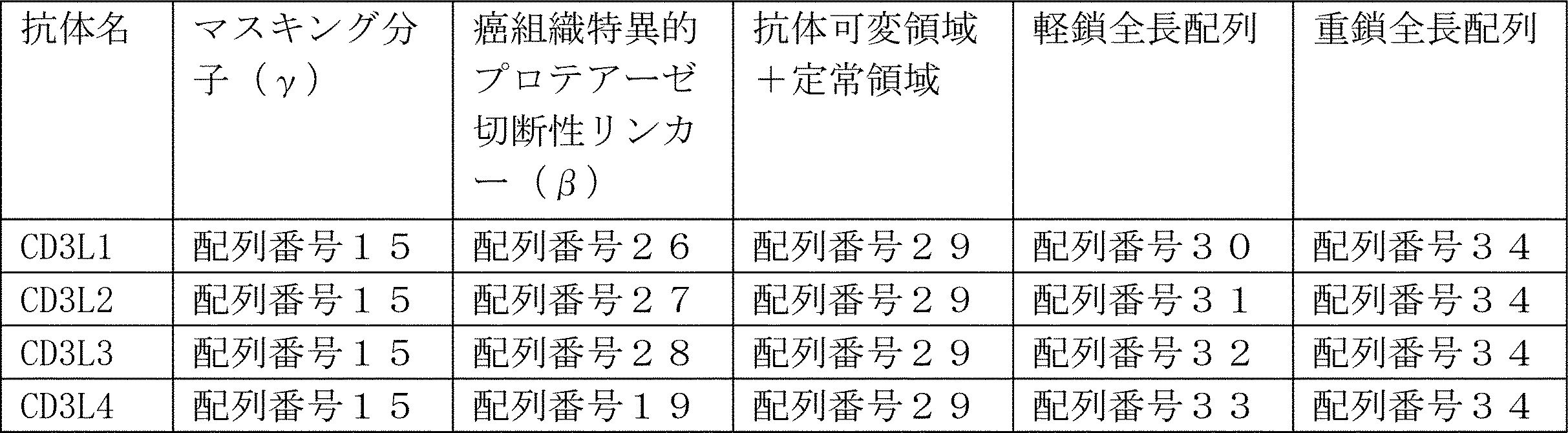
参考実施例2の方法に従い、抗CD3抗体を作製した。具体的には、まず、重鎖可変領域(配列番号:10)とpE22Hh(配列番号:35)とを融合したポリペプチドの動物細胞発現用の発現ベクター、軽鎖可変領域(配列番号:11)とKappa鎖(配列番号:36)とを融合したポリペプチドの動物細胞発現用の発現ベクター、および、重鎖定常領域のヒンジ部からC末端側(Kn0101G3(配列番号:37))の動物細胞発現用の発現ベクターを用いて、これらのポリペプチドを細胞に共発現させ、One arm抗体として抗CD3抗体を産生させた。次いで、培養上清から、抗CD3抗体を精製した。
抗CD3抗体とヒトCD3の結合評価は、BiacoreT200を用いて行った。具体的には、CM4チップにストレプトアビジンを介してビオチン化CD3ペプチドを結合させ、作製した抗体をアナライトとして流し、37℃で結合アフィニティーを解析した。
以下の表3に、結合評価の結果を示す。

参考実施例3-1.CD3結合を増強する方法
国際公開第2015/068847号に記載した抗CD3抗体由来の抗体ライブラリー(Dual Fab Library)を利用する方法として、CD3への結合が増大した抗体を得る方法が考えられる。一般に、結合力を増強する方法(Affinity maturation)は、取得された抗体配列に対して部位特異的変異法によってアミノ酸を改変して結合力を測定する方法と、Phage displayをはじめとするIn vitro display法を用いる方法が挙げられる。In vitro display法では、取得された配列に対してError prone PCR法等によって変異が導入された多種類の抗体配列をライブラリーとして、結合力が強い配列を選択する。
CD3への結合が、従来の抗CD3抗体(例えば、上述のテンプレート配列を有するCD3結合抗体)の場合の80%以上であるアミノ酸を選定したDual Fab Libraryを用いることで、CD3への結合が強い配列を効率よく見つけられると考えられる。
国際公開第2015/068847号で開示したDual Fab libraryからヒトCD3に対して結合するFabドメイン(抗体断片)を同定した。抗原として、ビオチン標識されたCD3ペプチドを用いて、ヒトCD3に対して結合能をもつ抗体断片の濃縮を行った。
構築されたファージディスプレイ用ファージミドを保持した大腸菌からファージ産生が行われた。ファージ産生が行われた大腸菌の培養液に2.5 M NaCl/10%PEGを添加することによって沈殿させたファージの集団をTBSにて希釈することによってファージライブラリー液が得られた。次に、ファージライブラリー液に終濃度4%BSAとなるようにBSAが添加された。パンニング方法として、一般的な方法である磁気ビーズに固定化した抗原を用いたパンニング方法が参照された(J. Immunol. Methods. (2008) 332 (1-2), 2-9、J. Immunol. Methods. (2001) 247 (1-2), 191-203、Biotechnol. Prog. (2002) 18 (2) 212-20、Mol. Cell Proteomics (2003) 2 (2), 61-9)。磁気ビーズとして、NeutrAvidin coated beads(Sera-Mag SpeedBeads NeutrAvidin-coated)もしくはStreptavidin coated beads(Dynabeads M-280 Streptavidin)が用いられた。
参考実施例3-2で得られたCD3結合能をもつ抗体断片の集団はFabドメインのみで構成される。そこで、IgG型(FabおよびFcの結合体)へ変換を行った。CD3結合能をもつ抗体断片を有する大腸菌からDual Fab LibraryのH鎖に特異的に結合するプライマーを用いて、PCRによってVH断片を増幅した。増幅したVH断片は参考実施例2の方法でF760mnP17(配列番号:39)が組み込まれた動物細胞発現用のプラスミドに組み込まれた。具体的には、重鎖としてAN121H-F760mnP17(配列番号:40)、L鎖としてGLS3000(配列番号:13)とKappa配列(配列番号:36)を連結した配列(配列番号:25)を採用し、参考実施例1に従って発現精製が行われた。この抗体をAN121と呼ぶ。
参考実施例で調製された抗体のCD3εδヘテロダイマーに対する結合を表面プラズモン法(SPR法)で評価した。
CD3εδヘテロダイマーは以下の方法で調製した。まず、CD3εの細胞外ドメインをコードする遺伝子の3'末端にFactor Xa切断サイトをコードする遺伝子とEU numbering 349番目がCys, 366番目がTrpとなっているヒト免疫グロブリン(IgG1)のヒンジ領域よりもC末端のアミノ酸をコードする遺伝子を融合し、さらにTEV protease切断サイトとBAPタグ配列をコードする遺伝子を融合した遺伝子(配列番号:41をコードする遺伝子)を動物細胞発現ベクターに挿入した。つぎにCD3δの細胞外ドメインをコードする遺伝子の3'末端にFactor Xa切断サイトをコードする遺伝子とEU numbering356番目がCys, 366番目がSer, 368番目がAla, 407番目がValとなっているヒト免疫グロブリン(IgG1)のヒンジ領域よりもC末端をコードする遺伝子を融合し、さらにFlagタグ配列をコードする遺伝子を融合した遺伝子(配列番号:42をコードする遺伝子)を動物細胞発現ベクターに挿入した。参考実施例2と同様に、配列番号:41をコードする遺伝子と配列番号:42をコードする遺伝子を持った動物細胞発現ベクターをFreeStyle293細胞(Invitrogen)に導入した。導入後プロトコルに従って37℃で振とう培養し、5日後に上清を回収した。上清からProteinAカラム(Eshmuno A (Merck))を用いて、抗体の定常領域(特にFc)が融合しているCD3εδヘテロダイマーを得た。さらにヘテロダイマーを取得する目的でAnti-FLAG M2カラム(Sigma)を用いて、抗体の定常領域が融合しているCD3εδヘテロダイマーを分画した。引き続き、ゲル濾過クロマトグラフィー(Superdex200、GE Healthcare)を実施して目的のCD3εδヘテロダイマーを分取した。
参考実施例1に記載の重鎖可変領域(配列番号:10)と軽鎖可変領域(配列番号:11)を可変領域として含むラット抗CD3抗体、当業者公知の方法で軽鎖をヒト化した重鎖可変領域(配列番号:12)と軽鎖可変領域(配列番号:13)を可変領域として含むラット抗CD3抗体とヒト抗CD3抗体の組み合わせ、当業者公知の方法でヒト化した重鎖可変領域(配列番号:12)と軽鎖可変領域(配列番号:13)を可変領域として含むヒト化抗CD3抗体、および重鎖可変領域(配列番号:43)と軽鎖可変領域(配列番号:13)を可変領域として含むAN121のCD3εδヘテロダイマーへの結合活性を測定した。重鎖可変領域は定常領域としてのF760mnP17(配列番号:39)と融合し、軽鎖可変領域は定常領域としてのKappa鎖(配列番号:36)と融合して、参考実施例2の方法に従って抗体を発現させた。
CD3εδヘテロダイマーへの結合活性はBiacoreT200を用いて評価した。具体的には、CM3 chipにProteinGを当業者公知の方法で固定化し、固定化されたProteinGへ評価したい抗体を結合させた。その後、4000, 1000, 250, 62.5, 15.6, 3.9nMに調製したCD3εδヘテロダイマーを結合させ、シングルサイクルカイネティクスモードで結合を評価した。測定は、20mM ACES, 150mM NaCl, pH7.4の条件で、37℃で実施した。その結果を表4に示す。
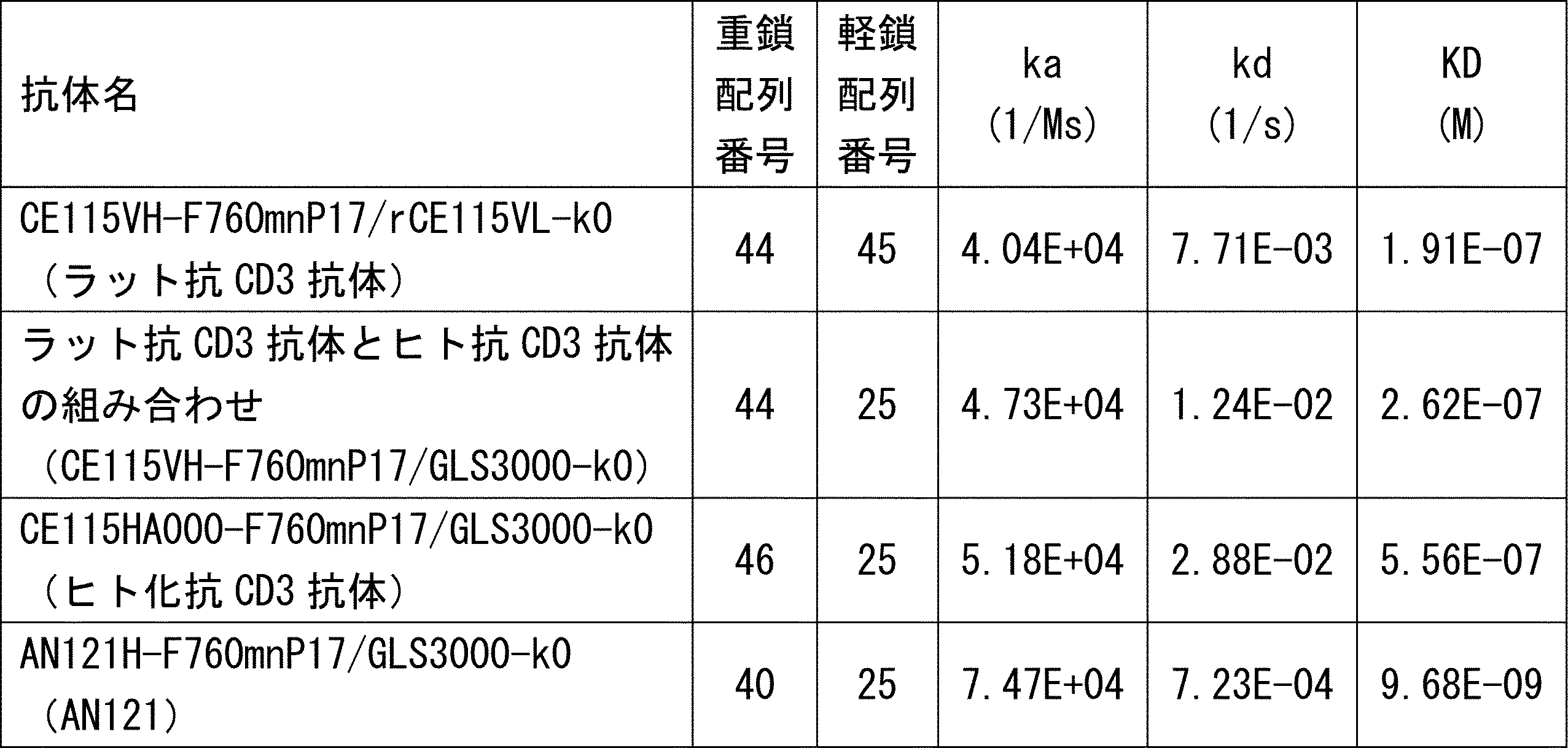
癌組織特異的プロテアーゼ切断性リンカー(β)を介したマスキング分子(γ)の付加によりCD3εへの結合がマスキングされた抗CD3抗体誘導体と、癌組織特異的プロテアーゼ切断性リンカー(β)を持たない切断を受けないリンカーを用いてマスキング分子(γ)を付加した抗CD3抗体誘導体(表5に示す)とを参考実施例2の方法に従い、作製した。癌組織特異的プロテアーゼ切断性リンカー(β)として、国際公開第2013/163631号に報告されている配列(LSGRSDNH:配列番号:47)を用いた。対照として、実施例3に示したMMP-2の切断配列(PLGLAG:配列番号:106)やGlyとSerで構成されるリンカーペプチドを癌組織特異的プロテアーゼ切断性リンカー(β)の部位に挿入した配列も作製した。具体的には、まず、重鎖可変領域とpE22Hh(配列番号:35)とを融合したポリペプチドの動物細胞発現用の発現ベクター、軽鎖可変領域とKappa鎖(配列番号:36)とを融合したポリペプチドの動物細胞発現用の発現ベクター、および、重鎖定常領域のヒンジ部からC末端側(Kn010(配列番号:48))の動物細胞発現用の発現ベクターを用いて、これらのポリペプチドを細胞に共発現させ、One arm抗体として抗CD3抗体を産生させた。次いで、培養上清から、抗CD3抗体を精製した。または、重鎖定常領域としてF760mnP17(配列番号:39)を重鎖可変領域に連結し、軽鎖全長と共に発現させてTWO arm抗体として抗CD3抗体を産生させ、培養上清から抗CD3抗体を精製した。
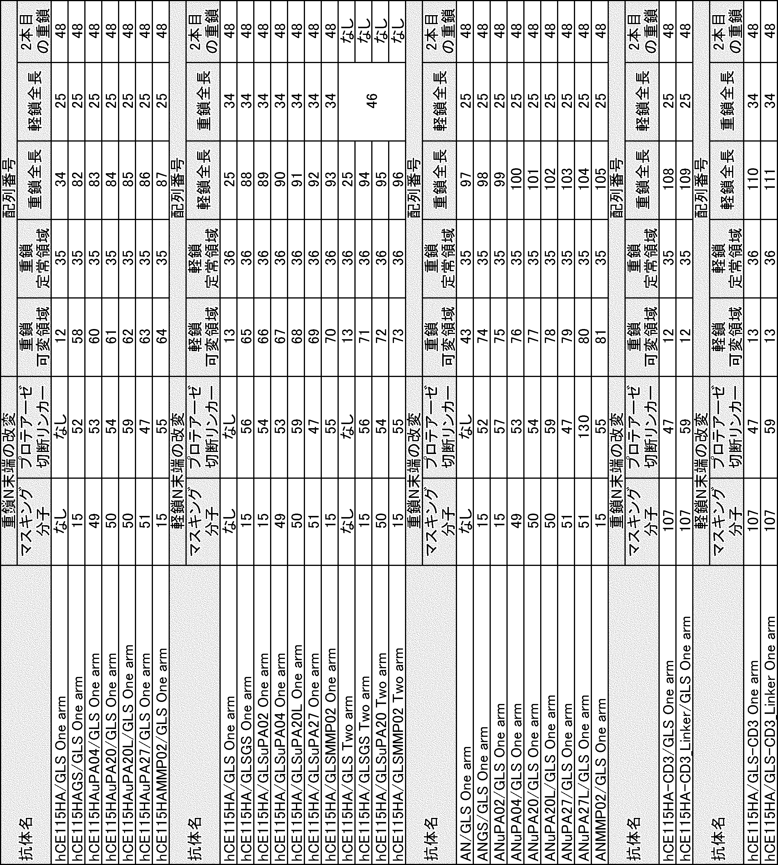

実施例6で調製された抗体をプロテアーゼで処理し、CD3εへの結合がマスキングされた抗CD3抗体誘導体がCD3εに結合するか評価した。参考実施例2に示した方法で抗体を取得し、取得した抗体5μgに対して終濃度25nMとなるようにuPA(Recombinant Human u-Plasminogen Activator , R&D systems)を加え、PBS中で37℃、16時間から20時間反応させた。
実施例7ではuPAを用いて切断を実施したが、当該配列はマトリプターゼでも切断されることが分かっている。そこで、実施例7で調製した検体がマトリプターゼでも切断され、CD3に対して結合するか評価した。抗体は実施例7の方法で調製され、その後3μgの抗体に対してヒトMT-SP1(Matriptase/ST14 Catalytic Domain, R&D systems)を終濃度50nMで添加した。添加後37℃で22時間保温した後、実施例7と同じ方法で結合活性を評価した。その結果を図9に示す。図9に示したように、癌組織特異的プロテアーゼ切断性リンカー(β)を含まない抗体はプロテアーゼを加えた場合(プロテアーゼ処理あり)とプロテアーゼを加えていない場合(プロテアーゼ未処理)で結合活性が変動しなかった。一方で、癌組織特異的プロテアーゼ切断性リンカー(β)を含む抗体は、プロテアーゼ処理によってCD3εへの結合が顕著に上昇した。具体的には、重鎖に癌組織特異的プロテアーゼ切断性リンカー(β)をつなげた場合には、hCE115HAuPA04に示されるように、マスキング分子(γ)が7アミノ酸からなり、癌組織特異的プロテアーゼ切断性リンカー(β)がGGGSの繰り返しで構成されるGSリンカーとuPAによって切断される配列を含みかつGGGSペプチド配列で重鎖N末端に接続されている場合に切断前後で結合活性が最も変化した。一方で、20アミノ酸(hCE115HAuPA20)や27アミノ酸(hCE115HAuPA27)にすると、マスキング分子(γ)が7アミノ酸であった場合(hCE115HAuPA04)と比べて、プロテアーゼ処理による結合活性の上昇率は低下した。また、軽鎖に癌組織特異的プロテアーゼ切断性リンカー(β)をつなげた場合も同様に、GLSuPA04に示されるように、マスキング分子(γ)が7アミノ酸からなり、癌組織特異的プロテアーゼ切断性リンカー(β)がGGGSの繰り返しで構成されるGSリンカーとuPAによって切断される配列を含みかつGGGSペプチド配列で重鎖N末端に接続されている場合に切断前後で結合活性が最も変化した。一方でマスキング分子(γ)を20アミノ酸(GLSuPA20)や27アミノ酸(GLSuPA27)にすると、マスキング分子(γ)が7アミノ酸であった場合(GLSuPA04)と比べて、プロテアーゼ処理による結合活性の上昇率は低下した。この結果は実施例7で示された結果と同様であり、切断を行うためのプロテーゼはuPAに限らず他のプロテアーゼでもよいことが示された。
実施例7および8では国際公開第2013/163631号に報告されている配列(LSGRSDNH:配列番号:47)を用い、uPAまたはマトリプターゼ(MT-SP1)を用いて切断を実施した。次に、実施例3に示すようにMMP-2によって切断される配列を用いた場合に、uPAやMT-SP1と同様に結合活性がプロテアーゼ処理によって上昇するか検討した。実施例7と同様の方法で抗体を調製した。
実施例7~9で、CD3εへの結合がマスクされた抗体は、プロテアーゼで処理することによってCD3εへの結合が顕著に上昇することが示された。次に、抗体のプロテアーゼ処理によってCD3の活性を誘導できるか評価した。SK-HEP1細胞にGPC3を強制発現させたSK-pca-60細胞を標的細胞とし、NFAT-RE-luc2-Jurkat細胞(Promega)をエフェクター細胞とした。NFAT-RE-luc2-Jurkat細胞(Promega)はヒト白血病T細胞株に由来し、CD3活性化に伴いNFATに応答してルシフェラーゼを発現するように改変された細胞である。参考実施例2の方法に従って、表7に示す抗CD3抗体とGCH065-F760mnN17(重鎖配列番号:112、軽鎖配列番号:113)を調製した。さらに二重特異性抗体とするために、精製したそれぞれのホモ体を当業者公知の手法(国際公開第2015/046467号)を用いて目的の片方のFabドメインがGPC3、もう片方のFabドメインがCD3に結合する二重特異性抗体を得た。二重特異性抗体は、XX/YY//ZZと表記され、XXは抗CD3抗体の重鎖可変領域を、YYは抗CD3抗体の軽鎖可変領域を、ZZは抗GPC3抗体を表す。その後、実施例7と同様にプロテアーゼで処理した。プロテアーゼ処理は、抗体に対して終濃度25nMとなるようにuPA(Recombinant Human u-Plasminogen Activator , R&D systems)を加え、PBS中で37℃、12時間以上反応させた。また、プロテアーゼ未処理の抗体もプロテアーゼ処理と同様に37℃で同じ時間保温した。
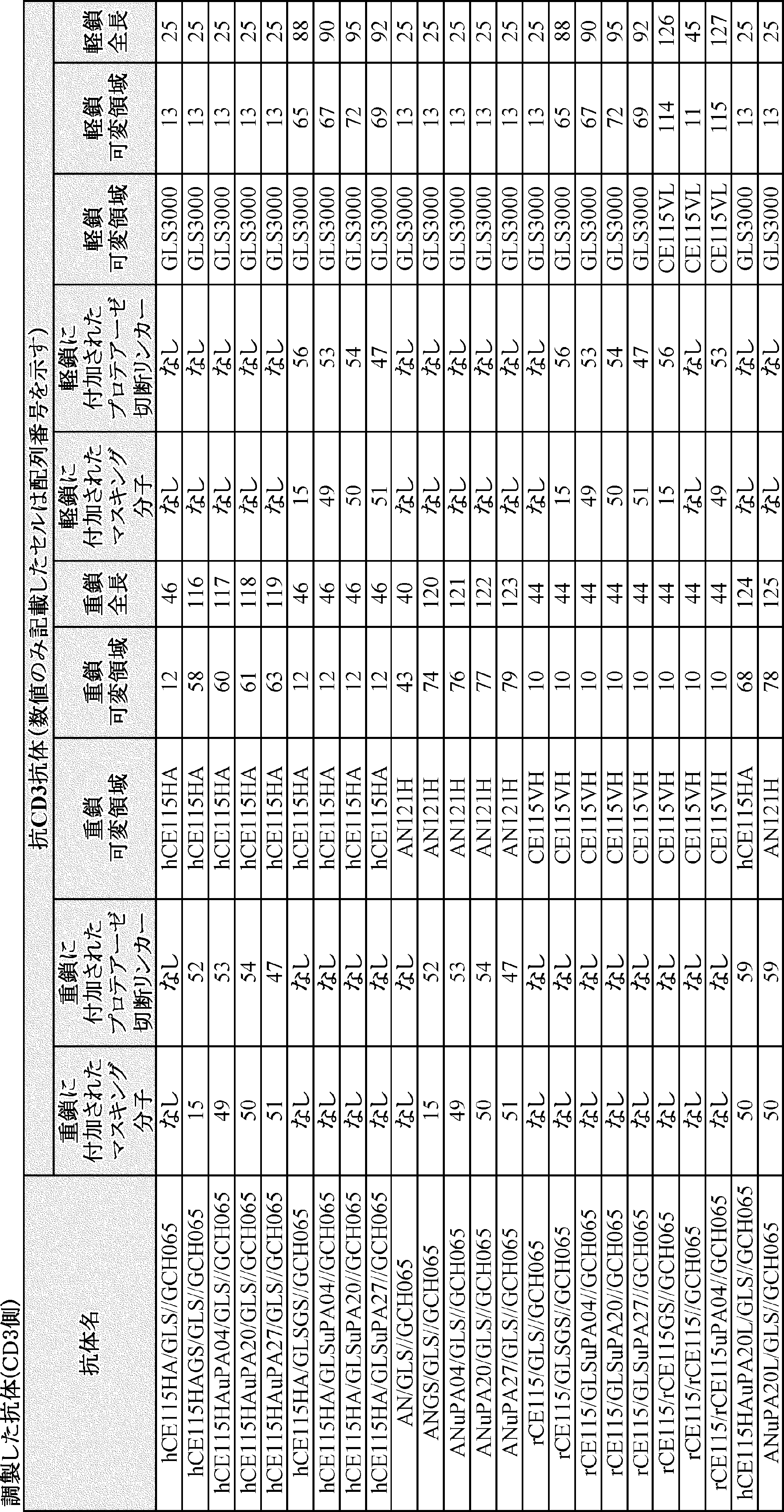

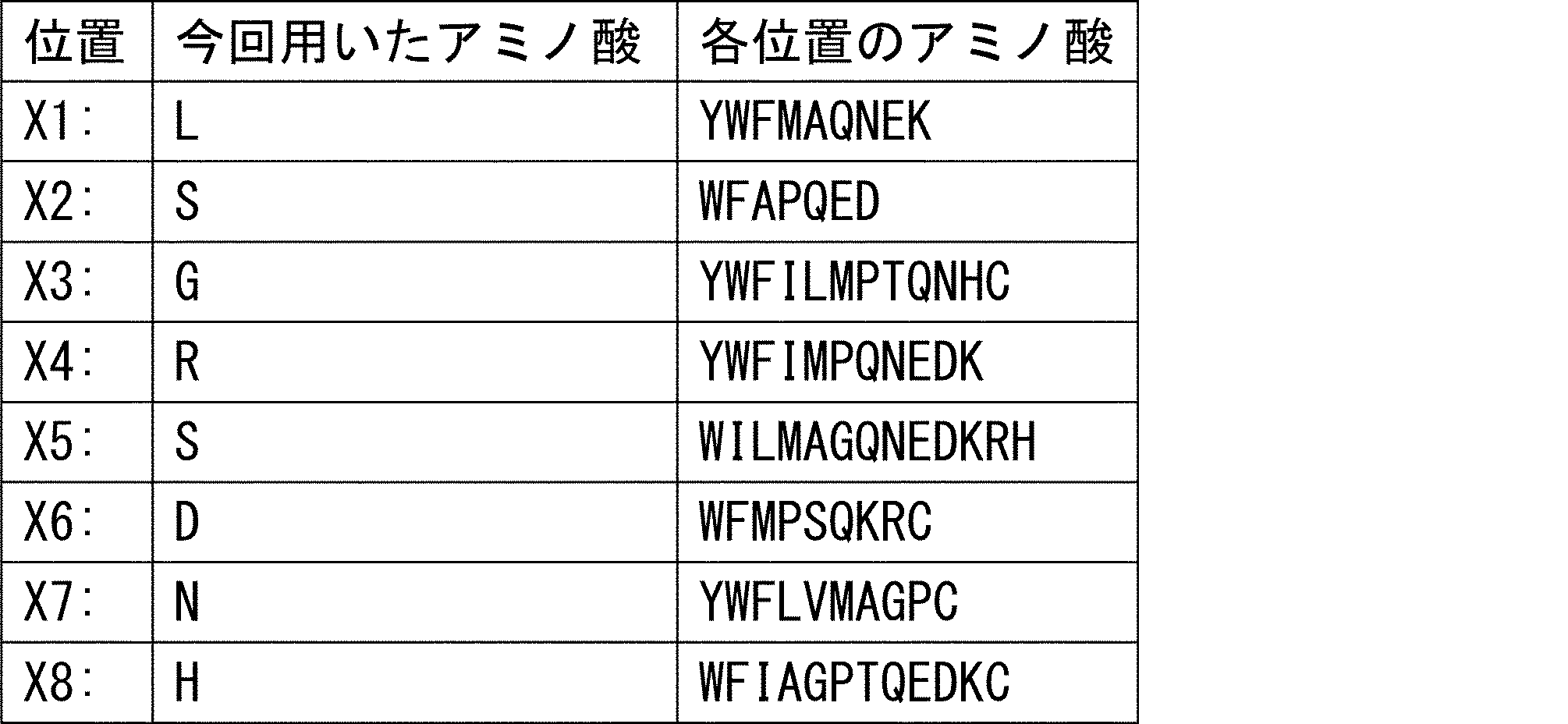
本発明によれば、マスキング分子(γ)は、癌組織特異的プロテアーゼ切断性リンカー(β)を介して多重抗原結合分子(α)に結合していることにより、正常組織には免疫細胞をリクルートしにくくなることから、多重抗原結合分子(α)による副作用を低減する効果を有するため、本発明の多重抗原結合分子融合体は特に医薬品に有用である。
Claims (14)
- アミノ酸配列QDGNE(配列番号:15)からなるポリペプチドを有する抗原を認識する免疫細胞抗原結合領域と、癌抗原を認識する癌抗原結合領域とを有する多重抗原結合分子(α)、
癌組織特異的プロテアーゼの標的配列からなるポリペプチドを有する癌組織特異的プロテアーゼ切断性リンカー(β)、および、
アミノ酸配列QDGNE(配列番号:15)からなるポリペプチドを有するマスキング分子(γ)を含み、
前記多重抗原結合分子(α)と前記マスキング分子(γ)とが前記癌組織特異的プロテアーゼ切断性リンカー(β)を介して結合している、多重抗原結合分子融合体。 - 前記免疫細胞抗原結合領域が、前記アミノ酸配列QDGNE(配列番号:15)からなるポリペプチドを有する抗原以外に少なくとも1種の免疫細胞抗原を認識する、請求項1に記載の多重抗原結合分子融合体。
- 前記免疫細胞抗原結合領域が、2以上の免疫細胞抗原を同時には認識し得ない、請求項2に記載の多重抗原結合分子融合体。
- 前記多重抗原結合分子(α)が、抗体または少なくとも2つのFv領域を有する抗体断片であり、前記癌抗原結合領域と前記免疫細胞抗原結合領域とが別々のFv領域により形成されている、請求項1乃至3のいずれか一項に記載の多重抗原結合分子融合体。
- 前記抗体または少なくとも2つのFv領域を有する抗体断片における軽鎖が、いずれも同じアミノ酸配列を有する、請求項4に記載の多重抗原結合分子融合体。
- 前記抗体または少なくとも2つのFv領域を有する抗体断片がさらにFc領域を有し、前記Fc領域がFcγ受容体を認識する機能を欠損するように改変されている、請求項4または5に記載の多重抗原結合分子融合体。
- 前記癌組織特異的プロテアーゼ切断性リンカー(β)が、前記免疫細胞抗原結合領域を形成する前記Fv領域の重鎖N末端または軽鎖N末端に融合している、請求項4乃至6のいずれか一項に記載の多重抗原結合分子融合体。
- 前記癌組織特異的プロテアーゼ切断性リンカー(β)と前記マスキング分子(γ)とが直鎖状の融合ポリペプチドであり、
前記癌組織特異的プロテアーゼ切断性リンカー(β)が前記免疫細胞抗原結合領域を形成する前記Fv領域の重鎖N末端に融合している場合には、前記融合ポリペプチドのアミノ酸数は11以上65以下であり、
前記癌組織特異的プロテアーゼ切断性リンカー(β)が前記免疫細胞抗原結合領域を形成する前記Fv領域の軽鎖N末端に融合している場合には、前記融合ポリペプチドのアミノ酸数は16以上65以下である、請求項7に記載の多重抗原結合分子融合体。 - 前記標的配列がアミノ酸配列PLGLAG(配列番号:9)である、請求項1乃至8のいずれか一項に記載の多重抗原結合分子融合体。
- 請求項1乃至9のいずれか一項に記載の多重抗原結合分子融合体および薬学的に許容される担体を含有する、医薬組成物。
- 癌治療用である、請求項10に記載の医薬組成物。
- 請求項10または11に記載の医薬組成物を患者に投与する工程を含む、癌の治療方法。
- 免疫細胞抗原と前記免疫細胞抗原を認識する免疫細胞抗原結合領域とのタンパク質複合体を用いたタンパク質立体構造解析のデータに基づき、前記免疫細胞抗原に含まれ、かつ、前記免疫細胞抗原結合領域により認識される線状エピトープを同定する工程を有する、線状エピトープの同定方法。
- 癌抗原を認識する癌抗原結合領域および免疫細胞抗原を認識する免疫細胞抗原結合領域を有する多重抗原結合分子(α)に、線状エピトープを、前記癌抗原を発現する癌組織で特異的に発現するプロテアーゼにより切断され得る領域を有する癌組織特異的プロテアーゼ切断性リンカー(β)を介して融合した融合タンパク質を発現する工程を有する、多重抗原結合分子融合体の製造方法。
Priority Applications (4)
| Application Number | Priority Date | Filing Date | Title |
|---|---|---|---|
| JP2017518007A JP6841754B2 (ja) | 2015-05-13 | 2016-05-13 | 多重抗原結合分子融合体、医薬組成物、線状エピトープの同定方法、および多重抗原結合分子融合体の製造方法 |
| US15/573,154 US11400157B2 (en) | 2015-05-13 | 2016-05-13 | Multiple antigen binding molecular fusion, pharmaceutical composition, method for identifying linear epitope, and method for preparing multiple antigen binding molecular fusion |
| EP16792788.8A EP3296395B2 (en) | 2015-05-13 | 2016-05-13 | Multiple antigen binding molecular fusion, pharmaceutical composition, method for identifying linear epitope, and method for preparing multiple antigen binding molecular fusion |
| US17/847,909 US12377143B2 (en) | 2015-05-13 | 2022-06-23 | Multiple antigen binding molecular fusion, pharmaceutical composition, method for identifying linear epitope, and method for preparing multiple antigen binding molecular fusion |
Applications Claiming Priority (2)
| Application Number | Priority Date | Filing Date | Title |
|---|---|---|---|
| JP2015-098208 | 2015-05-13 | ||
| JP2015098208 | 2015-05-13 |
Related Child Applications (2)
| Application Number | Title | Priority Date | Filing Date |
|---|---|---|---|
| US15/573,154 A-371-Of-International US11400157B2 (en) | 2015-05-13 | 2016-05-13 | Multiple antigen binding molecular fusion, pharmaceutical composition, method for identifying linear epitope, and method for preparing multiple antigen binding molecular fusion |
| US17/847,909 Division US12377143B2 (en) | 2015-05-13 | 2022-06-23 | Multiple antigen binding molecular fusion, pharmaceutical composition, method for identifying linear epitope, and method for preparing multiple antigen binding molecular fusion |
Publications (1)
| Publication Number | Publication Date |
|---|---|
| WO2016182064A1 true WO2016182064A1 (ja) | 2016-11-17 |
Family
ID=57249300
Family Applications (1)
| Application Number | Title | Priority Date | Filing Date |
|---|---|---|---|
| PCT/JP2016/064301 Ceased WO2016182064A1 (ja) | 2015-05-13 | 2016-05-13 | 多重抗原結合分子融合体、医薬組成物、線状エピトープの同定方法、および多重抗原結合分子融合体の製造方法 |
Country Status (4)
| Country | Link |
|---|---|
| US (2) | US11400157B2 (ja) |
| EP (1) | EP3296395B2 (ja) |
| JP (1) | JP6841754B2 (ja) |
| WO (1) | WO2016182064A1 (ja) |
Cited By (24)
| Publication number | Priority date | Publication date | Assignee | Title |
|---|---|---|---|---|
| WO2018155611A1 (ja) | 2017-02-24 | 2018-08-30 | 中外製薬株式会社 | 薬学的組成物、抗原結合分子、治療方法、およびスクリーニング方法 |
| WO2019230868A1 (ja) * | 2018-05-30 | 2019-12-05 | 中外製薬株式会社 | 単ドメイン抗体含有リガンド結合分子 |
| CN111836828A (zh) * | 2017-11-28 | 2020-10-27 | 中外制药株式会社 | 包括抗原结合结构域和运送部分的多肽 |
| CN112513083A (zh) * | 2018-05-14 | 2021-03-16 | 哈普恩治疗公司 | 用于条件性激活免疫球蛋白分子的结合部分 |
| WO2021207657A1 (en) | 2020-04-09 | 2021-10-14 | Cytomx Therapeutics, Inc. | Compositions containing activatable antibodies |
| US11168139B2 (en) | 2016-11-28 | 2021-11-09 | Chugai Seiyaku Kabushiki Kaisha | Antigen-binding domain, and polypeptide including conveying section |
| EP3794044A4 (en) * | 2018-05-14 | 2022-02-16 | Harpoon Therapeutics, Inc. | DOUBLE BINDING UNIT |
| US11453716B2 (en) | 2016-05-20 | 2022-09-27 | Harpoon Therapeutics, Inc. | Single domain serum albumin binding protein |
| US11535668B2 (en) | 2017-02-28 | 2022-12-27 | Harpoon Therapeutics, Inc. | Inducible monovalent antigen binding protein |
| US11607453B2 (en) | 2017-05-12 | 2023-03-21 | Harpoon Therapeutics, Inc. | Mesothelin binding proteins |
| US11623958B2 (en) | 2016-05-20 | 2023-04-11 | Harpoon Therapeutics, Inc. | Single chain variable fragment CD3 binding proteins |
| WO2023100829A1 (ja) * | 2021-11-30 | 2023-06-08 | 第一三共株式会社 | プロテアーゼ分解性マスク抗体 |
| US11807692B2 (en) | 2018-09-25 | 2023-11-07 | Harpoon Therapeutics, Inc. | DLL3 binding proteins and methods of use |
| US11976125B2 (en) | 2017-10-13 | 2024-05-07 | Harpoon Therapeutics, Inc. | B cell maturation antigen binding proteins |
| US12060654B2 (en) | 2016-11-28 | 2024-08-13 | Chugai Seiyaku Kabushiki Kaisha | Ligand-binding molecule having adjustable ligand binding activity |
| US12077577B2 (en) | 2018-05-30 | 2024-09-03 | Chugai Seiyaku Kabushiki Kaisha | Polypeptide comprising aggrecan binding domain and carrying moiety |
| US12084518B2 (en) | 2015-05-21 | 2024-09-10 | Harpoon Therapeutics, Inc. | Trispecific binding proteins and methods of use |
| US12195544B2 (en) | 2018-09-21 | 2025-01-14 | Harpoon Therapeutics, Inc. | EGFR binding proteins and methods of use |
| US12195528B2 (en) | 2017-11-28 | 2025-01-14 | Chugai Seiyaku Kabushiki Kaisha | Ligand-binding molecule having adjustable ligand-binding activity |
| US12371504B2 (en) | 2017-10-13 | 2025-07-29 | Harpoon Therapeutics, Inc. | Trispecific proteins and methods of use |
| US12516128B2 (en) | 2019-05-14 | 2026-01-06 | Harpoon Therapeutics, Inc. | EpCAM binding proteins and methods of use |
| US12655220B2 (en) | 2020-01-20 | 2026-06-16 | Chugai Seiyaku Kabushiki Kaisha | Ligand-binding fusion proteins |
| US12680093B2 (en) | 2019-06-05 | 2026-07-14 | Chugai Seiyaku Kabushiki Kaisha | Protease substrate, and polypeptide including protease cleavage sequence |
| JP7909374B2 (ja) | 2018-05-30 | 2026-08-21 | 中外製薬株式会社 | 単ドメイン抗体含有リガンド結合分子 |
Families Citing this family (5)
| Publication number | Priority date | Publication date | Assignee | Title |
|---|---|---|---|---|
| JP6841754B2 (ja) | 2015-05-13 | 2021-03-10 | 中外製薬株式会社 | 多重抗原結合分子融合体、医薬組成物、線状エピトープの同定方法、および多重抗原結合分子融合体の製造方法 |
| CN113603783B (zh) | 2018-12-21 | 2022-07-12 | 浙江时迈药业有限公司 | 蛋白酶可切割的双特异性抗体及其用途 |
| JPWO2020213724A1 (ja) | 2019-04-19 | 2020-10-22 | ||
| CN113811546B (zh) * | 2019-05-07 | 2025-03-11 | 免疫里森公司 | 前体三特异性抗体构建体及其使用方法 |
| CN113905757B (zh) | 2019-06-05 | 2026-01-13 | 中外制药株式会社 | 抗体切割位点结合分子 |
Citations (9)
| Publication number | Priority date | Publication date | Assignee | Title |
|---|---|---|---|---|
| WO2012073985A1 (ja) * | 2010-11-30 | 2012-06-07 | 中外製薬株式会社 | 細胞傷害誘導治療剤 |
| JP2013528569A (ja) * | 2010-04-01 | 2013-07-11 | アムゲン リサーチ (ミュンヘン) ゲーエムベーハー | 異種間特異的なPSMAxCD3二重特異性単鎖抗体 |
| JP2014519322A (ja) * | 2011-05-16 | 2014-08-14 | フェイビオン ファーマシューティカルズ, インコーポレイテッド | 多重特異性Fab融合タンパク質および使用法 |
| WO2014144722A2 (en) * | 2013-03-15 | 2014-09-18 | Amgen Inc. | BISPECIFIC-Fc MOLECULES |
| JP2015509952A (ja) * | 2012-02-28 | 2015-04-02 | ザ ユニバーシティ オブ バーミンガム | 免疫療法分子および使用 |
| WO2015046554A1 (ja) * | 2013-09-30 | 2015-04-02 | 中外製薬株式会社 | 改変されたヘルパーファージを用いて抗原結合分子を作製する方法 |
| JP2015509951A (ja) * | 2012-03-01 | 2015-04-02 | アムゲン リサーチ (ミュンヘン) ゲーエムベーハーAMGEN Research(Munich)GmbH | 長寿命ポリペプチド結合分子 |
| WO2016014974A2 (en) * | 2014-07-25 | 2016-01-28 | Cytomx Therapeutics, Inc. | Anti-cd3 antibodies, activatable anti-cd3 antibodies, multispecific anti-cd3 antibodies, multispecific activatable anti-cd3 antibodies, and methods of using the same |
| WO2016046778A2 (en) * | 2014-09-25 | 2016-03-31 | Amgen Inc | Protease-activatable bispecific proteins |
Family Cites Families (14)
| Publication number | Priority date | Publication date | Assignee | Title |
|---|---|---|---|---|
| AU2003256266A1 (en) | 2002-06-12 | 2003-12-31 | Genencor International, Inc. | Methods and compositions for milieu-dependent binding of a targeted agent to a target |
| EP2190861A4 (en) | 2007-08-22 | 2011-03-30 | Univ California | ACTIVE BINDING POLYPEPTIDES AND METHOD FOR THEIR IDENTIFICATION AND USE |
| EP3543256A1 (en) | 2009-01-12 | 2019-09-25 | Cytomx Therapeutics Inc. | Modified antibody compositions, methods of making and using thereof |
| WO2010096868A1 (en) | 2009-02-25 | 2010-09-02 | Stiefel Research Australia Pty Ltd | Topical foam composition |
| KR20130096731A (ko) | 2010-09-08 | 2013-08-30 | 할로자임, 아이엔씨 | 조건부 활성 치료적 단백질을 평가,확인 또는 진화시키는 방법 |
| WO2013192546A1 (en) | 2012-06-22 | 2013-12-27 | Cytomx Therapeutics, Inc. | Activatable antibodies having non-binding steric moieties and mehtods of using the same |
| JP6693872B2 (ja) | 2013-07-25 | 2020-05-13 | シトムクス セラピューティクス,インコーポレイティド | 多重特異性抗体、多重特異性活性化可能抗体、及びそれらの使用方法 |
| CA2929044A1 (en) | 2013-11-11 | 2015-05-14 | Chugai Seiyaku Kabushiki Kaisha | Antigen-binding molecule containing modified antibody variable region |
| JP5949731B2 (ja) | 2013-11-18 | 2016-07-13 | トヨタ自動車株式会社 | ハイブリッド車両 |
| EP3628328B1 (en) | 2014-01-31 | 2022-09-14 | Cytomx Therapeutics Inc. | Matriptase and u-plasminogen activator substrates and other cleavable moieties and methods of use thereof |
| IL300408B2 (en) | 2015-01-20 | 2025-11-01 | Cytomx Therapeutics Inc | Matrix metalloprotease-cleavable and serine protease cleavable substrates and methods of use thereof |
| MA42971A (fr) | 2015-03-13 | 2018-08-15 | Cytomx Therapeutics Inc | Anticorps anti-pdl1, anticorps anti-pld1 activables, et leurs procédés d'utilisation |
| EP3778640A1 (en) | 2015-05-01 | 2021-02-17 | Genentech, Inc. | Masked anti-cd3 antibodies and methods of use |
| JP6841754B2 (ja) | 2015-05-13 | 2021-03-10 | 中外製薬株式会社 | 多重抗原結合分子融合体、医薬組成物、線状エピトープの同定方法、および多重抗原結合分子融合体の製造方法 |
-
2016
- 2016-05-13 JP JP2017518007A patent/JP6841754B2/ja active Active
- 2016-05-13 EP EP16792788.8A patent/EP3296395B2/en active Active
- 2016-05-13 WO PCT/JP2016/064301 patent/WO2016182064A1/ja not_active Ceased
- 2016-05-13 US US15/573,154 patent/US11400157B2/en active Active
-
2022
- 2022-06-23 US US17/847,909 patent/US12377143B2/en active Active
Patent Citations (9)
| Publication number | Priority date | Publication date | Assignee | Title |
|---|---|---|---|---|
| JP2013528569A (ja) * | 2010-04-01 | 2013-07-11 | アムゲン リサーチ (ミュンヘン) ゲーエムベーハー | 異種間特異的なPSMAxCD3二重特異性単鎖抗体 |
| WO2012073985A1 (ja) * | 2010-11-30 | 2012-06-07 | 中外製薬株式会社 | 細胞傷害誘導治療剤 |
| JP2014519322A (ja) * | 2011-05-16 | 2014-08-14 | フェイビオン ファーマシューティカルズ, インコーポレイテッド | 多重特異性Fab融合タンパク質および使用法 |
| JP2015509952A (ja) * | 2012-02-28 | 2015-04-02 | ザ ユニバーシティ オブ バーミンガム | 免疫療法分子および使用 |
| JP2015509951A (ja) * | 2012-03-01 | 2015-04-02 | アムゲン リサーチ (ミュンヘン) ゲーエムベーハーAMGEN Research(Munich)GmbH | 長寿命ポリペプチド結合分子 |
| WO2014144722A2 (en) * | 2013-03-15 | 2014-09-18 | Amgen Inc. | BISPECIFIC-Fc MOLECULES |
| WO2015046554A1 (ja) * | 2013-09-30 | 2015-04-02 | 中外製薬株式会社 | 改変されたヘルパーファージを用いて抗原結合分子を作製する方法 |
| WO2016014974A2 (en) * | 2014-07-25 | 2016-01-28 | Cytomx Therapeutics, Inc. | Anti-cd3 antibodies, activatable anti-cd3 antibodies, multispecific anti-cd3 antibodies, multispecific activatable anti-cd3 antibodies, and methods of using the same |
| WO2016046778A2 (en) * | 2014-09-25 | 2016-03-31 | Amgen Inc | Protease-activatable bispecific proteins |
Non-Patent Citations (5)
Cited By (30)
| Publication number | Priority date | Publication date | Assignee | Title |
|---|---|---|---|---|
| US12084518B2 (en) | 2015-05-21 | 2024-09-10 | Harpoon Therapeutics, Inc. | Trispecific binding proteins and methods of use |
| US11453716B2 (en) | 2016-05-20 | 2022-09-27 | Harpoon Therapeutics, Inc. | Single domain serum albumin binding protein |
| US12528859B2 (en) | 2016-05-20 | 2026-01-20 | Harpoon Therapeutics, Inc. | Single domain serum albumin binding protein |
| US11623958B2 (en) | 2016-05-20 | 2023-04-11 | Harpoon Therapeutics, Inc. | Single chain variable fragment CD3 binding proteins |
| US11168139B2 (en) | 2016-11-28 | 2021-11-09 | Chugai Seiyaku Kabushiki Kaisha | Antigen-binding domain, and polypeptide including conveying section |
| US11932697B2 (en) | 2016-11-28 | 2024-03-19 | Chugai Seiyaku Kabushiki Kaisha | Antigen-binding domain, and polypeptide including conveying section |
| US12060654B2 (en) | 2016-11-28 | 2024-08-13 | Chugai Seiyaku Kabushiki Kaisha | Ligand-binding molecule having adjustable ligand binding activity |
| WO2018155611A1 (ja) | 2017-02-24 | 2018-08-30 | 中外製薬株式会社 | 薬学的組成物、抗原結合分子、治療方法、およびスクリーニング方法 |
| US11535668B2 (en) | 2017-02-28 | 2022-12-27 | Harpoon Therapeutics, Inc. | Inducible monovalent antigen binding protein |
| US11607453B2 (en) | 2017-05-12 | 2023-03-21 | Harpoon Therapeutics, Inc. | Mesothelin binding proteins |
| US12371504B2 (en) | 2017-10-13 | 2025-07-29 | Harpoon Therapeutics, Inc. | Trispecific proteins and methods of use |
| US11976125B2 (en) | 2017-10-13 | 2024-05-07 | Harpoon Therapeutics, Inc. | B cell maturation antigen binding proteins |
| CN111836828B (zh) * | 2017-11-28 | 2024-12-20 | 中外制药株式会社 | 包括抗原结合结构域和运送部分的多肽 |
| US12030955B2 (en) | 2017-11-28 | 2024-07-09 | Chugai Seiyaku Kabushiki Kaisha | Polypeptide including antigen-binding domain and carrying section |
| CN111836828A (zh) * | 2017-11-28 | 2020-10-27 | 中外制药株式会社 | 包括抗原结合结构域和运送部分的多肽 |
| US12195528B2 (en) | 2017-11-28 | 2025-01-14 | Chugai Seiyaku Kabushiki Kaisha | Ligand-binding molecule having adjustable ligand-binding activity |
| EP3794044A4 (en) * | 2018-05-14 | 2022-02-16 | Harpoon Therapeutics, Inc. | DOUBLE BINDING UNIT |
| CN112513083A (zh) * | 2018-05-14 | 2021-03-16 | 哈普恩治疗公司 | 用于条件性激活免疫球蛋白分子的结合部分 |
| US12415860B2 (en) | 2018-05-14 | 2025-09-16 | Harpoon Therapeutics, Inc. | Binding moiety for conditional activation of immunoglobulin molecules |
| WO2019230868A1 (ja) * | 2018-05-30 | 2019-12-05 | 中外製薬株式会社 | 単ドメイン抗体含有リガンド結合分子 |
| JPWO2019230868A1 (ja) * | 2018-05-30 | 2021-06-24 | 中外製薬株式会社 | 単ドメイン抗体含有リガンド結合分子 |
| US12077577B2 (en) | 2018-05-30 | 2024-09-03 | Chugai Seiyaku Kabushiki Kaisha | Polypeptide comprising aggrecan binding domain and carrying moiety |
| JP7909374B2 (ja) | 2018-05-30 | 2026-08-21 | 中外製薬株式会社 | 単ドメイン抗体含有リガンド結合分子 |
| US12195544B2 (en) | 2018-09-21 | 2025-01-14 | Harpoon Therapeutics, Inc. | EGFR binding proteins and methods of use |
| US11807692B2 (en) | 2018-09-25 | 2023-11-07 | Harpoon Therapeutics, Inc. | DLL3 binding proteins and methods of use |
| US12516128B2 (en) | 2019-05-14 | 2026-01-06 | Harpoon Therapeutics, Inc. | EpCAM binding proteins and methods of use |
| US12680093B2 (en) | 2019-06-05 | 2026-07-14 | Chugai Seiyaku Kabushiki Kaisha | Protease substrate, and polypeptide including protease cleavage sequence |
| US12655220B2 (en) | 2020-01-20 | 2026-06-16 | Chugai Seiyaku Kabushiki Kaisha | Ligand-binding fusion proteins |
| WO2021207657A1 (en) | 2020-04-09 | 2021-10-14 | Cytomx Therapeutics, Inc. | Compositions containing activatable antibodies |
| WO2023100829A1 (ja) * | 2021-11-30 | 2023-06-08 | 第一三共株式会社 | プロテアーゼ分解性マスク抗体 |
Also Published As
| Publication number | Publication date |
|---|---|
| EP3296395B1 (en) | 2021-06-30 |
| EP3296395A1 (en) | 2018-03-21 |
| US20220401557A1 (en) | 2022-12-22 |
| US12377143B2 (en) | 2025-08-05 |
| JP6841754B2 (ja) | 2021-03-10 |
| US20180125972A1 (en) | 2018-05-10 |
| EP3296395A4 (en) | 2018-12-05 |
| JPWO2016182064A1 (ja) | 2018-03-01 |
| EP3296395B2 (en) | 2024-06-19 |
| US11400157B2 (en) | 2022-08-02 |
Similar Documents
| Publication | Publication Date | Title |
|---|---|---|
| US12377143B2 (en) | Multiple antigen binding molecular fusion, pharmaceutical composition, method for identifying linear epitope, and method for preparing multiple antigen binding molecular fusion | |
| US20230151112A1 (en) | Immunoactivating antigen-binding molecule | |
| JP7273904B2 (ja) | 液性免疫応答の増強方法 | |
| US11660340B2 (en) | Combination therapy using T cell redirection antigen binding molecule against cell having immunosuppressing function | |
| US20180171017A1 (en) | Combined use of immune activators | |
| KR20220161156A (ko) | 면역 활성화 다중 특이성 항원 결합 분자 및 그의 사용 | |
| JP2020518584A (ja) | 細胞傷害誘導治療剤 | |
| KR20170017916A (ko) | 면역 억제 기능을 갖는 세포에 대한 t 세포 리다이렉트 항원 결합 분자 | |
| JP2023519776A (ja) | 多重特異性抗原結合分子を製造するための方法 | |
| US20250340631A1 (en) | Cldn18.2-targeting antibody, bispecific antibody and use thereof | |
| US20230357398A1 (en) | Novel human antibodies binding to human cd3 epsilon | |
| JP2021510064A (ja) | 細胞傷害誘導治療剤 | |
| IL299232A (en) | Bispecific antibody and its use | |
| US20250019457A1 (en) | Trop2 antibodies | |
| JP2017501723A (ja) | 新規な抗adam17抗体および癌の治療におけるその使用 | |
| US20260103514A1 (en) | Antibodies that specifically bind cldn6 or cd3 and bispecific antibodies that specifically bind cldn6 and cd3 | |
| HK40110242A (en) | Immunoactivating antigen-binding molecule | |
| HK40006054A (en) | Antigen-binding domain, and polypeptide including conveying section | |
| HK1228935B (en) | Immunoactivating antigen-binding molecule |
Legal Events
| Date | Code | Title | Description |
|---|---|---|---|
| 121 | Ep: the epo has been informed by wipo that ep was designated in this application |
Ref document number: 16792788 Country of ref document: EP Kind code of ref document: A1 |
|
| ENP | Entry into the national phase |
Ref document number: 2017518007 Country of ref document: JP Kind code of ref document: A |
|
| WWE | Wipo information: entry into national phase |
Ref document number: 15573154 Country of ref document: US |
|
| NENP | Non-entry into the national phase |
Ref country code: DE |
|
| WWE | Wipo information: entry into national phase |
Ref document number: 2016792788 Country of ref document: EP |