WO2018155611A1 - 薬学的組成物、抗原結合分子、治療方法、およびスクリーニング方法 - Google Patents
薬学的組成物、抗原結合分子、治療方法、およびスクリーニング方法 Download PDFInfo
- Publication number
- WO2018155611A1 WO2018155611A1 PCT/JP2018/006626 JP2018006626W WO2018155611A1 WO 2018155611 A1 WO2018155611 A1 WO 2018155611A1 JP 2018006626 W JP2018006626 W JP 2018006626W WO 2018155611 A1 WO2018155611 A1 WO 2018155611A1
- Authority
- WO
- WIPO (PCT)
- Prior art keywords
- antigen
- binding molecule
- binding
- molecule
- region
- Prior art date
Links
Images


Classifications
-
- C—CHEMISTRY; METALLURGY
- C07—ORGANIC CHEMISTRY
- C07K—PEPTIDES
- C07K16/00—Immunoglobulins [IGs], e.g. monoclonal or polyclonal antibodies
- C07K16/18—Immunoglobulins [IGs], e.g. monoclonal or polyclonal antibodies against material from animals or humans
- C07K16/28—Immunoglobulins [IGs], e.g. monoclonal or polyclonal antibodies against material from animals or humans against receptors, cell surface antigens or cell surface determinants
- C07K16/2803—Immunoglobulins [IGs], e.g. monoclonal or polyclonal antibodies against material from animals or humans against receptors, cell surface antigens or cell surface determinants against the immunoglobulin superfamily
-
- A—HUMAN NECESSITIES
- A61—MEDICAL OR VETERINARY SCIENCE; HYGIENE
- A61P—SPECIFIC THERAPEUTIC ACTIVITY OF CHEMICAL COMPOUNDS OR MEDICINAL PREPARATIONS
- A61P31/00—Antiinfectives, i.e. antibiotics, antiseptics, chemotherapeutics
- A61P31/04—Antibacterial agents
-
- A—HUMAN NECESSITIES
- A61—MEDICAL OR VETERINARY SCIENCE; HYGIENE
- A61P—SPECIFIC THERAPEUTIC ACTIVITY OF CHEMICAL COMPOUNDS OR MEDICINAL PREPARATIONS
- A61P31/00—Antiinfectives, i.e. antibiotics, antiseptics, chemotherapeutics
- A61P31/12—Antivirals
-
- A—HUMAN NECESSITIES
- A61—MEDICAL OR VETERINARY SCIENCE; HYGIENE
- A61P—SPECIFIC THERAPEUTIC ACTIVITY OF CHEMICAL COMPOUNDS OR MEDICINAL PREPARATIONS
- A61P35/00—Antineoplastic agents
- A61P35/02—Antineoplastic agents specific for leukemia
-
- G—PHYSICS
- G01—MEASURING; TESTING
- G01N—INVESTIGATING OR ANALYSING MATERIALS BY DETERMINING THEIR CHEMICAL OR PHYSICAL PROPERTIES
- G01N33/00—Investigating or analysing materials by specific methods not covered by groups G01N1/00 - G01N31/00
- G01N33/48—Biological material, e.g. blood, urine; Haemocytometers
- G01N33/50—Chemical analysis of biological material, e.g. blood, urine; Testing involving biospecific ligand binding methods; Immunological testing
- G01N33/68—Chemical analysis of biological material, e.g. blood, urine; Testing involving biospecific ligand binding methods; Immunological testing involving proteins, peptides or amino acids
- G01N33/6854—Immunoglobulins
-
- A—HUMAN NECESSITIES
- A61—MEDICAL OR VETERINARY SCIENCE; HYGIENE
- A61K—PREPARATIONS FOR MEDICAL, DENTAL OR TOILETRY PURPOSES
- A61K39/00—Medicinal preparations containing antigens or antibodies
- A61K2039/505—Medicinal preparations containing antigens or antibodies comprising antibodies
-
- C—CHEMISTRY; METALLURGY
- C07—ORGANIC CHEMISTRY
- C07K—PEPTIDES
- C07K2317/00—Immunoglobulins specific features
- C07K2317/30—Immunoglobulins specific features characterized by aspects of specificity or valency
- C07K2317/31—Immunoglobulins specific features characterized by aspects of specificity or valency multispecific
-
- C—CHEMISTRY; METALLURGY
- C07—ORGANIC CHEMISTRY
- C07K—PEPTIDES
- C07K2317/00—Immunoglobulins specific features
- C07K2317/50—Immunoglobulins specific features characterized by immunoglobulin fragments
- C07K2317/52—Constant or Fc region; Isotype
- C07K2317/524—CH2 domain
-
- C—CHEMISTRY; METALLURGY
- C07—ORGANIC CHEMISTRY
- C07K—PEPTIDES
- C07K2317/00—Immunoglobulins specific features
- C07K2317/50—Immunoglobulins specific features characterized by immunoglobulin fragments
- C07K2317/52—Constant or Fc region; Isotype
- C07K2317/526—CH3 domain
-
- C—CHEMISTRY; METALLURGY
- C07—ORGANIC CHEMISTRY
- C07K—PEPTIDES
- C07K2317/00—Immunoglobulins specific features
- C07K2317/50—Immunoglobulins specific features characterized by immunoglobulin fragments
- C07K2317/52—Constant or Fc region; Isotype
- C07K2317/53—Hinge
-
- C—CHEMISTRY; METALLURGY
- C07—ORGANIC CHEMISTRY
- C07K—PEPTIDES
- C07K2317/00—Immunoglobulins specific features
- C07K2317/70—Immunoglobulins specific features characterized by effect upon binding to a cell or to an antigen
- C07K2317/72—Increased effector function due to an Fc-modification
-
- C—CHEMISTRY; METALLURGY
- C07—ORGANIC CHEMISTRY
- C07K—PEPTIDES
- C07K2317/00—Immunoglobulins specific features
- C07K2317/70—Immunoglobulins specific features characterized by effect upon binding to a cell or to an antigen
- C07K2317/73—Inducing cell death, e.g. apoptosis, necrosis or inhibition of cell proliferation
- C07K2317/732—Antibody-dependent cellular cytotoxicity [ADCC]
-
- C—CHEMISTRY; METALLURGY
- C07—ORGANIC CHEMISTRY
- C07K—PEPTIDES
- C07K2317/00—Immunoglobulins specific features
- C07K2317/90—Immunoglobulins specific features characterized by (pharmaco)kinetic aspects or by stability of the immunoglobulin
-
- C—CHEMISTRY; METALLURGY
- C07—ORGANIC CHEMISTRY
- C07K—PEPTIDES
- C07K2317/00—Immunoglobulins specific features
- C07K2317/90—Immunoglobulins specific features characterized by (pharmaco)kinetic aspects or by stability of the immunoglobulin
- C07K2317/92—Affinity (KD), association rate (Ka), dissociation rate (Kd) or EC50 value
Definitions
- the present invention relates to a pharmaceutical composition, an antigen-binding molecule, a therapeutic method, and a screening method.
- Antibodies are attracting attention as pharmaceuticals because of their high stability in plasma and few side effects.
- many IgG-type antibody drugs are on the market, and many antibody drugs have been developed (Non-patent Documents 1 and 2).
- the functions required for therapeutic antibodies include blocking of interaction between specific molecules due to binding of the antibody to the target, antibody-dependent cytotoxicity (hereinafter also referred to as ADCC) activity, which is an antibody effector function, and complement dependency.
- ADCC antibody-dependent cytotoxicity
- CDC sexual cytotoxicity
- Native antibodies and immunoglobulins are usually composed of two identical light (L) chains and two identical heavy (H) chains.
- the L chain and the H chain are linked by a disulfide bond.
- the complex of the L chain and the H chain also forms a disulfide bond between the H chains to form a homodimer of the complex having a molecular weight of about 150,000 daltons.
- the L chain consists of an L chain variable region and an L chain constant region (hereinafter also referred to as CL), and the H chain is composed of an H chain variable region and an H chain constant region consisting of CH1, CH2, CH3 and a hinge region.
- H chains CH1 and CH2 are separated by a hinge region involved in disulfide bonds between H chains.
- IgG having such a structure has two antigen-binding sites (bivalent antibody).
- the Fc region contributes to dimer formation via the hinge region and the CH3 interface in terms of structure. Therefore, examples in which heterodimer formation was promoted by introducing mutations at the CH3 interface (Non-patent Documents 3, 4, and 5), and cysteine residues in the hinge region were replaced with other amino acids, followed by CH3 dimerization.
- Non-patent Document 6 An example of producing a monovalent antibody by introducing a modification that inhibits body formation has been reported (Non-patent Document 6).
- Non-patent Document 7 fetal Fc receptors
- ADCC and CDC via Fc receptors.
- effector functions such as antibody-dependent cell-mediated phagocytosis (ADCP).
- ADCP antibody-dependent cell-mediated phagocytosis
- Non-patent Document 8 a modification technique for improving the effector function has also been reported (Non-patent Document 8), which has been utilized for enhancing the efficacy of antibody drugs.
- Antibody molecules bind to antigens expressed in cancer cells and exert damaging activity against cancer cells by ADCC and the like. It is known that the cytotoxic activity by ADCC or the like depends on the number of antigens expressed in the target cells of the therapeutic antibody (Non-patent Document 9). Therefore, the expression level of the target antigen is high. From the viewpoint of the effect of the antibody for use, it is preferable. However, even if the expression level of the antigen is high, if the antigen is expressed in normal tissue, it exerts a damaging activity such as ADCC on normal cells, which causes a serious problem of side effects. Therefore, it is preferable that the antigen targeted by the therapeutic antibody as a cancer therapeutic agent is specifically expressed in cancer cells.
- Non-patent Document 10 an antibody molecule against EpCAM antigen known as a cancer antigen was considered promising as a cancer therapeutic agent, but EpCAM antigen is also known to be expressed in the pancreas. It has been reported that by administering an anti-EpCAM antibody, side effects of pancreatitis are observed due to cytotoxic activity against the pancreas (Non-patent Document 10).
- Non-patent Document 11 In response to the success of antibody drugs that exert cytotoxic activity by ADCC activity, enhancement of ADCC activity by removing fucose of N-type sugar chain in Fc region of natural human IgG1 (Non-patent Document 11), natural human A second generation improved antibody molecule that exhibits strong cytotoxic activity by enhancing ADCC activity by enhancing the binding to Fc ⁇ RIIIa by amino acid substitution in the Fc region of IgG1 (Non-patent Document 12) has been reported.
- ADC Antibody Drug Conjugate in which a drug having a strong cytotoxic activity is conjugated with an antibody as an antibody drug that exerts a cytotoxic activity on cancer cells by a mechanism other than the ADCC activity mediated by the above-mentioned NK cells (Non-patent Document 13) ) And improved antibody molecules that exhibit stronger cytotoxic activity such as low-molecular-weight antibodies (Non-Patent Document 14) that exert cytotoxic activity against cancer cells by recruiting T cells to cancer cells have also been reported. .
- Such antibody molecules that exhibit stronger cytotoxic activity can also exert cytotoxic activity against cancer cells that do not express much antigen, but also against normal tissues with less antigen expression. Similarly, it exerts cytotoxic activity.
- cetuximab which is a natural human IgG1 against EGFR antigen
- EGFR-BiTE a bispecific antibody against CD3 and EGFR
- EGFR-BiTE a bispecific antibody against CD3 and EGFR
- EGFR-BiTE a powerful cell against cancer cells by recruiting T cells to cancer cells It can exert an anti-tumor effect due to its damaging activity.
- EGFR-BiTE since EGFR is also expressed in normal tissues, it has been recognized that serious side effects appear when EGFR-BiTE is administered to cynomolgus monkeys (Non-patent Document 15).
- bivatuzumab mertansine an ADC that binds mertansine to an antibody against CD44v6 that is highly expressed in cancer cells
- the target antigen when using an antibody capable of exerting a strong cytotoxic activity against cancer cells with low antigen expression, the target antigen must be expressed in a cancer-specific manner, It seems that the number of cancer antigens that are extremely cancer-specifically expressed is limited, such as HER2 that is the target antigen of Herceptin and EGFR that is the target antigen of cetuximab, which is also expressed in normal tissues. Therefore, although the cytotoxic activity against cancer can be enhanced, side effects due to the cytotoxic action on normal tissues can be problematic.
- Non-patent Document 17 ipilimumab, which enhances tumor immunity by inhibiting CTLA4, which contributes to immunosuppression in cancer, prolongs overall survival for metastatic melanoma.
- ipilimumab inhibits CTLA4 systemically, tumor immunity is enhanced, while it causes serious side effects similar to autoimmune diseases caused by systemic immunity activation. (Non-patent Document 18).
- Non-Patent Document 19 there have been few reports on a technique that enables an antibody drug to act on a target tissue with a high degree of specificity in order to solve the above-mentioned side effects.
- Non-Patent Documents 20, 21, 22, and 23 As a technique aiming to impart a high degree of selectivity, a strategy for simultaneously targeting two types of targets has been reported (Non-Patent Documents 20, 21, 22, and 23). In the study of bispecific antibodies using CD4 and CD70 double positive cells and HER2 and EGFR double positive cells, the damage activity is improved about 10 times compared to single positive cells (Non-patent Document). 24, 25). Under such circumstances, in recent years, various techniques relating to a method for producing a bispecific antibody have been developed (Patent Documents 1 to 6).
- SEEDbodies fusion based on strand-exchange engineered domain (SEED) CH3 heterodimers in an Fc analogue platform for asymmetric binders or immunofusions and bispecific antibodies.
- SEED strand-exchange engineered domain
- Non-fucosylated therapeutic antibodies as next-generation therapeutic antibodies. Satoh M, Iida S, Shitara K., Expert Opin. Biol. Ther. (2006) 6 (11), 1161-1173 Optimizing engagement of the immune system by anti-tumor antibodies: an engineer's perspective. Desjarlais JR, Lazar GA, Zhukovsky EA, Chu SY., Drug Discov. Today (2007) 12 (21-22), 898-910 Antibody-drug conjugates: targeted drug delivery for cancer. Alley SC, Okeley NM, Senter PD., Curr. Opin. Chem. Biol. (2010) 14 (4), 529-537 BiTE: Teaching antibodies to engage T-cells for cancer therapy. Baeuerle PA, KuferufP, Bargou R., Curr. Opin. Mol. Ther. (2009) 11 (1), 22-30
- the first antigen-binding molecule and the second antigen-binding molecule are not bound by a covalent bond, and form a heterodimer more easily than a homodimer when mixed in a liquid; Pharmaceutical composition.
- the amount of the heterodimer formed in the presence of cells expressing the first antigen and the second antigen is greater than in the absence of the cells. [1] Or the composition as described in [2]. [4] When the heterodimer is formed, the binding activity of the heterodimer to Fc ⁇ R is that of the monomer of the first antigen-binding molecule or the monomer of the second antigen-binding molecule.
- the first polypeptide includes the first CH3, the second polypeptide includes the second CH3, and the first CH3 and the second CH3 are mixed in a liquid.
- a region of charge interacts with the region of negative charge;
- one of the first CH3 and the second CH3 has a convex portion and the other has a concave portion;
- Modification (iii) The first CH3 and the second CH3 are modified IgG CH3, and the modification, wherein the convex part fits into the concave part and interacts when a monomer is formed.
- Part of the CH3 of the IgG thus replaced with a part of the CH3 of IgA, and the part of the CH3 of the IgA replaced with the first CH3 when the heterodimer is formed.
- 357 of at Nba ring system further comprises a substitution of at least one other amino acid residues of the amino acid residues at position 397 and 409 of A composition according to any one of [5] [1].
- the effector function under conditions in which cells expressing the first antigen and the second antigen are present, the cells expressing the first antigen without expressing the second antigen or the first The composition according to any one of [1] to [9], which is higher than a condition under which cells that do not express one antigen and express the second antigen exist.
- a first antigen-binding molecule having a first antigen-binding region that binds to the first antigen, and a first polypeptide containing the first CH3, and a second antigen that binds to the second antigen Containing a second antigen binding molecule having an antigen binding region and a second polypeptide comprising a second CH3;
- the first antigen-binding molecule and the second antigen-binding molecule are not covalently bound,
- the pharmaceutical composition, wherein the first CH3 and the second CH3 have at least one of the following modifications (iv) to (vi): (Iv) One of the first CH3 and the second CH3 has a positively charged region and the other has a negatively charged region, and the positive dimer is formed when the heterodimer is formed.
- the region of charge interacts with the region of negative charge.
- V Either one of the first CH3 and the second CH3 has a convex portion, the other has a concave portion, Modification
- V Either one of the first CH3 and the second CH3 has a convex portion, the other has a concave portion, Modification
- the first CH3 and the second CH3 are modified CH3 of IgG, and the modification, wherein the convex part fits into the concave part and interacts when a monomer is formed Part of the CH3 of the IgG thus replaced with a part of the CH3 of IgA, and the part of the CH3 of the IgA replaced with the first CH3 when the heterodimer is formed. Modification that interacts with part of the CH3 of the IgA replaced by the second CH3
- a first antigen-binding molecule having a first antigen-binding region that binds to a first antigen, and a first polypeptide containing one or both of the first CH2 and the first CH3
- a second antigen-binding molecule having a second antigen-binding region that binds to the second antigen, and a second polypeptide containing one or both of the second CH2 and the second CH3, and a liquid
- a second antigen-binding molecule having a second antigen-binding region that binds to a second antigen, and a second polypeptide containing one or both of the second CH2 and the second CH3.
- a first antigen-binding molecule having a first antigen-binding region that binds to a first antigen, and a first polypeptide that includes one or both of the first CH2 and the first CH3, and a liquid When mixed, it is easier to form a heterodimer with the first antigen-binding molecule than a homodimer between the second antigen-binding molecules, and in the heterodimer, the first antigen binding
- a first antigen-binding molecule having a first antigen-binding region that binds to the first antigen, and a first polypeptide containing either or both of the first CH2 and the first CH3;
- a second antigen-binding molecule having a second antigen-binding region that binds to a second antigen, and a second polypeptide comprising either or both of the second CH2 and the second CH3,
- a method of treating a disease caused by the pathogenic cells in the subject, co-administered or sequentially administered to a subject having a pathogenic cell that expresses a first antigen and the second antigen comprising: The method in which the first antigen-binding molecule and the second antigen-binding molecule do not bind covalently before and after administration, and form an effector function by forming a heterodimer on the surface of the pathogenic cell.
- a first antigen-binding molecule having a first antigen-binding region that binds to a first antigen, and a first polypeptide containing either or both of the first CH2 and the first CH3
- a second antigen-binding comprising a variant group, a second antigen-binding region that binds to a second antigen, and a second polypeptide comprising one or both of the second CH2 and the second CH3 From molecular variants, (A) the first antigen-binding molecule and the second antigen-binding molecule do not bind covalently; (B) the first antigen-binding molecule and the second antigen-binding molecule are more than the first antigen-binding molecule and the second antigen-binding molecule than the homodimer between the first antigen-binding molecule or the second antigen-binding molecule; Easy to form heterodimers between antigen-binding molecules of (C) Both antigens using a sensor chip on which 50 pg of first anti
- FIG. 3 is a diagram showing a continuation of FIG. 1-2.
- ADCC reporter test shows that antibody half-molecules in the presence of two antigen-expressing cells (EREG_SK-pca60_ # 2) or one antigen-expressing cell (SK-pca60 or SKE-4B2) It is the figure which showed the result of having investigated ADCC activity by a combination.
- FIG. 5 is a diagram showing a continuation of FIG. 5-1.
- ADCC reporter test shows that antibody half-molecules in the presence of two antigen-expressing cells (EREG_SK-pca60_ # 2) or one antigen-expressing cell (SK-pca60 or SKE-4B2) It is the figure which showed the result of having investigated ADCC activity by a combination.
- polypeptide includes all peptides in which a plurality of amino acids are peptide-bonded.
- a polypeptide may be referred to as a “peptide” or “protein”.
- antigen-binding region means a compound having an activity of binding to an antigen.
- the antigen binding region may be peptidic or non-peptidic.
- CH1 means a single-chain polypeptide of CH1 of an antibody. Specifically, CH1 is a region represented by amino acid residues at positions 118 to 215 of the H chain in the EU numbering system. In this specification, in addition to the wild type, substitution, addition of amino acid residues in the wild type, Alternatively, deleted variants are also included.
- CH2 means a single-chain polypeptide of CH2 of an antibody. Specifically, CH2 is a region represented by amino acid residues at positions 231 to 340 of the H chain in the EU numbering system. In this specification, in addition to the wild type, substitution, addition of amino acid residues in the wild type, Alternatively, deleted variants are also included.
- CH3 means a single-chain polypeptide of CH3 of an antibody. Specifically, CH3 is a region represented by amino acid residues from position 341 of the H chain to the C-terminus in the EU numbering system, and in this specification, in addition to the wild type, substitution of amino acid residues in the wild type, Variants that have been added or deleted are also included.
- CL means a single chain polypeptide of CL of an antibody. Specifically, CL is a region represented by amino acid residues from position 108 to the C-terminus of the L chain in the EU numbering system.In this specification, in addition to the wild type, substitution of amino acid residues in the wild type, Variants that have been added or deleted are also included.
- antibody half molecule in the present specification means one molecule when the bond between H chains in the antibody is dissociated, and is generally called a monovalent antibody.
- Examples of the antibody half molecule when the antibody is IgG include a complex composed of one H chain and one L chain.
- the half antibody molecule is an antibody consisting of two H chains found in antibodies such as camelids, so-called heavy-chain antibody (also called VHH (VH originating from heavy-chain antibody) antibody). Molecules consisting of one H chain with dissociated bonds between the chains are included.
- antibody half molecules include those derived from chimeric or humanized antibodies.
- antibody half-molecules include those derived from various isotypes such as IgG, IgM, IgA, IgD, IgE.
- the antibody half molecule is preferably derived from IgG.
- IgG includes IgG1, IgG2, IgG3, and IgG4.
- the antibody half molecule may be derived from any of these subtypes.
- the antibody half molecule is preferably derived from IgG1 or IgG2 from the viewpoint of easily exerting an effector function.
- the “hinge region” in the present specification is a region located between CH1 and CH2 in an antibody. Specifically, the hinge region is a region represented by amino acid residues at positions 216 to 230 in the EU numbering system. In this specification, in addition to the wild type, substitution, addition, or addition of amino acid residues in the wild type Deletion variants are also included.
- the “hinge region portion in an antibody half molecule” means a hinge region portion in one H chain, which is composed of a single chain polypeptide.
- the “constant region” is a region including CH1, CH2, CH3, CL and a hinge region in an antibody.
- Constant region portion in antibody half molecule means a constant region portion in antibody half molecule.
- the term “Fc region” is used to define the C-terminal region of an immunoglobulin H chain that includes at least a part of the constant region.
- the term includes native sequence Fc regions and variant Fc regions.
- the H chain Fc region of human IgG extends from Cys226 or from Pro230 to the carboxyl terminus of the H chain.
- lysine (Lys447) or glycine-lysine (Gly446-Lys447) at the C-terminal of the Fc region may or may not be present.
- the numbering of amino acid residues in the Fc region or constant region is Kabat et al., Sequences of Proteins of Immunological Interest, 5th Ed. Public Health Service, National Institutes of Health, Bethesda, follow the EU numbering system (also called EU index) described in MD 1991.
- “Effector function” refers to a biological activity resulting from the Fc region of an antibody, depending on the antibody isotype.
- Examples of antibody effector functions include: C1q binding and complement-dependent cytotoxicity (CDC); Fc receptor binding; antibody-dependent cell-mediated cytotoxicity (antibody-dependent cell) -mediated cytotoxicity (ADCC); phagocytosis; downregulation of cell surface receptors (eg, B cell receptors); and B cell activation.
- Fc receptor refers to a receptor that binds to the Fc region of an antibody.
- the FcR is native human FcR.
- the FcR is one that binds to an IgG antibody (gamma receptor) and forms Fc ⁇ RI, Fc ⁇ RII, and Fc ⁇ RIII subclass receptors by allelic variants and alternative splicing of these receptors Including, including.
- Fc ⁇ RII receptors include Fc ⁇ RIIA (“activating receptor”) and Fc ⁇ RIIB (“inhibitory receptor”), which have similar amino acid sequences that differ primarily in their cytoplasmic domains.
- the activated receptor Fc ⁇ RIIA contains an immunoreceptor tyrosine-based activation motif (ITAM) in its cytoplasmic domain.
- the inhibitory receptor Fc ⁇ RIIB contains an immunoreceptor tyrosine-based inhibition motif (ITIM) in its cytoplasmic domain.
- FcR is, for example, Ravetch and Kinet, Annu. Rev. Immunol 9: 457-92 (1991); Capel et al., Immunomethods 4: 25-34 (1994); and de Haas et al., J. Lab. Clin. Med 126: 330-41 (1995).
- Other FcRs including those identified in the future, are also encompassed by the term “FcR” herein.
- Covalent bond in the present specification includes all generally known ones. Examples of the covalent bond include a disulfide bond and a carbon-carbon bond.
- cytotoxic agent refers to a substance that inhibits or prevents the function of cells and / or causes death or destruction of cells.
- Cytotoxic agents include, but are not limited to, radioisotopes (eg, 211 At, 131 I, 125 I, 90 Y, 186 Re, 188 Re, 153 Sm, 212 Bi, 32 P, 212 Pb And radioisotopes of Lu); chemotherapeutic or chemotherapeutic agents (eg, methotrexate, adriamycin, vinca alkaloids (vincristine, vinblastine, etoposide), doxorubicin, melphalan, mitomycin C, chlorambucil, daunorubicin, or other intercalates Growth inhibitors; enzymes such as nucleolytic enzymes and fragments thereof; antibiotics; for example, small molecule toxins or enzymatically active toxins of bacterial, fungal, plant, or animal origin (fragments and / or mutation
- the present invention provides a pharmaceutical composition containing either or both of a first antigen-binding molecule and a second antigen-binding molecule.
- First antigen-binding molecule The first antigen-binding molecule has a first antigen-binding region and a first polypeptide.
- the first antigen binding region is a region that binds to the first antigen.
- the first antigen binding region comprises the variable region portion of the antibody half molecule or the first antigen binding fragment thereof.
- the first antigen-binding fragment means a fragment of the variable region part of an antibody half molecule that retains the binding property to the first antigen.
- Examples of the first antigen include a protein expressed in the target cell.
- the protein is preferably an antigen expressed in an abnormal cell causing the target disease.
- the antigen expressed in the abnormal cell is preferably a membrane protein.
- the membrane protein is preferably in the extracellular region.
- the first antigen may be the same as or different from the second antigen described below.
- the first antigen and the second antigen are different.
- the target specificity to abnormal cells by the combination of the first antigen-binding molecule and the second antigen-binding molecule is improved.
- one or both of the first antigen and the second antigen are expressed in abnormal cells but not in normal cells, more preferably, the first antigen and the second antigen. Both are expressed in abnormal cells but not in normal cells.
- antigen is not particularly limited, and any antigen may be used.
- antigens include, but are not limited to, receptors or fragments thereof, cancer antigens, MHC antigens, differentiation antigens, and the like.
- Examples of the receptor include, for example, hematopoietic factor receptor family, cytokine receptor family, tyrosine kinase type receptor family, serine / threonine kinase type receptor family, TNF receptor family, G protein coupled receptor Examples include receptors belonging to the receptor family such as family, GPI anchor type receptor family, tyrosine phosphatase type receptor family, adhesion factor family, hormone receptor family and the like. Numerous references exist regarding receptors belonging to these receptor families, and their characteristics, for example, Cooke BA., King King RJB., Van van Der Molen KHJ. Ked. New Comprehesive Biochemistry Vol 18B, "Hormones Kand and their K Actions K Part II. "pp.
- Specific receptors belonging to the above receptor family include, for example, human or mouse erythropoietin (EPO) receptor, human or mouse granulocyte colony stimulating factor (G-CSF) receptor, human or mouse thrombopoietin (TPO).
- EPO erythropoietin
- G-CSF granulocyte colony stimulating factor
- TPO mouse thrombopoietin
- Receptor human or mouse insulin receptor, human or mouse Flt-3 ligand receptor, human or mouse platelet derived growth factor (PDGF) receptor, human or mouse interferon (IFN) - ⁇ , ⁇ receptor, human or Mouse leptin receptor, human or mouse growth hormone (GH) receptor, human or mouse interleukin (IL) -10 receptor, human or mouse insulin-like growth factor (IGF) -I receptor, human or mouse leukemia inhibitory factor (LIF) receptor, human or mouse ciliary neurotrophic factor (CNTF) receptor, etc.
- GH growth hormone
- IL interleukin
- IGF insulin-like growth factor
- LIF human or mouse leukemia inhibitory factor
- CNTF ciliary neurotrophic factor
- hG-CSFR Fukunaga, R. et al. (1990) Proc.Natl. Acad. Sci. USA. 87 , 28702-8706 .
- mG-CSFR Fukunaga, R. et al. (1990) Cell61, 341-350 .
- hTPOR Vigon, I. et al. (1992) 89, 5640-5644 .
- mTPOR Skoda, RC. Et al. (1993) 12, 2645-2653 .
- hInsR Ullrich, A. et al. (1985) Nature 313, 756-761 .
- hFlt-3 Small, D.
- Cancer antigens are antigens that are expressed as cells become malignant and are also called tumor-specific antigens.
- abnormal sugar chains appearing on the cell surface and protein molecules when cells become cancerous also become cancer antigens, and are particularly called cancer sugar chain antigens.
- cancer antigens include CA19-9, CA15-3, serial SSEA-1 (SLX), and the like.
- MHC antigens are broadly divided into MHC class I antigens and MHC class II antigens.
- MHC class I antigens include HLA-A, -B, -C, -E, -F, -G, and -H.
- the MHC-class II antigen includes HLA-DR, -DQ, and -DP.
- Differentiating antigens include CD1, CD2, CD3, CD4, CD5, CD6, CD7, CD8, CD10, CD11a, CD11b, CD11c, CD13, CD14, CD15s, CD16, CD18, CD19, CD20, CD21, CD23, CD25, CD28 CD29, CD30, CD32, CD33, CD34, CD35, CD38, CD40, CD41a, CD41b, CD42a, CD42b, CD43, CD44, CD45, CD45RO, CD48, CD49a, CD49b, CD49c, CD49d, CD49e, CD49f, CD51, CD54 , CD55, CD56, CD57, CD58, CD61, CD62E, CD62L, CD62P, CD64, CD69, CD71, CD73, CD95, CD102, CD106, CD122, CD126, CDw130 and the like.
- the first polypeptide includes one or both of the first CH2 and the first CH3.
- the first polypeptide preferably comprises a first CH3.
- the first polypeptide may further comprise a hinge region portion in the antibody half molecule.
- the first polypeptide may include an Fc region portion in an antibody half molecule.
- the first polypeptide may further contain CH1 in the antibody half molecule. In this embodiment, the first polypeptide may further comprise CL in the antibody half molecule. In this embodiment, the first polypeptide may comprise a constant region portion in an antibody half molecule. The constant region portion includes an Fc region portion.
- the Fc region part or the constant region part is further modified to improve or reduce the effector function inherent in the first polypeptide. May be added. Specific examples include, but are not limited to, modifications that increase or decrease the binding force to Fc ⁇ R, FcRn, and C1q.
- the first antigen-binding molecule may have a compound other than the first antigen-binding region and the first polypeptide described above.
- Examples of the “compound other than the first antigen-binding region and the first polypeptide” include peptidic or non-peptidic linkers and other compounds. Examples of other compounds include peptidic or non-peptidic cytotoxic agents.
- the second antigen-binding molecule has a second antigen-binding region and a second polypeptide.
- the second antigen binding region is a region that binds to the second antigen.
- the second antigen binding region comprises the variable region portion of an antibody half molecule or a second antigen binding fragment thereof.
- the second antigen-binding fragment means a fragment of the variable region part of an antibody half molecule that retains the binding property to the second antigen.
- Examples of the second antigen include a protein expressed in the target cell.
- the protein is preferably an antigen expressed in an abnormal cell causing the target disease.
- the antigen expressed in the abnormal cell is preferably a membrane protein.
- the membrane protein is preferably in the extracellular region.
- the second antigen may be the same as or different from the first antigen described above.
- the first antigen and the second antigen are different.
- the target specificity to abnormal cells by the combination of the first antigen-binding molecule and the second antigen-binding molecule is improved.
- one or both of the first antigen and the second antigen are expressed in abnormal cells but not in normal cells, more preferably, the first antigen and the second antigen. Both are expressed in abnormal cells but not in normal cells.
- the second polypeptide comprises one or both of a second CH2 and a second CH3.
- the second polypeptide preferably comprises a second CH3.
- the second polypeptide may further comprise a hinge region portion in the antibody half molecule.
- the second polypeptide may include an Fc region portion in an antibody half molecule.
- the second polypeptide may further contain CH1 in the antibody half molecule. In this embodiment, the second polypeptide may further comprise CL in the antibody half molecule. In this embodiment, the second polypeptide may comprise a constant region portion in the antibody half molecule. The constant region portion includes an Fc region portion.
- the Fc region portion or the constant region portion is further modified to improve or reduce the effector function inherent in the second polypeptide. May be added. Specific examples include, but are not limited to, modifications that increase or decrease the binding force to Fc ⁇ R, FcRn, and C1q.
- the second antigen binding molecule may have a compound other than the second antigen binding region and the second polypeptide described above.
- Examples of the “compound other than the second antigen-binding region and the second polypeptide” include peptidic or non-peptidic linkers and other compounds. Examples of other compounds include peptidic or non-peptidic cytotoxic agents.
- first antigen-binding molecule and the second antigen-binding molecule are not bound by a covalent bond.
- the first antigen binding molecule and the second antigen binding molecule may interact as long as they are not covalently bound. Examples of the interaction not based on a covalent bond include a hydrogen bond and an intermolecular bond. The amount of interaction is preferably small. The smaller the amount, the more side effects are reduced.
- the molar ratio of the binding amount of the first antigen-binding molecule and the second antigen-binding molecule measured by surface plasmon resonance can be used as an indicator of the interaction.
- surface plasmon resonance binding of both antigens using a sensor chip on which 50 pg of the first antigen-binding molecule is immobilized per 1 mm 2 and a measurement solution containing 2.5 mg / mL of the second antigen-binding molecule
- the binding amount of the second antigen-binding molecule to the first antigen-binding molecule is in the range of 1: 0.1 to 1: 0.9 in molar ratio.
- the molar ratio may be 1: 0.9 or less as the upper limit of the binding amount of the second antigen-binding molecule, preferably 1: 0.8 or less, more preferably 1: 0.7 or less, and even more preferably 1: 0.65 or less. Most preferably, it is 1: 0.5 or less.
- the lower the upper limit value the more difficult it is to form a heterodimer under conditions where there are no cells expressing the first antigen and the second antigen, and the side effects are further reduced.
- the molar ratio may be 1: 0.1 or more as the lower limit of the binding amount of the second antigen-binding molecule, preferably 1: 0.14 or more, more preferably 1: 0.17 or more, more preferably 1: It is 0.2 or more, most preferably 1: 0.23 or more.
- the higher the lower limit the more likely a heterodimer of the first antigen-binding molecule and the second antigen-binding molecule is formed on the surface of the cell expressing the first antigen and the second antigen, and the effector Function becomes higher.
- Examples of the apparatus used for surface plasmon resonance include Biacore (registered trademark) T200 (GE Healthcare).
- HBS-EP + 10X (GE Healthcare) is used as a measurement solution used in the measurement by surface plasmon resonance. Since HBS-EP + 10X is a measurement solution with a 10-fold concentration, it is used after being diluted to 1/10 when used.
- the specific composition of the measurement solution at the time of use is 0.01 M HEPES, 0.15 M NaCl, 3 mM EDTA, 0.05% (v / v) Surfactant P20, pH 7.4.
- a preferred temperature of the measurement liquid at the time of measurement is 25 ° C.
- the first antigen binding molecule and the second antigen binding molecule are more likely to form a heterodimer than a homodimer when mixed in a liquid.
- the “homodimer” in the embodiment is a dimer formed by an interaction other than a covalent bond between a first antigen-binding molecule and a first antigen-binding molecule, or a second antigen-binding molecule. And a second antigen-binding molecule by a non-covalent interaction.
- the “heterodimer” is a dimer formed by an interaction other than a covalent bond between a first antigen-binding molecule and a second antigen-binding molecule.
- the interaction examples include a hydrogen bond and an intermolecular bond.
- the first antigen-binding molecule and the second antigen-binding molecule are preferably less likely to interact in the liquid.
- the first in liquid is above the concentration suitable for administration to the subject in the pharmaceutical composition. This interaction may occur when the concentrations of the antigen binding molecule and the second antigen binding molecule are increased. In that case, the higher the concentration of the first antigen-binding molecule and the second antigen-binding molecule, the more the amount of the first antigen-binding molecule and the second antigen-binding molecule that interact.
- the first polypeptide is the first polypeptide.
- the second polypeptide contains the second CH3, and the first CH3 and the second CH3 form a heterodimer more easily than the homodimer when mixed in liquid.
- the modification to be performed include at least one modification (i) to (iii) below. (I) Either one of the first CH3 and the second CH3 has a positively charged region and the other has a negatively charged region, and the positive dimer is formed when the heterodimer is formed.
- a region of charge interacts with the region of negative charge; (ii) one of the first CH3 and the second CH3 has a convex portion and the other has a concave portion; Modification (iii) The first CH3 and the second CH3 are modified IgG CH3, and the modification, wherein the convex part fits into the concave part and interacts when a monomer is formed. Part of the CH3 of the IgG thus replaced with a part of the CH3 of IgA, and the part of the CH3 of the IgA replaced with the first CH3 when the heterodimer is formed. Modification that interacts with part of the CH3 of the IgA replaced by the second CH3
- Examples of the modification of (i) include those disclosed in International Publication No. 2006/106905, International Publication No. 2009/089004, International Publication No. 2010/129304, International Publication No. 2014/084607. It is done.
- Specific examples of the method include, for example, positions 356 and 439, positions 357 and 370, and positions 399 in the EU numbering system in the amino acid sequence of the heavy chain constant region of the polypeptide having the first antigen-binding activity.
- EU numbering system for heavy chain constant region of a polypeptide having a second antigen-binding activity or a non-antigen-binding activity by modifying at least one of the combinations at positions 409 and 409 to amino acids having the same charge
- At least one of the combinations of positions 356 and 439, positions 357 and 370, and positions 399 and 409 is an amino acid having a charge opposite to that of the polypeptide having the first antigen-binding activity. It may be modified. More specifically, for example, in the amino acid sequence of the heavy chain constant region of the polypeptide having the first antigen-binding activity and the polypeptide having the second antigen-binding activity, either one of the polypeptides can be converted into an EU numbering system. In which the Glu at position 356 is replaced with Lys, and the mutation at the position 439 in the EU numbering system is replaced with Glu in the other polypeptide.
- Examples of the modification of (ii) include those disclosed in International Publication No. 96/027011, Margaret Merchant et al., Nature Biotechnology 1998, 16 and 677-681.
- T366Y is introduced into CH3 and Y407A is introduced into the other CH3, or T366W is introduced into one CH3, Y407A is introduced into the other CH3, or F405A is introduced into one CH3.
- the modification (ii) can be combined with the modification (i). Examples of such combinations include those disclosed in International Publication No. 2012/058768.
- a part of CH3 of one H chain of an antibody is changed to an IgA-derived sequence corresponding to that part, and an IgA corresponding to that part of the complementary part of CH3 of the other H chain.
- This is a technology that efficiently induces the interaction of polypeptides with different sequences by complementary interaction of CH3 using strand-exchange engineered domain CH3, which is derived from sequences derived from the protein (Protein Engineering Design & Selection, 23 ; 195-202, 2010). Even if this known technique is used, it is possible to easily form a heterodimer efficiently.
- Examples of the modification (iii) include a modification technique disclosed in International Publication No. 2007/110205.
- first antigen-binding molecule and the second antigen-binding molecule which is more likely to form a heterodimer than a homodimer when mixed in a liquid, is modification to be added to the hinge region part. May be applied. Examples of such modification include modification techniques disclosed in International Publication No. 2011/143545.
- first CH3 and the second CH3 are preferably substituted with at least one other amino acid residue among the amino acid residues at positions 357, 397 and 409 in the EU numbering system.
- it contains a sensor chip on which 50 pg of the first antigen-binding molecule is immobilized per 1 mm 2 in the surface plasmon resonance and 2.5 mg / mL of the second antigen-binding molecule.
- the amount of heterodimer formation in the presence of cells expressing the first and second antigens is greater than in the absence of the cells. Under the conditions in which the cells do not exist, cells that express the first antigen and the second antigen do not exist, but cells that do not express the second antigen and express the first antigen or the first antigen Conditions under which cells that do not express and express the second antigen are present are included. That is, “the amount of heterodimer formation in the presence of cells expressing the first antigen and the second antigen is greater than in the absence of cells”.
- the amount of heterodimer formed on the surface of cells expressing the first and second antigens does not express the second antigen and expresses the first antigen More than the amount of heterodimer formed on the surface of the cell or cells that do not express the first antigen and express the second antigen is included.
- the binding activity of the heterodimer to Fc ⁇ R is a single amount of the first antigen-binding molecule. Higher than the binding activity of the monomer or the second antigen-binding molecule to the Fc ⁇ R, or the homodimer binding activity to the Fc ⁇ R when a homodimer is formed.
- the first antigen binding molecule and the second antigen binding that have reached the cell surface Means that the heterodimer formed by the molecule leads to higher Fc ⁇ R activation than the monomer simply bound to the cell surface or the homodimer formed on the cell surface, and exerts an effector function To do. Thereby, side effects are further reduced.
- examples of the Fc ⁇ R include rodent and primate Fc ⁇ R, and any one of these may be Fc ⁇ R.
- the Fc ⁇ R is preferably a rodent and primate Fc ⁇ R. Rodents are preferably mice and rats.
- Fc ⁇ R includes rodents and non-human primate homologues that are structurally homologous to human Fc ⁇ R and have similar functions.
- the Fc ⁇ R subclass in this embodiment includes human Fc ⁇ RI, human Fc ⁇ RII and human Fc ⁇ RIII, and homologs of these rodents and non-human primates.
- Fc ⁇ R is preferably human Fc ⁇ RII or human Fc ⁇ RIII, or homologs of these rodents and non-human primates, more preferably human Fc ⁇ RIII or homologs of these rodents and non-human primates. It is.
- the human Fc ⁇ R is preferably human Fc ⁇ RII or human Fc ⁇ RIII, more preferably human Fc ⁇ RIII.
- the human Fc ⁇ RII in this embodiment is further divided into human Fc ⁇ RIIA, human Fc ⁇ RIIB and human Fc ⁇ RIIC.
- human Fc ⁇ RII is preferably human Fc ⁇ RIIB.
- Human Fc ⁇ RIII is further divided into human Fc ⁇ RIIIA and human Fc ⁇ RIIIB.
- human Fc ⁇ RIII is preferably human Fc ⁇ RIIIA.
- the effector function under conditions in which cells expressing the first antigen and the second antigen are present, the cell expressing the first antigen without expressing the second antigen or the first antigen Higher than under conditions where there are cells that do not express and express the second antigen. Thereby, side effects are further reduced.
- the effector function is preferably ADCC and CDC, more preferably ADCC.
- first polypeptide and the second polypeptide comprise a hinge region portion in an antibody half molecule
- first polypeptide and the second polypeptide comprise a substitution to another amino acid residue in one or both cysteine residues at positions 226 and 229 in the EU numbering system.
- the hinge region portion in both the first polypeptide and the second polypeptide is other amino acids in cysteine residues at either or both positions 226 and 229 in the EU numbering system.
- a cysteine at both positions 226 and 229 in the EU numbering system having a substitution to a residue, or the hinge region portion in either or both of the first polypeptide and the second polypeptide Has a substitution for another amino acid residue in the residue. More preferably, the hinge region portion in both the first polypeptide and the second polypeptide has substitutions for other amino acid residues at both 226 and 229 cysteine residues in the EU numbering system. is doing. Since the substitution can suppress a disulfide bond between the H chains, the first antigen-binding molecule and the second antigen-binding molecule can be easily prevented from being bound by a covalent bond.
- the substitution of the cysteine residue at one or both of positions 226 and 229 in the EU numbering system with another amino acid residue includes the first CH3 and the second CH3 described above. Substitution of at least one of the amino acid residues at positions 357, 397 and 409 in the EU numbering system with any one or both of them is combined. Due to the combination of these modifications, the first antigen-binding molecule and the second antigen-binding molecule are not covalently bound, and when mixed in liquid, the heterodimer rather than the homodimer It becomes easy to form.
- the pharmaceutical composition may contain other components in addition to the first antigen-binding molecule and the second antigen-binding molecule.
- examples of other components include pharmaceutically acceptable carriers.
- the pharmaceutical composition can be formulated by methods known to those skilled in the art. For example, it can be used parenterally in the form of a sterile solution with water or other pharmaceutically acceptable liquid, or an injection of suspension.
- a pharmacologically acceptable carrier or medium specifically, sterile water or physiological saline, vegetable oil, emulsifier, suspension, surfactant, stabilizer, flavoring agent, excipient, vehicle, preservative
- a pharmaceutical preparation by combining with a binder or the like as appropriate and mixing in a unit dosage form generally required for pharmaceutical practice. The amount of the active ingredient in these preparations is set so as to obtain an appropriate volume within the indicated range.
- a sterile composition for injection can be formulated in accordance with normal pharmaceutical practice using a vehicle such as distilled water for injection.
- aqueous solutions for injection examples include isotonic solutions containing, for example, physiological saline, glucose and other adjuvants (for example, D-sorbitol, D-mannose, D-mannitol, sodium chloride).
- a suitable solubilizing agent such as alcohol (ethanol etc.), polyalcohol (propylene glycol, polyethylene glycol etc.), nonionic surfactant (polysorbate 80 (TM), HCO-50 etc.) may be used in combination.
- oily liquid examples include sesame oil and soybean oil, and benzyl benzoate and / or benzyl alcohol may be used in combination as a solubilizing agent.
- blend with a buffering agent for example, phosphate buffer and sodium acetate buffer
- a soothing agent for example, procaine hydrochloride
- a stabilizer for example, benzyl alcohol and phenol
- antioxidant for example, benzyl alcohol and phenol
- the target disease of the pharmaceutical composition is a malignant tumor among cell proliferative diseases
- the first antigen and the second antigen are both cancer antigens
- the pharmaceutical composition is composed of other components.
- Cytotoxic agents include immune checkpoint inhibitors in addition to those exemplified above.
- the pharmaceutical composition is preferably administered by parenteral administration.
- the composition can be an injection, nasal, pulmonary, or transdermal composition.
- it can be administered systemically or locally by intravenous injection, intramuscular injection, intraperitoneal injection, subcutaneous injection, or the like.
- the first antigen binding molecule and the second antigen binding molecule may be formulated in the same agent or in separate agents.
- the pharmaceutical composition contains a cytotoxic agent as the other component, the cytotoxic agent may be formulated in the same agent as the first antigen-binding molecule or the second antigen-binding molecule, or in a separate agent. May be.
- the timing of administration can be determined for each component.
- Target Disease The target disease of the pharmaceutical composition is not particularly limited, but is preferably a disease caused by pathogenic cells expressing the first antigen and the second antigen. That is, the disease is desirably a disease in which the first antigen-binding molecule and the second antigen-binding molecule form a heterodimer on the cell surface and exert an effector function.
- target diseases include cell proliferative diseases, immune enhancing diseases, infectious diseases, and the like.
- a cell proliferative disease includes a tumor.
- the immune enhancing disease include autoimmune diseases. Infectious diseases include bacterial infections and viral infections.
- the first antigen-binding molecule and the second antigen-binding molecule are produced by a general method for obtaining a protein.
- Antigen-binding molecules are usually obtained by expressing them in host cells using nucleic acids encoding them.
- Antigen binding molecules expressed in the host cell are usually recovered from the host cell and purified.
- the first antigen-binding molecule and the second antigen-binding molecule may be obtained by co-expression in a host cell or may be obtained by expression in separate host cells.
- a specific manufacturing method will be described.
- the nucleic acid is usually carried (inserted) into an appropriate vector and introduced into the host cell.
- the vector is not particularly limited as long as it stably holds the inserted nucleic acid.
- the cloning vector is preferably a pBluescript vector (Stratagene), but is commercially available.
- Various vectors can be used.
- An expression vector is particularly useful when a vector is used for the purpose of producing an antigen-binding molecule.
- the expression vector is not particularly limited as long as it is a vector that expresses a polypeptide in vitro, in E. coli, in cultured cells, or in an individual organism.
- pBEST vector manufactured by Promega
- E. coli PET vector manufactured by Invitrogen
- pME18S-FL3 vector GeneBank Accession No. AB009864
- pME18S vector Mol Cell Biol. 8: 466-472 (1988)
- the DNA of the present invention can be inserted into a vector by a conventional method, for example, by a ligase reaction using a restriction enzyme site (Current ⁇ ⁇ ⁇ protocols in Molecular Biologyedit. Ausubel et al. (1987) Publish. John Wiley & Sons Section 11.4-11.11.)
- the host cell is not particularly limited, and various host cells can be used depending on the purpose.
- Examples of cells for expressing an antigen-binding molecule include bacterial cells (eg, Streptococcus, Staphylococcus, E. coli, Streptomyces, Bacillus subtilis), fungal cells (eg, yeast, Aspergillus), and insect cells (eg, Drosophila).
- S2, Spodoptera SF9 animal cells (eg, CHO, COS, HeLa, C127, 3T3, BHK, HEK293, Bowes melanoma cells) and plant cells can be exemplified.
- Vector introduction into host cells can be performed by, for example, calcium phosphate precipitation method, electric pulse perforation method (Current protocols in Molecular Biology edit. Ausubel et al. (1987) Publish. John Wiley & Sons.Section 9.1-9.9), lipofectamine method (GIBCOBRL And a known method such as a microinjection method.
- an appropriate secretion signal can be incorporated into the target antigen-binding molecule.
- These signals may be endogenous to the antigen-binding molecule of interest or may be heterologous signals.
- the antigen-binding molecule is collected when the antigen-binding molecule is secreted into the medium.
- an antigen-binding molecule is produced in a cell, the cell is first lysed, and then the antigen-binding molecule is recovered.
- ammonium sulfate or ethanol precipitation acid extraction, anion or cation exchange chromatography, phosphocellulose chromatography, hydrophobic interaction chromatography, affinity chromatography, hydroxylapatite
- Known methods including chromatography and lectin chromatography can be used.
- the first antigen-binding molecule and the second antigen-binding molecule are co-expressed in a host cell
- the first antigen-binding molecule and the second antigen-binding molecule are obtained from the medium or cell lysate by various chromatography. It is only necessary to collect the existing fraction.
- the antigen-binding molecule may be purified after mixing the medium or cell lysate. The first antigen binding molecule and the second antigen binding molecule may be separately purified and then mixed.
- the first antigen-binding molecule and the second antigen-binding molecule are preferably expressed in separate host cells.
- the second antigen contains the first antigen-binding molecule described later but the second antigen
- sequentially administering a first pharmaceutical composition that does not contain a binding molecule and a third pharmaceutical composition that contains a second antigen binding molecule but no first antigen binding molecule, preferably, The first antigen binding molecule and the second antigen binding molecule are expressed in separate cells and purified to produce a pharmaceutical composition containing the first antigen binding molecule and the second antigen binding molecule separately.
- first antigen-binding molecule As another specific embodiment of the first antigen-binding molecule, for example, a first antigen-binding region that binds to the first antigen, a first CH2 and a first A first antigen-binding molecule having a first polypeptide comprising one or both of CH3, a second antigen-binding region that binds to a second antigen, and a second CH2 and a second The second antigen-binding molecule having a second polypeptide containing either or both of CH3 and the second dimer than a homodimer between the first antigen-binding molecules when mixed in a liquid.
- the first antigen-binding molecule and the second antigen-binding molecule are not covalently bound to each other in the heterodimer.
- Antigen-binding molecules In this embodiment, the first antigen-binding region, the first polypeptide, and other parts other than these are the same as those described in “1. First antigen-binding molecule”.
- the second antigen-binding region, the second polypeptide, and other parts other than these are the same as those described in “2.
- Second antigen-binding molecule The relationship between the first antigen-binding molecule and the second antigen-binding molecule is the same as that described in “3. Relationship between the first antigen-binding molecule and the second antigen-binding molecule”.
- the first antigen-binding molecule in this embodiment can be produced as a first pharmaceutical composition containing this but not containing the second antigen-binding molecule.
- a second pharmaceutical composition containing a second antigen binding molecule is produced separately.
- the second pharmaceutical composition may or may not contain the first antigen binding molecule.
- the second pharmaceutical composition may or may not be manufactured in the same office.
- These first pharmaceutical composition and second pharmaceutical composition are used in combination for a subject.
- the first pharmaceutical composition does not contain the second antigen-binding molecule, except that it contains the above-mentioned “1. First antigen-binding molecule”, “2. Second antigen-binding molecule”, “ 3. Same as “Relationship between first antigen-binding molecule and second antigen-binding molecule”, “4. Other components”, “5. Dosage form”, “6. Target disease” and “7. Manufacturing method” is there.
- Another embodiment of the second antigen-binding molecule includes, for example, a second antigen-binding region that binds to the second antigen, a second CH2, and a second A second antigen-binding molecule having a second polypeptide comprising one or both of CH3, the first antigen-binding region binding to the first antigen, the first CH2 and the first When mixed in liquid with a first antigen-binding molecule having a first polypeptide comprising either or both of CH3, the first antigen rather than a homodimer between the second antigen-binding molecules A second antigen that easily forms a heterodimer with an antigen-binding molecule, wherein the first antigen-binding molecule and the second antigen-binding molecule are not covalently bound in the heterodimer.
- Binding molecules In this embodiment, the first antigen-binding region, the first polypeptide, and other parts other than these are the same as those described in “1. First antigen-binding molecule”. The second antigen-binding region, the second polypeptide, and other parts other than these are the same as those described in “2. Second antigen-binding molecule”. The relationship between the first antigen-binding molecule and the second antigen-binding molecule is the same as that described in “3. Relationship between the first antigen-binding molecule and the second antigen-binding molecule”.
- the second antigen-binding molecule in this embodiment can be produced as a third pharmaceutical composition containing this, but not containing the first antigen-binding molecule.
- a fourth pharmaceutical composition containing the first antigen binding molecule is produced separately.
- the fourth pharmaceutical composition may or may not contain the first antigen binding molecule.
- These third pharmaceutical composition and fourth pharmaceutical composition are used in combination.
- the third pharmaceutical composition does not contain the first antigen-binding molecule, except that the above-mentioned “1. First antigen-binding molecule”, “2. Second antigen-binding molecule”, “ 3. Same as “Relationship between first antigen-binding molecule and second antigen-binding molecule”, “4. Other components”, “5. Dosage form”, “6. Target disease” and “7. Manufacturing method” is there.
- compositions having the following modifications: (Iv) One of the first CH3 and the second CH3 has a positively charged region and the other has a negatively charged region, and the positive dimer is formed when the heterodimer is formed.
- the region of charge interacts with the region of negative charge.
- V Either one of the first CH3 and the second CH3 has a convex portion, the other has a concave portion, Modification
- V Either one of the first CH3 and the second CH3 has a convex portion, the other has a concave portion, Modification
- the first CH3 and the second CH3 are modified CH3 of IgG, and the modification, wherein the convex part fits into the concave part and interacts when a monomer is formed Part of the CH3 of the IgG thus replaced with a part of the CH3 of IgA, and the part of the CH3 of the IgA replaced with the first CH3 when the heterodimer is formed. Modification that interacts with part of the CH3 of the IgA replaced by the second CH3
- the first antigen-binding molecule and the second antigen-binding molecule are the same as the above-described “1. First antigen-binding molecule” and “2. 2nd antigen-binding molecule”.
- the details of the pharmaceutical composition of this embodiment are the same as those described in “4. Other ingredients”, “5. Dosage form”, “6. Target disease”, and “7. Manufacturing method”.
- Examples of the modification (iv) include those disclosed in International Publication No. 2006/106905, International Publication No. 2009/089004, International Publication No. 2010/129304, International Publication No. 2014/084607. It is done.
- Specific examples of the method include, for example, positions 356 and 439, positions 357 and 370, and positions 399 in the EU numbering system in the amino acid sequence of the heavy chain constant region of the polypeptide having the first antigen-binding activity.
- EU numbering system for heavy chain constant region of a polypeptide having a second antigen-binding activity or a non-antigen-binding activity by modifying at least one of the combinations at positions 409 and 409 to amino acids having the same charge
- At least one of the combinations of positions 356 and 439, positions 357 and 370, and positions 399 and 409 is an amino acid having a charge opposite to that of the polypeptide having the first antigen-binding activity. It may be modified. More specifically, for example, in the amino acid sequence of the heavy chain constant region of the polypeptide having the first antigen-binding activity and the polypeptide having the second antigen-binding activity, either one of the polypeptides can be converted into an EU numbering system. In which the Glu at position 356 is replaced with Lys, and the mutation at the position 439 in the EU numbering system is replaced with Glu in the other polypeptide.
- T366Y is introduced into CH3 and Y407A is introduced into the other CH3, or T366W is introduced into one CH3, Y407A is introduced into the other CH3, or F405A is introduced into one CH3.
- the modification (v) can be combined with the modification (iv). Examples of such combinations include those disclosed in International Publication No. 2012/058768.
- a part of CH3 of one H chain of the antibody is changed to an IgA-derived sequence corresponding to the part, and an IgA corresponding to the part of the complementary part of CH3 of the other H chain
- This is a technology that efficiently induces the interaction of polypeptides with different sequences by complementary interaction of CH3 using strand-exchange engineered domain CH3, which is derived from sequences derived from the protein (Protein Engineering Design & Selection, 23 ; 195-202, 2010). Even if this known technique is used, it is possible to easily form a heterodimer efficiently.
- Examples of the modification (vi) include a modification technique disclosed in International Publication No. 2007/110205.
- first antigen-binding molecule and the second antigen-binding molecule which is more likely to form a heterodimer than a homodimer when mixed in a liquid, is modification to be added to the hinge region part. May be applied. Examples of such modification include modification techniques disclosed in International Publication No. 2011/143545.
- first CH3 and the second CH3 are preferably substituted with at least one other amino acid residue among the amino acid residues at positions 357, 397 and 409 in the EU numbering system.
- it contains a sensor chip on which 50 pg of the first antigen-binding molecule is immobilized per 1 mm 2 in the surface plasmon resonance and 2.5 mg / mL of the second antigen-binding molecule.
- the present invention provides a treatment method comprising co-administering or sequentially administering a first antigen-binding molecule and a second antigen-binding molecule to a subject.
- a subject suitable for the method of treatment has a disease caused by a pathogenic cell that expresses a first antigen and a second antigen.
- the first antigen-binding molecule is the above-mentioned “1.
- the second antigen-binding molecule is the above-mentioned “2.
- Second antigen-binding molecule of “B. Pharmaceutical composition” or “C. Another embodiment of antigen-binding molecule and pharmaceutical composition”. The same as in “Another embodiment of the second antigen-binding molecule”.
- the first antigen-binding molecule and the second antigen-binding molecule do not bind covalently before and after administration, but the first antigen-binding molecule and the second antigen-binding molecule are heterodimers on the surface of the pathogenic cell It plays an effector function.
- Co-administration includes administration of a pharmaceutical composition containing a first antigen binding molecule and a second antigen binding molecule, and a first containing a first antigen binding molecule but not a second antigen binding molecule. And a third pharmaceutical composition containing the second antigen-binding molecule but not the first antigen-binding molecule.
- Sequential administration includes a first pharmaceutical composition that contains a first antigen-binding molecule but no second antigen-binding molecule, and a second antigen-binding molecule that contains the first antigen-binding molecule
- a third pharmaceutical composition that is not administered is administered at different times.
- the administration interval between the first pharmaceutical composition and the second pharmaceutical composition is such that after the administration, the first antigen-binding molecule and the second antigen-binding molecule form a heterodimer on the surface of the pathogenic cell, and the effector It is set within the range where the function is played.
- “Simultaneous or sequential administration” includes a combination of simultaneous and sequential administration.
- the administration method can be appropriately selected depending on the age and symptoms of the patient.
- the dosage of the pharmaceutical composition containing the antibody or the polynucleotide encoding the antibody can be set, for example, in the range of 0.0001 mg to 1000 mg per kg body weight. Alternatively, for example, the dose may be 0.001 to 100,000 mg per patient, but the present invention is not necessarily limited to these values.
- the dose and administration method vary depending on the patient's weight, age, symptoms, etc., but those skilled in the art can set an appropriate dose and administration method in consideration of these conditions.
- the present invention provides a screening method for selecting a combination of a first antigen-binding molecule and a second antigen-binding molecule.
- the screening method is a method for selecting a combination of a first antigen-binding molecule and a second antigen-binding molecule.
- the combination is selected from a variant group of the first antigen binding molecule and a variant group of the second antigen binding molecule.
- the first antigen-binding molecule variant group includes a first antigen-binding region that binds to the first antigen, and a first polypeptide that includes one or both of the first CH2 and the first CH3.
- a collection of variants of antigen-binding molecules having The variant group of the 21st antigen-binding molecule includes a second antigen-binding region that binds to the second antigen, and a second polypeptide containing one or both of the second CH2 and the second CH3.
- the combination of the first antigen-binding molecule and the second antigen-binding molecule is selected from these aggregates as satisfying any of the following (a) to (c).
- selection method of (a) and (b) for example, a method of fractionation based on the molecular size is employed. Specific examples include size exclusion chromatography.
- selection method of (c) for example, it is carried out depending on whether or not the molar ratio calculated from the result using surface plasmon resonance is within the above range.
- Antibody half-molecules were purified from the obtained culture supernatant by a method known to those skilled in the art using rProtein A Sepharose (registered trademark) Fast Flow (GE Healthcare). The purified antibody half molecule concentration was determined by measuring the absorbance at 280 nm using a spectrophotometer, and calculating the antibody half molecule concentration from the obtained value using the extinction coefficient calculated by the PACE method (Protein Science 1995; 4: 2411-2423).
- the obtained gene fragment was inserted into an animal cell expression vector to prepare an expression vector.
- the produced expression vector was transiently introduced into human fetal kidney cancer cell-derived FreeStyle293 cells (Invitrogen) to express the target protein.
- the culture supernatant was obtained through a 0.22 ⁇ m filter.
- the obtained culture supernatant was purified by the following four steps.
- the first step is cation exchange column chromatography (SP Sepharose® FF)
- the second step is affinity column chromatography against His tag (HisTrap HP)
- the third step is gel filtration column chromatography (Superdex®) 200)
- the fourth step was aseptic filtration.
- the absorbance at 280 nm was measured using a spectrophotometer, and the concentration of the purified protein was calculated from the obtained value using an extinction coefficient calculated by a method such as PACE (Protein Science 1995; 4: 2411-2423).
- Biacore (registered trademark) T200 was used to analyze the interaction between the target antibody half molecule and Fc ⁇ R.
- Biotin CAPture Kit, Series S was used for measurement, and HBS-EP + 10X (GE Healthcare) was diluted to 1/10 for the running buffer, and the measurement temperature was 25 ° C.
- a chip As a sensor chip, a chip was used in which an antigen peptide previously biotinylated on Series S Sencor Chip CAP (GE Healthcare) was allowed to interact and immobilized. The target antibody half molecules were captured on these chips and allowed to interact with Fc ⁇ R diluted with a running buffer. The antigen and antibody half molecules captured on the chip were washed as described in the instructions attached to the kit, and the chip was regenerated and used repeatedly.
- the binding activity of the antibody half molecule to Fc ⁇ R was evaluated mainly using the binding activity to Fc ⁇ R and the dissociation constant for Fc ⁇ R as indices.
- the dissociation constant for Fc ⁇ R of each antibody half molecule was calculated by performing kinetic analysis on the measurement result of Biacore (registered trademark). Specifically, the binding rate constant ka (L / mol / s) and dissociation rate constant kd are obtained by globally fitting the sensorgram obtained by Biacore (registered trademark) ⁇ Evaluation Software with 1: 11Langmuir binding model. (1 / s) was calculated, and the dissociation constant KD (mol / L) was calculated from the value.
- Test Example 4 Measurement of ADCC activity of each test antibody half molecule using Fc ⁇ RIIIa-V158 Jurkat cells (Promega) as effector cells Fc ⁇ RIIIa-V158 Jurkat cells (hereinafter referred to as Jurkat cells) were used as effector cells.
- the ADCC activity of each test antibody half molecule was measured as follows.
- Jurkat cells were collected from the flask and washed once with RPMI 1640 medium (Gibco) containing 4% FBS (hereinafter referred to as Assay Buffer). It was suspended to 3 ⁇ 10 6 cells / mL. The cell suspension was used as a Jurkat cells solution for subsequent experiments.
- RPMI 1640 medium Gibco
- Assay Buffer 4% FBS
- ADCC reporter test ADCC activity was evaluated by fold change of luciferase luminescence.
- 25 ⁇ l (2.5 ⁇ 10 4 cells / well) of the target cells prepared in (2) was added to each well of a 96-well flat bottom white plate.
- 25 ⁇ l of antibody half molecule solution prepared to each concentration (0.00003, 0.0003, 0.003, 0.03, 0.3, 3, 30 ⁇ g / mL) was added to each well.
- the luminescent reagent prepared in (3) was melted, 75 ⁇ l was added to each well, and allowed to stand at room temperature for 10 minutes.
- the luciferase luminescence of 150 ⁇ l of the culture supernatant in each well of the plate was measured using a luminometer. Based on the following formula 1, ADCC activity was determined.
- A represents the average value of luciferase luminescence of 150 ⁇ l of culture supernatant in each well.
- B represents the average value of luciferase luminescence of 150 ⁇ l of the culture supernatant when 25 ⁇ l of Assay Buffer was added instead of the antibody half molecule solution in the experiment of (3).
- the test was performed in triplicate, and the average value of ADCC activity (Fold change) in the above test reflecting the ADCC activity of each test antibody half molecule was calculated.
- the target antibody half molecule A captured on the chip was washed according to the instructions attached to the kit, and the chip was regenerated and used repeatedly.
- MACS buffer (Miltenyi Biotec) Prepare a buffer (FACS buffer) with 1450 mL of BSA stock solution (Miltenyi Biotec) added to 75 mL, dilute FcR blocking reagent (Miltenyi Biotec) 10 times, and then add 20 ⁇ L / Tube to the cells. Was suspended.
- Example 1 Production of Antibody Half Molecule Specific amino acid substitution procedures for producing an antibody half molecule having double positive cell selective cytotoxic activity are shown below. Table 1 shows the abbreviations and names of the antibody half molecules prepared and evaluated, the CH3 modifications, and the SEQ ID NOs of the antibody half molecules.
- the name of the antibody half molecule H chain variable region is VH
- the sequence corresponding to the H chain of the antibody half molecule having CH in the constant region is called VH-CH
- the antibody half molecule L chain variable region part When the name of VL is VL, the sequence corresponding to the L chain of the antibody half molecule having CL in the constant region is called VL-CL.
- the antibody half molecule obtained after purification after expression is, for example, an expression vector corresponding to the antibody half molecule H chain used for the expression of the homodimerized antibody is VH1-CH1, and an expression vector corresponding to the antibody half molecule L chain In the case of a homodimerized antibody in which is VL1-CL1, it is denoted as VH1-CH1 / VL1-CL1.
- One of the expression vectors corresponding to the anti-half-molecule H chain used for the expression of the heterodimerized antibody having four chains is VH1-CH1, the other antibody half-molecule H chain is VH2-CH2, and the antibody half-molecule
- VH1-CH1 / VL1-CL1 the expression vectors corresponding to the L chain
- VL2-CL2 the other antibody half-chain L chain
- the first alphabet (corresponding to D in D356K) means the alphabet when the amino acid residue before modification is shown in single letter notation, and the following number (corresponding to 356 in D356K) means the EU number of the modified part
- the last alphabet (corresponding to K in D356K) means the alphabet in the case where the amino acid residue after modification is indicated by a single letter.
- a sequence lacking the terminal GK from natural IgG1 was used as a template for modification.
- the modification described in International Publication No. 2013/002362 was used so that ADCC activity was enhanced when heterodimerization was performed.
- the antibody half molecule A, D270E, K326D, A330M, and K334E were substituted with antibody half molecule B for L234Y, L235Q, G236W, S239M, H268D, D270E, and S298A, respectively.
- C226S and C229S were used for the antibody half molecules A and B, respectively.
- the modification described in WO 2006/106905 was used as a modification that suppresses the formation of homodimers and promotes the formation of heterodimers.
- D356K was substituted for anti-half molecule A and K439E was substituted for antibody half molecule B.
- the modification described in WO2015 / 046467 was used to weaken the interaction at the CH3 interface.
- ⁇ ⁇ Modifications were introduced into antibody half molecules A and B, respectively, at amino acid residues E357, V397 and K409 at the CH3 interface.
- the desired antibody half molecule A and antibody half molecule B were obtained by combining these ADCC enhancement modification, hinge region modification, heterodimerization modification, and CH3 interface destabilization modification.
- antibody half-molecule A and antibody half-molecule B do not homodimerize in the blood or on cells in which the respective antigens are expressed alone, and bind to cells with two types of antigens simultaneously
- antibody half-molecules are heterodimerized and can bind to Fc ⁇ RIIIa.
- Example 2 Evaluation of molecular weight of antibody half molecule The molecular weights of the prepared antibody half molecules A and B were evaluated by size exclusion chromatography. At this time, an antibody against human interleukin 6 receptor disclosed in WO2009 / 125825 was used as a control of whole antibody (peak of W in FIG. 1a).
- MRAH SEQ ID NO: 11
- SEQ ID NO: 12 the native IgG1 sequence
- MRAL SEQ ID NO: 13
- sequence of k0 SEQ ID NO: 14
- the heavy chain variable region is EGLVH (SEQ ID NO: 15) which is the heavy chain variable region part of the anti-human epiregulin antibody described in International Publication No. 2013/100120.
- the H chain constant region was reported by Lu et al. (Shan L, Colazet M, Rosenthal KL, Yu XQ, Bee JS, Ferguson A, Damschroder MM, Wu H, Dall'Acqua WF, Tsui P, Oganesyan V. ( 2016) Generation and Characterization of an IgG4 Monomeric Fc Platform. PLoS One.
- wtIgG4C4 (SEQ ID NO: 16) having the modification described in the above was used.
- L chain variable region EGLVL (SEQ ID NO: 17) which is the L chain variable region part of the anti-human epiregulin antibody described in International Publication No. 2013/100120 is used, and the L chain constant region is a sequence of k0 which is a natural ⁇ chain. It was used. The measurement was performed according to the method described in Test Example 2. At this time, the H chain variable region was measured using MRAH, and all the L chains were measured using MRAL-k0. As a result of the measurement, as shown in FIG.
- A1-A5 which is a series of antibody half-molecules A and B1-B5 which is a series of antibody half-molecules B did not show binding ability to Fc ⁇ RIIIa alone, but half of antibodies Only when the children A1 and B1, A2 and B2, A3 and B3, A4 and B4, and A5 and B5 were mixed, the binding ability to Fc ⁇ RIIIa was shown.
- Example 4 Evaluation of ADCC activity of antibody half-molecules Example 4 of whether these antibody half-molecules having constant regions have excellent double-positive cell-selective cytotoxic activity against existing bispecific antibodies The measurement was performed according to the method described in.
- antigen-expressing cells include SK-pca60 described in International Publication No. 2016/182064 in which glypican 3 is forcibly expressed in SK-Hep-1, and SKE described in International Publication No. 2014/208482 in which epiregulin is forcibly expressed. -4B2 was used.
- EREG_SK-pca60_ # 2 in which epiregulin was forcibly expressed in SK-pca60 was used as glypican 3 and epiregulin double positive cells.
- the heavy chain variable region portion is an anti-GPC3 antibody GH0 (SEQ ID NO: 18) described in International Publication No. 2009/041062.
- the L chain variable region portion was GL0 (SEQ ID NO: 19), which is an anti-GPC3 antibody described in WO2009 / 041062, and k0 was used as the L chain constant region portion.
- EGLVH was used
- L chain variable region was used EGLVL
- L chain constant region was k0.
- GH0-Kn125P17 / GL0-k0 // EGLVH-H1076N17 / EGLVL-k0 (GH0 / EGLVH-BiAb) was used.
- the constant region of the bispecific antibody has L234Y, L235Q, G236W, S239M, H268D, D270E, and S298A added to CH2 as described in WO2013 / 002362 so that ADCC activity is enhanced when heterodimerized.
- Kn270P17 SEQ ID NO: 20
- H27076N17 SEQ ID NO: 21
- A11 + B1, A4 + AB4, and A5 + B5 ADCC activity was low for SK-pca60 and SKE-4B2, and strong ADCC activity was shown for EREG_SK-pca60_ # 2.
- A2 + B2 and A32 + B3 showed low ADCC activity against SKE-4B2 and strong ADCC activity against SK-pca60 and EREG_SK-pca60_ # 2.
- strong ADCC activity was shown for all cells of SK-pca60, SKE-4B2, and EREG_SK-pca60_ # 2.
- the antibody half-molecules A1, A4, A5 and B1, B4, B5 produced this time have a double-positive cell-selective cytotoxicity superior to existing bispecific antibodies against both cells. It became clear that the antibody half molecules A2, A3 and B2, B3 also have superior double-positive cell-selective cytotoxic activity against one kind of cell than existing bispecific antibodies .
- Example 5 Evaluation of interaction between antibody half molecules The interaction between these antibody half molecules A1 and B1, A2 and B2, A3 and B3, A4 and B4, and A5 and B5 was measured using Biacore (registered trademark). The measurement was made according to the method described in Test Example 5.
- the heavy chain variable region portion of the antibody half molecule captured at this time is PF1H (SEQ ID NO: 22), which is the heavy chain variable region portion of the anti-human IL6 receptor antibody described in WO2009 / 041621.
- PF1L SEQ ID NO: 23
- k0 was used as the L chain constant region portion.
- the heavy chain variable region of the antibody half-molecule used as the analyte is IC17H (SEQ ID NO: 24), which is an anti-KLH antibody described in International Publication No. 2015/174439, and the light chain variable region is internationally disclosed.
- IC17L (SEQ ID NO: 25), an anti-KLH antibody described in No. 2015/174439, was used, and k0, which is a light chain constant region, was used.
- Table 3 when about 50RU of antibody half molecule A1-A5 was captured, double positive cell selective ADCC activity was observed when the molar binding ratio of antibody half molecule B1-B5 was 0.18-0.63, respectively. It was shown to be demonstrated.
- Example 6 In vivo evaluation of antibody half-molecules Whether or not the antibody half-molecules DA303v2 and DB220v2 form a dimer only when bound to an antigen in vivo in a mouse and exhibits cytotoxic activity. Evaluation was performed using an antibody half molecule having a variable region portion.
- Anti-mouse CD19 antibody (Clone: 1D3) was purchased from ATCC as cell line HB-305, and the antibody gene was cloned by methods known to those skilled in the art.
- DA303v2 (SEQ ID NO: 27) in which modified D356K / V397Y / K409D for promoting antibody half-molecule formation and heterodimerization is introduced into CH3, and the heavy chain variable region of anti-mouse CD19 antibody An antibody half-molecule heavy chain gene having a portion was prepared.
- D270E / K326D / A330M / K334E was introduced as an ADCC enhancement modification to CH2 of G1d as the antibody H chain constant region part
- V397Y / K409D / K439E was introduced into CH3 to promote antibody half-molecule formation and heterodimerization.
- An antibody half molecule H chain gene having the constant region portion DB220v2 (SEQ ID NO: 28) and having the H chain variable region portion of the anti-mouse CD19 antibody was prepared.
- L234Y / L235Q / G236W / S239M / H268D / D270E / S298A was introduced as an ADCC enhancement modification to CH2 of G1d as the antibody H chain constant region, It has Kn125 (SEQ ID NO: 29) in which Y349C / T366W is introduced as a heterodimerization modification in CH3, and the antibody H chain gene having the H chain variable region of the anti-mouse CD19 antibody and the antibody H chain constant region G1d has Hl076 (SEQ ID NO: 30) in which D270E / K326D / A330M / K334E was introduced as an ADCC enhancement modification in CH2 and D356
- Kn125 and Hl076 have introduced Knobs-into-holes technology (Margaret Merchant et al., Nature Biotechnology 1998, 16, 677-681), and these two H chain genes are combined with the L chain gene of anti-mouse CD19 antibody. By co-expression, a heterodimerized whole antibody is produced.
- the constant region F760 (SEQ ID NO: 31) introduced with a modification (L235R / S239K) for reducing the binding to mouse Fc ⁇ R described in International Publication No. 2013/047748 is used for G1d.
- an antibody heavy chain gene having an anti-mouse CD19 antibody heavy chain variable region was prepared.
- an antibody that does not bind to mouse CD19 there is a constant region F760nN17 that has IC17H (SEQ ID NO: 24), which is the heavy chain variable region of the anti-KLH antibody described in WO2015 / 174439, and has reduced binding to Fc ⁇ R.
- Each antibody heavy chain gene having SEQ ID NO: 33 was prepared.
- the antibody L chain includes a natural human kappa constant region (k0: SEQ ID NO: 14), an antibody L chain gene having an anti-mouse CD19 antibody L chain variable region, and a natural mouse kappa constant region (mk0).
- An antibody L chain gene having SEQ ID NO: 34) and having an anti-mouse CD19 antibody L chain variable region was prepared.
- an antibody L chain gene having an anti-KLH antibody L chain variable region IC17L (SEQ ID NO: 25) and a natural human kappa constant region (k0) was prepared. Each gene prepared here was expressed in the combinations shown in Table 4, and the target antibody was obtained by the method described in Test Example 1.
- G1d obtained by removing Cly terminal Gly and Lys from the sequence of natural human IgG1 as an H chain constant region, an antibody half molecule H chain gene having an H chain variable region portion of anti-mouse CD19 antibody, and An antibody L chain gene having a natural human kappa constant region k0 and an anti-mouse CD19 antibody L chain variable region was prepared, and an anti-mouse CD19 antibody half molecule, 1D3-G1d, was obtained according to the method of Test Example 1.
- Mouse Fc ⁇ RI, II, III and IV were prepared by the method described in International Publication No. 2014/030750.
- Biacore registered trademark
- T200 GE Healthcare
- HBS-EP + 10X GE Healthcare
- the measurement temperature 25 ° C.
- a chip in which Protein A / G (PIERCE) was immobilized by the amine coupling method was used in Series S Sensor Chip CM5 (GE Healthcare).
- the target antibody half molecule was captured on this sensor chip, and Fc ⁇ R diluted with a running buffer was allowed to interact with each other for measurement.
- the antibody half molecule captured on the sensor chip was washed by reacting 25 mM NaOH with 10 mM Glycine-HCl (pH 1.5), and the sensor chip was regenerated and used repeatedly.
- the kinetic analysis for calculating the KD value for Fc ⁇ R of each antibody half molecule or its dimer was performed according to the following method.
- Equation 3 The behavior of molecules interacting with a 1: 1 binding model on Biacore (registered trademark) can be expressed by Equation 3 below.
- KD can be expressed as in Equation 4 below.
- KD By substituting the values of R max , RI, and C into this equation, KD can be calculated.
- values can be obtained from the sensorgram of measurement results and measurement conditions.
- R max the following method was used.
- the R max value obtained when global fitting was performed with the 1: 1 Langmuir binding model was used as the sensor for the antibody to be compared.
- the value obtained by dividing by the capture amount on the chip and multiplying by the capture amount of the modified antibody to be evaluated was defined as R max .
- Rmax with respect sensorgrams obtained as interaction analysis results of G1d for each Fc [gamma] R: the obtained value of the R max when it is global fitting at 1 Langmuir binding model by dividing the capture of G1d, The value obtained by multiplying by the capture amount of each antibody was taken.
- R max at the time of measurement is the antibody that is bound on the chip at the time of measurement It is a calculation based on the assumption that it is proportional to the amount of.
- R eq was defined as the amount of each Fc ⁇ R binding to the antibody half molecule or its dimer observed at the time of measurement.
- Table 5 shows the KD values of the antibodies thus obtained for mouse Fc ⁇ R.
- the values shaded in gray in the table are calculated using Equation 4 above because it was determined that Fc ⁇ R binding to the antibody half molecule or its dimer was weak and could not be analyzed correctly by kinetic analysis. It is the value.
- the value of “KD fold for mouse Fc ⁇ Rs” is the value obtained by dividing the KD of G1d for each Fc ⁇ R by the KD of each antibody, and how much the affinity of each antibody for each Fc ⁇ R is compared with G1d. Indicates whether it is attenuated. N.D. indicates that no binding was confirmed on the sensorgram.
- mFa55 which is an Fc ⁇ R binding-enhanced mouse IgG2a antibody, was 44.7 times Fc ⁇ RI, 3.4 times Fc ⁇ RII, 11.7 times Fc ⁇ RIII, and 57.5 times Fc ⁇ RIV compared to G1d. Binding was enhanced. Kn125 / H1076, a human IgG1-type heterodimerized ADCC-enhancing antibody, has 0.4-fold reduced binding to Fc ⁇ RI compared to G1d, 5.7-fold for Fc ⁇ RII, and Fc ⁇ RIII Was enhanced by 13.2 times and Fc ⁇ RIV by 253.3 times. The antibody half molecule DA303v2 did not bind to any mouse Fc ⁇ R.
- the antibody half molecule DB220v2 also has reduced binding to any mouse Fc ⁇ R, and its affinity is 0.05 times that of Fc ⁇ RI, 0.03 times that of Fc ⁇ RII, and that of Fc ⁇ RIII compared to G1d. It was 0.04 times. It did not bind to Fc ⁇ RIV.
- 1D3-DA303v2 / 1D3-DB220v2 measured by mixing equal amounts of two types of antibody half molecules DA303v2 and DB220v2, compared to G1d, 0.03 times for Fc ⁇ RI, 0.08 times for Fc ⁇ RII, and for Fc ⁇ RIII The binding was attenuated 0.4 times, but the binding was enhanced 11.5 times against Fc ⁇ RIV. From the above results, the binding activity to Fc ⁇ R is almost lost in either antibody half molecule alone, but binding to Fc ⁇ RIV occurs when two antibody half molecules are mixed to form a heterodimer. Is expected to show effector activity.
- mice 1D3-Kn125 / Hl076, 1D3-DA303v2, 1D3-DB220v2, 1D3-DA303v2 prepared by the method described in Example 6-1 The blood pharmacokinetics in / DB220v2 mice were evaluated. Administration was performed at 10 mg / kg from the tail vein of the mouse, and blood was collected over time from the jugular vein.
- the plasma concentrations are electrochemical immunoassay (ECLIA: Test Example 6) for 1D3-Kn125 / H1076, 1D3-DA303v2, and 1D3-DB220v2, and LC-MS for each antibody half-molecule for 1D3-DA303v2 / DB220v2. Measurement was performed in (Test Example 7). As a result, the disappearance of 1D3-DA303v2, 1D3-DB220v2, 1D3-DA303v2 / DB220v2 from the blood is much faster than the whole antibody 1D3-Kn125 / Hl076, and the concentration immediately after administration is 3 days after administration. On the other hand, it decreased to about 1/100 (FIG. 3).
- Example 6-3 the antibody half-molecule is considered to exist as an antibody half-molecule if the variable region does not bind to the antigen even when administered in a mixed state. Therefore, the obtained B cell depletion activity was obtained here. Is considered to be the result of the ADCC activity that formed only when each antibody half molecule bound to the antigen in the variable region to form a heterodimer.
- the purified antibody half molecule concentration was determined by measuring the absorbance at 280 nm using a spectrophotometer, and calculating the antibody half molecule concentration from the obtained value using the extinction coefficient calculated by the PACE method (Protein Science 1995; 4: 2411-2423).
- Example 7 Production of antibody half molecule
- heterodimer Ha JH, Kim JE, Kim YS (2016) Immunoglobulin Fc Heterodimer Platform Technology: From Design to Applications in Therapeutic Antibodies and Proteins. Front Immunol. 2016 Oct 6; 7: 394.
- ADCC enhancement modification, hinge region modification, CH3 interface destabilization modification only half-molecule antibodies A and B, in addition to antibody half-molecule A Y349T, T394F, antibody half-molecule B S364H, Each of F405A was replaced.
- Table 7 shows the abbreviations and names of the antibody half molecules prepared and evaluated, the CH3 modifications, and the SEQ ID NOs of the antibody half molecules.
- Example 8 Evaluation of molecular weight of antibody half molecule The molecular weights of the prepared antibody half molecules A and B were evaluated by size exclusion chromatography. At this time, an antibody against the human interleukin 6 receptor disclosed in WO2009 / 125825 was used as a control of the whole antibody (peak of W in FIG. 5-1a).
- MRAH SEQ ID NO: 11
- SEQ ID NO: 12 the native IgG1 sequence
- MRAL SEQ ID NO: 13
- sequence of k0 SEQ ID NO: 14
- the heavy chain variable region is EGLVH (SEQ ID NO: 15) which is the heavy chain variable region part of the anti-human epiregulin antibody described in WO2013 / 100120.
- the H chain constant region was reported by Lu et al. (Shan L, Colazet M, Rosenthal KL, Yu XQ, Bee JS, Ferguson A, Damschroder MM, Wu H, Dall'Acqua WF, Tsui P, Oganesyan V. ( 2016) Generation and Characterization of an IgG4 Monomeric Fc Platform. PLoS One.
- wtIgG4C4 (SEQ ID NO: 16) having the modification described in the above was used.
- L chain variable region EGLVL (SEQ ID NO: 17) which is the L chain variable region part of the anti-human epiregulin antibody described in International Publication No. 2013/100120 is used, and the L chain constant region is a sequence of k0 which is a natural ⁇ chain. It was used. The measurement was performed according to the method described in Test Example 11. At this time, the H chain variable region was measured using MRAH, and all the L chains were measured using MRAL-k0. As a result of the measurement, as shown in FIG.
- Example 9 Evaluation of ADCC activity of antibody half-molecules
- Example 4 of whether these antibody half-molecules having constant regions have excellent double-positive cell-selective cytotoxic activity against existing bispecific antibodies The measurement was performed according to the method described in.
- antigen-expressing cells include SK-pca60 described in International Publication No. 2016/182064 in which glypican 3 is forcibly expressed in SK-Hep-1, and SKE described in International Publication No. 2014/208482 in which epiregulin is forcibly expressed. -4B2 was used.
- EREG_SK-pca60_ # 2 in which epiregulin was forcibly expressed in SK-pca60 was used as glypican 3 and epiregulin double positive cells.
- the H chain variable region portion is an anti-GPC3 antibody GH0 (SEQ ID NO: 18) described in WO2009 / 041062, and its L chain is used.
- the variable region portion was GL0 (SEQ ID NO: 19) which is an anti-GPC3 antibody described in WO2009 / 041062, and the L chain constant region portion was k0.
- EGLVH was used
- L chain variable region was used EGLVL
- L chain constant region was k0.
- GH0-Kn125P17 / GL0-k0 // EGLVH-H1076N17 / EGLVL-k0 (GH0 / EGLVH-BiAb) was used.
- the constant region of the bispecific antibody has L234Y, L235Q, G236W, S239M, H268D, D270E, and S298A added to CH2 as described in WO2013 / 002362 so that ADCC activity is enhanced when heterodimerized.
- Kn270P17 SEQ ID NO: 20
- H27076N17 SEQ ID NO: 21
- antibody half molecules A1 and B1, A6 and B6, and A7 and B7 were mixed and added.
- the measurement results are shown in FIG.
- A1 + B1, A6 + B6 and A7 + B7 showed low ADCC activity against SK-pca60 and SKE-4B2, and strong ADCC activity against EREG_SK-pca60_ # 2.
- strong ADCC activity was shown for all cells of SK-pca60, SKE-4B2, and EREG_SK-pca60_ # 2. From the above, it is clear that the antibody half-molecules A6, A7 and B6, B7 produced this time have a double-positive cell-selective cytotoxicity superior to existing bispecific antibodies against both cells. became.
- Example 10 Evaluation of interaction between antibody half molecules The interaction between these antibody half molecules A1 and B1, A6 and B6, and A7 and B7 was described in Test Example 5 using Biacore (registered trademark). Measured according to the method.
- the heavy chain variable region portion of the antibody half molecule captured at this time is PF1H (SEQ ID NO: 22), which is the heavy chain variable region portion of the anti-human IL6 receptor antibody described in WO2009 / 041621.
- PF1L SEQ ID NO: 23
- k0 was used as the L chain constant region portion.
- the heavy chain variable region of the antibody half-molecule used as the analyte is IC17H (SEQ ID NO: 24), which is an anti-KLH antibody described in International Publication No. 2015/174439, and the light chain variable region is internationally disclosed.
- IC17L (SEQ ID NO: 25) an anti-KLH antibody described in No. 2015/174439, was used, and k0, which is a light chain constant region, was used.
- Table 8 when about 50 RU of antibody half molecules A6 and A7 were captured, the molar binding ratios of antibody half molecules B6 and B7 were within the range of 0.18-0.63, respectively. ADCC activity was also demonstrated.
- the dilution of antibody half molecule B with the running buffer was diluted 18.6-28.0 in Example 5 while it was diluted 5.7-9.0 in this Example.
Abstract
本発明は、第1の抗原結合分子および第2の抗原結合分子の組み合わせを含有し、前記第1の抗原結合分子および前記第2の抗原結合分子が、共有結合によって結合しておらず、そして、液中で混合した場合にホモ二量体よりもヘテロ二量体を形成しやすい、薬学的組成物に関する。さらに本発明は、該組み合わせに関係する抗原結合分子、治療方法、およびスクリーニング方法に関する。
Description
本発明は、薬学的組成物、抗原結合分子、治療方法、およびスクリーニング方法に関する。
抗体は血漿中での安定性が高く、副作用も少ないことから医薬品として注目されている。中でもIgG型の抗体医薬は多数上市されており、現在も数多くの抗体医薬が開発されている(非特許文献1、および非特許文献2)。治療用抗体に求められる機能は、標的への抗体の結合による特定分子間の相互作用のブロックや、抗体のエフェクター機能である抗体依存性細胞傷害(以下ADCCとも表記する。)活性、補体依存性細胞傷害(以下CDCとも表記する。)活性を利用した標的細胞の除去などがある。
ネイティブな抗体および免疫グロブリン(以下IgGとも表記する。)は通常2本の同一軽(L)鎖および2本の同一重(H)鎖から構成されている。L鎖とH鎖はジスルフィド結合で連結しており、このL鎖H鎖の複合体同士もH鎖間でジスルフィド結合を形成し分子量約150,000ダルトンの該複合体のホモ二量体を形成している。L鎖はL鎖可変領域およびL鎖定常領域(以下CLとも表記する。)からなり、H鎖はH鎖可変領域およびCH1、CH2、CH3およびヒンジ領域からなるH鎖定常領域より構成されている。H鎖のCH1とCH2はH鎖間のジスルフィド結合に関与するヒンジ領域によって隔てられている。このような構造を持つIgGは二つの抗原結合部位を有する(二価抗体)。
Fc領域は構造面において、ヒンジ領域やCH3界面を介して二量体形成に寄与している。そのためCH3界面に変異を導入することでヘテロ二量体の形成を促進した例(非特許文献3、4、5)や、ヒンジ領域のシステイン残基を他のアミノ酸に置換したうえでCH3二量体形成を阻害するような改変を導入して、一価抗体を作製した例が報告されている(非特許文献6)。
また、機能面においては、胎児性Fc受容体(以下FcRnとも表記する。)との相互作用による血中滞留性の延長に寄与し(非特許文献7)、Fc受容体を介したADCC、CDC、抗体依存性細胞介在性ファゴサイトーシス(ADCP)といったエフェクター機能の発現に関与している。近年、エフェクター機能を向上させる改変技術も報告されており(非特許文献8)、抗体医薬の薬効増強に活用されている。
抗体分子は、癌細胞に発現している抗原に対して結合し、ADCC等によって癌細胞に対する傷害活性を発揮する。こうしたADCC等による細胞傷害活性は、治療用抗体の標的細胞に発現する抗原の数に依存することが知られている(非特許文献9)ため、標的となる抗原の発現量が高いことが治療用抗体の効果の観点からは好ましい。しかし、抗原の発現量が高くても、正常組織に抗原が発現していると、正常細胞に対してADCC等の傷害活性を発揮してしまうため、副作用が大きな問題となる。そのため、癌治療薬として治療用抗体が標的とする抗原は、癌細胞に特異的に発現していることが好ましい。例えば、癌抗原として知られているEpCAM抗原に対する抗体分子は、癌治療薬として有望と考えられていたが、EpCAM抗原は膵臓にも発現していることが知られており、実際、臨床試験において、抗EpCAM抗体を投与することによって、膵臓に対する細胞傷害活性により膵炎の副作用がみられることが報告されている(非特許文献10)。
ADCC活性による細胞傷害活性を発揮する抗体医薬の成功を受けて、天然型ヒトIgG1のFc領域のN型糖鎖のフコースを除去することによるADCC活性の増強(非特許文献11)、天然型ヒトIgG1のFc領域のアミノ酸置換によりFcγRIIIaへの結合を増強することによるADCC活性の増強(非特許文献12)等によって強力な細胞傷害活性を発揮する第二世代の改良抗体分子が報告されている。上述のNK細胞が介在するADCC活性以外のメカニズムで癌細胞に傷害活性を発揮する抗体医薬として、強力な細胞傷害活性のある薬物を抗体とコンジュゲートしたAntibody Drug Conjugate(ADC)(非特許文献13)、および、T細胞を癌細胞にリクルートすることによって癌細胞に対する傷害活性を発揮する低分子抗体(非特許文献14)等のより強力な細胞傷害活性を発揮する改良抗体分子も報告されている。
こうしたより強力な細胞傷害活性を発揮する抗体分子は、抗原の発現が多くはない癌細胞に対しても細胞傷害活性を発揮することができる一方で、抗原の発現が少ない正常組織に対しても同様に細胞傷害活性を発揮してしまう。実際、EGFR抗原に対する天然型ヒトIgG1であるセツキシマブと比較して、CD3とEGFRに対する二重特異性抗体であるEGFR-BiTEはT細胞を癌細胞にリクルートすることによって癌細胞に対して強力な細胞傷害活性を発揮し抗腫瘍効果を発揮することができる。その一方で、EGFRは正常組織においても発現しているため、EGFR-BiTEをカニクイザルに投与した際に深刻な副作用が現れることも認められている(非特許文献15)。また、癌細胞で高発現しているCD44v6に対する抗体にmertansineを結合させたADCであるbivatuzumab mertansineは、CD44v6が正常組織においても発現していることから、臨床において重篤な皮膚毒性および肝毒性が認められている(非特許文献16)。
このように抗原の発現が少ないような癌細胞に対しても強力な細胞傷害活性を発揮することができる抗体を用いた場合、標的抗原が極めて癌特異的に発現している必要があるが、ハーセプチンの標的抗原であるHER2やセツキシマブの標的抗原であるEGFRは正常組織にも発現しているように、極度に癌特異的に発現している癌抗原の数は限られていると考えられる。そのため、癌に対する細胞傷害活性を強化することはできるものの、正常組織に対する細胞傷害作用による副作用が問題となり得る。
また、最近、癌における免疫抑制に寄与しているCTLA4を阻害することによって腫瘍免疫を増強するイピリムマブが転移性メラノーマに対してOverall survivalを延長させることが示された(非特許文献17)。しかしながら、イピリムマブはCTLA4を全身的に阻害するため、腫瘍免疫が増強される一方で、全身的に免疫が活性化されることによる自己免疫疾患様の重篤な副作用を示すことが問題となっている(非特許文献18)。
第二世代の抗体医薬に適用可能な技術として様々な技術が開発されており、エフェクター機能、抗原結合能、薬物動態、安定性を向上させる、あるいは、免疫原性リスクを低減させる技術等が報告されているが(非特許文献19)、上記のような副作用を解決するための、抗体医薬を標的組織に高度な特異性を持って作用可能とする技術はあまり報告されていない。
高度な選択性を付与する事を目的とした技術として、二種類の標的を同時に狙う戦略が報告されている(非特許文献20、21、22、23)。
CD4およびCD70二重陽性細胞やHER2およびEGFR二重陽性細胞を用いた二重特異性抗体の検討では、単陽性細胞と比較して10倍程度の傷害活性向上が図られている(非特許文献24,25)。
そのような中、近年、二重特異性抗体の作製方法に関する様々な技術が開発されている(特許文献1~6)。
CD4およびCD70二重陽性細胞やHER2およびEGFR二重陽性細胞を用いた二重特異性抗体の検討では、単陽性細胞と比較して10倍程度の傷害活性向上が図られている(非特許文献24,25)。
そのような中、近年、二重特異性抗体の作製方法に関する様々な技術が開発されている(特許文献1~6)。
Monoclonal antibody successes in the clinic. Janice M Reichert, Clark J Rosensweig, Laura B Faden & Matthew C Dewitz, Nat. Biotechnol. (2005) 23, 1073 - 1078
The therapeutic antibodies market to 2008. Pavlou AK, Belsey MJ., Eur. J. Pharm. Biopharm. (2005) 59 (3), 389-396
’Knobs-into-hole’ engineering of antibody CH3 domains for heavy chain heterodimerization. Ridgway JB, Presta LG, Carter P, Protein Eng. (1996) 9 (7), 617-621
SEEDbodies: fusion based on strand-exchange engineered domain (SEED) CH3 heterodimers in an Fc analogue platform for asymmetric binders or immunofusions and bispecific antibodies. Davis JH, Aperlo C, Li Y, Kurosawa E, Lan Y, Lo KM, Huston JS, Protein Eng. Des. Sel. (2010) 23 (4) 195-202
A bispecific antibody to factor IXa and X restores factor VIII hemostatic activity in an hemophilia A model. Kitazawa T, Igawa T, Sampei Z, Muto A, Kojima T, Soeda T, Yoshihashi K, Okuyama-Nishida Y, Saito H, Tshunoda H, Suzuki T, Adachi H, Miyazaki T, Ishii S, Kamata-Sakurai M, Iida T, Harada A, Esaki K, Funaki M, Moriyama C, Tanaka E, Kikuchi Y, Wakabayashi T, Wada M, Goto M, Toyoda T, Ueyama A, Suzuki S, Haraya K, Tachibana T, Kawabe Y, Shima M, Yoshioka A, Hattori K, Nat. Med.(2012) 18 (10) 1570-1574
Monovalent IgG4 molecules Immunoglobulin Fc mutations that result in a monomeric structure. Wilkinson IC, Fowler SB, Machiesky L, Miller K, Adib M, Her C, Borrok MJ, Tsui P, Burrell M, Corkill DJ, Witt S, Lowe DC, Webster CI, mAbs (2013) 5 (3) 406-417
Multiple roles for the major histocompatibility complex class I-related receptor FcRn. Ghetie V, Ward S, Annu. Rev. Immunol. (2000) 18, 739-766
Fc engineering to improve the function of therapeutic antibodies. Mimoto F, Kuramochi T, Katada H, Igawa T, Hattori K, Curr. Pharm. Biotech. (2012) 7 1444-1450
Differential responses of human tumor cell lines to anti-p185HER2 monoclonal antibodies. Lewis GD, Figari I, Fendly B, Wong WL, Carter P, Gorman C, Shepard HM, Cancer Immunol. Immunother. (1993) 37, 255-263
ING-1, a monoclonal antibody targeting Ep-CAM in patients with advanced adenocarcinomas. de Bono JS, Tolcher AW, Forero A, Vanhove GF, Takimoto C, Bauer RJ, Hammond LA, Patnaik A, White ML, Shen S, Khazaeli MB, Rowinsky EK, LoBuglio AF, Clin. Cancer Res. (2004) 10 (22), 7555-7565
Non-fucosylated therapeutic antibodies as next-generation therapeutic antibodies. Satoh M, Iida S, Shitara K., Expert Opin. Biol. Ther. (2006) 6 (11), 1161-1173
Optimizing engagement of the immune system by anti-tumor antibodies: an engineer's perspective. Desjarlais JR, Lazar GA, Zhukovsky EA, Chu SY., Drug Discov. Today (2007) 12 (21-22), 898-910
Antibody-drug conjugates: targeted drug delivery for cancer. Alley SC, Okeley NM, Senter PD., Curr. Opin. Chem. Biol. (2010) 14 (4), 529-537
BiTE: Teaching antibodies to engage T-cells for cancer therapy. Baeuerle PA, Kufer P, Bargou R., Curr. Opin. Mol. Ther. (2009) 11 (1), 22-30
T cell-engaging BiTE antibodies specific for EGFR potently eliminate KRAS- and BRAF-mutated colorectal cancer cells. Lutterbuese R, Raum T, Kischel R, Hoffmann P, Mangold S, Rattel B, Friedrich M, Thomas O, Lorenczewski G, Rau D, Schaller E, Herrmann I, Wolf A, Urbig T, Baeuerle PA, Kufer P., Proc. Natl. Acad. Sci. U.S.A. (2010) 107 (28), 12605-12610
Phase I trial with the CD44v6-targeting immunoconjugate bivatuzumab mertansine in head and neck squamous cell carcinoma. Riechelmann H, Sauter A, Golze W, Hanft G, Schroen C, Hoermann K, Erhardt T, Gronau S., Oral Oncol. (2008) 44 (9), 823-829
Ipilimumab in the treatment of melanoma. Trinh VA, Hwu WJ. Expert Opin. Biol. Ther., (2012) 12 (6), 773-782
IPILIMUMAB - A NOVEL IMMUNOMODULATING THERAPY CAUSING AUTOIMMUNE HYPOPHYSITIS: A CASE REPORT AND REVIEW. Juszczak A, Gupta A, Karavitaki N, Middleton MR, Grossman A., Eur. J. Endocrinol. (2012) 167 (1), 1-5
Antibody engineering for the development of therapeutic antibodies. Kim SJ, Park Y, Hong HJ, Mol. Cells. (2005) 20 (1), 17-29
Dual targeting strategies with bispecific antibodies. Kontermann RE. MAbs (2012) 4 (2), 182-97
Smarter drugs: a focus on pan-specific monoclonal antibodies. Fagete S, Fischer N, BioDrugs (2011) 25 (6), 357-364
"NextGen" Biologics: Bispecific Antibodies and Emerging Clinical Results. Thakur A, Lum LG, Expert Opin. Biol. Ther. 2016 16 (5), 675-688.
Bispecific antibodies and their applications. Fan G, Wang Z, Hao M, Li J. J Hematol Oncol. (2015) 8:130.
A two-in-one antibody against HER3 and EGFR has superior inhibitory activity compared with monospecific antibodies. Schaefer G, Haber L, Crocker LM, Shia S, Shao L, Dowbenko D, Totpal K, Wong A, Lee CV, Stawicki S, Clark R, Fields C, Lewis Phillips GD, Prell RA, Danilenko DM, Franke Y, Stephan JP, Hwang J, Wu Y, Bostrom J, Sliwkowski MX, Fuh G, Eigenbrot C. Cancer Cell. (2011) 20 (4), 472-486.
Improving target cell specificity using a novel monovalent bispecific IgG design. Mazor Y, Oganesyan V, Yang C, Hansen A, Wang J, Liu H, Sachsenmeier K, Carlson M, Gadre DV, Borrok MJ, Yu XQ, Dall'Acqua W, Wu H, Chowdhury PS. MAbs. (2015) 7 (2), 377-389.
上述のとおり二重特異性抗体の有用性がますます期待されている状況において、エフェクター機能が正常細胞に比べ疾患に関与する細胞に対しより高度に特異的であり、その結果副作用がより高度に低減された抗体が望まれている。
本発明者らは、血漿中に抗原非結合状態で存在するときには相互作用せず抗体半分子として別々に存在し、標的の細胞表面上で二重特異性抗体が形成される抗原結合分子の組み合わせを創出し、本発明を完成するに至った。本発明の態様として、以下の[1]~[15]が例示される。
[1]第1の抗原に結合する第1の抗原結合領域と、第1のCH2および第1のCH3のいずれか一方または両方を含む第1のポリペプチドとを有する第1の抗原結合分子、ならびに第2の抗原に結合する第2の抗原結合領域と、第2のCH2および第2のCH3のいずれか一方または両方を含む第2のポリペプチドとを有する第2の抗原結合分子を含有し、
前記第1の抗原結合分子および前記第2の抗原結合分子が、共有結合によって結合しておらず、そして、液中で混合した場合にホモ二量体よりもヘテロ二量体を形成しやすい、薬学的組成物。
[2]表面プラズモン共鳴において1 mm2あたり50 pgの前記第1の抗原結合分子が固定化されたセンサーチップと2.5 mg/mLの前記第2の抗原結合分子を含有する測定液とを用いて両抗原結合分子の親和性を測定したときに、前記第1の抗原結合分子に対する前記第2の抗原結合分子の結合量がモル比で1:0.1~1:0.9の範囲内にある、[1]に記載の組成物。
前記第1の抗原結合分子および前記第2の抗原結合分子が、共有結合によって結合しておらず、そして、液中で混合した場合にホモ二量体よりもヘテロ二量体を形成しやすい、薬学的組成物。
[2]表面プラズモン共鳴において1 mm2あたり50 pgの前記第1の抗原結合分子が固定化されたセンサーチップと2.5 mg/mLの前記第2の抗原結合分子を含有する測定液とを用いて両抗原結合分子の親和性を測定したときに、前記第1の抗原結合分子に対する前記第2の抗原結合分子の結合量がモル比で1:0.1~1:0.9の範囲内にある、[1]に記載の組成物。
[3]前記第1の抗原および前記第2の抗原を発現する細胞が存在する条件下での前記ヘテロ二量体の形成量が、前記細胞が存在しない条件下におけるよりも多い、[1]または[2]に記載の組成物。
[4]前記ヘテロ二量体を形成した場合における前記ヘテロ二量体のFcγRへの結合活性が、前記第1の抗原結合分子の単量体もしくは前記第2の抗原結合分子の単量体のFcγRへの結合活性、または前記ホモ二量体を形成した場合における前記ホモ二量体のFcγRへの結合活性よりも高い、[1]から[3]のいずれかに記載の組成物。
[4]前記ヘテロ二量体を形成した場合における前記ヘテロ二量体のFcγRへの結合活性が、前記第1の抗原結合分子の単量体もしくは前記第2の抗原結合分子の単量体のFcγRへの結合活性、または前記ホモ二量体を形成した場合における前記ホモ二量体のFcγRへの結合活性よりも高い、[1]から[3]のいずれかに記載の組成物。
[5]前記第1のポリペプチドが前記第1のCH3を含み、前記第2のポリペプチドが前記第2のCH3を含み、前記第1のCH3および前記第2のCH3が、液中で混合した場合に前記ホモ二量体よりも前記ヘテロ二量体を形成しやすくする改変として以下の(i)から(iii)の少なくとも一の改変を有する、[1]から[4]のいずれかに記載の組成物。
(i)前記第1のCH3および前記第2のCH3のいずれか一方が正電荷の領域を有し、他方が負電荷の領域を有し、前記ヘテロ二量体が形成される際に前記正電荷の領域が前記負電荷の領域に相互作用する、改変
(ii)前記第1のCH3および前記第2のCH3のいずれか一方が凸部を有し、他方が凹部を有し、前記ヘテロ二量体が形成される際に前記凸部が前記凹部に嵌合して相互作用する、改変
(iii)前記第1のCH3および前記第2のCH3が改変されたIgGのCH3であり、前記改変されたIgGのCH3はその一部がIgAのCH3の一部と置き換えられており、前記ヘテロ二量体が形成される際に前記第1のCH3に置き換えられた前記IgAのCH3の一部と前記第2のCH3に置き換えられた前記IgAのCH3の一部とが相互作用する、改変
[6]前記第1のCH3および前記第2のCH3のいずれか一方または両方が、EUナンバリングシステムでの357位、397位および409位のアミノ酸残基のうち少なくとも一つの他のアミノ酸残基への置換をさらに有する、[1]から[5]のいずれかに記載の組成物。
[7]前記第1のポリペプチドおよび前記第2のポリペプチドのいずれか一方または両方が、抗体半分子におけるヒンジ領域部分をさらに含む、[1]から[6]のいずれかに記載の組成物。
[8]前記第1のポリペプチドおよび前記第2のポリペプチドのいずれか一方または両方における前記ヒンジ領域部分が、EUナンバリングシステムでの226位および229位のいずれか一方または両方のシステイン残基における他のアミノ酸残基への改変を有する、[7]に記載の組成物。
[8]前記第1のポリペプチドおよび前記第2のポリペプチドのいずれか一方または両方における前記ヒンジ領域部分が、EUナンバリングシステムでの226位および229位のいずれか一方または両方のシステイン残基における他のアミノ酸残基への改変を有する、[7]に記載の組成物。
[9]前記第1のポリペプチドおよび前記第2のポリペプチドが、それぞれ抗体半分子における定常領域部分を含む、[1]から[8]のいずれかに記載の組成物。
[10]前記第1の抗原および前記第2の抗原を発現する細胞が存在する条件下でのエフェクター機能が、前記第2の抗原を発現せず前記第1の抗原を発現する細胞または前記第1の抗原を発現せず前記第2の抗原を発現する細胞が存在する条件下よりも高い、[1]から[9]のいずれかに記載の組成物。
[10]前記第1の抗原および前記第2の抗原を発現する細胞が存在する条件下でのエフェクター機能が、前記第2の抗原を発現せず前記第1の抗原を発現する細胞または前記第1の抗原を発現せず前記第2の抗原を発現する細胞が存在する条件下よりも高い、[1]から[9]のいずれかに記載の組成物。
[11]第1の抗原に結合する第1の抗原結合領域と、第1のCH3を含む第1のポリペプチドとを有する第1の抗原結合分子、および第2の抗原に結合する第2の抗原結合領域と、第2のCH3を含む第2のポリペプチドとを有する第2の抗原結合分子を含有し、
前記第1の抗原結合分子および前記第2の抗原結合分子が、共有結合によって結合しておらず、
前記第1のCH3および前記第2のCH3が以下の(iv)から(vi)の少なくとも一の改変を有する、薬学的組成物。
(iv)前記第1のCH3および前記第2のCH3のいずれか一方が正電荷の領域を有し、他方が負電荷の領域を有し、前記ヘテロ二量体が形成される際に前記正電荷の領域が前記負電荷の領域に相互作用する、改変
(v)前記第1のCH3および前記第2のCH3のいずれか一方が凸部を有し、他方が凹部を有し、前記ヘテロ二量体が形成される際に前記凸部が前記凹部に嵌合して相互作用する、改変
(vi)前記第1のCH3および前記第2のCH3が改変されたIgGのCH3であり、前記改変されたIgGのCH3はその一部がIgAのCH3の一部と置き換えられており、前記ヘテロ二量体が形成される際に前記第1のCH3に置き換えられた前記IgAのCH3の一部と前記第2のCH3に置き換えられた前記IgAのCH3の一部とが相互作用する、改変
[12]第1の抗原に結合する第1の抗原結合領域と、第1のCH2および第1のCH3のいずれか一方または両方を含む第1のポリペプチドとを有する第1の抗原結合分子であって、
第2の抗原に結合する第2の抗原結合領域と、第2のCH2および第2のCH3のいずれか一方または両方を含む第2のポリペプチドとを有する第2の抗原結合分子と液中で混合した場合に前記第1の抗原結合分子間のホモ二量体よりも前記第2の抗原結合分子とのヘテロ二量体を形成しやすく、前記ヘテロ二量体において、前記第1の抗原結合分子と前記第2の抗原結合分子とは共有結合によって結合していない、第1の抗原結合分子。
[13]第2の抗原に結合する第2の抗原結合領域と、第2のCH2および第2のCH3のいずれか一方または両方を含む第2のポリペプチドとを有する第2の抗原結合分子であって、
第1の抗原に結合する第1の抗原結合領域と、第1のCH2および第1のCH3のいずれか一方または両方を含む第1のポリペプチドとを有する第1の抗原結合分子と液中で混合した場合に前記第2の抗原結合分子間のホモ二量体よりも前記第1の抗原結合分子とのヘテロ二量体を形成しやすく、前記ヘテロ二量体において、前記第1の抗原結合分子と前記第2の抗原結合分子とは共有結合によって結合していない、第2の抗原結合分子。
[14]第1の抗原に結合する第1の抗原結合領域と、第1のCH2および第1のCH3のいずれか一方または両方を含む第1のポリペプチドとを有する第1の抗原結合分子、ならびに第2の抗原に結合する第2の抗原結合領域と、第2のCH2および第2のCH3のいずれか一方または両方を含む第2のポリペプチドとを有する第2の抗原結合分子を、前記第1の抗原および前記第2の抗原を発現する病原細胞を有する対象に同時投与または逐次投与する、前記対象における前記病原細胞に起因する疾患を治療する方法であって、
前記第1の抗原結合分子および前記第2の抗原結合分子は、投与前および投与後に共有結合によって結合せず、前記病原細胞の表面でヘテロ二量体を形成してエフェクター機能を奏する、方法。
前記第1の抗原結合分子および前記第2の抗原結合分子は、投与前および投与後に共有結合によって結合せず、前記病原細胞の表面でヘテロ二量体を形成してエフェクター機能を奏する、方法。
[15]第1の抗原に結合する第1の抗原結合領域と、第1のCH2および第1のCH3のいずれか一方または両方を含む第1のポリペプチドとを有する第1の抗原結合分子の改変体群、ならびに第2の抗原に結合する第2の抗原結合領域と、第2のCH2および第2のCH3のいずれか一方または両方を含む第2のポリペプチドとを有する第2の抗原結合分子の改変体群から、
(a)第1の抗原結合分子と第2の抗原結合分子が、共有結合によって結合しない、
(b)第1の抗原結合分子および第2の抗原結合分子が、第1の抗原結合分子間または第2の抗原結合分子間のホモ二量体よりも、第1の抗原結合分子および第2の抗原結合分子の間のヘテロ二量体を形成しやすい、
(c)表面プラズモン共鳴において1 mm2あたり50 pgの第1の抗原結合分子が固定化されたセンサーチップと2.5 mg/mLの第2の抗原結合分子を含有する測定液とを用いて両抗原結合分子の親和性を測定したときに、当該第1の抗原結合分子に対する当該第2の抗原結合分子の結合量がモル比で1:0.1~1:0.9の範囲内にある、
第1の抗原結合分子および第2の抗原結合分子の組み合わせを選択する方法。
本発明によれば、副作用が低減された抗原結合分子の組み合わせが提供される。
A.定義
本明細書において「ポリペプチド」は、複数のアミノ酸がペプチド結合したペプチドすべてを包含する。本明細書においては、ポリペプチドを「ペプチド」または「タンパク質」と呼ぶことがある。
本明細書において「抗原結合領域」は、抗原に結合する活性を有する化合物を意味する。抗原結合領域は、ペプチド性であってもよく、非ペプチド性であってもよい。
本明細書において「ポリペプチド」は、複数のアミノ酸がペプチド結合したペプチドすべてを包含する。本明細書においては、ポリペプチドを「ペプチド」または「タンパク質」と呼ぶことがある。
本明細書において「抗原結合領域」は、抗原に結合する活性を有する化合物を意味する。抗原結合領域は、ペプチド性であってもよく、非ペプチド性であってもよい。
本明細書において、「CH1」は、抗体のCH1の1鎖のポリペプチドを意味する。具体的には、CH1は、EUナンバリングシステムでのH鎖118~215位のアミノ酸残基で示される領域であり、本明細書では野生型のほか、野生型においてアミノ酸残基の置換、付加、または欠失させた改変体も包含する。
本明細書において、「CH2」は、抗体のCH2の1鎖のポリペプチドを意味する。具体的には、CH2は、EUナンバリングシステムでのH鎖231~340位のアミノ酸残基で示される領域であり、本明細書では野生型のほか、野生型においてアミノ酸残基の置換、付加、または欠失させた改変体も包含する。
本明細書において、「CH3」は、抗体のCH3の1鎖のポリペプチドを意味する。具体的には、CH3は、EUナンバリングシステムでのH鎖341位からC末端までのアミノ酸残基で示される領域であり、本明細書では野生型のほか、野生型においてアミノ酸残基の置換、付加、または欠失させた改変体も包含する。
本明細書において、「CL」は、抗体のCLの1鎖のポリペプチドを意味する。具体的には、CLは、EUナンバリングシステムでのL鎖108位からC末端までのアミノ酸残基で示される領域であり、本明細書では野生型のほか、野生型においてアミノ酸残基の置換、付加、または欠失させた改変体も包含する。
本明細書における「抗体半分子」は、抗体におけるH鎖間の結合を解離させた場合の一分子を意味し、一般に一価抗体と呼ばれることがある。抗体がIgGの場合の抗体半分子としては、例えば、H鎖1鎖とL鎖1鎖からなる複合体が挙げられる。抗体半分子には、ラクダ科等の抗体で見られるH鎖2本からなる抗体、いわゆる重鎖抗体(heavy-chain antibody)(VHH(VH originating from heavy-chain antibody)抗体とも呼ばれる。)のH鎖間の結合を解離させた、H鎖1鎖からなる分子が包含される。
一態様において、抗体半分子は、キメラ抗体やヒト化抗体に由来するものを包含する。
一態様において、抗体半分子は、IgG、IgM、IgA、IgD、IgEなどの各種アイソタイプに由来するものを包含する。抗体半分子は、好ましくはIgG由来のものである。IgGには、IgG1、IgG2、IgG3およびIgG4が存在する。抗体半分子は、これらのうちいずれのサブタイプ由来でもよい。抗体半分子は、エフェクター機能を発揮しやすい観点から、好ましくはIgG1またはIgG2由来である。
一態様において、抗体半分子は、キメラ抗体やヒト化抗体に由来するものを包含する。
一態様において、抗体半分子は、IgG、IgM、IgA、IgD、IgEなどの各種アイソタイプに由来するものを包含する。抗体半分子は、好ましくはIgG由来のものである。IgGには、IgG1、IgG2、IgG3およびIgG4が存在する。抗体半分子は、これらのうちいずれのサブタイプ由来でもよい。抗体半分子は、エフェクター機能を発揮しやすい観点から、好ましくはIgG1またはIgG2由来である。
本明細書における「ヒンジ領域」は、抗体におけるCH1とCH2の間に位置する領域である。具体的には、ヒンジ領域は、EUナンバリングシステムでの216~230位のアミノ酸残基で示される領域であり、本明細書では野生型のほか、野生型においてアミノ酸残基の置換、付加、または欠失させた改変体も包含する。本明細書において「抗体半分子におけるヒンジ領域部分」とは、H鎖1鎖におけるヒンジ領域部分を意味し、1鎖のポリペプチドからなるものをいう。
本明細書において、「定常領域」とは、抗体におけるCH1、CH2、CH3、CLおよびヒンジ領域を含む領域である。本明細書における「抗体半分子における定常領域部分」とは、抗体半分子における定常領域部分を意味する。
本明細書で用語「Fc領域」は、少なくとも定常領域の一部分を含む免疫グロブリンH鎖のC末端領域を定義するために用いられる。この用語は、天然型配列のFc領域および変異体Fc領域を含む。一態様において、ヒトIgGのH鎖Fc領域はCys226から、またはPro230から、H鎖のカルボキシル末端まで延びる。ただし、Fc領域のC末端のリジン (Lys447) またはグリシン‐リジン(Gly446-Lys447)は、存在していてもしていなくてもよい。本明細書では別段特定しない限り、Fc領域または定常領域中のアミノ酸残基の番号付けは、Kabat et al., Sequences of Proteins of Immunological Interest, 5th Ed. Public Health Service, National Institutes of Health, Bethesda, MD 1991 に記載の、EUナンバリングシステム(EUインデックスとも呼ばれる)にしたがう。
「エフェクター機能」は、抗体のFc領域に起因する、抗体のアイソタイプによって異なる生物学的活性のことをいう。抗体のエフェクター機能の例には次のものが含まれる:C1q結合および補体依存性細胞傷害(complement dependent cytotoxicity:CDC);Fc受容体結合;抗体依存性細胞介在性細胞傷害(antibody-dependent cell-mediated cytotoxicity: ADCC);貪食作用;細胞表面受容体(例えば、B細胞受容体)の下方制御;および、B細胞活性化。
「Fc受容体」または「FcR」は、抗体のFc領域に結合する受容体のことをいう。いくつかの態様において、FcRは、天然型ヒトFcRである。いくつかの態様において、FcRは、IgG抗体に結合するもの(ガンマ受容体)であり、FcγRI、FcγRII、およびFcγRIIIサブクラスの受容体を、これらの受容体の対立遺伝子変異体および選択的スプライシングによる形態を含めて、含む。FcγRII受容体は、FcγRIIA(「活性化受容体」)およびFcγRIIB(「阻害受容体」)を含み、これらは主としてその細胞質ドメインにおいて相違する類似のアミノ酸配列を有する。活性化受容体FcγRIIAは、その細胞質ドメインに免疫受容体チロシン活性化モチーフ (immunoreceptor tyrosine-based activation motif: ITAM) を含む。阻害受容体FcγRIIBは、その細胞質ドメインに免疫受容体チロシン阻害モチーフ(immunoreceptor tyrosine-based inhibition motif: ITIM)を含む。(例えば、Daeron, Annu. Rev. Immunol. 15:203-234 (1997) を参照のこと。)FcRは、例えば、Ravetch and Kinet, Annu. Rev. Immunol 9:457-92 (1991);Capel et al., Immunomethods 4:25-34 (1994);およびde Haas et al., J. Lab. Clin. Med 126:330-41 (1995)において総説されている。将来同定されるものを含む他のFcRも、本明細書の用語「FcR」に包含される。
本明細書における「共有結合」は、一般に知られているあらゆるものが包含される。共有結合は、例えば、ジスルフィド結合および炭素-炭素結合が挙げられる。
本明細書でいう用語「細胞傷害剤」は、細胞の機能を阻害するまたは妨げる、および/または細胞の死または破壊の原因となる物質のことをいう。細胞傷害剤は、これらに限定されるものではないが、放射性同位体(例えば、211At、131I、125I、90Y、186Re、188Re、153Sm、212Bi、32P、212PbおよびLuの放射性同位体);化学療法剤または化学療法薬(例えば、メトトレキサート、アドリアマイシン、ビンカアルカロイド類(ビンクリスチン、ビンブラスチン、エトポシド)、ドキソルビシン、メルファラン、マイトマイシンC、クロラムブシル、ダウノルビシン、または他のインターカレート剤);増殖阻害剤;核酸分解酵素などの酵素およびその断片;抗生物質;例えば、低分子毒素または細菌、真菌、植物、または動物起源の酵素的に活性な毒素(その断片および/または変異体を含む)などの、毒素;および、以下に開示される、種々の抗腫瘍剤または抗がん剤を含む。
B.薬学的組成物
一局面において、本発明は、第1の抗原結合分子および第2の抗原結合分子のいずれか一方または両方を含有する薬学的組成物を提供する。
一局面において、本発明は、第1の抗原結合分子および第2の抗原結合分子のいずれか一方または両方を含有する薬学的組成物を提供する。
1.第1の抗原結合分子
第1の抗原結合分子は、第1の抗原結合領域と第1のポリペプチドとを有する。
第1の抗原結合分子は、第1の抗原結合領域と第1のポリペプチドとを有する。
a.第1の抗原結合領域
第1の抗原結合領域は、第1の抗原に結合する領域である。好ましくは、第1の抗原結合領域は、抗体半分子の可変領域部分またはその第1の抗原結合性断片を含んでいる。第1の抗原結合性断片とは、第1の抗原への結合性を保持した、抗体半分子の可変領域部分の断片を意味する。
第1の抗原結合領域は、第1の抗原に結合する領域である。好ましくは、第1の抗原結合領域は、抗体半分子の可変領域部分またはその第1の抗原結合性断片を含んでいる。第1の抗原結合性断片とは、第1の抗原への結合性を保持した、抗体半分子の可変領域部分の断片を意味する。
第1の抗原は、例えば、標的細胞に発現するタンパク質が挙げられる。該タンパク質は、好ましくは対象疾患の原因となっている異常細胞に発現している抗原である。異常細胞に発現している該抗原は、好ましくは膜タンパク質である。該膜タンパク質は、好ましくはその細胞外領域である。
第1の抗原は、後述する第2の抗原と同じであってもよく、異なっていてもよい。好ましくは、第1の抗原と第2の抗原とは異なる。第1の抗原と第2の抗原とが異なることにより、第1の抗原結合分子および第2の抗原結合分子の組み合わせによる異常細胞への標的特異性が向上される。
好ましくは、第1の抗原および第2の抗原のいずれか一方または両方が異常細胞に発現しているが正常細胞では発現しておらず、より好ましくは、第1の抗原および第2の抗原の両方が異常細胞に発現しているが正常細胞では発現していない。
好ましくは、第1の抗原および第2の抗原のいずれか一方または両方が異常細胞に発現しているが正常細胞では発現しておらず、より好ましくは、第1の抗原および第2の抗原の両方が異常細胞に発現しているが正常細胞では発現していない。
抗原の種類は特に限定されず、どのような抗原でもよい。抗原の例としては、例えば、受容体もしくはその断片、癌抗原、MHC抗原、分化抗原等を挙げることができるが、特にこれらに制限されない。
また、該受容体の例としては、例えば、造血因子受容体ファミリー、サイトカイン受容体ファミリー、チロシンキナーゼ型受容体ファミリー、セリン/スレオニンキナーゼ型受容体ファミリー、TNF受容体ファミリー、Gタンパク質共役型受容体ファミリー、GPIアンカー型受容体ファミリー、チロシンホスファターゼ型受容体ファミリー、接着因子ファミリー、ホルモン受容体ファミリー、等の受容体ファミリーに属する受容体などを挙げることができる。これら受容体ファミリーに属する受容体、及びその特徴に関しては多数の文献が存在し、例えば、Cooke BA., King RJB., van der Molen HJ. ed. New ComprehesiveBiochemistry Vol.18B "Hormones and their Actions Part II"pp.1-46 (1988) Elsevier Science Publishers BV., New York, USA、Patthy L. (1990) Cell, 61: 13-14.、Ullrich A., et al. (1990) Cell, 61: 203-212.、Massagul J. (1992) Cell, 69: 1067-1070.、Miyajima A., et al. (1992) Annu. Rev. Immunol., 10: 295-331.、Taga T. and Kishimoto T. (1992) FASEB J., 7: 3387-3396.、Fantl WI., et al. (1993) Annu. Rev. Biochem., 62: 453-481.、Smith CA., et al. (1994) Cell, 76: 959-962.、Flower DR. (1999) Biochim. Biophys. Acta, 1422: 207-234.、宮坂昌之監修, 細胞工学別冊ハンドブックシリーズ「接着因子ハンドブック」(1994) (秀潤社, 東京, 日本)等が挙げられる。上記受容体ファミリーに属する具体的な受容体としては、例えば、ヒト又はマウスエリスロポエチン(EPO)受容体、ヒト又はマウス顆粒球コロニー刺激因子(G-CSF)受容体、ヒト又はマウストロンボポイエチン(TPO)受容体、ヒト又はマウスインスリン受容体、ヒト又はマウスFlt-3リガンド受容体、ヒト又はマウス血小板由来増殖因子(PDGF)受容体、ヒト又はマウスインターフェロン(IFN)-α、β受容体、ヒト又はマウスレプチン受容体、ヒト又はマウス成長ホルモン(GH)受容体、ヒト又はマウスインターロイキン(IL)-10受容体、ヒト又はマウスインスリン様増殖因子(IGF)-I受容体、ヒト又はマウス白血病抑制因子(LIF)受容体、ヒト又はマウス毛様体神経栄養因子(CNTF)受容体等を例示することができる(hEPOR: Simon, S. et al. (1990) Blood 76, 31-35.; mEPOR: D'Andrea, AD. Et al. (1989) Cell 57, 277-285.; hG-CSFR: Fukunaga, R. et al. (1990) Proc.Natl. Acad. Sci. USA. 87, 8702-8706.; mG-CSFR: Fukunaga, R. et al. (1990) Cell61, 341-350.; hTPOR: Vigon, I. et al. (1992) 89, 5640-5644.; mTPOR: Skoda, RC. Et al. (1993) 12, 2645-2653.; hInsR: Ullrich, A. et al. (1985) Nature 313, 756-761.; hFlt-3: Small, D. et al. (1994) Proc. Natl. Acad. Sci. USA. 91, 459-463.; hPDGFR: Gronwald, RGK. Et al. (1988) Proc. Natl. Acad. Sci. USA. 85, 3435-3439.; hIFNα/βR: Uze, G. et al. (1990) Cell 60, 225-234.及びNovick, D. et al. (1994) Cell 77, 391-400.)。
癌抗原は細胞の悪性化に伴って発現する抗原であり、腫瘍特異性抗原とも呼ばれる。又、細胞が癌化した際に細胞表面やタンパク質分子上に現れる異常な糖鎖も癌抗原となり、特に癌糖鎖抗原と呼ばれる。癌抗原の例としては、例えば、CA19-9、CA15-3、シリアルSSEA-1(SLX)などを挙げることができる。
MHC抗原には、MHC class I抗原とMHC class II抗原に大別され、MHC class I抗原には、HLA-A,-B,-C,-E,-F,-G,-Hが含まれ、MHC class II抗原には、HLA-DR,-DQ,-DPが含まれる。
分化抗原には、CD1、CD2、CD3、CD4、CD5、CD6、CD7、CD8、CD10、CD11a、CD11b、CD11c、CD13、 CD14、CD15s、CD16、CD18、CD19、CD20、CD21、CD23、CD25、CD28、CD29、CD30、CD32、CD33、CD34、CD35、CD38、CD40、CD41a、CD41b、CD42a、CD42b、CD43、CD44、CD45、CD45RO、CD48、CD49a、CD49b、CD49c、CD49d、CD49e、CD49f、CD51、CD54、CD55、CD56、CD57、CD58、CD61、CD62E、CD62L、CD62P、CD64、CD69、CD71、CD73、CD95、CD102、CD106、CD122、CD126、CDw130などが含まれる。
b.第1のポリペプチド
第1のポリペプチドは、第1のCH2および第1のCH3のいずれか一方または両方を含む。第1のポリペプチドは、好ましくは、第1のCH3を含む。
一態様において、第1のポリペプチドは、抗体半分子におけるヒンジ領域部分をさらに含んでいてもよい。本態様において、第1のポリペプチドは、抗体半分子におけるFc領域部分を含んでいてもよい。
第1のポリペプチドは、第1のCH2および第1のCH3のいずれか一方または両方を含む。第1のポリペプチドは、好ましくは、第1のCH3を含む。
一態様において、第1のポリペプチドは、抗体半分子におけるヒンジ領域部分をさらに含んでいてもよい。本態様において、第1のポリペプチドは、抗体半分子におけるFc領域部分を含んでいてもよい。
別の態様において、第1のポリペプチドは、抗体半分子におけるCH1をさらに含んでいてもよい。本態様において、第1のポリペプチドは、抗体半分子におけるCLをさらに含んでいてもよい。本態様において、第1のポリペプチドは、抗体半分子における定常領域部分を含んでいてもよい。定常領域部分は、Fc領域部分を含んでいる。
第1のポリペプチドを、抗体半分子におけるFc領域部分または定常領域部分を含むものとした場合、当該Fc領域部分または当該定常領域部分に、それらが本来有するエフェクター機能を向上または低下させる改変をさらに加えてもよい。具体的には、FcγRやFcRnやC1qへの結合力を増強または低下させる改変が挙げられるが、これ限りではない。
c.その他の部分
第1の抗原結合分子は、上述の第1の抗原結合領域および第1のポリペプチド以外の化合物を有していてもよい。「第1の抗原結合領域と第1のポリペプチド以外の化合物」は、例えば、ペプチド性または非ペプチド性のリンカー、およびその他の化合物などが挙げられる。その他の化合物としては、ペプチド性または非ペプチド性の細胞傷害剤などが挙げられる。
第1の抗原結合分子は、上述の第1の抗原結合領域および第1のポリペプチド以外の化合物を有していてもよい。「第1の抗原結合領域と第1のポリペプチド以外の化合物」は、例えば、ペプチド性または非ペプチド性のリンカー、およびその他の化合物などが挙げられる。その他の化合物としては、ペプチド性または非ペプチド性の細胞傷害剤などが挙げられる。
2.第2の抗原結合分子
第2の抗原結合分子は、第2の抗原結合領域と第2のポリペプチドとを有する。
第2の抗原結合分子は、第2の抗原結合領域と第2のポリペプチドとを有する。
a.第2の抗原結合領域
第2の抗原結合領域は、第2の抗原に結合する領域である。好ましくは、第2の抗原結合領域は、抗体半分子の可変領域部分またはその第2の抗原結合性断片を含んでいる。第2の抗原結合性断片とは、第2の抗原への結合性を保持した、抗体半分子の可変領域部分の断片を意味する。
第2の抗原結合領域は、第2の抗原に結合する領域である。好ましくは、第2の抗原結合領域は、抗体半分子の可変領域部分またはその第2の抗原結合性断片を含んでいる。第2の抗原結合性断片とは、第2の抗原への結合性を保持した、抗体半分子の可変領域部分の断片を意味する。
第2の抗原は、例えば、標的細胞に発現するタンパク質が挙げられる。該タンパク質は、好ましくは対象疾患の原因となっている異常細胞に発現している抗原である。異常細胞に発現している該抗原は、好ましくは膜タンパク質である。該膜タンパク質は、好ましくはその細胞外領域である。
第2の抗原は、上述の第1の抗原と同じであってもよく、異なっていてもよい。好ましくは、第1の抗原と第2の抗原とは異なる。第1の抗原と第2の抗原とが異なることにより、第1の抗原結合分子および第2の抗原結合分子の組み合わせによる異常細胞への標的特異性が向上される。
好ましくは、第1の抗原および第2の抗原のいずれか一方または両方が異常細胞に発現しているが正常細胞では発現しておらず、より好ましくは、第1の抗原および第2の抗原の両方が異常細胞に発現しているが正常細胞では発現していない。
好ましくは、第1の抗原および第2の抗原のいずれか一方または両方が異常細胞に発現しているが正常細胞では発現しておらず、より好ましくは、第1の抗原および第2の抗原の両方が異常細胞に発現しているが正常細胞では発現していない。
b.第2のポリペプチド
第2のポリペプチドは、第2のCH2および第2のCH3のいずれか一方または両方を含む。第2のポリペプチドは、好ましくは、第2のCH3を含む。
一態様において、第2のポリペプチドは、抗体半分子におけるヒンジ領域部分をさらに含んでいてもよい。本態様において、第2のポリペプチドは、抗体半分子におけるFc領域部分を含んでいてもよい。
第2のポリペプチドは、第2のCH2および第2のCH3のいずれか一方または両方を含む。第2のポリペプチドは、好ましくは、第2のCH3を含む。
一態様において、第2のポリペプチドは、抗体半分子におけるヒンジ領域部分をさらに含んでいてもよい。本態様において、第2のポリペプチドは、抗体半分子におけるFc領域部分を含んでいてもよい。
別の態様において、第2のポリペプチドは、抗体半分子におけるCH1をさらに含んでいてもよい。本態様において、第2のポリペプチドは、抗体半分子におけるCLをさらに含んでいてもよい。本態様において、第2のポリペプチドは、抗体半分子における定常領域部分を含んでいてもよい。定常領域部分は、Fc領域部分を含んでいる。
第2のポリペプチドを、抗体半分子におけるFc領域部分または定常領域部分を含むものとした場合、当該Fc領域部分または当該定常領域部分に、それらが本来有するエフェクター機能を向上または低下させる改変をさらに加えてもよい。具体的には、FcγRやFcRnやC1qへの結合力を増強または低下させる改変が挙げられるが、これ限りではない
c.その他の部分
第2の抗原結合分子は、上述の第2の抗原結合領域および第2のポリペプチド以外の化合物を有していてもよい。「第2の抗原結合領域と第2のポリペプチド以外の化合物」は、例えば、ペプチド性または非ペプチド性のリンカー、およびその他の化合物などが挙げられる。その他の化合物としては、ペプチド性または非ペプチド性の細胞傷害剤などが挙げられる。
第2の抗原結合分子は、上述の第2の抗原結合領域および第2のポリペプチド以外の化合物を有していてもよい。「第2の抗原結合領域と第2のポリペプチド以外の化合物」は、例えば、ペプチド性または非ペプチド性のリンカー、およびその他の化合物などが挙げられる。その他の化合物としては、ペプチド性または非ペプチド性の細胞傷害剤などが挙げられる。
3.第1の抗原結合分子と第2の抗原結合分子の関係
第1の抗原結合分子および第2の抗原結合分子は、共有結合によって結合していない。薬学的組成物中で、第1の抗原結合分子および第2の抗原結合分子は、共有結合によって結合していない限り相互作用していてもよい。共有結合によらない該相互作用としては、水素結合および分子間結合などが挙げられる。該相互作用の量は、好ましくは少ない。該量が少ないほど、副作用がより低減される。
第1の抗原結合分子および第2の抗原結合分子は、共有結合によって結合していない。薬学的組成物中で、第1の抗原結合分子および第2の抗原結合分子は、共有結合によって結合していない限り相互作用していてもよい。共有結合によらない該相互作用としては、水素結合および分子間結合などが挙げられる。該相互作用の量は、好ましくは少ない。該量が少ないほど、副作用がより低減される。
一態様において、表面プラズモン共鳴において測定された第1の抗原結合分子と第2の抗原結合分子の結合量のモル比が、該相互作用の指標として用いられ得る。例えば、表面プラズモン共鳴において1 mm2あたり50 pgの第1の抗原結合分子が固定化されたセンサーチップと2.5 mg/mLの第2の抗原結合分子を含有する測定液とを用いて両抗原結合分子の親和性を測定したときに、該第1の抗原結合分子に対する該第2の抗原結合分子の結合量は、モル比で1:0.1~1:0.9の範囲内にある。
当該モル比は、第2の抗原結合分子の結合量の上限値として、1:0.9以下であればよく、好ましくは1:0.8以下、より好ましくは1:0.7以下、さらに好ましくは1:0.65以下、最も好ましくは1:0.5以下である。当該上限値がより低いほど、第1の抗原および第2の抗原を発現する細胞が存在しない条件下でヘテロ二量体がより形成されにくく、副作用がより低減される。
一方、当該モル比は、第2の抗原結合分子の結合量の下限値として、1:0.1以上であればよく、好ましくは1:0.14以上、より好ましくは1:0.17以上、さらに好ましくは1:0.2以上、最も好ましくは1:0.23以上である。当該下限値がより高いほど、第1の抗原および第2の抗原を発現する細胞の表面で第1の抗原結合分子および第2の抗原結合分子のヘテロ二量体が形成されやすくなり、そしてエフェクター機能がより高くなる。
表面プラズモン共鳴に用いられる装置としては、例えば、Biacore(登録商標) T200(GEヘルスケア)などが挙げられる。
表面プラズモン共鳴による測定において用いられる測定液としては、例えば、HBS-EP+10X(GEヘルスケア)が使用される。HBS-EP+10Xは10倍の濃度にされた測定液であるから、使用時には10分の1に希釈して使用される。使用時の測定液の具体的な組成は、0.01MのHEPES、0.15MのNaCl、3mMのEDTA、0.05% (v/v)のSurfactantP20、pH 7.4である。測定時の測定液の好ましい温度は25℃である。
表面プラズモン共鳴による測定において用いられる測定液としては、例えば、HBS-EP+10X(GEヘルスケア)が使用される。HBS-EP+10Xは10倍の濃度にされた測定液であるから、使用時には10分の1に希釈して使用される。使用時の測定液の具体的な組成は、0.01MのHEPES、0.15MのNaCl、3mMのEDTA、0.05% (v/v)のSurfactantP20、pH 7.4である。測定時の測定液の好ましい温度は25℃である。
一態様において、第1の抗原結合分子および第2の抗原結合分子は、液中で混合した場合にホモ二量体よりもヘテロ二量体を形成しやすい。該態様における「ホモ二量体」は、第1の抗原結合分子と第1の抗原結合分子との、共有結合によらない相互作用により形成された二量体、または、第2の抗原結合分子と第2の抗原結合分子との、共有結合によらない相互作用による二量体である。また、「ヘテロ二量体」は、第1の抗原結合分子と第2の抗原結合分子との、共有結合によらない相互作用により形成された二量体である。相互作用としては、水素結合、分子間結合などが挙げられる。
副作用を低減する観点から、第1の抗原結合分子および第2の抗原結合分子は、好ましくは、液中で相互作用しにくい。しかしながら、第1の抗原結合分子および第2の抗原結合分子の間の該相互作用は平衡状態にあるため、薬学的組成物中における対象への投与に適した濃度以上に、液中で第1の抗原結合分子および第2の抗原結合分子の濃度を高めた場合、該相互作用が生じる可能性がある。その場合において、第1の抗原結合分子および第2の抗原結合分子の濃度を高くすればするほど、相互作用する第1の抗原結合分子および第2の抗原結合分子の量は増加する。
副作用を低減する観点から、第1の抗原結合分子および第2の抗原結合分子は、好ましくは、液中で相互作用しにくい。しかしながら、第1の抗原結合分子および第2の抗原結合分子の間の該相互作用は平衡状態にあるため、薬学的組成物中における対象への投与に適した濃度以上に、液中で第1の抗原結合分子および第2の抗原結合分子の濃度を高めた場合、該相互作用が生じる可能性がある。その場合において、第1の抗原結合分子および第2の抗原結合分子の濃度を高くすればするほど、相互作用する第1の抗原結合分子および第2の抗原結合分子の量は増加する。
液中で混合した場合にホモ二量体よりもヘテロ二量体を形成しやすい第1の抗原結合分子および第2の抗原結合分子の具体的な態様としては、第1のポリペプチドが第1のCH3を含み、第2のポリペプチドが第2のCH3を含み、第1のCH3および第2のCH3が、液中で混合した場合にホモ二量体よりもヘテロ二量体を形成しやすくする改変としては、以下の(i)から(iii)の少なくとも一の改変が挙げられる。
(i)前記第1のCH3および前記第2のCH3のいずれか一方が正電荷の領域を有し、他方が負電荷の領域を有し、前記ヘテロ二量体が形成される際に前記正電荷の領域が前記負電荷の領域に相互作用する、改変
(ii)前記第1のCH3および前記第2のCH3のいずれか一方が凸部を有し、他方が凹部を有し、前記ヘテロ二量体が形成される際に前記凸部が前記凹部に嵌合して相互作用する、改変
(iii)前記第1のCH3および前記第2のCH3が改変されたIgGのCH3であり、前記改変されたIgGのCH3はその一部がIgAのCH3の一部と置き換えられており、前記ヘテロ二量体が形成される際に前記第1のCH3に置き換えられた前記IgAのCH3の一部と前記第2のCH3に置き換えられた前記IgAのCH3の一部とが相互作用する、改変
該(i)の改変としては、例えば、国際公開第2006/106905号、国際公開第2009/089004号、国際公開第2010/129304号、国際公開第2014/084607号に開示されているものが挙げられる。具体的な方法の一例としては、例えば第1の抗原結合活性を有するポリペプチドの重鎖定常領域のアミノ酸配列におけるEUナンバリングシステムでの356位と439位、357位と370位、及び、399位と409位の組み合わせのうち、少なくとも1つの組み合わせを同一の電荷を有するアミノ酸に改変すること、第2の抗原結合活性を有する又は抗原結合活性を有しないポリペプチドの重鎖定常領域のEUナンバリングシステムでの356位と439位、357位と370位、及び、399位と409位の組み合わせのうち、少なくとも1つの組み合わせを第1の抗原結合活性を有するポリペプチドとは反対の電荷を有するアミノ酸に改変することが挙げられる。より具体的には、例えば、第1の抗原結合活性を有するポリペプチドと第2の抗原結合活性を有するポリペプチドの重鎖定常領域のアミノ酸配列において、いずれか一方のポリペプチドにEUナンバリングシステムでの356位のGluをLysに置換する変異を導入し、他方のポリペプチドにEUナンバリングシステムでの439位のLysをGluに置換する変異を導入することが挙げられる。
該(ii)の改変としては、例えば、国際公開第96/027011号やMargaret Merchant et al., Nature Biotechnology 1998, 16, 677-681に開示されているものが挙げられる。具体的な方法の一例としては、CH3にT366Y,もう一方のCH3にY407Aを導入する組み合わせ、あるいは一方のCH3にT366W、もう一方のCH3にY407Aを導入する組み合わせ、あるいは一方のCH3にF405A、もう一方のCH3にT394Wを導入する組み合わせ、あるいは一方のCH3にY407T、もう一方のCH3にT366Yを導入する組み合わせ、あるいは一方のCH3にT366Y/F405A、もう一方のCH3にT394W/Y407Tを導入する組み合わせ、あるいは一方のCH3にT366W/F405W、もう一方のCH3にT394S/Y407Aを導入する組み合わせ、あるいは一方のCH3にF405W/Y407A、もう一方のCH3にT366W/T394Sを導入する組み合わせ、あるいは一方のCH3にF405W、もう一方のCH3にT394Sを導入する組み合わせ、あるいは一方のCH3にT366W、もう一方のCH3にT366S/L368A/Y407Vを導入する組み合わせ、が挙げられる。該(ii)の改変は該(i)の改変と組み合わせることも可能である。このような組み合わせとしては、例えば、国際公開第2012/058768号に開示されているものが挙げられる。
該(iii)の改変は、抗体の一方のH鎖のCH3の一部をその部分に対応するIgA由来の配列にし、もう一方のH鎖のCH3の相補的な部分にその部分に対応するIgA由来の配列を導入したstrand-exchange engineered domain CH3を用いることで、異なる配列を有するポリペプチドの相互作用をCH3の相補的な相互作用によって効率的に引き起こす技術である (Protein Engineering Design & Selection, 23; 195-202, 2010)。この公知技術を使っても効率的にヘテロ二量体を形成しやすくできる。(iii)の改変としては、例えば、国際公開第2007/110205号に開示される改変技術が挙げられる。
液中で混合した場合にホモ二量体よりもヘテロ二量体を形成しやすい第1の抗原結合分子および第2の抗原結合分子の別の具体的な態様としては、ヒンジ領域部分に加える改変を適用してもよい。このような改変としては、例えば、国際公開第2011/143545号に開示される改変技術が挙げられる。
第1のCH3および第2のCH3のいずれか一方または両方は、好ましくはEUナンバリングシステムでの357位、397位および409位のアミノ酸残基のうち少なくとも一つの他のアミノ酸残基への置換を有する。このような改変を加えることにより、上述の表面プラズモン共鳴において1 mm2あたり50 pgの第1の抗原結合分子が固定化されたセンサーチップと2.5 mg/mLの第2の抗原結合分子を含有する測定液とを用いて両抗原結合分子の親和性を測定したときに、該第1の抗原結合分子に対する該第2の抗原結合分子の結合量を、上述のモル比の範囲内に設定しやすくなる。
一態様において、第1の抗原および第2の抗原を発現する細胞が存在する条件下でのヘテロ二量体の形成量が、当該細胞が存在しない条件下におけるよりも多いことが好ましい。
当該細胞が存在しない条件下には、第1の抗原および第2の抗原を発現する細胞が存在しないが、第2の抗原を発現せず第1の抗原を発現する細胞または第1の抗原を発現せず第2の抗原を発現する細胞が存在する条件下が包含される。すなわち、「第1の抗原および第2の抗原を発現する細胞が存在する条件下でのヘテロ二量体の形成量が、細胞が存在しない条件下におけるよりも多い」には、薬学的組成物を生体内に投与したときに、第1の抗原および第2の抗原を発現する細胞の表面でのヘテロ二量体の形成量が、第2の抗原を発現せず第1の抗原を発現する細胞または第1の抗原を発現せず第2の抗原を発現する細胞の表面でのヘテロ二量体の形成量よりも多いことが包含される。
当該細胞が存在しない条件下には、第1の抗原および第2の抗原を発現する細胞が存在しないが、第2の抗原を発現せず第1の抗原を発現する細胞または第1の抗原を発現せず第2の抗原を発現する細胞が存在する条件下が包含される。すなわち、「第1の抗原および第2の抗原を発現する細胞が存在する条件下でのヘテロ二量体の形成量が、細胞が存在しない条件下におけるよりも多い」には、薬学的組成物を生体内に投与したときに、第1の抗原および第2の抗原を発現する細胞の表面でのヘテロ二量体の形成量が、第2の抗原を発現せず第1の抗原を発現する細胞または第1の抗原を発現せず第2の抗原を発現する細胞の表面でのヘテロ二量体の形成量よりも多いことが包含される。
一態様において、第1の抗原および第2の抗原を発現する細胞表面でヘテロ二量体を形成した場合における当該ヘテロ二量体のFcγRへの結合活性は、第1の抗原結合分子の単量体もしくは第2の抗原結合分子の単量体のFcγRへの結合活性、またはホモ二量体を形成した場合におけるホモ二量体のFcγRへの結合活性よりも高い。これは、例えば、薬学的組成物を第1の抗原および第2の抗原を発現する細胞を有する対象に投与したときに、該細胞表面に到達した第1の抗原結合分子と第2の抗原結合分子によって形成されたヘテロ二量体が、該細胞表面に単に結合した単量体または該細胞表面に形成されたホモ二量体比べて高いFcγRの活性化を導き、エフェクター機能を奏することを意味する。これにより、副作用がさらに低減される。
本態様においてFcγRは、たとえば、げっ歯類および霊長類のFcγRが挙げられ、これらのうちいずれか一のFcγRでよい。本態様においてFcγRは、好ましくはげっ歯類および霊長類のFcγRである。げっ歯類は好ましくはマウスおよびラットである。霊長類は好ましくはカニクイザルおよびヒトである。本態様においてFcγRは、ヒトFcγRと、これと構造的に相同性があり、同様の機能的を有するげっ歯類およびヒト以外の霊長類のホモログが挙げられる。
本態様におけるFcγRのサブクラスとしては、ヒトFcγRI、ヒトFcγRIIおよびヒトFcγRIII、ならびにこれらのげっ歯類およびヒト以外の霊長類のホモログが挙げられる。これらの中でも、FcγRは、好ましくはヒトFcγRIIもしくはヒトFcγRIII、またはこれらのげっ歯類およびヒト以外の霊長類のホモログであり、より好ましくはヒトFcγRIIIまたはこのげっ歯類およびヒト以外の霊長類のホモログである。ヒトFcγRは、好ましくはヒトFcγRIIまたはヒトFcγRIIIであり、より好ましくはヒトFcγRIIIである。
本態様におけるヒトFcγRIIは、さらにヒトFcγRIIA、ヒトFcγRIIBおよびヒトFcγRIICに分けられる。これらの中でも、ヒトFcγRIIは、好ましくはヒトFcγRIIBである。ヒトFcγRIIIは、さらにヒトFcγRIIIAおよびヒトFcγRIIIBに分けられる。これらの中でも、ヒトFcγRIIIは、好ましくはヒトFcγRIIIAである。
一態様において、第1の抗原および第2の抗原を発現する細胞が存在する条件下でのエフェクター機能が、第2の抗原を発現せず第1の抗原を発現する細胞または第1の抗原を発現せず第2の抗原を発現する細胞が存在する条件下よりも高い。これにより、副作用がさらに低減される。
エフェクター機能は、好ましくはADCCおよびCDCであり、より好ましくはADCCである。
エフェクター機能は、好ましくはADCCおよびCDCであり、より好ましくはADCCである。
一態様において、第1のポリペプチドおよび第2のポリペプチドのいずれか一方または両方が抗体半分子におけるヒンジ領域部分を含む場合、第1のポリペプチドおよび第2のポリペプチドのいずれか一方または両方におけるヒンジ領域部分は、EUナンバリングシステムでの226位および229位のいずれか一方または両方のシステイン残基における他のアミノ酸残基への置換を有している。該態様において、好ましくは、第1のポリペプチドおよび第2のポリペプチドの両方におけるヒンジ領域部分が、EUナンバリングシステムでの226位および229位のいずれか一方または両方のシステイン残基における他のアミノ酸残基への置換を有している、または、第1のポリペプチドおよび第2のポリペプチドのいずれか一方または両方におけるヒンジ領域部分が、EUナンバリングシステムでの226位および229位の両方のシステイン残基における他のアミノ酸残基への置換を有している。さらに好ましくは、第1のポリペプチドおよび第2のポリペプチドの両方におけるヒンジ領域部分が、EUナンバリングシステムでの226位および229位の両方のシステイン残基における他のアミノ酸残基への置換を有している。該置換によりH鎖間のジスルフィド結合を抑えることができるため、第1の抗原結合分子および第2の抗原結合分子が共有結合によって結合しないものとしやすくなる。
該態様において、より好ましくは、EUナンバリングシステムでの226位および229位のいずれか一方または両方のシステイン残基における他のアミノ酸残基への置換に、上述の第1のCH3および第2のCH3のいずれか一方または両方の、EUナンバリングシステムでの357位、397位および409位のアミノ酸残基のうち少なくとも一つにおける他のアミノ酸残基への置換が組み合わせられる。これらの改変の組み合わせにより、第1の抗原結合分子および第2の抗原結合分子は、共有結合によって結合していないものとなり、さらに液中で混合した場合にホモ二量体よりもヘテロ二量体を形成しやすいものとなる。
該態様において、より好ましくは、EUナンバリングシステムでの226位および229位のいずれか一方または両方のシステイン残基における他のアミノ酸残基への置換に、上述の第1のCH3および第2のCH3のいずれか一方または両方の、EUナンバリングシステムでの357位、397位および409位のアミノ酸残基のうち少なくとも一つにおける他のアミノ酸残基への置換が組み合わせられる。これらの改変の組み合わせにより、第1の抗原結合分子および第2の抗原結合分子は、共有結合によって結合していないものとなり、さらに液中で混合した場合にホモ二量体よりもヘテロ二量体を形成しやすいものとなる。
4.その他の成分
薬学的組成物は、第1の抗原結合分子および第2の抗原結合分子以外にその他の成分を含有していてもよい。
その他の成分としては、例えば、薬学的に許容される担体が挙げられる。
薬学的組成物は、第1の抗原結合分子および第2の抗原結合分子以外にその他の成分を含有していてもよい。
その他の成分としては、例えば、薬学的に許容される担体が挙げられる。
薬学的組成物は、当業者に公知の方法で製剤化することが可能である。例えば、水もしくはそれ以外の薬学的に許容し得る液との無菌性溶液、又は懸濁液剤の注射剤の形で非経口的に使用できる。例えば、薬理学上許容される担体もしくは媒体、具体的には、滅菌水や生理食塩水、植物油、乳化剤、懸濁剤、界面活性剤、安定剤、香味剤、賦形剤、ベヒクル、防腐剤、結合剤などと適宜組み合わせて、一般に認められた製薬実施に要求される単位用量形態で混和することによって製剤化することが考えられる。これら製剤における有効成分量は、指示された範囲の適当な容量が得られるように設定する。
注射のための無菌組成物は注射用蒸留水のようなベヒクルを用いて通常の製剤実施に従って処方することができる。
注射用の水溶液としては、例えば生理食塩水、ブドウ糖やその他の補助薬(例えばD-ソルビトール、D-マンノース、D-マンニトール、塩化ナトリウム)を含む等張液が挙げられる。適当な溶解補助剤、例えばアルコール(エタノール等)、ポリアルコール(プロピレングリコール、ポリエチレングリコール等)、非イオン性界面活性剤(ポリソルベート80(TM)、HCO-50等)を併用してもよい。
油性液としてはゴマ油、大豆油があげられ、溶解補助剤として安息香酸ベンジル及び/またはベンジルアルコールを併用してもよい。また、緩衝剤(例えば、リン酸塩緩衝液及び酢酸ナトリウム緩衝液)、無痛化剤(例えば、塩酸プロカイン)、安定剤(例えば、ベンジルアルコール及びフェノール)、酸化防止剤と配合してもよい。調製された注射液は通常、適当なアンプルに充填する。
一態様として、薬学的組成物の対象疾患が細胞増殖性疾患のうち悪性腫瘍の場合、好ましくは第1の抗原および第2の抗原はいずれも癌抗原であり、薬学的組成物は他の成分として細胞傷害剤を含有する。細胞傷害剤は、上記で例示したもの以外に免疫チェックポイント阻害剤なども包含する。
5.剤形
薬学的組成物は、好ましくは非経口投与により投与される。例えば、注射剤型、経鼻投与剤型、経肺投与剤型、経皮投与型の組成物とすることができる。例えば、静脈内注射、筋肉内注射、腹腔内注射、皮下注射などにより全身または局部的に投与することができる。
第1の抗原結合分子および第2の抗原結合分子は、同じ剤に調剤されてもよく、別々の剤に調剤されてもよい。薬学的組成物が他の成分として細胞傷害剤を含有する場合、細胞傷害剤は第1の抗原結合分子または第2の抗原結合分子と同じ剤に調剤されてもよく、別々の剤に調剤されてもよい。別々の剤に調剤された場合には投与するタイミングを含有成分ごとに決定され得る。
薬学的組成物は、好ましくは非経口投与により投与される。例えば、注射剤型、経鼻投与剤型、経肺投与剤型、経皮投与型の組成物とすることができる。例えば、静脈内注射、筋肉内注射、腹腔内注射、皮下注射などにより全身または局部的に投与することができる。
第1の抗原結合分子および第2の抗原結合分子は、同じ剤に調剤されてもよく、別々の剤に調剤されてもよい。薬学的組成物が他の成分として細胞傷害剤を含有する場合、細胞傷害剤は第1の抗原結合分子または第2の抗原結合分子と同じ剤に調剤されてもよく、別々の剤に調剤されてもよい。別々の剤に調剤された場合には投与するタイミングを含有成分ごとに決定され得る。
6.対象疾患
薬学的組成物の対象疾患は、特に限定されないが、好ましくは第1の抗原および第2の抗原を発現する病原細胞に起因する疾患である。すなわち、該疾患は、第1の抗原結合分子と第2の抗原結合分子が細胞表面でヘテロ二量体を形成し、エフェクター機能を発揮させることが望ましい疾患である。
具体的な対象疾患としては、例えば、細胞増殖性疾患、免疫亢進性疾患、感染性疾患などが挙げられる。細胞増殖性疾患としては、腫瘍が挙げられる。免疫亢進性疾患としては、自己免疫疾患が挙げられる。感染性疾患としては、細菌感染およびウイルス感染が挙げられる。
薬学的組成物の対象疾患は、特に限定されないが、好ましくは第1の抗原および第2の抗原を発現する病原細胞に起因する疾患である。すなわち、該疾患は、第1の抗原結合分子と第2の抗原結合分子が細胞表面でヘテロ二量体を形成し、エフェクター機能を発揮させることが望ましい疾患である。
具体的な対象疾患としては、例えば、細胞増殖性疾患、免疫亢進性疾患、感染性疾患などが挙げられる。細胞増殖性疾患としては、腫瘍が挙げられる。免疫亢進性疾患としては、自己免疫疾患が挙げられる。感染性疾患としては、細菌感染およびウイルス感染が挙げられる。
7.製造方法
第1の抗原結合分子および第2の抗原結合分子は、タンパク質を得るための一般的な方法により製造される。抗原結合分子は、通常、これらをコードする核酸を用いて、宿主細胞に発現させて得られる。宿主細胞で発現した抗原結合分子は、通常、宿主細胞から回収され、精製される。第1の抗原結合分子と第2の抗原結合分子は、宿主細胞に共発現させて得てもよく、別々の宿主細胞に発現させて得てもよい。以下、具体的な製造方法について述べる。
第1の抗原結合分子および第2の抗原結合分子は、タンパク質を得るための一般的な方法により製造される。抗原結合分子は、通常、これらをコードする核酸を用いて、宿主細胞に発現させて得られる。宿主細胞で発現した抗原結合分子は、通常、宿主細胞から回収され、精製される。第1の抗原結合分子と第2の抗原結合分子は、宿主細胞に共発現させて得てもよく、別々の宿主細胞に発現させて得てもよい。以下、具体的な製造方法について述べる。
核酸は、通常、適当なベクターへ担持(挿入)され、宿主細胞へ導入される。該ベクターとしては、挿入した核酸を安定に保持するものであれば特に制限されず、例えば宿主に大腸菌を用いるのであれば、クローニング用ベクターとしてはpBluescriptベクター(Stratagene社製)などが好ましいが、市販の種々のベクターを利用することができる。抗原結合分子を生産する目的においてベクターを用いる場合には、特に発現ベクターが有用である。発現ベクターとしては、試験管内、大腸菌内、培養細胞内、生物個体内でポリペプチドを発現するベクターであれば特に制限されないが、例えば、試験管内発現であればpBESTベクター(プロメガ社製)、大腸菌であればpETベクター(Invitrogen社製)、培養細胞であればpME18S-FL3ベクター(GenBank Accession No. AB009864)、生物個体であればpME18Sベクター(Mol Cell Biol. 8:466-472(1988))などが好ましい。ベクターへの本発明のDNAの挿入は、常法により、例えば、制限酵素サイトを用いたリガーゼ反応により行うことができる(Current protocols in Molecular Biologyedit. Ausubel et al. (1987) Publish. John Wiley & Sons.Section 11.4-11.11)。
上記宿主細胞としては特に制限はなく、目的に応じて種々の宿主細胞が用いられる。抗原結合分子を発現させるための細胞としては、例えば、細菌細胞(例:ストレプトコッカス、スタフィロコッカス、大腸菌、ストレプトミセス、枯草菌)、真菌細胞(例:酵母、アスペルギルス)、昆虫細胞(例:ドロソフィラS2、スポドプテラSF9)、動物細胞(例:CHO、COS、HeLa、C127、3T3、BHK、HEK293、Bowes メラノーマ細胞)および植物細胞を例示することができる。宿主細胞へのベクター導入は、例えば、リン酸カルシウム沈殿法、電気パルス穿孔法(Current protocols in Molecular Biology edit. Ausubel et al.(1987) Publish. John Wiley & Sons.Section 9.1-9.9)、リポフェクタミン法(GIBCOBRL社製)、マイクロインジェクション法などの公知の方法で行うことが可能である。
宿主細胞において発現した抗原結合分子を小胞体の内腔に、細胞周辺腔に、または細胞外の環境に分泌させるために、適当な分泌シグナルを目的の抗原結合分子に組み込むことができる。これらのシグナルは目的の抗原結合分子に対して内因性であっても、異種シグナルであってもよい。
上記製造方法における抗原結合分子の回収は、抗原結合分子が培地に分泌される場合は、培地を回収する。抗原結合分子が細胞内に産生される場合は、その細胞をまず溶解し、その後に抗原結合分子を回収する。
組換え細胞培養物から抗原結合分子を回収し精製するには、硫酸アンモニウムまたはエタノール沈殿、酸抽出、アニオンまたはカチオン交換クロマトグラフィー、ホスホセルロースクロマトグラフィー、疎水性相互作用クロマトグラフィー、アフィニティクロマトグラフィー、ヒドロキシルアパタイトクロマトグラフィーおよびレクチンクロマトグラフィーを含めた公知の方法を用いることができる。
H鎖間がジスルフィド結合などの共有結合によって結合する二重特異性抗体の製造においては、多くの場合、所望でないH鎖およびL鎖が組み合わせられた抗原結合分子を排除するための精製工程が必要となる。一方、本発明の第1の抗原結合分子および第2の抗原結合分子の製造においては、第1の抗原結合分子および第2の抗原結合分子間が共有結合していないため、そのような精製工程を省略できる。例えば、第1の抗原結合分子および第2の抗原結合分子を宿主細胞に共発現させた場合、培地または細胞溶解物から、各種クロマトグラフィーにより第1の抗原結合分子および第2の抗原結合分子が存在する画分を採取してくるのみでよい。第1の抗原結合分子および第2の抗原結合分子を別々の宿主細胞に発現させた場合、培地や細胞溶解物を混合してから抗原結合分子を精製してもよく、培地または細胞溶解物から第1の抗原結合分子および第2の抗原結合分子を別々に精製した後に混合してもよい。所望でないH鎖およびL鎖が組み合わせられた抗原結合分子を排除するための精製工程の手間と該工程中で排除される抗原結合分子の無駄を減らす観点から、第1の抗原結合分子および第2の抗原結合分子を別々の宿主細胞に発現させることが好ましい。
薬学的組成物を対象に投与する直前に、第1の抗原結合分子および第2の抗原結合分子を混合し調製する場合、または、後述する第1の抗原結合分子を含有するが第2の抗原結合分子を含有しない第1の薬学的組成物および第2 の抗原結合分子を含有するが第1 の抗原結合分子を含有しない第3 の薬学的組成物を逐次投与する場合には、好ましくは、第1の抗原結合分子と第2の抗原結合分子を別々の細胞に発現させて、精製し、第1の抗原結合分子と第2の抗原結合分子を別々に含有する薬学的組成物を製造する。
C.抗原結合分子および薬学的組成物の別の態様
1.第1の抗原結合分子の別の態様
第1の抗原結合分子の別の具体的な態様としては、例えば、第1の抗原に結合する第1の抗原結合領域と、第1のCH2および第1のCH3のいずれか一方または両方を含む第1のポリペプチドとを有する第1の抗原結合分子であって、第2の抗原に結合する第2の抗原結合領域と、第2のCH2および第2のCH3のいずれか一方または両方を含む第2のポリペプチドとを有する第2の抗原結合分子と液中で混合した場合に前記第1の抗原結合分子間のホモ二量体よりも前記第2の抗原結合分子とのヘテロ二量体を形成しやすく、前記ヘテロ二量体において、前記第1の抗原結合分子と前記第2の抗原結合分子とは共有結合によって結合していない、第1の抗原結合分子、が挙げられる。
本態様において、第1の抗原結合領域、第1のポリペプチド、およびこれら以外のその他の部分は、「1.第1の抗原結合分子」で述べたものと同じである。第2の抗原結合領域、第2のポリペプチド、およびこれら以外のその他の部分は、「2.第2の抗原結合分子」で述べたものと同じである。第1の抗原結合分子と第2の抗原結合分子との関係は、「3.第1の抗原結合分子と第2の抗原結合分子の関係」で述べたものと同じである。
本態様における第1の抗原結合分子は、これを含有するが、第2の抗原結合分子を含有しない第1の薬学的組成物として製造することができる。この場合、第2の抗原結合分子を含有する第2の薬学的組成物が別に製造される。第2の薬学的組成物は、第1の抗原結合分子を含有していても含有していなくてもよい。第2の薬学的組成物は、同一事業所内で製造されても製造されなくてもよい。これら第1の薬学的組成物と第2の薬学的組成物は、対象に対して組み合わせて使用される。
本態様において、第1の薬学的組成物は、第2の抗原結合分子を含有しない以外は、上述の「1.第1の抗原結合分子」、「2.第2の抗原結合分子」、「3.第1の抗原結合分子と第2の抗原結合分子の関係」、「4.その他の成分」、「5.剤形」、「6.対象疾患」および「7.製造方法」と同じである。
本態様において、第1の薬学的組成物は、第2の抗原結合分子を含有しない以外は、上述の「1.第1の抗原結合分子」、「2.第2の抗原結合分子」、「3.第1の抗原結合分子と第2の抗原結合分子の関係」、「4.その他の成分」、「5.剤形」、「6.対象疾患」および「7.製造方法」と同じである。
2.第2抗原結合分子の別の態様
第2の抗原結合分子の別の具体的な態様としては、例えば、第2の抗原に結合する第2の抗原結合領域と、第2のCH2および第2のCH3のいずれか一方または両方を含む第2のポリペプチドとを有する第2の抗原結合分子であって、第1の抗原に結合する第1の抗原結合領域と、第1のCH2および第1のCH3のいずれか一方または両方を含む第1のポリペプチドとを有する第1の抗原結合分子と液中で混合した場合に前記第2の抗原結合分子間のホモ二量体よりも前記第1の抗原結合分子とのヘテロ二量体を形成しやすく、前記ヘテロ二量体において、前記第1の抗原結合分子と前記第2の抗原結合分子とは共有結合によって結合していない、第2の抗原結合分子、が挙げられる。
本態様において、第1の抗原結合領域、第1のポリペプチド、およびこれら以外のその他の部分は、「1.第1の抗原結合分子」で述べたものと同じである。第2の抗原結合領域、第2のポリペプチド、およびこれら以外のその他の部分は、「2.第2の抗原結合分子」で述べたものと同じである。第1の抗原結合分子と第2の抗原結合分子との関係は、「3.第1の抗原結合分子と第2の抗原結合分子の関係」で述べたものと同じである。
本態様における第2 の抗原結合分子は、これを含有するが、第1 の抗原結合分子を含有しない第3 の薬学的組成物として製造することができる。この場合、第1の抗原結合分子を含有する第4の薬学的組成物が別に製造される。第4の薬学的組成物は、第1の抗原結合分子を含有していても含有していなくてもよい。これら第3の薬学的組成物と第4の薬学的組成物は組み合わせて使用される。
本態様において、第3の薬学的組成物は、第1の抗原結合分子を含有しない以外は、上述の「1.第1の抗原結合分子」、「2.第2の抗原結合分子」、「3.第1の抗原結合分子と第2の抗原結合分子の関係」、「4.その他の成分」、「5.剤形」、「6.対象疾患」および「7.製造方法」と同じである。
本態様において、第3の薬学的組成物は、第1の抗原結合分子を含有しない以外は、上述の「1.第1の抗原結合分子」、「2.第2の抗原結合分子」、「3.第1の抗原結合分子と第2の抗原結合分子の関係」、「4.その他の成分」、「5.剤形」、「6.対象疾患」および「7.製造方法」と同じである。
3.薬学的組成物の別の態様
薬学的組成物の別の具体的な態様としては、例えば、第1の抗原に結合する第1の抗原結合領域と、第1のCH3を含む第1のポリペプチドとを有する第1の抗原結合分子、および第2の抗原に結合する第2の抗原結合領域と、第2のCH3を含む第2のポリペプチドとを有する第2の抗原結合分子を含有し、前記第1の抗原結合分子および前記第2の抗原結合分子が、共有結合によって結合しておらず、前記第1のCH3および前記第2のCH3が以下の(iv)から(vi)の少なくとも一の改変を有する、薬学的組成物、が挙げられる。
(iv)前記第1のCH3および前記第2のCH3のいずれか一方が正電荷の領域を有し、他方が負電荷の領域を有し、前記ヘテロ二量体が形成される際に前記正電荷の領域が前記負電荷の領域に相互作用する、改変
(v)前記第1のCH3および前記第2のCH3のいずれか一方が凸部を有し、他方が凹部を有し、前記ヘテロ二量体が形成される際に前記凸部が前記凹部に嵌合して相互作用する、改変
(vi)前記第1のCH3および前記第2のCH3が改変されたIgGのCH3であり、前記改変されたIgGのCH3はその一部がIgAのCH3の一部と置き換えられており、前記ヘテロ二量体が形成される際に前記第1のCH3に置き換えられた前記IgAのCH3の一部と前記第2のCH3に置き換えられた前記IgAのCH3の一部とが相互作用する、改変
本態様において、第1の抗原結合分子と第2の抗原結合分子は、上述の「1.第1の抗原結合分子」および「2.第2の抗原結合分子」と同じである。本態様の薬学的組成物の詳細は、上述の「4.その他の成分」、「5.剤形」、「6.対象疾患」および「7.製造方法」と同じである。
上記(iv)の改変としては、例えば、国際公開第2006/106905号、国際公開第2009/089004号、国際公開第2010/129304号、国際公開第2014/084607号に開示されているものが挙げられる。具体的な方法の一例としては、例えば第1の抗原結合活性を有するポリペプチドの重鎖定常領域のアミノ酸配列におけるEUナンバリングシステムでの356位と439位、357位と370位、及び、399位と409位の組み合わせのうち、少なくとも1つの組み合わせを同一の電荷を有するアミノ酸に改変すること、第2の抗原結合活性を有する又は抗原結合活性を有しないポリペプチドの重鎖定常領域のEUナンバリングシステムでの356位と439位、357位と370位、及び、399位と409位の組み合わせのうち、少なくとも1つの組み合わせを第1の抗原結合活性を有するポリペプチドとは反対の電荷を有するアミノ酸に改変することが挙げられる。より具体的には、例えば、第1の抗原結合活性を有するポリペプチドと第2の抗原結合活性を有するポリペプチドの重鎖定常領域のアミノ酸配列において、いずれか一方のポリペプチドにEUナンバリングシステムでの356位のGluをLysに置換する変異を導入し、他方のポリペプチドにEUナンバリングシステムでの439位のLysをGluに置換する変異を導入することが挙げられる。
該(v)の改変としては、例えば、国際公開第96/027011号やMargaret Merchant et al., Nature Biotechnology 1998, 16, 677-681に開示されているものが挙げられる。具体的な方法の一例としては、CH3にT366Y,もう一方のCH3にY407Aを導入する組み合わせ、あるいは一方のCH3にT366W、もう一方のCH3にY407Aを導入する組み合わせ、あるいは一方のCH3にF405A、もう一方のCH3にT394Wを導入する組み合わせ、あるいは一方のCH3にY407T、もう一方のCH3にT366Yを導入する組み合わせ、あるいは一方のCH3にT366Y/F405A、もう一方のCH3にT394W/Y407Tを導入する組み合わせ、あるいは一方のCH3にT366W/F405W、もう一方のCH3にT394S/Y407Aを導入する組み合わせ、あるいは一方のCH3にF405W/Y407A、もう一方のCH3にT366W/T394Sを導入する組み合わせ、あるいは一方のCH3にF405W、もう一方のCH3にT394Sを導入する組み合わせ、あるいは一方のCH3にT366W、もう一方のCH3にT366S/L368A/Y407Vを導入する組み合わせ、が挙げられる。該(v)の改変は該(iv)の改変と組み合わせることも可能である。このような組み合わせとしては、例えば、国際公開第2012/058768号に開示されているものが挙げられる。
該(vi)の改変は、抗体の一方のH鎖のCH3の一部をその部分に対応するIgA由来の配列にし、もう一方のH鎖のCH3の相補的な部分にその部分に対応するIgA由来の配列を導入したstrand-exchange engineered domain CH3を用いることで、異なる配列を有するポリペプチドの相互作用をCH3の相補的な相互作用によって効率的に引き起こす技術である (Protein Engineering Design & Selection, 23; 195-202, 2010)。この公知技術を使っても効率的にヘテロ二量体を形成しやすくできる。(vi)の改変としては、例えば、国際公開第2007/110205号に開示される改変技術が挙げられる。
液中で混合した場合にホモ二量体よりもヘテロ二量体を形成しやすい第1の抗原結合分子および第2の抗原結合分子の別の具体的な態様としては、ヒンジ領域部分に加える改変を適用してもよい。このような改変としては、例えば、国際公開第2011/143545号に開示される改変技術が挙げられる。
第1のCH3および第2のCH3のいずれか一方または両方は、好ましくはEUナンバリングシステムでの357位、397位および409位のアミノ酸残基のうち少なくとも一つの他のアミノ酸残基への置換を有する。このような改変を加えることにより、上述の表面プラズモン共鳴において1 mm2あたり50 pgの第1の抗原結合分子が固定化されたセンサーチップと2.5 mg/mLの第2の抗原結合分子を含有する測定液とを用いて両抗原結合分子の親和性を測定したときに、該第1の抗原結合分子に対する該第2の抗原結合分子の結合量を、上述のモル比の範囲内に設定しやすくなる。
D.治療方法
一局面において、本発明は、第1の抗原結合分子および第2の抗原結合分子を、対象に同時投与または逐次投与することを含む治療方法を提供する。該治療方法に適した対象は、第1の抗原および第2の抗原を発現する病原細胞に起因する疾患を有する。
第1の抗原結合分子は、上述の「B.薬学的組成物」の「1.第1の抗原結合分子」、または「C.抗原結合分子および薬学的組成物の別の態様」の「1.第1の抗原結合分子の別の態様」におけるものと同じである。
第2の抗原結合分子は、上述の「B.薬学的組成物」の「2.第2の抗原結合分子」、または「C.抗原結合分子および薬学的組成物の別の態様」の「2.第2の抗原結合分子の別の態様」におけるものと同じである。
第1の抗原結合分子および第2の抗原結合分子は、投与前および投与後に共有結合によって結合しないが、第1の抗原結合分子と第2の抗原結合分子が病原細胞の表面でヘテロ二量体を形成してエフェクター機能を奏する。
同時投与は、第1の抗原結合分子および第2の抗原結合分子を含有する薬学的組成物の投与、ならびに、第1の抗原結合分子を含有するが第2の抗原結合分子を含有しない第1の薬学的組成物および第2 の抗原結合分子を含有するが第1 の抗原結合分子を含有しない第3 の薬学的組成物の同時投与を包含する。
逐次投与は、第1の抗原結合分子を含有するが第2の抗原結合分子を含有しない第1の薬学的組成物と、第2 の抗原結合分子を含有するが第1 の抗原結合分子を含有しない第3 の薬学的組成物が、時間をずらして投与される。第1の薬学的組成物と第2の薬学的組成物の投与間隔は、投与後に第1の抗原結合分子と第2の抗原結合分子が病原細胞の表面でヘテロ二量体を形成してエフェクター機能を奏する範囲内に設定される。
「同時投与または逐次投与」は、同時投与と逐次投与の組み合わせを包含する。
逐次投与は、第1の抗原結合分子を含有するが第2の抗原結合分子を含有しない第1の薬学的組成物と、第2 の抗原結合分子を含有するが第1 の抗原結合分子を含有しない第3 の薬学的組成物が、時間をずらして投与される。第1の薬学的組成物と第2の薬学的組成物の投与間隔は、投与後に第1の抗原結合分子と第2の抗原結合分子が病原細胞の表面でヘテロ二量体を形成してエフェクター機能を奏する範囲内に設定される。
「同時投与または逐次投与」は、同時投与と逐次投与の組み合わせを包含する。
投与方法は、患者の年齢、症状により適宜選択することができる。抗体または抗体をコードするポリヌクレオチドを含有する医薬組成物の投与量は、例えば、一回につき体重1kgあたり0.0001mgから1000mgの範囲に設定することが可能である。または、例えば、患者あたり0.001~100000mgの投与量とすることもできるが、本発明はこれらの数値に必ずしも制限されるものではない。投与量及び投与方法は、患者の体重、年齢、症状などにより変動するが、当業者であればそれらの条件を考慮し適当な投与量及び投与方法を設定することが可能である。
E.スクリーニング方法
一局面において、本発明は、第1の抗原結合分子および第2の抗原結合分子の組み合わせを選択するスクリーニング方法を提供する。該スクリーニング方法は、第1の抗原結合分子および第2の抗原結合分子の組み合わせを選択する方法である。
該組み合わせは、第1の抗原結合分子の改変体群および第2の抗原結合分子の改変体群から選択される。
一局面において、本発明は、第1の抗原結合分子および第2の抗原結合分子の組み合わせを選択するスクリーニング方法を提供する。該スクリーニング方法は、第1の抗原結合分子および第2の抗原結合分子の組み合わせを選択する方法である。
該組み合わせは、第1の抗原結合分子の改変体群および第2の抗原結合分子の改変体群から選択される。
第1の抗原結合分子の改変体群は、第1の抗原に結合する第1の抗原結合領域と、第1のCH2および第1のCH3のいずれか一方または両方を含む第1のポリペプチドとを有する、抗原結合分子の改変体の集合体である。
第21の抗原結合分子の改変体群は、第2の抗原に結合する第2の抗原結合領域と、第2のCH2および第2のCH3のいずれか一方または両方を含む第2のポリペプチドとを有する、抗原結合分子の改変体の集合体である。
第21の抗原結合分子の改変体群は、第2の抗原に結合する第2の抗原結合領域と、第2のCH2および第2のCH3のいずれか一方または両方を含む第2のポリペプチドとを有する、抗原結合分子の改変体の集合体である。
第1の抗原結合分子および第2の抗原結合分子の組み合わせは、これらの集合体から以下の(a)から(c)のいずれも満たすものとして選択される。
(a)第1の抗原結合分子と第2の抗原結合分子が、共有結合によって結合しない。
(b)第1の抗原結合分子および第2の抗原結合分子が、第1の抗原結合分子間または第2の抗原結合分子間のホモ二量体よりも、第1の抗原結合分子および第2の抗原結合分子の間のヘテロ二量体を形成しやすい。
(c)表面プラズモン共鳴において1 mm2あたり50 pgの第1の抗原結合分子が固定化されたセンサーチップと2.5 mg/mLの第2の抗原結合分子を含有する測定液とを用いて両抗原結合分子の親和性を測定したときに、当該第1の抗原結合分子に対する当該第2の抗原結合分子の結合量がモル比で1:0.1~1:0.9の範囲内にある。
(a)第1の抗原結合分子と第2の抗原結合分子が、共有結合によって結合しない。
(b)第1の抗原結合分子および第2の抗原結合分子が、第1の抗原結合分子間または第2の抗原結合分子間のホモ二量体よりも、第1の抗原結合分子および第2の抗原結合分子の間のヘテロ二量体を形成しやすい。
(c)表面プラズモン共鳴において1 mm2あたり50 pgの第1の抗原結合分子が固定化されたセンサーチップと2.5 mg/mLの第2の抗原結合分子を含有する測定液とを用いて両抗原結合分子の親和性を測定したときに、当該第1の抗原結合分子に対する当該第2の抗原結合分子の結合量がモル比で1:0.1~1:0.9の範囲内にある。
(a)(b)の選択方法としては、例えば、分子サイズに基づいた分画する手法が採用される。具体的な手法としては、サイズ排除クロマトグラフィーが挙げられる。
(c)の選択方法としては、例えば、表面プラズモン共鳴を用いた結果から算定された上記モル比が上記範囲内であるか否かにより行われる。
(c)の選択方法としては、例えば、表面プラズモン共鳴を用いた結果から算定された上記モル比が上記範囲内であるか否かにより行われる。
[試験例1]抗体半分子の発現ベクターの作製および抗体半分子の発現と精製
アミノ酸置換の導入はQuikChange Site-Directed Mutagenesis Kit(Stratagene)、PCRまたはIn fusion Advantage PCR cloning kit (TAKARA)等を用いて当業者公知の方法で行い、発現ベクターを構築した。得られた発現ベクターの塩基配列は当業者公知の方法で決定した。作製したプラスミドをヒト胎児腎癌細胞由来HEK293H株(Invitrogen)、またはFreeStyle293細胞(Invitrogen社)に、一過性に導入し、抗体半分子の発現を行った。得られた培養上清から、rProtein A Sepharose(登録商標) Fast Flow(GEヘルスケア)を用いて当業者公知の方法で、抗体半分子を精製した。精製抗体半分子濃度は、分光光度計を用いて280 nmでの吸光度を測定し、得られた値からPACE法により算出された吸光係数を用いて抗体半分子濃度を算出した(Protein Science 1995 ; 4 : 2411-2423)。
アミノ酸置換の導入はQuikChange Site-Directed Mutagenesis Kit(Stratagene)、PCRまたはIn fusion Advantage PCR cloning kit (TAKARA)等を用いて当業者公知の方法で行い、発現ベクターを構築した。得られた発現ベクターの塩基配列は当業者公知の方法で決定した。作製したプラスミドをヒト胎児腎癌細胞由来HEK293H株(Invitrogen)、またはFreeStyle293細胞(Invitrogen社)に、一過性に導入し、抗体半分子の発現を行った。得られた培養上清から、rProtein A Sepharose(登録商標) Fast Flow(GEヘルスケア)を用いて当業者公知の方法で、抗体半分子を精製した。精製抗体半分子濃度は、分光光度計を用いて280 nmでの吸光度を測定し、得られた値からPACE法により算出された吸光係数を用いて抗体半分子濃度を算出した(Protein Science 1995 ; 4 : 2411-2423)。
[試験例2]抗体半分子の分子量の分析
得られた抗体半分子の分子量をHPLCであるAgilent 1260 Infinity(登録商標)(Agilent Technologies)、カラムにはG3000SWXL(TOSOH)を用いて当業者公知の方法で分析した。抗体半分子タンパク質濃度は0.25mg/mLとして、80 μLのインジェクションを行った。
得られた抗体半分子の分子量をHPLCであるAgilent 1260 Infinity(登録商標)(Agilent Technologies)、カラムにはG3000SWXL(TOSOH)を用いて当業者公知の方法で分析した。抗体半分子タンパク質濃度は0.25mg/mLとして、80 μLのインジェクションを行った。
[試験例3]FcγRの調製とFcγRに対する結合活性の評価
FcγRの細胞外ドメインを以下の方法で調製した。まずFcγRの細胞外ドメインの遺伝子の合成を当業者公知の方法で実施した。その際、FcγRIIIaについては多型が知られているが、多型部位についてはJ. Clin. Invest., 1997, 100 (5): 1059-1070を参考にして作製した。
FcγRの細胞外ドメインを以下の方法で調製した。まずFcγRの細胞外ドメインの遺伝子の合成を当業者公知の方法で実施した。その際、FcγRIIIaについては多型が知られているが、多型部位についてはJ. Clin. Invest., 1997, 100 (5): 1059-1070を参考にして作製した。
得られた遺伝子断片を動物細胞発現ベクターに挿入し、発現ベクターを作製した。作製した発現ベクターをヒト胎児腎癌細胞由来FreeStyle293細胞(Invitrogen社)に、一過性に導入し、目的タンパク質を発現させた。培養し、得られた培養上清を回収した後、0.22μmフィルターを通して培養上清を得た。得られた培養上清は原則として次の4ステップで精製した。第1ステップは陽イオン交換カラムクロマトグラフィー(SP Sepharose(登録商標) FF)、第2ステップはHisタグに対するアフィニティカラムクロマトグラフィー(HisTrap HP)、第3ステップはゲルろ過カラムクロマトグラフィー(Superdex(登録商標)200)、第4ステップは無菌ろ過、を実施した。精製したタンパク質については分光光度計を用いて280 nmでの吸光度を測定し、得られた値からPACE等の方法により算出された吸光係数を用いて精製タンパク質の濃度を算出した(Protein Science 1995 ; 4 : 2411-2423)。
Biacore(登録商標) T200を用いて、目的の抗体半分子とFcγRとの相互作用解析を行った。測定にはBiotin CAPture Kit, Series S(GEヘルスケア)、ランニングバッファーにはHBS-EP+10X (GE ヘルスケア)を10分の1に希釈して用い、測定温度は25℃とした。センサーチップにはSeries S Sencor Chip CAP(GEヘルスケア)に予めビオチン化しておいた抗原ペプチドを相互作用させ固定化したチップを用いた。これらのチップへ目的の抗体半分子をキャプチャーさせ、ランニングバッファーで希釈したFcγRを相互作用させた。チップにキャプチャーした抗原と抗体半分子はKitに付属の説明書の通りに洗浄し、チップを再生して繰り返し用いた。
Biacore(登録商標) T200を用いて、目的の抗体半分子とFcγRとの相互作用解析を行った。測定にはBiotin CAPture Kit, Series S(GEヘルスケア)、ランニングバッファーにはHBS-EP+10X (GE ヘルスケア)を10分の1に希釈して用い、測定温度は25℃とした。センサーチップにはSeries S Sencor Chip CAP(GEヘルスケア)に予めビオチン化しておいた抗原ペプチドを相互作用させ固定化したチップを用いた。これらのチップへ目的の抗体半分子をキャプチャーさせ、ランニングバッファーで希釈したFcγRを相互作用させた。チップにキャプチャーした抗原と抗体半分子はKitに付属の説明書の通りに洗浄し、チップを再生して繰り返し用いた。
抗体半分子のFcγRに対する結合活性は主にFcγRに対する結合活性およびFcγRに対する解離定数を指標として評価した。
各抗体半分子のFcγRに対する解離定数は、Biacore(登録商標)の測定結果に対して速度論的解析を実施することで算出した。具体的には、Biacore(登録商標) Evaluation Softwareにより測定して得られたセンサーグラムを1:1 Langmuir binding modelでglobal fittingさせることで結合速度定数ka (L/mol/s)、解離速度定数kd(1/s)を算出し、その値から解離定数KD (mol/L) を算出した。
[試験例4]FcγRIIIa-V158 Jurkat cells(Promega)をエフェクター細胞として用いた各被験抗体半分子のADCC活性の測定
FcγRIIIa-V158 Jurkat cells(以下、Jurkat cellsと指称する。)をエフェクター細胞として用いて各被験抗体半分子のADCC活性を以下のように測定した。
FcγRIIIa-V158 Jurkat cells(以下、Jurkat cellsと指称する。)をエフェクター細胞として用いて各被験抗体半分子のADCC活性を以下のように測定した。
Jurkat cellsの調製
Jurkat cells をフラスコより回収し4%FBSを含むRPMI 1640培地(Gibco)(以下Assay Bufferと称する。)によって1回細胞を洗浄した後、当該細胞がAssay Buffer中にその細胞密度が3x10⁶ 細胞/ mLとなるように懸濁した。当該細胞懸濁液をJurkat cells溶液として以後の実験に供した。
Jurkat cells をフラスコより回収し4%FBSを含むRPMI 1640培地(Gibco)(以下Assay Bufferと称する。)によって1回細胞を洗浄した後、当該細胞がAssay Buffer中にその細胞密度が3x10⁶ 細胞/ mLとなるように懸濁した。当該細胞懸濁液をJurkat cells溶液として以後の実験に供した。
(1)標的細胞の調製
SK-Hep-1にヒトグリピカン3を強制発現させたSK-pca60、ヒトエピレグリンを発現させたSKE-4B2、またはヒトグリピカン3とヒトエピレグリンの両方を発現させたEREG_SK-pca60_#2をディッシュから剥離し、Assay Bufferによって1回細胞を洗浄した後、当該細胞がAssay Buffer中にその細胞密度が1x10⁶ 細胞/ mLとなるように懸濁した。当該細胞懸濁液を標的細胞溶液として以後の実験に供した。
SK-Hep-1にヒトグリピカン3を強制発現させたSK-pca60、ヒトエピレグリンを発現させたSKE-4B2、またはヒトグリピカン3とヒトエピレグリンの両方を発現させたEREG_SK-pca60_#2をディッシュから剥離し、Assay Bufferによって1回細胞を洗浄した後、当該細胞がAssay Buffer中にその細胞密度が1x10⁶ 細胞/ mLとなるように懸濁した。当該細胞懸濁液を標的細胞溶液として以後の実験に供した。
(2)発光試薬の調整
Bio-Glo Luciferase Assay Buffer 100mL(Promega)をBio-Glo Luciferase Assay Substrate(Promega)のボトルに加え、転倒混和する。ボトルを遮光し、-20℃で凍結する。当該発光試薬を以後の実験に供した。
Bio-Glo Luciferase Assay Buffer 100mL(Promega)をBio-Glo Luciferase Assay Substrate(Promega)のボトルに加え、転倒混和する。ボトルを遮光し、-20℃で凍結する。当該発光試薬を以後の実験に供した。
(3)ADCCレポーター試験(ADCC活性)
ADCC活性をルシフェラーゼ発光のFold changeによって評価した。まず、(2)で調製した標的細胞を96ウェルFlat底白色プレートの各ウェル中に25μlずつ(2.5 x 10⁴ cells/ウェル)添加した。次に、各濃度(0.00003、0.0003、0.003、0.03、0.3、3、30μg/mL)に調製した抗体半分子溶液を各ウェル中に25μlずつ添加した。各ウェル中に(1)で調製したJurkat cells溶液各25μl(7.5 x 10⁴ cells/ウェル)を加えた当該プレートを、5%炭酸ガスインキュベータ中において37℃で24時間静置した。(3)で調製した発光試薬を融解し各ウェルに75μlずつ加え室温で10分間静置した。当該プレートの各ウェル中の150μlの培養上清のルシフェラーゼの発光をルミノメーターを用いて測定した。下式1に基づいてADCC活性を求めた。
ADCC活性をルシフェラーゼ発光のFold changeによって評価した。まず、(2)で調製した標的細胞を96ウェルFlat底白色プレートの各ウェル中に25μlずつ(2.5 x 10⁴ cells/ウェル)添加した。次に、各濃度(0.00003、0.0003、0.003、0.03、0.3、3、30μg/mL)に調製した抗体半分子溶液を各ウェル中に25μlずつ添加した。各ウェル中に(1)で調製したJurkat cells溶液各25μl(7.5 x 10⁴ cells/ウェル)を加えた当該プレートを、5%炭酸ガスインキュベータ中において37℃で24時間静置した。(3)で調製した発光試薬を融解し各ウェルに75μlずつ加え室温で10分間静置した。当該プレートの各ウェル中の150μlの培養上清のルシフェラーゼの発光をルミノメーターを用いて測定した。下式1に基づいてADCC活性を求めた。
(式1)
Fold change=A/B
Fold change=A/B
上式1において、Aは各ウェル中の150μlの培養上清のルシフェラーゼ発光の平均値を表す。また、Bは(3)の実験において抗体半分子溶液の代わりに25μlのAssay Bufferを加えたときの150μlの培養上清のルシフェラーゼ発光の平均値を表す。試験はtriplicateにて実施し、各被験抗体半分子のADCC活性が反映される前記試験におけるADCC活性(Fold change)の平均値を算出した。
[試験例5]抗体半分子間における相互作用の測定
得られた抗体半分子間の相互作用をBiacore(登録商標) T200を用いて分析した。測定にはBiotin CAPture Kit, Series S(GEヘルスケア)、ランニングバッファーにはHBS-EP+10X (GE ヘルスケア)を10分の1に希釈して用い、測定温度は25℃とした。センサーチップにはSeries S Sencor Chip CAP(GEヘルスケア)に予めビオチン化しておいた抗原ペプチドを相互作用させ固定化したチップを用いた。これらのチップへ目的の抗体半分子Aを約50 RUキャプチャーさせ、ランニングバッファーで希釈した目的の抗体半分子Bを2.5 mg/mLとして、30μL/minの流速で180秒間相互作用させ測定を実施した。以下の式2に基づきモル結合割合を計算した。チップにキャプチャーした抗体半分子AはKitに付属の説明書の通りに洗浄し、チップを再生して繰り返し用いた。
得られた抗体半分子間の相互作用をBiacore(登録商標) T200を用いて分析した。測定にはBiotin CAPture Kit, Series S(GEヘルスケア)、ランニングバッファーにはHBS-EP+10X (GE ヘルスケア)を10分の1に希釈して用い、測定温度は25℃とした。センサーチップにはSeries S Sencor Chip CAP(GEヘルスケア)に予めビオチン化しておいた抗原ペプチドを相互作用させ固定化したチップを用いた。これらのチップへ目的の抗体半分子Aを約50 RUキャプチャーさせ、ランニングバッファーで希釈した目的の抗体半分子Bを2.5 mg/mLとして、30μL/minの流速で180秒間相互作用させ測定を実施した。以下の式2に基づきモル結合割合を計算した。チップにキャプチャーした抗体半分子AはKitに付属の説明書の通りに洗浄し、チップを再生して繰り返し用いた。
(式2)
モル結合割合=(アナライトとして結合した抗体半分子Bの結合量(RU、最大値)/抗体半分子Bの分子量)/(キャプチャーされた抗体半分子Aの結合量(RU)/抗体半分子Aの分子量)
モル結合割合=(アナライトとして結合した抗体半分子Bの結合量(RU、最大値)/抗体半分子Bの分子量)/(キャプチャーされた抗体半分子Aの結合量(RU)/抗体半分子Aの分子量)
[試験例6]1D3-Kn125/Hl076、1D3-DA303v2、1D3-DB220v2の血中濃度測定
電気化学免疫測定法(ECLIA)により測定した。Human IgG-heavy and light chain Antibody (Bethyl)を固相化したMULTI-ARRAY 96-well plate (MSD)に、血漿サンプルを添加し、室温で反応させた。続けて、Anti-Human IgG (Jackson Immuno Research)を反応後、SULFO-TAG streptavidin (MSD)を加えて反応させた。測定はSECTOR S 600 (MSD)で実施した。
電気化学免疫測定法(ECLIA)により測定した。Human IgG-heavy and light chain Antibody (Bethyl)を固相化したMULTI-ARRAY 96-well plate (MSD)に、血漿サンプルを添加し、室温で反応させた。続けて、Anti-Human IgG (Jackson Immuno Research)を反応後、SULFO-TAG streptavidin (MSD)を加えて反応させた。測定はSECTOR S 600 (MSD)で実施した。
[試験例7]1D3-DA303v2/DB220v2の血中濃度測定
LC-MSにより測定した。Ab-Capcher Mag (ProteNova)と血漿サンプルを混合した後、Lysozyme, DTT, Ureaを含む混合溶液を添加し、反応させた。さらに、Iodoacetamid溶液 (Sigma Aldrich)を反応させた後、Trypsin溶液を加えた。その後、上清を回収し、Trifluoroacetic acid (wako)を添加した後、Acquity UPLC (Waters)に注入し、Xevo TQ-S (Waters)で測定した。
LC-MSにより測定した。Ab-Capcher Mag (ProteNova)と血漿サンプルを混合した後、Lysozyme, DTT, Ureaを含む混合溶液を添加し、反応させた。さらに、Iodoacetamid溶液 (Sigma Aldrich)を反応させた後、Trypsin溶液を加えた。その後、上清を回収し、Trifluoroacetic acid (wako)を添加した後、Acquity UPLC (Waters)に注入し、Xevo TQ-S (Waters)で測定した。
[試験例8]血中のB細胞測定
C57BL/6NCr Slcマウス (♂、7週令、日本エスエルシー株式会社) からヘマクリット毛細管 (テルモ株式会社) を用いて背中足静脈採血を実施した。2 mLのACK Lyncing Buffer (gibco) に採取した血液を15 μLずつ加え、暗所、室温で5分間インキュベートした後、500×gで5分間遠心分離し上清を除去した。これを再度繰り返した。MACS buffer (Miltenyi Biotec) 1450 mLにBSA stock solution (Miltenyi Biotec) 75 mLを添加したBuffer (FACS buffer)を調製し、FcR blocking reagent (Miltenyi Biotec) を10倍希釈した後、20 μL/Tubeで細胞を懸濁した。10分間室温でインキュベートした後、BUV395 anti-CD45R/B220(BD) 0.5 μL、PE-CF594 anti-IgM (BD) 0.5 μLおよびZombie NIR Fixable Viability Kit 0.4 μLを添加し、4℃で20分間インキュベートした後、2 mLのFACS bufferを加え、500×gで5分間遠心分離し、上清を除去した。400 μLのFACS bufferで再懸濁し、BD FACS LSR Fortessa X-20 (BD) で解析した。
C57BL/6NCr Slcマウス (♂、7週令、日本エスエルシー株式会社) からヘマクリット毛細管 (テルモ株式会社) を用いて背中足静脈採血を実施した。2 mLのACK Lyncing Buffer (gibco) に採取した血液を15 μLずつ加え、暗所、室温で5分間インキュベートした後、500×gで5分間遠心分離し上清を除去した。これを再度繰り返した。MACS buffer (Miltenyi Biotec) 1450 mLにBSA stock solution (Miltenyi Biotec) 75 mLを添加したBuffer (FACS buffer)を調製し、FcR blocking reagent (Miltenyi Biotec) を10倍希釈した後、20 μL/Tubeで細胞を懸濁した。10分間室温でインキュベートした後、BUV395 anti-CD45R/B220(BD) 0.5 μL、PE-CF594 anti-IgM (BD) 0.5 μLおよびZombie NIR Fixable Viability Kit 0.4 μLを添加し、4℃で20分間インキュベートした後、2 mLのFACS bufferを加え、500×gで5分間遠心分離し、上清を除去した。400 μLのFACS bufferで再懸濁し、BD FACS LSR Fortessa X-20 (BD) で解析した。
[試験例9]生細胞中のB細胞の割合解析
Flowjo ver7.6 (トミーデジタルバイオロジー) を用いて解析を行った。生細胞をゲートした後、解析対象となるCD45R/B220+ IgM+の画分をB細胞とした。生細胞中のB細胞の割合を各サンプルごとに算出した。
Flowjo ver7.6 (トミーデジタルバイオロジー) を用いて解析を行った。生細胞をゲートした後、解析対象となるCD45R/B220+ IgM+の画分をB細胞とした。生細胞中のB細胞の割合を各サンプルごとに算出した。
[実施例1]抗体半分子の作製
以下に二重陽性細胞選択的傷害活性を有する抗体半分子を作製するための具体的なアミノ酸置換の手順を示す。作製、評価した抗体半分子の略称および名称と、CH3改変と、抗体半分子の配列番号を表1に示す。なお、抗体半分子H鎖可変領域部分の名称をVHとした場合、定常領域部分にCHを持つ抗体半分子のH鎖に対応する配列はVH-CHと呼び、抗体半分子L鎖可変領域部分の名称をVLとした場合、定常領域部分にCLを持つ抗体半分子のL鎖に対応する配列はVL-CLと呼ぶ。発現後に精製して得られた抗体半分子は、例えばホモ二量体化抗体の発現に用いた抗体半分子H鎖に対応する発現ベクターがVH1-CH1、抗体半分子L鎖に対応する発現ベクターがVL1-CL1であるホモ二量体化抗体の場合、VH1-CH1/VL1-CL1と表記する。4本鎖を持つヘテロ二量体化抗体の発現に用いた抗半分子体H鎖に対応する発現ベクターの一つがVH1-CH1、もう一つの抗体半分子H鎖がVH2-CH2、抗体半分子L鎖に対応する発現ベクターの一つがVL1-CL1、もう一つの抗体半分子L鎖がVL2-CL2である場合、VH1-CH1/VL1-CL1//VH2-CH2/VL2-CL2と表記する。また、それぞれの抗体半分子を等量混合する場合、抗体半分子AをVH1-CH1/VL1-CL1、抗体半分子BをVH2-CH2/VL2-CL2とした場合、VH1-CH1/VL1-CL1+VH2-CH2/VL2-CL2と表記する。簡略化のため定常領域の略称のみを用いてA1、B1、またはA1+B1のように表記する場合もある。アミノ酸の改変を示す場合には、D356Kのように示す。最初のアルファベット(D356KのDに該当)は、改変前のアミノ酸残基を一文字表記で示した場合のアルファベットを意味し、それに続く数字(D356Kの356に該当)はその改変箇所のEUナンバーを意味し、最後のアルファベット(D356KのKに該当)は改変後のアミノ酸残基を一文字表記で示した場合のアルファベットを意味する。また、改変を行う鋳型としては天然型IgG1に対して末端のGKが欠損した配列を使用した。
以下に二重陽性細胞選択的傷害活性を有する抗体半分子を作製するための具体的なアミノ酸置換の手順を示す。作製、評価した抗体半分子の略称および名称と、CH3改変と、抗体半分子の配列番号を表1に示す。なお、抗体半分子H鎖可変領域部分の名称をVHとした場合、定常領域部分にCHを持つ抗体半分子のH鎖に対応する配列はVH-CHと呼び、抗体半分子L鎖可変領域部分の名称をVLとした場合、定常領域部分にCLを持つ抗体半分子のL鎖に対応する配列はVL-CLと呼ぶ。発現後に精製して得られた抗体半分子は、例えばホモ二量体化抗体の発現に用いた抗体半分子H鎖に対応する発現ベクターがVH1-CH1、抗体半分子L鎖に対応する発現ベクターがVL1-CL1であるホモ二量体化抗体の場合、VH1-CH1/VL1-CL1と表記する。4本鎖を持つヘテロ二量体化抗体の発現に用いた抗半分子体H鎖に対応する発現ベクターの一つがVH1-CH1、もう一つの抗体半分子H鎖がVH2-CH2、抗体半分子L鎖に対応する発現ベクターの一つがVL1-CL1、もう一つの抗体半分子L鎖がVL2-CL2である場合、VH1-CH1/VL1-CL1//VH2-CH2/VL2-CL2と表記する。また、それぞれの抗体半分子を等量混合する場合、抗体半分子AをVH1-CH1/VL1-CL1、抗体半分子BをVH2-CH2/VL2-CL2とした場合、VH1-CH1/VL1-CL1+VH2-CH2/VL2-CL2と表記する。簡略化のため定常領域の略称のみを用いてA1、B1、またはA1+B1のように表記する場合もある。アミノ酸の改変を示す場合には、D356Kのように示す。最初のアルファベット(D356KのDに該当)は、改変前のアミノ酸残基を一文字表記で示した場合のアルファベットを意味し、それに続く数字(D356Kの356に該当)はその改変箇所のEUナンバーを意味し、最後のアルファベット(D356KのKに該当)は改変後のアミノ酸残基を一文字表記で示した場合のアルファベットを意味する。また、改変を行う鋳型としては天然型IgG1に対して末端のGKが欠損した配列を使用した。
まずヘテロ二量体化した時にADCC活性が増強されるように国際公開第2013/002362号に記載の改変を使用した。 CH2にL234Y、L235Q、G236W、S239M、H268D、D270E、S298Aを抗体半分子A、D270E、K326D、A330M、K334Eを抗体半分子Bにそれぞれ置換した。抗体のヒンジ領域間のジスルフィド結合を形成しないようにするために抗体半分子A、Bそれぞれに対してC226S、C229Sとした。これらの変異に加えてホモ二量体の形成を抑制し、かつヘテロ二量体の形成を促す改変として国際公開第2006/106905号に記載の改変を使用した。具体的には抗半分子AにD356K、抗体半分子BにK439Eと置換した。しかしながら、CH3界面の相互作用は強固なのでこの変異だけでは抗体半分子は取得できない。そこで、CH3界面の相互作用を弱めるために国際公開第2015/046467号に記載の改変を使用した。 CH3界面のアミノ酸残基であるE357、V397、K409に改変を抗体半分子A、Bそれぞれに対して導入した。これらADCC増強改変、ヒンジ領域の改変、ヘテロ二量体化改変およびCH3界面不安定化改変を組み合わせることで目的の抗体半分子Aおよび抗体半分子Bを取得した。すなわち、抗体半分子Aおよび抗体半分子Bが血中やそれぞれの抗原が単独で発現している細胞上ではホモ二量体化は起こらず、二種類の抗原が存在する細胞に同時に結合した場合にはじめて抗体半分子同士がヘテロ二量体化し、FcγRIIIaに結合できるようになる。
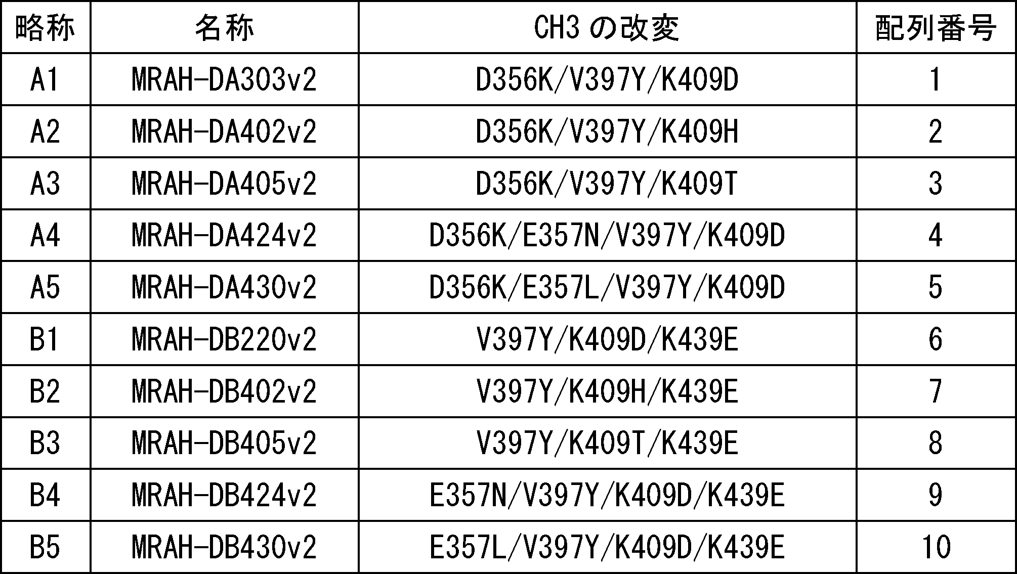
[実施例2]抗体半分子の分子量の評価
作製した抗体半分子A、Bの分子量の評価をサイズ排除クロマトグラフィーで実施した。この時whole抗体のコントロール(図1aのWのピーク)としては国際公開第2009/125825号に開示されているヒトインターロイキン6レセプターに対する抗体を使用した。H鎖可変領域はMRAH(配列番号:11)を用いてH鎖定常領域は天然型IgG1の配列(配列番号:12)を使用した。L鎖可変領域はMRAL(配列番号:13)を使用しL鎖定常領域は天然型κ鎖であるk0(配列番号:14)の配列を使用した。抗体半分子状態のコントロール(図1bのHのピーク)として、H鎖可変領域は国際公開第2013/100120号に記載の抗ヒトエピレグリン抗体のH鎖可変領域部分であるEGLVH(配列番号:15)を使用し、H鎖定常領域部分はLuらの報告(Shan L, Colazet M, Rosenthal KL, Yu XQ, Bee JS, Ferguson A, Damschroder MM, Wu H, Dall'Acqua WF, Tsui P, Oganesyan V. (2016) Generation and Characterization of an IgG4 Monomeric Fc Platform. PLoS One. 2016 Aug 1;11(8))に記載の改変を有するwtIgG4C4(配列番号:16)を使用した。L鎖可変領域は国際公開第2013/100120号に記載の抗ヒトエピレグリン抗体のL鎖可変領域部分であるEGLVL(配列番号:17)を使用しL鎖定常領域は天然型κ鎖であるk0の配列を使用した。また、測定は試験例2に記した方法に従って実施した。なお、この時のH鎖可変領域はそれぞれMRAHを用い、L鎖については全てMRAL-k0を用いて測定を実施した。
測定の結果、図1に示すように、抗体半分子A1、A3、A4、A5のみの場合は抗体半分子(Hのピーク)とwhole抗体(Wのピーク)の混合物が得られ(図1c、e-g)、A2は抗体半分子(Wのピーク)と抗体半分子とwhole抗体の平衡にあると推定される状態の分子(Eのピーク)が得られ(図1d)、抗体半分子B1、B2、B3、B4、B5のみの場合は抗体半分子(Hのピーク)として存在していた(図1h-l)。また、抗体半分子A1とB1(図1m)、A2とB2(図1n)、A3とB3(図1o)、A4とB4(図1p)、A5とB5(図1q)を混合しても、whole抗体の形成(Wのピーク)がそれぞれ抗体半分子A1、A2、A3、A4、A5のみの場合または抗体半分子B1、B2、B3、B4、B5のみの場合に比べて増えることは無かった。なお、A1-B1(図1m)、A3-B3(図1o)を混合した時については、抗体半分子とwhole抗体の平衡にあると推定される状態の分子(Eのピーク)の形成が見られた。
[実施例3]抗体半分子のFcγRIIIaへの結合能の評価
次に、これらの抗体半分子が溶液中では抗体半分子の状態でいて、FcγRIIIaへの結合能は示さないが、抗原結合に伴い局所的に濃縮されることによりヘテロ二量体化し、FcγRIIIaへ結合が回復するかを試験例3に記した方法に従って測定した。なお、この時のH鎖可変領域部分はMRAHを、L鎖可変領域部分はMRAL、L鎖定常領域部分はk0を用いて測定を実施した。天然型IgG1の値はMimotoらの報告(Mimoto F, Igawa T, Kuramochi T, Katada H, Kadono S, Kamikawa T, Shida-Kawazoe M, Hattori K.(2013) Novel asymmetrically engineered antibody Fc variant with superior FcγR binding affinity and specificity compared with afucosylated Fc variant. MAbs 5(2), 229-236)に記載の値を使用した。
測定の結果、表2に示すとおり、抗体半分子AのシリーズであるA1-A5や抗体半分子BのシリーズであるB1-B5はそれぞれ単独ではFcγRIIIaへの結合能は示さなかったが、抗体半分子A1とB1、A2とB2、A3とB3、A4とB4、A5とB5をそれぞれ混合した時に初めてFcγRIIIaへの結合能を示した。
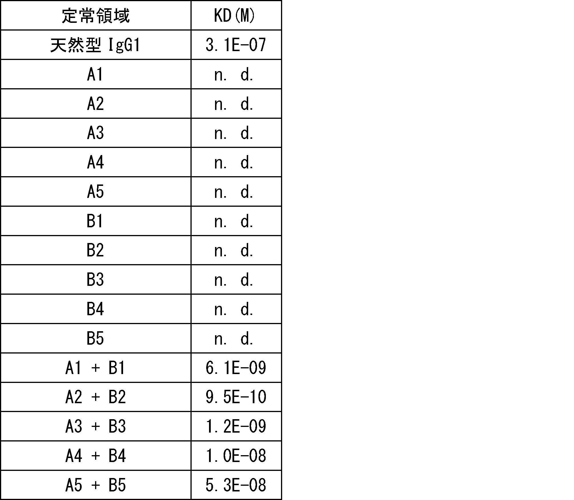
[実施例4]抗体半分子のADCC活性の評価
これらの定常領域を有する抗体半分子が既存の二重特異性抗体に対して優れた二重陽性細胞選択的傷害活性を有するかどうか試験例4に記された方法に従って測定を行った。抗原発現細胞としてはSK-Hep-1にグリピカン3を強制発現させた国際公開第2016/182064号に記載のSK-pca60を、エピレグリンを強制発現させた国際公開第2014/208482号に記載のSKE-4B2を使用した。グリピカン3、エピレグリン二重陽性細胞としてはSK-pca60にエピレグリンを強制発現させたEREG_SK-pca60_#2を使用した。この時実施例3で用いた抗体半分子Aのシリーズについては、そのH鎖可変領域部分が国際公開第2009/041062号に記載の抗GPC3抗体であるGH0(配列番号:18)であるものを使用し、そのL鎖可変領域部分が国際公開第2009/041062号に記載の抗GPC3抗体であるGL0(配列番号:19)であるものを使用し、L鎖定常領域部分はk0を使用した。抗体半分子BについてはEGLVHを使用し、L鎖可変領域はEGLVLを使用し、L鎖定常領域はk0を使用した。また、既存の二重特異性抗体としてはGH0-Kn125P17/GL0-k0//EGLVH-Hl076N17/EGLVL-k0(GH0/EGLVH-BiAb)を用いた。二重特異性抗体の定常領域はヘテロ二量体化した時にADCC活性が増強されるように国際公開第2013/002362号に記載のCH2にL234Y、L235Q、G236W、S239M、H268D、D270E、S298AをKn125P17(配列番号:20)に、D270E、K326D、A330M、K334EをHl076N17(配列番号:21)にそれぞれ置換した。また、二重特異性抗体を電荷の違いを利用して作製するために国際公開第2015/046467号に記載のD356K、V397YをKn125P17に、V397Y、K439EをHl076N17にそれぞれ置換した。また、356-358位の配列については天然型IgG1のアロタイプであるEEMの配列を使用した。このようにして作製されたGH0-Kn125P17/GL0-k0、EGLVH-Hl076N17/EGLVL-k0を定常領域の電荷の違いを利用した当業者公知の方法(Proc. Natl. Acad. Sci., 110, 5145-5150, 2013)で混合し、目的の二重特異性抗体を作製した。
これらの定常領域を有する抗体半分子が既存の二重特異性抗体に対して優れた二重陽性細胞選択的傷害活性を有するかどうか試験例4に記された方法に従って測定を行った。抗原発現細胞としてはSK-Hep-1にグリピカン3を強制発現させた国際公開第2016/182064号に記載のSK-pca60を、エピレグリンを強制発現させた国際公開第2014/208482号に記載のSKE-4B2を使用した。グリピカン3、エピレグリン二重陽性細胞としてはSK-pca60にエピレグリンを強制発現させたEREG_SK-pca60_#2を使用した。この時実施例3で用いた抗体半分子Aのシリーズについては、そのH鎖可変領域部分が国際公開第2009/041062号に記載の抗GPC3抗体であるGH0(配列番号:18)であるものを使用し、そのL鎖可変領域部分が国際公開第2009/041062号に記載の抗GPC3抗体であるGL0(配列番号:19)であるものを使用し、L鎖定常領域部分はk0を使用した。抗体半分子BについてはEGLVHを使用し、L鎖可変領域はEGLVLを使用し、L鎖定常領域はk0を使用した。また、既存の二重特異性抗体としてはGH0-Kn125P17/GL0-k0//EGLVH-Hl076N17/EGLVL-k0(GH0/EGLVH-BiAb)を用いた。二重特異性抗体の定常領域はヘテロ二量体化した時にADCC活性が増強されるように国際公開第2013/002362号に記載のCH2にL234Y、L235Q、G236W、S239M、H268D、D270E、S298AをKn125P17(配列番号:20)に、D270E、K326D、A330M、K334EをHl076N17(配列番号:21)にそれぞれ置換した。また、二重特異性抗体を電荷の違いを利用して作製するために国際公開第2015/046467号に記載のD356K、V397YをKn125P17に、V397Y、K439EをHl076N17にそれぞれ置換した。また、356-358位の配列については天然型IgG1のアロタイプであるEEMの配列を使用した。このようにして作製されたGH0-Kn125P17/GL0-k0、EGLVH-Hl076N17/EGLVL-k0を定常領域の電荷の違いを利用した当業者公知の方法(Proc. Natl. Acad. Sci., 110, 5145-5150, 2013)で混合し、目的の二重特異性抗体を作製した。
測定は抗体半分子A1とB1、A2とB2、A3とB3、A4とB4、A5とB5を混合して添加した。A1 + B1、A4 + B4、A5 + B5の場合はSK-pca60やSKE-4B2に対してはADCC活性が低く、EREG_SK-pca60_#2に対して強いADCC活性を示した。A2 + B2、A3 + B3の場合はSKE-4B2に対してはADCC活性が低く、SK-pca60やEREG_SK-pca60_#2に対して強いADCC活性を示した。既存の二重特異性抗体を用いた場合にはSK-pca60、SKE-4B2、EREG_SK-pca60_#2の全ての細胞に対して強いADCC活性が示された。以上のことから今回作製した抗体半分子A1、A4、A5及びB1、B4、B5はどちらの細胞に対しても既存の二重特異性抗体より優れた二重陽性細胞選択的傷害活性を有することが明らかとなり、抗体半分子A2、A3及びB2、B3に関しても既存の二重特異性抗体より一種類の細胞に対しては優れた二重陽性細胞選択的傷害活性を有することが明らかとなった。
[実施例5]抗体半分子間の相互作用の評価
これら抗体半分子A1とB1、A2とB2、A3とB3、A4とB4、A5とB5間における相互作用を、Biacore(登録商標)を用いて試験例5に記された方法に従って測定した。この時キャプチャーされる抗体半分子のH鎖可変領域部分は国際公開第2009/041621号に記載の抗ヒトIL6レセプター抗体のH鎖可変領域部分であるPF1H(配列番号:22)を使用し、L鎖可変領域部分は国際公開第2009/041621号に記載の抗ヒトIL6レセプター抗体のL鎖可変領域部分であるPF1L(配列番号:23)を使用しL鎖定常領域部分はk0を使用した。また、アナライトとして使用した抗体半分子のH鎖可変領域は国際公開第2015/174439号に記載の抗KLH抗体であるIC17H(配列番号:24)を使用し、L鎖可変領域については国際公開第2015/174439号に記載の抗KLH抗体であるIC17L(配列番号:25)を使用しL鎖定常領域であるk0を使用した。
その結果、表3に示すように、抗体半分子A1-A5が約50RUキャプチャーされたとき、それぞれ抗体半分子B1-B5のモル結合割合が0.18-0.63の時に二重陽性細胞選択的ADCC活性が発揮されることが示された。

[実施例6]抗体半分子のin vivo評価
抗体半分子DA303v2およびDB220v2がマウス生体内において抗原に結合した時にだけ二量体を形成し、細胞傷害活性を示すかどうかについて、抗マウスCD19抗体の可変領域部分を有する抗体半分子を用いて評価した。
抗体半分子DA303v2およびDB220v2がマウス生体内において抗原に結合した時にだけ二量体を形成し、細胞傷害活性を示すかどうかについて、抗マウスCD19抗体の可変領域部分を有する抗体半分子を用いて評価した。
6-1. 抗マウスCD19抗体半分子の作製
抗マウスCD19抗体(Clone: 1D3)はATCCから細胞株HB-305を購入し、当業者公知の方法によって抗体遺伝子をクローニングした。抗体半分子H鎖定常領域部分として天然型ヒトIgG1のC末端のGlyおよびLysを除去したG1d(配列番号:26)のCH2にADCC増強改変としてL234Y/L235Q/G236W/S239M/H268D/D270E/S298Aが、CH3に抗体半分子形成とヘテロ二量体化促進のための改変D356K/V397Y/K409Dが導入された定常領域部分DA303v2(配列番号:27)をもち、抗マウスCD19抗体のH鎖可変領域部分を有する抗体半分子H鎖の遺伝子を作成した。同様に抗体H鎖定常領域部分としてG1dのCH2にADCC増強改変としてD270E/K326D/A330M/K334Eが、CH3に抗体半分子形成とヘテロ二量体化促進のための改変V397Y/K409D/K439Eが導入された定常領域部分DB220v2(配列番号:28)をもち、抗マウスCD19抗体のH鎖可変領域部分を有する抗体半分子H鎖の遺伝子を作成した。またこれら抗体半分子と同じADCC増強改変を有するwhole抗体を作成するため、抗体H鎖定常領域としてG1dのCH2にADCC増強改変としてL234Y/L235Q/G236W/S239M/H268D/D270E/S298Aが導入され、CH3にヘテロ二量体化改変としてY349C/T366Wが導入されたKn125(配列番号:29) を持ち、抗マウスCD19抗体のH鎖可変領域を有する抗体H鎖の遺伝子と、抗体H鎖定常領域としてG1dのCH2にADCC増強改変としてD270E/K326D/A330M/K334Eが導入され、CH3にヘテロ二量体化改変としてD356C/T366S/L368A/Y407Vが導入されたHl076(配列番号:30)を有し、抗マウスCD19抗体のH鎖可変領域を有する抗体H鎖の遺伝子が作成された。Kn125とHl076にはKnobs-into-holes技術(Margaret Merchant et al., Nature Biotechnology 1998, 16, 677-681)が導入されており、これら2つのH鎖遺伝子を抗マウスCD19抗体のL鎖遺伝子と共発現することでヘテロ二量体化したwhole抗体が生成される。また同じくG1dに対して国際公開第2013/047748号に記載されているマウスFcγRへの結合を減弱するための改変(L235R/S239K)を導入した定常領域F760(配列番号:31)をH鎖定常領域としてもち、抗マウスCD19抗体H鎖可変領域を有する抗体H鎖の遺伝子と、国際公開第2015/174439号に記載されているADCC活性を増強したマウスIgG2a定常領域(mFa55: 配列番号:32)をH鎖定常領域としてもち、抗マウスCD19抗体H鎖可変領域を有する抗体H鎖の遺伝子を作成した。マウスCD19に結合しない抗体として、国際公開第2015/174439号に記載の抗KLH抗体のH鎖可変領域であるIC17H(配列番号:24)を有し、FcγRへの結合を減弱した定常領域F760nN17(配列番号:33)を有する抗体H鎖遺伝子をそれぞれ作成した。抗体L鎖としては、天然型ヒトkappa定常領域(k0:配列番号:14)を有し、抗マウスCD19抗体L鎖可変領域をもつ抗体L鎖の遺伝子と、同じく天然型マウスkappa定常領域(mk0: 配列番号:34)を有し、抗マウスCD19抗体L鎖可変領域をもつ抗体L鎖の遺伝子を作成した。また抗KLH抗体L鎖可変領域IC17L(配列番号:25)を持ち、天然型ヒトkappa定常領域(k0)を有する抗体L鎖遺伝子を作成した。ここで作成したそれぞれの遺伝子を表4に示す組み合わせで発現し、試験例1に記載の方法で目的の抗体を得た。
抗マウスCD19抗体(Clone: 1D3)はATCCから細胞株HB-305を購入し、当業者公知の方法によって抗体遺伝子をクローニングした。抗体半分子H鎖定常領域部分として天然型ヒトIgG1のC末端のGlyおよびLysを除去したG1d(配列番号:26)のCH2にADCC増強改変としてL234Y/L235Q/G236W/S239M/H268D/D270E/S298Aが、CH3に抗体半分子形成とヘテロ二量体化促進のための改変D356K/V397Y/K409Dが導入された定常領域部分DA303v2(配列番号:27)をもち、抗マウスCD19抗体のH鎖可変領域部分を有する抗体半分子H鎖の遺伝子を作成した。同様に抗体H鎖定常領域部分としてG1dのCH2にADCC増強改変としてD270E/K326D/A330M/K334Eが、CH3に抗体半分子形成とヘテロ二量体化促進のための改変V397Y/K409D/K439Eが導入された定常領域部分DB220v2(配列番号:28)をもち、抗マウスCD19抗体のH鎖可変領域部分を有する抗体半分子H鎖の遺伝子を作成した。またこれら抗体半分子と同じADCC増強改変を有するwhole抗体を作成するため、抗体H鎖定常領域としてG1dのCH2にADCC増強改変としてL234Y/L235Q/G236W/S239M/H268D/D270E/S298Aが導入され、CH3にヘテロ二量体化改変としてY349C/T366Wが導入されたKn125(配列番号:29) を持ち、抗マウスCD19抗体のH鎖可変領域を有する抗体H鎖の遺伝子と、抗体H鎖定常領域としてG1dのCH2にADCC増強改変としてD270E/K326D/A330M/K334Eが導入され、CH3にヘテロ二量体化改変としてD356C/T366S/L368A/Y407Vが導入されたHl076(配列番号:30)を有し、抗マウスCD19抗体のH鎖可変領域を有する抗体H鎖の遺伝子が作成された。Kn125とHl076にはKnobs-into-holes技術(Margaret Merchant et al., Nature Biotechnology 1998, 16, 677-681)が導入されており、これら2つのH鎖遺伝子を抗マウスCD19抗体のL鎖遺伝子と共発現することでヘテロ二量体化したwhole抗体が生成される。また同じくG1dに対して国際公開第2013/047748号に記載されているマウスFcγRへの結合を減弱するための改変(L235R/S239K)を導入した定常領域F760(配列番号:31)をH鎖定常領域としてもち、抗マウスCD19抗体H鎖可変領域を有する抗体H鎖の遺伝子と、国際公開第2015/174439号に記載されているADCC活性を増強したマウスIgG2a定常領域(mFa55: 配列番号:32)をH鎖定常領域としてもち、抗マウスCD19抗体H鎖可変領域を有する抗体H鎖の遺伝子を作成した。マウスCD19に結合しない抗体として、国際公開第2015/174439号に記載の抗KLH抗体のH鎖可変領域であるIC17H(配列番号:24)を有し、FcγRへの結合を減弱した定常領域F760nN17(配列番号:33)を有する抗体H鎖遺伝子をそれぞれ作成した。抗体L鎖としては、天然型ヒトkappa定常領域(k0:配列番号:14)を有し、抗マウスCD19抗体L鎖可変領域をもつ抗体L鎖の遺伝子と、同じく天然型マウスkappa定常領域(mk0: 配列番号:34)を有し、抗マウスCD19抗体L鎖可変領域をもつ抗体L鎖の遺伝子を作成した。また抗KLH抗体L鎖可変領域IC17L(配列番号:25)を持ち、天然型ヒトkappa定常領域(k0)を有する抗体L鎖遺伝子を作成した。ここで作成したそれぞれの遺伝子を表4に示す組み合わせで発現し、試験例1に記載の方法で目的の抗体を得た。

6-2. 抗マウスCD19抗体半分子またはその二量体のマウスFcγRに対する結合評価
実施例6-1で調製した抗体半分子のうち、1D3-mFa55、1D3-Kn125/Hl076、1D3-DA303v2、1D3-DB220v2および1D3-DA303v2と1D3-DB220v2の等量混合物のマウスFcγRI、II、III、IVに対する結合測定を実施した。また比較対象として天然型ヒトIgG1の配列からC末端のGlyおよびLysを除去したG1dをH鎖定常領域としてもち、抗マウスCD19抗体のH鎖可変領域部分を有する抗体半分子H鎖の遺伝子と、天然型ヒトkappa定常領域k0を有し、抗マウスCD19抗体L鎖可変領域をもつ抗体L鎖の遺伝子を作成し、試験例1の方法に従って抗マウスCD19抗体半分子、1D3-G1dを得た。マウスFcγRI、II、III、IVは国際公開第2014/030750号に記載の方法により調製した。Biacore(登録商標) T200 (GEヘルスケア)を用いて、各抗体半分子またはその二量体とFcγRとの相互作用解析を行った。ランニングバッファーにはHBS-EP+10X (GEヘルスケア)を10分の1に希釈して用い、測定温度は25℃とした。Series S Sensor Chip CM5(GEヘルスケア)に、アミンカップリング法によりProtein A/G (PIERCE)を固定化したチップを用いた。このセンサーチップに目的の抗体半分子をキャプチャーし、ランニングバッファーで希釈したFcγRを相互作用させて測定を行った。また25 mM NaOHと、10 mM Glycine-HCl (pH1.5)を反応させることでセンサーチップにキャプチャーした抗体半分子を洗浄し、センサーチップを再生して繰り返し用いた。各抗体半分子またはその二量体のFcγRに対するKD値を算出するための速度論的な解析は以下の方法にしたがって実施した。まず、上記のセンサーチップに目的の抗体半分子をキャプチャーさせ、ランニングバッファーで希釈したmFcγRを相互作用させ、得られたセンサーグラムに対してBiacore(登録商標) Evaluation Softwareにより測定結果を1:1 Langmuir binding modelでglobal fittingさせることで結合速度定数ka (L/mol/s)、解離速度定数kd(1/s)を算出し、その値から解離定数KD (mol/L) を算出した。また、抗体半分子またはその二量体とFcγRとの相互作用が微弱で、上記の速度論的な解析では正しく解析できないと判断された場合、その相互作用についてはBiacore(登録商標) T100 Software Handbook BR1006-48 Edition AEに記載の以下の1:1結合モデル式を利用してKDを算出した。
実施例6-1で調製した抗体半分子のうち、1D3-mFa55、1D3-Kn125/Hl076、1D3-DA303v2、1D3-DB220v2および1D3-DA303v2と1D3-DB220v2の等量混合物のマウスFcγRI、II、III、IVに対する結合測定を実施した。また比較対象として天然型ヒトIgG1の配列からC末端のGlyおよびLysを除去したG1dをH鎖定常領域としてもち、抗マウスCD19抗体のH鎖可変領域部分を有する抗体半分子H鎖の遺伝子と、天然型ヒトkappa定常領域k0を有し、抗マウスCD19抗体L鎖可変領域をもつ抗体L鎖の遺伝子を作成し、試験例1の方法に従って抗マウスCD19抗体半分子、1D3-G1dを得た。マウスFcγRI、II、III、IVは国際公開第2014/030750号に記載の方法により調製した。Biacore(登録商標) T200 (GEヘルスケア)を用いて、各抗体半分子またはその二量体とFcγRとの相互作用解析を行った。ランニングバッファーにはHBS-EP+10X (GEヘルスケア)を10分の1に希釈して用い、測定温度は25℃とした。Series S Sensor Chip CM5(GEヘルスケア)に、アミンカップリング法によりProtein A/G (PIERCE)を固定化したチップを用いた。このセンサーチップに目的の抗体半分子をキャプチャーし、ランニングバッファーで希釈したFcγRを相互作用させて測定を行った。また25 mM NaOHと、10 mM Glycine-HCl (pH1.5)を反応させることでセンサーチップにキャプチャーした抗体半分子を洗浄し、センサーチップを再生して繰り返し用いた。各抗体半分子またはその二量体のFcγRに対するKD値を算出するための速度論的な解析は以下の方法にしたがって実施した。まず、上記のセンサーチップに目的の抗体半分子をキャプチャーさせ、ランニングバッファーで希釈したmFcγRを相互作用させ、得られたセンサーグラムに対してBiacore(登録商標) Evaluation Softwareにより測定結果を1:1 Langmuir binding modelでglobal fittingさせることで結合速度定数ka (L/mol/s)、解離速度定数kd(1/s)を算出し、その値から解離定数KD (mol/L) を算出した。また、抗体半分子またはその二量体とFcγRとの相互作用が微弱で、上記の速度論的な解析では正しく解析できないと判断された場合、その相互作用についてはBiacore(登録商標) T100 Software Handbook BR1006-48 Edition AEに記載の以下の1:1結合モデル式を利用してKDを算出した。
1:1 binding modelで相互作用する分子のBiacore(登録商標)上での挙動は以下の式3によって表わすことができる。
(式3)
Req = C・Rmax/(KD+C) + RI
Req: a plot of steady state binding levels against analyte concentration
C: concentration
RI: bulk refractive index contribution in the sample
Rmax: analyte binding capacity of the surface
Req = C・Rmax/(KD+C) + RI
Req: a plot of steady state binding levels against analyte concentration
C: concentration
RI: bulk refractive index contribution in the sample
Rmax: analyte binding capacity of the surface
この式を変形すると、KDは以下の式4のように表わすことができる。
(式4)
KD = C・Rmax/(Req-RI)-C
KD = C・Rmax/(Req-RI)-C
この式にRmax、RI、Cの値を代入することで、KDを算出することが可能である。RI、Cについては測定結果のセンサーグラム、測定条件から値を求めることができる。Rmaxの算出については、以下の方法にしたがった。その測定回に同時に評価した比較対象となる相互作用が十分強い抗体について、上記の1:1 Langmuir binding modelでglobal fittingさせた際に得られたRmaxの値を、比較対象となる抗体のセンサーチップへのキャプチャー量で除し、評価したい改変抗体のキャプチャー量で乗じて得られた値をRmaxとした。今回の測定条件においてはRI = 0、C= 500 nM (FcγRI、FcγRIV)あるいは4000 nM (FcγRII、FcγRIII)となる。Rmaxには各FcγRに対するG1dの相互作用解析結果として得られたセンサーグラムに対して1:1 Langmuir binding modelでglobal fittingさせた際に得られたRmaxの値をG1dのキャプチャー量で除し、各抗体のキャプチャー量で乗じて得られた値とした。これはいずれの抗体半分子またはその二量体においても、G1dと比較して各FcγRが結合できる限界量には変化がなく、測定時のRmaxはその測定時にチップ上に結合している抗体の量に比例するという仮定に基づいた計算である。Reqは測定時に観察された各FcγRのセンサーチップ上の抗体半分子またはその二量体に対する結合量とした。
こうして得られた各抗体のマウスFcγRに対するKD値を表5に示した。表中の灰色で塗りつぶされた値は、FcγRの抗体半分子またはその二量体に対する結合が微弱であり、速度論的な解析では正しく解析できないと判断されたため、上記式4を利用して算出した値である。また「KD fold for mouse FcγRs」の値は、各FcγRに対するG1dのKDを、それぞれの抗体のKDで割った値であり、各抗体の各FcγRに対する親和性がG1dと比較してどれだけ増強あるいは減弱されているかを示す。またN.D.はセンサーグラム上で結合が確認されなかったことを示す。

表5の結果から、FcγR結合増強型マウスIgG2a抗体であるmFa55は、G1dと比較してFcγRIに対して44.7倍、FcγRIIに対して3.4倍、FcγRIIIに対して11.7倍、FcγRIVに対して57.5倍結合が増強されていた。ヒトIgG1型のヘテロ二量体化ADCC増強抗体であるKn125/Hl076は、G1dと比較してFcγRIに対して0.4倍に結合が減弱しており、FcγRIIに対しては5.7倍、FcγRIIIに対しては13.2倍、FcγRIVに対しては253.3倍結合が増強されていた。抗体半分子であるDA303v2はいずれのマウスFcγRに対しても結合しなかった。抗体半分子であるDB220v2もいずれのマウスFcγRに対しても結合が減弱されており、その親和性はG1dと比較してFcγRIに対しては0.05倍、FcγRIIに対しては0.03倍、FcγRIIIに対しては0.04倍であった。またFcγRIVに対しては結合しなかった。二種類の抗体半分子DA303v2とDB220v2を等量混合して測定した1D3-DA303v2/1D3-DB220v2では、G1dと比較してFcγRIに対しては0.03倍、FcγRIIに対しては0.08倍、FcγRIIIに対しては0.4倍に結合が減弱されていたが、FcγRIVに対しては11.5倍に結合が増強されていた。以上の結果から、どちらかの抗体半分子単独ではFcγRに対する結合活性がほとんど失われているのに対し、二種類の抗体半分子を混合してヘテロ二量体を形成した場合にはFcγRIVに対する結合が増強されるため、エフェクター活性を示すことが期待される。
6-3. マウスにおける抗体半分子の単体投与、および混合投与時の薬物動態評価
実施例6-1に記載の方法で作製した1D3-Kn125/Hl076、1D3-DA303v2、1D3-DB220v2、1D3-DA303v2/DB220v2のマウスにおける血中薬物動態を評価した。投与は10 mg/kgでマウス尾静脈より行い、頸静脈より経時的に採血を行った。血漿中濃度は、1D3-Kn125/Hl076、1D3-DA303v2、1D3-DB220v2については電気化学免疫測定法 (ECLIA: 試験例6)で、1D3-DA303v2/DB220v2についてはそれぞれの抗体半分子をLC-MS (試験例7)で測定した。その結果、whole抗体である1D3-Kn125/Hl076に比べて、1D3-DA303v2、1D3-DB220v2、1D3-DA303v2/DB220v2の血中からの消失は非常に速く、投与後3日で投与直後の濃度に対して1/100程度まで減少した(図3)。また、1D3-DA303v2、および1D3-DB220v2それぞれについて、単独投与と混合投与で血漿中濃度推移に顕著な差はないことが示された。このことは、混合投与においても抗体半分子はヘテロ二量体化せずに単体で存在し、可変領域で抗原に結合した際に初めてヘテロ二量体化することを示唆している。
実施例6-1に記載の方法で作製した1D3-Kn125/Hl076、1D3-DA303v2、1D3-DB220v2、1D3-DA303v2/DB220v2のマウスにおける血中薬物動態を評価した。投与は10 mg/kgでマウス尾静脈より行い、頸静脈より経時的に採血を行った。血漿中濃度は、1D3-Kn125/Hl076、1D3-DA303v2、1D3-DB220v2については電気化学免疫測定法 (ECLIA: 試験例6)で、1D3-DA303v2/DB220v2についてはそれぞれの抗体半分子をLC-MS (試験例7)で測定した。その結果、whole抗体である1D3-Kn125/Hl076に比べて、1D3-DA303v2、1D3-DB220v2、1D3-DA303v2/DB220v2の血中からの消失は非常に速く、投与後3日で投与直後の濃度に対して1/100程度まで減少した(図3)。また、1D3-DA303v2、および1D3-DB220v2それぞれについて、単独投与と混合投与で血漿中濃度推移に顕著な差はないことが示された。このことは、混合投与においても抗体半分子はヘテロ二量体化せずに単体で存在し、可変領域で抗原に結合した際に初めてヘテロ二量体化することを示唆している。
6-4. マウス生体内におけるB cell depletionの評価
実施例6-1に記載の方法で精製、調製した抗マウスCD19抗体半分子 (表4に記載) をマウスに静脈内投与することで細胞傷害活性を示すかを検証した。なお、in vivo B cell depletion試験の投与量の設定は、実施例6-3に示すwhole抗体および抗体半分子の消失濃度推移から、各検体でトラフ濃度が同程度になるように設定した。具体的には、C57BL/6NCr Slcマウスに必要血中濃度を維持するために表6に示す投与条件で、各種抗体または抗体半分子を静脈内投与した (各群n=3)。投与後3日目に背中足静脈採血を実施し、試験例8に記載の方法で血中のB細胞を染色した後、FACSで生細胞中におけるB細胞を検出した。試験例9に記載の方法で血中のB細胞の割合を算出した結果、KLH-F760nN17と比較して1D3-F760は細胞傷害活性を示さず、陽性対照である1D3-mFa55および1D3- Kn125/Hl076は有意に細胞傷害活性を示した。本条件下で、抗体半分子1D3-DA303v2および1D3-DB220v2はそれぞれの単独投与で細胞傷害活性を示さないが、これらを等量混合して投与した場合は、KLH-F760nN17と比較して有意に細胞傷害活性を示すことが明らかとなった (図4)。実施例6-3で示した通り、抗体半分子は混合した状態で投与しても可変領域が抗原に結合しなければ抗体半分子として存在すると考えられるため、ここで得られたB cell depletion活性は、それぞれの抗体半分子が可変領域で抗原に結合して初めてヘテロ二量体を形成し、ADCC活性を示した結果であると考察される。
実施例6-1に記載の方法で精製、調製した抗マウスCD19抗体半分子 (表4に記載) をマウスに静脈内投与することで細胞傷害活性を示すかを検証した。なお、in vivo B cell depletion試験の投与量の設定は、実施例6-3に示すwhole抗体および抗体半分子の消失濃度推移から、各検体でトラフ濃度が同程度になるように設定した。具体的には、C57BL/6NCr Slcマウスに必要血中濃度を維持するために表6に示す投与条件で、各種抗体または抗体半分子を静脈内投与した (各群n=3)。投与後3日目に背中足静脈採血を実施し、試験例8に記載の方法で血中のB細胞を染色した後、FACSで生細胞中におけるB細胞を検出した。試験例9に記載の方法で血中のB細胞の割合を算出した結果、KLH-F760nN17と比較して1D3-F760は細胞傷害活性を示さず、陽性対照である1D3-mFa55および1D3- Kn125/Hl076は有意に細胞傷害活性を示した。本条件下で、抗体半分子1D3-DA303v2および1D3-DB220v2はそれぞれの単独投与で細胞傷害活性を示さないが、これらを等量混合して投与した場合は、KLH-F760nN17と比較して有意に細胞傷害活性を示すことが明らかとなった (図4)。実施例6-3で示した通り、抗体半分子は混合した状態で投与しても可変領域が抗原に結合しなければ抗体半分子として存在すると考えられるため、ここで得られたB cell depletion活性は、それぞれの抗体半分子が可変領域で抗原に結合して初めてヘテロ二量体を形成し、ADCC活性を示した結果であると考察される。

[試験例10]抗体半分子の発現ベクターの作製および抗体半分子の発現と精製
アミノ酸置換の導入はQuikChange Site-Directed Mutagenesis Kit(Stratagene)、PCRまたはIn fusion Advantage PCR cloning kit (TAKARA)等を用いて当業者公知の方法で行い、発現ベクターを構築した。得られた発現ベクターの塩基配列は当業者公知の方法で決定した。作製したプラスミドをExpi293細胞(Invitrogen)に、一過性に導入し、抗体半分子の発現を行った。得られた培養上清から、MonoSpin ProA 96ウェルプレートタイプ(商標登録)(GLサイエンス)を用いて当業者公知の方法で、抗体半分子を精製した。精製抗体半分子濃度は、分光光度計を用いて280 nmでの吸光度を測定し、得られた値からPACE法により算出された吸光係数を用いて抗体半分子濃度を算出した(Protein Science 1995 ; 4 : 2411-2423)。
アミノ酸置換の導入はQuikChange Site-Directed Mutagenesis Kit(Stratagene)、PCRまたはIn fusion Advantage PCR cloning kit (TAKARA)等を用いて当業者公知の方法で行い、発現ベクターを構築した。得られた発現ベクターの塩基配列は当業者公知の方法で決定した。作製したプラスミドをExpi293細胞(Invitrogen)に、一過性に導入し、抗体半分子の発現を行った。得られた培養上清から、MonoSpin ProA 96ウェルプレートタイプ(商標登録)(GLサイエンス)を用いて当業者公知の方法で、抗体半分子を精製した。精製抗体半分子濃度は、分光光度計を用いて280 nmでの吸光度を測定し、得られた値からPACE法により算出された吸光係数を用いて抗体半分子濃度を算出した(Protein Science 1995 ; 4 : 2411-2423)。
[試験例11]抗体半分子の分子量の分析
得られた抗体半分子の分子量をHPLCであるACQUITY UPLC H-Class(登録商標)(Waters)、カラムにはSuperSW3000(TOSOH)を用いて当業者公知の方法で分析した。抗体半分子タンパク質濃度は0.10 mg/mLとして、10 μLのインジェクションを行った。
得られた抗体半分子の分子量をHPLCであるACQUITY UPLC H-Class(登録商標)(Waters)、カラムにはSuperSW3000(TOSOH)を用いて当業者公知の方法で分析した。抗体半分子タンパク質濃度は0.10 mg/mLとして、10 μLのインジェクションを行った。
[実施例7]抗体半分子の作製
実施例1で使用したヘテロ二量体化改変を使用せずにホモ二量体の形成を抑制することができるかどうかを検証するため、ヘテロ二量体化改変を使用しない、もしくはHaらの報告(Ha JH, Kim JE, Kim YS (2016) Immunoglobulin Fc Heterodimer Platform Technology: From Design to Applications in Therapeutic Antibodies and Proteins. Front Immunol. 2016 Oct 6;7:394.)に記載されている改変を使用した。具体的には、ADCC増強改変、ヒンジ領域の改変、CH3界面不安定化改変のみの半分子抗体A、Bと、それに加えて抗体半分子AにY349T、T394Fを、抗体半分子BにS364H、F405Aをそれぞれ置換した。作製、評価した抗体半分子の略称および名称と、CH3改変と、抗体半分子の配列番号を表7に示す。
実施例1で使用したヘテロ二量体化改変を使用せずにホモ二量体の形成を抑制することができるかどうかを検証するため、ヘテロ二量体化改変を使用しない、もしくはHaらの報告(Ha JH, Kim JE, Kim YS (2016) Immunoglobulin Fc Heterodimer Platform Technology: From Design to Applications in Therapeutic Antibodies and Proteins. Front Immunol. 2016 Oct 6;7:394.)に記載されている改変を使用した。具体的には、ADCC増強改変、ヒンジ領域の改変、CH3界面不安定化改変のみの半分子抗体A、Bと、それに加えて抗体半分子AにY349T、T394Fを、抗体半分子BにS364H、F405Aをそれぞれ置換した。作製、評価した抗体半分子の略称および名称と、CH3改変と、抗体半分子の配列番号を表7に示す。

[実施例8]抗体半分子の分子量の評価
作製した抗体半分子A、Bの分子量の評価をサイズ排除クロマトグラフィーで実施した。この時whole抗体のコントロール(図5-1aのWのピーク)としては国際公開第2009/125825号に開示されているヒトインターロイキン6レセプターに対する抗体を使用した。H鎖可変領域はMRAH(配列番号:11)を用いてH鎖定常領域は天然型IgG1の配列(配列番号:12)を使用した。L鎖可変領域はMRAL(配列番号:13)を使用しL鎖定常領域は天然型κ鎖であるk0(配列番号:14)の配列を使用した。抗体半分子状態のコントロール(図5bのHのピーク)として、H鎖可変領域は国際公開第2013/100120号に記載の抗ヒトエピレグリン抗体のH鎖可変領域部分であるEGLVH(配列番号:15)を使用し、H鎖定常領域部分はLuらの報告(Shan L, Colazet M, Rosenthal KL, Yu XQ, Bee JS, Ferguson A, Damschroder MM, Wu H, Dall'Acqua WF, Tsui P, Oganesyan V. (2016) Generation and Characterization of an IgG4 Monomeric Fc Platform. PLoS One. 2016 Aug 1;11(8))に記載の改変を有するwtIgG4C4(配列番号:16)を使用した。L鎖可変領域は国際公開第2013/100120号に記載の抗ヒトエピレグリン抗体のL鎖可変領域部分であるEGLVL(配列番号:17)を使用しL鎖定常領域は天然型κ鎖であるk0の配列を使用した。また、測定は試験例11に記した方法に従って実施した。なお、この時のH鎖可変領域はそれぞれMRAHを用い、L鎖については全てMRAL-k0を用いて測定を実施した。
測定の結果、図5に示すように、抗体半分子A1、A6のみの場合は抗体半分子(Hのピーク)とwhole抗体(Wのピーク)の混合物が得られ(図5c、d)、抗体半分子A7、B1、B6、B7のみの場合は抗体半分子(Hのピーク)として存在していた(図5e-h)。また、抗体半分子A1とB1(図5i)、A6とB6(図5j)、A7とB7(図5k)を混合しても、whole抗体の形成(Wのピーク)がそれぞれ抗体半分子A1、A6、A7のみの場合または抗体半分子B1、B6、B7のみの場合に比べて増えることは無かった。
[実施例9]抗体半分子のADCC活性の評価
これらの定常領域を有する抗体半分子が既存の二重特異性抗体に対して優れた二重陽性細胞選択的傷害活性を有するかどうか試験例4に記された方法に従って測定を行った。抗原発現細胞としてはSK-Hep-1にグリピカン3を強制発現させた国際公開第2016/182064号に記載のSK-pca60を、エピレグリンを強制発現させた国際公開第2014/208482号に記載のSKE-4B2を使用した。グリピカン3、エピレグリン二重陽性細胞としてはSK-pca60にエピレグリンを強制発現させたEREG_SK-pca60_#2を使用した。この時抗体半分子Aのシリーズについては、そのH鎖可変領域部分が国際公開第2009/041062号に記載の抗GPC3抗体であるGH0(配列番号:18)であるものを使用し、そのL鎖可変領域部分が国際公開第2009/041062号に記載の抗GPC3抗体であるGL0(配列番号:19)であるものを使用し、L鎖定常領域部分はk0を使用した。抗体半分子BについてはEGLVHを使用し、L鎖可変領域はEGLVLを使用し、L鎖定常領域はk0を使用した。また、既存の二重特異性抗体としてはGH0-Kn125P17/GL0-k0//EGLVH-Hl076N17/EGLVL-k0(GH0/EGLVH-BiAb)を用いた。二重特異性抗体の定常領域はヘテロ二量体化した時にADCC活性が増強されるように国際公開第2013/002362号に記載のCH2にL234Y、L235Q、G236W、S239M、H268D、D270E、S298AをKn125P17(配列番号:20)に、D270E、K326D、A330M、K334EをHl076N17(配列番号:21)にそれぞれ置換した。また、二重特異性抗体を電荷の違いを利用して作製するために国際公開第2015/046467号に記載のD356K、V397YをKn125P17に、V397Y、K439EをHl076N17にそれぞれ置換した。また、356-358位の配列については天然型IgG1のアロタイプであるEEMの配列を使用した。このようにして作製されたGH0-Kn125P17/GL0-k0、EGLVH-Hl076N17/EGLVL-k0を定常領域の電荷の違いを利用した当業者公知の方法(Proc. Natl. Acad. Sci., 110, 5145-5150, 2013)で混合し、目的の二重特異性抗体を作製した。
測定は抗体半分子A1とB1、A6とB6、A7とB7を混合して添加した。測定した結果を図6に示す。A1 + B1の場合と同様に、A6 + B6、A7 + B7の場合もSK-pca60やSKE-4B2に対してはADCC活性が低く、EREG_SK-pca60_#2に対して強いADCC活性を示した。既存の二重特異性抗体を用いた場合にはSK-pca60、SKE-4B2、EREG_SK-pca60_#2の全ての細胞に対して強いADCC活性が示された。以上のことから、今回作製した抗体半分子A6、A7およびB6、B7もどちらの細胞に対しても既存の二重特異性抗体より優れた二重陽性細胞選択的傷害活性を有することが明らかとなった。
[実施例10]抗体半分子間の相互作用の評価
これら抗体半分子A1とB1、A6とB6、A7とB7間における相互作用を、Biacore(登録商標)を用いて試験例5に記された方法に従って測定した。この時キャプチャーされる抗体半分子のH鎖可変領域部分は国際公開第2009/041621号に記載の抗ヒトIL6レセプター抗体のH鎖可変領域部分であるPF1H(配列番号:22)を使用し、L鎖可変領域部分は国際公開第2009/041621号に記載の抗ヒトIL6レセプター抗体のL鎖可変領域部分であるPF1L(配列番号:23)を使用しL鎖定常領域部分はk0を使用した。また、アナライトとして使用した抗体半分子のH鎖可変領域は国際公開第2015/174439号に記載の抗KLH抗体であるIC17H(配列番号:24)を使用し、L鎖可変領域については国際公開第2015/174439号に記載の抗KLH抗体であるIC17L(配列番号:25)を使用しL鎖定常領域であるk0を使用した。
その結果、表8に示すように、抗体半分子A6、A7が約50RUキャプチャーされたとき、それぞれ抗体半分子B6、B7のモル結合割合が0.18-0.63の範囲内となり、二重陽性細胞選択的ADCC活性も発揮されることが示された。抗体半分子Bのランニングバッファーによる希釈は、実施例5では18.6-28.0倍希釈したのに対し、本実施例では5.7-9.0倍希釈した。

Claims (15)
- 第1の抗原に結合する第1の抗原結合領域と、第1のCH2および第1のCH3のいずれか一方または両方を含む第1のポリペプチドとを有する第1の抗原結合分子、ならびに第2の抗原に結合する第2の抗原結合領域と、第2のCH2および第2のCH3のいずれか一方または両方を含む第2のポリペプチドとを有する第2の抗原結合分子を含有し、
前記第1の抗原結合分子および前記第2の抗原結合分子が、共有結合によって結合しておらず、そして、液中で混合した場合にホモ二量体よりもヘテロ二量体を形成しやすい、薬学的組成物。 - 表面プラズモン共鳴において1 mm2あたり50 pgの前記第1の抗原結合分子が固定化されたセンサーチップと2.5 mg/mLの前記第2の抗原結合分子を含有する測定液とを用いて両抗原結合分子の親和性を測定したときに、前記第1の抗原結合分子に対する前記第2の抗原結合分子の結合量がモル比で1:0.1~1:0.9の範囲内にある、請求項1に記載の組成物。
- 前記第1の抗原および前記第2の抗原を発現する細胞が存在する条件下での前記ヘテロ二量体の形成量が、前記細胞が存在しない条件下におけるよりも多い、請求項1または2に記載の組成物。
- 前記ヘテロ二量体を形成した場合における前記ヘテロ二量体のFcγRへの結合活性が、前記第1の抗原結合分子の単量体もしくは前記第2の抗原結合分子の単量体のFcγRへの結合活性、または前記ホモ二量体を形成した場合における前記ホモ二量体のFcγRへの結合活性よりも高い、請求項1から3のいずれか一項に記載の組成物。
- 前記第1のポリペプチドが前記第1のCH3を含み、前記第2のポリペプチドが前記第2のCH3を含み、前記第1のCH3および前記第2のCH3が、液中で混合した場合に前記ホモ二量体よりも前記ヘテロ二量体を形成しやすくする改変として以下の(i)から(iii)の少なくとも一の改変を有する、請求項1から4のいずれか一項に記載の組成物。
(i)前記第1のCH3および前記第2のCH3のいずれか一方が正電荷の領域を有し、他方が負電荷の領域を有し、前記ヘテロ二量体が形成される際に前記正電荷の領域が前記負電荷の領域に相互作用する、改変
(ii)前記第1のCH3および前記第2のCH3のいずれか一方が凸部を有し、他方が凹部を有し、前記ヘテロ二量体が形成される際に前記凸部が前記凹部に嵌合して相互作用する、改変
(iii)前記第1のCH3および前記第2のCH3が改変されたIgGのCH3であり、前記改変されたIgGのCH3はその一部がIgAのCH3の一部と置き換えられており、前記ヘテロ二量体が形成される際に前記第1のCH3に置き換えられた前記IgAのCH3の一部と前記第2のCH3に置き換えられた前記IgAのCH3の一部とが相互作用する、改変 - 前記第1のCH3および前記第2のCH3のいずれか一方または両方が、EUナンバリングシステムでの357位、397位および409位のアミノ酸残基のうち少なくとも一つの他のアミノ酸残基への置換をさらに有する、請求項1から5のいずれか一項に記載の組成物。
- 前記第1のポリペプチドおよび前記第2のポリペプチドのいずれか一方または両方が、抗体半分子におけるヒンジ領域部分をさらに含む、請求項1から6のいずれか一項に記載の組成物。
- 前記第1のポリペプチドおよび前記第2のポリペプチドのいずれか一方または両方における前記ヒンジ領域部分が、EUナンバリングシステムでの226位および229位のいずれか一方または両方のシステイン残基における他のアミノ酸残基への改変を有する、請求項7に記載の組成物。
- 前記第1のポリペプチドおよび前記第2のポリペプチドが、それぞれ抗体半分子における定常領域部分を含む、請求項1から8のいずれか一項に記載の組成物。
- 前記第1の抗原および前記第2の抗原を発現する細胞が存在する条件下でのエフェクター機能が、前記第2の抗原を発現せず前記第1の抗原を発現する細胞または前記第1の抗原を発現せず前記第2の抗原を発現する細胞が存在する条件下よりも高い、請求項1から9のいずれか一項に記載の組成物。
- 第1の抗原に結合する第1の抗原結合領域と、第1のCH3を含む第1のポリペプチドとを有する第1の抗原結合分子、および第2の抗原に結合する第2の抗原結合領域と、第2のCH3を含む第2のポリペプチドとを有する第2の抗原結合分子を含有し、
前記第1の抗原結合分子および前記第2の抗原結合分子が、共有結合によって結合しておらず、
前記第1のCH3および前記第2のCH3が以下の(iv)から(vi)の少なくとも一の改変を有する、薬学的組成物。
(iv)前記第1のCH3および前記第2のCH3のいずれか一方が正電荷の領域を有し、他方が負電荷の領域を有し、前記ヘテロ二量体が形成される際に前記正電荷の領域が前記負電荷の領域に相互作用する、改変
(v)前記第1のCH3および前記第2のCH3のいずれか一方が凸部を有し、他方が凹部を有し、前記ヘテロ二量体が形成される際に前記凸部が前記凹部に嵌合して相互作用する、改変
(vi)前記第1のCH3および前記第2のCH3が改変されたIgGのCH3であり、前記改変されたIgGのCH3はその一部がIgAのCH3の一部と置き換えられており、前記ヘテロ二量体が形成される際に前記第1のCH3に置き換えられた前記IgAのCH3の一部と前記第2のCH3に置き換えられた前記IgAのCH3の一部とが相互作用する、改変 - 第1の抗原に結合する第1の抗原結合領域と、第1のCH2および第1のCH3のいずれか一方または両方を含む第1のポリペプチドとを有する第1の抗原結合分子であって、
第2の抗原に結合する第2の抗原結合領域と、第2のCH2および第2のCH3のいずれか一方または両方を含む第2のポリペプチドとを有する第2の抗原結合分子と液中で混合した場合に前記第1の抗原結合分子間のホモ二量体よりも前記第2の抗原結合分子とのヘテロ二量体を形成しやすく、前記ヘテロ二量体において、前記第1の抗原結合分子と前記第2の抗原結合分子とは共有結合によって結合していない、第1の抗原結合分子。 - 第2の抗原に結合する第2の抗原結合領域と、第2のCH2および第2のCH3のいずれか一方または両方を含む第2のポリペプチドとを有する第2の抗原結合分子であって、
第1の抗原に結合する第1の抗原結合領域と、第1のCH2および第1のCH3のいずれか一方または両方を含む第1のポリペプチドとを有する第1の抗原結合分子と液中で混合した場合に前記第2の抗原結合分子間のホモ二量体よりも前記第1の抗原結合分子とのヘテロ二量体を形成しやすく、前記ヘテロ二量体において、前記第1の抗原結合分子と前記第2の抗原結合分子とは共有結合によって結合していない、第2の抗原結合分子。 - 第1の抗原に結合する第1の抗原結合領域と、第1のCH2および第1のCH3のいずれか一方または両方を含む第1のポリペプチドとを有する第1の抗原結合分子、ならびに第2の抗原に結合する第2の抗原結合領域と、第2のCH2および第2のCH3のいずれか一方または両方を含む第2のポリペプチドとを有する第2の抗原結合分子を、前記第1の抗原および前記第2の抗原を発現する病原細胞を有する対象に同時投与または逐次投与する、前記対象における前記病原細胞に起因する疾患を治療する方法であって、
前記第1の抗原結合分子および前記第2の抗原結合分子は、投与前および投与後に共有結合によって結合せず、前記病原細胞の表面でヘテロ二量体を形成してエフェクター機能を奏する、方法。 - 第1の抗原に結合する第1の抗原結合領域と、第1のCH2および第1のCH3のいずれか一方または両方を含む第1のポリペプチドとを有する第1の抗原結合分子の改変体群、ならびに第2の抗原に結合する第2の抗原結合領域と、第2のCH2および第2のCH3のいずれか一方または両方を含む第2のポリペプチドとを有する第2の抗原結合分子の改変体群から、
(a)第1の抗原結合分子と第2の抗原結合分子が、共有結合によって結合しない、
(b)第1の抗原結合分子および第2の抗原結合分子が、第1の抗原結合分子間または第2の抗原結合分子間のホモ二量体よりも、第1の抗原結合分子および第2の抗原結合分子の間のヘテロ二量体を形成しやすい、
(c)表面プラズモン共鳴において1 mm2あたり50 pgの第1の抗原結合分子が固定化されたセンサーチップと2.5 mg/mLの第2の抗原結合分子を含有する測定液とを用いて両抗原結合分子の親和性を測定したときに、当該第1の抗原結合分子に対する当該第2の抗原結合分子の結合量がモル比で1:0.1~1:0.9の範囲内にある、
第1の抗原結合分子および第2の抗原結合分子の組み合わせを選択する方法。
Priority Applications (5)
| Application Number | Priority Date | Filing Date | Title |
|---|---|---|---|
| EP18756600.5A EP3586872A4 (en) | 2017-02-24 | 2018-02-23 | PHARMACEUTICAL COMPOSITION, ANTIG-BINDING MOLECULES, TREATMENT METHODS AND SCREENING METHODS |
| JP2019501825A JPWO2018155611A1 (ja) | 2017-02-24 | 2018-02-23 | 薬学的組成物、抗原結合分子、治療方法、およびスクリーニング方法 |
| US16/488,115 US20190382484A1 (en) | 2017-02-24 | 2018-02-23 | Pharmaceutical composition, antigen-binding molecules, treatment method, and screening method |
| JP2022105369A JP2022130617A (ja) | 2017-02-24 | 2022-06-30 | 薬学的組成物、抗原結合分子、治療方法、およびスクリーニング方法 |
| JP2024029274A JP2024052890A (ja) | 2017-02-24 | 2024-02-29 | 薬学的組成物、抗原結合分子、治療方法、およびスクリーニング方法 |
Applications Claiming Priority (2)
| Application Number | Priority Date | Filing Date | Title |
|---|---|---|---|
| JP2017033564 | 2017-02-24 | ||
| JP2017-033564 | 2017-02-24 |
Publications (1)
| Publication Number | Publication Date |
|---|---|
| WO2018155611A1 true WO2018155611A1 (ja) | 2018-08-30 |
Family
ID=63253875
Family Applications (1)
| Application Number | Title | Priority Date | Filing Date |
|---|---|---|---|
| PCT/JP2018/006626 WO2018155611A1 (ja) | 2017-02-24 | 2018-02-23 | 薬学的組成物、抗原結合分子、治療方法、およびスクリーニング方法 |
Country Status (5)
| Country | Link |
|---|---|
| US (1) | US20190382484A1 (ja) |
| EP (1) | EP3586872A4 (ja) |
| JP (3) | JPWO2018155611A1 (ja) |
| TW (1) | TW201834688A (ja) |
| WO (1) | WO2018155611A1 (ja) |
Cited By (6)
| Publication number | Priority date | Publication date | Assignee | Title |
|---|---|---|---|---|
| WO2019077092A1 (en) | 2017-10-20 | 2019-04-25 | F. Hoffmann-La Roche Ag | METHOD FOR GENERATING MULTISPECIFIC ANTIBODIES FROM MONOSPECIFIC ANTIBODIES |
| WO2019086362A1 (en) | 2017-10-30 | 2019-05-09 | F. Hoffmann-La Roche Ag | Method for in vivo generation of multispecific antibodies from monospecific antibodies |
| WO2020045545A1 (ja) * | 2018-08-29 | 2020-03-05 | 中外製薬株式会社 | 抗体半分子、および抗体半分子のホモ二量体形成を抑制する方法 |
| WO2021201236A1 (ja) | 2020-04-01 | 2021-10-07 | 協和キリン株式会社 | 抗体組成物 |
| JP2022535324A (ja) * | 2019-04-05 | 2022-08-08 | セルジーン コーポレイション | Cd47の腫瘍選択的結合のための抗体の操作 |
| WO2022270612A1 (ja) * | 2021-06-25 | 2022-12-29 | 中外製薬株式会社 | 抗ctla-4抗体の使用 |
Citations (19)
| Publication number | Priority date | Publication date | Assignee | Title |
|---|---|---|---|---|
| WO1996027011A1 (en) | 1995-03-01 | 1996-09-06 | Genentech, Inc. | A method for making heteromultimeric polypeptides |
| WO2006106905A1 (ja) | 2005-03-31 | 2006-10-12 | Chugai Seiyaku Kabushiki Kaisha | 会合制御によるポリペプチド製造方法 |
| WO2007110205A2 (en) | 2006-03-24 | 2007-10-04 | Merck Patent Gmbh | Engineered heterodimeric protein domains |
| WO2009041621A1 (ja) | 2007-09-26 | 2009-04-02 | Chugai Seiyaku Kabushiki Kaisha | 抗il-6レセプター抗体 |
| WO2009041062A1 (ja) | 2007-09-28 | 2009-04-02 | Chugai Seiyaku Kabushiki Kaisha | 血漿中動態が改善されたグリピカン3抗体 |
| WO2009089004A1 (en) | 2008-01-07 | 2009-07-16 | Amgen Inc. | Method for making antibody fc-heterodimeric molecules using electrostatic steering effects |
| WO2009125825A1 (ja) | 2008-04-11 | 2009-10-15 | 中外製薬株式会社 | 複数分子の抗原に繰り返し結合する抗原結合分子 |
| WO2010129304A2 (en) | 2009-04-27 | 2010-11-11 | Oncomed Pharmaceuticals, Inc. | Method for making heteromultimeric molecules |
| WO2011143545A1 (en) | 2010-05-14 | 2011-11-17 | Rinat Neuroscience Corporation | Heterodimeric proteins and methods for producing and purifying them |
| WO2012058768A1 (en) | 2010-11-05 | 2012-05-10 | Zymeworks Inc. | Stable heterodimeric antibody design with mutations in the fc domain |
| WO2013002362A1 (ja) | 2011-06-30 | 2013-01-03 | 中外製薬株式会社 | ヘテロ二量化ポリペプチド |
| WO2013047748A1 (ja) | 2011-09-30 | 2013-04-04 | 中外製薬株式会社 | 複数の生理活性を有する抗原の消失を促進する抗原結合分子 |
| WO2013100120A1 (ja) | 2011-12-28 | 2013-07-04 | 中外製薬株式会社 | ヒト化抗Epiregulin抗体および当該抗体を有効成分として含む癌治療剤 |
| WO2014030750A1 (ja) | 2012-08-24 | 2014-02-27 | 中外製薬株式会社 | マウスFcγRII特異的Fc抗体 |
| WO2014084607A1 (ko) | 2012-11-27 | 2014-06-05 | 아주대학교산학협력단 | 항체 중쇄불변부위의 이종이중체 고효율 형성을 유도하는 ch3 도메인 변이체 쌍, 이의 제조방법, 및 용도 |
| WO2014208482A1 (ja) | 2013-06-24 | 2014-12-31 | 中外製薬株式会社 | ヒト化抗Epiregulin抗体を有効成分として含む腺癌以外の非小細胞肺癌の治療剤 |
| WO2015046467A1 (ja) | 2013-09-27 | 2015-04-02 | 中外製薬株式会社 | ポリペプチド異種多量体の製造方法 |
| WO2015174439A1 (ja) | 2014-05-13 | 2015-11-19 | 中外製薬株式会社 | 免疫抑制機能を有する細胞に対するt細胞リダイレクト抗原結合分子 |
| WO2016182064A1 (ja) | 2015-05-13 | 2016-11-17 | 中外製薬株式会社 | 多重抗原結合分子融合体、医薬組成物、線状エピトープの同定方法、および多重抗原結合分子融合体の製造方法 |
Family Cites Families (4)
| Publication number | Priority date | Publication date | Assignee | Title |
|---|---|---|---|---|
| UY33827A (es) * | 2010-12-22 | 2012-07-31 | Abbott Lab | Proteínas de unión a media-inmunoglobulina y sus usos |
| UY35148A (es) * | 2012-11-21 | 2014-05-30 | Amgen Inc | Immunoglobulinas heterodiméricas |
| US11142587B2 (en) * | 2015-04-01 | 2021-10-12 | Chugai Seiyaku Kabushiki Kaisha | Method for producing polypeptide hetero-oligomer |
| EP3463451A1 (en) * | 2016-05-26 | 2019-04-10 | Qilu Puget Sound Biotherapeutics Corporation | Mixtures of antibodies |
-
2018
- 2018-02-23 JP JP2019501825A patent/JPWO2018155611A1/ja active Pending
- 2018-02-23 EP EP18756600.5A patent/EP3586872A4/en active Pending
- 2018-02-23 TW TW107106137A patent/TW201834688A/zh unknown
- 2018-02-23 US US16/488,115 patent/US20190382484A1/en active Pending
- 2018-02-23 WO PCT/JP2018/006626 patent/WO2018155611A1/ja unknown
-
2022
- 2022-06-30 JP JP2022105369A patent/JP2022130617A/ja active Pending
-
2024
- 2024-02-29 JP JP2024029274A patent/JP2024052890A/ja active Pending
Patent Citations (20)
| Publication number | Priority date | Publication date | Assignee | Title |
|---|---|---|---|---|
| WO1996027011A1 (en) | 1995-03-01 | 1996-09-06 | Genentech, Inc. | A method for making heteromultimeric polypeptides |
| WO2006106905A1 (ja) | 2005-03-31 | 2006-10-12 | Chugai Seiyaku Kabushiki Kaisha | 会合制御によるポリペプチド製造方法 |
| WO2007110205A2 (en) | 2006-03-24 | 2007-10-04 | Merck Patent Gmbh | Engineered heterodimeric protein domains |
| WO2009041621A1 (ja) | 2007-09-26 | 2009-04-02 | Chugai Seiyaku Kabushiki Kaisha | 抗il-6レセプター抗体 |
| WO2009041062A1 (ja) | 2007-09-28 | 2009-04-02 | Chugai Seiyaku Kabushiki Kaisha | 血漿中動態が改善されたグリピカン3抗体 |
| WO2009089004A1 (en) | 2008-01-07 | 2009-07-16 | Amgen Inc. | Method for making antibody fc-heterodimeric molecules using electrostatic steering effects |
| WO2009125825A1 (ja) | 2008-04-11 | 2009-10-15 | 中外製薬株式会社 | 複数分子の抗原に繰り返し結合する抗原結合分子 |
| WO2010129304A2 (en) | 2009-04-27 | 2010-11-11 | Oncomed Pharmaceuticals, Inc. | Method for making heteromultimeric molecules |
| JP2013529084A (ja) * | 2010-05-14 | 2013-07-18 | ライナット ニューロサイエンス コーポレイション | ヘテロ二量体タンパク質ならびにそれを生産および精製するための方法 |
| WO2011143545A1 (en) | 2010-05-14 | 2011-11-17 | Rinat Neuroscience Corporation | Heterodimeric proteins and methods for producing and purifying them |
| WO2012058768A1 (en) | 2010-11-05 | 2012-05-10 | Zymeworks Inc. | Stable heterodimeric antibody design with mutations in the fc domain |
| WO2013002362A1 (ja) | 2011-06-30 | 2013-01-03 | 中外製薬株式会社 | ヘテロ二量化ポリペプチド |
| WO2013047748A1 (ja) | 2011-09-30 | 2013-04-04 | 中外製薬株式会社 | 複数の生理活性を有する抗原の消失を促進する抗原結合分子 |
| WO2013100120A1 (ja) | 2011-12-28 | 2013-07-04 | 中外製薬株式会社 | ヒト化抗Epiregulin抗体および当該抗体を有効成分として含む癌治療剤 |
| WO2014030750A1 (ja) | 2012-08-24 | 2014-02-27 | 中外製薬株式会社 | マウスFcγRII特異的Fc抗体 |
| WO2014084607A1 (ko) | 2012-11-27 | 2014-06-05 | 아주대학교산학협력단 | 항체 중쇄불변부위의 이종이중체 고효율 형성을 유도하는 ch3 도메인 변이체 쌍, 이의 제조방법, 및 용도 |
| WO2014208482A1 (ja) | 2013-06-24 | 2014-12-31 | 中外製薬株式会社 | ヒト化抗Epiregulin抗体を有効成分として含む腺癌以外の非小細胞肺癌の治療剤 |
| WO2015046467A1 (ja) | 2013-09-27 | 2015-04-02 | 中外製薬株式会社 | ポリペプチド異種多量体の製造方法 |
| WO2015174439A1 (ja) | 2014-05-13 | 2015-11-19 | 中外製薬株式会社 | 免疫抑制機能を有する細胞に対するt細胞リダイレクト抗原結合分子 |
| WO2016182064A1 (ja) | 2015-05-13 | 2016-11-17 | 中外製薬株式会社 | 多重抗原結合分子融合体、医薬組成物、線状エピトープの同定方法、および多重抗原結合分子融合体の製造方法 |
Non-Patent Citations (56)
| Title |
|---|
| "Current protocols in Molecular Biology", 1987, JOHN WILEY & SONS |
| "GenBank", Database accession no. AB009864 |
| "New Comprehensive Biochemistry", vol. 18B, 1988, ELSEVIER SCIENCE PUBLISHERS BV., article "Hormones and their Actions Part II", pages: 1 - 46 |
| "Protein Engineering Design & Selection", vol. 23, 2010, pages: 195 - 202 |
| ALLEY SCOKELEY NMSENTER PD.: "Antibody-drug conjugates: targeted drug delivery for cancer", CURR. OPIN. CHEM. BIOL., vol. 14, no. 4, 2010, pages 529 - 537, XP055042125, doi:10.1016/j.cbpa.2010.06.170 |
| BAEUERLE PAKUFER PBARGOU R.: "BiTE: Teaching antibodies to engage T-cells for cancer therapy", CURR. OPIN. MOL. THER., vol. 11, no. 1, 2009, pages 22 - 30, XP009151509 |
| CAPEL ET AL., IMMUNOMETHODS, vol. 4, 1994, pages 25 - 34 |
| DAERON, ANNU. REV. IMMUNOL., vol. 15, 1997, pages 203 - 234 |
| D'ANDREA, AD. ET AL., CELL, vol. 57, 1989, pages 277 - 285 |
| DAVIS JHAPERLO CLI YKUROSAWA ELAN YLO KMHUSTON JS: "SEEDbodies: fusion based on strand-exchange engineered domain (SEED) CH3 heterodimers in an Fc analogue platform for asymmetric binders or immunofusions and bispecific antibodies", PROTEIN ENG. DES. SEL., vol. 23, no. 4, 2010, pages 195 - 202, XP055018770, doi:10.1093/protein/gzp094 |
| DE BONO JSTOLCHER AWFORERO AVANHOVE GFTAKIMOTO CBAUER RJHAMMOND LAPATNAIK AWHITE MLSHEN S: "ING-1, a monoclonal antibody targeting Ep-CAM in patients with advanced adenocarcinomas", CLIN. CANCER RES., vol. 10, no. 22, 2004, pages 7555 - 7565 |
| DE HAAS ET AL., J. LAB. CLIN. MED., vol. 126, 1995, pages 330 - 41 |
| DESJARLAIS JRLAZAR GAZHUKOVSKY EACHU SY.: "Optimizing engagement of the immune system by anti-tumor antibodies: an engineer's perspective", DRUG DISCOV. TODAY, vol. 12, no. 21-22, 2007, pages 898 - 910, XP022338218, doi:10.1016/j.drudis.2007.08.009 |
| FAGETE SFISCHER N: "Smarter drugs: a focus on pan-specific monoclonal antibodies", BIODRUGS, vol. 25, no. 6, 2011, pages 357 - 364, XP009157143, doi:10.2165/11594690-000000000-00000 |
| FAN GWANG ZHAO MLI J: "Bispecific antibodies and their applications", J HEMATOL ONCOL, vol. 8, 2015, pages 130 |
| FANTL WI. ET AL., ANNU. REV. BIOCHEM., vol. 62, 1993, pages 453 - 481 |
| FLOWER DR., BIOCHIM. BIOPHYS. ACTA, vol. 1422, 1999, pages 207 - 234 |
| FUKUNAGA, R. ET AL., CELL, vol. 60, 1990, pages 225 - 234 |
| FUKUNAGA, R. ET AL., PROC. NATL. ACAD. SCI. USA., vol. 87, 1990, pages 8702 - 8706 |
| GHETIE VWARD S: "Multiple roles for the major histocompatibilitycomplex class I-related receptor FcRn", ANNU. REV. IMMUNOL., vol. 18, 2000, pages 739 - 766 |
| GRONWALD, RGK. ET AL., PROC. NATL. ACAD. SCI. USA., vol. 85, 1988, pages 3435 - 3439 |
| HA JHKIM JEKIM YS: "Immunoglobulin Fc Heterodimer Platform Technology: From Design to Applications in Therapeutic Antibodies and Proteins", FRONT IMMUNOL, vol. 7, 6 October 2016 (2016-10-06), pages 394 |
| J. CLIN. INVEST., vol. 100, no. 5, 1997, pages 1059 - 1070 |
| JANICE M REICHERTCLARK J ROSENSWEIGLAURA B FADENMATTHEW C DEWITZ: "Monoclonal antibody successes in the clinic", NAT. BIOTECHNOL., vol. 23, 2005, pages 1073 - 1078, XP002555770, doi:10.1038/nbt0905-1073 |
| JUSZCZAK AGUPTA AKARAVITAKI NMIDDLETON MRGROSSMAN A.: "IPILIMUMAB - A NOVEL IMMUNOMODULATING THERAPY CAUSING AUTOIMMUNE HYPOPHYSITIS: A CASE REPORT AND REVIEW", EUR. J. ENDOCRINOL., vol. 167, no. 1, 2012, pages 1 - 5, XP055300001, doi:10.1530/EJE-12-0167 |
| KIM SJPARK YHONG HJ: "Antibody engineering for the development of therapeutic antibodies", MOL. CELLS., vol. 20, no. 1, 2005, pages 17 - 29 |
| KITAZAWA TIGAWA TSAMPEI ZMUTO AKOJIMA TSOEDA TYOSHIHASHI KOKUYAMA-NISHIDA YSAITO HTSHUNODA H: "A bispecific antibody to factor IXa and X restores factor VIII hemostatic activity in an hemophilia A model", NAT. MED., vol. 18, no. 10, 2012, pages 1570 - 1574, XP055246561, doi:10.1038/nm.2942 |
| KONTERMANN RE: "Dual targeting strategies with bispecific antibodies", MABS, vol. 4, no. 2, 2012, pages 182 - 97, XP055566203, doi:10.4161/mabs.4.2.19000 |
| LEWIS GDFIGARI IFENDLY BWONG WLCARTER PGERMAN CSHEPARD HM: "Differential responses of human tumor cell lines to anti-pl 85HER2 monoclonal antibodies", CANCER IMMUNOL. IMMUNOTHER., vol. 37, 1993, pages 255 - 263, XP002961547, doi:10.1007/BF01518520 |
| LUTTERBUESE RRAUM TKISCHEL RHOFFMANN PMANGOLD SRATTEL BFRIEDRICH MTHOMAS OLORENCZEWSKI GRAU D: "T cell-engaging BiTE antibodies specific for EGFR potently eliminate KRAS- and BRAF-mutated colorectal cancer cells", PROC. NATL. ACAD. SCI. U.S.A., vol. 107, no. 28, 2010, pages 12605 - 12610, XP055067703, doi:10.1073/pnas.1000976107 |
| MAEDA, ATSUHIKO ET AL.: "Novel antibody modification technology and its application to antibody drugs", PHARMACIA, vol. 51, no. 5, 2015, pages 424 - 428, XP009517084, ISSN: 0014-8601, DOI: 10.14894/faruawpsj.51.5_424 * |
| MARGARET MERCHANT ET AL., NATURE BIOTECHNOLOGY, vol. 16, 1998, pages 677 - 681 |
| MASSAGUL J., CELL, vol. 69, 1992, pages 1067 - 1070 |
| MAZOR YOGANESYAN VYANG CHANSEN AWANG JLIU HSACHSENMEIER KCARLSON MGADRE DVBORROK MJ: "Improving target cell specificity using a novel monovalent bispecific IgG design", MABS, vol. 7, no. 2, 2015, pages 377 - 389, XP055218393, doi:10.1080/19420862.2015.1007816 |
| MIMOTO FIGAWA TKURAMOCHI TKATADA HKADONO SKAMIKAWA TSHIDA-KAWAZOE MHATTORI K: "Novel asymmetrically engineered antibody Fc variant with superior FcyR binding affinity and specificity compared with afucosylated Fc variant", MABS, vol. 5, no. 2, 2013, pages 229 - 236, XP055108766, doi:10.4161/mabs.23452 |
| MIMOTO FKURAMOCHI TKATADA HIGAWA THATTORI K: "Fc engineering to improve the function of therapeutic antibodies", CURR. PHARM. BIOTECH., vol. 7, 2012, pages 1444 - 1450 |
| MIYAJIMA A. ET AL., ANNU. REV. IMMUNOL., vol. 10, 1992, pages 295 - 331 |
| MOL. CELL BIOL., vol. 8, 1998, pages 466 - 472 |
| NOVICK, D. ET AL., CELL, vol. 77, 1994, pages 391 - 400 |
| PAVLOU AKBELSEY MJ.: "The therapeutic antibodies market to 2008", EUR. J. PHARM. BIOPHARM., vol. 59, no. 3, 2005, pages 389 - 396, XP025317626, doi:10.1016/j.ejpb.2004.11.007 |
| PROC. NATL. ACAD. SCI., vol. 110, 2013, pages 5145 - 5150 |
| PROTEIN ENGINEERING DESIGN & SELECTION, vol. 23, 2010, pages 195 - 202 |
| PROTEIN SCIENCE, vol. 4, 1995, pages 2411 - 2423 |
| RAVETCHKINET, ANNU. REV. IMMUNOL, vol. 9, 1991, pages 457 - 92 |
| RIDGWAY JBPRESTA LGCARTER P: "Knobs-into-hole' engineering of antibody CH3 domains for heavy chain heterodimerization", PROTEIN ENG., vol. 9, no. 7, 1996, pages 617 - 621 |
| RIECHELMANN HSAUTER AGOLZE WHANFT GSCHROEN CHOERMANN KERHARDT TGRONAU S.: "Phase I trial with the CD44v6-targeting immunoconjugate bivatuzumab mertansine in head and neck squamous cell carcinoma", ORAL ONCOL., vol. 44, no. 9, 2008, pages 823 - 829, XP024341160, doi:10.1016/j.oraloncology.2007.10.009 |
| SATOH MIIDA SSHITARA K.: "Non-fucosylated therapeutic antibodies as next-generation therapeutic antibodies", EXPERT OPIN. BIOL. THER., vol. 6, no. 11, 2006, pages 1161 - 1173, XP008078583, doi:10.1517/14712598.6.11.1161 |
| SCHAEFER GHABER LCROCKER LMSHIA SSHAO LDOWBENKO DTOTPAL KWONG ALEE CVSTAWICKI S: "A two-in-one antibody against HER3 and EGFR has superior inhibitory activity compared with monospecific antibodies", CANCER CELL, vol. 20, no. 4, 2011, pages 472 - 486 |
| SHAN LCOLAZET MROSENTHAL KLYU XQBEE JSFERGUSON ADAMSCHRODER MMWU HDALL'ACQUA WFTSUI P: "Generation and Characterization of an IgG 4 Monomeric Fc Platform", PLOS ONE, vol. 11, no. 8, 1 August 2016 (2016-08-01) |
| SIMON, S. ET AL., BLOOD, vol. 76, 1990, pages 31 - 35 |
| SMALL, D. ET AL., PROC. NATL. ACAD. SCI. USA., vol. 91, 1994, pages 459 - 463 |
| TAGA T.KISHIMOTO T., FASEB J., vol. 7, 1992, pages 3387 - 3396 |
| THAKUR ALUM LG: "NextGen'' Biologies: Bispecific Antibodies and Emerging Clinical Results", EXPERT OPIN. BIOL. THER., vol. 16, no. 5, 2016, pages 675 - 688 |
| TRINH VAHWU WJ: "Ipilimumab in the treatment of melanoma", EXPERT OPIN. BIOL. THER., vol. 12, no. 6, 2012, pages 773 - 782 |
| ULLRICH, A. ET AL., NATURE, vol. 313, 1985, pages 756 - 761 |
| WILKINSON ICFOWLER SBMACHIESKY LMILLER KADIB MHER CBORROK MJTSUI PBURRELL MCORKILL DJ: "Monovalent IgG4 molecules Immunoglobulin Fc mutations that result in a monomeric structure", MABS, vol. 5, no. 3, 2013, pages 406 - 417, XP055119013, doi:10.4161/mabs.23941 |
Cited By (6)
| Publication number | Priority date | Publication date | Assignee | Title |
|---|---|---|---|---|
| WO2019077092A1 (en) | 2017-10-20 | 2019-04-25 | F. Hoffmann-La Roche Ag | METHOD FOR GENERATING MULTISPECIFIC ANTIBODIES FROM MONOSPECIFIC ANTIBODIES |
| WO2019086362A1 (en) | 2017-10-30 | 2019-05-09 | F. Hoffmann-La Roche Ag | Method for in vivo generation of multispecific antibodies from monospecific antibodies |
| WO2020045545A1 (ja) * | 2018-08-29 | 2020-03-05 | 中外製薬株式会社 | 抗体半分子、および抗体半分子のホモ二量体形成を抑制する方法 |
| JP2022535324A (ja) * | 2019-04-05 | 2022-08-08 | セルジーン コーポレイション | Cd47の腫瘍選択的結合のための抗体の操作 |
| WO2021201236A1 (ja) | 2020-04-01 | 2021-10-07 | 協和キリン株式会社 | 抗体組成物 |
| WO2022270612A1 (ja) * | 2021-06-25 | 2022-12-29 | 中外製薬株式会社 | 抗ctla-4抗体の使用 |
Also Published As
| Publication number | Publication date |
|---|---|
| JP2022130617A (ja) | 2022-09-06 |
| US20190382484A1 (en) | 2019-12-19 |
| TW201834688A (zh) | 2018-10-01 |
| JP2024052890A (ja) | 2024-04-12 |
| JPWO2018155611A1 (ja) | 2019-12-12 |
| EP3586872A4 (en) | 2020-12-30 |
| EP3586872A1 (en) | 2020-01-01 |
Similar Documents
| Publication | Publication Date | Title |
|---|---|---|
| WO2018155611A1 (ja) | 薬学的組成物、抗原結合分子、治療方法、およびスクリーニング方法 | |
| RU2693661C2 (ru) | Антитело против pdl-1, его фармацевтическая композиция и применение | |
| CN105051069B (zh) | 新型异二聚体蛋白 | |
| TWI807362B (zh) | 細胞傷害誘導治療劑 | |
| KR20220083660A (ko) | 면역 세포 기능을 조정하기 위한 돌연변이체 인터류킨-2 폴리펩티드와 항원 결합 분자의 융합체 | |
| US20200140512A1 (en) | Pd-1 targeted il-15/il-15ralpha fc fusion proteins and uses in combination therapies thereof | |
| JP2019527047A (ja) | ソマトスタチン受容体2に結合するヘテロ二量体抗体 | |
| JP2019533444A (ja) | IL−15/IL−15RアルファFc融合タンパク質およびPD−1抗体の断片を含む二重特異性ヘテロ二量体融合タンパク質 | |
| US11618776B2 (en) | Targeted heterodimeric Fc fusion proteins containing IL-15/IL-15RA and NKG2D antigen binding domains | |
| TW202028238A (zh) | 能夠結合cd3及cd137但不同時結合兩者的抗原結合分子 | |
| JP2021520829A (ja) | IL−15/IL−15RA Fc融合タンパク質およびTIM−3抗原結合ドメインを含む、TIM−3標的化ヘテロ二量体融合タンパク質 | |
| TW202325741A (zh) | Dll3靶向之多特異性抗原結合分子及其用途 | |
| TW202030206A (zh) | 新型雙特異性cd3/cd20多肽複合物 | |
| US20230272110A1 (en) | Antibodies that bind psma and gamma-delta t cell receptors | |
| TW202304994A (zh) | 促效性抗il-2r抗體及使用方法 | |
| KR20230162013A (ko) | Nk 세포 관여를 위해 사이토카인에 융합된 nkp46-결합 부위, 암 항원 결합 부위를 포함하는 다중특이적 단백질 | |
| JP2021528973A (ja) | 抗steap1抗原結合タンパク質 | |
| TW202233684A (zh) | 結合於葉酸受體α之重鏈抗體 | |
| EP4182357A1 (en) | Fusion proteins comprising a ligand-receptor pair and a biologically functional protein | |
| CN113795276A (zh) | 成纤维细胞激活蛋白结合剂及其用途 | |
| KR20240019297A (ko) | Nkp46, 사이토카인 수용체, 종양 항원 및 cd16a 에 결합하는 다중특이적 단백질 | |
| WO2020045545A1 (ja) | 抗体半分子、および抗体半分子のホモ二量体形成を抑制する方法 | |
| TW202402796A (zh) | 雙功能蛋白質及其製劑和用途 | |
| TW202413396A (zh) | 掩蔽多肽、可活化的新型前藥及其使用方法 | |
| WO2024039973A2 (en) | Masking polypeptide, activatable novel prodrugs and methods of use thereof |
Legal Events
| Date | Code | Title | Description |
|---|---|---|---|
| 121 | Ep: the epo has been informed by wipo that ep was designated in this application |
Ref document number: 18756600 Country of ref document: EP Kind code of ref document: A1 |
|
| ENP | Entry into the national phase |
Ref document number: 2019501825 Country of ref document: JP Kind code of ref document: A |
|
| NENP | Non-entry into the national phase |
Ref country code: DE |
|
| ENP | Entry into the national phase |
Ref document number: 2018756600 Country of ref document: EP Effective date: 20190924 |