WO2016170944A1 - 血液検体の凝固能の評価方法、並びにその方法に用いるための試薬、試薬キット及び装置 - Google Patents
血液検体の凝固能の評価方法、並びにその方法に用いるための試薬、試薬キット及び装置 Download PDFInfo
- Publication number
- WO2016170944A1 WO2016170944A1 PCT/JP2016/060566 JP2016060566W WO2016170944A1 WO 2016170944 A1 WO2016170944 A1 WO 2016170944A1 JP 2016060566 W JP2016060566 W JP 2016060566W WO 2016170944 A1 WO2016170944 A1 WO 2016170944A1
- Authority
- WO
- WIPO (PCT)
- Prior art keywords
- reagent
- coagulation
- phospholipid
- fviii
- blood sample
- Prior art date
- Legal status (The legal status is an assumption and is not a legal conclusion. Google has not performed a legal analysis and makes no representation as to the accuracy of the status listed.)
- Ceased
Links
Images


Classifications
-
- G—PHYSICS
- G01—MEASURING; TESTING
- G01N—INVESTIGATING OR ANALYSING MATERIALS BY DETERMINING THEIR CHEMICAL OR PHYSICAL PROPERTIES
- G01N33/00—Investigating or analysing materials by specific methods not covered by groups G01N1/00 - G01N31/00
- G01N33/48—Biological material, e.g. blood, urine; Haemocytometers
- G01N33/483—Physical analysis of biological material
- G01N33/487—Physical analysis of biological material of liquid biological material
- G01N33/49—Blood
- G01N33/4905—Determining clotting time of blood
-
- G—PHYSICS
- G01—MEASURING; TESTING
- G01N—INVESTIGATING OR ANALYSING MATERIALS BY DETERMINING THEIR CHEMICAL OR PHYSICAL PROPERTIES
- G01N21/00—Investigating or analysing materials by the use of optical means, i.e. using sub-millimetre waves, infrared, visible or ultraviolet light
- G01N21/17—Systems in which incident light is modified in accordance with the properties of the material investigated
- G01N21/59—Transmissivity
-
- G—PHYSICS
- G01—MEASURING; TESTING
- G01N—INVESTIGATING OR ANALYSING MATERIALS BY DETERMINING THEIR CHEMICAL OR PHYSICAL PROPERTIES
- G01N33/00—Investigating or analysing materials by specific methods not covered by groups G01N1/00 - G01N31/00
- G01N33/48—Biological material, e.g. blood, urine; Haemocytometers
- G01N33/50—Chemical analysis of biological material, e.g. blood, urine; Testing involving biospecific ligand binding methods; Immunological testing
- G01N33/86—Chemical analysis of biological material, e.g. blood, urine; Testing involving biospecific ligand binding methods; Immunological testing involving blood coagulating time or factors, or their receptors
-
- B—PERFORMING OPERATIONS; TRANSPORTING
- B01—PHYSICAL OR CHEMICAL PROCESSES OR APPARATUS IN GENERAL
- B01L—CHEMICAL OR PHYSICAL LABORATORY APPARATUS FOR GENERAL USE
- B01L3/00—Containers or dishes for laboratory use, e.g. laboratory glassware; Droppers
-
- C—CHEMISTRY; METALLURGY
- C12—BIOCHEMISTRY; BEER; SPIRITS; WINE; VINEGAR; MICROBIOLOGY; ENZYMOLOGY; MUTATION OR GENETIC ENGINEERING
- C12Q—MEASURING OR TESTING PROCESSES INVOLVING ENZYMES, NUCLEIC ACIDS OR MICROORGANISMS; COMPOSITIONS OR TEST PAPERS THEREFOR; PROCESSES OF PREPARING SUCH COMPOSITIONS; CONDITION-RESPONSIVE CONTROL IN MICROBIOLOGICAL OR ENZYMOLOGICAL PROCESSES
- C12Q1/00—Measuring or testing processes involving enzymes, nucleic acids or microorganisms; Compositions therefor; Processes of preparing such compositions
- C12Q1/56—Measuring or testing processes involving enzymes, nucleic acids or microorganisms; Compositions therefor; Processes of preparing such compositions involving blood clotting factors, e.g. involving thrombin, thromboplastin, fibrinogen
-
- G—PHYSICS
- G01—MEASURING; TESTING
- G01N—INVESTIGATING OR ANALYSING MATERIALS BY DETERMINING THEIR CHEMICAL OR PHYSICAL PROPERTIES
- G01N21/00—Investigating or analysing materials by the use of optical means, i.e. using sub-millimetre waves, infrared, visible or ultraviolet light
- G01N21/17—Systems in which incident light is modified in accordance with the properties of the material investigated
- G01N21/47—Scattering, i.e. diffuse reflection
- G01N21/49—Scattering, i.e. diffuse reflection within a body or fluid
- G01N21/51—Scattering, i.e. diffuse reflection within a body or fluid inside a container, e.g. in an ampoule
-
- B—PERFORMING OPERATIONS; TRANSPORTING
- B01—PHYSICAL OR CHEMICAL PROCESSES OR APPARATUS IN GENERAL
- B01L—CHEMICAL OR PHYSICAL LABORATORY APPARATUS FOR GENERAL USE
- B01L3/00—Containers or dishes for laboratory use, e.g. laboratory glassware; Droppers
- B01L3/52—Containers specially adapted for storing or dispensing a reagent
- B01L3/523—Containers specially adapted for storing or dispensing a reagent with means for closing or opening
-
- G—PHYSICS
- G01—MEASURING; TESTING
- G01N—INVESTIGATING OR ANALYSING MATERIALS BY DETERMINING THEIR CHEMICAL OR PHYSICAL PROPERTIES
- G01N2201/00—Features of devices classified in G01N21/00
- G01N2201/12—Circuits of general importance; Signal processing
- G01N2201/122—Kinetic analysis; determining reaction rate
Definitions
- the present invention relates to a method for evaluating the coagulation ability of a blood sample obtained from a subject who has been administered a substance having a coagulation factor VIII substitution activity.
- the present invention also relates to a reagent for blood coagulation analysis, a reagent kit for blood coagulation analysis, and a device for blood coagulation analysis.
- the present invention relates to an apparatus and a computer program for evaluating the coagulation ability of a blood sample.
- Hemophilia is a hemorrhagic disease caused by a congenital defect or dysfunction of coagulation factor VIII (FVIII) or coagulation factor IX (FIX).
- FVIII coagulation factor VIII
- FIX coagulation factor IX
- hemophilia A When FVIII is the cause, it is called hemophilia A
- FIX coagulation factor IX
- hemophilia B When hemophilia patients, bleeding symptoms are observed in deep tissues such as joints and muscles, and in severe cases, intracranial bleeding also occurs.
- the severity of hemophilia is classified based on FVIII activity or FIX activity in the blood. Specifically, assuming that FVIII activity or FIX activity of a healthy person is 100%, a patient with an activity of less than 1% is serious, a patient with an activity of 1% or more and less than 5% is moderate, and an activity is 5% or more 40 Classify less than% patients as mild. Severe hemophilia patients exhibit bleeding symptoms at a significantly higher frequency than moderate and mild patients. However, FVIII or FIX replacement therapy can dramatically reduce the frequency of bleeding by maintaining FVIII or FIX activity in the patient's blood at 1% or higher.
- coagulation factor preparations mainly purified from plasma or produced by gene recombination techniques are used.
- bispecific antibodies having FVIII alternative activity have been developed.
- This bispecific antibody replaces the function as a cofactor of activated coagulation factor VIII (FVIIIa). That is, this bispecific antibody can promote the activation of FX by FIXa by binding to both activated coagulation factor IX (FIXa) and coagulation factor X (FX). Thereby, blood coagulation is promoted.
- the process of blood coagulation promoted by the above-mentioned bispecific antibody does not require activation from FVIII to FVIIIa, unlike the normal process by FVIII.
- a coagulation factor preparation such as FVIII
- the efficacy of the administered coagulation factor is monitored by measuring the activated partial thromboplastin time (APTT) or analyzing the coagulation waveform.
- APTT activated partial thromboplastin time
- blood coagulation with bispecific antibodies is a different process from normal blood coagulation with FVIII. It was difficult to obtain data reflecting the efficacy of the drug.
- the present inventors have so far found that the drug efficacy of a substance having an FVIII alternative activity can be evaluated with an appropriate sensitivity using the thrombin generation amount in a blood sample as an index (Patent Literature). 1).
- Patent Literature a blood sample as an index
- the present inventors have found that the factor VIII alternative activity of the bispecific antibody can be measured by coagulation waveform analysis using a commercially available APTT measurement reagent (see Non-patent Document 1). However, this method has failed to evaluate the drug efficacy of the bispecific antibody with appropriate sensitivity.
- a novel bispecific antibody (ACE910) againstcoagulation factors IXa and X improves procoagulant activity of patients with hemophilia A ex vivo to hemostatic level, ISTH, Abstract, OC 37.32, Muto A. et al., -Anti-factor IXa / X bispecific antibody (ACE910): hemostatic potency against ongoing bleeds in a hemophilia A model and the possibility of routine supplementation, J Thromb Haemost. 2014 Feb; 12
- the first aspect of the present invention provides a method for evaluating the coagulation ability of a blood sample.
- a measurement sample is prepared from a blood sample obtained from a subject who has been administered a substance having a coagulation factor VIII substitution activity, a coagulation factor XII activator, a phospholipid, and a calcium ion-containing aqueous solution.
- a step of preparing a step of irradiating the measurement sample with light to obtain optical information relating to the amount of light from the measurement sample, and at least one parameter relating to differentiation of the coagulation waveform based on the obtained optical information.
- a step of evaluating the coagulation ability of the blood sample based on the value of the acquired parameter wherein the final concentration of the factor XII activator in the measurement sample is 1 ⁇ M or more and 22 ⁇ M or less ⁇ M and The final concentration of phospholipid is 1.4 ⁇ M or more and 33 ⁇ M or less, or the final concentration of coagulation factor XII activator in the measurement sample is 1 ⁇ M or more and 2.9 ⁇ M or less, and the final concentration of phospholipid is 1.4 ⁇ M or more. 43 ⁇ M It is characterized by the following.
- the second aspect of the present invention provides a method for evaluating the coagulation ability of a blood sample.
- This method comprises a blood sample obtained from a subject who has been administered a substance having a coagulation factor VIII substitution activity, a coagulation factor XII activator, a phospholipid, a tissue factor, and an aqueous solution containing calcium ions.
- the step of preparing the measurement sample, the step of irradiating the measurement sample with light to obtain optical information about the light amount from the measurement sample, and the evaluation of the coagulation ability of the blood sample based on the obtained optical information Including the step of.
- the third aspect of the present invention comprises a coagulation factor XII activator at a concentration of 3 ⁇ M or more and 66 ⁇ M or less and a phospholipid at a concentration of 4.2 ⁇ M or more and 99 ⁇ M or less, or a coagulation factor XII activator Coagulation ability of a blood sample obtained from a subject who has been administered a substance having a coagulation factor VIII substitution activity, containing phospholipid at a concentration of 3 ⁇ M to 8.7 ⁇ M and a phospholipid at a concentration of 4.2 ⁇ M to 129 ⁇ M. Reagents for evaluation are provided.
- the fourth aspect of the present invention comprises a first reagent and a coagulation initiation reagent comprising a calcium ion-containing aqueous solution, wherein the first reagent contains a coagulation factor XII activator at a concentration of 3 ⁇ M to 66 ⁇ M, and Phospholipid is contained in a concentration of 4.2 ⁇ M or more and 99 ⁇ M or less, or the first reagent contains a coagulation factor XII activator in a concentration of 3 ⁇ M or more and 8.7 ⁇ M or less and phospholipid is contained in a concentration of 4.2 ⁇ M or more and 129 ⁇ M or less.
- a reagent kit for evaluating the coagulation ability of a blood sample obtained from a subject who has been administered a substance having a coagulation factor VIII substitution activity is included in a reagent kit for evaluating the coagulation ability of a blood sample obtained from a subject who has been administered a substance having a coagulation factor VIII
- a clotting ability of a blood sample obtained from a subject who has been administered a substance having a coagulation factor VIII substitute activity comprising a coagulation factor XII activator, a phospholipid, and a tissue factor.
- a reagent for assessing is provided.
- a coagulation factor VIII alternative activity comprising a coagulation factor XII activator, a phospholipid, a first reagent containing a tissue factor, and a coagulation initiation reagent comprising a calcium ion-containing aqueous solution.
- a reagent kit for evaluating the coagulation ability of a blood sample obtained from a subject who has been administered a substance having the same is provided.
- the seventh aspect of the present invention comprises a first reagent, a second reagent, and a coagulation initiation reagent comprising a calcium ion-containing aqueous solution, wherein the first reagent contains a coagulation factor XII activator and a phospholipid, And the second reagent contains tissue factor, the first reagent contains coagulation factor XII activator and phospholipid, and the second reagent contains tissue factor and phospholipid, or the first reagent coagulates To evaluate the coagulation ability of a blood sample obtained from a subject who has been administered a substance having a coagulation factor VIII substitution activity, which contains a factor XII activator and tissue factor and the second reagent contains a phospholipid.
- a reagent kit is provided.
- the eighth aspect of the present invention provides a blood sample analyzer.
- the apparatus includes a measurement sample preparation unit that prepares a measurement sample, an optical information acquisition unit that irradiates the prepared measurement sample with light, and acquires optical information regarding the amount of light from the measurement sample, and a control unit.
- This control unit dilutes an activated partial thromboplastin time measuring reagent containing a coagulation factor XII activator and a phospholipid at a predetermined magnification with a diluent, and the diluted reagent and a substance having a coagulation factor VIII substitute activity Controls the measurement sample preparation unit so as to prepare a measurement sample from a blood sample obtained from a subject who has been administered a calcium ion-containing aqueous solution, and parameters relating to the differentiation of the coagulation waveform based on optical information Is obtained, and reference information on the coagulation ability of the blood sample is output based on the obtained parameter value.
- the ninth aspect of the present invention provides a blood sample analyzer.
- the apparatus includes a measurement sample preparation unit that prepares a measurement sample, an optical information acquisition unit that irradiates the prepared measurement sample with light, and acquires optical information regarding the amount of light from the measurement sample, and a control unit.
- This control unit comprises a blood sample obtained from a subject who has been administered a substance having a coagulation factor VIII substitute activity, a coagulation factor XII activator, a phospholipid, a tissue factor, and an aqueous solution containing calcium ions. Then, the measurement sample preparation unit is controlled to prepare a measurement sample, and reference information about the coagulation ability of the blood sample is output based on optical information.
- a tenth aspect of the present invention provides an apparatus for evaluating the coagulation ability of a blood sample, comprising a processor and a computer including a memory under the control of the processor.
- the above memory is prepared from a blood sample obtained from a subject who has been administered a substance having a coagulation factor VIII substitute activity, a coagulation factor XII activator, a phospholipid, and an aqueous solution containing calcium ions.
- the computer program for making the said computer perform the step which outputs the reference information about coagulation ability of is recorded.
- the eleventh aspect of the present invention provides a computer program for evaluating the coagulation ability of a blood sample recorded in a computer-readable medium.
- This computer program was prepared from a blood sample obtained from a subject who was administered a substance having a coagulation factor VIII substitute activity, a coagulation factor XII activator, a phospholipid, and a calcium ion-containing aqueous solution.
- a twelfth aspect of the present invention comprises a first reagent and a second reagent, the first reagent contains a coagulation factor XII activator and a phospholipid, and the second reagent contains tissue factor and calcium ions.
- a reagent kit for evaluating the coagulation ability of a blood sample obtained from a subject administered with a substance having a coagulation factor VIII substitution activity is provided.
- FIG. 899-3062 is a graph showing FVIII activity values converted from
- FIG. 6 is a graph showing FVIII activity values converted from
- 1 is a graph showing
- FIG. 5 is a graph showing FVIII activity values converted from
- FIG. 6 is a graph showing FVIII activity values converted from
- 1 is a graph showing
- 3 is a graph showing
- FIG. 5 is a graph showing FVIII activity values converted from
- FIG. 6 is a graph showing FVIII activity values converted from
- FIG. 899-3062 is a graph showing the clotting time obtained from FVIII human-deficient plasma (Lot No. 899-3062) containing ACE910 using the reagents 11 to 13 for blood coagulation analysis.
- 6 is a graph showing
- FIG. 5 is a graph showing
- FIG. 5 is a graph showing
- FIG. 6 is a graph showing
- FIG. 6 is a graph showing
- FIG. 6 is a graph showing
- FIG. 6 is a graph showing
- FIG. 6 is a graph showing
- FIG. 6 is a graph showing
- FIG. 6 is a graph showing
- FIG. 6 is a graph showing
- FIG. 6 is a graph showing
- FIG. 6 is a graph showing
- FIG. 6 is a graph showing
- FIG. 5 is a graph showing
- FIG. 6 is a graph showing
- FIG. 6 is a graph showing
- FIG. 5 is a graph showing
- FIG. 6 is a graph showing
- FIG. 6 is a graph showing
- FIG. 6 is a graph showing
- 6 is a graph showing the clotting time obtained from FVIII human-deficient plasma containing ACE910 using the reagent kits 17 to 21 for blood coagulation analysis. 6 is a graph showing the clotting time obtained from FVIII human-deficient plasma containing ACE910 using the reagent kits 22 to 24 for blood coagulation analysis. It is a graph which shows the coagulation time obtained from the FVIII human deficient plasma containing ACE910 using the reagent kits 25 and 26 for blood coagulation analysis. 3 is a graph showing the clotting time obtained from FVIII human-deficient plasma containing ACE910 using reagent kits 27 to 31 for blood coagulation analysis.
- 6 is a graph showing the clotting time obtained from FVIII human-deficient plasma containing ACE910 using the reagent kits for blood coagulation analysis 32-34.
- 2 is a graph showing
- 6 is a graph showing
- 3 is a graph showing
- 3 is a graph showing
- 3 is a graph showing
- 6 is a graph showing
- FIG. 5 is a graph showing FVIII activity values converted from
- FIG. 5 is a graph showing FVIII activity values converted from
- FIG. 10 is a graph showing
- FIG. 6 is a graph showing
- FIG. 6 is a graph showing
- FIG. 10 is a graph showing
- FIG. 6 is a graph showing
- FIG. 7 is a graph showing
- FIG. 10 is a graph showing
- FIG. 6 is a graph showing
- FIG. 6 is a graph showing
- FIG. 10 is a graph showing
- FIG. 10 is a graph showing
- FIG. 10 is a graph showing
- FIG. 10 is a graph showing
- FIG. 10 is a graph showing
- FIG. 10 is a graph showing
- FIG. 10 is a graph showing
- FIG. 10 is a graph showing
- FIG. 10 is a graph showing
- FIG. 10 is a graph showing
- FIG. 10 is a graph showing
- [1. Evaluation method of blood sample clotting ability] In the method for evaluating the coagulation ability of a blood sample according to the first aspect (hereinafter also referred to as “method according to the first aspect”), first, a blood sample containing a substance having a coagulation factor VIII (FVIII) substitution activity; A measurement sample is prepared from a coagulation factor XII activator, a phospholipid, and a calcium ion-containing aqueous solution.
- the coagulation ability of a blood sample containing a substance having FVIII alternative activity is evaluated based on the measurement principle of activated partial thromboplastin time (APTT).
- APTT activated partial thromboplastin time
- the blood sample is not particularly limited as long as it is derived from blood containing a substance having FVIII alternative activity.
- the specimen include whole blood and plasma. Among them, plasma is preferable, and platelet-removed plasma is particularly preferable. Platelets can be removed by a known method such as centrifugation or filter separation.
- a blood sample a mixture obtained by adding a substance having FVIII alternative activity to commercially available plasma may be used. Examples of such commercially available plasma include coagulation factor-deficient plasma.
- a blood sample may be added with a coagulation factor preparation such as FVIII, if necessary.
- blood collected from a subject who has been administered a substance having FVIII substitute activity or plasma prepared from this blood is used as a blood sample containing the substance having FVIII substitute activity.
- subjects include patients with hemorrhagic diseases caused by clotting factor deficiencies or functional abnormalities.
- hemorrhagic diseases include hemophilia A, hemophilia B, acquired hemophilia, and von Willebrand disease.
- the subject is a patient with a bleeding disorder in which the activity of one or both of FVIII and FVIIIa is reduced or deficient.
- the FVIII activity value of such a patient is, for example, lower than 40%, 30% or 20%, preferably 10%, 9%, 8%, 7% or 6 when the FVIII activity value of a healthy subject is 100%. %, More preferably lower than 5%, 4%, 3% or 2%, particularly preferably lower than 1%.
- the method itself for measuring the activity value of FVIII in a blood sample is known in the art, and examples thereof include a synthetic substrate method.
- the substance having FVIII substitute activity is not particularly limited as long as it has a cofactor activity similar to FVIIIa in blood.
- substances having FVIII alternative activity do not include FVIII and FVIIIa.
- a substance having FVIII alternative activity a substance that can specifically bind to both FIX or FIXa and FX and can promote the activation of FX by FIXa (that is, production of FXa) is preferable.
- Such substances include, for example, bispecific antibodies that specifically bind to both FIX or FIXa and FX.
- bispecific antibodies themselves are known in the art, and are disclosed, for example, in WO2005 / 035756, WO2006 / 109592 and WO2012 / 067176. More specifically, as a substance having FVIII alternative activity, ACE910 (Q499-z121 / J327-z119 / L404-k) which is an anti-FIXa / FX bispecific antibody described in the patent document (WO 2012/067176) (Emicizumab).
- the origin of the bispecific antibody is not particularly limited, and may be an antibody derived from any mammal such as human, mouse, rat, hamster, rabbit, goat, horse, camel and the like. Are preferably human antibodies.
- a method for obtaining a human antibody is known in the art, and for example, a method using a non-human transgenic animal having a human antibody gene is known.
- bispecific antibody fragments and derivatives thereof may be used, and examples thereof include Fab fragments, F (ab ′) 2 fragments, diabodies, linear antibodies, single chain antibodies, and the like.
- the bispecific antibody may be a genetically modified antibody such as a chimeric antibody or a humanized antibody.
- the coagulation factor XII (FXII) activator is known to activate FXII to promote the production of activated factor XII (FXIIa) and promote blood clotting in vitro.
- Any known substance can be used without particular limitation.
- Examples of such an activator include a substance having a negative charge.
- Examples of such substances include ellagic acid, kaolin, celite, and silica. Of these, ellagic acid is preferred.
- the FXII activator is preferably in the form of a liquid dissolved in a suitable solvent.
- the final concentration of the FXII activator in the measurement sample is usually 1 ⁇ M or more and 22 ⁇ M or less, Preferably, it is 2.9 ⁇ M or more and 14 ⁇ M or less.
- examples of the phospholipid include phosphatidylethanolamine (PE), phosphatidylcholine (PC), and phosphatidylserine (PS).
- PE phosphatidylethanolamine
- PC phosphatidylcholine
- PS phosphatidylserine
- the phospholipid may be a naturally derived phospholipid or a synthetic phospholipid. Among these, synthetic phospholipids or naturally derived phospholipids purified to a purity of 99% or more are preferable.
- the fatty acid side chain of PE, PC, and PS is not specifically limited, For example, a palmitic acid, an oleic acid, a stearic acid etc. are mentioned. Of these, oleic acid is preferred.
- the phospholipid is preferably in the form of a liquid dissolved in a suitable solvent.
- the final concentration of phospholipid in the measurement sample is usually 1.4 ⁇ M or more and 33 ⁇ M or less, preferably Is 4.3 ⁇ M or more and 22 ⁇ M or less.
- the sum of the respective concentrations of PE, PC, and PS in the measurement sample may be within the above range.
- the upper limit of the final concentration of phospholipid in the measurement sample is set within the range of 33 ⁇ M to 43 ⁇ M. it can. Therefore, in this embodiment, in order to evaluate the coagulation ability of a blood sample containing a substance having FVIII alternative activity with appropriate sensitivity, when the final concentration of the FXII activator in the measurement sample is 1 ⁇ M or more and 2.9 ⁇ M or less, The final concentration of phospholipid in the measurement sample may be usually 1.4 ⁇ M or more and 43 ⁇ M or less.
- the order of mixing the blood sample, the FXII activator and the phospholipid is not particularly limited.
- the phospholipid may be mixed after mixing the blood sample and the FXII activator.
- the FXII activator may be mixed after mixing the blood sample and the phospholipid.
- the blood sample may be mixed after mixing the FXII activator and the phospholipid.
- the blood sample, FXII activator, and phospholipid may be mixed substantially simultaneously.
- a commercial APTT measurement reagent containing an FXII activator and a phospholipid may be used in this embodiment.
- an APTT measurement reagent may be used so that the final concentrations of the FXII activator and phospholipid in the measurement sample are within the above ranges.
- a general commercially available reagent for measuring APTT is not intended to be used with the final concentrations of the FXII activator and phospholipid in the measurement sample within the above ranges. Therefore, in the present embodiment, a commercially available APTT measurement reagent may be diluted as necessary.
- diluent examples include physiological saline, pH 6-8 buffer, water, and the like.
- Commercially available buffers may also be used, and examples include Oren Veronal buffer (Sysmex Corporation), TC buffer (Sysmex Corporation), and imidazole buffer (HYPHENBioMed).
- a blood sample it is preferable to mix a blood sample, an FXII activator, and a phospholipid, incubate under predetermined conditions, and then add a calcium-containing aqueous solution described later.
- a predetermined condition may be a known condition that promotes a reaction between a substance having a coagulation factor and FVIII substitute activity in a blood sample, an FXII activator, and a phospholipid. The conditions of incubating at the following temperature for 2 minutes or more and 5 minutes or less are mentioned.
- a calcium ion-containing aqueous solution is used as a reagent for initiating blood coagulation.
- the aqueous solution containing calcium ions is not particularly limited as long as calcium ions necessary for blood coagulation can be provided in the measurement sample.
- the aqueous solution of a calcium salt is preferable, For example, calcium chloride aqueous solution etc. are mentioned.
- the calcium ion content in the measurement sample may be an amount sufficient to cause coagulation.
- the calcium ion content is usually 2 mM or more and 20 mm or less, preferably 4 mM or more and 10 mm or less, expressed as the concentration of calcium chloride. .
- the calcium ion-containing aqueous solution is also referred to as “calcium solution”.
- a calcium solution is added to obtain a measurement sample.
- the optical information regarding the below-mentioned light quantity is acquired from a measurement sample by making the time of adding a calcium solution into a measurement start point.
- the preparation of the measurement sample may be performed by an ordinary method or may be performed by a fully automatic measurement apparatus.
- examples of such devices include CS-5100 (Sysmex Corporation), CS-2400 (Sysmex Corporation), CS-2000i (Sysmex Corporation), and the like.
- the measurement sample obtained as described above is irradiated with light, and optical information relating to the amount of light is acquired from the measurement sample.
- the light irradiated to the measurement sample may be light that is usually used for measurement of the coagulation time, and examples thereof include light having a wavelength in the vicinity of 660 nm, preferably 660 nm.
- a light source is not specifically limited, For example, a light emitting diode, a halogen lamp, etc. are mentioned.
- the optical information relating to the light amount include information relating to the scattered light amount or the transmitted light amount, and scattered light intensity, transmittance, absorbance, and the like are preferable.
- the measurement conditions are not particularly limited, but the irradiation of light and acquisition of optical information relating to the amount of light are continuous from the start of measurement (at the time of addition of calcium solution) to the end of the coagulation reaction (formation of fibrin clots) It is preferable to carry out periodically or intermittently.
- optical information e.g., scattered light intensity, transmittance, or absorbance
- you may perform irradiation of light and acquisition of the optical information regarding a light quantity with a fully automatic measuring apparatus. Examples of such an apparatus include CS-5100 (Sysmex Corporation), CS-2400 (Sysmex Corporation), and CS-2000i (Sysmex Corporation), which are fully automatic blood coagulation measuring apparatuses.
- the coagulation waveform is a waveform representing a change over time in optical information relating to the amount of light (for example, the amount of scattered light, transmittance, or absorbance).
- the coagulation waveform and its waveform analysis will be described.
- point a is the measurement start point
- point b is the fibrin precipitation (coagulation start) point
- ab represents the coagulation time
- Point c is the middle point of solidification
- point d is the end point of solidification
- point e is the end point of measurement.
- the c point of the solidification waveform corresponds to the maximum value of the first derivative.
- the solidification rate is differentiated (second derivative)
- the solidification acceleration is calculated (see the lower graph of FIG. 1).
- the acquisition of the coagulation time and the coagulation waveform is arbitrary.
- At least one parameter related to differentiation of the coagulation waveform is acquired based on the acquired optical information.
- the parameter relating to the differentiation of the coagulation waveform is not particularly limited as long as it is a value indicating at least one of the coagulation rate, the coagulation acceleration, and the coagulation deceleration obtained based on the acquired optical information.
- the value indicating the coagulation rate corresponds to a value that can be obtained from the first derivative of the coagulation waveform
- the value indicating the coagulation acceleration and the value indicating the coagulation deceleration correspond to values that can be obtained from the second derivative of the coagulation waveform.
- Examples of the parameters related to the differentiation of the solidification waveform include
- is the absolute value of the minimum value of the first derivative of the coagulation waveform and represents the maximum coagulation rate.
- is the absolute value of the minimum value of the second derivative of the solidification waveform, and represents the maximum solidification acceleration.
- Max 2 is the maximum value of the second derivative of the solidification waveform and represents the maximum solidification deceleration.
- AUC is the area of a coagulation waveform or a region surrounded by a first derivative or second derivative waveform of the coagulation waveform.
- the slope (Slope) is the magnitude of the tangent slope at an arbitrary point of the coagulation waveform or the waveform obtained by linear differentiation or quadratic differentiation of the coagulation waveform.
- , and Max 2 are preferable.
- , and Max 2 are also expressed as
- the parameter relating to the differentiation of the coagulation waveform may be a value obtained by combining two or more of these values. For example, the sum, difference, product, ratio, etc. of at least two values selected from
- the parameter related to the differentiation of the coagulation waveform may be a value obtained by combining the value obtained from the first or second derivative of the coagulation waveform and the coagulation time.
- values include, for example, the sum, difference, product, ratio, etc. of at least one value selected from
- the coagulation ability of a blood sample containing the substance having the FVIII alternative activity is evaluated based on the acquired parameter value.
- the degree of promotion of the blood coagulation reaction by the substance having FVIII alternative activity can be quantitatively evaluated based on the parameters. Therefore, the activity as a cofactor possessed by the substance, that is, the drug efficacy of the substance can be evaluated.
- a value indicating the coagulation ability of the blood sample it is preferable to acquire a value indicating the coagulation ability of the blood sample from the acquired parameter value.
- a value indicating such coagulation ability an index reflecting the activity of blood coagulation is preferable, and an FVIII activity value is particularly preferable.
- the FVIII activity value may be expressed as a percentage when the activity value of a healthy person is 100%, or may be expressed as an international unit (IU / dL or U / dL) in a predetermined amount of plasma.
- FVIII activity value acquisition from the acquired parameter value can be performed as follows. First, specimens with known FVIII activity values are prepared by adding FVIII preparations at various concentrations to commercially available FVIII-deficient plasma. In addition, the FVIII activity value of these specimens may be determined by a known method such as a synthetic substrate method, or may be determined from the added amount of the preparation. And about these samples, the parameter regarding the differentiation of a coagulation waveform is acquired as mentioned above using said FXII activator, a phospholipid, and a calcium solution. For each sample, a calibration curve is created by plotting the parameter values against the FVIII activity value.
- the parameter value for the blood sample containing the substance having FVIII alternative activity is converted into the FVIII activity value.
- a calibration curve may be created every time a blood sample is measured, or a predetermined calibration curve may be used.
- an equation for directly converting the parameter value into a value indicating the coagulation ability of the blood sample may be derived by accumulating data.
- the coagulation ability of a blood sample containing a substance having FVIII alternative activity can be evaluated based on the acquired value indicating the coagulation ability.
- the efficacy of the substance having FVIII alternative activity can be evaluated in the same manner as the efficacy of the FVIII preparation.
- An appropriate range when the efficacy of a substance having an FVIII alternative activity is converted into an FVIII activity value may be defined as FVIII 0.2 ⁇ U / dL or more and 0.4 ⁇ U / dL or less per ACE910 ⁇ g / mL.
- ACE910 is 36 ⁇ g / mL and porcine FVIII is 7.4 ⁇ U / dL when compared with the minimum blood concentration at each dose when equivalent hemostatic activity is exhibited, and hemostasis expected with 1 ⁇ g / mL ACE910 The effect was equivalent to 0.2 U / dL of FVIII preparation. It has been confirmed that the range of the converted value is not different from the result of the number of bleedings in the clinical trial of ACE910 in hemophilia A patients.
- the present inventors further added a tissue factor involved in the extrinsic clotting pathway, thereby containing a blood sample containing a substance having FVIII alternative activity It was newly found that the coagulation ability of can be evaluated.
- a method for evaluating the coagulation ability of a blood sample according to the second aspect further using tissue factor will be described (hereinafter, also referred to as “method according to the second aspect”).
- a blood sample containing a substance having FVIII alternative activity preferably a blood sample obtained from a subject who has been administered a substance having FVIII substitute activity
- an FXII activator preferably a blood sample obtained from a subject who has been administered a substance having FVIII substitute activity
- a measurement sample is prepared from phospholipid, tissue factor, and calcium ion-containing aqueous solution.
- the blood sample, the substance having FVIII alternative activity, the FXII activator, the phospholipid, and the calcium ion-containing aqueous solution are the same as described in the method according to the first aspect.
- tissue factor by further using tissue factor, even if the blood sample is a sample obtained from a subject using a bypass preparation and a substance having FVIII alternative activity in combination, coagulation is performed. Performance monitoring.
- By-pass preparations can promote blood coagulation even if FVIII and / or FIX are not present in sufficient amounts in the blood (that is, bypass the coagulation pathway involving FVIII and / or FIX). It is a general term for pharmaceutical preparations.
- bypass preparations for example, activated coagulation factor VII (FVIIa) preparation, activated prothrombin complex preparation (APCC) and the like are known.
- the tissue factor may be a natural tissue factor derived from a rabbit brain, a human placenta, or the like, or a tissue factor prepared by a gene recombination technique.
- the final concentration of the tissue factor in the measurement sample is not particularly limited, but is, for example, 0.002 ng / mL or more and 7.3 ng / mL or less, preferably 0.002 ng / mL or more and 0.49 ng / mL or less.
- the order of mixing the blood sample, the FXII activator, the phospholipid, and the tissue factor is not particularly limited.
- the remaining two may be mixed simultaneously or sequentially.
- two selected from the FXII activator, phospholipid, and tissue factor may be mixed with the blood sample, and the remaining one may be mixed.
- the blood sample may be mixed after mixing the FXII activator, phospholipid, and tissue factor.
- the final concentration of the FXII activator in the measurement sample is not particularly limited, but is, for example, 0.1 ⁇ M or more and 26 ⁇ M or less, preferably 0.1 ⁇ M or more and 9.6 ⁇ M or less.
- the final concentration of phospholipid in the measurement sample is not particularly limited, but is, for example, 0.39 ⁇ M or more and 43 ⁇ M or less, preferably 0.39 ⁇ M or more and 13 ⁇ M or less.
- the measurement sample obtained as described above is irradiated with light, and optical information relating to the amount of light is acquired from the measurement sample.
- the coagulation ability of a blood sample containing a substance having FVIII alternative activity is evaluated based on the acquired optical information.
- at least one selected from the acquired optical information is selected from parameters related to clotting time and clotting waveform differentiation, and the clotting ability of the blood sample is evaluated based on the acquired value.
- the types of parameters relating to the differentiation of the optical information and the coagulation waveform and the acquisition procedure are the same as those described in the method according to the first aspect.
- the procedure for evaluating the coagulation ability of the blood sample is the same as that described in the method according to the first aspect.
- the scope of the present invention also includes a reagent for evaluating the coagulation ability of a blood sample containing a substance having FVIII alternative activity (hereinafter also simply referred to as “reagent”).
- the reagent according to the third aspect is suitable for use in the method according to the first aspect, and includes the FXII activator and the phospholipid at predetermined concentrations.
- FIG. 2 shows the reagent accommodated in the first container 111 as an example of the appearance of the reagent of the present embodiment.
- the types of FXII activator and phospholipid are the same as those described for the method of the present embodiment.
- the concentration of the FXII activator in the reagent is not particularly limited as long as the final concentration in the measurement sample can be adjusted to the range described in the method according to the first aspect.
- the amount is, for example, 3 ⁇ M or more and 66 ⁇ M or less in consideration of the amount of reagent that can be aspirated by the device.
- the concentration of the phospholipid in the reagent is not particularly limited as long as the final concentration in the measurement sample can be adjusted to the range described in the method according to the first aspect.
- the amount is, for example, 4.2 ⁇ M or more and 99 ⁇ M or less in consideration of the amount of reagent that can be aspirated by the device.
- PE, PC, and PS are included as the phospholipid, the sum of the concentrations of PE, PC, and PS in the reagent may be in the above range.
- the concentration of the FXII activator in the reagent when the concentration of the FXII activator in the reagent is 3 ⁇ M or more and 8.7 ⁇ M or less, the concentration of the phospholipid in the reagent may be, for example, 4.2 ⁇ M or more and 129 ⁇ M or less.
- the scope of the present invention also includes a reagent kit for evaluating the coagulation ability of a blood sample containing a substance having FVIII alternative activity (hereinafter also simply referred to as “reagent kit”).
- the reagent kit according to the fourth aspect is suitable for use in the method according to the first aspect, and includes a first reagent containing an FXII activator and a phospholipid at predetermined concentrations, and an aqueous solution containing calcium ions. And a coagulation initiation reagent.
- FIG. 3A shows a reagent kit including a first reagent accommodated in the first container 111 and a coagulation start reagent accommodated in the second container 112 as an example of the appearance of the reagent kit according to the fourth aspect.
- the types of FXII activator and phospholipid are the same as those described for the method of the present embodiment.
- the concentrations of the FXII activator and phospholipid in the reagent are as described above.
- the coagulation initiation reagent is the same as the calcium ion-containing aqueous solution used in the method according to the first aspect.
- the calcium ion content in the coagulation initiation reagent may be adjusted to a final concentration capable of causing coagulation, for example, expressed in terms of the concentration of calcium chloride, usually 2.5 to 40 mM, preferably 10 to 10 mM. It is not lower than 30 mM and not lower than mM.
- the reagent for evaluating the coagulation ability of a blood sample containing a substance having FVIII alternative activity may further contain tissue factor in addition to the FXII activator and phospholipid.
- tissue factor in addition to the FXII activator and phospholipid.
- the reagent according to the fifth aspect is suitable for use in the method according to the second aspect, and includes an FXII activator, a phospholipid, and a tissue factor.
- the appearance of the reagent of this embodiment is the same as that of the reagent according to the third aspect, and is shown in FIG. 2, for example.
- a reagent containing an FXII activator, phospholipid, and tissue factor is contained in the first container 111.
- the types of FXII activator, phospholipid, and tissue factor are the same as those described for the methods according to the first and second aspects.
- the concentrations of the FXII activator and the phospholipid in the reagent according to the fifth aspect are not particularly limited, but may be the same as those described for the reagent according to the third aspect, for example.
- the concentration of tissue factor in the reagent is not particularly limited, and is, for example, 0.006 ng / mL or more and 21.9 ng / mL or less.
- the reagent kit for evaluating the coagulation ability of a blood sample containing a substance having FVIII alternative activity may further contain tissue factor in addition to the FXII activator and phospholipid.
- tissue factor in addition to the FXII activator and phospholipid.
- the reagent kit according to the sixth and seventh aspects is suitable for use in the method according to the second aspect.
- the reagent kit according to the sixth aspect includes a first reagent containing an FXII activator, a phospholipid, and a tissue factor, and a coagulation initiation reagent comprising a calcium ion-containing aqueous solution.
- the appearance of the reagent kit according to the sixth aspect is the same as that of the reagent kit according to the fourth aspect, and is shown in FIG. 3A, for example.
- a first reagent containing an FXII activator, a phospholipid, and a tissue factor is stored in the first container 111, and a coagulation initiation reagent made of a calcium ion-containing aqueous solution is stored in the second container 112.
- the reagent kit according to the seventh aspect includes a first reagent, a second reagent, and a coagulation initiation reagent composed of a calcium ion-containing aqueous solution.
- a first reagent stored in the first container 111
- the second reagent stored in the second container 112 are stored.
- a reagent kit containing the prepared coagulation initiation reagent is shown.
- each of the FXII activator, phospholipid, and tissue factor may be contained in either the first reagent or the second reagent.
- the first reagent contains FXII activator and phospholipid and the second reagent contains tissue factor, or the first reagent contains FXII activator and phospholipid, and the second reagent contains tissue factor and phospholipid.
- the lipid can be included, or the first reagent can include an FXII activator and tissue factor, and the second reagent can include a phospholipid.
- the types of FXII activator, phospholipid and tissue factor and the concentration in the reagent are not particularly limited.
- the reagent according to the third and fifth aspects described above The concentration may be the same as described above.
- the concentration of phospholipid in each reagent is the same as the concentration described for the reagent according to the third aspect. It may be half.
- the coagulation initiation reagent comprising a calcium ion-containing aqueous solution is the same as described for the reagent kit according to the fourth aspect.
- the reagent kit according to the twelfth aspect is suitable for use in the method according to the second aspect.
- the reagent kit according to the twelfth aspect includes a first reagent and a second reagent.
- the first reagent includes an FXII activator and a phospholipid
- the second reagent includes tissue factor and calcium ions.
- the second reagent may further contain a phospholipid.
- the appearance of the reagent kit according to the twelfth aspect is the same as that of the reagent kit according to the fourth aspect, and is shown in FIG. 3A, for example. Specifically, a first reagent containing an FXII activator and a phospholipid is contained in the first container 111, and a second reagent containing tissue factor and calcium ions is contained in the second container 112.
- the types and concentrations of the FXII activator, phospholipid, and tissue factor in each reagent are not particularly limited.
- the reagents according to the third and fifth aspects described above are described. It may be the same as the concentration.
- the concentration of phospholipid in each reagent is the same as the concentration described for the reagent according to the third aspect. It may be half.
- the calcium ion content in the second reagent is the same as described for the coagulation initiation reagent of the reagent kit according to the fourth aspect.
- the second reagent contains calcium ions that are coagulation initiation reagents. Therefore, first, a first reagent and a blood sample containing a substance having a coagulation factor VIII substitution activity are mixed, and then a second reagent is added to the obtained mixture.
- the blood sample analyzer 10 prepares a measurement sample and performs optical measurement, analyzes the measurement data acquired by the measurement apparatus 50, and gives an instruction to the measurement apparatus 50. And a control device 40.
- the measurement apparatus 50 includes a measurement unit 20 that acquires optical information related to the amount of light from a measurement sample, and a sample transport unit 30 that is disposed in front of the measurement unit 20.
- the measurement unit 20 is provided with lids 20a and 20b, a cover 20c, and a power button 20d.
- the user opens the lid 20a and replaces the reagent container 103 installed in the reagent tables 11 and 12 (see FIG. 5) with a new reagent container 103, or adds another reagent container 103.
- the reagent container 103 is attached with a barcode label 103a on which a barcode including a type of reagent to be stored and a reagent ID including a serial number assigned to the reagent is printed.
- the user can replace the lamp unit 27 (see FIG. 5) by opening the lid 20b. Further, the user can open the cover 20c and replace the piercer 17a (see FIG. 5).
- the sample transport unit 30 transports the sample container 101 supported by the sample rack 102 to a suction position by the piercer 17a.
- the sample container 101 is sealed with a rubber lid 101a.
- the user When using the blood sample analyzer 10, the user first activates the measurement unit 20 by pressing the power button 20 d of the measurement unit 20, and activates the control device 40 by pressing the power button 439 of the control device 40.
- the control device 40 When the control device 40 is activated, a logon screen is displayed on the display unit 41. The user enters the user name and password on the logon screen, logs on to the control device 40, and starts using the blood sample analyzer 10.
- the measurement unit 20 includes reagent tables 11 and 12, a cuvette table 13, a barcode reader 14, a cuvette supply unit 15, a catcher 16, a sample dispensing arm 17, and a reagent dispensing unit.
- An injection arm 18, an emergency sample setting unit 19, an optical fiber 21, a detection unit 22, a cuvette transfer unit 23, a heating unit 24, a disposal port 25, a fluid unit 26, and a lamp unit 27 are provided. ing.
- the reagent tables 11 and 12 and the cuvette table 13 each have an annular shape and are configured to be rotatable.
- the reagent tables 11 and 12 correspond to a reagent storage unit, on which a reagent container 103 is placed.
- the barcode of the reagent container 103 placed on the reagent tables 11 and 12 is read by the barcode reader 14.
- Information (reagent type, reagent ID) read from the barcode is input to the control device 40 and stored in the hard disk 434 (see FIG. 10).
- the reagent tables 11 and / or 12 contain an APTT measurement reagent containing an FXII activator and a phospholipid, a diluent for diluting the reagent, a calcium solution, and the like, respectively.
- the reagent container 103 is placed.
- a reagent container 103 in which an FXII activator, a phospholipid, a tissue factor, a calcium solution and the like are respectively stored is placed on the reagent table 11 and / or 12.
- the reagent container 103 in which the reagent according to the fifth aspect or each reagent of the kit according to the sixth or seventh aspect is accommodated may be placed.
- the reagent container 103 containing the FVIII preparation and the FVIII deficient plasma may be placed on the reagent table 11 and / or 12.
- the cuvette table 13 is formed with a support portion 13a composed of a plurality of holes capable of supporting the cuvette 104. New cuvettes 104 introduced into the cuvette supply unit 15 by the user are sequentially transferred by the cuvette supply unit 15 and installed on the support unit 13 a of the cuvette table 13 by the catcher 16.
- Stepping motors are connected to the specimen dispensing arm 17 and the reagent dispensing arm 18 so that they can move up and down and rotate respectively.
- a piercer 17a having a sharp tip is installed so that the lid 101a of the sample container 101 can be punctured.
- a pipette 18 a is installed at the tip of the reagent dispensing arm 18. Unlike the piercer 17a, the tip of the pipette 18a is formed flat. Further, a capacitance type liquid level detection sensor 213 (see FIG. 6) is connected to the pipette 18a.
- the piercer 17a When the sample container 101 is transported to a predetermined position by the sample transport unit 30 (see FIG. 4), the piercer 17a is positioned directly above the sample container 101 by the rotational movement of the sample dispensing arm 17. Then, the specimen dispensing arm 17 is moved downward, the piercer 17a passes through the lid 101a of the specimen container 101, and the blood specimen stored in the specimen container 101 is aspirated by the piercer 17a.
- the piercer 17a interrupts the sample supplied from the sample transport unit 3 and sucks the urgent blood sample. The blood sample sucked by the piercer 17 a is discharged to an empty cuvette 104 on the cuvette table 13.
- the cuvette 104 from which the blood sample has been discharged is transferred from the support part 13a of the cuvette table 13 to the support part 24a of the heating part 24 by the catcher 23a of the cuvette transfer part 23.
- the heating unit 24 heats the blood sample stored in the cuvette 104 installed in the support unit 24a at a predetermined temperature (for example, 37 ° C.) for a predetermined time.
- a predetermined temperature for example, 37 ° C.
- the cuvette 104 is held again by the catcher 23a.
- the cuvette 104 is positioned at a predetermined position while being held by the catcher 23a. In this state, the reagent sucked by the pipette 18a is discharged into the cuvette 104.
- the reagent tables 11 and 12 are rotated, and the reagent container 103 containing the reagent corresponding to the measurement item is conveyed to the suction position by the pipette 18a. Then, based on the sensor for detecting the origin position, after the vertical position of the pipette 18a is positioned at the origin position, until the lower end of the pipette 18a comes into contact with the liquid level of the reagent by the liquid level detection sensor 213, Pipette 18a is lowered.
- the pipette 18a When the lower end of the pipette 18a contacts the liquid level of the reagent, the pipette 18a is further lowered to the extent that a necessary amount of reagent can be sucked. Then, the lowering of the pipette 18a is stopped, and the reagent is aspirated by the pipette 18a. The reagent sucked by the pipette 18a is discharged to the cuvette 104 held by the catcher 23a. The blood sample and reagent in the cuvette 104 are agitated by the vibration function of the catcher 23a. Thereby, the measurement sample is prepared. Thereafter, the cuvette 104 containing the measurement sample is transferred to the support portion 22a of the detection portion 22 by the catcher 23a.
- the lamp unit 27 supplies light of a plurality of types of wavelengths used for detection of optical signals by the detection unit 22.
- An example of the configuration of the lamp unit 27 will be described with reference to FIG.
- the lamp unit 27 corresponds to a light source, and includes a halogen lamp 27a, a lamp case 27b, condenser lenses 27c to 27e, a disk-shaped filter portion 27f, a motor 27g, a light transmission type sensor 27h, and an optical fiber coupler. 27i.
- the light from the lamp unit 27 is supplied to the detection unit 22 via the optical fiber 21.
- the detection part 22 is provided with a plurality of hole-like support parts 22a, and the cuvettes 104 can be inserted into the support parts 22a.
- Each support portion 22a is provided with an end portion of the optical fiber 21, and the cuvette 104 supported by the support portion 22a can be irradiated with light from the optical fiber 21.
- the detection unit 22 irradiates the cuvette 104 with light supplied from the lamp unit 27 via the optical fiber 21, and detects the amount of light transmitted through the cuvette 104 (or scattered light from the cuvette 104).
- the detection unit 22 is formed with a circular hole 22b into which the tip of the optical fiber 21 is inserted. Further, the detection portion 22 is formed with a circular communication hole 22c that allows the hole 22b to communicate with the support portion 22a. The diameter of the hole 22b is larger than the diameter of the communication hole 22c. A lens 22d that condenses light from the optical fiber 21 is disposed at the end of the hole 22b. Further, a hole 22f is formed in the inner wall surface of the support portion 22a at a position facing the communication hole 22c.
- a photodetector 22g is disposed behind the hole 22f.
- the photodetector 22g corresponds to a light receiving unit, and outputs an electrical signal corresponding to the amount of received light.
- the light transmitted through the lens 22d is condensed on the light receiving surface of the photodetector 22g through the communication hole 22c, the support portion 22a, and the hole 22f.
- the optical fiber 21 is prevented from coming off by the leaf spring 22e in a state where the end portion is inserted into the hole 22b.
- the light collected by the lens 22d passes through the cuvette 104 and the sample stored in the cuvette 104 and enters the photodetector 22g. To do.
- the turbidity of the sample increases.
- the amount of light transmitted through the sample decreases, and the level of the detection signal of the photodetector 22g decreases.
- the configuration of the detection unit 22 in the case of using scattered light will be described.
- a hole 22h is provided at the same height as the communication hole 22c.
- the photodetector 22i is disposed behind the hole 22h.
- the detection signal from the photodetector 22i indicates the intensity of scattered light from the measurement sample.
- both transmitted light that passes through the measurement sample and scattered light scattered by the measurement sample may be detected.
- the detection unit 22 irradiates the cuvette 104 with the light supplied from the lamp unit 27, and acquires optical information from the measurement sample.
- the acquired optical information is transmitted to the control device 40.
- the control device 40 performs analysis based on the optical information, and displays the analysis result on the display unit 41.
- the cuvette 104 that is no longer needed is transported by the cuvette table 13 and discarded to the disposal port 25 by the catcher 16.
- the piercer 17 a and the pipette 18 a are appropriately cleaned with a liquid such as a cleaning liquid supplied from the fluid portion 26.
- the measurement unit 20 includes a control unit 200, a stepping motor unit 211, a rotary encoder unit 212, a liquid level detection sensor 213, a sensor unit 214, a mechanism unit 215, and optical information.
- An acquisition unit 216 and a barcode reader 14 are included.
- the control unit 200 includes a CPU 201, a memory 202, a communication interface 203, and an I / O interface 204.
- the CPU 201 executes a computer program stored in the memory 202.
- the memory 202 includes a ROM, a RAM, a hard disk, and the like.
- the CPU 201 drives the sample transport unit 30 via the communication interface 203 and transmits / receives an instruction signal and data to / from the control device 40. Further, the CPU 201 controls each unit in the measurement unit 20 via the I / O interface 204 and receives a signal output from each unit.
- the stepping motor unit 211 is a stepping motor for driving the reagent tables 11 and 12, the cuvette table 13, the catcher 16, the sample dispensing arm 17, the reagent dispensing arm 18, and the cuvette transfer unit 23, respectively.
- the rotary encoder unit 212 includes a rotary encoder that outputs a pulse signal corresponding to the rotational displacement amount of each stepping motor included in the stepping motor unit 211.
- the liquid level detection sensor 213 is connected to a pipette 18a installed at the tip of the reagent dispensing arm 18, and detects that the lower end of the pipette 18a has come into contact with the liquid level of the reagent.
- the sensor unit 214 includes a sensor that detects that the vertical position of the pipette 18a is positioned at the origin position, and a sensor that detects that the power button 20d has been pressed.
- the mechanism unit 215 includes a mechanism for driving the cuvette supply unit 15, the emergency sample setting unit 19, the heating unit 24, and the fluid unit 26, and a piercer so that a dispensing operation can be performed by the piercer 17 a and the pipette 18 a. It includes a pneumatic source that supplies pressure to 17a and pipette 18a.
- the optical information acquisition unit 216 includes at least a lamp unit 27, an optical fiber 21, and a detection unit 22.
- the control device 40 includes a display unit 41, an input unit 42, and a computer main body 43.
- the control device 40 receives optical information from the measurement unit 20.
- the processor of the control apparatus 40 calculates the parameter regarding the differentiation of a coagulation waveform based on optical information. Further, the processor of the control device 40 may calculate the coagulation time based on the optical information.
- the processor of the control device 40 executes a computer program for evaluating the coagulation ability of the blood sample. Therefore, the control device 40 also functions as a device for evaluating the coagulation ability of the blood sample.
- the control device 40 includes an acquisition unit 401, a storage unit 402, a calculation unit 403, and an output unit 404.
- the acquisition unit 401 is connected to the measurement unit 20 via a network so that communication is possible.
- the output unit 404 is communicably connected to the display unit 41.
- the acquisition unit 401 acquires the optical information transmitted from the measurement unit 20.
- the storage unit 402 stores formulas and the like for calculating various parameter values related to the differentiation of the coagulation waveform. Further, the storage unit 402 may store an equation for calculating the coagulation time, an equation for converting a parameter relating to the differentiation of the coagulation waveform into an FVIII activity value, a value of the parameter, or a threshold value for the converted value. Good.
- the calculation unit 403 uses the information acquired by the acquisition unit 401 to calculate the values of various parameters according to the expressions stored in the storage unit 402. The calculation unit 403 may convert the calculated parameter value into an FVIII activity value.
- the output unit 404 outputs the parameter value calculated by the calculation unit 403 or its converted value as reference information for the blood sample.
- the computer main body 43 of the control device 40 includes a CPU 431, a ROM 432, a RAM 433, a hard disk 434, a reading device 435, an input / output interface 436, a communication interface 437, and an image output interface 438. And a power button 439.
- the CPU 431, ROM 432, RAM 433, hard disk 434, reading device 435, input / output interface 436, communication interface 437, image output interface 438, and power button 439 are communicably connected via a bus 440.
- the CPU 431 executes the computer program stored in the ROM 432 and the computer program loaded in the RAM 433. Each functional block described above is realized by the CPU 431 executing the application program. Thereby, the computer system functions as a terminal as a determination device for evaluating the coagulation ability of the blood sample.
- the ROM 432 is configured by a mask ROM, PROM, EPROM, EEPROM, or the like.
- the ROM 432 stores computer programs executed by the CPU 431 and data used for the computer programs.
- the RAM 433 is configured by SRAM, DRAM, or the like.
- the RAM 433 is used to read out computer programs recorded in the ROM 432 and the hard disk 434.
- the RAM 433 is also used as a work area for the CPU 431 when executing these computer programs.
- the hard disk 434 is an operating system, a computer program such as an application program (a computer program for evaluating the coagulation ability of a blood sample) to be executed by the CPU 431, data used for executing the computer program, and setting contents of the control device 40. Is installed.
- a computer program such as an application program (a computer program for evaluating the coagulation ability of a blood sample) to be executed by the CPU 431, data used for executing the computer program, and setting contents of the control device 40. Is installed.
- the reading device 435 includes a flexible disk drive, a CD-ROM drive, a DVD-ROM drive, and the like.
- the reading device 435 can read a computer program or data recorded on a portable recording medium 441 such as a CD or a DVD.
- the input / output interface 436 includes, for example, a serial interface such as USB, IEEE1394, RS-232C, a parallel interface such as SCSI, IDE, IEEE1284, and an analog interface including a D / A converter, an A / D converter, and the like. It is configured.
- An input unit 42 such as a keyboard or a mouse is connected to the input / output interface 436. The user inputs an instruction via the input unit 42, and the input / output interface 436 receives a signal input via the input unit 42.
- the communication interface 437 is, for example, an Ethernet (registered trademark) interface.
- the control device 40 can transmit print data to the printer via the communication interface 437.
- the communication interface 437 is connected to the measurement unit 20, and the CPU 431 transmits and receives instruction signals and data to and from the measurement unit 20 via the communication interface 437.
- the image output interface 438 is connected to a display unit 41 composed of an LCD, a CRT, or the like.
- the image output interface 438 outputs a video signal corresponding to the image data to the display unit 41, and the display unit 41 displays an image based on the video signal output from the image output interface 438.
- CPU 201 of measurement unit 20 temporarily stores data (optical information) obtained by digitizing the detection signal output from detection unit 22 (see FIG. 5) in memory 202. To do.
- the storage area of the memory 202 is divided into areas for each support portion 22a. In each area, data (optical information) acquired when the cuvette 104 supported by the corresponding support portion 22a is irradiated with light of a predetermined wavelength is sequentially stored. In this way, data is sequentially stored in the memory 202 over a predetermined measurement time.
- the CPU 201 stops storing data in the memory 202 and transmits the stored data to the control device 40 via the communication interface 203.
- the control device 40 processes and analyzes the received data, and displays the analysis result on the display unit 41.
- the processing in the measurement unit 20 is mainly performed under the control of the CPU 201 of the measurement unit 20, and the processing in the control device 40 is mainly performed under the control of the CPU 431 of the control device 40.
- measurement unit 20 sucks a predetermined amount of test plasma from sample container 101 conveyed by the sample conveyance unit, and uses this as an empty cuvette on cuvette table 13. Dispense to 104.
- FVIII-deficient plasma is also measured as a control
- the measurement unit 20 aspirates a predetermined amount of FVIII-deficient plasma from the reagent container 103 containing the FVIII-deficient plasma stored in the reagent storage unit, and uses this to empty the cuvette 104. Dispense into.
- the measurement unit 20 When measuring FVIII-deficient plasma to which an FVIII preparation has been added, the measurement unit 20 sucks a predetermined amount of FVIII-deficient plasma from the reagent container 103 containing FVIII-deficient plasma and distributes it to an empty cuvette 104. Note. Then, the measuring unit 20 sucks a predetermined amount of the FVIII preparation from the reagent container 103 in which the FVIII preparation is accommodated, and dispenses it into the cuvette 104 containing the FVIII-deficient plasma and stirs it.
- the measurement unit 20 transfers the cuvette 104 into which the plasma has been dispensed to the heating unit 24 and heats the plasma in the cuvette 104 to a predetermined temperature (for example, 37 ° C.). Thereafter, the measurement unit 20 adds a reagent and a calcium solution to the cuvette 104 to prepare a measurement sample (step S11).
- the measurement unit 20 adds the APTT measurement reagent in a predetermined amount with a diluent.
- a reagent for blood coagulation analysis can be prepared by diluting at a magnification. Specifically, it is as follows.
- the measurement unit 20 aspirates a predetermined amount of reagent from the reagent container 103 in which a normal APTT measurement reagent is accommodated, and dispenses it into the empty reagent container 103. Then, the measurement unit 20 sucks a predetermined amount of the diluent from the reagent container 103 in which the diluent is accommodated, and adds this to the reagent container 104 into which a normal reagent for measuring APTT has been dispensed, and stirs.
- the dilution factor can be appropriately determined according to the APTT measurement reagent to be used.
- a reagent obtained by diluting a general commercially available APTT measurement reagent is used as a reagent for blood coagulation analysis.
- the measurement unit 20 uses the diluted APTT measurement reagent, and the final concentration of the FXII activator in the measurement sample is 1 ⁇ M to 22 ⁇ M and the final concentration of phospholipid is 1.4 ⁇ M to 33 ⁇ M.
- the measurement sample is prepared so that the final concentration of the FXII activator in the measurement sample is 1 ⁇ M or more and 2.9 ⁇ M or less and the final concentration of phospholipid is 1.4 ⁇ M or more and 43 ⁇ M or less.
- the measurement unit 20 transfers the cuvette 104 to which the reagent has been added to the detection unit 22, and irradiates the cuvette 104 with light to measure the measurement sample (step S12).
- the measurement unit 20 starts measuring time from the time when the calcium solution is added to the cuvette 104.
- data sintered light amount or transmitted light amount
- the measurement unit 20 stops the measurement and transmits the measurement result (data) stored in the memory 202 to the control device 40 (step S13).
- control apparatus 40 receives a measurement result (data) from the measurement part 20 (step S21: YES), the control apparatus 40 will perform an analysis process with respect to the received measurement result (step S22). That is, the control device 40 determines a value (for example, FVIII activity) of the blood sample from parameters (
- a value for example, FVIII activity
- is obtained as the parameter value relating to the differentiation of the coagulation waveform, and the obtained value is converted to obtain the FVIII value.
- An example of outputting as reference information regarding the coagulation ability of a blood sample will be described.
- the present embodiment is not limited to this example.
- , max 2, AUC, or Slope may be acquired.
- a plurality of parameters may be acquired, and the FVIII value may be acquired from each parameter value.
- an average value, a minimum value, a maximum value, or the like of FVIII values converted from the values of a plurality of parameters may be output.
- step S101 the acquisition unit 401 of the control device 40 acquires optical information (scattered light intensity, transmittance, or absorbance) based on data (scattered light amount or transmitted light amount) received from the measurement unit 20.
- step S ⁇ b> 102 the calculation unit 403 calculates
- step S103 the calculation unit 403 calculates the FVIII activity value from the calculated
- step S ⁇ b> 103 the calculation unit 403 transmits the acquired FVIII activity value to the output unit 404.
- step S104 the output unit 404 outputs the FVIII activation value and displays it on the display unit 41 or causes the printer to print it. Or you may output with an audio
- the screen D1 displays a region D11 for displaying a specimen number, a region D12 for displaying a measurement item name, a button D13 for displaying a detailed screen, a region D14 for displaying a measurement date and time, and a measurement result. It includes a region D15, a region D16 for displaying analysis information, and a region D17 for displaying a coagulation waveform and a graph obtained by differentiating it.
- measurement items and measurement values are displayed.
- APTT sec is the activated partial thromboplastin time.
- , Max 2, etc. may be displayed as parameter values relating to the differentiation of the coagulation waveform.
- Example 1 By diluting a commercially available APTT measurement reagent and using it as a reagent for blood coagulation analysis in which the concentration of FXII activator and phospholipid is adjusted, the coagulation ability of a blood sample containing a substance having FVIII alternative activity can be evaluated appropriately. We examined whether or not.
- Reagent Thrombocheck APTT-SLA (Sysmex Corporation) was used as a commercially available APTT measurement reagent.
- This reagent is composed of ellagic acid (86.4 ⁇ M) as an FXII activator and synthetic phospholipid (130 ⁇ M).
- a reagent for blood coagulation analysis was prepared (corresponding to a reagent in which thrombocheck APTT-SLA was diluted 1.33 times, 2 times, 3 times, 10 times and 30 times, respectively).
- a 20 mM calcium chloride solution (Sysmex Corporation) was used as a coagulation initiation reagent containing calcium ions.
- thrombocheck APTT-SLA was used without dilution.
- Substance having coagulation factor VIII substitution activity ACE910 which is an anti-FIXa / FX bispecific antibody described in the patent document (WO 2012/067176) as a substance having FVIII substitution activity z119 / L404-k) was used.
- This antibody was obtained by the method described in WO2005 / 035756, WO2006 / 109592 and WO2012 / 067176. Specifically, the antibody was obtained as follows. First, the antibody gene described in WO2012 / 067176 was incorporated into an animal cell expression vector, and the resulting construct was transfected into HEK293 cells to express an anti-FIXa / FX bispecific antibody.
- the bispecific antibody contained in the cell culture supernatant was purified by Protein A and gel filtration. It was confirmed by the method described in WO 2012/067176, WO2014 / 050926 that the obtained bispecific antibody has FXa production promoting activity.
- Blood sample FVIII-deficient human plasma (George King Bio-Medical) is added with bispecific antibody ACE910 to a concentration of 0, 10, 30, 100, or 300 ⁇ g / mL to provide FVIII alternative activity.
- a blood sample containing the substance was prepared.
- FVIII-deficient human plasma (George King Bio-Medical)
- rhFVIII recombinant human FVIII, Bayer Co., Ltd.
- a blood sample added to a concentration of / dL was prepared.
- a fully automatic blood coagulation measuring device CS-5100 (Sysmex Corporation) was used.
- a blood coagulation analysis reagent (50 ⁇ L) was added to a blood sample (50 ⁇ L) and incubated at 37 ° C. for 3 minutes.
- 20 mM calcium chloride solution (50 ⁇ L) was added to prepare a measurement sample.
- the permeability of the measurement sample was continuously measured for 420 seconds after the calcium chloride solution was added.
- the final concentrations of ellagic acid and phospholipid in the measurement sample are as shown in Table 1.
- the present inventors have previously found a relationship between ACE910 blood concentration and FVIII activity value from an animal model of hemophilia. Specifically, based on the hemostatic effect of ACE910 and rhFVIII in cynomolgus monkeys exhibiting symptoms similar to acquired hemophilia A by administration of anti-FVIII neutralizing antibody, ACE910 is 0.2-0.4 IU / dL per ⁇ g / mL It was defined to be converted into FVIII activity value. The present inventors have confirmed that this converted value does not deviate from the results of clinical trials (bleeding frequency) in hemophilia A patients. The two broken lines in FIG. 14B, FIG. 15B, FIG. 16B and FIG.
- FIG. 17B show graphs in which the ACE910 concentration is converted into FVIII activity values of 0.2 or 0.4 IU / dL per 1 ⁇ g / mL.
- the region between the two broken lines is defined as an ideal range, and the FVIII converted from each parameter is within this ideal range, and the above-mentioned blood coagulation analysis reagent has an appropriate coagulation ability of a blood sample containing ACE910.
- the FVIII activity value converted from the coagulation time was greatly deviated from the ideal range when any reagent was used. This indicates that the FVIII activity value converted from the clotting time deviates from the activity value predicted from the animal model. Therefore, it was shown that the clotting ability of a blood sample containing ACE910 cannot be appropriately evaluated based on the clotting time.
- , and Max 2 are ideal ranges. It was not inside.
- Example 2 A commercially available APTT measurement reagent different from the reagent used in Example 1 was also examined in the same manner that the coagulation ability of a blood sample containing a substance having FVIII alternative activity could be appropriately evaluated by diluting it.
- the FVIII activity value converted from the coagulation time obtained using each reagent that was not diluted was greatly deviated from the ideal range. Further, as shown in FIG. 18A, the FVIII activity value converted from the coagulation time obtained by using each reagent diluted 3 times was also greatly deviated from the ideal range.
- , and Max 2 obtained by using undiluted reagents were often out of the ideal range at ACE910 concentrations of 10-100 ⁇ g / mL. . Therefore, it was shown that it is difficult to appropriately evaluate the coagulation ability of a blood sample containing ACE910 with a commercially available APTT reagent. On the other hand, as shown in FIGS.
- , and Max 2 obtained using each of the three-fold diluted reagents are 10-100 ⁇ g / mL in ACE910 concentration. Many were included in the ideal range. Therefore, it was shown that by diluting a commercially available APTT reagent, the coagulation ability of a blood sample containing a substance having FVIII alternative activity can be appropriately evaluated.
- Example 3 Whether blood samples containing both a substance having FVIII alternative activity and FVIII could be appropriately evaluated for the coagulation ability by using a diluted APTT measurement reagent.
- Reagent Thrombocheck APTT-SLA (Sysmex Corporation) was used as a commercially available APTT measurement reagent.
- a 20 mM calcium chloride solution (Sysmex Corporation) was used as a coagulation initiation reagent containing calcium ions.
- Example 4 (1) Analysis of coagulation waveform of bispecific antibody in FVIII-deficient plasma using coagulation initiation reagent composed of ellagic acid and phospholipid Add bispecific antibody ACE910 or recombinant human FVIII (rhFVIII, Bayer) Factor VIII (FVIII) -deficient human plasma (George King Bio-Medical, Lot No. 899-3062, 895-2757, 899-2847) 50 ⁇ L of blood coagulation analysis reagent composed of ellagic acid and phospholipid 1 50 ⁇ L of each of ⁇ 3 was added.
- bispecific antibody ACE910 or recombinant human FVIII (rhFVIII, Bayer) Factor VIII (FVIII) -deficient human plasma George King Bio-Medical, Lot No. 899-3062, 895-2757, 899-2847
- Blood coagulation analysis reagents 1 to 3 are represented as reagents 1 to 3 in FIGS. 22 to 24, respectively.
- Each concentration of ellagic acid and phospholipid in Table 2 is described as a final concentration (final concentration in a measurement sample) after mixing plasma, a blood coagulation analysis reagent, and a calcium chloride solution.
- the blood coagulation analysis reagents 4 to 15 are represented as reagents 4 to 15 in FIGS. 25 to 29, respectively.
- the concentrations of tissue factor, ellagic acid, and phospholipid in Table 3 are described as final concentrations after mixing plasma, blood coagulation analysis reagent, and calcium chloride solution (final concentrations in the measurement sample). .
- reagent 1 for blood coagulation analysis composed of ellagic acid and phospholipid
- concentration of ellagic acid and phospholipid contained in reagent 1 for blood coagulation analysis is the same as ellagic acid and phospholipid contained in thrombocheck, which is a commercially available APTT measurement reagent.
- Reagent 2 for blood coagulation analysis is a reagent in which the phospholipid concentration is diluted to 1/10 without changing the ellagic acid concentration.
- Reagent 3 for blood coagulation analysis is diluted to 1/10 in the ellagic acid concentration. It is a reagent whose concentration is not changed.
- the blood coagulation analysis reagents 4 to 16 are reagents prepared so that each of tissue factor, ellagic acid, and phospholipid has various concentrations.
- the ACE910 coagulation time, coagulation rate and acceleration increased (FIGS. 25A to C, FIGS. 26A to I, FIGS. 27A to I and FIGS. 32A to 32A).
- a blood coagulation analysis reagent composed of tissue factor, ellagic acid and phospholipid.
- Example 5 Coagulation waveform analysis of bispecific antibody in FVIII-deficient plasma using a reagent kit for blood coagulation analysis containing tissue factor, ellagic acid and phospholipid
- a first reagent containing ellagic acid and phospholipid, Reagent kits 17 to 34 for blood coagulation analysis composed of a second reagent containing tissue factor and calcium chloride solution (0.02 mol / L) were used.
- 50 ⁇ L of the first reagent was added to 50 ⁇ L of FVIII-deficient human plasma (George King Bio-Medical) supplemented with ACE910 or recombinant human FVIII (rhFVIII, Bayer).
- the blood coagulation analysis reagent kits 17 to 34 are denoted as reagent 17 to reagent 34 in FIGS. 33 to 37, respectively.
- the concentrations of tissue factor, ellagic acid, and phospholipid in Table 4 are described as final concentrations after mixing plasma, the first reagent, and the second reagent (final concentrations in the measurement sample).
- the coagulation rate and acceleration of ACE910 increased under any condition using any of the blood coagulation analysis reagent kits 17 to 34 (see FIGS. 33A to E, FIGS. 34A to E and FIGS. 35A to 35E). From the above, it was shown that the efficacy of ACE910 can be monitored by using a blood coagulation analysis reagent composed of tissue factor, ellagic acid and phospholipid.
- Example 6 Coagulation waveform analysis of bispecific antibody in FVIII-deficient plasma in the presence of a bypass preparation using a reagent kit for blood coagulation analysis containing tissue factor, ellagic acid and phospholipid
- Example 6 contains ellagic acid and phospholipid Reagent kits 20, 21, 28, 30, 33, 35 and 36 for blood coagulation analysis composed of a first reagent and a second reagent containing tissue factor and calcium chloride solution (0.02 mol / L) were used.
- Reagent kits 20, 21, 28, 30 and 33 are the same as in Example 5.
- FVIII-deficient human plasma (George King Bio-Medical) supplemented with 0.5 or 1.0 U / mL APCC (Fiber (registered trademark), Baxter), or 0.25 or 1.0 ⁇ g / mL FVIIa (Novoseven (registered trademark))
- 50 ⁇ L of the first reagent was added to 50 ⁇ L of a sample obtained by adding ACE910 to FVIII-deficient human plasma (George King Bio-Medical) supplemented with Novo Nordisk Pharma.
- 50 ⁇ L of the second reagent was added to start the coagulation reaction, and the measurement was performed with a blood coagulation automatic measuring device CS-2000i (Sysmex Corporation).
- Table 5 shows the final concentrations of tissue factor, ellagic acid, and phospholipid in the measurement samples prepared using the above reagent kits.
- concentration of the tissue factor in Table 5 ellagic acid, and phospholipid was described by the final concentration (final concentration in a measurement sample) after mixing plasma, a 1st reagent, and a 2nd reagent.
Landscapes
- Health & Medical Sciences (AREA)
- Life Sciences & Earth Sciences (AREA)
- Chemical & Material Sciences (AREA)
- Engineering & Computer Science (AREA)
- Hematology (AREA)
- Immunology (AREA)
- Physics & Mathematics (AREA)
- Biomedical Technology (AREA)
- Analytical Chemistry (AREA)
- Biochemistry (AREA)
- General Health & Medical Sciences (AREA)
- Molecular Biology (AREA)
- Pathology (AREA)
- General Physics & Mathematics (AREA)
- Urology & Nephrology (AREA)
- Medicinal Chemistry (AREA)
- Biotechnology (AREA)
- Microbiology (AREA)
- Food Science & Technology (AREA)
- Organic Chemistry (AREA)
- Cell Biology (AREA)
- Proteomics, Peptides & Aminoacids (AREA)
- Wood Science & Technology (AREA)
- Biophysics (AREA)
- Zoology (AREA)
- Genetics & Genomics (AREA)
- Neurosurgery (AREA)
- Bioinformatics & Cheminformatics (AREA)
- General Engineering & Computer Science (AREA)
- Ecology (AREA)
- Clinical Laboratory Science (AREA)
- Chemical Kinetics & Catalysis (AREA)
- Investigating Or Analysing Biological Materials (AREA)
- Investigating Or Analysing Materials By Optical Means (AREA)
- Investigating Or Analysing Materials By The Use Of Chemical Reactions (AREA)
- Measuring Or Testing Involving Enzymes Or Micro-Organisms (AREA)
- Optical Measuring Cells (AREA)
Abstract
本発明は、凝固第VIII因子代替活性を有する物質を投与された被検者から得た血液検体の凝固能の評価方法に関する。また、本発明は、血液凝固分析用試薬、血液凝固分析用試薬キット及び血液凝固分析用装置に関する。さらに、本発明は、血液検体の凝固能の評価のための装置及びコンピュータプログラムに関する。
Description
本発明は、凝固第VIII因子代替活性を有する物質を投与された被検者から得た血液検体の凝固能の評価方法に関する。また、本発明は、血液凝固分析用試薬、血液凝固分析用試薬キット及び血液凝固分析用装置に関する。さらに、本発明は、血液検体の凝固能の評価のための装置及びコンピュータプログラムに関する。
血友病は、凝固第VIII因子(FVIII)又は凝固第IX因子(FIX)の先天的欠損又は機能不全に起因する出血性疾患である。FVIIIが原因である場合は血友病Aと呼ばれ、FIXが原因である場合は血友病Bと呼ばれる。血友病患者においては、関節内、筋肉内などの深部組織に出血症状が見られ、重篤な例では頭蓋内出血も生じる。
血友病の重症度は、血液中のFVIII活性又はFIX活性に基づいて分類される。具体的には、健常者のFVIII活性又はFIX活性を100%として、活性が1%未満の患者を重症に、活性が1%以上5%未満の患者を中等症に、活性が5%以上40%未満の患者を軽症に分類する。重症血友病患者は、中等症及び軽症の患者に比べて顕著に高い頻度で出血症状を呈する。しかし、FVIII又はFIXの補充療法により、患者の血液中のFVIII活性又はFIX活性を1%以上に維持することで、出血の頻度を劇的に減少させることができる。
補充療法には、主に、血漿から精製されたか又は遺伝子組換え技術により作製された凝固因子製剤が用いられる。近年では、FVIII代替活性を有する二重特異性抗体が開発されている。この二重特異性抗体は、活性化凝固第VIII因子(FVIIIa)の補因子としての機能を代替する。すなわち、この二重特異性抗体は、活性化凝固第IX因子(FIXa)と凝固第X因子(FX)の双方に結合することで、FIXaによるFXの活性化を促進させることができる。これにより、血液凝固が促進される。一方、上記の二重特異性抗体によって促進される血液凝固のプロセスは、FVIIIによる通常のプロセスとは異なり、FVIIIからFVIIIaへの活性化を要しない。
FVIIIなどの凝固因子製剤を用いる補充療法では、投与した凝固因子の薬効は、活性化部分トロンボプラスチン時間(APTT)測定や凝固波形解析などによってモニタリングされている。しかし、上記のように、二重特異性抗体による血液凝固は、FVIIIによる通常の血液凝固とはプロセスが異なるので、凝固因子製剤の薬効をモニタリングする既存の方法では、二重特異性抗体の実際の薬効を反映したデータを取得することは困難であった。
そのような状況下、本発明者らは、これまでに、血液試料中のトロンビン生成量を指標に用いて、FVIII代替活性を有する物質の薬効を適切な感度で評価できることを見出した(特許文献1参照)。しかし、この方法は、特殊な測定機器を要するので、臨床検査として普及させるには至っていない。また、本発明者らは、市販のAPTT測定試薬を用いる凝固波形解析により、上記の二重特異性抗体の第VIII因子代替活性を測定可能であることを見出した(非特許文献1参照)。しかし、この方法では、二重特異性抗体の薬効を適切な感度で評価できなかった。
Matsumoto T.ら, A novel bispecific antibody (ACE910) againstcoagulation factors IXa and X improves procoagulant activity of patients with hemophilia A ex vivo to hemostatic level, ISTH, Abstract, OC 37.3. July 2, 2013
Muto A.ら, Anti-factor IXa/X bispecific antibody (ACE910): hemostatic potency against ongoing bleeds in a hemophilia A model and the possibility of routine supplementation, J Thromb Haemost. 2014 Feb;12(2):206-13
上記の二重特異性抗体のようなFVIII代替活性を有する物質を血友病などの治療に用いるためには、当該物質の薬効を適切に評価できる方法を確立することが重要である。よって、FVIII代替活性を有する物質を含む血液検体の凝固能を適切に評価する手段の開発が望まれる。また、そのような血液検体の凝固能を評価するための新たな手段の開発も望まれる。本発明者らは、FVIII代替活性を有する物質を含む血液検体の凝固能を適切に評価可能な条件、及びそのような血液検体の凝固能を評価するための新規なアプローチを見出して、本発明を完成した。
本発明の第1の態様は、血液検体の凝固能の評価方法を提供する。この方法は、凝固第VIII因子代替活性を有する物質を投与された被検者から得た血液検体と、凝固第XII因子活性化剤と、リン脂質と、カルシウムイオン含有水溶液とから、測定試料を調製する工程と、この測定試料に光を照射して、測定試料から光量に関する光学的情報を取得する工程と、取得した光学的情報に基づいて、凝固波形の微分に関するパラメータを少なくとも1つ取得する工程と、取得したパラメータの値に基づいて、上記血液検体の凝固能を評価する工程とを含み、上記測定試料における第XII因子活性化剤の終濃度が1μM以上22μM以下μM以下であり、且つリン脂質の終濃度が1.4μM以上33μM以下であるか、又は、上記測定試料における凝固第XII因子活性化剤の終濃度が1μM以上2.9μM以下であり、且つリン脂質の終濃度が1.4μM以上43μM以下であることを特徴とする。
本発明の第2の態様は、血液検体の凝固能の評価方法を提供する。この方法は、凝固第VIII因子代替活性を有する物質を投与された被検者から得た血液検体と、凝固第XII因子活性化剤と、リン脂質と、組織因子と、カルシウムイオン含有水溶液とから、測定試料を調製する工程と、この測定試料に光を照射して、測定試料から光量に関する光学的情報を取得する工程と、取得した光学的情報に基づいて、上記血液検体の凝固能を評価する工程とを含む。
本発明の第3の態様は、凝固第XII因子活性化剤を3μM以上66μM以下の濃度で含み、且つリン脂質を4.2μM以上99μM以下の濃度で含むか、又は、凝固第XII因子活性化剤を3μM以上8.7μM以下の濃度で含み、且つリン脂質を4.2μM以上129μM以下の濃度で含む、凝固第VIII因子代替活性を有する物質を投与された被検者から得た血液検体の凝固能を評価するための試薬を提供する。
本発明の第4の態様は、第1試薬と、カルシウムイオン含有水溶液からなる凝固開始試薬とを備え、第1試薬が、凝固第XII因子活性化剤を3μM以上66μM以下の濃度で含み、且つリン脂質を4.2μM以上99μM以下の濃度で含むか、又は、第1試薬が、凝固第XII因子活性化剤を3μM以上8.7μM以下の濃度で含み、且つリン脂質を4.2μM以上129μM以下の濃度で含む、凝固第VIII因子代替活性を有する物質を投与された被検者から得た血液検体の凝固能を評価するための試薬キットを提供する。
本発明の第5の態様は、凝固第XII因子活性化剤、リン脂質、及び組織因子を含む、凝固第VIII因子代替活性を有する物質を投与された被検者から得た血液検体の凝固能を評価するための試薬を提供する。
本発明の第6の態様は、凝固第XII因子活性化剤、リン脂質、及び組織因子を含む第1試薬と、カルシウムイオン含有水溶液からなる凝固開始試薬とを備える、凝固第VIII因子代替活性を有する物質を投与された被検者から得た血液検体の凝固能を評価するための試薬キットを提供する。
本発明の第7の態様は、第1試薬と、第2試薬と、カルシウムイオン含有水溶液からなる凝固開始試薬とを備え、第1試薬が、凝固第XII因子活性化剤及びリン脂質を含み、且つ第2試薬が組織因子を含むか、第1試薬が、凝固第XII因子活性化剤及びリン脂質を含み、且つ第2試薬が組織因子及びリン脂質を含むか、又は第1試薬が、凝固第XII因子活性化剤及び組織因子を含み、且つ第2試薬がリン脂質を含む、凝固第VIII因子代替活性を有する物質を投与された被検者から得た血液検体の凝固能を評価するための試薬キットを提供する。
本発明の第8の態様は、血液検体分析装置を提供する。この装置は、測定試料を調製する測定試料調製部と、調製された測定試料に光を照射し、測定試料から光量に関する光学的情報を取得する光学的情報取得部と、制御部とを備え、この制御部が、凝固第XII因子活性化剤及びリン脂質を含む活性化部分トロンボプラスチン時間測定用試薬を希釈液で所定の倍率に希釈し、希釈した試薬と、凝固第VIII因子代替活性を有する物質を投与された被検者から得た血液検体と、カルシウムイオン含有水溶液とから、測定試料を調製するように上記測定試料調製部を制御し、光学的情報に基づいて、凝固波形の微分に関するパラメータを少なくとも1つ取得し、取得したパラメータの値に基づいて、上記血液検体の凝固能についての参考情報を出力することを特徴とする。
本発明の第9の態様は、血液検体分析装置を提供する。この装置は、測定試料を調製する測定試料調製部と、調製された測定試料に光を照射し、測定試料から光量に関する光学的情報を取得する光学的情報取得部と、制御部とを備え、この制御部が、凝固第VIII因子代替活性を有する物質を投与された被検者から得た血液検体と、凝固第XII因子活性化剤と、リン脂質と、組織因子と、カルシウムイオン含有水溶液とから、測定試料を調製するように上記測定試料調製部を制御し、光学的情報に基づいて、上記血液検体の凝固能についての参考情報を出力することを特徴とする。
本発明の第10の態様は、プロセッサ及びこのプロセッサの制御下にあるメモリを含むコンピュータを備える、血液検体の凝固能の評価のための装置を提供する。上記のメモリには、凝固第VIII因子代替活性を有する物質を投与された被検者から得た血液検体と、凝固第XII因子活性化剤と、リン脂質と、カルシウムイオン含有水溶液とから調製された測定試料から光量に関する光学的情報を取得するステップと、この光学的情報に基づいて、凝固波形の微分に関するパラメータを少なくとも1つ取得するステップと、取得したパラメータの値に基づいて、上記血液検体の凝固能についての参考情報を出力するステップとを、上記コンピュータに実行させるためのコンピュータプログラムが記録されている。
本発明の第11の態様は、コンピュータが読み取り可能な媒体に記録されている、血液検体の凝固能の評価のためのコンピュータプログラムを提供する。このコンピュータプログラムは、凝固第VIII因子代替活性を有する物質を投与された被検者から得た血液検体と、凝固第XII因子活性化剤と、リン脂質と、カルシウムイオン含有水溶液とから調製された測定試料から光量に関する光学的情報を取得するステップと、この光学的情報に基づいて、凝固波形の微分に関するパラメータを少なくとも1つ取得するステップと、取得したパラメータの値に基づいて、上記血液検体の凝固能についての参考情報を出力するステップとを、上記コンピュータに実行させることを特徴とする。
本発明の第12の態様は、第1試薬と、第2試薬とを備え、第1試薬が、凝固第XII因子活性化剤及びリン脂質を含み、且つ第2試薬が組織因子及びカルシウムイオンを含む、凝固第VIII因子代替活性を有する物質を投与された被検者から得た血液検体の凝固能を評価するための試薬キットを提供する。
本発明によれば、第VIII因子代替活性を有する物質を投与された被検者から得た血液検体の凝固能を適切に評価することを可能にする。
[1.血液検体の凝固能の評価方法]
第1の態様に係る血液検体の凝固能の評価方法(以下、「第1の態様に係る方法」ともいう)では、まず、凝固第VIII因子(FVIII)代替活性を有する物質を含む血液検体と、凝固第XII因子活性化剤と、リン脂質と、カルシウムイオン含有水溶液とから、測定試料を調製する。第1の態様に係る方法では、活性化部分トロンボプラスチン時間(APTT)の測定原理に基づいて、FVIII代替活性を有する物質を含む血液検体の凝固能を評価する。
第1の態様に係る血液検体の凝固能の評価方法(以下、「第1の態様に係る方法」ともいう)では、まず、凝固第VIII因子(FVIII)代替活性を有する物質を含む血液検体と、凝固第XII因子活性化剤と、リン脂質と、カルシウムイオン含有水溶液とから、測定試料を調製する。第1の態様に係る方法では、活性化部分トロンボプラスチン時間(APTT)の測定原理に基づいて、FVIII代替活性を有する物質を含む血液検体の凝固能を評価する。
本実施形態において、血液検体は、FVIII代替活性を有する物質を含む血液に由来するかぎり特に限定されない。検体の種類としては、全血及び血漿が挙げられる。それらの中でも血漿が好ましく、血小板除去血漿が特に好ましい。血小板は、遠心分離やフィルター分離などの公知の手法により除去できる。本実施形態では、血液検体として、市販の血漿にFVIII代替活性を有する物質を添加した混合物を用いてもよい。そのような市販の血漿としては、例えば、凝固因子欠乏血漿などが挙げられる。血液検体には、必要に応じて、FVIIIなどの凝固因子製剤が添加されてもよい。
好ましい実施形態では、FVIII代替活性を有する物質を含む血液検体として、FVIII代替活性を有する物質を投与された被検者から採取した血液又はこの血液から調製した血漿を用いる。そのような被検者としては、例えば、凝固因子の欠損又は機能異常を原因とする出血性疾患の患者が挙げられる。出血性疾患としては、例えば、血友病A、血友病B、後天性血友病、フォンビルブランド病などが挙げられる。より好ましくは、被検者は、FVIII及びFVIIIaのいずれか一方又は両方の活性が低下しているか又はそれらを欠損している出血性疾患の患者である。そのような患者のFVIIIの活性値としては、健常者のFVIII活性値を100%として、例えば40%、30%又は20%より低く、好ましくは10%、9%、8%、7%又は6%より低く、より好ましくは5%、4%、3%又は2%より低く、特に好ましくは1%より低いことが挙げられる。なお、血液検体におけるFVIIIの活性値を測定する方法自体は当該技術において公知であり、例えば合成基質法などが挙げられる。
本実施形態において、FVIII代替活性を有する物質は、血液中でFVIIIaと同様の補因子活性を有する物質であれば特に限定されない。ただし、FVIII代替活性を有する物質には、FVIII及びFVIIIaは含まれない。FVIII代替活性を有する物質としては、FIX又はFIXa、及びFXの双方に特異的に結合することができ、且つFIXaによるFXの活性化(すなわち、FXaの産生)を促進させることができる物質が好ましい。そのような物質としては、例えば、FIX又はFIXa、及びFXの双方に特異的に結合する二重特異性抗体が挙げられる。なお、そのような二重特異性抗体自体は当該技術において公知であり、例えばWO2005/035756、WO2006/109592及びWO2012/067176に開示されている。より具体的には、FVIII代替活性を有する物質として、特許文献(WO 2012/067176)に記載の抗FIXa/FX二重特異性抗体であるACE910 (Q499-z121/J327-z119/L404-k)(Emicizumab)を挙げることができる。
本実施形態では、上記の二重特異性抗体の由来は特に限定されず、ヒト、マウス、ラット、ハムスター、ウサギ、ヤギ、ウマ、ラクダなどのいずれの哺乳動物に由来する抗体であってもよいが、好ましくはヒト抗体である。なお、ヒト抗体を取得する方法自体は当該技術において公知であり、例えば、ヒト抗体遺伝子を有する非ヒトトランスジェニック動物を利用する方法などが知られている。本実施形態では、二重特異性抗体のフラグメント及びその誘導体を用いてもよく、例えば、Fabフラグメント、F(ab')2フラグメント、ダイアボディ、線状抗体、一本鎖抗体などが挙げられる。また、二重特異性抗体は、キメラ抗体やヒト化抗体などの遺伝子改変抗体であってもよい。
本実施形態では、凝固第XII因子(FXII)活性化剤は、FXIIを活性化して活性化第XII因子(FXIIa)の生成を促進して、インビトロでの血液凝固を促進することが知られている公知の物質であれば、特に限定されない。そのような活性化剤として、陰性荷電を有する物質が挙げられる。そのような物質としては、例えば、エラグ酸、カオリン、セライト及びシリカなどが挙げられる。これらの中でも、エラグ酸が好ましい。なお、エラグ酸として、金属イオンとキレートを形成した状態のエラグ酸を添加してもよい。本実施形態では、FXII活性化剤は、適切な溶媒に溶解された液体の形態にあることが好ましい。
第1の態様に係る方法では、FVIII代替活性を有する物質を含む血液検体の凝固能を適切な感度で評価するために、測定試料におけるFXII活性化剤の終濃度を、通常1μM以上22μM以下、好ましくは2.9μM以上14μM以下とする。
本実施形態では、リン脂質としては、ホスファチジルエタノールアミン(PE)、ホスファチジルコリン(PC)及びホスファチジルセリン(PS)が挙げられる。本実施形態では、PE、PC及びPSから選択される1種、好ましくは2種、より好ましくは全種のリン脂質を添加できる。リン脂質は、天然由来リン脂質であってもよいし、合成リン脂質であってもよい。それらの中でも、合成リン脂質又は純度99%以上に精製された天然由来リン脂質が好ましい。なお、PE、PC及びPSの脂肪酸側鎖は特に限定されないが、例えば、パルミチン酸、オレイン酸、ステアリン酸などが挙げられる。それらの中でもオレイン酸が好ましい。本実施形態では、リン脂質は、適切な溶媒に溶解された液体の形態にあることが好ましい。
第1の態様に係る方法では、FVIII代替活性を有する物質を含む血液検体の凝固能を適切な感度で評価するために、測定試料におけるリン脂質の終濃度を、通常1.4μM以上33μM以下、好ましくは4.3μM以上22μM以下とする。リン脂質としてPE、PC及びPSを含むとき、測定試料におけるPE、PC及びPSの各濃度の合計が、上記の範囲内であればよい。
第1の態様に係る方法では、測定試料におけるFXII活性化剤の終濃度を1μM以上2.9μM以下とする場合、測定試料におけるリン脂質の終濃度の上限を33μMより高く43μM以下の範囲内に設定できる。よって、本実施形態では、FVIII代替活性を有する物質を含む血液検体の凝固能を適切な感度で評価するために、測定試料におけるFXII活性化剤の終濃度を1μM以上2.9μM以下とする場合、測定試料におけるリン脂質の終濃度を、通常1.4μM以上43μM以下としてもよい。
測定試料の調製において、血液検体と、FXII活性化剤と、リン脂質とを混合する順序は特に限定されない。例えば、血液検体とFXII活性化剤とを混合してから、リン脂質を混合してもよい。また、血液検体とリン脂質とを混合してから、FXII活性化剤を混合してもよい。また、FXII活性化剤とリン脂質とを混合してから、血液検体を混合してもよい。あるいは、血液検体と、FXII活性化剤と、リン脂質とを実質的に同時に混合してもよい。
第1の態様に係る方法はAPTT測定の原理に基づいているので、本実施形態では、FXII活性化剤及びリン脂質を含む市販のAPTT測定用試薬を用いてもよい。この場合、測定試料におけるFXII活性化剤及びリン脂質のそれぞれの終濃度が上記の範囲内となるように、APTT測定用試薬を用いればよい。しかし、一般的な市販のAPTT測定用試薬は、測定試料におけるFXII活性化剤及びリン脂質のそれぞれの終濃度を上記の範囲内にして使用することを意図していない。そこで、本実施形態では、必要に応じて、市販のAPTT測定用試薬を希釈して用いてもよい。希釈液としては、例えば、生理食塩水、pH6~8の緩衝液、水などが挙げられる。また、市販の緩衝液を用いてもよく、例えば、オーレンベロナール緩衝液(シスメックス株式会社)、TC緩衝液(シスメックス株式会社)、イミダゾール緩衝液(HYPHENBioMed社)などが挙げられる。
本実施形態では、血液検体とFXII活性化剤とリン脂質とを混合し、所定の条件下でインキュベートしてから、後述のカルシウム含有水溶液を添加することが好ましい。そのような所定の条件は、血液検体中の凝固因子及びFVIII代替活性を有する物質と、FXII活性化剤及びリン脂質との反応を促進させる公知の条件であればよく、例えば35℃以上40℃以下の温度にて、2分以上5分以下の時間でインキュベートする条件が挙げられる。
本実施形態では、血液凝固を開始させる試薬として、カルシウムイオン含有水溶液が用いられる。カルシウムイオン含有水溶液は、測定試料中で血液凝固に必要なカルシウムイオンを提供できるかぎり、特に限定されない。カルシウムイオン含有水溶液としては、カルシウム塩の水溶液が好ましく、例えば、塩化カルシウム水溶液などが挙げられる。測定試料におけるカルシウムイオン含有量としては、凝固を生じさせるのに十分な量であればよく、例えば、塩化カルシウムの濃度で表して、通常2mM以上20 mM以下、好ましくは4mM以上10 mM以下である。なお、カルシウムイオン含有水溶液を、以下では「カルシウム溶液」とも呼ぶ。
本実施形態では、血液検体とFXII活性化剤とリン脂質とを混合した後に、カルシウム溶液を添加して測定試料を得る。そして、カルシウム溶液を添加した時を測定開始点として、測定試料から後述の光量に関する光学的情報を取得する。
本実施形態では、測定試料の調製は、用手法で行ってもよいし、全自動測定装置で行ってもよい。そのような装置としては、例えば、CS-5100(シスメックス株式会社)、CS-2400(シスメックス株式会社)、CS-2000i(シスメックス株式会社)などが挙げられる。
第1の態様に係る方法では、上記のようにして得られた測定試料に光を照射して、この測定試料から光量に関する光学的情報を取得する。本実施形態において、測定試料に照射する光は、凝固時間の測定に通常用いられる光であればよく、例えば、波長が660 nm近傍、好ましくは660 nmの光が挙げられる。光源は特に限定されないが、例えば、発光ダイオード、ハロゲンランプなどが挙げられる。
上記の光源から測定試料に光を照射することにより、該測定試料から散乱光及び透過光が生じる。本実施形態では、光量に関する光学的情報として、例えば、散乱光量又は透過光量に関する情報が挙げられ、散乱光強度、透過度、吸光度などが好ましい。
本実施形態では、測定条件は特に限定されないが、光の照射と、光量に関する光学的情報の取得は、測定の開始(カルシウム溶液の添加時)から凝固反応の終了(フィブリン塊の形成)まで連続的又は断続的に行うことが好ましい。このように、凝固の全過程にわたって連続的又は断続的に測定された光量に関する光学的情報(例えば、散乱光強度、透過度又は吸光度)に基づけば、凝固過程の任意の時点又は時間において、後述の凝固波形の微分に関するパラメータを取得することが可能となる。なお、光の照射と、光量に関する光学的情報の取得は、全自動測定装置により行ってもよい。そのような装置としては、例えば、全自動血液凝固測定装置のCS-5100(シスメックス株式会社)、CS-2400(シスメックス株式会社)、CS-2000i(シスメックス株式会社)などが挙げられる。
本実施形態において、凝固波形は、光量に関する光学的情報(例えば、散乱光量、透過度又は吸光度)の経時的変化を表す波形である。図1を参照して、凝固波形及びその波形解析について説明する。図1の凝固波形(上段のグラフ)において、a点は測定開始点であり、b点はフィブリン析出(凝固の開始)点であり、a-bは凝固時間を表す。c点は凝固の中点であり、d点は凝固の終点であり、e点は測定の終点である。凝固波形を微分(1次微分)すると、凝固速度が算出される(図1の中段のグラフ参照)。なお、凝固波形のc点は1次微分の最大値に当たる。凝固速度を微分(2次微分)すると、凝固加速度が算出される(図1の下段のグラフ参照)。なお、本実施形態に係る方法では、凝固時間及び凝固波形の取得は任意である。
第1の態様に係る方法では、取得した光学的情報に基づいて、凝固波形の微分に関するパラメータを少なくとも1つ取得する。凝固波形の微分に関するパラメータは、取得した光学的情報に基づいて得られる、凝固速度、凝固加速度及び凝固減速度の少なくとも1つを示す値であれば、特に限定されない。凝固速度を示す値は、凝固波形の一次微分から得ることができる値に相当し、凝固加速度を示す値及び凝固減速度を示す値は、凝固波形の二次微分から得ることができる値に相当する。
凝固波形の微分に関するパラメータとしては、例えば、|Min 1|、|Min 2|、Max 2、AUC及びSlopeが挙げられる。|Min 1|とは、凝固波形の一次微分の最小値の絶対値であり、最大凝固速度を表す。|Min 2|とは、凝固波形の二次微分の最小値の絶対値であり、最大凝固加速度を表す。Max 2とは、凝固波形の二次微分の最大値であり、最大凝固減速度を表す。AUCとは、凝固波形又は凝固波形を一次微分若しくは二次微分した波形で囲まれた領域の面積である。傾き(Slope)とは、凝固波形又は凝固波形を一次微分若しくは二次微分した波形の任意の点における接線の傾きの大きさである。これらの中でも、|Min 1|、|Min 2|及びMax 2が好ましい。なお、|Min 1|、|Min 2|及びMax 2は、それぞれ|min 1|、|min 2|及びmax 2とも表記される。凝固波形の微分に関するパラメータは、これらの値を2つ以上組み合わせて得られる値であってもよい。例えば、|Min 1|、|Min 2|、Max 2、AUC及びSlopeから選択される少なくとも2つの値の和、差、積、比などが挙げられる。
凝固時間も取得している場合は、凝固波形の微分に関するパラメータは、凝固波形の一次微分又は二次微分から取得される値と、凝固時間とを組み合わせて得られる値でもよい。そのような値としては、例えば、|Min 1|、|Min 2|及びMax 2、AUC及びSlopeから選択される少なくとも1つの値と凝固時間の値の和、差、積、比などが挙げられる。
第1の態様に係る方法では、取得したパラメータの値に基づいて、上記のFVIII代替活性を有する物質を含む血液検体の凝固能を評価する。本実施形態では、FVIII代替活性を有する物質による血液凝固反応の促進の程度を、パラメータに基づいて定量的に評価することができる。よって、当該物質が有する補因子としての活性、すなわち当該物質の薬効を評価することができる。
本実施形態においては、取得したパラメータの値から、上記の血液検体の凝固能を示す値を取得することが好ましい。そのような凝固能を示す値としては、血液凝固の活性を反映する指標が好ましく、特にFVIII活性値が好ましい。なお、FVIII活性値は、健常者の活性値を100%としたときのパーセンテージで表してもよいし、所定量の血漿における国際単位(IU/dL又はU/dL)で表してもよい。
例えば、取得したパラメータの値からのFVIII活性値の取得は、次のようにして行うことができる。まず、市販のFVIII欠乏血漿にFVIII製剤を種々の濃度で添加して、FVIII活性値が既知の検体を調製する。なお、これらの検体のFVIII活性値は、合成基質法などの公知の方法によって決定してもよいし、製剤の添加量から決定してもよい。そして、これらの検体について、上記のFXII活性化剤、リン脂質及びカルシウム溶液を用いて、上記のようにして凝固波形の微分に関するパラメータを取得する。各検体について、FVIII活性値に対するパラメータの値をプロットして検量線を作成する。得られた検量線に基づいて、FVIII代替活性を有する物質を含む血液検体についてのパラメータの値を、FVIII活性値に変換する。なお、このような検量線は、血液検体の測定のたびに作成してもよいし、所定の検量線を用いてもよい。あるいは、データの蓄積により、パラメータの値から、血液検体の凝固能を示す値に直接換算するための式を導出してもよい。
本実施形態においては、取得した凝固能を示す値に基づいて、FVIII代替活性を有する物質を含む血液検体の凝固能を評価できる。例えば、取得したパラメータの値からFVIII活性値を取得している場合、FVIII代替活性を有する物質の効力を、FVIII製剤の効力と同様に評価することができる。FVIII代替活性を有する物質の効力をFVIII活性値に換算した際の適正な範囲は、ACE910 1μg/mL当たりFVIII 0.2 U/dL以上0.4 U/dL以下と定義してもよい。本範囲は、カニクイザル血友病モデルにおけるブタFVIII及びACE910の止血効果や薬物動態の効果に基づき算出した(非特許文献2参照)。同等の止血活性を示した際のそれぞれの投与量における最大血中濃度で比較し、ACE910は61μg/mL、ブタFVIIIは25 U/dLであり、1μg/mLのACE910で期待される止血効果はFVIII製剤の0.4 U/dL相当であった。一方、同等の止血活性を示した際のそれぞれの投与量における最少血中濃度で比較すると、ACE910は36μg/mL、ブタFVIIIは7.4 U/dLであり、1μg/mLのACE910で期待される止血効果はFVIII製剤の0.2 U/dL相当であった。なお、この換算値の範囲が、血友病A患者におけるACE910の臨床試験での出血回数の結果とも乖離していないことを確認している。
本発明者らは、内因系凝固経路に関与するFXII活性化剤及びリン脂質に加えて、外因系凝固経路に関与する組織因子をさらに添加することにより、FVIII代替活性を有する物質を含む血液検体の凝固能を評価できることを新たに見出した。以下、組織因子をさらに用いる第2の態様に係る血液検体の凝固能の評価方法について説明する(以下、「第2の態様に係る方法」ともいう)。
第2の態様に係る方法では、FVIII代替活性を有する物質を含む血液検体(好ましくは、FVIII代替活性を有する物質を投与された被検者から得た血液検体)と、FXII活性化剤と、リン脂質と、組織因子と、カルシウムイオン含有水溶液とから、測定試料を調製する。本実施形態において、血液検体、FVIII代替活性を有する物質、FXII活性化剤、リン脂質、及びカルシウムイオン含有水溶液については、第1の態様に係る方法で述べたことと同様である。
第2の態様に係る方法では、組織因子をさらに用いることにより、血液検体が、バイパス製剤と、FVIII代替活性を有する物質とを併用している被検者から得た検体であっても、凝固能のモニタリングが可能となる。なお、バイパス製剤とは、血液中にFVIII及び/又はFIXが十分な量で存在していなくても、血液凝固を促進させることができる(すなわち、FVIII及び/又はFIXが関与する凝固経路を迂回できる)製剤の総称である。バイパス製剤としては、例えば、活性化凝固第VII因子(FVIIa)製剤、活性型プロトロンビン複合体製剤(activated prothombin complex concentrate:APCC)などが知られている。
組織因子は、ウサギ脳、ヒト胎盤などに由来する天然の組織因子であってもよいし、遺伝子組換え技術により作製した組織因子であってもよい。本実施形態において、測定試料における組織因子の終濃度は特に限定されないが、例えば0.002 ng/mL以上7.3 ng/mL以下であり、好ましくは0.002 ng/mL以上0.49 ng/mL以下である。本実施形態では、血液検体と、FXII活性化剤と、リン脂質と、組織因子とを混合する順序は特に限定されない。例えば、FXII活性化剤、リン脂質及び組織因子のいずれか1つと、血液検体とを混合してから、残りの2つを同時又は逐次混合してもよい。また、FXII活性化剤、リン脂質及び組織因子から選択される2つと、血液検体とを混合してから、残りの1つを混合してもよい。また、FXII活性化剤、リン脂質及び組織因子を混合してから、血液検体を混合してもよい。
第2の態様に係る方法では、測定試料におけるFXII活性化剤の終濃度は特に限定されないが、例えば0.1μM以上26μM以下であり、好ましくは0.1μM以上9.6μM以下である。また、測定試料におけるリン脂質の終濃度も特に限定されないが、例えば0.39μM以上43μM以下であり、好ましくは0.39μM以上13μM以下である。
第2の態様に係る方法では、上記のようにして得た測定試料に光を照射して、この測定試料から光量に関する光学的情報を取得する。そして、第2の態様に係る方法では、取得した光学的情報に基づいて、FVIII代替活性を有する物質を含む血液検体の凝固能を評価する。本実施形態においては、取得した光学的情報から、凝固時間、及び、凝固波形の微分に関するパラメータから選択される少なくとも1つ取得し、取得した値に基づいて、血液検体の凝固能を評価することが好ましい。ここで、光学的情報及び凝固波形の微分に関するパラメータの種類及び取得の手順については、第1の態様に係る方法で述べたことと同様である。また、血液検体の凝固能の評価の手順についても、第1の態様に係る方法で述べたことと同様である。
[2.血液検体の凝固能を評価するための試薬及び試薬キット]
本発明の範囲には、FVIII代替活性を有する物質を含む血液検体の凝固能を評価するための試薬も含まれる(以下、単に「試薬」ともいう)。第3の態様に係る試薬は、上記の第1の態様に係る方法での使用に適しており、FXII活性化剤及びリン脂質をそれぞれ所定の濃度で含む。図2に、本実施形態の試薬の外観の一例として、第1容器111に収容された試薬を示した。なお、FXII活性化剤及びリン脂質の種類については、上記の本実施形態の方法について述べたことと同じである。
本発明の範囲には、FVIII代替活性を有する物質を含む血液検体の凝固能を評価するための試薬も含まれる(以下、単に「試薬」ともいう)。第3の態様に係る試薬は、上記の第1の態様に係る方法での使用に適しており、FXII活性化剤及びリン脂質をそれぞれ所定の濃度で含む。図2に、本実施形態の試薬の外観の一例として、第1容器111に収容された試薬を示した。なお、FXII活性化剤及びリン脂質の種類については、上記の本実施形態の方法について述べたことと同じである。
第3の態様に係る試薬において、試薬中のFXII活性化剤の濃度は、測定試料における終濃度を上記の第1の態様に係る方法で述べた範囲に調整可能であるかぎり、特に限定されない。全自動凝固時間測定装置により測定試料を調製する場合、該装置が吸引可能な試薬量を考慮して、例えば3μM以上66μM以下である。
本実施形態において、試薬中のリン脂質の濃度は、測定試料における終濃度を上記の第1の態様に係る方法で述べた範囲に調整可能であるかぎり、特に限定されない。全自動凝固時間測定装置により測定試料を調製する場合、該装置が吸引可能な試薬量を考慮して、例えば4.2μM以上99μM以下である。リン脂質としてPE、PC及びPSを含むとき、試薬におけるPE、PC及びPSの各濃度の合計が、上記の範囲内であればよい。
本実施形態において、試薬中のFXII活性化剤の濃度を3μM以上8.7μM以下とする場合、試薬中のリン脂質の濃度を、例えば4.2μM以上129μM以下としてもよい。
また、本発明の範囲には、FVIII代替活性を有する物質を含む血液検体の凝固能を評価するための試薬キットも含まれる(以下、単に「試薬キット」ともいう)。第4の態様に係る試薬キットは、上記の第1の態様に係る方法での使用に適しており、FXII活性化剤及びリン脂質をそれぞれ所定の濃度で含む第1試薬と、カルシウムイオン含有水溶液からなる凝固開始試薬とを備える。図3Aに、第4の態様に係る試薬キットの外観の一例として、第1容器111に収容された第1試薬と、第2容器112に収容された凝固開始試薬とを含む試薬キットを示した。なお、FXII活性化剤及びリン脂質の種類については、上記の本実施形態の方法について述べたことと同じである。また、試薬中のFXII活性化剤及びリン脂質の各濃度は上記のとおりである。
本実施形態において、凝固開始試薬は、上記の第1の態様に係る方法に用いられるカルシウムイオン含有水溶液と同じである。凝固開始試薬におけるカルシウムイオン含有量としては、凝固を生じさせることが可能な終濃度に調整可能であればよく、例えば、塩化カルシウムの濃度で表して、通常2.5 mM以上40 mM以下、好ましくは10 mM以上30 mM以下である。
さらなる実施形態においては、FVIII代替活性を有する物質を含む血液検体の凝固能を評価するための試薬は、FXII活性化剤及びリン脂質に加えて、組織因子をさらに含んでいてもよい。以下、組織因子をさらに用いる第5の態様に係る試薬について説明する。
第5の態様に係る試薬は、上記の第2の態様に係る方法での使用に適しており、FXII活性化剤、リン脂質、及び組織因子を含む。本実施形態の試薬の外観は、第3の態様に係る試薬と同様であり、例えば図2に示される。具体的には、FXII活性化剤、リン脂質、及び組織因子を含む試薬が第1容器111に収容されている。なお、FXII活性化剤、リン脂質及び組織因子の種類については、上記の第1及び第2の態様に係る方法について述べたことと同じである。また、第5の態様に係る試薬中のFXII活性化剤及びリン脂質のそれぞれの濃度は特に限定されないが、例えば、第3の態様に係る試薬について述べたことと同じであってもよい。第5の態様に係る試薬において、試薬中の組織因子の濃度は特に限定されないが、例えば0.006 ng/mL以上21.9 ng/mL以下である。
さらなる実施形態においては、FVIII代替活性を有する物質を含む血液検体の凝固能を評価するための試薬キットは、FXII活性化剤及びリン脂質に加えて、組織因子をさらに含んでいてもよい。以下、組織因子をさらに用いる第6及び第7の態様に係る試薬キットについて説明する。
第6及び第7の態様に係る試薬キットは、上記の第2の態様に係る方法での使用に適している。第6の態様に係る試薬キットは、FXII活性化剤、リン脂質、及び組織因子を含む第1試薬と、カルシウムイオン含有水溶液からなる凝固開始試薬とを備える。第6の態様に係る試薬キットの外観は、第4の態様に係る試薬キットと同様であり、例えば図3Aに示される。具体的には、FXII活性化剤、リン脂質、及び組織因子を含む第1試薬が第1容器111に収容され、カルシウムイオン含有水溶液からなる凝固開始試薬が第2容器112に収容されている。
第7の態様に係る試薬キットは、第1試薬と、第2試薬と、カルシウムイオン含有水溶液からなる凝固開始試薬とを備える。図3Bに、第7の態様に係る試薬キットの外観の一例として、第1容器111に収容された第1試薬と、第2容器112に収容された第2試薬と、第3容器113に収容された凝固開始試薬とを含む試薬キットを示した。本実施形態では、FXII活性化剤、リン脂質、及び組織因子のそれぞれは、第1試薬及び第2試薬のいずれかに含まれていればよい。例えば、第1試薬がFXII活性化剤及びリン脂質を含み、且つ第2試薬が組織因子を含むか、第1試薬がFXII活性化剤及びリン脂質を含み、且つ第2試薬が組織因子及びリン脂質を含むか、又は、第1試薬がFXII活性化剤及び組織因子を含み、且つ第2試薬がリン脂質を含むことができる。
第6及び第7の態様に係る試薬キットにおいて、FXII活性化剤、リン脂質及び組織因子の種類及び試薬中の濃度は特に限定されないが、例えば、上記の第3及び第5の態様に係る試薬について述べた濃度と同じであってもよい。なお、第7の態様に係る試薬キットにおいて、第1試薬及び第2試薬の両方にリン脂質を含む場合は、各試薬におけるリン脂質の濃度は、第3の態様に係る試薬について述べた濃度の半分であってもよい。また、カルシウムイオン含有水溶液からなる凝固開始試薬は、上記の第4の態様に係る試薬キットについて述べたことと同じである。
第12の態様に係る試薬キットは、上記の第2の態様に係る方法での使用に適している。第12の態様に係る試薬キットは、第1試薬と、第2試薬とを備える。本実施形態では、第1試薬は、FXII活性化剤及びリン脂質を含み、第2試薬は、組織因子及びカルシウムイオンを含む。第2試薬は、リン脂質をさらに含んでいてもよい。第12の態様に係る試薬キットの外観は、第4の態様に係る試薬キットと同様であり、例えば図3Aに示される。具体的には、FXII活性化剤及びリン脂質を含む第1試薬が第1容器111に収容され、組織因子及びカルシウムイオンを含む第2試薬が第2容器112に収容されている。
第12の態様に係る試薬キットにおいて、各試薬におけるFXII活性化剤、リン脂質及び組織因子の種類及び濃度は特に限定されないが、例えば、上記の第3及び第5の態様に係る試薬について述べた濃度と同じであってもよい。なお、第12の態様に係る試薬キットにおいて、第1試薬及び第2試薬の両方にリン脂質を含む場合は、各試薬におけるリン脂質の濃度は、第3の態様に係る試薬について述べた濃度の半分であってもよい。また、第2試薬におけるカルシウムイオンの含有量は、上記の第4の態様に係る試薬キットの凝固開始試薬について述べたことと同じである。本実施形態では、第2試薬に凝固開始試薬であるカルシウムイオンが含まれている。したがって、まず、第1試薬と、凝固第VIII因子代替活性を有する物質を含む血液検体とを混合し、次に、得られた混合物に第2試薬を添加する。
[3.血液検体分析装置、血液検体の凝固能の評価のための装置及びコンピュータプログラム]
以下に、本実施形態に係る血液検体分析装置の一例を、図面を参照して説明する。しかし、本実施形態はこの例のみに限定されない。図4に示されるように、血液検体分析装置10は、測定試料の調製及び光学的測定を行う測定装置50と、測定装置50により取得された測定データを分析すると共に測定装置50に指示を与える制御装置40とを備える。測定装置50は、測定試料からの光量に関する光学的情報を取得する測定部20と、測定部20の前方に配置された検体搬送部30とを備える。
以下に、本実施形態に係る血液検体分析装置の一例を、図面を参照して説明する。しかし、本実施形態はこの例のみに限定されない。図4に示されるように、血液検体分析装置10は、測定試料の調製及び光学的測定を行う測定装置50と、測定装置50により取得された測定データを分析すると共に測定装置50に指示を与える制御装置40とを備える。測定装置50は、測定試料からの光量に関する光学的情報を取得する測定部20と、測定部20の前方に配置された検体搬送部30とを備える。
測定部20には、蓋20a及び20bと、カバー20cと、電源ボタン20dが設けられている。ユーザは、蓋20aを開けて、試薬テーブル11及び12(図5参照)に設置されている試薬容器103を新たな試薬容器103と交換したり、また、別の試薬容器103を新たに追加したりすることができる。試薬容器103には、収容する試薬の種類と、試薬に付与されたシリアルナンバーからなる試薬IDとを含むバーコードが印刷されたバーコードラベル103aが貼付されている。
ユーザは、蓋20bを開けて、ランプユニット27(図5参照)を交換できる。また、ユーザは、カバー20cを開けて、ピアサ17a(図5参照)を交換できる。検体搬送部30は、検体ラック102に支持された検体容器101を、ピアサ17aによる吸引位置まで搬送する。検体容器101は、ゴム製の蓋101aにより密封されている。
血液検体分析装置10を使用する場合、ユーザは、まず、測定部20の電源ボタン20dを押して測定部20を起動させ、制御装置40の電源ボタン439を押して制御装置40を起動させる。制御装置40が起動すると、表示部41にログオン画面が表示される。ユーザは、ログオン画面にユーザ名及びパスワードを入力して制御装置40にログオンし、血液検体分析装置10の使用を開始する。
測定装置の構成について、以下に説明する。図5に示されるように、測定部20は、試薬テーブル11及び12と、キュベットテーブル13と、バーコードリーダ14と、キュベット供給部15と、キャッチャ16と、検体分注アーム17と、試薬分注アーム18と、緊急検体セット部19と、光ファイバ21と、検出部22と、キュベット移送部23と、加温部24と、廃棄口25と、流体部26と、ランプユニット27とを備えている。
(測定試料調製部)
試薬テーブル11及び12とキュベットテーブル13は、それぞれ、円環形状を有し、回転可能に構成されている。試薬テーブル11及び12は試薬収納部に相当し、ここには試薬容器103が載せ置かれる。試薬テーブル11及び12に載せ置かれた試薬容器103のバーコードは、バーコードリーダ14により読み取られる。バーコードから読み取られた情報(試薬の種類、試薬ID)は、制御装置40に入力され、ハードディスク434(図10参照)に格納される。
試薬テーブル11及び12とキュベットテーブル13は、それぞれ、円環形状を有し、回転可能に構成されている。試薬テーブル11及び12は試薬収納部に相当し、ここには試薬容器103が載せ置かれる。試薬テーブル11及び12に載せ置かれた試薬容器103のバーコードは、バーコードリーダ14により読み取られる。バーコードから読み取られた情報(試薬の種類、試薬ID)は、制御装置40に入力され、ハードディスク434(図10参照)に格納される。
第8の態様に係る装置では、試薬テーブル11及び/又は12には、FXII活性化剤及びリン脂質を含むAPTT測定用試薬、該試薬を希釈するための希釈液、カルシウム溶液などがそれぞれ収容された試薬容器103が載せ置かれる。第9の態様に係る装置では、試薬テーブル11及び/又は12には、FXII活性化剤、リン脂質、組織因子、カルシウム溶液などがそれぞれ収容された試薬容器103が載せ置かれる。あるいは、第5の態様に係る試薬、又は第6若しくは第7の態様に係るキットの各試薬がそれぞれ収容された試薬容器103が載せ置かれてもよい。また、いずれの装置においても、試薬テーブル11及び/又は12に、FVIII製剤、及びFVIII欠損血漿のそれぞれが収容された試薬容器103が載せ置かれてもよい。
キュベットテーブル13には、キュベット104を支持可能な複数の孔からなる支持部13aが形成されている。ユーザによってキュベット供給部15に投入された新しいキュベット104は、キュベット供給部15により順次移送され、キャッチャ16によりキュベットテーブル13の支持部13aに設置される。
検体分注アーム17と試薬分注アーム18には、それぞれ、上下移動及び回転移動できるようステッピングモータが接続されている。検体分注アーム17の先端には、検体容器101の蓋101aを穿刺できるよう先端が鋭利に形成されたピアサ17aが設置されている。試薬分注アーム18の先端にはピペット18aが設置されている。ピペット18aの先端は、ピアサ17aと異なり平坦に形成されている。また、ピペット18aには、静電容量式の液面検知センサ213(図6参照)が接続されている。
検体搬送部30(図4参照)によって検体容器101が所定位置に搬送されると、ピアサ17aが、検体分注アーム17の回転移動により検体容器101の真上に位置付けられる。そして、検体分注アーム17が下方向に移動され、ピアサ17aが検体容器101の蓋101aを貫通し、検体容器101に収容されている血液検体が、ピアサ17aにより吸引される。緊急を要する血液検体が緊急検体セット部19にセットされている場合、ピアサ17aは、検体搬送部3から供給される検体に割り込んで、緊急を要する血液検体を吸引する。ピアサ17aにより吸引された血液検体は、キュベットテーブル13上の空のキュベット104に吐出される。
血液検体が吐出されたキュベット104は、キュベット移送部23のキャッチャ23aにより、キュベットテーブル13の支持部13aから、加温部24の支持部24aに移送される。加温部24は、支持部24aに設置されたキュベット104に収容されている血液検体を、所定の温度(例えば37℃)で一定時間加温する。加温部24による血液検体の加温が終了すると、このキュベット104は、キャッチャ23aによって再び把持される。そして、このキュベット104は、キャッチャ23aにより把持されたまま所定位置に位置付けられ、この状態で、ピペット18aにより吸引された試薬がキュベット104内に吐出される。
ピペット18aによる試薬の分注では、まず、試薬テーブル11及び12が回転され、測定項目に対応する試薬を収容する試薬容器103が、ピペット18aによる吸引位置に搬送される。そして、原点位置を検知するためのセンサに基づいて、ピペット18aの上下方向の位置が原点位置に位置付けられた後、液面検知センサ213によりピペット18aの下端が試薬の液面に接触するまで、ピペット18aが下降される。ピペット18aの下端が試薬の液面に接触すると、必要な量の試薬を吸引できる程度に、さらにピペット18aが下降される。そして、ピペット18aの下降が停止され、ピペット18aにより試薬が吸引される。ピペット18aにより吸引された試薬は、キャッチャ23aによって把持されたキュベット104に吐出される。そして、キャッチャ23aの振動機能により、キュベット104内の血液検体と試薬が攪拌される。これにより、測定試料の調製が行われる。その後、測定試料を収容するキュベット104は、キャッチャ23aにより、検出部22の支持部22aに移送される。
(光学的情報取得部)
ランプユニット27は、検出部22による光学的信号の検出に用いられる複数種類の波長の光を供給する。図7を参照して、ランプユニット27の構成の一例を説明する。ランプユニット27は光源に相当し、ハロゲンランプ27aと、ランプケース27bと、集光レンズ27c~27eと、円盤形状のフィルター部27fと、モータ27gと、光透過型のセンサ27hと、光ファイバカプラ27iとを備える。
ランプユニット27は、検出部22による光学的信号の検出に用いられる複数種類の波長の光を供給する。図7を参照して、ランプユニット27の構成の一例を説明する。ランプユニット27は光源に相当し、ハロゲンランプ27aと、ランプケース27bと、集光レンズ27c~27eと、円盤形状のフィルター部27fと、モータ27gと、光透過型のセンサ27hと、光ファイバカプラ27iとを備える。
図5を参照して、ランプユニット27からの光は、光ファイバ21を介して、検出部22に供給される。検出部22には、穴状の支持部22aが複数設けられており、各支持部22aには、キュベット104が挿入可能となっている。各支持部22aには、それぞれ、光ファイバ21の端部が装着され、支持部22aに支持されたキュベット104に光ファイバ21からの光が照射可能となっている。検出部22は、光ファイバ21を介して、ランプユニット27から供給される光をキュベット104に照射し、キュベット104を透過する光(又はキュベット104からの散乱光)の光量を検出する。
図8A~Dを参照して、検出部22に配された複数の支持部22aのうちの一つの構成の例を示すが、他の支持部22aも同様の構成を有する。図8Aを参照して、検出部22には、光ファイバ21の先端が挿入される円形の穴22bが形成される。さらに、検出部22には、穴22bを支持部22aに連通させる円形の連通孔22cが形成されている。穴22bの径は、連通孔22cの径よりも大きい。穴22bの端部には、光ファイバ21からの光を集光するレンズ22dが配置されている。さらに、支持部22a内壁面には、連通孔22cに対向する位置に孔22fが形成される。この孔22fの奥に、光検出器22gが配置されている。光検出器22gは受光部に相当し、受光光量に応じた電気信号を出力する。レンズ22dを透過した光は、連通孔22c、支持部22a及び孔22fを介して、光検出器22gの受光面に集光される。光ファイバ21は、端部が穴22bに挿入された状態で、板ばね22eによって抜け止めされる。
図8Bを参照して、支持部22aにキュベット104が支持されると、レンズ22dによって集光された光は、キュベット104およびキュベット104に収容された試料を透過して、光検出器22gに入射する。試料において血液凝固反応が進むと、試料の濁度が上昇する。これに伴い、試料を透過する光の光量(透過光量)が減少し、光検出器22gの検出信号のレベルが低下する。
図8Cを参照して、散乱光を用いる場合の検出部22の構成を説明する。支持部22aの内側面において、連通孔22cと同じ高さの位置に、孔22hが設けられる。この孔22hの奥に、光検出器22iが配置される。支持部22aにキュベット104が挿入され、光ファイバ21から光が出射されると、キュベット104内の測定試料によって散乱された光が、孔22hを介して光検出器22iに照射される。この例では、光検出器22iからの検出信号は、測定試料による散乱光の強度を示す。また、図8Dに示されるように、測定試料を透過する透過光と、測定試料により散乱される散乱光との両方を検出できるようにしてもよい。
上記のように、検出部22は、ランプユニット27から供給される光をキュベット104に照射し、測定試料からの光学的情報を取得する。取得された光学的情報は、制御装置40に送信される。制御装置40は、光学的情報に基づいて分析を行い、分析結果を表示部41に表示する。
測定終了後、不要となったキュベット104は、キュベットテーブル13により搬送され、キャッチャ16により廃棄口25に廃棄される。なお、測定動作の際に、ピアサ17aとピペット18aは、流体部26から供給される洗浄液などの液体により、適宜洗浄される。
測定装置のハードウェア構成について、以下に説明する。図6に示されるように、測定部20は、制御部200と、ステッピングモータ部211と、ロータリーエンコーダ部212と、液面検知センサ213と、センサ部214と、機構部215と、光学的情報取得部216と、バーコードリーダ14とを含む。
図6を参照して、制御部200は、CPU201と、メモリ202と、通信インターフェース203と、I/Oインターフェース204を含んでいる。CPU201は、メモリ202に記憶されているコンピュータプログラムを実行する。メモリ202は、ROM、RAM、ハードディスクなどからなる。また、CPU201は、通信インターフェース203を介して、検体搬送部30を駆動させると共に、制御装置40との間で指示信号及びデータの送受信を行う。また、CPU201は、I/Oインターフェース204を介して、測定部20内の各部を制御すると共に、各部から出力された信号を受信する。
ステッピングモータ部211は、試薬テーブル11及び12と、キュベットテーブル13と、キャッチャ16と、検体分注アーム17と、試薬分注アーム18と、キュベット移送部23を、それぞれ駆動するためのステッピングモータを含んでいる。ロータリーエンコーダ部212は、ステッピングモータ部211に含まれる各ステッピングモータの回転変位量に応じたパルス信号を出力するロータリーエンコーダを含んでいる。
液面検知センサ213は、試薬分注アーム18の先端に設置されたピペット18aに接続されており、ピペット18aの下端が試薬の液面に接触したことを検知する。センサ部214は、ピペット18aの上下方向の位置が原点位置に位置付けられたことを検知するセンサと、電源ボタン20dが押されたことを検知するセンサを含んでいる。機構部215は、キュベット供給部15と、緊急検体セット部19と、加温部24と、流体部26を駆動するための機構と、ピアサ17aとピペット18aによる分注動作が可能となるようピアサ17aとピペット18aに圧力を供給する空圧源を含んでいる。光学的情報取得部216は、図5を参照して、少なくとも、ランプユニット27と、光ファイバ21と、検出部22とを含む。
制御装置40の構成について、以下に説明する。図4に示されるように、制御装置40は、表示部41と、入力部42と、コンピュータ本体43とから構成されている。制御装置40は、測定部20から光学的情報を受信する。そして、制御装置40のプロセッサは、光学的情報に基づいて、凝固波形の微分に関するパラメータを算出する。また、制御装置40のプロセッサは、光学的情報に基づいて凝固時間を算出してもよい。そして、制御装置40のプロセッサは、血液検体の凝固能の評価のためのコンピュータプログラムを実行する。よって、制御装置40は、血液検体の凝固能の評価のための装置としても機能する。
制御装置40の機能構成について、図9に示されるように、制御装置40は、取得部401と、記憶部402と、算出部403と、出力部404とを備える。取得部401は、測定部20と、ネットワークを介して通信可能に接続されている。出力部404は、表示部41と通信可能に接続されている。
取得部401は、測定部20から送信された光学的情報を取得する。記憶部402は、凝固波形の微分に関する各種パラメータの値を算出するための式などを記憶する。また、記憶部402は、凝固時間を算出するための式、及び凝固波形の微分に関するパラメータをFVIII活性値などに換算する式、該パラメータの値又はその換算値についての閾値を記憶していてもよい。算出部403は、取得部401で取得された情報を用い、記憶部402に記憶された式にしたがって、各種パラメータの値を算出する。また、算出部403は、算出されたパラメータの値をFVIII活性値に換算してもよい。出力部404は、算出部403によって算出されたパラメータの値又はその換算値を、血液検体についての参考情報として出力する。
図10に示されるように、制御装置40のコンピュータ本体43は、CPU431と、ROM432と、RAM433と、ハードディスク434と、読出装置435と、入出力インターフェース436と、通信インターフェース437と、画像出力インターフェース438と、電源ボタン439とを備える。CPU431、ROM432、RAM433、ハードディスク434、読出装置435、入出力インターフェース436、通信インターフェース437、画像出力インターフェース438及び電源ボタン439は、バス440によって通信可能に接続されている。
CPU431は、ROM432に記憶されているコンピュータプログラム及びRAM433にロードされたコンピュータプログラムを実行する。CPU431がアプリケーションプログラムを実行することにより、上述した各機能ブロックが実現される。これにより、コンピュータシステムが、血液検体の凝固能を評価するための判定装置としての端末として機能する。
ROM432は、マスクROM、PROM、EPROM、EEPROMなどによって構成されている。ROM432には、CPU431によって実行されるコンピュータプログラム及びこれに用いるデータが記録されている。
RAM433は、SRAM、DRAMなどによって構成されている。RAM433は、ROM432及びハードディスク434に記録されているコンピュータプログラムの読み出しに用いられる。また、RAM433は、これらのコンピュータプログラムを実行するときに、CPU431の作業領域としても利用される。
ハードディスク434は、オペレーティングシステム、CPU431に実行させるためのアプリケーションプログラム(血液検体の凝固能の評価のためのコンピュータプログラム)などのコンピュータプログラム、当該コンピュータプログラムの実行に用いるデータ、及び制御装置40の設定内容がインストールされている。
読出装置435は、フレキシブルディスクドライブ、CD-ROMドライブ、DVD-ROMドライブなどによって構成されている。読出装置435は、CD、DVDなどの可搬型記録媒体441に記録されたコンピュータプログラムまたはデータを読み出すことができる。
入出力インターフェース436は、例えば、USB、IEEE1394、RS-232Cなどのシリアルインターフェイスと、SCSI、IDE、IEEE1284などのパラレルインターフェイスと、D/A変換器、A/D変換器などからなるアナログインターフェイスとから構成されている。入出力インターフェース436には、キーボード、マウスなどの入力部42が接続されている。ユーザは入力部42を介して指示を入力し、入出力インターフェース436は、入力部42を介して入力された信号を受け付ける。
通信インターフェース437は、例えば、Ethernet(登録商標)インターフェースなどである。制御装置40は、通信インターフェース437により、プリンタへの印刷データの送信が可能である。通信インターフェース437は測定部20に接続されており、CPU431は、通信インターフェース437を介して、測定部20との間で指示信号及びデータの送受信を行う。
画像出力インターフェース438は、LCD、CRTなどで構成される表示部41に接続されている。画像出力インターフェース438は、画像データに応じた映像信号を表示部41に出力し、表示部41は、画像出力インターフェース438から出力された映像信号に基づいて画像を表示する。
図6を参照して、測定動作の際、測定部20のCPU201は、検出部22(図5参照)から出力された検出信号をデジタル化したデータ(光学的情報)を、メモリ202に一時格納する。メモリ202の記憶領域は、支持部22a毎にエリア分割される。各エリアには、対応する支持部22aに支持されたキュベット104に対して所定波長の光を照射したときに取得されるデータ(光学的情報)が、順次格納される。こうして、所定の測定時間にわたって順次、データがメモリ202に格納される。測定時間が経過すると、CPU201は、メモリ202に対するデータの格納を中止し、格納したデータを、通信インターフェース203を介して制御装置40に送信する。制御装置40は、受信したデータを処理して解析を行い、解析結果を表示部41に表示する。
測定部20における処理は、主として測定部20のCPU201の制御の下で行われ、制御装置40における処理は、主として制御装置40のCPU431の制御の下で行われる。図11を参照して、測定処理が開始されると、測定部20は、検体搬送部により搬送された検体容器101から所定量の被検血漿を吸引し、これを、キュベットテーブル13上の空のキュベット104に分注する。なお、対照としてFVIII欠損血漿も測定する場合、測定部20は、試薬収容部に収容されたFVIII欠損血漿が入った試薬容器103から所定量のFVIII欠損血漿を吸引し、これを空のキュベット104に分注する。また、FVIII製剤を添加したFVIII欠損血漿も測定する場合、測定部20は、FVIII欠損血漿が収容された試薬容器103から所定量のFVIII欠損血漿を吸引して、これを空のキュベット104に分注する。そして、測定部20は、FVIII製剤が収容された試薬容器103から所定量のFVIII製剤を吸引し、これを、FVIII欠損血漿が入っているキュベット104に分注して攪拌する。
次いで、測定部20は、血漿が分注されたキュベット104を加温部24に移送して、キュベット104内の血漿を所定温度(例えば37℃)に加温する。その後、測定部20は、キュベット104に試薬及びカルシウム溶液を添加して、測定試料を調製する(ステップS11)。ここで、第8の態様に係る装置では、FXII活性化剤及びリン脂質を含む通常のAPTT測定用試薬を載せ置いている場合、測定部20は、希釈液で該APTT測定用試薬を所定の倍率で希釈して、血液凝固分析用試薬を調製できる。具体的には、次のとおりである。測定部20は、通常のAPTT測定用試薬が収容された試薬容器103から所定量の試薬を吸引して、これを空の試薬容器103に分注する。そして、測定部20は、希釈液が収容された試薬容器103から所定量の希釈液を吸引し、これを、通常のAPTT測定用試薬を分注した試薬容器104に添加して攪拌する。希釈倍率は、用いるAPTT測定用試薬に応じて適宜決定できるが、一般的な市販のAPTT測定用試薬であれば、例えば1.3倍以上30倍以下であり、好ましくは2倍以上10倍以下である。第8の態様に係る装置では、一般的な市販のAPTT測定用試薬を希釈して得た試薬が、血液凝固分析用試薬として用いられる。
好ましい実施形態において、測定部20は、上記の希釈したAPTT測定用試薬を用いて、測定試料におけるFXII活性化剤の終濃度が1μM以上22μM以下となり且つリン脂質の終濃度が1.4μM以上33μM以下となるか、又は、測定試料におけるFXII活性化剤の終濃度が1μM以上2.9μM以下となり且つリン脂質の終濃度が1.4μM以上43μM以下となるように、測定試料を調製する。
その後、測定部20は、試薬が添加されたキュベット104を検出部22に移送し、キュベット104に光を照射して測定試料を測定する(ステップS12)。測定部20は、キュベット104にカルシウム溶液を添加した時点から時間の計測を開始する。この測定では、波長660 nmの光に基づくデータ(散乱光量又は透過光量)が、測定時間の間、順次、メモリ202に格納される。このとき、データは、カルシウム溶液の添加時点からの経過時間に対応付けられた状態でメモリ202に格納される。そして、測定時間が経過すると、測定部20は、測定を中止し、メモリ202に格納された測定結果(データ)を制御装置40に送信する(ステップS13)。これにより、制御装置40が測定部20から測定結果(データ)を受信すると(ステップS21:YES)、制御装置40は、受信した測定結果に対して分析処理を実行する(ステップS22)。すなわち、制御装置40は、測定試料について、凝固波形の微分に関するパラメータ(|Min 1|、|Min 2|、Max 2、AUC及びSlope)から、血液検体の凝固能を示す値(例えば、FVIII活性値)に換算する。なお、制御装置40は、測定試料の凝固時間及び凝固波形も算出してもよい。分析処理を行った後、制御装置40は、分析結果の表示処理を実行する(ステップS23)。
図12を参照して、凝固波形の微分に関するパラメータを一つ用いる場合の処理のフローを説明する。ここでは、測定試料からの光量に関する光学的情報から、凝固波形の微分に関するパラメータの値として|min 1|の値を取得し、取得した値を換算してFVIII値を取得し、このFVIII値を血液検体の凝固能に関する参考情報として出力する場合を例として説明する。しかし、本実施形態は、この例のみに限定されるものではない。この例においては、|min 1|に代えて、|min 2|、max 2、AUC又はSlopeの値を取得してもよい。あるいは、複数のパラメータを取得して、それぞれのパラメータの値からFVIII値を取得してもよい。この場合、参考情報として、複数のパラメータの値から換算されたFVIII値の平均値、最小値、最大値などを出力してもよい。
まず、ステップS101において、制御装置40の取得部401は、測定部20から受信したデータ(散乱光量又は透過光量)に基づいて、光学的情報(散乱光強度、又は透過度もしくは吸光度)を取得する。次に、ステップS102において、算出部403は、取得部401が取得した光学的情報から、記憶部402に記憶された凝固波形の微分に関するパラメータを算出するための式にしたがって、|min 1|の値を算出する。なお、凝固時間及び凝固波形は後述の判定の処理には利用されないが、算出部403は、取得部401が取得した光学的情報から凝固時間及び凝固波形をさらに算出してもよい。
ステップS103において、算出部403は、算出した|min 1|の値から、記憶部402に記憶された換算式にしたがって、FVIII活性値を算出する。ステップS103において、算出部403は、取得したFVIII活性値を出力部404に送信する。ステップS104において、出力部404は、FVIII活性値を出力し、表示部41に表示させたり、プリンタに印刷させたりする。あるいは、音声で出力してもよい。これにより、FVIII活性値を、被検血漿の凝固能についての参考情報としてユーザに提供できる。
分析結果を表示する画面の一例として、図13を参照して、FXII活性化剤及びリン脂質を含む試薬を用いて被検血漿の凝固過程を分析した結果を表示する画面について説明する。画面D1は、検体番号を表示する領域D11と、測定項目名を表示する領域D12と、詳細画面を表示させるためのボタンD13と、測定日時を表示するための領域D14と、測定結果を表示する領域D15と、解析情報を表示する領域D16と、凝固波形及びそれを微分したグラフを表示する領域D17を含む。
領域D15には、測定項目と測定値が表示される。領域D15において、「APTT sec」は、活性化部分トロンボプラスチン時間である。領域D15には、凝固波形の微分に関するパラメータの値として、|Min 1|、|Min 2|、Max 2などが表示されてもよい。
領域D16には、解析項目と参考情報が表示される。領域D16において、「Index」は、FVIII活性値の算出に用いた凝固波形の微分に関するパラメータの値である。「FVIII換算値(参考)」は、Indexの値から換算されたFVIII活性値の値である。なお、FVIII代替活性を有する物質の薬効の評価は、この結果だけでなく、他の検査結果や被検者の身体所見などの情報も考慮して行われることが望ましい。よって、本実施形態に係る血液検体分析装置によるFVIII換算値が参考情報であることを示すために、「(参考)」と表示している。
以下に、本発明を実施例により詳細に説明するが、本発明はこれらの実施例に限定されない。
実施例1
市販のAPTT測定試薬を希釈して、FXII活性化剤及びリン脂質の濃度を調整した血液凝固分析用試薬として用いることで、FVIII代替活性を有する物質を含む血液検体の凝固能を適切に評価できるか否かを検討した。
市販のAPTT測定試薬を希釈して、FXII活性化剤及びリン脂質の濃度を調整した血液凝固分析用試薬として用いることで、FVIII代替活性を有する物質を含む血液検体の凝固能を適切に評価できるか否かを検討した。
(1)試薬
市販のAPTT測定試薬として、トロンボチェックAPTT-SLA(シスメックス株式会社)を用いた。この試薬は、FXII活性化剤としてのエラグ酸(86.4μM)と、合成リン脂質(130μM)とで構成される。このAPTT測定試薬とオーレンベロナール緩衝液(シスメックス株式会社)とを、体積比で表して、試薬:緩衝液=3:1、1:1、1:2、1:9、及び1:29の割合で混合して、血液凝固分析用試薬を調製した(それぞれ、トロンボチェックAPTT-SLAを1.33倍、2倍、3倍、10倍及び30倍希釈した試薬に相当する)。カルシウムイオンを含む凝固開始試薬として、20 mM塩化カルシウム液(シスメックス株式会社)を用いた。また、対照として、トロンボチェックAPTT-SLAを希釈しないで用いた。
市販のAPTT測定試薬として、トロンボチェックAPTT-SLA(シスメックス株式会社)を用いた。この試薬は、FXII活性化剤としてのエラグ酸(86.4μM)と、合成リン脂質(130μM)とで構成される。このAPTT測定試薬とオーレンベロナール緩衝液(シスメックス株式会社)とを、体積比で表して、試薬:緩衝液=3:1、1:1、1:2、1:9、及び1:29の割合で混合して、血液凝固分析用試薬を調製した(それぞれ、トロンボチェックAPTT-SLAを1.33倍、2倍、3倍、10倍及び30倍希釈した試薬に相当する)。カルシウムイオンを含む凝固開始試薬として、20 mM塩化カルシウム液(シスメックス株式会社)を用いた。また、対照として、トロンボチェックAPTT-SLAを希釈しないで用いた。
(2)凝固第VIII因子代替活性を有する物質
FVIII代替活性を有する物質として、特許文献(WO 2012/067176)に記載の抗FIXa/FX二重特異性抗体であるACE910 (Q499-z121/J327-z119/L404-k)を用いた。この抗体は、WO2005/035756、WO2006/109592及びWO2012/067176に記載の方法により取得した。具体的には、次のようにして抗体を取得した。まず、WO2012/067176に記載の抗体遺伝子を動物細胞発現用ベクターに組み込み、得られた構築物をHEK293細胞にトランスフェクションして、抗FIXa/FX二重特異性抗体を発現させた。そして、該細胞の培養上清に含まれる二重特異性抗体を、Protein A及びゲル濾過により精製した。なお、得られた二重特異性抗体がFXa産生促進活性を有することは、WO 2012/067176、WO2014/050926などに記載された方法で確認した。
FVIII代替活性を有する物質として、特許文献(WO 2012/067176)に記載の抗FIXa/FX二重特異性抗体であるACE910 (Q499-z121/J327-z119/L404-k)を用いた。この抗体は、WO2005/035756、WO2006/109592及びWO2012/067176に記載の方法により取得した。具体的には、次のようにして抗体を取得した。まず、WO2012/067176に記載の抗体遺伝子を動物細胞発現用ベクターに組み込み、得られた構築物をHEK293細胞にトランスフェクションして、抗FIXa/FX二重特異性抗体を発現させた。そして、該細胞の培養上清に含まれる二重特異性抗体を、Protein A及びゲル濾過により精製した。なお、得られた二重特異性抗体がFXa産生促進活性を有することは、WO 2012/067176、WO2014/050926などに記載された方法で確認した。
(3)血液検体
FVIII欠乏ヒト血漿(George King Bio-Medical)に、二重特異性抗体ACE910を0、10、30、100又は300μg/mLの濃度となるように添加して、FVIII代替活性を有する物質を含む血液検体を調製した。また、検量線を作成するための検体として、FVIII欠乏ヒト血漿(George King Bio-Medical)に、リコンビナントヒトFVIII(rhFVIII、バイエル株式会社)を0、1、3、10、30、100又は200 IU/dLの濃度となるように添加した血液検体を調製した。
FVIII欠乏ヒト血漿(George King Bio-Medical)に、二重特異性抗体ACE910を0、10、30、100又は300μg/mLの濃度となるように添加して、FVIII代替活性を有する物質を含む血液検体を調製した。また、検量線を作成するための検体として、FVIII欠乏ヒト血漿(George King Bio-Medical)に、リコンビナントヒトFVIII(rhFVIII、バイエル株式会社)を0、1、3、10、30、100又は200 IU/dLの濃度となるように添加した血液検体を調製した。
(4)測定試料の調製及び測定
測定試料の調製及び測定には、全自動血液凝固測定装置CS-5100(シスメックス株式会社)を用いた。血液検体(50μL)に血液凝固分析用試薬(50μL)を添加して、37℃で3分間インキュベーションした。そして、20 mM塩化カルシウム液(50μL)を添加して測定試料を調製した。測定試料の透過度を、塩化カルシウム液の添加時から420秒間連続的に測定した。なお、測定試料におけるエラグ酸及びリン脂質のそれぞれの終濃度は、表1に示すとおりである。
測定試料の調製及び測定には、全自動血液凝固測定装置CS-5100(シスメックス株式会社)を用いた。血液検体(50μL)に血液凝固分析用試薬(50μL)を添加して、37℃で3分間インキュベーションした。そして、20 mM塩化カルシウム液(50μL)を添加して測定試料を調製した。測定試料の透過度を、塩化カルシウム液の添加時から420秒間連続的に測定した。なお、測定試料におけるエラグ酸及びリン脂質のそれぞれの終濃度は、表1に示すとおりである。

(5)解析結果
得られた透過度の経時的変化に基づいて、凝固時間と、凝固波形の微分に関するパラメータとして|Min 1|、|Min 2|及びMax 2を算出し、グラフにプロットした。ACE910を含む血液検体及びrhFVIIIを含む血液検体から得られた凝固時間、|Min 1|、|Min 2|及びMax 2のグラフを、それぞれ図14A、図15A、図16A及び図17Aに示す。rhFVIIIを含む血液検体のグラフを検量線として、ACE910を含む血液検体の凝固時間、|Min 1|、|Min 2|及びMax 2をFVIII活性値に換算し、得られた換算値からグラフを作成した。得られたグラフを、それぞれ図14B、図15B、図16B及び図17Bに示す。
得られた透過度の経時的変化に基づいて、凝固時間と、凝固波形の微分に関するパラメータとして|Min 1|、|Min 2|及びMax 2を算出し、グラフにプロットした。ACE910を含む血液検体及びrhFVIIIを含む血液検体から得られた凝固時間、|Min 1|、|Min 2|及びMax 2のグラフを、それぞれ図14A、図15A、図16A及び図17Aに示す。rhFVIIIを含む血液検体のグラフを検量線として、ACE910を含む血液検体の凝固時間、|Min 1|、|Min 2|及びMax 2をFVIII活性値に換算し、得られた換算値からグラフを作成した。得られたグラフを、それぞれ図14B、図15B、図16B及び図17Bに示す。
本発明者らは、あらかじめ、血友病の動物モデルからACE910血中濃度とFVIII活性値との関係を見出している。具体的には、抗FVIII中和抗体の投与により後天性血友病Aと同様の症状を呈するカニクイザルにおけるACE910及びrhFVIIIの止血効果に基づいて、ACE910は1μg/mL当たり0.2~0.4 IU/dLのFVIII活性値に換算されることを定義した。なお、本発明者らは、この換算値が、血友病A患者における臨床試験の結果(出血回数)とも乖離しないことを確認している。図14B、図15B、図16B及び図17B中の2本の破線は、ACE910濃度を1μg/mL当たり0.2又は0.4 IU/dLのFVIII活性値に換算したグラフを示す。この2本の破線で挟まれた領域を理想範囲と定めて、各パラメータから換算されたFVIIIがこの理想範囲内に上記の血液凝固分析用試薬が、ACE910を含む血液検体の凝固能の適切な評価を可能にするか否かを検討した。特に、ACE910の血中濃度として臨床的に重要と考えられる10~100μg/mLの範囲の結果を検討した。
図14Bに示されるように、凝固時間から換算されたFVIII活性値は、いずれの試薬を用いた場合でも理想範囲から大きく外れていた。これは、凝固時間から換算されたFVIII活性値は、動物モデルから予測される活性値から乖離していることを示す。よって、凝固時間に基づいても、ACE910を含む血液検体の凝固能を適切に評価できないことが示された。図15B、図16B及び図17Bに示されるように、希釈していないAPTT試薬を用いた場合では、|Min 1|、|Min 2|及びMax 2のそれぞれから換算されたFVIII活性値は理想範囲内になかった。よって、市販のAPTT試薬では、ACE910を含む血液検体の凝固能を適切に評価できないことが示された。一方、1.33~30倍に希釈した試薬を用いた場合、|Min 1|から換算されたFVIII活性値が、ACE910濃度10~100μg/mLの範囲において、少なくとも1点のFVIII活性値が理想範囲内に含まれていた。また、2~3倍に希釈した試薬を用いた場合、|Min 2|から換算されたFVIII活性値が、ACE910濃度10~100μg/mLの範囲において、少なくとも1点のFVIII活性値が理想範囲内に含まれていた。また、1.33~10倍に希釈した試薬を用いた場合、Max 2から換算されたFVIII活性値が、ACE910濃度10~100μg/mLの範囲において、少なくとも1点のFVIII活性値が理想範囲内に含まれていた。よって、希釈によりAPTT測定試薬の活性化剤及びリン脂質の濃度を調整することで、FVIII代替活性を有する物質を含む血液検体の凝固能を適切に評価できることが示された。
実施例2
実施例1で用いた試薬とは異なる市販のAPTT測定試薬についても、希釈して用いることにより、FVIII代替活性を有する物質を含む血液検体の凝固能を適切に評価できる同様に検討した。
実施例1で用いた試薬とは異なる市販のAPTT測定試薬についても、希釈して用いることにより、FVIII代替活性を有する物質を含む血液検体の凝固能を適切に評価できる同様に検討した。
(1)試薬
市販のAPTT測定試薬として、トロンボチェックAPTT-SLA(シスメックス株式会社)、アクチンFSL(シスメックス株式会社)、及びデータファイ・APTT(FS)(シスメックス株式会社)を用いた。アクチンFSLは、エラグ酸と、大豆及びウサギ脳由来のリン脂質とで構成される。データファイ・APTT(FS)は、エラグ酸と、大豆由来のリン脂質とで構成される。各APTT測定試薬とオーレンベロナール緩衝液(シスメックス株式会社)とを、体積比で表して、試薬:緩衝液=1:2の割合で混合して、血液凝固分析用試薬を調製した(各試薬を3倍希釈した試薬に相当する)。カルシウムイオンを含む凝固開始試薬として、20 mM塩化カルシウム液(シスメックス株式会社)を用いた。また、対照として、各試薬を希釈しないで用いた。
市販のAPTT測定試薬として、トロンボチェックAPTT-SLA(シスメックス株式会社)、アクチンFSL(シスメックス株式会社)、及びデータファイ・APTT(FS)(シスメックス株式会社)を用いた。アクチンFSLは、エラグ酸と、大豆及びウサギ脳由来のリン脂質とで構成される。データファイ・APTT(FS)は、エラグ酸と、大豆由来のリン脂質とで構成される。各APTT測定試薬とオーレンベロナール緩衝液(シスメックス株式会社)とを、体積比で表して、試薬:緩衝液=1:2の割合で混合して、血液凝固分析用試薬を調製した(各試薬を3倍希釈した試薬に相当する)。カルシウムイオンを含む凝固開始試薬として、20 mM塩化カルシウム液(シスメックス株式会社)を用いた。また、対照として、各試薬を希釈しないで用いた。
(2)血液検体
FVIII欠乏ヒト血漿(George King Bio-Medical)に、二重特異性抗体ACE910を0、0.3、1、3、10、30、100又は300μg/mLの濃度となるように添加して、FVIII代替活性を有する物質を含む血液検体を調製した。また、検量線を作成するための検体として、FVIII欠乏ヒト血漿(George King Bio-Medical)に、リコンビナントヒトFVIII(rhFVIII、バイエル株式会社)を0、1、3、10、30、100又は200 IU/dLの濃度となるように添加した血液検体を調製した。
FVIII欠乏ヒト血漿(George King Bio-Medical)に、二重特異性抗体ACE910を0、0.3、1、3、10、30、100又は300μg/mLの濃度となるように添加して、FVIII代替活性を有する物質を含む血液検体を調製した。また、検量線を作成するための検体として、FVIII欠乏ヒト血漿(George King Bio-Medical)に、リコンビナントヒトFVIII(rhFVIII、バイエル株式会社)を0、1、3、10、30、100又は200 IU/dLの濃度となるように添加した血液検体を調製した。
(3)測定試料の調製及び測定
測定試料の調製及び測定は、実施例1と同様にして行った。
測定試料の調製及び測定は、実施例1と同様にして行った。
(4)解析結果
得られた透過度の経時的変化に基づいて、凝固時間と、凝固波形の微分に関するパラメータとして|Min 1|、|Min 2|及びMax 2を算出し、グラフにプロットした。rhFVIIIを含む血液検体のグラフを検量線として、ACE910を含む血液検体の凝固時間、|Min 1|、|Min 2|及びMax 2をFVIII活性値に換算し、得られた換算値からグラフを作成した。3倍希釈した試薬により得た各パラメータの換算値をそれぞれ図18A~Dに示す。これらの図において、破線で示されるグラフは、実施例1と同様、動物モデルから定義した理想範囲である。
得られた透過度の経時的変化に基づいて、凝固時間と、凝固波形の微分に関するパラメータとして|Min 1|、|Min 2|及びMax 2を算出し、グラフにプロットした。rhFVIIIを含む血液検体のグラフを検量線として、ACE910を含む血液検体の凝固時間、|Min 1|、|Min 2|及びMax 2をFVIII活性値に換算し、得られた換算値からグラフを作成した。3倍希釈した試薬により得た各パラメータの換算値をそれぞれ図18A~Dに示す。これらの図において、破線で示されるグラフは、実施例1と同様、動物モデルから定義した理想範囲である。
希釈していない各試薬を用いて得た凝固時間から換算されたFVIII活性値は、理想範囲から大きく外れていた。また、図18Aに示されるように、3倍希釈した各試薬を用いて得た凝固時間から換算されたFVIII活性値も理想範囲から大きく外れていた。また、希釈していない各試薬を用いて得た|Min 1|、|Min 2|及びMax 2からのFVIII活性値は、ACE910濃度10~100μg/mLにおいて理想範囲から外れているものが多かった。よって、市販のAPTT試薬では、ACE910を含む血液検体の凝固能を適切に評価することは難しいことが示された。一方、図18B~Dに示されるように、3倍希釈した各試薬を用いて得た|Min 1|、|Min 2|及びMax 2からのFVIII活性値は、ACE910濃度10~100μg/mLにおいて理想範囲に含まれるものが多かった。よって、市販のAPTT試薬を希釈することで、FVIII代替活性を有する物質を含む血液検体の凝固能を適切に評価できることが示された。
実施例3
FVIII代替活性を有する物質及びFVIIIの両方を含む血液検体であっても、希釈したAPTT測定試薬により、凝固能を適切に評価できるか否かを検討した。
FVIII代替活性を有する物質及びFVIIIの両方を含む血液検体であっても、希釈したAPTT測定試薬により、凝固能を適切に評価できるか否かを検討した。
(1)試薬
市販のAPTT測定試薬として、トロンボチェックAPTT-SLA(シスメックス株式会社)を用いた。このAPTT測定試薬とオーレンベロナール緩衝液(シスメックス株式会社)とを、体積比で表して、試薬:緩衝液=1:2の割合で混合して、血液凝固分析用試薬を調製した(トロンボチェックAPTT-SLAを3倍希釈した試薬に相当する)。カルシウムイオンを含む凝固開始試薬として、20 mM塩化カルシウム液(シスメックス株式会社)を用いた。
市販のAPTT測定試薬として、トロンボチェックAPTT-SLA(シスメックス株式会社)を用いた。このAPTT測定試薬とオーレンベロナール緩衝液(シスメックス株式会社)とを、体積比で表して、試薬:緩衝液=1:2の割合で混合して、血液凝固分析用試薬を調製した(トロンボチェックAPTT-SLAを3倍希釈した試薬に相当する)。カルシウムイオンを含む凝固開始試薬として、20 mM塩化カルシウム液(シスメックス株式会社)を用いた。
(2)血液検体
FVIII欠乏ヒト血漿(George King Bio-Medical)に、二重特異性抗体ACE910を0、0.3、1、3、10、30、100又は300μg/mLの濃度となるように添加して、FVIII代替活性を有する物質を含む血液検体を調製した。この種々の濃度でACE910を含む血液検体のシリーズをさらに3セット調製し、各セットにリコンビナントヒトFVIII(rhFVIII、バイエル株式会社)を1又は5IU/dLの濃度となるように添加して、ACE910及びFVIIIの両方を含む血液検体を調製した。また、検量線を作成するための検体として、FVIII欠乏ヒト血漿(George King Bio-Medical)に、リコンビナントヒトFVIII(rhFVIII、バイエル株式会社)を0、1、3、10、30、100又は200 IU/dLの濃度となるように添加した血液検体を調製した。
FVIII欠乏ヒト血漿(George King Bio-Medical)に、二重特異性抗体ACE910を0、0.3、1、3、10、30、100又は300μg/mLの濃度となるように添加して、FVIII代替活性を有する物質を含む血液検体を調製した。この種々の濃度でACE910を含む血液検体のシリーズをさらに3セット調製し、各セットにリコンビナントヒトFVIII(rhFVIII、バイエル株式会社)を1又は5IU/dLの濃度となるように添加して、ACE910及びFVIIIの両方を含む血液検体を調製した。また、検量線を作成するための検体として、FVIII欠乏ヒト血漿(George King Bio-Medical)に、リコンビナントヒトFVIII(rhFVIII、バイエル株式会社)を0、1、3、10、30、100又は200 IU/dLの濃度となるように添加した血液検体を調製した。
(3)測定試料の調製及び測定
測定試料の調製及び測定は、実施例1と同様にして行った。なお、測定試料におけるエラグ酸の終濃度は9.6μMであり、リン脂質の終濃度は14.4μMである。
測定試料の調製及び測定は、実施例1と同様にして行った。なお、測定試料におけるエラグ酸の終濃度は9.6μMであり、リン脂質の終濃度は14.4μMである。
(4)解析結果
得られた透過度の経時的変化に基づいて、凝固波形の微分に関するパラメータとして|Min 1|、|Min 2|及びMax 2を算出し、グラフにプロットした。得られたグラフを、それぞれ図19A、図20A及び図21Aに示す。rhFVIIIを含む血液検体のグラフを検量線として、各血液検体の|Min 1|、|Min 2|及びMax 2をFVIII活性値に換算し、得られた換算値からグラフを作成した。得られたグラフを、それぞれ図19B、図20B及び図21Bに示す。これらのグラフに示されるように、FVIII存在下においても、ACE910濃度に依存的なパラメータ値の変化が見られた。よって、希釈によりAPTT測定試薬の活性化剤及びリン脂質の濃度を調整することで、FVIII代替活性を有する物質及びFVIIIの両方を含む血液検体であっても、凝固能を適切に評価できることが示された。
得られた透過度の経時的変化に基づいて、凝固波形の微分に関するパラメータとして|Min 1|、|Min 2|及びMax 2を算出し、グラフにプロットした。得られたグラフを、それぞれ図19A、図20A及び図21Aに示す。rhFVIIIを含む血液検体のグラフを検量線として、各血液検体の|Min 1|、|Min 2|及びMax 2をFVIII活性値に換算し、得られた換算値からグラフを作成した。得られたグラフを、それぞれ図19B、図20B及び図21Bに示す。これらのグラフに示されるように、FVIII存在下においても、ACE910濃度に依存的なパラメータ値の変化が見られた。よって、希釈によりAPTT測定試薬の活性化剤及びリン脂質の濃度を調整することで、FVIII代替活性を有する物質及びFVIIIの両方を含む血液検体であっても、凝固能を適切に評価できることが示された。
実施例4
(1)エラグ酸とリン脂質で構成された凝固開始試薬を用いたFVIII欠乏血漿における二重特異性抗体の凝固波形解析
二重特異性抗体ACE910又はリコンビナントヒトFVIII(rhFVIII、バイエル株式会社)を添加した第VIII因子(FVIII)欠乏ヒト血漿(George King Bio-Medical, Lot No. 899-3062,895-2757, 899-2847) 50μLに、エラグ酸とリン脂質で構成された血液凝固分析用試薬1~3のそれぞれを50μL添加した。3分間インキュベーション後、0.02 mol/L塩化カルシウム溶液50 μLを添加して凝固反応を開始し、血液凝固自動測定装置CS-2000i(シスメックス株式会社)で測定した。測定試料におけるエラグ酸及びリン脂質のそれぞれの終濃度を、以下の表2に示した。なお、血液凝固分析用試薬1~3は、図22~図24中では、それぞれ試薬1~試薬3と表記される。
(1)エラグ酸とリン脂質で構成された凝固開始試薬を用いたFVIII欠乏血漿における二重特異性抗体の凝固波形解析
二重特異性抗体ACE910又はリコンビナントヒトFVIII(rhFVIII、バイエル株式会社)を添加した第VIII因子(FVIII)欠乏ヒト血漿(George King Bio-Medical, Lot No. 899-3062,895-2757, 899-2847) 50μLに、エラグ酸とリン脂質で構成された血液凝固分析用試薬1~3のそれぞれを50μL添加した。3分間インキュベーション後、0.02 mol/L塩化カルシウム溶液50 μLを添加して凝固反応を開始し、血液凝固自動測定装置CS-2000i(シスメックス株式会社)で測定した。測定試料におけるエラグ酸及びリン脂質のそれぞれの終濃度を、以下の表2に示した。なお、血液凝固分析用試薬1~3は、図22~図24中では、それぞれ試薬1~試薬3と表記される。

(2)組織因子とエラグ酸とリン脂質で構成された血液凝固分析用試薬を用いたFVIII欠乏血漿における二重特異性抗体の凝固波形解析
ACE910又はリコンビナントヒトFVIII(rhFVIII、バイエル株式会社)を添加したFVIII欠乏ヒト血漿(George King Bio-Medical, Lot No. 899-3062, 895-3394) 50μLに、組織因子とエラグ酸とリン脂質で構成された血液凝固分析用試薬4~16のそれぞれを50μL添加した。3分間インキュベーション後、0.02 mol/L塩化カルシウム溶液50μLを添加して凝固反応を開始し、血液凝固自動測定装置CS-2000i(シスメックス株式会社)で測定した。測定試料における組織因子、エラグ酸及びリン脂質のそれぞれの終濃度を、以下の表3に示した。なお、血液凝固分析用試薬4~15は、図25~図29中では、それぞれ試薬4~試薬15と表記される。
ACE910又はリコンビナントヒトFVIII(rhFVIII、バイエル株式会社)を添加したFVIII欠乏ヒト血漿(George King Bio-Medical, Lot No. 899-3062, 895-3394) 50μLに、組織因子とエラグ酸とリン脂質で構成された血液凝固分析用試薬4~16のそれぞれを50μL添加した。3分間インキュベーション後、0.02 mol/L塩化カルシウム溶液50μLを添加して凝固反応を開始し、血液凝固自動測定装置CS-2000i(シスメックス株式会社)で測定した。測定試料における組織因子、エラグ酸及びリン脂質のそれぞれの終濃度を、以下の表3に示した。なお、血液凝固分析用試薬4~15は、図25~図29中では、それぞれ試薬4~試薬15と表記される。
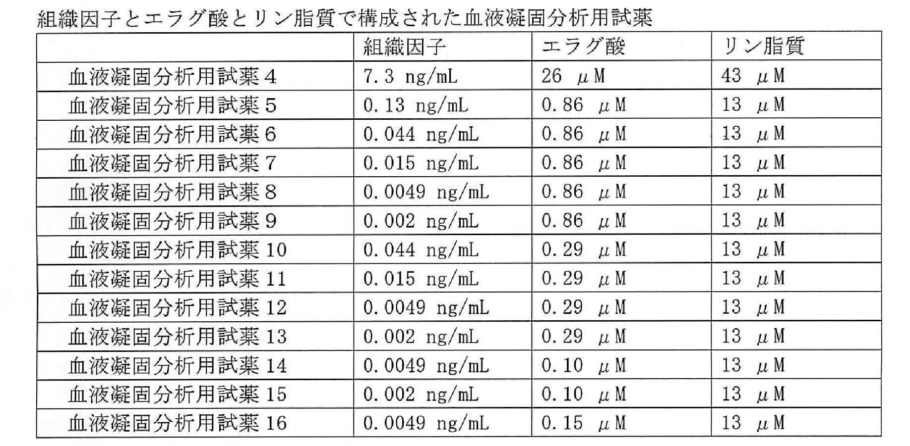
(3)エラグ酸及びリン脂質を含む血液凝固分析用薬を用いたFVIII存在下のFVIII欠乏血漿における二重特異性抗体の凝固波形解析
1、5、又は20 U/dLのリコンビナントヒトFVIII(rhFVIII、バイエル株式会社)を添加したFVIII欠乏ヒト血漿(George King Bio-Medical, Lot No. 899-3062)に、ACE910を添加して得たサンプル50μLに、エラグ酸とリン脂質で構成された血液凝固分析用試薬(表2の血液凝固分析用試薬3)を50μL添加した。3分間インキュベーション後、0.02 mol/L塩化カルシウム溶液50μLを添加して凝固反応を開始し、血液凝固自動測定装置CS-2000i(シスメックス株式会社)で測定した。
1、5、又は20 U/dLのリコンビナントヒトFVIII(rhFVIII、バイエル株式会社)を添加したFVIII欠乏ヒト血漿(George King Bio-Medical, Lot No. 899-3062)に、ACE910を添加して得たサンプル50μLに、エラグ酸とリン脂質で構成された血液凝固分析用試薬(表2の血液凝固分析用試薬3)を50μL添加した。3分間インキュベーション後、0.02 mol/L塩化カルシウム溶液50μLを添加して凝固反応を開始し、血液凝固自動測定装置CS-2000i(シスメックス株式会社)で測定した。
(4)組織因子、エラグ酸及びリン脂質を含む血液凝固分析用薬を用いたFVIII存在下のFVIII欠乏血漿における二重特異性抗体の凝固波形解析
1、5、20、50、100、又は200 U/dLのリコンビナントヒトFVIII (rhFVIII、バイエル株式会社)を添加したFVIII欠乏ヒト血漿(George King Bio-Medical, Lot No. 899-3394)に、ACE910を添加して得たサンプル50μLに、組織因子とエラグ酸とリン脂質で構成された血液凝固分析用試薬(表3の血液凝固分析用試薬12又は16)を50μL添加した。3分間インキュベーション後、0.02 mol/L塩化カルシウム溶液50μLを添加して凝固反応を開始し、血液凝固自動測定装置CS-2000i(シスメックス株式会社)で測定した。
1、5、20、50、100、又は200 U/dLのリコンビナントヒトFVIII (rhFVIII、バイエル株式会社)を添加したFVIII欠乏ヒト血漿(George King Bio-Medical, Lot No. 899-3394)に、ACE910を添加して得たサンプル50μLに、組織因子とエラグ酸とリン脂質で構成された血液凝固分析用試薬(表3の血液凝固分析用試薬12又は16)を50μL添加した。3分間インキュベーション後、0.02 mol/L塩化カルシウム溶液50μLを添加して凝固反応を開始し、血液凝固自動測定装置CS-2000i(シスメックス株式会社)で測定した。
結果
<エラグ酸とリン脂質で構成された血液凝固分析用試薬>
血液凝固分析用試薬1中に含まれるエラグ酸及びリン脂質の各濃度は、市販のAPTT測定試薬であるトロンボチェック中に含まれるエラグ酸及びリン脂質と同じである。血液凝固分析用試薬2はエラグ酸濃度を変更せずに、リン脂質濃度を1/10に希釈した試薬であり、血液凝固分析用試薬3はエラグ酸濃度を1/10に希釈し、リン脂質濃度を変更していない試薬である。血液凝固分析用試薬1、2及び3のいずれにおいてもrhFVIII及びACE910をそれぞれ含むFVIII欠乏血漿における凝固速度、及び凝固加速度を上昇させた(図22A及びB、図23A及びB、並びに図24A及びB参照)。一方、rhFVIIIを含む検体の|Min 1|及び|Min 2|の検量線から10、30、又は100μg/mLのACE910を含む検体の|Min 1|及び|Min 2|のそれぞれをFVIII活性に換算した値が、止血効果から期待する理想の範囲(ACE910 1μg/mL当たりFVIII 0.2~0.4 U/dL)に入ったのは血液凝固分析用試薬3を用いた場合のみであった(図22C及びD、図23C及びD、並びに図24C及びD参照。図中、二本の破線で挟まれた領域は理想範囲を示す)。なお、血液凝固分析用試薬3を用いた場合、ACE910を含む検体の|Min1|から換算したFVIII活性値は、10、30、又は100μg/mLのうち2点以上が理想の範囲に入っていた。これらの結果より、市販のAPTT試薬(血液凝固分析用試薬1)はACE910の薬効を過大評価するのに対して、血液凝固分析用試薬3はACE910の薬効を適正にモニタリングできることが示された。
<エラグ酸とリン脂質で構成された血液凝固分析用試薬>
血液凝固分析用試薬1中に含まれるエラグ酸及びリン脂質の各濃度は、市販のAPTT測定試薬であるトロンボチェック中に含まれるエラグ酸及びリン脂質と同じである。血液凝固分析用試薬2はエラグ酸濃度を変更せずに、リン脂質濃度を1/10に希釈した試薬であり、血液凝固分析用試薬3はエラグ酸濃度を1/10に希釈し、リン脂質濃度を変更していない試薬である。血液凝固分析用試薬1、2及び3のいずれにおいてもrhFVIII及びACE910をそれぞれ含むFVIII欠乏血漿における凝固速度、及び凝固加速度を上昇させた(図22A及びB、図23A及びB、並びに図24A及びB参照)。一方、rhFVIIIを含む検体の|Min 1|及び|Min 2|の検量線から10、30、又は100μg/mLのACE910を含む検体の|Min 1|及び|Min 2|のそれぞれをFVIII活性に換算した値が、止血効果から期待する理想の範囲(ACE910 1μg/mL当たりFVIII 0.2~0.4 U/dL)に入ったのは血液凝固分析用試薬3を用いた場合のみであった(図22C及びD、図23C及びD、並びに図24C及びD参照。図中、二本の破線で挟まれた領域は理想範囲を示す)。なお、血液凝固分析用試薬3を用いた場合、ACE910を含む検体の|Min1|から換算したFVIII活性値は、10、30、又は100μg/mLのうち2点以上が理想の範囲に入っていた。これらの結果より、市販のAPTT試薬(血液凝固分析用試薬1)はACE910の薬効を過大評価するのに対して、血液凝固分析用試薬3はACE910の薬効を適正にモニタリングできることが示された。
さらに、一例ではあるが、血液凝固分析用試薬3を用いてFVIII製剤が含まれるFVIII欠乏血漿におけるACE910の効果を評価したところ、ACE910の添加により凝固速度及び凝固加速度が上昇した(図30A~E参照)。これらの結果より、血液凝固分析用試薬3のような濃度のエラグ酸とリン脂質で構成された血液凝固分析用試薬を用いることにより、FVIII存在下であっても、ACE910の薬効をモニタリングできることが示された。
<組織因子とエラグ酸とリン脂質で構成された血液凝固分析用試薬>
血液凝固分析用試薬4~16は、組織因子とエラグ酸とリン脂質のそれぞれが種々の濃度となるように調製した試薬である。血液凝固分析用試薬4~16のいずれを用いた条件であっても、ACE910の凝固時間、凝固速度及び加速度は上昇した(図25A~C、図26A~I、図27A~I及び図32A~C参照)。以上より、組織因子とエラグ酸とリン脂質で構成された血液凝固分析用試薬を用いることにより、ACE910の薬効をモニタリングできることが示された。なお、一例ではあるが、血液凝固分析用試薬4、6、11、12及び15を用いることで、rhFVIIIを含む検体の|Min 1|の検量線から10、30、又は100μg/mLのACE910の凝固速度をFVIII活性に換算した値が、止血効果から期待する理想の範囲(ACE910 1μg/mL当たりFVIII 0.2~0.4 U/dL)に入ることを示した(図28及び図29参照。図中、二本の破線で挟まれた領域は理想範囲を示す)。
血液凝固分析用試薬4~16は、組織因子とエラグ酸とリン脂質のそれぞれが種々の濃度となるように調製した試薬である。血液凝固分析用試薬4~16のいずれを用いた条件であっても、ACE910の凝固時間、凝固速度及び加速度は上昇した(図25A~C、図26A~I、図27A~I及び図32A~C参照)。以上より、組織因子とエラグ酸とリン脂質で構成された血液凝固分析用試薬を用いることにより、ACE910の薬効をモニタリングできることが示された。なお、一例ではあるが、血液凝固分析用試薬4、6、11、12及び15を用いることで、rhFVIIIを含む検体の|Min 1|の検量線から10、30、又は100μg/mLのACE910の凝固速度をFVIII活性に換算した値が、止血効果から期待する理想の範囲(ACE910 1μg/mL当たりFVIII 0.2~0.4 U/dL)に入ることを示した(図28及び図29参照。図中、二本の破線で挟まれた領域は理想範囲を示す)。
さらに、一例ではあるが、血液凝固分析用試薬12及び16をそれぞれ用いて、FVIII製剤が含まれるFVIII欠乏血漿におけるACE910の効果を評価したところ、ACE910の添加により凝固速度及び凝固加速度が上昇した(図31A~K及び図32A~C参照)。これらの結果より、組織因子とエラグ酸とリン脂質で構成された血液凝固分析用試薬を用いることで、FVIII存在下であってもACE910の薬効をモニタリングできることが示された。
実施例5
組織因子とエラグ酸とリン脂質とを含む血液凝固分析用試薬キットを用いたFVIII欠乏血漿における二重特異性抗体の凝固波形解析
実施例5では、エラグ酸及びリン脂質を含む第1試薬と、組織因子及び塩化カルシウム溶液(0.02 mol/L)を含む第2試薬とで構成された血液凝固分析用試薬キット17~34を用いた。ACE910又はリコンビナントヒトFVIII(rhFVIII、バイエル株式会社)を添加したFVIII欠乏ヒト血漿(George King Bio-Medical) 50μLに、第1試薬を50μL添加した。3分間インキュベーション後、第2試薬を50μL添加して凝固反応を開始し、血液凝固自動測定装置CS-5100(シスメックス株式会社)で測定した。血液凝固分析用試薬キット17~34を用いて調製した測定試料における組織因子、エラグ酸及びリン脂質のそれぞれの終濃度を、以下の表4に示した。なお、血液凝固分析用試薬キット17~34は、図33~図37中では、それぞれ試薬17~試薬34と表記される。
組織因子とエラグ酸とリン脂質とを含む血液凝固分析用試薬キットを用いたFVIII欠乏血漿における二重特異性抗体の凝固波形解析
実施例5では、エラグ酸及びリン脂質を含む第1試薬と、組織因子及び塩化カルシウム溶液(0.02 mol/L)を含む第2試薬とで構成された血液凝固分析用試薬キット17~34を用いた。ACE910又はリコンビナントヒトFVIII(rhFVIII、バイエル株式会社)を添加したFVIII欠乏ヒト血漿(George King Bio-Medical) 50μLに、第1試薬を50μL添加した。3分間インキュベーション後、第2試薬を50μL添加して凝固反応を開始し、血液凝固自動測定装置CS-5100(シスメックス株式会社)で測定した。血液凝固分析用試薬キット17~34を用いて調製した測定試料における組織因子、エラグ酸及びリン脂質のそれぞれの終濃度を、以下の表4に示した。なお、血液凝固分析用試薬キット17~34は、図33~図37中では、それぞれ試薬17~試薬34と表記される。
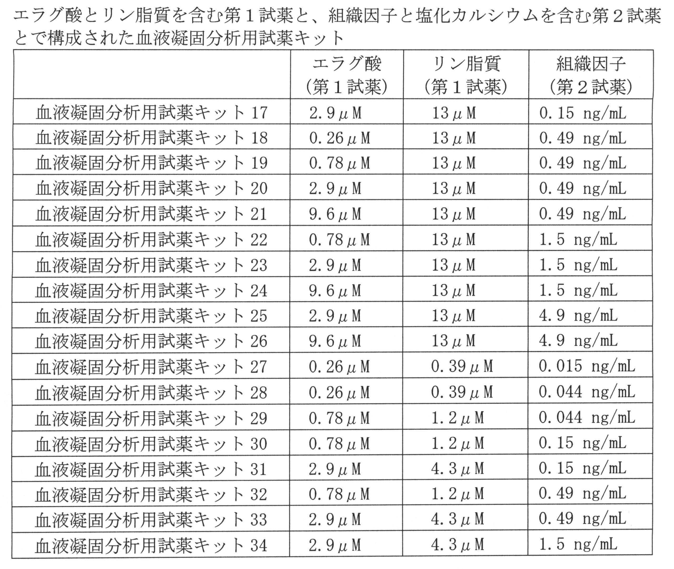
血液凝固分析用試薬キット17~34のいずれを用いた条件であっても、ACE910の凝固速度及び加速度は上昇した(図33A~E、図34A~E及び図35A~E参照)。以上より、組織因子とエラグ酸とリン脂質で構成された血液凝固分析用試薬を用いることにより、ACE910の薬効をモニタリングできることが示された。なお、一例ではあるが、血液凝固分析用試薬キット19、20、23、24、28、30、32、33及び34を用いることで、rhFVIIIを含む検体の|Min 1|の検量線から10、30、又は100μg/mLのACE910の凝固速度をFVIII活性に換算した値が、止血効果から期待する理想の範囲(ACE910 1μg/mL当たりFVIII 0.2~0.4 U/dL)に入ることを示した(図36A及びB参照。図中、二本の破線で挟まれた領域は理想範囲を示す)。
また、一例ではあるが、血液凝固分析用試薬キット22、23及び34を用いることで、rhFVIIIを含む検体の|Min 2|の検量線から10、30、又は100μg/mLのACE910の凝固速度をFVIII活性に換算した値が、止血効果から期待する理想の範囲(ACE910 1μg/mL当たりFVIII 0.2~0.4 U/dL)に入ることを示した(図37A及びB参照。図中、二本の破線で挟まれた領域は理想範囲を示す)。
実施例6
組織因子とエラグ酸とリン脂質とを含む血液凝固分析用試薬キットを用いたバイパス製剤存在下のFVIII欠乏血漿における二重特異性抗体の凝固波形解析
実施例6では、エラグ酸及びリン脂質を含む第1試薬と、組織因子及び塩化カルシウム溶液(0.02 mol/L)を含む第2試薬とで構成された血液凝固分析用試薬キット20、21、28、30、33、35及び36を用いた。試薬キット20、21、28、30及び33は実施例5と同じである。0.5又は1.0 U/mLのAPCC(ファイバ(登録商標)、バクスター株式会社)を添加したFVIII欠乏ヒト血漿(George King Bio-Medical)、又は0.25若しくは1.0μg/mLのFVIIa(ノボセブン(登録商標)、ノボノルディスクファーマ)を添加したFVIII欠乏ヒト血漿(George King Bio-Medical)にACE910を添加して得たサンプル50μLに、第1試薬を50μL添加した。3分間インキュベーション後、第2試薬を50μL添加して凝固反応を開始し、血液凝固自動測定装置CS-2000i(シスメックス株式会社)で測定した。上記の各試薬キットを用いて調製した測定試料における組織因子、エラグ酸及びリン脂質のそれぞれの終濃度を、以下の表5に示した。
組織因子とエラグ酸とリン脂質とを含む血液凝固分析用試薬キットを用いたバイパス製剤存在下のFVIII欠乏血漿における二重特異性抗体の凝固波形解析
実施例6では、エラグ酸及びリン脂質を含む第1試薬と、組織因子及び塩化カルシウム溶液(0.02 mol/L)を含む第2試薬とで構成された血液凝固分析用試薬キット20、21、28、30、33、35及び36を用いた。試薬キット20、21、28、30及び33は実施例5と同じである。0.5又は1.0 U/mLのAPCC(ファイバ(登録商標)、バクスター株式会社)を添加したFVIII欠乏ヒト血漿(George King Bio-Medical)、又は0.25若しくは1.0μg/mLのFVIIa(ノボセブン(登録商標)、ノボノルディスクファーマ)を添加したFVIII欠乏ヒト血漿(George King Bio-Medical)にACE910を添加して得たサンプル50μLに、第1試薬を50μL添加した。3分間インキュベーション後、第2試薬を50μL添加して凝固反応を開始し、血液凝固自動測定装置CS-2000i(シスメックス株式会社)で測定した。上記の各試薬キットを用いて調製した測定試料における組織因子、エラグ酸及びリン脂質のそれぞれの終濃度を、以下の表5に示した。
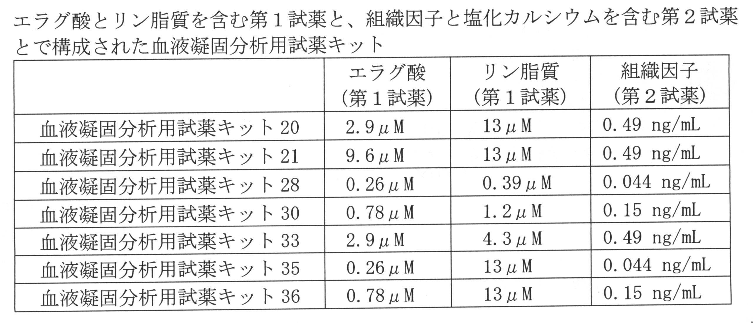
血液凝固分析用試薬キット20、21、28、30、33、35及び36を用いて、バイパス製剤が含まれるFVIII欠乏血漿におけるACE910の効果を評価したところ、ACE910の添加により凝固速度が上昇した(試薬キット20については図38A~D、試薬キット21については図39A~D、試薬キット28については図40A~D、試薬キット30については図41A~D、試薬キット33については図42A~D、試薬キット35については図43A~D、及び試薬キット36については図44A~Dを参照)。これらの結果より、組織因子とエラグ酸とリン脂質で構成された血液凝固分析用試薬を用いることで、バイパス製剤の存在下であってもACE910の薬効をモニタリングできることが示された。
10 血液検体分析装置
11、12 試薬テーブル(試薬収容部)
20 測定部
22g、22i 光検出器(受光部)
27 ランプユニット(光源)
30 検体搬送部
40 制御装置(制御部)
41 表示部
42 入力部
43 コンピュータ本体
50 測定装置(測定試料調製部及び光学的情報取得部)
111 第1容器
112 第2容器
113 第3容器
11、12 試薬テーブル(試薬収容部)
20 測定部
22g、22i 光検出器(受光部)
27 ランプユニット(光源)
30 検体搬送部
40 制御装置(制御部)
41 表示部
42 入力部
43 コンピュータ本体
50 測定装置(測定試料調製部及び光学的情報取得部)
111 第1容器
112 第2容器
113 第3容器
Claims (23)
- 凝固第VIII因子代替活性を有する物質を投与された被検者から得た血液検体と、凝固第XII因子活性化剤と、リン脂質と、カルシウムイオン含有水溶液とから、測定試料を調製する工程と、
前記測定試料に光を照射して、前記測定試料から光量に関する光学的情報を取得する工程と、
取得した光学的情報に基づいて、凝固波形の微分に関するパラメータを少なくとも1つ取得する工程と、
取得したパラメータの値に基づいて、前記血液検体の凝固能を評価する工程と
を含み、
前記測定試料における第XII因子活性化剤の終濃度が1μM以上22μM以下であり、且つリン脂質の終濃度が1.4μM以上33μM以下であるか、又は、
前記測定試料における凝固第XII因子活性化剤の終濃度が1μM以上2.9μM以下であり、且つリン脂質の終濃度が1.4μM以上43μM以下である
血液検体の凝固能の評価方法。 - 前記評価工程において、取得したパラメータの値から前記凝固第VIII因子代替活性を有する物質を含む血液検体の凝固能を示す値を取得し、取得した凝固能を示す値に基づいて、前記血液検体の凝固能を評価する請求項1に記載の方法。
- 前記凝固能を示す値が、凝固第VIII因子活性値である請求項2に記載の方法。
- 前記光学的情報が、連続的又は断続的に測定された散乱光量、透過度又は吸光度であり、前記凝固波形が、前記散乱光量、透過度又は吸光度の経時的変化を表す波形である請求項1~3のいずれか1項に記載の方法。
- 前記凝固波形の微分に関するパラメータが、最大凝固速度(|Min 1|)、最大凝固加速度(|Min 2|)、最大凝固減速度(Max 2)、波形下部面積(AUC)及び傾き(Slope)からなる群より選択される少なくとも1つである請求項1~4のいずれか1項に記載の方法。
- 前記凝固第XII因子活性化剤が、エラグ酸、カオリン、セライト及びシリカから選択される少なくとも1種である請求項1~5のいずれか1項に記載の方法。
- 凝固第VIII因子代替活性を有する物質を投与された被検者から得た血液検体と、凝固第XII因子活性化剤と、リン脂質と、組織因子と、カルシウムイオン含有水溶液とから、測定試料を調製する工程と、
前記測定試料に光を照射して、前記測定試料から光量に関する光学的情報を取得する工程と、
取得した光学的情報に基づいて、前記血液検体の凝固能を評価する工程と
を含む、
血液検体の凝固能の評価方法。 - 凝固第XII因子活性化剤を3μM以上66μM以下の濃度で含み、且つリン脂質を4.2μM以上99μM以下の濃度で含むか、又は、凝固第XII因子活性化剤を3μM以上8.7μM以下の濃度で含み、且つリン脂質を4.2μM以上129μM以下の濃度で含む、凝固第VIII因子代替活性を有する物質を投与された被検者から得た血液検体の凝固能を評価するための試薬。
- 第1試薬と、カルシウムイオン含有水溶液からなる凝固開始試薬とを備え、
前記第1試薬が、凝固第XII因子活性化剤を3μM以上66μM以下の濃度で含み、且つリン脂質を4.2μM以上99μM以下の濃度で含むか、又は、
前記第1試薬が、凝固第XII因子活性化剤を3μM以上8.7μM以下の濃度で含み、且つリン脂質を4.2μM以上129μM以下の濃度で含む、
凝固第VIII因子代替活性を有する物質を投与された被検者から得た血液検体の凝固能を評価するための試薬キット。 - 凝固第XII因子活性化剤、リン脂質、及び組織因子を含む、凝固第VIII因子代替活性を有する物質を投与された被検者から得た血液検体の凝固能を評価するための試薬。
- 凝固第XII因子活性化剤、リン脂質、及び組織因子を含む第1試薬と、カルシウムイオン含有水溶液からなる凝固開始試薬とを備える、凝固第VIII因子代替活性を有する物質を投与された被検者から得た血液検体の凝固能を評価するための試薬キット。
- 第1試薬と、第2試薬と、カルシウムイオン含有水溶液からなる凝固開始試薬とを備え、
前記第1試薬が、凝固第XII因子活性化剤及びリン脂質を含み、且つ前記第2試薬が組織因子を含むか、
前記第1試薬が、凝固第XII因子活性化剤及びリン脂質を含み、且つ前記第2試薬が組織因子及びリン脂質を含むか、又は
前記第1試薬が、凝固第XII因子活性化剤及び組織因子を含み、且つ前記第2試薬がリン脂質を含む、
凝固第VIII因子代替活性を有する物質を投与された被検者から得た血液検体の凝固能を評価するための試薬キット。 - 第1試薬と、第2試薬とを備え、
前記第1試薬が、凝固第XII因子活性化剤及びリン脂質を含み、且つ前記第2試薬が組織因子及びカルシウムイオンを含む、
凝固第VIII因子代替活性を有する物質を投与された被検者から得た血液検体の凝固能を評価するための試薬キット。 - 前記第2試薬が、リン脂質をさらに含む請求項13に記載の試薬キット。
- 測定試料を調製する測定試料調製部と、
調製された測定試料に光を照射し、前記測定試料から光量に関する光学的情報を取得する光学的情報取得部と、
制御部と
を備え、
前記制御部が、
凝固第XII因子活性化剤及びリン脂質を含む活性化部分トロンボプラスチン時間測定用試薬を希釈液で所定の倍率に希釈し、希釈した前記試薬と、凝固第VIII因子代替活性を有する物質を投与された被検者から得た血液検体と、カルシウムイオン含有水溶液とから、測定試料を調製するように前記測定試料調製部を制御し、
前記光学的情報に基づいて、凝固波形の微分に関するパラメータを少なくとも1つ取得し、
取得したパラメータの値に基づいて、前記血液検体の凝固能についての参考情報を出力する、
血液検体分析装置。 - 前記参考情報が、凝固第VIII因子活性値である、請求項15に記載の血液検体分析装置。
- 前記制御部が、前記活性化部分トロンボプラスチン時間測定用試薬を1.3倍以上30倍以下の倍率で希釈するように前記測定試料調製部を制御する請求項15又は16に記載の血液検体分析装置。
- 前記制御部が、前記測定試料における凝固第XII因子活性化剤の終濃度が1μM以上22μM以下となり且つリン脂質の終濃度が1.4μM以上33μM以下となるか、又は、前記測定試料における凝固第XII因子活性化剤の終濃度が1μM以上2.9μM以下となり且つリン脂質の終濃度が1.4μM以上43μM以下となるように前記測定試料調製部を制御する請求項15~17のいずれか1項に記載の血液検体分析装置。
- 前記光学的情報が、連続的又は断続的に測定された散乱光量、透過度又は吸光度である請求項15~18のいずれか1項に記載の血液検体分析装置。
- 測定試料を調製する測定試料調製部と、
調製された測定試料に光を照射し、前記測定試料から光量に関する光学的情報を取得する光学的情報取得部と、
制御部と
を備え、
前記制御部が、
凝固第VIII因子代替活性を有する物質を投与された被検者から得た血液検体と、凝固第XII因子活性化剤と、リン脂質と、組織因子と、カルシウムイオン含有水溶液とから、測定試料を調製するように前記測定試料調製部を制御し、
前記光学的情報に基づいて、前記血液検体の凝固能についての参考情報を出力する、
血液検体分析装置。 - 前記参考情報が、凝固第VIII因子活性値である請求項20に記載の血液検体分析装置。
- プロセッサ及び前記プロセッサの制御下にあるメモリを含むコンピュータを備え、
前記メモリには、下記のステップ:
凝固第VIII因子代替活性を有する物質を投与された被検者から得た血液検体と、凝固第XII因子活性化剤と、リン脂質と、カルシウムイオン含有水溶液とから調製された測定試料から光量に関する光学的情報を取得するステップと、
前記光学的情報に基づいて、凝固波形の微分に関するパラメータを少なくとも1つ取得するステップと、
取得したパラメータの値に基づいて、前記血液検体の凝固能についての参考情報を出力するステップと
を前記コンピュータに実行させるためのコンピュータプログラムが記録されている、
血液検体の凝固能の評価のための装置。 - コンピュータが読み取り可能な媒体に記録されているコンピュータプログラムであって、下記のステップ:
凝固第VIII因子代替活性を有する物質を投与された被検者から得た血液検体と、凝固第XII因子活性化剤と、リン脂質と、カルシウムイオン含有水溶液とから調製された測定試料から光量に関する光学的情報を取得するステップと、
前記光学的情報に基づいて、凝固波形の微分に関するパラメータを少なくとも1つ取得するステップと、
取得したパラメータの値に基づいて、前記血液検体の凝固能についての参考情報を出力するステップと
を前記コンピュータに実行させる、血液検体の凝固能の評価のためのコンピュータプログラム。
Priority Applications (5)
| Application Number | Priority Date | Filing Date | Title |
|---|---|---|---|
| CN201680022989.3A CN107533071A (zh) | 2015-04-24 | 2016-03-30 | 血液受试体的凝固能力的评价方法、以及用于在该方法中使用的试剂、试剂盒及装置 |
| CN202111594393.XA CN114578068A (zh) | 2015-04-24 | 2016-03-30 | 血液受试体的凝固能力的评价方法、以及用于在该方法中使用的试剂、试剂盒及装置 |
| EP16782966.2A EP3287791B1 (en) | 2015-04-24 | 2016-03-30 | Method for evaluating coagulation ability of blood specimen, and reagent, reagent kit and device to be used therein |
| JP2017505273A JP6147945B2 (ja) | 2015-04-24 | 2016-03-30 | 血液検体の凝固能の評価方法、並びにその方法に用いるための試薬、試薬キット及び装置 |
| US15/791,551 US10302626B2 (en) | 2015-04-24 | 2017-10-24 | Method for evaluating coagulation ability of blood specimen, and reagent, reagent kit and device to be used therein |
Applications Claiming Priority (2)
| Application Number | Priority Date | Filing Date | Title |
|---|---|---|---|
| JP2015089865 | 2015-04-24 | ||
| JP2015-089865 | 2015-04-24 |
Related Child Applications (1)
| Application Number | Title | Priority Date | Filing Date |
|---|---|---|---|
| US15/791,551 Continuation US10302626B2 (en) | 2015-04-24 | 2017-10-24 | Method for evaluating coagulation ability of blood specimen, and reagent, reagent kit and device to be used therein |
Publications (1)
| Publication Number | Publication Date |
|---|---|
| WO2016170944A1 true WO2016170944A1 (ja) | 2016-10-27 |
Family
ID=57143474
Family Applications (1)
| Application Number | Title | Priority Date | Filing Date |
|---|---|---|---|
| PCT/JP2016/060566 Ceased WO2016170944A1 (ja) | 2015-04-24 | 2016-03-30 | 血液検体の凝固能の評価方法、並びにその方法に用いるための試薬、試薬キット及び装置 |
Country Status (5)
| Country | Link |
|---|---|
| US (1) | US10302626B2 (ja) |
| EP (1) | EP3287791B1 (ja) |
| JP (2) | JP6147945B2 (ja) |
| CN (2) | CN114578068A (ja) |
| WO (1) | WO2016170944A1 (ja) |
Cited By (7)
| Publication number | Priority date | Publication date | Assignee | Title |
|---|---|---|---|---|
| WO2020101025A1 (ja) * | 2018-11-15 | 2020-05-22 | 積水メディカル株式会社 | 分析方法、分析装置及び分析プログラム |
| WO2021132552A1 (ja) * | 2019-12-26 | 2021-07-01 | 積水メディカル株式会社 | 血液凝固時間測定方法 |
| JP2021156833A (ja) * | 2020-03-30 | 2021-10-07 | シスメックス株式会社 | 血液凝固活性の測定方法 |
| WO2024018789A1 (ja) * | 2022-07-21 | 2024-01-25 | 株式会社日立ハイテク | 血液凝固時間の延長原因の推定方法、及び情報処理装置 |
| JP2024014684A (ja) * | 2022-07-21 | 2024-02-01 | 株式会社日立ハイテク | 血液凝固時間の延長原因の推定方法、及び情報処理装置 |
| WO2024143524A1 (ja) | 2022-12-28 | 2024-07-04 | 積水メディカル株式会社 | 血液検体の血液凝固能の分析方法 |
| WO2024143525A1 (ja) | 2022-12-28 | 2024-07-04 | 積水メディカル株式会社 | 血液検体の血液凝固能の分析方法 |
Families Citing this family (6)
| Publication number | Priority date | Publication date | Assignee | Title |
|---|---|---|---|---|
| US12493033B2 (en) | 2018-06-04 | 2025-12-09 | Chugai Seiyaku Kabushiki Kaisha | Method for detecting bi-specific antibody complex |
| EP3919913A4 (en) | 2019-01-31 | 2022-11-09 | Sekisui Medical Co., Ltd. | METHOD OF ANALYSIS OF BLOOD COAGULATION CHARACTERISTICS OF A BLOOD SAMPLE |
| CN113785197B (zh) * | 2019-04-26 | 2024-03-12 | 积水医疗株式会社 | 凝固因子抑制剂效价的测定方法 |
| CN110160980B (zh) * | 2019-06-25 | 2021-12-28 | 迈克医疗电子有限公司 | 样本吸光度变化率的分析方法、分析装置及光学检测系统 |
| JP7712022B2 (ja) | 2020-09-08 | 2025-07-23 | 積水メディカル株式会社 | 血液凝固反応の分析方法 |
| JP7665365B2 (ja) | 2021-03-19 | 2025-04-21 | シスメックス株式会社 | 被検者の出血傾向を解析する解析方法、解析装置、解析システム、及び解析プログラム |
Citations (6)
| Publication number | Priority date | Publication date | Assignee | Title |
|---|---|---|---|---|
| JP2004191320A (ja) * | 2002-12-13 | 2004-07-08 | Sysmex Corp | Aptt測定試薬の検査方法 |
| JP2008530992A (ja) * | 2005-02-22 | 2008-08-14 | イェナフィン・ゲーエムベーハー | 血液または血漿の試料の総凝固活性の測定方法 |
| JP2010217059A (ja) * | 2009-03-18 | 2010-09-30 | Shimadzu Corp | 血液凝固分析装置 |
| JP2012048586A (ja) * | 2010-08-27 | 2012-03-08 | Kyocera Corp | 文字入力装置、文字入力方法および文字入力プログラム |
| JP2013053960A (ja) * | 2011-09-05 | 2013-03-21 | Nippon Telegr & Teleph Corp <Ntt> | 凝固活性測定装置、測定チップおよび測定方法 |
| WO2014050926A1 (ja) * | 2012-09-28 | 2014-04-03 | 中外製薬株式会社 | 血液凝固反応の評価方法 |
Family Cites Families (4)
| Publication number | Priority date | Publication date | Assignee | Title |
|---|---|---|---|---|
| KR101962483B1 (ko) * | 2010-11-17 | 2019-03-29 | 추가이 세이야쿠 가부시키가이샤 | 혈액응고 제viii 인자의 기능을 대체하는 기능을 갖는 다중특이성 항원 결합 분자 |
| IS2809B (is) * | 2011-03-08 | 2012-10-15 | Haskoli Islands | Blóðstorkumæling |
| JP5911443B2 (ja) * | 2013-03-06 | 2016-04-27 | シスメックス株式会社 | 血液凝固分析装置および血液凝固分析方法 |
| JP2014190954A (ja) | 2013-03-28 | 2014-10-06 | Sysmex Corp | 活性化部分トロンボプラスチン時間測定用試薬およびその利用 |
-
2016
- 2016-03-30 CN CN202111594393.XA patent/CN114578068A/zh active Pending
- 2016-03-30 CN CN201680022989.3A patent/CN107533071A/zh active Pending
- 2016-03-30 EP EP16782966.2A patent/EP3287791B1/en active Active
- 2016-03-30 WO PCT/JP2016/060566 patent/WO2016170944A1/ja not_active Ceased
- 2016-03-30 JP JP2017505273A patent/JP6147945B2/ja active Active
-
2017
- 2017-01-27 JP JP2017012960A patent/JP6154969B2/ja active Active
- 2017-10-24 US US15/791,551 patent/US10302626B2/en active Active
Patent Citations (6)
| Publication number | Priority date | Publication date | Assignee | Title |
|---|---|---|---|---|
| JP2004191320A (ja) * | 2002-12-13 | 2004-07-08 | Sysmex Corp | Aptt測定試薬の検査方法 |
| JP2008530992A (ja) * | 2005-02-22 | 2008-08-14 | イェナフィン・ゲーエムベーハー | 血液または血漿の試料の総凝固活性の測定方法 |
| JP2010217059A (ja) * | 2009-03-18 | 2010-09-30 | Shimadzu Corp | 血液凝固分析装置 |
| JP2012048586A (ja) * | 2010-08-27 | 2012-03-08 | Kyocera Corp | 文字入力装置、文字入力方法および文字入力プログラム |
| JP2013053960A (ja) * | 2011-09-05 | 2013-03-21 | Nippon Telegr & Teleph Corp <Ntt> | 凝固活性測定装置、測定チップおよび測定方法 |
| WO2014050926A1 (ja) * | 2012-09-28 | 2014-04-03 | 中外製薬株式会社 | 血液凝固反応の評価方法 |
Non-Patent Citations (2)
| Title |
|---|
| MUTO A. ET AL.: "Anti-factor IXa/X bispecific antibody (ACE910): hemostatic potency against ongoing bleeds in a hemophilia A model and the possibility of routine supplementation", JOURNAL OF THROMBOSIS AND HAEMOSTASIS, vol. 12, no. 2, February 2014 (2014-02-01), pages 206 - 213, XP055214235 * |
| See also references of EP3287791A4 * |
Cited By (15)
| Publication number | Priority date | Publication date | Assignee | Title |
|---|---|---|---|---|
| US12174204B2 (en) | 2018-11-15 | 2024-12-24 | Sekisui Medical Co., Ltd. | Analysis method, analysis apparatus, and analysis program |
| JPWO2020101025A1 (ja) * | 2018-11-15 | 2021-10-07 | 積水メディカル株式会社 | 分析方法、分析装置及び分析プログラム |
| JP7328251B2 (ja) | 2018-11-15 | 2023-08-16 | 積水メディカル株式会社 | 分析方法、分析装置及び分析プログラム |
| WO2020101025A1 (ja) * | 2018-11-15 | 2020-05-22 | 積水メディカル株式会社 | 分析方法、分析装置及び分析プログラム |
| WO2021132552A1 (ja) * | 2019-12-26 | 2021-07-01 | 積水メディカル株式会社 | 血液凝固時間測定方法 |
| JPWO2021132552A1 (ja) * | 2019-12-26 | 2021-07-01 | ||
| JP7616747B2 (ja) | 2019-12-26 | 2025-01-17 | 積水メディカル株式会社 | 血液凝固時間測定方法 |
| JP2021156833A (ja) * | 2020-03-30 | 2021-10-07 | シスメックス株式会社 | 血液凝固活性の測定方法 |
| JP7681268B2 (ja) | 2020-03-30 | 2025-05-22 | シスメックス株式会社 | 血液凝固活性の測定方法 |
| WO2024018789A1 (ja) * | 2022-07-21 | 2024-01-25 | 株式会社日立ハイテク | 血液凝固時間の延長原因の推定方法、及び情報処理装置 |
| JP2024014684A (ja) * | 2022-07-21 | 2024-02-01 | 株式会社日立ハイテク | 血液凝固時間の延長原因の推定方法、及び情報処理装置 |
| WO2024143525A1 (ja) | 2022-12-28 | 2024-07-04 | 積水メディカル株式会社 | 血液検体の血液凝固能の分析方法 |
| WO2024143524A1 (ja) | 2022-12-28 | 2024-07-04 | 積水メディカル株式会社 | 血液検体の血液凝固能の分析方法 |
| EP4644905A1 (en) | 2022-12-28 | 2025-11-05 | Sekisui Medical Co., Ltd. | Method for analyzing blood clotting ability of blood sample |
| EP4644906A1 (en) | 2022-12-28 | 2025-11-05 | Sekisui Medical Co., Ltd. | Method for analyzing blood coagulation capability of blood specimen |
Also Published As
| Publication number | Publication date |
|---|---|
| EP3287791B1 (en) | 2025-02-19 |
| EP3287791A4 (en) | 2019-03-27 |
| US20180045709A1 (en) | 2018-02-15 |
| EP3287791A1 (en) | 2018-02-28 |
| JP2017106925A (ja) | 2017-06-15 |
| JP6147945B2 (ja) | 2017-06-14 |
| CN114578068A (zh) | 2022-06-03 |
| CN107533071A (zh) | 2018-01-02 |
| US10302626B2 (en) | 2019-05-28 |
| JPWO2016170944A1 (ja) | 2017-05-18 |
| JP6154969B2 (ja) | 2017-06-28 |
Similar Documents
| Publication | Publication Date | Title |
|---|---|---|
| JP6154969B2 (ja) | 血液検体の凝固能の評価方法、並びにその方法に用いるための試薬、試薬キット及び装置 | |
| JP6871673B2 (ja) | 血液検体を判定するための方法、装置及びコンピュータプログラム、並びに血液検体分析装置 | |
| JP6430809B2 (ja) | 血液検体を判定するための方法、システム及びコンピュータプログラム、並びに血液検体分析装置 | |
| CN107250801B (zh) | 取得关于凝固时间的延长原因的信息的方法和用于实施其的装置及程序 | |
| JP7477117B2 (ja) | 血液検体の分析方法、血液検体分析用試薬及び試薬キット、並びに血液検体分析装置 | |
| JP6793916B2 (ja) | 血友病の重症度の判定方法及び血液検体分析装置 | |
| JP6994528B2 (ja) | 血液検体を判定するための方法、装置及びコンピュータプログラム、並びに血液検体分析装置 | |
| CN109030837B (zh) | 血液样本的判断方法、以及血液样本分析装置 | |
| EP3355062B1 (en) | Method for determining blood specimen, and apparatus | |
| Dudek et al. | Development of a point of care lateral flow device for measuring human plasma fibrinogen | |
| Harris et al. | Point of care (POC) blood coagulation monitoring technologies | |
| WO2019057367A1 (en) | ENHANCED DETECTION OF ANTICOAGULANTS IN ORGANIC LIQUIDS | |
| JP7250103B2 (ja) | 血液検体を判定するための方法、装置及びコンピュータプログラム、並びに血液検体分析装置 |
Legal Events
| Date | Code | Title | Description |
|---|---|---|---|
| 121 | Ep: the epo has been informed by wipo that ep was designated in this application |
Ref document number: 16782966 Country of ref document: EP Kind code of ref document: A1 |
|
| ENP | Entry into the national phase |
Ref document number: 2017505273 Country of ref document: JP Kind code of ref document: A |
|
| NENP | Non-entry into the national phase |
Ref country code: DE |