WO2016167372A1 - 大脳皮質ニューロンの誘導方法 - Google Patents
大脳皮質ニューロンの誘導方法 Download PDFInfo
- Publication number
- WO2016167372A1 WO2016167372A1 PCT/JP2016/062578 JP2016062578W WO2016167372A1 WO 2016167372 A1 WO2016167372 A1 WO 2016167372A1 JP 2016062578 W JP2016062578 W JP 2016062578W WO 2016167372 A1 WO2016167372 A1 WO 2016167372A1
- Authority
- WO
- WIPO (PCT)
- Prior art keywords
- cells
- inhibitor
- medium
- days
- cell
- Prior art date
Links
- 0 NC(c(cc1)ccc1-c1nc(-c2ccc(*CO3)c3c2)c(-c2ccccn2)[n]1)=O Chemical compound NC(c(cc1)ccc1-c1nc(-c2ccc(*CO3)c3c2)c(-c2ccccn2)[n]1)=O 0.000 description 1
Images


Classifications
-
- C—CHEMISTRY; METALLURGY
- C12—BIOCHEMISTRY; BEER; SPIRITS; WINE; VINEGAR; MICROBIOLOGY; ENZYMOLOGY; MUTATION OR GENETIC ENGINEERING
- C12N—MICROORGANISMS OR ENZYMES; COMPOSITIONS THEREOF; PROPAGATING, PRESERVING, OR MAINTAINING MICROORGANISMS; MUTATION OR GENETIC ENGINEERING; CULTURE MEDIA
- C12N5/00—Undifferentiated human, animal or plant cells, e.g. cell lines; Tissues; Cultivation or maintenance thereof; Culture media therefor
- C12N5/06—Animal cells or tissues; Human cells or tissues
- C12N5/0602—Vertebrate cells
- C12N5/0618—Cells of the nervous system
- C12N5/0619—Neurons
-
- A—HUMAN NECESSITIES
- A61—MEDICAL OR VETERINARY SCIENCE; HYGIENE
- A61K—PREPARATIONS FOR MEDICAL, DENTAL OR TOILETRY PURPOSES
- A61K35/00—Medicinal preparations containing materials or reaction products thereof with undetermined constitution
- A61K35/12—Materials from mammals; Compositions comprising non-specified tissues or cells; Compositions comprising non-embryonic stem cells; Genetically modified cells
- A61K35/30—Nerves; Brain; Eyes; Corneal cells; Cerebrospinal fluid; Neuronal stem cells; Neuronal precursor cells; Glial cells; Oligodendrocytes; Schwann cells; Astroglia; Astrocytes; Choroid plexus; Spinal cord tissue
-
- A—HUMAN NECESSITIES
- A61—MEDICAL OR VETERINARY SCIENCE; HYGIENE
- A61P—SPECIFIC THERAPEUTIC ACTIVITY OF CHEMICAL COMPOUNDS OR MEDICINAL PREPARATIONS
- A61P25/00—Drugs for disorders of the nervous system
-
- A—HUMAN NECESSITIES
- A61—MEDICAL OR VETERINARY SCIENCE; HYGIENE
- A61P—SPECIFIC THERAPEUTIC ACTIVITY OF CHEMICAL COMPOUNDS OR MEDICINAL PREPARATIONS
- A61P43/00—Drugs for specific purposes, not provided for in groups A61P1/00-A61P41/00
-
- A—HUMAN NECESSITIES
- A61—MEDICAL OR VETERINARY SCIENCE; HYGIENE
- A61K—PREPARATIONS FOR MEDICAL, DENTAL OR TOILETRY PURPOSES
- A61K35/00—Medicinal preparations containing materials or reaction products thereof with undetermined constitution
-
- A—HUMAN NECESSITIES
- A61—MEDICAL OR VETERINARY SCIENCE; HYGIENE
- A61K—PREPARATIONS FOR MEDICAL, DENTAL OR TOILETRY PURPOSES
- A61K35/00—Medicinal preparations containing materials or reaction products thereof with undetermined constitution
- A61K35/12—Materials from mammals; Compositions comprising non-specified tissues or cells; Compositions comprising non-embryonic stem cells; Genetically modified cells
- A61K35/48—Reproductive organs
- A61K35/54—Ovaries; Ova; Ovules; Embryos; Foetal cells; Germ cells
- A61K35/545—Embryonic stem cells; Pluripotent stem cells; Induced pluripotent stem cells; Uncharacterised stem cells
-
- C—CHEMISTRY; METALLURGY
- C12—BIOCHEMISTRY; BEER; SPIRITS; WINE; VINEGAR; MICROBIOLOGY; ENZYMOLOGY; MUTATION OR GENETIC ENGINEERING
- C12N—MICROORGANISMS OR ENZYMES; COMPOSITIONS THEREOF; PROPAGATING, PRESERVING, OR MAINTAINING MICROORGANISMS; MUTATION OR GENETIC ENGINEERING; CULTURE MEDIA
- C12N2501/00—Active agents used in cell culture processes, e.g. differentation
- C12N2501/10—Growth factors
- C12N2501/115—Basic fibroblast growth factor (bFGF, FGF-2)
-
- C—CHEMISTRY; METALLURGY
- C12—BIOCHEMISTRY; BEER; SPIRITS; WINE; VINEGAR; MICROBIOLOGY; ENZYMOLOGY; MUTATION OR GENETIC ENGINEERING
- C12N—MICROORGANISMS OR ENZYMES; COMPOSITIONS THEREOF; PROPAGATING, PRESERVING, OR MAINTAINING MICROORGANISMS; MUTATION OR GENETIC ENGINEERING; CULTURE MEDIA
- C12N2501/00—Active agents used in cell culture processes, e.g. differentation
- C12N2501/10—Growth factors
- C12N2501/15—Transforming growth factor beta (TGF-β)
-
- C—CHEMISTRY; METALLURGY
- C12—BIOCHEMISTRY; BEER; SPIRITS; WINE; VINEGAR; MICROBIOLOGY; ENZYMOLOGY; MUTATION OR GENETIC ENGINEERING
- C12N—MICROORGANISMS OR ENZYMES; COMPOSITIONS THEREOF; PROPAGATING, PRESERVING, OR MAINTAINING MICROORGANISMS; MUTATION OR GENETIC ENGINEERING; CULTURE MEDIA
- C12N2501/00—Active agents used in cell culture processes, e.g. differentation
- C12N2501/10—Growth factors
- C12N2501/155—Bone morphogenic proteins [BMP]; Osteogenins; Osteogenic factor; Bone inducing factor
-
- C—CHEMISTRY; METALLURGY
- C12—BIOCHEMISTRY; BEER; SPIRITS; WINE; VINEGAR; MICROBIOLOGY; ENZYMOLOGY; MUTATION OR GENETIC ENGINEERING
- C12N—MICROORGANISMS OR ENZYMES; COMPOSITIONS THEREOF; PROPAGATING, PRESERVING, OR MAINTAINING MICROORGANISMS; MUTATION OR GENETIC ENGINEERING; CULTURE MEDIA
- C12N2501/00—Active agents used in cell culture processes, e.g. differentation
- C12N2501/40—Regulators of development
- C12N2501/415—Wnt; Frizzeled
-
- C—CHEMISTRY; METALLURGY
- C12—BIOCHEMISTRY; BEER; SPIRITS; WINE; VINEGAR; MICROBIOLOGY; ENZYMOLOGY; MUTATION OR GENETIC ENGINEERING
- C12N—MICROORGANISMS OR ENZYMES; COMPOSITIONS THEREOF; PROPAGATING, PRESERVING, OR MAINTAINING MICROORGANISMS; MUTATION OR GENETIC ENGINEERING; CULTURE MEDIA
- C12N2506/00—Differentiation of animal cells from one lineage to another; Differentiation of pluripotent cells
- C12N2506/02—Differentiation of animal cells from one lineage to another; Differentiation of pluripotent cells from embryonic cells
-
- C—CHEMISTRY; METALLURGY
- C12—BIOCHEMISTRY; BEER; SPIRITS; WINE; VINEGAR; MICROBIOLOGY; ENZYMOLOGY; MUTATION OR GENETIC ENGINEERING
- C12N—MICROORGANISMS OR ENZYMES; COMPOSITIONS THEREOF; PROPAGATING, PRESERVING, OR MAINTAINING MICROORGANISMS; MUTATION OR GENETIC ENGINEERING; CULTURE MEDIA
- C12N2506/00—Differentiation of animal cells from one lineage to another; Differentiation of pluripotent cells
- C12N2506/08—Differentiation of animal cells from one lineage to another; Differentiation of pluripotent cells from cells of the nervous system
-
- C—CHEMISTRY; METALLURGY
- C12—BIOCHEMISTRY; BEER; SPIRITS; WINE; VINEGAR; MICROBIOLOGY; ENZYMOLOGY; MUTATION OR GENETIC ENGINEERING
- C12N—MICROORGANISMS OR ENZYMES; COMPOSITIONS THEREOF; PROPAGATING, PRESERVING, OR MAINTAINING MICROORGANISMS; MUTATION OR GENETIC ENGINEERING; CULTURE MEDIA
- C12N2506/00—Differentiation of animal cells from one lineage to another; Differentiation of pluripotent cells
- C12N2506/13—Differentiation of animal cells from one lineage to another; Differentiation of pluripotent cells from connective tissue cells, from mesenchymal cells
-
- C—CHEMISTRY; METALLURGY
- C12—BIOCHEMISTRY; BEER; SPIRITS; WINE; VINEGAR; MICROBIOLOGY; ENZYMOLOGY; MUTATION OR GENETIC ENGINEERING
- C12N—MICROORGANISMS OR ENZYMES; COMPOSITIONS THEREOF; PROPAGATING, PRESERVING, OR MAINTAINING MICROORGANISMS; MUTATION OR GENETIC ENGINEERING; CULTURE MEDIA
- C12N2506/00—Differentiation of animal cells from one lineage to another; Differentiation of pluripotent cells
- C12N2506/45—Differentiation of animal cells from one lineage to another; Differentiation of pluripotent cells from artificially induced pluripotent stem cells
Definitions
- the present invention relates to a method for producing cerebral cortical neurons.
- Cerebral infarction which causes necrosis of brain tissue due to ischemia, is a high-frequency disease that accounts for many of the causes of death, and often requires sedentary care and is a major welfare issue. It is a disease with In recent years, neural stem cells have been induced after cerebral infarction, and the self-healing system of tissues has been elucidated. However, since such self-healing does not occur in large infarct lesions, exogenous neurons are administered. Transplantation therapy is under consideration. In recent years, methods for inducing pluripotent stem cells into various tissue cells have been developed, and many reports have been made on nerve cells (Non-Patent Documents 1, 2, and 3). However, there is room for improvement in the induction method for efficiently producing such nerve cells suitable for tissue regeneration after cerebral infarction.
- An object of the present invention is to efficiently produce cerebral cortical neurons from pluripotent stem cells. Therefore, the subject of this invention is providing the kit required for the manufacturing process or manufacture of a cerebral cortex neuron from a pluripotent stem cell.
- the inventors of the present invention have examined the culture conditions as appropriate in the induction process of cerebral cortical neurons from pluripotent stem cells, and extended the axons by transplanting the obtained cerebral cortical neurons. As a result, it was found to be suitable for brain tissue, and the present invention was completed. That is, the present invention is as follows.
- a method for producing cerebral cortical neurons from pluripotent stem cells including the following steps: (I) a step of suspending pluripotent stem cells in a culture solution containing a TGF ⁇ inhibitor, bFGF, Wnt inhibitor, and BMP inhibitor; (Ii) a step of subjecting the cells obtained in the step (i) to suspension culture in a culture solution containing a Wnt inhibitor and a BMP inhibitor; (Iii) A step of culturing the cells obtained in the step (ii). [2] The method according to [1], wherein the pluripotent stem cell is a human pluripotent stem cell.
- the TGF ⁇ inhibitor is SB431542 or A-83-01.
- the Wnt inhibitor is a POLCN inhibitor.
- the Wnt inhibitor is C59 or LGK-974.
- the BMP inhibitor is LDN193189.
- the method further includes a step (iv) of extracting cells positive for at least one marker protein selected from the group consisting of CD231, PCDH17 and CDH8. 9] Any one of the methods.
- the cerebral cortical neurons are neurons in the motor area of the cerebral cortex, wherein the neurons are Ctip2-positive CoupTF1 negative.
- [12] The method according to any one of [1] to [11], wherein the step (i) is performed for at least 3 days.
- step (13] The method according to any one of [1] to [12], wherein the step (ii) is performed for at least 6 days.
- a cell culture containing cerebral cortical neurons obtained by the method according to any one of [1] to [13].
- a kit for producing cerebral cortical neurons from pluripotent stem cells comprising a TGF ⁇ inhibitor, a bFGF, a Wnt inhibitor, and a BMP inhibitor.
- FIG. 1 shows an example of a manufacturing protocol for cerebral cortical neurons.
- FIG. 2 shows Sixx, Sox1 in cells on day 6 (day 6), day 12 (day 12) and day 18 (day 18) when DKK1, C59, XAV, or IWP4 is used as a Wnt inhibitor. It is a graph which shows the expression level of Foxg1, Lhx2, Emx2, CoupTF1, and Pax6. In the figure, no addition means the result when no Wnt inhibitor is used.
- FIG. 3A shows stained images of Ctip2 (red) and CupTF1 (green) of cells on day 46 cultured with C59, DKK1, or XAV as Wnt inhibitors.
- FIG. 1 shows an example of a manufacturing protocol for cerebral cortical neurons.
- FIG. 2 shows Sixx, Sox1 in cells on day 6 (day 6), day 12 (day 12) and day 18 (day 18) when DKK1, C59, XAV, or IWP4 is used as a Wnt inhibitor.
- FIG. 3B shows the content of CupTF1-negative cells in Ctip2-positive cells (left) and Ctip2-positive cells CupTF1 negative in all cells (DAPI) after 46 days of culturing with C59, DKK1, or XAV as Wnt inhibitors. It is a graph which shows the content rate (right figure) of a cell.
- FIG. 4A shows a staining image for DAPI and Pax6 of 18-day cells cultured with C59 as a Wnt inhibitor at 0 nM, 2.5 nM, or 10 nM.
- FIG. 4B shows cells on day 0 (d0), day 6 (d6), day 12 (d12) and day 18 (d18) cultured with C59 as a Wnt inhibitor at 0 nM, 2.5 nM or 10 nM. It is a graph which shows the expression level of CupTF1, Emx2, Lhx2, and Foxg1 in FIG.
- FIG. 5A shows stained images of Ctip2 (red) and CupTF1 (green) of cells on day 46 cultured with LDN193189 using 0.1 ⁇ M, 0.5 ⁇ M or 2 ⁇ M.
- FIG. 5B shows the content of CupTF1-negative cells in Ctip2-positive cells on the 46th day cultured with 0.1 ⁇ M, 0.5 ⁇ M or 2 ⁇ M of LDN193189 (left figure) and Ctip2-positive cells CupTF1-negative cells in all cells (DAPI). It is a graph which shows the content rate (right figure).
- FIG. 5C is a graph showing the expression levels of CupTF1 and Sfrp1 in cells on day 46 cultured with LDN193189 using 0.1 ⁇ M, 0.5 ⁇ M or 2 ⁇ M.
- FIG. 6A shows stained images for Ctip2 (red) and CupTF1 (green) of cells on day 46 cultured with 10%, 15%, or 20% KSR.
- FIG. 6B shows the content of CupTF1-negative cells in Ctip2-positive cells on day 46 cultured with 10%, 15%, or 20% KSR (left figure) and the content of Ctip2-positive cells CupTF1-negative cells in all cells ( It is a graph which shows a right figure.
- FIG. 6C is a graph showing the expression levels of CoupTF1 and Sfrp1 in cells on day 46 cultured with 10%, 15%, or 20% KSR.
- FIG. 7A shows stained images for Ctip2 (red) and CupTF1 (green) of cells on day 46 cultured with 0.5-, 2-, or 5- ⁇ M A-83-01 instead of SB431542.
- FIG. 7B shows the content of CupTF1-negative cells in Ctip2-positive cells on day 46 cultured with 0.5-, 2-, or 5- ⁇ M A-83-01 instead of SB431542 (left figure) and Ctip2-positive cells in all cells. It is a graph which shows the content rate (right figure) of CoupTF1 negative cell.
- FIG. 8 is a schematic diagram of the mouse brain (upper figure) and immunostained images (lower figure) for human NCAM at each site 6 months after transplanting the induced cells obtained by the method of the present invention into the mouse motor cortex. Show. The site
- FIG. 9 is a schematic diagram of the mouse brain (upper figure) and immunostained images (lower figure) for human NCAM at each site 6 months after transplantation of the induced cells obtained by the method of the present invention into the mouse motor cortex Show.
- the immune tissue in the number 1 or 2 in the upper figure is shown in the lower figure.
- the immunostained image shows staining of transplanted cell-derived human cells.
- FIG. 10A shows a phase contrast image in the vicinity of the spinal cord 6 months after transplantation of the induced cells obtained by the method of the present invention into the mouse motor cortex (left diagram) and an immunohistological image (right diagram) against human NCAM.
- FIG. 10A shows a phase contrast image in the vicinity of the spinal cord 6 months after transplantation of the induced cells obtained by the method of the present invention into the mouse motor cortex (left diagram) and an immunohistological image (right diagram) against human NCAM.
- FIG. 10B shows phase contrast images (upper figure) and immunohistochemical images of human NCAM (lower figure) at each site near the medulla in 6 months after transplantation of the induced cells obtained by the method of the present invention into the mouse motor cortex. Indicates.
- the immunostained image of the portion surrounded by the frame in the phase contrast image is shown in the following figure.
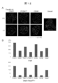
- FIG. 11 shows the analysis result of the flow cytometer in the cells 48 days after differentiation induction.
- the upper left figure shows the result of negative control
- the upper right figure shows the result stained with CD231 antibody
- the lower left figure shows the result stained with CDH8 antibody
- the lower right figure shows the result stained with PCDH17 antibody.
- the horizontal axis indicates the fluorescence intensity.
- FIG. 12A shows stained images of Ctip2 (red) and CupTF1 (green) of cells after isolation of each marker-positive cell (CD231, PCDH17 or CDH8) from cells on differentiation-inducing day 48 and further culturing for 14 days.
- Unsort represents the cells after the cells on the 48th day of differentiation induction were separated and further cultured for 14 days.
- FIG. 12B shows the content of Ctip2 positive cells on the 14th day after re-culture of each marker positive cell (CD231, PCDH17 or CDH8) (indicated by + in the figure) or each marker negative cell (indicated by-in the figure) (top).
- FIG. 4 is a graph showing the content of Ctip2 positive cells and Cup TF1 negative cells (lower panel). Unsort in the graph indicates the content of each cell after the cells on the 48th day of differentiation induction were dissociated and further cultured for 14 days.
- FIG. 13A shows DAPI (blue) / Ctip2 (red) (left), DAPI (blue) / CupTF1 (green) of cells on day 46 differentiated in adhesion culture (upper) or suspension culture (lower) ( Stained images for the center diagram), Ctip2 (red), and CupTF1 (green) (right) are shown.
- FIG. 13A shows DAPI (blue) / Ctip2 (red) (left), DAPI (blue) / CupTF1 (green) of cells on day 46 differentiated in adhesion culture (upper) or suspension culture (lower) ( Stained images for the center diagram), Ctip2 (red), and CupTF1 (green) (right) are shown.
- FIG. 13B shows the content of Ctip2-positive cells (left diagram) and the content of Ctip2-positive cells CupTF1-negative cells (right diagram) on the 46th day after differentiation induction in adhesion culture (Attach) or floating culture (Floating). It is a graph.
- FIG. 14A shows stained images of DAPI (blue), Ctip2 (red), and CupTF1 (green) of cells on day 46 cultured with C59, LGK-974, or ICG-001 as WNT inhibitors.
- FIG. 14B shows the content of Ctip2-positive cells in the total cells (DAPI) after culturing for 46 days using C59, LGK-974, or ICG-001 as a WNT inhibitor (left panel) and the total cells (DAPI). It is a graph which shows the content rate (right figure) of Ctip2-positive cell CupTF1-negative cell in FIG.
- the present invention provides a method for producing cerebral cortical neurons from pluripotent stem cells comprising the following steps: (I) a step of suspending pluripotent stem cells in a culture solution containing a TGF ⁇ inhibitor, bFGF, Wnt inhibitor, and BMP inhibitor; (Ii) a step of subjecting the cells obtained in the step (i) to suspension culture in a culture solution containing a Wnt inhibitor and a BMP inhibitor; (Iii) A step of further culturing the cells obtained in the step (ii).
- the pluripotent stem cell that can be used in the present invention is a stem cell that has pluripotency that can be differentiated into all cells existing in a living body, and also has proliferative ability.
- Specific examples of these pluripotent stem cells are not limited to the following, but include embryonic stem (ES) cells, embryonic stem (ntES) cells derived from cloned embryos obtained by nuclear transfer, sperm stem cells ("GS cells”) "), Embryonic germ cells (“ EG cells "), induced pluripotent stem (iPS) cells, and the like.
- ES embryonic stem
- ntES embryonic stem
- GS cells sperm stem cells
- EG cells Embryonic germ cells
- iPS induced pluripotent stem
- More preferred pluripotent stem cells are human pluripotent stem cells, particularly preferably human ES cells and human iPS cells.
- Embryonic stem cells ES cells are stem cells established from the inner cell mass of early embryos (eg, blastocysts) of mammals such as humans and mice, and having pluripotency and proliferation ability by self-replication. ES cells are embryonic stem cells derived from the inner cell mass of blastocysts, which are embryos in the 8-cell stage or morula stage of fertilized eggs, and have the ability to differentiate into any cell that constitutes an adult, so-called differentiation differentiation. And ability to proliferate by self-replication.
- ES cells were discovered in 1981 in mice (MJ Evans and MH Kaufman (1981), Nature 292: 154-156), and then ES cell lines were also established in primates such as humans and monkeys.
- JA Thomson et al. (1998), Science 282: 1145-1147, JA Thomson et al. (1995), Proc. Natl. Acad. Sci. USA, 92: 7844-7848, JA Thomson et al. (1996), Biol.Reprod., 55: 254-259 and JA Thomson and V.S. Marshall (1998), Curr.Top.Dev.Biol., 38: 133. -165).
- ES cells can be established by removing an inner cell mass from a blastocyst of a fertilized egg of a subject animal and culturing the inner cell mass on a fibroblast feeder.
- the maintenance of ES cells by subculture is performed using a medium supplemented with substances such as leukemia inhibitory factor (LIF) and basic fibroblast growth factor (bFGF). It can be carried out.
- LIF leukemia inhibitory factor
- bFGF basic fibroblast growth factor
- a medium for preparing ES cells for example, a DMEM / F-12 medium supplemented with 0.1 mM 2-mercaptoethanol, 0.1 mM non-essential amino acid, 2 mM L-glutamic acid, 20% KSR and 4 ng / ml bFGF is used. 37 ° C, 5% CO 2 Human ES cells can be maintained under a humid atmosphere.
- ES cells need to be passaged every 3 to 4 days. At this time, the passage is, for example, 1 mM CaCl. 2 And 0.25% trypsin and 0.1 mg / ml collagenase IV in PBS (phosphate buffered saline) containing 20% KSR.
- ES cells can be selected using the expression of gene markers such as alkaline phosphatase, Oct-3 / 4, Nanog, etc. as an index.
- sperm stem cells A sperm stem cell is a testis-derived pluripotent stem cell, which is a cell that is the origin for spermatogenesis.
- these cells can be induced to differentiate into various types of cells, and have the property that, for example, chimeric mice can be produced when transplanted into mouse blastocysts (M. Kanatsu-Shinohara et al. 2003) Biol.Reprod., 69: 612-616; K. Shinohara et al. (2004), Cell, 119: 1001-1012).
- Sperm stem cells can be self-replicating in a medium containing glial cell line-derived neurotrophic factor (GDNF), and by repeated passage under the same culture conditions as ES cells Sperm stem cells can be obtained (Takebayashi Masanori et al. (2008), Experimental Medicine, Vol. 26, No.
- GDNF glial cell line-derived neurotrophic factor
- Embryonic germ cells are cells that are established from embryonic primordial germ cells and have the same pluripotency as ES cells, and are primitive in the presence of substances such as LIF, bFGF, and stem cell factor. It can be established by culturing germ cells (Y. Matsui et al. (1992), Cell, 70: 841-847; JL Resnick et al. (1992), Nature, 359: 550-551).
- (D) Artificial pluripotent stem cells Artificial pluripotent stem (iPS) cells are obtained by introducing one or more specific nuclear reprogramming substances into somatic cells in the form of DNA or protein, or one or more specific one or more Produced by increasing the expression level of endogenous mRNA and protein of the nuclear reprogramming substance through the use of a seed drug, characteristics almost equivalent to those of ES cells, for example, proliferation by differentiation pluripotency and self-replication Somatic cell-derived artificial stem cells (K. Takahashi and S. Yamanaka (2006) Cell, 126: 663-676, K. Takahashi et al. (2007) Cell, 131: 861-872, J. Am. Yu et al.
- the nuclear reprogramming substance is not particularly limited as long as it is a gene specifically expressed in ES cells, a gene that plays an important role in maintaining undifferentiation of ES cells, or a gene product thereof.
- reprogramming substances may be used in combination when iPS cells are established.
- Such a combination can be a combination including at least one, two, or three kinds of the reprogramming substances, preferably a combination including three or four.
- Nucleotide sequence information of mouse and human cDNA of each nuclear reprogramming substance and amino acid sequence information of the protein encoded by the cDNA are described in GenBank (US NCBI) or EMBL (Germany) described in WO2007 / 069666. It can be obtained by accessing accession numbers.
- mouse and human cDNA sequence information and amino acid sequence information of L-Myc, Lin28, Lin28b, Esrrb, Esrrg, and Glis1 can be obtained by accessing NCBI accession numbers shown in Table 1.
- a person skilled in the art can prepare a desired nuclear reprogramming substance by a conventional method based on the cDNA sequence or amino acid sequence information.
- nuclear reprogramming substances may be introduced into somatic cells in the form of proteins, for example, by lipofection, binding to cell membrane permeable peptides, microinjection, or in the form of DNA, for example, It can be introduced into somatic cells by techniques such as vectors such as viruses, plasmids, artificial chromosomes, lipofection, use of liposomes, and microinjection.
- vectors such as viruses, plasmids, artificial chromosomes, lipofection, use of liposomes, and microinjection.
- viral vectors retrovirus vectors, lentiviral vectors (these vectors are Cell, 126, pp. 663-676, 2006; Cell, 131, pp. 861-872, 2007; Science, 318, pp.
- adenovirus vector (Science, 322, 945-949, 2008), adeno-associated virus vector, Sendai virus vector (Proc Jpn Acad Ser B Phys Biol Sci. 85, 348-62, 2009), etc.
- the artificial chromosome vector include human artificial chromosome (HAC), yeast artificial chromosome (YAC), and bacterial artificial chromosome (BAC and PAC).
- HAC human artificial chromosome
- YAC yeast artificial chromosome
- BAC and PAC bacterial artificial chromosome
- plasmid a plasmid for mammalian cells can be used (Science, 322: 949-953, 2008).
- the vector can contain a regulatory sequence such as a promoter, an enhancer, a ribosome binding sequence, a terminator, a polyadenylation site or a polyadenylation signal so that a nuclear reprogramming substance can be expressed.
- a regulatory sequence such as a promoter, an enhancer, a ribosome binding sequence, a terminator, a polyadenylation site or a polyadenylation signal so that a nuclear reprogramming substance can be expressed.
- the promoter used include EF1 ⁇ promoter, CAG promoter, SR ⁇ promoter, SV40 promoter, LTR promoter, CMV (cytomegalovirus) promoter, RSV (rous sarcoma virus) promoter, MoMuLV (Moloney murine leukemia virus) LTR, HSV- A TK (herpes simplex virus thymidine kinase) promoter or the like is used.
- EF1 ⁇ promoter EF1 ⁇ promoter, CAG promoter, MoMuLV LTR, CMV promoter, SR ⁇ promoter and the like are preferable examples.
- selectable marker sequences such as drug resistance genes (eg, kanamycin resistance gene, ampicillin resistance gene or puromycin resistance gene), thymidine kinase gene, and diphtheria toxin gene or fragments thereof, and green fluorescent protein (GFP) ),
- GFP green fluorescent protein
- a reporter gene sequence such as ⁇ -glucuronidase (GUS) or FLAG.
- the above vector contains a LoxP sequence before and after the gene or promoter encoding the nuclear reprogramming substance and the gene encoding the nuclear reprogramming substance that binds to it. You may have.
- a method is used in which a transposon is used to incorporate a transgene into a chromosome, and then a plasmid vector or an adenoviral vector is used to act on a cell with a transferase to completely remove the transgene from the chromosome.
- Preferred transposons include, for example, piggyBac, which is a transposon derived from lepidopterous insects (Kaji, K.
- the vectors are replicated without chromosomal integration and are present episomally, with the origins of lymphotropic herpesvirus, BK virus and bovine papillomavirus and their origins. It may contain sequences involved in replication. For example, including EBNA-1 and oriP or Large T and SV40ori sequences (WO 2009/115295, WO 2009/157201 and WO 2009/149233).
- an expression vector that can be expressed polycistronically may be used.
- the gene coding sequence may be linked by an IRES or foot-and-mouth disease virus (FMDV) 2A coding region (Science, 322: 949-953, 2008). , WO 2009/092042 and WO 2009/152529).
- FMDV foot-and-mouth disease virus
- HDAC histone deacetylase
- VPA valproic acid
- small molecule inhibitors such as trichostatin A, sodium butyrate, MC1293, M344, siRNA and shRNA against HDAC (eg, HDAC1 siRNA Smartpool® (Millipore), HuSH 29mer shRNA Constructs) nucleic acid expression inhibitors such as against HDAC1 (OriGene) and the like], DNA methyltransferase inhibitors (for example, 5′-azacytidine) (Nat.
- G9a histone methyltransferase inhibitors for example, BIX-01294 (Cell Stem Cell, 2: 525-528 (2008)), G9a, SiRNA and shRNA (eg, nucleic acid expression inhibitors such as G9a siRNA (human) (Santa Cruz Biotechnology), etc.)], L-channel calcium agonist (L-channel calcium agonist) (eg, Bayk 8644) (Cell Stem Cell 3,568-574 (2008)), p53 inhibitors (eg, siRNA and shRNA against p53) (Cell Stem Cell, 3,475-479 (2008)), Wnt Shi Gnal signaling activator (eg soluble Wnt3a) (Cell Stem Cell, 3, 132-135 (2008)), growth factor such as LIF or bFGF, ALK5 inhibitor (eg SB431542) (Nat.
- Such a drug used in the method of increasing the expression level of the endogenous protein of the nuclear reprogramming substance by a drug includes 6-bromoindirubin-3′-oxime, indirubin-5-nitro-3′- Oxime, valproic acid, 2- (3- (6-methylpyridin-2-yl) -1H-pyrazol-4-yl) -1,5-naphthyridine, 1- (4-methylphenyl) -2- (4 Examples include 5,6,7-tetrahydro-2-imino-3 (2H) -benzothiazolyl) ethanone HBr (pifthrin-alpha), prostaglandin J2, prostaglandin E2, and the like (WO 2010/068955).
- DMEM, DMEM / F12 or DME medium containing 10 to 15% FBS in addition to LIF, penicillin / streptomycin, puromycin, L- Glutamine, non-essential amino acids, ⁇ -mercaptoethanol and the like can be appropriately included.
- ES cell culture medium containing bFGF or SCF for example, mouse ES cell culture medium (for example, TX-WES medium, Thrombo X) or primate ES cell culture medium (for example, primate (human or monkey) ES cell culture medium (distributor: Reprocell, Kyoto, Japan), mTeSR-1).
- culture methods include, for example, 37 ° C., 5% CO 2
- somatic cells are contacted with a nuclear reprogramming substance (DNA, RNA, or protein) and cultured for about 4 to 7 days.
- a nuclear reprogramming substance DNA, RNA, or protein
- Mitomycin C-treated STO cells, SNL cells, etc. and cultured in a culture medium for primate ES cell culture containing bFGF about 10 days after contact between the somatic cells and the nuclear reprogramming substance, and about 30 to about ES cell-like colonies can be generated after 45 days or more.
- the cells may be cultured under conditions of an oxygen concentration as low as 5 to 10%.
- the cells are treated with 10% FBS-containing DMEM medium (including LIF, penicillin / streptomycin, puromycin, L-glutamine, non-essential amino acids on feeder cells (eg, mitomycin C-treated STO cells or SNL cells).
- FBS-containing DMEM medium including LIF, penicillin / streptomycin, puromycin, L-glutamine, non-essential amino acids on feeder cells (eg, mitomycin C-treated STO cells or SNL cells).
- b-mercaptoethanol and the like can be suitably included
- ES-like colonies can be formed after about 25 to about 30 days or more.
- the medium is replaced with a fresh medium once a day from the second day after the start of the culture.
- the number of somatic cells used for nuclear reprogramming is not limited, but culture dishes (100 cm 2 ) About 5 ⁇ 10 3 ⁇ About 5 ⁇ 10 6 A range of cells.
- cells expressing the marker gene can be selected by culturing the cells in a medium containing the corresponding drug (that is, a selective medium).
- a medium containing the corresponding drug that is, a selective medium.
- the marker gene is a fluorescent protein gene
- the marker gene-expressing cell is obtained by observing with a fluorescence microscope, in the case of a luminescent enzyme gene, by adding a luminescent substrate, or in the case of a chromogenic enzyme gene, by adding a chromogenic substrate. Can be detected.
- a “somatic cell” may be any cell other than germ cells from mammals (eg, humans, mice, monkeys, pigs and rats), for example, keratinized epithelial cells ( Eg, keratinized epidermal cells), mucosal epithelial cells (eg, epithelial cells of the tongue surface), exocrine glandular epithelial cells (eg, mammary cells), hormone-secreting cells (eg, adrenal medullary cells), cells for metabolism and storage (eg E.g., hepatocytes), luminal epithelial cells that make up the interface (e.g., type I alveolar cells), luminal epithelial cells of the inner chain (e.g., vascular endothelial cells), and cilia cells that have the ability to carry ( Eg, airway epithelial cells), extracellular matrix secreting cells (eg, fibroblasts), contractile cells (eg, smooth muscle cells), blood and immune system cells (eg, T
- the degree of cell differentiation and the age of the animal from which the cells are collected there is no particular limitation on the degree of cell differentiation and the age of the animal from which the cells are collected. This can be applied to both undifferentiated progenitor cells (including somatic stem cells) and finally differentiated mature cells. It can be used as the source of somatic cells in the invention.
- undifferentiated progenitor cells include tissue stem cells (somatic stem cells) such as neural stem cells, hematopoietic stem cells, mesenchymal stem cells, and dental pulp stem cells.
- the mammal from which somatic cells are collected is not particularly limited, but is preferably a human.
- ES cells derived from cloned embryos obtained by nuclear transfer The nt ES cell is a cloned embryo-derived ES cell produced by a nuclear transfer technique and has almost the same characteristics as a fertilized egg-derived ES cell (T. Wakayama et al. (2001), Science, 292). S. Wakayama et al. (2005), Biol.Reprod., 72: 932-936; J. Byrne et al. (2007), Nature, 450: 497-502).
- nt ES nuclear transfer ES cells
- nt ES cells are established from the inner cell mass of a blastocyst derived from a cloned embryo obtained by replacing the nucleus of an unfertilized egg with the nucleus of a somatic cell.
- a combination of nuclear transfer technology JB Cibelli et al. (1998), Nat. Biotechnol., 16: 642-646) and ES cell production technology (above) is used.
- JB Cibelli et al. (1998), Nat. Biotechnol., 16: 642-646)
- ES cell production technology above
- reprogramming can be performed by injecting a somatic cell nucleus into an enucleated unfertilized egg of a mammal and culturing it for several hours.
- F Fusion stem cells
- a fused stem cell is a stem cell that is produced by fusing a somatic cell and an egg or ES cell, has the same pluripotency as the fused ES cell, and also has a gene unique to the somatic cell (Tada M et al. Curr Biol. 11: 1553-8, 2001; Cowan CA et al. Science. 2005 Aug 26; 309 (5739): 1369-73).
- cerebral cortical neurons include one or more cells selected from the group consisting of cerebral cortical neurons, cerebral cortical neural stem cells, and cerebral cortical neural progenitors unless otherwise specified.
- Cerebral cortical neurons produced by the method of the present invention are preferably cells positive for Foxg1.
- Foxg1 includes a polynucleotide represented by NCBI accession number NM_005249 and proteins encoded by these.
- the cerebral cortex neurons produced by the method of the present invention are more preferably cerebral cortex motor cells or upper motor neurons, i.e., neurons in front of the cerebral cortex, more preferably motor cells of the cerebral cortex. It is a nerve cell of the V layer.
- Such nerve cells are a cell population characterized by being positive for Ctip2, which can be paraphrased as cells characterized by being negative for CupTF1.
- Ctip2 includes a polynucleotide represented by NCBI accession number NM_001282237, NM_001282238, NM_022898 or NM_138576 and a protein encoded by these.
- the cerebral cortical neurons produced in the present invention may be produced as a cell population containing other cell types, for example, 15% or more, 20% or more, 30% or more, 40% or more in the produced cell population. Alternatively, 50% or more may be cerebral cortical neurons. After producing cerebral cortical neurons by the method of the present invention, the cells may be concentrated.
- the step (iv) may further include a step of extracting cells positive for at least one marker protein selected from the group consisting of CD231, PCDH17 and CDH8 from the cells obtained in step (iii). .
- the culture may be further continued after the extraction. Examples of the culture after extraction include a method of culturing under the same conditions as in step (iii), but are not particularly limited.
- a TGF ⁇ inhibitor is a substance that inhibits signal transduction from binding of TGF ⁇ to a receptor to SMAD, and a substance that inhibits binding to the receptor ALK family, or SMAD by the ALK family. And a substance that inhibits phosphorylation of the enzyme.
- TGF ⁇ inhibitor include Lefty-1 (NCBI Accession No. includes mouse: NM — 010094, human: NM — 020997), SB431542, SB202190 (above, RK Lindemann et al., Mol.
- the TGF ⁇ inhibitor used in the present invention may preferably be SB431542 represented by the following formula I or A-83-01 represented by the following formula II.
- the concentration of SB431542 in the culture solution is not particularly limited as long as it inhibits ALK5.
- the concentration of A-83-01 in the culture solution is not particularly limited as long as it is a concentration that inhibits ALK5.
- it is 500 nM to 5 ⁇ M, more preferably 500 nM to 2 ⁇ M.
- bFGF is also referred to as FGF2, and is commercially available from, for example, Wako and Invitrogen, and can be easily used.
- bFGF may be obtained by forced expression into cells by methods known to those skilled in the art.
- the concentration of bFGF in the culture solution is, for example, 0.1 ng / mL, 0.5 ng / mL, 1 ng / mL, 2 ng / mL, 3 ng / mL, 4 ng / mL, 5 ng / mL, 6 ng / mL, 7 ng / mL.
- it is 1 ng / mL to 100 ng / mL, and more preferably 10 ng / mL.
- the Wnt inhibitor is a substance that suppresses the production of Wnt, or a substance that inhibits signal transduction from binding of Wnt to the receptor to ⁇ -catenin accumulation, and to the Frizzled family of receptors.
- Wnt inhibitors examples include DKK1 protein (for example, NCBI accession number: NM_012422 for humans), sclerostin (for example, NCBI accession number: NM_025237 for humans), IWR-1 ( Merck Millipore), IWP-2 (Sigma-Aldrich), IWP-3 (Sigma-Aldrich), IWP-4 (Sigma-Aldrich), IWP-L6 (EMD Millipore), C59 (or Nnt-C59tech) ), ICG-001 (Cellagen Technology), LGK-974 (or NVP-LGK-974) (Cellagen Technology), FH535.
- DKK1 protein for example, NCBI accession number: NM_012422 for humans
- sclerostin for example, NCBI accession number: NM_025237 for humans
- IWR-1 Merck Millipore
- IWP-2 Sigma-Aldrich
- IWP-3 Sigma-Aldrich
- IWP-4 Sigma
- a preferred Wnt inhibitor is a substance that suppresses the production of Wnt.
- PORCN involved in the processing of Wnt protein in the case of human, NCBI accessor
- NP_001269096, NP_077376, NP_982299, NP_982300, or NP_982301 for example, C59, IWP-3, IWP-4, IWP-L6 or LGK-974. It is.
- a more preferred Wnt inhibitor is C59 represented by the following formula III.
- the concentration of C59 in the culture solution is not particularly limited as long as it is a concentration that inhibits Wnt.
- LGK-974 represented by the following formula VI can also be preferably used as a Wnt inhibitor.
- the concentration of LGK-974 in the culture solution is not particularly limited as long as it is a concentration that inhibits Wnt.
- 1 nM, 10 nM, 25 nM, 50 nM, 100 nM, 150 nM, 200 nM, 500 nM, 750 nM, and 1 ⁇ M are included. It is not limited.
- the concentration is preferably 1 nM to 1 ⁇ M, for example, 1 nM to 500 nM, more preferably 10 nM to 200 nM, or 10 nM to 150 nM, for example, 10 nM to 100 nM.
- the BMP inhibitor refers to proteinaceous inhibitors such as Chordin, Noggin, Follistatin, Dorsorphine (ie, 6- [4- (2-piperidin-1-yl-ethoxy) phenyl] -3-pyridin- 4-yl-pyrazolo [1,5-a] pyrimidine), its derivatives (P.B.Yu et al.
- LDN193189 (ie 4- (6- (4- (piperazin-1-yl)). ) Phenyl) pyra zolo [1,5-a] pyrimidin-3-yl) quinoline) is exemplified.
- Dorsomorphin and LDN193189 are commercially available and are available from Sigma-Aldrich and Stemgent, respectively.
- the BMP inhibitor used in the present invention may preferably be LDN193189 represented by the following formula IV.
- the concentration of LDN193189 in the culture solution is not particularly limited as long as it is a concentration that inhibits BMP.
- the concentration is preferably 2 ⁇ M or less, for example, 100 nM to 2 ⁇ M, more preferably 500 nM to 2 ⁇ M, or less than 2 ⁇ M, for example, 100 nM or more and less than 2 ⁇ M, and more preferably Is 500 nM or more and less than 2 ⁇ M.
- the culture solution used in step (i) of the present invention can be prepared by adding the above-described TGF ⁇ inhibitor, bFGF, Wnt inhibitor, and BMP inhibitor using a medium used for culturing animal cells as a basal medium. .
- the basal medium examples include Glasgow's Minimal Essential Medium (GMEM) medium, IMDM medium, Medium 199 medium, Eagle's Minimum Essential Medium (EMEM) medium, ⁇ MEM medium, Dulbecco's medium EM medium, Dulbecco's medium Medium, Ham's F12 medium, RPMI 1640 medium, Fischer's medium, Neurobasal Medium (Invitrogen), and mixed media thereof are included.
- GMEM Glasgow's Minimal Essential Medium
- IMDM Medium IMDM medium
- Medium 199 medium Eagle's Minimum Essential Medium
- EMEM Eagle's Minimum Essential Medium
- ⁇ MEM medium Dulbecco's medium EM medium
- Dulbecco's medium Dulbecco's medium
- Ham's F12 medium RPMI 1640 medium
- Fischer's medium Neurobasal Medium (Invitrogen)
- mixed media thereof are included.
- it is a medium in which DMEM and Ham's F12 medium are mixed at a ratio of 1: 1.
- Serum substitutes include, for example, albumin, transferrin, Knockout Serum Replacement (KSR) (serum substitute for FBS during ES cell culture), N2 supplement (Invitrogen), B27 supplement (Invitrogen), fatty acid, insulin, collagen precursor, Examples include trace elements and a plurality of combinations selected from these.
- KSR Knockout Serum Replacement
- the serum replacement is KSR.
- the concentration in the basal medium is, for example, 5%, 10%, 11%, 12%, 13%, 14%, 15%, 16%, 17%, 18 %, 19%, and 20% are preferable, but the concentration is preferably less than 20%, for example, 10% or more and 15% or less.
- the basal medium includes 2-mercaptoethanol, 3′-thiolglycerol, lipids, amino acids, L-glutamine, Glutamax (Invitrogen), non-essential amino acids, vitamins, growth factors, low molecular compounds, antibiotics, antioxidants, pyruvin
- a preferred basal medium is a medium in which DMEM and Ham's F12 medium containing KSR, 2-mercaptoethanol, a non-essential amino acid and L-glutamine are mixed at a ratio of 1: 1.
- pluripotent stem cells may be dissociated and used.
- Examples of a method for dissociating cells include a method of dissociating dynamically, a dissociation solution having protease activity and collagenase activity (for example, , Accutase (TM) and Accumax (TM), etc.) or dissociation methods using a dissociation solution having only collagenase activity.
- a method of dissociating pluripotent stem cells using Accumax is exemplified.
- the ROCK inhibitor is not particularly limited as long as it can suppress the function of Rho kinase (ROCK).
- ROCK Rho kinase
- Y-27632 for example, Ishizaki et al., Mol. Pharmacol. 57, 976-). 983 (2000); Narumiya et al., Methods Enzymol. 325, 273-284 (2000)), Fasudil / HA1077 (see, for example, Uenata et al., Nature 389: 990-994 (1997)), H-1152.
- ROCK inhibitors may be used.
- the ROCK inhibitor used in the present invention may be preferably Y-27632 represented by the following formula (V).
- the concentration of Y-27632 in the medium is, for example, 100 nM, 500 nM, 750 nM, 1 ⁇ M, 2 ⁇ M, 3 ⁇ M, 4 ⁇ M, 5 ⁇ M, 6 ⁇ M, 7 ⁇ M, 8 ⁇ M, 9 ⁇ M, 10 ⁇ M, 15 ⁇ M, 20 ⁇ M, 25 ⁇ M, 30 ⁇ M, 40 ⁇ M, 50 ⁇ M, Although not limited to 60 ⁇ M, 70 ⁇ M, 80 ⁇ M, 90 ⁇ M, and 100 ⁇ M. Preferably, it is 10 ⁇ M or more and 50 ⁇ M or less.
- the culture in step (i) of the present invention is preferably carried out by suspension culture.
- the suspension culture means culturing cells by forming aggregates (also referred to as spheres) in a non-adherent state in a culture vessel, and is not particularly limited, but improves adhesion to cells.
- Culture vessels that have not been artificially treated (for example, coating treatment with extracellular matrix) for the purpose, or treatment for artificially suppressing adhesion (for example, polyhydroxyethyl methacrylic acid (poly-HEMA), nonionic)
- a culture vessel coated with a surface-active polyol such as Pluronic F-127) or a phospholipid-like structure (for example, a water-soluble polymer (Lipidure) containing 2-methacryloyloxyethyl phosphorylcholine).
- the culture temperature is not particularly limited, but is about 30 to 40 ° C., preferably about 37 ° C. 2 Cultivation is performed in an atmosphere containing air, and CO 2
- the concentration is preferably about 2-5%.
- O 2 The concentration is O in normal air. 2 It may be a concentration, or it may be a normal high oxygen condition or a normal low oxygen condition.
- the high oxygen condition means 25% or more of O. 2 Concentration, O over 30% 2 Concentration, over 35% O 2 Concentration or 40% or more O 2 The concentration is illustrated.
- Step (i) of the present invention is not particularly limited, and examples thereof include 3 days or more, 4 days or more, 5 days or more, 6 days or more, 7 days or more, or more days, and the upper limit is not particularly limited. However, 36 days or less, 30 days or less, 24 days or less, 18 days or less, or 12 days or less. More preferably, it is 3 days or more and 12 days or less, and more preferably 6 days.
- the culture solution used in step (ii) of the present invention can be prepared by adding the above-described Wnt inhibitor and BMP inhibitor using a medium used for animal cell culture as a basal medium.
- a basal medium include Glasgow's Minimal Essential Medium (GMEM) medium, IMDM medium, Medium 199 medium, Eagle's Minimum Essential Medium (EMEM) medium, ⁇ MEM medium, Dulbecco's medium EM medium, Dulbecco's medium Medium, Ham's F12 medium, RPMI 1640 medium, Fischer's medium, Neurobasal Medium (Invitrogen), and mixed media thereof are included.
- the basal medium may contain serum, or a serum substitute may be added instead of serum.
- Serum substitutes include, for example, albumin, transferrin, Knockout Serum Replacement (KSR) (serum substitute for FBS during ES cell culture), N2 supplement (Invitrogen), B27 supplement (Invitrogen), fatty acid, insulin, collagen precursor, Examples include trace elements and a plurality of combinations selected from these.
- the serum replacement is KSR.
- the concentration in the basal medium is, for example, 5%, 10%, 11%, 12%, 13%, 14%, 15%, 16%, 17%, 18 %, 19%, and 20% are preferable, but the concentration is preferably less than 20%, for example, 10% or more and 15% or less.
- the basal medium includes 2-mercaptoethanol, 3′-thiolglycerol, lipids, amino acids, L-glutamine, Glutamax (Invitrogen), non-essential amino acids, vitamins, growth factors, low molecular compounds, antibiotics, antioxidants, pyruvin
- One or more substances such as acids, buffers, inorganic salts, and the like may also be included.
- a preferred basal medium is a medium in which DMEM and Ham's F12 medium containing KSR, 2-mercaptoethanol, a non-essential amino acid and L-glutamine are mixed at a ratio of 1: 1.
- the cells obtained in the step (i) may be dissociated and used as they are. More preferably, in the cells obtained in the step (i), the culture is continued and the culture is continued. Therefore, the culture in the step (ii) of the present invention is preferably performed by suspension culture.
- the culture temperature is not particularly limited, but is about 30 to 40 ° C., preferably about 37 ° C.
- the concentration is preferably about 2-5%.
- O 2 The concentration is O in normal air. 2 It may be a concentration, or it may be a normal high oxygen condition or a normal low oxygen condition.
- the step (ii) of the present invention is not particularly limited.
- the number of days is 6 days or more, 7 days or more, 8 days or more, 9 days or more, 10 days or more, 11 days or more, 12 days or more, or more.
- an upper limit is not specifically limited, 48 days or less, 42 days or less, 36 days or less, 30 days or less, 24 days or less, 18 days or less are mentioned. More preferably, it is 6 days or more and 18 days or less, and more preferably 12 days.
- the culture medium used in step (iii) of the present invention can be a basal medium used for culturing animal cells.
- the basal medium include Glasgow's Minimal Essential Medium (GMEM) medium, IMDM medium, Medium 199 medium, Eagle's Minimum Essential Medium (EMEM) medium, ⁇ MEM medium, Dulbecco's medium EM medium, Dulbecco's medium Medium, Ham's F12 medium, RPMI 1640 medium, Fischer's medium, Neurobasal Medium (Invitrogen), and mixed media thereof are included.
- GMEM Glasgow's Minimal Essential Medium
- IMDM Medium IMDM
- Medium 199 medium Eagle's Minimum Essential Medium
- EMEM Eagle's Minimum Essential Medium
- ⁇ MEM medium Dulbecco's medium EM medium
- Dulbecco's medium Dulbecco's medium
- Ham's F12 medium RPMI 1640 medium
- Fischer's medium Neurobasal Medium (Invitrogen)
- mixed media thereof are included
- Serum substitutes include, for example, albumin, transferrin, Knockout Serum Replacement (KSR) (serum substitute for FBS during ES cell culture), N2 supplement (Invitrogen), B27 supplement (Invitrogen), fatty acid, insulin, collagen precursor, Examples include trace elements and a plurality of combinations selected from these.
- KSR Knockout Serum Replacement
- the serum replacement is a B27 supplement.
- the basal medium includes 2-mercaptoethanol, 3′-thiolglycerol, lipids, amino acids, L-glutamine, Glutamax (Invitrogen), non-essential amino acids, vitamins, growth factors, low molecular compounds, antibiotics, antioxidants, pyruvin
- a preferred basal medium is Neurobasal Medium containing B27 supplement and L-glutamine.
- FGF8 fibroblast growth factor 8
- neurotrophic factor or the like can be appropriately added to the basal medium.
- FGF8 is not particularly limited, but in the case of human FGF8, four splicing forms of FGF8a, FGF8b, FGF8e or FGF8f are exemplified, and FGF8b is more preferable in the present invention.
- FGF8 is commercially available from, for example, Wako and R & D systems, and can be easily used, but may be obtained by forced expression into cells by methods known to those skilled in the art.
- the concentration of FGF8 in the culture solution is, for example, 1 ng / mL, 5 ng / mL, 10 ng / mL, 50 ng / mL, 100 ng / mL, 150 ng / mL, 200 ng / mL, 250 ng / mL, 500 ng / mL, 1000 ng / mL. , 2000 ng / mL, 5000 ng / mL, but not limited thereto.
- it is 100 ng / mL.
- a neurotrophic factor is a ligand to a membrane receptor that plays an important role in the survival and function maintenance of nerve cells.
- nerve growth factor For example, nerve growth factor (NGF), brain-derived nerve Brain-derived Neurotrophic Factor (BDNF), Neurotrophin 3 (NT-3), Neurotrophin 4/5 (NT-4 / 5), Neurotrophin 6 (NT-6), bFGF, acidic FGF, FGF-5, epidermal growth factor (EGF), hepatocyte growth factor (HGF), insulin, insulin-like growth factor 1 (IGF1), insulin-like growth factor 2 (IGF2) ) Cell line-derived neurotrophic factor (GDNF), TGF-b2, TGF-b3, interleukin 6 (IL-6), ciliary neurotrophic factor (CNTF) And LIF.
- NGF nerve growth factor
- BDNF brain-derived nerve Brain-derived Neurotrophic Factor
- NT-3 Neurotrophin 3
- Neurotrophin 4/5 NT-4 / 5
- Neurotrophin 6 bFGF
- acidic FGF FGF-5
- EGF epidermal growth factor
- HGF hepatocyte growth factor
- insulin insulin-like growth factor 1
- the neurotrophic factor is commercially available from, for example, Wako and R & D systems, and can be easily used, but may be obtained by forced expression into cells by a method known to those skilled in the art.
- the step (iii) of the present invention is a step of culturing the cells obtained in the step (ii), and the culture may be adhesion culture or suspension culture.
- Adhesion culture can be performed by culturing using a culture container coated with an extracellular matrix.
- the coating treatment can be performed by placing a solution containing an extracellular matrix in a culture container and then removing the solution as appropriate.
- the extracellular matrix is a supramolecular structure existing outside the cell and may be naturally derived or an artificial product (recombinant).
- Examples include substances such as polylysine, polyornithine, collagen, proteoglycan, fibronectin, hyaluronic acid, tenascin, entactin, elastin, fibrillin, laminin and fragments thereof.
- extracellular matrices may be used in combination, for example, preparations from cells such as BD Matrigel TM. Preferably, it is a mixture of polyornithine, laminin and fibronectin.
- the culture temperature is not particularly limited, but is about 30 to 40 ° C., preferably about 37 ° C. 2 Cultivation is performed in an atmosphere containing air, and CO 2 The concentration is preferably about 2-5%. O 2 The concentration is O in normal air. 2 It may be a concentration, or it may be a normal high oxygen condition or a normal low oxygen condition. From the viewpoint that cerebral cortical neurons can be obtained, the step (iii) of the present invention is not particularly harmful even if the culture is continued for a long period of time.
- cerebral cortical neurons obtained in the present invention can be administered as a preparation to cerebral disorder patients.
- cerebral disorder refers to a state in which nerve cells are lost due to ischemia or the like, and examples include disorders after cerebral infarction.
- Cerebral cortical neurons produced by the above method are suspended in physiological saline or the like and transplanted to a nerve cell missing site of a patient.
- the present invention provides a therapeutic agent for cerebral disorder containing cerebral cortical neurons obtained from pluripotent stem cells by the above method, preferably a therapeutic agent for cerebral infarction.
- the number of cerebral cortical neurons contained in the therapeutic agent for cerebral disorder is not particularly limited as long as the graft can be engrafted after administration. 4 More than one can be included. Further, it may be prepared by appropriately increasing or decreasing according to the size of the symptom or body.
- kits for producing cerebral cortical neurons from pluripotent stem cells includes a culture solution, an additive, a culture container, or the like used in each step of producing the cerebral cortical neuron described above.
- a reagent selected from the group consisting of a TGF ⁇ inhibitor, bFGF, Wnt inhibitor, and BMP inhibitor can be mentioned.
- the kit may further include a document or instructions describing the procedure of the manufacturing process.
- Human ES cells (KhES-1) were received from the Institute of Regenerative Medicine, Kyoto University (Suemori H, et al. Biochem Biophys Res Commun. 345: 926-32, 2006).
- Human iPS cells 404C2 were received from Professor Yamanaka of Kyoto University as cells obtained by introducing Oct3 / 4, Sox2, Klf4, L-MYC, LIN28 and p53shRNA into human fibroblasts by episomal vectors. (Okita, et al, Nat Methods. 8: 409-412, 2011).
- a human iPS cell was obtained from Professor Yamanaka of Kyoto University as a cell obtained by introducing Oct3 / 4, Sox2, Klf4, L-MYC, LIN28, GRIS1 and p53shRNA into human fibroblasts by episomal vectors. Received. ES cells and iPS cells were cultured on SNL cells (Takahashi K, et al, Cell. 131: 861-872, 2007).
- the medium was replaced with the same medium containing no Y-27632 (day 3).
- various Wnt inhibitors DKK1, C59, XAV) or IWP4 (Stemgent)
- LDN193189 Stemgent
- KSR Invitrogen
- 0.1 mM MEM non-essential amino acid Invitrogen
- 0.1 mM 2-mercaptoethanol The medium was changed to DMEM / F12 (WAKO) containing (WAKO) and 2 mM L-Gln (Invitrogen) (day 6). Thereafter, the same medium was replaced every 3 days, and culturing was performed for 18 days from the start of differentiation induction (day 18).
- the resulting cell mass was transferred onto a dish (24 well plate (BD)) coated with 50 ⁇ g / ml ornithine (Sigma), 5 ⁇ g / ml laminin (Sigma), 5 ⁇ g / ml fibronectin (BD Bioscience Pharmingen) and B27 (Invitrogen). ) Culturing was continued in Neurobasal (Invitrogen) supplemented with 2 mM L-Gln and 10 units / ml penicillin and streptomycin (Invitrogen). The longest was 46 days from the start of differentiation induction. The medium was changed every 2 or 3 days. The protocol of the induction process is shown in FIG.
- Wnt inhibitors 500 ng / ml DKK1, 2 nM C59, 500 nM XAV or 500 nM IWP4 was added, and after culturing for 18 days, Six3 (forebrain marker), Sox1 (Neuroectoderm marker), Foxg1 (cerebral cortex formation factor), Lhx2 (cerebral cortex formation factor), Emx2 (forebrain posterior marker), CupTF1 (forebrain posterior marker) and Pax6 (anterior forebrain marker) The gene expression level was measured by the qPCR method (FIG. 2).
- C59 can be suitably used as a Wnt inhibitor.
- Examination of C59 Concentration The concentration in the case of using C59 as a Wnt inhibitor in the previous induction method was examined. Specifically, the cells were cultured for 18 days with no addition, 2.5 nM, 10 nM and 1 ⁇ M C59. At this time, when 1 ⁇ M C59 was used, no spheres were formed in the suspension culture, and hence no subsequent analysis was performed. When the obtained spheres were immunostained using an antibody against Pax6, positive cells were confirmed under any conditions (FIG. 4A). Furthermore, the expression level of Foxg1, Lhx2, Emx2, and CoupTF1 genes was measured by qPCR method for the obtained spheres (FIG.
- the cells were cultured for 46 days using 0.1 ⁇ M, 0.5 ⁇ M and 2 ⁇ M LDN193189.
- the obtained cells were immunostained using antibodies against CoupTF1 and Ctip2, it was confirmed that CoupTF1-negative Ctip2-positive cells were obtained in a concentration-dependent manner (FIGS. 5A and 5B).
- the expression levels of the CoupTF1 and Sfrp1 genes were measured by the qPCR method (FIG. 5C). As a result, it was confirmed that cells having a low expression level of CoupTF1 and a high expression level of Sfrp1 were obtained in a concentration-dependent manner.
- the cells were cultured for 46 days with 0.5 ⁇ M, 2 ⁇ M or 5 ⁇ M A-83-01 (WAKO).
- A-83-01 A-83-01
- the obtained cells were immunostained using antibodies against CoupTF1 and Ctip2
- CoupTF1-negative Ctip2-positive cells were obtained as in SB431542 (FIG. 7).
- concentration range of A-83-01 examined there was no significant difference in the induction efficiency of cerebral cortical neurons. Effects of transplantation Cerebral cortical neurons induced by the above method for 48 days were suspended in PBS, transplanted to the motor cortex of the mouse cerebral cortex, and the excised brain sections were immunized with human NCAM antibody (Santa Cruz) in June.
- iPS Cell Culture iPS cells (836B1) were obtained from Miyazaki T et al. Nat Commun. 3: Cultured according to the method described in 1236, 2012. Briefly, the cells were cultured in 6-well plates coated with Laminin 511E8.
- Improvement of cerebral cortex differentiation induction method Dissociate iPS cells (836B1) using Accumax, transfer 9 ⁇ 10 3 cells per well to a 96-well plate (Lipidure-coat 96-well plate), 50 ⁇ M Y-27632, 10 ⁇ M SB43152, 10 ng Suspension culture was performed in DMEM / F12 containing / ml bFGF, 50 nM C59, 0.1 ⁇ M LDN193189, 10% KSR, 0.1 mM MEM non-essential amino acids, 0.1 mM 2-mercaptoethanol and 2 mM L-Gln (Initiation of differentiation induction, day 0).
- the medium was replaced with the same medium containing no Y-27632 (day 3).
- the medium was changed to DMEM / F12 containing 50 nM C59, 0.1 ⁇ M LDN193189, 10% KSR, 0.1 mM MEM non-essential amino acid, 0.1 mM 2-mercaptoethanol and 2 mM L-Gln (day 6). Thereafter, the same medium was replaced every 3 days, and culturing was performed for 18 days from the start of differentiation induction (day 18).
- the obtained cell mass was transferred onto a dish (24 well plate) coated with 50 ⁇ g / ml ornithine, 5 ⁇ g / ml laminin, 5 ⁇ g / ml fibronectin, and B27, 2 mM L-Gln, and 10 units / ml penicillin and streptomycin were added.
- culture was performed from the start of differentiation induction to the 48th day. The medium was changed every 3 or 4 days.
- the cells on the 48th day of differentiation induction were dissociated using Accumax, labeled with anti-CD231 antibody (Thermo), anti-CDH8 antibody (Antibodies) or anti-PCDH17 antibody (Thermo), and analyzed using FACS ( FIG.
- Dissociated iPS cells (836B1) cultured under feeder-free conditions using Laminin 511E8 using Accumax and transferred 9 ⁇ 10 3 cells per well to a 96-well plate (Lipidure-coat 96-well plate) Contains 50 ⁇ M Y-27632, 10 ⁇ M SB43152, 10 ng / ml bFGF, 10 nM-50 nM C59, 0.1 ⁇ M LDN193189, 10% KSR, 0.1 mM MEM non-essential amino acids, 0.1 mM 2-mercaptoethanol and 2 mM L-Gln In DMEM / F12, suspension culture was performed (differentiation induction start, day 0).
- the medium was replaced with the same medium containing no Y-27632 (day 3).
- the medium was changed to DMEM / F12 containing 10 nM-50 nM C59, 0.1 ⁇ M LDN193189, 10% KSR, 0.1 mM MEM non-essential amino acids, 0.1 mM 2-mercaptoethanol and 2 mM L-Gln (day 6) ). Thereafter, the same medium was replaced every 3 days, and culturing was performed for 18 days from the start of differentiation induction (day 18).
- the medium was replaced with the same medium containing no Y-27632 (day 3).
- DMEM containing various WNT inhibitors (C59, LGK-974, ICG-001), 100 nM LDN193189, 10% KSR, 0.1 mM MEM non-essential amino acids, 0.1 mM 2-mercaptoethanol and 2 mM L-Gln.
- the medium was changed to / F12 (day 6). Thereafter, the same medium was replaced every 3 days, and culturing was performed for 18 days from the start of differentiation induction (day 18).
- the present invention is useful for regenerative medicine, particularly for treatment of injured brains such as cerebral ischemia. All publications, patents and patent applications cited herein are hereby incorporated by reference in their entirety.
Landscapes
- Health & Medical Sciences (AREA)
- Engineering & Computer Science (AREA)
- Biomedical Technology (AREA)
- Life Sciences & Earth Sciences (AREA)
- Chemical & Material Sciences (AREA)
- Neurology (AREA)
- Organic Chemistry (AREA)
- Bioinformatics & Cheminformatics (AREA)
- Zoology (AREA)
- Biotechnology (AREA)
- General Health & Medical Sciences (AREA)
- Wood Science & Technology (AREA)
- Genetics & Genomics (AREA)
- Cell Biology (AREA)
- Neurosurgery (AREA)
- Public Health (AREA)
- Veterinary Medicine (AREA)
- Animal Behavior & Ethology (AREA)
- Pharmacology & Pharmacy (AREA)
- Medicinal Chemistry (AREA)
- Biochemistry (AREA)
- Developmental Biology & Embryology (AREA)
- General Engineering & Computer Science (AREA)
- Microbiology (AREA)
- General Chemical & Material Sciences (AREA)
- Chemical Kinetics & Catalysis (AREA)
- Nuclear Medicine, Radiotherapy & Molecular Imaging (AREA)
- Ophthalmology & Optometry (AREA)
- Immunology (AREA)
- Virology (AREA)
- Epidemiology (AREA)
- Micro-Organisms Or Cultivation Processes Thereof (AREA)
Abstract
Description
近年では、多能性幹細胞から様々な組織細胞へと誘導する方法が開発されており、神経細胞についても多くの報告がなされている(非特許文献1、2、3)。
しかし、このような脳梗塞後の組織再生に適した神経細胞を効率よく作製するための誘導方法については、改良の余地がある。
本発明者らは、上記の課題を解決すべく、多能性幹細胞から大脳皮質ニューロンの誘導工程において、培養条件を適宜検討し、得られた大脳皮質ニューロンを移植することで軸索を伸長させ、脳組織に適合することを見出し、本発明を完成するに至った。
すなわち、本発明は以下の通りである。
[1]以下の工程を含む多能性幹細胞から大脳皮質ニューロンを製造する方法:
(i)多能性幹細胞をTGFβ阻害剤、bFGF、Wnt阻害剤、およびBMP阻害剤を含む培養液中で浮遊培養する工程、
(ii)前記工程(i)で得られた細胞をWnt阻害剤、およびBMP阻害剤を含む培養液中で浮遊培養する工程、
(iii)前記工程(ii)で得られた細胞を培養する工程。
[2]前記多能性幹細胞が、ヒト多能性幹細胞である、[1]の方法。
[3]前記ヒト多能性幹細胞が、ヒトiPS細胞又はヒトES細胞である、[2]の方法。
[4]前記TGFβ阻害剤が、SB431542またはA−83−01である、[1]から[3]のいずれかの方法。
[5]前記Wnt阻害剤が、PORCN阻害剤である、[1]から[4]のいずれかの方法。
[6]前記Wnt阻害剤が、C59またはLGK−974である[1]から[5]のいずれかの方法。
[7]前記BMP阻害剤が、LDN193189である、[1]から[6]のいずれかの方法。
[8]前記培養液が血清又は血清代替物をさらに含む、[1]から[7]のいずれかの方法。
[9]前記工程(i)の培養液がROCK阻害剤をさらに含む、[1]から[8]のいずれかの方法。
[10]前記工程(iii)に続いてさらに、工程(iv)CD231、PCDH17およびCDH8から成る群より選択される少なくとも一つのマーカータンパク質が陽性である細胞を抽出する工程を含む[1]から[9]のいずれかの方法。
[11]前記大脳皮質ニューロンが、Ctip2陽性CoupTF1陰性であることを特徴とする大脳皮質の運動野の神経細胞である、[1]から[10]のいずれかの方法。
[12]前記工程(i)が、少なくとも3日間行われる、[1]から[11]のいずれかの方法。
[13]前記工程(ii)が、少なくとも6日間行われる、[1]から[12]のいずれかの方法。
[14][1]~[13]のいずれかの方法で得られる、大脳皮質ニューロンを含む細胞培養物。
[15]TGFβ阻害剤、bFGF、Wnt阻害剤、及びBMP阻害剤を含む、多能性幹細胞から大脳皮質ニューロンを作製するためのキット。
[16]前記TGFβ阻害剤が、SB431542またはA−83−01であり、前記Wnt阻害剤が、C59またはLGK−974であり、前記BMP阻害剤が、LDN193189である、[15]のキット。
本発明によれば、脳梗塞の治療などに有用な、移植に適した大脳皮質ニューロンを効率よく得ることができる。
本明細書は本願の優先権の基礎となる日本国特許出願番号2015−082497号の開示内容を包含する。
(i)多能性幹細胞をTGFβ阻害剤、bFGF、Wnt阻害剤、およびBMP阻害剤を含む培養液中で浮遊培養する工程、
(ii)前記工程(i)で得られた細胞をWnt阻害剤、およびBMP阻害剤を含む培養液中で浮遊培養する工程、
(iii)前記工程(ii)で得られた細胞をさらに培養する工程。
<多能性幹細胞>
本発明で使用可能な多能性幹細胞は、生体に存在するすべての細胞に分化可能である多能性を有し、かつ、増殖能をも併せもつ幹細胞である。それらの多能性幹細胞の具体例は、以下のものに限定されないが、胚性幹(ES)細胞、核移植により得られるクローン胚由来の胚性幹(ntES)細胞、精子幹細胞(「GS細胞」)、胚性生殖細胞(「EG細胞」)、人工多能性幹(iPS)細胞などが含まれる。好ましい多能性幹細胞は、ES細胞、ntES細胞、およびiPS細胞である。より好ましい多能性幹細胞は、ヒト多能性幹細胞であり、特に好ましくはヒトES細胞、およびヒトiPS細胞である。
(A)胚性幹細胞
ES細胞は、ヒトやマウスなどの哺乳動物の初期胚(例えば胚盤胞)の内部細胞塊から樹立された、多能性と自己複製による増殖能とを有する幹細胞である。
ES細胞は、受精卵の8細胞期または桑実胚段階の胚である胚盤胞の内部細胞塊に由来する胚由来の幹細胞であり、成体を構成するあらゆる細胞に分化する能力、いわゆる分化多能性と、自己複製による増殖能とを有している。ES細胞は、マウスで1981年に発見され(M.J.Evans and M.H.Kaufman(1981),Nature 292:154−156)、その後、ヒト、サルなどの霊長類でもES細胞株が樹立された(J.A.Thomson et al.(1998),Science 282:1145−1147、J.A.Thomson et al.(1995),Proc.Natl.Acad.Sci.USA,92:7844−7848、J.A.Thomson et al.(1996),Biol.Reprod.,55:254−259およびJ.A.Thomson and V.S.Marshall(1998),Curr.Top.Dev.Biol.,38:133−165)。
ES細胞は、対象動物の受精卵の胚盤胞から内部細胞塊を取出し、内部細胞塊を線維芽細胞のフィーダー上で培養することによって樹立することができる。また、継代培養によるES細胞の維持は、白血病抑制因子(leukemia inhibitory factor(LIF))、塩基性線維芽細胞成長因子(basic fibroblast growth factor(bFGF))などの物質を添加した培地を用いて行うことができる。ヒトおよびサルのES細胞の樹立と維持の方法については、例えばH.Suemori et al.(2006),Biochem.Biophys.Res.Commun.,345:926−932、M.Ueno et al.(2006),Proc.Natl.Acad.Sci.USA,103:9554−9559、H.Suemori et al.(2001),Dev.Dyn.,222:273−279およびH.Kawasaki et al.(2002),Proc.Natl.Acad.Sci.USA,99:1580−1585などに記載されている。
ES細胞作製のための培地として、例えば0.1mM 2−メルカプトエタノール、0.1mM非必須アミノ酸、2mM L−グルタミン酸、20%KSR及び4ng/ml bFGFを補充したDMEM/F−12培地を使用し、37℃、5%CO2湿潤雰囲気下でヒトES細胞を維持することができる。また、ES細胞は、3~4日おきに継代する必要があり、このとき、継代は、例えば1mM CaCl2及び20%KSRを含有するPBS(リン酸緩衝生理食塩水)中の0.25%トリプシン及び0.1mg/mlコラゲナーゼIVを用いて行うことができる。
ES細胞の選択は、一般に、アルカリホスファターゼ、Oct−3/4、Nanogなどの遺伝子マーカーの発現を指標にして行うことができる。特に、ヒトES細胞の選択では、OCT−3/4、NANOG、などの遺伝子マーカーの発現をReal−Time PCR法で検出したり、および/または、細胞表面抗原であるSSEA−3、SSEA−4、TRA−1−60、TRA−1−81を免疫染色法にて検出することで行うことができる(Klimanskaya I,et al.(2006),Nature.444:481−485)。
ヒトES細胞株である例えばKhES−1、KhES−2及びKhES−3は、京都大学再生医科学研究所(京都、日本)から入手可能である。
(B)精子幹細胞
精子幹細胞は、精巣由来の多能性幹細胞であり、精子形成のための起源となる細胞である。この細胞は、ES細胞と同様に、種々の系列の細胞に分化誘導可能であり、例えばマウス胚盤胞に移植するとキメラマウスを作出できるなどの性質をもつ(M.Kanatsu−Shinohara et al.(2003)Biol.Reprod.,69:612−616;K.Shinohara et al.(2004),Cell,119:1001−1012)。精子幹細胞は、神経膠細胞系由来神経栄養因子(glial cell line−derived neurotrophic factor(GDNF))を含む培地で自己複製可能であるし、ES細胞と同様の培養条件下で継代を繰り返すことによって精子幹細胞を得ることができる(竹林正則ら(2008),実験医学,26巻,5号(増刊),41~46頁,羊土社(東京、日本))。
(C)胚性生殖細胞
胚性生殖細胞は、胎生期の始原生殖細胞から樹立される、ES細胞と同様な多能性をもつ細胞であり、LIF、bFGF、幹細胞因子(stem cell factor)などの物質の存在下で始原生殖細胞を培養することによって樹立しうる(Y.Matsui et al.(1992),Cell,70:841−847;J.L.Resnick et al.(1992),Nature,359:550−551)。
(D)人工多能性幹細胞
人工多能性幹(iPS)細胞は、ある特定の1種もしくは複数種の核初期化物質を、DNAまたはタンパク質の形態で体細胞に導入することによるか、または、ある特定の1種もしくは複数種の薬剤の使用により該核初期化物質の内在性のmRNAおよびタンパク質の発現量を上昇させることによって作製することができる、ES細胞とほぼ同等の特性、例えば分化多能性と自己複製による増殖能を有する体細胞由来の人工の幹細胞である(K.Takahashi and S.Yamanaka(2006)Cell,126:663−676、K.Takahashi et al.(2007)Cell,131:861−872、J.Yu et al.(2007)Science,318:1917−1920、M.Nakagawa et al.(2008)Nat.Biotechnol.,26:101−106、国際公開WO 2007/069666および国際公開WO 2010/068955)。核初期化物質は、ES細胞に特異的に発現している遺伝子もしくはES細胞の未分化維持に重要な役割を果たす遺伝子、またはそれらの遺伝子産物であればよく、特に限定されないが、例えば、Oct3/4,Klf4,Klf1,Klf2,Klf5,Sox2,Sox1,Sox3,Sox15,Sox17,Sox18,c−Myc,L−Myc,N−Myc,TERT,SV40 Large T antigen,HPV16 E6,HPV16E7,Bmil,Lin28,Lin28b,Nanog,Esrrb,EsrrgおよびGlis1が例示される。これらの初期化物質は、iPS細胞樹立の際には、組み合わされて使用されてもよい。そのような組合せは、上記初期化物質を、少なくとも1種、2種もしくは3種含む組み合わせ、好ましくは3種もしくは4種を含む組み合わせとすることができる。
上記の各核初期化物質のマウスおよびヒトcDNAのヌクレオチド配列情報、並びに、該cDNAによってコードされるタンパク質のアミノ酸配列情報は、WO2007/069666に記載のGenBank(米国NCBI)またはEMBL(ドイツ国)のaccession numbersにアクセスすることによって入手可能である。また、L−Myc、Lin28、Lin28b、Esrrb、EsrrgおよびGlis1のマウスおよびヒトのcDNA配列情報およびアミノ酸配列情報については、表1に示したNCBI accession numbersにアクセスすることにより取得できる。当業者は、該cDNA配列またはアミノ酸配列情報に基づいて、常法により所望の核初期化物質を調製することができる。

核初期化に際して、iPS細胞の誘導効率を高めるために、上記の因子の他に、例えば、ヒストンデアセチラーゼ(HDAC)阻害剤[例えば、バルプロ酸(VPA)(Nat.Biotechnol.,26(7):795−797(2008))、トリコスタチンA、酪酸ナトリウム、MC1293、M344等の低分子阻害剤、HDACに対するsiRNAおよびshRNA(例、HDAC1 siRNA Smartpool(登録商標)(Millipore)、HuSH 29mer shRNA Constructs against HDAC1(OriGene)等)等の核酸性発現阻害剤など]、DNAメチルトランスフェラーゼ阻害剤(例えば5’−アザシチジン(5’−azacytidine))(Nat.Biotechnol.,26(7):795−797(2008))、G9aヒストンメチルトランスフェラーゼ阻害剤[例えば、BIX−01294(Cell Stem Cell,2:525−528(2008))等の低分子阻害剤、G9aに対するsiRNAおよびshRNA(例、G9a siRNA(human)(Santa Cruz Biotechnology)等)等の核酸性発現阻害剤など]、L−チャネルカルシウムアゴニスト(L−channel calcium agonist)(例えばBayk8644)(Cell Stem Cell,3,568−574(2008))、p53阻害剤(例えばp53に対するsiRNAおよびshRNA)(Cell Stem Cell,3,475−479(2008))、Wntシグナル伝達活性化因子(例えば可溶性Wnt3a)(Cell Stem Cell,3,132−135(2008))、LIFまたはbFGFなどの増殖因子、ALK5阻害剤(例えば、SB431542)(Nat.Methods,6:805−8(2009))、有糸分裂活性化プロテインキナーゼシグナル伝達(mitogen−activated protein kinase signaling)阻害剤、グリコーゲンシンターゼキナーゼ(glycogen synthase kinase)−3阻害剤(PloS Biology,6(10),2237−2247(2008))、miR−291−3p、miR−294、miR−295などのmiRNA(R.L.Judson et al.,Nat.Biotech.,27:459−461(2009))、等を使用することができる。
薬剤によって核初期化物質の内在性のタンパク質の発現量を上昇させる方法に使用される、そのような薬剤としては、6−ブロモインジルビン−3’−オキシム、インジルビン−5−ニトロ−3’−オキシム、バルプロ酸、2−(3−(6−メチルピリジン−2−イル)−1H−ピラゾール−4−イル)−1,5−ナフチリジン、1−(4−メチルフェニル)−2−(4,5,6,7−テトラヒドロ−2−イミノ−3(2H)−ベンゾチアゾリル)エタノンHBr(pifithrin−alpha)、プロスタグランジンJ2およびプロスタグランジンE2等が例示される(WO 2010/068955)。
iPS細胞誘導のための培養培地としては、例えば(1)10~15%FBSを含有するDMEM、DMEM/F12またはDME培地(これらの培地にはさらに、LIF、ペニシリン/ストレプトマイシン、ピューロマイシン、L−グルタミン、非必須アミノ酸類、β−メルカプトエタノールなどを適宜含むことができる。)、(2)bFGFまたはSCFを含有するES細胞培養用培地、例えばマウスES細胞培養用培地(例えばTX−WES培地、トロンボX社)または霊長類ES細胞培養用培地(例えば霊長類(ヒトまたはサル)ES細胞用培地(販売先:リプロセル、京都、日本)、mTeSR−1)、などが含まれる。
培養法の例としては、例えば、37℃、5%CO2存在下にて、10%FBS含有DMEMまたはDMEM/F12培地中で体細胞と核初期化物質(DNA、RNAまたはタンパク質)を接触させ約4~7日間培養し、その後、細胞をフィーダー細胞(例えば、マイトマイシンC処理STO細胞、SNL細胞等)上にまきなおし、体細胞と核初期化物質の接触から約10日後からbFGF含有霊長類ES細胞培養用培地で培養し、該接触から約30~約45日またはそれ以上ののちにES細胞様コロニーを生じさせることができる。また、iPS細胞の誘導効率を高めるために、5~10%と低い酸素濃度の条件下で培養してもよい。
あるいは、前記細胞を、フィーダー細胞(例えば、マイトマイシンC処理STO細胞またはSNL細胞)上で10%FBS含有DMEM培地(これにはさらに、LIF、ペニシリン/ストレプトマイシン、ピューロマイシン、L−グルタミン、非必須アミノ酸類、b−メルカプトエタノールなどを適宜含むことができる。)で培養し、約25~約30日またはそれ以上ののちにES様コロニーを形成させることができる。
上記培養の間には、培養開始2日目以降から毎日1回新鮮な培地と培地交換を行う。また、核初期化に使用する体細胞の細胞数は、限定されないが、培養ディッシュ(100cm2)あたり約5×10³~約5×10⁶細胞の範囲である。
マーカー遺伝子として薬剤耐性遺伝子を含むDNAを用いた場合は、対応する薬剤を含む培地(すなわち、選択培地)で細胞の培養を行うことによりマーカー遺伝子発現細胞を選択することができる。またマーカー遺伝子が蛍光タンパク質遺伝子の場合は蛍光顕微鏡で観察することによって、発光酵素遺伝子の場合は発光基質を加えることによって、または、発色酵素遺伝子の場合は発色基質を加えることによって、マーカー遺伝子発現細胞を検出することができる。
本明細書中で使用する「体細胞」は、哺乳動物(例えば、ヒト、マウス、サル、ブタおよびラット)由来の生殖細胞以外のいかなる細胞であってもよく、例えば、角質化する上皮細胞(例、角質化表皮細胞)、粘膜上皮細胞(例、舌表層の上皮細胞)、外分泌腺上皮細胞(例、乳腺細胞)、ホルモン分泌細胞(例、副腎髄質細胞)、代謝・貯蔵用の細胞(例、肝細胞)、境界面を構成する内腔上皮細胞(例、I型肺胞細胞)、内鎖管の内腔上皮細胞(例、血管内皮細胞)、運搬能をもつ繊毛のある細胞(例、気道上皮細胞)、細胞外マトリックス分泌用細胞(例、線維芽細胞)、収縮性細胞(例、平滑筋細胞)、血液と免疫系の細胞(例、Tリンパ球)、感覚に関する細胞(例、桿細胞)、自律神経系ニューロン(例、コリン作動性ニューロン)、感覚器と末梢ニューロンの支持細胞(例、随伴細胞)、中枢神経系の神経細胞とグリア細胞(例、星状グリア細胞)、色素細胞(例、網膜色素上皮細胞)、およびそれらの前駆細胞(組織前駆細胞)等が挙げられる。細胞の分化の程度や細胞を採取する動物の齢などに特に制限はなく、未分化な前駆細胞(体性幹細胞も含む)であっても、最終分化した成熟細胞であっても、同様に本発明における体細胞の起源として使用することができる。ここで未分化な前駆細胞としては、たとえば神経幹細胞、造血幹細胞、間葉系幹細胞、歯髄幹細胞等の組織幹細胞(体性幹細胞)が挙げられる。
本発明において、体細胞を採取する由来となる哺乳動物は特に制限されないが、好ましくはヒトである。
(E)核移植により得られたクローン胚由来のES細胞
nt ES細胞は、核移植技術によって作製されたクローン胚由来のES細胞であり、受精卵由来のES細胞とほぼ同じ特性を有している(T.Wakayama et al.(2001),Science,292:740−743;S.Wakayama et al.(2005),Biol.Reprod.,72:932−936;J.Byrne et al.(2007),Nature,450:497−502)。具体的には、nt ES(nuclear transfer ES)細胞は、未受精卵の核を体細胞の核と置換することによって得られたクローン胚由来の胚盤胞の内部細胞塊から樹立される。nt ES細胞の作製のためには、核移植技術(J.B.Cibelli et al.(1998),Nat.Biotechnol.,16:642−646)とES細胞作製技術(上記)との組み合わせが利用される(若山清香ら(2008),実験医学,26巻,5号(増刊),47~52頁)。核移植においては、哺乳動物の除核した未受精卵に、体細胞の核を注入し、数時間培養することで再プログラム化することができる。
(F)融合幹細胞
融合幹細胞は、体細胞と卵子もしくはES細胞とを融合させることにより作製され、融合させたES細胞と同様な多能性を有し、さらに体細胞に特有の遺伝子も有する幹細胞である(Tada M et al.Curr Biol.11:1553−8,2001;Cowan CA et al.Science.2005 Aug 26;309(5739):1369−73)。
<大脳皮質ニューロン>
本発明において、大脳皮質ニューロンとは、特に断りがなければ、大脳皮質神経細胞、大脳皮質神経幹細胞及び大脳皮質神経前駆細胞から成る群から選択される一又は複数の細胞を含むものとする。本発明の方法によって製造される大脳皮質ニューロンは、好ましくは、Foxg1が陽性である細胞である。本発明において、Foxg1としては、NCBIアクセッション番号NM_005249で示されるポリヌクレオチドおよびこれらがコードするタンパク質が挙げられる。本発明の方法によって製造される大脳皮質ニューロンは、より好ましくは、大脳皮質の運動野の神経細胞または上位運動ニューロン、すなわち、大脳皮質の前方の神経細胞であり、さらに好ましくは大脳皮質の運動野の第V層の神経細胞である。このような神経細胞は、Ctip2が陽性であることを特徴とする細胞集団であり、これはCoupTF1が陰性であることを特徴とする細胞と言い換えることができる。本発明において、Ctip2としては、NCBIアクセッション番号NM_001282237、NM_001282238、NM_022898またはNM_138576で示されるポリヌクレオチドおよびこれらがコードするタンパク質が挙げられる。
本発明において製造される大脳皮質ニューロンは、他の細胞種が含まれる細胞集団として製造されてもよく、例えば、製造された細胞集団において15%以上、20%以上、30%以上、40%以上または50%以上が大脳皮質ニューロンであってもよい。本発明の方法により大脳皮質ニューロンを製造後、当該細胞を濃縮しても良い。大脳皮質ニューロンを濃縮する方法として、CD231、PCDH17およびCDH8から成る群より選択される少なくとも一つのマーカータンパク質が陽性である細胞を抗体により標識し、フローサイトメーター(FACS)や磁気細胞分離装置(MACS)を用いて濃縮する方法が例示される。これらの抗体は、市販の抗体を適宜利用することができる。従って、工程(iv)として、工程(iii)で得られた細胞から、CD231、PCDH17およびCDH8から成る群より選択される少なくとも一つのマーカータンパク質が陽性である細胞を抽出する工程をさらに含んでも良い。抽出後にさらに培養を継続しても良い。抽出後の培養は、例えば、工程(iii)と同様の条件で培養する方法が例示されるが、特に限定されない。
<TGFβ阻害剤>
本発明において、TGFβ阻害剤とは、TGFβの受容体への結合からSMADへと続くシグナル伝達を阻害する物質であり、受容体であるALKファミリーへの結合を阻害する物質、またはALKファミリーによるSMADのリン酸化を阻害する物質が挙げられる。このようなTGFβ阻害剤としては、例えば、Lefty−1(NCBI Accession No.として、マウス:NM_010094、ヒト:NM_020997が例示される)、SB431542、SB202190(以上、R.K.Lindemann et al.,Mol.Cancer,2003,2:20)、SB505124(GlaxoSmithKline)、NPC30345、SD093、SD908、SD208(Scios)、LY2109761、LY364947、LY580276(Lilly Research Laboratories)、A−83−01(WO 2009146408)およびこれらの誘導体などが例示される。本発明で使用されるTGFβ阻害剤は、好ましくは、下記の式Iで表されるSB431542または下記の式IIで表されるA−83−01であり得る。


培養液中におけるA−83−01の濃度は、ALK5を阻害する濃度であれば特に限定されないが、例えば、1nM、10nM、50nM、100nM、500nM、750nM、1μM、2μM、3μM、4μM、5μM、6μM、7μM、8μM、9μM、10μM、15μM、20μM、25μM、30μM、40μM、50μMであるがこれらに限定されない。好ましくは、500nMから5μMであり、より好ましくは、500nMから2μMである。
<bFGF>
本発明において、bFGFは、FGF2とも称し、例えばWako社やInvitrogen社等から市販されており容易に利用することが可能であるが、当業者に公知の方法によって細胞へ強制発現によって得てもよい。
培養液中におけるbFGFの濃度は、例えば、0.1ng/mL、0.5ng/mL、1ng/mL、2ng/mL、3ng/mL、4ng/mL、5ng/mL、6ng/mL、7ng/mL、8ng/mL、9ng/mL、10ng/mL、20ng/mL、30ng/mL、40ng/mL、50ng/mL、60ng/mL、70ng/mL、80ng/mL、90ng/mL、100ng/mL、500ng/mL、1000ng/mLであるがこれらに限定されない。好ましくは、1ng/mLから100ng/mLであり、より好ましくは、10ng/mLである。
<Wnt阻害剤>
本発明において、Wnt阻害剤とは、Wntの産生を抑制する物質、またはWntの受容体への結合からβカテニンの蓄積へと続くシグナル伝達を阻害する物質であり、受容体であるFrizzledファミリーへの結合を阻害する物質、またはβカテニンの分解を促進する物質などが挙げられる。このようなWnt阻害剤としては、例えば、DKK1タンパク質(例えば、ヒトの場合、NCBIのアクセッション番号:NM_012242)、スクレロスチン(例えば、ヒトの場合、NCBIのアクセッション番号:NM_025237)、IWR−1(Merck Millipore)、IWP−2(Sigma−Aldrich)、IWP−3(Sigma−Aldrich)、IWP−4(Sigma−Aldrich)、IWP−L6(EMD Millipore)、C59(または、Wnt−C59)(Cellagen technology)、ICG−001(Cellagen Technology)、LGK−974(または、NVP−LGK−974)(Cellagen Technology)、FH535(Sigma−Aldrich)、WIKI4(Sigma−Aldrich)、KYO2111(Minami I,et al,Cell Rep.2:1448−1460,2012)、PNU−74654(Sigma−Aldrich)、XAV939(Stemgent)およびこれらの誘導体などが例示される。本発明の多能性幹細胞から大脳皮質ニューロンを製造するにあたり、好ましいWnt阻害剤は、Wntの産生を抑制する物質であり、例えば、Wntタンパク質のプロセシングに関与するPORCN(ヒトの場合、NCBIのアクセション番号:NP_001269096、NP_073736、NP_982299、NP_982300、またはNP_982301で表されるタンパク質が例示される)を阻害する物質が挙げられ、例えば、C59、IWP−3、IWP−4、IWP−L6またはLGK−974である。本発明において、より好ましいWnt阻害剤は、下記の式IIIで表されるC59である。

また、本発明において、下記の式VIで表されるLGK−974もWnt阻害剤として好ましく利用することができる。

<BMP阻害剤>
本発明において、BMP阻害剤とは、Chordin、Noggin、Follistatin、などのタンパク質性阻害剤、Dorsomorphin(すなわち、6−[4−(2−piperidin−1−yl−ethoxy)phenyl]−3−pyridin−4−yl−pyrazolo[1,5−a]pyrimidine)、その誘導体(P.B.Yu et al.(2007),Circulation,116:II_60;P.B.Yu et al.(2008),Nat.Chem.Biol.,4:33−41;J.Hao et al.(2008),PLoS ONE,3(8):e2904)およびLDN193189(すなわち、4−(6−(4−(piperazin−1−yl)phenyl)pyrazolo[1,5−a]pyrimidin−3−yl)quinoline)が例示される。DorsomorphinおよびLDN193189は市販されており、それぞれSigma−Aldrich社およびStemgent社から入手可能である。本発明で使用されるBMP阻害剤は、好ましくは、下記の式IVで表されるLDN193189であり得る。

<工程(i)>
本発明の工程(i)で用いる培養液は、動物細胞の培養に用いられる培地を基礎培地として、上述したTGFβ阻害剤、bFGF、Wnt阻害剤、BMP阻害剤を添加して調製することができる。基礎培地としては、例えば、Glasgow’s Minimal Essential Medium(GMEM)培地、IMDM培地、Medium 199培地、Eagle’s Minimum Essential Medium(EMEM)培地、αMEM培地、Dulbecco’s modified Eagle’s Medium(DMEM)培地、Ham’s F12培地、RPMI 1640培地、Fischer’s培地、Neurobasal Medium(Invitrogen)およびこれらの混合培地などが包含される。好ましくは、DMEMとHam’s F12培地を1:1で混合した培地である。基礎培地には、血清が含有されていてもよいし、あるいは血清に代えて血清代替物を添加してもよい。血清代替物は、例えば、アルブミン、トランスフェリン、Knockout Serum Replacement(KSR)(ES細胞培養時のFBSの血清代替物)、N2サプリメント(Invitrogen)、B27サプリメント(Invitrogen)、脂肪酸、インスリン、コラーゲン前駆体、微量元素およびこれらから選択される複数の組み合わせなどが挙げられる。好ましくは、血清代替物はKSRである。本発明の工程(i)において、KSRを用いる場合、基礎培地における濃度は、例えば、5%、10%、11%、12%、13%、14%、15%、16%、17%、18%、19%、20%が挙げられるが、好ましくは、20%未満、例えば10%以上15%以下の濃度である。基礎培地には、2−メルカプトエタノール、3’−チオールグリセロール、脂質、アミノ酸、L−グルタミン、Glutamax(Invitrogen)、非必須アミノ酸、ビタミン、増殖因子、低分子化合物、抗生物質、抗酸化剤、ピルビン酸、緩衝剤、無機塩類などの1つ以上の物質も含有し得る。好ましい基礎培地は、KSR、2−メルカプトエタノール、非必須アミノ酸およびL−グルタミンを含有するDMEMとHam’s F12培地を1:1で混合した培地である。
本発明の工程(i)において、多能性幹細胞を解離させて用いてもよく、細胞を解離させる方法としては、例えば、力学的に解離する方法、プロテアーゼ活性とコラゲナーゼ活性を有する解離溶液(例えば、Accutase(商標)およびAccumax(商標)など)またはコラゲナーゼ活性のみを有する解離溶液を用いた解離方法が挙げられる。好ましくは、Accumaxが例示される)を用いて多能性幹細胞を解離する方法が用いられる。細胞を解離させた場合、ROCK阻害剤を適宜、解離後に添加して培養することが望ましい。ROCK阻害剤を添加する場合、少なくとも1日間添加して培養すればよい。多能性幹細胞を解離させる1日以上前、好ましくは1日前より、ROCK阻害剤を含有する培地で培養してもよい。
本発明において、ROCK阻害剤とは、Rhoキナーゼ(ROCK)の機能を抑制できるものである限り特に限定されず、例えば、Y−27632(例えば、Ishizaki et al.,Mol.Pharmacol.57,976−983(2000);Narumiya et al.,Methods Enzymol.325,273−284(2000)参照)、Fasudil/HA1077(例えば、Uenata et al.,Nature 389:990−994(1997)参照)、H−1152(例えば、Sasaki et al.,Pharmacol.Ther.93:225−232(2002)参照)、Wf−536(例えば、Nakajima et al.,Cancer Chemother Pharmacol.52(4):319−324(2003)参照)およびそれらの誘導体、ならびにROCKに対するアンチセンス核酸、RNA干渉誘導性核酸(例えば、siRNA)、ドミナントネガティブ変異体、及びそれらの発現ベクターが挙げられる。また、ROCK阻害剤としては他の低分子化合物も知られているので、本発明においてはこのような化合物またはそれらの誘導体も使用できる(例えば、米国特許出願公開第20050209261号、同第20050192304号、同第20040014755号、同第20040002508号、同第20040002507号、同第20030125344号、同第20030087919号、及び国際公開第2003/062227号、同第2003/059913号、同第2003/062225号、同第2002/076976号、同第2004/039796号参照)。本発明では、1種または2種以上のROCK阻害剤が使用され得る。本発明で使用されるROCK阻害剤は、好ましくは、下記式(V)で表されるY−27632であり得る。

本発明の工程(i)の培養では、浮遊培養によって行われることが好ましい。本発明において、浮遊培養とは、細胞を培養容器へ非接着の状態で凝集体(スフェアとも言う)を形成させて培養することであり、特に限定はされないが、細胞との接着性を向上させる目的で人工的に処理(例えば、細胞外マトリックス等によるコーティング処理)されていない培養容器、若しくは、人工的に接着を抑制する処理(例えば、ポリヒドロキシエチルメタクリル酸(poly−HEMA)、非イオン性の界面活性ポリオール(Pluronic F−127等)またはリン脂質類似構造物(例えば、2−メタクリロイルオキシエチルホスホリルコリンを構成単位とする水溶性ポリマー(Lipidure))によるコーティング処理した培養容器を使用することによって行う培養である。
本発明の工程(i)の培養条件について、培養温度は、特に限定されないが、約30~40℃、好ましくは約37℃であり、CO2含有空気の雰囲気下で培養が行われ、CO2濃度は、好ましくは約2~5%である。O2濃度は、通常の空気中におけるO2濃度であってもよく、あるいは、通常以上の高酸素条件であっても通常以下の低酸素条件であってもよい。本発明において、高酸素条件とは、25%以上のO2濃度、30%以上のO2濃度、35%以上のO2濃度、または40%以上のO2濃度が例示される。低酸素条件とは、10%以下のO2濃度、5%以下のO2濃度、4%以下のO2濃度、3%以下のO2濃度、2%以下のO2濃度、または1%以下のO2濃度が例示される。
本発明の工程(i)は、特に限定されないが、例えば、3日以上、4日以上、5日以上、6日以上、7日以上、またはそれ以上の日数が挙げられ、上限は特に限定されないが、36日以下、30日以下、24日以下、18日以下、12日以下が挙げられる。より好ましくは、3日以上、12日以下であり、さらに好ましくは、6日である。
<工程(ii)>
本発明の工程(ii)で用いる培養液は、動物細胞の培養に用いられる培地を基礎培地として、上述したWnt阻害剤、BMP阻害剤を添加して調製することができる。基礎培地としては、例えば、Glasgow’s Minimal Essential Medium(GMEM)培地、IMDM培地、Medium 199培地、Eagle’s Minimum Essential Medium(EMEM)培地、αMEM培地、Dulbecco’s modified Eagle’s Medium(DMEM)培地、Ham’s F12培地、RPMI 1640培地、Fischer’s培地、Neurobasal Medium(Invitrogen)およびこれらの混合培地などが包含される。好ましくは、DMEMとHam’s F12培地を1:1で混合した培地である。基礎培地には、血清が含有されていてもよいし、あるいは血清に代えて血清代替物を添加してもよい。血清代替物は、例えば、アルブミン、トランスフェリン、Knockout Serum Replacement(KSR)(ES細胞培養時のFBSの血清代替物)、N2サプリメント(Invitrogen)、B27サプリメント(Invitrogen)、脂肪酸、インスリン、コラーゲン前駆体、微量元素およびこれらから選択される複数の組み合わせなどが挙げられる。好ましくは、血清代替物はKSRである。本発明の工程(ii)において、KSRを用いる場合、基礎培地における濃度は、例えば、5%、10%、11%、12%、13%、14%、15%、16%、17%、18%、19%、20%が挙げられるが、好ましくは、20%未満、例えば10%以上15%以下の濃度である。基礎培地には、2−メルカプトエタノール、3’−チオールグリセロール、脂質、アミノ酸、L−グルタミン、Glutamax(Invitrogen)、非必須アミノ酸、ビタミン、増殖因子、低分子化合物、抗生物質、抗酸化剤、ピルビン酸、緩衝剤、無機塩類などの1つ以上の物質も含有し得る。好ましい基礎培地は、KSR、2−メルカプトエタノール、非必須アミノ酸およびL−グルタミンを含有するDMEMとHam’s F12培地を1:1で混合した培地である。
本発明の工程(ii)において、前記工程(i)で得られた細胞を解離させて用いてもよく、そのまま用いても良い。より好ましくは、前記工程(i)で得られたを細胞において、培養液を交換して培養を継続する方法である。従って、本発明の工程(ii)の培養では、浮遊培養によって行われることが好ましい。
本発明の工程(i)の培養条件について、培養温度は、特に限定されないが、約30~40℃、好ましくは約37℃であり、CO2含有空気の雰囲気下で培養が行われ、CO2濃度は、好ましくは約2~5%である。O2濃度は、通常の空気中におけるO2濃度であってもよく、あるいは、通常以上の高酸素条件であっても通常以下の低酸素条件であってもよい。
本発明の工程(ii)は、特に限定されないが、例えば、6日以上、7日以上、8日以上、9日以上、10日以上、11日以上、12日以上、またはそれ以上の日数が挙げられ、上限は特に限定されないが、48日以下、42日以下、36日以下、30日以下、24日以下、18日以下が挙げられる。より好ましくは、6日以上18日以下であり、さらに好ましくは、12日である。
<工程(iii)>
本発明の工程(iii)で用いる培養液は、動物細胞の培養に用いられる基礎培地を用いることができる。基礎培地としては、例えば、Glasgow’s Minimal Essential Medium(GMEM)培地、IMDM培地、Medium 199培地、Eagle’s Minimum Essential Medium(EMEM)培地、αMEM培地、Dulbecco’s modified Eagle’s Medium(DMEM)培地、Ham’s F12培地、RPMI 1640培地、Fischer’s培地、Neurobasal Medium(Invitrogen)およびこれらの混合培地などが包含される。好ましくは、Neurobasal Mediumである。基礎培地には、血清が含有されていてもよいし、あるいは血清に代えて血清代替物を添加してもよい。血清代替物は、例えば、アルブミン、トランスフェリン、Knockout Serum Replacement(KSR)(ES細胞培養時のFBSの血清代替物)、N2サプリメント(Invitrogen)、B27サプリメント(Invitrogen)、脂肪酸、インスリン、コラーゲン前駆体、微量元素およびこれらから選択される複数の組み合わせなどが挙げられる。好ましくは、血清代替物はB27サプリメントである。基礎培地には、2−メルカプトエタノール、3’−チオールグリセロール、脂質、アミノ酸、L−グルタミン、Glutamax(Invitrogen)、非必須アミノ酸、ビタミン、増殖因子、低分子化合物、抗生物質、抗酸化剤、ピルビン酸、緩衝剤、無機塩類などの1つ以上の物質も含有し得る。好ましい基礎培地は、B27サプリメントおよびL−グルタミンを含有するNeurobasal Mediumである。本発明の工程(iii)で用いる培養液は、上記基礎培地に例えば、FGF8(線維芽細胞成長因子8(fibroblast growth factor8))、神経栄養因子などを適宜添加したものを用いることができる。
本発明において、FGF8とは、特に限定されないが、ヒトFGF8の場合、FGF8a、FGF8b、FGF8eまたはFGF8fの4つのスプライシングフォームが例示され、本発明ではより好ましくは、FGF8bである。FGF8は、例えばWako社やR&D systems社等から市販されており容易に利用することが可能であるが、当業者に公知の方法によって細胞へ強制発現によって得てもよい。
培養液中におけるFGF8の濃度は、例えば、1ng/mL、5ng/mL、10ng/mL、50ng/mL、100ng/mL、150ng/mL、200ng/mL、250ng/mL、500ng/mL、1000ng/mL、2000ng/mL、5000ng/mLであるがこれらに限定されない。好ましくは、100ng/mLである。
本発明において、神経栄養因子とは、神経細胞の生存と機能維持に重要な役割を果たしている膜受容体へのリガンドであり、例えば、神経成長因子(Nerve Growth Factor(NGF))、脳由来神経栄養因子(Brain−derived Neurotrophic Factor(BDNF))、ニューロトロフィン3(NT−3)、ニューロトロフィン4/5(NT−4/5)、ニューロトロフィン6(NT−6)、bFGF、酸性FGF、FGF−5、上皮細胞増殖因子(Epidermal Growth Factor(EGF))、肝細胞増殖因子(Hepatocyte Growth Factor(HGF))、インスリン、インスリン様増殖因子1(IGF1)、インスリン様増殖因子2(IGF2)、グリア細胞株由来神経栄養因子(Glial cell line−derived Neurotrophic Factor(GDNF))、TGF−b2、TGF−b3、インターロイキン6(IL−6)、毛様体神経栄養因子(Ciliary Neurotrophic Factor(CNTF))およびLIFなどが挙げられる。神経栄養因子は、、例えばWako社やR&D systems社等から市販されており容易に利用することが可能であるが、当業者に公知の方法によって細胞へ強制発現によって得てもよい。
本発明の工程(iii)では、前記工程(ii)で得られた細胞を培養する工程であり、当該培養は、接着培養であっても浮遊培養であっても良い。接着培養を行う場合、細胞外基質をコーティング処理された培養容器を用いて培養することによって行い得る。コーティング処理は、細胞外基質を含有する溶液を培養容器に入れた後、当該溶液を適宜除くことによって行い得る。
本発明において、細胞外基質とは、細胞の外に存在する超分子構造体であり、天然由来であっても、人工物(組換え体)であってもよい。例えば、ポリリジン、ポリオルニチン、コラーゲン、プロテオグリカン、フィブロネクチン、ヒアルロン酸、テネイシン、エンタクチン、エラスチン、フィブリリン、ラミニンといった物質およびこれらの断片が挙げられる。これらの細胞外基質は、組み合わせて用いられてもよく、例えば、BD Matrigel(商標)などの細胞からの調製物であってもよい。好ましくは、ポリオルニチン、ラミニンおよびフィブロネクチンの混合物である。
本発明の工程(iii)において、浮遊培養を行う場合、前記工程(ii)で得られた細胞の培養液を適宜、上述の本発明の工程(iii)で用いる培養液に置換することによって行い得る。本発明において、培養液を置換するにあたり、全ての培養液を置換してもよく、例えば、半量の置換を数回に分けて行っても良い。
本発明の工程(iii)の培養条件について、培養温度は、特に限定されないが、約30~40℃、好ましくは約37℃であり、CO2含有空気の雰囲気下で培養が行われ、CO2濃度は、好ましくは約2~5%である。O2濃度は、通常の空気中におけるO2濃度であってもよく、あるいは、通常以上の高酸素条件であっても通常以下の低酸素条件であってもよい。
本発明の工程(iii)は、大脳皮質ニューロンが得られるという観点から、長期にわたって培養を継続しても特に弊害がないことから、特に上限を設定する必要はないが、例えば、6日以上、7日以上、8日以上、9日以上、10日以上、11日以上、12日以上、14日以上、21日以上、28日以上、30日以上、35日以上、またはそれ以上の日数が挙げられる。より好ましくは、28日以上であり、さらに好ましくは、28日または30日である。
<大脳障害治療剤>
本発明で得られた大脳皮質ニューロンは、製剤として大脳障害患者に投与することができる。本発明において、大脳障害とは、虚血等により神経細胞が欠落した状態を言い、例えば、脳梗塞後の障害が挙げられる。上記の方法により製造された大脳皮質ニューロンを生理食塩水等に懸濁させ、患者の神経細胞欠落部位に移植することによって行われる。従って、本発明では、上記の方法で多能性幹細胞より得られた大脳皮質ニューロンを含む大脳障害治療剤、好ましくは、脳梗塞治療剤を提供する。
本発明において、大脳障害治療剤に含まれる大脳皮質ニューロンの細胞数は、移植片が投与後に生着できれば特に限定されないが、例えば、15×104個以上含まれ得る。また、症状や体躯の大きさに合わせて適宜増減して調製されてもよい。
大脳皮質ニューロンの疾患部位への移植は、例えば、Nature Neuroscience,2,1137(1999)もしくはN Engl J Med.;344:710−9(2001)に記載されるような手法によって行うことができる。
<キット>
本発明での他の実施態様において、多能性幹細胞から大脳皮質ニューロンを作製するキットが含まれる。当該キットには、上述した大脳皮質ニューロンを作製する各工程に使用する培養液、添加剤または培養容器等が含まれる。例えば、TGFβ阻害剤、bFGF、Wnt阻害剤およびBMP阻害剤から成る群より選択される試薬が挙げられる。本キットには、さらに製造工程の手順を記載した書面や説明書を含んでもよい。
以下に実施例を挙げて本発明をより具体的に説明するが、本発明がこれらに限定されないことは言うまでもない。
ヒトES細胞(KhES−1)は、京都大学再生医科学研究所より受領した(Suemori H,et al.Biochem Biophys Res Commun.345:926−32,2006)。ヒトiPS細胞である404C2は、Oct3/4、Sox2、Klf4、L−MYC、LIN28およびp53shRNAをヒト線維芽細胞にエピソーマルベクターにより導入して得られた細胞として京都大学の山中教授らより受領した(Okita,et al,Nat Methods.8:409−412,2011)。ヒトiPS細胞である836B1は、Oct3/4、Sox2、Klf4、L−MYC、LIN28、GLIS1およびp53shRNAをヒト線維芽細胞にエピソーマルベクターにより導入して得られた細胞として京都大学の山中教授らより受領した。ES細胞およびiPS細胞は、SNL細胞上で培養した(Takahashi K,et al,Cell.131:861−872,2007)。
大脳皮質の分化誘導方法
分化誘導前日に10μM Y−27632(WAKO)を培養液へ添加して培養したES細胞またはiPS細胞をSNL feeder上から分離させるためにCTKでSNL細胞を除去した後、Accumax(ICT)を用いて解離させ、96wellプレート(Lipidure−coat 96wellプレート(NOF Corporation))に1wellあたり9×10³個を移し、50μM Y−27632(WAKO)、10μM SB43152(Sigma)、10ng/ml bFGF(Invitrogen)、各種Wnt阻害剤(DKK1(R&D,500ng/ml)、C59(Cellagen Technology,2−10nM)、XAV(Stemgent,500nM−2μM)またはIWP4(Stemgent,500nM−2.5μM))、LDN193189(Stemgent)、KSR(Invitrogen)、0.1mM MEM非必須アミノ酸(Invitrogen)、0.1mM 2−メルカプトエタノール(WAKO)および2mM L−Gln(Invitrogen)を含有するDMEM/F12(WAKO)中で、浮遊培養を行った(分化誘導開始、day0)。3日後、培地交換に際して、Y−27632を含有しない同培地へ交換した(day3)。3日後、各種Wnt阻害剤(DKK1、C59、XAV)またはIWP4(Stemgent))、LDN193189(Stemgent)、KSR(Invitrogen)、0.1mM MEM非必須アミノ酸(Invitrogen)、0.1mM 2−メルカプトエタノール(WAKO)および2mM L−Gln(Invitrogen)を含有するDMEM/F12(WAKO)へ培地を交換した(day6)。以後、3日毎に同じ培地に交換し、分化誘導開始から18日間培養を行った(day18)。
得られた細胞塊を、50μg/mlオルニチン(Sigma)、5μg/mlラミニン(Sigma)、5μg/mlフィブロネクチン(BD Bioscience Pharmingen)でコートしたディッシュ(24wellプレート(BD))上に移し、B27(Invitrogen)、2mM L−Glnおよび10units/mlペニシリン及びストレプトマイシン(Invitrogen)を添加したNeurobasal(Invitrogen)中で、培養を継続した。最長は、分化誘導開始から46日間培養を行った。なお、培地交換は、2または3日毎にて行った。誘導工程のプロトコールを図1に示す。
Wnt阻害剤の検討
前期の誘導方法において、Wnt阻害剤として、500ng/ml DKK1、2nM C59、500nM XAVまたは500nM IWP4を添加し、18日間培養した後の細胞において、Six3(前脳マーカー)、Sox1(神経外胚葉マーカー)、Foxg1(大脳皮質形成因子)、Lhx2(大脳皮質形成因子)、Emx2(前脳の後方マーカー)、CoupTF1(前脳の後方マーカー)およびPax6(前脳の前方マーカー)の遺伝子の発現量をqPCR法により測定した(図2)。その結果、Wnt阻害剤としてDKK1を用いた場合は、Emx2やCoupTF1の発現量が多く、前脳の後方の細胞が多く誘導されることが見出された。
さらに、Wnt阻害剤として、500ng/ml DKK1、2nM C59または500nM XAVを添加し、上記誘導方法にて、46日間培養した後の細胞に対して、CoupTF1および大脳皮質ニューロンの第V層のマーカーであるCtip2に対する抗体を用いて免疫染色を行ったところ、C59を用いた場合、CoupTF1陰性Ctip2陽性の大脳皮質ニューロンが多く誘導されることが確認された(図3)。
以上の結果より、C59がWnt阻害剤として好適に用いることができることが示唆された。
C59の濃度の検討
前期の誘導方法において、Wnt阻害剤としてC59を用いる場合の濃度について検討を行った。詳細には、無添加、2.5nM、10nMおよび1μMのC59を用いて18日間培養した。このとき、1μMのC59を用いた場合、浮遊培養において、スフェア(sphere)を形成しなかったため、以後の分析を行わなかった。得られたスフェアについて、Pax6に対する抗体を用いて免疫染色を行ったところ、いずれの条件においても陽性細胞が確認された(図4A)。さらに、得られたスフェアについて、Foxg1、Lhx2、Emx2およびCoupTF1の遺伝子の発現量をqPCR法により測定した(図4B)。その結果、10nMのC59を用いた場合に得られた細胞においてCoupTF1の発現量が低く、Lhx2およびFoxg1の発現量が高いことから、至適な条件であったことが確認できた。しかし、ES細胞(Kh−1)を用いた場合、10nMのC59を用いた場合に得られたスフェアは、OFLコートディッシュへの接着が少ないことから、10nMより低い濃度の方が適している場合もあることが示唆された。従って、以下の検討では、2nMのC59をWnt阻害剤として用いることとした。
LDN193189の濃度の検討
前期の誘導方法において、LDN193189の濃度について検討を行った。詳細には、0.1μM、0.5μMおよび2μMのLDN193189を用いて46日間培養した。得られた細胞について、CoupTF1およびCtip2に対する抗体を用いて免疫染色を行ったところ、濃度依存的に、CoupTF1陰性Ctip2陽性細胞が得られることが確認された(図5A,B)。さらに、CoupTF1およびSfrp1の遺伝子の発現量をqPCR法により測定した(図5C)。その結果、濃度依存的に、CoupTF1の発現量が低く、Sfrp1の発現量が高い細胞が得られることが確認された。しかし、2μMのLDN193189を用いた場合、培養18日目に得られたスフェアにおいて細胞死が多く確認されたことから、細胞毒性も強く現れることが確認された。LDN193189は、2μMより低い濃度の方が以後の操作を考慮すると適していることが示唆された。従って、以下の検討では、0.1μMまたは0.5μMのLDN193189を用いることとした。
KSRの濃度の検討
前期の誘導方法において、KSRの濃度について検討を行った。詳細には、10%、15%および20%のKSRを用いて46日間培養した。得られた細胞について、CoupTF1およびCtip2に対する抗体を用いて免疫染色を行ったところ、濃度依存的に、CoupTF1陰性Ctip2陽性細胞が減少することが確認された(図6A,B)。さらに、CoupTF1およびSfrp1の遺伝子の発現量をqPCR法により測定した(図6C)。その結果、濃度依存的に、CoupTF1の発現量が高く、Sfrp1の発現量が低い細胞が得られることが確認された。従って、KSRは、20%より低い濃度の方が大脳皮質の前方の大脳皮質ニューロンを誘導することに適していることが示唆された。以下の検討では、10%または15%のKSRを用いることとした。
SB431542の代替物の検討
前期の誘導方法において、SB431542の代替物として、A−83−01の濃度について検討を行った。詳細には、0.5μM、2μMまたは5μMのA−83−01(WAKO)を用いて46日間培養した。得られた細胞について、CoupTF1およびCtip2に対する抗体を用いて免疫染色を行ったところ、SB431542と同様に、CoupTF1陰性Ctip2陽性細胞が得られることが確認された(図7)。また、検討したA−83−01の濃度範囲では、大脳皮質ニューロンの誘導効率に大きな違いは見られなかった。
移植の効果
上記の方法で48日間誘導した大脳皮質ニューロンをPBSに懸濁させ、マウスの大脳皮質の運動野に移植し6月目に、摘出した脳切片をhuman NCAM抗体(Santa Cruz)で免疫染色し、解析したところ、移植細胞に由来する軸索が、中脳、橋、延髄と伸長し皮質脊髄路により、脊髄まで到達していることが確認された(図8~10)。
iPS細胞(836B1)は、Miyazaki T et al.,Nat Commun.3:1236,2012に記載に準拠した方法で培養した。簡潔には、Laminin511E8でコーティングした6wellプレートにて培養した。
大脳皮質の分化誘導方法の改良
iPS細胞(836B1)をAccumaxを用いて解離させ、96wellプレート(Lipidure−coat 96well plate)に1wellあたり9×10³個を移し、50μM Y−27632、10μM SB43152、10ng/ml bFGF、50nM C59、0.1μM LDN193189、10% KSR、0.1mM MEM非必須アミノ酸、0.1mM 2−メルカプトエタノールおよび2mM L−Glnを含有するDMEM/F12中で、浮遊培養を行った(分化誘導開始、day0)。3日後、培地交換に際して、Y−27632を含有しない同培地へ交換した(day3)。3日後、50nM C59、0.1μM LDN193189、10% KSR、0.1mM MEM非必須アミノ酸、0.1mM 2−メルカプトエタノールおよび2mM L−Glnを含有するDMEM/F12へ培地を交換した(day6)。以後、3日毎に同じ培地に交換し、分化誘導開始から18日間培養を行った(day18)。
得られた細胞塊を、50μg/mlオルニチン、5μg/mlラミニン、5μg/mlフィブロネクチンをコートしたディッシュ(24wellプレート)上に移し、B27、2mM L−Gln、および、10units/mlペニシリン及びストレプトマイシンを添加したNeurobasal中で、分化誘導開始から48日目まで培養を行った。なお、培地交換は、3または4日毎にて行った。
上述の分化誘導48日目の細胞をAccumaxを用いて解離し、抗CD231抗体(Thermo)、抗CDH8抗体(Antibodies)または抗PCDH17抗体(Thermo)を用いて標識し、FACSを用いて解析した(図11)。誘導後の細胞において、CD231、CDH8またはPCDH17が陽性である細胞が、それぞれ、6.9%、1.4%または8.4%を含有することが確認された。
続いて、このCD231、CDH8またはPCDH17の陽性細胞を回収し、96wellプレート(Lipidure−coat 96wellプレート)に1wellあたり1x10⁴個を移し、B27、2mM L−Gln、10units/mlペニシリン及びストレプトマイシン、および10μM Y−27632を添加したNeurobasal中で3日間培養し、その後、Y−27632を除いた同培地で、さらに11日間培養を行った。培養後、CoupTF1およびCtip2に対する抗体を用いて免疫染色を行ったところ、Ctip2陽性細胞が、CD231、CDH8またはPCDH17の陽性細胞に多く確認された(図12A)。また、CoupTF1が陰性でCtip2が陽性である細胞も、CD231、CDH8またはPCDH17の陽性細胞に多く含まれることが確認された。以上より、上記方法により誘導した細胞からCD231、CDH8またはPCDH17が陽性である細胞を抽出することで、大脳皮質運動野に存在する神経細胞を濃縮することができる可能性が示唆された。
Laminin511E8を用いてフィーダーフリー条件で培養したiPS細胞(836B1)をAccumaxを用いて解離させ、96wellプレート(Lipidure−coat 96wellプレート)に1wellあたり9×10³個を移し、50μM Y−27632、10μM SB43152、10ng/ml bFGF、10nM−50nM C59、0.1μM LDN193189、10% KSR、0.1mM MEM非必須アミノ酸、0.1mM 2−メルカプトエタノールおよび2mM L−Glnを含有するDMEM/F12中で、浮遊培養を行った(分化誘導開始、day0)。3日後、培地交換に際して、Y−27632を含有しない同培地へ交換した(day3)。3日後、10nM−50nM C59、0.1μM LDN193189、10% KSR、0.1mM MEM非必須アミノ酸、0.1mM 2−メルカプトエタノールおよび2mM L−Glnを含有するDMEM/F12へ培地を交換した(day6)。以後、3日毎に同じ培地に交換し、分化誘導開始から18日間培養を行った(day18)。
Day18の培地を半量吸引除去し、B27、2mM L−Gln、および、10units/mlペニシリンおよびストレプトマイシンを添加したNeurobasalを除去した量だけ添加した。同様に、Day21においても半量の培地を交換した。Day24において、ペトリディッシュへ細胞を移し、以後、3または4日毎に半量の培地を交換した。Day46にて、得られた細胞において、CoupTF1およびCtip2に対する抗体を用いて免疫染色を行ったところ、実施例1または2で得られた細胞とCoupTF1およびCtip2の発現において大きな違いは見られなかった(図13AおよびB)。以上より、Day18以降においても浮遊培養によって、所望の大脳皮質運動野に存在する神経細胞が誘導できることが確認された。
分化誘導前日に10μM Y27632を培養液へ添加して培養したiPS細胞(836B1)をSNL feeder上から分離させるためにCTKでSNL細胞を除去した後、Accumaxを用いて解離させ、96well plate(Lipidure−coat 96well plate)に1wellあたり9×10³個を移し、50μM Y−27632、10μM SB43152、10ng/ml bFGF、各種Wnt阻害剤(C59 50nM,LGK−974 100nM,ICG−001 1μM(Cellagen Technology))、100nM LDN193189、10% KSR、0.1mM MEM非必須アミノ酸、0.1mM 2−メルカプトエタノールおよび2mM L−Glnを含有するDMEM/F12中で、浮遊培養を行った(分化誘導開始、day0)。3日後、培地交換に際して、Y−27632を含有しない同培地へ交換した(day3)。3日後、各種WNT阻害剤(C59,LGK−974,ICG−001)、100nM LDN193189、10% KSR、0.1mM MEM非必須アミノ酸、0.1mM 2−メルカプトエタノールおよび2mM L−Glnを含有するDMEM/F12へ培地を交換した(day6)。以後、3日毎に同じ培地に交換し、分化誘導開始から18日間培養を行った(day18)。
Day18の培地を半量吸引除去し、B27、2mM L−Gln、および、10units/mlペニシリン及びストレプトマイシンを添加したNeurobasalを除去した量だけ添加した。同様に、Day21においても半量の培地を交換した。Day24において、ペトリディッシュへ細胞を移し、以後、3または4日毎に半量の培地を交換した。Day46にて、得られた細胞において、CoupTF1およびCtip2に対する抗体を用いて免疫染色を行ったところ、Ctip2の発現において、実施例1または2で得られた細胞と大きな違いは見られなかった(図14AおよびB)。また、CoupTF1陰性でCtip2陽性細胞の誘導効率はC59と類似の作用をもつLGK−974において、大きな違いはなかったが、C59と異なる作用をもつICG−001においては、著しく低かった(図14C)。以上より、C59と類似の作用をもつWNT阻害剤LGK−974を利用しても、所望の大脳皮質運動野に存在する神経細胞が誘導できることが確認された。
本明細書で引用した全ての刊行物、特許及び特許出願はそのまま引用により本明細書に組み入れられるものとする。
Claims (16)
- 以下の工程を含む多能性幹細胞から大脳皮質ニューロンを製造する方法:
(i)多能性幹細胞をTGFβ阻害剤、bFGF、Wnt阻害剤、およびBMP阻害剤を含む培養液中で浮遊培養する工程、
(ii)前記工程(i)で得られた細胞をWnt阻害剤、およびBMP阻害剤を含む培養液中で浮遊培養する工程、
(iii)前記工程(ii)で得られた細胞を培養する工程。 - 前記多能性幹細胞が、ヒト多能性幹細胞である、請求項1に記載の方法。
- 前記ヒト多能性幹細胞が、ヒトiPS細胞又はヒトES細胞である、請求項2に記載の方法。
- 前記TGFβ阻害剤が、SB431542またはA−83−01である、請求項1から3のいずれか1項に記載の方法。
- 前記Wnt阻害剤が、PORCN阻害剤である、請求項1から4のいずれか1項に記載の方法。
- 前記Wnt阻害剤が、C59またはLGK−974である請求項1から5のいずれか1項に記載の方法。
- 前記BMP阻害剤が、LDN193189である、請求項1から6のいずれか1項に記載の方法。
- 前記培養液が血清又は血清代替物をさらに含む、請求項1から7のいずれか1項に記載の方法。
- 前記工程(i)の培養液がROCK阻害剤をさらに含む、請求項1から8のいずれか1項に記載の方法。
- 前記工程(iii)に続いてさらに、工程(iv)CD231、PCDH17およびCDH8から成る群より選択される少なくとも一つのマーカータンパク質が陽性である細胞を抽出する工程を含む請求項1から9のいずれか1項に記載の方法。
- 前記大脳皮質ニューロンが、Ctip2陽性CoupTF1陰性であることを特徴とする大脳皮質の運動野の神経細胞である、請求項1から10のいずれか1項に記載の方法。
- 前記工程(i)が、少なくとも3日間行われる、請求項1から11のいずれか1項に記載の方法。
- 前記工程(ii)が、少なくとも6日間行われる、請求項1から12のいずれか1項に記載の方法。
- 請求項1~13のいずれか1項に記載の方法で得られる、大脳皮質ニューロンを含む細胞培養物。
- TGFβ阻害剤、bFGF、Wnt阻害剤、及びBMP阻害剤を含む、多能性幹細胞から大脳皮質ニューロンを作製するためのキット。
- 前記TGFβ阻害剤が、SB431542またはA−83−01であり、前記Wnt阻害剤が、C59またはLGK−974であり、前記BMP阻害剤が、LDN193189である、請求項15に記載のキット。
Priority Applications (2)
| Application Number | Priority Date | Filing Date | Title |
|---|---|---|---|
| JP2017512611A JP6824526B2 (ja) | 2015-04-14 | 2016-04-14 | 大脳皮質ニューロンの誘導方法 |
| US15/565,984 US10626368B2 (en) | 2015-04-14 | 2016-04-14 | Method for inducing cerebral cortex neurons |
Applications Claiming Priority (2)
| Application Number | Priority Date | Filing Date | Title |
|---|---|---|---|
| JP2015082497 | 2015-04-14 | ||
| JP2015-082497 | 2015-04-14 |
Publications (1)
| Publication Number | Publication Date |
|---|---|
| WO2016167372A1 true WO2016167372A1 (ja) | 2016-10-20 |
Family
ID=57127079
Family Applications (1)
| Application Number | Title | Priority Date | Filing Date |
|---|---|---|---|
| PCT/JP2016/062578 WO2016167372A1 (ja) | 2015-04-14 | 2016-04-14 | 大脳皮質ニューロンの誘導方法 |
Country Status (3)
| Country | Link |
|---|---|
| US (1) | US10626368B2 (ja) |
| JP (1) | JP6824526B2 (ja) |
| WO (1) | WO2016167372A1 (ja) |
Cited By (7)
| Publication number | Priority date | Publication date | Assignee | Title |
|---|---|---|---|---|
| WO2019103125A1 (ja) * | 2017-11-24 | 2019-05-31 | 住友化学株式会社 | 神経系細胞又は神経組織と非神経上皮組織とを含む細胞塊の製造方法及びその細胞塊 |
| WO2020045578A1 (ja) * | 2018-08-29 | 2020-03-05 | 学校法人慶應義塾 | 前脳型興奮性神経細胞の製造方法 |
| WO2020138510A1 (ja) * | 2018-12-28 | 2020-07-02 | 国立大学法人京都大学 | 大脳皮質細胞からのl1cam陽性細胞の取得および細胞製剤としてのその使用 |
| CN111386336A (zh) * | 2017-11-24 | 2020-07-07 | 住友化学株式会社 | 包含垂体组织的细胞团块的制备方法及该细胞团块 |
| WO2020203532A1 (ja) * | 2019-03-29 | 2020-10-08 | 株式会社カネカ | 多能性幹細胞の製造方法 |
| JP2021000024A (ja) * | 2019-06-21 | 2021-01-07 | 公益財団法人ヒューマンサイエンス振興財団 | 前脳型の神経前駆細胞の製造方法、分化用培地、及び、前脳型の神経前駆細胞 |
| WO2022265086A1 (ja) | 2021-06-17 | 2022-12-22 | 国立大学法人京都大学 | ヒト多能性幹細胞由来大脳皮質細胞製剤の製造方法 |
Families Citing this family (2)
| Publication number | Priority date | Publication date | Assignee | Title |
|---|---|---|---|---|
| CN112334771A (zh) * | 2018-06-28 | 2021-02-05 | 欧莱雅 | 用于体外筛选活性化合物的试剂盒,方法以及试剂盒的用途 |
| US20220340870A1 (en) * | 2019-09-10 | 2022-10-27 | The Board Of Trustees Of The Leland Stanford Junior University | Functional neuromodulatory assembloids |
Citations (3)
| Publication number | Priority date | Publication date | Assignee | Title |
|---|---|---|---|---|
| WO2010144696A1 (en) * | 2009-06-11 | 2010-12-16 | Burnham Institute For Medical Research | Directed differentiation of stem cells |
| WO2014176606A1 (en) * | 2013-04-26 | 2014-10-30 | Memorial Sloan-Kettering Center Center | Cortical interneurons and other neuronal cells produced by the directed differentiation of pluripotent and multipotent cells |
| JP2015019628A (ja) * | 2013-07-19 | 2015-02-02 | 学校法人慶應義塾 | 単層細胞層の製造方法 |
Family Cites Families (7)
| Publication number | Priority date | Publication date | Assignee | Title |
|---|---|---|---|---|
| WO2011010364A1 (ja) * | 2009-07-21 | 2011-01-27 | リードケミカル株式会社 | 神経細胞突起伸展作用を有する化合物 |
| US9487752B2 (en) * | 2011-03-30 | 2016-11-08 | Cellular Dynamics International, Inc. | Priming of pluripotent stem cells for neural differentiation |
| CN105814192A (zh) * | 2013-10-09 | 2016-07-27 | 加利福尼亚大学董事会 | 哺乳动物视网膜干细胞产生方法和应用 |
| GB201322333D0 (en) * | 2013-12-17 | 2014-01-29 | Agency Science Tech & Res | WNT pathway modulators |
| CN112011500B (zh) * | 2014-01-14 | 2023-10-24 | 耶鲁大学 | 制备气道细胞的组合物和方法 |
| WO2016076368A1 (ja) * | 2014-11-12 | 2016-05-19 | テルモ株式会社 | 心筋細胞シート |
| WO2017027280A1 (en) * | 2015-08-07 | 2017-02-16 | The J. David Gladstone Institutes, A Testamentary Trust Established Under The Will Of J. David Gladstone | Chemical reprogramming to generate neuronal cells |
-
2016
- 2016-04-14 WO PCT/JP2016/062578 patent/WO2016167372A1/ja active Application Filing
- 2016-04-14 JP JP2017512611A patent/JP6824526B2/ja active Active
- 2016-04-14 US US15/565,984 patent/US10626368B2/en active Active
Patent Citations (3)
| Publication number | Priority date | Publication date | Assignee | Title |
|---|---|---|---|---|
| WO2010144696A1 (en) * | 2009-06-11 | 2010-12-16 | Burnham Institute For Medical Research | Directed differentiation of stem cells |
| WO2014176606A1 (en) * | 2013-04-26 | 2014-10-30 | Memorial Sloan-Kettering Center Center | Cortical interneurons and other neuronal cells produced by the directed differentiation of pluripotent and multipotent cells |
| JP2015019628A (ja) * | 2013-07-19 | 2015-02-02 | 学校法人慶應義塾 | 単層細胞層の製造方法 |
Non-Patent Citations (2)
| Title |
|---|
| TETSUHIRO KIKUCHI ET AL.: "Fuyu Baiyoho ni yoru Hito iPS Saibo kara no Shinkei Bunka Yudo", ANNUAL MEETING OF THE JAPAN NEUROSURGICAL SOCIETY ABSTRACT, vol. 68 th, 2009 * |
| YU KAMISHIBAHARA ET AL.: "Promotion of mouse embryonic stem cell differentiation by Rho kinase inhibitor Y-27632", NEUROSCIENCE LETTERS, vol. 579, 2014, pages 58 - 63, XP029046798 * |
Cited By (11)
| Publication number | Priority date | Publication date | Assignee | Title |
|---|---|---|---|---|
| WO2019103125A1 (ja) * | 2017-11-24 | 2019-05-31 | 住友化学株式会社 | 神経系細胞又は神経組織と非神経上皮組織とを含む細胞塊の製造方法及びその細胞塊 |
| CN111386336A (zh) * | 2017-11-24 | 2020-07-07 | 住友化学株式会社 | 包含垂体组织的细胞团块的制备方法及该细胞团块 |
| JPWO2019103125A1 (ja) * | 2017-11-24 | 2020-11-19 | 住友化学株式会社 | 神経系細胞又は神経組織と非神経上皮組織とを含む細胞塊の製造方法及びその細胞塊 |
| JP7384671B2 (ja) | 2017-11-24 | 2023-11-21 | 住友化学株式会社 | 神経系細胞又は神経組織と非神経上皮組織とを含む細胞塊の製造方法及びその細胞塊 |
| WO2020045578A1 (ja) * | 2018-08-29 | 2020-03-05 | 学校法人慶應義塾 | 前脳型興奮性神経細胞の製造方法 |
| WO2020138510A1 (ja) * | 2018-12-28 | 2020-07-02 | 国立大学法人京都大学 | 大脳皮質細胞からのl1cam陽性細胞の取得および細胞製剤としてのその使用 |
| WO2020203532A1 (ja) * | 2019-03-29 | 2020-10-08 | 株式会社カネカ | 多能性幹細胞の製造方法 |
| JP2021000024A (ja) * | 2019-06-21 | 2021-01-07 | 公益財団法人ヒューマンサイエンス振興財団 | 前脳型の神経前駆細胞の製造方法、分化用培地、及び、前脳型の神経前駆細胞 |
| JP7364208B2 (ja) | 2019-06-21 | 2023-10-18 | 独立行政法人国立病院機構 | 前脳型の神経前駆細胞の製造方法、分化用培地、及び、前脳型の神経前駆細胞 |
| WO2022265086A1 (ja) | 2021-06-17 | 2022-12-22 | 国立大学法人京都大学 | ヒト多能性幹細胞由来大脳皮質細胞製剤の製造方法 |
| KR20240034191A (ko) | 2021-06-17 | 2024-03-13 | 고쿠리츠 다이가쿠 호진 교토 다이가쿠 | 인간 다능성 줄기세포 유래 대뇌 피질 세포 제제의 제조 방법 |
Also Published As
| Publication number | Publication date |
|---|---|
| JP6824526B2 (ja) | 2021-02-03 |
| US20180094241A1 (en) | 2018-04-05 |
| JPWO2016167372A1 (ja) | 2018-02-08 |
| US10626368B2 (en) | 2020-04-21 |
Similar Documents
| Publication | Publication Date | Title |
|---|---|---|
| JP6933843B2 (ja) | 新規ドーパミン産生神経前駆細胞の誘導方法 | |
| JP6824526B2 (ja) | 大脳皮質ニューロンの誘導方法 | |
| JP7356658B2 (ja) | ドーパミン産生神経前駆細胞の製造方法 | |
| CA2770753C (en) | Method for inducing differentiation of pluripotent stem cells into neural precursor cells | |
| EP2596096B1 (en) | Method for inducing differentiation of human pluripotent stem cell into intermediate mesoderm cell | |
| JP6143268B2 (ja) | ヒト多能性幹細胞から中間中胚葉細胞への分化誘導方法 | |
| JP6473686B2 (ja) | 細胞の選別方法 | |
| US8883498B2 (en) | Method for inducing differentiation of pluripotent stem cells into skeletal muscle or skeletal muscle progenitor cells | |
| JP6143027B2 (ja) | アストロサイトの誘導方法 | |
| WO2015064754A1 (ja) | 新規軟骨細胞誘導方法 | |
| JPWO2020022261A1 (ja) | 新規腎前駆細胞マーカーおよびそれを利用した腎前駆細胞の濃縮方法 | |
| JP7094567B2 (ja) | 神経堤細胞および交感神経細胞の製造方法 | |
| JP5842289B2 (ja) | 効率的な内皮細胞の誘導方法 | |
| US11261425B2 (en) | Method for inducing differentiation of pluripotent stem cells into neural precursor cells | |
| WO2020138510A1 (ja) | 大脳皮質細胞からのl1cam陽性細胞の取得および細胞製剤としてのその使用 |
Legal Events
| Date | Code | Title | Description |
|---|---|---|---|
| 121 | Ep: the epo has been informed by wipo that ep was designated in this application |
Ref document number: 16780163 Country of ref document: EP Kind code of ref document: A1 |
|
| ENP | Entry into the national phase |
Ref document number: 2017512611 Country of ref document: JP Kind code of ref document: A |
|
| WWE | Wipo information: entry into national phase |
Ref document number: 15565984 Country of ref document: US |
|
| NENP | Non-entry into the national phase |
Ref country code: DE |
|
| 122 | Ep: pct application non-entry in european phase |
Ref document number: 16780163 Country of ref document: EP Kind code of ref document: A1 |