WO2016136918A1 - 免疫学的測定方法及び該方法に用いられる測定試薬 - Google Patents
免疫学的測定方法及び該方法に用いられる測定試薬 Download PDFInfo
- Publication number
- WO2016136918A1 WO2016136918A1 PCT/JP2016/055729 JP2016055729W WO2016136918A1 WO 2016136918 A1 WO2016136918 A1 WO 2016136918A1 JP 2016055729 W JP2016055729 W JP 2016055729W WO 2016136918 A1 WO2016136918 A1 WO 2016136918A1
- Authority
- WO
- WIPO (PCT)
- Prior art keywords
- antibody
- sample
- fabp
- measured
- component
- Prior art date
Links
Classifications
-
- G—PHYSICS
- G01—MEASURING; TESTING
- G01N—INVESTIGATING OR ANALYSING MATERIALS BY DETERMINING THEIR CHEMICAL OR PHYSICAL PROPERTIES
- G01N33/00—Investigating or analysing materials by specific methods not covered by groups G01N1/00 - G01N31/00
- G01N33/48—Biological material, e.g. blood, urine; Haemocytometers
- G01N33/50—Chemical analysis of biological material, e.g. blood, urine; Testing involving biospecific ligand binding methods; Immunological testing
- G01N33/53—Immunoassay; Biospecific binding assay; Materials therefor
- G01N33/543—Immunoassay; Biospecific binding assay; Materials therefor with an insoluble carrier for immobilising immunochemicals
- G01N33/54393—Improving reaction conditions or stability, e.g. by coating or irradiation of surface, by reduction of non-specific binding, by promotion of specific binding
-
- G—PHYSICS
- G01—MEASURING; TESTING
- G01N—INVESTIGATING OR ANALYSING MATERIALS BY DETERMINING THEIR CHEMICAL OR PHYSICAL PROPERTIES
- G01N33/00—Investigating or analysing materials by specific methods not covered by groups G01N1/00 - G01N31/00
- G01N33/48—Biological material, e.g. blood, urine; Haemocytometers
- G01N33/50—Chemical analysis of biological material, e.g. blood, urine; Testing involving biospecific ligand binding methods; Immunological testing
- G01N33/53—Immunoassay; Biospecific binding assay; Materials therefor
- G01N33/5306—Improving reaction conditions, e.g. reduction of non-specific binding, promotion of specific binding
-
- G—PHYSICS
- G01—MEASURING; TESTING
- G01N—INVESTIGATING OR ANALYSING MATERIALS BY DETERMINING THEIR CHEMICAL OR PHYSICAL PROPERTIES
- G01N33/00—Investigating or analysing materials by specific methods not covered by groups G01N1/00 - G01N31/00
- G01N33/48—Biological material, e.g. blood, urine; Haemocytometers
- G01N33/50—Chemical analysis of biological material, e.g. blood, urine; Testing involving biospecific ligand binding methods; Immunological testing
- G01N33/53—Immunoassay; Biospecific binding assay; Materials therefor
- G01N33/536—Immunoassay; Biospecific binding assay; Materials therefor with immune complex formed in liquid phase
- G01N33/542—Immunoassay; Biospecific binding assay; Materials therefor with immune complex formed in liquid phase with steric inhibition or signal modification, e.g. fluorescent quenching
-
- G—PHYSICS
- G01—MEASURING; TESTING
- G01N—INVESTIGATING OR ANALYSING MATERIALS BY DETERMINING THEIR CHEMICAL OR PHYSICAL PROPERTIES
- G01N33/00—Investigating or analysing materials by specific methods not covered by groups G01N1/00 - G01N31/00
- G01N33/48—Biological material, e.g. blood, urine; Haemocytometers
- G01N33/50—Chemical analysis of biological material, e.g. blood, urine; Testing involving biospecific ligand binding methods; Immunological testing
- G01N33/53—Immunoassay; Biospecific binding assay; Materials therefor
- G01N33/543—Immunoassay; Biospecific binding assay; Materials therefor with an insoluble carrier for immobilising immunochemicals
- G01N33/544—Immunoassay; Biospecific binding assay; Materials therefor with an insoluble carrier for immobilising immunochemicals the carrier being organic
- G01N33/545—Synthetic resin
Definitions
- the present invention relates to a method for avoiding the influence of hemoglobin in immunological measurement and a reagent therefor.
- a measurement method using an immune reaction has been widely used to measure trace substances in a biological sample.
- an immunological measurement method there are many methods such as RIA method, EIA method, immunoturbidimetric method, latex agglutination method, metal colloid agglutination method, and immunochromatography method.
- hemoglobin is oxidized by the components in the measurement reagent, resulting in a change in the absorption wavelength of hemoglobin itself, and the measurement target in the biological sample. It is widely known that an error is given to a measured value of a substance.
- the method for avoiding the influence of hemoglobin using various surfactants or the like is applied to immunological measurement using an oxidoreductase or a dye coloring system.
- the measurement wavelength is long and does not overlap with the absorption wavelength of hemoglobin, so it is considered that the latex immunoturbidimetry is not affected by the change in the absorption wavelength of hemoglobin.
- the measured value may be different from the true value.
- Patent Document 1 discloses a non-specific property that is thought to be due to the presence of natural native hemoglobin (hemoglobin Ao) that is non-specifically attracted to negatively charged latex particles because of its net positive charge.
- a method for eliminating aggregation is described.
- a latex agglutination immunoassay performed on whole blood samples by neutralizing the net charge of hemoglobin Ao by holding the pH of the reaction mixture above about 8.5 and suppressing aggregation due to electrostatic attraction to latex particles Eliminate non-specific aggregation in
- Patent Document 2 discloses a reaction in the presence of a macrocyclic compound such as calixarenes for the purpose of avoiding the influence of hemoglobin in an immunological measurement method for a substance to be measured in a biological sample such as plasma. It is described to do. In particular, in the Examples, it is described that the influence of hemoglobin can be avoided by carrying out the latex agglutination method in the presence of calixarenes.
- a macrocyclic compound such as calixarenes
- An object of the present invention is to provide a method for avoiding the influence of hemoglobin in an immunological measurement method for a substance to be measured in a biological sample.
- a component to be measured which is suspected of being present in a sample containing hemoglobin, and an antibody that binds to the component to be measured are obtained by heat shock protein (HSP) derived from E. coli described in SEQ ID NO: 1. Containing hemoglobin, characterized by reacting in the presence of a polypeptide comprising the amino acid sequence from 419th to 607th of the amino acid sequence of a certain DnaK or a polypeptide having at least 90% sequence identity with the polypeptide An immunoassay for a component to be measured in a sample.
- HSP heat shock protein
- a measurement target component in a sample containing hemoglobin and an antibody that binds to the measurement target component are further reacted in the presence of one or more buffer components selected from HEPES, Bis-Tris, TES, and Tris [1. ] Method.
- the insoluble carrier is latex particles or metal colloid particles.
- [7] [2] The method according to [6], wherein two or more types of monoclonal antibodies having different recognition sites are immobilized on latex particles, and a measurement target component in a sample is detected by latex immunoturbidimetry. [8] The method according to any one of [1] to [7], wherein the sample is urine, whole blood, serum or plasma. [9] The method according to any one of [1] to [8], wherein the component to be measured is L-FABP (liver type fatty acid binding protein).
- L-FABP liver type fatty acid binding protein
- a method for avoiding the influence of hemoglobin in a method for detecting a measurement target component in a sample containing hemoglobin by an antibody that binds to the measurement target component are amino acids 419 to 607 in the amino acid sequence of DnaK, which is an E. coli-derived heat shock protein (HSP) described in SEQ ID NO: 1.
- HSP E. coli-derived heat shock protein
- an immunoassay and an immunoassay reagent that enable accurate measurement of a measurement target component in a sample containing hemoglobin, and hemoglobin in the immunoassay of the measurement target component in a sample containing hemoglobin. An impact avoidance method is provided.
- sample examples of biological samples used in the present invention include tissues such as urine, blood (whole blood, plasma or serum), kidney, heart, liver, and extracts from the tissues. Any sample such as a sample derived from a healthy person, a sample derived from a patient, or a sample derived from a person suspected of illness can be used as long as the sample is suspected of having a substance to be measured.
- liver type fatty acid binding protein L-FABP
- the anti-L-FABP antibody used in the present invention can be prepared using natural L-FABP purified from organs, cells, body fluids, etc. as an immunogen (antigen). Since L-FABP is mainly distributed in the liver or kidney, it can be purified and isolated from such organs. In addition, L-FABP is known to have high homology among humans, mice, pigs, cows, and rats, and has a homology of 90% or more at the amino acid level. Therefore, an antibody that binds to human L-FABP is used. For example, mouse L-FABP can also be used as an antigen.
- L-FABP L-FABP
- the cytoplasmic fraction obtained by ultracentrifugation is fractionated by gel filtration, anion exchange chromatography, etc., and the fraction containing L-FABP is used with molecular weight and fatty acid binding activity as indices. Is isolated and purified.
- the selected fraction is subjected to SDS-polyacrylamide electrophoresis to confirm that the purified protein is a single band, and further purified if necessary. By determining the amino acid composition and N-terminal amino acid sequence of the purified protein and comparing it with the reported composition and sequence, it can be confirmed that it is the intended molecular species.
- the L-FABP used as an antigen may be a recombinant protein produced by a genetic engineering technique.
- the amino acid sequence and gene sequence of L-FABP have already been reported (Veerkamp and Maatman, Prog. Lipid Res., 34, 17-52, 1995).
- the cDNA can be designed and cloned from an appropriate cDNA library or the like by PCR (polymerase chain reaction) method.
- Recombinant L-FABP can be prepared by carrying out gene recombination using this.
- a fragment of L-FABP or a synthetic peptide having a partial sequence thereof can be used by binding to a carrier polymer substance (BSA, hemocyanin, etc.) as necessary.
- BSA carrier polymer substance
- the antibody that specifically binds to L-FABP may be any of antisera, polyclonal antibodies, monoclonal antibodies, and the like.
- the antibody preferably has high specificity.
- the antibody in the case of an anti-L-FABP antibody, it is desirable that the antibody does not substantially cross-react with H-FABP.
- a warm-blooded animal other than a human is inoculated with the purified antigen prepared as described above.
- warm-blooded animals other than humans to be immunized include mammals (rabbits, sheep, rats, mice, guinea pigs, horses, pigs, etc.) and birds (chicken, ducks, geese, etc.).
- mice In the case of rabbits, for example, about 100 ⁇ g to 1 mg of antigen sputum is emulsified in about 1 mL of physiological saline and Freund's complete adjuvant, and inoculated subcutaneously on the back or hind limb palm. Instead, it is immunized 3-8 times every 2-4 weeks, using antibodies produced about 7-12 days after the final inoculation. In the case of mice, 10 to 30 ⁇ g / mouse antigen is usually immunized by subcutaneously, intraperitoneally, and intravenously inoculated 3 to 8 times at intervals of about 2 weeks, and about 2 to 4 days after the final inoculation. Later produced antibodies are used.
- Polyclonal antibodies can be prepared by collecting blood from animals immunized as described above, collecting serum (antiserum), and collecting Ig fractions from the resulting antiserum.
- the IgG fraction can be recovered from the antiserum by affinity chromatography using a Protein G column to obtain polyclonal IgG.
- Monoclonal antibodies are produced by hybridomas obtained by fusing antibody-producing cells collected from immunized animals with immortalized cells. Mice and rats are preferably used as immunized animals for monoclonal antibodies.
- Hybridoma can be prepared according to the method of Kohler and Milstein (Kohler & Milstein, Nature, Vol. 256, pp. 495-897, 1975) as follows.
- Antibody-producing cells (such as spleen cells or lymph node cells) are collected from the immunized animal as described above, and this is fused with appropriate immortalized cells.
- the immortalized cells for example, cell lines of myeloma cells (NSI-Ag4 / 1, Sp2 / O-Agl4, etc.) are preferably used.
- the myeloma cells are preferably of a non-secretory type that themselves do not produce antibodies or immunoglobulin heavy or light chains. Moreover, it is preferable to have a selection marker that can select hybridomas fused with unfused myeloma cells in a selective medium. For example, cell lines having 8-azaguanine resistance (hypoxanthine-guanine-phosphoribosyltransferase deficiency), thymidine kinase deficiency, etc. are often used as selection markers. Cell fusion is performed by adding an appropriate fusion promoter such as polyethylene glycol. Cell fusion is preferably performed at a ratio of about 10 antibody-producing cells per immortalized cell, and can be suitably performed at a cell density of about 10 6 antibody-producing cells / mL.
- Fused cells are appropriately diluted and then cultured in a selective medium for 1 to 2 weeks.
- a selective medium for example, when using myeloma cells resistant to 8-azaguanine, when cultured in HAT (hypoxanthine, aminopterin, thymidine) medium, unfused myeloma cells die, and unfused antibody-producing cells also divide. However, only fused cells can continue to divide and survive in the selective medium.
- the supernatant is recognized, for example, by detecting the presence or absence of the target antibody by performing ELISA or the like in which the antigen is immobilized on a solid phase, and recognizing the target antigen by cloning by limiting dilution.
- Hybridomas producing monoclonal antibodies can be selected. Upon selection, a hybridoma that produces a monoclonal antibody having desired properties such as antibody titer, antibody class, subclass, affinity with antigen, specificity, and epitope can be selected. As a class of the monoclonal antibody, IgG is generally preferable.
- Monoclonal antibody-producing hybridomas can be transplanted into the peritoneal cavity of animals of the same type as those used for immunization, and after a certain period of time, ascites can be collected from the animals to isolate the desired monoclonal antibody.
- the hybridoma can be cultured in an appropriate animal cell culture medium, and the monoclonal antibody can be isolated from the culture medium.
- a gene encoding a monoclonal antibody can be obtained from the hybridoma, and the target monoclonal antibody can be expressed and produced in an appropriate host (for example, silkworm, etc.) by a conventional gene recombination technique. it can.
- Separation and purification of the antibody can be performed according to a conventional purification method in which ammonium sulfate precipitation, gel chromatography, ion exchange chromatography, affinity chromatography and the like are combined as necessary.
- the anti-L-FABP antibody used in the present invention may be a known antibody or an antibody developed in the future.
- commercially available anti-L-FABP antibodies such as Santa Cruz Biotechnology C-4 (catalog No. sc-374537), F-9 (catalog No. sc-271591), R & D systems 328607 ( Catalog No. MAB2964), Hy2 biotech L2B10 (catalog No. HA2049-IA), Lifespan Biosciences 2G4 (catalog No. LS-B3001), etc. are available.
- the “antibody” in the present invention includes not only a complete immunoglobulin molecule but also Fab, Fab ′ 2 , CDR, humanized antibody, multifunctional antibody, single chain antibody (ScFv), etc., which are known in the art. Antibody fragments or antibody derivatives having the ability.
- the method for detecting L-FABP using the anti-L-FABP antibody of the present invention is an immunoassay. More specifically, particle immunoagglutination measurement methods such as latex immunoturbidimetry (LTIA), ELISA, chemiluminescence detection method, and immunochromatography (lateral flow type, flow-through type) can be mentioned, but these examples are limited. Not. Among these, an immunoassay method (homogeneous immunoassay method) that does not include a step for B / F separation is more preferable.
- LTIA latex immunoturbidimetry
- ELISA ELISA
- chemiluminescence detection method chemiluminescence detection method
- immunochromatography lateral flow type, flow-through type
- the detection method is a known detection method such as measurement of change in transmitted light (absorbance), measurement of change in scattered light, measurement of change in particle diameter, or the like. It is described that either can be used.
- the term “detection” or “measurement” should be interpreted in the broadest sense, including proof and / or quantification of the presence of L-FABP, and should not be interpreted in a limited way.
- an insoluble carrier comprising a polymer substrate such as polystyrene resin, an inorganic substrate such as glass, a polysaccharide substrate such as cellulose or agarose, and the like can be used.
- beads or particles for example, latex particles, metal colloid particles
- plates or sheets for example, porous membranes, immunoplates
- cylinders for example, test tubes
- particles examples include latex particles mainly composed of polystyrene, which are generally used in particle immunoagglutination measurement methods, as well as styrene-butadiene copolymers, (meth) acrylic acid ester polymers, and the like as base materials. Particles. In addition, particles made of a material such as metal colloid, gelatin, liposome, microcapsule, silica, alumina, carbon black, metal compound, metal, ceramic, or magnetic material can also be used.
- the carrier particles used in the present invention can be made of the same material or two or more materials.
- the particle diameter of the carrier particles is preferably about 0.15 to 0.45 ⁇ m, more preferably 0.2 to 0.4 ⁇ m. Further, two or more kinds of carrier particles having different average particle diameters can be used in combination.
- porous membrane known ones can be used, and any material can be used.
- the material for the porous membrane include, but are not limited to, polyethylene, polyethylene terephthalate, nylons, glass, polysaccharides such as cellulose and cellulose derivatives, and ceramics. Specific examples include glass fiber filter paper and cellulose filter paper sold by Millipore, Toyo Roshi, Whatman and the like.
- the plate (immunoplate) a known material can be used, and any material can be used.
- the material for the plate include, but are not limited to, synthetic polymer compounds such as vinyl chloride, polyethylene, polystyrene, polypropylene, and polyolefin elastomer, as well as glass.
- the method for immobilizing the anti-L-FABP antibody on the insoluble carrier is not particularly limited, and a known method can be used.
- the particle and the antibody are combined.
- a physical adsorption method using physical adsorption caused by mixing, or a chemical bonding method in which a carboxy group or amino group on the particle surface and an antibody molecule are chemically bonded with a coupling agent such as carbodiimide is used.
- the antibody molecule may be immobilized on the particle via a spacer molecule.
- the protein may be physically or chemically immobilized on the particle.
- a certain amount of a solution containing the antibody is applied to the porous membrane in a specific symbol shape such as a line, dot, or +. It can be fixed with.
- insoluble carrier is “solid phase”, antigen or antibody is physically or chemically supported on an insoluble carrier, or the state in which it is supported is “fixed”, “immobilized”, “solid phased” ”,“ Sensitization ”, and“ adsorption ”.
- labeling substance for labeling the antibody examples include enzymes, fluorescent substances, chemiluminescent substances, biotin, avidin, or radioisotopes, colloidal gold particles, and colored latex particles.
- methods for binding the labeling substance and the antibody methods such as a glutaraldehyde method, a maleimide method, a pyridyl disulfide method, or a periodic acid method that can be used by those skilled in the art can be used.
- a glutaraldehyde method a maleimide method, a pyridyl disulfide method, or a periodic acid method that can be used by those skilled in the art can be used.
- a known method can be used.
- an enzyme such as peroxidase or alkaline phosphatase
- a specific substrate of the enzyme when the enzyme is horseradish peroxidase, for example, 1,2-phenylenediamine or In the case of 3,3 ', 5,5'-tetramethylbenzidine, alkaline phosphatase, etc., the enzyme activity can be measured using biotin as a labeling substance.
- avidin labeled with a labeling substance other than biotin is reacted.
- BPF Blocking Peptide Fragment
- DnaK which is a heat shock protein (HSP) derived from E. coli
- HSP heat shock protein
- the concentration of BPF used in the method of the present invention is preferably 0.05 to 5% (w / v%), more preferably 0.075 to as the concentration in the measurement reagent or sample diluent.
- An example is 5%, more preferably 0.1 to 3%.
- a person skilled in the art can determine the optimum BPF concentration experimentally in consideration of the properties and concentration (amount) of the protein to be stored. For example, when BPF-301 having a molecular weight of about 22000 is used, 0.022 mmol / L to 2.27 mmol / L is preferable, 0.034 mmol / L to 2.27 mmol / L, and more preferably An example is 0.045 mmol / L to 1.36 mmol / L.
- Method of contacting sample suspected of presence of L-FABP, anti-L-FABP antibody, and BPF The step of contacting the sample suspected of the presence of L-FABP, the anti-L-FABP antibody, and the BPF is brought into contact with the anti-L-FABP antibody after the step of bringing the sample suspected of the presence of L-FABP into contact with the BPF.
- Any method may be used as long as it is performed in the order of the steps.
- a method of bringing a sample suspected of the presence of L-FABP into contact with an anti-L-FABP antibody and BPF for example, a particle having anti-L-FABP antibody immobilized thereon and a liquid reagent and sample containing BPF Can be mentioned.
- Another method is a method in which a sample suspected of the presence of L-FABP is brought into contact with an insoluble carrier such as a porous membrane infiltrated with BPF.
- an insoluble carrier such as a porous membrane infiltrated with BPF.
- a person skilled in the art can appropriately set in consideration of the configuration of the reagent for measurement.
- L-FABP in the sample is contacted with an anti-L-FABP antibody immobilized on an insoluble carrier by a known appropriate method after or simultaneously with contact with BPF.
- Examples of the buffer comprising a compound having a group represented by the following [Chemical Formula 1] or a salt thereof in the molecule of the present invention include compounds represented by the following General Formula [I].
- Formula [I] R 1 and R 2 in the general formula [I] may be the same or different from each other, and examples thereof include a hydrogen atom, an alkyl group, a hydroxyalkyl group, a di (hydroxyalkyl) alkyl group, and a tri (hydroxyalkyl) alkyl group. be able to.
- alkyl group, hydroxyalkyl group, di (hydroxyalkyl) alkyl group and tri (hydroxyalkyl) alkyl group include straight-chain or branched alkyl groups having 1 to 6 carbon atoms.
- alkyl group having 1 to 6 carbon atoms on the chain or branch include a methyl group, an ethyl group, a propyl group, a butyl group, an isobutyl group, a sec-butyl group, a tert-butyl group, a pentyl group, and a hexyl group. Specific examples can be given.
- R 3 and R 4 in the general formula [I] may be the same or different from each other, and are a hydrogen atom, an alkyl group, a hydroxyalkyl group, a di (hydroxyalkyl) alkyl group, a tri (hydroxyalkyl) alkyl group, Carboxyalkyl group, di (carboxyalkyl) alkyl group, tri (carboxyalkyl) alkyl group, sulfoalkyl group, di (sulfoalkyl) alkyl group, tri (sulfoalkyl) alkyl group, sulfo-hydroxy-alkyl group, di (sulfo) -Hydroxy-alkyl) alkyl group, tri (sulfo-hydroxy-alkyl) alkyl group and the like.
- linear or branched alkyl group having 1 to 6 carbon atoms examples include a methyl group, an ethyl group, a propyl group, Butyl, isobutyl, sec-butyl, tert-butyl, pentyl It can be specifically exemplified hexyl group.
- R 3 and R 4 in the general formula [I], R 3 and R 4 form a cyclic structure together with a nitrogen atom, and are substituted or unsubstituted piperazinyl group, substituted or unsubstituted morpholino group, or substituted
- a group that forms an unsubstituted piperidino group can be exemplified, and examples of the substituent in the substituted piperazinyl group, the substituted morpholino group, and the substituted piperidino group include an alkyl group, a hydroxyalkyl group, a di (hydroxyalkyl) alkyl group, Tri (hydroxyalkyl) alkyl group, carboxyalkyl group, di (carboxyalkyl) alkyl group, tri (carboxyalkyl) alkyl group, sulfoalkyl group, di (sulfoalkyl) alkyl group, tri (sulfoalkyl) alkyl group, sulfo
- hydroxyalkyl group examples include a hydroxymethyl group, a 2-hydroxyethyl group, a 2-hydroxypropyl group, a 2-hydroxybutyl group, a 2-hydroxypentyl group, and a 2-hydroxyhexyl group, and a di (hydroxyalkyl) alkyl group.
- Di (hydroxymethyl) methyl group, di (2-hydroxyethyl) methyl group, etc., and tri (hydroxyalkyl) alkyl group includes tri (hydroxymethyl) methyl group, tri (2-hydroxyethyl) methyl group
- Carboxyalkyl group includes carboxymethyl group, 2-carboxyethyl group and the like
- di (carboxyalkyl) alkyl group includes di (carboxymethyl) methyl group, di (2-carboxyethyl) methyl group and the like.
- tri (carboxyalkyl) alkyl group includes tri Carboxymethyl) methyl group, a tri (2-carboxyethyl) methyl group or the like, can be specifically exemplified, respectively.
- the sulfoalkyl group includes a sulfomethyl group, a 2-sulfoethyl group, and the like.
- the di (sulfoalkyl) alkyl group includes a di (sulfomethyl) methyl group, a di (2-sulfoethyl) methyl group, and the like.
- Alkyl) alkyl groups include tri (sulfomethyl) methyl group, tri (2-sulfoethyl) methyl group and the like, and sulfo-hydroxy-alkyl groups include 2-hydroxy-3-sulfopropyl group, 3-hydroxy-4- As the sulfo (propyl) group and the like, the di (sulfo-hydroxy-alkyl) alkyl group includes di (2-hydroxy-3-sulfopropyl) methyl group, di (3-hydroxy-4-sulfopropyl) methyl group and the like.
- (Sulfo-hydroxy-alkyl) alkyl group includes tri (2-hydroxy-3-sulfopropyl) Methyl group, a tri (3-hydroxy-4-sulfonyl) methyl group or the like, can be specifically exemplified, respectively.
- Bicine (CAS number: 150-25-4), BES (CAS number: 10191-18-1), BisTris (CAS number: 6976-37-0), DIPSO (CAS number: 68399-80-4), HEPES (CAS number: 7365-45-9), HEPPS (CAS number: 16052-06-5), HEPPSO Hydrate (CAS number: 68399-78-0), Tricine (CAS number: 5704-04-1), TES (CAS number: 7365-44-8), TAP (CAS number: 29915-38-6), TAPSO (CAS number: 68399-81-5), Bis Tris propane (CAS number: 64431-96-5), Tris hydrochloride (CAS number: 1185-53-1), Tris base (CAS number: 77-86-1), TES sodium hydrate (CAS number: 70331-82) -7).
- BisTris, HEPES, TES, and Tris are preferable.
- Measurement kit The components of the measurement kit provided by the present invention are not particularly limited as long as L-FABP can be measured immunologically.
- sandwich ELISA, immunochromatography and LTIA will be described as examples.
- the measurement kit includes at least (a) an insoluble carrier on which the anti-L-FABP antibody of the present invention is immobilized, and (b) an antibody labeled with a labeling substance and having a property of reacting with L-FABP.
- the insoluble carrier is preferably a plate (immunoplate), and the labeling substance can be appropriately selected and used.
- the antibody immobilized on the insoluble carrier captures L-FABP in the sample and forms a complex on the insoluble carrier.
- the antibody labeled with the labeling substance binds to the captured L-FABP to form a sandwich with the above complex.
- L-FABP in a sample can be measured by measuring the amount of the labeling substance by a method according to the labeling substance.
- methods such as a method for immobilizing an antibody on an insoluble carrier and a method for binding an antibody to a labeling substance, methods well known to those skilled in the art can be used without particular limitation. In the case of this configuration, either a homogeneous measurement method or a heterogeneous measurement method can be configured, but a homogeneous measurement method is more preferable.
- BPF can be brought into contact with L-FABP in a sample, for example, by adding the sample to a sample diluent or a solution that undergoes an antigen-antibody reaction.
- ⁇ Immunochromatography> In general immunochromatography, on a sheet-like insoluble carrier such as a porous membrane, “1. Sample supply site” and “2. Site) ”,“ 3. Site for immobilizing an antibody for capturing the complex formed by the labeled antibody and the L-FABP antibody (capture antibody site) ”is used, and the sample solution is a capillary tube. It is configured to move continuously due to the phenomenon.
- the measurement kit includes at least a test piece as described above.
- the sample when a predetermined amount of a sample containing L-FABP is added to the sample supply site, the sample enters the label holding site by capillary action, and L-FABP and the labeled antibody bind to form a complex.
- the complex expands the membrane and enters the capture antibody site, it is captured by the antibody immobilized on the membrane (capture antibody), and a ternary complex of capture antibody-L-FABP-labeled antibody is formed.
- the label is detected by an arbitrary method (for example, in the case of a collapsible label such as colloidal gold particles, the aggregated image thereof, in the case of an enzyme, a color reaction by adding a substrate), and L-FABP The presence of can be detected.
- BPF can be brought into contact with L-FABP in a sample, for example, by adding it to a sample diluent or by adding it to a sample supply site or a label holding site.
- the measurement kit includes at least latex particles on which an antibody is immobilized.
- the antibody used in the latex immunoagglutination assay any combination of “two types of monoclonal antibodies having different recognition sites for antigen”, “polyclonal antibody”, or “monoclonal antibody and polyclonal antibody” can be used.
- latex particles are a labeling substance as well as an insoluble carrier for immobilizing antibodies.
- Latex particles used in these measuring reagents can be appropriately selected in particle size and material in order to obtain desired performance such as sensitivity improvement.
- Latex particles may be any particles suitable for antibody loading.
- particles based on coalescence or the like examples include particles based on coalescence or the like.
- the shape of the latex particles is not particularly limited, but the average particle size is large enough that the aggregates resulting from the aggregation reaction between the antibody on the latex particle surface and L-FABP can be detected visually or optically. It is preferable. It should be noted that particles made of a material such as metal colloid, gelatin, liposome, microcapsule, silica, alumina, carbon black, metal compound, metal, ceramics or magnetic material can be used instead of latex particles.
- a general measurement kit for LTIA used in clinical tests is usually provided in the form of a first reagent and a second reagent.
- Latex particles on which BPF and antibody are immobilized can be contained in the first reagent or the second reagent. In general, it is preferable to contain latex particles on which an antibody is immobilized in the second reagent, but it can also be contained in the first reagent. Latex particles can also be contained in both the first reagent and the second reagent.
- the kit of the present invention contains saccharides, proteins and the like as necessary for the purpose of improving measurement sensitivity and suppressing nonspecific reaction.
- saccharides proteins and the like as necessary for the purpose of improving measurement sensitivity and suppressing nonspecific reaction.
- components that promote antigen-antibody reaction polymer compounds such as polyethylene glycol, polyvinyl pyrrolidone, and phospholipid polymers
- proteins and peptides albumin, casein, etc.
- amino acids amino acids
- saccharides saccharides
- saccharides saccharides
- saccharides saccharides
- antiseptics Agents sodium azide, ProClin 300, etc.
- L-FABP standard substance natural L-FABP derived from tissues such as liver and kidney can be used as a standard substance (L-FABP standard substance) in sample measurement, but it may be a recombinant protein produced by a genetic engineering technique.
- the amino acid sequence and gene sequence of L-FABP have already been reported (Veerkamp and Maatman, Prog. Lipid Res., 34, 17-52, 1995).
- the cDNA can be designed and cloned from an appropriate cDNA library or the like by PCR (polymerase chain reaction) method. Using this, recombinant L-FABP can be prepared by gene recombination technology. More preferably, a recombinant protein having a stable structure is used as the standard substance.
- L-FABP reference material The L-FABP reference material was obtained by gene recombination according to the description in JP-A-11-242026.
- L-FABP standard measurement method standard method
- An in vitro diagnostic drug (Lenapro (registered trademark) L-FABP test TMB) by ELISA was used as a standard method.
- LTIA measurement conditions (1) Analyzer: Hitachi 7170 automatic analyzer (manufactured by Hitachi High-Technologies Corporation) (2) Sample volume and reagent volume: sample 3 ⁇ L, first reagent 150 ⁇ L, second reagent 50 ⁇ L (3) Reaction time (reaction temperature): first reagent 5 minutes (37 ° C.), second reagent 5 minutes (37 ° C.) (4) Photometry point and photometric object: Absorbance change between immediately after the second reagent is added and 5 minutes after the addition.
- the ratio was already around 150% in the presence of 100 mg / dL of hemoglobin.
- the ratio decreased depending on the concentration of BPF, and as shown in Table 2, the fluctuation was within 15% in the shaded area. That is, at a hemoglobin concentration of 100 mg / dL, the fluctuation was within ⁇ 15%, and it was confirmed that the influence of hemoglobin could be avoided. Similarly, it was confirmed that even at a hemoglobin concentration of 200 to 500 mg / dL, the ratio decreased depending on the BPF concentration.
- L-FABP reference material The L-FABP reference material was obtained by gene recombination as described in Patent Document 1.
- L-FABP standard measurement method standard method
- An in vitro diagnostic drug (Lenapro (registered trademark) L-FABP test TMB) by ELISA was used as a standard method.
- LTIA measurement conditions (1) Analyzer: Hitachi 7170 automatic analyzer (manufactured by Hitachi High-Technologies Corporation) (2) Sample volume and reagent volume: sample 3 ⁇ L, first reagent 150 ⁇ L, second reagent 50 ⁇ L (3) Reaction time (reaction temperature): first reagent 5 minutes (37 ° C.), second reagent 5 minutes (37 ° C.) (4) Photometry point and photometric object: Absorbance change amount immediately after addition of the second reagent and 5 minutes after addition (5) Measurement wavelength 570 nm / 800 nm
- Example 10 Avoidance of influence of hemoglobin Immobilization of 2.5 Abs / mL CloneL antibody-immobilized latex particles and 2.5 Abs / mL Clone1 antibody immobilization in the first reagent, buffer solution shown in Table 4, and second reagent Latex particle suspension (Abs shows absorbance at 280 nm) was used to confirm the effect of avoiding the influence of hemoglobin. Except for these, the conditions are the same as in Examples 2 to 9.
- the net absorbance was calculated by subtracting the absorbance of the L-FABP 0 ng / mL sample (blank absorbance) from the measured absorbance.
- a calibration curve was prepared with the L-FABP concentration as the x-axis and the net absorbance as the y-axis.
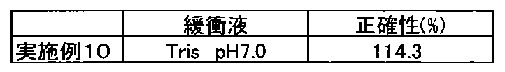
- the accuracy was within ⁇ 15%, and it was confirmed that the influence of hemoglobin could be avoided.
- the use of BPF can avoid the influence of hemoglobin on LTIA.
Landscapes
- Health & Medical Sciences (AREA)
- Life Sciences & Earth Sciences (AREA)
- Immunology (AREA)
- Engineering & Computer Science (AREA)
- Chemical & Material Sciences (AREA)
- Hematology (AREA)
- Urology & Nephrology (AREA)
- Biomedical Technology (AREA)
- Molecular Biology (AREA)
- Microbiology (AREA)
- Analytical Chemistry (AREA)
- Biotechnology (AREA)
- Pathology (AREA)
- Food Science & Technology (AREA)
- Medicinal Chemistry (AREA)
- Physics & Mathematics (AREA)
- Cell Biology (AREA)
- Biochemistry (AREA)
- General Health & Medical Sciences (AREA)
- General Physics & Mathematics (AREA)
- Chemical Kinetics & Catalysis (AREA)
- Peptides Or Proteins (AREA)
- Measuring Or Testing Involving Enzymes Or Micro-Organisms (AREA)
Abstract
Description
ヘモグロビンを含有する試料中に存在することが疑われる、試料中の測定対象成分と、該測定対象成分に結合する抗体とを、配列番号1に記載される大腸菌由来の熱ショックタンパク質(HSP)であるDnaKのアミノ酸配列の419番目から607番目のアミノ酸配列からなるポリペプチド又は当該ポリペプチドと少なくとも90%の配列同一性を有するポリペプチドの存在下に反応させることを特徴とする、ヘモグロビンを含有する試料中の測定対象成分の免疫測定法。
[2]
ヘモグロビンを含有する試料中の測定対象成分と該測定対象成分に結合する抗体とを、更に、HEPES、Bis-Tris、TESおよびTrisから選択される1以上の緩衝成分の存在下に反応させる[1]に記載の方法。
[3]
抗体が、不溶性担体に固定化されていることを特徴とする[1]または[2]に記載の方法。
[4]
不溶性担体が、ラテックス粒子、金属コロイド粒子である[3]に記載の方法。
[5]
粒子凝集測定法を利用する、[4]に記載の方法。
[6]
抗体が、互いに認識部位が異なる二種以上のモノクローナル抗体である、[1]~[5]のいずれかに記載の方法。
[7]
互いに認識部位が異なる二種以上のモノクローナル抗体が、ラテックス粒子にそれぞれ固定化されており、ラテックス免疫比濁法により試料中の測定対象成分を検出する、[6]に記載の方法。
[8]
試料が、尿、全血、血清又は血漿である[1]~[7]のいずれかに記載の方法。
[9]
測定対象成分が、L-FABP(肝臓型脂肪酸結合蛋白質)である[1]~[8]のいずれかに記載の方法。
[10]
ヘモグロビンを含有する試料中の測定対象成分を該測定対象成分に結合する抗体により検出する方法におけるヘモグロビンの影響回避方法であって、
測定対象成分の存在が疑われる試料と該測定対象成分に結合する抗体とを
配列番号1に記載される大腸菌由来の熱ショックタンパク質(HSP)であるDnaKのアミノ酸配列の419番目から607番目のアミノ酸配列からなるポリペプチド又は当該ポリペプチドと少なくとも90%の配列同一性を有するポリペプチドの存在下に反応させることを特徴とする
前記ヘモグロビンの影響回避方法。
[11]
ヘモグロビンを含有する試料中の測定対象成分と該測定対象成分に結合する抗体とを、更に、HEPES、Bis-Tris、TESおよびTrisから選択される1以上の緩衝成分の存在下に反応させる[10]に記載の方法。
[12]
抗体が、不溶性担体に固定化されていることを特徴とする[10]または[11]に記載の方法。
[13]
不溶性担体が、ラテックス粒子、金属コロイド粒子である[12]に記載の方法。
[14]
粒子凝集測定法を利用する、[13]に記載の方法。
[15]
抗体が、互いに認識部位が異なる二種以上のモノクローナル抗体である、[10]~[14]に記載の方法。
[16]
互いに認識部位が異なる二種以上のモノクローナル抗体が、ラテックス粒子にそれぞれ固定化されており、ラテックス免疫比濁法により試料中の測定対象成分を検出する、[15]に記載の方法。
[17]
試料が、尿、全血、血清又は血漿である[10]~[16]のいずれかに記載の方法。
[18]
測定対象成分が、L-FABP(肝臓型脂肪酸結合蛋白質)である[10]~[17]のいずれかに記載の方法。
本発明に用いる生体試料としては、尿、血液(全血、血漿又は血清)、腎臓、心臓、肝臓などの組織、及び該組織からの抽出液等が挙げられる。測定対象物質の存在が疑われる試料であれば、健常者由来の試料、患者由来の試料、罹病を疑われる者由来の試料等、いずれの試料も用いることができる。
本発明により測定される測定対象物質には特に制限はない。
本発明に用いる抗L-FABP抗体は、臓器、細胞、体液等から精製した天然のL-FABPを免疫原(抗原)として調製することができる。L-FABPは、主に肝臓又は腎臓に分布しているので、それらの臓器等から精製、単離することができる。また、L-FABPは、ヒト、マウス、ブタ、ウシ、ラット間でホモロジーが高く、アミノ酸レベルで90%以上のホモロジーがあることが知られているので、ヒトのL-FABPと結合する抗体を得るために、例えばマウスL-FABPを抗原として用いることもできる。
本発明の抗L-FABP抗体を用いたL-FABPを検出する方法は、免疫測定法である。より具体的には、ラテックス免疫比濁法(LTIA)等の粒子免疫凝集測定法、ELISA、化学発光検出法、イムノクロマトグラフィー(ラテラルフロー式、フロースルー式)が挙げられるが、これらの例に限定されない。なかでも、B/F分離のための工程を含まない免疫測定法(ホモジニアス免疫測定法)がより好ましい。
なお、本明細書において測定方法としてLTIAを記述する場合、その検出方法は、透過光(吸光度)の変化の測定、散乱光の変化の測定、粒子径の変化の測定等、公知の検出方法のいずれを用いても差し支えないものとして記述している。
さらに、「検出」又は「測定」という用語は、L-FABPの存在の証明及び/又は定量等を含めて最も広義に解釈する必要があり、限定的に解釈してはならない。
本発明で使用する不溶性担体としては、ポリスチレン樹脂等の高分子基材、ガラス等の無機基材、セルロースやアガロース等の多糖類基材等からなる不溶性担体を用いることができ、その形状は特に限定されず、ビーズあるいは粒子状(例えば、ラテックス粒子、金属コロイド粒子)、板あるいはシート状(例えば、多孔性メンブレン、イムノプレート)、筒状(例えば、試験管)等、採用する測定法に応じた任意の形状を選択できる。
担体粒子の粒子径としては、0.15~0.45μm程度が好ましく、より好ましくは、0.2~0.4μmである。また、平均粒子径の異なる二種類以上の担体粒子を組み合わせて用いることもできる。
抗L-FABP抗体を不溶性担体上に固定化する方法としては特に制限はなく、公知の方法を使用することができる
抗L-FABP抗体を粒子上に固定化する場合、例えば、粒子と抗体を混合することによりおこる物理的な吸着を用いる物理吸着法、カルボジイミド等のカップリング剤により、粒子表面のカルボキシ基やアミノ基と抗体分子を化学的に結合させる化学結合法が用いられる。また、抗体分子はスペーサー分子を介して粒子に固定化させてもよい。さらに、アルブミン等の他のタンパク質に化学結合法を用いて抗体を結合させた後に、そのタンパク質を粒子に物理的あるいは化学的に固定化してもよい。
また、抗L-FABP抗体を多孔性メンブレン上に固定化する場合、例えば、抗体を含む溶液を一定量、ライン状、点あるいは、+等の特定のシンボル状に、多孔性メンブレンに塗布することで固定化できる。
抗体を標識するための標識物質としては、例えば酵素、蛍光物質、化学発光物質、ビオチン、アビジン、又は放射性同位体、金コロイド粒子、着色ラテックス粒子等が挙げられる。また標識物質と抗体との結合方法としては、当業者に利用可能なグルタルアルデヒド法、マレイミド法、ピリジルジスルフィド法、又は過ヨウ素酸法等の方法を用いることができる。標識物質、結合方法のいずれも、上記に限定されることなく公知の方法を用いることができる。
標識の検出は、例えば、パーオキシダーゼやアルカリホスファターゼ等の酵素を標識物質として用いる場合には、その酵素の特異的基質(酵素が西洋ワサビパーオキシダーゼの場合には、例えば1,2-フェニレンジアミンあるいは3,3’,5,5’-テトラメチルベンジジン、アルカリホスファターゼの場合には、p-ニトロフェニルホスフェート等)を用いて酵素活性を測定することができ、ビオチンを標識物質として用いる場合には少なくともビオチン以外の標識物質で標識されたアビジンを反応させるのが一般的である。
大腸菌由来の熱ショックタンパク質(HSP)であるDnaKのアミノ酸配列419番目から607番目のポリペプチド(Blocking Peptide Fragmentとも呼称される。以下「BPF」ということがある。)は、免疫学的測定方法における新規なブロッキング用物質としてWO2005/003155号パンフレットおよび高分子学会予稿集:55巻2 Disk1号5211-5212頁(2006年),Polymer Preprints, Japan Vol. 55, No.2, 5211-5212 (2006)に開示され、市販もされている。
本発明方法等に使用するBPFは、WO2005/003155号パンフレットの記載に従い調製することができる。また、市販品(東洋紡社製、カタログ番号BPF-301)を使用してもよい。
例えば、分子量約22000のBPF-301を用いた場合、0.022mmol/L~2.27mmol/Lが好適であり、より好適には0.034mmol/L~2.27mmol/L、さらに好適には0.045mmol/L~1.36mmol/Lを例示することができる。
L-FABPの存在が疑われる試料、抗L-FABP抗体、及びBPFを接触させる工程は、L-FABPの存在が疑われる試料とBPFを接触させる工程の後、抗L-FABP抗体と接触させる工程の順に行われることを限度として、いずれの方法を用いてもよい。
例えば、L-FABPの存在が疑われる試料と抗L-FABP抗体、及びBPFとを接触させる方法としては、例えば抗L-FABP抗体が固定化された粒子及びBPFを含有する液状の試薬と試料とを混合する方法を挙げることができる。また、別の方法としては、BPFを浸潤させた多孔性メンブレン等の不溶性担体に、L-FABPの存在が疑われる試料を供給することによって接触させる方法を挙げることができる。当業者であれば、測定用試薬の構成等を考慮して適宜設定することができる。
さらに試料中のL-FABPは、BPFと接触した後、又は接触と同時に、不溶性担体に固定化された抗L-FABP抗体と公知の適宜な方法により接触される。
本発明の分子内に下記[化学式1]で表される基をもつ化合物又はその塩からなる緩衝剤としては、下記一般式[I]で表される化合物を例示することができる。
一般式[I]

一般式[I]中のR3、R4としては、互いに同一又は異なっていてもよく、水素原子、アルキル基、ヒドロキシアルキル基、ジ(ヒドロキシアルキル)アルキル基、トリ(ヒドロキシアルキル)アルキル基、カルボキシアルキル基、ジ(カルボキシアルキル)アルキル基、トリ(カルボキシアルキル)アルキル基、スルホアルキル基、ジ(スルホアルキル)アルキル基、トリ(スルホアルキル)アルキル基、スルホ-ヒドロキシ-アルキル基、ジ(スルホ-ヒドロキシ-アルキル)アルキル基、トリ(スルホ-ヒドロキシ-アルキル)アルキル基等を挙げることができる。
上記アルキル基、ヒドロキシアルキル基、ジ(ヒドロキシアルキル)アルキル基、トリ(ヒドロキシアルキル)アルキル基、カルボキシアルキル基、ジ(カルボキシアルキル)アルキル基、トリ(カルボキシアルキル)アルキル基、スルホアルキル基、ジ(スルホアルキル)アルキル基、トリ(スルホアルキル)アルキル基、スルホ-ヒドロキシ-アルキル基、ジ(スルホ-ヒドロキシ-アルキル)アルキル基、及びトリ(スルホ-ヒドロキシ-アルキル)アルキル基におけるアルキル基としては、直鎖又は分枝状の炭素数が1~6のアルキル基を挙げることができ、該直鎖又は分枝状の炭素数が1~6のアルキル基としては、メチル基、エチル基、プロピル基、ブチル基、イソブチル基、sec-ブチル基、tert-ブチル基、ペンチル基、ヘキシル基等を具体的に例示することができる。
また、一般式[I]中のR3、R4としては、R3,R4が窒素原子と共に環状構造を形成し、置換もしくは無置換のピペラジニル基、置換もしくは無置換のモルホリノ基、又は置換もしくは無置換のピペリジノ基を形成する基を挙げることができ、該置換ピペラジニル基、置換モルホリノ基、及び置換ピペリジノ基における置換基としては、アルキル基、ヒドロキシアルキル基、ジ(ヒドロキシアルキル)アルキル基、トリ(ヒドロキシアルキル)アルキル基、カルボキシアルキル基、ジ(カルボキシアルキル)アルキル基、トリ(カルボキシアルキル)アルキル基、スルホアルキル基、ジ(スルホアルキル)アルキル基、トリ(スルホアルキル)アルキル基、スルホ-ヒドロキシ-アルキル基、ジ(スルホ-ヒドロキシ-アルキル)アルキル基、トリ(スルホ-ヒドロキシ-アルキル)アルキル基等を挙げることができる。
上記アルキル基、ヒドロキシアルキル基、ジ(ヒドロキシアルキル)アルキル基、トリ(ヒドロキシアルキル)アルキル基、カルボキシアルキル基、ジ(カルボキシアルキル)アルキル基、トリ(カルボキシアルキル)アルキル基、スルホアルキル基、ジ(スルホアルキル)アルキル基、トリ(スルホアルキル)アルキル基、スルホ-ヒドロキシ-アルキル基、ジ(スルホ-ヒドロキシ-アルキル)アルキル基、及びトリ(スルホ-ヒドロキシ-アルキル)アルキル基におけるアルキル基としては、前記の直鎖又は分枝状の炭素数が1~6のアルキル基を挙げることができる。
前記ヒドロキシアルキル基としては、ヒドロキシメチル基、2-ヒドロキシエチル基、2-ヒドロキシプロピル基、2-ヒドロキシブチル基、2-ヒドロキシペンチル基、2-ヒドロキシヘキシル基等を、ジ(ヒドロキシアルキル)アルキル基としては、ジ(ヒドロキシメチル)メチル基、ジ(2-ヒドロキシエチル)メチル基等を、トリ(ヒドロキシアルキル)アルキル基としては、トリ(ヒドロキシメチル)メチル基、トリ(2-ヒドロキシエチル)メチル基等を、カルボキシアルキル基としては、カルボキシメチル基、2-カルボキシエチル基等を、ジ(カルボキシアルキル)アルキル基としては、ジ(カルボキシメチル)メチル基、ジ(2-カルボキシエチル)メチル基等を、トリ(カルボキシアルキル)アルキル基としては、トリ(カルボキシメチル)メチル基、トリ(2-カルボキシエチル)メチル基等を、それぞれ具体的に例示することができる。
また、前記スルホアルキル基としては、スルホメチル基、2-スルホエチル基等を、ジ(スルホアルキル)アルキル基としては、ジ(スルホメチル)メチル基、ジ(2-スルホエチル)メチル基等を、トリ(スルホアルキル)アルキル基としては、トリ(スルホメチル)メチル基、トリ(2-スルホエチル)メチル基等を、スルホ-ヒドロキシ-アルキル基としては、2-ヒドロキシ-3-スルホプロピル基、3-ヒドロキシ-4-スルホプロピル基等を、ジ(スルホ-ヒドロキシ-アルキル)アルキル基としては、ジ(2-ヒドロキシ-3-スルホプロピル)メチル基、ジ(3-ヒドロキシ-4-スルホプロピル)メチル基等を、トリ(スルホ-ヒドロキシ-アルキル)アルキル基としては、トリ(2-ヒドロキシ-3-スルホプロピル)メチル基、トリ(3-ヒドロキシ-4-スルホプロピル)メチル基等を、それぞれ具体的に例示することができる。
本発明により提供される測定キットの構成物は、L-FABPを免疫学的に測定できることを限度として、特に限定されるものではない。以下、サンドイッチELISA、イムノクロマトグラフィー及びLTIAを例にそれぞれを説明する。
サンドイッチELISAの場合、測定用キットは少なくとも、(a)本発明の抗L-FABP抗体を固定化した不溶性担体及び(b)標識物質で標識され、L-FABPと反応する性質を有する抗体、を含む。この場合、不溶性担体はプレート(イムノプレート)が好ましく、標識物質は、適宜選択して使用できる。
一般的なイムノクロマトグラフィーでは、多孔性メンブレン等のシート状の不溶性担体上に、試料を含む溶液の展開方向に順に「1.試料供給部位」、「2.標識抗体を保持する部位(標識抗体保持部位)」、「3.標識抗体とL-FABP抗体により形成された複合体を捕捉するための抗体を固定化する部位(捕捉抗体部位)」を具備した試験片が使用され、試料溶液が毛細管現象により連続的に移動するように構成されている。イムノクロマトグラフィーでは、測定用キットは、上記のような試験片を少なくとも含む。
ラテックス免疫凝集測定法では、測定用キットは少なくとも抗体が固定化されたラテックス粒子を含む。ラテックス免疫凝集測定法に使用される抗体としては、「抗原に対する認識部位が異なる二種類のモノクローナル抗体」、「ポリクローナル抗体」、又は「モノクローナル抗体とポリクローナル抗体」のいずれの組合せも用いることが出来る。この場合、ラテックス粒子は、抗体を固定化する不溶性担体であると同時に、標識物質である。
(1)CloneL抗体固定化ラテックス粒子懸濁液の調製
抗L-FABP抗体 CloneL(シミックホールディングス社製)を0.36mg/mL含む20mmol/L Tris緩衝液(pH8.5)13mLに、平均粒径0.21μmの1%ラテックス粒子(積水化学工業社製)懸濁液13mLを加え、4℃にて2時間撹拌した。これに、0.5%BSAを含む20mmol/L Tris緩衝液(pH8.5)13mLを加え、4℃で1時間撹拌した。その後、5mmol/L MOPS緩衝液(pH7.0)に透析して、CloneL抗体固定化ラテックス粒子懸濁液を得た。
(2)Clone1抗体固定化ラテックス粒子懸濁液の調製
抗L-FABP抗体 Clone1(シミックホールディングス社製)を0.54mg/mL含む5mmol/L Tris緩衝液(pH7.5)8mLに、平均粒径0.32μmの1%ラテックス粒子(積水化学工業社製)懸濁液8mLを加え、4℃にて2時間撹拌した。これに、0.5%BSAを含む5mmol/L Tris緩衝液(pH7.5)8mLを加え、4℃で1時間撹拌した。その後、5mmol/L MOPS緩衝液(pH7.0)に透析してClone1抗体固定化ラテックス粒子懸濁液を得た。
L-FABP標準物質は、特開平11-242026号公報の記載に従い、遺伝子組換えにより得た。
ELISAによる体外診断用医薬品(レナプロ(登録商標)L-FABPテスト TMB)を基準方法とした。
300mmol/L HEPES緩衝液(pH7.0)
各濃度 BPF(東洋紡社製、BPF-103)
100mmol/L NaCl
390mmol/L ベンズアミジン塩酸塩
0.4% Lipidure-BL103
(第二試薬)
5mmol/L MOPS緩衝液(pH7.0)
3.75Abs/mL CloneL抗体固定化ラテックス粒子懸濁液(注)
1.25Abs/mL Clone1抗体固定化ラテックス粒子懸濁液(注)
(注)Absは280nmにおける吸光度を示す。
(標準液)
L-FABP標準物質を、標準物質希釈液を用いて所望濃度に調整し、標準液とした。
(凍結融解尿)
採取後、-30℃で凍結保存されていた部分尿を、一度だけ融解して測定に使用した。予め基準方法にて凍結融解尿中のL-FABPを測定したところ、10ng/mLであった。
(検体)
ヘモグロビンの濃度が0~1000mg/dLとなるよう干渉チェックAプラス(シスメックス社製)を調整した。前記調整済み干渉チェックAプラスと上記凍結融解尿とを等量混和して、各濃度のヘモグロビンが共存する検体とした。
(1)分析装置:日立7170型自動分析装置(日立ハイテクノロジーズ社製)
(2)試料量及び試薬量:試料3μL、第一試薬150μL、第二試薬50μL
(3)反応時間(反応温度):第一試薬5分(37℃)、第二試薬5分(37℃)
(4)測光ポイント及び測光対象:第二試薬添加直後と添加5分後の間の吸光度変化量
BPFによる、ヘモグロビンの影響回避効果を確認した。
1.操作
(1)検量線の作成
標準液を試料として、上記第一試薬、及び第二試薬を用いて試料中のL-FABPの測定を行った。測定された吸光度から、L-FABP 0ng/mL試料の吸光度(ブランク吸光度)を差し引いて正味吸光度を算出した。L-FABP濃度をx軸、正味吸光度をy軸として、検量線を作成した。
(2)実施例1~4
表1の濃度のBPFを含む上記の第一試薬と第二試薬を用いて、検体中のL-FABPの測定を行った。
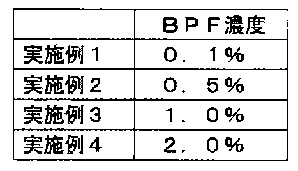
BPFを含まない第一試薬と、第二試薬を用いて試料中のL-FABPの測定を行った。
実施例1~4及び対照例において測定された吸光度から、(1)の検量線を用いてL-FABP濃度を算出した。各ヘモグロビン濃度において算出された測定値を、ヘモグロビンが0mg/dLの測定値で除して比率(%)を求め表2に示した。

(1)CloneL抗体固定化ラテックス粒子懸濁液の調製
抗L-FABP抗体 CloneL(シミックホールディングス社製)を0.36mg/mL含む20mmol/L Tris緩衝液(pH8.5)13mLに、平均粒径0.27μmの1%ラテックス粒子(積水化学工業社製)懸濁液13mLを加え、4℃にて2時間撹拌した。これに、0.5%BSAを含む20mmol/L Tris緩衝液(pH8.5)13mLを加え、4℃で1時間撹拌した。その後、5mmol/L MOPS緩衝液(pH7.0)に透析して、CloneL抗体固定化ラテックス粒子懸濁液を得た。
(2)Clone1抗体固定化ラテックス粒子懸濁液の調製
抗L-FABP抗体 Clone1(シミックホールディングス社製)を0.54mg/mL含む5mmol/L トリス緩衝液(pH7.5)8mLに、平均粒径0.25μmの1%ラテックス粒子(積水化学工業社製)懸濁液8mLを加え、4℃にて2時間撹拌した。これに、0.5%BSAを含む5mmol/L トリス緩衝液(pH7.5)8mLを加え、4℃で1時間撹拌した。その後、5mmol/L MOPS緩衝液(pH7.0)に透析してClone1抗体固定化ラテックス粒子懸濁液を得た。
L-FABP標準物質は、特許文献1の記載に従い、遺伝子組換えにより得た。
ELISAによる体外診断用医薬品(レナプロ(登録商標)L-FABPテスト TMB)を基準方法とした。
300mmol/L 緩衝液
0.1% BPF(東洋紡社製、BPF-103)
300mmol/L NaCl
390mmol/L ベンズアミジン塩酸塩
0.720%~1.020% Lipidure-BL103
(第二試薬)
5mmol/L MOPS緩衝液(pH7.0)
3.75Abs/mL CloneL抗体固定化ラテックス粒子懸濁液(注)
1.25Abs/mL Clone1抗体固定化ラテックス粒子懸濁液(注)
(注)Absは280nmにおける吸光度を示す。
(標準物質希釈液)
リン酸緩衝液(pH7.0)
0.1% BPF(東洋紡社製、BPF-103)
(標準液)
L-FABP標準物質を、標準物質希釈液を用いて所望濃度に調整し、標準液とした。
(凍結融解尿)
採取後、-30℃で凍結保存されていた部分尿を、一度だけ融解して測定に使用した。
(1)分析装置:日立7170型自動分析装置(日立ハイテクノロジーズ社製)
(2)試料量及び試薬量:試料3μL、第一試薬150μL、第二試薬50μL
(3)反応時間(反応温度):第一試薬5分(37℃)、第二試薬5分(37℃)
(4)測光ポイント及び測光対象:第二試薬添加直後と添加5分後の間の吸光度変化量
(5)測定波長570nm/800nm
第一試薬に表3に記載の各緩衝液を用い、ヘモグロビンの影響回避効果を確認した。
1.操作
(1)検体の調整
ヘモグロビンの濃度が200mg/dLとなるよう干渉チェックAプラス(シスメックス社製)を調整した。前記調整済み干渉チェックAプラスと上記凍結融解尿とを等量混和して、100mg/dLヘモグロビンが共存する検体とした。また、ヘモグロビンの濃度が0mg/dLの検体は、上記凍結融解尿をそのまま用いた。
(2)検量線の作成
標準液を試料として、上記第一試薬、及び第二試薬を用いて試料中のL-FABPの測定を行った。測定された吸光度から、L-FABP 0ng/mL試料の吸光度(ブランク吸光度)を差し引いて正味吸光度を算出した。L-FABP濃度をx軸、正味吸光度をy軸として、検量線を作成した。
(3)実施例5~9
表3の緩衝液を含む上記の第一試薬と、第二試薬を用いて、検体中のL-FABPの測定を行った。
実施例5~9において測定された吸光度から、(2)の検量線を用いてL-FABP濃度を算出した。算出された測定値を、ヘモグロビンが0mg/dLの測定値で除して比率(%)を求め表3に示した。

第一試薬に、表4に記載の緩衝液、第二試薬に、2.5Abs/mL CloneL抗体固定化ラテックス粒子懸濁液及び 2.5Abs/mL Clone1抗体固定化ラテックス粒子懸濁液(Absは280nmにおける吸光度を示す)を用いて、ヘモグロビンの影響回避効果を確認した。それら以外、条件は実施例2~9と同じである。
(1)検体の調整
ヘモグロビンの濃度が200mg/dLとなるよう干渉チェックAプラス(シスメックス社製)を調整した。前記調整済み干渉チェックAプラスと上記凍結融解尿とを等量混和して、100mg/dLヘモグロビンが共存する検体とした。また、ヘモグロビンの濃度が0mg/dLの検体は、上記凍結融解尿をそのまま用いた。
(2)検量線の作成
標準液を試料として、上記第一試薬、及び第二試薬を用いて試料中のL-FABPの測定を行った。測定された吸光度から、L-FABP 0ng/mL試料の吸光度(ブランク吸光度)を差し引いて正味吸光度を算出した。L-FABP濃度をx軸、正味吸光度をy軸として、検量線を作成した。
(3)実施例10
表4の緩衝液を含む上記の第一試薬と、第二試薬を用いて、検体中のL-FABPの測定を行った。
実施例10において測定された吸光度から、(2)の検量線を用いてL-FABP濃度を算出した。算出された測定値を、ヘモグロビンが0mg/dLの測定値で除して比率(%)を求め表4に示した。
Claims (10)
- ヘモグロビンを含有する試料中に存在することが疑われる、試料中の測定対象成分と、該測定対象成分に結合する抗体とを、配列番号1に記載される大腸菌由来の熱ショックタンパク質(HSP)であるDnaKのアミノ酸配列の419番目から607番目のアミノ酸配列からなるポリペプチド又は当該ポリペプチドと少なくとも90%の配列同一性を有するポリペプチドの存在下に反応させることを特徴とする、ヘモグロビンを含有する試料中の測定対象成分の免疫測定法。
- ヘモグロビンを含有する試料中の測定対象成分と該測定対象成分に結合する抗体とを、更に、HEPES、Bis-Tris、TESおよびTrisから選択される1以上の緩衝成分の存在下に反応させる請求項1記載の方法。
- 抗体が、不溶性担体に固定化されていることを特徴とする請求項1または2に記載の方法。
- 不溶性担体が、ラテックス粒子、金属コロイド粒子である請求項3に記載の方法。
- 粒子凝集測定法を利用する、請求項4に記載の方法。
- 抗体が、互いに認識部位が異なる二種以上のモノクローナル抗体である、請求項1~5のいずれかに記載の方法。
- 互いに認識部位が異なる二種以上のモノクローナル抗体が、ラテックス粒子にそれぞれ固定化されており、ラテックス免疫比濁法により試料中の測定対象成分を検出する、請求項6に記載の方法。
- 試料が、尿、全血、血清又は血漿である請求項1~7のいずれかに記載の方法。
- 測定対象成分が、L-FABP(肝臓型脂肪酸結合蛋白質)である請求項1~8のいずれかに記載の方法。
- ヘモグロビンを含有する試料中の測定対象成分を該測定対象成分に結合する抗体により検出する方法におけるヘモグロビンの影響回避方法であって、
測定対象成分の存在が疑われる試料と該測定対象成分に結合する抗体とを
配列番号1に記載される大腸菌由来の熱ショックタンパク質(HSP)であるDnaKのアミノ酸配列の419番目から607番目のアミノ酸配列からなるポリペプチド又は当該ポリペプチドと少なくとも90%の配列同一性を有するポリペプチドの存在下に反応させることを特徴とする
前記ヘモグロビンの影響回避方法。
Priority Applications (4)
| Application Number | Priority Date | Filing Date | Title |
|---|---|---|---|
| US15/553,343 US20180113127A1 (en) | 2015-02-25 | 2016-02-25 | Immunoassay method and assay reagent used in said method |
| JP2017502493A JP6660934B2 (ja) | 2015-02-25 | 2016-02-25 | 免疫学的測定方法及び該方法に用いられる測定試薬 |
| CN201680023848.3A CN107533055A (zh) | 2015-02-25 | 2016-02-25 | 免疫测定方法以及用于所述方法的测定试剂 |
| EP16755662.0A EP3264085A4 (en) | 2015-02-25 | 2016-02-25 | Immunoassay method and assay reagent used in said method |
Applications Claiming Priority (2)
| Application Number | Priority Date | Filing Date | Title |
|---|---|---|---|
| JP2015-035926 | 2015-02-25 | ||
| JP2015035926 | 2015-02-25 |
Publications (1)
| Publication Number | Publication Date |
|---|---|
| WO2016136918A1 true WO2016136918A1 (ja) | 2016-09-01 |
Family
ID=56789531
Family Applications (1)
| Application Number | Title | Priority Date | Filing Date |
|---|---|---|---|
| PCT/JP2016/055729 WO2016136918A1 (ja) | 2015-02-25 | 2016-02-25 | 免疫学的測定方法及び該方法に用いられる測定試薬 |
Country Status (5)
| Country | Link |
|---|---|
| US (1) | US20180113127A1 (ja) |
| EP (1) | EP3264085A4 (ja) |
| JP (1) | JP6660934B2 (ja) |
| CN (1) | CN107533055A (ja) |
| WO (1) | WO2016136918A1 (ja) |
Cited By (3)
| Publication number | Priority date | Publication date | Assignee | Title |
|---|---|---|---|---|
| JP2017083440A (ja) * | 2015-10-23 | 2017-05-18 | 株式会社Lsiメディエンス | 免疫学的測定試薬、測定方法及び測定範囲の拡大方法 |
| JPWO2018180987A1 (ja) * | 2017-03-29 | 2020-02-06 | 東洋紡株式会社 | 核酸の検出方法 |
| WO2024075847A1 (ja) * | 2022-10-06 | 2024-04-11 | 積水メディカル株式会社 | 非特異反応抑制剤、非特異反応抑制剤の使用方法、非特異反応抑制方法、生化学的測定用試薬、検体前処理液、及び生化学的測定用試薬キット |
Families Citing this family (1)
| Publication number | Priority date | Publication date | Assignee | Title |
|---|---|---|---|---|
| CN111812336A (zh) * | 2020-08-10 | 2020-10-23 | 苏州康和顺医疗技术有限公司 | 用于检测冠状病毒抗体的检测试剂盒及其制备方法 |
Citations (11)
| Publication number | Priority date | Publication date | Assignee | Title |
|---|---|---|---|---|
| JPH05322891A (ja) * | 1992-05-18 | 1993-12-07 | Kyoto Daiichi Kagaku:Kk | 免疫測定における非特異的吸着防止法 |
| JPH06289025A (ja) * | 1993-03-31 | 1994-10-18 | Cosmo Sogo Kenkyusho:Kk | ブロッキング用非特異反応防止組成物及び固相担体 |
| JPH08211056A (ja) * | 1995-02-07 | 1996-08-20 | Konica Corp | 免疫反応媒体組成物、酵素標識抗体組成物、該組成物の安定化方法、該組成物を用いる免疫学的測定法及び免疫学的測定法用キット |
| JPH11242026A (ja) * | 1997-11-26 | 1999-09-07 | Tanabe Seiyaku Co Ltd | 腎疾患の検査方法 |
| JP2001033442A (ja) * | 1999-07-26 | 2001-02-09 | A & T:Kk | 修飾ヘモグロビンの修飾基の免疫学的測定法 |
| JP2002202312A (ja) * | 2001-11-28 | 2002-07-19 | Tokuyama Corp | 診断用キット |
| WO2005003155A1 (ja) * | 2003-07-03 | 2005-01-13 | Toyo Boseki Kabushiki Kaisha | ブロッキング効率の向上したタンパク質 |
| WO2011125912A1 (ja) * | 2010-03-31 | 2011-10-13 | 積水メディカル株式会社 | 測定系外成分による干渉を低減させる方法 |
| JP2012112042A (ja) * | 2010-11-05 | 2012-06-14 | Tanaka Kikinzoku Kogyo Kk | 免疫学的測定用青色金ナノ粒子、その製造方法およびそれを用いた測定方法 |
| WO2013002403A1 (ja) * | 2011-06-30 | 2013-01-03 | 積水メディカル株式会社 | 免疫学的測定方法に用いられるコンジュゲート |
| JP2014052390A (ja) * | 2008-07-04 | 2014-03-20 | Sekisui Medical Co Ltd | 免疫学的測定における感度増強方法及びそのための試薬 |
Family Cites Families (10)
| Publication number | Priority date | Publication date | Assignee | Title |
|---|---|---|---|---|
| US6656697B1 (en) * | 1998-09-28 | 2003-12-02 | Lifescan, Inc. | Diagnostics based on tetrazolium compounds |
| US6379550B1 (en) * | 2000-07-24 | 2002-04-30 | Health Research, Inc. | Method for detecting PSA and its molecular forms using thiophilic gel |
| US6867004B2 (en) * | 2001-12-21 | 2005-03-15 | Focus Technologies, Inc. | Methods of using oxidized fungal antigens in antibody testing |
| JP2005147783A (ja) * | 2003-11-13 | 2005-06-09 | Eisai Co Ltd | チトクロムcによる細胞死の診断薬 |
| US8987005B2 (en) * | 2005-12-28 | 2015-03-24 | Sekisui Medical Co., Ltd. | Reagent for measuring agglutination and method of measuring agglutination |
| CN101568834A (zh) * | 2006-11-02 | 2009-10-28 | 协和梅迪克斯株式会社 | 待测组分的免疫测定方法 |
| EP2042870A1 (en) * | 2007-09-28 | 2009-04-01 | Fujifilm Corporation | Method of high sensitive immunoassay |
| WO2010048423A1 (en) * | 2008-10-24 | 2010-04-29 | Ark Diagnostics, Inc. | Levetiracetam immunoassays |
| EP2568291A1 (en) * | 2011-09-07 | 2013-03-13 | Roche Diagnostics GmbH | L-FABP based diagnosis of kidney injury after an acute event or after a surgical intervention |
| CN203881772U (zh) * | 2014-03-14 | 2014-10-15 | 瑞莱生物工程(深圳)有限公司 | 一种快速定量检测l-fabp的胶体金试纸 |
-
2016
- 2016-02-25 JP JP2017502493A patent/JP6660934B2/ja active Active
- 2016-02-25 WO PCT/JP2016/055729 patent/WO2016136918A1/ja active Application Filing
- 2016-02-25 US US15/553,343 patent/US20180113127A1/en not_active Abandoned
- 2016-02-25 CN CN201680023848.3A patent/CN107533055A/zh active Pending
- 2016-02-25 EP EP16755662.0A patent/EP3264085A4/en not_active Withdrawn
Patent Citations (11)
| Publication number | Priority date | Publication date | Assignee | Title |
|---|---|---|---|---|
| JPH05322891A (ja) * | 1992-05-18 | 1993-12-07 | Kyoto Daiichi Kagaku:Kk | 免疫測定における非特異的吸着防止法 |
| JPH06289025A (ja) * | 1993-03-31 | 1994-10-18 | Cosmo Sogo Kenkyusho:Kk | ブロッキング用非特異反応防止組成物及び固相担体 |
| JPH08211056A (ja) * | 1995-02-07 | 1996-08-20 | Konica Corp | 免疫反応媒体組成物、酵素標識抗体組成物、該組成物の安定化方法、該組成物を用いる免疫学的測定法及び免疫学的測定法用キット |
| JPH11242026A (ja) * | 1997-11-26 | 1999-09-07 | Tanabe Seiyaku Co Ltd | 腎疾患の検査方法 |
| JP2001033442A (ja) * | 1999-07-26 | 2001-02-09 | A & T:Kk | 修飾ヘモグロビンの修飾基の免疫学的測定法 |
| JP2002202312A (ja) * | 2001-11-28 | 2002-07-19 | Tokuyama Corp | 診断用キット |
| WO2005003155A1 (ja) * | 2003-07-03 | 2005-01-13 | Toyo Boseki Kabushiki Kaisha | ブロッキング効率の向上したタンパク質 |
| JP2014052390A (ja) * | 2008-07-04 | 2014-03-20 | Sekisui Medical Co Ltd | 免疫学的測定における感度増強方法及びそのための試薬 |
| WO2011125912A1 (ja) * | 2010-03-31 | 2011-10-13 | 積水メディカル株式会社 | 測定系外成分による干渉を低減させる方法 |
| JP2012112042A (ja) * | 2010-11-05 | 2012-06-14 | Tanaka Kikinzoku Kogyo Kk | 免疫学的測定用青色金ナノ粒子、その製造方法およびそれを用いた測定方法 |
| WO2013002403A1 (ja) * | 2011-06-30 | 2013-01-03 | 積水メディカル株式会社 | 免疫学的測定方法に用いられるコンジュゲート |
Non-Patent Citations (3)
| Title |
|---|
| KENJIRO KIMURA: "L-FABP no Rinshoteki Igi to Kongo no Tenbo", JAPANESE JOURNAL OF CLINICAL LABORATORY AUTOMATION, vol. 39, no. 4, 1 September 2014 (2014-09-01), pages 433, XP009505458 * |
| NOBUHIRO SATO: "Nyo-chu L-FABP no Sokuji Sokutei o Kano ni suru Shinki Latex-ho Shiyaku Norudia L-FABP no Kisoteki Seino Hyoka", JAPANESE JOURNAL OF CLINICAL LABORATORY AUTOMATION, vol. 40, no. 4, 1 September 2015 (2015-09-01), pages 523 269, XP009505460 * |
| See also references of EP3264085A4 * |
Cited By (4)
| Publication number | Priority date | Publication date | Assignee | Title |
|---|---|---|---|---|
| JP2017083440A (ja) * | 2015-10-23 | 2017-05-18 | 株式会社Lsiメディエンス | 免疫学的測定試薬、測定方法及び測定範囲の拡大方法 |
| JPWO2018180987A1 (ja) * | 2017-03-29 | 2020-02-06 | 東洋紡株式会社 | 核酸の検出方法 |
| JP7279633B2 (ja) | 2017-03-29 | 2023-05-23 | 東洋紡株式会社 | 核酸の検出方法 |
| WO2024075847A1 (ja) * | 2022-10-06 | 2024-04-11 | 積水メディカル株式会社 | 非特異反応抑制剤、非特異反応抑制剤の使用方法、非特異反応抑制方法、生化学的測定用試薬、検体前処理液、及び生化学的測定用試薬キット |
Also Published As
| Publication number | Publication date |
|---|---|
| JP6660934B2 (ja) | 2020-03-11 |
| JPWO2016136918A1 (ja) | 2017-11-30 |
| EP3264085A1 (en) | 2018-01-03 |
| EP3264085A4 (en) | 2018-09-12 |
| CN107533055A (zh) | 2018-01-02 |
| US20180113127A1 (en) | 2018-04-26 |
Similar Documents
| Publication | Publication Date | Title |
|---|---|---|
| JP6660932B2 (ja) | L−fabpの免疫学的測定方法及び該方法に用いられる測定試薬 | |
| JP5199067B2 (ja) | 免疫凝集反応試薬キット及び抗原の測定方法 | |
| JP6660934B2 (ja) | 免疫学的測定方法及び該方法に用いられる測定試薬 | |
| EP1653233A1 (en) | Method for determining antibodies of a particular class using an immune complex-specific antibody | |
| JP6016789B2 (ja) | Pivka−ii測定試薬における非特異反応の抑制方法 | |
| JP3652029B2 (ja) | 高感度免疫測定法 | |
| JP6660933B2 (ja) | 免疫学的測定方法及び該方法に用いられる測定試薬 | |
| JP2001512574A (ja) | ハプトグロビンの表現型を決定する方法およびキットならびにその使用 | |
| WO2022163605A1 (ja) | 免疫学的測定方法 | |
| JP2019027957A (ja) | L−fabpの免疫学的測定方法及び該方法に用いられる測定試薬 | |
| JP5750646B2 (ja) | Scca2濃度測定によるアレルギー疾患の検査方法 | |
| JP5750645B2 (ja) | アレルギー疾患の検査方法 | |
| JP2003028875A (ja) | 非特異反応を抑制したイムノアッセイ法 | |
| JP2002350442A (ja) | 免疫測定試薬及び免疫測定方法 |
Legal Events
| Date | Code | Title | Description |
|---|---|---|---|
| 121 | Ep: the epo has been informed by wipo that ep was designated in this application |
Ref document number: 16755662 Country of ref document: EP Kind code of ref document: A1 |
|
| ENP | Entry into the national phase |
Ref document number: 2017502493 Country of ref document: JP Kind code of ref document: A |
|
| WWE | Wipo information: entry into national phase |
Ref document number: 15553343 Country of ref document: US |
|
| NENP | Non-entry into the national phase |
Ref country code: DE |
|
| REEP | Request for entry into the european phase |
Ref document number: 2016755662 Country of ref document: EP |