WO2015098515A1 - 止血材 - Google Patents
止血材 Download PDFInfo
- Publication number
- WO2015098515A1 WO2015098515A1 PCT/JP2014/082653 JP2014082653W WO2015098515A1 WO 2015098515 A1 WO2015098515 A1 WO 2015098515A1 JP 2014082653 W JP2014082653 W JP 2014082653W WO 2015098515 A1 WO2015098515 A1 WO 2015098515A1
- Authority
- WO
- WIPO (PCT)
- Prior art keywords
- self
- hemostatic material
- gel
- peptide
- amino acid
- Prior art date
Links
Images
Classifications
-
- A—HUMAN NECESSITIES
- A61—MEDICAL OR VETERINARY SCIENCE; HYGIENE
- A61L—METHODS OR APPARATUS FOR STERILISING MATERIALS OR OBJECTS IN GENERAL; DISINFECTION, STERILISATION OR DEODORISATION OF AIR; CHEMICAL ASPECTS OF BANDAGES, DRESSINGS, ABSORBENT PADS OR SURGICAL ARTICLES; MATERIALS FOR BANDAGES, DRESSINGS, ABSORBENT PADS OR SURGICAL ARTICLES
- A61L26/00—Chemical aspects of, or use of materials for, wound dressings or bandages in liquid, gel or powder form
- A61L26/0009—Chemical aspects of, or use of materials for, wound dressings or bandages in liquid, gel or powder form containing macromolecular materials
- A61L26/0028—Polypeptides; Proteins; Degradation products thereof
-
- A—HUMAN NECESSITIES
- A61—MEDICAL OR VETERINARY SCIENCE; HYGIENE
- A61L—METHODS OR APPARATUS FOR STERILISING MATERIALS OR OBJECTS IN GENERAL; DISINFECTION, STERILISATION OR DEODORISATION OF AIR; CHEMICAL ASPECTS OF BANDAGES, DRESSINGS, ABSORBENT PADS OR SURGICAL ARTICLES; MATERIALS FOR BANDAGES, DRESSINGS, ABSORBENT PADS OR SURGICAL ARTICLES
- A61L15/00—Chemical aspects of, or use of materials for, bandages, dressings or absorbent pads
- A61L15/16—Bandages, dressings or absorbent pads for physiological fluids such as urine or blood, e.g. sanitary towels, tampons
- A61L15/22—Bandages, dressings or absorbent pads for physiological fluids such as urine or blood, e.g. sanitary towels, tampons containing macromolecular materials
- A61L15/32—Proteins, polypeptides; Degradation products or derivatives thereof, e.g. albumin, collagen, fibrin, gelatin
-
- A—HUMAN NECESSITIES
- A61—MEDICAL OR VETERINARY SCIENCE; HYGIENE
- A61L—METHODS OR APPARATUS FOR STERILISING MATERIALS OR OBJECTS IN GENERAL; DISINFECTION, STERILISATION OR DEODORISATION OF AIR; CHEMICAL ASPECTS OF BANDAGES, DRESSINGS, ABSORBENT PADS OR SURGICAL ARTICLES; MATERIALS FOR BANDAGES, DRESSINGS, ABSORBENT PADS OR SURGICAL ARTICLES
- A61L24/00—Surgical adhesives or cements; Adhesives for colostomy devices
- A61L24/04—Surgical adhesives or cements; Adhesives for colostomy devices containing macromolecular materials
- A61L24/10—Polypeptides; Proteins
-
- A—HUMAN NECESSITIES
- A61—MEDICAL OR VETERINARY SCIENCE; HYGIENE
- A61P—SPECIFIC THERAPEUTIC ACTIVITY OF CHEMICAL COMPOUNDS OR MEDICINAL PREPARATIONS
- A61P7/00—Drugs for disorders of the blood or the extracellular fluid
- A61P7/04—Antihaemorrhagics; Procoagulants; Haemostatic agents; Antifibrinolytic agents
-
- A—HUMAN NECESSITIES
- A61—MEDICAL OR VETERINARY SCIENCE; HYGIENE
- A61K—PREPARATIONS FOR MEDICAL, DENTAL OR TOILETRY PURPOSES
- A61K38/00—Medicinal preparations containing peptides
-
- A—HUMAN NECESSITIES
- A61—MEDICAL OR VETERINARY SCIENCE; HYGIENE
- A61L—METHODS OR APPARATUS FOR STERILISING MATERIALS OR OBJECTS IN GENERAL; DISINFECTION, STERILISATION OR DEODORISATION OF AIR; CHEMICAL ASPECTS OF BANDAGES, DRESSINGS, ABSORBENT PADS OR SURGICAL ARTICLES; MATERIALS FOR BANDAGES, DRESSINGS, ABSORBENT PADS OR SURGICAL ARTICLES
- A61L2400/00—Materials characterised by their function or physical properties
- A61L2400/04—Materials for stopping bleeding
Definitions
- the present invention relates to a hemostatic material containing a self-assembling peptide.
- a sheet-like or two-liquid mixed liquid or viscous liquid has been used in many cases.
- the sheet-like hemostatic material is often opaque in white, and it has been impossible to directly confirm the bleeding point and the completion of hemostasis visually.
- the two-component mixed type has a very high adhesiveness immediately after mixing, so it must be used immediately, and if the entire amount is not used up by the first hemostasis, the rest is used for the second time. Even if he tried to stop bleeding, there was a problem in terms of use such as being unable to take out from the container because of increased adhesiveness.
- many conventional hemostats contain animal-derived materials, the risk of infection has been pointed out.
- Patent Document 1 a hemostatic material that can be used in a one-pack type containing a self-assembling peptide that is a non-animal derived material has been proposed.
- the hemostatic material is typically placed on a biological tissue to be treated, so that the self-assembling peptide self-assembles into a gel and exhibits a hemostatic function (Patent Document 1, paragraph 0040, 0051 paragraph).
- the mechanical strength is insufficient, so the gel may be partially washed away against severe bleeding such as arterial bleeding. In some cases, it collapsed and a sufficient hemostatic function could not be obtained. For this reason, the gel-type hemostatic material using the self-assembling peptide is a non-biological material, and the merit that it can be used in a one-pack type cannot be fully utilized.
- the hemostatic material of the present invention is a hemostatic material comprising a gel containing self-assembled peptide and water and formed by self-assembly of the self-assembled peptide, wherein the gel is measured by a rotary rheometer at 37 ° C.
- the measured storage modulus is 350 Pa to 3000 Pa.
- the concentration of the self-assembling peptide is 1.0% to 3.0% by weight.
- the pH of the hemostatic material is 5-8.
- the visible light transmittance of the hemostatic material is 80% or more.
- the total charge of amino acid residues constituting the self-assembling peptide at pH 7.0 is ⁇ 3 to ⁇ 1 or +1 to +3.
- the hemostatic material further comprises a strong acid and base salt at a concentration of 0.03% to 0.5% by weight.
- a hemostatic material composed of a self-assembled peptide gel having a storage elastic modulus of 350 Pa to 3000 Pa, which is stronger than before, is provided.
- the method for achieving such a storage elastic modulus is not particularly limited, and can be achieved, for example, by using a predetermined stirring device or adding a salt.
- the hemostatic material of the present invention comprises a gel (hereinafter referred to as “self-assembling peptide gel”) containing self-assembling peptide and water and formed by self-assembly of the self-assembling peptide.
- the gel means a viscoelastic substance having both a viscous property and an elastic property.
- a substance that satisfies “G ′> G ′′” when the storage elastic modulus G ′ and the loss elastic modulus G ′′ are measured by performing dynamic viscoelasticity measurement can be referred to as a gel.
- the hemostatic material of the present invention has a storage elastic modulus (G ′) at 37 ° C. in dynamic viscoelasticity measurement using a rotary rheometer of 350 Pa to 3000 Pa, preferably 400 Pa to 2500 Pa, more preferably 450 Pa to 2200 Pa, more preferably 500 Pa to 1900 Pa, even more preferably 550 Pa to 1600 Pa.
- G ′ storage elastic modulus
- the storage elastic modulus is less than 350 Pa, the gel is partially swept away or destroyed by the blood flow, and if the hemostatic function is not sufficiently exhibited or the shape stability is small, simply from the bleeding site There is a case to slip down.
- the storage elastic modulus as used in this specification means the value when the angular frequency when frequency change measurement is performed is 1 radian / second.
- the hemostatic material of the present invention preferably has a pH of 5 to 8, more preferably 5.5 to 7.5, and even more preferably 6 to 7.
- a pH is within this range, hydrolysis of the self-assembling peptide during heating can be avoided, so that it can be subjected to sterilization treatment with heating such as autoclaving. Moreover, cytotoxicity can be reduced. Furthermore, an originally intended intermolecular interaction can occur between self-assembling peptides.
- the hemostatic material of the present invention has a visible light transmittance of preferably 80% or more, more preferably 85% or more, and still more preferably 90% in a cell having an optical path length of 10 mm, measured at an absorbance of 380 nm to 780 nm. That's it.
- the visible light transmittance is within the range, it is easy to visually recognize the application of the hemostatic material and the completion of hemostasis while confirming the bleeding site.
- the hemostatic material of the present invention preferably has an osmotic pressure of 200 mosm / kg ⁇ H 2 O or more, more preferably 230 mosm / kg ⁇ H 2 O to 400 mosm / kg ⁇ H 2 O.
- the osmotic pressure is within the range, cytotoxicity can be reduced.
- any suitable peptide that can spontaneously assemble through the interaction of peptide molecules in an aqueous solution to form a gel can be used.
- Examples of the interaction between peptide molecules include electrostatic interactions such as hydrogen bonds, ionic interactions, van der Waals forces, and hydrophobic interactions.
- the amino acid constituting the self-assembling peptide may be an L-amino acid or a D-amino acid. L-amino acids are preferred. Moreover, a natural amino acid may be sufficient and a non-natural amino acid may be sufficient. Natural amino acids are preferred because they are available at low cost and facilitate peptide synthesis.
- the total charge of amino acid residues constituting the self-assembling peptide at pH 7.0 is preferably ⁇ 3 to ⁇ 1 or +1 to +3, and more preferably ⁇ 3, ⁇ 2, +2 or +3. .
- the balance between the electrostatic attractive force and the repulsive force suitable for gel formation can be obtained because the positive charge and the negative charge derived from the side chain of the amino acid residue contained in the self-assembling peptide are not offset in the neutral region. This is because, as a result, a transparent and stable gel can be formed in the neutral region.
- the “neutral region” refers to a region of 6.0 to 8.5, preferably 6.5 to 8.0, more preferably 7.0.
- the charge of the self-assembling peptide at each pH can be calculated, for example, according to the method of Lehninger (Biochimie, 1979).
- the method of the Raininger is performed by a program that can be used, for example, on the EMBL WWW Gateway to Isoelectric Point Service website (http://www.embl-heidelberg.de/cgi/pi-wrapper.pl).
- a 1 to a 4 are basic amino acid residues
- b 1 to b 6 are uncharged polar amino acid residues and / or hydrophobic amino acid residues, at least of which 5 are hydrophobic amino acid residues
- c 1 and c 2 are acidic amino acid residues
- d is a hydrophobic amino acid residue.
- a 1 to a 4 are basic amino acid residues.
- the basic amino acid is preferably arginine, lysine or histidine, more preferably arginine or lysine. This is because these amino acids are strongly basic.
- a 1 to a 4 may be the same amino acid residue or different amino acid residues.
- b 1 to b 6 are uncharged polar amino acid residues and / or hydrophobic amino acid residues, and at least 5 of them are hydrophobic amino acid residues.
- the hydrophobic amino acid is preferably alanine, leucine, isoleucine, valine, methionine, phenylalanine, tryptophan, glycine or proline.
- the uncharged polar amino acid is preferably tyrosine, serine, threonine, asparagine, glutamine, or cysteine. This is because these amino acids are easily available.
- b 3 and b 4 are each independently any suitable hydrophobic amino acid residue, more preferably a leucine residue, an alanine residue, a valine residue, or an isoleucine residue, particularly preferably Is a leucine residue or an alanine residue.
- b 1 to b 6 are all hydrophobic amino acid residues. This is because the self-assembling peptide preferably forms a ⁇ -sheet structure and can self-assemble. More preferably, b 1 to b 6 are each independently a leucine residue, an alanine residue, a valine residue, or an isoleucine residue, and more preferably a leucine residue or an alanine residue. In a preferred embodiment, 4 or more of b 1 to b 6 are leucine residues, more preferably 5 or more of them are leucine residues, and more preferably all are leucine residues.
- c 1 and c 2 are acidic amino acid residues.
- the acidic amino acid is preferably aspartic acid or glutamic acid. This is because these amino acids are easily available.
- c 1 and c 2 may be the same amino acid residue or different amino acid residues.
- d is a hydrophobic amino acid residue.
- d is preferably an alanine residue, a valine residue, a leucine residue, or an isoleucine residue.
- two of the three consecutive amino acid residues of b 3 , d, b 4 are leucine residues and the rest are alanine residues.
- any of b 3 , d, and b 4 may be an alanine residue.
- all three consecutive amino acid residues of b 3 , d, and b 4 are leucine residues.
- n-RLDLRLALRLLDLR-c SEQ ID NO: 1
- n-RLDLRLLLLRLDLR-c SEQ ID NO: 2
- n-RADLRLALRLLDLR-c SEQ ID NO: 3
- n-RLDLRLALLRLDA-c SEQ ID NO: 4
- n-RADLRLLLRLLDLR-c SEQ ID NO: 5
- n-RADLRLLLRLDA-c SEQ ID NO: 6
- n-RLDLRLALLLDLR-c SEQ ID NO: 7
- n-RLDLRLLARLDLR-c SEQ ID NO: 8
- n-RASARADARASARADA-c SEQ ID NO: 9
- n-RANARADARANARADA-c SEQ ID NO: 10
- n-RAAARADAARAAARADA-c SEQ ID NO: 11
- n-RASARADARADARASA-c SEQ ID NO: 12
- n-RADARASARASARADA-c SEQ ID NO: 13
- n-RASARASARASARADA-c SEQ ID NO: 14
- n-RASARADARASA-c SEQ ID NO: 15)
- n-KASAKAEAKASAKAEA-c SEQ ID NO: 16
- n-SAEAKAEASAEAKAEA-c SEQ ID NO: 17
- the self-assembling peptide can be manufactured by any appropriate manufacturing method. Examples thereof include a chemical synthesis method such as a solid phase method such as the Fmoc method or a liquid phase method, and a molecular biological method such as gene recombinant expression.
- the self-assembling peptide can be in the form of any suitable salt during the purification process, but in the present invention, a salt-form self-assembling peptide can also be used. However, in the present invention, the salt-form self-assembling peptide is not included in the salt described in the section A-3.
- the self-assembling peptide may be subjected to any appropriate modification depending on the purpose and the like.
- the site where the modification is performed is not particularly limited, and examples thereof include an N-terminal amino group, a C-terminal carboxyl group, or both of the self-assembling peptide.
- any appropriate modification can be selected as long as the modified peptide has the ability to self-assemble.
- introduction of protecting groups such as acetylation of N-terminal amino group and amidation of C-terminal carboxyl group; introduction of functional groups such as alkylation, esterification or halogenation; hydrogenation; monosaccharide, disaccharide, oligo
- Introduction of sugar compounds such as sugars or polysaccharides
- introduction of lipid compounds such as fatty acids, phospholipids or glycolipids
- introduction of amino acids or proteins introduction of DNA; introduction of compounds having other physiological activities.
- Only one type of modification may be performed, or two or more types may be combined.
- the N-terminus of an added peptide having a desired amino acid introduced at the C-terminus of the self-assembling peptide may be acetylated and the C-terminus amidated.
- the number of amino acids to be introduced is preferably 1 to 180, more preferably 1 to 50, still more preferably 1 to 30, particularly preferably 1 to 10, and most preferably 1. ⁇ 5. If the number of amino acid residues to be introduced exceeds 180, the self-organizing ability may be impaired.
- the concentration of the self-assembling peptide in the hemostatic material of the present invention can be appropriately set according to the desired storage elastic modulus and the like.
- the concentration of the self-assembling peptide is preferably 1.0% to 3.0% by weight, more preferably 1.05% to 2.8% by weight, and even more preferably 1.1% to 2.5% by weight. %, Even more preferably 1.15 wt% to 2.0 wt%, even more preferably 1.2 wt% to 1.8 wt%.
- the method for producing the gel is not particularly limited. As described later, for example, a self-assembling peptide of 3.0% by weight or less can be obtained by using a predetermined stirring device and / or adding a salt.
- a hemostatic material (substantially self-assembled peptide gel) having a storage elastic modulus of 350 Pa or higher can be obtained even at a concentration.
- purified water such as ion-exchanged water or distilled water can be preferably used.
- the hemostatic material of the present invention may further contain a salt in order to adjust the storage elastic modulus.
- the storage elastic modulus of the hemostatic material (substantially, a self-assembled peptide gel) is increased by including a salt at a predetermined concentration, compared with a case where the salt is not included, A storage modulus can be obtained.
- the reason for such an effect is not clear, but is presumed as follows. That is, in the case of a positively charged peptide, a salt is dissociated in an aqueous solution to generate an anion having a negative charge, and the anion binds to a positively charged amino acid as a counter ion, thereby shielding the positive charge. .
- the salt used for adjusting the storage elastic modulus is a salt of a strong acid (for example, acid dissociation index pK of 3 or less) and a base.
- the base may be a strong base or a weak base.
- a neutral salt composed of a strong acid and a strong base is preferably used.
- Preferable specific examples of the salt that can be used for adjusting the storage elastic modulus include hydrochlorides such as sodium chloride, potassium chloride and calcium chloride, and sulfates such as sodium sulfate and magnesium sulfate.
- a salt may be used independently and may be used in combination of 2 or more type.
- the concentration of the salt in the hemostatic material of the present invention is preferably 0.03% to 0.5% by weight, more preferably 0.04% to 0.4% by weight, and still more preferably 0.05% to 0%. .3% by weight.
- the salt concentration exceeds 0.5% by weight, the charge of the self-assembling peptide is offset by ions derived from the salt, and the self-assembling ability is reduced. As a result, the gel does not have the desired storage modulus, or In some cases, transparency is lowered. If the salt concentration is less than 0.03% by weight, the effect of increasing the storage elastic modulus may not be obtained.
- the hemostatic material of the present invention may further contain any appropriate additive as required.
- the hemostatic material of the present invention preferably contains a pH adjuster.
- pH adjusters examples include acids such as hydrochloric acid, citric acid, and acetic acid; bases such as sodium hydroxide and potassium hydroxide; salts of weak acids such as sodium bicarbonate and sodium carbonate with strong bases.
- the amount of pH adjuster added can be set appropriately according to the desired pH.
- the pH adjuster is added in order to neutralize the aqueous solution of the self-assembling peptide and cause an originally intended intermolecular interaction between the self-assembling peptides. Therefore, even when a salt of a strong acid and a base is used as a pH adjuster, the salt added to adjust the pH of the aqueous solution of the self-assembling peptide that was outside the above preferred range to the preferred range is pH adjusted. It is not considered to play a role of adjusting the storage elastic modulus.
- antioxidants examples include buffers; isotonic agents; amino acids; vitamins; alcohols; drugs and the like. These other additives may be used alone or in combination of two or more.
- Buffers include phosphates such as phosphoric acid, sodium phosphate, sodium dihydrogen phosphate, disodium hydrogen phosphate, potassium phosphate, potassium dihydrogen phosphate, dipotassium hydrogen phosphate; boric acid, borax Borate salts such as sodium borate and potassium borate; citrate salts such as sodium citrate and disodium citrate; acetates such as sodium acetate and potassium acetate; Tris and HEPES.
- phosphates such as phosphoric acid, sodium phosphate, sodium dihydrogen phosphate, disodium hydrogen phosphate, potassium phosphate, potassium dihydrogen phosphate, dipotassium hydrogen phosphate
- boric acid borax Borate salts such as sodium borate and potassium borate
- citrate salts such as sodium citrate and disodium citrate
- acetates such as sodium acetate and potassium acetate
- Tris and HEPES Tris and HEPES.
- Isotonic agents include chlorides such as sodium chloride, potassium chloride, calcium chloride and magnesium chloride; monosaccharides such as glucose, fructose and galactose; disaccharides such as sucrose, trehalose, maltose and lactose; mannitol, sorbitol and the like Sugar alcohols; and the like.
- the addition amount of the other additives can be set to any appropriate value depending on the purpose and the like.
- the salt also increases the storage elastic modulus of the gel, and therefore corresponds to the salt used for adjusting the storage elastic modulus described in the section A-3.
- the salt concentration (the total concentration when two or more salts are used) is added to be 0.03% to 0.5% by weight.
- the amount is preferably adjusted, more preferably 0.04 to 0.4% by weight, still more preferably 0.05 to 0.3% by weight.
- the method for producing a hemostatic material of the present invention typically comprises mixing a self-assembling peptide, water, and optionally a salt and / or additive to obtain a self-assembling peptide aqueous solution (mixing step), and It includes self-assembling the self-assembled peptide by allowing the obtained aqueous solution to stand to obtain a self-assembled peptide gel (gelation step). If necessary, it may further comprise defoaming and / or degassing the aqueous self-assembling peptide after the mixing step and before the gelling step (defoaming step).
- any appropriate mixing method can be used. Examples thereof include a mixing method using ultrasonic irradiation, a mixing method using centrifugal force, and a mixing method using mechanical stirring. By using a rotation and revolution stirrer as the mixing means, the desired storage elastic modulus can be suitably achieved.
- mixing step two or more different mixing methods may be used in combination. Further, mixing by the same mixing method may be repeated twice or more.
- the time for allowing the self-assembled peptide aqueous solution to stand is usually 1 minute or longer, preferably 3 minutes or longer, more preferably 5 minutes or longer.
- the temperature at the time of standing is usually 4 to 50 ° C., preferably 15 to 45 ° C.
- any appropriate defoaming method such as ultrasonic degassing, vacuum decompression degassing, and centrifugal degassing can be used. Two or more defoaming methods may be combined.
- the method for producing a hemostatic material of the present invention may include any appropriate other process as necessary.
- the other steps include purification steps such as filtration, sterilization steps such as high-pressure steam sterilization, radiation sterilization, and dry heat sterilization, and dispensing steps into packaging containers.
- the hemostatic material of the present invention may be applied to the bleeding site by itself, or may be applied to any appropriate support (for example, a transparent film) and affixed to the bleeding site as a hemostatic film.
- a method of applying the hemostatic material alone to the bleeding site the hemostatic material placed in the syringe is pushed out with a plunger and placed in the bleeding site, the hemostatic material placed in a container such as a tube or bag is used as a spatula, etc. Examples include a method of applying to a bleeding site after transferring to an application device.
- the hemostatic material of the present invention is excellent in hemostasis because it has the flexibility to adhere along the shape of the bleeding site while maintaining a sufficient storage elastic modulus capable of pressing the bleeding site to stop bleeding.
- the hemostatic material of the present invention can complete the hemostasis within 20 seconds, preferably within 15 seconds, more preferably within 10 seconds after being placed at the bleeding site, for example.
- the storage elastic modulus G ′ of the hemostatic material was measured using a rotary rheometer (manufactured by TA instruments, product name “ADVANCED RHEOMETER AR 1000”) which is a dynamic viscoelasticity measuring device. Specifically, it is as follows. First, the geometry (aluminum cone, diameter 20 mm, cone angle 1 °, gap 24 ⁇ m) for contacting the sample and a thermostat for maintaining the sample stage at a constant temperature were attached to the rheometer.
- Example 1 A self-assembled peptide gel having the composition shown in Table 1 was prepared and used as hemostatic material 1. The specific procedure is as follows. That is, a self-assembling peptide (SPG-178: peptide of SEQ ID NO: 1 in which the N-terminal is acetylated and the C-terminal is amidated) and trehalose dihydrate were weighed in a container. Next, water was put into the container and the container was covered. The container was set in a rotation / revolution stirrer (manufactured by Shinky Co., Ltd., product number “ARE-310”) and stirred.
- SPG-178 peptide of SEQ ID NO: 1 in which the N-terminal is acetylated and the C-terminal is amidated
- trehalose dihydrate were weighed in a container. Next, water was put into the container and the container was covered. The container was set in a rotation / revolution stirrer (manufactured by Shinky Co., Ltd., product number
- a 100 mM Na 2 CO 3 aqueous solution was added to the obtained mixed solution to adjust the pH, and then the mixture was stirred with a rotation / revolution stirrer (product number “ARE-310”, manufactured by Shinky Corporation).
- the resulting solution was defoamed and degassed with a rotation and revolution stirrer (product number “ARE-310”, manufactured by Shinky Corporation) to obtain a self-assembled peptide aqueous solution.
- the obtained self-assembling peptide aqueous solution was dispensed into syringes by 5 g.
- the syringe was then stoppered, a plunger was attached, placed in a sterile bag, and autoclaved at 121 ° C. for 20 minutes.
- the self-assembled peptide aqueous solution after autoclaving was naturally cooled to room temperature to obtain a self-assembled peptide gel.
- Example 2 A self-assembled peptide gel having the composition shown in Table 1 was prepared and used as hemostatic material 2. The specific procedure is as follows. That is, self-assembling peptide (SPG-178) and sodium chloride were weighed and put into a centrifuge tube. Next, water was added, and the mixture was treated for 5 minutes at an output of 500 W using an ultrasonic homogenizer (manufactured by Sonic & Material, Inc., product number “Vibra-Cell VC-505”) and mixed.
- SPG-178 self-assembling peptide
- sodium chloride sodium chloride
- the obtained liquid mixture was centrifuged at 3000 rpm for 5 minutes to degas, and further degassed under reduced pressure using an aspirator. After degassing, 50 mM Na 2 CO 3 aqueous solution was added to the mixture to adjust the pH, and then 500 W using an ultrasonic homogenizer (product number “Vibra-Cell VC-505” manufactured by Sonic & Material, Inc.). And mixed for 5 minutes. The mixture was degassed under reduced pressure using an aspirator, thereby obtaining a self-assembled peptide aqueous solution.
- the obtained self-assembling peptide aqueous solution was dispensed into syringes by 5 g.
- the syringe was then stoppered, a plunger was attached, placed in a sterile bag, and autoclaved at 121 ° C. for 20 minutes.
- the self-assembled peptide aqueous solution after autoclaving was naturally cooled to room temperature to obtain a self-assembled peptide gel.
- Example 3 A self-assembling peptide gel having the composition shown in Table 1 was prepared in the same manner as in Example 2 except that sucrose was added to the centrifuge tube in addition to the self-assembling peptide and sodium chloride.
- Example 4 As a self-assembling peptide, SPG-220 (the peptide of SEQ ID NO: 18 in which the N-terminus is acetylated and the C-terminus is amidated) was used in the same manner as in Example 1 with the composition shown in Table 1. A self-assembled peptide gel was prepared and used as hemostatic material 4.
- Example 1 A self-assembling peptide gel having the composition shown in Table 1 was prepared in the same procedure as in Example 2 except that the self-assembling peptide and sucrose were charged into the centrifuge tube without using sodium chloride, and the hemostatic material C1 and did.
- Table 1 shows the composition and characteristics of the hemostatic materials obtained in the examples and comparative examples.
- the hemostatic materials of the examples all have a storage elastic modulus of 350 Pa or more by using a predetermined stirring device or adding salt at a predetermined concentration.
- the storage elastic modulus is too low, or the appearance of white turbidity is poor.
- Example 1 The hemostatic effect was evaluated using the hemostatic material 1 obtained in Example 1. Specifically, the guinea pig liver was incised with a scalpel by about 5 mm, and the self-assembled peptide gel obtained in Example 1 was placed in the incision. As a result, hemostasis was completed within 10 seconds after the hemostatic material was placed at the bleeding site. In addition, since the hemostatic material was transparent, the hemostatic material could be easily disposed on the bleeding site while confirming the bleeding site. Moreover, the completion of hemostasis could be confirmed visually through a hemostatic material placed at the bleeding site.
- a self-assembled peptide aqueous solution having the composition shown in Table 2 was prepared using a rotation and revolution stirring apparatus or an ultrasonic homogenizer, and a self-assembled peptide gel was formed from the aqueous solution.
- the preparation method and conditions of the self-assembled peptide aqueous solution and the gel using the rotation and revolution stirring apparatus and the ultrasonic homogenizer were the same as those in Example 1 and Example 2, respectively.
- the resulting self-assembled peptide gel was totally reflected using a Fourier transform infrared spectrophotometer (manufactured by PerkinElmer, product name “Spectrum One”) equipped with a trough plate made of HATR sampling accessory and zinc selenide crystal.
- IR measurement was performed by the absorption method under the following conditions.
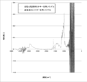
- a graph of the IR measurement results is shown in FIG. ⁇ IR measurement conditions> scan number: 32 times Wave number range: 400 to 4000 cm ⁇ 1 Resolution: 4cm -1 OPD velocity: 0.2cm / sec Measurement temperature: Room temperature Infrared incident angle to zinc selenide crystal: 45 °
- the peak at 1600-1500 cm ⁇ 1 is derived from the amide group hydrogen-bonded between the peptides, and corresponds to the content of ⁇ sheet structure in the gel. Therefore, it can be seen from the graph of FIG. 1 that the gel prepared using the rotation and revolution stirring apparatus has a more developed ⁇ sheet structure. Since the development of the ⁇ -sheet structure is advantageous for the formation of nanofibers and their development into a three-dimensional network structure, this result indicates that the mixing method using the above-mentioned predetermined stirring device is effective for the storage elasticity of the self-assembled peptide gel. This suggests that it contributes to the increase in the rate.
- the hemostatic material of the present invention can be suitably used in the medical field.
Landscapes
- Health & Medical Sciences (AREA)
- Life Sciences & Earth Sciences (AREA)
- Veterinary Medicine (AREA)
- Public Health (AREA)
- General Health & Medical Sciences (AREA)
- Animal Behavior & Ethology (AREA)
- Chemical & Material Sciences (AREA)
- Engineering & Computer Science (AREA)
- Epidemiology (AREA)
- Hematology (AREA)
- Materials Engineering (AREA)
- Organic Chemistry (AREA)
- Pharmacology & Pharmacy (AREA)
- Chemical Kinetics & Catalysis (AREA)
- General Chemical & Material Sciences (AREA)
- Diabetes (AREA)
- Bioinformatics & Cheminformatics (AREA)
- Nuclear Medicine, Radiotherapy & Molecular Imaging (AREA)
- Surgery (AREA)
- Medicinal Chemistry (AREA)
- Materials For Medical Uses (AREA)
- Peptides Or Proteins (AREA)
Abstract
自己組織化ペプチドを用いたゲル状止血材であって、従来よりも高強度な止血材を提供すること。自己組織化ペプチドを含む水溶液から形成されたゲルからなる止血材であって、該ゲルの37℃における回転式レオメーターによって測定される貯蔵弾性率が、350Pa~3000Paである、止血材。
Description
本発明は、自己組織化ペプチドを含む止血材に関する。
従来、手術等に用いられる止血材としては、シート状や2液混合型の液状または粘性液体状のものが多く用いられてきた。しかしながら、シート状の止血材は白色不透明であることが多く、出血点や止血の完了を目視で直接確認することが不可能であった。また、2液混合型のものは、混合後すぐに粘着性が非常に高くなるため、直ちに使用しなければならない、また、最初の止血で全量を使い切らなかった場合に、残りを用いて2回目の止血を行おうとしても、粘着性が増大しているために容器から出すことができない等、使用の面で問題があった。さらに、従来の止血材の多くは動物由来の材料を含むことから、感染の危険性が指摘されてきた。
これに対し、非動物由来の材料である自己組織化ペプチドを含む1液型で使用可能な止血材が提案されている(特許文献1)。当該止血材は、代表的には、処置対象である生物学的組織上に配置されることにより、自己組織化ペプチドが自己組織化してゲルとなり、止血機能を発揮する(特許文献1 0040段落、0051段落等)。
しかしながら、自己組織化ペプチドを用いたゲル状止血材の使用においては、力学的強度が不十分であるために、動脈性の出血等の激しい出血に対してゲルが部分的に押し流されてしまったり、崩壊してしまったりして、十分な止血機能が得られない場合があった。そのため、自己組織化ペプチドを用いたゲル状止血材が非生物由来の材料であることや、1液型で使用できるといったメリットを最大限に生かすことができていなかった。
本発明の止血材は、自己組織化ペプチドと水とを含み、該自己組織化ペプチドの自己組織化によって形成されたゲルからなる止血材であって、該ゲルの37℃における回転式レオメーターによって測定される貯蔵弾性率が、350Pa~3000Paである。
1つの実施形態において、上記自己組織化ペプチドの濃度が、1.0重量%~3.0重量%である
1つの実施形態において、上記止血材のpHが、5~8である。
1つの実施形態において、上記止血材の可視光透過率が80%以上である。
1つの実施形態において、上記自己組織化ペプチドを構成するアミノ酸残基のpH7.0における電荷の総和が、-3~-1または+1~+3である。
1つの実施形態において、上記止血材は、0.03重量%~0.5重量%の濃度でさらに強酸と塩基との塩を含む。
1つの実施形態において、上記自己組織化ペプチドの濃度が、1.0重量%~3.0重量%である
1つの実施形態において、上記止血材のpHが、5~8である。
1つの実施形態において、上記止血材の可視光透過率が80%以上である。
1つの実施形態において、上記自己組織化ペプチドを構成するアミノ酸残基のpH7.0における電荷の総和が、-3~-1または+1~+3である。
1つの実施形態において、上記止血材は、0.03重量%~0.5重量%の濃度でさらに強酸と塩基との塩を含む。
本発明によれば、従来よりも高強度な350Pa~3000Paの貯蔵弾性率を有する自己組織化ペプチドゲルからなる止血材が提供される。このような貯蔵弾性率を達成する方法は特に限定されないが、例えば、所定の撹拌装置の使用や、塩を添加することによって達成され得る。
[A.止血材]
本発明の止血材は、自己組織化ペプチドと水とを含み、該自己組織化ペプチドの自己組織化によって形成されたゲル(以下、「自己組織化ペプチドゲル」と称する)からなる。本明細書において、ゲルとは、粘性的な性質と弾性的な性質とを併せ持つ粘弾性物質を意味する。具体的には、動的粘弾性測定を行なって、貯蔵弾性率G’および損失弾性率G’’を測定したときに、「G’>G’’」となる物質をゲルということができる。
本発明の止血材は、自己組織化ペプチドと水とを含み、該自己組織化ペプチドの自己組織化によって形成されたゲル(以下、「自己組織化ペプチドゲル」と称する)からなる。本明細書において、ゲルとは、粘性的な性質と弾性的な性質とを併せ持つ粘弾性物質を意味する。具体的には、動的粘弾性測定を行なって、貯蔵弾性率G’および損失弾性率G’’を測定したときに、「G’>G’’」となる物質をゲルということができる。
本発明の止血材は、回転式レオメーターを用いた動的粘弾性測定における37℃での貯蔵弾性率(G’)が、350Pa~3000Paであり、好ましくは400Pa~2500Pa、より好ましくは450Pa~2200Pa、さらに好ましくは500Pa~1900Pa、さらにより好ましくは550Pa~1600Paである。貯蔵弾性率が350Pa未満であると、血流によってゲルが部分的に押し流されたり、破壊されてしまい、止血機能が十分に発揮されない場合や形状安定性が小さいために、単純に、出血部位からずり落ちる場合がある。一方、貯蔵弾性率が3000Paを超えると、シリンジ等の容器からの取り出しが困難である場合や出血部位の形状に沿って密着できない場合がある。なお、本明細書でいう貯蔵弾性率は、周波数変化測定を行ったときの角振動数が1ラジアン/秒であるときの値を意味する。
本発明の止血材は、好ましくは5~8、より好ましくは5.5~7.5、さらに好ましくは6~7のpHを有する。pHが当該範囲内であると、加熱時における自己組織化ペプチドの加水分解を回避し得るので、高圧蒸気滅菌などの加熱を伴う滅菌処理に供され得る。また、細胞障害性を低減することができる。さらには、自己組織化ペプチド間に本来意図される分子間相互作用を生じさせることができる。
本発明の止血材は、光路長10mmのセル中、380nm~780nmの吸光度で測定した可視光透過率が、好ましくは80%以上であり、より好ましくは85%以上であり、さらに好ましくは90%以上である。可視光透過率が当該範囲内であると、出血部位を確認しながら止血材を適用することや止血の完了を視認することが容易である。
本発明の止血材は、好ましくは200mosm/kg・H2O以上、より好ましくは230mosm/kg・H2O~400mosm/kg・H2Oの浸透圧を有する。浸透圧が当該範囲内であると、細胞障害性を低減することができる。
[A-1.自己組織化ペプチド]
自己組織化ペプチドとしては、水溶液中においてペプチド分子同士の相互作用を介して自発的に集合してゲルを形成し得る任意の適切なペプチドが用いられ得る。ペプチド分子同士の相互作用としては、例えば、水素結合、イオン間相互作用、ファンデルワールス力等の静電的相互作用および疎水性相互作用が挙げられる。
自己組織化ペプチドとしては、水溶液中においてペプチド分子同士の相互作用を介して自発的に集合してゲルを形成し得る任意の適切なペプチドが用いられ得る。ペプチド分子同士の相互作用としては、例えば、水素結合、イオン間相互作用、ファンデルワールス力等の静電的相互作用および疎水性相互作用が挙げられる。
自己組織化ペプチドを構成するアミノ酸は、L-アミノ酸であってもよく、D-アミノ酸であってもよい。好ましくはL-アミノ酸である。また、天然アミノ酸であってもよく、非天然アミノ酸であってもよい。低価格で入手可能であり、ペプチド合成が容易であることから、好ましくは天然アミノ酸である。
自己組織化ペプチドを構成するアミノ酸残基のpH7.0における電荷の総和は、好ましくは、-3~-1または+1~+3であり、より好ましくは、-3、-2、+2または+3である。このように、中性領域において自己組織化ペプチドに含まれるアミノ酸残基の側鎖に由来するプラス電荷とマイナス電荷とが相殺されないことにより、ゲル形成に適した静電的引力及び斥力のバランスが保たれ、その結果として、中性領域で透明かつ安定なゲルを形成し得るからである。なお、本明細書において、「中性領域」とは、6.0~8.5、好ましくは6.5~8.0、より好ましくは7.0の領域をいう。
各pHにおける上記自己組織化ペプチドの電荷は、例えば、レーニンジャー(Lehninger)〔Biochimie、1979〕の方法に従って算出され得る。レーニンジャーの方法は、例えば、EMBL WWW Gateway to Isoelectric Point Serviceのウェブサイト(http://www.embl-heidelberg.de/cgi/pi-wrapper.pl)上で利用可能なプログラムにより行われ得る。
本発明に好ましく使用され得る自己組織化ペプチドの具体例としては、下記の式(I)のアミノ酸配列からなるペプチドが挙げられる。
a1b1c1b2a2b3db4a3b5c2b6a4 (I)
(上記アミノ酸配列中、a1~a4は、塩基性アミノ酸残基であり;b1~b6は、非電荷極性アミノ酸残基および/または疎水性アミノ酸残基であり、ただし、そのうちの少なくとも5個は、疎水性アミノ酸残基であり;c1およびc2は、酸性アミノ酸残基であり;dは、疎水性アミノ酸残基である。)
a1b1c1b2a2b3db4a3b5c2b6a4 (I)
(上記アミノ酸配列中、a1~a4は、塩基性アミノ酸残基であり;b1~b6は、非電荷極性アミノ酸残基および/または疎水性アミノ酸残基であり、ただし、そのうちの少なくとも5個は、疎水性アミノ酸残基であり;c1およびc2は、酸性アミノ酸残基であり;dは、疎水性アミノ酸残基である。)
上記アミノ酸配列中、a1~a4は、塩基性アミノ酸残基である。塩基性アミノ酸は、好ましくはアルギニン、リシン、またはヒスチジンであり、より好ましくはアルギニンまたはリシンである。これらのアミノ酸は、塩基性が強いからである。a1~a4は、同一のアミノ酸残基であってもよく、異なるアミノ酸残基であってもよい。
上記アミノ酸配列中、b1~b6は、非電荷極性アミノ酸残基および/または疎水性アミノ酸残基であり、そのうちの少なくとも5個は、疎水性アミノ酸残基である。疎水性アミノ酸は、好ましくはアラニン、ロイシン、イソロイシン、バリン、メチオニン、フェニルアラニン、トリプトファン、グリシン、またはプロリンである。非電荷極性アミノ酸は、好ましくはチロシン、セリン、トレオニン、アスパラギン、グルタミン、またはシステインである。これらのアミノ酸は、入手が容易だからである。
好ましくは、b3およびb4は、それぞれ独立して任意の適切な疎水性アミノ酸残基であり、さらに好ましくはロイシン残基、アラニン残基、バリン残基、またはイソロイシン残基であり、特に好ましくはロイシン残基またはアラニン残基である。
好ましくは、b1~b6はすべて疎水性アミノ酸残基である。自己組織化ペプチドが好適にβシート構造を形成し、自己組織化し得るからである。より好ましくは、b1~b6は、それぞれ独立してロイシン残基、アラニン残基、バリン残基、またはイソロイシン残基であり、さらに好ましくはロイシン残基またはアラニン残基である。好ましい実施形態においては、b1~b6のうちの4個以上がロイシン残基であり、より好ましくはそのうちの5個以上がロイシン残基であり、さらに好ましくは全てがロイシン残基である。
上記アミノ酸配列中、c1およびc2は、酸性アミノ酸残基である。酸性アミノ酸は、好ましくはアスパラギン酸またはグルタミン酸である。これらのアミノ酸は、入手が容易だからである。c1およびc2は、同一のアミノ酸残基であってもよく、異なるアミノ酸残基であってもよい。
上記アミノ酸配列中、dは、疎水性アミノ酸残基である。dは、好ましくはアラニン残基、バリン残基、ロイシン残基、またはイソロイシン残基である。
1つの好ましい実施形態においては、b3、d、b4の連続する3つのアミノ酸残基のうち2つがロイシン残基であり、残りがアラニン残基である。この場合、b3、d、b4のいずれがアラニン残基であってもよい。また、別の好ましい実施形態においては、b3、d、b4の連続する3つのアミノ酸残基がすべてロイシン残基である。
式(I)のアミノ酸配列の好ましい具体例を以下に例示する。
n-RLDLRLALRLDLR-c(配列番号1)
n-RLDLRLLLRLDLR-c(配列番号2)
n-RADLRLALRLDLR-c(配列番号3)
n-RLDLRLALRLDAR-c(配列番号4)
n-RADLRLLLRLDLR-c(配列番号5)
n-RADLRLLLRLDAR-c(配列番号6)
n-RLDLRALLRLDLR-c(配列番号7)
n-RLDLRLLARLDLR-c(配列番号8)
n-RLDLRLALRLDLR-c(配列番号1)
n-RLDLRLLLRLDLR-c(配列番号2)
n-RADLRLALRLDLR-c(配列番号3)
n-RLDLRLALRLDAR-c(配列番号4)
n-RADLRLLLRLDLR-c(配列番号5)
n-RADLRLLLRLDAR-c(配列番号6)
n-RLDLRALLRLDLR-c(配列番号7)
n-RLDLRLLARLDLR-c(配列番号8)
本発明に好ましく使用され得る別の自己組織化ペプチドとしては、WO2007/000979に記載のペプチドが挙げられる。なかでも、以下に例示するアミノ酸配列からなる自己組織化ペプチドが好ましい。
n-RASARADARASARADA-c(配列番号9)
n-RANARADARANARADA-c(配列番号10)
n-RAAARADARAAARADA-c(配列番号11)
n-RASARADARADARASA-c(配列番号12)
n-RADARASARASARADA-c(配列番号13)
n-RASARASARASARADA-c(配列番号14)
n-RASARADARASA-c (配列番号15)
n-KASAKAEAKASAKAEA-c(配列番号16)
n-SAEAKAEASAEAKAEA-c(配列番号17)
n-KLSLKLDLKLSL-c (配列番号18)
n-KLALKLDLKLAL-c (配列番号19)
上記自己組織化ペプチドは、任意の適切な製造方法によって製造され得る。例えば、Fmoc法等の固相法又は液相法等の化学合成方法、遺伝子組換え発現等の分子生物学的方法が挙げられる。なお、自己組織化ペプチドは、精製の過程で任意の適切な塩の形態とされ得るが、本発明においては、塩形態の自己組織化ペプチドを用いることもできる。ただし、本発明において、塩形態の自己組織化ペプチドは、A-3項に記載の塩には含まれないものとする。
上記自己組織化ペプチドは、目的等に応じて任意の適切な修飾が施されていてもよい。修飾が行われる部位は、特に限定されず、例えば、自己組織化ペプチドのN末端アミノ基、C末端カルボキシル基、またはその両方が挙げられる。
上記修飾としては、修飾後のペプチドが自己組織化能を有する範囲において任意の適切な修飾が選択され得る。例えば、N末端アミノ基のアセチル化、C末端カルボキシル基のアミド化等の保護基の導入;アルキル化、エステル化、またはハロゲン化等の官能基の導入;水素添加;単糖、二糖、オリゴ糖、または多糖等の糖化合物の導入;脂肪酸、リン脂質、または糖脂質等の脂質化合物の導入;アミノ酸またはタンパク質の導入;DNAの導入;その他生理活性を有する化合物等の導入が挙げられる。修飾は1種のみ行われてもよく、2種以上を組み合わせて行ってもよい。例えば、上記自己組織化ペプチドのC末端に所望のアミノ酸を導入した付加ペプチドのN末端をアセチル化し、C末端をアミド化してもよい。
アミノ酸またはタンパク質が導入される場合、導入されるアミノ酸の数は、好ましくは1~180であり、より好ましくは1~50、さらに好ましくは1~30、特に好ましくは1~10、最も好ましくは1~5である。導入するアミノ酸残基数が180を超えると、自己組織化能が損なわれる場合がある。
本発明の止血材における自己組織化ペプチドの濃度は、所望の貯蔵弾性率等に応じて適切に設定され得る。自己組織化ペプチドの濃度は、好ましくは1.0重量%~3.0重量%、より好ましくは1.05重量%~2.8重量%、さらに好ましくは1.1重量%~2.5重量%、さらにより好ましくは1.15重量%~2.0重量%、さらにより好ましくは1.2重量%~1.8重量%である。本発明においてはゲルを製造する方法に特に限定はないが、後述するように、たとえば、所定の撹拌装置の使用および/または塩を添加する方法により、3.0重量%以下の自己組織化ペプチド濃度であっても350Pa以上の貯蔵弾性率を有する止血材(実質的には、自己組織化ペプチドゲル)が得られ得る。
[A-2.水]
水としては、イオン交換水、蒸留水等の精製された水が好ましく用いられ得る。
水としては、イオン交換水、蒸留水等の精製された水が好ましく用いられ得る。
本発明の止血材の含水率(%)(=止血材中の水の重量/止血材の総重量×100)は、例えば85%~99%、好ましくは86%~98%、より好ましくは87%~95%である。
[A-3.塩]
本発明の止血材は、貯蔵弾性率を調整するために塩をさらに含み得る。具体的には、所定の濃度の塩を含むことにより、単純に塩を含まない場合と比較すると、止血材(実質的には、自己組織化ペプチドゲル)の貯蔵弾性率が増加し、所望の貯蔵弾性率を獲得し得る。このような効果が奏される理由は定かではないが、以下のように推測される。すなわち、プラスに帯電したペプチドの場合、塩が水溶液中で解離することによりマイナス電荷を有するアニオンが生じ、該アニオンが正電荷アミノ酸にカウンターイオンとして結合することで、プラス電荷を遮蔽するようになる。その結果、ペプチド間の斥力の過剰分が減殺されて、ナノファイバー同士の結合がより強固になるためにゲルの貯蔵弾性率が350Pa~3000Paの範囲内に調整可能となると推測される。
本発明の止血材は、貯蔵弾性率を調整するために塩をさらに含み得る。具体的には、所定の濃度の塩を含むことにより、単純に塩を含まない場合と比較すると、止血材(実質的には、自己組織化ペプチドゲル)の貯蔵弾性率が増加し、所望の貯蔵弾性率を獲得し得る。このような効果が奏される理由は定かではないが、以下のように推測される。すなわち、プラスに帯電したペプチドの場合、塩が水溶液中で解離することによりマイナス電荷を有するアニオンが生じ、該アニオンが正電荷アミノ酸にカウンターイオンとして結合することで、プラス電荷を遮蔽するようになる。その結果、ペプチド間の斥力の過剰分が減殺されて、ナノファイバー同士の結合がより強固になるためにゲルの貯蔵弾性率が350Pa~3000Paの範囲内に調整可能となると推測される。
本発明において、貯蔵弾性率を調整するために使用される塩は、強酸(例えば、酸解離指数pKが3以下のもの)と塩基との塩である。塩基は強塩基であってもよく、弱塩基であってもよい。止血材のpHに影響を与えない観点からは、好ましくは強酸と強塩基とからなる中性塩が用いられる。貯蔵弾性率を調整するために使用可能な塩の好ましい具体例としては、塩化ナトリウム、塩化カリウム、塩化カルシウム等の塩酸塩、硫酸ナトリウム、硫酸マグネシウム等の硫酸塩が挙げられる。塩は、単独で用いられてもよく、二種以上を組み合わせて用いられてもよい。
本発明の止血材における塩の濃度は、好ましくは0.03重量%~0.5重量%、より好ましくは0.04重量%~0.4重量%、さらに好ましくは0.05重量%~0.3重量%である。塩濃度が0.5重量%を超えると、自己組織化ペプチドの電荷が塩由来のイオンによって相殺されてしまい、自己組織化能が低下する結果、ゲルが所望の貯蔵弾性率を有しない、または、透明性が低下する等の場合がある。塩濃度が0.03重量%未満であると、貯蔵弾性率の増加効果が得られない場合がある。
[A-4.添加物]
本発明の止血材は、必要に応じて任意の適切な添加物をさらに含み得る。例えば、本発明の止血材は、好ましくはpH調整剤を含む。
本発明の止血材は、必要に応じて任意の適切な添加物をさらに含み得る。例えば、本発明の止血材は、好ましくはpH調整剤を含む。
pH調整剤としては、塩酸、クエン酸、酢酸等の酸;水酸化ナトリウム、水酸化カリウム等の塩基;炭酸水素ナトリウム、炭酸ナトリウム等の弱酸と強塩基との塩が挙げられる。
pH調整剤の添加量は、所望のpHに応じて適切に設定され得る。本発明において、pH調整剤は、自己組織化ペプチド水溶液を中和して自己組織化ペプチド間に本来意図される分子間相互作用を生じさせるために添加されるものである。よって、pH調整剤として強酸と塩基との塩を用いる場合であっても、上記好適範囲外であった自己組織化ペプチド水溶液のpHを好適範囲内に調整するために添加される塩はpH調整剤であって、貯蔵弾性率の調整の役割を担うものとは考えない。
本発明の止血材が含み得る他の添加物としては、例えば、緩衝剤;等張化剤;アミノ酸類;ビタミン類;アルコール類;薬物等が挙げられる。これらの他の添加物は、単独で用いられてもよく、二種以上を組み合わせて用いられてもよい。
緩衝剤としては、リン酸、リン酸ナトリウム、リン酸二水素ナトリウム、リン酸水素二ナトリウム、リン酸カリウム、リン酸二水素カリウム、リン酸水素二カリウムなどのリン酸塩;ホウ酸、ホウ砂、ホウ酸ナトリウム、ホウ酸カリウムなどのホウ酸塩;クエン酸ナトリウム、クエン酸二ナトリウムなどのクエン酸塩;酢酸ナトリウム、酢酸カリウムなどの酢酸塩、Tris、HEPES等が挙げられる。
等張化剤としては、塩化ナトリウム、塩化カリウム、塩化カルシウム、塩化マグネシウム等の塩化物;グルコース、フルクトース、ガラクトース等の単糖;スクロース、トレハロース、マルトース、ラクトース等の二糖;マンニトール、ソルビトール等の糖アルコール;等が挙げられる。
上記他の添加物の添加量は、その目的等に応じて任意の適切な値に設定され得る。他の添加物として強酸と塩基との塩を用いる場合、当該塩もゲルの貯蔵弾性率の増加要因となるため、A-3項に記載の貯蔵弾性率を調整するために使用する塩に該当する。よって、それら、強酸と塩基との塩を用いる場合には、当該塩濃度(二種以上の塩を用いる場合はその合計濃度)が0.03重量%~0.5重量%となるように添加量を調整することが好ましく、より好ましくは0.04重量%~0.4重量%、さらに好ましくは0.05重量%~0.3重量%となるように調整される。
[B.止血材の製造方法]
本発明の止血材の製造方法は、代表的には、自己組織化ペプチドと水と任意に塩および/または添加物とを混合して自己組織化ペプチド水溶液を得ること(混合工程)、および、得られた水溶液を静置することにより自己組織化ペプチドを自己組織化させて自己組織化ペプチドゲルを得ること(ゲル化工程)を含む。必要に応じて、混合工程の後であってゲル化工程の前に、自己組織化ペプチド水溶液を脱泡および/または脱気すること(脱泡工程)をさらに含み得る。
本発明の止血材の製造方法は、代表的には、自己組織化ペプチドと水と任意に塩および/または添加物とを混合して自己組織化ペプチド水溶液を得ること(混合工程)、および、得られた水溶液を静置することにより自己組織化ペプチドを自己組織化させて自己組織化ペプチドゲルを得ること(ゲル化工程)を含む。必要に応じて、混合工程の後であってゲル化工程の前に、自己組織化ペプチド水溶液を脱泡および/または脱気すること(脱泡工程)をさらに含み得る。
混合工程においては、任意の適切な混合方法が用いられ得る。例えば、超音波照射を用いた混合方法、遠心力を用いた混合方法、機械的撹拌を用いた混合方法が挙げられる。混合手段として自転公転撹拌装置を用いることにより、上記所望の貯蔵弾性率が好適に達成され得る。
混合工程においては、2種以上の異なる混合方法を組み合わせて用いてもよい。また、同じ混合方法による混合を2回以上繰り返してもよい。
ゲル化工程において、自己組織化ペプチド水溶液を静置する時間は、通常1分以上、好ましくは3分以上、より好ましくは5分以上である。静置する際の温度は、通常4~50℃、好ましくは15~45℃である。
脱泡工程においては、超音波脱気、真空減圧脱気、遠心脱気等の任意の適切な脱泡方法が用いられ得る。二種以上の脱泡方法を組み合わせて行ってもよい。
本発明の止血材の製造方法は、必要に応じて任意の適切な他の工程を含み得る。当該他の工程としては、ろ過等の精製工程、高圧蒸気滅菌、放射線滅菌、乾熱滅菌等の滅菌工程、包装容器への分注工程等が挙げられる。
[C.止血材の使用方法]
本発明の止血材は、止血材単独で出血部位に適用されてもよく、任意の適切な支持体(例えば、透明フィルム)に塗布されて止血フィルムとして出血部位に貼付されてもよい。止血材単独で出血部位に適用する方法としては、シリンジ内に配置された止血材をプランジャで押し出して出血部位に配置する方法、チューブや袋などの容器内に配置された止血材をヘラ等の塗布用の器具に移してから出血部位に塗布する方法等が挙げられる。本発明の止血材は、出血部位を圧迫して止血し得る十分な貯蔵弾性率を維持しつつ、出血部位の形状に沿って密着し得る柔軟性を有するので、止血性に優れる。
本発明の止血材は、止血材単独で出血部位に適用されてもよく、任意の適切な支持体(例えば、透明フィルム)に塗布されて止血フィルムとして出血部位に貼付されてもよい。止血材単独で出血部位に適用する方法としては、シリンジ内に配置された止血材をプランジャで押し出して出血部位に配置する方法、チューブや袋などの容器内に配置された止血材をヘラ等の塗布用の器具に移してから出血部位に塗布する方法等が挙げられる。本発明の止血材は、出血部位を圧迫して止血し得る十分な貯蔵弾性率を維持しつつ、出血部位の形状に沿って密着し得る柔軟性を有するので、止血性に優れる。
出血の程度にも依るが、本発明の止血材は、例えば、出血部位に配置されてから20秒以内、好ましくは15秒以内、より好ましくは10秒以内に止血を完了することができる。
以下、実施例によって本発明を具体的に説明するが、本発明はこれら実施例によって限定されるものではない。
[貯蔵弾性率]
動的粘弾性測定装置である回転式レオメーター(TA instruments社製、製品名「ADVANCED RHEOMETER AR 1000」)を用いて、止血材の貯蔵弾性率G’を測定した。具体的には、以下のとおりである。まず、試料に接触させるためのジオメトリー(アルミニウム製コーン、直径20mm、コーン角1°、ギャップ24μm)と試料台を一定の温度に保つための恒温槽とをレオメーターに取り付けた。次いで、温度:37℃、トルク:1μN・m、周波数:0.5rad/s~100rad/sの測定条件下、以下の測定手順で1つの試料について3回測定し、1rad/sのときの値の平均値を貯蔵弾性率G’とした。
(1)ピペットを用いて55μLの試料をレオメーターの試料台に配置する。
(2)ジオメトリーを試料台から24μmのギャップまで移動させ、試料と接触させる。
(3)ジオメトリーをわずかに動かし、試料となじませる。
(4)ジオメトリーの動きを止めてから15秒後(この15秒の間に、試料からの溶媒の揮発を防止するためのソルベントトラップを取り付ける)に、測定を開始する。
[pH]
pHは、ポータブルpHメーター(堀場製作所社製、製品番号「B-712」)を用いて測定した。
pHは、ポータブルpHメーター(堀場製作所社製、製品番号「B-712」)を用いて測定した。
[外観]
得られた止血材の外観を目視で観察し、以下の基準で評価した。
良好:気泡および白濁が認められず、透明である。
不良:気泡および/または白濁が認められる。
得られた止血材の外観を目視で観察し、以下の基準で評価した。
良好:気泡および白濁が認められず、透明である。
不良:気泡および/または白濁が認められる。
[実施例1]
表1に示す組成の自己組織化ペプチドゲルを調製し、止血材1とした。具体的な手順は以下のとおりである。すなわち、自己組織化ペプチド(SPG-178:N末端がアセチル化され、C末端がアミド化された配列番号1のペプチド)およびトレハロース・二水和物を容器に秤り入れた。次いで、該容器に水を投入し、蓋をした。該容器を自転公転撹拌装置(株式会社シンキー製、製品番号「ARE-310」)にセットし、撹拌した。
表1に示す組成の自己組織化ペプチドゲルを調製し、止血材1とした。具体的な手順は以下のとおりである。すなわち、自己組織化ペプチド(SPG-178:N末端がアセチル化され、C末端がアミド化された配列番号1のペプチド)およびトレハロース・二水和物を容器に秤り入れた。次いで、該容器に水を投入し、蓋をした。該容器を自転公転撹拌装置(株式会社シンキー製、製品番号「ARE-310」)にセットし、撹拌した。
得られた混合液に100mM Na2CO3水溶液を添加してpHを調整した後、自転公転撹拌装置(株式会社シンキー製、製品番号「ARE-310」)にて撹拌した。
得られた溶液を自転公転撹拌装置(株式会社シンキー製、製品番号「ARE-310」)にて脱泡および脱気して自己組織化ペプチド水溶液を得た。
得られた自己組織化ペプチド水溶液を5gずつシリンジに分注した。次いで、シリンジに打栓し、プランジャを装着して、滅菌バッグに入れ、121℃で20分オートクレーブした。オートクレーブ後の自己組織化ペプチド水溶液を常温まで自然冷却し、自己組織化ペプチドゲルを得た。
[実施例2]
表1に示す組成の自己組織化ペプチドゲルを調製し、止血材2とした。具体的な手順は以下のとおりである。すなわち、自己組織化ペプチド(SPG-178)および塩化ナトリウムを秤量し、遠沈管に投入した。次いで、水を投入し、超音波型ホモジナイザー(Sonic&Material,Inc.社製、製品番号「Vibra-Cell VC-505」)を用いて500Wの出力で5分処理して混合した。
表1に示す組成の自己組織化ペプチドゲルを調製し、止血材2とした。具体的な手順は以下のとおりである。すなわち、自己組織化ペプチド(SPG-178)および塩化ナトリウムを秤量し、遠沈管に投入した。次いで、水を投入し、超音波型ホモジナイザー(Sonic&Material,Inc.社製、製品番号「Vibra-Cell VC-505」)を用いて500Wの出力で5分処理して混合した。
得られた混合液を3000rpmで5分遠心分離して脱泡し、さらにアスピレーターを用いて減圧脱気した。脱気後、混合液に50mM Na2CO3水溶液を添加してpHを調整した後、超音波型ホモジナイザー(Sonic&Material,Inc.社製、製品番号「Vibra-Cell VC-505」)を用いて500Wの出力で5分処理して混合した。アスピレーターを用いて混合液を減圧脱気し、これにより、自己組織化ペプチド水溶液を得た。
得られた自己組織化ペプチド水溶液を5gずつシリンジに分注した。次いで、シリンジに打栓し、プランジャを装着して、滅菌バッグに入れ、121℃で20分オートクレーブした。オートクレーブ後の自己組織化ペプチド水溶液を常温まで自然冷却し、自己組織化ペプチドゲルを得た。
[実施例3]
自己組織化ペプチドと塩化ナトリウムに加えてスクロースも遠沈管に投入したこと以外は実施例2と同様の手順で表1に示す組成の自己組織化ペプチドゲルを調製し、止血材3とした。
自己組織化ペプチドと塩化ナトリウムに加えてスクロースも遠沈管に投入したこと以外は実施例2と同様の手順で表1に示す組成の自己組織化ペプチドゲルを調製し、止血材3とした。
[実施例4]
自己組織化ペプチドとして、SPG-220(N末端がアセチル化され、C末端がアミド化された配列番号18のペプチド)を用いたこと以外は実施例1と同様の手順で表1に示す組成の自己組織化ペプチドゲルを調製し、止血材4とした。
自己組織化ペプチドとして、SPG-220(N末端がアセチル化され、C末端がアミド化された配列番号18のペプチド)を用いたこと以外は実施例1と同様の手順で表1に示す組成の自己組織化ペプチドゲルを調製し、止血材4とした。
[比較例1]
塩化ナトリウムを用いずに、自己組織化ペプチドとスクロースとを遠沈管に投入したこと以外は実施例2と同様の手順で表1に示す組成の自己組織化ペプチドゲルを調製し、止血材C1とした。
塩化ナトリウムを用いずに、自己組織化ペプチドとスクロースとを遠沈管に投入したこと以外は実施例2と同様の手順で表1に示す組成の自己組織化ペプチドゲルを調製し、止血材C1とした。
[比較例2]
実施例2と同様の手順で表1に示す組成の自己組織化ペプチドゲルを調製し、止血材C2とした。
実施例2と同様の手順で表1に示す組成の自己組織化ペプチドゲルを調製し、止血材C2とした。
[比較例3]
塩化ナトリウムを用いなかったこと以外は実施例2と同様の手順で表1に示す組成の自己組織化ペプチドゲルを調製し、止血材C3とした。
塩化ナトリウムを用いなかったこと以外は実施例2と同様の手順で表1に示す組成の自己組織化ペプチドゲルを調製し、止血材C3とした。
実施例および比較例で得られた止血材の組成および特性を表1に示す。

上記のとおり、所定の撹拌装置を用いることにより、あるいは、所定の濃度で塩を添加することにより、実施例の止血材は全て、350Pa以上の貯蔵弾性率を有している。一方、同じ自己組織化ペプチド濃度を有する比較例の止血材では、貯蔵弾性率が低すぎたり、外観に白濁の不良が生じたりしている。
[試験例1]
実施例1で得られた止血材1を用いて止血効果を評価した。具体的には、モルモットの肝臓をメスで5mm程度切開し、切開口に実施例1で得られた自己組織化ペプチドゲルを配置した。その結果、止血材を出血部位に配置してから10秒以内に止血が完了した。なお、止血材が透明であったために、出血部位を確認しながらその上に止血材を容易に配置することができた。また、出血部位に配置された止血材を介して目視で止血の完了を確認することができた。
実施例1で得られた止血材1を用いて止血効果を評価した。具体的には、モルモットの肝臓をメスで5mm程度切開し、切開口に実施例1で得られた自己組織化ペプチドゲルを配置した。その結果、止血材を出血部位に配置してから10秒以内に止血が完了した。なお、止血材が透明であったために、出血部位を確認しながらその上に止血材を容易に配置することができた。また、出血部位に配置された止血材を介して目視で止血の完了を確認することができた。
[参考例1]
自転公転撹拌装置または超音波ホモジナイザーを用いて、表2に記載の組成の自己組織化ペプチド水溶液を調製し、該水溶液から自己組織化ペプチドゲルを形成させた。自転公転撹拌装置および超音波ホモジナイザーを用いた自己組織化ペプチド水溶液およびゲルの調製方法および条件はそれぞれ、実施例1および実施例2と同様とした。得られた自己組織化ペプチドゲルに関して、HATRサンプリングアクセサリーとセレン化亜鉛結晶からなるトラフプレートを設置したフーリエ変換赤外分光光度計(パーキンエルマー社製、製品名「Spectrum One」)を用いて全反射吸収法にて以下の条件でIR測定を行った。IR測定結果のグラフを図1に示す。
<IR測定条件>
scan number:32回
波数の範囲:400~4000cm-1
分解能:4cm-1
OPD velocity:0.2cm/sec
測定温度:室温
セレン化亜鉛結晶への赤外線入射角:45°

図1に示すグラフにおいて、1600~1500cm-1のピークはペプチド間で水素結合したアミド基由来であり、ゲル内におけるβシート構造の含有量に対応する。よって、図1のグラフからは、自転公転撹拌装置を用いて調製されたゲルの方が、βシート構造がより発達した構造を有することがわかる。βシート構造の発達は、ナノファイバーの形成およびその三次元網目構造への発展に有利であることから、この結果は、上記所定の撹拌装置を用いた混合方法が自己組織化ペプチドゲルの貯蔵弾性率の増加に寄与していることを示唆するものである。
本発明の止血材は、医療の分野において好適に利用され得る。
Claims (6)
- 自己組織化ペプチドと水とを含み、該自己組織化ペプチドの自己組織化によって形成されたゲルからなる止血材であって、
該ゲルの37℃における回転式レオメーターによって測定される貯蔵弾性率が、350Pa~3000Paである、止血材。 - 前記自己組織化ペプチドの濃度が、1.0重量%~3.0重量%である、請求項1に記載の止血材。
- pHが、5~8である、請求項1または2に記載の止血材。
- 可視光透過率が80%以上である、請求項1から3のいずれかに記載の止血材。
- 前記自己組織化ペプチドを構成するアミノ酸残基のpH7.0における電荷の総和が、-3~-1または+1~+3である、請求項1から4のいずれかに記載の止血材。
- 0.03重量%~0.5重量%の濃度でさらに強酸と塩基との塩を含む、請求項1から5のいずれかに記載の止血材。
Applications Claiming Priority (4)
| Application Number | Priority Date | Filing Date | Title |
|---|---|---|---|
| JP2013-271430 | 2013-12-27 | ||
| JP2013271430 | 2013-12-27 | ||
| JP2014-225283 | 2014-11-05 | ||
| JP2014225283A JP2015142720A (ja) | 2013-12-27 | 2014-11-05 | 止血材 |
Publications (1)
| Publication Number | Publication Date |
|---|---|
| WO2015098515A1 true WO2015098515A1 (ja) | 2015-07-02 |
Family
ID=53478379
Family Applications (1)
| Application Number | Title | Priority Date | Filing Date |
|---|---|---|---|
| PCT/JP2014/082653 WO2015098515A1 (ja) | 2013-12-27 | 2014-12-10 | 止血材 |
Country Status (2)
| Country | Link |
|---|---|
| JP (1) | JP2015142720A (ja) |
| WO (1) | WO2015098515A1 (ja) |
Cited By (2)
| Publication number | Priority date | Publication date | Assignee | Title |
|---|---|---|---|---|
| WO2016006693A1 (ja) * | 2014-07-11 | 2016-01-14 | 株式会社メニコン | 視認性確保材および視認性確保材吐出装置 |
| WO2017104723A1 (ja) * | 2015-12-18 | 2017-06-22 | 株式会社スリー・ディー・マトリックス | 止血用組成物、および、止血用組成物を用いた止血方法 |
Citations (4)
| Publication number | Priority date | Publication date | Assignee | Title |
|---|---|---|---|---|
| WO2007000979A1 (ja) * | 2005-06-27 | 2007-01-04 | Menicon Co., Ltd. | 自己組織化ペプチドおよびそれより得られるゲル |
| JP2008539257A (ja) * | 2005-04-25 | 2008-11-13 | マサチューセッツ・インスティテュート・オブ・テクノロジー | 止血および他の生理学的活性を促進するための組成物および方法 |
| WO2010041636A1 (ja) * | 2008-10-06 | 2010-04-15 | 株式会社スリー・ディー・マトリックス | 組織閉塞剤 |
| JP2012131757A (ja) * | 2010-12-24 | 2012-07-12 | Okayama Univ | 高強度ペプチドゲル |
-
2014
- 2014-11-05 JP JP2014225283A patent/JP2015142720A/ja active Pending
- 2014-12-10 WO PCT/JP2014/082653 patent/WO2015098515A1/ja active Application Filing
Patent Citations (4)
| Publication number | Priority date | Publication date | Assignee | Title |
|---|---|---|---|---|
| JP2008539257A (ja) * | 2005-04-25 | 2008-11-13 | マサチューセッツ・インスティテュート・オブ・テクノロジー | 止血および他の生理学的活性を促進するための組成物および方法 |
| WO2007000979A1 (ja) * | 2005-06-27 | 2007-01-04 | Menicon Co., Ltd. | 自己組織化ペプチドおよびそれより得られるゲル |
| WO2010041636A1 (ja) * | 2008-10-06 | 2010-04-15 | 株式会社スリー・ディー・マトリックス | 組織閉塞剤 |
| JP2012131757A (ja) * | 2010-12-24 | 2012-07-12 | Okayama Univ | 高強度ペプチドゲル |
Non-Patent Citations (3)
| Title |
|---|
| KOMATSU,S. ET AL.: "The Neutral Self-Assembling Peptide Hydrogel SPG-178 as a Topical Hemostatic Agent", PLOS ONE, vol. 9, no. 7, 21 July 2014 (2014-07-21), pages 1 - 8, Retrieved from the Internet <URL:http://journals.plos.org/prosone/article?id=10.1371/journal.pone.0102778> [retrieved on 20150305] * |
| LUO,Z. ET AL.: "Fabrication of self-assembling D-form peptide nanofiber scaffold d-EAK16 for rapid hemostasis", BIOMATERIALS, vol. 32, no. 8, March 2011 (2011-03-01), pages 2013 - 2020 * |
| SONG,H. ET AL.: "Hemostatic Efficacy of Biological Self-Assembling Peptide Nanofibers in a Rat Kidney Model", MACROMOL. BIOSCI., vol. 10, no. 1, January 2010 (2010-01-01), pages 33 - 39 * |
Cited By (7)
| Publication number | Priority date | Publication date | Assignee | Title |
|---|---|---|---|---|
| WO2016006693A1 (ja) * | 2014-07-11 | 2016-01-14 | 株式会社メニコン | 視認性確保材および視認性確保材吐出装置 |
| AU2015288635B2 (en) * | 2014-07-11 | 2018-07-26 | Menicon Co., Ltd. | Visibility-ensuring material, and device for discharging visibility-ensuring material |
| WO2017104723A1 (ja) * | 2015-12-18 | 2017-06-22 | 株式会社スリー・ディー・マトリックス | 止血用組成物、および、止血用組成物を用いた止血方法 |
| JPWO2017104723A1 (ja) * | 2015-12-18 | 2018-10-04 | 株式会社スリー・ディー・マトリックス | 止血用組成物、および、止血用組成物を用いた止血方法 |
| CN108697755A (zh) * | 2015-12-18 | 2018-10-23 | 立美基股份有限公司 | 止血组合物和使用止血组合物的止血方法 |
| AU2016374425B2 (en) * | 2015-12-18 | 2021-10-21 | 3-D Matrix, Ltd. | Hemostatic composition and hemostatic method using hemostatic composition |
| US11298397B2 (en) | 2015-12-18 | 2022-04-12 | 3-D Matrix, Ltd. | Hemostatic composition and hemostatic method using hemostatic composition |
Also Published As
| Publication number | Publication date |
|---|---|
| JP2015142720A (ja) | 2015-08-06 |
Similar Documents
| Publication | Publication Date | Title |
|---|---|---|
| Pandey et al. | Mussel-inspired bioadhesives in healthcare: design parameters, current trends, and future perspectives | |
| Zhou et al. | Dopamine-modified hyaluronic acid hydrogel adhesives with fast-forming and high tissue adhesion | |
| Choi et al. | Human gelatin tissue-adhesive hydrogels prepared by enzyme-mediated biosynthesis of DOPA and Fe 3+ ion crosslinking | |
| Lu et al. | Mussel-inspired thermoresponsive polypeptide–pluronic copolymers for versatile surgical adhesives and hemostasis | |
| JP2015131845A (ja) | 自己組織化ペプチドおよび高強度ペプチドゲル | |
| Ryu et al. | Catechol-functionalized chitosan/pluronic hydrogels for tissue adhesives and hemostatic materials | |
| Huang et al. | Degradable and bioadhesive alginate-based composites: an effective hemostatic agent | |
| US8299032B2 (en) | Self-assembling peptide and gel produced from the same | |
| CA2853356C (en) | Hemostatic compositions | |
| RU2584348C2 (ru) | Композиционная коллагеновая губка и способ ее изготовления | |
| JP2019052169A (ja) | (ポリ)ペプチドのアンフォールディングを防止し、および/または(ポリ)ペプチドの(リ)フォールディングを誘導するための方法 | |
| WO2015098515A1 (ja) | 止血材 | |
| JP6490897B2 (ja) | 自己組織化ペプチドゲル | |
| JP2022046711A (ja) | 架橋化ヒアルロン酸誘導体マトリックスが含まれている止血組成物 | |
| JP6081670B2 (ja) | 視認性確保材および視認性確保材吐出装置 | |
| WO2020137903A1 (ja) | 粉体、創傷被覆材、癒着防止材、止血材、及び紛体の製造方法 | |
| RU2740185C2 (ru) | Пептидная композиция | |
| Kanayama et al. | Development of low endotoxin gelatin for regenerative medicine | |
| JP6877360B2 (ja) | 止血組成物 | |
| WO2021171846A1 (ja) | 消化液漏出防止材および消化液による消化からの器官保護材 | |
| EP3191133A1 (en) | Bioresorbable tissue repair composition | |
| Reichsöllner et al. | A comparative high‐resolution physicochemical analysis of commercially available fibrin sealants: Impact of sealant osmolality on biological performance | |
| Lee et al. | Injectable Click Fibroin Bioadhesive Derived from Spider Silk for Accelerating Wound Closure and Healing Bone Fracture. Materials 2022, 15, 5269 | |
| Ma et al. | Biomedical Applications of Ultra-Strong Bio-Glue from Genetically Engineered Polypeptides | |
| Kusunoki et al. | Priming effects of triethylene glycol and triethylene glycol monomethacrylate on dentin bonding |
Legal Events
| Date | Code | Title | Description |
|---|---|---|---|
| 121 | Ep: the epo has been informed by wipo that ep was designated in this application |
Ref document number: 14875622 Country of ref document: EP Kind code of ref document: A1 |
|
| NENP | Non-entry into the national phase |
Ref country code: DE |
|
| 122 | Ep: pct application non-entry in european phase |
Ref document number: 14875622 Country of ref document: EP Kind code of ref document: A1 |