WO2010071114A1 - 血液試料中の癌細胞の検出方法 - Google Patents
血液試料中の癌細胞の検出方法 Download PDFInfo
- Publication number
- WO2010071114A1 WO2010071114A1 PCT/JP2009/070843 JP2009070843W WO2010071114A1 WO 2010071114 A1 WO2010071114 A1 WO 2010071114A1 JP 2009070843 W JP2009070843 W JP 2009070843W WO 2010071114 A1 WO2010071114 A1 WO 2010071114A1
- Authority
- WO
- WIPO (PCT)
- Prior art keywords
- blood sample
- oncolytic virus
- nonionic surfactant
- cancer cells
- cells
- Prior art date
- Legal status (The legal status is an assumption and is not a legal conclusion. Google has not performed a legal analysis and makes no representation as to the accuracy of the status listed.)
- Ceased
Links
Images




Classifications
-
- G—PHYSICS
- G01—MEASURING; TESTING
- G01N—INVESTIGATING OR ANALYSING MATERIALS BY DETERMINING THEIR CHEMICAL OR PHYSICAL PROPERTIES
- G01N33/00—Investigating or analysing materials by specific methods not covered by groups G01N1/00 - G01N31/00
- G01N33/48—Biological material, e.g. blood, urine; Haemocytometers
- G01N33/50—Chemical analysis of biological material, e.g. blood, urine; Testing involving biospecific ligand binding methods; Immunological testing
- G01N33/53—Immunoassay; Biospecific binding assay; Materials therefor
- G01N33/575—Immunoassay; Biospecific binding assay; Materials therefor for cancer
-
- C—CHEMISTRY; METALLURGY
- C12—BIOCHEMISTRY; BEER; SPIRITS; WINE; VINEGAR; MICROBIOLOGY; ENZYMOLOGY; MUTATION OR GENETIC ENGINEERING
- C12N—MICROORGANISMS OR ENZYMES; COMPOSITIONS THEREOF; PROPAGATING, PRESERVING, OR MAINTAINING MICROORGANISMS; MUTATION OR GENETIC ENGINEERING; CULTURE MEDIA
- C12N7/00—Viruses; Bacteriophages; Compositions thereof; Preparation or purification thereof
-
- G—PHYSICS
- G01—MEASURING; TESTING
- G01N—INVESTIGATING OR ANALYSING MATERIALS BY DETERMINING THEIR CHEMICAL OR PHYSICAL PROPERTIES
- G01N33/00—Investigating or analysing materials by specific methods not covered by groups G01N1/00 - G01N31/00
- G01N33/48—Biological material, e.g. blood, urine; Haemocytometers
- G01N33/50—Chemical analysis of biological material, e.g. blood, urine; Testing involving biospecific ligand binding methods; Immunological testing
- G01N33/68—Chemical analysis of biological material, e.g. blood, urine; Testing involving biospecific ligand binding methods; Immunological testing involving proteins, peptides or amino acids
Definitions
- the present invention relates to a method for detecting cancer cells in a blood sample.
- the present invention also relates to a reagent kit that can be used in the method.
- Cancer metastasis is considered to occur when cancer cells in the primary lesion spread throughout the body through blood vessels and lymphatic vessels, and some of the cells engraft in other organs. Cancer cells circulating in the blood in this way are called CTC (Circulating Tumor Cell). The number of CTCs in the blood has been reported to correlate with cancer metastasis and prognosis, and measurement of CTC counts is useful for predicting the prognosis and therapeutic effects of metastatic cancers, such as metastatic breast cancer. It is considered.
- Patent Document 1 Japanese Translation of PCT International Publication No. 2002-503814
- telomere activity is enhanced in cancer cells, but the activity is hardly detected in most normal cells.
- a virus Oncolytic Virus
- a replication cassette containing a telomerase promoter and a labeled expression cassette containing a labeled protein (eg, green fluorescent protein (GFP)) gene is produced in cancer cells.
- GFP green fluorescent protein
- An oncolytic virus having a GFP gene is commercially available as Telomescan (registered trademark) (OBP-401). Telomescan (registered trademark) can specifically proliferate in cancer cells and produce GFP, thereby allowing the cancer cells to specifically fluoresce.
- Oncolytic viruses such as Telomescan® have a low infection / proliferation rate to normal blood cells and can be specifically propagated in “living” cancer cells. Thus, it would be possible to detect “surviving” cancer cells in the blood, ie CTC.
- the present inventors have been able to remove red blood cells by treating a blood sample on which an oncolytic virus has been grown with a fixing agent and a nonionic surfactant. And the present invention has been completed by finding that false positive signals from normal cells such as leukocytes can be reduced.
- the present invention Proliferating the oncolytic virus in at least the cancer cells by incubating a blood sample suspected of having cancer cells with the oncolytic virus; A step of mixing a blood sample treated in the proliferation step, a fixing agent, and a nonionic surfactant, and a cancer cell in which an oncolytic virus is propagated contained in the blood sample obtained in the mixing step.
- a method of detecting cancer cells in a blood sample is provided, comprising a step of detecting.
- the present invention also provides a method for detecting cancer cells in a blood sample, comprising a first reagent containing an oncolytic virus, a second reagent containing an immobilizing agent, and a third reagent containing a nonionic surfactant.
- a reagent kit is also provided.
- the method and reagent kit of the present invention makes it possible to detect a small amount of cancer cells present in a blood sample, that is, CTC, simply, with high sensitivity and high accuracy.
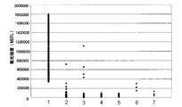
- Example 4 is a graph showing fluorescence intensities measured in Example 1, Example 2, Example 3, and Comparative Example 1.
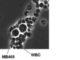
- Example 4 it is a microscope picture at the time of using Emulgen 2025G as surfactant.
- WBC is an abbreviation for white blood cells.
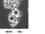
- Example 4 it is a microscope picture at the time of using Nikkor BL-9EX as surfactant.
- Example 4 it is a microscope picture at the time of using Nikkor BO-20V as surfactant.
- Example 4 it is a microscope picture at the time of using Nikkor BT-12 as surfactant.
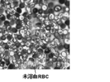
- Example 4 it is a microscope picture at the time of using Nissan cation AB600 as surfactant. In Example 4, it is a microscope picture at the time of using Nissan cation BB as surfactant. In the figure, “RBC” is an abbreviation for red blood cells. 2 is a photomicrograph obtained in Comparative Example 2.
- the cancer cell (CTC) in the blood sample is not particularly limited as long as it is a cancer cell having telomerase activity, including not only cancer cells that have fallen from solid cancer and entered into peripheral blood, but also blood cancer.
- the CTC detected by the method and reagent kit of the present embodiment is not particularly limited, but CTC derived from solid cancer is preferable, and CTC derived from breast cancer is more preferable.
- oncolytic virus refers to a restricted-proliferating virus that cannot normally grow in normal cells and can specifically grow in living cancer cells.
- an oncolytic virus can specifically propagate in cancer cells by having a mechanism that allows it to specifically propagate in cancer cells, for example, a promoter that specifically shows promoter activity in cancer cells. There may be cells in the cell that can make this promoter function (eg, leukocytes).
- Such a normal cell can be detected as a cancer cell in a method for detecting a cancer cell in which a cell in which an oncolytic virus has proliferated is detected as a cancer cell, which may lead to a false positive result.
- a blood sample is mixed with a fixing agent and a nonionic surfactant, so that normal cells can be obtained without affecting the cancer cells on which the oncolytic virus has proliferated. Since it is selectively damaged, false positive signals that can arise from the normal cells can be reduced. Furthermore, in the method of the present invention, red blood cells in a blood sample can be hemolyzed by the above-described treatment, so that cancer cells that may be present in a trace amount in the blood sample can be detected with higher sensitivity.
- damaged means that at least a part of the cell membrane of the subject cell is broken to elute intracellular components.
- red blood cells are hemolyzed means that the cell membrane of red blood cells is solubilized.
- oncolytic viruses include, but are not limited to, viruses that incorporate a promoter that specifically shows promoter activity in cancer cells.
- Promoters that exhibit specific promoter activity in cancer cells include human telomerase promoter, human prostate cancer specific antigen (PSA) promoter, human alpha protein (AFP) promoter, fetus Sex cancer antigen (CEA) promoter and the like.
- PSA prostate cancer specific antigen
- AFP human alpha protein
- CEA fetus Sex cancer antigen
- the human telomerase promoter is preferred in that it can exhibit promoter activity in many types of cancer cells.
- the promoter of hTERT which is a gene encoding human telomerase reverse transcriptase, is more preferable.
- the nucleic acid sequence of the hTERT promoter is shown in SEQ ID NO: 1.
- the hTERT promoter may be the entire 455 bp nucleic acid sequence shown in SEQ ID NO: 1, but the 5 'upstream 181 bp region is considered to be an important core region for downstream gene expression. A nucleic acid sequence containing at least this core region may be used.
- the oncolytic virus is preferably one that expresses a labeled protein. That is, the oncolytic virus preferably has a gene encoding a labeled protein.
- labeled protein refers to a detectable protein that can be distinguished from other cells in which the protein is not expressed by being expressed in the cell.
- a labeling protein usually used in the biochemical field can be used. Green fluorescent protein (GFP) and its mutants (for example, enhanced-humanized GFP (EGFP), red-shift GFP (rsGFP)), yellow Fluorescent proteins such as fluorescent protein (YFP) and blue fluorescent protein (BFP) can be used.
- the labeling protein is preferably a green fluorescent protein.
- the marker protein gene can be placed under the control of the cancer cell-specific promoter.
- the above-mentioned marker protein gene can be placed under the control of a promoter capable of exhibiting promoter activity in an oncolytic virus.
- promoters that can control the expression of the labeled protein gene include cytomegalovirus (CMV) promoter, SV40 late promoter, MMTV LTR promoter, RSV LTR promoter, SR ⁇ promoter, and the like.
- CMV cytomegalovirus
- SV40 late promoter SV40 late promoter
- MMTV LTR promoter MMTV LTR promoter
- RSV LTR promoter SR ⁇ promoter
- the marker protein gene is preferably placed under the control of the CMV promoter or hTERT promoter.
- the nucleic acid sequences of these promoters are known.
- An expression cassette comprising at least a gene encoding the above-described marker protein and a promoter for expressing it (a cancer cell-specific promoter or a promoter capable of exhibiting promoter activity in an oncolytic virus) is a common genetic engineering It can be obtained by a technique. For example, a gene and a promoter encoding a labeled protein are amplified by polymerase chain reaction (PCR) or the like based on a known sequence, the obtained amplification product of each gene is ligated to an appropriate plasmid, and a necessary portion is By cutting out, an expression cassette can be obtained (see, for example, WO 2006/36004).
- PCR polymerase chain reaction
- the above-mentioned oncolytic virus may have a replication cassette in which a gene required for virus growth is linked downstream of the above cancer cell-specific promoter.
- genes necessary for virus growth include early genes (E1A, E1B, etc.), IRES, which is a protein synthesis initiation signal specific to the Picornaviridae family. The nucleic acid sequences of these genes are known.
- the above replication cassette can be prepared by a normal genetic engineering technique.
- the above cancer cell-specific promoter and optionally a gene necessary for virus growth are amplified by PCR or the like based on a known sequence, and the obtained amplification product of each gene is linked to an appropriate plasmid,
- a replication cassette can be obtained by cutting out the necessary part (see eg WO 2006/36004).
- virus examples include those derived from adenovirus, herpes simplex virus, Sendai virus, and reovirus. Of these, human adenovirus is preferable.
- oncolytic viruses can be used.
- an adenovirus having a replication cassette containing the hTERT promoter, E1A gene, IRES gene and E1B gene, and an expression cassette containing the CMV promoter and GFP gene, Telomescan® (OBP-401) ) (Oncoris Biopharma Co., Ltd.) is preferred.
- the method of the present invention comprises a step of incubating a blood sample suspected of having cancer cells with the above-mentioned oncolytic virus to propagate the oncolytic virus in at least the cancer cells in the blood sample (hereinafter referred to as “proliferation”). Process ”).
- the blood sample is usually collected from a subject who is trying to detect the presence of cancer cells, such as a patient suspected of having cancer or a cancer patient.
- the blood sample may be either whole blood or treated blood obtained by pretreatment of whole blood, but serum is removed from whole blood, particularly peripheral blood, in that detection efficiency is further improved. Those are preferred.
- Methods for removing serum from whole blood are known, and examples include a method of centrifuging whole blood to which an anticoagulant (for example, ethylenediaminetetraacetic acid, sodium citrate, heparin, etc.) is added. Centrifugation is preferably performed at 500 to 3500 rpm for 3 to 30 minutes.
- an anticoagulant for example, ethylenediaminetetraacetic acid, sodium citrate, heparin, etc.
- Incubation of the blood sample with the oncolytic virus is preferably performed in the presence of a medium usually used for animal cell culture.
- a medium usually used for animal cell culture examples include RPMI-1640, Dulbecco's modified Eagle medium (DMEM), minimal essential medium (MEM), and the like.
- the oncolytic virus is preferably incubated with the blood sample in an amount of 6 ⁇ 10 4 to 6 ⁇ 10 8 PFU (Plaque Forming Unit) per ml of blood sample.
- the time and temperature at which the above blood sample is incubated with the oncolytic virus may be any conditions as long as the oncolytic virus can grow at least in cancer cells in the blood sample, and can be adjusted as appropriate according to the type of oncolytic virus. .
- Such incubation conditions are preferably performed at a temperature of 25 to 40 ° C. for 1 to 36 hours, more preferably at 30 to 37 ° C. for 12 to 24 hours.
- the oncolytic virus can infect cancer cells that can be contained in the blood sample and can grow in the cells.
- leukocytes monocytes, lymphocytes, etc.
- the above-mentioned incubation may cause the oncolytic virus to grow in such normal (non-cancerous) leukocytes.
- the method of the present invention includes a step of mixing the blood sample treated in the above growth step with a fixing agent and a nonionic surfactant (hereinafter also referred to as “mixing step”).
- the mixing step the blood sample and the nonionic surfactant may be mixed and then the immobilizing agent may be mixed, or after mixing the blood sample and the immobilizing agent, the nonionic surfactant may be mixed.
- a blood sample, a fixing agent, and a nonionic surfactant may be mixed at the same time.
- a more preferable order of mixing is the order of mixing the nonionic surfactant after mixing the blood sample and the fixing agent.
- the above blood sample may be obtained by removing fractions other than the cell fraction by centrifugation or the like before mixing with the immobilizing agent and the nonionic surfactant.
- the concentration of the nonionic surfactant when mixed with the blood sample is preferably a concentration that damages leukocytes without damaging cancer cells.
- a concentration can be appropriately determined depending on the kind of the nonionic surfactant, but is preferably 0.003 to 0.4% by weight, more preferably 0.030 to 0.037% by weight.
- the concentration of the nonionic surfactant when mixed with the blood sample means “the concentration when the blood sample and the nonionic surfactant are directly mixed”, It is also intended to include “concentration when a nonionic surfactant is mixed into a mixture of blood sample and immobilizing agent”.
- the above concentration is a mixed solution of the blood sample, the immobilizing agent and the nonionic surfactant.
- concentration of the nonionic surfactant in is shown.
- the nonionic surfactant may be either a polyoxyethylene surfactant or a fatty acid ester surfactant of a polyoxy compound, but a polyoxyethylene surfactant is preferable.
- the polyoxyethylene surfactant include higher alcohol / ethylene oxide adduct, alkylphenol / ethylene oxide adduct, higher fatty acid / ethylene oxide adduct, higher aliphatic amine / ethylene oxide adduct, higher fatty acid amide / ethylene oxide adduct, etc. . Of these, higher alcohol / ethylene oxide adducts are preferred.
- the alkyl group of the higher alcohol of the higher alcohol / ethylene oxide adduct may be linear or branched, and preferably has 12 to 30 carbon atoms, more preferably 15 to 25 carbon atoms.
- the higher alcohol / ethylene oxide adduct preferably has an ethylene oxide polymerization number of 15 to 40, more preferably 20 to 30.
- a more specific higher alcohol / ethylene oxide adduct in which the alkyl group of the higher alcohol is a branched chain is represented by the following formula (I).
- Formula (I) (In the formula, l is an integer of 6 to 24, m is an integer of 1 to 19, and n is an integer of 15 to 40.)
- nonionic surfactant polyoxyethylene octyldodecyl ether in which the alkyl group of the higher alcohol is branched, the carbon number is 20, and the ethylene oxide polymerization number is 25 is preferable.
- the concentration of the fixing agent when mixed with a blood sample is preferably a concentration that hemolyzes red blood cells. Such a concentration is preferably 1 to 7% by weight, more preferably 2 to 4% by weight.
- the concentration of the immobilizing agent when mixed with the blood sample refers to “the concentration when the blood sample and the immobilizing agent are directly mixed” in addition to “the immobilizing agent, It is also intended to include “concentration when mixed in a mixture of blood sample and nonionic surfactant”. That is, in the mixing step, when the immobilizing agent is mixed after mixing the blood sample and the nonionic surfactant, the above concentration is a mixed solution of the blood sample, the immobilizing agent and the nonionic surfactant. The concentration of the immobilizing agent in is shown.
- aldehyde compounds such as paraformaldehyde, glutaraldehyde, and formaldehyde are preferable. Of these, paraformaldehyde is more preferable.
- the mixing step is preferably performed under conditions that do not damage cancer cells.
- pH 6-9 and osmotic pressure 200-400 ⁇ mOsm are preferable.
- the method of the present invention includes a step (hereinafter also referred to as “detection step”) of detecting cancer cells in which an oncolytic virus has proliferated, contained in a blood sample obtained from the mixing step.
- the detection step is preferably a step of detecting the above labeled protein expressed by the oncolytic virus growing.
- the method for detecting the labeled protein can be appropriately selected depending on the type of the labeled protein.
- the labeled protein is a fluorescent protein
- the labeled protein can be detected by observation under a fluorescence microscope, detection with a flow cytometer, or the like.
- a blood sample obtained from the mixing process is used to prepare a smear on a slide glass by an ordinary method, and the smear is observed under a fluorescence microscope equipped with a CCD camera. Then, it can be performed by detecting cells emitting fluorescence and counting as desired.
- Detection using a flow cytometer can be performed, for example, by subjecting a blood sample obtained from the mixing step to a flow cytometer that can detect fluorescence, detecting cells that emit fluorescence, and counting as desired.
- the method of the present invention may further comprise a dead cell staining step.
- a dead cell staining step By including such a step, “surviving” cancer cells and “dead” cancer cells can be detected, and the survival rate of the cancer cells in the blood sample can be calculated.
- This step is preferably performed before the step of mixing the blood sample with the immobilizing agent and the nonionic surfactant.
- the step of staining dead cells can be performed by bringing a reagent that specifically stains dead cells into contact with a blood sample.
- the “reagent that specifically stains dead cells” refers to a reagent that stains dead cells in a manner distinguishable from viable cells.
- a commercially available kit can be used, for example, Live / Dead Fixable Red Dead Cell Stain Kit (Invitrogen) is preferable.
- the present invention also provides a reagent kit for detecting cancer cells in a blood sample.
- the reagent kit of the present invention includes a first reagent containing the oncolytic virus, a second reagent containing the immobilizing agent, and a third reagent containing the nonionic surfactant. .
- first reagent, second reagent, and third reagent may be in the form of a liquid, or may be in the form of a solid that can be made liquid by adding an appropriate solvent at the time of use.
- the above first reagent when incubated with a blood sample in liquid form, provides a tumor at a concentration that will provide an amount of 6 ⁇ 10 4 to 6 ⁇ 10 8 PFU of oncolytic virus per ml of blood sample.
- it contains a lytic virus.
- the above-mentioned second reagent preferably contains an immobilizing agent in such an amount that when it is mixed with a blood sample, the concentration is such that red blood cells are hemolyzed.
- the second reagent contains the immobilizing agent in an amount that, when mixed with a blood sample, is preferably 1-7 wt%, more preferably 2-4 wt% immobilizing agent.
- the above-mentioned third reagent preferably contains a nonionic surfactant in such an amount that, when mixed with a blood sample, it does not damage cancer cells but damages leukocytes.
- the third reagent is non-ionic in an amount that, when mixed with a blood sample, is preferably a concentration of non-ionic surfactant of 0.003-0.4 wt%, more preferably 0.030-0.037 wt%.
- the third reagent is adjusted so that the concentration of the nonionic surfactant in the mixture of the blood sample, the second reagent, and the third reagent becomes the above-described concentration. Contains a nonionic surfactant.
- the above-mentioned second reagent and third reagent can contain an appropriate buffer and osmotic pressure adjusting agent for making the cancer cells not damaged when mixed with a blood sample.
- the conditions that do not damage cancer cells are preferably pH 6-9 and osmotic pressure 200-400 ⁇ mOsm. Therefore, as the buffer, phosphate physiological buffer (PBS), citrate buffer, HEPES, and the like can be used. Moreover, ethylene glycol, sodium chloride, etc. can be used as an osmotic pressure regulator.
- the present invention will be described in more detail with reference to examples. However, the present invention is not limited to the following examples. 1. Effect of oncolytic virus growth on the mononuclear cell fraction
- Spheres are detected as cancer cells. Therefore, a mononuclear cell fraction is collected from blood collected from a human subject, and this fraction is treated by the method of the present invention (treatment with an immobilizing agent and a nonionic surfactant) to obtain a mononuclear cell.
- the mononuclear cell is a general term for monocytes and lymphocytes.
- Example 1 Preparation of mononuclear cell fraction> Blood was collected from two healthy women using a vacuum blood collection tube (Benoject (registered trademark) II vacuum blood collection tube (4.5 ml), containing 3.8% sodium citrate, Terumo Corporation). Within 5 hours of collection, 5.0 ml of blood was carefully layered in a 15 ml centrifuge tube pre-filled with 5.0 ml of Polymorphprep TM (Daiichi Chemical Co., Ltd.), a reagent for separating mononuclear cells. The centrifuge tube was then centrifuged at 500 ⁇ g for 30 minutes at room temperature using a swing rotor.
- a vacuum blood collection tube (Benoject (registered trademark) II vacuum blood collection tube (4.5 ml), containing 3.8% sodium citrate, Terumo Corporation).
- 5.0 ml of blood was carefully layered in a 15 ml centrifuge tube pre-filled with 5.0 ml of Polymorphprep TM (Daiich
- the mononuclear cell suspension was transferred to another centrifuge tube using a Pasteur pipette, and an equal amount of PBS ( ⁇ ) was added thereto and mixed.
- PBS ( ⁇ ) was added thereto and mixed.
- This solution was centrifuged at room temperature at 400 ⁇ g for 10 minutes, and then the supernatant was removed to obtain precipitated mononuclear cells.
- the obtained mononuclear cells were suspended in PBS ( ⁇ ) and centrifuged at 400 ⁇ g for 10 minutes, and then the supernatant was removed to obtain a mononuclear cell fraction.
- RPMI-1640 was added to the resulting mononuclear cell fraction to make up to 10 ml.
- oncolytic virus Telomescan (registered trademark) (OBP-401), Oncoris Biopharma Co., Ltd.) was added to a final concentration of 6 ⁇ 10 6 PFU / ml, while rotating at 37 ° C. Cultured for 24 hours. The obtained culture broth was centrifuged at 1500 rpm for 5 minutes with a weak brake, and then the supernatant was removed to obtain cells on which oncolytic virus had proliferated (hereinafter also referred to as proliferating cells).
- Example 2 A treated sample was prepared in the same manner as in Example 1 except that Nikkor BO-20V (Nihon Chemicals Sales Co., Ltd.) was used as the nonionic surfactant in place of Emulgen 2025G. Then, a slide was prepared from the treated sample in the same manner as in Example 1, and the fluorescence intensity of the slide was measured under the same conditions as in Example 1 to quantify the fluorescence signal. The obtained results are shown in Fig. 1 (No. 6).
- Example 3 In Example 1, a treated sample was prepared in the same manner as described above except that Emulgen 2020G (Kao Corporation) was used in place of Emulgen 2025G as the nonionic surfactant. Then, a slide was prepared from the treated sample in the same manner as in Example 1, and the fluorescence intensity of the slide was measured under the same conditions as in Example 1 to quantify the fluorescence signal. The obtained results are shown in Fig. 1 (No. 7). The structural formula of Emulgen 2020G is shown in the following formula (III). Formula (III) (In the formula, n is 20.)
- Example 1 In Example 1, in place of the treatment with the immobilizing agent and the nonionic surfactant, the treatment with the ammonium chloride and the immobilizing agent conventionally used for hemolysis is performed, and the influence on the detection by the mononuclear cell. I investigated. 30% volume of 1% ammonium chloride solution is added to and suspended in the proliferating cells obtained in the same manner as in the above ⁇ Preparation of mononuclear cells> and ⁇ Virus growth>, and left at room temperature for 20 minutes. did. Next, 2% PFA was added thereto and allowed to stand at room temperature for 20 minutes to obtain a treated sample.
- the obtained treated sample was centrifuged with a weak brake at 1500 rpm for 5 minutes, and then the supernatant was removed to obtain a precipitate.
- the resulting precipitate was then suspended in 1 ml PBS ( ⁇ ).
- the obtained suspension was centrifuged at 1500 rpm for 5 minutes with a weak brake, and then the supernatant was removed, and a slide was prepared using cytospin (1000 rpm, 4 minutes).
- a mounting medium (Fluorescent Mounting Medium, S3032, Dako) was placed on the slide, and then a cover glass was placed. Then, the fluorescence intensity was measured under the conditions of Example 1, and the fluorescence signal was digitized.
- a breast cancer cell line (MB468) was used as a positive control.
- Oncolytic virus was grown in MB468 in the same manner as in Example 1, and this MB468 was not treated with an immobilizing agent and a nonionic surfactant, and a fluorescence microscope was used under the same conditions as in Example 1. The fluorescence intensity was measured. The results are shown in FIG. In FIG. 1, positive control (No. 1), Comparative Example 1 (No. 2 and 3), Example 1 (No. 4 and 5), Example 2 (No. 6) and Example 3 (No. 7). ) Result.
- Emulgen 2025G and Emulgen 2020G which are adducts of higher alcohol / ethylene oxide whose alkyl group of higher alcohol is branched, as the nonionic surfactant (No. 4, 5, and 7)
- the nonionic surfactant No. 4, 5, and 7
- Example 4 Peripheral blood 7.5 ml was collected from healthy human blood anticoagulant storage solution (CPD solution; 100 ml of sterile water citric acid 3Na ⁇ 2H 2 O 2.63 g, citric acid ⁇ 2H 2 O 0.327 g, D (+) (Containing 2.32 g of glucose and 0.251 g of sodium dihydrogen phosphate and 0.251 g of 2H 2 O) 1120 ⁇ l was added and thoroughly mixed by inversion. Next, the mixture was centrifuged at 1500 rpm for 5 minutes with a weak brake, and the supernatant (serum) was removed as much as possible using a pipette. PBS (-) was added thereto to make up to 14 ml and suspended. Then, the same centrifugation and serum removal as described above were repeated four times. Thereafter, a breast cancer cell line (MB468) was added thereto at 3 ⁇ 10 5 cells / ml to obtain a cancer cell-containing blood sample.
- the obtained sample was treated in the same manner as in ⁇ Virus propagation>, ⁇ Treatment with immobilizing agent and nonionic surfactant> and ⁇ Slide preparation and microscopic observation> in Example 1, and a photomicrograph was taken. did.
- the obtained photograph is shown in FIG.
- Nikkor BL-9EX Nonionic Surfactant, Nippon Chemicals Sales Co., Ltd.
- Nikkor BO-20V Nonippon Chemicals Sales Co., Ltd.
- Emulgen 2025G Surfactant for the above cancer cell-containing blood sample
- Nikkor BT-12 nonionic surfactant, Nippon Chemical Sales Co., Ltd.
- Emulgen 2020G Korean Organic Chemical Sales Co., Ltd.
- Nissan Cation AB600 Cationic Surfactant, NOF Corporation
- Nissan Cation BB (Cation)
- FIGS. 2B to 2G show photographs when Nikkor BL-9EX, Nikkor BO-20V, Nikkor BT-12, Emulgen 2020G, Nissan Cation AB600 and Nissan Cation BB are used as surfactants, respectively.
- Virus was grown in a cancer cell-containing blood sample obtained in the same manner as in Example 4 in the same manner as in ⁇ Virus propagation> in Example 1 to obtain proliferated cells.
- the same volume of 4% PFA was added and suspended, and allowed to stand at room temperature for 20 minutes.
- a 1% ammonium chloride solution having a volume twice the total volume of the proliferating cells and PFA was added and suspended, and incubated at 40 ° C. for 5 minutes with stirring to obtain a treated sample.
- a micrograph of the treated sample was taken. The obtained photograph is shown in FIG.
- FIG. 2 (H) shows that in the blood sample treated by the method of Comparative Example 2, unhemolyzed erythrocytes (unhemolyzed RBC) and erythrocyte debris (RBC ghost) are concentrated around the cancer cells. .
- FIGS. 2 (F) and 2 (G) in the blood sample treated by the method using a cationic surfactant as the surfactant, as in Comparative Example 2, unhemolyzed RBC and RBC are formed around the cancer cells. It can be seen that debris derived from ghosts is dense.
- FIGS. 2A to 2E show that in the blood sample treated by the method using the nonionic surfactant of the present invention, red blood cells are almost removed and cancer cells can be confirmed more clearly. .
Landscapes
- Health & Medical Sciences (AREA)
- Life Sciences & Earth Sciences (AREA)
- Engineering & Computer Science (AREA)
- Immunology (AREA)
- Chemical & Material Sciences (AREA)
- Biomedical Technology (AREA)
- Molecular Biology (AREA)
- Hematology (AREA)
- Urology & Nephrology (AREA)
- Microbiology (AREA)
- General Health & Medical Sciences (AREA)
- Biotechnology (AREA)
- Biochemistry (AREA)
- Medicinal Chemistry (AREA)
- Physics & Mathematics (AREA)
- Analytical Chemistry (AREA)
- Food Science & Technology (AREA)
- Cell Biology (AREA)
- General Physics & Mathematics (AREA)
- Pathology (AREA)
- Genetics & Genomics (AREA)
- Bioinformatics & Cheminformatics (AREA)
- Wood Science & Technology (AREA)
- Zoology (AREA)
- Organic Chemistry (AREA)
- Virology (AREA)
- Proteomics, Peptides & Aminoacids (AREA)
- General Engineering & Computer Science (AREA)
- Measuring Or Testing Involving Enzymes Or Micro-Organisms (AREA)
- Micro-Organisms Or Cultivation Processes Thereof (AREA)
- Investigating Or Analysing Biological Materials (AREA)
Abstract
Description
GFP遺伝子を有する腫瘍溶解性ウイルスは、テロメスキャン(登録商標) (OBP-401)として市販されている。テロメスキャン(登録商標)は、癌細胞内で特異的に増殖し、GFPを産生することにより、癌細胞を特異的に蛍光発光させることができる。
- 赤血球の除去:血液中に存在する癌細胞の数は、極めて少ない。よって、血液中に多量に存在する赤血球により癌細胞が遮蔽され、癌細胞の検出を妨げる可能性がある。
- 偽陽性のシグナルの抑制:正常な血球系細胞であるにもかかわらず、腫瘍溶解性ウイルスが増殖できる細胞、例えば白血球(一部の単球及びリンパ球)が希に存在する。このような正常細胞内で腫瘍溶解性ウイルスが増殖すると、癌細胞の検出に偽陽性のシグナルを与える可能性がある。
癌細胞の存在が疑われる血液試料を、腫瘍溶解性ウイルスとインキュベートすることにより、少なくとも前記癌細胞内で腫瘍溶解性ウイルスを増殖させる工程、
増殖工程で処理された血液試料と、固定化剤と、非イオン性界面活性剤とを混合する工程、及び
混合工程で得られた血液試料に含まれる、腫瘍溶解性ウイルスが増殖した癌細胞を検出する工程
を含む、血液試料中の癌細胞の検出方法を提供する。
本明細書において、「腫瘍溶解性ウイルス」とは、正常細胞内では通常増殖できず、生存している癌細胞内で特異的に増殖可能な制限増殖型ウイルスのことをいう。
しかし、制限増殖型とするための機構によっては、腫瘍溶解性ウイルスが正常細胞内でも増殖する可能性が否定されないことは、当業者に明確に認識される。
すなわち、腫瘍溶解性ウイルスは、癌細胞内で特異的に増殖可能とする機構、例えば癌細胞で特異的にプロモーター活性を示すプロモーターを有することにより、癌細胞内で特異的に増殖できるが、正常細胞の中にもこのプロモーターを機能させ得る細胞が存在し得る(例えば白血球)。このような正常細胞は、腫瘍溶解性ウイルスが増殖した細胞を癌細胞として検出する癌細胞の検出方法においては、癌細胞として検出され得るので、偽陽性の結果を導く可能性がある。
さらに、本発明の方法では、上記の処理により、血液試料中の赤血球も溶血できるので、血液試料中に微量しか存在しない可能性がある癌細胞を、より高い感度で検出できる。
また、本明細書において、「赤血球が溶血する」とは、赤血球の細胞膜が可溶化されることをいう。
標識タンパク質は、緑色蛍光タンパク質が好ましい。
標識タンパク質遺伝子の発現を制御し得るプロモーターとしては、サイトメガロウイルス(CMV)プロモーター、SV40後期プロモーター、MMTV LTRプロモーター、RSV LTRプロモーター、SRαプロモーターなどが挙げられる。なかでも、標識タンパク質の遺伝子は、CMVプロモーター又はhTERTプロモーターの制御下におくことが好ましい。これらのプロモーターの核酸配列は、公知である。
本発明の方法は、癌細胞の存在が疑われる血液試料を、上記の腫瘍溶解性ウイルスとインキュベートして、血液試料中の少なくとも癌細胞内で腫瘍溶解性ウイルスを増殖させる工程(以下、「増殖工程」ともいう)を含む。
上記の血液試料は、通常、癌の罹患が疑われる患者、癌患者などの癌細胞の存在を検出しようとする被験者から採取され得る。血液試料は、全血、及び全血を前処理して得られた処理血液のいずれであってもよいが、検出効率がより向上される点で、全血、特に末梢血から血清を除去したものが好ましい。
全血から血清を除去する方法は、公知であり、例えば抗凝固剤(例えばエチレンジアミン四酢酸、クエン酸ナトリウム、ヘパリンなど)を加えた全血を、遠心分離する方法が挙げられる。遠心分離は、500~3500rpm、3~30分間行うことが好ましい。
上記の血液試料を、腫瘍溶解性ウイルスとインキュベートする時間及び温度は、腫瘍溶解性ウイルスが血液試料中の少なくとも癌細胞内で増殖できる条件であればよく、腫瘍溶解性ウイルスの種類により適宜調節できる。そのようなインキュベートの条件は、好ましくは25~40℃の温度で、1~36時間、より好ましくは30~37℃にて12~24時間行われる。
しかし、血液試料中に存在する白血球(単球、リンパ球など)も、テロメラーゼのような癌細胞が特異的に発現する酵素を希に発現する。したがって、上記のインキュベーションにより、このような正常な(癌細胞でない)白血球においても、腫瘍溶解性ウイルスが増殖する可能性がある。
本発明の方法は、上記の増殖工程で処理された血液試料を、固定化剤及び非イオン性界面活性剤と混合する工程(以下、「混合工程」ともいう)を含む。
混合工程においては、血液試料と非イオン性界面活性剤とを混合した後に、固定化剤を混合してもよいし、血液試料と固定化剤とを混合した後に、非イオン性界面活性剤を混合してもよいし、血液試料、固定化剤及び非イオン性界面活性剤を同時に混合してもよい。より好ましい混合の順序としては、血液試料と固定化剤とを混合した後に、非イオン性界面活性剤を混合する順序である。
なお、本明細書において、「血液試料と混合したときの非イオン性界面活性剤の濃度」とは、「血液試料と非イオン性界面活性剤とを直接混合した場合の濃度」に加えて、「非イオン性界面活性剤を、血液試料及び固定化剤の混合物に混合した場合の濃度」も含むことを意図する。すなわち、混合工程において、血液試料と固定化剤とを混合した後に、非イオン性界面活性剤を混合する場合、上記の濃度は、血液試料、固定化剤及び非イオン性界面活性剤の混合液における非イオン性界面活性剤の濃度を示す。
より具体的な高級アルコールのアルキル基が分岐鎖である高級アルコール・エチレンオキシド付加物を、以下の式(I)に示す。
式(I)

なお、本明細書において、「血液試料と混合したときの固定化剤の濃度」とは、「血液試料と固定化剤とを直接混合した場合の濃度」に加えて、「固定化剤を、血液試料及び非イオン性界面活性剤の混合物に混合した場合の濃度」も含むことを意図する。すなわち、混合工程において、血液試料と非イオン性界面活性剤とを混合した後に、固定化剤を混合する場合、上記の濃度は、血液試料、固定化剤及び非イオン性界面活性剤の混合液における固定化剤の濃度を示す。
本発明の方法は、混合工程から得られた血液試料に含まれる、腫瘍溶解性ウイルスが増殖した癌細胞を検出する工程(以下、「検出工程」ともいう)を含む。
検出工程は、腫瘍溶解性ウイルスが増殖することにより発現される上記の標識タンパク質を検出する工程であることが好ましい。
本発明の方法は、さらに、死細胞の染色工程を含むことができる。このような工程を含むことにより、「生存している」癌細胞と「死んでいる」癌細胞とを検出することができ、血液試料中の癌細胞の生存率を算出することができる。この工程は、血液試料を、固定化剤及び非イオン性界面活性剤と混合する工程の前に行うことが好ましい。
死細胞を染色する工程は、死細胞を特異的に染色する試薬と血液試料とを接触させることにより行うことができる。「死細胞を特異的に染色する試薬」とは、生存細胞とは区別可能に死細胞を染色する試薬のことである。死細胞を特異的に染色する試薬としては、市販のキットを用いることができ、例えばLive/Dead Fixable Red Dead Cell Stain Kit (Invitrogen社)が好ましい。
本発明は、血液試料中の癌細胞を検出するための試薬キットも提供する。本発明の試薬キットは、上記の腫瘍溶解性ウイルスを含有する第1試薬と、上記の固定化剤を含有する第2試薬と、上記の非イオン性界面活性剤を含む第3試薬とを含む。
1. 単核球画分への腫瘍溶解性ウイルスの増殖による影響
腫瘍溶解性ウイルスを用いる血液試料中の癌細胞の検出においては、試料中の単核球内で腫瘍溶解性ウイルスが増殖すると、単核球が癌細胞として検出されてしまう。
そこで、ヒト対象から採取した血液から単核球画分を回収し、この画分に本発明の方法による処理(固定化剤及び非イオン性界面活性剤による処理)を行うことにより、単核球による検出への影響を低減できるか否かを検討した。なお、単核球とは、単球及びリンパ球の総称である。
<単核球画分の調製>
真空採血管(ベノジェクト(登録商標)II真空採血管(4.5 ml)、3.8%クエン酸ナトリウム含有、テルモ株式会社)を用いて、2人の健常女性から血液を採取した。採取から2時間以内の血液5.0 mlを、単核球を分離するための試薬であるPolymorphprep (商標)(第一化学薬品株式会社)5.0 mlを予め入れた15 mlの遠心チューブに注意深く重層した。次いで、遠心チューブを、スウィングローターを用いて、室温で500×g、30分間遠心分離した。そして、パスツールピペットを用いて単核球浮遊液を別の遠心チューブに移し、これに等量のPBS(-)を加えて混和した。この液を室温で400×g、10分間遠心分離した後、上清を除去して、沈殿した単核球を得た。そして、得られた単核球をPBS(-)で懸濁し、これを400×g、10分間遠心分離した後、上清を除去して、単核球画分を得た。
得られた単核球画分にRPMI-1640を加えて、10 mlにメスアップした。これに腫瘍溶解性ウイルス(テロメスキャン(登録商標) (OBP-401)、オンコリスバイオファーマ株式会社)を、最終濃度6×10⁶ PFU/mlとなるように加え、37℃にてローテーションしながら24時間培養した。得られた培養液を、1500 rpmで5分間、弱ブレーキで遠心分離した後、上清を除去して、腫瘍溶解性ウイルスが増殖した細胞(以下、増殖細胞ともいう)を得た。
得られた増殖細胞に、これと同体積の4%パラホルムアルデヒド(PFA)を加え、室温にて20分間静置した。ここに、増殖細胞及びPFAの合計体積の2倍の体積の非イオン性界面活性剤(エマルゲン2025G、花王株式会社)のPBS(-)溶液(濃度0.05重量%)を加えて懸濁し、撹拌しながら40℃にて5分間インキュベートして、処理試料を得た。なお、エマルゲン2025Gの構造式を、以下の式(II)に示す。
式(II)

得られた処理試料を1500 rpmで5分間、弱ブレーキで遠心分離した後、上清を除去し、沈殿物を得た。次いで、得られた沈殿物を1 mlのPBSに懸濁した。そして、得られた懸濁液を1500 rpmで5分間、弱ブレーキで遠心分離した後、上清を除去し、サイトスピンを用いて(1000 rpm、4分)、スライドを作製した。該スライドにマウンティングメディウム(Fluorescent Mounting Medium、S3032、Dako社)を載せ、次いでカバーガラスを載せた後、倒立型リサーチ顕微鏡(Power IX 71、オリンパス株式会社製)を用いて、露光時間20 ms、50 ms、100 ms、200 ms及び400 msの条件下で、蛍光強度を測定した。
なお、上記と同様の条件下で、それぞれが異なる既知の蛍光強度を有する7種のビーズ(7 Peaks Beads(Cyto-Cal Multifluor Fluorescence Intensity Calibrator、FC3M、Thermo Scientific社))を撮像して、それぞれの露光時間における検量線を作成した。得られた検量線に基づいて、上記の処理試料からの蛍光シグナルを数値化した。得られた結果を、図1(No.4及び5)に示す。
実施例1において、非イオン性界面活性剤として、エマルゲン2025Gに代えて、ニッコールBO-20V(日本ケミカルズ販売株式会社)を用いた以外、上記と同様にして、処理試料を調製した。そして、実施例1と同様にして該処理試料からスライドを作製し、該スライドについて実施例1と同じ条件で蛍光強度を測定して、蛍光シグナルを数値化した。得られた結果を、図1(No.6)に示す。
実施例1において、非イオン性界面活性剤として、エマルゲン2025Gに代えて、エマルゲン2020G(花王株式会社)を用いた以外、上記と同様にして、処理試料を調製した。そして、実施例1と同様にして該処理試料からスライドを作製し、該スライドについて実施例1と同じ条件で蛍光強度を測定して、蛍光シグナルを数値化した。得られた結果を、図1(No.7)に示す。なお、エマルゲン2020Gの構造式を、以下の式(III)に示す。
式(III)

実施例1において、固定化剤及び非イオン性界面活性剤での処理に代えて、溶血のために従来用いられる塩化アンモニウム及び固定化剤での処理を行って、単核球による検出への影響を調べた。
上記の<単核球の調製>及び<ウイルス増殖>と同様にして得られた増殖細胞に、これの30倍の体積の1%塩化アンモニウム溶液を加えて懸濁し、室温にて20分間静置した。次いで、ここに2% PFAを加え、室温にて20分間静置して、処理試料を得た。
得られた処理試料を、1500 rpmで5分間、弱ブレーキで遠心分離した後、上清を除去し、沈殿物を得た。次いで、得られた沈殿物を1 mlのPBS(-)に懸濁した。そして、得られた懸濁液を1500 rpmで5分間、弱ブレーキで遠心分離した後、上清を除去し、サイトスピンを用いて(1000 rpm、4分)、スライドを作製した。該スライドにマウンティングメディウム(Fluorescent Mounting Medium、S3032、Dako社)を載せ、次いでカバーガラスを載せた後、実施例1の条件で蛍光強度を測定し、蛍光シグナルを数値化した。
結果を、図1に示す。図1では、陽性対照(No.1)、比較例1(No.2及び3)、実施例1(No.4及び5)、実施例2(No.6)並びに実施例3(No.7)の結果を示す。
種々の界面活性剤を用いて、実際の血液試料中での癌細胞の検出について検討した。
健常なヒトから採取した末梢血液7.5 mlに、血液抗凝固保存液(CPD液;100 mlの滅菌水中にクエン酸3Na・2H2O 2.63 g、クエン酸・2H2O 0.327 g、D(+)-グルコース2.32 g、リン酸2水素ナトリウム・2H2O 0.251 gを含有)1120 μlを加え、充分に転倒混和した。次いで、この混合液を1500 rpmで5分間、弱ブレーキで遠心分離した後、ピペットを用いて上清(血清)をできるだけ取り除いた。ここにPBS(-)を加えて14 mlにメスアップし、懸濁した。そして、上記と同様の遠心分離及び血清の除去を4回繰り返した。その後、ここに乳癌細胞株(MB468)を、3×10⁵個/mlとなるように添加して、癌細胞含有血液試料を得た。
界面活性剤としてニッコールBL-9EX、ニッコールBO-20V、ニッコールBT-12、エマルゲン2020G、ニッサンカチオンAB600及びニッサンカチオンBBを用いた場合の写真を、それぞれ図2(B)~(G)に示す。
実施例4と同様にして得られた癌細胞含有血液試料に、実施例1の<ウイルス増殖>と同様にしてウイルスを増殖させ、増殖細胞を得た。これと同体積の4% PFAを加えて懸濁し、室温にて20分間静置した。次いで、増殖細胞及びPFAの合計体積の2倍の体積の1%塩化アンモニウム溶液を加えて懸濁し、撹拌しながら40℃にて5分間インキュベートして、処理試料を得た。
実施例1の<スライド作製及び顕微鏡観察>と同様にして、該処理試料の顕微鏡写真を撮影した。得られた写真を、図2(H)に示す。
一方、図2(A)~(E)から、本発明の非イオン性界面活性剤を用いた方法により処理された血液試料では、赤血球がほとんど除去され、癌細胞がより鮮明に確認できることがわかる。
Claims (20)
- 癌細胞の存在が疑われる血液試料を、腫瘍溶解性ウイルスとインキュベートすることにより、少なくとも前記癌細胞内で腫瘍溶解性ウイルスを増殖させる工程、
増殖工程で処理された血液試料と、固定化剤と、非イオン性界面活性剤とを混合する工程、及び
混合工程で得られた血液試料に含まれる、腫瘍溶解性ウイルスが増殖した癌細胞を検出する工程
を含む、血液試料中の癌細胞の検出方法。 - 混合工程が、血液試料と固定化剤とを混合した後に、非イオン性界面活性剤を混合する工程である、請求項1に記載の方法。
- 混合工程において、血液試料と混合したときの非イオン性界面活性剤の濃度が、癌細胞に損傷を与えずに、赤血球および白血球に損傷を与える濃度である、請求項1に記載の方法。
- 非イオン性界面活性剤が、ポリオキシエチレン系の非イオン性界面活性剤である、請求項1に記載の方法。
- ポリオキシエチレン系の非イオン性界面活性剤が、高級アルコール・エチレンオキシド付加物である、請求項4に記載の方法。
- 高級アルコール・エチレンオキシド付加物の高級アルコールのアルキル基が分岐鎖である、請求項5に記載の方法。
- 混合工程において、血液試料と混合したときの固定化剤の濃度が、赤血球を溶血する濃度である、請求項1に記載の方法。
- 固定化剤が、パラホルムアルデヒド、グルタルアルデヒド又はホルムアルデヒドである、請求項1に記載の方法。
- 腫瘍溶解性ウイルスが、標識タンパク質を発現する腫瘍溶解性ウイルスである、請求項1に記載の方法。
- 腫瘍溶解性ウイルスが、テロメラーゼプロモーターが組み込まれた腫瘍溶解性ウイルスである、請求項1に記載の方法。
- 検出工程が、標識タンパク質からのシグナルを検出する工程である、請求項9に記載の方法。
- 腫瘍溶解性ウイルスを含有する第1試薬、
固定化剤を含有する第2試薬、及び
非イオン性界面活性剤を含有する第3試薬
を含む、血液試料中の癌細胞の検出用試薬キット。 - 第3試薬が、血液試料と混合したときに、癌細胞に損傷を与えずに、赤血球および白血球に損傷を与える濃度となる量で非イオン性界面活性剤を含有する、請求項12に記載のキット。
- 非イオン性界面活性剤が、ポリオキシエチレン系の非イオン性界面活性剤である、請求項12に記載のキット。
- ポリオキシエチレン系の非イオン性界面活性剤が、高級アルコール・エチレンオキシド付加物である、請求項14に記載のキット。
- 高級アルコール・エチレンオキシド付加物の高級アルコールのアルキル基が分岐鎖である、請求項15に記載のキット。
- 第2試薬が、血液試料と混合したときに、赤血球を溶血する濃度となる量で固定化剤を含有する、請求項12に記載のキット。
- 固定化剤が、パラホルムアルデヒド、グルタルアルデヒド又はホルムアルデヒドである、請求項12に記載のキット。
- 腫瘍溶解性ウイルスが、標識タンパク質を発現する腫瘍溶解性ウイルスである、請求項12に記載のキット。
- 腫瘍溶解性ウイルスが、テロメラーゼプロモーターが組み込まれた腫瘍溶解性ウイルスである、請求項12に記載のキット。
Priority Applications (4)
| Application Number | Priority Date | Filing Date | Title |
|---|---|---|---|
| EP09833417A EP2377947A4 (en) | 2008-12-18 | 2009-12-14 | METHOD FOR DETECTING CANCER CELLS IN A BLOOD TEST |
| JP2010542962A JPWO2010071114A1 (ja) | 2008-12-18 | 2009-12-14 | 血液試料中の癌細胞の検出方法 |
| US13/140,723 US20110250588A1 (en) | 2008-12-18 | 2009-12-14 | Method for detecting cancer cells in blood sample |
| CN2009801511228A CN102257156A (zh) | 2008-12-18 | 2009-12-14 | 血样中癌细胞的检测方法 |
Applications Claiming Priority (2)
| Application Number | Priority Date | Filing Date | Title |
|---|---|---|---|
| JP2008-322439 | 2008-12-18 | ||
| JP2008322439 | 2008-12-18 |
Publications (1)
| Publication Number | Publication Date |
|---|---|
| WO2010071114A1 true WO2010071114A1 (ja) | 2010-06-24 |
Family
ID=42268785
Family Applications (1)
| Application Number | Title | Priority Date | Filing Date |
|---|---|---|---|
| PCT/JP2009/070843 Ceased WO2010071114A1 (ja) | 2008-12-18 | 2009-12-14 | 血液試料中の癌細胞の検出方法 |
Country Status (6)
| Country | Link |
|---|---|
| US (1) | US20110250588A1 (ja) |
| EP (1) | EP2377947A4 (ja) |
| JP (1) | JPWO2010071114A1 (ja) |
| KR (1) | KR20110111377A (ja) |
| CN (1) | CN102257156A (ja) |
| WO (1) | WO2010071114A1 (ja) |
Cited By (4)
| Publication number | Priority date | Publication date | Assignee | Title |
|---|---|---|---|---|
| WO2011099565A1 (ja) * | 2010-02-10 | 2011-08-18 | シスメックス株式会社 | 血液中癌細胞の検出方法及びそれに用いるプログラム |
| WO2012086802A1 (ja) * | 2010-12-24 | 2012-06-28 | アークレイ株式会社 | 癌細胞の検出方法 |
| EP2677317A1 (en) | 2012-06-20 | 2013-12-25 | ARKRAY, Inc. | Method for treating a blood component containing sample |
| WO2015174539A1 (ja) * | 2014-05-13 | 2015-11-19 | 学校法人順天堂 | 細胞の検出方法 |
Families Citing this family (3)
| Publication number | Priority date | Publication date | Assignee | Title |
|---|---|---|---|---|
| KR102080996B1 (ko) * | 2018-07-23 | 2020-02-25 | 주식회사 코어파마 | 암세포 특이적 아데노바이러스 |
| US20210198654A1 (en) * | 2019-12-26 | 2021-07-01 | Jerome Canady Research Institute for Advanced Biological and Technological Sciences | Method for isolation and harvesting microvesicles |
| CN112111456B (zh) * | 2020-08-28 | 2022-05-27 | 益善生物技术股份有限公司 | 一种循环肿瘤细胞分离富集试剂盒 |
Citations (7)
| Publication number | Priority date | Publication date | Assignee | Title |
|---|---|---|---|---|
| JP2002503814A (ja) | 1998-02-12 | 2002-02-05 | イムニベスト・コーポレイション | 循環ガン細胞の迅速かつ効率的な単離のための方法および試薬 |
| JP2002207034A (ja) * | 2001-01-10 | 2002-07-26 | Sysmex Corp | 異常細胞検出方法 |
| JP2004033186A (ja) | 2002-07-08 | 2004-02-05 | Kansai Tlo Kk | 腫瘍細胞において選択的に増殖する腫瘍融解ウイルス |
| WO2004056978A1 (en) * | 2002-12-19 | 2004-07-08 | Ivonex Gmbh | Method for the separation of cell fractions |
| WO2006036004A1 (ja) | 2004-09-29 | 2006-04-06 | Oncolys Biopharma, Inc. | テロメライシン-gfp遺伝子含有組換えウイルス |
| JP2007178193A (ja) | 2005-12-27 | 2007-07-12 | Srl Inc | 浮遊細胞の検査方法 |
| WO2007089911A2 (en) * | 2006-01-30 | 2007-08-09 | The Scripps Research Institute | Methods for detection of circulating tumor cells and methods of diagnosis of cancer in a mammalian subject |
Family Cites Families (4)
| Publication number | Priority date | Publication date | Assignee | Title |
|---|---|---|---|---|
| AU602129B2 (en) * | 1985-09-06 | 1990-10-04 | Technicon Instruments Corportion | Method for the determination of a differential white blood cell count |
| JPH01107154A (ja) * | 1987-10-20 | 1989-04-25 | Seitetsu Kagaku Co Ltd | 加工赤血球およびその製造方法 |
| CA2184826A1 (en) * | 1994-10-25 | 1996-05-02 | Bruce A. Kottke | Direct fluorescence-conjugated immunoassay for platelet activation |
| JP3942912B2 (ja) * | 2001-09-03 | 2007-07-11 | 川澄化学工業株式会社 | 血液成分分離装置 |
-
2009
- 2009-12-14 EP EP09833417A patent/EP2377947A4/en not_active Withdrawn
- 2009-12-14 JP JP2010542962A patent/JPWO2010071114A1/ja active Pending
- 2009-12-14 US US13/140,723 patent/US20110250588A1/en not_active Abandoned
- 2009-12-14 CN CN2009801511228A patent/CN102257156A/zh active Pending
- 2009-12-14 KR KR1020117013704A patent/KR20110111377A/ko not_active Withdrawn
- 2009-12-14 WO PCT/JP2009/070843 patent/WO2010071114A1/ja not_active Ceased
Patent Citations (7)
| Publication number | Priority date | Publication date | Assignee | Title |
|---|---|---|---|---|
| JP2002503814A (ja) | 1998-02-12 | 2002-02-05 | イムニベスト・コーポレイション | 循環ガン細胞の迅速かつ効率的な単離のための方法および試薬 |
| JP2002207034A (ja) * | 2001-01-10 | 2002-07-26 | Sysmex Corp | 異常細胞検出方法 |
| JP2004033186A (ja) | 2002-07-08 | 2004-02-05 | Kansai Tlo Kk | 腫瘍細胞において選択的に増殖する腫瘍融解ウイルス |
| WO2004056978A1 (en) * | 2002-12-19 | 2004-07-08 | Ivonex Gmbh | Method for the separation of cell fractions |
| WO2006036004A1 (ja) | 2004-09-29 | 2006-04-06 | Oncolys Biopharma, Inc. | テロメライシン-gfp遺伝子含有組換えウイルス |
| JP2007178193A (ja) | 2005-12-27 | 2007-07-12 | Srl Inc | 浮遊細胞の検査方法 |
| WO2007089911A2 (en) * | 2006-01-30 | 2007-08-09 | The Scripps Research Institute | Methods for detection of circulating tumor cells and methods of diagnosis of cancer in a mammalian subject |
Non-Patent Citations (3)
| Title |
|---|
| KUNIMOTO M ET AL.: "Comparison of the cytoskeleton fractions of rat red blood cells prepared with non-ionic detergents..", J BIOCHEM., vol. 105, no. 2, 1989, pages 190 - 195 * |
| See also references of EP2377947A4 |
| TOSHIYOSHI FUJIWARA ET AL.: "Telomerase Izonsho Shuyo Yukai Virus Seizai no Gan Shindan?Chiryo eno Oyo", JAPANESE JOURNAL OF CLINICAL MEDICINE, vol. 65, no. 10, 2007, pages 1913 - 1922, XP008164337 * |
Cited By (15)
| Publication number | Priority date | Publication date | Assignee | Title |
|---|---|---|---|---|
| JPWO2011099565A1 (ja) * | 2010-02-10 | 2013-06-17 | シスメックス株式会社 | 血液中癌細胞の検出方法及びそれに用いるプログラム |
| WO2011099565A1 (ja) * | 2010-02-10 | 2011-08-18 | シスメックス株式会社 | 血液中癌細胞の検出方法及びそれに用いるプログラム |
| JPWO2012086802A1 (ja) * | 2010-12-24 | 2014-06-05 | アークレイ株式会社 | 癌細胞の検出方法 |
| CN103270168A (zh) * | 2010-12-24 | 2013-08-28 | 爱科来株式会社 | 癌细胞的检测方法 |
| WO2012086802A1 (ja) * | 2010-12-24 | 2012-06-28 | アークレイ株式会社 | 癌細胞の検出方法 |
| CN104152530A (zh) * | 2010-12-24 | 2014-11-19 | 爱科来株式会社 | 癌细胞的检测方法 |
| JP2017023153A (ja) * | 2010-12-24 | 2017-02-02 | アークレイ株式会社 | 癌細胞の検出方法 |
| US9645154B2 (en) | 2010-12-24 | 2017-05-09 | Arkray, Inc. | Method for detecting cancer cell |
| CN104152530B (zh) * | 2010-12-24 | 2017-09-19 | 爱科来株式会社 | 癌细胞的检测方法 |
| EP2677317A1 (en) | 2012-06-20 | 2013-12-25 | ARKRAY, Inc. | Method for treating a blood component containing sample |
| JP2014025918A (ja) * | 2012-06-20 | 2014-02-06 | Arkray Inc | 血液成分を含む試料の処理方法 |
| US9429501B2 (en) | 2012-06-20 | 2016-08-30 | Arkray, Inc. | Method for treating a blood component containing sample |
| US9816994B2 (en) | 2012-06-20 | 2017-11-14 | Arkray, Inc. | Method for treating a blood component containing sample |
| WO2015174539A1 (ja) * | 2014-05-13 | 2015-11-19 | 学校法人順天堂 | 細胞の検出方法 |
| JPWO2015174539A1 (ja) * | 2014-05-13 | 2017-04-20 | 学校法人順天堂 | 細胞の検出方法 |
Also Published As
| Publication number | Publication date |
|---|---|
| EP2377947A4 (en) | 2012-05-30 |
| CN102257156A (zh) | 2011-11-23 |
| KR20110111377A (ko) | 2011-10-11 |
| US20110250588A1 (en) | 2011-10-13 |
| JPWO2010071114A1 (ja) | 2012-05-31 |
| EP2377947A1 (en) | 2011-10-19 |
Similar Documents
| Publication | Publication Date | Title |
|---|---|---|
| Kho et al. | Evaluation of splenic accumulation and colocalization of immature reticulocytes and Plasmodium vivax in asymptomatic malaria: A prospective human splenectomy study | |
| Takao et al. | Enumeration, characterization, and collection of intact circulating tumor cells by cross contamination‐free flow cytometry | |
| US5648222A (en) | Method for preserving cells, and uses of said method | |
| JP7554472B2 (ja) | 細胞解析におけるか細胞解析に関連する改善 | |
| WO2010071114A1 (ja) | 血液試料中の癌細胞の検出方法 | |
| CN105954246B (zh) | 一种在人的生物液体样本中检测游离的稀有肿瘤细胞的方法和试剂盒 | |
| JP6208473B2 (ja) | 血液成分を含む試料の処理方法 | |
| US20090081689A1 (en) | Reagents and methods to enrich rare cells from body fluids | |
| RU2642293C2 (ru) | Условно реплицирующий аденовирус | |
| WO2013063981A1 (zh) | 鉴别从人或动物生物体液中富集的非血源性有核细胞的方法 | |
| WO2011099565A1 (ja) | 血液中癌細胞の検出方法及びそれに用いるプログラム | |
| WO2020255124A1 (en) | Method for stabilizing intracellular rna | |
| EP2450457B1 (en) | Method of analyzing genetically abnormals cells | |
| CN111812323B (zh) | 己糖激酶2用于体液样本中稀有肿瘤细胞检测及试剂盒 | |
| JP2017083247A (ja) | 血液試料中に含まれる目的細胞の検出方法 | |
| WO2015174539A1 (ja) | 細胞の検出方法 | |
| CN120555351B (zh) | 一种恶性胸腹水肿瘤相关淋巴细胞的规模化细胞培养方法 | |
| Müller-Schmah et al. | Immune response as a possible mechanism of long-lasting disease control in spontaneous remission of MLL/AF9-positive acute myeloid leukemia | |
| JP2022089919A (ja) | 循環腫瘍細胞のユニバーサル検出のためのプローブ | |
| RU2707083C1 (ru) | Способ обнаружения или выделения/получения циркулирующей опухолевой клетки, использующий метод пролиферации клеток | |
| WO2021010369A1 (ja) | ウイルスを用いた循環腫瘍細胞の検出方法 | |
| CN119688983B (zh) | 用于检测高转移潜能的循环肿瘤细胞的试剂盒及其用途 | |
| Wasissa et al. | Feline coronavirus detection in clinically suspected feline infectious peritonitis cats | |
| JP7368678B1 (ja) | Cd4+t細胞集団中の特定の細胞亜集団の相対量の測定方法 | |
| US20160223557A1 (en) | Method of isolating circulating tumor cells |
Legal Events
| Date | Code | Title | Description |
|---|---|---|---|
| WWE | Wipo information: entry into national phase |
Ref document number: 200980151122.8 Country of ref document: CN |
|
| 121 | Ep: the epo has been informed by wipo that ep was designated in this application |
Ref document number: 09833417 Country of ref document: EP Kind code of ref document: A1 |
|
| ENP | Entry into the national phase |
Ref document number: 2010542962 Country of ref document: JP Kind code of ref document: A |
|
| ENP | Entry into the national phase |
Ref document number: 20117013704 Country of ref document: KR Kind code of ref document: A |
|
| WWE | Wipo information: entry into national phase |
Ref document number: 13140723 Country of ref document: US |
|
| NENP | Non-entry into the national phase |
Ref country code: DE |
|
| WWE | Wipo information: entry into national phase |
Ref document number: 2009833417 Country of ref document: EP |