WO2004001412A1 - 標準プローブを持つdnaマイクロアレイ及び該アレイを有するキット - Google Patents
標準プローブを持つdnaマイクロアレイ及び該アレイを有するキット Download PDFInfo
- Publication number
- WO2004001412A1 WO2004001412A1 PCT/JP2003/007918 JP0307918W WO2004001412A1 WO 2004001412 A1 WO2004001412 A1 WO 2004001412A1 JP 0307918 W JP0307918 W JP 0307918W WO 2004001412 A1 WO2004001412 A1 WO 2004001412A1
- Authority
- WO
- WIPO (PCT)
- Prior art keywords
- nucleic acid
- probe
- base sequence
- dna microarray
- target base
- Prior art date
Links
Classifications
-
- C—CHEMISTRY; METALLURGY
- C12—BIOCHEMISTRY; BEER; SPIRITS; WINE; VINEGAR; MICROBIOLOGY; ENZYMOLOGY; MUTATION OR GENETIC ENGINEERING
- C12Q—MEASURING OR TESTING PROCESSES INVOLVING ENZYMES, NUCLEIC ACIDS OR MICROORGANISMS; COMPOSITIONS OR TEST PAPERS THEREFOR; PROCESSES OF PREPARING SUCH COMPOSITIONS; CONDITION-RESPONSIVE CONTROL IN MICROBIOLOGICAL OR ENZYMOLOGICAL PROCESSES
- C12Q1/00—Measuring or testing processes involving enzymes, nucleic acids or microorganisms; Compositions therefor; Processes of preparing such compositions
- C12Q1/68—Measuring or testing processes involving enzymes, nucleic acids or microorganisms; Compositions therefor; Processes of preparing such compositions involving nucleic acids
- C12Q1/6813—Hybridisation assays
- C12Q1/6834—Enzymatic or biochemical coupling of nucleic acids to a solid phase
- C12Q1/6837—Enzymatic or biochemical coupling of nucleic acids to a solid phase using probe arrays or probe chips
Definitions
- the present invention relates to a DNA microarray useful for quantitative analysis of the amount of a nucleic acid having a target base sequence contained in a sample. More specifically, a nucleic acid probe having a complementary base sequence is immobilized on a solid phase for a nucleic acid having a target base sequence contained in a sample.
- the present invention relates to a DNA microarray capable of improving the quantification accuracy when performing analysis, and a detection kit used for a detection operation using the DNA microarray. Background art
- the seeds are immobilized in an array on the surface of a substrate such as glass.
- a single-stranded nucleic acid fragment having a complementary base sequence, which is used as a hybridization probe for the target gene, is called oligo-DNA, a chemically synthesized DNA oligomer, or c-DNA.
- oligo-DNA a single-stranded nucleic acid fragment having a complementary base sequence
- c-DNA a chemically synthesized DNA oligomer
- c-DNA Complemented by enzymatic biosynthesis
- a gene derived from biological tissue Chain DNA fragments and the like are commonly used.
- DNA is used, for example, in US Pat. No. 5,474,796 (or Japanese Patent Application Laid-open No.
- Hei 9-150568 Applicant: ProtoGene Laboratories
- a method in which the terminal is fixed in advance and the DNA molecule itself is sequentially synthesized on a substrate to obtain an immobilized nucleic acid fragment After synthesizing oligo DNA separately as described in the method described in Japanese Patent Application Laid-Open No. 11-187070 (Applicant: Canon Inc.), the nucleic acid fragment was synthesized using various binding means. The method is roughly divided into a method of immobilizing on a substrate.
- Various methods can be used to immobilize the separately prepared nucleic acid fragments on the substrate, for example, an adsorption and immobilization method using the charge of the substrate and the charge of the nucleic acid fragment, and a poly L-lysine, aminosilane coupling agent
- an adsorption and immobilization method using the charge of the substrate and the charge of the nucleic acid fragment, and a poly L-lysine, aminosilane coupling agent
- a fixing method has been proposed in which the substrate surface is coated with such a material and the coating film is used to improve fixing efficiency.
- samples subjected to genetic testing using DNA microarrays are generally subjected to amplification of nucleic acids having the target base sequence due to sensitivity issues, and amplified products that incorporate a detectable label at the same time. obtain.
- the basic technology of this amplification operation is called PCR, and is described in U.S. Pat. Nos. 4,683,195, 4,683,022, No. 4,965,188.
- PC contains many difficult-to-control factors during the amplification reaction.For example, even if the same sample is amplified at the same time, the yield of the amplified product varies from several times to several tens times.
- the present invention solves the above-mentioned problems, and an object of the present invention is to significantly simplify these assay operations, and to correct the correction data when quantifying and measuring a nucleic acid having a target base sequence contained in a sample.
- An object of the present invention is to provide a DNA microarray that can be obtained and an assay kit to be used at that time.
- the inventor of the present invention has conducted intensive studies to solve the above-mentioned problems.
- a method of fixing the probe a separately prepared nucleic acid probe was applied to a substrate by using an inkjet method when preparing a DNA microarray.
- An immobilization method Japanese Unexamined Patent Application Publication No. 11-187900 was adopted, and in addition to a nucleic acid probe for detecting a nucleic acid having a target base sequence, an internal standard protocol was placed on the same DNA microarray. It has been found that the use of a DNA microarray in which DNA and Z or a probe for an external standard are combined and immobilized in the same manner can greatly simplify these assay operations. Based on such findings, the present inventors have devised a kit for accurately and easily operating these assay operations in the above-described detection operation using the DNA microarray, and have completed the present invention.
- a kit for accurately and easily operating these assay operations in the above-described detection operation using the DNA microarray and have completed the present invention
- DNA that is used to detect a nucleic acid molecule having a target base sequence contained in a sample and has a nucleic acid probe having a base sequence complementary to the base sequence of the target nucleic acid molecule immobilized on a substrate In microarrays,
- a DNA microarray comprising one or more probes selected from the group consisting of: At this time, the amount of the internal standard probe and / or the external standard probe immobilized on the substrate is 4 points or more. Preferably, they are fixed on the substrate as a series at a time.
- the internal standard probe immobilized on the DNA microarray is desirably composed of two or more types of probes corresponding to different chain lengths of amplification reaction products derived from the internal standard nucleic acid.
- the probe for external standard is preferably composed of two or more types of probes containing a non-complementary base sequence having an arbitrary number of nucleobases to the external standard nucleic acid to be added.
- the internal standard probe and the partial standard probe have a synthetic nucleic acid immobilized on a substrate.
- the synthetic nucleic acid preferably has a chain length of 15 to 75 bases.
- the DNA microarray according to the present invention comprises a probe for an internal standard
- the primer set according to the present invention is a primer set for an amplification reaction of an internal standard nucleic acid which is subjected to amplification simultaneously with the nucleic acid having the target base sequence during the amplification reaction of the nucleic acid having the target base sequence.
- the length of the amplification product derived from the nucleic acid having the target base sequence is designed to be equivalent to the length of the amplification product derived from the internal standard nucleic acid provided by the primer set. It is a featured primer set.
- the present invention provides a primer suitable for use in quantitatively detecting a nucleic acid having a target base sequence using the DNA microarray of the present invention, including a set of primers for amplification reaction of an internal standard nucleic acid.
- Kit is also provided. That is, the kit for detecting a target nucleotide sequence according to the present invention comprises:
- the nucleic acid having the target base sequence is subjected to amplification simultaneously with the nucleic acid having the target base sequence during an amplification reaction of the nucleic acid having the target base sequence.
- Plies for amplification reaction of internal standard nucleic acid A primer kit according to claim 7, which is a detection kit containing a set of primers, wherein when two or more types of amplification products derived from the nucleic acid having the target base sequence have two or more chain lengths, the primers correspond to the different chain lengths.
- a kit for detecting a target base sequence comprising two or more sets.
- a target base characterized in that one or more of the primer sets are included for amplification product chain length of 200 bp or less, 200 to 500 bp, 500 to 2000 bp, or 2000 bp or more. It can be a kit for sequence detection.
- the nucleic acid having the target base sequence is subjected to amplification simultaneously with the nucleic acid having the target base sequence during an amplification reaction of the nucleic acid having the target base sequence.
- a detection kit containing an internal standard nucleic acid characterized in that it contains at least two types of nucleic acids derived from microorganisms or nucleic acids derived from viruses that have no homology to the nucleotide sequence to be detected as internal standard nucleic acids. It is desirable to use a nucleotide sequence detection kit.
- the present invention utilizes the DNA microarray of the present invention, including the above-mentioned external standard nucleic acid, to obtain a target base sequence.
- a detection kit suitable for use in quantitatively detecting a nucleic acid having the same comprises:
- a detection kit containing an external standard nucleic acid added to a sample, wherein the synthetic nucleic acid is labeled with a detectable label.
- a kit for detecting a base sequence of interest comprising at least two types of external standard nucleic acids.
- the label be one of a fluorescent substance, a radioactive substance, and a luminescent substance.
- the nucleic acid probe is composed of a single-stranded nucleic acid
- a nucleic acid probe composed of a single-stranded nucleic acid is used for DNA, RNA, PNA (peptide nucleic acid), cDNA (complementary DNA), and cDNA (complementary RNA). It can be composed of either.
- the DNA microarray is a DNA microarray composed of both a single-stranded nucleic acid as a nucleic acid probe and a target nucleic acid introduced by hybridization to the nucleic acid probe. Is also good. BRIEF DESCRIPTION OF THE FIGURES
- FIG. 1 shows an example of an embodiment of the present invention, and is a diagram showing a probe for an internal standard, a probe for an external standard, an external standard nucleic acid, a primer set for amplifying an internal standard nucleic acid, and an amplification product thereof.
- FIG. 2 schematically shows an embodiment of the DNA microarray according to the present invention, and is a diagram showing the arrangement of internal standard probes provided in the DNA microarray.
- FIG. 3 is a diagram showing an example of preparing a calibration curve for calculating a probe immobilization rate using a probe for an external standard provided in a DNA microarray in a quantitative analysis using a DNA microarray according to the present invention. It is.
- FIG. 4 shows the calibration curve and the nucleic acid chain length of the amplification product in the calculation of the PCR amplification factor using the internal standard probe provided in the DNA microarray in the quantitative analysis using the DNA microarray according to the present invention.
- FIG. 4 is a diagram showing a difference in detection sensitivity.
- FIG. 5 is a diagram schematically illustrating a planar arrangement and a cross-sectional shape (cross-section in AA) showing an example of the configuration of the biochip according to the present invention.
- Figure 6 is a graph showing the relationship between the measured secondary ionic strength and the DNA formation density (the number of DNA molecules per probe dot) for the biochip.
- the internal standard probe is, in a broad sense, a probe for detecting an internal standard nucleic acid used for assisting the quantification of a nucleic acid having a target base sequence.
- a nucleic acid probe which has a base sequence complementary to the internal standard nucleic acid (gene) whose abundance in the sample is known, and is capable of binding to the internal standard nucleic acid (gene); It refers to one that enables quantification of a nucleic acid having a target base sequence by measuring the relative ratio to the abundance of a nucleic acid having the target base sequence.
- a transcript of a gene housekeeping gene
- mRNA of 3-actin and liposomal RNA.
- an internal standard nucleic acid in a narrow sense is used to amplify the amount of a nucleic acid or to add a detectable label to a nucleic acid having a target base sequence by using a nucleic acid amplification reaction represented by PCR.
- a nucleic acid having a known base sequence which is added to a sample in a known amount when performing a reaction.
- the internal standard nucleic acid in a narrow sense to be added to the sample is selected so that it is amplified or labeled with the same efficiency as the nucleic acid having the target base sequence when performing the amplification reaction or labeling reaction of the nucleic acid having the target base sequence.
- the amplification magnification and the label addition rate can be calculated, and the abundance of the nucleic acid having the target base sequence can be determined by the internal standard. It can be determined as a relative ratio to the amount of nucleic acid present.
- a second mode according to the present invention is to mount a probe for an external standard nucleic acid (external standard probe) on a DNA microarray.
- External standard nucleic acids are those already added to a sample, mainly for the purpose of correcting variations in the DNA microarray production process and / or detection and measurement processes.
- a nucleic acid having a known base sequence Specifically, a highly pure nucleic acid such as synthetic DNA or plasmid DNA into which a detectable and quantifiable label is introduced is used. The label attached to such an external standard nucleic acid has been tested in advance for its labeling efficiency and the like. Regardless of the type of DNA microarray (target gene), the same external standard nucleic acid can be used for all DNA microarrays as long as it has a base sequence that does not show homology to the base sequence to be detected. .
- the external standard probe is a nucleic acid probe having a base sequence complementary to the external standard nucleic acid and capable of selectively binding.
- a DNA microarray equipped with three or more, preferably five or more types of external standard probes is prepared, and a labeled external standard nucleic acid corresponding to the DNA microarray is converted to a nucleic acid having the target base sequence. Change the concentration (known amount) for each type and add to the detection operation. By plotting the detected amounts of these external standard nucleic acids, a calibration curve can be created. It is also possible to test the amount of probes fixed on the DNA microarray using a set of one type of external standard nucleic acid and a probe for external standard.
- an external standard probe is immobilized on a DNA microarray at a concentration of 3 or more, preferably 5 or more.
- the ratio of the number of molecules of the probe actually immobilized on the DNA microarray can be measured.
- a third embodiment according to the present invention is a detection kit used to more effectively and easily utilize the detection and quantification methods using the DNA microarrays of the first and second embodiments.
- a detection kit using a DNA microarray having an internal standard probe includes a set of primers for amplifying an internal standard nucleic acid.
- a detection kit using a DNA microarray provided with a probe for an external standard includes an external standard nucleic acid that is labeled with a detectable and quantifiable label and whose labeling ratio is tested.
- the contents of the primer set for amplifying the internal standard nucleic acid vary depending on various requirements.
- the base sequence (corresponding position) of one set of primers must be set so as to generate an amplification product having a base sequence complementary to the probe for the internal standard in an amplification reaction using an internal standard nucleic acid as type II. No.
- the nucleic acid having the target base sequence is in the form of ⁇ to avoid the generation of an unintended amplification product.
- the nucleic acid chain length of the amplification product generated in the amplification reaction of the nucleic acid having the target base sequence, and the internal It should be avoided that the nucleic acid strand length of the amplification product obtained when used for amplification is significantly different.
- the nucleic acid chain length of the amplification product derived from the internal standard nucleic acid is set in several steps, and any one of the chain length of the amplification product derived from the internal standard nucleic acid and amplification derived from the nucleic acid having the target base sequence is used. It is desirable to select such that the chain length of the product is equivalent.
- a set of four types of primers in which the chain length of the amplification product obtained is 200 bp or less, 200-500 bp, 500-2000 bp, or 2,000 bp or more, is prepared, and it depends on the nucleic acid having the target base sequence. It is desirable to select a suitable one according to the chain length of the incoming amplification product. Of course, it is no problem to further subdivide the range setting of the amplification product chain length to prepare a large set of primers.
- Nucleic acids used as internal standard nucleic acids have a known base sequence and high purity
- nucleic acids commonly available in the field of molecular biology and medical care such as plasmid vectors, phage DNA, and microbial genomes can be used.
- plasmid vector, phage DNA, and the like are more preferably used because a single sample of high purity can be obtained relatively easily.
- a nucleic acid having a known base sequence and having high purity can be preferably used as the external standard nucleic acid which is labeled with a detectable and quantifiable label and whose labeling ratio has been tested.
- Synthetic DNA, plasmid vector and the like are more preferably used as the external standard nucleic acid which is labeled with a detectable and quantifiable label and whose labeling ratio has been tested.
- Synthetic DNA, plasmid vector and the like are more preferably used.
- Synthetic DNA is particularly preferably used because it has a stable labeling efficiency and a label having a high labeling rate per molecular weight can be obtained.
- the DNA microarray has both an internal standard probe and an external standard probe.
- the assay kit is preferably a kit that includes both an internal standard nucleic acid amplification primer set and an external standard nucleic acid.
- the same Probe array
- the concentration standard probe can also be used as an external standard probe.
- the present invention further provides a method for analyzing the DNA array itself. I do. Hereinafter, a specific embodiment will be described.
- Quantitative analysis using the conventional TOF-SIMS method had the following problems. That is, the measurement conditions, specifically, the primary ion irradiation conditions (acceleration energy, incident angle, ion species, irradiation amount), the secondary ion detection conditions (energy width), and the sample In the case of There is a problem that the intensity of the detected secondary ion varies depending on the degree of desorption caused by the electron beam and the degree of charge neutralization.
- the standard sample and the sample to be measured do not always have the same charging state when irradiated with primary ions. Therefore, in order to achieve high quantification, it is necessary to strictly optimize the measurement conditions every time the sample is changed, and if the optimization is insufficient, the accuracy of the measurement results will be lost .
- the preparation of the calibration curve required cumbersome work because it required the same number of standard samples as the number of concentration levels to be measured.
- the present invention provides a so-called nucleic acid chip probe in which a plurality of nucleic acid-related substances (probes) are arranged in a matrix on a substrate, and is capable of achieving high quantitative performance.
- Time-of-flight secondary ion mass spectrometry (TOF-SIMS) analysis, nucleic acid chip analysis method, and the use of such an analysis method enables the analysis of nucleic acid probes placed on nucleic acid chips and nucleic acids present in dots.
- a DNA microarray in a form in which the amount, that is, the formation density (the amount of nucleic acid per probe / dot) can be easily determined.
- the present inventors have conducted intensive studies to solve the above-mentioned problems, and found that, in addition to nucleic acid probes and dots of which formation density is unknown, which are arranged on a nucleic acid chip, a part of the substrate surface has a dot.
- a nucleic acid probe and a dot for concentration reference for which the average nucleic acid density per unit is known, are set in the same manner as in the formation of the nucleic acid probe dot, and the nucleic acid probe and dot for concentration reference are set.
- Based on the signal intensity of the secondary ion detected by performing secondary ion mass spectrometry it is located on the substrate. It has been found that the nucleic acid concentration of an unknown concentration of nucleic acid probe / dot can be determined by secondary ion mass spectrometry, and that its quantification shows excellent reproducibility.
- Both of the target nucleic acids introduced by the hybridization may be present on the substrate.
- the present embodiment is a method for producing a DNA microarray in which nucleic acid probes composed of a plurality of nucleic acid-related substances are arranged in a matrix on a substrate, wherein the nucleic acid probes and dots are formed in a DNA microarray. It is characterized in that a nucleic acid probe and a dot for concentration reference are provided on a part of the substrate surface, the average nucleic acid density per dot being determined, and at least the nucleic acid probe and the dot are arranged in a matrix on the substrate.
- a step of forming a nucleic acid probe * dot is characterized in that a region where the nucleic acid concentration of the formed probe dot is known is formed on a part of the DNA microarray substrate.
- a calibration curve is created from the secondary ion intensity detected by the TOF-SIMS method, and the calibration curve is used to convert the secondary ion intensity detected in other regions on the DNA microarray substrate to the probe dot. It determines the amount of nucleic acid (forming density) of the fixed nucleic acid probe.
- a plurality of nucleic acid-related substances are arranged in a matrix on the substrate, that is, a part of the substrate on which the so-called DNA microarray is formed has a nucleic acid per dot.
- Nuclei of known concentration An acid probe and a dot are provided and used as a concentration standard for quantitative measurement by the TOF-SIMS method.
- the calibration curve can be formed on the spot. It is possible to make a measurement with higher quantification.
- nucleic acid probes that have a known nucleic acid concentration per dot, which is used as a concentration standard
- a dot is prepared by chemical analysis in advance using nucleic acid probes and dots prepared under the same preparation conditions. Use the nucleic acid probe / dot for which the concentration of nucleic acid contained in each sample has been determined.
- the DNA microarray generally has an outer size.
- the present invention employs means for analyzing the concentration of nucleic acid probes fixed to each matrix dot on the surface of a DNA microarray using the TOF-SIMS method, and is provided on the same substrate. Using the above-mentioned concentration standard, it is possible to carry out a test with high accuracy.
- the nucleic acid probe arranged on the substrate is immobilized on the substrate by a covalent bond as described later.
- the covalent bond for immobilization, for example, in the process of treating the surface of the substrate, the surface of the substrate is treated with an amino group-bonded silane coupling agent (N —— (aminoethyl) —aminopropyltrimethoxysilane). It is also effective to form a coating film by treating with ⁇ -maleimidocaproyloxysuccinimide (EMCS) which is a crosslinking agent.
- EMCS ⁇ -maleimidocaproyloxysuccinimide
- the microanalytical means used for quantifying the amount of phosphorus in the nucleic acid is not particularly limited, but is preferably one with high sensitivity and high accuracy, such as the ICP-MS method.
- the limit of phosphorus detection by the ICP-MS method is about 10 ppb, and at least 2 to 3 times this, that is, about 20 to 30 ppb is required for quantification.
- the sample volume (solution volume) must be at least lml.
- a nucleic acid probe is formed by the ink jet method described below, it is very difficult to form a nucleic acid probe having a matrix size of, for example, about 200 rows ⁇ 300 columns in an area of 1 inch ⁇ 3 inches. That's not something.
- a single ejection amount in the inkjet method shows high reproducibility, and the average value (on the order of p1) can be estimated with high accuracy.
- the concentration of the probe DNA in the discharged solution (of the order of xM) used for producing the above-mentioned concentration reference dot is set in advance, and is of course known.
- the sample required for the above ICP-MS analysis can be estimated to some extent.
- a matrix size of about 200 rows x 300 columns and a discharge amount per dot pi It is necessary to use several tens of DNA microarrays.
- the concentration reference dots provided on the DNA microarray the number of probes and dots whose nucleic acid concentration is known for each concentration is particularly limited. However, it is preferable to form a plurality.
- a nucleic acid probe for unknown concentration is used by using a calibration curve created based on the signal intensity of secondary ions detected between a plurality of dots and a nucleic acid probe for concentration reference in which the nucleic acid concentration is changed stepwise. ⁇ It is possible to determine the nucleic acid concentration of the dot. At that time, in the analysis method of the present invention, using secondary ion intensity detected by time-of-flight secondary ion mass spectrometry is effective in achieving higher quantitativeness.
- the above-mentioned secondary ion intensity is preferably not the counting rate but the integrated intensity counted for a certain time under a certain condition.
- the dose of primary ions is a constant value of 1 X 10 12 / cm 2 or less, which is the so-called static condition, and the count value (count number) of the following secondary ions emitted from a fixed area It is preferable that The dose of primary ions must be at least 1 ⁇ 10 14 cm 2 or less.
- probe nucleic acid and target nucleic acid introduced by hybridization include single-stranded nucleic acid, more specifically, DNA, RNA, cDNA (complementary DNA), cRNA. for (complementary one RN a), derived from the phosphoric acid which constitutes the nucleic acid backbone P-, PO ", ⁇ 2 - ., ⁇ 0 3 one, etc.
- the electron beam irradiation used together with the primary ion irradiation uses the pulse width and frequency of the primary ions. It is necessary to determine appropriate irradiation conditions in consideration of the dielectric constant of the sample and the thickness of the glass substrate.
- the analysis method of the DNA microarray of the present invention is also characterized in that the secondary ion intensity due to nucleic acids can be quantitatively displayed as a two-dimensional image in accordance with the primary ion irradiation scan. .
- Example 1 Preparation of Internal Standard Nucleic Acid Plasmid DNA having a known base sequence: pUC118 (3,162 bp, manufactured by Takara Shuzo Co., Ltd.) was selected as the internal standard nucleic acid. In order to use it as a template for PCR, the plasmid pUC118 was cut with a restriction enzyme EcoRI according to a standard method to obtain linear DNA. Next, after confirming the cleavage status by agarose gel electrophoresis, the restriction enzyme cleavage reaction solution was treated with phenol, and the desired linear DNA was recovered by ethanol precipitation.
- EcoRI restriction enzyme
- the recovered linear DNA is dissolved in TE buffer (10 mM Tris-HC1 (pH 7.5), ImM EDTA) to a final concentration of 10 ng // z1 (2.4 pmolZml). This was used as a nucleic acid solution for internal standard.
- Example 2 Preparation of primer for amplification of nucleic acid for internal standard Based on the base sequence information of linear DNA: pUC118EcoR ID igest prepared in Example 1, as primer for PCR, As shown, 5 forward primers: P1 to P5 and 4 reverse primers: RP1 to RP4 were selected. Name Sequence Chain length Position GC (%)
- the positions shown in Table 1 indicate the positions of the nucleotide sequences of the primers when renumbering was performed with the start of the cleavage site of the restriction enzyme EcoRI as Position 1.
- the reverse primers in Table 1; RP1 to RP4 indicate the position of the base sequence on the complementary strand.
- each primer whose base sequence is shown in Table 1 was purified by high performance liquid chromatography (HPLC), and dissolved in TE buffer to a final concentration of 10 pmo ⁇ / 1, respectively.
- FIG. 1 shows the nucleic acid for the internal standard prepared in Example 1, using PUC118 EcoR I Digest as type III, and using the primers shown in Table 1 to carry out the PCR amplification reaction.
- combinations of forward primer and reverse primer to be used are shown.
- a reverse primer having a fluorescent label (rhodamine) at the 5 ′ end was separately prepared, and the same treatment as the unlabeled primer was performed to obtain a fluorescent-labeled primer solution.
- Example 3 Preparation of probe for internal standard Linear plasmid DNA prepared in Example 1: pUC118EcoRIDig
- the positions shown in Table 2 show the positions of the nucleotide sequences of the probes when renumbering was performed with the start of the restriction site EcoRI cut as Position 1.
- a sulfanyl group was introduced at the 5 'end of each nucleic acid according to a standard method as a functional group for immobilization on a DNA microarray. After introduction of the functional group, it was purified and freeze-dried. The lyophilized probe for the internal standard was stored at 130 ° C in a freezer.
- Figure 1 shows the position of the target base sequence where the four internal standard probes hybridize. The position of the target nucleotide sequence is also indicated for each PCR amplification product.
- the positions shown in Table 3 indicate the nucleotide sequence of the relevant external standard nucleic acid when renumbering was performed with the start of the restriction site EcoRI cut as Position 1. Indicates the position of.
- These four kinds of external standard nucleic acids have base sequences complementary to the four kinds of internal standard probes shown in Table 2.
- Each of the four types of external standard nucleic acids whose base sequences are shown in Table 3 was used as a marker for detection and quantification.
- a fluorescent dye rhodamine
- purification was carried out and dissolved in a TE buffer solution to a final concentration of 5 M to obtain an external standard nucleic acid solution.
- This external standard nucleic acid solution was placed in a light-shielded container, and stored at ⁇ 30 ° C. in a freezer.
- Figure 1 shows that the four external standard nucleic acids also have complementary nucleotide sequences to the four nucleotides, and that the four internal standard probes hybridize with the target nucleotide sequence position and the nucleotide sequence position. Is shown.
- a synthetic quartz glass substrate (size: 25 mm X 75 mm XL, manufactured by Iiyama Special Glass Co., Ltd.) was placed in a heat-resistant and alkali-resistant rack, and immersed in a cleaning solution for ultrasonic cleaning prepared at a predetermined concentration. After soaking in the washing solution overnight, ultrasonic cleaning was performed for 20 minutes. Subsequently, the substrate was taken out of the cleaning solution, lightly rinsed with pure water, and then subjected to ultrasonic cleaning in ultrapure water for 20 minutes. Next, the substrate was immersed in a 1 N aqueous sodium hydroxide solution heated to 80 ° C. for 10 minutes. Re-cleaning, pure water cleaning and ultrapure water cleaning were performed to prepare a cleaned quartz glass substrate for a DNA chip.
- a silane coupling agent; KB M-603 (manufactured by Shin-Etsu Silicone Co., Ltd.) was dissolved in pure water so as to have a concentration of 1 wt%, and stirred at room temperature for 2 hours. Subsequently, the washed glass substrate was immersed in an aqueous solution of a silane coupling agent, and left at room temperature for 20 minutes. After pulling up the glass substrate and gently cleaning the surface of the substrate with pure water, the substrate was dried by blowing nitrogen gas onto both sides of the substrate. Next, the dried substrate is baked for 1 hour in an oven heated to 120 ° C to complete the surface treatment with the coupling agent. I let you. With the aminosilane coupling agent treatment, an amino group is introduced into the substrate surface.
- N-maleimidocaproyloxysuccinimide N- (6—Ma1eimi dodocaproyloxy) succinimido, hereinafter abbreviated as EMCS
- An EMCS solution was prepared by dissolving dimethylsulfoxide and ethanol in a 1: 1 (volume ratio) mixed solvent to a final concentration of 0.3 mgZml. After the completion of the baking, the glass substrate was allowed to cool, and then immersed in the prepared EMCS solution at room temperature for 2 hours.
- the amino group introduced into the surface by the silane coupling agent treatment reacts with the succinimide group of EMCS, and a maleimide group derived from EMCS is introduced into the substrate surface.
- the glass substrate pulled up from the EMCS solution is washed using the above-mentioned mixed solvent of dimethyl sulfoxide and ethanol. Further, after washing with ethanol, the surface-treated glass substrate was dried in a nitrogen gas atmosphere.
- Example 3 Using four kinds of internal standard probes prepared in Example 3, they were dissolved in pure water to prepare solutions having various concentrations and mixed solutions in accordance with the composition table shown in Table 4 below. Each of the internal standard probe solutions was subdivided into a fixed amount after the preparation, and lyophilized to remove water.
- the bubble jet printer used here has been modified so that it can be printed on a flat plate.
- this bubble jet printer discharges ink dots in the corresponding pattern by inputting a print pattern according to a predetermined file creation method, and spots a DNA solution of about 5 pico-liters at a pitch of about 120 micrometer. Is possible.
- the DNA solution remaining on the surface was washed away with (pH 7.0) to obtain a gene chip (DNA probe array substrate) on which a single-stranded DNA spotted in a matrix on the glass substrate surface was immobilized.
- Example 6 Assay of a substrate with an external standard nucleic acid
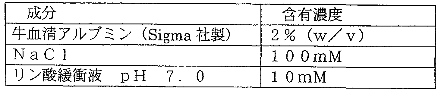
- Eight DNA microarrays prepared in Example 5 were prepared. The eight DNA microarrays were immersed in a blocking solution having the composition shown below, and allowed to stand at room temperature for 3 hours. Treatment.
- the DNA microarray was rinsed with the basic buffer.
- One piece of each of the blocking-treated DNA microarrays was immersed in each of the prepared eight hybridization solutions and sealed with a plastic pack. Each was subjected to a hybridization reaction at 45 ° C for 3 hours.
- Room temperature 1 minute XI times The composition of 2XSSC is 17.5 g of NaC 11 / trisodium citrate. Dihydrate is 8.8 gZl, adjusted to pH 7.0 using sodium hydroxide solution. After the washing was completed, the solution was drained using an air blow and dried. After drying, the amount of fluorescence of each spot was measured using a DNA array scanner (GenePix 4000 B, manufactured by Axon). The measurement data was analyzed using the analysis software (GenePixPro3.0) attached to the DNA array scanner.
- the density ratio of each probe was determined from the measured amount of fluorescence. See Table 7. ⁇
- the density ratio was determined as a relative ratio to the fixed density of the DNA probe in Ink No. 5 assuming 100%.
- the labeling ratio of each external standard nucleic acid is measured separately, and the density ratio obtained by correcting the labeling ratio is shown.
- the density ratio (fluorescence amount) of Ink.- 1 to 4 is less than 200% of the theoretical value because the fixed amount of probe nucleic acid reaches the allowable amount (saturation amount) of the substrate. Because it is.
- Example 8 Preparation of calibration curve using external standard nucleic acid From the results of Example 7, the fixed amount of each probe was tested, and a calibration curve was prepared using the corrected data. Specifically, using the DNA microarray prepared in Example 5, a hybridization experiment was performed in the same manner as in Example 6. On the other hand, the addition amount of the external standard nucleic acid was a mixed amount shown below. Mixing ratio of external standard nucleic acid
- Example 9 Preparation of a calibration curve using PCR products Using the nucleic acid for internal standard prepared in Example 1 and the primer for internal standard prepared in Example 2, a calibration curve of PCR amplification product was prepared. Went.
- a PCR amplification reaction was performed according to a standard method using a combination of primers for the internal standard shown in Table 8.
- the composition of the PCR reaction solution is as follows.
- the reverse primer used in the present example was the one prepared by performing fluorescent labeling at the 5 ′ end prepared in Example 3.
- the asterisk indicates that a fluorescent label is attached.
- the following example shows an example in which a probe for concentration standard added for abundance or density analysis of the probe formed by the same method as that for forming a nucleic acid probe is used.
- FIG. 5 is a diagram schematically illustrating a planar arrangement and a cross-sectional structure of a probe array in which a plurality of probes are coupled in a spot shape on a substrate.
- Reference numeral 501 denotes a probe group prepared under arbitrary conditions
- reference numeral 502 denotes a probe group having a known nucleic acid concentration.
- 503 is a substrate
- 504 is a surface treatment layer made of an organic substance
- 5 00 is a nucleic acid probe.
- a single-stranded nucleic acid of SEQ ID NO: 9 (40-mer of dT) was synthesized at the request of a DNA synthesizer (Vex).
- a sulfonyl 10 group (SH) was introduced into the 5 ′ end of the single-stranded DNA of SEQ ID NO: 1 using a zinc modifier (Dallen Research) at the time of synthesis.
- a zinc modifier Dallen Research
- the single-stranded DNA of SEQ ID NO: 1 described in [3] was used at a final concentration of 8 M at a concentration of 7.5% by weight of dariserin, 7.5% by weight of urea, 7.5% by weight of thiodiglycol, and 20% by weight of acetylene alcohol (trade name: Acetyleneol EH (manufactured by Kawaken Fine Chemical Co., Ltd.) was dissolved in a solution containing 1%.
- a printer head BC-50 (Canon) for a bubble jet printer BJF-850 (Cannon) using a bubble jet method, which is a kind of the solar jet method, was modified to discharge several hundreds of solutions.
- the modified head was mounted on a discharge drawing machine that was modified to discharge ink onto a flat quartz substrate.
- a discharge drawing machine Into the modified tank of this head, Several tens of liquids were injected, and an EMC S-treated substrate was mounted on a discharge drawing machine, and spotted on an EMCS-treated surface.
- the discharge amount at the time of spotting was 4 plZd rodlet, and the spotting range was 200 dpi, that is, 127 m pitch, within the range of 2 Omm X 3 Omm on the substrate. Under these conditions, the dots spotted in a matrix (157 rows and 236 columns) had a diameter of about 50 m.
- the substrate is allowed to stand in a humidified chamber for 30 minutes to allow the maleimide group on the substrate surface to react with the sulfanyl group (1SH) at the 5 'end of the nucleic acid probe, thereby immobilizing the DNA probe.
- the substrate was washed with pure water and stored in pure water.
- the DNA-bonded substrate was dried by blowing nitrogen gas immediately before analysis by TOF-SIMS, and further dried in a vacuum desiccator. Under these conditions, a total of 15 DNA chips were produced.
- the single-stranded DNA concentration of SEQ ID NO: 1 in the DNA solution used for spotting was selected to be 5 / zM and 2.5 M, and the same procedure was repeated for 25 and 35 DNA chips, respectively. Produced.
- the above three were used as standard DNA chips.
- a DNA chip using a general solution concentration of single-stranded DNA (about 8 M) was prepared under the same conditions as above.
- the same three single-stranded DNA solutions used in the preparation of the standard DNA chip were used in the first to third lines at the end.
- the first line at the end of the substrate is the spot of the probe formed with the DNA solution at the concentration of IOM
- the second line is the spot of the probe formed with the DNA solution at the concentration of 5 M
- the third line is the spot of the concentration of 2
- Rows other than the first to third rows at the end were prepared using another lot of a 8 M solution of DNA.
- the number of probe arrays can be set arbitrarily.
- the standard DNA chips prepared at three different DNA concentrations were each washed with an acid to dissolve the DNA probe, and then concentrated until the acid solution was not dispersed to 1 ml or less. Next, ultrapure water was added to the concentrated solution to adjust the volume to 1 ml. This solution was introduced into an ICP-MS apparatus, and the weight of P (phosphorus) was measured. Based on this value, the average value of the number of DNA molecules (per dot) of the DNA probe prepared at each concentration was determined in consideration of the number of dots (number of spots) used for the measurement.
- the number of substrates prepared at each DNA concentration can be determined by considering the P detection limit concentration (about 10 ppb) in the ICP-MS device and the number of DNA molecules on each substrate. The number is not limited to the number shown in the tenth embodiment.
- the DNA chip shown in Fig. 5 was transported to the analysis room of a time-of-flight secondary ion mass spectrometer (TO FS IMS IV: ION TOF), and the injection dose of primary ions was adjusted under the conditions shown in Table 1. Irradiated until 1 X 10 12 at oms / cm 2 , accumulated secondary intensity P0 3 — (mass-to-charge ratio: 78.958 amu (atom mass unit)) was calculated, and cumulative intensity was calculated. .
- Figure 6 shows the average secondary ion intensity measured at the dot positions at each DNA concentration in the first to third lines, and the density of DNA formation.
- the formation density of DNA and the second-order ion intensity in the dots in each row were proportional.
- the nucleic acid concentration in the D NA solution ejected by the ink jet printer one formed as 8 M, for nucleic acid probes was T OF- S IMS measurements, P0 3 - each dot obtained from the count number (D NA number of molecules per dot) formation density of D NA in DOO is approximately 2. 0 X 1 0 7, the variation in 1 within 0 dots arbitrarily selected, a 2 0% or less was.
- Image display of formation density distribution A plurality of nucleic acid probes of the same sample used in Example 12 were set, primary ions were scanned on the sample surface, and the generated secondary ions were made to correspond to each scanning point.
- the intensity of ⁇ _ obtained at each scanning point is divided into a plurality of steps to set a pseudo color, and the intensity distribution of ⁇ ⁇ ⁇ ⁇ _, that is, the area density of nucleic acids (the number of nucleic acid molecules per probe / dot) )
- the distribution of the formation density was quantitatively compared.
- the present invention it is possible to provide a DN microarray capable of simple and detailed analysis. Specifically, the assay operation can be easily performed at the same time as the actual nucleic acid detection operation. In addition, the test work itself can be performed at the same time, resulting in highly accurate test results. Therefore, by using the DNA microarray according to the present invention, the quantitative accuracy of quantitative analysis of the target nucleic acid molecule is significantly improved. In addition, in the quantitative analysis operation, the user can use the present invention very easily by using the primer for internal standard amplification and the external standard nucleic acid as a detection kit corresponding to the DNA microarray according to the present invention. it can.
- a nucleic acid probe and a dot for concentration reference which are formed by the same method as the formation of the nucleic acid probe and the dot and whose average nucleic acid density per dot is determined, are provided on the substrate.
- the nucleic acid concentration of an unknown concentration of the nucleic acid probe-dot can be determined by secondary ion mass spectrometry, and reproducibility and quantification are improved as compared with the conventional method.
- the reliability of a DNA microarray product can be improved. It is also possible to display the distribution of the formation density of nucleic acid probes and dots arranged in a matrix in a DNA microarray as an image.
Abstract
Description


Claims
Priority Applications (3)
| Application Number | Priority Date | Filing Date | Title |
|---|---|---|---|
| EP03733543A EP1522853A4 (en) | 2002-06-24 | 2003-06-23 | DNA MICROARRAY WITH STANDARD PROBE AND KIT CONTAINING THE MICROARRAY |
| US10/736,545 US20040132080A1 (en) | 2002-06-24 | 2003-12-17 | DNA micro-array having standard probe and kit including the array |
| US12/264,241 US20090093373A1 (en) | 2002-06-24 | 2008-11-03 | Dna micro-array having standard probe and kit including the array |
Applications Claiming Priority (4)
| Application Number | Priority Date | Filing Date | Title |
|---|---|---|---|
| JP2002-183249 | 2002-06-24 | ||
| JP2002183249A JP3998237B2 (ja) | 2002-06-24 | 2002-06-24 | 含有量検定用標準プローブを持つdnaマイクロアレイ及び検出用キット |
| JP2002191390A JP4040372B2 (ja) | 2002-06-28 | 2002-06-28 | 核酸チップおよび核酸チップの分析方法 |
| JP2002-191390 | 2002-06-28 |
Related Child Applications (1)
| Application Number | Title | Priority Date | Filing Date |
|---|---|---|---|
| US10/736,545 Continuation US20040132080A1 (en) | 2002-06-24 | 2003-12-17 | DNA micro-array having standard probe and kit including the array |
Publications (1)
| Publication Number | Publication Date |
|---|---|
| WO2004001412A1 true WO2004001412A1 (ja) | 2003-12-31 |
Family
ID=30002262
Family Applications (1)
| Application Number | Title | Priority Date | Filing Date |
|---|---|---|---|
| PCT/JP2003/007918 WO2004001412A1 (ja) | 2002-06-24 | 2003-06-23 | 標準プローブを持つdnaマイクロアレイ及び該アレイを有するキット |
Country Status (4)
| Country | Link |
|---|---|
| US (2) | US20040132080A1 (ja) |
| EP (1) | EP1522853A4 (ja) |
| CN (1) | CN1668923A (ja) |
| WO (1) | WO2004001412A1 (ja) |
Cited By (1)
| Publication number | Priority date | Publication date | Assignee | Title |
|---|---|---|---|---|
| EP1591543A3 (en) * | 2004-04-26 | 2005-11-30 | Canon Kabushiki Kaisha | PCR amplification reaction apparatus and method for PCR amplification reaction using apparatus |
Families Citing this family (39)
| Publication number | Priority date | Publication date | Assignee | Title |
|---|---|---|---|---|
| WO2003006676A2 (en) | 2001-07-13 | 2003-01-23 | Nanosphere, Inc. | Method for immobilizing molecules onto surfaces |
| US7461730B2 (en) * | 2001-09-05 | 2008-12-09 | Inventive Travelware | Handle apparatus with cantilevered handle grip for luggage case |
| US7833745B2 (en) * | 2003-09-11 | 2010-11-16 | E. I. Du Pont De Nemours And Company | Direct detection method for products of cellular metabolism using ToF-SIMS |
| JP4711326B2 (ja) * | 2003-12-22 | 2011-06-29 | キヤノン株式会社 | 検定試料および検量線の作製方法 |
| JP4541731B2 (ja) * | 2004-03-12 | 2010-09-08 | キヤノン株式会社 | 核酸検出方法 |
| KR100666754B1 (ko) | 2004-10-13 | 2007-01-09 | 한국표준과학연구원 | Dna의 정량방법 |
| JP2007010413A (ja) * | 2005-06-29 | 2007-01-18 | Canon Inc | 核酸ハイブリッドの融点測定方法及びそのための装置 |
| JP4789518B2 (ja) * | 2005-06-30 | 2011-10-12 | キヤノン株式会社 | 製造条件付きプローブ固定担体を用いた標的物質の検出方法ならびにそのための装置、キット及びシステム |
| US7642086B2 (en) * | 2005-08-09 | 2010-01-05 | Canon Kabushiki Kaisha | Labeled probe bound object, method for producing the same and method for using the same |
| US8962302B2 (en) * | 2005-11-07 | 2015-02-24 | Canon Kabushiki Kaisha | Biological tissue processing substrate for fixing proteins or protein degradation products in tissue for analysis |
| US20070128611A1 (en) * | 2005-12-02 | 2007-06-07 | Nelson Charles F | Negative control probes |
| US20090011413A1 (en) * | 2005-12-14 | 2009-01-08 | Canon Kabushiki Kaisha | Method for screening colon cancer cells and gene set used for examination of colon cancer |
| WO2007114517A1 (en) * | 2006-03-31 | 2007-10-11 | Canon Kabushiki Kaisha | Probe, probe set, probe-immobilized carrier, and genetic testing method |
| US8009889B2 (en) * | 2006-06-27 | 2011-08-30 | Affymetrix, Inc. | Feature intensity reconstruction of biological probe array |
| US7629124B2 (en) | 2006-06-30 | 2009-12-08 | Canon U.S. Life Sciences, Inc. | Real-time PCR in micro-channels |
| JP4861788B2 (ja) * | 2006-10-11 | 2012-01-25 | キヤノン株式会社 | 生体標本の処理方法及び解析方法 |
| US20080102452A1 (en) * | 2006-10-31 | 2008-05-01 | Roberts Douglas N | Control nucleic acid constructs for use in analysis of methylation status |
| JP4426600B2 (ja) * | 2007-04-27 | 2010-03-03 | キヤノン株式会社 | 処理溶液の使用方法 |
| JP2008275550A (ja) | 2007-05-07 | 2008-11-13 | Canon Inc | 検体の前処理方法及び検体の分析方法 |
| JP4854590B2 (ja) * | 2007-05-11 | 2012-01-18 | キヤノン株式会社 | 飛行時間型2次イオン質量分析装置 |
| JP5078440B2 (ja) * | 2007-05-25 | 2012-11-21 | キヤノン株式会社 | 情報取得方法 |
| US8380457B2 (en) * | 2007-08-29 | 2013-02-19 | Canon U.S. Life Sciences, Inc. | Microfluidic devices with integrated resistive heater electrodes including systems and methods for controlling and measuring the temperatures of such heater electrodes |
| DE102007055386B4 (de) | 2007-11-20 | 2015-07-16 | Boehringer Ingelheim Vetmedica Gmbh | Verfahren zur Kalibrierung eines Sensorelements |
| JP2010004873A (ja) * | 2008-05-27 | 2010-01-14 | Fujifilm Corp | 核酸マイクロアレイを用いた解析方法 |
| US8384021B2 (en) | 2009-04-10 | 2013-02-26 | Canon Kabushiki Kaisha | Method of forming mass image |
| JP5698471B2 (ja) * | 2009-06-30 | 2015-04-08 | シスメックス株式会社 | マイクロアレイを用いた核酸の検出方法およびマイクロアレイデータ解析用プログラム |
| JP5607373B2 (ja) | 2010-01-04 | 2014-10-15 | 東洋鋼鈑株式会社 | マイクロアレイの検出方法 |
| EP2580354A4 (en) * | 2010-06-11 | 2013-10-30 | Pathogenica Inc | NUCLEIC ACIDS FOR MULTIPLEX DETECTION OF ORGANISMS AND METHOD FOR THEIR USE AND MANUFACTURE |
| CN103890161A (zh) * | 2011-10-31 | 2014-06-25 | 株式会社日立高新技术 | 核酸扩增方法、核酸基板、核酸分析方法及核酸分析装置 |
| CN103570782A (zh) * | 2012-07-23 | 2014-02-12 | 医影生物纳米技术(苏州)有限公司 | 纳米分子探针fmau前体及其制备方法 |
| CN106591303A (zh) * | 2017-01-23 | 2017-04-26 | 苏州贝斯派生物科技有限公司 | 一种脱氧核糖核酸探针集、试剂盒及其制备方法与应用 |
| GB201701688D0 (en) | 2017-02-01 | 2017-03-15 | Illumia Inc | System and method with fiducials in non-recliner layouts |
| SG11201906442TA (en) | 2017-02-01 | 2019-08-27 | Illumina Inc | System and method with fiducials responding to multiple excitation frequencies |
| GB201701689D0 (en) | 2017-02-01 | 2017-03-15 | Illumia Inc | System and method with fiducials of non-closed shapes |
| GB201701691D0 (en) | 2017-02-01 | 2017-03-15 | Illumina Inc | System and method with reflective fiducials |
| GB201701686D0 (en) | 2017-02-01 | 2017-03-15 | Illunina Inc | System & method with fiducials having offset layouts |
| WO2021025935A1 (en) * | 2019-08-02 | 2021-02-11 | Georgetown University | Dna-fish method for measurement of telomere length |
| CN112980891B (zh) * | 2019-12-16 | 2023-12-12 | 中国科学院分子植物科学卓越创新中心 | 一种基于CRISPR-Cas的大肠杆菌基因组编辑工具 |
| CN111394475A (zh) * | 2020-03-26 | 2020-07-10 | 迈杰转化医学研究(苏州)有限公司 | 一种核苷酸组合物、包含其的检测试剂盒及其应用 |
Citations (5)
| Publication number | Priority date | Publication date | Assignee | Title |
|---|---|---|---|---|
| JP2000270896A (ja) * | 1999-01-28 | 2000-10-03 | Canon Inc | プローブ結合基板、プローブ結合基板の製造方法、プローブアレイ、標的物質の検出方法、サンプル中の一本鎖核酸の塩基配列を特定する方法、及びサンプル中の標的物質の定量方法 |
| WO2001031055A2 (en) * | 1999-10-28 | 2001-05-03 | Facultes Universitaires Notre-Dame De La Paix | Method and kit for the screening and/or the quantification of multiple homologous nucleic acid sequences on arrays |
| JP2001526381A (ja) * | 1997-12-05 | 2001-12-18 | マックス−プランク−ゲゼルシャフト・ツア・フェルデルング・デア・ヴィッセンシャフテン・エー・ファオ | マトリックス補助レーザー脱着/イオン化質量分光分析による核酸の同定方法 |
| JP2002071688A (ja) * | 2000-09-01 | 2002-03-12 | Hitachi Software Eng Co Ltd | バイオチップを用いたハイブリダイゼーション反応の実験結果表示方法及び実験誤差評価方法 |
| JP2003194812A (ja) * | 2001-12-27 | 2003-07-09 | Mitsubishi Heavy Ind Ltd | マイクロアレイ及びこれを用いた測定方法 |
Family Cites Families (15)
| Publication number | Priority date | Publication date | Assignee | Title |
|---|---|---|---|---|
| US4683195A (en) * | 1986-01-30 | 1987-07-28 | Cetus Corporation | Process for amplifying, detecting, and/or-cloning nucleic acid sequences |
| US4965188A (en) * | 1986-08-22 | 1990-10-23 | Cetus Corporation | Process for amplifying, detecting, and/or cloning nucleic acid sequences using a thermostable enzyme |
| US4683202A (en) * | 1985-03-28 | 1987-07-28 | Cetus Corporation | Process for amplifying nucleic acid sequences |
| US6309822B1 (en) * | 1989-06-07 | 2001-10-30 | Affymetrix, Inc. | Method for comparing copy number of nucleic acid sequences |
| US5474796A (en) * | 1991-09-04 | 1995-12-12 | Protogene Laboratories, Inc. | Method and apparatus for conducting an array of chemical reactions on a support surface |
| BE1010608A3 (fr) * | 1996-09-09 | 1998-11-03 | Jose Remacle | Procede de quantification d'une sequence d'acides nucleiques. |
| US6024925A (en) * | 1997-01-23 | 2000-02-15 | Sequenom, Inc. | Systems and methods for preparing low volume analyte array elements |
| DK0937096T3 (da) * | 1996-11-06 | 2004-06-14 | Sequenom Inc | Fremgangsmåde til massespektrometri-analyse |
| DE19782095T1 (de) * | 1996-11-06 | 2000-03-23 | Sequenom Inc | DNA-Diagnose auf der Basis von Massenspektrometrie |
| JP4313861B2 (ja) * | 1997-08-01 | 2009-08-12 | キヤノン株式会社 | プローブアレイの製造方法 |
| US7122303B2 (en) * | 1999-09-17 | 2006-10-17 | Agilent Technologies, Inc. | Arrays comprising background features that provide for a measure of a non-specific binding and methods for using the same |
| US20010026919A1 (en) * | 2000-02-08 | 2001-10-04 | Alex Chenchik | Nucleic acid assays employing universal arrays |
| JP2002071687A (ja) * | 2000-08-31 | 2002-03-12 | Canon Inc | 変異遺伝子のスクリーニング方法 |
| EP1186673A3 (en) * | 2000-09-11 | 2003-03-26 | Agilent Technologies, Inc. (a Delaware corporation) | Calibration of molecular array data |
| JP2004024203A (ja) * | 2002-06-28 | 2004-01-29 | Canon Inc | 飛行時間型二次イオン質量分析法によるrnaの分析方法 |
-
2003
- 2003-06-23 CN CNA038168146A patent/CN1668923A/zh active Pending
- 2003-06-23 EP EP03733543A patent/EP1522853A4/en not_active Withdrawn
- 2003-06-23 WO PCT/JP2003/007918 patent/WO2004001412A1/ja active Application Filing
- 2003-12-17 US US10/736,545 patent/US20040132080A1/en not_active Abandoned
-
2008
- 2008-11-03 US US12/264,241 patent/US20090093373A1/en not_active Abandoned
Patent Citations (5)
| Publication number | Priority date | Publication date | Assignee | Title |
|---|---|---|---|---|
| JP2001526381A (ja) * | 1997-12-05 | 2001-12-18 | マックス−プランク−ゲゼルシャフト・ツア・フェルデルング・デア・ヴィッセンシャフテン・エー・ファオ | マトリックス補助レーザー脱着/イオン化質量分光分析による核酸の同定方法 |
| JP2000270896A (ja) * | 1999-01-28 | 2000-10-03 | Canon Inc | プローブ結合基板、プローブ結合基板の製造方法、プローブアレイ、標的物質の検出方法、サンプル中の一本鎖核酸の塩基配列を特定する方法、及びサンプル中の標的物質の定量方法 |
| WO2001031055A2 (en) * | 1999-10-28 | 2001-05-03 | Facultes Universitaires Notre-Dame De La Paix | Method and kit for the screening and/or the quantification of multiple homologous nucleic acid sequences on arrays |
| JP2002071688A (ja) * | 2000-09-01 | 2002-03-12 | Hitachi Software Eng Co Ltd | バイオチップを用いたハイブリダイゼーション反応の実験結果表示方法及び実験誤差評価方法 |
| JP2003194812A (ja) * | 2001-12-27 | 2003-07-09 | Mitsubishi Heavy Ind Ltd | マイクロアレイ及びこれを用いた測定方法 |
Cited By (2)
| Publication number | Priority date | Publication date | Assignee | Title |
|---|---|---|---|---|
| EP1591543A3 (en) * | 2004-04-26 | 2005-11-30 | Canon Kabushiki Kaisha | PCR amplification reaction apparatus and method for PCR amplification reaction using apparatus |
| US7569366B2 (en) | 2004-04-26 | 2009-08-04 | Canon Kabushiki Kaisha | PCR amplification reaction apparatus and method for PCR amplification reaction using apparatus |
Also Published As
| Publication number | Publication date |
|---|---|
| EP1522853A4 (en) | 2005-10-19 |
| CN1668923A (zh) | 2005-09-14 |
| US20090093373A1 (en) | 2009-04-09 |
| EP1522853A1 (en) | 2005-04-13 |
| US20040132080A1 (en) | 2004-07-08 |
Similar Documents
| Publication | Publication Date | Title |
|---|---|---|
| WO2004001412A1 (ja) | 標準プローブを持つdnaマイクロアレイ及び該アレイを有するキット | |
| EP1138782B1 (en) | Multiplex sequence variation analysis of DNA samples by mass spectrometry | |
| Guo et al. | Development of a single nucleotide polymorphism DNA microarray for the detection and genotyping of the SARS coronavirus | |
| JP5404832B2 (ja) | ハイブリッド体の検出によるサンプル核酸の検出方法に用いる前記プローブセットの設計方法 | |
| AU2005284980A1 (en) | Methods for long-range sequence analysis of nucleic acids | |
| JP3658397B2 (ja) | 飛行時間型二次イオン質量分析法による素子の情報取得方法、および、情報取得装置 | |
| JP3998237B2 (ja) | 含有量検定用標準プローブを持つdnaマイクロアレイ及び検出用キット | |
| JP5229895B2 (ja) | 核酸標準物質 | |
| Koch-Paiz et al. | Estimation of relative mRNA content by filter hybridization to a polyuridylic probe | |
| US20040137491A1 (en) | Method of analyzing probe carrier using time-of-flight secondary ion mass spectrometry | |
| EP1519189A1 (en) | Method of analyzing probe support by using flying time secondary ion mass spectrometry | |
| WO2004067765A2 (en) | Organism fingerprinting using nicking agents | |
| JP4040372B2 (ja) | 核酸チップおよび核酸チップの分析方法 | |
| JP4380535B2 (ja) | プローブ担体および該プローブ担体の分析方法 | |
| JP4532874B2 (ja) | 質量分析可能な原子群を構成単位とする鎖状構造を有する分子に対して付加情報を付与して、情報記録コードとして利用する方法 | |
| JP2007300829A (ja) | Dnaマイクロアレイ等に供する検体の調製方法 | |
| US20060147913A1 (en) | Method of analyzing substance on substrate by mass spectrometry | |
| US8518642B2 (en) | Method of analyzing probe nucleic acid, microarray and kit for the same | |
| JP2004024203A (ja) | 飛行時間型二次イオン質量分析法によるrnaの分析方法 | |
| JP2004037120A (ja) | 飛行時間型二次イオン質量分析法による有機膜の組成分析方法 | |
| JP4353504B2 (ja) | 核酸の増幅方法および標識化方法、これを用いた核酸検出方法 | |
| JP3990948B2 (ja) | ハロゲン標識による飛行時間型二次イオン質量分析法を用いた核酸ハイブリッド体の分析方法 | |
| JP3890265B2 (ja) | 飛行時間型二次イオン質量分析法による核酸チップの分析方法 | |
| JP3990946B2 (ja) | ハロゲン標識による飛行時間型二次イオン質量分析法を用いたプローブ担体の分析方法 | |
| WO2014188941A1 (ja) | 核酸分析キットおよび核酸分析方法 |
Legal Events
| Date | Code | Title | Description |
|---|---|---|---|
| WWE | Wipo information: entry into national phase |
Ref document number: 10736545 Country of ref document: US |
|
| AK | Designated states |
Kind code of ref document: A1 Designated state(s): CN KR US |
|
| AL | Designated countries for regional patents |
Kind code of ref document: A1 Designated state(s): AT BE BG CH CY CZ DE DK EE ES FI FR GB GR HU IE IT LU MC NL PT RO SE SI SK TR |
|
| 121 | Ep: the epo has been informed by wipo that ep was designated in this application | ||
| DFPE | Request for preliminary examination filed prior to expiration of 19th month from priority date (pct application filed before 20040101) | ||
| WWE | Wipo information: entry into national phase |
Ref document number: 2003733543 Country of ref document: EP |
|
| WWE | Wipo information: entry into national phase |
Ref document number: 20038168146 Country of ref document: CN |
|
| WWP | Wipo information: published in national office |
Ref document number: 2003733543 Country of ref document: EP |