US9834606B2 - Anti-PD1 antibodies and their use as therapeutics and diagnostics - Google Patents
Anti-PD1 antibodies and their use as therapeutics and diagnostics Download PDFInfo
- Publication number
- US9834606B2 US9834606B2 US14/736,966 US201514736966A US9834606B2 US 9834606 B2 US9834606 B2 US 9834606B2 US 201514736966 A US201514736966 A US 201514736966A US 9834606 B2 US9834606 B2 US 9834606B2
- Authority
- US
- United States
- Prior art keywords
- seq
- antibody
- cells
- mabs
- binding
- Prior art date
- Legal status (The legal status is an assumption and is not a legal conclusion. Google has not performed a legal analysis and makes no representation as to the accuracy of the status listed.)
- Expired - Fee Related, expires
Links
Images


Classifications
-
- A—HUMAN NECESSITIES
- A61—MEDICAL OR VETERINARY SCIENCE; HYGIENE
- A61K—PREPARATIONS FOR MEDICAL, DENTAL OR TOILETRY PURPOSES
- A61K39/00—Medicinal preparations containing antigens or antibodies
- A61K39/395—Antibodies; Immunoglobulins; Immune serum, e.g. antilymphocytic serum
- A61K39/39591—Stabilisation, fragmentation
-
- A—HUMAN NECESSITIES
- A61—MEDICAL OR VETERINARY SCIENCE; HYGIENE
- A61P—SPECIFIC THERAPEUTIC ACTIVITY OF CHEMICAL COMPOUNDS OR MEDICINAL PREPARATIONS
- A61P25/00—Drugs for disorders of the nervous system
-
- A—HUMAN NECESSITIES
- A61—MEDICAL OR VETERINARY SCIENCE; HYGIENE
- A61P—SPECIFIC THERAPEUTIC ACTIVITY OF CHEMICAL COMPOUNDS OR MEDICINAL PREPARATIONS
- A61P31/00—Antiinfectives, i.e. antibiotics, antiseptics, chemotherapeutics
- A61P31/12—Antivirals
-
- A—HUMAN NECESSITIES
- A61—MEDICAL OR VETERINARY SCIENCE; HYGIENE
- A61P—SPECIFIC THERAPEUTIC ACTIVITY OF CHEMICAL COMPOUNDS OR MEDICINAL PREPARATIONS
- A61P35/00—Antineoplastic agents
-
- C—CHEMISTRY; METALLURGY
- C07—ORGANIC CHEMISTRY
- C07K—PEPTIDES
- C07K16/00—Immunoglobulins [IG], e.g. monoclonal or polyclonal antibodies
- C07K16/18—Immunoglobulins [IG], e.g. monoclonal or polyclonal antibodies against material from animals or humans
- C07K16/28—Immunoglobulins [IG], e.g. monoclonal or polyclonal antibodies against material from animals or humans against receptors, cell surface antigens or cell surface determinants
- C07K16/2803—Immunoglobulins [IG], e.g. monoclonal or polyclonal antibodies against material from animals or humans against receptors, cell surface antigens or cell surface determinants against the immunoglobulin superfamily
- C07K16/2818—Immunoglobulins [IG], e.g. monoclonal or polyclonal antibodies against material from animals or humans against receptors, cell surface antigens or cell surface determinants against the immunoglobulin superfamily against CD28 or CD152
-
- A—HUMAN NECESSITIES
- A61—MEDICAL OR VETERINARY SCIENCE; HYGIENE
- A61K—PREPARATIONS FOR MEDICAL, DENTAL OR TOILETRY PURPOSES
- A61K39/00—Medicinal preparations containing antigens or antibodies
- A61K2039/505—Medicinal preparations containing antigens or antibodies comprising antibodies
-
- C—CHEMISTRY; METALLURGY
- C07—ORGANIC CHEMISTRY
- C07K—PEPTIDES
- C07K2317/00—Immunoglobulins specific features
- C07K2317/20—Immunoglobulins specific features characterized by taxonomic origin
- C07K2317/24—Immunoglobulins specific features characterized by taxonomic origin containing regions, domains or residues from different species, e.g. chimeric, humanized or veneered
-
- C—CHEMISTRY; METALLURGY
- C07—ORGANIC CHEMISTRY
- C07K—PEPTIDES
- C07K2317/00—Immunoglobulins specific features
- C07K2317/30—Immunoglobulins specific features characterized by aspects of specificity or valency
- C07K2317/33—Crossreactivity, e.g. for species or epitope, or lack of said crossreactivity
-
- C—CHEMISTRY; METALLURGY
- C07—ORGANIC CHEMISTRY
- C07K—PEPTIDES
- C07K2317/00—Immunoglobulins specific features
- C07K2317/30—Immunoglobulins specific features characterized by aspects of specificity or valency
- C07K2317/34—Identification of a linear epitope shorter than 20 amino acid residues or of a conformational epitope defined by amino acid residues
-
- C—CHEMISTRY; METALLURGY
- C07—ORGANIC CHEMISTRY
- C07K—PEPTIDES
- C07K2317/00—Immunoglobulins specific features
- C07K2317/50—Immunoglobulins specific features characterized by immunoglobulin fragments
- C07K2317/52—Constant or Fc region; Isotype
-
- C—CHEMISTRY; METALLURGY
- C07—ORGANIC CHEMISTRY
- C07K—PEPTIDES
- C07K2317/00—Immunoglobulins specific features
- C07K2317/50—Immunoglobulins specific features characterized by immunoglobulin fragments
- C07K2317/52—Constant or Fc region; Isotype
- C07K2317/53—Hinge
-
- C—CHEMISTRY; METALLURGY
- C07—ORGANIC CHEMISTRY
- C07K—PEPTIDES
- C07K2317/00—Immunoglobulins specific features
- C07K2317/50—Immunoglobulins specific features characterized by immunoglobulin fragments
- C07K2317/55—Fab or Fab'
-
- C—CHEMISTRY; METALLURGY
- C07—ORGANIC CHEMISTRY
- C07K—PEPTIDES
- C07K2317/00—Immunoglobulins specific features
- C07K2317/50—Immunoglobulins specific features characterized by immunoglobulin fragments
- C07K2317/56—Immunoglobulins specific features characterized by immunoglobulin fragments variable (Fv) region, i.e. VH and/or VL
- C07K2317/565—Complementarity determining region [CDR]
-
- C—CHEMISTRY; METALLURGY
- C07—ORGANIC CHEMISTRY
- C07K—PEPTIDES
- C07K2317/00—Immunoglobulins specific features
- C07K2317/50—Immunoglobulins specific features characterized by immunoglobulin fragments
- C07K2317/56—Immunoglobulins specific features characterized by immunoglobulin fragments variable (Fv) region, i.e. VH and/or VL
- C07K2317/567—Framework region [FR]
-
- C—CHEMISTRY; METALLURGY
- C07—ORGANIC CHEMISTRY
- C07K—PEPTIDES
- C07K2317/00—Immunoglobulins specific features
- C07K2317/50—Immunoglobulins specific features characterized by immunoglobulin fragments
- C07K2317/56—Immunoglobulins specific features characterized by immunoglobulin fragments variable (Fv) region, i.e. VH and/or VL
- C07K2317/569—Single domain, e.g. dAb, sdAb, VHH, VNAR or nanobody®
-
- C—CHEMISTRY; METALLURGY
- C07—ORGANIC CHEMISTRY
- C07K—PEPTIDES
- C07K2317/00—Immunoglobulins specific features
- C07K2317/70—Immunoglobulins specific features characterized by effect upon binding to a cell or to an antigen
-
- C—CHEMISTRY; METALLURGY
- C07—ORGANIC CHEMISTRY
- C07K—PEPTIDES
- C07K2317/00—Immunoglobulins specific features
- C07K2317/70—Immunoglobulins specific features characterized by effect upon binding to a cell or to an antigen
- C07K2317/71—Decreased effector function due to an Fc-modification
-
- C—CHEMISTRY; METALLURGY
- C07—ORGANIC CHEMISTRY
- C07K—PEPTIDES
- C07K2317/00—Immunoglobulins specific features
- C07K2317/70—Immunoglobulins specific features characterized by effect upon binding to a cell or to an antigen
- C07K2317/73—Inducing cell death, e.g. apoptosis, necrosis or inhibition of cell proliferation
-
- C—CHEMISTRY; METALLURGY
- C07—ORGANIC CHEMISTRY
- C07K—PEPTIDES
- C07K2317/00—Immunoglobulins specific features
- C07K2317/70—Immunoglobulins specific features characterized by effect upon binding to a cell or to an antigen
- C07K2317/76—Antagonist effect on antigen, e.g. neutralization or inhibition of binding
-
- C—CHEMISTRY; METALLURGY
- C07—ORGANIC CHEMISTRY
- C07K—PEPTIDES
- C07K2317/00—Immunoglobulins specific features
- C07K2317/90—Immunoglobulins specific features characterized by (pharmaco)kinetic aspects or by stability of the immunoglobulin
- C07K2317/92—Affinity (KD), association rate (Ka), dissociation rate (Kd) or EC50 value
-
- C—CHEMISTRY; METALLURGY
- C07—ORGANIC CHEMISTRY
- C07K—PEPTIDES
- C07K2317/00—Immunoglobulins specific features
- C07K2317/90—Immunoglobulins specific features characterized by (pharmaco)kinetic aspects or by stability of the immunoglobulin
- C07K2317/94—Stability, e.g. half-life, pH, temperature or enzyme-resistance
Definitions
- PD-1 Programmed Death-1
- CD279 is a 55 KD receptor protein related to CD28/CTLA4 co-stimulatory/inhibitory receptor family (Blank et al., 2005 Cancer Immunol Immunother 54:307-314).
- the genes and cDNAs coding for PD-1 were cloned and characterized in mouse and human (Ishida et al., 1992 EMBO J 11:3887-3395; Shinohara et al., 1994 Genomics 23:704-706).
- the full length PD-1 contains 288 amino acid residues (NCBI accession number: NP_005009).
- PD-L1 B7-H1
- PD-L2 B7-DC
- PD-1 protein kinase-like protein
- T-cells T-cells
- B-cells monocytes and natural killer cells
- NK natural killer cells
- high level of PD-1 expression is often associated with activation of immune cells.
- human T-cell line Jurkat
- PHA phytohaemagglutinin
- phorbol ester (12-O-tetradecanoylphorbol-13-acetate, or TPA)
- TPA phytohaemagglutinin
- TPA phytohaemagglutinin
- TPA phorbol ester
- TILs tumor-infiltrating lymphocytes
- PD-1 ligand expression in tumor cells were reported in varieties of cancers involved in different types of tissues and organs such as lung (Konishi et al., 2004 Clin Cancer Res 10:5094-5100), liver (Shi et al., 2008 Int J Cancer 128:887-896; Gao et al., 2009 Clin Cancer Res 15:971-979), stomach (Wu et al., 2006 Acta Histochem 108:19-24), kidney (Thompson et al., 2004 Proc Natl Acad Sci 101:17174-17179; Thompson et al., 2007 Clin Cancer Res 13:1757-1761), breast (Ghebeh et al., 2006 Neoplasia 8:190-198), ovary (Hamanishi et al.
- Therapeutic modulation of PD-1 signaling by antagonist molecules may revert immune cells from tolerance, and reactivated to eradicate cancer and chronic viral infection (Blank et al., 2005 Cancer Immunol Immunother 54:307-314; Okazaki et al., 2007 Int Immunol 19:813-824).
- the invention provides methods and compositions for immune-inhibition of PD-1.
- the invention provides an antibody antigen binding domain which specifically binds human PD-1, and comprises a complementarity determining region (CDR) having a sequence selected from SEQ ID NOS 11-22, 31-42 and 59-63.
- CDR complementarity determining region
- the domain comprises a heavy chain variable region (Vh) or a light chain variable region (Vk) comprising:
- CDR-H1 (SEQ ID NO: 11, 17, 31, or 37), b) CDR-H2 (SEQ ID NO: 12, 18, 32, or 38), c) CDR-H3 (SEQ ID NO: 13, 18, 33, or 39); d) CDR-L1 (SEQ ID NO: 14, 20, 34, or 40), e) CDR-L2 (SEQ ID NO: 15, 21, 35, or 41), or f) CDR-L3 (SEQ ID NO: 16, 22, 36, or 42).
- the domain comprises a heavy chain variable region (Vh) and/or a light chain variable region (Vk) comprising:
- the domain comprises a heavy chain variable region (Vh) and a light chain variable region (Vk) comprising:
- the domain comprises a heavy chain variable region (Vh) or a light chain variable region (Vk) comprising:
- the domain comprises a heavy chain variable region (Vh) and a light chain variable region (Vk) comprising:
- the domain specifically binds PD1 residues: (a) K45 and I93 (AA numbering based on 2008 PNAS, 105:10483; equivalent to K58 and 1106 in SEQ ID NO 2); or (b) I93, L95 and P97 (AA numbering based on 2008 PNAS, 105:10483; equivalent to I106, L108 and P110 in SEQ ID NO 2).
- the domain induces IL-2 release in HuT78/PD-1 cells co-cultured with HEK293/OS8/PD-L1 cells or with EK293/OS8/PD-L2 cells, and/or inhibits IL-2 secretion in HuT78/P3Z cells co-cultured with HEK293/PD-L1 cells or with HEK293/PD-L2 cells.
- the invention also provides an antibody IgG4 heavy chain effector or constant domain comprising any of SEQ ID NO:83-88, particularly SEQ ID NO 87 or 88.
- the invention also provides antibodies, F(ab) or F(ab)2 comprising a subject PD-1 binding domain.
- the invention also provides antibodies comprising a subject PD-1 binding domain and a IgG4 heavy chain effector or constant domain comprising any of SEQ ID NO:83-88, particularly SEQ ID NO 87 or 88.
- the invention also provides a polynucleotide encoding a subject PD-1 binding domain, particularly cDNA sequences.
- the invention provides methods of using the subject domains by administering the domain to a person determined to have cancer or a viral infection or to otherwise be in need of PD-1 antagonism.
- the invention also provides fusion proteins comprising: (a) a single chain variable fragment (scFv) of an anti-human CD3 mAb OKT3 fused to the C-terminal domain (113-220) of mouse CD8 ⁇ (SEQ ID NO:89); or (b) the extracellular and transmembrane domains of human PD-1 fused to the cytoplasmic domain of human CD3 ⁇ chain (SEQ ID NO: 90).
- the invention also provides methods of using the subject fusion proteins, comprising assaying, screening or selecting anti-PD-1 antibodies with a cell line expressing the fusion protein.
- FIG. 1 Schematic presentation of PD-1/Fc (top) and PD-1/His (bottom).
- ECD extracellular domain.
- L linker.
- H His tag.
- Fc ⁇ 4Fc fragment from human IgG4.
- N N-terminus
- C C-terminus
- FIG. 2 Dose-dependent reaction curves of murine mAbs binding to human PD-1 in ELISA.
- the murine mAbs were indicated at top-left corner of each figure.
- MAb 317 and 517 share high degree of homology the variable region of heavy and light chains.
- the binding signal strength was indicated by direct OD 450 readings.
- the antigen, PD-1/His was coated at increasing concentrations up to 70 nanograms per well in a volume of 50 microliters. The method was described in Example 1.
- FIG. 3 Dose-dependent reaction curve of murine mAbs binding to human PD-1 expressed on live cells by FACS analyses.
- Murine antibody codes and EC 50 were indicated on each panel.
- MFI stands for mean fluorescence intensity.
- HuT78/PD-1 cells were suspended in 96-well plate at 5 ⁇ 10 4 cells per well for FACS.
- PD-1 mAbs binding to the cell surface target and FACS detection were performed as described in Example 1.
- FIG. 4 Schematic presentation of the cell co-culture systems used for assaying functional activities of anti-PD-1 mAbs.
- T-cells either CD4 + or CD8 + ) represent HuT78/PD-1 or primary T-cells in PBMCs.
- TCR T-cell receptor.
- N nucleus.
- C cytoplasm
- FIG. 5 Dose-dependent reaction curve of murine mAb-induced IL-2 secretion in HuT78/PD-1 cells co-cultured with HEK293/OS8/PD-L1 cells.
- Baseline Average IL-2 release induced by mIgGs at all tested concentrations.
- Top line Highest IL-2 release based on regression calculation by Prizm Software.
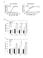
- FIG. 6 (A) Histograms showing IFN- ⁇ secretion induced by anti-PD-1 mAbs in PBMCs (Donor-19) co-cultured with cell line HEK293/OS8/PD-L1. (B) Histograms showing IFN- ⁇ secretion induced by anti-PD-1 mAbs in PBMCs (Donor-20) co-cultured with cell line HEK293/OS8/PD-L1.
- FIGS. 7 (A) and (B) ADCC activities of anti-PD-1 mAbs by co-culture of effector cells (NK92MI/PD-1) and target cells (HuT78/PD-1). Means were calculated from two data points of the representative experiments. The mAbs were added to concentration of 10 ⁇ m/ml. Experiment performed as described in Example 9.
- FIG. 8 Mapping the binding epitopes of anti-PD-1 mAbs by ELISA (up-panel) and Western Blot (lower panel). Conditioned media containing WT or Mt PD-1 were used to assess binding activity by ELISA and Western Blot. ** indicates the AA residues to which the mAb binding activity reduced to 25-50% of WT PD-1. *** indicates the AA residues to which the mAb binding activity reduced below 25% of WT PD-1.
- FIG. 9 IFN- ⁇ release induced by humanized anti-PD-1 mAbs in primary human PBMCs from different healthy donors, which were co-cultured with HEK293/OS8/PD-L1 cells.
- FIG. 10 Cytotoxicity of NK92MI/PD-1 cells enhanced by humanized anti-PD-1 mAbs, hu317 (A) and hu326 (B).
- the target lung cancer cells, SK-MES-1/PD-L1 were co-cultured with the effector cells at the (T to E) ratio of 1 to 2, and assayed as described in Example 12.
- FIG. 11 Individual tumor growth curves in three treatment groups, vehicle (PBS), human IgGs (huIgGs) and anti-PD-1 mAb (hu317-1/IgG4mt2). Each curve represents a tumor growth path, the tumor-bearing mice coded by numbers indicated on the right of each panel.
- Hep3B/OS8/PD-L1 cells established from hepatocellular carcinoma line Hep3B) were seeded at Day 1, PBMCs were implanted at Day 15 and three doses of hu317-1/IgG4mt2 were injected at Day 18, 28 and 38, respectively. Methods described in Example 12.
- PD-1 initiates inhibitory signaling in immune cells when engaged by its ligands, PD-L1 or PD-L2.
- the activation of PD-1 signaling promotes immune tolerance, leading to the cancers or virus-infected cells escaping from immune surveillance and cancer metastasis or viral load increase
- Inhibition of PD-1 mediated cellular signaling by therapeutic agents can activate immune cells including T-cells, B-cells and NK cells, and therefore enhance immune cell functions inhibiting cancer cell growth or viral infection, and restore immune surveillance and immune memory function to treat such human diseases.
- the invention provides antibodies whose functions are antagonistic to the ligand-induced and PD-1-mediated cellular signaling in immune cells.
- Murine anti-PD-1 antibodies were humanized to a high degree of similarity to human antibodies in the framework regions.
- the full antibodies made in the modified human IgG4 variant format have a unique set of features in the aspects of effector functions and physicochemical properties.
- the disclosed anti-PD-1 antibodies are suitable for therapeutic uses in cancer treatment, controlling viral infections and other human diseases that are mechanistically involved in exacerbated immune tolerance.
- antibody is used in the broadest sense and specifically covers antibodies (including full length monoclonal antibodies) and antibody fragments so long as they recognize PD-1.
- An antibody molecule is usually monospecific, but may also be described as idiospecific, heterospecific, or polyspecific.
- Antibody molecules bind by means of specific binding sites to specific antigenic determinants or epitopes on antigens.
- Antibody fragments comprise a portion of a full length antibody, generally the antigen binding or variable region thereof. Examples of antibody fragments include Fab, Fab′, F(ab′).sub.2, and Fv fragments; diabodies; linear antibodies; single-chain antibody molecules; and multispecific antibodies formed from antibody fragments.
- Monoclonal antibodies may be obtained by methods known to those skilled in the art. See, for example Kohler et al (1975); U.S. Pat. No. 4,376,110; Ausubel et al (1987-1999); Harlow et al (1988); and Colligan et al (1993).
- the mAbs of the invention may be of any immunoglobulin class including IgG, IgM, IgE, IgA, and any subclass thereof.
- a hybridoma producing a mAb may be cultivated in vitro or in vivo.
- High titers of mAbs can be obtained in in vivo production where cells from the individual hybridomas are injected intraperitoneally into mice, such as pristine-primed Balb/c mice to produce ascites fluid containing high concentrations of the desired mAbs.
- MAbs of isotype IgM or IgG may be purified from such ascites fluids, or from culture supernatants, using column chromatography methods well known to those of skill in the art.
- isolated polynucleotide refers to a polynucleotide segment or fragment which has been separated from sequences which flank it in a naturally occurring state, e.g., a DNA fragment which has been removed from the sequences which are normally adjacent to the fragment, e.g., the sequences adjacent to the fragment in a genome in which it naturally occurs.
- the term therefore includes, for example, a recombinant DNA which is incorporated into a vector, into an autonomously replicating plasmid or virus, or into the genomic DNA of a prokaryote or eukaryote, or which exists as a separate molecule (e.g., as a cDNA or a genomic or cDNA fragment produced by PCR or restriction enzyme digestion) independent of other sequences. It also includes a recombinant DNA, which is part of a hybrid gene encoding additional polypeptide sequence.
- a “construct” means any recombinant polynucleotide molecule such as a plasmid, cosmid, virus, autonomously replicating polynucleotide molecule, phage, or linear or circular single-stranded or double-stranded DNA or RNA polynucleotide molecule, derived from any source, capable of genomic integration or autonomous replication, comprising a polynucleotide molecule where one or more polynucleotide molecule has been linked in a functionally operative manner, i.e. operably linked.
- a recombinant construct will typically comprise the polynucleotides of the invention operably linked to transcriptional initiation regulatory sequences that will direct the transcription of the polynucleotide in the intended host cell.
- transcriptional initiation regulatory sequences that will direct the transcription of the polynucleotide in the intended host cell.
- Both heterologous and non-heterologous (i.e., endogenous) promoters can be employed to direct expression of the nucleic acids of the invention.
- a “vector” refers any recombinant polynucleotide construct that may be used for the purpose of transformation, i.e. the introduction of heterologous DNA into a host cell.
- a “plasmid” refers to a circular double stranded DNA loop into which additional DNA segments can be ligated.
- a viral vector Another type of vector is a viral vector, wherein additional DNA segments can be ligated into the viral genome.
- Certain vectors are capable of autonomous replication in a host cell into which they are introduced (e.g., bacterial vectors having a bacterial origin of replication and episomal mammalian vectors).
- vectors e.g., non-episomal mammalian vectors
- expression vectors are referred to herein as “expression vectors”.
- an “expression vector” as used herein refers to a nucleic acid molecule capable of replication and expressing a gene of interest when transformed, transfected or transduced into a host cell.
- the expression vectors comprise one or more phenotypic selectable markers and an origin of replication to ensure maintenance of the vector and to, if desired, provide amplification within the host.
- the expression vector further comprises a promoter to drive the expression of the polypeptide within the cells.
- Suitable expression vectors may be plasmids derived, for example, from pBR322 or various pUC plasmids, which are commercially available. Other expression vectors may be derived from bacteriophage, phagemid, or cosmid expression vectors.
- the invention provides mouse monoclonal antibodies identified from screening murine hybridoma clones as disclosed herein.
- compositions comprising complement determinant region (CDR) sequences, which mediate binding to the target antigens, PD-1, including the CDR sequences of mu317 and m326:
- CDR complement determinant region
- the CDR1 of mu317 heavy chain contains amino acid sequence of GFSLTSYGVH (SEQ ID NO 11);
- the mu317 H-CDR2 contains amino acid sequence of VIWAGGSTNYNSALMS (SEQ ID NO 12);
- the mu317 H-CDR3 contains amino acid sequence of ARAYGNYWYIDV (SEQ ID NO 13);
- the CDR1 of mu317 light chain (mu317 L-CDR1) contains amino acid sequence of KASQSVSNDVA (SEQ ID NO 14);
- the mu317 L-CDR2 contains amino acid sequence of YAFHRFT (SEQ ID NO 15);
- the mu317 L-CDR3 contains amino acid sequence of HQAYSSPYT (SEQ NO 16);
- the mu326 H-CDR1 contains amino acid sequence of GYTFTNYGMN (SEQ ID NO 17);
- the mu326 H-CDR2 contains amino acid sequence of WINNNNGEPTYAEEFKG (SEQ ID NO 18);
- the mu326 H-CDR3 contains amino acid sequence of ARDVMDY (SEQ ID NO 19);
- the mu326 L-CDR1 contains amino acid sequence of RASESVDNYGYSFMH (SEQ ID NO 20);
- the mu326 L-CDR2 contains amino acid sequence of RASNLES (SEQ ID NO 21);
- the mu326 L-CDR3 contains amino acid sequence of QQSKEYPT (SEQ ID NO 22).
- compositions comprising the sequences of the humanization monoclonal antibodies emanated from murine mAbs mu317 and mu326, including:
- the humanization mAb hu317-4B6 comprises protein sequence of heavy chain variable region (Vh) as SEQ ID NO 24, which is encoded by
- the humanization mAb hu317-4B6 also comprises protein sequence of light chain variable region (Vk) as SEQ ID NO 26, which is encoded by
- the humanization mAb hu326-4A3 comprises protein sequence of Vh as SEQ ID NO 28, which is encoded by
- the humanization mAb hu326-4A3 also comprises protein sequence of Vk as SEQ ID NO 30, which is encoded by
- the invention provides compositions comprising the CDR sequences of the humanization monoclonal antibodies.
- the CDRs may be shared among the same series of humanization mAbs, such as hu317 or hu326 (see Table 15-16).
- Non-redundant CDRs are listed below:
- H-CDR1 sequence of GFSLTSYGVH (SEQ ID NO 31), shared throughout humanization mAbs hu317 and mu317 in the heavy chains;
- H-CDR3 sequence of ARAYGNYWYIDV (SEQ ID NO 33), shared throughout humanization mAbs hu317 and mu317 in the heavy chains;
- H-CDR2 sequence of WINNNNGEPTYAQGFRG (SEQ ID NO 62) in the Vh of hu326_1 and other hu317 mAbs.
- the invention provides particular binding epitopes of the humanized anti-PD-1 mAbs on the antigen, and functional use thereof.
- Six critical amino acid (AA) residues in PD-1 required for the ligand binding were mutated individually, and mutant and wild-type PD-1 proteins were used to assess the binding epitopes. The residue whose mutation significantly impaired the antibody binding is recognized as a key or significant binding epitope.
- mAbs hu317-4B5 and hu317-4B6 are K45 and I93 (AA numbering based on 2008 PNAS, 105:10483; equivalent to K58 and 1106 in SEQ ID NO 2); and significant binding epitopes of mAbs hu326-3B1 and hu317-4A3 are I93, L95 and P97 (AA numbering based on 2008 PNAS, 105:10483; equivalent to I106, L108 and P110 in SEQ ID NO 2).
- compositions comprising the constant region sequences of recombinant human IgG4 variants, which may be linked to the variable regions of the subject antibodies, including the humanized anti-PD-1 mAbs, which showed preferred effector functions and physicochemical properties.
- the sequences are as follows:
- the constant region sequence of IgG4mt10 (SEQ ID NO 88);
- the invention provides methods for assaying anti-PD-1 antibody functions, using a plasmid expressing the recombinant fusion protein, OS8, to generate stable cell lines, HEK293/OS8/PD-L1 or HEK293/OS8/PD-L2, which co-expresses OS8 (a T cell-activating molecule) and a PD-1 ligand.
- OS8 a T cell-activating molecule
- the cell lines were used to engage T-cells and PBMCs by co-culture to assess the functionality of anti-PD-1 mAbs (see Example 3 and Example 4).
- P3Z recombinant fusion protein
- HuT78/P3Z stable cell line
- P3Z functions as molecular sensor and signal transduction mediator.
- P3Z When P3Z is engaged by PD-1 ligand, it will transmit intracellular signal to activate IL-2 release in the HuT78 cells.
- the systems may be used to assess inhibitory effect of anti-PD-1 mAbs (see Example 3).
- compositions comprising the amino acid sequences of the recombinant fusion proteins as follows:
- the invention provides methods of generating the stable cell lines that express the recombinant fusion proteins described herein, and methods of using the system to quantitatively assay the functional activities of anti-PD-1 mAbs.
- the invention provides polynucleotides encoding the subject proteins.
- the polynucleotides may be operably linked to a heterologous transcription regulating sequence for expression, and may be incorporated into vectors, cells, etc.
- the invention provides the murine anti-PD-1 antibodies and humanized version anti-PD-1 antibodies, including hu317-4B6, hu317-4B5, hu317-4B2, etc., and hu326-4A3, hu326-3B1, hu326-3G1, etc., having functions to suppress PD-1 mediated signal transduction, and to activate immune cells, which trigger a cascade of immune responses including cytokine secretion and cytotoxicity towards target cells such as cancer cells, and such functional use of the antibodies.
- the invention provides humanized anti-PD-1 antibodies that activate several types of immune cells that express PD-1, including human T-cells, NK-cells and PBMCs, whose functions are to amplify the immune response signals, to mobilize immune system and to act as immune effector cells for clearance of cancer cells and viral infections, and such functional use of the antibodies.
- the humanized anti-PD-1 mAbs are used as therapeutic agents to treat human diseases that are involved in suppression of immune cells by PD-1 mediated intracellular signaling, leading to disease progression, particularly cancers and viral infections.
- compositions of the invention are useful for the treatment of cancer, neurodegenerative and infectious, particularly viral, diseases and other conditions in which inappropriate or detrimental expression of the human PD-1 and/or is a component of the etiology or pathology of the condition.
- the invention provides methods for treating cancer or inhibiting tumor progression in a subject in need thereof with a subject anti-PD-1 protein.
- the invention further provides the use of subject polynucleotides for the manufacture of a medicament for treating cancer or inhibiting tumor progression in a subject.
- Anti-PD-1 monoclonal antibodies were generated based on conventional hybridoma fusion technology (Kohler and Milstein 1976 Eur J Immunol 6:511-519; de St Groth and Sheidegger 1980, J Immunol Methods 35:1-21; Mechetner 2007 Methods Mol Biol 378:1-13) with minor modifications. MAbs with high binding activities in enzyme-linked immunosorbent assay (ELISA) and fluorescence-activated cell sorting (FACS) assay were selected for further characterization
- Expression plasmid containing full-length human PD-1 cDNA was obtained from Origene (Cat. No. SC117011, NCBI Accession No: NM_005018.1, Beijing, China).
- the extracellular domain consisting of amino acid (AA) 1-168 of PD-1 (SEQ NO.1, SEQ NO.2) was PCR-amplified, and subcloned in pcDNA3.1-based expression vector (Invitrogen, Carlsbad, Calif., USA) with C-terminus fused either to a His 6 tag or to the ⁇ Fc domain of human IgG4 heavy chain, which resulted in two recombinant fusion protein expression plasmids, PD-1-EC/His and PD-1-EC/Fc (abbreviated as PD-1/His and PD-1/Fc).
- FIG. 1 The schematic presentation of immunogen/antigen proteins were shown in FIG. 1 .
- PD-1/His and PD-1/Fc plasmids were transiently transfected into 293-F cells in 1-3 liters of medium (Invitrogen), and cultured for 5-7 days in a CO 2 incubator equipped with rotating shaker. The supernatant containing the recombinant protein was collected and cleared by centrifugation at 15000 g for 30 minutes.
- PD-1/His was purified through immobilized metal affinity chromatography using Ni-Sepharose Fast Flow (Cat. No.
- PD-1/Fc was purified using a Protein G Sepharose Fast Flow column (Cat. No. 17061805, GE Lifesciences). Both PD-1/His and PD-1/Fc proteins were dialyzed against phosphate buffered saline (PBS) and stored in ⁇ 80° C. freezer in small aliquots.
- PBS phosphate buffered saline
- the cDNA coding for human PD-L1 was chemically synthesized by Genescript (Nanjing, China) based on the published sequence (NCBI Accession No. NM_014143).
- the PD-L2 expression plasmid was purchased from Origene (Cat. No. SC108873, NCBI Accession No. NM_025239.2, Beijing, China). Both cDNAs were cloned in pcDNA3.1/Hygromycin (Cat. No. V870-20, Invitrogen), and pcDNA3.1/V5-His (Cat. No. V810-20, Invitrogen), respectively.
- Stable cell lines expressing human PD-1, PD-L1 or PD-L2 were established by transfection of pcDNA3.1 plasmids containing PD-1, PD-L1 and PD-L2 to HUT78 (ATCC, Manassas, Va., USA) and HEK293 (ATCC), respectively, and followed by selection with medium containing 200 micrograms of hygromycin (Cat. No. 10687-010, Invitrogen) or 1 mg of G418 (Sigma) per milliliter. Single clones were isolated by conventional method, either limited dilution or picking up single colonies from culture-well surface.
- mice Eight to twelve week-old Balb/c mice (from BEIJING HFK BIOCSIENCE CO., LTD, Beijing, China) were immunized subcutaneously with 100 ul of adjuvant (Cat. No. KX0210041, KangBiQuan, Beijing, China) containing 5 micrograms of PD-1/Fc.
- the immunization was conducted by two injections of the above immunogen with three weeks apart. Two weeks after the 2nd immunization, the mice sera were evaluated for PD-1 binding by FACS (following sections). The mice with high anti-PD-1 antibody titers in sera were selected and boosted intraperitoneally with 50 micrograms of PD-1/Fc in the absence of any adjuvant.
- the splenocytes were isolated and fused with the murine myeloma cell line, SP2/0 cells (ATCC), using standard techniques (Gefter, M. L. et al., 1977 Somat Cell Genet, 3:231-236).
- the supernatants of hybridoma clones were initially screened by Enzyme-Linked Immuno-Sorbent Assay (ELISA) as described in “Flanagan, M. L. et al. 2007 Methods in Molecular Biology 378:33-52” with some modifications. Briefly, 50-200 nanograms of PD-1/His or PD-1/Fc protein in 50 microliters of phosphate buffered saline (PBS) were coated in 96-well plate (Shenzhen JinCanHua Industry Co., Ltd, Shenzhen, China) on per well base.
- the HRP-linked anti-mouse IgG antibody (Cat. No.
- chemiluminescent reagent (Cat. No. PA107-01, TIANGEN, China) were used to detect and develop the ELISA signal, which were read out by a plate reader (PHREAstar FS, BMG LABTECH, Germany) at wavelength of 450 nm.
- the ELISA-positive antibody producer clones were further verified by fluorescence-activated cell sorting (FACS) using a conventional method.
- the positive hybridoma clones from primary screening through ELISA, FACS and functional assays were subcloned by the conventional method of limited dilution.
- Each of the positive clones was plated out in a 96-well plate, cultured in RPMI1640 medium (Cat. No. SH30809.01B, Hyclone, Shanghai, China) with 10% fetal bovine serum (FBS, Cat. No. SH30084.03, Hyclone, Beijing, China) in CO 2 incubator.
- Three subclones from each limited dilution plate were selected and characterized by FACS and functional assays.
- the subclones selected through functional assays were defined as monoclonal antibody.
- the top subclones were adapted for growth in the CDM4MAb medium (Cat. No. SH30801.02, Hyclone) with 1-3% FBS.
- Either murine monoclonal antibody-producing hybridoma cells or recombinant antibody plasmids-transfected 293-F cells were cultured in CDM4MAb medium (Cat. No. SH30801.02, Hyclone) or Freestyle293 Expression medium (Cat. No. 12338018, Invitrogen), respectively, in a CO 2 incubator at 37° C. for 5 to 7 days.
- the conditioned medium was collected through centrifugation at 10,000 g for 30 minutes to remove all cells and cell debris, and filtrated through a 0.22 ⁇ m membrane before purification.
- Murine or recombinant antibodies were applied and bound to a Protein A column (Cat. No.
- Protein A-affinity purified antibodies were either dialyzed against PBS or further purified using a HiLoad 16/60 Superdex200 column (Cat. No. 17531801, GE Life Sciences) to remove aggregates. Protein concentrations were determined by measuring absorbance at 280 nm or by Bradford assay (Cat. No. 1856210, Thermo Scientific, Rockford, Ill., USA) using bovine IgG of defined concentration (Cat. No. 23212, Thermo Scientific) as the standards. The purified antibodies were stored in aliquots in ⁇ 80° C. freezer.
- mAb monoclonal antibodies
- FIG. 2 ELISA assay
- three of the top antibodies elicited such binding strength and specificity.
- FACS analysis results demonstrated the selected monoclonal antibodies bind to the native PD-1 proteins expressed on cell surface.
- Murine mAb317 (mu317), mu326 and mu150 showed concentration-dependent binding activity, and their binding EC 50 (Effective concentration at 50% activity) was significantly lower than that of the control mu55 ( FIG. 3 ).
- Murine anti-PD-1 mAbs were purified from hybridoma supernatants using protein A Flow column (Cat. No. 17531801, GE Life Sciences) followed by exclusion chromatography using a HiLoad 16/60 Superdex200 column (Cat. No. 17106901, GE Life Sciences).
- the purified anti-PD-1 antibodies were concentrated to 0.5-1 mg/mL in PBS and stored in aliquots in ⁇ 80° C. freezer.
- PD-1 mAbs at 0.3 ⁇ g/ml were captured on anti-mouse Fc surface for 1 min at 10 ⁇ l/min PD-1/Fc in a serial dilutions from 3.3 nM to 120 nM was injected over antibody-bound surface for 3 min at 30 ⁇ l/min followed by a 10 min dissociation phase.
- Association rates (K a or k on ) and dissociation rates (K d or k off ) were calculated using the one-to-one Langmuir binding model (BIA Evaluation Software, GE Life Sciences).
- the equilibrium dissociation constant (K D ) was calculated as the ratio k off /k on .
- both mu326 and mu517 have a sub-nanomolar K D equaling to 0.324 nM and 0.289 nM, respectively, which is significantly better than that of mu134.
- the K on rate was similar among the three mAbs listed in Table 1, yet the K off rate was significantly different, much faster dissociation rate was observed in mu134.
- Anti-PD-1 mAbs were converted into Fab version by PCR to fuse the variable regions of heavy and light chains to the N-terminus of human IgG2-CH1 and constant region of kappa chain, respectively, and subcloned in pcDNA3.1 vector (Invitrogen). Both expression vectors were co-expressed in 293-F cells using a transient transfection protocol similar to the transient expression of whole antibodies. Briefly, the Fab kappa chain was PCR amplified and subcloned in pcDNA3.1-based expression vector (Invitrogen, Carlsbad, Calif., USA).
- the heavy chain variable region (VH) together with the CH1 coding sequence from human IgG2 was fused with a C-terminal c-Myc-His8 tag by overlapping PCR, and then subcloned in the expression vector.
- Both constructs contained a signal peptide upstream of the Fab mature sequences.
- Secreted expression of Fab was achieved by co-transfection of above 2 plasmids into 293-F cells and cell culture supernatants were harvested 6-7 days post transfection.
- His8-tagged Fabs were purified from cell culture supernatants using a Ni-sepharose Fast Flow column (Cat. No. 17531801, GE Life Sciences) followed by size exclusion chromatography using a HiLoad 16/60 Superdex200 column (Cat. No. 17106901, GE Life Sciences).
- the purified Fabs were concentrated to 0.5-5 mg/mL in PBS and stored in aliquots in ⁇ 80° C. freezer.
- CM5 biosensor chips For affinity determinations of anti-PD-1 Fabs, SPR assays were used with the BIAcoreTM T-200 instrument (GE Life Sciences). Briefly, human PD-1/His or cynomolgus monkey PD-1/His was coupled to activated CM5 biosensor chips (Cat. No.
- Retroviral packaging cell line PT67 human T cell lines HuT78 and HEK293 were obtained from the American Type Culture Collection (ATCC, Rockville, Md.).
- a HuT78 subline HuT78/PD-1 that expresses PD-1 was generated by retroviral transduction using pFB-neo vector (Strategene/Agilent Tech, Santa Clara, Calif.) containing the PD-1 gene, according to the protocol described previously (Zhang et al. 2005 Blood 106: 1544-1551).
- the T cell engager a membrane-anchored chimeric Ab (OS8), was constructed by fusing the single chain variable fragment (scFv) of an anti-human CD3 mAb OKT3 (Kipriyanov et al.
- Stable cell lines HEK293/OS8/PD-L1, Hep3B/OS8/PD-L1 and HEK293/OS8/PD-L2 that co-express both OS8 and PD-L1 or PD-L2 cDNAs were generated by co-transfection of HEK293 and Hep3B cells (ATCC) with the paired plasmids, followed by hygromycin or G418 selection for 10-14 days. Cell lines were then cloned by limiting dilution as described previously (Fuller S A, et al. Curr Protoc Mol Biol.
- Chimeric PD-1 receptor was constructed by fusing the extracellular and transmembrane domains) of human PD-1 to the cytoplasmic domain of human CD3 ⁇ chain (NCBI Accession No. NP_932170.1). P3Z-coding cDNA sequence was cloned into pFB-neo and delivered into HuT78 cells via retroviral transduction to generate HuT78/P3Z cells.
- HuT78/PD-1 cells (1.5 ⁇ 10 4 cells per well in 96-well plate) were pre-incubated with hybridoma supernatants or PD-1 antibodies for 15 minutes prior to co-culture with HEK293/OS8/PD-L1 or HEK293/OS8/PD-L2 cells (4 ⁇ 10 4 per well) in a flat bottom plate fed with 200 ⁇ l of RPMI1640 growth medium per well at 37° C. After 16-18 hours, supernatants of the co-culture were collected.
- IL-2 was assayed by ELISA using human IL-2 Ready-Set-Go! ELISA kits (Cat. No. 88-7025, eBiosciences, San Diego, Calif.). In this assay, blockade of PD-1 signaling with anti-PD-1 antibodies resulted in enhanced TCR signaling and IL-2 production ( FIG. 4 ).
- PD-1 signaling domain was replaced with the cytoplasmic domain of CD3 ⁇ . Therefore, P3Z mediates activation upon engagement with PD-L1, rather than inhibition as original PD-1 receptor.
- HuT78/P3Z cells (3 ⁇ 10 4 /well) were pre-incubated with hybridoma supernatants or PD-1 antibodies for 15 minutes prior to co-culture with HEK293/PD-L1 or HEK293/PD-L2 cells (5 ⁇ 10 4 /well) in 96-well flat bottom plates (a total volume of 200 ⁇ l/well) at 37° C. After 16-18 hours, supernatants were collected and IL-2 production was assayed by ELISA as described above.
- PBMCs peripheral blood mononuclear cells
- T-cells 50-70%)
- B-cells and NK cells 15-30%)
- monocytes 2-10%).
- Human PBMCs were isolated from healthy donors by density gradient centrifugation using ficoll lymphocyte separation medium (Histopaque-1077; Sigma-Aldrich, MO) according to the manufacturer's instructions. All the human blood collection followed the Internal Procedure of Beigene.
- PBMCs were then stimulated with anti-CD3 mAb (40 ng/mL) OKT3 (Cat. No.
- PBMCs (1 ⁇ 10 4 ) were co-cultured with either HEK293/OS8/PD-L1 or HEK293/OS8/PD-L2 cells (3 ⁇ 10 4 ) in 96-well flat-bottom plates for 15-18 hours.
- Cell-free supernatants were assayed for IFN- ⁇ level by ELISA using Ready-Set-Go! ELISA kits (Cat. No. 88-7316, eBiosciences), which is the most prominent indicator of T-cell activation, as well as of other immune cell activation (Thakur A. et al. 2012 Vaccine, 30:4907-4920).
- FIG. 6 demonstrated that presence of mAbs mu317 and mu326 in the co-culture of pre-activated PBMCs and HEK293/OS8/PD-L1 cells resulted in increasing IFN- ⁇ accumulation in a dose-dependent manner.
- the base level of IFN- ⁇ with control murine IgG treatment varies among different donors, the increase of IFN- ⁇ secretion in PBMCs treated by mu317 or mu326 is statistically significant in the range of 0.1 to 10 ⁇ g/ml of antibody treatment.
- IFN- ⁇ secretion induced by mu317 and mu326 between the 0.1 to 10 ⁇ g/ml concentration levels increased 2.5 to 3.2 fold in PBMCs from Donor-19, and increased 1.4 to 2.3 fold in PBMCs of Donor-20, respectively.
- NK92MI ATCC
- SK-Mes-1 ATCC
- NK92MI/PD-1 cells Functional activity of the anti-PD-1 mAbs on NK cells was assayed by quantitative measurement of IFN- ⁇ production and secretion in NK92MI/PD-1 cells which were co-cultured with lung cancer cell line SK-MES-1/PD-L1 at ratio of 1 to 2 in 96-well flat-bottom plate with total of 6 ⁇ 10 4 cells per well.
- the anti-PD-1 mAbs were added to NK92MI/PD-1 cells 15 minutes before the co-culture started, then the cells were co-cultured for overnight in CO 2 incubator. Cell-free supernatants were assayed for ITN- ⁇ level by ELISA as described in Example 4.
- Anti-PD-1 Antibody Enhances Cancer Cell Killing Mediated by NK92MI/PD-1 Cells
- Cytotoxicity of NK92MI/PD-1 cells against SK-MES-1/PD-L1 cells was determined by lactate dehydrogenase (LDH) release assay using the CytoTox 96 Non-Radioactive Cytotoxicity Assay kit (Promega, Madison, Wis.).
- LDH lactate dehydrogenase
- NK92MI/PD-1 cells (10 5 ) were preincubated with anti-PD-1 mAbs at final concentrations within the range of 0.004-10 ⁇ g/ml for 15 minutes, and SK-MES-1/PD-L1 cells (2 ⁇ 10 4 ) were added to the immune cell culture in a 96-well V-bottom plate at an effector to tumor cell (E:T) ratio of 5:1, then co-cultured for 5 hours.
- E:T effector to tumor cell
- the complete tumor cell lysis was set as maximum cell killing, the LDH-release assay readout of each sample was calculated as percentage of maximum cell killing.
- the cell killings (%) of all samples were normal
- Mu317 and mu326 had lower EC50 than mu336, indicating better potency to trigger NK92MI/PD-1 cell-mediated tumor cell killing (Table 8).
- the murine hybridoma clones secreting a specific mAb were cultured to a density of 3 to 10 ⁇ 10 6 cells in a 100 mm-tissue culture dish, and the cells were harvested through centrifugation at 1500 rpm in a swing bucket rotor.
- Total cellular RNA was isolated using Ultrapure RNA kit (Cat. No. CW0581, CWBIOTECH, Beijing, China) following the manufacturer's protocol. The RNA was resuspended in double-deionized water, concentration measured by NanoDrop (ThermoFisher, Shanghai, China).
- PCR primers used for mAb cDNA cloning were synthesized by Invitrogen (Beijing, China) based on the sequences reported previously (Brocks et al. 2001 Mol Med 7:461-469).
- the 1 st strand cDNA was synthesized using reverse transcriptase (Cat. No. AH301-02, Transgen Biotech, Beijing, China).
- PCR amplification of specific mAb cDNA was performed using PCR reagent kit (Cat. No. Ap221-12, TransGen Biotech, Beijing, China) and following manufacturer's protocol.
- the PCR product was either directly sequenced by service provider (GeneWiz, Beijing, China) or subcloned into a pCR vector (Invitrogen), subsequently sequenced (GeneWiz).
- CDRs Complement determinant regions
- MAbs were grouped based on sequence homology and epitope-mapping results (Example 13).
- Complement determinant regions (CDRs) were identified based on Kabat (Wu, T. T. and Kabat, E. A., 1970 J. Exp. Med. 132: 211-250) and IMGT system (Lefranc M.-P. et al., 1999 Nucleic Acids Research, 27, 209-212) by sequence annotation and by internet-based sequence analysis (http://www.imgt.org/IMGT_vquest/share/textes/index.html and http://www.ncbi.nlm.nih.gov/igblast/). As shown in Table 9, the CDRs of mu317 and mu326 are very different in sequence length and identity.
- the three dimensional structures were simulated for variable domains of mu317 and mu326 in order to identify framework residues that might be important for supporting CDR loop structures. Potentially important framework residues were kept as the original murine residues in the first round antibody humanization.
- the previously established structural modeling method for antibodies (Morea et al. Methods 2000 20:267-279) was adopted to simulate 3D structure of anti-PD-1 mAbs based on the known canonical structures of antibodies (Al-Lazikani et al. 1997 Journal of Molecular Biology 273:927-948).
- Vk and Vh variable domains
- PDB database Protein Data Bank, http://blast.ncbi.nlm.nih.gov/
- Selected structure templates for modeling mu317 and mu326 (listed in Table 10) had the same classes of canonical loop structures in L-CDR1, L-CDR2, L-CDR3, H-CDR1, and H-CDR2 to the target antibodies to be modeled.
- Vk-Vh interface residues were used as the templates for structural homology modeling by Swiss-model program (Kiefer et al. 2009 Nucleic Acids Research 37, D387-D392). Certain side chain conformation was adjusted while the main chain conformations were retained. At the sites where the parent structure and the modeled structure had the same residue, the side chain conformation was retained. At sites where the residues were different, side chain conformations were modeled on the basis of template structure, rotamer libraries and packing considerations. After homology modeling, PLOP program (Jacobson et al.
- the structures were also simulated for CDR-grafted 317-1 and 326-1 in order to guide further rounds of antibody engineering to enhance the extents of humanization and/or enhance antibody stabilities.
- the selected structure templates are also listed in Table 10.
- the structure simulations were done in a similar way to above procedure, except that the possible conformations of H-CDR3 were taken from PDB templates 1AY1 for 317-1 and 3CXD for 326-1, respectively, which contained H-CDR3s of similar size and torso region. Energy minimization for grafted H-CDR3 residues was done using PLOP.
- Humanization was carried out in principle by CDR-grafting.
- mutations from murine to human amino acid residues in framework sequences of variable regions was guided by the simulated 3D structures, and only the murine amino acid residues whose changes retain the overall antibody and CDR loop structure were mutated to human sequence as described above.
- the initial versions of humanized mAbs were hu317-1 (SEQ NO 47-50) and hu326-1 (SEQ NO 55-58), which comprise a heavy chain with humanized variable heavy chain (Vh) fused to human IgG2 constant region (NCBI accession No.
- FACS and functional assays demonstrated that mAb hu317-1 almost retained the same binding and functional activity as the mu317 and ch317.
- the EC 50 difference in FACS analysis between mu317 versus ch317 and hu317-1 may be interpreted by the fact that two different detection antibodies, a goat anti-mouse IgG and a goat anti-human IgG, were used in FACS. In the two functional assays, all three versions of 317 were treated more equal, and the results also close to each other (Table 11).
- mAb hu326-1 retained similar functional feature to the parental ch326 and mu326 although functional activity in FACS binding assay and in HuT78/PD-1 cell-based IL-2 release assay may be slightly weaker than ch326 (Table 12).
- hu326-1 reached significant humanization level in the FR except for a few of murine AA residues left. Yet, it has weaker function than the mu326. Therefore, we made more individual mutations either back to murine residues or forward to human residues to explore the contribution of each individual AA to mAb326 function.
- Table 14 presented all single AA mutations made based on hu326-1_Vh template (SEQ NO 56, SEQ NO 57) and their functional assay results. Majority of the mutations showed better functional activity than those of hu326-1, matching the original mu326 mAb.
- a couple of mutations (E46K and F95Y) showed slightly less potency in the EC 50 or IC 50 , indicating the role of those residues in the antibody structure and function.
- Vh and V ⁇ sequence composition for mAbs 317 and 326 that could be used as therapeutics in human, we made a variety of combination mutations (including some mutations in the CDR sequences) in considerations of the antibody features, such as humanization level in FR, functional activities, physicochemical properties, antibody-dependent cell-mediated cytotoxicy (ADCC) and complement-dependent cytotoxicity (CDC). Most of the mutations were deemed not passing the qualification standards.
- hu317-4B2 SEQ ID NO 43-44
- hu317-4B5 SEQ ID NO 45-46
- hu317-4B6 SEQ ID NO 23-26
- hu326-3B1 SEQ ID NO 51-52
- hu326-3G1 SEQ ID NO 53-54
- hu326-4A3 SEQ ID NO 27-30.
- the CDRs of the mAb were compared to those of original murine antibodies, shown in Table 15 and Table 16.
- hu317-4B2, hu317-4B5 and hu317-4B6 are closely related to each other in sequences and very similar in their functional activities and strength.
- hu326-3B1, hu326-3G1 and hu326-4A3 are quite close to each other in sequences and functionalities (Table 17-18).
- physicochemical properties and binding epitopes described in Examples 10 and 11 though some minor differences do exist.
- Anti-PD-1 mAbs were converted into Fab version by PCR to fuse the variable regions of heavy and light chains to the N-terminus of human IgG2-CH1 and constant region of kappa chain, respectively, and subcloned in pcDNA3.1 vector (Invitrogen). Both expression vectors were co-expressed in 293-F cells using a transient transfection protocol similar to the transient expression of whole antibodies. Briefly, the Fab kappa chain was PCR amplified and subcloned in pcDNA3.1-based expression vector (Invitrogen, Carlsbad, Calif., USA).
- the heavy chain variable region (VH) together with the CH1 coding sequence from human IgG2 was fused with a C-terminal c-Myc-His8 tag by overlapping PCR, and then subcloned in the expression vector.
- Both constructs contained a signal peptide upstream of the Fab mature sequences.
- Secreted expression of Fab was achieved by co-transfection of above 2 plasmids into 293-F cells and cell culture supernatants were harvested 6-7 days post transfection.
- His8-tagged Fabs were purified from cell culture supernatants using a Ni-sepharose Fast Flow column (Cat. No. 17531801, GE Life Sciences) followed by size exclusion chromatography using a HiLoad 16/60 Superdex200 column (Cat. No. 17106901, GE Life Sciences).
- the purified Fabs were concentrated to 0.5-5 mg/mL in PBS and stored in aliquots in ⁇ 80° C. freezer.
- CM5 biosensor chips Cat. No. BR100530, GE Life Sciences
- PD-1 blocking antibodies linked to naturally occurring type of IgG-Fe moieties are expected to induce Fc-mediated effector functions, such as ADCC and CDC, to a variable degree depending on the IgG subclasses, which results in elimination of activated T cells (Natsume A, et al, 2009 Drug Des Devel Ther. 3: 7-16).
- Human antibody subclass IgG4 was shown in many previous reports that it has modest ADCC and almost no CDC effector function (Moore G L, et al. 2010 MAbs, 2:181-189).
- natural IgG4 was found less stable in stress conditions such as in acidic buffer or under increasing temperature (Angal, S.
- IgG4 Fab arm exchange
- Fab arm exchange Van der Neut Kolfschoten M, et al. 2007 Science, 317:1554-157.
- the mutation of serine to proline at position 228 (EU numbering system) appeared inhibitory to the IgG4 heavy chain separation (Angal, S. 1993 Mol Immunol, 30:105-108; Aalberse et al. 2002 Immunol, 105:9-19).
- IgG4 isoforms in human population may also elicit different physicochemical properties (Brusco, A. et al. 1998 Eur J Immunogenet, 25:349-55; Aalberse et al. 2002 Inununol, 105:9-19).
- lumping all the mutations and isoforms previously discovered into a specific antibody does not warrant for an ideal antibody molecule to share all the features for therapeutics such as described above, which may be resulted from contradictory effect of the combined mutations and from impact of variable region to the effector function and physicochemical properties of an antibody (Igawa T. et al., 2010 Prot Eng Design Select, 23:385-392; Perchiacca J. M. and Tessier P. M., 2012 Ann Rev Biomol Eng 3:263-286).
- ADCC is initiated when an antibody binds to cell surface target protein followed by ligation to Fc ⁇ receptors (Fc ⁇ Rs) expressed on effector cells.
- Fc ⁇ Rs Fc ⁇ receptors
- human IgG1 has significantly higher binding affinity to Fc ⁇ Rs than IgG2 and IgG4, specially, binding to Fc ⁇ R-I and Fc ⁇ R-IIIA, which correlated to the strength of IgG1 to activate ADCC.
- Reminiscent of ADCC CDC is activated when an antibody cross-links a cell surface target and C1q protein, which followed by a cascade reaction of complement complex formation and target cell lysis.
- assays for antibody binding to Fc ⁇ Rs and C1q may serve as the fundamental indicator of ADCC and CDC. We therefore systematically assessed the mAbs binding to all the major Fc ⁇ Rs.
- Binding of various IgG4 mutants to Fc ⁇ Rs was determined by flow cytometry.
- a series of HEK293 transfectants expressing human Fc ⁇ Rs were established. These transfectants expressed Fc ⁇ RI, Fc ⁇ RIIA, Fc ⁇ RIIB or Fc ⁇ RIIIA.
- Multi-subunit Fc ⁇ Rs i.e., Fc ⁇ RI and Fc ⁇ RIIIA
- Fc ⁇ R ⁇ i.e., Fc ⁇ RI and Fc ⁇ RIIIA
- Polymorphic variants i.e., Fc ⁇ RIIA H131 and R131, Fc ⁇ RIIIA F158 and V158 were also included.
- a secondary antibody (goat anti-human IgG F(ab)′2-Alexa Fluor 488, Jackson ImmunoResearch, West Grove, Pa., USA) was used to detect the binding of anti-PD-1 mAbs with modified IgG4 variants (Table 19) to Fc ⁇ R + HEK293 cells.
- anti-PD-1 mAbs in IgG1 format hu317-1/IgG1 and hu317-4B6/IgG1
- hu317-4B6 and hu326-4A3 were made in IgG4mt10 format, they have the lowest binding activity to Fc ⁇ Rs among the PD-1 mAbs and IgG variant formats listed in the table, as well as many other humanization mAbs and IgG formats we have tested in the study.
- the uniqueness of hu317-4B6 and hu326-4A3 in IgG4mt10 format in this regard may not be extended to the same family of humanization mAbs with somewhat distant sequence homology, such as hu317-1, as described above.
- NK92MI/CD16V cells which were generated from NK92MI cells (ATCC) by co-transducing expression plasmids containing CD16 (V158 allele) and FcR ⁇ genes, were used as effector cells, and PD-1-expressing T cell line, HuT78/PD-1, was used as target cells.
- NK92MI/CD16V cells (4 ⁇ 10 4 ) were co-cultured with equal number of HuT78/PD-1 cells in 96-well V-bottom plates for 5 h. Cytotoxicity was determined by LDH release assay described in previous section.
- Human IgG4 antibodies in general, do not induce any CDC via classical pathway. Whether anti-PD-1 mAbs in IgG4mt10 format will trigger CDC was evaluated using a PD-1-expressing T cell line, Hut78/PD-1, and fresh human serum from healthy donors. Cell lysis by CDC was determined by Celltiter glo assay kits (Promega, Beijing, China). In brief, HuT78/PD-1 cells (2 ⁇ 10 4 ) were incubated in serum-free RPMI1640 (Invitrogen) with anti-PD-1 Abs (10 ⁇ g/ml) at 37° C.
- PBMCs isolated from healthy donors were pre-activated with anti-CD3 Ab OKT3 (40 ng/ml) for 3 days before co-culture with anti-PD-1 Abs plus NHS.
- the amount of ATP is directly proportional to the number of cells present in culture. Fluorescence was read using a 96-well fluorometer (PHERA Star FS, BMG LABTECH).
- % CDC activity [RFU test ⁇ RFU background)/(RFU at total cell lysis—RFU background)] ⁇ 100.
- Example 10 Humanized Anti-PD-1 mAbs in IgG4mt10 Format have Enhanced Stability Under Stress Conditions
- Anti-PD-1 antibodies used in stability studies were all purified from protein A column followed by size exclusion chromatography (SEC) as described in previous sections. Following purification, the aggregate contents of purified antibody samples were monitored in analytical size exclusion chromatography-high performance liquid chromatography (SEC-HPLC), which fell within the range of 0%-0.5%.
- the antibody samples were analyzed using a TSKgel G3000 SWXL column (7.8 ⁇ 300 mm, Cat. No. 08541, Tosoh Bioscience, Shanghai, China) under isocratic elution condition (elution buffer 0.2M sodium phosphate, pH7.2), and subsequent detection at UV-215 nm.
- 10 microliters of antibody sample was loaded onto the column and eluted at a flow rate of 1 mL/minute.
- the dimer or larger aggregate species of antibody were separated from monomeric species and the percentages of dimers and aggregates were determined based on the integrated peak areas from UV traces.
- anti-PD-1 antibodies (10-40 mg/mL in PBS) were kept in incubators at 40-50° C. for 4-7 days in order to test the stability of antibodies in high temperature condition.
- the antibody samples were then analyzed for heat-induced formation of dimer and aggregates in SEC-HPLC.
- Antibody's stability in acidic condition has been a key challenge in the downstream manufacturing process (Liu et al. 2010 mAbs 2:480-499). Antibody elution from protein A and inactivation of virus usually require incubation of antibody in low pH (2.5-4) conditions. However, such acidic conditions could potentially cause antibody denaturation and aggregation. Human IgG4 has been known to be less stable than IgG1 and IgG2 (2002 Immunology 105:9). Therefore, we assayed the humanized mAbs made with various IgG4 mutant forms.
- Antibody stabilities in low pH conditions were studied by 1:1 volume of each antibody sample (10 mg/mL in PBS) mixed with low pH buffers containing 50 mM sodium citrate, 100 mM NaCl at pH3.6, 3.3, 3.0 or 2.7, respectively. After 1 hour incubation at room temperature, the antibody samples in low pH conditions were neutralized by 1:5 dilution into SEC-HPLC elution buffer containing 0.2M sodium phosphate, pH7.2. SEC-HPLC analyses were done as described above and percentages of dimers and aggregates induced by low pH conditions were quantified. The anti-PD-1 mAb 317-4B6 in IgG1 format was most stable in bioprocessing-relevant acidic conditions even when pH value get as low as 2.7.
- hu317-4B6/IgG4mt10 and hu326-4A3/IgG4mt10 were the most stable under the acidic buffer condition (Table 21) as the acid-induced aggregates were significantly reduced to a level that was comparable to that of the IgG1 format of anti-PD-1 mAbs, 317-4B6 and 326-4A3, i.e. the soluble aggregate is less than 2% (Table 21).
- the mutant PD-1/Fc and PD-1/His ( FIG. 1 ) were used as templates for PCR-guided mutagenesis or rolling-circle mutagenesis using Fast Mutagenesis System (Cat. No. FM111, Transgen Biotech, Beijing, China). All mutants were sub-cloned in our pcDNA-based expression vectors, and verified by sequencing.
- the mutated and wild-type PD-1 proteins were expressed by transient transfection (described in Example 1), and prepared after 4 to 6 days of culture.
- the conditioned media (CM) were analyzed by Western blot to verify the PD-1 protein expression in terms of quality and quantity.
- the supernatants (CM), after clearing cell debris, were directly used in ELISA analysis or Western blot for epitope-mapping.
- ELISA assays using the wild-type (WT) and mutant (Mt) PD-1 were performed to assess the binding activities of hu317-4B5, hu317-4B6, hu326-3B1 and hu326-4A3.
- two reference antibody Reference Ab-1 and Reference Ab-2 from U.S. Pat. No. 8,008,449B2 and U.S. Pat. No. 8,168,757B2, respectively
- Equal volume of CM containing WT or Mt PD-1 was coated in 96-well plate for all mAbs in the same ELISA assay.
- the significant or very significant epitopes whose mutations resulted in low binding signals in ELISA, also gave weakest Western Blot band comparing to the binding to other mutant PD-1 ( FIG. 8 ).
- Some minor differences between ELISA and Western Blot were also observed, e.g., the ELISA binding signals on I93A and E103A by reference Ab-2 were relatively stronger than those in Western Blot. It may be indicative of that those AA residues may also contribute to the binding because whose mutations impacted the binding though only under stress condition (i.e. denaturation or losing native conformation).
- the anti-PD-1 mAbs in this invention have identifiable binding epitopes differing from other anti-PD-1 antibody.
- the humanized anti-PD-1 mAbs at various stages retained similar functional activities as assessed by ELISA, FACS and immune cell-based cytokine release assays.
- Humanized Anti-PD-1 mAb Activates Human PBMCs and Inhibits Tumor Growth in a Mouse Xenograft Cancer Model in Vivo
- mice bearing tumor size between 100-250 mm 3 were randomized and divided into three treatment groups.
Landscapes
- Health & Medical Sciences (AREA)
- Chemical & Material Sciences (AREA)
- Immunology (AREA)
- Life Sciences & Earth Sciences (AREA)
- Medicinal Chemistry (AREA)
- Organic Chemistry (AREA)
- General Health & Medical Sciences (AREA)
- Pharmacology & Pharmacy (AREA)
- Animal Behavior & Ethology (AREA)
- Public Health (AREA)
- Veterinary Medicine (AREA)
- Biochemistry (AREA)
- Proteomics, Peptides & Aminoacids (AREA)
- Molecular Biology (AREA)
- Genetics & Genomics (AREA)
- Biophysics (AREA)
- Bioinformatics & Cheminformatics (AREA)
- Engineering & Computer Science (AREA)
- General Chemical & Material Sciences (AREA)
- Chemical Kinetics & Catalysis (AREA)
- Nuclear Medicine, Radiotherapy & Molecular Imaging (AREA)
- Microbiology (AREA)
- Epidemiology (AREA)
- Mycology (AREA)
- Virology (AREA)
- Neurology (AREA)
- Neurosurgery (AREA)
- Biomedical Technology (AREA)
- Communicable Diseases (AREA)
- Oncology (AREA)
- Peptides Or Proteins (AREA)
- Medicines Containing Antibodies Or Antigens For Use As Internal Diagnostic Agents (AREA)
- Medicines That Contain Protein Lipid Enzymes And Other Medicines (AREA)
- Measuring Or Testing Involving Enzymes Or Micro-Organisms (AREA)
- Preparation Of Compounds By Using Micro-Organisms (AREA)
Abstract
Description
| a) | CDR-H1 | (SEQ ID NO: 11, 17, 31, or 37), | ||
| b) | CDR-H2 | (SEQ ID NO: 12, 18, 32, or 38), | ||
| c) | CDR-H3 | (SEQ ID NO: 13, 18, 33, or 39); | ||
| d) | CDR-L1 | (SEQ ID NO: 14, 20, 34, or 40), | ||
| e) | CDR-L2 | (SEQ ID NO: 15, 21, 35, or 41), or | ||
| f) | CDR-L3 | (SEQ ID NO: 16, 22, 36, or 42). | ||
| a) | mu317 | CDR-H1, CDR-H2 and CDR-H3 | (SEQ ID NOS: 11-13); |
| CDR-L1, CDR-L2 and CDR-L3 | (SEQ ID NOS: 14-16); | ||
| b) | mu326 | CDR-H1, CDR-H2 and CDR-H3 | (SEQ ID NOS: 17-19); |
| CDR-L1, CDR-L2 and CDR-L3 | (SEQ ID NOS: 20-22); | ||
| c) | 317-4B6 | CDR-H1, CDR-H2 and CDR-H3 | (SEQ ID NOS: 31-33); |
| CDR-L1, CDR-L2 and CDR-L3 | (SEQ ID NOS: 34-36); | ||
| d) | 326-4A3 | CDR-H1, CDR-H2 and CDR-H3 | (SEQ ID NOS: 37-39); |
| CDR-L1, CDR-L2 and CDR-L3 | (SEQ ID NOS: 40-42); | ||
| e) | 317-1 | CDR-H1, CDR-H2 and CDR-H3 | (SEQ ID NOS: 11, 59, 13); |
| CDR-L1, CDR-L2 and CDR-L3 | (SEQ ID NOS: 14-16); | ||
| f) | 317-4B2 | CDR-H1, CDR-H2 and CDR-H3 | (SEQ ID NOS: 11, 60, 13); |
| CDR-L1, CDR-L2 and CDR-L3 | (SEQ ID NOS: 61, 15, 16); | ||
| g) | 317-4B5 | CDR-H1, CDR-H2 and CDR-H3 | (SEQ ID NOS: 11, 60, 13); |
| CDR-L1, CDR-L2 and CDR-L3 | (SEQ ID NOS: 61, 15, 16); | ||
| h) | 317-4B6 | CDR-H1, CDR-H2 and CDR-H3 | (SEQ ID NOS: 11, 32, 13); |
| CDR-L1, CDR-L2 and CDR-L3 | (SEQ ID NOS: 61, 15, 16); | ||
| i) | 326-1 | CDR-H1, CDR-H2 and CDR-H3 | (SEQ ID NOS: 17, 62, 19); |
| CDR-L1, CDR-L2 and CDR-L3 | (SEQ ID NOS: 20-22); | ||
| j) | 326-3B1 | CDR-H1, CDR-H2 and CDR-H3 | (SEQ ID NOS: 17, 62, 19); |
| CDR-L1, CDR-L2 and CDR-L3 | (SEQ ID NOS: 20-22); | ||
| k) | 326-3G1 | CDR-H1, CDR-H2 and CDR-H3 | (SEQ ID NOS: 17, 62, 19); or |
| CDR-L1, CDR-L2 and CDR-L3 | (SEQ ID NOS: 20-22). | ||
-
- CDR-L1 (
SEQ ID NO 14, 34 or 61), CDR-L2 (SEQ ID NO 35) and CDR-L3 (SEQ ID NO 36); or
- CDR-L1 (
-
- CDR-L1 (SEQ ID NO 40), CDR-L2 (SEQ ID NO 41) and CDR-L3 (SEQ ID NO 42).
| a) | mu317 | (SEQ ID NOS: 4 or 6); | ||
| b) | mu326 | (SEQ ID NOS: 8 or 10); | ||
| c) | 317-4B6 | (SEQ ID NOS: 24 or 26); | ||
| d) | 326-4A3 | (SEQ ID NOS: 28 or 30); | ||
| e) | 317-4B2 | (SEQ ID NOS: 43 or 44); | ||
| f) | 317-4B5 | (SEQ ID NOS: 45 or 46); | ||
| g) | 317-1 | (SEQ ID NOS: 48 or 50); | ||
| h) | 326-3B1 | (SEQ ID NOS: 51 or 52); | ||
| i) | 326-3G1 | (SEQ ID NOS: 53 or 54); | ||
| j) | 326-1 | (SEQ ID NOS: 56 or 58); | ||
| k) | 317-3A1 | (SEQ ID NOS: 64); | ||
| l) | 317-3C1 | (SEQ ID NOS: 65); | ||
| m) | 317-3E1 | (SEQ ID NOS: 66); | ||
| n) | 317-3F1 | (SEQ ID NOS: 67); | ||
| o) | 317-3G1 | (SEQ ID NOS: 68); | ||
| p) | 317-3H1 | (SEQ ID NOS: 69); | ||
| q) | 317-3I1 | (SEQ ID NOS: 70); | ||
| r) | 317-4B1 | (SEQ ID NOS: 71); | ||
| s) | 317-4B3 | (SEQ ID NOS: 72); | ||
| t) | 317-4B4 | (SEQ ID NOS: 73); | ||
| u) | 317-4A2 | (SEQ ID NOS: 74); | ||
| v) | 326-3A1 | (SEQ ID NOS: 75); | ||
| w) | 326-3C1 | (SEQ ID NOS: 76); | ||
| x) | 326-3D1 | (SEQ ID NOS: 77); | ||
| y) | 326-3E1 | (SEQ ID NOS: 78); | ||
| z) | 326-3F1 | (SEQ ID NOS: 79); | ||
| aa) | 326-3B N55D | (SEQ ID NOS: 80); | ||
| ab) | 326-4A1 | (SEQ ID NOS: 81); or | ||
| ac) | 326-4A2 | (SEQ ID NOS: 82). | ||
| a) | mu317 | (SEQ ID NOS: 4 and 6); | ||
| b) | mu326 | (SEQ ID NOS: 8 and 10); | ||
| c) | 317-4B6 | (SEQ ID NOS: 24 and 26); | ||
| d) | 326-4A3 | (SEQ ID NOS: 28 and 30); | ||
| e) | 317-4B2 | (SEQ ID NOS: 43 and 44); | ||
| f) | 317-4B5 | (SEQ ID NOS: 45 and 46); | ||
| g) | 317-1 | (SEQ ID NOS: 48 and 50); | ||
| h) | 326-3B1 | (SEQ ID NOS: 51 and 52); | ||
| i) | 326-3G1 | (SEQ ID NOS: 53 and 54); | ||
| j) | 326-1 | (SEQ ID NOS: 56 and 58); | ||
| k) | 317-3A1 | (SEQ ID NOS: 64 and 26); | ||
| l) | 317-3C1 | (SEQ ID NOS: 65 and 26); | ||
| m) | 317-3E1 | (SEQ ID NOS: 66 and 26); | ||
| n) | 317-3F1 | (SEQ ID NOS: 67 and 26); | ||
| o) | 317-3G1 | (SEQ ID NOS: 68 and 26); | ||
| p) | 317-3H1 | (SEQ ID NOS: 69 and 26); | ||
| q) | 317-3I1 | (SEQ ID NOS: 70 and 26); | ||
| r) | 317-4B1 | (SEQ ID NOS: 71 and 26); | ||
| s) | 317-4B3 | (SEQ ID NOS: 72 and 26); | ||
| t) | 317-4B4 | (SEQ ID NOS: 73 and 26); | ||
| u) | 317-4A2 | (SEQ ID NOS: 74 and 26); | ||
| v) | 326-3A1 | (SEQ ID NOS: 75 and 30); | ||
| w) | 326-3C1 | (SEQ ID NOS: 76 and 30); | ||
| x) | 326-3D1 | (SEQ ID NOS: 77 and 30); | ||
| y) | 326-3E1 | (SEQ ID NOS: 78 and 30); | ||
| z) | 326-3F1 | (SEQ ID NOS: 79 and 30); | ||
| aa) | 326-3B N55D | (SEQ ID NOS: 80 and 30); | ||
| ab) | 326-4A1 | (SEQ ID NOS: 28 and 81); or | ||
| ac) | 326-4A2 | (SEQ ID NOS: 28 and 82). | ||
| TABLE 1 |
| Binding constant of certain top antibodies |
| mAbs | Kon (M−1, s−1) | Koff (s) | KD (M) | ||
| mu326 | 2.4 × 10⁵ | 7.79 × 10⁻⁵ | 3.24 × 10⁻¹⁰ | ||
| mu517 | 1.96 × 10⁵ | 5.66 × 10⁻⁵ | 2.89 × 10⁻¹⁰ | ||
| mu134 | 1.1 × 10⁵ | 3.69 × 10⁻⁴ | 3.35 × 10⁻⁹ | ||
| TABLE 2 |
| IL-2 release induced by anti-PD-1 mAbs in HuT78/PD-1 |
| cells co-cultured with HEK293/OS8/PD-L1 cells |
| Antibody | Baseline (pg/ml) | Top line (pg/ml) | EC50 (μg/ml) |
| mu30 | 95 | 527 | 0.229 |
| mu317 | 95 | 675 | 0.083 |
| mu326 | 95 | 634 | 0.053 |
| mIgGs | 95 | N/A | N/A |
| Baseline: Average IL-2 release induced by mIgGs at all tested concentrations, see FIG. 4. | |||
| Top line: Highest IL-2 release based on regression calculation by Prizm Software, FIG. 4. | |||
| N/A: Not applicable | |||
| TABLE 3 |
| IL-2 release induced by anti-PD-1 mAbs in HuT78/PD-1 |
| cells co-cultured with HEK293/OS8/PD-L2 cells |
| Antibody | Baseline (pg/ml) | Top line (pg/ml) | EC50 (μg/ml) |
| 476 | 180 | 599 | 0.183 |
| 317 | 192 | 563 | 0.032 |
| 326 | 218 | 635 | 0.038 |
| Baseline: Average IL-2 release induced in the lower tail part of the sigmoid reaction curve. | |||
| Top line: Average IL-2 release induced at the plateau part of the sigmoid reaction curve | |||
| TABLE 4 |
| Inhibition of IL-2 secretion by anti-PD-1 mAbs in |
| HuT78/P3Z cells co-cultured with HEK293/PD-L1 cells |
| Antibody | IC50 (μg/ml) | Max inhibition, % | ||
| 37 | 0.287 | 86.9 | ||
| 317 | 0.083 | 99.9 | ||
| 326 | 0.039 | 97.6 | ||
| Maximum inhibition was calculated as percentage (%) of inhibition with anti-PD-1 mAbs added to the highest level of 10 μg/ml in culture. | ||||
| TABLE 5 |
| Inhibition of IL-2 secretion by anti-PD-1 mAbs in |
| HuT78/P3Z cells co-cultured with HEK293/PD-L2 cells |
| Antibody | IC50 (μg/ml) | Max inhibition, % | ||
| 37 | 0.127 | 43.3 | ||
| 317 | 0.020 | 94.3 | ||
| 326 | 0.018 | 93.4 | ||
| Maximum inhibition was calculated as percentage (%) of inhibition with anti-PD-1 mAbs added to the highest level of 10 μg/ml in culture. | ||||
| Percent gated PD-1 staining positive | |||
| cells versus total PMBCs stained | |||
| PBMCs and treatment | Donor-3 | Donor-4 |
| PBMCs, not stimulated/ | 12.0% | 3.2% |
| stained by PD-1 Ab | ||
| PBMCs, stimulated/ | 40.0% | 38.1% |
| stained by PD-1 Ab | ||
| PBMCs, not stimulated/ | ≦0.5% | ≦0.5% |
| stained by control Ab | ||
| PBMCs, stimulated/ | ≦0.5% | ≦0.5% |
| stained by control Ab | ||
| Stimulation: freshly isolated PBMCs were cultured for 3 days in presence of anti-CD3 antibody, OKT3, and IL-2. | ||
| Without stimulation: fresh PBMCs subjected to antibody staining and FACS analysis. | ||
| TABLE 7 |
| IFN-γ secreted in medium (pg/ml) by NK92MI/PD-1 |
| cells in presence of anti-PD-1 mAb and SK-MES-1/PD-L1 cells |
| Antibody | Baseline (pg/ml) | Top line (pg/ml) | EC50 (μg/ml) |
| 317 | 28 | 532 | 0.40 |
| 326 | 15 | 509 | 0.20 |
| |
20 | 535 | 1.17 |
| Baseline: Average IFN-γ release induced in the lower tail part of the sigmoid reaction curve. | |||
| Top line: Average IFN-γ release induced at the plateau part of the sigmoid reaction curve | |||
| TABLE 8 |
| Cytotoxicity of NK92MI/PD-1 cells towards |
| tumor cells induced by anti-PD-1 mAb |
| Antibody | Baseline (%) | Top line (%) | EC50 (μg/ml) | ||
| 317 | 10 | 29.06 | 0.50 | ||
| 326 | 10 | 30.19 | 0.37 | ||
| 336 | 10 | 29.72 | 1.52 | ||
| Baseline: Percent of tumor cells killed not due to the effect of anti-PD-1 mAbs, normalized to 10% cross plates. | |||||
| Top line: Average percent of tumor killed in presence of highest concentrations of mAbs, i.e. 3 μg/ml and 10 μg/ml | |||||
| TABLE 9 |
| CDRs of mu317 and mu326 |
| SEQ | SEQ | SEQ | ||||
| ID | ID | ID | ||||
| MAbs | CDR1 | NO | CDR2 | NO | CDR3 | NO |
| mu317, HC | GFSLT |
11 |
|
12 | ARAYGNYWYIDV | 13 |
| mu317, LC |
|
14 |
|
15 | HQAYSSPYT | 16 |
| mu326, HC | GYTFTNYG MN | 17 | W INNNNGEP TYAEEFKG | 18 |
|
19 |
| mu326, LC |
|
20 |
|
21 | QQSKEYPT | 22 |
| Note: | ||||||
| CDRs in bold face are based on Kabat system; | ||||||
| CDRs underlined are based IMGT system. | ||||||
| TABLE 10 |
| Structure templates used in antibody structure simulations |
| PDB code of template structure | Sequence | Sequence | |
| Antibody chain | (PDB template for H-CDR3) | identity | similarity |
| mu317 Vk | 3MXV | 87% | 92% |
| mu317 Vh | 3VFG | 83% | 91% |
| mu326 Vk | 1EJO | 92% | 94% |
| mu326 Vh | 1NCA | 88% | 90% |
| 317-1 Vk | 4HJJ | 90% | 95% |
| 317-1 Vh | 3VFG (1AY1) | 75% | 87% |
| 326-1 Vk | 1EJO | 87% | 92% |
| 326-1 Vh | 3T2N (3CXD) | 84% | 86% |
| TABLE 11 |
| Comparison of mu317, ch317 and hu317-1 |
| by FACS and functional assays |
| Parameter |
| Assay | mu317 | ch317 | hu317-1 | ||
| FACS | EC50 (μg/ml) | 0.11 | 0.36 | 0.46 | ||
| Top MFI* | 205 | 217 | 203 | |||
| Assay-1 | EC50 (μg/ml) | 0.11 | 0.08 | 0.09 | ||
| Top line (pg/ml) | 346 | 294 | 386 | |||
| Baseline (pg/ml) | 98 | 82 | 91 | |||
| Assay-2 | IC50 (μg/ml) | 0.11 | 0.10 | 0.11 | ||
| Max inhibition | 99.5% | 99.0% | 99.8% | |||
| *MFI: mean fluorescence intensity from FACS analysis | ||||||
| Assay-1: IL-2 release induced by the mAbs in HuT78/PD-1 cells co-cultured with HEK293/OS8/PD-L1 cells | ||||||
| Assay-2: IL-2 release induced by the mAbs in HuT78/P3Z cells co-cultured with HEK293/PD-L1 cells | ||||||
| TABLE 12 |
| Comparison of mu317, ch317 and hu317-1 |
| by FACS and functional assays |
| Parameter |
| Assay | mu326 | ch326 | hu326-1 |
| FACS | EC50 (μg/ml) | 0.126 | 0.072 | 0.117 |
| Top MFI | 195 | 163 | 129 | |
| Assay-1 | EC50 (μg/ml) | 0.038 | 0.074 | 0.112 |
| Top line (pg/ml) | 1149 | 1057 | 1143 | |
| Baseline (pg/ml) | 242 | 250 | 283 | |
| Assay-2 | IC50 (μg/ml) | 0.14 | 0.12 | 0.10 |
| Max inhibition | 96.9% | 81.0% | 84.4% | |
| Assay-1: IL-2 release induced by the mAbs in HuT78/PD-1 cells co-cultured with HEK293/OS8/PD-L1 cells | ||||
| Assay-2: IL-2 release induced by the mAbs in HuT78/P3Z cells co-cultured with HEK293/PD-L1 cells | ||||
| TABLE 13 |
| Comparison of functional activity of Fabs with |
| humanization mutations in hu317-1 framework |
| Fab and composition | FACS, | IL-2 release in HuT78/P3Z |
| Vh | Vκ | EC50 | Max inhibition, % | EC50 |
| hu317-1_Vh | hu317-1_Vκ | 0.19 | 98.78 | 0.30 |
| hu317-2_L48I | hu317-1_Vκ | 0.14 | 98.51 | 0.37 |
| hu317-2_L67V | hu317-1_Vκ | 0.15 | 98.57 | 0.30 |
| hu317-2_K71V | hu317-1_Vκ | 0.18 | 96.55 | 0.48 |
| hu317-2_N73T | hu317-1_Vκ | 0.15 | 98.29 | 0.31 |
| hu317-2_S76N | hu317-1_Vκ | 0.13 | 98.56 | 0.28 |
| hu317-2_V78F | hu317-1_Vκ | 0.18 | 98.03 | 0.38 |
| hu317-2_M82L | hu317-1_Vκ | 0.13 | 98.47 | 0.27 |
| hu317-1_Vh | HU317- | 0.21 | 98.86 | 0.27 |
| 2_G100Q | ||||
| hu317-3A | hu317-1_Vκ | 0.32 | 79.66 | 0.35 |
| Note: | ||||
| Unit for EC50 is μg/ml; mutated amino acid residue numbering is same as in the listed sequences for hu317-1; hu317-3A has all the framework sequence mutated to human. | ||||
| TABLE 14 |
| Comparison of functional activity of mAbs |
| with mutations in hu326-1 framework |
| IL-2 release | IL-2 release | ||
| FACS, | in HuT78/P3Z | in HuT78/PD-1 |
| EC50 | Max inhibition, | IC50, | Top line, | EC50, | |
| Antibody | μg/ml | % | μg/ml | pg/ml | μg/ml |
| ch326 | 0.118 | 93.05 | 0.074 | 993 | 0.135 |
| hu326-1 | 0.317 | 92.38 | 0.087 | 987 | 0.213 |
| hu326-2 S9PB | 0.145 | 96.04 | 0.075 | 1022 | 0.136 |
| hu326-2 A16EB | 0.155 | 96.33 | 0.078 | 1048 | 0.126 |
| hu326-2 E46KB | 0.132 | 95.25 | 0.079 | 1244 | 0.259 |
| hu326-2 G63DB | 0.139 | 96.44 | 0.064 | 1069 | 0.120 |
| hu326-2 A76VF | 0.102 | 96.65 | 0.071 | 1002 | 0.112 |
| hu326-2 S84NB | 0.131 | 96.52 | 0.060 | 1015 | 0.126 |
| hu326-2 S85NB | 0.110 | 95.62 | 0.093 | 932 | 0.104 |
| hu326-2 T88NB | 0.098 | 95.85 | 0.102 | ||
| hu326-2 F95YF | 0.097 | 95.62 | 0.166 | 1028 | 0.135 |
| BBack mutation to murine amino acid; | |||||
| FForward mutation to human amino acid. | |||||
| All of the mutations were made in hu326-1_Vh (SEQ NO 56), which were paired with hu326-1_Vk (SEQ NO 58). | |||||
| TABLE 15 |
| Comparison of CDRs among different versions of |
| SEQ | SEQ | SEQ | |||||
| mAbs | CDR1 | ID NO | CDR2 | ID NO | CDR3 | ID NO | |
| mu317, HC | GFSLTSYGVH | 11 | |
12 | ARAYGNYWYIDV | 13 | |
| hu317-1, HC | GFSLTSYGVH | 11 | VIWAGGSTNYNPSLKS | 59 | ARAYGNYWYIDV | 13 | |
| hu317-4B2, HC | GFSLTSYGVH | 11 | VIYAGGSTNYNPSLKS | 60 | ARAYGNYWYIDV | 13 | |
| hu317-4B5, HC | GFSLTSYGVH | 11 | VIYAGGSTNYNPSLKS | 60 | ARAYGNYWYIDV | 13 | |
| hu317-4B6, HC | GFSLTSYGVH | 11 | VIYADGSTNYNPSLKS | 32 | ARAYGNYWYIDV | 13 | |
| mu317, | KASQSVSNDVA | 14 | |
15 | HQAYSSPYT | 16 | |
| hu317-1, | KASQSVSNDVA | 14 | |
15 | HQAYSSPYT | 16 | |
| hu317-4B2, LC | KSSESVSNDVA | 61 | |
15 | HQAYSSPYT | 16 | |
| hu317-4B5, LC | KSSESVSNDVA | 61 | |
15 | HQAYSSPYT | 16 | |
| hu317-4B6, LC | KSSESVSNDVA | 61 | |
15 | HQAYSSPYT | 16 | |
| Note: | |||||||
| AA residues underlined are changed from murine sequence to human antibody sequences or for improvement of physicochemical properties. | |||||||
| TABLE 16 |
| Comparison of CDRs among different versions of |
| SEQ | SEQ | SEQ | |||||
| mAbs | CDR1 | ID NO | CDR2 | ID NO | CDR3 | ID NO | |
| mu326, | GYTFTNYGMN | 17 | WINNNNGEPTYAEEFKG | 18 | |
19 | |
| hu326-1, | GYTFTNYGMN | 17 | WINNNNGEPTYAQGFRG | 62 | |
19 | |
| hu326-3B1, | GYTFTNYGMN | 17 | WINNNNGEPTYAQDFRG | 63 | |
19 | |
| hu326-3G1, | GYTFTNYGMN | 17 | WINNNNGEPTYAQDFRG | 63 | |
19 | |
| hu326-4A3, | GYTFTNYGMN | 17 | WINNNNAEPTYAQDFRG | 38 | |
19 | |
| mu326, | RASESVDNYGYSFMH | 20 | |
21 | QQSKEYPI | 22 | |
| hu326-1, | RASESVDNYGYSFMH | 20 | |
21 | QQSKEYPI | 22 | |
| hu326-3B1, | RASESVDNYGYSFMH | 20 | |
21 | QQSKEYPI | 22 | |
| hu326-3G1, | RASESVDNYGYSFMH | 20 | |
21 | QQSKEYPI | 22 | |
| hu326-4A3, | RASESVDNYGYSFMH | 20 | |
21 | QQSKEYPI | 22 | |
| Note: | |||||||
| AA residues underlined are changed from murine sequence to human antibody sequences or for improvement of physicochemical properties. | |||||||
| TABLE 17 |
| Binding activities of humanized |
| mAbs assayed by ELISA and FACS |
| mAbs | ELISA, EC50 μg/ml | FACS, EC50 μg/ml | ||
| hu317-4B2 | 0.066 | 0.129* | ||
| hu317-4B5 | 0.057 | 0.115* | ||
| hu317-4B6 | 0.061 | 0.092* | ||
| hu326-3B1 | 0.092 | 0.165 | ||
| hu326-3G1 | 0.088 | 0.190 | ||
| hu326-4A3 | 0.091* | 0.142* | ||
| *FACS data by using Fab version of antibodies without normalization. | ||||
| ** Data from bridging study and normalized. | ||||
| TABLE 18 |
| Binding affinity of Fabs assayed by SPR |
| Fab | Kon (M−1, s−1) | Koff (s) | KD (M) | ||
| hu317-4B5 | 3.89 × 10⁵ | 9.07 × 10⁻⁵ | 2.33 × 10⁻¹⁰ | ||
| hu317-4B6 | 5.71 × 10⁵ | 8.37 × 10⁻⁵ | 1.47 × 10⁻¹⁰ | ||
| hu326-3B1 | 2.18 × 10⁵ | 1.90 × 10⁻⁴ | 8.70 × 10⁻¹⁰ | ||
| hu326-3G1 | 2.00 × 10⁵ | 2.01 × 10⁻⁴ | 1.00 × 10⁻⁹ | ||
| TABLE 19 |
| Sequence modifications of IgG4 variants |
| IgG4 and | Amino acid residues* |
| variants | . . . | 228 | 229 | 230 | 231 | 232 | 233 | 234 | 235 | 236 | . . . | 265 | . . . | 309 | . . . | 409 | . . . |
| IgG4 | . . . | S | C | P | A | P | E | F | L | G | . . . | D | . . . | L | . . . | R | . . . |
| IgG4mt1 | . . . | P | C | P | A | P | E | F | L | G | . . . | D | . . . | L | . . . | R | . . . |
| IgG4mt2 | . . . | P | C | P | A | P | P | V | A | G | . . . | D | . . . | L | . . . | R | . . . |
| IgG4mt6 | . . . | P | C | P | A | P | P | V | A | G | . . . | A | . . . | L | . . . | R | . . . |
| IgG4mt8 | . . . | P | C | P | A | P | P | V | A | G | . . . | T | . . . | L | . . . | R | . . . |
| IgG4mt9 | . . . | P | C | P | A | P | P | V | A | G | . . . | A | . . . | L | . . . | K | . . . |
| IgG4mt10 | . . . | P | C | P | A | P | P | V | A | G | . . . | A | . . . | V | . . . | K | . . . |
| *Amino acid numbering is based on EU system. Changes are highlighted by underline. | |||||||||||||||||
| TABLE 20 |
| Binding strength (MFI*) of anti-PD-1 mAbs to Fc · Rs determined by |
| FACS |
| FcγRIIA | FcγRIIA | FcγRIIIA | FcγRIIIA | |||
| mAbs | FcγRI | (H131) | (R131) | FcγRIIB | (F158) | (V158) |
| hu317-1/ | 2152.9 | 168.7 | 139.6 | 442.4 | 99.7 | 277.2 |
| IgG1 | ||||||
| hu317-4B6/ | 2771.7 | 1.7 | 0.6 | 1.9 | 28.0 | 293.7 |
| IgG1 | ||||||
| hu317-I/ | 455.2 | 21.3 | 21.9 | 434.6 | 0.6 | 20.7 |
| gG4mt1 | ||||||
| hu317-4B6/ | 115.7 | 0.2 | 0.0 | 4.9 | 0 | 6.1 |
| IgG4mt1 | ||||||
| hu317-1/ | 13.6 | 1.0 | 0.8 | 1.8 | 0.9 | 1.1 |
| IgG4mt6 | ||||||
| hu317-4B6/ | 1.0 | 0 | 0 | 0 | 0 | 0 |
| IgG4mt6 | ||||||
| hu317-4B6/ | 0.4 | 0 | 0 | 0 | 0 | 0 |
| IgG4mt10 | ||||||
| hu326-4A3/ | 0.5 | 0 | 0 | 0 | 0 | 0 |
| IgG4mt10 | ||||||
| *MFI: mean fluorescence intensity from FACS analysis | ||||||
| TABLE 21 |
| Dimer and soluble aggregates formed in |
| acidic buffers and assayed by SEC-HPLC |
| % of dimer and aggregates |
| anti-PD-1 mAbs | pH 7.2 | pH 3.6 | pH 3.3 | pH 3.0 | pH 2.7 |
| 317-4B6/IgG1 | 0.0% | 0.0% | 0.2% | 0.1% | 0.2% |
| 317-4B6/IgG4mt1 | 0.0% | 1.0% | 11.0% | 49.0% | 48.0% |
| 317-4B6/IgG4mt3 | 0.0% | 13.0% | 31.0% | >50% | >50% |
| 317-4B6/IgG4mt6 | 0.0% | 4.0% | 41.0% | >50% | >50% |
| 317-4B6/IgG4mt9 | 0.0% | 0.5% | 2.1% | 3.3% | 2.0% |
| 317-4B6/IgG4mt10 | 0.0% | 0.2% | 0.6% | 0.6% | 1.4% |
| 326-4A3/IgG4mt10 | 0.0% | 0.0% | 0.4% | 0.5% | 1.2% |
| TABLE 22 |
| Summary* of key epitopes by anti-PD-1 mAbs |
| K45A | I93A | L95A | P97A | I101A | E103A | |
| hu317-4B5 | *** | ** | ||||
| hu317-4B6 | *** | ** | ||||
| hu326-3B1 | ** | ** | ** | |||
| hu326-4A3 | *** | ** | ** | |||
| Ref. Ab-1 | ** | |||||
| Ref. Ab-2 | ** | ** | ||||
| *based on FIG. 8 | ||||||
Claims (11)
Priority Applications (8)
| Application Number | Priority Date | Filing Date | Title |
|---|---|---|---|
| US14/736,966 US9834606B2 (en) | 2013-09-13 | 2015-06-11 | Anti-PD1 antibodies and their use as therapeutics and diagnostics |
| US15/802,093 US9988450B2 (en) | 2013-09-13 | 2017-11-02 | Anti-PD1 antibodies and their use as therapeutics and diagnostics |
| US15/978,695 US10519235B2 (en) | 2013-09-13 | 2018-05-14 | Anti-PD1 antibodies and their use as therapeutics and diagnostics |
| US16/684,237 US11673951B2 (en) | 2013-09-13 | 2019-11-14 | Anti-PD1 antibodies and their use as therapeutics and diagnostics |
| US17/154,696 US11186637B2 (en) | 2013-09-13 | 2021-01-21 | Anti-PD1 antibodies and their use as therapeutics and diagnostics |
| US17/513,217 US20220119524A1 (en) | 2013-09-13 | 2021-10-28 | Anti-pd1 antibodies and their use as therapeutics and diagnostics |
| US18/309,700 US20230303689A1 (en) | 2013-09-13 | 2023-04-28 | Anti-pd1 antibodies and their use as therapeutics and diagnostics |
| US18/936,922 US20250059277A1 (en) | 2013-09-13 | 2024-11-04 | Anti-pd1 antibodies and their use as therapeutics and diagnostics |
Applications Claiming Priority (4)
| Application Number | Priority Date | Filing Date | Title |
|---|---|---|---|
| PCT/CN2013/083467 WO2015035606A1 (en) | 2013-09-13 | 2013-09-13 | Anti-pd1 antibodies and their use as therapeutics and diagnostics |
| US14/076,214 US8735553B1 (en) | 2013-09-13 | 2013-11-10 | Anti-PD1 antibodies and their use as therapeutics and diagnostics |
| US14/194,797 US9217034B2 (en) | 2013-09-13 | 2014-03-02 | Anti-PD1 antibodies and their use as therapeutics and diagnostics |
| US14/736,966 US9834606B2 (en) | 2013-09-13 | 2015-06-11 | Anti-PD1 antibodies and their use as therapeutics and diagnostics |
Related Parent Applications (2)
| Application Number | Title | Priority Date | Filing Date |
|---|---|---|---|
| PCT/CN2013/083467 Continuation WO2015035606A1 (en) | 2013-09-13 | 2013-09-13 | Anti-pd1 antibodies and their use as therapeutics and diagnostics |
| US14/194,797 Division US9217034B2 (en) | 2013-09-13 | 2014-03-02 | Anti-PD1 antibodies and their use as therapeutics and diagnostics |
Related Child Applications (1)
| Application Number | Title | Priority Date | Filing Date |
|---|---|---|---|
| US15/802,093 Continuation US9988450B2 (en) | 2013-09-13 | 2017-11-02 | Anti-PD1 antibodies and their use as therapeutics and diagnostics |
Publications (2)
| Publication Number | Publication Date |
|---|---|
| US20150315274A1 US20150315274A1 (en) | 2015-11-05 |
| US9834606B2 true US9834606B2 (en) | 2017-12-05 |
Family
ID=50736425
Family Applications (10)
| Application Number | Title | Priority Date | Filing Date |
|---|---|---|---|
| US14/076,214 Active US8735553B1 (en) | 2013-09-13 | 2013-11-10 | Anti-PD1 antibodies and their use as therapeutics and diagnostics |
| US14/194,797 Active 2033-12-29 US9217034B2 (en) | 2013-09-13 | 2014-03-02 | Anti-PD1 antibodies and their use as therapeutics and diagnostics |
| US14/736,966 Expired - Fee Related US9834606B2 (en) | 2013-09-13 | 2015-06-11 | Anti-PD1 antibodies and their use as therapeutics and diagnostics |
| US15/802,093 Active US9988450B2 (en) | 2013-09-13 | 2017-11-02 | Anti-PD1 antibodies and their use as therapeutics and diagnostics |
| US15/978,695 Active US10519235B2 (en) | 2013-09-13 | 2018-05-14 | Anti-PD1 antibodies and their use as therapeutics and diagnostics |
| US16/684,237 Active 2035-02-24 US11673951B2 (en) | 2013-09-13 | 2019-11-14 | Anti-PD1 antibodies and their use as therapeutics and diagnostics |
| US17/154,696 Expired - Fee Related US11186637B2 (en) | 2013-09-13 | 2021-01-21 | Anti-PD1 antibodies and their use as therapeutics and diagnostics |
| US17/513,217 Abandoned US20220119524A1 (en) | 2013-09-13 | 2021-10-28 | Anti-pd1 antibodies and their use as therapeutics and diagnostics |
| US18/309,700 Pending US20230303689A1 (en) | 2013-09-13 | 2023-04-28 | Anti-pd1 antibodies and their use as therapeutics and diagnostics |
| US18/936,922 Pending US20250059277A1 (en) | 2013-09-13 | 2024-11-04 | Anti-pd1 antibodies and their use as therapeutics and diagnostics |
Family Applications Before (2)
| Application Number | Title | Priority Date | Filing Date |
|---|---|---|---|
| US14/076,214 Active US8735553B1 (en) | 2013-09-13 | 2013-11-10 | Anti-PD1 antibodies and their use as therapeutics and diagnostics |
| US14/194,797 Active 2033-12-29 US9217034B2 (en) | 2013-09-13 | 2014-03-02 | Anti-PD1 antibodies and their use as therapeutics and diagnostics |
Family Applications After (7)
| Application Number | Title | Priority Date | Filing Date |
|---|---|---|---|
| US15/802,093 Active US9988450B2 (en) | 2013-09-13 | 2017-11-02 | Anti-PD1 antibodies and their use as therapeutics and diagnostics |
| US15/978,695 Active US10519235B2 (en) | 2013-09-13 | 2018-05-14 | Anti-PD1 antibodies and their use as therapeutics and diagnostics |
| US16/684,237 Active 2035-02-24 US11673951B2 (en) | 2013-09-13 | 2019-11-14 | Anti-PD1 antibodies and their use as therapeutics and diagnostics |
| US17/154,696 Expired - Fee Related US11186637B2 (en) | 2013-09-13 | 2021-01-21 | Anti-PD1 antibodies and their use as therapeutics and diagnostics |
| US17/513,217 Abandoned US20220119524A1 (en) | 2013-09-13 | 2021-10-28 | Anti-pd1 antibodies and their use as therapeutics and diagnostics |
| US18/309,700 Pending US20230303689A1 (en) | 2013-09-13 | 2023-04-28 | Anti-pd1 antibodies and their use as therapeutics and diagnostics |
| US18/936,922 Pending US20250059277A1 (en) | 2013-09-13 | 2024-11-04 | Anti-pd1 antibodies and their use as therapeutics and diagnostics |
Country Status (32)
| Country | Link |
|---|---|
| US (10) | US8735553B1 (en) |
| EP (3) | EP3044234B9 (en) |
| JP (1) | JP6623353B2 (en) |
| KR (1) | KR102100419B1 (en) |
| CN (6) | CN112552401B (en) |
| AU (3) | AU2013400609B9 (en) |
| BR (1) | BR112016005408B1 (en) |
| CA (3) | CA3078121A1 (en) |
| CY (2) | CY1125652T1 (en) |
| DK (2) | DK3044234T3 (en) |
| EA (1) | EA034666B1 (en) |
| ES (2) | ES2927567T3 (en) |
| FI (1) | FIC20240001I1 (en) |
| FR (1) | FR24C1001I2 (en) |
| HR (1) | HRP20221262T1 (en) |
| HU (3) | HUE049281T2 (en) |
| IL (5) | IL296026B2 (en) |
| LT (2) | LT3702373T (en) |
| LU (1) | LUC00325I2 (en) |
| MX (1) | MX374781B (en) |
| NL (1) | NL301252I2 (en) |
| NO (1) | NO2023045I1 (en) |
| NZ (1) | NZ718643A (en) |
| PL (2) | PL3702373T3 (en) |
| PT (2) | PT3702373T (en) |
| RS (1) | RS63571B9 (en) |
| SG (1) | SG11201601844TA (en) |
| SI (1) | SI3702373T1 (en) |
| SM (1) | SMT202300039T1 (en) |
| TW (1) | TWI636995B (en) |
| WO (1) | WO2015035606A1 (en) |
| ZA (1) | ZA201601953B (en) |
Cited By (47)
| Publication number | Priority date | Publication date | Assignee | Title |
|---|---|---|---|---|
| US9988450B2 (en) * | 2013-09-13 | 2018-06-05 | Beigene Switzerland Gmbh | Anti-PD1 antibodies and their use as therapeutics and diagnostics |
| US10316083B2 (en) | 2013-07-19 | 2019-06-11 | Cedars-Sinai Medical Center | Signature of TL1A (TNFSF15) signaling pathway |
| US10322174B2 (en) | 2016-10-26 | 2019-06-18 | Cedars-Sinai Medical Center | Neutralizing anti-TL1A monoclonal antibodies |
| US10428146B2 (en) | 2014-07-22 | 2019-10-01 | Cb Therapeutics, Inc. | Anti PD-1 antibodies |
| US10435470B2 (en) | 2014-08-05 | 2019-10-08 | Cb Therapeutics, Inc. | Anti-PD-L1 antibodies |
| WO2019217457A1 (en) | 2018-05-07 | 2019-11-14 | Genmab A/S | Methods of treating cancer with a combination of an anti-pd-1 antibody and an anti-tissue factor antibody-drug conjugate |
| WO2019224275A1 (en) | 2018-05-23 | 2019-11-28 | Adc Therapeutics Sa | Molecular adjuvant |
| US10513558B2 (en) | 2015-07-13 | 2019-12-24 | Cytomx Therapeutics, Inc. | Anti-PD1 antibodies, activatable anti-PD1 antibodies, and methods of use thereof |
| US10544225B2 (en) | 2014-07-03 | 2020-01-28 | Beigene, Ltd. | Anti-PD-L1 antibodies and their use as therapeutics and diagnostics |
| US10633449B2 (en) | 2013-03-27 | 2020-04-28 | Cedars-Sinai Medical Center | Treatment and reversal of fibrosis and inflammation by inhibition of the TL1A-DR3 signaling pathway |
| US10689439B2 (en) | 2018-04-25 | 2020-06-23 | Prometheus Biosciences, Inc. | Optimized anti-TL1A antibodies |
| US10864203B2 (en) | 2016-07-05 | 2020-12-15 | Beigene, Ltd. | Combination of a PD-1 antagonist and a RAF inhibitor for treating cancer |
| US10912287B2 (en) | 2016-06-28 | 2021-02-09 | Biocytogen Pharmaceuticals (Beijing) Co., Ltd | Genetically modified mice expressing humanized PD-1 |
| US20210040213A1 (en) * | 2018-02-09 | 2021-02-11 | Beigene, Ltd. | Immunomonotherapy for urothelial carcinoma |
| GB202102396D0 (en) | 2021-02-19 | 2021-04-07 | Adc Therapeutics Sa | Molecular adjuvant |
| WO2021104834A1 (en) | 2019-11-27 | 2021-06-03 | Adc Therapeutics Sa | Combination therapy |
| US11155625B2 (en) | 2018-02-23 | 2021-10-26 | Eucure (Beijing) Biopharma Co., Ltd | Anti-PD-1 antibodies and uses thereof |
| US11186872B2 (en) | 2016-03-17 | 2021-11-30 | Cedars-Sinai Medical Center | Methods of diagnosing inflammatory bowel disease through RNASET2 |
| US11236393B2 (en) | 2008-11-26 | 2022-02-01 | Cedars-Sinai Medical Center | Methods of determining responsiveness to anti-TNFα therapy in inflammatory bowel disease |
| US11272695B2 (en) | 2017-10-13 | 2022-03-15 | Biocytogen Pharmaceuticals (Beijing) Co., Ltd. | Genetically modified non-human animal with human or chimeric PD-1 |
| US11292848B2 (en) | 2019-10-24 | 2022-04-05 | Prometheus Biosciences, Inc. | Humanized antibodies to TNF-like ligand 1A (TL1A) and uses thereof |
| US11318211B2 (en) | 2017-06-14 | 2022-05-03 | Adc Therapeutics Sa | Dosage regimes for the administration of an anti-CD19 ADC |
| WO2022108931A2 (en) | 2020-11-17 | 2022-05-27 | Seagen Inc. | Methods of treating cancer with a combination of tucatinib and an anti-pd-1/anti-pd-l1 antibody |
| US11352429B2 (en) | 2018-11-19 | 2022-06-07 | Biocytogen Pharmaceuticals (Beijing) Co., Ltd. | Anti-PD-1 antibodies and uses thereof |
| US11555038B2 (en) | 2017-01-25 | 2023-01-17 | Beigene, Ltd. | Crystalline forms of (S)-7-(1-(but-2-ynoyl)piperidin-4-yl)-2-(4-phenoxyphenyl)-4,5,6,7-tetrahydropyrazolo[1,5-a]pyrimidine-3-carboxamide, preparation, and uses thereof |
| WO2023285552A1 (en) | 2021-07-13 | 2023-01-19 | BioNTech SE | Multispecific binding agents against cd40 and cd137 in combination therapy for cancer |
| US11597768B2 (en) | 2017-06-26 | 2023-03-07 | Beigene, Ltd. | Immunotherapy for hepatocellular carcinoma |
| US11596696B2 (en) | 2017-04-20 | 2023-03-07 | Adc Therapeutics Sa | Combination therapy with an anti-CD25 antibody-drug conjugate |
| WO2023057534A1 (en) | 2021-10-06 | 2023-04-13 | Genmab A/S | Multispecific binding agents against pd-l1 and cd137 in combination |
| WO2023083868A1 (en) | 2021-11-09 | 2023-05-19 | BioNTech SE | Tlr7 agonist and combinations for cancer treatment |
| US11667713B2 (en) * | 2017-12-28 | 2023-06-06 | Chugai Seiyaku Kabushiki Kaisha | Cytotoxicity-inducing therapeutic agent |
| US11701357B2 (en) | 2016-08-19 | 2023-07-18 | Beigene Switzerland Gmbh | Treatment of B cell cancers using a combination comprising Btk inhibitors |
| US11786529B2 (en) | 2017-11-29 | 2023-10-17 | Beigene Switzerland Gmbh | Treatment of indolent or aggressive B-cell lymphomas using a combination comprising BTK inhibitors |
| WO2023218046A1 (en) | 2022-05-12 | 2023-11-16 | Genmab A/S | Binding agents capable of binding to cd27 in combination therapy |
| WO2024115725A1 (en) | 2022-12-01 | 2024-06-06 | BioNTech SE | Multispecific antibody against cd40 and cd137 in combination therapy with anti-pd1 ab and chemotherapy |
| WO2024209072A1 (en) | 2023-04-06 | 2024-10-10 | Genmab A/S | Multispecific binding agents against pd-l1 and cd137 for treating cancer |
| WO2025046540A1 (en) | 2023-09-01 | 2025-03-06 | Beigene Switzerland Gmbh | Immune-related gene expression signatures and methods relating thereto |
| US12246025B2 (en) | 2018-03-21 | 2025-03-11 | Genmab A/S | Methods of treating cancer with a combination of a platinum-based agent and an anti-tissue factor antibody-drug conjugate |
| WO2025079039A1 (en) | 2023-10-12 | 2025-04-17 | Beigene Switzerland Gmbh | Anti-pd-1-based treatment before and after surgery |
| WO2025114541A1 (en) | 2023-11-30 | 2025-06-05 | Genmab A/S | Antibodies capable of binding to ox40 in combination therapy |
| US12324841B2 (en) | 2018-05-07 | 2025-06-10 | Genmab A/S | Methods of treating cancer with a combination of an anti-PD-1 antibody and an anti-tissue factor antibody-drug conjugate |
| US12404331B2 (en) | 2019-04-19 | 2025-09-02 | Tcrcure Biopharma Corp. | Anti-PD-1 antibodies and uses thereof |
| US12410258B2 (en) | 2023-05-12 | 2025-09-09 | Ganmab A/S | Antibodies capable of binding to OX40, variants thereof and uses thereof |
| US12453781B2 (en) | 2018-10-30 | 2025-10-28 | Genmab A/S | Methods of treating cancer with a combination of an anti-VEGF antibody and an anti-tissue factor antibody-drug conjugate |
| US12570751B2 (en) | 2017-08-04 | 2026-03-10 | Genmab A/S | Binding agents binding to PD-L1 and CD137 and use thereof |
| US12583926B2 (en) | 2023-05-31 | 2026-03-24 | Fbd Biologics Limited | CD47/pd-L1-targeting protein complex and methods of use thereof |
| US12618111B2 (en) | 2021-10-28 | 2026-05-05 | Cedars-Sinai Medical Center | Methods of diagnosing inflammatory bowel disease through RNASET2 |
Families Citing this family (682)
| Publication number | Priority date | Publication date | Assignee | Title |
|---|---|---|---|---|
| EP2445932B1 (en) | 2009-06-26 | 2018-02-28 | Soricimed Biopharma Inc. | Soricidin derived peptides and methods for the detection of trpv-6 cancers and drug delivery |
| EP3604339B1 (en) | 2011-01-14 | 2021-03-10 | The Regents Of The University Of California | Therapeutic antibodies against ror-1 protein and methods for use of same |
| EP3812387A1 (en) | 2011-07-21 | 2021-04-28 | Sumitomo Dainippon Pharma Oncology, Inc. | Heterocyclic protein kinase inhibitors |
| MX353578B (en) | 2011-12-31 | 2018-01-19 | Beigene Ltd | Fused tetra or penta-cyclic dihydrodiazepinocarbazolones as parp inhibitors. |
| HK1203971A1 (en) | 2012-05-15 | 2015-11-06 | Bristol-Myers Squibb Company | Cancer immunotherapy by disrupting pd-1/pd-l1 signaling |
| CA2902686C (en) | 2013-04-25 | 2017-01-24 | Beigene, Ltd. | Fused heterocyclic compounds as protein kinase inhibitors |
| EA029661B1 (en) | 2013-09-06 | 2018-04-30 | Ауриген Дискавери Текнолоджиз Лимитед | 1,3,4-oxadiazole and 1,3,4-thiadiazole derivatives as immunomodulators |
| HUE038169T2 (en) | 2013-09-06 | 2018-09-28 | Aurigene Discovery Tech Ltd | 1,2,4-Oxadiazole derivatives as immunomodulators |
| HRP20181271T1 (en) | 2013-09-06 | 2018-10-05 | Aurigene Discovery Technologies Limited | CYCLIC PEPTIDOMYMETIC COMPOUNDS AS IMMUNOMODULATORS |
| WO2015048312A1 (en) | 2013-09-26 | 2015-04-02 | Costim Pharmaceuticals Inc. | Methods for treating hematologic cancers |
| SG10201804945WA (en) | 2013-12-12 | 2018-07-30 | Shanghai hengrui pharmaceutical co ltd | Pd-1 antibody, antigen-binding fragment thereof, and medical application thereof |
| CN106029697B (en) | 2013-12-20 | 2021-06-04 | 英特维特国际股份有限公司 | Canine antibodies with modified CH2-CH3 sequences |
| PE20170255A1 (en) | 2014-01-24 | 2017-03-22 | Dana Farber Cancer Inst Inc | ANTIBODY MOLECULES BINDING AND USES OF PD-1 |
| HUE045065T2 (en) | 2014-01-31 | 2019-12-30 | Novartis Ag | TIM-3 antibody molecules and their uses |
| US9394365B1 (en) | 2014-03-12 | 2016-07-19 | Yeda Research And Development Co., Ltd | Reducing systemic regulatory T cell levels or activity for treatment of alzheimer's disease |
| US10519237B2 (en) | 2014-03-12 | 2019-12-31 | Yeda Research And Development Co. Ltd | Reducing systemic regulatory T cell levels or activity for treatment of disease and injury of the CNS |
| US10618963B2 (en) | 2014-03-12 | 2020-04-14 | Yeda Research And Development Co. Ltd | Reducing systemic regulatory T cell levels or activity for treatment of disease and injury of the CNS |
| CA2942245C (en) | 2014-03-12 | 2021-11-02 | Yeda Research And Development Co. Ltd. | Reducing systemic regulatory t cell levels or activity for treatment of disease and injury of the cns |
| KR102442436B1 (en) | 2014-03-14 | 2022-09-15 | 노파르티스 아게 | Antibody molecules to lag-3 and uses thereof |
| TWI693232B (en) | 2014-06-26 | 2020-05-11 | 美商宏觀基因股份有限公司 | Covalently bonded diabodies having immunoreactivity with pd-1 and lag-3, and methods of use thereof |
| KR102357893B1 (en) * | 2014-08-05 | 2022-02-04 | 맵퀘스트 에스아 | Immunological reagents binding to pd-1 |
| US9982052B2 (en) | 2014-08-05 | 2018-05-29 | MabQuest, SA | Immunological reagents |
| TW201618775A (en) | 2014-08-11 | 2016-06-01 | 艾森塔製藥公司 | Therapeutic composition of BTK inhibitor, PI3K inhibitor, JAK-2 inhibitor, PD-1 inhibitor and/or PD-L1 inhibitor |
| PT3186281T (en) | 2014-08-28 | 2019-07-10 | Halozyme Inc | Combination therapy with a hyaluronan-degrading enzyme and an immune checkpoint inhibitor |
| TW201617368A (en) | 2014-09-05 | 2016-05-16 | 史坦森特瑞斯公司 | Novel anti-MFI2 antibody and method of use |
| KR20170060042A (en) | 2014-09-13 | 2017-05-31 | 노파르티스 아게 | Combination therapies of alk inhibitors |
| BR112017007765B1 (en) | 2014-10-14 | 2023-10-03 | Halozyme, Inc | COMPOSITIONS OF ADENOSINE DEAMINASE-2 (ADA2), VARIANTS THEREOF AND METHODS OF USING THE SAME |
| US11236139B2 (en) | 2014-11-05 | 2022-02-01 | The Regents Of The University Of California | Combination immunotherapy |
| EP3218409A2 (en) * | 2014-11-11 | 2017-09-20 | Sutro Biopharma, Inc. | Anti-pd-1 antibodies, compositions comprising anti-pd-1 antibodies and methods of using anti-pd-1 antibodies |
| JP6668345B2 (en) | 2014-11-21 | 2020-03-18 | ブリストル−マイヤーズ スクイブ カンパニーBristol−Myers Squibb Company | Antibodies containing modified heavy chain constant regions |
| AU2015349878A1 (en) | 2014-11-21 | 2017-05-25 | Bristol-Myers Squibb Company | Antibodies against CD73 and uses thereof |
| US11639385B2 (en) | 2014-12-22 | 2023-05-02 | Pd-1 Acquisition Group, Llc | Anti-PD-1 antibodies |
| DK3237446T3 (en) * | 2014-12-22 | 2021-07-26 | Pd 1 Acquisition Group Llc | Anti-PD-1-antistoffer |
| WO2016117618A1 (en) * | 2015-01-20 | 2016-07-28 | 国立大学法人九州大学 | Chronic inflammatory demyelinating polyneuropathy diagnostic method, kit, and biomarker |
| KR102894264B1 (en) | 2015-03-10 | 2025-12-02 | 오리진 온콜로지 리미티드 | 1,2,4-oxadiazole and thiadiazole compounds as immunomodulators |
| CN108112254B (en) | 2015-03-13 | 2022-01-28 | 西托姆克斯治疗公司 | anti-PDL 1 antibodies, activatable anti-PDL 1 antibodies, and methods of use thereof |
| PE20171790A1 (en) | 2015-03-23 | 2017-12-28 | Bayer Pharma AG | ANTI-CEACAM6 ANTIBODIES AND THEIR USES |
| CN106146667B (en) * | 2015-03-27 | 2020-06-19 | 四川大学华西医院 | Exendin-4 fusion protein and preparation method and application thereof |
| WO2016160792A1 (en) | 2015-03-30 | 2016-10-06 | Stcube & Co., Inc. | Antibodies specific to glycosylated pd-l1 and methods of use thereof |
| US11933786B2 (en) | 2015-03-30 | 2024-03-19 | Stcube, Inc. | Antibodies specific to glycosylated PD-L1 and methods of use thereof |
| EP3770171A1 (en) | 2015-04-03 | 2021-01-27 | XOMA Technology Ltd. | Treatment of cancer using inhibitors of tgf-beta and pd-1 |
| GB201506411D0 (en) | 2015-04-15 | 2015-05-27 | Bergenbio As | Humanized anti-axl antibodies |
| KR20250004095A (en) | 2015-04-17 | 2025-01-07 | 브리스톨-마이어스 스큅 컴퍼니 | Compositions comprising a combination of an anti-pd-1 antibody and another antibody |
| JP2018514550A (en) | 2015-04-28 | 2018-06-07 | ブリストル−マイヤーズ スクイブ カンパニーBristol−Myers Squibb Company | Treatment of PD-L1 negative melanoma using anti-PD-1 antibody and anti-CTLA-4 antibody |
| JP2018515474A (en) | 2015-04-28 | 2018-06-14 | ブリストル−マイヤーズ スクイブ カンパニーBristol−Myers Squibb Company | Treatment of PD-L1 positive melanoma using anti-PD-1 antibody |
| EP3711488A1 (en) | 2015-05-06 | 2020-09-23 | Snipr Technologies Limited | Altering microbial populations & modifying microbiota |
| WO2016179472A2 (en) * | 2015-05-07 | 2016-11-10 | University Of Maryland, Baltimore | Modulation of natural killer cell tolerance |
| KR102608921B1 (en) | 2015-05-18 | 2023-12-01 | 스미토모 파마 온콜로지, 인크. | Albocidip prodrug with increased bioavailability |
| US9708412B2 (en) | 2015-05-21 | 2017-07-18 | Harpoon Therapeutics, Inc. | Trispecific binding proteins and methods of use |
| US20180155429A1 (en) | 2015-05-28 | 2018-06-07 | Bristol-Myers Squibb Company | Treatment of pd-l1 positive lung cancer using an anti-pd-1 antibody |
| MA53355A (en) | 2015-05-29 | 2022-03-16 | Agenus Inc | ANTI-CTLA-4 ANTIBODIES AND METHODS OF USE THEREOF |
| WO2016196389A1 (en) | 2015-05-29 | 2016-12-08 | Bristol-Myers Squibb Company | Treatment of renal cell carcinoma |
| TWI773646B (en) | 2015-06-08 | 2022-08-11 | 美商宏觀基因股份有限公司 | Lag-3-binding molecules and methods of use thereof |
| CN105061597B (en) * | 2015-06-09 | 2016-04-27 | 北京东方百泰生物科技有限公司 | The monoclonal antibody of a kind of anti-PD-1 and preparation method thereof |
| EP3322731B1 (en) | 2015-07-14 | 2021-01-13 | Bristol-Myers Squibb Company | Method of treating cancer using immune checkpoint inhibitor; antibody that binds to programmed death-1 receptor (pd-1) or programmed death ligand 1 (pd-l1) |
| CA2991628C (en) | 2015-07-16 | 2020-04-07 | Bioxcel Therapeutics, Inc. | A novel approach for treatment of cancer using immunomodulation |
| HRP20211058T8 (en) | 2015-07-29 | 2021-11-26 | Novartis Ag | Combination therapies comprising antibody molecules to lag-3 |
| HUE068868T2 (en) * | 2015-07-30 | 2025-02-28 | Macrogenics Inc | Pd-1-binding molecules and methods of use thereof |
| CA2994631A1 (en) | 2015-08-07 | 2017-02-16 | Pieris Pharmaceuticals Gmbh | Novel fusion polypeptide specific for lag-3 and pd-1 |
| WO2017024465A1 (en) | 2015-08-10 | 2017-02-16 | Innovent Biologics (Suzhou) Co., Ltd. | Pd-1 antibodies |
| EA037366B1 (en) | 2015-08-25 | 2021-03-18 | Бейджин, Лтд. | Process for preparing parp inhibitor, crystalline forms, and uses thereof |
| AU2016317915B2 (en) * | 2015-09-01 | 2021-02-18 | Agenus Inc. | Anti-PD-1 antibodies and methods of use thereof |
| EP3747472B1 (en) | 2015-09-15 | 2024-04-03 | Acerta Pharma B.V. | Therapeutic combinations of a cd19 inhibitor and a btk inhibitor |
| WO2017055484A1 (en) | 2015-09-29 | 2017-04-06 | INSERM (Institut National de la Santé et de la Recherche Médicale) | Methods for determining the metabolic status of lymphomas |
| BR112018006237A2 (en) * | 2015-09-29 | 2018-10-09 | Celgene Corp | pd-1 binding proteins and methods of using them |
| US10981991B2 (en) | 2015-09-29 | 2021-04-20 | Shanghai Zhangjiang Biotechnology Co., Ltd. | PD-1 antibodies and uses thereof |
| JP6981973B2 (en) * | 2015-10-01 | 2021-12-17 | ヒート バイオロジクス,インコーポレイテッド | Compositions and methods for linking type I and type II extracellular domains as heterologous chimeric proteins |
| US12030942B2 (en) | 2015-10-02 | 2024-07-09 | Les Laboratoires Servier | Anti-PD-1 antibodies and compositions |
| CN106632674B (en) * | 2015-10-30 | 2018-11-16 | 泽达生物医药有限公司 | A kind of anti-PD-1 monoclonal antibody, its medical composition and its use |
| ES2892972T3 (en) | 2015-11-02 | 2022-02-07 | Univ Texas | Methods of CD40 activation and immune checkpoint blockade |
| PE20181326A1 (en) | 2015-11-03 | 2018-08-20 | Janssen Biotech Inc | ANTIBODIES THAT SPECIFICALLY BIND PD-1 AND ITS USES |
| WO2017079746A2 (en) | 2015-11-07 | 2017-05-11 | Multivir Inc. | Methods and compositions comprising tumor suppressor gene therapy and immune checkpoint blockade for the treatment of cancer |
| CN106699889A (en) | 2015-11-18 | 2017-05-24 | 礼进生物医药科技(上海)有限公司 | PD-1 resisting antibody and treatment application thereof |
| SMT202500282T1 (en) | 2015-11-18 | 2025-09-12 | Bristol Myers Squibb Co | Treatment of lung cancer using a combination of an anti-pd-1 antibody and an anti-ctla-4 antibody |
| JP7003036B2 (en) * | 2015-12-02 | 2022-02-04 | エスティーキューブ,インコーポレイテッド | Antibodies specific for glycosylated PD-1 and how to use them |
| IL260021B (en) | 2015-12-14 | 2022-09-01 | Macrogenics Inc | Bispecific molecules having immunoreactivity with pd-1 and ctla-4, and methods of use thereof |
| US10392442B2 (en) | 2015-12-17 | 2019-08-27 | Bristol-Myers Squibb Company | Use of anti-PD-1 antibody in combination with anti-CD27 antibody in cancer treatment |
| CA3005696A1 (en) | 2015-12-18 | 2017-06-22 | Intervet International B.V. | Caninized human antibodies to human and canine il-4r alpha |
| US11091556B2 (en) | 2015-12-18 | 2021-08-17 | Intervet Inc. | Caninized human antibodies to human IL-4R alpha |
| PL3394093T3 (en) | 2015-12-23 | 2022-05-16 | Modernatx, Inc. | Methods of using ox40 ligand encoding polynucleotides |
| EP3400443B1 (en) | 2016-01-04 | 2020-09-16 | INSERM (Institut National de la Santé et de la Recherche Médicale) | Use of pd-1 and tim-3 as a measure for cd8+ cells in predicting and treating renal cell carcinoma |
| DK3402503T3 (en) | 2016-01-13 | 2020-12-21 | Acerta Pharma Bv | THERAPEUTIC COMBINATIONS OF AN ANTIFOLATE AND A BTK INHIBITOR |
| US10759859B2 (en) * | 2016-01-14 | 2020-09-01 | Bps Bioscience, Inc. | Anti-PD-1 antibodies and uses thereof |
| EP3195878A1 (en) | 2016-01-22 | 2017-07-26 | Werner Lubitz | Bacterial ghosts for the treatment of cancer |
| ES3034233T3 (en) | 2016-01-22 | 2025-08-14 | MabQuest SA | Non-blocking pd1 specific antibodies |
| US11214617B2 (en) | 2016-01-22 | 2022-01-04 | MabQuest SA | Immunological reagents |
| AU2017219254B2 (en) | 2016-02-17 | 2019-12-12 | Novartis Ag | TGFbeta 2 antibodies |
| JP7069032B2 (en) | 2016-03-24 | 2022-05-17 | ミレニアム ファーマシューティカルズ, インコーポレイテッド | Treatment of gastrointestinal immune-related adverse events in cancer immunotherapy |
| WO2017165742A1 (en) | 2016-03-24 | 2017-09-28 | Millennium Pharmaceuticals, Inc. | Methods of treating gastrointestinal immune-related adverse events in anti-ctla4 anti-pd-1 combination treatments |
| CN109071636A (en) | 2016-03-29 | 2018-12-21 | 斯特库比股份有限公司 | Method for selecting antibodies that specifically bind glycosylated immune checkpoint proteins |
| ES2883297T3 (en) | 2016-03-29 | 2021-12-07 | Stcube Inc | Specific Dual Function Antibodies for Glycosylated PD-L1 and Methods of Using The Same |
| EP3436480A4 (en) | 2016-03-30 | 2019-11-27 | Musc Foundation for Research Development | Methods for treatment and diagnosis of cancer by targeting glycoprotein a repetitions predominant (garp) and for providing effective immunotherapy alone or in combination |
| US11209441B2 (en) | 2016-04-05 | 2021-12-28 | Bristol-Myers Squibb Company | Cytokine profiling analysis |
| JP7131773B2 (en) | 2016-04-29 | 2022-09-06 | ボード オブ リージェンツ,ザ ユニバーシティ オブ テキサス システム | A targeted measure of transcriptional activity associated with hormone receptors |
| WO2017200796A1 (en) * | 2016-05-17 | 2017-11-23 | Albert Einstein College Of Medicine, Inc. | Engineered pd-1 variants |
| JP7194594B2 (en) | 2016-05-18 | 2022-12-22 | モデルナティエックス インコーポレイテッド | Combinations of mRNAs encoding immunomodulatory polypeptides and uses thereof |
| SI3458083T1 (en) | 2016-05-18 | 2023-03-31 | Modernatx, Inc. | Polynucleotides encoding interleukin-12 (il12) and uses thereof |
| JP2019519516A (en) | 2016-05-18 | 2019-07-11 | モデルナティーエックス, インコーポレイテッド | MRNA combination therapy for the treatment of cancer |
| EP3493844A4 (en) | 2016-05-20 | 2021-03-24 | Harpoon Therapeutics Inc. | SINGLE DOMAIN SERIAL ALBUMIN BINDING PROTEIN |
| US11623958B2 (en) | 2016-05-20 | 2023-04-11 | Harpoon Therapeutics, Inc. | Single chain variable fragment CD3 binding proteins |
| CN105968200B (en) | 2016-05-20 | 2019-03-15 | 瑞阳(苏州)生物科技有限公司 | Anti-human PD-L1 humanized monoclonal antibody and its application |
| CN106008714B (en) | 2016-05-24 | 2019-03-15 | 瑞阳(苏州)生物科技有限公司 | Anti-human PD-1 Humanized monoclonal antibodies and its application |
| EP3464360B1 (en) | 2016-05-27 | 2025-11-12 | Agenus Inc. | Anti-tim-3 antibodies and methods of use thereof |
| ES2954139T3 (en) | 2016-06-02 | 2023-11-20 | Bristol Myers Squibb Co | PD-1 Blockade With Nivolumab in Refractory Hodgkin Lymphoma |
| SG10202101062YA (en) | 2016-06-02 | 2021-03-30 | Bristol Myers Squibb Co | Use of an anti-pd-1 antibody in combination with an anti-cd30 antibody in lymphoma treatment |
| CN109476753A (en) | 2016-06-03 | 2019-03-15 | 百时美施贵宝公司 | Anti-PD-1 antibodies for use in methods of treating tumors |
| JP2019517512A (en) | 2016-06-03 | 2019-06-24 | ブリストル−マイヤーズ スクイブ カンパニーBristol−Myers Squibb Company | Use of anti-PD-1 antibodies in the treatment of patients with colorectal cancer |
| CN109475634A (en) | 2016-06-03 | 2019-03-15 | 百时美施贵宝公司 | Anti-PD-1 Antibodies for Methods of Treating Recurrent Small Cell Lung Cancer |
| GB201609811D0 (en) | 2016-06-05 | 2016-07-20 | Snipr Technologies Ltd | Methods, cells, systems, arrays, RNA and kits |
| WO2017214182A1 (en) * | 2016-06-07 | 2017-12-14 | The United States Of America. As Represented By The Secretary, Department Of Health & Human Services | Fully human antibody targeting pdi for cancer immunotherapy |
| CN109715196A (en) | 2016-06-13 | 2019-05-03 | 转矩医疗股份有限公司 | Compositions and methods for promoting immune cell function |
| CA3027705A1 (en) * | 2016-06-17 | 2017-12-21 | The Trustees Of The University Of Pennsylvania | Compounds, compositions and methods for prevention and/or treatment of cancer |
| RU2769282C2 (en) | 2016-06-20 | 2022-03-30 | Кимаб Лимитед | Anti-pd-l1 and il-2 cytokines |
| RU2656181C1 (en) * | 2016-07-13 | 2018-05-31 | Закрытое Акционерное Общество "Биокад" | Anti-pd-1 antibodies, method for their production, and method of application |
| CN109715666B (en) | 2016-07-20 | 2023-02-21 | 斯特库比股份有限公司 | Methods of cancer treatment and therapy using combinations of antibodies that bind glycosylated PD-L1 |
| WO2018033853A2 (en) | 2016-08-16 | 2018-02-22 | Beigene, Ltd. | Crystalline form of (s)-7-(1-acryloylpiperidin-4-yl)-2-(4-phenoxyphenyl)-4,5,6,7-tetra-hydropyrazolo[1,5-a]pyrimidine-3-carboxamide, preparation, and uses thereof |
| EP3500294A4 (en) | 2016-08-22 | 2020-07-29 | Arbutus Biopharma Corporation | ANTI-PD-1 ANTIBODIES OR FRAGMENTS THEREOF FOR TREATING HEPATITIS B |
| US11203637B2 (en) | 2016-08-26 | 2021-12-21 | Beigene, Ltd. | Anti-Tim-3 antibodies and use thereof |
| CN112481217A (en) | 2016-09-01 | 2021-03-12 | 嵌合体生物工程公司 | GOLD-optimized CAR T-cells |
| BR112019004470A2 (en) | 2016-09-06 | 2019-09-03 | Incelldx, Inc. | methods for detecting pd-l1 expression by cells and its uses |
| US12298309B2 (en) | 2016-09-06 | 2025-05-13 | Incelldx, Inc. | Methods of assaying neoplastic and neoplasia-related cells and uses thereof |
| US11726089B2 (en) | 2016-09-06 | 2023-08-15 | Incelldx, Inc. | Methods of assaying neoplastic and neoplasia-related cells and uses thereof |
| KR102451510B1 (en) | 2016-09-08 | 2022-10-07 | 2세븐티 바이오, 인코포레이티드 | PD-1 Homing Endonuclease Variants, Compositions and Methods of Use |
| PE20191076A1 (en) | 2016-09-14 | 2019-08-20 | Beijing hanmi pharm co ltd | ANTIBODY SPECIFICALLY JOINING PD-1 AND A FUNCTIONAL FRAGMENT OF THE SAME |
| JP2019534859A (en) | 2016-09-19 | 2019-12-05 | セルジーン コーポレイション | Method for treating vitiligo using PD-1 binding protein |
| MX2019002867A (en) | 2016-09-19 | 2019-11-12 | Celgene Corp | Methods of treating immune disorders using pd-1 binding proteins. |
| CN114456269A (en) * | 2016-09-21 | 2022-05-10 | 基石药业(苏州)有限公司 | Novel PD-1 monoclonal antibody |
| CN110087730B (en) * | 2016-09-27 | 2023-03-28 | 百济神州(苏州)生物科技有限公司 | Treatment of cancer using a combination comprising a PARP inhibitor |
| KR102708641B1 (en) | 2016-09-27 | 2024-09-24 | 더 보드 오브 리젠츠 오브 더 유니버시티 오브 텍사스 시스템 | The method for making the immunity check point blockade therapy reinforced by regulating the microbial genus whole |
| JP7066696B2 (en) | 2016-10-11 | 2022-05-13 | アジェナス インコーポレイテッド | Anti-LAG-3 antibody and its usage |
| JP7041136B2 (en) | 2016-10-12 | 2022-03-23 | ボード オブ リージェンツ,ザ ユニバーシティ オブ テキサス システム | Methods and Compositions for TUSC2 Immunotherapy |
| JP7258747B2 (en) | 2016-10-28 | 2023-04-17 | ブリストル-マイヤーズ スクイブ カンパニー | Method for treating urothelial cancer using anti-PD-1 antibody |
| TWI788307B (en) | 2016-10-31 | 2023-01-01 | 美商艾歐凡斯生物治療公司 | Engineered artificial antigen presenting cells for tumor infiltrating lymphocyte expansion |
| HRP20211703T1 (en) | 2016-11-02 | 2022-02-04 | Jounce Therapeutics, Inc. | Antibodies to pd-1 and uses thereof |
| MY200695A (en) | 2016-11-03 | 2024-01-11 | Bristol Myers Squibb Co | Activatable anti-ctla-4 antibodies and uses thereof |
| EP3538112B1 (en) | 2016-11-09 | 2026-02-25 | Musc Foundation for Research Development | Cd38-nad+ regulated metabolic axis in anti-tumor immunotherapy |
| KR102771603B1 (en) | 2016-11-17 | 2025-02-24 | 더 보드 오브 리젠츠 오브 더 유니버시티 오브 텍사스 시스템 | Compounds with anti-tumor activity against cancer cells bearing egfr or her2 exon 20 mutations |
| DK3541841T3 (en) | 2016-11-18 | 2024-10-21 | Servier Lab | Anti-PD-1 Antibodies and Compositions |
| WO2018094275A1 (en) | 2016-11-18 | 2018-05-24 | Tolero Pharmaceuticals, Inc. | Alvocidib prodrugs and their use as protein kinase inhibitors |
| WO2018098352A2 (en) | 2016-11-22 | 2018-05-31 | Jun Oishi | Targeting kras induced immune checkpoint expression |
| DK3551660T5 (en) | 2016-12-07 | 2024-09-02 | Agenus Inc | ANTI-CTLA-4 ANTIBODIES AND METHODS OF USING THEREOF |
| CN110300599B (en) | 2016-12-07 | 2024-07-02 | 艾吉纳斯公司 | Antibodies and methods of use thereof |
| KR20190112263A (en) | 2016-12-12 | 2019-10-04 | 멀티비르 인코포레이티드 | Methods and compositions comprising viral gene therapy and immune checkpoint inhibitors for the treatment and prevention of cancer and infectious diseases |
| CN106519034B (en) * | 2016-12-22 | 2020-09-18 | 鲁南制药集团股份有限公司 | anti-PD-1 antibodies and uses thereof |
| CN108239083B (en) | 2016-12-26 | 2021-08-17 | 阿里根公司 | Aromatic hydrocarbon receptor modulators |
| US11584733B2 (en) | 2017-01-09 | 2023-02-21 | Shuttle Pharmaceuticals, Inc. | Selective histone deacetylase inhibitors for the treatment of human disease |
| ES2914123T3 (en) | 2017-01-09 | 2022-06-07 | Shuttle Pharmaceuticals Inc | Selective histone deacetylase inhibitors for the treatment of a human disease |
| JP2020503883A (en) | 2017-01-13 | 2020-02-06 | アジェナス インコーポレイテッド | T-cell receptor binding to NY-ESO-1 and method of using same |
| WO2018134279A1 (en) | 2017-01-18 | 2018-07-26 | Pieris Pharmaceuticals Gmbh | Novel fusion polypeptides specific for lag-3 and pd-1 |
| WO2018134784A1 (en) | 2017-01-20 | 2018-07-26 | Novartis Ag | Combination therapy for the treatment of cancer |
| JP7275030B2 (en) | 2017-01-20 | 2023-05-17 | タユー ファシャ バイオテック メディカル グループ カンパニー, リミテッド | ANTI-PD-1 ANTIBODY AND USES THEREOF |
| EP3571227A1 (en) | 2017-01-20 | 2019-11-27 | Sanofi | Anti-tgf-beta antibodies and their use |
| TWI832600B (en) | 2017-01-20 | 2024-02-11 | 美商健臻公司 | Bone-targeting antibodies |
| TWI787230B (en) | 2017-01-20 | 2022-12-21 | 法商賽諾菲公司 | Anti-tgf-beta antibodies and their use |
| CN108341871A (en) * | 2017-01-24 | 2018-07-31 | 三生国健药业(上海)股份有限公司 | Anti- PD-1 monoclonal antibodies and its preparation method and application |
| HUE057337T2 (en) | 2017-02-10 | 2022-05-28 | Novartis Ag | 1-(4-amino-5-bromo-6-(1 h-pyrazol-1-yl)pyrimidin-2-yl)-1 h-pyrazol-4-ol and use thereof in the treatment of cancer |
| TWI674261B (en) | 2017-02-17 | 2019-10-11 | 美商英能腫瘤免疫股份有限公司 | Nlrp3 modulators |
| CN110612447B (en) | 2017-02-24 | 2024-02-06 | 德克萨斯州立大学董事会 | Assays for detecting early pancreatic cancer |
| US20190367579A1 (en) | 2017-02-27 | 2019-12-05 | Shattuck Labs, Inc. | Tigit- and light-based chimeric proteins |
| TW202428301A (en) | 2017-02-28 | 2024-07-16 | 法商賽諾菲公司 | Therapeutic rna |
| CN115433187B (en) | 2017-02-28 | 2023-10-27 | 百济神州(苏州)生物科技有限公司 | Crystalline form composition of salt of fused tetracyclic or pentacyclic dihydro-diazepin carbazolone and use thereof |
| US11161905B2 (en) | 2017-03-04 | 2021-11-02 | Xiangtan Tenghua Bioscience | Recombinant antibodies to programmed death 1 (PD-1) and uses thereof |
| US20200095323A1 (en) * | 2017-03-20 | 2020-03-26 | The General Hospital Corporation | MITIGATING Fc-Fc RECEPTOR INTERACTIONS IN CANCER IMMUNOTHERAPY |
| US20210186982A1 (en) | 2017-03-24 | 2021-06-24 | Universite Nice Sophia Antipolis | Methods and compositions for treating melanoma |
| CN107082812B (en) * | 2017-03-29 | 2018-11-13 | 上海科医联创生物科技有限公司 | It is a kind of restore debilitating immune cell function fusion protein and its application |
| EP3601355A1 (en) | 2017-03-31 | 2020-02-05 | Bristol-Myers Squibb Company | Methods of treating tumor |
| UA129904C2 (en) | 2017-04-05 | 2025-09-10 | Ле Лаборатуар Сервьє | Combination therapies targeting pd-1, tim-3, and lag-3 |
| TWI788340B (en) | 2017-04-07 | 2023-01-01 | 美商必治妥美雅史谷比公司 | Anti-icos agonist antibodies and uses thereof |
| KR20240017409A (en) | 2017-04-13 | 2024-02-07 | 아게누스 인코포레이티드 | Anti-cd137 antibodies and methods of use thereof |
| CN106939049B (en) * | 2017-04-20 | 2019-10-01 | 苏州思坦维生物技术股份有限公司 | The monoclonal antibody and the preparation method and application thereof of antagonism inhibition people PD-1 antigen and its ligand binding |
| US11492403B2 (en) | 2017-04-20 | 2022-11-08 | Dana-Farber Cancer Institute, Inc. | Anti-phosphotyrosinylated programmed death 1 (PD-1) monoclonal antibodies, methods of making and methods of using thereof |
| WO2018193102A1 (en) | 2017-04-20 | 2018-10-25 | Adc Therapeutics Sa | Combination therapy with an anti-axl antibody-drug conjugate |
| AR111651A1 (en) | 2017-04-28 | 2019-08-07 | Novartis Ag | CONJUGATES OF ANTIBODIES THAT INCLUDE TOLL TYPE RECEIVER AGONISTS AND COMBINATION THERAPIES |
| JP7257967B2 (en) * | 2017-05-01 | 2023-04-14 | ザ チルドレンズ メディカル センター コーポレーション | Methods and compositions relating to anti-PD1 antibody reagents |
| LT3618863T (en) | 2017-05-01 | 2023-10-10 | Agenus Inc. | Anti-tigit antibodies and methods of use thereof |
| EP3621994A4 (en) | 2017-05-12 | 2020-12-30 | Harpoon Therapeutics, Inc. | MESOTHELINE BINDING PROTEINS |
| JP7285220B2 (en) | 2017-05-18 | 2023-06-01 | モデルナティエックス インコーポレイテッド | Lipid nanoparticles comprising linked interleukin-12 (IL12) polypeptide-encoding polynucleotides |
| AR111760A1 (en) | 2017-05-19 | 2019-08-14 | Novartis Ag | COMPOUNDS AND COMPOSITIONS FOR THE TREATMENT OF SOLID TUMORS THROUGH INTRATUMORAL ADMINISTRATION |
| CN110678200B (en) | 2017-05-30 | 2024-05-17 | 百时美施贵宝公司 | Compositions comprising an anti-LAG-3 antibody or an anti-LAG-3 antibody and an anti-PD-1 or anti-PD-L1 antibody |
| ES2965352T3 (en) | 2017-05-30 | 2024-04-12 | Bristol Myers Squibb Co | Treatment of lymphocyte activation gene 3 (LAG-3) positive tumors |
| JP2020522495A (en) | 2017-05-30 | 2020-07-30 | ブリストル−マイヤーズ スクイブ カンパニーBristol−Myers Squibb Company | Composition comprising a combination of anti-LAG-3 antibody, PD-1 pathway inhibitor and immunotherapeutic agent |
| JP7273732B2 (en) | 2017-05-31 | 2023-05-15 | ノバルティス アーゲー | Crystal forms of 5-bromo-2,6-di(1H-pyrazol-1-yl)pyrimidin-4-amine and new salts |
| JP7348072B2 (en) | 2017-06-01 | 2023-09-20 | コンピュジェン リミテッド | Triple combination antibody therapy |
| CN110914302A (en) | 2017-06-01 | 2020-03-24 | 赛托姆克斯治疗学股份有限公司 | Activatable anti-PDL1 antibodies and methods of using the same |
| KR20240149982A (en) | 2017-06-01 | 2024-10-15 | 브리스톨-마이어스 스큅 컴퍼니 | Methods of treating a tumor using an anti-pd-1 antibody |
| WO2018229715A1 (en) | 2017-06-16 | 2018-12-20 | Novartis Ag | Compositions comprising anti-cd32b antibodies and methods of use thereof |
| EP3642240A1 (en) | 2017-06-22 | 2020-04-29 | Novartis AG | Antibody molecules to cd73 and uses thereof |
| MY204117A (en) | 2017-06-22 | 2024-08-08 | Novartis Ag | Antibody molecules to cd73 and uses thereof |
| CA3061874A1 (en) | 2017-06-22 | 2018-12-27 | Novartis Ag | Il-1beta binding antibodies for use in treating cancer |
| CA3066747A1 (en) | 2017-06-27 | 2019-01-03 | Novartis Ag | Dosage regimens for anti-tim-3 antibodies and uses thereof |
| EP3421494A1 (en) | 2017-06-29 | 2019-01-02 | Sanofi | Use of isatuximab in combination with an anti-pd-1 antibody |
| US11344543B2 (en) | 2017-07-14 | 2022-05-31 | Innate Tumor Immunity, Inc. | NLRP3 modulators |
| JP2020527572A (en) | 2017-07-20 | 2020-09-10 | ノバルティス アーゲー | Anti-LAG-3 antibody dosage regimen and its use |
| JP7186764B2 (en) | 2017-07-28 | 2022-12-09 | ブリストル-マイヤーズ スクイブ カンパニー | Cyclic dinucleotides as anticancer agents |
| JP7274454B2 (en) | 2017-07-28 | 2023-05-16 | ブリストル-マイヤーズ スクイブ カンパニー | Predictive peripheral blood biomarkers for checkpoint inhibitors |
| US11377449B2 (en) | 2017-08-12 | 2022-07-05 | Beigene, Ltd. | BTK inhibitors with improved dual selectivity |
| BR112020003362A2 (en) | 2017-08-28 | 2020-08-18 | Bristol-Myers Squibb Company | tim-3 antagonists for the treatment and diagnosis of cancers |
| JP7208225B2 (en) | 2017-08-31 | 2023-01-18 | ブリストル-マイヤーズ スクイブ カンパニー | Cyclic dinucleotides as anticancer agents |
| US10947263B2 (en) | 2017-08-31 | 2021-03-16 | Bristol-Myers Squibb Company | Cyclic dinucleotides as anticancer agents |
| KR102812783B1 (en) | 2017-08-31 | 2025-05-26 | 브리스톨-마이어스 스큅 컴퍼니 | Cyclic dinucleotides as anticancer agents |
| CA3073055A1 (en) | 2017-09-04 | 2019-03-07 | Agenus Inc. | T cell receptors that bind to mixed lineage leukemia (mll)-specific phosphopeptides and methods of use thereof |
| JP7196160B2 (en) | 2017-09-12 | 2022-12-26 | スミトモ ファーマ オンコロジー, インコーポレイテッド | Treatment Regimens for Cancers Insensitive to BCL-2 Inhibitors Using the MCL-1 Inhibitor Albocidib |
| US11351163B2 (en) | 2017-09-29 | 2022-06-07 | Bristol-Myers Squibb Company | Compositions and methods of treating cancer |
| WO2019061324A1 (en) | 2017-09-29 | 2019-04-04 | Curis Inc. | Crystal forms of immunomodulators |
| US11660311B2 (en) | 2017-10-10 | 2023-05-30 | Bristol-Myers Squibb Company | Cyclic dinucleotides as anticancer agents |
| EP3694841A1 (en) | 2017-10-11 | 2020-08-19 | Aurigene Discovery Technologies Limited | Crystalline forms of 3-substituted 1,2,4-oxadiazole |
| JP2021500878A (en) | 2017-10-12 | 2021-01-14 | ボード・オブ・リージエンツ,ザ・ユニバーシテイ・オブ・テキサス・システム | T cell receptor for immunotherapy |
| IL315737A (en) | 2017-10-13 | 2024-11-01 | Harpoon Therapeutics Inc | B cell maturation antigen binding proteins |
| KR101982001B1 (en) * | 2017-10-13 | 2019-05-27 | 한국과학기술원 | Method for selecting of anti-pd-1 or anti-pd-l1 antibodies using alloimmunity or xenoimmunity |
| MX2020003915A (en) | 2017-10-13 | 2020-10-08 | Harpoon Therapeutics Inc | TRISPECIFIC PROTEINS AND METHODS OF USE. |
| CN111247169A (en) | 2017-10-15 | 2020-06-05 | 百时美施贵宝公司 | Method for treating tumors |
| EP3697801B1 (en) | 2017-10-16 | 2024-11-20 | Bristol-Myers Squibb Company | Cyclic dinucleotides as anticancer agents |
| WO2019081983A1 (en) | 2017-10-25 | 2019-05-02 | Novartis Ag | Antibodies targeting cd32b and methods of use thereof |
| MY206121A (en) | 2017-11-03 | 2024-11-29 | Aurigene Discovery Tech Ltd | Dual inhibitors of tim-3 and pd-1 pathways |
| AU2018360389B2 (en) | 2017-11-06 | 2024-09-19 | Aurigene Oncology Limited | Conjoint therapies for immunomodulation |
| WO2019090330A1 (en) | 2017-11-06 | 2019-05-09 | Bristol-Myers Squibb Company | Methods of treating a tumor |
| TW201923089A (en) | 2017-11-06 | 2019-06-16 | 美商建南德克公司 | Diagnostic and therapeutic methods for cancer |
| WO2019094360A1 (en) | 2017-11-07 | 2019-05-16 | The Board Of Regents Of The University Of Texas System | Targeting lilrb4 with car-t or car-nk cells in the treatment of cancer |
| WO2019094265A1 (en) * | 2017-11-10 | 2019-05-16 | Armo Biosciences, Inc. | Pd1 polypeptide binding molecules |
| BR112020008325A2 (en) | 2017-11-14 | 2020-10-20 | Pfizer Inc. | combination therapies with the ezh2 inhibitor |
| CN111655288A (en) | 2017-11-16 | 2020-09-11 | 诺华股份有限公司 | combination therapy |
| EP3713937A2 (en) | 2017-11-20 | 2020-09-30 | Ariagen, Inc. | Indole compounds as aryl hydrocarbon receptor (ahr) modulators |
| CN107952069A (en) * | 2017-11-24 | 2018-04-24 | 长春百克生物科技股份公司 | Recombinant vaccine and its application |
| US20200371091A1 (en) | 2017-11-30 | 2020-11-26 | Novartis Ag | Bcma-targeting chimeric antigen receptor, and uses thereof |
| AU2018386215B2 (en) | 2017-12-15 | 2024-11-28 | Board Of Regents, The University Of Texas System | Methods and compositions for treating cancer using exosomes-associated gene editing |
| EP3735590A1 (en) | 2018-01-04 | 2020-11-11 | INSERM (Institut National de la Santé et de la Recherche Médicale) | Methods and compositions for treating melanoma resistant |
| US12539308B2 (en) | 2018-01-08 | 2026-02-03 | The Trustees Of The University Of Pennsylvania | Immune-enhancing RNAs for combination with chimeric antigen receptor therapy |
| CN112004537A (en) | 2018-01-09 | 2020-11-27 | 穿梭药业公司 | Selective histone deacetylase inhibitors for the treatment of human disease |
| CN111699200B (en) * | 2018-01-15 | 2023-05-26 | 南京传奇生物科技有限公司 | Single domain antibodies against PD-1 and variants thereof |
| EP3740506A1 (en) | 2018-01-16 | 2020-11-25 | Bristol-Myers Squibb Company | Methods of treating cancer with antibodies against tim3 |
| EP3743076A1 (en) | 2018-01-22 | 2020-12-02 | Bristol-Myers Squibb Company | Compositions and methods of treating cancer |
| CA3096287A1 (en) | 2018-01-22 | 2019-07-25 | Pascal Biosciences Inc. | Cannabinoids and derivatives for promoting immunogenicity of tumor and infected cells |
| US12398209B2 (en) | 2018-01-22 | 2025-08-26 | Janssen Biotech, Inc. | Methods of treating cancers with antagonistic anti-PD-1 antibodies |
| AU2019215031C1 (en) | 2018-01-31 | 2026-02-26 | Novartis Ag | Combination therapy using a chimeric antigen receptor |
| WO2019157124A1 (en) | 2018-02-08 | 2019-08-15 | Bristol-Myers Squibb Company | Combination of a tetanus toxoid, anti-ox40 antibody and/or anti-pd-1 antibody to treat tumors |
| US10519187B2 (en) | 2018-02-13 | 2019-12-31 | Bristol-Myers Squibb Company | Cyclic dinucleotides as anticancer agents |
| US20200399383A1 (en) | 2018-02-13 | 2020-12-24 | Novartis Ag | Chimeric antigen receptor therapy in combination with il-15r and il15 |
| EP3756012A1 (en) | 2018-02-21 | 2020-12-30 | INSERM (Institut National de la Santé et de la Recherche Médicale) | Use of sk1 as biomarker for predicting response to immunecheckpoint inhibitors |
| GB201803745D0 (en) * | 2018-03-08 | 2018-04-25 | Ultrahuman Eight Ltd | PD1 binding agents |
| GB201803746D0 (en) * | 2018-03-08 | 2018-04-25 | Ultrahuman Eight Ltd | PD1 binding agents |
| ES2980374T3 (en) | 2018-03-08 | 2024-10-01 | Bristol Myers Squibb Co | Cyclic dinucleotides as anticancer agents |
| WO2020036635A2 (en) | 2018-03-19 | 2020-02-20 | Multivir Inc. | Methods and compositions comprising tumor suppressor gene therapy and cd122/cd132 agonists for the treatment of cancer |
| EP3768715A1 (en) | 2018-03-23 | 2021-01-27 | Bristol-Myers Squibb Company | Antibodies against mica and/or micb and uses thereof |
| US10760075B2 (en) | 2018-04-30 | 2020-09-01 | Snipr Biome Aps | Treating and preventing microbial infections |
| KR20210006344A (en) | 2018-03-25 | 2021-01-18 | 에스엔아이피알 바이옴 에이피에스. | Treatment and prevention of microbial infections |
| AU2019243738B2 (en) | 2018-03-27 | 2024-05-30 | Board Of Regents, The University Of Texas System | Compounds with anti-tumor activity against cancer cells bearing HER2 exon 19 mutations |
| KR20200139724A (en) | 2018-03-30 | 2020-12-14 | 브리스톨-마이어스 스큅 컴퍼니 | How to treat a tumor |
| EP3774903A1 (en) | 2018-04-04 | 2021-02-17 | Bristol-Myers Squibb Company | Anti-cd27 antibodies and uses thereof |
| AU2019247511B2 (en) | 2018-04-06 | 2025-10-16 | Atyr Pharma, Inc. | Compositions and methods comprising anti-NRP2 antibodies |
| US20210147547A1 (en) | 2018-04-13 | 2021-05-20 | Novartis Ag | Dosage Regimens For Anti-Pd-L1 Antibodies And Uses Thereof |
| PE20210160A1 (en) | 2018-04-25 | 2021-01-26 | Innate Tumor Immunity Inc | NLRP3 MODULATORS |
| MA52363A (en) | 2018-04-26 | 2021-03-03 | Agenus Inc | THERMAL SHOCK PROTEIN (HSP) PEPTIDIC COMPOSITIONS AND THEIR METHODS OF USE |
| US11957695B2 (en) | 2018-04-26 | 2024-04-16 | The Broad Institute, Inc. | Methods and compositions targeting glucocorticoid signaling for modulating immune responses |
| WO2019213660A2 (en) | 2018-05-04 | 2019-11-07 | The Broad Institute, Inc. | Compositions and methods for modulating cgrp signaling to regulate innate lymphoid cell inflammatory responses |
| CN112236455B (en) | 2018-05-17 | 2023-05-16 | 南京维立志博生物科技有限公司 | Antibodies binding to PD-1 and uses thereof |
| BR112020023746A2 (en) | 2018-05-23 | 2021-02-17 | Beigene, Ltd. | antibody, pharmaceutical composition, method for treating cancer, isolated nucleic acid, vector, host cell, process for producing an antibody and diagnostic reagent |
| TWI869346B (en) | 2018-05-30 | 2025-01-11 | 瑞士商諾華公司 | Entpd2 antibodies, combination therapies, and methods of using the antibodies and combination therapies |
| WO2019232244A2 (en) | 2018-05-31 | 2019-12-05 | Novartis Ag | Antibody molecules to cd73 and uses thereof |
| WO2019232319A1 (en) | 2018-05-31 | 2019-12-05 | Peloton Therapeutics, Inc. | Compositions and methods for inhibiting cd73 |
| KR102870868B1 (en) | 2018-06-01 | 2025-10-15 | 노파르티스 아게 | Binding molecules for BCMA and uses thereof |
| CN114181297B (en) * | 2018-06-07 | 2023-06-30 | 江苏东抗生物医药科技有限公司 | Fusion protein of high-affinity PD-1 extracellular region mutant, and pharmaceutical composition and application thereof |
| CN112566643A (en) | 2018-06-12 | 2021-03-26 | 加利福尼亚大学董事会 | Single chain bispecific chimeric antigen receptors for the treatment of cancer |
| US12448448B2 (en) | 2018-06-20 | 2025-10-21 | Incyte Corporation | Anti-PD-1 antibodies and uses thereof |
| MY205645A (en) | 2018-06-23 | 2024-11-02 | Genentech Inc | Methods of treating lung cancer with a pd-1 axis binding antagonist, a platinum agent, and a topoisomerase ii inhibitor |
| BR122022012697B1 (en) | 2018-07-10 | 2023-04-04 | Novartis Ag | USES OF 3-(5-HYDROXY-1-OXOISOINDOLIN-2-IL)PIPERIDINE-2,6- DIONE DERIVATIVES, AND KIT |
| AR116109A1 (en) | 2018-07-10 | 2021-03-31 | Novartis Ag | DERIVATIVES OF 3- (5-AMINO-1-OXOISOINDOLIN-2-IL) PIPERIDINE-2,6-DIONA AND USES OF THE SAME |
| AR115730A1 (en) | 2018-07-10 | 2021-02-17 | Regeneron Pharma | MODIFICATION OF BINDING MOLECULES TO MINIMIZE PRE-EXISTING INTERACTIONS |
| JP7340591B2 (en) | 2018-07-11 | 2023-09-07 | アクティム・セラピューティクス・インコーポレイテッド | Genetically engineered immunostimulatory bacterial strains and their uses |
| US20210277135A1 (en) | 2018-07-13 | 2021-09-09 | Bristol-Myers Squibb Company | Ox-40 agonist, pd-1 pathway inhibitor and ctla-4 inhibitor combination for use in a method of treating a cancer or a solid tumor |
| WO2020018789A1 (en) | 2018-07-18 | 2020-01-23 | Genentech, Inc. | Methods of treating lung cancer with a pd-1 axis binding antagonist, an antimetabolite, and a platinum agent |
| US12258407B2 (en) | 2018-07-19 | 2025-03-25 | Tayu Huaxia Biotech Medical Group Co., Ltd. | Anti-PD-1 antibodies, dosages and uses thereof |
| JP2021531306A (en) | 2018-07-25 | 2021-11-18 | アドバンスド アクセラレーター アプリケーションズ エスエー | How to treat neuroendocrine tumors |
| EP3826743A4 (en) | 2018-07-25 | 2022-09-14 | Merck Sharp & Dohme Corp. | METHODS FOR SEPARATION OF HOST CELL LIPASES FROM PRODUCTION PROTEIN IN CHROMATOGRAPHIC METHODS |
| WO2020021061A1 (en) | 2018-07-26 | 2020-01-30 | Pieris Pharmaceuticals Gmbh | Humanized anti-pd-1 antibodies and uses thereof |
| JP7490925B2 (en) | 2018-07-26 | 2024-05-28 | エータイアー ファーマ, インコーポレイテッド | Compositions and methods for treating NRP2-associated diseases |
| CN112739371A (en) | 2018-07-26 | 2021-04-30 | 百时美施贵宝公司 | LAG-3 combination therapy for the treatment of cancer |
| ES2974964T3 (en) | 2018-08-16 | 2024-07-02 | Innate Tumor Immunity Inc | Imidazo[4,5-c]quinoline-derived NLRP3 modulators |
| WO2020037094A1 (en) | 2018-08-16 | 2020-02-20 | Innate Tumor Immunity, Inc. | Substitued 4-amino-1h-imidazo[4,5-c]quinoline compounds and improved methods for their preparation |
| WO2020037091A1 (en) | 2018-08-16 | 2020-02-20 | Innate Tumor Immunity, Inc. | Imidazo[4,5-c]quinoline derived nlrp3-modulators |
| WO2020041655A1 (en) | 2018-08-24 | 2020-02-27 | Sanofi | Therapeutic rna for solid tumor cancers |
| US20210246219A1 (en) | 2018-08-27 | 2021-08-12 | Pieris Pharmaceuticals Gmbh | Combination therapies comprising cd137/her2 bispecific agents and pd-1 axis inhibitors and uses thereof |
| US10780121B2 (en) | 2018-08-29 | 2020-09-22 | Shattuck Labs, Inc. | FLT3L-based chimeric proteins |
| TW202031273A (en) | 2018-08-31 | 2020-09-01 | 美商艾歐凡斯生物治療公司 | Treatment of nsclc patients refractory for anti-pd-1 antibody |
| WO2020044252A1 (en) | 2018-08-31 | 2020-03-05 | Novartis Ag | Dosage regimes for anti-m-csf antibodies and uses thereof |
| EP3847154A1 (en) | 2018-09-03 | 2021-07-14 | F. Hoffmann-La Roche AG | Carboxamide and sulfonamide derivatives useful as tead modulators |
| WO2020049534A1 (en) | 2018-09-07 | 2020-03-12 | Novartis Ag | Sting agonist and combination therapy thereof for the treatment of cancer |
| EP3853247A2 (en) | 2018-09-19 | 2021-07-28 | Alpine Immune Sciences, Inc. | Methods and uses of variant cd80 fusion proteins and related constructs |
| IL281423B2 (en) | 2018-09-20 | 2024-08-01 | Iovance Biotherapeutics Inc | Expansion of tils from cryopreserved tumor samples |
| US12195544B2 (en) | 2018-09-21 | 2025-01-14 | Harpoon Therapeutics, Inc. | EGFR binding proteins and methods of use |
| US10815311B2 (en) | 2018-09-25 | 2020-10-27 | Harpoon Therapeutics, Inc. | DLL3 binding proteins and methods of use |
| JP2022512642A (en) | 2018-10-09 | 2022-02-07 | ブリストル-マイヤーズ スクイブ カンパニー | Anti-MerTK antibody to treat cancer |
| JP2022504905A (en) | 2018-10-16 | 2022-01-13 | ノバルティス アーゲー | Tumor mutation loading alone or in combination with immune markers as a biomarker to predict response to targeted therapy |
| CN109470806A (en) * | 2018-10-19 | 2019-03-15 | 张骐 | By Two-dimensional Liquid with respect to the method that monoclonal antibody class product cell strain carries out high flux screening |
| LT4445958T (en) | 2018-10-19 | 2025-12-10 | Bristol-Myers Squibb Company | Combination therapy for melanoma |
| WO2020086724A1 (en) | 2018-10-23 | 2020-04-30 | Bristol-Myers Squibb Company | Methods of treating tumor |
| US20230053449A1 (en) | 2018-10-31 | 2023-02-23 | Novartis Ag | Dc-sign antibody drug conjugates |
| US12611427B2 (en) | 2018-11-05 | 2026-04-28 | Iovance Biotherapeutics, Inc. | Treatment of NSCLC patients refractory for anti-PD-1 antibody |
| WO2020097409A2 (en) | 2018-11-08 | 2020-05-14 | Modernatx, Inc. | Use of mrna encoding ox40l to treat cancer in human patients |
| KR20210119380A (en) | 2018-11-09 | 2021-10-05 | 피어리언 바이오사이언스즈, 엘엘씨 | Methods and compositions for determining the composition of a tumor microenvironment |
| CN112996814B (en) | 2018-11-14 | 2025-01-03 | 拜耳公司 | Drug combinations of anti-CEACAM6 and anti-PD-1 or anti-PD-L1 antibodies for the treatment of cancer |
| CN113365650A (en) | 2018-11-16 | 2021-09-07 | 新免疫技术有限公司 | Methods of treating tumors with combinations of IL-7 proteins and immune checkpoint inhibitors |
| CN113316590B (en) | 2018-11-16 | 2025-02-28 | 百时美施贵宝公司 | Anti-NKG2A antibodies and uses thereof |
| CN109452229B (en) * | 2018-11-19 | 2021-10-22 | 百奥赛图(北京)医药科技股份有限公司 | Preparation method and application of caninized PD-1 gene modified animal model |
| EP3883955A1 (en) | 2018-11-19 | 2021-09-29 | Board of Regents, The University of Texas System | A modular, polycistronic vector for car and tcr transduction |
| US20230054194A1 (en) | 2018-11-19 | 2023-02-23 | Ariagen, Inc. | Methods of Treating Cancer |
| US20230183379A1 (en) | 2018-11-20 | 2023-06-15 | INSERM (Institut National de la Santé et de la Recherche Médicale) | Bispecific antibody targeting transferrin receptor 1 and soluble antigen |
| WO2020104479A1 (en) | 2018-11-20 | 2020-05-28 | INSERM (Institut National de la Santé et de la Recherche Médicale) | Methods and compositions for treating cancers and resistant cancers with anti transferrin receptor 1 antibodies |
| MX2021006208A (en) | 2018-11-28 | 2021-10-01 | Univ Texas | EDITION BY MULTIPLEXATION OF THE GENOME OF IMMUNE CELLS TO IMPROVE THE FUNCTIONALITY AND RESISTANCE TO THE SUPPRESSIVE ENVIRONMENT. |
| MX2021006393A (en) | 2018-11-29 | 2021-10-13 | Univ Texas | METHODS FOR EX VIVO EXPANSION OF NATURAL KILLER CELLS AND THEIR USE. |
| AU2019392090A1 (en) | 2018-12-03 | 2021-06-17 | Agensys, Inc. | Pharmaceutical compositions comprising anti-191P4D12 antibody drug conjugates and methods of use thereof |
| US11034710B2 (en) | 2018-12-04 | 2021-06-15 | Sumitomo Dainippon Pharma Oncology, Inc. | CDK9 inhibitors and polymorphs thereof for use as agents for treatment of cancer |
| KR102835299B1 (en) | 2018-12-04 | 2025-07-18 | 브리스톨-마이어스 스큅 컴퍼니 | Analysis method using in-sample calibration curve by multi-isotope reaction monitoring |
| WO2020115261A1 (en) | 2018-12-07 | 2020-06-11 | INSERM (Institut National de la Santé et de la Recherche Médicale) | Methods and compositions for treating melanoma |
| KR102195135B1 (en) * | 2018-12-07 | 2020-12-24 | 한국과학기술원 | A screening method of immunomodulators |
| JP2022513750A (en) | 2018-12-10 | 2022-02-09 | 2セブンティ バイオ インコーポレイテッド | Homing endonuclease variant |
| IL283724B1 (en) * | 2018-12-10 | 2026-02-01 | Genetix Biotherapeutics Inc | Pdcd-1 homing endonuclease variants |
| BR112021011224A2 (en) | 2018-12-11 | 2021-08-24 | Theravance Biopharma R&D Ip, Llc | alk5 inhibitors |
| EP3666905A1 (en) | 2018-12-11 | 2020-06-17 | Sanofi | E. coli positive for pks island as marker of positive response to anti-pd1 therapy in colorectal cancer |
| WO2020120592A1 (en) | 2018-12-12 | 2020-06-18 | INSERM (Institut National de la Santé et de la Recherche Médicale) | Methods and compositions for predicting and treating melanoma |
| EP3898699A1 (en) | 2018-12-19 | 2021-10-27 | INSERM (Institut National de la Santé et de la Recherche Médicale) | Methods and compositions for treating cancers by immuno-modulation using antibodies against cathespin-d |
| EP3897844B1 (en) | 2018-12-19 | 2023-11-15 | Deutsches Krebsforschungszentrum | Pharmaceutical combination of anti ceacam6 and tim3 antibodies |
| KR20210106437A (en) | 2018-12-20 | 2021-08-30 | 노파르티스 아게 | Dosage regimens and pharmaceutical combinations comprising 3-(1-oxoisoindolin-2-yl)piperidine-2,6-dione derivatives |
| JP2022514087A (en) | 2018-12-21 | 2022-02-09 | ノバルティス アーゲー | Use of IL-1β binding antibody |
| KR20210107730A (en) | 2018-12-21 | 2021-09-01 | 노파르티스 아게 | Use of IL-1 beta antibodies in the treatment or prevention of myelodysplastic syndrome |
| WO2020128637A1 (en) | 2018-12-21 | 2020-06-25 | Novartis Ag | Use of il-1 binding antibodies in the treatment of a msi-h cancer |
| NL2024544B1 (en) | 2018-12-21 | 2021-03-15 | Aim Immunotech Inc | Compositions And Methods For Cancer Therapy |
| US20220025036A1 (en) | 2018-12-21 | 2022-01-27 | Novartis Ag | Use of il-1beta binding antibodies |
| WO2020127885A1 (en) | 2018-12-21 | 2020-06-25 | INSERM (Institut National de la Santé et de la Recherche Médicale) | Compositions for treating cancers and resistant cancers |
| BR112021012066A2 (en) | 2018-12-21 | 2021-11-03 | Onxeo | New conjugated nucleic acid molecules and their uses |
| US11739156B2 (en) | 2019-01-06 | 2023-08-29 | The Broad Institute, Inc. Massachusetts Institute of Technology | Methods and compositions for overcoming immunosuppression |
| CN111423510B (en) | 2019-01-10 | 2024-02-06 | 迈威(上海)生物科技股份有限公司 | Recombinant anti-human PD-1 antibody and application thereof |
| WO2020150114A1 (en) | 2019-01-14 | 2020-07-23 | Innate Tumor Immunity, Inc. | Heterocyclic nlrp3 modulators, for use in the treatment of cancer |
| AU2020208193A1 (en) | 2019-01-14 | 2021-07-29 | BioNTech SE | Methods of treating cancer with a PD-1 axis binding antagonist and an RNA vaccine |
| CN113301963A (en) | 2019-01-14 | 2021-08-24 | 先天肿瘤免疫公司 | Substituted quinazolines as NLRP3 modulators for the treatment of cancer |
| US12391675B2 (en) | 2019-01-14 | 2025-08-19 | Innate Tumor Immunity, Inc. | NLRP3 modulators |
| US12338228B2 (en) | 2019-01-14 | 2025-06-24 | Innate Tumor Immunity, Inc. | NLRP3 modulators |
| AU2020210614A1 (en) | 2019-01-21 | 2021-08-26 | Sanofi | Therapeutic RNA and anti-PD1 antibodies for advanced stage solid tumor cancers |
| ES3033655T3 (en) | 2019-01-30 | 2025-08-06 | Inst Nat Sante Rech Med | Methods and compositions for identifying whether a subject suffering from a cancer will achieve a response with an immune-checkpoint inhibitor |
| EP3921031A1 (en) | 2019-02-04 | 2021-12-15 | Institut National de la Santé et de la Recherche Médicale (INSERM) | Methods and compositions for modulating blood-brain barrier |
| MX2021009562A (en) | 2019-02-12 | 2021-09-08 | Novartis Ag | PHARMACEUTICAL COMBINATION COMPRISING TNO155 AND A PD-1 INHIBITOR. |
| KR20260008165A (en) | 2019-02-12 | 2026-01-15 | 스미토모 파마 아메리카, 인크. | Formulations comprising heterocyclic protein kinase inhibitors |
| EP3924520A1 (en) | 2019-02-13 | 2021-12-22 | INSERM (Institut National de la Santé et de la Recherche Médicale) | Methods and compositions for selecting a cancer treatment in a subject suffering from cancer |
| CA3123519A1 (en) | 2019-02-15 | 2020-08-20 | Novartis Ag | Substituted 3-(1-oxoisoindolin-2-yl)piperidine-2,6-dione derivatives and uses thereof |
| CN113490528B (en) | 2019-02-15 | 2024-12-03 | 诺华股份有限公司 | 3-(1-oxo-5-(piperidin-4-yl)isoindolin-2-yl)piperidine-2,6-dione derivatives and uses thereof |
| EP3924521A4 (en) | 2019-02-15 | 2023-03-29 | IncellDx, Inc. | BLADDER-ASSOCIATED SPECIMEN ASSAY, IDENTIFICATION AND TREATMENT OF BLADDER-ASSOCIATED NEOPLASIA, AND KITS FOR USE THEREOF |
| EP3942023A1 (en) | 2019-03-18 | 2022-01-26 | The Broad Institute, Inc. | Compositions and methods for modulating metabolic regulators of t cell pathogenicity |
| CN110297093B (en) * | 2019-03-18 | 2022-04-22 | 山西瑞豪生物科技有限公司 | Method and kit for detecting human immunoglobulin G4 |
| US20220184121A1 (en) | 2019-03-18 | 2022-06-16 | The Regents Of The University Of California | Augmentation of t-cell activation by oscillatory forces and engineered antigen-presenting cells |
| WO2020191326A1 (en) | 2019-03-20 | 2020-09-24 | Sumitomo Dainippon Pharma Oncology, Inc. | Treatment of acute myeloid leukemia (aml) with venetoclax failure |
| KR20210141621A (en) | 2019-03-22 | 2021-11-23 | 스미토모 다이니폰 파마 온콜로지, 인크. | Compositions comprising PKM2 modulators and methods of treatment using same |
| KR20210146348A (en) | 2019-03-28 | 2021-12-03 | 브리스톨-마이어스 스큅 컴퍼니 | how to treat a tumor |
| WO2020198676A1 (en) | 2019-03-28 | 2020-10-01 | Bristol-Myers Squibb Company | Methods of treating tumor |
| PT3946377T (en) | 2019-04-03 | 2026-03-09 | Univ Montpellier | Combination therapies based on PD1 and IL-17B inhibitors |
| IL287177B2 (en) | 2019-04-15 | 2026-04-01 | Ariagen Inc | Chiral indole-based thiazole derivatives and their use |
| EP3725370A1 (en) | 2019-04-19 | 2020-10-21 | ImmunoBrain Checkpoint, Inc. | Modified anti-pd-l1 antibodies and methods and uses for treating a neurodegenerative disease |
| BR112021020867A2 (en) | 2019-04-19 | 2022-01-04 | Genentech Inc | Antibodies, nucleic acid, vector, host cell, method of producing an antibody, immunoconjugate, pharmaceutical formulation, uses of the antibody, method of treating an individual with cancer, and method of reducing clearance |
| US20220152067A1 (en) * | 2019-04-29 | 2022-05-19 | lndustry-Academic Cooperation Foundation, Yonsei University | Cancer Immunotherapy Adjuvant |
| EP3963109A1 (en) | 2019-04-30 | 2022-03-09 | INSERM (Institut National de la Santé et de la Recherche Médicale) | Methods and compositions for treating melanoma |
| JP7556502B2 (en) | 2019-05-09 | 2024-09-26 | フジフィルム セルラー ダイナミクス,インコーポレイテッド | Method for producing hepatocytes |
| US20230295087A1 (en) | 2019-05-13 | 2023-09-21 | Bristol-Myers Squibb Company | AGONISTS OF ROR GAMMAt |
| US12012374B2 (en) | 2019-05-13 | 2024-06-18 | Bristol-Myers Squibb Company | Agonists of ROR GAMMAt |
| WO2020232247A1 (en) | 2019-05-14 | 2020-11-19 | Provention Bio, Inc. | Methods and compositions for preventing type 1 diabetes |
| CN114096240A (en) | 2019-05-17 | 2022-02-25 | 癌症预防制药股份有限公司 | Method for treating familial adenomatous polyposis |
| WO2020239558A1 (en) | 2019-05-24 | 2020-12-03 | Pfizer Inc. | Combination therapies using cdk inhibitors |
| EP3976195A4 (en) | 2019-05-28 | 2023-10-18 | The Regents Of The University Of California | METHOD FOR TREATING SMALL CELL NEUROENDOCRINE CARCINOMA AND ASSOCIATED CARCINOMA |
| KR20220016155A (en) | 2019-05-30 | 2022-02-08 | 브리스톨-마이어스 스큅 컴퍼니 | Methods of Identifying Suitable Subjects for Immuno-Oncology (I-O) Therapy |
| US20220363760A1 (en) | 2019-05-30 | 2022-11-17 | Bristol-Myers Squibb Company | Multi-tumor gene signature for suitability to immuno-oncology therapy |
| WO2020243570A1 (en) | 2019-05-30 | 2020-12-03 | Bristol-Myers Squibb Company | Cell localization signature and combination therapy |
| WO2020247973A1 (en) | 2019-06-03 | 2020-12-10 | The University Of Chicago | Methods and compositions for treating cancer with cancer-targeted adjuvants |
| JP7680375B2 (en) | 2019-06-03 | 2025-05-20 | ザ・ユニバーシティ・オブ・シカゴ | Methods and compositions for treating cancer using collagen-bound drug carriers |
| US20220249491A1 (en) | 2019-06-10 | 2022-08-11 | Beigene Switzerland Gmbh | Oral solid tablet comprising bruton's tyrosine kinase inhibitor and preparation method therefor |
| US20210038684A1 (en) | 2019-06-11 | 2021-02-11 | Alkermes Pharma Ireland Limited | Compositions and Methods for Cancer Immunotherapy |
| KR20220041080A (en) | 2019-06-18 | 2022-03-31 | 얀센 사이언시즈 아일랜드 언리미티드 컴퍼니 | Combination of hepatitis B virus (HBV) vaccine and anti-PD-1 or anti-PC-L1 antibody |
| EP3986460A2 (en) | 2019-06-18 | 2022-04-27 | Janssen Sciences Ireland Unlimited Company | Combination of hepatitis b virus (hbv) vaccines and anti-pd-1 antibody |
| CN117447596A (en) * | 2019-06-30 | 2024-01-26 | 福州创方医药科技有限公司 | PD-1 targeting monoclonal antibody and application thereof |
| CN112225809B (en) * | 2019-06-30 | 2023-09-05 | 福州创方医药科技有限公司 | PD-1 targeting monoclonal antibody and application thereof |
| EP3994132A1 (en) | 2019-07-03 | 2022-05-11 | Sumitomo Dainippon Pharma Oncology, Inc. | Tyrosine kinase non-receptor 1 (tnk1) inhibitors and uses thereof |
| JP6881658B2 (en) | 2019-07-05 | 2021-06-02 | 小野薬品工業株式会社 | Blood cancer treatment with PD-1 / CD3 bispecific protein |
| US20220306630A1 (en) | 2019-08-06 | 2022-09-29 | Bristol-Myers Squibb Company | AGONISTS OF ROR GAMMAt |
| WO2021024020A1 (en) | 2019-08-06 | 2021-02-11 | Astellas Pharma Inc. | Combination therapy involving antibodies against claudin 18.2 and immune checkpoint inhibitors for treatment of cancer |
| US20220332825A1 (en) | 2019-08-08 | 2022-10-20 | Ono Pharmaceutical Co., Ltd. | Bispecific protein |
| WO2021030627A1 (en) | 2019-08-13 | 2021-02-18 | The General Hospital Corporation | Methods for predicting outcomes of checkpoint inhibition and treatment thereof |
| US12421557B2 (en) | 2019-08-16 | 2025-09-23 | The Broad Institute, Inc. | Methods for predicting outcomes and treating colorectal cancer using a cell atlas |
| GB201912107D0 (en) | 2019-08-22 | 2019-10-09 | Amazentis Sa | Combination |
| TW202122420A (en) | 2019-08-30 | 2021-06-16 | 美商艾吉納斯公司 | Anti-cd96 antibodies and methods of use thereof |
| WO2021047623A1 (en) * | 2019-09-11 | 2021-03-18 | Beigene, Ltd. | Treatment of cancer using a combination comprising multi-tyrosine kinase inhibitor and immune checkpoint inhibitor |
| WO2021048292A1 (en) | 2019-09-11 | 2021-03-18 | INSERM (Institut National de la Santé et de la Recherche Médicale) | Methods and compositions for treating melanoma |
| US12121565B2 (en) | 2019-09-13 | 2024-10-22 | Duke University | Methods of treatment of specific cancers with NLRP3 inhibitors and anti-PD1/PD-L1 antibodies |
| BR112022004791A2 (en) | 2019-09-17 | 2022-06-21 | Bial R&D Invest S A | Substituted imidazole carboxamides and their use in the treatment of medical disorders |
| KR20220100859A (en) | 2019-09-17 | 2022-07-18 | 비알 - 알&디 인베스트먼츠, 에스.에이. | Substituted N-heterocyclic carboxamides as acid ceramidase inhibitors, and their use as medicaments |
| JP2022549227A (en) | 2019-09-17 | 2022-11-24 | バイアル-アールアンドディー インベストメンツ ソシエダッド アノニマ | Substituted saturated and unsaturated N-heterocyclic carboxamides and related compounds for use in treating medical disorders |
| CN114502590A (en) | 2019-09-18 | 2022-05-13 | 诺华股份有限公司 | ENTPD2 antibodies, combination therapies, and methods of using these antibodies and combination therapies |
| TW202124446A (en) | 2019-09-18 | 2021-07-01 | 瑞士商諾華公司 | Combination therapies with entpd2 antibodies |
| WO2021055994A1 (en) | 2019-09-22 | 2021-03-25 | Bristol-Myers Squibb Company | Quantitative spatial profiling for lag-3 antagonist therapy |
| WO2021061749A1 (en) | 2019-09-24 | 2021-04-01 | Mirati Therapeutics, Inc. | Combination therapies |
| AU2020351751A1 (en) | 2019-09-25 | 2022-04-21 | Seagen Inc. | Combination anti-CD30 ADC, anti-PD-1 and chemotherapeutic for treatment of hematopoietic cancers |
| KR20220066950A (en) | 2019-09-25 | 2022-05-24 | 브리스톨-마이어스 스큅 컴퍼니 | Complex biomarkers for cancer therapy |
| US12195725B2 (en) | 2019-10-03 | 2025-01-14 | Dana-Farber Cancer Institute, Inc. | Compositions and methods for modulating and detecting tissue specific TH17 cell pathogenicity |
| US11981922B2 (en) | 2019-10-03 | 2024-05-14 | Dana-Farber Cancer Institute, Inc. | Methods and compositions for the modulation of cell interactions and signaling in the tumor microenvironment |
| WO2021064180A1 (en) | 2019-10-03 | 2021-04-08 | INSERM (Institut National de la Santé et de la Recherche Médicale) | Methods and compositions for modulating macrophages polarization |
| US11793787B2 (en) | 2019-10-07 | 2023-10-24 | The Broad Institute, Inc. | Methods and compositions for enhancing anti-tumor immunity by targeting steroidogenesis |
| GB201914747D0 (en) | 2019-10-11 | 2019-11-27 | Ultrahuman Eight Ltd | PD1 and vegfr2 dual-binding agents |
| US12380963B2 (en) | 2019-10-14 | 2025-08-05 | The Medical College Of Wisconsin, Inc. | Gene expression signature of hyperprogressive disease (HPD) in patients after anti-PD-1 immunotherapy |
| WO2021074391A1 (en) | 2019-10-17 | 2021-04-22 | INSERM (Institut National de la Santé et de la Recherche Médicale) | Methods for diagnosing nasal intestinal type adenocarcinomas |
| CN114786679A (en) | 2019-10-21 | 2022-07-22 | 诺华股份有限公司 | Combination therapy with Vernetork and TIM-3 inhibitors |
| BR112022007179A2 (en) | 2019-10-21 | 2022-08-23 | Novartis Ag | TIM-3 INHIBITORS AND USES THEREOF |
| EP4051286A1 (en) | 2019-10-29 | 2022-09-07 | Institut National de la Santé et de la Recherche Médicale (INSERM) | Methods and compositions for treating uveal melanoma |
| EP4051278B1 (en) | 2019-10-29 | 2025-12-17 | Eisai R&D Management Co., Ltd. | Combination of a pd-1 antagonist, a vegfr/fgfr/ret tyrosine kinase inhibitor and a cbp/beta-catenin inhibitor for treating cancer |
| EP4051321A4 (en) | 2019-10-30 | 2023-11-22 | Duke University | IMMUNOTHERAPY WITH COMBINATION THERAPY WITH AN IMMUNE TOXIN |
| US20220380765A1 (en) | 2019-11-02 | 2022-12-01 | Board Of Regents, The University Of Texas System | Targeting nonsense-mediated decay to activate p53 pathway for the treatment of cancer |
| US20220387529A1 (en) | 2019-11-04 | 2022-12-08 | Duke University | Treatment for primary and metastatic cancer |
| US20220390455A1 (en) | 2019-11-05 | 2022-12-08 | Bristol-Myers Squibb Company | M-protein assays and uses thereof |
| WO2021092221A1 (en) | 2019-11-06 | 2021-05-14 | Bristol-Myers Squibb Company | Methods of identifying a subject with a tumor suitable for a checkpoint inhibitor therapy |
| WO2021092220A1 (en) | 2019-11-06 | 2021-05-14 | Bristol-Myers Squibb Company | Methods of identifying a subject with a tumor suitable for a checkpoint inhibitor therapy |
| US20220411499A1 (en) | 2019-11-08 | 2022-12-29 | Bristol-Myers Squibb Company | LAG-3 Antagonist Therapy for Melanoma |
| US20220396839A1 (en) | 2019-11-12 | 2022-12-15 | Foundation Medicine, Inc. | Methods of detecting a fusion gene encoding a neoantigen |
| CN112794906B (en) * | 2019-11-13 | 2022-04-05 | 合肥瀚科迈博生物技术有限公司 | Anti-4-1BB single chain antibody and its application |
| TW202130618A (en) | 2019-11-13 | 2021-08-16 | 美商建南德克公司 | Therapeutic compounds and methods of use |
| WO2021097256A1 (en) | 2019-11-14 | 2021-05-20 | Cohbar, Inc. | Cxcr4 antagonist peptides |
| BR112022009147A2 (en) * | 2019-11-21 | 2022-07-26 | Beigene Ltd | METHODS OF TREATMENT OF CANCER AND METHODS TO INCREASE, POTENTIAL, OR STIMULATE AN IMMUNE RESPONSE OR FUNCTION |
| KR20220104208A (en) | 2019-11-22 | 2022-07-26 | 세라밴스 바이오파마 알앤디 아이피, 엘엘씨 | Substituted 1,5-naphthyridine or quinoline as an ALK5 inhibitor |
| US20230000864A1 (en) | 2019-11-22 | 2023-01-05 | Sumitomo Pharma Oncology, Inc. | Solid dose pharmaceutical composition |
| WO2021113644A1 (en) | 2019-12-05 | 2021-06-10 | Multivir Inc. | Combinations comprising a cd8+ t cell enhancer, an immune checkpoint inhibitor and radiotherapy for targeted and abscopal effects for the treatment of cancer |
| KR20220114002A (en) | 2019-12-09 | 2022-08-17 | 씨젠 인크. | Combination therapy with LIV1-ADC and PD-1 antagonists |
| EP4076508A1 (en) | 2019-12-19 | 2022-10-26 | INSERM (Institut National de la Santé et de la Recherche Médicale) | Methods and vaccine compositions to treat cancers |
| IL294085A (en) | 2019-12-19 | 2022-08-01 | Bristol Myers Squibb Co | Combinations of dgk inhibitors and checkpoint antagonists |
| EP4079763A4 (en) * | 2019-12-20 | 2023-10-11 | Guangdong Feipeng Pharmaceutical Co., Ltd | Anti-human programmed death -1 (pd-1) monoclonal antibody |
| BR112022011902A2 (en) | 2019-12-20 | 2022-09-06 | Novartis Ag | COMBINATION THERAPIES |
| IL294557A (en) | 2020-01-07 | 2022-09-01 | Univ Texas | Enhanced human methylthioadenosine/adenosine-depleting enzyme variants for cancer therapy |
| EP4087842A1 (en) | 2020-01-10 | 2022-11-16 | Innate Tumor Immunity, Inc. | Nlrp3 modulators |
| US20230076415A1 (en) | 2020-01-17 | 2023-03-09 | INSERM (Institut National de la Santé et de la Recherche Médicale) | Methods and compositions for treating melanoma |
| BR112022012310A2 (en) | 2020-01-17 | 2022-09-06 | Novartis Ag | A COMBINATION COMPRISING A TIM-3 INHIBITOR AND A HYPOMETYLING AGENT FOR USE IN THE TREATMENT OF MYELODYSPLASTIC SYNDROME OR CHRONIC MYELOMONOCYTIC LEUKEMIA |
| CN115362270A (en) | 2020-01-29 | 2022-11-18 | 得克萨斯州大学系统董事会 | Use of an EGFR/HER2 tyrosine kinase inhibitor and/or a HER2/HER3 antibody in the treatment of cancer with NRG1 fusion |
| JP7777533B2 (en) | 2020-01-29 | 2025-11-28 | ボード オブ リージェンツ,ザ ユニバーシティ オブ テキサス システム | Use of poziotinib for the treatment of cancers with NRG1 fusions |
| CA3168923A1 (en) | 2020-01-30 | 2021-08-05 | ONA Therapeutics S.L. | Combination therapy for treatment of cancer and cancer metastasis |
| MX2022009391A (en) | 2020-01-31 | 2022-09-26 | Genentech Inc | Methods of inducing neoepitope-specific t cells with a pd-1 axis binding antagonist and an rna vaccine. |
| EP4100126A1 (en) | 2020-02-05 | 2022-12-14 | Institut National de la Santé et de la Recherche Médicale (INSERM) | Methods for discontinuing a treatment with a tyrosine kinase inhibitor (tki) |
| JP2023514152A (en) | 2020-02-06 | 2023-04-05 | ブリストル-マイヤーズ スクイブ カンパニー | IL-10 and its uses |
| CA3168337A1 (en) | 2020-02-17 | 2021-08-26 | Marie-Andree Forget | Methods for expansion of tumor infiltrating lymphocytes and use thereof |
| WO2021170777A1 (en) | 2020-02-28 | 2021-09-02 | INSERM (Institut National de la Santé et de la Recherche Médicale) | Methods for diagnosing, prognosing and managing treatment of breast cancer |
| WO2021170750A1 (en) | 2020-02-28 | 2021-09-02 | Orega Biotech | Combination therapies based on ctla4 and il-17b inhibitors |
| EP4110341A2 (en) | 2020-02-28 | 2023-01-04 | Novartis AG | A triple pharmaceutical combination comprising dabrafenib, an erk inhibitor and a raf inhibitor |
| CN115484978A (en) | 2020-03-05 | 2022-12-16 | 尼奥克斯医疗有限公司 | Methods and compositions for treating cancer using immune cells |
| EP4114398A1 (en) | 2020-03-06 | 2023-01-11 | Celgene Quanticel Research, Inc. | Combination of an lsd-1 inhibitor and nivolumab for use in treating sclc or sqnsclc |
| AU2021232158A1 (en) | 2020-03-06 | 2022-09-29 | Ona Therapeutics, S.L. | Anti-CD36 antibodies and their use to treat cancer |
| EP3878446A1 (en) | 2020-03-09 | 2021-09-15 | Universite De Geneve | Hsd11b1 inhibitors for use in immunotherapy and uses thereof |
| AU2021244200A1 (en) | 2020-03-23 | 2022-11-24 | Bristol-Myers Squibb Company | Anti-CCR8 antibodies for treating cancer |
| CN115916963A (en) | 2020-03-27 | 2023-04-04 | 门德斯有限公司 | Ex vivo use of leukemia-derived modified cells to enhance the efficacy of adoptive cell therapy |
| TW202204339A (en) | 2020-03-31 | 2022-02-01 | 美商施萬生物製藥研發 Ip有限責任公司 | Substituted pyrimidines and methods of use |
| IL296226A (en) | 2020-04-21 | 2022-11-01 | Novartis Ag | Dosing regimen for the treatment of CSF-1r regulated disease |
| CN115997008A (en) | 2020-04-22 | 2023-04-21 | 艾欧凡斯生物治疗公司 | Systems and methods for coordinating the manufacture of cells for patient-specific immunotherapy |
| EP4134377A4 (en) | 2020-05-06 | 2024-05-15 | Korea University Research and Business Foundation | PD-1 VARIANTS WITH INCREASED PD-L1 AFFINITY |
| KR102623161B1 (en) * | 2020-10-08 | 2024-01-09 | 고려대학교 산학협력단 | PD-1 variants with increased binding affinity to PD-L1 |
| AU2021275239A1 (en) | 2020-05-21 | 2022-12-15 | Board Of Regents, The University Of Texas System | T cell receptors with VGLL1 specificity and uses thereof |
| EP4157319A1 (en) | 2020-05-28 | 2023-04-05 | Modernatx, Inc. | Use of mrnas encoding ox40l, il-23 and il-36gamma for treating cancer |
| WO2021247836A1 (en) | 2020-06-03 | 2021-12-09 | Board Of Regents, The University Of Texas System | Methods for targeting shp-2 to overcome resistance |
| EP4165041A1 (en) | 2020-06-10 | 2023-04-19 | Theravance Biopharma R&D IP, LLC | Naphthyridine derivatives useful as alk5 inhibitors |
| IL298999A (en) | 2020-06-11 | 2023-02-01 | Provention Bio Inc | Methods and compositions for preventing type 1 diabetes |
| TW202214857A (en) | 2020-06-19 | 2022-04-16 | 法商昂席歐公司 | New conjugated nucleic acid molecules and their uses |
| KR20230027056A (en) | 2020-06-23 | 2023-02-27 | 노파르티스 아게 | Dosage regimen comprising 3-(1-oxoisoindolin-2-yl)piperidine-2,6-dione derivatives |
| PE20231082A1 (en) | 2020-06-26 | 2023-07-17 | Amgen Inc | IL-10 MUTEINS AND FUSION PROTEINS THEREOF CROSS REFERENCE TO RELATED APPLICATIONS |
| EP4171617A1 (en) | 2020-06-30 | 2023-05-03 | Mendus B.V. | Use of leukemia-derived cells in ovarian cancer vaccines |
| KR20230035576A (en) | 2020-07-07 | 2023-03-14 | 비온테크 에스이 | RNA for the treatment of HPV-positive cancer |
| WO2022009157A1 (en) | 2020-07-10 | 2022-01-13 | Novartis Ag | Lhc165 and spartalizumab combinations for treating solid tumors |
| US11787775B2 (en) | 2020-07-24 | 2023-10-17 | Genentech, Inc. | Therapeutic compounds and methods of use |
| US20230266332A1 (en) | 2020-07-28 | 2023-08-24 | Inserm (Institut National De La Santè Et De La Recherch Médicale) | Methods and compositions for preventing and treating a cancer |
| JP7819176B2 (en) | 2020-08-03 | 2026-02-24 | ノバルティス アーゲー | Heteroaryl-substituted 3-(1-oxoisoindolin-2-yl)piperidine-2,6-dione derivatives and uses thereof |
| EP4192486A4 (en) | 2020-08-07 | 2024-05-01 | The Broad Institute, Inc. | Therapeutic targeting of phosphate dysregulation in cancer via the xpr1:kidins220 protein complex |
| US12516307B2 (en) | 2020-08-18 | 2026-01-06 | Onchilles Pharma, Inc. | Modified porcine pancreatic elastase proteins |
| IL300813A (en) | 2020-08-28 | 2023-04-01 | Bristol Myers Squibb Co | Lag-3 antagonist therapy for hepatocellular carcinoma |
| AU2021334361A1 (en) | 2020-08-31 | 2023-05-11 | Bristol-Myers Squibb Company | Cell localization signature and immunotherapy |
| EP4204021A1 (en) | 2020-08-31 | 2023-07-05 | Advanced Accelerator Applications International S.A. | Method of treating psma-expressing cancers |
| EP4204020A1 (en) | 2020-08-31 | 2023-07-05 | Advanced Accelerator Applications International S.A. | Method of treating psma-expressing cancers |
| WO2022049526A1 (en) | 2020-09-02 | 2022-03-10 | Pharmabcine Inc. | Combination therapy of a pd-1 antagonist and an antagonist for vegfr-2 for treating patients with cancer |
| JP2023545926A (en) | 2020-09-24 | 2023-11-01 | メルク・シャープ・アンド・ドーム・エルエルシー | Stable formulations of programmed death receptor 1 (PD-1) antibodies and hyaluronidase variants and fragments thereof and methods of use thereof |
| CN112285224A (en) * | 2020-10-02 | 2021-01-29 | 朱吉安 | Quality standard detection method of anti-PD-1 monoclonal antibody medicine |
| CN116406369A (en) | 2020-10-05 | 2023-07-07 | 百时美施贵宝公司 | Methods for Concentrating Proteins |
| US20230364127A1 (en) | 2020-10-06 | 2023-11-16 | Codiak Biosciences, Inc. | Extracellular vesicle-aso constructs targeting stat6 |
| JP2023546359A (en) | 2020-10-06 | 2023-11-02 | アイオバンス バイオセラピューティクス,インコーポレイテッド | Treatment of NSCLC patients with tumor-infiltrating lymphocyte therapy |
| AR123855A1 (en) | 2020-10-20 | 2023-01-18 | Genentech Inc | PEG-CONJUGATED ANTI-MERTK ANTIBODIES AND METHODS OF USE |
| WO2022084531A1 (en) | 2020-10-23 | 2022-04-28 | INSERM (Institut National de la Santé et de la Recherche Médicale) | Methods and compositions for treating glioma |
| KR20230093282A (en) | 2020-10-23 | 2023-06-27 | 브리스톨-마이어스 스큅 컴퍼니 | LAG-3 antagonist therapy for lung cancer |
| MX2023004847A (en) | 2020-10-28 | 2023-07-11 | Ikena Oncology Inc | Combination of an ahr inhibitor with a pdx inhibitor or doxorubicine. |
| WO2022093981A1 (en) | 2020-10-28 | 2022-05-05 | Genentech, Inc. | Combination therapy comprising ptpn22 inhibitors and pd-l1 binding antagonists |
| AU2021374594B2 (en) | 2020-11-04 | 2026-03-05 | Genentech, Inc. | Dosing for treatment with anti-cd20/anti-cd3 bispecific antibodies and anti-cd79b antibody drug conjugates |
| JP7716473B2 (en) | 2020-11-04 | 2025-07-31 | ジェネンテック, インコーポレイテッド | Subcutaneous administration of anti-CD20/anti-CD3 bispecific antibodies |
| TWI874719B (en) | 2020-11-04 | 2025-03-01 | 美商建南德克公司 | Dosing for treatment with anti-cd20/anti-cd3 bispecific antibodies |
| JP2023548556A (en) | 2020-11-05 | 2023-11-17 | ボード オブ リージェンツ,ザ ユニバーシティ オブ テキサス システム | Engineered T cell receptors targeting EGFR antigen and methods of use |
| CA3199095A1 (en) | 2020-11-06 | 2022-05-12 | Novartis Ag | Cd19 binding molecules and uses thereof |
| EP4243839A1 (en) | 2020-11-13 | 2023-09-20 | Catamaran Bio, Inc. | Genetically modified natural killer cells and methods of use thereof |
| US20230416830A1 (en) | 2020-11-16 | 2023-12-28 | INSERM (Institut National de la Santé et de la Recherche Médicale) | Methods and compositions for predicting and treating uveal melanoma |
| US20230416838A1 (en) | 2020-11-16 | 2023-12-28 | INSERM (Institut National de la Santé et de la Recherche Médicale) | Methods and compositions for predicting and treating uveal melanoma |
| EP4251645A1 (en) | 2020-11-25 | 2023-10-04 | Catamaran Bio, Inc. | Cellular therapeutics engineered with signal modulators and methods of use thereof |
| EP4255481A1 (en) | 2020-12-02 | 2023-10-11 | Genentech, Inc. | Methods and compositions for neoadjuvant and adjuvant urothelial carcinoma therapy |
| US20220168293A1 (en) | 2020-12-02 | 2022-06-02 | Pfizer Inc. | Time to resolution of axitinib-related adverse events |
| WO2022120179A1 (en) | 2020-12-03 | 2022-06-09 | Bristol-Myers Squibb Company | Multi-tumor gene signatures and uses thereof |
| PH12023500013A1 (en) | 2020-12-04 | 2024-03-11 | Tidal Therapeutics Inc | Ionizable cationic lipids and lipi nanoparticles, and methods of synthesis and use thereof |
| TW202237119A (en) | 2020-12-10 | 2022-10-01 | 美商住友製藥腫瘤公司 | Alk-5 inhibitors and uses thereof |
| TW202245808A (en) | 2020-12-21 | 2022-12-01 | 德商拜恩迪克公司 | Therapeutic rna for treating cancer |
| WO2022135667A1 (en) | 2020-12-21 | 2022-06-30 | BioNTech SE | Therapeutic rna for treating cancer |
| WO2022135666A1 (en) | 2020-12-21 | 2022-06-30 | BioNTech SE | Treatment schedule for cytokine proteins |
| JP2024501029A (en) | 2020-12-28 | 2024-01-10 | ブリストル-マイヤーズ スクイブ カンパニー | Subcutaneous administration of PD1/PD-L1 antibodies |
| US20220233693A1 (en) | 2020-12-28 | 2022-07-28 | Bristol-Myers Squibb Company | Antibody Compositions and Methods of Use Thereof |
| WO2022159492A1 (en) | 2021-01-19 | 2022-07-28 | William Marsh Rice University | Bone-specific delivery of polypeptides |
| JP2024506249A (en) | 2021-01-22 | 2024-02-13 | メンドゥス・ベスローテン・フェンノートシャップ | Methods of tumor vaccination |
| US20240384350A1 (en) | 2021-01-29 | 2024-11-21 | Board Of Regents, The University Of Texas System | Methods of treating cancer with kinase inhibitors |
| US20240141060A1 (en) | 2021-01-29 | 2024-05-02 | Novartis Ag | Dosage regimes for anti-cd73 and anti-entpd2 antibodies and uses thereof |
| WO2022165260A1 (en) | 2021-01-29 | 2022-08-04 | Iovance Biotherapeutics, Inc. | Methods of making modified tumor infiltrating lymphocytes and their use in adoptive cell therapy |
| WO2022169921A1 (en) | 2021-02-04 | 2022-08-11 | Bristol-Myers Squibb Company | Benzofuran compounds as sting agonists |
| IL305795A (en) * | 2021-03-10 | 2023-11-01 | B G Negev Technologies & Applications Ltd At Ben Gurion Univ | Reporter cells expressing chimeric polypeptides for use in determining presence and or activity of immune checkpoint molecules |
| CA3212351A1 (en) | 2021-03-12 | 2022-09-15 | Mendus B.V. | Methods of vaccination and use of cd47 blockade |
| US20240165094A1 (en) | 2021-03-17 | 2024-05-23 | Institut National de la Santé et de la Recherche Médicale | Methods and compositions for treating melanoma |
| US20240310266A1 (en) | 2021-03-18 | 2024-09-19 | Novartis Ag | Biomarkers for cancer and methods of use thereof |
| JP2024512029A (en) | 2021-03-25 | 2024-03-18 | アイオバンス バイオセラピューティクス,インコーポレイテッド | Methods and compositions for T cell co-culture efficacy assays and use with cell therapy products |
| TW202304506A (en) | 2021-03-25 | 2023-02-01 | 日商安斯泰來製藥公司 | Combination therapy involving antibodies against claudin 18.2 for treatment of cancer |
| CN117858719A (en) | 2021-03-29 | 2024-04-09 | 朱诺治疗学股份有限公司 | Methods of administering and treating with a combination of checkpoint inhibitor therapy and CAR T cell therapy |
| EP4314068A1 (en) | 2021-04-02 | 2024-02-07 | The Regents Of The University Of California | Antibodies against cleaved cdcp1 and uses thereof |
| TW202304979A (en) | 2021-04-07 | 2023-02-01 | 瑞士商諾華公司 | USES OF ANTI-TGFβ ANTIBODIES AND OTHER THERAPEUTIC AGENTS FOR THE TREATMENT OF PROLIFERATIVE DISEASES |
| EP4319755A4 (en) | 2021-04-08 | 2025-03-26 | Board of Regents, The University of Texas System | Compounds and methods for theranostic targeting of PARP activity |
| TW202309022A (en) | 2021-04-13 | 2023-03-01 | 美商努法倫特公司 | Amino-substituted heterocycles for treating cancers with egfr mutations |
| EP4322938A1 (en) | 2021-04-14 | 2024-02-21 | Institut National de la Santé et de la Recherche Médicale (INSERM) | New method to improve nk cells cytotoxicity |
| EP4326903A1 (en) | 2021-04-23 | 2024-02-28 | Inserm (Institut National De La Sante Et De La Recherche Medicale) | Methods and compositions for treating cell senescence accumulation related disease |
| CA3213632A1 (en) | 2021-04-30 | 2022-11-03 | F. Hoffmann-La Roche Ag | Dosing for combination treatment with anti-cd20/anti-cd3 bispecific antibody and anti-cd79b antibody drug conjugate |
| WO2022232503A1 (en) | 2021-04-30 | 2022-11-03 | Genentech, Inc. | Therapeutic and diagnostic methods and compositions for cancer |
| AR125874A1 (en) | 2021-05-18 | 2023-08-23 | Novartis Ag | COMBINATION THERAPIES |
| JP2024520444A (en) | 2021-05-24 | 2024-05-24 | プロヴェンション・バイオ・インコーポレイテッド | Methods for Treating Type 1 Diabetes |
| EP4346887A1 (en) | 2021-05-25 | 2024-04-10 | Edelweiss Immune Inc | C-x-c motif chemokine receptor 6 (cxcr6) binding molecules, and methods of using the same |
| WO2022251359A1 (en) | 2021-05-26 | 2022-12-01 | Theravance Biopharma R&D Ip, Llc | Bicyclic inhibitors of alk5 and methods of use |
| EP4346904A1 (en) | 2021-06-03 | 2024-04-10 | Synthorx, Inc. | Head and neck cancer combination therapy comprising an il-2 conjugate and cetuximab |
| GB202107994D0 (en) | 2021-06-04 | 2021-07-21 | Kymab Ltd | Treatment of cancer |
| KR20240022608A (en) | 2021-06-18 | 2024-02-20 | 젠자임 코포레이션 | Anti-TGF-beta antibody formulations and uses thereof |
| WO2023279092A2 (en) | 2021-07-02 | 2023-01-05 | Genentech, Inc. | Methods and compositions for treating cancer |
| AU2022317820A1 (en) | 2021-07-28 | 2023-12-14 | F. Hoffmann-La Roche Ag | Methods and compositions for treating cancer |
| WO2023010094A2 (en) | 2021-07-28 | 2023-02-02 | Genentech, Inc. | Methods and compositions for treating cancer |
| KR20240042476A (en) | 2021-07-30 | 2024-04-02 | 오엔에이 테라퓨틱스 에스.엘. | Anti-CD36 antibodies and their use to treat cancer |
| IL310550A (en) | 2021-08-04 | 2024-03-01 | Univ Colorado Regents | Lat activating chimeric antigen receptor t cells and methods of use thereof |
| CA3229448A1 (en) | 2021-08-23 | 2023-03-02 | Immunitas Therapeutics, Inc. | Anti-cd161 antibodies and uses thereof |
| CN118043352A (en) | 2021-09-02 | 2024-05-14 | 德国癌症研究中心公共法律基金会 | Anti-CECAM antibody with reduced side effects |
| WO2023039249A1 (en) | 2021-09-10 | 2023-03-16 | Leap Therapeutics, Inc. | Combination therapy |
| WO2023056361A1 (en) | 2021-09-29 | 2023-04-06 | Board Of Regents, The University Of Texas System | Anti-hsp70 antibodies and therapeutic uses thereof |
| WO2023056403A1 (en) | 2021-09-30 | 2023-04-06 | Genentech, Inc. | Methods for treatment of hematologic cancers using anti-tigit antibodies, anti-cd38 antibodies, and pd-1 axis binding antagonists |
| WO2023051926A1 (en) | 2021-09-30 | 2023-04-06 | BioNTech SE | Treatment involving non-immunogenic rna for antigen vaccination and pd-1 axis binding antagonists |
| MX2024004122A (en) | 2021-10-05 | 2024-05-13 | Cytovia Therapeutics Llc | NATURAL CYTOLYTIC (KILLER) CELLS AND METHODS OF USING THEM. |
| TW202327595A (en) | 2021-10-05 | 2023-07-16 | 美商輝瑞大藥廠 | Combinations of azalactam compounds for the treatment of cancer |
| TW202333802A (en) | 2021-10-11 | 2023-09-01 | 德商拜恩迪克公司 | Therapeutic rna for lung cancer |
| IL311837A (en) | 2021-10-20 | 2024-05-01 | Takeda Pharmaceuticals Co | BCMA designated preparations and methods for their use |
| WO2023076880A1 (en) | 2021-10-25 | 2023-05-04 | Board Of Regents, The University Of Texas System | Foxo1-targeted therapy for the treatment of cancer |
| KR20240099331A (en) | 2021-10-28 | 2024-06-28 | 라이엘 이뮤노파마, 인크. | Method for cultivating immune cells |
| CA3224890A1 (en) | 2021-10-29 | 2023-05-04 | Bristol-Myers Squibb Company | Lag-3 antagonist therapy for hematological cancer |
| WO2023079428A1 (en) | 2021-11-03 | 2023-05-11 | Pfizer Inc. | Combination therapies using tlr7/8 agonist |
| US20240426823A1 (en) | 2021-11-03 | 2024-12-26 | Institut National de la Santé et de la Recherche Médicale | Methods and compositions for treating triple negative breast cancer (tnbc) |
| WO2023080900A1 (en) | 2021-11-05 | 2023-05-11 | Genentech, Inc. | Methods and compositions for classifying and treating kidney cancer |
| AU2022384793A1 (en) | 2021-11-12 | 2024-04-11 | Advanced Accelerator Applications | Combination therapy for treating lung cancer |
| US12275745B2 (en) | 2021-11-24 | 2025-04-15 | Genentech, Inc. | Therapeutic compounds and methods of use |
| US12110276B2 (en) | 2021-11-24 | 2024-10-08 | Genentech, Inc. | Pyrazolo compounds and methods of use thereof |
| KR20240133795A (en) | 2021-12-16 | 2024-09-04 | 발레리오 테라퓨틱스 | Novel conjugated nucleic acid molecules and uses thereof |
| EP4452327A1 (en) | 2021-12-20 | 2024-10-30 | Synthorx, Inc. | Head and neck cancer combination therapy comprising an il-2 conjugate and pembrolizumab |
| EP4452257A1 (en) | 2021-12-21 | 2024-10-30 | INSERM (Institut National de la Santé et de la Recherche Médicale) | Methods and compositions for treating melanoma |
| WO2023130081A1 (en) | 2021-12-30 | 2023-07-06 | Neoimmunetech, Inc. | Method of treating a tumor with a combination of il-7 protein and vegf antagonist |
| WO2023133424A2 (en) * | 2022-01-05 | 2023-07-13 | TCR2 Therapeutics Inc. | Compositions and methods for tcr reprogramming using fusion proteins and anti-pd-1 fusion peptides |
| KR20240135661A (en) | 2022-01-26 | 2024-09-11 | 브리스톨-마이어스 스큅 컴퍼니 | Combination therapy for hepatocellular carcinoma |
| EP4469065A1 (en) | 2022-01-28 | 2024-12-04 | Iovance Biotherapeutics, Inc. | Cytokine associated tumor infiltrating lymphocytes compositions and methods |
| WO2023164638A1 (en) | 2022-02-25 | 2023-08-31 | Bristol-Myers Squibb Company | Combination therapy for colorectal carcinoma |
| WO2023168404A1 (en) | 2022-03-04 | 2023-09-07 | Bristol-Myers Squibb Company | Methods of treating a tumor |
| JP2025508076A (en) | 2022-03-08 | 2025-03-21 | アレンティス・セラピューティクス・アクチェンゲゼルシャフト | Use of anti-claudin-1 antibodies to improve T cell availability |
| WO2023178329A1 (en) | 2022-03-18 | 2023-09-21 | Bristol-Myers Squibb Company | Methods of isolating polypeptides |
| IL315770A (en) | 2022-04-01 | 2024-11-01 | Genentech Inc | Dosing for treatment with anti-fcrh5/anti-cd3 bispecific antibodies |
| WO2023196987A1 (en) | 2022-04-07 | 2023-10-12 | Bristol-Myers Squibb Company | Methods of treating tumor |
| WO2023196988A1 (en) | 2022-04-07 | 2023-10-12 | Modernatx, Inc. | Methods of use of mrnas encoding il-12 |
| JP7851417B2 (en) | 2022-04-08 | 2026-04-24 | ブリストル-マイヤーズ スクイブ カンパニー | Machine learning-based identification, classification, and quantification of tertiary lymphoid tissue-like structures. |
| EP4257609A1 (en) | 2022-04-08 | 2023-10-11 | iOmx Therapeutics AG | Combination therapies based on pd-1 inhibitors and sik3 inhibitors |
| CN119173272A (en) * | 2022-04-14 | 2024-12-20 | 百济神州瑞士有限责任公司 | Stable high concentration arginine formulations containing PD-1 antibodies and methods of use thereof |
| JP2025512401A (en) | 2022-04-15 | 2025-04-17 | アイオバンス バイオセラピューティクス,インコーポレイテッド | TIL Expansion Process Using Specific Cytokine Combinations and/or AKTi Treatment |
| EP4514382A1 (en) | 2022-04-28 | 2025-03-05 | Musc Foundation for Research Development | Chimeric antigen receptor modified regulatory t cells for treating cancer |
| WO2023214325A1 (en) | 2022-05-05 | 2023-11-09 | Novartis Ag | Pyrazolopyrimidine derivatives and uses thereof as tet2 inhibitors |
| CN114920830B (en) * | 2022-05-07 | 2023-05-16 | 北京大学 | Vkappa 4-1-IgLC polypeptide and use thereof |
| JP2025517650A (en) | 2022-05-11 | 2025-06-10 | ジェネンテック, インコーポレイテッド | Administration for Treatment with Anti-FcRH5/Anti-CD3 Bispecific Antibody |
| WO2023230554A1 (en) | 2022-05-25 | 2023-11-30 | Pfizer Inc. | Combination of a braf inhibitor, an egfr inhibitor, and a pd-1 antagonist for the treatment of braf v600e-mutant, msi-h/dmmr colorectal cancer |
| KR20250022071A (en) | 2022-06-02 | 2025-02-14 | 브리스톨-마이어스 스큅 컴퍼니 | Antibody composition and method of use thereof |
| KR20250022049A (en) | 2022-06-07 | 2025-02-14 | 제넨테크, 인크. | Method for determining the efficacy of a treatment for lung cancer comprising an anti-PD-L1 antagonist and an anti-TIGIT antagonist antibody |
| US11786531B1 (en) | 2022-06-08 | 2023-10-17 | Beigene Switzerland Gmbh | Methods of treating B-cell proliferative disorder |
| WO2023240156A1 (en) | 2022-06-08 | 2023-12-14 | Tidal Therapeutics, Inc. | Ionizable cationic lipids and lipid nanoparticles, and methods of synthesis and use thereof |
| WO2023241659A1 (en) * | 2022-06-16 | 2023-12-21 | Beigene, Ltd. | Methods of treating lymphoma using anti-tigit antibodies |
| GB202209518D0 (en) | 2022-06-29 | 2022-08-10 | Snipr Biome Aps | Treating & preventing E coli infections |
| IL318205A (en) * | 2022-07-12 | 2025-03-01 | Beigene Switzerland Gmbh | Preparation methods for a highly concentrated pd1 antibody solution by ultrafiltration/diafiltration (uf/df) |
| AU2023305619A1 (en) | 2022-07-13 | 2025-01-23 | F. Hoffmann-La Roche Ag | Dosing for treatment with anti-fcrh5/anti-cd3 bispecific antibodies |
| CA3261847A1 (en) * | 2022-07-15 | 2024-01-18 | Beigene Switzerland Gmbh | Methods of cancer treatment using anti-tigit antibodies |
| KR20250040020A (en) | 2022-07-19 | 2025-03-21 | 제넨테크, 인크. | Dosage regimen for treatment with anti-FCRH5/anti-CD3 bispecific antibodies |
| EP4310197A1 (en) | 2022-07-21 | 2024-01-24 | Fundación para la Investigación Biomédica del Hospital Universitario Puerta de Hierro Majadahonda | Method for identifying lung cancer patients for a combination treatment of immuno- and chemotherapy |
| CN119923267A (en) | 2022-07-27 | 2025-05-02 | 阿斯利康(瑞典)有限公司 | Combination of recombinant virus expressing interleukin-12 and PD-1/PD-L1 inhibitor |
| WO2024028794A1 (en) | 2022-08-02 | 2024-02-08 | Temple Therapeutics BV | Methods for treating endometrial and ovarian hyperproliferative disorders |
| EP4568670A1 (en) | 2022-08-10 | 2025-06-18 | Institut National de la Santé et de la Recherche Médicale | Sigmar1 ligand for the treatment of pancreatic cancer |
| WO2024033400A1 (en) | 2022-08-10 | 2024-02-15 | Institut National de la Santé et de la Recherche Médicale | Sk2 inhibitor for the treatment of pancreatic cancer |
| WO2024041652A1 (en) * | 2022-08-25 | 2024-02-29 | Beigene, Ltd. | Methods of cancer treatment |
| WO2024044675A1 (en) | 2022-08-25 | 2024-02-29 | Beigene, Ltd. | Methods of cancer treatment using anti-pd1 antibodies in combination with anti-tim3 antibodies |
| WO2024041651A1 (en) * | 2022-08-25 | 2024-02-29 | Beigene, Ltd. | Methods of cancer treatment using anti-pd1 antibodies in combination with anti-tim3 antibodies and anti-lag3 antibodies |
| EP4581366A1 (en) | 2022-09-01 | 2025-07-09 | Genentech, Inc. | Therapeutic and diagnostic methods for bladder cancer |
| US20260061031A1 (en) | 2022-09-14 | 2026-03-05 | Institut National de la Santé et de la Recherche Médicale | Methods and pharmaceutical compositions for the treatment of dilated cardiomyopathy |
| US20260015416A1 (en) | 2022-09-30 | 2026-01-15 | Alentis Therapeutics Ag | Treatment of drug-resistant hepatocellular carcinoma |
| WO2024077095A1 (en) | 2022-10-05 | 2024-04-11 | Genentech, Inc. | Methods and compositions for classifying and treating bladder cancer |
| EP4599088A1 (en) | 2022-10-05 | 2025-08-13 | Genentech, Inc. | Methods and compositions for classifying and treating lung cancer |
| EP4605422A2 (en) | 2022-10-20 | 2025-08-27 | Repertoire Immune Medicines, Inc. | Cd8 t cell targeted il2 |
| EP4605000A1 (en) | 2022-10-21 | 2025-08-27 | Institut National de la Santé et de la Recherche Médicale | Methods and pharmaceutical compositions for the treatment of osteoarthritis |
| KR20250093336A (en) | 2022-10-25 | 2025-06-24 | 제넨테크, 인크. | Treatment and Diagnosis Methods for Multiple Myeloma |
| JP2026504619A (en) | 2022-11-07 | 2026-02-06 | ネオイミューンテック, インコーポレイテッド | Methods for treating tumors containing unmethylated MGMT promoter |
| JP2025539816A (en) | 2022-11-21 | 2025-12-09 | アイオバンス バイオセラピューティクス,インコーポレイテッド | Two-dimensional process for the expansion of tumor-infiltrating lymphocytes and therapeutic methods therefrom |
| TW202434306A (en) | 2022-11-24 | 2024-09-01 | 瑞士商百濟神州瑞士有限責任公司 | Anti-cea antibody drug conjugates and methods of use |
| EP4626552A1 (en) | 2022-12-01 | 2025-10-08 | MedImmune Limited | Combination therapy for treatment of cancer comprising anti-pd-l1 and anti-cd73 antibodies |
| WO2024124044A1 (en) | 2022-12-07 | 2024-06-13 | The Brigham And Women’S Hospital, Inc. | Compositions and methods targeting sat1 for enhancing anti¬ tumor immunity during tumor progression |
| JP2026501506A (en) | 2022-12-14 | 2026-01-16 | アステラス・ファーマ・ヨーロッパ・ベスローデン・フェンノートシャップ | Combination therapy comprising a bispecific binding agent that binds to CLDN18.2 and CD3 and an immune checkpoint inhibitor |
| JP2026501282A (en) | 2022-12-20 | 2026-01-14 | ジェネンテック, インコーポレイテッド | Methods of treating pancreatic cancer with pd-1 axis binding antagonists and rna vaccines |
| EP4638503A1 (en) | 2022-12-21 | 2025-10-29 | Bristol-Myers Squibb Company | Combination therapy for lung cancer |
| TW202430560A (en) | 2023-01-06 | 2024-08-01 | 美商拉森醫療公司 | Anti-il-18bp antibodies |
| KR20250133728A (en) | 2023-01-06 | 2025-09-08 | 라센 테라퓨틱스, 인코포레이티드 | anti-IL-18BP antibodies |
| WO2024150177A1 (en) | 2023-01-11 | 2024-07-18 | Advesya | Treatment methods for solid tumors |
| WO2024151885A1 (en) | 2023-01-13 | 2024-07-18 | Iovance Biotherapeutics, Inc. | Use of til as maintenance therapy for nsclc patients who achieved pr/cr after prior therapy |
| CN121293352A (en) | 2023-01-19 | 2026-01-09 | 广州百济神州生物制药有限公司 | Anti-CMET antibodies and their usage |
| CN120813375A (en) | 2023-01-30 | 2025-10-17 | 凯玛布有限公司 | Antibodies to |
| TW202432179A (en) * | 2023-02-08 | 2024-08-16 | 瑞士商百濟神州瑞士有限責任公司 | Ultrafiltration/diafiltration process for a highly concentrated pd1 antibody solution |
| IL322506A (en) | 2023-02-23 | 2025-10-01 | Imcheck Therapeutics | Combination of btn3a activating antibody and immune checkpoint inhibitors |
| EP4676975A1 (en) | 2023-03-03 | 2026-01-14 | Beone Medicines I GmbH | Muc1 and cd16a antibodies and methods of use |
| AR132043A1 (en) | 2023-03-03 | 2025-05-21 | Beigene Switzerland Gmbh | MUC1 ANTIBODIES AND METHODS OF USE |
| WO2024184812A1 (en) | 2023-03-06 | 2024-09-12 | Beigene Switzerland Gmbh | Anti-cldn6 antibodies and methods of use |
| TW202436345A (en) | 2023-03-06 | 2024-09-16 | 瑞士商百濟神州瑞士有限責任公司 | Anti-cd3 multispecific antibodies and methods of use |
| WO2024184810A1 (en) | 2023-03-06 | 2024-09-12 | Beigene Switzerland Gmbh | Anti-cldn6 and anti-cd3 multispecific antibodies and methods of use |
| WO2024196952A1 (en) | 2023-03-20 | 2024-09-26 | Bristol-Myers Squibb Company | Tumor subtype assessment for cancer therapy |
| EP4689658A1 (en) | 2023-03-28 | 2026-02-11 | Institut National de la Santé et de la Recherche Médicale | Method for discriminating mono-immunotherapy from combined immunotherapy in cancers |
| EP4694894A1 (en) | 2023-04-12 | 2026-02-18 | Agenus Inc. | Methods of treating cancer using an anti-ctla4 antibody and an enpp1 inhibitor |
| WO2024226943A1 (en) * | 2023-04-26 | 2024-10-31 | Seismic Therapeutic, Inc. | Molecules for modulating the immune system and uses thereof |
| EP4704807A1 (en) | 2023-05-03 | 2026-03-11 | IOX Therapeutics Limited | Inkt cell modulator liposomal compositions and methods of use |
| AU2024270495A1 (en) | 2023-05-05 | 2025-10-09 | Genentech, Inc. | Dosing for treatment with anti-fcrh5/anti-cd3 bispecific antibodies |
| WO2024231384A1 (en) | 2023-05-10 | 2024-11-14 | Institut National de la Santé et de la Recherche Médicale | Compositions for treating senescence related disease |
| EP4709484A1 (en) | 2023-05-10 | 2026-03-18 | Genentech, Inc. | Methods and compositions for treating cancer |
| WO2024236048A1 (en) | 2023-05-16 | 2024-11-21 | Nh Theraguix | Combination therapy for treating tumors with radiotherapy |
| WO2024240247A1 (en) * | 2023-05-25 | 2024-11-28 | Beigene, Ltd. | Methods of cancer treatment using anti-ox40 antibodies in combination with anti-pd1 antibodies |
| WO2024245951A1 (en) | 2023-05-26 | 2024-12-05 | Institut National de la Santé et de la Recherche Médicale | Combination of slc8a1 inhibitor and mitochondria-targeted antioxidant for treating melanoma |
| WO2024256635A1 (en) | 2023-06-15 | 2024-12-19 | Institut National de la Santé et de la Recherche Médicale | Dpm1 inhibitor for treating cancer |
| WO2024263904A1 (en) | 2023-06-23 | 2024-12-26 | Genentech, Inc. | Methods for treatment of liver cancer |
| WO2024263195A1 (en) | 2023-06-23 | 2024-12-26 | Genentech, Inc. | Methods for treatment of liver cancer |
| EP4731681A1 (en) | 2023-06-23 | 2026-04-29 | Imcheck Therapeutics | Bispecific antibodies targeting btn3a and the pd-1/pd-l1 inhibitory axis |
| WO2025006811A1 (en) | 2023-06-27 | 2025-01-02 | Lyell Immunopharma, Inc. | Methods for culturing immune cells |
| KR20260047513A (en) | 2023-06-30 | 2026-04-08 | 머크 샤프 앤드 돔 엘엘씨 | Treatment method and use of a pharmaceutical combination containing a conjugate |
| CN121443321A (en) | 2023-06-30 | 2026-01-30 | 默沙东有限责任公司 | Treatment combinations, their uses, and treatment methods |
| WO2025021073A1 (en) | 2023-07-21 | 2025-01-30 | 江苏迈威康新药研发有限公司 | Pharmaceutical composition containing anti-nectin4 antibody-drug conjugate and anti-pd-1 antibody and use thereof |
| WO2025024257A1 (en) | 2023-07-21 | 2025-01-30 | Genentech, Inc. | Diagnostic and therapeutic methods for cancer |
| WO2025034883A1 (en) | 2023-08-08 | 2025-02-13 | Quanta Therapeutics, Inc. | Combination therapies with kras modulators |
| WO2025038763A1 (en) | 2023-08-15 | 2025-02-20 | Bristol-Myers Squibb Company | Ceramic hydroxyapatite chromatography flow through method |
| TW202515614A (en) | 2023-08-25 | 2025-04-16 | 美商建南德克公司 | Methods and compositions for treating non-small cell lung cancer |
| WO2025050009A2 (en) | 2023-09-01 | 2025-03-06 | Children's Hospital Medical Center | Identification of targets for immunotherapy in melanoma using splicing-derived neoantigens |
| WO2025056180A1 (en) | 2023-09-15 | 2025-03-20 | BioNTech SE | Methods of treatment using agents binding to epcam and cd137 in combination with pd-1 axis binding antagonists |
| WO2025073765A1 (en) | 2023-10-03 | 2025-04-10 | Institut National de la Santé et de la Recherche Médicale | Methods of prognosis and treatment of patients suffering from melanoma |
| WO2025078632A1 (en) | 2023-10-12 | 2025-04-17 | Institut National de la Santé et de la Recherche Médicale | Methods of prognosis and treatment of patients suffering from cancer |
| WO2025085404A1 (en) | 2023-10-16 | 2025-04-24 | Genentech, Inc. | Diagnostic and therapeutic methods for treating lung cancer |
| WO2025106905A1 (en) | 2023-11-17 | 2025-05-22 | Quanta Therapeutics, Inc. | Combination therapies with a kras modulator and an immunomodulator inhibitor |
| WO2025120867A1 (en) | 2023-12-08 | 2025-06-12 | Astellas Pharma Inc. | Combination therapy involving bispecific binding agents binding to cldn18.2 and cd3 and anti-vegfr2 antibodies |
| TW202541837A (en) | 2023-12-08 | 2025-11-01 | 日商安斯泰來製藥公司 | Combination therapy involving bispecific binding agents binding to cldn18.2 and cd3 and agents stabilizing or increasing expression of cldn18.2 |
| WO2025120866A1 (en) | 2023-12-08 | 2025-06-12 | Astellas Pharma Inc. | Combination therapy involving bispecific binding agents binding to cldn18.2 and cd3 and agents stabilizing or increasing expression of cldn18.2 |
| WO2026033885A1 (en) | 2024-08-08 | 2026-02-12 | Astellas Pharma Inc. | Combination therapy involving bispecific binding agents binding to cldn18.2 and cd3 and agents stabilizing or increasing expression of cldn18.2 |
| WO2025132479A1 (en) | 2023-12-18 | 2025-06-26 | Institut National de la Santé et de la Recherche Médicale | Flt3 inhibitor for modulating macrophages polarization |
| WO2025132770A1 (en) | 2023-12-22 | 2025-06-26 | Institut National de la Santé et de la Recherche Médicale | Affitins for the treatment of cancer |
| WO2025145207A1 (en) | 2023-12-29 | 2025-07-03 | Bristol-Myers Squibb Company | Combination therapy of kras inhibitor and treg-depleting agent |
| JP2026508860A (en) | 2024-01-05 | 2026-03-13 | ビーワン メディシンズ ワン ゲーエムベーハー | Anti-FGFR2b antibody, conjugate, and method of use |
| WO2025155607A1 (en) | 2024-01-16 | 2025-07-24 | Genentech, Inc. | Methods of treating urothelial carcinoma with a pd-1 axis binding antagonist and an rna vaccine |
| WO2025174825A2 (en) | 2024-02-12 | 2025-08-21 | Aera Therapeutics, Inc. | Delivery compositions |
| WO2025174933A1 (en) | 2024-02-14 | 2025-08-21 | Genentech, Inc. | Methods for treatment of pancreatic cancer with anti-pd-l1 ab, anti-tigit ab, gemcitabine and nab-placlitaxel |
| US12521446B2 (en) | 2024-02-27 | 2026-01-13 | Bristol-Myers Squibb Company | Anti-CEACAM5 antibody drug conjugates |
| WO2025184208A1 (en) | 2024-02-27 | 2025-09-04 | Bristol-Myers Squibb Company | Anti-ceacam5 antibodies and uses thereof |
| WO2025210123A1 (en) | 2024-04-03 | 2025-10-09 | Institut National de la Santé et de la Recherche Médicale | Methods and pharmaceutical composition for treating cancers |
| WO2025219330A1 (en) | 2024-04-15 | 2025-10-23 | Institut National de la Santé et de la Recherche Médicale | Detection of ppix for use in methods for melanoma ferroptosis sensitivity and targeted therapy resistance prediction |
| WO2025228998A1 (en) | 2024-04-30 | 2025-11-06 | Institut National de la Santé et de la Recherche Médicale | Use of hdac4 inhibitors for the treatment of melanoma |
| WO2025232879A1 (en) | 2024-05-10 | 2025-11-13 | Cytocares (Shanghai) Inc. | Anti-lilrb2 monospecific and bispecific antibody constructs and uses thereof |
| WO2025245489A1 (en) | 2024-05-24 | 2025-11-27 | Bristol-Myers Squibb Company | Treatment of tumors in subjects having fgl-1 positive samples |
| WO2025247829A1 (en) | 2024-05-27 | 2025-12-04 | Institut National de la Santé et de la Recherche Médicale | Methods and pharmaceutical composition for treating prostate cancer |
| WO2025248505A1 (en) | 2024-05-31 | 2025-12-04 | Wayne State University | Methods for treating endometrial and ovarian hyperproliferative disorders |
| WO2026006604A1 (en) | 2024-06-26 | 2026-01-02 | Lyell Immunopharma, Inc. | Feeder cell replacement |
| WO2026021476A1 (en) | 2024-07-24 | 2026-01-29 | Beone Guangzhou Biologics Manufacturing Co., Ltd. | Anti-cmet and anti-egfr multispecific antibody drug conjugates |
| WO2026035866A1 (en) | 2024-08-07 | 2026-02-12 | Iovance Biotherapeutics, Inc. | Treatment of cancer patients with tumor infiltrating lymphocyte therapies in combination with a lag-3 inhibitor and a pd-1 inhibitor |
| WO2026041144A1 (en) * | 2024-08-23 | 2026-02-26 | 百图生科(苏州)智能科技有限公司 | Pd-1 agonistic antibody and use thereof |
| US12605349B2 (en) | 2024-09-05 | 2026-04-21 | Orbus Therapeutics, Inc. | Methods of treating grade 3 astrocytoma |
| WO2026057739A1 (en) | 2024-09-12 | 2026-03-19 | BioNTech SE | Combination therapy comprising anti-ctla4 antibodies and anti-pd-1 antibodies for platinum-resistant ovarian cancer treatment |
| WO2026060384A1 (en) | 2024-09-16 | 2026-03-19 | Quanta Therapeutics, Inc. | Combination therapies with kras modulators |
| WO2026072557A2 (en) | 2024-09-24 | 2026-04-02 | Genzyme Corporation | Ionizable cationic lipids and lipid nanoparticles, and methods of synthesis and use thereof |
Citations (69)
| Publication number | Priority date | Publication date | Assignee | Title |
|---|---|---|---|---|
| WO1994029351A2 (en) | 1993-06-16 | 1994-12-22 | Celltech Limited | Antibodies |
| US5624821A (en) | 1987-03-18 | 1997-04-29 | Scotgen Biopharmaceuticals Incorporated | Antibodies with altered effector functions |
| US5629204A (en) | 1994-03-01 | 1997-05-13 | Ono Pharmaceutical Co., Ltd. | Peptide related to human programmed cell death and DNA encoding it |
| US5994514A (en) | 1991-08-14 | 1999-11-30 | Genentech, Inc. | Immunoglobulin variants |
| US6165745A (en) | 1992-04-24 | 2000-12-26 | Board Of Regents, The University Of Texas System | Recombinant production of immunoglobulin-like domains in prokaryotic cells |
| US6194551B1 (en) | 1998-04-02 | 2001-02-27 | Genentech, Inc. | Polypeptide variants |
| US6528624B1 (en) | 1998-04-02 | 2003-03-04 | Genentech, Inc. | Polypeptide variants |
| US6632927B2 (en) | 1989-12-21 | 2003-10-14 | Celltech Therapeutics Limited | Humanized antibodies |
| US20040038339A1 (en) | 2000-03-24 | 2004-02-26 | Peter Kufer | Multifunctional polypeptides comprising a binding site to an epitope of the nkg2d receptor complex |
| US6737056B1 (en) | 1999-01-15 | 2004-05-18 | Genentech, Inc. | Polypeptide variants with altered effector function |
| US6808710B1 (en) | 1999-08-23 | 2004-10-26 | Genetics Institute, Inc. | Downmodulating an immune response with multivalent antibodies to PD-1 |
| WO2005077981A2 (en) | 2003-12-22 | 2005-08-25 | Xencor, Inc. | Fc POLYPEPTIDES WITH NOVEL Fc LIGAND BINDING SITES |
| US6936704B1 (en) | 1999-08-23 | 2005-08-30 | Dana-Farber Cancer Institute, Inc. | Nucleic acids encoding costimulatory molecule B7-4 |
| US20050202028A1 (en) | 2001-01-17 | 2005-09-15 | Trubion Pharmaceuticals, Inc. | Binding domain-immunoglobulin fusion proteins |
| US7029674B2 (en) | 2001-04-02 | 2006-04-18 | Wyeth | Methods for downmodulating immune cells using an antibody to PD-1 |
| US20060121032A1 (en) | 2003-03-03 | 2006-06-08 | Xencor, Inc. | Optimized anti-CD20 monoclonal antibodies having Fc variants |
| WO2006084015A2 (en) | 2005-02-04 | 2006-08-10 | Genentech, Inc. | Raf inhibitor compounds and methods |
| WO2006121168A1 (en) | 2005-05-09 | 2006-11-16 | Ono Pharmaceutical Co., Ltd. | Human monoclonal antibodies to programmed death 1(pd-1) and methods for treating cancer using anti-pd-1 antibodies alone or in combination with other immunotherapeutics |
| US20060263856A1 (en) | 2001-03-07 | 2006-11-23 | Emd Lexigen Research Center Corp. | Expression technology for proteins containing a hybrid isotype antibody moiety |
| WO2007067444A1 (en) | 2005-12-08 | 2007-06-14 | Millennium Pharmaceuticals, Inc. | Bicyclic compounds with kinase inhibitory activity |
| US20070160597A1 (en) | 2002-03-01 | 2007-07-12 | Xencor, Inc. | Optimized Fc variants and methods for their generation |
| WO2007136572A2 (en) | 2006-05-15 | 2007-11-29 | Merck & Co., Inc. | Antidiabetic bicyclic compounds |
| US7317091B2 (en) | 2002-03-01 | 2008-01-08 | Xencor, Inc. | Optimized Fc variants |
| CN101104640A (en) | 2006-07-10 | 2008-01-16 | 苏州大学 | Preparation and application of anti-human PD-L1 monoclonal antibody |
| US7355008B2 (en) | 2003-01-09 | 2008-04-08 | Macrogenics, Inc. | Identification and engineering of antibodies with variant Fc regions and methods of using same |
| US7371826B2 (en) | 1999-01-15 | 2008-05-13 | Genentech, Inc. | Polypeptide variants with altered effector function |
| US7414171B2 (en) | 2000-11-15 | 2008-08-19 | Ono Pharmaceutical Co., Ltd. | PD-1-lacking mouse and use thereof |
| US7416726B2 (en) | 2000-04-13 | 2008-08-26 | The Rockefeller University | Enhancement of antibody-mediated immune responses |
| WO2008145142A1 (en) | 2007-05-31 | 2008-12-04 | Genmab A/S | Stable igg4 antibodies |
| WO2008156712A1 (en) | 2007-06-18 | 2008-12-24 | N. V. Organon | Antibodies to human programmed death receptor pd-1 |
| US7488802B2 (en) | 2002-12-23 | 2009-02-10 | Wyeth | Antibodies against PD-1 |
| US7563869B2 (en) | 2003-01-23 | 2009-07-21 | Ono Pharmaceutical Co., Ltd. | Substance specific to human PD-1 |
| US7595048B2 (en) | 2002-07-03 | 2009-09-29 | Ono Pharmaceutical Co., Ltd. | Method for treatment of cancer by inhibiting the immunosuppressive signal induced by PD-1 |
| US7597889B1 (en) | 1998-05-08 | 2009-10-06 | Cambridge Enterprise Limited | Binding molecules derived from immunoglobulins which do not trigger complement mediated lysis |
| US7608429B2 (en) | 2002-10-31 | 2009-10-27 | Genentech, Inc. | Methods and compositions for increasing antibody production |
| US7670600B2 (en) | 2000-12-12 | 2010-03-02 | MedImmine, LLC | Molecules with extended half-lives, compositions and uses thereof |
| US20100197924A1 (en) | 2008-12-22 | 2010-08-05 | Millennium Pharmaceuticals, Inc. | Preparation of aminotetralin compounds |
| WO2010089411A2 (en) | 2009-02-09 | 2010-08-12 | Universite De La Mediterranee | Pd-1 antibodies and pd-l1 antibodies and uses thereof |
| US7851598B2 (en) | 2004-01-09 | 2010-12-14 | Isis Innovation Limited | Receptor modulators |
| US20100317834A1 (en) * | 2004-10-21 | 2010-12-16 | Xencor, Inc. | IgG Immunoglobulin Variants with Optimized Effector Function |
| US7863419B2 (en) | 2003-08-22 | 2011-01-04 | Biogen Idec Ma Inc. | Antibodies having altered effector function and methods for making the same |
| US20110008369A1 (en) * | 2008-03-12 | 2011-01-13 | Finnefrock Adam C | Pd-1 binding proteins |
| US7943743B2 (en) | 2005-07-01 | 2011-05-17 | Medarex, Inc. | Human monoclonal antibodies to programmed death ligand 1 (PD-L1) |
| US20110159023A1 (en) | 2008-08-25 | 2011-06-30 | Solomon Langermann | Pd-1 antagonists and methods for treating infectious disease |
| US20110171215A1 (en) | 2008-09-12 | 2011-07-14 | Isis Innovation Limited | Pd-1 specific antibodies and uses thereof |
| US20110171220A1 (en) | 2008-09-12 | 2011-07-14 | Isis Innovation Limited | Pd-1 specific antibodies and uses thereof |
| US20110177088A1 (en) | 2007-08-17 | 2011-07-21 | Universite De La Mediterranee | Method for Treating and Diagnosing Hematologic Malignancies |
| US20120076726A1 (en) | 2006-03-23 | 2012-03-29 | Bioarctic Neuroscience Ab | Protofibril selective antibodies and the use thereof |
| US20120251531A1 (en) | 2011-03-29 | 2012-10-04 | Genentech, Inc. | ANTIBODY Fc VARIANTS |
| WO2012135408A1 (en) | 2011-03-31 | 2012-10-04 | Merck Sharp & Dohme Corp. | Stable formulations of antibodies to human programmed death receptor pd-1 and related treatments |
| WO2012145493A1 (en) | 2011-04-20 | 2012-10-26 | Amplimmune, Inc. | Antibodies and other molecules that bind b7-h1 and pd-1 |
| US20130004514A1 (en) * | 2011-06-06 | 2013-01-03 | Novo Nordisk A/S | Therapeutic antibodies |
| US20130089541A1 (en) | 2010-03-29 | 2013-04-11 | Zymeworks Inc | Antibodies with Enhanced or Suppressed Effector Function |
| US20130309250A1 (en) * | 2012-05-15 | 2013-11-21 | Bristol-Myers Squibb Company | Cancer immunotherapy by disrupting pd-1/pd-l1 signaling |
| US8617546B2 (en) | 2008-10-02 | 2013-12-31 | Seoul National University Industry Foundation | Anticancer agent comprising anti-PD-1 antibody or anti-PD-L1 antibody |
| WO2014055897A2 (en) | 2012-10-04 | 2014-04-10 | Dana-Farber Cancer Institute, Inc. | Human monoclonal anti-pd-l1 antibodies and methods of use |
| US8735553B1 (en) | 2013-09-13 | 2014-05-27 | Beigene, Ltd. | Anti-PD1 antibodies and their use as therapeutics and diagnostics |
| WO2014100079A1 (en) | 2012-12-21 | 2014-06-26 | Merck Sharp & Dohme Corp. | Antibodies that bind to human programmed death ligand 1 (pd-l1) |
| US20140245468A1 (en) * | 2013-02-20 | 2014-08-28 | Regeneron Pharmaceuticals, Inc. | Non-human animals with modified immunoglobulin heavy chain sequences |
| US20140243504A1 (en) | 2013-02-01 | 2014-08-28 | Regeneron Pharmaceuticals, Inc. | Antibodies comprising chimeric constant domains |
| US20140271642A1 (en) * | 2013-03-15 | 2014-09-18 | Regeneron Pharmaceuticals, Inc. | Il-33 antagonists and uses thereof |
| US20140341902A1 (en) * | 2011-08-01 | 2014-11-20 | Genentech, Inc. | Methods of treating cancer using pd-1 axis binding antagonists and mek inhibitors |
| US8911726B2 (en) | 2004-09-22 | 2014-12-16 | Kyowa Hakko Kirin Co., Ltd | Stabilized human Igg4 antibodies |
| US20150125444A1 (en) | 2012-04-30 | 2015-05-07 | Medimmune, Llc | Molecules with Reduced Effector Function and Extended Half-Lives, Compositions, and Uses Thereof |
| US20150210763A1 (en) | 2012-02-09 | 2015-07-30 | Chugai Seiyaku Kabushiki Kaisha | MODIFIED Fc REGION OF ANTIBODY |
| US9139653B1 (en) * | 2015-04-30 | 2015-09-22 | Kymab Limited | Anti-human OX40L antibodies and methods of treatment |
| US20150337053A1 (en) * | 2009-11-30 | 2015-11-26 | Janssen Biotech, Inc. | Antibody Fc Mutants with Ablated Effector Functions |
| WO2016000619A1 (en) | 2014-07-03 | 2016-01-07 | Beigene, Ltd. | Anti-pd-l1 antibodies and their use as therapeutics and diagnostics |
| US20170044260A1 (en) | 2015-08-10 | 2017-02-16 | Innovent Biologics, Inc. | PD-1 Antibodies |
Family Cites Families (137)
| Publication number | Priority date | Publication date | Assignee | Title |
|---|---|---|---|---|
| BE792533A (en) | 1971-12-09 | 1973-06-08 | Int Chem & Nuclear Corp | NEW PYRAZOLO (1.5A) PYRIMIDINES AND THEIR PREPARATION PROCESS |
| US4376110A (en) | 1980-08-04 | 1983-03-08 | Hybritech, Incorporated | Immunometric assays using monoclonal antibodies |
| JP2778921B2 (en) | 1994-11-18 | 1998-07-23 | 三共株式会社 | Imidazopyrazole derivatives |
| JP2003508401A (en) | 1999-08-27 | 2003-03-04 | アボット・ラボラトリーズ | Sulfonylphenylpyrazole compounds useful as COX-2 inhibitors |
| JP2003509428A (en) | 1999-09-17 | 2003-03-11 | アボツト・ゲー・エム・ベー・ハー・ウント・コンパニー・カーゲー | Pyrazolopyrimidines as therapeutics |
| US20020094989A1 (en) | 2000-10-11 | 2002-07-18 | Hale Jeffrey J. | Pyrrolidine modulators of CCR5 chemokine receptor activity |
| WO2002050071A1 (en) | 2000-12-21 | 2002-06-27 | Bristol-Myers Squibb Company | Thiazolyl inhibitors of tec family tyrosine kinases |
| AP2002002460A0 (en) | 2001-03-09 | 2002-06-30 | Pfizer Prod Inc | Novel benzimidazole anti-inflammatory compounds. |
| WO2003004497A1 (en) | 2001-07-05 | 2003-01-16 | Sumitomo Pharmaceuticals Company, Limited | Novel heterocyclic compound |
| CN1771231B (en) | 2002-08-26 | 2011-05-25 | 武田药品工业株式会社 | Calcium receptor modulating compounds and uses thereof |
| EP1572113B1 (en) | 2002-08-26 | 2017-05-17 | Takeda Pharmaceutical Company Limited | Calcium receptor modulating compound and use thereof |
| US7960512B2 (en) * | 2003-01-09 | 2011-06-14 | Macrogenics, Inc. | Identification and engineering of antibodies with variant Fc regions and methods of using same |
| US7405295B2 (en) | 2003-06-04 | 2008-07-29 | Cgi Pharmaceuticals, Inc. | Certain imidazo[1,2-a]pyrazin-8-ylamines and method of inhibition of Bruton's tyrosine kinase by such compounds |
| US20060183746A1 (en) | 2003-06-04 | 2006-08-17 | Currie Kevin S | Certain imidazo[1,2-a]pyrazin-8-ylamines and method of inhibition of Bruton's tyrosine kinase by such compounds |
| WO2005005429A1 (en) | 2003-06-30 | 2005-01-20 | Cellular Genomics, Inc. | Certain heterocyclic substituted imidazo[1,2-a]pyrazin-8-ylamines and methods of inhibition of bruton’s tyrosine kinase by such compounds |
| WO2005011597A2 (en) | 2003-07-29 | 2005-02-10 | Irm Llc | Compounds and compositions as protein kinase inhibitors |
| WO2005047290A2 (en) | 2003-11-11 | 2005-05-26 | Cellular Genomics Inc. | Imidazo[1,2-a] pyrazin-8-ylamines as kinase inhibitors |
| AU2005304473A1 (en) | 2004-11-10 | 2006-05-18 | Cgi Pharmaceuticals, Inc. | Imidazo[1 , 2-a] pyrazin-8-ylamines useful as modulators of kinase activity |
| US8101770B2 (en) | 2004-12-16 | 2012-01-24 | Vertex Pharmaceuticals Incorporated | Pyridones useful as inhibitors of kinases |
| MX2007011041A (en) | 2005-03-10 | 2008-02-22 | Cgi Pharmaceuticals Inc | Certain substituted amides, method of making, and method of use thereof. |
| CA2611861C (en) * | 2005-06-08 | 2017-11-28 | The Brigham And Women's Hospital, Inc. | Methods and compositions for the treatment of persistent infections |
| ATE548363T1 (en) | 2005-08-29 | 2012-03-15 | Vertex Pharma | 3,5-DISUBSTITUTED PYRID-2-ONES, WHICH ARE SUITABLE AS INHIBITORS OF THE TEC FAMILY OF NON-RECEPTOR TYROSINE KINASES |
| EP1919906B1 (en) | 2005-08-29 | 2011-10-12 | Vertex Pharmaceuticals Incorporated | 3, 5-disubstituted pyrid-2-ones useful as inhibitors of tec family of non-receptor tyrosine kinases |
| WO2007026720A1 (en) | 2005-08-31 | 2007-03-08 | Taisho Pharmaceutical Co., Ltd. | Ring-fused pyrazole derivative |
| WO2007026950A1 (en) | 2005-09-01 | 2007-03-08 | Astellas Pharma Inc. | Pyridazinone derivatives used for the treatment of pain |
| KR20080098490A (en) | 2006-01-13 | 2008-11-10 | 파마시클릭스, 인코포레이티드 | Tyrosine Kinase Inhibitors and Uses thereof |
| WO2007136790A2 (en) | 2006-05-18 | 2007-11-29 | Mannkind Corporation | Intracellular kinase inhibitors |
| TWI398252B (en) | 2006-05-26 | 2013-06-11 | 諾華公司 | Pyrrolopyrimidine compound and use thereof |
| WO2008033858A2 (en) | 2006-09-11 | 2008-03-20 | Cgi Pharmaceuticals, Inc. | Kinase inhibitors, and methods of using and identifying kinase inhibitors |
| AR063946A1 (en) | 2006-09-11 | 2009-03-04 | Cgi Pharmaceuticals Inc | CERTAIN REPLACED PIRIMIDINS, THE USE OF THE SAME FOR THE TREATMENT OF DISEASES MEDIATED BY THE INHIBITION OF THE ACTIVITY OF BTK AND PHARMACEUTICAL COMPOSITIONS THAT UNDERSTAND THEM. |
| PE20081370A1 (en) | 2006-09-11 | 2008-11-28 | Cgi Pharmaceuticals Inc | CERTAIN AMIDAS SUBSTITUTED, METHOD OF PREPARATION AND METHOD OF USE OF THE SAME |
| AR063706A1 (en) | 2006-09-11 | 2009-02-11 | Cgi Pharmaceuticals Inc | CERTAIN AMIDAS REPLACED, THE USE OF THE SAME FOR THE TREATMENT OF DISEASES MEDIATED BY THE INHIBITION OF THE ACTIVITY OF BTK AND PHARMACEUTICAL COMPOSITIONS THAT UNDERSTAND THEM. |
| DK2526933T3 (en) | 2006-09-22 | 2015-05-18 | Pharmacyclics Inc | Inhibitors of Bruton's tyrosine kinase |
| JP2010505859A (en) | 2006-10-06 | 2010-02-25 | アイアールエム・リミテッド・ライアビリティ・カンパニー | Protein kinase inhibitors and methods for using the same |
| CA2668286C (en) | 2006-11-03 | 2014-09-16 | Pharmacyclics, Inc. | Bruton's tyrosine kinase activity probe and method of using |
| CN101730699A (en) | 2007-03-21 | 2010-06-09 | 百时美施贵宝公司 | Can be used for treating the condensed heterocyclic compouds of proliferative, allergy, autoimmunity and diseases associated with inflammation |
| WO2008144253A1 (en) | 2007-05-14 | 2008-11-27 | Irm Llc | Protein kinase inhibitors and methods for using thereof |
| CL2008002793A1 (en) | 2007-09-20 | 2009-09-04 | Cgi Pharmaceuticals Inc | Compounds derived from substituted amides, inhibitors of btk activity; pharmaceutical composition comprising them; Useful in the treatment of cancer, bone disorders, autoimmune diseases, among others |
| JO3076B1 (en) * | 2007-10-17 | 2017-03-15 | Janssen Alzheimer Immunotherap | Immunotherapy regimes dependent on apoe status |
| US7989465B2 (en) | 2007-10-19 | 2011-08-02 | Avila Therapeutics, Inc. | 4,6-disubstituted pyrimidines useful as kinase inhibitors |
| TWI552752B (en) | 2007-10-19 | 2016-10-11 | 賽基艾維洛米斯研究股份有限公司 | Heteroaryl compounds and uses thereof |
| CN101835755B (en) | 2007-10-23 | 2013-12-11 | 霍夫曼-拉罗奇有限公司 | Novel kinase inhibitors |
| JP5643105B2 (en) | 2007-12-14 | 2014-12-17 | エフ.ホフマン−ラ ロシュ アーゲーF. Hoffmann−La Roche Aktiengesellschaft | Novel imidazo [1,2-a] pyridine and imidazo [1,2-b] pyridazine derivatives |
| PL2242749T3 (en) | 2008-02-05 | 2013-09-30 | Hoffmann La Roche | Novel pyridinones and pyridazinones |
| KR20160070165A (en) | 2008-02-08 | 2016-06-17 | 메디뮨 엘엘씨 | Anti-ifnar1 antibodies with reduced fc ligand affinity |
| CA2723237A1 (en) | 2008-05-06 | 2009-11-12 | Peter A. Blomgren | Substituted amides, method of making, and use as btk inhibitors |
| US8338439B2 (en) | 2008-06-27 | 2012-12-25 | Celgene Avilomics Research, Inc. | 2,4-disubstituted pyrimidines useful as kinase inhibitors |
| NZ603525A (en) | 2008-06-27 | 2015-02-27 | Celgene Avilomics Res Inc | Pyrimidine based compound and uses thereof |
| PE20110164A1 (en) | 2008-07-02 | 2011-03-28 | Hoffmann La Roche | NEW PHENYLpyRAZINONES AS KINASE INHIBITORS |
| WO2010006947A1 (en) | 2008-07-15 | 2010-01-21 | F. Hoffmann-La Roche Ag | Novel phenyl-imidazopyridines and pyridazines |
| CA2730930C (en) | 2008-07-16 | 2015-01-13 | Pharmacyclics, Inc. | Inhibitors of bruton's tyrosine kinase for the treatment of solid tumors |
| ES2461494T3 (en) | 2008-07-18 | 2014-05-20 | F. Hoffmann-La Roche Ag | New phenylimidazopyrazines |
| US8476430B2 (en) | 2008-07-24 | 2013-07-02 | Bristol-Myers Squibb Company | Fused heterocyclic compounds useful as kinase modulators |
| JP2012501654A (en) | 2008-09-05 | 2012-01-26 | アビラ セラピューティクス, インコーポレイテッド | Algorithms for the design of irreversible inhibitors |
| US8552154B2 (en) * | 2008-09-26 | 2013-10-08 | Emory University | Anti-PD-L1 antibodies and uses therefor |
| EP2348860B1 (en) | 2008-10-31 | 2015-05-27 | Genentech, Inc. | Pyrazolopyrimidine jak inhibitor compounds and methods |
| US20120028981A1 (en) | 2008-11-05 | 2012-02-02 | Principia Biopharma Inc. | Kinase Knockdown Via Electrophilically Enhanced Inhibitors |
| US8598174B2 (en) | 2008-11-12 | 2013-12-03 | Genetech, Inc. | Pyridazinones, method of making, and method of use thereof |
| MX2011005691A (en) * | 2008-11-28 | 2011-07-20 | Univ Emory | Methods for the treatment of infections and tumors. |
| US8426428B2 (en) | 2008-12-05 | 2013-04-23 | Principia Biopharma, Inc. | EGFR kinase knockdown via electrophilically enhanced inhibitors |
| EP3255060A1 (en) | 2008-12-09 | 2017-12-13 | F. Hoffmann-La Roche AG | Anti-pd-l1 antibodies and their use to enhance t-cell function |
| WO2010068788A1 (en) | 2008-12-10 | 2010-06-17 | Cgi Pharmaceuticals, Inc. | Heterocyclic amides as btk inhibitors |
| WO2010068810A2 (en) | 2008-12-10 | 2010-06-17 | Cgi Pharmaceuticals, Inc. | Certain substituted amides, method of making, and method of use thereof |
| WO2010068806A1 (en) | 2008-12-10 | 2010-06-17 | Cgi Pharmaceuticals, Inc. | Amide derivatives as btk inhibitors in the treatment of allergic, autoimmune and inflammatory disorders as well as cancer |
| US8084620B2 (en) | 2008-12-19 | 2011-12-27 | Bristol-Myers Squibb Company | Carbazole carboxamide compounds useful as kinase inhibitors |
| EP3828185B1 (en) | 2009-01-06 | 2024-11-20 | Dana-Farber Cancer Institute, Inc. | Pyrimido-diazepinone kinase scaffold compounds and methods of treating disorders |
| US8299077B2 (en) | 2009-03-02 | 2012-10-30 | Roche Palo Alto Llc | Inhibitors of Bruton's tyrosine kinase |
| WO2010122038A1 (en) | 2009-04-24 | 2010-10-28 | F. Hoffmann-La Roche Ag | Inhibitors of bruton's tyrosine kinase |
| US8765754B2 (en) | 2009-04-29 | 2014-07-01 | Locus Pharmaceuticals, Inc. | Pyrrolotriazine compounds |
| EP2440204B1 (en) | 2009-06-12 | 2013-12-18 | Bristol-Myers Squibb Company | Nicotinamide compounds useful as kinase modulators |
| AR077468A1 (en) | 2009-07-09 | 2011-08-31 | Array Biopharma Inc | PIRAZOLO COMPOUNDS (1,5-A) PYRIMIDINE SUBSTITUTED AS TRK-QUINASA INHIBITORS |
| US8846673B2 (en) | 2009-08-11 | 2014-09-30 | Bristol-Myers Squibb Company | Azaindazoles as kinase inhibitors and use thereof |
| WO2011028952A1 (en) * | 2009-09-02 | 2011-03-10 | Xencor, Inc. | Compositions and methods for simultaneous bivalent and monovalent co-engagement of antigens |
| EP2485589A4 (en) | 2009-09-04 | 2013-02-06 | Biogen Idec Inc | HETEROARYARY INHIBITORS OF BTK |
| NZ598985A (en) | 2009-09-04 | 2013-07-26 | Biogen Idec Inc | Bruton's tyrosine kinase inhibitors |
| US7718662B1 (en) | 2009-10-12 | 2010-05-18 | Pharmacyclics, Inc. | Pyrazolo-pyrimidine inhibitors of bruton's tyrosine kinase |
| ES2617281T3 (en) * | 2009-10-28 | 2017-06-16 | Janssen Biotech, Inc. | Anti-glp-1r antibodies and their uses |
| US8961967B2 (en) * | 2009-11-30 | 2015-02-24 | Janssen Biotech, Inc. | Antibody Fc mutants with ablated effector functions |
| HUE030100T2 (en) * | 2010-02-19 | 2017-04-28 | Xencor Inc | Novel ctla4-ig immunoadhesins |
| EP2566869B1 (en) | 2010-05-07 | 2016-03-02 | Gilead Connecticut, Inc. | Pyridone and aza-pyridone compounds and methods of use |
| TWI500617B (en) | 2010-05-31 | 2015-09-21 | Ono Pharmaceutical Co | Purine ketone derivatives |
| CA3007787C (en) | 2010-06-03 | 2020-03-10 | Pharmacyclics Llc | The use of inhibitors of bruton's tyrosine kinase (btk) |
| WO2011159857A1 (en) | 2010-06-16 | 2011-12-22 | Bristol-Myers Squibb Company | Carboline carboxamide compounds useful as kinase inhibitors |
| MX342164B (en) | 2010-06-23 | 2016-09-19 | Hanmi Science Co Ltd | Novel fused pyrimidine derivatives for inhibition of tyrosine kinase activity. |
| US20120053189A1 (en) | 2010-06-28 | 2012-03-01 | Pharmacyclics, Inc. | Btk inhibitors for the treatment of immune mediated conditions |
| WO2012009705A1 (en) * | 2010-07-15 | 2012-01-19 | Zyngenia, Inc. | Ang-2 binding complexes and uses thereof |
| MX336875B (en) | 2010-08-10 | 2016-02-04 | Celgene Avilomics Res Inc | BESYLATE SALT OF A BRUTON TYPEOSIN CINASA INHIBITOR (BTK). |
| AR082590A1 (en) | 2010-08-12 | 2012-12-19 | Hoffmann La Roche | INHIBITORS OF THE TIROSINA-QUINASA DE BRUTON |
| CA2809836C (en) | 2010-09-01 | 2019-01-15 | Gilead Connecticut, Inc. | Pyridinones/pyrazinones, method of making, and method of use thereof for the treatment of disorders mediated by bruton's tyrosine kinase |
| US8975260B2 (en) | 2010-09-01 | 2015-03-10 | Genetech, Inc | Pyridazinones, method of making, and method of use thereof |
| US9505826B2 (en) * | 2010-12-22 | 2016-11-29 | Teva Pharmaceuticals Australia Pty Ltd | Modified antibody with improved half-life |
| JP6147727B2 (en) | 2011-04-01 | 2017-06-14 | ユニヴァーシティー オブ ユタ リサーチ ファウンデーション | Substituted N- (3- (pyrimidin-4-yl) phenyl) acrylamide analogs as tyrosine receptor kinase BTK inhibitors |
| CA2760174A1 (en) | 2011-12-01 | 2013-06-01 | Pharmascience Inc. | Protein kinase inhibitors and uses thereof |
| CN103596958A (en) | 2011-04-20 | 2014-02-19 | 葛兰素集团有限公司 | Tetrahydropyrazolo [1,5 -a] pyrimidine as anti -tuberculosis compounds |
| WO2012158795A1 (en) | 2011-05-17 | 2012-11-22 | Principia Biopharma Inc. | Pyrazolopyrimidine derivatives as tyrosine kinase inhibitors |
| AU2012257802A1 (en) | 2011-05-17 | 2013-10-31 | F. Hoffmann-La Roche Ag | Inhibitors of Bruton's tyrosine kinase |
| US20140107151A1 (en) | 2011-05-17 | 2014-04-17 | Principia Biophama Inc. | Tyrosine kinase inhibitors |
| PL2710005T3 (en) | 2011-05-17 | 2017-07-31 | Principia Biopharma Inc. | Tyrosine kinase inhibitors |
| MX354412B (en) | 2011-06-10 | 2018-03-05 | Merck Patent Gmbh | Compositions and methods for the production of pyrimidine and pyridine compounds with btk inhibitory activity. |
| ES2677367T3 (en) | 2011-06-22 | 2018-08-01 | INSERM (Institut National de la Santé et de la Recherche Médicale) | Anti-Axl antibodies and uses thereof |
| ES2548414T3 (en) | 2011-07-08 | 2015-10-16 | Novartis Ag | Novel pyrimidine pyrrolo derivatives |
| KR101764096B1 (en) | 2011-11-28 | 2017-08-02 | 메르크 파텐트 게엠베하 | Anti-pd-l1 antibodies and uses thereof |
| KR20220084444A (en) | 2012-05-31 | 2022-06-21 | 소렌토 쎄라퓨틱스, 인코포레이티드 | Antigen binding proteins that bind pd-l1 |
| JP2015526409A (en) * | 2012-07-03 | 2015-09-10 | ヤンセン・サイエンシズ・アイルランド・ユーシー | C-terminal and central epitope A-beta antibodies |
| EP2934577A1 (en) * | 2012-12-19 | 2015-10-28 | Adimab, LLC | Multivalent antibody analogs, and methods of their preparation and use |
| CA2902686C (en) | 2013-04-25 | 2017-01-24 | Beigene, Ltd. | Fused heterocyclic compounds as protein kinase inhibitors |
| WO2014194302A2 (en) * | 2013-05-31 | 2014-12-04 | Sorrento Therapeutics, Inc. | Antigen binding proteins that bind pd-1 |
| EA201690746A1 (en) | 2013-10-25 | 2016-12-30 | Фармасайкликс Элэлси | TREATMENT WITH THE USE OF BLUTON AND IMMUNOTHERAPY THYROZINKINASE INHIBITORS |
| PE20170255A1 (en) | 2014-01-24 | 2017-03-22 | Dana Farber Cancer Inst Inc | ANTIBODY MOLECULES BINDING AND USES OF PD-1 |
| CN106687446B (en) | 2014-07-18 | 2020-04-28 | 百济神州(北京)生物科技有限公司 | 5-amino-4-carbamoyl-pyrazole compounds as selective and irreversible kinase inhibitors of T790M/WT-EGFR and uses thereof |
| TW201618775A (en) | 2014-08-11 | 2016-06-01 | 艾森塔製藥公司 | Therapeutic composition of BTK inhibitor, PI3K inhibitor, JAK-2 inhibitor, PD-1 inhibitor and/or PD-L1 inhibitor |
| US10035802B2 (en) | 2014-08-14 | 2018-07-31 | Teva Pharmaceuticals Usa, Inc. | Solid state forms of ibrutinib |
| TW201625304A (en) | 2014-10-24 | 2016-07-16 | 美國禮來大藥廠 | Therapy for urothelial carcinoma |
| WO2016087994A1 (en) | 2014-12-05 | 2016-06-09 | Acerta Pharma B.V. | Btk inhibitors to treat solid tumors through modulation of the tumor microenvironment |
| SG11201704808VA (en) | 2014-12-18 | 2017-07-28 | Principia Biopharma Inc | Treatment of pemphigus |
| US20180193274A1 (en) | 2014-12-24 | 2018-07-12 | Principia Biopharma Inc. | Compositions for ileo-jejunal drug delivery |
| ES2789500T5 (en) | 2015-05-29 | 2023-09-20 | Hoffmann La Roche | Therapeutic and diagnostic procedures for cancer |
| WO2017046746A1 (en) | 2015-09-15 | 2017-03-23 | Acerta Pharma B.V. | Therapeutic combinations of a btk inhibitor and a gitr binding molecule, a 4-1bb agonist, or an ox40 agonist |
| US20190054090A1 (en) | 2015-10-01 | 2019-02-21 | Gilead Sciences, Inc. | Combination of a btk inhibitor and a checkpoint inhibitor for treating cancers |
| US10864203B2 (en) | 2016-07-05 | 2020-12-15 | Beigene, Ltd. | Combination of a PD-1 antagonist and a RAF inhibitor for treating cancer |
| WO2018033853A2 (en) | 2016-08-16 | 2018-02-22 | Beigene, Ltd. | Crystalline form of (s)-7-(1-acryloylpiperidin-4-yl)-2-(4-phenoxyphenyl)-4,5,6,7-tetra-hydropyrazolo[1,5-a]pyrimidine-3-carboxamide, preparation, and uses thereof |
| WO2018033135A1 (en) | 2016-08-19 | 2018-02-22 | Beigene, Ltd. | Use of a combination comprising a btk inhibitor for treating cancers |
| US11203637B2 (en) * | 2016-08-26 | 2021-12-21 | Beigene, Ltd. | Anti-Tim-3 antibodies and use thereof |
| US10793632B2 (en) | 2016-08-30 | 2020-10-06 | Xencor, Inc. | Bispecific immunomodulatory antibodies that bind costimulatory and checkpoint receptors |
| CN110087730B (en) | 2016-09-27 | 2023-03-28 | 百济神州(苏州)生物科技有限公司 | Treatment of cancer using a combination comprising a PARP inhibitor |
| MY203000A (en) | 2016-10-14 | 2024-06-01 | Xencor Inc | Il15/il15r� heterodimeric fc-fusion proteins |
| US11555038B2 (en) | 2017-01-25 | 2023-01-17 | Beigene, Ltd. | Crystalline forms of (S)-7-(1-(but-2-ynoyl)piperidin-4-yl)-2-(4-phenoxyphenyl)-4,5,6,7-tetrahydropyrazolo[1,5-a]pyrimidine-3-carboxamide, preparation, and uses thereof |
| EP3612839A1 (en) | 2017-04-20 | 2020-02-26 | ADC Therapeutics SA | Combination therapy |
| EP3645569A4 (en) | 2017-06-26 | 2021-03-24 | BeiGene, Ltd. | IMMUNOTHERAPY FOR HEPATOCELLULAR CARCINOMA |
| US11377449B2 (en) | 2017-08-12 | 2022-07-05 | Beigene, Ltd. | BTK inhibitors with improved dual selectivity |
| WO2019108795A1 (en) * | 2017-11-29 | 2019-06-06 | Beigene Switzerland Gmbh | Treatment of indolent or aggressive b-cell lymphomas using a combination comprising btk inhibitors |
| US20210040213A1 (en) * | 2018-02-09 | 2021-02-11 | Beigene, Ltd. | Immunomonotherapy for urothelial carcinoma |
| GB201803746D0 (en) | 2018-03-08 | 2018-04-25 | Ultrahuman Eight Ltd | PD1 binding agents |
| CN112165939A (en) | 2018-03-21 | 2021-01-01 | 梅制药公司 | combination therapy |
| CN113950327A (en) | 2019-06-10 | 2022-01-18 | 百济神州瑞士有限责任公司 | Oral capsule and preparation method thereof |
| US20220249491A1 (en) | 2019-06-10 | 2022-08-11 | Beigene Switzerland Gmbh | Oral solid tablet comprising bruton's tyrosine kinase inhibitor and preparation method therefor |
| WO2021047623A1 (en) * | 2019-09-11 | 2021-03-18 | Beigene, Ltd. | Treatment of cancer using a combination comprising multi-tyrosine kinase inhibitor and immune checkpoint inhibitor |
-
2013
- 2013-09-13 CN CN202011322092.7A patent/CN112552401B/en active Active
- 2013-09-13 PT PT201543865T patent/PT3702373T/en unknown
- 2013-09-13 MX MX2016003292A patent/MX374781B/en active IP Right Grant
- 2013-09-13 DK DK13893636.4T patent/DK3044234T3/en active
- 2013-09-13 CN CN201380079581.6A patent/CN105531288B/en active Active
- 2013-09-13 DK DK20154386.5T patent/DK3702373T3/en active
- 2013-09-13 EP EP13893636.4A patent/EP3044234B9/en active Active
- 2013-09-13 EP EP22182191.1A patent/EP4130044A1/en active Pending
- 2013-09-13 SM SM20230039T patent/SMT202300039T1/en unknown
- 2013-09-13 CN CN201810552595.XA patent/CN108715615B/en active Active
- 2013-09-13 WO PCT/CN2013/083467 patent/WO2015035606A1/en not_active Ceased
- 2013-09-13 PL PL20154386.5T patent/PL3702373T3/en unknown
- 2013-09-13 CN CN202011322081.9A patent/CN112457403B/en active Active
- 2013-09-13 SG SG11201601844TA patent/SG11201601844TA/en unknown
- 2013-09-13 HR HRP20221262TT patent/HRP20221262T1/en unknown
- 2013-09-13 CA CA3078121A patent/CA3078121A1/en not_active Withdrawn
- 2013-09-13 PT PT138936364T patent/PT3044234T/en unknown
- 2013-09-13 NZ NZ718643A patent/NZ718643A/en unknown
- 2013-09-13 LT LTEP20154386.5T patent/LT3702373T/en unknown
- 2013-09-13 IL IL296026A patent/IL296026B2/en unknown
- 2013-09-13 HU HUE13893636A patent/HUE049281T2/en unknown
- 2013-09-13 RS RS20220865A patent/RS63571B9/en unknown
- 2013-09-13 IL IL312577A patent/IL312577A/en unknown
- 2013-09-13 KR KR1020167009436A patent/KR102100419B1/en active Active
- 2013-09-13 CN CN201710207300.0A patent/CN107011441B/en active Active
- 2013-09-13 CN CN201710208535.1A patent/CN107090041B/en active Active
- 2013-09-13 CA CA2924172A patent/CA2924172C/en active Active
- 2013-09-13 EP EP20154386.5A patent/EP3702373B9/en active Active
- 2013-09-13 BR BR112016005408-3A patent/BR112016005408B1/en active IP Right Grant
- 2013-09-13 JP JP2016541756A patent/JP6623353B2/en active Active
- 2013-09-13 EA EA201690567A patent/EA034666B1/en not_active IP Right Cessation
- 2013-09-13 ES ES20154386T patent/ES2927567T3/en active Active
- 2013-09-13 CA CA3080200A patent/CA3080200A1/en active Pending
- 2013-09-13 AU AU2013400609A patent/AU2013400609B9/en active Active
- 2013-09-13 HU HUE20154386A patent/HUE060420T2/en unknown
- 2013-09-13 SI SI201332011T patent/SI3702373T1/en unknown
- 2013-09-13 ES ES13893636T patent/ES2792183T3/en active Active
- 2013-09-13 PL PL13893636T patent/PL3044234T3/en unknown
- 2013-11-10 US US14/076,214 patent/US8735553B1/en active Active
-
2014
- 2014-03-02 US US14/194,797 patent/US9217034B2/en active Active
- 2014-09-12 TW TW103131625A patent/TWI636995B/en active
-
2015
- 2015-06-11 US US14/736,966 patent/US9834606B2/en not_active Expired - Fee Related
-
2016
- 2016-03-09 IL IL244514A patent/IL244514B/en active IP Right Grant
- 2016-03-22 ZA ZA2016/01953A patent/ZA201601953B/en unknown
-
2017
- 2017-11-02 US US15/802,093 patent/US9988450B2/en active Active
-
2018
- 2018-05-14 US US15/978,695 patent/US10519235B2/en active Active
-
2019
- 2019-11-14 US US16/684,237 patent/US11673951B2/en active Active
-
2020
- 2020-02-14 AU AU2020201069A patent/AU2020201069B2/en active Active
- 2020-02-23 IL IL272867A patent/IL272867B/en unknown
-
2021
- 2021-01-21 US US17/154,696 patent/US11186637B2/en not_active Expired - Fee Related
- 2021-06-21 IL IL284258A patent/IL284258B2/en unknown
- 2021-10-28 US US17/513,217 patent/US20220119524A1/en not_active Abandoned
-
2022
- 2022-06-15 AU AU2022204171A patent/AU2022204171A1/en not_active Abandoned
- 2022-11-04 CY CY20221100718T patent/CY1125652T1/en unknown
-
2023
- 2023-04-28 US US18/309,700 patent/US20230303689A1/en active Pending
- 2023-11-24 NL NL301252C patent/NL301252I2/en unknown
- 2023-11-27 CY CY2023025C patent/CY2023025I2/en unknown
- 2023-12-04 HU HUS2300041C patent/HUS2300041I1/en unknown
- 2023-12-07 NO NO2023045C patent/NO2023045I1/en unknown
- 2023-12-12 LU LU00325C patent/LUC00325I2/fr unknown
- 2023-12-13 LT LTPA2023536C patent/LTC3702373I2/lt unknown
-
2024
- 2024-01-08 FR FR24C1001C patent/FR24C1001I2/en active Active
- 2024-01-11 FI FIC20240001C patent/FIC20240001I1/en unknown
- 2024-11-04 US US18/936,922 patent/US20250059277A1/en active Pending
Patent Citations (105)
| Publication number | Priority date | Publication date | Assignee | Title |
|---|---|---|---|---|
| US5648260A (en) | 1987-03-18 | 1997-07-15 | Scotgen Biopharmaceuticals Incorporated | DNA encoding antibodies with altered effector functions |
| US5624821A (en) | 1987-03-18 | 1997-04-29 | Scotgen Biopharmaceuticals Incorporated | Antibodies with altered effector functions |
| US6632927B2 (en) | 1989-12-21 | 2003-10-14 | Celltech Therapeutics Limited | Humanized antibodies |
| US5994514A (en) | 1991-08-14 | 1999-11-30 | Genentech, Inc. | Immunoglobulin variants |
| US6165745A (en) | 1992-04-24 | 2000-12-26 | Board Of Regents, The University Of Texas System | Recombinant production of immunoglobulin-like domains in prokaryotic cells |
| WO1994029351A2 (en) | 1993-06-16 | 1994-12-22 | Celltech Limited | Antibodies |
| US5629204A (en) | 1994-03-01 | 1997-05-13 | Ono Pharmaceutical Co., Ltd. | Peptide related to human programmed cell death and DNA encoding it |
| US5698520A (en) | 1994-03-01 | 1997-12-16 | Ono Pharmaceutical Co., Ltd. | Peptide related to human programmed cell death and DNA encoding the same |
| US6194551B1 (en) | 1998-04-02 | 2001-02-27 | Genentech, Inc. | Polypeptide variants |
| US6528624B1 (en) | 1998-04-02 | 2003-03-04 | Genentech, Inc. | Polypeptide variants |
| US7297775B2 (en) | 1998-04-02 | 2007-11-20 | Genentech, Inc. | Polypeptide variants |
| US7364731B2 (en) | 1998-04-02 | 2008-04-29 | Genentech, Inc. | Polypeptide variants |
| US7741072B2 (en) | 1998-04-02 | 2010-06-22 | Genentech, Inc. | Polypeptide variants |
| US7597889B1 (en) | 1998-05-08 | 2009-10-06 | Cambridge Enterprise Limited | Binding molecules derived from immunoglobulins which do not trigger complement mediated lysis |
| US6737056B1 (en) | 1999-01-15 | 2004-05-18 | Genentech, Inc. | Polypeptide variants with altered effector function |
| US7122637B2 (en) | 1999-01-15 | 2006-10-17 | Genentech, Inc. | Polypeptide variants with altered effector function |
| US7371826B2 (en) | 1999-01-15 | 2008-05-13 | Genentech, Inc. | Polypeptide variants with altered effector function |
| US7335742B2 (en) | 1999-01-15 | 2008-02-26 | Genentech, Inc. | Polypeptide variants with altered effector function |
| US7332581B2 (en) | 1999-01-15 | 2008-02-19 | Genentech, Inc. | Polypeptide variants with altered effector function |
| US7790858B2 (en) | 1999-01-15 | 2010-09-07 | Genentech, Inc. | Polypeptide variants with altered effector function |
| US7101550B2 (en) | 1999-08-23 | 2006-09-05 | Dana-Farber Cancer Institute, Inc. | PD-1, a receptor for B7-4, and uses therefor |
| US6936704B1 (en) | 1999-08-23 | 2005-08-30 | Dana-Farber Cancer Institute, Inc. | Nucleic acids encoding costimulatory molecule B7-4 |
| US6808710B1 (en) | 1999-08-23 | 2004-10-26 | Genetics Institute, Inc. | Downmodulating an immune response with multivalent antibodies to PD-1 |
| US7038013B2 (en) | 1999-08-23 | 2006-05-02 | Dana-Faber Cancer Institute, Inc. | B7-4 polypeptides and uses therefor |
| US7638492B2 (en) | 1999-08-23 | 2009-12-29 | Dana-Farber Cancer Institute, Inc. | Methods of upmodulating an immune response with non-activating forms of B7-4 |
| US7635757B2 (en) | 1999-08-23 | 2009-12-22 | Dana-Farber Cancer Institute, Inc. | B7-4 Antibodies and uses therefor |
| US20040038339A1 (en) | 2000-03-24 | 2004-02-26 | Peter Kufer | Multifunctional polypeptides comprising a binding site to an epitope of the nkg2d receptor complex |
| US7416726B2 (en) | 2000-04-13 | 2008-08-26 | The Rockefeller University | Enhancement of antibody-mediated immune responses |
| US20110052584A1 (en) | 2000-04-13 | 2011-03-03 | The Rockefeller University | Method of enhancement of cytotoxicity in antibody mediated immune responses |
| US7414171B2 (en) | 2000-11-15 | 2008-08-19 | Ono Pharmaceutical Co., Ltd. | PD-1-lacking mouse and use thereof |
| US7670600B2 (en) | 2000-12-12 | 2010-03-02 | MedImmine, LLC | Molecules with extended half-lives, compositions and uses thereof |
| US20050202028A1 (en) | 2001-01-17 | 2005-09-15 | Trubion Pharmaceuticals, Inc. | Binding domain-immunoglobulin fusion proteins |
| US20060263856A1 (en) | 2001-03-07 | 2006-11-23 | Emd Lexigen Research Center Corp. | Expression technology for proteins containing a hybrid isotype antibody moiety |
| US7029674B2 (en) | 2001-04-02 | 2006-04-18 | Wyeth | Methods for downmodulating immune cells using an antibody to PD-1 |
| US20070160597A1 (en) | 2002-03-01 | 2007-07-12 | Xencor, Inc. | Optimized Fc variants and methods for their generation |
| US20090068175A1 (en) | 2002-03-01 | 2009-03-12 | Xencor, Inc. | Optimized FC Variants and Methods for Their Generation |
| US7317091B2 (en) | 2002-03-01 | 2008-01-08 | Xencor, Inc. | Optimized Fc variants |
| US8168179B2 (en) | 2002-07-03 | 2012-05-01 | Ono Pharmaceutical Co., Ltd. | Treatment method using anti-PD-L1 antibody |
| US8728474B2 (en) | 2002-07-03 | 2014-05-20 | Ono Pharmaceutical Co., Ltd. | Immunopotentiative composition |
| US20140314714A1 (en) | 2002-07-03 | 2014-10-23 | Ono Pharmaceutical Co., Ltd. | Immunopotentiative composition |
| US9073994B2 (en) | 2002-07-03 | 2015-07-07 | Ono Pharmaceutical Co., Ltd. | Immunopotentiative composition |
| US7595048B2 (en) | 2002-07-03 | 2009-09-29 | Ono Pharmaceutical Co., Ltd. | Method for treatment of cancer by inhibiting the immunosuppressive signal induced by PD-1 |
| US9067999B1 (en) | 2002-07-03 | 2015-06-30 | Ono Pharmaceutical Co., Ltd. | Immunopotentiative composition |
| US7655783B2 (en) | 2002-10-31 | 2010-02-02 | Genentech, Inc. | Methods and compositions for increasing antibody production |
| US7608429B2 (en) | 2002-10-31 | 2009-10-27 | Genentech, Inc. | Methods and compositions for increasing antibody production |
| US7488802B2 (en) | 2002-12-23 | 2009-02-10 | Wyeth | Antibodies against PD-1 |
| US8088905B2 (en) | 2002-12-23 | 2012-01-03 | Wyeth | Nucleic acids encoding antibodies against PD-1 |
| CN101899114A (en) | 2002-12-23 | 2010-12-01 | 惠氏公司 | Anti-PD-1 antibody and uses thereof |
| US7355008B2 (en) | 2003-01-09 | 2008-04-08 | Macrogenics, Inc. | Identification and engineering of antibodies with variant Fc regions and methods of using same |
| US7998479B2 (en) | 2003-01-23 | 2011-08-16 | Ono Pharmaceutical Co., Ltd. | Substance specific to human PD-1 |
| US7563869B2 (en) | 2003-01-23 | 2009-07-21 | Ono Pharmaceutical Co., Ltd. | Substance specific to human PD-1 |
| US8246955B2 (en) | 2003-01-23 | 2012-08-21 | Ono Pharmaceutical Co., Ltd. | Substance specific to human PD-1 |
| US20060121032A1 (en) | 2003-03-03 | 2006-06-08 | Xencor, Inc. | Optimized anti-CD20 monoclonal antibodies having Fc variants |
| US7863419B2 (en) | 2003-08-22 | 2011-01-04 | Biogen Idec Ma Inc. | Antibodies having altered effector function and methods for making the same |
| WO2005077981A2 (en) | 2003-12-22 | 2005-08-25 | Xencor, Inc. | Fc POLYPEPTIDES WITH NOVEL Fc LIGAND BINDING SITES |
| US7851598B2 (en) | 2004-01-09 | 2010-12-14 | Isis Innovation Limited | Receptor modulators |
| US8945561B2 (en) | 2004-01-09 | 2015-02-03 | Isis Innovation Limited | Receptor modulators |
| US8911726B2 (en) | 2004-09-22 | 2014-12-16 | Kyowa Hakko Kirin Co., Ltd | Stabilized human Igg4 antibodies |
| US20100317834A1 (en) * | 2004-10-21 | 2010-12-16 | Xencor, Inc. | IgG Immunoglobulin Variants with Optimized Effector Function |
| WO2006084015A2 (en) | 2005-02-04 | 2006-08-10 | Genentech, Inc. | Raf inhibitor compounds and methods |
| US9084776B2 (en) | 2005-05-09 | 2015-07-21 | E.R. Squibb & Sons, L.L.C. | Methods for treating cancer using anti-PD-1 antibodies |
| WO2006121168A1 (en) | 2005-05-09 | 2006-11-16 | Ono Pharmaceutical Co., Ltd. | Human monoclonal antibodies to programmed death 1(pd-1) and methods for treating cancer using anti-pd-1 antibodies alone or in combination with other immunotherapeutics |
| US8779105B2 (en) | 2005-05-09 | 2014-07-15 | Medarex, L.L.C. | Monoclonal antibodies to programmed death 1 (PD-1) |
| US9492540B2 (en) * | 2005-05-09 | 2016-11-15 | Ono Pharmaceutical Co., Ltd. | Methods for treating cancer using anti-PD-1 antibodies |
| US20090217401A1 (en) * | 2005-05-09 | 2009-08-27 | Medarex, Inc | Human Monoclonal Antibodies To Programmed Death 1(PD-1) And Methods For Treating Cancer Using Anti-PD-1 Antibodies Alone or in Combination with Other Immunotherapeutics |
| US8008449B2 (en) | 2005-05-09 | 2011-08-30 | Medarex, Inc. | Human monoclonal antibodies to programmed death 1 (PD-1) and methods for treating cancer using anti-PD-1 antibodies alone or in combination with other immunotherapeutics |
| US7943743B2 (en) | 2005-07-01 | 2011-05-17 | Medarex, Inc. | Human monoclonal antibodies to programmed death ligand 1 (PD-L1) |
| WO2007067444A1 (en) | 2005-12-08 | 2007-06-14 | Millennium Pharmaceuticals, Inc. | Bicyclic compounds with kinase inhibitory activity |
| US20120076726A1 (en) | 2006-03-23 | 2012-03-29 | Bioarctic Neuroscience Ab | Protofibril selective antibodies and the use thereof |
| WO2007136572A2 (en) | 2006-05-15 | 2007-11-29 | Merck & Co., Inc. | Antidiabetic bicyclic compounds |
| CN101104640A (en) | 2006-07-10 | 2008-01-16 | 苏州大学 | Preparation and application of anti-human PD-L1 monoclonal antibody |
| WO2008145142A1 (en) | 2007-05-31 | 2008-12-04 | Genmab A/S | Stable igg4 antibodies |
| US8354509B2 (en) | 2007-06-18 | 2013-01-15 | Msd Oss B.V. | Antibodies to human programmed death receptor PD-1 |
| WO2008156712A1 (en) | 2007-06-18 | 2008-12-24 | N. V. Organon | Antibodies to human programmed death receptor pd-1 |
| US20110177088A1 (en) | 2007-08-17 | 2011-07-21 | Universite De La Mediterranee | Method for Treating and Diagnosing Hematologic Malignancies |
| US8168757B2 (en) | 2008-03-12 | 2012-05-01 | Merck Sharp & Dohme Corp. | PD-1 binding proteins |
| US20110008369A1 (en) * | 2008-03-12 | 2011-01-13 | Finnefrock Adam C | Pd-1 binding proteins |
| US20110195068A1 (en) | 2008-08-25 | 2011-08-11 | Solomon Langermann | Pd-1 antagonists and methods of use thereof |
| US20110159023A1 (en) | 2008-08-25 | 2011-06-30 | Solomon Langermann | Pd-1 antagonists and methods for treating infectious disease |
| US20110171220A1 (en) | 2008-09-12 | 2011-07-14 | Isis Innovation Limited | Pd-1 specific antibodies and uses thereof |
| US20110171215A1 (en) | 2008-09-12 | 2011-07-14 | Isis Innovation Limited | Pd-1 specific antibodies and uses thereof |
| US8617546B2 (en) | 2008-10-02 | 2013-12-31 | Seoul National University Industry Foundation | Anticancer agent comprising anti-PD-1 antibody or anti-PD-L1 antibody |
| US20100197924A1 (en) | 2008-12-22 | 2010-08-05 | Millennium Pharmaceuticals, Inc. | Preparation of aminotetralin compounds |
| WO2010089411A2 (en) | 2009-02-09 | 2010-08-12 | Universite De La Mediterranee | Pd-1 antibodies and pd-l1 antibodies and uses thereof |
| US20150337053A1 (en) * | 2009-11-30 | 2015-11-26 | Janssen Biotech, Inc. | Antibody Fc Mutants with Ablated Effector Functions |
| US20130089541A1 (en) | 2010-03-29 | 2013-04-11 | Zymeworks Inc | Antibodies with Enhanced or Suppressed Effector Function |
| US20120251531A1 (en) | 2011-03-29 | 2012-10-04 | Genentech, Inc. | ANTIBODY Fc VARIANTS |
| WO2012135408A1 (en) | 2011-03-31 | 2012-10-04 | Merck Sharp & Dohme Corp. | Stable formulations of antibodies to human programmed death receptor pd-1 and related treatments |
| WO2012145493A1 (en) | 2011-04-20 | 2012-10-26 | Amplimmune, Inc. | Antibodies and other molecules that bind b7-h1 and pd-1 |
| US20130004514A1 (en) * | 2011-06-06 | 2013-01-03 | Novo Nordisk A/S | Therapeutic antibodies |
| US20140341902A1 (en) * | 2011-08-01 | 2014-11-20 | Genentech, Inc. | Methods of treating cancer using pd-1 axis binding antagonists and mek inhibitors |
| US20150210763A1 (en) | 2012-02-09 | 2015-07-30 | Chugai Seiyaku Kabushiki Kaisha | MODIFIED Fc REGION OF ANTIBODY |
| US20150125444A1 (en) | 2012-04-30 | 2015-05-07 | Medimmune, Llc | Molecules with Reduced Effector Function and Extended Half-Lives, Compositions, and Uses Thereof |
| US20130309250A1 (en) * | 2012-05-15 | 2013-11-21 | Bristol-Myers Squibb Company | Cancer immunotherapy by disrupting pd-1/pd-l1 signaling |
| WO2014055897A2 (en) | 2012-10-04 | 2014-04-10 | Dana-Farber Cancer Institute, Inc. | Human monoclonal anti-pd-l1 antibodies and methods of use |
| WO2014100079A1 (en) | 2012-12-21 | 2014-06-26 | Merck Sharp & Dohme Corp. | Antibodies that bind to human programmed death ligand 1 (pd-l1) |
| US20140243504A1 (en) | 2013-02-01 | 2014-08-28 | Regeneron Pharmaceuticals, Inc. | Antibodies comprising chimeric constant domains |
| US20140245468A1 (en) * | 2013-02-20 | 2014-08-28 | Regeneron Pharmaceuticals, Inc. | Non-human animals with modified immunoglobulin heavy chain sequences |
| US20140271642A1 (en) * | 2013-03-15 | 2014-09-18 | Regeneron Pharmaceuticals, Inc. | Il-33 antagonists and uses thereof |
| WO2015035606A1 (en) | 2013-09-13 | 2015-03-19 | Beigene, Ltd. | Anti-pd1 antibodies and their use as therapeutics and diagnostics |
| US9217034B2 (en) | 2013-09-13 | 2015-12-22 | Beigene, Ltd. | Anti-PD1 antibodies and their use as therapeutics and diagnostics |
| US8735553B1 (en) | 2013-09-13 | 2014-05-27 | Beigene, Ltd. | Anti-PD1 antibodies and their use as therapeutics and diagnostics |
| WO2016000619A1 (en) | 2014-07-03 | 2016-01-07 | Beigene, Ltd. | Anti-pd-l1 antibodies and their use as therapeutics and diagnostics |
| US9139653B1 (en) * | 2015-04-30 | 2015-09-22 | Kymab Limited | Anti-human OX40L antibodies and methods of treatment |
| US20170044260A1 (en) | 2015-08-10 | 2017-02-16 | Innovent Biologics, Inc. | PD-1 Antibodies |
Non-Patent Citations (34)
| Title |
|---|
| Ahmadzadeh et al. (Blood. 2009;114:1537-1544). * |
| Araki et al. (2013) Cold Spring Harbor Symposia on Quantitative Biology, vol. LXXVIII, 239-247. * |
| Arlauckas, S.P. et al., "In vivo imaging reveals a tumor-associated macrophage-mediated resistance pathway in anti-PD-1 therapy," Sci. Transl. Med., 9, eaal3604 (May 2017). |
| Berger, R. et al., "Phase I safety and pharmacokinetic study of CT-011, a humanized antibody interacting with PD-1, in patients with advanced hematologic malignancies," Clinical Cancer Research, 14(10):3044-3051 (May 2008). |
| Brahmer et al. 2010 J Clin Oncol 28:3167-3175. * |
| Brahmer, Julie R. et al. "Safety and Activity of Anti-PD-L1 Antibody in Patients with Advanced Cancer." N. Engl J Med., (Jun. 28, 2012), vol. 366, No. 26, p. 2455-2465. |
| Chia-Jui, Y. et al., Abstract of "Preliminary results of a phase 1A/1B study of BGB-A317, an anti-PD-1 monoclonal antibody (mAb), in patients with advanced hepatocellular carcinoma (HCC)," Annals of Oncology (2017). |
| Clynes, R. A. et al., "Inhibitory Fc receptors modulate in vivo cytoxicity against tumor targets," Nat. Med. 6(4):443-446 (Apr. 2000). |
| Dahan et al., 2015, Cancer Cell 28, 285-295. * |
| Dorfman, D. M. et al., "Programmed death-1 (PD-1) is a marker of germinal center-associated T Cells and angioimmunoblastic T-cell lymphoma," American Journal of Surgical Pathology, 30(7):802-810 (Jul. 2006). |
| European Search Report for European Application No. 16167542.6, dated Nov. 14, 2016, 5 pages. |
| Fuller, M. J. et al., "Immunotherapy of chronic hepatitis C virus infection with antibodies against programmed cell death-1 (PD-1)," Proceedings of the National Academy of Sciences, 110(37):15001-15006 (Sep. 2013). |
| Gelderman, K. A. et al., "Complement function in mAb-mediated cancer immunotherapy," Trends in Immunology, 25(3):158-164 (Mar. 2004). |
| Hamid, O. et al., "Safety and tumor responses with lambrolizumab (Anti-PD-1) in melanoma," New England Journal of Medicine, 369(2):134-144 (Jul. 2013). |
| International Search Report and Written Opinion for International Application No. PCT/CN2013/083467, dated Jun. 16, 2014, 9 pages. |
| International Search Report and Written Opinion for International Application No. PCT/CN2015/083066, dated Sep. 24, 2015, 8 pages. |
| InvivoGen Insight, "IgG-Fc Engineering for Therapeutic Use," Apr./May 2006, 4 pages. |
| James et al. (2016) Curr Allergy Asthma Rep 16: 23; 7 pages. * |
| Jiao Yu et al., "Advances in the research of the anti-cancer agent-Raf kinase inhibitor," Strait Pharmaceutical Journal, vol. 19, No. 8, 2007, pp. 1-5 (with English Abstract). |
| Jiao Yu et al., "Advances in the research of the anti-cancer agent—Raf kinase inhibitor," Strait Pharmaceutical Journal, vol. 19, No. 8, 2007, pp. 1-5 (with English Abstract). |
| Lund, J. et al., "Multiple interactions of IgG with its core oligosaccharide can modulate recognition by complement and human Fc gamma receptor I and influence the synthesis of its oligosaccharide chains," J Immunol., Dec. 1, 1996, 157(11):4963-4969. |
| Panka et al., 1988, Proc. Natl. Acad. Sci. USA, 85, p. 3080-3084. |
| Presta, L. G. et al., "Engineering therapeutic antibodies for improved function," Biochemical Society Transactions (2002) vol. 30, Part 4, pp. 487-490. |
| Rudikoff et al. 1982, Proc. Natl. Acad. Sci. USA, 79, p. 1979-1983. |
| Sequence Alignment, 2014, 1 page. |
| Shields, R. L. et al., "High Resolution Mapping of the Binding Site on Human IgG1 for FcγRI, FcγRII, FcγRIII, and FcRn and Design of IgG1 Variants with Improved Binding to the FcγR," The Journal of Biological Chemistry, 276(9):6591-6604 (2001). |
| Smith et al., 2010 Nature 10: 328-343. * |
| Stave, J. W. et al., "Antibody and antigen contact residues define epitope and paratope size and structure," The Journal of Immunology, vol. 191, Jan. 2013, pp. 1428-1435. |
| Supplementary Partial European Search Report for European Application No. 13893636.4, dated Feb. 28, 2017, 13 pages. |
| Sznol, M. et al., "Antagonist antibodies to PD-1 and B7-H1 (PD-L1) in the treatment of advanced human cancer," Clinical Cancer Research, 19(5):1021-1034 (Mar. 2013). |
| Wang, C. et al., "In Vitro Characterization of the Anti-Pd-1 Antibody Nivolumab, BMS-936558, and In Vivo Toxicology in Non-Human Primates," Cancer Immunol Res; 2(9):846-856 (Sep. 2014). |
| Wherry EJ (2011) Nature Immunology 12(6): 492-499. * |
| Wu, H. et al., "Humanization of a murine monoclonal antibody by simultaneous optimization of framework and CDR residues," Journal of Molecular Biology, 294(1):151-162 (Nov. 1999). |
| Xu et al. Cell Immunol 2000; 200: 16-26. * |
Cited By (67)
| Publication number | Priority date | Publication date | Assignee | Title |
|---|---|---|---|---|
| US12084722B2 (en) | 2008-11-26 | 2024-09-10 | Cedars-Sinai Medical Center | Methods of determining responsiveness to anti-TNFα therapy in inflammatory bowel disease |
| US11236393B2 (en) | 2008-11-26 | 2022-02-01 | Cedars-Sinai Medical Center | Methods of determining responsiveness to anti-TNFα therapy in inflammatory bowel disease |
| US10633449B2 (en) | 2013-03-27 | 2020-04-28 | Cedars-Sinai Medical Center | Treatment and reversal of fibrosis and inflammation by inhibition of the TL1A-DR3 signaling pathway |
| US10316083B2 (en) | 2013-07-19 | 2019-06-11 | Cedars-Sinai Medical Center | Signature of TL1A (TNFSF15) signaling pathway |
| US11312768B2 (en) | 2013-07-19 | 2022-04-26 | Cedars-Sinai Medical Center | Signature of TL1A (TNFSF15) signaling pathway |
| US12269873B2 (en) | 2013-07-19 | 2025-04-08 | Cedars-Sinai Medical Center | Signature of TL1A (TNFSF15) signaling pathway |
| US20220119524A1 (en) * | 2013-09-13 | 2022-04-21 | Beigene Switzerland Gmbh | Anti-pd1 antibodies and their use as therapeutics and diagnostics |
| US10519235B2 (en) | 2013-09-13 | 2019-12-31 | Beigene Switzerland Gmbh | Anti-PD1 antibodies and their use as therapeutics and diagnostics |
| US9988450B2 (en) * | 2013-09-13 | 2018-06-05 | Beigene Switzerland Gmbh | Anti-PD1 antibodies and their use as therapeutics and diagnostics |
| US11186637B2 (en) | 2013-09-13 | 2021-11-30 | Beigene Switzerland Gmbh | Anti-PD1 antibodies and their use as therapeutics and diagnostics |
| US11673951B2 (en) | 2013-09-13 | 2023-06-13 | Beigene Switzerland Gmbh | Anti-PD1 antibodies and their use as therapeutics and diagnostics |
| US10544225B2 (en) | 2014-07-03 | 2020-01-28 | Beigene, Ltd. | Anti-PD-L1 antibodies and their use as therapeutics and diagnostics |
| US11512132B2 (en) | 2014-07-03 | 2022-11-29 | Beigene, Ltd. | Anti-PD-L1 antibodies and their use as therapeutics and diagnostics |
| US11560429B2 (en) | 2014-07-22 | 2023-01-24 | Apollomics Inc. | Anti PD-1 antibodies |
| US10981994B2 (en) | 2014-07-22 | 2021-04-20 | Apollomics Inc. | Anti PD-1 antibodies |
| US10428146B2 (en) | 2014-07-22 | 2019-10-01 | Cb Therapeutics, Inc. | Anti PD-1 antibodies |
| US11827707B2 (en) | 2014-08-05 | 2023-11-28 | Apollomics Inc. | Anti PD-L1 antibodies |
| US11111300B2 (en) | 2014-08-05 | 2021-09-07 | Apollomics Inc. | Anti PD-L1 antibodies |
| US10435470B2 (en) | 2014-08-05 | 2019-10-08 | Cb Therapeutics, Inc. | Anti-PD-L1 antibodies |
| US10513558B2 (en) | 2015-07-13 | 2019-12-24 | Cytomx Therapeutics, Inc. | Anti-PD1 antibodies, activatable anti-PD1 antibodies, and methods of use thereof |
| US11186872B2 (en) | 2016-03-17 | 2021-11-30 | Cedars-Sinai Medical Center | Methods of diagnosing inflammatory bowel disease through RNASET2 |
| US10912287B2 (en) | 2016-06-28 | 2021-02-09 | Biocytogen Pharmaceuticals (Beijing) Co., Ltd | Genetically modified mice expressing humanized PD-1 |
| US11234420B2 (en) | 2016-06-28 | 2022-02-01 | Biocytogen Pharmaceuticals (Beijing) Co., Ltd. | Method for constructing PD-1 gene modified humanized animal model and use thereof |
| US10864203B2 (en) | 2016-07-05 | 2020-12-15 | Beigene, Ltd. | Combination of a PD-1 antagonist and a RAF inhibitor for treating cancer |
| US11534431B2 (en) | 2016-07-05 | 2022-12-27 | Beigene Switzerland Gmbh | Combination of a PD-1 antagonist and a RAF inhibitor for treating cancer |
| US11701357B2 (en) | 2016-08-19 | 2023-07-18 | Beigene Switzerland Gmbh | Treatment of B cell cancers using a combination comprising Btk inhibitors |
| US10322174B2 (en) | 2016-10-26 | 2019-06-18 | Cedars-Sinai Medical Center | Neutralizing anti-TL1A monoclonal antibodies |
| US11555038B2 (en) | 2017-01-25 | 2023-01-17 | Beigene, Ltd. | Crystalline forms of (S)-7-(1-(but-2-ynoyl)piperidin-4-yl)-2-(4-phenoxyphenyl)-4,5,6,7-tetrahydropyrazolo[1,5-a]pyrimidine-3-carboxamide, preparation, and uses thereof |
| US11596696B2 (en) | 2017-04-20 | 2023-03-07 | Adc Therapeutics Sa | Combination therapy with an anti-CD25 antibody-drug conjugate |
| US11938192B2 (en) | 2017-06-14 | 2024-03-26 | Medimmune Limited | Dosage regimes for the administration of an anti-CD19 ADC |
| US11318211B2 (en) | 2017-06-14 | 2022-05-03 | Adc Therapeutics Sa | Dosage regimes for the administration of an anti-CD19 ADC |
| US11597768B2 (en) | 2017-06-26 | 2023-03-07 | Beigene, Ltd. | Immunotherapy for hepatocellular carcinoma |
| US12570751B2 (en) | 2017-08-04 | 2026-03-10 | Genmab A/S | Binding agents binding to PD-L1 and CD137 and use thereof |
| US11272695B2 (en) | 2017-10-13 | 2022-03-15 | Biocytogen Pharmaceuticals (Beijing) Co., Ltd. | Genetically modified non-human animal with human or chimeric PD-1 |
| US11786529B2 (en) | 2017-11-29 | 2023-10-17 | Beigene Switzerland Gmbh | Treatment of indolent or aggressive B-cell lymphomas using a combination comprising BTK inhibitors |
| US11667713B2 (en) * | 2017-12-28 | 2023-06-06 | Chugai Seiyaku Kabushiki Kaisha | Cytotoxicity-inducing therapeutic agent |
| US12479915B2 (en) | 2017-12-28 | 2025-11-25 | Chugai Seiyaku Kabushiki Kaisha | Cytotoxicity-inducing therapeutic agent |
| US20210040213A1 (en) * | 2018-02-09 | 2021-02-11 | Beigene, Ltd. | Immunomonotherapy for urothelial carcinoma |
| US11155625B2 (en) | 2018-02-23 | 2021-10-26 | Eucure (Beijing) Biopharma Co., Ltd | Anti-PD-1 antibodies and uses thereof |
| US12246025B2 (en) | 2018-03-21 | 2025-03-11 | Genmab A/S | Methods of treating cancer with a combination of a platinum-based agent and an anti-tissue factor antibody-drug conjugate |
| US10689439B2 (en) | 2018-04-25 | 2020-06-23 | Prometheus Biosciences, Inc. | Optimized anti-TL1A antibodies |
| US11440954B2 (en) | 2018-04-25 | 2022-09-13 | Prometheus Biosciences, Inc. | Optimized anti-TL1A antibodies |
| WO2019217457A1 (en) | 2018-05-07 | 2019-11-14 | Genmab A/S | Methods of treating cancer with a combination of an anti-pd-1 antibody and an anti-tissue factor antibody-drug conjugate |
| US12324841B2 (en) | 2018-05-07 | 2025-06-10 | Genmab A/S | Methods of treating cancer with a combination of an anti-PD-1 antibody and an anti-tissue factor antibody-drug conjugate |
| WO2019224275A1 (en) | 2018-05-23 | 2019-11-28 | Adc Therapeutics Sa | Molecular adjuvant |
| US12453781B2 (en) | 2018-10-30 | 2025-10-28 | Genmab A/S | Methods of treating cancer with a combination of an anti-VEGF antibody and an anti-tissue factor antibody-drug conjugate |
| US12180281B2 (en) | 2018-11-19 | 2024-12-31 | Biocytogen Pharmaceuticals (Beijing) Co., Ltd. | Anti-PD-1 antibodies and methods of treating cancer |
| US11352429B2 (en) | 2018-11-19 | 2022-06-07 | Biocytogen Pharmaceuticals (Beijing) Co., Ltd. | Anti-PD-1 antibodies and uses thereof |
| US12404331B2 (en) | 2019-04-19 | 2025-09-02 | Tcrcure Biopharma Corp. | Anti-PD-1 antibodies and uses thereof |
| US11292848B2 (en) | 2019-10-24 | 2022-04-05 | Prometheus Biosciences, Inc. | Humanized antibodies to TNF-like ligand 1A (TL1A) and uses thereof |
| US11999789B2 (en) | 2019-10-24 | 2024-06-04 | Prometheus Biosciences, Inc. | Humanized antibodies to TNF-like ligand 1A (TL1A) and uses thereof |
| WO2021104834A1 (en) | 2019-11-27 | 2021-06-03 | Adc Therapeutics Sa | Combination therapy |
| WO2022108931A2 (en) | 2020-11-17 | 2022-05-27 | Seagen Inc. | Methods of treating cancer with a combination of tucatinib and an anti-pd-1/anti-pd-l1 antibody |
| GB202102396D0 (en) | 2021-02-19 | 2021-04-07 | Adc Therapeutics Sa | Molecular adjuvant |
| WO2023285552A1 (en) | 2021-07-13 | 2023-01-19 | BioNTech SE | Multispecific binding agents against cd40 and cd137 in combination therapy for cancer |
| WO2023057534A1 (en) | 2021-10-06 | 2023-04-13 | Genmab A/S | Multispecific binding agents against pd-l1 and cd137 in combination |
| US12618111B2 (en) | 2021-10-28 | 2026-05-05 | Cedars-Sinai Medical Center | Methods of diagnosing inflammatory bowel disease through RNASET2 |
| WO2023083439A1 (en) | 2021-11-09 | 2023-05-19 | BioNTech SE | Tlr7 agonist and combinations for cancer treatment |
| WO2023083868A1 (en) | 2021-11-09 | 2023-05-19 | BioNTech SE | Tlr7 agonist and combinations for cancer treatment |
| WO2023218046A1 (en) | 2022-05-12 | 2023-11-16 | Genmab A/S | Binding agents capable of binding to cd27 in combination therapy |
| WO2024115725A1 (en) | 2022-12-01 | 2024-06-06 | BioNTech SE | Multispecific antibody against cd40 and cd137 in combination therapy with anti-pd1 ab and chemotherapy |
| WO2024209072A1 (en) | 2023-04-06 | 2024-10-10 | Genmab A/S | Multispecific binding agents against pd-l1 and cd137 for treating cancer |
| US12410258B2 (en) | 2023-05-12 | 2025-09-09 | Ganmab A/S | Antibodies capable of binding to OX40, variants thereof and uses thereof |
| US12583926B2 (en) | 2023-05-31 | 2026-03-24 | Fbd Biologics Limited | CD47/pd-L1-targeting protein complex and methods of use thereof |
| WO2025046540A1 (en) | 2023-09-01 | 2025-03-06 | Beigene Switzerland Gmbh | Immune-related gene expression signatures and methods relating thereto |
| WO2025079039A1 (en) | 2023-10-12 | 2025-04-17 | Beigene Switzerland Gmbh | Anti-pd-1-based treatment before and after surgery |
| WO2025114541A1 (en) | 2023-11-30 | 2025-06-05 | Genmab A/S | Antibodies capable of binding to ox40 in combination therapy |
Also Published As
Similar Documents
| Publication | Publication Date | Title |
|---|---|---|
| US20250059277A1 (en) | Anti-pd1 antibodies and their use as therapeutics and diagnostics | |
| JP6800191B2 (en) | Anti-PD-1 antibody and its use for treatment and diagnosis | |
| HK40085327A (en) | Anti-pd1 antibodies and their use as therapeutics and diagnostics | |
| EA047964B1 (en) | ANTIBODIES TO PD1 AND THEIR USE AS THERAPEUTIC AND DIAGNOSTIC AGENTS | |
| EA044655B1 (en) | ANTIBODIES AGAINST PD1 AND THEIR USE AS THERAPEUTIC AND DIAGNOSTIC AGENTS | |
| HK1217501B (en) | Anti-pd1 antibodies and their use as therapeutics and diagnostics |
Legal Events
| Date | Code | Title | Description |
|---|---|---|---|
| AS | Assignment |
Owner name: BEIGENE, LTD, CAYMAN ISLANDS Free format text: ASSIGNMENT OF ASSIGNORS INTEREST;ASSIGNORS:LI, KANG;XU, LANLAN;ZHANG, TONG;AND OTHERS;REEL/FRAME:035824/0596 Effective date: 20131105 |
|
| STCF | Information on status: patent grant |
Free format text: PATENTED CASE |
|
| AS | Assignment |
Owner name: BEIGENE SWITZERLAND GMBH, SWITZERLAND Free format text: ASSIGNMENT OF ASSIGNORS INTEREST;ASSIGNOR:BEIGENE, LTD.;REEL/FRAME:044353/0160 Effective date: 20171204 |
|
| FEPP | Fee payment procedure |
Free format text: ENTITY STATUS SET TO UNDISCOUNTED (ORIGINAL EVENT CODE: BIG.) |
|
| FEPP | Fee payment procedure |
Free format text: PETITION RELATED TO MAINTENANCE FEES GRANTED (ORIGINAL EVENT CODE: PTGR); ENTITY STATUS OF PATENT OWNER: LARGE ENTITY |
|
| MAFP | Maintenance fee payment |
Free format text: PAYMENT OF MAINTENANCE FEE, 4TH YEAR, LARGE ENTITY (ORIGINAL EVENT CODE: M1551); ENTITY STATUS OF PATENT OWNER: LARGE ENTITY Year of fee payment: 4 |
|
| AS | Assignment |
Owner name: BEIGENE SWITZERLAND GMBH, SWITZERLAND Free format text: CHANGE OF ASSIGNEE ADDRESS;ASSIGNOR:BEIGENE SWITZERLAND GMBH;REEL/FRAME:058267/0358 Effective date: 20210616 |
|
| FEPP | Fee payment procedure |
Free format text: MAINTENANCE FEE REMINDER MAILED (ORIGINAL EVENT CODE: REM.); ENTITY STATUS OF PATENT OWNER: LARGE ENTITY |
|
| LAPS | Lapse for failure to pay maintenance fees |
Free format text: PATENT EXPIRED FOR FAILURE TO PAY MAINTENANCE FEES (ORIGINAL EVENT CODE: EXP.); ENTITY STATUS OF PATENT OWNER: LARGE ENTITY |
|
| STCH | Information on status: patent discontinuation |
Free format text: PATENT EXPIRED DUE TO NONPAYMENT OF MAINTENANCE FEES UNDER 37 CFR 1.362 |
|
| FP | Lapsed due to failure to pay maintenance fee |
Effective date: 20251205 |