JP2019534859A - Method for treating vitiligo using PD-1 binding protein - Google Patents
Method for treating vitiligo using PD-1 binding protein Download PDFInfo
- Publication number
- JP2019534859A JP2019534859A JP2019513778A JP2019513778A JP2019534859A JP 2019534859 A JP2019534859 A JP 2019534859A JP 2019513778 A JP2019513778 A JP 2019513778A JP 2019513778 A JP2019513778 A JP 2019513778A JP 2019534859 A JP2019534859 A JP 2019534859A
- Authority
- JP
- Japan
- Prior art keywords
- seq
- antibody
- amino acid
- acid sequence
- antigen
- Prior art date
- Legal status (The legal status is an assumption and is not a legal conclusion. Google has not performed a legal analysis and makes no representation as to the accuracy of the status listed.)
- Pending
Links
Images


Classifications
-
- C—CHEMISTRY; METALLURGY
- C07—ORGANIC CHEMISTRY
- C07K—PEPTIDES
- C07K16/00—Immunoglobulins [IGs], e.g. monoclonal or polyclonal antibodies
- C07K16/18—Immunoglobulins [IGs], e.g. monoclonal or polyclonal antibodies against material from animals or humans
- C07K16/28—Immunoglobulins [IGs], e.g. monoclonal or polyclonal antibodies against material from animals or humans against receptors, cell surface antigens or cell surface determinants
- C07K16/2803—Immunoglobulins [IGs], e.g. monoclonal or polyclonal antibodies against material from animals or humans against receptors, cell surface antigens or cell surface determinants against the immunoglobulin superfamily
- C07K16/2818—Immunoglobulins [IGs], e.g. monoclonal or polyclonal antibodies against material from animals or humans against receptors, cell surface antigens or cell surface determinants against the immunoglobulin superfamily against CD28 or CD152
-
- A—HUMAN NECESSITIES
- A61—MEDICAL OR VETERINARY SCIENCE; HYGIENE
- A61K—PREPARATIONS FOR MEDICAL, DENTAL OR TOILETRY PURPOSES
- A61K47/00—Medicinal preparations characterised by the non-active ingredients used, e.g. carriers or inert additives; Targeting or modifying agents chemically bound to the active ingredient
- A61K47/50—Medicinal preparations characterised by the non-active ingredients used, e.g. carriers or inert additives; Targeting or modifying agents chemically bound to the active ingredient the non-active ingredient being chemically bound to the active ingredient, e.g. polymer-drug conjugates
- A61K47/51—Medicinal preparations characterised by the non-active ingredients used, e.g. carriers or inert additives; Targeting or modifying agents chemically bound to the active ingredient the non-active ingredient being chemically bound to the active ingredient, e.g. polymer-drug conjugates the non-active ingredient being a modifying agent
- A61K47/68—Medicinal preparations characterised by the non-active ingredients used, e.g. carriers or inert additives; Targeting or modifying agents chemically bound to the active ingredient the non-active ingredient being chemically bound to the active ingredient, e.g. polymer-drug conjugates the non-active ingredient being a modifying agent the modifying agent being an antibody, an immunoglobulin or a fragment thereof, e.g. an Fc-fragment
- A61K47/6835—Medicinal preparations characterised by the non-active ingredients used, e.g. carriers or inert additives; Targeting or modifying agents chemically bound to the active ingredient the non-active ingredient being chemically bound to the active ingredient, e.g. polymer-drug conjugates the non-active ingredient being a modifying agent the modifying agent being an antibody, an immunoglobulin or a fragment thereof, e.g. an Fc-fragment the modifying agent being an antibody or an immunoglobulin bearing at least one antigen-binding site
- A61K47/6849—Medicinal preparations characterised by the non-active ingredients used, e.g. carriers or inert additives; Targeting or modifying agents chemically bound to the active ingredient the non-active ingredient being chemically bound to the active ingredient, e.g. polymer-drug conjugates the non-active ingredient being a modifying agent the modifying agent being an antibody, an immunoglobulin or a fragment thereof, e.g. an Fc-fragment the modifying agent being an antibody or an immunoglobulin bearing at least one antigen-binding site the antibody targeting a receptor, a cell surface antigen or a cell surface determinant
-
- A—HUMAN NECESSITIES
- A61—MEDICAL OR VETERINARY SCIENCE; HYGIENE
- A61P—SPECIFIC THERAPEUTIC ACTIVITY OF CHEMICAL COMPOUNDS OR MEDICINAL PREPARATIONS
- A61P17/00—Drugs for dermatological disorders
-
- A—HUMAN NECESSITIES
- A61—MEDICAL OR VETERINARY SCIENCE; HYGIENE
- A61K—PREPARATIONS FOR MEDICAL, DENTAL OR TOILETRY PURPOSES
- A61K39/00—Medicinal preparations containing antigens or antibodies
- A61K2039/505—Medicinal preparations containing antigens or antibodies comprising antibodies
-
- C—CHEMISTRY; METALLURGY
- C07—ORGANIC CHEMISTRY
- C07K—PEPTIDES
- C07K2317/00—Immunoglobulins specific features
- C07K2317/20—Immunoglobulins specific features characterized by taxonomic origin
- C07K2317/21—Immunoglobulins specific features characterized by taxonomic origin from primates, e.g. man
-
- C—CHEMISTRY; METALLURGY
- C07—ORGANIC CHEMISTRY
- C07K—PEPTIDES
- C07K2317/00—Immunoglobulins specific features
- C07K2317/20—Immunoglobulins specific features characterized by taxonomic origin
- C07K2317/24—Immunoglobulins specific features characterized by taxonomic origin containing regions, domains or residues from different species, e.g. chimeric, humanized or veneered
-
- C—CHEMISTRY; METALLURGY
- C07—ORGANIC CHEMISTRY
- C07K—PEPTIDES
- C07K2317/00—Immunoglobulins specific features
- C07K2317/30—Immunoglobulins specific features characterized by aspects of specificity or valency
- C07K2317/33—Crossreactivity, e.g. for species or epitope, or lack of said crossreactivity
-
- C—CHEMISTRY; METALLURGY
- C07—ORGANIC CHEMISTRY
- C07K—PEPTIDES
- C07K2317/00—Immunoglobulins specific features
- C07K2317/30—Immunoglobulins specific features characterized by aspects of specificity or valency
- C07K2317/34—Identification of a linear epitope shorter than 20 amino acid residues or of a conformational epitope defined by amino acid residues
-
- C—CHEMISTRY; METALLURGY
- C07—ORGANIC CHEMISTRY
- C07K—PEPTIDES
- C07K2317/00—Immunoglobulins specific features
- C07K2317/50—Immunoglobulins specific features characterized by immunoglobulin fragments
- C07K2317/51—Complete heavy chain or Fd fragment, i.e. VH + CH1
-
- C—CHEMISTRY; METALLURGY
- C07—ORGANIC CHEMISTRY
- C07K—PEPTIDES
- C07K2317/00—Immunoglobulins specific features
- C07K2317/50—Immunoglobulins specific features characterized by immunoglobulin fragments
- C07K2317/515—Complete light chain, i.e. VL + CL
-
- C—CHEMISTRY; METALLURGY
- C07—ORGANIC CHEMISTRY
- C07K—PEPTIDES
- C07K2317/00—Immunoglobulins specific features
- C07K2317/50—Immunoglobulins specific features characterized by immunoglobulin fragments
- C07K2317/52—Constant or Fc region; Isotype
-
- C—CHEMISTRY; METALLURGY
- C07—ORGANIC CHEMISTRY
- C07K—PEPTIDES
- C07K2317/00—Immunoglobulins specific features
- C07K2317/50—Immunoglobulins specific features characterized by immunoglobulin fragments
- C07K2317/56—Immunoglobulins specific features characterized by immunoglobulin fragments variable (Fv) region, i.e. VH and/or VL
- C07K2317/565—Complementarity determining region [CDR]
-
- C—CHEMISTRY; METALLURGY
- C07—ORGANIC CHEMISTRY
- C07K—PEPTIDES
- C07K2317/00—Immunoglobulins specific features
- C07K2317/50—Immunoglobulins specific features characterized by immunoglobulin fragments
- C07K2317/56—Immunoglobulins specific features characterized by immunoglobulin fragments variable (Fv) region, i.e. VH and/or VL
- C07K2317/567—Framework region [FR]
-
- C—CHEMISTRY; METALLURGY
- C07—ORGANIC CHEMISTRY
- C07K—PEPTIDES
- C07K2317/00—Immunoglobulins specific features
- C07K2317/70—Immunoglobulins specific features characterized by effect upon binding to a cell or to an antigen
- C07K2317/71—Decreased effector function due to an Fc-modification
-
- C—CHEMISTRY; METALLURGY
- C07—ORGANIC CHEMISTRY
- C07K—PEPTIDES
- C07K2317/00—Immunoglobulins specific features
- C07K2317/70—Immunoglobulins specific features characterized by effect upon binding to a cell or to an antigen
- C07K2317/73—Inducing cell death, e.g. apoptosis, necrosis or inhibition of cell proliferation
- C07K2317/732—Antibody-dependent cellular cytotoxicity [ADCC]
-
- C—CHEMISTRY; METALLURGY
- C07—ORGANIC CHEMISTRY
- C07K—PEPTIDES
- C07K2317/00—Immunoglobulins specific features
- C07K2317/70—Immunoglobulins specific features characterized by effect upon binding to a cell or to an antigen
- C07K2317/73—Inducing cell death, e.g. apoptosis, necrosis or inhibition of cell proliferation
- C07K2317/734—Complement-dependent cytotoxicity [CDC]
-
- C—CHEMISTRY; METALLURGY
- C07—ORGANIC CHEMISTRY
- C07K—PEPTIDES
- C07K2317/00—Immunoglobulins specific features
- C07K2317/70—Immunoglobulins specific features characterized by effect upon binding to a cell or to an antigen
- C07K2317/76—Antagonist effect on antigen, e.g. neutralization or inhibition of binding
-
- C—CHEMISTRY; METALLURGY
- C07—ORGANIC CHEMISTRY
- C07K—PEPTIDES
- C07K2317/00—Immunoglobulins specific features
- C07K2317/90—Immunoglobulins specific features characterized by (pharmaco)kinetic aspects or by stability of the immunoglobulin
- C07K2317/92—Affinity (KD), association rate (Ka), dissociation rate (Kd) or EC50 value
Abstract
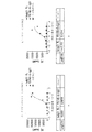
プログラム死1(PD−1)に特異的に結合し、PD−1の発現及び/または活性を調節するタンパク質を使用して、白斑を管理、治療、または予防するための方法が本明細書に提供される。【選択図】図1Methods for managing, treating, or preventing vitiligo using a protein that specifically binds to programmed death 1 (PD-1) and modulates PD-1 expression and / or activity are described herein. Provided. [Selection] Figure 1
Description
関連出願の相互参照
本出願は、2016年9月19日に出願された米国仮特許出願第62/396,720号に対する優先権の利益を主張するものであり、その開示は、その全体において参照により本明細書に組み込まれる。
技術分野
プログラム死1(PD−1)に特異的に結合し、PD−1の発現及び/または活性を調節するタンパク質を使用して、白斑を管理、治療、または予防するための方法が本明細書に提供される。
This application claims the benefit of priority over US Provisional Patent Application No. 62 / 396,720 filed on September 19, 2016, the disclosure of which is incorporated by reference in its entirety Is incorporated herein by reference.
TECHNICAL FIELD Methods for managing, treating, or preventing vitiligo using a protein that specifically binds to program death 1 (PD-1) and modulates PD-1 expression and / or activity are described herein. Provided in the certificate.
本開示は、PD−1に結合する抗体などの結合タンパク質を含む、PD−1(例えば、ヒトPD−1、配列番号43)に結合する治療有効量のタンパク質を対象に投与することを含む対象における白斑を管理、予防、または治療する方法を提供する。抗体を含むそのような結合タンパク質は、PD−1ポリペプチド、PD−1断片、及び/またはPD−1エピトープに結合し得る。抗体を含むそのような結合タンパク質は、アゴニスト(例えば、PD−1リガンド様シグナル伝達を誘発する)であり得る。いくつかの実施形態では、結合タンパク質は、PD−1(例えば、非ブロッキング抗体)との相互作用について、PD−1リガンド(例えば、PD−L1及びPD−L2)と競合しない。 The disclosure includes administering to a subject a therapeutically effective amount of a protein that binds to PD-1 (eg, human PD-1, SEQ ID NO: 43), including a binding protein such as an antibody that binds to PD-1. Methods for managing, preventing, or treating vitiligo in Such binding proteins, including antibodies, can bind to PD-1 polypeptides, PD-1 fragments, and / or PD-1 epitopes. Such binding proteins, including antibodies, can be agonists (eg, induce PD-1 ligand-like signaling). In some embodiments, the binding protein does not compete with PD-1 ligands (eg, PD-L1 and PD-L2) for interaction with PD-1 (eg, non-blocking antibody).
本開示はまた、ある特定の実施形態では、(i)ヒトPD−1に結合し、(ii)PD−1リガンド様シグナル伝達を誘発し、(iii)PD−1との相互作用についてPD−L1及び/またはPD−L2と競合しない、抗体またはその断片を含む治療有効量の結合タンパク質を対象に投与することを含む対象における白斑を管理、予防、または治療する方法を提供する。 The present disclosure also provides, in certain embodiments, (i) binding to human PD-1, (ii) inducing PD-1 ligand-like signaling, and (iii) PD-- for interaction with PD-1. Provided is a method for managing, preventing, or treating vitiligo in a subject comprising administering to the subject a therapeutically effective amount of a binding protein comprising an antibody or fragment thereof that does not compete with L1 and / or PD-L2.
いくつかの実施形態では、本明細書に提供される方法における使用のための結合タンパク質(例えば、抗PD−1抗体)は、6つの相補性決定領域(CDR)または6つ未満のCDRを含む。他の実施形態では、結合タンパク質(例えば、抗PD−1抗体)は、重鎖可変領域(VH)CDR1、VH CDR2、VH CDR3、軽鎖可変領域(VL)CDR1、VL CDR2、及び/またはVL CDR3から選択される、1、2、3、4、5、または6つのCDRを含む。ある特定の実施形態では、結合タンパク質(例えば、抗PD−1抗体)は、本明細書に記載されるようにPD1AB−1、PD1AB−2、PD1AB−3、PD1AB−4、PD1AB−5、もしくはPD1AB−6、またはそれらのヒト化バリアントとして示されるモノクローナル抗体のVH CDR1、VH CDR2、VH CDR3、VL CDR1、VL CDR2、及び/またはVL CDR3から選択される、1、2、3、4、5、または6つのCDRを含む。いくつかの実施形態では、結合タンパク質(例えば、抗PD−1抗体)は、ヒト免疫グロブリンアミノ酸配列またはそのバリアントのVH FR1、VH FR2、VH FR3、VH FR4、VL FR1、VL FR2、VL FR3、及び/またはVL FR4を含む、骨格領域またはフレームワーク領域(FR)をさらに含む。 In some embodiments, a binding protein (eg, an anti-PD-1 antibody) for use in the methods provided herein comprises 6 complementarity determining regions (CDRs) or fewer than 6 CDRs. . In other embodiments, the binding protein (eg, anti-PD-1 antibody) is a heavy chain variable region (VH) CDR1, VH CDR2, VH CDR3, light chain variable region (VL) CDR1, VL CDR2, and / or VL. 1, 2, 3, 4, 5, or 6 CDRs selected from CDR3. In certain embodiments, the binding protein (eg, anti-PD-1 antibody) is PD1AB-1, PD1AB-2, PD1AB-3, PD1AB-4, PD1AB-5, or as described herein. 1, 2, 3, 4, 5 selected from PDHAB-6, or monoclonal antibodies VH CDR1, VH CDR2, VH CDR3, VL CDR1, VL CDR2, and / or VL CDR3 shown as humanized variants thereof Or 6 CDRs. In some embodiments, the binding protein (eg, anti-PD-1 antibody) is a human immunoglobulin amino acid sequence or variant thereof VH FR1, VH FR2, VH FR3, VH FR4, VL FR1, VL FR2, VL FR3, And / or further comprising a framework or framework region (FR) comprising VL FR4.
本方法のいくつかの実施形態では、抗体またはその抗原結合断片は、配列番号8のアミノ酸配列を有する軽鎖可変領域及び配列番号13のアミノ酸配列を有する重鎖可変領域を含む抗体によって認識されるヒトPD−1のエピトープに結合する。 In some embodiments of the method, the antibody or antigen-binding fragment thereof is recognized by an antibody comprising a light chain variable region having the amino acid sequence of SEQ ID NO: 8 and a heavy chain variable region having the amino acid sequence of SEQ ID NO: 13. It binds to an epitope of human PD-1.
本方法の他の実施形態では、抗体またはその抗体断片は、配列番号8のアミノ酸配列を有する軽鎖可変領域及び配列番号13のアミノ酸配列を有する重鎖可変領域を含む抗体を有するヒトPD−1のエピトープに結合する。 In another embodiment of the method, the antibody or antibody fragment thereof comprises a human PD-1 having an antibody comprising a light chain variable region having the amino acid sequence of SEQ ID NO: 8 and a heavy chain variable region having the amino acid sequence of SEQ ID NO: 13. Binds to the epitope of
本方法のいくつかの実施形態では、抗体またはその抗原結合断片は、表1に示される抗体PD1AB−1、PD1AB−2、PD1AB−3、PD1AB−4、PD1AB−5、またはPD1AB−6のうちのいずれか1つのVL CDR1、VL CDR2、及びVL CDR3を含むVLを含む。 In some embodiments of the method, the antibody or antigen-binding fragment thereof is one of the antibodies PD1AB-1, PD1AB-2, PD1AB-3, PD1AB-4, PD1AB-5, or PD1AB-6 shown in Table 1. A VL comprising any one of VL CDR1, VL CDR2, and VL CDR3.
本方法の他の実施形態では、抗体またはその抗原結合断片は、表2に示される抗体PD1AB−1、PD1AB−2、PD1AB−3、PD1AB−4、PD1AB−5、またはPD1AB−6のうちのいずれか1つのVH CDR1、VH CDR2、及びVH CDR3を含むVHを含む。 In other embodiments of the method, the antibody or antigen-binding fragment thereof is one of the antibodies PD1AB-1, PD1AB-2, PD1AB-3, PD1AB-4, PD1AB-5, or PD1AB-6 shown in Table 2. VH including any one VH CDR1, VH CDR2, and VH CDR3.
本方法のさらなる他の実施形態では、抗体またはその抗原結合断片は、
(a)表3に示される抗体PD1AB−1、PD1AB−2、PD1AB−3、PD1AB−4、PD1AB−5、またはPD1AB−6のうちのいずれか1つのVL FR1、VL FR2、VL FR3、及びVL FR4を含むVL、及び
(b)表4に示される抗体PD1AB−1、PD1AB−2、PD1AB−3、PD1AB−4、PD1AB−5、またはPD1AB−6のうちのいずれか1つのVH FR1、VH FR2、VH FR3、及びVH FR4を含むVHを含む。
In still other embodiments of the methods, the antibody or antigen-binding fragment thereof is
(A) any one of the antibodies PD1AB-1, PD1AB-2, PD1AB-3, PD1AB-4, PD1AB-5, or PD1AB-6 shown in Table 3, VL FR1, VL FR2, VL FR3, and VL including VL FR4, and (b) VH FR1 of any one of antibodies PD1AB-1, PD1AB-2, PD1AB-3, PD1AB-4, PD1AB-5, or PD1AB-6 shown in Table 4. VH includes VH FR2, VH FR3, and VH FR4.
本方法のある特定の実施形態では、抗体またはその抗原結合断片のVL CDR1、VL CDR2、及びVL CDR3は、それぞれ配列番号1、2、及び3のアミノ酸配列を含み、抗体またはその抗原結合断片のVH CDR1、VH CDR2、及びVH CDR3は、それぞれ配列番号4、5、及び6のアミノ酸配列を含む。 In certain embodiments of the method, the antibody or antigen-binding fragment thereof, VL CDR1, VL CDR2, and VL CDR3 comprise the amino acid sequences of SEQ ID NOs: 1, 2, and 3, respectively, and the antibody or antigen-binding fragment thereof. VH CDR1, VH CDR2, and VH CDR3 comprise the amino acid sequences of SEQ ID NOs: 4, 5, and 6, respectively.
本方法のさらなる別の実施形態では、抗体またはその抗原結合断片のVL CDR1、VL CDR2、及びVL CDR3は、それぞれ配列番号7、2、及び3のアミノ酸配列を含み、抗体またはその抗原結合断片のVH CDR1、VH CDR2、及びVH CDR3は、それぞれ配列番号4、5、及び6のアミノ酸配列を含む。 In yet another embodiment of the method, the VL CDR1, VL CDR2, and VL CDR3 of the antibody or antigen-binding fragment thereof comprise the amino acid sequences of SEQ ID NOs: 7, 2, and 3, respectively, VH CDR1, VH CDR2, and VH CDR3 comprise the amino acid sequences of SEQ ID NOs: 4, 5, and 6, respectively.
本方法の別の実施形態では、抗体またはその抗原結合断片は、配列番号8のアミノ酸配列を含むVLを含む。いくつかの実施形態では、アミノ酸配列は、1つ以上のその保存的修飾を含む。 In another embodiment of this method, the antibody or antigen-binding fragment thereof comprises a VL comprising the amino acid sequence of SEQ ID NO: 8. In some embodiments, the amino acid sequence includes one or more conservative modifications thereof.
本方法のある特定の実施形態では、抗体またはその抗原結合断片は、配列番号9のアミノ酸配列を含むVLを含む。いくつかの実施形態では、アミノ酸配列は、1つ以上のその保存的修飾を含む。 In certain embodiments of the method, the antibody or antigen-binding fragment thereof comprises a VL comprising the amino acid sequence of SEQ ID NO: 9. In some embodiments, the amino acid sequence includes one or more conservative modifications thereof.
本方法のいくつかの実施形態では、抗体またはその抗原結合断片は、配列番号10のアミノ酸配列を含むVLを含む。いくつかの実施形態では、アミノ酸配列は、1つ以上のその保存的修飾を含む。 In some embodiments of the method, the antibody or antigen-binding fragment thereof comprises a VL comprising the amino acid sequence of SEQ ID NO: 10. In some embodiments, the amino acid sequence includes one or more conservative modifications thereof.
本方法のある特定の実施形態では、抗体またはその抗原結合断片は、配列番号11のアミノ酸配列を含むVHを含む。いくつかの実施形態では、アミノ酸配列は、1つ以上のその保存的修飾を含む。 In certain embodiments of the method, the antibody or antigen-binding fragment thereof comprises a VH comprising the amino acid sequence of SEQ ID NO: 11. In some embodiments, the amino acid sequence includes one or more conservative modifications thereof.
本方法の他の実施形態では、抗体またはその抗原結合断片は、配列番号12のアミノ酸配列を含むVHを含む。いくつかの実施形態では、アミノ酸配列は、1つ以上のその保存的修飾を含む。 In other embodiments of the method, the antibody or antigen-binding fragment thereof comprises a VH comprising the amino acid sequence of SEQ ID NO: 12. In some embodiments, the amino acid sequence includes one or more conservative modifications thereof.
本方法の別の実施形態では、抗体またはその抗原結合断片は、配列番号13のアミノ酸配列を含むVHを含む。いくつかの実施形態では、アミノ酸配列は、1つ以上のその保存的修飾を含む。 In another embodiment of this method, the antibody or antigen-binding fragment thereof comprises a VH comprising the amino acid sequence of SEQ ID NO: 13. In some embodiments, the amino acid sequence includes one or more conservative modifications thereof.
本方法のある特定の実施形態では、抗体またはその抗原結合断片は、(a)配列番号8のアミノ酸配列を含むVL、及び(b)配列番号11のアミノ酸配列を含むVHを含む。 In certain embodiments of the method, the antibody or antigen-binding fragment thereof comprises (a) a VL comprising the amino acid sequence of SEQ ID NO: 8, and (b) a VH comprising the amino acid sequence of SEQ ID NO: 11.
本方法のいくつかの実施形態では、抗体またはその抗原結合断片は、(a)配列番号9のアミノ酸配列を含むVL、及び(b)配列番号11のアミノ酸配列を含むVHを含む。 In some embodiments of the method, the antibody or antigen-binding fragment thereof comprises (a) a VL comprising the amino acid sequence of SEQ ID NO: 9, and (b) a VH comprising the amino acid sequence of SEQ ID NO: 11.
本方法の他の実施形態では、抗体またはその抗原結合断片は、(a)配列番号10のアミノ酸配列を含むVL、及び(b)配列番号11のアミノ酸配列を含むVHを含む。 In other embodiments of the method, the antibody or antigen-binding fragment thereof comprises (a) a VL comprising the amino acid sequence of SEQ ID NO: 10, and (b) a VH comprising the amino acid sequence of SEQ ID NO: 11.
本方法の一実施形態では、抗体またはその抗原結合断片は、(a)配列番号8のアミノ酸配列を含むVL、及び(b)配列番号12のアミノ酸配列を含むVHを含む。 In one embodiment of this method, the antibody or antigen-binding fragment thereof comprises (a) a VL comprising the amino acid sequence of SEQ ID NO: 8, and (b) a VH comprising the amino acid sequence of SEQ ID NO: 12.
本方法の別の実施形態では、抗体またはその抗原結合断片は、(a)配列番号9のアミノ酸配列を含むVL、及び(b)配列番号12のアミノ酸配列を含むVHを含む。 In another embodiment of the method, the antibody or antigen-binding fragment thereof comprises (a) a VL comprising the amino acid sequence of SEQ ID NO: 9, and (b) a VH comprising the amino acid sequence of SEQ ID NO: 12.
本方法のある特定の実施形態では、抗体またはその抗原結合断片は、(a)配列番号10のアミノ酸配列を含むVL、及び(b)配列番号12のアミノ酸配列を含むVHを含む。 In certain embodiments of the method, the antibody or antigen-binding fragment thereof comprises (a) a VL comprising the amino acid sequence of SEQ ID NO: 10, and (b) a VH comprising the amino acid sequence of SEQ ID NO: 12.
本方法のいくつかの実施形態では、抗体またはその抗原結合断片は、(a)配列番号8のアミノ酸配列を含むVL、及び(b)配列番号13のアミノ酸配列を含むVHを含む。 In some embodiments of the method, the antibody or antigen-binding fragment thereof comprises (a) a VL comprising the amino acid sequence of SEQ ID NO: 8, and (b) a VH comprising the amino acid sequence of SEQ ID NO: 13.
本方法の他の実施形態では、抗体またはその抗原結合断片は、(a)配列番号9のアミノ酸配列を含むVL、(b)配列番号13のアミノ酸配列を含むVHを含む。 In other embodiments of the method, the antibody or antigen-binding fragment thereof comprises (a) a VL comprising the amino acid sequence of SEQ ID NO: 9, (b) a VH comprising the amino acid sequence of SEQ ID NO: 13.
本方法のある特定の実施形態では、抗体またはその抗原結合断片は、(a)配列番号10のアミノ酸配列を含むVL、及び(b)配列番号13のアミノ酸配列を含むVHを含む。 In certain embodiments of the method, the antibody or antigen-binding fragment thereof comprises (a) a VL comprising the amino acid sequence of SEQ ID NO: 10, and (b) a VH comprising the amino acid sequence of SEQ ID NO: 13.
いくつかの実施形態では、VLのアミノ酸配列は、1つ以上のその保存的修飾を含む。いくつかの実施形態では、VHのアミノ酸配列は、1つ以上のその保存的修飾を含む。いくつかの実施形態では、VL及びVHのアミノ酸配列は、1つ以上のその保存的修飾を含む。 In some embodiments, the amino acid sequence of the VL includes one or more conservative modifications thereof. In some embodiments, the amino acid sequence of VH includes one or more of its conservative modifications. In some embodiments, the amino acid sequences of VL and VH include one or more conservative modifications thereof.
本方法のいくつかの実施形態では、抗体は、ヒトIgG1 Fc領域を含む。他の実施形態では、抗体は、バリアントヒトIgG1 Fc領域を含む。 In some embodiments of the method, the antibody comprises a human IgG1 Fc region. In other embodiments, the antibody comprises a variant human IgG1 Fc region.
本方法の一実施形態では、抗体は、ヒトIgG1−K322A Fc領域を含む。 In one embodiment of this method, the antibody comprises a human IgG1-K322A Fc region.
本方法のいくつかの実施形態では、抗体は、ヒトIgG4 Fc領域を含む。他の実施形態では、抗体は、バリアントヒトIgG4 Fc領域を含む。 In some embodiments of the method, the antibody comprises a human IgG4 Fc region. In other embodiments, the antibody comprises a variant human IgG4 Fc region.
本方法の別の実施形態では、抗体は、ヒトIgG4P Fc領域を含む。 In another embodiment of the method, the antibody comprises a human IgG4P Fc region.
本方法のさらなる別の実施形態では、抗体は、ヒトIgG4PE Fc領域を含む。 In yet another embodiment of the method, the antibody comprises a human IgG4PE Fc region.
本方法のいくつかの実施形態では、抗体またはその抗原結合断片は、配列番号41のアミノ酸配列を含む軽鎖定常領域をさらに含む。 In some embodiments of the method, the antibody or antigen-binding fragment thereof further comprises a light chain constant region comprising the amino acid sequence of SEQ ID NO: 41.
本方法の他の実施形態では、抗体またはその抗原結合断片が、配列番号36〜40からなる群から選択されるアミノ酸配列を含む重鎖Fc領域をさらに含む。 In other embodiments of the method, the antibody or antigen-binding fragment thereof further comprises a heavy chain Fc region comprising an amino acid sequence selected from the group consisting of SEQ ID NOs: 36-40.
本方法のさらなる別の実施形態では、抗体またはその抗原結合断片は、配列番号41のアミノ酸配列を含む軽鎖定常領域、及び配列番号36〜40からなる群から選択されるアミノ酸配列を含む重鎖Fc領域をさらに含む。 In yet another embodiment of the method, the antibody or antigen-binding fragment thereof comprises a light chain constant region comprising the amino acid sequence of SEQ ID NO: 41, and a heavy chain comprising an amino acid sequence selected from the group consisting of SEQ ID NOs: 36-40. It further contains an Fc region.
本方法のいくつかの実施形態では、抗体またはその抗原結合断片は、配列番号31のアミノ酸配列を含む軽鎖を含む。 In some embodiments of the method, the antibody or antigen-binding fragment thereof comprises a light chain comprising the amino acid sequence of SEQ ID NO: 31.
本方法の別の実施形態では、抗体またはその抗原結合断片は、配列番号32のアミノ酸配列を含む重鎖を含む。 In another embodiment of this method, the antibody or antigen-binding fragment thereof comprises a heavy chain comprising the amino acid sequence of SEQ ID NO: 32.
本方法の他の実施形態では、抗体またはその抗原結合断片は、(a)配列番号31のアミノ酸配列を含む軽鎖、及び(b)配列番号32のアミノ酸配列を含む重鎖を含む。 In other embodiments of the method, the antibody or antigen-binding fragment thereof comprises (a) a light chain comprising the amino acid sequence of SEQ ID NO: 31, and (b) a heavy chain comprising the amino acid sequence of SEQ ID NO: 32.
本方法のある特定の実施形態では、抗体またはその抗原結合断片は、配列番号33のアミノ酸配列を含む重鎖を含む。 In certain embodiments of the method, the antibody or antigen-binding fragment thereof comprises a heavy chain comprising the amino acid sequence of SEQ ID NO: 33.
本方法の他の実施形態では、抗体またはその抗原結合断片は、(a)配列番号31のアミノ酸配列を含む軽鎖、及び(b)配列番号33のアミノ酸配列を含む重鎖を含む。 In other embodiments of the method, the antibody or antigen-binding fragment thereof comprises (a) a light chain comprising the amino acid sequence of SEQ ID NO: 31, and (b) a heavy chain comprising the amino acid sequence of SEQ ID NO: 33.
本方法の一実施形態では、抗体またはその抗原結合断片は、配列番号34のアミノ酸配列を含む重鎖を含む。 In one embodiment of this method, the antibody or antigen-binding fragment thereof comprises a heavy chain comprising the amino acid sequence of SEQ ID NO: 34.
本方法のさらなる別の実施形態では、抗体またはその抗原結合断片は、(a)配列番号31のアミノ酸配列を含む軽鎖、及び(b)配列番号34のアミノ酸配列を含む重鎖を含む。 In yet another embodiment of the method, the antibody or antigen-binding fragment thereof comprises (a) a light chain comprising the amino acid sequence of SEQ ID NO: 31, and (b) a heavy chain comprising the amino acid sequence of SEQ ID NO: 34.
本方法のいくつかの実施形態では、抗体またはその抗原結合断片は、配列番号35のアミノ酸配列を含む重鎖を含む。 In some embodiments of the method, the antibody or antigen-binding fragment thereof comprises a heavy chain comprising the amino acid sequence of SEQ ID NO: 35.
本方法の他の実施形態では、抗体またはその抗原結合断片は、(a)配列番号31のアミノ酸配列を含む軽鎖、及び(b)配列番号35のアミノ酸配列を含む重鎖を含む。 In other embodiments of the method, the antibody or antigen-binding fragment thereof comprises (a) a light chain comprising the amino acid sequence of SEQ ID NO: 31, and (b) a heavy chain comprising the amino acid sequence of SEQ ID NO: 35.
本方法のある特定の実施形態では、抗体またはその抗原結合断片は、PD−1に結合したとき、配列番号42のアミノ酸配列を有する残基100〜109のうちの少なくとも1つに結合する。 In certain embodiments of the method, the antibody or antigen-binding fragment thereof binds to at least one of residues 100-109 having the amino acid sequence of SEQ ID NO: 42 when bound to PD-1.
本方法のいくつかの実施形態では、抗体またはその抗原結合断片は、PD−1に結合したとき、配列番号42のアミノ酸配列を有する残基100〜105のうちの少なくとも1つに結合する。 In some embodiments of the method, the antibody or antigen-binding fragment thereof binds to at least one of residues 100-105 having the amino acid sequence of SEQ ID NO: 42 when bound to PD-1.
本方法の特定の実施形態では、抗体またはその抗原結合断片は、PD−1に結合したとき、配列番号42のアミノ酸配列内のN33、T51、S57、L100、N102、G103、R104、D105、H107、及びS109からなる群から選択される少なくとも1つの残基に結合する。 In a particular embodiment of the method, the antibody or antigen-binding fragment thereof, when bound to PD-1, is N33, T51, S57, L100, N102, G103, R104, D105, H107 within the amino acid sequence of SEQ ID NO: 42. And at least one residue selected from the group consisting of S109.
本方法のいくつかの実施形態では、抗体またはその抗原結合断片は、PD−1に結合したとき、配列番号42のアミノ酸配列内のN33、T51、S57、L100、N102、G103、R104、D105、H107、及びS109からなる群から選択される2つ以上の残基に結合する。 In some embodiments of the method, the antibody or antigen-binding fragment thereof, when bound to PD-1, is N33, T51, S57, L100, N102, G103, R104, D105, within the amino acid sequence of SEQ ID NO: 42, It binds to two or more residues selected from the group consisting of H107 and S109.
本方法の他の実施形態では、抗体またはその抗原結合断片は、PD−1に結合したとき、配列番号42のアミノ酸配列内のN33、T51、S57、L100、N102、G103、R104、D105、H107、及びS109からなる群から選択される3つ以上の残基に結合する。 In other embodiments of the method, the antibody or antigen-binding fragment thereof, when bound to PD-1, is N33, T51, S57, L100, N102, G103, R104, D105, H107 within the amino acid sequence of SEQ ID NO: 42. And 3 or more residues selected from the group consisting of S109.
本方法のある特定の実施形態では、抗体またはその抗原結合断片は、PD−1に結合したとき、配列番号42のアミノ酸配列内のN33、T51、S57、L100、N102、G103、R104、D105、H107、及びS109からなる群から選択される4つ以上の残基に結合する。 In certain embodiments of the method, the antibody or antigen-binding fragment thereof, when bound to PD-1, is N33, T51, S57, L100, N102, G103, R104, D105, within the amino acid sequence of SEQ ID NO: 42, It binds to 4 or more residues selected from the group consisting of H107 and S109.
本方法の一実施形態では、抗体またはその抗原結合断片は、PD−1に結合したとき、配列番号42のアミノ酸配列内のN33、T51、S57、L100、N102、G103、R104、D105、H107、及びS109からなる群から選択される5つ以上の残基に結合する。 In one embodiment of this method, the antibody or antigen-binding fragment thereof, when bound to PD-1, is N33, T51, S57, L100, N102, G103, R104, D105, H107, within the amino acid sequence of SEQ ID NO: 42, And 5 or more residues selected from the group consisting of S109.
本方法の別の実施形態では、抗体またはその抗原結合断片は、PD−1に結合したとき、配列番号42のアミノ酸配列内のN33、T51、S57、L100、N102、G103、R104、D105、H107、及びS109からなる群から選択される6つ以上の残基に結合する。 In another embodiment of this method, the antibody or antigen-binding fragment thereof, when bound to PD-1, is N33, T51, S57, L100, N102, G103, R104, D105, H107 within the amino acid sequence of SEQ ID NO: 42. And 6 or more residues selected from the group consisting of S109.
本方法のさらなる別の実施形態では、抗体またはその抗原結合断片は、PD−1に結合したとき、配列番号42のアミノ酸配列内のN33、T51、S57、L100、N102、G103、R104、D105、H107、及びS109からなる群から選択される7つ以上の残基に結合する。 In yet another embodiment of the method, the antibody or antigen-binding fragment thereof, when bound to PD-1, is N33, T51, S57, L100, N102, G103, R104, D105, within the amino acid sequence of SEQ ID NO: 42, It binds to 7 or more residues selected from the group consisting of H107 and S109.
本方法のさらなる別の実施形態では、抗体またはその抗原結合断片は、PD−1に結合したとき、配列番号42のアミノ酸配列内のN33、T51、S57、L100、N102、G103、R104、D105、H107、及びS109からなる群から選択される8つ以上の残基に結合する。 In yet another embodiment of the method, the antibody or antigen-binding fragment thereof, when bound to PD-1, is N33, T51, S57, L100, N102, G103, R104, D105, within the amino acid sequence of SEQ ID NO: 42, It binds to 8 or more residues selected from the group consisting of H107 and S109.
本方法のある特定の実施形態では、抗体またはその抗原結合断片は、PD−1に結合したとき、配列番号42のアミノ酸配列内のN33、T51、S57、L100、N102、G103、R104、D105、H107、及びS109からなる群から選択される9つ以上の残基に結合する。 In certain embodiments of the method, the antibody or antigen-binding fragment thereof, when bound to PD-1, is N33, T51, S57, L100, N102, G103, R104, D105, within the amino acid sequence of SEQ ID NO: 42, It binds to 9 or more residues selected from the group consisting of H107 and S109.
本方法の他の実施形態では、抗体またはその抗原結合断片は、PD−1に結合したとき、配列番号42のアミノ酸配列内のN33、T51、S57、L100、N102、G103、R104、D105、H107、及びS109からなる群から選択される10個すべての残基に結合する。 In other embodiments of the method, the antibody or antigen-binding fragment thereof, when bound to PD-1, is N33, T51, S57, L100, N102, G103, R104, D105, H107 within the amino acid sequence of SEQ ID NO: 42. And all 10 residues selected from the group consisting of S109.
本方法の一実施形態では、抗体またはその抗原結合断片は、PD−1に結合したとき、配列番号42のアミノ酸配列内のN33に結合する。 In one embodiment of this method, the antibody or antigen-binding fragment thereof binds to N33 within the amino acid sequence of SEQ ID NO: 42 when bound to PD-1.
本方法の別の実施形態では、抗体またはその抗原結合断片は、PD−1に結合したとき、配列番号42のアミノ酸配列内のT51に結合する。 In another embodiment of the method, the antibody or antigen-binding fragment thereof binds to T51 within the amino acid sequence of SEQ ID NO: 42 when bound to PD-1.
本方法の特定の実施形態では、抗体またはその抗原結合断片は、PD−1に結合したとき、配列番号42のアミノ酸配列内のS57に結合する。 In a particular embodiment of the method, the antibody or antigen-binding fragment thereof binds to S57 within the amino acid sequence of SEQ ID NO: 42 when bound to PD-1.
本方法の1つの特定の実施形態では、抗体またはその抗原結合断片は、PD−1に結合したとき、配列番号42のアミノ酸配列内のL100に結合する。 In one particular embodiment of the method, the antibody or antigen-binding fragment thereof binds to L100 within the amino acid sequence of SEQ ID NO: 42 when bound to PD-1.
本方法のいくつかの実施形態では、抗体またはその抗原結合断片は、PD−1に結合したとき、配列番号42のアミノ酸配列内のN102に結合する。 In some embodiments of the methods, the antibody or antigen-binding fragment thereof binds to N102 within the amino acid sequence of SEQ ID NO: 42 when bound to PD-1.
本方法の他の実施形態では、抗体またはその抗原結合断片は、PD−1に結合したとき、配列番号42のアミノ酸配列内のG103に結合する。 In other embodiments of the method, the antibody or antigen-binding fragment thereof binds to G103 within the amino acid sequence of SEQ ID NO: 42 when bound to PD-1.
本方法の別の実施形態では、抗体またはその抗原結合断片は、PD−1に結合したとき、配列番号42のアミノ酸配列内のR104に結合する。 In another embodiment of the method, the antibody or antigen-binding fragment thereof binds to R104 within the amino acid sequence of SEQ ID NO: 42 when bound to PD-1.
本方法のさらなる別の実施形態では、抗体またはその抗原結合断片は、PD−1に結合したとき、配列番号42のアミノ酸配列内のG103及びR104に結合する。 In yet another embodiment of the method, the antibody or antigen-binding fragment thereof binds to G103 and R104 within the amino acid sequence of SEQ ID NO: 42 when bound to PD-1.
本方法のさらなる別の実施形態では、抗体またはその抗原結合断片は、PD−1に結合したとき、配列番号42のアミノ酸配列内のD105に結合する。 In yet another embodiment of the method, the antibody or antigen-binding fragment thereof binds to D105 within the amino acid sequence of SEQ ID NO: 42 when bound to PD-1.
本方法のいくつかの実施形態では、抗体またはその抗原結合断片は、PD−1に結合したとき、配列番号42のアミノ酸配列内のH107に結合する。 In some embodiments of the method, the antibody or antigen-binding fragment thereof binds to H107 within the amino acid sequence of SEQ ID NO: 42 when bound to PD-1.
本方法のある特定の実施形態では、抗体またはその抗原結合断片は、PD−1に結合したとき、配列番号42のアミノ酸配列内のS109に結合する。 In certain embodiments of the method, the antibody or antigen-binding fragment thereof binds to S109 within the amino acid sequence of SEQ ID NO: 42 when bound to PD-1.
本方法の一実施形態では、ヒトPD−1のエピトープは、PD−L1結合部位とは異なる。別の実施形態では、ヒトPD−1のエピトープは、PD−L2結合部位とは異なる。特定の実施形態では、ヒトPD−1のエピトープは、PD−L1結合部位及びPD−L2結合部位の両方とは異なる。 In one embodiment of this method, the epitope of human PD-1 is different from the PD-L1 binding site. In another embodiment, the epitope of human PD-1 is different from the PD-L2 binding site. In certain embodiments, the epitope of human PD-1 is different from both the PD-L1 and PD-L2 binding sites.
本方法の実施形態では、抗体またはその抗原結合断片は、ヒトPD−1及び/またはサルPD−1(例えば、カニクイザル)に特異的に結合するが、齧歯類PD−1にはしない。 In embodiments of the method, the antibody or antigen-binding fragment thereof specifically binds to human PD-1 and / or monkey PD-1 (eg, cynomolgus monkey), but not to rodent PD-1.
本方法のある特定の実施形態では、抗体またはその抗原結合断片は、減弱化された抗体依存性細胞障害(ADCC)活性を有する。他の実施形態では、抗体またはその抗原結合断片は、減弱化された補体依存性細胞障害(CDC)活性を有する。いくつかの実施形態では、抗体またはその抗原結合断片は、減弱化されたADCC及び/または減弱化されたCDC活性を有する。 In certain embodiments of the method, the antibody or antigen-binding fragment thereof has attenuated antibody-dependent cellular cytotoxicity (ADCC) activity. In other embodiments, the antibody or antigen-binding fragment thereof has attenuated complement dependent cytotoxicity (CDC) activity. In some embodiments, the antibody or antigen-binding fragment thereof has attenuated ADCC and / or attenuated CDC activity.
一態様では、対象における白斑を管理、予防、または治療する方法が本明細書に提供され、ヒトPD−1のエピトープに結合する治療有効量の抗体またはその抗原結合断片を対象に投与することを含み、抗体またはその抗原結合断片は、(a)T細胞活性を減弱化し、及び/または(b)T細胞の表面でのPD−1発現を下方制御する。 In one aspect, a method for managing, preventing, or treating vitiligo in a subject is provided herein, comprising administering to the subject a therapeutically effective amount of an antibody or antigen-binding fragment thereof that binds to an epitope of human PD-1. Including, the antibody or antigen-binding fragment thereof (a) attenuates T cell activity and / or (b) downregulates PD-1 expression on the surface of T cells.
本方法の一実施形態では、抗体は、T細胞活性を減弱化する。別の実施形態では、抗体は、T細胞の表面でのPD−1発現を下方制御する。 In one embodiment of this method, the antibody attenuates T cell activity. In another embodiment, the antibody downregulates PD-1 expression on the surface of T cells.
ある特定の実施形態では、T細胞活性の減弱化は、T細胞エフェクター機能によって測定される。 In certain embodiments, attenuation of T cell activity is measured by T cell effector function.
本方法のいくつかの実施形態では、T細胞活性の減弱化は、サイトカイン産生の阻害によって測定される。 In some embodiments of the method, attenuation of T cell activity is measured by inhibition of cytokine production.
本方法のある特定の実施形態では、抗体またはその抗原結合断片によって阻害されるサイトカインは、IL−2、IL−17、及び/またはIFN−γを含む。いくつかの実施形態では、サイトカインは、IL−1、IL−2、IL−6、IL−12、IL−17、IL−22、IL−23、GM−CSF、IFN−γ、及びTNF−αからなる群から選択される。ある特定の実施形態では、サイトカインはIL−1である。いくつかの実施形態では、サイトカインはIL−2である。他の実施形態では、サイトカインはIL−6である。別の実施形態では、サイトカインはIL−12である。いくつかの他の実施形態では、サイトカインはIL−17である。さらなる他の実施形態では、サイトカインはIL−22である。さらなる他の実施形態では、サイトカインはIL−23である。いくつかの実施形態では、サイトカインはGM−CSFである。他の実施形態では、サイトカインはIFN−γである。さらなる他の実施形態では、サイトカインはTNF−αである。ある特定の実施形態では、サイトカインはIL−2及びIL−17である。いくつかの実施形態では、サイトカインはIL−2及びIFN−γである。さらなる他の実施形態では、サイトカインはIL−17及びIFN−γである。さらなる他の実施形態では、サイトカインは、IL−2、IL−17、及びIFN−γである。上記に記述のサイトカインのうちの2つ、3つ、またはそれ以上の他の組み合わせが企図される。 In certain embodiments of the method, the cytokine inhibited by the antibody or antigen-binding fragment thereof comprises IL-2, IL-17, and / or IFN-γ. In some embodiments, the cytokine is IL-1, IL-2, IL-6, IL-12, IL-17, IL-22, IL-23, GM-CSF, IFN-γ, and TNF-α. Selected from the group consisting of In certain embodiments, the cytokine is IL-1. In some embodiments, the cytokine is IL-2. In other embodiments, the cytokine is IL-6. In another embodiment, the cytokine is IL-12. In some other embodiments, the cytokine is IL-17. In still other embodiments, the cytokine is IL-22. In still other embodiments, the cytokine is IL-23. In some embodiments, the cytokine is GM-CSF. In other embodiments, the cytokine is IFN-γ. In still other embodiments, the cytokine is TNF-α. In certain embodiments, the cytokine is IL-2 and IL-17. In some embodiments, the cytokine is IL-2 and IFN-γ. In still other embodiments, the cytokine is IL-17 and IFN-γ. In still other embodiments, the cytokine is IL-2, IL-17, and IFN-γ. Other combinations of two, three, or more of the cytokines described above are contemplated.
本方法のいくつかの実施形態では、T細胞活性の減弱化は、T細胞増殖の阻害によって測定される。 In some embodiments of the method, attenuation of T cell activity is measured by inhibition of T cell proliferation.
本方法のいくつかの実施形態では、T細胞活性の減弱化は、T細胞活性化マーカーの下方制御によって測定される。 In some embodiments of the method, attenuation of T cell activity is measured by downregulation of a T cell activation marker.
本方法のいくつかの実施形態では、T細胞活性の減弱化は、制御性T細胞バイオマーカーの上方制御によって測定される。 In some embodiments of the method, attenuation of T cell activity is measured by upregulation of regulatory T cell biomarkers.
本方法のいくつかの実施形態では、T細胞活性の減弱化は、制御性T細胞数の増加によって測定される。 In some embodiments of the method, attenuation of T cell activity is measured by an increase in the number of regulatory T cells.
本方法のある特定の実施形態では、T細胞の表面でのPD−1発現の下方制御は、抗体またはその抗原結合断片との接触の4時間後に早ければ生じる。別の実施形態では、下方制御は、接触の6時間後に早ければ生じる。さらなる別の実施形態では、下方制御は、接触の8時間後に早ければ生じる。さらなる別の実施形態では、下方制御は、接触の10時間後に早ければ生じる。一実施形態では、下方制御は、接触の12時間後に早ければ生じる。別の実施形態では、下方制御は、接触の14時間後に早ければ生じる。さらなる別の実施形態では、下方制御は、接触の16時間後に早ければ生じる。さらなる別の実施形態では、下方制御は、接触の18時間後に早ければ生じる。一実施形態では、下方制御は、接触の20時間後に早ければ生じる。別の実施形態では、下方制御は、接触の22時間後に早ければ生じる。さらなる別の実施形態では、下方制御は、接触の24時間後に早ければ生じる。いくつかの実施形態では、接触は、抗体を伴う。他の実施形態では、接触は、その抗原結合断片を伴う。 In certain embodiments of the method, downregulation of PD-1 expression on the surface of T cells occurs as early as 4 hours after contact with the antibody or antigen-binding fragment thereof. In another embodiment, down control occurs as early as 6 hours after contact. In yet another embodiment, down-regulation occurs as early as 8 hours after contact. In yet another embodiment, down-regulation occurs as early as 10 hours after contact. In one embodiment, down control occurs as early as 12 hours after contact. In another embodiment, down control occurs as early as 14 hours after contact. In yet another embodiment, down-regulation occurs as early as 16 hours after contact. In yet another embodiment, down-regulation occurs as early as 18 hours after contact. In one embodiment, down control occurs as early as 20 hours after contact. In another embodiment, down control occurs as early as 22 hours after contact. In yet another embodiment, down-regulation occurs as early as 24 hours after contact. In some embodiments, the contacting involves an antibody. In other embodiments, the contacting involves an antigen-binding fragment thereof.
いくつかの実施形態では、T細胞の表面でのPD−1発現の下方制御は、サイトカイン阻害に先立つ。一実施形態では、T細胞の表面でのPD−1発現の下方制御は、抗体またはその抗原結合断片との接触の4時間後に早ければ生じ、サイトカイン阻害に先立つ。別の実施形態では、下方制御は、抗体またはその抗原結合断片との接触の6時間後に早ければ生じ、サイトカイン阻害に先立つ。さらなる別の実施形態では、下方制御は、抗体またはその抗原結合断片との接触の8時間後に早ければ生じ、サイトカイン阻害に先立つ。さらなる別の実施形態では、下方制御は、抗体またはその抗原結合断片との接触の10時間後に早ければ生じ、サイトカイン阻害に先立つ。一実施形態では、下方制御は、抗体またはその抗原結合断片との接触の12時間後に早ければ生じ、サイトカイン阻害に先立つ。別の実施形態では、下方制御は、抗体またはその抗原結合断片との接触の14時間後に早ければ生じ、サイトカイン阻害に先立つ。さらなる別の実施形態では、下方制御は、抗体またはその抗原結合断片との接触の16時間後に早ければ生じ、サイトカイン阻害に先立つ。さらなる別の実施形態では、下方制御は、抗体またはその抗原結合断片との接触の18時間後に早ければ生じ、サイトカイン阻害に先立つ。一実施形態では、下方制御は、抗体またはその抗原結合断片との接触の20時間後に早ければ生じ、サイトカイン阻害に先立つ。別の実施形態では、下方制御は、抗体またはその抗原結合断片との接触の22時間後に早ければ生じ、サイトカイン阻害に先立つ。さらなる別の実施形態では、下方制御は、抗体またはその抗原結合断片との接触の24時間後に早ければ生じ、サイトカイン阻害に先立つ。 In some embodiments, down-regulation of PD-1 expression on the surface of T cells precedes cytokine inhibition. In one embodiment, downregulation of PD-1 expression on the surface of T cells occurs as early as 4 hours after contact with the antibody or antigen-binding fragment thereof and precedes cytokine inhibition. In another embodiment, downregulation occurs as early as 6 hours after contact with the antibody or antigen-binding fragment thereof and precedes cytokine inhibition. In yet another embodiment, downregulation occurs as early as 8 hours after contact with the antibody or antigen-binding fragment thereof and precedes cytokine inhibition. In yet another embodiment, downregulation occurs as early as 10 hours after contact with the antibody or antigen-binding fragment thereof and precedes cytokine inhibition. In one embodiment, downregulation occurs as early as 12 hours after contact with the antibody or antigen-binding fragment thereof and precedes cytokine inhibition. In another embodiment, downregulation occurs as early as 14 hours after contact with the antibody or antigen-binding fragment thereof and precedes cytokine inhibition. In yet another embodiment, downregulation occurs as early as 16 hours after contact with the antibody or antigen-binding fragment thereof and precedes cytokine inhibition. In yet another embodiment, downregulation occurs as early as 18 hours after contact with the antibody or antigen-binding fragment thereof and precedes cytokine inhibition. In one embodiment, downregulation occurs as early as 20 hours after contact with the antibody or antigen-binding fragment thereof and precedes cytokine inhibition. In another embodiment, downregulation occurs as early as 22 hours after contact with the antibody or antigen-binding fragment thereof and precedes cytokine inhibition. In yet another embodiment, downregulation occurs as early as 24 hours after contact with the antibody or antigen-binding fragment thereof and precedes cytokine inhibition.
他の実施形態では、T細胞の表面でのPD−1発現の下方制御は、サイトカイン阻害と同時である。一実施形態では、T細胞の表面でのPD−1発現の下方制御は、抗体またはその抗原結合断片との接触の4時間後に早ければ生じ、サイトカイン阻害と同時である。別の実施形態では、下方制御は、抗体またはその抗原結合断片との接触の6時間後に早ければ生じ、サイトカイン阻害と同時である。さらなる別の実施形態では、下方制御は、抗体またはその抗原結合断片との接触の8時間後に早ければ生じ、サイトカイン阻害と同時である。さらなる別の実施形態では、下方制御は、抗体またはその抗原結合断片との接触の10時間後に早ければ生じ、サイトカイン阻害と同時である。一実施形態では、下方制御は、抗体またはその抗原結合断片との接触の12時間後に早ければ生じ、サイトカイン阻害と同時である。別の実施形態では、下方制御は、抗体またはその抗原結合断片との接触の14時間後に早ければ生じ、サイトカイン阻害と同時である。さらなる別の実施形態では、下方制御は、抗体またはその抗原結合断片との接触の16時間後に早ければ生じ、サイトカイン阻害と同時である。さらなる別の実施形態では、下方制御は、抗体またはその抗原結合断片との接触の18時間後に早ければ生じ、サイトカイン阻害と同時である。一実施形態では、下方制御は、抗体またはその抗原結合断片との接触の20時間後に早ければ生じ、サイトカイン阻害と同時である。別の実施形態では、下方制御は、抗体またはその抗原結合断片との接触の22時間後に早ければ生じ、サイトカイン阻害と同時である。さらなる別の実施形態では、下方制御は、抗体またはその抗原結合断片との接触の24時間後に早ければ生じ、サイトカイン阻害と同時である。 In other embodiments, downregulation of PD-1 expression on the surface of T cells is concurrent with cytokine inhibition. In one embodiment, downregulation of PD-1 expression on the surface of T cells occurs as early as 4 hours after contact with the antibody or antigen-binding fragment thereof and is concurrent with cytokine inhibition. In another embodiment, downregulation occurs as early as 6 hours after contact with the antibody or antigen-binding fragment thereof and is concurrent with cytokine inhibition. In yet another embodiment, downregulation occurs as early as 8 hours after contact with the antibody or antigen-binding fragment thereof and is concurrent with cytokine inhibition. In yet another embodiment, downregulation occurs as early as 10 hours after contact with the antibody or antigen-binding fragment thereof and is concurrent with cytokine inhibition. In one embodiment, downregulation occurs as early as 12 hours after contact with the antibody or antigen-binding fragment thereof and is concurrent with cytokine inhibition. In another embodiment, downregulation occurs as early as 14 hours after contact with the antibody or antigen-binding fragment thereof and is concurrent with cytokine inhibition. In yet another embodiment, downregulation occurs as early as 16 hours after contact with the antibody or antigen-binding fragment thereof and is concurrent with cytokine inhibition. In yet another embodiment, downregulation occurs as early as 18 hours after contact with the antibody or antigen-binding fragment thereof and is concurrent with cytokine inhibition. In one embodiment, downregulation occurs as early as 20 hours after contact with the antibody or antigen-binding fragment thereof and is concurrent with cytokine inhibition. In another embodiment, downregulation occurs as early as 22 hours after contact with the antibody or antigen-binding fragment thereof and is concurrent with cytokine inhibition. In yet another embodiment, downregulation occurs as early as 24 hours after contact with the antibody or antigen-binding fragment thereof and is concurrent with cytokine inhibition.
さらなる他の実施形態では、T細胞の表面でのPD−1発現の下方制御は、サイトカイン阻害の後である。一実施形態では、T細胞の表面でのPD−1発現の下方制御は、抗体またはその抗原結合断片との接触の4時間後に早ければ生じ、サイトカイン阻害の後である。別の実施形態では、下方制御は、抗体またはその抗原結合断片との接触の6時間後に早ければ生じ、サイトカイン阻害の後である。さらなる別の実施形態では、下方制御は、抗体またはその抗原結合断片との接触の8時間後に早ければ生じ、サイトカイン阻害の後である。さらなる別の実施形態では、下方制御は、抗体またはその抗原結合断片との接触の10時間後に早ければ生じ、サイトカイン阻害の後である。一実施形態では、下方制御は、抗体またはその抗原結合断片との接触の12時間後に早ければ生じ、サイトカイン阻害の後である。別の実施形態では、下方制御は、抗体またはその抗原結合断片との接触の14時間後に早ければ生じ、サイトカイン阻害の後である。さらなる別の実施形態では、下方制御は、抗体またはその抗原結合断片との接触の16時間後に早ければ生じ、サイトカイン阻害の後である。さらなる別の実施形態では、下方制御は、抗体またはその抗原結合断片との接触の18時間後に早ければ生じ、サイトカイン阻害の後である。一実施形態では、下方制御は、抗体またはその抗原結合断片との接触の20時間後に早ければ生じ、サイトカイン阻害の後である。別の実施形態では、下方制御は、抗体またはその抗原結合断片との接触の22時間後に早ければ生じ、サイトカイン阻害の後である。さらなる別の実施形態では、下方制御は、抗体またはその抗原結合断片との接触の24時間後に早ければ生じ、サイトカイン阻害の後である。 In still other embodiments, downregulation of PD-1 expression on the surface of T cells is after cytokine inhibition. In one embodiment, downregulation of PD-1 expression on the surface of T cells occurs as early as 4 hours after contact with the antibody or antigen-binding fragment thereof and after cytokine inhibition. In another embodiment, downregulation occurs as early as 6 hours after contact with the antibody or antigen-binding fragment thereof and after cytokine inhibition. In yet another embodiment, downregulation occurs as early as 8 hours after contact with the antibody or antigen-binding fragment thereof and after cytokine inhibition. In yet another embodiment, downregulation occurs as early as 10 hours after contact with the antibody or antigen-binding fragment thereof and after cytokine inhibition. In one embodiment, downregulation occurs as early as 12 hours after contact with the antibody or antigen-binding fragment thereof and after cytokine inhibition. In another embodiment, downregulation occurs as early as 14 hours after contact with the antibody or antigen-binding fragment thereof and after cytokine inhibition. In yet another embodiment, downregulation occurs as early as 16 hours after contact with the antibody or antigen-binding fragment thereof and after cytokine inhibition. In yet another embodiment, downregulation occurs as early as 18 hours after contact with the antibody or antigen-binding fragment thereof and after cytokine inhibition. In one embodiment, downregulation occurs as early as 20 hours after contact with the antibody or antigen-binding fragment thereof and after cytokine inhibition. In another embodiment, downregulation occurs as early as 22 hours after contact with the antibody or antigen-binding fragment thereof and after cytokine inhibition. In yet another embodiment, downregulation occurs as early as 24 hours after contact with the antibody or antigen-binding fragment thereof and after cytokine inhibition.
本方法の一実施形態では、精製されたヒトPD−1に結合する抗体またはその抗原結合断片のKDは、約1nM〜約100nMである。本方法の別の実施形態では、細胞表面に発現されたヒトPD−1及び細胞表面に発現されたサルPD−1に結合する抗体またはその抗原結合断片のKDは、約100pM〜約10nMである。 In one embodiment of the method, K D of the antibodies or antigen-binding fragment thereof that binds to human PD-1 purified is about 1nM~ about 100 nM. In another embodiment of the method, K D of the antibodies or antigen-binding fragments thereof that bind to monkey PD-1 expressed on the expressed human PD-1 and cell surface to the cell surface is about 100pM~ about 10nM is there.
本方法のいくつかの実施形態では、T細胞活性を減弱化するための抗体またはその抗原結合断片のEC50は、約1pM〜約10pM、約10pM〜約100pM、約100pM〜約1nM、約1nM〜約10nM、または約10nM〜約100nMである。 In some embodiments of the method, the EC 50 of the antibody or antigen-binding fragment thereof for reducing T cell activity is about 1 pM to about 10 pM, about 10 pM to about 100 pM, about 100 pM to about 1 nM, about 1 nM. To about 10 nM, or about 10 nM to about 100 nM.
本方法の他の実施形態では、抗体またはその抗原結合断片によるT細胞活性の最大減弱化パーセントは、少なくとも約10%、約20%、約30%、約40%、約45%、約50%、約55%、約60%、約65%、約70%、約75%、約80%、約85%、約90%、約95%、または約100%である。 In other embodiments of the method, the maximum attenuation percentage of T cell activity by the antibody or antigen-binding fragment thereof is at least about 10%, about 20%, about 30%, about 40%, about 45%, about 50%. About 55%, about 60%, about 65%, about 70%, about 75%, about 80%, about 85%, about 90%, about 95%, or about 100%.
本方法の別の実施形態では、抗体またはその抗原結合断片によるPD−1発現の最大下方制御パーセントは、少なくとも約10%、約20%、約30%、約40%、約45%、約50%、約55%、約60%、約65%、約70%、約75%、約80%、約85%、約90%、約95%、または約100%である。 In another embodiment of this method, the maximum percent down-regulation of PD-1 expression by the antibody or antigen-binding fragment thereof is at least about 10%, about 20%, about 30%, about 40%, about 45%, about 50 %, About 55%, about 60%, about 65%, about 70%, about 75%, about 80%, about 85%, about 90%, about 95%, or about 100%.
本方法のある特定の実施形態では、抗体は、モノクローナル抗体である。いくつかの実施形態では、抗体は、ヒト化抗体、ヒト抗体、またはキメラ抗体である。別の実施形態では、ヒト化抗体は、脱免疫化抗体または複合ヒト抗体である。ある特定の実施形態では、抗体は、ヒト化抗体である。特定の実施形態では、抗体は、ヒトPD−1に特異的に結合するヒト化抗体である。 In certain embodiments of the method, the antibody is a monoclonal antibody. In some embodiments, the antibody is a humanized antibody, a human antibody, or a chimeric antibody. In another embodiment, the humanized antibody is a deimmunized antibody or a conjugated human antibody. In certain embodiments, the antibody is a humanized antibody. In certain embodiments, the antibody is a humanized antibody that specifically binds human PD-1.
本方法のある特定の実施形態では、抗体またはその抗原結合断片は、Fab、Fab’、F(ab’)2、Fv、scFv、dsFv、ダイアボディ、トリアボディ、またはテトラボディである。いくつかの実施形態では、抗体またはその抗原結合断片は、抗体断片から形成された多重特異性抗体である。他の実施形態では、抗体またはその抗原結合断片は、二重特異性抗体である。 In certain embodiments of the method, the antibody or antigen-binding fragment thereof is Fab, Fab ′, F (ab ′) 2 , Fv, scFv, dsFv, diabody, triabody, or tetrabody. In some embodiments, the antibody or antigen-binding fragment thereof is a multispecific antibody formed from an antibody fragment. In other embodiments, the antibody or antigen-binding fragment thereof is a bispecific antibody.
本方法のいくつかの実施形態では、抗体またはその抗原結合断片は、薬剤に複合体化されている。一実施形態では、薬剤は、放射性同位体、金属キレート剤、酵素、蛍光化合物、生物発光化合物、または化学発光化合物である。 In some embodiments of the method, the antibody or antigen-binding fragment thereof is conjugated to a drug. In one embodiment, the agent is a radioisotope, metal chelator, enzyme, fluorescent compound, bioluminescent compound, or chemiluminescent compound.
本方法の別の実施形態では、抗体またはその抗原結合断片は、T細胞活性を減弱化する。一実施形態では、T細胞活性の最大減弱化パーセントは、少なくとも約10%、約20%、約30%、約40%、約45%、約50%、約55%、約60%、約65%、約70%、約75%、約80%、約85%、約90%、約95%、または約100%である。いくつかの実施形態では、T細胞活性の減弱化は、T細胞増殖の阻害によって測定される。いくつかの実施形態では、T細胞活性の減弱化は、サイトカイン産生の阻害によって測定される。いくつかの実施形態では、サイトカインは、IL−2、IL−17、IFN−γ、またはそれらの任意の組み合わせからなる群から選択される。ある特定の実施形態では、サイトカインは、IL−1、IL−2、IL−6、IL−12、IL−17、IL−22、IL−23、GM−CSF、IFN−γ、及びTNF−αからなる群から選択される。ある特定の実施形態では、サイトカインはIL−1である。いくつかの実施形態では、サイトカインはIL−2である。他の実施形態では、サイトカインはIL−6である。別の実施形態では、サイトカインはIL−12である。いくつかの他の実施形態では、サイトカインはIL−17である。さらなる他の実施形態では、サイトカインはIL−22である。さらなる他の実施形態では、サイトカインはIL−23である。いくつかの実施形態では、サイトカインはGM−CSFである。他の実施形態では、サイトカインはIFN−γである。さらなる他の実施形態では、サイトカインはTNF−αである。ある特定の実施形態では、サイトカインはIL−2及びIL−17である。いくつかの実施形態では、サイトカインはIL−2及びIFN−γである。さらなる他の実施形態では、サイトカインはIL−17及びIFN−γである。さらなる他の実施形態では、サイトカインは、IL−2、IL−17、及びIFN−γである。上記に記述のサイトカインのうちの2つ、3つ、またはそれ以上の他の組み合わせが企図される。ある特定の実施形態では、サイトカイン産生の阻害は、T細胞の表面でのPD−1発現の下方制御に続く。他の実施形態では、T細胞の表面でのPD−1発現の下方制御は、抗体またはその抗原結合断片との接触の4時間、6時間、8時間、10時間、12時間、14時間、16時間、18時間、20時間、22時間、または24時間後に早ければ生じる。一実施形態では、下方制御は、接触の4時間後に早ければ生じ、サイトカイン阻害に先立つ。一実施形態では、下方制御は、接触の6時間後に早ければ生じ、サイトカイン阻害に先立つ。一実施形態では、下方制御は、接触の8時間後に早ければ生じ、サイトカイン阻害に先立つ。一実施形態では、下方制御は、接触の10時間後に早ければ生じ、サイトカイン阻害に先立つ。別の実施形態では、下方制御は、接触の12時間後に早ければ生じ、サイトカイン阻害に先立つ。実施形態では、下方制御は、接触の14時間後に早ければ生じ、サイトカイン阻害に先立つ。他の実施形態では、下方制御は、接触の16時間後に早ければ生じ、サイトカイン阻害に先立つ。一実施形態では、下方制御は、接触の18時間後に早ければ生じ、サイトカイン阻害に先立つ。別の実施形態では、下方制御は、接触の20時間後に早ければ生じ、サイトカイン阻害に先立つ。一実施形態では、下方制御は、接触の22時間後に早ければ生じ、サイトカイン阻害に先立つ。いくつかの実施形態では、下方制御は、接触の24時間後に早ければ生じ、サイトカイン阻害に先立つ。いくつかの実施形態では、サイトカイン産生の阻害は、T細胞の表面でのPD−1発現の下方制御と同時である。一実施形態では、下方制御は、接触の4時間後に早ければ生じ、サイトカイン阻害と同時である。一実施形態では、下方制御は、接触の6時間後に早ければ生じ、サイトカイン阻害と同時である。一実施形態では、下方制御は、接触の8時間後に早ければ生じ、サイトカイン阻害と同時である。一実施形態では、下方制御は、接触の10時間後に早ければ生じ、サイトカイン阻害と同時である。別の実施形態では、下方制御は、接触の12時間後に早ければ生じ、サイトカイン阻害と同時である。実施形態では、下方制御は、接触の14時間後に早ければ生じ、サイトカイン阻害と同時である。他の実施形態では、下方制御は、接触の16時間後に早ければ生じ、サイトカイン阻害と同時である。一実施形態では、下方制御は、接触の18時間後に早ければ生じ、サイトカイン阻害と同時である。別の実施形態では、下方制御は、接触の20時間後に早ければ生じ、サイトカイン阻害と同時である。一実施形態では、下方制御は、接触の22時間後に早ければ生じ、サイトカイン阻害と同時である。いくつかの実施形態では、下方制御は、接触の24時間後に早ければ生じ、サイトカイン阻害と同時である。いくつかの実施形態では、サイトカイン産生の阻害は、T細胞の表面でのPD−1発現の下方制御に先立つ。一実施形態では、下方制御は、接触の4時間後に早ければ生じ、サイトカイン阻害の後である。一実施形態では、下方制御は、接触の6時間後に早ければ生じ、サイトカイン阻害の後である。一実施形態では、下方制御は、接触の8時間後に早ければ生じ、サイトカイン阻害の後である。一実施形態では、下方制御は、接触の10時間後に早ければ生じ、サイトカイン阻害の後である。別の実施形態では、下方制御は、接触の12時間後に早ければ生じ、サイトカイン阻害の後である。実施形態では、下方制御は、接触の14時間後に早ければ生じ、サイトカイン阻害の後である。他の実施形態では、下方制御は、接触の16時間後に早ければ生じ、サイトカイン阻害の後である。一実施形態では、下方制御は、接触の18時間後に早ければ生じ、サイトカイン阻害の後である。別の実施形態では、下方制御は、接触の20時間後に早ければ生じ、サイトカイン阻害の後である。一実施形態では、下方制御は、接触の22時間後に早ければ生じ、サイトカイン阻害の後である。いくつかの実施形態では、下方制御は、接触の24時間後に早ければ生じ、サイトカイン阻害の後である。 In another embodiment of the method, the antibody or antigen-binding fragment thereof attenuates T cell activity. In one embodiment, the maximum percent attenuation of T cell activity is at least about 10%, about 20%, about 30%, about 40%, about 45%, about 50%, about 55%, about 60%, about 65. %, About 70%, about 75%, about 80%, about 85%, about 90%, about 95%, or about 100%. In some embodiments, attenuation of T cell activity is measured by inhibition of T cell proliferation. In some embodiments, attenuation of T cell activity is measured by inhibition of cytokine production. In some embodiments, the cytokine is selected from the group consisting of IL-2, IL-17, IFN-γ, or any combination thereof. In certain embodiments, the cytokine is IL-1, IL-2, IL-6, IL-12, IL-17, IL-22, IL-23, GM-CSF, IFN-γ, and TNF-α. Selected from the group consisting of In certain embodiments, the cytokine is IL-1. In some embodiments, the cytokine is IL-2. In other embodiments, the cytokine is IL-6. In another embodiment, the cytokine is IL-12. In some other embodiments, the cytokine is IL-17. In still other embodiments, the cytokine is IL-22. In still other embodiments, the cytokine is IL-23. In some embodiments, the cytokine is GM-CSF. In other embodiments, the cytokine is IFN-γ. In still other embodiments, the cytokine is TNF-α. In certain embodiments, the cytokine is IL-2 and IL-17. In some embodiments, the cytokine is IL-2 and IFN-γ. In still other embodiments, the cytokine is IL-17 and IFN-γ. In still other embodiments, the cytokine is IL-2, IL-17, and IFN-γ. Other combinations of two, three, or more of the cytokines described above are contemplated. In certain embodiments, inhibition of cytokine production follows downregulation of PD-1 expression on the surface of T cells. In other embodiments, down-regulation of PD-1 expression on the surface of T cells is 4 hours, 6 hours, 8 hours, 10 hours, 12 hours, 14 hours, 16 hours of contact with the antibody or antigen-binding fragment thereof. As early as after 18 hours, 20 hours, 22 hours, or 24 hours. In one embodiment, downregulation occurs as early as 4 hours after contact and precedes cytokine inhibition. In one embodiment, downregulation occurs as early as 6 hours after contact and precedes cytokine inhibition. In one embodiment, downregulation occurs as early as 8 hours after contact and precedes cytokine inhibition. In one embodiment, downregulation occurs as early as 10 hours after contact and precedes cytokine inhibition. In another embodiment, downregulation occurs as early as 12 hours after contact and precedes cytokine inhibition. In embodiments, downregulation occurs as early as 14 hours after contact and precedes cytokine inhibition. In other embodiments, down-regulation occurs as early as 16 hours after contact and precedes cytokine inhibition. In one embodiment, downregulation occurs as early as 18 hours after contact and precedes cytokine inhibition. In another embodiment, downregulation occurs as early as 20 hours after contact and precedes cytokine inhibition. In one embodiment, downregulation occurs as early as 22 hours after contact and precedes cytokine inhibition. In some embodiments, downregulation occurs as early as 24 hours after contact and precedes cytokine inhibition. In some embodiments, inhibition of cytokine production is concomitant with downregulation of PD-1 expression on the surface of T cells. In one embodiment, downregulation occurs as early as 4 hours after contact and is coincident with cytokine inhibition. In one embodiment, downregulation occurs as early as 6 hours after contact and is coincident with cytokine inhibition. In one embodiment, downregulation occurs as early as 8 hours after contact and is coincident with cytokine inhibition. In one embodiment, downregulation occurs as early as 10 hours after contact and is coincident with cytokine inhibition. In another embodiment, downregulation occurs as early as 12 hours after contact and is coincident with cytokine inhibition. In embodiments, downregulation occurs as early as 14 hours after contact and is coincident with cytokine inhibition. In other embodiments, downregulation occurs as early as 16 hours after contact and is coincident with cytokine inhibition. In one embodiment, downregulation occurs as early as 18 hours after contact and is coincident with cytokine inhibition. In another embodiment, downregulation occurs as early as 20 hours after contact and is coincident with cytokine inhibition. In one embodiment, downregulation occurs as early as 22 hours after contact and is coincident with cytokine inhibition. In some embodiments, downregulation occurs as early as 24 hours after contact and is coincident with cytokine inhibition. In some embodiments, inhibition of cytokine production precedes downregulation of PD-1 expression on the surface of T cells. In one embodiment, downregulation occurs as early as 4 hours after contact and after cytokine inhibition. In one embodiment, downregulation occurs as early as 6 hours after contact and after cytokine inhibition. In one embodiment, downregulation occurs as early as 8 hours after contact and after cytokine inhibition. In one embodiment, downregulation occurs as early as 10 hours after contact and after cytokine inhibition. In another embodiment, downregulation occurs as early as 12 hours after contact and after cytokine inhibition. In embodiments, downregulation occurs as early as 14 hours after contact and after cytokine inhibition. In other embodiments, downregulation occurs as early as 16 hours after contact and after cytokine inhibition. In one embodiment, downregulation occurs as early as 18 hours after contact and after cytokine inhibition. In another embodiment, downregulation occurs as early as 20 hours after contact and after cytokine inhibition. In one embodiment, downregulation occurs as early as 22 hours after contact and after cytokine inhibition. In some embodiments, downregulation occurs as early as 24 hours after contact and after cytokine inhibition.
本方法のさらなる別の実施形態では、抗体またはその抗原結合断片は、T細胞の表面でのPD−1発現を下方制御する。一実施形態では、抗体またはその抗原結合断片によるPD−1発現の最大下方制御パーセントは、少なくとも約10%、20%、30%、40%、45%、50%、55%、60%、65%、70%、75%、80%、85%、90%、95%、または100%である。別の実施形態では、T細胞の表面でのPD−1発現の下方制御は、抗体またはその抗原結合断片との接触の4時間後に早ければ生じる。一実施形態では、T細胞の表面でのPD−1発現の下方制御は、サイトカイン阻害に先立つ。一実施形態では、T細胞の表面でのPD−1発現の下方制御は、サイトカイン阻害と同時である。一実施形態では、T細胞の表面でのPD−1発現の下方制御は、サイトカイン阻害に続く。ある特定の実施形態では、サイトカインは、IL−2、IL−17、IFN−γ、またはそれらの任意の組み合わせである。ある特定の実施形態では、サイトカインは、IL−1、IL−2、IL−6、IL−12、IL−17、IL−22、IL−23、GM−CSF、IFN−γ、及びTNF−αからなる群から選択される。ある特定の実施形態では、サイトカインはIL−1である。いくつかの実施形態では、サイトカインはIL−2である。他の実施形態では、サイトカインはIL−6である。別の実施形態では、サイトカインはIL−12である。他の実施形態では、サイトカインはIL−17である。さらなる他の実施形態では、サイトカインはIL−22である。さらなる他の実施形態では、サイトカインはIL−23である。いくつかの実施形態では、サイトカインはGM−CSFである。他の実施形態では、サイトカインはIFN−γである。さらなる他の実施形態では、サイトカインはTNF−αである。ある特定の実施形態では、サイトカインはIL−2及びIL−17である。いくつかの実施形態では、サイトカインはIL−2及びIFN−γである。さらなる他の実施形態では、サイトカインはIL−17及びIFN−γである。さらなる他の実施形態では、サイトカインは、IL−2、IL−17、及びIFN−γである。 In yet another embodiment of the method, the antibody or antigen-binding fragment thereof downregulates PD-1 expression on the surface of T cells. In one embodiment, the maximum percent down-regulation of PD-1 expression by an antibody or antigen-binding fragment thereof is at least about 10%, 20%, 30%, 40%, 45%, 50%, 55%, 60%, 65 %, 70%, 75%, 80%, 85%, 90%, 95%, or 100%. In another embodiment, downregulation of PD-1 expression on the surface of T cells occurs as early as 4 hours after contact with the antibody or antigen-binding fragment thereof. In one embodiment, down-regulation of PD-1 expression on the surface of T cells precedes cytokine inhibition. In one embodiment, downregulation of PD-1 expression on the surface of T cells is concurrent with cytokine inhibition. In one embodiment, down-regulation of PD-1 expression on the surface of T cells follows cytokine inhibition. In certain embodiments, the cytokine is IL-2, IL-17, IFN-γ, or any combination thereof. In certain embodiments, the cytokine is IL-1, IL-2, IL-6, IL-12, IL-17, IL-22, IL-23, GM-CSF, IFN-γ, and TNF-α. Selected from the group consisting of In certain embodiments, the cytokine is IL-1. In some embodiments, the cytokine is IL-2. In other embodiments, the cytokine is IL-6. In another embodiment, the cytokine is IL-12. In other embodiments, the cytokine is IL-17. In still other embodiments, the cytokine is IL-22. In still other embodiments, the cytokine is IL-23. In some embodiments, the cytokine is GM-CSF. In other embodiments, the cytokine is IFN-γ. In still other embodiments, the cytokine is TNF-α. In certain embodiments, the cytokine is IL-2 and IL-17. In some embodiments, the cytokine is IL-2 and IFN-γ. In still other embodiments, the cytokine is IL-17 and IFN-γ. In still other embodiments, the cytokine is IL-2, IL-17, and IFN-γ.
本方法の別の実施形態では、抗体またはその抗原結合断片は、サイトカイン産生を阻害することによってT細胞活性を減弱化する。いくつかの実施形態では、抗体またはその抗原結合断片によって阻害されるサイトカインは、IL−2、IL−17、IFN−γ、またはそれらの任意の組み合わせから選択される。ある特定の実施形態では、サイトカインは、IL−1、IL−2、IL−6、IL−12、IL−17、IL−22、IL−23、GM−CSF、IFN−γ、及びTNF−αからなる群から選択される。ある特定の実施形態では、サイトカインはIL−1である。いくつかの実施形態では、サイトカインはIL−2である。他の実施形態では、サイトカインはIL−6である。別の実施形態では、サイトカインはIL−12である。他の実施形態では、サイトカインはIL−17である。さらなる他の実施形態では、サイトカインはIL−22である。さらなる他の実施形態では、サイトカインはIL−23である。いくつかの実施形態では、サイトカインはGM−CSFである。他の実施形態では、サイトカインはIFN−γである。さらなる他の実施形態では、サイトカインはTNF−αである。ある特定の実施形態では、サイトカインはIL−2及びIL−17である。いくつかの実施形態では、サイトカインはIL−2及びIFN−γである。さらなる他の実施形態では、サイトカインはIL−17及びIFN−γである。さらなる他の実施形態では、サイトカインは、IL−2、IL−17、及びIFN−γである。上記に記述のサイトカインのうちの2つ、3つ、またはそれ以上の他の組み合わせが企図される。 In another embodiment of the method, the antibody or antigen-binding fragment thereof attenuates T cell activity by inhibiting cytokine production. In some embodiments, the cytokine inhibited by the antibody or antigen-binding fragment thereof is selected from IL-2, IL-17, IFN-γ, or any combination thereof. In certain embodiments, the cytokine is IL-1, IL-2, IL-6, IL-12, IL-17, IL-22, IL-23, GM-CSF, IFN-γ, and TNF-α. Selected from the group consisting of In certain embodiments, the cytokine is IL-1. In some embodiments, the cytokine is IL-2. In other embodiments, the cytokine is IL-6. In another embodiment, the cytokine is IL-12. In other embodiments, the cytokine is IL-17. In still other embodiments, the cytokine is IL-22. In still other embodiments, the cytokine is IL-23. In some embodiments, the cytokine is GM-CSF. In other embodiments, the cytokine is IFN-γ. In still other embodiments, the cytokine is TNF-α. In certain embodiments, the cytokine is IL-2 and IL-17. In some embodiments, the cytokine is IL-2 and IFN-γ. In still other embodiments, the cytokine is IL-17 and IFN-γ. In still other embodiments, the cytokine is IL-2, IL-17, and IFN-γ. Other combinations of two, three, or more of the cytokines described above are contemplated.
また、本明細書に記載の抗体またはその抗原結合断片を含む治療有効量の組成物を対象に投与することを含む、対象における白斑を管理、予防、または治療する方法が本明細書に提供される。本方法のある特定の実施形態では、組成物は、薬学的に許容される担体をさらに含む。 Also provided herein is a method for managing, preventing, or treating vitiligo in a subject, comprising administering to the subject a therapeutically effective amount of a composition comprising an antibody or antigen-binding fragment thereof described herein. The In certain embodiments of the method, the composition further comprises a pharmaceutically acceptable carrier.
ある特定の実施形態では、対象は、白斑を有するか、または抗炎症性治療で白斑を治療される。 In certain embodiments, the subject has vitiligo or is treated with anti-inflammatory treatment.
特定の他の実施形態では、対象の免疫細胞は、PD−1を発現する。 In certain other embodiments, the subject immune cells express PD-1.
PD−1に結合する抗体など有効量の結合タンパク質を対象に投与することを含む、対象における白斑を管理、予防、または治療する方法が本明細書に提供される。一実施形態では、有効量のPD−1結合タンパク質を対象に投与することを含む、対象における白斑を管理する方法が本明細書に提供される。一実施形態では、有効量のPD−1結合タンパク質を対象に投与することを含む、対象における白斑を予防する方法が本明細書に提供される。一実施形態では、有効量のPD−1結合タンパク質を対象に投与することを含む、対象における白斑を治療する方法が本明細書に提供される。特定の実施形態では、PD−1結合タンパク質は、PD−1に結合する抗体である。これらの方法において有用である例示的なPD−1抗体が本明細書に提供される。 Provided herein is a method for managing, preventing, or treating vitiligo in a subject comprising administering to the subject an effective amount of a binding protein, such as an antibody that binds PD-1. In one embodiment, provided herein is a method of managing vitiligo in a subject comprising administering to the subject an effective amount of a PD-1 binding protein. In one embodiment, provided herein is a method of preventing vitiligo in a subject comprising administering to the subject an effective amount of a PD-1 binding protein. In one embodiment, provided herein is a method of treating vitiligo in a subject comprising administering to the subject an effective amount of a PD-1 binding protein. In certain embodiments, the PD-1 binding protein is an antibody that binds to PD-1. Exemplary PD-1 antibodies that are useful in these methods are provided herein.
本明細書に提供される様々な方法のいくつかの実施形態では、抗体は、ヒト及び/またはカニクイザルPD−1に結合する。いくつかの実施形態では、ヒト及び/またはカニクイザルPD−1に結合する抗体などの結合タンパク質は、齧歯類PD−1に結合しない。ある特定の実施形態では、本明細書に開示される抗体を含むPD−1結合タンパク質は、アゴニストである(例えば、PD−1リガンドPD−1の効果を模倣し、シグナル伝達を誘発し得る)。いくつかの実施形態では、本明細書に記載されるPD−1に対する抗体などの結合タンパク質は、(i)ヒト及び/もしくはカニクイザルPD−1に結合し、(ii)結合についてPD−1リガンド(例えば、PD−L1及び/もしくはPD−L2)と競合せず、ならびに/または(iii)PD−1シグナル伝達を誘発する。一実施形態では、PD−1抗体は、ヒトPD−1に結合する。一実施形態では、PD−1抗体は、カニクイザルPD−1に結合する。一実施形態では、PD−1抗体は、ヒトPD−1及びカニクイザルPD−1の両方に結合する。いくつかの実施形態では、PD−1抗体は、PD−1への結合についてPD−L1と競合しない。他の実施形態では、PD−1抗体は、PD−1への結合についてPD−L2と競合しない。さらなる他の実施形態では、PD−1抗体は、PD−1への結合についてPD−L1またはPD−L2のいずれかと競合しない。他の実施形態では、PD−1抗体は、PD−1シグナル伝達を誘発する。特定の実施形態では、本明細書に提供されるPD−1抗体は、ヒトPD−1及びカニクイザルPD−1の両方に結合し、PD−1への結合についてPD−L1またはPD−L2のいずれかと競合せず、PD−1シグナル伝達を誘発する。いくつかの実施形態では、結合、競合、及び/またはシグナル伝達は、インビトロ、例えば、細胞に基づくアッセイで評価される。他の実施形態では、結合、競合、及び/またはシグナル伝達は、エクスビボ、例えば、T細胞機能アッセイで評価される。一実施形態では、PD−1抗体は、ヒトPD−1に結合する。ある特定の実施形態では、アッセイ及び測定値は、(1)ヒトまたはカニクイザルPBMCアッセイ(例えば、実施例5.2.1及び5.2.2を参照されたい)、ならびに(2)ヒト全血試料アッセイ(例えば、実施例5.2.1を参照されたい)を含む。ある特定の実施形態では、抗PD−1抗体などの結合タンパク質は、本明細書に記載されるように、PD−L1及び/またはPD−L2の自然生物学的機能と一貫する活性を呈する。いくつかの実施形態では、活性は、インビトロで呈される。他の実施形態では、活性は、エクスビボで呈される。 In some embodiments of the various methods provided herein, the antibody binds to human and / or cynomolgus monkey PD-1. In some embodiments, a binding protein, such as an antibody that binds human and / or cynomolgus monkey PD-1, does not bind to rodent PD-1. In certain embodiments, a PD-1 binding protein comprising an antibody disclosed herein is an agonist (eg, can mimic the effects of the PD-1 ligand PD-1 and induce signal transduction). . In some embodiments, a binding protein, such as an antibody to PD-1 described herein, binds to (i) human and / or cynomolgus PD-1, and (ii) PD-1 ligand ( For example, it does not compete with PD-L1 and / or PD-L2) and / or (iii) induces PD-1 signaling. In one embodiment, the PD-1 antibody binds to human PD-1. In one embodiment, the PD-1 antibody binds to cynomolgus PD-1. In one embodiment, the PD-1 antibody binds to both human PD-1 and cynomolgus PD-1. In some embodiments, the PD-1 antibody does not compete with PD-L1 for binding to PD-1. In other embodiments, the PD-1 antibody does not compete with PD-L2 for binding to PD-1. In still other embodiments, the PD-1 antibody does not compete with either PD-L1 or PD-L2 for binding to PD-1. In other embodiments, the PD-1 antibody induces PD-1 signaling. In certain embodiments, the PD-1 antibodies provided herein bind to both human PD-1 and cynomolgus PD-1, and either PD-L1 or PD-L2 for binding to PD-1. It induces PD-1 signaling without competing with it. In some embodiments, binding, competition, and / or signaling is assessed in an in vitro, eg, cell based assay. In other embodiments, binding, competition, and / or signaling is assessed in an ex vivo, eg, T cell functional assay. In one embodiment, the PD-1 antibody binds to human PD-1. In certain embodiments, the assays and measurements are (1) human or cynomolgus monkey PBMC assay (see, eg, Examples 5.2.1 and 5.2.2), and (2) human whole blood. Includes sample assays (see, eg, Example 5.2.1). In certain embodiments, a binding protein, such as an anti-PD-1 antibody, exhibits activity that is consistent with the natural biological function of PD-L1 and / or PD-L2, as described herein. In some embodiments, the activity is exhibited in vitro. In other embodiments, the activity is exhibited ex vivo.
他の実施形態では、結合、競合、及び/またはシグナル伝達は、インビボで、例えば、白斑のマウスモデルで評価される(例えば、実施例5.4を参照されたい)。ある特定の実施形態では、抗PD−1抗体などの結合タンパク質は、本明細書に記載されるように、PD−L1及び/またはPD−L2の自然生物学的機能と一貫する活性を呈する。いくつかの実施形態では、活性は、インビトロで呈される。他の実施形態では、活性は、インビボで呈される。 In other embodiments, binding, competition, and / or signaling is assessed in vivo, eg, in a mouse model of vitiligo (see, eg, Example 5.4). In certain embodiments, a binding protein, such as an anti-PD-1 antibody, exhibits activity that is consistent with the natural biological function of PD-L1 and / or PD-L2, as described herein. In some embodiments, the activity is exhibited in vitro. In other embodiments, the activity is exhibited in vivo.
抗PD−1抗体を含む、そのような結合タンパク質がPD−1シグナル伝達を誘発するという発見は、それらを白斑の治療のための様々な治療法にする。 The discovery that such binding proteins, including anti-PD-1 antibodies, induce PD-1 signaling makes them a variety of therapies for the treatment of vitiligo.
本明細書に提供される様々な方法の特定の実施形態では、PD−1に結合する抗体など、本明細書に記載の結合タンパク質は、PD−1の結合のために互いに競合するという共通の特徴を共有する。この競合的阻害は、各抗体が、PD−1の同じ領域(例えば、同じエピトープ)に結合することを示し得、それによって同様の効果を主張する。ある特定の実施形態では、本明細書に提供される抗PD−1抗体は、抗体PD1AB−1、PD1AB−2、PD1AB−3、PD1AB−4、PD1AB−5、及び/またはPD1AB−6に由来するか、またはそれらに基づくものなどのヒト化抗PD−1抗体を含む。他の実施形態では、本明細書に提供される抗PD−1抗体は、結合について、PD1AB−1、PD1AB−2、PD1AB−3、PD1AB−4、PD1AB−5、及び/またはPD1AB−6に由来するか、またはそれらに基づく抗体と競合する。いくつかの実施形態では、抗PD−1抗体は、表1及び表2に記載される通りCDR配列を有する。ある特定の実施形態では、抗PD−1抗体は、特定のドメインまたはヒトPD−1(例えば、残基100〜105、実施例5.1.4を参照されたい)のエピトープに結合する。さらに、そのような結合は、領域内で特定のアミノ酸残基(例えば、G103及びR104、実施例5.1.4を参照されたい)に大いに起因し、それは、本明細書に提供される抗PD−1抗体によって認識されるエピトープを含む。まとめると、本明細書に提供される結果は、表1及び表2に記載の1つ以上のCDRを有する抗体を含む、PD1AB−6に由来するか、またはそれに基づく抗PD−1抗体について観察される効果が、同じまたは同様のエピトープ特異性(例えば、同じまたは同様のCDR)を有する本明細書に提供される他の抗PD−1抗体に外装され得ることを示す。例えば、例示的なヒト化抗PD−1抗体について、実施例5.1.2〜3、5.1.7〜10、5.2.1〜3及び5.3.1に示される抗体の活性は、本明細書に提供される抗PD−1抗体の活性及び効果の代表である。 In certain embodiments of the various methods provided herein, the binding proteins described herein, such as antibodies that bind PD-1, compete with each other for PD-1 binding. Share features. This competitive inhibition may indicate that each antibody binds to the same region (eg, the same epitope) of PD-1, thereby asserting a similar effect. In certain embodiments, the anti-PD-1 antibodies provided herein are derived from antibodies PD1AB-1, PD1AB-2, PD1AB-3, PD1AB-4, PD1AB-5, and / or PD1AB-6. Or humanized anti-PD-1 antibodies such as those based thereon. In other embodiments, the anti-PD-1 antibodies provided herein bind to PD1AB-1, PD1AB-2, PD1AB-3, PD1AB-4, PD1AB-5, and / or PD1AB-6 for binding. Compete with antibodies derived from or based on them. In some embodiments, the anti-PD-1 antibody has a CDR sequence as described in Tables 1 and 2. In certain embodiments, the anti-PD-1 antibody binds to a specific domain or epitope of human PD-1 (eg, residues 100-105, see Example 5.1.4). In addition, such binding is largely due to specific amino acid residues within the region (see, eg, G103 and R104, see Example 5.1.4), which is the anti-tumor provided herein. Contains the epitope recognized by the PD-1 antibody. In summary, the results provided herein are observed for anti-PD-1 antibodies derived from or based on PD1AB-6, including antibodies having one or more of the CDRs listed in Table 1 and Table 2. It is shown that the effect to be applied can be masked to other anti-PD-1 antibodies provided herein having the same or similar epitope specificity (eg, the same or similar CDRs). For example, for exemplary humanized anti-PD-1 antibodies, the antibodies shown in Examples 5.1.2-3, 5.1.7-10, 5.2.1-3, and 5.3.1 Activity is representative of the activity and effect of the anti-PD-1 antibodies provided herein.
本明細書に提供される様々な方法のいくつかの実施形態では、抗PD−1抗体などの結合タンパク質は、表1及び表2に記載の1つ以上のCDRを含む免疫グロブリン可変領域を含み得る。そのような結合タンパク質(例えば、抗PD−1抗体)では、CDRは、1つ以上の骨格領域またはフレームワーク領域(FR)と結合され得、それは、CDR(複数可)の適切な抗原結合特性が達成されるようにCDR(複数可)を適応させる。本明細書に記載の抗PD−1抗体を含むそのような結合タンパク質は、PD−1シグナル伝達を誘発し得る。 In some embodiments of the various methods provided herein, a binding protein, such as an anti-PD-1 antibody, comprises an immunoglobulin variable region comprising one or more CDRs listed in Table 1 and Table 2. obtain. In such a binding protein (eg, an anti-PD-1 antibody), the CDR can be bound to one or more backbone regions or framework regions (FR), which are suitable antigen binding properties of the CDR (s). Adapt the CDR (s) so that is achieved. Such binding proteins, including anti-PD-1 antibodies described herein, can induce PD-1 signaling.
4.1一般的技法
本明細書において記載されるか、または参照される技法及び手順は、当業者により概して十分に理解され、及び/または、例えば、Sambrook et al.,Molecular Cloning:A Laboratory Manual(3d ed.2001)、Current Protocols in Molecular Biology(Ausubel et al.eds.,2003)、Therapeutic Monoclonal Antibodies:From Bench to Clinic(An ed.2009)、Monoclonal Antibodies:Methods and Protocols(Albitar ed.2010)、及びAntibody Engineering Vols 1 and 2(Kontermann and Dubel eds.,2d ed.2010)に記載されている幅広く利用される方法論などの従来の方法論を使用して一般に用いられるものを含む。
4.1 General Techniques Techniques and procedures described or referenced herein are generally well understood by those skilled in the art and / or described, for example, in Sambrook et al. , Molecular Cloning: A Laboratory Manual (3d ed.2001), Current Protocols in Molecular Biology (Ausubel et al.eds, 2003.), Therapeutic Monoclonal Antibodies: From Bench to Clinic (An ed.2009), Monoclonal Antibodies: Methods and Generally using conventional methodologies such as the widely used methodologies described in Protocols (Albitar ed. 2010), and
4.2用語法
別途記載されない限り、本明細書で使用されるすべての技術用語及び科学用語は、当業者によって一般的に理解されるものと同じ意味を有する。本明細書を解釈する目的において、以下の説明が適用され、適当な場合はいつでも、単数形で使用される用語は複数形も含み、その逆も同様である。すべての特許、出願、公開された出願、及び他の出版物は、それらの全体において参照により組み込まれる。記載されるいずれかの説明が、参照により本明細書に組み込まれるいずれかの文献と矛盾する場合には、以下に記載される説明が優先する。
4.2 Terminology Unless otherwise stated, all technical and scientific terms used herein have the same meaning as commonly understood by one of ordinary skill in the art. For the purposes of interpreting this specification, the following description applies and whenever appropriate, terms used in the singular include the plural and vice versa. All patents, applications, published applications, and other publications are incorporated by reference in their entirety. In the event that any description set forth conflicts with any document incorporated herein by reference, the description set forth below shall prevail.
用語「プログラム死1リガンド1」、「プログラム細胞死1」、「タンパク質PD−1」、「PD−1」、「PD−1ポリペプチド」、または「PD1」は、別途示されない限り霊長類などの哺乳動物(例えば、ヒト及びカニクイザル(cynos))イヌ、及び齧歯類(例えば、マウス及びラット)を含む、任意の脊椎動物供給源からの任意の天然ポリペプチドを含むポリペプチド(「ポリペプチド」及び「タンパク質」が本明細書において交換可能に使用される)を包含する。ある特定の実施形態では、用語は、「関連PD−1ポリペプチド」を含み、そのSNPバリアントを含む。用語「PD−1」はまた、「全長」で未処理のPD−1、ならびに細胞内における処理により生じるPD−1の任意の形態を包含する。いくつかの実施形態では、PD1は、配列番号43のアミノ酸配列を有する。GenBank(商標)受託番号U64863は、別の例示的なヒトPD−1核酸配列を提供する。
The terms “programmed
「関連PD−1ポリペプチド」は、対立遺伝子バリアント(例えば、SNPバリアント);スプライスバリアント;断片;誘導体;置換、欠失、及び挿入バリアント;融合ポリペプチド;ならびにPD−1活性を保持し得る種間相同体を含む。当業者には理解されるように、本明細書に提供される抗PD−1抗体は、PD−1ポリペプチド、PD−1ポリペプチド断片、PD−1抗原、及び/またはPD−1エピトープに結合し得る。「エピトープ」は、より大きなPD−1抗原の部分であり得、それはより大きなPD−1ポリペプチド断片の部分であり得、次いでより大きなPD−1ポリペプチドの部分であり得る。PD−1は、天然の形態または変性された形態で存在し得る。本明細書に記載されるPD−1ポリペプチドは、ヒト組織型から、もしくは別の源から等、多様な源から単離され得るか、または組み換え法または合成法によって調製され得る。PD−1ポリペプチドに対するオーソログもまた、当該技術分野において周知である。 “Related PD-1 polypeptides” include allelic variants (eg, SNP variants); splice variants; fragments; derivatives; substitution, deletion, and insertion variants; fusion polypeptides; and species that can retain PD-1 activity. Including homologues. As will be appreciated by those skilled in the art, the anti-PD-1 antibodies provided herein are directed against PD-1 polypeptides, PD-1 polypeptide fragments, PD-1 antigens, and / or PD-1 epitopes. Can be combined. An “epitope” can be part of a larger PD-1 antigen, which can be part of a larger PD-1 polypeptide fragment, and then part of a larger PD-1 polypeptide. PD-1 can exist in natural or modified form. The PD-1 polypeptides described herein can be isolated from a variety of sources, such as from human tissue types or from another source, or can be prepared by recombinant or synthetic methods. Orthologs for PD-1 polypeptides are also well known in the art.
用語「PD1AB−6−IgG1」、「PD1AB−6 IgG1」、「PD1AB−6_IgG1」、「IgG1_PD1AB−6」、及び「IgG1−PD1AB−6」は、交換可能に使用され、IgG1 Fc領域を有する抗体PD1AB−6を指す。ある特定の実施形態では、抗体PD1AB−6は、例えば、図4に示されるように、LC_PD1AB−6−IgG1(配列番号31)の軽鎖アミノ酸配列及びHC_PD1AB−6−IgG1(配列番号32)の重鎖アミノ酸配列を含む。 The terms “PD1AB-6-IgG1”, “PD1AB-6 IgG1”, “PD1AB-6_IgG1”, “IgG1_PD1AB-6”, and “IgG1-PD1AB-6” are used interchangeably and have an IgG1 Fc region. Refers to PD1AB-6. In certain embodiments, the antibody PD1AB-6 is a light chain amino acid sequence of LC_PD1AB-6-IgG1 (SEQ ID NO: 31) and HC_PD1AB-6-IgG1 (SEQ ID NO: 32), eg, as shown in FIG. Contains the heavy chain amino acid sequence.
用語「PD1AB−6−K3」、「PD1AB−6−IgG1−K322A」、「PD1AB−6−K322A」、「IgG1_PD1AB−6_K322A」、「IgG1_PD1AB−6_K3」、「IgG1−PD1AB−6−K322A」、及び「IgG1−PD1AB−6−K3」は、交換可能に使用され、IgG1 Fc領域においてK322A置換を有するPD1AB−6バリアントを指す。ある特定の実施形態では、PD1AB−6バリアントは、例えば、図4に示されるように、HC_PD1AB−6−IgG1−K322A(配列番号33)の重鎖アミノ酸配列を有する。 The terms “PD1AB-6-K3”, “PD1AB-6-IgG1-K322A”, “PD1AB-6-K322A”, “IgG1_PD1AB-6_K322A”, “IgG1_PD1AB-6_K3”, “IgG1-PD1AB-6-K322A”, and “IgG1-PD1AB-6-K3” is used interchangeably and refers to a PD1AB-6 variant with a K322A substitution in the IgG1 Fc region. In certain embodiments, the PD1AB-6 variant has a heavy chain amino acid sequence of HC_PD1AB-6-IgG1-K322A (SEQ ID NO: 33), for example, as shown in FIG.
用語「PD1AB−6−4P」、「IgG4P_PD1AB−6」、「IgG4P−PD1AB−6」、「PD1AB−6_IgG4P」、及び「PD1AB−6−IgG4P」は、交換可能に使用され、IgG4P Fc領域を有するPD1AB−6バリアントを指す。ある特定の実施形態では、PD−1抗体バリアントは、例えば、図4に示されるように、HC_PD1AB−6−IgG4P(配列番号34)の重鎖アミノ酸配列を有する。 The terms “PD1AB-6-4P”, “IgG4P_PD1AB-6”, “IgG4P-PD1AB-6”, “PD1AB-6_IgG4P”, and “PD1AB-6-6IgG4P” are used interchangeably and have an IgG4P Fc region. Refers to PD1AB-6 variant. In certain embodiments, the PD-1 antibody variant has a heavy chain amino acid sequence of HC_PD1AB-6-IgG4P (SEQ ID NO: 34), for example, as shown in FIG.
用語「PD1AB−6−4PE」、「IgG4PE_PD1AB−6」、「IgG4PE−PD1AB−6」、ならびに「PD1AB−6_IgG4PE」及び「PD1AB−6−IgG4PE」は、交換可能に使用され、HC_PD1AB−6−IgG4PE(配列番号35)としてIgG4PE重鎖アミノ酸配列を有するPD1AB−6バリアントを指す。 The terms “PD1AB-6-4PE”, “IgG4PE_PD1AB-6”, “IgG4PE-PD1AB-6”, and “PD1AB-6_IgG4PE” and “PD1AB-6-IgG4PE” are used interchangeably and HC_PD1AB-6-IgG4PE PD1AB-6 variant having the IgG4PE heavy chain amino acid sequence as (SEQ ID NO: 35).
用語「PD−1リガンド」は、例えば、インビボまたはインビトロで、PD−1に結合する分子を指す。PD−1リガンドの非限定的な実施例は、天然発生リガンド、例えば、PD−1リガンド1(PD−L1、B7−H1またはCD274としても知られている)及びPD−1リガンド2(PD−L2、B7−DCまたはCD273としても知られている)、ならびに人工生成リガンドを含む。 The term “PD-1 ligand” refers to a molecule that binds to PD-1, for example, in vivo or in vitro. Non-limiting examples of PD-1 ligands include naturally occurring ligands such as PD-1 ligand 1 (also known as PD-L1, B7-H1 or CD274) and PD-1 ligand 2 (PD- L2, B7-DC or CD273), as well as artificially generated ligands.
用語「PD−L1」及び「PDL−1」は、本明細書において交換可能に使用され、PD−1リガンド1(B7−H1またはCD274としても知られている)を指す。 The terms “PD-L1” and “PDL-1” are used interchangeably herein and refer to PD-1 ligand 1 (also known as B7-H1 or CD274).
用語「PD−1活性」、「PD−lシグナル伝達」、及び「PD−1リガンド様シグナル伝達」は、本開示のPD−1に結合する抗体などの結合タンパク質に適用されるとき、結合タンパク質(例えば、抗体)が、PD−1リガンドの結合によって誘発される生物学的効果を模倣または調節し、例えば、インビボまたはインビトロで、そうでなければPD−1へのPD−1リガンド結合に起因するであろう生物学的応答を誘発することを意味する。抗PD−1抗体の結合特異性を評価することにおいて、例えば、PD−1(例えば、ヒトPD−1)に結合する抗体またはその断片など、抗体は、応答が、10%、15%、20%、25%、30%、35%、40%、45%、50%、55%、60%、65%、70%、75%、80%、85%、90%、95%、100%、125%、150%、175%、または200%以上の野生型PD−1リガンド標準の活性など、5%以上であるときに、生物学的応答を誘発すると考えられる。一実施形態では、抗PD−1抗体またはPD−1リガンドは、固定化される(例えば、プラスチック表面またはビーズ上)。ある特定の実施形態では、抗体は、以下の特性を有する:(1)ヒトもしくはカニクイザルPBMCアッセイ(例えば、実施例5.2.1及び5.2.2を参照されたい)、または(2)ヒト全血試料アッセイ(例えば、実施例5.2.1を参照されたい)における、100nM以下、例えば、90nM、80nM、70nM、60nM、50nM、40nM、30nM、20nM、10nM、5nM、2nM、1nM、0.5nM、0.2nM、または0.1nMのEC50を有する、5%以上のPD−1リガンド標準の有効性レベルを呈する。 The terms “PD-1 activity”, “PD-1 signaling”, and “PD-1 ligand-like signaling” when applied to a binding protein, such as an antibody that binds PD-1 of the present disclosure, are binding proteins. (Eg, an antibody) mimics or modulates a biological effect elicited by binding of PD-1 ligand, eg due to PD-1 ligand binding to PD-1 in vivo or in vitro Means to induce a biological response that would In assessing the binding specificity of an anti-PD-1 antibody, the antibody has a response of 10%, 15%, 20%, eg, an antibody or fragment thereof that binds to PD-1 (eg, human PD-1). %, 25%, 30%, 35%, 40%, 45%, 50%, 55%, 60%, 65%, 70%, 75%, 80%, 85%, 90%, 95%, 100%, A biological response is considered to be elicited when it is 5% or greater, such as the activity of a wild-type PD-1 ligand standard of 125%, 150%, 175%, or 200% or greater. In one embodiment, the anti-PD-1 antibody or PD-1 ligand is immobilized (eg, on a plastic surface or bead). In certain embodiments, the antibody has the following properties: (1) a human or cynomolgus monkey PBMC assay (see, eg, Examples 5.2.1 and 5.2.2), or (2) 100 nM or less, eg, 90 nM, 80 nM, 70 nM, 60 nM, 50 nM, 40 nM, 30 nM, 20 nM, 10 nM, 5 nM, 2 nM, 1 nM, in human whole blood sample assays (see, eg, Example 5.2.1) Exhibit an efficacy level of PD-1 ligand standards of 5% or higher, with an EC 50 of 0.5 nM, 0.2 nM, or 0.1 nM.
用語「結合タンパク質」は、ヒト及び/またはカニクイザルPD−1を含む、PD−1に結合する部分(例えば、CDRなどの1つ以上の結合領域)、任意に、結合部分が結合タンパク質のPD−1ポリペプチド、断片、またはエピトープへの結合を促進する立体配座を採用することを可能にする骨格またはフレームワーク部分(例えば、1つ以上の骨格またはフレームワーク領域)を含むタンパク質を指す。そのような結合タンパク質の例としては、ヒト抗体、ヒト化抗体、キメラ抗体、組み換え抗体、一本鎖抗体、ダイアボディ、トリアボディ、テトラボディ、Fab断片、F(ab’)2断片、IgD抗体、IgE抗体、IgM抗体、IgG1抗体、IgG2抗体、IgG3抗体、またはIgG4抗体などの抗体、及びそれらの断片が挙げられる。結合タンパク質は、例えば、CDRまたはCDR誘導体を有する代替的なタンパク質骨格または人工骨格を含み得る。そのような骨格は、これらに限定されないが、例えば、結合タンパク質、ならびに例えば、生体適合性ポリマーを含む全合成骨格の三次元構造を安定させるために導入される変異を含む抗体由来骨格を含む。例えば、Korndorfer et al.,2003,Proteins:Structure,Function,and Bioinformatics 53(1):121−29、及びRoque et al.,2004,Biotechnol.Prog.20:639−54を参照されたい。さらに、ペプチド抗体模倣物(「PAM」)が使用され得、ならびに骨格としてフィブロネクチン構成成分を利用する抗体模倣物に基づく骨格も使用され得る。本開示の文脈において、結合タンパク質は、例えば、解離定数(KD)が≦10−7Mであるときに、PD−1に特異的に結合するか、または選択的に結合すると考えられている。いくつかの実施形態では、結合タンパク質(例えば、抗体)は、約10−7M〜約10−12MのKDでPD−1に特異的に結合し得る。ある特定の実施形態では、KDが≦10−8Mであるか、またはKDが≦10−9Mであるときに、結合タンパク質(例えば、抗体)は、高い親和性を有するPD−1に特異的に結合し得る。一実施形態では、結合タンパク質(例えば、抗体)は、Biacore(登録商標)によって測定して、1x10−9M〜10x10−9MのKDで精製されたヒトPD−1に特異的に結合し得る。別の実施形態では、結合タンパク質(例えば、抗体)は、KinExA(商標)(Sapidyne,Boise,ID)によって測定して、0.1x10−9M〜1x10−9MのKDで精製されたヒトPD−1に特異的に結合し得る。さらなる別の実施形態では、結合タンパク質(例えば、抗体)は、約0.1x10−9M〜10x10−9MのKDで細胞上に発現されたヒトPD−1に特異的に結合する。ある特定の実施形態では、結合タンパク質(例えば、抗体)は、0.1x10−9M〜1x10−9MのKDで細胞上に発現されたヒトPD−1に特異的に結合する。いくつかの実施形態では、結合タンパク質(例えば、抗体)は、1x10−9M〜10x10−9MのKDで細胞上に発現されたヒトPD−1に特異的に結合する。ある特定の実施形態では、結合タンパク質(例えば、抗体)は、約0.1x10−9M、約0.5x10−9M、約1x10−9M、約5x10−9M、約10x10−9M、または任意の範囲もしくはその間のKDで細胞上に発現されたヒトPD−1に特異的に結合する。さらなる別の実施形態では、結合タンパク質(例えば、抗体)は、0.1x10−9M〜10x10−9MのKDで細胞上に発現されたカニクイザルPD−1に特異的に結合する。ある特定の実施形態では、結合タンパク質(例えば、抗体)は、0.1x10−9M〜1x10−9MのKDで細胞上に発現されたカニクイザルPD−1に特異的に結合する。いくつかの実施形態では、結合タンパク質(例えば、抗体)は、1x10−9M〜10x10−9MのKDで細胞上に発現されたカニクイザルPD−1に特異的に結合する。ある特定の実施形態では、結合タンパク質(例えば、抗体)は、約0.1x10−9M、約0.5x10−9M、約1x10−9M、約5x10−9M、約10x10−9M、または任意の範囲もしくはその間のKDで細胞上に発現されたカニクイザルPD−1に特異的に結合する。 The term “binding protein” refers to a portion that binds to PD-1, including human and / or cynomolgus monkey PD-1 (eg, one or more binding regions such as CDRs), optionally where the binding portion is the PD- A protein that includes a backbone or framework moiety (eg, one or more backbones or framework regions) that allows one to adopt a conformation that facilitates binding to a polypeptide, fragment, or epitope. Examples of such binding proteins include human antibodies, humanized antibodies, chimeric antibodies, recombinant antibodies, single chain antibodies, diabodies, triabodies, tetrabodies, Fab fragments, F (ab ′) 2 fragments, IgD antibodies , Antibodies such as IgE antibody, IgM antibody, IgG1 antibody, IgG2 antibody, IgG3 antibody, or IgG4 antibody, and fragments thereof. A binding protein can include, for example, an alternative protein scaffold or artificial scaffold with CDRs or CDR derivatives. Such scaffolds include, but are not limited to, antibody-derived scaffolds that contain, for example, binding proteins and mutations introduced to stabilize the three-dimensional structure of the total synthetic scaffold, including, for example, biocompatible polymers. For example, Korndorfer et al. , 2003, Proteins: Structure, Function, and Bioinformatics 53 (1): 121-29, and Roque et al. , 2004, Biotechnol. Prog. 20: 639-54. In addition, peptide antibody mimetics (“PAM”) can be used, as well as scaffolds based on antibody mimics that utilize fibronectin components as the scaffold. In the context of this disclosure, a binding protein is believed to specifically bind to or selectively bind to PD-1 when, for example, the dissociation constant (K D ) is ≦ 10 −7 M. . In some embodiments, a binding protein (e.g., antibody) capable of specifically binding to PD-1 with a K D of about 10 -7 M to about 10 -12 M. In certain embodiments, when K D of whether a ≦ 10 -8 M, or K D of ≦ 10 -9 M, binding proteins (e.g., antibodies), PD-1 with high affinity It can bind specifically to. In one embodiment, the binding protein (e.g., antibody), as measured by Biacore (TM) specifically binds to human PD-1 purified with a K D of 1x10 -9 M~10x10 -9 M obtain. Human In another embodiment, the binding protein (e.g., antibody), KinExA (TM) (Sapidyne, Boise, ID) was measured by purified with a K D of 0.1x10 -9 M~1x10 -9 M It can specifically bind to PD-1. In yet another embodiment, the binding protein (e.g., antibody) specifically binds to human PD-1 expressed on cells with a K D of about 0.1x10 -9 M~10x10 -9 M. In certain embodiments, a binding protein (e.g., antibody) specifically binds to human PD-1 expressed on cells with a K D of 0.1x10 -9 M~1x10 -9 M. In some embodiments, a binding protein (e.g., antibody) specifically binds to human PD-1 expressed on cells with a K D of 1x10 -9 M~10x10 -9 M. In certain embodiments, a binding protein (e.g., antibody), about 0.1 × 10 -9 M, about 0.5 × 10 -9 M, about 1x10 -9 M, about 5x10 -9 M, about 10x10 -9 M, or specifically binds to human PD-1 expressed on cells in any range or therebetween K D. In yet another embodiment, the binding protein (e.g., antibody) specifically binds to cynomolgus PD-1 expressed on cells with a K D of 0.1x10 -9 M~10x10 -9 M. In certain embodiments, a binding protein (e.g., antibody) specifically binds to cynomolgus PD-1 expressed on cells with a K D of 0.1x10 -9 M~1x10 -9 M. In some embodiments, a binding protein (e.g., antibody) specifically binds to cynomolgus PD-1 expressed on cells with a K D of 1x10 -9 M~10x10 -9 M. In certain embodiments, a binding protein (e.g., antibody), about 0.1 × 10 -9 M, about 0.5 × 10 -9 M, about 1x10 -9 M, about 5x10 -9 M, about 10x10 -9 M, or specifically binds to cynomolgus PD-1 expressed on cells in any range or therebetween K D.
用語「抗体」、「免疫グロブリン」、または「Ig」は、本明細書において交換可能に使用され、最も広い意味で使用され、具体的に、例えば、以下に記載されるように、少なくとも2つの無傷抗体、一本鎖抗PD−1抗体、及び抗PD−1抗体の断片から形成される個々の抗PD−1モノクローナル抗体(アゴニスト、アンタゴニスト、中和抗体、全長または無傷モノクローナル抗体を含む)、ポリエピトープもしくはモノエピトープ特異性を有する抗PD−1抗体組成物、ポリクローナル、または一価抗体、多価抗体、多重特異性抗体(例えば、所望の生物学的活性を呈する限り二重特異性抗体)を網羅する。抗体は、ヒト型、ヒト化型、キメラ型、及び/または親和性成熟型、ならびに例えば、マウス及びウサギ等の他の種からの抗体であり得る。用語「抗体」は、特異性分子抗原に結合することができ、ポリペプチド鎖の2つの同一の対から構成されるポリペプチドの免疫グロブリンクラス内にB細胞のポリペプチド生成物を含むことを意図し、各対は、1つの重鎖(約50〜70kDa)及び1つの軽鎖(約25kDa)を有し、各鎖の各アミノ末端部分は、約100〜約130またはそれ以上のアミノ酸の様々な領域を含み、各鎖の各カルボキシ末端部分は、定常領域を含む。例えば、Antibody Engineering(Borrebaeck ed.,2d ed.1995)、及びKuby,Immunology(3d ed.1997)を参照されたい。特定の実施形態では、特異性分子抗原は、PD−1ポリペプチド、PD−1断片、またはPD−1エピトープを含む、本明細書に提供される抗体によって結合され得る。抗体はまた、これらに限定されないが、合成抗体、組み換え産生された抗体、ラクダ化抗体、内部抗体、抗イディオタイプ(抗Id)抗体、及び上記のうちのいずれかの機能的断片(例えば、PD−1−結合断片などの抗原結合断片)を含み、断片が由来した抗体のいくつかまたはすべての結合活性を保持する抗体重鎖または軽鎖ポリペプチドの部分を指す。機能的断片(例えば、PD−1−結合断片などの抗原結合断片)の非限定的な例としては、一本鎖Fv(scFv)(例えば、単一特異性、二重特異性等を含む)、Fab断片、F(ab’)断片、F(ab)2断片、F(ab’)2断片、ジスルフィド結合Fv(dsFv)、Fd断片、Fv断片、ダイアボディ、トリアボディ、テトラボディ、及びミニボディを含む。特に、本明細書に提供される抗体は、免疫グロブリン分子及び免疫グロブリン分子の免疫学的に活性な部分、例えば、抗原結合ドメイン、またはPD−1抗原(例えば、抗PD−1抗体の1つ以上のCDR)に結合する抗原結合部位を含む分子を含む。そのような抗体断片は、例えば、Harlow and Lane,Antibodies:A Laboratory Manual(1989)、Mol.Biology and Biotechnology:A Comprehensive Desk Reference(Myers ed.,1995)、Huston et al.,1993,Cell Biophysics 22:189−224、Pluckthun and Skerra,1989,Meth.Enzymol.178:497−515、及びDay,Advanced Immunochemistry(2d ed.1990)に見出され得る。本明細書に提供される抗体は、免疫グロブリン分子の任意のクラス(例えば、IgG、IgE、IgM、IgD、及びIgA)または任意のサブクラス(例えば、IgG1、IgG2、IgG3、IgG4、IgA1、及びIgA2)であり得る。抗PD−1抗体は、アゴニスト抗体またはアンタゴニスト抗体であり得る。PD−1シグナル伝達を誘発する抗体を含む、PD−1に対するアゴニスト抗体が本明細書に記載される。特定の実施形態では、PD−1に対するアゴニスト抗体は、PD−1へのPD−L1及び/またはPD−L2の結合について競合しない。 The terms “antibody”, “immunoglobulin”, or “Ig” are used interchangeably herein and are used in the broadest sense, specifically, for example, at least two, as described below. Individual anti-PD-1 monoclonal antibodies (including agonists, antagonists, neutralizing antibodies, full length or intact monoclonal antibodies) formed from intact antibodies, single chain anti-PD-1 antibodies, and fragments of anti-PD-1 antibodies, Anti-PD-1 antibody composition having polyepitope or monoepitope specificity, polyclonal, or monovalent antibody, multivalent antibody, multispecific antibody (eg, bispecific antibody as long as it exhibits the desired biological activity) Is covered. The antibodies can be human, humanized, chimeric, and / or affinity matured, and antibodies from other species such as, for example, mice and rabbits. The term "antibody" is intended to include a B cell polypeptide product within the immunoglobulin class of a polypeptide that is capable of binding to a specific molecular antigen and is composed of two identical pairs of polypeptide chains. Each pair has one heavy chain (about 50-70 kDa) and one light chain (about 25 kDa), and each amino terminal portion of each chain can vary from about 100 to about 130 or more amino acids. Each carboxy-terminal portion of each chain contains a constant region. See, for example, Antibody Engineering (Borrebaeck ed., 2d ed. 1995), and Kuby, Immunology (3d ed. 1997). In certain embodiments, a specific molecular antigen can be bound by an antibody provided herein comprising a PD-1 polypeptide, PD-1 fragment, or PD-1 epitope. Antibodies also include, but are not limited to, synthetic antibodies, recombinantly produced antibodies, camelized antibodies, internal antibodies, anti-idiotype (anti-Id) antibodies, and functional fragments of any of the above (eg, PD -1-antigen fragment such as a binding fragment) and refers to the portion of an antibody heavy or light chain polypeptide that retains some or all of the binding activity of the antibody from which the fragment was derived. Non-limiting examples of functional fragments (eg, antigen-binding fragments such as PD-1-binding fragments) include single chain Fv (scFv) (eg, including monospecificity, bispecificity, etc.) , Fab fragment, F (ab ′) fragment, F (ab) 2 fragment, F (ab ′) 2 fragment, disulfide bond Fv (dsFv), Fd fragment, Fv fragment, diabody, triabody, tetrabody, and mini Includes body. In particular, the antibodies provided herein include immunoglobulin molecules and immunologically active portions of immunoglobulin molecules such as antigen binding domains or PD-1 antigens (eg, one of the anti-PD-1 antibodies). The molecule includes an antigen binding site that binds to the above CDR). Such antibody fragments are described, for example, in Harlow and Lane, Antibodies: A Laboratory Manual (1989), Mol. Biology and Biotechnology: A Comprehensive Desk Reference (Myers ed., 1995), Huston et al. , 1993, Cell Biophysics 22: 189-224, Pluckthun and Skerra, 1989, Meth. Enzymol. 178: 497-515, and Day, Advanced Immunochemistry (2d ed. 1990). The antibodies provided herein can be any class of immunoglobulin molecules (eg, IgG, IgE, IgM, IgD, and IgA) or any subclass (eg, IgG1, IgG2, IgG3, IgG4, IgA1, and IgA2). ). The anti-PD-1 antibody can be an agonist antibody or an antagonist antibody. Agonist antibodies against PD-1 are described herein, including antibodies that induce PD-1 signaling. In certain embodiments, the agonist antibody to PD-1 does not compete for binding of PD-L1 and / or PD-L2 to PD-1.
「抗原」は、抗体が選択的に結合し得る所定の抗原である。標的抗原は、ポリペプチド、炭水化物、核酸、脂質、ハプテン、または他の天然発生化合物もしくは合成化合物であり得る。いくつかの実施形態では、標的抗原は、ポリペプチドである。 An “antigen” is a predetermined antigen to which an antibody can selectively bind. The target antigen can be a polypeptide, carbohydrate, nucleic acid, lipid, hapten, or other naturally occurring or synthetic compound. In some embodiments, the target antigen is a polypeptide.
用語「抗原結合断片」、「抗原結合ドメイン」、「抗原結合領域」、及び同様の用語は、抗原と相互作用し、結合剤に抗原(例えば、CDR)のその特異性及び親和性を付与するアミノ酸残基を含む、抗体の部分を指す。 The terms “antigen-binding fragment”, “antigen-binding domain”, “antigen-binding region”, and similar terms interact with an antigen and confer to the binding agent its specificity and affinity of the antigen (eg, CDR). The portion of an antibody that contains amino acid residues.
用語「結合する」または「結合」は、例えば、複合体を形成することを含む、分子間の相互作用を指す。相互作用は、例えば、水素結合、イオン結合、疎水性相互作用、及び/またはファンデルワールス相互作用を含む非共有結合相互作用であり得る。複合体はまた、共有結合または非共有結合、相互作用、または力によって一緒に保持される2つ以上の分子の結合を含み得る。抗体上の単一抗原結合部位とPD−1などの標的分子の単一エピトープとの間の全非共有結合相互作用の強度は、抗体の親和性またはエピトープの機能的断片である。解離速度(koff)対抗体の一価抗原に対する会合速度(kon)(koff/kon)の比率は、親和性に反比例する解離定数KDである。KD値がより低いほど、抗体の親和性はより高くなる。KDの値は、抗体及び抗原の異なる複合体に対して変化し、kon及びkoffの両方に依存する。本明細書に提供される抗体の解離定数KDは、本明細に提供される任意の方法または当業者に周知の任意の他の方法を使用して決定され得る。1つの結合部位での親和性は、抗体と抗原との間の相互作用の真の強度を常に反映するわけではない。多価PD−1などの複数の反復抗原決定基を含む複合体抗原が、複数の結合部位を含む抗体と接触するとき、第1の部位での抗原との抗体の相互作用は、第2の部位での反応の確率を増加させるであろう。多価の抗体と抗原との間のそのような複数の相互作用の強度は、結合活性と称される。抗体の結合活性は、その個々の結合部位親和性よりもその結合能のより良好な基準であり得る。例えば、高い結合活性は、IgGより低い親和性を有し得る五量体IgM抗体のために見出されることがあるため、低い親和性を補うことができるが、その多価に起因するIgMの高い結合活性は、それが抗原を有効に結合することを可能にする。 The term “bind” or “binding” refers to an interaction between molecules, including, for example, forming a complex. The interaction can be a non-covalent interaction including, for example, a hydrogen bond, an ionic bond, a hydrophobic interaction, and / or a van der Waals interaction. A complex can also include the binding of two or more molecules held together by covalent or non-covalent bonds, interactions, or forces. The strength of the total non-covalent interaction between a single antigen binding site on the antibody and a single epitope of a target molecule such as PD-1 is the affinity of the antibody or a functional fragment of the epitope. The ratio of dissociation rate (k off) the association rate (k on) for monovalent antigens to antibody (k off / k on) is the dissociation constant, K D, which is inversely proportional to affinity. The lower the KD value, the higher the affinity of the antibody. The value of the K D, varies for different complexes of antibody and antigen, it depends on both the k on and k off. Dissociation constant, K D, of the antibodies provided herein may be applied to any method or the skilled person provided herein may be determined using any number of ways known. The affinity at one binding site does not always reflect the true strength of the interaction between the antibody and the antigen. When a complex antigen comprising a plurality of repetitive antigenic determinants such as multivalent PD-1 is contacted with an antibody comprising a plurality of binding sites, the interaction of the antibody with the antigen at the first site is a second Will increase the probability of reaction at the site. The strength of such multiple interactions between a multivalent antibody and an antigen is referred to as binding activity. The binding activity of an antibody can be a better measure of its binding ability than its individual binding site affinity. For example, high binding activity may be found for pentameric IgM antibodies that may have a lower affinity than IgG, thus making up for low affinity, but high IgM due to its multivalent nature. The binding activity allows it to effectively bind the antigen.
用語「PD−1に特異的に結合する抗体」、「PD−1エピトープに特異的に結合する抗体」、及び類似の用語はまた、本明細書で交換可能に使用され、PD−1抗原、または断片、またはエピトープ(例えば、ヒトPD−1ポリペプチド、抗原、またはエピトープなどのヒトPD−1)などのPD−1ポリペプチドに特異的に結合する抗体を指す。PD−1(例えば、ヒトPD−1)に特異的に結合する抗体は、細胞外ドメインまたはPD−1の細胞外に由来するペプチドに結合し得る。PD−1抗原(例えば、ヒトPD−1)に特異的に結合する抗体は、関連抗原(例えば、カニクイザルPD−1)と交差反応性であり得る。ある特定の実施形態では、PD−1抗原に特異的に結合する抗体は、他の抗原と交差反応しない。PD−1抗原に特異的に結合する抗体は、例えば、免疫測定法、Biacore(登録商標)、または当業者に既知の他の技法によって特定され得る。抗体は、放射性免疫測定法(RIA)及び酵素結合免疫吸着測定法(ELISA)などの実験技術を使用して決定される場合、交差反応性抗原へよりもより高い親和性でPD−1抗原に結合するときに、PD−1抗原に特異的に結合する。典型的には、特定のまたは選択的な反応は、少なくとも2倍のバックグラウンド信号またはノイズであり、約10倍超のバックグラウンドであり得る。例えば、抗体特異性に関する議論について、Fundamental Immunology 332−36(Paul ed.,2d ed.1989)を参照されたい。「対象の抗原に結合する」(例えば、PD−1などの標的抗原)抗体は、抗体が、抗原を発現する細胞または組織を標的とするのに治療剤として有用であり、他のタンパク質と有意には交差反応しないように十分な親和性で抗原を結合するものである。このような実施形態では、抗体の「非標的」タンパク質への結合の程度は、例えば、蛍光活性化細胞選別(FACS)分析または(RIA)によって決定されるように、抗体のその特定の標的タンパク質への結合の約10%未満である。抗体の標的分子への結合に関して、特定のポリペプチドまたは特定のポリペプチド標的上のエピトープへの「特異的結合」、それ「に特異的に結合する」、またはそれ「に特異的」という用語は、非特異的相互作用とは測定可能に異なる結合を意味する。特異的結合は、例えば、一般に結合活性を有しない同様の構造の分子である対照分子の結合と比較して、分子の結合を決定することによって測定され得る。例えば、特異的結合は、標的に類似した対照分子、例えば、過剰な標識されていない標的との競合によって決定することができる。この場合、標識された標的のプローブへの結合が過剰な標識されていない標的によって阻害されると、特異的結合が示される。用語「抗PD−1抗体」または「PD−1に結合する抗体」は、抗体が、例えば、PD−1を標的とする上で診断剤として有用になるように、十分な親和性でPD−1に結合することができる抗体を含む。本明細書において使用される場合、特定のポリペプチドまたは特定のポリペプチド標的上のエピトープへの「特異的結合」、それ「に特異的に結合する」、またはそれ「に特異的」という用語は、分子が、いずれの他のポリペプチドまたはポリペプチドエピトープにも実質的に結合することなく、特定のポリペプチドまたは特定のポリペプチド上のエピトープに結合する、結合を指す。ある特定の実施形態では、PD−1に結合する抗体は、10nM以下、5nM以下、4nM以下、3nM以下、2nM以下、1nM以下、0.9nM以下、0.8nM以下、0.7nM以下、0.6nM以下、0.5nM以下、0.4nM以下、0.3nM以下、0.2nM以下、または0.1nM以下の解離定数(KD)を有する。ある特定の実施形態では、抗PD−1抗体は、異なる種由来のPD−1の間で保存されているPD−1のエピトープに結合する(例えば、ヒトとカニクイザルPD−1との間)。 The terms “an antibody that specifically binds to PD-1”, “an antibody that specifically binds to a PD-1 epitope”, and similar terms are also used interchangeably herein, and refers to a PD-1 antigen, Or refers to an antibody that specifically binds to a PD-1 polypeptide, such as a fragment, or epitope (eg, human PD-1 polypeptide, antigen, or human PD-1 such as an epitope). An antibody that specifically binds to PD-1 (eg, human PD-1) can bind to an extracellular domain or a peptide derived from the extracellular side of PD-1. An antibody that specifically binds to a PD-1 antigen (eg, human PD-1) can be cross-reactive with a related antigen (eg, cynomolgus PD-1). In certain embodiments, antibodies that specifically bind to PD-1 antigen do not cross-react with other antigens. Antibodies that specifically bind to PD-1 antigen can be identified, for example, by immunoassays, Biacore®, or other techniques known to those of skill in the art. Antibodies are determined to PD-1 antigen with higher affinity than to cross-reactive antigens when determined using experimental techniques such as radioimmunoassay (RIA) and enzyme-linked immunosorbent assay (ELISA). When bound, it specifically binds to PD-1 antigen. Typically, a specific or selective response is at least twice background signal or noise, and can be more than about 10 times background. See, for example, Fundamental Immunology 332-36 (Paul ed., 2d ed. 1989) for a discussion on antibody specificity. An antibody that “binds to an antigen of interest” (eg, a target antigen such as PD-1) is useful as a therapeutic agent to target cells or tissues in which the antibody expresses the antigen and is significantly different from other proteins. Are those that bind the antigen with sufficient affinity so that they do not cross-react. In such embodiments, the degree of binding of an antibody to a “non-target” protein is determined by that particular target protein of the antibody, as determined, for example, by fluorescence activated cell sorting (FACS) analysis or (RIA). Less than about 10% of the binding to With respect to binding of an antibody to a target molecule, the term “specific binding”, “specifically binds”, or “specifically” to a specific polypeptide or epitope on a specific polypeptide target is Non-specific interaction means a measurablely different binding. Specific binding can be measured, for example, by determining the binding of a molecule as compared to the binding of a control molecule, which is generally a similarly structured molecule having no binding activity. For example, specific binding can be determined by competition with a control molecule similar to the target, eg, an excess of unlabeled target. In this case, specific binding is indicated when the binding of the labeled target to the probe is inhibited by excess unlabeled target. The term “anti-PD-1 antibody” or “antibody that binds to PD-1” refers to PD- with sufficient affinity such that the antibody is useful as a diagnostic agent, eg, for targeting PD-1. Antibodies capable of binding to 1. As used herein, the term “specific binding”, “specifically binds”, or “specific to” a specific polypeptide or epitope on a specific polypeptide target is , Refers to binding in which a molecule binds to a specific polypeptide or epitope on a specific polypeptide without substantially binding to any other polypeptide or polypeptide epitope. In certain embodiments, the antibody that binds to PD-1 is 10 nM or less, 5 nM or less, 4 nM or less, 3 nM or less, 2 nM or less, 1 nM or less, 0.9 nM or less, 0.8 nM or less, 0.7 nM or less, 0 And a dissociation constant (K D ) of 6 nM or less, 0.5 nM or less, 0.4 nM or less, 0.3 nM or less, 0.2 nM or less, or 0.1 nM or less. In certain embodiments, the anti-PD-1 antibody binds to an epitope of PD-1 that is conserved among PD-1 from different species (eg, between human and cynomolgus monkey PD-1).
用語「競合する」は、抗PD−1抗体(例えば、PD−1に結合し、標的上の同じエピトープまたは結合部位について競合するアゴニスト抗体及び結合タンパク質)の文脈において使用される場合、研究下でその抗体(または結合断片)が、参照分子(例えば、参照抗体などの参照リガンドまたは参照抗原結合タンパク質)の共通の抗原(例えば、PD−1またはその断片)への特異的結合を防ぐまたは阻害するアッセイによって決定される場合に、競合を意味する。多数の種類の競合的結合アッセイは、試験抗体がPD−1(例えば、ヒトPD−1)への結合について参照抗体と競合するかどうかを測定するために使用され得る。用いられ得るアッセイの例としては、固相直接または間接RIA、固相直接または間接酵素免疫測定法(EIA)、サンドイッチ法(例えば、Stahli et al.,1983,Methods in Enzymology 9:242−53)、固相直接ビオチン−アビジンEIA(例えば、Kirkland et al.,1986,J.Immunol.137:3614−19を参照されたい)、固相直接標識アッセイ、固相直接標識サンドイッチ法(例えば、Harlow and Lane,Antibodies,A Laboratory Manual(1988)を参照されたい)、I−125標識を使用した固相直接標識RIA(例えば、Morel et al.,1988,Mol.Immunol.25:7−15を参照されたい)、及び直接標識RIA(Moldenhauer et al.,1990,Scand.J.Immunol.32:77−82)が挙げられる。典型的には、そのようなアッセイは、固体表面に結合される精製された抗原(例えば、ヒトPD−1などのPD−1)、または非標識試験抗原結合タンパク質(例えば、試験抗PD−1抗体)または標識参照抗原結合タンパク質(例えば、参照抗PD−1抗体)のいずれかを保有する細胞の使用を含む。競合的阻害は、試験抗原結合タンパク質の存在下で固体表面または細胞に結合されるラベルの量を測定することによって測定され得る。通常、試験抗原結合タンパク質は、過剰に存在する。競合アッセイ(競合抗体)によって特定される抗体は、参照抗体として同じエピトープ結合している抗体、及び/または生じる立体障害性の抗体の参照によって結合されるエピトープに十分に近位にある隣接するエピトープに結合している抗体を含む。競合的結合を測定するための方法に関する追加の詳細は、以下に説明される。通常、競合抗体タンパク質が過剰に存在するとき、それは、参照抗体の共通抗原への特異的結合を少なくとも30%、例えば40%、45%、50%、55%、60%、65%、70%、または75%阻害する。いくつかの例では、結合は、少なくとも80%、85%、90%、95%、96%、97%、98%、99%、またはそれ以上阻害される。 The term “competing” when used in the context of an anti-PD-1 antibody (eg, agonist antibody and binding protein that binds to PD-1 and competes for the same epitope or binding site on the target) under study The antibody (or binding fragment) prevents or inhibits specific binding of a reference molecule (eg, a reference ligand such as a reference antibody or reference antigen binding protein) to a common antigen (eg, PD-1 or a fragment thereof). When determined by an assay, it means competition. A number of types of competitive binding assays can be used to determine whether a test antibody competes with a reference antibody for binding to PD-1 (eg, human PD-1). Examples of assays that can be used include solid phase direct or indirect RIA, solid phase direct or indirect enzyme immunoassay (EIA), sandwich methods (eg, Stahli et al., 1983, Methods in Enzymology 9: 242-53). , Solid phase direct biotin-avidin EIA (see, eg, Kirkland et al., 1986, J. Immunol. 137: 3614-19), solid phase direct labeling assay, solid phase direct labeling sandwich method (eg, Harlow and See Lane, Antibodies, A Laboratory Manual (1988), solid phase direct labeling RIA using I-125 labels (eg, Morel et al., 1988, Mol. Immunol. 25: 7). See 15), and direct labeled RIA (Moldenhauer et al, 1990, Scand.J.Immunol.32:. 77-82) and the like. Typically, such assays involve purified antigen (eg, PD-1 such as human PD-1) bound to a solid surface, or unlabeled test antigen binding protein (eg, test anti-PD-1 Antibody) or labeled reference antigen binding protein (eg, a reference anti-PD-1 antibody). Competitive inhibition can be measured by measuring the amount of label bound to the solid surface or cells in the presence of the test antigen binding protein. Usually, the test antigen binding protein is present in excess. An antibody identified by a competitive assay (competitive antibody) is an antibody that binds the same epitope as the reference antibody and / or an adjacent epitope that is sufficiently proximal to the epitope bound by the reference of the resulting sterically hindered antibody An antibody bound to Additional details regarding the method for measuring competitive binding are described below. Usually, when the competing antibody protein is present in excess, it causes at least 30%, such as 40%, 45%, 50%, 55%, 60%, 65%, 70%, specific binding of the reference antibody to the common antigen. Or 75% inhibition. In some examples, binding is inhibited by at least 80%, 85%, 90%, 95%, 96%, 97%, 98%, 99%, or more.
「単離された」抗体は、細胞もしくは組織供給源及び/または抗体が由来する他の混入構成成分から他の細胞性物質もしくは他の混入タンパク質を実質的に含まないか、または化学合成される場合、化学的前駆体または他の化学物質を実質的に含まない。「細胞性物質を実質的に含まない」という語は、抗体が単離されているかまたは組み換え産生される細胞の細胞構成成分から抗体が分離される、抗体の調製物を含む。したがって、実質的に細胞性物質を含まない抗体は、約30%未満、約25%未満、約20%未満、約15%未満、約10%未満、約5%未満、または約1%未満(乾燥重量による)の異種タンパク質(本明細書において「混入タンパク質」とも称される)を有する抗体の調製物を含む。ある特定の実施形態では、抗体が組み換え産生されるとき、それは、培養培地、例えば、約20%未満、15%未満、10%未満、5%未満、または1%未満のタンパク質調製物の量を表す培養培地を実質的に含まない。ある特定の実施形態では、抗体が化学合成によって産生されるとき、それは、化学的前駆体または他の化学物質を実質的に含まず、例えば、それはタンパク質の合成に含まれる化学的前駆体または他の化学物質から分離される。したがって、抗体そのような調製物は、対象の抗体以外に約30%未満、約25%未満、約20%未満、約15%未満、約10%未満、約5%未満、または約1%未満(乾燥重量による)の化学的前駆体または化合物を有する。混入構成成分はまた、これらに限定されないが、抗体の治療上の使用を妨げる材料を含み得、酵素、ホルモン、及び他のタンパク質性または非タンパク質性溶質を含み得る。ある特定の実施形態では、抗体は、(1)Lowry法(Lowry et al.,1951,J.Bio.Chem.193:265−75)によって測定された場合に、96%、97%、98%、または99%など、95重量%超の抗体に、(2)回転カップ配列決定装置の使用によりN末端もしくは内部アミノ酸配列の少なくとも15個の残基を得るのに十分な程度まで、あるいは(3)クーマシーブルーもしくは銀染色を使用した還元もしくは非還元条件下でSDS−PAGEによって同種になるように、精製される。単離抗体には、組み換え細胞内でインサイツの抗体が含まれるが、これは、抗体の天然環境の少なくとも1つの構成要素が存在していないためである。しかしながら、通常、単離抗体は、少なくとも1つの精製ステップにより調製されるであろう。特定の実施形態では、本明細書に提供される抗体は単離される。 An “isolated” antibody is substantially free of other cellular material or other contaminating proteins or chemically synthesized from a cell or tissue source and / or other contaminating components from which the antibody is derived. If present, it is substantially free of chemical precursors or other chemicals. The term “substantially free of cellular material” includes preparations of antibodies in which the antibody is separated from the cellular constituents of the cell in which the antibody is isolated or recombinantly produced. Accordingly, antibodies that are substantially free of cellular material are less than about 30%, less than about 25%, less than about 20%, less than about 15%, less than about 10%, less than about 5%, or less than about 1% ( Antibody preparations with heterologous proteins (also referred to herein as “contaminating proteins”). In certain embodiments, when the antibody is produced recombinantly, it comprises an amount of protein preparation that is less than about 20%, less than 15%, less than 10%, less than 5%, or less than 1% of the culture medium. The culture medium represented is substantially free. In certain embodiments, when an antibody is produced by chemical synthesis, it is substantially free of chemical precursors or other chemicals, eg, it is a chemical precursor or other involved in protein synthesis. Separated from chemicals. Accordingly, antibodies such preparations are less than about 30%, less than about 25%, less than about 20%, less than about 15%, less than about 10%, less than about 5%, or less than about 1% other than the subject antibody. With chemical precursors or compounds (by dry weight). Contaminating components can also include, but are not limited to, materials that interfere with the therapeutic use of antibodies, and can include enzymes, hormones, and other proteinaceous or non-proteinaceous solutes. In certain embodiments, the antibody is (1) 96%, 97%, 98% as measured by the Lowry method (Lowry et al., 1951, J. Bio. Chem. 193: 265-75). , Or 99%, such as greater than 95% by weight of the antibody, (2) to a degree sufficient to obtain at least 15 residues of the N-terminal or internal amino acid sequence by use of a rotating cup sequencer, or (3 ) Purified to be homogenous by SDS-PAGE under reducing or non-reducing conditions using Coomassie blue or silver staining. Isolated antibody includes the antibody in situ within recombinant cells since at least one component of the antibody's natural environment will not be present. Ordinarily, however, isolated antibody will be prepared by at least one purification step. In certain embodiments, the antibodies provided herein are isolated.
4鎖抗体ユニットは、2本の同一の軽(L)鎖及び2本の同一の重(H)鎖から構成されるヘテロ四量体糖タンパク質である。IgGの場合、4本鎖ユニットは、概して約150,000ダルトンである。各L鎖は、1つのジスルフィド共有結合によってH鎖に結合されているが、一方で2本のH鎖は、H鎖アイソタイプに応じて1つ以上のジスルフィド結合によって互いに結合されている。H鎖及びL鎖はそれぞれ、規則的に離間した鎖間ジスルフィド架橋も有する。各H鎖は、N末端に可変ドメイン(VH)を有し、続いてα鎖及びγ鎖のそれぞれについては3つの定常ドメイン(CH)、ならびにμ及びεアイソタイプについては4つのCHドメインを有する。各L鎖は、N末端に可変ドメイン(VL)を有し、続いてその反対側の端部に定常ドメイン(CL)を有する。VLは、VHと整列しており、CLは、重鎖の第1の定常ドメイン(CH1)と整列している。特定のアミノ酸残基が軽鎖可変ドメインと重鎖可変ドメインとの間に界面を形成すると考えられている。VHとVLとが一緒に対合することにより、単一の抗原結合部位を形成する。異なるクラスの抗体の構造及び特性については、例えば、Basic and Clinical Immunology 71(Stites et al.eds.,8th ed.1994)を参照されたい。 The 4-chain antibody unit is a heterotetrameric glycoprotein composed of two identical light (L) chains and two identical heavy (H) chains. In the case of IgG, the four chain unit is generally about 150,000 daltons. Each L chain is linked to the H chain by one disulfide covalent bond, while the two H chains are linked to each other by one or more disulfide bonds depending on the H chain isotype. Each of the H and L chains also has regularly spaced interchain disulfide bridges. Each heavy chain has a variable domain (VH) at the N-terminus, followed by three constant domains (CH) for each of the α and γ chains, and four CH domains for the μ and ε isotypes. Each light chain has a variable domain (VL) at the N-terminus, followed by a constant domain (CL) at its opposite end. VL is aligned with VH and CL is aligned with the first constant domain (CH1) of the heavy chain. Certain amino acid residues are thought to form an interface between the light chain variable domain and the heavy chain variable domain. VH and VL pair together to form a single antigen binding site. See, for example, Basic and Clinical Immunology 71 (Stites et al. Eds., 8th ed. 1994) for the structure and properties of different classes of antibodies.
用語「可変領域」、「可変ドメイン」、「V領域」、または「Vドメイン」は、概して軽鎖または重鎖のアミノ末端に位置し、重鎖において約120個〜約130個のアミノ酸及び軽鎖において約100個〜約110個のアミノ酸の長さを有する抗体の軽鎖または重鎖の部分を指し、その特定の抗原に対する各特定の抗体の結合及び特異性において使用される。重鎖の可変領域は、「VH」と称され得る。軽鎖の可変領域は、「VL」と称され得る。用語「可変」は、可変領域の特定のセグメントが配列において抗体間で広範囲にわたって異なるという事実を指す。V領域は、抗原結合を媒介し、特定の抗体の、その特定の抗原に対する特異性を定義する。しかしながら、可変性は、110個のアミノ酸の長さの可変領域全体に均等に分布しているわけではない。代わりに、V領域は、各々9個〜12個のアミノ酸長である「超可変領域」と称されるより大きな可変性(例えば、極度の可変性)のより短い領域によって分離される約15個〜約30個のアミノ酸のフレームワーク領域(FR)と称されるより少ない可変(例えば、比較的不変)である範囲からなる。重鎖及び軽鎖の可変領域は各々、主にβシート構成を採用し、3つの超可変領域により連結され、βシート構造に連結し、ある場合にはその一部を形成するループを形成する4つのFRを含む。各鎖における超可変領域は、FRによって、他方の鎖の超可変領域と近接して保持されており、抗体の抗原結合部位の形成に寄与している(例えば、Kabat et al.,Sequences of Proteins of Immunological Interest(5th ed.1991)を参照されたい)。定常領域は、抗体の抗原への結合に直接関与しないが、抗体の抗体依存性細胞毒性(ADCC)及び補体依存性細胞障害(CDC)への参加などの様々なエフェクター機能を呈する。可変領域は、異なる抗体間で配列において広範囲にわたって異なる。特定の実施形態では、可変領域は、ヒト可変領域である。 The term “variable region”, “variable domain”, “V region”, or “V domain” is generally located at the amino terminus of a light or heavy chain and comprises about 120 to about 130 amino acids and light chain in the heavy chain. Refers to the light or heavy chain portion of an antibody having a length of about 100 to about 110 amino acids in the chain and is used in the binding and specificity of each particular antibody to its particular antigen. The variable region of the heavy chain can be referred to as “VH”. The variable region of the light chain may be referred to as “VL”. The term “variable” refers to the fact that a particular segment of the variable region differs widely in sequence between antibodies. The V region mediates antigen binding and defines the specificity of a particular antibody for that particular antigen. However, the variability is not evenly distributed throughout the variable region of 110 amino acids in length. Instead, the V regions are about 15 separated by shorter regions of greater variability (e.g., extreme variability), referred to as "hypervariable regions", each 9 to 12 amino acids long. It consists of a less variable (eg, relatively invariant) range referred to as a framework region (FR) of about 30 amino acids. Each of the variable regions of the heavy and light chains mainly adopts a β-sheet configuration and is linked by three hypervariable regions, linked to a β-sheet structure, and in some cases forms a loop that forms part of it Includes 4 FRs. The hypervariable region in each chain is held in close proximity to the hypervariable region of the other chain by FR and contributes to the formation of the antigen binding site of the antibody (see, eg, Kabat et al., Sequences of Proteins of Immunological Interest (5th ed. 1991)). The constant region is not directly involved in binding of the antibody to the antigen, but exhibits various effector functions such as antibody participation in antibody-dependent cytotoxicity (ADCC) and complement-dependent cytotoxicity (CDC). The variable region varies widely in sequence between different antibodies. In certain embodiments, the variable region is a human variable region.
用語「Kabatにあるような可変領域残基番号付け」または「Kabatにあるようなアミノ酸位置番号付け」、及びそれらの変形は、Kabat et al.(上記参照)における抗体の編集物の重鎖可変領域または軽鎖可変領域に使用される番号付けシステムを指す。この番号付けシステムを使用すると、実際の直鎖状アミノ酸配列は、可変ドメインのFRもしくはCDRの短縮、またはそれへの挿入に対応するより少ないアミノ酸または追加のアミノ酸を含み得る。例えば、重鎖可変ドメインは、残基52の後に単一のアミノ酸挿入断片(Kabatによると残基52a)、及び残基82の後に3つの挿入された残基(例えば、Kabatによる残基82a、82b、及び82c等)を含み得る。残基のKabat番号付けは、「標準の」Kabatにより番号付けられた配列との抗体の配列の相同性領域での整列によって所与の抗体について決定され得る。Kabat番号付けシステムは、一般に、可変ドメイン内の残基(おおよそ軽鎖の残基1〜107及び重鎖の残基1〜113)を指すときに用いられる(例えば、Kabatら(上記))。「EU番号付けシステム」または「EU指標」は、一般に、免疫グロブリン重鎖定常領域内の残基に言及する際に使用される(例えば、Kabat et al.(上記参照)で報告されているEU指標)。「KabatにあるようなEU指標」とは、ヒトIgG1 EU抗体の残基番号付けを指す。他の番号付けシステムは、例えば、AbM、Chothia、Contact、IMGT、及びAHonによって説明されている。
The terms “variable region residue numbering as in Kabat” or “amino acid position numbering as in Kabat” and variations thereof are described in Kabat et al. Refers to the numbering system used for the heavy chain variable region or light chain variable region of a compilation of antibodies in (see above). Using this numbering system, the actual linear amino acid sequence may contain fewer amino acids or additional amino acids corresponding to a shortened or inserted into the FR or CDR of the variable domain. For example, the heavy chain variable domain is a single amino acid insert after residue 52 (residue 52a according to Kabat) and three inserted residues after residue 82 (eg, residue 82a according to Kabat, 82b, 82c, etc.). The Kabat numbering of residues can be determined for a given antibody by alignment in the region of homology of the antibody's sequence with the “standard” Kabat numbered sequence. The Kabat numbering system is generally used to refer to residues within the variable domain (approximately
「無傷抗体」は、抗原結合部位、ならびにCL及び少なくとも重鎖定常領域、CH1、CH2、及びCH3を含むものである。定常領域は、ヒト定常領域またはそのアミノ酸配列バリアントを含み得る。ある特定の実施形態では、無傷抗体は、1つ以上のエフェクター機能を有する。 An “intact antibody” is one that includes an antigen binding site and CL and at least a heavy chain constant region, CH1, CH2, and CH3. The constant region can comprise a human constant region or an amino acid sequence variant thereof. In certain embodiments, the intact antibody has one or more effector functions.
「抗体断片」は、無傷抗体の抗原結合領域または可変領域などの無傷抗体の部分を含む。抗体断片の例としては、限定することなく、Fab、Fab’、F(ab’)2、及びFv断片;ダイアボディ及びジダイアボディ(例えば、Holliger et al.,1993,Proc.Natl.Acad.Sci.90:6444−48、Lu et al.,2005,J.Biol.Chem.280:19665−72、Hudson et al.,2003,Nat.Med.9:129−34、WO93/11161、及び米国特許第5,837,242号及び第6,492,123号を参照されたい);一本鎖抗体分子(例えば、米国特許第4,946,778号、第5,260,203号、第5,482,858号、及び第5,476,786号を参照されたい);二重可変ドメイン抗体(例えば、米国特許第7,612,181号を参照されたい);単一可変ドメイン抗体(sdAb)(例えば、Woolven et al.,1999,Immunogenetics 50:98−101、及びStreltsov et al.,2004,Proc Natl Acad Sci USA.101:12444−49を参照されたい);ならびに抗体断片から形成される多重特異性抗体が挙げられる。 “Antibody fragments” comprise a portion of an intact antibody, such as the antigen binding or variable region of the intact antibody. Examples of antibody fragments include, but are not limited to, Fab, Fab ′, F (ab ′) 2 , and Fv fragments; diabodies and didiabodies (see, eg, Holliger et al., 1993, Proc. Natl. Acad. Sci. 90: 6444-48, Lu et al., 2005, J. Biol. Chem. 280: 19665-72, Hudson et al., 2003, Nat. Med. 9: 129-34, WO93 / 11161, and the United States. See US Pat. Nos. 5,837,242 and 6,492,123); single chain antibody molecules (eg, US Pat. Nos. 4,946,778, 5,260,203, 5). , 482,858, and 5,476,786); double variable domain antibodies (eg, US Pat. No. 7,6). No. 12,181); single variable domain antibody (sdAb) (eg, Woolven et al., 1999, Immunogenetics 50: 98-101, and Stretsov et al., 2004, Proc Natl Aci Sci USA. 101). : 12444-49); as well as multispecific antibodies formed from antibody fragments.
治療抗体の「機能的断片」、「結合断片」、または「抗原結合断片」は、無傷抗体に起因する少なくとも1つ、そうでない場合はいくつかまたはすべての生物学的機能を呈し、その機能は、標的抗原(例えば、PD−1結合断片またはPD−1に結合する断片)に少なくとも結合することを含む。 A “functional fragment”, “binding fragment”, or “antigen-binding fragment” of a therapeutic antibody exhibits at least one, otherwise some or all biological function resulting from an intact antibody, the function of which Binding at least to a target antigen (eg, a PD-1 binding fragment or a fragment that binds to PD-1).
本明細書において使用される場合、用語「融合タンパク質」は、抗体のアミノ酸配列、及び異種ポリペプチドまたはタンパク質(例えば、通常は抗体(例えば、非抗PD−1抗原結合抗体)の部分ではないポリペプチドまたはタンパク質)のアミノ酸配列を含むポリペプチドを指す。用語「融合」は、PD−1または抗PD−1抗体に関連して使用される場合、ペプチドもしくはポリペプチド、または異種ペプチドまたはポリペプチドを有する断片、バリアント、及び/またはその誘導体の結合を指す。ある特定の実施形態では、融合タンパク質は、PD−1または抗PD−1抗体の生物学的活性を保持する。ある特定の実施形態では、融合タンパク質は、PD−1抗体のVH領域、VL領域、VH CDR(1、2、または3つのVH CDR)、及び/またはVL CDR(1、2、または3つのVL CDR)を含み、融合タンパク質は、PD−1エピトープ、PD−1断片、及び/またはPD−1ポリペプチドに結合する。 As used herein, the term “fusion protein” refers to the amino acid sequence of an antibody and a polymorphic polypeptide or protein (eg, a polymorphism that is not normally part of an antibody (eg, a non-anti-PD-1 antigen binding antibody)). Peptide or protein). The term “fusion” when used in the context of PD-1 or anti-PD-1 antibody refers to the binding of a peptide or polypeptide, or a fragment, variant, and / or derivative thereof having a heterologous peptide or polypeptide. . In certain embodiments, the fusion protein retains the biological activity of PD-1 or anti-PD-1 antibody. In certain embodiments, the fusion protein is a PD-1 antibody VH region, VL region, VH CDR (1, 2, or 3 VH CDRs), and / or VL CDR (1, 2, or 3 VL). The fusion protein binds to a PD-1 epitope, PD-1 fragment, and / or PD-1 polypeptide.
用語「重鎖」は、抗体に関して使用される場合、約50〜約70kDaのポリペプチド鎖を指し、アミノ末端部分は、約120個〜130個またはそれ以上のアミノ酸の可変領域を含み、カルボキシ末端部分は、定常領域を含む。定常領域は、重鎖定常領域のアミノ酸に基づいて、アルファ(α)、デルタ(δ)、イプシロン(ε)、ガンマ(γ)、及びミュー(μ)と称される5つの異なる種類(例えば、アイソタイプ)のうちの1つであり得る。異なるサイズ:α、δ、及びγの異なる重鎖は、おおよそ450個のアミノ酸を含むが、μ及びεは、おおよそ550個のアミノ酸を含む。軽鎖と組み合わされる場合、重鎖のこれらの異なる種類は、抗体それぞれIgA、IgD、IgE、IgG、及びIgMの5つの周知のクラス(例えば、アイソタイプ)を引き起こし、IgGの4つのサブクラス、すなわちIgG1、IgG2、IgG3、及びIgG4を含む。重鎖はヒト重鎖であり得る。 The term “heavy chain” when used in reference to an antibody refers to a polypeptide chain of about 50 to about 70 kDa, wherein the amino terminal portion comprises a variable region of about 120 to 130 or more amino acids and is carboxy-terminal. The portion includes a constant region. The constant region is based on the amino acids of the heavy chain constant region, and is classified into five different types called alpha (α), delta (δ), epsilon (ε), gamma (γ), and mu (μ) (eg, Isotype). Different heavy chains of different sizes: α, δ, and γ contain approximately 450 amino acids, whereas μ and ε contain approximately 550 amino acids. When combined with the light chain, these different types of heavy chains cause five well-known classes (eg, isotypes) of antibodies, IgA, IgD, IgE, IgG, and IgM, respectively, and four subclasses of IgG, namely IgG1 , IgG2, IgG3, and IgG4. The heavy chain can be a human heavy chain.
用語「軽鎖」は、抗体に関して使用される場合、約25kDaのポリペプチド鎖を指し、アミノ末端部分は、約100個〜110個またはそれ以上のアミノ酸の可変領域を含み、カルボキシ末端部分は、定常領域を含む。軽鎖のおおよその長さは、211個〜217個のアミノ酸である。定常ドメインのアミノ酸配列に基づいて、カッパ(κ)またはラムダ(λ)と称される2つの異なる種類が存在する。軽鎖アミノ酸配列は、当該技術分野で周知である。軽鎖はヒト軽鎖であり得る。 The term “light chain” when used with respect to an antibody refers to a polypeptide chain of about 25 kDa, the amino terminal portion comprising a variable region of about 100 to 110 or more amino acids, the carboxy terminal portion being Includes a constant region. The approximate length of the light chain is 211 to 217 amino acids. There are two different types, called kappa (κ) or lambda (λ), based on the amino acid sequence of the constant domain. Light chain amino acid sequences are well known in the art. The light chain can be a human light chain.
本明細書において使用される場合、用語「宿主」は、哺乳動物(例えば、ヒト)などの動物を指す。 As used herein, the term “host” refers to an animal, such as a mammal (eg, a human).
本明細書において使用される場合、用語「宿主細胞」は、核酸分子及びそのような細胞の子孫または潜在的な子孫でトランスフェクトされ得る特定の対象細胞を指す。そのような細胞の子孫は、後続の世代または核酸分子の宿主細胞ゲノムへの統合において生じ得る変異または環境的影響に起因して、核酸分子でトランスフェクトされた親細胞と同一でない場合がある。 As used herein, the term “host cell” refers to a particular subject cell that can be transfected with a nucleic acid molecule and the progeny or potential progeny of such a cell. The progeny of such a cell may not be identical to the parent cell transfected with the nucleic acid molecule due to mutations or environmental effects that may occur in subsequent generations or integration of the nucleic acid molecule into the host cell genome.
本明細書において使用される場合、用語「モノクローナル抗体」は、実質的に同種の抗体の集団から得られた抗体を指し、例えば、その集団を構成する個々の抗体は、少量で存在し得る天然発生変異の可能性を除いて同一であり、各モノクローナル抗体は、典型的には、抗原上の単一エピトープを認識する。特定の実施形態では、本明細書において使用される場合、「モノクローナル抗体」は、単一ハイブリドーマまたは他の細胞によって産生される抗体であり、抗体は、例えば、ELISAまたは当該技術分野で既知の他の抗原結合もしくは競合的結合アッセイによって測定される場合、PD−1エピトープのみに結合する。用語「モノクローナル」は、抗体を作製するための任意の特定の方法に限定されない。例えば、本開示において有用なモノクローナル抗体は、Kohler et al.,1975,Nature 256:495によって最初に記載されたハイブリドーマ方法論によって調製されてもよく、または細菌または真核生物の動物または植物細胞における組み換えDNA法を使用して作製されてもよい(例えば、米国特許第4,816,567号を参照されたい)。「モノクローナル抗体」はまた、例えば、Clackson et al.,1991,Nature 352:624−28及びMarks et al.,1991,J.Mol.Biol.222:581−97に記載されている技術を使用してファージ抗体ライブラリから単離され得る。クローナル細胞株の調整、及びそれによって発現されたモノクローナル抗体の調整のための他の方法は、当該技術分野で周知である。例えば、Short Protocols in Molecular Biology(Ausubel et al.eds.,5th ed.2002)を参照されたい。モノクローナル抗体を産生する例示的な方法が本明細書の実施例に提供される。 As used herein, the term “monoclonal antibody” refers to an antibody obtained from a population of substantially homogeneous antibodies, eg, the individual antibodies comprising the population may be present in small amounts. Identical except for the possibility of developmental mutation, each monoclonal antibody typically recognizes a single epitope on the antigen. In certain embodiments, as used herein, a “monoclonal antibody” is an antibody produced by a single hybridoma or other cell, such as an ELISA or other known in the art. It binds only to the PD-1 epitope as determined by antigen binding or competitive binding assays. The term “monoclonal” is not limited to any particular method for making an antibody. For example, monoclonal antibodies useful in this disclosure can be found in Kohler et al. , 1975, Nature 256: 495, or may be prepared using recombinant DNA methods in bacterial or eukaryotic animal or plant cells (eg, the United States). No. 4,816,567). “Monoclonal antibodies” are also described, for example, in Clackson et al. 1991, Nature 352: 624-28 and Marks et al. 1991, J. Am. Mol. Biol. 222: 581-97 can be used to isolate from a phage antibody library. Other methods for the preparation of clonal cell lines and the monoclonal antibodies expressed thereby are well known in the art. See, for example, Short Protocols in Molecular Biology (Ausubel et al. Eds., 5th ed. 2002). Exemplary methods for producing monoclonal antibodies are provided in the Examples herein.
用語「天然」は、核酸分子、ポリペプチド、宿主細胞等などの生物学的物質に関して使用される場合、自然に見出され、人間によって操作、修飾、及び/または変更(例えば、精製、選択)されないものを指す。 The term “natural” is found in nature when used with respect to biological materials such as nucleic acid molecules, polypeptides, host cells, etc. and is manipulated, modified, and / or altered (eg, purified, selected) by humans. Refers to things that are not.
本明細書に提供される抗体は、所望の生物学的活性を呈する限り、重鎖及び/または軽鎖の一部分が、特定の種に由来する抗体、または特定の抗体クラスもしくはサブクラスに属する抗体における対応する配列と同一または同種である一方で、鎖(複数可)の残りが、別の種に由来する抗体、または別の抗体クラスもしくはサブクラスに属する抗体における対応する配列と同一または同種である「キメラ」抗体、ならびにそのような抗体の断片を含む(米国特許第4,816,567号、及びMorrison et al.,1984,Proc.Natl.Acad.Sci.USA 81:6851−55を参照されたい)。 The antibodies provided herein are antibodies in which a portion of the heavy and / or light chain is derived from a particular species, or belongs to a particular antibody class or subclass, as long as it exhibits the desired biological activity. While the same or homologous to the corresponding sequence, the remainder of the chain (s) is identical or homologous to the corresponding sequence in an antibody from another species, or an antibody belonging to another antibody class or subclass. "Chimeric" antibodies, as well as fragments of such antibodies (see U.S. Pat. No. 4,816,567 and Morrison et al., 1984, Proc. Natl. Acad. Sci. USA 81: 6851-55). ).
非ヒト(例えば、マウス)抗体の「ヒト化」形態は、天然CDR残基が、所望の特異性、親和性、及び能力を有するマウス、ラット、ウサギ、または非ヒト霊長類などの非ヒト種(例えば、ドナー抗体)の対応するCDRからの残基によって置き換えられるヒト免疫グロブリン(例えば、受容体抗体)を含むキメラ抗体である。いくつかの例では、ヒト免疫グロブリンの1つ以上のFR領域残基は、非ヒト残基に対応することによって置き換えられる。さらに、ヒト化抗体は、レシピエント抗体またはドナー抗体には見られない残基を含み得る。これらの修飾は、抗体の性能をさらに改良するために行われる。ヒト化抗体の重鎖または軽鎖は、少なくとも1つ以上の可変領域の実質的にすべてを含み得、CDRのすべてまたは実質的にすべてが非ヒト免疫グロブリンのCDRに対応し、FRのすべてまたは実質的にすべてがヒト免疫グロブリン配列のFRである。ある特定の実施形態では、ヒト化抗体は、免疫グロブリン定常領域(Fc)、典型的にはヒト免疫グロブリンの定常領域の少なくとも一部も含むことになる。さらなる詳細については、Jones et al.,1986,Nature 321:522−25、Riechmann et al.,1988,Nature 332:323−29、Presta,1992,Curr.Op.Struct.Biol.2:593−96、Carter et al.,1992,Proc.Natl.Acad.Sci.USA 89:4285−89、米国特許第6,800,738号、第6,719,971号、第6,639,055号、第6,407,213号、及び第6,054,297号を参照されたい。 “Humanized” forms of non-human (eg, murine) antibodies are non-human species, such as mice, rats, rabbits, or non-human primates, in which the natural CDR residues have the desired specificity, affinity, and ability. A chimeric antibody comprising a human immunoglobulin (eg, a receptor antibody) that is replaced by a residue from the corresponding CDR (eg, a donor antibody). In some examples, one or more FR region residues of a human immunoglobulin are replaced by corresponding non-human residues. Furthermore, humanized antibodies may comprise residues that are not found in the recipient antibody or donor antibody. These modifications are made to further refine antibody performance. The heavy or light chain of a humanized antibody can comprise substantially all of at least one or more variable regions, all or substantially all of the CDRs correspond to the CDRs of a non-human immunoglobulin, and all or Virtually all are FRs of human immunoglobulin sequences. In certain embodiments, a humanized antibody will also comprise at least a portion of an immunoglobulin constant region (Fc), typically that of a human immunoglobulin. For further details, see Jones et al. , 1986, Nature 321: 522-25, Riechmann et al. , 1988, Nature 332: 323-29, Presta, 1992, Curr. Op. Struct. Biol. 2: 593-96, Carter et al. , 1992, Proc. Natl. Acad. Sci. USA 89: 4285-89, U.S. Pat. Nos. 6,800,738, 6,719,971, 6,639,055, 6,407,213, and 6,054,297. Please refer.
「ヒト抗体」は、ヒトによって産生され、及び/または本明細書に開示されるヒト抗体を作製するための技法のうちのいずれかを使用して作製された抗体のアミノ酸配列に対応するアミノ酸配列を有するものである。ヒト抗体のこの定義は、非ヒト抗原結合残基を含むヒト化抗体を明確に除外する。ヒト抗体は、ファージディスプレイライブラリ(Hoogenboom and Winter,1991,J.Mol.Biol.227:381、Marks et al.,1991,J.Mol.Biol.222:581)及び酵母ディスプレイライブラリ(Chao et al.,2006,Nature Protocols 1:755−68)を含む、当該技術分野において既知の様々な技法を使用して産生され得る。Cole et al.,Monoclonal Antibodies and Cancer Therapy 77(1985)、Boerner et al.,1991,J.Immunol.147(1):86−95、及びvan Dijk and van de Winkel,2001,Curr.Opin.Pharmacol.5:368−74に記載される方法も、ヒトモノクローナル抗体の調製のために利用可能である。ヒト抗体は、抗原攻撃に応答してそのような抗体を産生するように修飾されているが、その内因性遺伝子座が無効化されているトランスジェニック動物、例えば、マウスに抗原を投与することにより調製され得る(例えば、Jakobovits,1995,Curr.Opin.Biotechnol.6(5):561−66、Bruggemann and Taussing,1997,Curr.Opin.Biotechnol.8(4):455−58、ならびにXENOMOUSE(商標)に関する米国特許第6,075,181号及び第6,150,584号を参照されたい)。例えば、ヒトB細胞ハイブリドーマ技術によって生成されたヒト抗体に関するLi et al.,2006,Proc.Natl.Acad.Sci.USA 103:3557−62もまた参照されたい。 A “human antibody” is an amino acid sequence that corresponds to the amino acid sequence of an antibody produced by a human and / or made using any of the techniques for making a human antibody disclosed herein. It is what has. This definition of a human antibody specifically excludes humanized antibodies that contain non-human antigen binding residues. Human antibodies can be obtained from phage display libraries (Hoogenboom and Winter, 1991, J. Mol. Biol. 227: 381, Marks et al., 1991, J. Mol. Biol. 222: 581) and yeast display libraries (Chao et al. , 2006, Nature Protocols 1: 755-68) and can be produced using various techniques known in the art. Cole et al. , Monoclonal Antibodies and Cancer Therapy 77 (1985), Boerner et al. 1991, J. Am. Immunol. 147 (1): 86-95, and van Dijk and van de Winkel, 2001, Curr. Opin. Pharmacol. 5: 368-74 are also available for the preparation of human monoclonal antibodies. Human antibodies have been modified to produce such antibodies in response to antigenic attack, but by administering the antigen to a transgenic animal, eg, a mouse, whose endogenous locus has been abolished (E.g., Jakobovits, 1995, Curr. Opin. Biotechnol. 6 (5): 561-66, Bruggemann and Taussing, 1997, Curr. Opin. Biotechnol. 8 (4): 455-58, and XENOMOUSE (TM) U.S. Pat. Nos. 6,075,181 and 6,150,584). See, eg, Li et al., For human antibodies produced by human B cell hybridoma technology. 2006, Proc. Natl. Acad. Sci. See also USA 103: 3557-62.
「CDR」は、免疫グロブリン(Igまたは抗体)VH βシートフレームワークの非フレームワーク領域内の3つの超可変領域(H1、H2、またはH3)のうちの1つ、または抗体VL βシートフレームワークの非フレームワーク領域内の3つの超可変領域(L1、L2、またはL3)のうちの1つを指す。したがって、CDRは、フレームワーク領域配列内に散在されている可変領域配列である。CDR領域は、当業者に周知であり、例えば、抗体可変(V)ドメイン内で最も超可変性の領域として、Kabatによって定義されている(Kabat et al.,1997,J.Biol.Chem.252:6609−16、Kabat,1978,Adv.Prot.Chem.32:1−75)。CDR領域配列はまた、保存されているβシートフレームワークの一部ではなく、それ故に異なる立体配座を適合させることができるそれらの残基として、Chothiaによって構造的に定義されている(Chothia and Lesk,1987,J.Mol.Biol.196:901−17を参照されたい)。両方の用語法は、当該技術分野において十分に認識される。CDR領域配列はまた、AbM、Contact、及びIMGTによって定義されている。正準抗体可変領域内のCDRの位置は、多数の構造の比較によって決定されている(Al−Lazikani et al.,1997,J.Mol.Biol.273:927−48、Morea et al.,2000,Methods 20:267−79)。超可変領域内の残基の数は、異なる抗体中で変化するため、正準位置に対する追加の残基は、正準可変領域番号付けスキームにおける残基番号の次に、a、b、cなどを用いて慣例的に番号付けされる(Al−Lazikani et al.(上記参照))。そのような命名法は同様に、当該技術分野において十分に周知である。 “CDR” is one of three hypervariable regions (H1, H2, or H3) within the non-framework region of an immunoglobulin (Ig or antibody) VH β sheet framework, or antibody VL β sheet framework Refers to one of the three hypervariable regions (L1, L2, or L3) in the non-framework region. Thus, a CDR is a variable region sequence interspersed within a framework region sequence. CDR regions are well known to those skilled in the art and are defined, for example, by Kabat as the most hypervariable region within an antibody variable (V) domain (Kabat et al., 1997, J. Biol. Chem. 252). : 6609-16, Kabat, 1978, Adv.Prot.Chem.32: 1-75). CDR region sequences are also structurally defined by Chothia as those residues that are not part of the conserved β-sheet framework and therefore can accommodate different conformations (Cothia and Lesk, 1987, J. Mol. Biol. 196: 901-17). Both terminology are well recognized in the art. CDR region sequences are also defined by AbM, Contact, and IMGT. The position of the CDR within the canonical antibody variable region has been determined by a number of structural comparisons (Al-Lazikani et al., 1997, J. Mol. Biol. 273: 927-48, Morea et al., 2000). , Methods 20: 267-79). Since the number of residues in the hypervariable region varies in different antibodies, additional residues for the canonical position are the residue numbers in the canonical variable region numbering scheme followed by a, b, c, etc. Are conventionally numbered (Al-Lazikani et al. (See above)). Such nomenclature is likewise well known in the art.
本明細書で使用される場合、用語「超可変領域」、「HVR」、または「HV」は、配列が超可変性であり、及び/または構造的に定義されたループを形成する抗体可変領域の領域を指す。一般に、抗体は6つの超可変領域を含み、このうち3つがVHにあり(H1、H2、H3)、3つがVLにある(L1、L2、L3)。いくつかの超可変領域記述法が使用されており、本明細書に包含される。Kabat相補性決定領域(CDR)は、配列可変性に基づくものであり、最も一般的に使用されている(Kabat et al.(上記参照))。Chothiaは、その代わりに、構造的ループの位置を指す(例えば、Chothia and Lesk,1987,J.Mol.Biol.196:901−17を参照されたい)。Chothia CDR−H1ループの端は、Kabat番号付け規則を使用して番号付けされる場合、ループの長さに応じてH32及びH34の間で変化する(これは、Kabat番号付けスキームが、H35A及びH35Bで挿入を配置するためであり;35Aまたは35Bのいずれも存在しない場合、ループは32で終了し;35Aのみ存在する場合、ループは33で終了し;35A及び35Bの両方が存在する場合、ループは34で終了する)。AbM超可変領域は、KabatのCDRとChothiaの構造的ループとの折衷物であり、Oxford MolecularのAbM抗体モデリングソフトウェアによって使用されている(例えば、Antibody Engineering Vol.2(Kontermann and Dubel eds.,2d ed.2010)を参照されたい)。「接触」超可変領域は、利用可能な複合体結晶構造の分析に基づく。これらの超可変領域またはCDRのうちの各々に由来する残基が以下に記載される。 As used herein, the term “hypervariable region”, “HVR”, or “HV” refers to an antibody variable region that is hypervariable in sequence and / or forms a structurally defined loop. Refers to the area. In general, antibodies contain six hypervariable regions, three of which are in VH (H1, H2, H3) and three of which are in VL (L1, L2, L3). Several hypervariable region description methods have been used and are encompassed herein. Kabat complementarity determining regions (CDRs) are based on sequence variability and are most commonly used (Kabat et al. (See above)). Chothia refers instead to the location of a structural loop (see, eg, Chothia and Lesk, 1987, J. Mol. Biol. 196: 901-17). The end of the Chothia CDR-H1 loop, when numbered using the Kabat numbering convention, varies between H32 and H34 depending on the length of the loop (this means that the Kabat numbering scheme is H35A and To place the insert at H35B; if neither 35A or 35B is present, the loop ends at 32; if only 35A is present, the loop ends at 33; if both 35A and 35B are present, The loop ends at 34). The AbM hypervariable region is a compromise between the Kabat CDR and the Chothia structural loop, and is used by Oxford Molecular's AbM antibody modeling software (see, eg, Antibody Engineering Vol. 2, Kontermann and Dubel eds. 2). ed.2010)). The “contact” hypervariable region is based on an analysis of available complex crystal structures. Residues from each of these hypervariable regions or CDRs are described below.
最近は、ユニバーサル番号付けシステム、ImMunoGeneTics(IMGT)Information System(登録商標)が開発され、幅広く採用されている(Lafranc et al.,2003,Dev.Comp.Immunol.27(1):55−77)。IMGTは、ヒト及び他の脊椎動物の免疫グロブリン(IG)、T細胞受容体(TCR)、及び主要組織適合性複合体(MHC)を専門とする総合情報システムである。本明細書において、CDRは、軽鎖または重鎖内のアミノ酸配列及び位置の両方に関して言及される。免疫グロブリン可変ドメインの構造内のCDRの「位置」は、種族間で保存され、構造的特徴に従って可変ドメイン配列を整列する番号付けシステムを使用することによってループと称される構造内に存在するため、CDR及びフレームワーク残基は容易に特定される。この情報は、典型的には、ヒト抗体からの受容体フレームワークへの1種の免疫グロブリンからのCDR残基のグラフティング及び置き換えにおいて使用され得る。追加の番号付けシステム(AHon)は、Honegger and Pluckthun,2001,J.Mol.Biol.309:657−70によって開発されている。例えば、Kabat番号付け及びIMGT独自番号付けシステムを含む、番号付けシステム間の対応は、当業者に周知である(例えば、Kabat(上記参照);Chothia及びLesk(上記参照);Martin(上記参照);Lefranc et al.(上記参照)を参照されたい)。いくつかの実施形態では、CDRは、IMGT番号付けシステムによって定義される通りである。他の実施形態では、CDRは、Kabat番号付けシステムによって定義される通りである。ある特定の実施形態では、CDRは、AbM番号付けシステムによって定義される通りである。他の実施形態では、CDRは、Chothiaシステムによって定義される通りである。さらなる他の実施形態では、CDRは、Contact番号付けシステムによって定義される通りである。

超可変領域は、以下の「伸長超可変領域」を含み得る:VLにおいて、24−36または24−34(L1)、46−56または50−56(L2)、及び89−97または89−96(L3)、ならびにVHにおいて、26−35または26−35A(H1)、50−65または49−65(H2)、及び93−102、94−102、または95−102(H3)。本明細書において使用される場合、用語「HVR」及び「CDR」は、交換可能に使用される。 Hypervariable regions may include the following “elongated hypervariable regions”: in VL, 24-36 or 24-34 (L1), 46-56 or 50-56 (L2), and 89-97 or 89-96. (L3), and in VH, 26-35 or 26-35A (H1), 50-65 or 49-65 (H2), and 93-102, 94-102, or 95-102 (H3). As used herein, the terms “HVR” and “CDR” are used interchangeably.
用語「定常領域」または「定常ドメイン」は、抗体の抗原への結合に直接的に含まれていないが、Fc受容体との相互作用など様々なエフェクター機能を呈する軽鎖及び重鎖のカルボキシ末端部分を指す。用語は、抗原結合部位を含む可変領域である免疫グロブリンの他方の部分と比較してより保存されたアミノ酸配列を有する免疫グロブリン分子の部分を指す。定常領域は、重鎖のCH1、CH2、及びCH3領域、ならびに軽鎖のCL領域を含み得る。 The term “constant region” or “constant domain” is the carboxy terminus of light and heavy chains that are not directly involved in binding of an antibody to an antigen but exhibit various effector functions such as interaction with Fc receptors. Refers to the part. The term refers to the part of an immunoglobulin molecule that has a more conserved amino acid sequence compared to the other part of the immunoglobulin, which is the variable region containing the antigen binding site. The constant region can include the CH1, CH2, and CH3 regions of the heavy chain and the CL region of the light chain.
用語「フレームワーク」または「FR」は、CDRに隣接する可変領域残基を指す。FR残基は、例えば、キメラ抗体、ヒト化抗体、ヒト抗体、ドメイン抗体ダイアボディ、直鎖状抗体、及び二重特異性抗体に存在する。FR残基は、超可変領域残基またはCDR残基以外の可変ドメイン残基である。 The term “framework” or “FR” refers to variable region residues adjacent to a CDR. FR residues are present, for example, in chimeric antibodies, humanized antibodies, human antibodies, domain antibody diabodies, linear antibodies, and bispecific antibodies. FR residues are variable domain residues other than hypervariable region residues or CDR residues.
「親和性成熟」抗体は、その1つ以上のHVRに1つ以上の変化(例えば、変化、付加、及び/または欠失を含むアミノ酸配列の変形)を有し、これらの変化が、これらの変化(複数可)を有しない親抗体と比較して、抗原に対する抗体の親和性を向上させるものである。親和性成熟抗体は、標的抗原に対してナノモルまたはさらにはピコモルの親和性を有し得る。親和性成熟抗体は、当該技術分野において既知の手技によって産生される。論評のために、Hudson and Souriau,2003,Nature Medicine 9:129−34、Hoogenboom,2005,Nature Biotechnol.23:1105−16、Quiroz and Sinclair,2010,Revista Ingeneria Biomedia 4:39−51を参照されたい。 An “affinity matured” antibody has one or more changes in its one or more HVRs (eg, amino acid sequence variations including changes, additions, and / or deletions), and these changes are It improves the affinity of an antibody for an antigen compared to a parent antibody that does not have change (s). Affinity matured antibodies can have nanomolar or even picomolar affinities for the target antigen. Affinity matured antibodies are produced by procedures known in the art. For commentary, Hudson and Souliau, 2003, Nature Medicine 9: 129-34, Hoogenboom, 2005, Nature Biotechnol. 23: 1105-16, Quiroz and Sinclair, 2010, Revista Ingeneria Biomedia 4: 39-51.
「ブロッキング」抗体または「アンタゴニスト」抗体とは、それが結合する抗原の生物学的活性を阻害または低減する抗体である。例えば、ブロッキング抗体またはアンタゴニスト抗体は、抗原の生物学的活性を実質的にまたは完全に阻害する。 A “blocking” or “antagonist” antibody is an antibody that inhibits or reduces the biological activity of the antigen to which it binds. For example, a blocking antibody or antagonist antibody substantially or completely inhibits the biological activity of the antigen.
「アゴニスト」抗体は、例えば、対象のポリペプチド(例えば、PD−L1)の機能的活性の少なくとも1つを模倣するものなど、反応を引き起こす抗体である。アゴニスト抗体は、リガンド模倣物である抗体を含み、例えば、リガンドは細胞表面受容体に結合し、結合は細胞間細胞シグナル伝達経路を介して細胞シグナル伝達または活性を誘発し、抗体は同様の細胞シグナル伝達または活性化を誘発する。PD−1の「アゴニスト」は、PD−1を発現する細胞中でなど、PD−1の生物学的活性の1つ以上を活性化するか、そうでなければそれを増加させることが可能である分子を指す。いくつかの実施形態では、PD−1のアゴニスト(例えば、本明細書に記載されるようなアゴニスト抗体)は、例えば、PD−1タンパク質を発現する細胞の活性化及び/または細胞シグナル伝達経路を活性化することによって、またはそうでなければそれを増加させることによって作用し、それによって、アゴニストの不在下でPD−1媒介生物学的活性に対して細胞のPD−1媒介生物学的活性を増加させる。いくつかの実施形態では、本明細書に提供される抗体は、PD−1シグナル伝達を誘発する抗体を含むアゴニスト抗PD−1抗体である。 An “agonist” antibody is an antibody that elicits a response, such as one that mimics at least one of the functional activities of a polypeptide of interest (eg, PD-L1). Agonist antibodies include antibodies that are ligand mimetics, for example, a ligand binds to a cell surface receptor, binding induces cell signaling or activity via an intercellular cell signaling pathway, and the antibody is a similar cell. Induces signal transduction or activation. An “agonist” of PD-1 is capable of activating or otherwise increasing one or more of the biological activities of PD-1, such as in cells that express PD-1. Refers to a molecule. In some embodiments, an agonist of PD-1 (eg, an agonist antibody as described herein), for example, activates cells that express PD-1 protein and / or cell signaling pathways. Act by activating or otherwise increasing it, thereby making the PD-1 mediated biological activity of a cell in contrast to PD-1 mediated biological activity in the absence of an agonist increase. In some embodiments, the antibodies provided herein are agonist anti-PD-1 antibodies, including antibodies that induce PD-1 signaling.
「結合親和性」は、一般的に、分子(例えば、抗体などの結合タンパク質)の単一結合部位とその結合パートナー(例えば抗原)との間の非共有結合相互作用の合計の強度を指す。別途示されない限り、本明細書で使用される場合、「結合親和性」とは、結合対のメンバー(例えば、抗体及び抗原)間の1:1の相互作用を反映する固有の結合親和性を指す。分子Xの、そのパートナーYに対する親和性は一般に、解離定数(KD)によって表すことができる。親和性は、本明細書に記載される方法を含む当該技術分野で既知の一般的な方法によって測定することができる。低親和性抗体は、一般に、抗原にゆっくり結合し、容易に解離する傾向があるが、高親和性抗体は、一般に、抗原により早く結合し、より長く結合したままである傾向がある。結合親和性を測定する様々な方法が当該技術分野で既知であり、これらのいずれも、本開示の目的のために使用することができる。特定の例示としての実施形態は以下のものを含む。一実施形態では、「KD」または「KD値」は、当該技術分野において既知のアッセイによって、例えば結合アッセイによって測定され得る。KDは、RIAによって測定され得、例えば、対象の抗体及びその抗原のFabバージョンで行われ得る(Chen et al.,1999,J.Mol Biol 293:865−81)。KDまたはKD値はまた、例えば、Biacore(登録商標)TM−2000またはBiacore(登録商標)TM−3000を使用した、Biacore(登録商標)による表面プラズモン共鳴アッセイを使用することによって、または例えば、Octet(登録商標)QK384システムを使用したバイオレイヤー干渉法によって測定され得る。「結合速度」または「会合の速度」または「会合速度」または「kon」はまた、例えば、Biacore(登録商標)TM−2000もしくはBiacore(登録商標)TM−3000、またはOctet(登録商標)QK384システムを使用した上記に記載の同じ表面プラズモン共鳴法またはバイオレイヤー干渉技術で測定され得る。 “Binding affinity” generally refers to the total strength of a non-covalent interaction between a single binding site of a molecule (eg, a binding protein such as an antibody) and its binding partner (eg, an antigen). Unless indicated otherwise, as used herein, “binding affinity” refers to the intrinsic binding affinity that reflects a 1: 1 interaction between members of a binding pair (eg, antibody and antigen). Point to. The affinity of a molecule X for its partner Y can generally be represented by the dissociation constant (K D ). Affinity can be measured by common methods known in the art, including the methods described herein. Low affinity antibodies generally tend to bind slowly to antigens and dissociate easily, whereas high affinity antibodies generally tend to bind faster to antigens and remain longer bound. Various methods for measuring binding affinity are known in the art, any of which can be used for the purposes of this disclosure. Specific exemplary embodiments include the following: In one embodiment, “K D ” or “K D value” can be measured by assays known in the art, eg, by binding assays. The K D, can be measured by RIA, for example, it is carried out in Fab version of the antibody and antigen of interest (Chen et al, 1999, J.Mol Biol 293:. 865-81). K D or K D values also, for example, Biacore was used (R) TM-2000 or Biacore (R) TM-3000, by using the surface plasmon resonance assay by Biacore (TM) or, for example, , Measured by biolayer interferometry using the Octet® QK384 system. "Association rate" or "rate of association" or "association rate" or "k on" also includes, for example, Biacore (registered trademark) TM-2000 or Biacore (registered trademark) TM-3000 or Octet, (registered trademark) QK384 It can be measured with the same surface plasmon resonance method or biolayer interference technique described above using the system.
句「実質的に同様の」または「実質的に同じ」は、2つの数値(例えば、一方は本開示の抗体に関連し、他方は参照抗体に関連する)間の十分に高い度合いの類似性を表し、これは、当業者が、2つの値の間の差異を、値(例えば、KD値)によって測定される生物学的特徴の文脈において生物学的及び/または統計的有意性がほとんどもしくは全くないものとみなすようなものである。例えば、2つの値の間の差異は、参照抗体に関する値の関数として、約50%未満、約40%未満、約30%未満、約20%未満、約10%未満であり得る。 The phrase “substantially similar” or “substantially the same” means a sufficiently high degree of similarity between two numbers (eg, one related to an antibody of the present disclosure and the other related to a reference antibody). the stands, which those skilled in the art, the difference between the two values, the value (e.g., K D value) biological and / or statistical significance most in the context of the biological characteristic measured by Or it's something that you consider as nothing at all. For example, the difference between the two values can be less than about 50%, less than about 40%, less than about 30%, less than about 20%, less than about 10% as a function of the value for the reference antibody.
本明細書に使用される場合、句「実質的に増加された」、「実質的に低減された」、または「実質的に異なる」は、2つの数値(例えば、一方が本開示の抗体と関連付けられており、他方が参照抗体と関連付けられている)の間の十分に高い相違を表し、結果として、当業者であれば、これら2つの値の間の相違が、値によって測定される生物学的特徴に関して、統計的に有意なものであるとみなすであろう。例えば、2つの値の間の差異は、参照抗体に対する値の関数として、約10%超、約20%超、約30%超、約40%超、及び/または約50%超であり得る。 As used herein, the phrases “substantially increased”, “substantially reduced”, or “substantially different” are two numerical values (eg, one with the antibody of the present disclosure). Associated with the other, and the other is associated with the reference antibody), as a result, those skilled in the art will recognize that the difference between these two values is the organism measured by the value. As regards the scientific characteristics, it will be considered statistically significant. For example, the difference between two values can be greater than about 10%, greater than about 20%, greater than about 30%, greater than about 40%, and / or greater than about 50% as a function of the value relative to the reference antibody.
本明細書で使用される場合、用語「阻害」または「阻害する」は、部分的な(1%、2%、5%、10%、20%、25%、50%、75%、90%、95%、99%など)または完全な(すなわち、100%)阻害を指す。 As used herein, the term “inhibit” or “inhibit” refers to partial (1%, 2%, 5%, 10%, 20%, 25%, 50%, 75%, 90% 95%, 99%, etc.) or complete (ie 100%) inhibition.
「抗体エフェクター機能」は、抗体のFc領域(例えば、天然配列Fc領域またはアミノ酸配列バリアントFc領域)に帰属する生物学的活性を指し、抗体のアイソタイプに応じて変化する。抗体エフェクター機能の例としては、これらに限定されないが、C1q結合、CDC、Fc受容体結合、ADCC、食作用、細胞表面受容体(例えば、B細胞受容体)の下方制御、及びB細胞活性化が挙げられる。 “Antibody effector function” refers to a biological activity attributable to the Fc region (eg, a native sequence Fc region or amino acid sequence variant Fc region) of an antibody, and varies depending on the antibody isotype. Examples of antibody effector functions include, but are not limited to, C1q binding, CDC, Fc receptor binding, ADCC, phagocytosis, cell surface receptor (eg, B cell receptor) downregulation, and B cell activation Is mentioned.
「T細胞エフェクター機能」は、これらに限定されないが、細胞障害性T細胞、Tヘルパー細胞、制御性T細胞、及びメモリーT細胞を含む、様々な種類のT細胞に帰属する生物学的活性を指す。T細胞エフェクター機能の例としては、T細胞増殖を増加させること、サイトカインを分泌すること、細胞毒を放出すること、膜関連分子を発現すること、標的細胞を殺滅すること、マクロファージを活性化すること、及びB細胞を活性化することが挙げられる。 “T cell effector function” refers to biological activity attributed to various types of T cells, including but not limited to cytotoxic T cells, T helper cells, regulatory T cells, and memory T cells. Point to. Examples of T cell effector functions include increasing T cell proliferation, secreting cytokines, releasing cytotoxins, expressing membrane-related molecules, killing target cells, activating macrophages And activating B cells.
本明細書における用語「Fc領域」は、免疫グロブリン重鎖のC末端領域を定義して使用され、例えば、天然配列Fc領域、組み換えFc領域、及び変異体Fc領域を含む。免疫グロブリン重鎖のFc領域の境界は様々であり得るが、ヒトIgG重鎖Fc領域はしばしば、Cys226位のアミノ酸残基から、またはPro230から、そのカルボキシル末端まで伸びると定義される。Fc領域のC末端リジン(EU番号付けシステムによると残基447)は、例えば、抗体の産生もしくは精製の間に、または抗体の重鎖をコードする核酸を組み換え操作することによって、除去されてもよい。したがって、無傷抗体の組成物は、全K447残基が除去された抗体集団、除去されたK447残基がない抗体集団、及びK447残基を有する抗体と有しない抗体との混合物を有する抗体集団を含んでもよい。 The term “Fc region” herein is used to define the C-terminal region of an immunoglobulin heavy chain and includes, for example, native sequence Fc regions, recombinant Fc regions, and variant Fc regions. Although the boundaries of the Fc region of an immunoglobulin heavy chain can vary, the human IgG heavy chain Fc region is often defined to extend from the amino acid residue at position Cys226 or from Pro230 to its carboxyl terminus. The C-terminal lysine of the Fc region (residue 447 according to the EU numbering system) may be removed, for example, during antibody production or purification, or by recombinant manipulation of the nucleic acid encoding the antibody heavy chain. Good. Thus, an intact antibody composition comprises an antibody population with all K447 residues removed, an antibody population with no removed K447 residues, and an antibody population with a mixture of antibodies with and without K447 residues. May be included.
「機能的Fc領域」は、天然配列Fc領域の「エフェクター機能」を保有する。例示的な「エフェクター機能」としては、C1q結合、CDC、Fc受容体結合、ADCC、食作用、細胞表面受容体(例えば、B細胞受容体)の下方制御等が挙げられる。そのようなエフェクター機能は、概して、Fc領域と結合領域及び結合ドメイン(例えば、抗体可変領域またはドメイン)との結合を必要とし、例えば、開示される様々なアッセイを使用して評価することができる。 A “functional Fc region” possesses an “effector function” of a native sequence Fc region. Exemplary “effector functions” include C1q binding, CDC, Fc receptor binding, ADCC, phagocytosis, cell surface receptor (eg, B cell receptor) downregulation, and the like. Such effector functions generally require binding of the Fc region to a binding region and a binding domain (eg, an antibody variable region or domain) and can be assessed using, for example, various disclosed assays. .
「天然配列Fc領域」は、天然で見出される、ヒトによって操作されていない、修飾されていない、及び/または変化されていない(例えば、単離、精製、選択されていない、様々な領域配列などの他の配列を含まない、またはそれらと組み合わせていない)Fc領域のアミノ酸配列と同一のアミノ酸配列を含む。天然配列ヒトIgG1 Fc領域は、天然配列ヒトIgG1 Fc領域(非A及びAアロタイプ)、天然配列ヒトIgG2 Fc領域、天然配列ヒトIgG3 Fc領域、及び天然配列ヒトIgG4 Fc領域、ならびにその天然発生バリアントを含む。例えば、天然ヒトIgG1 Fc領域アミノ酸配列は、以下に提供される:
例示的な天然ヒトIgG4 Fc領域配列は、以下に提供される:
Exemplary native human IgG4 Fc region sequences are provided below:
「バリアントFc領域」は、少なくとも1つのアミノ酸修飾(例えば、置換、付加、または欠失)によって天然配列Fc領域のアミノ酸配列とは異なるアミノ酸配列を含む。ある特定の実施形態では、バリアントFc領域は、天然配列Fc領域または親ポリペプチドのFc領域と比較して少なくとも1個のアミノ酸置換、例えば、約1個〜約10個のアミノ酸置換、また約1個〜約5個のアミノ酸置換を、天然配列Fc領域または親ポリペプチドのFc領域内に有する。本明細書におけるバリアントFc領域は天然配列Fc領域及び/または親ポリペプチドのFc領域と少なくとも約80%の相同性、またはそれらと少なくとも約90%の相同性、例えば、それらと少なくとも約95%の相同性を保有し得る。例えば、ヒトIgG1 Fcアミノ酸配列、IgG1−K322A Fc領域において322位でAに対する1つのアミノ酸のKの変化を有するバリアントは、以下に提供される:
ヒトIgG4 Fcアミノ酸配列、IgG4P Fc領域において228位でPに対する1つのアミノ酸のSの変化を有する例示的なバリアントは、以下に提供される:
ヒトIgG4 Fcアミノ酸配列、IgG4PE Fc領域において228及び235位で2つのアミノ酸変化を有する例示的なバリアントは、以下に提供される:
An exemplary variant having a single amino acid S change to P at position 228 in the human IgG4 Fc amino acid sequence, IgG4P Fc region, is provided below:
An exemplary variant having two amino acid changes at positions 228 and 235 in the human IgG4 Fc amino acid sequence, IgG4PE Fc region, is provided below:
用語「バリアント」は、PD−1または抗PD−1抗体に関連して使用される場合、天然または修飾されていない配列と比較して、1つ以上(例えば、約1〜約25、約1〜約20、約1〜約15、約1〜約10、または約1〜約5など)のアミノ酸配列の置換、欠失、及び/または付加を含むペプチドまたはポリペプチドを指し得る。例えば、PD−1バリアントは、天然PD−1のアミノ酸配列への1つ以上(例えば、約1〜約25、約1〜約20、約1〜約15、約1〜約10、または約1〜約5など)の変化に起因し得る。また、例として、抗PD−1抗体のバリアントは、天然または事前に修飾されていない抗PD−1抗体のアミノ酸配列への1つ以上(例えば、約1〜約25、約1〜約20、約1〜約15、約1〜約10、または約1〜約5など)の変化に起因し得る。バリアントは、対立遺伝子またはスプライスバリアントなど天然発生であり得るか、または人工的に構築され得る。ポリペプチドバリアントは、バリアントをコードする対応する核酸分子から調製され得る。特定の実施形態では、PD−1バリアントまたは抗PD−1抗体バリアントは、それぞれPD−1または抗PD−1抗体の機能的活性を少なくとも保持する。特定の実施形態では、抗PD−1抗体バリアントは、PD−1に結合し、及び/またはPD−1活性に対してアンタゴニスト性である。特定の実施形態では、抗PD−1抗体バリアントは、PD−1に結合し、及び/またはPD−1活性に対してアゴニスト性である。ある特定の実施形態では、バリアントは、1つ以上のCDRなどPD−1または抗PD−1抗体のVHもしくはVL領域またはサブ領域をコードする核酸分子の単一ヌクレオチド多型(SNP)バリアントによってコードされる。 The term “variant” when used in the context of PD-1 or anti-PD-1 antibody is one or more (eg, about 1 to about 25, about 1 compared to a native or unmodified sequence). To about 20, about 1 to about 15, about 1 to about 10, or about 1 to about 5, etc.) amino acid sequence substitutions, deletions, and / or additions may refer to peptides or polypeptides. For example, a PD-1 variant can include one or more of the amino acid sequences of native PD-1 (eg, about 1 to about 25, about 1 to about 20, about 1 to about 15, about 1 to about 10, or about 1 ~ About 5 etc.). Also by way of example, a variant of an anti-PD-1 antibody may include one or more (eg, about 1 to about 25, about 1 to about 20, About 1 to about 15, about 1 to about 10, or about 1 to about 5, etc.). Variants can be naturally occurring, such as alleles or splice variants, or can be artificially constructed. Polypeptide variants can be prepared from corresponding nucleic acid molecules that encode the variants. In certain embodiments, the PD-1 variant or anti-PD-1 antibody variant retains at least the functional activity of the PD-1 or anti-PD-1 antibody, respectively. In certain embodiments, the anti-PD-1 antibody variant binds to PD-1 and / or is antagonistic to PD-1 activity. In certain embodiments, the anti-PD-1 antibody variant binds to PD-1 and / or is agonistic for PD-1 activity. In certain embodiments, the variant is encoded by a single nucleotide polymorphism (SNP) variant of a nucleic acid molecule that encodes a VH or VL region or subregion of PD-1 or anti-PD-1 antibody, such as one or more CDRs. Is done.
用語「ベクター」は、例えば、核酸配列を宿主細胞に導入するために、本明細書に記載される抗PD−1抗体をコードする核酸配列を含む核酸配列を処理または含むために使用される物質を指す。使用に適用可能なベクターとしては、例えば、発現ベクター、プラスミド、ファージベクター、ウイルスベクター、エピソーム、及び人工クロモソームが挙げられ、それは、宿主細胞のクロモソームへの安定した統合のために操作可能な選択配列または選択マーカーを含み得る。さらに、ベクターは、1つ以上の選択可能なマーカー遺伝子及び適切な発現制御配列を含み得る。含まれ得る選択可能なマーカー遺伝子は、例えば、抗生物質または毒素への耐性を提供するか、栄養要求性欠損を補完するか、または培養培地において重要な栄養素を供給する。発現制御配列は、当該技術分野において周知である、構成的及び誘導的プロモーター、転写エンハンサー、転写ターミネーター等を含み得る。2つ以上の核酸分子が共発現される場合(例えば、抗体重鎖及び軽鎖の両方、または抗体VH及びVL)、両方の核酸分子は、例えば、単一発現ベクターまたは分離発現ベクターに挿入され得る。単一ベクター発現については、コードする核酸は、1つの誘導的プロモーター及び1つの構造的プロモーターなどの1つの共通の発現制御配列に操作可能に結合され得るか、または異なる発現制御配列に結合され得る。核酸分子の宿主細胞への導入は、当該技術分野において周知の方法を使用して確認され得る。そのような方法は、例えば、mRNAのノーザンブロットまたはポリメラーゼ連鎖反応(PCR)増幅などの核酸分析、遺伝子生成物の発現のための免疫ブロット、または導入された核酸配列またはその対応する遺伝子生成物の発現を試験するために他の好適な分析的方法を含む。核酸分子が、所望の生成物(例えば、本明細書に記載されるような抗PD−1抗体)を生成するのに十分な量で発現されることが当業者によって理解され、発現レベルが、当該技術分野において周知の方法を使用して十分な発現を得るために最適化され得ることがさらに理解される。 The term “vector” refers to a substance used to process or contain a nucleic acid sequence comprising a nucleic acid sequence encoding an anti-PD-1 antibody described herein, eg, to introduce the nucleic acid sequence into a host cell. Point to. Vectors applicable for use include, for example, expression vectors, plasmids, phage vectors, viral vectors, episomes, and artificial chromosomes, which are selectable sequences that can be manipulated for stable integration of host cells into chromosomes. Or it can include a selectable marker. In addition, the vector can include one or more selectable marker genes and appropriate expression control sequences. Selectable marker genes that can be included, for example, provide resistance to antibiotics or toxins, complement auxotrophic defects, or supply important nutrients in the culture medium. Expression control sequences can include constitutive and inducible promoters, transcription enhancers, transcription terminators, etc., well known in the art. When two or more nucleic acid molecules are co-expressed (eg, both antibody heavy and light chains, or antibodies VH and VL), both nucleic acid molecules are inserted into, for example, a single expression vector or a separate expression vector. obtain. For single vector expression, the encoding nucleic acid can be operably linked to one common expression control sequence, such as one inducible promoter and one structural promoter, or can be linked to different expression control sequences. . Introduction of a nucleic acid molecule into a host cell can be confirmed using methods well known in the art. Such methods include, for example, nucleic acid analysis, such as Northern blots or polymerase chain reaction (PCR) amplification of mRNA, immunoblots for expression of gene products, or of introduced nucleic acid sequences or their corresponding gene products. Other suitable analytical methods are included to test for expression. It will be appreciated by those skilled in the art that the nucleic acid molecule is expressed in an amount sufficient to produce the desired product (eg, an anti-PD-1 antibody as described herein) It is further understood that methods can be optimized to obtain sufficient expression using methods well known in the art.
「抗体依存性細胞媒介性細胞毒性」または「ADCC」は、ある特定の細胞毒性細胞(例えばナチュラルキラー(NK)細胞、好中球、及びマクロファージ)上に存在するFc受容体(FcR)と結合した分泌免疫グロブリンが、これらの細胞毒性エフェクター細胞が抗原保有標的細胞と特異的に結合して、続いて細胞毒を用いて標的細胞を殺滅することができるようにする細胞毒性の形態を指す。抗体は細胞毒性細胞を「備え」ており、このような殺滅には絶対に必要である。ADCCを媒介するための初代細胞であるNK細胞がFcγRIIIのみを発現する一方で、単球は、FcγRI、FcγRII、及びFcγRIIIを発現する。造血細胞上のFcR発現は、既知である(例えば、Ravetch and Kinet,1991,Annu.Rev.Immunol.9:457−92を参照されたい)。対象の分子のADCC活性を評価するために、インビトロADCCアッセイ(例えば、米国特許第5,500,362号及び第5,821,337号を参照されたい)を行ってもよい。そのようなアッセイに有用なエフェクター細胞には、末梢血単核細胞(PBMC)及びナチュラルキラー(NK)細胞が挙げられる。代替的または追加的には、対象の分子のADCC活性は、インビボで、例えば、動物モデル(例えば、Clynes et al.,1998,Proc.Natl.Acad.Sci.USA 95:652−56を参照されたい)において評定され得る。ADCC活性をほとんどまたは全く有さない抗体が使用のために選択され得る。 “Antibody-dependent cell-mediated cytotoxicity” or “ADCC” binds to Fc receptors (FcR) present on certain cytotoxic cells, such as natural killer (NK) cells, neutrophils, and macrophages. Secreted immunoglobulin refers to a form of cytotoxicity that allows these cytotoxic effector cells to specifically bind antigen-bearing target cells and subsequently kill the target cells using cytotoxic agents . Antibodies are “equipped” with cytotoxic cells and are absolutely necessary for such killing. Monocytes express FcγRI, FcγRII, and FcγRIII, while NK cells, which are primary cells for mediating ADCC, express FcγRIII only. FcR expression on hematopoietic cells is known (see, for example, Ravetch and Kinet, 1991, Annu. Rev. Immunol. 9: 457-92). In order to assess the ADCC activity of the molecule of interest, an in vitro ADCC assay (see, eg, US Pat. Nos. 5,500,362 and 5,821,337) may be performed. Effector cells useful in such assays include peripheral blood mononuclear cells (PBMC) and natural killer (NK) cells. Alternatively or additionally, the ADCC activity of the molecule of interest can be determined in vivo, for example in animal models (see, for example, Clynes et al., 1998, Proc. Natl. Acad. Sci. USA 95: 652-56. )). Antibodies that have little or no ADCC activity can be selected for use.
「抗体依存性細胞性食作用」または「ADCP」は、ある特定の食細胞(例えば、好中球、単球、及びマクロファージ)上に存在するFc受容体(FcR)と結合した分泌免疫グロブリンが、これらの食細胞が抗原保有標的細胞と特異的に結合して、続いて標的細胞を殺滅することを可能にするときに、単球またはマクロファージ媒介食作用による標的細胞の破滅を指す。対象の分子のADCP活性を評価するために、インビトロADCPアッセイ(例えば、Bracher et al.,2007,J.Immunol.Methods 323:160−71を参照されたい)を行ってもよい。そのようなアッセイに有用な食細胞には、末梢血単核細胞(PBMC)、PBMCから精製された単球、または単核型に分化されたU937細胞が挙げられる。代替的または追加的には、対象の分子のADCP活性は、インビボで、例えば、動物モデル(例えば、Wallace et al.,2001,J.Immunol.Methods 248:167−82を参照されたい)において評価され得る。ADCP活性をほとんどまたは全く有さない抗体が使用のために選択され得る。 “Antibody-dependent cellular phagocytosis” or “ADCP” is a secreted immunoglobulin that binds to Fc receptors (FcR) present on certain phagocytic cells (eg, neutrophils, monocytes, and macrophages). , Refers to the destruction of target cells by monocyte or macrophage-mediated phagocytosis, when these phagocytes bind specifically to antigen-bearing target cells and subsequently allow the target cells to be killed. In order to assess the ADCP activity of the molecule of interest, an in vitro ADCP assay (see, eg, Bracher et al., 2007, J. Immunol. Methods 323: 160-71) may be performed. Phagocytic cells useful for such assays include peripheral blood mononuclear cells (PBMC), monocytes purified from PBMC, or U937 cells differentiated to mononuclear types. Alternatively or additionally, the ADCP activity of the molecule of interest is assessed in vivo, for example in animal models (see, eg, Wallace et al., 2001, J. Immunol. Methods 248: 167-82). Can be done. Antibodies that have little or no ADCP activity can be selected for use.
「Fc受容体」または「FcR」は、抗体のFc領域に結合する受容体を表す。例示的なFcRは、天然配列のヒトFcRである。さらに、例示的なFcRは、IgG抗体(例えば、ガンマ受容体)に結合するものであり、FcγRI、FcγRII、及びFcγRIIIサブクラスの受容体を含み、これらの受容体の対立遺伝子バリアント及び代替的にスプライシングされた形態も含む。FcγRII受容体は、FcγRIIA(「活性化受容体」)及びFcγRIIB(「阻害受容体」)を含み、これらは、主にその細胞質ドメインが異なる同様のアミノ酸配列を有する(例えば、Daeron,1997,Annu.Rev.Immunol.15:203−34を参照されたい)。様々なFcRが既知である(例えば、Ravetch and Kinet,1991,Annu.Rev.Immunol.9:457−92、Capel et al.,1994,Immunomethods 4:25−34、及びde Haas et al.,1995,J.Lab.Clin.Med.126:330−41を参照されたい)。将来的に特定されるものも含む他のFcRは、本明細書における「FcR」という用語に包含される。この用語はまた、母体IgGの胎児への移行に関与している新生児受容体FcRnも含む(例えば、Guyer et al.,1976,J.Immunol.117:587−93、及びKim et al.,1994,Eu.J.Immunol.24:2429−34を参照されたい)。FcRへの結合が改善または減少された抗体バリアントが記載されている(例えば、WO2000/42072、米国特許第7,183,387号、第7,332,581号、及び第7.335,742号、Shields et al.(2001)J.Biol.Chem.9(2):6591−604を参照されたい)。 “Fc receptor” or “FcR” refers to a receptor that binds to the Fc region of an antibody. An exemplary FcR is a native sequence human FcR. In addition, exemplary FcRs bind to IgG antibodies (eg, gamma receptors) and include receptors of the FcγRI, FcγRII, and FcγRIII subclasses, allelic variants and alternatively splicing of these receptors Also included is a modified form. FcγRII receptors include FcγRIIA (“activating receptors”) and FcγRIIB (“inhibiting receptors”), which have similar amino acid sequences that differ primarily in their cytoplasmic domains (eg, Daeron, 1997, Annu). Rev. Immunol.15: 203-34). Various FcRs are known (see, eg, Ravetch and Kinet, 1991, Annu. Rev. Immunol. 9: 457-92, Capel et al., 1994, Immunomethods 4: 25-34, and de Haas et al., 1995. , J. Lab. Clin. Med. 126: 330-41). Other FcRs, including those specified in the future, are encompassed by the term “FcR” herein. This term also includes the neonatal receptor FcRn, which is involved in the transfer of maternal IgG to the fetus (eg, Guyer et al., 1976, J. Immunol. 117: 587-93, and Kim et al., 1994). , Eu. J. Immunol. 24: 2429-34). Antibody variants with improved or reduced binding to FcR have been described (eg, WO2000 / 42072, US Pat. Nos. 7,183,387, 7,332,581, and 7.335,742). Shields et al. (2001) J. Biol. Chem. 9 (2): 6591-604).
「補体依存性細胞障害」または「CDC」とは、補体の存在下での標的細胞の溶解を指す。古典的補体経路の活性化は、補体系の第1成分(C1q)が(適当なサブクラスの)抗体と結合することにより開始され、抗体にはそれらの同族抗原が結合する。補体活性化を評価するために、CDCアッセイ(例えば、Gazzano−Santoro et al.,1996,J.Immunol.Methods 202:163を参照されたい)を行ってもよい。改変されたFc領域アミノ酸配列を有するポリペプチドバリアント(バリアントFc領域を有するポリペプチド)及びC1q結合能力の増加または減少について記載されている。(例えば、米国特許第6,194,551号、WO1999/51642、Idusogie et al.,2000,J.Immunol.164:4178−84を参照されたい)。CDC活性をほとんどまたは全く有さない抗体が使用のために選択され得る。 “Complement dependent cytotoxicity” or “CDC” refers to lysis of a target cell in the presence of complement. Activation of the classical complement pathway is initiated by binding of the first component of the complement system (C1q) to an antibody (of the appropriate subclass), which binds its cognate antigen. To assess complement activation, a CDC assay (see, eg, Gazzano-Santoro et al., 1996, J. Immunol. Methods 202: 163) may be performed. Described are polypeptide variants having altered Fc region amino acid sequences (polypeptides having variant Fc regions) and increased or decreased C1q binding capacity. (See, eg, US Pat. No. 6,194,551, WO 1999/51642, Idusogie et al., 2000, J. Immunol. 164: 4178-84). Antibodies that have little or no CDC activity can be selected for use.
PD−1ポリペプチド「細胞外ドメイン」または「ECD」は、
膜貫通及び細胞質ドメインを本質的に含まないPD−1ポリペプチドの形態を指す。例えば、PD−1ポリペプチドECDは、1%未満の膜貫通及び/または細胞質ドメインを有し得、0.5%未満のそのようなドメインを有し得る。
The PD-1 polypeptide “extracellular domain” or “ECD”
Refers to the form of PD-1 polypeptide essentially free of transmembrane and cytoplasmic domains. For example, a PD-1 polypeptide ECD can have less than 1% transmembrane and / or cytoplasmic domains and can have less than 0.5% such domains.
用語「同一性」は、配列を整列すること及び含むことによって決定される場合、2つ以上のポリペプチド分子または2つ以上の核酸分子の配列間の関係を指す。基準ポリペプチド配列に関する「アミノ酸配列同一性パーセント(%)」は、最大の配列同一性パーセントを達成するように、配列を整列させ、必要に応じてギャップを導入した後に、かついかなる保存的置換も配列同一性の一部とは見なさずに、候補配列内のアミノ酸残基が、基準ポリペプチド配列内のアミノ酸残基と同一であるパーセンテージとして定義される。アミノ酸配列同一性パーセントを決定するための整列は、当業者が備えている技能の範囲内の様々な方法で、例えば、BLAST、BLAST−2、ALIGN、またはMEGALIGN(DNAStar,Inc.)ソフトウェアなどの公的に入手可能なコンピュータソフトウェアを使用して達成され得る。当業者であれば、比較されている配列の完全長にわたって最大の整列を達成するために必要な任意のアルゴリズムを含む、配列を整列させるための適切なパラメータを決定することができる。 The term “identity” refers to a relationship between the sequences of two or more polypeptide molecules or two or more nucleic acid molecules, as determined by aligning and including the sequences. “Percent amino acid sequence identity (%)” with respect to a reference polypeptide sequence refers to any conservative substitutions after aligning the sequences and introducing gaps as necessary to achieve the maximum percent sequence identity. Without considering part of the sequence identity, the amino acid residues in the candidate sequence are defined as the percentage that is identical to the amino acid residues in the reference polypeptide sequence. Alignment to determine percent amino acid sequence identity can be done in a variety of ways within the skill of those skilled in the art, such as BLAST, BLAST-2, ALIGN, or MEGALIGN (DNAStar, Inc.) software. It can be accomplished using publicly available computer software. One skilled in the art can determine appropriate parameters for aligning sequences, including any algorithms needed to achieve maximal alignment over the full length of the sequences being compared.
アミノ酸残基/位置の「修飾」は、開始アミノ酸配列と比較した場合、一次アミノ酸配列の変化を指し、変化は、アミノ酸残基/位置を含む配列改変に起因する。例えば、典型的な修飾は、別のアミノ酸を有する残基の置換(例えば、保存的または非保存的置換)、残基/位置に近接する1つ以上(例えば、概して5、4、または3つ未満)のアミノ酸の挿入、及び/または残基/位置の欠失を含む。 “Modification” of an amino acid residue / position refers to a change in the primary amino acid sequence when compared to the starting amino acid sequence, and the change is due to a sequence alteration involving the amino acid residue / position. For example, typical modifications include substitution of a residue with another amino acid (eg, a conservative or non-conservative substitution), one or more in proximity to the residue / position (eg, generally 5, 4, or 3). Less) amino acid insertions and / or deletions of residues / positions.
「エピトープ」は、抗体の1つ以上の抗原結合領域に結合されることができ、免疫応答を引き出すことができる哺乳動物(例えば、ヒト)などの動物において抗原または免疫原性活性である、PD−1ポリペプチド、PD−1ポリペプチド断片など、抗原の表面での局在化領域などの単一抗体分子が結合する抗原分子の表面での部位である。免疫原性活性を有するエピトープは、動物において抗体応答を引き出すポリペプチドの部分である。抗原活性を有するエピトープは、例えば、免疫アッセイによることを含む、当該技術分野において周知の任意の方法によって決定される場合、抗体が結合するポリペプチドの部分である。抗原エピトープは、必ずしも免疫原性である必要はない。エピトープはしばしば、アミノ酸または糖側鎖などの分子の化学的に活性な表面分類からなり、特定の三次元構造特徴及び特定の電荷特徴を有する。抗体エピトープは、直鎖エピトープまたは立体配座エピトープであり得る。直鎖エピトープは、タンパク質中のアミノ酸の連続配列によって形成される。立体配座エピトープは、タンパク質配列において不連続であるアミノ酸から形成されるが、それは、その三次元構造へのタンパク質の折り畳み時に一緒になる。誘発されたエピトープは、タンパク質の三次元構造が以下の別のタンパク質またはリガンドの活性化または結合などの改変された立体配座にあるときに形成される。ある特定の実施形態では、PD−1エピトープは、PD−1ポリペプチドの三次元表面特徴である。他の実施形態では、PD−1エピトープは、PD−1ポリペプチドの直鎖特徴である。概して、抗原は、いくつかまたは多くの異なるエピトープを有し、多くの異なる抗体と反応し得る。 An “epitope” is an antigen or immunogenic activity in an animal, such as a mammal (eg, a human) that can be bound to one or more antigen binding regions of an antibody and elicit an immune response. -1 polypeptide, PD-1 polypeptide fragment, etc., a site on the surface of an antigen molecule to which a single antibody molecule such as a localized region on the surface of the antigen binds. An epitope having immunogenic activity is that portion of a polypeptide that elicits an antibody response in an animal. An epitope having antigenic activity is that portion of a polypeptide to which an antibody binds as determined by any method known in the art, including, for example, by immunoassay. An antigenic epitope need not necessarily be immunogenic. Epitopes often consist of chemically active surface classification of molecules such as amino acids or sugar side chains and have specific three-dimensional structural characteristics and specific charge characteristics. The antibody epitope can be a linear epitope or a conformational epitope. A linear epitope is formed by a contiguous sequence of amino acids in a protein. Conformational epitopes are formed from amino acids that are discontinuous in the protein sequence, which come together when the protein is folded into its three-dimensional structure. Induced epitopes are formed when the three-dimensional structure of a protein is in a modified conformation such as activation or binding of another protein or ligand: In certain embodiments, the PD-1 epitope is a three-dimensional surface feature of a PD-1 polypeptide. In other embodiments, the PD-1 epitope is a linear feature of a PD-1 polypeptide. In general, an antigen has several or many different epitopes and can react with many different antibodies.
抗体は、「エピトープ」、2つの抗体が三次元空間で同一、重複、または隣接するエピトープを認識するときに、参照抗体として「本質的に同じエピトープ」、または「同じエピトープ」に結合する。2つの抗体が三次元空間で同一、重複、または隣接するエピトープに結合するかどうかを決定するために最も幅広く使用され、かつ迅速な方法は、例えば、標識された抗原または標識された抗体のいずれかを使用していくつかの異なる形式で構成され得る、競合アッセイである。いくつかのアッセイでは、抗原は、96ウェルプレート上に固定化されるか、または細胞表面上に発現され、標識された抗体の結合をブロックする標識されていない抗体の能力は、放射性、蛍光性、または酵素の標識を使用して測定される。 An antibody binds to “essentially the same epitope” or “same epitope” as a reference antibody when two epitopes recognize the same, overlapping, or adjacent epitopes in three-dimensional space. The most widely used and rapid method to determine whether two antibodies bind to the same, overlapping, or adjacent epitope in three-dimensional space is, for example, either labeled antigen or labeled antibody Is a competitive assay that can be configured in several different formats. In some assays, the antigen is immobilized on a 96-well plate or expressed on the cell surface, and the ability of unlabeled antibodies to block the binding of labeled antibodies is radioactive, fluorescent Or measured using an enzyme label.
「エピトープマッピング」は、抗体の標的抗原上の抗体の結合部位、またはエピトープを特定するプロセスである。「エピトープビニング」は、抗体が認識するエピトープに基づいて抗体を分類するプロセスである。より具体的には、エピトープビニングは、抗体のエピトープ認識特性に基づいて抗体をクラスター化し、異なる結合特異性を有する抗体を特定するためにコンピュータプロセスと組み合わせた競合アッセイを使用して異なる抗体のエピトープ認識特性を区別するための方法及びシステムを含む。 “Epitope mapping” is the process of identifying an antibody binding site, or epitope, on an antibody target antigen. “Epitope binning” is the process of classifying antibodies based on the epitopes they recognize. More specifically, epitope binning is the clustering of antibodies based on the epitope recognition properties of antibodies and using different competitive epitopes combined with computer processes to identify antibodies with different binding specificities A method and system for distinguishing recognition characteristics is included.
「有効量」は、概して、症状の重症度及び/または頻度を低減、症状及び/または根本的原因を排除、症状及び/またはそれらの根本的原因の発生を予防、及び/または例えば、白斑を含む、疾患、障害、または状態に起因するか、もしくはそれらに関連する損傷を改善または回復するために十分な量である。いくつかの実施形態では、有効量は、治療有効量または予防有効量である。 An “effective amount” generally reduces the severity and / or frequency of symptoms, eliminates symptoms and / or root causes, prevents the occurrence of symptoms and / or their root causes, and / or eliminates vitiligo, for example. An amount sufficient to ameliorate or ameliorate damage resulting from or associated with a disease, disorder, or condition. In some embodiments, the effective amount is a therapeutically effective amount or a prophylactically effective amount.
本明細書において使用される場合、用語「治療有効量」は、所与の疾患、障害、または状態、及び/またはそれらに関連する症状(例えば、白斑)の重症度及び/または期間を低減及び/または改善するために十分な薬剤(例えば、本明細書に提供される抗体、または本明細書に記載される任意の他の薬剤)の量を指す。本開示の物質/分子/薬剤の「治療有効量」(例えば、抗PD−1抗体)は、個体の疾患状態、年齢、性別、及び体重等の要因、ならびに個体における所望の応答を引き出す物質/分子/薬剤の能力により変動し得る。治療有効量は、治療上有益な効果が、物質/分子/薬剤の任意の毒性または有害効果を上回る量を包含する。ある特定の実施形態では、用語「治療有効量」は、対象または哺乳動物における疾患、障害、または状態を「治療する」のに有効な抗体もしくは他の薬剤(例えば、薬物)の量を指す。 As used herein, the term “therapeutically effective amount” reduces the severity and / or duration of a given disease, disorder, or condition, and / or symptoms associated therewith (eg, vitiligo) and It refers to the amount of an agent (eg, an antibody provided herein, or any other agent described herein) sufficient to ameliorate. A “therapeutically effective amount” (eg, an anti-PD-1 antibody) of a substance / molecule / drug of the present disclosure is a substance / a substance that elicits factors such as the disease state, age, sex, and weight of the individual, and the desired response in the individual / It can vary depending on the ability of the molecule / drug. A therapeutically effective amount includes an amount where the therapeutically beneficial effect exceeds any toxic or adverse effects of the substance / molecule / drug. In certain embodiments, the term “therapeutically effective amount” refers to the amount of an antibody or other agent (eg, drug) effective to “treat” a disease, disorder, or condition in a subject or mammal.
「予防有効量」は、対象に投与されるときに、例えば、疾患、障害、状態、または関連症状(複数可)(例えば、白斑)の発症(もしくは再発)の可能性を予防、遅延、または低減などの意図される予防効果を有する薬学的組成物の量である。典型的であって必ずしもそうではないが、疾患、障害、もしくは状態の早期段階前または早期段階で予防用量が対象において用いられるため、予防上有効な量は、治療上有効な量よりも少ない場合がある。完全な治療または予防効果は、1用量の投与によって必ずしも生じるとは限らず、一連の用量の投与後にのみ生じる場合もある。したがって、治療または予防有効量は、1回以上の投与で投与することができる。 A “prophylactically effective amount”, when administered to a subject, prevents, delays, or delays the onset (or recurrence) of, for example, a disease, disorder, condition, or related symptom (s) (eg, vitiligo), or An amount of a pharmaceutical composition that has an intended prophylactic effect, such as a reduction. A prophylactically effective amount is less than a therapeutically effective amount, although typically and not necessarily, because a prophylactic dose is used in subjects prior to or at an early stage of the disease, disorder, or condition There is. A complete therapeutic or prophylactic effect does not necessarily occur with administration of a single dose, but may occur only after administration of a series of doses. Accordingly, a therapeutically or prophylactically effective amount can be administered in one or more administrations.
「慢性」投与は、急性の形態に対して、初期の治療効果(活性)を長期間維持するような連続方式での薬剤(複数可)の投与を指す。「間欠的」投与は、中断なく連続的に行うのではなく、むしろ周期的な治療である。 “Chronic” administration refers to the administration of the drug (s) in a continuous fashion that maintains the initial therapeutic effect (activity) for an extended period of time against an acute form. “Intermittent” administration is not a continuous, uninterrupted, but rather periodic treatment.
本明細書で使用される場合、「担体」は、用いられる投与量及び濃度でそれに曝露されている細胞または哺乳動物に非毒性の薬学的に許容される担体、賦形剤、または安定剤を含む。多くの場合、生理的に許容される担体は、pH緩衝水溶液である。生理的に許容される担体の例としては、リン酸、クエン酸、及び他の有機酸などの緩衝液;アスコルビン酸を含む抗酸化剤;低分子量(例えば、約10残基未満)ポリペプチド;血清アルブミン、ゼラチン、もしくは免疫グロブリンなどのタンパク質;ポリビニルピロリドンなどの親水性ポリマー;グリシン、グルタミン、アスパラギン、アルギニン、もしくはリジンなどのアミノ酸;グルコース、マンノース、もしくはデキストリンを含む、単糖類、二糖類、及び他の炭水化物;EDTAなどのキレート剤;マンニトールもしくはソルビトールなどの糖アルコール;ナトリウムなどの塩形成対イオン;及び/またはTWEEN(商標)、ポリエチレングリコール(PEG)、及びPLURONICS(商標)などの非イオン性界面活性剤が挙げられる。用語「担体」はまたは、希釈剤、アジュバント(例えば、フロイントアジュバント(完全または不完全))、賦形剤、またはビヒクルを指す。薬学的担体を含むそのような担体は、水、及びピーナッツ油、ダイズ油、鉱物油、ゴマ油といった石油、動物、植物、もしくは合成起源のものを含む油などの無菌液体であり得る。組成物(例えば、薬学的組成物)が静脈内投与されるとき、水は例示的な担体である。食塩水溶液ならびにデキストロース及びグリセロールの水溶液はまた、特に注射溶液のために液体担体として用いられ得る。好適な薬学的賦形剤としてはまた、デンプン、グルコース、ラクトース、スクロース、ゼラチン、麦芽、コメ、小麦粉、チョーク、シリカゲル、ステアリン酸ナトリウム、モノステアリン酸グリセロール、タルク、塩化ナトリウム、乾燥脱脂乳、グリセロール、プロピレングリコール、水、エタノールなどが挙げられる。組成物は、所望の場合、少量の湿潤剤、乳化剤、またはpH緩衝剤を含有することもできる。組成物は、液剤、懸濁剤、乳剤、錠剤、丸剤、カプセル剤、粉末剤、徐放性製剤などの形態をとり得る。製剤を含む経口組成物は、マンニトール、ラクトース、デンプン、ステアリン酸マグネシウム、サッカリンナトリウム、セルロース、炭酸マグネシウム等の標準的な担体を含み得る。好適な薬学的担体の例は、Remington and Gennaro,Remington’s Pharmaceutical Sciences(18th ed.1990)に記載されている。薬学的化合物を含む組成物は、例えば、好適な量の担体とともに単離されたまたは精製された形態で、抗PD−1抗体を含み得る。 As used herein, “carrier” refers to a pharmaceutically acceptable carrier, excipient, or stabilizer that is non-toxic to cells or mammals exposed to it at the dosage and concentration employed. Including. Often the physiologically acceptable carrier is an aqueous pH buffered solution. Examples of physiologically acceptable carriers include buffers such as phosphate, citrate, and other organic acids; antioxidants including ascorbic acid; low molecular weight (eg, less than about 10 residues) polypeptides; Proteins such as serum albumin, gelatin, or immunoglobulin; hydrophilic polymers such as polyvinylpyrrolidone; amino acids such as glycine, glutamine, asparagine, arginine, or lysine; monosaccharides, disaccharides, including glucose, mannose, or dextrin; and Other carbohydrates; chelating agents such as EDTA; sugar alcohols such as mannitol or sorbitol; salt-forming counterions such as sodium; and / or nonionic such as TWEEN ™, polyethylene glycol (PEG), and PLURONICS ™ Surfactant It is below. The term “carrier” or refers to a diluent, adjuvant (eg, Freund's adjuvant (complete or incomplete)), excipient, or vehicle. Such carriers, including pharmaceutical carriers, can be sterile liquids such as water and oils including those of petroleum, animal, vegetable or synthetic origin, such as peanut oil, soybean oil, mineral oil, sesame oil. Water is an exemplary carrier when the composition (eg, pharmaceutical composition) is administered intravenously. Saline solutions and aqueous dextrose and glycerol solutions can also be employed as liquid carriers, particularly for injectable solutions. Suitable pharmaceutical excipients are also starch, glucose, lactose, sucrose, gelatin, malt, rice, flour, chalk, silica gel, sodium stearate, glycerol monostearate, talc, sodium chloride, dried skim milk, glycerol , Propylene glycol, water, ethanol and the like. The composition can also contain minor amounts of wetting, emulsifying, or pH buffering agents if desired. The composition may take the form of solutions, suspensions, emulsions, tablets, pills, capsules, powders, sustained release formulations and the like. Oral compositions containing the formulation may include standard carriers such as mannitol, lactose, starch, magnesium stearate, sodium saccharin, cellulose, magnesium carbonate and the like. Examples of suitable pharmaceutical carriers are described in Remington and Gennaro, Remington's Pharmaceutical Sciences (18th ed. 1990). A composition comprising a pharmaceutical compound may comprise an anti-PD-1 antibody, for example, in isolated or purified form with a suitable amount of carrier.
本明細書において使用される場合、用語「薬学的に許容される」は、動物において、及びより具体的にはヒトにおいての使用のために、連邦政府もしくは州政府の規制機関によって承認されているか、または米国薬局方、欧州薬局方、もしくは他の一般に認識される薬局方に列挙されていることを意味する。 As used herein, the term “pharmaceutically acceptable” is approved by a federal or state government regulatory agency for use in animals, and more specifically in humans. Or listed in the United States Pharmacopeia, the European Pharmacopeia, or other commonly recognized pharmacopoeias.
本明細書において使用される場合、「ポリクローナル抗体」は、多くのエピトープを有するタンパク質に対する免疫原性応答で生成され、したがって、タンパク質内の同じまたは異なるエピトープに対する様々な異なる抗体を含む、抗体集団を指す。ポリクローナル抗体を産生するための方法は、当該技術分野において既知である(例えば、Short Protocols in Molecular Biology(Ausubel et al.eds.,5th ed.2002)を参照されたい)。 As used herein, a “polyclonal antibody” is an antibody population that is generated in an immunogenic response to a protein having many epitopes and thus includes a variety of different antibodies to the same or different epitopes within the protein. Point to. Methods for producing polyclonal antibodies are known in the art (see, eg, Short Protocols in Molecular Biology (Ausubel et al. Eds., 5th ed. 2002)).
「単離された核酸」は、天然配列を天然に伴うリボソーム及びポリメラーゼなどの他のゲノムDNA配列、ならびにタンパク質または複合体から実質的に分離されている核酸、例えば、RNA、DNA、または混合された核酸である。「単離された」核酸分子は、核酸分子の天然の供給源に存在する他の核酸分子から分離されている核酸分子である。さらに、cDNAなどの「単離された」核酸分子は、組み換え技法によって産生されるか、または化学的前駆体もしくは他の化学物質を実質的に含まない場合、化学合成される場合、他の細胞性物質または培地を実質的に含み得ない。特定の実施形態では、本明細書に記載される抗体をコードする1つ以上の核酸分子は、単離または精製される。用語は、それらの天然発生環境から除去されている核酸配列を包含し、組み換えまたはクローン化DNA単離物及び化学合成された類似体または異種系によって生物学的に合成された類似体を含む。実質的に純粋な分子は、単離された形態の分子を含み得る。 An “isolated nucleic acid” is a nucleic acid that is substantially separated from other genomic DNA sequences, such as ribosomes and polymerases that naturally accompany the natural sequence and polymerase, and proteins or complexes, eg, RNA, DNA, or mixed. Nucleic acid. An “isolated” nucleic acid molecule is a nucleic acid molecule that is separated from other nucleic acid molecules that are present in the natural source of the nucleic acid molecule. Furthermore, an “isolated” nucleic acid molecule, such as a cDNA, is produced by recombinant techniques, or is substantially free of chemical precursors or other chemicals, or is chemically synthesized if it is chemically synthesized. It can be substantially free of sex substances or media. In certain embodiments, one or more nucleic acid molecules encoding the antibodies described herein are isolated or purified. The term encompasses nucleic acid sequences that have been removed from their naturally occurring environment and includes recombinant or cloned DNA isolates and chemically synthesized analogs or analogs biologically synthesized by heterologous systems. A substantially pure molecule can include an isolated form of the molecule.
本明細書において交換可能に使用される場合、「ポリヌクレオチド」または「核酸」は、任意の長さのヌクレオチドのポリマーを指し、DNA及びRNAを含む。ヌクレオチドは、デオキシリボヌクレオチド、リボヌクレオチド、修飾ヌクレオチドもしくは塩基、及び/またはそれらの類似体、あるいはDNAもしくはRNAポリメラーゼによってまたは合成反応によってポリマーに組み込まれ得る任意の基質であり得る。ポリヌクレオチドは、メチル化ヌクレオチド及びそれらの類似体などの修飾されたヌクレオチドを含んでもよい。本明細書で使用される場合、「オリゴヌクレオチド」は、一般に、約200ヌクレオチド長未満であるが、必ずしもそうではない一般に短い一本鎖合成ポリヌクレオチドを指す。用語「オリゴヌクレオチド」及び「ポリヌクレオチド」は、相互排他的ではない。ポリヌクレオチドに関する上記の説明は、等しく完全にオリゴヌクレオチドに適用可能である。本開示の抗PD−1抗体を産生する細胞は、親ハイブリドーマ細胞、ならびに抗体をコードする核酸が導入されている細菌及び真核生物宿主細胞を含み得る。好適な宿主細胞は、以下に開示される。 As used herein interchangeably, “polynucleotide” or “nucleic acid” refers to a polymer of nucleotides of any length and includes DNA and RNA. Nucleotides can be deoxyribonucleotides, ribonucleotides, modified nucleotides or bases, and / or their analogs, or any substrate that can be incorporated into a polymer by DNA or RNA polymerase or by a synthetic reaction. A polynucleotide may comprise modified nucleotides, such as methylated nucleotides and their analogs. As used herein, “oligonucleotide” refers to a generally short single-stranded synthetic polynucleotide that is generally less than about 200 nucleotides in length, but not necessarily so. The terms “oligonucleotide” and “polynucleotide” are not mutually exclusive. The above description regarding polynucleotides is equally fully applicable to oligonucleotides. Cells that produce the anti-PD-1 antibodies of the present disclosure can include parental hybridoma cells, as well as bacterial and eukaryotic host cells into which the nucleic acid encoding the antibody has been introduced. Suitable host cells are disclosed below.
別段指定されない限り、本明細書に開示される任意の一本鎖ポリヌクレオチド配列の左手端は5’端であり、二本鎖ポリヌクレオチド配列の左手方向は5’方向と称される。新生RNA転写の5’から3’の付加の方向は、転写方向と称され、RNA転写と同一の配列を有し、RNA転写の5’から5’端であるDNA鎖上の配列領域は、「上流配列」と称され、RNA転写と同一の配列を有し、RNA転写の3’から3’端であるDNA鎖上の配列領域は、「下流配列」と称される。 Unless specified otherwise, the left-hand end of any single-stranded polynucleotide sequence disclosed herein is the 5 'end; the left-hand direction of double-stranded polynucleotide sequences is referred to as the 5' direction. The direction of 5 ′ to 3 ′ addition of nascent RNA transcription is referred to as transcription direction and has the same sequence as RNA transcription, and the sequence region on the DNA strand that is the 5 ′ to 5 ′ end of RNA transcription is: The sequence region on the DNA strand that is called “upstream sequence” and has the same sequence as RNA transcription and that is 3 ′ to 3 ′ end of RNA transcription is called “downstream sequence”.
用語「予防する」、「予防すること」、及び「予防」は、疾患、障害、状態、または関連症状(複数可)(例えば、白斑)の発症(もしくは再発)の可能性を低減することを指す。 The terms “prevent”, “preventing”, and “prevention” refer to reducing the likelihood of the onset (or recurrence) of a disease, disorder, condition, or related symptom (s) (eg, vitiligo). Point to.
用語「予防薬剤」は、対象における疾患(例えば、白斑)及び/またはそれに関連する症状の発達、再発、発症、または蔓延を全体的もしくは部分的に阻害し得る任意の薬剤を指す。ある特定の実施形態では、「予防薬剤」という用語は、本明細書に記載される抗PD−1抗体を指す。 The term “prophylactic agent” refers to any agent that can totally or partially inhibit the development, recurrence, onset, or spread of a disease (eg, vitiligo) and / or associated symptoms in a subject. In certain embodiments, the term “prophylactic agent” refers to an anti-PD-1 antibody described herein.
本明細書において使用される場合、「予防有効血清価」は、対象における疾患(例えば、白斑)、障害、もしくは状態、及び/またはそれに関連する症状の発達、再発、発症、または蔓延を全体的もしくは部分的に阻害するPD−1抗体、例えば、対象(例えば、ヒト)における本明細書に記載されるPD−1抗体の血清価である。 As used herein, “prophylactically effective serum titer” refers to the overall development, recurrence, onset, or spread of a disease (eg, vitiligo), disorder, or condition and / or associated symptoms in a subject. Or a PD-1 antibody that partially inhibits, for example, the serum titer of a PD-1 antibody described herein in a subject (eg, a human).
ある特定の実施形態では、「治療有効血清価」は、対象におけるPD−1媒介疾患、障害、もしくは状態に関連する重症度、期間、及び/または症状を低減するPD−1抗体、例えば、対象(例えば、ヒト)における本明細書に記載されるPD−1抗体の血清価である。 In certain embodiments, a “therapeutically effective serum titer” is a PD-1 antibody that reduces the severity, duration, and / or symptoms associated with a PD-1 mediated disease, disorder, or condition in a subject, eg, a subject Serum titer of the PD-1 antibody described herein (eg, human).
用語「組み換え抗体」は、組み換え手段によって調製、発現、作製、または単離された抗体を指す。組み換え抗体は、宿主細胞にトランスフェクトされた組み換え発現ベクター、組み換えコンビナトリアル抗体ライブラリから単離された抗体、ヒト免疫グロブリン遺伝子に対してトランスジェニック及び/またはトランスクロモソーマルである動物(例えば、マウスまたはウシ)から単離された抗体(例えば、Taylor et al.,1992,Nucl.Acids Res.20:6287−95を参照されたい)、または他のDNA配列に対して免疫グロブリン遺伝子配列のスプライシングを含む任意の他の手段によって調製、発現、作製、もしくは単離された抗体を使用して発現された抗体であり得る。そのような組み換え抗体は、ヒト生殖細胞系免疫グロブリン配列に由来するものを含む様々な及び定常領域を有し得る(Kabat et al.(上記参照)を参照されたい)。しかしながら、ある特定の実施形態では、そのような組み換え抗体は、インビトロ変異誘発(または、ヒトIg配列に対して動物トランスジェニックが使用される場合、インビボ体細胞変異誘発)に施され得、したがって、組み換え抗体のVH及びVL領域のアミノ酸配列は、ヒト生殖細胞系VH及びVL配列に由来及び関連するが、インビボでヒト抗体生殖細胞系レパートリー内に天然には存在しない場合がある配列である。 The term “recombinant antibody” refers to an antibody prepared, expressed, produced or isolated by recombinant means. Recombinant antibodies can be recombinant expression vectors transfected into host cells, antibodies isolated from recombinant combinatorial antibody libraries, animals that are transgenic and / or transchromosomal for human immunoglobulin genes (eg, mice or Including splicing of immunoglobulin gene sequences to antibodies isolated from bovine) (see, eg, Taylor et al., 1992, Nucl. Acids Res. 20: 6287-95), or other DNA sequences It can be an antibody expressed using an antibody prepared, expressed, produced or isolated by any other means. Such recombinant antibodies can have various and constant regions, including those derived from human germline immunoglobulin sequences (see Kabat et al. (See above)). However, in certain embodiments, such recombinant antibodies can be subjected to in vitro mutagenesis (or in vivo somatic mutagenesis if animal transgenics are used for human Ig sequences), and thus The amino acid sequences of the VH and VL regions of a recombinant antibody are sequences that are derived from and related to human germline VH and VL sequences, but may not occur naturally within the human antibody germline repertoire in vivo.
用語「血清価」は、複数の試料(例えば、複数の時点で)からの対象における、または少なくとも10人、少なくとも20人、少なくとも40人の対象、最大約100人、約1000人、またはそれ以上の集団における平均血清価を指す。 The term “serum titer” refers to a subject from multiple samples (eg, at multiple time points) or at least 10, at least 20, at least 40 subjects, up to about 100, about 1000, or more. Mean serum titer in the population.
用語「副作用」は、治療(例えば、予防薬剤または治療薬剤)の不必要な作用及び/または有害作用を包含する。不必要な作用は、必ずしも有害であるわけではない。治療(例えば、予防薬剤または治療薬剤)からの有害作用は、有害、不快、または危険であり得る。副作用の例としては、下痢、咳、胃腸炎、喘鳴、吐き気、嘔吐、食欲不振、腹部疝痛、発熱、疼痛、体重減少、脱水症状、脱毛、呼吸困難、不眠症、眩暈、粘膜炎、神経及び筋肉作用、疲労、口渇、食欲不振、投与の部位での発疹または腫脹、発熱、悪寒、及び疲労などのインフルエンザ様症状、消化管異常、ならびにアレルギー反応が挙げられる。患者によって経験される追加の不所望な作用は、数多くあり、当該技術分野において既知である。多くはPhysician’s Desk Reference(68th ed.2014)に記載される。 The term “side effects” encompasses unwanted and / or adverse effects of a treatment (eg, a prophylactic or therapeutic agent). Unnecessary effects are not necessarily harmful. Adverse effects from treatment (eg, prophylactic or therapeutic agents) can be harmful, uncomfortable, or dangerous. Examples of side effects include diarrhea, cough, gastroenteritis, wheezing, nausea, vomiting, loss of appetite, abdominal colic, fever, pain, weight loss, dehydration, hair loss, dyspnea, insomnia, dizziness, mucositis, nerves and Influenza-like symptoms such as muscle action, fatigue, dry mouth, loss of appetite, rash or swelling at the site of administration, fever, chills, and fatigue, gastrointestinal abnormalities, and allergic reactions. There are numerous additional undesirable effects experienced by patients and are known in the art. Many are described in the Physician's Desk Reference (68th ed. 2014).
用語「対象」及び「患者」は、交換可能に使用されてもよい。本明細書において使用される場合、ある特定の実施形態では、対象は、非霊長動物(例えば、ウシ、ブタ、ウマ、ネコ、イヌ、ラットなど)または霊長動物(例えば、サル及びヒト)などの哺乳動物である。特定の実施形態では、対象はヒトである。一実施形態では、対象は、疾患、障害、または状態(例えば、白斑)を有する哺乳動物(例えば、ヒト)である。別の実施形態では、対象は、疾患、障害、または状態(例えば、白斑)を発症するリスクがある哺乳動物(例えば、ヒト)である。 The terms “subject” and “patient” may be used interchangeably. As used herein, in certain embodiments, the subject is a non-primate (eg, cow, pig, horse, cat, dog, rat, etc.) or primate (eg, monkey and human). It is a mammal. In certain embodiments, the subject is a human. In one embodiment, the subject is a mammal (eg, a human) having a disease, disorder, or condition (eg, vitiligo). In another embodiment, the subject is a mammal (eg, a human) at risk of developing a disease, disorder, or condition (eg, vitiligo).
「実質的にすべて」は、少なくとも約60%、少なくとも約65%、少なくとも約70%、少なくとも約75%、少なくとも約80%、少なくとも約85%、少なくとも約90%、少なくとも約95%、少なくとも約98%、少なくとも約99%、または約100%を指す。 “Substantially all” means at least about 60%, at least about 65%, at least about 70%, at least about 75%, at least about 80%, at least about 85%, at least about 90%, at least about 95%, at least about Refers to 98%, at least about 99%, or about 100%.
用語「治療薬剤」は、疾患、障害、もしくは状態(例えば、白斑)及び/またはそれに関連する症状の1つ以上の症状の治療、予防、もしくは軽減を含む、疾患、障害、または状態を治療すること、予防すること、または軽減することにおいて使用され得る任意の薬剤を指す。ある特定の実施形態では、治療薬剤は、本明細書に記載される抗PD−1抗体を指す。 The term “therapeutic agent” treats a disease, disorder, or condition that includes treating, preventing, or alleviating one or more symptoms of a disease, disorder, or condition (eg, vitiligo) and / or symptoms associated therewith. Refers to any agent that can be used in preventing, preventing or alleviating. In certain embodiments, the therapeutic agent refers to an anti-PD-1 antibody described herein.
「治療」という用語は、疾患、障害、もしくは状態(例えば、白斑)の予防、管理、治療、及び/または回復において使用され得る任意のプロトコル、方法、及び/または薬剤を指す。ある特定の実施形態では、「治療(therapies)」及び「治療(therapy)」という用語は、医療関係者などの当業者に既知である、疾患、障害、もしくは状態(例えば、白斑)の予防、管理、治療、及び/または回復に有用な生物学的療法、支持療法、及び/または他の治療を指す。 The term “treatment” refers to any protocol, method, and / or agent that can be used in the prevention, management, treatment, and / or amelioration of a disease, disorder, or condition (eg, vitiligo). In certain embodiments, the terms “therapies” and “therapy” are used to prevent diseases, disorders, or conditions (eg, vitiligo) known to those of skill in the art, such as medical personnel, Refers to biological therapy, supportive therapy, and / or other treatments useful for management, treatment, and / or recovery.
用語「検出可能なプローブ」は、検出可能なシグナルを提供する組成物を指す。用語は、限定することなく、その活性を介して検出可能なシグナルを提供する、任意の蛍光体、発色団、放射標識、酵素、抗体または抗体断片等を含む。 The term “detectable probe” refers to a composition that provides a detectable signal. The term includes, without limitation, any fluorophore, chromophore, radiolabel, enzyme, antibody or antibody fragment that provides a detectable signal through its activity.
用語「検出可能な薬剤」は、試料または対象における、本明細書に記載される抗PD−1抗体などの所望の分子の生存または存在を確認するために使用され得る物質を指す。検出可能な薬剤は、可視化されることが可能である物質か、またはそうでなければ、決定及び/または測定(例えば、定量化によって)されることが可能である物質であり得る。 The term “detectable agent” refers to a substance that can be used to confirm the survival or presence of a desired molecule, such as an anti-PD-1 antibody described herein, in a sample or subject. A detectable agent can be a substance that can be visualized or otherwise can be determined and / or measured (eg, by quantification).
「診断剤」という用語は、疾患、障害、または状態の診断を助ける対象に投与される物質を指す。そのような物質は、疾患を引き起こすプロセスの局在化を明らかにする、特定する、及び/または定義するために使用され得る。ある特定の実施形態では、診断剤は、対象に投与されるか、または対象からの試料と接触されるときに疾患(例えば、白斑)の診断を助ける、本明細書に記載される抗PD−1抗体に複合体化される物質を含む。 The term “diagnostic agent” refers to a substance that is administered to a subject to help diagnose a disease, disorder, or condition. Such substances can be used to reveal, identify, and / or define the localization of processes that cause disease. In certain embodiments, a diagnostic agent is administered to a subject or an anti-PD− as described herein that aids in the diagnosis of a disease (eg, vitiligo) when contacted with a sample from a subject. A substance complexed to one antibody is included.
核酸分子の参照において使用される場合、用語「コードする核酸」またはその文法的等価物は、その天然状態にあるか、またはmRNAを生成するために転写され得る当業者に周知の方法によって操作され、それは次いで、ポリペプチド及び/またはその断片へ翻訳されるときの核酸分子を指す。アンチセンス鎖はそのような核酸分子の補体であり、コードする配列はそこから推定され得る。 When used in reference to a nucleic acid molecule, the term “encoding nucleic acid” or grammatical equivalent thereof is in its native state or manipulated by methods well known to those skilled in the art that can be transcribed to produce mRNA. , Which then refers to the nucleic acid molecule when translated into a polypeptide and / or fragment thereof. The antisense strand is the complement of such a nucleic acid molecule, and the encoding sequence can be deduced therefrom.
用語「賦形剤」は、希釈剤、ビヒクル、防腐剤、結合剤、または安定剤として一般に使用され、これらに限定されないが、タンパク質(例えば、血清アルブミン等)、アミノ酸(例えば、アスパラギン酸、グルタミン酸、リジン、アルギニン、グリシン、ヒスチジン等)、脂肪酸及びリン脂質(例えば、スルホン酸アルキル、カプリル酸エステル等)、界面活性剤(例えば、SDS、ポリソルベート、非イオン性界面活性剤等)、糖類(例えば、スクロース、マルトース、トレハロース等)、ならびにポリオール(例えば、マンニトール、ソルビトール等)を含む、不活性物質を指す。また、Remington and Gennaro,Remington’s Pharmaceutical Sciences(18th ed.1990)も参照されたく、これは、その全体において参照により本明細書に組み込まれる。 The term “excipient” is commonly used as a diluent, vehicle, preservative, binder, or stabilizer, including but not limited to proteins (eg, serum albumin, etc.), amino acids (eg, aspartic acid, glutamic acid). Lysine, arginine, glycine, histidine, etc.), fatty acids and phospholipids (eg, alkyl sulfonates, caprylate esters, etc.), surfactants (eg, SDS, polysorbate, nonionic surfactants, etc.), sugars (eg, , Sucrose, maltose, trehalose, etc.), as well as polyols (eg mannitol, sorbitol, etc.). See also Remington and Gennaro, Remington's Pharmaceutical Sciences (18th ed. 1990), which is incorporated herein by reference in its entirety.
ペプチドまたはポリペプチドの文脈において、本明細書において使用される場合、用語「断片」は、全長未満のアミノ酸配列を含むペプチドまたはポリペプチドを指す。そのような断片は、例えば、アミノ末端での切断、カルボキシ末端での切断、及び/またはアミノ酸配列からの残基(複数可)の内部欠失から生じ得る。断片は、例えば、代替的なRNAスプライシングまたはインビボプロテアーゼ活性に起因する。ある特定の実施形態では、PD−1断片または抗PD−1抗体断片は、PD−1ポリペプチドもしくは抗PD−1抗体のアミノ酸配列の少なくとも5個の連続したアミノ酸残基、少なくとも10個の連続したアミノ酸残基、少なくとも15個の連続したアミノ酸残基、少なくとも20個の連続したアミノ酸残基、少なくとも25個の連続したアミノ酸残基、少なくとも30個の連続したアミノ酸残基、少なくとも40個の連続したアミノ酸残基、少なくとも50個の連続したアミノ酸残基、少なくとも60個の連続したアミノ残基、少なくとも70個の連続したアミノ酸残基、少なくとも80個の連続したアミノ酸残基、少なくとも90個の連続したアミノ酸残基、少なくとも連続した100個のアミノ酸残基、少なくとも125個の連続したアミノ酸残基、少なくとも150個の連続したアミノ酸残基、少なくとも175個の連続したアミノ酸残基、少なくとも200個の連続したアミノ酸残基、少なくとも250個、少なくとも300個、少なくとも350個、少なくとも400個、少なくとも450個、少なくとも500個、少なくとも550個、少なくとも600個、少なくとも650個、少なくとも700個、少なくとも750個、少なくとも800個、少なくとも850個、少なくとも900個、または少なくとも950個の連続したアミノ酸残基のアミノ酸配列を含むポリペプチドを含む。特定の実施形態では、PD−1ポリペプチドの断片または抗PD−1抗体は、ポリペプチドまたは抗体の少なくとも1つ、少なくとも2つ、少なくとも3つ、またはそれ以上の機能を保持する。 In the context of a peptide or polypeptide, as used herein, the term “fragment” refers to a peptide or polypeptide comprising an amino acid sequence that is less than the full length. Such fragments can result, for example, from truncation at the amino terminus, truncation at the carboxy terminus, and / or internal deletion of residue (s) from the amino acid sequence. Fragments result from, for example, alternative RNA splicing or in vivo protease activity. In certain embodiments, the PD-1 fragment or anti-PD-1 antibody fragment is at least 5 contiguous amino acid residues, at least 10 contiguous sequences of the amino acid sequence of a PD-1 polypeptide or anti-PD-1 antibody. Amino acid residues, at least 15 consecutive amino acid residues, at least 20 consecutive amino acid residues, at least 25 consecutive amino acid residues, at least 30 consecutive amino acid residues, at least 40 consecutive Amino acid residues, at least 50 consecutive amino acid residues, at least 60 consecutive amino residues, at least 70 consecutive amino acid residues, at least 80 consecutive amino acid residues, at least 90 consecutive Amino acid residues, at least 100 consecutive amino acid residues, at least 125 consecutive Amino acid residues, at least 150 contiguous amino acid residues, at least 175 contiguous amino acid residues, at least 200 contiguous amino acid residues, at least 250, at least 300, at least 350, at least 400, At least 450, at least 500, at least 550, at least 600, at least 650, at least 700, at least 750, at least 800, at least 850, at least 900, or at least 950 consecutive amino acid residues A polypeptide comprising the amino acid sequence of In certain embodiments, a fragment of a PD-1 polypeptide or an anti-PD-1 antibody retains at least one, at least two, at least three, or more functions of the polypeptide or antibody.
「管理する」、「管理すること」、及び「管理」という用語は、対象が治療(例えば、予防薬剤または治療薬剤)から得る有益な効果を指し、それは疾患の治癒をもたらさない。ある特定の実施形態では、対象は、疾患(例えば、白斑)、その1つ以上の症状を「管理する」ために1つ以上の治療剤(例えば、本明細書に提供される抗体などの予防薬剤または治療薬剤)を投与され、疾患の進行もしくは悪化を予防する。 The terms “manage”, “managing”, and “management” refer to the beneficial effects that a subject obtains from a treatment (eg, a prophylactic or therapeutic agent) that does not result in cure of the disease. In certain embodiments, a subject has one or more therapeutic agents (eg, antibodies such as those provided herein) to “manage” a disease (eg, vitiligo), one or more symptoms thereof. Drug or therapeutic agent) to prevent progression or worsening of the disease.
用語「約」及び「おおよそ」は、20%以内、15%以内、10%以内、9%以内、8%以内、7%以内、6%以内、5%以内、4%以内、3%以内、2%以内、1%以内、または所定の値もしくは範囲未満を意味する。 The terms “about” and “approximately” are within 20%, within 15%, within 10%, within 9%, within 8%, within 7%, within 6%, within 5%, within 4%, within 3%, It means within 2%, within 1%, or less than a predetermined value or range.
「投与する」または「投与」は、粘膜、皮内、静脈内、筋肉内送達、及び/または本明細書に記載されるかもしくは当該技術分野において既知の任意の他の物理的送達方法などによって、体外に存在する物質(例えば、本明細書に記載される抗PD−1抗体)を、注射または他の様式で患者へ物理的に送達する行為を指す。疾患、障害、状態、またはその症状が治療されるとき、物質の投与は、典型的には、疾患、障害、状態、またはその症状の発症の後に生じる。疾患、障害、状態、またはその症状が予防されるとき、物質の投与は、典型的には、疾患、障害、状態、またはその症状の発症の前に生じる。 “Administer” or “administration” can be by mucosal, intradermal, intravenous, intramuscular delivery, and / or any other physical delivery method described herein or known in the art. , Refers to the act of physically delivering a substance that is present outside the body (eg, an anti-PD-1 antibody described herein) to a patient by injection or otherwise. When a disease, disorder, condition, or symptom thereof is being treated, administration of the substance typically occurs after the onset of the disease, disorder, condition, or symptom thereof. When a disease, disorder, condition, or symptom thereof is prevented, administration of the substance typically occurs prior to the onset of the disease, disorder, condition, or symptom thereof.
ポリペプチドの文脈において、本明細書において使用される場合、用語「類似体」は、PD−1ポリペプチド、PD−1ポリペプチドの断片、もしくは抗PD−1抗体と同様または同一の機能を有するが、PD−1ポリペプチド、PD−1ポリペプチドの断片、もしくは抗PD−1抗体と同様または同一のアミノ酸配列を必ずしも含まないか、またはPD−1ポリペプチド、PD−1ポリペプチドの断片、もしくは抗PD−1抗体と同様または同一の構造を有しないポリペプチドを指す。同様のアミノ酸配列を有するポリペプチドは、以下のうちの少なくとも1つを満たすポリペプチドを指す:(a)PD−1ポリペプチド、PD−1ポリペプチドの断片、または本明細書に提供される抗PD−1抗体のアミノ酸配列と少なくとも30%、少なくとも35%、少なくとも40%、少なくとも45%、少なくとも50%、少なくとも55%、少なくとも60%、少なくとも65%、少なくとも70%、少なくとも75%、少なくとも80%、少なくとも85%、少なくとも90%、少なくとも95%、または少なくとも99%同一であるアミノ酸配列を有するポリペプチド、(b)少なくとも5個のアミノ酸残基、少なくとも10個のアミノ酸残基、少なくとも15個のアミノ酸残基、少なくとも20個のアミノ酸残基、少なくとも25個のアミノ酸残基、少なくとも30個のアミノ酸残基、少なくとも40個のアミノ酸残基、少なくとも50個のアミノ酸残基、少なくとも60個のアミノ残基、少なくとも70個のアミノ酸残基、少なくとも80個のアミノ酸残基、少なくとも90個のアミノ酸残基、少なくとも100個のアミノ酸残基、少なくとも125個のアミノ酸残基、または少なくとも150個のアミノ酸残基のPD−1ポリペプチド、PD−1ポリペプチドの断片、または本明細書に記載される抗PD−1抗体(またはそのVHもしくはVL領域)をコードするヌクレオチド配列にストリンジェントな条件下でハイブリダイズするヌクレオチド配列によってコードされたポリペプチド(例えば、Sambrook et al.,Molecular Cloning:A Laboratory Manual(2001)、及びManiatis et al.,Molecular Cloning:A Laboratory Manual(1982)を参照されたい);または(c)PD−1ポリペプチド、PD−1ポリペプチドの断片、または本明細書に記載される抗PD−1抗体(またはそのVHもしくはVL領域)をコードするヌクレオチド配列と少なくとも30%、少なくとも35%、少なくとも40%、少なくとも45%、少なくとも50%、少なくとも55%、少なくとも60%、少なくとも65%、少なくとも70%、少なくとも75%、少なくとも80%、少なくとも85%、少なくとも90%、少なくとも95%、または少なくとも99%同一であるヌクレオチド配列によってコードされたポリペプチド。PD−1ポリペプチド、PD−1ポリペプチドの断片、または本明細書に提供される抗PD−1抗体と同様の構造を有するポリペプチドは、PD−1ポリペプチド、PD−1ポリペプチドの断片、または本明細書に提供される抗PD−1抗体と同様の二次、三次、もしくは四次構造を有するポリペプチドを指す。ポリペプチドの構造は、これらに限定されないが、X線結晶学、核磁気共鳴、及び結晶学電子顕微鏡を含む、当業者に既知の方法によって決定され得る。 In the context of a polypeptide, as used herein, the term “analog” has a function that is similar or identical to a PD-1 polypeptide, a fragment of a PD-1 polypeptide, or an anti-PD-1 antibody. Does not necessarily contain the same or identical amino acid sequence as PD-1 polypeptide, PD-1 polypeptide, or anti-PD-1 antibody, or PD-1 polypeptide, PD-1 polypeptide fragment, Alternatively, it refers to a polypeptide that does not have the same or the same structure as an anti-PD-1 antibody. A polypeptide having a similar amino acid sequence refers to a polypeptide that satisfies at least one of the following: (a) a PD-1 polypeptide, a fragment of a PD-1 polypeptide, or an anti-protein provided herein. PD-1 antibody amino acid sequence and at least 30%, at least 35%, at least 40%, at least 45%, at least 50%, at least 55%, at least 60%, at least 65%, at least 70%, at least 75%, at least 80 Polypeptide having an amino acid sequence that is%, at least 85%, at least 90%, at least 95%, or at least 99% identical, (b) at least 5 amino acid residues, at least 10 amino acid residues, at least 15 Amino acid residues, at least 20 amino acid residues, at least 25 amino acid residues, at least 30 amino acid residues, at least 40 amino acid residues, at least 50 amino acid residues, at least 60 amino residues, at least 70 amino acid residues, at least 80 A PD-1 polypeptide of at least 90 amino acid residues, at least 100 amino acid residues, at least 125 amino acid residues, or at least 150 amino acid residues, of a PD-1 polypeptide A polypeptide encoded by a fragment, or a nucleotide sequence that hybridizes under stringent conditions to a nucleotide sequence encoding an anti-PD-1 antibody (or VH or VL region thereof) described herein (eg, Sambrook) et al., Molecular Clo ing: A Laboratory Manual (2001), and Maniatis et al., Molecular Cloning: A Laboratory Manual (1982)); or (c) PD-1 polypeptide, fragment of PD-1 polypeptide, or book At least 30%, at least 35%, at least 40%, at least 45%, at least 50%, at least 55%, at least the nucleotide sequence encoding the anti-PD-1 antibody (or VH or VL region thereof) described herein By nucleotide sequences that are 60%, at least 65%, at least 70%, at least 75%, at least 80%, at least 85%, at least 90%, at least 95%, or at least 99% identical Encoded polypeptide Te. PD-1 polypeptide, PD-1 polypeptide fragment, or polypeptide having the same structure as the anti-PD-1 antibody provided herein is PD-1 polypeptide, PD-1 polypeptide fragment Or a polypeptide having a secondary, tertiary, or quaternary structure similar to an anti-PD-1 antibody provided herein. The structure of a polypeptide can be determined by methods known to those skilled in the art including, but not limited to, x-ray crystallography, nuclear magnetic resonance, and crystallography electron microscopy.
ポリペプチドの文脈において、本明細書において使用される場合、用語「誘導体」は、アミノ酸残基の置換、欠失、もしくは付加の導入によって改変されている、PD−1ポリペプチド、PD−1ポリペプチドの断片、またはPD−1ポリペプチドに結合する抗体のアミノ酸配列を含むポリペプチドを指す。本明細書において使用される場合、用語「誘導体」はまた、PD−1ポリペプチド、PD−1ポリペプチドの断片、または例えば、任意の種類の分子のポリペプチドへの共有結合によって化学修飾されているPD−1ポリペプチドに結合する抗体を指す。例えば、限定するものではないが、PD−1ポリペプチド、PD−1ポリペプチドの断片、または抗PD−1抗体は、例えば、グリコシル化、アセチル化、PEG化、リン酸化、アミド化、既知の保護基/ブロッキング基による誘導体化、タンパク質分解切断、化学的切断、配合、ツニカマイシンの代謝合成、細胞リガンドまたは他のタンパク質への結合等によって化学修飾され得る。誘導体は、結合された分子の種類または位置のいずれかで、天然発生または開始ペプチドもしくはポリペプチドとは異なる様式で修飾される。誘導体は、ペプチドまたはポリペプチド上に天然に存在する1つ以上の化学基の欠失をさらに含む。さらに、PD−1ポリペプチド、PD−1ポリペプチドの断片、または抗PD−1抗体の誘導体は、1つ以上の非古典的アミノ酸を含み得る。ポリペプチド誘導体は、PD−1ポリペプチド、PD−1ポリペプチドの断片、もしくは本明細書に提供される抗PD−1抗体と同様または同一の機能を有する。 In the context of a polypeptide, as used herein, the term “derivative” refers to a PD-1 polypeptide, PD-1 poly, which has been modified by the introduction of amino acid residue substitutions, deletions, or additions. It refers to a peptide fragment, or a polypeptide comprising the amino acid sequence of an antibody that binds to a PD-1 polypeptide. As used herein, the term “derivative” is also chemically modified by covalent attachment of a PD-1 polypeptide, a fragment of a PD-1 polypeptide, or, for example, any type of molecule to a polypeptide. Refers to an antibody that binds to a PD-1 polypeptide. For example, but not limited to, a PD-1 polypeptide, a fragment of a PD-1 polypeptide, or an anti-PD-1 antibody can be, for example, glycosylated, acetylated, PEGylated, phosphorylated, amidated, known It can be chemically modified by derivatization with protecting / blocking groups, proteolytic cleavage, chemical cleavage, formulation, metabolic synthesis of tunicamycin, binding to cellular ligands or other proteins, and the like. Derivatives are modified in a manner different from the naturally occurring or starting peptide or polypeptide, either at the type or position of the molecule attached. Derivatives further include deletion of one or more chemical groups that are naturally present on the peptide or polypeptide. Further, a PD-1 polypeptide, a fragment of a PD-1 polypeptide, or a derivative of an anti-PD-1 antibody can contain one or more non-classical amino acids. The polypeptide derivative has a similar or identical function as a PD-1 polypeptide, a fragment of a PD-1 polypeptide, or an anti-PD-1 antibody provided herein.
用語「組成物」は、任意に特定量で、特定成分(例えば、本明細書に提供される抗体)を含む組成物を包含するよう意図されている。 The term “composition” is intended to encompass a composition comprising a particular component (eg, an antibody provided herein), optionally in a particular amount.
4.3組成物及びその作製方法
PD−1ポリペプチド、PD−1ポリペプチド断片、PD−1ペプチド、またはPD−1エピトープに結合する治療有効量の抗体を対象に投与することを含む、対象における白斑を管理、予防、または治療する方法が本明細書に提供される。
4.3 Compositions and methods of making the same Subject comprising administering to the subject a therapeutically effective amount of an antibody that binds to PD-1 polypeptide, PD-1 polypeptide fragment, PD-1 peptide, or PD-1 epitope Provided herein are methods for managing, preventing, or treating vitiligo in
本方法のある特定の実施形態では、本明細書に提供される抗体は、ヒト及び/またはカニクイザルPD−1に結合する。一実施形態では、PD−1抗体は、ヒトPD−1に結合する。一実施形態では、PD−1抗体は、カニクイザルPD−1に結合する。一実施形態では、PD−1抗体は、ヒトPD−1及びカニクイザルPD−1の両方に結合する。他の実施形態では、本明細書に提供される抗体は、齧歯類PD−1に結合しない。 In certain embodiments of the method, the antibodies provided herein bind to human and / or cynomolgus monkey PD-1. In one embodiment, the PD-1 antibody binds to human PD-1. In one embodiment, the PD-1 antibody binds to cynomolgus PD-1. In one embodiment, the PD-1 antibody binds to both human PD-1 and cynomolgus PD-1. In other embodiments, the antibodies provided herein do not bind to rodent PD-1.
本方法のいくつかの実施形態では、抗PD−1抗体は、PD−1の細胞外ドメイン(ECD)に結合する。ある特定の実施形態では、抗PD−1抗体は、PD−L1結合部位とは異なるPD−1のECDにおいてエピトープに結合する。ある特定の実施形態では、抗PD−1抗体は、PD−L2結合部位とは異なるPD−1のECDにおいてエピトープに結合する。ある特定の実施形態では、抗PD−1抗体は、PD−L1及びPD−L2結合部位の両方とは異なるPD−1のECDにおいてエピトープに結合する。 In some embodiments of the method, the anti-PD-1 antibody binds to the extracellular domain (ECD) of PD-1. In certain embodiments, the anti-PD-1 antibody binds to an epitope in an ECD of PD-1 that is different from the PD-L1 binding site. In certain embodiments, the anti-PD-1 antibody binds to an epitope in an ECD of PD-1 that is different from the PD-L2 binding site. In certain embodiments, the anti-PD-1 antibody binds to an epitope in an ECD of PD-1 that is different from both the PD-L1 and PD-L2 binding sites.
本方法のさらなる他の実施形態では、抗体は、本明細書に提供される抗PD−1抗体が、PD−1ポリペプチドへ結合することを競合的にブロックする。 In still other embodiments of the method, the antibody competitively blocks the anti-PD-1 antibodies provided herein from binding to the PD-1 polypeptide.
本方法の別の実施形態では、抗体は、PD−1ポリペプチドへの結合について本明細書に提供される抗PD−1抗体と競合する。 In another embodiment of the method, the antibody competes with an anti-PD-1 antibody provided herein for binding to a PD-1 polypeptide.
いくつかの実施形態では、抗PD−1抗体は、PD−L1のPD−1ポリペプチドへの結合をブロックしない。いくつかの実施形態では、抗PD−1抗体は、PD−L2のPD−1ポリペプチドへの結合をブロックしない。いくつかの実施形態では、抗PD−1抗体は、PD−L1またはPD−L2のPD−1ポリペプチドへの結合をブロックしない。 In some embodiments, the anti-PD-1 antibody does not block the binding of PD-L1 to the PD-1 polypeptide. In some embodiments, the anti-PD-1 antibody does not block the binding of PD-L2 to the PD-1 polypeptide. In some embodiments, the anti-PD-1 antibody does not block the binding of PD-L1 or PD-L2 to the PD-1 polypeptide.
いくつかの実施形態では、抗PD−1抗体は、PD−1ポリペプチドへの結合についてPD−L1と競合しない。いくつかの実施形態では、抗PD−1抗体は、PD−1ポリペプチドへの結合についてPD−L2と競合しない。いくつかの実施形態では、抗PD−1抗体は、PD−1ポリペプチドへの結合についてPD−L1またはPD−L2と競合しない。 In some embodiments, the anti-PD-1 antibody does not compete with PD-L1 for binding to the PD-1 polypeptide. In some embodiments, the anti-PD-1 antibody does not compete with PD-L2 for binding to the PD-1 polypeptide. In some embodiments, the anti-PD-1 antibody does not compete with PD-L1 or PD-L2 for binding to the PD-1 polypeptide.
ある特定の実施形態では、PD−L1のPD−1への結合は、抗体によって阻害されない。他の実施形態では、PD−L2のPD−1への結合は、抗体によって阻害されない。特定の実施形態では、PD−L1のPD−1への結合またはPD−L2のPD−1への結合のいずれも、抗体によって阻害されない。 In certain embodiments, the binding of PD-L1 to PD-1 is not inhibited by the antibody. In other embodiments, the binding of PD-L2 to PD-1 is not inhibited by the antibody. In certain embodiments, neither PD-L1 binding to PD-1 nor PD-L2 binding to PD-1 is inhibited by the antibody.
本明細書に提供される抗PD−1抗体はまた、例えば、診断剤または検出可能な薬剤に複合体化または組み換え融合され得る。抗PD−1抗体を含む組成物がさらに提供される。 The anti-PD-1 antibodies provided herein can also be conjugated or recombinantly fused to, for example, a diagnostic or detectable agent. Further provided is a composition comprising an anti-PD-1 antibody.
4.3.1抗PD−1抗体
一実施形態では、本開示は、治療剤として本明細書においての使用を見出し得る抗PD−1抗体を提供する。別の実施形態では、本開示は、診断剤として本明細書においての使用を見出し得る抗PD−1抗体を提供する。例示的な抗体としては、ポリクローナル抗体、モノクローナル抗体、ヒト化抗体、ヒト抗体、二重特異性抗体、及びヘテロ複合体抗体、ならびに改善された親和性または他の特性を有するそれらのバリアントが挙げられる。
4.3.1 Anti-PD-1 Antibodies In one embodiment, the present disclosure provides anti-PD-1 antibodies that may find use herein as therapeutic agents. In another embodiment, the present disclosure provides an anti-PD-1 antibody that may find use herein as a diagnostic agent. Exemplary antibodies include polyclonal antibodies, monoclonal antibodies, humanized antibodies, human antibodies, bispecific antibodies, and heteroconjugate antibodies, and variants thereof with improved affinity or other properties. .
いくつかの実施形態では、PD−1ポリペプチド、PD−1ポリペプチド断片、PD−1ペプチド、またはPD−1エピトープを含むPD−1に結合する抗体が本明細書に記載される。ある特定の実施形態では、本明細書に提供される抗体は、ヒト及び/またはカニクイザルPD−1に結合する。他の実施形態では、本明細書に提供される抗体は、齧歯類PD−1(例えば、マウスPD−1)に結合しない。一実施形態では、本明細書に提供される抗体は、ヒトPD−1に結合する。別の実施形態では、本明細書に提供される抗体は、カニクイザルPD−1に結合する。別の実施形態では、本明細書に提供される抗体は、ヒトPD−1及びカニクイザルPD−1に結合する。いくつかの実施形態では、本明細書に提供される抗体は、ヒトPD−1に結合し、齧歯類PD−1(例えば、マウスPD−1)には結合しない。いくつかの実施形態では、本明細書に提供される抗体は、カニクイザルPD−1に結合し、齧歯類PD−1(例えば、マウスPD−1)には結合しない。いくつかの実施形態では、本明細書に提供される抗体は、ヒトPD−1に結合し、カニクイザルPD−1に結合し、齧歯類PD−1(例えば、マウスPD−1)には結合しない。いくつかの実施形態では、抗PD−1抗体は、PD−L1のPD−1ポリペプチドへの結合をブロックしない。いくつかの実施形態では、抗PD−1抗体は、PD−L2のPD−1ポリペプチドへの結合をブロックしない。いくつかの実施形態では、抗PD−1抗体は、PD−L1またはPD−L2のPD−1ポリペプチドへの結合をブロックしない。他の実施形態では、抗PD−1抗体は、PD−1ポリペプチド、PD−1ポリペプチド断片、PD−1ペプチド、またはPD−1エピトープを含むPD−1に結合するヒト化抗体(例えば、ヒト定常領域を含む)である。 In some embodiments, antibodies that bind to PD-1 comprising a PD-1 polypeptide, PD-1 polypeptide fragment, PD-1 peptide, or PD-1 epitope are described herein. In certain embodiments, the antibodies provided herein bind to human and / or cynomolgus monkey PD-1. In other embodiments, the antibodies provided herein do not bind to rodent PD-1 (eg, mouse PD-1). In one embodiment, the antibodies provided herein bind to human PD-1. In another embodiment, the antibody provided herein binds to cynomolgus monkey PD-1. In another embodiment, the antibodies provided herein bind to human PD-1 and cynomolgus monkey PD-1. In some embodiments, the antibodies provided herein bind to human PD-1 and do not bind to rodent PD-1 (eg, mouse PD-1). In some embodiments, the antibodies provided herein bind to cynomolgus monkey PD-1 and not to rodent PD-1 (eg, mouse PD-1). In some embodiments, the antibodies provided herein bind to human PD-1, bind to cynomolgus monkey PD-1, and bind to rodent PD-1 (eg, mouse PD-1). do not do. In some embodiments, the anti-PD-1 antibody does not block the binding of PD-L1 to the PD-1 polypeptide. In some embodiments, the anti-PD-1 antibody does not block the binding of PD-L2 to the PD-1 polypeptide. In some embodiments, the anti-PD-1 antibody does not block the binding of PD-L1 or PD-L2 to the PD-1 polypeptide. In other embodiments, the anti-PD-1 antibody is a humanized antibody that binds to PD-1 comprising a PD-1 polypeptide, PD-1 polypeptide fragment, PD-1 peptide, or PD-1 epitope (eg, Including human constant regions).
ある特定の実施形態では、抗PD−1抗体は、本明細書に提供されるマウスのモノクローナル抗体のいずれか1つのVH CDR1、VH CDR2、VH CDR3、VL CDR1、VL CDR2、及び/またはVL CDR3を含む。いくつかの実施形態では、VH CDR1、VH CDR2、VH CDR3、VL CDR1、VL CDR2、及び/またはVL CDR3は、IMGT番号付けシステムによって定義される通りである。他の実施形態では、VH CDR1、VH CDR2、VH CDR3、VL CDR1、VL CDR2、及び/またはVL CDR3は、Kabat番号付けシステムによって定義される通りである。ある特定の実施形態では、VH CDR1、VH CDR2、VH CDR3、VL CDR1、VL CDR2、及び/またはVL CDR3は、AbM番号付けシステムによって定義される通りである。他の実施形態では、VH CDR1、VH CDR2、VH CDR3、VL CDR1、VL CDR2、及び/またはVL CDR3は、Chothiaシステムによって定義される通りである。さらなる他の実施形態では、VH CDR1、VH CDR2、VH CDR3、VL CDR1、VL CDR2、及び/またはVL CDR3は、Contact番号付けシステムによって定義される通りである。したがって、いくつかの実施形態では、本明細書に提供される単離された抗体またはその機能的断片は、表1及び表2に示されるように、(a)抗体PD1AB−1、(b)抗体PD1AB−2、(c)抗体PD1AB−3、(d)抗体PD1AB−4、(e)抗体PD1AB−5、または(f)抗体PD1AB−6からの、1、2、及び/または3つの重鎖CDR及び/または1、2、及び/または3つの軽鎖CDRを含む。
表1.VL CDRアミノ酸配列
Table 1. VL CDR amino acid sequence
いくつかの実施形態では、本明細書に提供される抗体は、6つのCDR、例えば、表1及び表2に特定されるVH CDR1、VH CDR2、VH CDR3、VL CDR1、VL CDR2、及び/またはVL CDR3を含むか、またはそれらからなる。いくつかの実施形態では、本明細書に提供される抗体は、6つ未満のCDRを含み得る。いくつかの実施形態では、抗体は、表1及び表2に特定されるVH CDR1、VH CDR2、VH CDR3、VL CDR1、VL CDR2、及び/またはVL CDR3からなる群から選択される、1、2、3、4、または5つのCDRを含むか、またはそれらからなる。いくつかの実施形態では、抗体は、本明細書に記載される(a)抗体PD1AB−1、(b)抗体PD1AB−2、(c)抗体PD1AB−3、(d)抗体PD1AB−4、(e)抗体PD1AB−5、及び(f)抗体PD1AB−6からなる群から選択されるモノクローナル抗体のVH CDR1、VH CDR2、VH CDR3、VL CDR1、VL CDR2、及び/またはVL CDR3からなる群から選択される1、2、3、4、または5つのCDRを含むか、またはそれらからなる。したがって、いくつかの実施形態では、抗体は、表1及び表2に特定されるVH CDR1、VH CDR2、VH CDR3、VL CDR1、VL CDR2、及び/またはVL CDR3のいずれか1つの1、2、3、4、または5つのCDRを含むか、またはそれらからなる。 In some embodiments, an antibody provided herein has six CDRs, eg, VH CDR1, VH CDR2, VH CDR3, VL CDR1, VL CDR2, and / or specified in Table 1 and Table 2. Contains or consists of VL CDR3. In some embodiments, an antibody provided herein can comprise less than 6 CDRs. In some embodiments, the antibody is selected from the group consisting of VH CDR1, VH CDR2, VH CDR3, VL CDR1, VL CDR2, and / or VL CDR3 specified in Tables 1 and 2. Contains, or consists of 3, 4, or 5 CDRs. In some embodiments, the antibody is (a) antibody PD1AB-1, (b) antibody PD1AB-2, (c) antibody PD1AB-3, (d) antibody PD1AB-4, ( e) a monoclonal antibody selected from the group consisting of antibody PD1AB-5 and (f) antibody PD1AB-6, selected from the group consisting of VH CDR1, VH CDR2, VH CDR3, VL CDR1, VL CDR2, and / or VL CDR3 Comprise, or consist of 1, 2, 3, 4 or 5 CDRs. Thus, in some embodiments, the antibody is a VH CDR1, VH CDR2, VH CDR3, VL CDR1, VL CDR2, and / or VL CDR3 as specified in Tables 1 and 2, Contains, or consists of 3, 4, or 5 CDRs.
いくつかの実施形態では、本明細書に提供される抗体は、表2に列挙される1つ以上(例えば、1、2、または3つ)のVH CDRを含む。他の実施形態では、本明細書に提供される抗体は、表1に列挙される1つ以上(例えば、1、2、または3つ)のVL CDRを含む。さらなる他の実施形態では、本明細書に提供される抗体は、表2に列挙される1つ以上(例えば、1、2、または3つ)のVH CDR、及び表1に列挙される1つ以上のVL CDRを含む。したがって、いくつかの実施形態では、抗体は、配列番号4のアミノ酸配列を有するVH CDR1を含む。いくつかの実施形態では、抗体は、配列番号5のアミノ酸配列を有するVH CDR2を含む。いくつかの実施形態では、抗体は、配列番号6のアミノ酸配列を有するVH CDR3を含む。いくつかの実施形態では、抗体は、表2に示されるVH CDR1、VH CDR2、VH CDR3アミノ酸配列(複数可)のいずれか1つから独立して選択されるVH CDR1及び/またはVH CDR2及び/またはVH CDR3を含む。いくつかの実施形態では、抗体は、配列番号1及び7のアミノ酸配列を有するVL CDR1を含む。別の実施形態では、抗体は、配列番号2のアミノ酸配列を有するVL CDR2を含む。いくつかの実施形態では、抗体は、配列番号3のアミノ酸配列を有するVL CDR3を含む。いくつかの実施形態では、抗体は、表1に示されるVL CDR1、VL CDR2、VL CDR3アミノ酸配列のいずれか1つから独立して選択されるVL CDR1及び/またはVL CDR2及び/またはVL CDR3を含む。 In some embodiments, an antibody provided herein comprises one or more (eg, 1, 2, or 3) VH CDRs listed in Table 2. In other embodiments, the antibodies provided herein comprise one or more (eg, 1, 2, or 3) VL CDRs listed in Table 1. In still other embodiments, an antibody provided herein has one or more (eg, 1, 2, or 3) VH CDRs listed in Table 2, and one listed in Table 1. The above VL CDRs are included. Thus, in some embodiments, the antibody comprises a VH CDR1 having the amino acid sequence of SEQ ID NO: 4. In some embodiments, the antibody comprises a VH CDR2 having the amino acid sequence of SEQ ID NO: 5. In some embodiments, the antibody comprises a VH CDR3 having the amino acid sequence of SEQ ID NO: 6. In some embodiments, the antibody is VH CDR1 and / or VH CDR2 and / or selected independently from any one of the VH CDR1, VH CDR2, VH CDR3 amino acid sequence (s) shown in Table 2. Or VH CDR3. In some embodiments, the antibody comprises a VL CDR1 having the amino acid sequence of SEQ ID NOs: 1 and 7. In another embodiment, the antibody comprises VL CDR2 having the amino acid sequence of SEQ ID NO: 2. In some embodiments, the antibody comprises a VL CDR3 having the amino acid sequence of SEQ ID NO: 3. In some embodiments, the antibody comprises a VL CDR1 and / or VL CDR2 and / or VL CDR3 selected independently from any one of the VL CDR1, VL CDR2, VL CDR3 amino acid sequences shown in Table 1. Including.
ある特定の実施形態では、本明細書に提供される抗体は、(1)配列番号4のアミノ酸配列を有するVH CDR1、(2)配列番号5のアミノ酸配列を有するVH CDR2、及び(3)配列番号6のアミノ酸配列を有するVH CDR3を含むVH領域、ならびに(1)配列番号1のアミノ酸配列を有するVL CDR1、(2)配列番号2のアミノ酸配列を有するVL CDR2、及び(3)配列番号3のアミノ酸配列を有するVL CDR3を含むVL領域を含む。 In certain embodiments, an antibody provided herein comprises (1) a VH CDR1 having an amino acid sequence of SEQ ID NO: 4, (2) a VH CDR2 having an amino acid sequence of SEQ ID NO: 5, and (3) a sequence A VH region comprising a VH CDR3 having the amino acid sequence of No. 6, and (1) a VL CDR1 having the amino acid sequence of SEQ ID No. 1, (2) a VL CDR2 having the amino acid sequence of SEQ ID No. 2, and (3) SEQ ID No. 3 A VL region comprising VL CDR3 having the amino acid sequence of:
ある特定の実施形態では、本明細書に提供される抗体は、(1)配列番号4のアミノ酸配列を有するVH CDR1、(2)配列番号5のアミノ酸配列を有するVH CDR2、及び(3)配列番号6のアミノ酸配列を有するVH CDR3を含むVH領域、ならびに(1)配列番号7のアミノ酸を有するVL CDR1、(2)配列番号2のアミノ酸配列を有するVL CDR2、及び(3)配列番号3のアミノ酸配列を有するVL CDR3を含むVL領域を含む。 In certain embodiments, an antibody provided herein comprises (1) a VH CDR1 having an amino acid sequence of SEQ ID NO: 4, (2) a VH CDR2 having an amino acid sequence of SEQ ID NO: 5, and (3) a sequence A VH region comprising a VH CDR3 having the amino acid sequence of No. 6, and (1) a VL CDR1 having the amino acid of SEQ ID No. 7, (2) a VL CDR2 having the amino acid sequence of SEQ ID No. 2, and (3) It includes a VL region comprising VL CDR3 having an amino acid sequence.
いくつかの実施形態では、本明細書に提供される抗体は、(1)配列番号4のアミノ酸配列を有するVH CDR1、(2)配列番号5のアミノ酸配列を有するVH CDR2、及び(3)配列番号6のアミノ酸配列を有するVH CDR3を含むVH領域を含む。 In some embodiments, an antibody provided herein comprises (1) a VH CDR1 having an amino acid sequence of SEQ ID NO: 4, (2) a VH CDR2 having an amino acid sequence of SEQ ID NO: 5, and (3) a sequence A VH region comprising VH CDR3 having the amino acid sequence of No. 6 is included.
他の実施形態では、本明細書に提供される抗体は、(1)配列番号1のアミノ酸配列を有するVL CDR1、(2)配列番号2のアミノ酸配列を有するVL CDR2、及び(3)配列番号3のアミノ酸配列を有するVL CDR3を含むVL領域を含む。 In other embodiments, an antibody provided herein comprises (1) a VL CDR1 having the amino acid sequence of SEQ ID NO: 1, (2) a VL CDR2 having the amino acid sequence of SEQ ID NO: 2, and (3) SEQ ID NO: It includes a VL region comprising VL CDR3 having 3 amino acid sequences.
いくつかの実施形態では、本明細書に提供される抗体は、(1)配列番号7のアミノ酸配列を有するVL CDR1、(2)配列番号2のアミノ酸配列を有するVL CDR2、及び(3)配列番号3のアミノ酸配列を有するVL CDR3を含むVL領域を含む。
In some embodiments, an antibody provided herein comprises (1) a VL CDR1 having the amino acid sequence of SEQ ID NO: 7, (2) a VL CDR2 having the amino acid sequence of SEQ ID NO: 2, and (3) the sequence It includes a VL region comprising VL CDR3 having the amino acid sequence of
表1及び表2に列挙される1つ以上(例えば、1、2、または3つ)のVH CDR及び1つ以上(例えば、1、2、または3つ)のVL CDRを含む抗体もまた本明細書に記載される。特に、VH CDR1(配列番号4)及びVL CDR1(配列番号1または7)を含む抗体が本明細書に提供される。一実施形態では、抗体は、VH CDR1(配列番号4)及びVL CDR2(配列番号2)を含む。他の実施形態では、抗体は、VH CDR1(配列番号4)及びVL CDR3(配列番号3)を含む。別の実施形態では、抗体は、VH CDR2(配列番号5)及びVL CDR1(配列番号1または7)を含む。いくつかの実施形態では、抗体は、VH CDR2(配列番号5)及びVL CDR2(配列番号2)を含む。一実施形態では、抗体は、VH CDR2(配列番号5)及びVL CDR3(配列番号3)を含む。別の実施形態では、抗体は、VH CDR3(配列番号6)及びVL CDR1(配列番号1または7)を含む。他の実施形態では、抗体は、VH CDR3(配列番号6)及びVL CDR2(配列番号2)を含む。いくつかの実施形態では、抗体は、VH CDR3(配列番号6)及びVL CDR3(配列番号3)を含む。別の実施形態では、抗体は、VH CDR1(配列番号4)、VH CDR2(配列番号5)、及びVL CDR1(配列番号1または7)を含む。一実施形態では、抗体は、VH CDR1(配列番号4)、VH CDR2(配列番号5)、及びVL CDR2(配列番号2)を含む。他の実施形態では、抗体は、VH CDR1(配列番号4)、VH CDR2(配列番号5)、及びVL CDR3(配列番号3)を含む。別の実施形態では、抗体は、VH CDR2(配列番号5)、VH CDR3(配列番号6)、及びVL CDR1(配列番号1または7)を含む。いくつかの実施形態では、抗体は、VH CDR2(配列番号5)、VH CDR3(配列番号6)、及びVL CDR2(配列番号2)を含む。一実施形態では、抗体は、VH CDR2(配列番号5)、VH CDR3(配列番号6)、及びVL CDR3(配列番号3)を含む。別の実施形態では、抗体は、VH CDR1(配列番号4)、VH CDR3(配列番号6)、及びVL CDR1(配列番号1または7)を含む。他の実施形態では、抗体は、VH CDR1(配列番号4)、VH CDR3(配列番号6)、及びVL CDR2(配列番号2)を含む。いくつかの実施形態では、抗体は、VH CDR1(配列番号4)、VH CDR3(配列番号6)、及びVL CDR3(配列番号3)を含む。別の実施形態では、抗体は、VH CDR1(配列番号4)、VL CDR1(配列番号1または7)、及びVL CDR2(配列番号2)を含む。一実施形態では、抗体は、VH CDR1(配列番号4)、VL CDR1(配列番号1または7)、及びVL CDR3(配列番号3)を含む。他の実施形態では、抗体は、VH CDR1(配列番号4)、VL CDR2(配列番号2)、及びVL CDR3(配列番号3)を含む。別の実施形態では、抗体は、VH CDR2(配列番号5)、VL CDR1(配列番号1または7)、及びVL CDR2(配列番号2)を含む。いくつかの実施形態では、抗体は、VH CDR2(配列番号5)、VL CDR1(配列番号1または7)、及びVL CDR3(配列番号3)を含む。一実施形態では、抗体は、VH CDR2(配列番号5)、VL CDR2(配列番号2)、及びVL CDR3(配列番号3)を含む。別の実施形態では、抗体は、VH CDR3(配列番号6)、VL CDR1(配列番号1または7)、及びVL CDR2(配列番号2)を含む。他の実施形態では、抗体は、VH CDR3(配列番号6)、VL CDR1(配列番号1または7)、及びVL CDR3(配列番号3)を含む。いくつかの実施形態では、抗体は、VH CDR3(配列番号6)、VL CDR2(配列番号2)、及びVL CDR3(配列番号3)を含む。別の実施形態では、抗体は、VH CDR1(配列番号4)、VH CDR2(配列番号5)、VH CDR3(配列番号6)、及びVL CDR1(配列番号1または7)を含む。一実施形態では、抗体は、VH CDR1(配列番号4)、VH CDR2(配列番号5)、VH CDR3(配列番号6)、及びVL CDR2(配列番号2)を含む。他の実施形態では、抗体は、VH CDR1(配列番号4)、VH CDR2(配列番号5)、VH CDR3(配列番号6)、及びVL CDR3(配列番号3)を含む。別の実施形態では、抗体は、VH CDR1(配列番号4)、VH CDR2(配列番号5)、VL CDR1(配列番号1または7)、及びVL CDR2(配列番号2)を含む。いくつかの実施形態では、抗体は、VH CDR1(配列番号4)、VH CDR2(配列番号5)、VL CDR1(配列番号1または7)、及びVL CDR3(配列番号3)を含む。一実施形態では、抗体は、VH CDR1(配列番号4)、VH CDR2(配列番号5)、VL CDR2(配列番号2)、及びVL CDR3(配列番号3)を含む。別の実施形態では、抗体は、VH CDR1(配列番号4)、VH CDR3(配列番号6)、VL CDR1(配列番号1または7)、及びVL CDR2(配列番号2)を含む。他の実施形態では、抗体は、VH CDR1(配列番号4)、VH CDR3(配列番号6)、VL CDR1(配列番号1または7)、及びVL CDR3(配列番号3)を含む。いくつかの実施形態では、抗体は、VH CDR1(配列番号4)、VH CDR3(配列番号6)、VL CDR2(配列番号2)、及びVL CDR3(配列番号3)を含む。別の実施形態では、抗体は、VH CDR2(配列番号5)、VH CDR3(配列番号6)、VL CDR1(配列番号1または7)、及びVL CDR2(配列番号2)を含む。一実施形態では、抗体は、VH CDR2(配列番号5)、VH CDR3(配列番号6)、VL CDR1(配列番号1または7)、及びVL CDR3(配列番号3)を含む。他の実施形態では、抗体は、VH CDR2(配列番号5)、VH CDR3(配列番号6)、VL CDR2(配列番号2)、及びVL CDR3(配列番号3)を含む。別の実施形態では、抗体は、VH CDR1(配列番号4)、VH CDR2(配列番号5)、VH CDR3(配列番号6)、VL CDR1(配列番号1または7)、及びVL CDR2(配列番号2)を含む。いくつかの実施形態では、抗体は、VH CDR1(配列番号4)、VH CDR2(配列番号5)、VH CDR3(配列番号6)、VL CDR1(配列番号1または7)、及びVL CDR3(配列番号3)を含む。一実施形態では、抗体は、VH CDR1(配列番号4)、VH CDR2(配列番号5)、VH CDR3(配列番号6)、VL CDR2(配列番号2)、及びVL CDR3(配列番号3)を含む。別の実施形態では、抗体は、VH CDR1(配列番号4)、VH CDR2(配列番号5)、VL CDR1(配列番号1または7)、VL CDR2(配列番号2)、及びVL CDR3(配列番号3)を含む。他の実施形態では、抗体は、VH CDR1(配列番号4)、VH CDR3(配列番号6)、VL CDR1(配列番号1または7)、VL CDR2(配列番号2)、及びVL CDR3(配列番号3)を含む。いくつかの実施形態では、抗体は、VH CDR2(配列番号5)、VH CDR3(配列番号6)、VL CDR1(配列番号1または7)、VL CDR2(配列番号2)、及びVL CDR3(配列番号3)を含む。別の実施形態では、抗体は、VH CDR1(配列番号4)、VL CDR1(配列番号1または7)、VL CDR2(配列番号2)、及びVL CDR3(配列番号3)を含む。一実施形態では、抗体は、VH CDR2(配列番号5)、VL CDR1(配列番号1または7)、VL CDR2(配列番号2)、及びVL CDR3(配列番号3)を含む。他の実施形態では、抗体は、VH CDR3(配列番号6)、VL CDR1(配列番号1または7)、VL CDR2(配列番号2)、及びVL CDR3(配列番号3)を含む。別の実施形態では、抗体は、表1及び表2に列挙されるVH CDR及びVL CDRのそれらの任意の組み合わせを含む。 Antibodies comprising one or more (eg, 1, 2, or 3) VH CDRs and one or more (eg, 1, 2, or 3) VL CDRs listed in Table 1 and Table 2 are also present. Described in the specification. In particular, provided herein are antibodies comprising VH CDR1 (SEQ ID NO: 4) and VL CDR1 (SEQ ID NO: 1 or 7). In one embodiment, the antibody comprises VH CDR1 (SEQ ID NO: 4) and VL CDR2 (SEQ ID NO: 2). In other embodiments, the antibody comprises VH CDR1 (SEQ ID NO: 4) and VL CDR3 (SEQ ID NO: 3). In another embodiment, the antibody comprises VH CDR2 (SEQ ID NO: 5) and VL CDR1 (SEQ ID NO: 1 or 7). In some embodiments, the antibody comprises VH CDR2 (SEQ ID NO: 5) and VL CDR2 (SEQ ID NO: 2). In one embodiment, the antibody comprises VH CDR2 (SEQ ID NO: 5) and VL CDR3 (SEQ ID NO: 3). In another embodiment, the antibody comprises VH CDR3 (SEQ ID NO: 6) and VL CDR1 (SEQ ID NO: 1 or 7). In other embodiments, the antibody comprises VH CDR3 (SEQ ID NO: 6) and VL CDR2 (SEQ ID NO: 2). In some embodiments, the antibody comprises VH CDR3 (SEQ ID NO: 6) and VL CDR3 (SEQ ID NO: 3). In another embodiment, the antibody comprises VH CDR1 (SEQ ID NO: 4), VH CDR2 (SEQ ID NO: 5), and VL CDR1 (SEQ ID NO: 1 or 7). In one embodiment, the antibody comprises VH CDR1 (SEQ ID NO: 4), VH CDR2 (SEQ ID NO: 5), and VL CDR2 (SEQ ID NO: 2). In other embodiments, the antibody comprises VH CDR1 (SEQ ID NO: 4), VH CDR2 (SEQ ID NO: 5), and VL CDR3 (SEQ ID NO: 3). In another embodiment, the antibody comprises VH CDR2 (SEQ ID NO: 5), VH CDR3 (SEQ ID NO: 6), and VL CDR1 (SEQ ID NO: 1 or 7). In some embodiments, the antibody comprises VH CDR2 (SEQ ID NO: 5), VH CDR3 (SEQ ID NO: 6), and VL CDR2 (SEQ ID NO: 2). In one embodiment, the antibody comprises VH CDR2 (SEQ ID NO: 5), VH CDR3 (SEQ ID NO: 6), and VL CDR3 (SEQ ID NO: 3). In another embodiment, the antibody comprises VH CDR1 (SEQ ID NO: 4), VH CDR3 (SEQ ID NO: 6), and VL CDR1 (SEQ ID NO: 1 or 7). In other embodiments, the antibody comprises VH CDR1 (SEQ ID NO: 4), VH CDR3 (SEQ ID NO: 6), and VL CDR2 (SEQ ID NO: 2). In some embodiments, the antibody comprises VH CDR1 (SEQ ID NO: 4), VH CDR3 (SEQ ID NO: 6), and VL CDR3 (SEQ ID NO: 3). In another embodiment, the antibody comprises VH CDR1 (SEQ ID NO: 4), VL CDR1 (SEQ ID NO: 1 or 7), and VL CDR2 (SEQ ID NO: 2). In one embodiment, the antibody comprises VH CDR1 (SEQ ID NO: 4), VL CDR1 (SEQ ID NO: 1 or 7), and VL CDR3 (SEQ ID NO: 3). In other embodiments, the antibody comprises VH CDR1 (SEQ ID NO: 4), VL CDR2 (SEQ ID NO: 2), and VL CDR3 (SEQ ID NO: 3). In another embodiment, the antibody comprises VH CDR2 (SEQ ID NO: 5), VL CDR1 (SEQ ID NO: 1 or 7), and VL CDR2 (SEQ ID NO: 2). In some embodiments, the antibody comprises VH CDR2 (SEQ ID NO: 5), VL CDR1 (SEQ ID NO: 1 or 7), and VL CDR3 (SEQ ID NO: 3). In one embodiment, the antibody comprises VH CDR2 (SEQ ID NO: 5), VL CDR2 (SEQ ID NO: 2), and VL CDR3 (SEQ ID NO: 3). In another embodiment, the antibody comprises VH CDR3 (SEQ ID NO: 6), VL CDR1 (SEQ ID NO: 1 or 7), and VL CDR2 (SEQ ID NO: 2). In other embodiments, the antibody comprises VH CDR3 (SEQ ID NO: 6), VL CDR1 (SEQ ID NO: 1 or 7), and VL CDR3 (SEQ ID NO: 3). In some embodiments, the antibody comprises VH CDR3 (SEQ ID NO: 6), VL CDR2 (SEQ ID NO: 2), and VL CDR3 (SEQ ID NO: 3). In another embodiment, the antibody comprises VH CDR1 (SEQ ID NO: 4), VH CDR2 (SEQ ID NO: 5), VH CDR3 (SEQ ID NO: 6), and VL CDR1 (SEQ ID NO: 1 or 7). In one embodiment, the antibody comprises VH CDR1 (SEQ ID NO: 4), VH CDR2 (SEQ ID NO: 5), VH CDR3 (SEQ ID NO: 6), and VL CDR2 (SEQ ID NO: 2). In other embodiments, the antibody comprises VH CDR1 (SEQ ID NO: 4), VH CDR2 (SEQ ID NO: 5), VH CDR3 (SEQ ID NO: 6), and VL CDR3 (SEQ ID NO: 3). In another embodiment, the antibody comprises VH CDR1 (SEQ ID NO: 4), VH CDR2 (SEQ ID NO: 5), VL CDR1 (SEQ ID NO: 1 or 7), and VL CDR2 (SEQ ID NO: 2). In some embodiments, the antibody comprises VH CDR1 (SEQ ID NO: 4), VH CDR2 (SEQ ID NO: 5), VL CDR1 (SEQ ID NO: 1 or 7), and VL CDR3 (SEQ ID NO: 3). In one embodiment, the antibody comprises VH CDR1 (SEQ ID NO: 4), VH CDR2 (SEQ ID NO: 5), VL CDR2 (SEQ ID NO: 2), and VL CDR3 (SEQ ID NO: 3). In another embodiment, the antibody comprises VH CDR1 (SEQ ID NO: 4), VH CDR3 (SEQ ID NO: 6), VL CDR1 (SEQ ID NO: 1 or 7), and VL CDR2 (SEQ ID NO: 2). In other embodiments, the antibody comprises VH CDR1 (SEQ ID NO: 4), VH CDR3 (SEQ ID NO: 6), VL CDR1 (SEQ ID NO: 1 or 7), and VL CDR3 (SEQ ID NO: 3). In some embodiments, the antibody comprises VH CDR1 (SEQ ID NO: 4), VH CDR3 (SEQ ID NO: 6), VL CDR2 (SEQ ID NO: 2), and VL CDR3 (SEQ ID NO: 3). In another embodiment, the antibody comprises VH CDR2 (SEQ ID NO: 5), VH CDR3 (SEQ ID NO: 6), VL CDR1 (SEQ ID NO: 1 or 7), and VL CDR2 (SEQ ID NO: 2). In one embodiment, the antibody comprises VH CDR2 (SEQ ID NO: 5), VH CDR3 (SEQ ID NO: 6), VL CDR1 (SEQ ID NO: 1 or 7), and VL CDR3 (SEQ ID NO: 3). In other embodiments, the antibody comprises VH CDR2 (SEQ ID NO: 5), VH CDR3 (SEQ ID NO: 6), VL CDR2 (SEQ ID NO: 2), and VL CDR3 (SEQ ID NO: 3). In another embodiment, the antibody comprises VH CDR1 (SEQ ID NO: 4), VH CDR2 (SEQ ID NO: 5), VH CDR3 (SEQ ID NO: 6), VL CDR1 (SEQ ID NO: 1 or 7), and VL CDR2 (SEQ ID NO: 2 )including. In some embodiments, the antibody comprises VH CDR1 (SEQ ID NO: 4), VH CDR2 (SEQ ID NO: 5), VH CDR3 (SEQ ID NO: 6), VL CDR1 (SEQ ID NO: 1 or 7), and VL CDR3 (SEQ ID NO: 7 3). In one embodiment, the antibody comprises VH CDR1 (SEQ ID NO: 4), VH CDR2 (SEQ ID NO: 5), VH CDR3 (SEQ ID NO: 6), VL CDR2 (SEQ ID NO: 2), and VL CDR3 (SEQ ID NO: 3). . In another embodiment, the antibody comprises VH CDR1 (SEQ ID NO: 4), VH CDR2 (SEQ ID NO: 5), VL CDR1 (SEQ ID NO: 1 or 7), VL CDR2 (SEQ ID NO: 2), and VL CDR3 (SEQ ID NO: 3 )including. In other embodiments, the antibody comprises VH CDR1 (SEQ ID NO: 4), VH CDR3 (SEQ ID NO: 6), VL CDR1 (SEQ ID NO: 1 or 7), VL CDR2 (SEQ ID NO: 2), and VL CDR3 (SEQ ID NO: 3). )including. In some embodiments, the antibody comprises VH CDR2 (SEQ ID NO: 5), VH CDR3 (SEQ ID NO: 6), VL CDR1 (SEQ ID NO: 1 or 7), VL CDR2 (SEQ ID NO: 2), and VL CDR3 (SEQ ID NO: 2). 3). In another embodiment, the antibody comprises VH CDR1 (SEQ ID NO: 4), VL CDR1 (SEQ ID NO: 1 or 7), VL CDR2 (SEQ ID NO: 2), and VL CDR3 (SEQ ID NO: 3). In one embodiment, the antibody comprises VH CDR2 (SEQ ID NO: 5), VL CDR1 (SEQ ID NO: 1 or 7), VL CDR2 (SEQ ID NO: 2), and VL CDR3 (SEQ ID NO: 3). In other embodiments, the antibody comprises VH CDR3 (SEQ ID NO: 6), VL CDR1 (SEQ ID NO: 1 or 7), VL CDR2 (SEQ ID NO: 2), and VL CDR3 (SEQ ID NO: 3). In another embodiment, the antibody comprises any combination of the VH CDRs and VL CDRs listed in Table 1 and Table 2.
さらなる別の態様では、本明細書に開示されるCDRは、関連抗体(例えば、表1及び表2を参照されたい)の群に由来するコンセンサス配列を含む。本明細書に記載されるように、「コンセンサス配列」は、いくつかの配列及び所定のアミノ酸配列内で変化する可変アミノ酸の間で共通である保存されたアミノ酸を有するアミノ酸配列を指す。 In yet another aspect, the CDRs disclosed herein comprise a consensus sequence from a group of related antibodies (see, eg, Table 1 and Table 2). As described herein, a “consensus sequence” refers to an amino acid sequence having a conserved amino acid that is common between several sequences and variable amino acids that vary within a given amino acid sequence.
いくつかの実施形態では、本明細書に提供される単離された抗体またはその機能的断片は、表3及び表4に示されるように、(a)抗体PD1AB−1、(b)抗体PD1AB−2、(c)抗体PD1AB−3、(d)抗体PD1AB−4、(e)抗体PD1AB−5、または(f)抗体PD1AB−6からの、1、2、3、及び/または4つの重鎖FR及び/または1、2、3、及び/または4つの軽鎖FRをさらに含む。
表3.VL FRアミノ酸配列
Table 3. VL FR amino acid sequence
ある特定の実施形態では、単離された抗体またはその機能的断片は、表4に示されるように、(a)抗体PD1AB−1、(b)抗体PD1AB−2、(c)抗体PD1AB−3、(d)抗体PD1AB−4、(e)抗体PD1AB−5、または(f)抗体PD1AB−6からの1、2、3、及び/または4つの重鎖FRをさらに含む。いくつかの実施形態では、抗体重鎖FR(複数可)は、抗体PD1AB−1からのものである。いくつかの実施形態では、抗体重鎖FR(複数可)は、抗体PD1AB−2からのものである。他の実施形態では、抗体重鎖FR(複数可)は、抗体PD1AB−3からのものである。ある特定の実施形態では、抗体重鎖FR(複数可)は、抗体PD1AB−4からのものである。他の実施形態では、抗体重鎖FR(複数可)は、抗体PD1AB−5からのものである。別の実施形態では、抗体重鎖FR(複数可)は、抗体PD1AB−6からのものである。 In certain embodiments, the isolated antibody or functional fragment thereof is selected from (a) antibody PD1AB-1, (b) antibody PD1AB-2, (c) antibody PD1AB-3, as shown in Table 4. Further comprising 1, 2, 3, and / or 4 heavy chain FRs from (d) antibody PD1AB-4, (e) antibody PD1AB-5, or (f) antibody PD1AB-6. In some embodiments, the antibody heavy chain FR (s) are from antibody PD1AB-1. In some embodiments, the antibody heavy chain FR (s) are from antibody PD1AB-2. In other embodiments, the antibody heavy chain FR (s) are from antibody PD1AB-3. In certain embodiments, the antibody heavy chain FR (s) are from antibody PD1AB-4. In other embodiments, the antibody heavy chain FR (s) are from antibody PD1AB-5. In another embodiment, the antibody heavy chain FR (s) is from antibody PD1AB-6.
いくつかの実施形態では、単離された抗体またはその機能的断片は、表3に示されるように、(a)抗体PD1AB−1、(b)抗体PD1AB−2、(c)抗体PD1AB−3、(d)抗体PD1AB−4、(e)抗体PD1AB−5、または(f)抗体PD1AB−6からの1、2、3、及び/または4つの軽鎖FRをさらに含む。いくつかの実施形態では、抗体軽鎖FR(複数可)は、抗体PD1AB−1からのものである。いくつかの実施形態では、抗体軽鎖FR(複数可)は、抗体PD1AB−2からのものである。他の実施形態では、抗体軽鎖FR(複数可)は、抗体PD1AB−3からのものである。ある特定の実施形態では、抗体軽鎖FR(複数可)は、抗体PD1AB−4からのものである。他の実施形態では、抗体軽鎖FR(複数可)は、抗体PD1AB−5からのものである。別の実施形態では、抗体軽鎖FR(複数可)は、抗体PD1AB−6からのものである。 In some embodiments, the isolated antibody or functional fragment thereof is, as shown in Table 3, (a) antibody PD1AB-1, (b) antibody PD1AB-2, (c) antibody PD1AB-3. , (D) antibody PD1AB-4, (e) antibody PD1AB-5, or (f) 1, 2, 3, and / or 4 light chain FRs from antibody PD1AB-6. In some embodiments, the antibody light chain FR (s) is from antibody PD1AB-1. In some embodiments, the antibody light chain FR (s) are from antibody PD1AB-2. In other embodiments, the antibody light chain FR (s) are from antibody PD1AB-3. In certain embodiments, the antibody light chain FR (s) is from antibody PD1AB-4. In other embodiments, the antibody light chain FR (s) are from antibody PD1AB-5. In another embodiment, the antibody light chain FR (s) is from antibody PD1AB-6.
ある特定の実施形態では、本明細書に記載されるその断片の抗体は、(1)配列番号19及び24からなる群から選択されるアミノ酸配列を有するVH FR1、(2)配列番号20のアミノ酸配列を有するVH FR2、(3)配列番号21及び23からなる群から選択されるアミノ酸配列を有するVH FR3、及び/または(4)配列番号22のアミノ酸配列を有するVH FR4を含むVH領域を含む。ある特定の実施形態では、本明細書に記載されるその断片の抗体は、(1)配列番号19のアミノ酸を有するVH FR1、(2)配列番号20のアミノ酸配列を有するVH FR2、(3)配列番号21のアミノ酸配列を有するVH FR3、及び/または(4)配列番号22のアミノ酸配列を有するVH FR4を含むVH領域を含む。ある特定の実施形態では、本明細書に記載されるその断片の抗体は、(1)配列番号19のアミノ酸配列を有するVH FR1、(2)配列番号20のアミノ酸配列を有するVH FR2、(3)配列番号23のアミノ酸配列を有するVH FR3、及び/または(4)配列番号22のアミノ酸配列を有するVH FR4を含むVH領域を含む。ある特定の実施形態では、本明細書に記載されるその断片の抗体は、(1)配列番号24のアミノ酸配列を有するVH FR1、(2)配列番号20のアミノ酸配列を有するVH FR2、(3)配列番号21のアミノ酸配列を有するVH FR3、及び/または(4)配列番号22のアミノ酸配列を有するVH FR4を含むVH領域を含む。ある特定の実施形態では、本明細書に記載されるその断片の抗体は、(1)配列番号24のアミノ酸配列を有するVH FR1、(2)配列番号20のアミノ酸配列を有するVH FR2、(3)配列番号23のアミノ酸配列を有するVH FR3、及び/または(4)配列番号22のアミノ酸配列を有するVH FR4を含むVH領域を含む。特定の実施形態では、抗体は、上記参照のVH FR1、VH FR2、VH FR3、及びVH FR4の4つすべてを含むVH領域を含む。 In certain embodiments, an antibody of a fragment thereof described herein comprises (1) VH FR1 having an amino acid sequence selected from the group consisting of SEQ ID NOs: 19 and 24, (2) amino acids of SEQ ID NO: 20 A VH region comprising VH FR2 having the sequence, (3) VH FR3 having an amino acid sequence selected from the group consisting of SEQ ID NOs: 21 and 23, and / or (4) VH FR4 having the amino acid sequence of SEQ ID NO: 22 . In certain embodiments, an antibody of a fragment thereof described herein comprises (1) VH FR1 having an amino acid of SEQ ID NO: 19, (2) VH FR2 having an amino acid sequence of SEQ ID NO: 20, (3) A VH region comprising VH FR3 having the amino acid sequence of SEQ ID NO: 21 and / or (4) VH FR4 having the amino acid sequence of SEQ ID NO: 22. In certain embodiments, an antibody of a fragment thereof described herein comprises (1) VH FR1 having the amino acid sequence of SEQ ID NO: 19, (2) VH FR2 having the amino acid sequence of SEQ ID NO: 20, (3 A VH region comprising VH FR3 having the amino acid sequence of SEQ ID NO: 23 and / or VH FR4 having the amino acid sequence of SEQ ID NO: 22. In certain embodiments, an antibody of a fragment thereof described herein comprises (1) VH FR1 having the amino acid sequence of SEQ ID NO: 24, (2) VH FR2 having the amino acid sequence of SEQ ID NO: 20, (3 A VH region comprising: VH FR3 having the amino acid sequence of SEQ ID NO: 21 and / or (4) VH FR4 having the amino acid sequence of SEQ ID NO: 22. In certain embodiments, an antibody of a fragment thereof described herein comprises (1) VH FR1 having the amino acid sequence of SEQ ID NO: 24, (2) VH FR2 having the amino acid sequence of SEQ ID NO: 20, (3 A VH region comprising VH FR3 having the amino acid sequence of SEQ ID NO: 23 and / or VH FR4 having the amino acid sequence of SEQ ID NO: 22. In certain embodiments, the antibody comprises a VH region comprising all four of the above referenced VH FR1, VH FR2, VH FR3, and VH FR4.
したがって、いくつかの実施形態では、ヒト化抗体は、配列番号19及び24からなる群から選択されるアミノ酸配列を有するVH FR1を含むVH領域を含む。一実施形態では、ヒト化抗体は、配列番号19のアミノ酸配列を有するVH FR1を含むVH領域を含む。一実施形態では、ヒト化抗体は、配列番号24のアミノ酸配列を有するVH FR1を含むVH領域を含む。いくつかの実施形態では、ヒト化抗体は、配列番号20のアミノ酸配列を有するVH FR2を含むVH領域を含む。いくつかの実施形態では、ヒト化抗体は、配列番号21及び23からなる群から選択されるアミノ酸配列を有するVH FR3を含むVH領域を含む。一実施形態では、ヒト化抗体は、配列番号21のアミノ酸配列を有するVH FR3を含むVH領域を含む。一実施形態では、ヒト化抗体は、配列番号23のアミノ酸配列を有するVH FR3を含むVH領域を含む。他の実施形態では、ヒト化抗体は、配列番号22のアミノ酸配列を有するVH FR4を含むVH領域を含む。 Accordingly, in some embodiments, the humanized antibody comprises a VH region comprising VH FR1 having an amino acid sequence selected from the group consisting of SEQ ID NOs: 19 and 24. In one embodiment, the humanized antibody comprises a VH region comprising VH FR1 having the amino acid sequence of SEQ ID NO: 19. In one embodiment, the humanized antibody comprises a VH region comprising VH FR1 having the amino acid sequence of SEQ ID NO: 24. In some embodiments, the humanized antibody comprises a VH region comprising VH FR2 having the amino acid sequence of SEQ ID NO: 20. In some embodiments, the humanized antibody comprises a VH region comprising VH FR3 having an amino acid sequence selected from the group consisting of SEQ ID NOs: 21 and 23. In one embodiment, the humanized antibody comprises a VH region comprising VH FR3 having the amino acid sequence of SEQ ID NO: 21. In one embodiment, the humanized antibody comprises a VH region comprising VH FR3 having the amino acid sequence of SEQ ID NO: 23. In other embodiments, the humanized antibody comprises a VH region comprising VH FR4 having the amino acid sequence of SEQ ID NO: 22.
ある特定の実施形態では、本明細書に記載されるその断片の抗体は、(1)配列番号14のアミノ酸配列を有するVL FR1、(2)配列番号15のアミノ酸配列を有するVL FR2、(3)配列番号16及び18からなる群から選択されるアミノ酸配列を有するVL FR3、及び/または(4)配列番号17のアミノ酸配列を有するVL FR4を含むVL領域を含む。いくつかの実施形態では、VL領域は、(1)配列番号14のアミノ酸配列を有するVL FR1、(2)配列番号15のアミノ酸配列を有するVL FR2、(3)配列番号16のアミノ酸配列を有するVL FR3、及び/または(4)配列番号17のアミノ酸配列を有するVL FR4を含む。他の実施形態では、VL領域は、(1)配列番号14のアミノ酸配列を有するVL FR1、(2)配列番号15のアミノ酸配列を有するVL FR2、(3)配列番号18のアミノ酸配列を有するVL FR3、及び/または(4)配列番号17のアミノ酸配列を有するVL FR4を含む。 In certain embodiments, an antibody of a fragment thereof described herein comprises (1) VL FR1 having the amino acid sequence of SEQ ID NO: 14, (2) VL FR2 having the amino acid sequence of SEQ ID NO: 15, (3 VL FR3 having an amino acid sequence selected from the group consisting of SEQ ID NOS: 16 and 18, and / or (4) a VL region comprising VL FR4 having an amino acid sequence of SEQ ID NO: 17. In some embodiments, the VL region has (1) VL FR1 having the amino acid sequence of SEQ ID NO: 14, (2) VL FR2 having the amino acid sequence of SEQ ID NO: 15, and (3) the amino acid sequence of SEQ ID NO: 16. VL FR3 and / or (4) VL FR4 having the amino acid sequence of SEQ ID NO: 17. In other embodiments, the VL region comprises (1) VL FR1 having the amino acid sequence of SEQ ID NO: 14, (2) VL FR2 having the amino acid sequence of SEQ ID NO: 15, (3) VL having the amino acid sequence of SEQ ID NO: 18. FR3 and / or (4) VL FR4 having the amino acid sequence of SEQ ID NO: 17.
したがって、いくつかの実施形態では、ヒト化抗体は、配列番号14のアミノ酸配列を有するVL FR1を含むVL領域を含む。ある特定の実施形態では、ヒト化抗体は、配列番号15のアミノ酸配列を有するVL FR2を含むVL領域を含む。他の実施形態では、ヒト化抗体は、配列番号16及び18からなる群から選択されるアミノ酸配列を有するVL FR3を含むVL領域を含む。一実施形態では、ヒト化抗体は、配列番号16のアミノ酸配列を有するVL FR3を含むVL領域を含む。他の実施形態では、ヒト化抗体は、配列番号18のアミノ酸配列を有するVL FR3を含むVL領域を含む。さらなる他の実施形態では、ヒト化抗体は、配列番号17のアミノ酸配列を有するVL FR4を含むVL領域を含む。 Thus, in some embodiments, a humanized antibody comprises a VL region comprising VL FR1 having the amino acid sequence of SEQ ID NO: 14. In certain embodiments, the humanized antibody comprises a VL region comprising VL FR2 having the amino acid sequence of SEQ ID NO: 15. In other embodiments, the humanized antibody comprises a VL region comprising VL FR3 having an amino acid sequence selected from the group consisting of SEQ ID NOs: 16 and 18. In one embodiment, the humanized antibody comprises a VL region comprising VL FR3 having the amino acid sequence of SEQ ID NO: 16. In other embodiments, the humanized antibody comprises a VL region comprising VL FR3 having the amino acid sequence of SEQ ID NO: 18. In still other embodiments, the humanized antibody comprises a VL region comprising VL FR4 having the amino acid sequence of SEQ ID NO: 17.
ある特定の実施形態では、本明細書に記載されるその断片の抗体は、VH領域及びVL領域を含み、VH領域は、(1)配列番号19及び24からなる群から選択されるアミノ酸配列を有するVH FR1、(2)配列番号20のアミノ酸配列を有するVH FR2、(3)配列番号21及び23からなる群から選択されるアミノ酸配列を有するVH FR3、及び/または(4)配列番号22のアミノ酸配列を有するVH FR4を含み、VL領域は、(1)配列番号14のアミノ酸配列を有するVL FR1、(2)配列番号15のアミノ酸配列を有するVL FR2、(3)配列番号16及び18からなる群から選択されるアミノ酸配列を有するVL FR3、及び/または(4)配列番号17のアミノ酸配列を有するVL FR4を含む。いくつかの実施形態では、抗体は、上記参照のVH FR1、VH FR2、VH FR3、及びVH FR4の4つすべてを含むVH領域を含む。他の実施形態では、抗体は、上記参照のVL FR1、VL FR2、VL FR3、及びVL FR4の4つすべてを含むVL領域を含む。さらなる他の実施形態では、抗体は、上記参照のVH FR1、VH FR2、VH FR3、及びVH FR4の4つすべてを含むVH領域、ならびに上記参照のVL FR1、VL FR2、VL FR3、及びVL FR4の4つすべてを含むVL領域を含む。 In certain embodiments, an antibody of a fragment thereof described herein comprises a VH region and a VL region, wherein the VH region comprises (1) an amino acid sequence selected from the group consisting of SEQ ID NOs: 19 and 24 VH FR1, having (2) VH FR2 having the amino acid sequence of SEQ ID NO: 20, (3) VH FR3 having an amino acid sequence selected from the group consisting of SEQ ID NOs: 21 and 23, and / or (4) of SEQ ID NO: 22 VH FR4 having an amino acid sequence, wherein the VL region comprises (1) VL FR1 having the amino acid sequence of SEQ ID NO: 14, (2) VL FR2 having the amino acid sequence of SEQ ID NO: 15, (3) from SEQ ID NOs: 16 and 18 VL FR3 having an amino acid sequence selected from the group consisting of: and / or (4) VL FR4 having an amino acid sequence of SEQ ID NO: 17. In some embodiments, the antibody comprises a VH region comprising all four of the above referenced VH FR1, VH FR2, VH FR3, and VH FR4. In other embodiments, the antibody comprises a VL region comprising all four of the above referenced VL FR1, VL FR2, VL FR3, and VL FR4. In still other embodiments, the antibody comprises a VH region comprising all four of the above referenced VH FR1, VH FR2, VH FR3, and VH FR4, and the above referenced VL FR1, VL FR2, VL FR3, and VL FR4. Including a VL region including all four.
いくつかの実施形態では、その断片の抗体は、VH領域及びVL領域を含み、VH領域は、(1)配列番号19のアミノ酸配列を有するVH FR1、(2)配列番号20のアミノ酸配列を有するVH FR2、(3)配列番号21のアミノ酸配列を有するVH FR3、及び/または(4)配列番号22のアミノ酸配列を有するVH FR4を含み、VL領域は、(1)配列番号14のアミノ酸配列を有するVL FR1、(2)配列番号15のアミノ酸配列を有するVL FR2、(3)配列番号16のアミノ酸配列を有するVL FR3、及び/または(4)配列番号17のアミノ酸配列を有するVL FR4を含む。いくつかの実施形態では、抗体は、上記参照のVH FR1、VH FR2、VH FR3、及びVH FR4の4つすべてを含むVH領域を含む。他の実施形態では、抗体は、上記参照のVL FR1、VL FR2、VL FR3、及びVL FR4の4つすべてを含むVL領域を含む。さらなる他の実施形態では、抗体は、上記参照のVH FR1、VH FR2、VH FR3、及びVH FR4の4つすべてを含むVH領域、ならびに上記参照のVL FR1、VL FR2、VL FR3、及びVL FR4の4つすべてを含むVL領域を含む。 In some embodiments, the antibody of the fragment comprises a VH region and a VL region, wherein the VH region has (1) VH FR1 having the amino acid sequence of SEQ ID NO: 19, and (2) the amino acid sequence of SEQ ID NO: 20. VH FR2, (3) VH FR3 having the amino acid sequence of SEQ ID NO: 21, and / or (4) VH FR4 having the amino acid sequence of SEQ ID NO: 22, wherein the VL region comprises (1) the amino acid sequence of SEQ ID NO: 14 VL FR1, having (2) VL FR2 having the amino acid sequence of SEQ ID NO: 15, (3) VL FR3 having the amino acid sequence of SEQ ID NO: 16, and / or (4) VL FR4 having the amino acid sequence of SEQ ID NO: 17 . In some embodiments, the antibody comprises a VH region comprising all four of the above referenced VH FR1, VH FR2, VH FR3, and VH FR4. In other embodiments, the antibody comprises a VL region comprising all four of the above referenced VL FR1, VL FR2, VL FR3, and VL FR4. In still other embodiments, the antibody comprises a VH region comprising all four of the above referenced VH FR1, VH FR2, VH FR3, and VH FR4, and the above referenced VL FR1, VL FR2, VL FR3, and VL FR4. Including a VL region including all four.
いくつかの実施形態では、その断片の抗体は、VH領域及びVL領域を含み、VH領域は、(1)配列番号19のアミノ酸配列を有するVH FR1、(2)配列番号20のアミノ酸配列を有するVH FR2、(3)配列番号21のアミノ酸配列を有するVH FR3、及び/または(4)配列番号22のアミノ酸配列を有するVH FR4を含み、VL領域は、(1)配列番号14のアミノ酸配列を有するVL FR1、(2)配列番号15のアミノ酸配列を有するVL FR2、(3)配列番号18のアミノ酸配列を有するVL FR3、及び/または(4)配列番号17のアミノ酸配列を有するVL FR4を含む。いくつかの実施形態では、抗体は、上記参照のVH FR1、VH FR2、VH FR3、及びVH FR4の4つすべてを含むVH領域を含む。他の実施形態では、抗体は、上記参照のVL FR1、VL FR2、VL FR3、及びVL FR4の4つすべてを含むVL領域を含む。さらなる他の実施形態では、抗体は、上記参照のVH FR1、VH FR2、VH FR3、及びVH FR4の4つすべてを含むVH領域、ならびに上記参照のVL FR1、VL FR2、VL FR3、及びVL FR4の4つすべてを含むVL領域を含む。 In some embodiments, the antibody of the fragment comprises a VH region and a VL region, wherein the VH region has (1) VH FR1 having the amino acid sequence of SEQ ID NO: 19, and (2) the amino acid sequence of SEQ ID NO: 20. VH FR2, (3) VH FR3 having the amino acid sequence of SEQ ID NO: 21, and / or (4) VH FR4 having the amino acid sequence of SEQ ID NO: 22, wherein the VL region comprises (1) the amino acid sequence of SEQ ID NO: 14 VL FR1 having, (2) VL FR2 having the amino acid sequence of SEQ ID NO: 15, (3) VL FR3 having the amino acid sequence of SEQ ID NO: 18, and / or (4) VL FR4 having the amino acid sequence of SEQ ID NO: 17 . In some embodiments, the antibody comprises a VH region comprising all four of the above referenced VH FR1, VH FR2, VH FR3, and VH FR4. In other embodiments, the antibody comprises a VL region comprising all four of the above referenced VL FR1, VL FR2, VL FR3, and VL FR4. In still other embodiments, the antibody comprises a VH region comprising all four of the above referenced VH FR1, VH FR2, VH FR3, and VH FR4, and the above referenced VL FR1, VL FR2, VL FR3, and VL FR4. Including a VL region including all four.
いくつかの実施形態では、その断片の抗体は、VH領域及びVL領域を含み、VH領域は、(1)配列番号19のアミノ酸配列を有するVH FR1、(2)配列番号20のアミノ酸配列を有するVH FR2、(3)配列番号23のアミノ酸配列を有するVH FR3、及び/または(4)配列番号22のアミノ酸配列を有するVH FR4を含み、VL領域は、(1)配列番号14のアミノ酸配列を有するVL FR1、(2)配列番号15のアミノ酸配列を有するVL FR2、(3)配列番号16のアミノ酸配列を有するVL FR3、及び/または(4)配列番号17のアミノ酸配列を有するVL FR4を含む。いくつかの実施形態では、抗体は、上記参照のVH FR1、VH FR2、VH FR3、及びVH FR4の4つすべてを含むVH領域を含む。他の実施形態では、抗体は、上記参照のVL FR1、VL FR2、VL FR3、及びVL FR4の4つすべてを含むVL領域を含む。さらなる他の実施形態では、抗体は、上記参照のVH FR1、VH FR2、VH FR3、及びVH FR4の4つすべてを含むVH領域、ならびに上記参照のVL FR1、VL FR2、VL FR3、及びVL FR4の4つすべてを含むVL領域を含む。 In some embodiments, the antibody of the fragment comprises a VH region and a VL region, wherein the VH region has (1) VH FR1 having the amino acid sequence of SEQ ID NO: 19, and (2) the amino acid sequence of SEQ ID NO: 20. VH FR2, (3) VH FR3 having the amino acid sequence of SEQ ID NO: 23, and / or (4) VH FR4 having the amino acid sequence of SEQ ID NO: 22, wherein the VL region comprises (1) the amino acid sequence of SEQ ID NO: 14 VL FR1 having, (2) VL FR2 having the amino acid sequence of SEQ ID NO: 15, (3) VL FR3 having the amino acid sequence of SEQ ID NO: 16, and / or (4) VL FR4 having the amino acid sequence of SEQ ID NO: 17 . In some embodiments, the antibody comprises a VH region comprising all four of the above referenced VH FR1, VH FR2, VH FR3, and VH FR4. In other embodiments, the antibody comprises a VL region comprising all four of the above referenced VL FR1, VL FR2, VL FR3, and VL FR4. In still other embodiments, the antibody comprises a VH region comprising all four of the above referenced VH FR1, VH FR2, VH FR3, and VH FR4, and the above referenced VL FR1, VL FR2, VL FR3, and VL FR4. Including a VL region including all four.
いくつかの実施形態では、その断片の抗体は、VH領域及びVL領域を含み、VH領域は、(1)配列番号19のアミノ酸配列を有するVH FR1、(2)配列番号20のアミノ酸配列を有するVH FR2、(3)配列番号23のアミノ酸配列を有するVH FR3、及び/または(4)配列番号22のアミノ酸配列を有するVH FR4を含み、VL領域は、(1)配列番号14のアミノ酸配列を有するVL FR1、(2)配列番号15のアミノ酸配列を有するVL FR2、(3)配列番号18のアミノ酸配列を有するVL FR3、及び/または(4)配列番号17のアミノ酸配列を有するVL FR4を含む。いくつかの実施形態では、抗体は、上記参照のVH FR1、VH FR2、VH FR3、及びVH FR4の4つすべてを含むVH領域を含む。他の実施形態では、抗体は、上記参照のVL FR1、VL FR2、VL FR3、及びVL FR4の4つすべてを含むVL領域を含む。さらなる他の実施形態では、抗体は、上記参照のVH FR1、VH FR2、VH FR3、及びVH FR4の4つすべてを含むVH領域、ならびに上記参照のVL FR1、VL FR2、VL FR3、及びVL FR4の4つすべてを含むVL領域を含む。 In some embodiments, the antibody of the fragment comprises a VH region and a VL region, wherein the VH region has (1) VH FR1 having the amino acid sequence of SEQ ID NO: 19, and (2) the amino acid sequence of SEQ ID NO: 20. VH FR2, (3) VH FR3 having the amino acid sequence of SEQ ID NO: 23, and / or (4) VH FR4 having the amino acid sequence of SEQ ID NO: 22, wherein the VL region comprises (1) the amino acid sequence of SEQ ID NO: 14 VL FR1 having, (2) VL FR2 having the amino acid sequence of SEQ ID NO: 15, (3) VL FR3 having the amino acid sequence of SEQ ID NO: 18, and / or (4) VL FR4 having the amino acid sequence of SEQ ID NO: 17 . In some embodiments, the antibody comprises a VH region comprising all four of the above referenced VH FR1, VH FR2, VH FR3, and VH FR4. In other embodiments, the antibody comprises a VL region comprising all four of the above referenced VL FR1, VL FR2, VL FR3, and VL FR4. In still other embodiments, the antibody comprises a VH region comprising all four of the above referenced VH FR1, VH FR2, VH FR3, and VH FR4, and the above referenced VL FR1, VL FR2, VL FR3, and VL FR4. Including a VL region including all four.
いくつかの実施形態では、その断片の抗体は、VH領域及びVL領域を含み、VH領域は、(1)配列番号24のアミノ酸配列を有するVH FR1、(2)配列番号20のアミノ酸配列を有するVH FR2、(3)配列番号21のアミノ酸配列を有するVH FR3、及び/または(4)配列番号22のアミノ酸配列を有するVH FR4を含み、VL領域は、(1)配列番号14のアミノ酸配列を有するVL FR1、(2)配列番号15のアミノ酸配列を有するVL FR2、(3)配列番号16のアミノ酸配列を有するVL FR3、及び/または(4)配列番号17のアミノ酸配列を有するVL FR4を含む。いくつかの実施形態では、抗体は、上記参照のVH FR1、VH FR2、VH FR3、及びVH FR4の4つすべてを含むVH領域を含む。他の実施形態では、抗体は、上記参照のVL FR1、VL FR2、VL FR3、及びVL FR4の4つすべてを含むVL領域を含む。さらなる他の実施形態では、抗体は、上記参照のVH FR1、VH FR2、VH FR3、及びVH FR4の4つすべてを含むVH領域、ならびに上記参照のVL FR1、VL FR2、VL FR3、及びVL FR4の4つすべてを含むVL領域を含む。 In some embodiments, the antibody of the fragment comprises a VH region and a VL region, wherein the VH region has (1) VH FR1 having the amino acid sequence of SEQ ID NO: 24, (2) the amino acid sequence of SEQ ID NO: 20 VH FR2, (3) VH FR3 having the amino acid sequence of SEQ ID NO: 21, and / or (4) VH FR4 having the amino acid sequence of SEQ ID NO: 22, wherein the VL region comprises (1) the amino acid sequence of SEQ ID NO: 14 VL FR1, having (2) VL FR2 having the amino acid sequence of SEQ ID NO: 15, (3) VL FR3 having the amino acid sequence of SEQ ID NO: 16, and / or (4) VL FR4 having the amino acid sequence of SEQ ID NO: 17 . In some embodiments, the antibody comprises a VH region comprising all four of the above referenced VH FR1, VH FR2, VH FR3, and VH FR4. In other embodiments, the antibody comprises a VL region comprising all four of the above referenced VL FR1, VL FR2, VL FR3, and VL FR4. In still other embodiments, the antibody comprises a VH region comprising all four of the above referenced VH FR1, VH FR2, VH FR3, and VH FR4, and the above referenced VL FR1, VL FR2, VL FR3, and VL FR4. Including a VL region including all four.
いくつかの実施形態では、その断片の抗体は、VH領域及びVL領域を含み、VH領域は、(1)配列番号24のアミノ酸配列を有するVH FR1、(2)配列番号20のアミノ酸配列を有するVH FR2、(3)配列番号21のアミノ酸配列を有するVH FR3、及び/または(4)配列番号22のアミノ酸配列を有するVH FR4を含み、VL領域は、(1)配列番号14のアミノ酸配列を有するVL FR1、(2)配列番号15のアミノ酸配列を有するVL FR2、(3)配列番号18のアミノ酸配列を有するVL FR3、及び/または(4)配列番号17のアミノ酸配列を有するVL FR4を含む。いくつかの実施形態では、抗体は、上記参照のVH FR1、VH FR2、VH FR3、及びVH FR4の4つすべてを含むVH領域を含む。他の実施形態では、抗体は、上記参照のVL FR1、VL FR2、VL FR3、及びVL FR4の4つすべてを含むVL領域を含む。さらなる他の実施形態では、抗体は、上記参照のVH FR1、VH FR2、VH FR3、及びVH FR4の4つすべてを含むVH領域、ならびに上記参照のVL FR1、VL FR2、VL FR3、及びVL FR4の4つすべてを含むVL領域を含む。 In some embodiments, the antibody of the fragment comprises a VH region and a VL region, wherein the VH region has (1) VH FR1 having the amino acid sequence of SEQ ID NO: 24, (2) the amino acid sequence of SEQ ID NO: 20 VH FR2, (3) VH FR3 having the amino acid sequence of SEQ ID NO: 21, and / or (4) VH FR4 having the amino acid sequence of SEQ ID NO: 22, wherein the VL region comprises (1) the amino acid sequence of SEQ ID NO: 14 VL FR1 having, (2) VL FR2 having the amino acid sequence of SEQ ID NO: 15, (3) VL FR3 having the amino acid sequence of SEQ ID NO: 18, and / or (4) VL FR4 having the amino acid sequence of SEQ ID NO: 17 . In some embodiments, the antibody comprises a VH region comprising all four of the above referenced VH FR1, VH FR2, VH FR3, and VH FR4. In other embodiments, the antibody comprises a VL region comprising all four of the above referenced VL FR1, VL FR2, VL FR3, and VL FR4. In still other embodiments, the antibody comprises a VH region comprising all four of the above referenced VH FR1, VH FR2, VH FR3, and VH FR4, and the above referenced VL FR1, VL FR2, VL FR3, and VL FR4. Including a VL region including all four.
いくつかの実施形態では、その断片の抗体は、VH領域及びVL領域を含み、VH領域は、(1)配列番号24のアミノ酸配列を有するVH FR1、(2)配列番号20のアミノ酸配列を有するVH FR2、(3)配列番号23のアミノ酸配列を有するVH FR3、及び/または(4)配列番号22のアミノ酸配列を有するVH FR4を含み、VL領域は、(1)配列番号14のアミノ酸配列を有するVL FR1、(2)配列番号15のアミノ酸配列を有するVL FR2、(3)配列番号16のアミノ酸配列を有するVL FR3、及び/または(4)配列番号17のアミノ酸配列を有するVL FR4を含む。いくつかの実施形態では、抗体は、上記参照のVH FR1、VH FR2、VH FR3、及びVH FR4の4つすべてを含むVH領域を含む。他の実施形態では、抗体は、上記参照のVL FR1、VL FR2、VL FR3、及びVL FR4の4つすべてを含むVL領域を含む。さらなる他の実施形態では、抗体は、上記参照のVH FR1、VH FR2、VH FR3、及びVH FR4の4つすべてを含むVH領域、ならびに上記参照のVL FR1、VL FR2、VL FR3、及びVL FR4の4つすべてを含むVL領域を含む。 In some embodiments, the antibody of the fragment comprises a VH region and a VL region, wherein the VH region has (1) VH FR1 having the amino acid sequence of SEQ ID NO: 24, (2) the amino acid sequence of SEQ ID NO: 20 VH FR2, (3) VH FR3 having the amino acid sequence of SEQ ID NO: 23, and / or (4) VH FR4 having the amino acid sequence of SEQ ID NO: 22, wherein the VL region comprises (1) the amino acid sequence of SEQ ID NO: 14 VL FR1 having, (2) VL FR2 having the amino acid sequence of SEQ ID NO: 15, (3) VL FR3 having the amino acid sequence of SEQ ID NO: 16, and / or (4) VL FR4 having the amino acid sequence of SEQ ID NO: 17 . In some embodiments, the antibody comprises a VH region comprising all four of the above referenced VH FR1, VH FR2, VH FR3, and VH FR4. In other embodiments, the antibody comprises a VL region comprising all four of the above referenced VL FR1, VL FR2, VL FR3, and VL FR4. In still other embodiments, the antibody comprises a VH region comprising all four of the above referenced VH FR1, VH FR2, VH FR3, and VH FR4, and the above referenced VL FR1, VL FR2, VL FR3, and VL FR4. Including a VL region including all four.
いくつかの実施形態では、その断片の抗体は、VH領域及びVL領域を含み、VH領域は、(1)配列番号24のアミノ酸配列を有するVH FR1、(2)配列番号20のアミノ酸配列を有するVH FR2、(3)配列番号23のアミノ酸配列を有するVH FR3、及び/または(4)配列番号22のアミノ酸配列を有するVH FR4を含み、VL領域は、(1)配列番号14のアミノ酸配列を有するVL FR1、(2)配列番号15のアミノ酸配列を有するVL FR2、(3)配列番号18のアミノ酸配列を有するVL FR3、及び/または(4)配列番号17のアミノ酸配列を有するVL FR4を含む。いくつかの実施形態では、抗体は、上記参照のVH FR1、VH FR2、VH FR3、及びVH FR4の4つすべてを含むVH領域を含む。他の実施形態では、抗体は、上記参照のVL FR1、VL FR2、VL FR3、及びVL FR4の4つすべてを含むVL領域を含む。さらなる他の実施形態では、抗体は、上記参照のVH FR1、VH FR2、VH FR3、及びVH FR4の4つすべてを含むVH領域、ならびに上記参照のVL FR1、VL FR2、VL FR3、及びVL FR4の4つすべてを含むVL領域を含む。 In some embodiments, the antibody of the fragment comprises a VH region and a VL region, wherein the VH region has (1) VH FR1 having the amino acid sequence of SEQ ID NO: 24, (2) the amino acid sequence of SEQ ID NO: 20 VH FR2, (3) VH FR3 having the amino acid sequence of SEQ ID NO: 23, and / or (4) VH FR4 having the amino acid sequence of SEQ ID NO: 22, wherein the VL region comprises (1) the amino acid sequence of SEQ ID NO: 14 VL FR1 having, (2) VL FR2 having the amino acid sequence of SEQ ID NO: 15, (3) VL FR3 having the amino acid sequence of SEQ ID NO: 18, and / or (4) VL FR4 having the amino acid sequence of SEQ ID NO: 17 . In some embodiments, the antibody comprises a VH region comprising all four of the above referenced VH FR1, VH FR2, VH FR3, and VH FR4. In other embodiments, the antibody comprises a VL region comprising all four of the above referenced VL FR1, VL FR2, VL FR3, and VL FR4. In still other embodiments, the antibody comprises a VH region comprising all four of the above referenced VH FR1, VH FR2, VH FR3, and VH FR4, and the above referenced VL FR1, VL FR2, VL FR3, and VL FR4. Including a VL region including all four.
表3及び表4に列挙される1つ以上(例えば、1、2、または3つ)のVH FR及び1つ以上(例えば、1、2、または3つ)のVL CDRを含む抗体もまた本明細書に記載される。特に、VH FR1(配列番号19または24)及びVL FR1(配列番号14)を含む抗体が本明細書に提供される。一実施形態では、抗体は、VH FR1(配列番号19または24)及びVL FR2(配列番号15)を含む。いくつかの実施形態では、抗体は、VH FR1(配列番号19または24)及びVL FR3(配列番号16または18)を含む。別の実施形態では、抗体は、VH FR1(配列番号19または24)及びVL FR4(配列番号17)を含む。他の実施形態では、抗体は、VH FR2(配列番号20)及びVL FR1(配列番号14)を含む。一実施形態では、抗体は、VH FR2(配列番号20)及びVL FR2(配列番号15)を含む。いくつかの実施形態では、抗体は、VH FR2(配列番号20)及びVL FR3(配列番号16または18)を含む。別の実施形態では、抗体は、VH FR2(配列番号20)及びVL FR4(配列番号17)を含む。一実施形態では、抗体は、VH FR3(配列番号21)及びVL FR1(配列番号14)を含む。他の実施形態では、抗体は、VH FR3(配列番号21)及びVL FR2(配列番号15)を含む。別の実施形態では、抗体は、VH FR3(配列番号21)及びVL FR3(配列番号16または18)を含む。いくつかの実施形態では、抗体は、VH FR3(配列番号21)及びVL FR4(配列番号17)を含む。一実施形態では、抗体は、VH FR4(配列番号22)及びVL FR1(配列番号14)を含む。別の実施形態では、抗体は、VH FR4(配列番号22)及びVL FR2(配列番号15)を含む。一実施形態では、抗体は、VH FR4(配列番号22)及びVL FR3(配列番号16または18)を含む。いくつかの実施形態では、抗体は、VH FR4(配列番号22)及びVL FR4(配列番号17)を含む。別の実施形態では、抗体は、VH FR1(配列番号19または24)、VH FR2(配列番号20)、及びVL FR1(配列番号14)を含む。他の実施形態では、抗体は、VH FR1(配列番号19または24)、VH FR2(配列番号20)、及びVL FR2(配列番号15)を含む。一実施形態では、抗体は、VH FR1(配列番号19または24)、VH FR2(配列番号20)、及びVL FR3(配列番号16または18)を含む。別の実施形態では、抗体は、VH FR1(配列番号19または24)、VH FR2(配列番号20)、及びVL FR4(配列番号17)を含む。いくつかの実施形態では、抗体は、VH FR2(配列番号20)、VH FR3(配列番号21または23)、及びVL FR1(配列番号14)を含む。一実施形態では、抗体は、VH FR2(配列番号20)、VH FR3(配列番号21または23)、及びVL FR2(配列番号15)を含む。別の実施形態では、抗体は、VH FR2(配列番号20)、VH FR3(配列番号21または23)、及びVL FR3(配列番号16または18)を含む。他の実施形態では、抗体は、VH FR2(配列番号20)、VH FR3(配列番号21または23)、及びVL FR4(配列番号17)を含む。いくつかの実施形態では、抗体は、VH FR1(配列番号19または24)、VL FR1(配列番号14)、及びVL FR2(配列番号15)を含む。別の実施形態では、抗体は、VH FR1(配列番号19または24)、VL FR1(配列番号14)、及びVL FR3(配列番号16または18)を含む。一実施形態では、抗体は、VH FR1(配列番号19または24)、VL FR1(配列番号14)、及びVL FR4(配列番号17)を含む。一実施形態では、抗体は、VH FR1(配列番号19または24)、VL FR2(配列番号15)、及びVL FR3(配列番号16または18)を含む。別の実施形態では、抗体は、VH FR1(配列番号19または24)、VL FR2(配列番号15)、及びVL FR4(配列番号17)を含む。いくつかの実施形態では、抗体は、VH FR1(配列番号19または24)、VL FR3(配列番号16または18)、及びVL FR4(配列番号17)を含む。他の実施形態では、抗体は、VH FR2(配列番号20)、VL FR1(配列番号14)、及びVL FR2(配列番号15)を含む。別の実施形態では、抗体は、VH FR2(配列番号20)、VL FR1(配列番号14)、及びVL FR3(配列番号16または18)を含む。一実施形態では、抗体は、VH FR2(配列番号20)、VL FR1(配列番号14)、及びVL FR4(配列番号17)を含む。いくつかの実施形態では、抗体は、VH FR2(配列番号20)、VL FR2(配列番号15)、及びVL FR3(配列番号16または18)を含む。別の実施形態では、抗体は、VH FR2(配列番号20)、VL FR2(配列番号15)、及びVL FR4(配列番号17)を含む。一実施形態では、抗体は、VH FR2(配列番号20)、VL FR3(配列番号16または18)、及びVL FR4(配列番号17)を含む。別の実施形態では、抗体は、VH FR3(配列番号21または23)、VL FR1(配列番号14)、及びVL FR2(配列番号15)を含む。他の実施形態では、抗体は、VH FR3(配列番号21または23)、VL FR1(配列番号14)、及びVL FR3(配列番号16または18)を含む。いくつかの実施形態では、抗体は、VH FR3(配列番号21または23)、VL FR1(配列番号14)、及びVL FR4(配列番号17)を含む。別の実施形態では、抗体は、VH FR3(配列番号21または23)、VL FR2(配列番号15)、及びVL FR3(配列番号16または18)を含む。一実施形態では、抗体は、VH FR3(配列番号21または23)、VL FR2(配列番号15)、及びVL FR4(配列番号17)を含む。一実施形態では、抗体は、VH FR3(配列番号21または23)、VL FR3(配列番号16または18)、及びVL FR4(配列番号17)を含む。別の実施形態では、抗体は、VH FR4(配列番号22)、VL FR1(配列番号14)、及びVL FR2(配列番号15)を含む。いくつかの実施形態では、抗体は、VH FR4(配列番号22)、VL FR1(配列番号14)、及びVL FR3(配列番号16または18)を含む。他の実施形態では、抗体は、VH FR4(配列番号22)、VL FR1(配列番号14)、及びVL FR4(配列番号17)を含む。別の実施形態では、抗体は、VH FR4(配列番号22)、VL FR2(配列番号15)、及びVL FR3(配列番号16または18)を含む。一実施形態では、抗体は、VH FR4(配列番号22)、VL FR2(配列番号15)、及びVL FR4(配列番号17)を含む。いくつかの実施形態では、抗体は、VH FR4(配列番号22)、VL FR3(配列番号16または18)、及びVL FR4(配列番号17)を含む。別の実施形態では、抗体は、VH FR1(配列番号19または24)、VH FR2(配列番号20)、VH FR3(配列番号21)、及びVL FR1(配列番号14)を含む。一実施形態では、抗体は、VH FR1(配列番号19または24)、VH FR2(配列番号20)、VH FR3(配列番号21)、及びVL FR2(配列番号15)を含む。他の実施形態では、抗体は、VH FR1(配列番号19または24)、VH FR2(配列番号20)、VH FR3(配列番号21)、及びVL FR3(配列番号16または18)を含む。別の実施形態では、抗体は、VH FR1(配列番号19または24)、VH FR2(配列番号20)、VH FR3(配列番号21)、及びVL FR4(配列番号17)を含む。いくつかの実施形態では、抗体は、VH FR1(配列番号19または24)、VH FR2(配列番号20)、VH FR4(配列番号22)、及びVL FR1(配列番号14)を含む。一実施形態では、抗体は、VH FR1(配列番号19または24)、VH FR2(配列番号20)、VH FR4(配列番号22)、及びVL FR2(配列番号15)を含む。別の実施形態では、抗体は、VH FR1(配列番号19または24)、VH FR2(配列番号20)、VH FR4(配列番号22)、及びVL FR3(配列番号16または18)を含む。一実施形態では、抗体は、VH FR1(配列番号19または24)、VH FR2(配列番号20)、VH FR4(配列番号22)、及びVL FR4(配列番号17)を含む。いくつかの実施形態では、抗体は、VH FR1(配列番号19または24)、VH FR3(配列番号21または23)、VH FR4(配列番号22)、及びVL FR1(配列番号14)を含む。別の実施形態では、抗体は、VH FR1(配列番号19または24)、VH FR3(配列番号21または23)、VH FR4(配列番号22)、及びVL FR2(配列番号15)を含む。他の実施形態では、抗体は、VH FR1(配列番号19または24)、VH FR3(配列番号21または23)、VH FR4(配列番号22)、及びVL FR3(配列番号16または18)を含む。一実施形態では、抗体は、VH FR1(配列番号19または24)、VH FR3(配列番号21または23)、VH FR4(配列番号22)、及びVL FR4(配列番号17)を含む。別の実施形態では、抗体は、VH FR2(配列番号20)、VH FR3(配列番号21または23)、VH FR4(配列番号22)、及びVL FR1(配列番号14)を含む。いくつかの実施形態では、抗体は、VH FR2(配列番号20)、VH FR3(配列番号21または23)、VH FR4(配列番号22)、及びVL FR2(配列番号15)を含む。一実施形態では、抗体は、VH FR2(配列番号20)、VH FR3(配列番号21または23)、VH FR4(配列番号22)、及びVL FR3(配列番号16または18)を含む。別の実施形態では、抗体は、VH FR2(配列番号20)、VH FR3(配列番号21または23)、VH FR4(配列番号22)、及びVL FR4(配列番号17)を含む。他の実施形態では、抗体は、VH FR1(配列番号19または24)、VH FR2(配列番号20)、VL FR1(配列番号14)、及びVL FR2(配列番号15)を含む。いくつかの実施形態では、抗体は、VH FR1(配列番号19または24)、VH FR2(配列番号20)、VL FR1(配列番号14)、及びVL FR3(配列番号16または18)を含む。別の実施形態では、抗体は、VH FR1(配列番号19または24)、VH FR2(配列番号20)、VL FR1(配列番号14)、及びVL FR4(配列番号17)を含む。一実施形態では、抗体は、VH FR1(配列番号19または24)、VH FR2(配列番号20)、VL FR2(配列番号15)、及びVL FR3(配列番号 16または18)を含む。一実施形態では、抗体は、VH FR1(配列番号19または24)、VH FR2(配列番号20)、VL FR2(配列番号15)、及びVL FR4(配列番号17)を含む。別の実施形態では、抗体は、VH FR1(配列番号19または24)、VH FR2(配列番号20)、VL FR3(配列番号16または18)、及びVL FR4(配列番号17)を含む。いくつかの実施形態では、抗体は、VH FR1(配列番号19または24)、VH FR3(配列番号21)、VL FR1(配列番号14)、及びVL FR2(配列番号15)を含む。他の実施形態では、抗体は、VH FR1(配列番号19または24)、VH FR3(配列番号21)、VL FR1(配列番号14)、
及びVL FR3(配列番号16または18)を含む。別の実施形態では、抗体は、VH FR1(配列番号19または24)、VH FR3(配列番号21)、VL FR1(配列番号14)、及びVL FR4(配列番号17)を含む。一実施形態では、抗体は、VH FR1(配列番号19または24)、VH FR3(配列番号21)、VL FR2(配列番号15)、及びVL FR3(配列番号16または18)を含む。いくつかの実施形態では、抗体は、VH FR1(配列番号19または24)、VH FR3(配列番号21)、VL FR2(配列番号15)、及びVL FR4(配列番号17)を含む。別の実施形態では、抗体は、VH FR1(配列番号19または24)、VH FR3(配列番号21)、VL FR3(配列番号16または18)、及びVL FR4(配列番号17)を含む。一実施形態では、抗体は、VH FR1(配列番号19または24)、VH FR4(配列番号22)、VL FR1(配列番号14)、及びVL FR2(配列番号15)を含む。他の実施形態では、抗体は、VH FR1(配列番号19または24)、VH FR4(配列番号22)、VL FR1(配列番号14)、及びVL FR3(配列番号16または18)を含む。別の実施形態では、抗体は、VH FR1(配列番号19または24)、VH FR4(配列番号22)、VL FR1(配列番号14)、及びVL FR4(配列番号17)を含む。いくつかの実施形態では、抗体は、VH FR1(配列番号19または24)、VH FR4(配列番号22)、VL FR2(配列番号15)、及びVL FR3(配列番号16または18)を含む。一実施形態では、抗体は、VH FR1(配列番号19または24)、VH FR4(配列番号22)、VL FR2(配列番号15)、及びVL FR4(配列番号17)を含む。別の実施形態では、抗体は、VH FR1(配列番号19または24)、VH FR4(配列番号22)、VL FR3(配列番号16または18)、及びVL FR4(配列番号17)を含む。一実施形態では、抗体は、VH FR2(配列番号20)、VH FR3(配列番号21)、VL FR1(配列番号14)、及びVL FR2(配列番号15)を含む。いくつかの実施形態では、抗体は、VH FR2(配列番号20)、VH FR3(配列番号21)、VL FR1(配列番号14)、及びVL FR3(配列番号16または18)を含む。別の実施形態では、抗体は、VH FR2(配列番号20)、VH FR3(配列番号21)、VL FR1(配列番号14)、及びVL FR4(配列番号17)を含む。他の実施形態では、抗体は、VH FR2(配列番号20)、VH FR3(配列番号21)、VL FR2(配列番号15)、及びVL FR3(配列番号16または18)を含む。一実施形態では、抗体は、VH FR2(配列番号20)、VH FR3(配列番号21)、VL FR2(配列番号15)、及びVL FR4(配列番号17)を含む。別の実施形態では、抗体は、VH FR2(配列番号20)、VH FR3(配列番号21)、VL FR3(配列番号16または18)、及びVL FR4(配列番号17)を含む。いくつかの実施形態では、抗体は、VH FR2(配列番号20)、VH FR4(配列番号22)、VL FR1(配列番号14)、及びVL FR2(配列番号15)を含む。一実施形態では、抗体は、VH FR2(配列番号20)、VH FR4(配列番号22)、VL FR1(配列番号14)、及びVL FR3(配列番号16または18)を含む。別の実施形態では、抗体は、VH FR2(配列番号20)、VH FR4(配列番号22)、VL FR1(配列番号14)、及びVL FR4(配列番号17)を含む。他の実施形態では、抗体は、VH FR2(配列番号20)、VH FR4(配列番号22)、VL FR2(配列番号15)、及びVL FR3(配列番号16または18)を含む。いくつかの実施形態では、抗体は、VH FR2(配列番号20)、VH FR4(配列番号22)、VL FR2(配列番号15)、及びVL FR4(配列番号17)を含む。別の実施形態では、抗体は、VH FR2(配列番号20)、VH FR4(配列番号22)、VL FR3(配列番号16または18)、及びVL FR4(配列番号17)を含む。一実施形態では、抗体は、VH FR3(配列番号21または23)、VH FR4(配列番号22)、VL FR1(配列番号14)、及びVL FR2(配列番号15)を含む。一実施形態では、抗体は、VH FR3(配列番号21または23)、VH FR4(配列番号22)、VL FR1(配列番号14)、及びVL FR3(配列番号16または18)を含む。別の実施形態では、抗体は、VH FR3(配列番号21または23)、VH FR4(配列番号22)、VL FR1(配列番号14)、及びVL FR4(配列番号17)を含む。いくつかの実施形態では、抗体は、VH FR3(配列番号21または23)、VH FR4(配列番号22)、VL FR2(配列番号15)、及びVL FR3(配列番号16または18)を含む。他の実施形態では、抗体は、VH FR3(配列番号21または23)、VH FR4(配列番号22)、VL FR2(配列番号15)、及びVL FR4(配列番号17)を含む。別の実施形態では、抗体は、VH FR3(配列番号21または23)、VH FR4(配列番号22)、VL FR3(配列番号16または18)、及びVL FR4(配列番号17)を含む。一実施形態では、抗体は、VH FR1(配列番号19または24)、VL FR1(配列番号14)、VL FR2(配列番号15)、及びVL FR3(配列番号16または18)を含む。いくつかの実施形態では、抗体は、VH FR1(配列番号19または24)、VL FR1(配列番号14)、VL FR2(配列番号15)、及びVL FR4(配列番号17)を含む。別の実施形態では、抗体は、VH FR1(配列番号19または24)、VL FR1(配列番号14)、VL FR3(配列番号16または18)、及びVL FR4(配列番号17)を含む。一実施形態では、抗体は、VH FR1(配列番号19または24)、VL FR2(配列番号15)、VL FR3(配列番号16または18)、及びVL FR4(配列番号17)を含む。他の実施形態では、抗体は、VH FR2(配列番号20)、VL FR1(配列番号14)、VL FR2(配列番号15)、及びVL FR3(配列番号16または18)を含む。別の実施形態では、抗体は、VH FR2(配列番号20)、VL FR1(配列番号14)、VL FR2(配列番号15)、及びVL FR4(配列番号17)を含む。いくつかの実施形態では、抗体は、VH FR2(配列番号20)、VL FR1(配列番号14)、VL FR3(配列番号16または18)、及びVL FR4(配列番号17)を含む。一実施形態では、抗体は、VH FR2(配列番号20)、VL FR2(配列番号15)、VL FR3(配列番号16または18)、及びVL FR4(配列番号17)を含む。別の実施形態では、抗体は、VH FR3(配列番号21または23)、VL FR1(配列番号14)、VL FR2(配列番号15)、及びVL FR3(配列番号16または18)を含む。一実施形態では、抗体は、VH FR3(配列番号21または23)、VL FR1(配列番号14)、VL FR2(配列番号15)、及びVL FR4(配列番号17)を含む。いくつかの実施形態では、抗体は、VH FR3(配列番号21または23)、VL FR1(配列番号14)、VL FR3(配列番号16または18)、及びVL FR4(配列番号17)を含む。別の実施形態では、抗体は、VH FR3(配列番号21または23)、VL FR2(配列番号15)、VL FR3(配列番号16または18)、及びVL FR4(配列番号17)を含む。他の実施形態では、抗体は、VH FR4(配列番号22)、VL FR1(配列番号14)、VL FR2(配列番号15)、及びVL FR3(配列番号16または18)を含む。一実施形態では、抗体は、VH FR4(配列番号22)、VL FR1(配列番号14)、VL FR2(配列番号15)、及びVL FR4(配列番号17)を含む。別の実施形態では、抗体は、VH FR4(配列番号22)、VL FR1(配列番号14)、VL FR3(配列番号16または18)、及びVL FR4(配列番号17)を含む。いくつかの実施形態では、抗体は、VH FR4(配列番号22)、VL FR2(配列番号15)、VL FR3(配列番号16または18)、及びVL FR4(配列番号17)を含む。一実施形態では、抗体は、VH FR1(配列番号19または24)、VH FR2(配列番号20)、VH FR3(配列番号21または23)、VH FR4(配列番号22)、及びVL FR1(配列番号14)を含む。別の実施形態では、抗体は、VH FR1(配列番号19または24)、VH FR2(配列番号20)、VH FR3(配列番号21または23)、VH FR4(配列番号22)、及びVL FR2(配列番号15)を含む。他の実施形態では、抗体は、VH FR1(配列番号19または24)、VH FR2(配列番号20)、VH FR3(配列番号21または23)、VH FR4(配列番号22)、及びVL FR3(配列番号16または18)を含む。いくつかの実施形態では、抗体は、VH FR1(配列番号19または24)、VH FR2(配列番号20)、VH FR3(配列番号21または23)、VH FR4(配列番号22)、及びVL FR4(配列番号17)を含む。別の実施形態では、抗体は、VH FR1(配列番号19または24)、VH FR2(配列番号20)、VH FR3(配列番号21または23)、VL FR1(配列番号14)、及びVL FR2(配列番号15)を含む。一実施形態では、抗体は、VH FR1(配列番号19または24)、VH FR2(配列番号20)、VH FR3(配列番号21または23)、VL FR1(配列番号14)、及びVL FR3(配列番号16または18)を含む。一実施形態では、抗体は、VH FR1(配列番号19または24)、VH FR2(配列番号20)、VH FR3(配列番号21または23)、VL FR1(配列番号14)、及びVL FR4(配列番号17)を含む。別の実施形態では、抗体は、VH FR1(配列番号19または24)、VH FR2(配列番号20)、VH FR3(配列番号21または23)、VL FR2(配列番号15)、及びVL FR3(配列番号16または18)を含む。いくつかの実施形態では、抗体は、VH FR1(配列番号19または24)、VH FR2(配列番号20)、VH FR3(配列番号21または23)、VL FR2(配列番号15)、及びVL FR4(配列番号17)を含む。他の実施形態では、抗体は、VH FR1(配列番号19または24)、VH FR2(配列番号20)、VH FR3(配列番号21または23)、VL FR3(配列番号16または18)、及びVL FR4(配列番号17)を含む。別の実施形態では、抗体は、VH FR1(配列番号19または24)、VH FR2(配列番号20)、VH FR4(配列番号22)、VL FR1(配列番号14)、及びVL FR2(配列番号15)を含む。一実施形態では、抗体は、VH FR1(配列番号19または24)、VH FR2(配列番号20)、VH FR4(配列番号22)、VL FR1(配列番号14)、及びVL FR3(配列番号16または18)を含む。いくつかの実施形態では、抗体は、VH FR1(配列番号19または24)、VH FR2(配列番号20)、VH FR4(配列番号22)、VL FR1(配列番号14)、及びVL FR4(配列番号17)を含む。別の実施形態では、抗体は、VH FR1(配列番号19または24)、VH FR2(配列番
号20)、VH FR4(配列番号22)、VL FR2(配列番号15)、及びVL FR3(配列番号16または18)を含む。一実施形態では、抗体は、VH FR1(配列番号19または24)、VH FR2(配列番号20)、VH FR4(配列番号22)、VL FR2(配列番号15)、及びVL FR4(配列番号17)を含む。他の実施形態では、抗体は、VH FR1(配列番号19または24)、VH FR2(配列番号20)、VH FR4(配列番号22)、VL FR3(配列番号16または18)、及びVL FR4(配列番号17)を含む。別の実施形態では、抗体は、VH FR1(配列番号19または24)、VH FR3(配列番号21または23)、VH FR4(配列番号22)、VL FR1(配列番号14)、及びVL FR2(配列番号15)を含む。いくつかの実施形態では、抗体は、VH FR1(配列番号19または24)、VH FR3(配列番号21または23)、VH FR4(配列番号22)、VL FR1(配列番号14)、及びVL FR3(配列番号16または18)を含む。一実施形態では、抗体は、VH FR1(配列番号19または24)、VH FR3(配列番号21または23)、VH FR4(配列番号22)、VL FR1(配列番号14)、及びVL FR4(配列番号17)を含む。別の実施形態では、抗体は、VH FR1(配列番号19または24)、VH FR3(配列番号21または23)、VH FR4(配列番号22)、VL FR2(配列番号15)、及びVL FR3(配列番号16または18)を含む。一実施形態では、抗体は、VH FR1(配列番号19または24)、VH FR3(配列番号21または23)、VH FR4(配列番号22)、VL FR2(配列番号15)、及びVL FR4(配列番号17)を含む。いくつかの実施形態では、抗体は、VH FR1(配列番号19または24)、VH FR3(配列番号21または23)、VH FR4(配列番号22)、VL FR3(配列番号16または18)、及びVL FR4(配列番号17)を含む。別の実施形態では、抗体は、VH FR2(配列番号20)、VH FR3(配列番号21または23)、VH FR4(配列番号22)、VL FR1(配列番号14)、及びVL FR2(配列番号15)を含む。他の実施形態では、抗体は、VH FR2(配列番号20)、VH FR3(配列番号21または23)、VH FR4(配列番号22)、VL FR1(配列番号14)、及びVL FR3(配列番号16または18)を含む。一実施形態では、抗体は、VH FR2(配列番号20)、VH FR3(配列番号21または23)、VH FR4(配列番号22)、VL FR1(配列番号14)、及びVL FR4(配列番号17)を含む。別の実施形態では、抗体は、VH FR2(配列番号20)、VH FR3(配列番号21または23)、VH FR4(配列番号22)、VL FR2(配列番号15)、及びVL FR3(配列番号16または18)を含む。いくつかの実施形態では、抗体は、VH FR2(配列番号20)、VH FR3(配列番号21または23)、VH FR4(配列番号22)、VL FR2(配列番号15)、及びVL FR4(配列番号17)を含む。一実施形態では、抗体は、VH FR2(配列番号20)、VH FR3(配列番号21または23)、VH FR4(配列番号22)、VL FR3(配列番号16または18)、及びVL FR4(配列番号17)を含む。別の実施形態では、抗体は、VH FR1(配列番号19または24)、VH FR2(配列番号20)、VL FR1(配列番号14)、VL FR2(配列番号15)、及びVL FR3(配列番号16または18)を含む。他の実施形態では、抗体は、VH FR1(配列番号19または24)、VH FR2(配列番号20)、VL FR1(配列番号14)、VL FR2(配列番号15)、及びVL FR4(配列番号17)を含む。いくつかの実施形態では、抗体は、VH FR1(配列番号19または24)、VH FR2(配列番号20)、VL FR1(配列番号14)、VL FR3(配列番号16または18)、及びVL FR4(配列番号17)を含む。別の実施形態では、抗体は、VH FR1(配列番号19または24)、VH FR2(配列番号20)、VL FR2(配列番号15)、VL FR3(配列番号16または18)、及びVL FR4(配列番号17)を含む。一実施形態では、抗体は、VH FR1(配列番号19または24)、VH FR3(配列番号21または23)、VL FR1(配列番号14)、VL FR2(配列番号15)、及びVL FR3(配列番号16または18)を含む。一実施形態では、抗体は、VH FR1(配列番号19または24)、VH FR3(配列番号21または23)、VL FR1(配列番号14)、VL FR2(配列番号15)、及びVL FR4(配列番号17)を含む。別の実施形態では、抗体は、VH FR1(配列番号19または24)、VH FR3(配列番号21または23)、VL FR1(配列番号14)、VL FR3(配列番号16または18)、及びVL FR4(配列番号17)を含む。いくつかの実施形態では、抗体は、VH FR1(配列番号19または24)、VH FR3(配列番号21または23)、VL FR2(配列番号15)、VL FR3(配列番号16または18)、及びVL FR4(配列番号17)を含む。他の実施形態では、抗体は、VH FR1(配列番号19または24)、VH FR4(配列番号22)、VL FR1(配列番号14)、VL FR2(配列番号15)、及びVL FR3(配列番号16または18)を含む。別の実施形態では、抗体は、VH FR1(配列番号19または24)、VH FR4(配列番号22)、VL FR1(配列番号14)、VL FR2(配列番号15)、及びVL FR4(配列番号17)を含む。一実施形態では、抗体は、VH FR1(配列番号19または24)、VH FR4(配列番号22)、VL FR1(配列番号14)、VL FR3(配列番号16または18)、及びVL FR4(配列番号17)を含む。いくつかの実施形態では、抗体は、VH FR1(配列番号19または24)、VH FR4(配列番号22)、VL FR2(配列番号15)、VL FR3(配列番号16または18)、及びVL FR4(配列番号17)を含む。別の実施形態では、抗体は、VH FR2(配列番号20)、VH FR3(配列番号21または23)、VL FR1(配列番号14)、VL FR2(配列番号15)、及びVL FR3(配列番号16または18)を含む。一実施形態では、抗体は、VH FR2(配列番号20)、VH FR3(配列番号21または23)、VL FR1(配列番号14)、VL FR2(配列番号15)、及びVL FR4(配列番号17)を含む。他の実施形態では、抗体は、VH FR2(配列番号20)、VH FR3(配列番号21または23)、VL FR1(配列番号14)、VL FR3(配列番号16または18)、及びVL FR4(配列番号17)を含む。別の実施形態では、抗体は、VH FR2(配列番号20)、VH FR3(配列番号21または23)、VL FR2(配列番号15)、VL FR3(配列番号16または18)、及びVL FR4(配列番号17)を含む。いくつかの実施形態では、抗体は、VH FR2(配列番号20)、VH FR4(配列番号22)、VL FR1(配列番号14)、VL FR2(配列番号15)、及びVL FR3(配列番号16または18)を含む。一実施形態では、抗体は、VH FR2(配列番号20)、VH FR4(配列番号22)、VL FR1(配列番号14)、VL FR2(配列番号15)、及びVL FR4(配列番号17)を含む。別の実施形態では、抗体は、VH FR2(配列番号20)、VH FR4(配列番号22)、VL FR1(配列番号14)、VL FR3(配列番号16または18)、及びVL FR4(配列番号17)を含む。一実施形態では、抗体は、VH FR2(配列番号20)、VH FR4(配列番号22)、VL FR2(配列番号15)、VL FR3(配列番号16または18)、及びVL FR4(配列番号17)を含む。いくつかの実施形態では、抗体は、VH FR3(配列番号21または23)、VH FR4(配列番号22)、VL FR1(配列番号14)、VL FR2(配列番号15)、及びVL FR3(配列番号16または18)を含む。別の実施形態では、抗体は、VH FR3(配列番号21または23)、VH FR4(配列番号22)、VL FR1(配列番号14)、VL FR2(配列番号15)、及びVL FR4(配列番号17)を含む。他の実施形態では、抗体は、VH FR3(配列番号21または23)、VH FR4(配列番号22)、VL FR1(配列番号14)、VL FR3(配列番号16または18)、及びVL FR4(配列番号17)を含む。一実施形態では、抗体は、VH FR3(配列番号21または23)、VH FR4(配列番号22)、VL FR2(配列番号15)、VL FR3(配列番号16または18)、及びVL FR4(配列番号17)を含む。別の実施形態では、抗体は、VH FR1(配列番号19または24)、VL FR1(配列番号14)、VL FR2(配列番号15)、VL FR3(配列番号16または18)、及びVL FR4(配列番号17)を含む。いくつかの実施形態では、抗体は、VH FR2(配列番号20)、VL FR1(配列番号14)、VL FR2(配列番号15)、VL FR3(配列番号16または18)、及びVL FR4(配列番号17)を含む。一実施形態では、抗体は、VH FR3(配列番号21または23)、VL FR1(配列番号14)、VL FR2(配列番号15)、VL FR3(配列番号16または18)、及びVL FR4(配列番号17)を含む。別の実施形態では、抗体は、VH FR4(配列番号22)、VL FR1(配列番号14)、VL FR2(配列番号15)、VL FR3(配列番号16または18)、及びVL FR4(配列番号17)を含む。他の実施形態では、抗体は、VH FR1(配列番号19または24)、VH FR2(配列番号20)、VH FR3(配列番号21または23)、VH FR4(配列番号22)、VL FR1(配列番号14)、及びVL FR2(配列番号15)を含む。いくつかの実施形態では、抗体は、VH FR1(配列番号19または24)、VH FR2(配列番号20)、VH FR3(配列番号21または23)、VH FR4(配列番号22)、VL FR1(配列番号14)、及びVL FR3(配列番号16または18)を含む。別の実施形態では、抗体は、VH FR1(配列番号19または24)、VH FR2(配列番号20)、VH FR3(配列番号21または23)、VH FR4(配列番号22)、VL FR1(配列番号14)、及びVL FR4(配列番号17)を含む。一実施形態では、抗体は、VH FR1(配列番号19または24)、VH FR2(配列番号20)、VH FR3(配列番号21または23)、VH FR4(配列番号22)、VL FR2(配列番号15)、及びVL FR3(配列番号16または18)を含む。一実施形態では、抗体は、VH FR1(配列番号19または24)、VH FR2(配列番号20)、VH FR3(配列番号21または23)、VH FR4(配列番号22)、VL FR2(配列番号15)、及びVL FR4(配列番号17)を含む。別の実施形態では、抗体は、VH FR1(配列番号19または24)、VH FR2(配列番号20)、VH FR3(配列番号21または23)、VH FR4(配列番号22)、VL FR3(配列番号16または18)、及びVL FR4(配列番号17)を含む。いくつかの実施形態では、抗体は、VH FR1(配列番号19または24)、VH FR2
(配列番号20)、VH FR3(配列番号21または23)、VL FR1(配列番号14)、VL FR2(配列番号15)、及びVL FR3(配列番号16または18)を含む。他の実施形態では、抗体は、VH FR1(配列番号19または24)、VH FR2(配列番号20)、VH FR3(配列番号21または23)、VL FR1(配列番号14)、VL FR2(配列番号15)、及びVL FR4(配列番号17)を含む。別の実施形態では、抗体は、VH FR1(配列番号19または24)、VH FR2(配列番号20)、VH FR3(配列番号21または23)、VL FR1(配列番号14)、VL FR3(配列番号16または18)、及びVL FR4(配列番号17)を含む。一実施形態では、抗体は、VH FR1(配列番号19または24)、VH FR2(配列番号20)、VH FR3(配列番号21または23)、VL FR2(配列番号15)、VL FR3(配列番号16または18)、及びVL FR4(配列番号17)を含む。いくつかの実施形態では、抗体は、VH FR1(配列番号19または24)、VH FR2(配列番号20)、VH FR4(配列番号22)、VL FR1(配列番号14)、VL FR2(配列番号15)、及びVL FR3(配列番号16または18)を含む。別の実施形態では、抗体は、VH FR1(配列番号19または24)、VH FR2(配列番号20)、VH FR4(配列番号22)、VL FR1(配列番号14)、VL FR2(配列番号15)、及びVL FR4(配列番号17)を含む。一実施形態では、抗体は、VH FR1(配列番号19または24)、VH FR2(配列番号20)、VH FR4(配列番号22)、VL FR1(配列番号14)、VL FR3(配列番号16または18)、及びVL FR4(配列番号17)を含む。他の実施形態では、抗体は、VH FR1(配列番号19または24)、VH FR2(配列番号20)、VH FR4(配列番号22)、VL FR2(配列番号15)、VL FR3(配列番号16または18)、及びVL FR4(配列番号17)を含む。別の実施形態では、抗体は、VH FR1(配列番号19または24)、VH FR3(配列番号21または23)、VH FR4(配列番号22)、VL FR1(配列番号14)、VL FR2(配列番号15)、及びVL FR3(配列番号16または18)を含む。いくつかの実施形態では、抗体は、VH FR1(配列番号19または24)、VH FR3(配列番号21または23)、VH FR4(配列番号22)、VL FR1(配列番号14)、VL FR2(配列番号15)、及びVL FR4(配列番号17)を含む。一実施形態では、抗体は、VH FR1(配列番号19または24)、VH FR3(配列番号21または23)、VH FR4(配列番号22)、VL FR1(配列番号14)、VL FR3(配列番号16または18)、及びVL FR4(配列番号17)を含む。別の実施形態では、抗体は、VH FR1(配列番号19または24)、VH FR3(配列番号21または23)、VH FR4(配列番号22)、VL FR2(配列番号15)、VL FR3(配列番号16または18)、及びVL FR4(配列番号17)を含む。一実施形態では、抗体は、VH FR2(配列番号20)、VH FR3(配列番号21または23)、VH FR4(配列番号22)、VL FR1(配列番号14)、VL FR2(配列番号15)、及びVL FR3(配列番号16または18)を含む。いくつかの実施形態では、抗体は、VH FR2(配列番号20)、VH FR3(配列番号21または23)、VH FR4(配列番号22)、VL FR1(配列番号14)、VL FR2(配列番号15)、及びVL FR4(配列番号17)を含む。別の実施形態では、抗体は、VH FR2(配列番号20)、VH FR3(配列番号21または23)、VH FR4(配列番号22)、VL FR1(配列番号14)、VL FR3(配列番号16または18)、及びVL FR4(配列番号17)を含む。他の実施形態では、抗体は、VH FR2(配列番号20)、VH FR3(配列番号21または23)、VH FR4(配列番号22)、VL FR2(配列番号15)、VL FR3(配列番号16または18)、及びVL FR4(配列番号17)を含む。一実施形態では、抗体は、VH FR1(配列番号19または24)、VH FR2(配列番号20)、VL FR1(配列番号14)、VL FR2(配列番号15)、VL FR3(配列番号16または18)、及びVL FR4(配列番号17)を含む。別の実施形態では、抗体は、VH FR1(配列番号19または24)、VH FR3(配列番号21または23)、VL FR1(配列番号14)、VL FR2(配列番号15)、VL FR3(配列番号16または18)、及びVL FR4(配列番号17)を含む。いくつかの実施形態では、抗体は、VH FR1(配列番号19または24)、VH FR4(配列番号22)、VL FR1(配列番号14)、VL FR2(配列番号15)、VL FR3(配列番号16または18)、及びVL FR4(配列番号17)を含む。一実施形態では、抗体は、VH FR2(配列番号20)、VH FR3(配列番号21または23)、VL FR1(配列番号14)、VL FR2(配列番号15)、VL FR3(配列番号16または18)、及びVL FR4(配列番号17)を含む。別の実施形態では、抗体は、VH FR2(配列番号20)、VH FR4(配列番号22)、VL FR1(配列番号14)、VL FR2(配列番号15)、VL FR3(配列番号16または18)、及びVL FR4(配列番号17)を含む。他の実施形態では、抗体は、VH FR3(配列番号21または23)、VH FR4(配列番号22)、VL FR1(配列番号14)、VL FR2(配列番号15)、VL FR3(配列番号16または18)、及びVL FR4(配列番号17)を含む。いくつかの実施形態では、抗体は、VH FR1(配列番号19または24)、VH FR2(配列番号20)、VH FR3(配列番号21または23)、VH FR4(配列番号22)、VL FR1(配列番号14)、VL FR2(配列番号15)、及びVL FR3(配列番号16または18)を含む。別の実施形態では、抗体は、VH FR1(配列番号19または24)、VH FR2(配列番号20)、VH FR3(配列番号21または23)、VH FR4(配列番号22)、VL FR1(配列番号14)、VL FR2(配列番号15)、及びVL FR4(配列番号17)を含む。一実施形態では、抗体は、VH FR1(配列番号19または24)、VH FR2(配列番号20)、VH FR3(配列番号21または23)、VH FR4(配列番号22)、VL FR1(配列番号14)、VL FR3(配列番号16または18)、及びVL FR4(配列番号17)を含む。一実施形態では、抗体は、VH FR1(配列番号19または24)、VH FR2(配列番号20)、VH FR3(配列番号21または23)、VH FR4(配列番号22)、VL FR2(配列番号15)、VL FR3(配列番号16または18)、及びVL FR4(配列番号17)を含む。別の実施形態では、抗体は、VH FR1(配列番号19または24)、VH FR2(配列番号20)、VH FR3(配列番号21または23)、VL FR1(配列番号14)、VL FR2(配列番号15)、VL FR3(配列番号16または18)、及びVL FR4(配列番号17)を含む。いくつかの実施形態では、抗体は、VH FR1(配列番号19または24)、VH FR2(配列番号20)、VH FR4(配列番号22)、VL FR1(配列番号14)、VL FR2(配列番号15)、VL FR3(配列番号16または18)、及びVL FR4(配列番号17)を含む。他の実施形態では、抗体は、VH FR1(配列番号19または24)、VH FR3(配列番号21または23)、VH FR4(配列番号22)、VL FR1(配列番号14)、VL FR2(配列番号15)、VL FR3(配列番号16または18)、及びVL FR4(配列番号17)を含む。一実施形態では、抗体は、VH FR2(配列番号20)、VH FR3(配列番号21または23)、VH FR4(配列番号22)、VL FR1(配列番号14)、VL FR2(配列番号15)、VL FR3(配列番号16または18)、及びVL FR4(配列番号17)を含む。いくつかの実施形態では、抗体は、表3及び表4に列挙されるVH FR(配列番号19〜24)及びVL FR(配列番号14〜18)のその任意の組み合わせを含む。
Antibodies comprising one or more (eg, 1, 2, or 3) VH FRs and one or more (eg, 1, 2, or 3) VL CDRs listed in Table 3 and Table 4 are also present. Described in the specification. In particular, provided herein are antibodies comprising VH FR1 (SEQ ID NO: 19 or 24) and VL FR1 (SEQ ID NO: 14). In one embodiment, the antibody comprises VH FR1 (SEQ ID NO: 19 or 24) and VL FR2 (SEQ ID NO: 15). In some embodiments, the antibody comprises VH FR1 (SEQ ID NO: 19 or 24) and VL FR3 (SEQ ID NO: 16 or 18). In another embodiment, the antibody comprises VH FR1 (SEQ ID NO: 19 or 24) and VL FR4 (SEQ ID NO: 17). In other embodiments, the antibody comprises VH FR2 (SEQ ID NO: 20) and VL FR1 (SEQ ID NO: 14). In one embodiment, the antibody comprises VH FR2 (SEQ ID NO: 20) and VL FR2 (SEQ ID NO: 15). In some embodiments, the antibody comprises VH FR2 (SEQ ID NO: 20) and VL FR3 (SEQ ID NO: 16 or 18). In another embodiment, the antibody comprises VH FR2 (SEQ ID NO: 20) and VL FR4 (SEQ ID NO: 17). In one embodiment, the antibody comprises VH FR3 (SEQ ID NO: 21) and VL FR1 (SEQ ID NO: 14). In other embodiments, the antibody comprises VH FR3 (SEQ ID NO: 21) and VL FR2 (SEQ ID NO: 15). In another embodiment, the antibody comprises VH FR3 (SEQ ID NO: 21) and VL FR3 (SEQ ID NO: 16 or 18). In some embodiments, the antibody comprises VH FR3 (SEQ ID NO: 21) and VL FR4 (SEQ ID NO: 17). In one embodiment, the antibody comprises VH FR4 (SEQ ID NO: 22) and VL FR1 (SEQ ID NO: 14). In another embodiment, the antibody comprises VH FR4 (SEQ ID NO: 22) and VL FR2 (SEQ ID NO: 15). In one embodiment, the antibody comprises VH FR4 (SEQ ID NO: 22) and VL FR3 (SEQ ID NO: 16 or 18). In some embodiments, the antibody comprises VH FR4 (SEQ ID NO: 22) and VL FR4 (SEQ ID NO: 17). In another embodiment, the antibody comprises VH FR1 (SEQ ID NO: 19 or 24), VH FR2 (SEQ ID NO: 20), and VL FR1 (SEQ ID NO: 14). In other embodiments, the antibody comprises VH FR1 (SEQ ID NO: 19 or 24), VH FR2 (SEQ ID NO: 20), and VL FR2 (SEQ ID NO: 15). In one embodiment, the antibody comprises VH FR1 (SEQ ID NO: 19 or 24), VH FR2 (SEQ ID NO: 20), and VL FR3 (SEQ ID NO: 16 or 18). In another embodiment, the antibody comprises VH FR1 (SEQ ID NO: 19 or 24), VH FR2 (SEQ ID NO: 20), and VL FR4 (SEQ ID NO: 17). In some embodiments, the antibody comprises VH FR2 (SEQ ID NO: 20), VH FR3 (SEQ ID NO: 21 or 23), and VL FR1 (SEQ ID NO: 14). In one embodiment, the antibody comprises VH FR2 (SEQ ID NO: 20), VH FR3 (SEQ ID NO: 21 or 23), and VL FR2 (SEQ ID NO: 15). In another embodiment, the antibody comprises VH FR2 (SEQ ID NO: 20), VH FR3 (SEQ ID NO: 21 or 23), and VL FR3 (SEQ ID NO: 16 or 18). In other embodiments, the antibody comprises VH FR2 (SEQ ID NO: 20), VH FR3 (SEQ ID NO: 21 or 23), and VL FR4 (SEQ ID NO: 17). In some embodiments, the antibody comprises VH FR1 (SEQ ID NO: 19 or 24), VL FR1 (SEQ ID NO: 14), and VL FR2 (SEQ ID NO: 15). In another embodiment, the antibody comprises VH FR1 (SEQ ID NO: 19 or 24), VL FR1 (SEQ ID NO: 14), and VL FR3 (SEQ ID NO: 16 or 18). In one embodiment, the antibody comprises VH FR1 (SEQ ID NO: 19 or 24), VL FR1 (SEQ ID NO: 14), and VL FR4 (SEQ ID NO: 17). In one embodiment, the antibody comprises VH FR1 (SEQ ID NO: 19 or 24), VL FR2 (SEQ ID NO: 15), and VL FR3 (SEQ ID NO: 16 or 18). In another embodiment, the antibody comprises VH FR1 (SEQ ID NO: 19 or 24), VL FR2 (SEQ ID NO: 15), and VL FR4 (SEQ ID NO: 17). In some embodiments, the antibody comprises VH FR1 (SEQ ID NO: 19 or 24), VL FR3 (SEQ ID NO: 16 or 18), and VL FR4 (SEQ ID NO: 17). In other embodiments, the antibody comprises VH FR2 (SEQ ID NO: 20), VL FR1 (SEQ ID NO: 14), and VL FR2 (SEQ ID NO: 15). In another embodiment, the antibody comprises VH FR2 (SEQ ID NO: 20), VL FR1 (SEQ ID NO: 14), and VL FR3 (SEQ ID NO: 16 or 18). In one embodiment, the antibody comprises VH FR2 (SEQ ID NO: 20), VL FR1 (SEQ ID NO: 14), and VL FR4 (SEQ ID NO: 17). In some embodiments, the antibody comprises VH FR2 (SEQ ID NO: 20), VL FR2 (SEQ ID NO: 15), and VL FR3 (SEQ ID NO: 16 or 18). In another embodiment, the antibody comprises VH FR2 (SEQ ID NO: 20), VL FR2 (SEQ ID NO: 15), and VL FR4 (SEQ ID NO: 17). In one embodiment, the antibody comprises VH FR2 (SEQ ID NO: 20), VL FR3 (SEQ ID NO: 16 or 18), and VL FR4 (SEQ ID NO: 17). In another embodiment, the antibody comprises VH FR3 (SEQ ID NO: 21 or 23), VL FR1 (SEQ ID NO: 14), and VL FR2 (SEQ ID NO: 15). In other embodiments, the antibody comprises VH FR3 (SEQ ID NO: 21 or 23), VL FR1 (SEQ ID NO: 14), and VL FR3 (SEQ ID NO: 16 or 18). In some embodiments, the antibody comprises VH FR3 (SEQ ID NO: 21 or 23), VL FR1 (SEQ ID NO: 14), and VL FR4 (SEQ ID NO: 17). In another embodiment, the antibody comprises VH FR3 (SEQ ID NO: 21 or 23), VL FR2 (SEQ ID NO: 15), and VL FR3 (SEQ ID NO: 16 or 18). In one embodiment, the antibody comprises VH FR3 (SEQ ID NO: 21 or 23), VL FR2 (SEQ ID NO: 15), and VL FR4 (SEQ ID NO: 17). In one embodiment, the antibody comprises VH FR3 (SEQ ID NO: 21 or 23), VL FR3 (SEQ ID NO: 16 or 18), and VL FR4 (SEQ ID NO: 17). In another embodiment, the antibody comprises VH FR4 (SEQ ID NO: 22), VL FR1 (SEQ ID NO: 14), and VL FR2 (SEQ ID NO: 15). In some embodiments, the antibody comprises VH FR4 (SEQ ID NO: 22), VL FR1 (SEQ ID NO: 14), and VL FR3 (SEQ ID NO: 16 or 18). In other embodiments, the antibody comprises VH FR4 (SEQ ID NO: 22), VL FR1 (SEQ ID NO: 14), and VL FR4 (SEQ ID NO: 17). In another embodiment, the antibody comprises VH FR4 (SEQ ID NO: 22), VL FR2 (SEQ ID NO: 15), and VL FR3 (SEQ ID NO: 16 or 18). In one embodiment, the antibody comprises VH FR4 (SEQ ID NO: 22), VL FR2 (SEQ ID NO: 15), and VL FR4 (SEQ ID NO: 17). In some embodiments, the antibody comprises VH FR4 (SEQ ID NO: 22), VL FR3 (SEQ ID NO: 16 or 18), and VL FR4 (SEQ ID NO: 17). In another embodiment, the antibody comprises VH FR1 (SEQ ID NO: 19 or 24), VH FR2 (SEQ ID NO: 20), VH FR3 (SEQ ID NO: 21), and VL FR1 (SEQ ID NO: 14). In one embodiment, the antibody comprises VH FR1 (SEQ ID NO: 19 or 24), VH FR2 (SEQ ID NO: 20), VH FR3 (SEQ ID NO: 21), and VL FR2 (SEQ ID NO: 15). In other embodiments, the antibody comprises VH FR1 (SEQ ID NO: 19 or 24), VH FR2 (SEQ ID NO: 20), VH FR3 (SEQ ID NO: 21), and VL FR3 (SEQ ID NO: 16 or 18). In another embodiment, the antibody comprises VH FR1 (SEQ ID NO: 19 or 24), VH FR2 (SEQ ID NO: 20), VH FR3 (SEQ ID NO: 21), and VL FR4 (SEQ ID NO: 17). In some embodiments, the antibody comprises VH FR1 (SEQ ID NO: 19 or 24), VH FR2 (SEQ ID NO: 20), VH FR4 (SEQ ID NO: 22), and VL FR1 (SEQ ID NO: 14). In one embodiment, the antibody comprises VH FR1 (SEQ ID NO: 19 or 24), VH FR2 (SEQ ID NO: 20), VH FR4 (SEQ ID NO: 22), and VL FR2 (SEQ ID NO: 15). In another embodiment, the antibody comprises VH FR1 (SEQ ID NO: 19 or 24), VH FR2 (SEQ ID NO: 20), VH FR4 (SEQ ID NO: 22), and VL FR3 (SEQ ID NO: 16 or 18). In one embodiment, the antibody comprises VH FR1 (SEQ ID NO: 19 or 24), VH FR2 (SEQ ID NO: 20), VH FR4 (SEQ ID NO: 22), and VL FR4 (SEQ ID NO: 17). In some embodiments, the antibody comprises VH FR1 (SEQ ID NO: 19 or 24), VH FR3 (SEQ ID NO: 21 or 23), VH FR4 (SEQ ID NO: 22), and VL FR1 (SEQ ID NO: 14). In another embodiment, the antibody comprises VH FR1 (SEQ ID NO: 19 or 24), VH FR3 (SEQ ID NO: 21 or 23), VH FR4 (SEQ ID NO: 22), and VL FR2 (SEQ ID NO: 15). In other embodiments, the antibody comprises VH FR1 (SEQ ID NO: 19 or 24), VH FR3 (SEQ ID NO: 21 or 23), VH FR4 (SEQ ID NO: 22), and VL FR3 (SEQ ID NO: 16 or 18). In one embodiment, the antibody comprises VH FR1 (SEQ ID NO: 19 or 24), VH FR3 (SEQ ID NO: 21 or 23), VH FR4 (SEQ ID NO: 22), and VL FR4 (SEQ ID NO: 17). In another embodiment, the antibody comprises VH FR2 (SEQ ID NO: 20), VH FR3 (SEQ ID NO: 21 or 23), VH FR4 (SEQ ID NO: 22), and VL FR1 (SEQ ID NO: 14). In some embodiments, the antibody comprises VH FR2 (SEQ ID NO: 20), VH FR3 (SEQ ID NO: 21 or 23), VH FR4 (SEQ ID NO: 22), and VL FR2 (SEQ ID NO: 15). In one embodiment, the antibody comprises VH FR2 (SEQ ID NO: 20), VH FR3 (SEQ ID NO: 21 or 23), VH FR4 (SEQ ID NO: 22), and VL FR3 (SEQ ID NO: 16 or 18). In another embodiment, the antibody comprises VH FR2 (SEQ ID NO: 20), VH FR3 (SEQ ID NO: 21 or 23), VH FR4 (SEQ ID NO: 22), and VL FR4 (SEQ ID NO: 17). In other embodiments, the antibody comprises VH FR1 (SEQ ID NO: 19 or 24), VH FR2 (SEQ ID NO: 20), VL FR1 (SEQ ID NO: 14), and VL FR2 (SEQ ID NO: 15). In some embodiments, the antibody comprises VH FR1 (SEQ ID NO: 19 or 24), VH FR2 (SEQ ID NO: 20), VL FR1 (SEQ ID NO: 14), and VL FR3 (SEQ ID NO: 16 or 18). In another embodiment, the antibody comprises VH FR1 (SEQ ID NO: 19 or 24), VH FR2 (SEQ ID NO: 20), VL FR1 (SEQ ID NO: 14), and VL FR4 (SEQ ID NO: 17). In one embodiment, the antibody comprises VH FR1 (SEQ ID NO: 19 or 24), VH FR2 (SEQ ID NO: 20), VL FR2 (SEQ ID NO: 15), and VL FR3 (SEQ ID NO: 16 or 18). In one embodiment, the antibody comprises VH FR1 (SEQ ID NO: 19 or 24), VH FR2 (SEQ ID NO: 20), VL FR2 (SEQ ID NO: 15), and VL FR4 (SEQ ID NO: 17). In another embodiment, the antibody comprises VH FR1 (SEQ ID NO: 19 or 24), VH FR2 (SEQ ID NO: 20), VL FR3 (SEQ ID NO: 16 or 18), and VL FR4 (SEQ ID NO: 17). In some embodiments, the antibody comprises VH FR1 (SEQ ID NO: 19 or 24), VH FR3 (SEQ ID NO: 21), VL FR1 (SEQ ID NO: 14), and VL FR2 (SEQ ID NO: 15). In other embodiments, the antibody comprises VH FR1 (SEQ ID NO: 19 or 24), VH FR3 (SEQ ID NO: 21), VL FR1 (SEQ ID NO: 14),
And VL FR3 (SEQ ID NO: 16 or 18). In another embodiment, the antibody comprises VH FR1 (SEQ ID NO: 19 or 24), VH FR3 (SEQ ID NO: 21), VL FR1 (SEQ ID NO: 14), and VL FR4 (SEQ ID NO: 17). In one embodiment, the antibody comprises VH FR1 (SEQ ID NO: 19 or 24), VH FR3 (SEQ ID NO: 21), VL FR2 (SEQ ID NO: 15), and VL FR3 (SEQ ID NO: 16 or 18). In some embodiments, the antibody comprises VH FR1 (SEQ ID NO: 19 or 24), VH FR3 (SEQ ID NO: 21), VL FR2 (SEQ ID NO: 15), and VL FR4 (SEQ ID NO: 17). In another embodiment, the antibody comprises VH FR1 (SEQ ID NO: 19 or 24), VH FR3 (SEQ ID NO: 21), VL FR3 (SEQ ID NO: 16 or 18), and VL FR4 (SEQ ID NO: 17). In one embodiment, the antibody comprises VH FR1 (SEQ ID NO: 19 or 24), VH FR4 (SEQ ID NO: 22), VL FR1 (SEQ ID NO: 14), and VL FR2 (SEQ ID NO: 15). In other embodiments, the antibody comprises VH FR1 (SEQ ID NO: 19 or 24), VH FR4 (SEQ ID NO: 22), VL FR1 (SEQ ID NO: 14), and VL FR3 (SEQ ID NO: 16 or 18). In another embodiment, the antibody comprises VH FR1 (SEQ ID NO: 19 or 24), VH FR4 (SEQ ID NO: 22), VL FR1 (SEQ ID NO: 14), and VL FR4 (SEQ ID NO: 17). In some embodiments, the antibody comprises VH FR1 (SEQ ID NO: 19 or 24), VH FR4 (SEQ ID NO: 22), VL FR2 (SEQ ID NO: 15), and VL FR3 (SEQ ID NO: 16 or 18). In one embodiment, the antibody comprises VH FR1 (SEQ ID NO: 19 or 24), VH FR4 (SEQ ID NO: 22), VL FR2 (SEQ ID NO: 15), and VL FR4 (SEQ ID NO: 17). In another embodiment, the antibody comprises VH FR1 (SEQ ID NO: 19 or 24), VH FR4 (SEQ ID NO: 22), VL FR3 (SEQ ID NO: 16 or 18), and VL FR4 (SEQ ID NO: 17). In one embodiment, the antibody comprises VH FR2 (SEQ ID NO: 20), VH FR3 (SEQ ID NO: 21), VL FR1 (SEQ ID NO: 14), and VL FR2 (SEQ ID NO: 15). In some embodiments, the antibody comprises VH FR2 (SEQ ID NO: 20), VH FR3 (SEQ ID NO: 21), VL FR1 (SEQ ID NO: 14), and VL FR3 (SEQ ID NO: 16 or 18). In another embodiment, the antibody comprises VH FR2 (SEQ ID NO: 20), VH FR3 (SEQ ID NO: 21), VL FR1 (SEQ ID NO: 14), and VL FR4 (SEQ ID NO: 17). In other embodiments, the antibody comprises VH FR2 (SEQ ID NO: 20), VH FR3 (SEQ ID NO: 21), VL FR2 (SEQ ID NO: 15), and VL FR3 (SEQ ID NO: 16 or 18). In one embodiment, the antibody comprises VH FR2 (SEQ ID NO: 20), VH FR3 (SEQ ID NO: 21), VL FR2 (SEQ ID NO: 15), and VL FR4 (SEQ ID NO: 17). In another embodiment, the antibody comprises VH FR2 (SEQ ID NO: 20), VH FR3 (SEQ ID NO: 21), VL FR3 (SEQ ID NO: 16 or 18), and VL FR4 (SEQ ID NO: 17). In some embodiments, the antibody comprises VH FR2 (SEQ ID NO: 20), VH FR4 (SEQ ID NO: 22), VL FR1 (SEQ ID NO: 14), and VL FR2 (SEQ ID NO: 15). In one embodiment, the antibody comprises VH FR2 (SEQ ID NO: 20), VH FR4 (SEQ ID NO: 22), VL FR1 (SEQ ID NO: 14), and VL FR3 (SEQ ID NO: 16 or 18). In another embodiment, the antibody comprises VH FR2 (SEQ ID NO: 20), VH FR4 (SEQ ID NO: 22), VL FR1 (SEQ ID NO: 14), and VL FR4 (SEQ ID NO: 17). In other embodiments, the antibody comprises VH FR2 (SEQ ID NO: 20), VH FR4 (SEQ ID NO: 22), VL FR2 (SEQ ID NO: 15), and VL FR3 (SEQ ID NO: 16 or 18). In some embodiments, the antibody comprises VH FR2 (SEQ ID NO: 20), VH FR4 (SEQ ID NO: 22), VL FR2 (SEQ ID NO: 15), and VL FR4 (SEQ ID NO: 17). In another embodiment, the antibody comprises VH FR2 (SEQ ID NO: 20), VH FR4 (SEQ ID NO: 22), VL FR3 (SEQ ID NO: 16 or 18), and VL FR4 (SEQ ID NO: 17). In one embodiment, the antibody comprises VH FR3 (SEQ ID NO: 21 or 23), VH FR4 (SEQ ID NO: 22), VL FR1 (SEQ ID NO: 14), and VL FR2 (SEQ ID NO: 15). In one embodiment, the antibody comprises VH FR3 (SEQ ID NO: 21 or 23), VH FR4 (SEQ ID NO: 22), VL FR1 (SEQ ID NO: 14), and VL FR3 (SEQ ID NO: 16 or 18). In another embodiment, the antibody comprises VH FR3 (SEQ ID NO: 21 or 23), VH FR4 (SEQ ID NO: 22), VL FR1 (SEQ ID NO: 14), and VL FR4 (SEQ ID NO: 17). In some embodiments, the antibody comprises VH FR3 (SEQ ID NO: 21 or 23), VH FR4 (SEQ ID NO: 22), VL FR2 (SEQ ID NO: 15), and VL FR3 (SEQ ID NO: 16 or 18). In other embodiments, the antibody comprises VH FR3 (SEQ ID NO: 21 or 23), VH FR4 (SEQ ID NO: 22), VL FR2 (SEQ ID NO: 15), and VL FR4 (SEQ ID NO: 17). In another embodiment, the antibody comprises VH FR3 (SEQ ID NO: 21 or 23), VH FR4 (SEQ ID NO: 22), VL FR3 (SEQ ID NO: 16 or 18), and VL FR4 (SEQ ID NO: 17). In one embodiment, the antibody comprises VH FR1 (SEQ ID NO: 19 or 24), VL FR1 (SEQ ID NO: 14), VL FR2 (SEQ ID NO: 15), and VL FR3 (SEQ ID NO: 16 or 18). In some embodiments, the antibody comprises VH FR1 (SEQ ID NO: 19 or 24), VL FR1 (SEQ ID NO: 14), VL FR2 (SEQ ID NO: 15), and VL FR4 (SEQ ID NO: 17). In another embodiment, the antibody comprises VH FR1 (SEQ ID NO: 19 or 24), VL FR1 (SEQ ID NO: 14), VL FR3 (SEQ ID NO: 16 or 18), and VL FR4 (SEQ ID NO: 17). In one embodiment, the antibody comprises VH FR1 (SEQ ID NO: 19 or 24), VL FR2 (SEQ ID NO: 15), VL FR3 (SEQ ID NO: 16 or 18), and VL FR4 (SEQ ID NO: 17). In other embodiments, the antibody comprises VH FR2 (SEQ ID NO: 20), VL FR1 (SEQ ID NO: 14), VL FR2 (SEQ ID NO: 15), and VL FR3 (SEQ ID NO: 16 or 18). In another embodiment, the antibody comprises VH FR2 (SEQ ID NO: 20), VL FR1 (SEQ ID NO: 14), VL FR2 (SEQ ID NO: 15), and VL FR4 (SEQ ID NO: 17). In some embodiments, the antibody comprises VH FR2 (SEQ ID NO: 20), VL FR1 (SEQ ID NO: 14), VL FR3 (SEQ ID NO: 16 or 18), and VL FR4 (SEQ ID NO: 17). In one embodiment, the antibody comprises VH FR2 (SEQ ID NO: 20), VL FR2 (SEQ ID NO: 15), VL FR3 (SEQ ID NO: 16 or 18), and VL FR4 (SEQ ID NO: 17). In another embodiment, the antibody comprises VH FR3 (SEQ ID NO: 21 or 23), VL FR1 (SEQ ID NO: 14), VL FR2 (SEQ ID NO: 15), and VL FR3 (SEQ ID NO: 16 or 18). In one embodiment, the antibody comprises VH FR3 (SEQ ID NO: 21 or 23), VL FR1 (SEQ ID NO: 14), VL FR2 (SEQ ID NO: 15), and VL FR4 (SEQ ID NO: 17). In some embodiments, the antibody comprises VH FR3 (SEQ ID NO: 21 or 23), VL FR1 (SEQ ID NO: 14), VL FR3 (SEQ ID NO: 16 or 18), and VL FR4 (SEQ ID NO: 17). In another embodiment, the antibody comprises VH FR3 (SEQ ID NO: 21 or 23), VL FR2 (SEQ ID NO: 15), VL FR3 (SEQ ID NO: 16 or 18), and VL FR4 (SEQ ID NO: 17). In other embodiments, the antibody comprises VH FR4 (SEQ ID NO: 22), VL FR1 (SEQ ID NO: 14), VL FR2 (SEQ ID NO: 15), and VL FR3 (SEQ ID NO: 16 or 18). In one embodiment, the antibody comprises VH FR4 (SEQ ID NO: 22), VL FR1 (SEQ ID NO: 14), VL FR2 (SEQ ID NO: 15), and VL FR4 (SEQ ID NO: 17). In another embodiment, the antibody comprises VH FR4 (SEQ ID NO: 22), VL FR1 (SEQ ID NO: 14), VL FR3 (SEQ ID NO: 16 or 18), and VL FR4 (SEQ ID NO: 17). In some embodiments, the antibody comprises VH FR4 (SEQ ID NO: 22), VL FR2 (SEQ ID NO: 15), VL FR3 (SEQ ID NO: 16 or 18), and VL FR4 (SEQ ID NO: 17). In one embodiment, the antibody comprises VH FR1 (SEQ ID NO: 19 or 24), VH FR2 (SEQ ID NO: 20), VH FR3 (SEQ ID NO: 21 or 23), VH FR4 (SEQ ID NO: 22), and VL FR1 (SEQ ID NO: 22). 14). In another embodiment, the antibody comprises VH FR1 (SEQ ID NO: 19 or 24), VH FR2 (SEQ ID NO: 20), VH FR3 (SEQ ID NO: 21 or 23), VH FR4 (SEQ ID NO: 22), and VL FR2 (SEQ ID NO: 22). Number 15). In other embodiments, the antibody comprises VH FR1 (SEQ ID NO: 19 or 24), VH FR2 (SEQ ID NO: 20), VH FR3 (SEQ ID NO: 21 or 23), VH FR4 (SEQ ID NO: 22), and VL FR3 (SEQ ID NO: 22). Number 16 or 18). In some embodiments, the antibody comprises VH FR1 (SEQ ID NO: 19 or 24), VH FR2 (SEQ ID NO: 20), VH FR3 (SEQ ID NO: 21 or 23), VH FR4 (SEQ ID NO: 22), and VL FR4 ( SEQ ID NO: 17). In another embodiment, the antibody comprises VH FR1 (SEQ ID NO: 19 or 24), VH FR2 (SEQ ID NO: 20), VH FR3 (SEQ ID NO: 21 or 23), VL FR1 (SEQ ID NO: 14), and VL FR2 (SEQ ID NO: 14). Number 15). In one embodiment, the antibody comprises VH FR1 (SEQ ID NO: 19 or 24), VH FR2 (SEQ ID NO: 20), VH FR3 (SEQ ID NO: 21 or 23), VL FR1 (SEQ ID NO: 14), and VL FR3 (SEQ ID NO: 14). 16 or 18). In one embodiment, the antibody comprises VH FR1 (SEQ ID NO: 19 or 24), VH FR2 (SEQ ID NO: 20), VH FR3 (SEQ ID NO: 21 or 23), VL FR1 (SEQ ID NO: 14), and VL FR4 (SEQ ID NO: 14). 17). In another embodiment, the antibody comprises VH FR1 (SEQ ID NO: 19 or 24), VH FR2 (SEQ ID NO: 20), VH FR3 (SEQ ID NO: 21 or 23), VL FR2 (SEQ ID NO: 15), and VL FR3 (SEQ ID NO: 15). Number 16 or 18). In some embodiments, the antibody comprises VH FR1 (SEQ ID NO: 19 or 24), VH FR2 (SEQ ID NO: 20), VH FR3 (SEQ ID NO: 21 or 23), VL FR2 (SEQ ID NO: 15), and VL FR4 ( SEQ ID NO: 17). In other embodiments, the antibody is VH FR1 (SEQ ID NO: 19 or 24), VH FR2 (SEQ ID NO: 20), VH FR3 (SEQ ID NO: 21 or 23), VL FR3 (SEQ ID NO: 16 or 18), and VL FR4. (SEQ ID NO: 17). In another embodiment, the antibody is VH FR1 (SEQ ID NO: 19 or 24), VH FR2 (SEQ ID NO: 20), VH FR4 (SEQ ID NO: 22), VL FR1 (SEQ ID NO: 14), and VL FR2 (SEQ ID NO: 15). )including. In one embodiment, the antibody comprises VH FR1 (SEQ ID NO: 19 or 24), VH FR2 (SEQ ID NO: 20), VH FR4 (SEQ ID NO: 22), VL FR1 (SEQ ID NO: 14), and VL FR3 (SEQ ID NO: 16 or 18). In some embodiments, the antibody comprises VH FR1 (SEQ ID NO: 19 or 24), VH FR2 (SEQ ID NO: 20), VH FR4 (SEQ ID NO: 22), VL FR1 (SEQ ID NO: 14), and VL FR4 (SEQ ID NO: 14). 17). In another embodiment, the antibody comprises VH FR1 (SEQ ID NO: 19 or 24), VH FR2 (SEQ ID NO: 19).
No. 20), VH FR4 (SEQ ID NO: 22), VL FR2 (SEQ ID NO: 15), and VL FR3 (SEQ ID NO: 16 or 18). In one embodiment, the antibody is VH FR1 (SEQ ID NO: 19 or 24), VH FR2 (SEQ ID NO: 20), VH FR4 (SEQ ID NO: 22), VL FR2 (SEQ ID NO: 15), and VL FR4 (SEQ ID NO: 17). including. In other embodiments, the antibody comprises VH FR1 (SEQ ID NO: 19 or 24), VH FR2 (SEQ ID NO: 20), VH FR4 (SEQ ID NO: 22), VL FR3 (SEQ ID NO: 16 or 18), and VL FR4 (sequence Number 17). In another embodiment, the antibody comprises VH FR1 (SEQ ID NO: 19 or 24), VH FR3 (SEQ ID NO: 21 or 23), VH FR4 (SEQ ID NO: 22), VL FR1 (SEQ ID NO: 14), and VL FR2 (SEQ ID NO: 14). Number 15). In some embodiments, the antibody comprises VH FR1 (SEQ ID NO: 19 or 24), VH FR3 (SEQ ID NO: 21 or 23), VH FR4 (SEQ ID NO: 22), VL FR1 (SEQ ID NO: 14), and VL FR3 ( SEQ ID NO: 16 or 18). In one embodiment, the antibody comprises VH FR1 (SEQ ID NO: 19 or 24), VH FR3 (SEQ ID NO: 21 or 23), VH FR4 (SEQ ID NO: 22), VL FR1 (SEQ ID NO: 14), and VL FR4 (SEQ ID NO: 14). 17). In another embodiment, the antibody comprises VH FR1 (SEQ ID NO: 19 or 24), VH FR3 (SEQ ID NO: 21 or 23), VH FR4 (SEQ ID NO: 22), VL FR2 (SEQ ID NO: 15), and VL FR3 (SEQ ID NO: 15). Number 16 or 18). In one embodiment, the antibody comprises VH FR1 (SEQ ID NO: 19 or 24), VH FR3 (SEQ ID NO: 21 or 23), VH FR4 (SEQ ID NO: 22), VL FR2 (SEQ ID NO: 15), and VL FR4 (SEQ ID NO: 15). 17). In some embodiments, the antibody comprises VH FR1 (SEQ ID NO: 19 or 24), VH FR3 (SEQ ID NO: 21 or 23), VH FR4 (SEQ ID NO: 22), VL FR3 (SEQ ID NO: 16 or 18), and VL. Includes FR4 (SEQ ID NO: 17). In another embodiment, the antibody is VH FR2 (SEQ ID NO: 20), VH FR3 (SEQ ID NO: 21 or 23), VH FR4 (SEQ ID NO: 22), VL FR1 (SEQ ID NO: 14), and VL FR2 (SEQ ID NO: 15). )including. In other embodiments, the antibody comprises VH FR2 (SEQ ID NO: 20), VH FR3 (SEQ ID NO: 21 or 23), VH FR4 (SEQ ID NO: 22), VL FR1 (SEQ ID NO: 14), and VL FR3 (SEQ ID NO: 16). Or 18). In one embodiment, the antibody is VH FR2 (SEQ ID NO: 20), VH FR3 (SEQ ID NO: 21 or 23), VH FR4 (SEQ ID NO: 22), VL FR1 (SEQ ID NO: 14), and VL FR4 (SEQ ID NO: 17). including. In another embodiment, the antibody comprises VH FR2 (SEQ ID NO: 20), VH FR3 (SEQ ID NO: 21 or 23), VH FR4 (SEQ ID NO: 22), VL FR2 (SEQ ID NO: 15), and VL FR3 (SEQ ID NO: 16). Or 18). In some embodiments, the antibody comprises VH FR2 (SEQ ID NO: 20), VH FR3 (SEQ ID NO: 21 or 23), VH FR4 (SEQ ID NO: 22), VL FR2 (SEQ ID NO: 15), and VL FR4 (SEQ ID NO: 15). 17). In one embodiment, the antibody comprises VH FR2 (SEQ ID NO: 20), VH FR3 (SEQ ID NO: 21 or 23), VH FR4 (SEQ ID NO: 22), VL FR3 (SEQ ID NO: 16 or 18), and VL FR4 (SEQ ID NO: 20). 17). In another embodiment, the antibody comprises VH FR1 (SEQ ID NO: 19 or 24), VH FR2 (SEQ ID NO: 20), VL FR1 (SEQ ID NO: 14), VL FR2 (SEQ ID NO: 15), and VL FR3 (SEQ ID NO: 16). Or 18). In other embodiments, the antibody is VH FR1 (SEQ ID NO: 19 or 24), VH FR2 (SEQ ID NO: 20), VL FR1 (SEQ ID NO: 14), VL FR2 (SEQ ID NO: 15), and VL FR4 (SEQ ID NO: 17). )including. In some embodiments, the antibody comprises VH FR1 (SEQ ID NO: 19 or 24), VH FR2 (SEQ ID NO: 20), VL FR1 (SEQ ID NO: 14), VL FR3 (SEQ ID NO: 16 or 18), and VL FR4 ( SEQ ID NO: 17). In another embodiment, the antibody comprises VH FR1 (SEQ ID NO: 19 or 24), VH FR2 (SEQ ID NO: 20), VL FR2 (SEQ ID NO: 15), VL FR3 (SEQ ID NO: 16 or 18), and VL FR4 (sequence Number 17). In one embodiment, the antibody comprises VH FR1 (SEQ ID NO: 19 or 24), VH FR3 (SEQ ID NO: 21 or 23), VL FR1 (SEQ ID NO: 14), VL FR2 (SEQ ID NO: 15), and VL FR3 (SEQ ID NO: 15). 16 or 18). In one embodiment, the antibody comprises VH FR1 (SEQ ID NO: 19 or 24), VH FR3 (SEQ ID NO: 21 or 23), VL FR1 (SEQ ID NO: 14), VL FR2 (SEQ ID NO: 15), and VL FR4 (SEQ ID NO: 15). 17). In another embodiment, the antibody is VH FR1 (SEQ ID NO: 19 or 24), VH FR3 (SEQ ID NO: 21 or 23), VL FR1 (SEQ ID NO: 14), VL FR3 (SEQ ID NO: 16 or 18), and VL FR4. (SEQ ID NO: 17). In some embodiments, the antibody comprises VH FR1 (SEQ ID NO: 19 or 24), VH FR3 (SEQ ID NO: 21 or 23), VL FR2 (SEQ ID NO: 15), VL FR3 (SEQ ID NO: 16 or 18), and VL. Includes FR4 (SEQ ID NO: 17). In other embodiments, the antibody is VH FR1 (SEQ ID NO: 19 or 24), VH FR4 (SEQ ID NO: 22), VL FR1 (SEQ ID NO: 14), VL FR2 (SEQ ID NO: 15), and VL FR3 (SEQ ID NO: 16). Or 18). In another embodiment, the antibody is VH FR1 (SEQ ID NO: 19 or 24), VH FR4 (SEQ ID NO: 22), VL FR1 (SEQ ID NO: 14), VL FR2 (SEQ ID NO: 15), and VL FR4 (SEQ ID NO: 17). )including. In one embodiment, the antibody comprises VH FR1 (SEQ ID NO: 19 or 24), VH FR4 (SEQ ID NO: 22), VL FR1 (SEQ ID NO: 14), VL FR3 (SEQ ID NO: 16 or 18), and VL FR4 (SEQ ID NO: 16). 17). In some embodiments, the antibody comprises VH FR1 (SEQ ID NO: 19 or 24), VH FR4 (SEQ ID NO: 22), VL FR2 (SEQ ID NO: 15), VL FR3 (SEQ ID NO: 16 or 18), and VL FR4 ( SEQ ID NO: 17). In another embodiment, the antibody comprises VH FR2 (SEQ ID NO: 20), VH FR3 (SEQ ID NO: 21 or 23), VL FR1 (SEQ ID NO: 14), VL FR2 (SEQ ID NO: 15), and VL FR3 (SEQ ID NO: 16). Or 18). In one embodiment, the antibody is VH FR2 (SEQ ID NO: 20), VH FR3 (SEQ ID NO: 21 or 23), VL FR1 (SEQ ID NO: 14), VL FR2 (SEQ ID NO: 15), and VL FR4 (SEQ ID NO: 17). including. In other embodiments, the antibody comprises VH FR2 (SEQ ID NO: 20), VH FR3 (SEQ ID NO: 21 or 23), VL FR1 (SEQ ID NO: 14), VL FR3 (SEQ ID NO: 16 or 18), and VL FR4 (SEQ ID NO: 20). Number 17). In another embodiment, the antibody comprises VH FR2 (SEQ ID NO: 20), VH FR3 (SEQ ID NO: 21 or 23), VL FR2 (SEQ ID NO: 15), VL FR3 (SEQ ID NO: 16 or 18), and VL FR4 (sequence Number 17). In some embodiments, the antibody comprises VH FR2 (SEQ ID NO: 20), VH FR4 (SEQ ID NO: 22), VL FR1 (SEQ ID NO: 14), VL FR2 (SEQ ID NO: 15), and VL FR3 (SEQ ID NO: 16 or 18). In one embodiment, the antibody comprises VH FR2 (SEQ ID NO: 20), VH FR4 (SEQ ID NO: 22), VL FR1 (SEQ ID NO: 14), VL FR2 (SEQ ID NO: 15), and VL FR4 (SEQ ID NO: 17). . In another embodiment, the antibody comprises VH FR2 (SEQ ID NO: 20), VH FR4 (SEQ ID NO: 22), VL FR1 (SEQ ID NO: 14), VL FR3 (SEQ ID NO: 16 or 18), and VL FR4 (SEQ ID NO: 17 )including. In one embodiment, the antibody is VH FR2 (SEQ ID NO: 20), VH FR4 (SEQ ID NO: 22), VL FR2 (SEQ ID NO: 15), VL FR3 (SEQ ID NO: 16 or 18), and VL FR4 (SEQ ID NO: 17). including. In some embodiments, the antibody comprises VH FR3 (SEQ ID NO: 21 or 23), VH FR4 (SEQ ID NO: 22), VL FR1 (SEQ ID NO: 14), VL FR2 (SEQ ID NO: 15), and VL FR3 (SEQ ID NO: 15). 16 or 18). In another embodiment, the antibody is VH FR3 (SEQ ID NO: 21 or 23), VH FR4 (SEQ ID NO: 22), VL FR1 (SEQ ID NO: 14), VL FR2 (SEQ ID NO: 15), and VL FR4 (SEQ ID NO: 17). )including. In other embodiments, the antibody comprises VH FR3 (SEQ ID NO: 21 or 23), VH FR4 (SEQ ID NO: 22), VL FR1 (SEQ ID NO: 14), VL FR3 (SEQ ID NO: 16 or 18), and VL FR4 (sequence Number 17). In one embodiment, the antibody comprises VH FR3 (SEQ ID NO: 21 or 23), VH FR4 (SEQ ID NO: 22), VL FR2 (SEQ ID NO: 15), VL FR3 (SEQ ID NO: 16 or 18), and VL FR4 (SEQ ID NO: 18). 17). In another embodiment, the antibody comprises VH FR1 (SEQ ID NO: 19 or 24), VL FR1 (SEQ ID NO: 14), VL FR2 (SEQ ID NO: 15), VL FR3 (SEQ ID NO: 16 or 18), and VL FR4 (sequence Number 17). In some embodiments, the antibody comprises VH FR2 (SEQ ID NO: 20), VL FR1 (SEQ ID NO: 14), VL FR2 (SEQ ID NO: 15), VL FR3 (SEQ ID NO: 16 or 18), and VL FR4 (SEQ ID NO: 16). 17). In one embodiment, the antibody comprises VH FR3 (SEQ ID NO: 21 or 23), VL FR1 (SEQ ID NO: 14), VL FR2 (SEQ ID NO: 15), VL FR3 (SEQ ID NO: 16 or 18), and VL FR4 (SEQ ID NO: 18). 17). In another embodiment, the antibody comprises VH FR4 (SEQ ID NO: 22), VL FR1 (SEQ ID NO: 14), VL FR2 (SEQ ID NO: 15), VL FR3 (SEQ ID NO: 16 or 18), and VL FR4 (SEQ ID NO: 17 )including. In other embodiments, the antibody is VH FR1 (SEQ ID NO: 19 or 24), VH FR2 (SEQ ID NO: 20), VH FR3 (SEQ ID NO: 21 or 23), VH FR4 (SEQ ID NO: 22), VL FR1 (SEQ ID NO: 22). 14), and VL FR2 (SEQ ID NO: 15). In some embodiments, the antibody comprises VH FR1 (SEQ ID NO: 19 or 24), VH FR2 (SEQ ID NO: 20), VH FR3 (SEQ ID NO: 21 or 23), VH FR4 (SEQ ID NO: 22), VL FR1 (sequence) No. 14), and VL FR3 (SEQ ID NO: 16 or 18). In another embodiment, the antibody comprises VH FR1 (SEQ ID NO: 19 or 24), VH FR2 (SEQ ID NO: 20), VH FR3 (SEQ ID NO: 21 or 23), VH FR4 (SEQ ID NO: 22), VL FR1 (SEQ ID NO: 22). 14), and VL FR4 (SEQ ID NO: 17). In one embodiment, the antibody comprises VH FR1 (SEQ ID NO: 19 or 24), VH FR2 (SEQ ID NO: 20), VH FR3 (SEQ ID NO: 21 or 23), VH FR4 (SEQ ID NO: 22), VL FR2 (SEQ ID NO: 15 ), And VL FR3 (SEQ ID NO: 16 or 18). In one embodiment, the antibody comprises VH FR1 (SEQ ID NO: 19 or 24), VH FR2 (SEQ ID NO: 20), VH FR3 (SEQ ID NO: 21 or 23), VH FR4 (SEQ ID NO: 22), VL FR2 (SEQ ID NO: 15 ), And VL FR4 (SEQ ID NO: 17). In another embodiment, the antibody comprises VH FR1 (SEQ ID NO: 19 or 24), VH FR2 (SEQ ID NO: 20), VH FR3 (SEQ ID NO: 21 or 23), VH FR4 (SEQ ID NO: 22), VL FR3 (SEQ ID NO: 22). 16 or 18), and VL FR4 (SEQ ID NO: 17). In some embodiments, the antibody comprises VH FR1 (SEQ ID NO: 19 or 24), VH FR2
(SEQ ID NO: 20), VH FR3 (SEQ ID NO: 21 or 23), VL FR1 (SEQ ID NO: 14), VL FR2 (SEQ ID NO: 15), and VL FR3 (SEQ ID NO: 16 or 18). In other embodiments, the antibody is VH FR1 (SEQ ID NO: 19 or 24), VH FR2 (SEQ ID NO: 20), VH FR3 (SEQ ID NO: 21 or 23), VL FR1 (SEQ ID NO: 14), VL FR2 (SEQ ID NO: 14). 15), and VL FR4 (SEQ ID NO: 17). In another embodiment, the antibody comprises VH FR1 (SEQ ID NO: 19 or 24), VH FR2 (SEQ ID NO: 20), VH FR3 (SEQ ID NO: 21 or 23), VL FR1 (SEQ ID NO: 14), VL FR3 (SEQ ID NO: 14). 16 or 18), and VL FR4 (SEQ ID NO: 17). In one embodiment, the antibody comprises VH FR1 (SEQ ID NO: 19 or 24), VH FR2 (SEQ ID NO: 20), VH FR3 (SEQ ID NO: 21 or 23), VL FR2 (SEQ ID NO: 15), VL FR3 (SEQ ID NO: 16 Or 18), and VL FR4 (SEQ ID NO: 17). In some embodiments, the antibody comprises VH FR1 (SEQ ID NO: 19 or 24), VH FR2 (SEQ ID NO: 20), VH FR4 (SEQ ID NO: 22), VL FR1 (SEQ ID NO: 14), VL FR2 (SEQ ID NO: 15 ), And VL FR3 (SEQ ID NO: 16 or 18). In another embodiment, the antibody is VH FR1 (SEQ ID NO: 19 or 24), VH FR2 (SEQ ID NO: 20), VH FR4 (SEQ ID NO: 22), VL FR1 (SEQ ID NO: 14), VL FR2 (SEQ ID NO: 15). And VL FR4 (SEQ ID NO: 17). In one embodiment, the antibody is VH FR1 (SEQ ID NO: 19 or 24), VH FR2 (SEQ ID NO: 20), VH FR4 (SEQ ID NO: 22), VL FR1 (SEQ ID NO: 14), VL FR3 (SEQ ID NO: 16 or 18). ), And VL FR4 (SEQ ID NO: 17). In other embodiments, the antibody is VH FR1 (SEQ ID NO: 19 or 24), VH FR2 (SEQ ID NO: 20), VH FR4 (SEQ ID NO: 22), VL FR2 (SEQ ID NO: 15), VL FR3 (SEQ ID NO: 16 or 18), and VL FR4 (SEQ ID NO: 17). In another embodiment, the antibody comprises VH FR1 (SEQ ID NO: 19 or 24), VH FR3 (SEQ ID NO: 21 or 23), VH FR4 (SEQ ID NO: 22), VL FR1 (SEQ ID NO: 14), VL FR2 (SEQ ID NO: 14). 15), and VL FR3 (SEQ ID NO: 16 or 18). In some embodiments, the antibody comprises VH FR1 (SEQ ID NO: 19 or 24), VH FR3 (SEQ ID NO: 21 or 23), VH FR4 (SEQ ID NO: 22), VL FR1 (SEQ ID NO: 14), VL FR2 (sequence) No. 15), and VL FR4 (SEQ ID NO: 17). In one embodiment, the antibody comprises VH FR1 (SEQ ID NO: 19 or 24), VH FR3 (SEQ ID NO: 21 or 23), VH FR4 (SEQ ID NO: 22), VL FR1 (SEQ ID NO: 14), VL FR3 (SEQ ID NO: 16 Or 18), and VL FR4 (SEQ ID NO: 17). In another embodiment, the antibody comprises VH FR1 (SEQ ID NO: 19 or 24), VH FR3 (SEQ ID NO: 21 or 23), VH FR4 (SEQ ID NO: 22), VL FR2 (SEQ ID NO: 15), VL FR3 (SEQ ID NO: 15). 16 or 18), and VL FR4 (SEQ ID NO: 17). In one embodiment, the antibody comprises VH FR2 (SEQ ID NO: 20), VH FR3 (SEQ ID NO: 21 or 23), VH FR4 (SEQ ID NO: 22), VL FR1 (SEQ ID NO: 14), VL FR2 (SEQ ID NO: 15), And VL FR3 (SEQ ID NO: 16 or 18). In some embodiments, the antibody comprises VH FR2 (SEQ ID NO: 20), VH FR3 (SEQ ID NO: 21 or 23), VH FR4 (SEQ ID NO: 22), VL FR1 (SEQ ID NO: 14), VL FR2 (SEQ ID NO: 15 ), And VL FR4 (SEQ ID NO: 17). In another embodiment, the antibody comprises VH FR2 (SEQ ID NO: 20), VH FR3 (SEQ ID NO: 21 or 23), VH FR4 (SEQ ID NO: 22), VL FR1 (SEQ ID NO: 14), VL FR3 (SEQ ID NO: 16 or 18), and VL FR4 (SEQ ID NO: 17). In other embodiments, the antibody is VH FR2 (SEQ ID NO: 20), VH FR3 (SEQ ID NO: 21 or 23), VH FR4 (SEQ ID NO: 22), VL FR2 (SEQ ID NO: 15), VL FR3 (SEQ ID NO: 16 or 18), and VL FR4 (SEQ ID NO: 17). In one embodiment, the antibody is VH FR1 (SEQ ID NO: 19 or 24), VH FR2 (SEQ ID NO: 20), VL FR1 (SEQ ID NO: 14), VL FR2 (SEQ ID NO: 15), VL FR3 (SEQ ID NO: 16 or 18). ), And VL FR4 (SEQ ID NO: 17). In another embodiment, the antibody comprises VH FR1 (SEQ ID NO: 19 or 24), VH FR3 (SEQ ID NO: 21 or 23), VL FR1 (SEQ ID NO: 14), VL FR2 (SEQ ID NO: 15), VL FR3 (SEQ ID NO: 15). 16 or 18), and VL FR4 (SEQ ID NO: 17). In some embodiments, the antibody comprises VH FR1 (SEQ ID NO: 19 or 24), VH FR4 (SEQ ID NO: 22), VL FR1 (SEQ ID NO: 14), VL FR2 (SEQ ID NO: 15), VL FR3 (SEQ ID NO: 16 Or 18), and VL FR4 (SEQ ID NO: 17). In one embodiment, the antibody is VH FR2 (SEQ ID NO: 20), VH FR3 (SEQ ID NO: 21 or 23), VL FR1 (SEQ ID NO: 14), VL FR2 (SEQ ID NO: 15), VL FR3 (SEQ ID NO: 16 or 18). ), And VL FR4 (SEQ ID NO: 17). In another embodiment, the antibody is VH FR2 (SEQ ID NO: 20), VH FR4 (SEQ ID NO: 22), VL FR1 (SEQ ID NO: 14), VL FR2 (SEQ ID NO: 15), VL FR3 (SEQ ID NO: 16 or 18). And VL FR4 (SEQ ID NO: 17). In other embodiments, the antibody is VH FR3 (SEQ ID NO: 21 or 23), VH FR4 (SEQ ID NO: 22), VL FR1 (SEQ ID NO: 14), VL FR2 (SEQ ID NO: 15), VL FR3 (SEQ ID NO: 16 or 18), and VL FR4 (SEQ ID NO: 17). In some embodiments, the antibody comprises VH FR1 (SEQ ID NO: 19 or 24), VH FR2 (SEQ ID NO: 20), VH FR3 (SEQ ID NO: 21 or 23), VH FR4 (SEQ ID NO: 22), VL FR1 (sequence) No. 14), VL FR2 (SEQ ID NO: 15), and VL FR3 (SEQ ID NO: 16 or 18). In another embodiment, the antibody comprises VH FR1 (SEQ ID NO: 19 or 24), VH FR2 (SEQ ID NO: 20), VH FR3 (SEQ ID NO: 21 or 23), VH FR4 (SEQ ID NO: 22), VL FR1 (SEQ ID NO: 22). 14), VL FR2 (SEQ ID NO: 15), and VL FR4 (SEQ ID NO: 17). In one embodiment, the antibody is VH FR1 (SEQ ID NO: 19 or 24), VH FR2 (SEQ ID NO: 20), VH FR3 (SEQ ID NO: 21 or 23), VH FR4 (SEQ ID NO: 22), VL FR1 (SEQ ID NO: 14). ), VL FR3 (SEQ ID NO: 16 or 18), and VL FR4 (SEQ ID NO: 17). In one embodiment, the antibody comprises VH FR1 (SEQ ID NO: 19 or 24), VH FR2 (SEQ ID NO: 20), VH FR3 (SEQ ID NO: 21 or 23), VH FR4 (SEQ ID NO: 22), VL FR2 (SEQ ID NO: 15 ), VL FR3 (SEQ ID NO: 16 or 18), and VL FR4 (SEQ ID NO: 17). In another embodiment, the antibody comprises VH FR1 (SEQ ID NO: 19 or 24), VH FR2 (SEQ ID NO: 20), VH FR3 (SEQ ID NO: 21 or 23), VL FR1 (SEQ ID NO: 14), VL FR2 (SEQ ID NO: 14). 15), VL FR3 (SEQ ID NO: 16 or 18), and VL FR4 (SEQ ID NO: 17). In some embodiments, the antibody comprises VH FR1 (SEQ ID NO: 19 or 24), VH FR2 (SEQ ID NO: 20), VH FR4 (SEQ ID NO: 22), VL FR1 (SEQ ID NO: 14), VL FR2 (SEQ ID NO: 15 ), VL FR3 (SEQ ID NO: 16 or 18), and VL FR4 (SEQ ID NO: 17). In other embodiments, the antibody is VH FR1 (SEQ ID NO: 19 or 24), VH FR3 (SEQ ID NO: 21 or 23), VH FR4 (SEQ ID NO: 22), VL FR1 (SEQ ID NO: 14), VL FR2 (SEQ ID NO: 14). 15), VL FR3 (SEQ ID NO: 16 or 18), and VL FR4 (SEQ ID NO: 17). In one embodiment, the antibody comprises VH FR2 (SEQ ID NO: 20), VH FR3 (SEQ ID NO: 21 or 23), VH FR4 (SEQ ID NO: 22), VL FR1 (SEQ ID NO: 14), VL FR2 (SEQ ID NO: 15), Includes VL FR3 (SEQ ID NO: 16 or 18) and VL FR4 (SEQ ID NO: 17). In some embodiments, the antibody comprises any combination thereof of VH FRs (SEQ ID NOs: 19-24) and VL FRs (SEQ ID NOs: 14-18) listed in Tables 3 and 4.
いくつかの実施形態では、本明細書に提供される抗体は、VH領域またはVHドメインを含む。他の実施形態では、本明細書に提供される抗体は、VL領域またはVLドメインを含む。ある特定の実施形態では、本明細書に提供される抗体は、(i)VHドメインもしくはVH領域、及び/または(ii)VLドメインもしくはVL領域の組み合わせを有する。さらなる他の実施形態では、本明細書に提供される抗体は、表5及び表6に示される配列番号8〜13からなる群から選択される(i)VHドメインもしくはVH領域、及び/または(ii)VLドメインもしくはVL領域の組み合わせを有する。さらなる他の実施形態では、本明細書に提供される抗体は、表5及び表6に示される抗体PD1AB−1、PD1AB−2、PD1AB−3、PD1AB−4、PD1AB−5、またはPD1AB−6のいずれか1つの(i)VHドメインもしくはVH領域、及び/または(ii)VLドメインもしくはVL領域の組み合わせを有する。 In some embodiments, an antibody provided herein comprises a VH region or VH domain. In other embodiments, the antibodies provided herein comprise a VL region or VL domain. In certain embodiments, an antibody provided herein has (i) a VH domain or VH region, and / or (ii) a combination of VL domains or VL regions. In still other embodiments, an antibody provided herein has (i) a VH domain or VH region selected from the group consisting of SEQ ID NOs: 8-13 shown in Table 5 and Table 6, and / or ( ii) It has a combination of VL domain or VL region. In still other embodiments, the antibodies provided herein comprise antibodies PD1AB-1, PD1AB-2, PD1AB-3, PD1AB-4, PD1AB-5, or PD1AB-6 shown in Table 5 and Table 6. (I) a VH domain or VH region, and / or (ii) a combination of a VL domain or VL region.
ある特定の実施形態では、本明細書に提供される抗体は、(1)配列番号4のアミノ酸配列を有するVH CDR1、(2)配列番号5のアミノ酸配列を有するVH CDR2、及び(3)配列番号6のアミノ酸配列を有するVH CDR3、ならびに表5に示される配列番号8〜10からなる群から選択されるVL領域を含むVH領域を含む。いくつかの実施形態では、VL領域は、配列番号8のアミノ酸配列を有する。他の実施形態では、VL領域は、配列番号9のアミノ酸配列を有する。いくつかの実施形態では、VL領域は、配列番号10のアミノ酸配列を有する。 In certain embodiments, an antibody provided herein comprises (1) a VH CDR1 having an amino acid sequence of SEQ ID NO: 4, (2) a VH CDR2 having an amino acid sequence of SEQ ID NO: 5, and (3) a sequence VH CDR3 having the amino acid sequence of No. 6 and a VH region comprising a VL region selected from the group consisting of SEQ ID Nos. 8 to 10 shown in Table 5. In some embodiments, the VL region has the amino acid sequence of SEQ ID NO: 8. In other embodiments, the VL region has the amino acid sequence of SEQ ID NO: 9. In some embodiments, the VL region has the amino acid sequence of SEQ ID NO: 10.
他の実施形態では、本明細書に提供される抗体は、表6に示される配列番号11〜13からなる群から選択されるVH領域、ならびに(1)配列番号1及び7からなる群から選択されるアミノ酸配列を有するVL CDR1、(2)配列番号2のアミノ酸配列を有するVL CDR2、及び(3)配列番号3のアミノ酸配列を有するVL CDR3を含むVL領域を含む。さらなるいくつかの実施形態では、本明細書に提供される抗体は、表6に示される配列番号11〜13からなる群から選択されるVH領域、ならびに(1)配列番号1のアミノ酸配列を有するVL CDR1、(2)配列番号2のアミノ酸配列を有するVL CDR2及び(3)配列番号3のアミノ酸配列を有するVL CDR3を含むVL領域を含む。さらなる他の実施形態では、本明細書に提供される抗体は、表6に示される配列番号11〜13からなる群から選択されるVH領域、ならびに(1)配列番号7のアミノ酸配列を有するVL CDR1、(2)配列番号2のアミノ酸配列を有するVL CDR2、及び(3)配列番号3のアミノ酸配列を有するVL CDR3を含むVL領域を含む。いくつかの実施形態では、VH領域は、配列番号11のアミノ酸配列を有する。いくつかの実施形態では、VH領域は、配列番号12のアミノ酸配列を有する。いくつかの実施形態では、VH領域は、配列番号13のアミノ酸配列を有する。
表5.VLドメインアミノ酸配列
Table 5. VL domain amino acid sequence
PD−1ポリペプチド、PD−1ポリペプチド断片、PD−1ペプチド、またはPD−1エピトープに結合する抗PD−1抗体の免疫グロブリンの重鎖、軽鎖、VH領域、VL領域、VH CDR1、VH CDR2、VH CDR3、VL CDR1、VL CDR2、及び/またはVL CDR3をコードする単離された核酸分子もまた本明細書に提供される。抗体PD1AB−1、PD1AB−2、PD1AB−3、PD1AB−4、PD1AB−5、及びPD1AB−6のいずれか1つのVL領域及びVH領域の例示的な核酸配列は、表7及び表8に示される。
表7.VL核酸配列
Table 7. VL nucleic acid sequence
いくつかの実施形態では、本明細書に提供される抗体は、PD1AB−1のVH及びVLアミノ酸配列を有する。いくつかの実施形態では、抗体は、配列番号11のVHアミノ酸配列、及び配列番号8のVLアミノ酸配列を含む。 In some embodiments, an antibody provided herein has the VH and VL amino acid sequences of PD1AB-1. In some embodiments, the antibody comprises the VH amino acid sequence of SEQ ID NO: 11 and the VL amino acid sequence of SEQ ID NO: 8.
他の実施形態では、本明細書に提供される抗体は、PD1AB−2のVH及びVLアミノ酸配列を有する。いくつかの実施形態では、抗体は、配列番号11のVHアミノ酸配列、及び配列番号9のVLアミノ酸配列を含む。 In other embodiments, the antibodies provided herein have the VH and VL amino acid sequences of PD1AB-2. In some embodiments, the antibody comprises the VH amino acid sequence of SEQ ID NO: 11 and the VL amino acid sequence of SEQ ID NO: 9.
いくつかの実施形態では、本明細書に提供される抗体は、PD1AB−3のVH及びVLアミノ酸配列を有する。いくつかの実施形態では、抗体は、配列番号12のVHアミノ酸配列、及び配列番号10のVLアミノ酸配列を含む。 In some embodiments, an antibody provided herein has the VH and VL amino acid sequences of PD1AB-3. In some embodiments, the antibody comprises the VH amino acid sequence of SEQ ID NO: 12 and the VL amino acid sequence of SEQ ID NO: 10.
他の実施形態では、本明細書に提供される抗体は、PD1AB−4のVH及びVLアミノ酸配列を有する。いくつかの実施形態では、抗体は、配列番号12のVHアミノ酸配列、及び配列番号9のVLアミノ酸配列を含む。 In other embodiments, the antibodies provided herein have the VH and VL amino acid sequences of PD1AB-4. In some embodiments, the antibody comprises the VH amino acid sequence of SEQ ID NO: 12 and the VL amino acid sequence of SEQ ID NO: 9.
いくつかの実施形態では、本明細書に提供される抗体は、PD1AB−5のVH及びVLアミノ酸配列を有する。いくつかの実施形態では、抗体は、配列番号13のVHアミノ酸配列、及び配列番号9のVLアミノ酸配列を含む。 In some embodiments, an antibody provided herein has the VH and VL amino acid sequences of PD1AB-5. In some embodiments, the antibody comprises the VH amino acid sequence of SEQ ID NO: 13 and the VL amino acid sequence of SEQ ID NO: 9.
他の実施形態では、本明細書に提供される抗体は、PD1AB−6のVH及びVLアミノ酸配列を有する。いくつかの実施形態では、抗体は、配列番号13のVHアミノ酸配列、及び配列番号8のVLアミノ酸配列を含む。 In other embodiments, the antibodies provided herein have the VH and VL amino acid sequences of PD1AB-6. In some embodiments, the antibody comprises the VH amino acid sequence of SEQ ID NO: 13 and the VL amino acid sequence of SEQ ID NO: 8.
ある特定の実施形態では、PD−1ポリペプチド(例えば、PD−1のECD、例えばヒトPD−1)に特異的に結合する本明細書に記載される抗体またはその抗原結合断片は、軽鎖及び重鎖を含み、軽鎖は、以下のアミノ酸配列を有する定常領域を含む:
TVAAPSVFIFPPSDEQLKSGTASVVCLLNNFYPREAKVQWKVDNALQSGNSQESVTEQDSKDSTYSLSSTLTLSKADYEKHKVYACEVTHQGLSSPVTKSFNRGEC(配列番号41)。
In certain embodiments, an antibody or antigen-binding fragment thereof described herein that specifically binds to a PD-1 polypeptide (eg, an ECD of PD-1 such as human PD-1) is a light chain. And the light chain comprises a constant region having the following amino acid sequence:
TVAAPSVFIFPPSDEQLKSGTASVCLLNNFYPREAKVQWKVDNALQSGNSQESVTEQDSKDSTYSLSSTTLSKADDYEKHKVYACEVTHQGLSSPVTKSFNRGEC (SEQ ID NO: 41).
他の実施形態では、PD−1ポリペプチド(例えば、PD−1のECD、例えばヒトPD−1)に特異的に結合する本明細書に記載される抗体またはその抗原結合断片は、軽鎖及び重鎖を含み、重鎖は、以下のアミノ酸配列を有するヒトIgG1 Fc領域を含む:
ある特定の実施形態では、PD−1ポリペプチド(例えば、PD−1のECD、例えばヒトPD−1)に特異的に結合する本明細書に記載される抗体またはその抗原結合断片は、軽鎖及び重鎖を含み、重鎖は、以下のアミノ酸配列を有するヒトIgG1−K322A Fc領域を含む:
いくつかの実施形態では、PD−1ポリペプチド(例えば、PD−1のECD、例えばヒトPD−1)に特異的に結合する本明細書に記載される抗体またはその抗原結合断片は、軽鎖及び重鎖を含み、重鎖は、以下のアミノ酸配列を有するヒトIgG4 Fc領域を含む:
別の実施形態では、PD−1ポリペプチド(例えば、PD−1のECD、例えばヒトPD−1)に特異的に結合する本明細書に記載される抗体またはその抗原結合断片は、軽鎖及び重鎖を含み、重鎖は、以下のアミノ酸配列を有するヒトIgG4P Fc領域を含む:
さらなる別の実施形態では、PD−1ポリペプチド(例えば、PD−1のECD、例えばヒトPD−1)に特異的に結合する本明細書に記載される抗体またはその抗原結合断片は、軽鎖及び重鎖を含み、重鎖は、以下のアミノ酸配列を有するヒトIgG4PE Fc領域を含む:
いくつかの実施形態では、PD−1ポリペプチド(例えば、PD−1のECD、例えばヒトPD−1)に特異的に結合する本明細書に記載される抗体またはその抗原結合断片は、軽鎖及び重鎖を含み、重鎖は、配列番号40のアミノ酸配列を有するヒトIgG4PE Fc領域を含まない。
In yet another embodiment, an antibody or antigen-binding fragment thereof described herein that specifically binds to a PD-1 polypeptide (eg, an ECD of PD-1 such as human PD-1) is a light chain. And the heavy chain comprises a human IgG4PE Fc region having the following amino acid sequence:
In some embodiments, an antibody or antigen-binding fragment thereof described herein that specifically binds to a PD-1 polypeptide (eg, an ECD of PD-1 such as human PD-1) is a light chain. And the heavy chain does not include a human IgG4PE Fc region having the amino acid sequence of SEQ ID NO: 40.
さらなる別の実施形態では、PD−1ポリペプチド(例えば、PD−1のECD、例えばヒトPD−1)に特異的に結合する本明細書に記載される抗体またはその抗原結合断片は、軽鎖及び重鎖を含み、軽鎖は、配列番号41のアミノ酸配列を有する定常領域を含み、重鎖は、配列番号36〜40からなる群から選択されるアミノ酸配列を有するFc領域を含む。 In yet another embodiment, an antibody or antigen-binding fragment thereof described herein that specifically binds to a PD-1 polypeptide (eg, an ECD of PD-1 such as human PD-1) is a light chain. And the light chain comprises a constant region having the amino acid sequence of SEQ ID NO: 41, and the heavy chain comprises an Fc region having an amino acid sequence selected from the group consisting of SEQ ID NOs: 36-40.
ある特定の実施形態では、PD−1ポリペプチド(例えば、PD−1のECD、例えばヒトPD−1)に特異的に結合する本明細書に提供される抗体は、軽鎖及び重鎖を含み、軽鎖は、以下の通りアミノ酸配列を含む:
DIVMTQSPDSLAVSLGERATINCKSGQSVLYSSNQKNFLAWYQQKPGQPPKLLIYWASTRESGVPDRFSGSGSGTDFTLTISSLQAEDVAVYYCHQYLYSWTFGQGTKLEIKRTVAAPSVFIFPPSDEQLKSGTASVVCLLNNFYPREAKVQWKVDNALQSGNSQESVTEQDSKDSTYSLSSTLTLSKADYEKHKVYACEVTHQGLSSPVTKSFNRGEC(配列番号31、LC_PD1AB−6−IgG1)。
In certain embodiments, an antibody provided herein that specifically binds to a PD-1 polypeptide (eg, PD-1 ECD, eg, human PD-1) comprises a light chain and a heavy chain. The light chain comprises the amino acid sequence as follows:
DIVMTQSPDSLAVSLGERATINCKSGQSVLYSSNQKNFLAWYQQKPGQPPKLLIYWASTRESGVPDRFSGSGSGTDFTLTISSLQAEDVAVYYCHQYLYSWTFGQGTKLEIKRTVAAPSVFIFPPSDEQLKSGTASVVCLLNNFYPREAKVQWKVDNALQSGNSQESVTEQDSKDSTYSLSSTLTLSKADYEKHKVYACEVTHQGLSSPVTKSFNRGEC (SEQ ID NO: 31, LC_PD1AB-6-IgG1).
いくつかの実施形態では、PD−1ポリペプチド(例えば、PD−1のECD、例えばヒトPD−1)に特異的に結合する本明細書に提供される抗体は、軽鎖及び重鎖を含み、重鎖は、以下の通りアミノ酸配列を含む:
他の実施形態では、PD−1ポリペプチド(例えば、PD−1のECD、例えばヒトPD−1)に特異的に結合する本明細書に提供される抗体は、軽鎖及び重鎖を含み、重鎖は、以下の通りアミノ酸配列を含む:
別の実施形態では、PD−1ポリペプチド(例えば、PD−1のECD、例えばヒトPD−1)に特異的に結合する本明細書に提供される抗体は、軽鎖及び重鎖を含み、重鎖は、以下の通りアミノ酸配列を含む:
さらなる別の実施形態では、PD−1ポリペプチド(例えば、PD−1のECD、例えばヒトPD−1)に特異的に結合する本明細書に提供される抗体は、軽鎖及び重鎖を含み、重鎖は、以下の通りアミノ酸配列を含む:
1つの特定の実施形態では、PD−1ポリペプチド(例えば、PD−1のECD、例えばヒトPD−1)に特異的に結合する本明細書に提供される抗体は、軽鎖及び重鎖を含み、(i)軽鎖は配列番号31のアミノ酸配列を含み、(ii)重鎖は配列番号32のアミノ酸配列を含む。 In one particular embodiment, an antibody provided herein that specifically binds to a PD-1 polypeptide (eg, PD-1 ECD, eg, human PD-1) comprises a light chain and a heavy chain. And (i) the light chain comprises the amino acid sequence of SEQ ID NO: 31, and (ii) the heavy chain comprises the amino acid sequence of SEQ ID NO: 32.
別の特定の実施形態では、PD−1ポリペプチド(例えば、PD−1のECD、例えばヒトPD−1)に特異的に結合する本明細書に提供される抗体は、軽鎖及び重鎖を含み、(i)軽鎖は配列番号31のアミノ酸配列を含み、(ii)重鎖は配列番号33のアミノ酸配列を含む。 In another specific embodiment, an antibody provided herein that specifically binds to a PD-1 polypeptide (eg, PD-1 ECD, eg, human PD-1) comprises a light chain and a heavy chain. And (i) the light chain comprises the amino acid sequence of SEQ ID NO: 31, and (ii) the heavy chain comprises the amino acid sequence of SEQ ID NO: 33.
さらなる別の特定の実施形態では、PD−1ポリペプチド(例えば、PD−1のECD、例えばヒトPD−1)に特異的に結合する本明細書に提供される抗体は、軽鎖及び重鎖を含み、(i)軽鎖は配列番号31のアミノ酸配列を含み、(ii)重鎖は配列番号34のアミノ酸配列を含む。 In yet another specific embodiment, the antibodies provided herein that specifically bind to a PD-1 polypeptide (eg, PD-1 ECD, eg, human PD-1) are light and heavy chains. (I) the light chain comprises the amino acid sequence of SEQ ID NO: 31, and (ii) the heavy chain comprises the amino acid sequence of SEQ ID NO: 34.
さらなる別の特定の実施形態では、PD−1ポリペプチド(例えば、PD−1のECD、例えばヒトPD−1)に特異的に結合する本明細書に提供される抗体は、軽鎖及び重鎖を含み、(i)軽鎖は配列番号31のアミノ酸配列を含み、(ii)重鎖は配列番号35のアミノ酸配列を含む。 In yet another specific embodiment, the antibodies provided herein that specifically bind to a PD-1 polypeptide (eg, PD-1 ECD, eg, human PD-1) are light and heavy chains. (I) the light chain comprises the amino acid sequence of SEQ ID NO: 31, and (ii) the heavy chain comprises the amino acid sequence of SEQ ID NO: 35.
さらなる別の態様では、PD−1への結合について例示される抗体または機能的断片の1つと競合する抗体が提供される。そのような抗体はまた、本明細書に例示される抗体の1つと同じエピトープ、または重複するエピトープに結合し得る。例示される抗体として同じエピトープと競合するか、またはそれに結合する抗体及び断片は、同様の機能的特性を示すことが予期される。例示される抗原結合タンパク質及び断片は、表1〜表6中のものを含む、VH及びVL領域、及び本明細書に提供されるCDRを有するものを含む。したがって、特定の例として、提供される抗体は、(a)表1及び表2に列挙される抗体について列挙されるCDRの1、2、3、4、5、もしくは6つすべて、(b)表5及び表6に列挙される抗体について列挙されるVH及びVL領域から選択されるVH及びVL、または(c)表5及び表6に列挙される抗体について特定されるVH及びVLを含む2つの軽鎖及び2つの重鎖を含む抗体と競合するものを含む。いくつかの実施形態では、抗体は、PD1AB−1である。いくつかの実施形態では、抗体は、PD1AB−2である。いくつかの実施形態では、抗体は、PD1AB−3である。いくつかの実施形態では、抗体は、PD1AB−4である。いくつかの実施形態では、抗体は、PD1AB−5である。いくつかの実施形態では、抗体は、PD1AB−6である。 In yet another aspect, an antibody is provided that competes with one of the exemplified antibodies or functional fragments for binding to PD-1. Such an antibody may also bind to the same epitope as one of the antibodies exemplified herein, or an overlapping epitope. Antibodies and fragments that compete with or bind to the same epitope as the exemplified antibody are expected to exhibit similar functional properties. Exemplary antigen binding proteins and fragments include those having the VH and VL regions and CDRs provided herein, including those in Tables 1-6. Thus, by way of specific example, provided antibodies are (a) 1, 2, 3, 4, 5, or all 6 of the CDRs listed for the antibodies listed in Table 1 and Table 2, (b) 2 including VH and VL selected from the VH and VL regions listed for the antibodies listed in Tables 5 and 6, or (c) VH and VL specified for the antibodies listed in Tables 5 and 6 Including those that compete with an antibody comprising one light chain and two heavy chains. In some embodiments, the antibody is PD1AB-1. In some embodiments, the antibody is PD1AB-2. In some embodiments, the antibody is PD1AB-3. In some embodiments, the antibody is PD1AB-4. In some embodiments, the antibody is PD1AB-5. In some embodiments, the antibody is PD1AB-6.
別の態様では、本明細書に提供される抗体またはその抗原結合断片は、ヒトPD−1またはカニクイザルPD−1のエピトープを含む領域に結合する。例えば、いくつかの実施形態では、本明細書に提供される抗体は、ヒトPD−1のアミノ酸残基33〜109を含むヒトPD−1(配列番号42)の領域に結合する。さらなる別の態様では、本明細書に提供される抗体は、ヒトPD−1の特異的エピトープに結合する。 In another aspect, an antibody or antigen-binding fragment thereof provided herein binds to a region comprising an epitope of human PD-1 or cynomolgus monkey PD-1. For example, in some embodiments, an antibody provided herein binds to a region of human PD-1 (SEQ ID NO: 42) comprising amino acid residues 33-109 of human PD-1. In yet another aspect, the antibodies provided herein bind to a specific epitope of human PD-1.
ある特定の実施形態では、抗体またはその抗原結合断片は、PD−1に結合したとき、配列番号42のアミノ酸配列内の残基100〜109のうちの少なくとも1つ(配列番号43)に結合する。いくつかの実施形態では、抗体またはその抗原結合断片は、PD−1に結合したとき、配列番号42のアミノ酸配列内の100〜105(配列番号44)の少なくとも1つに結合する。 In certain embodiments, the antibody or antigen-binding fragment thereof binds to at least one of residues 100-109 (SEQ ID NO: 43) within the amino acid sequence of SEQ ID NO: 42 when bound to PD-1. . In some embodiments, the antibody or antigen-binding fragment thereof binds to at least one of 100-105 (SEQ ID NO: 44) within the amino acid sequence of SEQ ID NO: 42 when bound to PD-1.
特定の実施形態では、抗体またはその抗原結合断片は、PD−1に結合したとき、配列番号42のアミノ酸配列内のN33、T51、S57、L100、N102、G103、R104、D105、H107、及びS109からなる群から選択される少なくとも1つの残基に結合する。いくつかの実施形態では、抗体またはその抗原結合断片は、PD−1に結合したとき、配列番号42のアミノ酸配列内のL100、N102、G103、R104、D105、H107、及びS109からなる群から選択される少なくとも1つの残基に結合する。 In certain embodiments, the antibody or antigen-binding fragment thereof, when bound to PD-1, is N33, T51, S57, L100, N102, G103, R104, D105, H107, and S109 within the amino acid sequence of SEQ ID NO: 42. To at least one residue selected from the group consisting of: In some embodiments, the antibody or antigen-binding fragment thereof is selected from the group consisting of L100, N102, G103, R104, D105, H107, and S109 within the amino acid sequence of SEQ ID NO: 42 when bound to PD-1. To at least one residue.
いくつかの実施形態では、抗体またはその抗原結合断片は、PD−1に結合したとき、配列番号42のアミノ酸配列内のN33、T51、S57、L100、N102、G103、R104、D105、H107、及びS109からなる群から選択される2つ以上の残基に結合する。 In some embodiments, the antibody or antigen-binding fragment thereof, when bound to PD-1, is N33, T51, S57, L100, N102, G103, R104, D105, H107, and within the amino acid sequence of SEQ ID NO: 42, and It binds to two or more residues selected from the group consisting of S109.
他の実施形態では、抗体またはその抗原結合断片は、PD−1に結合したとき、配列番号42のアミノ酸配列内のN33、T51、S57、L100、N102、G103、R104、D105、H107、及びS109からなる群から選択される3つ以上の残基に結合する。 In other embodiments, the antibody or antigen-binding fragment thereof, when bound to PD-1, is N33, T51, S57, L100, N102, G103, R104, D105, H107, and S109 within the amino acid sequence of SEQ ID NO: 42. Binds to three or more residues selected from the group consisting of
ある特定の実施形態では、抗体またはその抗原結合断片は、PD−1に結合したとき、配列番号42のアミノ酸配列内のN33、T51、S57、L100、N102、G103、R104、D105、H107、及びS109からなる群から選択される4つ以上の残基に結合する。 In certain embodiments, the antibody or antigen-binding fragment thereof, when bound to PD-1, is N33, T51, S57, L100, N102, G103, R104, D105, H107, and within the amino acid sequence of SEQ ID NO: 42, and It binds to 4 or more residues selected from the group consisting of S109.
一実施形態では、抗体またはその抗原結合断片は、PD−1に結合したとき、配列番号42のアミノ酸配列内のN33、T51、S57、L100、N102、G103、R104、D105、H107、及びS109からなる群から選択される5つ以上の残基に結合する。 In one embodiment, the antibody or antigen-binding fragment thereof from N33, T51, S57, L100, N102, G103, R104, D105, H107, and S109 within the amino acid sequence of SEQ ID NO: 42 when bound to PD-1. Binds to 5 or more residues selected from the group consisting of
別の実施形態では、抗体またはその抗原結合断片は、PD−1に結合したとき、配列番号42のアミノ酸配列内のN33、T51、S57、L100、N102、G103、R104、D105、H107、及びS109からなる群から選択される6つ以上の残基に結合する。 In another embodiment, the antibody or antigen-binding fragment thereof, when bound to PD-1, is N33, T51, S57, L100, N102, G103, R104, D105, H107, and S109 within the amino acid sequence of SEQ ID NO: 42. Binds to 6 or more residues selected from the group consisting of
さらなる別の実施形態では、抗体またはその抗原結合断片は、PD−1に結合したとき、配列番号42のアミノ酸配列内のN33、T51、S57、L100、N102、G103、R104、D105、H107、及びS109からなる群から選択される7つ以上の残基に結合する。 In yet another embodiment, the antibody or antigen-binding fragment thereof, when bound to PD-1, is N33, T51, S57, L100, N102, G103, R104, D105, H107, and within the amino acid sequence of SEQ ID NO: 42, and It binds to 7 or more residues selected from the group consisting of S109.
さらなる別の実施形態では、抗体またはその抗原結合断片は、PD−1に結合したとき、配列番号42のアミノ酸配列内のN33、T51、S57、L100、N102、G103、R104、D105、H107、及びS109からなる群から選択される8つ以上の残基に結合する。 In yet another embodiment, the antibody or antigen-binding fragment thereof, when bound to PD-1, is N33, T51, S57, L100, N102, G103, R104, D105, H107, and within the amino acid sequence of SEQ ID NO: 42, and It binds to 8 or more residues selected from the group consisting of S109.
ある特定の実施形態では、抗体またはその抗原結合断片は、PD−1に結合したとき、配列番号42のアミノ酸配列内のN33、T51、S57、L100、N102、G103、R104、D105、H107、及びS109からなる群から選択される9つ以上の残基に結合する。 In certain embodiments, the antibody or antigen-binding fragment thereof, when bound to PD-1, is N33, T51, S57, L100, N102, G103, R104, D105, H107, and within the amino acid sequence of SEQ ID NO: 42, and It binds to 9 or more residues selected from the group consisting of S109.
他の実施形態では、抗体またはその抗原結合断片は、PD−1に結合したとき、配列番号42のアミノ酸配列内のN33、T51、S57、L100、N102、G103、R104、D105、H107、及びS109からなる群の10個すべての残基に結合する。 In other embodiments, the antibody or antigen-binding fragment thereof, when bound to PD-1, is N33, T51, S57, L100, N102, G103, R104, D105, H107, and S109 within the amino acid sequence of SEQ ID NO: 42. Binds to all 10 residues of the group consisting of
別の実施形態では、抗体またはその抗原結合断片は、PD−1に結合したとき、配列番号42のアミノ酸配列内のN33に結合する。別の実施形態では、抗体またはその抗原結合断片は、PD−1に結合したとき、配列番号42のアミノ酸配列内のT51に結合する。特定の実施形態では、抗体またはその抗原結合断片は、PD−1に結合したとき、配列番号42のアミノ酸配列内のS57に結合する。1つの特定の実施形態では、抗体またはその抗原結合断片は、PD−1に結合したとき、配列番号42のアミノ酸配列内のL100に結合する。いくつかの実施形態では、抗体またはその抗原結合断片は、PD−1に結合したとき、配列番号42のアミノ酸配列内のN102に結合する。他の実施形態では、抗体またはその抗原結合断片は、PD−1に結合したとき、配列番号42のアミノ酸配列内のG103に結合する。別の実施形態では、抗体またはその抗原結合断片は、PD−1に結合したとき、配列番号42のアミノ酸配列内のR104に結合する。さらなる別の実施形態では、抗体またはその抗原結合断片は、PD−1に結合したとき、配列番号42のアミノ酸配列内のG103及びR104に結合する。さらなる別の実施形態では、抗体またはその抗原結合断片は、PD−1に結合したとき、配列番号42のアミノ酸配列内のD105に結合する。いくつかの実施形態では、抗体またはその抗原結合断片は、PD−1に結合したとき、配列番号42のアミノ酸配列内のH107に結合する。ある特定の実施形態では、抗体またはその抗原結合断片は、PD−1に結合したとき、配列番号42のアミノ酸配列内のS109に結合する。上記参照のアミノ酸PD−1結合部位の2、3、4、5、6、7、8、9、10、またはそれ以上の任意の組み合わせはまた企図される。 In another embodiment, the antibody or antigen-binding fragment thereof binds to N33 within the amino acid sequence of SEQ ID NO: 42 when bound to PD-1. In another embodiment, the antibody or antigen-binding fragment thereof binds to T51 within the amino acid sequence of SEQ ID NO: 42 when bound to PD-1. In certain embodiments, the antibody or antigen-binding fragment thereof binds to S57 within the amino acid sequence of SEQ ID NO: 42 when bound to PD-1. In one particular embodiment, the antibody or antigen-binding fragment thereof binds to L100 within the amino acid sequence of SEQ ID NO: 42 when bound to PD-1. In some embodiments, the antibody or antigen-binding fragment thereof binds to N102 within the amino acid sequence of SEQ ID NO: 42 when bound to PD-1. In other embodiments, the antibody or antigen-binding fragment thereof binds to G103 within the amino acid sequence of SEQ ID NO: 42 when bound to PD-1. In another embodiment, the antibody or antigen-binding fragment thereof binds to R104 within the amino acid sequence of SEQ ID NO: 42 when bound to PD-1. In yet another embodiment, the antibody or antigen-binding fragment thereof binds to G103 and R104 within the amino acid sequence of SEQ ID NO: 42 when bound to PD-1. In yet another embodiment, the antibody or antigen-binding fragment thereof binds to D105 within the amino acid sequence of SEQ ID NO: 42 when bound to PD-1. In some embodiments, the antibody or antigen-binding fragment thereof binds to H107 within the amino acid sequence of SEQ ID NO: 42 when bound to PD-1. In certain embodiments, the antibody or antigen-binding fragment thereof binds to S109 within the amino acid sequence of SEQ ID NO: 42 when bound to PD-1. Any combination of 2, 3, 4, 5, 6, 7, 8, 9, 10, or more of the above referenced amino acid PD-1 binding sites is also contemplated.
1つの態様では、PD−1に特異的に結合し、PD−1活性及び/または発現を調節する(例えば、PD−1シグナル伝達を活性化及び/またはPD−1発現を阻害する)抗体が本明細書に記載される。ある特定の実施形態では、本明細書に提供される抗体である、ヒトPD−1のECDに特異的に結合し、少なくとも1つのPD−1活性(例えば、サイトカイン産生を阻害する)を活性化する(例えば、部分的に活性化する)PD−1アゴニストが本明細書に提供される。ある特定の実施形態では、本明細書に提供されるPD−1アゴニストは、ヒトPD−1のECDに特異的に結合し、PD−1発現を下方制御する本明細書に提供される抗体である。ある特定の実施形態では、PD−1に特異的に結合し、(a)例えば、サイトカイン産生の阻害によって測定される場合、T細胞活性を減弱化し、及び/または(b)細胞中のPD−1発現を下方制御する抗体が本明細書に記載される。ある特定の実施形態では、PD−1に特異的に結合し、(a)例えば、サイトカイン産生の阻害によって測定される場合、T細胞活性を減弱化し、(b)細胞中のPD−1発現を下方制御し、及び/または(c)PD−1へのPD−L1及び/またはPD−L2結合を阻害しない抗体が本明細書に記載される。ある特定の実施形態では、PD−1に特異的に結合する抗体は、ヒトPD−1のECD、またはそのヒトPD−1のECDのエピトープに結合する。ある特定の実施形態では、抗体は、PD−L1結合部位とは異なるヒトPD−1のECDのエピトープに特異的に結合する。ある特定の実施形態では、抗体は、PD−L2結合部位とは異なるヒトPD−1のECDのエピトープに特異的に結合する。ある特定の実施形態では、抗体は、PD−L1及びPD−L2結合部位の両方とは異なるヒトPD−1のECDのエピトープに特異的に結合する。ある特定の実施形態では、PD−L1のPD−1への結合は、抗体によって阻害されない。他の実施形態では、PD−L2のPD−1への結合は、抗体によって阻害されない。特定の実施形態では、PD−L1のPD−1への結合またはPD−L2のPD−1への結合のいずれも、抗体によって阻害されない。 In one embodiment, an antibody that specifically binds to PD-1 and modulates PD-1 activity and / or expression (eg, activates PD-1 signaling and / or inhibits PD-1 expression). Described herein. In certain embodiments, the antibody provided herein specifically binds to the ECD of human PD-1 and activates at least one PD-1 activity (eg, inhibits cytokine production). PD-1 agonists that do (eg, partially activate) are provided herein. In certain embodiments, a PD-1 agonist provided herein is an antibody provided herein that specifically binds to the ECD of human PD-1 and downregulates PD-1 expression. is there. In certain embodiments, it specifically binds to PD-1 and (a) attenuates T cell activity, for example as measured by inhibition of cytokine production, and / or (b) PD− in cells. 1 Antibodies that downregulate expression are described herein. In certain embodiments, it specifically binds to PD-1, and (a) attenuates T cell activity as measured by, eg, inhibition of cytokine production, and (b) reduces PD-1 expression in the cell. Described herein are antibodies that down-regulate and / or (c) do not inhibit PD-L1 and / or PD-L2 binding to PD-1. In certain embodiments, an antibody that specifically binds PD-1 binds to an ECD of human PD-1 or an epitope of the ECD of human PD-1. In certain embodiments, the antibody specifically binds to an ECD epitope of human PD-1 that differs from the PD-L1 binding site. In certain embodiments, the antibody specifically binds to an epitope of an ECD of human PD-1 that differs from the PD-L2 binding site. In certain embodiments, the antibody specifically binds to an epitope of an ECD of human PD-1 that differs from both PD-L1 and PD-L2 binding sites. In certain embodiments, the binding of PD-L1 to PD-1 is not inhibited by the antibody. In other embodiments, the binding of PD-L2 to PD-1 is not inhibited by the antibody. In certain embodiments, neither PD-L1 binding to PD-1 nor PD-L2 binding to PD-1 is inhibited by the antibody.
PD−1活性は、当業者に既知であるかまたは当該技術分野において記載されているものなどのPD−1の任意の活性に関連し得る。PD−1活性及びPD−1シグナル伝達は、本明細書において交換可能に使用される。ある特定の態様では、PD−1活性は、PD−1へのPD−1リガンド(例えば、PD−L1)結合によって誘発される。PD−1の発現レベルは、本明細書に記載されるかまたは当業者に既知の方法によって評価され得る(例えば、ウエスタンブロット法、ELISA、免疫組織化学、またはフローサイトメトリー)。ある特定の実施形態では、PD−1に特異的に結合し、PD−1発現を減少させる抗体が本明細書に記載される。ある特定の実施形態では、PD−1に特異的に結合し、T細胞活性を減弱化する抗体が本明細書に記載される。ある特定の実施形態では、PD−1に特異的に結合し、サイトカイン産生を阻害する抗体が本明細書に記載される。ある特定の実施形態では、PD−1に特異的に結合し、PD−1シグナル伝達を活性化する(例えば、部分的に活性化する)抗体が本明細書に記載される。ある特定の実施形態では、PD−1に特異的に結合する抗体は、ヒトPD−1のECD、またはそのヒトPD−1のECDのエピトープに結合する。ある特定の実施形態では、抗体は、PD−L1結合部位とは異なるヒトPD−1のECDのエピトープに特異的に結合する。ある特定の実施形態では、抗体は、PD−L2結合部位とは異なるヒトPD−1のECDのエピトープに特異的に結合する。ある特定の実施形態では、抗体は、PD−L1及びPD−L2結合部位の両方とは異なるヒトPD−1のECDのエピトープに特異的に結合する。ある特定の実施形態では、PD−L1のPD−1への結合は、抗体によって阻害されない。他の実施形態では、PD−L2のPD−1への結合は、抗体によって阻害されない。特定の実施形態では、PD−L1のPD−1への結合またはPD−L2のPD−1への結合のいずれも、抗体によって阻害されない。 PD-1 activity can be related to any activity of PD-1, such as those known to those skilled in the art or described in the art. PD-1 activity and PD-1 signaling are used interchangeably herein. In certain embodiments, PD-1 activity is induced by PD-1 ligand (eg, PD-L1) binding to PD-1. PD-1 expression levels can be assessed by methods described herein or known to those of skill in the art (eg, Western blotting, ELISA, immunohistochemistry, or flow cytometry). In certain embodiments, antibodies that specifically bind to PD-1 and reduce PD-1 expression are described herein. In certain embodiments, antibodies that specifically bind to PD-1 and attenuate T cell activity are described herein. In certain embodiments, antibodies that specifically bind to PD-1 and inhibit cytokine production are described herein. In certain embodiments, described herein are antibodies that specifically bind to PD-1 and activate (eg, partially activate) PD-1 signaling. In certain embodiments, an antibody that specifically binds PD-1 binds to an ECD of human PD-1 or an epitope of the ECD of human PD-1. In certain embodiments, the antibody specifically binds to an ECD epitope of human PD-1 that differs from the PD-L1 binding site. In certain embodiments, the antibody specifically binds to an epitope of an ECD of human PD-1 that differs from the PD-L2 binding site. In certain embodiments, the antibody specifically binds to an epitope of an ECD of human PD-1 that differs from both PD-L1 and PD-L2 binding sites. In certain embodiments, the binding of PD-L1 to PD-1 is not inhibited by the antibody. In other embodiments, the binding of PD-L2 to PD-1 is not inhibited by the antibody. In certain embodiments, neither PD-L1 binding to PD-1 nor PD-L2 binding to PD-1 is inhibited by the antibody.
ある特定の実施形態では、本明細書に提供される抗PD−1抗体は、T細胞活性を減弱化する(例えば、部分的に減弱化する)。いくつかの実施形態では、本明細書に提供される抗PD−1抗体は、T細胞活性を少なくとも約10%減弱化する。いくつかの実施形態では、本明細書に提供される抗PD−1抗体は、T細胞活性を少なくとも約15%減弱化する。いくつかの実施形態では、本明細書に提供される抗PD−1抗体は、T細胞活性を少なくとも約20%減弱化する。いくつかの実施形態では、本明細書に提供される抗PD−1抗体は、T細胞活性を少なくとも約25%減弱化する。いくつかの実施形態では、本明細書に提供される抗PD−1抗体は、T細胞活性を少なくとも約30%減弱化する。いくつかの実施形態では、本明細書に提供される抗PD−1抗体は、T細胞活性を少なくとも約35%減弱化する。いくつかの実施形態では、本明細書に提供される抗PD−1抗体は、T細胞活性を少なくとも約40%減弱化する。いくつかの実施形態では、本明細書に提供される抗PD−1抗体は、T細胞活性を少なくとも約45%減弱化する。いくつかの実施形態では、本明細書に提供される抗PD−1抗体は、T細胞活性を少なくとも約50%減弱化する。いくつかの実施形態では、本明細書に提供される抗PD−1抗体は、T細胞活性を少なくとも約55%減弱化する。いくつかの実施形態では、本明細書に提供される抗PD−1抗体は、T細胞活性を少なくとも約60%減弱化する。いくつかの実施形態では、本明細書に提供される抗PD−1抗体は、T細胞活性を少なくとも約65%減弱化する。いくつかの実施形態では、本明細書に提供される抗PD−1抗体は、T細胞活性を少なくとも約70%減弱化する。いくつかの実施形態では、本明細書に提供される抗PD−1抗体は、T細胞活性を少なくとも約75%減弱化する。いくつかの実施形態では、本明細書に提供される抗PD−1抗体は、T細胞活性を少なくとも約80%減弱化する。いくつかの実施形態では、本明細書に提供される抗PD−1抗体は、T細胞活性を少なくとも約85%減弱化する。いくつかの実施形態では、本明細書に提供される抗PD−1抗体は、T細胞活性を少なくとも約90%減弱化する。いくつかの実施形態では、本明細書に提供される抗PD−1抗体は、T細胞活性を少なくとも約95%減弱化する。いくつかの実施形態では、本明細書に提供される抗PD−1抗体は、T細胞活性を少なくとも約98%減弱化する。いくつかの実施形態では、本明細書に提供される抗PD−1抗体は、T細胞活性を少なくとも約99%減弱化する。いくつかの実施形態では、本明細書に提供される抗PD−1抗体は、T細胞活性を少なくとも約100%減弱化する。ある特定の実施形態では、本明細書に提供される抗PD−1抗体は、T細胞活性を少なくとも約25%〜約65%減弱化し得る(例えば、部分的に減弱化する)。特定の実施形態では、T細胞活性の減弱化は、本明細書に記載される方法によって評価される。いくつかの実施形態では、T細胞活性の減弱化は、当業者に既知の方法によって評価される。ある特定の実施形態では、T細胞活性の減弱化は、いずれの抗PD−1抗体にもよらない刺激の存在下でのT細胞活性と相対的である。ある特定の実施形態では、T細胞活性減弱化は、無関連抗体(例えば、PD−1に特異的に結合しない抗体)による刺激の存在下でのT細胞活性と相対的である。 In certain embodiments, the anti-PD-1 antibodies provided herein attenuate (eg, partially attenuate) T cell activity. In some embodiments, an anti-PD-1 antibody provided herein attenuates T cell activity by at least about 10%. In some embodiments, an anti-PD-1 antibody provided herein attenuates T cell activity by at least about 15%. In some embodiments, an anti-PD-1 antibody provided herein attenuates T cell activity by at least about 20%. In some embodiments, an anti-PD-1 antibody provided herein attenuates T cell activity by at least about 25%. In some embodiments, an anti-PD-1 antibody provided herein attenuates T cell activity by at least about 30%. In some embodiments, an anti-PD-1 antibody provided herein attenuates T cell activity by at least about 35%. In some embodiments, an anti-PD-1 antibody provided herein attenuates T cell activity by at least about 40%. In some embodiments, an anti-PD-1 antibody provided herein attenuates T cell activity by at least about 45%. In some embodiments, an anti-PD-1 antibody provided herein attenuates T cell activity by at least about 50%. In some embodiments, an anti-PD-1 antibody provided herein attenuates T cell activity by at least about 55%. In some embodiments, an anti-PD-1 antibody provided herein attenuates T cell activity by at least about 60%. In some embodiments, an anti-PD-1 antibody provided herein attenuates T cell activity by at least about 65%. In some embodiments, an anti-PD-1 antibody provided herein attenuates T cell activity by at least about 70%. In some embodiments, an anti-PD-1 antibody provided herein attenuates T cell activity by at least about 75%. In some embodiments, an anti-PD-1 antibody provided herein attenuates T cell activity by at least about 80%. In some embodiments, an anti-PD-1 antibody provided herein attenuates T cell activity by at least about 85%. In some embodiments, an anti-PD-1 antibody provided herein attenuates T cell activity by at least about 90%. In some embodiments, an anti-PD-1 antibody provided herein attenuates T cell activity by at least about 95%. In some embodiments, an anti-PD-1 antibody provided herein attenuates T cell activity by at least about 98%. In some embodiments, an anti-PD-1 antibody provided herein attenuates T cell activity by at least about 99%. In some embodiments, an anti-PD-1 antibody provided herein attenuates T cell activity by at least about 100%. In certain embodiments, an anti-PD-1 antibody provided herein can attenuate (eg, partially attenuate) T cell activity by at least about 25% to about 65%. In certain embodiments, attenuation of T cell activity is assessed by the methods described herein. In some embodiments, attenuation of T cell activity is assessed by methods known to those skilled in the art. In certain embodiments, the attenuation of T cell activity is relative to T cell activity in the presence of a stimulus not due to any anti-PD-1 antibody. In certain embodiments, T cell activity attenuation is relative to T cell activity in the presence of stimulation by an irrelevant antibody (eg, an antibody that does not specifically bind to PD-1).
一実施形態では、T細胞活性の減弱化は、T細胞増殖の阻害によって示される。特定の実施形態では、T細胞増殖は、CD4+T細胞増殖である。ある特定の実施形態では、T細胞増殖は、CD8+T細胞増殖である。いくつかの実施形態では、本明細書に提供される抗PD−1抗体は、T細胞増殖を少なくとも約10%阻害する。いくつかの実施形態では、本明細書に提供される抗PD−1抗体は、T細胞増殖を少なくとも約15%阻害する。いくつかの実施形態では、本明細書に提供される抗PD−1抗体は、T細胞増殖を少なくとも約20%阻害する。いくつかの実施形態では、本明細書に提供される抗PD−1抗体は、T細胞増殖を少なくとも約25%阻害する。いくつかの実施形態では、本明細書に提供される抗PD−1抗体は、T細胞増殖を少なくとも約30%を阻害する。いくつかの実施形態では、本明細書に提供される抗PD−1抗体は、T細胞増殖を少なくとも約35%を阻害する。いくつかの実施形態では、本明細書に提供される抗PD−1抗体は、T細胞増殖を少なくとも約40%を阻害する。いくつかの実施形態では、本明細書に提供される抗PD−1抗体は、T細胞増殖を少なくとも約45%を阻害する。いくつかの実施形態では、本明細書に提供される抗PD−1抗体は、T細胞増殖を少なくとも約50%阻害する。いくつかの実施形態では、本明細書に提供される抗PD−1抗体は、T細胞増殖を少なくとも約55%を阻害する。いくつかの実施形態では、本明細書に提供される抗PD−1抗体は、T細胞増殖を少なくとも約60%阻害する。いくつかの実施形態では、本明細書に提供される抗PD−1抗体は、T細胞増殖を少なくとも約65%阻害する。いくつかの実施形態では、本明細書に提供される抗PD−1抗体は、T細胞増殖を少なくとも約70%を阻害する。いくつかの実施形態では、本明細書に提供される抗PD−1抗体は、T細胞増殖を少なくとも約75%阻害する。いくつかの実施形態では、本明細書に提供される抗PD−1抗体は、T細胞増殖を少なくとも約80%阻害する。いくつかの実施形態では、本明細書に提供される抗PD−1抗体は、T細胞増殖を少なくとも約85%阻害する。いくつかの実施形態では、本明細書に提供される抗PD−1抗体は、T細胞増殖を少なくとも約90%阻害する。いくつかの実施形態では、本明細書に提供される抗PD−1抗体は、T細胞増殖を少なくとも約95%阻害する。いくつかの実施形態では、本明細書に提供される抗PD−1抗体は、T細胞増殖を少なくとも約98%阻害する。いくつかの実施形態では、本明細書に提供される抗PD−1抗体は、T細胞増殖を少なくとも約99%阻害する。いくつかの実施形態では、本明細書に提供される抗PD−1抗体は、T細胞増殖を少なくとも約100%阻害する。ある特定の実施形態では、本明細書に提供される抗PD−1抗体は、T細胞増殖を少なくとも約25%〜約65%阻害し得る(例えば、部分的に阻害する)。特定の実施形態では、T細胞増殖は、本明細書に記載される方法によって評価される。いくつかの実施形態では、T細胞増殖は、当業者に既知の方法によって評価される。ある特定の実施形態では、T細胞増殖は、いずれの抗PD−1抗体にもよらない刺激の存在下でのT細胞増殖と相対的である。ある特定の実施形態では、T細胞増殖は、無関連抗体(例えば、PD−1に特異的に結合しない抗体)による刺激の存在下でのT細胞増殖と相対的である。 In one embodiment, attenuation of T cell activity is indicated by inhibition of T cell proliferation. In certain embodiments, the T cell proliferation is CD4 + T cell proliferation. In certain embodiments, the T cell proliferation is CD8 + T cell proliferation. In some embodiments, the anti-PD-1 antibodies provided herein inhibit T cell proliferation by at least about 10%. In some embodiments, the anti-PD-1 antibodies provided herein inhibit T cell proliferation by at least about 15%. In some embodiments, the anti-PD-1 antibodies provided herein inhibit T cell proliferation by at least about 20%. In some embodiments, an anti-PD-1 antibody provided herein inhibits T cell proliferation by at least about 25%. In some embodiments, an anti-PD-1 antibody provided herein inhibits T cell proliferation by at least about 30%. In some embodiments, an anti-PD-1 antibody provided herein inhibits T cell proliferation by at least about 35%. In some embodiments, an anti-PD-1 antibody provided herein inhibits T cell proliferation by at least about 40%. In some embodiments, an anti-PD-1 antibody provided herein inhibits T cell proliferation by at least about 45%. In some embodiments, an anti-PD-1 antibody provided herein inhibits T cell proliferation by at least about 50%. In some embodiments, an anti-PD-1 antibody provided herein inhibits T cell proliferation by at least about 55%. In some embodiments, an anti-PD-1 antibody provided herein inhibits T cell proliferation by at least about 60%. In some embodiments, an anti-PD-1 antibody provided herein inhibits T cell proliferation by at least about 65%. In some embodiments, an anti-PD-1 antibody provided herein inhibits T cell proliferation by at least about 70%. In some embodiments, an anti-PD-1 antibody provided herein inhibits T cell proliferation by at least about 75%. In some embodiments, an anti-PD-1 antibody provided herein inhibits T cell proliferation by at least about 80%. In some embodiments, the anti-PD-1 antibodies provided herein inhibit T cell proliferation by at least about 85%. In some embodiments, an anti-PD-1 antibody provided herein inhibits T cell proliferation by at least about 90%. In some embodiments, an anti-PD-1 antibody provided herein inhibits T cell proliferation by at least about 95%. In some embodiments, an anti-PD-1 antibody provided herein inhibits T cell proliferation by at least about 98%. In some embodiments, the anti-PD-1 antibodies provided herein inhibit T cell proliferation by at least about 99%. In some embodiments, an anti-PD-1 antibody provided herein inhibits T cell proliferation by at least about 100%. In certain embodiments, an anti-PD-1 antibody provided herein can inhibit (eg, partially inhibit) T cell proliferation by at least about 25% to about 65%. In certain embodiments, T cell proliferation is assessed by the methods described herein. In some embodiments, T cell proliferation is assessed by methods known to those skilled in the art. In certain embodiments, T cell proliferation is relative to T cell proliferation in the presence of stimulation that is not due to any anti-PD-1 antibody. In certain embodiments, T cell proliferation is relative to T cell proliferation in the presence of stimulation by an irrelevant antibody (eg, an antibody that does not specifically bind to PD-1).
ある特定の実施形態では、本明細書に提供される抗体(例えば、抗体PD1AB−1、PD1AB−2、PD1AB−3、PD1AB−4、PD1AB−5、もしくはPD1AB−6のいずれか1つまたはその抗原結合断片、あるいは抗体PD1AB−1、PD1AB−2、PD1AB−3、PD1AB−4、PD1AB−5、もしくはPD1AB−6のいずれか1つのCDRを含む抗体)は、PD−1に特異的に結合し、CD4+T細胞増殖を阻害する。一実施形態では、本明細書に提供される抗体は、PD−1に特異的に結合し、CD4+T細胞増殖を少なくとも約5%阻害する。一実施形態では、本明細書に提供される抗体は、PD−1に特異的に結合し、CD4+T細胞増殖を少なくとも約10%阻害する。別の実施形態では、本明細書に提供される抗体は、PD−1に特異的に結合し、CD4+T細胞増殖を少なくとも約15%阻害する。いくつかの実施形態では、本明細書に提供される抗体は、PD−1に特異的に結合し、CD4+T細胞増殖を少なくとも約20%阻害する。他の実施形態では、本明細書に提供される抗体は、PD−1に特異的に結合し、CD4+T細胞増殖を少なくとも約25%阻害する。別の実施形態では、本明細書に提供される抗体は、PD−1に特異的に結合し、CD4+T細胞増殖を少なくとも約30%阻害する。一実施形態では、本明細書に提供される抗体は、PD−1に特異的に結合し、CD4+T細胞増殖を少なくとも約35%阻害する。いくつかの実施形態では、本明細書に提供される抗体は、PD−1に特異的に結合し、CD4+T細胞増殖を少なくとも約40%阻害する。別の実施形態では、本明細書に提供される抗体は、PD−1に特異的に結合し、CD4+T細胞増殖を少なくとも約45%阻害する。一実施形態では、本明細書に提供される抗体は、PD−1に特異的に結合し、CD4+T細胞増殖を少なくとも約50%阻害する。他の実施形態では、本明細書に提供される抗体は、PD−1に特異的に結合し、CD4+T細胞増殖を少なくとも約55%阻害する。別の実施形態では、本明細書に提供される抗体は、PD−1に特異的に結合し、CD4+T細胞増殖を少なくとも約60%阻害する。いくつかの実施形態では、本明細書に提供される抗体は、PD−1に特異的に結合し、CD4+T細胞増殖を少なくとも約65%阻害する。一実施形態では、本明細書に提供される抗体は、PD−1に特異的に結合し、CD4+T細胞増殖を少なくとも約70%阻害する。別の実施形態では、本明細書に提供される抗体は、PD−1に特異的に結合し、CD4+T細胞増殖を少なくとも約75%阻害する。一実施形態では、本明細書に提供される抗体は、PD−1に特異的に結合し、CD4+T細胞増殖を少なくとも約80%阻害する。いくつかの実施形態では、本明細書に提供される抗体は、PD−1に特異的に結合し、CD4+T細胞増殖を少なくとも約85%阻害する。別の実施形態では、本明細書に提供される抗体は、PD−1に特異的に結合し、CD4+T細胞増殖を少なくとも約90%阻害する。他の実施形態では、本明細書に提供される抗体は、PD−1に特異的に結合し、CD4+T細胞増殖を少なくとも約95%阻害する。一実施形態では、本明細書に提供される抗体は、PD−1に特異的に結合し、CD4+T細胞増殖を少なくとも約98%阻害する。別の実施形態では、本明細書に提供される抗体は、PD−1に特異的に結合し、CD4+T細胞増殖を少なくとも約99%阻害する。特定の実施形態では、本明細書に提供される抗体は、PD−1に特異的に結合し、CD4+T細胞増殖を少なくとも約25%または35%、任意に約75%阻害する。いくつかの実施形態では、CD4+T細胞増殖の阻害は、本明細書に記載される方法によって評価される。他の実施形態では、CD4+T細胞増殖の阻害は、当業者に既知の方法によって評価される(例えば、チミジン取り込み法、MTTアッセイ、または細胞増殖バイオマーカーアッセイ)。特定の実施形態では、CD4+T細胞増殖の阻害は、チミジン取り込み法によって評価される。別の実施形態では、CD4+T細胞増殖の阻害は、MTTアッセイによって評価される。さらなる別の実施形態では、CD4+T細胞増殖の阻害は、細胞増殖バイオマーカーアッセイによって評価される。特定の実施形態では、CD4+T細胞増殖は、いずれの抗PD−1抗体にもよらない刺激の存在下でのCD4+T細胞増殖と比較して阻害される。他の実施形態では、CD4+T細胞増殖は、無関連抗体(例えば、PD−1に特異的に結合しない抗体)による刺激の存在下でのCD4+T細胞増殖と比較して阻害される。 In certain embodiments, an antibody provided herein (eg, any one of antibodies PD1AB-1, PD1AB-2, PD1AB-3, PD1AB-4, PD1AB-5, or PD1AB-6, or An antigen-binding fragment or an antibody comprising any one CDR of PD1AB-1, PD1AB-2, PD1AB-3, PD1AB-4, PD1AB-5, or PD1AB-6) specifically binds to PD-1. And inhibits CD4 + T cell proliferation. In one embodiment, the antibodies provided herein specifically bind to PD-1 and inhibit CD4 + T cell proliferation by at least about 5%. In one embodiment, the antibodies provided herein specifically bind to PD-1 and inhibit CD4 + T cell proliferation by at least about 10%. In another embodiment, the antibodies provided herein specifically bind to PD-1 and inhibit CD4 + T cell proliferation by at least about 15%. In some embodiments, the antibodies provided herein specifically bind to PD-1 and inhibit CD4 + T cell proliferation by at least about 20%. In other embodiments, the antibodies provided herein specifically bind to PD-1 and inhibit CD4 + T cell proliferation by at least about 25%. In another embodiment, an antibody provided herein specifically binds to PD-1 and inhibits CD4 + T cell proliferation by at least about 30%. In one embodiment, the antibodies provided herein specifically bind to PD-1 and inhibit CD4 + T cell proliferation by at least about 35%. In some embodiments, the antibodies provided herein specifically bind to PD-1 and inhibit CD4 + T cell proliferation by at least about 40%. In another embodiment, an antibody provided herein specifically binds to PD-1 and inhibits CD4 + T cell proliferation by at least about 45%. In one embodiment, the antibodies provided herein specifically bind to PD-1 and inhibit CD4 + T cell proliferation by at least about 50%. In other embodiments, the antibodies provided herein specifically bind to PD-1 and inhibit CD4 + T cell proliferation by at least about 55%. In another embodiment, the antibodies provided herein specifically bind to PD-1 and inhibit CD4 + T cell proliferation by at least about 60%. In some embodiments, the antibodies provided herein specifically bind to PD-1 and inhibit CD4 + T cell proliferation by at least about 65%. In one embodiment, the antibodies provided herein specifically bind to PD-1 and inhibit CD4 + T cell proliferation by at least about 70%. In another embodiment, an antibody provided herein specifically binds to PD-1 and inhibits CD4 + T cell proliferation by at least about 75%. In one embodiment, the antibodies provided herein specifically bind to PD-1 and inhibit CD4 + T cell proliferation by at least about 80%. In some embodiments, the antibodies provided herein specifically bind to PD-1 and inhibit CD4 + T cell proliferation by at least about 85%. In another embodiment, an antibody provided herein specifically binds to PD-1 and inhibits CD4 + T cell proliferation by at least about 90%. In other embodiments, the antibodies provided herein specifically bind to PD-1 and inhibit CD4 + T cell proliferation by at least about 95%. In one embodiment, the antibodies provided herein specifically bind to PD-1 and inhibit CD4 + T cell proliferation by at least about 98%. In another embodiment, an antibody provided herein specifically binds to PD-1 and inhibits CD4 + T cell proliferation by at least about 99%. In certain embodiments, the antibodies provided herein specifically bind to PD-1 and inhibit CD4 + T cell proliferation by at least about 25% or 35%, optionally about 75%. In some embodiments, inhibition of CD4 + T cell proliferation is assessed by the methods described herein. In other embodiments, inhibition of CD4 + T cell proliferation is assessed by methods known to those skilled in the art (eg, thymidine incorporation methods, MTT assays, or cell proliferation biomarker assays). In certain embodiments, inhibition of CD4 + T cell proliferation is assessed by the thymidine incorporation method. In another embodiment, inhibition of CD4 + T cell proliferation is assessed by an MTT assay. In yet another embodiment, inhibition of CD4 + T cell proliferation is assessed by a cell proliferation biomarker assay. In certain embodiments, CD4 + T cell proliferation is inhibited as compared to CD4 + T cell proliferation in the presence of stimulation that is not due to any anti-PD-1 antibody. In other embodiments, CD4 + T cell proliferation is inhibited as compared to CD4 + T cell proliferation in the presence of stimulation by an irrelevant antibody (eg, an antibody that does not specifically bind to PD-1).
ある特定の実施形態では、本明細書に提供される抗PD−1抗体(例えば、抗体PD1AB−1、PD1AB−2、PD1AB−3、PD1AB−4、PD1AB−5、もしくはPD1AB−6のいずれか1つまたはその抗原結合断片、あるいは抗体PD1AB−1、PD1AB−2、PD1AB−3、PD1AB−4、PD1AB−5、もしくはPD1AB−6のいずれか1つのCDRを含む抗体)は、CD4+T細胞増殖を阻害する。一実施形態では、本明細書に提供される抗PD−1抗体は、多くても約50nMのEC50でCD4+T細胞増殖を阻害する。他の実施形態では、本明細書に提供される抗PD−1抗体は、多くても約40nMのEC50でCD4+T細胞増殖を阻害する。別の実施形態では、本明細書に提供される抗PD−1抗体は、多くても約30nMのEC50でCD4+T細胞増殖を阻害する。いくつかの実施形態では、本明細書に提供される抗PD−1抗体は、多くても約20nMのEC50でCD4+T細胞増殖を阻害する。一実施形態では、本明細書に提供される抗PD−1抗体は、多くても約10nMのEC50でCD4+T細胞増殖を阻害する。別の実施形態では、本明細書に提供される抗PD−1抗体は、多くても約5nMのEC50でCD4+T細胞増殖を阻害する。一実施形態では、本明細書に提供される抗PD−1抗体は、多くても約1nMのEC50でCD4+T細胞増殖を阻害する。いくつかの実施形態では、本明細書に提供される抗PD−1抗体は、多くても約0.75nMのEC50でCD4+T細胞増殖を阻害する。別の実施形態では、本明細書に提供される抗PD−1抗体は、多くても約0.5nMのEC50でCD4+T細胞増殖を阻害する。他の実施形態では、本明細書に提供される抗PD−1抗体は、多くても約0.1nMのEC50でCD4+T細胞増殖を阻害する。一実施形態では、本明細書に提供される抗PD−1抗体は、多くても約0.05nMのEC50でCD4+T細胞増殖を阻害する。別の実施形態では、本明細書に提供される抗PD−1抗体は、多くても約0.01nMのEC50でCD4+T細胞増殖を阻害する。いくつかの実施形態では、本明細書に提供される抗PD−1抗体は、多くても約0.005nMのEC50でCD4+T細胞増殖を阻害する。一実施形態では、本明細書に提供される抗PD−1抗体は、多くても約0.001nMのEC50でCD4+T細胞増殖を阻害する。別の実施形態では、本明細書に提供される抗PD−1抗体は、少なくとも約50nMのEC50でCD4+T細胞増殖を阻害する。他の実施形態では、本明細書に提供される抗PD−1抗体は、少なくとも約40nMのEC50でCD4+T細胞増殖を阻害する。いくつかの実施形態では、本明細書に提供される抗PD−1抗体は、少なくとも約30nMのEC50でCD4+T細胞増殖を阻害する。別の実施形態では、本明細書に提供される抗PD−1抗体は、少なくとも約20nMのEC50でCD4+T細胞増殖を阻害する。一実施形態では、本明細書に提供される抗PD−1抗体は、少なくとも約10nMのEC50でCD4+T細胞増殖を阻害する。一実施形態では、本明細書に提供される抗PD−1抗体は、少なくとも約5nMのEC50でCD4+T細胞増殖を阻害する。別の実施形態では、本明細書に提供される抗PD−1抗体は、少なくとも約1nMのEC50でCD4+T細胞増殖を阻害する。いくつかの実施形態では、本明細書に提供される抗PD−1抗体は、少なくとも約0.75nMのEC50でCD4+T細胞増殖を阻害する。他の実施形態では、本明細書に提供される抗PD−1抗体は、少なくとも約0.5nMのEC50でCD4+T細胞増殖を阻害する。別の実施形態では、本明細書に提供される抗PD−1抗体は、少なくとも約0.1nMのEC50でCD4+T細胞増殖を阻害する。一実施形態では、本明細書に提供される抗PD−1抗体は、少なくとも約0.05nMのEC50でCD4+T細胞増殖を阻害する。いくつかの実施形態では、本明細書に提供される抗PD−1抗体は、少なくとも約0.01nMのEC50でCD4+T細胞増殖を阻害する。別の実施形態では、本明細書に提供される抗PD−1抗体は、少なくとも約0.005nMのEC50でCD4+T細胞増殖を阻害する。一実施形態では、本明細書に提供される抗PD−1抗体は、少なくとも約0.001nMのEC50でCD4+T細胞増殖を阻害する。特定の実施形態では、EC50は、本明細書に記載される方法によって評価される。他の実施形態では、EC50は、当業者に既知の方法によって評価される(例えば、チミジン取り込み法、MTTアッセイ、または細胞増殖バイオマーカーアッセイ)。特定の実施形態では、EC50は、チミジン取り込み法によって評価される。別の実施形態では、EC50は、MTTアッセイによって評価される。さらなる別の実施形態では、EC50は、細胞増殖バイオマーカーアッセイによって評価される。 In certain embodiments, an anti-PD-1 antibody provided herein (eg, any of antibodies PD1AB-1, PD1AB-2, PD1AB-3, PD1AB-4, PD1AB-5, or PD1AB-6) One or an antigen-binding fragment thereof, or an antibody comprising any one of the CDRs of antibodies PD1AB-1, PD1AB-2, PD1AB-3, PD1AB-4, PD1AB-5, or PD1AB-6) Inhibit. In one embodiment, the anti-PD-1 antibodies provided herein inhibit CD4 + T cell proliferation with an EC 50 of at most about 50 nM. In other embodiments, the anti-PD-1 antibodies provided herein inhibit CD4 + T cell proliferation with an EC 50 of at most about 40 nM. In another embodiment, an anti-PD-1 antibody provided herein inhibits CD4 + T cell proliferation with an EC 50 of at most about 30 nM. In some embodiments, an anti-PD-1 antibody provided herein inhibits CD4 + T cell proliferation with an EC 50 of at most about 20 nM. In one embodiment, an anti-PD-1 antibody provided herein inhibits CD4 + T cell proliferation with an EC 50 of at most about 10 nM. In another embodiment, an anti-PD-1 antibody provided herein inhibits CD4 + T cell proliferation with an EC 50 of at most about 5 nM. In one embodiment, the anti-PD-1 antibodies provided herein inhibit CD4 + T cell proliferation with an EC 50 of at most about 1 nM. In some embodiments, an anti-PD-1 antibody provided herein inhibits CD4 + T cell proliferation with an EC 50 of at most about 0.75 nM. In another embodiment, an anti-PD-1 antibody provided herein inhibits CD4 + T cell proliferation with an EC 50 of at most about 0.5 nM. In other embodiments, the anti-PD-1 antibodies provided herein inhibit CD4 + T cell proliferation with an EC 50 of at most about 0.1 nM. In one embodiment, an anti-PD-1 antibody provided herein inhibits CD4 + T cell proliferation with an EC 50 of at most about 0.05 nM. In another embodiment, an anti-PD-1 antibody provided herein inhibits CD4 + T cell proliferation with an EC 50 of at most about 0.01 nM. In some embodiments, an anti-PD-1 antibody provided herein inhibits CD4 + T cell proliferation with an EC 50 of at most about 0.005 nM. In one embodiment, an anti-PD-1 antibody provided herein inhibits CD4 + T cell proliferation with an EC 50 of at most about 0.001 nM. In another embodiment, an anti-PD-1 antibody provided herein inhibits CD4 + T cell proliferation with an EC 50 of at least about 50 nM. In other embodiments, the anti-PD-1 antibodies provided herein inhibit CD4 + T cell proliferation with an EC 50 of at least about 40 nM. In some embodiments, an anti-PD-1 antibody provided herein inhibits CD4 + T cell proliferation with an EC 50 of at least about 30 nM. In another embodiment, an anti-PD-1 antibody provided herein inhibits CD4 + T cell proliferation with an EC 50 of at least about 20 nM. In one embodiment, an anti-PD-1 antibody provided herein inhibits CD4 + T cell proliferation with an EC 50 of at least about 10 nM. In one embodiment, an anti-PD-1 antibody provided herein inhibits CD4 + T cell proliferation with an EC 50 of at least about 5 nM. In another embodiment, an anti-PD-1 antibody provided herein inhibits CD4 + T cell proliferation with an EC 50 of at least about 1 nM. In some embodiments, an anti-PD-1 antibody provided herein inhibits CD4 + T cell proliferation with an EC 50 of at least about 0.75 nM. In other embodiments, the anti-PD-1 antibodies provided herein inhibit CD4 + T cell proliferation with an EC 50 of at least about 0.5 nM. In another embodiment, an anti-PD-1 antibody provided herein inhibits CD4 + T cell proliferation with an EC 50 of at least about 0.1 nM. In one embodiment, an anti-PD-1 antibody provided herein inhibits CD4 + T cell proliferation with an EC 50 of at least about 0.05 nM. In some embodiments, an anti-PD-1 antibody provided herein inhibits CD4 + T cell proliferation with an EC 50 of at least about 0.01 nM. In another embodiment, an anti-PD-1 antibody provided herein inhibits CD4 + T cell proliferation with an EC 50 of at least about 0.005 nM. In one embodiment, an anti-PD-1 antibody provided herein inhibits CD4 + T cell proliferation with an EC 50 of at least about 0.001 nM. In certain embodiments, EC 50 is assessed by the methods described herein. In other embodiments, EC 50 is assessed by methods known to those of skill in the art (eg, thymidine incorporation method, MTT assay, or cell proliferation biomarker assay). In certain embodiments, EC 50 is assessed by the thymidine incorporation method. In another embodiment, EC 50 is assessed by MTT assay. In yet another embodiment, EC 50 is assessed by a cell proliferation biomarker assay.
ある特定の実施形態では、本明細書に提供される抗体(例えば、抗体PD1AB−1、PD1AB−2、PD1AB−3、PD1AB−4、PD1AB−5、もしくはPD1AB−6のいずれか1つまたはその抗原結合断片、あるいは抗体PD1AB−1、PD1AB−2、PD1AB−3、PD1AB−4、PD1AB−5、もしくはPD1AB−6のいずれか1つのCDRを含む抗体)は、PD−1に特異的に結合し、CD8+T細胞増殖を阻害する。一実施形態では、本明細書に提供される抗体は、PD−1に特異的に結合し、CD8+T細胞増殖を少なくとも約5%阻害する。一実施形態では、本明細書に提供される抗体は、PD−1に特異的に結合し、CD8+T細胞増殖を少なくとも約10%阻害する。別の実施形態では、本明細書に提供される抗体は、PD−1に特異的に結合し、CD8+T細胞増殖を少なくとも約15%阻害する。いくつかの実施形態では、本明細書に提供される抗体は、PD−1に特異的に結合し、CD8+T細胞増殖を少なくとも約20%阻害する。他の実施形態では、本明細書に提供される抗体は、PD−1に特異的に結合し、CD8+T細胞増殖を少なくとも約25%阻害する。別の実施形態では、本明細書に提供される抗体は、PD−1に特異的に結合し、CD8+T細胞増殖を少なくとも約30%阻害する。一実施形態では、本明細書に提供される抗体は、PD−1に特異的に結合し、CD8+T細胞増殖を少なくとも約35%阻害する。いくつかの実施形態では、本明細書に提供される抗体は、PD−1に特異的に結合し、CD8+T細胞増殖を少なくとも約40%阻害する。別の実施形態では、本明細書に提供される抗体は、PD−1に特異的に結合し、CD8+T細胞増殖を少なくとも約45%阻害する。一実施形態では、本明細書に提供される抗体は、PD−1に特異的に結合し、CD8+T細胞増殖を少なくとも約50%阻害する。他の実施形態では、本明細書に提供される抗体は、PD−1に特異的に結合し、CD8+T細胞増殖を少なくとも約55%阻害する。別の実施形態では、本明細書に提供される抗体は、PD−1に特異的に結合し、CD8+T細胞増殖を少なくとも約60%阻害する。いくつかの実施形態では、本明細書に提供される抗体は、PD−1に特異的に結合し、CD8+T細胞増殖を少なくとも約65%阻害する。一実施形態では、本明細書に提供される抗体は、PD−1に特異的に結合し、CD8+T細胞増殖を少なくとも約70%阻害する。別の実施形態では、本明細書に提供される抗体は、PD−1に特異的に結合し、CD8+T細胞増殖を少なくとも約75%阻害する。一実施形態では、本明細書に提供される抗体は、PD−1に特異的に結合し、CD8+T細胞増殖を少なくとも約80%阻害する。いくつかの実施形態では、本明細書に提供される抗体は、PD−1に特異的に結合し、CD8+T細胞増殖を少なくとも約85%阻害する。別の実施形態では、本明細書に提供される抗体は、PD−1に特異的に結合し、CD8+T細胞増殖を少なくとも約90%阻害する。他の実施形態では、本明細書に提供される抗体は、PD−1に特異的に結合し、CD8+T細胞増殖を少なくとも約95%阻害する。一実施形態では、本明細書に提供される抗体は、PD−1に特異的に結合し、CD8+T細胞増殖を少なくとも約98%阻害する。別の実施形態では、本明細書に提供される抗体は、PD−1に特異的に結合し、CD8+T細胞増殖を少なくとも約99%阻害する。特定の実施形態では、本明細書に提供される抗体は、PD−1に特異的に結合し、CD8+T細胞増殖を少なくとも約25%または35%、任意に約75%阻害する。いくつかの実施形態では、CD8+T細胞増殖の阻害は、本明細書に記載される方法によって評価される。他の実施形態では、CD8+T細胞増殖の阻害は、当業者に既知の方法によって評価される(例えば、チミジン取り込み法、MTTアッセイ、または細胞増殖バイオマーカーアッセイ)。特定の実施形態では、CD8+T細胞増殖の阻害は、チミジン取り込み法によって評価される。別の実施形態では、CD8+T細胞増殖の阻害は、MTTアッセイによって評価される。さらなる別の実施形態では、CD8+T細胞増殖の阻害は、細胞増殖バイオマーカーアッセイによって評価される。特定の実施形態では、CD8+T細胞増殖は、いずれの抗PD−1抗体にもよらない刺激の存在下でのCD8+T細胞増殖と比較して阻害される。他の実施形態では、CD8+T細胞増殖は、無関連抗体(例えば、PD−1に特異的に結合しない抗体)による刺激の存在下でのCD8+T細胞増殖と比較して阻害される。 In certain embodiments, an antibody provided herein (eg, any one of antibodies PD1AB-1, PD1AB-2, PD1AB-3, PD1AB-4, PD1AB-5, or PD1AB-6, or An antigen-binding fragment or an antibody comprising any one CDR of PD1AB-1, PD1AB-2, PD1AB-3, PD1AB-4, PD1AB-5, or PD1AB-6) specifically binds to PD-1. And inhibits CD8 + T cell proliferation. In one embodiment, the antibodies provided herein specifically bind to PD-1 and inhibit CD8 + T cell proliferation by at least about 5%. In one embodiment, the antibodies provided herein specifically bind to PD-1 and inhibit CD8 + T cell proliferation by at least about 10%. In another embodiment, an antibody provided herein specifically binds to PD-1 and inhibits CD8 + T cell proliferation by at least about 15%. In some embodiments, the antibodies provided herein specifically bind to PD-1 and inhibit CD8 + T cell proliferation by at least about 20%. In other embodiments, the antibodies provided herein specifically bind to PD-1 and inhibit CD8 + T cell proliferation by at least about 25%. In another embodiment, the antibodies provided herein specifically bind to PD-1 and inhibit CD8 + T cell proliferation by at least about 30%. In one embodiment, the antibodies provided herein specifically bind to PD-1 and inhibit CD8 + T cell proliferation by at least about 35%. In some embodiments, the antibodies provided herein specifically bind to PD-1 and inhibit CD8 + T cell proliferation by at least about 40%. In another embodiment, an antibody provided herein specifically binds to PD-1 and inhibits CD8 + T cell proliferation by at least about 45%. In one embodiment, the antibodies provided herein specifically bind to PD-1 and inhibit CD8 + T cell proliferation by at least about 50%. In other embodiments, the antibodies provided herein specifically bind to PD-1 and inhibit CD8 + T cell proliferation by at least about 55%. In another embodiment, an antibody provided herein specifically binds to PD-1 and inhibits CD8 + T cell proliferation by at least about 60%. In some embodiments, the antibodies provided herein specifically bind to PD-1 and inhibit CD8 + T cell proliferation by at least about 65%. In one embodiment, the antibodies provided herein specifically bind to PD-1 and inhibit CD8 + T cell proliferation by at least about 70%. In another embodiment, an antibody provided herein specifically binds to PD-1 and inhibits CD8 + T cell proliferation by at least about 75%. In one embodiment, the antibodies provided herein specifically bind to PD-1 and inhibit CD8 + T cell proliferation by at least about 80%. In some embodiments, the antibodies provided herein specifically bind to PD-1 and inhibit CD8 + T cell proliferation by at least about 85%. In another embodiment, an antibody provided herein specifically binds to PD-1 and inhibits CD8 + T cell proliferation by at least about 90%. In other embodiments, the antibodies provided herein specifically bind to PD-1 and inhibit CD8 + T cell proliferation by at least about 95%. In one embodiment, the antibodies provided herein specifically bind to PD-1 and inhibit CD8 + T cell proliferation by at least about 98%. In another embodiment, an antibody provided herein specifically binds to PD-1 and inhibits CD8 + T cell proliferation by at least about 99%. In certain embodiments, the antibodies provided herein specifically bind to PD-1 and inhibit CD8 + T cell proliferation by at least about 25% or 35%, optionally about 75%. In some embodiments, inhibition of CD8 + T cell proliferation is assessed by the methods described herein. In other embodiments, inhibition of CD8 + T cell proliferation is assessed by methods known to those skilled in the art (eg, thymidine incorporation methods, MTT assays, or cell proliferation biomarker assays). In certain embodiments, inhibition of CD8 + T cell proliferation is assessed by the thymidine incorporation method. In another embodiment, inhibition of CD8 + T cell proliferation is assessed by an MTT assay. In yet another embodiment, inhibition of CD8 + T cell proliferation is assessed by a cell proliferation biomarker assay. In certain embodiments, CD8 + T cell proliferation is inhibited as compared to CD8 + T cell proliferation in the presence of stimulation that is not due to any anti-PD-1 antibody. In other embodiments, CD8 + T cell proliferation is inhibited as compared to CD8 + T cell proliferation in the presence of stimulation by an irrelevant antibody (eg, an antibody that does not specifically bind to PD-1).
ある特定の実施形態では、本明細書に提供される抗PD−1抗体(例えば、抗体PD1AB−1、PD1AB−2、PD1AB−3、PD1AB−4、PD1AB−5、もしくはPD1AB−6のいずれか1つまたはその抗原結合断片、あるいは抗体PD1AB−1、PD1AB−2、PD1AB−3、PD1AB−4、PD1AB−5、もしくはPD1AB−6のいずれか1つのCDRを含む抗体)は、CD8+T細胞増殖を阻害する。一実施形態では、本明細書に提供される抗PD−1抗体は、多くても約50nMのEC50でCD8+T細胞増殖を阻害する。他の実施形態では、本明細書に提供される抗PD−1抗体は、多くても約40nMのEC50でCD8+T細胞増殖を阻害する。別の実施形態では、本明細書に提供される抗PD−1抗体は、多くても約30nMのEC50でCD8+T細胞増殖を阻害する。いくつかの実施形態では、本明細書に提供される抗PD−1抗体は、多くても約20nMのEC50でCD8+T細胞増殖を阻害する。一実施形態では、本明細書に提供される抗PD−1抗体は、多くても約10nMのEC50でCD8+T細胞増殖を阻害する。別の実施形態では、本明細書に提供される抗PD−1抗体は、多くても約5nMのEC50でCD8+T細胞増殖を阻害する。一実施形態では、本明細書に提供される抗PD−1抗体は、多くても約1nMのEC50でCD8+T細胞増殖を阻害する。いくつかの実施形態では、本明細書に提供される抗PD−1抗体は、多くても約0.75nMのEC50でCD8+T細胞増殖を阻害する。別の実施形態では、本明細書に提供される抗PD−1抗体は、多くても約0.5nMのEC50でCD8+T細胞増殖を阻害する。他の実施形態では、本明細書に提供される抗PD−1抗体は、多くても約0.1nMのEC50でCD8+T細胞増殖を阻害する。一実施形態では、本明細書に提供される抗PD−1抗体は、多くても約0.05nMのEC50でCD8+T細胞増殖を阻害する。別の実施形態では、本明細書に提供される抗PD−1抗体は、多くても約0.01nMのEC50でCD8+T細胞増殖を阻害する。いくつかの実施形態では、本明細書に提供される抗PD−1抗体は、多くても約0.005nMのEC50でCD8+T細胞増殖を阻害する。一実施形態では、本明細書に提供される抗PD−1抗体は、多くても約0.001nMのEC50でCD8+T細胞増殖を阻害する。別の実施形態では、本明細書に提供される抗PD−1抗体は、少なくとも約50nMのEC50でCD8+T細胞増殖を阻害する。他の実施形態では、本明細書に提供される抗PD−1抗体は、少なくとも約40nMのEC50でCD8+T細胞増殖を阻害する。いくつかの実施形態では、本明細書に提供される抗PD−1抗体は、少なくとも約30nMのEC50でCD8+T細胞増殖を阻害する。別の実施形態では、本明細書に提供される抗PD−1抗体は、少なくとも約20nMのEC50でCD8+T細胞増殖を阻害する。一実施形態では、本明細書に提供される抗PD−1抗体は、少なくとも約10nMのEC50でCD8+T細胞増殖を阻害する。一実施形態では、本明細書に提供される抗PD−1抗体は、少なくとも約5nMのEC50でCD8+T細胞増殖を阻害する。別の実施形態では、本明細書に提供される抗PD−1抗体は、少なくとも約1nMのEC50でCD8+T細胞増殖を阻害する。いくつかの実施形態では、本明細書に提供される抗PD−1抗体は、少なくとも約0.75nMのEC50でCD8+T細胞増殖を阻害する。他の実施形態では、本明細書に提供される抗PD−1抗体は、少なくとも約0.5nMのEC50でCD8+T細胞増殖を阻害する。別の実施形態では、本明細書に提供される抗PD−1抗体は、少なくとも約0.1nMのEC50でCD8+T細胞増殖を阻害する。一実施形態では、本明細書に提供される抗PD−1抗体は、少なくとも約0.05nMのEC50でCD8+T細胞増殖を阻害する。いくつかの実施形態では、本明細書に提供される抗PD−1抗体は、少なくとも約0.01nMのEC50でCD8+T細胞増殖を阻害する。別の実施形態では、本明細書に提供される抗PD−1抗体は、少なくとも約0.005nMのEC50でCD8+T細胞増殖を阻害する。一実施形態では、本明細書に提供される抗PD−1抗体は、少なくとも約0.001nMのEC50でCD8+T細胞増殖を阻害する。特定の実施形態では、EC50は、本明細書に記載される方法によって評価される。他の実施形態では、EC50は、当業者に既知の方法によって評価される(例えば、チミジン取り込み法、MTTアッセイ、または細胞増殖バイオマーカーアッセイ)。特定の実施形態では、EC50は、チミジン取り込み法によって評価される。別の実施形態では、EC50は、MTTアッセイによって評価される。さらなる別の実施形態では、EC50は、細胞増殖バイオマーカーアッセイによって評価される。 In certain embodiments, an anti-PD-1 antibody provided herein (eg, any of antibodies PD1AB-1, PD1AB-2, PD1AB-3, PD1AB-4, PD1AB-5, or PD1AB-6) One or an antigen-binding fragment thereof, or an antibody comprising any one of the CDRs of antibodies PD1AB-1, PD1AB-2, PD1AB-3, PD1AB-4, PD1AB-5, or PD1AB-6) Inhibit. In one embodiment, an anti-PD-1 antibody provided herein inhibits CD8 + T cell proliferation with an EC 50 of at most about 50 nM. In other embodiments, the anti-PD-1 antibodies provided herein inhibit CD8 + T cell proliferation with an EC 50 of at most about 40 nM. In another embodiment, an anti-PD-1 antibody provided herein inhibits CD8 + T cell proliferation with an EC 50 of at most about 30 nM. In some embodiments, an anti-PD-1 antibody provided herein inhibits CD8 + T cell proliferation with an EC 50 of at most about 20 nM. In one embodiment, an anti-PD-1 antibody provided herein inhibits CD8 + T cell proliferation with an EC 50 of at most about 10 nM. In another embodiment, an anti-PD-1 antibody provided herein inhibits CD8 + T cell proliferation with an EC 50 of at most about 5 nM. In one embodiment, an anti-PD-1 antibody provided herein inhibits CD8 + T cell proliferation with an EC 50 of at most about 1 nM. In some embodiments, an anti-PD-1 antibody provided herein inhibits CD8 + T cell proliferation with an EC 50 of at most about 0.75 nM. In another embodiment, an anti-PD-1 antibody provided herein inhibits CD8 + T cell proliferation with an EC 50 of at most about 0.5 nM. In other embodiments, the anti-PD-1 antibodies provided herein inhibit CD8 + T cell proliferation with an EC 50 of at most about 0.1 nM. In one embodiment, an anti-PD-1 antibody provided herein inhibits CD8 + T cell proliferation with an EC 50 of at most about 0.05 nM. In another embodiment, an anti-PD-1 antibody provided herein inhibits CD8 + T cell proliferation with an EC 50 of at most about 0.01 nM. In some embodiments, an anti-PD-1 antibody provided herein inhibits CD8 + T cell proliferation with an EC 50 of at most about 0.005 nM. In one embodiment, an anti-PD-1 antibody provided herein inhibits CD8 + T cell proliferation with an EC 50 of at most about 0.001 nM. In another embodiment, an anti-PD-1 antibody provided herein inhibits CD8 + T cell proliferation with an EC 50 of at least about 50 nM. In other embodiments, the anti-PD-1 antibodies provided herein inhibit CD8 + T cell proliferation with an EC 50 of at least about 40 nM. In some embodiments, an anti-PD-1 antibody provided herein inhibits CD8 + T cell proliferation with an EC 50 of at least about 30 nM. In another embodiment, an anti-PD-1 antibody provided herein inhibits CD8 + T cell proliferation with an EC 50 of at least about 20 nM. In one embodiment, an anti-PD-1 antibody provided herein inhibits CD8 + T cell proliferation with an EC 50 of at least about 10 nM. In one embodiment, an anti-PD-1 antibody provided herein inhibits CD8 + T cell proliferation with an EC 50 of at least about 5 nM. In another embodiment, an anti-PD-1 antibody provided herein inhibits CD8 + T cell proliferation with an EC 50 of at least about 1 nM. In some embodiments, an anti-PD-1 antibody provided herein inhibits CD8 + T cell proliferation with an EC 50 of at least about 0.75 nM. In other embodiments, the anti-PD-1 antibodies provided herein inhibit CD8 + T cell proliferation with an EC 50 of at least about 0.5 nM. In another embodiment, an anti-PD-1 antibody provided herein inhibits CD8 + T cell proliferation with an EC 50 of at least about 0.1 nM. In one embodiment, an anti-PD-1 antibody provided herein inhibits CD8 + T cell proliferation with an EC 50 of at least about 0.05 nM. In some embodiments, an anti-PD-1 antibody provided herein inhibits CD8 + T cell proliferation with an EC 50 of at least about 0.01 nM. In another embodiment, an anti-PD-1 antibody provided herein inhibits CD8 + T cell proliferation with an EC 50 of at least about 0.005 nM. In one embodiment, an anti-PD-1 antibody provided herein inhibits CD8 + T cell proliferation with an EC 50 of at least about 0.001 nM. In certain embodiments, EC 50 is assessed by the methods described herein. In other embodiments, EC 50 is assessed by methods known to those of skill in the art (eg, thymidine incorporation method, MTT assay, or cell proliferation biomarker assay). In certain embodiments, EC 50 is assessed by the thymidine incorporation method. In another embodiment, EC 50 is assessed by MTT assay. In yet another embodiment, EC 50 is assessed by a cell proliferation biomarker assay.
いくつかの実施形態では、T細胞活性の減弱化は、T細胞活性化バイオマーカーの下方制御によって示される。一実施形態では、T細胞活性化バイオマーカーは、CD25である。別の実施形態では、T細胞活性化バイオマーカーは、CD69である。 In some embodiments, attenuation of T cell activity is indicated by downregulation of T cell activation biomarkers. In one embodiment, the T cell activation biomarker is CD25. In another embodiment, the T cell activation biomarker is CD69.
ある特定の実施形態では、本明細書に提供される抗体(例えば、抗体PD1AB−1、PD1AB−2、PD1AB−3、PD1AB−4、PD1AB−5、もしくはPD1AB−6のいずれか1つまたはその抗原結合断片、あるいは抗体PD1AB−1、PD1AB−2、PD1AB−3、PD1AB−4、PD1AB−5、もしくはPD1AB−6のいずれか1つのCDRを含む抗体)は、PD−1に特異的に結合し、CD25の発現を下方制御する。一実施形態では、本明細書に提供される抗体は、PD−1に特異的に結合し、CD25発現を少なくとも約5%下方制御する。一実施形態では、本明細書に提供される抗体は、PD−1に特異的に結合し、CD25発現を少なくとも約10%下方制御する。別の実施形態では、本明細書に提供される抗体は、PD−1に特異的に結合し、CD25発現を少なくとも約15%下方制御する。いくつかの実施形態では、本明細書に提供される抗体は、PD−1に特異的に結合し、CD25発現を少なくとも約20%下方制御する。他の実施形態では、本明細書に提供される抗体は、PD−1に特異的に結合し、CD25発現を少なくとも約25%下方制御する。別の実施形態では、本明細書に提供される抗体は、PD−1に特異的に結合し、CD25発現を少なくとも約30%下方制御する。一実施形態では、本明細書に提供される抗体は、PD−1に特異的に結合し、CD25発現を少なくとも約35%下方制御する。いくつかの実施形態では、本明細書に提供される抗体は、PD−1に特異的に結合し、CD25発現を少なくとも約40%下方制御する。別の実施形態では、本明細書に提供される抗体は、PD−1に特異的に結合し、CD25発現を少なくとも約45%下方制御する。一実施形態では、本明細書に提供される抗体は、PD−1に特異的に結合し、CD25発現を少なくとも約50%下方制御する。他の実施形態では、本明細書に提供される抗体は、PD−1に特異的に結合し、CD25発現を少なくとも約55%下方制御する。別の実施形態では、本明細書に提供される抗体は、PD−1に特異的に結合し、CD25発現を少なくとも約60%下方制御する。いくつかの実施形態では、本明細書に提供される抗体は、PD−1に特異的に結合し、CD25発現を少なくとも約65%下方制御する。一実施形態では、本明細書に提供される抗体は、PD−1に特異的に結合し、CD25発現を少なくとも約70%下方制御する。別の実施形態では、本明細書に提供される抗体は、PD−1に特異的に結合し、CD25発現を少なくとも約75%下方制御する。一実施形態では、本明細書に提供される抗体は、PD−1に特異的に結合し、CD25発現を少なくとも約80%下方制御する。いくつかの実施形態では、本明細書に提供される抗体は、PD−1に特異的に結合し、CD25発現を少なくとも約85%下方制御する。別の実施形態では、本明細書に提供される抗体は、PD−1に特異的に結合し、CD25発現を少なくとも約90%下方制御する。他の実施形態では、本明細書に提供される抗体は、PD−1に特異的に結合し、CD25発現を少なくとも約95%下方制御する。一実施形態では、本明細書に提供される抗体は、PD−1に特異的に結合し、CD25発現を少なくとも約98%下方制御する。別の実施形態では、本明細書に提供される抗体は、PD−1に特異的に結合し、CD25発現を少なくとも約99%下方制御する。特定の実施形態では、本明細書に提供される抗体は、PD−1に特異的に結合し、CD25発現を少なくとも約25%または35%、任意に約75%下方制御する。いくつかの実施形態では、CD25発現の下方制御は、本明細書に記載される方法によって評価される。他の実施形態では、CD25発現の下方制御は、当業者に既知の方法によって評価される(例えば、フローサイトメトリー、ウエスタンブロット法、ノーザンブロット法、またはRT−PCR)。特定の実施形態では、CD25発現の下方制御は、フローサイトメトリーによって評価される。別の実施形態では、CD25発現の下方制御は、ウエスタンブロット法によって評価される。さらなる別の実施形態では、CD25発現の下方制御は、ノーザンブロット法によって評価される。さらなる別の実施形態では、CD25発現の下方制御は、RT−PCRによって評価される。特定の実施形態では、CD25発現は、いずれの抗PD−1抗体にもよらない刺激の存在下でのCD25発現と比較して下方制御される。他の実施形態では、CD25発現は、無関連抗体(例えば、PD−1に特異的に結合しない抗体)による刺激の存在下でのCD25発現と比較して下方制御される。 In certain embodiments, an antibody provided herein (eg, any one of antibodies PD1AB-1, PD1AB-2, PD1AB-3, PD1AB-4, PD1AB-5, or PD1AB-6, or An antigen-binding fragment or an antibody comprising any one CDR of PD1AB-1, PD1AB-2, PD1AB-3, PD1AB-4, PD1AB-5, or PD1AB-6) specifically binds to PD-1. And down-regulate CD25 expression. In one embodiment, an antibody provided herein specifically binds to PD-1 and downregulates CD25 expression by at least about 5%. In one embodiment, the antibodies provided herein specifically bind to PD-1 and downregulate CD25 expression by at least about 10%. In another embodiment, the antibodies provided herein specifically bind to PD-1 and downregulate CD25 expression by at least about 15%. In some embodiments, the antibodies provided herein specifically bind to PD-1 and down-regulate CD25 expression by at least about 20%. In other embodiments, the antibodies provided herein specifically bind to PD-1 and downregulate CD25 expression by at least about 25%. In another embodiment, an antibody provided herein specifically binds to PD-1 and downregulates CD25 expression by at least about 30%. In one embodiment, the antibodies provided herein specifically bind to PD-1 and downregulate CD25 expression by at least about 35%. In some embodiments, the antibodies provided herein specifically bind to PD-1 and downregulate CD25 expression by at least about 40%. In another embodiment, an antibody provided herein specifically binds to PD-1 and downregulates CD25 expression by at least about 45%. In one embodiment, the antibodies provided herein specifically bind to PD-1 and downregulate CD25 expression by at least about 50%. In other embodiments, the antibodies provided herein specifically bind to PD-1 and downregulate CD25 expression by at least about 55%. In another embodiment, an antibody provided herein specifically binds to PD-1 and downregulates CD25 expression by at least about 60%. In some embodiments, the antibodies provided herein specifically bind to PD-1 and downregulate CD25 expression by at least about 65%. In one embodiment, the antibodies provided herein specifically bind to PD-1 and down-regulate CD25 expression by at least about 70%. In another embodiment, an antibody provided herein specifically binds to PD-1 and downregulates CD25 expression by at least about 75%. In one embodiment, the antibodies provided herein specifically bind to PD-1 and downregulate CD25 expression by at least about 80%. In some embodiments, the antibodies provided herein specifically bind to PD-1 and down-regulate CD25 expression by at least about 85%. In another embodiment, the antibodies provided herein specifically bind to PD-1 and downregulate CD25 expression by at least about 90%. In other embodiments, the antibodies provided herein specifically bind to PD-1 and downregulate CD25 expression by at least about 95%. In one embodiment, the antibodies provided herein specifically bind to PD-1 and downregulate CD25 expression by at least about 98%. In another embodiment, the antibodies provided herein specifically bind to PD-1 and downregulate CD25 expression by at least about 99%. In certain embodiments, the antibodies provided herein specifically bind to PD-1 and downregulate CD25 expression by at least about 25% or 35%, optionally about 75%. In some embodiments, downregulation of CD25 expression is assessed by the methods described herein. In other embodiments, downregulation of CD25 expression is assessed by methods known to those skilled in the art (eg, flow cytometry, Western blot, Northern blot, or RT-PCR). In certain embodiments, downregulation of CD25 expression is assessed by flow cytometry. In another embodiment, downregulation of CD25 expression is assessed by Western blot. In yet another embodiment, downregulation of CD25 expression is assessed by Northern blotting. In yet another embodiment, downregulation of CD25 expression is assessed by RT-PCR. In certain embodiments, CD25 expression is down-regulated compared to CD25 expression in the presence of stimuli that are not due to any anti-PD-1 antibody. In other embodiments, CD25 expression is downregulated compared to CD25 expression in the presence of stimulation by an irrelevant antibody (eg, an antibody that does not specifically bind PD-1).
ある特定の実施形態では、本明細書に提供される抗PD−1抗体(例えば、抗体PD1AB−1、PD1AB−2、PD1AB−3、PD1AB−4、PD1AB−5、もしくはPD1AB−6のいずれか1つまたはその抗原結合断片、あるいは抗体PD1AB−1、PD1AB−2、PD1AB−3、PD1AB−4、PD1AB−5、もしくはPD1AB−6のいずれか1つのCDRを含む抗体)は、CD25発現を下方制御する。一実施形態では、本明細書に提供される抗PD−1抗体は、多くても約50nMのEC50でCD25発現を下方制御する。他の実施形態では、本明細書に提供される抗PD−1抗体は、多くても約40nMのEC50でCD25発現を下方制御する。別の実施形態では、本明細書に提供される抗PD−1抗体は、多くても約30nMのEC50でCD25発現を下方制御する。いくつかの実施形態では、本明細書に提供される抗PD−1抗体は、多くても約20nMのEC50でCD25発現を下方制御する。一実施形態では、本明細書に提供される抗PD−1抗体は、多くても約10nMのEC50でCD25発現を下方制御する。別の実施形態では、本明細書に提供される抗PD−1抗体は、多くても約5nMのEC50でCD25発現を下方制御する。一実施形態では、本明細書に提供される抗PD−1抗体は、多くても約1nMのEC50でCD25発現を下方制御する。いくつかの実施形態では、本明細書に提供される抗PD−1抗体は、多くても約0.75nMのEC50でCD25発現を下方制御する。別の実施形態では、本明細書に提供される抗PD−1抗体は、多くても約0.5nMのEC50でCD25発現を下方制御する。他の実施形態では、本明細書に提供される抗PD−1抗体は、多くても約0.1nMのEC50でCD25発現を下方制御する。一実施形態では、本明細書に提供される抗PD−1抗体は、多くても約0.05nMのEC50でCD25発現を下方制御する。別の実施形態では、本明細書に提供される抗PD−1抗体は、多くても約0.01nMのEC50でCD25発現を下方制御する。いくつかの実施形態では、本明細書に提供される抗PD−1抗体は、多くても約0.005nMのEC50でCD25発現を下方制御する。一実施形態では、本明細書に提供される抗PD−1抗体は、多くても約0.001nMのEC50でCD25発現を下方制御する。別の実施形態では、本明細書に提供される抗PD−1抗体は、少なくとも約50nMのEC50でCD25発現を下方制御する。他の実施形態では、本明細書に提供される抗PD−1抗体は、少なくとも約40nMのEC50でCD25発現を下方制御する。いくつかの実施形態では、本明細書に提供される抗PD−1抗体は、少なくとも約30nMのEC50でCD25発現を下方制御する。別の実施形態では、本明細書に提供される抗PD−1抗体は、少なくとも約20nMのEC50でCD25発現を下方制御する。一実施形態では、本明細書に提供される抗PD−1抗体は、少なくとも約10nMのEC50でCD25発現を下方制御する。一実施形態では、本明細書に提供される抗PD−1抗体は、少なくとも約5nMのEC50でCD25発現を下方制御する。別の実施形態では、本明細書に提供される抗PD−1抗体は、少なくとも約1nMのEC50でCD25発現を下方制御する。いくつかの実施形態では、本明細書に提供される抗PD−1抗体は、少なくとも約0.75nMのEC50でCD25発現を下方制御する。他の実施形態では、本明細書に提供される抗PD−1抗体は、少なくとも約0.5nMのEC50でCD25発現を下方制御する。別の実施形態では、本明細書に提供される抗PD−1抗体は、少なくとも約0.1nMのEC50でCD25発現を下方制御する。一実施形態では、本明細書に提供される抗PD−1抗体は、少なくとも約0.05nMのEC50でCD25発現を下方制御する。いくつかの実施形態では、本明細書に提供される抗PD−1抗体は、少なくとも約0.01nMのEC50でCD25発現を下方制御する。別の実施形態では、本明細書に提供される抗PD−1抗体は、少なくとも約0.005nMのEC50でCD25発現を下方制御する。一実施形態では、本明細書に提供される抗PD−1抗体は、少なくとも約0.001nMのEC50でCD25発現を下方制御する。特定の実施形態では、EC50は、本明細書に記載される方法によって評価される。他の実施形態では、EC50は、当業者に既知の他の方法によって評価される(例えば、フローサイトメトリー、ウエスタンブロット法、ノーザンブロット法、またはRT−PCR)。特定の実施形態では、EC50は、フローサイトメトリーによって評価される。別の実施形態では、EC50は、ウエスタンブロット法によって評価される。さらなる別の実施形態では、EC50は、ノーザンブロット法によって評価される。さらなる別の実施形態では、EC50は、RT−PCRによって評価される。 In certain embodiments, an anti-PD-1 antibody provided herein (eg, any of antibodies PD1AB-1, PD1AB-2, PD1AB-3, PD1AB-4, PD1AB-5, or PD1AB-6) One or an antigen-binding fragment thereof, or an antibody comprising any one CDR of the antibodies PD1AB-1, PD1AB-2, PD1AB-3, PD1AB-4, PD1AB-5, or PD1AB-6) down-regulates CD25 expression Control. In one embodiment, an anti-PD-1 antibody provided herein downregulates CD25 expression with an EC 50 of at most about 50 nM. In other embodiments, the anti-PD-1 antibodies provided herein downregulate CD25 expression with an EC 50 of at most about 40 nM. In another embodiment, an anti-PD-1 antibody provided herein downregulates CD25 expression with an EC 50 of at most about 30 nM. In some embodiments, an anti-PD-1 antibody provided herein downregulates CD25 expression with an EC 50 of at most about 20 nM. In one embodiment, an anti-PD-1 antibody provided herein downregulates CD25 expression with an EC 50 of at most about 10 nM. In another embodiment, an anti-PD-1 antibody provided herein downregulates CD25 expression with an EC 50 of at most about 5 nM. In one embodiment, an anti-PD-1 antibody provided herein downregulates CD25 expression with an EC 50 of at most about 1 nM. In some embodiments, an anti-PD-1 antibody provided herein downregulates CD25 expression with an EC 50 of at most about 0.75 nM. In another embodiment, an anti-PD-1 antibody provided herein downregulates CD25 expression with an EC 50 of at most about 0.5 nM. In other embodiments, the anti-PD-1 antibodies provided herein down-regulate CD25 expression with an EC 50 of at most about 0.1 nM. In one embodiment, an anti-PD-1 antibody provided herein downregulates CD25 expression with an EC 50 of at most about 0.05 nM. In another embodiment, an anti-PD-1 antibody provided herein downregulates CD25 expression with an EC 50 of at most about 0.01 nM. In some embodiments, an anti-PD-1 antibody provided herein downregulates CD25 expression with an EC 50 of at most about 0.005 nM. In one embodiment, an anti-PD-1 antibody provided herein downregulates CD25 expression with an EC 50 of at most about 0.001 nM. In another embodiment, an anti-PD-1 antibody provided herein downregulates CD25 expression with an EC 50 of at least about 50 nM. In other embodiments, the anti-PD-1 antibodies provided herein down-regulate CD25 expression with an EC 50 of at least about 40 nM. In some embodiments, an anti-PD-1 antibody provided herein downregulates CD25 expression with an EC 50 of at least about 30 nM. In another embodiment, an anti-PD-1 antibody provided herein downregulates CD25 expression with an EC 50 of at least about 20 nM. In one embodiment, an anti-PD-1 antibody provided herein downregulates CD25 expression with an EC 50 of at least about 10 nM. In one embodiment, an anti-PD-1 antibody provided herein downregulates CD25 expression with an EC 50 of at least about 5 nM. In another embodiment, an anti-PD-1 antibody provided herein downregulates CD25 expression with an EC 50 of at least about 1 nM. In some embodiments, an anti-PD-1 antibody provided herein downregulates CD25 expression with an EC 50 of at least about 0.75 nM. In other embodiments, the anti-PD-1 antibodies provided herein down-regulate CD25 expression with an EC 50 of at least about 0.5 nM. In another embodiment, an anti-PD-1 antibody provided herein downregulates CD25 expression with an EC 50 of at least about 0.1 nM. In one embodiment, an anti-PD-1 antibody provided herein downregulates CD25 expression with an EC 50 of at least about 0.05 nM. In some embodiments, an anti-PD-1 antibody provided herein downregulates CD25 expression with an EC 50 of at least about 0.01 nM. In another embodiment, an anti-PD-1 antibody provided herein downregulates CD25 expression with an EC 50 of at least about 0.005 nM. In one embodiment, an anti-PD-1 antibody provided herein downregulates CD25 expression with an EC 50 of at least about 0.001 nM. In certain embodiments, EC 50 is assessed by the methods described herein. In other embodiments, EC 50 is assessed by other methods known to those skilled in the art (eg, flow cytometry, Western blot, Northern blot, or RT-PCR). In certain embodiments, EC 50 is assessed by flow cytometry. In another embodiment, EC 50 is assessed by Western blot. In yet another embodiment, EC 50 is assessed by Northern blot. In yet another embodiment, EC 50 is assessed by RT-PCR.
ある特定の実施形態では、本明細書に提供される抗体(例えば、抗体PD1AB−1、PD1AB−2、PD1AB−3、PD1AB−4、PD1AB−5、もしくはPD1AB−6のいずれか1つまたはその抗原結合断片、あるいは抗体PD1AB−1、PD1AB−2、PD1AB−3、PD1AB−4、PD1AB−5、もしくはPD1AB−6のいずれか1つのCDRを含む抗体)は、PD−1に特異的に結合し、CD69の発現を下方制御する。一実施形態では、本明細書に提供される抗体は、PD−1に特異的に結合し、CD69発現を少なくとも約5%下方制御する。一実施形態では、本明細書に提供される抗体は、PD−1に特異的に結合し、CD69発現を少なくとも約10%下方制御する。別の実施形態では、本明細書に提供される抗体は、PD−1に特異的に結合し、CD69発現を少なくとも約15%下方制御する。いくつかの実施形態では、本明細書に提供される抗体は、PD−1に特異的に結合し、CD69発現を少なくとも約20%下方制御する。他の実施形態では、本明細書に提供される抗体は、PD−1に特異的に結合し、CD69発現を少なくとも約25%下方制御する。別の実施形態では、本明細書に提供される抗体は、PD−1に特異的に結合し、CD69発現を少なくとも約30%下方制御する。一実施形態では、本明細書に提供される抗体は、PD−1に特異的に結合し、CD69発現を少なくとも約35%下方制御する。いくつかの実施形態では、本明細書に提供される抗体は、PD−1に特異的に結合し、CD69発現を少なくとも約40%下方制御する。別の実施形態では、本明細書に提供される抗体は、PD−1に特異的に結合し、CD69発現を少なくとも約45%下方制御する。一実施形態では、本明細書に提供される抗体は、PD−1に特異的に結合し、CD69発現を少なくとも約50%下方制御する。他の実施形態では、本明細書に提供される抗体は、PD−1に特異的に結合し、CD69発現を少なくとも約55%下方制御する。別の実施形態では、本明細書に提供される抗体は、PD−1に特異的に結合し、CD69発現を少なくとも約60%下方制御する。いくつかの実施形態では、本明細書に提供される抗体は、PD−1に特異的に結合し、CD69発現を少なくとも約65%下方制御する。一実施形態では、本明細書に提供される抗体は、PD−1に特異的に結合し、CD69発現を少なくとも約70%下方制御する。別の実施形態では、本明細書に提供される抗体は、PD−1に特異的に結合し、CD69発現を少なくとも約75%下方制御する。一実施形態では、本明細書に提供される抗体は、PD−1に特異的に結合し、CD69発現を少なくとも約80%下方制御する。いくつかの実施形態では、本明細書に提供される抗体は、PD−1に特異的に結合し、CD69発現を少なくとも約85%下方制御する。別の実施形態では、本明細書に提供される抗体は、PD−1に特異的に結合し、CD69発現を少なくとも約90%下方制御する。他の実施形態では、本明細書に提供される抗体は、PD−1に特異的に結合し、CD69発現を少なくとも約95%下方制御する。一実施形態では、本明細書に提供される抗体は、PD−1に特異的に結合し、CD69発現を少なくとも約98%下方制御する。別の実施形態では、本明細書に提供される抗体は、PD−1に特異的に結合し、CD69発現を少なくとも約99%下方制御する。特定の実施形態では、本明細書に提供される抗体は、PD−1に特異的に結合し、CD69発現を少なくとも約25%または35%、任意に約75%下方制御する。いくつかの実施形態では、CD69発現の下方制御は、本明細書に記載される方法によって評価される。他の実施形態では、CD69発現の下方制御は、当業者に既知の方法によって評価される(例えば、フローサイトメトリー、ウエスタンブロット法、ノーザンブロット法、またはRT−PCR)。特定の実施形態では、CD69発現の下方制御は、フローサイトメトリーによって評価される。別の実施形態では、CD69発現の下方制御は、ウエスタンブロット法によって評価される。さらなる別の実施形態では、CD69発現の下方制御は、ノーザンブロット法によって評価される。さらなる別の実施形態では、CD69発現の下方制御は、RT−PCRによって評価される。特定の実施形態では、CD69発現は、いずれの抗PD−1抗体にもよらない刺激の存在下でのCD69発現と比較して下方制御される。他の実施形態では、CD69発現は、無関連抗体(例えば、PD−1に特異的に結合しない抗体)による刺激の存在下でのCD69発現と比較して下方制御される。 In certain embodiments, an antibody provided herein (eg, any one of antibodies PD1AB-1, PD1AB-2, PD1AB-3, PD1AB-4, PD1AB-5, or PD1AB-6, or An antigen-binding fragment or an antibody comprising any one CDR of PD1AB-1, PD1AB-2, PD1AB-3, PD1AB-4, PD1AB-5, or PD1AB-6) specifically binds to PD-1. And down-regulate the expression of CD69. In one embodiment, the antibodies provided herein specifically bind to PD-1 and downregulate CD69 expression by at least about 5%. In one embodiment, the antibodies provided herein specifically bind to PD-1 and downregulate CD69 expression by at least about 10%. In another embodiment, an antibody provided herein specifically binds to PD-1 and downregulates CD69 expression by at least about 15%. In some embodiments, the antibodies provided herein specifically bind to PD-1 and downregulate CD69 expression by at least about 20%. In other embodiments, the antibodies provided herein specifically bind to PD-1 and downregulate CD69 expression by at least about 25%. In another embodiment, an antibody provided herein specifically binds to PD-1 and downregulates CD69 expression by at least about 30%. In one embodiment, the antibodies provided herein specifically bind to PD-1 and downregulate CD69 expression by at least about 35%. In some embodiments, the antibodies provided herein specifically bind to PD-1 and downregulate CD69 expression by at least about 40%. In another embodiment, the antibodies provided herein specifically bind to PD-1 and downregulate CD69 expression by at least about 45%. In one embodiment, the antibodies provided herein specifically bind to PD-1 and downregulate CD69 expression by at least about 50%. In other embodiments, the antibodies provided herein specifically bind to PD-1 and down-regulate CD69 expression by at least about 55%. In another embodiment, the antibodies provided herein specifically bind to PD-1 and downregulate CD69 expression by at least about 60%. In some embodiments, the antibodies provided herein specifically bind to PD-1 and downregulate CD69 expression by at least about 65%. In one embodiment, the antibodies provided herein specifically bind to PD-1 and downregulate CD69 expression by at least about 70%. In another embodiment, an antibody provided herein specifically binds to PD-1 and downregulates CD69 expression by at least about 75%. In one embodiment, the antibodies provided herein specifically bind to PD-1 and downregulate CD69 expression by at least about 80%. In some embodiments, the antibodies provided herein specifically bind to PD-1 and downregulate CD69 expression by at least about 85%. In another embodiment, an antibody provided herein specifically binds to PD-1 and downregulates CD69 expression by at least about 90%. In other embodiments, the antibodies provided herein specifically bind to PD-1 and downregulate CD69 expression by at least about 95%. In one embodiment, the antibodies provided herein specifically bind to PD-1 and downregulate CD69 expression by at least about 98%. In another embodiment, the antibodies provided herein specifically bind to PD-1 and downregulate CD69 expression by at least about 99%. In certain embodiments, the antibodies provided herein specifically bind to PD-1 and down-regulate CD69 expression by at least about 25% or 35%, optionally about 75%. In some embodiments, downregulation of CD69 expression is assessed by the methods described herein. In other embodiments, down-regulation of CD69 expression is assessed by methods known to those skilled in the art (eg, flow cytometry, Western blot, Northern blot, or RT-PCR). In certain embodiments, downregulation of CD69 expression is assessed by flow cytometry. In another embodiment, downregulation of CD69 expression is assessed by Western blot. In yet another embodiment, downregulation of CD69 expression is assessed by Northern blotting. In yet another embodiment, downregulation of CD69 expression is assessed by RT-PCR. In certain embodiments, CD69 expression is down-regulated compared to CD69 expression in the presence of stimuli that are not due to any anti-PD-1 antibody. In other embodiments, CD69 expression is downregulated compared to CD69 expression in the presence of stimulation by an irrelevant antibody (eg, an antibody that does not specifically bind to PD-1).
ある特定の実施形態では、本明細書に提供される抗PD−1抗体(例えば、抗体PD1AB−1、PD1AB−2、PD1AB−3、PD1AB−4、PD1AB−5、もしくはPD1AB−6のいずれか1つまたはその抗原結合断片、あるいは抗体PD1AB−1、PD1AB−2、PD1AB−3、PD1AB−4、PD1AB−5、もしくはPD1AB−6のいずれか1つのCDRを含む抗体)は、CD69発現を下方制御する。一実施形態では、本明細書に提供される抗PD−1抗体は、多くても約50nMのEC50でCD69発現を下方制御する。他の実施形態では、本明細書に提供される抗PD−1抗体は、多くても約40nMのEC50でCD69発現を下方制御する。別の実施形態では、本明細書に提供される抗PD−1抗体は、多くても約30nMのEC50でCD69発現を下方制御する。いくつかの実施形態では、本明細書に提供される抗PD−1抗体は、多くても約20nMのEC50でCD69発現を下方制御する。一実施形態では、本明細書に提供される抗PD−1抗体は、多くても約10nMのEC50でCD69発現を下方制御する。別の実施形態では、本明細書に提供される抗PD−1抗体は、多くても約5nMのEC50でCD69発現を下方制御する。一実施形態では、本明細書に提供される抗PD−1抗体は、多くても約1nMのEC50でCD69発現を下方制御する。いくつかの実施形態では、本明細書に提供される抗PD−1抗体は、多くても約0.75nMのEC50でCD69発現を下方制御する。別の実施形態では、本明細書に提供される抗PD−1抗体は、多くても約0.5nMのEC50でCD69発現を下方制御する。他の実施形態では、本明細書に提供される抗PD−1抗体は、多くても約0.1nMのEC50でCD69発現を下方制御する。一実施形態では、本明細書に提供される抗PD−1抗体は、多くても約0.05nMのEC50でCD69発現を下方制御する。別の実施形態では、本明細書に提供される抗PD−1抗体は、多くても約0.01nMのEC50でCD69発現を下方制御する。いくつかの実施形態では、本明細書に提供される抗PD−1抗体は、多くても約0.005nMのEC50でCD69発現を下方制御する。一実施形態では、本明細書に提供される抗PD−1抗体は、多くても約0.001nMのEC50でCD69発現を下方制御する。別の実施形態では、本明細書に提供される抗PD−1抗体は、少なくとも約50nMのEC50でCD69発現を下方制御する。他の実施形態では、本明細書に提供される抗PD−1抗体は、少なくとも約40nMのEC50でCD69発現を下方制御する。いくつかの実施形態では、本明細書に提供される抗PD−1抗体は、少なくとも約30nMのEC50でCD69発現を下方制御する。別の実施形態では、本明細書に提供される抗PD−1抗体は、少なくとも約20nMのEC50でCD69発現を下方制御する。一実施形態では、本明細書に提供される抗PD−1抗体は、少なくとも約10nMのEC50でCD69発現を下方制御する。一実施形態では、本明細書に提供される抗PD−1抗体は、少なくとも約5nMのEC50でCD69発現を下方制御する。別の実施形態では、本明細書に提供される抗PD−1抗体は、少なくとも約1nMのEC50でCD69発現を下方制御する。いくつかの実施形態では、本明細書に提供される抗PD−1抗体は、少なくとも約0.75nMのEC50でCD69発現を下方制御する。他の実施形態では、本明細書に提供される抗PD−1抗体は、少なくとも約0.5nMのEC50でCD69発現を下方制御する。別の実施形態では、本明細書に提供される抗PD−1抗体は、少なくとも約0.1nMのEC50でCD69発現を下方制御する。一実施形態では、本明細書に提供される抗PD−1抗体は、少なくとも約0.05nMのEC50でCD69発現を下方制御する。いくつかの実施形態では、本明細書に提供される抗PD−1抗体は、少なくとも約0.01nMのEC50でCD69発現を下方制御する。別の実施形態では、本明細書に提供される抗PD−1抗体は、少なくとも約0.005nMのEC50でCD69発現を下方制御する。一実施形態では、本明細書に提供される抗PD−1抗体は、少なくとも約0.001nMのEC50でCD69発現を下方制御する。特定の実施形態では、EC50は、本明細書に記載される方法によって評価される。他の実施形態では、EC50は、当業者に既知の他の方法によって評価される(例えば、フローサイトメトリー、ウエスタンブロット法、ノーザンブロット法、またはRT−PCR)。特定の実施形態では、EC50は、フローサイトメトリーによって評価される。別の実施形態では、EC50は、ウエスタンブロット法によって評価される。さらなる別の実施形態では、EC50は、ノーザンブロット法によって評価される。さらなる別の実施形態では、EC50は、RT−PCRによって評価される。 In certain embodiments, an anti-PD-1 antibody provided herein (eg, any of antibodies PD1AB-1, PD1AB-2, PD1AB-3, PD1AB-4, PD1AB-5, or PD1AB-6) One or an antigen-binding fragment thereof, or an antibody comprising one of the CDRs of antibodies PD1AB-1, PD1AB-2, PD1AB-3, PD1AB-4, PD1AB-5, or PD1AB-6) reduces CD69 expression. Control. In one embodiment, an anti-PD-1 antibody provided herein downregulates CD69 expression with an EC 50 of at most about 50 nM. In other embodiments, the anti-PD-1 antibodies provided herein down-regulate CD69 expression with an EC 50 of at most about 40 nM. In another embodiment, an anti-PD-1 antibody provided herein downregulates CD69 expression with an EC 50 of at most about 30 nM. In some embodiments, an anti-PD-1 antibody provided herein downregulates CD69 expression with an EC 50 of at most about 20 nM. In one embodiment, an anti-PD-1 antibody provided herein downregulates CD69 expression with an EC 50 of at most about 10 nM. In another embodiment, an anti-PD-1 antibody provided herein downregulates CD69 expression with an EC 50 of at most about 5 nM. In one embodiment, an anti-PD-1 antibody provided herein downregulates CD69 expression with an EC 50 of at most about 1 nM. In some embodiments, an anti-PD-1 antibody provided herein downregulates CD69 expression with an EC 50 of at most about 0.75 nM. In another embodiment, an anti-PD-1 antibody provided herein downregulates CD69 expression with an EC 50 of at most about 0.5 nM. In other embodiments, the anti-PD-1 antibodies provided herein downregulate CD69 expression with an EC 50 of at most about 0.1 nM. In one embodiment, an anti-PD-1 antibody provided herein downregulates CD69 expression with an EC 50 of at most about 0.05 nM. In another embodiment, an anti-PD-1 antibody provided herein downregulates CD69 expression with an EC 50 of at most about 0.01 nM. In some embodiments, an anti-PD-1 antibody provided herein downregulates CD69 expression with an EC 50 of at most about 0.005 nM. In one embodiment, an anti-PD-1 antibody provided herein downregulates CD69 expression with an EC 50 of at most about 0.001 nM. In another embodiment, an anti-PD-1 antibody provided herein downregulates CD69 expression with an EC 50 of at least about 50 nM. In other embodiments, the anti-PD-1 antibodies provided herein down-regulate CD69 expression with an EC 50 of at least about 40 nM. In some embodiments, an anti-PD-1 antibody provided herein downregulates CD69 expression with an EC 50 of at least about 30 nM. In another embodiment, an anti-PD-1 antibody provided herein downregulates CD69 expression with an EC 50 of at least about 20 nM. In one embodiment, an anti-PD-1 antibody provided herein downregulates CD69 expression with an EC 50 of at least about 10 nM. In one embodiment, an anti-PD-1 antibody provided herein downregulates CD69 expression with an EC 50 of at least about 5 nM. In another embodiment, an anti-PD-1 antibody provided herein downregulates CD69 expression with an EC 50 of at least about 1 nM. In some embodiments, an anti-PD-1 antibody provided herein downregulates CD69 expression with an EC 50 of at least about 0.75 nM. In other embodiments, the anti-PD-1 antibodies provided herein downregulate CD69 expression with an EC 50 of at least about 0.5 nM. In another embodiment, an anti-PD-1 antibody provided herein downregulates CD69 expression with an EC 50 of at least about 0.1 nM. In one embodiment, an anti-PD-1 antibody provided herein downregulates CD69 expression with an EC 50 of at least about 0.05 nM. In some embodiments, an anti-PD-1 antibody provided herein downregulates CD69 expression with an EC 50 of at least about 0.01 nM. In another embodiment, an anti-PD-1 antibody provided herein downregulates CD69 expression with an EC 50 of at least about 0.005 nM. In one embodiment, an anti-PD-1 antibody provided herein downregulates CD69 expression with an EC 50 of at least about 0.001 nM. In certain embodiments, EC 50 is assessed by the methods described herein. In other embodiments, EC 50 is assessed by other methods known to those skilled in the art (eg, flow cytometry, Western blot, Northern blot, or RT-PCR). In certain embodiments, EC 50 is assessed by flow cytometry. In another embodiment, EC 50 is assessed by Western blot. In yet another embodiment, EC 50 is assessed by Northern blot. In yet another embodiment, EC 50 is assessed by RT-PCR.
いくつかの実施形態では、T細胞活性の減弱化は、制御性T細胞バイオマーカーの上方制御によって示される。一実施形態では、制御性T細胞バイオマーカーは、Foxp3である。 In some embodiments, attenuation of T cell activity is indicated by upregulation of regulatory T cell biomarkers. In one embodiment, the regulatory T cell biomarker is Foxp3.
ある特定の実施形態では、本明細書に提供される抗体(例えば、抗体PD1AB−1、PD1AB−2、PD1AB−3、PD1AB−4、PD1AB−5、もしくはPD1AB−6のいずれか1つまたはその抗原結合断片、あるいは抗体PD1AB−1、PD1AB−2、PD1AB−3、PD1AB−4、PD1AB−5、もしくはPD1AB−6のいずれか1つのCDRを含む抗体)は、PD−1に特異的に結合し、Foxp3の発現を上方制御する。一実施形態では、本明細書に提供される抗体は、PD−1に特異的に結合し、Foxp3発現を少なくとも約5%上方制御する。一実施形態では、本明細書に提供される抗体は、PD−1に特異的に結合し、Foxp3発現を少なくとも約10%上方制御する。別の実施形態では、本明細書に提供される抗体は、PD−1に特異的に結合し、Foxp3発現を少なくとも約15%上方制御する。いくつかの実施形態では、本明細書に提供される抗体は、PD−1に特異的に結合し、Foxp3発現を少なくとも約20%上方制御する。他の実施形態では、本明細書に提供される抗体は、PD−1に特異的に結合し、Foxp3発現を少なくとも約25%上方制御する。別の実施形態では、本明細書に提供される抗体は、PD−1に特異的に結合し、Foxp3発現を少なくとも約30%上方制御する。一実施形態では、本明細書に提供される抗体は、PD−1に特異的に結合し、Foxp3発現を少なくとも約35%上方制御する。いくつかの実施形態では、本明細書に提供される抗体は、PD−1に特異的に結合し、Foxp3発現を少なくとも約40%上方制御する。別の実施形態では、本明細書に提供される抗体は、PD−1に特異的に結合し、Foxp3発現を少なくとも約45%上方制御する。一実施形態では、本明細書に提供される抗体は、PD−1に特異的に結合し、Foxp3発現を少なくとも約50%上方制御する。他の実施形態では、本明細書に提供される抗体は、PD−1に特異的に結合し、Foxp3発現を少なくとも約55%上方制御する。別の実施形態では、本明細書に提供される抗体は、PD−1に特異的に結合し、Foxp3発現を少なくとも約60%上方制御する。いくつかの実施形態では、本明細書に提供される抗体は、PD−1に特異的に結合し、Foxp3発現を少なくとも約65%上方制御する。一実施形態では、本明細書に提供される抗体は、PD−1に特異的に結合し、Foxp3発現を少なくとも約70%上方制御する。別の実施形態では、本明細書に提供される抗体は、PD−1に特異的に結合し、Foxp3発現を少なくとも約75%上方制御する。一実施形態では、本明細書に提供される抗体は、PD−1に特異的に結合し、Foxp3発現を少なくとも約80%上方制御する。いくつかの実施形態では、本明細書に提供される抗体は、PD−1に特異的に結合し、Foxp3発現を少なくとも約85%上方制御する。別の実施形態では、本明細書に提供される抗体は、PD−1に特異的に結合し、Foxp3発現を少なくとも約90%上方制御する。他の実施形態では、本明細書に提供される抗体は、PD−1に特異的に結合し、Foxp3発現を少なくとも約95%上方制御する。一実施形態では、本明細書に提供される抗体は、PD−1に特異的に結合し、Foxp3発現を少なくとも約98%上方制御する。別の実施形態では、本明細書に提供される抗体は、PD−1に特異的に結合し、Foxp3発現を少なくとも約99%上方制御する。特定の実施形態では、本明細書に提供される抗体は、PD−1に特異的に結合し、Foxp3発現を少なくとも約25%または35%、任意に約75%上方制御する。いくつかの実施形態では、Foxp3発現の上方制御は、本明細書に記載される方法によって評価される。他の実施形態では、Foxp3発現の上方制御は、当業者に既知の方法によって評価される(例えば、フローサイトメトリー、ウエスタンブロット法、ノーザンブロット法、またはRT−PCR)。特定の実施形態では、Foxp3発現の上方制御は、フローサイトメトリーによって評価される。別の実施形態では、Foxp3発現の上方制御は、ウエスタンブロット法によって評価される。さらなる別の実施形態では、Foxp3発現の上方制御は、ノーザンブロット法によって評価される。さらなる別の実施形態では、Foxp3発現の上方制御は、RT−PCRによって評価される。特定の実施形態では、Foxp3発現は、いずれの抗PD−1抗体にもよらない刺激の存在下でのFoxp3発現と比較して上方制御される。他の実施形態では、Foxp3発現は、無関連抗体(例えば、PD−1に特異的に結合しない抗体)による刺激の存在下でのFOXP3発現と比較して上方制御される。 In certain embodiments, an antibody provided herein (eg, any one of antibodies PD1AB-1, PD1AB-2, PD1AB-3, PD1AB-4, PD1AB-5, or PD1AB-6, or An antigen-binding fragment or an antibody comprising any one CDR of PD1AB-1, PD1AB-2, PD1AB-3, PD1AB-4, PD1AB-5, or PD1AB-6) specifically binds to PD-1. And up-regulate Foxp3 expression. In one embodiment, the antibodies provided herein specifically bind to PD-1 and upregulate Foxp3 expression by at least about 5%. In one embodiment, the antibodies provided herein specifically bind to PD-1 and upregulate Foxp3 expression by at least about 10%. In another embodiment, an antibody provided herein specifically binds to PD-1 and upregulates Foxp3 expression by at least about 15%. In some embodiments, the antibodies provided herein specifically bind to PD-1 and upregulate Foxp3 expression by at least about 20%. In other embodiments, the antibodies provided herein specifically bind to PD-1 and upregulate Foxp3 expression by at least about 25%. In another embodiment, an antibody provided herein specifically binds to PD-1 and upregulates Foxp3 expression by at least about 30%. In one embodiment, the antibodies provided herein specifically bind to PD-1 and upregulate Foxp3 expression by at least about 35%. In some embodiments, the antibodies provided herein specifically bind to PD-1 and upregulate Foxp3 expression by at least about 40%. In another embodiment, an antibody provided herein specifically binds to PD-1 and upregulates Foxp3 expression by at least about 45%. In one embodiment, the antibodies provided herein specifically bind to PD-1 and upregulate Foxp3 expression by at least about 50%. In other embodiments, the antibodies provided herein specifically bind to PD-1 and upregulate Foxp3 expression by at least about 55%. In another embodiment, an antibody provided herein specifically binds to PD-1 and upregulates Foxp3 expression by at least about 60%. In some embodiments, the antibodies provided herein specifically bind to PD-1 and upregulate Foxp3 expression by at least about 65%. In one embodiment, the antibodies provided herein specifically bind to PD-1 and upregulate Foxp3 expression by at least about 70%. In another embodiment, an antibody provided herein specifically binds to PD-1 and upregulates Foxp3 expression by at least about 75%. In one embodiment, the antibodies provided herein specifically bind to PD-1 and upregulate Foxp3 expression by at least about 80%. In some embodiments, the antibodies provided herein specifically bind to PD-1 and upregulate Foxp3 expression by at least about 85%. In another embodiment, an antibody provided herein specifically binds to PD-1 and upregulates Foxp3 expression by at least about 90%. In other embodiments, the antibodies provided herein specifically bind to PD-1 and upregulate Foxp3 expression by at least about 95%. In one embodiment, the antibodies provided herein specifically bind to PD-1 and upregulate Foxp3 expression by at least about 98%. In another embodiment, an antibody provided herein specifically binds to PD-1 and upregulates Foxp3 expression by at least about 99%. In certain embodiments, the antibodies provided herein specifically bind to PD-1 and upregulate Foxp3 expression by at least about 25% or 35%, optionally about 75%. In some embodiments, upregulation of Foxp3 expression is assessed by the methods described herein. In other embodiments, upregulation of Foxp3 expression is assessed by methods known to those skilled in the art (eg, flow cytometry, Western blot, Northern blot, or RT-PCR). In certain embodiments, upregulation of Foxp3 expression is assessed by flow cytometry. In another embodiment, upregulation of Foxp3 expression is assessed by Western blotting. In yet another embodiment, upregulation of Foxp3 expression is assessed by Northern blotting. In yet another embodiment, upregulation of Foxp3 expression is assessed by RT-PCR. In certain embodiments, Foxp3 expression is upregulated compared to Foxp3 expression in the presence of stimuli that are not due to any anti-PD-1 antibody. In other embodiments, Foxp3 expression is upregulated compared to FOXP3 expression in the presence of stimulation by an irrelevant antibody (eg, an antibody that does not specifically bind to PD-1).
ある特定の実施形態では、本明細書に提供される抗PD−1抗体(例えば、抗体PD1AB−1、PD1AB−2、PD1AB−3、PD1AB−4、PD1AB−5、もしくはPD1AB−6のいずれか1つまたはその抗原結合断片、あるいは抗体PD1AB−1、PD1AB−2、PD1AB−3、PD1AB−4、PD1AB−5、もしくはPD1AB−6のいずれか1つのCDRを含む抗体)は、Foxp3発現を上方制御する。一実施形態では、本明細書に提供される抗PD−1抗体は、多くても約50nMのEC50でFoxp3発現を上方制御する。他の実施形態では、本明細書に提供される抗PD−1抗体は、多くても約40nMのEC50でFoxp3発現を上方制御する。別の実施形態では、本明細書に提供される抗PD−1抗体は、多くても約30nMのEC50でFoxp3発現を上方制御する。いくつかの実施形態では、本明細書に提供される抗PD−1抗体は、多くても約20nMのEC50でFoxp3発現を上方制御する。一実施形態では、本明細書に提供される抗PD−1抗体は、多くても約10nMのEC50でFoxp3発現を上方制御する。別の実施形態では、本明細書に提供される抗PD−1抗体は、多くても約5nMのEC50でFoxp3発現を上方制御する。一実施形態では、本明細書に提供される抗PD−1抗体は、多くても約1nMのEC50でFoxp3発現を上方制御する。いくつかの実施形態では、本明細書に提供される抗PD−1抗体は、多くても約0.75nMのEC50でFoxp3発現を上方制御する。別の実施形態では、本明細書に提供される抗PD−1抗体は、多くても約0.5nMのEC50でFoxp3発現を上方制御する。他の実施形態では、本明細書に提供される抗PD−1抗体は、多くても約0.1nMのEC50でFoxp3発現を上方制御する。一実施形態では、本明細書に提供される抗PD−1抗体は、多くても約0.05nMのEC50でFoxp3発現を上方制御する。別の実施形態では、本明細書に提供される抗PD−1抗体は、多くても約0.01nMのEC50でFoxp3発現を上方制御する。いくつかの実施形態では、本明細書に提供される抗PD−1抗体は、多くても約0.005nMのEC50でFoxp3発現を上方制御する。一実施形態では、本明細書に提供される抗PD−1抗体は、多くても約0.001nMのEC50でFoxp3発現を上方制御する。別の実施形態では、本明細書に提供される抗PD−1抗体は、少なくとも約50nMのEC50でFoxp3発現を上方制御する。他の実施形態では、本明細書に提供される抗PD−1抗体は、少なくとも約40nMのEC50でFoxp3発現を上方制御する。いくつかの実施形態では、本明細書に提供される抗PD−1抗体は、少なくとも約30nMのEC50でFoxp3発現を上方制御する。別の実施形態では、本明細書に提供される抗PD−1抗体は、少なくとも約20nMのEC50でFoxp3発現を上方制御する。一実施形態では、本明細書に提供される抗PD−1抗体は、少なくとも約10nMのEC50でFoxp3発現を上方制御する。一実施形態では、本明細書に提供される抗PD−1抗体は、少なくとも約5nMのEC50でFoxp3発現を上方制御する。別の実施形態では、本明細書に提供される抗PD−1抗体は、少なくとも約1nMのEC50でFoxp3発現を上方制御する。いくつかの実施形態では、本明細書に提供される抗PD−1抗体は、少なくとも約0.75nMのEC50でFoxp3発現を上方制御する。他の実施形態では、本明細書に提供される抗PD−1抗体は、少なくとも約0.5nMのEC50でFoxp3発現を上方制御する。別の実施形態では、本明細書に提供される抗PD−1抗体は、少なくとも約0.1nMのEC50でFoxp3発現を上方制御する。一実施形態では、本明細書に提供される抗PD−1抗体は、少なくとも約0.05nMのEC50でFoxp3発現を上方制御する。いくつかの実施形態では、本明細書に提供される抗PD−1抗体は、少なくとも約0.01nMのEC50でFoxp3発現を上方制御する。別の実施形態では、本明細書に提供される抗PD−1抗体は、少なくとも約0.005nMのEC50でFoxp3発現を上方制御する。一実施形態では、本明細書に提供される抗PD−1抗体は、少なくとも約0.001nMのEC50でFoxp3発現を上方制御する。特定の実施形態では、EC50は、本明細書に記載される方法によって評価される。他の実施形態では、EC50は、当業者に既知の他の方法によって評価される(例えば、フローサイトメトリー、ウエスタンブロット法、ノーザンブロット法、またはRT−PCR)。特定の実施形態では、EC50は、フローサイトメトリーによって評価される。別の実施形態では、EC50は、ウエスタンブロット法によって評価される。さらなる別の実施形態では、EC50は、ノーザンブロット法によって評価される。さらなる別の実施形態では、EC50は、RT−PCRによって評価される。 In certain embodiments, an anti-PD-1 antibody provided herein (eg, any of antibodies PD1AB-1, PD1AB-2, PD1AB-3, PD1AB-4, PD1AB-5, or PD1AB-6) One or an antigen-binding fragment thereof, or an antibody comprising any one of the CDRs of antibodies PD1AB-1, PD1AB-2, PD1AB-3, PD1AB-4, PD1AB-5, or PD1AB-6) up-regulates Foxp3 expression Control. In one embodiment, an anti-PD-1 antibody provided herein upregulates Foxp3 expression with an EC 50 of at most about 50 nM. In other embodiments, the anti-PD-1 antibodies provided herein up-regulate Foxp3 expression with an EC 50 of at most about 40 nM. In another embodiment, an anti-PD-1 antibody provided herein upregulates Foxp3 expression with an EC 50 of at most about 30 nM. In some embodiments, an anti-PD-1 antibody provided herein upregulates Foxp3 expression with an EC 50 of at most about 20 nM. In one embodiment, the anti-PD-1 antibodies provided herein up-regulate Foxp3 expression with an EC 50 of at most about 10 nM. In another embodiment, an anti-PD-1 antibody provided herein upregulates Foxp3 expression with an EC 50 of at most about 5 nM. In one embodiment, the anti-PD-1 antibodies provided herein up-regulate Foxp3 expression with an EC 50 of at most about 1 nM. In some embodiments, an anti-PD-1 antibody provided herein up-regulates Foxp3 expression with an EC 50 of at most about 0.75 nM. In another embodiment, an anti-PD-1 antibody provided herein upregulates Foxp3 expression with an EC 50 of at most about 0.5 nM. In other embodiments, the anti-PD-1 antibodies provided herein up-regulate Foxp3 expression with an EC 50 of at most about 0.1 nM. In one embodiment, an anti-PD-1 antibody provided herein up-regulates Foxp3 expression with an EC 50 of at most about 0.05 nM. In another embodiment, an anti-PD-1 antibody provided herein upregulates Foxp3 expression with an EC 50 of at most about 0.01 nM. In some embodiments, an anti-PD-1 antibody provided herein upregulates Foxp3 expression with an EC 50 of at most about 0.005 nM. In one embodiment, the anti-PD-1 antibodies provided herein upregulate Foxp3 expression with an EC 50 of at most about 0.001 nM. In another embodiment, an anti-PD-1 antibody provided herein upregulates Foxp3 expression with an EC 50 of at least about 50 nM. In other embodiments, the anti-PD-1 antibodies provided herein up-regulate Foxp3 expression with an EC 50 of at least about 40 nM. In some embodiments, an anti-PD-1 antibody provided herein upregulates Foxp3 expression with an EC 50 of at least about 30 nM. In another embodiment, an anti-PD-1 antibody provided herein upregulates Foxp3 expression with an EC 50 of at least about 20 nM. In one embodiment, an anti-PD-1 antibody provided herein up-regulates Foxp3 expression with an EC 50 of at least about 10 nM. In one embodiment, an anti-PD-1 antibody provided herein upregulates Foxp3 expression with an EC 50 of at least about 5 nM. In another embodiment, an anti-PD-1 antibody provided herein upregulates Foxp3 expression with an EC 50 of at least about 1 nM. In some embodiments, an anti-PD-1 antibody provided herein upregulates Foxp3 expression with an EC 50 of at least about 0.75 nM. In other embodiments, the anti-PD-1 antibodies provided herein up-regulate Foxp3 expression with an EC 50 of at least about 0.5 nM. In another embodiment, an anti-PD-1 antibody provided herein upregulates Foxp3 expression with an EC 50 of at least about 0.1 nM. In one embodiment, an anti-PD-1 antibody provided herein upregulates Foxp3 expression with an EC 50 of at least about 0.05 nM. In some embodiments, an anti-PD-1 antibody provided herein upregulates Foxp3 expression with an EC 50 of at least about 0.01 nM. In another embodiment, an anti-PD-1 antibody provided herein upregulates Foxp3 expression with an EC 50 of at least about 0.005 nM. In one embodiment, the anti-PD-1 antibodies provided herein up-regulate Foxp3 expression with an EC 50 of at least about 0.001 nM. In certain embodiments, EC 50 is assessed by the methods described herein. In other embodiments, EC 50 is assessed by other methods known to those skilled in the art (eg, flow cytometry, Western blot, Northern blot, or RT-PCR). In certain embodiments, EC 50 is assessed by flow cytometry. In another embodiment, EC 50 is assessed by Western blot. In yet another embodiment, EC 50 is assessed by Northern blot. In yet another embodiment, EC 50 is assessed by RT-PCR.
一実施形態では、T細胞活性の減弱化は、制御性T細胞の増加によって示される。いくつかの実施形態では、T細胞活性の減弱化は、Foxp3+制御性T細胞の増加によって示される。ある特定の実施形態では、T細胞活性の減弱化は、CD25+Foxp3+制御性T細胞の増加によって示される。 In one embodiment, attenuation of T cell activity is indicated by an increase in regulatory T cells. In some embodiments, attenuation of T cell activity is indicated by an increase in Foxp3 + regulatory T cells. In certain embodiments, attenuation of T cell activity is indicated by an increase in CD25 + Foxp3 + regulatory T cells.
ある特定の実施形態では、本明細書に提供される抗体(例えば、抗体PD1AB−1、PD1AB−2、PD1AB−3、PD1AB−4、PD1AB−5、もしくはPD1AB−6のいずれか1つまたはその抗原結合断片、あるいは抗体PD1AB−1、PD1AB−2、PD1AB−3、PD1AB−4、PD1AB−5、もしくはPD1AB−6のいずれか1つのCDRを含む抗体)は、PD−1に特異的に結合し、CD25+Foxp3+制御性T細胞を増加させる。一実施形態では、本明細書に提供される抗体は、PD−1に特異的に結合し、CD25+Foxp3+制御性T細胞を少なくとも約5%増加させる。一実施形態では、本明細書に提供される抗体は、PD−1に特異的に結合し、CD25+Foxp3+制御性T細胞を少なくとも約10%増加させる。別の実施形態では、本明細書に提供される抗体は、PD−1に特異的に結合し、CD25+Foxp3+制御性T細胞を少なくとも約15%増加させる。いくつかの実施形態では、本明細書に提供される抗体は、PD−1に特異的に結合し、CD25+Foxp3+制御性T細胞を少なくとも約20%増加させる。他の実施形態では、本明細書に提供される抗体は、PD−1に特異的に結合し、CD25+Foxp3+制御性T細胞を少なくとも約25%増加させる。別の実施形態では、本明細書に提供される抗体は、PD−1に特異的に結合し、CD25+Foxp3+制御性T細胞を少なくとも約30%増加させる。一実施形態では、本明細書に提供される抗体は、PD−1に特異的に結合し、CD25+Foxp3+制御性T細胞を少なくとも約35%増加させる。いくつかの実施形態では、本明細書に提供される抗体は、PD−1に特異的に結合し、CD25+Foxp3+制御性T細胞を少なくとも約40%増加させる。別の実施形態では、本明細書に提供される抗体は、PD−1に特異的に結合し、CD25+Foxp3+制御性T細胞を少なくとも約45%増加させる。一実施形態では、本明細書に提供される抗体は、PD−1に特異的に結合し、CD25+Foxp3+制御性T細胞を増少なくとも約50%増加させる。他の実施形態では、本明細書に提供される抗体は、PD−1に特異的に結合し、CD25+Foxp3+制御性T細胞を少なくとも約55%増加させる。別の実施形態では、本明細書に提供される抗体は、PD−1に特異的に結合し、CD25+Foxp3+制御性T細胞を少なくとも約60%増加させる。いくつかの実施形態では、本明細書に提供される抗体は、PD−1に特異的に結合し、CD25+Foxp3+制御性T細胞を少なくとも約65%増加させる。一実施形態では、本明細書に提供される抗体は、PD−1に特異的に結合し、CD25+Foxp3+制御性T細胞を少なくとも約70%増加させる。別の実施形態では、本明細書に提供される抗体は、PD−1に特異的に結合し、CD25+Foxp3+制御性T細胞を少なくとも約75%増加させる。一実施形態では、本明細書に提供される抗体は、PD−1に特異的に結合し、CD25+Foxp3+制御性T細胞を少なくとも約80%増加させる。いくつかの実施形態では、本明細書に提供される抗体は、PD−1に特異的に結合し、CD25+Foxp3+制御性T細胞を少なくとも約85%増加させる。別の実施形態では、本明細書に提供される抗体は、PD−1に特異的に結合し、CD25+Foxp3+制御性T細胞を少なくとも約90%増加させる。他の実施形態では、本明細書に提供される抗体は、PD−1に特異的に結合し、CD25+Foxp3+制御性T細胞を少なくとも約95%増加させる。一実施形態では、本明細書に提供される抗体は、PD−1に特異的に結合し、CD25+Foxp3+制御性T細胞を少なくとも約98%増加させる。別の実施形態では、本明細書に提供される抗体は、PD−1に特異的に結合し、CD25+Foxp3+制御性T細胞を少なくとも約99%増加させる。特定の実施形態では、本明細書に提供される抗体は、PD−1に特異的に結合し、CD25+Foxp3+制御性T細胞を少なくとも約25%または35%、任意に約75%増加させる。いくつかの実施形態では、CD25+Foxp3+制御性T細胞の増加は、本明細書に記載される方法によって評価される。他の実施形態では、CD25+Foxp3+制御性T細胞の増加は、当業者に既知の方法によって評価される(例えば、フローサイトメトリー、ウエスタンブロット法、ノーザンブロット法、またはRT−PCR)。特定の実施形態では、CD25+Foxp3+制御性T細胞の増加は、フローサイトメトリーによって評価される。別の実施形態では、CD25+Foxp3+制御性T細胞の増加は、ウエスタンブロット法によって評価される。さらなる別の実施形態では、CD25+Foxp3+制御性T細胞の増加は、ノーザンブロット法によって評価される。さらなる別の実施形態では、CD25+Foxp3+制御性T細胞の増加は、RT−PCRによって評価される。特定の実施形態では、CD25+Foxp3+制御性T細胞は、いずれの抗PD−1抗体にもよらない刺激の存在下でのCD25+Foxp3+制御性T細胞と比較して増加される。他の実施形態では、CD25+Foxp3+制御性T細胞は、無関連抗体(例えば、PD−1に特異的に結合しない抗体)による刺激の存在下でのCD25+Foxp3+制御性T細胞と比較して増加される。 In certain embodiments, an antibody provided herein (eg, any one of antibodies PD1AB-1, PD1AB-2, PD1AB-3, PD1AB-4, PD1AB-5, or PD1AB-6, or An antigen-binding fragment or an antibody comprising any one CDR of PD1AB-1, PD1AB-2, PD1AB-3, PD1AB-4, PD1AB-5, or PD1AB-6) specifically binds to PD-1. And increase CD25 + Foxp3 + regulatory T cells. In one embodiment, the antibodies provided herein specifically bind to PD-1 and increase CD25 + Foxp3 + regulatory T cells by at least about 5%. In one embodiment, the antibodies provided herein specifically bind to PD-1 and increase CD25 + Foxp3 + regulatory T cells by at least about 10%. In another embodiment, the antibodies provided herein specifically bind to PD-1 and increase CD25 + Foxp3 + regulatory T cells by at least about 15%. In some embodiments, the antibodies provided herein specifically bind to PD-1 and increase CD25 + Foxp3 + regulatory T cells by at least about 20%. In other embodiments, the antibodies provided herein specifically bind to PD-1 and increase CD25 + Foxp3 + regulatory T cells by at least about 25%. In another embodiment, the antibodies provided herein specifically bind to PD-1 and increase CD25 + Foxp3 + regulatory T cells by at least about 30%. In one embodiment, the antibodies provided herein specifically bind to PD-1 and increase CD25 + Foxp3 + regulatory T cells by at least about 35%. In some embodiments, the antibodies provided herein specifically bind to PD-1 and increase CD25 + Foxp3 + regulatory T cells by at least about 40%. In another embodiment, the antibodies provided herein specifically bind to PD-1 and increase CD25 + Foxp3 + regulatory T cells by at least about 45%. In one embodiment, the antibodies provided herein specifically bind to PD-1 and increase CD25 + Foxp3 + regulatory T cells by at least about 50%. In other embodiments, the antibodies provided herein specifically bind to PD-1 and increase CD25 + Foxp3 + regulatory T cells by at least about 55%. In another embodiment, the antibodies provided herein specifically bind to PD-1 and increase CD25 + Foxp3 + regulatory T cells by at least about 60%. In some embodiments, the antibodies provided herein specifically bind to PD-1 and increase CD25 + Foxp3 + regulatory T cells by at least about 65%. In one embodiment, the antibodies provided herein specifically bind to PD-1 and increase CD25 + Foxp3 + regulatory T cells by at least about 70%. In another embodiment, the antibodies provided herein specifically bind to PD-1 and increase CD25 + Foxp3 + regulatory T cells by at least about 75%. In one embodiment, the antibodies provided herein specifically bind to PD-1 and increase CD25 + Foxp3 + regulatory T cells by at least about 80%. In some embodiments, the antibodies provided herein specifically bind to PD-1 and increase CD25 + Foxp3 + regulatory T cells by at least about 85%. In another embodiment, the antibodies provided herein specifically bind to PD-1 and increase CD25 + Foxp3 + regulatory T cells by at least about 90%. In other embodiments, the antibodies provided herein specifically bind to PD-1 and increase CD25 + Foxp3 + regulatory T cells by at least about 95%. In one embodiment, the antibodies provided herein specifically bind to PD-1 and increase CD25 + Foxp3 + regulatory T cells by at least about 98%. In another embodiment, the antibodies provided herein specifically bind to PD-1 and increase CD25 + Foxp3 + regulatory T cells by at least about 99%. In certain embodiments, the antibodies provided herein specifically bind to PD-1 and increase CD25 + Foxp3 + regulatory T cells by at least about 25% or 35%, optionally about 75%. In some embodiments, the increase in CD25 + Foxp3 + regulatory T cells is assessed by the methods described herein. In other embodiments, the increase in CD25 + Foxp3 + regulatory T cells is assessed by methods known to those skilled in the art (eg, flow cytometry, Western blot, Northern blot, or RT-PCR). In certain embodiments, the increase in CD25 + Foxp3 + regulatory T cells is assessed by flow cytometry. In another embodiment, the increase in CD25 + Foxp3 + regulatory T cells is assessed by Western blot. In yet another embodiment, the increase in CD25 + Foxp3 + regulatory T cells is assessed by Northern blotting. In yet another embodiment, the increase in CD25 + Foxp3 + regulatory T cells is assessed by RT-PCR. In certain embodiments, CD25 + Foxp3 + regulatory T cells are increased compared to CD25 + Foxp3 + regulatory T cells in the presence of stimuli that are not due to any anti-PD-1 antibody. In other embodiments, CD25 + Foxp3 + regulatory T cells are increased compared to CD25 + Foxp3 + regulatory T cells in the presence of stimulation with an irrelevant antibody (eg, an antibody that does not specifically bind to PD-1).
ある特定の実施形態では、本明細書に提供される抗PD−1抗体(例えば、抗体PD1AB−1、PD1AB−2、PD1AB−3、PD1AB−4、PD1AB−5、もしくはPD1AB−6のいずれか1つまたはその抗原結合断片、あるいは抗体PD1AB−1、PD1AB−2、PD1AB−3、PD1AB−4、PD1AB−5、もしくはPD1AB−6のいずれか1つのCDRを含む抗体)は、CD25+Foxp3+制御性T細胞を増加させる。一実施形態では、本明細書に提供される抗PD−1抗体は、多くても約50nMのEC50でCD25+Foxp3+制御性T細胞を増加させる。他の実施形態では、本明細書に提供される抗PD−1抗体は、多くても約40nMのEC50でCD25+Foxp3+制御性T細胞を増加させる。別の実施形態では、本明細書に提供される抗PD−1抗体は、多くても約30nMのEC50でCD25+Foxp3+制御性T細胞を増加させる。いくつかの実施形態では、本明細書に提供される抗PD−1抗体は、多くても約20nMのEC50でCD25+Foxp3+制御性T細胞を増加させる。一実施形態では、本明細書に提供される抗PD−1抗体は、多くても約10nMのEC50でCD25+Foxp3+制御性T細胞を増加させる。別の実施形態では、本明細書に提供される抗PD−1抗体は、多くても約5nMのEC50でCD25+Foxp3+制御性T細胞を増加させる。一実施形態では、本明細書に提供される抗PD−1抗体は、多くても約1nMのEC50でCD25+Foxp3+制御性T細胞を増加させる。いくつかの実施形態では、本明細書に提供される抗PD−1抗体は、多くても約0.75nMのEC50でCD25+Foxp3+制御性T細胞を増加させる。別の実施形態では、本明細書に提供される抗PD−1抗体は、多くても約0.5nMのEC50でCD25+Foxp3+制御性T細胞を増加させる。他の実施形態では、本明細書に提供される抗PD−1抗体は、多くても約0.1nMのEC50でCD25+Foxp3+制御性T細胞を増加させる。一実施形態では、本明細書に提供される抗PD−1抗体は、多くても約0.05nMのEC50でCD25+Foxp3+制御性T細胞を増加させる。別の実施形態では、本明細書に提供される抗PD−1抗体は、多くても約0.01nMのEC50でCD25+Foxp3+制御性T細胞を増加させる。いくつかの実施形態では、本明細書に提供される抗PD−1抗体は、多くても約0.005nMのEC50でD25+Foxp3+制御性T細胞を増加させる。一実施形態では、本明細書に提供される抗PD−1抗体は、多くても約0.001nMのEC50でCD25+Foxp3+制御性T細胞を増加させる。別の実施形態では、本明細書に提供される抗PD−1抗体は、少なくとも約50nMのEC50でCD25+Foxp3+制御性T細胞を増加させる。他の実施形態では、本明細書に提供される抗PD−1抗体は、少なくとも約40nMのEC50でCD25+Foxp3+制御性T細胞を増加させる。いくつかの実施形態では、本明細書に提供される抗PD−1抗体は、少なくとも約30nMのEC50でCD25+Foxp3+制御性T細胞を増加させる。別の実施形態では、本明細書に提供される抗PD−1抗体は、少なくとも約20nMのEC50でCD25+Foxp3+制御性T細胞を増加させる。一実施形態では、本明細書に提供される抗PD−1抗体は、少なくとも約10nMのEC50でCD25+Foxp3+制御性T細胞を増加させる。一実施形態では、本明細書に提供される抗PD−1抗体は、少なくとも約5nMのEC50でCD25+Foxp3+制御性T細胞を増加させる。別の実施形態では、本明細書に提供される抗PD−1抗体は、少なくとも約1nMのEC50でCD25+Foxp3+制御性T細胞を増加させる。いくつかの実施形態では、本明細書に提供される抗PD−1抗体は、少なくとも約0.75nMのEC50でCD25+Foxp3+制御性T細胞を増加させる。他の実施形態では、本明細書に提供される抗PD−1抗体は、少なくとも約0.5nMのEC50でCD25+Foxp3+制御性T細胞を増加させる。別の実施形態では、本明細書に提供される抗PD−1抗体は、少なくとも約0.1nMのEC50でCD25+Foxp3+制御性T細胞を増加させる。一実施形態では、本明細書に提供される抗PD−1抗体は、少なくとも約0.05nMのEC50でCD25+Foxp3+制御性T細胞を増加させる。いくつかの実施形態では、本明細書に提供される抗PD−1抗体は、少なくとも約0.01nMのEC50でCD25+Foxp3+制御性T細胞を増加させる。別の実施形態では、本明細書に提供される抗PD−1抗体は、少なくとも約0.005nMのEC50でCD25+Foxp3+制御性T細胞を増加させる。一実施形態では、本明細書に提供される抗PD−1抗体は、少なくとも約0.001nMのEC50でCD25+Foxp3+制御性T細胞を増加させる。特定の実施形態では、EC50は、本明細書に記載される方法によって評価される。他の実施形態では、EC50は、当業者に既知の他の方法によって評価される(例えば、フローサイトメトリー、ウエスタンブロット法、ノーザンブロット法、またはRT−PCR)。特定の実施形態では、EC50は、フローサイトメトリーによって評価される。別の実施形態では、EC50は、ウエスタンブロット法によって評価される。さらなる別の実施形態では、EC50は、ノーザンブロット法によって評価される。さらなる別の実施形態では、EC50は、RT−PCRによって評価される。 In certain embodiments, an anti-PD-1 antibody provided herein (eg, any of antibodies PD1AB-1, PD1AB-2, PD1AB-3, PD1AB-4, PD1AB-5, or PD1AB-6) One or an antigen-binding fragment thereof, or an antibody comprising any one CDR of antibodies PD1AB-1, PD1AB-2, PD1AB-3, PD1AB-4, PD1AB-5, or PD1AB-6) is CD25 + Foxp3 + regulatory T Increase cells. In one embodiment, an anti-PD-1 antibody provided herein increases CD25 + Foxp3 + regulatory T cells with an EC 50 of at most about 50 nM. In other embodiments, the anti-PD-1 antibodies provided herein increase CD25 + Foxp3 + regulatory T cells with an EC 50 of at most about 40 nM. In another embodiment, an anti-PD-1 antibody provided herein increases CD25 + Foxp3 + regulatory T cells with an EC 50 of at most about 30 nM. In some embodiments, an anti-PD-1 antibody provided herein increases CD25 + Foxp3 + regulatory T cells with an EC 50 of at most about 20 nM. In one embodiment, the anti-PD-1 antibodies provided herein increase CD25 + Foxp3 + regulatory T cells with an EC 50 of at most about 10 nM. In another embodiment, an anti-PD-1 antibody provided herein increases CD25 + Foxp3 + regulatory T cells with an EC 50 of at most about 5 nM. In one embodiment, an anti-PD-1 antibody provided herein increases CD25 + Foxp3 + regulatory T cells with an EC 50 of at most about 1 nM. In some embodiments, an anti-PD-1 antibody provided herein increases CD25 + Foxp3 + regulatory T cells with an EC 50 of at most about 0.75 nM. In another embodiment, an anti-PD-1 antibody provided herein increases CD25 + Foxp3 + regulatory T cells with an EC 50 of at most about 0.5 nM. In other embodiments, the anti-PD-1 antibodies provided herein increase CD25 + Foxp3 + regulatory T cells with an EC 50 of at most about 0.1 nM. In one embodiment, an anti-PD-1 antibody provided herein increases CD25 + Foxp3 + regulatory T cells with an EC 50 of at most about 0.05 nM. In another embodiment, an anti-PD-1 antibody provided herein increases CD25 + Foxp3 + regulatory T cells with an EC 50 of at most about 0.01 nM. In some embodiments, an anti-PD-1 antibody provided herein increases D25 + Foxp3 + regulatory T cells with an EC 50 of at most about 0.005 nM. In one embodiment, the anti-PD-1 antibodies provided herein increase CD25 + Foxp3 + regulatory T cells with an EC 50 of at most about 0.001 nM. In another embodiment, an anti-PD-1 antibody provided herein increases CD25 + Foxp3 + regulatory T cells with an EC 50 of at least about 50 nM. In other embodiments, the anti-PD-1 antibodies provided herein increase CD25 + Foxp3 + regulatory T cells with an EC 50 of at least about 40 nM. In some embodiments, an anti-PD-1 antibody provided herein increases CD25 + Foxp3 + regulatory T cells with an EC 50 of at least about 30 nM. In another embodiment, an anti-PD-1 antibody provided herein increases CD25 + Foxp3 + regulatory T cells with an EC 50 of at least about 20 nM. In one embodiment, an anti-PD-1 antibody provided herein increases CD25 + Foxp3 + regulatory T cells with an EC 50 of at least about 10 nM. In one embodiment, an anti-PD-1 antibody provided herein increases CD25 + Foxp3 + regulatory T cells with an EC 50 of at least about 5 nM. In another embodiment, an anti-PD-1 antibody provided herein increases CD25 + Foxp3 + regulatory T cells with an EC 50 of at least about 1 nM. In some embodiments, an anti-PD-1 antibody provided herein increases CD25 + Foxp3 + regulatory T cells with an EC 50 of at least about 0.75 nM. In other embodiments, the anti-PD-1 antibodies provided herein increase CD25 + Foxp3 + regulatory T cells with an EC 50 of at least about 0.5 nM. In another embodiment, an anti-PD-1 antibody provided herein increases CD25 + Foxp3 + regulatory T cells with an EC 50 of at least about 0.1 nM. In one embodiment, an anti-PD-1 antibody provided herein increases CD25 + Foxp3 + regulatory T cells with an EC 50 of at least about 0.05 nM. In some embodiments, an anti-PD-1 antibody provided herein increases CD25 + Foxp3 + regulatory T cells with an EC 50 of at least about 0.01 nM. In another embodiment, an anti-PD-1 antibody provided herein increases CD25 + Foxp3 + regulatory T cells with an EC 50 of at least about 0.005 nM. In one embodiment, an anti-PD-1 antibody provided herein increases CD25 + Foxp3 + regulatory T cells with an EC 50 of at least about 0.001 nM. In certain embodiments, EC 50 is assessed by the methods described herein. In other embodiments, EC 50 is assessed by other methods known to those skilled in the art (eg, flow cytometry, Western blot, Northern blot, or RT-PCR). In certain embodiments, EC 50 is assessed by flow cytometry. In another embodiment, EC 50 is assessed by Western blot. In yet another embodiment, EC 50 is assessed by Northern blot. In yet another embodiment, EC 50 is assessed by RT-PCR.
一実施形態では、T細胞活性の減弱化は、サイトカイン分泌の阻害によって示される。いくつかの実施形態では、サイトカインは、IL−2、IL−17、IFN−γ、またはそれらの任意の組み合わせからなる群から選択される。ある特定の実施形態では、サイトカインは、IL−1、IL−2、IL−6、IL−12、IL−17、IL−22、IL−23、GM−CSF、IFN−γ、及びTNF−αからなる群から選択される。ある特定の実施形態では、サイトカインはIL−1である。いくつかの実施形態では、サイトカインはIL−2である。他の実施形態では、サイトカインはIL−6である。別の実施形態では、サイトカインはIL−12である。他の実施形態では、サイトカインはIL−17である。さらなる他の実施形態では、サイトカインはIL−22である。さらなる他の実施形態では、サイトカインはIL−23である。いくつかの実施形態では、サイトカインはGM−CSFである。他の実施形態では、サイトカインはIFN−γである。さらなる他の実施形態では、サイトカインはTNF−αである。ある特定の実施形態では、サイトカインはIL−2及びIL−17である。いくつかの実施形態では、サイトカインはIL−2及びIFN−γである。さらなる他の実施形態では、サイトカインはIL−17及びIFN−γである。さらなる他の実施形態では、サイトカインは、IL−2、IL−17、及びIFN−γである。 In one embodiment, attenuation of T cell activity is indicated by inhibition of cytokine secretion. In some embodiments, the cytokine is selected from the group consisting of IL-2, IL-17, IFN-γ, or any combination thereof. In certain embodiments, the cytokine is IL-1, IL-2, IL-6, IL-12, IL-17, IL-22, IL-23, GM-CSF, IFN-γ, and TNF-α. Selected from the group consisting of In certain embodiments, the cytokine is IL-1. In some embodiments, the cytokine is IL-2. In other embodiments, the cytokine is IL-6. In another embodiment, the cytokine is IL-12. In other embodiments, the cytokine is IL-17. In still other embodiments, the cytokine is IL-22. In still other embodiments, the cytokine is IL-23. In some embodiments, the cytokine is GM-CSF. In other embodiments, the cytokine is IFN-γ. In still other embodiments, the cytokine is TNF-α. In certain embodiments, the cytokine is IL-2 and IL-17. In some embodiments, the cytokine is IL-2 and IFN-γ. In still other embodiments, the cytokine is IL-17 and IFN-γ. In still other embodiments, the cytokine is IL-2, IL-17, and IFN-γ.
特定の実施形態では、本明細書に提供される抗体(例えば、抗体PD1AB−1、PD1AB−2、PD1AB−3、PD1AB−4、PD1AB−5、もしくはPD1AB−6のいずれか1つまたはその抗原結合断片、あるいは抗体PD1AB−1、PD1AB−2、PD1AB−3、PD1AB−4、PD1AB−5、もしくはPD1AB−6のいずれか1つのCDRを含む抗体)は、PD−1に特異的に結合し、IL−2分泌を阻害する(例えば、細胞、例えば、T細胞から)。一実施形態では、本明細書に提供される抗体は、PD−1に特異的に結合し、IL−2分泌を少なくとも約5%阻害する。いくつかの実施形態では、本明細書に提供される抗体は、PD−1に特異的に結合し、IL−2分泌を少なくとも約10%阻害する。別の実施形態では、本明細書に提供される抗体は、PD−1に特異的に結合し、IL−2分泌を少なくとも約15%阻害する。他の実施形態では、本明細書に提供される抗体は、PD−1に特異的に結合し、IL−2分泌を少なくとも約20%阻害する。一実施形態では、本明細書に提供される抗体は、PD−1に特異的に結合し、IL−2分泌を少なくとも約25%阻害する。別の実施形態では、本明細書に提供される抗体は、PD−1に特異的に結合し、IL−2分泌を少なくとも約30%阻害する。いくつかの実施形態では、本明細書に提供される抗体は、PD−1に特異的に結合し、IL−2分泌を少なくとも約35%阻害する。一実施形態では、本明細書に提供される抗体は、PD−1に特異的に結合し、IL−2分泌を少なくとも約40%阻害する。別の実施形態では、本明細書に提供される抗体は、PD−1に特異的に結合し、IL−2分泌を少なくとも約45%阻害する。他の実施形態では、本明細書に提供される抗体は、PD−1に特異的に結合し、IL−2分泌を少なくとも約50%阻害する。いくつかの実施形態では、本明細書に提供される抗体は、PD−1に特異的に結合し、IL−2分泌を少なくとも約55%阻害する。別の実施形態では、本明細書に提供される抗体は、PD−1に特異的に結合し、IL−2分泌を少なくとも約60%阻害する。一実施形態では、本明細書に提供される抗体は、PD−1に特異的に結合し、IL−2分泌を少なくとも約65%阻害する。一実施形態では、本明細書に提供される抗体は、PD−1に特異的に結合し、IL−2分泌を少なくとも約70%阻害する。別の実施形態では、本明細書に提供される抗体は、PD−1に特異的に結合し、IL−2分泌を少なくとも約75%阻害する。いくつかの実施形態では、本明細書に提供される抗体は、PD−1に特異的に結合し、IL−2分泌を少なくとも約80%阻害する。他の実施形態では、本明細書に提供される抗体は、PD−1に特異的に結合し、IL−2分泌を少なくとも約85%阻害する。別の実施形態では、本明細書に提供される抗体は、PD−1に特異的に結合し、IL−2分泌を少なくとも約90%阻害する。一実施形態では、本明細書に提供される抗体は、PD−1に特異的に結合し、IL−2分泌を少なくとも約95%阻害する。いくつかの実施形態では、本明細書に提供される抗体は、PD−1に特異的に結合し、IL−2分泌を少なくとも約98%阻害する。別の実施形態では、本明細書に提供される抗体は、PD−1に特異的に結合し、IL−2分泌を少なくとも約99%阻害する。特定の実施形態では、本明細書に提供される抗体は、PD−1に特異的に結合し、IL−2分泌を少なくとも約25%または35%、任意に約75%阻害する。いくつかの実施形態では、IL−2分泌の阻害は、本明細書に記載される方法によって評価される。他の実施形態では、IL−2分泌の阻害は、当業者に既知の方法によって評価される(例えば、MesoScale(商標)Discovery(MSD)マルチプレックスアッセイ)。特定の実施形態では、IL−2分泌は、抗PD−1抗体の不在下でのIL−2分泌と比較して阻害される。他の実施形態では、IL−2分泌は、無関連抗体(例えば、PD−1に特異的に結合しない抗体)の存在下でのIL−2分泌と比較して阻害される。 In certain embodiments, an antibody provided herein (eg, any one of antibodies PD1AB-1, PD1AB-2, PD1AB-3, PD1AB-4, PD1AB-5, or PD1AB-6 or an antigen thereof) A binding fragment or an antibody comprising one of the CDRs of antibodies PD1AB-1, PD1AB-2, PD1AB-3, PD1AB-4, PD1AB-5, or PD1AB-6) specifically binds to PD-1. Inhibits IL-2 secretion (eg, from cells, eg, T cells). In one embodiment, the antibodies provided herein specifically bind to PD-1 and inhibit IL-2 secretion by at least about 5%. In some embodiments, the antibodies provided herein specifically bind to PD-1 and inhibit IL-2 secretion by at least about 10%. In another embodiment, an antibody provided herein specifically binds to PD-1 and inhibits IL-2 secretion by at least about 15%. In other embodiments, the antibodies provided herein specifically bind to PD-1 and inhibit IL-2 secretion by at least about 20%. In one embodiment, the antibodies provided herein specifically bind to PD-1 and inhibit IL-2 secretion by at least about 25%. In another embodiment, the antibodies provided herein specifically bind to PD-1 and inhibit IL-2 secretion by at least about 30%. In some embodiments, the antibodies provided herein specifically bind to PD-1 and inhibit IL-2 secretion by at least about 35%. In one embodiment, the antibodies provided herein specifically bind to PD-1 and inhibit IL-2 secretion by at least about 40%. In another embodiment, an antibody provided herein specifically binds to PD-1 and inhibits IL-2 secretion by at least about 45%. In other embodiments, the antibodies provided herein specifically bind to PD-1 and inhibit IL-2 secretion by at least about 50%. In some embodiments, the antibodies provided herein specifically bind to PD-1 and inhibit IL-2 secretion by at least about 55%. In another embodiment, the antibodies provided herein specifically bind to PD-1 and inhibit IL-2 secretion by at least about 60%. In one embodiment, the antibodies provided herein specifically bind to PD-1 and inhibit IL-2 secretion by at least about 65%. In one embodiment, the antibodies provided herein specifically bind to PD-1 and inhibit IL-2 secretion by at least about 70%. In another embodiment, an antibody provided herein specifically binds to PD-1 and inhibits IL-2 secretion by at least about 75%. In some embodiments, the antibodies provided herein specifically bind to PD-1 and inhibit IL-2 secretion by at least about 80%. In other embodiments, the antibodies provided herein specifically bind to PD-1 and inhibit IL-2 secretion by at least about 85%. In another embodiment, the antibodies provided herein specifically bind to PD-1 and inhibit IL-2 secretion by at least about 90%. In one embodiment, the antibodies provided herein specifically bind to PD-1 and inhibit IL-2 secretion by at least about 95%. In some embodiments, the antibodies provided herein specifically bind to PD-1 and inhibit IL-2 secretion by at least about 98%. In another embodiment, the antibodies provided herein specifically bind to PD-1 and inhibit IL-2 secretion by at least about 99%. In certain embodiments, the antibodies provided herein specifically bind to PD-1 and inhibit IL-2 secretion by at least about 25% or 35%, optionally about 75%. In some embodiments, inhibition of IL-2 secretion is assessed by the methods described herein. In other embodiments, inhibition of IL-2 secretion is assessed by methods known to those of skill in the art (eg, MesoScale ™ Discovery (MSD) multiplex assay). In certain embodiments, IL-2 secretion is inhibited as compared to IL-2 secretion in the absence of anti-PD-1 antibody. In other embodiments, IL-2 secretion is inhibited as compared to IL-2 secretion in the presence of an irrelevant antibody (eg, an antibody that does not specifically bind PD-1).
ある特定の実施形態では、本明細書に提供される抗PD−1抗体(例えば、抗体PD1AB−1、PD1AB−2、PD1AB−3、PD1AB−4、PD1AB−5、もしくはPD1AB−6のいずれか1つまたはその抗原結合断片、あるいは抗体PD1AB−1、PD1AB−2、PD1AB−3、PD1AB−4、PD1AB−5、もしくはPD1AB−6のいずれか1つのCDRを含む抗体)は、IL−2分泌を阻害する。一実施形態では、本明細書に提供される抗PD−1抗体は、多くても約50nMのEC50でIL−2分泌を阻害する。他の実施形態では、本明細書に提供される抗PD−1抗体は、多くても約40nMのEC50でIL−2分泌を阻害する。別の実施形態では、本明細書に提供される抗PD−1抗体は、多くても約30nMのEC50でIL−2分泌を阻害する。いくつかの実施形態では、本明細書に提供される抗PD−1抗体は、多くても約20nMのEC50でIL−2分泌を阻害する。一実施形態では、本明細書に提供される抗PD−1抗体は、多くても約10nMのEC50でIL−2分泌を阻害する。別の実施形態では、本明細書に提供される抗PD−1抗体は、多くても約5nMのEC50でIL−2分泌を阻害する。一実施形態では、本明細書に提供される抗PD−1抗体は、多くても約1nMのEC50でIL−2分泌を阻害する。いくつかの実施形態では、本明細書に提供される抗PD−1抗体は、多くても約0.75nMのEC50でIL−2分泌を阻害する。別の実施形態では、本明細書に提供される抗PD−1抗体は、多くても約0.5nMのEC50でIL−2分泌を阻害する。他の実施形態では、本明細書に提供される抗PD−1抗体は、多くても約0.1nMのEC50でIL−2分泌を阻害する。一実施形態では、本明細書に提供される抗PD−1抗体は、多くても約0.05nMのEC50でIL−2分泌を阻害する。別の実施形態では、本明細書に提供される抗PD−1抗体は、多くても約0.01nMのEC50でIL−2分泌を阻害する。いくつかの実施形態では、本明細書に提供される抗PD−1抗体は、多くても約0.005nMのEC50でIL−2分泌を阻害する。一実施形態では、本明細書に提供される抗PD−1抗体は、多くても約0.001nMのEC50でIL−2分泌を阻害する。別の実施形態では、本明細書に提供される抗PD−1抗体は、少なくとも約50nMのEC50でIL−2分泌を阻害する。他の実施形態では、本明細書に提供される抗PD−1抗体は、少なくとも約40nMのEC50でIL−2分泌を阻害する。いくつかの実施形態では、本明細書に提供される抗PD−1抗体は、少なくとも約30nMのEC50でIL−2分泌を阻害する。別の実施形態では、本明細書に提供される抗PD−1抗体は、少なくとも約20nMのEC50でIL−2分泌を阻害する。一実施形態では、本明細書に提供される抗PD−1抗体は、少なくとも約10nMのEC50でIL−2分泌を阻害する。一実施形態では、本明細書に提供される抗PD−1抗体は、少なくとも約5nMのEC50でIL−2分泌を阻害する。別の実施形態では、本明細書に提供される抗PD−1抗体は、少なくとも約1nMのEC50でIL−2分泌を阻害する。いくつかの実施形態では、本明細書に提供される抗PD−1抗体は、少なくとも約0.75nMのEC50でIL−2分泌を阻害する。他の実施形態では、本明細書に提供される抗PD−1抗体は、少なくとも約0.5nMのEC50でIL−2分泌を阻害する。別の実施形態では、本明細書に提供される抗PD−1抗体は、少なくとも約0.1nMのEC50でIL−2分泌を阻害する。一実施形態では、本明細書に提供される抗PD−1抗体は、少なくとも約0.05nMのEC50でIL−2分泌を阻害する。いくつかの実施形態では、本明細書に提供される抗PD−1抗体は、少なくとも約0.01nMのEC50でIL−2分泌を阻害する。別の実施形態では、本明細書に提供される抗PD−1抗体は、少なくとも約0.005nMのEC50でIL−2分泌を阻害する。一実施形態では、本明細書に提供される抗PD−1抗体は、少なくとも約0.001nMのEC50でIL−2分泌を阻害する。特定の実施形態では、EC50は、本明細書に記載される方法によって評価される。他の実施形態では、EC50は、当業者に既知の他の方法によって評価される(例えば、MSDマルチプレックスアッセイ)。特定の実施形態では、EC50は、MSDマルチプレックスアッセイによって評価される。 In certain embodiments, an anti-PD-1 antibody provided herein (eg, any of antibodies PD1AB-1, PD1AB-2, PD1AB-3, PD1AB-4, PD1AB-5, or PD1AB-6) One or an antigen-binding fragment thereof, or antibody PD1AB-1, PD1AB-2, PD1AB-3, PD1AB-4, PD1AB-5, or an antibody comprising any one CDR of PD1AB-6) is IL-2 secreted Inhibits. In one embodiment, the anti-PD-1 antibodies provided herein inhibit IL-2 secretion with an EC 50 of at most about 50 nM. In other embodiments, the anti-PD-1 antibodies provided herein inhibit IL-2 secretion with an EC 50 of at most about 40 nM. In another embodiment, an anti-PD-1 antibody provided herein inhibits IL-2 secretion with an EC 50 of at most about 30 nM. In some embodiments, the anti-PD-1 antibodies provided herein inhibit IL-2 secretion with an EC 50 of at most about 20 nM. In one embodiment, an anti-PD-1 antibody provided herein inhibits IL-2 secretion with an EC 50 of at most about 10 nM. In another embodiment, an anti-PD-1 antibody provided herein inhibits IL-2 secretion with an EC 50 of at most about 5 nM. In one embodiment, the anti-PD-1 antibodies provided herein inhibit IL-2 secretion with an EC 50 of at most about 1 nM. In some embodiments, an anti-PD-1 antibody provided herein inhibits IL-2 secretion with an EC 50 of at most about 0.75 nM. In another embodiment, an anti-PD-1 antibody provided herein inhibits IL-2 secretion with an EC 50 of at most about 0.5 nM. In other embodiments, the anti-PD-1 antibodies provided herein inhibit IL-2 secretion with an EC 50 of at most about 0.1 nM. In one embodiment, an anti-PD-1 antibody provided herein inhibits IL-2 secretion with an EC 50 of at most about 0.05 nM. In another embodiment, an anti-PD-1 antibody provided herein inhibits IL-2 secretion with an EC 50 of at most about 0.01 nM. In some embodiments, an anti-PD-1 antibody provided herein inhibits IL-2 secretion with an EC 50 of at most about 0.005 nM. In one embodiment, an anti-PD-1 antibody provided herein inhibits IL-2 secretion with an EC 50 of at most about 0.001 nM. In another embodiment, an anti-PD-1 antibody provided herein inhibits IL-2 secretion with an EC 50 of at least about 50 nM. In other embodiments, the anti-PD-1 antibodies provided herein inhibit IL-2 secretion with an EC 50 of at least about 40 nM. In some embodiments, an anti-PD-1 antibody provided herein inhibits IL-2 secretion with an EC 50 of at least about 30 nM. In another embodiment, an anti-PD-1 antibody provided herein inhibits IL-2 secretion with an EC 50 of at least about 20 nM. In one embodiment, an anti-PD-1 antibody provided herein inhibits IL-2 secretion with an EC 50 of at least about 10 nM. In one embodiment, an anti-PD-1 antibody provided herein inhibits IL-2 secretion with an EC 50 of at least about 5 nM. In another embodiment, an anti-PD-1 antibody provided herein inhibits IL-2 secretion with an EC 50 of at least about 1 nM. In some embodiments, an anti-PD-1 antibody provided herein inhibits IL-2 secretion with an EC 50 of at least about 0.75 nM. In other embodiments, the anti-PD-1 antibodies provided herein inhibit IL-2 secretion with an EC 50 of at least about 0.5 nM. In another embodiment, an anti-PD-1 antibody provided herein inhibits IL-2 secretion with an EC 50 of at least about 0.1 nM. In one embodiment, an anti-PD-1 antibody provided herein inhibits IL-2 secretion with an EC 50 of at least about 0.05 nM. In some embodiments, an anti-PD-1 antibody provided herein inhibits IL-2 secretion with an EC 50 of at least about 0.01 nM. In another embodiment, an anti-PD-1 antibody provided herein inhibits IL-2 secretion with an EC 50 of at least about 0.005 nM. In one embodiment, an anti-PD-1 antibody provided herein inhibits IL-2 secretion with an EC 50 of at least about 0.001 nM. In certain embodiments, EC 50 is assessed by the methods described herein. In other embodiments, EC 50 is assessed by other methods known to those of skill in the art (eg, MSD multiplex assay). In certain embodiments, EC 50 is assessed by an MSD multiplex assay.
特定の実施形態では、本明細書に提供される抗体(例えば、抗体PD1AB−1、PD1AB−2、PD1AB−3、PD1AB−4、PD1AB−5、もしくはPD1AB−6のいずれか1つまたはその抗原結合断片、あるいは抗体PD1AB−1、PD1AB−2、PD1AB−3、PD1AB−4、PD1AB−5、もしくはPD1AB−6のいずれか1つのCDRを含む抗体)は、PD−1に特異的に結合し、IL−17分泌を阻害する(例えば、細胞、例えば、T細胞から)。他の実施形態では、本明細書に提供される抗体は、PD−1に特異的に結合し、IL−17分泌を少なくとも約5%阻害する。別の実施形態では、本明細書に提供される抗体は、PD−1に特異的に結合し、IL−17分泌を少なくとも約10%阻害する。いくつかの実施形態では、本明細書に提供される抗体は、PD−1に特異的に結合し、IL−17分泌を少なくとも約15%阻害する。一実施形態では、本明細書に提供される抗体は、PD−1に特異的に結合し、IL−17分泌を少なくとも約20%阻害する。別の実施形態では、本明細書に提供される抗体は、PD−1に特異的に結合し、IL−17分泌を少なくとも約25%阻害する。一実施形態では、本明細書に提供される抗体は、PD−1に特異的に結合し、IL−17分泌を少なくとも約30%阻害する。いくつかの実施形態では、本明細書に提供される抗体は、PD−1に特異的に結合し、IL−17分泌を少なくとも約35%阻害する。別の実施形態では、本明細書に提供される抗体は、PD−1に特異的に結合し、IL−17分泌を少なくとも約40%阻害する。他の実施形態では、本明細書に提供される抗体は、PD−1に特異的に結合し、IL−17分泌を少なくとも約45%阻害する。一実施形態では、本明細書に提供される抗体は、PD−1に特異的に結合し、IL−17分泌を少なくとも約50%阻害する。別の実施形態では、本明細書に提供される抗体は、PD−1に特異的に結合し、IL−17分泌を少なくとも約55%阻害する。いくつかの実施形態では、本明細書に提供される抗体は、PD−1に特異的に結合し、IL−17分泌を少なくとも約60%阻害する。一実施形態では、本明細書に提供される抗体は、PD−1に特異的に結合し、IL−17分泌を少なくとも約65%阻害する。別の実施形態では、本明細書に提供される抗体は、PD−1に特異的に結合し、IL−17分泌を少なくとも約70%阻害する。他の実施形態では、本明細書に提供される抗体は、PD−1に特異的に結合し、IL−17分泌を少なくとも約75%阻害する。いくつかの実施形態では、本明細書に提供される抗体は、PD−1に特異的に結合し、IL−17分泌を少なくとも約80%阻害する。別の実施形態では、本明細書に提供される抗体は、PD−1に特異的に結合し、IL−17分泌を少なくとも約85%阻害する。一実施形態では、本明細書に提供される抗体は、PD−1に特異的に結合し、IL−17分泌を少なくとも約90%阻害する。一実施形態では、本明細書に提供される抗体は、PD−1に特異的に結合し、IL−17分泌を少なくとも約95%阻害する。別の実施形態では、本明細書に提供される抗体は、PD−1に特異的に結合し、IL−17分泌を少なくとも約98%阻害する。いくつかの実施形態では、本明細書に提供される抗体は、PD−1に特異的に結合し、IL−17分泌を少なくとも約99%阻害する。特定の実施形態では、本明細書に提供される抗体は、PD−1に特異的に結合し、IL−17分泌を少なくとも約25%または35%、任意に約75%阻害する。いくつかの実施形態では、IL−17分泌の阻害は、本明細書に記載される方法によって評価される。他の実施形態では、IL−17分泌の阻害は、当業者に既知の他の方法によって評価される(例えば、MSDマルチプレックスアッセイ)。特定の実施形態では、IL−17分泌は、抗PD−1抗体の不在下でのIL−17分泌と比較して阻害される。他の実施形態では、IL−17分泌は、無関連抗体(例えば、PD−1に特異的に結合しない抗体)の存在下でのIL−17分泌と比較して阻害される。 In certain embodiments, an antibody provided herein (eg, any one of antibodies PD1AB-1, PD1AB-2, PD1AB-3, PD1AB-4, PD1AB-5, or PD1AB-6 or an antigen thereof) A binding fragment or an antibody comprising one of the CDRs of antibodies PD1AB-1, PD1AB-2, PD1AB-3, PD1AB-4, PD1AB-5, or PD1AB-6) specifically binds to PD-1. Inhibits IL-17 secretion (eg from cells, eg T cells). In other embodiments, the antibodies provided herein specifically bind to PD-1 and inhibit IL-17 secretion by at least about 5%. In another embodiment, an antibody provided herein specifically binds to PD-1 and inhibits IL-17 secretion by at least about 10%. In some embodiments, the antibodies provided herein specifically bind to PD-1 and inhibit IL-17 secretion by at least about 15%. In one embodiment, the antibodies provided herein specifically bind to PD-1 and inhibit IL-17 secretion by at least about 20%. In another embodiment, an antibody provided herein specifically binds to PD-1 and inhibits IL-17 secretion by at least about 25%. In one embodiment, the antibodies provided herein specifically bind to PD-1 and inhibit IL-17 secretion by at least about 30%. In some embodiments, the antibodies provided herein specifically bind to PD-1 and inhibit IL-17 secretion by at least about 35%. In another embodiment, an antibody provided herein specifically binds to PD-1 and inhibits IL-17 secretion by at least about 40%. In other embodiments, the antibodies provided herein specifically bind to PD-1 and inhibit IL-17 secretion by at least about 45%. In one embodiment, the antibodies provided herein specifically bind to PD-1 and inhibit IL-17 secretion by at least about 50%. In another embodiment, an antibody provided herein specifically binds to PD-1 and inhibits IL-17 secretion by at least about 55%. In some embodiments, the antibodies provided herein specifically bind to PD-1 and inhibit IL-17 secretion by at least about 60%. In one embodiment, the antibodies provided herein specifically bind to PD-1 and inhibit IL-17 secretion by at least about 65%. In another embodiment, an antibody provided herein specifically binds to PD-1 and inhibits IL-17 secretion by at least about 70%. In other embodiments, the antibodies provided herein specifically bind to PD-1 and inhibit IL-17 secretion by at least about 75%. In some embodiments, the antibodies provided herein specifically bind to PD-1 and inhibit IL-17 secretion by at least about 80%. In another embodiment, an antibody provided herein specifically binds to PD-1 and inhibits IL-17 secretion by at least about 85%. In one embodiment, the antibodies provided herein specifically bind to PD-1 and inhibit IL-17 secretion by at least about 90%. In one embodiment, the antibodies provided herein specifically bind to PD-1 and inhibit IL-17 secretion by at least about 95%. In another embodiment, an antibody provided herein specifically binds to PD-1 and inhibits IL-17 secretion by at least about 98%. In some embodiments, an antibody provided herein specifically binds to PD-1 and inhibits IL-17 secretion by at least about 99%. In certain embodiments, the antibodies provided herein specifically bind to PD-1 and inhibit IL-17 secretion by at least about 25% or 35%, optionally about 75%. In some embodiments, inhibition of IL-17 secretion is assessed by the methods described herein. In other embodiments, inhibition of IL-17 secretion is assessed by other methods known to those of skill in the art (eg, MSD multiplex assay). In certain embodiments, IL-17 secretion is inhibited as compared to IL-17 secretion in the absence of anti-PD-1 antibody. In other embodiments, IL-17 secretion is inhibited as compared to IL-17 secretion in the presence of an irrelevant antibody (eg, an antibody that does not specifically bind to PD-1).
ある特定の実施形態では、本明細書に提供される抗PD−1抗体(例えば、抗体PD1AB−1、PD1AB−2、PD1AB−3、PD1AB−4、PD1AB−5、もしくはPD1AB−6のいずれか1つまたはその抗原結合断片、あるいは抗体PD1AB−1、PD1AB−2、PD1AB−3、PD1AB−4、PD1AB−5、もしくはPD1AB−6のいずれか1つのCDRを含む抗体)は、IL−17分泌を阻害する。他の実施形態では、本明細書に提供される抗PD−1抗体は、多くても約50nMのEC50でIL−17分泌を阻害する。別の実施形態では、本明細書に提供される抗PD−1抗体は、多くても約40nMのEC50でIL−17分泌を阻害する。一実施形態では、本明細書に提供される抗PD−1抗体は、多くても約30nMのEC50でIL−17分泌を阻害する。いくつかの実施形態では、本明細書に提供される抗PD−1抗体は、多くても約20nMのEC50でIL−17分泌を阻害する。別の実施形態では、本明細書に提供される抗PD−1抗体は、多くても約10nMのEC50でIL−17分泌を阻害する。一実施形態では、本明細書に提供される抗PD−1抗体は、多くても約5nMのEC50でIL−17分泌を阻害する。他の実施形態では、本明細書に提供される抗PD−1抗体は、多くても約1nMのEC50でIL−17分泌を阻害する。別の実施形態では、本明細書に提供される抗PD−1抗体は、多くても約0.75nMのEC50でIL−17分泌を阻害する。いくつかの実施形態では、本明細書に提供される抗PD−1抗体は、多くても約0.5nMのEC50でIL−17分泌を阻害する。一実施形態では、本明細書に提供される抗PD−1抗体は、多くても約0.1nMのEC50でIL−17分泌を阻害する。別の実施形態では、本明細書に提供される抗PD−1抗体は、多くても約0.05nMのEC50でIL−17分泌を阻害する。一実施形態では、本明細書に提供される抗PD−1抗体は、多くても約0.01nMのEC50でIL−17分泌を阻害する。いくつかの実施形態では、本明細書に提供される抗PD−1抗体は、多くても約0.005nMのEC50でIL−17分泌を阻害する。別の実施形態では、本明細書に提供される抗PD−1抗体は、多くても約0.001nMのEC50でIL−17分泌を阻害する。他の実施形態では、本明細書に提供される抗PD−1抗体は、少なくとも約50nMのEC50でIL−17分泌を阻害する。一実施形態では、本明細書に提供される抗PD−1抗体は、少なくとも約40nMのEC50でIL−17分泌を阻害する。別の実施形態では、本明細書に提供される抗PD−1抗体は、少なくとも約30nMのEC50でIL−17分泌を阻害する。いくつかの実施形態では、本明細書に提供される抗PD−1抗体は、少なくとも約20nMのEC50でIL−17分泌を阻害する。一実施形態では、本明細書に提供される抗PD−1抗体は、少なくとも約10nMのEC50でIL−17分泌を阻害する。別の実施形態では、本明細書に提供される抗PD−1抗体は、少なくとも約5nMのEC50でIL−17分泌を阻害する。他の実施形態では、本明細書に提供される抗PD−1抗体は、少なくとも約1nMのEC50でIL−17分泌を阻害する。いくつかの実施形態では、本明細書に提供される抗PD−1抗体は、少なくとも約0.75nMのEC50でIL−17分泌を阻害する。別の実施形態では、本明細書に提供される抗PD−1抗体は、少なくとも約0.5nMのEC50でIL−17分泌を阻害する。一実施形態では、本明細書に提供される抗PD−1抗体は、少なくとも約0.1nMのEC50でIL−17分泌を阻害する。一実施形態では、本明細書に提供される抗PD−1抗体は、少なくとも約0.05nMのEC50でIL−17分泌を阻害する。別の実施形態では、本明細書に提供される抗PD−1抗体は、少なくとも約0.01nMのEC50でIL−17分泌を阻害する。いくつかの実施形態では、本明細書に提供される抗PD−1抗体は、少なくとも約0.005nMのEC50でIL−17分泌を阻害する。他の実施形態では、本明細書に提供される抗PD−1抗体は、少なくとも約0.001nMのEC50でIL−17分泌を阻害する。特定の実施形態では、EC50は、本明細書に記載される方法によって評価される。他の実施形態では、EC50は、当業者に既知の他の方法によって評価される(例えば、MSDマルチプレックスアッセイ)。特定の実施形態では、EC50は、MSDマルチプレックスアッセイによって評価される。 In certain embodiments, an anti-PD-1 antibody provided herein (eg, any of antibodies PD1AB-1, PD1AB-2, PD1AB-3, PD1AB-4, PD1AB-5, or PD1AB-6) One or an antigen-binding fragment thereof, or antibody PD1AB-1, PD1AB-2, PD1AB-3, PD1AB-4, PD1AB-5, or an antibody comprising any one CDR of PD1AB-6) is secreted IL-17 Inhibits. In other embodiments, the anti-PD-1 antibodies provided herein inhibit IL-17 secretion with an EC 50 of at most about 50 nM. In another embodiment, an anti-PD-1 antibody provided herein inhibits IL-17 secretion with an EC 50 of at most about 40 nM. In one embodiment, an anti-PD-1 antibody provided herein inhibits IL-17 secretion with an EC 50 of at most about 30 nM. In some embodiments, an anti-PD-1 antibody provided herein inhibits IL-17 secretion with an EC 50 of at most about 20 nM. In another embodiment, an anti-PD-1 antibody provided herein inhibits IL-17 secretion with an EC 50 of at most about 10 nM. In one embodiment, an anti-PD-1 antibody provided herein inhibits IL-17 secretion with an EC 50 of at most about 5 nM. In other embodiments, the anti-PD-1 antibodies provided herein inhibit IL-17 secretion with an EC 50 of at most about 1 nM. In another embodiment, an anti-PD-1 antibody provided herein inhibits IL-17 secretion with an EC 50 of at most about 0.75 nM. In some embodiments, an anti-PD-1 antibody provided herein inhibits IL-17 secretion with an EC 50 of at most about 0.5 nM. In one embodiment, an anti-PD-1 antibody provided herein inhibits IL-17 secretion with an EC 50 of at most about 0.1 nM. In another embodiment, an anti-PD-1 antibody provided herein inhibits IL-17 secretion with an EC 50 of at most about 0.05 nM. In one embodiment, an anti-PD-1 antibody provided herein inhibits IL-17 secretion with an EC 50 of at most about 0.01 nM. In some embodiments, an anti-PD-1 antibody provided herein inhibits IL-17 secretion with an EC 50 of at most about 0.005 nM. In another embodiment, an anti-PD-1 antibody provided herein inhibits IL-17 secretion with an EC 50 of at most about 0.001 nM. In other embodiments, the anti-PD-1 antibodies provided herein inhibit IL-17 secretion with an EC 50 of at least about 50 nM. In one embodiment, an anti-PD-1 antibody provided herein inhibits IL-17 secretion with an EC 50 of at least about 40 nM. In another embodiment, an anti-PD-1 antibody provided herein inhibits IL-17 secretion with an EC 50 of at least about 30 nM. In some embodiments, an anti-PD-1 antibody provided herein inhibits IL-17 secretion with an EC 50 of at least about 20 nM. In one embodiment, an anti-PD-1 antibody provided herein inhibits IL-17 secretion with an EC 50 of at least about 10 nM. In another embodiment, an anti-PD-1 antibody provided herein inhibits IL-17 secretion with an EC 50 of at least about 5 nM. In other embodiments, an anti-PD-1 antibody provided herein inhibits IL-17 secretion with an EC 50 of at least about 1 nM. In some embodiments, an anti-PD-1 antibody provided herein inhibits IL-17 secretion with an EC 50 of at least about 0.75 nM. In another embodiment, an anti-PD-1 antibody provided herein inhibits IL-17 secretion with an EC 50 of at least about 0.5 nM. In one embodiment, an anti-PD-1 antibody provided herein inhibits IL-17 secretion with an EC 50 of at least about 0.1 nM. In one embodiment, the anti-PD-1 antibodies provided herein inhibit IL-17 secretion with an EC 50 of at least about 0.05 nM. In another embodiment, an anti-PD-1 antibody provided herein inhibits IL-17 secretion with an EC 50 of at least about 0.01 nM. In some embodiments, an anti-PD-1 antibody provided herein inhibits IL-17 secretion with an EC 50 of at least about 0.005 nM. In other embodiments, the anti-PD-1 antibodies provided herein inhibit IL-17 secretion with an EC 50 of at least about 0.001 nM. In certain embodiments, EC 50 is assessed by the methods described herein. In other embodiments, EC 50 is assessed by other methods known to those of skill in the art (eg, MSD multiplex assay). In certain embodiments, EC 50 is assessed by an MSD multiplex assay.
特定の実施形態では、本明細書に提供される抗体(例えば、抗体PD1AB−1、PD1AB−2、PD1AB−3、PD1AB−4、PD1AB−5、もしくはPD1AB−6のいずれか1つまたはその抗原結合断片、あるいは抗体PD1AB−1、PD1AB−2、PD1AB−3、PD1AB−4、PD1AB−5、もしくはPD1AB−6のいずれか1つのCDRを含む抗体)は、PD−1に特異的に結合し、IFN−γ分泌を阻害する(例えば、細胞、例えば、T細胞から)。別の実施形態では、本明細書に提供される抗体は、PD−1に特異的に結合し、IFN−γ分泌を少なくとも約5%阻害する。一実施形態では、本明細書に提供される抗体は、PD−1に特異的に結合し、IFN−γ分泌を少なくとも約10%阻害する。いくつかの実施形態では、本明細書に提供される抗体は、PD−1に特異的に結合し、IFN−γ分泌を少なくとも約15%阻害する。別の実施形態では、本明細書に提供される抗体は、PD−1に特異的に結合し、IFN−γ分泌を少なくとも約20%阻害する。一実施形態では、本明細書に提供される抗体は、PD−1に特異的に結合し、IFN−γ分泌を少なくとも約25%阻害する。他の実施形態では、本明細書に提供される抗体は、PD−1に特異的に結合し、IFN−γ分泌を少なくとも約30%阻害する。別の実施形態では、本明細書に提供される抗体は、PD−1に特異的に結合し、IFN−γ分泌を少なくとも約35%阻害する。いくつかの実施形態では、本明細書に提供される抗体は、PD−1に特異的に結合し、IFN−γ分泌を少なくとも約40%阻害する。一実施形態では、本明細書に提供される抗体は、PD−1に特異的に結合し、IFN−γ分泌を少なくとも約45%阻害する。別の実施形態では、本明細書に提供される抗体は、PD−1に特異的に結合し、IFN−γ分泌を少なくとも約50%阻害する。一実施形態では、本明細書に提供される抗体は、PD−1に特異的に結合し、IFN−γ分泌を少なくとも約55%阻害する。いくつかの実施形態では、本明細書に提供される抗体は、PD−1に特異的に結合し、IFN−γ分泌を少なくとも約60%阻害する。別の実施形態では、本明細書に提供される抗体は、PD−1に特異的に結合し、IFN−γ分泌を少なくとも約65%阻害する。他の実施形態では、本明細書に提供される抗体は、PD−1に特異的に結合し、IFN−γ分泌を少なくとも約70%阻害する。一実施形態では、本明細書に提供される抗体は、PD−1に特異的に結合し、IFN−γ分泌を少なくとも約75%阻害する。別の実施形態では、本明細書に提供される抗体は、PD−1に特異的に結合し、IFN−γ分泌を少なくとも約80%阻害する。いくつかの実施形態では、本明細書に提供される抗体は、PD−1に特異的に結合し、IFN−γ分泌を少なくとも約85%阻害する。一実施形態では、本明細書に提供される抗体は、PD−1に特異的に結合し、IFN−γ分泌を少なくとも約90%阻害する。別の実施形態では、本明細書に提供される抗体は、PD−1に特異的に結合し、IFN−γ分泌を少なくとも約95%阻害する。他の実施形態では、本明細書に提供される抗体は、PD−1に特異的に結合し、IFN−γ分泌を少なくとも約98%阻害する。いくつかの実施形態では、本明細書に提供される抗体は、PD−1に特異的に結合し、IFN−γ分泌を少なくとも約99%阻害する。特定の実施形態では、本明細書に提供される抗体は、PD−1に特異的に結合し、IFN−γ分泌を少なくとも約25%または35%、任意に約75%阻害する。いくつかの実施形態では、IFN−γ分泌の阻害は、本明細書に記載される方法によって評価される。他の実施形態では、IFN−γ分泌の阻害は、当業者に既知の他の方法によって評価される(例えば、MSDマルチプレックスアッセイ)。特定の実施形態では、IFN−γ分泌は、抗PD−1抗体の不在下でのIFN−γ分泌と比較して阻害される。他の実施形態では、IFN−γ分泌は、無関連抗体(例えば、PD−1に特異的に結合しない抗体)の存在下でのIFN−γ分泌と比較して阻害される。 In certain embodiments, an antibody provided herein (eg, any one of antibodies PD1AB-1, PD1AB-2, PD1AB-3, PD1AB-4, PD1AB-5, or PD1AB-6 or an antigen thereof) A binding fragment or an antibody comprising one of the CDRs of antibodies PD1AB-1, PD1AB-2, PD1AB-3, PD1AB-4, PD1AB-5, or PD1AB-6) specifically binds to PD-1. Inhibits IFN-γ secretion (eg, from cells, eg, T cells). In another embodiment, an antibody provided herein specifically binds to PD-1 and inhibits IFN-γ secretion by at least about 5%. In one embodiment, the antibodies provided herein specifically bind to PD-1 and inhibit IFN-γ secretion by at least about 10%. In some embodiments, an antibody provided herein specifically binds to PD-1 and inhibits IFN-γ secretion by at least about 15%. In another embodiment, an antibody provided herein specifically binds to PD-1 and inhibits IFN-γ secretion by at least about 20%. In one embodiment, the antibodies provided herein specifically bind to PD-1 and inhibit IFN-γ secretion by at least about 25%. In other embodiments, the antibodies provided herein specifically bind to PD-1 and inhibit IFN-γ secretion by at least about 30%. In another embodiment, the antibodies provided herein specifically bind to PD-1 and inhibit IFN-γ secretion by at least about 35%. In some embodiments, the antibodies provided herein specifically bind to PD-1 and inhibit IFN-γ secretion by at least about 40%. In one embodiment, the antibodies provided herein specifically bind to PD-1 and inhibit IFN-γ secretion by at least about 45%. In another embodiment, an antibody provided herein specifically binds to PD-1 and inhibits IFN-γ secretion by at least about 50%. In one embodiment, the antibodies provided herein specifically bind to PD-1 and inhibit IFN-γ secretion by at least about 55%. In some embodiments, an antibody provided herein specifically binds to PD-1 and inhibits IFN-γ secretion by at least about 60%. In another embodiment, the antibodies provided herein specifically bind to PD-1 and inhibit IFN-γ secretion by at least about 65%. In other embodiments, the antibodies provided herein specifically bind to PD-1 and inhibit IFN-γ secretion by at least about 70%. In one embodiment, the antibodies provided herein specifically bind to PD-1 and inhibit IFN-γ secretion by at least about 75%. In another embodiment, an antibody provided herein specifically binds to PD-1 and inhibits IFN-γ secretion by at least about 80%. In some embodiments, an antibody provided herein specifically binds to PD-1 and inhibits IFN-γ secretion by at least about 85%. In one embodiment, the antibodies provided herein specifically bind to PD-1 and inhibit IFN-γ secretion by at least about 90%. In another embodiment, an antibody provided herein specifically binds to PD-1 and inhibits IFN-γ secretion by at least about 95%. In other embodiments, the antibodies provided herein specifically bind to PD-1 and inhibit IFN-γ secretion by at least about 98%. In some embodiments, the antibodies provided herein specifically bind to PD-1 and inhibit IFN-γ secretion by at least about 99%. In certain embodiments, the antibodies provided herein specifically bind to PD-1 and inhibit IFN-γ secretion by at least about 25% or 35%, optionally about 75%. In some embodiments, inhibition of IFN-γ secretion is assessed by the methods described herein. In other embodiments, inhibition of IFN-γ secretion is assessed by other methods known to those of skill in the art (eg, MSD multiplex assay). In certain embodiments, IFN-γ secretion is inhibited as compared to IFN-γ secretion in the absence of anti-PD-1 antibody. In other embodiments, IFN-γ secretion is inhibited as compared to IFN-γ secretion in the presence of an irrelevant antibody (eg, an antibody that does not specifically bind to PD-1).
ある特定の実施形態では、本明細書に提供される抗PD−1抗体(例えば、抗体PD1AB−1、PD1AB−2、PD1AB−3、PD1AB−4、PD1AB−5、もしくはPD1AB−6のいずれか1つまたはその抗原結合断片、あるいは抗体PD1AB−1、PD1AB−2、PD1AB−3、PD1AB−4、PD1AB−5、もしくはPD1AB−6のいずれか1つのCDRを含む抗体)は、IFN−γ分泌を阻害する。別の実施形態では、本明細書に提供される抗PD−1抗体は、多くても約50nMのEC50でIFN−γ分泌を阻害する。一実施形態では、本明細書に提供される抗PD−1抗体は、多くても約40nMのEC50でIFN−γ分泌を阻害する。一実施形態では、本明細書に提供される抗PD−1抗体は、多くても約30nMのEC50でIFN−γ分泌を阻害する。別の実施形態では、本明細書に提供される抗PD−1抗体は、多くても約20nMのEC50でIFN−γ分泌を阻害する。いくつかの実施形態では、本明細書に提供される抗PD−1抗体は、多くても約10nMのEC50でIFN−γ分泌を阻害する。他の実施形態では、本明細書に提供される抗PD−1抗体は、多くても約5nMのEC50でIFN−γ分泌を阻害する。別の実施形態では、本明細書に提供される抗PD−1抗体は、多くても約1nMのEC50でIFN−γ分泌を阻害する。一実施形態では、本明細書に提供される抗PD−1抗体は、多くても約0.75nMのEC50でIFN−γ分泌を阻害する。いくつかの実施形態では、本明細書に提供される抗PD−1抗体は、多くても約0.5nMのEC50でIFN−γ分泌を阻害する。別の実施形態では、本明細書に提供される抗PD−1抗体は、多くても約0.1nMのEC50でIFN−γ分泌を阻害する。一実施形態では、本明細書に提供される抗PD−1抗体は、多くても約0.05nMのEC50でIFN−γ分泌を阻害する。他の実施形態では、本明細書に提供される抗PD−1抗体は、多くても約0.01nMのEC50でIFN−γ分泌を阻害する。別の実施形態では、本明細書に提供される抗PD−1抗体は、多くても約0.005nMのEC50でIFN−γ分泌を阻害する。いくつかの実施形態では、本明細書に提供される抗PD−1抗体は、多くても約0.001nMのEC50でIFN−γ分泌を阻害する。一実施形態では、本明細書に提供される抗PD−1抗体は、少なくとも約50nMのEC50でIFN−γ分泌を阻害する。別の実施形態では、本明細書に提供される抗PD−1抗体は、少なくとも約40nMのEC50でIFN−γ分泌を阻害する。一実施形態では、本明細書に提供される抗PD−1抗体は、少なくとも約30nMのEC50でIFN−γ分泌を阻害する。いくつかの実施形態では、本明細書に提供される抗PD−1抗体は、少なくとも約20nMのEC50でIFN−γ分泌を阻害する。別の実施形態では、本明細書に提供される抗PD−1抗体は、少なくとも約10nMのEC50でIFN−γ分泌を阻害する。他の実施形態では、本明細書に提供される抗PD−1抗体は、少なくとも約5nMのEC50でIFN−γ分泌を阻害する。一実施形態では、本明細書に提供される抗PD−1抗体は、少なくとも約1nMのEC50でIFN−γ分泌を阻害する。別の実施形態では、本明細書に提供される抗PD−1抗体は、少なくとも約0.75nMのEC50でIFN−γ分泌を阻害する。いくつかの実施形態では、本明細書に提供される抗PD−1抗体は、少なくとも約0.5nMのEC50でIFN−γ分泌を阻害する。一実施形態では、本明細書に提供される抗PD−1抗体は、少なくとも約0.1nMのEC50でIFN−γ分泌を阻害する。別の実施形態では、本明細書に提供される抗PD−1抗体は、少なくとも約0.05nMのEC50でIFN−γ分泌を阻害する。他の実施形態では、本明細書に提供される抗PD−1抗体は、少なくとも約0.01nMのEC50でIFN−γ分泌を阻害する。いくつかの実施形態では、本明細書に提供される抗PD−1抗体は、少なくとも約0.005nMのEC50でIFN−γ分泌を阻害する。別の実施形態では、本明細書に提供される抗PD−1抗体は、少なくとも約0.001nMのEC50でIFN−γ分泌を阻害する。特定の実施形態では、EC50は、本明細書に記載される方法によって評価される。他の実施形態では、EC50は、当業者に既知の他の方法によって評価される(例えば、MSDマルチプレックスアッセイ)。特定の実施形態では、EC50は、MSDマルチプレックスアッセイによって評価される。 In certain embodiments, an anti-PD-1 antibody provided herein (eg, any of antibodies PD1AB-1, PD1AB-2, PD1AB-3, PD1AB-4, PD1AB-5, or PD1AB-6) One or an antigen-binding fragment thereof, or the antibody PD1AB-1, PD1AB-2, PD1AB-3, PD1AB-4, PD1AB-5, or an antibody comprising the CDR of any one of PD1AB-6) is IFN-γ secreted Inhibits. In another embodiment, an anti-PD-1 antibody provided herein inhibits IFN-γ secretion with an EC 50 of at most about 50 nM. In one embodiment, the anti-PD-1 antibodies provided herein inhibit IFN-γ secretion with an EC 50 of at most about 40 nM. In one embodiment, the anti-PD-1 antibodies provided herein inhibit IFN-γ secretion with an EC 50 of at most about 30 nM. In another embodiment, an anti-PD-1 antibody provided herein inhibits IFN-γ secretion with an EC 50 of at most about 20 nM. In some embodiments, an anti-PD-1 antibody provided herein inhibits IFN-γ secretion with an EC 50 of at most about 10 nM. In other embodiments, the anti-PD-1 antibodies provided herein inhibit IFN-γ secretion with an EC 50 of at most about 5 nM. In another embodiment, an anti-PD-1 antibody provided herein inhibits IFN-γ secretion with an EC 50 of at most about 1 nM. In one embodiment, the anti-PD-1 antibodies provided herein inhibit IFN-γ secretion with an EC 50 of at most about 0.75 nM. In some embodiments, an anti-PD-1 antibody provided herein inhibits IFN-γ secretion with an EC 50 of at most about 0.5 nM. In another embodiment, an anti-PD-1 antibody provided herein inhibits IFN-γ secretion with an EC 50 of at most about 0.1 nM. In one embodiment, the anti-PD-1 antibodies provided herein inhibit IFN-γ secretion with an EC 50 of at most about 0.05 nM. In other embodiments, the anti-PD-1 antibodies provided herein inhibit IFN-γ secretion with an EC 50 of at most about 0.01 nM. In another embodiment, an anti-PD-1 antibody provided herein inhibits IFN-γ secretion with an EC 50 of at most about 0.005 nM. In some embodiments, an anti-PD-1 antibody provided herein inhibits IFN-γ secretion with an EC 50 of at most about 0.001 nM. In one embodiment, an anti-PD-1 antibody provided herein inhibits IFN-γ secretion with an EC 50 of at least about 50 nM. In another embodiment, an anti-PD-1 antibody provided herein inhibits IFN-γ secretion with an EC 50 of at least about 40 nM. In one embodiment, an anti-PD-1 antibody provided herein inhibits IFN-γ secretion with an EC 50 of at least about 30 nM. In some embodiments, an anti-PD-1 antibody provided herein inhibits IFN-γ secretion with an EC 50 of at least about 20 nM. In another embodiment, an anti-PD-1 antibody provided herein inhibits IFN-γ secretion with an EC 50 of at least about 10 nM. In other embodiments, the anti-PD-1 antibodies provided herein inhibit IFN-γ secretion with an EC 50 of at least about 5 nM. In one embodiment, an anti-PD-1 antibody provided herein inhibits IFN-γ secretion with an EC 50 of at least about 1 nM. In another embodiment, an anti-PD-1 antibody provided herein inhibits IFN-γ secretion with an EC 50 of at least about 0.75 nM. In some embodiments, an anti-PD-1 antibody provided herein inhibits IFN-γ secretion with an EC 50 of at least about 0.5 nM. In one embodiment, an anti-PD-1 antibody provided herein inhibits IFN-γ secretion with an EC 50 of at least about 0.1 nM. In another embodiment, an anti-PD-1 antibody provided herein inhibits IFN-γ secretion with an EC 50 of at least about 0.05 nM. In other embodiments, the anti-PD-1 antibodies provided herein inhibit IFN-γ secretion with an EC 50 of at least about 0.01 nM. In some embodiments, an anti-PD-1 antibody provided herein inhibits IFN-γ secretion with an EC 50 of at least about 0.005 nM. In another embodiment, an anti-PD-1 antibody provided herein inhibits IFN-γ secretion with an EC 50 of at least about 0.001 nM. In certain embodiments, EC 50 is assessed by the methods described herein. In other embodiments, EC 50 is assessed by other methods known to those of skill in the art (eg, MSD multiplex assay). In certain embodiments, EC 50 is assessed by an MSD multiplex assay.
特定の実施形態では、本明細書に提供される抗体(例えば、抗体PD1AB−1、PD1AB−2、PD1AB−3、PD1AB−4、PD1AB−5、もしくはPD1AB−6のいずれか1つまたはその抗原結合断片、あるいは抗体PD1AB−1、PD1AB−2、PD1AB−3、PD1AB−4、PD1AB−5、もしくはPD1AB−6のいずれか1つのCDRを含む抗体)は、PD−1に特異的に結合し、IL−1分泌を阻害する(例えば、細胞、例えば、T細胞から)。一実施形態では、本明細書に提供される抗体は、PD−1に特異的に結合し、IL−1分泌を少なくとも約5%阻害する。いくつかの実施形態では、本明細書に提供される抗体は、PD−1に特異的に結合し、IL−1分泌を少なくとも約10%阻害する。別の実施形態では、本明細書に提供される抗体は、PD−1に特異的に結合し、IL−1分泌を少なくとも約15%阻害する。他の実施形態では、本明細書に提供される抗体は、PD−1に特異的に結合し、IL−1分泌を少なくとも約20%阻害する。一実施形態では、本明細書に提供される抗体は、PD−1に特異的に結合し、IL−1分泌を少なくとも約25%阻害する。別の実施形態では、本明細書に提供される抗体は、PD−1に特異的に結合し、IL−1分泌を少なくとも約30%阻害する。いくつかの実施形態では、本明細書に提供される抗体は、PD−1に特異的に結合し、IL−1分泌を少なくとも約35%阻害する。一実施形態では、本明細書に提供される抗体は、PD−1に特異的に結合し、IL−1分泌を少なくとも約40%阻害する。別の実施形態では、本明細書に提供される抗体は、PD−1に特異的に結合し、IL−1分泌を少なくとも約45%阻害する。他の実施形態では、本明細書に提供される抗体は、PD−1に特異的に結合し、IL−1分泌を少なくとも約50%阻害する。いくつかの実施形態では、本明細書に提供される抗体は、PD−1に特異的に結合し、IL−1分泌を少なくとも約55%阻害する。別の実施形態では、本明細書に提供される抗体は、PD−1に特異的に結合し、IL−1分泌を少なくとも約60%阻害する。一実施形態では、本明細書に提供される抗体は、PD−1に特異的に結合し、IL−1分泌を少なくとも約65%阻害する。一実施形態では、本明細書に提供される抗体は、PD−1に特異的に結合し、IL−1分泌を少なくとも約70%阻害する。別の実施形態では、本明細書に提供される抗体は、PD−1に特異的に結合し、IL−1分泌を少なくとも約75%阻害する。いくつかの実施形態では、本明細書に提供される抗体は、PD−1に特異的に結合し、IL−1分泌を少なくとも約80%阻害する。他の実施形態では、本明細書に提供される抗体は、PD−1に特異的に結合し、IL−1分泌を少なくとも約85%阻害する。別の実施形態では、本明細書に提供される抗体は、PD−1に特異的に結合し、IL−1分泌を少なくとも約90%阻害する。一実施形態では、本明細書に提供される抗体は、PD−1に特異的に結合し、IL−1分泌を少なくとも約95%阻害する。いくつかの実施形態では、本明細書に提供される抗体は、PD−1に特異的に結合し、IL−1分泌を少なくとも約98%阻害する。別の実施形態では、本明細書に提供される抗体は、PD−1に特異的に結合し、IL−1分泌を少なくとも約99%阻害する。特定の実施形態では、本明細書に提供される抗体は、PD−1に特異的に結合し、IL−1分泌を少なくとも約25%または35%、任意に約75%阻害する。いくつかの実施形態では、IL−1分泌の阻害は、本明細書に記載される方法によって評価される。他の実施形態では、IL−1分泌の阻害は、当業者に既知の他の方法によって評価される(例えば、MSDマルチプレックスアッセイ)。特定の実施形態では、IL−1分泌は、抗PD−1抗体の不在下でのIL−1分泌と比較して阻害される。他の実施形態では、IL−1分泌は、無関連抗体(例えば、PD−1に特異的に結合しない抗体)の存在下でのIL−1分泌と比較して阻害される。 In certain embodiments, an antibody provided herein (eg, any one of antibodies PD1AB-1, PD1AB-2, PD1AB-3, PD1AB-4, PD1AB-5, or PD1AB-6 or an antigen thereof) A binding fragment or an antibody comprising one of the CDRs of antibodies PD1AB-1, PD1AB-2, PD1AB-3, PD1AB-4, PD1AB-5, or PD1AB-6) specifically binds to PD-1. Inhibits IL-1 secretion (eg, from cells, eg, T cells). In one embodiment, the antibodies provided herein specifically bind to PD-1 and inhibit IL-1 secretion by at least about 5%. In some embodiments, the antibodies provided herein specifically bind to PD-1 and inhibit IL-1 secretion by at least about 10%. In another embodiment, an antibody provided herein specifically binds to PD-1 and inhibits IL-1 secretion by at least about 15%. In other embodiments, the antibodies provided herein specifically bind to PD-1 and inhibit IL-1 secretion by at least about 20%. In one embodiment, the antibodies provided herein specifically bind to PD-1 and inhibit IL-1 secretion by at least about 25%. In another embodiment, the antibodies provided herein specifically bind to PD-1 and inhibit IL-1 secretion by at least about 30%. In some embodiments, the antibodies provided herein specifically bind to PD-1 and inhibit IL-1 secretion by at least about 35%. In one embodiment, the antibodies provided herein specifically bind to PD-1 and inhibit IL-1 secretion by at least about 40%. In another embodiment, an antibody provided herein specifically binds to PD-1 and inhibits IL-1 secretion by at least about 45%. In other embodiments, the antibodies provided herein specifically bind to PD-1 and inhibit IL-1 secretion by at least about 50%. In some embodiments, the antibodies provided herein specifically bind to PD-1 and inhibit IL-1 secretion by at least about 55%. In another embodiment, an antibody provided herein specifically binds to PD-1 and inhibits IL-1 secretion by at least about 60%. In one embodiment, the antibodies provided herein specifically bind to PD-1 and inhibit IL-1 secretion by at least about 65%. In one embodiment, the antibodies provided herein specifically bind to PD-1 and inhibit IL-1 secretion by at least about 70%. In another embodiment, an antibody provided herein specifically binds to PD-1 and inhibits IL-1 secretion by at least about 75%. In some embodiments, the antibodies provided herein specifically bind to PD-1 and inhibit IL-1 secretion by at least about 80%. In other embodiments, the antibodies provided herein specifically bind to PD-1 and inhibit IL-1 secretion by at least about 85%. In another embodiment, the antibodies provided herein specifically bind to PD-1 and inhibit IL-1 secretion by at least about 90%. In one embodiment, the antibodies provided herein specifically bind to PD-1 and inhibit IL-1 secretion by at least about 95%. In some embodiments, the antibodies provided herein specifically bind to PD-1 and inhibit IL-1 secretion by at least about 98%. In another embodiment, an antibody provided herein specifically binds to PD-1 and inhibits IL-1 secretion by at least about 99%. In certain embodiments, the antibodies provided herein specifically bind to PD-1 and inhibit IL-1 secretion by at least about 25% or 35%, optionally about 75%. In some embodiments, inhibition of IL-1 secretion is assessed by the methods described herein. In other embodiments, inhibition of IL-1 secretion is assessed by other methods known to those of skill in the art (eg, MSD multiplex assay). In certain embodiments, IL-1 secretion is inhibited as compared to IL-1 secretion in the absence of anti-PD-1 antibody. In other embodiments, IL-1 secretion is inhibited compared to IL-1 secretion in the presence of an irrelevant antibody (eg, an antibody that does not specifically bind to PD-1).
ある特定の実施形態では、本明細書に提供される抗PD−1抗体(例えば、抗体PD1AB−1、PD1AB−2、PD1AB−3、PD1AB−4、PD1AB−5、もしくはPD1AB−6のいずれか1つまたはその抗原結合断片、あるいは抗体PD1AB−1、PD1AB−2、PD1AB−3、PD1AB−4、PD1AB−5、もしくはPD1AB−6のいずれか1つのCDRを含む抗体)は、IL−1分泌を阻害する。一実施形態では、本明細書に提供される抗PD−1抗体は、多くても約50nMのEC50でIL−1分泌を阻害する。他の実施形態では、本明細書に提供される抗PD−1抗体は、多くても約40nMのEC50でIL−1分泌を阻害する。別の実施形態では、本明細書に提供される抗PD−1抗体は、多くても約30nMのEC50でIL−1分泌を阻害する。いくつかの実施形態では、本明細書に提供される抗PD−1抗体は、多くても約20nMのEC50でIL−1分泌を阻害する。一実施形態では、本明細書に提供される抗PD−1抗体は、多くても約10nMのEC50でIL−1分泌を阻害する。別の実施形態では、本明細書に提供される抗PD−1抗体は、多くても約5nMのEC50でIL−1分泌を阻害する。一実施形態では、本明細書に提供される抗PD−1抗体は、多くても約1nMのEC50でIL−1分泌を阻害する。いくつかの実施形態では、本明細書に提供される抗PD−1抗体は、多くても約0.75nMのEC50でIL−1分泌を阻害する。別の実施形態では、本明細書に提供される抗PD−1抗体は、多くても約0.5nMのEC50でIL−1分泌を阻害する。他の実施形態では、本明細書に提供される抗PD−1抗体は、多くても約0.1nMのEC50でIL−1分泌を阻害する。一実施形態では、本明細書に提供される抗PD−1抗体は、多くても約0.05nMのEC50でIL−1分泌を阻害する。別の実施形態では、本明細書に提供される抗PD−1抗体は、多くても約0.01nMのEC50でIL−1分泌を阻害する。いくつかの実施形態では、本明細書に提供される抗PD−1抗体は、多くても約0.005nMのEC50でIL−1分泌を阻害する。一実施形態では、本明細書に提供される抗PD−1抗体は、多くても約0.001nMのEC50でIL−1分泌を阻害する。別の実施形態では、本明細書に提供される抗PD−1抗体は、少なくとも約50nMのEC50でIL−1分泌を阻害する。他の実施形態では、本明細書に提供される抗PD−1抗体は、少なくとも約40nMのEC50でIL−1分泌を阻害する。いくつかの実施形態では、本明細書に提供される抗PD−1抗体は、少なくとも約30nMのEC50でIL−1分泌を阻害する。別の実施形態では、本明細書に提供される抗PD−1抗体は、少なくとも約20nMのEC50でIL−1分泌を阻害する。一実施形態では、本明細書に提供される抗PD−1抗体は、少なくとも約10nMのEC50でIL−1分泌を阻害する。一実施形態では、本明細書に提供される抗PD−1抗体は、少なくとも約5nMのEC50でIL−1分泌を阻害する。別の実施形態では、本明細書に提供される抗PD−1抗体は、少なくとも約1nMのEC50でIL−1分泌を阻害する。いくつかの実施形態では、本明細書に提供される抗PD−1抗体は、少なくとも約0.75nMのEC50でIL−1分泌を阻害する。他の実施形態では、本明細書に提供される抗PD−1抗体は、少なくとも約0.5nMのEC50でIL−1分泌を阻害する。別の実施形態では、本明細書に提供される抗PD−1抗体は、少なくとも約0.1nMのEC50でIL−1分泌を阻害する。一実施形態では、本明細書に提供される抗PD−1抗体は、少なくとも約0.05nMのEC50でIL−1分泌を阻害する。いくつかの実施形態では、本明細書に提供される抗PD−1抗体は、少なくとも約0.01nMのEC50でIL−1分泌を阻害する。別の実施形態では、本明細書に提供される抗PD−1抗体は、少なくとも約0.005nMのEC50でIL−1分泌を阻害する。一実施形態では、本明細書に提供される抗PD−1抗体は、少なくとも約0.001nMのEC50でIL−1分泌を阻害する。特定の実施形態では、EC50は、本明細書に記載される方法によって評価される。他の実施形態では、EC50は、当業者に既知の他の方法によって評価される(例えば、MSDマルチプレックスアッセイ)。特定の実施形態では、EC50は、MSDマルチプレックスアッセイによって評価される。 In certain embodiments, an anti-PD-1 antibody provided herein (eg, any of antibodies PD1AB-1, PD1AB-2, PD1AB-3, PD1AB-4, PD1AB-5, or PD1AB-6) One or an antigen-binding fragment thereof, or the antibody PD1AB-1, PD1AB-2, PD1AB-3, PD1AB-4, PD1AB-5, or an antibody comprising any one CDR of PD1AB-6) is IL-1 secreted Inhibits. In one embodiment, an anti-PD-1 antibody provided herein inhibits IL-1 secretion with an EC 50 of at most about 50 nM. In other embodiments, the anti-PD-1 antibodies provided herein inhibit IL-1 secretion with an EC 50 of at most about 40 nM. In another embodiment, an anti-PD-1 antibody provided herein inhibits IL-1 secretion with an EC 50 of at most about 30 nM. In some embodiments, an anti-PD-1 antibody provided herein inhibits IL-1 secretion with an EC 50 of at most about 20 nM. In one embodiment, an anti-PD-1 antibody provided herein inhibits IL-1 secretion with an EC 50 of at most about 10 nM. In another embodiment, an anti-PD-1 antibody provided herein inhibits IL-1 secretion with an EC 50 of at most about 5 nM. In one embodiment, the anti-PD-1 antibodies provided herein inhibit IL-1 secretion with an EC 50 of at most about 1 nM. In some embodiments, an anti-PD-1 antibody provided herein inhibits IL-1 secretion with an EC 50 of at most about 0.75 nM. In another embodiment, an anti-PD-1 antibody provided herein inhibits IL-1 secretion with an EC 50 of at most about 0.5 nM. In other embodiments, the anti-PD-1 antibodies provided herein inhibit IL-1 secretion with an EC 50 of at most about 0.1 nM. In one embodiment, the anti-PD-1 antibodies provided herein inhibit IL-1 secretion with an EC 50 of at most about 0.05 nM. In another embodiment, an anti-PD-1 antibody provided herein inhibits IL-1 secretion with an EC 50 of at most about 0.01 nM. In some embodiments, an anti-PD-1 antibody provided herein inhibits IL-1 secretion with an EC 50 of at most about 0.005 nM. In one embodiment, an anti-PD-1 antibody provided herein inhibits IL-1 secretion with an EC 50 of at most about 0.001 nM. In another embodiment, an anti-PD-1 antibody provided herein inhibits IL-1 secretion with an EC 50 of at least about 50 nM. In other embodiments, the anti-PD-1 antibodies provided herein inhibit IL-1 secretion with an EC 50 of at least about 40 nM. In some embodiments, an anti-PD-1 antibody provided herein inhibits IL-1 secretion with an EC 50 of at least about 30 nM. In another embodiment, an anti-PD-1 antibody provided herein inhibits IL-1 secretion with an EC 50 of at least about 20 nM. In one embodiment, an anti-PD-1 antibody provided herein inhibits IL-1 secretion with an EC 50 of at least about 10 nM. In one embodiment, an anti-PD-1 antibody provided herein inhibits IL-1 secretion with an EC 50 of at least about 5 nM. In another embodiment, an anti-PD-1 antibody provided herein inhibits IL-1 secretion with an EC 50 of at least about 1 nM. In some embodiments, an anti-PD-1 antibody provided herein inhibits IL-1 secretion with an EC 50 of at least about 0.75 nM. In other embodiments, the anti-PD-1 antibodies provided herein inhibit IL-1 secretion with an EC 50 of at least about 0.5 nM. In another embodiment, an anti-PD-1 antibody provided herein inhibits IL-1 secretion with an EC 50 of at least about 0.1 nM. In one embodiment, an anti-PD-1 antibody provided herein inhibits IL-1 secretion with an EC 50 of at least about 0.05 nM. In some embodiments, an anti-PD-1 antibody provided herein inhibits IL-1 secretion with an EC 50 of at least about 0.01 nM. In another embodiment, an anti-PD-1 antibody provided herein inhibits IL-1 secretion with an EC 50 of at least about 0.005 nM. In one embodiment, an anti-PD-1 antibody provided herein inhibits IL-1 secretion with an EC 50 of at least about 0.001 nM. In certain embodiments, EC 50 is assessed by the methods described herein. In other embodiments, EC 50 is assessed by other methods known to those of skill in the art (eg, MSD multiplex assay). In certain embodiments, EC 50 is assessed by an MSD multiplex assay.
特定の実施形態では、本明細書に提供される抗体(例えば、抗体PD1AB−1、PD1AB−2、PD1AB−3、PD1AB−4、PD1AB−5、もしくはPD1AB−6のいずれか1つまたはその抗原結合断片、あるいは抗体PD1AB−1、PD1AB−2、PD1AB−3、PD1AB−4、PD1AB−5、もしくはPD1AB−6のいずれか1つのCDRを含む抗体)は、PD−1に特異的に結合し、IL−6分泌を阻害する(例えば、細胞、例えば、T細胞から)。一実施形態では、本明細書に提供される抗体は、PD−1に特異的に結合し、IL−6分泌を少なくとも約5%阻害する。いくつかの実施形態では、本明細書に提供される抗体は、PD−1に特異的に結合し、IL−6分泌を少なくとも約10%阻害する。別の実施形態では、本明細書に提供される抗体は、PD−1に特異的に結合し、IL−6分泌を少なくとも約15%阻害する。他の実施形態では、本明細書に提供される抗体は、PD−1に特異的に結合し、IL−6分泌を少なくとも約20%阻害する。一実施形態では、本明細書に提供される抗体は、PD−1に特異的に結合し、IL−6分泌を少なくとも約25%阻害する。別の実施形態では、本明細書に提供される抗体は、PD−1に特異的に結合し、IL−6分泌を少なくとも約30%阻害する。いくつかの実施形態では、本明細書に提供される抗体は、PD−1に特異的に結合し、IL−6分泌を少なくとも約35%阻害する。一実施形態では、本明細書に提供される抗体は、PD−1に特異的に結合し、IL−6分泌を少なくとも約40%阻害する。別の実施形態では、本明細書に提供される抗体は、PD−1に特異的に結合し、IL−6分泌を少なくとも約45%阻害する。他の実施形態では、本明細書に提供される抗体は、PD−1に特異的に結合し、IL−6分泌を少なくとも約50%阻害する。いくつかの実施形態では、本明細書に提供される抗体は、PD−1に特異的に結合し、IL−6分泌を少なくとも約55%阻害する。別の実施形態では、本明細書に提供される抗体は、PD−1に特異的に結合し、IL−6分泌を少なくとも約60%阻害する。一実施形態では、本明細書に提供される抗体は、PD−1に特異的に結合し、IL−6分泌を少なくとも約65%阻害する。一実施形態では、本明細書に提供される抗体は、PD−1に特異的に結合し、IL−6分泌を少なくとも約70%阻害する。別の実施形態では、本明細書に提供される抗体は、PD−1に特異的に結合し、IL−6分泌を少なくとも約75%阻害する。いくつかの実施形態では、本明細書に提供される抗体は、PD−1に特異的に結合し、IL−6分泌を少なくとも約80%阻害する。他の実施形態では、本明細書に提供される抗体は、PD−1に特異的に結合し、IL−6分泌を少なくとも約85%阻害する。別の実施形態では、本明細書に提供される抗体は、PD−1に特異的に結合し、IL−6分泌を少なくとも約90%阻害する。一実施形態では、本明細書に提供される抗体は、PD−1に特異的に結合し、IL−6分泌を少なくとも約95%阻害する。いくつかの実施形態では、本明細書に提供される抗体は、PD−1に特異的に結合し、IL−6分泌を少なくとも約98%阻害する。別の実施形態では、本明細書に提供される抗体は、PD−1に特異的に結合し、IL−6分泌を少なくとも約99%阻害する。特定の実施形態では、本明細書に提供される抗体は、PD−1に特異的に結合し、IL−6分泌を少なくとも約25%または35%、任意に約75%阻害する。いくつかの実施形態では、IL−6分泌の阻害は、本明細書に記載される方法によって評価される。他の実施形態では、IL−6分泌の阻害は、当業者に既知の他の方法によって評価される(例えば、MSDマルチプレックスアッセイ)。特定の実施形態では、IL−6分泌は、抗PD−1抗体の不在下でのIL−6分泌と比較して阻害される。他の実施形態では、IL−6分泌は、無関連抗体(例えば、PD−1に特異的に結合しない抗体)の存在下でのIL−6分泌と比較して阻害される。 In certain embodiments, an antibody provided herein (eg, any one of antibodies PD1AB-1, PD1AB-2, PD1AB-3, PD1AB-4, PD1AB-5, or PD1AB-6 or an antigen thereof) A binding fragment or an antibody comprising one of the CDRs of antibodies PD1AB-1, PD1AB-2, PD1AB-3, PD1AB-4, PD1AB-5, or PD1AB-6) specifically binds to PD-1. Inhibits IL-6 secretion (eg, from cells, eg, T cells). In one embodiment, the antibodies provided herein specifically bind to PD-1 and inhibit IL-6 secretion by at least about 5%. In some embodiments, the antibodies provided herein specifically bind to PD-1 and inhibit IL-6 secretion by at least about 10%. In another embodiment, an antibody provided herein specifically binds to PD-1 and inhibits IL-6 secretion by at least about 15%. In other embodiments, the antibodies provided herein specifically bind to PD-1 and inhibit IL-6 secretion by at least about 20%. In one embodiment, the antibodies provided herein specifically bind to PD-1 and inhibit IL-6 secretion by at least about 25%. In another embodiment, an antibody provided herein specifically binds to PD-1 and inhibits IL-6 secretion by at least about 30%. In some embodiments, the antibodies provided herein specifically bind to PD-1 and inhibit IL-6 secretion by at least about 35%. In one embodiment, the antibodies provided herein specifically bind to PD-1 and inhibit IL-6 secretion by at least about 40%. In another embodiment, an antibody provided herein specifically binds to PD-1 and inhibits IL-6 secretion by at least about 45%. In other embodiments, the antibodies provided herein specifically bind to PD-1 and inhibit IL-6 secretion by at least about 50%. In some embodiments, the antibodies provided herein specifically bind to PD-1 and inhibit IL-6 secretion by at least about 55%. In another embodiment, an antibody provided herein specifically binds to PD-1 and inhibits IL-6 secretion by at least about 60%. In one embodiment, the antibodies provided herein specifically bind to PD-1 and inhibit IL-6 secretion by at least about 65%. In one embodiment, the antibodies provided herein specifically bind to PD-1 and inhibit IL-6 secretion by at least about 70%. In another embodiment, an antibody provided herein specifically binds to PD-1 and inhibits IL-6 secretion by at least about 75%. In some embodiments, an antibody provided herein specifically binds to PD-1 and inhibits IL-6 secretion by at least about 80%. In other embodiments, the antibodies provided herein specifically bind to PD-1 and inhibit IL-6 secretion by at least about 85%. In another embodiment, an antibody provided herein specifically binds to PD-1 and inhibits IL-6 secretion by at least about 90%. In one embodiment, the antibodies provided herein specifically bind to PD-1 and inhibit IL-6 secretion by at least about 95%. In some embodiments, an antibody provided herein specifically binds to PD-1 and inhibits IL-6 secretion by at least about 98%. In another embodiment, an antibody provided herein specifically binds to PD-1 and inhibits IL-6 secretion by at least about 99%. In certain embodiments, the antibodies provided herein specifically bind to PD-1 and inhibit IL-6 secretion by at least about 25% or 35%, optionally about 75%. In some embodiments, inhibition of IL-6 secretion is assessed by the methods described herein. In other embodiments, inhibition of IL-6 secretion is assessed by other methods known to those of skill in the art (eg, MSD multiplex assay). In certain embodiments, IL-6 secretion is inhibited as compared to IL-6 secretion in the absence of anti-PD-1 antibody. In other embodiments, IL-6 secretion is inhibited as compared to IL-6 secretion in the presence of an irrelevant antibody (eg, an antibody that does not specifically bind to PD-1).
ある特定の実施形態では、本明細書に提供される抗PD−1抗体(例えば、抗体PD1AB−1、PD1AB−2、PD1AB−3、PD1AB−4、PD1AB−5、もしくはPD1AB−6のいずれか1つまたはその抗原結合断片、あるいは抗体PD1AB−1、PD1AB−2、PD1AB−3、PD1AB−4、PD1AB−5、もしくはPD1AB−6のいずれか1つのCDRを含む抗体)は、IL−6分泌を阻害する。一実施形態では、本明細書に提供される抗PD−1抗体は、多くても約50nMのEC50でIL−6分泌を阻害する。他の実施形態では、本明細書に提供される抗PD−1抗体は、多くても約40nMのEC50でIL−6分泌を阻害する。別の実施形態では、本明細書に提供される抗PD−1抗体は、多くても約30nMのEC50でIL−6分泌を阻害する。いくつかの実施形態では、本明細書に提供される抗PD−1抗体は、多くても約20nMのEC50でIL−6分泌を阻害する。一実施形態では、本明細書に提供される抗PD−1抗体は、多くても約10nMのEC50でIL−6分泌を阻害する。別の実施形態では、本明細書に提供される抗PD−1抗体は、多くても約5nMのEC50でIL−6分泌を阻害する。一実施形態では、本明細書に提供される抗PD−1抗体は、多くても約1nMのEC50でIL−6分泌を阻害する。いくつかの実施形態では、本明細書に提供される抗PD−1抗体は、多くても約0.75nMのEC50でIL−6分泌を阻害する。別の実施形態では、本明細書に提供される抗PD−1抗体は、多くても約0.5nMのEC50でIL−6分泌を阻害する。他の実施形態では、本明細書に提供される抗PD−1抗体は、多くても約0.1nMのEC50でIL−6分泌を阻害する。一実施形態では、本明細書に提供される抗PD−1抗体は、多くても約0.05nMのEC50でIL−6分泌を阻害する。別の実施形態では、本明細書に提供される抗PD−1抗体は、多くても約0.01nMのEC50でIL−6分泌を阻害する。いくつかの実施形態では、本明細書に提供される抗PD−1抗体は、多くても約0.005nMのEC50でIL−6分泌を阻害する。一実施形態では、本明細書に提供される抗PD−1抗体は、多くても約0.001nMのEC50でIL−6分泌を阻害する。別の実施形態では、本明細書に提供される抗PD−1抗体は、少なくとも約50nMのEC50でIL−6分泌を阻害する。他の実施形態では、本明細書に提供される抗PD−1抗体は、少なくとも約40nMのEC50でIL−6分泌を阻害する。いくつかの実施形態では、本明細書に提供される抗PD−1抗体は、少なくとも約30nMのEC50でIL−6分泌を阻害する。別の実施形態では、本明細書に提供される抗PD−1抗体は、少なくとも約20nMのEC50でIL−6分泌を阻害する。一実施形態では、本明細書に提供される抗PD−1抗体は、少なくとも約10nMのEC50でIL−6分泌を阻害する。一実施形態では、本明細書に提供される抗PD−1抗体は、少なくとも約5nMのEC50でIL−6分泌を阻害する。別の実施形態では、本明細書に提供される抗PD−1抗体は、少なくとも約1nMのEC50でIL−6分泌を阻害する。いくつかの実施形態では、本明細書に提供される抗PD−1抗体は、少なくとも約0.75nMのEC50でIL−6分泌を阻害する。他の実施形態では、本明細書に提供される抗PD−1抗体は、少なくとも約0.5nMのEC50でIL−6分泌を阻害する。別の実施形態では、本明細書に提供される抗PD−1抗体は、少なくとも約0.1nMのEC50でIL−6分泌を阻害する。一実施形態では、本明細書に提供される抗PD−1抗体は、少なくとも約0.05nMのEC50でIL−6分泌を阻害する。いくつかの実施形態では、本明細書に提供される抗PD−1抗体は、少なくとも約0.01nMのEC50でIL−6分泌を阻害する。別の実施形態では、本明細書に提供される抗PD−1抗体は、少なくとも約0.005nMのEC50でIL−6分泌を阻害する。一実施形態では、本明細書に提供される抗PD−1抗体は、少なくとも約0.001nMのEC50でIL−6分泌を阻害する。特定の実施形態では、EC50は、本明細書に記載される方法によって評価される。他の実施形態では、EC50は、当業者に既知の他の方法によって評価される(例えば、MSDマルチプレックスアッセイ)。特定の実施形態では、EC50は、MSDマルチプレックスアッセイによって評価される。 In certain embodiments, an anti-PD-1 antibody provided herein (eg, any of antibodies PD1AB-1, PD1AB-2, PD1AB-3, PD1AB-4, PD1AB-5, or PD1AB-6) One or an antigen-binding fragment thereof, or antibody PD1AB-1, PD1AB-2, PD1AB-3, PD1AB-4, PD1AB-5, or an antibody comprising any one CDR of PD1AB-6) is IL-6 secreted Inhibits. In one embodiment, an anti-PD-1 antibody provided herein inhibits IL-6 secretion with an EC 50 of at most about 50 nM. In other embodiments, the anti-PD-1 antibodies provided herein inhibit IL-6 secretion with an EC 50 of at most about 40 nM. In another embodiment, an anti-PD-1 antibody provided herein inhibits IL-6 secretion with an EC 50 of at most about 30 nM. In some embodiments, an anti-PD-1 antibody provided herein inhibits IL-6 secretion with an EC 50 of at most about 20 nM. In one embodiment, an anti-PD-1 antibody provided herein inhibits IL-6 secretion with an EC 50 of at most about 10 nM. In another embodiment, an anti-PD-1 antibody provided herein inhibits IL-6 secretion with an EC 50 of at most about 5 nM. In one embodiment, an anti-PD-1 antibody provided herein inhibits IL-6 secretion with an EC 50 of at most about 1 nM. In some embodiments, an anti-PD-1 antibody provided herein inhibits IL-6 secretion with an EC 50 of at most about 0.75 nM. In another embodiment, an anti-PD-1 antibody provided herein inhibits IL-6 secretion with an EC 50 of at most about 0.5 nM. In other embodiments, the anti-PD-1 antibodies provided herein inhibit IL-6 secretion with an EC 50 of at most about 0.1 nM. In one embodiment, an anti-PD-1 antibody provided herein inhibits IL-6 secretion with an EC 50 of at most about 0.05 nM. In another embodiment, an anti-PD-1 antibody provided herein inhibits IL-6 secretion with an EC 50 of at most about 0.01 nM. In some embodiments, an anti-PD-1 antibody provided herein inhibits IL-6 secretion with an EC 50 of at most about 0.005 nM. In one embodiment, an anti-PD-1 antibody provided herein inhibits IL-6 secretion with an EC 50 of at most about 0.001 nM. In another embodiment, an anti-PD-1 antibody provided herein inhibits IL-6 secretion with an EC 50 of at least about 50 nM. In other embodiments, the anti-PD-1 antibodies provided herein inhibit IL-6 secretion with an EC 50 of at least about 40 nM. In some embodiments, an anti-PD-1 antibody provided herein inhibits IL-6 secretion with an EC 50 of at least about 30 nM. In another embodiment, an anti-PD-1 antibody provided herein inhibits IL-6 secretion with an EC 50 of at least about 20 nM. In one embodiment, an anti-PD-1 antibody provided herein inhibits IL-6 secretion with an EC 50 of at least about 10 nM. In one embodiment, an anti-PD-1 antibody provided herein inhibits IL-6 secretion with an EC 50 of at least about 5 nM. In another embodiment, an anti-PD-1 antibody provided herein inhibits IL-6 secretion with an EC 50 of at least about 1 nM. In some embodiments, an anti-PD-1 antibody provided herein inhibits IL-6 secretion with an EC 50 of at least about 0.75 nM. In other embodiments, the anti-PD-1 antibodies provided herein inhibit IL-6 secretion with an EC 50 of at least about 0.5 nM. In another embodiment, an anti-PD-1 antibody provided herein inhibits IL-6 secretion with an EC 50 of at least about 0.1 nM. In one embodiment, the anti-PD-1 antibody provided herein inhibits IL-6 secretion with an EC 50 of at least about 0.05 nM. In some embodiments, an anti-PD-1 antibody provided herein inhibits IL-6 secretion with an EC 50 of at least about 0.01 nM. In another embodiment, an anti-PD-1 antibody provided herein inhibits IL-6 secretion with an EC 50 of at least about 0.005 nM. In one embodiment, an anti-PD-1 antibody provided herein inhibits IL-6 secretion with an EC 50 of at least about 0.001 nM. In certain embodiments, EC 50 is assessed by the methods described herein. In other embodiments, EC 50 is assessed by other methods known to those of skill in the art (eg, MSD multiplex assay). In certain embodiments, EC 50 is assessed by an MSD multiplex assay.
特定の実施形態では、本明細書に提供される抗体(例えば、抗体PD1AB−1、PD1AB−2、PD1AB−3、PD1AB−4、PD1AB−5、もしくはPD1AB−6のいずれか1つまたはその抗原結合断片、あるいは抗体PD1AB−1、PD1AB−2、PD1AB−3、PD1AB−4、PD1AB−5、もしくはPD1AB−6のいずれか1つのCDRを含む抗体)は、PD−1に特異的に結合し、IL−12分泌を阻害する(例えば、細胞、例えば、T細胞から)。一実施形態では、本明細書に提供される抗体は、PD−1に特異的に結合し、IL−12分泌を少なくとも約5%阻害する。いくつかの実施形態では、本明細書に提供される抗体は、PD−1に特異的に結合し、IL−12分泌を少なくとも約10%阻害する。別の実施形態では、本明細書に提供される抗体は、PD−1に特異的に結合し、IL−12分泌を少なくとも約15%阻害する。他の実施形態では、本明細書に提供される抗体は、PD−1に特異的に結合し、IL−12分泌を少なくとも約20%阻害する。一実施形態では、本明細書に提供される抗体は、PD−1に特異的に結合し、IL−12分泌を少なくとも約25%阻害する。別の実施形態では、本明細書に提供される抗体は、PD−1に特異的に結合し、IL−12分泌を少なくとも約30%阻害する。いくつかの実施形態では、本明細書に提供される抗体は、PD−1に特異的に結合し、IL−12分泌を少なくとも約35%阻害する。一実施形態では、本明細書に提供される抗体は、PD−1に特異的に結合し、IL−12分泌を少なくとも約40%阻害する。別の実施形態では、本明細書に提供される抗体は、PD−1に特異的に結合し、IL−12分泌を少なくとも約45%阻害する。他の実施形態では、本明細書に提供される抗体は、PD−1に特異的に結合し、IL−12分泌を少なくとも約50%阻害する。いくつかの実施形態では、本明細書に提供される抗体は、PD−1に特異的に結合し、IL−12分泌を少なくとも約55%阻害する。別の実施形態では、本明細書に提供される抗体は、PD−1に特異的に結合し、IL−12分泌を少なくとも約60%阻害する。一実施形態では、本明細書に提供される抗体は、PD−1に特異的に結合し、IL−12分泌を少なくとも約65%阻害する。一実施形態では、本明細書に提供される抗体は、PD−1に特異的に結合し、IL−12分泌を少なくとも約70%阻害する。別の実施形態では、本明細書に提供される抗体は、PD−1に特異的に結合し、IL−12分泌を少なくとも約75%阻害する。いくつかの実施形態では、本明細書に提供される抗体は、PD−1に特異的に結合し、IL−12分泌を少なくとも約80%阻害する。他の実施形態では、本明細書に提供される抗体は、PD−1に特異的に結合し、IL−12分泌を少なくとも約85%阻害する。別の実施形態では、本明細書に提供される抗体は、PD−1に特異的に結合し、IL−12分泌を少なくとも約90%阻害する。一実施形態では、本明細書に提供される抗体は、PD−1に特異的に結合し、IL−12分泌を少なくとも約95%阻害する。いくつかの実施形態では、本明細書に提供される抗体は、PD−1に特異的に結合し、IL−12分泌を少なくとも約98%阻害する。別の実施形態では、本明細書に提供される抗体は、PD−1に特異的に結合し、IL−12分泌を少なくとも約99%阻害する。特定の実施形態では、本明細書に提供される抗体は、PD−1に特異的に結合し、IL−12分泌を少なくとも約25%または35%、任意に約75%阻害する。いくつかの実施形態では、IL−12分泌の阻害は、本明細書に記載される方法によって評価される。他の実施形態では、IL−12分泌の阻害は、当業者に既知の他の方法によって評価される(例えば、MSDマルチプレックスアッセイ)。特定の実施形態では、IL−12分泌は、抗PD−1抗体の不在下でのIL−12分泌と比較して阻害される。他の実施形態では、IL−12分泌は、無関連抗体(例えば、PD−1に特異的に結合しない抗体)の存在下でのIL−12分泌と比較して阻害される。 In certain embodiments, an antibody provided herein (eg, any one of antibodies PD1AB-1, PD1AB-2, PD1AB-3, PD1AB-4, PD1AB-5, or PD1AB-6 or an antigen thereof) A binding fragment or an antibody comprising one of the CDRs of antibodies PD1AB-1, PD1AB-2, PD1AB-3, PD1AB-4, PD1AB-5, or PD1AB-6) specifically binds to PD-1. Inhibits IL-12 secretion (eg, from cells, eg, T cells). In one embodiment, the antibodies provided herein specifically bind to PD-1 and inhibit IL-12 secretion by at least about 5%. In some embodiments, an antibody provided herein specifically binds to PD-1 and inhibits IL-12 secretion by at least about 10%. In another embodiment, an antibody provided herein specifically binds to PD-1 and inhibits IL-12 secretion by at least about 15%. In other embodiments, the antibodies provided herein specifically bind to PD-1 and inhibit IL-12 secretion by at least about 20%. In one embodiment, the antibodies provided herein specifically bind to PD-1 and inhibit IL-12 secretion by at least about 25%. In another embodiment, an antibody provided herein specifically binds to PD-1 and inhibits IL-12 secretion by at least about 30%. In some embodiments, the antibodies provided herein specifically bind to PD-1 and inhibit IL-12 secretion by at least about 35%. In one embodiment, an antibody provided herein specifically binds to PD-1 and inhibits IL-12 secretion by at least about 40%. In another embodiment, an antibody provided herein specifically binds to PD-1 and inhibits IL-12 secretion by at least about 45%. In other embodiments, the antibodies provided herein specifically bind to PD-1 and inhibit IL-12 secretion by at least about 50%. In some embodiments, an antibody provided herein specifically binds to PD-1 and inhibits IL-12 secretion by at least about 55%. In another embodiment, an antibody provided herein specifically binds to PD-1 and inhibits IL-12 secretion by at least about 60%. In one embodiment, the antibodies provided herein specifically bind to PD-1 and inhibit IL-12 secretion by at least about 65%. In one embodiment, the antibodies provided herein specifically bind to PD-1 and inhibit IL-12 secretion by at least about 70%. In another embodiment, an antibody provided herein specifically binds to PD-1 and inhibits IL-12 secretion by at least about 75%. In some embodiments, the antibodies provided herein specifically bind to PD-1 and inhibit IL-12 secretion by at least about 80%. In other embodiments, the antibodies provided herein specifically bind to PD-1 and inhibit IL-12 secretion by at least about 85%. In another embodiment, an antibody provided herein specifically binds to PD-1 and inhibits IL-12 secretion by at least about 90%. In one embodiment, the antibodies provided herein specifically bind to PD-1 and inhibit IL-12 secretion by at least about 95%. In some embodiments, the antibodies provided herein specifically bind to PD-1 and inhibit IL-12 secretion by at least about 98%. In another embodiment, an antibody provided herein specifically binds to PD-1 and inhibits IL-12 secretion by at least about 99%. In certain embodiments, the antibodies provided herein specifically bind to PD-1 and inhibit IL-12 secretion by at least about 25% or 35%, optionally about 75%. In some embodiments, inhibition of IL-12 secretion is assessed by the methods described herein. In other embodiments, inhibition of IL-12 secretion is assessed by other methods known to those of skill in the art (eg, MSD multiplex assay). In certain embodiments, IL-12 secretion is inhibited as compared to IL-12 secretion in the absence of anti-PD-1 antibody. In other embodiments, IL-12 secretion is inhibited as compared to IL-12 secretion in the presence of an irrelevant antibody (eg, an antibody that does not specifically bind PD-1).
ある特定の実施形態では、本明細書に提供される抗PD−1抗体(例えば、抗体PD1AB−1、PD1AB−2、PD1AB−3、PD1AB−4、PD1AB−5、もしくはPD1AB−6のいずれか1つまたはその抗原結合断片、あるいは抗体PD1AB−1、PD1AB−2、PD1AB−3、PD1AB−4、PD1AB−5、もしくはPD1AB−6のいずれか1つのCDRを含む抗体)は、IL−12分泌を阻害する。一実施形態では、本明細書に提供される抗PD−1抗体は、多くても約50nMのEC50でIL−12分泌を阻害する。他の実施形態では、本明細書に提供される抗PD−1抗体は、多くても約40nMのEC50でIL−12分泌を阻害する。別の実施形態では、本明細書に提供される抗PD−1抗体は、多くても約30nMのEC50でIL−12分泌を阻害する。いくつかの実施形態では、本明細書に提供される抗PD−1抗体は、多くても約20nMのEC50でIL−12分泌を阻害する。一実施形態では、本明細書に提供される抗PD−1抗体は、多くても約10nMのEC50でIL−12分泌を阻害する。別の実施形態では、本明細書に提供される抗PD−1抗体は、多くても約5nMのEC50でIL−12分泌を阻害する。一実施形態では、本明細書に提供される抗PD−1抗体は、多くても約1nMのEC50でIL−12分泌を阻害する。いくつかの実施形態では、本明細書に提供される抗PD−1抗体は、多くても約0.75nMのEC50でIL−12分泌を阻害する。別の実施形態では、本明細書に提供される抗PD−1抗体は、多くても約0.5nMのEC50でIL−12分泌を阻害する。他の実施形態では、本明細書に提供される抗PD−1抗体は、多くても約0.1nMのEC50でIL−12分泌を阻害する。一実施形態では、本明細書に提供される抗PD−1抗体は、多くても約0.05nMのEC50でIL−12分泌を阻害する。別の実施形態では、本明細書に提供される抗PD−1抗体は、多くても約0.01nMのEC50でIL−12分泌を阻害する。いくつかの実施形態では、本明細書に提供される抗PD−1抗体は、多くても約0.005nMのEC50でIL−12分泌を阻害する。一実施形態では、本明細書に提供される抗PD−1抗体は、多くても約0.001nMのEC50でIL−12分泌を阻害する。別の実施形態では、本明細書に提供される抗PD−1抗体は、少なくとも約50nMのEC50でIL−12分泌を阻害する。他の実施形態では、本明細書に提供される抗PD−1抗体は、少なくとも約40nMのEC50でIL−12分泌を阻害する。いくつかの実施形態では、本明細書に提供される抗PD−1抗体は、少なくとも約30nMのEC50でIL−12分泌を阻害する。別の実施形態では、本明細書に提供される抗PD−1抗体は、少なくとも約20nMのEC50でIL−12分泌を阻害する。一実施形態では、本明細書に提供される抗PD−1抗体は、少なくとも約10nMのEC50でIL−12分泌を阻害する。一実施形態では、本明細書に提供される抗PD−1抗体は、少なくとも約5nMのEC50でIL−12分泌を阻害する。別の実施形態では、本明細書に提供される抗PD−1抗体は、少なくとも約1nMのEC50でIL−12分泌を阻害する。いくつかの実施形態では、本明細書に提供される抗PD−1抗体は、少なくとも約0.75nMのEC50でIL−12分泌を阻害する。他の実施形態では、本明細書に提供される抗PD−1抗体は、少なくとも約0.5nMのEC50でIL−12分泌を阻害する。別の実施形態では、本明細書に提供される抗PD−1抗体は、少なくとも約0.1nMのEC50でIL−12分泌を阻害する。一実施形態では、本明細書に提供される抗PD−1抗体は、少なくとも約0.05nMのEC50でIL−12分泌を阻害する。いくつかの実施形態では、本明細書に提供される抗PD−1抗体は、少なくとも約0.01nMのEC50でIL−12分泌を阻害する。別の実施形態では、本明細書に提供される抗PD−1抗体は、少なくとも約0.005nMのEC50でIL−12分泌を阻害する。一実施形態では、本明細書に提供される抗PD−1抗体は、少なくとも約0.001nMのEC50でIL−12分泌を阻害する。特定の実施形態では、EC50は、本明細書に記載される方法によって評価される。他の実施形態では、EC50は、当業者に既知の他の方法によって評価される(例えば、MSDマルチプレックスアッセイ)。特定の実施形態では、EC50は、MSDマルチプレックスアッセイによって評価される。 In certain embodiments, an anti-PD-1 antibody provided herein (eg, any of antibodies PD1AB-1, PD1AB-2, PD1AB-3, PD1AB-4, PD1AB-5, or PD1AB-6) One or an antigen-binding fragment thereof, or antibody PD1AB-1, PD1AB-2, PD1AB-3, PD1AB-4, PD1AB-5, or an antibody comprising any one CDR of PD1AB-6) is IL-12 secreted Inhibits. In one embodiment, an anti-PD-1 antibody provided herein inhibits IL-12 secretion with an EC 50 of at most about 50 nM. In other embodiments, the anti-PD-1 antibodies provided herein inhibit IL-12 secretion with an EC 50 of at most about 40 nM. In another embodiment, an anti-PD-1 antibody provided herein inhibits IL-12 secretion with an EC 50 of at most about 30 nM. In some embodiments, an anti-PD-1 antibody provided herein inhibits IL-12 secretion with an EC 50 of at most about 20 nM. In one embodiment, an anti-PD-1 antibody provided herein inhibits IL-12 secretion with an EC 50 of at most about 10 nM. In another embodiment, an anti-PD-1 antibody provided herein inhibits IL-12 secretion with an EC 50 of at most about 5 nM. In one embodiment, an anti-PD-1 antibody provided herein inhibits IL-12 secretion with an EC 50 of at most about 1 nM. In some embodiments, an anti-PD-1 antibody provided herein inhibits IL-12 secretion with an EC 50 of at most about 0.75 nM. In another embodiment, an anti-PD-1 antibody provided herein inhibits IL-12 secretion with an EC 50 of at most about 0.5 nM. In other embodiments, the anti-PD-1 antibodies provided herein inhibit IL-12 secretion with an EC 50 of at most about 0.1 nM. In one embodiment, an anti-PD-1 antibody provided herein inhibits IL-12 secretion with an EC 50 of at most about 0.05 nM. In another embodiment, an anti-PD-1 antibody provided herein inhibits IL-12 secretion with an EC 50 of at most about 0.01 nM. In some embodiments, an anti-PD-1 antibody provided herein inhibits IL-12 secretion with an EC 50 of at most about 0.005 nM. In one embodiment, an anti-PD-1 antibody provided herein inhibits IL-12 secretion with an EC 50 of at most about 0.001 nM. In another embodiment, an anti-PD-1 antibody provided herein inhibits IL-12 secretion with an EC 50 of at least about 50 nM. In other embodiments, the anti-PD-1 antibodies provided herein inhibit IL-12 secretion with an EC 50 of at least about 40 nM. In some embodiments, an anti-PD-1 antibody provided herein inhibits IL-12 secretion with an EC 50 of at least about 30 nM. In another embodiment, an anti-PD-1 antibody provided herein inhibits IL-12 secretion with an EC 50 of at least about 20 nM. In one embodiment, an anti-PD-1 antibody provided herein inhibits IL-12 secretion with an EC 50 of at least about 10 nM. In one embodiment, the anti-PD-1 antibodies provided herein inhibit IL-12 secretion with an EC 50 of at least about 5 nM. In another embodiment, an anti-PD-1 antibody provided herein inhibits IL-12 secretion with an EC 50 of at least about 1 nM. In some embodiments, an anti-PD-1 antibody provided herein inhibits IL-12 secretion with an EC 50 of at least about 0.75 nM. In other embodiments, the anti-PD-1 antibodies provided herein inhibit IL-12 secretion with an EC 50 of at least about 0.5 nM. In another embodiment, an anti-PD-1 antibody provided herein inhibits IL-12 secretion with an EC 50 of at least about 0.1 nM. In one embodiment, an anti-PD-1 antibody provided herein inhibits IL-12 secretion with an EC 50 of at least about 0.05 nM. In some embodiments, an anti-PD-1 antibody provided herein inhibits IL-12 secretion with an EC 50 of at least about 0.01 nM. In another embodiment, an anti-PD-1 antibody provided herein inhibits IL-12 secretion with an EC 50 of at least about 0.005 nM. In one embodiment, the anti-PD-1 antibodies provided herein inhibit IL-12 secretion with an EC 50 of at least about 0.001 nM. In certain embodiments, EC 50 is assessed by the methods described herein. In other embodiments, EC 50 is assessed by other methods known to those of skill in the art (eg, MSD multiplex assay). In certain embodiments, EC 50 is assessed by an MSD multiplex assay.
特定の実施形態では、本明細書に提供される抗体(例えば、抗体PD1AB−1、PD1AB−2、PD1AB−3、PD1AB−4、PD1AB−5、もしくはPD1AB−6のいずれか1つまたはその抗原結合断片、あるいは抗体PD1AB−1、PD1AB−2、PD1AB−3、PD1AB−4、PD1AB−5、もしくはPD1AB−6のいずれか1つのCDRを含む抗体)は、PD−1に特異的に結合し、IL−22分泌を阻害する(例えば、細胞、例えば、T細胞から)。一実施形態では、本明細書に提供される抗体は、PD−1に特異的に結合し、IL−22分泌を少なくとも約5%阻害する。いくつかの実施形態では、本明細書に提供される抗体は、PD−1に特異的に結合し、IL−22分泌を少なくとも約10%阻害する。別の実施形態では、本明細書に提供される抗体は、PD−1に特異的に結合し、IL−22分泌を少なくとも約15%阻害する。他の実施形態では、本明細書に提供される抗体は、PD−1に特異的に結合し、IL−22分泌を少なくとも約20%阻害する。一実施形態では、本明細書に提供される抗体は、PD−1に特異的に結合し、IL−22分泌を少なくとも約25%阻害する。別の実施形態では、本明細書に提供される抗体は、PD−1に特異的に結合し、IL−22分泌を少なくとも約30%阻害する。いくつかの実施形態では、本明細書に提供される抗体は、PD−1に特異的に結合し、IL−22分泌を少なくとも約35%阻害する。一実施形態では、本明細書に提供される抗体は、PD−1に特異的に結合し、IL−22分泌を少なくとも約40%阻害する。別の実施形態では、本明細書に提供される抗体は、PD−1に特異的に結合し、IL−22分泌を少なくとも約45%阻害する。他の実施形態では、本明細書に提供される抗体は、PD−1に特異的に結合し、IL−22分泌を少なくとも約50%阻害する。いくつかの実施形態では、本明細書に提供される抗体は、PD−1に特異的に結合し、IL−22分泌を少なくとも約55%阻害する。別の実施形態では、本明細書に提供される抗体は、PD−1に特異的に結合し、IL−22分泌を少なくとも約60%阻害する。一実施形態では、本明細書に提供される抗体は、PD−1に特異的に結合し、IL−22分泌を少なくとも約65%阻害する。一実施形態では、本明細書に提供される抗体は、PD−1に特異的に結合し、IL−22分泌を少なくとも約70%阻害する。別の実施形態では、本明細書に提供される抗体は、PD−1に特異的に結合し、IL−22分泌を少なくとも約75%阻害する。いくつかの実施形態では、本明細書に提供される抗体は、PD−1に特異的に結合し、IL−22分泌を少なくとも約80%阻害する。他の実施形態では、本明細書に提供される抗体は、PD−1に特異的に結合し、IL−22分泌を少なくとも約85%阻害する。別の実施形態では、本明細書に提供される抗体は、PD−1に特異的に結合し、IL−22分泌を少なくとも約90%阻害する。一実施形態では、本明細書に提供される抗体は、PD−1に特異的に結合し、IL−22分泌を少なくとも約95%阻害する。いくつかの実施形態では、本明細書に提供される抗体は、PD−1に特異的に結合し、IL−22分泌を少なくとも約98%阻害する。別の実施形態では、本明細書に提供される抗体は、PD−1に特異的に結合し、IL−22分泌を少なくとも約99%阻害する。特定の実施形態では、本明細書に提供される抗体は、PD−1に特異的に結合し、IL−22分泌を少なくとも約25%または35%、任意に約75%阻害する。いくつかの実施形態では、IL−22分泌の阻害は、本明細書に記載される方法によって評価される。他の実施形態では、IL−22分泌の阻害は、当業者に既知の他の方法によって評価される(例えば、MSDマルチプレックスアッセイ)。特定の実施形態では、IL−22分泌は、抗PD−1抗体の不在下でのIL−22分泌と比較して阻害される。他の実施形態では、IL−22分泌は、無関連抗体(例えば、PD−1に特異的に結合しない抗体)の存在下でのIL−22分泌と比較して阻害される。 In certain embodiments, an antibody provided herein (eg, any one of antibodies PD1AB-1, PD1AB-2, PD1AB-3, PD1AB-4, PD1AB-5, or PD1AB-6 or an antigen thereof) A binding fragment or an antibody comprising one of the CDRs of antibodies PD1AB-1, PD1AB-2, PD1AB-3, PD1AB-4, PD1AB-5, or PD1AB-6) specifically binds to PD-1. Inhibits IL-22 secretion (eg, from cells, eg, T cells). In one embodiment, the antibodies provided herein specifically bind to PD-1 and inhibit IL-22 secretion by at least about 5%. In some embodiments, the antibodies provided herein specifically bind to PD-1 and inhibit IL-22 secretion by at least about 10%. In another embodiment, an antibody provided herein specifically binds to PD-1 and inhibits IL-22 secretion by at least about 15%. In other embodiments, the antibodies provided herein specifically bind to PD-1 and inhibit IL-22 secretion by at least about 20%. In one embodiment, the antibodies provided herein specifically bind to PD-1 and inhibit IL-22 secretion by at least about 25%. In another embodiment, an antibody provided herein specifically binds to PD-1 and inhibits IL-22 secretion by at least about 30%. In some embodiments, an antibody provided herein specifically binds to PD-1 and inhibits IL-22 secretion by at least about 35%. In one embodiment, the antibodies provided herein specifically bind to PD-1 and inhibit IL-22 secretion by at least about 40%. In another embodiment, an antibody provided herein specifically binds to PD-1 and inhibits IL-22 secretion by at least about 45%. In other embodiments, the antibodies provided herein specifically bind to PD-1 and inhibit IL-22 secretion by at least about 50%. In some embodiments, an antibody provided herein specifically binds to PD-1 and inhibits IL-22 secretion by at least about 55%. In another embodiment, an antibody provided herein specifically binds to PD-1 and inhibits IL-22 secretion by at least about 60%. In one embodiment, the antibodies provided herein specifically bind to PD-1 and inhibit IL-22 secretion by at least about 65%. In one embodiment, the antibodies provided herein specifically bind to PD-1 and inhibit IL-22 secretion by at least about 70%. In another embodiment, an antibody provided herein specifically binds to PD-1 and inhibits IL-22 secretion by at least about 75%. In some embodiments, the antibodies provided herein specifically bind to PD-1 and inhibit IL-22 secretion by at least about 80%. In other embodiments, the antibodies provided herein specifically bind to PD-1 and inhibit IL-22 secretion by at least about 85%. In another embodiment, an antibody provided herein specifically binds to PD-1 and inhibits IL-22 secretion by at least about 90%. In one embodiment, the antibodies provided herein specifically bind to PD-1 and inhibit IL-22 secretion by at least about 95%. In some embodiments, the antibodies provided herein specifically bind to PD-1 and inhibit IL-22 secretion by at least about 98%. In another embodiment, an antibody provided herein specifically binds to PD-1 and inhibits IL-22 secretion by at least about 99%. In certain embodiments, the antibodies provided herein specifically bind to PD-1 and inhibit IL-22 secretion by at least about 25% or 35%, optionally about 75%. In some embodiments, inhibition of IL-22 secretion is assessed by the methods described herein. In other embodiments, inhibition of IL-22 secretion is assessed by other methods known to those of skill in the art (eg, MSD multiplex assay). In certain embodiments, IL-22 secretion is inhibited as compared to IL-22 secretion in the absence of anti-PD-1 antibody. In other embodiments, IL-22 secretion is inhibited as compared to IL-22 secretion in the presence of an irrelevant antibody (eg, an antibody that does not specifically bind to PD-1).
ある特定の実施形態では、本明細書に提供される抗PD−1抗体(例えば、抗体PD1AB−1、PD1AB−2、PD1AB−3、PD1AB−4、PD1AB−5、もしくはPD1AB−6のいずれか1つまたはその抗原結合断片、あるいは抗体PD1AB−1、PD1AB−2、PD1AB−3、PD1AB−4、PD1AB−5、もしくはPD1AB−6のいずれか1つのCDRを含む抗体)は、IL−22分泌を阻害する。一実施形態では、本明細書に提供される抗PD−1抗体は、多くても約50nMのEC50でIL−22分泌を阻害する。他の実施形態では、本明細書に提供される抗PD−1抗体は、多くても約40nMのEC50でIL−22分泌を阻害する。別の実施形態では、本明細書に提供される抗PD−1抗体は、多くても約30nMのEC50でIL−22分泌を阻害する。いくつかの実施形態では、本明細書に提供される抗PD−1抗体は、多くても約20nMのEC50でIL−22分泌を阻害する。一実施形態では、本明細書に提供される抗PD−1抗体は、多くても約10nMのEC50でIL−22分泌を阻害する。別の実施形態では、本明細書に提供される抗PD−1抗体は、多くても約5nMのEC50でIL−22分泌を阻害する。一実施形態では、本明細書に提供される抗PD−1抗体は、多くても約1nMのEC50でIL−22分泌を阻害する。いくつかの実施形態では、本明細書に提供される抗PD−1抗体は、多くても約0.75nMのEC50でIL−22分泌を阻害する。別の実施形態では、本明細書に提供される抗PD−1抗体は、多くても約0.5nMのEC50でIL−22分泌を阻害する。他の実施形態では、本明細書に提供される抗PD−1抗体は、多くても約0.1nMのEC50でIL−22分泌を阻害する。一実施形態では、本明細書に提供される抗PD−1抗体は、多くても約0.05nMのEC50でIL−22分泌を阻害する。別の実施形態では、本明細書に提供される抗PD−1抗体は、多くても約0.01nMのEC50でIL−22分泌を阻害する。いくつかの実施形態では、本明細書に提供される抗PD−1抗体は、多くても約0.005nMのEC50でIL−22分泌を阻害する。一実施形態では、本明細書に提供される抗PD−1抗体は、多くても約0.001nMのEC50でIL−22分泌を阻害する。別の実施形態では、本明細書に提供される抗PD−1抗体は、少なくとも約50nMのEC50でIL−22分泌を阻害する。他の実施形態では、本明細書に提供される抗PD−1抗体は、少なくとも約40nMのEC50でIL−22分泌を阻害する。いくつかの実施形態では、本明細書に提供される抗PD−1抗体は、少なくとも約30nMのEC50でIL−22分泌を阻害する。別の実施形態では、本明細書に提供される抗PD−1抗体は、少なくとも約20nMのEC50でIL−22分泌を阻害する。一実施形態では、本明細書に提供される抗PD−1抗体は、少なくとも約10nMのEC50でIL−22分泌を阻害する。一実施形態では、本明細書に提供される抗PD−1抗体は、少なくとも約5nMのEC50でIL−22分泌を阻害する。別の実施形態では、本明細書に提供される抗PD−1抗体は、少なくとも約1nMのEC50でIL−22分泌を阻害する。いくつかの実施形態では、本明細書に提供される抗PD−1抗体は、少なくとも約0.75nMのEC50でIL−22分泌を阻害する。他の実施形態では、本明細書に提供される抗PD−1抗体は、少なくとも約0.5nMのEC50でIL−22分泌を阻害する。別の実施形態では、本明細書に提供される抗PD−1抗体は、少なくとも約0.1nMのEC50でIL−22分泌を阻害する。一実施形態では、本明細書に提供される抗PD−1抗体は、少なくとも約0.05nMのEC50でIL−22分泌を阻害する。いくつかの実施形態では、本明細書に提供される抗PD−1抗体は、少なくとも約0.01nMのEC50でIL−22分泌を阻害する。別の実施形態では、本明細書に提供される抗PD−1抗体は、少なくとも約0.005nMのEC50でIL−22分泌を阻害する。一実施形態では、本明細書に提供される抗PD−1抗体は、少なくとも約0.001nMのEC50でIL−22分泌を阻害する。特定の実施形態では、EC50は、本明細書に記載される方法によって評価される。他の実施形態では、EC50は、当業者に既知の他の方法によって評価される(例えば、MSDマルチプレックスアッセイ)。特定の実施形態では、EC50は、MSDマルチプレックスアッセイによって評価される。 In certain embodiments, an anti-PD-1 antibody provided herein (eg, any of antibodies PD1AB-1, PD1AB-2, PD1AB-3, PD1AB-4, PD1AB-5, or PD1AB-6) One or an antigen-binding fragment thereof, or the antibody PD1AB-1, PD1AB-2, PD1AB-3, PD1AB-4, PD1AB-5, or an antibody comprising the CDR of any one of PD1AB-6) is secreted IL-22 Inhibits. In one embodiment, an anti-PD-1 antibody provided herein inhibits IL-22 secretion with an EC 50 of at most about 50 nM. In other embodiments, the anti-PD-1 antibodies provided herein inhibit IL-22 secretion with an EC 50 of at most about 40 nM. In another embodiment, an anti-PD-1 antibody provided herein inhibits IL-22 secretion with an EC 50 of at most about 30 nM. In some embodiments, an anti-PD-1 antibody provided herein inhibits IL-22 secretion with an EC 50 of at most about 20 nM. In one embodiment, an anti-PD-1 antibody provided herein inhibits IL-22 secretion with an EC 50 of at most about 10 nM. In another embodiment, an anti-PD-1 antibody provided herein inhibits IL-22 secretion with an EC 50 of at most about 5 nM. In one embodiment, an anti-PD-1 antibody provided herein inhibits IL-22 secretion with an EC 50 of at most about 1 nM. In some embodiments, an anti-PD-1 antibody provided herein inhibits IL-22 secretion with an EC 50 of at most about 0.75 nM. In another embodiment, an anti-PD-1 antibody provided herein inhibits IL-22 secretion with an EC 50 of at most about 0.5 nM. In other embodiments, the anti-PD-1 antibodies provided herein inhibit IL-22 secretion with an EC 50 of at most about 0.1 nM. In one embodiment, an anti-PD-1 antibody provided herein inhibits IL-22 secretion with an EC 50 of at most about 0.05 nM. In another embodiment, an anti-PD-1 antibody provided herein inhibits IL-22 secretion with an EC 50 of at most about 0.01 nM. In some embodiments, an anti-PD-1 antibody provided herein inhibits IL-22 secretion with an EC 50 of at most about 0.005 nM. In one embodiment, an anti-PD-1 antibody provided herein inhibits IL-22 secretion with an EC 50 of at most about 0.001 nM. In another embodiment, an anti-PD-1 antibody provided herein inhibits IL-22 secretion with an EC 50 of at least about 50 nM. In other embodiments, the anti-PD-1 antibodies provided herein inhibit IL-22 secretion with an EC 50 of at least about 40 nM. In some embodiments, an anti-PD-1 antibody provided herein inhibits IL-22 secretion with an EC 50 of at least about 30 nM. In another embodiment, an anti-PD-1 antibody provided herein inhibits IL-22 secretion with an EC 50 of at least about 20 nM. In one embodiment, an anti-PD-1 antibody provided herein inhibits IL-22 secretion with an EC 50 of at least about 10 nM. In one embodiment, an anti-PD-1 antibody provided herein inhibits IL-22 secretion with an EC 50 of at least about 5 nM. In another embodiment, an anti-PD-1 antibody provided herein inhibits IL-22 secretion with an EC 50 of at least about 1 nM. In some embodiments, an anti-PD-1 antibody provided herein inhibits IL-22 secretion with an EC 50 of at least about 0.75 nM. In other embodiments, the anti-PD-1 antibodies provided herein inhibit IL-22 secretion with an EC 50 of at least about 0.5 nM. In another embodiment, an anti-PD-1 antibody provided herein inhibits IL-22 secretion with an EC 50 of at least about 0.1 nM. In one embodiment, an anti-PD-1 antibody provided herein inhibits IL-22 secretion with an EC 50 of at least about 0.05 nM. In some embodiments, an anti-PD-1 antibody provided herein inhibits IL-22 secretion with an EC 50 of at least about 0.01 nM. In another embodiment, an anti-PD-1 antibody provided herein inhibits IL-22 secretion with an EC 50 of at least about 0.005 nM. In one embodiment, an anti-PD-1 antibody provided herein inhibits IL-22 secretion with an EC 50 of at least about 0.001 nM. In certain embodiments, EC 50 is assessed by the methods described herein. In other embodiments, EC 50 is assessed by other methods known to those of skill in the art (eg, MSD multiplex assay). In certain embodiments, EC 50 is assessed by an MSD multiplex assay.
特定の実施形態では、本明細書に提供される抗体(例えば、抗体PD1AB−1、PD1AB−2、PD1AB−3、PD1AB−4、PD1AB−5、もしくはPD1AB−6のいずれか1つまたはその抗原結合断片、あるいは抗体PD1AB−1、PD1AB−2、PD1AB−3、PD1AB−4、PD1AB−5、もしくはPD1AB−6のいずれか1つのCDRを含む抗体)は、PD−1に特異的に結合し、IL−23分泌を阻害する(例えば、細胞、例えば、T細胞から)。一実施形態では、本明細書に提供される抗体は、PD−1に特異的に結合し、IL−23分泌を少なくとも約5%阻害する。いくつかの実施形態では、本明細書に提供される抗体は、PD−1に特異的に結合し、IL−23分泌を少なくとも約10%阻害する。別の実施形態では、本明細書に提供される抗体は、PD−1に特異的に結合し、IL−23分泌を少なくとも約15%阻害する。他の実施形態では、本明細書に提供される抗体は、PD−1に特異的に結合し、IL−23分泌を少なくとも約20%阻害する。一実施形態では、本明細書に提供される抗体は、PD−1に特異的に結合し、IL−23分泌を少なくとも約25%阻害する。別の実施形態では、本明細書に提供される抗体は、PD−1に特異的に結合し、IL−23分泌を少なくとも約30%阻害する。いくつかの実施形態では、本明細書に提供される抗体は、PD−1に特異的に結合し、IL−23分泌を少なくとも約35%阻害する。一実施形態では、本明細書に提供される抗体は、PD−1に特異的に結合し、IL−23分泌を少なくとも約40%阻害する。別の実施形態では、本明細書に提供される抗体は、PD−1に特異的に結合し、IL−23分泌を少なくとも約45%阻害する。他の実施形態では、本明細書に提供される抗体は、PD−1に特異的に結合し、IL−23分泌を少なくとも約50%阻害する。いくつかの実施形態では、本明細書に提供される抗体は、PD−1に特異的に結合し、IL−23分泌を少なくとも約55%阻害する。別の実施形態では、本明細書に提供される抗体は、PD−1に特異的に結合し、IL−23分泌を少なくとも約60%阻害する。一実施形態では、本明細書に提供される抗体は、PD−1に特異的に結合し、IL−23分泌を少なくとも約65%阻害する。一実施形態では、本明細書に提供される抗体は、PD−1に特異的に結合し、IL−23分泌を少なくとも約70%阻害する。別の実施形態では、本明細書に提供される抗体は、PD−1に特異的に結合し、IL−23分泌を少なくとも約75%阻害する。いくつかの実施形態では、本明細書に提供される抗体は、PD−1に特異的に結合し、IL−23分泌を少なくとも約80%阻害する。他の実施形態では、本明細書に提供される抗体は、PD−1に特異的に結合し、IL−23分泌を少なくとも約85%阻害する。別の実施形態では、本明細書に提供される抗体は、PD−1に特異的に結合し、IL−23分泌を少なくとも約90%阻害する。一実施形態では、本明細書に提供される抗体は、PD−1に特異的に結合し、IL−23分泌を少なくとも約95%阻害する。いくつかの実施形態では、本明細書に提供される抗体は、PD−1に特異的に結合し、IL−23分泌を少なくとも約98%阻害する。別の実施形態では、本明細書に提供される抗体は、PD−1に特異的に結合し、IL−23分泌を少なくとも約99%阻害する。特定の実施形態では、本明細書に提供される抗体は、PD−1に特異的に結合し、IL−23分泌を少なくとも約25%または35%、任意に約75%阻害する。いくつかの実施形態では、IL−23分泌の阻害は、本明細書に記載される方法によって評価される。他の実施形態では、IL−23分泌の阻害は、当業者に既知の他の方法によって評価される(例えば、MSDマルチプレックスアッセイ)。特定の実施形態では、IL−23分泌は、抗PD−1抗体の不在下でのIL−23分泌と比較して阻害される。他の実施形態では、IL−23分泌は、無関連抗体(例えば、PD−1に特異的に結合しない抗体)の存在下でのIL−23分泌と比較して阻害される。 In certain embodiments, an antibody provided herein (eg, any one of antibodies PD1AB-1, PD1AB-2, PD1AB-3, PD1AB-4, PD1AB-5, or PD1AB-6 or an antigen thereof) A binding fragment or an antibody comprising one of the CDRs of antibodies PD1AB-1, PD1AB-2, PD1AB-3, PD1AB-4, PD1AB-5, or PD1AB-6) specifically binds to PD-1. Inhibits IL-23 secretion (eg from cells, eg T cells). In one embodiment, the antibodies provided herein specifically bind to PD-1 and inhibit IL-23 secretion by at least about 5%. In some embodiments, the antibodies provided herein specifically bind to PD-1 and inhibit IL-23 secretion by at least about 10%. In another embodiment, an antibody provided herein specifically binds to PD-1 and inhibits IL-23 secretion by at least about 15%. In other embodiments, the antibodies provided herein specifically bind to PD-1 and inhibit IL-23 secretion by at least about 20%. In one embodiment, the antibodies provided herein specifically bind to PD-1 and inhibit IL-23 secretion by at least about 25%. In another embodiment, an antibody provided herein specifically binds to PD-1 and inhibits IL-23 secretion by at least about 30%. In some embodiments, an antibody provided herein specifically binds to PD-1 and inhibits IL-23 secretion by at least about 35%. In one embodiment, the antibodies provided herein specifically bind to PD-1 and inhibit IL-23 secretion by at least about 40%. In another embodiment, an antibody provided herein specifically binds to PD-1 and inhibits IL-23 secretion by at least about 45%. In other embodiments, the antibodies provided herein specifically bind to PD-1 and inhibit IL-23 secretion by at least about 50%. In some embodiments, an antibody provided herein specifically binds to PD-1 and inhibits IL-23 secretion by at least about 55%. In another embodiment, an antibody provided herein specifically binds to PD-1 and inhibits IL-23 secretion by at least about 60%. In one embodiment, the antibodies provided herein specifically bind to PD-1 and inhibit IL-23 secretion by at least about 65%. In one embodiment, the antibodies provided herein specifically bind to PD-1 and inhibit IL-23 secretion by at least about 70%. In another embodiment, an antibody provided herein specifically binds to PD-1 and inhibits IL-23 secretion by at least about 75%. In some embodiments, the antibodies provided herein specifically bind to PD-1 and inhibit IL-23 secretion by at least about 80%. In other embodiments, the antibodies provided herein specifically bind to PD-1 and inhibit IL-23 secretion by at least about 85%. In another embodiment, an antibody provided herein specifically binds to PD-1 and inhibits IL-23 secretion by at least about 90%. In one embodiment, the antibodies provided herein specifically bind to PD-1 and inhibit IL-23 secretion by at least about 95%. In some embodiments, the antibodies provided herein specifically bind to PD-1 and inhibit IL-23 secretion by at least about 98%. In another embodiment, an antibody provided herein specifically binds to PD-1 and inhibits IL-23 secretion by at least about 99%. In certain embodiments, the antibodies provided herein specifically bind to PD-1 and inhibit IL-23 secretion by at least about 25% or 35%, optionally about 75%. In some embodiments, inhibition of IL-23 secretion is assessed by the methods described herein. In other embodiments, inhibition of IL-23 secretion is assessed by other methods known to those of skill in the art (eg, MSD multiplex assay). In certain embodiments, IL-23 secretion is inhibited as compared to IL-23 secretion in the absence of anti-PD-1 antibody. In other embodiments, IL-23 secretion is inhibited as compared to IL-23 secretion in the presence of an irrelevant antibody (eg, an antibody that does not specifically bind to PD-1).
ある特定の実施形態では、本明細書に提供される抗PD−1抗体(例えば、抗体PD1AB−1、PD1AB−2、PD1AB−3、PD1AB−4、PD1AB−5、もしくはPD1AB−6のいずれか1つまたはその抗原結合断片、あるいは抗体PD1AB−1、PD1AB−2、PD1AB−3、PD1AB−4、PD1AB−5、もしくはPD1AB−6のいずれか1つのCDRを含む抗体)は、IL−23分泌を阻害する。一実施形態では、本明細書に提供される抗PD−1抗体は、多くても約50nMのEC50でIL−23分泌を阻害する。他の実施形態では、本明細書に提供される抗PD−1抗体は、多くても約40nMのEC50でIL−23分泌を阻害する。別の実施形態では、本明細書に提供される抗PD−1抗体は、多くても約30nMのEC50でIL−23分泌を阻害する。いくつかの実施形態では、本明細書に提供される抗PD−1抗体は、多くても約20nMのEC50でIL−23分泌を阻害する。一実施形態では、本明細書に提供される抗PD−1抗体は、多くても約10nMのEC50でIL−23分泌を阻害する。別の実施形態では、本明細書に提供される抗PD−1抗体は、多くても約5nMのEC50でIL−23分泌を阻害する。一実施形態では、本明細書に提供される抗PD−1抗体は、多くても約1nMのEC50でIL−23分泌を阻害する。いくつかの実施形態では、本明細書に提供される抗PD−1抗体は、多くても約0.75nMのEC50でIL−23分泌を阻害する。別の実施形態では、本明細書に提供される抗PD−1抗体は、多くても約0.5nMのEC50でIL−23分泌を阻害する。他の実施形態では、本明細書に提供される抗PD−1抗体は、多くても約0.1nMのEC50でIL−23分泌を阻害する。一実施形態では、本明細書に提供される抗PD−1抗体は、多くても約0.05nMのEC50でIL−23分泌を阻害する。別の実施形態では、本明細書に提供される抗PD−1抗体は、多くても約0.01nMのEC50でIL−23分泌を阻害する。いくつかの実施形態では、本明細書に提供される抗PD−1抗体は、多くても約0.005nMのEC50でIL−23分泌を阻害する。一実施形態では、本明細書に提供される抗PD−1抗体は、多くても約0.001nMのEC50でIL−23分泌を阻害する。別の実施形態では、本明細書に提供される抗PD−1抗体は、少なくとも約50nMのEC50でIL−23分泌を阻害する。他の実施形態では、本明細書に提供される抗PD−1抗体は、少なくとも約40nMのEC50でIL−23分泌を阻害する。いくつかの実施形態では、本明細書に提供される抗PD−1抗体は、少なくとも約30nMのEC50でIL−23分泌を阻害する。別の実施形態では、本明細書に提供される抗PD−1抗体は、少なくとも約20nMのEC50でIL−23分泌を阻害する。一実施形態では、本明細書に提供される抗PD−1抗体は、少なくとも約10nMのEC50でIL−23分泌を阻害する。一実施形態では、本明細書に提供される抗PD−1抗体は、少なくとも約5nMのEC50でIL−23分泌を阻害する。別の実施形態では、本明細書に提供される抗PD−1抗体は、少なくとも約1nMのEC50でIL−23分泌を阻害する。いくつかの実施形態では、本明細書に提供される抗PD−1抗体は、少なくとも約0.75nMのEC50でIL−23分泌を阻害する。他の実施形態では、本明細書に提供される抗PD−1抗体は、少なくとも約0.5nMのEC50でIL−23分泌を阻害する。別の実施形態では、本明細書に提供される抗PD−1抗体は、少なくとも約0.1nMのEC50でIL−23分泌を阻害する。一実施形態では、本明細書に提供される抗PD−1抗体は、少なくとも約0.05nMのEC50でIL−23分泌を阻害する。いくつかの実施形態では、本明細書に提供される抗PD−1抗体は、少なくとも約0.01nMのEC50でIL−23分泌を阻害する。別の実施形態では、本明細書に提供される抗PD−1抗体は、少なくとも約0.005nMのEC50でIL−23分泌を阻害する。一実施形態では、本明細書に提供される抗PD−1抗体は、少なくとも約0.001nMのEC50でIL−23分泌を阻害する。特定の実施形態では、EC50は、本明細書に記載される方法によって評価される。他の実施形態では、EC50は、当業者に既知の他の方法によって評価される(例えば、MSDマルチプレックスアッセイ)。特定の実施形態では、EC50は、MSDマルチプレックスアッセイによって評価される。 In certain embodiments, an anti-PD-1 antibody provided herein (eg, any of antibodies PD1AB-1, PD1AB-2, PD1AB-3, PD1AB-4, PD1AB-5, or PD1AB-6) One or an antigen-binding fragment thereof, or antibody PD1AB-1, PD1AB-2, PD1AB-3, PD1AB-4, PD1AB-5, or an antibody comprising any one CDR of PD1AB-6) is secreted IL-23 Inhibits. In one embodiment, an anti-PD-1 antibody provided herein inhibits IL-23 secretion with an EC 50 of at most about 50 nM. In other embodiments, the anti-PD-1 antibodies provided herein inhibit IL-23 secretion with an EC 50 of at most about 40 nM. In another embodiment, an anti-PD-1 antibody provided herein inhibits IL-23 secretion with an EC 50 of at most about 30 nM. In some embodiments, an anti-PD-1 antibody provided herein inhibits IL-23 secretion with an EC 50 of at most about 20 nM. In one embodiment, an anti-PD-1 antibody provided herein inhibits IL-23 secretion with an EC 50 of at most about 10 nM. In another embodiment, an anti-PD-1 antibody provided herein inhibits IL-23 secretion with an EC 50 of at most about 5 nM. In one embodiment, an anti-PD-1 antibody provided herein inhibits IL-23 secretion with an EC 50 of at most about 1 nM. In some embodiments, an anti-PD-1 antibody provided herein inhibits IL-23 secretion with an EC 50 of at most about 0.75 nM. In another embodiment, an anti-PD-1 antibody provided herein inhibits IL-23 secretion with an EC 50 of at most about 0.5 nM. In other embodiments, the anti-PD-1 antibodies provided herein inhibit IL-23 secretion with an EC 50 of at most about 0.1 nM. In one embodiment, an anti-PD-1 antibody provided herein inhibits IL-23 secretion with an EC 50 of at most about 0.05 nM. In another embodiment, an anti-PD-1 antibody provided herein inhibits IL-23 secretion with an EC 50 of at most about 0.01 nM. In some embodiments, an anti-PD-1 antibody provided herein inhibits IL-23 secretion with an EC 50 of at most about 0.005 nM. In one embodiment, an anti-PD-1 antibody provided herein inhibits IL-23 secretion with an EC 50 of at most about 0.001 nM. In another embodiment, an anti-PD-1 antibody provided herein inhibits IL-23 secretion with an EC 50 of at least about 50 nM. In other embodiments, the anti-PD-1 antibodies provided herein inhibit IL-23 secretion with an EC 50 of at least about 40 nM. In some embodiments, an anti-PD-1 antibody provided herein inhibits IL-23 secretion with an EC 50 of at least about 30 nM. In another embodiment, an anti-PD-1 antibody provided herein inhibits IL-23 secretion with an EC 50 of at least about 20 nM. In one embodiment, an anti-PD-1 antibody provided herein inhibits IL-23 secretion with an EC 50 of at least about 10 nM. In one embodiment, an anti-PD-1 antibody provided herein inhibits IL-23 secretion with an EC 50 of at least about 5 nM. In another embodiment, an anti-PD-1 antibody provided herein inhibits IL-23 secretion with an EC 50 of at least about 1 nM. In some embodiments, an anti-PD-1 antibody provided herein inhibits IL-23 secretion with an EC 50 of at least about 0.75 nM. In other embodiments, the anti-PD-1 antibodies provided herein inhibit IL-23 secretion with an EC 50 of at least about 0.5 nM. In another embodiment, an anti-PD-1 antibody provided herein inhibits IL-23 secretion with an EC 50 of at least about 0.1 nM. In one embodiment, an anti-PD-1 antibody provided herein inhibits IL-23 secretion with an EC 50 of at least about 0.05 nM. In some embodiments, an anti-PD-1 antibody provided herein inhibits IL-23 secretion with an EC 50 of at least about 0.01 nM. In another embodiment, an anti-PD-1 antibody provided herein inhibits IL-23 secretion with an EC 50 of at least about 0.005 nM. In one embodiment, an anti-PD-1 antibody provided herein inhibits IL-23 secretion with an EC 50 of at least about 0.001 nM. In certain embodiments, EC 50 is assessed by the methods described herein. In other embodiments, EC 50 is assessed by other methods known to those of skill in the art (eg, MSD multiplex assay). In certain embodiments, EC 50 is assessed by an MSD multiplex assay.
特定の実施形態では、本明細書に提供される抗体(例えば、抗体PD1AB−1、PD1AB−2、PD1AB−3、PD1AB−4、PD1AB−5、もしくはPD1AB−6のいずれか1つまたはその抗原結合断片、あるいは抗体PD1AB−1、PD1AB−2、PD1AB−3、PD1AB−4、PD1AB−5、もしくはPD1AB−6のいずれか1つのCDRを含む抗体)は、PD−1に特異的に結合し、GM−CSF分泌を阻害する(例えば、細胞、例えば、T細胞から)。一実施形態では、本明細書に提供される抗体は、PD−1に特異的に結合し、GM−CSF分泌を少なくとも約5%阻害する。いくつかの実施形態では、本明細書に提供される抗体は、PD−1に特異的に結合し、GM−CSF分泌を少なくとも約10%阻害する。別の実施形態では、本明細書に提供される抗体は、PD−1に特異的に結合し、GM−CSF分泌を少なくとも約15%阻害する。他の実施形態では、本明細書に提供される抗体は、PD−1に特異的に結合し、GM−CSF分泌を少なくとも約20%阻害する。一実施形態では、本明細書に提供される抗体は、PD−1に特異的に結合し、GM−CSF分泌を少なくとも約25%阻害する。別の実施形態では、本明細書に提供される抗体は、PD−1に特異的に結合し、GM−CSF分泌を少なくとも約30%阻害する。いくつかの実施形態では、本明細書に提供される抗体は、PD−1に特異的に結合し、GM−CSF分泌を少なくとも約35%阻害する。一実施形態では、本明細書に提供される抗体は、PD−1に特異的に結合し、GM−CSF分泌を少なくとも約40%阻害する。別の実施形態では、本明細書に提供される抗体は、PD−1に特異的に結合し、GM−CSF分泌を少なくとも約45%阻害する。他の実施形態では、本明細書に提供される抗体は、PD−1に特異的に結合し、GM−CSF分泌を少なくとも約50%阻害する。いくつかの実施形態では、本明細書に提供される抗体は、PD−1に特異的に結合し、GM−CSF分泌を少なくとも約55%阻害する。別の実施形態では、本明細書に提供される抗体は、PD−1に特異的に結合し、GM−CSF分泌を少なくとも約60%阻害する。一実施形態では、本明細書に提供される抗体は、PD−1に特異的に結合し、GM−CSF分泌を少なくとも約65%阻害する。一実施形態では、本明細書に提供される抗体は、PD−1に特異的に結合し、GM−CSF分泌を少なくとも約70%阻害する。別の実施形態では、本明細書に提供される抗体は、PD−1に特異的に結合し、GM−CSF分泌を少なくとも約75%阻害する。いくつかの実施形態では、本明細書に提供される抗体は、PD−1に特異的に結合し、GM−CSF分泌を少なくとも約80%阻害する。他の実施形態では、本明細書に提供される抗体は、PD−1に特異的に結合し、GM−CSF分泌を少なくとも約85%阻害する。別の実施形態では、本明細書に提供される抗体は、PD−1に特異的に結合し、GM−CSF分泌を少なくとも約90%阻害する。一実施形態では、本明細書に提供される抗体は、PD−1に特異的に結合し、GM−CSF分泌を少なくとも約95%阻害する。いくつかの実施形態では、本明細書に提供される抗体は、PD−1に特異的に結合し、GM−CSF分泌を少なくとも約98%阻害する。別の実施形態では、本明細書に提供される抗体は、PD−1に特異的に結合し、GM−CSF分泌を少なくとも約99%阻害する。特定の実施形態では、本明細書に提供される抗体は、PD−1に特異的に結合し、GM−CSF分泌を少なくとも約25%または35%、任意に約75%阻害する。いくつかの実施形態では、GM−CSF分泌の阻害は、本明細書に記載される方法によって評価される。他の実施形態では、GM−CSF分泌の阻害は、当業者に既知の他の方法によって評価される(例えば、MSDマルチプレックスアッセイ)。特定の実施形態では、GM−CSF分泌、抗PD−1抗体の不在下でのGM−CSF分と比較して阻害される。他の実施形態では、GM−CSF分泌は、無関連抗体(例えば、PD−1に特異的に結合しない抗体)の存在下でのGM−CSF分泌と比較して阻害される。 In certain embodiments, an antibody provided herein (eg, any one of antibodies PD1AB-1, PD1AB-2, PD1AB-3, PD1AB-4, PD1AB-5, or PD1AB-6 or an antigen thereof) A binding fragment, or an antibody comprising one of the CDRs of antibodies PD1AB-1, PD1AB-2, PD1AB-3, PD1AB-4, PD1AB-5, or PD1AB-6) specifically binds to PD-1. Inhibits GM-CSF secretion (eg, from cells, eg, T cells). In one embodiment, the antibodies provided herein specifically bind to PD-1 and inhibit GM-CSF secretion by at least about 5%. In some embodiments, the antibodies provided herein specifically bind to PD-1 and inhibit GM-CSF secretion by at least about 10%. In another embodiment, an antibody provided herein specifically binds PD-1 and inhibits GM-CSF secretion by at least about 15%. In other embodiments, the antibodies provided herein specifically bind to PD-1 and inhibit GM-CSF secretion by at least about 20%. In one embodiment, the antibodies provided herein specifically bind to PD-1 and inhibit GM-CSF secretion by at least about 25%. In another embodiment, an antibody provided herein specifically binds to PD-1 and inhibits GM-CSF secretion by at least about 30%. In some embodiments, an antibody provided herein specifically binds to PD-1 and inhibits GM-CSF secretion by at least about 35%. In one embodiment, the antibodies provided herein specifically bind to PD-1 and inhibit GM-CSF secretion by at least about 40%. In another embodiment, an antibody provided herein specifically binds to PD-1 and inhibits GM-CSF secretion by at least about 45%. In other embodiments, the antibodies provided herein specifically bind to PD-1 and inhibit GM-CSF secretion by at least about 50%. In some embodiments, an antibody provided herein specifically binds to PD-1 and inhibits GM-CSF secretion by at least about 55%. In another embodiment, an antibody provided herein specifically binds to PD-1 and inhibits GM-CSF secretion by at least about 60%. In one embodiment, the antibodies provided herein specifically bind to PD-1 and inhibit GM-CSF secretion by at least about 65%. In one embodiment, the antibodies provided herein specifically bind to PD-1 and inhibit GM-CSF secretion by at least about 70%. In another embodiment, the antibodies provided herein specifically bind to PD-1 and inhibit GM-CSF secretion by at least about 75%. In some embodiments, the antibodies provided herein specifically bind to PD-1 and inhibit GM-CSF secretion by at least about 80%. In other embodiments, the antibodies provided herein specifically bind to PD-1 and inhibit GM-CSF secretion by at least about 85%. In another embodiment, an antibody provided herein specifically binds to PD-1 and inhibits GM-CSF secretion by at least about 90%. In one embodiment, the antibodies provided herein specifically bind to PD-1 and inhibit GM-CSF secretion by at least about 95%. In some embodiments, the antibodies provided herein specifically bind to PD-1 and inhibit GM-CSF secretion by at least about 98%. In another embodiment, an antibody provided herein specifically binds to PD-1 and inhibits GM-CSF secretion by at least about 99%. In certain embodiments, the antibodies provided herein specifically bind to PD-1 and inhibit GM-CSF secretion by at least about 25% or 35%, optionally about 75%. In some embodiments, inhibition of GM-CSF secretion is assessed by the methods described herein. In other embodiments, inhibition of GM-CSF secretion is assessed by other methods known to those of skill in the art (eg, MSD multiplex assay). In certain embodiments, GM-CSF secretion is inhibited, compared to GM-CSF content in the absence of anti-PD-1 antibody. In other embodiments, GM-CSF secretion is inhibited as compared to GM-CSF secretion in the presence of an irrelevant antibody (eg, an antibody that does not specifically bind to PD-1).
ある特定の実施形態では、本明細書に提供される抗PD−1抗体(例えば、抗体PD1AB−1、PD1AB−2、PD1AB−3、PD1AB−4、PD1AB−5、もしくはPD1AB−6のいずれか1つまたはその抗原結合断片、あるいは抗体PD1AB−1、PD1AB−2、PD1AB−3、PD1AB−4、PD1AB−5、もしくはPD1AB−6のいずれか1つのCDRを含む抗体)は、GM−CSF分泌を阻害する。一実施形態では、本明細書に提供される抗PD−1抗体は、多くても約50nMのEC50でGM−CSF分泌を阻害する。他の実施形態では、本明細書に提供される抗PD−1抗体は、多くても約40nMのEC50でGM−CSF分泌を阻害する。別の実施形態では、本明細書に提供される抗PD−1抗体は、多くても約30nMのEC50でGM−CSF分泌を阻害する。いくつかの実施形態では、本明細書に提供される抗PD−1抗体は、多くても約20nMのEC50でGM−CSF分泌を阻害する。一実施形態では、本明細書に提供される抗PD−1抗体は、多くても約10nMのEC50でGM−CSF分泌を阻害する。別の実施形態では、本明細書に提供される抗PD−1抗体は、多くても約5nMのEC50でGM−CSF分泌を阻害する。一実施形態では、本明細書に提供される抗PD−1抗体は、多くても約1nMのEC50でGM−CSF分泌を阻害する。いくつかの実施形態では、本明細書に提供される抗PD−1抗体は、多くても約0.75nMのEC50でGM−CSF分泌を阻害する。別の実施形態では、本明細書に提供される抗PD−1抗体は、多くても約0.5nMのEC50でGM−CSF分泌を阻害する。他の実施形態では、本明細書に提供される抗PD−1抗体は、多くても約0.1nMのEC50でGM−CSF分泌を阻害する。一実施形態では、本明細書に提供される抗PD−1抗体は、多くても約0.05nMのEC50でGM−CSF分泌を阻害する。別の実施形態では、本明細書に提供される抗PD−1抗体は、多くても約0.01nMのEC50でGM−CSF分泌を阻害する。いくつかの実施形態では、本明細書に提供される抗PD−1抗体は、多くても約0.005nMのEC50でGM−CSF分泌を阻害する。一実施形態では、本明細書に提供される抗PD−1抗体は、多くても約0.001nMのEC50でGM−CSF分泌を阻害する。別の実施形態では、本明細書に提供される抗PD−1抗体は、少なくとも約50nMのEC50でGM−CSF分泌を阻害する。他の実施形態では、本明細書に提供される抗PD−1抗体は、少なくとも約40nMのEC50でGM−CSF分泌を阻害する。いくつかの実施形態では、本明細書に提供される抗PD−1抗体は、少なくとも約30nMのEC50でGM−CSF分泌を阻害する。別の実施形態では、本明細書に提供される抗PD−1抗体は、少なくとも約20nMのEC50でGM−CSF分泌を阻害する。一実施形態では、本明細書に提供される抗PD−1抗体は、少なくとも約10nMのEC50でGM−CSF分泌を阻害する。一実施形態では、本明細書に提供される抗PD−1抗体は、少なくとも約5nMのEC50でGM−CSF分泌を阻害する。別の実施形態では、本明細書に提供される抗PD−1抗体は、少なくとも約1nMのEC50でGM−CSF分泌を阻害する。いくつかの実施形態では、本明細書に提供される抗PD−1抗体は、少なくとも約0.75nMのEC50でGM−CSF分泌を阻害する。他の実施形態では、本明細書に提供される抗PD−1抗体は、少なくとも約0.5nMのEC50でGM−CSF分泌を阻害する。別の実施形態では、本明細書に提供される抗PD−1抗体は、少なくとも約0.1nMのEC50でGM−CSF分泌を阻害する。一実施形態では、本明細書に提供される抗PD−1抗体は、少なくとも約0.05nMのEC50でGM−CSF分泌を阻害する。いくつかの実施形態では、本明細書に提供される抗PD−1抗体は、少なくとも約0.01nMのEC50でGM−CSF分泌を阻害する。別の実施形態では、本明細書に提供される抗PD−1抗体は、少なくとも約0.005nMのEC50でGM−CSF分泌を阻害する。一実施形態では、本明細書に提供される抗PD−1抗体は、少なくとも約0.001nMのEC50でGM−CSF分泌を阻害する。特定の実施形態では、EC50は、本明細書に記載される方法によって評価される。他の実施形態では、EC50は、当業者に既知の他の方法によって評価される(例えば、MSDマルチプレックスアッセイ)。特定の実施形態では、EC50は、MSDマルチプレックスアッセイによって評価される。 In certain embodiments, an anti-PD-1 antibody provided herein (eg, any of antibodies PD1AB-1, PD1AB-2, PD1AB-3, PD1AB-4, PD1AB-5, or PD1AB-6) One or an antigen-binding fragment thereof, or the antibody PD1AB-1, PD1AB-2, PD1AB-3, PD1AB-4, PD1AB-5, or an antibody comprising the CDR of any one of PD1AB-6) is GM-CSF secreted Inhibits. In one embodiment, an anti-PD-1 antibody provided herein inhibits GM-CSF secretion with an EC 50 of at most about 50 nM. In other embodiments, the anti-PD-1 antibodies provided herein inhibit GM-CSF secretion with an EC 50 of at most about 40 nM. In another embodiment, an anti-PD-1 antibody provided herein inhibits GM-CSF secretion with an EC 50 of at most about 30 nM. In some embodiments, an anti-PD-1 antibody provided herein inhibits GM-CSF secretion with an EC 50 of at most about 20 nM. In one embodiment, the anti-PD-1 antibodies provided herein inhibit GM-CSF secretion with an EC 50 of at most about 10 nM. In another embodiment, an anti-PD-1 antibody provided herein inhibits GM-CSF secretion with an EC 50 of at most about 5 nM. In one embodiment, an anti-PD-1 antibody provided herein inhibits GM-CSF secretion with an EC 50 of at most about 1 nM. In some embodiments, an anti-PD-1 antibody provided herein inhibits GM-CSF secretion with an EC 50 of at most about 0.75 nM. In another embodiment, an anti-PD-1 antibody provided herein inhibits GM-CSF secretion with an EC 50 of at most about 0.5 nM. In other embodiments, the anti-PD-1 antibodies provided herein inhibit GM-CSF secretion with an EC 50 of at most about 0.1 nM. In one embodiment, an anti-PD-1 antibody provided herein inhibits GM-CSF secretion with an EC 50 of at most about 0.05 nM. In another embodiment, an anti-PD-1 antibody provided herein inhibits GM-CSF secretion with an EC 50 of at most about 0.01 nM. In some embodiments, an anti-PD-1 antibody provided herein inhibits GM-CSF secretion with an EC 50 of at most about 0.005 nM. In one embodiment, an anti-PD-1 antibody provided herein inhibits GM-CSF secretion with an EC 50 of at most about 0.001 nM. In another embodiment, an anti-PD-1 antibody provided herein inhibits GM-CSF secretion with an EC 50 of at least about 50 nM. In other embodiments, the anti-PD-1 antibodies provided herein inhibit GM-CSF secretion with an EC 50 of at least about 40 nM. In some embodiments, an anti-PD-1 antibody provided herein inhibits GM-CSF secretion with an EC 50 of at least about 30 nM. In another embodiment, an anti-PD-1 antibody provided herein inhibits GM-CSF secretion with an EC 50 of at least about 20 nM. In one embodiment, an anti-PD-1 antibody provided herein inhibits GM-CSF secretion with an EC 50 of at least about 10 nM. In one embodiment, an anti-PD-1 antibody provided herein inhibits GM-CSF secretion with an EC 50 of at least about 5 nM. In another embodiment, an anti-PD-1 antibody provided herein inhibits GM-CSF secretion with an EC 50 of at least about 1 nM. In some embodiments, an anti-PD-1 antibody provided herein inhibits GM-CSF secretion with an EC 50 of at least about 0.75 nM. In other embodiments, the anti-PD-1 antibodies provided herein inhibit GM-CSF secretion with an EC 50 of at least about 0.5 nM. In another embodiment, an anti-PD-1 antibody provided herein inhibits GM-CSF secretion with an EC 50 of at least about 0.1 nM. In one embodiment, the anti-PD-1 antibody provided herein inhibits GM-CSF secretion with an EC 50 of at least about 0.05 nM. In some embodiments, an anti-PD-1 antibody provided herein inhibits GM-CSF secretion with an EC 50 of at least about 0.01 nM. In another embodiment, an anti-PD-1 antibody provided herein inhibits GM-CSF secretion with an EC 50 of at least about 0.005 nM. In one embodiment, an anti-PD-1 antibody provided herein inhibits GM-CSF secretion with an EC 50 of at least about 0.001 nM. In certain embodiments, EC 50 is assessed by the methods described herein. In other embodiments, EC 50 is assessed by other methods known to those of skill in the art (eg, MSD multiplex assay). In certain embodiments, EC 50 is assessed by an MSD multiplex assay.
特定の実施形態では、本明細書に提供される抗体(例えば、抗体PD1AB−1、PD1AB−2、PD1AB−3、PD1AB−4、PD1AB−5、もしくはPD1AB−6のいずれか1つまたはその抗原結合断片、あるいは抗体PD1AB−1、PD1AB−2、PD1AB−3、PD1AB−4、PD1AB−5、もしくはPD1AB−6のいずれか1つのCDRを含む抗体)は、PD−1に特異的に結合し、TNF−α分泌を阻害する(例えば、細胞、例えば、T細胞から)。一実施形態では、本明細書に提供される抗体は、PD−1に特異的に結合し、TNF−α分泌を少なくとも約5%阻害する。いくつかの実施形態では、本明細書に提供される抗体は、PD−1に特異的に結合し、TNF−α分泌を少なくとも約10%阻害する。別の実施形態では、本明細書に提供される抗体は、PD−1に特異的に結合し、TNF−α分泌を少なくとも約15%阻害する。他の実施形態では、本明細書に提供される抗体は、PD−1に特異的に結合し、TNF−α分泌を少なくとも約20%阻害する。一実施形態では、本明細書に提供される抗体は、PD−1に特異的に結合し、TNF−α分泌を少なくとも約25%阻害する。別の実施形態では、本明細書に提供される抗体は、PD−1に特異的に結合し、TNF−α分泌を少なくとも約30%阻害する。いくつかの実施形態では、本明細書に提供される抗体は、PD−1に特異的に結合し、TNF−α分泌を少なくとも約35%阻害する。一実施形態では、本明細書に提供される抗体は、PD−1に特異的に結合し、TNF−α分泌を少なくとも約40%阻害する。別の実施形態では、本明細書に提供される抗体は、PD−1に特異的に結合し、TNF−α分泌を少なくとも約45%阻害する。他の実施形態では、本明細書に提供される抗体は、PD−1に特異的に結合し、TNF−α分泌を少なくとも約50%阻害する。いくつかの実施形態では、本明細書に提供される抗体は、PD−1に特異的に結合し、TNF−α分泌を少なくとも約55%阻害する。別の実施形態では、本明細書に提供される抗体は、PD−1に特異的に結合し、TNF−α分泌を少なくとも約60%阻害する。一実施形態では、本明細書に提供される抗体は、PD−1に特異的に結合し、TNF−α分泌を少なくとも約65%阻害する。一実施形態では、本明細書に提供される抗体は、PD−1に特異的に結合し、TNF−α分泌を少なくとも約70%阻害する。別の実施形態では、本明細書に提供される抗体は、PD−1に特異的に結合し、TNF−α分泌を少なくとも約75%阻害する。いくつかの実施形態では、本明細書に提供される抗体は、PD−1に特異的に結合し、TNF−α分泌を少なくとも約80%阻害する。他の実施形態では、本明細書に提供される抗体は、PD−1に特異的に結合し、TNF−α分泌を少なくとも約85%阻害する。別の実施形態では、本明細書に提供される抗体は、PD−1に特異的に結合し、TNF−α分泌を少なくとも約90%阻害する。一実施形態では、本明細書に提供される抗体は、PD−1に特異的に結合し、TNF−α分泌を少なくとも約95%阻害する。いくつかの実施形態では、本明細書に提供される抗体は、PD−1に特異的に結合し、TNF−α分泌を少なくとも約98%阻害する。別の実施形態では、本明細書に提供される抗体は、PD−1に特異的に結合し、TNF−α分泌を少なくとも約99%阻害する。特定の実施形態では、本明細書に提供される抗体は、PD−1に特異的に結合し、TNF−α分泌を少なくとも約25%または35%、任意に約75%阻害する。いくつかの実施形態では、TNF−α分泌の阻害は、本明細書に記載される方法によって評価される。他の実施形態では、TNF−α分泌の阻害は、当業者に既知の他の方法によって評価される(例えば、MSDマルチプレックスアッセイ)。特定の実施形態では、TNF−α分泌は、抗PD−1抗体の不在下でのTNF−α分と比較して阻害される。他の実施形態では、TNF−α分泌は、無関連抗体(例えば、PD−1に特異的に結合しない抗体)の存在下でのTNF−α分泌と比較して阻害される。 In certain embodiments, an antibody provided herein (eg, any one of antibodies PD1AB-1, PD1AB-2, PD1AB-3, PD1AB-4, PD1AB-5, or PD1AB-6 or an antigen thereof) A binding fragment or an antibody comprising one of the CDRs of antibodies PD1AB-1, PD1AB-2, PD1AB-3, PD1AB-4, PD1AB-5, or PD1AB-6) specifically binds to PD-1. Inhibits TNF-α secretion (eg, from cells, eg, T cells). In one embodiment, the antibodies provided herein specifically bind to PD-1 and inhibit TNF-α secretion by at least about 5%. In some embodiments, an antibody provided herein specifically binds to PD-1 and inhibits TNF-α secretion by at least about 10%. In another embodiment, an antibody provided herein specifically binds to PD-1 and inhibits TNF-α secretion by at least about 15%. In other embodiments, the antibodies provided herein specifically bind to PD-1 and inhibit TNF-α secretion by at least about 20%. In one embodiment, the antibodies provided herein specifically bind to PD-1 and inhibit TNF-α secretion by at least about 25%. In another embodiment, the antibodies provided herein specifically bind to PD-1 and inhibit TNF-α secretion by at least about 30%. In some embodiments, the antibodies provided herein specifically bind to PD-1 and inhibit TNF-α secretion by at least about 35%. In one embodiment, the antibodies provided herein specifically bind to PD-1 and inhibit TNF-α secretion by at least about 40%. In another embodiment, an antibody provided herein specifically binds to PD-1 and inhibits TNF-α secretion by at least about 45%. In other embodiments, the antibodies provided herein specifically bind to PD-1 and inhibit TNF-α secretion by at least about 50%. In some embodiments, the antibodies provided herein specifically bind to PD-1 and inhibit TNF-α secretion by at least about 55%. In another embodiment, an antibody provided herein specifically binds to PD-1 and inhibits TNF-α secretion by at least about 60%. In one embodiment, an antibody provided herein specifically binds to PD-1 and inhibits TNF-α secretion by at least about 65%. In one embodiment, the antibodies provided herein specifically bind to PD-1 and inhibit TNF-α secretion by at least about 70%. In another embodiment, an antibody provided herein specifically binds to PD-1 and inhibits TNF-α secretion by at least about 75%. In some embodiments, an antibody provided herein specifically binds to PD-1 and inhibits TNF-α secretion by at least about 80%. In other embodiments, the antibodies provided herein specifically bind to PD-1 and inhibit TNF-α secretion by at least about 85%. In another embodiment, an antibody provided herein specifically binds to PD-1 and inhibits TNF-α secretion by at least about 90%. In one embodiment, the antibodies provided herein specifically bind to PD-1 and inhibit TNF-α secretion by at least about 95%. In some embodiments, the antibodies provided herein specifically bind to PD-1 and inhibit TNF-α secretion by at least about 98%. In another embodiment, an antibody provided herein specifically binds to PD-1 and inhibits TNF-α secretion by at least about 99%. In certain embodiments, the antibodies provided herein specifically bind to PD-1 and inhibit TNF-α secretion by at least about 25% or 35%, optionally about 75%. In some embodiments, inhibition of TNF-α secretion is assessed by the methods described herein. In other embodiments, inhibition of TNF-α secretion is assessed by other methods known to those skilled in the art (eg, MSD multiplex assay). In certain embodiments, TNF-α secretion is inhibited compared to TNF-α content in the absence of anti-PD-1 antibody. In other embodiments, TNF-α secretion is inhibited as compared to TNF-α secretion in the presence of an irrelevant antibody (eg, an antibody that does not specifically bind to PD-1).
ある特定の実施形態では、本明細書に提供される抗PD−1抗体(例えば、抗体PD1AB−1、PD1AB−2、PD1AB−3、PD1AB−4、PD1AB−5、もしくはPD1AB−6のいずれか1つまたはその抗原結合断片、あるいは抗体PD1AB−1、PD1AB−2、PD1AB−3、PD1AB−4、PD1AB−5、もしくはPD1AB−6のいずれか1つのCDRを含む抗体)は、TNF−α分泌を阻害する。一実施形態では、本明細書に提供される抗PD−1抗体は、多くても約50nMのEC50でTNF−α分泌を阻害する。他の実施形態では、本明細書に提供される抗PD−1抗体は、多くても約40nMのEC50でTNF−α分泌を阻害する。別の実施形態では、本明細書に提供される抗PD−1抗体は、多くても約30nMのEC50でTNF−α分泌を阻害する。いくつかの実施形態では、本明細書に提供される抗PD−1抗体は、多くても約20nMのEC50でTNF−α分泌を阻害する。一実施形態では、本明細書に提供される抗PD−1抗体は、多くても約10nMのEC50でTNF−α分泌を阻害する。別の実施形態では、本明細書に提供される抗PD−1抗体は、多くても約5nMのEC50でTNF−α分泌を阻害する。一実施形態では、本明細書に提供される抗PD−1抗体は、多くても約1nMのEC50でTNF−α分泌を阻害する。いくつかの実施形態では、本明細書に提供される抗PD−1抗体は、多くても約0.75nMのEC50でTNF−α分泌を阻害する。別の実施形態では、本明細書に提供される抗PD−1抗体は、多くても約0.5nMのEC50でTNF−α分泌を阻害する。他の実施形態では、本明細書に提供される抗PD−1抗体は、多くても約0.1nMのEC50でTNF−α分泌を阻害する。一実施形態では、本明細書に提供される抗PD−1抗体は、多くても約0.05nMのEC50でTNF−α分泌を阻害する。別の実施形態では、本明細書に提供される抗PD−1抗体は、多くても約0.01nMのEC50でTNF−α分泌を阻害する。いくつかの実施形態では、本明細書に提供される抗PD−1抗体は、多くても約0.005nMのEC50でTNF−α分泌を阻害する。一実施形態では、本明細書に提供される抗PD−1抗体は、多くても約0.001nMのEC50でTNF−α分泌を阻害する。別の実施形態では、本明細書に提供される抗PD−1抗体は、少なくとも約50nMのEC50でTNF−α分泌を阻害する。他の実施形態では、本明細書に提供される抗PD−1抗体は、少なくとも約40nMのEC50でTNF−α分泌を阻害する。いくつかの実施形態では、本明細書に提供される抗PD−1抗体は、少なくとも約30nMのEC50でTNF−α分泌を阻害する。別の実施形態では、本明細書に提供される抗PD−1抗体は、少なくとも約20nMのEC50でTNF−α分泌を阻害する。一実施形態では、本明細書に提供される抗PD−1抗体は、少なくとも約10nMのEC50でTNF−α分泌を阻害する。一実施形態では、本明細書に提供される抗PD−1抗体は、少なくとも約5nMのEC50でTNF−α分泌を阻害する。別の実施形態では、本明細書に提供される抗PD−1抗体は、少なくとも約1nMのEC50でTNF−α分泌を阻害する。いくつかの実施形態では、本明細書に提供される抗PD−1抗体は、少なくとも約0.75nMのEC50でTNF−α分泌を阻害する。他の実施形態では、本明細書に提供される抗PD−1抗体は、少なくとも約0.5nMのEC50でTNF−α分泌を阻害する。別の実施形態では、本明細書に提供される抗PD−1抗体は、少なくとも約0.1nMのEC50でTNF−α分泌を阻害する。一実施形態では、本明細書に提供される抗PD−1抗体は、少なくとも約0.05nMのEC50でTNF−α分泌を阻害する。いくつかの実施形態では、本明細書に提供される抗PD−1抗体は、少なくとも約0.01nMのEC50でTNF−α分泌を阻害する。別の実施形態では、本明細書に提供される抗PD−1抗体は、少なくとも約0.005nMのEC50でTNF−α分泌を阻害する。一実施形態では、本明細書に提供される抗PD−1抗体は、少なくとも約0.001nMのEC50でTNF−α分泌を阻害する。特定の実施形態では、EC50は、本明細書に記載される方法によって評価される。他の実施形態では、EC50は、当業者に既知の他の方法によって評価される(例えば、MSDマルチプレックスアッセイ)。特定の実施形態では、EC50は、MSDマルチプレックスアッセイによって評価される。 In certain embodiments, an anti-PD-1 antibody provided herein (eg, any of antibodies PD1AB-1, PD1AB-2, PD1AB-3, PD1AB-4, PD1AB-5, or PD1AB-6) One or an antigen-binding fragment thereof, or the antibody PD1AB-1, PD1AB-2, PD1AB-3, PD1AB-4, PD1AB-5, or an antibody comprising any one CDR of PD1AB-6) is secreted by TNF-α Inhibits. In one embodiment, the anti-PD-1 antibodies provided herein inhibit TNF-α secretion with an EC 50 of at most about 50 nM. In other embodiments, the anti-PD-1 antibodies provided herein inhibit TNF-α secretion with an EC 50 of at most about 40 nM. In another embodiment, an anti-PD-1 antibody provided herein inhibits TNF-α secretion with an EC 50 of at most about 30 nM. In some embodiments, an anti-PD-1 antibody provided herein inhibits TNF-α secretion with an EC 50 of at most about 20 nM. In one embodiment, the anti-PD-1 antibodies provided herein inhibit TNF-α secretion with an EC 50 of at most about 10 nM. In another embodiment, an anti-PD-1 antibody provided herein inhibits TNF-α secretion with an EC 50 of at most about 5 nM. In one embodiment, the anti-PD-1 antibodies provided herein inhibit TNF-α secretion with an EC 50 of at most about 1 nM. In some embodiments, an anti-PD-1 antibody provided herein inhibits TNF-α secretion with an EC 50 of at most about 0.75 nM. In another embodiment, an anti-PD-1 antibody provided herein inhibits TNF-α secretion with an EC 50 of at most about 0.5 nM. In other embodiments, the anti-PD-1 antibodies provided herein inhibit TNF-α secretion with an EC 50 of at most about 0.1 nM. In one embodiment, an anti-PD-1 antibody provided herein inhibits TNF-α secretion with an EC 50 of at most about 0.05 nM. In another embodiment, an anti-PD-1 antibody provided herein inhibits TNF-α secretion with an EC 50 of at most about 0.01 nM. In some embodiments, an anti-PD-1 antibody provided herein inhibits TNF-α secretion with an EC 50 of at most about 0.005 nM. In one embodiment, an anti-PD-1 antibody provided herein inhibits TNF-α secretion with an EC 50 of at most about 0.001 nM. In another embodiment, an anti-PD-1 antibody provided herein inhibits TNF-α secretion with an EC 50 of at least about 50 nM. In other embodiments, the anti-PD-1 antibodies provided herein inhibit TNF-α secretion with an EC 50 of at least about 40 nM. In some embodiments, an anti-PD-1 antibody provided herein inhibits TNF-α secretion with an EC 50 of at least about 30 nM. In another embodiment, an anti-PD-1 antibody provided herein inhibits TNF-α secretion with an EC 50 of at least about 20 nM. In one embodiment, an anti-PD-1 antibody provided herein inhibits TNF-α secretion with an EC 50 of at least about 10 nM. In one embodiment, an anti-PD-1 antibody provided herein inhibits TNF-α secretion with an EC 50 of at least about 5 nM. In another embodiment, an anti-PD-1 antibody provided herein inhibits TNF-α secretion with an EC 50 of at least about 1 nM. In some embodiments, an anti-PD-1 antibody provided herein inhibits TNF-α secretion with an EC 50 of at least about 0.75 nM. In other embodiments, the anti-PD-1 antibodies provided herein inhibit TNF-α secretion with an EC 50 of at least about 0.5 nM. In another embodiment, an anti-PD-1 antibody provided herein inhibits TNF-α secretion with an EC 50 of at least about 0.1 nM. In one embodiment, an anti-PD-1 antibody provided herein inhibits TNF-α secretion with an EC 50 of at least about 0.05 nM. In some embodiments, an anti-PD-1 antibody provided herein inhibits TNF-α secretion with an EC 50 of at least about 0.01 nM. In another embodiment, an anti-PD-1 antibody provided herein inhibits TNF-α secretion with an EC 50 of at least about 0.005 nM. In one embodiment, an anti-PD-1 antibody provided herein inhibits TNF-α secretion with an EC 50 of at least about 0.001 nM. In certain embodiments, EC 50 is assessed by the methods described herein. In other embodiments, EC 50 is assessed by other methods known to those of skill in the art (eg, MSD multiplex assay). In certain embodiments, EC 50 is assessed by an MSD multiplex assay.
特定の実施形態では、本明細書に提供される抗体(例えば、抗体PD1AB−1、PD1AB−2、PD1AB−3、PD1AB−4、PD1AB−5、もしくはPD1AB−6のいずれか1つまたはその抗原結合断片、あるいは抗体PD1AB−1、PD1AB−2、PD1AB−3、PD1AB−4、PD1AB−5、もしくはPD1AB−6のいずれか1つのCDRを含む抗体)は、PD−1に特異的に結合し、PD−1発現を下方制御する(例えば、細胞、例えば、T細胞中で)。一実施形態では、本明細書に提供される抗体は、PD−1に特異的に結合し、PD−1発現を少なくとも約5%下方制御する。一実施形態では、本明細書に提供される抗体は、PD−1に特異的に結合し、PD−1発現を少なくとも約10%下方制御する。別の実施形態では、本明細書に提供される抗体は、PD−1に特異的に結合し、PD−1発現を少なくとも約15%下方制御する。いくつかの実施形態では、本明細書に提供される抗体は、PD−1に特異的に結合し、PD−1発現を少なくとも約20%下方制御する。他の実施形態では、本明細書に提供される抗体は、PD−1に特異的に結合し、PD−1発現を少なくとも約25%下方制御する。別の実施形態では、本明細書に提供される抗体は、PD−1に特異的に結合し、PD−1発現を少なくとも約30%下方制御する。一実施形態では、本明細書に提供される抗体は、PD−1に特異的に結合し、PD−1発現を少なくとも約35%下方制御する。いくつかの実施形態では、本明細書に提供される抗体は、PD−1に特異的に結合し、PD−1発現を少なくとも約40%下方制御する。別の実施形態では、本明細書に提供される抗体は、PD−1に特異的に結合し、PD−1発現を少なくとも約45%下方制御する。一実施形態では、本明細書に提供される抗体は、PD−1に特異的に結合し、PD−1発現を少なくとも約50%下方制御する。他の実施形態では、本明細書に提供される抗体は、PD−1に特異的に結合し、PD−1発現を少なくとも約55%下方制御する。別の実施形態では、本明細書に提供される抗体は、PD−1に特異的に結合し、PD−1発現を少なくとも約60%下方制御する。いくつかの実施形態では、本明細書に提供される抗体は、PD−1に特異的に結合し、PD−1発現を少なくとも約65%下方制御する。一実施形態では、本明細書に提供される抗体は、PD−1に特異的に結合し、PD−1発現を少なくとも約70%下方制御する。別の実施形態では、本明細書に提供される抗体は、PD−1に特異的に結合し、PD−1発現を少なくとも約75%下方制御する。一実施形態では、本明細書に提供される抗体は、PD−1に特異的に結合し、PD−1発現を少なくとも約80%下方制御する。いくつかの実施形態では、本明細書に提供される抗体は、PD−1に特異的に結合し、PD−1発現を少なくとも約85%下方制御する。別の実施形態では、本明細書に提供される抗体は、PD−1に特異的に結合し、PD−1発現を少なくとも約90%下方制御する。他の実施形態では、本明細書に提供される抗体は、PD−1に特異的に結合し、PD−1発現を少なくとも約95%下方制御する。一実施形態では、本明細書に提供される抗体は、PD−1に特異的に結合し、PD−1発現を少なくとも約98%下方制御する。別の実施形態では、本明細書に提供される抗体は、PD−1に特異的に結合し、PD−1発現を少なくとも約99%下方制御する。特定の実施形態では、本明細書に提供される抗体は、PD−1に特異的に結合し、PD−1発現を少なくとも約25%または35%、任意に約75%下方制御する。いくつかの実施形態では、PD−1発現の下方制御は、本明細書に記載される方法によって評価される。他の実施形態では、PD−1発現の下方制御は、当業者に既知の方法によって評価される(例えば、フローサイトメトリー、ウエスタンブロット法、ノーザンブロット法、またはRT−PCR)。特定の実施形態では、PD−1発現の下方制御は、フローサイトメトリーによって評価される。別の実施形態では、PD−1発現の下方制御は、ウエスタンブロット法によって評価される。さらなる別の実施形態では、PD−1発現の下方制御は、ノーザンブロット法によって評価される。さらなる別の実施形態では、PD−1発現の下方制御は、RT−PCRによって評価される。特定の実施形態では、PD−1発現は、抗PD−1抗体の不在下でのPD−1発現と比較して下方制御される。他の実施形態では、PD−1発現は、無関連抗体(例えば、PD−1に特異的に結合しない抗体)による刺激の存在下でのPD−1発現と比較して下方制御される。 In certain embodiments, an antibody provided herein (eg, any one of antibodies PD1AB-1, PD1AB-2, PD1AB-3, PD1AB-4, PD1AB-5, or PD1AB-6 or an antigen thereof) A binding fragment or an antibody comprising one of the CDRs of antibodies PD1AB-1, PD1AB-2, PD1AB-3, PD1AB-4, PD1AB-5, or PD1AB-6) specifically binds to PD-1. PD-1 expression is downregulated (eg, in cells, eg, T cells). In one embodiment, the antibodies provided herein specifically bind to PD-1 and downregulate PD-1 expression by at least about 5%. In one embodiment, the antibodies provided herein specifically bind to PD-1 and downregulate PD-1 expression by at least about 10%. In another embodiment, an antibody provided herein specifically binds to PD-1 and downregulates PD-1 expression by at least about 15%. In some embodiments, the antibodies provided herein specifically bind to PD-1 and downregulate PD-1 expression by at least about 20%. In other embodiments, the antibodies provided herein specifically bind to PD-1 and downregulate PD-1 expression by at least about 25%. In another embodiment, the antibodies provided herein specifically bind to PD-1 and downregulate PD-1 expression by at least about 30%. In one embodiment, the antibodies provided herein specifically bind to PD-1 and downregulate PD-1 expression by at least about 35%. In some embodiments, the antibodies provided herein specifically bind to PD-1 and downregulate PD-1 expression by at least about 40%. In another embodiment, an antibody provided herein specifically binds to PD-1 and downregulates PD-1 expression by at least about 45%. In one embodiment, the antibodies provided herein specifically bind to PD-1 and downregulate PD-1 expression by at least about 50%. In other embodiments, the antibodies provided herein specifically bind to PD-1 and downregulate PD-1 expression by at least about 55%. In another embodiment, the antibodies provided herein specifically bind to PD-1 and downregulate PD-1 expression by at least about 60%. In some embodiments, the antibodies provided herein specifically bind to PD-1 and downregulate PD-1 expression by at least about 65%. In one embodiment, the antibodies provided herein specifically bind to PD-1 and downregulate PD-1 expression by at least about 70%. In another embodiment, the antibodies provided herein specifically bind to PD-1 and downregulate PD-1 expression by at least about 75%. In one embodiment, the antibodies provided herein specifically bind to PD-1 and downregulate PD-1 expression by at least about 80%. In some embodiments, the antibodies provided herein specifically bind to PD-1 and downregulate PD-1 expression by at least about 85%. In another embodiment, the antibodies provided herein specifically bind to PD-1 and downregulate PD-1 expression by at least about 90%. In other embodiments, the antibodies provided herein specifically bind to PD-1 and downregulate PD-1 expression by at least about 95%. In one embodiment, the antibodies provided herein specifically bind to PD-1 and downregulate PD-1 expression by at least about 98%. In another embodiment, the antibodies provided herein specifically bind to PD-1 and downregulate PD-1 expression by at least about 99%. In certain embodiments, the antibodies provided herein specifically bind to PD-1 and downregulate PD-1 expression by at least about 25% or 35%, optionally about 75%. In some embodiments, downregulation of PD-1 expression is assessed by the methods described herein. In other embodiments, downregulation of PD-1 expression is assessed by methods known to those skilled in the art (eg, flow cytometry, Western blot, Northern blot, or RT-PCR). In certain embodiments, downregulation of PD-1 expression is assessed by flow cytometry. In another embodiment, downregulation of PD-1 expression is assessed by Western blot. In yet another embodiment, downregulation of PD-1 expression is assessed by Northern blotting. In yet another embodiment, downregulation of PD-1 expression is assessed by RT-PCR. In certain embodiments, PD-1 expression is downregulated compared to PD-1 expression in the absence of anti-PD-1 antibody. In other embodiments, PD-1 expression is downregulated compared to PD-1 expression in the presence of stimulation by an irrelevant antibody (eg, an antibody that does not specifically bind to PD-1).
ある特定の実施形態では、本明細書に提供される抗PD−1抗体(例えば、抗体PD1AB−1、PD1AB−2、PD1AB−3、PD1AB−4、PD1AB−5、もしくはPD1AB−6のいずれか1つまたはその抗原結合断片、あるいは抗体PD1AB−1、PD1AB−2、PD1AB−3、PD1AB−4、PD1AB−5、もしくはPD1AB−6のいずれか1つのCDRを含む抗体)は、PD−1発現を下方制御する。一実施形態では、本明細書に提供される抗PD−1抗体は、多くても約50nMのEC50でPD−1発現を下方制御する。他の実施形態では、本明細書に提供される抗PD−1抗体は、多くても約40nMのEC50でPD−1発現を下方制御する。別の実施形態では、本明細書に提供される抗PD−1抗体は、多くても約30nMのEC50でPD−1発現を下方制御する。いくつかの実施形態では、本明細書に提供される抗PD−1抗体は、多くても約20nMのEC50でPD−1発現を下方制御する。一実施形態では、本明細書に提供される抗PD−1抗体は、多くても約10nMのEC50でPD−1発現を下方制御する。別の実施形態では、本明細書に提供される抗PD−1抗体は、多くても約5nMのEC50でPD−1発現を下方制御する。一実施形態では、本明細書に提供される抗PD−1抗体は、多くても約1nMのEC50でPD−1発現を下方制御する。いくつかの実施形態では、本明細書に提供される抗PD−1抗体は、多くても約0.75nMのEC50でPD−1発現を下方制御する。別の実施形態では、本明細書に提供される抗PD−1抗体は、多くても約0.5nMのEC50でPD−1発現を下方制御する。他の実施形態では、本明細書に提供される抗PD−1抗体は、多くても約0.1nMのEC50でPD−1発現を下方制御する。一実施形態では、本明細書に提供される抗PD−1抗体は、多くても約0.05nMのEC50でPD−1発現を下方制御する。別の実施形態では、本明細書に提供される抗PD−1抗体は、多くても約0.01nMのEC50でPD−1発現を下方制御する。いくつかの実施形態では、本明細書に提供される抗PD−1抗体は、多くても約0.005nMのEC50でPD−1発現を下方制御する。一実施形態では、本明細書に提供される抗PD−1抗体は、多くても約0.001nMのEC50でPD−1発現を下方制御する。別の実施形態では、本明細書に提供される抗PD−1抗体は、少なくとも約50nMのEC50でPD−1発現を下方制御する。他の実施形態では、本明細書に提供される抗PD−1抗体は、少なくとも約40nMのEC50でPD−1発現を下方制御する。いくつかの実施形態では、本明細書に提供される抗PD−1抗体は、少なくとも約30nMのEC50でPD−1発現を下方制御する。別の実施形態では、本明細書に提供される抗PD−1抗体は、少なくとも約20nMのEC50でPD−1発現を下方制御する。一実施形態では、本明細書に提供される抗PD−1抗体は、少なくとも約10nMのEC50でPD−1発現を下方制御する。一実施形態では、本明細書に提供される抗PD−1抗体は、少なくとも約5nMのEC50でPD−1発現を下方制御する。別の実施形態では、本明細書に提供される抗PD−1抗体は、少なくとも約1nMのEC50でPD−1発現を下方制御する。いくつかの実施形態では、本明細書に提供される抗PD−1抗体は、少なくとも約0.75nMのEC50でPD−1発現を下方制御する。他の実施形態では、本明細書に提供される抗PD−1抗体は、少なくとも約0.5nMのEC50でPD−1発現を下方制御する。別の実施形態では、本明細書に提供される抗PD−1抗体は、少なくとも約0.1nMのEC50でPD−1発現を下方制御する。一実施形態では、本明細書に提供される抗PD−1抗体は、少なくとも約0.05nMのEC50でPD−1発現を下方制御する。いくつかの実施形態では、本明細書に提供される抗PD−1抗体は、少なくとも約0.01nMのEC50でPD−1発現を下方制御する。別の実施形態では、本明細書に提供される抗PD−1抗体は、少なくとも約0.005nMのEC50でPD−1発現を下方制御する。一実施形態では、本明細書に提供される抗PD−1抗体は、少なくとも約0.001nMのEC50でPD−1発現を下方制御する。特定の実施形態では、EC50は、本明細書に記載される方法によって評価される。他の実施形態では、EC50は、当業者に既知の他の方法によって評価される(例えば、フローサイトメトリー、ウエスタンブロット法、ノーザンブロット法、またはRT−PCR)。特定の実施形態では、EC50は、フローサイトメトリーによって評価される。別の実施形態では、EC50は、ウエスタンブロット法によって評価される。さらなる別の実施形態では、EC50は、ノーザンブロット法によって評価される。さらなる別の実施形態では、EC50は、RT−PCRによって評価される。 In certain embodiments, an anti-PD-1 antibody provided herein (eg, any of antibodies PD1AB-1, PD1AB-2, PD1AB-3, PD1AB-4, PD1AB-5, or PD1AB-6) One or an antigen-binding fragment thereof, or an antibody comprising any one CDR of the antibodies PD1AB-1, PD1AB-2, PD1AB-3, PD1AB-4, PD1AB-5, or PD1AB-6) Is controlled downward. In one embodiment, an anti-PD-1 antibody provided herein downregulates PD-1 expression with an EC 50 of at most about 50 nM. In other embodiments, the anti-PD-1 antibodies provided herein down-regulate PD-1 expression with an EC 50 of at most about 40 nM. In another embodiment, an anti-PD-1 antibody provided herein downregulates PD-1 expression with an EC 50 of at most about 30 nM. In some embodiments, an anti-PD-1 antibody provided herein downregulates PD-1 expression with an EC 50 of at most about 20 nM. In one embodiment, an anti-PD-1 antibody provided herein downregulates PD-1 expression with an EC 50 of at most about 10 nM. In another embodiment, an anti-PD-1 antibody provided herein downregulates PD-1 expression with an EC 50 of at most about 5 nM. In one embodiment, an anti-PD-1 antibody provided herein downregulates PD-1 expression with an EC 50 of at most about 1 nM. In some embodiments, an anti-PD-1 antibody provided herein downregulates PD-1 expression with an EC 50 of at most about 0.75 nM. In another embodiment, an anti-PD-1 antibody provided herein downregulates PD-1 expression with an EC 50 of at most about 0.5 nM. In other embodiments, the anti-PD-1 antibodies provided herein down-regulate PD-1 expression with an EC 50 of at most about 0.1 nM. In one embodiment, an anti-PD-1 antibody provided herein downregulates PD-1 expression with an EC 50 of at most about 0.05 nM. In another embodiment, an anti-PD-1 antibody provided herein downregulates PD-1 expression with an EC 50 of at most about 0.01 nM. In some embodiments, an anti-PD-1 antibody provided herein downregulates PD-1 expression with an EC 50 of at most about 0.005 nM. In one embodiment, an anti-PD-1 antibody provided herein downregulates PD-1 expression with an EC 50 of at most about 0.001 nM. In another embodiment, an anti-PD-1 antibody provided herein downregulates PD-1 expression with an EC 50 of at least about 50 nM. In other embodiments, the anti-PD-1 antibodies provided herein down-regulate PD-1 expression with an EC 50 of at least about 40 nM. In some embodiments, an anti-PD-1 antibody provided herein downregulates PD-1 expression with an EC 50 of at least about 30 nM. In another embodiment, an anti-PD-1 antibody provided herein downregulates PD-1 expression with an EC 50 of at least about 20 nM. In one embodiment, an anti-PD-1 antibody provided herein downregulates PD-1 expression with an EC 50 of at least about 10 nM. In one embodiment, an anti-PD-1 antibody provided herein downregulates PD-1 expression with an EC 50 of at least about 5 nM. In another embodiment, an anti-PD-1 antibody provided herein downregulates PD-1 expression with an EC 50 of at least about 1 nM. In some embodiments, an anti-PD-1 antibody provided herein downregulates PD-1 expression with an EC 50 of at least about 0.75 nM. In other embodiments, an anti-PD-1 antibody provided herein downregulates PD-1 expression with an EC 50 of at least about 0.5 nM. In another embodiment, an anti-PD-1 antibody provided herein downregulates PD-1 expression with an EC 50 of at least about 0.1 nM. In one embodiment, an anti-PD-1 antibody provided herein downregulates PD-1 expression with an EC 50 of at least about 0.05 nM. In some embodiments, an anti-PD-1 antibody provided herein downregulates PD-1 expression with an EC 50 of at least about 0.01 nM. In another embodiment, an anti-PD-1 antibody provided herein downregulates PD-1 expression with an EC 50 of at least about 0.005 nM. In one embodiment, an anti-PD-1 antibody provided herein downregulates PD-1 expression with an EC 50 of at least about 0.001 nM. In certain embodiments, EC 50 is assessed by the methods described herein. In other embodiments, EC 50 is assessed by other methods known to those skilled in the art (eg, flow cytometry, Western blot, Northern blot, or RT-PCR). In certain embodiments, EC 50 is assessed by flow cytometry. In another embodiment, EC 50 is assessed by Western blot. In yet another embodiment, EC 50 is assessed by Northern blot. In yet another embodiment, EC 50 is assessed by RT-PCR.
本方法のある特定の実施形態では、T細胞の表面でのPD−1発現の下方制御は、抗体またはその抗原結合断片との接触の4時間後に早ければ生じる。別の実施形態では、下方制御は、接触の6時間後に早ければ生じる。さらなる別の実施形態では、下方制御は、接触の8時間後に早ければ生じる。さらなる別の実施形態では、下方制御は、接触の10時間後に早ければ生じる。一実施形態では、下方制御は、接触の12時間後に早ければ生じる。別の実施形態では、下方制御は、接触の14時間後に早ければ生じる。さらなる別の実施形態では、下方制御は、接触の16時間後に早ければ生じる。さらなる別の実施形態では、下方制御は、接触の18時間後に早ければ生じる。一実施形態では、下方制御は、接触の20時間後に早ければ生じる。別の実施形態では、下方制御は、接触の22時間後に早ければ生じる。さらなる別の実施形態では、下方制御は、接触の24時間後に早ければ生じる。いくつかの実施形態では、接触は、抗体を伴う。他の実施形態では、接触は、その抗原結合断片を伴う。 In certain embodiments of the method, downregulation of PD-1 expression on the surface of T cells occurs as early as 4 hours after contact with the antibody or antigen-binding fragment thereof. In another embodiment, down control occurs as early as 6 hours after contact. In yet another embodiment, down-regulation occurs as early as 8 hours after contact. In yet another embodiment, down-regulation occurs as early as 10 hours after contact. In one embodiment, down control occurs as early as 12 hours after contact. In another embodiment, down control occurs as early as 14 hours after contact. In yet another embodiment, down-regulation occurs as early as 16 hours after contact. In yet another embodiment, down-regulation occurs as early as 18 hours after contact. In one embodiment, down control occurs as early as 20 hours after contact. In another embodiment, down control occurs as early as 22 hours after contact. In yet another embodiment, down-regulation occurs as early as 24 hours after contact. In some embodiments, the contacting involves an antibody. In other embodiments, the contacting involves an antigen-binding fragment thereof.
いくつかの実施形態では、T細胞の表面でのPD−1発現の下方制御は、サイトカイン阻害に先立つ。一実施形態では、T細胞の表面でのPD−1発現の下方制御は、抗体またはその抗原結合断片との接触の4時間後に早ければ生じ、サイトカイン阻害に先立つ。別の実施形態では、下方制御は、抗体またはその抗原結合断片との接触の6時間後に早ければ生じ、サイトカイン阻害に先立つ。さらなる別の実施形態では、下方制御は、抗体またはその抗原結合断片との接触の8時間後に早ければ生じ、サイトカイン阻害に先立つ。さらなる別の実施形態では、下方制御は、抗体またはその抗原結合断片との接触の10時間後に早ければ生じ、サイトカイン阻害に先立つ。一実施形態では、下方制御は、抗体またはその抗原結合断片との接触の12時間後に早ければ生じ、サイトカイン阻害に先立つ。別の実施形態では、下方制御は、抗体またはその抗原結合断片との接触の14時間後に早ければ生じ、サイトカイン阻害に先立つ。さらなる別の実施形態では、下方制御は、抗体またはその抗原結合断片との接触の16時間後に早ければ生じ、サイトカイン阻害に先立つ。さらなる別の実施形態では、下方制御は、抗体またはその抗原結合断片との接触の18時間後に早ければ生じ、サイトカイン阻害に先立つ。一実施形態では、下方制御は、抗体またはその抗原結合断片との接触の20時間後に早ければ生じ、サイトカイン阻害に先立つ。別の実施形態では、下方制御は、抗体またはその抗原結合断片との接触の22時間後に早ければ生じ、サイトカイン阻害に先立つ。さらなる別の実施形態では、下方制御は、抗体またはその抗原結合断片との接触の24時間後に早ければ生じ、サイトカイン阻害に先立つ。 In some embodiments, down-regulation of PD-1 expression on the surface of T cells precedes cytokine inhibition. In one embodiment, downregulation of PD-1 expression on the surface of T cells occurs as early as 4 hours after contact with the antibody or antigen-binding fragment thereof and precedes cytokine inhibition. In another embodiment, downregulation occurs as early as 6 hours after contact with the antibody or antigen-binding fragment thereof and precedes cytokine inhibition. In yet another embodiment, downregulation occurs as early as 8 hours after contact with the antibody or antigen-binding fragment thereof and precedes cytokine inhibition. In yet another embodiment, downregulation occurs as early as 10 hours after contact with the antibody or antigen-binding fragment thereof and precedes cytokine inhibition. In one embodiment, downregulation occurs as early as 12 hours after contact with the antibody or antigen-binding fragment thereof and precedes cytokine inhibition. In another embodiment, downregulation occurs as early as 14 hours after contact with the antibody or antigen-binding fragment thereof and precedes cytokine inhibition. In yet another embodiment, downregulation occurs as early as 16 hours after contact with the antibody or antigen-binding fragment thereof and precedes cytokine inhibition. In yet another embodiment, downregulation occurs as early as 18 hours after contact with the antibody or antigen-binding fragment thereof and precedes cytokine inhibition. In one embodiment, downregulation occurs as early as 20 hours after contact with the antibody or antigen-binding fragment thereof and precedes cytokine inhibition. In another embodiment, downregulation occurs as early as 22 hours after contact with the antibody or antigen-binding fragment thereof and precedes cytokine inhibition. In yet another embodiment, downregulation occurs as early as 24 hours after contact with the antibody or antigen-binding fragment thereof and precedes cytokine inhibition.
他の実施形態では、T細胞の表面でのPD−1発現の下方制御は、サイトカイン阻害と同時である。一実施形態では、T細胞の表面でのPD−1発現の下方制御は、抗体またはその抗原結合断片との接触の4時間後に早ければ生じ、サイトカイン阻害と同時である。別の実施形態では、下方制御は、抗体またはその抗原結合断片との接触の6時間後に早ければ生じ、サイトカイン阻害と同時である。さらなる別の実施形態では、下方制御は、抗体またはその抗原結合断片との接触の8時間後に早ければ生じ、サイトカイン阻害と同時である。さらなる別の実施形態では、下方制御は、抗体またはその抗原結合断片との接触の10時間後に早ければ生じ、サイトカイン阻害と同時である。一実施形態では、下方制御は、抗体またはその抗原結合断片との接触の12時間後に早ければ生じ、サイトカイン阻害と同時である。別の実施形態では、下方制御は、抗体またはその抗原結合断片との接触の14時間後に早ければ生じ、サイトカイン阻害と同時である。さらなる別の実施形態では、下方制御は、抗体またはその抗原結合断片との接触の16時間後に早ければ生じ、サイトカイン阻害と同時である。さらなる別の実施形態では、下方制御は、抗体またはその抗原結合断片との接触の18時間後に早ければ生じ、サイトカイン阻害と同時である。一実施形態では、下方制御は、抗体またはその抗原結合断片との接触の20時間後に早ければ生じ、サイトカイン阻害と同時である。別の実施形態では、下方制御は、抗体またはその抗原結合断片との接触の22時間後に早ければ生じ、サイトカイン阻害と同時である。さらなる別の実施形態では、下方制御は、抗体またはその抗原結合断片との接触の24時間後に早ければ生じ、サイトカイン阻害と同時である。 In other embodiments, downregulation of PD-1 expression on the surface of T cells is concurrent with cytokine inhibition. In one embodiment, downregulation of PD-1 expression on the surface of T cells occurs as early as 4 hours after contact with the antibody or antigen-binding fragment thereof and is concurrent with cytokine inhibition. In another embodiment, downregulation occurs as early as 6 hours after contact with the antibody or antigen-binding fragment thereof and is concurrent with cytokine inhibition. In yet another embodiment, downregulation occurs as early as 8 hours after contact with the antibody or antigen-binding fragment thereof and is concurrent with cytokine inhibition. In yet another embodiment, downregulation occurs as early as 10 hours after contact with the antibody or antigen-binding fragment thereof and is concurrent with cytokine inhibition. In one embodiment, downregulation occurs as early as 12 hours after contact with the antibody or antigen-binding fragment thereof and is concurrent with cytokine inhibition. In another embodiment, downregulation occurs as early as 14 hours after contact with the antibody or antigen-binding fragment thereof and is concurrent with cytokine inhibition. In yet another embodiment, downregulation occurs as early as 16 hours after contact with the antibody or antigen-binding fragment thereof and is concurrent with cytokine inhibition. In yet another embodiment, downregulation occurs as early as 18 hours after contact with the antibody or antigen-binding fragment thereof and is concurrent with cytokine inhibition. In one embodiment, downregulation occurs as early as 20 hours after contact with the antibody or antigen-binding fragment thereof and is concurrent with cytokine inhibition. In another embodiment, downregulation occurs as early as 22 hours after contact with the antibody or antigen-binding fragment thereof and is concurrent with cytokine inhibition. In yet another embodiment, downregulation occurs as early as 24 hours after contact with the antibody or antigen-binding fragment thereof and is concurrent with cytokine inhibition.
さらなる他の実施形態では、T細胞の表面でのPD−1発現の下方制御は、サイトカイン阻害の後である。一実施形態では、T細胞の表面でのPD−1発現の下方制御は、抗体またはその抗原結合断片との接触の4時間後に早ければ生じ、サイトカイン阻害の後である。別の実施形態では、下方制御は、抗体またはその抗原結合断片との接触の6時間後に早ければ生じ、サイトカイン阻害の後である。さらなる別の実施形態では、下方制御は、抗体またはその抗原結合断片との接触の8時間後に早ければ生じ、サイトカイン阻害の後である。さらなる別の実施形態では、下方制御は、抗体またはその抗原結合断片との接触の10時間後に早ければ生じ、サイトカイン阻害の後である。一実施形態では、下方制御は、抗体またはその抗原結合断片との接触の12時間後に早ければ生じ、サイトカイン阻害の後である。別の実施形態では、下方制御は、抗体またはその抗原結合断片との接触の14時間後に早ければ生じ、サイトカイン阻害の後である。さらなる別の実施形態では、下方制御は、抗体またはその抗原結合断片との接触の16時間後に早ければ生じ、サイトカイン阻害の後である。さらなる別の実施形態では、下方制御は、抗体またはその抗原結合断片との接触の18時間後に早ければ生じ、サイトカイン阻害の後である。一実施形態では、下方制御は、抗体またはその抗原結合断片との接触の20時間後に早ければ生じ、サイトカイン阻害の後である。別の実施形態では、下方制御は、抗体またはその抗原結合断片との接触の22時間後に早ければ生じ、サイトカイン阻害の後である。さらなる別の実施形態では、下方制御は、抗体またはその抗原結合断片との接触の24時間後に早ければ生じ、サイトカイン阻害の後である。 In still other embodiments, downregulation of PD-1 expression on the surface of T cells is after cytokine inhibition. In one embodiment, downregulation of PD-1 expression on the surface of T cells occurs as early as 4 hours after contact with the antibody or antigen-binding fragment thereof and after cytokine inhibition. In another embodiment, downregulation occurs as early as 6 hours after contact with the antibody or antigen-binding fragment thereof and after cytokine inhibition. In yet another embodiment, downregulation occurs as early as 8 hours after contact with the antibody or antigen-binding fragment thereof and after cytokine inhibition. In yet another embodiment, downregulation occurs as early as 10 hours after contact with the antibody or antigen-binding fragment thereof and after cytokine inhibition. In one embodiment, downregulation occurs as early as 12 hours after contact with the antibody or antigen-binding fragment thereof and after cytokine inhibition. In another embodiment, downregulation occurs as early as 14 hours after contact with the antibody or antigen-binding fragment thereof and after cytokine inhibition. In yet another embodiment, downregulation occurs as early as 16 hours after contact with the antibody or antigen-binding fragment thereof and after cytokine inhibition. In yet another embodiment, downregulation occurs as early as 18 hours after contact with the antibody or antigen-binding fragment thereof and after cytokine inhibition. In one embodiment, downregulation occurs as early as 20 hours after contact with the antibody or antigen-binding fragment thereof and after cytokine inhibition. In another embodiment, downregulation occurs as early as 22 hours after contact with the antibody or antigen-binding fragment thereof and after cytokine inhibition. In yet another embodiment, downregulation occurs as early as 24 hours after contact with the antibody or antigen-binding fragment thereof and after cytokine inhibition.
4.3.1.1ポリクローナル抗体
本開示の抗体は、ポリクローナル抗体を含み得る。ポリクローナル抗体の調製方法は、当業者に既知である。ポリクローナル抗体は、哺乳動物において、例えば、免疫付与剤及び必要に応じてアジュバントの1回以上の注入によって産生され得る。典型的には、免疫付与剤及び/またはアジュバントは、複数の皮下または腹腔内注入によって哺乳動物に注入される。免疫付与剤は、PD−1ポリペプチドまたはその融合タンパク質を含み得る。免疫付与される哺乳動物において免疫原性であることが知られているタンパク質に免疫付与剤を複合体化させるか、または哺乳動物をタンパク質及び1つ以上のアジュバントで免疫化することが有用であり得る。そのような免疫原性タンパク質の例としては、キーホールリンペットヘモシアニン、血清アルブミン、ウシサイログロブリン、及び大豆トリプシン阻害剤が挙げられるが、それらに限定されない。用いられ得るアジュバントの例としては、Ribi、CpG、Poly 1C、フロイント完全アジュバント、及びMPL−TDMアジュバント(モノホスホリル脂質A、合成トレハロースジコリノミコラート)が挙げられる。免疫化プロトコルは、過度な実験なしに当業者によって選択され得る。次いで、哺乳動物が採血され得、血清がPD−1抗体力価についてアッセイされる。望ましい場合、哺乳動物は、抗体力価が増加または定常化するまで追加免疫され得る。追加的または代替的には、リンパ球は、以下に記載される通り、ハイブリドーマからのモノクローナル抗体の融合及び調製ために免疫化された動物から得られる場合がある。
4.3.1.1 Polyclonal Antibodies The antibodies of the present disclosure may include polyclonal antibodies. Methods for preparing polyclonal antibodies are known to those skilled in the art. Polyclonal antibodies can be produced in mammals by, for example, one or more injections of an immunizing agent and optionally an adjuvant. Typically, the immunizing agent and / or adjuvant is injected into the mammal by multiple subcutaneous or intraperitoneal injections. The immunizing agent can comprise a PD-1 polypeptide or a fusion protein thereof. It is useful to complex the immunizing agent to a protein known to be immunogenic in the mammal to be immunized or to immunize the mammal with the protein and one or more adjuvants obtain. Examples of such immunogenic proteins include, but are not limited to, keyhole limpet hemocyanin, serum albumin, bovine thyroglobulin, and soybean trypsin inhibitor. Examples of adjuvants that can be used include Ribi, CpG, Poly 1C, Freund's complete adjuvant, and MPL-TDM adjuvant (monophosphoryl lipid A, synthetic trehalose dicorynomycolate). The immunization protocol can be selected by one skilled in the art without undue experimentation. The mammal can then be bled and the serum assayed for PD-1 antibody titer. If desired, the mammal can be boosted until the antibody titer increases or stabilizes. Additionally or alternatively, lymphocytes may be obtained from animals immunized for fusion and preparation of monoclonal antibodies from hybridomas, as described below.
4.3.1.2モノクローナル抗体
本開示の抗体は、代替的に、モノクローナル抗体であり得る。モノクローナル抗体は、最初にKohler et al.,1975,Nature,256:495−97により記載されるハイブリドーマ法を使用して作製され得るか、または組み換えDNA法により作製され得る(例えば、米国特許第4,816,567号を参照されたい)。
4.3.1.2 Monoclonal antibodies The antibodies of the present disclosure may alternatively be monoclonal antibodies. Monoclonal antibodies were first described by Kohler et al. , 1975, Nature, 256: 495-97, or can be made by recombinant DNA methods (see, eg, US Pat. No. 4,816,567). .
ハイブリドーマ法では、マウスまたは他の適切な宿主動物、例えばハムスターなどが、上述のように免疫化されて、免疫化に使用されるタンパク質に特異的に結合する抗体を産生するか、または産生することのできるリンパ球が誘発される。あるいは、リンパ球は、インビトロで免疫化され得る。免疫化の後、リンパ球が単離され、次いでポリエチレングリコールなどの好適な融合剤を使用して骨髄腫細胞株と融合されて、ハイブリドーマ細胞が形成される(Goding,Monoclonal Antibodies:Principles and Practice 59−103(1986))。 In the hybridoma method, a mouse or other suitable host animal, such as a hamster, is immunized as described above to produce or produce antibodies that specifically bind to the protein used for immunization. Lymphocytes are induced. Alternatively, lymphocytes can be immunized in vitro. After immunization, lymphocytes are isolated and then fused with a myeloma cell line using a suitable fusion agent such as polyethylene glycol to form hybridoma cells (Goding, Monoclonal Antibodies: Principles and Practice 59 -103 (1986)).
このように調製されたハイブリドーマ細胞は、好適な培地に播種され増殖し、この培地は、ある特定の実施形態では、非融合の親骨髄腫細胞(融合パートナーとも称される)の増殖または生存を阻害する1つ以上の物質を含有する。例えば、親骨髄腫細胞が酵素ヒポキサンチングアニンホスホリボシルトランスフェラーゼ(HGPRTまたはHPRT)を欠如する場合、ハイブリドーマの選択培地は、典型的には、ヒポキサンチン、アミノプテリン、及びチミジンを含むことになり(HAT培地)、これらは、HGPRT欠損細胞の増殖を防止する。 The hybridoma cells thus prepared are seeded and grown in a suitable medium, which, in certain embodiments, allows growth or survival of unfused parent myeloma cells (also referred to as fusion partners). Contains one or more substances that inhibit. For example, if the parent myeloma cells lack the enzyme hypoxanthine guanine phosphoribosyltransferase (HGPRT or HPRT), the hybridoma selection medium will typically contain hypoxanthine, aminopterin, and thymidine (HAT). Medium), these prevent the growth of HGPRT-deficient cells.
例示的な融合パートナー骨髄腫細胞は、効率的に融合し、選択された抗体産生細胞による抗体の安定した高レベルの産生を支持し、かつ非融合の親細胞に対して選択する選択培地に対して感受性を示すものである。例示的な骨髄腫細胞株は、SP−2及び誘導体、例えば、American Type Culture Collection(Manassas,VA)から入手可能なX63−Ag8−653細胞、ならびにSalk Institute Cell Distribution Center(San Diego,CA)から入手可能なMOPC−21及びMPC−11マウス腫瘍由来のものなどのマウス骨髄腫株である。ヒト骨髄腫及びマウス−ヒトヘテロ骨髄腫細胞株も、ヒトモノクローナル抗体の産生について説明されている(Kozbor,1984,Immunol.133:3001−05、及びBrodeur et al.,Monoclonal Antibody Production Techniques and Applications 51−63(1987))。 An exemplary fusion partner myeloma cell is against a selective medium that fuses efficiently, supports stable high level production of antibodies by selected antibody-producing cells, and selects against unfused parental cells. Show susceptibility. Exemplary myeloma cell lines include SP-2 and derivatives such as X63-Ag8-653 cells available from American Type Culture Collection (Manassas, Va.), As well as from the Salk Institute Cell Distribution Center (San Diego, Calif.). Mouse myeloma lines, such as those derived from available MOPC-21 and MPC-11 mouse tumors. Human myeloma and mouse-human heteromyeloma cell lines have also been described for the production of human monoclonal antibodies (Kozbor, 1984, Immunol. 133: 3001-05, and Brodeur et al., Monoclonal Production Technologies and Applications-51 63 (1987)).
ハイブリドーマ細胞が成長する培養培地は、抗原に対して指向されたモノクローナル抗体の産生に関してアッセイされる。ハイブリドーマ細胞によって産生されるモノクローナル抗体の結合特異性は、免疫沈降またはインビトロ結合アッセイ、例えば、RIAまたはELISAによって決定される。モノクローナル抗体の結合親和性は、例えば、Munson et al.,1980,Anal.Biochem.107:220−39に記載されているスキャチャード分析によって決定され得る。 The culture medium in which the hybridoma cells are growing is assayed for production of monoclonal antibodies directed against the antigen. The binding specificity of monoclonal antibodies produced by hybridoma cells is determined by immunoprecipitation or in vitro binding assays, such as RIA or ELISA. The binding affinity of a monoclonal antibody is described, for example, in Munson et al. , 1980, Anal. Biochem. 107: 220-39 can be determined by Scatchard analysis.
所望の特異性、親和性、及び/または活性を有する抗体を産生するハイブリドーマ細胞が一旦特定されると、クローンが限界希釈手技によってサブクローニングされ、標準の方法によって成長し得る(Goding(上記参照))。この目的に好適な培養培地としては、例えば、DMEMまたはRPMI−1640培地が挙げられる。加えて、ハイブリドーマ細胞は、例えば、マウスにこの細胞をi.p.注射することによって、動物において腹水癌としてインビボで増殖させることができる。 Once hybridoma cells producing antibodies with the desired specificity, affinity, and / or activity are identified, clones can be subcloned by limiting dilution procedures and grown by standard methods (Goding (see above)). . Suitable culture media for this purpose include, for example, DMEM or RPMI-1640 medium. In addition, the hybridoma cells can be e.g. p. By injection, it can be grown in vivo as ascites cancer in animals.
サブクローンにより分泌されるモノクローナル抗体は、例えば、親和性クロマトグラフィー(例えば、タンパク質Aまたはタンパク質G−セファロースを使用する)またはイオン交換クロマトグラフィー、ヒドロキシルアパタイトクロマトグラフィー、ゲル電気泳動、透析などの従来の抗体精製手技によって、培地、腹水、または血清から好適に分離される。 Monoclonal antibodies secreted by subclones are conventional, eg, affinity chromatography (eg, using protein A or protein G-sepharose) or ion exchange chromatography, hydroxylapatite chromatography, gel electrophoresis, dialysis, etc. It is preferably separated from the culture medium, ascites fluid, or serum by antibody purification procedures.
モノクローナル抗体をコードするDNAは、従来の手順を使用して(例えば、マウス抗体の重及び軽鎖をコードする遺伝子に特異的に結合することができるオリゴヌクレオチドプローブを使用することにより)容易に単離及び配列決定される。ハイブリドーマ細胞は、そのようなDNAの源として機能する。単離されると、DNAが発現ベクター内に入れられてよく、これは次いで、宿主細胞、例えばE.coli細胞、サルCOS細胞、チャイニーズハムスター卵巣(CHO)細胞、またはさもなければ抗体タンパク質を産生しない骨髄腫などにトランスフェクトされて、組み換え宿主細胞内のモノクローナル抗体の合成体が得られる。抗体をコードするDNAの細菌における組み換え発現に関する概説論文としては、Skerra et al.,1993,Curr.Opinion in Immunol.5:256−62及びPluckthun,1992,Immunol.Revs.130:151−88が挙げられる。 The DNA encoding the monoclonal antibody can be easily isolated using conventional procedures (eg, by using oligonucleotide probes that can specifically bind to the genes encoding the heavy and light chains of the murine antibody). Separation and sequencing. Hybridoma cells function as a source of such DNA. Once isolated, the DNA may be placed in an expression vector, which is then host cells such as E. coli. E. coli cells, monkey COS cells, Chinese hamster ovary (CHO) cells, or myeloma that otherwise does not produce antibody protein, etc., are obtained to give a composite of monoclonal antibodies in recombinant host cells. Review articles on recombinant expression in bacteria of DNA encoding the antibody include Skerra et al. 1993, Curr. Opinion in Immunol. 5: 256-62 and Pluckthun, 1992, Immunol. Revs. 130: 151-88.
いくつかの実施形態では、PD−1エピトープに結合する抗体は、(1)本明細書に記載されるVH及び/またはVLドメインのいずれか1つをコードするヌクレオチド配列の補体に、ストリンジェントな条件下(例えば、約45℃で6倍の塩化ナトリウム/クエン酸ナトリウム(SSC)中でフィルター結合されたDNAにハイブリダイゼーションし、その後に約50〜65℃で0.2倍のSSC/0.1%SDS中で1回以上の洗浄)、高度にストリンジェントな条件下(例えば、約45℃で6倍のSSC中でフィルター結合された核酸にハイブリダイゼーションし、その後に約68℃で0.1倍のSSC/0.2%SDSで1回以上の洗浄)、または当業者に既知の他のストリンジェントなハイブリダイゼーション条件下でハイブリダイズするヌクレオチド配列によってコードされる、VHドメインのアミノ酸配列及び/またはVLドメインのアミノ酸配列を含む。例えば、Current Protocols in Molecular Biology Vol.I,6.3.1−6.3.6 and 2.10.3(Ausubel et al.eds.,1989)を参照されたい。 In some embodiments, an antibody that binds to the PD-1 epitope is (1) stringent to the complement of a nucleotide sequence encoding any one of the VH and / or VL domains described herein. Hybridize to filter-bound DNA under mild conditions (eg, about 45 ° C. in 6 × sodium chloride / sodium citrate (SSC) followed by 0.2 × SSC / 0 at about 50-65 ° C. Hybridize to filtered nucleic acid under highly stringent conditions (eg, about 45 ° C. in 6 × SSC, followed by 0 ° C. at about 68 ° C.). Hybridizer under one string of SSC / 0.2% SDS or more) or other stringent hybridization conditions known to those skilled in the art Encoded by Zusuru nucleotide sequence comprises the amino acid sequence of the amino acid sequence and / or VL domain of the VH domain. See, eg, Current Protocols in Molecular Biology Vol. I, 6.3.1-6.3.6 and 2.10.3 (Ausubel et al. Eds., 1989).
いくつかの実施形態では、PD−1エピトープに結合する抗体は、表1及び表2に示されるVH CDR及び/またはVL CDRのいずれか1つをコードするヌクレオチド配列の補体に、ストリンジェントな条件下(例えば、約45℃で6倍のSSC中でフィルター結合されたDNAにハイブリダイゼーションし、その後に約50〜65℃で0.2倍のSSC/0.1%SDS中で1回以上の洗浄)、高度にストリンジェントな条件下(例えば、約45℃で6倍のSSC中でフィルター結合された核酸にハイブリダイゼーションし、その後に約68℃で0.1倍のSSC/0.2%SDSで1回以上の洗浄)、または当業者に既知の他のストリンジェントなハイブリダイゼーション条件下でハイブリダイズするヌクレオチド配列によってコードされるVH CDRのアミノ酸配列またはVL CDRのアミノ酸配列を含む(例えば、Ausubel et al.(上記参照)を参照されたい)。 In some embodiments, the antibody that binds to the PD-1 epitope is stringent to the complement of the nucleotide sequence encoding any one of the VH CDRs and / or VL CDRs shown in Tables 1 and 2. Under conditions (eg, hybridized to filter-bound DNA in 6 × SSC at about 45 ° C., then one or more times in 0.2 × SSC / 0.1% SDS at about 50-65 ° C. Washing) under high stringency conditions (eg, hybridization to filter-bound nucleic acid in 6 × SSC at about 45 ° C., followed by 0.1 × SSC / 0.2 at about 68 ° C.). One or more washes with% SDS), or other stringent hybridization conditions known to those skilled in the art. The amino acid sequence of the VH CDR or the amino acid sequence of the VL CDR to be loaded (see, for example, Ausubel et al. (See above)).
さらなる実施形態では、モノクローナル抗体または抗体断片は、例えば、Antibody Phage Display:Methods and Protocols(O’Brien and Aitken eds.,2002)に記載されている技術を使用して産生される抗体ファージライブラリから単離され得る。原則として、合成抗体クローンは、ファージコートタンパク質に融合した抗体可変領域(Fv)の様々な断片をディスプレイするファージを含むファージライブラリをスクリーニングすることによって選択される。そのようなファージライブラリは、所望の抗原に対してスクリーニングされる。所望の抗原に結合することができるFv断片を発現するクローンは、抗原に吸着し、それ故に、ライブラリ内の非結合クローンから分離される。その後、結合クローンは、抗原から溶出し、抗原吸着/溶出の追加のサイクルによってさらに富化され得る。 In a further embodiment, a monoclonal antibody or antibody fragment is derived from an antibody phage library produced using, for example, the techniques described in Antibody Page Display: Methods and Protocols (O'Brien and Aitken eds., 2002). Can be separated. In principle, synthetic antibody clones are selected by screening phage libraries containing phage that display various fragments of antibody variable regions (Fv) fused to phage coat proteins. Such phage libraries are screened for the desired antigen. Clones expressing Fv fragments that can bind to the desired antigen will adsorb to the antigen and are therefore separated from unbound clones in the library. The binding clones can then be eluted from the antigen and further enriched by additional cycles of antigen adsorption / elution.
可変ドメインは、例えば、Winter et al.,1994,Ann.Rev.Immunol.12:433−55に記載されるように、VH及びVLが短い柔軟性ペプチドを介して共有結合される一本鎖Fv(scFv)断片として、またはそれらが各々定常ドメインに融合して非共有結合的に相互作用するFab断片としてのいずれかで、ファージ上に機能的にディスプレイされ得る。 Variable domains are described, for example, in Winter et al. , 1994, Ann. Rev. Immunol. 12: 433-55 as non-covalently bound as single chain Fv (scFv) fragments in which VH and VL are covalently linked via a short flexible peptide, or they are each fused to a constant domain Can be functionally displayed on phage, either as interacting Fab fragments.
VH及びVL遺伝子のレパートリーは、PCRによって別個にクローニングされ、ファージライブラリ内でランダムに組み換えられ得、その後、これは、Winter et al.(上記参照)に記載されるように、抗原結合クローンについて検索され得る。免疫化源由来のライブラリは、ハイブリドーマの構築を必要とすることなく、高親和性抗体を免疫原に提供する。あるいは、ナイーブレパートリーは、Griffiths et al.,1993,EMBO J 12:725−34に記載されるように、クローニングされて、いずれの免疫化も伴うことなく、単一のヒト抗体源を広範囲の非自己抗原及び自己抗原に提供することができる。最後に、ナイーブライブラリは、例えば、Hoogenboom and Winter,1992,J.Mol.Biol.227:381−88に記載されるように、幹細胞由来の再配置されていないV遺伝子セグメントをクローニングし、かつランダム配列を含むPCRプライマーを使用することによっても合成的に作製されて、高度可変CDR3領域をコードし、インビトロでの再配置を達成することができる。 The repertoire of VH and VL genes can be cloned separately by PCR and recombined randomly in a phage library, which is then described by Winter et al. (See above) can be searched for antigen binding clones. The library from the immunization source provides high affinity antibodies to the immunogen without the need for hybridoma construction. Alternatively, the naive repertoire is described by Griffiths et al. 1993, EMBO J 12: 725-34, and can be cloned to provide a single source of human antibodies to a wide range of non-self and self antigens without any immunization. it can. Finally, the naive library is described, for example, in Hoogenboom and Winter, 1992, J. Am. Mol. Biol. 227: 381-88, which is also made synthetically by cloning a non-rearranged V gene segment derived from stem cells and using PCR primers containing random sequences, highly variable CDR3 The region can be encoded and rearrangement in vitro can be achieved.
ライブラリのスクリーニングは、当該技術分野で既知の様々な技法によって達成され得る。例えば、PD−1(PD−1ポリペプチド、断片、またはエピトープ)は、吸着プレートに付着した宿主細胞に発現された吸着プレートのウェルを被覆するために使用され得るか、または細胞選別で使用され得るか、ストレプトアビジン被覆ビーズで捕捉するためにビオチンに複合体化され得るか、またはディスプレイライブラリをパニングするための任意の他の方法で使用され得る。遅い解離速度(例えば、良好な結合親和性)を有する抗体の選択は、Bass et al.,1990,Proteins,8:309−14及びWO92/09690に記載されるように、長時間の洗浄及び一価ファージディスプレイの使用、ならびにMarks et al.,1992,Biotechnol.10:779−83に記載されるように、抗原の低被覆密度の使用によって促進され得る。 Library screening can be accomplished by various techniques known in the art. For example, PD-1 (PD-1 polypeptide, fragment, or epitope) can be used to coat the wells of an adsorption plate expressed in host cells attached to the adsorption plate, or used in cell sorting. Can be obtained, conjugated to biotin for capture with streptavidin-coated beads, or used in any other way to pan the display library. Selection of antibodies with slow dissociation rates (eg, good binding affinity) can be found in Bass et al. , 1990, Proteins, 8: 309-14 and WO 92/09690, as well as the use of extended washing and monovalent phage display, and Marks et al. , 1992, Biotechnol. 10: 779-83, which can be facilitated by the use of a low coverage density of the antigen.
抗PD−1抗体は、目的とするファージクローンを選択するのに好適な抗原スクリーニング手順を設計し、続いて、目的とするファージクローン由来のVH及び/またはVL配列(例えば、Fv配列)、またはVH及びVL配列からの様々なCDR配列を使用した完全長抗PD−1抗体クローンの構造、ならびにKabat et al.(上記参照)に記載されている好適な定常領域(例えば、Fc)配列を設計することによって得られることができる。 The anti-PD-1 antibody designs an antigen screening procedure suitable for selecting the desired phage clone, followed by the VH and / or VL sequence (eg, Fv sequence) from the desired phage clone, or Structure of full-length anti-PD-1 antibody clones using various CDR sequences from VH and VL sequences, and Kabat et al. Can be obtained by designing suitable constant region (eg Fc) sequences as described in (see above).
別の実施形態では、抗PD−1抗体は、Bowers et al.,2011,Proc Natl Acad Sci USA.108:20455−60、例えば、SHM−XHL(商標)プラットフォーム(AnaptysBio,San Diego,CA)に記載されるような方法を使用することによって産生される。簡潔には、このアプローチでは、IgGの完全ヒトライブラリは、開始ライブラリとして哺乳動物細胞株(例えば、HEK293)中に構築される。標的ペプチドまたはエピトープに結合する免疫グロブリンをディスプレイする哺乳動物細胞が選択され(例えば、FACS選別によって)、次いで、活性化誘導性シチジンデアミナーゼ(AID)誘発体細胞超変異は、インビトロで再産生され、抗体の最初に選択されたプールの多様性を拡大する。インビトロ体細胞超変異で哺乳動物細胞表面ディスプレイに結合することによる親和性成熟のいくつかのラウンドの後、高親和性、高特異性抗PD−1抗体が生成される。抗体ライブラリ及び/または抗体親和性成熟を生成するために使用され得るさらなる方法は、例えば、米国特許第8,685,897号及び第8,603,930号、ならびに米国公開第2014/0170705号、第2014/0094392号、第2012/0028301号、第2011/0183855号、及び第2009/0075378号に開示されており、それらの各々は、参照により本明細書に組み込まれる。 In another embodiment, the anti-PD-1 antibody is conjugated to Bowers et al. , 2011, Proc Natl Acad Sci USA. 108: 20455-60, for example, by using methods such as those described in the SHM-XHL ™ platform (AnalyticsBio, San Diego, Calif.). Briefly, in this approach, a fully human library of IgGs is constructed in a mammalian cell line (eg, HEK293) as a starting library. Mammalian cells that display immunoglobulins that bind to the target peptide or epitope are selected (eg, by FACS sorting), and then the activation-induced cytidine deaminase (AID) -induced somatic hypermutation is regenerated in vitro, Expand the diversity of the initially selected pool of antibodies. After several rounds of affinity maturation by binding to mammalian cell surface display with in vitro somatic hypermutation, high affinity, high specificity anti-PD-1 antibodies are produced. Additional methods that can be used to generate antibody libraries and / or antibody affinity maturation are described, for example, in U.S. Patent Nos. 8,685,897 and 8,603,930, and U.S. Publication No. 2014/0170705, 2014/0094392, 2012/0028301, 2011/0183855, and 2009/0075378, each of which is incorporated herein by reference.
4.3.1.3抗体断片
本開示は、PD−1に結合する抗体及び抗体断片を提供する。ある特定の状況では、全抗体ではなく抗体断片を使用する利点がある。より小さなサイズの断片により、迅速なクリアランスが可能になり、細胞、組織、または器官へのアクセスの改善がもたらされ得る。ある特定の抗体断片の概説については、Hudson et al.,2003,Nature Med.9:129−34を参照されたい。
4.3.1.3 Antibody Fragments The present disclosure provides antibodies and antibody fragments that bind to PD-1. In certain situations, there are advantages to using antibody fragments rather than whole antibodies. Smaller sized pieces can allow for rapid clearance and can provide improved access to cells, tissues, or organs. For a review of certain antibody fragments, see Hudson et al. , 2003, Nature Med. 9: 129-34.
抗体断片を産生するための様々な技法が開発されている。従来、これらの断片は、無傷抗体のタンパク質分解消化により得られていた(例えば、Morimoto et al.,1992,J.Biochem.Biophys.Methods 24:107−17、及びBrennan et al.,1985,Science 229:81−83を参照されたい)。しかしながら、これらの断片は、現在、組み換え宿主細胞により直接産生され得る。Fab、Fv、及びscFv抗体断片がすべて、E.coliまたは酵母細胞中に発現され、E.coliまたは酵母細胞から分泌され得るため、これらの断片の容易な大量産生が可能になる。抗体断片は、上述の抗体ファージライブラリから単離され得る。あるいは、Fab’−SH断片は、E.coliから直接回収され、化学的にカップリングされて、F(ab’)2断片を形成することができる(Carter et al.,1992,Bio/Technology 10:163−67)。別のアプローチに従って、F(ab’)2断片は、組み換え宿主細胞培養から直接単離することができる。サルベージ受容体結合エピトープ残基を含むインビボ半減期が増加したFab及びF(ab’)2断片が、米国特許第5,869,046号に記載されている。抗体断片を産生するための他の技法が、当業者に明らかであるだろう。ある特定の実施形態では、抗体は、一本鎖Fv断片(scFv)である(例えば、WO93/16185、米国特許第5,571,894号及び第5,587,458号を参照されたい)。Fv及びscFvは、定常領域を欠く無傷の結合部位を有し、それ故に、それらは、インビボでの使用中の非特異的結合の低減に好適であり得る。scFv融合タンパク質が構築されて、scFvのアミノ末端またはカルボキシ末端のいずれかでのエフェクタータンパク質の融合をもたらすことができる(例えば、Borrebaeck ed.(上記参照)を参照されたい)。抗体断片はまた、例えば、上記に引用される参考文献に記載されるように、「直鎖状抗体」であり得る。そのような直鎖状抗体は、単一特異性、または二重特異性などの多重特異性であり得る。 Various techniques have been developed for producing antibody fragments. Traditionally, these fragments have been obtained by proteolytic digestion of intact antibodies (eg, Morimoto et al., 1992, J. Biochem. Biophys. Methods 24: 107-17, and Brennan et al., 1985, Science). 229: 81-83). However, these fragments can now be produced directly by recombinant host cells. Fab, Fv, and scFv antibody fragments are all E. coli. expressed in E. coli or yeast cells; Easily mass production of these fragments is possible because they can be secreted from E. coli or yeast cells. Antibody fragments can be isolated from the antibody phage libraries described above. Alternatively, Fab′-SH fragments can be obtained from E. coli. can be recovered directly from E. coli and chemically coupled to form F (ab ′) 2 fragments (Carter et al., 1992, Bio / Technology 10: 163-67). According to another approach, F (ab ′) 2 fragments can be isolated directly from recombinant host cell culture. Fab and F (ab ′) 2 fragments with increased in vivo half-life containing salvage receptor binding epitope residues are described in US Pat. No. 5,869,046. Other techniques for producing antibody fragments will be apparent to those skilled in the art. In certain embodiments, the antibody is a single chain Fv fragment (scFv) (see, eg, WO 93/16185, US Pat. Nos. 5,571,894 and 5,587,458). Fv and scFv have an intact binding site that lacks a constant region, and thus they may be suitable for reducing non-specific binding during in vivo use. An scFv fusion protein can be constructed resulting in the fusion of an effector protein at either the amino terminus or the carboxy terminus of the scFv (see, eg, Borrebaeck ed. (see above)). An antibody fragment can also be a “linear antibody”, for example, as described in the references cited above. Such linear antibodies can be monospecific or multispecific, such as bispecific.
同様の抗体由来結合構造は、分離可変ドメイン(Vドメイン)であり、また単一可変ドメイン抗体(sdAb)とも命名されている。ある特定の種類の生物、ラクダ科及び軟骨魚類は、それらの免疫系の一部としてFc同等のドメイン構造上にマウントされている高親和性単一V様ドメインを有する。(Woolven et al.,1999,Immunogenetics 50:98−101、及びStreltsov et al.,2004,Proc Natl Acad Sci USA.101:12444−49)。V様ドメイン(ラクダ類においてVhH、及びサメ類においてV−NARを称される)は、典型的には、標的抗原の腔の浸透を可能にする長い表面ループをディスプレイする。それらはまた、疎水性表面パッチを被覆することによって単離されたVHドメインを安定化する。 Similar antibody-derived binding structures are segregated variable domains (V domains) and are also named single variable domain antibodies (sdAbs). Certain types of organisms, camelids and cartilaginous fish have high affinity single V-like domains mounted on Fc-equivalent domain structures as part of their immune system. (Woolven et al., 1999, Immunogenetics 50: 98-101, and Stretsov et al., 2004, Proc Natl Acad Sci USA. 101: 12444-49). V-like domains (referred to as VhH in camels and V-NAR in sharks) typically display long surface loops that allow penetration of the target antigen cavity. They also stabilize the isolated VH domain by coating a hydrophobic surface patch.
これらのVhH及びV−NARドメインは、sdAbを操作するために使用されている。ヒトVドメインバリアントは、安定した、高結合VL及びVH由来ドメインをもたらしているファージライブラリ及び他のアプローチからの選択を使用して設計されている。 These VhH and V-NAR domains have been used to manipulate sdAbs. Human V domain variants have been designed using selection from phage libraries and other approaches resulting in stable, high binding VL and VH derived domains.
本明細書に提供される抗体は、これらに限定されないが、免疫グロブリン分子及びi免疫グロブリン分子の免疫学的に活性な部分、例えば、PD−1エピトープに結合する抗原結合部位を含む分子を含む。本明細書に提供される免疫グロブリン分子は、免疫グロブリン分子の任意のクラス(例えば、IgG、IgE、IgM、IgD、及びIgA)または任意のサブクラス(例えば、IgG1、IgG2、IgG3、IgG4、IgA1、及びIgA2)であり得る。 The antibodies provided herein include, but are not limited to, immunoglobulin molecules and immunologically active portions of i immunoglobulin molecules, eg, molecules that include an antigen binding site that binds to the PD-1 epitope. . The immunoglobulin molecules provided herein can be any class of immunoglobulin molecules (eg, IgG, IgE, IgM, IgD, and IgA) or any subclass (eg, IgG1, IgG2, IgG3, IgG4, IgA1, And IgA2).
抗体のバリアント及び誘導体は、PD−1エピトープに結合する能力を保持する抗体の機能的断片を含む。例示的な機能的断片は、Fab断片(例えば、抗原結合ドメインを含み、かつジスルフィド結合によって架橋された軽鎖及び重鎖の一部を含む、抗体断片);Fab’(例えば、ヒンジ領域を通してFab及び重鎖の追加の部分を含む単一抗原結合ドメインを含む、抗体断片);F(ab’)2(例えば、重鎖のヒンジ領域で鎖間ジスルフィド結合によって結合されている2つのFab’分子;Fab’分子は、同じまたは異なるエピトープに対して指向され得る);二重特異性Fab(例えば、2つの抗原結合ドメインを有するFab分子であり、それら各々は異なるエピトープに指向され得る);scFvとしても既知である可変領域を含む一本鎖(例えば、10〜25個のアミノ酸の鎖によって一緒に結合されている抗体の単一軽鎖及び重鎖の可変、抗原結合決定領域)、ジスルフィド結合Fv、またはdsFv(例えば、ジスルフィド結合によって一緒に結合されている抗体の単一軽鎖及び重鎖の可変、抗原結合決定領域);ラクダ化VH(例えば、VH界面でいくつかのアミノ酸が天然発生ラクダ抗体の重鎖において見出されるものである抗体の単一重鎖の可変、抗原結合決定領域)、二重特異性scFv(例えば、2つの抗原結合ドメインを有するscFvまたはdsFv分子であり、それらの各々は、異なるエピトープに指向され得る);ダイアボディ(例えば、第1のscFvのVHドメインが第2のscFvのVLドメインと組み立て、第1のscFvのVLドメインが第2のscFvのVHドメインと組み立てるときに形成される二量体化scFv;ダイアボディの2つの抗原結合領域は、同じまたは異なるエピトープに対して指向され得る);及びトリアボディ(例えば、ダイアボディと同様の様式であるが、3つの抗原結合ドメインが単一複合体中に作製される様式で形成される三量体化;3つの抗原結合ドメインは、同じまたは異なるエピトープに対して指向され得る)を含む。 Antibody variants and derivatives include functional fragments of antibodies that retain the ability to bind to the PD-1 epitope. Exemplary functional fragments are Fab fragments (eg, antibody fragments comprising an antigen binding domain and a portion of a light and heavy chain cross-linked by disulfide bonds); Fab ′ (eg, Fab through the hinge region). And a single antigen-binding domain comprising an additional portion of the heavy chain); F (ab ′) 2 (eg, two Fab ′ molecules joined by interchain disulfide bonds at the hinge region of the heavy chain) Fab ′ molecules can be directed against the same or different epitopes); Bispecific Fabs (eg, Fab molecules with two antigen binding domains, each of which can be directed to a different epitope); scFv Single chain and heavy chain of an antibody linked together by a chain of 10 to 25 amino acids, including variable regions also known as Variable, antigen binding determining region), disulfide bond Fv, or dsFv (eg, single light and heavy chain variable, antigen binding determining regions of an antibody joined together by disulfide bonds); camelized VH (eg, A single heavy chain variable, antigen binding determinant region of an antibody, where several amino acids are found in the heavy chain of a naturally occurring camel antibody at the VH interface, bispecific scFv (eg, two antigen binding domains) A scFv or dsFv molecule, each of which can be directed to a different epitope; a diabody (eg, the VH domain of the first scFv is assembled with the VL domain of the second scFv, and the VL of the first scFv Dimerized scFv formed when the domain assembles with the second scFv VH domain; Two antigen binding regions can be directed against the same or different epitopes; and triabodies (eg, in a manner similar to diabodies, but three antigen binding domains are created in a single complex) Trimerization formed in a manner; the three antigen binding domains can be directed to the same or different epitopes).
4.3.1.4ヒト化抗体
いくつかの実施形態では、本明細書に提供される抗体は、ヒト及び/またはカニクイザルPD−1を含むPD−1に結合するヒト化抗体であり得る。例えば、本開示のヒト化抗体は、表1及び表2に示されるように、1つ以上のCDRを含み得る。非ヒト抗体をヒト化するための様々な方法が当該技術分野で既知である。例えば、ヒト化抗体は、非ヒトである源からそれに導入された1つ以上のアミノ酸残基を有し得る。これらの非ヒトアミノ酸残基は、多くの場合、典型的には「移入」可変ドメインから得られる「移入」残基と称される。ヒト化は、例えば、Jones et al.,1986,Nature 321:522−25、Riechmann et al.,1988,Nature 332:323−27、及びVerhoeyen et al.,1988,Science 239:1534−36)の方法に従って、超可変領域配列をヒト抗体の対応する配列に置換することによって行うことができる。
4.3.1.4 Humanized antibodies In some embodiments, the antibodies provided herein can be humanized antibodies that bind to PD-1 including human and / or cynomolgus monkey PD-1. For example, the humanized antibodies of the present disclosure may comprise one or more CDRs as shown in Table 1 and Table 2. Various methods are known in the art for humanizing non-human antibodies. For example, a humanized antibody can have one or more amino acid residues introduced into it from a source that is non-human. These non-human amino acid residues are often referred to as “import” residues, which are typically taken from an “import” variable domain. Humanization is described, for example, in Jones et al. , 1986, Nature 321: 522-25, Riechmann et al. , 1988, Nature 332: 323-27, and Verhoeyen et al. , 1988, Science 239: 1534-36) by replacing the hypervariable region sequence with the corresponding sequence of a human antibody.
いくつかの場合では、ヒト化抗体は、CDRグラフティングによって構築され、そこで親非ヒト抗体(例えば、齧歯類)6つのCDRのアミノ酸配列ヒト抗体フレームワーク上にグラフティングされる。例えば、Padlanらは、CDR中の3分の1の残基のみが抗原を実際に接触させることを判定し、これらを「特異性決定残基」、またはSDRと命名した(Padlan et al.,1995,FASEB J.9:133−39)。SDRの技術では、SDR残基のみが、ヒト抗体フレームワーク上にグラフティングされる(例えば、Kashmiri et al.,2005,Methods 36:25−34を参照されたい)。 In some cases, humanized antibodies are constructed by CDR grafting, where the parent non-human antibody (eg, rodent) is grafted onto the amino acid sequence of the six CDRs human antibody framework. For example, Padlan et al. Determined that only one third of the residues in the CDR actually contact the antigen and named them “specificity determining residues”, or SDRs (Padlan et al., 1995, FASEB J. 9: 133-39). In SDR technology, only SDR residues are grafted onto the human antibody framework (see, eg, Kashmiri et al., 2005, Methods 36: 25-34).
ヒト化抗体を作製するのに使用される軽及び重の両方のヒト可変ドメインの選択は、抗原性を低減するのに非常に重要であり得る。例えば、いわゆる「最良適合」法に従って、非ヒト(例えば、齧歯類)抗体の可変ドメインの配列を、既知のヒト可変−ドメイン配列の全ライブラリに対してスクリーニングする。齧歯類の配列に最も近いヒト配列をヒト化抗体のヒトフレームワークとして選択され得る(Sims et al.,1993,J.Immunol.151:2296−308、及びChothia et al.,1987,J.Mol.Biol.196:901−17)。別の方法は、軽鎖または重鎖の特定の下位群のすべてのヒト抗体のコンセンサス配列に由来する特定のフレームワークを使用する。同じフレームワークがいくつかの異なるヒト化抗体に使用され得る(Carter et al.,Proc.Natl.Acad.Sci.USA 89:4285−89、及びPresta et al.,1993,J.Immunol.151:2623−32)。いくつかの場合では、フレームワークは、最も豊富なヒトサブクラス、VL6下位群I(VL6I)及びVH下位群III(VHIII)のコンセンサス配列に由来する。別の方法では、ヒト生殖細胞系遺伝子は、フレームワーク領域の源として使用される。
The selection of both light and heavy human variable domains used to make humanized antibodies can be very important in reducing antigenicity. For example, according to the so-called “best fit” method, the variable domain sequences of non-human (eg, rodent) antibodies are screened against an entire library of known human variable-domain sequences. The human sequence closest to the rodent sequence can be selected as the human framework for the humanized antibody (Sims et al., 1993, J. Immunol. 151: 2296-308, and Chothia et al., 1987, J. Am. Mol. Biol. 196: 901-17). Another method uses a particular framework derived from the consensus sequence of all human antibodies of a particular subgroup of light or heavy chains. The same framework can be used for several different humanized antibodies (Carter et al., Proc. Natl. Acad. Sci. USA 89: 4285-89, and Presta et al., 1993, J. Immunol. 151: 2623-32). In some cases, the framework is derived from consensus sequences of the most abundant human subclasses,
超ヒト化と称されるCDRの比較に基づく代替的な規範では、FR相同性は、無関係である。方法は、機能的ヒト生殖細胞系遺伝子レパートリーを有する非ヒト配列の比較からなる。次いで、マウス配列と同じまたは密接に関係する正準構造が選択される。次に、非ヒト抗体を有する正準構造を共有する遺伝子内で、CDR内で最も高い相同性を有するものがFRドナーとして選択される。最後に、非ヒトCDRは、これらのFR上にグラフティングされる(例えば、Tan et al.,2002,J.Immunol.169:1119−25を参照されたい)。 In an alternative norm based on CDR comparisons called superhumanization, FR homology is irrelevant. The method consists of comparing non-human sequences with a functional human germline gene repertoire. A canonical structure that is the same or closely related to the mouse sequence is then selected. Next, genes that share the canonical structure with non-human antibodies and that have the highest homology within the CDRs are selected as FR donors. Finally, non-human CDRs are grafted on these FRs (see, eg, Tan et al., 2002, J. Immunol. 169: 1119-25).
抗体が、抗原に対するそれらの親和性及び他の好ましい生物学的特性を保持してヒト化されることがさらに概して望ましい。この目標を達成するために、一方法によれば、ヒト化抗体は、親及びヒト化配列の三次元モデルを使用した親配列及び様々な概念的ヒト化産物の分析過程によって調製される。三次元免疫グロブリンモデルは一般的に、利用可能であり、当業者にはよく知られている。選択された候補免疫グロブリン配列の推定三次元立体配座構造を図解及び表示するコンピュータプログラムが利用可能である。これらは、例えば、WAM(Whitelegg and Rees,2000,Protein Eng.13:819−24)、Modeller(Sali and Blundell,1993,J.Mol.Biol.234:779−815)、及びSwiss PDB Viewer(Guex and Peitsch,1997,Electrophoresis 18:2714−23)を含む。これらのディスプレイの精査は、候補免疫グロブリン配列の機能における残基の可能性の高い役割の分析、例えば、候補免疫グロブリンがその抗原に結合する能力に影響する残基の分析を可能にする。このように、標的抗原(複数可)に対する増加した親和性等の所望の抗体特徴が達成されるように、FR残基をレシピエント及び移入配列から選択し、組み合わされ得る。概して、超可変領域残基は、抗原結合への影響に直接的に、かつ最も実質的に関与している。 It is further generally desirable that antibodies be humanized with retention of their affinity for antigen and other favorable biological properties. To achieve this goal, according to one method, humanized antibodies are prepared by an analysis process of the parent sequence and various conceptual humanized products using a three-dimensional model of the parent and humanized sequences. Three-dimensional immunoglobulin models are generally available and are familiar to those skilled in the art. Computer programs are available that illustrate and display the predicted three-dimensional conformational structure of selected candidate immunoglobulin sequences. These include, for example, WAM (Whitelegg and Rees, 2000, Protein Eng. 13: 819-24), Modeller (Sali and Blundell, 1993, J. Mol. Biol. 234: 779-815), and Swiss PDB Viewer (Guex). and Peitsch, 1997, Electrophoresis 18: 2714-23). A review of these displays allows analysis of the likely role of residues in the function of a candidate immunoglobulin sequence, for example, analysis of residues that affect the ability of a candidate immunoglobulin to bind its antigen. In this way, FR residues can be selected and combined from the recipient and import sequences so that the desired antibody characteristic, such as increased affinity for the target antigen (s), is achieved. In general, the hypervariable region residues are directly and most substantially involved in influencing antigen binding.
抗体のヒト化のための別の方法は、ヒトストリング含有量(HSC(Human String Content))と命名されている抗体人間性の計量に基づく。この方法は、マウス配列をヒト生殖細胞系遺伝子のレパートリーと比較し、その差異は、HSCとして点数化される。次いで、標的配列は、グローバルな同一性測定を使用するよりも、そのHSCを最大化することによってヒト化され、複数の多様なヒト化バリアントを生成する(Lazar et al.,2007,Mol.Immunol.44:1986−98)。 Another method for antibody humanization is based on an antibody humanity metric termed human string content (HSC). This method compares the mouse sequence with the repertoire of human germline genes, and the difference is scored as HSC. The target sequence is then humanized by maximizing its HSC rather than using global identity measurements, producing multiple diverse humanized variants (Lazar et al., 2007, Mol. Immunol). 44: 1986-98).
上記に記載される方法に加えて、経験的な方法は、ヒト化抗体を生成及び選択するために使用され得る。これら方法は、ヒト化バリアントの大きなライブラリの生成及び技術の豊富化または高スループットスクリーニング技術を使用した最良のクローンの選択に基づくものである。抗体バリアントは、ファージ、リボソーム、及び酵母ディスプレイライブラリから、ならびに細菌コロニースクリーニングによって単離され得る(例えば、Hoogenboom,2005,Nat.Biotechnol.23:1105−16、Dufner et al.,2006,Trends Biotechnol.24:523−29、Feldhaus et al.,2003,Nat.Biotechnol.21:163−70、及びSchlapschy et al.,2004,Protein Eng.Des.Sel.17:847−60を参照されたい)。 In addition to the methods described above, empirical methods can be used to generate and select humanized antibodies. These methods are based on the generation of large libraries of humanized variants and enrichment of the technology or selection of the best clones using high throughput screening techniques. Antibody variants can be isolated from phage, ribosome, and yeast display libraries and by bacterial colony screening (see, eg, Hoogenboom, 2005, Nat. Biotechnol. 23: 1105-16, Dufner et al., 2006, Trends Biotechnol. 24: 523-29, Feldhaus et al., 2003, Nat. Biotechnol. 21: 163-70, and Schlapschy et al., 2004, Protein Eng. Des. Sel. 17: 847-60).
FRライブラリアプローチでは、残基バリアントの収集は、FRにおける特定の位置で導入され、その後にグラフティングされたCDRを最良で支持するFRを選択するためにライブラリのスクリーニングが続く。置換される残基は、CDR構造に潜在的に寄与するとして特定されるか(例えば、Foote and Winter,1992,J.Mol.Biol.224:487−99を参照されたい)、またはBacaら (1997,J.Biol.Chem.272:10678−84)によって特定される標的残基のより限定されるセットからの、いくつかまたはすべての「バーニア」残基を含み得る。 In the FR library approach, collection of residue variants is introduced at a specific position in the FR, followed by library screening to select the FR that best supports the grafted CDR. Substituted residues are identified as potentially contributing to the CDR structure (see, eg, Foote and Winter, 1992, J. Mol. Biol. 224: 487-99), or Baca et al. 1997, J. Biol. Chem. 272: 10678-84) may include some or all “vernier” residues from a more limited set of target residues.
FRシャッフリングでは、FR全体は、選択される残基バリアントのコンビナトリアルライブラリを作製する代わりに非ヒトCDRと組み合わされる(例えば、Dall’Acqua et al.,2005,Methods 36:43−60)。ライブラリは、最初にVL、その後にVHをヒト化する2段階プロセスで結合するためにスクリーニングされ得る。代替的には、1段階FRシャッフリングプロセスが使用され得る。そのようなプロセスは、結果として得られる抗体が、増強された発現、増加された親和性、及び熱安定性を含む、改善された生化学的及び物理化学特性を呈したため、2段階スクリーニングよりも効率的であることが示されている(例えば、Damschroder et al.,2007,Mol.Immunol.44:3049−60を参照されたい)。 In FR shuffling, the entire FR is combined with non-human CDRs instead of creating a combinatorial library of selected residue variants (eg, Dall'Acqua et al., 2005, Methods 36: 43-60). Libraries can be screened for binding in a two-step process that first humanizes VL followed by VH. Alternatively, a one-stage FR shuffling process can be used. Such a process is more than a two-step screening because the resulting antibody exhibited improved biochemical and physicochemical properties, including enhanced expression, increased affinity, and thermal stability. It has been shown to be efficient (see, for example, Damschroder et al., 2007, Mol. Immunol. 44: 3049-60).
「ヒューマニアリング」方法は、本質的な最小の特異性決定因子(MSD)の実験的同一化に基づき、ヒトFRのライブラリへの非ヒト断片の連続的な置き換え、及び結合の評価に基づく。それは、非ヒトVH及びVL鎖のCDR3の領域で始まり、VH及びVLの両方のCDR1及びCDR2を含むヒトFRへの非ヒト抗体の他の領域を進行的に置き換える。この方法論は、典型的には、エピトープ保持及び異なるヒトVセグメントCDRを有する複数のサブクラスからの抗体の同一化をもたらす。ヒューマニアリングは、ヒト生殖細胞系遺伝子抗体と91〜96%同種である抗体の単離を可能にする(例えば、Alfenito,Cambridge Healthtech Institute’s Third Annual PEGS,The Protein Engineering Summit,2007を参照されたい)。 The “Humaniaring” method is based on experimental identification of essential minimal specificity determinants (MSDs), based on sequential replacement of non-human fragments into a library of human FRs and evaluation of binding. It begins with the CDR3 region of the non-human VH and VL chains and progressively replaces other regions of the non-human antibody to human FRs, including both VH and VL CDR1 and CDR2. This methodology typically results in the identification of antibodies from multiple subclasses with epitope retention and different human V segment CDRs. Humaniaring allows the isolation of antibodies that are 91-96% homologous to human germline gene antibodies (see, eg, Alfenito, Cambridge Healthtech Institute's Third Annual PEGS, The Protein Engineering Summit, 2007). ).
「ヒト操作」方法は、低減された免疫原性を有するが、それでも元の非ヒト抗体の望ましい結合特性を保持するヒトにおける修飾された抗体を産生するために、抗体のアミノ酸配列に特異的変化を作ることによって、マウスもしくはキメラ抗体または抗体断片などの非ヒト抗体または抗体断片を改変することを含む。概して、本技法は、非ヒト(例えば、マウス)抗体のアミノ酸残基を「低リスク」、「中リスク」または「高リスク」残基として分類することを含む。分類は、置換が結果として得られる抗体の折り畳みに影響するというリスクに対する特定の置換(例えば、ヒトにおける免疫原性)を作製することの予期される利益を評価するグローバルリスク/報酬算出を使用して行われる。非ヒト(例えば、マウス)抗体配列の所定の位置(例えば、低リスクまたは中リスク)で置換される特定のヒトアミノ酸残基は、特異性またはコンセンサスヒト抗体配列の対応する領域を有する非ヒト抗体の可変領域からのアミノ酸配列を整列することによって選択され得る。非ヒト配列における低リスクまたは中リスクでのアミノ酸残基は、整列に従ってヒト抗体配列における対応する残基へ置換され得る。ヒト操作タンパク質を作製するために技法は、Studnicka et al.,1994,Protein Engineering 7:805−14、米国特許第5,766,886号、第5,770,196号、第5,821,123号、及び第5,869,619号、ならびにPCT公開WO93/11794により詳細に記載されている。 “Human manipulation” methods specifically alter the amino acid sequence of an antibody to produce a modified antibody in humans that has reduced immunogenicity but still retains the desired binding properties of the original non-human antibody. By modifying non-human antibodies or antibody fragments, such as murine or chimeric antibodies or antibody fragments. In general, the techniques involve classifying amino acid residues of a non-human (eg, murine) antibody as “low risk”, “medium risk” or “high risk” residues. Classification uses a global risk / reward calculation that evaluates the expected benefit of creating a particular substitution (eg, immunogenicity in humans) against the risk that the substitution affects the folding of the resulting antibody. Done. A specific human amino acid residue that is substituted at a given position (eg, low risk or medium risk) of a non-human (eg, mouse) antibody sequence has a corresponding region of the specificity or consensus human antibody sequence Can be selected by aligning amino acid sequences from the variable regions of A low-risk or medium-risk amino acid residue in the non-human sequence can be replaced with the corresponding residue in the human antibody sequence according to the alignment. Techniques for making human engineered proteins are described in Studnicka et al. , 1994, Protein Engineering 7: 805-14, US Pat. Nos. 5,766,886, 5,770,196, 5,821,123, and 5,869,619, and PCT Publication WO 93 / 11794 in more detail.
4.3.1.5ヒト抗体
ヒト抗PD−1抗体は、ヒト由来のファージディスプレイライブラリから選択されたFvクローン可変ドメイン配列(複数可)を既知のヒト定常ドメイン配列(複数可)と組み合わせることによって構築され得る。あるいは、本開示のヒトモノクローナル抗PD−1抗体は、ハイブリドーマ法によって作製され得る。ヒトモノクローナル抗体の産生のためのヒト骨髄腫及びマウス−ヒトヘテロ骨髄腫細胞株は、例えば、Kozbor,1984,J.Immunol.133:3001−05、Brodeur et al.,Monoclonal Antibody Production Techniques and Applications 51−63(1987)、及びBoerner et al.,1991,J.Immunol.147:86−95によって説明されている。
4.3.1.5 Human antibodies Human anti-PD-1 antibodies combine Fv clone variable domain sequence (s) selected from a human-derived phage display library with known human constant domain sequence (s). Can be constructed. Alternatively, the human monoclonal anti-PD-1 antibody of the present disclosure can be produced by a hybridoma method. Human myeloma and mouse-human heteromyeloma cell lines for the production of human monoclonal antibodies are described, for example, in Kozbor, 1984, J. MoI. Immunol. 133: 3001-05, Brodeur et al. , Monoclonal Antibody Production Techniques and Applications 51-63 (1987), and Boerner et al. , 1991, J. et al. Immunol. 147: 86-95.
内因性免疫グロブリン産生の不在下で免疫化時にヒト抗体の全レパートリーを産生することができるトランスジェニック動物(例えば、マウス)を産生することが可能である。ヒト抗体レパートリーを発現するトランスジェニックマウスは、幅広い様々な潜在的な薬物標的に対して高親和性ヒト配列モノクローナル抗体を生成するために使用されている(例えば、Jakobovits,A.,1995,Curr.Opin.Biotechnol.6(5):561−66、Bruggemann and Taussing,1997,Curr.Opin.Biotechnol.8(4):455−58、米国特許第6,075,181号及び第6,150,584号、ならびにLonberg et al.,2005,Nature Biotechnol.23:1117−25を参照されたい)。 It is possible to produce transgenic animals (eg, mice) that can produce a full repertoire of human antibodies upon immunization in the absence of endogenous immunoglobulin production. Transgenic mice expressing the human antibody repertoire have been used to generate high affinity human sequence monoclonal antibodies against a wide variety of potential drug targets (see, eg, Jakobovits, A., 1995, Curr. Opin.Biotechnol.6 (5): 561-66, Bruggemann and Taussing, 1997, Curr.Opin.Biotechnol.8 (4): 455-58, US Patent Nos. 6,075,181 and 6,150,584. As well as Lonberg et al., 2005, Nature Biotechnol. 23: 1117-25).
代替的には、ヒト抗体は、標的抗原に対して指向される抗体を産生するヒトBリンパ球の不死化を介して調製され得る(例えば、そのようなBリンパ球は、個体から回収され得るか、またはインビトロで免疫化されている場合がある)(例えば、Cole et al.,Monoclonal Antibodies and Cancer Therapy(1985)、Boerner et al.,1991,J.Immunol.147(1):86−95、及び米国特許第5,750,373号を参照されたい)。 Alternatively, human antibodies can be prepared through immortalization of human B lymphocytes that produce antibodies directed against the target antigen (eg, such B lymphocytes can be recovered from an individual). Or may have been immunized in vitro) (eg, Cole et al., Monoclonal Antibodies and Cancer Therapies (1985), Boerner et al., 1991, J. Immunol. 147 (1): 86-95). And US Pat. No. 5,750,373).
遺伝子シャッフリングを使用して、ヒト抗体を非ヒト抗体、例えば、齧歯類抗体から誘導することもでき、ヒト抗体は、出発非ヒト抗体と同様の親和性及び特異性を有する。「エピトープインプリンティング」または「誘導選択」とも称されるこの方法に従って、本明細書に記載のファージディスプレイ技法によって得られた非ヒト抗体断片の重鎖可変領域または軽鎖可変領域のいずれかが、ヒトVドメイン遺伝子のレパートリーで置き換えられ、非ヒト鎖/ヒト鎖scFvまたはFabキメラ集団を作り出す。抗原を用いた選択により、非ヒト鎖/ヒト鎖キメラscFvまたはFabの単離がもたらされ、そこで、ヒト鎖が一次ファージディスプレイクローンにおける対応する非ヒト鎖の除去時に破壊された抗原結合部位を復元する(例えば、エピトープがヒト鎖パートナーの選択を誘導する(インプリントする)。残りの非ヒト鎖を置き換えるためにこのプロセスが繰り返されると、ヒト抗体が得られる(例えば、PCT WO93/06213、及びOsbourn et al.,2005,Methods 36:61−68を参照されたい)。CDRグラフティングによる非ヒト抗体の伝統的なヒト化とは異なり、この技法は、非ヒト起源のFRまたはCDR残基を有しない完全なヒト抗体を提供する。細胞表面抗原に対してマウス抗体をヒト化するための誘導選択の例としては、卵巣癌細胞上に存在する葉酸塩結合タンパク質(例えば、Figini et al.,1998,Cancer Res.58:991−96を参照されたい)及びCD147が挙げられ、それは、肝細胞癌上に高度に発現される(例えば、Bao et al.,2005,Cancer Biol.Ther.4:1374−80を参照されたい)。 Gene shuffling can also be used to derive a human antibody from a non-human antibody, eg, a rodent antibody, which has similar affinity and specificity as the starting non-human antibody. According to this method, also referred to as “epitope imprinting” or “induced selection”, either the heavy chain variable region or the light chain variable region of the non-human antibody fragment obtained by the phage display technique described herein is: Replaced with a repertoire of human V domain genes, creating a non-human chain / human chain scFv or Fab chimeric population. Selection with antigen results in the isolation of non-human chain / human chain chimeric scFv or Fab, where the antigen binding site was destroyed upon removal of the corresponding non-human chain in the primary phage display clone. Restore (eg, epitopes induce (imprint) selection of human chain partners) If this process is repeated to replace the remaining non-human chains, human antibodies are obtained (eg, PCT WO 93/06213, And Osbourn et al., 2005, Methods 36: 61-68) Unlike traditional humanization of non-human antibodies by CDR grafting, this technique involves FR or CDR residues of non-human origin. A fully human antibody that does not contain a mouse antibody against a cell surface antigen. Examples of inductive selection to become include folate binding protein present on ovarian cancer cells (see, eg, Figini et al., 1998, Cancer Res. 58: 991-96) and CD147. It is highly expressed on hepatocellular carcinoma (see, eg, Bao et al., 2005, Cancer Biol. Ther. 4: 1374-80).
誘導選択アプローチの潜在的な欠点は、一方の抗体鎖のシャッフリングが、他方を維持している間にエピトープドリフトをもたらし得ることである。非ヒト抗体によって認識されるエピトープを維持するために、CDR保持が適用され得る(例えば、Klimka et al.,2000,Br.J.Cancer.83:252−60、及びBeiboer et al.,2000,J.Mol.Biol.296:833−49を参照されたい)。この方法では、非ヒトVH CDR3は、このCDRが抗原結合部位の中心であり得、かつ抗原認識のための抗体の最も重要な領域であり得るため、一般に保持される。いくつかの例では、非ヒト抗体のVH CDR3及びVL CDR3、ならびにVH CDR2、VL CDR2、及びVL CDR1は保持され得る。 A potential disadvantage of the guided selection approach is that shuffling of one antibody chain can lead to epitope drift while maintaining the other. CDR retention can be applied to maintain epitopes recognized by non-human antibodies (eg, Klimka et al., 2000, Br. J. Cancer. 83: 252-60, and Beiboer et al., 2000, J. Mol. Biol. 296: 833-49). In this way, non-human VH CDR3 is generally retained because this CDR can be the center of the antigen binding site and can be the most important region of an antibody for antigen recognition. In some examples, the non-human antibodies VH CDR3 and VL CDR3, and VH CDR2, VL CDR2, and VL CDR1 may be retained.
4.3.1.6二重特性抗体
二重特異性抗体は、少なくとも2つの異なる抗原に対する結合特異性を有するモノクローナル抗体である。ある特定の実施形態では、二重特異性抗体は、ヒト抗体またはヒト化抗体である。ある特定の実施形態では、結合特異性の一方は、PD−1へのものであり、他方は、任意の他の抗原へのものである。いくつかの実施形態では、結合特異性の一方は、PD−1へのものであり、他方は、PD−1を発現する細胞上に発現された別の表面抗原へのものである。特定の実施形態では、二重特異性抗体は、PD−1の2つの異なるエピトープに結合し得る。二重特異性抗体は、全長抗体または抗体断片(例えば、F(ab’)2二重特異性抗体)として調製され得る。
4.3.1.6 Bispecific antibodies Bispecific antibodies are monoclonal antibodies that have binding specificities for at least two different antigens. In certain embodiments, the bispecific antibody is a human antibody or a humanized antibody. In certain embodiments, one of the binding specificities is to PD-1 and the other is to any other antigen. In some embodiments, one of the binding specificities is to PD-1 and the other is to another surface antigen expressed on cells that express PD-1. In certain embodiments, bispecific antibodies may bind to two different epitopes of PD-1. Bispecific antibodies can be prepared as full length antibodies or antibody fragments (eg F (ab ′) 2 bispecific antibodies).
二重特異性抗体を作製するための方法は、2つの免疫グロブリン重鎖−軽鎖対の共発現によってなど、当該技術分野で既知であり、2つの重鎖は、異なる特異性を有する(例えば、Milstein and Cuello,1983,Nature 305:537−40を参照されたい)。二重特異性抗体を生成のさらなる詳細に関しては、例えば、Bispecific Antibodies(Kontermann ed.,2011)を参照されたい。 Methods for making bispecific antibodies are known in the art, such as by co-expression of two immunoglobulin heavy chain-light chain pairs, where the two heavy chains have different specificities (eg, , Milstein and Cuello, 1983, Nature 305: 537-40). For further details of generating bispecific antibodies, see, for example, Bisspecific Antibodies (Kontermann ed., 2011).
4.3.1.7多価抗体
多価抗体は、その抗体が結合する抗原を発現する細胞によって、二価抗体よりも速く内部移行(及び/または異化)され得る。本開示の抗体は、3つ以上の抗原結合部位を有する(IgMクラス以外の)多価抗体(例えば、四価抗体)とすることができ、これは、抗体のポリペプチド鎖をコードする核酸の組み換え発現によって容易に産生することができる。多価抗体は、二量体化ドメイン、及び3つ以上の抗原結合部位を含むことができる。ある特定の実施形態では、二量体化ドメインは、Fc領域またはヒンジ領域を含む(またはそれらからなる)。このシナリオでは、抗体は、Fc領域と、Fc領域のアミノ末端側に3つ以上の抗原結合部位とを含む。ある特定の実施形態では、多価抗体は、3つ〜約8つの抗原結合部位を含む(またはそれらからなる)。1つのそのような実施形態では、多価抗体は、4つの抗原結合部位を含む(またはそれらからなる)。多価抗体は、少なくとも1つのポリペプチド鎖(例えば、2つのポリペプチド鎖)を含み、このポリペプチド鎖(複数可)は、2つ以上の可変ドメインを含む。例えば、ポリペプチド鎖(複数可)は、VD1−(X1)n−VD2−(X2)n−Fcを含み得、VD1は第1の可変ドメインであり、VD2は第2の可変ドメインであり、FcはFc領域の1つのポリペプチド鎖であり、X1及びX2はアミノ酸またはポリペプチドを表し、nは0または1である。例えば、ポリペプチド鎖(複数可)は、VH−CH1−可動性リンカー−VH−CH1−Fc領域鎖、またはVH−CH1−VH−CH1−Fc領域鎖を含んでもよい。本明細書における多価抗体は、少なくとも2つ(例えば、4つ)の軽鎖可変ドメインポリペプチドをさらに含んでもよい。本明細書における多価抗体は、例えば、約2つ〜約8つの軽鎖可変ドメインポリペプチドを含んでもよい。本明細書に企図される軽鎖可変ドメインポリペプチドは、軽鎖可変ドメインを含み、任意に、CLドメインをさらに含む。
4.3.1.7 Multivalent antibodies Multivalent antibodies can be internalized (and / or catabolized) faster than bivalent antibodies by cells expressing the antigen to which the antibody binds. The antibody of the present disclosure can be a multivalent antibody (eg, a tetravalent antibody) having three or more antigen binding sites (eg, a tetravalent antibody), which is a nucleic acid encoding a polypeptide chain of the antibody. It can be easily produced by recombinant expression. A multivalent antibody can comprise a dimerization domain and three or more antigen binding sites. In certain embodiments, the dimerization domain comprises (or consists of) an Fc region or a hinge region. In this scenario, the antibody comprises an Fc region and three or more antigen binding sites on the amino terminal side of the Fc region. In certain embodiments, the multivalent antibody comprises (or consists of) 3 to about 8 antigen binding sites. In one such embodiment, the multivalent antibody comprises (or consists of) four antigen binding sites. A multivalent antibody includes at least one polypeptide chain (eg, two polypeptide chains), and the polypeptide chain (s) include two or more variable domains. For example, the polypeptide chain (s) can comprise VD1- (X1) n -VD2- (X2) n -Fc, where VD1 is the first variable domain and VD2 is the second variable domain; Fc is one polypeptide chain of the Fc region, X1 and X2 represent amino acids or polypeptides, and n is 0 or 1. For example, the polypeptide chain (s) may comprise a VH-CH1-mobile linker-VH-CH1-Fc region chain, or a VH-CH1-VH-CH1-Fc region chain. The multivalent antibody herein may further comprise at least two (eg, four) light chain variable domain polypeptides. The multivalent antibody herein may comprise, for example, from about 2 to about 8 light chain variable domain polypeptides. The light chain variable domain polypeptides contemplated herein comprise a light chain variable domain and optionally further comprise a CL domain.
4.3.1.8Fc操作
Fc操作によって本明細書に提供される抗PD−1抗体を修飾することが望ましい場合がある。ある特定の実施形態では、抗体のFc領域への修飾は、抗体のエフェクター機能の減少または排除をもたらす。ある特定の実施形態では、エフェクター機能は、ADCC、ADCP、及び/またはCDCである。いくつかの実施形態では、エフェクター機能は、ADCCである。他の実施形態では、エフェクター機能は、ADCPである。他の実施形態では、エフェクター機能は、CDCである。一実施形態では、エフェクター機能は、ADCC及びADCPである。一実施形態では、エフェクター機能は、ADCC及びCDCである。一実施形態では、エフェクター機能は、ADCP及びCDCである。一実施形態では、エフェクター機能は、ADCC、ADCP、及びCDCである。これは、1つ以上のアミノ酸置換を抗体のFc領域内に導入することによって達成され得る。例えば、233位〜236位のIgG2残基及び327位、330位、及び331位のIgG4残基を使用したヒトIgG1への置換、ADCC及びCDCを大いに低減することが示された(例えば、Armour et al.,1999,Eur.J.Immunol.29(8):2613−24、及びShields et al.,2001,J.Biol.Chem.276(9):6591−604を参照されたい)。他のFcバリアントは、本明細書の他の箇所で提供される。
4.3.1.8 Fc manipulation It may be desirable to modify the anti-PD-1 antibodies provided herein by Fc manipulation. In certain embodiments, modifications to the Fc region of an antibody result in a reduction or elimination of the antibody effector function. In certain embodiments, the effector function is ADCC, ADCP, and / or CDC. In some embodiments, the effector function is ADCC. In other embodiments, the effector function is ADCP. In other embodiments, the effector function is CDC. In one embodiment, the effector functions are ADCC and ADCP. In one embodiment, the effector functions are ADCC and CDC. In one embodiment, the effector functions are ADCP and CDC. In one embodiment, the effector functions are ADCC, ADCP, and CDC. This can be accomplished by introducing one or more amino acid substitutions into the Fc region of the antibody. For example, it has been shown to greatly reduce substitution of human IgG1, ADCC and CDC using IgG2 residues at positions 233-236 and IgG4 residues at positions 327, 330, and 331 (eg, Armour) et al., 1999, Eur. J. Immunol. 29 (8): 2613-24, and Shields et al., 2001, J. Biol. Chem. 276 (9): 6591-604). Other Fc variants are provided elsewhere herein.
抗体の血清半減期を増加させるため、例えば、米国特許第5,739,277号に記載されるように、1つはサルベージ受容体結合エピトープを抗体(特に抗体断片)に組み込み得る。用語「サルベージ受容体結合エピトープ」は、IgG分子のインビボ血清半減期を増加させる原因となる、IgG分子(例えば、IgG1、IgG2、IgG3、またはIgG4)のFc領域のエピトープを指す。 To increase the serum half life of the antibody, one can incorporate a salvage receptor binding epitope into the antibody (especially an antibody fragment) as described in US Pat. No. 5,739,277, for example. The term “salvage receptor binding epitope” refers to an epitope of the Fc region of an IgG molecule (eg, IgG1, IgG2, IgG3, or IgG4) that is responsible for increasing the in vivo serum half-life of the IgG molecule.
4.3.1.9代替的な結合剤
本開示は、本明細書に開示される抗PD−1と同じエピトープに特異的に結合する非免疫グロブリン結合剤を包含する。いくつかの実施形態では、非免疫グロブリン結合剤は、競合結合アッセイにおいて本開示の抗PD−1抗体に取って代わるかまたは取って代えられる薬剤として定義される。これらの代替的な結合剤は、例えば、当該技術分野で既知の操作されたタンパク質骨格のいずれかを含み得る。そのような骨格は、表1及び表2に示されるように、1つ以上のCDRを含み得る。そのような骨格は、例えば、リポカリン骨格、リガンド結合部位を形成する4つの超可変ループを支持する強固なβバレルによって特徴付けられているタンパク質構造に基づくアンチカリンを含む。新規の結合特異性は、機能的ディスプレイ及び誘導選択と組み合わせて、ループ領域内で標的化されたランダム変異誘発によって操作され得る(例えば、Skerra,2008,FEBS J.275:2677−83を参照されたい)。他の好適な骨格は、例えば、ヒトフィブロネクチンIIIの10番目の細胞外ドメインに基づく、アドネクチン、またはモノボディ(例えば、Koide and Koide,2007,Methods Mol.Biol.352:95−109を参照されたい);ブドウ球菌プロテインAのZドメインに基づくアフィボディ(例えば、Nygren et al.,2008,FEBS J.275:2668−76を参照されたい);アンキリンリピートタンパク質に基づくDARPin(例えば、Stumpp et al.,2008,Drug.Discov.Today 13:695−701を参照されたい);ヒトFynタンパク質キナーゼのSH3ドメインに基づくフィノマー(例えば、Grabulovski et al.,2007,J.Biol.Chem.282:3196−204を参照されたい);スルフォロバスアシドラリウスからのSac7dに基づくアフィチン(例えば、Krehenbrink et al.,2008,J.Mol.Biol.383:1058−68を参照されたい);ヒトy−B−クリスタリンに基づくアフィリン(例えば、Ebersbach et al.,2007,J.Mol.Biol.372:172−85);膜受容体タンパク質のAドメインに基づくアビマー(例えば、Silverman et al.,2005,Biotechnol.23:1556−61を参照されたい);システインリッチノットペプチド(例えば、Kolmar,2008,FEBS J.275:2684−90);及び操作されたKunitz型阻害剤(例えば、Nixon and Wood,2006,Curr.Opin.Drug.Discov.Dev.9:261−68を参照されたい)を含み得る。論評のために、例えば、Gebauer and Skerra,2009,Curr.Opin.Chem.Biol.13:245−55を参照されたい。
4.3.1.9 Alternative Binding Agents The present disclosure includes non-immunoglobulin binding agents that specifically bind to the same epitope as the anti-PD-1 disclosed herein. In some embodiments, a non-immunoglobulin binding agent is defined as an agent that replaces or replaces an anti-PD-1 antibody of the present disclosure in a competitive binding assay. These alternative binders can include, for example, any of the engineered protein backbones known in the art. Such a skeleton may comprise one or more CDRs, as shown in Table 1 and Table 2. Such scaffolds include, for example, an anticalin based protein structure characterized by a lipocalin scaffold, a rigid β-barrel that supports the four hypervariable loops that form the ligand binding site. The novel binding specificity can be manipulated by random mutagenesis targeted within the loop region in combination with functional display and inductive selection (see, eg, Skerra, 2008, FEBS J.275: 2667-83). Wanna) Other suitable scaffolds are, for example, adnectins based on the 10th extracellular domain of human fibronectin III, or monobodies (see, eg, Koide and Koide, 2007, Methods Mol. Biol. 352: 95-109). ); Affibodies based on the Z domain of staphylococcal protein A (see eg Nygren et al., 2008, FEBS J. 275: 2668-76); DARPin based on ankyrin repeat protein (eg Stumpp et al. , 2008, Drug.Discov.Today 13: 695-701); a phenomer based on the SH3 domain of human Fyn protein kinase (eg, Grabulovski et al., 2). 07, J. Biol. Chem. 282: 3196-204); Sac7d-based afitin from Sulfolobus acididolius (eg, Krehenblink et al., 2008, J. Mol. Biol. 383: 1058). -68)); affinins based on human yB-crystallin (eg, Ebersbach et al., 2007, J. Mol. Biol. 372: 172-85); Avimers based on the A domain of membrane receptor proteins (See, eg, Silverman et al., 2005, Biotechnol. 23: 1556-61); a cysteine-rich knot peptide (eg, Kolmar, 2008, FEBS J. 275: 2684-90); and engineered unitz type inhibitors (e.g., Nixon and Wood, 2006, Curr.Opin.Drug.Discov.Dev.9: 261-68 see) may include. For review, see, for example, Gebauer and Skerra, 2009, Curr. Opin. Chem. Biol. 13: 245-55.
4.3.2抗体バリアント
いくつかの実施形態では、PD−1に結合する抗体のアミノ酸配列修飾(複数可)が企図される。例えば、限定されないが、特異性、熱安定性、発現レベル、エフェクター機能、グリコシル化、低減された免疫原性、または可溶性を含む、抗体の結合親和性及び/または他の生物学的特性を改善することが望ましい場合がある。したがって、本明細書に提供される抗PD−1抗体に加えては、抗PD−1抗体バリアントが調製され得ることが企図される。例えば、抗PD−1抗体バリアントは、適切なヌクレオチド変化を、コードするDNAに導入することによって、及び/または所望の抗体またはポリペプチドの合成によって調製され得る。当業者は、アミノ酸変化は、抗PD−1抗体の翻訳後処理を改変し得、例えば、グリコシル化部位の数もしくは位置を変化させることまたは膜固着特性を改変することができることを理解する。
4.3.2 Antibody Variants In some embodiments, amino acid sequence modification (s) of antibodies that bind PD-1 are contemplated. For example, improve the binding affinity and / or other biological properties of an antibody, including, but not limited to, specificity, thermostability, expression level, effector function, glycosylation, reduced immunogenicity, or solubility. It may be desirable to do so. Accordingly, it is contemplated that in addition to the anti-PD-1 antibodies provided herein, anti-PD-1 antibody variants may be prepared. For example, anti-PD-1 antibody variants can be prepared by introducing appropriate nucleotide changes into the encoding DNA and / or by synthesis of the desired antibody or polypeptide. One skilled in the art will appreciate that amino acid changes can alter the post-translational processing of anti-PD-1 antibodies, for example, changing the number or position of glycosylation sites or altering membrane anchoring properties.
いくつかの実施形態では、本明細書に提供される抗体は、例えば、抗体への分子の任意の種類の共有結合によって化学修飾される。抗体誘導体は、例えば、グリコシル化、アセチル化、PEG化、リン酸化、アミド化、既知の保護基/ブロッキング基による誘導体化、タンパク質分解切断、細胞リガンドまたは他のタンパク質への結合等によって化学修飾されている抗体を含み得る。多数の化学修飾のいずれかは、これらに限定されないが、特異的化学的切断、アセチル化、配合、ツニカマイシンの代謝合成等を含む、既知の技術によって実施され得る。さらに、抗体は、1つ以上の非古典的アミノ酸を含み得る。 In some embodiments, the antibodies provided herein are chemically modified, eg, by any type of covalent attachment of the molecule to the antibody. Antibody derivatives are chemically modified, for example, by glycosylation, acetylation, PEGylation, phosphorylation, amidation, derivatization with known protecting / blocking groups, proteolytic cleavage, binding to cellular ligands or other proteins, etc. Antibody may be included. Any of a number of chemical modifications can be performed by known techniques including, but not limited to, specific chemical cleavage, acetylation, formulation, metabolic synthesis of tunicamycin, and the like. In addition, the antibody may comprise one or more non-classical amino acids.
変形は、天然配列抗体またはポリペプチドと比較した場合に、アミノ酸配列における変化をもたらす抗体またはポリペプチドをコードする1つ以上のコドンの置換、欠失、もしくは挿入であり得る。アミノ酸置換は、ロイシンのセリンでの置き換え、例えば、保存的アミノ酸の置き換えなど、1つのアミノ酸を、同様の構造及び/または化学特性を有する別のアミノ酸で置き換えた結果であり得る。挿入または欠失は、任意に、約1〜5つのアミノ酸の範囲であり得る。ある特定の実施形態では、置換、欠失、または挿入は、元の分子に対して、25個未満のアミノ酸置換、20個未満のアミノ酸置換、15個未満のアミノ酸置換、10個未満のアミノ酸置換、5個未満のアミノ酸置換、4個未満のアミノ酸置換、3個未満のアミノ酸置換、または2個未満のアミノ酸置換を含む。特定の実施形態では、置換は、1つ以上の予期される非必須アミノ酸残基で作製される保守的アミノ酸置換である。許容される変形は、配列におけるアミノ酸の挿入、欠失、または置換を合成的に行うことと、完全長または成熟天然配列によって呈される活性について結果として得られるバリアントを試験することとによって決定され得る。 A variation can be a substitution, deletion or insertion of one or more codons encoding an antibody or polypeptide that results in a change in amino acid sequence when compared to a native sequence antibody or polypeptide. An amino acid substitution may be the result of replacing one amino acid with another amino acid having similar structural and / or chemical properties, such as replacement of leucine with serine, eg, a conservative amino acid. Insertions or deletions can optionally range from about 1 to 5 amino acids. In certain embodiments, the substitution, deletion, or insertion is less than 25 amino acid substitutions, less than 20 amino acid substitutions, less than 15 amino acid substitutions, less than 10 amino acid substitutions relative to the original molecule. Contains less than 5, amino acid substitutions, less than 4 amino acid substitutions, less than 3 amino acid substitutions, or less than 2 amino acid substitutions. In certain embodiments, the substitution is a conservative amino acid substitution made with one or more expected non-essential amino acid residues. The permissible variation is determined by synthetic amino acid insertions, deletions or substitutions in the sequence and by testing the resulting variant for activity exhibited by the full-length or mature native sequence. obtain.
アミノ酸配列挿入には、長さが1残基から100以上の残基を含有するポリペプチドの範囲であるアミノ末端及び/またはカルボキシル末端の融合、ならびに単一または複数のアミノ酸残基の配列内挿入が含まれる。末端挿入の例には、N末端メチオニル残基を有する抗体が含まれる。抗体分子の他の挿入バリアントには、酵素(例えば、抗体指向性酵素プロドラッグ療法のための)またはポリペプチドへの抗体のN末端もしくはC末端の融合が挙げられ、これは抗体の血清半減期を増加させる。 Amino acid sequence insertions include amino-terminal and / or carboxyl-terminal fusions ranging in length from polypeptides containing from 1 residue to more than 100 residues, and intra-sequence insertion of single or multiple amino acid residues Is included. Examples of terminal insertions include antibodies having an N-terminal methionyl residue. Other insertional variants of the antibody molecule include an N-terminal or C-terminal fusion of the antibody to an enzyme (eg, for antibody directed enzyme prodrug therapy) or a polypeptide, which is the serum half-life of the antibody Increase.
抗体の生物学的特性の実質的な修飾は、置換を選択することにより達成され、これらは、(a)置換領域におけるポリペプチド骨格の構造、例えば、シートもしくは螺旋立体配座、(b)標的部位における分子の荷電もしくは疎水性、または(c)側鎖のバルクの維持に対するそれらの影響が著しく異なる。代替的には、保守的(例えば、同様の特性を有するアミノ酸基及び/または側鎖内)置換は、特性を維持するか、または著しく変化しないように行われ得る。アミノ酸は、それらの側鎖の特性の類似性に従って群分けされ得る(例えば、Lehninger,Biochemistry 73−75(2d ed.1975)を参照されたい):(1)非極性:Ala(A)、Val(V)、Leu(L)、Ile(I)、Pro(P)、Phe(F)、Trp(W)、Met(M);(2)非荷電極性:Gly(G)、Ser(S)、Thr(T)、Cys(C)、Tyr(Y)、Asn(N)、Gln(Q);(3)酸性:Asp(D)、Glu(E);及び(4)塩基性:Lys(K)、Arg(R)、His(H)。 Substantial modification of the biological properties of the antibody is achieved by selecting substitutions, which are: (a) the structure of the polypeptide backbone in the substitution region, eg a sheet or helical conformation, (b) the target Their effects on the charge or hydrophobicity of the molecules at the site, or (c) the bulk of the side chain are significantly different. Alternatively, conservative (eg, within amino acid groups and / or side chains with similar properties) substitutions can be made that maintain or do not change significantly. Amino acids can be grouped according to the similarity of their side chain properties (see, eg, Lehninger, Biochemistry 73-75 (2d ed. 1975)): (1) Nonpolar: Ala (A), Val (V), Leu (L), Ile (I), Pro (P), Phe (F), Trp (W), Met (M); (2) Uncharged polarity: Gly (G), Ser (S) , Thr (T), Cys (C), Tyr (Y), Asn (N), Gln (Q); (3) acidic: Asp (D), Glu (E); and (4) basic: Lys ( K), Arg (R), His (H).
代替的には、天然発生残基は、共通の側鎖特性に基づいて群に分割され得る:(1)疎水性:ノルロイシン、Met、Ala、Val、Leu、Ile;(2)中性親水性:Cys、Ser、Thr、Asn、Gln;(3)酸性:Asp、Glu;(4)塩基性:His、Lys、Arg;(5)鎖配向に影響を及ぼす残基:Gly、Pro;及び(6)芳香性:Trp、Tyr、Phe。 Alternatively, naturally occurring residues can be divided into groups based on common side chain properties: (1) Hydrophobic: norleucine, Met, Ala, Val, Leu, Ile; (2) neutral hydrophilicity (3) Acidity: Asp, Glu; (4) Basicity: His, Lys, Arg; (5) Residues affecting chain orientation: Gly, Pro; and ( 6) Aromaticity: Trp, Tyr, Phe.
非保存的置換は、これらのクラスのうちの1つのメンバーを別のクラスと交換することを伴う。そのような置換された残基はまた、保守的置換部位、または残りの(非保存された)部位に導入され得る。したがって、一実施形態では、PD−1エピトープに結合する抗体またはその断片は、本明細書に提供されるマウスモノクローナル抗体のアミノ酸配列と少なくとも35%、少なくとも40%、少なくとも45%、少なくとも50%、少なくとも55%、少なくとも60%、少なくとも65%、少なくとも70%、少なくとも75%、少なくとも80%、少なくとも85%、少なくとも90%、少なくとも95%、または少なくとも99%同一であるアミノ酸配列を含む。一実施形態では、PD−1エピトープに結合する抗体またはその断片は、表1〜表6に示されるアミノ酸配列と少なくとも35%、少なくとも40%、少なくとも45%、少なくとも50%、少なくとも55%、少なくとも60%、少なくとも65%、少なくとも70%、少なくとも75%、少なくとも80%、少なくとも85%、少なくとも90%、少なくとも95%、または少なくとも99%同一であるアミノ酸配列を含む。さらなる別の実施形態では、PD−1エピトープに結合する抗体またはその断片は、表2に示されるVH CDRアミノ酸配列及び/または表1に示されるVL CDRアミノ酸配列と少なくとも35%、少なくとも40%、少なくとも45%、少なくとも50%、少なくとも55%、少なくとも60%、少なくとも65%、少なくとも70%、少なくとも75%、少なくとも80%、少なくとも85%、少なくとも90%、少なくとも95%、または少なくとも99%同一であるVH CDR及び/またはVL CDRアミノ酸配列を含む。変形は、オリゴヌクレオチド媒介(部位指向性)変異誘発、アラニンスキャニング、及びPCR変異誘発など、当該技術分野で既知の方法を使用して行われ得る。部位指向性変異誘発(例えば、Carter,1986,Biochem J.237:1−7、及びZoller et al.,1982,Nucl.Acids Res.10:6487−500を参照されたい)、カセット変異誘発(例えば、Wells et al.,1985,Gene 34:315−23)、または他の既知の技術は、抗PD−1抗体バリアントDNAを産生するためにクローン化されたDNA上で行われ得る。 Non-conservative substitutions involve exchanging a member of one of these classes for another class. Such substituted residues can also be introduced at conservative substitution sites, or at remaining (non-conserved) sites. Thus, in one embodiment, an antibody or fragment thereof that binds to the PD-1 epitope is at least 35%, at least 40%, at least 45%, at least 50% of the amino acid sequence of a mouse monoclonal antibody provided herein, Amino acid sequences that are at least 55%, at least 60%, at least 65%, at least 70%, at least 75%, at least 80%, at least 85%, at least 90%, at least 95%, or at least 99% identical. In one embodiment, the antibody or fragment thereof that binds to the PD-1 epitope has at least 35%, at least 40%, at least 45%, at least 50%, at least 55%, at least the amino acid sequence shown in Tables 1-6. Amino acid sequences that are 60%, at least 65%, at least 70%, at least 75%, at least 80%, at least 85%, at least 90%, at least 95%, or at least 99% identical. In yet another embodiment, the antibody or fragment thereof that binds to the PD-1 epitope is at least 35%, at least 40% of the VH CDR amino acid sequence shown in Table 2 and / or the VL CDR amino acid sequence shown in Table 1. At least 45%, at least 50%, at least 55%, at least 60%, at least 65%, at least 70%, at least 75%, at least 80%, at least 85%, at least 90%, at least 95%, or at least 99% identical Contains certain VH CDR and / or VL CDR amino acid sequences. Variations can be made using methods known in the art, such as oligonucleotide-mediated (site-directed) mutagenesis, alanine scanning, and PCR mutagenesis. Site-directed mutagenesis (see, eg, Carter, 1986, Biochem J. 237: 1-7, and Zoller et al., 1982, Nucl. Acids Res. 10: 6487-500), cassette mutagenesis (eg, Wells et al., 1985, Gene 34: 315-23), or other known techniques can be performed on the cloned DNA to produce anti-PD-1 antibody variant DNA.
抗PD−1抗体の適切な立体配座の維持に関与しない任意のシステイン残基もまた、例えば、アラニンまたはセリンなどの別のアミノ酸で置換されて、分子の酸化的安定性が改善され、異常な架橋が阻止され得る。逆に、システイン結合(複数可)は抗PD−1抗体に付加されて、その安定性を改善することができる(例えば、抗体がFv断片などの抗体断片である場合)。 Any cysteine residue that is not involved in maintaining the proper conformation of the anti-PD-1 antibody can also be replaced with another amino acid such as alanine or serine to improve the oxidative stability of the molecule and cause abnormalities. Cross-linking can be prevented. Conversely, cysteine bond (s) can be added to an anti-PD-1 antibody to improve its stability (eg, when the antibody is an antibody fragment such as an Fv fragment).
いくつかの実施形態では、本開示の抗PD−1抗体分子は、「脱免疫化」抗体である。「脱免疫化」抗PD−1抗体は、それぞれの元の非脱免疫化抗体と比較して、抗体の免疫原性の低減をもたらすそのアミノ酸配列において1つ以上の改変を有するヒト化またはキメラ抗PD−1抗体由来の抗体である。そのような抗体変異体を生成するための手順の1つは、抗体分子のT細胞エピトープの同一化及び除去を含む。最初のステップにおいて、抗体分子の免疫原性は、いくつかの方法、例えば、当該技術分野で既知のような、T細胞エピトープのインビトロ測定によって、またはそのようなエピトープのインシリコ予測によって測定され得る。T細胞エピトープ機能の重要な残基が一旦同一化されると、変異が行われ、免疫原性を除去し、抗体活性を保持し得る。論評のために、例えば、Jones et al.,2009,Methods in Molecular Biology 525:405−23を参照されたい。 In some embodiments, an anti-PD-1 antibody molecule of the present disclosure is a “deimmunized” antibody. A “deimmunized” anti-PD-1 antibody is a humanized or chimeric having one or more modifications in its amino acid sequence that results in a reduction in the immunogenicity of the antibody compared to the respective original non-deimmunized antibody. It is an antibody derived from an anti-PD-1 antibody. One procedure for generating such antibody variants involves the identification and removal of T cell epitopes of the antibody molecule. In the first step, the immunogenicity of an antibody molecule can be measured in several ways, for example by in vitro measurement of T cell epitopes, as known in the art, or by in silico prediction of such epitopes. Once critical residues of T cell epitope function are identified, mutations can be made to remove immunogenicity and retain antibody activity. For commentary, see, for example, Jones et al. , 2009, Methods in Molecular Biology 525: 405-23.
4.3.2.1インビトロ親和性成熟
いくつかの実施形態では、親抗体と比較した場合に、親和性、安定性、または発現レベルなどの改善された特性を有する抗体バリアントは、インビトロ親和性成熟によって調製され得る。天然プロトタイプの様に、インビトロ親和性は、変異及び選択の原理に基づく。抗体のライブラリは、生物の表面に(例えば、ファージ、細菌、酵母、または哺乳動物細胞)またはそれらのコードするmRNAまたはDNAと関連して(例えば、共有的にまたは非共有的に)のいずれかで、Fab、scFv、またはVドメイン断片としてディスプレイされる。ディスプレイされた抗体の親和性選択は、生物、または抗体をコードする遺伝情報を運ぶ複合体の単離を可能にする。ファージディスプレイなどのディスプレイ法を使用した変異及び選択の2つまたは3つのラウンドは、通常、低ナノモル範囲において親和性を有する抗体断片をもたらす。親和性成熟抗体は、標的抗原に対してナノモルまたはさらにはピコモルの親和性を有し得る。
4.3.2.1 In Vitro Affinity Maturation In some embodiments, an antibody variant that has improved properties, such as affinity, stability, or expression level when compared to the parent antibody, has an in vitro affinity. Can be prepared by maturation. Like natural prototypes, in vitro affinity is based on mutation and selection principles. The library of antibodies is either on the surface of an organism (eg, phage, bacteria, yeast, or mammalian cells) or in association with (eg, covalently or non-covalently) their encoded mRNA or DNA. Are displayed as Fab, scFv, or V domain fragments. Affinity selection of the displayed antibodies allows for the isolation of organisms or complexes carrying genetic information encoding the antibodies. Two or three rounds of mutation and selection using display methods such as phage display usually result in antibody fragments with affinity in the low nanomolar range. Affinity matured antibodies can have nanomolar or even picomolar affinities for the target antigen.
ファージディスプレイは、抗体のディスプレイ及び選択のための広く普及した方法である。抗体は、バクテリオファージ被覆タンパク質への融合としてFdまたはM13バクテリオファージの表面にディスプレイされる。選択は、ファージディスプレイされた抗体がそれらの標的に結合することを可能にするために、抗原への曝露、「パニング」と称されるプロセスを含む。抗原に結合するファージは、回収され、細菌に感染させるために使用され、選択のさらなるラウンドのためにファージを生成する。論評のために、例えば、Hoogenboom,2002,Methods.Mol.Biol.178:1−37、及びBradbury and Marks,2004,J.Immunol.Methods 290:29−49を参照されたい。 Phage display is a widely popular method for antibody display and selection. The antibody is displayed on the surface of the Fd or M13 bacteriophage as a fusion to a bacteriophage coat protein. Selection includes exposure to antigen, a process called “panning”, to allow phage-displayed antibodies to bind to their target. Phages that bind to the antigen are recovered and used to infect bacteria, producing phage for a further round of selection. For commentary, see, for example, Hoogenboom, 2002, Methods. Mol. Biol. 178: 1-37, and Bradbury and Marks, 2004, J. Am. Immunol. See Methods 290: 29-49.
酵母ディスプレイ系(例えば、Boder et al.,1997,Nat.Biotech.15:553−57、及びChao et al.,2006,Nat.Protocols 1:755−68を参照されたい)では、抗体は、重鎖及び軽鎖が可動性リンカーによって連結される一本鎖可変融合(scFv)としてディスプレイされ得る。scFvは、酵母凝集素タンパク質Aga2pの付着サブユニットに融合され、それは、Aga1pへのジスルフィド結合を通して酵母細胞壁に結合する。Aga2pを介するタンパク質のディスプレイは、タンパク質を細胞表面から突出させ、酵母細胞壁上の他の分子との潜在的な相互作用を最小化する。磁気分離及びフローサイトメトリーは、ライブラリをスクリーニングするために使用され、改善された親和性及び安定性を有する抗体を選択する。対象の可溶性抗原への結合は、ビオチン化された抗原及び蛍光体に複合体化されたストレプトアビジンなどの二次試薬で酵母を標識することによって決定される。抗体の表面発現における変形は、赤血球凝集素またはscFvに隣接するc−Mycエピトープタグのいずれかの免疫蛍光標識を通して決定され得る。発現は、ディスプレイされたタンパク質の安定性と相関することが示されており、したがって、抗体は、改善された安定性、ならびに親和性のために選択され得る(例えば、Shusta et al.,1999,J.Mol.Biol.292:949−56を参照されたい)。酵母ディスプレイの追加の利点は、ディスプレイされたタンパク質が、核生物の酵母細胞の小胞体中に折り畳まれ、小胞体シャペロン及び品質管理の仕組みを活用することである。一旦成熟が完了すると、抗体親和性は、酵母の表面上にディスプレイされる間に好都合に「滴定され」得、各クローンの発現及び精製の必要性を排除する。酵母表面ディスプレイの理論限界は、他のディスプレイ法のそれよりも潜在的により小さな機能的ライブラリサイズであるが、しかしながら、最近のアプローチは、酵母細胞の配偶様式を使用して、1014のサイズであることが推定される組み換え多様性を作製する(例えば、米国特許 公開第2003/0186374号、及びBlaise et al.,2004,Gene 342:211−18を参照されたい)。 In yeast display systems (see, eg, Boder et al., 1997, Nat. Biotech. 15: 553-57 and Chao et al., 2006, Nat. Protocols 1: 755-68), the antibody is It can be displayed as a single chain variable fusion (scFv) in which the chain and light chain are linked by a flexible linker. The scFv is fused to the attachment subunit of the yeast agglutinin protein Aga2p, which binds to the yeast cell wall through a disulfide bond to Aga1p. Protein display via Aga2p causes the protein to protrude from the cell surface and minimize potential interactions with other molecules on the yeast cell wall. Magnetic separation and flow cytometry are used to screen libraries and select antibodies with improved affinity and stability. Binding to the soluble antigen of interest is determined by labeling the yeast with a secondary reagent such as biotinylated antigen and streptavidin conjugated to a fluorophore. Variations in the surface expression of the antibody can be determined through immunofluorescent labeling of either the hemagglutinin or the c-Myc epitope tag adjacent to the scFv. Expression has been shown to correlate with the stability of the displayed protein, so antibodies can be selected for improved stability as well as affinity (see, eg, Shusta et al., 1999, J. Mol. Biol. 292: 949-56). An additional advantage of yeast display is that the displayed protein folds into the endoplasmic reticulum of nucleated yeast cells, taking advantage of endoplasmic reticulum chaperones and quality control mechanisms. Once maturation is complete, antibody affinity can be conveniently “titrated” while displayed on the surface of the yeast, eliminating the need for expression and purification of each clone. The theoretical limit of yeast surface display is a potentially smaller functional library size than that of other display methods, however, recent approaches have used yeast cell mating patterns at a size of 10 14 . A presumed recombinant diversity is created (see, eg, US Patent Publication No. 2003/0186374 and Blaise et al., 2004, Gene 342: 211-18).
リボソームディスプレイでは、抗体−リボソーム−mRNA(ARM)複合体は、無細胞系における選択のために生成される。抗体の特定のライブラリをコードするDNAライブラリは、停止コドンを欠如するスペーサー配列に遺伝子的に融合される。このスペーサー配列は、翻訳されるとき、ペプチジルtRNAに依然として結合され、リボソームのトンネルを占有し、したがって、対象のタンパク質がリボソームから突出し、折り畳むことを可能にする。mRNA、リボソーム、及びタンパク質の結果として得られる複合体は、表面結合されたリガンドに結合し得、リガンドを伴う親和性捕捉を通して抗体及びそのコードmRNAの同時単離を可能にする。次いで、リボソーム結合されたmRNAは、cDNAに逆転写され、次いでそれは、変異誘発を経て、選択の次のラウンドにおいて使用される(例えば、Fukuda et al.,2006,Nucleic Acids Res.34:e127を参照されたい)。mRNAディスプレイでは、抗体及びmRNA間の共有結合は、ピューロマイシン分子を使用して確立される(Wilson et al.,2001,Proc.Natl.Acad.Sci.USA 98:3750−55)。 In ribosome display, antibody-ribosome-mRNA (ARM) complexes are generated for selection in a cell-free system. A DNA library encoding a particular library of antibodies is genetically fused to a spacer sequence lacking a stop codon. This spacer sequence, when translated, is still bound to the peptidyl tRNA and occupies the ribosomal tunnel, thus allowing the protein of interest to protrude from the ribosome and fold. The resulting complex of mRNA, ribosome, and protein can bind to surface bound ligand, allowing simultaneous isolation of the antibody and its encoded mRNA through affinity capture with the ligand. The ribosome-bound mRNA is then reverse transcribed into cDNA, which is then used in the next round of selection via mutagenesis (eg, Fukuda et al., 2006, Nucleic Acids Res. 34: e127). See). In mRNA display, the covalent bond between antibody and mRNA is established using a puromycin molecule (Wilson et al., 2001, Proc. Natl. Acad. Sci. USA 98: 3750-55).
これらの方法は全体的にインビトロで行われるため、それらは、他の選択技術よりも2つの主な利点を提供する。第1に、ライブラリの多様性は、細菌細胞の形質転換効率によって限定されないが、試験チューブ内に存在するリボソーム及び異なるmRNA分子の数によってのみ限定される。第2に、ランダム変異は、ライブラリが任意の多様化ステップ後に形質転換される必要がないため、例えば、非プルーフリーディングポリメラーゼによって、各選択ラウンドの後に非容易に導入され得る。 Because these methods are performed entirely in vitro, they offer two major advantages over other selection techniques. First, library diversity is not limited by bacterial cell transformation efficiency, but only by the number of ribosomes and different mRNA molecules present in the test tube. Second, random mutations can be introduced non-easily after each selection round, for example, by a non-proofreading polymerase, since the library need not be transformed after any diversification step.
哺乳動物細胞ディスプレイ系(例えば、Bowers et al.,2011,Proc Natl Acad Sci USA.108:20455−60を参照されたい)では、IgGの完全ヒトは、事前に組み替えられたD(J)領域に結合された生殖細胞系配列V遺伝子セグメントに基づいて構築される。重鎖及び軽鎖の完全長V領域は、ヒト重鎖及び軽鎖定常領域と組み立てられ、哺乳動物細胞株(例えば、HEK293)にトランスフェクトされる。トランスフェクトされたライブラリは、拡大され、ストレプトアビジン(SA)結合磁性ビーズに対して負の選択のいくつかのラウンド、その後ビオチン化された標的タンパク質、ペプチド断片、またはエピトープで被覆されたSA結合磁性ビーズに対して正の選択のラウンドにかけられる。正に選択された細胞は拡大され、次いで、FACSのラウンドによって分類され、標的タンパク質、ペプチド断片、またはエピトープに特異的に結合する抗体をディスプレイする単一細胞クローンを単離する。これらの単一細胞クローンの重鎖及び軽鎖対は、さらなる成熟のためにAIDで再トランスフェクトされる。AID誘発体細胞超変異と結合された哺乳動物細胞ディスプレイのいくつかのラウンドは、高特異性、高親和性抗体を生成する。 In mammalian cell display systems (see, for example, Bowers et al., 2011, Proc Natl Acad Sci USA. 108: 20455-60), fully human IgGs are in the pre-recombinant D (J) region. Constructed based on the combined germline sequence V gene segments. The full length V regions of the heavy and light chains are assembled with human heavy and light chain constant regions and transfected into a mammalian cell line (eg, HEK293). The transfected library is expanded and several rounds of negative selection against streptavidin (SA) -bound magnetic beads, followed by SA-bound magnetism coated with biotinylated target protein, peptide fragment, or epitope. It is subjected to a round of positive selection for the beads. Positively selected cells are expanded and then sorted by FACS rounds to isolate single cell clones displaying antibodies that specifically bind to the target protein, peptide fragment, or epitope. The heavy and light chain pairs of these single cell clones are retransfected with AID for further maturation. Several rounds of mammalian cell display combined with AID-induced somatic hypermutation produce high specificity, high affinity antibodies.
多様性はまた、標的化された様式でまたはランダム導入を介して抗体ライブラリのCDRまたはV遺伝子全体に導入され得る。以前のアプローチは、高レベルまたは低レベル変異誘発を介して抗体のすべてのCDRを逐次的に標的化すること、または体細胞超変異の単離されたホットスポット(例えば、Ho et al.,2005,J.Biol.Chem.280:607−17)もしくは実験的基礎または構造的理由において親和性に影響する疑いのある残基を標的化することを含む。特定の実施形態では、体細胞超変異は、AID誘発体細胞超変異によって、例えば、SHM−XEL(商標)プラットフォーム(AnaptysBio,San Diego,CA)を使用して行われる。ランダム変異は、E.coli変異誘発株、DNAポリメラーゼを用いた誤りがちな複製(例えば、Hawkins et al.,1992,J.Mol.Biol.226:889−96を参照されたい)、またはRNA複製酵素を使用してV遺伝子全体を通して導入され得る。多様性はまた、DNAシャッフリングまたは同様の技術を介して、天然に多様である領域の置き換えによって導入され得る(例えば、Lu et al.,2003,J.Biol.Chem.278:43496−507;、米国特許第5,565,332号及び第6,989,250号を参照されたい)。フレームワーク領域残基内に伸長する超可変ループを標的とする代替的な技術(例えば、Bond et al.,2005,J.Mol.Biol.348:699−709を参照されたい)は、CDRにおいてループ欠失及び挿入を用いるか、またはハイブリダイゼーションに基づく多様化を使用する(例えば、米国特許 公開第2004/0005709号を参照されたい)。CDRにおいて多様性を生成する追加の方法は、例えば、米国特許第7,985,840号に開示されている。抗体ライブラリ及び/または抗体親和性成熟を生成するために使用され得るさらなる方法は、例えば、米国特許第8,685,897号及び第8,603,930号、ならびに米国公開第2014/0170705号、第2014/0094392号、第2012/0028301号、第2011/0183855号、及び第2009/0075378号に開示されており、それらの各々は、参照により本明細書に組み込まれる。 Diversity can also be introduced into the entire CDR or V gene of an antibody library in a targeted manner or via random introduction. Previous approaches have been directed to sequentially target all the CDRs of an antibody via high or low level mutagenesis, or isolated hot spots of somatic hypermutations (eg, Ho et al., 2005). , J. Biol. Chem. 280: 607-17) or targeting residues suspected of affecting affinity on an experimental basis or for structural reasons. In certain embodiments, somatic hypermutation is performed by AID-induced somatic hypermutation using, for example, the SHM-XEL ™ platform (AnattysBio, San Diego, Calif.). Random mutations are E. coli mutagenized strains, error-prone replication using DNA polymerase (see, eg, Hawkins et al., 1992, J. Mol. Biol. 226: 889-96), or using RNA replication enzymes It can be introduced throughout the gene. Diversity can also be introduced by replacement of naturally diverse regions via DNA shuffling or similar techniques (eg, Lu et al., 2003, J. Biol. Chem. 278: 43496-507; See U.S. Pat. Nos. 5,565,332 and 6,989,250). Alternative techniques that target hypervariable loops that extend into framework region residues (see, eg, Bond et al., 2005, J. Mol. Biol. 348: 699-709) are available in CDRs. Use loop deletions and insertions or use hybridization-based diversification (see, eg, US Patent Publication No. 2004/0005709). Additional methods for generating diversity in CDRs are disclosed, for example, in US Pat. No. 7,985,840. Additional methods that can be used to generate antibody libraries and / or antibody affinity maturation are described, for example, in U.S. Patent Nos. 8,685,897 and 8,603,930, and U.S. Publication No. 2014/0170705, 2014/0094392, 2012/0028301, 2011/0183855, and 2009/0075378, each of which is incorporated herein by reference.
ライブラリのスクリーニングは、当該技術分野で既知の様々な技法によって達成され得る。例えば、PD−1は、固体支持体、カラム、ピン、またはセルロース/ポリ(フッ化ビニリデン)膜/他のフィルター上に固定化されるか、吸着プレートに付着した宿主細胞に発現させるか、または細胞選別で使用され得るか、またはストレプトアビジン被覆ビーズで捕捉するためにビオチンに複合体化され得るか、またはディスプレイライブラリをパニングするための任意の他の方法で使用され得る。 Library screening can be accomplished by various techniques known in the art. For example, PD-1 is immobilized on a solid support, column, pin, or cellulose / poly (vinylidene fluoride) membrane / other filter, or expressed in host cells attached to an adsorption plate, or It can be used in cell sorting, can be conjugated to biotin for capture with streptavidin-coated beads, or can be used in any other method for panning a display library.
インビトロ親和性成熟方法論評のために、例えば、Hoogenboom,2005,Nature Biotechnology 23:1105−16、Quiroz and Sinclair,2010,Revista Ingeneria Biomedia 4:39−51、及びそれらにおける参考文献を参照されたい。 For in vitro affinity maturation methodological reviews, see, for example, Hoogenboom, 2005, Nature Biotechnology 23: 1105-16, Quiroz and Sinclair, 2010, Revista Ingeneria Biomedia 4: 39-51, and references therein.
4.3.2.2抗PD−1抗体の修飾
抗PD−1抗体の共有結合修飾は、本開示の範囲内に含まれる。共有結合修飾は、抗PD−1抗体の標的化されたアミノ酸残基を、選択された側鎖または抗PD−1抗体のN末端もしくはC末端残基と反応することができる有機誘導体化剤と反応させることを含む。他の修飾としては、グルタミニル及びアスパラギニル残基のそれぞれ対応するグルタミル及びアスパルチル残基への脱アミド化、プロリン及びリジンのヒドロキシル化、セリルまたはトレオニル残基のヒドロキシル基のリン酸化、リジン、アルギニン、及びヒスチジン側鎖のα−アミノ基のメチル化(例えば、Creighton,Proteins:Structure and Molecular Properties 79−86(1983)を参照されたい)、N末端アミンのアセチル化、ならびに任意のC末端カルボキシル基のアミド化が挙げられる。
4.3.2.2 Modification of anti-PD-1 antibodies Covalent modifications of anti-PD-1 antibodies are included within the scope of this disclosure. Covalent modifications include an organic derivatizing agent capable of reacting a targeted amino acid residue of an anti-PD-1 antibody with a selected side chain or N-terminal or C-terminal residue of an anti-PD-1 antibody. Including reacting. Other modifications include deamidation of glutaminyl and asparaginyl residues to the corresponding glutamyl and aspartyl residues, hydroxylation of proline and lysine, phosphorylation of the hydroxyl group of seryl or threonyl residues, lysine, arginine, and Methylation of the α-amino group of the histidine side chain (see, eg, Creighton, Proteins: Structure and Molecular Properties 79-86 (1983)), acetylation of N-terminal amines, and amides of any C-terminal carboxyl group Can be mentioned.
本開示の範囲内に含まれる抗PD−1抗体の共有結合修飾の他の種類は、抗体またはポリペプチドの天然グリコシル化パターンを改変すること(例えば、Beck et al.,2008,Curr.Pharm.Biotechnol.9:482−501、及びWalsh,2010,Drug Discov.Today 15:773−80を参照されたい)、及び抗体を、例えば、米国特許第4,640,835号、第4,496,689号、第4,301,144号、第4,670,417号、第4,791,192号、または第4,179,337号に示される様式で、多様な非タンパク質性ポリマー、例えば、ポリエチレングリコール(PEG)、ポリプロピレングリコール、またはポリオキシアルキレンのうちの1つに連結することを含む。 Other types of covalent modifications of anti-PD-1 antibodies included within the scope of this disclosure alter the native glycosylation pattern of antibodies or polypeptides (see, eg, Beck et al., 2008, Curr. Pharm. Biotechnol.9: 482-501, and Walsh, 2010, Drug Discov.Today 15: 773-80), and antibodies are described, for example, in US Pat. No. 4,301,144, 4,670,417, 4,791,192, or 4,179,337 in the manner shown in various non-proteinaceous polymers such as polyethylene Linked to one of glycol (PEG), polypropylene glycol, or polyoxyalkylene Including that.
本開示の抗PD−1抗体はまた、別の異種ポリペプチドまたはアミノ酸配列、例えば、エピトープタグ(例えば、Terpe,2003,Appl.Microbiol.Biotechnol.60:523−33を参照されたい)またはIgG分子のFc領域(例えば、Aruffo,Antibody Fusion Proteins 221−42(Chamow and Ashkenazi eds.,1999)を参照されたい)に融合した抗PD−1抗体を含むキメラ分子を形成するように修飾され得る。 The anti-PD-1 antibodies of the present disclosure may also include other heterologous polypeptide or amino acid sequences, such as epitope tags (see, eg, Terpe, 2003, Appl. Microbiol. Biotechnol. 60: 523-33) or IgG molecules Can be modified to form a chimeric molecule comprising an anti-PD-1 antibody fused to the Fc region of (see, eg, Aruffo, Antibody Fusion Proteins 221-42 (Chamow and Ashkenazi eds., 1999)).
PD−1抗原及び異種ポリペプチドに結合する本明細書に提供される抗体を含む融合タンパク質もまた本明細書に提供される。いくつかの実施形態では、抗体が融合される異種ポリペプチドは、抗体を細胞表面発現されたPD−1を有する細胞に標的化するために有用である。 Also provided herein is a fusion protein comprising a PD-1 antigen and an antibody provided herein that binds to a heterologous polypeptide. In some embodiments, the heterologous polypeptide to which the antibody is fused is useful for targeting the antibody to cells having PD-1 expressed on the cell surface.
PD−1抗原に結合する抗体のパネルもまた本明細書に提供される。特定の実施形態では、抗体のパネルは、PD−1抗原の異なる会合速度、異なる解離速度、異なる親和性、及び/またはPD−1抗原の異なる特異性を有する。いくつかの実施形態では、パネルは、約10、約25、約50、約75、約100、約125、約150、約175、約200、約250、約300、約350、約400、約450、約500、約550、約600、約650、約700、約750、約800、約850、約900、約950、または約1000個の抗体またはそれ以上を含むか、またはそれらからなる。抗体のパネルは、例えば、ELISAなどのアッセイのために96ウェルプレートまたは384ウェルプレートで使用され得る。 A panel of antibodies that bind to the PD-1 antigen is also provided herein. In certain embodiments, the panel of antibodies has different association rates of PD-1 antigen, different dissociation rates, different affinities, and / or different specificities of PD-1 antigen. In some embodiments, the panel is about 10, about 25, about 50, about 75, about 100, about 125, about 150, about 175, about 200, about 250, about 300, about 350, about 400, about It comprises or consists of 450, about 500, about 550, about 600, about 650, about 700, about 750, about 800, about 850, about 900, about 950, or about 1000 antibodies or more. A panel of antibodies can be used in 96-well plates or 384-well plates for assays such as, for example, ELISA.
4.3.3抗PD−1抗体の調製
抗PD−1抗体は、抗PD−1抗体コード核酸を含むベクターで形質転換またはトランスフェクトされた細胞を培養することによって産生され得る。本開示の抗体のポリペプチド成分をコードするポリヌクレオチド配列は、標準の組み換え技術を使用して得られ得る。所望のポリヌクレオチド配列は、ハイブリドーマ細胞等の抗体産生細胞から単離及び配列決定され得る。あるいは、ポリヌクレオチドは、ヌクレオチド合成機またはPCR技術を使用して合成され得る。得られた時点で、ポリペプチドをコードする配列は、宿主細胞内で異種ポリヌクレオチドを複製及び発現することができる組み換えベクターに挿入される。利用可能であり、当該技術分野で既知である多くのベクターが本開示で使用され得る。適切なベクターの選択は、ベクターに挿入される核酸の大きさ及びベクターで形質転換される特定の宿主細胞に主に依存する。本出願の抗体の発現に好適な宿主細胞としては、グラム陰性またはグラム陽性生物を含むArchaebacteria及びEubacteriaなどの原核生物、糸状菌または酵母などの真核微生物、昆虫細胞または植物細胞などの無脊椎動物細胞、ならびに哺乳動物宿主細胞株などの脊椎動物細胞が挙げられる。宿主細胞は、上述の発現ベクターで形質転換され、プロモーターの誘導、形質転換体の選択、または所望の配列をコードする遺伝子の増幅に適切になるように修飾された従来の栄養素培地で培養される。宿主細胞によって産生された抗体は、当該技術分野で既知の標準タンパク質精製方法を使用して精製される。
4.3.3 Preparation of anti-PD-1 antibodies Anti-PD-1 antibodies can be produced by culturing cells transformed or transfected with a vector containing an anti-PD-1 antibody-encoding nucleic acid. Polynucleotide sequences encoding the polypeptide component of the antibodies of this disclosure can be obtained using standard recombinant techniques. Desired polynucleotide sequences can be isolated and sequenced from antibody producing cells such as hybridoma cells. Alternatively, polynucleotides can be synthesized using nucleotide synthesizers or PCR techniques. Once obtained, the sequence encoding the polypeptide is inserted into a recombinant vector capable of replicating and expressing the heterologous polynucleotide in the host cell. Many vectors that are available and known in the art can be used in this disclosure. The selection of an appropriate vector mainly depends on the size of the nucleic acid inserted into the vector and the particular host cell transformed with the vector. Suitable host cells for expression of the antibodies of this application include prokaryotes such as Archaebacteria and Eubacterium, including gram negative or gram positive organisms, eukaryotic microorganisms such as filamentous fungi or yeast, and invertebrates such as insect cells or plant cells. And vertebrate cells such as mammalian host cell lines. Host cells are transformed with the expression vectors described above and cultured in a conventional nutrient medium modified to be suitable for induction of promoters, selection of transformants, or amplification of genes encoding desired sequences. . Antibodies produced by the host cells are purified using standard protein purification methods known in the art.
ベクター構築発現、及び生成を含む抗体産生の方法は、Pluckthun et al.,Antibody Engineering:Producing antibodies in Escherichia coli:From PCR to fermentation 203−52(McCafferty et al.eds.,1996)、Kwong and Rader,E.coli Expression and Purification of Fab Antibody Fragments,Current Protocols in Protein Science(2009)、Tachibana and Takekoshi,Production of Antibody Fab Fragments in Escherischia coli,Antibody Expression and Production(Al−Rubeai ed., 2011)、及びTherapeutic Monoclonal Antibodies:From Bench to Clinic(An ed.,2009)にさらに記載されている。 Methods of antibody production, including vector construction expression and generation, are described in Pluckthun et al. , Antibody Engineering: Producing Antibodies in Escherichia coli: From PCR to fermentation 203-52 (McCafferty et al. Eds., 1996), Kwon and Reader, E. et al. (. Al-Rubeai ed, 2011) coli Expression and Purification of Fab Antibody Fragments, Current Protocols in Protein Science (2009), Tachibana and Takekoshi, Production of Antibody Fab Fragments in Escherischia coli, Antibody Expression and Production, and Therapeutic Monoclonal Antibodies: From Bench to Clinic (An ed., 2009).
当然、当該技術分野で周知の代替的な方法が抗PD−1抗体を調製するために用いられ得ることが企図される。例えば、適切なアミノ酸配列またはその部分は、固相技法を使用する直接ペプチド合成によって産生され得る(例えば、Stewart et al.,Solid−Phase Peptide Synthesis(1969)、及びMerrifield,1963,J.Am.Chem.Soc.85:2149−54を参照されたい)。インビトロタンパク質合成は、手動技法を使用して、または自動で行われ得る。抗PD−1抗体の様々な部分が別個に化学的に合成され、化学的または酵素的方法を使用して組み合わせられて、所望の抗PD−1抗体を産生することができる。代替的には、抗体は、例えば、米国特許第5,545,807号及び第5,827,690号に開示されているように、抗体を発現するように操作されたトランスジェニック動物の、乳などの細胞または体液から精製され得る。 Of course, it is contemplated that alternative methods well known in the art can be used to prepare anti-PD-1 antibodies. For example, a suitable amino acid sequence or portion thereof can be produced by direct peptide synthesis using solid phase techniques (see, eg, Stewart et al., Solid-Phase Peptide Synthesis (1969), and Merrifield, 1963, J. Am. Chem. Soc. 85: 2149-54). In vitro protein synthesis can be performed using manual techniques or automatically. Various portions of the anti-PD-1 antibody can be chemically synthesized separately and combined using chemical or enzymatic methods to produce the desired anti-PD-1 antibody. Alternatively, the antibody may be obtained from the milk of transgenic animals engineered to express the antibody, eg, as disclosed in US Pat. Nos. 5,545,807 and 5,827,690. Or purified from cells or body fluids.
4.3.4免疫複合体
本開示はまた、合成リンカーによって1つ以上の非抗体剤に共有結合された本開示の抗PD−1抗体のいずれか1つを含む複合体を提供する。
4.3.4 Immunoconjugates The present disclosure also provides conjugates comprising any one of the anti-PD-1 antibodies of the present disclosure covalently linked to one or more non-antibody agents by a synthetic linker.
いくつかの実施形態では、本明細書に提供される抗体は、例えば、診断もしくは検出可能分子に複合体化または組み換え融合される。複合体化または組み換え融合された抗体は、例えば、PD−1媒介疾患の発症、発達、進行、及び/または重症度を監視もしくは予後診断するために有用であり得る。 In some embodiments, an antibody provided herein is conjugated or recombinantly fused, eg, to a diagnostic or detectable molecule. The conjugated or recombinantly fused antibody may be useful, for example, for monitoring or prognosing the onset, development, progression, and / or severity of PD-1 mediated diseases.
そのような診断及び検出は、例えば、抗体を、これらに限定されないが、様々な酵素、例えば、これらに限定されないが、ホースラディッシュペルオキシダーゼ、アルカリホスファターゼ、βガラクトシダーゼ、またはアセチルコリンエステラーゼ;補欠分子族、例えば、これらに限定されないが、ストレプトアビジン/ビオチンまたはアビジン/ビオチン;蛍光物質、例えば、これらに限定されないが、ウンベリフェロン、フルオレセイン、フルオレセインイソチオシアネート、ローダミン、ジクロロトリアジニルアミンフルオレセイン、ダンシルクロリド、またはフィコエリトリン;発光物質、例えば、これらに限定されないが、ルミノール;生物発光物質、例えば、これらに限定されないが、ルシフェラーゼ、ルシフェリン、またはイクオリン;化学発光物質、例えば、これらに限定されないが、アクリジニウム系化合物またはHALOTAG;放射性物質、例えば、これらに限定されないが、ヨウ素(131I、125I、123I、及び121I)、炭素(14C)、硫黄(35S)、トリチウム(3H)、インジウム(115In、113In、112In、及び111In)、テクニチウム(99Tc)、タリウム(201Ti)、ガリウム(68Ga及び67Ga)、パラジウム(103Pd)、モリブデン(99Mo)、キセノン(133Xe)、フッ素(18F)、153Sm、177Lu、159Gd、149Pm、140La、175Yb、166Ho、90Y、47Sc、186Re、188Re、142Pr、105Rh、97Ru、68Ge、57Co、65Zn、85Sr、32P、153Gd、169Yb、51Cr、54Mn、75Se、113Sn、または117Sn;様々な陽電子放出断層撮影法を使用した陽電子放出金属;ならびに非放射性常磁性金属イオンを含む検出可能な物質に結合することによって達成され得る。 Such diagnosis and detection can include, for example, antibodies, but not limited to various enzymes such as, but not limited to, horseradish peroxidase, alkaline phosphatase, β-galactosidase, or acetylcholinesterase; Streptavidin / biotin or avidin / biotin; fluorescent materials such as, but not limited to umbelliferone, fluorescein, fluorescein isothiocyanate, rhodamine, dichlorotriazinylamine fluorescein, dansyl chloride, or phycoerythrin A luminescent material such as, but not limited to, luminol; a bioluminescent material such as, but not limited to, luciferase, luciferin, or Olin; chemiluminescers, such as, but not limited to, acridinium compound or HaloTag; radioactive materials, such as, but not limited to, iodine (131 I, 125 I, 123 I, and 121 I), carbon (14 C), sulfur (35 S), tritium (3 H), indium (115 In, 113 In, 112 In, and 111 an In), technetium (99 Tc), thallium (201 Ti), gallium (68 Ga and 67 Ga ), Palladium ( 103 Pd), molybdenum ( 99 Mo), xenon ( 133 Xe), fluorine ( 18 F), 153 Sm, 177 Lu, 159 Gd, 149 Pm, 140 La, 175 Yb, 166 Ho, 90 Y, 47 Sc, 186 Re, 188 Re , 142 Pr 105 Rh, 97 Ru, 68 Ge , 57 Co, 65 Zn, 85 Sr, 32 P, 153 Gd, 169 Yb, 51 Cr, 54 Mn, 75 Se, 113 Sn , or 117 Sn,; various positron emission tomography Can be achieved by binding to a detectable substance including a positron emitting metal using a non-radioactive paramagnetic metal ion.
融合タンパク質を生成するように、異種タンパク質またはポリペプチド(もしくはその断片、例えば、約10、約20、約30、約40、約50、約60、約70、約80、約90、または約100個のアミノ酸のポリペプチドに)に、組み換え融合または化学的に複合体化(共有または非共有複合体化)される抗体、ならびにその使用もまた本明細書に記載される。特に、本明細書に提供される抗体の抗原結合断片(例えば、Fab断片、Fc断片、Fv断片、F(ab)2断片、VHドメイン、VH CDR、VLドメイン、またはVL CDR)、及び異種タンパク質、ポリペプチド、またはペプチドを含む融合タンパク質が本明細書に提供される。一実施形態では、抗体が融合される異種タンパク質、ポリペプチド、またはペプチドは、抗体をPD−1を発現する細胞などの特定の細胞型に標的化するために有用である。例えば、特定の細胞型によって発現された細胞表面受容体に結合する抗体は、本明細書に提供される修飾された抗体に融合または複合体化され得る。 A heterologous protein or polypeptide (or fragment thereof, eg, about 10, about 20, about 30, about 40, about 50, about 60, about 70, about 80, about 90, or about 100, so as to produce a fusion protein. Also described herein are antibodies that are recombinantly fused or chemically conjugated (covalently or non-covalently conjugated) to a polypeptide of amino acids, and uses thereof. In particular, antigen-binding fragments of antibodies provided herein (eg, Fab fragments, Fc fragments, Fv fragments, F (ab) 2 fragments, VH domains, VH CDRs, VL domains, or VL CDRs), and heterologous proteins , Polypeptides, or fusion proteins comprising peptides are provided herein. In one embodiment, the heterologous protein, polypeptide, or peptide to which the antibody is fused is useful for targeting the antibody to a particular cell type, such as a cell that expresses PD-1. For example, an antibody that binds to a cell surface receptor expressed by a particular cell type can be fused or conjugated to a modified antibody provided herein.
さらに、本明細書に提供される抗体は、マーカーまたはペプチドなどの「タグ」配列に融合され、精製を容易にし得る。特定の実施形態では、マーカーまたはタグアミノ酸配列は、とりわけ、pQEベクター(例えば、QIAGEN,Inc.を参照されたい)に提供されるタグなどのヘキサヒスチジンペプチドであり、それらの多くは市販されている。例えば、Gentz et al.,1989,Proc.Natl.Acad.Sci.USA 86:821−24に記載されているように、ヘキサヒスチジンは、融合タンパク質の好都合な精製を提供する。精製に有用な他のペプチドタグとしては、これらに限定されないが、インフルエンザヘマグルチニンタンパク質(Wilson et al.,1984,Cell 37:767−78)由来のエピトープに対応するヘマグルチニン(「HA」)タグ、及び「FLAG」タグが挙げられる。 Further, the antibodies provided herein can be fused to “tag” sequences such as markers or peptides to facilitate purification. In certain embodiments, the marker or tag amino acid sequence is a hexahistidine peptide, such as a tag provided in a pQE vector (see, eg, QIAGEN, Inc.), many of which are commercially available. . For example, Gentz et al. , 1989, Proc. Natl. Acad. Sci. As described in USA 86: 821-24, hexahistidine provides convenient purification of the fusion protein. Other peptide tags useful for purification include, but are not limited to, hemagglutinin ("HA") tags corresponding to epitopes from influenza hemagglutinin protein (Wilson et al., 1984, Cell 37: 767-78), and “FLAG” tag.
抗体へ部分(ポリペプチドを含む)を融合または複合体化する方法は既知である(例えば、Arnon et al.,Monoclonal Antibodies for Immunotargeting of Drugs in Cancer Therapy,Monoclonal Antibodies and Cancer Therapy 243−56(Reisfeld et al.eds.,1985)、Hellstrom et al.,Antibodies for Drug Delivery,Controlled Drug Delivery 623−53 (Robinson et al.eds.,2d ed.1987)、Thorpe,Antibody Carriers of Cytotoxic Agents in Cancer Therapy:A Review,Monoclonal Antibodies:Biological and Clinical Applications 475−506(Pinchera et al.eds.,1985)、Analysis,Results,and Future Prospective of the Therapeutic Use of Radiolabeled Antibody in Cancer Therapy,Monoclonal Antibodies for Cancer Detection and Therapy 303−16(Baldwin et al.eds.,1985)、Thorpe et al.,1982,Immunol.Rev.62:119−58、米国特許第5,336,603号、第5,622,929号、第5,359,046号、第5,349,053号、第5,447,851号、第5,723,125号、第5,783,181号、第5,908,626号、第5,844,095号、及び第5,112,946;EP307,434、EP367,166、EP394,827;PCT公開WO91/06570、WO96/04388、WO96/22024、WO97/34631、及びWO99/04813;Ashkenazi et al.,1991,Proc.Natl.Acad.Sci.USA,88:10535−39、Traunecker et al.,1988,Nature,331:84−86、Zheng et al.,1995,J.Immunol.154:5590−600、ならびにVil et al.,1992,Proc.Natl.Acad.Sci.USA 89:11337−41を参照されたい)。 Methods for fusing or conjugating moieties (including polypeptides) to antibodies are known (eg, Arnon et al., Monoclonal Antibodies for Immunization of Drugs in Cancer Therapy, Monoclonal Anti-T24) eds., 1985), Hellstrom et al., Antibodies for Drug Delivery, Controlled Drug Delivery 623-53 (Robinson et al. eds., 2d edr. 1987), Thortorp. c Agents in Cancer Therapy: A Review, Monoclonal Antibodies: Biological and Clinical Applications 475-506 (. Pinchera et al.eds, 1985), Analysis, Results, and Future Prospective of the Therapeutic Use of Radiolabeled Antibody in Cancer Therapy, Monoclonal Antibodies for Cancer Detection and Therapy 303-16 (Baldwin et al. eds., 1985), Thorpe et al., 1982, Immu No. Rev. 62: 119-58, U.S. Patent Nos. 5,336,603, 5,622,929, 5,359,046, 5,349,053, 5,447,851. No. 5,723,125, No. 5,783,181, No. 5,908,626, No. 5,844,095, and No. 5,112,946; EP307,434, EP367,166, EP 394,827; PCT publications WO 91/06570, WO 96/04388, WO 96/22024, WO 97/34631, and WO 99/04813; Ashkenazi et al. 1991, Proc. Natl. Acad. Sci. USA, 88: 10535-39, Traunecker et al. , 1988, Nature, 331: 84-86, Zheng et al. 1995, J. MoI. Immunol. 154: 5590-600, as well as Vil et al. , 1992, Proc. Natl. Acad. Sci. USA 89: 11337-41).
融合タンパク質は、例えば、遺伝子シャッフリング、モチーフシャッフリング、エキソンシャッフリング、及び/またはコドンシャッフリング(まとめると、「DNAシャッフリング」と称される)の技術を通して生成され得る。DNAシャッフリングは、例えば、より高い親和性及びより低い解離速度を有する抗体を含む、本明細書に提供される抗PD−1抗体の活性を改変するために用いられ得る(例えば、米国特許第5,605,793号、第5,811,238号、第5,830,721号、第5,834,252号、及び第5,837,458号、Patten et al.,1997,Curr.Opinion Biotechnol.8:724−33、Harayama,1998,Trends Biotechnol.16(2):76−82、Hansson et al.,1999,J.Mol.Biol.287:265−76、ならびにLorenzo and Blasco,1998,Biotechniques 24(2):308−13を参照されたい)。抗体、またはコードされた抗体は、組み換え前に、誤りがちなPCR、ランダムヌクレオチド挿入、または他の方法によってランダム変異誘発を施されることによって改変され得る。本明細書に提供される抗体をコードするポリヌクレオチドは、1つ以上の異種分子の、1つ以上の成分、モチーフ、切片、部分、ドメイン、断片等で組み換えられ得る。 Fusion proteins can be generated, for example, through techniques of gene shuffling, motif shuffling, exon shuffling, and / or codon shuffling (collectively referred to as “DNA shuffling”). DNA shuffling can be used to modify the activity of anti-PD-1 antibodies provided herein, including, for example, antibodies with higher affinity and lower dissociation rate (see, eg, US Pat. No. 5, , 605,793, 5,811,238, 5,830,721, 5,834,252 and 5,837,458, Patten et al., 1997, Curr. Opinion Biotechnol. 8: 724-33, Harayama, 1998, Trends Biotechnol.16 (2): 76-82, Hansson et al., 1999, J. Mol. Biol. 24 (2): 30 See -13). The antibody, or encoded antibody, can be modified by being subjected to random mutagenesis by error-prone PCR, random nucleotide insertion, or other methods prior to recombination. A polynucleotide encoding an antibody provided herein can be recombined with one or more components, motifs, sections, portions, domains, fragments, etc. of one or more heterologous molecules.
本明細書に提供される抗体は、米国特許第4,676,980号に記載されるように、第2の抗体に共役されて抗体ヘテロ共役体を形成することができる。 The antibodies provided herein can be conjugated to a second antibody to form an antibody heteroconjugate, as described in US Pat. No. 4,676,980.
本明細書に提供されるように、PD−1に結合する抗体はまた、標的抗原の免疫アッセイまたは精製に特に有用である、固体支持体に結合され得る。そのような固体支持体としては、これらに限定されないが、ガラス、セルロース、ポリアクリルアミド、ナイロン、ポリスチレン、ポリ塩化ビニル、またはポリプロピレンが挙げられる。 As provided herein, antibodies that bind to PD-1 can also be bound to a solid support that is particularly useful for immunoassay or purification of the target antigen. Such solid supports include, but are not limited to glass, cellulose, polyacrylamide, nylon, polystyrene, polyvinyl chloride, or polypropylene.
リンカーは、細胞内で複合体化された薬剤の放出を容易にする「切断可能なリンカー」であり得るが、非切断可能なリンカーもまた本明細書に企図される。本開示の複合体における使用のためのリンカーとしては、限定することなく、酸に不安定なリンカー(例えば、ヒドラゾンリンカー)、ジスルフィド含有リンカー、ペプチダーゼ感受性リンカー(例えば、アミノ酸、例えば、シトルリン−バリンもしくはフェニルアラニン−リジンなどのバリン及び/またはシトルリンを含むペプチドリンカー)、感光性リンカー、ジメチルリンカー(例えば、Chari et al.,1992,Cancer Res.52:127−31、及び米国特許第5,208,020号を参照されたい)、チオエーテルリンカー、または多剤輸送体媒介耐性を回避するように設計された親水性リンカー(例えば、Kovtun et al.,2010,Cancer Res.70:2528−37を参照されたい)が挙げられる。 The linker can be a “cleavable linker” that facilitates release of the complexed agent within the cell, although non-cleavable linkers are also contemplated herein. Linkers for use in the conjugates of the present disclosure include, but are not limited to, acid labile linkers (eg, hydrazone linkers), disulfide-containing linkers, peptidase sensitive linkers (eg, amino acids such as citrulline-valine or Peptide linkers containing valine and / or citrulline, such as phenylalanine-lysine), photosensitive linkers, dimethyl linkers (eg Chari et al., 1992, Cancer Res. 52: 127-31, and US Pat. No. 5,208,020). No.), thioether linkers, or hydrophilic linkers designed to avoid multidrug transporter-mediated resistance (see, for example, Kovtun et al., 2010, Cancer Res. 70: 2528-37). Stomach), and the like.
抗体及び薬剤の複合体は、BMPS、EMCS、GMBS、HBVS、LC−SMCC、MBS、MPBH、SBAP、SIA、SIAB、SMCC、SMPB、SMPH、スルホ−EMCS、スルホ−GMBS、スルホ−KMUS、スルホ−MBS、スルホ−SIAB、スルホ−SMCC、スルホ−SMPB、及びSVSB(スクシンイミジル−(4−ビニルスルホン)ベンゾエート)などの様々な二官能性タンパク質結合剤を使用して作製され得る。本開示は、抗体及び薬剤の複合体は、当該技術分野において開示されているように、任意の好適な方法を使用して調製され得る(例えば、Bioconjugate Techniques(Hermanson ed.,2d ed.2008)を参照されたい)。 Antibodies and drug conjugates are BMPS, EMCS, GMBS, HBVS, LC-SMCC, MBS, MPBH, SBAP, SIA, SIAB, SMCC, SMPB, SMPH, Sulfo-EMCS, Sulfo-GMBS, Sulfo-KMUS, Sulfo- It can be made using a variety of bifunctional protein binders such as MBS, sulfo-SIAB, sulfo-SMCC, sulfo-SMPB, and SVSB (succinimidyl- (4-vinylsulfone) benzoate). In the present disclosure, antibody and drug conjugates can be prepared using any suitable method, as disclosed in the art (eg, Bioconjugate Technologies (Hermanson ed., 2d ed. 2008)). See).
抗体及び薬剤の従来の複合体化方略は、異種複合体をもたらすLys残基のε−アミノ基またはCys残基のチオール基を含むランダム複合体化化学に基づいている。最近開発された技術は、抗体への部位特異的複合体化を可能にし、均質な負荷をもたらし、改変された抗原結合または薬物動態を有する複合体の亜集団を回避する。これらは、反応性チオール基を提供し、かつ免疫グロブリンの折り畳み及び組み立てを乱さないか、または抗原結合を改変しない重鎖及び軽鎖上の位置でのシステイン置換を含む「チオMab」の操作を含む(例えば、Junutula et al.,2008,J.Immunol.Meth.332:41−52、及びJunutula et al.,2008,Nature Biotechnol.26:925−32を参照されたい)。別の方法では、セレノシステインは、末端からセレノシステイン挿入まで停止コドンUGAを再コードすることによって抗体配列に同時翻訳で挿入され、他の天然アミノ酸の存在下でセレノシステインの求核セレノール基で部位特異的共有複合体化を可能にする(例えば、Hofer et al.,2008,Proc.Natl.Acad.Sci.USA 105:12451−56、及びHofer et al.,2009,Biochemistry 48(50):12047−57を参照されたい)。 Traditional conjugation strategies for antibodies and drugs are based on random conjugation chemistry involving the ε-amino group of Lys residues or the thiol group of Cys residues resulting in heterologous complexes. Recently developed techniques allow site-specific conjugation to antibodies, resulting in homogeneous loading and avoiding subpopulations of complexes with altered antigen binding or pharmacokinetics. These provide for the manipulation of “thioMabs” that include cysteine substitutions at positions on the heavy and light chains that provide reactive thiol groups and do not disrupt immunoglobulin folding and assembly or alter antigen binding. (See, e.g., Junutula et al., 2008, J. Immunol. Meth. 332: 41-52, and Junutula et al., 2008, Nature Biotechnol. 26: 925-32). In another method, selenocysteine is inserted co-translated into the antibody sequence by recoding the stop codon UGA from the terminal to the selenocysteine insertion, and sited at the nucleophilic selenol group of selenocysteine in the presence of other natural amino acids. Allows specific covalent complexation (eg, Hofer et al., 2008, Proc. Natl. Acad. Sci. USA 105: 12451-56, and Hofer et al., 2009, Biochemistry 48 (50): 12047. -57).
4.4抗体及びその組成物を使用する方法
対象における白斑を治療する方法が本明細書に提供される。ある特定の実施形態では、方法は、白斑を治療するのに有効な量で、本明細書に提供されるPD−1結合タンパク質を対象に投与することを含む。対象における白斑を管理する方法もまた本明細書に提供される。ある特定の実施形態では、方法は、白斑を管理するのに有効な量で、本明細書に提供されるPD−1結合タンパク質を対象に投与することを含む。対象における白斑を予防する方法もまた本明細書に提供される。ある特定の実施形態では、方法は、白斑を予防するのに有効な量で、本明細書に提供されるPD−1結合タンパク質を対象に投与することを含む。いくつかの実施形態では、対象は、白斑を有する。他の実施形態では、対象は、白斑を有するリスクがある。一実施形態では、対象は、その必要がある対象である。特定の実施形態では、PD−1結合タンパク質は、本明細書に提供されるPD−1抗体である。ある特定の実施形態では、PD−1結合タンパク質は、本明細書に提供されるPD−1抗体の抗原結合断片である。
4.4 Methods of using the antibodies and compositions thereof Provided herein are methods of treating vitiligo in a subject. In certain embodiments, the method comprises administering to the subject a PD-1 binding protein provided herein in an amount effective to treat vitiligo. A method for managing vitiligo in a subject is also provided herein. In certain embodiments, the method comprises administering to the subject a PD-1 binding protein provided herein in an amount effective to manage vitiligo. Also provided herein are methods for preventing vitiligo in a subject. In certain embodiments, the method comprises administering to the subject a PD-1 binding protein provided herein in an amount effective to prevent vitiligo. In some embodiments, the subject has vitiligo. In other embodiments, the subject is at risk of having vitiligo. In one embodiment, a subject is a subject in need thereof. In certain embodiments, the PD-1 binding protein is a PD-1 antibody provided herein. In certain embodiments, the PD-1 binding protein is an antigen-binding fragment of a PD-1 antibody provided herein.
いくつかの実施形態では、本明細書に提供される様々な方法は、PD−1ポリペプチド、PD−1ポリペプチド断片、PD−1ペプチド、またはPD−1エピトープを含むPD−1に結合する抗体を投与することを含む。ある特定の実施形態では、抗体は、ヒト及び/またはカニクイザルPD−1に結合する。他の実施形態では、抗体は、齧歯類PD−1(例えば、マウスPD−1)には結合しない。一実施形態では、抗体は、ヒトPD−1に結合する。別の実施形態では、抗体は、カニクイザルPD−1に結合する。別の実施形態では、抗体は、ヒトPD−1及びカニクイザルPD−1に結合する。いくつかの実施形態では、抗体は、ヒトPD−1に結合し、齧歯類PD−1(例えば、マウスPD−1)には結合しない。いくつかの実施形態では、抗体は、カニクイザルPD−1に結合し、齧歯類PD−1(例えば、マウスPD−1)には結合しない。いくつかの実施形態では、抗体は、ヒトPD−1に結合し、カニクイザルPD−1に結合し、齧歯類PD−1(例えば、マウスPD−1)には結合しない。いくつかの実施形態では、抗PD−1抗体は、PD−L1のPD−1ポリペプチドへの結合をブロックしない。いくつかの実施形態では、抗PD−1抗体は、PD−L2のPD−1ポリペプチドへの結合をブロックしない。いくつかの実施形態では、抗PD−1抗体は、PD−L1またはPD−L2のPD−1ポリペプチドへの結合をブロックしない。他の実施形態では、抗PD−1抗体は、PD−1ポリペプチド、PD−1ポリペプチド断片、PD−1ペプチド、またはPD−1エピトープを含むPD−1に結合するヒト化抗体(例えば、ヒト定常領域を含む)である。ある特定の実施形態では、抗PD−1抗体は、表1〜表6に示されるアミノ酸配列などの、本明細書に提供されるマウスのモノクローナル抗体のいずれか1つのVH領域、VL領域、VH CDR1、VH CDR2、VH CDR3、VL CDR1、VL CDR2、及び/またはVL CDR3を含む。したがって、いくつかの実施形態では、本明細書に提供される単離された抗体またはその機能的断片は、表1及び表2に示されるように、(a)抗体PD1AB−1、(b)抗体PD1AB−2、(c)抗体PD1AB−3、(d)抗体PD1AB−4、(e)抗体PD1AB−5、または(f)抗体PD1AB−6からの、1、2、及び/または3つの重鎖CDR及び/または1、2、及び/または3つの軽鎖CDRを含む。 In some embodiments, the various methods provided herein bind to PD-1 comprising a PD-1 polypeptide, PD-1 polypeptide fragment, PD-1 peptide, or PD-1 epitope. Administering an antibody. In certain embodiments, the antibody binds to human and / or cynomolgus monkey PD-1. In other embodiments, the antibody does not bind to rodent PD-1 (eg, mouse PD-1). In one embodiment, the antibody binds to human PD-1. In another embodiment, the antibody binds to cynomolgus PD-1. In another embodiment, the antibody binds to human PD-1 and cynomolgus monkey PD-1. In some embodiments, the antibody binds human PD-1 and does not bind to rodent PD-1 (eg, mouse PD-1). In some embodiments, the antibody binds to cynomolgus monkey PD-1 and does not bind to rodent PD-1 (eg, mouse PD-1). In some embodiments, the antibody binds to human PD-1, binds to cynomolgus monkey PD-1, and does not bind to rodent PD-1 (eg, mouse PD-1). In some embodiments, the anti-PD-1 antibody does not block the binding of PD-L1 to the PD-1 polypeptide. In some embodiments, the anti-PD-1 antibody does not block the binding of PD-L2 to the PD-1 polypeptide. In some embodiments, the anti-PD-1 antibody does not block the binding of PD-L1 or PD-L2 to the PD-1 polypeptide. In other embodiments, the anti-PD-1 antibody is a humanized antibody that binds to PD-1 comprising a PD-1 polypeptide, PD-1 polypeptide fragment, PD-1 peptide, or PD-1 epitope (eg, Including human constant regions). In certain embodiments, the anti-PD-1 antibody is a VH region, VL region, VH of any one of the murine monoclonal antibodies provided herein, such as the amino acid sequences shown in Tables 1-6. CDR1, VH CDR2, VH CDR3, VL CDR1, VL CDR2, and / or VL CDR3. Accordingly, in some embodiments, an isolated antibody or functional fragment thereof provided herein is, as shown in Table 1 and Table 2, (a) antibody PD1AB-1, (b) Antibody PD1AB-2, (c) antibody PD1AB-3, (d) antibody PD1AB-4, (e) antibody PD1AB-5, or (f) antibody PD1AB-6 It comprises a chain CDR and / or 1, 2, and / or 3 light chain CDRs.
いくつかの実施形態では、本明細書に提供される様々な方法は、6つのCDR、例えば、表1及び表2に特定されるVH CDR1、VH CDR2、VH CDR3、VL CDR1、VL CDR2、及び/またはVL CDR3を含むか、またはそれらからなる抗体を投与することを含む。いくつかの実施形態では、抗体は、6つ未満のCDRを含み得る。いくつかの実施形態では、抗体は、表1及び表2に特定されるVH CDR1、VH CDR2、VH CDR3、VL CDR1、VL CDR2、及び/またはVL CDR3からなる群から選択される、1、2、3、4、または5つのCDRを含むか、またはそれらからなる。いくつかの実施形態では、抗体は、本明細書に記載される(a)抗体PD1AB−1、(b)抗体PD1AB−2、(c)抗体PD1AB−3、(d)抗体PD1AB−4、(e)抗体PD1AB−5、及び(f)抗体PD1AB−6からなる群から選択されるモノクローナル抗体のVH CDR1、VH CDR2、VH CDR3、VL CDR1、VL CDR2、及び/またはVL CDR3からなる群から選択される1、2、3、4、または5つのCDRを含むか、またはそれらからなる。したがって、いくつかの実施形態では、本明細書に提供される様々な方法は、表1及び表2に特定されるVH CDR1、VH CDR2、VH CDR3、VL CDR1、VL CDR2、及び/またはVL CDR3のいずれか1つの1、2、3、4、または5つのCDRを含むか、またはそれらからなる抗体を投与することを含む。 In some embodiments, the various methods provided herein include six CDRs, eg, VH CDR1, VH CDR2, VH CDR3, VL CDR1, VL CDR2, and the like identified in Table 1 and Table 2. And / or administering an antibody comprising or consisting of VL CDR3. In some embodiments, the antibody may comprise less than 6 CDRs. In some embodiments, the antibody is selected from the group consisting of VH CDR1, VH CDR2, VH CDR3, VL CDR1, VL CDR2, and / or VL CDR3 specified in Tables 1 and 2. Contains, or consists of 3, 4, or 5 CDRs. In some embodiments, the antibody is (a) antibody PD1AB-1, (b) antibody PD1AB-2, (c) antibody PD1AB-3, (d) antibody PD1AB-4, ( e) a monoclonal antibody selected from the group consisting of antibody PD1AB-5 and (f) antibody PD1AB-6, selected from the group consisting of VH CDR1, VH CDR2, VH CDR3, VL CDR1, VL CDR2, and / or VL CDR3 Comprise, or consist of 1, 2, 3, 4 or 5 CDRs. Accordingly, in some embodiments, the various methods provided herein can be VH CDR1, VH CDR2, VH CDR3, VL CDR1, VL CDR2, and / or VL CDR3 specified in Table 1 and Table 2. Administering an antibody comprising, or consisting of any one of 1, 2, 3, 4, or 5 CDRs.
いくつかの実施形態では、本明細書に提供される様々な方法は、表2に列挙される1つ以上(例えば、1、2、または3つ)のVH CDRを含む抗体を投与することを含む。他の実施形態では、抗体は、表1に列挙される1つ以上(例えば、1、2、または3つ)のVL CDRを含む。さらなる他の実施形態では、抗体は、表2に列挙される1つ以上(例えば、1、2、または3つ)のVH CDR及び表1に列挙される1つ以上のVL CDRを含む。したがって、いくつかの実施形態では、抗体は、配列番号4のアミノ酸配列を有するVH CDR1を含む。いくつかの実施形態では、抗体は、配列番号5のアミノ酸配列を有するVH CDR2を含む。いくつかの実施形態では、抗体は、配列番号6のアミノ酸配列を有するVH CDR3を含む。いくつかの実施形態では、抗体は、表2に示されるVH CDR1、VH CDR2、VH CDR3アミノ酸配列(複数可)のいずれか1つから独立して選択されるVH CDR1及び/またはVH CDR2及び/またはVH CDR3を含む。いくつかの実施形態では、抗体は、配列番号1及び7のアミノ酸配列を有するVL CDR1を含む。別の実施形態では、抗体は、配列番号2のアミノ酸配列を有するVL CDR2を含む。いくつかの実施形態では、抗体は、配列番号3のアミノ酸配列を有するVL CDR3を含む。いくつかの実施形態では、抗体は、表1に示されるVL CDR1、VL CDR2、VL CDR3アミノ酸配列のいずれか1つから独立して選択されるVL CDR1及び/またはVL CDR2及び/またはVL CDR3を含む。 In some embodiments, the various methods provided herein comprise administering an antibody comprising one or more (eg, 1, 2, or 3) VH CDRs listed in Table 2. Including. In other embodiments, the antibody comprises one or more (eg, 1, 2, or 3) VL CDRs listed in Table 1. In still other embodiments, the antibody comprises one or more (eg, 1, 2, or 3) VH CDRs listed in Table 2 and one or more VL CDRs listed in Table 1. Thus, in some embodiments, the antibody comprises a VH CDR1 having the amino acid sequence of SEQ ID NO: 4. In some embodiments, the antibody comprises a VH CDR2 having the amino acid sequence of SEQ ID NO: 5. In some embodiments, the antibody comprises a VH CDR3 having the amino acid sequence of SEQ ID NO: 6. In some embodiments, the antibody is VH CDR1 and / or VH CDR2 and / or selected independently from any one of the VH CDR1, VH CDR2, VH CDR3 amino acid sequence (s) shown in Table 2. Or VH CDR3. In some embodiments, the antibody comprises a VL CDR1 having the amino acid sequence of SEQ ID NOs: 1 and 7. In another embodiment, the antibody comprises VL CDR2 having the amino acid sequence of SEQ ID NO: 2. In some embodiments, the antibody comprises a VL CDR3 having the amino acid sequence of SEQ ID NO: 3. In some embodiments, the antibody comprises a VL CDR1 and / or VL CDR2 and / or VL CDR3 selected independently from any one of the VL CDR1, VL CDR2, VL CDR3 amino acid sequences shown in Table 1. Including.
いくつかの実施形態では、本明細書に提供される様々な方法は、(1)配列番号4のアミノ酸配列を有するVH CDR1、(2)配列番号5のアミノ酸配列を有するVH CDR2、及び(3)配列番号6のアミノ酸配列を有するVH CDR3を含むVH領域、ならびに(1)配列番号1のアミノ酸配列を有するVL CDR1、(2)配列番号2のアミノ酸配列を有するVL CDR2、及び(3)配列番号3のアミノ酸配列を有するVL CDR3を含むVL領域を含む抗体を投与することを含む。他の実施形態では、抗体は、(1)配列番号4のアミノ酸配列を有するVH CDR1、(2)配列番号5のアミノ酸配列を有するVH CDR2、及び(3)配列番号6のアミノ酸配列を有するVH CDR3を含むVH領域、ならびに(1)配列番号7のアミノ酸を有するVL CDR1、(2)配列番号2のアミノ酸配列を有するVL CDR2、及び(3)配列番号3のアミノ酸配列を有するVL CDR3を含むVL領域を含む。いくつかの実施形態では、抗体は、(1)配列番号4のアミノ酸配列を有するVH CDR1、(2)配列番号5のアミノ酸配列を有するVH CDR2、及び(3)配列番号6のアミノ酸配列を有するVH CDR3を含むVH領域を含む。他の実施形態では、抗体は、(1)配列番号1のアミノ酸配列を有するVL CDR1、(2)配列番号2のアミノ酸配列を有するVL CDR2、及び(3)配列番号3のアミノ酸配列を有するVL CDR3を含むVL領域を含む。いくつかの実施形態では、抗体は、(1)配列番号7のアミノ酸配列を有するVL CDR1、(2)配列番号2のアミノ酸配列を有するVL CDR2、及び(3)配列番号3のアミノ酸配列を有するVL CDR3を含むVL領域を含む。
In some embodiments, the various methods provided herein include (1) VH CDR1 having the amino acid sequence of SEQ ID NO: 4, (2) VH CDR2 having the amino acid sequence of SEQ ID NO: 5, and (3 ) A VH region comprising VH CDR3 having the amino acid sequence of SEQ ID NO: 6, and (1) VL CDR1 having the amino acid sequence of SEQ ID NO: 1, (2) VL CDR2 having the amino acid sequence of SEQ ID NO: 2, and (3) the sequence Administering an antibody comprising a VL region comprising VL CDR3 having the amino acid sequence of
いくつかの実施形態では、本明細書に提供される様々な方法は、表1及び表2に列挙される1つ以上(例えば、1、2、または3つ)のVH CDR及び1つ以上(例えば、1、2、または3つ)のVL CDRを含む抗体を投与することを含む。特定の実施形態では、抗体は、VH CDR1(配列番号4)及びVL CDR1(配列番号1または7)を含む。一実施形態では、抗体は、VH CDR1(配列番号4)及びVL CDR2(配列番号2)を含む。他の実施形態では、抗体は、VH CDR1(配列番号4)及びVL CDR3(配列番号3)を含む。別の実施形態では、抗体は、VH CDR2(配列番号5)及びVL CDR1(配列番号1または7)を含む。いくつかの実施形態では、抗体は、VH CDR2(配列番号5)及びVL CDR2(配列番号2)を含む。一実施形態では、抗体は、VH CDR2(配列番号5)及びVL CDR3(配列番号3)を含む。別の実施形態では、抗体は、VH CDR3(配列番号6)及びVL CDR1(配列番号1または7)を含む。他の実施形態では、抗体は、VH CDR3(配列番号6)及びVL CDR2(配列番号2)を含む。いくつかの実施形態では、抗体は、VH CDR3(配列番号6)及びVL CDR3(配列番号3)を含む。別の実施形態では、抗体は、VH CDR1(配列番号4)、VH CDR2(配列番号5)、及びVL CDR1(配列番号1または7)を含む。一実施形態では、抗体は、VH CDR1(配列番号4)、VH CDR2(配列番号5)、及びVL CDR2(配列番号2)を含む。他の実施形態では、抗体は、VH CDR1(配列番号4)、VH CDR2(配列番号5)、及びVL CDR3(配列番号3)を含む。別の実施形態では、抗体は、VH CDR2(配列番号5)、VH CDR3(配列番号6)、及びVL CDR1(配列番号1または7)を含む。いくつかの実施形態では、抗体は、VH CDR2(配列番号5)、VH CDR3(配列番号6)、及びVL CDR2(配列番号2)を含む。一実施形態では、抗体は、VH CDR2(配列番号5)、VH CDR3(配列番号6)、及びVL CDR3(配列番号3)を含む。別の実施形態では、抗体は、VH CDR1(配列番号4)、VH CDR3(配列番号6)、及びVL CDR1(配列番号1または7)を含む。他の実施形態では、抗体は、VH CDR1(配列番号4)、VH CDR3(配列番号6)、及びVL CDR2(配列番号2)を含む。いくつかの実施形態では、抗体は、VH CDR1(配列番号4)、VH CDR3(配列番号6)、及びVL CDR3(配列番号3)を含む。別の実施形態では、抗体は、VH CDR1(配列番号4)、VL CDR1(配列番号1または7)、及びVL CDR2(配列番号2)を含む。一実施形態では、抗体は、VH CDR1(配列番号4)、VL CDR1(配列番号1または7)、及びVL CDR3(配列番号3)を含む。他の実施形態では、抗体は、VH CDR1(配列番号4)、VL CDR2(配列番号2)、及びVL CDR3(配列番号3)を含む。別の実施形態では、抗体は、VH CDR2(配列番号5)、VL CDR1(配列番号1または7)、及びVL CDR2(配列番号2)を含む。いくつかの実施形態では、抗体は、VH CDR2(配列番号5)、VL CDR1(配列番号1または7)、及びVL CDR3(配列番号3)を含む。一実施形態では、抗体は、VH CDR2(配列番号5)、VL CDR2(配列番号2)、及びVL CDR3(配列番号3)を含む。別の実施形態では、抗体は、VH CDR3(配列番号6)、VL CDR1(配列番号1または7)、及びVL CDR2(配列番号2)を含む。他の実施形態では、抗体は、VH CDR3(配列番号6)、VL CDR1(配列番号1または7)、及びVL CDR3(配列番号3)を含む。いくつかの実施形態では、抗体は、VH CDR3(配列番号6)、VL CDR2(配列番号2)、及びVL CDR3(配列番号3)を含む。別の実施形態では、抗体は、VH CDR1(配列番号4)、VH CDR2(配列番号5)、VH CDR3(配列番号6)、及びVL CDR1(配列番号1または7)を含む。一実施形態では、抗体は、VH CDR1(配列番号4)、VH CDR2(配列番号5)、VH CDR3(配列番号6)、及びVL CDR2(配列番号2)を含む。他の実施形態では、抗体は、VH CDR1(配列番号4)、VH CDR2(配列番号5)、VH CDR3(配列番号6)、及びVL CDR3(配列番号3)を含む。別の実施形態では、抗体は、VH CDR1(配列番号4)、VH CDR2(配列番号5)、VL CDR1(配列番号1または7)、及びVL CDR2(配列番号2)を含む。いくつかの実施形態では、抗体は、VH CDR1(配列番号4)、VH CDR2(配列番号5)、VL CDR1(配列番号1または7)、及びVL CDR3(配列番号3)を含む。一実施形態では、抗体は、VH CDR1(配列番号4)、VH CDR2(配列番号5)、VL CDR2(配列番号2)、及びVL CDR3(配列番号3)を含む。別の実施形態では、抗体は、VH CDR1(配列番号4)、VH CDR3(配列番号6)、VL CDR1(配列番号1または7)、及びVL CDR2(配列番号2)を含む。他の実施形態では、抗体は、VH CDR1(配列番号4)、VH CDR3(配列番号6)、VL CDR1(配列番号1または7)、及びVL CDR3(配列番号3)を含む。いくつかの実施形態では、抗体は、VH CDR1(配列番号4)、VH CDR3(配列番号6)、VL CDR2(配列番号2)、及びVL CDR3(配列番号3)を含む。別の実施形態では、抗体は、VH CDR2(配列番号5)、VH CDR3(配列番号6)、VL CDR1(配列番号1または7)、及びVL CDR2(配列番号2)を含む。一実施形態では、抗体は、VH CDR2(配列番号5)、VH CDR3(配列番号6)、VL CDR1(配列番号1または7)、及びVL CDR3(配列番号3)を含む。他の実施形態では、抗体は、VH CDR2(配列番号5)、VH CDR3(配列番号6)、VL CDR2(配列番号2)、及びVL CDR3(配列番号3)を含む。別の実施形態では、抗体は、VH CDR1(配列番号4)、VH CDR2(配列番号5)、VH CDR3(配列番号6)、VL CDR1(配列番号1または7)、及びVL CDR2(配列番号2)を含む。いくつかの実施形態では、抗体は、VH CDR1(配列番号4)、VH CDR2(配列番号5)、VH CDR3(配列番号6)、VL CDR1(配列番号1または7)、及びVL CDR3(配列番号3)を含む。一実施形態では、抗体は、VH CDR1(配列番号4)、VH CDR2(配列番号5)、VH CDR3(配列番号6)、VL CDR2(配列番号2)、及びVL CDR3(配列番号3)を含む。別の実施形態では、抗体は、VH CDR1(配列番号4)、VH CDR2(配列番号5)、VL CDR1(配列番号1または7)、VL CDR2(配列番号2)、及びVL CDR3(配列番号3)を含む。他の実施形態では、抗体は、VH CDR1(配列番号4)、VH CDR3(配列番号6)、VL CDR1(配列番号1または7)、VL CDR2(配列番号2)、及びVL CDR3(配列番号3)を含む。いくつかの実施形態では、抗体は、VH CDR2(配列番号5)、VH CDR3(配列番号6)、VL CDR1(配列番号1または7)、VL CDR2(配列番号2)、及びVL CDR3(配列番号3)を含む。別の実施形態では、抗体は、VH CDR1(配列番号4)、VL CDR1(配列番号1または7)、VL CDR2(配列番号2)、及びVL CDR3(配列番号3)を含む。一実施形態では、抗体は、VH CDR2(配列番号5)、VL CDR1(配列番号1または7)、VL CDR2(配列番号2)、及びVL CDR3(配列番号3)を含む。他の実施形態では、抗体は、VH CDR3(配列番号6)、VL CDR1(配列番号1または7)、VL CDR2(配列番号2)、及びVL CDR3(配列番号3)を含む。別の実施形態では、抗体は、表1及び表2に列挙されるVH CDR及びVL CDRのそれらの任意の組み合わせを含む。 In some embodiments, the various methods provided herein can include one or more (eg, 1, 2, or 3) VH CDRs and one or more (eg, one or more (listed in Table 1 and Table 2). For example, administering an antibody comprising one, two, or three) VL CDRs. In certain embodiments, the antibody comprises VH CDR1 (SEQ ID NO: 4) and VL CDR1 (SEQ ID NO: 1 or 7). In one embodiment, the antibody comprises VH CDR1 (SEQ ID NO: 4) and VL CDR2 (SEQ ID NO: 2). In other embodiments, the antibody comprises VH CDR1 (SEQ ID NO: 4) and VL CDR3 (SEQ ID NO: 3). In another embodiment, the antibody comprises VH CDR2 (SEQ ID NO: 5) and VL CDR1 (SEQ ID NO: 1 or 7). In some embodiments, the antibody comprises VH CDR2 (SEQ ID NO: 5) and VL CDR2 (SEQ ID NO: 2). In one embodiment, the antibody comprises VH CDR2 (SEQ ID NO: 5) and VL CDR3 (SEQ ID NO: 3). In another embodiment, the antibody comprises VH CDR3 (SEQ ID NO: 6) and VL CDR1 (SEQ ID NO: 1 or 7). In other embodiments, the antibody comprises VH CDR3 (SEQ ID NO: 6) and VL CDR2 (SEQ ID NO: 2). In some embodiments, the antibody comprises VH CDR3 (SEQ ID NO: 6) and VL CDR3 (SEQ ID NO: 3). In another embodiment, the antibody comprises VH CDR1 (SEQ ID NO: 4), VH CDR2 (SEQ ID NO: 5), and VL CDR1 (SEQ ID NO: 1 or 7). In one embodiment, the antibody comprises VH CDR1 (SEQ ID NO: 4), VH CDR2 (SEQ ID NO: 5), and VL CDR2 (SEQ ID NO: 2). In other embodiments, the antibody comprises VH CDR1 (SEQ ID NO: 4), VH CDR2 (SEQ ID NO: 5), and VL CDR3 (SEQ ID NO: 3). In another embodiment, the antibody comprises VH CDR2 (SEQ ID NO: 5), VH CDR3 (SEQ ID NO: 6), and VL CDR1 (SEQ ID NO: 1 or 7). In some embodiments, the antibody comprises VH CDR2 (SEQ ID NO: 5), VH CDR3 (SEQ ID NO: 6), and VL CDR2 (SEQ ID NO: 2). In one embodiment, the antibody comprises VH CDR2 (SEQ ID NO: 5), VH CDR3 (SEQ ID NO: 6), and VL CDR3 (SEQ ID NO: 3). In another embodiment, the antibody comprises VH CDR1 (SEQ ID NO: 4), VH CDR3 (SEQ ID NO: 6), and VL CDR1 (SEQ ID NO: 1 or 7). In other embodiments, the antibody comprises VH CDR1 (SEQ ID NO: 4), VH CDR3 (SEQ ID NO: 6), and VL CDR2 (SEQ ID NO: 2). In some embodiments, the antibody comprises VH CDR1 (SEQ ID NO: 4), VH CDR3 (SEQ ID NO: 6), and VL CDR3 (SEQ ID NO: 3). In another embodiment, the antibody comprises VH CDR1 (SEQ ID NO: 4), VL CDR1 (SEQ ID NO: 1 or 7), and VL CDR2 (SEQ ID NO: 2). In one embodiment, the antibody comprises VH CDR1 (SEQ ID NO: 4), VL CDR1 (SEQ ID NO: 1 or 7), and VL CDR3 (SEQ ID NO: 3). In other embodiments, the antibody comprises VH CDR1 (SEQ ID NO: 4), VL CDR2 (SEQ ID NO: 2), and VL CDR3 (SEQ ID NO: 3). In another embodiment, the antibody comprises VH CDR2 (SEQ ID NO: 5), VL CDR1 (SEQ ID NO: 1 or 7), and VL CDR2 (SEQ ID NO: 2). In some embodiments, the antibody comprises VH CDR2 (SEQ ID NO: 5), VL CDR1 (SEQ ID NO: 1 or 7), and VL CDR3 (SEQ ID NO: 3). In one embodiment, the antibody comprises VH CDR2 (SEQ ID NO: 5), VL CDR2 (SEQ ID NO: 2), and VL CDR3 (SEQ ID NO: 3). In another embodiment, the antibody comprises VH CDR3 (SEQ ID NO: 6), VL CDR1 (SEQ ID NO: 1 or 7), and VL CDR2 (SEQ ID NO: 2). In other embodiments, the antibody comprises VH CDR3 (SEQ ID NO: 6), VL CDR1 (SEQ ID NO: 1 or 7), and VL CDR3 (SEQ ID NO: 3). In some embodiments, the antibody comprises VH CDR3 (SEQ ID NO: 6), VL CDR2 (SEQ ID NO: 2), and VL CDR3 (SEQ ID NO: 3). In another embodiment, the antibody comprises VH CDR1 (SEQ ID NO: 4), VH CDR2 (SEQ ID NO: 5), VH CDR3 (SEQ ID NO: 6), and VL CDR1 (SEQ ID NO: 1 or 7). In one embodiment, the antibody comprises VH CDR1 (SEQ ID NO: 4), VH CDR2 (SEQ ID NO: 5), VH CDR3 (SEQ ID NO: 6), and VL CDR2 (SEQ ID NO: 2). In other embodiments, the antibody comprises VH CDR1 (SEQ ID NO: 4), VH CDR2 (SEQ ID NO: 5), VH CDR3 (SEQ ID NO: 6), and VL CDR3 (SEQ ID NO: 3). In another embodiment, the antibody comprises VH CDR1 (SEQ ID NO: 4), VH CDR2 (SEQ ID NO: 5), VL CDR1 (SEQ ID NO: 1 or 7), and VL CDR2 (SEQ ID NO: 2). In some embodiments, the antibody comprises VH CDR1 (SEQ ID NO: 4), VH CDR2 (SEQ ID NO: 5), VL CDR1 (SEQ ID NO: 1 or 7), and VL CDR3 (SEQ ID NO: 3). In one embodiment, the antibody comprises VH CDR1 (SEQ ID NO: 4), VH CDR2 (SEQ ID NO: 5), VL CDR2 (SEQ ID NO: 2), and VL CDR3 (SEQ ID NO: 3). In another embodiment, the antibody comprises VH CDR1 (SEQ ID NO: 4), VH CDR3 (SEQ ID NO: 6), VL CDR1 (SEQ ID NO: 1 or 7), and VL CDR2 (SEQ ID NO: 2). In other embodiments, the antibody comprises VH CDR1 (SEQ ID NO: 4), VH CDR3 (SEQ ID NO: 6), VL CDR1 (SEQ ID NO: 1 or 7), and VL CDR3 (SEQ ID NO: 3). In some embodiments, the antibody comprises VH CDR1 (SEQ ID NO: 4), VH CDR3 (SEQ ID NO: 6), VL CDR2 (SEQ ID NO: 2), and VL CDR3 (SEQ ID NO: 3). In another embodiment, the antibody comprises VH CDR2 (SEQ ID NO: 5), VH CDR3 (SEQ ID NO: 6), VL CDR1 (SEQ ID NO: 1 or 7), and VL CDR2 (SEQ ID NO: 2). In one embodiment, the antibody comprises VH CDR2 (SEQ ID NO: 5), VH CDR3 (SEQ ID NO: 6), VL CDR1 (SEQ ID NO: 1 or 7), and VL CDR3 (SEQ ID NO: 3). In other embodiments, the antibody comprises VH CDR2 (SEQ ID NO: 5), VH CDR3 (SEQ ID NO: 6), VL CDR2 (SEQ ID NO: 2), and VL CDR3 (SEQ ID NO: 3). In another embodiment, the antibody comprises VH CDR1 (SEQ ID NO: 4), VH CDR2 (SEQ ID NO: 5), VH CDR3 (SEQ ID NO: 6), VL CDR1 (SEQ ID NO: 1 or 7), and VL CDR2 (SEQ ID NO: 2 )including. In some embodiments, the antibody comprises VH CDR1 (SEQ ID NO: 4), VH CDR2 (SEQ ID NO: 5), VH CDR3 (SEQ ID NO: 6), VL CDR1 (SEQ ID NO: 1 or 7), and VL CDR3 (SEQ ID NO: 7 3). In one embodiment, the antibody comprises VH CDR1 (SEQ ID NO: 4), VH CDR2 (SEQ ID NO: 5), VH CDR3 (SEQ ID NO: 6), VL CDR2 (SEQ ID NO: 2), and VL CDR3 (SEQ ID NO: 3). . In another embodiment, the antibody comprises VH CDR1 (SEQ ID NO: 4), VH CDR2 (SEQ ID NO: 5), VL CDR1 (SEQ ID NO: 1 or 7), VL CDR2 (SEQ ID NO: 2), and VL CDR3 (SEQ ID NO: 3 )including. In other embodiments, the antibody comprises VH CDR1 (SEQ ID NO: 4), VH CDR3 (SEQ ID NO: 6), VL CDR1 (SEQ ID NO: 1 or 7), VL CDR2 (SEQ ID NO: 2), and VL CDR3 (SEQ ID NO: 3). )including. In some embodiments, the antibody comprises VH CDR2 (SEQ ID NO: 5), VH CDR3 (SEQ ID NO: 6), VL CDR1 (SEQ ID NO: 1 or 7), VL CDR2 (SEQ ID NO: 2), and VL CDR3 (SEQ ID NO: 2). 3). In another embodiment, the antibody comprises VH CDR1 (SEQ ID NO: 4), VL CDR1 (SEQ ID NO: 1 or 7), VL CDR2 (SEQ ID NO: 2), and VL CDR3 (SEQ ID NO: 3). In one embodiment, the antibody comprises VH CDR2 (SEQ ID NO: 5), VL CDR1 (SEQ ID NO: 1 or 7), VL CDR2 (SEQ ID NO: 2), and VL CDR3 (SEQ ID NO: 3). In other embodiments, the antibody comprises VH CDR3 (SEQ ID NO: 6), VL CDR1 (SEQ ID NO: 1 or 7), VL CDR2 (SEQ ID NO: 2), and VL CDR3 (SEQ ID NO: 3). In another embodiment, the antibody comprises any combination of the VH CDRs and VL CDRs listed in Table 1 and Table 2.
いくつかの実施形態では、本明細書に提供される様々な方法は、関連抗体に由来するコンセンサス配列を含む本明細書に開示されるCDRを含む抗体を投与することを含む(例えば、表1及び表2を参照されたい)。 In some embodiments, various methods provided herein comprise administering an antibody comprising a CDR disclosed herein comprising a consensus sequence derived from the relevant antibody (eg, Table 1). And Table 2).
他の実施形態では、本明細書に提供される様々な方法は、表3及び表4に示されるように、(a)抗体PD1AB−1、(b)抗体PD1AB−2、(c)抗体PD1AB−3、(d)抗体PD1AB−4、(e)抗体PD1AB−5、または(f)抗体PD1AB−6からの、1、2、3、及び/または4つの重鎖FR及び/または1、2、3、及び/または4つの軽鎖FRをさらに含む抗体(もしくはその機能的断片)を投与することを含む。 In other embodiments, the various methods provided herein, as shown in Table 3 and Table 4, include (a) antibody PD1AB-1, (b) antibody PD1AB-2, (c) antibody PD1AB. -3, (d) antibody PD1AB-4, (e) antibody PD1AB-5, or (f) antibody PD1AB-6, 1, 2, 3, and / or 4 heavy chain FRs and / or 1,2 Administering an antibody (or functional fragment thereof) further comprising 3, and / or 4 light chain FRs.
ある特定の実施形態では、本明細書に提供される様々な方法は、表4に示されるように、(a)抗体PD1AB−1、(b)抗体PD1AB−2、(c)抗体PD1AB−3、(d)抗体PD1AB−4、(e)抗体PD1AB−5、または(f)抗体PD1AB−6からの、1、2、3、及び/または4つの重鎖FRをさらに含む抗体(もしくはその機能的断片)を投与することを含む。いくつかの実施形態では、抗体重鎖FR(複数可)は、抗体PD1AB−1からのものである。いくつかの実施形態では、抗体重鎖FR(複数可)は、抗体PD1AB−2からのものである。他の実施形態では、抗体重鎖FR(複数可)は、抗体PD1AB−3からのものである。ある特定の実施形態では、抗体重鎖FR(複数可)は、抗体PD1AB−4からのものである。他の実施形態では、抗体重鎖FR(複数可)は、抗体PD1AB−5からのものである。別の実施形態では、抗体重鎖FR(複数可)は、抗体PD1AB−6からのものである。 In certain embodiments, the various methods provided herein, as shown in Table 4, include (a) antibody PD1AB-1, (b) antibody PD1AB-2, (c) antibody PD1AB-3. An antibody (or its function) further comprising 1, 2, 3, and / or 4 heavy chain FRs from (d) antibody PD1AB-4, (e) antibody PD1AB-5, or (f) antibody PD1AB-6 Administration fragment). In some embodiments, the antibody heavy chain FR (s) are from antibody PD1AB-1. In some embodiments, the antibody heavy chain FR (s) are from antibody PD1AB-2. In other embodiments, the antibody heavy chain FR (s) are from antibody PD1AB-3. In certain embodiments, the antibody heavy chain FR (s) are from antibody PD1AB-4. In other embodiments, the antibody heavy chain FR (s) are from antibody PD1AB-5. In another embodiment, the antibody heavy chain FR (s) is from antibody PD1AB-6.
他の実施形態では、本明細書に提供される様々な方法は、表3に示されるように、(a)抗体PD1AB−1、(b)抗体PD1AB−2、(c)抗体PD1AB−3、(d)抗体PD1AB−4、(e)抗体PD1AB−5、または(f)抗体PD1AB−6からの、1、2、3、及び/または4つの軽鎖FRをさらに含む抗体(もしくはその機能的断片)を投与することを含む。いくつかの実施形態では、抗体軽鎖FR(複数可)は、抗体PD1AB−1からのものである。いくつかの実施形態では、抗体軽鎖FR(複数可)は、抗体PD1AB−2からのものである。他の実施形態では、抗体軽鎖FR(複数可)は、抗体PD1AB−3からのものである。ある特定の実施形態では、抗体軽鎖FR(複数可)は、抗体PD1AB−4からのものである。他の実施形態では、抗体軽鎖FR(複数可)は、抗体PD1AB−5からのものである。別の実施形態では、抗体軽鎖FR(複数可)は、抗体PD1AB−6からのものである。 In other embodiments, the various methods provided herein include (a) antibody PD1AB-1, (b) antibody PD1AB-2, (c) antibody PD1AB-3, as shown in Table 3. (D) an antibody (or a functional thereof) further comprising 1, 2, 3, and / or four light chain FRs from antibody PD1AB-4, (e) antibody PD1AB-5, or (f) antibody PD1AB-6 Administration). In some embodiments, the antibody light chain FR (s) is from antibody PD1AB-1. In some embodiments, the antibody light chain FR (s) are from antibody PD1AB-2. In other embodiments, the antibody light chain FR (s) are from antibody PD1AB-3. In certain embodiments, the antibody light chain FR (s) is from antibody PD1AB-4. In other embodiments, the antibody light chain FR (s) are from antibody PD1AB-5. In another embodiment, the antibody light chain FR (s) is from antibody PD1AB-6.
いくつかの実施形態では、本明細書に提供される様々な方法は、(1)配列番号19及び24からなる群から選択されるアミノ酸配列を有するVH FR1、(2)配列番号20のアミノ酸配列を有するVH FR2、(3)配列番号21及び23からなる群から選択されるアミノ酸配列を有するVH FR3、及び/または(4)配列番号22のアミノ酸配列を有するVH FR4を含むか、またはそれらをさらに含むVH領域を含む抗体を投与することを含む。ある特定の実施形態では、抗体は、(1)配列番号19のアミノ酸配列を有するVH FR1、(2)配列番号20のアミノ酸配列を有するVH FR2、(3)配列番号21のアミノ酸配列を有するVH FR3、及び/または(4)配列番号22のアミノ酸配列を有するVH FR4を含むか、またはそれらをさらに含むVH領域を含む。ある特定の実施形態では、抗体は、(1)配列番号19のアミノ酸配列を有するVH FR1、(2)配列番号20のアミノ酸配列を有するVH FR2、(3)配列番号23のアミノ酸配列を有するVH FR3、及び/または(4)配列番号22のアミノ酸配列を有するVH FR4を含むか、またはそれらをさらに含むVH領域を含む。ある特定の実施形態では、抗体は、(1)配列番号24のアミノ酸配列を有するVH FR1、(2)配列番号20のアミノ酸配列を有するVH FR2、(3)配列番号21のアミノ酸配列を有するVH FR3、及び/または(4)配列番号22のアミノ酸配列を有するVH FR4を含むか、またはそれらをさらに含むVH領域を含む。ある特定の実施形態では、抗体は、(1)配列番号24のアミノ酸配列を有するVH FR1、(2)配列番号20のアミノ酸配列を有するVH FR2、(3)配列番号23のアミノ酸配列を有するVH FR3、及び/または(4)配列番号22のアミノ酸配列を有するVH FR4を含むか、またはそれらをさらに含むVH領域を含む。特定の実施形態では、抗体は、上記参照のVH FR1、VH FR2、VH FR3、及びVH FR4の4つすべてを含むVH領域を含む。 In some embodiments, the various methods provided herein include (1) VH FR1 having an amino acid sequence selected from the group consisting of SEQ ID NOs: 19 and 24, (2) the amino acid sequence of SEQ ID NO: 20 VH FR2, having (3) VH FR3 having an amino acid sequence selected from the group consisting of SEQ ID NOs: 21 and 23, and / or (4) VH FR4 having an amino acid sequence of SEQ ID NO: 22, or including them Further comprising administering an antibody comprising a VH region. In certain embodiments, the antibody comprises (1) VH FR1 having the amino acid sequence of SEQ ID NO: 19, (2) VH FR2 having the amino acid sequence of SEQ ID NO: 20, and (3) VH having the amino acid sequence of SEQ ID NO: 21. FR3, and / or (4) includes a VH FR4 having the amino acid sequence of SEQ ID NO: 22, or a VH region further comprising them. In certain embodiments, the antibody comprises (1) VH FR1 having the amino acid sequence of SEQ ID NO: 19, (2) VH FR2 having the amino acid sequence of SEQ ID NO: 20, and (3) VH having the amino acid sequence of SEQ ID NO: 23. FR3, and / or (4) includes a VH FR4 having the amino acid sequence of SEQ ID NO: 22, or a VH region further comprising them. In certain embodiments, the antibody comprises (1) VH FR1 having the amino acid sequence of SEQ ID NO: 24, (2) VH FR2 having the amino acid sequence of SEQ ID NO: 20, and (3) VH having the amino acid sequence of SEQ ID NO: 21. FR3, and / or (4) includes a VH FR4 having the amino acid sequence of SEQ ID NO: 22, or a VH region further comprising them. In certain embodiments, the antibody comprises (1) VH FR1 having the amino acid sequence of SEQ ID NO: 24, (2) VH FR2 having the amino acid sequence of SEQ ID NO: 20, and (3) VH having the amino acid sequence of SEQ ID NO: 23. FR3, and / or (4) includes a VH FR4 having the amino acid sequence of SEQ ID NO: 22, or a VH region further comprising them. In certain embodiments, the antibody comprises a VH region comprising all four of the above referenced VH FR1, VH FR2, VH FR3, and VH FR4.
したがって、いくつかの実施形態では、本明細書に提供される様々な方法は、配列番号19及び24からなる群から選択されるアミノ酸配列を有するVH FR1を含むVH領域を含むヒト化抗体を投与することを含む。一実施形態では、ヒト化抗体は、配列番号19のアミノ酸配列を有するVH FR1を含むVH領域を含む。一実施形態では、ヒト化抗体は、配列番号24のアミノ酸配列を有するVH FR1を含むVH領域を含む。いくつかの実施形態では、ヒト化抗体は、配列番号20のアミノ酸配列を有するVH FR2を含むVH領域を含む。いくつかの実施形態では、ヒト化抗体は、配列番号21及び23からなる群から選択されるアミノ酸配列を有するVH FR3を含むVH領域を含む。一実施形態では、ヒト化抗体は、配列番号21のアミノ酸配列を有するVH FR3を含むVH領域を含む。一実施形態では、ヒト化抗体は、配列番号23のアミノ酸配列を有するVH FR3を含むVH領域を含む。他の実施形態では、ヒト化抗体は、配列番号22のアミノ酸配列を有するVH FR4を含むVH領域を含む。 Accordingly, in some embodiments, various methods provided herein administer a humanized antibody comprising a VH region comprising VH FR1 having an amino acid sequence selected from the group consisting of SEQ ID NOs: 19 and 24. Including doing. In one embodiment, the humanized antibody comprises a VH region comprising VH FR1 having the amino acid sequence of SEQ ID NO: 19. In one embodiment, the humanized antibody comprises a VH region comprising VH FR1 having the amino acid sequence of SEQ ID NO: 24. In some embodiments, the humanized antibody comprises a VH region comprising VH FR2 having the amino acid sequence of SEQ ID NO: 20. In some embodiments, the humanized antibody comprises a VH region comprising VH FR3 having an amino acid sequence selected from the group consisting of SEQ ID NOs: 21 and 23. In one embodiment, the humanized antibody comprises a VH region comprising VH FR3 having the amino acid sequence of SEQ ID NO: 21. In one embodiment, the humanized antibody comprises a VH region comprising VH FR3 having the amino acid sequence of SEQ ID NO: 23. In other embodiments, the humanized antibody comprises a VH region comprising VH FR4 having the amino acid sequence of SEQ ID NO: 22.
いくつかの実施形態では、本明細書に提供される様々な方法は、(1)配列番号14のアミノ酸配列を有するVL FR1、(2)配列番号15のアミノ酸配列を有するVL FR2、(3)配列番号16及び18からなる群から選択されるアミノ酸配列を有するVL FR3、及び/または(4)配列番号17のアミノ酸配列を有するVL FR4を含むVL領域を含む抗体を投与することを含む いくつかの実施形態では、VL領域は、(1)配列番号14のアミノ酸配列を有するVL FR1、(2)配列番号15のアミノ酸配列を有するVL FR2、(3)配列番号16のアミノ酸配列を有するVL FR3、及び/または(4)配列番号17のアミノ酸配列を有するVL FR4を含む。他の実施形態では、VL領域は、(1)配列番号14のアミノ酸配列を有するVL FR1、(2)配列番号15のアミノ酸配列を有するVL FR2、(3)配列番号18のアミノ酸配列を有するVL FR3、及び/または(4)配列番号17のアミノ酸配列を有するVL FR4を含む。 In some embodiments, the various methods provided herein include (1) VL FR1 having the amino acid sequence of SEQ ID NO: 14, (2) VL FR2 having the amino acid sequence of SEQ ID NO: 15, (3) Administering an antibody comprising a VL region comprising VL FR3 having an amino acid sequence selected from the group consisting of SEQ ID NOs: 16 and 18, and / or (4) VL FR4 having an amino acid sequence of SEQ ID NO: 17 In this embodiment, the VL region comprises (1) VL FR1 having the amino acid sequence of SEQ ID NO: 14, (2) VL FR2 having the amino acid sequence of SEQ ID NO: 15, and (3) VL FR3 having the amino acid sequence of SEQ ID NO: 16. And / or (4) includes VL FR4 having the amino acid sequence of SEQ ID NO: 17. In other embodiments, the VL region comprises (1) VL FR1 having the amino acid sequence of SEQ ID NO: 14, (2) VL FR2 having the amino acid sequence of SEQ ID NO: 15, (3) VL having the amino acid sequence of SEQ ID NO: 18. FR3 and / or (4) VL FR4 having the amino acid sequence of SEQ ID NO: 17.
したがって、いくつかの実施形態では、本明細書に提供される様々な方法は、配列番号14のアミノ酸配列を有するVL FR1を含むVL領域を含むヒト化抗体を投与することを含む。ある特定の実施形態では、ヒト化抗体は、配列番号15のアミノ酸配列を有するVL FR2を含むVL領域を含む。他の実施形態では、ヒト化抗体は、配列番号16及び18からなる群から選択されるアミノ酸配列を有するVL FR3を含むVL領域を含む。一実施形態では、ヒト化抗体は、配列番号16のアミノ酸配列を有するVL FR3を含むVL領域を含む。他の実施形態では、ヒト化抗体は、配列番号18のアミノ酸配列を有するVL FR3を含むVL領域を含む。さらなる他の実施形態では、ヒト化抗体は、配列番号17のアミノ酸配列を有するVL FR4を含むVL領域を含む。 Thus, in some embodiments, the various methods provided herein comprise administering a humanized antibody comprising a VL region comprising VL FR1 having the amino acid sequence of SEQ ID NO: 14. In certain embodiments, the humanized antibody comprises a VL region comprising VL FR2 having the amino acid sequence of SEQ ID NO: 15. In other embodiments, the humanized antibody comprises a VL region comprising VL FR3 having an amino acid sequence selected from the group consisting of SEQ ID NOs: 16 and 18. In one embodiment, the humanized antibody comprises a VL region comprising VL FR3 having the amino acid sequence of SEQ ID NO: 16. In other embodiments, the humanized antibody comprises a VL region comprising VL FR3 having the amino acid sequence of SEQ ID NO: 18. In still other embodiments, the humanized antibody comprises a VL region comprising VL FR4 having the amino acid sequence of SEQ ID NO: 17.
他の実施形態では、本明細書に提供される様々な方法は、VH領域及びVL領域を含む抗体を投与することを含み、VH領域は、(1)配列番号19及び24からなる群から選択されるアミノ酸配列を有するVH FR1、(2)配列番号20のアミノ酸配列を有するVH FR2、(3)配列番号21及び23からなる群から選択されるアミノ酸配列を有するVH FR3、及び/または(4)配列番号22のアミノ酸配列を有するVH FR4を含む、VL領域は、(1)配列番号14のアミノ酸配列を有するVL FR1、(2)配列番号15のアミノ酸配列を有するVL FR2、(3)配列番号16及び18からなる群から選択されるアミノ酸配列を有するVL FR3、及び/または(4)配列番号17のアミノ酸配列を有するVL FR4を含む。いくつかの実施形態では、抗体は、上記参照のVH FR1、VH FR2、VH FR3、及びVH FR4の4つすべてを含むVH領域を含む。他の実施形態では、抗体は、上記参照のVL FR1、VL FR2、VL FR3、及びVL FR4の4つすべてを含むVL領域を含む。さらなる他の実施形態では、抗体は、上記参照のVH FR1、VH FR2、VH FR3、及びVH FR4の4つすべてを含むVH領域、ならびに上記参照のVL FR1、VL FR2、VL FR3、及びVL FR4の4つすべてを含むVL領域を含む。 In other embodiments, the various methods provided herein comprise administering an antibody comprising a VH region and a VL region, wherein the VH region is selected from the group consisting of (1) SEQ ID NOs: 19 and 24 (2) VH FR2 having the amino acid sequence of SEQ ID NO: 20, (3) VH FR3 having an amino acid sequence selected from the group consisting of SEQ ID NOs: 21 and 23, and / or (4 ) VL region comprising VH FR4 having the amino acid sequence of SEQ ID NO: 22, (1) VL FR1 having the amino acid sequence of SEQ ID NO: 14, (2) VL FR2 having the amino acid sequence of SEQ ID NO: 15, (3) sequence VL FR3 having an amino acid sequence selected from the group consisting of numbers 16 and 18 and / or (4) a VL having the amino acid sequence of SEQ ID NO: 17 Includes FR4. In some embodiments, the antibody comprises a VH region comprising all four of the above referenced VH FR1, VH FR2, VH FR3, and VH FR4. In other embodiments, the antibody comprises a VL region comprising all four of the above referenced VL FR1, VL FR2, VL FR3, and VL FR4. In still other embodiments, the antibody comprises a VH region comprising all four of the above referenced VH FR1, VH FR2, VH FR3, and VH FR4, and the above referenced VL FR1, VL FR2, VL FR3, and VL FR4. Including a VL region including all four.
ある特定の実施形態では、本明細書に提供される様々な方法は、VH領域及びVL領域を含む抗体を投与することを含み、VH領域は、(1)配列番号19のアミノ酸配列を有するVH FR1、(2)配列番号20のアミノ酸配列を有するVH FR2、(3)配列番号21のアミノ酸配列を有するVH FR3、及び/または(4)配列番号22のアミノ酸配列を有するVH FR4を含み、VL領域は、(1)配列番号14のアミノ酸配列を有するVL FR1、(2)配列番号15のアミノ酸配列を有するVL FR2、(3)配列番号16のアミノ酸配列を有するVL FR3、及び/または(4)配列番号17のアミノ酸配列を有するVL FR4を含む。いくつかの実施形態では、抗体は、上記参照のVH FR1、VH FR2、VH FR3、及びVH FR4の4つすべてを含むVH領域を含む。他の実施形態では、抗体は、上記参照のVL FR1、VL FR2、VL FR3、及びVL FR4の4つすべてを含むVL領域を含む。さらなる他の実施形態では、抗体は、上記参照のVH FR1、VH FR2、VH FR3、及びVH FR4の4つすべてを含むVH領域、ならびに上記参照のVL FR1、VL FR2、VL FR3、及びVL FR4の4つすべてを含むVL領域を含む。 In certain embodiments, the various methods provided herein comprise administering an antibody comprising a VH region and a VL region, wherein the VH region comprises (1) a VH having the amino acid sequence of SEQ ID NO: 19. FR1, (2) VH FR2 having the amino acid sequence of SEQ ID NO: 20, (3) VH FR3 having the amino acid sequence of SEQ ID NO: 21, and / or (4) VH FR4 having the amino acid sequence of SEQ ID NO: 22, The regions are (1) VL FR1 having the amino acid sequence of SEQ ID NO: 14, (2) VL FR2 having the amino acid sequence of SEQ ID NO: 15, (3) VL FR3 having the amino acid sequence of SEQ ID NO: 16, and / or (4 ) Contains VL FR4 having the amino acid sequence of SEQ ID NO: 17. In some embodiments, the antibody comprises a VH region comprising all four of the above referenced VH FR1, VH FR2, VH FR3, and VH FR4. In other embodiments, the antibody comprises a VL region comprising all four of the above referenced VL FR1, VL FR2, VL FR3, and VL FR4. In still other embodiments, the antibody comprises a VH region comprising all four of the above referenced VH FR1, VH FR2, VH FR3, and VH FR4, and the above referenced VL FR1, VL FR2, VL FR3, and VL FR4. Including a VL region including all four.
いくつかの実施形態では、本明細書に提供される様々な方法は、VH領域及びVL領域を含む抗体を投与することを含み、VH領域は、(1)配列番号19のアミノ酸配列を有するVH FR1、(2)配列番号20のアミノ酸配列を有するVH FR2、(3)配列番号21のアミノ酸配列を有するVH FR3、及び/または(4)配列番号22のアミノ酸配列を有するVH FR4を含み、VL領域は、(1)配列番号14のアミノ酸配列を有するVL FR1、(2)配列番号15のアミノ酸配列を有するVL FR2、(3)配列番号18のアミノ酸配列を有するVL FR3、及び/または(4)配列番号17のアミノ酸配列を有するVL FR4を含む。いくつかの実施形態では、抗体は、上記参照のVH FR1、VH FR2、VH FR3、及びVH FR4の4つすべてを含むVH領域を含む。他の実施形態では、抗体は、上記参照のVL FR1、VL FR2、VL FR3、及びVL FR4の4つすべてを含むVL領域を含む。さらなる他の実施形態では、抗体は、上記参照のVH FR1、VH FR2、VH FR3、及びVH FR4の4つすべてを含むVH領域、ならびに上記参照のVL FR1、VL FR2、VL FR3、及びVL FR4の4つすべてを含むVL領域を含む。 In some embodiments, the various methods provided herein comprise administering an antibody comprising a VH region and a VL region, wherein the VH region comprises (1) a VH having the amino acid sequence of SEQ ID NO: 19. FR1, (2) VH FR2 having the amino acid sequence of SEQ ID NO: 20, (3) VH FR3 having the amino acid sequence of SEQ ID NO: 21, and / or (4) VH FR4 having the amino acid sequence of SEQ ID NO: 22, The regions are (1) VL FR1 having the amino acid sequence of SEQ ID NO: 14, (2) VL FR2 having the amino acid sequence of SEQ ID NO: 15, (3) VL FR3 having the amino acid sequence of SEQ ID NO: 18, and / or (4 ) Contains VL FR4 having the amino acid sequence of SEQ ID NO: 17. In some embodiments, the antibody comprises a VH region comprising all four of the above referenced VH FR1, VH FR2, VH FR3, and VH FR4. In other embodiments, the antibody comprises a VL region comprising all four of the above referenced VL FR1, VL FR2, VL FR3, and VL FR4. In still other embodiments, the antibody comprises a VH region comprising all four of the above referenced VH FR1, VH FR2, VH FR3, and VH FR4, and the above referenced VL FR1, VL FR2, VL FR3, and VL FR4. Including a VL region including all four.
他の実施形態では、本明細書に提供される様々な方法は、VH領域及びVL領域抗体を投与することを含み、VH領域は、(1)配列番号19のアミノ酸配列を有するVH FR1、(2)配列番号20のアミノ酸配列を有するVH FR2、(3)配列番号23のアミノ酸配列を有するVH FR3、及び/または(4)配列番号22のアミノ酸配列を有するVH FR4を含み、VL領域は、(1)配列番号14のアミノ酸配列を有するVL FR1、(2)配列番号15のアミノ酸配列を有するVL FR2、(3)配列番号16のアミノ酸配列を有するVL FR3、及び/または(4)配列番号17のアミノ酸配列を有するVL FR4を含む。いくつかの実施形態では、抗体は、上記参照のVH FR1、VH FR2、VH FR3、及びVH FR4の4つすべてを含むVH領域を含む。他の実施形態では、抗体は、上記参照のVL FR1、VL FR2、VL FR3、及びVL FR4の4つすべてを含むVL領域を含む。さらなる他の実施形態では、抗体は、上記参照のVH FR1、VH FR2、VH FR3、及びVH FR4の4つすべてを含むVH領域、ならびに上記参照のVL FR1、VL FR2、VL FR3、及びVL FR4の4つすべてを含むVL領域を含む。 In other embodiments, the various methods provided herein comprise administering a VH region and a VL region antibody, wherein the VH region is (1) VH FR1, having the amino acid sequence of SEQ ID NO: 19, ( 2) VH FR2 having the amino acid sequence of SEQ ID NO: 20, (3) VH FR3 having the amino acid sequence of SEQ ID NO: 23, and / or (4) VH FR4 having the amino acid sequence of SEQ ID NO: 22, (1) VL FR1 having the amino acid sequence of SEQ ID NO: 14, (2) VL FR2 having the amino acid sequence of SEQ ID NO: 15, (3) VL FR3 having the amino acid sequence of SEQ ID NO: 16, and / or (4) SEQ ID NO: Includes VL FR4 with 17 amino acid sequences. In some embodiments, the antibody comprises a VH region comprising all four of the above referenced VH FR1, VH FR2, VH FR3, and VH FR4. In other embodiments, the antibody comprises a VL region comprising all four of the above referenced VL FR1, VL FR2, VL FR3, and VL FR4. In still other embodiments, the antibody comprises a VH region comprising all four of the above referenced VH FR1, VH FR2, VH FR3, and VH FR4, and the above referenced VL FR1, VL FR2, VL FR3, and VL FR4. Including a VL region including all four.
いくつかの実施形態では、本明細書に提供される様々な方法は、VH領域及びVL領域を含む抗体を投与することを含み、VH領域は、(1)配列番号19のアミノ酸配列を有するVH FR1、(2)配列番号20のアミノ酸配列を有するVH FR2、(3)配列番号23のアミノ酸配列を有するVH FR3、及び/または(4)配列番号22のアミノ酸配列を有するVH FR4を含み、VL領域は、(1)配列番号14のアミノ酸配列を有するVL FR1、(2)配列番号15のアミノ酸配列を有するVL FR2、(3)配列番号18のアミノ酸配列を有するVL FR3、及び/または(4)配列番号17のアミノ酸配列を有するVL FR4を含む。いくつかの実施形態では、抗体は、上記参照のVH FR1、VH FR2、VH FR3、及びVH FR4の4つすべてを含むVH領域を含む。他の実施形態では、抗体は、上記参照のVL FR1、VL FR2、VL FR3、及びVL FR4の4つすべてを含むVL領域を含む。さらなる他の実施形態では、抗体は、上記参照のVH FR1、VH FR2、VH FR3、及びVH FR4の4つすべてを含むVH領域、ならびに上記参照のVL FR1、VL FR2、VL FR3、及びVL FR4の4つすべてを含むVL領域を含む。 In some embodiments, the various methods provided herein comprise administering an antibody comprising a VH region and a VL region, wherein the VH region comprises (1) a VH having the amino acid sequence of SEQ ID NO: 19. FR1, (2) VH FR2 having the amino acid sequence of SEQ ID NO: 20, (3) VH FR3 having the amino acid sequence of SEQ ID NO: 23, and / or (4) VH FR4 having the amino acid sequence of SEQ ID NO: 22, The regions are (1) VL FR1 having the amino acid sequence of SEQ ID NO: 14, (2) VL FR2 having the amino acid sequence of SEQ ID NO: 15, (3) VL FR3 having the amino acid sequence of SEQ ID NO: 18, and / or (4 ) Contains VL FR4 having the amino acid sequence of SEQ ID NO: 17. In some embodiments, the antibody comprises a VH region comprising all four of the above referenced VH FR1, VH FR2, VH FR3, and VH FR4. In other embodiments, the antibody comprises a VL region comprising all four of the above referenced VL FR1, VL FR2, VL FR3, and VL FR4. In still other embodiments, the antibody comprises a VH region comprising all four of the above referenced VH FR1, VH FR2, VH FR3, and VH FR4, and the above referenced VL FR1, VL FR2, VL FR3, and VL FR4. Including a VL region including all four.
ある特定の実施形態では、本明細書に提供される様々な方法は、VH領域及びVL領域を含む抗体を投与することを含み、VH領域は、(1)配列番号24のアミノ酸配列を有するVH FR1、(2)配列番号20のアミノ酸配列を有するVH FR2、(3)配列番号21のアミノ酸配列を有するVH FR3、及び/または(4)配列番号22のアミノ酸配列を有するVH FR4を含み、VL領域は、(1)配列番号14のアミノ酸配列を有するVL FR1、(2)配列番号15のアミノ酸配列を有するVL FR2、(3)配列番号16のアミノ酸配列を有するVL FR3、及び/または(4)配列番号17のアミノ酸配列を有するVL FR4を含む。いくつかの実施形態では、抗体は、上記参照のVH FR1、VH FR2、VH FR3、及びVH FR4の4つすべてを含むVH領域を含む。他の実施形態では、抗体は、上記参照のVL FR1、VL FR2、VL FR3、及びVL FR4の4つすべてを含むVL領域を含む。さらなる他の実施形態では、抗体は、上記参照のVH FR1、VH FR2、VH FR3、及びVH FR4の4つすべてを含むVH領域、ならびに上記参照のVL FR1、VL FR2、VL FR3、及びVL FR4の4つすべてを含むVL領域を含む。 In certain embodiments, the various methods provided herein comprise administering an antibody comprising a VH region and a VL region, wherein the VH region comprises (1) a VH having the amino acid sequence of SEQ ID NO: 24. FR1, (2) VH FR2 having the amino acid sequence of SEQ ID NO: 20, (3) VH FR3 having the amino acid sequence of SEQ ID NO: 21, and / or (4) VH FR4 having the amino acid sequence of SEQ ID NO: 22, The regions are (1) VL FR1 having the amino acid sequence of SEQ ID NO: 14, (2) VL FR2 having the amino acid sequence of SEQ ID NO: 15, (3) VL FR3 having the amino acid sequence of SEQ ID NO: 16, and / or (4 ) Contains VL FR4 having the amino acid sequence of SEQ ID NO: 17. In some embodiments, the antibody comprises a VH region comprising all four of the above referenced VH FR1, VH FR2, VH FR3, and VH FR4. In other embodiments, the antibody comprises a VL region comprising all four of the above referenced VL FR1, VL FR2, VL FR3, and VL FR4. In still other embodiments, the antibody comprises a VH region comprising all four of the above referenced VH FR1, VH FR2, VH FR3, and VH FR4, and the above referenced VL FR1, VL FR2, VL FR3, and VL FR4. Including a VL region including all four.
いくつかの実施形態では、本明細書に提供される様々な方法は、VH領域及びVL領域を含む抗体を投与することを含み、VH領域は、(1)配列番号24のアミノ酸配列を有するVH FR1、(2)配列番号20のアミノ酸配列を有するVH FR2、(3)配列番号21のアミノ酸配列を有するVH FR3、及び/または(4)配列番号22のアミノ酸配列を有するVH FR4を含み、VL領域は、(1)配列番号14のアミノ酸配列を有するVL FR1、(2)配列番号15のアミノ酸配列を有するVL FR2、(3)配列番号18のアミノ酸配列を有するVL FR3、及び/または(4)配列番号17のアミノ酸配列を有するVL FR4を含む。いくつかの実施形態では、抗体は、上記参照のVH FR1、VH FR2、VH FR3、及びVH FR4の4つすべてを含むVH領域を含む。他の実施形態では、抗体は、上記参照のVL FR1、VL FR2、VL FR3、及びVL FR4の4つすべてを含むVL領域を含む。さらなる他の実施形態では、抗体は、上記参照のVH FR1、VH FR2、VH FR3、及びVH FR4の4つすべてを含むVH領域、ならびに上記参照のVL FR1、VL FR2、VL FR3、及びVL FR4の4つすべてを含むVL領域を含む。 In some embodiments, the various methods provided herein comprise administering an antibody comprising a VH region and a VL region, wherein the VH region comprises (1) a VH having the amino acid sequence of SEQ ID NO: 24. FR1, (2) VH FR2 having the amino acid sequence of SEQ ID NO: 20, (3) VH FR3 having the amino acid sequence of SEQ ID NO: 21, and / or (4) VH FR4 having the amino acid sequence of SEQ ID NO: 22, The regions are (1) VL FR1 having the amino acid sequence of SEQ ID NO: 14, (2) VL FR2 having the amino acid sequence of SEQ ID NO: 15, (3) VL FR3 having the amino acid sequence of SEQ ID NO: 18, and / or (4 ) Contains VL FR4 having the amino acid sequence of SEQ ID NO: 17. In some embodiments, the antibody comprises a VH region comprising all four of the above referenced VH FR1, VH FR2, VH FR3, and VH FR4. In other embodiments, the antibody comprises a VL region comprising all four of the above referenced VL FR1, VL FR2, VL FR3, and VL FR4. In still other embodiments, the antibody comprises a VH region comprising all four of the above referenced VH FR1, VH FR2, VH FR3, and VH FR4, and the above referenced VL FR1, VL FR2, VL FR3, and VL FR4. Including a VL region including all four.
他の実施形態では、本明細書に提供される様々な方法は、VH領域及びVL領域を含む抗体を投与することを含み、VH領域は、(1)配列番号24のアミノ酸配列を有するVH FR1、(2)配列番号20のアミノ酸配列を有するVH FR2、(3)配列番号23のアミノ酸配列を有するVH FR3、及び/または(4)配列番号22のアミノ酸配列を有するVH FR4を含み、VL領域は、(1)配列番号14のアミノ酸配列を有するVL FR1、(2)配列番号15のアミノ酸配列を有するVL FR2、(3)配列番号16のアミノ酸配列を有するVL FR3、及び/または(4)配列番号17のアミノ酸配列を有するVL FR4を含む。いくつかの実施形態では、抗体は、上記参照のVH FR1、VH FR2、VH FR3、及びVH FR4の4つすべてを含むVH領域を含む。他の実施形態では、抗体は、上記参照のVL FR1、VL FR2、VL FR3、及びVL FR4の4つすべてを含むVL領域を含む。さらなる他の実施形態では、抗体は、上記参照のVH FR1、VH FR2、VH FR3、及びVH FR4の4つすべてを含むVH領域、ならびに上記参照のVL FR1、VL FR2、VL FR3、及びVL FR4の4つすべてを含むVL領域を含む。 In other embodiments, the various methods provided herein comprise administering an antibody comprising a VH region and a VL region, wherein the VH region comprises (1) VH FR1 having the amino acid sequence of SEQ ID NO: 24. VL region comprising: (2) VH FR2 having the amino acid sequence of SEQ ID NO: 20, (3) VH FR3 having the amino acid sequence of SEQ ID NO: 23, and / or (4) VH FR4 having the amino acid sequence of SEQ ID NO: 22. (1) VL FR1 having the amino acid sequence of SEQ ID NO: 14, (2) VL FR2 having the amino acid sequence of SEQ ID NO: 15, (3) VL FR3 having the amino acid sequence of SEQ ID NO: 16, and / or (4) VL FR4 having the amino acid sequence of SEQ ID NO: 17 is included. In some embodiments, the antibody comprises a VH region comprising all four of the above referenced VH FR1, VH FR2, VH FR3, and VH FR4. In other embodiments, the antibody comprises a VL region comprising all four of the above referenced VL FR1, VL FR2, VL FR3, and VL FR4. In still other embodiments, the antibody comprises a VH region comprising all four of the above referenced VH FR1, VH FR2, VH FR3, and VH FR4, and the above referenced VL FR1, VL FR2, VL FR3, and VL FR4. Including a VL region including all four.
ある特定の実施形態では、本明細書に提供される様々な方法は、VH領域及びVL領域を含む抗体を投与することを含み、VH領域は、(1)配列番号24のアミノ酸配列を有するVH FR1、(2)配列番号20のアミノ酸配列を有するVH FR2、(3)配列番号23のアミノ酸配列を有するVH FR3、及び/または(4)配列番号22のアミノ酸配列を有するVH FR4を含み、VL領域は、(1)配列番号14のアミノ酸配列を有するVL FR1、(2)配列番号15のアミノ酸配列を有するVL FR2、(3)配列番号18のアミノ酸配列を有するVL FR3、及び/または(4)配列番号17のアミノ酸配列を有するVL FR4を含む。いくつかの実施形態では、抗体は、上記参照のVH FR1、VH FR2、VH FR3、及びVH FR4の4つすべてを含むVH領域を含む。他の実施形態では、抗体は、上記参照のVL FR1、VL FR2、VL FR3、及びVL FR4の4つすべてを含むVL領域を含む。さらなる他の実施形態では、抗体は、上記参照のVH FR1、VH FR2、VH FR3、及びVH FR4の4つすべてを含むVH領域、ならびに上記参照のVL FR1、VL FR2、VL FR3、及びVL FR4の4つすべてを含むVL領域を含む。 In certain embodiments, the various methods provided herein comprise administering an antibody comprising a VH region and a VL region, wherein the VH region comprises (1) a VH having the amino acid sequence of SEQ ID NO: 24. FR1, (2) VH FR2 having the amino acid sequence of SEQ ID NO: 20, (3) VH FR3 having the amino acid sequence of SEQ ID NO: 23, and / or (4) VH FR4 having the amino acid sequence of SEQ ID NO: 22, The regions are (1) VL FR1 having the amino acid sequence of SEQ ID NO: 14, (2) VL FR2 having the amino acid sequence of SEQ ID NO: 15, (3) VL FR3 having the amino acid sequence of SEQ ID NO: 18, and / or (4 ) Contains VL FR4 having the amino acid sequence of SEQ ID NO: 17. In some embodiments, the antibody comprises a VH region comprising all four of the above referenced VH FR1, VH FR2, VH FR3, and VH FR4. In other embodiments, the antibody comprises a VL region comprising all four of the above referenced VL FR1, VL FR2, VL FR3, and VL FR4. In still other embodiments, the antibody comprises a VH region comprising all four of the above referenced VH FR1, VH FR2, VH FR3, and VH FR4, and the above referenced VL FR1, VL FR2, VL FR3, and VL FR4. Including a VL region including all four.
本明細書に提供される方法は、ある特定の実施形態では、表3及び表4に列挙される1つ以上(例えば、1、2、3、または4つ)のVH FR及び1つ以上(例えば、1、2、3、または4つ)のVL FRを含む抗体を投与することを含む。特に、いくつかの実施形態では、抗体は、VH FR1(配列番号19または24)及びVL FR1(配列番号14)を含む。一実施形態では、抗体は、VH FR1(配列番号19または24)及びVL FR2(配列番号15)を含む。いくつかの実施形態では、抗体は、VH FR1(配列番号19または24)及びVL FR3(配列番号16または18)を含む。別の実施形態では、抗体は、VH FR1(配列番号19または24)及びVL FR4(配列番号17)を含む。他の実施形態では、抗体は、VH FR2(配列番号20)及びVL FR1(配列番号14)を含む。一実施形態では、抗体は、VH FR2(配列番号20)及びVL FR2(配列番号15)を含む。いくつかの実施形態では、抗体は、VH FR2(配列番号20)及びVL FR3(配列番号16または18)を含む。別の実施形態では、抗体は、VH FR2(配列番号20)及びVL FR4(配列番号17)を含む。一実施形態では、抗体は、VH FR3(配列番号21)及びVL FR1(配列番号14)を含む。他の実施形態では、抗体は、VH FR3(配列番号21)及びVL FR2(配列番号15)を含む。別の実施形態では、抗体は、VH FR3(配列番号21)及びVL FR3(配列番号16または18)を含む。いくつかの実施形態では、抗体は、VH FR3(配列番号21)及びVL FR4(配列番号17)を含む。一実施形態では、抗体は、VH FR4(配列番号22)及びVL FR1(配列番号14)を含む。別の実施形態では、抗体は、VH FR4(配列番号22)及びVL FR2(配列番号15)を含む。一実施形態では、抗体は、VH FR4(配列番号22)及びVL FR3(配列番号16または18)を含む。いくつかの実施形態では、抗体は、VH FR4(配列番号22)及びVL FR4(配列番号17)を含む。別の実施形態では、抗体は、VH FR1(配列番号19または24)、VH FR2(配列番号20)、及びVL FR1(配列番号14)を含む。他の実施形態では、抗体は、VH FR1(配列番号19または24)、VH FR2(配列番号20)、及びVL FR2(配列番号15)を含む。一実施形態では、抗体は、VH FR1(配列番号19または24)、VH FR2(配列番号20)、及びVL FR3(配列番号16または18)を含む。別の実施形態では、抗体は、VH FR1(配列番号19または24)、VH FR2(配列番号20)、及びVL FR4(配列番号17)を含む。いくつかの実施形態では、抗体は、VH FR2(配列番号20)、VH FR3(配列番号21または23)、及びVL FR1(配列番号14)を含む。一実施形態では、抗体は、VH FR2(配列番号20)、VH FR3(配列番号21または23)、及びVL FR2(配列番号15)を含む。別の実施形態では、抗体は、VH FR2(配列番号20)、VH FR3(配列番号21または23)、及びVL FR3(配列番号16または18)を含む。他の実施形態では、抗体は、VH FR2(配列番号20)、VH FR3(配列番号21または23)、及びVL FR4(配列番号17)を含む。いくつかの実施形態では、抗体は、VH FR1(配列番号19または24)、VL FR1(配列番号14)、及びVL FR2(配列番号15)を含む。別の実施形態では、抗体は、VH FR1(配列番号19または24)、VL FR1(配列番号14)、及びVL FR3(配列番号16または18)を含む。一実施形態では、抗体は、VH FR1(配列番号19または24)、VL FR1(配列番号14)、及びVL FR4(配列番号17)を含む。一実施形態では、抗体は、VH FR1(配列番号19または24)、VL FR2(配列番号15)、及びVL FR3(配列番号16または18)を含む。別の実施形態では、抗体は、VH FR1(配列番号19または24)、VL FR2(配列番号15)、及びVL FR4(配列番号17)を含む。いくつかの実施形態では、抗体は、VH FR1(配列番号19または24)、VL FR3(配列番号16または18)、及びVL FR4(配列番号17)を含む。他の実施形態では、抗体は、VH FR2(配列番号20)、VL FR1(配列番号14)、及びVL FR2(配列番号15)を含む。別の実施形態では、抗体は、VH FR2(配列番号20)、VL FR1(配列番号14)、及びVL FR3(配列番号16または18)を含む。一実施形態では、抗体は、VH FR2(配列番号20)、VL FR1(配列番号14)、及びVL FR4(配列番号17)を含む。いくつかの実施形態では、抗体は、VH FR2(配列番号20)、VL FR2(配列番号15)、及びVL FR3(配列番号16または18)を含む。別の実施形態では、抗体は、VH FR2(配列番号20)、VL FR2(配列番号15)、及びVL FR4(配列番号17)を含む。一実施形態では、抗体は、VH FR2(配列番号20)、VL FR3(配列番号16または18)、及びVL FR4(配列番号17)を含む。別の実施形態では、抗体は、VH FR3(配列番号21または23)、VL FR1(配列番号14)、及びVL FR2(配列番号15)を含む。他の実施形態では、抗体は、VH FR3(配列番号21または23)、VL FR1(配列番号14)、及びVL FR3(配列番号16または18)を含む。いくつかの実施形態では、抗体は、VH FR3(配列番号21または23)、VL FR1(配列番号14)、及びVL FR4(配列番号17)を含む。別の実施形態では、抗体は、VH FR3(配列番号21または23)、VL FR2(配列番号15)、及びVL FR3(配列番号16または18)を含む。一実施形態では、抗体は、VH FR3(配列番号21または23)、VL FR2(配列番号15)、及びVL FR4(配列番号17)を含む。一実施形態では、抗体は、VH FR3(配列番号21または23)、VL FR3(配列番号16または18)、及びVL FR4(配列番号17)を含む。別の実施形態では、抗体は、VH FR4(配列番号22)、VL FR1(配列番号14)、及びVL FR2(配列番号15)を含む。いくつかの実施形態では、抗体は、VH FR4(配列番号22)、VL FR1(配列番号14)、及びVL FR3(配列番号16または18)を含む。他の実施形態では、抗体は、VH FR4(配列番号22)、VL FR1(配列番号14)、及びVL FR4(配列番号17)を含む。別の実施形態では、抗体は、VH FR4(配列番号22)、VL FR2(配列番号15)、及びVL FR3(配列番号16または18)を含む。一実施形態では、抗体は、VH FR4(配列番号22)、VL FR2(配列番号15)、及びVL FR4(配列番号17)を含む。いくつかの実施形態では、抗体は、VH FR4(配列番号22)、VL FR3(配列番号16または18)、及びVL FR4(配列番号17)を含む。別の実施形態では、抗体は、VH FR1(配列番号19または24)、VH FR2(配列番号20)、VH FR3(配列番号21)、及びVL FR1(配列番号14)を含む。一実施形態では、抗体は、VH FR1(配列番号19または24)、VH FR2(配列番号20)、VH FR3(配列番号21)、及びVL FR2(配列番号15)を含む。他の実施形態では、抗体は、VH FR1(配列番号19または24)、VH FR2(配列番号20)、VH FR3(配列番号21)、及びVL FR3(配列番号16または18)を含む。別の実施形態では、抗体は、VH FR1(配列番号19または24)、VH FR2(配列番号20)、VH FR3(配列番号21)、及びVL FR4(配列番号17)を含む。いくつかの実施形態では、抗体は、VH FR1(配列番号19または24)、VH FR2(配列番号20)、VH FR4(配列番号22)、及びVL FR1(配列番号14)を含む。一実施形態では、抗体は、VH FR1(配列番号19または24)、VH FR2(配列番号20)、VH FR4(配列番号22)、及びVL FR2(配列番号15)を含む。別の実施形態では、抗体は、VH FR1(配列番号19または24)、VH FR2(配列番号20)、VH FR4(配列番号22)、及びVL FR3(配列番号16または18)を含む。一実施形態では、抗体は、VH FR1(配列番号19または24)、VH FR2(配列番号20)、VH FR4(配列番号22)、及びVL FR4(配列番号17)を含む。いくつかの実施形態では、抗体は、VH FR1(配列番号19または24)、VH FR3(配列番号21または23)、VH FR4(配列番号22)、及びVL FR1(配列番号14)を含む。別の実施形態では、抗体は、VH FR1(配列番号19または24)、VH FR3(配列番号21または23)、VH FR4(配列番号22)、及びVL FR2(配列番号15)を含む。他の実施形態では、抗体は、VH FR1(配列番号19または24)、VH FR3(配列番号21または23)、VH FR4(配列番号22)、及びVL FR3(配列番号16または18)を含む。一実施形態では、抗体は、VH FR1(配列番号19または24)、VH FR3(配列番号21または23)、VH FR4(配列番号22)、及びVL FR4(配列番号17)を含む。別の実施形態では、抗体は、VH FR2(配列番号20)、VH FR3(配列番号21または23)、VH FR4(配列番号22)、及びVL FR1(配列番号14)を含む。いくつかの実施形態では、抗体は、VH FR2(配列番号20)、VH FR3(配列番号21または23)、VH FR4(配列番号22)、及びVL FR2(配列番号15)を含む。一実施形態では、抗体は、VH FR2(配列番号20)、VH FR3(配列番号21または23)、VH FR4(配列番号22)、及びVL FR3(配列番号16または18)を含む。別の実施形態では、抗体は、VH FR2(配列番号20)、VH FR3(配列番号21または23)、VH FR4(配列番号22)、及びVL FR4(配列番号17)を含む。他の実施形態では、抗体は、VH FR1(配列番号19または24)、VH FR2(配列番号20)、VL FR1(配列番号14)、及びVL FR2(配列番号15)を含む。いくつかの実施形態では、抗体は、VH FR1(配列番号19または24)、VH FR2(配列番号20)、VL FR1(配列番号14)、及びVL FR3(配列番号16または18)を含む。別の実施形態では、抗体は、VH FR1(配列番号19または24)、VH FR2(配列番号20)、VL FR1(配列番号14)、及びVL FR4(配列番号17)を含む。一実施形態では、抗体は、VH FR1(配列番号19または24)、VH FR2(配列番号20)、VL FR2(配列番号15)、及びVL FR3(配列番号 16または18)を含む。一実施形態では、抗体は、VH FR1(配列番号19または24)、VH FR2(配列番号20)、VL FR2(配列番号15)、及びVL FR4(配列番号17)を含む。別の実施形態では、抗体は、VH FR1(配列番号19または24)、VH FR2(配列番号20)、VL FR3(配列番号16または18)、及びVL FR4(配列番号17)を含む。いくつかの実施形態では、抗体は、VH FR1(配列番号19または24)、VH FR3(配列番号21)、VL FR1(配列番号14)、及びVL FR2(配列番号15)を含む。他の実施形態では、抗体は、VH FR1(配列番号19または24)、V
H FR3(配列番号21)、VL FR1(配列番号14)、及びVL FR3(配列番号16または18)を含む。別の実施形態では、抗体は、VH FR1(配列番号19または24)、VH FR3(配列番号21)、VL FR1(配列番号14)、及びVL FR4(配列番号17)を含む。一実施形態では、抗体は、VH FR1(配列番号19または24)、VH FR3(配列番号21)、VL FR2(配列番号15)、及びVL FR3(配列番号16または18)を含む。いくつかの実施形態では、抗体は、VH FR1(配列番号19または24)、VH FR3(配列番号21)、VL FR2(配列番号15)、及びVL FR4(配列番号17)を含む。別の実施形態では、抗体は、VH FR1(配列番号19または24)、VH FR3(配列番号21)、VL FR3(配列番号16または18)、及びVL FR4(配列番号17)を含む。一実施形態では、抗体は、VH FR1(配列番号19または24)、VH FR4(配列番号22)、VL FR1(配列番号14)、及びVL FR2(配列番号15)を含む。他の実施形態では、抗体は、VH FR1(配列番号19または24)、VH FR4(配列番号22)、VL FR1(配列番号14)、及びVL FR3(配列番号16または18)を含む。別の実施形態では、抗体は、VH FR1(配列番号19または24)、VH FR4(配列番号22)、VL FR1(配列番号14)、及びVL FR4(配列番号17)を含む。いくつかの実施形態では、抗体は、VH FR1(配列番号19または24)、VH FR4(配列番号22)、VL FR2(配列番号15)、及びVL FR3(配列番号16または18)を含む。一実施形態では、抗体は、VH FR1(配列番号19または24)、VH FR4(配列番号22)、VL FR2(配列番号15)、及びVL FR4(配列番号17)を含む。別の実施形態では、抗体は、VH FR1(配列番号19または24)、VH FR4(配列番号22)、VL FR3(配列番号16または18)、及びVL FR4(配列番号17)を含む。一実施形態では、抗体は、VH FR2(配列番号20)、VH FR3(配列番号21)、VL FR1(配列番号14)、及びVL FR2(配列番号15)を含む。いくつかの実施形態では、抗体は、VH FR2(配列番号20)、VH FR3(配列番号21)、VL FR1(配列番号14)、及びVL FR3(配列番号16または18)を含む。別の実施形態では、抗体は、VH FR2(配列番号20)、VH FR3(配列番号21)、VL FR1(配列番号14)、及びVL FR4(配列番号17)を含む。他の実施形態では、抗体は、VH FR2(配列番号20)、VH FR3(配列番号21)、VL FR2(配列番号15)、及びVL FR3(配列番号16または18)を含む。一実施形態では、抗体は、VH FR2(配列番号20)、VH FR3(配列番号21)、VL FR2(配列番号15)、及びVL FR4(配列番号17)を含む。別の実施形態では、抗体は、VH FR2(配列番号20)、VH FR3(配列番号21)、VL FR3(配列番号16または18)、及びVL FR4(配列番号17)を含む。いくつかの実施形態では、抗体は、VH FR2(配列番号20)、VH FR4(配列番号22)、VL FR1(配列番号14)、及びVL FR2(配列番号15)を含む。一実施形態では、抗体は、VH FR2(配列番号20)、VH FR4(配列番号22)、VL FR1(配列番号14)、及びVL FR3(配列番号16または18)を含む。別の実施形態では、抗体は、VH FR2(配列番号20)、VH FR4(配列番号22)、VL FR1(配列番号14)、及びVL FR4(配列番号17)を含む。他の実施形態では、抗体は、VH FR2(配列番号20)、VH FR4(配列番号22)、VL FR2(配列番号15)、及びVL FR3(配列番号16または18)を含む。いくつかの実施形態では、抗体は、VH FR2(配列番号20)、VH FR4(配列番号22)、VL FR2(配列番号15)、及びVL FR4(配列番号17)を含む。別の実施形態では、抗体は、VH FR2(配列番号20)、VH FR4(配列番号22)、VL FR3(配列番号16または18)、及びVL FR4(配列番号17)を含む。一実施形態では、抗体は、VH FR3(配列番号21または23)、VH FR4(配列番号22)、VL FR1(配列番号14)、及びVL FR2(配列番号15)を含む。一実施形態では、抗体は、VH FR3(配列番号21または23)、VH FR4(配列番号22)、VL FR1(配列番号14)、及びVL FR3(配列番号16または18)を含む。別の実施形態では、抗体は、VH FR3(配列番号21または23)、VH FR4(配列番号22)、VL FR1(配列番号14)、及びVL FR4(配列番号17)を含む。いくつかの実施形態では、抗体は、VH FR3(配列番号21または23)、VH FR4(配列番号22)、VL FR2(配列番号15)、及びVL FR3(配列番号16または18)を含む。他の実施形態では、抗体は、VH FR3(配列番号21または23)、VH FR4(配列番号22)、VL FR2(配列番号15)、及びVL FR4(配列番号17)を含む。別の実施形態では、抗体は、VH FR3(配列番号21または23)、VH FR4(配列番号22)、VL FR3(配列番号16または18)、及びVL FR4(配列番号17)を含む。一実施形態では、抗体は、VH FR1(配列番号19または24)、VL FR1(配列番号14)、VL FR2(配列番号15)、及びVL FR3(配列番号16または18)を含む。いくつかの実施形態では、抗体は、VH FR1(配列番号19または24)、VL FR1(配列番号14)、VL FR2(配列番号15)、及びVL FR4(配列番号17)を含む。別の実施形態では、抗体は、VH FR1(配列番号19または24)、VL FR1(配列番号14)、VL FR3(配列番号16または18)、及びVL FR4(配列番号17)を含む。一実施形態では、抗体は、VH FR1(配列番号19または24)、VL FR2(配列番号15)、VL FR3(配列番号16または18)、及びVL FR4(配列番号17)を含む。他の実施形態では、抗体は、VH FR2(配列番号20)、VL FR1(配列番号14)、VL FR2(配列番号15)、及びVL FR3(配列番号16または18)を含む。別の実施形態では、抗体は、VH FR2(配列番号20)、VL FR1(配列番号14)、VL FR2(配列番号15)、及びVL FR4(配列番号17)を含む。いくつかの実施形態では、抗体は、VH FR2(配列番号20)、VL FR1(配列番号14)、VL FR3(配列番号16または18)、及びVL FR4(配列番号17)を含む。一実施形態では、抗体は、VH FR2(配列番号20)、VL FR2(配列番号15)、VL FR3(配列番号16または18)、及びVL FR4(配列番号17)を含む。別の実施形態では、抗体は、VH FR3(配列番号21または23)、VL FR1(配列番号14)、VL FR2(配列番号15)、及びVL FR3(配列番号16または18)を含む。一実施形態では、抗体は、VH FR3(配列番号21または23)、VL FR1(配列番号14)、VL FR2(配列番号15)、及びVL FR4(配列番号17)を含む。いくつかの実施形態では、抗体は、VH FR3(配列番号21または23)、VL FR1(配列番号14)、VL FR3(配列番号16または18)、及びVL FR4(配列番号17)を含む。別の実施形態では、抗体は、VH FR3(配列番号21または23)、VL FR2(配列番号15)、VL FR3(配列番号16または18)、及びVL FR4(配列番号17)を含む。他の実施形態では、抗体は、VH FR4(配列番号22)、VL FR1(配列番号14)、VL FR2(配列番号15)、及びVL FR3(配列番号16または18)を含む。一実施形態では、抗体は、VH FR4(配列番号22)、VL FR1(配列番号14)、VL FR2(配列番号15)、及びVL FR4(配列番号17)を含む。別の実施形態では、抗体は、VH FR4(配列番号22)、VL FR1(配列番号14)、VL FR3(配列番号16または18)、及びVL FR4(配列番号17)を含む。いくつかの実施形態では、抗体は、VH FR4(配列番号22)、VL FR2(配列番号15)、VL FR3(配列番号16または18)、及びVL FR4(配列番号17)を含む。一実施形態では、抗体は、VH FR1(配列番号19または24)、VH FR2(配列番号20)、VH FR3(配列番号21または23)、VH FR4(配列番号22)、及びVL FR1(配列番号14)を含む。別の実施形態では、抗体は、VH FR1(配列番号19または24)、VH FR2(配列番号20)、VH FR3(配列番号21または23)、VH FR4(配列番号22)、及びVL FR2(配列番号15)を含む。他の実施形態では、抗体は、VH FR1(配列番号19または24)、VH FR2(配列番号20)、VH FR3(配列番号21または23)、VH FR4(配列番号22)、及びVL FR3(配列番号16または18)を含む。いくつかの実施形態では、抗体は、VH FR1(配列番号19または24)、VH FR2(配列番号20)、VH FR3(配列番号21または23)、VH FR4(配列番号22)、及びVL FR4(配列番号17)を含む。別の実施形態では、抗体は、VH FR1(配列番号19または24)、VH FR2(配列番号20)、VH FR3(配列番号21または23)、VL FR1(配列番号14)、及びVL FR2(配列番号15)を含む。一実施形態では、抗体は、VH FR1(配列番号19または24)、VH FR2(配列番号20)、VH FR3(配列番号21または23)、VL FR1(配列番号14)、及びVL FR3(配列番号16または18)を含む。一実施形態では、抗体は、VH FR1(配列番号19または24)、VH FR2(配列番号20)、VH FR3(配列番号21または23)、VL FR1(配列番号14)、及びVL FR4(配列番号17)を含む。別の実施形態では、抗体は、VH FR1(配列番号19または24)、VH FR2(配列番号20)、VH FR3(配列番号21または23)、VL FR2(配列番号15)、及びVL FR3(配列番号16または18)を含む。いくつかの実施形態では、抗体は、VH FR1(配列番号19または24)、VH FR2(配列番号20)、VH FR3(配列番号21または23)、VL FR2(配列番号15)、及びVL FR4(配列番号17)を含む。他の実施形態では、抗体は、VH FR1(配列番号19または24)、VH FR2(配列番号20)、VH FR3(配列番号21または23)、VL FR3(配列番号16または18)、及びVL FR4(配列番号17)を含む。別の実施形態では、抗体は、VH FR1(配列番号19または24)、VH FR2(配列番号20)、VH FR4(配列番号22)、VL FR1(配列番号14)、及びVL FR2(配列番号15)を含む。一実施形態では、抗体は、VH FR1(配列番号19または24)、VH FR2(配列番号20)、VH FR4(配列番号22)、VL FR1(配列番号14)、及びVL FR3(配列番号16または18)を含む。いくつかの実施形態では、抗体は、VH FR1(配列番号19または24)、VH FR2(配列番号20)、VH FR4(配列番号22)、VL FR1(配列番号14)、及びVL FR4(配列番号17)を含む。別の実施形態では、抗体は、V
H FR1(配列番号19または24)、VH FR2(配列番号20)、VH FR4(配列番号22)、VL FR2(配列番号15)、及びVL FR3(配列番号16または18)を含む。一実施形態では、抗体は、VH FR1(配列番号19または24)、VH FR2(配列番号20)、VH FR4(配列番号22)、VL FR2(配列番号15)、及びVL FR4(配列番号17)を含む。他の実施形態では、抗体は、VH FR1(配列番号19または24)、VH FR2(配列番号20)、VH FR4(配列番号22)、VL FR3(配列番号16または18)、及びVL FR4(配列番号17)を含む。別の実施形態では、抗体は、VH FR1(配列番号19または24)、VH FR3(配列番号21または23)、VH FR4(配列番号22)、VL FR1(配列番号14)、及びVL FR2(配列番号15)を含む。いくつかの実施形態では、抗体は、VH FR1(配列番号19または24)、VH FR3(配列番号21または23)、VH FR4(配列番号22)、VL FR1(配列番号14)、及びVL FR3(配列番号16または18)を含む。一実施形態では、抗体は、VH FR1(配列番号19または24)、VH FR3(配列番号21または23)、VH FR4(配列番号22)、VL FR1(配列番号14)、及びVL FR4(配列番号17)を含む。別の実施形態では、抗体は、VH FR1(配列番号19または24)、VH FR3(配列番号21または23)、VH FR4(配列番号22)、VL FR2(配列番号15)、及びVL FR3(配列番号16または18)を含む。一実施形態では、抗体は、VH FR1(配列番号19または24)、VH FR3(配列番号21または23)、VH FR4(配列番号22)、VL FR2(配列番号15)、及びVL FR4(配列番号17)を含む。いくつかの実施形態では、抗体は、VH FR1(配列番号19または24)、VH FR3(配列番号21または23)、VH FR4(配列番号22)、VL FR3(配列番号16または18)、及びVL FR4(配列番号17)を含む。別の実施形態では、抗体は、VH FR2(配列番号20)、VH FR3(配列番号21または23)、VH FR4(配列番号22)、VL FR1(配列番号14)、及びVL FR2(配列番号15)を含む。他の実施形態では、抗体は、VH FR2(配列番号20)、VH FR3(配列番号21または23)、VH FR4(配列番号22)、VL FR1(配列番号14)、及びVL FR3(配列番号16または18)を含む。一実施形態では、抗体は、VH FR2(配列番号20)、VH FR3(配列番号21または23)、VH FR4(配列番号22)、VL FR1(配列番号14)、及びVL FR4(配列番号17)を含む。別の実施形態では、抗体は、VH FR2(配列番号20)、VH FR3(配列番号21または23)、VH FR4(配列番号22)、VL FR2(配列番号15)、及びVL FR3(配列番号16または18)を含む。いくつかの実施形態では、抗体は、VH FR2(配列番号20)、VH FR3(配列番号21または23)、VH FR4(配列番号22)、VL FR2(配列番号15)、及びVL FR4(配列番号17)を含む。一実施形態では、抗体は、VH FR2(配列番号20)、VH FR3(配列番号21または23)、VH FR4(配列番号22)、VL FR3(配列番号16または18)、及びVL FR4(配列番号17)を含む。別の実施形態では、抗体は、VH FR1(配列番号19または24)、VH FR2(配列番号20)、VL FR1(配列番号14)、VL FR2(配列番号15)、及びVL FR3(配列番号16または18)を含む。他の実施形態では、抗体は、VH FR1(配列番号19または24)、VH FR2(配列番号20)、VL FR1(配列番号14)、VL FR2(配列番号15)、及びVL FR4(配列番号17)を含む。いくつかの実施形態では、抗体は、VH FR1(配列番号19または24)、VH FR2(配列番号20)、VL FR1(配列番号14)、VL FR3(配列番号16または18)、及びVL FR4(配列番号17)を含む。別の実施形態では、抗体は、VH FR1(配列番号19または24)、VH FR2(配列番号20)、VL FR2(配列番号15)、VL FR3(配列番号16または18)、及びVL FR4(配列番号17)を含む。一実施形態では、抗体は、VH FR1(配列番号19または24)、VH FR3(配列番号21または23)、VL FR1(配列番号14)、VL FR2(配列番号15)、及びVL FR3(配列番号16または18)を含む。一実施形態では、抗体は、VH FR1(配列番号19または24)、VH FR3(配列番号21または23)、VL FR1(配列番号14)、VL FR2(配列番号15)、及びVL FR4(配列番号17)を含む。別の実施形態では、抗体は、VH FR1(配列番号19または24)、VH FR3(配列番号21または23)、VL FR1(配列番号14)、VL FR3(配列番号16または18)、及びVL FR4(配列番号17)を含む。いくつかの実施形態では、抗体は、VH FR1(配列番号19または24)、VH FR3(配列番号21または23)、VL FR2(配列番号15)、VL FR3(配列番号16または18)、及びVL FR4(配列番号17)を含む。他の実施形態では、抗体は、VH FR1(配列番号19または24)、VH FR4(配列番号22)、VL FR1(配列番号14)、VL FR2(配列番号15)、及びVL FR3(配列番号16または18)を含む。別の実施形態では、抗体は、VH FR1(配列番号19または24)、VH FR4(配列番号22)、VL FR1(配列番号14)、VL FR2(配列番号15)、及びVL FR4(配列番号17)を含む。一実施形態では、抗体は、VH FR1(配列番号19または24)、VH FR4(配列番号22)、VL FR1(配列番号14)、VL FR3(配列番号16または18)、及びVL FR4(配列番号17)を含む。いくつかの実施形態では、抗体は、VH FR1(配列番号19または24)、VH FR4(配列番号22)、VL FR2(配列番号15)、VL FR3(配列番号16または18)、及びVL FR4(配列番号17)を含む。別の実施形態では、抗体は、VH FR2(配列番号20)、VH FR3(配列番号21または23)、VL FR1(配列番号14)、VL FR2(配列番号15)、及びVL FR3(配列番号16または18)を含む。一実施形態では、抗体は、VH FR2(配列番号20)、VH FR3(配列番号21または23)、VL FR1(配列番号14)、VL FR2(配列番号15)、及びVL FR4(配列番号17)を含む。他の実施形態では、抗体は、VH FR2(配列番号20)、VH FR3(配列番号21または23)、VL FR1(配列番号14)、VL FR3(配列番号16または18)、及びVL FR4(配列番号17)を含む。別の実施形態では、抗体は、VH FR2(配列番号20)、VH FR3(配列番号21または23)、VL FR2(配列番号15)、VL FR3(配列番号16または18)、及びVL FR4(配列番号17)を含む。いくつかの実施形態では、抗体は、VH FR2(配列番号20)、VH FR4(配列番号22)、VL FR1(配列番号14)、VL FR2(配列番号15)、及びVL FR3(配列番号16または18)を含む。一実施形態では、抗体は、VH FR2(配列番号20)、VH FR4(配列番号22)、VL FR1(配列番号14)、VL FR2(配列番号15)、及びVL FR4(配列番号17)を含む。別の実施形態では、抗体は、VH FR2(配列番号20)、VH FR4(配列番号22)、VL FR1(配列番号14)、VL FR3(配列番号16または18)、及びVL FR4(配列番号17)を含む。一実施形態では、抗体は、VH FR2(配列番号20)、VH FR4(配列番号22)、VL FR2(配列番号15)、VL FR3(配列番号16または18)、及びVL FR4(配列番号17)を含む。いくつかの実施形態では、抗体は、VH FR3(配列番号21または23)、VH FR4(配列番号22)、VL FR1(配列番号14)、VL FR2(配列番号15)、及びVL FR3(配列番号16または18)を含む。別の実施形態では、抗体は、VH FR3(配列番号21または23)、VH FR4(配列番号22)、VL FR1(配列番号14)、VL FR2(配列番号15)、及びVL FR4(配列番号17)を含む。他の実施形態では、抗体は、VH FR3(配列番号21または23)、VH FR4(配列番号22)、VL FR1(配列番号14)、VL FR3(配列番号16または18)、及びVL FR4(配列番号17)を含む。一実施形態では、抗体は、VH FR3(配列番号21または23)、VH FR4(配列番号22)、VL FR2(配列番号15)、VL FR3(配列番号16または18)、及びVL FR4(配列番号17)を含む。別の実施形態では、抗体は、VH FR1(配列番号19または24)、VL FR1(配列番号14)、VL FR2(配列番号15)、VL FR3(配列番号16または18)、及びVL FR4(配列番号17)を含む。いくつかの実施形態では、抗体は、VH FR2(配列番号20)、VL FR1(配列番号14)、VL FR2(配列番号15)、VL FR3(配列番号16または18)、及びVL FR4(配列番号17)を含む。一実施形態では、抗体は、VH FR3(配列番号21または23)、VL FR1(配列番号14)、VL FR2(配列番号15)、VL FR3(配列番号16または18)、及びVL FR4(配列番号17)を含む。別の実施形態では、抗体は、VH FR4(配列番号22)、VL FR1(配列番号14)、VL FR2(配列番号15)、VL FR3(配列番号16または18)、及びVL FR4(配列番号17)を含む。他の実施形態では、抗体は、VH FR1(配列番号19または24)、VH FR2(配列番号20)、VH FR3(配列番号21または23)、VH FR4(配列番号22)、VL FR1(配列番号14)、及びVL FR2(配列番号15)を含む。いくつかの実施形態では、抗体は、VH FR1(配列番号19または24)、VH FR2(配列番号20)、VH FR3(配列番号21または23)、VH FR4(配列番号22)、VL FR1(配列番号14)、及びVL FR3(配列番号16または18)を含む。別の実施形態では、抗体は、VH FR1(配列番号19または24)、VH FR2(配列番号20)、VH FR3(配列番号21または23)、VH FR4(配列番号22)、VL FR1(配列番号14)、及びVL FR4(配列番号17)を含む。一実施形態では、抗体は、VH FR1(配列番号19または24)、VH FR2(配列番号20)、VH FR3(配列番号21または23)、VH FR4(配列番号22)、VL FR2(配列番号15)、及びVL FR3(配列番号16または18)を含む。一実施形態では、抗体は、VH FR1(配列番号19または24)、VH FR2(配列番号20)、VH FR3(配列番号21または23)、VH FR4(配列番号22)、VL FR2(配列番号15)、及びVL FR4(配列番号17)を含む。別の実施形態では、抗体は、VH FR1(配列番号19または24)、VH FR2(配列番号20)、VH FR3(配列番号21または23)、VH FR4(配列番号22)、VL FR3(配列番号16または18)、及びVL FR4(配列番号17)を含む。いくつかの実施形態では、抗
体は、VH FR1(配列番号19または24)、VH FR2(配列番号20)、VH FR3(配列番号21または23)、VL FR1(配列番号14)、VL FR2(配列番号15)、及びVL FR3(配列番号16または18)を含む。他の実施形態では、抗体は、VH FR1(配列番号19または24)、VH FR2(配列番号20)、VH FR3(配列番号21または23)、VL FR1(配列番号14)、VL FR2(配列番号15)、及びVL FR4(配列番号17)を含む。別の実施形態では、抗体は、VH FR1(配列番号19または24)、VH FR2(配列番号20)、VH FR3(配列番号21または23)、VL FR1(配列番号14)、VL FR3(配列番号16または18)、及びVL FR4(配列番号17)を含む。一実施形態では、抗体は、VH FR1(配列番号19または24)、VH FR2(配列番号20)、VH FR3(配列番号21または23)、VL FR2(配列番号15)、VL FR3(配列番号16または18)、及びVL FR4(配列番号17)を含む。いくつかの実施形態では、抗体は、VH FR1(配列番号19または24)、VH FR2(配列番号20)、VH FR4(配列番号22)、VL FR1(配列番号14)、VL FR2(配列番号15)、及びVL FR3(配列番号16または18)を含む。別の実施形態では、抗体は、VH FR1(配列番号19または24)、VH FR2(配列番号20)、VH FR4(配列番号22)、VL FR1(配列番号14)、VL FR2(配列番号15)、及びVL FR4(配列番号17)を含む。一実施形態では、抗体は、VH FR1(配列番号19または24)、VH FR2(配列番号20)、VH FR4(配列番号22)、VL FR1(配列番号14)、VL FR3(配列番号16または18)、及びVL FR4(配列番号17)を含む。他の実施形態では、抗体は、VH FR1(配列番号19または24)、VH FR2(配列番号20)、VH FR4(配列番号22)、VL FR2(配列番号15)、VL FR3(配列番号16または18)、及びVL FR4(配列番号17)を含む。別の実施形態では、抗体は、VH FR1(配列番号19または24)、VH FR3(配列番号21または23)、VH FR4(配列番号22)、VL FR1(配列番号14)、VL FR2(配列番号15)、及びVL FR3(配列番号16または18)を含む。いくつかの実施形態では、抗体は、VH FR1(配列番号19または24)、VH FR3(配列番号21または23)、VH FR4(配列番号22)、VL FR1(配列番号14)、VL FR2(配列番号15)、及びVL FR4(配列番号17)を含む。一実施形態では、抗体は、VH FR1(配列番号19または24)、VH FR3(配列番号21または23)、VH FR4(配列番号22)、VL FR1(配列番号14)、VL FR3(配列番号16または18)、及びVL FR4(配列番号17)を含む。別の実施形態では、抗体は、VH FR1(配列番号19または24)、VH FR3(配列番号21または23)、VH FR4(配列番号22)、VL FR2(配列番号15)、VL FR3(配列番号16または18)、及びVL FR4(配列番号17)を含む。一実施形態では、抗体は、VH FR2(配列番号20)、VH FR3(配列番号21または23)、VH FR4(配列番号22)、VL FR1(配列番号14)、VL FR2(配列番号15)、及びVL FR3(配列番号16または18)を含む。いくつかの実施形態では、抗体は、VH FR2(配列番号20)、VH FR3(配列番号21または23)、VH FR4(配列番号22)、VL FR1(配列番号14)、VL FR2(配列番号15)、及びVL FR4(配列番号17)を含む。別の実施形態では、抗体は、VH FR2(配列番号20)、VH FR3(配列番号21または23)、VH FR4(配列番号22)、VL FR1(配列番号14)、VL FR3(配列番号16または18)、及びVL FR4(配列番号17)を含む。他の実施形態では、抗体は、VH FR2(配列番号20)、VH FR3(配列番号21または23)、VH FR4(配列番号22)、VL FR2(配列番号15)、VL FR3(配列番号16または18)、及びVL FR4(配列番号17)を含む。一実施形態では、抗体は、VH FR1(配列番号19または24)、VH FR2(配列番号20)、VL FR1(配列番号14)、VL FR2(配列番号15)、VL FR3(配列番号16または18)、及びVL FR4(配列番号17)を含む。別の実施形態では、抗体は、VH FR1(配列番号19または24)、VH FR3(配列番号21または23)、VL FR1(配列番号14)、VL FR2(配列番号15)、VL FR3(配列番号16または18)、及びVL FR4(配列番号17)を含む。いくつかの実施形態では、抗体は、VH FR1(配列番号19または24)、VH FR4(配列番号22)、VL FR1(配列番号14)、VL FR2(配列番号15)、VL FR3(配列番号16または18)、及びVL FR4(配列番号17)を含む。一実施形態では、抗体は、VH FR2(配列番号20)、VH FR3(配列番号21または23)、VL FR1(配列番号14)、VL FR2(配列番号15)、VL FR3(配列番号16または18)、及びVL FR4(配列番号17)を含む。別の実施形態では、抗体は、VH FR2(配列番号20)、VH FR4(配列番号22)、VL FR1(配列番号14)、VL FR2(配列番号15)、VL FR3(配列番号16または18)、及びVL FR4(配列番号17)を含む。他の実施形態では、抗体は、VH FR3(配列番号21または23)、VH FR4(配列番号22)、VL FR1(配列番号14)、VL FR2(配列番号15)、VL FR3(配列番号16または18)、及びVL FR4(配列番号17)を含む。いくつかの実施形態では、抗体は、VH FR1(配列番号19または24)、VH FR2(配列番号20)、VH FR3(配列番号21または23)、VH FR4(配列番号22)、VL FR1(配列番号14)、VL FR2(配列番号15)、及びVL FR3(配列番号16または18)を含む。別の実施形態では、抗体は、VH FR1(配列番号19または24)、VH FR2(配列番号20)、VH FR3(配列番号21または23)、VH FR4(配列番号22)、VL FR1(配列番号14)、VL FR2(配列番号15)、及びVL FR4(配列番号17)を含む。一実施形態では、抗体は、VH FR1(配列番号19または24)、VH FR2(配列番号20)、VH FR3(配列番号21または23)、VH FR4(配列番号22)、VL FR1(配列番号14)、VL FR3(配列番号16または18)、及びVL FR4(配列番号17)を含む。一実施形態では、抗体は、VH FR1(配列番号19または24)、VH FR2(配列番号20)、VH FR3(配列番号21または23)、VH FR4(配列番号22)、VL FR2(配列番号15)、VL FR3(配列番号16または18)、及びVL FR4(配列番号17)を含む。別の実施形態では、抗体は、VH FR1(配列番号19または24)、VH FR2(配列番号20)、VH FR3(配列番号21または23)、VL FR1(配列番号14)、VL FR2(配列番号15)、VL FR3(配列番号16または18)、及びVL FR4(配列番号17)を含む。いくつかの実施形態では、抗体は、VH FR1(配列番号19または24)、VH FR2(配列番号20)、VH FR4(配列番号22)、VL FR1(配列番号14)、VL FR2(配列番号15)、VL FR3(配列番号16または18)、及びVL FR4(配列番号17)を含む。他の実施形態では、抗体は、VH FR1(配列番号19または24)、VH FR3(配列番号21または23)、VH FR4(配列番号22)、VL FR1(配列番号14)、VL FR2(配列番号15)、VL FR3(配列番号16または18)、及びVL FR4(配列番号17)を含む。一実施形態では、抗体は、VH FR2(配列番号20)、VH FR3(配列番号21または23)、VH FR4(配列番号22)、VL FR1(配列番号14)、VL FR2(配列番号15)、VL FR3(配列番号16または18)、及びVL FR4(配列番号17)を含む。いくつかの実施形態では、抗体は、表3及び表4に列挙されるVH FR(配列番号19〜24)及びVL FR(配列番号14〜18)のその任意の組み合わせを含む。
The methods provided herein may, in certain embodiments, include one or more (eg, 1, 2, 3, or 4) VH FRs and one or more (eg, 1, 2, 3, or 4) listed in Table 3 and Table 4. For example, administering an antibody comprising 1, 2, 3, or 4) VL FRs. In particular, in some embodiments, the antibody comprises VH FR1 (SEQ ID NO: 19 or 24) and VL FR1 (SEQ ID NO: 14). In one embodiment, the antibody comprises VH FR1 (SEQ ID NO: 19 or 24) and VL FR2 (SEQ ID NO: 15). In some embodiments, the antibody comprises VH FR1 (SEQ ID NO: 19 or 24) and VL FR3 (SEQ ID NO: 16 or 18). In another embodiment, the antibody comprises VH FR1 (SEQ ID NO: 19 or 24) and VL FR4 (SEQ ID NO: 17). In other embodiments, the antibody comprises VH FR2 (SEQ ID NO: 20) and VL FR1 (SEQ ID NO: 14). In one embodiment, the antibody comprises VH FR2 (SEQ ID NO: 20) and VL FR2 (SEQ ID NO: 15). In some embodiments, the antibody comprises VH FR2 (SEQ ID NO: 20) and VL FR3 (SEQ ID NO: 16 or 18). In another embodiment, the antibody comprises VH FR2 (SEQ ID NO: 20) and VL FR4 (SEQ ID NO: 17). In one embodiment, the antibody comprises VH FR3 (SEQ ID NO: 21) and VL FR1 (SEQ ID NO: 14). In other embodiments, the antibody comprises VH FR3 (SEQ ID NO: 21) and VL FR2 (SEQ ID NO: 15). In another embodiment, the antibody comprises VH FR3 (SEQ ID NO: 21) and VL FR3 (SEQ ID NO: 16 or 18). In some embodiments, the antibody comprises VH FR3 (SEQ ID NO: 21) and VL FR4 (SEQ ID NO: 17). In one embodiment, the antibody comprises VH FR4 (SEQ ID NO: 22) and VL FR1 (SEQ ID NO: 14). In another embodiment, the antibody comprises VH FR4 (SEQ ID NO: 22) and VL FR2 (SEQ ID NO: 15). In one embodiment, the antibody comprises VH FR4 (SEQ ID NO: 22) and VL FR3 (SEQ ID NO: 16 or 18). In some embodiments, the antibody comprises VH FR4 (SEQ ID NO: 22) and VL FR4 (SEQ ID NO: 17). In another embodiment, the antibody comprises VH FR1 (SEQ ID NO: 19 or 24), VH FR2 (SEQ ID NO: 20), and VL FR1 (SEQ ID NO: 14). In other embodiments, the antibody comprises VH FR1 (SEQ ID NO: 19 or 24), VH FR2 (SEQ ID NO: 20), and VL FR2 (SEQ ID NO: 15). In one embodiment, the antibody comprises VH FR1 (SEQ ID NO: 19 or 24), VH FR2 (SEQ ID NO: 20), and VL FR3 (SEQ ID NO: 16 or 18). In another embodiment, the antibody comprises VH FR1 (SEQ ID NO: 19 or 24), VH FR2 (SEQ ID NO: 20), and VL FR4 (SEQ ID NO: 17). In some embodiments, the antibody comprises VH FR2 (SEQ ID NO: 20), VH FR3 (SEQ ID NO: 21 or 23), and VL FR1 (SEQ ID NO: 14). In one embodiment, the antibody comprises VH FR2 (SEQ ID NO: 20), VH FR3 (SEQ ID NO: 21 or 23), and VL FR2 (SEQ ID NO: 15). In another embodiment, the antibody comprises VH FR2 (SEQ ID NO: 20), VH FR3 (SEQ ID NO: 21 or 23), and VL FR3 (SEQ ID NO: 16 or 18). In other embodiments, the antibody comprises VH FR2 (SEQ ID NO: 20), VH FR3 (SEQ ID NO: 21 or 23), and VL FR4 (SEQ ID NO: 17). In some embodiments, the antibody comprises VH FR1 (SEQ ID NO: 19 or 24), VL FR1 (SEQ ID NO: 14), and VL FR2 (SEQ ID NO: 15). In another embodiment, the antibody comprises VH FR1 (SEQ ID NO: 19 or 24), VL FR1 (SEQ ID NO: 14), and VL FR3 (SEQ ID NO: 16 or 18). In one embodiment, the antibody comprises VH FR1 (SEQ ID NO: 19 or 24), VL FR1 (SEQ ID NO: 14), and VL FR4 (SEQ ID NO: 17). In one embodiment, the antibody comprises VH FR1 (SEQ ID NO: 19 or 24), VL FR2 (SEQ ID NO: 15), and VL FR3 (SEQ ID NO: 16 or 18). In another embodiment, the antibody comprises VH FR1 (SEQ ID NO: 19 or 24), VL FR2 (SEQ ID NO: 15), and VL FR4 (SEQ ID NO: 17). In some embodiments, the antibody comprises VH FR1 (SEQ ID NO: 19 or 24), VL FR3 (SEQ ID NO: 16 or 18), and VL FR4 (SEQ ID NO: 17). In other embodiments, the antibody comprises VH FR2 (SEQ ID NO: 20), VL FR1 (SEQ ID NO: 14), and VL FR2 (SEQ ID NO: 15). In another embodiment, the antibody comprises VH FR2 (SEQ ID NO: 20), VL FR1 (SEQ ID NO: 14), and VL FR3 (SEQ ID NO: 16 or 18). In one embodiment, the antibody comprises VH FR2 (SEQ ID NO: 20), VL FR1 (SEQ ID NO: 14), and VL FR4 (SEQ ID NO: 17). In some embodiments, the antibody comprises VH FR2 (SEQ ID NO: 20), VL FR2 (SEQ ID NO: 15), and VL FR3 (SEQ ID NO: 16 or 18). In another embodiment, the antibody comprises VH FR2 (SEQ ID NO: 20), VL FR2 (SEQ ID NO: 15), and VL FR4 (SEQ ID NO: 17). In one embodiment, the antibody comprises VH FR2 (SEQ ID NO: 20), VL FR3 (SEQ ID NO: 16 or 18), and VL FR4 (SEQ ID NO: 17). In another embodiment, the antibody comprises VH FR3 (SEQ ID NO: 21 or 23), VL FR1 (SEQ ID NO: 14), and VL FR2 (SEQ ID NO: 15). In other embodiments, the antibody comprises VH FR3 (SEQ ID NO: 21 or 23), VL FR1 (SEQ ID NO: 14), and VL FR3 (SEQ ID NO: 16 or 18). In some embodiments, the antibody comprises VH FR3 (SEQ ID NO: 21 or 23), VL FR1 (SEQ ID NO: 14), and VL FR4 (SEQ ID NO: 17). In another embodiment, the antibody comprises VH FR3 (SEQ ID NO: 21 or 23), VL FR2 (SEQ ID NO: 15), and VL FR3 (SEQ ID NO: 16 or 18). In one embodiment, the antibody comprises VH FR3 (SEQ ID NO: 21 or 23), VL FR2 (SEQ ID NO: 15), and VL FR4 (SEQ ID NO: 17). In one embodiment, the antibody comprises VH FR3 (SEQ ID NO: 21 or 23), VL FR3 (SEQ ID NO: 16 or 18), and VL FR4 (SEQ ID NO: 17). In another embodiment, the antibody comprises VH FR4 (SEQ ID NO: 22), VL FR1 (SEQ ID NO: 14), and VL FR2 (SEQ ID NO: 15). In some embodiments, the antibody comprises VH FR4 (SEQ ID NO: 22), VL FR1 (SEQ ID NO: 14), and VL FR3 (SEQ ID NO: 16 or 18). In other embodiments, the antibody comprises VH FR4 (SEQ ID NO: 22), VL FR1 (SEQ ID NO: 14), and VL FR4 (SEQ ID NO: 17). In another embodiment, the antibody comprises VH FR4 (SEQ ID NO: 22), VL FR2 (SEQ ID NO: 15), and VL FR3 (SEQ ID NO: 16 or 18). In one embodiment, the antibody comprises VH FR4 (SEQ ID NO: 22), VL FR2 (SEQ ID NO: 15), and VL FR4 (SEQ ID NO: 17). In some embodiments, the antibody comprises VH FR4 (SEQ ID NO: 22), VL FR3 (SEQ ID NO: 16 or 18), and VL FR4 (SEQ ID NO: 17). In another embodiment, the antibody comprises VH FR1 (SEQ ID NO: 19 or 24), VH FR2 (SEQ ID NO: 20), VH FR3 (SEQ ID NO: 21), and VL FR1 (SEQ ID NO: 14). In one embodiment, the antibody comprises VH FR1 (SEQ ID NO: 19 or 24), VH FR2 (SEQ ID NO: 20), VH FR3 (SEQ ID NO: 21), and VL FR2 (SEQ ID NO: 15). In other embodiments, the antibody comprises VH FR1 (SEQ ID NO: 19 or 24), VH FR2 (SEQ ID NO: 20), VH FR3 (SEQ ID NO: 21), and VL FR3 (SEQ ID NO: 16 or 18). In another embodiment, the antibody comprises VH FR1 (SEQ ID NO: 19 or 24), VH FR2 (SEQ ID NO: 20), VH FR3 (SEQ ID NO: 21), and VL FR4 (SEQ ID NO: 17). In some embodiments, the antibody comprises VH FR1 (SEQ ID NO: 19 or 24), VH FR2 (SEQ ID NO: 20), VH FR4 (SEQ ID NO: 22), and VL FR1 (SEQ ID NO: 14). In one embodiment, the antibody comprises VH FR1 (SEQ ID NO: 19 or 24), VH FR2 (SEQ ID NO: 20), VH FR4 (SEQ ID NO: 22), and VL FR2 (SEQ ID NO: 15). In another embodiment, the antibody comprises VH FR1 (SEQ ID NO: 19 or 24), VH FR2 (SEQ ID NO: 20), VH FR4 (SEQ ID NO: 22), and VL FR3 (SEQ ID NO: 16 or 18). In one embodiment, the antibody comprises VH FR1 (SEQ ID NO: 19 or 24), VH FR2 (SEQ ID NO: 20), VH FR4 (SEQ ID NO: 22), and VL FR4 (SEQ ID NO: 17). In some embodiments, the antibody comprises VH FR1 (SEQ ID NO: 19 or 24), VH FR3 (SEQ ID NO: 21 or 23), VH FR4 (SEQ ID NO: 22), and VL FR1 (SEQ ID NO: 14). In another embodiment, the antibody comprises VH FR1 (SEQ ID NO: 19 or 24), VH FR3 (SEQ ID NO: 21 or 23), VH FR4 (SEQ ID NO: 22), and VL FR2 (SEQ ID NO: 15). In other embodiments, the antibody comprises VH FR1 (SEQ ID NO: 19 or 24), VH FR3 (SEQ ID NO: 21 or 23), VH FR4 (SEQ ID NO: 22), and VL FR3 (SEQ ID NO: 16 or 18). In one embodiment, the antibody comprises VH FR1 (SEQ ID NO: 19 or 24), VH FR3 (SEQ ID NO: 21 or 23), VH FR4 (SEQ ID NO: 22), and VL FR4 (SEQ ID NO: 17). In another embodiment, the antibody comprises VH FR2 (SEQ ID NO: 20), VH FR3 (SEQ ID NO: 21 or 23), VH FR4 (SEQ ID NO: 22), and VL FR1 (SEQ ID NO: 14). In some embodiments, the antibody comprises VH FR2 (SEQ ID NO: 20), VH FR3 (SEQ ID NO: 21 or 23), VH FR4 (SEQ ID NO: 22), and VL FR2 (SEQ ID NO: 15). In one embodiment, the antibody comprises VH FR2 (SEQ ID NO: 20), VH FR3 (SEQ ID NO: 21 or 23), VH FR4 (SEQ ID NO: 22), and VL FR3 (SEQ ID NO: 16 or 18). In another embodiment, the antibody comprises VH FR2 (SEQ ID NO: 20), VH FR3 (SEQ ID NO: 21 or 23), VH FR4 (SEQ ID NO: 22), and VL FR4 (SEQ ID NO: 17). In other embodiments, the antibody comprises VH FR1 (SEQ ID NO: 19 or 24), VH FR2 (SEQ ID NO: 20), VL FR1 (SEQ ID NO: 14), and VL FR2 (SEQ ID NO: 15). In some embodiments, the antibody comprises VH FR1 (SEQ ID NO: 19 or 24), VH FR2 (SEQ ID NO: 20), VL FR1 (SEQ ID NO: 14), and VL FR3 (SEQ ID NO: 16 or 18). In another embodiment, the antibody comprises VH FR1 (SEQ ID NO: 19 or 24), VH FR2 (SEQ ID NO: 20), VL FR1 (SEQ ID NO: 14), and VL FR4 (SEQ ID NO: 17). In one embodiment, the antibody comprises VH FR1 (SEQ ID NO: 19 or 24), VH FR2 (SEQ ID NO: 20), VL FR2 (SEQ ID NO: 15), and VL FR3 (SEQ ID NO: 16 or 18). In one embodiment, the antibody comprises VH FR1 (SEQ ID NO: 19 or 24), VH FR2 (SEQ ID NO: 20), VL FR2 (SEQ ID NO: 15), and VL FR4 (SEQ ID NO: 17). In another embodiment, the antibody comprises VH FR1 (SEQ ID NO: 19 or 24), VH FR2 (SEQ ID NO: 20), VL FR3 (SEQ ID NO: 16 or 18), and VL FR4 (SEQ ID NO: 17). In some embodiments, the antibody comprises VH FR1 (SEQ ID NO: 19 or 24), VH FR3 (SEQ ID NO: 21), VL FR1 (SEQ ID NO: 14), and VL FR2 (SEQ ID NO: 15). In other embodiments, the antibody is VH FR1 (SEQ ID NO: 19 or 24), V
H FR3 (SEQ ID NO: 21), VL FR1 (SEQ ID NO: 14), and VL FR3 (SEQ ID NO: 16 or 18). In another embodiment, the antibody comprises VH FR1 (SEQ ID NO: 19 or 24), VH FR3 (SEQ ID NO: 21), VL FR1 (SEQ ID NO: 14), and VL FR4 (SEQ ID NO: 17). In one embodiment, the antibody comprises VH FR1 (SEQ ID NO: 19 or 24), VH FR3 (SEQ ID NO: 21), VL FR2 (SEQ ID NO: 15), and VL FR3 (SEQ ID NO: 16 or 18). In some embodiments, the antibody comprises VH FR1 (SEQ ID NO: 19 or 24), VH FR3 (SEQ ID NO: 21), VL FR2 (SEQ ID NO: 15), and VL FR4 (SEQ ID NO: 17). In another embodiment, the antibody comprises VH FR1 (SEQ ID NO: 19 or 24), VH FR3 (SEQ ID NO: 21), VL FR3 (SEQ ID NO: 16 or 18), and VL FR4 (SEQ ID NO: 17). In one embodiment, the antibody comprises VH FR1 (SEQ ID NO: 19 or 24), VH FR4 (SEQ ID NO: 22), VL FR1 (SEQ ID NO: 14), and VL FR2 (SEQ ID NO: 15). In other embodiments, the antibody comprises VH FR1 (SEQ ID NO: 19 or 24), VH FR4 (SEQ ID NO: 22), VL FR1 (SEQ ID NO: 14), and VL FR3 (SEQ ID NO: 16 or 18). In another embodiment, the antibody comprises VH FR1 (SEQ ID NO: 19 or 24), VH FR4 (SEQ ID NO: 22), VL FR1 (SEQ ID NO: 14), and VL FR4 (SEQ ID NO: 17). In some embodiments, the antibody comprises VH FR1 (SEQ ID NO: 19 or 24), VH FR4 (SEQ ID NO: 22), VL FR2 (SEQ ID NO: 15), and VL FR3 (SEQ ID NO: 16 or 18). In one embodiment, the antibody comprises VH FR1 (SEQ ID NO: 19 or 24), VH FR4 (SEQ ID NO: 22), VL FR2 (SEQ ID NO: 15), and VL FR4 (SEQ ID NO: 17). In another embodiment, the antibody comprises VH FR1 (SEQ ID NO: 19 or 24), VH FR4 (SEQ ID NO: 22), VL FR3 (SEQ ID NO: 16 or 18), and VL FR4 (SEQ ID NO: 17). In one embodiment, the antibody comprises VH FR2 (SEQ ID NO: 20), VH FR3 (SEQ ID NO: 21), VL FR1 (SEQ ID NO: 14), and VL FR2 (SEQ ID NO: 15). In some embodiments, the antibody comprises VH FR2 (SEQ ID NO: 20), VH FR3 (SEQ ID NO: 21), VL FR1 (SEQ ID NO: 14), and VL FR3 (SEQ ID NO: 16 or 18). In another embodiment, the antibody comprises VH FR2 (SEQ ID NO: 20), VH FR3 (SEQ ID NO: 21), VL FR1 (SEQ ID NO: 14), and VL FR4 (SEQ ID NO: 17). In other embodiments, the antibody comprises VH FR2 (SEQ ID NO: 20), VH FR3 (SEQ ID NO: 21), VL FR2 (SEQ ID NO: 15), and VL FR3 (SEQ ID NO: 16 or 18). In one embodiment, the antibody comprises VH FR2 (SEQ ID NO: 20), VH FR3 (SEQ ID NO: 21), VL FR2 (SEQ ID NO: 15), and VL FR4 (SEQ ID NO: 17). In another embodiment, the antibody comprises VH FR2 (SEQ ID NO: 20), VH FR3 (SEQ ID NO: 21), VL FR3 (SEQ ID NO: 16 or 18), and VL FR4 (SEQ ID NO: 17). In some embodiments, the antibody comprises VH FR2 (SEQ ID NO: 20), VH FR4 (SEQ ID NO: 22), VL FR1 (SEQ ID NO: 14), and VL FR2 (SEQ ID NO: 15). In one embodiment, the antibody comprises VH FR2 (SEQ ID NO: 20), VH FR4 (SEQ ID NO: 22), VL FR1 (SEQ ID NO: 14), and VL FR3 (SEQ ID NO: 16 or 18). In another embodiment, the antibody comprises VH FR2 (SEQ ID NO: 20), VH FR4 (SEQ ID NO: 22), VL FR1 (SEQ ID NO: 14), and VL FR4 (SEQ ID NO: 17). In other embodiments, the antibody comprises VH FR2 (SEQ ID NO: 20), VH FR4 (SEQ ID NO: 22), VL FR2 (SEQ ID NO: 15), and VL FR3 (SEQ ID NO: 16 or 18). In some embodiments, the antibody comprises VH FR2 (SEQ ID NO: 20), VH FR4 (SEQ ID NO: 22), VL FR2 (SEQ ID NO: 15), and VL FR4 (SEQ ID NO: 17). In another embodiment, the antibody comprises VH FR2 (SEQ ID NO: 20), VH FR4 (SEQ ID NO: 22), VL FR3 (SEQ ID NO: 16 or 18), and VL FR4 (SEQ ID NO: 17). In one embodiment, the antibody comprises VH FR3 (SEQ ID NO: 21 or 23), VH FR4 (SEQ ID NO: 22), VL FR1 (SEQ ID NO: 14), and VL FR2 (SEQ ID NO: 15). In one embodiment, the antibody comprises VH FR3 (SEQ ID NO: 21 or 23), VH FR4 (SEQ ID NO: 22), VL FR1 (SEQ ID NO: 14), and VL FR3 (SEQ ID NO: 16 or 18). In another embodiment, the antibody comprises VH FR3 (SEQ ID NO: 21 or 23), VH FR4 (SEQ ID NO: 22), VL FR1 (SEQ ID NO: 14), and VL FR4 (SEQ ID NO: 17). In some embodiments, the antibody comprises VH FR3 (SEQ ID NO: 21 or 23), VH FR4 (SEQ ID NO: 22), VL FR2 (SEQ ID NO: 15), and VL FR3 (SEQ ID NO: 16 or 18). In other embodiments, the antibody comprises VH FR3 (SEQ ID NO: 21 or 23), VH FR4 (SEQ ID NO: 22), VL FR2 (SEQ ID NO: 15), and VL FR4 (SEQ ID NO: 17). In another embodiment, the antibody comprises VH FR3 (SEQ ID NO: 21 or 23), VH FR4 (SEQ ID NO: 22), VL FR3 (SEQ ID NO: 16 or 18), and VL FR4 (SEQ ID NO: 17). In one embodiment, the antibody comprises VH FR1 (SEQ ID NO: 19 or 24), VL FR1 (SEQ ID NO: 14), VL FR2 (SEQ ID NO: 15), and VL FR3 (SEQ ID NO: 16 or 18). In some embodiments, the antibody comprises VH FR1 (SEQ ID NO: 19 or 24), VL FR1 (SEQ ID NO: 14), VL FR2 (SEQ ID NO: 15), and VL FR4 (SEQ ID NO: 17). In another embodiment, the antibody comprises VH FR1 (SEQ ID NO: 19 or 24), VL FR1 (SEQ ID NO: 14), VL FR3 (SEQ ID NO: 16 or 18), and VL FR4 (SEQ ID NO: 17). In one embodiment, the antibody comprises VH FR1 (SEQ ID NO: 19 or 24), VL FR2 (SEQ ID NO: 15), VL FR3 (SEQ ID NO: 16 or 18), and VL FR4 (SEQ ID NO: 17). In other embodiments, the antibody comprises VH FR2 (SEQ ID NO: 20), VL FR1 (SEQ ID NO: 14), VL FR2 (SEQ ID NO: 15), and VL FR3 (SEQ ID NO: 16 or 18). In another embodiment, the antibody comprises VH FR2 (SEQ ID NO: 20), VL FR1 (SEQ ID NO: 14), VL FR2 (SEQ ID NO: 15), and VL FR4 (SEQ ID NO: 17). In some embodiments, the antibody comprises VH FR2 (SEQ ID NO: 20), VL FR1 (SEQ ID NO: 14), VL FR3 (SEQ ID NO: 16 or 18), and VL FR4 (SEQ ID NO: 17). In one embodiment, the antibody comprises VH FR2 (SEQ ID NO: 20), VL FR2 (SEQ ID NO: 15), VL FR3 (SEQ ID NO: 16 or 18), and VL FR4 (SEQ ID NO: 17). In another embodiment, the antibody comprises VH FR3 (SEQ ID NO: 21 or 23), VL FR1 (SEQ ID NO: 14), VL FR2 (SEQ ID NO: 15), and VL FR3 (SEQ ID NO: 16 or 18). In one embodiment, the antibody comprises VH FR3 (SEQ ID NO: 21 or 23), VL FR1 (SEQ ID NO: 14), VL FR2 (SEQ ID NO: 15), and VL FR4 (SEQ ID NO: 17). In some embodiments, the antibody comprises VH FR3 (SEQ ID NO: 21 or 23), VL FR1 (SEQ ID NO: 14), VL FR3 (SEQ ID NO: 16 or 18), and VL FR4 (SEQ ID NO: 17). In another embodiment, the antibody comprises VH FR3 (SEQ ID NO: 21 or 23), VL FR2 (SEQ ID NO: 15), VL FR3 (SEQ ID NO: 16 or 18), and VL FR4 (SEQ ID NO: 17). In other embodiments, the antibody comprises VH FR4 (SEQ ID NO: 22), VL FR1 (SEQ ID NO: 14), VL FR2 (SEQ ID NO: 15), and VL FR3 (SEQ ID NO: 16 or 18). In one embodiment, the antibody comprises VH FR4 (SEQ ID NO: 22), VL FR1 (SEQ ID NO: 14), VL FR2 (SEQ ID NO: 15), and VL FR4 (SEQ ID NO: 17). In another embodiment, the antibody comprises VH FR4 (SEQ ID NO: 22), VL FR1 (SEQ ID NO: 14), VL FR3 (SEQ ID NO: 16 or 18), and VL FR4 (SEQ ID NO: 17). In some embodiments, the antibody comprises VH FR4 (SEQ ID NO: 22), VL FR2 (SEQ ID NO: 15), VL FR3 (SEQ ID NO: 16 or 18), and VL FR4 (SEQ ID NO: 17). In one embodiment, the antibody comprises VH FR1 (SEQ ID NO: 19 or 24), VH FR2 (SEQ ID NO: 20), VH FR3 (SEQ ID NO: 21 or 23), VH FR4 (SEQ ID NO: 22), and VL FR1 (SEQ ID NO: 22). 14). In another embodiment, the antibody comprises VH FR1 (SEQ ID NO: 19 or 24), VH FR2 (SEQ ID NO: 20), VH FR3 (SEQ ID NO: 21 or 23), VH FR4 (SEQ ID NO: 22), and VL FR2 (SEQ ID NO: 22). Number 15). In other embodiments, the antibody comprises VH FR1 (SEQ ID NO: 19 or 24), VH FR2 (SEQ ID NO: 20), VH FR3 (SEQ ID NO: 21 or 23), VH FR4 (SEQ ID NO: 22), and VL FR3 (SEQ ID NO: 22). Number 16 or 18). In some embodiments, the antibody comprises VH FR1 (SEQ ID NO: 19 or 24), VH FR2 (SEQ ID NO: 20), VH FR3 (SEQ ID NO: 21 or 23), VH FR4 (SEQ ID NO: 22), and VL FR4 ( SEQ ID NO: 17). In another embodiment, the antibody comprises VH FR1 (SEQ ID NO: 19 or 24), VH FR2 (SEQ ID NO: 20), VH FR3 (SEQ ID NO: 21 or 23), VL FR1 (SEQ ID NO: 14), and VL FR2 (SEQ ID NO: 14). Number 15). In one embodiment, the antibody comprises VH FR1 (SEQ ID NO: 19 or 24), VH FR2 (SEQ ID NO: 20), VH FR3 (SEQ ID NO: 21 or 23), VL FR1 (SEQ ID NO: 14), and VL FR3 (SEQ ID NO: 14). 16 or 18). In one embodiment, the antibody comprises VH FR1 (SEQ ID NO: 19 or 24), VH FR2 (SEQ ID NO: 20), VH FR3 (SEQ ID NO: 21 or 23), VL FR1 (SEQ ID NO: 14), and VL FR4 (SEQ ID NO: 14). 17). In another embodiment, the antibody comprises VH FR1 (SEQ ID NO: 19 or 24), VH FR2 (SEQ ID NO: 20), VH FR3 (SEQ ID NO: 21 or 23), VL FR2 (SEQ ID NO: 15), and VL FR3 (SEQ ID NO: 15). Number 16 or 18). In some embodiments, the antibody comprises VH FR1 (SEQ ID NO: 19 or 24), VH FR2 (SEQ ID NO: 20), VH FR3 (SEQ ID NO: 21 or 23), VL FR2 (SEQ ID NO: 15), and VL FR4 ( SEQ ID NO: 17). In other embodiments, the antibody is VH FR1 (SEQ ID NO: 19 or 24), VH FR2 (SEQ ID NO: 20), VH FR3 (SEQ ID NO: 21 or 23), VL FR3 (SEQ ID NO: 16 or 18), and VL FR4. (SEQ ID NO: 17). In another embodiment, the antibody is VH FR1 (SEQ ID NO: 19 or 24), VH FR2 (SEQ ID NO: 20), VH FR4 (SEQ ID NO: 22), VL FR1 (SEQ ID NO: 14), and VL FR2 (SEQ ID NO: 15). )including. In one embodiment, the antibody comprises VH FR1 (SEQ ID NO: 19 or 24), VH FR2 (SEQ ID NO: 20), VH FR4 (SEQ ID NO: 22), VL FR1 (SEQ ID NO: 14), and VL FR3 (SEQ ID NO: 16 or 18). In some embodiments, the antibody comprises VH FR1 (SEQ ID NO: 19 or 24), VH FR2 (SEQ ID NO: 20), VH FR4 (SEQ ID NO: 22), VL FR1 (SEQ ID NO: 14), and VL FR4 (SEQ ID NO: 14). 17). In another embodiment, the antibody is V
H FR1 (SEQ ID NO: 19 or 24), VH FR2 (SEQ ID NO: 20), VH FR4 (SEQ ID NO: 22), VL FR2 (SEQ ID NO: 15), and VL FR3 (SEQ ID NO: 16 or 18). In one embodiment, the antibody is VH FR1 (SEQ ID NO: 19 or 24), VH FR2 (SEQ ID NO: 20), VH FR4 (SEQ ID NO: 22), VL FR2 (SEQ ID NO: 15), and VL FR4 (SEQ ID NO: 17). including. In other embodiments, the antibody comprises VH FR1 (SEQ ID NO: 19 or 24), VH FR2 (SEQ ID NO: 20), VH FR4 (SEQ ID NO: 22), VL FR3 (SEQ ID NO: 16 or 18), and VL FR4 (sequence Number 17). In another embodiment, the antibody comprises VH FR1 (SEQ ID NO: 19 or 24), VH FR3 (SEQ ID NO: 21 or 23), VH FR4 (SEQ ID NO: 22), VL FR1 (SEQ ID NO: 14), and VL FR2 (SEQ ID NO: 14). Number 15). In some embodiments, the antibody comprises VH FR1 (SEQ ID NO: 19 or 24), VH FR3 (SEQ ID NO: 21 or 23), VH FR4 (SEQ ID NO: 22), VL FR1 (SEQ ID NO: 14), and VL FR3 ( SEQ ID NO: 16 or 18). In one embodiment, the antibody comprises VH FR1 (SEQ ID NO: 19 or 24), VH FR3 (SEQ ID NO: 21 or 23), VH FR4 (SEQ ID NO: 22), VL FR1 (SEQ ID NO: 14), and VL FR4 (SEQ ID NO: 14). 17). In another embodiment, the antibody comprises VH FR1 (SEQ ID NO: 19 or 24), VH FR3 (SEQ ID NO: 21 or 23), VH FR4 (SEQ ID NO: 22), VL FR2 (SEQ ID NO: 15), and VL FR3 (SEQ ID NO: 15). Number 16 or 18). In one embodiment, the antibody comprises VH FR1 (SEQ ID NO: 19 or 24), VH FR3 (SEQ ID NO: 21 or 23), VH FR4 (SEQ ID NO: 22), VL FR2 (SEQ ID NO: 15), and VL FR4 (SEQ ID NO: 15). 17). In some embodiments, the antibody comprises VH FR1 (SEQ ID NO: 19 or 24), VH FR3 (SEQ ID NO: 21 or 23), VH FR4 (SEQ ID NO: 22), VL FR3 (SEQ ID NO: 16 or 18), and VL. Includes FR4 (SEQ ID NO: 17). In another embodiment, the antibody is VH FR2 (SEQ ID NO: 20), VH FR3 (SEQ ID NO: 21 or 23), VH FR4 (SEQ ID NO: 22), VL FR1 (SEQ ID NO: 14), and VL FR2 (SEQ ID NO: 15). )including. In other embodiments, the antibody comprises VH FR2 (SEQ ID NO: 20), VH FR3 (SEQ ID NO: 21 or 23), VH FR4 (SEQ ID NO: 22), VL FR1 (SEQ ID NO: 14), and VL FR3 (SEQ ID NO: 16). Or 18). In one embodiment, the antibody is VH FR2 (SEQ ID NO: 20), VH FR3 (SEQ ID NO: 21 or 23), VH FR4 (SEQ ID NO: 22), VL FR1 (SEQ ID NO: 14), and VL FR4 (SEQ ID NO: 17). including. In another embodiment, the antibody comprises VH FR2 (SEQ ID NO: 20), VH FR3 (SEQ ID NO: 21 or 23), VH FR4 (SEQ ID NO: 22), VL FR2 (SEQ ID NO: 15), and VL FR3 (SEQ ID NO: 16). Or 18). In some embodiments, the antibody comprises VH FR2 (SEQ ID NO: 20), VH FR3 (SEQ ID NO: 21 or 23), VH FR4 (SEQ ID NO: 22), VL FR2 (SEQ ID NO: 15), and VL FR4 (SEQ ID NO: 15). 17). In one embodiment, the antibody comprises VH FR2 (SEQ ID NO: 20), VH FR3 (SEQ ID NO: 21 or 23), VH FR4 (SEQ ID NO: 22), VL FR3 (SEQ ID NO: 16 or 18), and VL FR4 (SEQ ID NO: 20). 17). In another embodiment, the antibody comprises VH FR1 (SEQ ID NO: 19 or 24), VH FR2 (SEQ ID NO: 20), VL FR1 (SEQ ID NO: 14), VL FR2 (SEQ ID NO: 15), and VL FR3 (SEQ ID NO: 16). Or 18). In other embodiments, the antibody is VH FR1 (SEQ ID NO: 19 or 24), VH FR2 (SEQ ID NO: 20), VL FR1 (SEQ ID NO: 14), VL FR2 (SEQ ID NO: 15), and VL FR4 (SEQ ID NO: 17). )including. In some embodiments, the antibody comprises VH FR1 (SEQ ID NO: 19 or 24), VH FR2 (SEQ ID NO: 20), VL FR1 (SEQ ID NO: 14), VL FR3 (SEQ ID NO: 16 or 18), and VL FR4 ( SEQ ID NO: 17). In another embodiment, the antibody comprises VH FR1 (SEQ ID NO: 19 or 24), VH FR2 (SEQ ID NO: 20), VL FR2 (SEQ ID NO: 15), VL FR3 (SEQ ID NO: 16 or 18), and VL FR4 (sequence Number 17). In one embodiment, the antibody comprises VH FR1 (SEQ ID NO: 19 or 24), VH FR3 (SEQ ID NO: 21 or 23), VL FR1 (SEQ ID NO: 14), VL FR2 (SEQ ID NO: 15), and VL FR3 (SEQ ID NO: 15). 16 or 18). In one embodiment, the antibody comprises VH FR1 (SEQ ID NO: 19 or 24), VH FR3 (SEQ ID NO: 21 or 23), VL FR1 (SEQ ID NO: 14), VL FR2 (SEQ ID NO: 15), and VL FR4 (SEQ ID NO: 15). 17). In another embodiment, the antibody is VH FR1 (SEQ ID NO: 19 or 24), VH FR3 (SEQ ID NO: 21 or 23), VL FR1 (SEQ ID NO: 14), VL FR3 (SEQ ID NO: 16 or 18), and VL FR4. (SEQ ID NO: 17). In some embodiments, the antibody comprises VH FR1 (SEQ ID NO: 19 or 24), VH FR3 (SEQ ID NO: 21 or 23), VL FR2 (SEQ ID NO: 15), VL FR3 (SEQ ID NO: 16 or 18), and VL. Includes FR4 (SEQ ID NO: 17). In other embodiments, the antibody is VH FR1 (SEQ ID NO: 19 or 24), VH FR4 (SEQ ID NO: 22), VL FR1 (SEQ ID NO: 14), VL FR2 (SEQ ID NO: 15), and VL FR3 (SEQ ID NO: 16). Or 18). In another embodiment, the antibody is VH FR1 (SEQ ID NO: 19 or 24), VH FR4 (SEQ ID NO: 22), VL FR1 (SEQ ID NO: 14), VL FR2 (SEQ ID NO: 15), and VL FR4 (SEQ ID NO: 17). )including. In one embodiment, the antibody comprises VH FR1 (SEQ ID NO: 19 or 24), VH FR4 (SEQ ID NO: 22), VL FR1 (SEQ ID NO: 14), VL FR3 (SEQ ID NO: 16 or 18), and VL FR4 (SEQ ID NO: 16). 17). In some embodiments, the antibody comprises VH FR1 (SEQ ID NO: 19 or 24), VH FR4 (SEQ ID NO: 22), VL FR2 (SEQ ID NO: 15), VL FR3 (SEQ ID NO: 16 or 18), and VL FR4 ( SEQ ID NO: 17). In another embodiment, the antibody comprises VH FR2 (SEQ ID NO: 20), VH FR3 (SEQ ID NO: 21 or 23), VL FR1 (SEQ ID NO: 14), VL FR2 (SEQ ID NO: 15), and VL FR3 (SEQ ID NO: 16). Or 18). In one embodiment, the antibody is VH FR2 (SEQ ID NO: 20), VH FR3 (SEQ ID NO: 21 or 23), VL FR1 (SEQ ID NO: 14), VL FR2 (SEQ ID NO: 15), and VL FR4 (SEQ ID NO: 17). including. In other embodiments, the antibody comprises VH FR2 (SEQ ID NO: 20), VH FR3 (SEQ ID NO: 21 or 23), VL FR1 (SEQ ID NO: 14), VL FR3 (SEQ ID NO: 16 or 18), and VL FR4 (SEQ ID NO: 20). Number 17). In another embodiment, the antibody comprises VH FR2 (SEQ ID NO: 20), VH FR3 (SEQ ID NO: 21 or 23), VL FR2 (SEQ ID NO: 15), VL FR3 (SEQ ID NO: 16 or 18), and VL FR4 (sequence Number 17). In some embodiments, the antibody comprises VH FR2 (SEQ ID NO: 20), VH FR4 (SEQ ID NO: 22), VL FR1 (SEQ ID NO: 14), VL FR2 (SEQ ID NO: 15), and VL FR3 (SEQ ID NO: 16 or 18). In one embodiment, the antibody comprises VH FR2 (SEQ ID NO: 20), VH FR4 (SEQ ID NO: 22), VL FR1 (SEQ ID NO: 14), VL FR2 (SEQ ID NO: 15), and VL FR4 (SEQ ID NO: 17). . In another embodiment, the antibody comprises VH FR2 (SEQ ID NO: 20), VH FR4 (SEQ ID NO: 22), VL FR1 (SEQ ID NO: 14), VL FR3 (SEQ ID NO: 16 or 18), and VL FR4 (SEQ ID NO: 17 )including. In one embodiment, the antibody is VH FR2 (SEQ ID NO: 20), VH FR4 (SEQ ID NO: 22), VL FR2 (SEQ ID NO: 15), VL FR3 (SEQ ID NO: 16 or 18), and VL FR4 (SEQ ID NO: 17). including. In some embodiments, the antibody comprises VH FR3 (SEQ ID NO: 21 or 23), VH FR4 (SEQ ID NO: 22), VL FR1 (SEQ ID NO: 14), VL FR2 (SEQ ID NO: 15), and VL FR3 (SEQ ID NO: 15). 16 or 18). In another embodiment, the antibody is VH FR3 (SEQ ID NO: 21 or 23), VH FR4 (SEQ ID NO: 22), VL FR1 (SEQ ID NO: 14), VL FR2 (SEQ ID NO: 15), and VL FR4 (SEQ ID NO: 17). )including. In other embodiments, the antibody comprises VH FR3 (SEQ ID NO: 21 or 23), VH FR4 (SEQ ID NO: 22), VL FR1 (SEQ ID NO: 14), VL FR3 (SEQ ID NO: 16 or 18), and VL FR4 (sequence Number 17). In one embodiment, the antibody comprises VH FR3 (SEQ ID NO: 21 or 23), VH FR4 (SEQ ID NO: 22), VL FR2 (SEQ ID NO: 15), VL FR3 (SEQ ID NO: 16 or 18), and VL FR4 (SEQ ID NO: 18). 17). In another embodiment, the antibody comprises VH FR1 (SEQ ID NO: 19 or 24), VL FR1 (SEQ ID NO: 14), VL FR2 (SEQ ID NO: 15), VL FR3 (SEQ ID NO: 16 or 18), and VL FR4 (sequence Number 17). In some embodiments, the antibody comprises VH FR2 (SEQ ID NO: 20), VL FR1 (SEQ ID NO: 14), VL FR2 (SEQ ID NO: 15), VL FR3 (SEQ ID NO: 16 or 18), and VL FR4 (SEQ ID NO: 16). 17). In one embodiment, the antibody comprises VH FR3 (SEQ ID NO: 21 or 23), VL FR1 (SEQ ID NO: 14), VL FR2 (SEQ ID NO: 15), VL FR3 (SEQ ID NO: 16 or 18), and VL FR4 (SEQ ID NO: 18). 17). In another embodiment, the antibody comprises VH FR4 (SEQ ID NO: 22), VL FR1 (SEQ ID NO: 14), VL FR2 (SEQ ID NO: 15), VL FR3 (SEQ ID NO: 16 or 18), and VL FR4 (SEQ ID NO: 17 )including. In other embodiments, the antibody is VH FR1 (SEQ ID NO: 19 or 24), VH FR2 (SEQ ID NO: 20), VH FR3 (SEQ ID NO: 21 or 23), VH FR4 (SEQ ID NO: 22), VL FR1 (SEQ ID NO: 22). 14), and VL FR2 (SEQ ID NO: 15). In some embodiments, the antibody comprises VH FR1 (SEQ ID NO: 19 or 24), VH FR2 (SEQ ID NO: 20), VH FR3 (SEQ ID NO: 21 or 23), VH FR4 (SEQ ID NO: 22), VL FR1 (sequence) No. 14), and VL FR3 (SEQ ID NO: 16 or 18). In another embodiment, the antibody comprises VH FR1 (SEQ ID NO: 19 or 24), VH FR2 (SEQ ID NO: 20), VH FR3 (SEQ ID NO: 21 or 23), VH FR4 (SEQ ID NO: 22), VL FR1 (SEQ ID NO: 22). 14), and VL FR4 (SEQ ID NO: 17). In one embodiment, the antibody comprises VH FR1 (SEQ ID NO: 19 or 24), VH FR2 (SEQ ID NO: 20), VH FR3 (SEQ ID NO: 21 or 23), VH FR4 (SEQ ID NO: 22), VL FR2 (SEQ ID NO: 15 ), And VL FR3 (SEQ ID NO: 16 or 18). In one embodiment, the antibody comprises VH FR1 (SEQ ID NO: 19 or 24), VH FR2 (SEQ ID NO: 20), VH FR3 (SEQ ID NO: 21 or 23), VH FR4 (SEQ ID NO: 22), VL FR2 (SEQ ID NO: 15 ), And VL FR4 (SEQ ID NO: 17). In another embodiment, the antibody comprises VH FR1 (SEQ ID NO: 19 or 24), VH FR2 (SEQ ID NO: 20), VH FR3 (SEQ ID NO: 21 or 23), VH FR4 (SEQ ID NO: 22), VL FR3 (SEQ ID NO: 22). 16 or 18), and VL FR4 (SEQ ID NO: 17). In some embodiments, the antibody comprises VH FR1 (SEQ ID NO: 19 or 24), VH FR2 (SEQ ID NO: 20), VH FR3 (SEQ ID NO: 21 or 23), VL FR1 (SEQ ID NO: 14), VL FR2 (SEQ ID NO: 14). No. 15), and VL FR3 (SEQ ID NO: 16 or 18). In other embodiments, the antibody is VH FR1 (SEQ ID NO: 19 or 24), VH FR2 (SEQ ID NO: 20), VH FR3 (SEQ ID NO: 21 or 23), VL FR1 (SEQ ID NO: 14), VL FR2 (SEQ ID NO: 14). 15), and VL FR4 (SEQ ID NO: 17). In another embodiment, the antibody comprises VH FR1 (SEQ ID NO: 19 or 24), VH FR2 (SEQ ID NO: 20), VH FR3 (SEQ ID NO: 21 or 23), VL FR1 (SEQ ID NO: 14), VL FR3 (SEQ ID NO: 14). 16 or 18), and VL FR4 (SEQ ID NO: 17). In one embodiment, the antibody comprises VH FR1 (SEQ ID NO: 19 or 24), VH FR2 (SEQ ID NO: 20), VH FR3 (SEQ ID NO: 21 or 23), VL FR2 (SEQ ID NO: 15), VL FR3 (SEQ ID NO: 16 Or 18), and VL FR4 (SEQ ID NO: 17). In some embodiments, the antibody comprises VH FR1 (SEQ ID NO: 19 or 24), VH FR2 (SEQ ID NO: 20), VH FR4 (SEQ ID NO: 22), VL FR1 (SEQ ID NO: 14), VL FR2 (SEQ ID NO: 15 ), And VL FR3 (SEQ ID NO: 16 or 18). In another embodiment, the antibody is VH FR1 (SEQ ID NO: 19 or 24), VH FR2 (SEQ ID NO: 20), VH FR4 (SEQ ID NO: 22), VL FR1 (SEQ ID NO: 14), VL FR2 (SEQ ID NO: 15). And VL FR4 (SEQ ID NO: 17). In one embodiment, the antibody is VH FR1 (SEQ ID NO: 19 or 24), VH FR2 (SEQ ID NO: 20), VH FR4 (SEQ ID NO: 22), VL FR1 (SEQ ID NO: 14), VL FR3 (SEQ ID NO: 16 or 18). ), And VL FR4 (SEQ ID NO: 17). In other embodiments, the antibody is VH FR1 (SEQ ID NO: 19 or 24), VH FR2 (SEQ ID NO: 20), VH FR4 (SEQ ID NO: 22), VL FR2 (SEQ ID NO: 15), VL FR3 (SEQ ID NO: 16 or 18), and VL FR4 (SEQ ID NO: 17). In another embodiment, the antibody comprises VH FR1 (SEQ ID NO: 19 or 24), VH FR3 (SEQ ID NO: 21 or 23), VH FR4 (SEQ ID NO: 22), VL FR1 (SEQ ID NO: 14), VL FR2 (SEQ ID NO: 14). 15), and VL FR3 (SEQ ID NO: 16 or 18). In some embodiments, the antibody comprises VH FR1 (SEQ ID NO: 19 or 24), VH FR3 (SEQ ID NO: 21 or 23), VH FR4 (SEQ ID NO: 22), VL FR1 (SEQ ID NO: 14), VL FR2 (sequence) No. 15), and VL FR4 (SEQ ID NO: 17). In one embodiment, the antibody comprises VH FR1 (SEQ ID NO: 19 or 24), VH FR3 (SEQ ID NO: 21 or 23), VH FR4 (SEQ ID NO: 22), VL FR1 (SEQ ID NO: 14), VL FR3 (SEQ ID NO: 16 Or 18), and VL FR4 (SEQ ID NO: 17). In another embodiment, the antibody comprises VH FR1 (SEQ ID NO: 19 or 24), VH FR3 (SEQ ID NO: 21 or 23), VH FR4 (SEQ ID NO: 22), VL FR2 (SEQ ID NO: 15), VL FR3 (SEQ ID NO: 15). 16 or 18), and VL FR4 (SEQ ID NO: 17). In one embodiment, the antibody comprises VH FR2 (SEQ ID NO: 20), VH FR3 (SEQ ID NO: 21 or 23), VH FR4 (SEQ ID NO: 22), VL FR1 (SEQ ID NO: 14), VL FR2 (SEQ ID NO: 15), And VL FR3 (SEQ ID NO: 16 or 18). In some embodiments, the antibody comprises VH FR2 (SEQ ID NO: 20), VH FR3 (SEQ ID NO: 21 or 23), VH FR4 (SEQ ID NO: 22), VL FR1 (SEQ ID NO: 14), VL FR2 (SEQ ID NO: 15 ), And VL FR4 (SEQ ID NO: 17). In another embodiment, the antibody comprises VH FR2 (SEQ ID NO: 20), VH FR3 (SEQ ID NO: 21 or 23), VH FR4 (SEQ ID NO: 22), VL FR1 (SEQ ID NO: 14), VL FR3 (SEQ ID NO: 16 or 18), and VL FR4 (SEQ ID NO: 17). In other embodiments, the antibody is VH FR2 (SEQ ID NO: 20), VH FR3 (SEQ ID NO: 21 or 23), VH FR4 (SEQ ID NO: 22), VL FR2 (SEQ ID NO: 15), VL FR3 (SEQ ID NO: 16 or 18), and VL FR4 (SEQ ID NO: 17). In one embodiment, the antibody is VH FR1 (SEQ ID NO: 19 or 24), VH FR2 (SEQ ID NO: 20), VL FR1 (SEQ ID NO: 14), VL FR2 (SEQ ID NO: 15), VL FR3 (SEQ ID NO: 16 or 18). ), And VL FR4 (SEQ ID NO: 17). In another embodiment, the antibody comprises VH FR1 (SEQ ID NO: 19 or 24), VH FR3 (SEQ ID NO: 21 or 23), VL FR1 (SEQ ID NO: 14), VL FR2 (SEQ ID NO: 15), VL FR3 (SEQ ID NO: 15). 16 or 18), and VL FR4 (SEQ ID NO: 17). In some embodiments, the antibody comprises VH FR1 (SEQ ID NO: 19 or 24), VH FR4 (SEQ ID NO: 22), VL FR1 (SEQ ID NO: 14), VL FR2 (SEQ ID NO: 15), VL FR3 (SEQ ID NO: 16 Or 18), and VL FR4 (SEQ ID NO: 17). In one embodiment, the antibody is VH FR2 (SEQ ID NO: 20), VH FR3 (SEQ ID NO: 21 or 23), VL FR1 (SEQ ID NO: 14), VL FR2 (SEQ ID NO: 15), VL FR3 (SEQ ID NO: 16 or 18). ), And VL FR4 (SEQ ID NO: 17). In another embodiment, the antibody is VH FR2 (SEQ ID NO: 20), VH FR4 (SEQ ID NO: 22), VL FR1 (SEQ ID NO: 14), VL FR2 (SEQ ID NO: 15), VL FR3 (SEQ ID NO: 16 or 18). And VL FR4 (SEQ ID NO: 17). In other embodiments, the antibody is VH FR3 (SEQ ID NO: 21 or 23), VH FR4 (SEQ ID NO: 22), VL FR1 (SEQ ID NO: 14), VL FR2 (SEQ ID NO: 15), VL FR3 (SEQ ID NO: 16 or 18), and VL FR4 (SEQ ID NO: 17). In some embodiments, the antibody comprises VH FR1 (SEQ ID NO: 19 or 24), VH FR2 (SEQ ID NO: 20), VH FR3 (SEQ ID NO: 21 or 23), VH FR4 (SEQ ID NO: 22), VL FR1 (sequence) No. 14), VL FR2 (SEQ ID NO: 15), and VL FR3 (SEQ ID NO: 16 or 18). In another embodiment, the antibody comprises VH FR1 (SEQ ID NO: 19 or 24), VH FR2 (SEQ ID NO: 20), VH FR3 (SEQ ID NO: 21 or 23), VH FR4 (SEQ ID NO: 22), VL FR1 (SEQ ID NO: 22). 14), VL FR2 (SEQ ID NO: 15), and VL FR4 (SEQ ID NO: 17). In one embodiment, the antibody is VH FR1 (SEQ ID NO: 19 or 24), VH FR2 (SEQ ID NO: 20), VH FR3 (SEQ ID NO: 21 or 23), VH FR4 (SEQ ID NO: 22), VL FR1 (SEQ ID NO: 14). ), VL FR3 (SEQ ID NO: 16 or 18), and VL FR4 (SEQ ID NO: 17). In one embodiment, the antibody comprises VH FR1 (SEQ ID NO: 19 or 24), VH FR2 (SEQ ID NO: 20), VH FR3 (SEQ ID NO: 21 or 23), VH FR4 (SEQ ID NO: 22), VL FR2 (SEQ ID NO: 15 ), VL FR3 (SEQ ID NO: 16 or 18), and VL FR4 (SEQ ID NO: 17). In another embodiment, the antibody comprises VH FR1 (SEQ ID NO: 19 or 24), VH FR2 (SEQ ID NO: 20), VH FR3 (SEQ ID NO: 21 or 23), VL FR1 (SEQ ID NO: 14), VL FR2 (SEQ ID NO: 14). 15), VL FR3 (SEQ ID NO: 16 or 18), and VL FR4 (SEQ ID NO: 17). In some embodiments, the antibody comprises VH FR1 (SEQ ID NO: 19 or 24), VH FR2 (SEQ ID NO: 20), VH FR4 (SEQ ID NO: 22), VL FR1 (SEQ ID NO: 14), VL FR2 (SEQ ID NO: 15 ), VL FR3 (SEQ ID NO: 16 or 18), and VL FR4 (SEQ ID NO: 17). In other embodiments, the antibody is VH FR1 (SEQ ID NO: 19 or 24), VH FR3 (SEQ ID NO: 21 or 23), VH FR4 (SEQ ID NO: 22), VL FR1 (SEQ ID NO: 14), VL FR2 (SEQ ID NO: 14). 15), VL FR3 (SEQ ID NO: 16 or 18), and VL FR4 (SEQ ID NO: 17). In one embodiment, the antibody comprises VH FR2 (SEQ ID NO: 20), VH FR3 (SEQ ID NO: 21 or 23), VH FR4 (SEQ ID NO: 22), VL FR1 (SEQ ID NO: 14), VL FR2 (SEQ ID NO: 15), Includes VL FR3 (SEQ ID NO: 16 or 18) and VL FR4 (SEQ ID NO: 17). In some embodiments, the antibody comprises any combination thereof of VH FRs (SEQ ID NOs: 19-24) and VL FRs (SEQ ID NOs: 14-18) listed in Tables 3 and 4.
いくつかの実施形態では、本明細書に提供される様々な方法は、VH領域またはVHドメインを含む抗体を投与することを含む。他の実施形態では、抗体は、VL領域またはVLドメインを含む。ある特定の実施形態では、抗体は、(i)VHドメインもしくはVH領域、及び/または(ii)VLドメインもしくはVL領域の組み合わせを有する。方法において有用な抗体の例示的なVH領域、VHドメイン、VL領域、及びVLドメインは、本明細書の他の箇所で示される。いくつかの実施形態では、抗体は、表5及び表6に示されるように、配列番号8〜13からなる群から選択される(i)VHドメインもしくはVH領域、及び/または(ii)VLドメインもしくはVL領域の組み合わせを有する。他の実施形態では、本明細書に提供される抗体は、表5及び表6に示されるように、抗体PD1AB−1、PD1AB−2、PD1AB−3、PD1AB−4、PD1AB−5、またはPD1AB−6のいずれか1つの(i)VHドメインもしくはVH領域、及び/または(ii)VLドメインもしくはVL領域の組み合わせを有する。 In some embodiments, the various methods provided herein comprise administering an antibody comprising a VH region or VH domain. In other embodiments, the antibody comprises a VL region or VL domain. In certain embodiments, the antibody has a combination of (i) a VH domain or VH region, and / or (ii) a VL domain or VL region. Exemplary VH regions, VH domains, VL regions, and VL domains of antibodies useful in the methods are shown elsewhere herein. In some embodiments, the antibody is selected from the group consisting of SEQ ID NOs: 8-13, as shown in Table 5 and Table 6, (i) a VH domain or VH region, and / or (ii) a VL domain Alternatively, it has a combination of VL regions. In other embodiments, the antibodies provided herein are antibodies PD1AB-1, PD1AB-2, PD1AB-3, PD1AB-4, PD1AB-5, or PD1AB, as shown in Table 5 and Table 6. Any one of −6 (i) a VH domain or VH region, and / or (ii) a combination of a VL domain or VL region.
いくつかの実施形態では、本明細書に提供される様々な方法は、(1)配列番号4のアミノ酸配列を有するVH CDR1、(2)配列番号5のアミノ酸配列を有するVH CDR2、及び(3)配列番号6のアミノ酸配列を有するVH CDR3を含むVH領域、ならびに表5に示されるように、配列番号8〜10からなる群から選択されるVL領域を含む抗体を投与することを含む。いくつかの実施形態では、VL領域は、配列番号8のアミノ酸配列を有する。他の実施形態では、VL領域は、配列番号9のアミノ酸配列を有する。いくつかの実施形態では、VL領域は、配列番号10のアミノ酸配列を有する。 In some embodiments, the various methods provided herein include (1) VH CDR1 having the amino acid sequence of SEQ ID NO: 4, (2) VH CDR2 having the amino acid sequence of SEQ ID NO: 5, and (3 ) Administration of an antibody comprising a VH region comprising a VH CDR3 having the amino acid sequence of SEQ ID NO: 6 and a VL region selected from the group consisting of SEQ ID NOs: 8-10 as shown in Table 5. In some embodiments, the VL region has the amino acid sequence of SEQ ID NO: 8. In other embodiments, the VL region has the amino acid sequence of SEQ ID NO: 9. In some embodiments, the VL region has the amino acid sequence of SEQ ID NO: 10.
他の実施形態では、本明細書に提供される様々な方法は、表6に示されるように、配列番号11〜13からなる群から選択されるVH領域、ならびに(1)配列番号1及び7からなる群から選択されるアミノ酸配列を有するVL CDR1、(2)配列番号2のアミノ酸配列を有するVL CDR2、及び(3)配列番号3のアミノ酸配列を有するVL CDR3を含むVL領域を含む抗体を投与することを含む。他の実施形態では、本明細書に提供される様々な方法は、表6に示されるように、配列番号11〜13からなる群から選択されるVH領域、ならびに(1)配列番号1のアミノ酸配列を有するVL CDR1、(2)配列番号2のアミノ酸配列を有するVL CDR2、及び(3)配列番号3のアミノ酸配列を有するVL CDR3を含むVL領域を含む抗体を投与することを含む。さらなる他の実施形態では、本明細書に提供される様々な方法は、表6に示されるように、配列番号11〜13からなる群から選択されるVH領域、ならびにVL領域(1)配列番号7のアミノ酸配列を有するVL CDR1、(2)配列番号2のアミノ酸配列を有するVL CDR2、及び(3)配列番号3のアミノ酸配列を有するVL CDR3を含むVL領域を含む抗体を投与することを含む。いくつかの実施形態では、VH領域は、配列番号11のアミノ酸配列を有する。いくつかの実施形態では、VH領域は、配列番号12のアミノ酸配列を有する。いくつかの実施形態では、VH領域は、配列番号13のアミノ酸配列を有する。 In other embodiments, the various methods provided herein, as shown in Table 6, include a VH region selected from the group consisting of SEQ ID NOs: 11-13, and (1) SEQ ID NOs: 1 and 7 An antibody comprising a VL region comprising a VL CDR1 having an amino acid sequence selected from the group consisting of: (2) a VL CDR2 having an amino acid sequence of SEQ ID NO: 2, and (3) a VL CDR3 having an amino acid sequence of SEQ ID NO: 3 Administration. In other embodiments, the various methods provided herein include, as shown in Table 6, a VH region selected from the group consisting of SEQ ID NOs: 11-13, and (1) an amino acid of SEQ ID NO: 1 Administering an antibody comprising a VL region comprising VL CDR1 having the sequence, (2) VL CDR2 having the amino acid sequence of SEQ ID NO: 2, and (3) VL CDR3 having the amino acid sequence of SEQ ID NO: 3. In still other embodiments, the various methods provided herein, as shown in Table 6, include a VH region selected from the group consisting of SEQ ID NOs: 11-13, and a VL region (1) SEQ ID NO: Administering an antibody comprising a VL region comprising VL CDR1 having an amino acid sequence of 7, (2) VL CDR2 having an amino acid sequence of SEQ ID NO: 2, and (3) VL CDR3 having an amino acid sequence of SEQ ID NO: 3 . In some embodiments, the VH region has the amino acid sequence of SEQ ID NO: 11. In some embodiments, the VH region has the amino acid sequence of SEQ ID NO: 12. In some embodiments, the VH region has the amino acid sequence of SEQ ID NO: 13.
いくつかの実施形態では、本明細書に提供される様々な方法は、PD1AB−1のVH及びVLアミノ酸配列を含む抗体を投与することを含む。いくつかの実施形態では、抗体は、配列番号11のVHアミノ酸配列、及び配列番号8のVLアミノ酸配列を含む。他の実施形態では、抗体は、PD1AB−2のVH及びVLアミノ酸配列を有する。いくつかの実施形態では、抗体は、配列番号11のVHアミノ酸配列、及び配列番号9のVLアミノ酸配列を含む。いくつかの実施形態では、抗体は、PD1AB−3のVH及びVLアミノ酸配列を有する。いくつかの実施形態では、抗体は、配列番号12のVHアミノ酸配列、及び配列番号10のVLアミノ酸配列を含む。他の実施形態では、抗体は、VH及びVLアミノ酸配列を有する。いくつかの実施形態では、抗体は、配列番号12のVHアミノ酸配列、及び配列番号9のVLアミノ酸配列を含む。いくつかの実施形態では、抗体は、PD1AB−5のVH及びVLアミノ酸配列を有する。いくつかの実施形態では、抗体は、配列番号13のVHアミノ酸配列、及び配列番号9のVLアミノ酸配列を含む。他の実施形態では、抗体は、PD1AB−6のVH及びVLアミノ酸配列を有する。いくつかの実施形態では、抗体は、配列番号13のVHアミノ酸配列、及び配列番号8のVLアミノ酸配列を含む。ある特定の実施形態では、本明細書に提供される様々な方法は、軽鎖及び重鎖を含むPD−1ポリペプチド(例えば、PD−1のECD、例えばヒトPD−1)に特異的に結合する抗体を投与することを含み、軽鎖は、配列番号41のアミノ酸配列を有する定常領域を含む。他の実施形態では、抗体は、軽鎖及び重鎖を含み、重鎖は、配列番号36のアミノ酸配列を有するヒトIgG1 Fc領域を含む。いくつかの実施形態では、抗体は、軽鎖及び重鎖を含み、重鎖は、配列番号36のアミノ酸配列を有するヒトIgG1 Fc領域を含まない。特定の実施形態では、抗体は、軽鎖及び重鎖を含み、重鎖は、配列番号37のアミノ酸配列を有するヒトIgG1−K322A Fc領域を含む。いくつかの実施形態では、抗体は、軽鎖及び重鎖を含み、重鎖は、配列番号38のアミノ酸配列を有するヒトIgG4 Fc領域を含む。別の実施形態では、抗体は、軽鎖及び重鎖を含み、重鎖は、配列番号39のアミノ酸配列を有するヒトIgG4P Fc領域を含む。さらなる別の実施形態では、抗体は、軽鎖及び重鎖を含み、重鎖は、配列番号40のアミノ酸配列を有するヒトIgG4PE Fc領域を含む。いくつかの実施形態では、抗体は、軽鎖及び重鎖を含み、重鎖は、配列番号40のアミノ酸配列を有するヒトIgG4PE Fc領域を含まない。さらなる別の実施形態では、抗体は、軽鎖及び重鎖を含み、軽鎖は、配列番号41のアミノ酸配列を有する定常領域を含み、重鎖は、配列番号36〜40からなる群から選択されるアミノ酸配列を有するFc領域を含む。 In some embodiments, the various methods provided herein comprise administering an antibody comprising PD1AB-1 VH and VL amino acid sequences. In some embodiments, the antibody comprises the VH amino acid sequence of SEQ ID NO: 11 and the VL amino acid sequence of SEQ ID NO: 8. In other embodiments, the antibody has the VH and VL amino acid sequences of PD1AB-2. In some embodiments, the antibody comprises the VH amino acid sequence of SEQ ID NO: 11 and the VL amino acid sequence of SEQ ID NO: 9. In some embodiments, the antibody has the VH and VL amino acid sequences of PD1AB-3. In some embodiments, the antibody comprises the VH amino acid sequence of SEQ ID NO: 12 and the VL amino acid sequence of SEQ ID NO: 10. In other embodiments, the antibody has VH and VL amino acid sequences. In some embodiments, the antibody comprises the VH amino acid sequence of SEQ ID NO: 12 and the VL amino acid sequence of SEQ ID NO: 9. In some embodiments, the antibody has the VH and VL amino acid sequences of PD1AB-5. In some embodiments, the antibody comprises the VH amino acid sequence of SEQ ID NO: 13 and the VL amino acid sequence of SEQ ID NO: 9. In other embodiments, the antibody has the VH and VL amino acid sequences of PD1AB-6. In some embodiments, the antibody comprises the VH amino acid sequence of SEQ ID NO: 13 and the VL amino acid sequence of SEQ ID NO: 8. In certain embodiments, the various methods provided herein are specific for PD-1 polypeptides comprising a light chain and a heavy chain (eg, PD-1 ECD, eg, human PD-1). The light chain comprises a constant region having the amino acid sequence of SEQ ID NO: 41. In other embodiments, the antibody comprises a light chain and a heavy chain, wherein the heavy chain comprises a human IgG1 Fc region having the amino acid sequence of SEQ ID NO: 36. In some embodiments, the antibody comprises a light chain and a heavy chain, wherein the heavy chain does not comprise a human IgG1 Fc region having the amino acid sequence of SEQ ID NO: 36. In certain embodiments, the antibody comprises a light chain and a heavy chain, wherein the heavy chain comprises a human IgG1-K322A Fc region having the amino acid sequence of SEQ ID NO: 37. In some embodiments, the antibody comprises a light chain and a heavy chain, wherein the heavy chain comprises a human IgG4 Fc region having the amino acid sequence of SEQ ID NO: 38. In another embodiment, the antibody comprises a light chain and a heavy chain, wherein the heavy chain comprises a human IgG4P Fc region having the amino acid sequence of SEQ ID NO: 39. In yet another embodiment, the antibody comprises a light chain and a heavy chain, wherein the heavy chain comprises a human IgG4PE Fc region having the amino acid sequence of SEQ ID NO: 40. In some embodiments, the antibody comprises a light chain and a heavy chain, wherein the heavy chain does not comprise a human IgG4PE Fc region having the amino acid sequence of SEQ ID NO: 40. In yet another embodiment, the antibody comprises a light chain and a heavy chain, the light chain comprises a constant region having the amino acid sequence of SEQ ID NO: 41, and the heavy chain is selected from the group consisting of SEQ ID NOs: 36-40. An Fc region having a specific amino acid sequence.
いくつかの実施形態では、本明細書に提供される様々な方法は、軽鎖及び重鎖を含む抗体を投与することを含み、軽鎖は、配列番号31のアミノ酸配列を含む。いくつかの実施形態では、抗体は、軽鎖及び重鎖を含み、重鎖は、配列番号32のアミノ酸配列を含む。他の実施形態では、抗体は、軽鎖及び重鎖を含み、重鎖は、配列番号33のアミノ酸配列を含む。別の実施形態では、抗体は、軽鎖及び重鎖を含み、重鎖は、配列番号34のアミノ酸配列を含む。さらなる別の実施形態では、抗体は、軽鎖及び重鎖を含み、重鎖は、配列番号35のアミノ酸配列を含む。1つの特定の実施形態では、抗体は、軽鎖及び重鎖を含み、(i)軽鎖は、配列番号31のアミノ酸配列を含み、(ii)重鎖は、配列番号32のアミノ酸配列を含む。別の特定の実施形態では、抗体は、軽鎖及び重鎖を含み、(i)軽鎖は、配列番号31のアミノ酸配列を含み、(ii)重鎖は、配列番号33のアミノ酸配列を含む。さらなる別の特定の実施形態では、抗体は、軽鎖及び重鎖を含み、(i)軽鎖は、配列番号31のアミノ酸配列を含み、(ii)重鎖は、配列番号34のアミノ酸配列を含む。さらなる別の特定の実施形態では、抗体は、軽鎖及び重鎖を含み、(i)軽鎖は、配列番号31のアミノ酸配列を含み、(ii)重鎖は、配列番号35のアミノ酸配列を含む。 In some embodiments, the various methods provided herein comprise administering an antibody comprising a light chain and a heavy chain, wherein the light chain comprises the amino acid sequence of SEQ ID NO: 31. In some embodiments, the antibody comprises a light chain and a heavy chain, wherein the heavy chain comprises the amino acid sequence of SEQ ID NO: 32. In other embodiments, the antibody comprises a light chain and a heavy chain, wherein the heavy chain comprises the amino acid sequence of SEQ ID NO: 33. In another embodiment, the antibody comprises a light chain and a heavy chain, wherein the heavy chain comprises the amino acid sequence of SEQ ID NO: 34. In yet another embodiment, the antibody comprises a light chain and a heavy chain, wherein the heavy chain comprises the amino acid sequence of SEQ ID NO: 35. In one particular embodiment, the antibody comprises a light chain and a heavy chain, (i) the light chain comprises the amino acid sequence of SEQ ID NO: 31, and (ii) the heavy chain comprises the amino acid sequence of SEQ ID NO: 32. . In another specific embodiment, the antibody comprises a light chain and a heavy chain, (i) the light chain comprises the amino acid sequence of SEQ ID NO: 31, and (ii) the heavy chain comprises the amino acid sequence of SEQ ID NO: 33. . In yet another specific embodiment, the antibody comprises a light chain and a heavy chain, (i) the light chain comprises the amino acid sequence of SEQ ID NO: 31, and (ii) the heavy chain comprises the amino acid sequence of SEQ ID NO: 34. Including. In yet another specific embodiment, the antibody comprises a light chain and a heavy chain, (i) the light chain comprises the amino acid sequence of SEQ ID NO: 31, and (ii) the heavy chain comprises the amino acid sequence of SEQ ID NO: 35. Including.
さらなる別の実施形態では、本明細書に提供される様々な方法は、本明細書に提供されるPD−1への結合について例示される抗体または機能的断片の1つと競合する抗体を投与することを含む。そのような抗体はまた、本明細書に例示される抗体の1つと同じエピトープ、または重複するエピトープに結合し得る。例示される抗体として同じエピトープと競合するか、またはそれに結合する抗体及び断片は、同様の機能的特性を示すことが予期される。例示される抗原結合タンパク質及び断片は、表1〜表6中のものを含む、VH及びVL領域、及び本明細書に提供されるCDRを有するものを含む。したがって、特定の例として、本明細書に提供される方法において有用な抗体は、(a)表1及び表2に列挙される抗体について列挙されるCDRの1、2、3、4、5、もしくは6つすべて、(b)表5及び表6に列挙される抗体について列挙されるVH及びVL領域から選択されるVH及びVL、または(c)表5及び表6に列挙される抗体について特定されるVH及びVLを含む2つの軽鎖及び2つの重鎖を含む抗体と競合するものを含む。いくつかの実施形態では、抗体は、PD1AB−1である。いくつかの実施形態では、抗体は、PD1AB−2である。いくつかの実施形態では、抗体は、PD1AB−3である。いくつかの実施形態では、抗体は、PD1AB−4である。いくつかの実施形態では、抗体は、PD1AB−5である。いくつかの実施形態では、抗体は、PD1AB−6である。
In yet another embodiment, the various methods provided herein administer an antibody that competes with one of the exemplified antibodies or functional fragments for binding to PD-1 provided herein. Including that. Such an antibody may also bind to the same epitope as one of the antibodies exemplified herein, or an overlapping epitope. Antibodies and fragments that compete with or bind to the same epitope as the exemplified antibody are expected to exhibit similar functional properties. Exemplary antigen binding proteins and fragments include those having the VH and VL regions and CDRs provided herein, including those in Tables 1-6. Thus, as a specific example, antibodies useful in the methods provided herein are (a)
したがって、有効量の抗PD−1抗体を対象に投与することを含む、対象における白斑を治療する方法が本明細書に提供される。いくつかの実施形態では、抗PD−1抗体は、PD1AB−1、PD1AB−2、PD1AB−3、PD1AB−4、PD1AB−5、またはPD1AB−6である。いくつかの実施形態では、抗PD−1抗体は、PD1AB−1である。いくつかの実施形態では、抗PD−1抗体は、PD1AB−2である。いくつかの実施形態では、抗PD−1抗体は、PD1AB−3である。いくつかの実施形態では、抗PD−1抗体は、PD1AB−4である。いくつかの実施形態では、抗PD−1抗体は、PD1AB−5である。いくつかの実施形態では、抗PD−1抗体は、PD1AB−6である。また、有効量の抗PD−1抗体の抗原結合断片を対象に投与することを含む、対象における白斑を治療する方法が本明細書に提供される。ある特定の実施形態では、抗原結合断片は、PD1AB−1、PD1AB−2、PD1AB−3、PD1AB−4、PD1AB−5、またはPD1AB−6抗体の断片である。いくつかの実施形態では、抗PD−1抗体断片は、PD1AB−1の断片である。いくつかの実施形態では、抗PD−1抗体断片は、PD1AB−2の断片である。いくつかの実施形態では、抗PD−1抗体断片は、PD1AB−3の断片である。いくつかの実施形態では、抗PD−1抗体断片は、PD1AB−4の断片である。いくつかの実施形態では、抗PD−1抗体断片は、PD1AB−5の断片である。いくつかの実施形態では、抗PD−1抗体断片は、PD1AB−6の断片である。一実施形態では、抗PD−1抗体、またはその抗原結合断片のVLは、PD1AB−1のVL CDR1、VL CDR2、及びVL CDR3を含む。いくつかの実施形態では、抗PD−1抗体、またはその抗原結合断片のVHは、PD1AB−1のVH CDR1、VH CDR2、及びVH CDR3を含む。ある特定の実施形態では、抗PD−1抗体、またはその抗原結合断片のVLは、PD1AB−1のVL CDR1、VL CDR2、及びVL CDR3を含み、抗PD−1抗体、またはその抗原結合断片のVHは、PD1AB−1のVH CDR1、VH CDR2、及びVH CDR3を含む。一実施形態では、抗PD−1抗体、またはその抗原結合断片のVLは、PD1AB−2のVL CDR1、VL CDR2、及びVL CDR3を含む。いくつかの実施形態では、抗PD−1抗体、またはその抗原結合断片のVHは、PD1AB−2のVH CDR1、VH CDR2、及びVH CDR3を含む。ある特定の実施形態では、抗PD−1抗体、またはその抗原結合断片のVLは、PD1AB−2のVL CDR1、VL CDR2、及びVL CDR3を含み、抗PD−1抗体、またはその抗原結合断片のVHは、PD1AB−2のVH CDR1、VH CDR2、及びVH CDR3を含む。一実施形態では、抗PD−1抗体、またはその抗原結合断片のVLは、PD1AB−3のVL CDR1、VL CDR2、及びVL CDR3を含む。いくつかの実施形態では、抗PD−1抗体、またはその抗原結合断片のVHは、PD1AB−3のVH CDR1、VH CDR2、及びVH CDR3を含む。ある特定の実施形態では、抗PD−1抗体、またはその抗原結合断片のVLは、PD1AB−3のVL CDR1、VL CDR2、及びVL CDR3を含み、抗PD−1抗体、またはその抗原結合断片のVHは、PD1AB−3のVH CDR1、VH CDR2、及びVH CDR3を含む。一実施形態では、抗PD−1抗体、またはその抗原結合断片のVLは、PD1AB−4のVL CDR1、VL CDR2、及びVL CDR3を含む。いくつかの実施形態では、抗PD−1抗体、またはその抗原結合断片のVHは、PD1AB−4のVH CDR1、VH CDR2、及びVH CDR3を含む。ある特定の実施形態では、抗PD−1抗体、またはその抗原結合断片のVLは、PD1AB−4のVL CDR1、VL CDR2、及びVL CDR3を含み、抗PD−1抗体、またはその抗原結合断片のVHは、PD1AB−4のVH CDR1、VH CDR2、及びVH CDR3を含む。一実施形態では、抗PD−1抗体、またはその抗原結合断片のVLは、PD1AB−5のVL CDR1、VL CDR2、及びVL CDR3を含む。いくつかの実施形態では、抗PD−1抗体、またはその抗原結合断片のVHは、PD1AB−5のVH CDR1、VH CDR2、及びVH CDR3を含む。ある特定の実施形態では、抗PD−1抗体、またはその抗原結合断片のVLは、PD1AB−5のVL CDR1、VL CDR2、及びVL CDR3を含み、及び抗PD−1抗体、またはその抗原結合断片のVHは、PD1AB−5のVH CDR1、VH CDR2、及びVH CDR3を含む。一実施形態では、抗PD−1抗体、またはその抗原結合断片のVLは、PD1AB−6のVL CDR1、VL CDR2、及びVL CDR3を含む。いくつかの実施形態では、抗PD−1抗体、またはその抗原結合断片のVHは、PD1AB−6のVH CDR1、VH CDR2、及びVH CDR3を含む。ある特定の実施形態では、抗PD−1抗体、またはその抗原結合断片のVLは、PD1AB−6のVL CDR1、VL CDR2、及びVL CDR3を含み、抗PD−1抗体、またはその抗原結合断片のVHは、PD1AB−6のVH CDR1、VH CDR2、及びVH CDR3を含む。一実施形態では、抗PD−1抗体、またはその抗原結合断片のVLは、配列番号8のアミノ酸配列を含む。別の実施形態では、抗PD−1抗体、またはその抗原結合断片のVHは、配列番号13のアミノ酸配列を含む。さらなる別の実施形態では、抗PD−1抗体、またはその抗原結合断片のVLは、配列番号8のアミノ酸配列を含み、抗PD−1抗体、またはその抗原結合断片のVHは、配列番号13のアミノ酸配列を含む。別の実施形態では、抗PD−1抗体、またはその抗原結合断片は、配列番号41のアミノ酸配列を含む軽鎖定常領域を含む。さらなる別の実施形態では、抗PD−1抗体、またはその抗原結合断片は、配列番号37のアミノ酸配列を含む重鎖定常領域を含む。さらなる別の実施形態では、抗PD−1抗体、またはその抗原結合断片は、配列番号41のアミノ酸配列を含む軽鎖定常領域、及び配列番号37のアミノ酸配列を含む重鎖定常領域を含む。一実施形態では、抗PD−1抗体、またはその抗原結合断片は、配列番号31のアミノ酸配列を含む軽鎖を含む。別の実施形態では、抗PD−1抗体、またはその抗原結合断片は、配列番号33のアミノ酸配列を含む重鎖を含む。さらなる別の実施形態では、抗PD−1抗体、またはその抗原結合断片は、配列番号31のアミノ酸配列を含む軽鎖、及び配列番号33のアミノ酸配列を含む重鎖を含む。ある特定の実施形態では、抗PD−1抗体は、ヒトIgG1定常領域を含む。特定の実施形態では、IgG1定常領域は、K322A置換を含む。一実施形態では、抗PD−1抗体は、PD1AB−6−K3(PD1AB−6−K322A)を含む。いくつかの実施形態では、対象は、白斑を有する。他の実施形態では、対象は、白斑を有するリスクがある。本明細書に提供される方法のある特定の実施形態では、方法は、対象における白斑の1つ以上の症状の軽減をもたらす。一実施形態では、対象は、その必要がある対象である。 Accordingly, provided herein is a method of treating vitiligo in a subject comprising administering to the subject an effective amount of an anti-PD-1 antibody. In some embodiments, the anti-PD-1 antibody is PD1AB-1, PD1AB-2, PD1AB-3, PD1AB-4, PD1AB-5, or PD1AB-6. In some embodiments, the anti-PD-1 antibody is PD1AB-1. In some embodiments, the anti-PD-1 antibody is PD1AB-2. In some embodiments, the anti-PD-1 antibody is PD1AB-3. In some embodiments, the anti-PD-1 antibody is PD1AB-4. In some embodiments, the anti-PD-1 antibody is PD1AB-5. In some embodiments, the anti-PD-1 antibody is PD1AB-6. Also provided herein is a method of treating vitiligo in a subject comprising administering to the subject an effective amount of an antigen-binding fragment of an anti-PD-1 antibody. In certain embodiments, the antigen-binding fragment is a fragment of a PD1AB-1, PD1AB-2, PD1AB-3, PD1AB-4, PD1AB-5, or PD1AB-6 antibody. In some embodiments, the anti-PD-1 antibody fragment is a fragment of PD1AB-1. In some embodiments, the anti-PD-1 antibody fragment is a fragment of PD1AB-2. In some embodiments, the anti-PD-1 antibody fragment is a fragment of PD1AB-3. In some embodiments, the anti-PD-1 antibody fragment is a fragment of PD1AB-4. In some embodiments, the anti-PD-1 antibody fragment is a fragment of PD1AB-5. In some embodiments, the anti-PD-1 antibody fragment is a fragment of PD1AB-6. In one embodiment, the VL of the anti-PD-1 antibody, or antigen-binding fragment thereof comprises PD1AB-1 VL CDR1, VL CDR2, and VL CDR3. In some embodiments, the VH of the anti-PD-1 antibody, or antigen-binding fragment thereof, comprises VH CDR1, VH CDR2, and VH CDR3 of PD1AB-1. In certain embodiments, the VL of the anti-PD-1 antibody, or antigen-binding fragment thereof comprises VL CDR1, VL CDR2, and VL CDR3 of PD1AB-1, and the anti-PD-1 antibody, or antigen-binding fragment thereof, VH includes PDHAB-1, VH CDR1, VH CDR2, and VH CDR3. In one embodiment, the VL of the anti-PD-1 antibody, or antigen-binding fragment thereof comprises PD1AB-2 VL CDR1, VL CDR2, and VL CDR3. In some embodiments, the VH of the anti-PD-1 antibody, or antigen-binding fragment thereof, comprises VH CDR1, VH CDR2, and VH CDR3 of PD1AB-2. In certain embodiments, the VL of the anti-PD-1 antibody, or antigen-binding fragment thereof comprises VL CDR1, VL CDR2, and VL CDR3 of PD1AB-2, and the anti-PD-1 antibody, or antigen-binding fragment thereof, VH includes PDHAB-2 VH CDR1, VH CDR2, and VH CDR3. In one embodiment, the VL of the anti-PD-1 antibody, or antigen-binding fragment thereof comprises VL CDR1, VL CDR2, and VL CDR3 of PD1AB-3. In some embodiments, the VH of the anti-PD-1 antibody, or antigen-binding fragment thereof, comprises VH CDR1, VH CDR2, and VH CDR3 of PD1AB-3. In certain embodiments, the VL of the anti-PD-1 antibody, or antigen-binding fragment thereof comprises VL CDR1, VL CDR2, and VL CDR3 of PD1AB-3, and the anti-PD-1 antibody, or antigen-binding fragment thereof, VH includes PDHAB-3 VH CDR1, VH CDR2, and VH CDR3. In one embodiment, the VL of the anti-PD-1 antibody, or antigen-binding fragment thereof comprises PD1AB-4 VL CDR1, VL CDR2, and VL CDR3. In some embodiments, the VH of the anti-PD-1 antibody, or antigen-binding fragment thereof, comprises VH CDR1, VH CDR2, and VH CDR3 of PD1AB-4. In certain embodiments, the VL of the anti-PD-1 antibody, or antigen-binding fragment thereof comprises VL CDR1, VL CDR2, and VL CDR3 of PD1AB-4, and the anti-PD-1 antibody, or antigen-binding fragment thereof, VH includes PDHAB-4 VH CDR1, VH CDR2, and VH CDR3. In one embodiment, the VL of the anti-PD-1 antibody, or antigen binding fragment thereof comprises VL CDR1, VL CDR2, and VL CDR3 of PD1AB-5. In some embodiments, the VH of the anti-PD-1 antibody, or antigen-binding fragment thereof, comprises VH CDR1, VH CDR2, and VH CDR3 of PD1AB-5. In certain embodiments, the VL of the anti-PD-1 antibody, or antigen-binding fragment thereof comprises VL CDR1, VL CDR2, and VL CDR3 of PD1AB-5, and the anti-PD-1 antibody, or antigen-binding fragment thereof VH of PD1AB-5 includes VH CDR1, VH CDR2, and VH CDR3 of PD1AB-5. In one embodiment, the VL of the anti-PD-1 antibody, or antigen-binding fragment thereof, comprises VL CDR1, VL CDR2, and VL CDR3 of PD1AB-6. In some embodiments, the VH of the anti-PD-1 antibody, or antigen-binding fragment thereof, includes VH CDR1, VH CDR2, and VH CDR3 of PD1AB-6. In certain embodiments, the VL of the anti-PD-1 antibody, or antigen-binding fragment thereof comprises VL CDR1, VL CDR2, and VL CDR3 of PD1AB-6, and the anti-PD-1 antibody, or antigen-binding fragment thereof, VH includes VH CDR1, VH CDR2, and VH CDR3 of PD1AB-6. In one embodiment, the VL of the anti-PD-1 antibody, or antigen-binding fragment thereof comprises the amino acid sequence of SEQ ID NO: 8. In another embodiment, the VH of the anti-PD-1 antibody, or antigen-binding fragment thereof comprises the amino acid sequence of SEQ ID NO: 13. In yet another embodiment, the VL of the anti-PD-1 antibody, or antigen-binding fragment thereof comprises the amino acid sequence of SEQ ID NO: 8, and the VH of the anti-PD-1 antibody, or antigen-binding fragment thereof, of SEQ ID NO: 13 Contains amino acid sequence. In another embodiment, the anti-PD-1 antibody, or antigen-binding fragment thereof, comprises a light chain constant region comprising the amino acid sequence of SEQ ID NO: 41. In yet another embodiment, the anti-PD-1 antibody, or antigen-binding fragment thereof, comprises a heavy chain constant region comprising the amino acid sequence of SEQ ID NO: 37. In yet another embodiment, the anti-PD-1 antibody, or antigen-binding fragment thereof, comprises a light chain constant region comprising the amino acid sequence of SEQ ID NO: 41, and a heavy chain constant region comprising the amino acid sequence of SEQ ID NO: 37. In one embodiment, the anti-PD-1 antibody, or antigen-binding fragment thereof, comprises a light chain comprising the amino acid sequence of SEQ ID NO: 31. In another embodiment, the anti-PD-1 antibody, or antigen-binding fragment thereof comprises a heavy chain comprising the amino acid sequence of SEQ ID NO: 33. In yet another embodiment, the anti-PD-1 antibody, or antigen-binding fragment thereof, comprises a light chain comprising the amino acid sequence of SEQ ID NO: 31, and a heavy chain comprising the amino acid sequence of SEQ ID NO: 33. In certain embodiments, the anti-PD-1 antibody comprises a human IgG1 constant region. In certain embodiments, the IgG1 constant region comprises a K322A substitution. In one embodiment, the anti-PD-1 antibody comprises PD1AB-6-K3 (PD1AB-6-K322A). In some embodiments, the subject has vitiligo. In other embodiments, the subject is at risk of having vitiligo. In certain embodiments of the methods provided herein, the methods provide for the reduction of one or more symptoms of vitiligo in the subject. In one embodiment, a subject is a subject in need thereof.
また、有効量の抗PD−1抗体を対象に投与することを含む、対象における白斑を管理する方法が本明細書に提供される。いくつかの実施形態では、抗PD−1抗体は、PD1AB−1、PD1AB−2、PD1AB−3、PD1AB−4、PD1AB−5、またはPD1AB−6である。いくつかの実施形態では、抗PD−1抗体は、PD1AB−1である。いくつかの実施形態では、抗PD−1抗体は、PD1AB−2である。いくつかの実施形態では、抗PD−1抗体は、PD1AB−3である。いくつかの実施形態では、抗PD−1抗体は、PD1AB−4である。いくつかの実施形態では、抗PD−1抗体は、PD1AB−5である。いくつかの実施形態では、抗PD−1抗体は、PD1AB−6である。また、有効量の抗PD−1抗体の抗原結合断片を対象に投与することを含む、対象における白斑を管理する方法が本明細書に提供される。ある特定の実施形態では、抗原結合断片は、PD1AB−1、PD1AB−2、PD1AB−3、PD1AB−4、PD1AB−5、またはPD1AB−6抗体の断片である。いくつかの実施形態では、抗PD−1抗体断片は、PD1AB−1の断片である。いくつかの実施形態では、抗PD−1抗体断片は、PD1AB−2の断片である。いくつかの実施形態では、抗PD−1抗体断片は、PD1AB−3の断片である。いくつかの実施形態では、抗PD−1抗体断片は、PD1AB−4の断片である。いくつかの実施形態では、抗PD−1抗体断片は、PD1AB−5の断片である。いくつかの実施形態では、抗PD−1抗体断片は、PD1AB−6の断片である。一実施形態では、抗PD−1抗体、またはその抗原結合断片のVLは、PD1AB−1のVL CDR1、VL CDR2、及びVL CDR3を含む。いくつかの実施形態では、抗PD−1抗体、またはその抗原結合断片のVHは、PD1AB−1のVH CDR1、VH CDR2、及びVH CDR3を含む。ある特定の実施形態では、抗PD−1抗体、またはその抗原結合断片のVLは、PD1AB−1のVL CDR1、VL CDR2、及びVL CDR3を含み、抗PD−1抗体、またはその抗原結合断片のVHは、PD1AB−1のVH CDR1、VH CDR2、及びVH CDR3を含む。一実施形態では、抗PD−1抗体、またはその抗原結合断片のVLは、PD1AB−2のVL CDR1、VL CDR2、及びVL CDR3を含む。いくつかの実施形態では、抗PD−1抗体、またはその抗原結合断片のVHは、PD1AB−2のVH CDR1、VH CDR2、及びVH CDR3を含む。ある特定の実施形態では、抗PD−1抗体、またはその抗原結合断片のVLは、PD1AB−2のVL CDR1、VL CDR2、及びVL CDR3を含み、抗PD−1抗体、またはその抗原結合断片のVHは、PD1AB−2のVH CDR1、VH CDR2、及びVH CDR3を含む。一実施形態では、抗PD−1抗体、またはその抗原結合断片のVLは、PD1AB−3のVL CDR1、VL CDR2、及びVL CDR3を含む。いくつかの実施形態では、抗PD−1抗体、またはその抗原結合断片のVHは、PD1AB−3のVH CDR1、VH CDR2、及びVH CDR3を含む。ある特定の実施形態では、抗PD−1抗体、またはその抗原結合断片のVLは、PD1AB−3のVL CDR1、VL CDR2、及びVL CDR3を含み、抗PD−1抗体、またはその抗原結合断片のVHは、PD1AB−3のVH CDR1、VH CDR2、及びVH CDR3を含む。一実施形態では、抗PD−1抗体、またはその抗原結合断片のVLは、PD1AB−4のVL CDR1、VL CDR2、及びVL CDR3を含む。いくつかの実施形態では、抗PD−1抗体、またはその抗原結合断片のVHは、PD1AB−4のVH CDR1、VH CDR2、及びVH CDR3を含む。ある特定の実施形態では、抗PD−1抗体、またはその抗原結合断片のVLは、PD1AB−4のVL CDR1、VL CDR2、及びVL CDR3を含み、抗PD−1抗体、またはその抗原結合断片のVHは、PD1AB−4のVH CDR1、VH CDR2、及びVH CDR3を含む。一実施形態では、抗PD−1抗体、またはその抗原結合断片のVLは、PD1AB−5のVL CDR1、VL CDR2、及びVL CDR3を含む。いくつかの実施形態では、抗PD−1抗体、またはその抗原結合断片のVHは、PD1AB−5のVH CDR1、VH CDR2、及びVH CDR3を含む。ある特定の実施形態では、抗PD−1抗体、またはその抗原結合断片のVLは、PD1AB−5のVL CDR1、VL CDR2、及びVL CDR3を含み、及び抗PD−1抗体、またはその抗原結合断片のVHは、PD1AB−5のVH CDR1、VH CDR2、及びVH CDR3を含む。一実施形態では、抗PD−1抗体、またはその抗原結合断片のVLは、PD1AB−6のVL CDR1、VL CDR2、及びVL CDR3を含む。いくつかの実施形態では、抗PD−1抗体、またはその抗原結合断片のVHは、PD1AB−6のVH CDR1、VH CDR2、及びVH CDR3を含む。ある特定の実施形態では、抗PD−1抗体、またはその抗原結合断片のVLは、PD1AB−6のVL CDR1、VL CDR2、及びVL CDR3を含み、抗PD−1抗体、またはその抗原結合断片のVHは、PD1AB−6のVH CDR1、VH CDR2、及びVH CDR3を含む。一実施形態では、抗PD−1抗体、またはその抗原結合断片のVLは、配列番号8のアミノ酸配列を含む。別の実施形態では、抗PD−1抗体、またはその抗原結合断片のVHは、配列番号13のアミノ酸配列を含む。さらなる別の実施形態では、抗PD−1抗体、またはその抗原結合断片のVLは、配列番号8のアミノ酸配列を含み、抗PD−1抗体、またはその抗原結合断片のVHは、配列番号13のアミノ酸配列を含む。別の実施形態では、抗PD−1抗体、またはその抗原結合断片は、配列番号41のアミノ酸配列を含む軽鎖定常領域を含む。さらなる別の実施形態では、抗PD−1抗体、またはその抗原結合断片は、配列番号37のアミノ酸配列を含む重鎖定常領域を含む。さらなる別の実施形態では、抗PD−1抗体、またはその抗原結合断片は、配列番号41のアミノ酸配列を含む軽鎖定常領域、及び配列番号37のアミノ酸配列を含む重鎖定常領域を含む。一実施形態では、抗PD−1抗体、またはその抗原結合断片は、配列番号31のアミノ酸配列を含む軽鎖を含む。別の実施形態では、抗PD−1抗体、またはその抗原結合断片は、配列番号33のアミノ酸配列を含む重鎖を含む。さらなる別の実施形態では、抗PD−1抗体、またはその抗原結合断片は、配列番号31のアミノ酸配列を含む軽鎖、及び配列番号33のアミノ酸配列を含む重鎖を含む。ある特定の実施形態では、抗PD−1抗体は、ヒトIgG1定常領域を含む。特定の実施形態では、IgG1定常領域は、K322A置換を含む。一実施形態では、抗PD−1抗体は、PD1AB−6−K3(PD1AB−6−K322A)を含む。いくつかの実施形態では、抗PD−1抗体断片は、PD1AB−1の断片である。いくつかの実施形態では、抗PD−1抗体断片は、PD1AB−2の断片である。いくつかの実施形態では、対象は、白斑を有する。他の実施形態では、対象は、白斑を有するリスクがある。本明細書に提供される方法のある特定の実施形態では、方法は、対象における白斑の1つ以上の症状の軽減をもたらす。一実施形態では、対象は、その必要がある対象である。 Also provided herein is a method of managing vitiligo in a subject comprising administering to the subject an effective amount of an anti-PD-1 antibody. In some embodiments, the anti-PD-1 antibody is PD1AB-1, PD1AB-2, PD1AB-3, PD1AB-4, PD1AB-5, or PD1AB-6. In some embodiments, the anti-PD-1 antibody is PD1AB-1. In some embodiments, the anti-PD-1 antibody is PD1AB-2. In some embodiments, the anti-PD-1 antibody is PD1AB-3. In some embodiments, the anti-PD-1 antibody is PD1AB-4. In some embodiments, the anti-PD-1 antibody is PD1AB-5. In some embodiments, the anti-PD-1 antibody is PD1AB-6. Also provided herein is a method of managing vitiligo in a subject comprising administering to the subject an effective amount of an antigen-binding fragment of an anti-PD-1 antibody. In certain embodiments, the antigen-binding fragment is a fragment of a PD1AB-1, PD1AB-2, PD1AB-3, PD1AB-4, PD1AB-5, or PD1AB-6 antibody. In some embodiments, the anti-PD-1 antibody fragment is a fragment of PD1AB-1. In some embodiments, the anti-PD-1 antibody fragment is a fragment of PD1AB-2. In some embodiments, the anti-PD-1 antibody fragment is a fragment of PD1AB-3. In some embodiments, the anti-PD-1 antibody fragment is a fragment of PD1AB-4. In some embodiments, the anti-PD-1 antibody fragment is a fragment of PD1AB-5. In some embodiments, the anti-PD-1 antibody fragment is a fragment of PD1AB-6. In one embodiment, the VL of the anti-PD-1 antibody, or antigen-binding fragment thereof comprises PD1AB-1 VL CDR1, VL CDR2, and VL CDR3. In some embodiments, the VH of the anti-PD-1 antibody, or antigen-binding fragment thereof, comprises VH CDR1, VH CDR2, and VH CDR3 of PD1AB-1. In certain embodiments, the VL of the anti-PD-1 antibody, or antigen-binding fragment thereof comprises VL CDR1, VL CDR2, and VL CDR3 of PD1AB-1, and the anti-PD-1 antibody, or antigen-binding fragment thereof, VH includes PDHAB-1, VH CDR1, VH CDR2, and VH CDR3. In one embodiment, the VL of the anti-PD-1 antibody, or antigen-binding fragment thereof comprises PD1AB-2 VL CDR1, VL CDR2, and VL CDR3. In some embodiments, the VH of the anti-PD-1 antibody, or antigen-binding fragment thereof, comprises VH CDR1, VH CDR2, and VH CDR3 of PD1AB-2. In certain embodiments, the VL of the anti-PD-1 antibody, or antigen-binding fragment thereof comprises VL CDR1, VL CDR2, and VL CDR3 of PD1AB-2, and the anti-PD-1 antibody, or antigen-binding fragment thereof, VH includes PDHAB-2 VH CDR1, VH CDR2, and VH CDR3. In one embodiment, the VL of the anti-PD-1 antibody, or antigen-binding fragment thereof comprises VL CDR1, VL CDR2, and VL CDR3 of PD1AB-3. In some embodiments, the VH of the anti-PD-1 antibody, or antigen-binding fragment thereof, comprises VH CDR1, VH CDR2, and VH CDR3 of PD1AB-3. In certain embodiments, the VL of the anti-PD-1 antibody, or antigen-binding fragment thereof comprises VL CDR1, VL CDR2, and VL CDR3 of PD1AB-3, and the anti-PD-1 antibody, or antigen-binding fragment thereof, VH includes PDHAB-3 VH CDR1, VH CDR2, and VH CDR3. In one embodiment, the VL of the anti-PD-1 antibody, or antigen-binding fragment thereof comprises PD1AB-4 VL CDR1, VL CDR2, and VL CDR3. In some embodiments, the VH of the anti-PD-1 antibody, or antigen-binding fragment thereof, comprises VH CDR1, VH CDR2, and VH CDR3 of PD1AB-4. In certain embodiments, the VL of the anti-PD-1 antibody, or antigen-binding fragment thereof comprises VL CDR1, VL CDR2, and VL CDR3 of PD1AB-4, and the anti-PD-1 antibody, or antigen-binding fragment thereof, VH includes PDHAB-4 VH CDR1, VH CDR2, and VH CDR3. In one embodiment, the VL of the anti-PD-1 antibody, or antigen binding fragment thereof comprises VL CDR1, VL CDR2, and VL CDR3 of PD1AB-5. In some embodiments, the VH of the anti-PD-1 antibody, or antigen-binding fragment thereof, comprises VH CDR1, VH CDR2, and VH CDR3 of PD1AB-5. In certain embodiments, the VL of the anti-PD-1 antibody, or antigen-binding fragment thereof comprises VL CDR1, VL CDR2, and VL CDR3 of PD1AB-5, and the anti-PD-1 antibody, or antigen-binding fragment thereof VH of PD1AB-5 includes VH CDR1, VH CDR2, and VH CDR3 of PD1AB-5. In one embodiment, the VL of the anti-PD-1 antibody, or antigen-binding fragment thereof, comprises VL CDR1, VL CDR2, and VL CDR3 of PD1AB-6. In some embodiments, the VH of the anti-PD-1 antibody, or antigen-binding fragment thereof, includes VH CDR1, VH CDR2, and VH CDR3 of PD1AB-6. In certain embodiments, the VL of the anti-PD-1 antibody, or antigen-binding fragment thereof comprises VL CDR1, VL CDR2, and VL CDR3 of PD1AB-6, and the anti-PD-1 antibody, or antigen-binding fragment thereof, VH includes VH CDR1, VH CDR2, and VH CDR3 of PD1AB-6. In one embodiment, the VL of the anti-PD-1 antibody, or antigen-binding fragment thereof comprises the amino acid sequence of SEQ ID NO: 8. In another embodiment, the VH of the anti-PD-1 antibody, or antigen-binding fragment thereof comprises the amino acid sequence of SEQ ID NO: 13. In yet another embodiment, the VL of the anti-PD-1 antibody, or antigen-binding fragment thereof comprises the amino acid sequence of SEQ ID NO: 8, and the VH of the anti-PD-1 antibody, or antigen-binding fragment thereof, of SEQ ID NO: 13 Contains amino acid sequence. In another embodiment, the anti-PD-1 antibody, or antigen-binding fragment thereof, comprises a light chain constant region comprising the amino acid sequence of SEQ ID NO: 41. In yet another embodiment, the anti-PD-1 antibody, or antigen-binding fragment thereof, comprises a heavy chain constant region comprising the amino acid sequence of SEQ ID NO: 37. In yet another embodiment, the anti-PD-1 antibody, or antigen-binding fragment thereof, comprises a light chain constant region comprising the amino acid sequence of SEQ ID NO: 41, and a heavy chain constant region comprising the amino acid sequence of SEQ ID NO: 37. In one embodiment, the anti-PD-1 antibody, or antigen-binding fragment thereof, comprises a light chain comprising the amino acid sequence of SEQ ID NO: 31. In another embodiment, the anti-PD-1 antibody, or antigen-binding fragment thereof comprises a heavy chain comprising the amino acid sequence of SEQ ID NO: 33. In yet another embodiment, the anti-PD-1 antibody, or antigen-binding fragment thereof, comprises a light chain comprising the amino acid sequence of SEQ ID NO: 31, and a heavy chain comprising the amino acid sequence of SEQ ID NO: 33. In certain embodiments, the anti-PD-1 antibody comprises a human IgG1 constant region. In certain embodiments, the IgG1 constant region comprises a K322A substitution. In one embodiment, the anti-PD-1 antibody comprises PD1AB-6-K3 (PD1AB-6-K322A). In some embodiments, the anti-PD-1 antibody fragment is a fragment of PD1AB-1. In some embodiments, the anti-PD-1 antibody fragment is a fragment of PD1AB-2. In some embodiments, the subject has vitiligo. In other embodiments, the subject is at risk of having vitiligo. In certain embodiments of the methods provided herein, the methods provide for the reduction of one or more symptoms of vitiligo in the subject. In one embodiment, a subject is a subject in need thereof.
また、有効量の抗PD−1抗体を対象に投与することを含む、対象における白斑を予防する方法が本明細書に提供される。いくつかの実施形態では、抗PD−1抗体は、PD1AB−1、PD1AB−2、PD1AB−3、PD1AB−4、PD1AB−5、またはPD1AB−6である。いくつかの実施形態では、抗PD−1抗体は、PD1AB−1である。いくつかの実施形態では、抗PD−1抗体は、PD1AB−2である。いくつかの実施形態では、抗PD−1抗体は、PD1AB−3である。いくつかの実施形態では、抗PD−1抗体は、PD1AB−4である。いくつかの実施形態では、抗PD−1抗体は、PD1AB−5である。いくつかの実施形態では、抗PD−1抗体は、PD1AB−6である。また、有効量の抗PD−1抗体の抗原結合断片を対象に投与することを含む、対象における白斑を予防する方法が本明細書に提供される。ある特定の実施形態では、抗原結合断片は、PD1AB−1、PD1AB−2、PD1AB−3、PD1AB−4、PD1AB−5、またはPD1AB−6抗体の断片である。いくつかの実施形態では、抗PD−1抗体断片は、PD1AB−1の断片である。いくつかの実施形態では、抗PD−1抗体断片は、PD1AB−2の断片である。いくつかの実施形態では、抗PD−1抗体断片は、PD1AB−3の断片である。いくつかの実施形態では、抗PD−1抗体断片は、PD1AB−4の断片である。いくつかの実施形態では、抗PD−1抗体断片は、PD1AB−5の断片である。いくつかの実施形態では、抗PD−1抗体断片は、PD1AB−6の断片である。一実施形態では、抗PD−1抗体、またはその抗原結合断片のVLは、PD1AB−1のVL CDR1、VL CDR2、及びVL CDR3を含む。いくつかの実施形態では、抗PD−1抗体、またはその抗原結合断片のVHは、PD1AB−1のVH CDR1、VH CDR2、及びVH CDR3を含む。ある特定の実施形態では、抗PD−1抗体、またはその抗原結合断片のVLは、PD1AB−1のVL CDR1、VL CDR2、及びVL CDR3を含み、抗PD−1抗体、またはその抗原結合断片のVHは、PD1AB−1のVH CDR1、VH CDR2、及びVH CDR3を含む。一実施形態では、抗PD−1抗体、またはその抗原結合断片のVLは、PD1AB−2のVL CDR1、VL CDR2、及びVL CDR3を含む。いくつかの実施形態では、抗PD−1抗体、またはその抗原結合断片のVHは、PD1AB−2のVH CDR1、VH CDR2、及びVH CDR3を含む。ある特定の実施形態では、抗PD−1抗体、またはその抗原結合断片のVLは、PD1AB−2のVL CDR1、VL CDR2、及びVL CDR3を含み、抗PD−1抗体、またはその抗原結合断片のVHは、PD1AB−2のVH CDR1、VH CDR2、及びVH CDR3を含む。一実施形態では、抗PD−1抗体、またはその抗原結合断片のVLは、PD1AB−3のVL CDR1、VL CDR2、及びVL CDR3を含む。いくつかの実施形態では、抗PD−1抗体、またはその抗原結合断片のVHは、PD1AB−3のVH CDR1、VH CDR2、及びVH CDR3を含む。ある特定の実施形態では、抗PD−1抗体、またはその抗原結合断片のVLは、PD1AB−3のVL CDR1、VL CDR2、及びVL CDR3を含み、抗PD−1抗体、またはその抗原結合断片のVHは、PD1AB−3のVH CDR1、VH CDR2、及びVH CDR3を含む。一実施形態では、抗PD−1抗体、またはその抗原結合断片のVLは、PD1AB−4のVL CDR1、VL CDR2、及びVL CDR3を含む。いくつかの実施形態では、抗PD−1抗体、またはその抗原結合断片のVHは、PD1AB−4のVH CDR1、VH CDR2、及びVH CDR3を含む。ある特定の実施形態では、抗PD−1抗体、またはその抗原結合断片のVLは、PD1AB−4のVL CDR1、VL CDR2、及びVL CDR3を含み、抗PD−1抗体、またはその抗原結合断片のVHは、PD1AB−4のVH CDR1、VH CDR2、及びVH CDR3を含む。一実施形態では、抗PD−1抗体、またはその抗原結合断片のVLは、PD1AB−5のVL CDR1、VL CDR2、及びVL CDR3を含む。いくつかの実施形態では、抗PD−1抗体、またはその抗原結合断片のVHは、PD1AB−5のVH CDR1、VH CDR2、及びVH CDR3を含む。ある特定の実施形態では、抗PD−1抗体、またはその抗原結合断片のVLは、PD1AB−5のVL CDR1、VL CDR2、及びVL CDR3を含み、及び抗PD−1抗体、またはその抗原結合断片のVHは、PD1AB−5のVH CDR1、VH CDR2、及びVH CDR3を含む。一実施形態では、抗PD−1抗体、またはその抗原結合断片のVLは、PD1AB−6のVL CDR1、VL CDR2、及びVL CDR3を含む。いくつかの実施形態では、抗PD−1抗体、またはその抗原結合断片のVHは、PD1AB−6のVH CDR1、VH CDR2、及びVH CDR3を含む。ある特定の実施形態では、抗PD−1抗体、またはその抗原結合断片のVLは、PD1AB−6のVL CDR1、VL CDR2、及びVL CDR3を含み、抗PD−1抗体、またはその抗原結合断片のVHは、PD1AB−6のVH CDR1、VH CDR2、及びVH CDR3を含む。一実施形態では、抗PD−1抗体、またはその抗原結合断片のVLは、配列番号8のアミノ酸配列を含む。別の実施形態では、抗PD−1抗体、またはその抗原結合断片のVHは、配列番号13のアミノ酸配列を含む。さらなる別の実施形態では、抗PD−1抗体、またはその抗原結合断片のVLは、配列番号8のアミノ酸配列を含み、抗PD−1抗体、またはその抗原結合断片のVHは、配列番号13のアミノ酸配列を含む。別の実施形態では、抗PD−1抗体、またはその抗原結合断片は、配列番号41のアミノ酸配列を含む軽鎖定常領域を含む。さらなる別の実施形態では、抗PD−1抗体、またはその抗原結合断片は、配列番号37のアミノ酸配列を含む重鎖定常領域を含む。さらなる別の実施形態では、抗PD−1抗体、またはその抗原結合断片は、配列番号41のアミノ酸配列を含む軽鎖定常領域、及び配列番号37のアミノ酸配列を含む重鎖定常領域を含む。一実施形態では、抗PD−1抗体、またはその抗原結合断片は、配列番号31のアミノ酸配列を含む軽鎖を含む。別の実施形態では、抗PD−1抗体、またはその抗原結合断片は、配列番号33のアミノ酸配列を含む重鎖を含む。さらなる別の実施形態では、抗PD−1抗体、またはその抗原結合断片は、配列番号31のアミノ酸配列を含む軽鎖、及び配列番号33のアミノ酸配列を含む重鎖を含む。ある特定の実施形態では、抗PD−1抗体は、ヒトIgG1定常領域を含む。特定の実施形態では、IgG1定常領域は、K322A置換を含む。一実施形態では、抗PD−1抗体は、PD1AB−6−K3(PD1AB−6−K322A)を含む。いくつかの実施形態では、対象は、白斑を有する。他の実施形態では、対象は、白斑を有するリスクがある。本明細書に提供される方法のある特定の実施形態では、方法は、対象における白斑の1つ以上の症状の軽減をもたらす。一実施形態では、対象は、その必要がある対象である。 Also provided herein is a method of preventing vitiligo in a subject comprising administering to the subject an effective amount of an anti-PD-1 antibody. In some embodiments, the anti-PD-1 antibody is PD1AB-1, PD1AB-2, PD1AB-3, PD1AB-4, PD1AB-5, or PD1AB-6. In some embodiments, the anti-PD-1 antibody is PD1AB-1. In some embodiments, the anti-PD-1 antibody is PD1AB-2. In some embodiments, the anti-PD-1 antibody is PD1AB-3. In some embodiments, the anti-PD-1 antibody is PD1AB-4. In some embodiments, the anti-PD-1 antibody is PD1AB-5. In some embodiments, the anti-PD-1 antibody is PD1AB-6. Also provided herein is a method of preventing vitiligo in a subject comprising administering to the subject an effective amount of an antigen-binding fragment of an anti-PD-1 antibody. In certain embodiments, the antigen-binding fragment is a fragment of a PD1AB-1, PD1AB-2, PD1AB-3, PD1AB-4, PD1AB-5, or PD1AB-6 antibody. In some embodiments, the anti-PD-1 antibody fragment is a fragment of PD1AB-1. In some embodiments, the anti-PD-1 antibody fragment is a fragment of PD1AB-2. In some embodiments, the anti-PD-1 antibody fragment is a fragment of PD1AB-3. In some embodiments, the anti-PD-1 antibody fragment is a fragment of PD1AB-4. In some embodiments, the anti-PD-1 antibody fragment is a fragment of PD1AB-5. In some embodiments, the anti-PD-1 antibody fragment is a fragment of PD1AB-6. In one embodiment, the VL of the anti-PD-1 antibody, or antigen-binding fragment thereof comprises PD1AB-1 VL CDR1, VL CDR2, and VL CDR3. In some embodiments, the VH of the anti-PD-1 antibody, or antigen-binding fragment thereof, comprises VH CDR1, VH CDR2, and VH CDR3 of PD1AB-1. In certain embodiments, the VL of the anti-PD-1 antibody, or antigen-binding fragment thereof comprises VL CDR1, VL CDR2, and VL CDR3 of PD1AB-1, and the anti-PD-1 antibody, or antigen-binding fragment thereof, VH includes PDHAB-1, VH CDR1, VH CDR2, and VH CDR3. In one embodiment, the VL of the anti-PD-1 antibody, or antigen-binding fragment thereof comprises PD1AB-2 VL CDR1, VL CDR2, and VL CDR3. In some embodiments, the VH of the anti-PD-1 antibody, or antigen-binding fragment thereof, comprises VH CDR1, VH CDR2, and VH CDR3 of PD1AB-2. In certain embodiments, the VL of the anti-PD-1 antibody, or antigen-binding fragment thereof comprises VL CDR1, VL CDR2, and VL CDR3 of PD1AB-2, and the anti-PD-1 antibody, or antigen-binding fragment thereof, VH includes PDHAB-2 VH CDR1, VH CDR2, and VH CDR3. In one embodiment, the VL of the anti-PD-1 antibody, or antigen-binding fragment thereof comprises VL CDR1, VL CDR2, and VL CDR3 of PD1AB-3. In some embodiments, the VH of the anti-PD-1 antibody, or antigen-binding fragment thereof, comprises VH CDR1, VH CDR2, and VH CDR3 of PD1AB-3. In certain embodiments, the VL of the anti-PD-1 antibody, or antigen-binding fragment thereof comprises VL CDR1, VL CDR2, and VL CDR3 of PD1AB-3, and the anti-PD-1 antibody, or antigen-binding fragment thereof, VH includes PDHAB-3 VH CDR1, VH CDR2, and VH CDR3. In one embodiment, the VL of the anti-PD-1 antibody, or antigen-binding fragment thereof comprises PD1AB-4 VL CDR1, VL CDR2, and VL CDR3. In some embodiments, the VH of the anti-PD-1 antibody, or antigen-binding fragment thereof, comprises VH CDR1, VH CDR2, and VH CDR3 of PD1AB-4. In certain embodiments, the VL of the anti-PD-1 antibody, or antigen-binding fragment thereof comprises VL CDR1, VL CDR2, and VL CDR3 of PD1AB-4, and the anti-PD-1 antibody, or antigen-binding fragment thereof, VH includes PDHAB-4 VH CDR1, VH CDR2, and VH CDR3. In one embodiment, the VL of the anti-PD-1 antibody, or antigen binding fragment thereof comprises VL CDR1, VL CDR2, and VL CDR3 of PD1AB-5. In some embodiments, the VH of the anti-PD-1 antibody, or antigen-binding fragment thereof, comprises VH CDR1, VH CDR2, and VH CDR3 of PD1AB-5. In certain embodiments, the VL of the anti-PD-1 antibody, or antigen-binding fragment thereof comprises VL CDR1, VL CDR2, and VL CDR3 of PD1AB-5, and the anti-PD-1 antibody, or antigen-binding fragment thereof VH of PD1AB-5 includes VH CDR1, VH CDR2, and VH CDR3 of PD1AB-5. In one embodiment, the VL of the anti-PD-1 antibody, or antigen-binding fragment thereof, comprises VL CDR1, VL CDR2, and VL CDR3 of PD1AB-6. In some embodiments, the VH of the anti-PD-1 antibody, or antigen-binding fragment thereof, includes VH CDR1, VH CDR2, and VH CDR3 of PD1AB-6. In certain embodiments, the VL of the anti-PD-1 antibody, or antigen-binding fragment thereof comprises VL CDR1, VL CDR2, and VL CDR3 of PD1AB-6, and the anti-PD-1 antibody, or antigen-binding fragment thereof, VH includes VH CDR1, VH CDR2, and VH CDR3 of PD1AB-6. In one embodiment, the VL of the anti-PD-1 antibody, or antigen-binding fragment thereof comprises the amino acid sequence of SEQ ID NO: 8. In another embodiment, the VH of the anti-PD-1 antibody, or antigen-binding fragment thereof comprises the amino acid sequence of SEQ ID NO: 13. In yet another embodiment, the VL of the anti-PD-1 antibody, or antigen-binding fragment thereof comprises the amino acid sequence of SEQ ID NO: 8, and the VH of the anti-PD-1 antibody, or antigen-binding fragment thereof, of SEQ ID NO: 13 Contains amino acid sequence. In another embodiment, the anti-PD-1 antibody, or antigen-binding fragment thereof, comprises a light chain constant region comprising the amino acid sequence of SEQ ID NO: 41. In yet another embodiment, the anti-PD-1 antibody, or antigen-binding fragment thereof, comprises a heavy chain constant region comprising the amino acid sequence of SEQ ID NO: 37. In yet another embodiment, the anti-PD-1 antibody, or antigen-binding fragment thereof, comprises a light chain constant region comprising the amino acid sequence of SEQ ID NO: 41, and a heavy chain constant region comprising the amino acid sequence of SEQ ID NO: 37. In one embodiment, the anti-PD-1 antibody, or antigen-binding fragment thereof, comprises a light chain comprising the amino acid sequence of SEQ ID NO: 31. In another embodiment, the anti-PD-1 antibody, or antigen-binding fragment thereof comprises a heavy chain comprising the amino acid sequence of SEQ ID NO: 33. In yet another embodiment, the anti-PD-1 antibody, or antigen-binding fragment thereof, comprises a light chain comprising the amino acid sequence of SEQ ID NO: 31, and a heavy chain comprising the amino acid sequence of SEQ ID NO: 33. In certain embodiments, the anti-PD-1 antibody comprises a human IgG1 constant region. In certain embodiments, the IgG1 constant region comprises a K322A substitution. In one embodiment, the anti-PD-1 antibody comprises PD1AB-6-K3 (PD1AB-6-K322A). In some embodiments, the subject has vitiligo. In other embodiments, the subject is at risk of having vitiligo. In certain embodiments of the methods provided herein, the methods provide for the reduction of one or more symptoms of vitiligo in the subject. In one embodiment, a subject is a subject in need thereof.
さらなる別の実施形態では、本明細書に提供される様々な方法は、ヒトPD−1またはカニクイザルPD−1のエピトープを含む領域に結合する本明細書に記載される抗体またはその抗原結合断片を含む。例えば、いくつかの実施形態では、抗体は、ヒトPD−1のアミノ酸残基33〜109を含むヒトPD−1(配列番号42)の領域に結合する。さらなる別の態様では、抗体は、ヒトPD−1の特異的エピトープに結合する。 In yet another embodiment, the various methods provided herein comprise an antibody or antigen-binding fragment thereof described herein that binds to a region comprising an epitope of human PD-1 or cynomolgus monkey PD-1. Including. For example, in some embodiments, the antibody binds to a region of human PD-1 (SEQ ID NO: 42) comprising amino acid residues 33-109 of human PD-1. In yet another aspect, the antibody binds to a specific epitope of human PD-1.
ある特定の実施形態では、本明細書に提供される様々な方法は、PD−1に結合したとき、配列番号42のアミノ酸配列内の残基100〜109(配列番号43)のうちの少なくとも1つに結合する抗体またはその抗原結合断片を投与することを含む。いくつかの実施形態では、抗体またはその抗原結合断片は、PD−1に結合したとき、配列番号42のアミノ酸配列内の100〜105(配列番号44)の少なくとも1つに結合する。特定の実施形態では、抗体またはその抗原結合断片は、PD−1に結合したとき、配列番号42のアミノ酸配列内のN33、T51、S57、L100、N102、G103、R104、D105、H107、及びS109からなる群から選択される少なくとも1つの残基に結合する。いくつかの実施形態では、抗体またはその抗原結合断片は、PD−1に結合したとき、配列番号42のアミノ酸配列内のL100、N102、G103、R104、D105、H107、及びS109からなる群から選択される少なくとも1つの残基に結合する。いくつかの実施形態では、抗体またはその抗原結合断片は、PD−1に結合したとき、配列番号42のアミノ酸配列内のN33、T51、S57、L100、N102、G103、R104、D105、H107、及びS109からなる群から選択される2つ以上の残基に結合する。他の実施形態では、抗体またはその抗原結合断片は、PD−1に結合したとき、配列番号42のアミノ酸配列内のN33、T51、S57、L100、N102、G103、R104、D105、H107、及びS109からなる群から選択される3つ以上の残基に結合する。ある特定の実施形態では、抗体またはその抗原結合断片は、PD−1に結合したとき、配列番号42のアミノ酸配列内のN33、T51、S57、L100、N102、G103、R104、D105、H107、及びS109からなる群から選択される4つ以上の残基に結合する。一実施形態では、抗体またはその抗原結合断片は、PD−1に結合したとき、配列番号42のアミノ酸配列内のN33、T51、S57、L100、N102、G103、R104、D105、H107、及びS109からなる群から選択される5つ以上の残基に結合する。別の実施形態では、抗体またはその抗原結合断片は、PD−1に結合したとき、配列番号42のアミノ酸配列内のN33、T51、S57、L100、N102、G103、R104、D105、H107、及びS109からなる群から選択される6つ以上の残基に結合する。さらなる別の実施形態では、抗体またはその抗原結合断片は、PD−1に結合したとき、配列番号42のアミノ酸配列内のN33、T51、S57、L100、N102、G103、R104、D105、H107、及びS109からなる群から選択される7つ以上の残基に結合する。さらなる別の実施形態では、抗体またはその抗原結合断片は、PD−1に結合したとき、配列番号42のアミノ酸配列内のN33、T51、S57、L100、N102、G103、R104、D105、H107、及びS109からなる群から選択される8つ以上の残基に結合する。ある特定の実施形態では、抗体またはその抗原結合断片は、PD−1に結合したとき、配列番号42のアミノ酸配列内のN33、T51、S57、L100、N102、G103、R104、D105、H107、及びS109からなる群から選択される9つ以上の残基に結合する。他の実施形態では、抗体またはその抗原結合断片は、PD−1に結合したとき、配列番号42のアミノ酸配列内のN33、T51、S57、L100、N102、G103、R104、D105、H107、及びS109からなる群の10個すべての残基に結合する。別の実施形態では、抗体またはその抗原結合断片は、PD−1に結合したとき、配列番号42のアミノ酸配列内のN33に結合する。別の実施形態では、抗体またはその抗原結合断片は、PD−1に結合したとき、配列番号42のアミノ酸配列内のT51に結合する。特定の実施形態では、抗体またはその抗原結合断片は、PD−1に結合したとき、配列番号42のアミノ酸配列内のS57に結合する。1つの特定の実施形態では、抗体またはその抗原結合断片は、PD−1に結合したとき、配列番号42のアミノ酸配列内のL100に結合する。いくつかの実施形態では、抗体またはその抗原結合断片は、PD−1に結合したとき、配列番号42のアミノ酸配列内のN102に結合する。他の実施形態では、抗体またはその抗原結合断片は、PD−1に結合したとき、配列番号42のアミノ酸配列内のG103に結合する。別の実施形態では、抗体またはその抗原結合断片は、PD−1に結合したとき、配列番号42のアミノ酸配列内のR104に結合する。さらなる別の実施形態では、抗体またはその抗原結合断片は、PD−1に結合したとき、配列番号42のアミノ酸配列内のG103及びR104に結合する。さらなる別の実施形態では、抗体またはその抗原結合断片は、PD−1に結合したとき、配列番号42のアミノ酸配列内のD105に結合する。いくつかの実施形態では、抗体またはその抗原結合断片は、PD−1に結合したとき、配列番号42のアミノ酸配列内のH107に結合する。ある特定の実施形態では、抗体またはその抗原結合断片は、PD−1に結合したとき、配列番号42のアミノ酸配列内のS109に結合する。上記参照のアミノ酸PD−1結合部位の2、3、4、5、6、7、8、9、10、またはそれ以上の任意の組み合わせはまた企図される。 In certain embodiments, the various methods provided herein comprise at least one of residues 100-109 (SEQ ID NO: 43) within the amino acid sequence of SEQ ID NO: 42 when bound to PD-1. Administering an antibody or antigen-binding fragment thereof that binds to one. In some embodiments, the antibody or antigen-binding fragment thereof binds to at least one of 100-105 (SEQ ID NO: 44) within the amino acid sequence of SEQ ID NO: 42 when bound to PD-1. In certain embodiments, the antibody or antigen-binding fragment thereof, when bound to PD-1, is N33, T51, S57, L100, N102, G103, R104, D105, H107, and S109 within the amino acid sequence of SEQ ID NO: 42. To at least one residue selected from the group consisting of: In some embodiments, the antibody or antigen-binding fragment thereof is selected from the group consisting of L100, N102, G103, R104, D105, H107, and S109 within the amino acid sequence of SEQ ID NO: 42 when bound to PD-1. To at least one residue. In some embodiments, the antibody or antigen-binding fragment thereof, when bound to PD-1, is N33, T51, S57, L100, N102, G103, R104, D105, H107, and within the amino acid sequence of SEQ ID NO: 42, and It binds to two or more residues selected from the group consisting of S109. In other embodiments, the antibody or antigen-binding fragment thereof, when bound to PD-1, is N33, T51, S57, L100, N102, G103, R104, D105, H107, and S109 within the amino acid sequence of SEQ ID NO: 42. Binds to three or more residues selected from the group consisting of In certain embodiments, the antibody or antigen-binding fragment thereof, when bound to PD-1, is N33, T51, S57, L100, N102, G103, R104, D105, H107, and within the amino acid sequence of SEQ ID NO: 42, and It binds to 4 or more residues selected from the group consisting of S109. In one embodiment, the antibody or antigen-binding fragment thereof from N33, T51, S57, L100, N102, G103, R104, D105, H107, and S109 within the amino acid sequence of SEQ ID NO: 42 when bound to PD-1. Binds to 5 or more residues selected from the group consisting of In another embodiment, the antibody or antigen-binding fragment thereof, when bound to PD-1, is N33, T51, S57, L100, N102, G103, R104, D105, H107, and S109 within the amino acid sequence of SEQ ID NO: 42. Binds to 6 or more residues selected from the group consisting of In yet another embodiment, the antibody or antigen-binding fragment thereof, when bound to PD-1, is N33, T51, S57, L100, N102, G103, R104, D105, H107, and within the amino acid sequence of SEQ ID NO: 42, and It binds to 7 or more residues selected from the group consisting of S109. In yet another embodiment, the antibody or antigen-binding fragment thereof, when bound to PD-1, is N33, T51, S57, L100, N102, G103, R104, D105, H107, and within the amino acid sequence of SEQ ID NO: 42, and It binds to 8 or more residues selected from the group consisting of S109. In certain embodiments, the antibody or antigen-binding fragment thereof, when bound to PD-1, is N33, T51, S57, L100, N102, G103, R104, D105, H107, and within the amino acid sequence of SEQ ID NO: 42, and It binds to 9 or more residues selected from the group consisting of S109. In other embodiments, the antibody or antigen-binding fragment thereof, when bound to PD-1, is N33, T51, S57, L100, N102, G103, R104, D105, H107, and S109 within the amino acid sequence of SEQ ID NO: 42. Binds to all 10 residues of the group consisting of In another embodiment, the antibody or antigen-binding fragment thereof binds to N33 within the amino acid sequence of SEQ ID NO: 42 when bound to PD-1. In another embodiment, the antibody or antigen-binding fragment thereof binds to T51 within the amino acid sequence of SEQ ID NO: 42 when bound to PD-1. In certain embodiments, the antibody or antigen-binding fragment thereof binds to S57 within the amino acid sequence of SEQ ID NO: 42 when bound to PD-1. In one particular embodiment, the antibody or antigen-binding fragment thereof binds to L100 within the amino acid sequence of SEQ ID NO: 42 when bound to PD-1. In some embodiments, the antibody or antigen-binding fragment thereof binds to N102 within the amino acid sequence of SEQ ID NO: 42 when bound to PD-1. In other embodiments, the antibody or antigen-binding fragment thereof binds to G103 within the amino acid sequence of SEQ ID NO: 42 when bound to PD-1. In another embodiment, the antibody or antigen-binding fragment thereof binds to R104 within the amino acid sequence of SEQ ID NO: 42 when bound to PD-1. In yet another embodiment, the antibody or antigen-binding fragment thereof binds to G103 and R104 within the amino acid sequence of SEQ ID NO: 42 when bound to PD-1. In yet another embodiment, the antibody or antigen-binding fragment thereof binds to D105 within the amino acid sequence of SEQ ID NO: 42 when bound to PD-1. In some embodiments, the antibody or antigen-binding fragment thereof binds to H107 within the amino acid sequence of SEQ ID NO: 42 when bound to PD-1. In certain embodiments, the antibody or antigen-binding fragment thereof binds to S109 within the amino acid sequence of SEQ ID NO: 42 when bound to PD-1. Any combination of 2, 3, 4, 5, 6, 7, 8, 9, 10, or more of the above referenced amino acid PD-1 binding sites is also contemplated.
本明細書に提供される様々な方法のいくつかの実施形態では、PD−1結合タンパク質(例えば、本明細書に記載される抗PD−1抗体)は、T細胞過敏性を治療するのに有効な量で投与される。本明細書に提供される様々な方法のいくつかの実施形態では、PD−1結合タンパク質(例えば、本明細書に記載される抗PD−1抗体)は、血清/血漿の炎症誘発性サイトカインの望ましくないまたは異常に高められたレベルを低減するのに有効な量で投与される。本明細書に提供される様々な方法のいくつかの実施形態では、PD−1結合タンパク質(例えば、本明細書に記載される抗PD−1抗体)は、対象におけるPD−1シグナル伝達を活性化するのに有効な量で投与される。 In some embodiments of the various methods provided herein, a PD-1 binding protein (eg, an anti-PD-1 antibody described herein) is used to treat T cell hypersensitivity. It is administered in an effective amount. In some embodiments of the various methods provided herein, a PD-1 binding protein (eg, an anti-PD-1 antibody described herein) is a pro-inflammatory cytokine of serum / plasma. Administered in an amount effective to reduce undesirable or abnormally elevated levels. In some embodiments of the various methods provided herein, a PD-1 binding protein (eg, an anti-PD-1 antibody described herein) activates PD-1 signaling in a subject. It is administered in an amount effective to form.
本明細書に提供される様々な方法のいくつかの実施形態では、PD−1結合タンパク質(例えば、本明細書に記載される抗PD−1抗体)は、対象におけるPD−1発現を下方制御するのに有効な量で投与される。本明細書に提供される様々な方法のいくつかの実施形態では、PD−1結合タンパク質(例えば、本明細書に記載される抗PD−1抗体)は、対象におけるT細胞活性を減弱化するのに有効な量で投与される。いくつかの実施形態では、治療の効果は、(a)T細胞活性を減弱化すること、及び/または(b)PD−1発現を下方制御することによって測定される。ある特定の実施形態では、治療の効果は、T細胞活性を減弱化することによって測定される。他の実施形態では、治療の効果は、PD−1発現を下方制御することによって測定される。いくつかの実施形態では、治療の効果は、(a)T細胞活性を減弱化すること、及び(b)PD−1発現を下方制御することによって測定される。ある特定の実施形態では、T細胞活性の減弱化は、サイトカインの分泌を阻害することによって測定される。いくつかの実施形態では、サイトカインは、IL−1、IL−2、IL−6、IL−12、IL−17、IL−22、IL−23、GM−CSF、TNF−α、IFN−γ、またはそれらの組み合わせである。一実施形態では、T細胞活性の減弱化は、IL−1の分泌を阻害することによって測定される。いくつかの実施形態では、T細胞活性の減弱化は、IL−2の分泌を阻害することによって測定される。別の実施形態では、T細胞活性の減弱化は、IL−6の分泌を阻害することによって測定される。さらなる別の実施形態では、T細胞活性の減弱化は、IL−12の分泌を阻害することによって測定される。さらなる別の実施形態では、T細胞活性の減弱化は、IL−17の分泌を阻害することによって測定される。一実施形態では、T細胞活性の減弱化は、IL−22の分泌を阻害することによって測定される。いくつかの実施形態では、T細胞活性の減弱化は、IL−23の分泌を阻害することによって測定される。別の実施形態では、T細胞活性の減弱化は、GM−CSFの分泌を阻害することによって測定される。さらなる別の実施形態では、T細胞活性の減弱化は、TNF−αの分泌を阻害することによって測定される。さらなる別の実施形態では、T細胞活性の減弱化は、IFN−γの分泌を阻害することによって測定される。ある特定の実施形態では、T細胞活性の減弱化は、T細胞増殖の阻害によって測定される。いくつかの実施形態では、T細胞増殖の阻害は、CD4+T細胞増殖の阻害である。他の実施形態では、T細胞増殖の阻害は、CD8+T細胞増殖の阻害である。さらなる別の実施形態では、T細胞活性の減弱化は、T細胞活性化マーカーの下方制御によって測定される。一実施形態では、T細胞活性化マーカーは、CD25である。別の実施形態では、T細胞活性化マーカーは、CD69である。さらなる別の実施形態では、T細胞活性の減弱化は、制御性T細胞バイオマーカーの上方制御によって測定される。一実施形態では、制御性T細胞バイオマーカーは、Foxp3である。さらなる別の実施形態では、T細胞活性の減弱化は、制御性T細胞数の増加によって測定される。一実施形態では、制御性T細胞は、誘発されたFoxp3+制御性T細胞である。一実施形態では、制御性T細胞は、誘発されたCD25+Foxp3+制御性T細胞である。 In some embodiments of the various methods provided herein, a PD-1 binding protein (eg, an anti-PD-1 antibody described herein) downregulates PD-1 expression in a subject. To be administered in an effective amount. In some embodiments of the various methods provided herein, a PD-1 binding protein (eg, an anti-PD-1 antibody described herein) attenuates T cell activity in the subject. Is administered in an effective amount. In some embodiments, the therapeutic effect is measured by (a) attenuating T cell activity and / or (b) downregulating PD-1 expression. In certain embodiments, the therapeutic effect is measured by attenuating T cell activity. In other embodiments, the therapeutic effect is measured by down-regulating PD-1 expression. In some embodiments, the therapeutic effect is measured by (a) attenuating T cell activity and (b) downregulating PD-1 expression. In certain embodiments, attenuation of T cell activity is measured by inhibiting cytokine secretion. In some embodiments, the cytokine is IL-1, IL-2, IL-6, IL-12, IL-17, IL-22, IL-23, GM-CSF, TNF-α, IFN-γ, Or a combination thereof. In one embodiment, attenuation of T cell activity is measured by inhibiting IL-1 secretion. In some embodiments, attenuation of T cell activity is measured by inhibiting IL-2 secretion. In another embodiment, attenuation of T cell activity is measured by inhibiting IL-6 secretion. In yet another embodiment, attenuation of T cell activity is measured by inhibiting IL-12 secretion. In yet another embodiment, attenuation of T cell activity is measured by inhibiting IL-17 secretion. In one embodiment, attenuation of T cell activity is measured by inhibiting IL-22 secretion. In some embodiments, attenuation of T cell activity is measured by inhibiting IL-23 secretion. In another embodiment, attenuation of T cell activity is measured by inhibiting secretion of GM-CSF. In yet another embodiment, attenuation of T cell activity is measured by inhibiting secretion of TNF-α. In yet another embodiment, attenuation of T cell activity is measured by inhibiting secretion of IFN-γ. In certain embodiments, attenuation of T cell activity is measured by inhibition of T cell proliferation. In some embodiments, inhibition of T cell proliferation is inhibition of CD4 + T cell proliferation. In other embodiments, inhibition of T cell proliferation is inhibition of CD8 + T cell proliferation. In yet another embodiment, attenuation of T cell activity is measured by downregulation of a T cell activation marker. In one embodiment, the T cell activation marker is CD25. In another embodiment, the T cell activation marker is CD69. In yet another embodiment, attenuation of T cell activity is measured by upregulation of regulatory T cell biomarkers. In one embodiment, the regulatory T cell biomarker is Foxp3. In yet another embodiment, attenuation of T cell activity is measured by an increase in the number of regulatory T cells. In one embodiment, the regulatory T cells are induced Foxp3 + regulatory T cells. In one embodiment, the regulatory T cells are induced CD25 + Foxp3 + regulatory T cells.
さらに、抗PD−1抗体またはその断片は、獣医学目的のためまたはヒト疾患の動物モデルとして、抗体が交差反応するPD−1を発現する非ヒト哺乳動物(例えば、霊長動物)に投与され得る。後者に関して、そのような動物モデルは、本開示の抗体の治療有効性を評価するのに有用であり得る(例えば、投与量及び投与の時間経過を試験する)。 Furthermore, the anti-PD-1 antibody or fragment thereof can be administered to non-human mammals (eg, primates) that express PD-1 with which the antibody cross-reacts for veterinary purposes or as an animal model of human disease. . With respect to the latter, such animal models can be useful for assessing the therapeutic efficacy of the antibodies of the present disclosure (eg, testing dosages and time courses of administration).
本明細書に提供される様々な方法のある特定の実施形態では、治療有効量もしくは予防有効量の抗PD−1抗体またはその抗原結合断片は、対象に投与される。抗PD−1抗体もしくはその抗原結合断片の治療有効量もしくは予防有効量は、1日当たり約0.005〜約1,000mg、1日当たり約0.01〜約500mg、1日当たり約0.01〜約250mg、1日当たり約0.01〜約100mg、1日当たり約0.1〜約100mg、1日当たり約0.5〜約100mg、1日当たり約1〜約100mg、1日当たり約0.01〜約50mg、1日当たり約0.1〜約50mg、1日当たり約0.5〜約50mg、1日当たり約1〜約50mg、1日当たり約0.02〜約25mg、または1日当たり約0.05〜約10mgである。 In certain embodiments of the various methods provided herein, a therapeutically or prophylactically effective amount of an anti-PD-1 antibody or antigen-binding fragment thereof is administered to a subject. The therapeutically or prophylactically effective amount of the anti-PD-1 antibody or antigen-binding fragment thereof is about 0.005 to about 1,000 mg per day, about 0.01 to about 500 mg per day, about 0.01 to about 0.01 per day. 250 mg, about 0.01 to about 100 mg per day, about 0.1 to about 100 mg per day, about 0.5 to about 100 mg per day, about 1 to about 100 mg per day, about 0.01 to about 50 mg per day, About 0.1 to about 50 mg per day, about 0.5 to about 50 mg per day, about 1 to about 50 mg per day, about 0.02 to about 25 mg per day, or about 0.05 to about 10 mg per day .
ある特定の実施形態では、抗PD−1抗体もしくはその抗原結合断片の治療有効量もしくは予防有効量は、1日当たり約0.1、約0.2、約0.5、約1、約2、約5、約10、約15、約20、約25、約30、約40、約45、約50、約60、約70、約80、約90、約100、または約150mgである。 In certain embodiments, the therapeutically or prophylactically effective amount of the anti-PD-1 antibody or antigen-binding fragment thereof is about 0.1, about 0.2, about 0.5, about 1, about 2, About 5, about 10, about 15, about 20, about 25, about 30, about 40, about 45, about 50, about 60, about 70, about 80, about 90, about 100, or about 150 mg.
一実施形態では、本明細書に記載の状態についての抗PD−1抗体またはその抗原結合断片の推奨される1日用量範囲は、1日当たり約0.5mg〜約50mgの範囲であり、好ましくは単一の1日1回用量として、または1日を通して分割用量で与えられる。いくつかの実施形態では、投与量は、1日当たり約1mg〜約50mgの範囲に及ぶ。他の実施形態では、投与量は、1日当たり約0.5〜約5mgの範囲に及ぶ。1日当たりの特定の用量には、1日当たり0.1、0.2、0.5、1、2、3、4、5、6、7、8、9、10、11、12、13、14、15、16、17、18、19、20、21、22、23、24、25、26、27、28、29、30、31、32、33、34、35、36、37、38、39、40、41、42、43、44、45、46、47、48、49、または50mgを含む。
In one embodiment, the recommended daily dose range of the anti-PD-1 antibody or antigen-binding fragment thereof for the conditions described herein is in the range of about 0.5 mg to about 50 mg per day, preferably It is given as a single once-daily dose or in divided doses throughout the day. In some embodiments, the dosage ranges from about 1 mg to about 50 mg per day. In other embodiments, the dosage ranges from about 0.5 to about 5 mg per day. Specific doses per day include 0.1, 0.2, 0.5, 1, 2, 3, 4, 5, 6, 7, 8, 9, 10, 11, 12, 13, 14 per
特定の実施形態では、推奨される開始投与量は、1日当たり0.1、0.5、1、2、3、4、5、10、15、20、25、または50mgであり得る。別の実施形態では、推奨される開始投与量は、1日当たり0.5、1、2、3、4、または5mgであり得る。投与量は、15、20、25、30、35、40、45、及び50mg/日まで増量され得る。 In certain embodiments, the recommended starting dose may be 0.1, 0.5, 1, 2, 3, 4, 5, 10, 15, 20, 25, or 50 mg per day. In another embodiment, the recommended starting dose may be 0.5, 1, 2, 3, 4, or 5 mg per day. The dosage can be increased to 15, 20, 25, 30, 35, 40, 45, and 50 mg / day.
ある特定の実施形態では、抗PD−1抗体もしくはその抗原結合断片の治療有効量または予防有効量は、約0.001〜約100mg/kg/日、約0.01〜約50mg/kg/日、約0.01〜約25mg/kg/日、約0.01〜約10mg/kg/日、約0.01〜約9mg/kg/日、0.01〜約8mg/kg/日、約0.01〜約7mg/kg/日、約0.01〜約6mg/kg/日、約0.01〜約5mg/kg/日、約0.01〜約4mg/kg/日、約0.01〜約3mg/kg/日、約0.01〜約2mg/kg/日、または約0.01〜約1mg/kg/日である。 In certain embodiments, the therapeutically or prophylactically effective amount of the anti-PD-1 antibody or antigen-binding fragment thereof is about 0.001 to about 100 mg / kg / day, about 0.01 to about 50 mg / kg / day. About 0.01 to about 25 mg / kg / day, about 0.01 to about 10 mg / kg / day, about 0.01 to about 9 mg / kg / day, 0.01 to about 8 mg / kg / day, about 0 0.01 to about 7 mg / kg / day, about 0.01 to about 6 mg / kg / day, about 0.01 to about 5 mg / kg / day, about 0.01 to about 4 mg / kg / day, about 0.01 To about 3 mg / kg / day, about 0.01 to about 2 mg / kg / day, or about 0.01 to about 1 mg / kg / day.
投与された用量はまた、mg/kg/日以外の単位で表され得る。例えば、非経口投与のための投与量は、mg/m2/日として表され得る。当業者であれば、所与の対象の身長もしくは体重のいずれかまたはそれらの両方に対する投与量をmg/kg/日からmg/m2/日に変換する方法を容易に理解し得る(www.fda.gov/cder/cancer/animalframe.htmを参照されたい)。例えば、65kgのヒトに対する1mg/kg/日の投与量は、38mg/m2/日とほぼ同等である。
The administered dose can also be expressed in units other than mg / kg / day. For example, the dosage for parenteral administration can be expressed as mg /
ある特定の実施形態では、投与される抗PD−1抗体またはその抗原結合断片の量は、約0.001〜約500μM、約0.002〜約200μM、約0.005〜約100μM、約0.01〜約50μM、約1〜約50μM、約0.02〜約25μM、約0.05〜約20μM、約0.1〜約20μM、約0.5〜約20μM、または約1〜約20μMの範囲に及ぶ、抗体の定常状態での血漿濃度を提供するのに十分である。 In certain embodiments, the amount of anti-PD-1 antibody or antigen-binding fragment thereof administered is about 0.001 to about 500 μM, about 0.002 to about 200 μM, about 0.005 to about 100 μM, about 0. 0.01 to about 50 μM, about 1 to about 50 μM, about 0.02 to about 25 μM, about 0.05 to about 20 μM, about 0.1 to about 20 μM, about 0.5 to about 20 μM, or about 1 to about 20 μM Is sufficient to provide steady state plasma concentrations of the antibody, which range from
他の実施形態では、投与される抗PD−1抗体またはその抗原結合断片の量は、約5〜約100nM、約5〜約50nM、約10〜約100nM、約10〜約50nM、または約50〜約100nMの範囲に及ぶ、抗体の定常状態での血漿濃度を提供するのに十分である。 In other embodiments, the amount of anti-PD-1 antibody or antigen-binding fragment thereof administered is about 5 to about 100 nM, about 5 to about 50 nM, about 10 to about 100 nM, about 10 to about 50 nM, or about 50. It is sufficient to provide steady state plasma concentrations of the antibody, ranging from ˜about 100 nM.
本明細書で使用される場合、用語「定常状態での血漿濃度」は、本明細書に提供される抗PD−1抗体またはその抗原結合断片、例えば、PD1AB−6−K3の投与期間後に到達される濃度である。定常状態に達すると、抗体の血漿濃度の時間依存曲線上に微小ピーク及びトラフが存在する。 As used herein, the term “steady state plasma concentration” is reached after a period of administration of an anti-PD-1 antibody or antigen-binding fragment thereof, eg, PD1AB-6-K3, provided herein. Concentration. When steady state is reached, there are minute peaks and troughs on the time-dependent curve of the antibody plasma concentration.
ある特定の実施形態では、投与される抗PD−1抗体またはその抗原結合断片の量は、約0.001〜約500μM、約0.002〜約200μM、約0.005〜約100μM、約0.01〜約50μM、約1〜約50μM、約0.02〜約25μM、約0.05〜約20μM、約0.1〜約20μM、約0.5〜約20μM、または約1〜約20μMの範囲に及ぶ、抗体の最大血漿濃度(ピーク濃度)を提供するのに十分である。 In certain embodiments, the amount of anti-PD-1 antibody or antigen-binding fragment thereof administered is about 0.001 to about 500 μM, about 0.002 to about 200 μM, about 0.005 to about 100 μM, about 0. 0.01 to about 50 μM, about 1 to about 50 μM, about 0.02 to about 25 μM, about 0.05 to about 20 μM, about 0.1 to about 20 μM, about 0.5 to about 20 μM, or about 1 to about 20 μM Is sufficient to provide the maximum plasma concentration (peak concentration) of the antibody, which ranges from
特定の実施形態では、投与される抗PD−1抗体またはその抗原結合断片の量は、約0.001〜約500μM、約0.002〜約200μM、約0.005〜約100μM、約0.01〜約50μM、約1〜約50μM、約0.01〜約25μM、約0.01〜約20μM、約0.02〜約20μM、約0.02〜約20μM、または約0.01〜約20μMの範囲に及ぶ、抗体の最小血漿濃度(トラフ濃度)を提供するのに十分である。 In certain embodiments, the amount of anti-PD-1 antibody or antigen-binding fragment thereof administered is about 0.001 to about 500 μM, about 0.002 to about 200 μM, about 0.005 to about 100 μM, about 0.00. 01 to about 50 μM, about 1 to about 50 μM, about 0.01 to about 25 μM, about 0.01 to about 20 μM, about 0.02 to about 20 μM, about 0.02 to about 20 μM, or about 0.01 to about It is sufficient to provide a minimum plasma concentration of the antibody (trough concentration), which ranges from 20 μM.
ある特定の実施形態では、投与される抗PD−1抗体またはその抗原結合断片の量は、約100〜約100,000ng*hr/mL、約1,000〜約50,000ng*hr/mL、約5,000〜約25,000ng*hr/mL、または約5,000〜約10,000ng*hr/mLの範囲に及ぶ、抗体の曲線下面積(AUC)を提供するのに十分である。 In certain embodiments, the amount of anti-PD-1 antibody or antigen-binding fragment thereof administered is about 100 to about 100,000 ng * hr / mL, about 1,000 to about 50,000 ng * hr / mL, It is sufficient to provide the area under the curve (AUC) of the antibody, which ranges from about 5,000 to about 25,000 ng * hr / mL, or from about 5,000 to about 10,000 ng * hr / mL.
本明細書に提供される抗PD−1抗体またはその抗原結合断片、例えば、PD1AB−6−K3は、1日1回(QD)投与され得るか、または1日2回(BID)、1日3回(TID)、及び1日4回(QID)等の複数の1日投与量に分割され得る。加えて、投与は、連続して(すなわち、連続した日にわたって毎日、または毎日)、間欠的に、例えば、サイクルで(すなわち、数日間、数週間、または数ヶ月の休薬期間を含む)あり得る。本明細書で使用される場合、用語「毎日」は、PD1AB−6−K3などの治療的抗体が、例えば、ある期間にわたって、1日1回または1回超投与されることを意味するよう意図されている。用語「連続」は、PD1AB−6−K3などの治療的抗体が、少なくとも10日間〜52週間の中断なしの期間にわたって毎日投与されることを意味するよう意図されている。本明細書において使用される場合、用語「間欠的」または「間欠的に」は、規則的間隔または不規則な間隔のいずれかで、停止及び開始することを意味することが意図されている。例えば、PD1AB−6−K3などの抗体の間欠的投与は、週に1〜6日間の投与、サイクル投与(例えば、連続した2〜8週間にわたって連日投与、その後、最大1週間の投与なしの休止期間)、または隔日投与である。本明細書において使用される場合、用語「サイクル」は、PD1AB−6−K3などの治療的抗体が、毎日または連続して投与されるが、休止期間を伴うことを意味するよう意図されている。 An anti-PD-1 antibody or antigen-binding fragment thereof provided herein, eg, PD1AB-6-K3, can be administered once daily (QD) or twice daily (BID), one day It can be divided into multiple daily doses, such as 3 times (TID) and 4 times a day (QID). In addition, administration may be continuous (ie, daily, or daily for consecutive days), intermittently, eg, in cycles (ie, including a drug holiday of days, weeks, or months) obtain. As used herein, the term “daily” is intended to mean that a therapeutic antibody, such as PD1AB-6-K3, is administered once or more than once a day, eg, over a period of time. Has been. The term “continuous” is intended to mean that a therapeutic antibody, such as PD1AB-6-K3, is administered daily for a period of at least 10 days to 52 weeks without interruption. As used herein, the term “intermittently” or “intermittently” is intended to mean stopping and starting at either regular or irregular intervals. For example, intermittent administration of an antibody, such as PD1AB-6-K3, can be administered for 1-6 days per week, cycled (eg, daily for 2-8 consecutive weeks, followed by up to 1 week of rest without administration) Period) or every other day. As used herein, the term “cycle” is intended to mean that a therapeutic antibody, such as PD1AB-6-K3, is administered daily or continuously but with a rest period. .
いくつかの実施形態では、投与頻度は、約1日投与量から約1月投与量までの範囲である。ある特定の実施形態では、投与は、1日1回、1日2回、1日3回、1日4回、隔日1回、週2回、週1回、2週間毎に1回、3週間毎に1回、または4週間毎に1回である。一実施形態では、PD1AB−6−K3などの抗体は、1日1回投与される。別の実施形態では、PD1AB−6−K3などの抗体は、1日2回投与される。さらなる別の実施形態では、PD1AB−6−K3などの抗体は、1日3回投与される。さらなる別の実施形態では、PD1AB−6−K3などの抗体は、1日4回投与される。 In some embodiments, the frequency of administration ranges from about a daily dose to about a monthly dose. In certain embodiments, administration is once a day, twice a day, three times a day, four times a day, once every other day, twice a week, once a week, once every two weeks, 3 Once every week or once every 4 weeks. In one embodiment, an antibody such as PD1AB-6-K3 is administered once daily. In another embodiment, an antibody such as PD1AB-6-K3 is administered twice daily. In yet another embodiment, an antibody such as PD1AB-6-K3 is administered three times daily. In yet another embodiment, an antibody such as PD1AB-6-K3 is administered four times daily.
ある特定の実施形態では、PD1AB−6−K3などの抗PD−1抗体は、1日〜6ヶ月間、1週間〜3ヶ月間、1週間〜4週間、1週間〜3週間、または1週間〜2週間にわたって1日1回投与される。ある特定の実施形態では、PD1AB−6−K3などの抗体は、1週間、2週間、3週間、または4週間にわたって1日1回投与される。一実施形態では、PD1AB−6−K3などの抗体は、1週間にわたって1日1回投与される。別の実施形態では、PD1AB−6−K3などの抗体は、2週間にわたって1日1回投与される。さらなる別の実施形態では、PD1AB−6−K3などの抗体は、3週間にわたって1日1回投与される。さらなる別の実施形態では、PD1AB−6−K3などの抗体は、4週間にわたって1日1回投与される。 In certain embodiments, the anti-PD-1 antibody, such as PD1AB-6-K3, is 1 day to 6 months, 1 week to 3 months, 1 week to 4 weeks, 1 week to 3 weeks, or 1 week. Administer once a day for up to 2 weeks. In certain embodiments, an antibody such as PD1AB-6-K3 is administered once daily for one week, two weeks, three weeks, or four weeks. In one embodiment, an antibody such as PD1AB-6-K3 is administered once daily for a week. In another embodiment, an antibody such as PD1AB-6-K3 is administered once daily for 2 weeks. In yet another embodiment, an antibody such as PD1AB-6-K3 is administered once daily for 3 weeks. In yet another embodiment, an antibody such as PD1AB-6-K3 is administered once daily for 4 weeks.
いくつかの実施形態では、抗PD−1抗体もしくはその抗原結合断片の治療有効量または予防有効量は、約0.001〜約50mg/kg、約0.01〜約50mg/kg、約0.01〜約25mg/kg、約0.01〜約10mg/kg、約0.01〜約9mg/kg、約0.01〜約8mg/kg、約0.01〜約7mg/kg、約0.01〜約6mg/kg、約0.01〜約5mg/kg、約0.01〜約4mg/kg、約0.01〜約3mg/kg、約0.01〜約2mg/kg、または約0.01〜約1mg/kgである。ある特定の実施形態では、抗PD−1抗体もしくはその抗原結合断片の治療有効量または予防有効量は、約0.001、約0.003、約0.005、約0.01、約0.03、約0.05、約0.1、約0.3、約0.5、約1、約3、約5、約10、約30、または約50mg/kgである。一実施形態では、抗PD−1抗体もしくはその抗原結合断片の治療有効量または予防有効量は、0.001mg/kgである。一実施形態では、抗PD−1抗体もしくはその抗原結合断片の治療有効量または予防有効量は、0.01mg/kgである。一実施形態では、抗PD−1抗体もしくはその抗原結合断片の治療有効量または予防有効量は、0.1mg/kgある。別の実施形態では、抗PD−1抗体もしくはその抗原結合断片の治療有効量または予防有効量は、0.3mg/kgある。別の実施形態では、抗PD−1抗体もしくはその抗原結合断片の治療有効量または予防有効量は、0.5mg/kgである。さらなる別の実施形態では、抗PD−1抗体もしくはその抗原結合断片の治療有効量または予防有効量は、1mg/kgである。さらなる別の実施形態では、抗PD−1抗体もしくはその抗原結合断片の治療有効量または予防有効量は、3mg/kgである。一実施形態では、抗PD−1抗体もしくはその抗原結合断片の治療有効量または予防有効量は、5mg/kgである。別の実施形態では、抗PD−1抗体もしくはその抗原結合断片の治療有効量または予防有効量は、10mg/kgである。さらなる別の実施形態では、抗PD−1抗体もしくはその抗原結合断片の治療有効量または予防有効量は、30mg/kgである。さらなる別の実施形態では、抗PD−1抗体もしくはその抗原結合断片の治療有効量または予防有効量は、50mg/kgである。 In some embodiments, the therapeutically or prophylactically effective amount of the anti-PD-1 antibody or antigen-binding fragment thereof is about 0.001 to about 50 mg / kg, about 0.01 to about 50 mg / kg, about 0. 01 to about 25 mg / kg, about 0.01 to about 10 mg / kg, about 0.01 to about 9 mg / kg, about 0.01 to about 8 mg / kg, about 0.01 to about 7 mg / kg, about 0.0. 01 to about 6 mg / kg, about 0.01 to about 5 mg / kg, about 0.01 to about 4 mg / kg, about 0.01 to about 3 mg / kg, about 0.01 to about 2 mg / kg, or about 0 0.01 to about 1 mg / kg. In certain embodiments, the therapeutically or prophylactically effective amount of the anti-PD-1 antibody or antigen-binding fragment thereof is about 0.001, about 0.003, about 0.005, about 0.01, about 0.00. 03, about 0.05, about 0.1, about 0.3, about 0.5, about 1, about 3, about 5, about 10, about 30, or about 50 mg / kg. In one embodiment, the therapeutically or prophylactically effective amount of the anti-PD-1 antibody or antigen-binding fragment thereof is 0.001 mg / kg. In one embodiment, the therapeutically or prophylactically effective amount of the anti-PD-1 antibody or antigen-binding fragment thereof is 0.01 mg / kg. In one embodiment, the therapeutically or prophylactically effective amount of the anti-PD-1 antibody or antigen-binding fragment thereof is 0.1 mg / kg. In another embodiment, the therapeutically or prophylactically effective amount of anti-PD-1 antibody or antigen-binding fragment thereof is 0.3 mg / kg. In another embodiment, the therapeutically or prophylactically effective amount of anti-PD-1 antibody or antigen-binding fragment thereof is 0.5 mg / kg. In yet another embodiment, the therapeutically or prophylactically effective amount of the anti-PD-1 antibody or antigen-binding fragment thereof is 1 mg / kg. In yet another embodiment, the therapeutically or prophylactically effective amount of the anti-PD-1 antibody or antigen-binding fragment thereof is 3 mg / kg. In one embodiment, the therapeutically or prophylactically effective amount of the anti-PD-1 antibody or antigen-binding fragment thereof is 5 mg / kg. In another embodiment, the therapeutically or prophylactically effective amount of anti-PD-1 antibody or antigen-binding fragment thereof is 10 mg / kg. In yet another embodiment, the therapeutically or prophylactically effective amount of the anti-PD-1 antibody or antigen-binding fragment thereof is 30 mg / kg. In yet another embodiment, the therapeutically or prophylactically effective amount of the anti-PD-1 antibody or antigen-binding fragment thereof is 50 mg / kg.
いくつかの実施形態では、投与頻度は、週1回、2週間毎に1回、3週間毎に1回、または4週間毎に1回である。一実施形態では、PD1AB−6−K3などの抗体は、週1回投与される。別の実施形態では、PD1AB−6−K3などの抗体は、2週間毎に1回投与される。さらなる別の実施形態では、PD1AB−6−K3などの抗体は、3週間毎に投与される。さらなる別の実施形態では、PD1AB−6−K3などの抗体は、4週間毎に投与される。 In some embodiments, the dosing frequency is once a week, once every two weeks, once every three weeks, or once every four weeks. In one embodiment, an antibody such as PD1AB-6-K3 is administered once a week. In another embodiment, an antibody such as PD1AB-6-K3 is administered once every two weeks. In yet another embodiment, an antibody such as PD1AB-6-K3 is administered every 3 weeks. In yet another embodiment, an antibody such as PD1AB-6-K3 is administered every 4 weeks.
一実施形態では、PD1AB−6−K3などの抗体は、0.1mg/kgの投与量で週1回投与される。別の実施形態では、PD1AB−6−K3などの抗体は、0.3mg/kgの投与量で週1回投与される。さらなる別の実施形態では、PD1AB−6−K3などの抗体は、0.5mg/kgの投与量で週1回投与される。さらなる別の実施形態では、PD1AB−6−K3などの抗体は、1mg/kgの投与量で週1回投与される。一実施形態では、PD1AB−6−K3などの抗体は、3mg/kgの投与量で週1回投与される。別の実施形態では、PD1AB−6−K3などの抗体は、5mg/kgの投与量で週1回投与される。さらなる別の実施形態では、PD1AB−6−K3などの抗体は、10mg/kgの投与量で週1回投与される。さらなる別の実施形態では、PD1AB−6−K3などの抗体は、30mg/kgの投与量で週1回投与される。 In one embodiment, an antibody such as PD1AB-6-K3 is administered once a week at a dose of 0.1 mg / kg. In another embodiment, an antibody such as PD1AB-6-K3 is administered once a week at a dose of 0.3 mg / kg. In yet another embodiment, an antibody such as PD1AB-6-K3 is administered once a week at a dose of 0.5 mg / kg. In yet another embodiment, an antibody such as PD1AB-6-K3 is administered once a week at a dose of 1 mg / kg. In one embodiment, an antibody such as PD1AB-6-K3 is administered weekly at a dose of 3 mg / kg. In another embodiment, an antibody such as PD1AB-6-K3 is administered once a week at a dose of 5 mg / kg. In yet another embodiment, an antibody such as PD1AB-6-K3 is administered once a week at a dose of 10 mg / kg. In yet another embodiment, an antibody such as PD1AB-6-K3 is administered once a week at a dose of 30 mg / kg.
一実施形態では、PD1AB−6−K3などの抗体は、0.1mg/kgの投与量で2週間毎に1回投与される。別の実施形態では、PD1AB−6−K3などの抗体は、0.3mg/kgの投与量で2週間毎に1回投与される。さらなる別の実施形態では、PD1AB−6−K3などの抗体は、0.5mg/kgの投与量で2週間毎に1回投与される。さらなる別の実施形態では、PD1AB−6−K3などの抗体は、1mg/kgの投与量で2週間毎に1回投与される。一実施形態では、PD1AB−6−K3などの抗体は、3mg/kgの投与量で2週間毎に1回投与される。別の実施形態では、PD1AB−6−K3などの抗体は、5mg/kgの投与量で2週間毎に1回投与される。さらなる別の実施形態では、PD1AB−6−K3などの抗体は、10mg/kgの投与量で2週間毎に1回投与される。さらなる別の実施形態では、PD1AB−6−K3などの抗体は、30mg/kgの投与量で2週間毎に1回投与される。 In one embodiment, an antibody such as PD1AB-6-K3 is administered once every two weeks at a dose of 0.1 mg / kg. In another embodiment, an antibody such as PD1AB-6-K3 is administered once every two weeks at a dose of 0.3 mg / kg. In yet another embodiment, an antibody such as PD1AB-6-K3 is administered once every two weeks at a dose of 0.5 mg / kg. In yet another embodiment, an antibody such as PD1AB-6-K3 is administered once every two weeks at a dose of 1 mg / kg. In one embodiment, an antibody such as PD1AB-6-K3 is administered once every two weeks at a dose of 3 mg / kg. In another embodiment, an antibody such as PD1AB-6-K3 is administered once every two weeks at a dose of 5 mg / kg. In yet another embodiment, an antibody such as PD1AB-6-K3 is administered once every two weeks at a dose of 10 mg / kg. In yet another embodiment, an antibody such as PD1AB-6-K3 is administered once every two weeks at a dose of 30 mg / kg.
一実施形態では、PD1AB−6−K3などの抗体は、0.1mg/kgの投与量で3週間毎に1回投与される。別の実施形態では、PD1AB−6−K3などの抗体は、0.3mg/kgの投与量で3週間毎に1回投与される。さらなる別の実施形態では、PD1AB−6−K3などの抗体は、0.5mg/kgの投与量で3週間毎に1回投与される。さらなる別の実施形態では、PD1AB−6−K3などの抗体は、1mg/kgの投与量で3週間毎に1回投与される。一実施形態では、PD1AB−6−K3などの抗体は、3mg/kgの投与量で3週間毎に1回投与される。別の実施形態では、PD1AB−6−K3などの抗体は、5mg/kgの投与量で3週間毎に1回投与される。さらなる別の実施形態では、PD1AB−6−K3などの抗体は、10mg/kgの投与量で3週間毎に1回投与される。さらなる別の実施形態では、PD1AB−6−K3などの抗体は、30mg/kgの投与量で3週間毎に1回投与される。 In one embodiment, an antibody such as PD1AB-6-K3 is administered once every 3 weeks at a dose of 0.1 mg / kg. In another embodiment, an antibody such as PD1AB-6-K3 is administered once every 3 weeks at a dose of 0.3 mg / kg. In yet another embodiment, an antibody such as PD1AB-6-K3 is administered once every 3 weeks at a dose of 0.5 mg / kg. In yet another embodiment, an antibody such as PD1AB-6-K3 is administered once every 3 weeks at a dose of 1 mg / kg. In one embodiment, an antibody such as PD1AB-6-K3 is administered once every 3 weeks at a dose of 3 mg / kg. In another embodiment, an antibody such as PD1AB-6-K3 is administered once every 3 weeks at a dose of 5 mg / kg. In yet another embodiment, an antibody such as PD1AB-6-K3 is administered once every 3 weeks at a dose of 10 mg / kg. In yet another embodiment, an antibody such as PD1AB-6-K3 is administered once every 3 weeks at a dose of 30 mg / kg.
一実施形態では、PD1AB−6−K3などの抗体は、0.1mg/kgの投与量で4週間毎に1回投与される。別の実施形態では、PD1AB−6−K3などの抗体は、0.3mg/kgの投与量で4週間毎に1回投与される。さらなる別の実施形態では、PD1AB−6−K3などの抗体は、0.5mg/kgの投与量で4週間毎に1回投与される。さらなる別の実施形態では、PD1AB−6−K3などの抗体は、1mg/kgの投与量で4週間毎に1回投与される。一実施形態では、PD1AB−6−K3などの抗体は、3mg/kgの投与量で4週間毎に1回投与される。別の実施形態では、PD1AB−6−K3などの抗体は、5mg/kgの投与量で4週間毎に1回投与される。さらなる別の実施形態では、PD1AB−6−K3などの抗体は、10mg/kgの投与量で4週間毎に1回投与される。さらなる別の実施形態では、PD1AB−6−K3などの抗体は、30mg/kgの投与量で4週間毎に1回投与される。 In one embodiment, an antibody such as PD1AB-6-K3 is administered once every 4 weeks at a dose of 0.1 mg / kg. In another embodiment, an antibody such as PD1AB-6-K3 is administered once every 4 weeks at a dose of 0.3 mg / kg. In yet another embodiment, an antibody such as PD1AB-6-K3 is administered once every 4 weeks at a dose of 0.5 mg / kg. In yet another embodiment, an antibody such as PD1AB-6-K3 is administered once every 4 weeks at a dose of 1 mg / kg. In one embodiment, an antibody such as PD1AB-6-K3 is administered once every 4 weeks at a dose of 3 mg / kg. In another embodiment, an antibody such as PD1AB-6-K3 is administered once every 4 weeks at a dose of 5 mg / kg. In yet another embodiment, an antibody such as PD1AB-6-K3 is administered once every 4 weeks at a dose of 10 mg / kg. In yet another embodiment, an antibody such as PD1AB-6-K3 is administered once every 4 weeks at a dose of 30 mg / kg.
治療される疾患及び対象の状態に応じて、本明細書に提供される抗体、例えば、PD1AB−6−K3は、非経口(例えば、筋肉内、腹腔内、静脈内、持続静脈内、嚢内注射もしくは注入、皮下注射、または埋植)、吸入、鼻腔内、膣内、直腸、舌下、または局部(例えば、経皮もしくは局所)投与経路により投与され得る。一実施形態では、投与経路は、皮下注射である。別の実施形態では、投与経路は、静脈内である。さらなる別の実施形態では、投与経路は、筋肉内である。さらなる別の実施形態では、投与経路は、腹腔内である。一実施形態では、投与経路は、持続静脈内である。別の実施形態では、投与経路は、嚢内注射もしくは注入である。さらなる別の実施形態では、投与経路は、埋植である。さらなる別の実施形態では、投与経路は、吸入である。一実施形態では、投与経路は、鼻腔内である。別の実施形態では、投与経路は、直腸である。さらなる別の実施形態では、投与経路は、舌下である。さらなる別の実施形態では、投与経路は、経皮である。本明細書に提供される任意の抗PD−1抗体、例えば、PD1AB−6−K3は、単独で、または好適な投薬単位で各投与経路に適切な薬学的に許容される賦形剤、担体、アジュバント、及びビヒクルと一緒に製剤化され得る。 Depending on the disease being treated and the condition of the subject, the antibodies provided herein, such as PD1AB-6-K3, can be administered parenterally (eg, intramuscular, intraperitoneal, intravenous, continuous intravenous, intracapsular injection). Or by injection, subcutaneous injection, or implantation), inhalation, intranasal, intravaginal, rectal, sublingual, or topical (eg, transdermal or topical) routes of administration. In one embodiment, the route of administration is subcutaneous injection. In another embodiment, the route of administration is intravenous. In yet another embodiment, the route of administration is intramuscular. In yet another embodiment, the route of administration is intraperitoneal. In one embodiment, the route of administration is continuous intravenous. In another embodiment, the route of administration is intracapsular injection or infusion. In yet another embodiment, the route of administration is implantation. In yet another embodiment, the route of administration is inhalation. In one embodiment, the route of administration is intranasal. In another embodiment, the route of administration is rectal. In yet another embodiment, the route of administration is sublingual. In yet another embodiment, the route of administration is transdermal. Any anti-PD-1 antibody provided herein, eg, PD1AB-6-K3, alone or in a suitable dosage unit, is a pharmaceutically acceptable excipient, carrier suitable for each route of administration. , Adjuvants, and vehicles.
本明細書に提供される様々方法のいくつかの実施形態では、方法は、治療有効量の第2の活性薬剤を投与すること、または支持療法をさらに含む。第2の活性薬剤は、大分子(例えば、タンパク質)または小分子(例えば、合成無機、有機金属、もしくは有機分子)であり得る。いくつかの実施形態では、第2の活性薬剤は、本明細書に提供される抗体の投与に関連する有害作用を緩和することができる小分子である。しかしながら、いくつかの大分子と同様に、多くは、(例えば、その投与前、その投与後、またはその投与と同時に)本明細書に提供される抗体と(例えば、前、後、または同時に)投与される場合、相乗効果を提供することができると考えられる。小分子の第2の活性薬剤の例としては、抗がん剤、抗炎症剤、免疫抑制剤、及びステロイドが挙げられるが、これらに限定されない。 In some embodiments of the various methods provided herein, the method further comprises administering a therapeutically effective amount of a second active agent, or supportive therapy. The second active agent can be a large molecule (eg, a protein) or a small molecule (eg, a synthetic inorganic, organometallic, or organic molecule). In some embodiments, the second active agent is a small molecule that can alleviate the adverse effects associated with administration of the antibodies provided herein. However, as with some large molecules, many (eg, before, after, or concurrently with) the antibodies provided herein (eg, before, after, or simultaneously). When administered, it is believed that a synergistic effect can be provided. Examples of small molecule second active agents include, but are not limited to, anticancer agents, anti-inflammatory agents, immunosuppressive agents, and steroids.
4.5薬学的組成物
1つの態様では、本開示は、本開示の少なくとも1つの抗PD−1抗体を含む本明細書に提供される様々な方法における使用のための薬学的組成物をさらに記載する。いくつかの実施形態では、薬学的組成物は、1)抗PD−1抗体、及び2)薬学的に許容可能な担体を含む。
4.5 Pharmaceutical Compositions In one aspect, the present disclosure further provides a pharmaceutical composition for use in various methods provided herein comprising at least one anti-PD-1 antibody of the present disclosure. Describe. In some embodiments, the pharmaceutical composition comprises 1) an anti-PD-1 antibody, and 2) a pharmaceutically acceptable carrier.
抗体を含む薬学的組成物は、所望の純度を有する抗体を、水性溶液または凍結乾燥もしくはその他の乾燥製剤の形で、任意選択的な生理的に許容される担体、賦形剤、または安定剤(例えば、Remington,Remington’s Pharmaceutical Sciences(18th ed.1980)を参照されたい)と混合することによって、貯蔵のために調製される。 A pharmaceutical composition comprising an antibody comprises an antibody having a desired purity, optionally in the form of an aqueous solution or lyophilized or other dry formulation, and an optionally physiologically acceptable carrier, excipient, or stabilizer. (See, eg, Remington, Remington's Pharmaceutical Sciences (18th ed. 1980)) to prepare for storage.
本開示の抗体は、標的細胞/組織への送達のために任意の適切な形、例えば、マイクロカプセルもしくはマクロエマルジョン(Remington(上記参照)、Park et al.,2005,Molecules 10:146−61、Malik et al.,2007,Curr.Drug.Deliv.4:141−51)として、徐放性製剤として(Putney and Burke,1998,Nature Biotechnol.16:153−57)、またはリポソーム中に(Maclean et al.,1997,Int.J.Oncol.11:325−32、Kontermann,2006,Curr.Opin.Mol.Ther.8:39−45)調製され得る。 The antibodies of the present disclosure can be used in any suitable form for delivery to target cells / tissues, such as microcapsules or macroemulsions (Remington (see above), Park et al., 2005, Moleculares 10: 146-61, Malik et al., 2007, Curr. Drug. Deliv. al., 1997, Int. J. Oncol. 11: 325-32, Kontermann, 2006, Curr.Opin.Mol.Ther.8: 39-45).
本明細書に提供される抗体はまた、例えば、コアセルベーション技術によって、または界面重合によって調製されたマイクロカプセル、例えば、それぞれ、ヒドロキシメチルセルロースまたはゼラチン−マイクロカプセル及びポリ−(メチルメタクリレート)マイクロカプセル中に、コロイド薬物送達系(例えば、リポソーム、アルブミンマイクロスフェア、マイクロエマルジョン、ナノ粒子、及びナノカプセル)中に、あるいはマクロエマルジョン中に、封入され得る。そのような技術は、例えば、Remington(上記参照)に開示されている。 The antibodies provided herein can also be used in microcapsules prepared by, for example, coacervation techniques or by interfacial polymerization, such as hydroxymethylcellulose or gelatin-microcapsules and poly- (methyl methacrylate) microcapsules, respectively. Can be encapsulated in colloidal drug delivery systems (eg, liposomes, albumin microspheres, microemulsions, nanoparticles, and nanocapsules) or in macroemulsions. Such techniques are disclosed, for example, in Remington (see above).
様々な組成物及び送達系は既知であり、これらに限定されないが、リポソーム中のカプセル封入、微小粒子、マイクロカプセル、抗体を発現することができる組み換え細胞、受容体媒介エンドサイトーシス(例えば、Wu and Wu,1987,J.Biol.Chem.262:4429−32を参照されたい)、レトロウイルスまたは他のベクターの一部としての核酸の構造等を含む、本明細書に記載のPD−1に結合する抗体とともに使用され得る。別の実施形態では、組成物は、制御放出系または持続放出系として提供され得る。一実施形態では、ポンプは、制御放出または持続放出を達成するために使用され得る(例えば、Langer(上記参照)、Sefton,1987,Crit.Ref.Biomed.Eng.14:201−40、Buchwald et al.,1980,Surgery 88:507−16、及びSaudek et al.,1989,N.Engl.J.Med.321:569−74を参照されたい)。別の実施形態では、ポリマー物質は、予防薬剤もしくは治療薬剤(例えば、本明細書に記載されるPD−1に結合する抗体)、または本発明の組成物(例えば、Medical Applications of Controlled Release(Langer and Wise eds.,1974)、Controlled Drug Bioavailability,Drug Product Design and Performance(Smolen and Ball eds.,1984)、Ranger and Peppas,1983,J.Macromol.Sci.Rev.Macromol.Chem.23:61−126、Levy et al.,1985,Science 228:190−92、During et al.,1989,Ann.Neurol.25:351−56、Howard et al.,1989,J.Neurosurg.71:105−12、米国特許第5,679,377号、第5,916,597号、第5,912,015号、第5,989,463号、及び第5,128,326号、PCT公開第WO99/15154号、ならびに第WO99/20253号を参照されたい)の制御放出または持続放出を達成するために使用され得る。持続放出製剤中で使用されるポリマーの例としては、これらに限定されないが、ポリ(2−ヒドロキシエチルメタクリレート)、ポリ(メチルメタクリレート)、ポリ(アクリル酸)、ポリ(エチレン−co−酢酸ビニル)、ポリ(メタクリル酸)、ポリグリコリド(PLG)、ポリアンハイドライド、ポリ(N−ビニルピロリドン)、ポリ(ビニルアルコール)、ポリアクリルアミド、ポリ(エチレングリコール)、ポリラクチド(PLA)、ポリ(ラクチド−co−グリコリド)(PLGA)、及びポリオルトエステルが挙げられる。一実施形態では、持続放出製剤中で使用されるポリマーは、不活性、浸出性不純物を含まない、貯蔵上安定した、無菌、及び生物分解性である。 Various compositions and delivery systems are known, including but not limited to, encapsulation in liposomes, microparticles, microcapsules, recombinant cells capable of expressing antibodies, receptor-mediated endocytosis (eg, Wu and Wu, 1987, J. Biol. Chem. 262: 4429-32), including the structure of a nucleic acid as part of a retrovirus or other vector, etc. Can be used with antibodies that bind. In another embodiment, the composition can be provided as a controlled release or sustained release system. In one embodiment, the pump can be used to achieve controlled or sustained release (eg, Langer (see above), Sefton, 1987, Crit. Ref. Biomed. Eng. 14: 201-40, Buchwald et al., 1980, Surgery 88: 507-16, and Saudek et al., 1989, N. Engl. J. Med. 321: 569-74). In another embodiment, the polymeric material is a prophylactic or therapeutic agent (eg, an antibody that binds to PD-1 as described herein) or a composition of the invention (eg, Medical Applications of Controlled Release (Langer). and Wise eds., 1974), Controlled Drug Bioavailability, Drug Product Design and Performance (Smolen and Ball eds., 1984), Ranger and Pepros, 1986, M. v. c. Levy et al., 1985, Science 228: 190-92, D. ring et al., 1989, Ann.Neurol.25: 351-56, Howard et al., 1989, J. Neurosurg.71: 105-12, U.S. Patents 5,679,377, 5,916,597. No. 5,912,015, 5,989,463, and 5,128,326, see PCT Publication Nos. WO99 / 15154 and WO99 / 20253) or Can be used to achieve sustained release. Examples of polymers used in sustained release formulations include, but are not limited to, poly (2-hydroxyethyl methacrylate), poly (methyl methacrylate), poly (acrylic acid), poly (ethylene-co-vinyl acetate). , Poly (methacrylic acid), polyglycolide (PLG), polyanhydride, poly (N-vinylpyrrolidone), poly (vinyl alcohol), polyacrylamide, poly (ethylene glycol), polylactide (PLA), poly (lactide-co- Glycolide) (PLGA), and polyorthoesters. In one embodiment, the polymer used in the sustained release formulation is inert, free of leachable impurities, storage stable, sterile, and biodegradable.
さらなる別の実施形態では、制御放出系または持続放出系は、特定の標的組織、例えば、鼻腔または肺に近接して配置され得、それ故に、全身用量のほんの一画分を必要とし得る(例えば、Goodson,Medical Applications of Controlled Release Vol.2,115−38(1984)を参照されたい)。制御放出系は、例えば、Langer,1990,Science 249:1527−33によって議論されている。当業者に既知の任意の技術は、本明細書に記載されるように、PD−1に結合する1つ以上の抗体を含む持続放出製剤を生成するために使用され得る(例えば、米国特許第4,526,938号、PCT公開第 WO91/05548及びWO96/20698、Ning et al.,1996,Radiotherapy&Oncology 39:179−89、Song et al.,1995,PDA J.of Pharma.Sci.& Tech.50:372−97、Cleek et al.,1997,Pro.Int’l.Symp.Control.Rel.Bioact.Mater.24:853−54、ならびにLam et al.,1997,Proc.Int’l.Symp.Control Rel.Bioact.Mater.24:759−60を参照されたい)。 In yet another embodiment, a controlled release or sustained release system may be placed in proximity to a particular target tissue, such as the nasal cavity or lung, and therefore may require only a fraction of the systemic dose (e.g., , Goodson, Medical Applications of Controlled Release Vol. 2, 115-38 (1984)). Controlled release systems are discussed, for example, by Langer, 1990, Science 249: 1527-33. Any technique known to those skilled in the art can be used to generate sustained release formulations comprising one or more antibodies that bind PD-1 as described herein (see, eg, US Pat. 4,526,938, PCT Publication Nos. WO91 / 05548 and WO96 / 20698, Ning et al., 1996, Radiotherapy & Oncology 39: 179-89, Song et al., 1995, PDA J. of Pharma. Sci. & Tech. 50: 372-97, Cleek et al., 1997, Pro. Int'l. Symp. Control. Rel. Bioact. Mater 24: 853-54, and Lam et al., 1997, Proc. Int'l. Control Rel.B ioact.Matter.24: 759-60).
別途定義されない限り、本明細書で使用されるすべての技術及び科学用語は、本発明が属する当業者により一般に理解されるものと同じ意味を有する。本明細書に記載されるものと同様または同等の方法及び材料が本発明の実施または試験で使用され得るが、好適な方法及び材料が本明細書に記載される。 Unless defined otherwise, all technical and scientific terms used herein have the same meaning as commonly understood by one of ordinary skill in the art to which this invention belongs. Although methods and materials similar or equivalent to those described herein can be used in the practice or testing of the present invention, suitable methods and materials are described herein.
本明細書で示されるすべての出願、出版物、特許、及び他の参考文献、GenBankの引用、ならびにATCCの引用は、それらの全体において参照により組み込まれる。矛盾する場合には、定義を含めて、明細書が優先する。 All applications, publications, patents, and other references mentioned herein, GenBank citations, and ATCC citations are incorporated by reference in their entirety. In case of conflict, the specification, including definitions, will control.
本明細書で使用される場合、単数形「a」「and」、及び「the」は、文脈上そうではないという指示がない限り、複数の指示対象を含む。したがって、例えば、「ペプチド配列」は、複数のそのような配列などを含む。 As used herein, the singular forms “a”, “and”, and “the” include plural referents unless the context indicates otherwise. Thus, for example, “a peptide sequence” includes a plurality of such sequences, and the like.
本明細書で使用される場合、多数の値は、この文書を通して範囲形式でしばしば表される。範囲形式の使用は、単に便宜及び簡潔さのためのものであり、文脈上そうではないという指示がない限り、本発明の範囲の柔軟性のない制限として解釈されるべきでない。したがって、範囲の使用は、文脈上そうではないという指示がない限り、そのような範囲内の整数及び値の端数または範囲内の整数を含む、すべての可能な部分範囲、範囲内のすべての個々の多数の値、及びすべての多数の値または多数の範囲を明確に含む。この解釈は、範囲の幅に関わらず、この特許文書を通してすべての文脈において適用する。したがって、例えば、90〜100%の範囲の参照は、91〜99%、92〜98%、93〜95%、91〜98%、91〜97%、91〜96%、91〜95%、91〜94%、91〜93%などを含む。90〜100%の範囲の参照はまた、91%、92%、93%、94%、95%、95%、97%等、ならびに91.1%、91.2%、91.3%、91.4%、91.5%等、92.1%、92.2%、92.3%、92.4%、92.5%等などを含む。 As used herein, a number of values are often represented in range format throughout this document. Use of the range format is merely for convenience and brevity and should not be construed as an inflexible limitation on the scope of the invention, unless the context indicates otherwise. Accordingly, the use of a range is intended to include all possible subranges, all individual values within the range, including integers within such ranges and fractions of values or integers within ranges, unless the context indicates otherwise. Explicitly including all values and all values or ranges. This interpretation applies in all contexts throughout this patent document, regardless of the breadth of scope. Thus, for example, references in the range of 90-100% include 91-99%, 92-98%, 93-95%, 91-98%, 91-97%, 91-96%, 91-95%, 91 -94%, 91-93%, etc. are included. References in the 90-100% range are also 91%, 92%, 93%, 94%, 95%, 95%, 97%, etc., and 91.1%, 91.2%, 91.3%, 91 4%, 91.5%, etc., 92.1%, 92.2%, 92.3%, 92.4%, 92.5%, etc.
さらに、1〜3、3〜5、5〜10、10〜20、20〜30、30〜40、40〜50、50〜60、60〜70、70〜80、80〜90、90〜100、100〜110、110〜120、120〜130、130〜140、140〜150、150〜160、160〜170、170〜180、180〜190、190〜200、200〜225、225〜250の範囲の参照は、1、2、3、4、5、6、7、8、9、10、11、12、13、14、15、16、17、18、19、20等を含む。さらなる例では、25〜250、250〜500、500〜1,000、1,000〜2,500、2,500〜5,000、5,000〜25,000、25,000〜50,000の範囲の参照は、そのような値、例えば、25、26、27、28、29…250、251、252、253、254…500、501、502、503、504…等以内のまたはそれらを包含する任意の多数の値もしくは範囲を含む。 Furthermore, 1-3, 3-5, 5-10, 10-20, 20-30, 30-40, 40-50, 50-60, 60-70, 70-80, 80-90, 90-100, 100-110, 110-120, 120-130, 130-140, 140-150, 150-160, 160-170, 170-180, 180-190, 190-200, 200-225, 225-250 References include 1, 2, 3, 4, 5, 6, 7, 8, 9, 10, 11, 12, 13, 14, 15, 16, 17, 18, 19, 20, etc. Further examples include 25 to 250, 250 to 500, 500 to 1,000, 1,000 to 2,500, 2,500 to 5,000, 5,000 to 25,000, 25,000 to 50,000. Range references include or include within such values, for example, 25, 26, 27, 28, 29 ... 250, 251,252,253,254 ... 500, 501, 502, 503, 504 ... etc. Includes any number of values or ranges.
本明細書で使用される場合、一連の範囲は、この文書を通して開示されている。一連の範囲の使用は、別の範囲を提供するために上限及び下限の組み合わせを含む。この解釈は、範囲の幅に関わらず、この特許文書を通してすべての文脈において適用する。したがって、例えば、5〜10、10〜20、20〜30、30〜40、40〜50、50〜75、75〜100、100〜150などの一連の範囲の参照は、5〜20、5〜30、5〜40、5〜50、5〜75、5〜100、5〜150、及び10〜30、10〜40、10〜50、10〜75、10〜100、10〜150、及び20〜40、20〜50、20〜75、20〜100、20〜150などの範囲を含む。 As used herein, a range of ranges is disclosed throughout this document. The use of a series of ranges includes a combination of upper and lower limits to provide another range. This interpretation applies in all contexts throughout this patent document, regardless of the breadth of scope. Thus, for example, reference to a range of ranges such as 5-10, 10-20, 20-30, 30-40, 40-50, 50-75, 75-100, 100-150 etc. is 5-20, 5 30, 5-40, 5-50, 5-75, 5-100, 5-150, and 10-30, 10-40, 10-50, 10-75, 10-100, 10-150, and 20- 40, 20-50, 20-75, 20-100, 20-150 etc. are included.
簡潔さのために、ある特定の略語が本明細書で使用される。1つの例は、アミノ酸残基を表すための1文字略語である。アミノ酸及びそれらの対応する3文字及び1文字略語は、以下の通りである:
本発明は、概して、多数の実施形態を記載するために肯定的な言語を使用して本明細書に開示される。本発明はまた、物質(substances)または物質(materials)、方法のステップ及び条件、プロトコル、手順、アッセイまたは分析など、全部または一部において、特定の主題が除外される実施形態を特に含む。したがって、本発明は、概して、本発明が含まないことに関して本明細書に表現されていないが、本発明に明確に含まれていない態様は、言うまでもなく本明細書に開示されている。 The present invention is generally disclosed herein using a positive language to describe a number of embodiments. The invention also specifically includes embodiments in which particular subject matter is excluded, in whole or in part, such as substances or materials, method steps and conditions, protocols, procedures, assays or analyses. Thus, although the invention is generally not expressed herein with respect to the exclusion of the invention, aspects that are not expressly included in the invention are, of course, disclosed herein.
本発明のいくつかの実施形態が説明されている。言うまでもなく、本発明の趣旨及ぶ範囲から逸脱することなく、多様な修正がなされ得ることが理解される。したがって、以下の実施例は、請求項に記載される本発明の範囲を例示するために意図されるが、それを制限するものではない。 Several embodiments of the invention have been described. Of course, it will be understood that various modifications can be made without departing from the spirit and scope of the invention. Accordingly, the following examples are intended to illustrate but not limit the scope of the invention as set forth in the claims.
この項(すなわち、5項)の実施例は、限定するものではなく、例証として提供されている。 The examples in this section (i.e., section 5) are provided by way of illustration and not limitation.
5.1実施例1:抗PD−1抗体の生成
5.1.1抗PD−1抗体の生成
親PD−1−IgG1 mAbは、ヒトPD−1細胞外ドメイン(ECD)抗原またはCHO−hPD−1トランスフェクト細胞を使用して、マウス免疫化方法によって最初に生成された。ハイブリドーマプールの最初の特徴付けは、可溶性抗原に対して、Biacore(登録商標)によって測定されたKD約6nMを有する抗ヒトPD−1、PD−L1非ブロッキング及びPD−L2非ブロッキング抗体(データには示されていない)を産生するPD1Sub1を設計したサブクローンを特定した。PD1Sub1ハイブリドーマのマウスVH及びVL遺伝子は、最も近いJ領域(それぞれ、IgH J6及びIgκ J2)を利用する最も同種のヒトVH及びVLフレームワーク遺伝子(それぞれ、IgH1−f及びVκ4−1)のヒトγ1及びκ定常領域へのヒトCDRグラフティングのために配列及び使用された。ヒト生殖細胞系HG1 VH及びVL領域についてのみ、マウスCDR3セグメントは、ヒトVH及びVLフレームワーク生殖細胞遺伝子、それぞれ、IgH 1−f及びVκ 4−1に入れられた。
5.1 Example 1: Generation of anti-PD-1 antibody 5.1.1 Generation of anti-PD-1 antibody The parent PD-1-IgG1 mAb is a human PD-1 extracellular domain (ECD) antigen or CHO-hPD. -1 transfected cells were first generated by the mouse immunization method. The first characterization of the hybridoma pools, to soluble antigens, Biacore (TM) anti-human PD-1 with a measured K D of about 6nM by, PD-L1 nonblocking and PD-L2 non-blocking antibody (data The subclone that designed PD1Sub1 producing (not shown) was identified. The mouse VH and VL genes of the PD1Sub1 hybridoma are the most homologous human VH and VL framework genes (IgH1-f and Vκ4-1, respectively) that utilize the closest J region (IgH J6 and Igκ J2 respectively). And sequenced and used for human CDR grafting to human γ1 and κ constant regions. For human germline HG1 V H and VL regions only, mouse CDR3 segments were put into human V H and VL framework germline genes, IgH 1-f and Vκ 4-1, respectively.
安定したAID(活性化誘導性デアミナーゼ)を有するDeciduous(商標)構築物中にCDRグラフティングまたは生殖細胞系HG1抗体のいずれかを発現する安定したHEK−293c18細胞株は、SHM−XEL(商標)親和性成熟プラットフォーム(AnaptysBio,San Diego,CA)における使用のために生成された。抗体可変ドメインにおける遺伝子多様性のインサイツの生成は、親抗体のより高い親和性バリアントを発現する細胞をもたらした。これらは、単量体または二量体hPD−1を使用してフローサイトメトリーによって単離された。親和性精製及び選択の複数のラウンドは、6のコリドー及び45のクローンを産出した。追加の「インシリコでのSHM」イベントに加えて、これらのクローンのサンガー及びディープ配列決定は、変異をVH及びVLCDRに富化することを組み込む部位指向性変異誘発のラウンドをもたらした。最も高い親和性結合である12の変異タンパク質は、PD−1への結合動態、及びCHO細胞の表面に発現された完全長PD−1への結合について、生化学的、生物物理学的にさらに特徴付けられた。精製された抗体の機能的な特徴付けは、2つのアッセイ:細胞表面PD−1への結合についてのPD−L1の競合、及び活性化されたヒトCD4+T細胞の再活性化のIL−2産生における阻害活性で行われた。 A stable HEK-293c18 cell line expressing either CDR grafting or germline HG1 antibody in a Decidious ™ construct with stable AID (activation-inducing deaminase) is an SHM-XEL ™ affinity. Produced for use on the sex maturity platform (Anatytys Bio, San Diego, CA). In situ generation of genetic diversity in antibody variable domains has resulted in cells expressing higher affinity variants of the parent antibody. They were isolated by flow cytometry using monomeric or dimeric hPD-1. Multiple rounds of affinity purification and selection yielded 6 corridors and 45 clones. In addition to the additional “SHM in silico” event, Sanger and deep sequencing of these clones resulted in a round of site-directed mutagenesis that incorporates the enrichment of mutations into V H and V L CDRs. The 12 affinity proteins with the highest affinity binding are further biochemically and biophysically determined for binding kinetics to PD-1 and binding to full-length PD-1 expressed on the surface of CHO cells. Characterized. The functional characterization of purified antibodies is in two assays: IL-2 production in PD-L1 competition for binding to cell surface PD-1 and reactivation of activated human CD4 + T cells. Performed with inhibitory activity.
これらの方法に基づいて、抗PD−1抗体PD1AB−1、PD1AB−2、PD1AB−3、PD1AB−4、PD1AB−5、またはPD1AB−6は、表9に示されるように生成された。
表9.抗PD−1抗体の特徴付け
Table 9. Characterization of anti-PD-1 antibody
5.1.2ヒトPBMCまたはヒト全血におけるCD4+再活性化アッセイ
PD−1発現は、37℃で48時間のPHA活性化で白血球低減系(LRS)から単離されたヒトPBMC上で誘発された。CD4+T細胞は、CD4単離キット(Miltenyi Biotec,San Diego,CA)を使用してPBMCから単離され、固定化された抗CD3または抗CD3と滴定された抗PD−1またはhIgG1アイソタイプ対照抗体とともに96ウェルプレート上に再播種された。上清は、IL−2、IFN−γ、及びIL−17サイトカイン測定のために24時間及び48時間で収集された。6つの抗PD−1クローンすべては、表10に支援されるように、IL−2については20〜36nM、IFN−γについては34〜58nM、及びIL−17については27〜41nMの範囲に及ぶ、同様の阻害EC50を示した。
表10.PBMC再活性化アッセイにおける6つのリード抗体の活性を減弱化するT細胞の比較
Table 10. Comparison of T cells that attenuate the activity of six lead antibodies in a PBMC reactivation assay
次いで、特異性T細胞機能の阻害は、ヒト全血マトリックスにおいて評価された。新たに抜き取られた、ヘパリン化されたヒト血液は、抗CD3または抗CD3と滴定された抗PD−1またはhIgG1アイソタイプ対照抗体のいずれかとともに固定化されたウェル上に播種された。24時間及び48時間で収集された血漿は、IL−2(24時間)及びIFN−γ/IL−17(48時間)について測定された。3つの他の試験された抗体クローンと比較して、PD1AB−6は、表11に示されるように、特定のIL−2(EC504.0+0.9nM、n=4)、IFN−γ(EC504.1+2.2nM、n=2)、及びIL−17(EC503.6+1.2nM、n=3)阻害において2〜3倍増加した有効性を示した。
表11.全血アッセイにおける4つのリード分子の活性を減弱化するT細胞の比較
Table 11. Comparison of T cells that attenuate the activity of four lead molecules in whole blood assays
5.1.3細胞系リガンド結合アッセイ
リガンド競合を評価するために、細胞結合アッセイが導入され、特定された6つの抗体クローンを評価した。簡潔には、100nM〜100pMの片対数濃度での個々の抗体クローンは、ヒトPD−1−CHO細胞(2x10⁵細胞)に追加する前に、氷上で45分間、10nMのDyL650−PD−L1と予混合された。次いで、細胞は、DyL650−PD−L1結合がBD FACSArray(商標)上で分析される前に洗浄され、アイソタイプ抗体に対する平均蛍光強度は、各濃度でプロットされた。図1A及び図1Bに示されるように、PD1AB−6、ならびに親クローンPD1AB−1を含む他の5つのクローンは、最大100nMまでDyL650−PD−L1結合に対する著しい競合は示さなかった。対照的に、MDX 4H1(AnaptysBio,San Diego,CA)、アンタゴニスト、リガンドブロッキング、PD−1抗体は、標識されたPD−L1結合を用量依存的にブロックし、結合EC50約5〜10nMを生成した。
5.1.3 Cell Line Ligand Binding Assay In order to assess ligand competition, a cell binding assay was introduced to evaluate six identified antibody clones. Briefly, individual antibody clones at a semilog concentration of 100 nM to 100 pM are added with 10 nM DyL650-PD-L1 for 45 minutes on ice before being added to human PD-1-CHO cells (2 × 10 5 cells). Premixed. Cells were then washed before DyL650-PD-L1 binding was analyzed on a BD FACSArray ™ and the mean fluorescence intensity for isotype antibodies was plotted at each concentration. As shown in FIGS. 1A and 1B, PD1AB-6, as well as the other five clones including the parental clone PD1AB-1, did not show significant competition for DyL650-PD-L1 binding up to 100 nM. In contrast, MDX 4H1 (AnaptysBio, San Diego, Calif.), Antagonist, ligand blocking, PD-1 antibody dose-dependently blocks labeled PD-L1 binding, producing bound EC 50 of about 5-10 nM. did.
5.1.4エピトープマッピング
PD−1エピトープは、ヒトPD−1細胞外ドメインと複合したPD1AB−6 Fabの結晶構造を1.8Åの解像度まで解くことによって測定された。PD−1:
PD1AB−6 Fab互作用部位は、PD−1:PD−L1相互作用部位(図2)に対してPD−1の遠位側で生じ、PD−L1及びPD1AB−6がPD−1結合について競合しないという結果と一致する。PD1AB−6 Fabは、PD−1 βシートに対して結合し、PD−1ループで形成される実質的な相互作用が残基100〜105から構成される(図3)。PD−1上のR104は、Fab CDR H1上の残基との複数の極性相互作用を結合する。隣接する残基G103はまた、Fabとの密接な極性相互作用を作る。R104及びG103の両方は、マウスPD−1(それぞれ、ヒスチジン及びアルギニンへの)において変異され、PD1AB−6のマウスPD−1への結合の欠如について構造理論的根拠を提供する。PD−1と相互作用するPD1AB−6 Fab領域は、CDR H1、H2、H3、L1、及びL2である。PD−1:PD1AB−6 Fab相互作用の原子の詳細は、表12に記載される。PD−1エピトープと相互作用するHC及びLC残基が記載される。略語は以下の通りである:HB−水素結合、HYD−疎水性相互作用、ION−イオン性相互作用。
表12.PD1AB−6:PD−1相互作用の原子の詳細
The PD1AB-6 Fab interaction site occurs distal to PD-1 relative to the PD-1: PD-L1 interaction site (FIG. 2), and PD-L1 and PD1AB-6 compete for PD-1 binding. This is consistent with the result of not doing. PD1AB-6 Fab binds to the PD-1 β sheet and the substantial interaction formed by the PD-1 loop is composed of residues 100-105 (FIG. 3). R104 on PD-1 binds multiple polar interactions with residues on Fab CDR H1. The adjacent residue G103 also makes a close polar interaction with the Fab. Both R104 and G103 are mutated in mouse PD-1 (to histidine and arginine, respectively), providing a structural rationale for the lack of binding of PD1AB-6 to mouse PD-1. The PD1AB-6 Fab regions that interact with PD-1 are CDRs H1, H2, H3, L1, and L2. Details of the atoms of the PD-1: PD1AB-6 Fab interaction are listed in Table 12. HC and LC residues that interact with the PD-1 epitope are described. Abbreviations are as follows: HB-hydrogen bond, HYD-hydrophobic interaction, ION-ionic interaction.
Table 12. PD1AB-6: Details of atoms of PD-1 interaction
5.1.5PD1AB−6のバリアントの生成
PD1AB−6 IgG1抗体(PD1AB−6−IgG1)及びFc修飾IgG4PE抗体(PD1AB−6−4PE)が生成された。PD1AB−6−4PEは、著しくより低いFc媒介エフェクター機能を有するように設計された。CH領域であるγ4は、2つの非標準アミノ酸置換、S228P及びL235E(EU番号付けシステム、Kabat and Wu 1991)を含む。IgG4のヒンジにおける共通のアミノ酸の種類であるセリン228は、IgG4においてより一般的に観察されないアミノ酸の種類及びIgG1において高度に保存されたアミノ酸である、プロリンに変化された。この変化は、IgG4サブクラス抗体の産生において一般的に観察される「半抗体」のレベルを著しく低減した。Fcγ受容体との重鎖の相互作用に含まれる重要なアミノ酸の1つであるロイシン235は、グルタミン酸に変化された。L235E置換は、γ4鎖のFcγRへの相互作用を著しく低減し、PD−1発現正常細胞のADCC及びFc受容体媒介排除を排除した。加えて、γ4重鎖による補体結合の固有の欠如は、CDC機能を欠くPD1AB−6−4PE分子を与える。2つの他のバリアントは、低減されたCDCについてC1qへの結合親和性を最小化するために生成された(図4)。PD1AB−6−K3を生成するために、リジン322は、PD1AB−6−IgG1においてアラニンで置換された。K322A置換は、ヒトIgG1 Fcを有するキメラ抗体であるリツキシマブ上でC1q結合を抑制すると報告される(Idusogie et al.,2000,J.Immunol.164(8):4178−84)。PD1AB−6−4Pは、PD1AB−6−IgG1のFc骨格をS228P置換を有するIgG4のFc骨格に変換することによって生成された。IgG4のヒンジにおける共通のアミノ酸の種類であるセリン228は、IgG4においてより一般的に観察されないアミノ酸の種類及びIgG1において高度に保存されたアミノ酸である、プロリンに変化された。この変化は、IgG4サブクラス抗体の産生において頻繁に観察される「半抗体」のレベルを著しく低減した。IgG4抗体は、減弱化されたADCC及びCDC機能を有することが報告される(Overdijk et al.,2012,J.Immunol.189(7):3430−38)。すべての変化は、可変領域内での変化を伴わないCH領域で作られた。PD1AB−6−IgG1の重鎖及び軽鎖のアミノ酸配列は、それぞれ、標識されたLC_PD1AB−6−IgG1及びHC_PD1AB−6−IgG1である(図4)。2つの重鎖バリアントは、HC_PD1AB−6−IgG1−K322A及びHC_PD1AB−6−IgG4Pを含む。軽鎖LC_PD1AB−6−IgG1は、3つの個々の重鎖と対になる、それぞれ、PD1AB−6−IgG1、PD1AB−6−K3、及びPD1AB−6−4Pを生成する。
5. Generation of 1.5PD1AB-6 Variant A PD1AB-6 IgG1 antibody (PD1AB-6-IgG1) and an Fc modified IgG4PE antibody (PD1AB-6-4PE) were generated. PD1AB-6-4PE was designed to have a significantly lower Fc-mediated effector function. The CH region γ4 contains two non-standard amino acid substitutions, S228P and L235E (EU numbering system, Kabat and Wu 1991). Serine 228, a common amino acid type in the IgG4 hinge, was changed to proline, a type of amino acid that is not more commonly observed in IgG4 and a highly conserved amino acid in IgG1. This change significantly reduced the level of “half antibody” commonly observed in the production of IgG4 subclass antibodies. Leucine 235, one of the important amino acids involved in heavy chain interaction with the Fcγ receptor, was changed to glutamic acid. The L235E substitution significantly reduced the interaction of the γ4 chain with FcγR and eliminated ADCC and Fc receptor-mediated elimination of PD-1 expressing normal cells. In addition, the inherent lack of complement binding by the γ4 heavy chain gives a PD1AB-6-4PE molecule that lacks CDC function. Two other variants were generated to minimize binding affinity to C1q for reduced CDC (FIG. 4). To produce PD1AB-6-K3, lysine 322 was replaced with alanine in PD1AB-6-IgG1. The K322A substitution is reported to suppress C1q binding on rituximab, a chimeric antibody with human IgG1 Fc (Idusogie et al., 2000, J. Immunol. 164 (8): 4178-84). PD1AB-6-4P was generated by converting the Fc backbone of PD1AB-6-IgG1 into an IgG4 Fc backbone with S228P substitution. Serine 228, a common amino acid type in the IgG4 hinge, was changed to proline, a type of amino acid that is not more commonly observed in IgG4 and a highly conserved amino acid in IgG1. This change significantly reduced the level of “half antibody” frequently observed in the production of IgG4 subclass antibodies. IgG4 antibodies are reported to have attenuated ADCC and CDC function (Overdijk et al., 2012, J. Immunol. 189 (7): 3430-38). All changes were made in the CH region with no change in the variable region. The amino acid sequences of the heavy and light chains of PD1AB-6-IgG1 are labeled LC_PD1AB-6-IgG1 and HC_PD1AB-6-IgG1, respectively (FIG. 4). Two heavy chain variants include HC_PD1AB-6-IgG1-K322A and HC_PD1AB-6-IgG4P. The light chain LC_PD1AB-6-IgG1 produces PD1AB-6-IgG1, PD1AB-6-K3, and PD1AB-6-4P, which are paired with three individual heavy chains, respectively.
5.1.6一過性トランスフェクションからの細胞株開発及び抗体製造
5.1.6.1重鎖及び軽鎖の分子クローニング
IgG LC発現ベクターpFUSE2ss−CLIg−hk及びIgG HC発現ベクターpFUSEss−CHIg−hG1は、InvivoGen(San Diego,CA)より購入された。
5.1.6 Cell line development and antibody production from transient transfection 5.1.6.1 Molecular cloning of heavy and light chains IgG LC expression vector pFUSE2ss-CLIg-hk and IgG HC expression vector pFUSEss-CHIg -HG1 was purchased from InvivoGen (San Diego, CA).
LC_PD1AB−6−IgG1(図4)をコードするアミノ酸配列は、哺乳動物細胞におけるタンパク質発現のためにコドン最適化遺伝子配列に変換された。制限酵素部位である5’末端のEcoRI及び3’末端のNheIは、最適化遺伝子に追加された。EcoRI及びNheI部位を有する最適化LC遺伝子は、合成され、挿入断片を産生した。IgG LC発現ベクターpFUSE2ss−CLIg−hkは、EcoRI及びNheIで消化され、おおよそ3.5kbのpFUSE2ss−CLIg−hk−EcoRI/NheI断片を産生した。挿入断片は、pFUSE2ss−CLIg−hk−EcoRI/NheI断片にライゲーションされ、pFUSE2ss−CLIg−hk−LC_PD1AB−6−IgG1であるpJS−1の産生をもたらした。 The amino acid sequence encoding LC_PD1AB-6-IgG1 (FIG. 4) was converted to a codon optimized gene sequence for protein expression in mammalian cells. The restriction enzyme sites EcoRI at the 5 'end and NheI at the 3' end were added to the optimized gene. An optimized LC gene with EcoRI and NheI sites was synthesized to produce an insert. The IgG LC expression vector pFUSE2ss-CLIg-hk was digested with EcoRI and NheI to produce an approximately 3.5 kb pFUSE2ss-CLIg-hk-EcoRI / NheI fragment. The insert was ligated to the pFUSE2ss-CLIg-hk-EcoRI / NheI fragment resulting in the production of pJS-1 which is pFUSE2ss-CLIg-hk-LC_PD1AB-6-IgG1.
HC_PD1AB−6−IgG1、HC_PD1AB−6−IgG1−K322A、またはHC_PD1AB−6−IgG4P(図4)をコードするアミノ酸配列は、哺乳動物細胞におけるタンパク質発現のためにコドン最適化遺伝子配列に変換された。制限酵素部位である5’末端のEcoRI、停止コドン(pFUSEss−CHIg−hG1におけるHC配列の3’末端の後)から3’末端のHpaIまでの定常領域が追加された。EcoRI及びHpaI部位を有する最適化遺伝子は、合成され、それぞれ、HC_PD1AB−6−IgG1、HC_PD1AB−6−IgG1−K322A、及びHC_PD1AB−6−IgG4Pをコードする遺伝子を含む挿入断片を産生した。IgG HC発現ベクターpFUSEss−CHIg−hG1は、EcoRI及びHpaIで消化され、おおよそ3.4kbのpFUSEss−CHIg−hG1−EcoRI/HpaI断片を産生した。挿入断片は、pFUSEss−CHIg−hG1−EcoRI/HpaI断片にライゲーションされ、それぞれ、pFUSEss−CHIg−hG1−HC_PD1AB−6−IgG1、pFUSEss−CHIg−hG1−HC_PD1AB−6−IgG1−K322A、及びpFUSEss−CHIg−hG1−HC_PD1AB−6−IgG4PであるpJS−2、pJS−3、及びpJS−12の産生をもたらした。 The amino acid sequence encoding HC_PD1AB-6-IgG1, HC_PD1AB-6-IgG1-K322A, or HC_PD1AB-6-IgG4P (FIG. 4) was converted to a codon optimized gene sequence for protein expression in mammalian cells. The restriction enzyme site EcoRI at the 5 'end, a constant region from the stop codon (after the 3' end of the HC sequence in pFUSEss-CHIg-hG1) to HpaI at the 3 'end were added. Optimized genes with EcoRI and HpaI sites were synthesized to produce inserts containing genes encoding HC_PD1AB-6-IgG1, HC_PD1AB-6-IgG1-K322A, and HC_PD1AB-6-IgG4P, respectively. The IgG HC expression vector pFUSEss-CHIg-hG1 was digested with EcoRI and HpaI to produce an approximately 3.4 kb pFUSEss-CHIg-hG1-EcoRI / HpaI fragment. The inserts were ligated into pFUSEss-CHIg-hG1-EcoRI / HpaI fragments, pFUSEss-CHIg-hG1-HC_PD1AB-6-IgG1, pFUSEss-CHIg-hG1-HC_PD1AB-6-IgG1-K322A, and pFUSEss-CHIg, respectively. -Led to the production of pJS-2, pJS-3, and pJS-12, which are hG1-HC_PD1AB-6-IgG4P.
5.1.6.2タンパク質産生
PD1AB−6−IgG1、PD1AB−6−K3、及びPD1AB−6−4Pの3つのバリアントすべては、インビトロ及びインビボでの有効性研究のために振盪フラスコ中に実験室規模で製造された。非GLP毒性学研究及び追加の特徴付けのために、PD1AB−6−4P及びPD1AB−6−K3抗体は、Life Technologies(Carlsbad,CA)のFreeStyle(商標)MAX CHO発現系、ならびにExpi293(商標)発現系を使用して、50Lのバイオリアクター(50Lの撹拌槽及び50Lの波動バッグ)中に製造された。FreeStyle(商標)MAX CHO発現系は、製造者の標準プロトコルを使用してCHO−S細胞の一過性トランスフェクションのために使用された。Expi293(商標)発現系は、製造者の標準プロトコルを使用してExpi293細胞の一過性トランスフェクションのために使用された。3:2比の軽鎖対重鎖は、トランスフェクションの間、1L当たり1mgの培養でDNA混合のために使用された。細胞は、37℃で50Lのバイオリアクター中に0.5百万細胞/mLで播種され、及び一晩成長させて1百万細胞/mLに達した。次いで、細胞は、製造者の標準プロトコルを使用してトランスフェクトされた。トランスフェクション後1日目に、1mMの酪酸ナトリウムを加えた1%v/vの供給培地(Yeastolate、CHO CD EfficientFeed(商標)A、Glutamax、及びGlucose)はバイオリアクターに追加され、温度は32 ℃まで低下された。40リットルの細胞を加えた添加剤は、50Lの撹拌槽に播種され、25Lの細胞を加えた添加剤は、50Lの波動バイオリアクターに播種された。細胞生存性及び力価は、毎日監視され、バッチは、細胞生存性が50%未満のときに回収された。Vi−Cell(商標)器具は、生存性分析のために使用され、抗ヒトIgGセンサを備えたOctet REDは、標準曲線について精製された抗体を使用して力価分析のために使用された。細胞及び上清は、GE Life Sciencesの深層濾過及び滅菌カラムを使用して回収され、ULTA Prime GF 5μMカプセルは、深層濾過のために使用され、その後ULTA Pure HC0.6/0.2μM滅菌カプセルが使用された。清澄化された上清は、クロスフロー濾過を使用して5〜8倍に濃縮され、GE Life Scienceの50KdカットオフKvick(商標)Lab SCUは、TFFのために使用された。回収時に各アイソタイプから得られた力価及び最大細胞密度は、表13に示される。
表13.流加バイオリアクター(50リットル)における生産性
Table 13. Productivity in fed-batch bioreactor (50 liters)
5.1.6.3タンパク質精製
産生された物質の精製は、プロテインA親和性クロマトグラフィー及び低pHウイルス不活性化を含む一連の下流精製ステップによって行われ、その後IEX相互作用(Capto(商標)Adhere&Capto(商標)SP ImpRes)クロマトグラフィーステップによって行われた。精製された抗体は、緩衝液に対して交換された緩衝液(10mMコハク酸塩、pH5.5、9%スクロース、0.05%PS20)によって大量に製剤化され、0.2μMフィルターを通して濾過され、等分される。
5.1.6.3 Protein Purification Purification of the produced material is performed by a series of downstream purification steps including protein A affinity chromatography and low pH virus inactivation followed by IEX interaction (Capto ™) Adhere & Capto ™ SP ImpRes) chromatography step. The purified antibody is formulated in bulk with buffer exchanged for buffer (10 mM succinate, pH 5.5, 9% sucrose, 0.05% PS20) and filtered through a 0.2 μM filter. , Equally divided.
プロテインA親和性クロマトグラフィーは、生成物を捕獲し、関連不純物のプロセスを除去するように設計されたMabSelect SuRe(商標)を使用して実施された。その後のウイルス不活性化ステップは、酸性条件下(pH3.4±0.1で45分間)で行われ、その後pH5.5±0.1へ不活性プールの調整を行った。ウイルス不活性化の後、アニオン交換体は、Capto(商標)Adhereを使用した中間研磨ステップのためにフロースルーモードで使用され、凝集物、DNA、宿主細胞タンパク質、及び内毒素などの不純物を除去した。生成物プールは、pH6.5±0.1に調整され、導伝性は、次のプロセスステップの前に2mS/cmまで低減された。カチオン交換体Capto(商標)SP ImpResは、研磨ステップとして使用され、生成物は10mS/cmで分解された。次いで、抗体は、保存溶液(10mMコハク酸塩、9%スクロース、0.05%PS20、pH5.5)中で緩衝液交換され、20mg/mLに濃縮された。次いで、生成物プールは、0.2μMフィルターを通して濾過され、アリコートされた。 Protein A affinity chromatography was performed using MabSelect SuRe ™ designed to capture the product and remove the process of related impurities. The subsequent virus inactivation step was carried out under acidic conditions (pH 3.4 ± 0.1 for 45 minutes), after which the inactive pool was adjusted to pH 5.5 ± 0.1. After virus inactivation, the anion exchanger is used in flow-through mode for an intermediate polishing step using Capto ™ Adhere to remove impurities such as aggregates, DNA, host cell proteins, and endotoxins did. The product pool was adjusted to pH 6.5 ± 0.1 and the conductivity was reduced to 2 mS / cm before the next process step. The cation exchanger Capto ™ SP ImpRes was used as a polishing step and the product was degraded at 10 mS / cm. The antibody was then buffer exchanged in a stock solution (10 mM succinate, 9% sucrose, 0.05% PS20, pH 5.5) and concentrated to 20 mg / mL. The product pool was then filtered through a 0.2 μM filter and aliquoted.
5.1.7細胞系PD−1結合アッセイ
PD1AB−6−IgG1結合は、ヒトPD−1及びカニクイザルPD−1(図5A及び図5B)を発現するCHO細胞上、及び一次ヒトPBMC(図6)及びカニクイザルPBMC(図7)上で評価された。
5.1.7 Cell Line PD-1 Binding Assay PD1AB-6-IgG1 binding is performed on CHO cells expressing human PD-1 and cynomolgus monkey PD-1 (FIGS. 5A and 5B) and primary human PBMC (FIG. 6). ) And cynomolgus monkey PBMC (FIG. 7).
ヒトPD−1及びカニクイザルPD−1を発現するCHO細胞は、4℃で40分間、様々な濃度の標識されていないPD1AB−6−IgG1抗体とともにインキュベートされ、洗浄され、4℃で30分間、抗ヒトIgG Fc(eBioscience,San Diego,CA)で染色された。ヒトIgG1 Fcは、陰性対照として使用された。PD1AB−6−IgG1は、EC50=0.4nMでCHO細胞上に発現されたヒトPD−1に結合し、EC50=0.8nMでCHO細胞上に発現されたカニクイザルPD−1に結合する(図5)。 CHO cells expressing human PD-1 and cynomolgus monkey PD-1 were incubated with various concentrations of unlabeled PD1AB-6-IgG1 antibody for 40 minutes at 4 ° C., washed, anti-antibody for 30 minutes at 4 ° C. Stained with human IgG Fc (eBioscience, San Diego, CA). Human IgG1 Fc was used as a negative control. PD1AB-6-IgG1 binds to human PD-1 expressed on CHO cells with EC 50 = 0.4 nM and binds to cynomolgus monkey PD-1 expressed on CHO cells with EC 50 = 0.8 nM (FIG. 5).
ヒトPBMCは、3日間抗CD3を結合した1μg/mLプレートで活性化され、T細胞上にPD−1発現を誘発した。細胞は、4℃で30分間、様々な濃度の標識されていないPD1AB−6−IgG1抗体とともにインキュベートされ、洗浄され、4℃で30分間、抗ヒトIgG Fc(eBioscience,San Diego,CA)で染色された。ヒトIgG1 Fcは、陰性対照として使用された。幾何学的MFIは、CD4+T細胞上で測定された。2名のヒトの健康なドナーのうちの1名のデータは、図6に示される。 Human PBMC were activated with 1 μg / mL plates conjugated with anti-CD3 for 3 days to induce PD-1 expression on T cells. Cells were incubated with various concentrations of unlabeled PD1AB-6-IgG1 antibody for 30 minutes at 4 ° C, washed and stained with anti-human IgG Fc (eBioscience, San Diego, CA) for 30 minutes at 4 ° C. It was. Human IgG1 Fc was used as a negative control. Geometric MFI was measured on CD4 + T cells. Data for one of two human healthy donors is shown in FIG.
カニクイザルPBMCは、2日間1μg/mL抗カニクイザルCD3/CD28で活性化され、T細胞上にPD−1発現を誘発した。細胞は、4℃で30分間、様々な濃度の標識されていないPD1AB−6−IgG1抗体とともにインキュベートされ、洗浄され、4℃で30分間、抗ヒトIgG Fc(eBioscience)で染色された。ヒトIgG1 Fcは、陰性対照として使用された。幾何学的MFIは、CD4+T細胞上で測定された。2匹のカニクイザルドナーのうちの1匹のデータは、図7に示される。 Cynomolgus PBMC were activated with 1 μg / mL anti-cynomolgus CD3 / CD28 for 2 days and induced PD-1 expression on T cells. Cells were incubated with various concentrations of unlabeled PD1AB-6-IgG1 antibody for 30 minutes at 4 ° C., washed and stained with anti-human IgG Fc (eBioscience) for 30 minutes at 4 ° C. Human IgG1 Fc was used as a negative control. Geometric MFI was measured on CD4 + T cells. Data for one of two cynomolgus monkey donors is shown in FIG.
5.1.8Fc受容体結合アッセイ
バリアント生成、すなわち、減少されたFcγR媒介エフェクター機能の目的を確認するために、PD1AB−6−K3及びPD1AB−6−4PバリアントへのFcγR結合は、2つの方法論によって分析された。第1に、結合は、Cisbio Tag−lite(登録商標)検出を使用した置換FcγRアッセイで試験された(図8A〜図8D)。テルビウム(Tb)ドナー色素で事前に標識された特定のFcγR(FcγRI、FcγRIIIa、またはFcγRIIb)を発現するように操作されたHEK293細胞は、参照対照またはPD1AB−6−IgG1、PD1AB−6−K3、及びPD1AB−6−4P抗体とともに、10000nM〜0.1pMの範囲の対数濃度にわたって混合された。次いで、第2の、ヒトhIgG−d2(受容体)は、受容体結合について競合するために追加された。Tb−d2近接によって生成された蛍光共鳴エネルギー移動(FRET)シグナルの検出は、測定され、PD1AB−6バリアント結合FcγRに反比例する。図8A〜図8Dに示されるように、PD1AB−6−K3バリアントは、ADCC活性の原因となるNK細胞上の低親和性受容体であるFcγRIIIa(CD16)への減少された結合を示した。FcγRI(穎粒球、樹状細胞(DC)、または単球上に発現された)への結合は、親PD1AB−6−IgG1分子と同様であった。
5.1.8 Fc receptor binding assay To confirm the purpose of variant generation, ie, reduced FcγR-mediated effector function, FcγR binding to PD1AB-6-K3 and PD1AB-6-4P variants is based on two methodologies. Was analyzed by. First, binding was tested in a displacement FcγR assay using Cisbio Tag-lite® detection (FIGS. 8A-8D). HEK293 cells engineered to express a specific FcγR (FcγRI, FcγRIIIa, or FcγRIIb) pre-labeled with a terbium (Tb) donor dye are designated as reference controls or PD1AB-6-IgG1, PD1AB-6-K3, And PD1AB-6-4P antibody was mixed over a log concentration ranging from 10000 nM to 0.1 pM. A second, human hIgG-d2 (receptor) was then added to compete for receptor binding. Detection of the fluorescence resonance energy transfer (FRET) signal generated by Tb-d2 proximity is measured and is inversely proportional to PD1AB-6 variant binding FcγR. As shown in FIGS. 8A-8D, the PD1AB-6-K3 variant showed reduced binding to FcγRIIIa (CD16), a low affinity receptor on NK cells responsible for ADCC activity. Binding to FcγRI (expressed on granulocytes, dendritic cells (DC), or monocytes) was similar to the parent PD1AB-6-IgG1 molecule.
第2に、PD1AB−6−K3及びPD1AB−6−4Pバリアントの両方は、FACSに基づく結合アッセイで試験された(図9A〜図9C)。簡潔には、細胞株を発現するFcγRI−CHOまたはFcγRIIIaV158−CHOは、取り出され、氷上で1時間異なる濃度にわたってPD1AB−6−K3及びPD1AB−6−4Pバリアントとの混合前に洗浄された。PD1AB−6バリアント結合細胞は、氷上でさらに1時間、標識されたPE複合体化F(ab’)2ヤギ抗ヒト二次抗体で検出され、洗浄され、FACSによる分析の前に固定され、平均蛍光強度は、各濃度でプロットされた。PD1AB−6−4Pバリアントは、それぞれ、FcγRI及びFcγRIIIa株に対して著しくより高い結合EC50(>15X及び>21X)に回復した(図9A〜図9C)。 Second, both PD1AB-6-K3 and PD1AB-6-4P variants were tested in FACS-based binding assays (FIGS. 9A-9C). Briefly, FcγRI-CHO or FcγRIIIaV158-CHO expressing cell lines were removed and washed prior to mixing with PD1AB-6-K3 and PD1AB-6-4P variants for 1 hour at different concentrations on ice. PD1AB-6 variant binding cells were detected with labeled PE-conjugated F (ab ′) 2 goat anti-human secondary antibody for an additional hour on ice, washed, fixed prior to analysis by FACS, and averaged. The fluorescence intensity was plotted at each concentration. PD1AB-6-4P variants recovered to significantly higher binding EC 50 (> 15X and> 21X), respectively, for the FcγRI and FcγRIIIa strains (FIGS. 9A-9C).
5.1.9インビトロADCCアッセイ
PD1AB−6バリアントのADCCを誘発する能力は、健康なドナーからのナチュラルキラー(NK)細胞及びPD−1発現標的細胞を含む共培養アッセイで評価された。PD1AB−6バリアントで前処理された標的細胞(NCI−OCI−Ly3)は、4時間活性化されたNK細胞で共培養された。上清LDH濃度は、特異的溶解を計算するために使用した。EC50(nM)は、プリズムを使用して計算された。エラーバーは、実験が3連であることを表す。データは、4名の個体の健康なドナーのうちの2名の代表である。図10A及び図10Bに示されるように、PD1AB−6−IgG1の滴定は、用量依存的ADCCを誘発したが、一方でPD1AB−6−K3は低減されたADCC活性を示した。
5.1.9 In Vitro ADCC Assay The ability of PD1AB-6 variants to induce ADCC was evaluated in a co-culture assay involving natural killer (NK) cells and PD-1 expressing target cells from healthy donors. Target cells (NCI-OCI-Ly3) pretreated with PD1AB-6 variant were co-cultured with NK cells activated for 4 hours. The supernatant LDH concentration was used to calculate specific lysis. EC 50 (nM) was calculated using a prism. Error bars represent triplicate experiments. Data are representative of 2 of 4 healthy donors. As shown in FIGS. 10A and 10B, titration of PD1AB-6-IgG1 induced dose-dependent ADCC, while PD1AB-6-K3 showed reduced ADCC activity.
5.1.10インビトロCDCアッセイ
PD1AB−6バリアントのCDCを誘発する能力は、CD20+NCI−OCI−Ly3細胞を発現するPD−1を使用して評価された。抗体で前処理された標的細胞(NCI−OCI−Ly3)は、4時間5%のウサギ補体で捕捉された血清を含まない培地で培養された。細胞溶解は、FACSによる7−AAD+細胞によって測定された。データは、3つの独立した実験の代表である:(i)PD1AB−6−IgG1及び抗CD20 IgG1のCDC活性、(ii)PD1AB−6−IgG1及びPD1AB−6−K3のCDC活性、(iii)PD1AB−6−4P及び商用のマウス抗PD−1 IgG1抗体のCDC活性。図11に示されるように、PD1AB−6−K3は、CDCを一貫して誘発しなかった(n=3)。親PD1AB−6−IgG1及びPD1AB−6−4Pもまたは、CDCを誘発しなかった。これは、抗CD20 IgG1が、5%のウサギ補体の存在下でNCI−OCI−Ly3細胞上に用量依存的CDCを繰り返し誘発したため、標的T細胞株の補体殺滅に対する耐性に起因するものではなかった。
5.1.10 In Vitro CDC Assay The ability of PD1AB-6 variants to induce CDC was evaluated using PD-1 expressing CD20 + NCI-OCI-Ly3 cells. Target cells pre-treated with antibody (NCI-OCI-Ly3) were cultured for 4 hours in serum-free medium captured with 5% rabbit complement. Cell lysis was measured by 7-AAD + cells by FACS. Data are representative of three independent experiments: (i) CDC activity of PD1AB-6-IgG1 and anti-CD20 IgG1, (ii) CDC activity of PD1AB-6-IgG1 and PD1AB-6-K3, (iii) CDC activity of PD1AB-6-4P and commercial mouse anti-PD-1 IgG1 antibody. As shown in FIG. 11, PD1AB-6-K3 did not consistently induce CDC (n = 3). Parental PD1AB-6-IgG1 and PD1AB-6-4P also did not induce CDC. This is due to the resistance of the target T cell line to complement killing because anti-CD20 IgG1 repeatedly induced dose-dependent CDC on NCI-OCI-Ly3 cells in the presence of 5% rabbit complement It wasn't.
5.2実施例2:活性アッセイ
5.2.1ヒトT細胞活性化アッセイ
T細胞エフェクター機能を阻害することについてのPD1AB−6バリアントの機能的評価は、2つの方法によって行われた。1つのアッセイでは、末梢血単核細胞は、PD−1を発現するように事前に活性化され、可溶性PD1AB−6−K3の存在下で再刺激された(図12)。健康なドナーの末梢血単核細胞(PBMC)は、マイトジェンであるPHAで48時間事前に活性化され、PD−1発現を上方制御した。次いで、これらの細胞は、100nM〜0.1nMの範囲の最終濃度にわたって希釈されたPD1AB−6バリアントの存在下で、Dynabeads(登録商標)(Life Technologies,Carlsbad,CA)を複合体化した抗CD3を使用して再刺激された。T細胞活性化は、刺激後24時間で培養上清中のIL−2レベルを使用して測定された。図12に示されるように、PD1AB−6−K3及び2つの他のバリアントは、5〜25nMのEC50を用いたこのアッセイにおいて強力なT細胞阻害活性を示した。
5.2 Example 2: Activity assay 5.2.1 Human T cell activation assay The functional evaluation of PD1AB-6 variants for inhibiting T cell effector function was performed by two methods. In one assay, peripheral blood mononuclear cells were preactivated to express PD-1 and restimulated in the presence of soluble PD1AB-6-K3 (FIG. 12). Healthy donor peripheral blood mononuclear cells (PBMC) were pre-activated with the mitogen PHA for 48 hours to up-regulate PD-1 expression. These cells were then anti-CD3 complexed with Dynabeads® (Life Technologies , Carlsbad, Calif.) In the presence of PD1AB-6 variants diluted over final concentrations ranging from 100 nM to 0.1 nM. Re-stimulated using. T cell activation was measured using IL-2 levels in the
2つ目のアッセイは、T細胞機能を阻害することにおいてPD1AB−6−K3の直接エクスビボ測定のために使用された。(図13)。これは、新鮮なヒト全血を抗CD3+/−PD1AB−6−K3で共被覆された96ウェルプレート上に直接的に入れること、及びT細胞活性化の読み取りとしてIL−17及びIFN−γレベルを測定することによって行われた。CTLA4Ig(Orencia(登録商標))は、これらのアッセイにおいて陽性対照として使用され、ヒトIgG Fc断片は、陰性対照として使用された。図13に示されるように、全体として、PD1AB−6−K3は、PD1AB−6−4Pよりも良好な有効性である傾向にあった。陰性対照であるhIgG Fcは、EC50>100nMで活性を示さなかった。 The second assay was used for direct ex vivo measurement of PD1AB-6-K3 in inhibiting T cell function. (FIG. 13). This places fresh human whole blood directly on 96-well plates co-coated with anti-CD3 +/− PD1AB-6-K3, and IL-17 and IFN-γ levels as a read of T cell activation. Was done by measuring. CTLA4Ig (Orencia®) was used as a positive control in these assays, and a human IgG Fc fragment was used as a negative control. As shown in FIG. 13, overall, PD1AB-6-K3 tended to be better effective than PD1AB-6-4P. The negative control, hIgG Fc, showed no activity at EC 50 > 100 nM.
5.2.2カニクイザル交差反応性アッセイ
リードPD1AB−6−K3を用いた機能的カニクイザル交差反応性の測定は、新たに単離されたカニクイザルPBMCを使用してヒトの試料と同様に行われ、示されるように、抗カニクイザルCD3、CD28、及びPD1AB−6バリアントで活性化された。CTLA4Igは、これらのアッセイにおいて陽性対照として使用され、hIgG1 Fcは、陰性対照として使用された。培養上清は、カニクイザルIL−2 MSDアッセイのためにサイトカイン測定の48時間後に除去された。表14に示されるように、これらのアッセイは、PD1AB−6−K3が、カニクイザルT細胞サイトカイン分泌を、陽性対照のCTLA4Igに相当するレベルに減弱化し、活性は、ヒトアッセイにおいて見られたものに相当したことを示した。
表14.カニクイザルPBMCアッセイにおけるPD1AB−6−K3活性
Table 14. PD1AB-6-K3 activity in cynomolgus monkey PBMC assay
5.2.3作用のインビトロメカニズム
CD3及びCD4などのT細胞表面分子に結合するいくつかの抗体は、シグナル伝達及びその後のそれらの分子の表面発現の下方制御をもたらす。PD1AB−6抗体は、PD−1を介してアゴニストシグナルを提供するように設計されているため、インビトロでのPD1AB−6治療後にPD−1発現を評価することは重要であった。
5.2.3 In vitro mechanism of action Some antibodies that bind to T cell surface molecules such as CD3 and CD4 result in down-regulation of signal transduction and subsequent surface expression of those molecules. Since PD1AB-6 antibodies are designed to provide agonist signals via PD-1, it was important to assess PD-1 expression after PD1AB-6 treatment in vitro.
5.2.3.1PD1AB−6治療後の減少されたPD−1発現
異なるドナーからのヒトPBMCは、様々な濃度の可溶性対照のIgG1またはPD1AB−6−IgG1で抗CD28を結合した抗CD3+0.25μg/mLを結合した1μg/mLプレートで活性化された。4時間〜72時間のインキュベーション後、細胞は、CD3、CD45RO、及びPD−1へ染色され、T細胞上のPD−1発現を評価した。図14A〜図14Cは、48時間のPD1AB−6−IgG1治療後のヒトCD3+T細胞上のPD−1の低減された発現を示す。PD−1発現の分析は、PD1AB−6−IgG1治療が、T細胞の表面のPD−1発現の下方制御をもたらしたことを示した(図14A及び図14Bの1名のドナーの代表的なヒストグラム、及び図14Cの3名のドナー及び様々な抗体濃度にわたる48時間での平均蛍光強度の分析)。PD−1下方制御は、PD1AB−6−IgG1でのインキュベーションから4時間後に早くも見れらた。表面PD−1発現の下方制御は、PD−1を介するシグナル伝達を誘発したPD1AB−6に起因する可能性が高く、T細胞受容体シグナル伝達とともに観察されるものと同様のメカニズムを介した(San Jose et al.,2000,Immunity 12(2):161−70)。
5.2.3.1 Reduced PD-1 expression after PD1AB-6 treatment Human PBMC from different donors were treated with various concentrations of soluble control IgG1 or PD1AB-6-IgG1 anti-CD3 + 0. Activated in 1 μg / mL plate bound to 25 μg / mL. After 4 hours to 72 hours incubation, cells were stained to CD3, CD45RO, and PD-1 to assess PD-1 expression on T cells. FIGS. 14A-14C show reduced expression of PD-1 on human CD3 + T cells after 48 hours of PD1AB-6-IgG1 treatment. Analysis of PD-1 expression showed that PD1AB-6-IgG1 treatment resulted in down-regulation of PD-1 expression on the surface of T cells (representative of one donor in FIGS. 14A and 14B). Histogram and analysis of mean fluorescence intensity at 48 hours across the three donors and various antibody concentrations in FIG. 14C). PD-1 down-regulation was seen as early as 4 hours after incubation with PD1AB-6-IgG1. Down-regulation of surface PD-1 expression is likely due to PD1AB-6 that induced PD-1 mediated signal transduction, through a mechanism similar to that observed with T cell receptor signaling ( San Jose et al., 2000, Immunity 12 (2): 161-70).
5.3実施例3:PD1AB−6バリアントの物理化学的特徴付け
5.3.1Biacore(登録商標)結合分析
精製されたPD1AB−6バリアント抗体は、捕捉方法を使用してhPD1抗原への結合についてBiacore(登録商標)T200上で分析された。Fc特異性抗ヒトIgGは、Fc2上に固定化され、Fc1は、基準チャネルとして空のままされた。精製されたPD1抗体は、抗ヒトIgG上に捕捉され、最初に産生されたhPD1抗原(PD1_002)は、結合の動態を測定するために、100nM〜200pMの2倍の希釈系列を使用して両方のチャネルにわたって流された。使用されたPD−1は、封入体としてE.Coli中に発現され、再び折り畳まれたヒトPD−1(残基32〜160)の細胞外ドメインであった。表面は、3Mの塩化マグネシウムを使用して各抗原濃度の間に再生成された。PD1AB−6−IgG1、PD1AB−6−4P、及びPD1AB−6−K3についての結合動態の実施例ならびにkon、koff、及びKDの値は、図15A〜図15Cに示される。3つのバリアントすべては、19〜22nMの相当するKD値を有するPD−1抗原と同様の会合速度及び解離速度を有した。
5.3 Example 3: Physicochemical characterization of PD1AB-6 variant 5.3.1 Biacore® binding assay Purified PD1AB-6 variant antibody is used for binding to hPD1 antigen using capture methods Analyzed on Biacore® T200. Fc-specific anti-human IgG was immobilized on Fc2 and Fc1 was left empty as a reference channel. Purified PD1 antibody was captured on anti-human IgG and the originally produced hPD1 antigen (PD1 — 002) was both used using a 2-fold dilution series from 100 nM to 200 pM to measure binding kinetics. Shed across the channels. The PD-1 used was E. coli as an inclusion body. It was the extracellular domain of human PD-1 (residues 32-160) expressed in E. coli and refolded. The surface was regenerated between each antigen concentration using 3M magnesium chloride. PD1AB-6-IgG1, PD1AB- 6-4P, and examples of binding kinetics for PD1AB-6-K3 and k on, k off, and the value of the K D is shown in FIG 15A~ Figure 15C. All three variants had similar association rate and dissociation rate and PD-1 antigen with K D values of corresponding of 19~22NM.
5.4実施例4:白斑マウスモデルにおけるPD−1抗体の効果
白斑の治療においてPD−1抗体の有効性を試験するために、疾患マウスモデルが使用される。KRT14−Kitl*4XTG2Bjl(Krt14−Kitl*)マウスは、ケラチン14プロモーター下で、Kitリガンドの非開裂可能な膜結合型を発現し、表皮へのその発現を制限する(Harris et al.,2012,J.Invest.Dermatol.132(7):1869−76)。したがって、Krt14−Kitl*マウスは、黒い皮膚、黒い毛髪、及びメラニン細胞の上昇したレベルを有する(Kunisada et al.,1998,Development 125(15):2915−23)。白斑は、ヒトメラニン細胞に特異的なCD8+T細胞の養子免疫伝達を通してマウスにおいて誘導され、その後にインビボで活性化される(Harris et al.,2012,J.Invest.Dermatol.132(7):1869−76)。
5.4 Example 4: Effect of PD-1 antibody in vitiligo mouse model A disease mouse model is used to test the effectiveness of PD-1 antibody in the treatment of vitiligo. KRT14-Kitl * 4XTG2Bjl (Krt14-Kitl * ) mice express a non-cleavable membrane-bound form of the Kit ligand under the
KRT14−Kitl*4XTG2Bjl(Krt14−Kitl*)マウスは、Jackson Laboratories(Bar Harbor,ME、ストック番号009687、Tg(KRT14−Kitl*)4XTG2Bjl)から得られる。Thy1.1+PMEL TCRトランスジェニックマウスは、Jackson Laboratories(Bar Harbor,ME、ストック番号005023、B6.Cg Thy1a/CyTg(TcraTcrb)8Rest/J)から得られる。Thy1.1+PMEL TCRトランスジェニックマウスは、ヒト化PD−1ノックインマウス(Oxford University Innovation、製品コード4460)と交配して、Thy1.1+PMEL TCRトランスジェニックヒト化PD−1ノックインマウス(PMEL−hPD−1 KI)を得る。全てのマウスは、C57BL/6Jバックグラウンド上にあり、病原体フリー施設において維持される。 KRT14-Kitl * 4XTG2Bjl (Krt14-Kitl * ) mice are obtained from Jackson Laboratories (Bar Harbor, ME, stock number 009687, Tg (KRT14-Kitl * ) 4XTG2Bjl). Thy1.1 + PMEL TCR transgenic mice, Jackson Laboratories obtained from (Bar Harbor, ME, stock number 005023, B6.Cg Thy1 a / CyTg ( TcraTcrb) 8Rest / J). Thy1.1 + PMEL TCR transgenic mice were crossed with humanized PD-1 knock-in mice (Oxford University Innovation, product code 4460) to produce Thy1.1 + PMEL TCR transgenic humanized PD-1 knock-in mice (PMEL- hPD-1 KI) is obtained. All mice are on a C57BL / 6J background and are maintained in a pathogen-free facility.
CD8+T細胞は、PMEL−hPD−1 KIマウスから単離される。白斑は、上述のように、PMEL−hPD−1 KI CD8+T細胞の養子免疫伝達を通して誘導される(Harris et al.,2012,J.Invest.Dermatol.132:1869−76)。換言すれば、PMEL CD8+T細胞は、製造業者の指示に従って、マイクロビーズ(Miltenyi Biotec,Auburn,CA)におけるネガティブ選択によってPMEL−hPD−1 KIマウスの膵臓から単離される。精製されたCD8+T細胞(1×10⁶)は、亜致死的照射を受けた(移植1日前に100〜500 rad)Krt14−Kitl*宿主(12〜16週齢)に静脈内投与される。受容マウスは、移植の同日に、ヒトPMEL抗原(精製されたタンパク質として、またはP.Agarwal et al.,2015,J Invest Dermatol.135(4):1080−88に記載されるようなウイルスベクターにおいて発現された)とともに投与される。抗PD−1抗体(例えば、PD1AB−6、PD1AB−6−K3、またはPD−1結合可変領域を保持し、Fc領域が、マウスIgG2aまたはK322Aに相当する変異を有するマウスIgG2aに転換された、PD1AB−6もしくはPD1AB−6−K3のマウスの代用)による処置は、研究期間中、毎日から週1回の頻度である用量(0.1mpk〜10mpkの範囲)の腹腔内注射によって行われる。対照マウスは、処置を受けないか、または等体積の生理食塩水もしくはアイソタイプ対照で処置される。「白斑スコア」は、上述のように、耳、鼻、後部足蹠、及び尾部を含む、4つの容易に見える位置で脱色素の程度に基づいたポイントスケールを使用して、客観的に定量化される(Harris et al.,2012,J.Invest.Dermatol.132:1869−76)。各位置が試験され、脱色素の程度は、解剖学的部位の割合として推定され、左及び右耳ならびに左及び右後部足蹠の両方が一緒に推定され、そのため、単一部位として評価される。具体的には、ポイントは、以下のとおりに与えれる。
表15.白斑スコアにおけるマウスの脱色素のスケール
Table 15. Mouse depigmentation scale in vitiligo score
白斑スコアは、4つ全ての部位でのスコアの合計であり、最大スコアは20ポイントである。PD−1への抗体(例えば、PD1AB−6、PD1AB−6−K3、またはPD−1結合可変領域を保持し、Fc領域が、マウスIgG2aもしくはマウスK322Aに相当する変異を有するマウスIgG2aに転換された、PD1AB−6もしくはPD1AB−6−K3のマウスの代用)は、マウスの白斑スコア及び脱色素の程度を減少させる。 The vitiligo score is the sum of the scores at all four sites, with a maximum score of 20 points. An antibody to PD-1 (eg, PD1AB-6, PD1AB-6-K3, or PD-1 binding variable region is retained, and the Fc region is converted to mouse IgG2a having a mutation corresponding to mouse IgG2a or mouse K322A. In addition, substitution of PD1AB-6 or PD1AB-6-K3 mice) reduces the vitiligo score and degree of depigmentation in mice.
細胞は、指示された時間(抗体処置から30分から14日後)に、耳、尾部、膵臓、及び皮膚流入領域リンパ節から単離され、T細胞数、活性化マーカー(CD25及びCD69など)、増殖、細胞内サイトカイン(IFN−γなど)、ならびに細胞毒性マーカー(グランザイムB及びパーフォリンなど)を定量化するためにフローサイトメトリー分析のために処理される。細胞はまた、Nanostring、qPCR、またはRNAseqなどの技術を使用して、遺伝子発現分析のためにも処理される。抗PD−1抗体は、T細胞数、T細胞における活性化マーカー、ならびにIFN−γ、IL−2、TNF−α、及びGM−CSFレベルを低下させる。抗PD−1抗体はまた、白斑マウスにおける、細胞毒性マーカーを低下させ、T細胞におけるPD−1発現を減少し、T細胞の増殖を阻害する。加えて、抗PD1抗体は、CD25+Foxp3+制御性T細胞を増加する。患部皮膚の顕微鏡検査については、マウス尾部または耳が、切除され、10%の中性緩衝ホルマリン中に固定される。組織は、パラフィン中に包埋され、切断され、ヘマトキシリン及びエオシン(H+E)で染色される。抗PD−1抗体は、皮膚におけるT細胞浸潤を減少させる。 Cells are isolated from ear, tail, pancreas, and skin draining lymph nodes at the indicated times (30 minutes to 14 days after antibody treatment), T cell counts, activation markers (such as CD25 and CD69), proliferation Processed for flow cytometric analysis to quantify intracellular cytokines (such as IFN-γ), and cytotoxic markers (such as granzyme B and perforin). Cells are also processed for gene expression analysis using techniques such as Nanostring, qPCR, or RNAseq. Anti-PD-1 antibodies reduce T cell numbers, activation markers in T cells, and IFN-γ, IL-2, TNF-α, and GM-CSF levels. Anti-PD-1 antibodies also reduce cytotoxic markers, reduce PD-1 expression in T cells, and inhibit T cell proliferation in vitiligo mice. In addition, anti-PD1 antibodies increase CD25 + Foxp3 + regulatory T cells. For microscopic examination of affected skin, the mouse tail or ear is excised and fixed in 10% neutral buffered formalin. Tissues are embedded in paraffin, cut and stained with hematoxylin and eosin (H + E). Anti-PD-1 antibody reduces T cell infiltration in the skin.
6.配列表
本明細書は、コンピュータ可読形式(CRF)コピーの配列表とともに提出されている。2017年9月11日に作成され、50,874バイトのサイズである10624−418−228_SEQLIST.txtと題されるCRFは、配列表の紙コピーと同一であり、参照によりその全体が本明細書に組み込まれる。
6). Sequence Listing This specification is submitted with a sequence listing in computer readable form (CRF) copy. 10624-418-228_SEQLIST. Created on September 11, 2017 and having a size of 50,874 bytes. The CRF, titled txt, is identical to the paper copy of the sequence listing and is hereby incorporated by reference in its entirety.
Claims (82)
(a)配列番号8のアミノ酸配列を有する軽鎖可変領域及び配列番号13のアミノ酸配列を有する重鎖可変領域を含む抗体によって認識されるヒトPD−1のエピトープに結合するか、または
(b)ヒトPD−1への結合について、配列番号8のアミノ酸配列を有する軽鎖可変領域及び配列番号13のアミノ酸配列を有する重鎖可変領域を含む抗体と競合する、治療有効量の抗体またはその抗原結合断片を前記対象に投与することを含む、前記方法。 A method of managing, preventing, or treating vitiligo in a subject comprising:
(A) binds to an epitope of human PD-1 recognized by an antibody comprising a light chain variable region having the amino acid sequence of SEQ ID NO: 8 and a heavy chain variable region having the amino acid sequence of SEQ ID NO: 13, or (b) A therapeutically effective amount of an antibody or antigen binding thereof that competes with an antibody comprising a light chain variable region having the amino acid sequence of SEQ ID NO: 8 and a heavy chain variable region having the amino acid sequence of SEQ ID NO: 13 for binding to human PD-1 Said method comprising administering a fragment to said subject.
(a)表1に示される抗体PD1AB−1、PD1AB−2、PD1AB−3、PD1AB−4、PD1AB−5、もしくはPD1AB−6のうちのいずれか1つの軽鎖可変領域(VL)相補性決定領域1(CDR1)、VL CDR2、及びVL CDR3を含む、軽鎖可変領域(VL)、及び/または
(b)表2に示される抗体PD1AB−1、PD1AB−2、PD1AB−3、PD1AB−4、PD1AB−5、もしくはPD1AB−6のうちのいずれか1つの重鎖可変領域(VH)相補性決定領域1(CDR1)、VH CDR2、及びVH CDR3を含む、重鎖可変領域(VH)を含む、前記方法。 A method of managing, preventing, or treating vitiligo in a subject comprising administering to said subject a therapeutically effective amount of an antibody or antigen-binding fragment thereof that binds to PD-1, wherein said antibody or antigen-binding fragment thereof comprises ,
(A) Light chain variable region (VL) complementarity determination of any one of the antibodies PD1AB-1, PD1AB-2, PD1AB-3, PD1AB-4, PD1AB-5, or PD1AB-6 shown in Table 1 The antibody PD1AB-1, PD1AB-2, PD1AB-3, PD1AB-4 shown in Table 2 comprising the region 1 (CDR1), VL CDR2, and VL CDR3, and the light chain variable region (VL) and / or (b) A heavy chain variable region (VH) comprising a heavy chain variable region (VH) complementarity determining region 1 (CDR1), VH CDR2, and VH CDR3 of any one of PD1, AB-5, or PD1AB-6 , Said method.
(a)表3に示される抗体PD1AB−1、PD1AB−2、PD1AB−3、PD1AB−4、PD1AB−5、もしくはPD1AB−6のうちのいずれか1つの軽鎖可変領域(VL)フレームワーク1(FR1)、VL FR2、VL FR3、及びVL FR4をさらに含む、軽鎖可変領域(VL)、及び/または
(b)表4に示される抗体PD1AB−1、PD1AB−2、PD1AB−3、PD1AB−4、PD1AB−5、もしくはPD1AB−6のうちのいずれか1つの重鎖可変領域(VH)フレームワーク1(FR1)、VH FR2、VH FR3、及びVH FR4をさらに含む、重鎖可変領域(VH)を含む、請求項2に記載の方法。 The antibody or antigen-binding fragment thereof,
(A) Light chain variable region (VL) framework 1 of any one of antibodies PD1AB-1, PD1AB-2, PD1AB-3, PD1AB-4, PD1AB-5, or PD1AB-6 shown in Table 3 (FR1), light chain variable region (VL) further comprising VL FR2, VL FR3, and VL FR4, and / or (b) antibodies PD1AB-1, PD1AB-2, PD1AB-3, PD1AB shown in Table 4 -4, a heavy chain variable region further comprising a heavy chain variable region (VH) framework 1 (FR1), VH FR2, VH FR3, and VH FR4 of any one of PD1AB-5 or PD1AB-6 ( The method of claim 2 comprising VH).
(a)配列番号8のアミノ酸配列を含むVL、及び
(b)配列番号11のアミノ酸配列を含むVHを含む、請求項2に記載の方法。 The antibody or antigen-binding fragment thereof,
3. The method of claim 2, comprising (a) a VL comprising the amino acid sequence of SEQ ID NO: 8, and (b) a VH comprising the amino acid sequence of SEQ ID NO: 11.
(a)配列番号9のアミノ酸配列を含むVL、及び
(b)配列番号11のアミノ酸配列を含むVHを含む、請求項2に記載の方法。 The antibody or antigen-binding fragment thereof,
3. The method of claim 2, comprising (a) a VL comprising the amino acid sequence of SEQ ID NO: 9, and (b) a VH comprising the amino acid sequence of SEQ ID NO: 11.
(a)配列番号10のアミノ酸配列を含むVL、及び
(b)配列番号11のアミノ酸配列を含むVHを含む、請求項2に記載の方法。 The antibody or antigen-binding fragment thereof,
3. The method of claim 2, comprising (a) a VL comprising the amino acid sequence of SEQ ID NO: 10, and (b) a VH comprising the amino acid sequence of SEQ ID NO: 11.
(a)配列番号8のアミノ酸配列を含むVL、及び
(b)配列番号12のアミノ酸配列を含むVHを含む、請求項2に記載の方法。 The antibody or antigen-binding fragment thereof,
3. The method of claim 2, comprising (a) a VL comprising the amino acid sequence of SEQ ID NO: 8, and (b) a VH comprising the amino acid sequence of SEQ ID NO: 12.
(a)配列番号9のアミノ酸配列を含むVL、及び
(b)配列番号12のアミノ酸配列を含むVHを含む、請求項2に記載の方法。 The antibody or antigen-binding fragment thereof,
3. The method of claim 2, comprising (a) a VL comprising the amino acid sequence of SEQ ID NO: 9, and (b) a VH comprising the amino acid sequence of SEQ ID NO: 12.
(a)配列番号10のアミノ酸配列を含むVL、及び
(b)配列番号12のアミノ酸配列を含むVHを含む、請求項2に記載の方法。 The antibody or antigen-binding fragment thereof,
3. The method of claim 2, comprising (a) a VL comprising the amino acid sequence of SEQ ID NO: 10, and (b) a VH comprising the amino acid sequence of SEQ ID NO: 12.
(a)配列番号8のアミノ酸配列を含むVL、及び
(b)配列番号13のアミノ酸配列を含むVHを含む、請求項2に記載の方法。 The antibody or antigen-binding fragment thereof,
3. The method of claim 2, comprising (a) a VL comprising the amino acid sequence of SEQ ID NO: 8, and (b) a VH comprising the amino acid sequence of SEQ ID NO: 13.
(a)配列番号9のアミノ酸配列を含むVL、及び
(b)配列番号13のアミノ酸配列を含むVHを含む、請求項2に記載の方法。 The antibody or antigen-binding fragment thereof,
3. The method of claim 2, comprising (a) a VL comprising the amino acid sequence of SEQ ID NO: 9, and (b) a VH comprising the amino acid sequence of SEQ ID NO: 13.
(a)配列番号10のアミノ酸配列を含むVL、及び
(b)配列番号13のアミノ酸配列を含むVHを含む、請求項2に記載の方法。 The antibody or antigen-binding fragment thereof,
3. The method of claim 2, comprising (a) a VL comprising the amino acid sequence of SEQ ID NO: 10, and (b) a VH comprising the amino acid sequence of SEQ ID NO: 13.
(a)配列番号41のアミノ酸配列を含む軽鎖定常領域、及び
(b)配列番号36〜40からなる群から選択されるアミノ酸配列を含む重鎖Fc領域を含む、請求項1〜20のいずれか一項に記載の方法。 The antibody or antigen-binding fragment thereof,
21. A light chain constant region comprising the amino acid sequence of SEQ ID NO: 41, and (b) a heavy chain Fc region comprising an amino acid sequence selected from the group consisting of SEQ ID NOs: 36-40. The method according to claim 1.
(a)配列番号31のアミノ酸配列を含む軽鎖、及び
(b)配列番号32のアミノ酸配列を含む重鎖を含む、請求項1〜25のいずれか一項に記載の方法。 The antibody or antigen-binding fragment thereof,
26. The method of any one of claims 1-25, comprising (a) a light chain comprising the amino acid sequence of SEQ ID NO: 31, and (b) a heavy chain comprising the amino acid sequence of SEQ ID NO: 32.
(a)配列番号31のアミノ酸配列を含む軽鎖、及び
(b)配列番号33のアミノ酸配列を含む重鎖を含む、請求項1〜25のいずれか一項に記載の方法。 The antibody or antigen-binding fragment thereof,
26. The method of any one of claims 1 to 25, comprising (a) a light chain comprising the amino acid sequence of SEQ ID NO: 31, and (b) a heavy chain comprising the amino acid sequence of SEQ ID NO: 33.
(a)配列番号31のアミノ酸配列を含む軽鎖、及び
(b)配列番号34のアミノ酸配列を含む重鎖を含む、請求項1〜25のいずれか一項に記載の方法。 The antibody or antigen-binding fragment thereof,
26. The method according to any one of claims 1 to 25, comprising (a) a light chain comprising the amino acid sequence of SEQ ID NO: 31, and (b) a heavy chain comprising the amino acid sequence of SEQ ID NO: 34.
(a)配列番号31のアミノ酸配列を含む軽鎖、及び
(b)配列番号35のアミノ酸配列を含む重鎖を含む、請求項1〜25のいずれか一項に記載の方法。 The antibody or antigen-binding fragment thereof,
26. The method of any one of claims 1 to 25, comprising (a) a light chain comprising the amino acid sequence of SEQ ID NO: 31, and (b) a heavy chain comprising the amino acid sequence of SEQ ID NO: 35.
(a)T細胞活性を減弱化し、及び/または
(b)T細胞の表面でのPD−1発現を下方制御する、請求項1〜48のいずれか一項に記載の方法。 The antibody or antigen-binding fragment thereof,
49. The method of any one of claims 1-48, wherein (a) attenuates T cell activity and / or (b) downregulates PD-1 expression on the surface of T cells.
(a)前記抗体またはその抗原結合断片での治療の4時間後に早ければ生じ、及び/または
(b)サイトカイン阻害と同時もしくはそれに先立つ、請求項49に記載の方法。 Said down-regulation of PD-1 expression on the surface of T cells,
50. The method of claim 49, wherein (a) occurs as early as 4 hours after treatment with the antibody or antigen-binding fragment thereof, and / or (b) coincides with or precedes cytokine inhibition.
Applications Claiming Priority (3)
| Application Number | Priority Date | Filing Date | Title |
|---|---|---|---|
| US201662396720P | 2016-09-19 | 2016-09-19 | |
| US62/396,720 | 2016-09-19 | ||
| PCT/US2017/052016 WO2018053401A1 (en) | 2016-09-19 | 2017-09-18 | Methods of treating vitiligo using pd-1 binding proteins |
Publications (2)
| Publication Number | Publication Date |
|---|---|
| JP2019534859A true JP2019534859A (en) | 2019-12-05 |
| JP2019534859A5 JP2019534859A5 (en) | 2021-03-18 |
Family
ID=61619277
Family Applications (1)
| Application Number | Title | Priority Date | Filing Date |
|---|---|---|---|
| JP2019513778A Pending JP2019534859A (en) | 2016-09-19 | 2017-09-18 | Method for treating vitiligo using PD-1 binding protein |
Country Status (4)
| Country | Link |
|---|---|
| US (2) | US10766958B2 (en) |
| EP (1) | EP3515943A4 (en) |
| JP (1) | JP2019534859A (en) |
| WO (1) | WO2018053401A1 (en) |
Families Citing this family (12)
| Publication number | Priority date | Publication date | Assignee | Title |
|---|---|---|---|---|
| CA2991976A1 (en) | 2015-07-13 | 2017-01-19 | Cytomx Therapeutics, Inc. | Anti-pd-1 antibodies, activatable anti-pd-1 antibodies, and methods of use thereof |
| CN108289953B (en) | 2015-09-29 | 2022-03-11 | 细胞基因公司 | PD-1 binding proteins and methods of use thereof |
| MX2019002867A (en) | 2016-09-19 | 2019-11-12 | Celgene Corp | Methods of treating immune disorders using pd-1 binding proteins. |
| EP3596108A4 (en) | 2017-03-15 | 2020-12-23 | Pandion Operations, Inc. | Targeted immunotolerance |
| MX2019010999A (en) * | 2017-03-29 | 2020-02-05 | Celgene Corp | Formulations comprising pd-1 binding proteins and methods of making thereof. |
| JP2020521452A (en) | 2017-05-24 | 2020-07-27 | パンディオン・セラピューティクス・インコーポレイテッド | Targeted tolerance |
| EA202090005A1 (en) | 2017-07-06 | 2020-06-18 | Мерус Н.В. | ANTIBODIES MODULATING THE BIOLOGICAL ACTIVITY MANIFESTED BY A CELL |
| US10946068B2 (en) | 2017-12-06 | 2021-03-16 | Pandion Operations, Inc. | IL-2 muteins and uses thereof |
| US10174092B1 (en) | 2017-12-06 | 2019-01-08 | Pandion Therapeutics, Inc. | IL-2 muteins |
| WO2020128908A2 (en) * | 2018-12-20 | 2020-06-25 | Medimmune, Llc | Methods for selection and expansion of t cells expressing pd-1 |
| EP3972992A4 (en) | 2019-05-20 | 2023-07-19 | Pandion Operations, Inc. | Madcam targeted immunotolerance |
| US20220227851A1 (en) * | 2019-05-24 | 2022-07-21 | Alexion Pharmaceuticals, Inc. | Methods of treating vitiligo using an anti-c5 antibody |
Citations (1)
| Publication number | Priority date | Publication date | Assignee | Title |
|---|---|---|---|---|
| WO2017058859A1 (en) * | 2015-09-29 | 2017-04-06 | Celgene Corporation | Pd-1 binding proteins and methods of use thereof |
Family Cites Families (193)
| Publication number | Priority date | Publication date | Assignee | Title |
|---|---|---|---|---|
| US4179337A (en) | 1973-07-20 | 1979-12-18 | Davis Frank F | Non-immunogenic polypeptides |
| JPS6023084B2 (en) | 1979-07-11 | 1985-06-05 | 味の素株式会社 | blood substitute |
| US4640835A (en) | 1981-10-30 | 1987-02-03 | Nippon Chemiphar Company, Ltd. | Plasminogen activator derivatives |
| DE3378250D1 (en) | 1982-04-22 | 1988-11-24 | Ici Plc | Continuous release formulations |
| US4816567A (en) | 1983-04-08 | 1989-03-28 | Genentech, Inc. | Recombinant immunoglobin preparations |
| US4496689A (en) | 1983-12-27 | 1985-01-29 | Miles Laboratories, Inc. | Covalently attached complex of alpha-1-proteinase inhibitor with a water soluble polymer |
| US5128326A (en) | 1984-12-06 | 1992-07-07 | Biomatrix, Inc. | Drug delivery systems based on hyaluronans derivatives thereof and their salts and methods of producing same |
| DE3675588D1 (en) | 1985-06-19 | 1990-12-20 | Ajinomoto Kk | HAEMOGLOBIN TIED TO A POLY (ALKENYLENE OXIDE). |
| US4676980A (en) | 1985-09-23 | 1987-06-30 | The United States Of America As Represented By The Secretary Of The Department Of Health And Human Services | Target specific cross-linked heteroantibodies |
| US4791192A (en) | 1986-06-26 | 1988-12-13 | Takeda Chemical Industries, Ltd. | Chemically modified protein with polyethyleneglycol |
| US4946778A (en) | 1987-09-21 | 1990-08-07 | Genex Corporation | Single polypeptide chain binding molecules |
| US5260203A (en) | 1986-09-02 | 1993-11-09 | Enzon, Inc. | Single polypeptide chain binding molecules |
| IL85035A0 (en) | 1987-01-08 | 1988-06-30 | Int Genetic Eng | Polynucleotide molecule,a chimeric antibody with specificity for human b cell surface antigen,a process for the preparation and methods utilizing the same |
| WO1988007089A1 (en) | 1987-03-18 | 1988-09-22 | Medical Research Council | Altered antibodies |
| US5258498A (en) | 1987-05-21 | 1993-11-02 | Creative Biomolecules, Inc. | Polypeptide linkers for production of biosynthetic proteins |
| ATE243754T1 (en) | 1987-05-21 | 2003-07-15 | Micromet Ag | MULTIFUNCTIONAL PROTEINS WITH PREDEFINED TARGET |
| US5336603A (en) | 1987-10-02 | 1994-08-09 | Genentech, Inc. | CD4 adheson variants |
| GB8823869D0 (en) | 1988-10-12 | 1988-11-16 | Medical Res Council | Production of antibodies |
| KR900005995A (en) | 1988-10-31 | 1990-05-07 | 우메모또 요시마사 | Modified Interleukin-2 and Method of Making the Same |
| EP0394827A1 (en) | 1989-04-26 | 1990-10-31 | F. Hoffmann-La Roche Ag | Chimaeric CD4-immunoglobulin polypeptides |
| US5112946A (en) | 1989-07-06 | 1992-05-12 | Repligen Corporation | Modified pf4 compositions and methods of use |
| WO1991005548A1 (en) | 1989-10-10 | 1991-05-02 | Pitman-Moore, Inc. | Sustained release composition for macromolecular proteins |
| WO1991006570A1 (en) | 1989-10-25 | 1991-05-16 | The University Of Melbourne | HYBRID Fc RECEPTOR MOLECULES |
| US5208020A (en) | 1989-10-25 | 1993-05-04 | Immunogen Inc. | Cytotoxic agents comprising maytansinoids and their therapeutic use |
| EP0550436A1 (en) | 1989-11-06 | 1993-07-14 | Alkermes Controlled Therapeutics, Inc. | Protein microspheres and methods of using them |
| US6150584A (en) | 1990-01-12 | 2000-11-21 | Abgenix, Inc. | Human antibodies derived from immunized xenomice |
| US6075181A (en) | 1990-01-12 | 2000-06-13 | Abgenix, Inc. | Human antibodies derived from immunized xenomice |
| US5349053A (en) | 1990-06-01 | 1994-09-20 | Protein Design Labs, Inc. | Chimeric ligand/immunoglobulin molecules and their uses |
| WO1992009690A2 (en) | 1990-12-03 | 1992-06-11 | Genentech, Inc. | Enrichment method for variant proteins with altered binding properties |
| KR100246529B1 (en) | 1990-12-14 | 2000-04-01 | 스티븐 에이. 서윈. 엠.디. | Chimeric chains for receptor-associated signal transduction pathways |
| US5571894A (en) | 1991-02-05 | 1996-11-05 | Ciba-Geigy Corporation | Recombinant antibodies specific for a growth factor receptor |
| US6800738B1 (en) | 1991-06-14 | 2004-10-05 | Genentech, Inc. | Method for making humanized antibodies |
| WO1994004679A1 (en) | 1991-06-14 | 1994-03-03 | Genentech, Inc. | Method for making humanized antibodies |
| DE69233254T2 (en) | 1991-06-14 | 2004-09-16 | Genentech, Inc., South San Francisco | Humanized Heregulin antibody |
| US5844095A (en) | 1991-06-27 | 1998-12-01 | Bristol-Myers Squibb Company | CTLA4 Ig fusion proteins |
| US5565332A (en) | 1991-09-23 | 1996-10-15 | Medical Research Council | Production of chimeric antibodies - a combinatorial approach |
| EP0605522B1 (en) | 1991-09-23 | 1999-06-23 | Medical Research Council | Methods for the production of humanized antibodies |
| US5587458A (en) | 1991-10-07 | 1996-12-24 | Aronex Pharmaceuticals, Inc. | Anti-erbB-2 antibodies, combinations thereof, and therapeutic and diagnostic uses thereof |
| CA2122732C (en) | 1991-11-25 | 2008-04-08 | Marc D. Whitlow | Multivalent antigen-binding proteins |
| US5869619A (en) | 1991-12-13 | 1999-02-09 | Xoma Corporation | Modified antibody variable domains |
| JP4157160B2 (en) | 1991-12-13 | 2008-09-24 | ゾーマ テクノロジー リミテッド | Methods for the preparation of modified antibody variable regions |
| US5622929A (en) | 1992-01-23 | 1997-04-22 | Bristol-Myers Squibb Company | Thioether conjugates |
| ATE419355T1 (en) | 1992-02-06 | 2009-01-15 | Novartis Vaccines & Diagnostic | MARKER FOR CANCER AND BIOSYNTHETIC BINDING PROTEIN FOR IT |
| US5912015A (en) | 1992-03-12 | 1999-06-15 | Alkermes Controlled Therapeutics, Inc. | Modulated release from biocompatible polymers |
| US5447851B1 (en) | 1992-04-02 | 1999-07-06 | Univ Texas System Board Of | Dna encoding a chimeric polypeptide comprising the extracellular domain of tnf receptor fused to igg vectors and host cells |
| GB9225453D0 (en) | 1992-12-04 | 1993-01-27 | Medical Res Council | Binding proteins |
| US5837242A (en) | 1992-12-04 | 1998-11-17 | Medical Research Council | Multivalent and multispecific binding proteins, their manufacture and use |
| US5827690A (en) | 1993-12-20 | 1998-10-27 | Genzyme Transgenics Corporatiion | Transgenic production of antibodies in milk |
| US7435802B2 (en) | 1994-01-25 | 2008-10-14 | Elan Pharaceuticals, Inc. | Humanized anti-VLA4 immunoglobulins |
| IL108501A (en) | 1994-01-31 | 1998-10-30 | Mor Research Applic Ltd | Antibodies and pharmaceutical compositions containing them |
| US5834252A (en) | 1995-04-18 | 1998-11-10 | Glaxo Group Limited | End-complementary polymerase reaction |
| US5605793A (en) | 1994-02-17 | 1997-02-25 | Affymax Technologies N.V. | Methods for in vitro recombination |
| US5837458A (en) | 1994-02-17 | 1998-11-17 | Maxygen, Inc. | Methods and compositions for cellular and metabolic engineering |
| CA2143491C (en) | 1994-03-01 | 2011-02-22 | Yasumasa Ishida | A novel peptide related to human programmed cell death and dna encoding it |
| NZ292124A (en) | 1994-07-29 | 1998-10-28 | Smithkline Beecham Plc | Il-4 antagonist comprising a fusion of a mutant il-4-antibody fragment |
| GB9415379D0 (en) | 1994-07-29 | 1994-09-21 | Smithkline Beecham Plc | Novel compounds |
| JPH10511957A (en) | 1995-01-05 | 1998-11-17 | ザ ボード オブ リージェンツ オブ ザ ユニヴァーシティ オブ ミシガン | Surface-modified nanoparticles and methods for their production and use |
| US6030613A (en) | 1995-01-17 | 2000-02-29 | The Brigham And Women's Hospital, Inc. | Receptor specific transepithelial transport of therapeutics |
| WO1996022024A1 (en) | 1995-01-17 | 1996-07-25 | Brigham And Women's Hospital, Inc. | Receptor specific transepithelial transport of immunogens |
| US5869046A (en) | 1995-04-14 | 1999-02-09 | Genentech, Inc. | Altered polypeptides with increased half-life |
| US5739277A (en) | 1995-04-14 | 1998-04-14 | Genentech Inc. | Altered polypeptides with increased half-life |
| US5916597A (en) | 1995-08-31 | 1999-06-29 | Alkermes Controlled Therapeutics, Inc. | Composition and method using solid-phase particles for sustained in vivo release of a biologically active agent |
| US5723125A (en) | 1995-12-28 | 1998-03-03 | Tanox Biosystems, Inc. | Hybrid with interferon-alpha and an immunoglobulin Fc linked through a non-immunogenic peptide |
| ATE279947T1 (en) | 1996-03-18 | 2004-11-15 | Univ Texas | IMMUNOGLOBULIN-LIKE DOMAIN WITH INCREASED HALF-LIFE TIMES |
| GB9701425D0 (en) | 1997-01-24 | 1997-03-12 | Bioinvent Int Ab | A method for in vitro molecular evolution of protein function |
| US5989463A (en) | 1997-09-24 | 1999-11-23 | Alkermes Controlled Therapeutics, Inc. | Methods for fabricating polymer-based controlled release devices |
| SE512663C2 (en) | 1997-10-23 | 2000-04-17 | Biogram Ab | Active substance encapsulation process in a biodegradable polymer |
| US20060029600A1 (en) | 1998-01-21 | 2006-02-09 | Elan Pharmaceuticals, Inc. | Treatment of viral encephalitis by agents blocking alpha-VLA-4 integrin function |
| US6194551B1 (en) | 1998-04-02 | 2001-02-27 | Genentech, Inc. | Polypeptide variants |
| CA2323757C (en) | 1998-04-02 | 2011-08-02 | Genentech, Inc. | Antibody variants and fragments thereof |
| US6737056B1 (en) | 1999-01-15 | 2004-05-18 | Genentech, Inc. | Polypeptide variants with altered effector function |
| US7183387B1 (en) | 1999-01-15 | 2007-02-27 | Genentech, Inc. | Polypeptide variants with altered effector function |
| WO2000042072A2 (en) | 1999-01-15 | 2000-07-20 | Genentech, Inc. | Polypeptide variants with altered effector function |
| CA2383456C (en) | 1999-08-23 | 2016-06-07 | Clive Wood | Pd-1, a receptor for b7-4, and uses therefor |
| US7605238B2 (en) | 1999-08-24 | 2009-10-20 | Medarex, Inc. | Human CTLA-4 antibodies and their uses |
| CA2413857C (en) | 2000-06-28 | 2010-04-27 | Genetics Institute, Llc. | Pd-l2 molecules: pd-1 ligands and uses therefor |
| CA2442066C (en) | 2001-04-02 | 2005-11-01 | Wyeth | Pd-1, a receptor for b7-4, and uses therefor |
| AR036993A1 (en) | 2001-04-02 | 2004-10-20 | Wyeth Corp | USE OF AGENTS THAT MODULATE THE INTERACTION BETWEEN PD-1 AND ITS LINKS IN THE SUBMODULATION OF IMMUNOLOGICAL ANSWERS |
| JP4249013B2 (en) | 2001-07-31 | 2009-04-02 | 佑 本庶 | Substance with specificity for PD-1 |
| ES2327905T3 (en) | 2001-10-01 | 2009-11-05 | Dyax Corp. | MULTI-CHAIN EUCARIOT PRESENTATION VECTORS AND USES OF THE SAME. |
| US20040005709A1 (en) | 2001-10-24 | 2004-01-08 | Hoogenboom Henricus Renerus Jacobus Mattheus | Hybridization control of sequence variation |
| IL149820A0 (en) | 2002-05-23 | 2002-11-10 | Curetech Ltd | Humanized immunomodulatory monoclonal antibodies for the treatment of neoplastic disease or immunodeficiency |
| WO2003102157A2 (en) | 2002-06-03 | 2003-12-11 | Genentech, Inc. | Synthetic antibody phage libraries |
| EP2243493A1 (en) | 2002-07-03 | 2010-10-27 | Ono Pharmaceutical Co., Ltd. | Immunopotentiative composition |
| TWI323265B (en) | 2002-08-06 | 2010-04-11 | Glaxo Group Ltd | Antibodies |
| ATE514713T1 (en) | 2002-12-23 | 2011-07-15 | Wyeth Llc | ANTIBODIES TO PD-1 AND THEIR USE |
| WO2004072286A1 (en) | 2003-01-23 | 2004-08-26 | Ono Pharmaceutical Co., Ltd. | Substance specific to human pd-1 |
| GB0400440D0 (en) | 2004-01-09 | 2004-02-11 | Isis Innovation | Receptor modulators |
| ES2671893T3 (en) | 2004-10-06 | 2018-06-11 | Mayo Foundation For Medical Education And Research | B7-H1 and PD-1 in the treatment of renal cell carcinoma |
| MX2007013978A (en) | 2005-05-09 | 2008-02-22 | Ono Pharmaceutical Co | Human monoclonal antibodies to programmed death 1(pd-1) and methods for treating cancer using anti-pd-1 antibodies alone or in combination with other immunotherapeutics. |
| EP4242655A3 (en) | 2005-06-08 | 2024-02-21 | Dana-Farber Cancer Institute, Inc. | Methods and compositions for the treatment of persistent infections and cancer by inhibiting the programmed cell death 1 (pd-1)pathway |
| US7612181B2 (en) | 2005-08-19 | 2009-11-03 | Abbott Laboratories | Dual variable domain immunoglobulin and uses thereof |
| US8603930B2 (en) | 2005-10-07 | 2013-12-10 | Sulzer Metco (Us), Inc. | High-purity fused and crushed zirconia alloy powder and method of producing same |
| NZ612319A (en) | 2006-06-12 | 2015-04-24 | Emergent Product Dev Seattle | Single-chain multivalent binding proteins with effector function |
| EP2093237B1 (en) | 2006-10-20 | 2015-12-30 | Chugai Seiyaku Kabushiki Kaisha | Anti-cancer agent comprising anti-hb-egf antibody as active ingredient |
| AU2007331672A1 (en) | 2006-12-15 | 2008-06-19 | Ablynx N.V. | Amino acid sequences that modulate the interaction between cells of the immune system |
| US8603950B2 (en) | 2007-02-20 | 2013-12-10 | Anaptysbio, Inc. | Methods of generating libraries and uses thereof |
| WO2008145142A1 (en) | 2007-05-31 | 2008-12-04 | Genmab A/S | Stable igg4 antibodies |
| SI2170959T1 (en) | 2007-06-18 | 2014-04-30 | Merck Sharp & Dohme B.V. | Antibodies to human programmed death receptor pd-1 |
| WO2009024531A1 (en) | 2007-08-17 | 2009-02-26 | INSERM (Institut National de la Santé et de la Recherche Médicale) | Method for treating and diagnosing hematologic malignancies |
| JP5745854B2 (en) | 2007-11-13 | 2015-07-08 | テバ バイオファーマシューティカルズ ユーエスエー インコーポレーティッド | Humanized antibody against TL1A |
| CN104548091A (en) | 2008-02-11 | 2015-04-29 | 治疗科技公司 | Monoclonal antibodies for tumor treatment |
| EP2262837A4 (en) | 2008-03-12 | 2011-04-06 | Merck Sharp & Dohme | Pd-1 binding proteins |
| AU2009228158B2 (en) | 2008-03-27 | 2014-02-27 | Zymogenetics, Inc. | Compositions and methods for inhibiting PDGFRbeta and VEGF-A |
| WO2010029434A1 (en) | 2008-09-12 | 2010-03-18 | Isis Innovation Limited | Pd-1 specific antibodies and uses thereof |
| CA2736829C (en) | 2008-09-12 | 2018-02-27 | Isis Innovation Limited | Pd-1 specific antibodies and uses thereof |
| SI2342226T1 (en) | 2008-09-26 | 2016-11-30 | Dana-Farber Cancer Institute Inc. | Human anti-pd-1, pd-l1, and pd-l2 antibodies and uses thereof |
| PL2344540T3 (en) * | 2008-10-02 | 2018-05-30 | Aptevo Research And Development Llc | Cd86 antagonist multi-target binding proteins |
| CA3053156A1 (en) | 2008-12-03 | 2010-06-10 | Genmab A/S | Antibody variants having modifications in the constant region |
| EP2393835B1 (en) | 2009-02-09 | 2017-04-05 | Université d'Aix-Marseille | Pd-1 antibodies and pd-l1 antibodies and uses thereof |
| WO2011100841A1 (en) | 2010-02-16 | 2011-08-25 | Valorisation-Recherche, Limited Partnership | Pd-1 modulation and uses thereof for modulating hiv replication |
| TW201134488A (en) | 2010-03-11 | 2011-10-16 | Ucb Pharma Sa | PD-1 antibodies |
| EP2545078A1 (en) | 2010-03-11 | 2013-01-16 | UCB Pharma, S.A. | Pd-1 antibody |
| US20180237525A9 (en) | 2010-03-26 | 2018-08-23 | Randolph J. Noelle | VISTA Agonist and Methods of Use |
| MX340683B (en) * | 2010-04-16 | 2016-07-21 | Biogen Ma Inc | Anti-vla-4 antibodies. |
| ES2676205T3 (en) | 2011-03-31 | 2018-07-17 | Merck Sharp & Dohme Corp. | Stable antibody formulations for the human PD-1 receptor of programmed death and related treatments |
| JP6072771B2 (en) | 2011-04-20 | 2017-02-01 | メディミューン,エルエルシー | Antibodies and other molecules that bind to B7-H1 and PD-1 |
| WO2013006449A2 (en) | 2011-07-01 | 2013-01-10 | The Trustees Of The University Of Pennsylvania | Anti-properdin antibodies and uses thereof |
| KR20140041598A (en) | 2011-07-24 | 2014-04-04 | 큐어 테크 리미티드 | Variants of humanized immunomodulatory monoclonal antibodies |
| ES2893855T3 (en) | 2011-08-11 | 2022-02-10 | Ono Pharmaceutical Co | Therapeutic agent for autoimmune diseases comprising PD-1 agonist |
| EP2807250B1 (en) | 2011-11-23 | 2020-01-08 | The Trustees Of The University Of Pennsylvania | Use of pdl1 expressing cells to convert t cells into regulatory t cells |
| WO2013142255A2 (en) | 2012-03-22 | 2013-09-26 | University Of Miami | Multi-specific binding agents |
| WO2013177102A2 (en) | 2012-05-21 | 2013-11-28 | Felder Mitchell S | Treatment of cancer by manipulating the immune system |
| EP3421486B1 (en) | 2012-06-22 | 2023-09-27 | The Trustees Of Dartmouth College | Novel vista-ig constructs and the use of vista-ig for treatment of autoimmune, allergic and inflammatory disorders |
| EP2879710B1 (en) | 2012-08-03 | 2019-11-13 | Dana-Farber Cancer Institute, Inc. | Medical uses of agents that modulate immune cell activation and corresponding screening methods |
| US20140242077A1 (en) | 2013-01-23 | 2014-08-28 | Abbvie, Inc. | Methods and compositions for modulating an immune response |
| US9657082B2 (en) | 2013-01-31 | 2017-05-23 | Thomas Jefferson University | PD-L1 and PD-L2-based fusion proteins and uses thereof |
| EP2954327A1 (en) | 2013-02-07 | 2015-12-16 | INSERM (Institut National de la Santé et de la Recherche Médicale) | Methods for predicting the survival time of patients suffering from diffuse large b-cell lymphomas |
| ES2823586T3 (en) | 2013-02-26 | 2021-05-07 | Memorial Sloan Kettering Cancer Center | Compositions and methods for immunotherapy |
| AU2014233528B2 (en) | 2013-03-15 | 2019-02-28 | Abbvie Biotherapeutics Inc. | Fc variants |
| CA2906927C (en) | 2013-03-15 | 2021-07-13 | Xencor, Inc. | Modulation of t cells with bispecific antibodies and fc fusions |
| CN105431449B (en) | 2013-04-05 | 2021-08-24 | 港大科桥有限公司 | Novel PD1 isoform and use thereof for boosting immune responses |
| KR102243062B1 (en) | 2013-05-02 | 2021-04-21 | 아납티스바이오, 아이엔씨. | Antibodies directed against programmed death-1 (pd-1) |
| JP6563906B2 (en) | 2013-05-31 | 2019-08-21 | ソレント・セラピューティクス・インコーポレイテッドSorrento Therapeutics, Inc. | Antigen binding protein that binds to PD-1 |
| CA2953287A1 (en) | 2013-06-19 | 2014-12-24 | Massachusetts Institute Of Technology | In vivo targeting of cells with ligand-conjugated particles |
| CN104250302B (en) | 2013-06-26 | 2017-11-14 | 上海君实生物医药科技股份有限公司 | The anti-antibody of PD 1 and its application |
| EP3017048A4 (en) | 2013-07-01 | 2017-05-17 | University of Maryland, College Park | Fc coupled compositions and methods of their use |
| WO2015038538A1 (en) | 2013-09-10 | 2015-03-19 | Medimmune, Llc | Compositions and methods for treating sepsis |
| WO2015036394A1 (en) | 2013-09-10 | 2015-03-19 | Medimmune Limited | Antibodies against pd-1 and uses thereof |
| RS63571B9 (en) | 2013-09-13 | 2023-02-28 | Beigene Switzerland Gmbh | Anti-pd1 antibodies and their use as therapeutics and diagnostics |
| KR102331663B1 (en) | 2013-09-27 | 2021-11-25 | 제넨테크, 인크. | Anti-pdl1 antibody formulations |
| ES2714708T3 (en) | 2013-10-01 | 2019-05-29 | Mayo Found Medical Education & Res | Procedures for the treatment of cancer in patients with elevated levels of Bim |
| CN104558177B (en) | 2013-10-25 | 2020-02-18 | 苏州思坦维生物技术股份有限公司 | Monoclonal antibody for antagonizing and inhibiting programmed death receptor PD-1and ligand combination thereof, and coding sequence and application thereof |
| WO2015077491A1 (en) | 2013-11-20 | 2015-05-28 | Regeneron Pharmaceuticals, Inc. | Aplnr modulators and uses thereof |
| RS59480B1 (en) | 2013-12-12 | 2019-12-31 | Shanghai hengrui pharmaceutical co ltd | Pd-1 antibody, antigen-binding fragment thereof, and medical application thereof |
| US9067998B1 (en) | 2014-07-15 | 2015-06-30 | Kymab Limited | Targeting PD-1 variants for treatment of cancer |
| US9914769B2 (en) | 2014-07-15 | 2018-03-13 | Kymab Limited | Precision medicine for cholesterol treatment |
| BR112016015140A2 (en) | 2013-12-30 | 2018-01-23 | Epimab Biotherapeutics Inc. | tandem-fab immunoglobulin and its uses |
| US20160130336A1 (en) | 2013-12-31 | 2016-05-12 | Development Center For Biotechnology | Anti-vegf antibodies and use thereof |
| TWI681969B (en) | 2014-01-23 | 2020-01-11 | 美商再生元醫藥公司 | Human antibodies to pd-1 |
| JOP20200094A1 (en) * | 2014-01-24 | 2017-06-16 | Dana Farber Cancer Inst Inc | Antibody molecules to pd-1 and uses thereof |
| WO2015119841A1 (en) | 2014-02-05 | 2015-08-13 | Merck Sharp & Dohme Corp. | Role of il-23 and pd-1 in autoreactive immune response |
| EP3105318A1 (en) | 2014-02-10 | 2016-12-21 | Nvigen, Inc. | Cell modulation nanocomposition, and methods of use |
| CA2939293C (en) | 2014-03-11 | 2023-10-03 | The Board Of Trustees Of The Leland Standford Junior University | Anti sirp-alpha antibodies and bi-specific macrophage enhancing antibodies |
| US9394365B1 (en) | 2014-03-12 | 2016-07-19 | Yeda Research And Development Co., Ltd | Reducing systemic regulatory T cell levels or activity for treatment of alzheimer's disease |
| WO2017042633A2 (en) | 2015-09-10 | 2017-03-16 | Yeda Research And Development Co. Ltd | Reducing systemic regulatory t cell levels or activity for treatment of disease and injury of the cns |
| US10519237B2 (en) | 2014-03-12 | 2019-12-31 | Yeda Research And Development Co. Ltd | Reducing systemic regulatory T cell levels or activity for treatment of disease and injury of the CNS |
| BR112016020919A2 (en) | 2014-03-12 | 2018-01-23 | Yeda Res & Dev | reduced levels or systemic activity of regulatory t cells for the treatment of snc disease and injury |
| US11059898B2 (en) | 2014-03-24 | 2021-07-13 | Cancer Research Technology Limited | Modified antibodies containing modified IGG2 domains which elicit agonist or antagonistic properties and use thereof |
| WO2015195163A1 (en) | 2014-06-20 | 2015-12-23 | R-Pharm Overseas, Inc. | Pd-l1 antagonist fully human antibody |
| TWI693232B (en) | 2014-06-26 | 2020-05-11 | 美商宏觀基因股份有限公司 | Covalently bonded diabodies having immunoreactivity with pd-1 and lag-3, and methods of use thereof |
| WO2016011069A1 (en) | 2014-07-15 | 2016-01-21 | The Board Of Trustees Of The Leland Stanford Junior University | Medical uses of cd38 agonists (antibodies) |
| BR112017001385B1 (en) | 2014-07-22 | 2023-12-05 | Cb Therapeutics, Inc. | ISOLATED ANTIBODY OR FRAGMENT THEREOF THAT BINDS PD-1, USE OF IT, COMPOSITION, ISOLATED POLYNUCLEOTIDE AND EXPRESSION VECTOR |
| WO2016014799A1 (en) | 2014-07-23 | 2016-01-28 | University Of Iowa Research Foundation | Epidermal growth factor and blockade of immune checkpoints in cancer immunotherapy |
| CN105330740B (en) | 2014-07-30 | 2018-08-17 | 珠海市丽珠单抗生物技术有限公司 | Anti- PD-1 antibody and its application |
| ES2847311T3 (en) | 2014-08-05 | 2021-08-02 | MabQuest SA | Immunological reagents that bind to PD-1 |
| GB201419084D0 (en) | 2014-10-27 | 2014-12-10 | Agency Science Tech & Res | Anti-PD-1 antibodies |
| WO2016077397A2 (en) | 2014-11-11 | 2016-05-19 | Sutro Biopharma, Inc. | Anti-pd-1 antibodies, compositions comprising anti-pd-1 antibodies and methods of using anti-pd-1 antibodies |
| TWI595006B (en) | 2014-12-09 | 2017-08-11 | 禮納特神經系統科學公司 | Anti-pd-1 antibodies and methods of use thereof |
| WO2016106159A1 (en) | 2014-12-22 | 2016-06-30 | Enumeral Biomedical Holding, Inc. | Anti-pd-1 antibodies |
| US20180244779A1 (en) | 2015-02-06 | 2018-08-30 | Kadmon Corporation Llc | Immunomodulatory agents |
| AR103726A1 (en) | 2015-02-27 | 2017-05-31 | Merck Sharp & Dohme | HUMAN ANTI-PD-1 MONOCLONAL ANTIBODY CRYSTALS |
| MY188749A (en) | 2015-04-17 | 2021-12-28 | Bristol Myers Squibb Co | Compositions comprising a combination of nivolumab and ipilimumab |
| CN105061597B (en) | 2015-06-09 | 2016-04-27 | 北京东方百泰生物科技有限公司 | The monoclonal antibody of a kind of anti-PD-1 and preparation method thereof |
| CN114133448A (en) | 2015-06-23 | 2022-03-04 | 纪念斯隆-凯特琳癌症中心 | Novel PD-1 immunomodulators |
| CA2991976A1 (en) | 2015-07-13 | 2017-01-19 | Cytomx Therapeutics, Inc. | Anti-pd-1 antibodies, activatable anti-pd-1 antibodies, and methods of use thereof |
| CN106699888B (en) | 2015-07-28 | 2020-11-06 | 上海昀怡健康科技发展有限公司 | PD-1 antibody and preparation method and application thereof |
| SG10201906059VA (en) | 2015-07-30 | 2019-08-27 | Macrogenics Inc | Pd-1-binding molecules and methods of use thereof |
| BR112018002436A2 (en) | 2015-08-04 | 2018-09-18 | Glaxosmithkline Ip Dev Ltd | combination treatment of methods uses of these |
| WO2017024465A1 (en) | 2015-08-10 | 2017-02-16 | Innovent Biologics (Suzhou) Co., Ltd. | Pd-1 antibodies |
| WO2017024515A1 (en) | 2015-08-11 | 2017-02-16 | Wuxi Biologics (Cayman) Inc. | Novel anti-pd-1 antibodies |
| CA2993276A1 (en) | 2015-08-11 | 2017-02-16 | Yong Zheng | Novel anti-pd-1 antibodies |
| WO2017040790A1 (en) | 2015-09-01 | 2017-03-09 | Agenus Inc. | Anti-pd-1 antibodies and methods of use thereof |
| LU92821B1 (en) | 2015-09-09 | 2017-03-20 | Mologen Ag | Combination comprising immunostimulatory oligonucleotides |
| AU2016329960A1 (en) | 2015-09-28 | 2018-04-26 | Jiangsu Hengrui Medicine Co., Ltd. | Stable anti-PD-1 antibody pharmaceutical preparation and application thereof in medicine |
| KR20180083318A (en) | 2015-09-29 | 2018-07-20 | 아시아 바이오테크 피티이. 엘티디. | PD-1 antibodies and uses thereof |
| WO2017055547A1 (en) | 2015-10-02 | 2017-04-06 | Symphogen A/S | Anti-pd-1 antibodies and compositions |
| BR112018005164A2 (en) | 2015-10-02 | 2019-10-01 | Hoffmann La Roche | antibodies, nucleic acid, host cell, method for producing an antibody, pharmaceutical formulation, use of the antibody and method of treating an individual who has cancer |
| US11207393B2 (en) | 2015-10-16 | 2021-12-28 | President And Fellows Of Harvard College | Regulatory T cell PD-1 modulation for regulating T cell effector immune responses |
| CN107586342A (en) | 2016-07-08 | 2018-01-16 | 生命序有限公司 | Recombinant immune checkpoint acceptor and its application |
| MX2019002867A (en) | 2016-09-19 | 2019-11-12 | Celgene Corp | Methods of treating immune disorders using pd-1 binding proteins. |
| MX2019010999A (en) | 2017-03-29 | 2020-02-05 | Celgene Corp | Formulations comprising pd-1 binding proteins and methods of making thereof. |
| CN107137712A (en) | 2017-05-09 | 2017-09-08 | 中山大学 | The application of PD 1H activators or antagonist |
-
2017
- 2017-09-18 US US15/707,906 patent/US10766958B2/en active Active
- 2017-09-18 WO PCT/US2017/052016 patent/WO2018053401A1/en unknown
- 2017-09-18 EP EP17851699.3A patent/EP3515943A4/en not_active Withdrawn
- 2017-09-18 JP JP2019513778A patent/JP2019534859A/en active Pending
-
2020
- 2020-07-15 US US16/930,238 patent/US20210163595A1/en not_active Abandoned
Patent Citations (1)
| Publication number | Priority date | Publication date | Assignee | Title |
|---|---|---|---|---|
| WO2017058859A1 (en) * | 2015-09-29 | 2017-04-06 | Celgene Corporation | Pd-1 binding proteins and methods of use thereof |
Non-Patent Citations (2)
| Title |
|---|
| BRITISH JOURNAL OF DERMATOLOGY, 2015, 172, PP. 940-950, JPN6021033420, ISSN: 0004582744 * |
| SKIN SURGERY, JUN 2016, 25 (2), PP.89-93, JPN6021033419, ISSN: 0004582743 * |
Also Published As
| Publication number | Publication date |
|---|---|
| WO2018053401A1 (en) | 2018-03-22 |
| US20210163595A1 (en) | 2021-06-03 |
| EP3515943A4 (en) | 2020-05-06 |
| US20180094057A1 (en) | 2018-04-05 |
| US10766958B2 (en) | 2020-09-08 |
| EP3515943A1 (en) | 2019-07-31 |
Similar Documents
| Publication | Publication Date | Title |
|---|---|---|
| US20230272076A1 (en) | PD-1 Binding Proteins and Methods of Use Thereof | |
| US20210069325A1 (en) | Methods of treating immune disorders using pd-1 binding proteins | |
| US20240034789A1 (en) | SIRP-Alpha BINDING PROTEINS AND METHODS OF USE THEREOF | |
| US20210163595A1 (en) | Methods of treating vitiligo using pd-1 binding antibodies | |
| US20180289802A1 (en) | Formulations comprising pd-1 binding proteins and methods of making thereof | |
| US11591390B2 (en) | SIRP-α binding proteins and methods of use thereof |
Legal Events
| Date | Code | Title | Description |
|---|---|---|---|
| A521 | Request for written amendment filed |
Free format text: JAPANESE INTERMEDIATE CODE: A523 Effective date: 20190603 |
|
| A521 | Request for written amendment filed |
Free format text: JAPANESE INTERMEDIATE CODE: A523 Effective date: 20200917 |
|
| A621 | Written request for application examination |
Free format text: JAPANESE INTERMEDIATE CODE: A621 Effective date: 20200917 |
|
| A521 | Request for written amendment filed |
Free format text: JAPANESE INTERMEDIATE CODE: A523 Effective date: 20210119 |
|
| A131 | Notification of reasons for refusal |
Free format text: JAPANESE INTERMEDIATE CODE: A131 Effective date: 20210831 |
|
| A02 | Decision of refusal |
Free format text: JAPANESE INTERMEDIATE CODE: A02 Effective date: 20220329 |