US11827707B2 - Anti PD-L1 antibodies - Google Patents

Anti PD-L1 antibodies Download PDF

Info

Publication number
US11827707B2
US11827707B2 US17/399,891 US202117399891A US11827707B2 US 11827707 B2 US11827707 B2 US 11827707B2 US 202117399891 A US202117399891 A US 202117399891A US 11827707 B2 US11827707 B2 US 11827707B2
Authority
US
United States
Prior art keywords
antibody
homology
seq
fragment
antibodies
Prior art date
Legal status (The legal status is an assumption and is not a legal conclusion. Google has not performed a legal analysis and makes no representation as to the accuracy of the status listed.)
Active, expires
Application number
US17/399,891
Other versions
US20210371531A1 (en
Inventor
Jiping Zha
Ziyong Sun
Junzhuan Qiu
Current Assignee (The listed assignees may be inaccurate. Google has not performed a legal analysis and makes no representation or warranty as to the accuracy of the list.)
Apollomics Inc USA
Original Assignee
Apollomics Inc USA
Priority date (The priority date is an assumption and is not a legal conclusion. Google has not performed a legal analysis and makes no representation as to the accuracy of the date listed.)
Filing date
Publication date
Application filed by Apollomics Inc USA filed Critical Apollomics Inc USA
Priority to US17/399,891 priority Critical patent/US11827707B2/en
Assigned to APOLLOMICS INC. reassignment APOLLOMICS INC. CHANGE OF NAME (SEE DOCUMENT FOR DETAILS). Assignors: CB THERAPEUTICS, INC.
Assigned to APOLLOMICS INC. reassignment APOLLOMICS INC. ASSIGNMENT OF ASSIGNORS INTEREST (SEE DOCUMENT FOR DETAILS). Assignors: APOLLOMICS INC.
Assigned to CB THERAPEUTICS, INC. reassignment CB THERAPEUTICS, INC. ASSIGNMENT OF ASSIGNORS INTEREST (SEE DOCUMENT FOR DETAILS). Assignors: QIU, JUNZHUAN, ZHA, JIPING, SUN, ZIYONG
Publication of US20210371531A1 publication Critical patent/US20210371531A1/en
Application granted granted Critical
Publication of US11827707B2 publication Critical patent/US11827707B2/en
Active legal-status Critical Current
Adjusted expiration legal-status Critical

Links

Images

Classifications

    • CCHEMISTRY; METALLURGY
    • C07ORGANIC CHEMISTRY
    • C07KPEPTIDES
    • C07K16/00Immunoglobulins [IGs], e.g. monoclonal or polyclonal antibodies
    • C07K16/18Immunoglobulins [IGs], e.g. monoclonal or polyclonal antibodies against material from animals or humans
    • C07K16/28Immunoglobulins [IGs], e.g. monoclonal or polyclonal antibodies against material from animals or humans against receptors, cell surface antigens or cell surface determinants
    • C07K16/2803Immunoglobulins [IGs], e.g. monoclonal or polyclonal antibodies against material from animals or humans against receptors, cell surface antigens or cell surface determinants against the immunoglobulin superfamily
    • C07K16/2827Immunoglobulins [IGs], e.g. monoclonal or polyclonal antibodies against material from animals or humans against receptors, cell surface antigens or cell surface determinants against the immunoglobulin superfamily against B7 molecules, e.g. CD80, CD86
    • AHUMAN NECESSITIES
    • A61MEDICAL OR VETERINARY SCIENCE; HYGIENE
    • A61KPREPARATIONS FOR MEDICAL, DENTAL OR TOILETRY PURPOSES
    • A61K47/00Medicinal preparations characterised by the non-active ingredients used, e.g. carriers or inert additives; Targeting or modifying agents chemically bound to the active ingredient
    • A61K47/50Medicinal preparations characterised by the non-active ingredients used, e.g. carriers or inert additives; Targeting or modifying agents chemically bound to the active ingredient the non-active ingredient being chemically bound to the active ingredient, e.g. polymer-drug conjugates
    • A61K47/51Medicinal preparations characterised by the non-active ingredients used, e.g. carriers or inert additives; Targeting or modifying agents chemically bound to the active ingredient the non-active ingredient being chemically bound to the active ingredient, e.g. polymer-drug conjugates the non-active ingredient being a modifying agent
    • A61K47/68Medicinal preparations characterised by the non-active ingredients used, e.g. carriers or inert additives; Targeting or modifying agents chemically bound to the active ingredient the non-active ingredient being chemically bound to the active ingredient, e.g. polymer-drug conjugates the non-active ingredient being a modifying agent the modifying agent being an antibody, an immunoglobulin or a fragment thereof, e.g. an Fc-fragment
    • A61K47/6801Drug-antibody or immunoglobulin conjugates defined by the pharmacologically or therapeutically active agent
    • A61K47/6803Drugs conjugated to an antibody or immunoglobulin, e.g. cisplatin-antibody conjugates
    • AHUMAN NECESSITIES
    • A61MEDICAL OR VETERINARY SCIENCE; HYGIENE
    • A61KPREPARATIONS FOR MEDICAL, DENTAL OR TOILETRY PURPOSES
    • A61K47/00Medicinal preparations characterised by the non-active ingredients used, e.g. carriers or inert additives; Targeting or modifying agents chemically bound to the active ingredient
    • A61K47/50Medicinal preparations characterised by the non-active ingredients used, e.g. carriers or inert additives; Targeting or modifying agents chemically bound to the active ingredient the non-active ingredient being chemically bound to the active ingredient, e.g. polymer-drug conjugates
    • A61K47/51Medicinal preparations characterised by the non-active ingredients used, e.g. carriers or inert additives; Targeting or modifying agents chemically bound to the active ingredient the non-active ingredient being chemically bound to the active ingredient, e.g. polymer-drug conjugates the non-active ingredient being a modifying agent
    • A61K47/68Medicinal preparations characterised by the non-active ingredients used, e.g. carriers or inert additives; Targeting or modifying agents chemically bound to the active ingredient the non-active ingredient being chemically bound to the active ingredient, e.g. polymer-drug conjugates the non-active ingredient being a modifying agent the modifying agent being an antibody, an immunoglobulin or a fragment thereof, e.g. an Fc-fragment
    • A61K47/6835Medicinal preparations characterised by the non-active ingredients used, e.g. carriers or inert additives; Targeting or modifying agents chemically bound to the active ingredient the non-active ingredient being chemically bound to the active ingredient, e.g. polymer-drug conjugates the non-active ingredient being a modifying agent the modifying agent being an antibody, an immunoglobulin or a fragment thereof, e.g. an Fc-fragment the modifying agent being an antibody or an immunoglobulin bearing at least one antigen-binding site
    • A61K47/6849Medicinal preparations characterised by the non-active ingredients used, e.g. carriers or inert additives; Targeting or modifying agents chemically bound to the active ingredient the non-active ingredient being chemically bound to the active ingredient, e.g. polymer-drug conjugates the non-active ingredient being a modifying agent the modifying agent being an antibody, an immunoglobulin or a fragment thereof, e.g. an Fc-fragment the modifying agent being an antibody or an immunoglobulin bearing at least one antigen-binding site the antibody targeting a receptor, a cell surface antigen or a cell surface determinant
    • AHUMAN NECESSITIES
    • A61MEDICAL OR VETERINARY SCIENCE; HYGIENE
    • A61PSPECIFIC THERAPEUTIC ACTIVITY OF CHEMICAL COMPOUNDS OR MEDICINAL PREPARATIONS
    • A61P31/00Antiinfectives, i.e. antibiotics, antiseptics, chemotherapeutics
    • A61P31/04Antibacterial agents
    • AHUMAN NECESSITIES
    • A61MEDICAL OR VETERINARY SCIENCE; HYGIENE
    • A61PSPECIFIC THERAPEUTIC ACTIVITY OF CHEMICAL COMPOUNDS OR MEDICINAL PREPARATIONS
    • A61P31/00Antiinfectives, i.e. antibiotics, antiseptics, chemotherapeutics
    • A61P31/04Antibacterial agents
    • A61P31/06Antibacterial agents for tuberculosis
    • AHUMAN NECESSITIES
    • A61MEDICAL OR VETERINARY SCIENCE; HYGIENE
    • A61PSPECIFIC THERAPEUTIC ACTIVITY OF CHEMICAL COMPOUNDS OR MEDICINAL PREPARATIONS
    • A61P31/00Antiinfectives, i.e. antibiotics, antiseptics, chemotherapeutics
    • A61P31/10Antimycotics
    • AHUMAN NECESSITIES
    • A61MEDICAL OR VETERINARY SCIENCE; HYGIENE
    • A61PSPECIFIC THERAPEUTIC ACTIVITY OF CHEMICAL COMPOUNDS OR MEDICINAL PREPARATIONS
    • A61P31/00Antiinfectives, i.e. antibiotics, antiseptics, chemotherapeutics
    • A61P31/12Antivirals
    • AHUMAN NECESSITIES
    • A61MEDICAL OR VETERINARY SCIENCE; HYGIENE
    • A61PSPECIFIC THERAPEUTIC ACTIVITY OF CHEMICAL COMPOUNDS OR MEDICINAL PREPARATIONS
    • A61P31/00Antiinfectives, i.e. antibiotics, antiseptics, chemotherapeutics
    • A61P31/12Antivirals
    • A61P31/14Antivirals for RNA viruses
    • A61P31/16Antivirals for RNA viruses for influenza or rhinoviruses
    • AHUMAN NECESSITIES
    • A61MEDICAL OR VETERINARY SCIENCE; HYGIENE
    • A61PSPECIFIC THERAPEUTIC ACTIVITY OF CHEMICAL COMPOUNDS OR MEDICINAL PREPARATIONS
    • A61P33/00Antiparasitic agents
    • AHUMAN NECESSITIES
    • A61MEDICAL OR VETERINARY SCIENCE; HYGIENE
    • A61PSPECIFIC THERAPEUTIC ACTIVITY OF CHEMICAL COMPOUNDS OR MEDICINAL PREPARATIONS
    • A61P33/00Antiparasitic agents
    • A61P33/02Antiprotozoals, e.g. for leishmaniasis, trichomoniasis, toxoplasmosis
    • A61P33/06Antimalarials
    • AHUMAN NECESSITIES
    • A61MEDICAL OR VETERINARY SCIENCE; HYGIENE
    • A61PSPECIFIC THERAPEUTIC ACTIVITY OF CHEMICAL COMPOUNDS OR MEDICINAL PREPARATIONS
    • A61P33/00Antiparasitic agents
    • A61P33/10Anthelmintics
    • A61P33/12Schistosomicides
    • AHUMAN NECESSITIES
    • A61MEDICAL OR VETERINARY SCIENCE; HYGIENE
    • A61PSPECIFIC THERAPEUTIC ACTIVITY OF CHEMICAL COMPOUNDS OR MEDICINAL PREPARATIONS
    • A61P35/00Antineoplastic agents
    • AHUMAN NECESSITIES
    • A61MEDICAL OR VETERINARY SCIENCE; HYGIENE
    • A61PSPECIFIC THERAPEUTIC ACTIVITY OF CHEMICAL COMPOUNDS OR MEDICINAL PREPARATIONS
    • A61P35/00Antineoplastic agents
    • A61P35/02Antineoplastic agents specific for leukemia
    • CCHEMISTRY; METALLURGY
    • C07ORGANIC CHEMISTRY
    • C07KPEPTIDES
    • C07K16/00Immunoglobulins [IGs], e.g. monoclonal or polyclonal antibodies
    • C07K16/18Immunoglobulins [IGs], e.g. monoclonal or polyclonal antibodies against material from animals or humans
    • C07K16/28Immunoglobulins [IGs], e.g. monoclonal or polyclonal antibodies against material from animals or humans against receptors, cell surface antigens or cell surface determinants
    • CCHEMISTRY; METALLURGY
    • C12BIOCHEMISTRY; BEER; SPIRITS; WINE; VINEGAR; MICROBIOLOGY; ENZYMOLOGY; MUTATION OR GENETIC ENGINEERING
    • C12NMICROORGANISMS OR ENZYMES; COMPOSITIONS THEREOF; PROPAGATING, PRESERVING, OR MAINTAINING MICROORGANISMS; MUTATION OR GENETIC ENGINEERING; CULTURE MEDIA
    • C12N15/00Mutation or genetic engineering; DNA or RNA concerning genetic engineering, vectors, e.g. plasmids, or their isolation, preparation or purification; Use of hosts therefor
    • C12N15/09Recombinant DNA-technology
    • C12N15/63Introduction of foreign genetic material using vectors; Vectors; Use of hosts therefor; Regulation of expression
    • AHUMAN NECESSITIES
    • A61MEDICAL OR VETERINARY SCIENCE; HYGIENE
    • A61KPREPARATIONS FOR MEDICAL, DENTAL OR TOILETRY PURPOSES
    • A61K39/00Medicinal preparations containing antigens or antibodies
    • A61K2039/505Medicinal preparations containing antigens or antibodies comprising antibodies
    • CCHEMISTRY; METALLURGY
    • C07ORGANIC CHEMISTRY
    • C07KPEPTIDES
    • C07K2317/00Immunoglobulins specific features
    • C07K2317/20Immunoglobulins specific features characterized by taxonomic origin
    • C07K2317/24Immunoglobulins specific features characterized by taxonomic origin containing regions, domains or residues from different species, e.g. chimeric, humanized or veneered
    • CCHEMISTRY; METALLURGY
    • C07ORGANIC CHEMISTRY
    • C07KPEPTIDES
    • C07K2317/00Immunoglobulins specific features
    • C07K2317/50Immunoglobulins specific features characterized by immunoglobulin fragments
    • C07K2317/56Immunoglobulins specific features characterized by immunoglobulin fragments variable (Fv) region, i.e. VH and/or VL
    • C07K2317/565Complementarity determining region [CDR]
    • CCHEMISTRY; METALLURGY
    • C07ORGANIC CHEMISTRY
    • C07KPEPTIDES
    • C07K2317/00Immunoglobulins specific features
    • C07K2317/70Immunoglobulins specific features characterized by effect upon binding to a cell or to an antigen
    • C07K2317/74Inducing cell proliferation
    • CCHEMISTRY; METALLURGY
    • C07ORGANIC CHEMISTRY
    • C07KPEPTIDES
    • C07K2317/00Immunoglobulins specific features
    • C07K2317/70Immunoglobulins specific features characterized by effect upon binding to a cell or to an antigen
    • C07K2317/76Antagonist effect on antigen, e.g. neutralization or inhibition of binding
    • CCHEMISTRY; METALLURGY
    • C07ORGANIC CHEMISTRY
    • C07KPEPTIDES
    • C07K2317/00Immunoglobulins specific features
    • C07K2317/90Immunoglobulins specific features characterized by (pharmaco)kinetic aspects or by stability of the immunoglobulin
    • C07K2317/92Affinity (KD), association rate (Ka), dissociation rate (Kd) or EC50 value

Definitions

  • the present invention relates to antibodies and antigen-binding fragments thereof that bind to PD-L1, and to methods of using such antibodies and antigen-binding fragments.
  • PD-L1 Programmed death receptor Ligand 1
  • PD-1 is a ligand of programmed death receptor 1 (PD-1).
  • PD-1 is primarily expressed on lymphocytes and has two ligands, PD-L1 and PD-L2.
  • PD-L2 is not as common as PD-L1.
  • PD-L1 is also known as cluster of differentiation 274 (CD274) or B7 homolog 1 (B7-H1) and is a 40 kDa type 1 transmembrane protein which is encoded by the CD274 gene.
  • Both PD-L1 and PD-1 belong to immunoglobulin superfamily and consist of two extracellular Ig domains, an N-terminal V domain, and a C-terminal constant domain.
  • PD-L1 to programmed death 1 (PD-1) and B7-1 (CD80) is on the IgV-like domain (Lin et al. (2008) PNAS 105:3011-3016). While PD-L1 contains a conserved short intracellular tail (about 30 amino acids), PD-1 contains two cytoplasmic tyrosine-based signaling motifs, an immunoreceptor tyrosine-based inhibition motif (ITIM) and an immunoreceptor tyrosine-based switch motif (ITSM).
  • ITIM immunoreceptor tyrosine-based inhibition motif
  • ITDM immunoreceptor tyrosine-based switch motif
  • PD-1 recruits the tyrosine phosphatase SHP-2 to the ITSM motif within its cytoplasmic tail, leading to the dephosphorylation of effector molecules such as CD3 Zeta, PKC theta and ZAP70 that are involved in the CD3 T cell signaling cascade (Freeman et al. (2000) J Exp Med 192:1027-34; Latchman, et. al. (2001) Nat Immunol 2:261-8; Carter et al. (2002) Eur J Immunol 32:634-43).
  • effector molecules such as CD3 Zeta, PKC theta and ZAP70 that are involved in the CD3 T cell signaling cascade
  • PD-L1 is not only widely distributed on leukocytes and nonhematopoietic cells in lymphoid and nonlymphoid tissues, but also in various cancer cells. Clinical data suggest that high tumor expression of PD-L1 is associated with increased tumor aggressiveness and poorer prognosis.
  • the formation of PD-1/PD-L1 complex transmits an inhibitory signal and negatively regulates T cell immune responses; it inhibits TCR-mediated T cell activation, cytokine production and T cell proliferation (Fife et al. (2011) Nature Immunology 10:1185-1193); induces exhaustion or anergy among cognate antigen-specific T cells (Hofmeyer et al.
  • the present invention provides antibodies and antigen-binding fragments thereof that bind to programmed death-1 ligand 1 (PD-L1).
  • the antibodies and antigen-binding fragments thereof bind to human PD-L1.
  • the antibodies and antigen-binding fragments thereof bind to PD-L1 and block binding of PD-1 and/or CD80 to PD-L1.
  • the anti-PD-L1 antibodies and fragments thereof bind to PD-L1 and disrupt the PD-L1/PD-1 or PD-L1/CD80 pathway.
  • the antibody or fragment thereof is a murine antibody, a chimeric antibody, a human antibody or a humanized antibody.
  • the anti-PD-L1 antibody or fragment thereof is a monoclonal antibody, scFv, Fab fragment, Fab′ fragment, F(ab)′ fragment, bispecific antibody, immunoconjugate, or a combination thereof.
  • the present invention provides an isolated antibody or fragment thereof comprising one or more CDRs selected from the group consisting of SEQ ID NOs: 81-140.
  • the antibody or fragment thereof comprises a heavy chain CDR1 sequence having at least 80% homology, at least 81% homology, at least 82% homology, at least 83% homology, at least 84% homology, at least 85% homology, at least 86% homology, at least 87% homology, at least 88% homology, at least 89% homology, at least 90% homology, at least 91% homology, at least 92% homology, at least 93% homology, at least 94% homology, at least 95% homology, at least 96% homology, at least 97% homology, at least 98% homology, or at least 99% homology to an amino acid sequence selected from the group consisting of SEQ ID NOs: 81, 87, 93, 99, 105, 111, 117, 123, 129, and 135.
  • the antibody or fragment thereof comprises a heavy chain CDR2 sequence having at least 80% homology, at least 81% homology, at least 82% homology, at least 83% homology, at least 84% homology, at least 85% homology, at least 86% homology, at least 87% homology, at least 88% homology, at least 89% homology, at least 90% homology, at least 91% homology, at least 92% homology, at least 93% homology, at least 94% homology, at least 95% homology, at least 96% homology, at least 97% homology, at least 98% homology, or at least 99% homology to an amino acid sequence selected from the group consisting of SEQ ID NOs: 82, 88, 94, 100, 106, 112, 118, 124, 130, and 136.
  • the antibody or fragment thereof comprises a heavy chain CDR3 sequence having at least 80% homology, at least 81% homology, at least 82% homology, at least 83% homology, at least 84% homology, at least 85% homology, at least 86% homology, at least 87% homology, at least 88% homology, at least 89% homology, at least 90% homology, at least 91% homology, at least 92% homology, at least 93% homology, at least 94% homology, at least 95% homology, at least 96% homology, at least 97% homology, at least 98% homology, or at least 99% homology to an amino acid sequence selected from the group consisting of SEQ ID NOs: 83, 89, 95, 101, 107, 113, 119, 125, 131, and 137.
  • the antibody or fragment thereof comprises a light chain CDR1 sequence having at least 80% homology, at least 81% homology, at least 82% homology, at least 83% homology, at least 84% homology, at least 85% homology, at least 86% homology, at least 87% homology, at least 88% homology, at least 89% homology, at least 90% homology, at least 91% homology, at least 92% homology, at least 93% homology, at least 94% homology, at least 95% homology, at least 96% homology, at least 97% homology, at least 98% homology, or at least 99% homology to an amino acid sequence selected from the group consisting of SEQ ID NOs: 84, 90, 96, 102, 108, 114, 120, 126, 132, and 138.
  • the antibody or fragment thereof comprises a light chain CDR2 sequence having at least 80% homology, at least 81% homology, at least 82% homology, at least 83% homology, at least 84% homology, at least 85% homology, at least 86% homology, at least 87% homology, at least 88% homology, at least 89% homology, at least 90% homology, at least 91% homology, at least 92% homology, at least 93% homology, at least 94% homology, at least 95% homology, at least 96% homology, at least 97% homology, at least 98% homology, or at least 99% homology to an amino acid sequence selected from the group consisting of SEQ ID NOs: 85, 91, 97, 103, 109, 115, 121, 127, 133, and 139.
  • the antibody or fragment thereof comprises a light chain CDR3 sequence having at least 80% homology, at least 81% homology, at least 82% homology, at least 83% homology, at least 84% homology, at least 85% homology, at least 86% homology, at least 87% homology, at least 88% homology, at least 89% homology, at least 90% homology, at least 91% homology, at least 92% homology, at least 93% homology, at least 94% homology, at least 95% homology, at least 96% homology, at least 97% homology, at least 98% homology, or at least 99% homology to an amino acid sequence selected from the group consisting of SEQ ID NOs: 86, 92, 98, 104, 110, 116, 122, 128, 134, and 140.
  • the antibody or fragment thereof comprises a heavy chain CDR1 consisting of an amino acid sequence selected from the group consisting of SEQ ID NOs: 81, 87, 93, 99, 105, 111, 117, 123, 129, and 135; a heavy chain CDR2 consisting of an amino acid sequence selected from the group consisting of SEQ ID NOs: 82, 88, 94, 100, 106, 112, 118, 124, 130, and 136; a heavy chain CDR3 consisting of an amino acid sequences selected from the group consisting of SEQ ID NOs: 83, 89, 95, 101, 107, 113, 119, 125, 131, and 137; a light chain CDR1 consisting of an amino acid sequence selected from the group consisting of SEQ ID NOs: 84, 90, 96, 102, 108, 114, 120, 126, 132, and 138; a light chain CDR2 consisting of an amino acid sequence selected from the group consisting of SEQ
  • the antibody or fragment thereof binds PD-L1 and comprises a heavy chain CDR1, CDR2, and CDR3 comprising an amino acid sequence having at least 80% homology, at least 85% homology, at least 90% homology, at least 91% homology, at least 92% homology, at least 93% homology, at least 94% homology, at least 95% homology, at least 96% homology, at least 97% homology, at least 98% homology, or at least 99% homology to an amino acid sequence according to SEQ ID NOs: 81, 82, and 83, respectively; and a light chain CDR1, CDR2, and CDR3 comprising an amino acid sequence having at least 80% homology, at least 85% homology, at least 90% homology, at least 91% homology, at least 92% homology, at least 93% homology, at least 94% homology, at least 95% homology, at least 96% homology, at least 97% homology, at least 98% homology, or at least 99% homology to an amino acid sequence
  • the antibody or antibody fragment thereof comprises a heavy chain CDR1, CDR2, and CDR3 according to SEQ ID NOs: 81, 82, and 83, respectively, and a light chain CDR1, CDR2, and CDR3 according to SEQ ID NOs: 84, 85, and 86, respectively.
  • the antibody or fragment thereof binds PD-L1 and comprises a heavy chain CDR1, CDR2, and CDR3 comprising an amino acid sequence having at least 80% homology, at least 85% homology, at least 90% homology, at least 91% homology, at least 92% homology, at least 93% homology, at least 94% homology, at least 95% homology, at least 96% homology, at least 97% homology, at least 98% homology, or at least 99% homology to an amino acid sequence according to SEQ ID NOs: 87, 88, and 89, respectively; and a light chain CDR1, CDR2, and CDR3 comprising an amino acid sequence having at least 80% homology, at least 85% homology, at least 90% homology, at least 91% homology, at least 92% homology, at least 93% homology, at least 94% homology, at least 95% homology, at least 96% homology, at least 97% homology, at least 98% homology, or at least 99% homology to an amino acid sequence
  • the antibody or antibody fragment thereof comprises a heavy chain CDR1, CDR2, and CDR3 according to SEQ ID NOs: 87, 88, and 89, respectively, and a light chain CDR1, CDR2, and CDR3 according to SEQ ID NOs: 90, 91, and 92, respectively.
  • the antibody or fragment thereof binds PD-L1 and comprises a heavy chain CDR1, CDR2, and CDR3 comprising an amino acid sequence having at least 80% homology, at least 85% homology, at least 90% homology, at least 91% homology, at least 92% homology, at least 93% homology, at least 94% homology, at least 95% homology, at least 96% homology, at least 97% homology, at least 98% homology, or at least 99% homology to an amino acid sequence according to SEQ ID NOs: 93, 94, and 95, respectively; and a light chain CDR1, CDR2, and CDR3 comprising an amino acid sequence having at least 80% homology, at least 85% homology, at least 90% homology, at least 91% homology, at least 92% homology, at least 93% homology, at least 94% homology, at least 95% homology, at least 96% homology, at least 97% homology, at least 98% homology, or at least 99% homology to an amino acid sequence according
  • the antibody or antibody fragment thereof comprises a heavy chain CDR1, CDR2, and CDR3 according to SEQ ID NOs: 93, 94, and 95, respectively, and a light chain CDR1, CDR2, and CDR3 according to SEQ ID NOs: 96, 97, and 98, respectively.
  • the antibody or fragment thereof binds PD-L1 and comprises a heavy chain CDR1, CDR2, and CDR3 comprising an amino acid sequence having at least 80% homology, at least 85% homology, at least 90% homology, at least 91% homology, at least 92% homology, at least 93% homology, at least 94% homology, at least 95% homology, at least 96% homology, at least 97% homology, at least 98% homology, or at least 99% homology to an amino acid sequence according to SEQ ID NOs: 99, 100, and 101, respectively; and a light chain CDR1, CDR2, and CDR3 comprising an amino acid sequence having at least 80% homology, at least 85% homology, at least 90% homology, at least 91% homology, at least 92% homology, at least 93% homology, at least 94% homology, at least 95% homology, at least 96% homology, at least 97% homology, at least 98% homology, or at least 99% homology to an amino acid sequence
  • the antibody or antibody fragment thereof comprises a heavy chain CDR1, CDR2, and CDR3 according to SEQ ID NOs: 99, 100, and 101, respectively, and a light chain CDR1, CDR2, and CDR3 according to SEQ ID NOs: 102, 103, and 104, respectively.
  • the antibody or fragment thereof binds PD-L1 and comprises a heavy chain CDR1, CDR2, and CDR3 comprising an amino acid sequence having at least 80% homology, at least 85% homology, at least 90% homology, at least 91% homology, at least 92% homology, at least 93% homology, at least 94% homology, at least 95% homology, at least 96% homology, at least 97% homology, at least 98% homology, or at least 99% homology to an amino acid sequence according to SEQ ID NOs: 105, 106, and 107, respectively; and a light chain CDR1, CDR2, and CDR3 comprising an amino acid sequence having at least 80% homology, at least 85% homology, at least 90% homology, at least 91% homology, at least 92% homology, at least 93% homology, at least 94% homology, at least 95% homology, at least 96% homology, at least 97% homology, at least 98% homology, or at least 99% homology to an amino acid sequence
  • the antibody or antibody fragment thereof comprises a heavy chain CDR1, CDR2, and CDR3 according to SEQ ID NOs: 105, 106, and 107, respectively, and a light chain CDR1, CDR2, and CDR3 according to SEQ ID NOs: 108, 109, and 110, respectively.
  • the antibody or fragment thereof binds PD-L1 and comprises a heavy chain CDR1, CDR2, and CDR3 comprising an amino acid sequence having at least 80% homology, at least 85% homology, at least 90% homology, at least 91% homology, at least 92% homology, at least 93% homology, at least 94% homology, at least 95% homology, at least 96% homology, at least 97% homology, at least 98% homology, or at least 99% homology to an amino acid sequence according to SEQ ID NOs: 111, 112, and 113, respectively; and a light chain CDR1, CDR2, and CDR3 comprising an amino acid sequence having at least 80% homology, at least 85% homology, at least 90% homology, at least 91% homology, at least 92% homology, at least 93% homology, at least 94% homology, at least 95% homology, at least 96% homology, at least 97% homology, at least 98% homology, or at least 99% homology to an amino acid sequence according
  • the antibody or antibody fragment thereof comprises a heavy chain CDR1, CDR2, and CDR3 according to SEQ ID NOs: 111, 112, and 113, respectively, and a light chain CDR1, CDR2, and CDR3 according to SEQ ID NOs: 114, 115, and 116, respectively.
  • the antibody or fragment thereof binds PD-L1 and comprises a heavy chain CDR1, CDR2, and CDR3 comprising an amino acid sequence having at least 80% homology, at least 85% homology, at least 90% homology, at least 91% homology, at least 92% homology, at least 93% homology, at least 94% homology, at least 95% homology, at least 96% homology, at least 97% homology, at least 98% homology, or at least 99% homology to an amino acid sequence according to SEQ ID NOs: 117, 118, and 119, respectively; and a light chain CDR1, CDR2, and CDR3 comprising an amino acid sequence having at least 80% homology, at least 85% homology, at least 90% homology, at least 91% homology, at least 92% homology, at least 93% homology, at least 94% homology, at least 95% homology, at least 96% homology, at least 97% homology, at least 98% homology, or at least 99% homology to an amino acid sequence
  • the antibody or antibody fragment thereof comprises a heavy chain CDR1, CDR2, and CDR3 according to SEQ ID NOs: 117, 118, and 119, respectively, and a light chain CDR1, CDR2, and CDR3 according to SEQ ID NOs: 120, 121, and 122, respectively.
  • the antibody or fragment thereof binds PD-L1 and comprises a heavy chain CDR1, CDR2, and CDR3 comprising an amino acid sequence having at least 80% homology, at least 85% homology, at least 90% homology, at least 91% homology, at least 92% homology, at least 93% homology, at least 94% homology, at least 95% homology, at least 96% homology, at least 97% homology, at least 98% homology, or at least 99% homology to an amino acid sequence according to SEQ ID NOs: 123, 124, and 125, respectively; and a light chain CDR1, CDR2, and CDR3 comprising an amino acid sequence having at least 80% homology, at least 85% homology, at least 90% homology, at least 91% homology, at least 92% homology, at least 93% homology, at least 94% homology, at least 95% homology, at least 96% homology, at least 97% homology, at least 98% homology, or at least 99% homology to an amino acid sequence
  • the antibody or antibody fragment thereof comprises a heavy chain CDR1, CDR2, and CDR3 according to SEQ ID NOs: 123, 124, and 125, respectively, and a light chain CDR1, CDR2, and CDR3 according to SEQ ID NOs: 126, 127, and 128, respectively.
  • the antibody or fragment thereof binds PD-L1 and comprises a heavy chain CDR1, CDR2, and CDR3 comprising an amino acid sequence having at least 80% homology, at least 85% homology, at least 90% homology, at least 91% homology, at least 92% homology, at least 93% homology, at least 94% homology, at least 95% homology, at least 96% homology, at least 97% homology, at least 98% homology, or at least 99% homology to an amino acid sequence according to SEQ ID NOs: 129, 130, and 131, respectively; and a light chain CDR1, CDR2, and CDR3 comprising an amino acid sequence having at least 80% homology, at least 85% homology, at least 90% homology, at least 91% homology, at least 92% homology, at least 93% homology, at least 94% homology, at least 95% homology, at least 96% homology, at least 97% homology, at least 98% homology, or at least 99% homology to an amino acid sequence according
  • the antibody or antibody fragment thereof comprises a heavy chain CDR1, CDR2, and CDR3 according to SEQ ID NOs: 129, 130, and 131, respectively, and a light chain CDR1, CDR2, and CDR3 according to SEQ ID NOs: 132, 133, and 134, respectively.
  • the antibody or fragment thereof binds PD-L1 and comprises a heavy chain CDR1, CDR2, and CDR3 comprising an amino acid sequence having at least 80% homology, at least 85% homology, at least 90% homology, at least 91% homology, at least 92% homology, at least 93% homology, at least 94% homology, at least 95% homology, at least 96% homology, at least 97% homology, at least 98% homology, or at least 99% homology to an amino acid sequence according to SEQ ID NOs: 135, 136, and 137, respectively; and a light chain CDR1, CDR2, and CDR3 comprising an amino acid sequence having at least 80% homology, at least 85% homology, at least 90% homology, at least 91% homology, at least 92% homology, at least 93% homology, at least 94% homology, at least 95% homology, at least 96% homology, at least 97% homology, at least 98% homology, or at least 99% homology to an amino acid sequence according
  • the antibody or antibody fragment thereof comprises a heavy chain CDR1, CDR2, and CDR3 according to SEQ ID NOs: 135, 136, and 137, respectively, and a light chain CDR1, CDR2, and CDR3 according to SEQ ID NOs: 138, 139, and 140, respectively.
  • the antibody or fragment thereof binds PD-L1 and comprises a heavy chain variable region comprising an amino acid sequence having at least 80% homology, at least 85% homology, at least 90% homology, at least 91% homology, at least 92% homology, at least 93% homology, at least 94% homology, at least 95% homology, at least 96% homology, at least 97% homology, at least 98% homology, or at least 99% homology to an amino acid sequence selected from the group consisting of SEQ ID NOs: 2, 6, 10, 14, 18, 22, 26, 30, 34, 38, 42, and 46; and a light chain variable region comprising an amino acid sequence having at least 80% homology, at least 85% homology, at least 90% homology, at least 91% homology, at least 92% homology, at least 93% homology, at least 94% homology, at least 95% homology, at least 96% homology, at least 97% homology, at least 98% homology, or at least 99% homology to an amino acid sequence selected from the group consisting
  • the isolated antibody or fragment thereof binds PD-L1 and comprises a heavy chain variable region comprising, consisting essentially of, or consisting of an amino acid sequence selected from the group consisting of SEQ ID NOs: 2, 6, 10, 14, 18, 22, 26, 30, 34, 38, 42, and 46; and a light chain variable region comprising, consisting essentially of, or consisting of an amino acid sequence selected from the group consisting of SEQ ID NOs: 4, 8, 12, 16, 20, 24, 28, 32, 36, 40, 44, and 48.
  • the invention provides anti-PD-L1 antibodies that comprise a variable heavy chain of an antibody selected from the group consisting of 13C5, 5G9, 5G11, 8C6, 7B4, 4D1, 4A8, 8H4, 8H3, and 15F1 and a variable light chain of an antibody selected from the group consisting of 13C5, 5G9, 5G11, 8C6, 7B4, 4D1, 4A8, 8H4, 8H3, and 15F1.
  • the invention provides an antibody or fragment thereof comprising a heavy chain variable region comprising SEQ ID NO: 2 and a light chain variable region comprising SEQ ID NO: 4; a heavy chain variable region comprising SEQ ID NO: 6 and a light chain variable region comprising SEQ ID NO: 8; a heavy chain variable region comprising SEQ ID NO: 10 and a light chain variable region comprising SEQ ID NO: 12; a heavy chain variable region comprising SEQ ID NO: 14 and a light chain variable region comprising SEQ ID NO: 16; a heavy chain variable region comprising SEQ ID NO: 18 and a light chain variable region comprising SEQ ID NO: 20; a heavy chain variable region comprising SEQ ID NO: 22 and a light chain variable region comprising SEQ ID NO: 24; a heavy chain variable region comprising SEQ ID NO: 26 and a light chain variable region comprising SEQ ID NO: 28; a heavy chain variable region comprising SEQ ID NO: 30 and a light chain variable region comprising SEQ ID NO: 32
  • the present invention provides a chimeric anti-PD-L1 antibody, wherein the antibody comprises a heavy chain having an amino acid sequence having at least 80% homology, at least 85% homology, at least 90% homology, at least 91% homology, at least 92% homology, at least 93% homology, at least 94% homology, at least 95% homology, at least 96% homology, at least 97% homology, at least 98% homology, or at least 99% homology to an amino acid sequence selected from the group consisting of SEQ ID NOs: 50, 54, 58, 60, 64, and 66; and a light chain having an amino acid sequence having at least 80% homology, at least 85% homology, at least 90% homology, at least 91% homology, at least 92% homology, at least 93% homology, at least 94% homology, at least 95% homology, at least 96% homology, at least 97% homology, at least 98% homology, or at least 99% homology to an amino acid sequence selected from the group consisting of
  • the present invention provides a humanized anti-PD-L1 antibody, wherein the antibody comprises a heavy chain variable region having an amino acid sequence having at least 80% homology, at least 85% homology, at least 90% homology, at least 91% homology, at least 92% homology, at least 93% homology, at least 94% homology, at least 95% homology, at least 96% homology, at least 97% homology, at least 98% homology, or at least 99% homology to an amino acid sequence selected from the group consisting of SEQ ID NOs: 42 and 46.
  • the present invention provides a humanized anti-PD-L1 antibody, wherein the antibody comprises a light chain variable region having an amino acid sequence having at least 80% homology, at least 85% homology, at least 90% homology, at least 91% homology, at least 92% homology, at least 93% homology, at least 94% homology, at least 95% homology, at least 96% homology, at least 97% homology, at least 98% homology, or at least 99% homology to an amino acid sequence selected from the group consisting of SEQ ID NOs: 44 and 48.
  • the present invention provides a humanized anti-PD-L1 antibody, wherein the antibody comprises a heavy chain variable region having at least 80% homology, at least 85% homology, at least 90% homology, at least 91% homology, at least 92% homology, at least 93% homology, at least 94% homology, at least 95% homology, at least 96% homology, at least 97% homology, at least 98% homology, or at least 99% homology to SEQ ID NO: 42 and a light chain variable region having least 80% homology, at least 85% homology, at least 90% homology, at least 91% homology, at least 92% homology, at least 93% homology, at least 94% homology, at least 95% homology, at least 96% homology, at least 97% homology, at least 98% homology, or at least 99% homology to SEQ ID NO: 44.
  • the present invention provides a humanized anti-PD-L1 antibody, wherein the antibody comprises a heavy chain variable region having at least 80% homology, at least 85% homology, at least 90% homology, at least 91% homology, at least 92% homology, at least 93% homology, at least 94% homology, at least 95% homology, at least 96% homology, at least 97% homology, at least 98% homology, or at least 99% homology to SEQ ID NO: 46 and a light chain variable region having least 80% homology, at least 85% homology, at least 90% homology, at least 91% homology, at least 92% homology, at least 93% homology, at least 94% homology, at least 95% homology, at least 96% homology, at least 97% homology, at least 98% homology, or at least 99% homology to SEQ ID NO: 48.
  • the present invention provides a humanized anti-PD-L1 antibody, wherein the antibody comprises a full heavy chain having an amino acid sequence having at least 80% homology, at least 85% homology, at least 90% homology, at least 91% homology, at least 92% homology, at least 93% homology, at least 94% homology, at least 95% homology, at least 96% homology, at least 97% homology, at least 98% homology, or at least 99% homology to an amino acid sequence selected from the group consisting of SEQ ID NOs: 70, 72, 76, and 78.
  • the present invention provides a humanized anti-PD-L1 antibody, wherein the antibody comprises a full light chain having an amino acid sequence having at least 80% homology, at least 85% homology, at least 90% homology, at least 91% homology, at least 92% homology, at least 93% homology, at least 94% homology, at least 95% homology, at least 96% homology, at least 97% homology, at least 98% homology, or at least 99% homology to an amino acid sequence selected from the group consisting of SEQ ID NOs: 74 and 80.
  • the present invention provides a humanized anti-PD-L1 antibody, wherein the antibody comprises a heavy chain according to SEQ ID NO: 70 and a light chain according to SEQ ID NO: 74. In another embodiment, the present invention provides a humanized anti-PD-L1 antibody, wherein the antibody comprises a heavy chain according to SEQ ID NO: 72 and a light chain according to SEQ ID NO: 74. In another embodiment, the present invention provides a humanized anti-PD-L1 antibody, wherein the antibody comprises a heavy chain according to SEQ ID NO: 76 and a light chain according to SEQ ID NO: 80. In another embodiment, the present invention provides a humanized anti-PD-L1 antibody, wherein the antibody comprises a heavy chain according to SEQ ID NO: 78 and a light chain according to SEQ ID NO: 80.
  • the present invention provides anti-PD-L1 antibodies or fragments thereof that bind to the same epitope on PD-L1 as any of the exemplary antibodies provided herein. In one embodiment, the antibodies or fragments thereof compete with any of the exemplary antibodies provided herein for binding to PD-L1. Binding to PD-L1 may be measured by ELISA, flow cytometry, surface plasmon resonance (SPR) assay, or any other method known in the art.
  • SPR surface plasmon resonance
  • the present invention provides anti-PD-L1 antibodies and fragments thereof that bind to PD-L1 with a binding affinity kD of about 10 nM to about 0.01 nM.
  • the anti-PD-L1 antibodies and fragments thereof provided herein bind to PD-L1 with a binding affinity kD of from about 10 nM to about 0.05 nM.
  • the anti-PD-L1 antibodies and fragments thereof provided herein bind to PD-L1 with a binding affinity kD of from about 8 nM to about 0.1 nM.
  • the anti-PD-L1 antibodies and fragments thereof provided herein bind to PD-L1 with a binding affinity kD of from about 5 nM to about 0.2 nM. In another embodiment, the anti-PD-L1 antibodies and fragments thereof provided herein bind to PD-L1 with a binding affinity kD of about 10 nM or less. In a further embodiment, the anti-PD-L1 antibodies and fragments thereof provided herein bind to PD-L1 with a binding affinity kD of about 6 nM or less. In a further embodiment, the anti-PD-L1 antibodies and fragments thereof provided herein bind to PD-L1 with a binding affinity kD of about 4 nM or less.
  • the anti-PD-L1 antibodies and fragments thereof provided herein bind to PD-L1 with a binding affinity kD of about 2 nM or less. In a further embodiment, the anti-PD-L1 antibodies and fragments thereof provided herein bind to PD-L1 with a binding affinity kD of about 1 nM or less. In a further embodiment, the anti-PD-L1 antibodies and fragments thereof provided herein bind to PD-L1 with a binding affinity kD of about 0.75 nM or less. In a further embodiment, the anti-PD-L1 antibodies and fragments thereof provided herein bind to PD-L1 with a binding affinity kD of about 0.5 nM or less.
  • the anti-PD-L1 antibodies and fragments thereof provided herein bind to PD-L1 with a binding affinity kD of about 0.25 nM or less. In a further embodiment, the anti-PD-L1 antibodies and fragments thereof provided herein bind to PD-L1 with a binding affinity kD of about 0.2 nM or less. In a further embodiment, the anti-PD-L1 antibodies and fragments thereof provided herein bind to PD-L1 with a binding affinity kD of about 0.15 nM or less. In a further embodiment, the anti-PD-L1 antibodies and fragments thereof provided herein bind to PD-L1 with a binding affinity kD of about 0.1 nM or less.
  • the anti-PD-L1 antibodies and fragments thereof provided herein bind to PD-L1 with a binding affinity kD of about 0.075 nM or less. In a further embodiment, the anti-PD-L1 antibodies and fragments thereof provided herein bind to PD-L1 with a binding affinity kD of about 0.05 nM or less. In a further embodiment, the anti-PD-L1 antibodies and fragments thereof provided herein bind to PD-L1 with a binding affinity kD of about 0.025 nM or less. In a further embodiment, the anti-PD-L1 antibodies and fragments thereof provided herein bind to PD-L1 with a binding affinity kD of about 0.02 nM or less.
  • the anti-PD-L1 antibodies and fragments thereof provided herein bind to PD-L1 with a binding affinity kD of about 0.015 nM or less. In a further embodiment, the anti-PD-L1 antibodies and fragments thereof provided herein bind to PD-L1 with a binding affinity kD of about 0.01 nM or less. In one embodiment, the binding affinity kD of the anti-PD-L1 antibodies and fragments provided herein is measured by Biacore assay.
  • the anti PD-L1 antibodies and fragments thereof provided herein have a binding EC50 for PD-L1 of about 1 ng/mL to about 2000 ng/mL. In a further embodiment, the anti PD-L1 antibodies and fragments thereof provided herein have a binding EC50 for PD-L1 of about 1 ng/mL to about 1500 ng/mL. In a further embodiment, the anti PD-L1 antibodies and fragments thereof provided herein have a binding EC50 for PD-L1 of about 1 ng/mL to about 1000 ng/mL.
  • the anti PD-L1 antibodies and fragments thereof provided herein have a binding EC50 for PD-L1 of about 2 ng/mL to about 500 ng/mL. In a further embodiment, the anti PD-L1 antibodies and fragments thereof provided herein have a binding EC50 for PD-L1 of about 2 ng/mL to about 250 ng/mL. In a further embodiment, the anti PD-L1 antibodies and fragments thereof provided herein have a binding EC50 for PD-L1 of about 5 ng/mL to about 200 ng/mL.
  • the anti PD-L1 antibodies and fragments thereof provided herein have a binding EC50 for PD-L1 of about 5 ng/mL to about 50 ng/mL. In one embodiment, the anti PD-L1 antibodies and fragments thereof provided herein have a binding EC50 for PD-L1 of about 500 ng/mL or less, about 400 ng/mL or less, about 300 ng/mL or less, about 250 ng/mL or less, about 200 ng/mL or less, about 150 ng/mL or less, about 100 ng/mL or less, about 75 ng/mL or less, about 60 ng/mL or less, about 50 ng/mL or less, about 40 ng/mL or less, or about 30 ng/mL or less. In one embodiment, the EC50 of the anti-PD-L1 antibodies and fragments provided herein is measured by ELISA or FACS.
  • the anti PD-L1 antibodies and fragments thereof provided herein inhibit PDL1/PD-1 binding with an IC50 of about of about 1 ng/mL to about 1500 ng/mL. In a further embodiment, the anti PD-L1 antibodies and fragments thereof provided herein inhibit PDL1/PD-1 binding with an IC50 of about 2 ng/mL to about 1200 ng/mL. In a further embodiment, the anti PD-L1 antibodies and fragments thereof provided herein inhibit PDL1/PD-1 binding with an IC50 of about 5 ng/mL to about 500 ng/mL.
  • the anti PD-L1 antibodies and fragments thereof provided herein inhibit PDL1/PD-1 binding with an IC50 of about 5 ng/mL to about 100 ng/mL. In a further embodiment, the anti PD-L1 antibodies and fragments thereof provided herein inhibit PDL1/PD-1 binding with an IC50 of about 10 ng/mL to about 50 ng/mL.
  • the anti PD-L1 antibodies and fragments thereof provided herein inhibit PDL1/PD-1 binding with an IC50 of about 1200 ng/mL or less, about 1000 ng/mL or less, about 800 ng/mL or less, about 400 ng/mL or less, about 300 ng/mL or less, about 250 ng/mL or less, about 200 ng/mL or less, about 150 ng/mL or less, about 100 ng/mL or less, about 75 ng/mL or less, about 60 ng/mL or less, about 50 ng/mL or less, about 40 ng/mL or less, about 30 ng/mL or less, about 20 ng/mL or less, or about 10 ng/mL or less.
  • the IC50 of the anti-PD-L1 antibodies and fragments provided herein is measured by ELISA or FACS.
  • the anti-PD-L1 antibody provided herein is a humanized antibody having a heavy chain variable region amino acid sequence according to SEQ ID NO: 42 and a light chain variable region amino acid according to SEQ ID NO: 44; or having a heavy chain variable region amino acid sequence according to SEQ ID NO: 46 and a light chain variable region amino acid sequence according to SEQ ID NO: 48; wherein the anti-PD-L1 antibody has a PD-L1 binding EC50 of about 200 ng/ml or less or about 150 ng/mL or less or about 100 ng/mL or less or about 80 ng/ml or less or about 60 ng/mL or less or about 50 ng/mL or less, as measured by ELISA or FACS.
  • the anti-PD-L1 antibody provided herein is a humanized antibody having a heavy chain variable region amino acid sequence according to SEQ ID NO: 42 and a light chain variable region amino acid according to SEQ ID NO: 44; or having a heavy chain variable region amino acid sequence according to SEQ ID NO: 46 and a light chain variable region amino acid sequence according to SEQ ID NO: 48; wherein the anti-PD-L1 antibody has a PDL1/PD-1 blockage IC50 of about 1200 ng/mL or less, or about 1000 ng/mL or less, or about 800 ng/mL or less, or about 600 ng/mL or less, or about 500 ng/mL or less, or about 400 ng/mL or less, or about 300 ng/mL or less, or about 200 ng/mL or less, or about 100 ng/mL or less, or about 60 ng/mL or less, or about 30 ng/mL or less, or about 25 ng/mL or less, or about 20
  • the anti-PD-L1 antibody provided herein is a humanized antibody having a heavy chain variable region amino acid sequence according to SEQ ID NO: 42 and a light chain variable region amino acid according to SEQ ID NO: 44; or having a heavy chain variable region amino acid sequence according to SEQ ID NO: 46 and a light chain variable region amino acid sequence according to SEQ ID NO: 48; wherein the anti-PD-L1 antibody has a binding affinity kD for PD-L1 of about 10 nM or less, or about 5 nM or less, or about 2 nM or less, or about 1 nM or less, or about 0.5 nM or less, or about 0.1 nM or less, or about 0.05 nM or less, as measured by Biacore assay.
  • the humanized anti-PD-L1 antibody has a binding affinity kD for PD-L1 of about 2 nM. In another embodiment, the humanized anti-PD-L1 antibody has a binding affinity kD for PD-L1 of about 1 nM. In another embodiment, the humanized anti-PD-L1 antibody has a binding affinity kD for PD-L1 of about 0.5 nM. In another embodiment, the humanized anti-PD-L1 antibody has a binding affinity kD for PD-L1 of about 0.1 nM.
  • the anti-PD-L1 antibodies and fragments thereof provided herein bind to PD-L1, disrupting the PD-1/PD-L1 interaction and resulting in an increase in T cell activation.
  • the antibodies and fragments thereof bind PD-L1 and result in an increase in T cell proliferation and/or cytokine production.
  • the antibodies and fragments thereof bind PD-L1 and result in an increase of one or more cytokines selected from the group consisting of IL-2, IFN ⁇ , TNF, IL-1, IL-4, IL-5, IL-6, IL-12, IL-13, IL-17, and GM-CSF.
  • the present invention provides methods for modulating an immune response comprising contacting T cells and antigen presenting cells with the anti-PD-L1 antibody or fragment thereof.
  • the modulation of an immune response by the anti-PD-L1 antibodies and fragments provided herein may be measured in a mixed lymphocyte (MLR) reaction.
  • the anti-PD-L1 antibodies provided herein increase the level of cytokine production from lymphocytes in an MLR.
  • the anti-PD-L1 antibodies increase the level of IL-2 production and/or IFN ⁇ production in an MLR.
  • the anti-PD-L1 antibodies increase the level of IL-2 production and IFN ⁇ production in an MLR.
  • the anti-PD-L1 antibodies enhance memory T cell responses.
  • the anti-PD-L1 antibodies enhance memory T cell responses as measured by an increase in IFN ⁇ production from memory T cells.
  • the anti-PD-L1 antibodies and fragments thereof provided herein inhibit regulatory T cell function. In a further embodiment, the anti-PD-L1 antibodies and fragments thereof inhibit the suppression of effector T cells by regulatory T cells. In another embodiment, the anti-PD-L1 antibodies and fragments thereof restore the effector functions of T cells in the presence of regulatory T cells. In a further embodiment, the anti-PD-L1 antibodies and fragments thereof restore the ability of effector T cells to proliferate and/or produce cytokines in the presence of regulatory T cells.
  • the present invention provides a method for inhibiting the suppressive effects of regulatory T cells in vitro or in a subject in need thereof.
  • an isolated antibody or fragment thereof that binds to PD-L1 is provided, wherein the antibody is produced by a hybridoma selected from the group consisting of the hybridomas herein termed 13C5, 5G9, 5G11, 8C6, 7B4, 4D1, 4A8, 8H4, 8H3, and 15F1.
  • the present invention also encompasses the hybridomas 13C5, 5G9, 5G11, 8C6, 7B4, 4D1, 4A8, 8H4, 8H3, and 15F1, as well as any hybridoma producing an antibody disclosed herein.
  • the present invention also provides isolated polynucleotides encoding the antibodies and fragments thereof provided herein. Expression vectors comprising the isolated polynucleotides, and host cells comprising such expression vectors, are also encompassed in the invention.
  • the present invention provides anti-PD-L1 antibody immunoconjugates.
  • the present invention provides an antibody or fragment thereof that binds to PD-L1 and that is linked or conjugated to a therapeutic agent.
  • Therapeutic agents that may be linked or conjugated to the anti-PD-L1 antibody may include, but are not limited to, cytotoxic drugs, radioactive isotopes, immunomodulators, or antibodies.
  • the present invention provides compositions comprising one or more anti-PD-L1 antibody or fragment thereof provided herein, and a pharmaceutically acceptable carrier.
  • the present invention provides methods for modulating an immune response in a subject, the method comprising administering to the subject a therapeutically effective amount of an anti-PD-L1 antibody or fragment thereof provided herein. In one embodiment, the present invention provides methods for treating or preventing a disease or disorder in a subject in need thereof, comprising administering to the subject a therapeutically effective amount of an anti-PD-L1 antibody or fragment thereof provided herein.
  • the present invention provides a method for enhancing anti-tumor responses in a subject in need thereof, comprising administering to the subject a therapeutically effective amount of an anti-PD-L1 antibody or fragment of the invention.
  • the present invention provides a method for reducing tumors or inhibiting the growth of tumor cells in a subject in need thereof, comprising administering to the subject a therapeutically effective amount of an anti-PD-L1 antibody or fragment of the invention.
  • the present invention provides a method for treating cancer in a subject in need thereof, comprising administering to the subject a therapeutically effective amount of an anti-PD-L1 antibody or fragment of the invention.
  • the cancer is selected from the group consisting of lymphoma, leukemia, melanoma, glioma, breast cancer, lung cancer, colon cancer, bone cancer, ovarian cancer, bladder cancer, kidney cancer, liver cancer, stomach cancer, rectal cancer, testicular cancer, salivary cancer, thyroid cancer, thymic cancer, epithelial cancer, head or neck cancer, gastric cancer, pancreatic cancer, or a combination thereof.
  • the present invention provides a method for treating an infectious disease in a subject in need thereof, comprising administering to the subject a therapeutically effective amount of an anti-PD-L1 antibody or fragment of the invention.
  • the infectious disease is selected from the group consisting of candidiasis, candidemia, aspergillosis, streptococcal pneumonia, streptococcal skin and oropharyngeal conditions, gram negative sepsis, tuberculosis, mononucleosis, influenza, respiratory illness caused by Respiratory Syncytial Virus, malaria, schistosomiasis, and trypanosomiasis.
  • FIG. 1 a - d is set of graphs showing the binding of the murine hybridoma anti-PD-L1 antibodies to PD-L1 over a range of antibody concentrations as measured by ELISA. Binding of hybridoma antibodies 8H3-mIgG (m8H3), 15F1-mIgG (m15F1), 5G9-mIgG (m5G9), and 4A8-mIgG (m4A8) is shown in FIG. 1 a .
  • FIG. 1 b Binding of hybridoma antibodies 5G11-mIgG (m5G11), 7B4-mIgG (m7B4), 4D1-mIgG (m4D1), and 8H4-mIgG (m8H4) is shown in FIG. 1 b . Binding of hybridoma antibody 8C6-mIgG (m8C6) is shown in FIG. 1 c . Binding of hybridoma antibody 13C5-mIgG (m13C5) is shown in FIG. 1 d . In each of FIGS. 1 a - 1 d , binding of mIgG1 is shown as a negative control.
  • FIG. 2 a - c is set of graphs showing the binding of chimeric anti-PD-L1 antibodies to PD-L1 over a range of concentrations as measured by ELISA.
  • Binding of chimeric antibodies ch5G11-hIgG4 and ch5G11-hIgG1 is shown in FIG. 2 a .
  • Binding of chimeric antibodies ch13C5-hIgG4, ch13C5-hIgG1, and ch8H4-hIgG4 is shown in FIG. 2 b .
  • Binding of chimeric antibody ch8C6-hIgG4 is shown in FIG. 2 c .
  • binding of hIgG4 is shown as a negative control.
  • FIG. 3 a - b is set of graphs showing the binding of humanized anti-PD-L1 antibodies to PD-L1 over a range of antibody concentrations as measured by ELISA. Binding of control hIgG4 and humanized antibodies hu5G11-hIgG1 and hu5G11-hIgG4 is shown in FIG. 3 a . Binding of control hIgG4 and humanized antibodies hu13C5-hIgG1 and hu13C5-hIgG4 is shown in FIG. 3 b.
  • FIG. 4 a - c is a set of graphs showing the blockage of the PD-1/PD-L1 interaction by hybridoma anti-PD-L1 antibodies over a range of antibody concentrations as measured by ELISA.
  • Blockage of PD-1/PD-L1 binding by hybridoma antibodies 13C5-mIgG (m13C5), 8C6-mIgG (m8C6), 5G9-mIgG (m5G9), and 4A8-mIgG (m4A8) as compared to control mIgG1 is shown in FIG. 4 a .
  • Blockage of PD-1/PD-L1 binding by hybridoma antibodies 5G11-mIgG (m5G11), 7B4-mIgG (m7B4), 4D1-mIgG (m4D1), and 8H4-mIgG (m8H4) as compared to control mIgG1 is shown in FIG. 4 b .
  • Blockage of PD-1/PD-L1 binding by hybridoma antibodies 8H3-mIgG (m8H3) and 15F1-mIgG (m15F1) as compared to control mIgG1 is shown in FIG. 4 c.
  • FIG. 5 a - c is a set of graphs showing the blockage of the PD-1/PD-L1 interaction by chimeric anti-PD-L1 antibodies over a range of antibody concentrations as measured by ELISA.
  • Blockage of PD-1/PD-L1 binding by chimeric antibodies ch5G11 hIgG4 and ch5G11 hIgG1 as compared to control hIgG4 is shown in FIG. 5 a .
  • Blockage of PD-1/PD-L1 binding by chimeric antibody ch8C6-hIgG4 as compared to control hIgG4 is shown in FIG. 5 b .
  • Blockage of PD-1/PD-L1 binding by chimeric antibodies ch8H4-hIgG4, ch13C5-hIgG1, and ch13C5-hIgG4 as compared to control hIgG4 is shown in FIG. 5 c.
  • FIG. 6 a - b is a set of graphs showing the blockage of the PD-1/PD-L1 interaction by humanized anti-PD-L1 antibodies over a range of antibody concentrations as measured by ELISA.
  • Blockage of PD-1/PD-L1 binding by control hIgG4 and humanized antibodies 5G11-hIgG1 and 5G11-hIgG4 is shown in FIG. 6 a .
  • Blockage of PD-1/PD-L1 binding by control hIgG4 and humanized antibodies 13C5-hIgG1 and 13C5-hIgG4 is shown in FIG. 6 b.
  • FIGS. 7 a and 7 b show the binding of the hybridoma anti-PD-L1 antibodies to PD-L1 over a range of antibody concentrations as measured by FACS. Binding (as measured by the mean fluorescence intensity) of hybridoma antibodies 4A8, 15F1, 4D1, 13C5, 8H4, and 8H3 as compared to control antibody mIgG1 is shown in FIG. 7 a . Binding (as measured by the mean fluorescence intensity) of hybridoma antibodies 5G11, 8C6, 5G9, or 7B4 as compared to control antibody mIgG1 is shown in FIG. 7 b.
  • FIG. 8 shows the binding of the chimeric anti-PD-L1 antibodies to PD-L1 over a range of antibody concentrations as measured by FACS. Binding of control antibody hIgG4, and chimeric antibodies ch13C5-hIgG1, ch5G11-hIgG1, and ch5G11-hIgG4 are shown.
  • FIG. 9 shows the binding of humanized anti-PD-L1 antibodies to PD-L1 over a range of antibody concentrations as measured by FACS. Binding of control antibody hIgG4 and humanized antibodies hu13C5-hIgG1, hu13C5-hIgG4, hu5G11-hIgG1, and hu5G11-hIgG4 are shown.
  • FIGS. 10 a and 10 b show the blockage of the PD-1/PD-L1 interaction by hybridoma anti-PD-L1 antibodies over a range of antibody concentrations as measured by FACS.
  • Blockage of PD-1/PD-L1 binding by control antibody mIgG1 and hybridoma antibodies m4D1, m5G11, m13C5, m7B4, and m8H4 is shown in FIG. 10 a .
  • Blockage of PD-1/PD-L1 binding by control antibody mIgG1 and hybridoma antibodies m4A8, m5G9, m8C6, m8H3, and m15F1 is shown in FIG. 10 b.
  • FIG. 11 shows the blockage of the PD-1/PD-L1 interaction over a range of concentrations of control antibody hIgG4 or chimeric anti-PD-L1 antibodies ch8C6-hIgG4, ch5G11-hIgG1, ch5G11-hIgG4, ch13C5-hIgG1, ch13C5-hIgG4, or ch8H4-hIgG4, as measured by FACS.
  • FIG. 12 shows the blockage of the PD-1/PD-L1 interaction over a range of concentrations of control antibody hIgG4 or humanized antibodies hu 13C5-hIgG1, hu13C5-hIgG4, hu5G11-hIgG1, or hu5G11-hIgG4, as measured by FACS.
  • FIG. 13 a is a graph showing IL-2 (pg/mL) production in an MLR in response to different concentrations of hybridoma anti-PD-L1 antibodies.
  • FIG. 13 b is a graph showing IFN ⁇ (pg/mL) production in an MLR in response to different concentrations of hybridoma anti-PD-L1 antibodies.
  • the antibodies tested were, from left to right, control mIgG1, m8C6, m4D1, m5G11, m7B4, m8H4, m5G9, m13C5, m8H3, and m15F1. T cell only and/or DC only wells were also included as negative controls.
  • each antibody was tested at 20 ⁇ g/mL, 2 ⁇ g/mL, 0.2 ⁇ g/mL, 0.02 ⁇ g/mL, and 0.002 ⁇ g/mL.
  • FIG. 14 a is a graph showing IL-2 (pg/mL) production in an MLR in response to different concentrations of chimeric anti-PD-L1 antibodies.
  • FIG. 14 b is a graph showing IFN ⁇ (pg/mL) production in an MLR in response to different concentrations of chimeric anti-PD-L1 antibodies.
  • the antibodies tested were, from left to right, control hIgG4, chimeric 8C6-hIgG4, chimeric 8H4-hIgG4, chimeric 5G11-hIgG4, and chimeric 13C5-hIgG1.
  • each antibody was tested at 20 ⁇ g/mL, 2 ⁇ g/mL, 0.2 ⁇ g/mL, 0.02 ⁇ g/mL, and 0.002 ⁇ g/mL.
  • FIG. 15 a is a graph showing IL-2 (pg/mL) production in an MLR in response to different concentrations of humanized anti-PD-L1 antibodies.
  • FIG. 15 b is a graph showing IFN ⁇ (pg/mL) production in an MLR in response to different concentrations of humanized anti-PD-L1 antibodies.
  • the antibodies tested were, from left to right, control hIgG4, hu13C5-hIgG1, hu13C5-hIgG4, hu5G11-hIgG1, and hu5G11-hIgG4.
  • each antibody was tested at 20 ⁇ g/mL, 2 ⁇ g/mL, 0.2 ⁇ g/mL, 0.02 ⁇ g/mL, and 0.002 ⁇ g/mL.
  • FIG. 16 shows the effects of chimeric (ch) or humanized (hu) anti-PD-L1 antibodies on Treg-mediated inhibition of IFN ⁇ production (pg/mL), in an allogeneic MLR with CD4+ CD25+ Treg cells, CD4+CD25 ⁇ T cells, and dendritic cells.
  • the antibodies tested were, from left to right, control hIgG4, ch13C5-hIgG1, ch13C5-hIgG4, hu13C5-hIgG1, hu13C5-hIgG4, ch5G11-hIgG1, ch5G11-hIgG4, hu5G11-hIgG1, and hu5G11-hIgG4.
  • FIG. 17 shows IFN- ⁇ production (pg/mL) from T cells in response to costimulation with autologous DCs and anti-CD3 antibody, in the presence of humanized anti-PD-L1 antibody (hu13C5-hIgG1, hu13C5-hIgG4, hu5G11-hIgG1, or hu5G11-hIgG4), isotype control (hIgG4) antibody, or no antibody.
  • humanized anti-PD-L1 antibody hu13C5-hIgG1, hu13C5-hIgG4, hu5G11-hIgG1, or hu5G11-hIgG4
  • isotype control (hIgG4) antibody isotype control antibody, or no antibody.
  • FIGS. 18 a and 18 b show the effect of humanized anti-PD-L1 antibodies on memory T cell responses recalled by tetanus toxin, as measured by IFN- ⁇ production (pg/mL).
  • Negative control hIgG4 or humanized antibody hu13C5-hIgG1, hu13C5-hIgG4, hu5G11-hIgG1, or hu5G11-hIgG4 were tested at the following concentrations: 20 ⁇ g/mL, 2 ⁇ g/mL, 0.2 ⁇ g/mL, 0.02 ⁇ g/mL, and 0.002 ⁇ g/mL.
  • PD1/PDL1 interactions inhibit T cell receptor signaling by recruiting the SHP1 and SHP2 phosphatases, which interfere with TCR signaling (Chemnitz et al. (2004) J. Immunol. 17:945-954).
  • PD-L1 can not only promote tumor progression through inhibition of PD1-expressing immune effectors, but also modulate cell-mediated immunity in some infectious diseases (Mueller et al. (2010) J. Clin. Invest. 120:2508-2515).
  • allogeneic effector T cell responses are susceptible to PD-1 pathway modulation in graft rejection (Lee et al. (2003) J. Immunol. 171:6929-6935).
  • the interaction of PD-1 with PD-L1 exerts a vital and diverse range of immunoregulatory roles in T cell activation, tolerance, and immune-mediated tissue damage.
  • the interaction can be reversed by blocking the local binding of PD-1 with PD-L1 (Iwai et al. (2002) Proc. Nat'l. Acad Sci. USA 99: 12293-7; Brown et al. (2003) J. Immunol. 170:1257-66).
  • PD-1 has been found to have a correlation with cancer growth and development due to its role in protecting tumor cells from efficient immune destruction. Its ligand, PD-L1, has been revealed to have significant expression on a number of mouse and human tumors, which is postulated to mediate immune evasion (Iwai, Y. et al., Proc. Natl. Acad. Sci. USA. 99: 12293-12297 (2002); Strome S. E. et al., Cancer Res., 63:6501-6505 (2003); Dong et al. (2002) Nat. Med. 8:787-9).
  • PD-1 on tumor infiltrating lymphocytes
  • PD-L1 on tumor cells
  • Such tissues include cancers of the lung, liver, ovary, cervix, skin, colon, glioma, bladder, breast, kidney, esophagus, stomach, oral squamous cell, urothelial cell, and pancreas as well as tumors of the head and neck (Brown J. A. et al., J. Immunol. 170: 1257-1266 (2003); Dong H. et al., Nat. Med.
  • PD-L1 expressing myeloma cells grew only in Wild-type animals (resulting in tumor growth and associated animal death), but not in PD-1 deficient mice (Iwai Y., et al., Proc. Natl. Acad. Sci. USA. 99: 12293-12297 (2002)).
  • R. M. Wong et al. showed that PD-1 blockade using a fully human anti-PD-1 antibody augmented the absolute numbers of tumor-specific CD8+ T cells (CTLs) in ex vivo stimulation assays using vaccine antigens and cells from vaccinated individuals.
  • CTLs tumor-specific CD8+ T cells
  • antibody blockade of PD-L1 resulted in enhanced cytolytic activity of tumor-associated antigen-specific cytotoxic T cells and increased cytokine production by tumor specific TH cells (Blank C. et al., Int. J. Cancer 119: 317-327 (2006)).
  • the same authors showed that PD-L1 blockade augments tumor-specific T cell responses in vitro when used in combination with anti-CTLA-4 blockade.
  • the PD-1/PD-L1 pathway is a target for the development of antibody therapeutics for cancer treatment.
  • Anti-PD-L1 antibodies may also be useful in chronic viral infection. Memory CD8+ T cells generated after an acute viral infection are highly functional and constitute an important component of protective immunity.
  • the present invention provides antibodies or antigen binding fragments thereof that bind to programmed death ligand 1 (PD-L1). In one embodiment, the antibodies or fragments thereof bind to human PD-L1. In another embodiment, the antibodies or fragments thereof bind to human and to cynomolgous PD-L1. In another embodiment, the antibodies or fragments thereof block the interaction of PD-L1 with its receptor PD-1 on T cells. In one aspect, the present invention provides methods of making and using the anti-PD-L1 antibodies or fragments thereof, and compositions comprising anti-PD-L1 antibodies or fragments thereof, including pharmaceutical compositions.
  • PD-L1 programmed death ligand 1
  • antibody refers to a binding protein having at least one antigen binding domain.
  • the antibodies and fragments thereof of the present invention may be whole antibodies or any fragment thereof.
  • the antibodies and fragments of the invention include monoclonal antibodies or fragments thereof and antibody variants or fragments thereof, as well as immunoconjugates.
  • antibody fragments include Fab fragments, Fab′ fragments, F(ab)′ fragments, Fv fragments, isolated CDR regions, single chain Fv molecules (scFv), and other antibody fragments known in the art.
  • Antibodies and fragments thereof may also include recombinant polypeptides, fusion proteins, and bi-specific antibodies.
  • the anti-PD-L1 antibodies and fragments thereof disclosed herein may be of an IgG1, IgG2, IgG3, or IgG4 isotype.
  • the term “isotype” refers to the antibody class encoded by the heavy chain constant region genes.
  • the anti-PD-L1 antibodies and fragments thereof disclosed herein are of an IgG1 or an IgG4 isotype.
  • the PD-L1 antibodies and fragments thereof of the present invention may be derived from any species including, but not limited to, mouse, rat, rabbit, primate, llama, and human.
  • the PD-L1 antibodies and fragments thereof may be chimeric, humanized, or fully human antibodies.
  • the anti-PD-L1 antibodies are antibodies produced by a hybridoma cell line derived from a mouse.
  • the anti-PD-L1 antibodies are murine antibodies.
  • the anti-PD-L1 antibodies are chimeric antibodies.
  • the chimeric antibodies are mouse-human chimeric antibodies.
  • the antibodies are humanized antibodies.
  • the antibodies are derived from murine antibodies and are humanized.
  • a “chimeric antibody” is an antibody having at least a portion of the heavy chain variable region and at least a portion of the light chain variable region derived from one species; and at least a portion of a constant region derived from another species.
  • a chimeric antibody may comprise murine variable regions and a human constant region.
  • a “humanized antibody” is an antibody containing complementarity determining regions (CDRs) that are derived from a non-human antibody; and framework regions as well as constant regions that are derived from a human antibody.
  • the anti-PD-L1 antibodies provided herein may comprise CDRs derived from one or more murine antibodies and human framework and constant regions.
  • the humanized antibody provided herein binds to the same epitope on PD-L1 as the murine antibody from which the antibody's CDRs are derived.
  • Exemplary humanized antibodies are provided herein. Additional anti-PD-L1 antibodies comprising the heavy and light chain CDRs provided herein, or variants thereof, may be generated using any human framework sequence, and are also encompassed in the present invention.
  • framework sequences suitable for use in the present invention include those framework sequences that are structurally similar to the framework sequences provided herein. Further modifications in the framework regions may be made to improve the properties of the antibodies provided herein. Such further framework modifications may include chemical modifications; point mutations to reduce immunogenicity or remove T cell epitopes; or back mutation to the residue in the original germline sequence. In some embodiments, such modifications include those corresponding to the mutations exemplified herein, including backmutations to the germline sequence. For example, in one embodiment, one or more amino acids in the human framework regions of the VH and/or VL of the humanized antibodies provided herein are back mutated to the corresponding amino acid in the parent murine antibody.
  • VH and VL of humanized 5G11 and humanized 13C5 several sites of framework amino acid of the aforementioned template human antibody were back mutated to the corresponding amino acid sequences in mouse 5G11 and 13C5 antibodies.
  • the amino acid at positions 53 and/or 60 and/or 67 of the light chain variable region is back mutated to the corresponding amino acid found at that position in the mouse 5G11 or 13C5 light chain variable region.
  • the amino acid at positions 24 and/or 28 and/or 30 and/or 49 and/or 73 and/or 83 and/or 94 of the heavy chain variable region is back mutated to the corresponding amino acid found at that position in the mouse 5G11 or 13C5 heavy chain variable region.
  • the humanized 5G11 antibody comprises a light chain variable region wherein the amino acid at position 60 is mutated from Ser (S) to Asp (D) and the amino acid at position 67 is mutated from Ser (S) to Tyr (Y); and a heavy chain variable region wherein the amino acid at position 24 is mutated from Phe (F) to Val (V), the amino acid at position 49 is mutated from Ala (A) to Gly (G), the amino acid at position 73 is mutated from Thr (T) to Asn (N), and the amino acid at position 83 is mutated from Thr (T) to Asn (N).
  • the humanized 13C5 antibody comprises a light chain variable region wherein the amino acid at position 53 is mutated from Tyr (Y) to Lys (K); and a heavy chain variable region wherein the amino acid at position 28 is mutated from Thr (T) to Ile (I), the amino acid at position 30 is mutated from Ser (S) to Arg (R), the amino acid at position 49 is mutated from Ser (S) to Ala (A), and the amino acid at position 94 is mutated from Tyr (Y) to Asp (D). Additional or alternate back mutations may be made in the framework regions of the humanized antibodies provided herein in order to improve the properties of the antibodies.
  • the present invention also encompasses humanized antibodies that bind to PD-L1 and comprise framework modifications corresponding to the exemplary modifications described herein with respect to any suitable framework sequence, as well as other framework modifications that otherwise improve the properties of the antibodies.
  • the term “derived” when used to refer to a molecule or polypeptide relative to a reference antibody or other binding protein means a molecule or polypeptide that is capable of binding with specificity to the same epitope as the reference antibody or other binding protein.
  • the antibodies and antigen-binding fragments thereof disclosed herein are specific for PD-L1.
  • the antibodies and fragments thereof are specific for human PD-L1.
  • the antibodies and fragments provided herein bind to human or primate PD-L1 but not to PD-L1 from any other mammal.
  • the antibodies and fragments thereof do not bind to mouse PD-L1.
  • the terms “human PD-L1,” “hPD-L1”, and “huPD-L1” and the like are used interchangeably herein and refer to human PD-L1 and variants or isoforms of human PD-L1.
  • specifically for is meant that the antibodies and fragments thereof bind PD-L1 with greater affinity than any other target.
  • EC50 refers to the effective concentration, 50% maximal response of the antibody.
  • IC50 refers to the inhibitory concentration, 50% maximal response of the antibody. Both EC50 and IC50 may be measured by ELISA or FACS analysis, or any other method known in the art.
  • the anti-PD1 antibodies and fragments or variants thereof have a binding affinity (KD) for PD-L1 in the range of about 0.001 nM to about 100 nM, about 0.002 nM to about 50 nM, about 0.005 nM to about 5 nM, about 0.01 nM to about 1 nM, or about 0.05 nM to about 0.1 nM.
  • KD binding affinity
  • the antibodies and fragments thereof have a binding affinity (KD) for PD-L1 of about 50 nM or less, about 25 nM or less, about 20 nM or less, about 15 nM or less, about 10 nM or less, about 8 nM or less, about 6 nM or less, about 5 nM or less, about 4 nM or less, about 3 nM or less, about 2 nM or less, about 1 nM or less, about 0.9 nM or less, about 0.8 nM or less, about 0.7 nM or less, about 0.6 nM or less, about 0.5 nM or less, about 0.4 nM or less, about 0.3 nM or less, about 0.2 nM or less, about 0.1 nM or less, about 0.09 nM or less, about 0.08 nM or less, about 0.07 nM or less, about 0.06 nM or less, about 0.05 nM or less, about 0.04 nM or
  • the antibodies and fragments thereof have a binding affinity (KD) for PD-L1 of about 10 nM, about 9 nM, about 8 nM, about 7 nM, about 6 nM, about 5 nM, about 4 nM, about 3 nM, about 2 nM, about 1 nM, about 0.9 nM, about 0.8 nM, about 0.7 nM, about 0.6 nM, about 0.5 nM, about 0.4 nM, about 0.3 nM, about 0.2 nM, about 0.1 nM, about 0.09 nM, about 0.08 nM, about 0.07 nM, about 0.06 nM, about 0.05 nM, about 0.04 nM, about 0.03 nM, about 0.02 nM, about 0.01 nM, about 0.009 nM, about 0.008 nM, about 0.007 nM, about 0.006 nM, about 0.005 nM, about 0.00
  • the antibodies and fragments provided herein comprise a light chain and a heavy chain, each of which comprises three CDR regions.
  • Exemplary heavy chain CDR sequences (HCDR1, HCDR2, and HCDR3) for PD-L1 antibodies of the invention are provided below in Table 1.
  • Exemplary light chain CDR sequences (LCDR1, LCDR2, and LCDR3) for PD-L1 antibodies of the invention are provided below in Table 2.
  • Exemplary variable regions and full length heavy and light chain sequences for PD-L1 antibodies of the invention are provided below in Table 3.
  • the invention provides anti-PD-L1 antibodies that comprise the light chain CDRs and heavy chain CDRs of antibodies 13C5, 5G9, 5G11, 8C6, 7B4, 4D1, 4A8, 8H4, 8H3, and/or 15F1.
  • the person of skill in the art will understand that the heavy and light chain CDRs of the antibodies provided herein may be independently selected, or mixed and matched, to form an antibody or binding fragment thereof comprising any heavy chain CDR1, CDR2, and CDR3; and any light chain CDR1, CDR2, and CDR3 from the antibodies provided herein.
  • the invention provides anti-PD-L1 antibodies that comprise a heavy chain CDR1 comprising an amino acid sequence selected from the group consisting of SEQ ID NOs: 81, 87, 93, 99, 105, 111, 117, 123, 129, and 135; a heavy chain CDR2 comprising an amino acid sequence selected from the group consisting of SEQ ID NOs: 82, 88, 94, 100, 106, 112, 118, 124, 130, and 136; a heavy chain CDR3 comprising an amino acid sequence selected from the group consisting of SEQ ID NOs: 83, 89, 95, 101, 107, 113, 119, 125, 131, and 137; a light chain CDR1 comprising an amino acid sequence selected from the group consisting of SEQ ID NOs: 84, 90, 96, 102, 108, 114, 120, 126, 132, and 138; a light chain CDR2 comprising an amino acid sequence selected from the group consisting of SEQ
  • the present invention provides anti-PD-L1 antibodies comprising heavy and light chain CDR regions comprising amino acid sequences having at least 75%, at least 80%, at least at least 81%, at least 82%, at least 83%, at least 84%, at least 85%, at least 86%, at least 87%, at least 88%, at least 89%, at least 90%, at least 91%, at least 92%, at least 93%, at least 94%, at least 95%, at least 96%, at least 97%, at least 98%, or at least 99% homology to the corresponding light or heavy chain CDR1, CDR2, or CDR3 provided herein.
  • the present invention provides anti-PD-L1 antibodies comprising heavy and light chain CDR regions comprising amino acid sequences having 1, 2, 3, 4, 5, or 6 amino acid substitutions, deletions, or insertions relative to the corresponding light or heavy chain CDR1, CDR2, or CDR3 provided herein.
  • the invention provides anti-PD-L1 antibodies that comprise a variable heavy chain of an antibody selected from the group consisting of 13C5, 5G9, 5G11, 8C6, 7B4, 4D1, 4A8, 8H4, 8H3, and/or 15F1 and a variable light chain of an antibody selected from the group consisting of 13C5, 5G9, 5G11, 8C6, 7B4, 4D1, 4A8, 8H4, 8H3, and/or 15F1.
  • the antibodies and fragments provided herein comprise a heavy chain variable region comprising an amino acid sequence that is at least 75%, at least 80%, at least at least 81%, at least 82%, at least 83%, at least 84%, at least 85%, at least 86%, at least 87%, at least 88%, at least 89%, at least 90%, at least 91%, at least 92%, at least 93%, at least 94%, at least 95%, at least 96%, at least 97%, at least 98%, or at least 99% homology to a heavy chain variable region selected from the group consisting of SEQ ID NOs: 2, 6, 10, 14, 18, 22, 26, 30, 34, 38, 42, and 46.
  • the antibodies and fragments provided herein comprise a heavy chain variable region comprising an amino acid sequence according to SEQ ID NO: 2, 6, 10, 14, 18, 22, 26, 30, 34, 38, 42, 46, or a variant thereof, wherein the variant comprises 1, 2, 3, 4, 5, 6, 7, 8, 9, or 10 amino acid substitutions or deletions, or a combination thereof.
  • the amino acid substitutions are conservative substitutions.
  • the antibodies and fragments provided herein comprise a light chain variable region comprising an amino acid sequence that is at least 75%, at least 80%, at least at least 81%, at least 82%, at least 83%, at least 84%, at least 85%, at least 86%, at least 87%, at least 88%, at least 89%, at least 90%, at least 91%, at least 92%, at least 93%, at least 94%, at least 95%, at least 96%, at least 97%, at least 98%, or at least 99% homology to a light chain variable region selected from the group consisting of SEQ ID NOs: 4, 8, 12, 16, 20, 24, 28, 32, 36, 40, 44, or 48.
  • the antibodies and fragments provided herein comprise a light chain variable region comprising an amino acid sequence according to SEQ ID NO: 4, 8, 12, 16, 20, 24, 28, 32, 36, 40, 44, 48, or a variant thereof, wherein the variant comprises 1, 2, 3, 4, 5, 6, 7, 8, 9, 10, or more amino acid substitutions, insertions, or deletions, or a combination thereof.
  • the amino acid substitutions are conservative substitutions.
  • the anti-PD-L1 antibodies disclosed herein having one or more amino acid substitution, insertion, deletion, or combination thereof in the CDR or variable light or heavy chain region retain the biological activity of the corresponding anti-PD-L1 antibody that does not have an amino acid substitution, insertion, or deletion.
  • the variant anti-PD-L1 antibodies provided herein retain binding to PD-L1.
  • Percent homology refers to the number of identical amino acid sequences shared by two reference sequences, divided by the total number of amino acid positions, multiplied by 100.
  • the anti-PD-L1 antibodies provided herein comprise conservative amino acid substitutions.
  • a conservative amino acid substitution is a substitution of one amino acid with another amino acid that has a similar structural or chemical properties, such as, for example, a similar side chain.
  • Exemplary conservative substitutions are described in the art, for example, in Watson et al., Molecular Biology of the Gene , The Bengamin/Cummings Publication Company, 4 th Ed. (1987).
  • variable light and variable heavy chains may be independently selected, or mixed and matched, from the antibodies provided herein.
  • the present invention provides anti-PD-L1 antibodies comprising a heavy chain variable region having at least 80% homology to an amino acid sequence selected from the group consisting of SEQ ID NOs: 2, 6, 10, 14, 18, 22, 26, 30, 34, 38, 42, and 46; and a light chain variable region having at least 80% homology to an amino acid sequence selected from the group consisting of SEQ ID NOs: 4, 8, 12, 16, 20, 24, 28, 32, 36, 40, 44, and 48.
  • the present invention provides antibodies that bind to the same epitope as any one of the exemplary antibodies disclosed herein.
  • the present invention provides antibodies that compete for binding to PD-L1 with the exemplary antibodies provided herein.
  • the anti-PD-L1 antibodies and fragments thereof provided herein may further comprise Fc region modifications to alter effector functions.
  • Fc modifications may be amino acid insertions, deletions, or substitutions, or may be chemical modifications.
  • Fc region modifications may be made to increase or decrease complement binding, to increase or decrease antibody-dependent cellular cytoxicity, or to increase or decrease the half life of the antibody.
  • Some Fc modifications increase or decrease the affinity of the antibody for an Fc ⁇ receptor such as Fc ⁇ RI, Fc ⁇ RII, Fc ⁇ RIII, or FcRn.
  • Fc region glycosylation patters are altered.
  • the Fc region is modified by pegylation (e.g., by reacting the antibody or fragment thereof with polyethylene glycol (PEG).
  • the antibodies or fragments thereof provided herein are immunoconjugates comprising an anti-PD-L1 antibody or fragment thereof and further comprising an agent selected from the group including an additional therapeutic agent, a cytotoxic agent, an immunoadhesion molecule, and an imaging agent.
  • the imaging agent is selected from the group consisting of a radiolabel, an enzyme, a fluorescent label, a luminescent label, a bioluminescent label, a magnetic label, and biotin.
  • the imaging agent is a radiolabel selected from the group consisting of: 3 H, 14 C, 35 S, 62 Cu, 64 Cu, 89 Zr, 90 Y, 99 Tc, 111 In, 125 I, 131 I, 177 Lu, 166 Ho, and 153 Sm.
  • the therapeutic agent or cytotoxic agent is selected from the group including a chemotherapeutic agent, an immunosuppressive agent, an immuno-stimulatory agent, an anti-metabolite, an alkylating agent, an antibiotic, a growth factor, a cytokine, an anti-angiogenic agent, an anti-mitotic agent, an anthracycline, a toxin, and an apoptotic agent.
  • the binding protein is conjugated directly to the agent. In other embodiments, the binding protein is conjugated to the agent via a linker.
  • Suitable linkers include, but are not limited to, amino acid and polypeptide linkers disclosed herein. Linkers may be cleavable or non-cleavable.
  • the present invention provides bispecific or multispecific antibodies specific for PD-L1 and at least one other antigen or epitope.
  • the anti-PD-L1 antibodies and fragments thereof provided herein may be tested for binding to PD-L1 using the binding assays provided herein, or any other binding assay known in the art.
  • the practice of the present invention employs conventional molecular biology, cell biology, biochemistry, and immunology techniques that are well known in the art and described, for example, in Methods in Molecular Biology, Humana Press; Molecular Cloning: A Laboratory Manual, second edition (Sambrook et al., 1989), Current Protocols in Immunology (J. E. Coligan et al., eds., 1991); Immunobiology (C. A. Janeway and P. Travers, 1997); Antibodies (P. Finch, 1997); Antibodies: a practical approach (D. Catty, ed., IRL Press, 1988-1989); Monoclonal antibodies: a practical approach (P. Shepherd and C.
  • the present invention provides methods for treating a subject for a disease or condition responsive to enhancing, stimulating, or eliciting an immune response.
  • treatment or “treating” refers to both therapeutic treatment and prophylactic or preventive measures.
  • Subjects in need of treatment include those subjects that already have the disease or condition, as well as those that may develop the disease or condition and in whom the object is to prevent, delay, or diminish the disease or condition.
  • subject denotes a mammal, such as a rodent, a feline, a canine, and a primate.
  • a subject according to the invention is a human.
  • terapéuticaally effective amount refers to the amount of a compound or composition that is necessary to provide a therapeutic and/or preventative benefit to the subject.
  • the antibodies and antigen binding fragments thereof are useful in the treatment of solid or non-solid tumors.
  • the present invention provides methods for treatment of cancer.
  • Cancer refers to the physiological condition in mammals that is typically characterized by unregulated cell growth.
  • cancer examples include but are not limited to carcinoma, lymphoma, blastoma, sarcoma (including liposarcoma, osteogenic sarcoma, angiosarcoma, endotheliosarcoma, leiomyosarcoma, chordoma, lymphangiosarcoma, lymphangioendotheliosarcoma, rhabdomyosarcoma, fibrosarcoma, myxosarcoma, chondrosarcoma), neuroendocrine tumors, mesothelioma, synovioma, schwanoma, meningioma, adenocarcinoma, melanoma, and leukemia or lymphoid malignancies.
  • sarcoma including liposarcoma, osteogenic sarcoma, angiosarcoma, endotheliosarcoma, leiomyosarcoma, chordoma, lymphangiosarcoma, lymph
  • squamous cell cancer e.g. epithelial squamous cell cancer
  • Hodgkin's lymphoma e.g. epithelial squamous cell cancer
  • non-Hodgkin's lymphomas Bovine's lymphoma, small lymphocytic lymphoma/chronic lymphocytic leukemia, mycosis fungoides, mantle cell lymphoma, follicular lymphoma, diffuse large B-cell lymphoma, marginal zone lymphoma, hairy cell leukemia and lymphoplasmacytic leukemia
  • tumors of lymphocyte precursor cells including B-cell acute lymphoblastic leukemia/lymphoma, and T-cell acute lymphoblastic leukemia/lymphoma, thymoma, tumors of the mature T and NK cells, including peripheral T-cell leukemias, adult T-cell leukemia/T-cell lymphomas and large granular lymphocytic leukemia
  • the antibodies and fragments thereof provided herein are useful in the treatment of diseases caused by infectious agents.
  • infectious agents include, but are not limited to, bacterial, mycological, parasitic, and viral agents.
  • infectious agents include the following: staphylococcus , methicillin-resistant Staphylococcus aureus, Escherichia coli , streptococcaceae, neisseriaaceae, cocci, enterobacteriaceae, enterococcus , vancomycin-resistant enterococcus, cryptococcus , histoplasmosis, aspergillus , pseudomonadaceae, vibrionaceae, campylobacter , pasteurellaceae, bordetella, francisella, brucella , legionellaceae, bacteroidaceae, gram-negativebacilli, clostridium, corynebacterium, propionibacterium , gram-positive bacilli, an
  • infectious diseases include but are not limited to candidiasis, candidemia, aspergillosis, streptococcal pneumonia, streptococcal skin and oropharyngeal conditions, gram negative sepsis, tuberculosis, mononucleosis, influenza, respiratory illness caused by Respiratory Syncytial Virus, malaria, schistosomiasis, and trypanosomiasis.
  • the antibodies and fragments thereof provided herein are useful in the treatment of diseases mediated by T-helper type 2 (Th2) T cells, such as, for example, asthma, allergy, or graft versus host disease.
  • Th2 T-helper type 2
  • the antibodies and fragments thereof provided herein are useful in for the stimulation of an immune response in a subject in need thereof.
  • the anti-PD-L1 antibodies and fragments thereof may be administered in conjunction with an antigen of interest for the purpose of eliciting an immune response to said antigen.
  • An antigen of interest may be an antigen associated with a pathogen such as a virus or bacterium.
  • the present invention provides a vaccine comprising an anti-PD-L1 antibody and an antigen, wherein the vaccine elicits an antigen-specific immune response.
  • the anti-PD-L1 antibodies provided herein modulate regulatory T cell function.
  • CD4+ CD25+ regulatory T cells are lymphocytes that suppress or reduce the effects of effector T cell functions.
  • the terms “regulatory T cell” and “Treg” are used interchangeably herein.
  • the anti-PD-L1 antibodies provided herein prevent or reverse the inhibitory effects of regulatory T cells on effector T cell cytokine production.
  • the anti-PD-L1 antibodies provided herein restore the capacity for IFN ⁇ production to effector T cells in contact with regulatory T cells.
  • the antibodies and fragments thereof disclosed herein may be administered to the subject by at least one route selected from parenteral, subcutaneous, intramuscular, intravenous, intrarticular, intrabronchial, intraabdominal, intracapsular, intracartilaginous, intracavitary, intracelial, intracerebellar, intracerebroventricular, intracolic, intracervical, intragastric, intrahepatic, intramyocardial, intraosteal, intrapelvic, intrapericardiac, intraperitoneal, intrapleural, intraprostatic, intrapulmonary, intrarectal, intrarenal, intraretinal, intraspinal, intrasynovial, intrathoracic, intratympanic, intrauterine, intravesical, intravitreal, bolus, subconjunctival, vaginal, rectal, buccal, sublingual, intranasal, intratumoral, and transdermal.
  • parenteral subcutaneous, intramuscular, intravenous, intrarticular, intrabronchial, intraa
  • the antibodies and fragments thereof disclosed herein may be administered to a subject in need thereof in combination with one or more additional therapeutic agent.
  • the antibodies and fragments thereof may be administered to a subject before, during, and/or after administration to the subject of the additional therapeutic agent.
  • the additional therapeutic agent is a chemotherapeutic agent, radiotherapeutic agent, cytokine, antibody or fragment thereof, or any other additional therapeutic that is indicated for the disease to be treated.
  • the anti-PD-L1 antibody and the additional therapeutic agent exhibit therapeutic synergy when administered together, whether concurrently or sequentially.
  • the anti-PD-L1 antibody and the additional therapeutic agent are administered in separate formulations.
  • the anti-PD-L1 antibody and the additional therapeutic agent are administered in the same formulation.
  • the anti-PD-L1 antibodies and fragments provided herein enhance the immune modulating effect of the one or more additional therapeutic agent.
  • the one or more additional therapeutic agent enhances the effect of the anti-PD-L1 antibody or fragment thereof.
  • the present invention provides isolated antibodies and antigen binding fragments thereof, and nucleic acids encoding such antibodies and fragments, as well as compositions comprising such isolated antibodies, fragments, and nucleic acids.
  • isolated refers to a compound of interest (e.g., an antibody or nucleic acid) that has been separated from its natural environment.
  • present invention further provides pharmaceutical compositions comprising the isolated antibodies or fragments thereof, or nucleic acids encoding such antibodies or fragments, and further comprising one or more pharmaceutically acceptable carrier.
  • Pharmaceutically acceptable carriers include, for example, excipients, diluents, encapsulating materials, fillers, buffers, or other agents.
  • cDNAs encoding the open reading frame of the extracellular domain of hPD-L1 fused with a histidine tag (hPD-L1-HisTag, SEQ ID NOs:143 and 144), mouse Fc (hPD-L1-mFc, SEQ ID NOs:145 and 146), and human Fc tag (hPD-L1-hFc, SEQ ID NO:147 and 148) were obtained by PCR and subcloned into expression vector pcDNA3.1 (Invitrogen CAT #:V-790), respectively.
  • hPD-L1-HisTag was purified with NTA column (GE healthcare)
  • hPD-L1-mFc and hPD-L1-hFc were purified with Protein G column (GE healthcare).
  • mice were immunized subcutaneously every 2 weeks for 6 weeks with recombinant hPD-L1-HisTag protein (100 ⁇ g/mouse) or hPD-L1-mFc emulsified with an equal volume of Freund's complete/incomplete adjuvant.
  • mice were boosted by intravenous injection of the antigen without adjuvant.
  • Spleen cells (1 ⁇ 10 8 ) from immunized mouse were fused with SP2/0 myeloma cells (1.5 ⁇ 10 7 ) with PEG Hybri-Max (Sigma Inc., CAT #:7181).
  • the cells were distributed into 96-well plates at 0.1 ml per well and incubated at 37° C., 5% CO 2 incubator. On day 1, cells were fed by adding an additional 0.1 ml per well with media containing serum and HAT plus 2 ⁇ methotrexate. On day 3 and day 7, 0.1 ml of media from each well was replaced with 0.1 ml of fresh HT media. The screening typically occurred between days 9-14, and culture supernatant was tested for antibody reacting with hPD-L1-hFc by ELISA.
  • the hybridoma cells were cultured in Dulbecco's Modified Eagle's medium (GIBCO; Invitrogen Corporation, Carlsbad, Calif.) containing 10% fetal calf serum, 1% penicillin/streptomycin, 2% L-glutamine, and 1% adjusted NaHCO 3 solution.
  • the selected hybridoma cells were then adapted in serum free culture medium and the antibody was purified from the supernatant using Protein-G column (GE healthcare). After washing with PBS, bound antibodies were eluted using 0.1 M Glycine pH3.0, followed by pH neutralization using 2.0 M Tris. Ultra-15 centrifugal concentrators (Amicon) were used for buffer exchanging and antibody concentrating.
  • RNA isolated from the hybridoma cell line producing hPD-L1 antibody by RNeasy Mini Kit (Qiagen, CAT #:74104) was used as the template to synthesize first-strand cDNA with SuperScript® II Reverse Transcriptase (Life Technology, CAT #:18064-14) according to the manufacturer's instructions.
  • the cDNA product was then subjected to PCR in a 50 ⁇ l volume reaction mixture using degenerate mouse IgG primers (Kettleborough C A, et al, European Journal of Immunology 23: 206-211 (1993), Strebe N, et al, Antibody Engineering 1:3-14 (2010)).
  • the reaction was carried out in a S1000TM Thermal Cycler (Bio-Rad, CAT #:184-2000) with 30 cycles of: 94° C., 1.5 minutes for denaturation; 50° C., 1 minutes for annealing; and 72° C., 1 minute for synthesis. At the end of the 30th cycle, the reaction mixture was incubated another 7 minutes at 72° C. for extension.
  • S1000TM Thermal Cycler Bio-Rad, CAT #:184-2000
  • the PCR mixture was subjected to electrophoresis in a 1% agarose/Tris-Borate gel containing 0.5 ⁇ g/ml ethidium bromide. DNA fragments having the expected sizes (approximately 450 bp for the heavy chain and the light chain) were excised from the gel and purified. 3 ⁇ l of purified PCR product were cloned into the pMD-18T vector (Takara, CAT #:D101A) and transformed into One Shot® TOP10 chemically competent E. coli (Invitrogen, CAT #:C4040-03). Clones were screened by colony PCR using universal M13 forward and reverse primers, and 10 positive clones from each reaction were chosen for DNA sequencing in both directions using M13 forward and M13 reverse primers.
  • the heavy and light variable region sequences of antibodies m4A8 (SEQ ID NOs: 25-28), m4D1 (SEQ ID NOs: 21-24), m5G9 (SEQ ID NOs: 5-8), m5G11 (SEQ ID NOs: 9-12), m8C6 (SEQ ID NOs: 13-16), m8H3 (SEQ ID NOs: 33-36), m8H4 (SEQ ID NOs: 29-32), m7B4 (SEQ ID NOs: 17-20), m13C5 (SEQ ID NOs: 1-4) and m15F1 (SEQ ID NOs: 37-40) were amplified from the corresponding hybridoma clones. These antibodies showed desired functions, such as blocking PD-L1 binding to PD-1, and enhanced T cell activation and cytokine release.
  • 8C6, 8H4, 5G11 and 13C5 chimeric light chains (SEQ ID NOs: 52, 56, 62, and 68, respectively) were constructed by linking the PCR-cloned cDNAs of mouse VL regions to human kappa chain constant region, respectively.
  • 8C6, 8H4, 5G11 and 13C5 chimeric heavy chains (SEQ ID NOs: 50 (8C6-IgG4), 54 (8H4-IgG4), 58 (5G11-IgG1), 60 (5G11-IgG4), 64 (13C5-IgG1), and 66 (13C5-IgG4)) were constructed by linking the PCR-cloned cDNAs of mouse VH regions to human IgG1 and IgG4 constant regions. The 5′ends of the mouse cDNA sequences were modified using PCR primers designed to add a leader sequence to both light chain and heavy chain.
  • Freestyle 293 cells (200 mL at 10 6 /mL) were transfected with 100 ⁇ g of each of the chimeric heavy and light chain expression plasmids and cultured for 6 days.
  • the chimeric antibody in the supernatant was then purified with Protein-G column (GE healthcare). Binding of the chimeric antibody with PD-L1 was measured by ELISA and Biacore, and was shown to bind to PD-L1 with comparable affinity to that of the murine parent antibody.
  • 5G11 and 13C5 antibodies were humanized using CDR grafting approach (see, for example, U.S. Pat. No. 5,225,539).
  • the light chain and heavy chain variable chain sequences of the murine antibody 5G11 and 13C5 were compared to those available in the Research Collaboratory for Structural Bioinformatics (RC SB) protein databank (www.ncbi.nlm.nih.gov/igblast/igblast.cgi).
  • the model of 5G11 and 13C5 were generated respectively based on the VH and VL structure with the highest sequence homology.
  • the template human antibodies to be grafted with the complementary determining regions (CDRs) in the VH and VL of mouse 5G11 and 13C5 antibody were selected from human antibody germlines having high sequence homology with mouse 5G11 and 13C5 antibody by searching the international immunogenetics information system website (www.imgt.org/3Dstructure-DB/cgi/DomainGapAlign.cgi).
  • CDRs complementary determining regions
  • CDR amino acid sequences of the aforementioned template human antibodies were substituted by the CDRs of hybridoma (mouse) 5G11 (SEQ ID NOs 93-98) and 13C5 (SEQ ID NOs 81-86) antibodies.
  • the frameworks of the above-mentioned template human antibody VH and VL were grafted with the necessary amino acid sequences from VH and VL of mouse 5G11 and 13C5 antibody to give a functional humanized antibody.
  • VH and VL of 5G11 and 13C5 several sites of framework amino acid of the aforementioned template human antibody were backmutated to the corresponding amino acid sequences in mouse 5G11 and 13C5 antibody.
  • the amino acid at position 60 is mutated from Ser (S) to Asp (D), and the amino acid at position 67 is mutated from Ser (S) to Tyr (Y); and for the heavy chain variable region of humanized 5G11 antibody, the amino acid at position 24 is mutated from Phe (F) to Val (V), the amino acid at position 49 is mutated from Ala (A) to Gly (G), the amino acid at position 73 is mutated from Thr (T) to Asn (N), and the amino acid at position 83 is mutated from Thr (T) to Asn (N).
  • the amino acid at position 53 is mutated from Tyr (Y) to Lys (K); and for the heavy chain variable region of humanized 13C5, the amino acid at position 28 is mutated from Thr (T) to Ile (I), the amino acid at position 30 is mutated from Ser (S) to Arg (R), the amino acid at position 49 is mutated from Ser (S) to Ala (A), and the amino acid at position 94 is mutated from Tyr (Y) to Asp (D).
  • amino acid sequences of VH and VL of humanized 5G11 are provided as SEQ ID NOs:42 and 44, respectively; DNA sequences encoding the VH and VL of humanized 5G11 are provided as SEQ ID NOs: 41 and 43, respectively.
  • the amino acid sequences of VH and VL of humanized 13C5 are provided as SEQ ID NOs: 46 and 48, respectively); DNA sequences encoding the VH and VL of humanized 13C5 are provided as SEQ ID NOs: 45 and 47, respectively.
  • the amino acid sequences of the full light chain for humanized antibodies 5G11 and 13C5 are provided as SEQ ID NOs: 74 and 80, respectively.
  • the DNA sequences encoding the full length humanized 5G11 and 13C5 are provided as SEQ ID NOs: 73 and 79, respectively.
  • IgG1 and IgG4 versions of the humanized 5G11 and 13C5 antibodies were produced.
  • the IgG1 constant region carries D265A mutation (Clynes R, et al, Nature Medicine 6: 443-446 (2000)), while IgG4 constant region has F234A and L235A double mutation (Xu D, et al, Cellular Immunology 200: 16-26 (2000)).
  • the DNA and amino acid sequences for the full length IgG1 heavy chain of humanized antibody 5G11-hIgG1 are provided as SEQ ID NOs: 69 and 70, respectively.
  • the DNA and amino acid sequences for the full length IgG4 heavy chain of humanized antibody 5G11-hIgG4 are provided as SEQ ID NOs: 71 and 72, respectively.
  • the DNA and amino acid sequences for the full length IgG1 heavy chain of humanized antibody 13C5-hIgG1 are provided as SEQ ID NOs: 75 and 76, respectively.
  • the DNA and amino acid sequences for the full length IgG4 heavy chain of humanized antibody 13C5-hIgG4 are provided as SEQ ID NOs: 77 and 78, respectively.
  • DNA encoding humanized 5G11 and 13C5 antibody light chain and heavy chain was synthesized and cloned to the expression vector pcDNA3.1 (Invitrogen, CAT: #V-790). Freestyle 293 cells (200 mL at 10 6 /mL) were transfected with 100 ⁇ g of each of the humanized heavy and light chain expression plasmids and cultured for 6 days. The humanized antibody in the supernatant was then purified with Protein-G column (GE healthcare).
  • the binding kinetics between PD-L1 and PD-L1 antibodies were measured by Biacore analysis, which was performed at 25° C. on a Biacore3000 instrument and recorded with a data collection rate of 1 Hz.
  • Polyclonal rabbit anti-mouse IgG (GE, BR-1008-38) was diluted with 10 mM pH 5.0 sodium acetate and immobilized onto reference and experiment flow cells of a CMS biosensor chip to around 15000 RU using an amine coupling kit (GE, BR10050). In the beginning of each cycle, diluted test antibody (1.5 ⁇ g/mL) was injected over experiment flow cell for 1 minute to be captured.
  • PD-L1 analyte series were prepared by diluting the stocks with running buffer to 100 nM followed by 2 ⁇ serial dilution in the same buffer down to 0.78 nM. Analytes were injected in series over the reference and experiment flow cells for 3 minutes at a flow rate of 30 ⁇ L/minute. Running buffer (PBS with 0.05% P20) was allowed to flow over for 10 minutes at a flow rate of 30 ⁇ L/minute. At the end of each cycle, the biosensor surface was regenerated with 3 minutes injection of 10 mM pH1.7 Glycine-HCl buffer at a flow rate of 10 ⁇ L/minute. For each analyte sample injection (i.e.
  • binding responses obtained from the experimental biosensor surface were double referenced by subtracting simultaneously recorded responses from the reference surface followed by additional subtraction of responses from a single referenced running buffer sample.
  • the association and dissociation rate constants (ka and kd) were determined simultaneously by fitting double-referenced sensorgrams of the entire titration series to Langmuir model (1:1) using Biaevaluation 4.0 software.
  • ELISA binding analyses were conducted based on human PD-L1-mFc (for chimeric and humanized antibody detection) and PD-L1-hFc protein (for hybridoma antibody detection).
  • 96-well plates (Costar, Cat No: 9018) were coated with 100 ⁇ L of 2 ⁇ g/ml PD-L1-mFc (Crownbio) in coating buffer PBS (Hyclone, Cat No:SH30256.01B) overnight at 4° C.
  • the wells were aspirated and non-specific binding sites were blocked by adding 200 ⁇ L of blocking buffer (PBS with 1% (w/v) of bovine serum albumin (BSA, Roche, Cat No:738328)) and incubating for 1 hour at 37° C.
  • blocking buffer PBS with 1% (w/v) of bovine serum albumin (BSA, Roche, Cat No:738328)
  • ELISA based ligand blockage analyses were conducted via blocking biotinylated human PD-L1-mFc's binding to human PD-1-hFc.
  • PD-1-hFc antigen (Crownbio) was suspended in PBS buffer (2 ug/ml, 100 ul/well) and coated on the 96 well plate (Costar, Cat No: 9018) 4° C. overnight. The wells were aspirated and non-specific binding sites were blocked by adding 200 ⁇ L of blocking buffer (PBS with 1% (w/v) of bovine serum albumin (BSA, Roche, Cat No:738328)) and incubating for 1 hour at 37° C.
  • BSA bovine serum albumin
  • Binding of hybridoma antibodies is shown in FIGS. 7 a and 7 b . Binding of chimeric antibodies is shown in FIG. 8 . Binding of humanized antibodies is shown in FIG. 9 . The calculated EC50 for hybridoma, chimeric, and humanized antibodies are shown below in Tables 11, 12, and 13, respectively. These data demonstrated that anti-PD-L1 antibodies (hybridoma, chimeric, and humanized) bind PD-L1, as measured by FACS analysis.
  • PD-L1-293T cells were suspended in FACS buffer (PBS with 3% fetal calf serum).
  • FACS buffer PBS with 3% fetal calf serum.
  • Various concentrations of the hybridoma ( FIG. 10 ), chimeric ( FIG. 11 ), or humanized ( FIG. 12 ) anti-PD-L1 antibodies were added to the cell suspension and incubated at 4° C. for 60 minutes in 96 well plates.
  • Biotin-labeled PD-L1 protein was then added to the wells and incubated at 4° C. for 60 minutes.
  • the cells were washed 3 times with PBS and incubated with mouse anti-biotin PE (Biolegend, cat #409004).
  • the cell-associated fluorescence was then detected by flow cytometry analysis using FACS array.
  • the effects of anti-PD-L1 antibodies on PD-1 binding with PD-L1-293T were measured by the mean fluorescent intensity (MFI) of staining.
  • MFI mean fluorescent intensity
  • Inhibition of PD-1 binding by anti-PD-L1 hybridoma antibodies is shown in FIGS. 10 a and 10 b .
  • Inhibition of PD-1 binding by anti-PD-L1 chimeric antibodies is shown in FIG. 11 .
  • Inhibition of PD-1 binding by anti-PD-L1 humanized antibodies is shown in FIG. 12 .
  • the calculated IC50 for the hybridoma (Table 14), chimeric (Table 15), and humanized (Table 16) antibodies are shown in the tables below.
  • a mixed lymphocyte reaction was employed to demonstrate the effect of murine ( FIG. 13 a , 13 b ), chimeric ( FIG. 14 a , 14 b ), or humanized ( FIG. 15 a , 15 b ) anti-PD-L1 antibodies in blocking the PD-L1/PD-1 pathway in lymphocyte effector cells.
  • T cells in the assay were tested for IFN- ⁇ and IL-2 secretion in the presence or absence of humanized anti-PD-L1 antibody.
  • Human CD4 + T-cells were purified from human PBMC using a CD4 + negative selection isolation kit (Mitenyi Biotech, cat #130-091-155).
  • Immature dendritic cells (DC) were derived from monocytes isolated from human PBMC using the Mo-DC Generation Toolbox (Miltenyi, Cat #130-093-568). The cells were cultured with Mo-DC Differentiation Medium for 7 days, and were then induced to be mature DC with Mo-Dc Maturation medium for 2 days.
  • 10 5 purified T-cells and 10 4 allogeneic mature DC cells were added in a total volume of 200 ⁇ l. The testing antibody was assayed at different concentrations as shown in FIGS.
  • FIGS. 13 b , 14 b , and 15 b for IFN- ⁇ secretion The results of the study showed that hybridoma, chimeric, and humanized anti-PD-L1 antibodies promoted T-cell IFN- ⁇ and IL-2 secretion in a concentration dependent manner. In contrast, cultures containing the isotype control antibody did not show increase in IFN- ⁇ and IL-2 secretion.
  • T regulatory cells are lymphocytes that suppress the immune response. The effect of T regulatory cells on cytokine secretion of T effector cells in MLR was tested in the presence or absence of chimeric or humanized anti-PD-L1 antibodies.
  • T regulatory cells (CD4+CD25+) were purified from PBMC using a regulatory T cell isolation kit (Miltenyi Biotec, cat #130-091-301). Immature dendritic cells (DC) were derived from monocytes isolated from human PBMC using the Mo-DC Generation Toolbox (Miltenyi, cat #130-093-568).
  • T regulatory cells were added into a mixed lymphocyte reaction containing purified CD4 + CD25 ⁇ T cells and allogeneic dendritic cells in a 4:1 ratio of CD4 + CD25 ⁇ to T regulatory cells.
  • the reaction was added with 1 ⁇ 10 ⁇ circumflex over ( ) ⁇ 5 cells/well of CD4 + CD25 ⁇ cells, 1 ⁇ 10 ⁇ circumflex over ( ) ⁇ 4 cells/well of mDC, and 0.25 ⁇ 10 ⁇ circumflex over ( ) ⁇ 5 cells/well of CD4 + CD25 + cells.
  • Antibody was added to each reaction at a concentration of 10 ⁇ g/ml. Either no antibody or an isotype control antibody was used as a negative control.
  • the cells were cultured for 5 days at 37° C. On the 5 th day, 50 ⁇ medium was taken to detect IL-2 and IFN-gamma concentration. After supplementing each well with 50 ⁇ l culture medium, the cells were cultured for another 2 days before analyzed for cell proliferation by CTG (Promega, G7573).
  • the levels of IFN- ⁇ and IL-2 in the culture medium were measured using a hIFN- ⁇ ELISA kit (R&D, cat #DY285) and IL-2 ELISA kit (eBioscience). As shown in FIG.
  • chimeric and humanized anti-PD-L1 antibodies can reduce the inhibitory effect of Treg cells on the secretion of IFN- ⁇ by CD4 + CD25 ⁇ T effector cells, suggesting that anti-PD-L1 antibodies can modulate the immune suppression function of T regulatory cells.
  • Example 8 Human Recall T Cell Response to Tetanus Toxoid Challenge is Enhanced by Humanized Anti-PD-L1 Antibody
  • the human T-cell recall assay was employed using tetanus toxoid (TT) antigen to stimulate pre-existing memory T cells in the blood of healthy TT immunized donors.
  • TT tetanus toxoid
  • PBMC from recently [ ⁇ 1 year] TT immunized donors were plated into 96-well round bottom plates (costar, cat #3799) at 4 ⁇ 10 ⁇ circumflex over ( ) ⁇ 5 cells/well using RPMI1640 (Invitrogen, cat #A10491-01) supplemented with 80 U/ml penicillin, 80 g/ml streptomycin and 30% autologous serum, added with humanized 5G11 or 13C5 at various concentrations, and stimulated with 0.1 ug/ml SEB and 1 ⁇ g/ml TT (Astarte Biologies). After co-culture for 7 days at 37° C., 5% CO 2 , the supernatant was harvested and the concentration of IFN- ⁇ was measured.
  • FIGS. 18 a and 18 b provide the results of the assay using PBMC from two separate donors.
  • the results of the study demonstrate that, compared to TT antigen alone, PD-L1 blockage with anti-PD-L1 antibody resulted in enhanced IFN- ⁇ secretion by memory T cells.
  • the humanized 5G11 and 13C5 antibody retained the functional activity of their parental antibodies during the humanization process.

Abstract

The present disclosure relates to antibodies and antigen-binding fragments thereof that bind to PD-L1, and to methods of using such antibodies and antigen-binding fragments. For example, the present invention provides humanized anti-PD-L1 antibodies and methods of use thereof.

Description

RELATED APPLICATIONS
This application is a Divisional application which claims the benefit of U.S. application Ser. No. 16/538,440 filed Aug. 12, 2019, now U.S. Pat. No. 11,111,300, which claims the benefit of U.S. application Ser. No. 15/328,232 filed Jan. 23, 2017, now U.S. Pat. No. 10,435,470, which is a U.S. National Stage of International Application No. PCT/US2015/043723, filed Aug. 5, 2015, which claims priority to International Application No. PCT/CN2014/083715, filed Aug. 5, 2014, each of which is incorporated herein by reference in its entirety for all purposes.
FIELD OF THE INVENTION
The present invention relates to antibodies and antigen-binding fragments thereof that bind to PD-L1, and to methods of using such antibodies and antigen-binding fragments.
DESCRIPTION OF THE TEXT FILE SUBMITTED ELECTRONICALLY
The content of the text file submitted electronically herewith is incorporated herein by reference in its entirety: A computer readable format copy of the Sequence Listing (filename: CRBI_007_01WO_SeqList_ST25.txt); date recorded: Aug. 12, 2019; file size 156,325 bytes).
BACKGROUND
Programmed death receptor Ligand 1 (PD-L1) is a ligand of programmed death receptor 1 (PD-1). PD-1 is primarily expressed on lymphocytes and has two ligands, PD-L1 and PD-L2. PD-L2 is not as common as PD-L1. PD-L1 is also known as cluster of differentiation 274 (CD274) or B7 homolog 1 (B7-H1) and is a 40 kDa type 1 transmembrane protein which is encoded by the CD274 gene. Both PD-L1 and PD-1 belong to immunoglobulin superfamily and consist of two extracellular Ig domains, an N-terminal V domain, and a C-terminal constant domain. The binding interface of PD-L1 to programmed death 1 (PD-1) and B7-1 (CD80) is on the IgV-like domain (Lin et al. (2008) PNAS 105:3011-3016). While PD-L1 contains a conserved short intracellular tail (about 30 amino acids), PD-1 contains two cytoplasmic tyrosine-based signaling motifs, an immunoreceptor tyrosine-based inhibition motif (ITIM) and an immunoreceptor tyrosine-based switch motif (ITSM). Following T cell stimulation, PD-1 recruits the tyrosine phosphatase SHP-2 to the ITSM motif within its cytoplasmic tail, leading to the dephosphorylation of effector molecules such as CD3 Zeta, PKC theta and ZAP70 that are involved in the CD3 T cell signaling cascade (Freeman et al. (2000) J Exp Med 192:1027-34; Latchman, et. al. (2001) Nat Immunol 2:261-8; Carter et al. (2002) Eur J Immunol 32:634-43).
PD-L1 is not only widely distributed on leukocytes and nonhematopoietic cells in lymphoid and nonlymphoid tissues, but also in various cancer cells. Clinical data suggest that high tumor expression of PD-L1 is associated with increased tumor aggressiveness and poorer prognosis. The formation of PD-1/PD-L1 complex transmits an inhibitory signal and negatively regulates T cell immune responses; it inhibits TCR-mediated T cell activation, cytokine production and T cell proliferation (Fife et al. (2011) Nature Immunology 10:1185-1193); induces exhaustion or anergy among cognate antigen-specific T cells (Hofmeyer et al. (2011) Journal of Biomedicine and Biotechnology 2011:1-9); promotes the differentiation of Th1 cells into Foxp3+ regulatory T cells (Armanath et al. (2011) Science TransMed 3:1-13; Francisco et al. (2009) J. Exp. Med. 206:3015-3029); and induces apoptosis of effector T cells. Disruption of the PD-L1 gene leads to up-regulated T cell responses and the generation of self-reactive T cells (Latchman et al. (2004) PNAS 101:10691-10696). Antibody blockade of either PD-1 or PD-L1 leads to increased antitumor immunity (Iwai et al. (2002) PNAS 99:12293-12297).
Thus, there is an important role for the PD-1/PD-L1 pathway in controlling immune responses. Dysfunction of PD-1/PD-L1 signaling appears to be correlated with initiation and development of diseases such as cancer and viral infection. Analysis of knockout animals has led to the understanding that PD-1/PD-L1 functions mainly in inducing and regulating peripheral tolerance. Thus, therapeutic blockade of the PD-1/PD-L1 pathway would be helpful in overcoming immune tolerance and in the treatment of cancer or infection as well as in boosting immunity during vaccination (either prophylactic or therapeutic). There is a need in the art for improved methods for blocking the PD-1/PD-L1 pathway.
SUMMARY OF THE INVENTION
In one aspect, the present invention provides antibodies and antigen-binding fragments thereof that bind to programmed death-1 ligand 1 (PD-L1). In some embodiments, the antibodies and antigen-binding fragments thereof bind to human PD-L1. In some embodiments, the antibodies and antigen-binding fragments thereof bind to PD-L1 and block binding of PD-1 and/or CD80 to PD-L1. In further embodiments, the anti-PD-L1 antibodies and fragments thereof bind to PD-L1 and disrupt the PD-L1/PD-1 or PD-L1/CD80 pathway. In one embodiment, the antibody or fragment thereof is a murine antibody, a chimeric antibody, a human antibody or a humanized antibody. In one embodiment, the anti-PD-L1 antibody or fragment thereof is a monoclonal antibody, scFv, Fab fragment, Fab′ fragment, F(ab)′ fragment, bispecific antibody, immunoconjugate, or a combination thereof.
In one embodiment, the present invention provides an isolated antibody or fragment thereof comprising one or more CDRs selected from the group consisting of SEQ ID NOs: 81-140.
In one embodiment, the antibody or fragment thereof comprises a heavy chain CDR1 sequence having at least 80% homology, at least 81% homology, at least 82% homology, at least 83% homology, at least 84% homology, at least 85% homology, at least 86% homology, at least 87% homology, at least 88% homology, at least 89% homology, at least 90% homology, at least 91% homology, at least 92% homology, at least 93% homology, at least 94% homology, at least 95% homology, at least 96% homology, at least 97% homology, at least 98% homology, or at least 99% homology to an amino acid sequence selected from the group consisting of SEQ ID NOs: 81, 87, 93, 99, 105, 111, 117, 123, 129, and 135.
In one embodiment, the antibody or fragment thereof comprises a heavy chain CDR2 sequence having at least 80% homology, at least 81% homology, at least 82% homology, at least 83% homology, at least 84% homology, at least 85% homology, at least 86% homology, at least 87% homology, at least 88% homology, at least 89% homology, at least 90% homology, at least 91% homology, at least 92% homology, at least 93% homology, at least 94% homology, at least 95% homology, at least 96% homology, at least 97% homology, at least 98% homology, or at least 99% homology to an amino acid sequence selected from the group consisting of SEQ ID NOs: 82, 88, 94, 100, 106, 112, 118, 124, 130, and 136.
In one embodiment, the antibody or fragment thereof comprises a heavy chain CDR3 sequence having at least 80% homology, at least 81% homology, at least 82% homology, at least 83% homology, at least 84% homology, at least 85% homology, at least 86% homology, at least 87% homology, at least 88% homology, at least 89% homology, at least 90% homology, at least 91% homology, at least 92% homology, at least 93% homology, at least 94% homology, at least 95% homology, at least 96% homology, at least 97% homology, at least 98% homology, or at least 99% homology to an amino acid sequence selected from the group consisting of SEQ ID NOs: 83, 89, 95, 101, 107, 113, 119, 125, 131, and 137.
In one embodiment, the antibody or fragment thereof comprises a light chain CDR1 sequence having at least 80% homology, at least 81% homology, at least 82% homology, at least 83% homology, at least 84% homology, at least 85% homology, at least 86% homology, at least 87% homology, at least 88% homology, at least 89% homology, at least 90% homology, at least 91% homology, at least 92% homology, at least 93% homology, at least 94% homology, at least 95% homology, at least 96% homology, at least 97% homology, at least 98% homology, or at least 99% homology to an amino acid sequence selected from the group consisting of SEQ ID NOs: 84, 90, 96, 102, 108, 114, 120, 126, 132, and 138.
In one embodiment, the antibody or fragment thereof comprises a light chain CDR2 sequence having at least 80% homology, at least 81% homology, at least 82% homology, at least 83% homology, at least 84% homology, at least 85% homology, at least 86% homology, at least 87% homology, at least 88% homology, at least 89% homology, at least 90% homology, at least 91% homology, at least 92% homology, at least 93% homology, at least 94% homology, at least 95% homology, at least 96% homology, at least 97% homology, at least 98% homology, or at least 99% homology to an amino acid sequence selected from the group consisting of SEQ ID NOs: 85, 91, 97, 103, 109, 115, 121, 127, 133, and 139.
In one embodiment, the antibody or fragment thereof comprises a light chain CDR3 sequence having at least 80% homology, at least 81% homology, at least 82% homology, at least 83% homology, at least 84% homology, at least 85% homology, at least 86% homology, at least 87% homology, at least 88% homology, at least 89% homology, at least 90% homology, at least 91% homology, at least 92% homology, at least 93% homology, at least 94% homology, at least 95% homology, at least 96% homology, at least 97% homology, at least 98% homology, or at least 99% homology to an amino acid sequence selected from the group consisting of SEQ ID NOs: 86, 92, 98, 104, 110, 116, 122, 128, 134, and 140.
In one embodiment, the antibody or fragment thereof comprises a heavy chain CDR1 consisting of an amino acid sequence selected from the group consisting of SEQ ID NOs: 81, 87, 93, 99, 105, 111, 117, 123, 129, and 135; a heavy chain CDR2 consisting of an amino acid sequence selected from the group consisting of SEQ ID NOs: 82, 88, 94, 100, 106, 112, 118, 124, 130, and 136; a heavy chain CDR3 consisting of an amino acid sequences selected from the group consisting of SEQ ID NOs: 83, 89, 95, 101, 107, 113, 119, 125, 131, and 137; a light chain CDR1 consisting of an amino acid sequence selected from the group consisting of SEQ ID NOs: 84, 90, 96, 102, 108, 114, 120, 126, 132, and 138; a light chain CDR2 consisting of an amino acid sequence selected from the group consisting of SEQ ID NOs: 85, 91, 97, 103, 109, 115, 121, 127, 133, and 139 and a light chain CDR3 consisting of an amino acid sequence selected from the group consisting of SEQ ID NOs: 86, 92, 98, 104, 110, 116, 122, 128, 134, and 140.
In one embodiment, the antibody or fragment thereof binds PD-L1 and comprises a heavy chain CDR1, CDR2, and CDR3 comprising an amino acid sequence having at least 80% homology, at least 85% homology, at least 90% homology, at least 91% homology, at least 92% homology, at least 93% homology, at least 94% homology, at least 95% homology, at least 96% homology, at least 97% homology, at least 98% homology, or at least 99% homology to an amino acid sequence according to SEQ ID NOs: 81, 82, and 83, respectively; and a light chain CDR1, CDR2, and CDR3 comprising an amino acid sequence having at least 80% homology, at least 85% homology, at least 90% homology, at least 91% homology, at least 92% homology, at least 93% homology, at least 94% homology, at least 95% homology, at least 96% homology, at least 97% homology, at least 98% homology, or at least 99% homology to an amino acid sequence according to SEQ ID NOs: 84, 85, and 86, respectively. In a further embodiment, the antibody or antibody fragment thereof comprises a heavy chain CDR1, CDR2, and CDR3 according to SEQ ID NOs: 81, 82, and 83, respectively, and a light chain CDR1, CDR2, and CDR3 according to SEQ ID NOs: 84, 85, and 86, respectively.
In one embodiment, the antibody or fragment thereof binds PD-L1 and comprises a heavy chain CDR1, CDR2, and CDR3 comprising an amino acid sequence having at least 80% homology, at least 85% homology, at least 90% homology, at least 91% homology, at least 92% homology, at least 93% homology, at least 94% homology, at least 95% homology, at least 96% homology, at least 97% homology, at least 98% homology, or at least 99% homology to an amino acid sequence according to SEQ ID NOs: 87, 88, and 89, respectively; and a light chain CDR1, CDR2, and CDR3 comprising an amino acid sequence having at least 80% homology, at least 85% homology, at least 90% homology, at least 91% homology, at least 92% homology, at least 93% homology, at least 94% homology, at least 95% homology, at least 96% homology, at least 97% homology, at least 98% homology, or at least 99% homology to an amino acid sequence according to SEQ ID NOs: 90, 91, and 92, respectively. In a further embodiment, the antibody or antibody fragment thereof comprises a heavy chain CDR1, CDR2, and CDR3 according to SEQ ID NOs: 87, 88, and 89, respectively, and a light chain CDR1, CDR2, and CDR3 according to SEQ ID NOs: 90, 91, and 92, respectively.
In one embodiment, the antibody or fragment thereof binds PD-L1 and comprises a heavy chain CDR1, CDR2, and CDR3 comprising an amino acid sequence having at least 80% homology, at least 85% homology, at least 90% homology, at least 91% homology, at least 92% homology, at least 93% homology, at least 94% homology, at least 95% homology, at least 96% homology, at least 97% homology, at least 98% homology, or at least 99% homology to an amino acid sequence according to SEQ ID NOs: 93, 94, and 95, respectively; and a light chain CDR1, CDR2, and CDR3 comprising an amino acid sequence having at least 80% homology, at least 85% homology, at least 90% homology, at least 91% homology, at least 92% homology, at least 93% homology, at least 94% homology, at least 95% homology, at least 96% homology, at least 97% homology, at least 98% homology, or at least 99% homology to an amino acid sequence according to SEQ ID NOs: 96, 97, and 98, respectively. In a further embodiment, the antibody or antibody fragment thereof comprises a heavy chain CDR1, CDR2, and CDR3 according to SEQ ID NOs: 93, 94, and 95, respectively, and a light chain CDR1, CDR2, and CDR3 according to SEQ ID NOs: 96, 97, and 98, respectively.
In one embodiment, the antibody or fragment thereof binds PD-L1 and comprises a heavy chain CDR1, CDR2, and CDR3 comprising an amino acid sequence having at least 80% homology, at least 85% homology, at least 90% homology, at least 91% homology, at least 92% homology, at least 93% homology, at least 94% homology, at least 95% homology, at least 96% homology, at least 97% homology, at least 98% homology, or at least 99% homology to an amino acid sequence according to SEQ ID NOs: 99, 100, and 101, respectively; and a light chain CDR1, CDR2, and CDR3 comprising an amino acid sequence having at least 80% homology, at least 85% homology, at least 90% homology, at least 91% homology, at least 92% homology, at least 93% homology, at least 94% homology, at least 95% homology, at least 96% homology, at least 97% homology, at least 98% homology, or at least 99% homology to an amino acid sequence according to SEQ ID NOs: 102, 103, and 104, respectively. In a further embodiment, the antibody or antibody fragment thereof comprises a heavy chain CDR1, CDR2, and CDR3 according to SEQ ID NOs: 99, 100, and 101, respectively, and a light chain CDR1, CDR2, and CDR3 according to SEQ ID NOs: 102, 103, and 104, respectively.
In one embodiment, the antibody or fragment thereof binds PD-L1 and comprises a heavy chain CDR1, CDR2, and CDR3 comprising an amino acid sequence having at least 80% homology, at least 85% homology, at least 90% homology, at least 91% homology, at least 92% homology, at least 93% homology, at least 94% homology, at least 95% homology, at least 96% homology, at least 97% homology, at least 98% homology, or at least 99% homology to an amino acid sequence according to SEQ ID NOs: 105, 106, and 107, respectively; and a light chain CDR1, CDR2, and CDR3 comprising an amino acid sequence having at least 80% homology, at least 85% homology, at least 90% homology, at least 91% homology, at least 92% homology, at least 93% homology, at least 94% homology, at least 95% homology, at least 96% homology, at least 97% homology, at least 98% homology, or at least 99% homology to an amino acid sequence according to SEQ ID NOs: 108, 109, and 110, respectively. In a further embodiment, the antibody or antibody fragment thereof comprises a heavy chain CDR1, CDR2, and CDR3 according to SEQ ID NOs: 105, 106, and 107, respectively, and a light chain CDR1, CDR2, and CDR3 according to SEQ ID NOs: 108, 109, and 110, respectively.
In one embodiment, the antibody or fragment thereof binds PD-L1 and comprises a heavy chain CDR1, CDR2, and CDR3 comprising an amino acid sequence having at least 80% homology, at least 85% homology, at least 90% homology, at least 91% homology, at least 92% homology, at least 93% homology, at least 94% homology, at least 95% homology, at least 96% homology, at least 97% homology, at least 98% homology, or at least 99% homology to an amino acid sequence according to SEQ ID NOs: 111, 112, and 113, respectively; and a light chain CDR1, CDR2, and CDR3 comprising an amino acid sequence having at least 80% homology, at least 85% homology, at least 90% homology, at least 91% homology, at least 92% homology, at least 93% homology, at least 94% homology, at least 95% homology, at least 96% homology, at least 97% homology, at least 98% homology, or at least 99% homology to an amino acid sequence according to SEQ ID NOs: 114, 115, and 116, respectively. In a further embodiment, the antibody or antibody fragment thereof comprises a heavy chain CDR1, CDR2, and CDR3 according to SEQ ID NOs: 111, 112, and 113, respectively, and a light chain CDR1, CDR2, and CDR3 according to SEQ ID NOs: 114, 115, and 116, respectively.
In one embodiment, the antibody or fragment thereof binds PD-L1 and comprises a heavy chain CDR1, CDR2, and CDR3 comprising an amino acid sequence having at least 80% homology, at least 85% homology, at least 90% homology, at least 91% homology, at least 92% homology, at least 93% homology, at least 94% homology, at least 95% homology, at least 96% homology, at least 97% homology, at least 98% homology, or at least 99% homology to an amino acid sequence according to SEQ ID NOs: 117, 118, and 119, respectively; and a light chain CDR1, CDR2, and CDR3 comprising an amino acid sequence having at least 80% homology, at least 85% homology, at least 90% homology, at least 91% homology, at least 92% homology, at least 93% homology, at least 94% homology, at least 95% homology, at least 96% homology, at least 97% homology, at least 98% homology, or at least 99% homology to an amino acid sequence according to SEQ ID NOs: 120, 121, and 122, respectively. In a further embodiment, the antibody or antibody fragment thereof comprises a heavy chain CDR1, CDR2, and CDR3 according to SEQ ID NOs: 117, 118, and 119, respectively, and a light chain CDR1, CDR2, and CDR3 according to SEQ ID NOs: 120, 121, and 122, respectively.
In one embodiment, the antibody or fragment thereof binds PD-L1 and comprises a heavy chain CDR1, CDR2, and CDR3 comprising an amino acid sequence having at least 80% homology, at least 85% homology, at least 90% homology, at least 91% homology, at least 92% homology, at least 93% homology, at least 94% homology, at least 95% homology, at least 96% homology, at least 97% homology, at least 98% homology, or at least 99% homology to an amino acid sequence according to SEQ ID NOs: 123, 124, and 125, respectively; and a light chain CDR1, CDR2, and CDR3 comprising an amino acid sequence having at least 80% homology, at least 85% homology, at least 90% homology, at least 91% homology, at least 92% homology, at least 93% homology, at least 94% homology, at least 95% homology, at least 96% homology, at least 97% homology, at least 98% homology, or at least 99% homology to an amino acid sequence according to SEQ ID NOs: 126, 127, and 128, respectively. In a further embodiment, the antibody or antibody fragment thereof comprises a heavy chain CDR1, CDR2, and CDR3 according to SEQ ID NOs: 123, 124, and 125, respectively, and a light chain CDR1, CDR2, and CDR3 according to SEQ ID NOs: 126, 127, and 128, respectively.
In one embodiment, the antibody or fragment thereof binds PD-L1 and comprises a heavy chain CDR1, CDR2, and CDR3 comprising an amino acid sequence having at least 80% homology, at least 85% homology, at least 90% homology, at least 91% homology, at least 92% homology, at least 93% homology, at least 94% homology, at least 95% homology, at least 96% homology, at least 97% homology, at least 98% homology, or at least 99% homology to an amino acid sequence according to SEQ ID NOs: 129, 130, and 131, respectively; and a light chain CDR1, CDR2, and CDR3 comprising an amino acid sequence having at least 80% homology, at least 85% homology, at least 90% homology, at least 91% homology, at least 92% homology, at least 93% homology, at least 94% homology, at least 95% homology, at least 96% homology, at least 97% homology, at least 98% homology, or at least 99% homology to an amino acid sequence according to SEQ ID NOs: 132, 133, and 134, respectively. In a further embodiment, the antibody or antibody fragment thereof comprises a heavy chain CDR1, CDR2, and CDR3 according to SEQ ID NOs: 129, 130, and 131, respectively, and a light chain CDR1, CDR2, and CDR3 according to SEQ ID NOs: 132, 133, and 134, respectively.
In one embodiment, the antibody or fragment thereof binds PD-L1 and comprises a heavy chain CDR1, CDR2, and CDR3 comprising an amino acid sequence having at least 80% homology, at least 85% homology, at least 90% homology, at least 91% homology, at least 92% homology, at least 93% homology, at least 94% homology, at least 95% homology, at least 96% homology, at least 97% homology, at least 98% homology, or at least 99% homology to an amino acid sequence according to SEQ ID NOs: 135, 136, and 137, respectively; and a light chain CDR1, CDR2, and CDR3 comprising an amino acid sequence having at least 80% homology, at least 85% homology, at least 90% homology, at least 91% homology, at least 92% homology, at least 93% homology, at least 94% homology, at least 95% homology, at least 96% homology, at least 97% homology, at least 98% homology, or at least 99% homology to an amino acid sequence according to SEQ ID NOs: 138, 139, and 140, respectively. In a further embodiment, the antibody or antibody fragment thereof comprises a heavy chain CDR1, CDR2, and CDR3 according to SEQ ID NOs: 135, 136, and 137, respectively, and a light chain CDR1, CDR2, and CDR3 according to SEQ ID NOs: 138, 139, and 140, respectively.
In one embodiment, the antibody or fragment thereof binds PD-L1 and comprises a heavy chain variable region comprising an amino acid sequence having at least 80% homology, at least 85% homology, at least 90% homology, at least 91% homology, at least 92% homology, at least 93% homology, at least 94% homology, at least 95% homology, at least 96% homology, at least 97% homology, at least 98% homology, or at least 99% homology to an amino acid sequence selected from the group consisting of SEQ ID NOs: 2, 6, 10, 14, 18, 22, 26, 30, 34, 38, 42, and 46; and a light chain variable region comprising an amino acid sequence having at least 80% homology, at least 85% homology, at least 90% homology, at least 91% homology, at least 92% homology, at least 93% homology, at least 94% homology, at least 95% homology, at least 96% homology, at least 97% homology, at least 98% homology, or at least 99% homology to an amino acid sequence selected from the group consisting of SEQ ID NOs: 4, 8, 12, 16, 20, 24, 28, 32, 36, 40, 44, and 48. In a further embodiment, the isolated antibody or fragment thereof binds PD-L1 and comprises a heavy chain variable region comprising, consisting essentially of, or consisting of an amino acid sequence selected from the group consisting of SEQ ID NOs: 2, 6, 10, 14, 18, 22, 26, 30, 34, 38, 42, and 46; and a light chain variable region comprising, consisting essentially of, or consisting of an amino acid sequence selected from the group consisting of SEQ ID NOs: 4, 8, 12, 16, 20, 24, 28, 32, 36, 40, 44, and 48.
In one embodiment, the invention provides anti-PD-L1 antibodies that comprise a variable heavy chain of an antibody selected from the group consisting of 13C5, 5G9, 5G11, 8C6, 7B4, 4D1, 4A8, 8H4, 8H3, and 15F1 and a variable light chain of an antibody selected from the group consisting of 13C5, 5G9, 5G11, 8C6, 7B4, 4D1, 4A8, 8H4, 8H3, and 15F1. Thus, in one embodiment, the invention provides an antibody or fragment thereof comprising a heavy chain variable region comprising SEQ ID NO: 2 and a light chain variable region comprising SEQ ID NO: 4; a heavy chain variable region comprising SEQ ID NO: 6 and a light chain variable region comprising SEQ ID NO: 8; a heavy chain variable region comprising SEQ ID NO: 10 and a light chain variable region comprising SEQ ID NO: 12; a heavy chain variable region comprising SEQ ID NO: 14 and a light chain variable region comprising SEQ ID NO: 16; a heavy chain variable region comprising SEQ ID NO: 18 and a light chain variable region comprising SEQ ID NO: 20; a heavy chain variable region comprising SEQ ID NO: 22 and a light chain variable region comprising SEQ ID NO: 24; a heavy chain variable region comprising SEQ ID NO: 26 and a light chain variable region comprising SEQ ID NO: 28; a heavy chain variable region comprising SEQ ID NO: 30 and a light chain variable region comprising SEQ ID NO: 32; a heavy chain variable region comprising SEQ ID NO: 34 and a light chain variable region comprising SEQ ID NO: 36; or a heavy chain variable region comprising SEQ ID NO: 38 and a light chain variable region comprising SEQ ID NO: 40.
In one embodiment, the present invention provides a chimeric anti-PD-L1 antibody, wherein the antibody comprises a heavy chain having an amino acid sequence having at least 80% homology, at least 85% homology, at least 90% homology, at least 91% homology, at least 92% homology, at least 93% homology, at least 94% homology, at least 95% homology, at least 96% homology, at least 97% homology, at least 98% homology, or at least 99% homology to an amino acid sequence selected from the group consisting of SEQ ID NOs: 50, 54, 58, 60, 64, and 66; and a light chain having an amino acid sequence having at least 80% homology, at least 85% homology, at least 90% homology, at least 91% homology, at least 92% homology, at least 93% homology, at least 94% homology, at least 95% homology, at least 96% homology, at least 97% homology, at least 98% homology, or at least 99% homology to an amino acid sequence selected from the group consisting of SEQ ID NOs: 52, 56, 62 and 68.
In one embodiment, the present invention provides a humanized anti-PD-L1 antibody, wherein the antibody comprises a heavy chain variable region having an amino acid sequence having at least 80% homology, at least 85% homology, at least 90% homology, at least 91% homology, at least 92% homology, at least 93% homology, at least 94% homology, at least 95% homology, at least 96% homology, at least 97% homology, at least 98% homology, or at least 99% homology to an amino acid sequence selected from the group consisting of SEQ ID NOs: 42 and 46. In another embodiment, the present invention provides a humanized anti-PD-L1 antibody, wherein the antibody comprises a light chain variable region having an amino acid sequence having at least 80% homology, at least 85% homology, at least 90% homology, at least 91% homology, at least 92% homology, at least 93% homology, at least 94% homology, at least 95% homology, at least 96% homology, at least 97% homology, at least 98% homology, or at least 99% homology to an amino acid sequence selected from the group consisting of SEQ ID NOs: 44 and 48.
In another embodiment, the present invention provides a humanized anti-PD-L1 antibody, wherein the antibody comprises a heavy chain variable region having at least 80% homology, at least 85% homology, at least 90% homology, at least 91% homology, at least 92% homology, at least 93% homology, at least 94% homology, at least 95% homology, at least 96% homology, at least 97% homology, at least 98% homology, or at least 99% homology to SEQ ID NO: 42 and a light chain variable region having least 80% homology, at least 85% homology, at least 90% homology, at least 91% homology, at least 92% homology, at least 93% homology, at least 94% homology, at least 95% homology, at least 96% homology, at least 97% homology, at least 98% homology, or at least 99% homology to SEQ ID NO: 44. In another embodiment, the present invention provides a humanized anti-PD-L1 antibody, wherein the antibody comprises a heavy chain variable region having at least 80% homology, at least 85% homology, at least 90% homology, at least 91% homology, at least 92% homology, at least 93% homology, at least 94% homology, at least 95% homology, at least 96% homology, at least 97% homology, at least 98% homology, or at least 99% homology to SEQ ID NO: 46 and a light chain variable region having least 80% homology, at least 85% homology, at least 90% homology, at least 91% homology, at least 92% homology, at least 93% homology, at least 94% homology, at least 95% homology, at least 96% homology, at least 97% homology, at least 98% homology, or at least 99% homology to SEQ ID NO: 48.
In one embodiment, the present invention provides a humanized anti-PD-L1 antibody, wherein the antibody comprises a full heavy chain having an amino acid sequence having at least 80% homology, at least 85% homology, at least 90% homology, at least 91% homology, at least 92% homology, at least 93% homology, at least 94% homology, at least 95% homology, at least 96% homology, at least 97% homology, at least 98% homology, or at least 99% homology to an amino acid sequence selected from the group consisting of SEQ ID NOs: 70, 72, 76, and 78. In another embodiment, the present invention provides a humanized anti-PD-L1 antibody, wherein the antibody comprises a full light chain having an amino acid sequence having at least 80% homology, at least 85% homology, at least 90% homology, at least 91% homology, at least 92% homology, at least 93% homology, at least 94% homology, at least 95% homology, at least 96% homology, at least 97% homology, at least 98% homology, or at least 99% homology to an amino acid sequence selected from the group consisting of SEQ ID NOs: 74 and 80.
In one embodiment, the present invention provides a humanized anti-PD-L1 antibody, wherein the antibody comprises a heavy chain according to SEQ ID NO: 70 and a light chain according to SEQ ID NO: 74. In another embodiment, the present invention provides a humanized anti-PD-L1 antibody, wherein the antibody comprises a heavy chain according to SEQ ID NO: 72 and a light chain according to SEQ ID NO: 74. In another embodiment, the present invention provides a humanized anti-PD-L1 antibody, wherein the antibody comprises a heavy chain according to SEQ ID NO: 76 and a light chain according to SEQ ID NO: 80. In another embodiment, the present invention provides a humanized anti-PD-L1 antibody, wherein the antibody comprises a heavy chain according to SEQ ID NO: 78 and a light chain according to SEQ ID NO: 80.
In one embodiment, the present invention provides anti-PD-L1 antibodies or fragments thereof that bind to the same epitope on PD-L1 as any of the exemplary antibodies provided herein. In one embodiment, the antibodies or fragments thereof compete with any of the exemplary antibodies provided herein for binding to PD-L1. Binding to PD-L1 may be measured by ELISA, flow cytometry, surface plasmon resonance (SPR) assay, or any other method known in the art.
In one embodiment, the present invention provides anti-PD-L1 antibodies and fragments thereof that bind to PD-L1 with a binding affinity kD of about 10 nM to about 0.01 nM. In a further embodiment, the anti-PD-L1 antibodies and fragments thereof provided herein bind to PD-L1 with a binding affinity kD of from about 10 nM to about 0.05 nM. In a further embodiment, the anti-PD-L1 antibodies and fragments thereof provided herein bind to PD-L1 with a binding affinity kD of from about 8 nM to about 0.1 nM. In a further embodiment, the anti-PD-L1 antibodies and fragments thereof provided herein bind to PD-L1 with a binding affinity kD of from about 5 nM to about 0.2 nM. In another embodiment, the anti-PD-L1 antibodies and fragments thereof provided herein bind to PD-L1 with a binding affinity kD of about 10 nM or less. In a further embodiment, the anti-PD-L1 antibodies and fragments thereof provided herein bind to PD-L1 with a binding affinity kD of about 6 nM or less. In a further embodiment, the anti-PD-L1 antibodies and fragments thereof provided herein bind to PD-L1 with a binding affinity kD of about 4 nM or less. In a further embodiment, the anti-PD-L1 antibodies and fragments thereof provided herein bind to PD-L1 with a binding affinity kD of about 2 nM or less. In a further embodiment, the anti-PD-L1 antibodies and fragments thereof provided herein bind to PD-L1 with a binding affinity kD of about 1 nM or less. In a further embodiment, the anti-PD-L1 antibodies and fragments thereof provided herein bind to PD-L1 with a binding affinity kD of about 0.75 nM or less. In a further embodiment, the anti-PD-L1 antibodies and fragments thereof provided herein bind to PD-L1 with a binding affinity kD of about 0.5 nM or less. In a further embodiment, the anti-PD-L1 antibodies and fragments thereof provided herein bind to PD-L1 with a binding affinity kD of about 0.25 nM or less. In a further embodiment, the anti-PD-L1 antibodies and fragments thereof provided herein bind to PD-L1 with a binding affinity kD of about 0.2 nM or less. In a further embodiment, the anti-PD-L1 antibodies and fragments thereof provided herein bind to PD-L1 with a binding affinity kD of about 0.15 nM or less. In a further embodiment, the anti-PD-L1 antibodies and fragments thereof provided herein bind to PD-L1 with a binding affinity kD of about 0.1 nM or less. In a further embodiment, the anti-PD-L1 antibodies and fragments thereof provided herein bind to PD-L1 with a binding affinity kD of about 0.075 nM or less. In a further embodiment, the anti-PD-L1 antibodies and fragments thereof provided herein bind to PD-L1 with a binding affinity kD of about 0.05 nM or less. In a further embodiment, the anti-PD-L1 antibodies and fragments thereof provided herein bind to PD-L1 with a binding affinity kD of about 0.025 nM or less. In a further embodiment, the anti-PD-L1 antibodies and fragments thereof provided herein bind to PD-L1 with a binding affinity kD of about 0.02 nM or less. In a further embodiment, the anti-PD-L1 antibodies and fragments thereof provided herein bind to PD-L1 with a binding affinity kD of about 0.015 nM or less. In a further embodiment, the anti-PD-L1 antibodies and fragments thereof provided herein bind to PD-L1 with a binding affinity kD of about 0.01 nM or less. In one embodiment, the binding affinity kD of the anti-PD-L1 antibodies and fragments provided herein is measured by Biacore assay.
In one embodiment, the anti PD-L1 antibodies and fragments thereof provided herein have a binding EC50 for PD-L1 of about 1 ng/mL to about 2000 ng/mL. In a further embodiment, the anti PD-L1 antibodies and fragments thereof provided herein have a binding EC50 for PD-L1 of about 1 ng/mL to about 1500 ng/mL. In a further embodiment, the anti PD-L1 antibodies and fragments thereof provided herein have a binding EC50 for PD-L1 of about 1 ng/mL to about 1000 ng/mL. In a further embodiment, the anti PD-L1 antibodies and fragments thereof provided herein have a binding EC50 for PD-L1 of about 2 ng/mL to about 500 ng/mL. In a further embodiment, the anti PD-L1 antibodies and fragments thereof provided herein have a binding EC50 for PD-L1 of about 2 ng/mL to about 250 ng/mL. In a further embodiment, the anti PD-L1 antibodies and fragments thereof provided herein have a binding EC50 for PD-L1 of about 5 ng/mL to about 200 ng/mL. In a further embodiment, the anti PD-L1 antibodies and fragments thereof provided herein have a binding EC50 for PD-L1 of about 5 ng/mL to about 50 ng/mL. In one embodiment, the anti PD-L1 antibodies and fragments thereof provided herein have a binding EC50 for PD-L1 of about 500 ng/mL or less, about 400 ng/mL or less, about 300 ng/mL or less, about 250 ng/mL or less, about 200 ng/mL or less, about 150 ng/mL or less, about 100 ng/mL or less, about 75 ng/mL or less, about 60 ng/mL or less, about 50 ng/mL or less, about 40 ng/mL or less, or about 30 ng/mL or less. In one embodiment, the EC50 of the anti-PD-L1 antibodies and fragments provided herein is measured by ELISA or FACS.
In one embodiment, the anti PD-L1 antibodies and fragments thereof provided herein inhibit PDL1/PD-1 binding with an IC50 of about of about 1 ng/mL to about 1500 ng/mL. In a further embodiment, the anti PD-L1 antibodies and fragments thereof provided herein inhibit PDL1/PD-1 binding with an IC50 of about 2 ng/mL to about 1200 ng/mL. In a further embodiment, the anti PD-L1 antibodies and fragments thereof provided herein inhibit PDL1/PD-1 binding with an IC50 of about 5 ng/mL to about 500 ng/mL. In a further embodiment, the anti PD-L1 antibodies and fragments thereof provided herein inhibit PDL1/PD-1 binding with an IC50 of about 5 ng/mL to about 100 ng/mL. In a further embodiment, the anti PD-L1 antibodies and fragments thereof provided herein inhibit PDL1/PD-1 binding with an IC50 of about 10 ng/mL to about 50 ng/mL. In one embodiment, the anti PD-L1 antibodies and fragments thereof provided herein inhibit PDL1/PD-1 binding with an IC50 of about 1200 ng/mL or less, about 1000 ng/mL or less, about 800 ng/mL or less, about 400 ng/mL or less, about 300 ng/mL or less, about 250 ng/mL or less, about 200 ng/mL or less, about 150 ng/mL or less, about 100 ng/mL or less, about 75 ng/mL or less, about 60 ng/mL or less, about 50 ng/mL or less, about 40 ng/mL or less, about 30 ng/mL or less, about 20 ng/mL or less, or about 10 ng/mL or less. In one embodiment, the IC50 of the anti-PD-L1 antibodies and fragments provided herein is measured by ELISA or FACS.
In one embodiment, the anti-PD-L1 antibody provided herein is a humanized antibody having a heavy chain variable region amino acid sequence according to SEQ ID NO: 42 and a light chain variable region amino acid according to SEQ ID NO: 44; or having a heavy chain variable region amino acid sequence according to SEQ ID NO: 46 and a light chain variable region amino acid sequence according to SEQ ID NO: 48; wherein the anti-PD-L1 antibody has a PD-L1 binding EC50 of about 200 ng/ml or less or about 150 ng/mL or less or about 100 ng/mL or less or about 80 ng/ml or less or about 60 ng/mL or less or about 50 ng/mL or less, as measured by ELISA or FACS. In another embodiment, the anti-PD-L1 antibody provided herein is a humanized antibody having a heavy chain variable region amino acid sequence according to SEQ ID NO: 42 and a light chain variable region amino acid according to SEQ ID NO: 44; or having a heavy chain variable region amino acid sequence according to SEQ ID NO: 46 and a light chain variable region amino acid sequence according to SEQ ID NO: 48; wherein the anti-PD-L1 antibody has a PDL1/PD-1 blockage IC50 of about 1200 ng/mL or less, or about 1000 ng/mL or less, or about 800 ng/mL or less, or about 600 ng/mL or less, or about 500 ng/mL or less, or about 400 ng/mL or less, or about 300 ng/mL or less, or about 200 ng/mL or less, or about 100 ng/mL or less, or about 60 ng/mL or less, or about 30 ng/mL or less, or about 25 ng/mL or less, or about 20 ng/mL or less, or about 10 ng/mL or less, as measured by ELISA or FACS. In another embodiment, the anti-PD-L1 antibody provided herein is a humanized antibody having a heavy chain variable region amino acid sequence according to SEQ ID NO: 42 and a light chain variable region amino acid according to SEQ ID NO: 44; or having a heavy chain variable region amino acid sequence according to SEQ ID NO: 46 and a light chain variable region amino acid sequence according to SEQ ID NO: 48; wherein the anti-PD-L1 antibody has a binding affinity kD for PD-L1 of about 10 nM or less, or about 5 nM or less, or about 2 nM or less, or about 1 nM or less, or about 0.5 nM or less, or about 0.1 nM or less, or about 0.05 nM or less, as measured by Biacore assay. In one embodiment, the humanized anti-PD-L1 antibody has a binding affinity kD for PD-L1 of about 2 nM. In another embodiment, the humanized anti-PD-L1 antibody has a binding affinity kD for PD-L1 of about 1 nM. In another embodiment, the humanized anti-PD-L1 antibody has a binding affinity kD for PD-L1 of about 0.5 nM. In another embodiment, the humanized anti-PD-L1 antibody has a binding affinity kD for PD-L1 of about 0.1 nM.
In one embodiment, the anti-PD-L1 antibodies and fragments thereof provided herein bind to PD-L1, disrupting the PD-1/PD-L1 interaction and resulting in an increase in T cell activation. In a further embodiment, the antibodies and fragments thereof bind PD-L1 and result in an increase in T cell proliferation and/or cytokine production. In a yet further embodiment, the antibodies and fragments thereof bind PD-L1 and result in an increase of one or more cytokines selected from the group consisting of IL-2, IFNγ, TNF, IL-1, IL-4, IL-5, IL-6, IL-12, IL-13, IL-17, and GM-CSF. Thus, in one aspect, the present invention provides methods for modulating an immune response comprising contacting T cells and antigen presenting cells with the anti-PD-L1 antibody or fragment thereof. In one embodiment, the modulation of an immune response by the anti-PD-L1 antibodies and fragments provided herein may be measured in a mixed lymphocyte (MLR) reaction. In one embodiment, the anti-PD-L1 antibodies provided herein increase the level of cytokine production from lymphocytes in an MLR. In a further embodiment, the anti-PD-L1 antibodies increase the level of IL-2 production and/or IFNγ production in an MLR. In a yet further embodiment, the anti-PD-L1 antibodies increase the level of IL-2 production and IFNγ production in an MLR. In one embodiment, the anti-PD-L1 antibodies enhance memory T cell responses. In a further embodiment, the anti-PD-L1 antibodies enhance memory T cell responses as measured by an increase in IFNγ production from memory T cells.
In one embodiment, the anti-PD-L1 antibodies and fragments thereof provided herein inhibit regulatory T cell function. In a further embodiment, the anti-PD-L1 antibodies and fragments thereof inhibit the suppression of effector T cells by regulatory T cells. In another embodiment, the anti-PD-L1 antibodies and fragments thereof restore the effector functions of T cells in the presence of regulatory T cells. In a further embodiment, the anti-PD-L1 antibodies and fragments thereof restore the ability of effector T cells to proliferate and/or produce cytokines in the presence of regulatory T cells. Thus, in one embodiment, the present invention provides a method for inhibiting the suppressive effects of regulatory T cells in vitro or in a subject in need thereof.
In one aspect, an isolated antibody or fragment thereof that binds to PD-L1 is provided, wherein the antibody is produced by a hybridoma selected from the group consisting of the hybridomas herein termed 13C5, 5G9, 5G11, 8C6, 7B4, 4D1, 4A8, 8H4, 8H3, and 15F1. Thus, the present invention also encompasses the hybridomas 13C5, 5G9, 5G11, 8C6, 7B4, 4D1, 4A8, 8H4, 8H3, and 15F1, as well as any hybridoma producing an antibody disclosed herein. The present invention also provides isolated polynucleotides encoding the antibodies and fragments thereof provided herein. Expression vectors comprising the isolated polynucleotides, and host cells comprising such expression vectors, are also encompassed in the invention.
In one embodiment, the present invention provides anti-PD-L1 antibody immunoconjugates. Thus, the present invention provides an antibody or fragment thereof that binds to PD-L1 and that is linked or conjugated to a therapeutic agent. Therapeutic agents that may be linked or conjugated to the anti-PD-L1 antibody may include, but are not limited to, cytotoxic drugs, radioactive isotopes, immunomodulators, or antibodies.
In one aspect, the present invention provides compositions comprising one or more anti-PD-L1 antibody or fragment thereof provided herein, and a pharmaceutically acceptable carrier.
In one aspect, the present invention provides methods for modulating an immune response in a subject, the method comprising administering to the subject a therapeutically effective amount of an anti-PD-L1 antibody or fragment thereof provided herein. In one embodiment, the present invention provides methods for treating or preventing a disease or disorder in a subject in need thereof, comprising administering to the subject a therapeutically effective amount of an anti-PD-L1 antibody or fragment thereof provided herein.
In one embodiment, the present invention provides a method for enhancing anti-tumor responses in a subject in need thereof, comprising administering to the subject a therapeutically effective amount of an anti-PD-L1 antibody or fragment of the invention. In another embodiment, the present invention provides a method for reducing tumors or inhibiting the growth of tumor cells in a subject in need thereof, comprising administering to the subject a therapeutically effective amount of an anti-PD-L1 antibody or fragment of the invention. In another embodiment, the present invention provides a method for treating cancer in a subject in need thereof, comprising administering to the subject a therapeutically effective amount of an anti-PD-L1 antibody or fragment of the invention. In a further embodiment, the cancer is selected from the group consisting of lymphoma, leukemia, melanoma, glioma, breast cancer, lung cancer, colon cancer, bone cancer, ovarian cancer, bladder cancer, kidney cancer, liver cancer, stomach cancer, rectal cancer, testicular cancer, salivary cancer, thyroid cancer, thymic cancer, epithelial cancer, head or neck cancer, gastric cancer, pancreatic cancer, or a combination thereof.
In one embodiment, the present invention provides a method for treating an infectious disease in a subject in need thereof, comprising administering to the subject a therapeutically effective amount of an anti-PD-L1 antibody or fragment of the invention. In a further embodiment, the infectious disease is selected from the group consisting of candidiasis, candidemia, aspergillosis, streptococcal pneumonia, streptococcal skin and oropharyngeal conditions, gram negative sepsis, tuberculosis, mononucleosis, influenza, respiratory illness caused by Respiratory Syncytial Virus, malaria, schistosomiasis, and trypanosomiasis.
BRIEF DESCRIPTION OF THE FIGURES
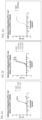
FIG. 1 a-d is set of graphs showing the binding of the murine hybridoma anti-PD-L1 antibodies to PD-L1 over a range of antibody concentrations as measured by ELISA. Binding of hybridoma antibodies 8H3-mIgG (m8H3), 15F1-mIgG (m15F1), 5G9-mIgG (m5G9), and 4A8-mIgG (m4A8) is shown in FIG. 1 a . Binding of hybridoma antibodies 5G11-mIgG (m5G11), 7B4-mIgG (m7B4), 4D1-mIgG (m4D1), and 8H4-mIgG (m8H4) is shown in FIG. 1 b . Binding of hybridoma antibody 8C6-mIgG (m8C6) is shown in FIG. 1 c . Binding of hybridoma antibody 13C5-mIgG (m13C5) is shown in FIG. 1 d . In each of FIGS. 1 a-1 d , binding of mIgG1 is shown as a negative control.
FIG. 2 a-c is set of graphs showing the binding of chimeric anti-PD-L1 antibodies to PD-L1 over a range of concentrations as measured by ELISA. Binding of chimeric antibodies ch5G11-hIgG4 and ch5G11-hIgG1 is shown in FIG. 2 a . Binding of chimeric antibodies ch13C5-hIgG4, ch13C5-hIgG1, and ch8H4-hIgG4 is shown in FIG. 2 b . Binding of chimeric antibody ch8C6-hIgG4 is shown in FIG. 2 c . In each of FIGS. 2 a-2 c , binding of hIgG4 is shown as a negative control.
FIG. 3 a-b is set of graphs showing the binding of humanized anti-PD-L1 antibodies to PD-L1 over a range of antibody concentrations as measured by ELISA. Binding of control hIgG4 and humanized antibodies hu5G11-hIgG1 and hu5G11-hIgG4 is shown in FIG. 3 a . Binding of control hIgG4 and humanized antibodies hu13C5-hIgG1 and hu13C5-hIgG4 is shown in FIG. 3 b.
FIG. 4 a-c is a set of graphs showing the blockage of the PD-1/PD-L1 interaction by hybridoma anti-PD-L1 antibodies over a range of antibody concentrations as measured by ELISA. Blockage of PD-1/PD-L1 binding by hybridoma antibodies 13C5-mIgG (m13C5), 8C6-mIgG (m8C6), 5G9-mIgG (m5G9), and 4A8-mIgG (m4A8) as compared to control mIgG1 is shown in FIG. 4 a . Blockage of PD-1/PD-L1 binding by hybridoma antibodies 5G11-mIgG (m5G11), 7B4-mIgG (m7B4), 4D1-mIgG (m4D1), and 8H4-mIgG (m8H4) as compared to control mIgG1 is shown in FIG. 4 b . Blockage of PD-1/PD-L1 binding by hybridoma antibodies 8H3-mIgG (m8H3) and 15F1-mIgG (m15F1) as compared to control mIgG1 is shown in FIG. 4 c.
FIG. 5 a-c is a set of graphs showing the blockage of the PD-1/PD-L1 interaction by chimeric anti-PD-L1 antibodies over a range of antibody concentrations as measured by ELISA. Blockage of PD-1/PD-L1 binding by chimeric antibodies ch5G11 hIgG4 and ch5G11 hIgG1 as compared to control hIgG4 is shown in FIG. 5 a . Blockage of PD-1/PD-L1 binding by chimeric antibody ch8C6-hIgG4 as compared to control hIgG4 is shown in FIG. 5 b . Blockage of PD-1/PD-L1 binding by chimeric antibodies ch8H4-hIgG4, ch13C5-hIgG1, and ch13C5-hIgG4 as compared to control hIgG4 is shown in FIG. 5 c.
FIG. 6 a-b is a set of graphs showing the blockage of the PD-1/PD-L1 interaction by humanized anti-PD-L1 antibodies over a range of antibody concentrations as measured by ELISA. Blockage of PD-1/PD-L1 binding by control hIgG4 and humanized antibodies 5G11-hIgG1 and 5G11-hIgG4 is shown in FIG. 6 a . Blockage of PD-1/PD-L1 binding by control hIgG4 and humanized antibodies 13C5-hIgG1 and 13C5-hIgG4 is shown in FIG. 6 b.
FIGS. 7 a and 7 b show the binding of the hybridoma anti-PD-L1 antibodies to PD-L1 over a range of antibody concentrations as measured by FACS. Binding (as measured by the mean fluorescence intensity) of hybridoma antibodies 4A8, 15F1, 4D1, 13C5, 8H4, and 8H3 as compared to control antibody mIgG1 is shown in FIG. 7 a . Binding (as measured by the mean fluorescence intensity) of hybridoma antibodies 5G11, 8C6, 5G9, or 7B4 as compared to control antibody mIgG1 is shown in FIG. 7 b.
FIG. 8 shows the binding of the chimeric anti-PD-L1 antibodies to PD-L1 over a range of antibody concentrations as measured by FACS. Binding of control antibody hIgG4, and chimeric antibodies ch13C5-hIgG1, ch5G11-hIgG1, and ch5G11-hIgG4 are shown.
FIG. 9 shows the binding of humanized anti-PD-L1 antibodies to PD-L1 over a range of antibody concentrations as measured by FACS. Binding of control antibody hIgG4 and humanized antibodies hu13C5-hIgG1, hu13C5-hIgG4, hu5G11-hIgG1, and hu5G11-hIgG4 are shown.
FIGS. 10 a and 10 b show the blockage of the PD-1/PD-L1 interaction by hybridoma anti-PD-L1 antibodies over a range of antibody concentrations as measured by FACS. Blockage of PD-1/PD-L1 binding by control antibody mIgG1 and hybridoma antibodies m4D1, m5G11, m13C5, m7B4, and m8H4 is shown in FIG. 10 a . Blockage of PD-1/PD-L1 binding by control antibody mIgG1 and hybridoma antibodies m4A8, m5G9, m8C6, m8H3, and m15F1 is shown in FIG. 10 b.
FIG. 11 shows the blockage of the PD-1/PD-L1 interaction over a range of concentrations of control antibody hIgG4 or chimeric anti-PD-L1 antibodies ch8C6-hIgG4, ch5G11-hIgG1, ch5G11-hIgG4, ch13C5-hIgG1, ch13C5-hIgG4, or ch8H4-hIgG4, as measured by FACS.
FIG. 12 shows the blockage of the PD-1/PD-L1 interaction over a range of concentrations of control antibody hIgG4 or humanized antibodies hu 13C5-hIgG1, hu13C5-hIgG4, hu5G11-hIgG1, or hu5G11-hIgG4, as measured by FACS.
FIG. 13 a is a graph showing IL-2 (pg/mL) production in an MLR in response to different concentrations of hybridoma anti-PD-L1 antibodies. FIG. 13 b is a graph showing IFNγ (pg/mL) production in an MLR in response to different concentrations of hybridoma anti-PD-L1 antibodies. For both FIGS. 13 a and 13 b , the antibodies tested were, from left to right, control mIgG1, m8C6, m4D1, m5G11, m7B4, m8H4, m5G9, m13C5, m8H3, and m15F1. T cell only and/or DC only wells were also included as negative controls. As shown on the x-axis for both FIGS. 13 a and 13 b , each antibody was tested at 20 μg/mL, 2 μg/mL, 0.2 μg/mL, 0.02 μg/mL, and 0.002 μg/mL.
FIG. 14 a is a graph showing IL-2 (pg/mL) production in an MLR in response to different concentrations of chimeric anti-PD-L1 antibodies. FIG. 14 b is a graph showing IFNγ (pg/mL) production in an MLR in response to different concentrations of chimeric anti-PD-L1 antibodies. For both FIGS. 14 a and 14 b , the antibodies tested were, from left to right, control hIgG4, chimeric 8C6-hIgG4, chimeric 8H4-hIgG4, chimeric 5G11-hIgG4, and chimeric 13C5-hIgG1. As shown on the x-axis for both FIGS. 14 a and 14 b , each antibody was tested at 20 μg/mL, 2 μg/mL, 0.2 μg/mL, 0.02 μg/mL, and 0.002 μg/mL.
FIG. 15 a is a graph showing IL-2 (pg/mL) production in an MLR in response to different concentrations of humanized anti-PD-L1 antibodies. FIG. 15 b is a graph showing IFNγ (pg/mL) production in an MLR in response to different concentrations of humanized anti-PD-L1 antibodies. For both FIGS. 15 a and 15 b , the antibodies tested were, from left to right, control hIgG4, hu13C5-hIgG1, hu13C5-hIgG4, hu5G11-hIgG1, and hu5G11-hIgG4. As shown on the x-axis for both FIGS. 15 a and 15 b , each antibody was tested at 20 μg/mL, 2 μg/mL, 0.2 μg/mL, 0.02 μg/mL, and 0.002 μg/mL.
FIG. 16 shows the effects of chimeric (ch) or humanized (hu) anti-PD-L1 antibodies on Treg-mediated inhibition of IFNγ production (pg/mL), in an allogeneic MLR with CD4+ CD25+ Treg cells, CD4+CD25− T cells, and dendritic cells. The antibodies tested were, from left to right, control hIgG4, ch13C5-hIgG1, ch13C5-hIgG4, hu13C5-hIgG1, hu13C5-hIgG4, ch5G11-hIgG1, ch5G11-hIgG4, hu5G11-hIgG1, and hu5G11-hIgG4.
FIG. 17 shows IFN-γ production (pg/mL) from T cells in response to costimulation with autologous DCs and anti-CD3 antibody, in the presence of humanized anti-PD-L1 antibody (hu13C5-hIgG1, hu13C5-hIgG4, hu5G11-hIgG1, or hu5G11-hIgG4), isotype control (hIgG4) antibody, or no antibody.
FIGS. 18 a and 18 b show the effect of humanized anti-PD-L1 antibodies on memory T cell responses recalled by tetanus toxin, as measured by IFN-γ production (pg/mL). Negative control hIgG4 or humanized antibody hu13C5-hIgG1, hu13C5-hIgG4, hu5G11-hIgG1, or hu5G11-hIgG4 were tested at the following concentrations: 20 μg/mL, 2 μg/mL, 0.2 μg/mL, 0.02 μg/mL, and 0.002 μg/mL.
DETAILED DESCRIPTION
PD1/PDL1 interactions inhibit T cell receptor signaling by recruiting the SHP1 and SHP2 phosphatases, which interfere with TCR signaling (Chemnitz et al. (2004) J. Immunol. 17:945-954). PD-L1 can not only promote tumor progression through inhibition of PD1-expressing immune effectors, but also modulate cell-mediated immunity in some infectious diseases (Mueller et al. (2010) J. Clin. Invest. 120:2508-2515). Furthermore, allogeneic effector T cell responses are susceptible to PD-1 pathway modulation in graft rejection (Lee et al. (2003) J. Immunol. 171:6929-6935). Therefore, the interaction of PD-1 with PD-L1 exerts a vital and diverse range of immunoregulatory roles in T cell activation, tolerance, and immune-mediated tissue damage. However, the interaction can be reversed by blocking the local binding of PD-1 with PD-L1 (Iwai et al. (2002) Proc. Nat'l. Acad Sci. USA 99: 12293-7; Brown et al. (2003) J. Immunol. 170:1257-66).
PD-1 has been found to have a correlation with cancer growth and development due to its role in protecting tumor cells from efficient immune destruction. Its ligand, PD-L1, has been revealed to have significant expression on a number of mouse and human tumors, which is postulated to mediate immune evasion (Iwai, Y. et al., Proc. Natl. Acad. Sci. USA. 99: 12293-12297 (2002); Strome S. E. et al., Cancer Res., 63:6501-6505 (2003); Dong et al. (2002) Nat. Med. 8:787-9). In humans, expression of PD-1 (on tumor infiltrating lymphocytes) and/or PD-L1 (on tumor cells) has been found in a number of primary tumor biopsies as assessed by immunohistochemistry. Such tissues include cancers of the lung, liver, ovary, cervix, skin, colon, glioma, bladder, breast, kidney, esophagus, stomach, oral squamous cell, urothelial cell, and pancreas as well as tumors of the head and neck (Brown J. A. et al., J. Immunol. 170: 1257-1266 (2003); Dong H. et al., Nat. Med. 8: 793-800 (2002); Wintterle et al., Cancer Res. 63:7462-7467 (2003); Strome S. E. et al., Cancer Res., 63: 6501-6505 (2003); Thompson R. H. et al., Cancer Res. 66: 3381-5 (2006); Thompson et al., Clin. Cancer Res. 13: 1757-61 (2007); Nomi T. et al., Clin. Cancer Res. 13: 2151-7. (2007)). More strikingly, PD-1 ligand expression on tumor cells has been correlated to poor prognosis of cancer patients across multiple tumor types (reviewed in OkaZaki and Honjo, Int. Immunol. 19: 813-824 (2007)).
While the interaction between PD-1 and PD-L1 results in a decrease in tumor infiltrating lymphocytes, a decrease in T-cell receptor mediated proliferation, and immune evasion by the cancerous cells (Dong et al. (2003) J. Mol. Med. 81:281-7; Blank et al. (2005) Cancer Immunol. Immunother. 54: 3 07-3 14; Konishi et al. (2004) Clin. Cancer Res. 10:5094-100), blockade of the PD-1/PD-L1 interaction was accordingly shown to enhance tumor-specific T-cell immunity and be helpful in clearance of tumor cells by the immune system. In a murine model of aggressive pancreatic cancer, for example, Nomi T., et al. (Clin. Cancer Res. 13: 2151-2157, 2007) demonstrated the therapeutic efficacy of PD-1/PD-L1 blockade. Administration of either PD-1 or PD-L1 directed antibody significantly inhibited tumor growth. Antibody blockade effectively promoted tumor reactive CD8+ T cell infiltration into the tumor resulting in the up-regulation of anti-tumor effectors including IFN-γ, granzyme B and perforin. Additionally, the authors showed that PDL1/PD-1 blockade can be effectively combined with chemotherapy to yield a synergistic effect. In another study, using a model of squamous cell carcinoma in mice, antibody blockade of PD-1 or PD-L1 significantly inhibited tumor growth (Tsushima F. et al., Oral Oncol. 42:268-274 (2006)).
Furthermore, transfection of a murine mastocytoma line with PD-L1 led to decreased lysis of the tumor cells when co-cultured with a tumor-specific CTL clone. Lysis was restored when anti-PD-L1 mAb was added (Iwai Y. et al., Proc. Natl. Acad. Sci. USA. 99: 12293-12297 (2002)). In vivo, blocking the PD1/PD-L1 interaction was shown to increase the efficacy of adoptive T cell transfer therapy in a mouse tumor model (Strome S. E. et al., Cancer Res. 63:6501-6505 (2003)). Further evidence for the role of PD-1 in cancer treatment comes from experiments performed with PD-1 knockout mice. PD-L1 expressing myeloma cells grew only in Wild-type animals (resulting in tumor growth and associated animal death), but not in PD-1 deficient mice (Iwai Y., et al., Proc. Natl. Acad. Sci. USA. 99: 12293-12297 (2002)). In human studies, R. M. Wong et al. (Int. Immunol. 19:1223-1234 (2007)) showed that PD-1 blockade using a fully human anti-PD-1 antibody augmented the absolute numbers of tumor-specific CD8+ T cells (CTLs) in ex vivo stimulation assays using vaccine antigens and cells from vaccinated individuals. In a similar study, antibody blockade of PD-L1 resulted in enhanced cytolytic activity of tumor-associated antigen-specific cytotoxic T cells and increased cytokine production by tumor specific TH cells (Blank C. et al., Int. J. Cancer 119: 317-327 (2006)). The same authors showed that PD-L1 blockade augments tumor-specific T cell responses in vitro when used in combination with anti-CTLA-4 blockade. Overall, the PD-1/PD-L1 pathway is a target for the development of antibody therapeutics for cancer treatment. Anti-PD-L1 antibodies may also be useful in chronic viral infection. Memory CD8+ T cells generated after an acute viral infection are highly functional and constitute an important component of protective immunity. In contrast, chronic infections are often characterized by varying degrees of functional impairment (exhaustion) of virus-specific T-cell responses, and this defect is a principal reason for the inability of the host to eliminate the persisting pathogen. Although functional effector T cells are initially generated during the early stages of infection, they gradually lose function during the course of a chronic infection. Barber et al. (Barber et al., Nature 439: 682-687 (2006)) showed that mice infected with a laboratory strain of LCMV developed chronic infection resulting in high levels of virus in the blood and other tissues. These mice initially developed a robust T cell response, but eventually succumbed to the infection upon T cell exhaustion. The authors found that the decline in number and function of the effector T cells in chronically infected mice could be reversed by injecting an antibody that blocked the interaction between PD-1 and PD-L1.
In one aspect, the present invention provides antibodies or antigen binding fragments thereof that bind to programmed death ligand 1 (PD-L1). In one embodiment, the antibodies or fragments thereof bind to human PD-L1. In another embodiment, the antibodies or fragments thereof bind to human and to cynomolgous PD-L1. In another embodiment, the antibodies or fragments thereof block the interaction of PD-L1 with its receptor PD-1 on T cells. In one aspect, the present invention provides methods of making and using the anti-PD-L1 antibodies or fragments thereof, and compositions comprising anti-PD-L1 antibodies or fragments thereof, including pharmaceutical compositions.
As used herein, the term “antibody” refers to a binding protein having at least one antigen binding domain. The antibodies and fragments thereof of the present invention may be whole antibodies or any fragment thereof. Thus, the antibodies and fragments of the invention include monoclonal antibodies or fragments thereof and antibody variants or fragments thereof, as well as immunoconjugates. Examples of antibody fragments include Fab fragments, Fab′ fragments, F(ab)′ fragments, Fv fragments, isolated CDR regions, single chain Fv molecules (scFv), and other antibody fragments known in the art. Antibodies and fragments thereof may also include recombinant polypeptides, fusion proteins, and bi-specific antibodies. The anti-PD-L1 antibodies and fragments thereof disclosed herein may be of an IgG1, IgG2, IgG3, or IgG4 isotype. The term “isotype” refers to the antibody class encoded by the heavy chain constant region genes. In one embodiment, the anti-PD-L1 antibodies and fragments thereof disclosed herein are of an IgG1 or an IgG4 isotype. The PD-L1 antibodies and fragments thereof of the present invention may be derived from any species including, but not limited to, mouse, rat, rabbit, primate, llama, and human. The PD-L1 antibodies and fragments thereof may be chimeric, humanized, or fully human antibodies. In one embodiment, the anti-PD-L1 antibodies are antibodies produced by a hybridoma cell line derived from a mouse. Thus, in one embodiment, the anti-PD-L1 antibodies are murine antibodies. In another embodiment, the anti-PD-L1 antibodies are chimeric antibodies. In a further embodiment, the chimeric antibodies are mouse-human chimeric antibodies. In another embodiment, the antibodies are humanized antibodies. In a further embodiment, the antibodies are derived from murine antibodies and are humanized.
A “chimeric antibody” is an antibody having at least a portion of the heavy chain variable region and at least a portion of the light chain variable region derived from one species; and at least a portion of a constant region derived from another species. For example, in one embodiment, a chimeric antibody may comprise murine variable regions and a human constant region.
A “humanized antibody” is an antibody containing complementarity determining regions (CDRs) that are derived from a non-human antibody; and framework regions as well as constant regions that are derived from a human antibody. For example, the anti-PD-L1 antibodies provided herein may comprise CDRs derived from one or more murine antibodies and human framework and constant regions. Thus, in one embodiment, the humanized antibody provided herein binds to the same epitope on PD-L1 as the murine antibody from which the antibody's CDRs are derived. Exemplary humanized antibodies are provided herein. Additional anti-PD-L1 antibodies comprising the heavy and light chain CDRs provided herein, or variants thereof, may be generated using any human framework sequence, and are also encompassed in the present invention. In one embodiment, framework sequences suitable for use in the present invention include those framework sequences that are structurally similar to the framework sequences provided herein. Further modifications in the framework regions may be made to improve the properties of the antibodies provided herein. Such further framework modifications may include chemical modifications; point mutations to reduce immunogenicity or remove T cell epitopes; or back mutation to the residue in the original germline sequence. In some embodiments, such modifications include those corresponding to the mutations exemplified herein, including backmutations to the germline sequence. For example, in one embodiment, one or more amino acids in the human framework regions of the VH and/or VL of the humanized antibodies provided herein are back mutated to the corresponding amino acid in the parent murine antibody. As an example, as for VH and VL of humanized 5G11 and humanized 13C5, several sites of framework amino acid of the aforementioned template human antibody were back mutated to the corresponding amino acid sequences in mouse 5G11 and 13C5 antibodies. In one embodiment, the amino acid at positions 53 and/or 60 and/or 67 of the light chain variable region is back mutated to the corresponding amino acid found at that position in the mouse 5G11 or 13C5 light chain variable region. In another embodiment, the amino acid at positions 24 and/or 28 and/or 30 and/or 49 and/or 73 and/or 83 and/or 94 of the heavy chain variable region is back mutated to the corresponding amino acid found at that position in the mouse 5G11 or 13C5 heavy chain variable region. In one embodiment, the humanized 5G11 antibody comprises a light chain variable region wherein the amino acid at position 60 is mutated from Ser (S) to Asp (D) and the amino acid at position 67 is mutated from Ser (S) to Tyr (Y); and a heavy chain variable region wherein the amino acid at position 24 is mutated from Phe (F) to Val (V), the amino acid at position 49 is mutated from Ala (A) to Gly (G), the amino acid at position 73 is mutated from Thr (T) to Asn (N), and the amino acid at position 83 is mutated from Thr (T) to Asn (N). In one embodiment, the humanized 13C5 antibody comprises a light chain variable region wherein the amino acid at position 53 is mutated from Tyr (Y) to Lys (K); and a heavy chain variable region wherein the amino acid at position 28 is mutated from Thr (T) to Ile (I), the amino acid at position 30 is mutated from Ser (S) to Arg (R), the amino acid at position 49 is mutated from Ser (S) to Ala (A), and the amino acid at position 94 is mutated from Tyr (Y) to Asp (D). Additional or alternate back mutations may be made in the framework regions of the humanized antibodies provided herein in order to improve the properties of the antibodies. The present invention also encompasses humanized antibodies that bind to PD-L1 and comprise framework modifications corresponding to the exemplary modifications described herein with respect to any suitable framework sequence, as well as other framework modifications that otherwise improve the properties of the antibodies.
As used herein, the term “derived” when used to refer to a molecule or polypeptide relative to a reference antibody or other binding protein, means a molecule or polypeptide that is capable of binding with specificity to the same epitope as the reference antibody or other binding protein.
The antibodies and antigen-binding fragments thereof disclosed herein are specific for PD-L1. In one embodiment, the antibodies and fragments thereof are specific for human PD-L1. In one embodiment, the antibodies and fragments provided herein bind to human or primate PD-L1 but not to PD-L1 from any other mammal. In a further embodiment, the antibodies and fragments thereof do not bind to mouse PD-L1. The terms “human PD-L1,” “hPD-L1”, and “huPD-L1” and the like are used interchangeably herein and refer to human PD-L1 and variants or isoforms of human PD-L1. By “specific for” is meant that the antibodies and fragments thereof bind PD-L1 with greater affinity than any other target. As used herein, the term “EC50” refers to the effective concentration, 50% maximal response of the antibody. As used herein, the term “IC50” refers to the inhibitory concentration, 50% maximal response of the antibody. Both EC50 and IC50 may be measured by ELISA or FACS analysis, or any other method known in the art.
In one embodiment, the anti-PD1 antibodies and fragments or variants thereof have a binding affinity (KD) for PD-L1 in the range of about 0.001 nM to about 100 nM, about 0.002 nM to about 50 nM, about 0.005 nM to about 5 nM, about 0.01 nM to about 1 nM, or about 0.05 nM to about 0.1 nM. In one embodiment, the antibodies and fragments thereof have a binding affinity (KD) for PD-L1 of about 50 nM or less, about 25 nM or less, about 20 nM or less, about 15 nM or less, about 10 nM or less, about 8 nM or less, about 6 nM or less, about 5 nM or less, about 4 nM or less, about 3 nM or less, about 2 nM or less, about 1 nM or less, about 0.9 nM or less, about 0.8 nM or less, about 0.7 nM or less, about 0.6 nM or less, about 0.5 nM or less, about 0.4 nM or less, about 0.3 nM or less, about 0.2 nM or less, about 0.1 nM or less, about 0.09 nM or less, about 0.08 nM or less, about 0.07 nM or less, about 0.06 nM or less, about 0.05 nM or less, about 0.04 nM or less, about 0.03 nM or less, about 0.02 nM or less, about 0.01 nM or less, about 0.009 nM or less, about 0.008 nM or less, about 0.007 nM or less, about 0.006 nM or less, about 0.005 nM or less, about 0.004 nM or less, about 0.003 nM or less, about 0.002 nM or less, or about 0.001 nM or less. In one embodiment, the antibodies and fragments thereof have a binding affinity (KD) for PD-L1 of about 10 nM, about 9 nM, about 8 nM, about 7 nM, about 6 nM, about 5 nM, about 4 nM, about 3 nM, about 2 nM, about 1 nM, about 0.9 nM, about 0.8 nM, about 0.7 nM, about 0.6 nM, about 0.5 nM, about 0.4 nM, about 0.3 nM, about 0.2 nM, about 0.1 nM, about 0.09 nM, about 0.08 nM, about 0.07 nM, about 0.06 nM, about 0.05 nM, about 0.04 nM, about 0.03 nM, about 0.02 nM, about 0.01 nM, about 0.009 nM, about 0.008 nM, about 0.007 nM, about 0.006 nM, about 0.005 nM, about 0.004 nM, about 0.003 nM, about 0.002 nM, or about 0.001 nM.
In one embodiment, the antibodies and fragments provided herein comprise a light chain and a heavy chain, each of which comprises three CDR regions. Exemplary heavy chain CDR sequences (HCDR1, HCDR2, and HCDR3) for PD-L1 antibodies of the invention are provided below in Table 1. Exemplary light chain CDR sequences (LCDR1, LCDR2, and LCDR3) for PD-L1 antibodies of the invention are provided below in Table 2. Exemplary variable regions and full length heavy and light chain sequences for PD-L1 antibodies of the invention are provided below in Table 3.
TABLE 1
Heavy Chain CDR Sequences
Name HCDR SEQ ID NO Sequence
13C5
1 81 SYGMS
2 82 SISSGGSTYYPDSVKG
3 83 GYDSGFAY
5G9
1 87 SYGMS
2 88 SISSGGTTYYPDSVKG
3 89 GYDSGFAY
5G11
1 93 TYGVH
2 94 VIWRGVTTDYNAAFMS
3 95 LGFYAMDY
8C6
1 99 SYGVH
2 100 VIWSGGVTDYNAAFIS
3 101 LGFYAMDY
7B4
1 105 TYWMH
2 106 QINPDSTTINYAPSLKD
3 107 PGDYGYDFDC
4D1
1 111 SGYWN
2 112 YISYSGSTYYNPSLKS
3 113 SLLWFSTGFAY
4A8
1 117 SYGVH
2 118 VIWSGGITDYNAAFKS
3 119 LGFYAMDY
8H4
1 123 SYGMS
2 124 SISSGGTTYYLGSVQG
3 125 GYDAGFAY
8H3
1 129 SGYWT
2 130 YISYTGSTYYNPSLKS
3 131 QRDWLGFAY
15F1
1 135 SYGMS
2 136 SISSGGSIYYPDSVKG
3 137 GYDAGFAF
TABLE 2
Light chain CDR Sequences
Name LCDR SEQ ID NO Sequence
13C5
1 84 ASQSVSTSSSSFMH
2 85 YASNLES
3 86 QHSWEIPYT
5G9
1 90 RASQSVSTSSSSYMH
2 91 YASNLES
3 92 QHSWEIPYT
5G11
1 96 KASQSVSNDVA
2 97 YAANRYT
3 98 QQDYTSPYT
8C6
1 102 KASQSVSNDVG
2 103 YASNRYS
3 104 QQDYTSPYT
7B4
1 108 RSSQIIVHSNANTYLE
2 109 KVSNRFS
3 110 FQGSHVPYT
4D1
1 114 SASSSVSSSYLY
2 115 NTSNLAS
3 116 HQWRSYPPT
4A8
1 120 SANSSVSYMH
2 121 DTSKLAS
3 122 QQWSSNPWT
8H4
1 126 RASQSVSTSSYSYMH
2 127 YASNLES
3 128 QNSWEIPYT
8H3
1 132 KSSQSLLYSSNQKNSLA
2 133 WASNRES
3 134 QQYYSYPLT
15F1
1 138 RASQSVSTSSYSYVH
2 139 YASNLES
3 140 QHSWEIPYT
TABLE 3
Heavy chain and light chain variable region and full length
heavy and light chain amino acid sequences
SEQ
ID
Name Region NO Sequence
13C5 Heavy 2 EVKLVESGGGLVKPGGSLKLSCAASGFIFRSYGMSWVRQTPE
murine chain KRLEWVASISSGGSTYYPDSVKGRFTISRDNAR
variable NILYLQMSSLRSEDTAMYDCARGYDSGFAYWGQGTLVTVSE
13C5 Light 4 DIVLTQSPASLAVSLGQRATISCRASQSVSTSSSSFMHWYQQK
murine chain PGQPPKLLIKYASNLESGVPARFSGSGSGTDFT
variable LNIHPVEEEDTATYYCQHSWEIPYTFGGGTKLEIKR
5G9 murine Heavy 6 EVKLVESGGGLVKPGGSLKLSCAASGFTFRSYGMSWVRQTP
chain EKRLEWVASISSGGTTYYPDSVKGRFIISRDNARNILYLQMSS
variable LRSEDTAMYYCAKGYDSGFAYWGQGTLVIVSA
5G9 murine Light 8 DIVLTQSPPSLAVSLGQRATISCRASQSVSTSSSSYMHWYQQK
chain PGQPPKLLIKYASNLESGVPARFSGSGSGTDFTLNIHPVEEEDT
variable ATYYCQHSWEIPYTFGGGTKLEIK
5G11 Heavy 10 QVQLKQSGPGLVQPSQSLSITCTVSGFSLTTYGVHWVRQSPG
murine chain KGLEWLGVIWRGVTTDYNAAFMSRLTITKDNSKSQVFFKMN
variable SLQANDTAIYYCARLGFYAMDYWGQGTSVTVSS
5G11 Light 12 SIVMTQTPKFLLVSAGDRVTITCKASQSVSNDVAWYQQKPG
murine chain QSPKLLIYYAANRYTGVPDRFTGSGYGTDFTFTISIVQAEDLA
variable VYFCQQDYTSPYTFGGGTKLEIK
8C6 murine Heavy 14 QVQLKQSGPGLVQPSQSLSITCTVSGFSLTSYGVHWVRQSPG
chain KGLEWLGVIWSGGVTDYNAAFISRLSISKDNSKSQVFFKMNS
variable LQANDTAIYYCARLGFYAMDYWGQGTSVTVSS
8C6 murine Light 16 SIVMTQTPKFLLVSAGDRVTITCKASQSVSNDVGWYQQKPG
chain QSPKLLIYYASNRYSGVPDRFTGSGYGTDFTFTISTVQAEDLA
variable VYFCQQDYTSPYTFGGGTKLEIK
7B4 murine Heavy 18 EVKLFESGGGLVQPGGSLKLSCVASGFDFSTYWMHWVRQAP
chain GQGLEWIGQINPDSTTINYAPSLKDRFIISRDNAKNTLFLQMS
variable KVRSEDTALYYCAKPGDYGYDFDCWGQGTTLTVSS
7B4 murine Light 20 DVLMTQTPLYLPVSLGDQASISCRSSQIIVHSNANTYLEWFLQ
chain KPGQSPKWYKVSNRFSGVPDRFSGSGSGTDFTLKISRVEAE
variable DLGVYYCFQGSHVPYTFGGGTKLEIK
4D1 murine Heavy 22 EVQLQESGPSLVKPSQTLSLTCSVTGDSITSGYWNWIRKFPGN
chain KLEYMGYISYSGSTYYNPSLKSRISITRDTSKNQYYLQLNSVT
variable TEDTATYYCARSLLWFSTGFAYWGQGTLVTVSA
4D1 murine Light 24 QIVLTQSPAIMSASPGEKVTLTCSASSSVSSSYLYWNQQKPGS
chain SPKVWIYNTSNLASGVPARFSGSGSGTSYSLTISSMEAEDAAS
variable YFCHQWRSYPPTLGAGTKLELK
4A8 murine Heavy 26 QVQLKQSGPGLVQPSQSLSITCTVSGFSLTSYGVHWVRQSPG
chain KGLEWLGVIWSGGITDYNAAFKSRLSISKDNSKSQVFFKMNS
variable LQANDTAIYFCARLGFYAMDYWGQGTSVTVSS
4A8 murine Light 28 QIVLTQSPAIMSASPGEKVTMTCSANSSVSYMHWYQQKSGTS
chain PKRWIYDTSKLASGVPARFSGSGSGTSYSLTISSMGAEDAAT
variable YYCQQWSSNPWTFGGGTKLEIK
8H4 murine Heavy 30 EVKLVESGGGLVKPGGSLKLSCAASGFTFRSYGMSWARQIPE
chain KRLEWVASISSGGTTYYLGSVQGRFTISRDNARNILYLQMSSL
variable RSEDTAMYYCARGYDAGFAYWGQGTLVSVSE
8H4 murine Light 32 DIVLTQSPASLAVSLGQRATISCRASQSVSTSSYSYMHWYQQ
chain KPGQPPKLLIKYASNLESGVPARFSGSGSGTDFTLNIHPVEEE
variable DTATYYCQNSWEIPYTFGGGTKLEIK
8H3 murine Heavy 34 EVQLQESGPSLVKPSQTLSLTCSVTGDSITSGYWTWIRKFPGN
chain KLEYMGYISYTGSTYYNPSLKSRISISRDTSKSQYYLQLNSVT
variable TEDTATYYCARQRDWLGFAYWGQGTLVTVSA
8H3 murine Light 36 DIVMTQTPSSLAVSLGEKVTMSCKSSQSLLYSSNQKNSLAWY
chain QQKPGQSPKLLIYWASNRESGVPDRFTGSSSGTDFTLTISSVK
variable AEDLAVYYCQQYYSYPLTFGAGTKLELK
15F1 murine Heavy 38 EEKLVESGGGLVKPGGSLKLSCAASGFSFSSYGMSWVRQTPE
chain KRLEWVASISSGGSIYYPDSVKGRFTISRDNARNILYLQMSSL
variable RSEDTAMYYCARGYDAGFAFWGQGTLVTASA
15F1 murine Light 40 DIVLTQSPASLAVSLGQRATISCRASQSVSTSSYSYVHWYQQ
chain KPGQPPKLLIKYASNLESGVPARFSGSGSGTDFTLNIHPVEEE
variable DTATYYCQHSWEIPYTFGGGTKLEIK
5G11 Heavy 42 QITLKESGPTLVKPTQTLTLTCTVSGFSLSTYGVHWIRQPPGK
humanized chain ALEWLGVIWRGVTTDYNAAFMSRLTITKDNSKNQVVLTMN
variable NMDPVDTATYYCARLGFYAMDYWGQGTLVTVSS
5G11 Light 44 DIQMTQSPSSLSASVGDRVTITCKASQSVSNDVAWYQQKPGK
humanized chain APKLLIYYAANRYTGVPDRFSGSGYGTDFTFTISSLQPEDIAT
variable YFCQQDYTSPYTFGQGTKLEIK
13C5 Heavy 46 EVQLVESGGGLVKPGGSLRLSCAASGFIFRSYGMSWVRQAP
humanized chain GKGLEWVASISSGGSTYYPDSVKGRFTISRDNAKNSLYLQMN
variable SLRAEDTAVYDCARGYDSGFAYWGQGTLVTVSS
13C5 Light 48 DIVLTQSPASLAVSPGQRATITCRASQSVSTSSSSFMHWYQQK
humanized chain PGQPPKLLIKYASNLESGVPARFSGSGSGTDFTLTINPVEAND
variable TANYYCQHSWEIPYTFGQGTKLEIK
Chimeric Full 50 QVQLKQSGPGLVQPSQSLSITCTVSGFSLTSYGVHWVRQSPG
8C6-IgG4 length KGLEWLGVIWSGGVTDYNAAFISRLSISKDNSKSQVFFKMNS
(F234A/ heavy LQANDTAIYYCARLGFYAMDYWGQGTSVTVSSASTKGPSVF
L235A) chain PLAPCSRSTSESTAALGCLVKDYFPEPVTVSWNSGALTSGVH
(IgG4) TFPAVLQSSGLYSLSSVVTVPSSSLGTKTYTCNVDHKPSNTKV
DKRVESKYGPPCPPCPAPEAAGGPSVFLFPPKPKDTLMISRTP
EVTCVVVDVSQEDPEVQFNWYVDGVEVHNAKTKPREEQFN
STYRVVSVLTVLHQDWLNGKEYKCKVSNKGLPSSIEKTISKA
KGQPREPQVYTLPPSQEEMTKNQVSLTCLVKGFYPSDIAVEW
ESNGQPENNYKTTPPVLDSDGSFFLYSRLTVDKSRWQEGNVF
SCSVMHEALHNHYTQKSLSLSLG
Chimeric Full 52 SIVMTQTPKFLLVSAGDRVTITCKASQSVSNDVGWYQQKPG
8C6 length QSPKLLIYYASNRYSGVPDRFTGSGYGTDFTFTISTVQAEDLA
light VYFCQQDYTSPYTFGGGTKLEIKRTVAAPSVFIFPPSDEQLKS
chain GTASVVCLLNNFYPREAKVQWKVDNALQSGNSQESVTEQDS
KDSTYSLSSTLTLSKADYEKHKVYACEVTHQGLSSPVTKSFN
RGEC
Chimeric Full 54 EVKLVESGGGLVKPGGSLKLSCAASGFTFRSYGMSWARQIPE
8H4-IgG4 length KRLEWVASISSGGTTYYLGSVQGRFTISRDNARNILYLQMSSL
(F234A/ heavy RSEDTAMYYCARGYDAGFAYWGQGTLVSVSSASTKGPSVFP
L235A) chain LAPCSRSTSESTAALGCLVKDYFPEPVTVSWNSGALTSGVHT
(IgG4) FPAVLQSSGLYSLSSVVTVPSSSLGTKTYTCNVDHKPSNTKV
DKRVESKYGPPCPPCPAPEAAGGPSVFLFPPKPKDTLMISRTP
EVTCVVVDVSQEDPEVQFNWYVDGVEVHNAKTKPREEQFN
STYRVVSVLTVLHQDWLNGKEYKCKVSNKGLPSSIEKTISKA
KGQPREPQVYTLPPSQEEMTKNQVSLTCLVKGFYPSDIAVEW
ESNGQPENNYKTTPPVLDSDGSFFLYSRLTVDKSRWQEGNVF
SCSVMHEALHNHYTQKSLSLSLG
Chimeric Full 56 DIVLTQSPASLAVSLGQRATISCRASQSVSTSSYSYMHWYQQ
8H4 length KPGQPPKLLIKYASNLESGVPARFSGSGSGTDFTLNIHPVEEE
light DTATYYCQNSWEIPYTFGGGTKLEIKRTVAAPSVFIFPPSDEQ
chain LKSGTASVVCLLNNFYPREAKVQWKVDNALQSGNSQESVTE
QDSKDSTYSLSSTLTLSKADYEKHKVYACEVTHQGLSSPVTK
SFNRGEC
Chimeric Full 58 QVQLKQSGPGLVQPSQSLSITCTVSGFSLTTYGVHWVRQSPG
5G11-IgG1 length KGLEWLGVIWRGVTTDYNAAFMSRLTITKDNSKSQVFFKMN
(D265A) heavy SLQANDTAIYYCARLGFYAMDYWGQGTSVTVSSASTKGPSV
chain FPLAPSSKSTSGGTAALGCLVKDYFPEPVTVSWNSGALTSGV
(IgG1) HTFPAVLQSSGLYSLSSVVTVPSSSLGTQTYICNVNHKPSNTK
VDKKVEPKSCDKTHTCPPCPAPELLGGPSVFLFPPKPKDTLMI
SRTPEVTCVVVAVSHEDPEVKFNWYVDGVEVHNAKTKPREE
QYASTYRVVSVLTVLHQDWLNGKEYKCKVSNKALPAPIEKT
ISKAKGQPREPQVYTLPPSRDELTKNQVSLTCLVKGFYPSDIA
VEWESNGQPENNYKTTPPVLDSDGSFFLYSKLTVDKSRWQQ
GNVFSCSVMHEALHNHYTQKSLSLSPGK
Chimeric Full 60 QVQLKQSGPGLVQPSQSLSITCTVSGFSLTTYGVHWVRQSPG
5G11-IgG4 length KGLEWLGVIWRGVTTDYNAAFMSRLTITKDNSKSQVFFKMN
(F234A/ heavy SLQANDTAIYYCARLGFYAMDYWGQGTSVTVSSASTKGPSV
L235A) chain FPLAPCSRSTSESTAALGCLVKDYFPEPVTVSWNSGALTSGV
(IgG4) HTFPAVLQSSGLYSLSSVVTVPSSSLGTKTYTCNVDHKPSNTK
VDKRVESKYGPPCPPCPAPEAAGGPSVFLFPPKPKDTLMISRT
PEVTCVVVDVSQEDPEVQFNWYVDGVEVHNAKTKPREEQF
NSTYRVVSVLTVLHQDWLNGKEYKCKVSNKGLPSSIEKTISK
AKGQPREPQVYTLPPSQEEMTKNQVSLTCLVKGFYPSDIAVE
WESNGQPENNYKTTPPVLDSDGSFFLYSRLTVDKSRWQEGN
VFSCSVMHEALHNHYTQKSLSLSLG
Chimeric Full 62 SIVMTQTPKFLLVSAGDRVTITCKASQSVSNDVAWYQQKPG
5G11 length QSPKLLIYYAANRYTGVPDRFTGSGYGTDFTFTISIVQAEDLA
light VYFCQQDYTSPYTFGGGTKLEIKRTVAAPSVFIFPPSDEQLKS
chain GTASVVCLLNNFYPREAKVQWKVDNALQSGNSQESVTEQDS
KDSTYSLSSTLTLSKADYEKHKVYACEVTHQGLSSPVTKSFN
RGEC
Chimeric Full 64 EVKLVESGGGLVKPGGSLKLSCAASGFIFRSYGMSWVRQTPE
13C5-IgG1 length KRLEWVASISSGGSTYYPDSVKGRFTISRDNARNILYLQMSSL
(D265A) heavy RSEDTAMYDCARGYDSGFAYWGQGTLVTVSSASTKGPSVFP
chain LAPSSKSTSGGTAALGCLVKDYFPEPVTVSWNSGALTSGVHT
(IgG1) FPAVLQSSGLYSLSSVVTVPSSSLGTQTYICNVNHKPSNTKVD
KKVEPKSCDKTHTCPPCPAPELLGGPSVFLFPPKPKDTLMISR
TPEVTCVVVAVSHEDPEVKFNWYVDGVEVHNAKTKPREEQ
YASTYRVVSVLTVLHQDWLNGKEYKCKVSNKALPAPIEKTIS
KAKGQPREPQVYTLPPSRDELTKNQVSLTCLVKGFYPSDIAV
EWESNGQPENNYKTTPPVLDSDGSFFLYSKLTVDKSRWQQG
NVFSCSVMHEALHNHYTQKSLSLSPGK
Chimeric Full 66 EVKLVESGGGLVKPGGSLKLSCAASGFIFRSYGMSWVRQTPE
13C5-IgG4 length KRLEWVASISSGGSTYYPDSVKGRFTISRDNARNILYLQMSSL
(F234A/ heavy RSEDTAMYDCARGYDSGFAYWGQGTLVTVSSASTKGPSVFP
L235A) chain LAPCSRSTSESTAALGCLVKDYFPEPVTVSWNSGALTSGVHT
(IgG4) FPAVLQSSGLYSLSSVVTVPSSSLGTKTYTCNVDHKPSNTKV
DKRVESKYGPPCPPCPAPEAAGGPSVFLFPPKPKDTLMISRTP
EVTCVVVDVSQEDPEVQFNWYVDGVEVHNAKTKPREEQFN
STYRVVSVLTVLHQDWLNGKEYKCKVSNKGLPSSIEKTISKA
KGQPREPQVYTLPPSQEEMTKNQVSLTCLVKGFYPSDIAVEW
ESNGQPENNYKTTPPVLDSDGSFFLYSRLTVDKSRWQEGNVF
SCSVMHEALHNHYTQKSLSLSLG
Chimeric Full 68 DIVLTQSPASLAVSLGQRATISCRASQSVSTSSSSFMHWYQQK
13C5 length PGQPPKLLIKYASNLESGVPARFSGSGSGTDFTLNIHPVEEEDT
light ATYYCQHSWEIPYTFGGGTKLEIKRTRTVAAPSVFIFPPSDEQ
chain LKSGTASVVCLLNNFYPREAKVQWKVDNALQSGNSQESVTE
QDSKDSTYSLSSTLTLSKADYEKHKVYACEVTHQGLSSPVTK
SFNRGEC
Humanized Full 70 QITLKESGPTLVKPTQTLTLTCTVSGFSLSTYGVHWIRQPPGK
5G11-IgG1 length ALEWLGVIWRGVTTDYNAAFMSRLTITKDNSKNQVVLTMN
(D265A) heavy NMDPVDTATYYCARLGFYAMDYWGQGTLVTVSSASTKGPS
chain VFPLAPSSKSTSGGTAALGCLVKDYFPEPVTVSWNSGALTSG
(IgG1 ) VHTFPAVLQSSGLYSLSSVVTVPSSSLGTQTYICNVNHKPSNT
KVDKKVEPKSCDKTHTCPPCPAPELLGGPSVFLFPPKPKDTL
MISRTPEVTCVVVAVSHEDPEVKFNWYVDGVEVHNAKTKPR
EEQYNSTYRVVSVLTVLHQDWLNGKEYKCKVSNKALPAPIE
KTISKAKGQPREPQVYTLPPSRDELTKNQVSLTCLVKGFYPSD
IAVEWESNGQPENNYKTTPPVLDSDGSFFLYSKLTVDKSRWQ
QGNVFSCSVMHEALHNHYTQKSLSLSPGK
Humanized Full 72 QITLKESGPTLVKPTQTLTLTCTVSGFSLSTYGVHWIRQPPGK
5G11-IgG4 length ALEWLGVIWRGVTTDYNAAFMSRLTITKDNSKNQVVLTMN
(F234A/ heavy NMDPVDTATYYCARLGFYAMDYWGQGTLVTVSSASTKGPS
L235A) chain VFPLAPCSRSTSESTAALGCLVKDYFPEPVTVSWNSGALTSG
(IgG4) VHTFPAVLQSSGLYSLSSVVTVPSSSLGTKTYTCNVDHKPSNT
KVDKRVESKYGPPCPPCPAPEAAGGPSVFLFPPKPKDTLMISR
TPEVTCVVVDVSQEDPEVQFNWYVDGVEVHNAKTKPREEQF
NSTYRVVSVLTVLHQDWLNGKEYKCKVSNKGLPSSIEKTISK
AKGQPREPQVYTLPPSQEEMTKNQVSLTCLVKGFYPSDIAVE
WESNGQPENNYKTTPPVLDSDGSFFLYSRLTVDKSRWQEGN
VFSCSVMHEALHNHYTQKSLSLSLG
Humanized Full 74 DIQMTQSPSSLSASVGDRVTITCKASQSVSNDVAWYQQKPGK
5G11 length APKLLIYYAANRYTGVPDRFSGSGYGTDFTFTISSLQPEDIAT
light YFCQQDYTSPYTFGQGTKLEIKRTVAAPSVFIFPPSDEQLKSG
chain TASVVCLLNNFYPREAKVQWKVDNALQSGNSQESVTEQDSK
DSTYSLSSTLTLSKADYEKHKVYACEVTHQGLSSPVTKSFNR
GEC
Humanized Full 76 EVQLVESGGGLVKPGGSLRLSCAASGFIFRSYGMSWVRQAP
13C5-IgG1 length GKGLEWVASISSGGSTYYPDSVKGRFTISRDNAKNSLYLQMN
(D265A) heavy SLRAEDTAVYDCARGYDSGFAYWGQGTLVTVSSASTKGPSV
chain FPLAPSSKSTSGGTAALGCLVKDYFPEPVTVSWNSGALTSGV
(IgG1) HTFPAVLQSSGLYSLSSVVTVPSSSLGTQTYICNVNHKPSNTK
VDKKVEPKSCDKTHTCPPCPAPELLGGPSVFLFPPKPKDTLMI
SRTPEVTCVVVAVSHEDPEVKFNWYVDGVEVHNAKTKPREE
QYNSTYRVVSVLTVLHQDWLNGKEYKCKVSNKALPAPIEKT
ISKAKGQPREPQVYTLPPSRDELTKNQVSLTCLVKGFYPSDIA
VEWESNGQPENNYKTTPPVLDSDGSFFLYSKLTVDKSRWQQ
GNVFSCSVMHEALHNHYTQKSLSLSPGK
Humanized Full 78 EVQLVESGGGLVKPGGSLRLSCAASGFIFRSYGMSWVRQAP
13C5-IgG4 length GKGLEWVASISSGGSTYYPDSVKGRFTISRDNAKNSLYLQMN
(F234A/ heavy SLRAEDTAVYDCARGYDSGFAYWGQGTLVTVSSASTKGPSV
L235A) chain FPLAPCSRSTSESTAALGCLVKDYFPEPVTVSWNSGALTSGV
(IgG4) HTFPAVLQSSGLYSLSSVVTVPSSSLGTKTYTCNVDHKPSNTK
VDKRVESKYGPPCPPCPAPEAAGGPSVFLFPPKPKDTLMISRT
PEVTCVVVDVSQEDPEVQFNWYVDGVEVHNAKTKPREEQF
NSTYRVVSVLTVLHQDWLNGKEYKCKVSNKGLPSSIEKTISK
AKGQPREPQVYTLPPSQEEMTKNQVSLTCLVKGFYPSDIAVE
WESNGQPENNYKTTPPVLDSDGSFFLYSRLTVDKSRWQEGN
VFSCSVMHEALHNHYTQKSLSLSLG
Humanized Full 80 DIVLTQSPASLAVSPGQRATITCRASQSVSTSSSSFMHWYQQK
13C5 length PGQPPKLLIKYASNLESGVPARFSGSGSGTDFTLTINPVEAND
light TANYYCQHSWEIPYTFGQGTKLEIKRTVAAPSVFIFPPSDEQL
chain KSGTASVVCLLNNFYPREAKVQWKVDNALQSGNSQESVTEQ
DSKDSTYSLSSTLTLSKADYEKHKVYACEVTHQGLSSPVTKS
FNRGEC
In one embodiment, the invention provides anti-PD-L1 antibodies that comprise the light chain CDRs and heavy chain CDRs of antibodies 13C5, 5G9, 5G11, 8C6, 7B4, 4D1, 4A8, 8H4, 8H3, and/or 15F1. The person of skill in the art will understand that the heavy and light chain CDRs of the antibodies provided herein may be independently selected, or mixed and matched, to form an antibody or binding fragment thereof comprising any heavy chain CDR1, CDR2, and CDR3; and any light chain CDR1, CDR2, and CDR3 from the antibodies provided herein. Thus, the invention provides anti-PD-L1 antibodies that comprise a heavy chain CDR1 comprising an amino acid sequence selected from the group consisting of SEQ ID NOs: 81, 87, 93, 99, 105, 111, 117, 123, 129, and 135; a heavy chain CDR2 comprising an amino acid sequence selected from the group consisting of SEQ ID NOs: 82, 88, 94, 100, 106, 112, 118, 124, 130, and 136; a heavy chain CDR3 comprising an amino acid sequence selected from the group consisting of SEQ ID NOs: 83, 89, 95, 101, 107, 113, 119, 125, 131, and 137; a light chain CDR1 comprising an amino acid sequence selected from the group consisting of SEQ ID NOs: 84, 90, 96, 102, 108, 114, 120, 126, 132, and 138; a light chain CDR2 comprising an amino acid sequence selected from the group consisting of SEQ ID NOs: 85, 91, 97, 103, 109, 115, 121, 127, 133, and 139; and a light chain CDR3 comprising an amino acid sequence selected from the group consisting of SEQ ID NOs:86, 92, 98, 104, 110, 116, 122, 128, 134, and 140. In one embodiment, the present invention provides anti-PD-L1 antibodies comprising heavy and light chain CDR regions comprising amino acid sequences having at least 75%, at least 80%, at least at least 81%, at least 82%, at least 83%, at least 84%, at least 85%, at least 86%, at least 87%, at least 88%, at least 89%, at least 90%, at least 91%, at least 92%, at least 93%, at least 94%, at least 95%, at least 96%, at least 97%, at least 98%, or at least 99% homology to the corresponding light or heavy chain CDR1, CDR2, or CDR3 provided herein. In one embodiment, the present invention provides anti-PD-L1 antibodies comprising heavy and light chain CDR regions comprising amino acid sequences having 1, 2, 3, 4, 5, or 6 amino acid substitutions, deletions, or insertions relative to the corresponding light or heavy chain CDR1, CDR2, or CDR3 provided herein.
In one embodiment, the invention provides anti-PD-L1 antibodies that comprise a variable heavy chain of an antibody selected from the group consisting of 13C5, 5G9, 5G11, 8C6, 7B4, 4D1, 4A8, 8H4, 8H3, and/or 15F1 and a variable light chain of an antibody selected from the group consisting of 13C5, 5G9, 5G11, 8C6, 7B4, 4D1, 4A8, 8H4, 8H3, and/or 15F1. In one embodiment, the antibodies and fragments provided herein comprise a heavy chain variable region comprising an amino acid sequence that is at least 75%, at least 80%, at least at least 81%, at least 82%, at least 83%, at least 84%, at least 85%, at least 86%, at least 87%, at least 88%, at least 89%, at least 90%, at least 91%, at least 92%, at least 93%, at least 94%, at least 95%, at least 96%, at least 97%, at least 98%, or at least 99% homology to a heavy chain variable region selected from the group consisting of SEQ ID NOs: 2, 6, 10, 14, 18, 22, 26, 30, 34, 38, 42, and 46. In one embodiment, the antibodies and fragments provided herein comprise a heavy chain variable region comprising an amino acid sequence according to SEQ ID NO: 2, 6, 10, 14, 18, 22, 26, 30, 34, 38, 42, 46, or a variant thereof, wherein the variant comprises 1, 2, 3, 4, 5, 6, 7, 8, 9, or 10 amino acid substitutions or deletions, or a combination thereof. In a further embodiment, the amino acid substitutions are conservative substitutions.
In one embodiment, the antibodies and fragments provided herein comprise a light chain variable region comprising an amino acid sequence that is at least 75%, at least 80%, at least at least 81%, at least 82%, at least 83%, at least 84%, at least 85%, at least 86%, at least 87%, at least 88%, at least 89%, at least 90%, at least 91%, at least 92%, at least 93%, at least 94%, at least 95%, at least 96%, at least 97%, at least 98%, or at least 99% homology to a light chain variable region selected from the group consisting of SEQ ID NOs: 4, 8, 12, 16, 20, 24, 28, 32, 36, 40, 44, or 48. In one embodiment, the antibodies and fragments provided herein comprise a light chain variable region comprising an amino acid sequence according to SEQ ID NO: 4, 8, 12, 16, 20, 24, 28, 32, 36, 40, 44, 48, or a variant thereof, wherein the variant comprises 1, 2, 3, 4, 5, 6, 7, 8, 9, 10, or more amino acid substitutions, insertions, or deletions, or a combination thereof. In a further embodiment, the amino acid substitutions are conservative substitutions.
The anti-PD-L1 antibodies disclosed herein having one or more amino acid substitution, insertion, deletion, or combination thereof in the CDR or variable light or heavy chain region retain the biological activity of the corresponding anti-PD-L1 antibody that does not have an amino acid substitution, insertion, or deletion. Thus, the variant anti-PD-L1 antibodies provided herein retain binding to PD-L1. Percent homology, as used herein, refers to the number of identical amino acid sequences shared by two reference sequences, divided by the total number of amino acid positions, multiplied by 100.
In some embodiments, the anti-PD-L1 antibodies provided herein comprise conservative amino acid substitutions. The person of skill in the art will recognize that a conservative amino acid substitution is a substitution of one amino acid with another amino acid that has a similar structural or chemical properties, such as, for example, a similar side chain. Exemplary conservative substitutions are described in the art, for example, in Watson et al., Molecular Biology of the Gene, The Bengamin/Cummings Publication Company, 4th Ed. (1987).
The skilled person will understand that the variable light and variable heavy chains may be independently selected, or mixed and matched, from the antibodies provided herein. Thus, the present invention provides anti-PD-L1 antibodies comprising a heavy chain variable region having at least 80% homology to an amino acid sequence selected from the group consisting of SEQ ID NOs: 2, 6, 10, 14, 18, 22, 26, 30, 34, 38, 42, and 46; and a light chain variable region having at least 80% homology to an amino acid sequence selected from the group consisting of SEQ ID NOs: 4, 8, 12, 16, 20, 24, 28, 32, 36, 40, 44, and 48.
In one embodiment, the present invention provides antibodies that bind to the same epitope as any one of the exemplary antibodies disclosed herein. Thus, in one embodiment, the present invention provides antibodies that compete for binding to PD-L1 with the exemplary antibodies provided herein.
The anti-PD-L1 antibodies and fragments thereof provided herein may further comprise Fc region modifications to alter effector functions. Fc modifications may be amino acid insertions, deletions, or substitutions, or may be chemical modifications. For example, Fc region modifications may be made to increase or decrease complement binding, to increase or decrease antibody-dependent cellular cytoxicity, or to increase or decrease the half life of the antibody. Some Fc modifications increase or decrease the affinity of the antibody for an Fcγ receptor such as FcγRI, FcγRII, FcγRIII, or FcRn. Various Fc modifications have been described in the art, for example, in Shields et al., J Biol. Chem 276; 6591 (2001); Tai et al. Blood 119; 2074 (2012); Spiekermann et al. J Exp. Med 196; 303 (2002); Moore et al. mAbs 2:2; 181 (2010); Medzihradsky Methods in Molecular Biology 446; 293 (2008); Mannan et al. Drug Metabolism and Disposition 35; 86 (2007); and Idusogie et al. J Immunol 164; 4178 (2000). In some embodiments, Fc region glycosylation patters are altered. In other embodiments, the Fc region is modified by pegylation (e.g., by reacting the antibody or fragment thereof with polyethylene glycol (PEG).
In one embodiment, the antibodies or fragments thereof provided herein are immunoconjugates comprising an anti-PD-L1 antibody or fragment thereof and further comprising an agent selected from the group including an additional therapeutic agent, a cytotoxic agent, an immunoadhesion molecule, and an imaging agent. In some embodiments, the imaging agent is selected from the group consisting of a radiolabel, an enzyme, a fluorescent label, a luminescent label, a bioluminescent label, a magnetic label, and biotin. In some embodiments, the imaging agent is a radiolabel selected from the group consisting of: 3H, 14C, 35S, 62Cu, 64Cu, 89Zr, 90Y, 99Tc, 111In, 125I, 131I, 177Lu, 166Ho, and 153Sm. In some embodiments, the therapeutic agent or cytotoxic agent is selected from the group including a chemotherapeutic agent, an immunosuppressive agent, an immuno-stimulatory agent, an anti-metabolite, an alkylating agent, an antibiotic, a growth factor, a cytokine, an anti-angiogenic agent, an anti-mitotic agent, an anthracycline, a toxin, and an apoptotic agent. In some embodiments, the binding protein is conjugated directly to the agent. In other embodiments, the binding protein is conjugated to the agent via a linker. Suitable linkers include, but are not limited to, amino acid and polypeptide linkers disclosed herein. Linkers may be cleavable or non-cleavable.
In one embodiment, the present invention provides bispecific or multispecific antibodies specific for PD-L1 and at least one other antigen or epitope. The anti-PD-L1 antibodies and fragments thereof provided herein may be tested for binding to PD-L1 using the binding assays provided herein, or any other binding assay known in the art.
Unless otherwise stated, the practice of the present invention employs conventional molecular biology, cell biology, biochemistry, and immunology techniques that are well known in the art and described, for example, in Methods in Molecular Biology, Humana Press; Molecular Cloning: A Laboratory Manual, second edition (Sambrook et al., 1989), Current Protocols in Immunology (J. E. Coligan et al., eds., 1991); Immunobiology (C. A. Janeway and P. Travers, 1997); Antibodies (P. Finch, 1997); Antibodies: a practical approach (D. Catty, ed., IRL Press, 1988-1989); Monoclonal antibodies: a practical approach (P. Shepherd and C. Dean, eds., Oxford University Press, 2000); Phage display: a laboratory manual (C. Barbas III et al, Cold Spring Harbor Laboratory Press, 2001); and Using antibodies: a laboratory manual (E. Harlow and D. Lane (Cold Spring Harbor Laboratory Press, 1999).
In one aspect the present invention provides methods for treating a subject for a disease or condition responsive to enhancing, stimulating, or eliciting an immune response. As used herein, the terms “treatment” or “treating” refers to both therapeutic treatment and prophylactic or preventive measures. Subjects in need of treatment include those subjects that already have the disease or condition, as well as those that may develop the disease or condition and in whom the object is to prevent, delay, or diminish the disease or condition. As used herein, the term “subject” denotes a mammal, such as a rodent, a feline, a canine, and a primate. Preferably, a subject according to the invention is a human.
The term “therapeutically effective amount,” as used herein, refers to the amount of a compound or composition that is necessary to provide a therapeutic and/or preventative benefit to the subject.
In one aspect, the antibodies and antigen binding fragments thereof are useful in the treatment of solid or non-solid tumors. Thus, in one aspect, the present invention provides methods for treatment of cancer. “Cancer” as used herein refers to the physiological condition in mammals that is typically characterized by unregulated cell growth. Examples of cancer include but are not limited to carcinoma, lymphoma, blastoma, sarcoma (including liposarcoma, osteogenic sarcoma, angiosarcoma, endotheliosarcoma, leiomyosarcoma, chordoma, lymphangiosarcoma, lymphangioendotheliosarcoma, rhabdomyosarcoma, fibrosarcoma, myxosarcoma, chondrosarcoma), neuroendocrine tumors, mesothelioma, synovioma, schwanoma, meningioma, adenocarcinoma, melanoma, and leukemia or lymphoid malignancies. More particular examples of such cancers include squamous cell cancer (e.g. epithelial squamous cell cancer), Hodgkin's lymphoma; non-Hodgkin's lymphomas (Burkitt's lymphoma, small lymphocytic lymphoma/chronic lymphocytic leukemia, mycosis fungoides, mantle cell lymphoma, follicular lymphoma, diffuse large B-cell lymphoma, marginal zone lymphoma, hairy cell leukemia and lymphoplasmacytic leukemia), tumors of lymphocyte precursor cells, including B-cell acute lymphoblastic leukemia/lymphoma, and T-cell acute lymphoblastic leukemia/lymphoma, thymoma, tumors of the mature T and NK cells, including peripheral T-cell leukemias, adult T-cell leukemia/T-cell lymphomas and large granular lymphocytic leukemia, Langerhans cell histocytosis, myeloid neoplasias such as acute myelogenous leukemias, including AML with maturation, AML without differentiation, acute promyelocytic leukemia, acute myelomonocytic leukemia, and acute monocytic leukemias, myelodysplastic syndromes, and chronic myeloproliferative disorders, including chronic myelogenous leukemia, B-cell acute lymphoblastic leukemia/lymphoma, T-cell acute lymphoblastic leukemia/lymphoma, lung cancer including small-cell lung cancer, non-small cell lung cancer, adenocarcinoma of the lung and squamous carcinoma of the lung, small cell lung carcinoma, cancer of the peritoneum, hepatocellular cancer, gastric or stomach cancer including gastrointestinal cancer, pancreatic cancer, glioblastoma, cervical cancer, ovarian cancer, liver cancer, bladder cancer, hepatoma, breast cancer, colon cancer, rectal cancer, colorectal cancer, endometrial or uterine carcinoma, salivary gland carcinoma, kidney or renal cancer, prostate cancer, vulval cancer, thyroid cancer, hepatic carcinoma, anal carcinoma, penile carcinoma, testicular cancer, esophageal cancer, tumors of the biliary tract, Ewing's tumor, basal cell carcinoma, adenocarcinoma, sweat gland carcinoma, sebaceous gland carcinoma, papillary carcinoma, papillary adenocarcinomas, cystadenocarcinoma, medullary carcinoma, bronchogenic carcinoma, renal cell carcinoma, hepatoma, bile duct carcinoma, choriocarcinoma, seminoma, embryonal carcinoma, Wilms' tumor, testicular tumor, lung carcinoma, bladder carcinoma, epithelial carcinoma, glioma, astrocytoma, medulloblastoma, craniopharyngioma, ependymoma, pinealoma, hemangioblastoma, acoustic neuroma, oligodendroglioma, meningioma, melanoma, neuroblastoma, retinoblastoma, leukemia, lymphoma, multiple myeloma, Waldenstrom's macroglobulinemia, myelodysplastic disease, heavy chain disease, neuroendocrine tumors, Schwanoma, and other carcinomas, as well as head and neck cancer.
In one embodiment, the antibodies and fragments thereof provided herein are useful in the treatment of diseases caused by infectious agents. Infectious agents include, but are not limited to, bacterial, mycological, parasitic, and viral agents. Examples of such infectious agents include the following: staphylococcus, methicillin-resistant Staphylococcus aureus, Escherichia coli, streptococcaceae, neisseriaaceae, cocci, enterobacteriaceae, enterococcus, vancomycin-resistant enterococcus, cryptococcus, histoplasmosis, aspergillus, pseudomonadaceae, vibrionaceae, campylobacter, pasteurellaceae, bordetella, francisella, brucella, legionellaceae, bacteroidaceae, gram-negativebacilli, clostridium, corynebacterium, propionibacterium, gram-positive bacilli, anthrax, actinomyces, nocardia, mycobacterium, treponema, borrelia, leptospira, mycoplasma, ureaplasma, rickettsia, chlamydiae, candida, systemic mycoses, opportunistic mycoses, protozoa, nematodes, trematodes, cestodes, adenoviruses, herpesviruses (including, for example, herpes simplex virus and Epstein Barr virus, and herpes zoster virus), poxviruses, papovaviruses, hepatitis viruses, (including, for example, hepatitis B virus and hepatitis C virus), papilloma viruses, orthomyxoviruses (including, for example, influenza A, influenza B, and influenza C), paramyxoviruses, coronaviruses, picornaviruses, reoviruses, togaviruses, flaviviruses, bunyaviridae, rhabdoviruses, rotavirus, respiratory syncitial virus, human immunodeficiency virus and retroviruses. Exemplary infectious diseases include but are not limited to candidiasis, candidemia, aspergillosis, streptococcal pneumonia, streptococcal skin and oropharyngeal conditions, gram negative sepsis, tuberculosis, mononucleosis, influenza, respiratory illness caused by Respiratory Syncytial Virus, malaria, schistosomiasis, and trypanosomiasis.
In one embodiment, the antibodies and fragments thereof provided herein are useful in the treatment of diseases mediated by T-helper type 2 (Th2) T cells, such as, for example, asthma, allergy, or graft versus host disease.
In one embodiment, the antibodies and fragments thereof provided herein are useful in for the stimulation of an immune response in a subject in need thereof. For example, in one embodiment, the anti-PD-L1 antibodies and fragments thereof may be administered in conjunction with an antigen of interest for the purpose of eliciting an immune response to said antigen. An antigen of interest may be an antigen associated with a pathogen such as a virus or bacterium. Thus, in one embodiment, the present invention provides a vaccine comprising an anti-PD-L1 antibody and an antigen, wherein the vaccine elicits an antigen-specific immune response.
In one embodiment, the anti-PD-L1 antibodies provided herein modulate regulatory T cell function. CD4+ CD25+ regulatory T cells are lymphocytes that suppress or reduce the effects of effector T cell functions. The terms “regulatory T cell” and “Treg” are used interchangeably herein. In one embodiment, the anti-PD-L1 antibodies provided herein prevent or reverse the inhibitory effects of regulatory T cells on effector T cell cytokine production. For example, in one embodiment, the anti-PD-L1 antibodies provided herein restore the capacity for IFNγ production to effector T cells in contact with regulatory T cells.
In one embodiment, the antibodies and fragments thereof disclosed herein may be administered to the subject by at least one route selected from parenteral, subcutaneous, intramuscular, intravenous, intrarticular, intrabronchial, intraabdominal, intracapsular, intracartilaginous, intracavitary, intracelial, intracerebellar, intracerebroventricular, intracolic, intracervical, intragastric, intrahepatic, intramyocardial, intraosteal, intrapelvic, intrapericardiac, intraperitoneal, intrapleural, intraprostatic, intrapulmonary, intrarectal, intrarenal, intraretinal, intraspinal, intrasynovial, intrathoracic, intratympanic, intrauterine, intravesical, intravitreal, bolus, subconjunctival, vaginal, rectal, buccal, sublingual, intranasal, intratumoral, and transdermal.
In one embodiment, the antibodies and fragments thereof disclosed herein may be administered to a subject in need thereof in combination with one or more additional therapeutic agent. In one embodiment, the antibodies and fragments thereof may be administered to a subject before, during, and/or after administration to the subject of the additional therapeutic agent. In one embodiment, the additional therapeutic agent is a chemotherapeutic agent, radiotherapeutic agent, cytokine, antibody or fragment thereof, or any other additional therapeutic that is indicated for the disease to be treated. In one embodiment, the anti-PD-L1 antibody and the additional therapeutic agent exhibit therapeutic synergy when administered together, whether concurrently or sequentially. In one embodiment, the anti-PD-L1 antibody and the additional therapeutic agent are administered in separate formulations. In another embodiment, the anti-PD-L1 antibody and the additional therapeutic agent are administered in the same formulation. In one embodiment, the anti-PD-L1 antibodies and fragments provided herein enhance the immune modulating effect of the one or more additional therapeutic agent. In another embodiment, the one or more additional therapeutic agent enhances the effect of the anti-PD-L1 antibody or fragment thereof.
The present invention provides isolated antibodies and antigen binding fragments thereof, and nucleic acids encoding such antibodies and fragments, as well as compositions comprising such isolated antibodies, fragments, and nucleic acids. The term “isolated” refers to a compound of interest (e.g., an antibody or nucleic acid) that has been separated from its natural environment. The present invention further provides pharmaceutical compositions comprising the isolated antibodies or fragments thereof, or nucleic acids encoding such antibodies or fragments, and further comprising one or more pharmaceutically acceptable carrier. Pharmaceutically acceptable carriers include, for example, excipients, diluents, encapsulating materials, fillers, buffers, or other agents.
The use of the singular includes the plural unless specifically stated otherwise. The word “a” or “an” means “at least one” unless specifically stated otherwise. The use of “or” means “and/or” unless stated otherwise. The meaning of the phrase “at least one” is equivalent to the meaning of the phrase “one or more.” Furthermore, the use of the term “including,” as well as other forms, such as “includes” and “included,” is not limiting. Also, terms such as “element” or “component” encompass both elements or components comprising one unit and elements or components comprising more than one unit unless specifically stated otherwise.
Although the foregoing invention has been described in some detail by way of illustration and example for purposes of clarity of understanding, it will be readily apparent to one of ordinary skill in the art in light of the teachings of this invention that certain changes and modifications may be made thereto without departing from the spirit or scope of the appended claims. The following examples are provided by way of illustration only and not by way of limitation. Those of skill in the art will readily recognize a variety of non-critical parameters that could be changed or modified to yield essentially similar results.
EXAMPLES Example 1: Generation of hPD-L1 Monoclonal Antibody
Immunization of Mice with hPD-L1-HisTag and hPD-L1-mFc
To generate antibodies against the human PD-L1, cDNAs encoding the open reading frame of the extracellular domain of hPD-L1 fused with a histidine tag (hPD-L1-HisTag, SEQ ID NOs:143 and 144), mouse Fc (hPD-L1-mFc, SEQ ID NOs:145 and 146), and human Fc tag (hPD-L1-hFc, SEQ ID NO:147 and 148) were obtained by PCR and subcloned into expression vector pcDNA3.1 (Invitrogen CAT #:V-790), respectively. After transient expression in freestyle 293 cells, hPD-L1-HisTag was purified with NTA column (GE healthcare), hPD-L1-mFc and hPD-L1-hFc were purified with Protein G column (GE healthcare).
BALB/cJ mice were immunized subcutaneously every 2 weeks for 6 weeks with recombinant hPD-L1-HisTag protein (100 μg/mouse) or hPD-L1-mFc emulsified with an equal volume of Freund's complete/incomplete adjuvant. Three days before fusion, mice were boosted by intravenous injection of the antigen without adjuvant. Spleen cells (1×10⁸) from immunized mouse were fused with SP2/0 myeloma cells (1.5×10⁷) with PEG Hybri-Max (Sigma Inc., CAT #:7181). After fusion, the cells were distributed into 96-well plates at 0.1 ml per well and incubated at 37° C., 5% CO2 incubator. On day 1, cells were fed by adding an additional 0.1 ml per well with media containing serum and HAT plus 2×methotrexate. On day 3 and day 7, 0.1 ml of media from each well was replaced with 0.1 ml of fresh HT media. The screening typically occurred between days 9-14, and culture supernatant was tested for antibody reacting with hPD-L1-hFc by ELISA.
To clone the selected hybridoma cell, limiting dilution was carried out four times. The hybridoma cells were cultured in Dulbecco's Modified Eagle's medium (GIBCO; Invitrogen Corporation, Carlsbad, Calif.) containing 10% fetal calf serum, 1% penicillin/streptomycin, 2% L-glutamine, and 1% adjusted NaHCO3 solution. The selected hybridoma cells were then adapted in serum free culture medium and the antibody was purified from the supernatant using Protein-G column (GE healthcare). After washing with PBS, bound antibodies were eluted using 0.1 M Glycine pH3.0, followed by pH neutralization using 2.0 M Tris. Ultra-15 centrifugal concentrators (Amicon) were used for buffer exchanging and antibody concentrating.
Example 2: Anti-PD-L1 Antibodies cDNA Sequences Cloning and Humanization
Cloning of Immunoglobulin cDNAs
Total RNA isolated from the hybridoma cell line producing hPD-L1 antibody by RNeasy Mini Kit (Qiagen, CAT #:74104) was used as the template to synthesize first-strand cDNA with SuperScript® II Reverse Transcriptase (Life Technology, CAT #:18064-14) according to the manufacturer's instructions. The cDNA product was then subjected to PCR in a 50 μl volume reaction mixture using degenerate mouse IgG primers (Kettleborough C A, et al, European Journal of Immunology 23: 206-211 (1993), Strebe N, et al, Antibody Engineering 1:3-14 (2010)). The reaction was carried out in a S1000™ Thermal Cycler (Bio-Rad, CAT #:184-2000) with 30 cycles of: 94° C., 1.5 minutes for denaturation; 50° C., 1 minutes for annealing; and 72° C., 1 minute for synthesis. At the end of the 30th cycle, the reaction mixture was incubated another 7 minutes at 72° C. for extension.
The PCR mixture was subjected to electrophoresis in a 1% agarose/Tris-Borate gel containing 0.5 μg/ml ethidium bromide. DNA fragments having the expected sizes (approximately 450 bp for the heavy chain and the light chain) were excised from the gel and purified. 3 μl of purified PCR product were cloned into the pMD-18T vector (Takara, CAT #:D101A) and transformed into One Shot® TOP10 chemically competent E. coli (Invitrogen, CAT #:C4040-03). Clones were screened by colony PCR using universal M13 forward and reverse primers, and 10 positive clones from each reaction were chosen for DNA sequencing in both directions using M13 forward and M13 reverse primers.
The heavy and light variable region sequences of antibodies m4A8 (SEQ ID NOs: 25-28), m4D1 (SEQ ID NOs: 21-24), m5G9 (SEQ ID NOs: 5-8), m5G11 (SEQ ID NOs: 9-12), m8C6 (SEQ ID NOs: 13-16), m8H3 (SEQ ID NOs: 33-36), m8H4 (SEQ ID NOs: 29-32), m7B4 (SEQ ID NOs: 17-20), m13C5 (SEQ ID NOs: 1-4) and m15F1 (SEQ ID NOs: 37-40) were amplified from the corresponding hybridoma clones. These antibodies showed desired functions, such as blocking PD-L1 binding to PD-1, and enhanced T cell activation and cytokine release.
Construction and Expression of Chimeric 5G11 and 13C5 Antibody
8C6, 8H4, 5G11 and 13C5 chimeric light chains (SEQ ID NOs: 52, 56, 62, and 68, respectively) were constructed by linking the PCR-cloned cDNAs of mouse VL regions to human kappa chain constant region, respectively. 8C6, 8H4, 5G11 and 13C5 chimeric heavy chains (SEQ ID NOs: 50 (8C6-IgG4), 54 (8H4-IgG4), 58 (5G11-IgG1), 60 (5G11-IgG4), 64 (13C5-IgG1), and 66 (13C5-IgG4)) were constructed by linking the PCR-cloned cDNAs of mouse VH regions to human IgG1 and IgG4 constant regions. The 5′ends of the mouse cDNA sequences were modified using PCR primers designed to add a leader sequence to both light chain and heavy chain.
Freestyle 293 cells (200 mL at 106/mL) were transfected with 100 μg of each of the chimeric heavy and light chain expression plasmids and cultured for 6 days. The chimeric antibody in the supernatant was then purified with Protein-G column (GE healthcare). Binding of the chimeric antibody with PD-L1 was measured by ELISA and Biacore, and was shown to bind to PD-L1 with comparable affinity to that of the murine parent antibody.
Antibody Humanization Design
5G11 and 13C5 antibodies were humanized using CDR grafting approach (see, for example, U.S. Pat. No. 5,225,539). The light chain and heavy chain variable chain sequences of the murine antibody 5G11 and 13C5 were compared to those available in the Research Collaboratory for Structural Bioinformatics (RC SB) protein databank (www.ncbi.nlm.nih.gov/igblast/igblast.cgi). The model of 5G11 and 13C5 were generated respectively based on the VH and VL structure with the highest sequence homology.
The template human antibodies to be grafted with the complementary determining regions (CDRs) in the VH and VL of mouse 5G11 and 13C5 antibody were selected from human antibody germlines having high sequence homology with mouse 5G11 and 13C5 antibody by searching the international immunogenetics information system website (www.imgt.org/3Dstructure-DB/cgi/DomainGapAlign.cgi). For 5G11, the template human VH selected was a combination of IGHV2-5*10 and IGHJ4*01, and template human VL selected was a combination of IGKV1-33*01 and IGKJ2*01. For 13C5, the template human VH selected was a combination of IGHV3-21*04 and IGHJ4*01, and template human VL selected was a combination of IGKV7-3*01 and IGKJ2*01.
CDR amino acid sequences of the aforementioned template human antibodies were substituted by the CDRs of hybridoma (mouse) 5G11 (SEQ ID NOs 93-98) and 13C5 (SEQ ID NOs 81-86) antibodies. The frameworks of the above-mentioned template human antibody VH and VL were grafted with the necessary amino acid sequences from VH and VL of mouse 5G11 and 13C5 antibody to give a functional humanized antibody. As for VH and VL of 5G11 and 13C5, several sites of framework amino acid of the aforementioned template human antibody were backmutated to the corresponding amino acid sequences in mouse 5G11 and 13C5 antibody. For the light chain variable region of humanized 5G11 antibody, the amino acid at position 60 is mutated from Ser (S) to Asp (D), and the amino acid at position 67 is mutated from Ser (S) to Tyr (Y); and for the heavy chain variable region of humanized 5G11 antibody, the amino acid at position 24 is mutated from Phe (F) to Val (V), the amino acid at position 49 is mutated from Ala (A) to Gly (G), the amino acid at position 73 is mutated from Thr (T) to Asn (N), and the amino acid at position 83 is mutated from Thr (T) to Asn (N). For the light chain variable region of humanized 13C5, the amino acid at position 53 is mutated from Tyr (Y) to Lys (K); and for the heavy chain variable region of humanized 13C5, the amino acid at position 28 is mutated from Thr (T) to Ile (I), the amino acid at position 30 is mutated from Ser (S) to Arg (R), the amino acid at position 49 is mutated from Ser (S) to Ala (A), and the amino acid at position 94 is mutated from Tyr (Y) to Asp (D). The amino acid sequences of VH and VL of humanized 5G11 are provided as SEQ ID NOs:42 and 44, respectively; DNA sequences encoding the VH and VL of humanized 5G11 are provided as SEQ ID NOs: 41 and 43, respectively. The amino acid sequences of VH and VL of humanized 13C5 are provided as SEQ ID NOs: 46 and 48, respectively); DNA sequences encoding the VH and VL of humanized 13C5 are provided as SEQ ID NOs: 45 and 47, respectively.
The amino acid sequences of the full light chain for humanized antibodies 5G11 and 13C5 are provided as SEQ ID NOs: 74 and 80, respectively. The DNA sequences encoding the full length humanized 5G11 and 13C5 are provided as SEQ ID NOs: 73 and 79, respectively. IgG1 and IgG4 versions of the humanized 5G11 and 13C5 antibodies were produced. The IgG1 constant region carries D265A mutation (Clynes R, et al, Nature Medicine 6: 443-446 (2000)), while IgG4 constant region has F234A and L235A double mutation (Xu D, et al, Cellular Immunology 200: 16-26 (2000)). The DNA and amino acid sequences for the full length IgG1 heavy chain of humanized antibody 5G11-hIgG1 are provided as SEQ ID NOs: 69 and 70, respectively. The DNA and amino acid sequences for the full length IgG4 heavy chain of humanized antibody 5G11-hIgG4 are provided as SEQ ID NOs: 71 and 72, respectively. The DNA and amino acid sequences for the full length IgG1 heavy chain of humanized antibody 13C5-hIgG1 are provided as SEQ ID NOs: 75 and 76, respectively. The DNA and amino acid sequences for the full length IgG4 heavy chain of humanized antibody 13C5-hIgG4 are provided as SEQ ID NOs: 77 and 78, respectively.
Construction and Expression of Humanized 5G11 and 13C5 Antibody
DNA encoding humanized 5G11 and 13C5 antibody light chain and heavy chain was synthesized and cloned to the expression vector pcDNA3.1 (Invitrogen, CAT: #V-790). Freestyle 293 cells (200 mL at 106/mL) were transfected with 100 μg of each of the humanized heavy and light chain expression plasmids and cultured for 6 days. The humanized antibody in the supernatant was then purified with Protein-G column (GE healthcare).
The binding kinetics between PD-L1 and PD-L1 antibodies were measured by Biacore analysis, which was performed at 25° C. on a Biacore3000 instrument and recorded with a data collection rate of 1 Hz. Polyclonal rabbit anti-mouse IgG (GE, BR-1008-38) was diluted with 10 mM pH 5.0 sodium acetate and immobilized onto reference and experiment flow cells of a CMS biosensor chip to around 15000 RU using an amine coupling kit (GE, BR10050). In the beginning of each cycle, diluted test antibody (1.5 μg/mL) was injected over experiment flow cell for 1 minute to be captured. PD-L1 analyte series were prepared by diluting the stocks with running buffer to 100 nM followed by 2× serial dilution in the same buffer down to 0.78 nM. Analytes were injected in series over the reference and experiment flow cells for 3 minutes at a flow rate of 30 μL/minute. Running buffer (PBS with 0.05% P20) was allowed to flow over for 10 minutes at a flow rate of 30 μL/minute. At the end of each cycle, the biosensor surface was regenerated with 3 minutes injection of 10 mM pH1.7 Glycine-HCl buffer at a flow rate of 10 μL/minute. For each analyte sample injection (i.e. each cycle), binding responses obtained from the experimental biosensor surface were double referenced by subtracting simultaneously recorded responses from the reference surface followed by additional subtraction of responses from a single referenced running buffer sample. The association and dissociation rate constants (ka and kd) were determined simultaneously by fitting double-referenced sensorgrams of the entire titration series to Langmuir model (1:1) using Biaevaluation 4.0 software. The dissociation constant, KD, was calculated from the determined rate constants by the relation KD=kd/ka. The binding affinity of anti-PD-L1 antibodies with human PD-L1 and cynomolgus PD-L1 (cyno-PD-L1) are summarized in Table 4.
TABLE 4
PD-L1 binding affinity of anti-PD-L1 antibodies
Selected Antibody Antigen KD (M)
m4A8 Human PD-L1 2.33E−9
m4D1 Human PD-L1 4.39E−9
m5G9 Human PD-L1 4.78E−9
m5G11 Human PD-L1  1.90E−10
m7B4 Human PD-L1 6.01E−9
m8H3 Human PD-L1 6.60E−9
m8H4 Human PD-L1 4.56E−9
m8C6 Human PD-L1 1.53E−9
m13C5 Human PD-L1 1.35E−9
m15F1 Human PD-L1 3.59E−9
ch5G11 Human PD-L1  2.86E−10
ch13C5 Human PD-L1  2.28E−09
hu5G11 Human PD-L1  2.25E−10
hu13C5 Human PD-L1  1.74E−09
hu5G11 Cyno-PD-L1  2.75E−10
hu13C5 Cyno-PD-L1  2.43E−09
Example 3: ELISA Based Binding Analysis of Anti-PD-L1 Antibodies
ELISA binding analyses were conducted based on human PD-L1-mFc (for chimeric and humanized antibody detection) and PD-L1-hFc protein (for hybridoma antibody detection). 96-well plates (Costar, Cat No: 9018) were coated with 100 μL of 2 μg/ml PD-L1-mFc (Crownbio) in coating buffer PBS (Hyclone, Cat No:SH30256.01B) overnight at 4° C. The wells were aspirated and non-specific binding sites were blocked by adding 200 μL of blocking buffer (PBS with 1% (w/v) of bovine serum albumin (BSA, Roche, Cat No:738328)) and incubating for 1 hour at 37° C. After the plates were washed three times with wash buffer (PBS with 0.05% (v/v) Tween20 (Sigma, Cat No:P1379)), 100 μL/well of 1:10 serial dilutions of hybridoma (FIG. 1 ), chimeric (FIG. 2 ), or humanized (FIG. 3 ) anti-PD-L1 antibodies in blocking buffer (starting from 20 μg/mL) were added and incubated at room temperature for 1 hour. The plates were washed and incubated with 100 μL/well of Goat anti-Mouse IgG (H+L) (Thermo, Cat No: 31432) in blocking buffer for 60 min. After the plates were washed, 100 μL/well of substrate solution TMB (eBioscience, Cat No: 00-4201-56) were added and the plates were incubated for 2 min at room temperature. 100 μL/well of stop solution (2N H2504) was added to stop the reaction. The colorimetric signals were developed and read at 450 nm using an Auto Plate SpectraMax Plus (Supplier: Molecular Devices; Model: MNR0643; Software: SoftMax Pro v5.4). Data were analyzed using GraphPad Prism 5 and EC50 was calculated (FIGS. 1-3 ; Tables 5-7). These data demonstrated that anti-PD-L1 antibodies (hybridoma, chimeric, and humanized) bind PD-L1, as measured by ELISA.
TABLE 5
ELISA based binding EC50 of anti-PD-L1 hybridoma
monoclonal antibody with PD-L1
hybridoma Ab m5G11 m7B4 m4D1 m8H4 m13C5
EC50 ng/ml 45.9 31.42 7.14 29.04 65.1
hybridoma Ab m8C6 m5G9 m4A8 m8H3 m15F1
EC50 ng/ml 18.2 31.2  57.6  48.7  48.7
TABLE 6
ELISA based binding EC50 of anti-PD-L1 chimeric antibody with PD-L1
ch5G11 ch5G11 ch8C6 ch8H4- ch13C5 ch13C5
Chimeric Ab hIgG1 hIgG4 hIgG4 hIgG4 hIgG1 hIgG4
EC50 ng/ml 82.1 90 76 133.6 72.1 118
TABLE 7
ELISA based binding EC50 of humanized
anti-PD-L1 antibody with PD-L1
Humanized hu13C5- hu13C5- hu5G11- hu5G11-
Ab hIgG1 hIgG4 hIgG1 hIgG4
EC50 85.6 126.82 49.5 69.9
(ng/ml)
ELISA based ligand blockage analyses were conducted via blocking biotinylated human PD-L1-mFc's binding to human PD-1-hFc. PD-1-hFc antigen (Crownbio) was suspended in PBS buffer (2 ug/ml, 100 ul/well) and coated on the 96 well plate (Costar, Cat No: 9018) 4° C. overnight. The wells were aspirated and non-specific binding sites were blocked by adding 200 μL of blocking buffer (PBS with 1% (w/v) of bovine serum albumin (BSA, Roche, Cat No:738328)) and incubating for 1 hour at 37° C. After the plate was washed three times with wash buffer (PBS with 0.05% (v/v) Tween20 (Sigma, Cat No:P1379)), 100 μL/well of 1:3 serial dilutions of hybridoma (FIG. 4 ), chimeric (FIG. 5 ), or humanized (FIG. 6 ) anti-PD-L1 antibodies in blocking buffer (starting from 20 μg/mL) were added and incubated at 37° C. for 1 hour. 100 μl PDL-1-mFc-biotin (0.1 μg/ml) was then added to each well and incubated at 37° C. for 2 h. After the plate was washed 3 times, secondary antibody (Avidin HRP eBioscience cat No.:E07418-1632, 1:500, 100 μl/well) was added and incubated at 37° C. for 0.5 hour. After the plate was washed, 100 μL/well of substrate solution TMB (eBioscience, Cat No: 00-4201-56) was added and the plate was incubated for 3 min at room temperature. 100 μL/well of stop solution (2N H2504) was added to stop the reaction. The colorimetric signals were developed and read at 450 nm using an Auto Plate SpectraMax Plus (Supplier: Molecular Devices; Model: MNR0643; Software: SoftMax Pro v5.4). Data were analyzed using GraphPad Prism 5 and IC50 was calculated (FIGS. 4-6 ; Tables 8-10). These data demonstrated that anti-PD-L1 antibodies (hybridoma, chimeric, and humanized) can block PD-1's binding with PD-L1 on the cell surface, as measured by ELISA.
TABLE 8
IC50 of anti-PD-L1 hybridoma monoclonal antibody inhibiting PD-1 binding with
PD-L1 on solid surface
Hybridoma
Ab m5G11 m7B4 m4D1 m8H4 m13C5 m8C6 m5G9 m4A8 m8H3 m15F1
IC50 710.2 892.0 332.2 787.8 871.7 343.7 613.2 867.8 647.4 655.3
(ng/ml)
TABLE 9
IC50 of anti-PD-L1 chimeric antibody inhibiting PD-1 binding
with PD-L1 on solid surface
ch5G11- ch5G11- ch8C6- ch8H4- ch13C5- ch13C5-
Chimeric Ab hIgG1 hIgG4 hIgG4 hIgG4 hIgG1 hIgG4
IC50 (ng/mL) 1006 926.1 476.6 848.1 805.2 375.3
TABLE 10
IC50 of humanized anti-PD-L1 antibody inhibiting PD-1 binding with
PD-L1 on solid surface
Humanized hu5G11- hu5G11-
Ab hIgG1 hIgG4 hul3C5-hIgG1 hu13C5-hIgG4
IC50 (ng/ml) 793.6 822.5 1202.6 1192.4
Example 4: Cell-Based Binding Analysis of Anti-PD-L1 Antibodies
Cell binding analyses of anti-PD-L1 antibodies were performed based on binding to a 293T cell line stably expressing PD-L1 (PD-L1-293T). 2×10⁵ 293 T-PD-L1 cells were added into each well of 96-well culture plates and incubated with the indicated antibody (20 μg/ml with the dilution of 1:5) at 4° C. for 1 h. After the cells were washed three times with FACS buffer, the secondary antibody (PE Goat anti-mouse: 1:200; PE mouse anti-human: 1:10) was added to the cells at 100 μl/well, and incubated at 4° C. for 40 min. Cells were washed three times with FACS buffer and analyzed by FACS Array. Binding of hybridoma antibodies is shown in FIGS. 7 a and 7 b . Binding of chimeric antibodies is shown in FIG. 8 . Binding of humanized antibodies is shown in FIG. 9 . The calculated EC50 for hybridoma, chimeric, and humanized antibodies are shown below in Tables 11, 12, and 13, respectively. These data demonstrated that anti-PD-L1 antibodies (hybridoma, chimeric, and humanized) bind PD-L1, as measured by FACS analysis.
TABLE 11
EC50 of anti-PD-L1 hybridoma monoclonal antibody with the PD-L1
on cell surface
Hybridoma Ab m4D1 m4A8 m5G11 m8H4 m8H3
EC50 ng/ml 36.07 67.83 35.94 43.49 50.81
Hybridoma Ab m8C6 m9G9 m7B4 m13C5 m15F1
EC50 ng/ml 40.97 33.7  47.41 45.29 47.8 
TABLE 12
EC50 of anti-PD-L1 chimeric antibody with the PD-L1 on cell surface
Chimeric Ab ch13C5 hIgG1 ch5G11 hIgG1 ch5G11 hIgG4
EC50 ng/ml 75.75 58.26 89.68
TABLE 13
EC50 of humanized anti-PD-L1 antibody with the PD-L1 on cell surface
Humanized hu5G11- hu5G11-
Ab hIgG1 hIgG4 hu13C5-hIgG1 hu13C5-hIgG4
EC50 ng/ml 47.93 54.33 80.01 80.39
The effect of anti-PD-L1 antibody on PD-1 binding to PD-L1 on the cell surface was also investigated. Briefly, PD-L1-293T cells were suspended in FACS buffer (PBS with 3% fetal calf serum). Various concentrations of the hybridoma (FIG. 10 ), chimeric (FIG. 11 ), or humanized (FIG. 12 ) anti-PD-L1 antibodies were added to the cell suspension and incubated at 4° C. for 60 minutes in 96 well plates. Biotin-labeled PD-L1 protein was then added to the wells and incubated at 4° C. for 60 minutes. The cells were washed 3 times with PBS and incubated with mouse anti-biotin PE (Biolegend, cat #409004). The cell-associated fluorescence was then detected by flow cytometry analysis using FACS array. The effects of anti-PD-L1 antibodies on PD-1 binding with PD-L1-293T were measured by the mean fluorescent intensity (MFI) of staining. Inhibition of PD-1 binding by anti-PD-L1 hybridoma antibodies is shown in FIGS. 10 a and 10 b . Inhibition of PD-1 binding by anti-PD-L1 chimeric antibodies is shown in FIG. 11 . Inhibition of PD-1 binding by anti-PD-L1 humanized antibodies is shown in FIG. 12 . The calculated IC50 for the hybridoma (Table 14), chimeric (Table 15), and humanized (Table 16) antibodies are shown in the tables below. These data demonstrated that anti-PD-L1 antibodies (hybridoma, chimeric, and humanized) can block PD-1's binding with PD-L1 on the cell surface, as measured by FACS analysis.
TABLE 14
IC50 of anti-PD-L1 hybridoma monoclonal antibody inhibiting
PD-1 binding with PD-L1 on cell surface
Hybridoma Ab mIgG1 m4D1 m5G11 m13C5 m7B4 m8H4
IC50 ng/ml NA 27.3 16.3 28.9 38.1 30.6
Hybridoma Ab m4A8 m5G9 m8C6 m8H3 m15F1
IC50 ng/ml 29.1 49.1 8.2 33.6 21.1
TABLE 15
IC50 of anti-PD-L1 chimeric antibody inhibiting PD-1 binding with
PD-L1 on cell surface
ch5G11- ch5G11- ch8C6- ch8H4- ch13C5- ch13C5-
Chimeric Ab hIgG1 hIgG4 hIgG4 hIgG4 hIgG1 hIgG4
IC50 ng/ml 40.36 33.18 34.91 42.02 42.71 35.78
TABLE 16
IC50 of humanized anti-PD-L1 antibody inhibiting PD-1 binding
with PD-L1 on cell surface
hu13C5- hu13C5- hu5G11- hu5G11-
Humanized Ab hIgG4 hIgG1 hIgG4 hIgG1 hIgG4
IC50 ng/ml NA 18.5 49.9 16.5 9.6
Example 5: Effect of Anti-PD-L1 Antibodies on T Cell Activation in a Mixed Lymphocyte Reaction
A mixed lymphocyte reaction was employed to demonstrate the effect of murine (FIG. 13 a, 13 b ), chimeric (FIG. 14 a, 14 b ), or humanized (FIG. 15 a, 15 b ) anti-PD-L1 antibodies in blocking the PD-L1/PD-1 pathway in lymphocyte effector cells. T cells in the assay were tested for IFN-γ and IL-2 secretion in the presence or absence of humanized anti-PD-L1 antibody.
Human CD4+ T-cells were purified from human PBMC using a CD4+ negative selection isolation kit (Mitenyi Biotech, cat #130-091-155). Immature dendritic cells (DC) were derived from monocytes isolated from human PBMC using the Mo-DC Generation Toolbox (Miltenyi, Cat #130-093-568). The cells were cultured with Mo-DC Differentiation Medium for 7 days, and were then induced to be mature DC with Mo-Dc Maturation medium for 2 days. To set up the MLR, for each reaction, 105 purified T-cells and 104 allogeneic mature DC cells were added in a total volume of 200 μl. The testing antibody was assayed at different concentrations as shown in FIGS. 13 a, 13 b, 14 a, 14 b, 15 a, and 15 b (i.e., 20 μg/mL, 2 μg/mL, 0.2 μg/mL, 0.02 μg/mL, and 0.002 μg/mL). Either no antibody or an isotype control antibody was used as a negative control. The cells were cultured for 5 days at 37° C. On day 6th, the levels of IFN-γ and IL-2 in the culture medium were measured using the IL-2 ELISA kit (eBioscience) and hIFN-γ ELISA kit (R&D, cat #DY285). The results are shown in FIGS. 13 a, 14 a, and 15 a for IL-2 secretion, and FIGS. 13 b, 14 b, and 15 b for IFN-γ secretion. The results of the study showed that hybridoma, chimeric, and humanized anti-PD-L1 antibodies promoted T-cell IFN-γ and IL-2 secretion in a concentration dependent manner. In contrast, cultures containing the isotype control antibody did not show increase in IFN-γ and IL-2 secretion.
Example 6: Effect of Anti-PD-L1 Antibody on the Function of T Regulatory Cells
T regulatory cells (CD4+, CD25+) are lymphocytes that suppress the immune response. The effect of T regulatory cells on cytokine secretion of T effector cells in MLR was tested in the presence or absence of chimeric or humanized anti-PD-L1 antibodies. T regulatory cells (CD4+CD25+) were purified from PBMC using a regulatory T cell isolation kit (Miltenyi Biotec, cat #130-091-301). Immature dendritic cells (DC) were derived from monocytes isolated from human PBMC using the Mo-DC Generation Toolbox (Miltenyi, cat #130-093-568). The cells were cultured with Mo-DC Differentiation Medium for 7 days, and were then induced to be mature DC with Mo-Dc Maturation medium for 2 days. T regulatory cells were added into a mixed lymphocyte reaction containing purified CD4+ CD25 T cells and allogeneic dendritic cells in a 4:1 ratio of CD4+ CD25 to T regulatory cells. For example: the reaction was added with 1×10{circumflex over ( )}5 cells/well of CD4+CD25 cells, 1×10{circumflex over ( )}4 cells/well of mDC, and 0.25×10{circumflex over ( )}5 cells/well of CD4+CD25+ cells. Antibody was added to each reaction at a concentration of 10 μg/ml. Either no antibody or an isotype control antibody was used as a negative control. The cells were cultured for 5 days at 37° C. On the 5th day, 50 μmedium was taken to detect IL-2 and IFN-gamma concentration. After supplementing each well with 50 μl culture medium, the cells were cultured for another 2 days before analyzed for cell proliferation by CTG (Promega, G7573). The levels of IFN-γ and IL-2 in the culture medium were measured using a hIFN-γ ELISA kit (R&D, cat #DY285) and IL-2 ELISA kit (eBioscience). As shown in FIG. 16 , chimeric and humanized anti-PD-L1 antibodies, ch-13C5-hIgG1, ch-13C5-hIgG4, hu-13C5-IgG1, hu-13C5-IgG4, ch-5G11-IgG1, ch-5G11-IgG4, hu-5G11-IgG1, and hu-5G11-IgG4, can reduce the inhibitory effect of Treg cells on the secretion of IFN-γ by CD4+CD25 T effector cells, suggesting that anti-PD-L1 antibodies can modulate the immune suppression function of T regulatory cells.
Example 7: Effect of Humanized Anti-PD-L1 Antibody on Autologous T Cell Activation
In this example, the effect of blocking PD-1/PD-L1 pathway by anti-PD-L1 antibody on T cell activation was examined. Purified human CD4+ T cells (Mitenyi Biotech, cat #130-091-155) were activated with 1 μg/ml soluble anti-CD3 antibody (R&D, cat #MAB100) in the presence of autologous monocyte-derived dendritic cells (DCs). After three days of activation in the presence or absence of titrated anti-PD-L1 antibody, culture medium was harvested and the concentration of IFNγ was measured with ELISA. The results are shown in FIG. 17 and suggest that PD-L1 blockage by humanized anti-PD-L1 antibodies enhanced IFN-γ secretion by T cells.
Example 8: Human Recall T Cell Response to Tetanus Toxoid Challenge is Enhanced by Humanized Anti-PD-L1 Antibody
To investigate whether the antigen-specific T cell receptor triggering was modulated by blocking PD-1/PD-L1 pathway with anti-PD-L1 antibodies, the human T-cell recall assay was employed using tetanus toxoid (TT) antigen to stimulate pre-existing memory T cells in the blood of healthy TT immunized donors. To this end, fresh PBMC from recently [<1 year] TT immunized donors were plated into 96-well round bottom plates (costar, cat #3799) at 4×10{circumflex over ( )}5 cells/well using RPMI1640 (Invitrogen, cat #A10491-01) supplemented with 80 U/ml penicillin, 80 g/ml streptomycin and 30% autologous serum, added with humanized 5G11 or 13C5 at various concentrations, and stimulated with 0.1 ug/ml SEB and 1 μg/ml TT (Astarte Biologies). After co-culture for 7 days at 37° C., 5% CO2, the supernatant was harvested and the concentration of IFN-γ was measured. FIGS. 18 a and 18 b provide the results of the assay using PBMC from two separate donors. The results of the study demonstrate that, compared to TT antigen alone, PD-L1 blockage with anti-PD-L1 antibody resulted in enhanced IFN-γ secretion by memory T cells.
In summary, the humanized 5G11 and 13C5 antibody retained the functional activity of their parental antibodies during the humanization process.

Claims (25)

The invention claimed is:
1. An antibody or fragment thereof that binds to PD-L1, wherein the antibody or fragment thereof comprises:
(i) a heavy chain CDR1, CDR2, and CDR3 sequence comprising SEQ ID NO: 87, 88, and 89, respectively, and a light chain CDR1, CDR2, and CDR3 sequence comprising SEQ ID NO: 90, 91, and 92, respectively;
(ii) a heavy chain CDR1, CDR2, and CDR3 sequence comprising SEQ ID NO: 105, 106, and 107, respectively, and a light chain CDR1, CDR2, and CDR3 sequence comprising SEQ ID NO: 108, 109, and 110, respectively;
(iii) a heavy chain CDR1, CDR2, and CDR3 sequence comprising SEQ ID NO: 117, 118, and 119, respectively, and a light chain CDR1, CDR2, and CDR3 sequence comprising SEQ ID NO: 120, 121, and 122, respectively;
(iv) a heavy chain CDR1, CDR2, and CDR3 sequence comprising SEQ ID NO: 129, 130, and 131, respectively, and a light chain CDR1, CDR2, and CDR3 sequence comprising SEQ ID NO: 132, 133, and 134, respectively; or
(v) a heavy chain CDR1, CDR2, and CDR3 sequence comprising SEQ ID NO: 135, 136, and 137, respectively, and a light chain CDR1, CDR2, and CDR3 sequence comprising SEQ ID NO: 138, 139, and 140, respectively.
2. The antibody or fragment thereof of claim 1, wherein the antibody or fragment thereof is chimeric or humanized.
3. The antibody or fragment thereof of claim 1, wherein the antibody or fragment thereof is selected from the group consisting of a monoclonal antibody, an scFv, a Fab fragment, an Fab′ fragment, and an F(ab)′2 fragment.
4. The antibody or fragment thereof of claim 1, wherein the antibody or fragment thereof is linked or conjugated to a therapeutic agent.
5. The antibody or fragment thereof of claim 4, wherein the therapeutic agent is a cytotoxic drug, a radioactive isotope, an immunomodulator, or an antibody.
6. A composition comprising the antibody or fragment thereof of claim 1.
7. The antibody or fragment of claim 1, wherein the antibody or fragment thereof comprises a heavy chain CDR1, CDR2, and CDR3 comprising an amino acid sequence according to SEQ ID NOs: 87, 88, and 89, respectively; and a light chain CDR1, CDR2, and CDR3 comprising an amino acid sequence according to SEQ ID NOs: 90, 91, and 92, respectively.
8. The antibody or fragment of claim 1, wherein the antibody or fragment thereof comprises a heavy chain CDR1, CDR2, and CDR3 comprising an amino acid sequence according to SEQ ID NOs: 105, 106, and 107, respectively; and a light chain CDR1, CDR2, and CDR3 comprising an amino acid sequence according to SEQ ID NOs: 108, 109, and 110, respectively.
9. The antibody or fragment of claim 1, wherein the antibody or fragment thereof comprises a heavy chain CDR1, CDR2, and CDR3 comprising an amino acid sequence according to SEQ ID NOs: 117, 118, and 119, respectively; and a light chain CDR1, CDR2, and CDR3 comprising an amino acid sequence according to SEQ ID NOs: 120, 121, and 122, respectively.
10. The antibody or fragment of claim 1, wherein the antibody or fragment thereof comprises a heavy chain CDR1, CDR2, and CDR3 comprising an amino acid sequence according to SEQ ID NOs: 129, 130, and 131, respectively; and a light chain CDR1, CDR2, and CDR3 comprising an amino acid sequence according to SEQ ID NOs: 132, 133, and 134, respectively.
11. The antibody or fragment of claim 1, wherein the antibody or fragment thereof comprises a heavy chain CDR1, CDR2, and CDR3 comprising an amino acid sequence according to SEQ ID NOs: 135, 136, and 137, respectively; and a light chain CDR1, CDR2, and CDR3 comprising an amino acid sequence according to SEQ ID NOs: 138, 139, and 140, respectively.
12. The antibody or fragment thereof of claim 1, wherein the antibody or fragment thereof comprises:
(i) a heavy chain variable region comprising SEQ ID NO: 6 and a light chain variable region comprising SEQ ID NO: 8;
(ii) a heavy chain variable region comprising SEQ ID NO: 18 and a light chain variable region comprising SEQ ID NO: 20;
(iii) a heavy chain variable region comprising SEQ ID NO: 26 and a light chain variable region comprising SEQ ID NO: 28;
(iv) a heavy chain variable region comprising SEQ ID NO: 34 and a light chain variable region comprising SEQ ID NO: 36; or
(v) a heavy chain variable region comprising SEQ ID NO: 38 and a light chain variable region comprising SEQ ID NO: 40.
13. The antibody or fragment thereof according to claim 1, wherein the antibody or fragment thereof has an affinity for PD-L1 of about 10 nM to about 0.01 nM.
14. The antibody or fragment thereof according to claim 13, wherein the antibody or fragment thereof as an affinity for PD-L1 of about 10 nM or less.
15. The antibody or fragment thereof according to claim 13, wherein the antibody or fragment thereof as an affinity for PD-L1 of about 1.0 nM or less.
16. The antibody or fragment thereof according to claim 1, wherein the antibody has a binding EC50 of about 5 ng/mL to about 1000 ng/mL.
17. The antibody or fragment thereof of claim 1, wherein the antibody blocks binding of PD-L1 to PD-1.
18. The antibody or fragment thereof of claim 17, wherein the antibody or fragment thereof blocks the binding of PD-L1 to PD-1 at an IC50 of about 5 ng/mL to about 1000 ng/mL.
19. The isolated antibody or fragment thereof of claim 1, wherein the antibody or fragment increases T cell activation as measured by inflammatory cytokine production.
20. The antibody or fragment thereof according to claim 19, wherein the antibody or fragment thereof increases T cell production of IL-2 and IFNγ.
21. A composition comprising the antibody or fragment thereof according to claim 1 and a pharmaceutically acceptable carrier.
22. A polynucleotide encoding the antibody or fragment thereof according to claim 1.
23. An expression vector comprising the isolated polynucleotide according to claim 22.
24. A host cell comprising the expression vector according to claim 23.
25. A method for increasing T cell activation, the method comprising contacting T cells with an antibody or fragment thereof according to claim 1.
US17/399,891 2014-08-05 2021-08-11 Anti PD-L1 antibodies Active 2036-01-14 US11827707B2 (en)

Priority Applications (1)

Application Number Priority Date Filing Date Title
US17/399,891 US11827707B2 (en) 2014-08-05 2021-08-11 Anti PD-L1 antibodies

Applications Claiming Priority (7)

Application Number Priority Date Filing Date Title
CN2014083715 2014-08-05
WOPCT/CN2014/083715 2014-08-05
CNPCT/CN2014/83715 2014-08-05
PCT/US2015/043723 WO2016022630A1 (en) 2014-08-05 2015-08-05 Anti-pd-l1 antibodies
US201715328232A 2017-01-23 2017-01-23
US16/538,440 US11111300B2 (en) 2014-08-05 2019-08-12 Anti PD-L1 antibodies
US17/399,891 US11827707B2 (en) 2014-08-05 2021-08-11 Anti PD-L1 antibodies

Related Parent Applications (1)

Application Number Title Priority Date Filing Date
US16/538,440 Division US11111300B2 (en) 2014-08-05 2019-08-12 Anti PD-L1 antibodies

Publications (2)

Publication Number Publication Date
US20210371531A1 US20210371531A1 (en) 2021-12-02
US11827707B2 true US11827707B2 (en) 2023-11-28

Family

ID=55264464

Family Applications (3)

Application Number Title Priority Date Filing Date
US15/328,232 Active 2035-09-10 US10435470B2 (en) 2014-08-05 2015-08-05 Anti-PD-L1 antibodies
US16/538,440 Active US11111300B2 (en) 2014-08-05 2019-08-12 Anti PD-L1 antibodies
US17/399,891 Active 2036-01-14 US11827707B2 (en) 2014-08-05 2021-08-11 Anti PD-L1 antibodies

Family Applications Before (2)

Application Number Title Priority Date Filing Date
US15/328,232 Active 2035-09-10 US10435470B2 (en) 2014-08-05 2015-08-05 Anti-PD-L1 antibodies
US16/538,440 Active US11111300B2 (en) 2014-08-05 2019-08-12 Anti PD-L1 antibodies

Country Status (15)

Country Link
US (3) US10435470B2 (en)
EP (1) EP3177649B1 (en)
JP (2) JP6909153B2 (en)
KR (1) KR102476226B1 (en)
CN (3) CN110964109B (en)
AU (2) AU2015301126B2 (en)
BR (1) BR112017002234A2 (en)
CA (1) CA2956399A1 (en)
IL (1) IL250415B (en)
MX (2) MX2017001597A (en)
NZ (1) NZ728749A (en)
RU (1) RU2722212C9 (en)
SG (2) SG10201901057UA (en)
WO (1) WO2016022630A1 (en)
ZA (1) ZA201700785B (en)

Families Citing this family (101)

* Cited by examiner, † Cited by third party
Publication number Priority date Publication date Assignee Title
SG196798A1 (en) 2008-12-09 2014-02-13 Genentech Inc Anti-pd-l1 antibodies and their use to enhance t-cell function
GB201403775D0 (en) 2014-03-04 2014-04-16 Kymab Ltd Antibodies, uses & methods
TWI693232B (en) 2014-06-26 2020-05-11 美商宏觀基因股份有限公司 Covalently bonded diabodies having immunoreactivity with pd-1 and lag-3, and methods of use thereof
WO2016014688A2 (en) 2014-07-22 2016-01-28 Junzhuan Qiu Anti-pd-1 antibodies
JP6909153B2 (en) 2014-08-05 2021-07-28 アポロミクス インコーポレイテッド Anti-PD-L1 antibody
IL292449B2 (en) 2015-03-13 2024-02-01 Cytomx Therapeutics Inc Nucleic acids encoding anti-pdl1 antibodie and methods of producing same
TWI773646B (en) 2015-06-08 2022-08-11 美商宏觀基因股份有限公司 Lag-3-binding molecules and methods of use thereof
EA201890296A1 (en) 2015-07-30 2018-08-31 Макродженикс, Инк. PD-1-binding molecules and methods of their application
WO2017077382A1 (en) 2015-11-06 2017-05-11 Orionis Biosciences Nv Bi-functional chimeric proteins and uses thereof
GEP20217328B (en) 2015-12-14 2021-12-10 Macrogenics Inc Bispecific molecules having immunoreactivity with pd-1 and ctla-4, and methods of use thereof
EP3411407B1 (en) 2016-02-05 2024-04-03 Orionis Biosciences BV Bispecific signaling agents and uses thereof
JP6979971B2 (en) * 2016-05-09 2021-12-15 アイジーエム バイオサイエンシズ インコーポレイテッド Anti-PD-L1 antibody
CA3023883A1 (en) 2016-05-13 2017-11-16 Orionis Biosciences Nv Targeted mutant interferon-beta and uses thereof
CN109563141A (en) 2016-05-13 2019-04-02 奥里尼斯生物科学公司 To the therapeutic targeting of cellular structures
CN105968200B (en) 2016-05-20 2019-03-15 瑞阳(苏州)生物科技有限公司 Anti human PD-L 1 Humanized monoclonal antibodies and its application
CN106008714B (en) 2016-05-24 2019-03-15 瑞阳(苏州)生物科技有限公司 Anti-human PD-1 Humanized monoclonal antibodies and its application
CN107460221B (en) * 2016-06-02 2021-01-15 正大天晴药业集团股份有限公司 Cell culture method for reducing protein polymer in anti-PD-L1 antibody
US10077308B2 (en) 2016-06-13 2018-09-18 Askgene Pharma Inc. PD-L1 specific monoclonal antibodies for disease treatment and diagnosis
WO2018029474A2 (en) 2016-08-09 2018-02-15 Kymab Limited Anti-icos antibodies
US9567399B1 (en) 2016-06-20 2017-02-14 Kymab Limited Antibodies and immunocytokines
KR102531889B1 (en) 2016-06-20 2023-05-17 키맵 리미티드 Anti-PD-L1 and IL-2 cytokines
WO2018029124A1 (en) * 2016-08-08 2018-02-15 F. Hoffmann-La Roche Ag Therapeutic and diagnostic methods for cancer
CN116640214A (en) 2016-08-09 2023-08-25 科马布有限公司 Isolated antibodies and uses thereof
CN107815466B (en) 2016-08-31 2020-03-13 百奥赛图江苏基因生物技术有限公司 Preparation method and application of humanized gene modified animal model
WO2018041118A1 (en) 2016-08-31 2018-03-08 Beijing Biocytogen Co., Ltd Genetically modified non-human animal with human or chimeric pd-l1
CN110114368A (en) 2016-10-24 2019-08-09 奥睿尼斯生物科学公司 Target mutation disturbance element-γ and application thereof
EP3534947A1 (en) 2016-11-03 2019-09-11 Kymab Limited Antibodies, combinations comprising antibodies, biomarkers, uses & methods
CN108203720A (en) * 2016-12-20 2018-06-26 上海生博生物医药科技有限公司 Her2, which can be targeted, and close PD-L1 reduces the CAR-T carriers of tumor immune escape and its construction method and application
JP2020505955A (en) 2017-02-06 2020-02-27 オリオンズ バイオサイエンス インコーポレイテッド Targeted modified interferon and uses thereof
KR102642385B1 (en) 2017-02-06 2024-03-04 오리오니스 바이오사이언시스 엔브이 Targeted chimeric proteins and uses thereof
CN108456251A (en) * 2017-02-21 2018-08-28 上海君实生物医药科技股份有限公司 Anti- PD-L1 antibody and its application
RU2665790C1 (en) * 2017-04-17 2018-09-04 Закрытое Акционерное Общество "Биокад" Monoclonal pd-l1 antibody
CN107299110B (en) 2017-05-27 2019-11-22 上海优卡迪生物医药科技有限公司 A kind of cancer of pancreas based on OCTS technology, malignant mesothelioma CAR-T therapy vector and its construction method and application
CN107267555B (en) * 2017-05-27 2020-03-20 上海优卡迪生物医药科技有限公司 Malignant glioma CAR-T therapeutic vector based on OCTS technology and construction method and application thereof
CN107164410B (en) * 2017-05-27 2019-09-03 上海优卡迪生物医药科技有限公司 It is a kind of based on the prostate cancer CAR-T therapy vector and its construction method of OCTS technology and application
KR20200016899A (en) 2017-06-01 2020-02-17 싸이톰스 테라퓨틱스, 인크. Activatable anti-PDL1 antibody, and methods of using the same
GB201709808D0 (en) 2017-06-20 2017-08-02 Kymab Ltd Antibodies
US20200237906A1 (en) 2017-11-02 2020-07-30 Nanjing Shunxin Pharmaceutical Co., Ltd. Pharmaceutical Composition of Humanized Monoclonal Anti-PD-L1 Antibody
GB201721338D0 (en) 2017-12-19 2018-01-31 Kymab Ltd Anti-icos Antibodies
US11629189B2 (en) 2017-12-19 2023-04-18 Kymab Limited Bispecific antibody for ICOS and PD-L1
KR102311838B1 (en) * 2017-12-27 2021-10-14 주식회사 파멥신 Anti-PD-L1 Antibody and Use Thereof
CN109970857B (en) * 2017-12-27 2022-09-30 信达生物制药(苏州)有限公司 anti-PD-L1 antibodies and uses thereof
TW201930350A (en) * 2017-12-28 2019-08-01 大陸商南京傳奇生物科技有限公司 Antibodies and variants thereof against PD-L1
WO2019137397A1 (en) * 2018-01-10 2019-07-18 江苏恒瑞医药股份有限公司 Pd-l1 antibody, antigen-binding fragment thereof, and pharmaceutical use thereof
WO2019152979A1 (en) 2018-02-05 2019-08-08 Orionis Biosciences, Inc. Fibroblast binding agents and use thereof
EP3752528A4 (en) * 2018-02-17 2021-11-03 Apollomics Inc. Cancer treatment using combination of neutrophil modulator with modulator of immune checkpoint
CA3094534A1 (en) * 2018-03-19 2019-09-26 Abeome Corporation High affinity neutralizing monoclonal antibodies to programmed death ligand 1 (pd-l1) and uses thereof
SG11202009791VA (en) * 2018-04-09 2020-11-27 Origincell Therapeutics Co Ltd Anti-pd-l1 antibody and use thereof
TWI822815B (en) * 2018-07-14 2023-11-21 財團法人生物技術開發中心 Anti- human pd-l1 antibodies and their uses
WO2020041404A1 (en) * 2018-08-23 2020-02-27 Macrogenics, Inc. Pd-l1-binding molecules and use of the same for the treatment of disease
CA3119161A1 (en) 2018-11-13 2020-05-22 Compass Therapeutics Llc Multispecific binding constructs against checkpoint molecules and uses thereof
WO2020102375A1 (en) 2018-11-14 2020-05-22 Regeneron Pharmaceuticals, Inc. Intralesional administration of pd-1 inhibitors for treating skin cancer
WO2020135415A1 (en) * 2018-12-24 2020-07-02 正大天晴药业集团股份有限公司 Use of anti-pd-l1 monoclonal antibody for treatment of cancer
WO2020140090A1 (en) * 2018-12-27 2020-07-02 Gigagen, Inc. Anti-pd-l1 binding proteins and methods of use thereof
EP3915583A4 (en) 2019-01-25 2023-01-25 Chia Tai Tianqing Pharmaceutical Group Co., Ltd. Combined pharmaceutical composition for treating tumor
WO2020156500A1 (en) * 2019-01-31 2020-08-06 正大天晴药业集团股份有限公司 Use of anti-pd-l1 antibody in treatment of head and neck cancer
MX2021010228A (en) 2019-02-28 2021-10-26 Regeneron Pharma Administration of pd-1 inhibitors for treating skin cancer.
CN113677707A (en) 2019-03-06 2021-11-19 瑞泽恩制药公司 IL-4/IL-13 pathway inhibitors for enhanced efficacy in the treatment of cancer
US20220175759A1 (en) 2019-03-15 2022-06-09 Chia Tai Tianqing Pharmaceutical Group Co., Ltd. Combined pharmaceutical composition for treating small cell lung cancer
CN111973739B (en) * 2019-05-23 2024-02-13 正大天晴药业集团股份有限公司 Use of anti-PD-L1 monoclonal antibodies for the treatment of cancer
CN113939315B (en) * 2019-05-30 2024-04-02 正大天晴药业集团南京顺欣制药有限公司 Combined pharmaceutical composition for treating melanoma
WO2020249018A1 (en) * 2019-06-10 2020-12-17 正大天晴药业集团南京顺欣制药有限公司 Combined pharmaceutical composition for treating driver-gene-positive lung cancer
CN112168961A (en) * 2019-07-03 2021-01-05 正大天晴药业集团南京顺欣制药有限公司 Combined pharmaceutical composition for treating colorectal cancer
WO2021024020A1 (en) 2019-08-06 2021-02-11 Astellas Pharma Inc. Combination therapy involving antibodies against claudin 18.2 and immune checkpoint inhibitors for treatment of cancer
EP4023679A4 (en) * 2019-08-30 2023-10-04 Nanjing Shunxin Pharmaceuticals Co., Ltd. of Chiatai Tianqing Pharmaceutical Group Fusion protein targeting pd-l1 and tgf-beta and use thereof
WO2021063352A1 (en) * 2019-09-30 2021-04-08 和铂医药(上海)有限责任公司 Anti-pd-l1 antigen binding protein, and application thereof
WO2021167885A1 (en) * 2020-02-21 2021-08-26 Macrogenics, Inc. Cd137 binding molecules and uses thereof
WO2021197401A1 (en) 2020-04-02 2021-10-07 正大天晴药业集团股份有限公司 Antigen-binding polypeptide binding to cd47, and use thereof
CN115461079A (en) 2020-04-30 2022-12-09 正大天晴药业集团股份有限公司 Combined medicine for treating kidney cancer
IL298273A (en) 2020-05-26 2023-01-01 Regeneron Pharma Methods of treating cervical cancer by administering the pd-1 inhibitor antibody cemiplimab
EP4159238A1 (en) 2020-06-02 2023-04-05 Chia Tai Tianqing Pharmaceutical Group Co., Ltd. Combined pharmaceutical composition of c-met kinase inhibitor and anti-pd-l1 antibody
WO2022002153A1 (en) * 2020-06-30 2022-01-06 正大天晴药业集团股份有限公司 Drug for treating tumor
JP2023532768A (en) 2020-07-07 2023-07-31 バイオエヌテック エスエー RNA for treatment of HPV-positive cancer
US20230285554A1 (en) 2020-08-13 2023-09-14 Chia Tai Tianqing Pharmaceutical Group Co., Ltd. Combined medication for treating soft tissue sarcoma
CA3168743A1 (en) 2020-08-26 2022-03-03 Matthew G. Fury Methods of treating cancer by administering a pd-1 inhibitor
US11932692B2 (en) 2020-09-03 2024-03-19 Regeneron Pharmaceuticals, Inc. Methods of treating cancer pain by administering a PD-1 inhibitor
WO2022135667A1 (en) 2020-12-21 2022-06-30 BioNTech SE Therapeutic rna for treating cancer
TW202245808A (en) 2020-12-21 2022-12-01 德商拜恩迪克公司 Therapeutic rna for treating cancer
WO2022135666A1 (en) 2020-12-21 2022-06-30 BioNTech SE Treatment schedule for cytokine proteins
BR112023015217A2 (en) 2021-02-07 2023-11-07 Chia Tai Tianqing Pharmaceutical Group Co Ltd BIESPECIFIC ANTIBODY
KR20230159590A (en) 2021-03-23 2023-11-21 리제너론 파아마슈티컬스, 인크. Method for treating cancer in immunosuppressed or immunocompromised patients by administering PD-1 inhibitors
TW202304506A (en) 2021-03-25 2023-02-01 日商安斯泰來製藥公司 Combination therapy involving antibodies against claudin 18.2 for treatment of cancer
EP4316518A1 (en) 2021-03-29 2024-02-07 Chia Tai Tianqing Pharmaceutical Group Co., Ltd. Drug combination of toll-like receptor 7 agonist and anti-pd-l1 antibody
WO2022243378A1 (en) 2021-05-18 2022-11-24 Kymab Limited Uses of anti-icos antibodies
GB202107994D0 (en) 2021-06-04 2021-07-21 Kymab Ltd Treatment of cancer
WO2023285552A1 (en) 2021-07-13 2023-01-19 BioNTech SE Multispecific binding agents against cd40 and cd137 in combination therapy for cancer
KR20240038991A (en) 2021-07-19 2024-03-26 리제너론 파마슈티칼스 인코포레이티드 Combination of checkpoint inhibitors and oncolytic viruses to treat cancer
WO2023001283A1 (en) 2021-07-22 2023-01-26 正大天晴药业集团股份有限公司 Combination drug for treatment of gastric carcinoma and/or esophagogastric junction cancer
CN115521378B (en) * 2021-07-23 2023-12-22 南京吉盛澳玛生物医药有限公司 PD-L1 antibodies and uses thereof
AU2022321783A1 (en) 2021-08-05 2024-03-14 Chia Tai Tianqing Pharmaceutical Group Co., Ltd. Pharmaceutical composition for treating small cell lung cancer
CN117642181A (en) * 2021-09-06 2024-03-01 正大天晴药业集团股份有限公司 Pharmaceutical composition for treating esophageal cancer
WO2023041006A1 (en) 2021-09-16 2023-03-23 正大天晴药业集团股份有限公司 Anti-her3 antibody drug conjugate, composition thereof, and use thereof
WO2023051674A1 (en) 2021-09-30 2023-04-06 正大天晴药业集团南京顺欣制药有限公司 Anti-cd47 antibody for combined treatment of blood tumor
WO2023051926A1 (en) 2021-09-30 2023-04-06 BioNTech SE Treatment involving non-immunogenic rna for antigen vaccination and pd-1 axis binding antagonists
TW202333802A (en) 2021-10-11 2023-09-01 德商拜恩迪克公司 Therapeutic rna for lung cancer
WO2023083439A1 (en) 2021-11-09 2023-05-19 BioNTech SE Tlr7 agonist and combinations for cancer treatment
WO2023098798A1 (en) * 2021-12-01 2023-06-08 正大天晴药业集团股份有限公司 Drug combination for treating non-small cell lung cancer
WO2023159102A1 (en) 2022-02-17 2023-08-24 Regeneron Pharmaceuticals, Inc. Combinations of checkpoint inhibitors and oncolytic virus for treating cancer
WO2023174408A1 (en) * 2022-03-18 2023-09-21 正大天晴药业集团南京顺欣制药有限公司 Pharmaceutical combination of anti-tim-3 antibody and anti-pd-l1 antibody
WO2023222854A1 (en) 2022-05-18 2023-11-23 Kymab Limited Uses of anti-icos antibodies
WO2023232100A1 (en) * 2022-06-02 2023-12-07 正大天晴药业集团股份有限公司 Pharmaceutical combination for treating uterine malignant tumor

Citations (100)

* Cited by examiner, † Cited by third party
Publication number Priority date Publication date Assignee Title
US5225539A (en) 1986-03-27 1993-07-06 Medical Research Council Recombinant altered antibodies and methods of making altered antibodies
WO1994029351A2 (en) 1993-06-16 1994-12-22 Celltech Limited Antibodies
US5595756A (en) 1993-12-22 1997-01-21 Inex Pharmaceuticals Corporation Liposomal compositions for enhanced retention of bioactive agents
US5624821A (en) 1987-03-18 1997-04-29 Scotgen Biopharmaceuticals Incorporated Antibodies with altered effector functions
US5629204A (en) 1994-03-01 1997-05-13 Ono Pharmaceutical Co., Ltd. Peptide related to human programmed cell death and DNA encoding it
US5994514A (en) 1991-08-14 1999-11-30 Genentech, Inc. Immunoglobulin variants
US6165745A (en) 1992-04-24 2000-12-26 Board Of Regents, The University Of Texas System Recombinant production of immunoglobulin-like domains in prokaryotic cells
US6194551B1 (en) 1998-04-02 2001-02-27 Genentech, Inc. Polypeptide variants
US6528624B1 (en) 1998-04-02 2003-03-04 Genentech, Inc. Polypeptide variants
US6632927B2 (en) 1989-12-21 2003-10-14 Celltech Therapeutics Limited Humanized antibodies
US20040038339A1 (en) 2000-03-24 2004-02-26 Peter Kufer Multifunctional polypeptides comprising a binding site to an epitope of the nkg2d receptor complex
US6737056B1 (en) 1999-01-15 2004-05-18 Genentech, Inc. Polypeptide variants with altered effector function
US6808710B1 (en) 1999-08-23 2004-10-26 Genetics Institute, Inc. Downmodulating an immune response with multivalent antibodies to PD-1
US20050064514A1 (en) 2003-01-09 2005-03-24 Macrogenics, Inc. Identification and engineering of antibodies with variant Fc regions and methods of using same
WO2005077981A2 (en) 2003-12-22 2005-08-25 Xencor, Inc. Fc POLYPEPTIDES WITH NOVEL Fc LIGAND BINDING SITES
US6936704B1 (en) 1999-08-23 2005-08-30 Dana-Farber Cancer Institute, Inc. Nucleic acids encoding costimulatory molecule B7-4
US20050202028A1 (en) 2001-01-17 2005-09-15 Trubion Pharmaceuticals, Inc. Binding domain-immunoglobulin fusion proteins
CN1687135A (en) 2005-03-30 2005-10-26 王哲 Monoclonal antibody for anti pig growth hormone, preparation process and application
US7029674B2 (en) 2001-04-02 2006-04-18 Wyeth Methods for downmodulating immune cells using an antibody to PD-1
US20060121032A1 (en) 2003-03-03 2006-06-08 Xencor, Inc. Optimized anti-CD20 monoclonal antibodies having Fc variants
US20060134105A1 (en) 2004-10-21 2006-06-22 Xencor, Inc. IgG immunoglobulin variants with optimized effector function
WO2006084015A2 (en) 2005-02-04 2006-08-10 Genentech, Inc. Raf inhibitor compounds and methods
WO2006121168A1 (en) 2005-05-09 2006-11-16 Ono Pharmaceutical Co., Ltd. Human monoclonal antibodies to programmed death 1(pd-1) and methods for treating cancer using anti-pd-1 antibodies alone or in combination with other immunotherapeutics
US20060263856A1 (en) 2001-03-07 2006-11-23 Emd Lexigen Research Center Corp. Expression technology for proteins containing a hybrid isotype antibody moiety
WO2007005874A2 (en) 2005-07-01 2007-01-11 Medarex, Inc. Human monoclonal antibodies to programmed death ligand 1 (pd-l1)
WO2007064972A2 (en) 2005-11-30 2007-06-07 Abbott Laboratories Monoclonal antibodies against amyloid beta protein and uses thereof
WO2007067444A1 (en) 2005-12-08 2007-06-14 Millennium Pharmaceuticals, Inc. Bicyclic compounds with kinase inhibitory activity
US20070160597A1 (en) 2002-03-01 2007-07-12 Xencor, Inc. Optimized Fc variants and methods for their generation
WO2007136572A2 (en) 2006-05-15 2007-11-29 Merck & Co., Inc. Antidiabetic bicyclic compounds
US7317091B2 (en) 2002-03-01 2008-01-08 Xencor, Inc. Optimized Fc variants
CN101104640A (en) 2006-07-10 2008-01-16 苏州大学 Preparation for anti human PD-L1 monoclonal antibody and application thereof
WO2008008315A2 (en) 2006-07-10 2008-01-17 Biogen Idec Ma Inc. Compositions and methods for inhibiting growth of smad4-deficient cancers
US7355008B2 (en) 2003-01-09 2008-04-08 Macrogenics, Inc. Identification and engineering of antibodies with variant Fc regions and methods of using same
US7371826B2 (en) 1999-01-15 2008-05-13 Genentech, Inc. Polypeptide variants with altered effector function
WO2008083174A2 (en) 2006-12-27 2008-07-10 Emory University Compositions and methods for the treatment of infections and tumors
US7414171B2 (en) 2000-11-15 2008-08-19 Ono Pharmaceutical Co., Ltd. PD-1-lacking mouse and use thereof
US7416726B2 (en) 2000-04-13 2008-08-26 The Rockefeller University Enhancement of antibody-mediated immune responses
US20080227704A1 (en) 2006-12-21 2008-09-18 Kamens Joanne S CXCL13 binding proteins
WO2008145142A1 (en) 2007-05-31 2008-12-04 Genmab A/S Stable igg4 antibodies
WO2008156712A1 (en) 2007-06-18 2008-12-24 N. V. Organon Antibodies to human programmed death receptor pd-1
US7488802B2 (en) 2002-12-23 2009-02-10 Wyeth Antibodies against PD-1
US20090155256A1 (en) 2007-10-17 2009-06-18 Wyeth Immunotherapy Regimes Dependent On APOE Status
US7563869B2 (en) 2003-01-23 2009-07-21 Ono Pharmaceutical Co., Ltd. Substance specific to human PD-1
US7595048B2 (en) 2002-07-03 2009-09-29 Ono Pharmaceutical Co., Ltd. Method for treatment of cancer by inhibiting the immunosuppressive signal induced by PD-1
US7597889B1 (en) 1998-05-08 2009-10-06 Cambridge Enterprise Limited Binding molecules derived from immunoglobulins which do not trigger complement mediated lysis
US7608429B2 (en) 2002-10-31 2009-10-27 Genentech, Inc. Methods and compositions for increasing antibody production
US7670600B2 (en) 2000-12-12 2010-03-02 MedImmine, LLC Molecules with extended half-lives, compositions and uses thereof
WO2010036959A2 (en) 2008-09-26 2010-04-01 Dana-Farber Cancer Institute Human anti-pd-1, pd-l1, and pd-l2 antibodies and uses therefor
US20100151492A1 (en) 2008-11-28 2010-06-17 Emory University And Dana Farber Cancer Institute, Inc. Methods for the treatment of infections and tumors
WO2010077634A1 (en) 2008-12-09 2010-07-08 Genentech, Inc. Anti-pd-l1 antibodies and their use to enhance t-cell function
US20100197924A1 (en) 2008-12-22 2010-08-05 Millennium Pharmaceuticals, Inc. Preparation of aminotetralin compounds
WO2010089411A2 (en) 2009-02-09 2010-08-12 Universite De La Mediterranee Pd-1 antibodies and pd-l1 antibodies and uses thereof
WO2010104949A2 (en) 2009-03-10 2010-09-16 Biogen Idec Ma Inc. Anti-bcma antibodies
US20100278832A1 (en) 2007-10-16 2010-11-04 Yumiko Kamogawa Anti-bst2 antibody
US7851598B2 (en) 2004-01-09 2010-12-14 Isis Innovation Limited Receptor modulators
US7863419B2 (en) 2003-08-22 2011-01-04 Biogen Idec Ma Inc. Antibodies having altered effector function and methods for making the same
US20110008369A1 (en) 2008-03-12 2011-01-13 Finnefrock Adam C Pd-1 binding proteins
US20110054151A1 (en) 2009-09-02 2011-03-03 Xencor, Inc. Compositions and methods for simultaneous bivalent and monovalent co-engagement of antigens
US20110159023A1 (en) 2008-08-25 2011-06-30 Solomon Langermann Pd-1 antagonists and methods for treating infectious disease
US20110171215A1 (en) 2008-09-12 2011-07-14 Isis Innovation Limited Pd-1 specific antibodies and uses thereof
US20110171220A1 (en) 2008-09-12 2011-07-14 Isis Innovation Limited Pd-1 specific antibodies and uses thereof
US20110177088A1 (en) 2007-08-17 2011-07-21 Universite De La Mediterranee Method for Treating and Diagnosing Hematologic Malignancies
WO2011110604A1 (en) 2010-03-11 2011-09-15 Ucb Pharma, S.A. Pd-1 antibody
US20110287032A1 (en) 2010-02-19 2011-11-24 Xencor, Inc. Novel ctla4-ig immunoadhesins
US20120076726A1 (en) 2006-03-23 2012-03-29 Bioarctic Neuroscience Ab Protofibril selective antibodies and the use thereof
US20120251531A1 (en) 2011-03-29 2012-10-04 Genentech, Inc. ANTIBODY Fc VARIANTS
WO2012135408A1 (en) 2011-03-31 2012-10-04 Merck Sharp & Dohme Corp. Stable formulations of antibodies to human programmed death receptor pd-1 and related treatments
WO2012145493A1 (en) 2011-04-20 2012-10-26 Amplimmune, Inc. Antibodies and other molecules that bind b7-h1 and pd-1
US20130004514A1 (en) 2011-06-06 2013-01-03 Novo Nordisk A/S Therapeutic antibodies
US20130089541A1 (en) 2010-03-29 2013-04-11 Zymeworks Inc Antibodies with Enhanced or Suppressed Effector Function
WO2013079174A1 (en) 2011-11-28 2013-06-06 Merck Patent Gmbh Anti-pd-l1 antibodies and uses thereof
US20130259868A1 (en) 2010-07-15 2013-10-03 Zyngenia, Inc. Ang-2 Binding Complexes and Uses Thereof
US20130309250A1 (en) 2012-05-15 2013-11-21 Bristol-Myers Squibb Company Cancer immunotherapy by disrupting pd-1/pd-l1 signaling
WO2013181452A1 (en) 2012-05-31 2013-12-05 Genentech, Inc. Methods of treating cancer using pd-l1 axis binding antagonists and vegf antagonists
WO2013181634A2 (en) 2012-05-31 2013-12-05 Sorrento Therapeutics Inc. Antigen binding proteins that bind pd-l1
US8617546B2 (en) 2008-10-02 2013-12-31 Seoul National University Industry Foundation Anticancer agent comprising anti-PD-1 antibody or anti-PD-L1 antibody
WO2014022758A1 (en) 2012-08-03 2014-02-06 Dana-Farber Cancer Institute, Inc. Single agent anti-pd-l1 and pd-l2 dual binding antibodies and methods of use
AU2014201367A1 (en) 2007-06-18 2014-04-03 Merck Sharp & Dohme B.V. Antibodies to human programmed death receptor pd-1
WO2014055897A2 (en) 2012-10-04 2014-04-10 Dana-Farber Cancer Institute, Inc. Human monoclonal anti-pd-l1 antibodies and methods of use
WO2014066834A1 (en) 2012-10-26 2014-05-01 The University Of Chicago Synergistic combination of immunologic inhibitors for the treatment of cancer
US8735553B1 (en) 2013-09-13 2014-05-27 Beigene, Ltd. Anti-PD1 antibodies and their use as therapeutics and diagnostics
US20140162316A1 (en) 2009-10-28 2014-06-12 Janssen Biotech, Inc. Anti-GLP-1R Antibodies and Their Uses
WO2014100079A1 (en) 2012-12-21 2014-06-26 Merck Sharp & Dohme Corp. Antibodies that bind to human programmed death ligand 1 (pd-l1)
US20140243504A1 (en) 2013-02-01 2014-08-28 Regeneron Pharmaceuticals, Inc. Antibodies comprising chimeric constant domains
US20140245468A1 (en) 2013-02-20 2014-08-28 Regeneron Pharmaceuticals, Inc. Non-human animals with modified immunoglobulin heavy chain sequences
US20140271642A1 (en) 2013-03-15 2014-09-18 Regeneron Pharmaceuticals, Inc. Il-33 antagonists and uses thereof
US20140341902A1 (en) 2011-08-01 2014-11-20 Genentech, Inc. Methods of treating cancer using pd-1 axis binding antagonists and mek inhibitors
US20140356363A1 (en) 2013-05-31 2014-12-04 Sorrento Therapeutics, Inc. Antigen Binding Proteins that Bind PD-1
US8911726B2 (en) 2004-09-22 2014-12-16 Kyowa Hakko Kirin Co., Ltd Stabilized human Igg4 antibodies
US20140377269A1 (en) 2012-12-19 2014-12-25 Adimab, Llc Multivalent antibody analogs, and methods of their preparation and use
US20150125444A1 (en) 2012-04-30 2015-05-07 Medimmune, Llc Molecules with Reduced Effector Function and Extended Half-Lives, Compositions, and Uses Thereof
US20150210763A1 (en) 2012-02-09 2015-07-30 Chugai Seiyaku Kabushiki Kaisha MODIFIED Fc REGION OF ANTIBODY
US9139653B1 (en) 2015-04-30 2015-09-22 Kymab Limited Anti-human OX40L antibodies and methods of treatment
US20150337053A1 (en) 2009-11-30 2015-11-26 Janssen Biotech, Inc. Antibody Fc Mutants with Ablated Effector Functions
US20150353631A1 (en) 2012-07-03 2015-12-10 Janssen Alzheimer Immunotherapy C-terminal and central epitope a-beta antibodies
WO2016000619A1 (en) 2014-07-03 2016-01-07 Beigene, Ltd. Anti-pd-l1 antibodies and their use as therapeutics and diagnostics
WO2016014688A2 (en) 2014-07-22 2016-01-28 Junzhuan Qiu Anti-pd-1 antibodies
WO2016022630A1 (en) 2014-08-05 2016-02-11 Jiping Zha Anti-pd-l1 antibodies
WO2017011580A2 (en) 2015-07-13 2017-01-19 Cytomx Therapeutics, Inc. Anti-pd-1 antibodies, activatable anti-pd-1 antibodies, and methods of use thereof
US20170044260A1 (en) 2015-08-10 2017-02-16 Innovent Biologics, Inc. PD-1 Antibodies

Family Cites Families (6)

* Cited by examiner, † Cited by third party
Publication number Priority date Publication date Assignee Title
WO2002068414A2 (en) 2001-02-27 2002-09-06 The Governement Of The United States Of America, As Represented By The Secretary Of The Department Of Health And Human Services Analogs of thalidomide as potential angiogenesis inhibitors
US7615883B2 (en) 2007-06-06 2009-11-10 Meheen H Joe Wind driven venturi turbine
HUE035406T2 (en) * 2007-10-08 2018-05-02 Intrexon Corp Engineered dendritic cells and uses for the treatment of cancer
AU2013204861B2 (en) * 2008-09-26 2016-05-12 Dana-Farber Cancer Institute, Inc. Human anti-PD-1, PD-L1, and PD-L2 antibodies and uses therefor
WO2012149602A1 (en) 2011-05-02 2012-11-08 Ym Biosciences Australia Pty Ltd Multiple myeloma treatment
CN107519486B (en) * 2011-06-24 2021-06-11 台北荣民总医院 Method for enhancing immune response in the treatment of infectious and malignant diseases

Patent Citations (174)

* Cited by examiner, † Cited by third party
Publication number Priority date Publication date Assignee Title
US5225539A (en) 1986-03-27 1993-07-06 Medical Research Council Recombinant altered antibodies and methods of making altered antibodies
US5648260A (en) 1987-03-18 1997-07-15 Scotgen Biopharmaceuticals Incorporated DNA encoding antibodies with altered effector functions
US5624821A (en) 1987-03-18 1997-04-29 Scotgen Biopharmaceuticals Incorporated Antibodies with altered effector functions
US6632927B2 (en) 1989-12-21 2003-10-14 Celltech Therapeutics Limited Humanized antibodies
US5994514A (en) 1991-08-14 1999-11-30 Genentech, Inc. Immunoglobulin variants
US6165745A (en) 1992-04-24 2000-12-26 Board Of Regents, The University Of Texas System Recombinant production of immunoglobulin-like domains in prokaryotic cells
WO1994029351A2 (en) 1993-06-16 1994-12-22 Celltech Limited Antibodies
US5595756A (en) 1993-12-22 1997-01-21 Inex Pharmaceuticals Corporation Liposomal compositions for enhanced retention of bioactive agents
US5629204A (en) 1994-03-01 1997-05-13 Ono Pharmaceutical Co., Ltd. Peptide related to human programmed cell death and DNA encoding it
US5698520A (en) 1994-03-01 1997-12-16 Ono Pharmaceutical Co., Ltd. Peptide related to human programmed cell death and DNA encoding the same
US7741072B2 (en) 1998-04-02 2010-06-22 Genentech, Inc. Polypeptide variants
US6528624B1 (en) 1998-04-02 2003-03-04 Genentech, Inc. Polypeptide variants
US6194551B1 (en) 1998-04-02 2001-02-27 Genentech, Inc. Polypeptide variants
US7364731B2 (en) 1998-04-02 2008-04-29 Genentech, Inc. Polypeptide variants
US7297775B2 (en) 1998-04-02 2007-11-20 Genentech, Inc. Polypeptide variants
US7597889B1 (en) 1998-05-08 2009-10-06 Cambridge Enterprise Limited Binding molecules derived from immunoglobulins which do not trigger complement mediated lysis
US7332581B2 (en) 1999-01-15 2008-02-19 Genentech, Inc. Polypeptide variants with altered effector function
US6737056B1 (en) 1999-01-15 2004-05-18 Genentech, Inc. Polypeptide variants with altered effector function
US7371826B2 (en) 1999-01-15 2008-05-13 Genentech, Inc. Polypeptide variants with altered effector function
US7790858B2 (en) 1999-01-15 2010-09-07 Genentech, Inc. Polypeptide variants with altered effector function
US7335742B2 (en) 1999-01-15 2008-02-26 Genentech, Inc. Polypeptide variants with altered effector function
US7122637B2 (en) 1999-01-15 2006-10-17 Genentech, Inc. Polypeptide variants with altered effector function
US7635757B2 (en) 1999-08-23 2009-12-22 Dana-Farber Cancer Institute, Inc. B7-4 Antibodies and uses therefor
US7638492B2 (en) 1999-08-23 2009-12-29 Dana-Farber Cancer Institute, Inc. Methods of upmodulating an immune response with non-activating forms of B7-4
US7101550B2 (en) 1999-08-23 2006-09-05 Dana-Farber Cancer Institute, Inc. PD-1, a receptor for B7-4, and uses therefor
US7038013B2 (en) 1999-08-23 2006-05-02 Dana-Faber Cancer Institute, Inc. B7-4 polypeptides and uses therefor
US6808710B1 (en) 1999-08-23 2004-10-26 Genetics Institute, Inc. Downmodulating an immune response with multivalent antibodies to PD-1
US6936704B1 (en) 1999-08-23 2005-08-30 Dana-Farber Cancer Institute, Inc. Nucleic acids encoding costimulatory molecule B7-4
US20040038339A1 (en) 2000-03-24 2004-02-26 Peter Kufer Multifunctional polypeptides comprising a binding site to an epitope of the nkg2d receptor complex
US20110052584A1 (en) 2000-04-13 2011-03-03 The Rockefeller University Method of enhancement of cytotoxicity in antibody mediated immune responses
US7416726B2 (en) 2000-04-13 2008-08-26 The Rockefeller University Enhancement of antibody-mediated immune responses
US7414171B2 (en) 2000-11-15 2008-08-19 Ono Pharmaceutical Co., Ltd. PD-1-lacking mouse and use thereof
US7670600B2 (en) 2000-12-12 2010-03-02 MedImmine, LLC Molecules with extended half-lives, compositions and uses thereof
US20050202028A1 (en) 2001-01-17 2005-09-15 Trubion Pharmaceuticals, Inc. Binding domain-immunoglobulin fusion proteins
US20060263856A1 (en) 2001-03-07 2006-11-23 Emd Lexigen Research Center Corp. Expression technology for proteins containing a hybrid isotype antibody moiety
US7029674B2 (en) 2001-04-02 2006-04-18 Wyeth Methods for downmodulating immune cells using an antibody to PD-1
US7317091B2 (en) 2002-03-01 2008-01-08 Xencor, Inc. Optimized Fc variants
US20070160597A1 (en) 2002-03-01 2007-07-12 Xencor, Inc. Optimized Fc variants and methods for their generation
US20090068175A1 (en) 2002-03-01 2009-03-12 Xencor, Inc. Optimized FC Variants and Methods for Their Generation
US7595048B2 (en) 2002-07-03 2009-09-29 Ono Pharmaceutical Co., Ltd. Method for treatment of cancer by inhibiting the immunosuppressive signal induced by PD-1
US8168179B2 (en) 2002-07-03 2012-05-01 Ono Pharmaceutical Co., Ltd. Treatment method using anti-PD-L1 antibody
US20140314714A1 (en) 2002-07-03 2014-10-23 Ono Pharmaceutical Co., Ltd. Immunopotentiative composition
US8728474B2 (en) 2002-07-03 2014-05-20 Ono Pharmaceutical Co., Ltd. Immunopotentiative composition
US9073994B2 (en) 2002-07-03 2015-07-07 Ono Pharmaceutical Co., Ltd. Immunopotentiative composition
US9067999B1 (en) 2002-07-03 2015-06-30 Ono Pharmaceutical Co., Ltd. Immunopotentiative composition
US7655783B2 (en) 2002-10-31 2010-02-02 Genentech, Inc. Methods and compositions for increasing antibody production
US7608429B2 (en) 2002-10-31 2009-10-27 Genentech, Inc. Methods and compositions for increasing antibody production
US8088905B2 (en) 2002-12-23 2012-01-03 Wyeth Nucleic acids encoding antibodies against PD-1
US7488802B2 (en) 2002-12-23 2009-02-10 Wyeth Antibodies against PD-1
CN101899114A (en) 2002-12-23 2010-12-01 惠氏公司 Anti-PD-1 antibody and uses thereof
US20050064514A1 (en) 2003-01-09 2005-03-24 Macrogenics, Inc. Identification and engineering of antibodies with variant Fc regions and methods of using same
US7355008B2 (en) 2003-01-09 2008-04-08 Macrogenics, Inc. Identification and engineering of antibodies with variant Fc regions and methods of using same
US7563869B2 (en) 2003-01-23 2009-07-21 Ono Pharmaceutical Co., Ltd. Substance specific to human PD-1
US7998479B2 (en) 2003-01-23 2011-08-16 Ono Pharmaceutical Co., Ltd. Substance specific to human PD-1
US8246955B2 (en) 2003-01-23 2012-08-21 Ono Pharmaceutical Co., Ltd. Substance specific to human PD-1
US20060121032A1 (en) 2003-03-03 2006-06-08 Xencor, Inc. Optimized anti-CD20 monoclonal antibodies having Fc variants
US7863419B2 (en) 2003-08-22 2011-01-04 Biogen Idec Ma Inc. Antibodies having altered effector function and methods for making the same
WO2005077981A2 (en) 2003-12-22 2005-08-25 Xencor, Inc. Fc POLYPEPTIDES WITH NOVEL Fc LIGAND BINDING SITES
US7851598B2 (en) 2004-01-09 2010-12-14 Isis Innovation Limited Receptor modulators
US8945561B2 (en) 2004-01-09 2015-02-03 Isis Innovation Limited Receptor modulators
US8911726B2 (en) 2004-09-22 2014-12-16 Kyowa Hakko Kirin Co., Ltd Stabilized human Igg4 antibodies
US20100317834A1 (en) 2004-10-21 2010-12-16 Xencor, Inc. IgG Immunoglobulin Variants with Optimized Effector Function
US20060134105A1 (en) 2004-10-21 2006-06-22 Xencor, Inc. IgG immunoglobulin variants with optimized effector function
WO2006084015A2 (en) 2005-02-04 2006-08-10 Genentech, Inc. Raf inhibitor compounds and methods
CN1687135A (en) 2005-03-30 2005-10-26 王哲 Monoclonal antibody for anti pig growth hormone, preparation process and application
US20090217401A1 (en) 2005-05-09 2009-08-27 Medarex, Inc Human Monoclonal Antibodies To Programmed Death 1(PD-1) And Methods For Treating Cancer Using Anti-PD-1 Antibodies Alone or in Combination with Other Immunotherapeutics
US9084776B2 (en) 2005-05-09 2015-07-21 E.R. Squibb & Sons, L.L.C. Methods for treating cancer using anti-PD-1 antibodies
WO2006121168A1 (en) 2005-05-09 2006-11-16 Ono Pharmaceutical Co., Ltd. Human monoclonal antibodies to programmed death 1(pd-1) and methods for treating cancer using anti-pd-1 antibodies alone or in combination with other immunotherapeutics
US20130133091A1 (en) 2005-05-09 2013-05-23 Ono Pharmaceutical Co., Ltd. Human Monoclonal Antibodies To Programmed Death 1 (PD-1) And Methods For Treating Cancer Using Anti-PD-1 Antibodies Alone Or In Combination With Other Immunotherapeutics
US8008449B2 (en) 2005-05-09 2011-08-30 Medarex, Inc. Human monoclonal antibodies to programmed death 1 (PD-1) and methods for treating cancer using anti-PD-1 antibodies alone or in combination with other immunotherapeutics
US8779105B2 (en) 2005-05-09 2014-07-15 Medarex, L.L.C. Monoclonal antibodies to programmed death 1 (PD-1)
US9492540B2 (en) 2005-05-09 2016-11-15 Ono Pharmaceutical Co., Ltd. Methods for treating cancer using anti-PD-1 antibodies
CN103059138A (en) 2005-05-09 2013-04-24 小野药品工业株式会社 Human monoclonal antibodies to programmed death 1 (pd-1) and methods for treating cancer using anti-pd-1 antibodies alone or in combination with other immunotherapeutics
WO2007005874A2 (en) 2005-07-01 2007-01-11 Medarex, Inc. Human monoclonal antibodies to programmed death ligand 1 (pd-l1)
US7943743B2 (en) 2005-07-01 2011-05-17 Medarex, Inc. Human monoclonal antibodies to programmed death ligand 1 (PD-L1)
WO2007064972A2 (en) 2005-11-30 2007-06-07 Abbott Laboratories Monoclonal antibodies against amyloid beta protein and uses thereof
WO2007067444A1 (en) 2005-12-08 2007-06-14 Millennium Pharmaceuticals, Inc. Bicyclic compounds with kinase inhibitory activity
US20120076726A1 (en) 2006-03-23 2012-03-29 Bioarctic Neuroscience Ab Protofibril selective antibodies and the use thereof
WO2007136572A2 (en) 2006-05-15 2007-11-29 Merck & Co., Inc. Antidiabetic bicyclic compounds
WO2008008315A2 (en) 2006-07-10 2008-01-17 Biogen Idec Ma Inc. Compositions and methods for inhibiting growth of smad4-deficient cancers
CN101104640A (en) 2006-07-10 2008-01-16 苏州大学 Preparation for anti human PD-L1 monoclonal antibody and application thereof
US20080227704A1 (en) 2006-12-21 2008-09-18 Kamens Joanne S CXCL13 binding proteins
RU2478400C2 (en) 2006-12-27 2013-04-10 Эмори Юниверсити Compositions and methods of treating infections and tumours
WO2008083174A2 (en) 2006-12-27 2008-07-10 Emory University Compositions and methods for the treatment of infections and tumors
WO2008145142A1 (en) 2007-05-31 2008-12-04 Genmab A/S Stable igg4 antibodies
US8354509B2 (en) 2007-06-18 2013-01-15 Msd Oss B.V. Antibodies to human programmed death receptor PD-1
WO2008156712A1 (en) 2007-06-18 2008-12-24 N. V. Organon Antibodies to human programmed death receptor pd-1
AU2014201367A1 (en) 2007-06-18 2014-04-03 Merck Sharp & Dohme B.V. Antibodies to human programmed death receptor pd-1
US20110177088A1 (en) 2007-08-17 2011-07-21 Universite De La Mediterranee Method for Treating and Diagnosing Hematologic Malignancies
US20100278832A1 (en) 2007-10-16 2010-11-04 Yumiko Kamogawa Anti-bst2 antibody
US20090155256A1 (en) 2007-10-17 2009-06-18 Wyeth Immunotherapy Regimes Dependent On APOE Status
US20110008369A1 (en) 2008-03-12 2011-01-13 Finnefrock Adam C Pd-1 binding proteins
US8168757B2 (en) 2008-03-12 2012-05-01 Merck Sharp & Dohme Corp. PD-1 binding proteins
US20110195068A1 (en) 2008-08-25 2011-08-11 Solomon Langermann Pd-1 antagonists and methods of use thereof
US20110159023A1 (en) 2008-08-25 2011-06-30 Solomon Langermann Pd-1 antagonists and methods for treating infectious disease
US20110171220A1 (en) 2008-09-12 2011-07-14 Isis Innovation Limited Pd-1 specific antibodies and uses thereof
US20110171215A1 (en) 2008-09-12 2011-07-14 Isis Innovation Limited Pd-1 specific antibodies and uses thereof
US20130291136A1 (en) 2008-09-26 2013-10-31 Emory University Human anti-pd-1, pd-l1, and pd-l2 antibodies and uses therefor
US20110271358A1 (en) 2008-09-26 2011-11-03 Dana-Farber Cancer Institute, Inc. Human anti-pd-1, pd-l1, and pd-l2 antibodies and uses therefor
WO2010036959A2 (en) 2008-09-26 2010-04-01 Dana-Farber Cancer Institute Human anti-pd-1, pd-l1, and pd-l2 antibodies and uses therefor
US8617546B2 (en) 2008-10-02 2013-12-31 Seoul National University Industry Foundation Anticancer agent comprising anti-PD-1 antibody or anti-PD-L1 antibody
US20100151492A1 (en) 2008-11-28 2010-06-17 Emory University And Dana Farber Cancer Institute, Inc. Methods for the treatment of infections and tumors
US9920123B2 (en) 2008-12-09 2018-03-20 Genentech, Inc. Anti-PD-L1 antibodies, compositions and articles of manufacture
WO2010077634A1 (en) 2008-12-09 2010-07-08 Genentech, Inc. Anti-pd-l1 antibodies and their use to enhance t-cell function
US8217149B2 (en) 2008-12-09 2012-07-10 Genentech, Inc. Anti-PD-L1 antibodies, compositions and articles of manufacture
US20100197924A1 (en) 2008-12-22 2010-08-05 Millennium Pharmaceuticals, Inc. Preparation of aminotetralin compounds
WO2010089411A2 (en) 2009-02-09 2010-08-12 Universite De La Mediterranee Pd-1 antibodies and pd-l1 antibodies and uses thereof
WO2010104949A2 (en) 2009-03-10 2010-09-16 Biogen Idec Ma Inc. Anti-bcma antibodies
US20110054151A1 (en) 2009-09-02 2011-03-03 Xencor, Inc. Compositions and methods for simultaneous bivalent and monovalent co-engagement of antigens
US20140162316A1 (en) 2009-10-28 2014-06-12 Janssen Biotech, Inc. Anti-GLP-1R Antibodies and Their Uses
US20150337053A1 (en) 2009-11-30 2015-11-26 Janssen Biotech, Inc. Antibody Fc Mutants with Ablated Effector Functions
US20110287032A1 (en) 2010-02-19 2011-11-24 Xencor, Inc. Novel ctla4-ig immunoadhesins
WO2011110604A1 (en) 2010-03-11 2011-09-15 Ucb Pharma, S.A. Pd-1 antibody
US20130089541A1 (en) 2010-03-29 2013-04-11 Zymeworks Inc Antibodies with Enhanced or Suppressed Effector Function
US20130259868A1 (en) 2010-07-15 2013-10-03 Zyngenia, Inc. Ang-2 Binding Complexes and Uses Thereof
US20120251531A1 (en) 2011-03-29 2012-10-04 Genentech, Inc. ANTIBODY Fc VARIANTS
WO2012135408A1 (en) 2011-03-31 2012-10-04 Merck Sharp & Dohme Corp. Stable formulations of antibodies to human programmed death receptor pd-1 and related treatments
WO2012145493A1 (en) 2011-04-20 2012-10-26 Amplimmune, Inc. Antibodies and other molecules that bind b7-h1 and pd-1
US20140044738A1 (en) 2011-04-20 2014-02-13 Amplimmune, Inc. Antibodies And Other Molecules That Bind B7-H1 And PD-1
US20130004514A1 (en) 2011-06-06 2013-01-03 Novo Nordisk A/S Therapeutic antibodies
US20150044231A1 (en) 2011-06-06 2015-02-12 Novo Nordisk A/S Therapeutic Antibodies
US20140341902A1 (en) 2011-08-01 2014-11-20 Genentech, Inc. Methods of treating cancer using pd-1 axis binding antagonists and mek inhibitors
US10487147B2 (en) 2011-11-28 2019-11-26 Merck Patent Gmbh Anti-PD-L1 antibodies and uses thereof
WO2013079174A1 (en) 2011-11-28 2013-06-06 Merck Patent Gmbh Anti-pd-l1 antibodies and uses thereof
US9624298B2 (en) 2011-11-28 2017-04-18 Merck Patent Gmbh Anti-PD-L1 antibodies and uses thereof
US20150210763A1 (en) 2012-02-09 2015-07-30 Chugai Seiyaku Kabushiki Kaisha MODIFIED Fc REGION OF ANTIBODY
US20150125444A1 (en) 2012-04-30 2015-05-07 Medimmune, Llc Molecules with Reduced Effector Function and Extended Half-Lives, Compositions, and Uses Thereof
US20130309250A1 (en) 2012-05-15 2013-11-21 Bristol-Myers Squibb Company Cancer immunotherapy by disrupting pd-1/pd-l1 signaling
WO2013173223A1 (en) 2012-05-15 2013-11-21 Bristol-Myers Squibb Company Cancer immunotherapy by disrupting pd-1/pd-l1 signaling
US9175082B2 (en) 2012-05-31 2015-11-03 Sorrento Therapeutics, Inc. Antigen binding proteins that bind PD-L1
US10058609B2 (en) 2012-05-31 2018-08-28 Sorrento Therapeutics, Inc. Antigen binding proteins that bind PD-L1
WO2013181452A1 (en) 2012-05-31 2013-12-05 Genentech, Inc. Methods of treating cancer using pd-l1 axis binding antagonists and vegf antagonists
WO2013181634A2 (en) 2012-05-31 2013-12-05 Sorrento Therapeutics Inc. Antigen binding proteins that bind pd-l1
US20150353631A1 (en) 2012-07-03 2015-12-10 Janssen Alzheimer Immunotherapy C-terminal and central epitope a-beta antibodies
WO2014022758A1 (en) 2012-08-03 2014-02-06 Dana-Farber Cancer Institute, Inc. Single agent anti-pd-l1 and pd-l2 dual binding antibodies and methods of use
WO2014055897A2 (en) 2012-10-04 2014-04-10 Dana-Farber Cancer Institute, Inc. Human monoclonal anti-pd-l1 antibodies and methods of use
WO2014066834A1 (en) 2012-10-26 2014-05-01 The University Of Chicago Synergistic combination of immunologic inhibitors for the treatment of cancer
US20140377269A1 (en) 2012-12-19 2014-12-25 Adimab, Llc Multivalent antibody analogs, and methods of their preparation and use
WO2014100079A1 (en) 2012-12-21 2014-06-26 Merck Sharp & Dohme Corp. Antibodies that bind to human programmed death ligand 1 (pd-l1)
US20140243504A1 (en) 2013-02-01 2014-08-28 Regeneron Pharmaceuticals, Inc. Antibodies comprising chimeric constant domains
US20140245468A1 (en) 2013-02-20 2014-08-28 Regeneron Pharmaceuticals, Inc. Non-human animals with modified immunoglobulin heavy chain sequences
US20140271642A1 (en) 2013-03-15 2014-09-18 Regeneron Pharmaceuticals, Inc. Il-33 antagonists and uses thereof
US20140356363A1 (en) 2013-05-31 2014-12-04 Sorrento Therapeutics, Inc. Antigen Binding Proteins that Bind PD-1
US9217034B2 (en) 2013-09-13 2015-12-22 Beigene, Ltd. Anti-PD1 antibodies and their use as therapeutics and diagnostics
US20200216535A1 (en) 2013-09-13 2020-07-09 Beigene Switzerland Gmbh Anti-pd1 antibodies and their use as therapeutics and diagnostics
US20180251551A1 (en) 2013-09-13 2018-09-06 Beigene, Ltd. Anti-pd1 antibodies and their use as therapeutics and diagnostics
WO2015035606A1 (en) 2013-09-13 2015-03-19 Beigene, Ltd. Anti-pd1 antibodies and their use as therapeutics and diagnostics
US11186637B2 (en) 2013-09-13 2021-11-30 Beigene Switzerland Gmbh Anti-PD1 antibodies and their use as therapeutics and diagnostics
US9988450B2 (en) 2013-09-13 2018-06-05 Beigene Switzerland Gmbh Anti-PD1 antibodies and their use as therapeutics and diagnostics
US20150315274A1 (en) 2013-09-13 2015-11-05 Beigene, Ltd. Anti-PD1 Antibodies and their Use as Therapeutics and Diagnostics
US8735553B1 (en) 2013-09-13 2014-05-27 Beigene, Ltd. Anti-PD1 antibodies and their use as therapeutics and diagnostics
US20180111995A1 (en) 2013-09-13 2018-04-26 Beigene, Ltd. Anti-pd1 antibodies and their use as therapeutics and diagnostics
US9834606B2 (en) 2013-09-13 2017-12-05 Beigene, Ltd Anti-PD1 antibodies and their use as therapeutics and diagnostics
US10519235B2 (en) 2013-09-13 2019-12-31 Beigene Switzerland Gmbh Anti-PD1 antibodies and their use as therapeutics and diagnostics
US10544225B2 (en) 2014-07-03 2020-01-28 Beigene, Ltd. Anti-PD-L1 antibodies and their use as therapeutics and diagnostics
US20200283527A1 (en) 2014-07-03 2020-09-10 Beigene, Ltd. Anti-pd-l1 antibodies and their use as therapeutics and diagnostics
US20180215825A1 (en) 2014-07-03 2018-08-02 Beigene, Ltd. Anti-pd-l1 antibodies and their use as therapeutics and diagnostics
WO2016000619A1 (en) 2014-07-03 2016-01-07 Beigene, Ltd. Anti-pd-l1 antibodies and their use as therapeutics and diagnostics
US11512132B2 (en) 2014-07-03 2022-11-29 Beigene, Ltd. Anti-PD-L1 antibodies and their use as therapeutics and diagnostics
US20170267762A1 (en) 2014-07-22 2017-09-21 Cb Therapeutics, Inc. Anti pd-1 antibodies
US20210198363A1 (en) 2014-07-22 2021-07-01 Apollomics Inc. Anti pd-1 antibodies
US10428146B2 (en) 2014-07-22 2019-10-01 Cb Therapeutics, Inc. Anti PD-1 antibodies
US11560429B2 (en) 2014-07-22 2023-01-24 Apollomics Inc. Anti PD-1 antibodies
WO2016014688A2 (en) 2014-07-22 2016-01-28 Junzhuan Qiu Anti-pd-1 antibodies
US10981994B2 (en) 2014-07-22 2021-04-20 Apollomics Inc. Anti PD-1 antibodies
US20200031931A1 (en) 2014-07-22 2020-01-30 Cb Therapeutics, Inc. Anti pd-1 antibodies
US20170204184A1 (en) 2014-08-05 2017-07-20 Cb Therapeutics, Inc. Anti-pd-l1 antibodies
US20200031935A1 (en) 2014-08-05 2020-01-30 Cb Therapeutics, Inc. Anti pd-l1 antibodies
US10435470B2 (en) 2014-08-05 2019-10-08 Cb Therapeutics, Inc. Anti-PD-L1 antibodies
US11111300B2 (en) 2014-08-05 2021-09-07 Apollomics Inc. Anti PD-L1 antibodies
WO2016022630A1 (en) 2014-08-05 2016-02-11 Jiping Zha Anti-pd-l1 antibodies
US9139653B1 (en) 2015-04-30 2015-09-22 Kymab Limited Anti-human OX40L antibodies and methods of treatment
WO2017011580A2 (en) 2015-07-13 2017-01-19 Cytomx Therapeutics, Inc. Anti-pd-1 antibodies, activatable anti-pd-1 antibodies, and methods of use thereof
US20170044260A1 (en) 2015-08-10 2017-02-16 Innovent Biologics, Inc. PD-1 Antibodies

Non-Patent Citations (115)

* Cited by examiner, † Cited by third party
Title
Abdiche, et al., "Assessing kinetic and epitopic diversity across orthogonal monoclonal antibody generation platforms." mABs (Feb.-Mar. 2016); 8(2): 264-277.
Agata, et al., "Expression of the PD-1 antigen on the surface of stimulated mouse T and B lymphocytes." Int Immunol. (1996); 8(5): 765-772.
Ahmadzadeh, M. et al., "Tumor antigen-specific CD8 T cells infiltrating the tumor express high levels of PD-1 and are functionally impaired," Blood. Aug. 20, 2009;114(8):1537-1544. doi: 10.1182/blood-2008-12-195792. Epub May 7, 2009.
Amarnath, et al., "The PDL1-PD1 Axis Converts Human Th1 Cells Into Regulatory T Cells." Sci Transl Med. (2011); 3(111): 111ra120.
Araki, K. et al., "Programmed cell death 1-directed immunotherapy for enhancing T-cell function," Cold Spring Harbor Symposia on Quantitative Biology, vol. LXXVIII, 239-247 (2013).
Arlauckas, S.P. et al., "In vivo imaging reveals a tumor-associated macrophage-mediated resistance pathway in anti-PD-1 therapy," Sci. Transl. Med., 9, eaal3604 (May 2017).
Auerbach, et al., "Angiogenesis Assays: Problems and Pitfalls". Cancer and Metastasis Reviews (2000); 19: 167-172.
Barber, et al., "Restoring function in exhausted CD8 T cells during chronic viral infection." Nature (2006); 439.7077: 682.
Bennett, et al., "Program death-1 engagement upon TCR activation has distinct effects on costimulation and cytokine-driven proliferation: attenuation of ICOS, IL-4, and IL-21, but not CD28, IL-7, and IL-15 responses." The Journal of Immunology (2003); 170(2): 711-718.
Berger, R. et al., "Phase I safety and pharmacokinetic study of CT-011, a humanized antibody interacting with PD-1, in patients with advanced hematologic malignancies," Clinical Cancer Research (2008); 14(10): 3044-3051.
Bewersdorf, J.P., et al., "Immune Checkpoint Inhibition in Myeloid Malignancies: Moving Beyond the PD-1/PD-L1 and CTLA-4 Pathways," Blood Reviews, Jan. 2021, vol. 45, 100709.
Blank et al., "Blockade of PD-L1 (B7-H1) augments human tumor-specific T cell responses in vitro." International Journal of Cancer (2006); 119(2): 317-327.
Blank, et al., "Interaction of PD-L1 on tumor cells with PD-1 on tumor-specific T cells as a mechanism of immune evasion: implications for tumor immunotherapy." Cancer Immunology, Immunotherapy (2005); 54(4): 307-314.
Boyd and Crowe Jr., "Deep sequencing and human antibody repertoire analysis." Current Opinion in Immunology (Jun. 2016); 40: 103-109. Epub Apr. 8, 2016.
Brahmer, J. R. et al., "Phase I study of single-agent anti-programmed death-1 (MDX-1106) in refractory solid tumors: safety, clinical activity, pharmacodynamics, and immunologic correlates," J Clin Oncol. (2010); 28(19): 3167-3175.
Brahmer, J. R. et al., "Safety and Activity of Anti-PD-L1 Antibody in Patients with Advanced Cancer," N Engl J Med. (Jun. 28, 2012), 366(26):2455-2465.
Brand, et al., "Prospect for Anti-HER2 Receptor Therapy in breast Cancer". Anticancer Res. (Jan.-Feb. 2006); 26(1B): 463-470.
Brown, et al. "Blockade of programmed death-1 ligands on dendritic cells enhances T cell activation and cytokine production." The Journal of Immunology (2003); 170(3): 1257-1266.
Brown, M., et al., "Tolerance of single, but not multiple, amino acid replacements in antibody VH CDR 2: a means of minimizing B cell wastage from somatic hypermutation?" J Immunol. (May 1996); 156 (9): 3285-3291.
Carter, et al. "PD-1: PD-L inhibitory pathway affects both CD4+ and CD8+ T cells and is overcome by IL-2." European Journal of Immunology (2002); 32(3): 634-643.
Casset, et al., "A peptide mimetic of an anti-CD4 monoclonal antibody by rational design". Biochem Biophys Res Commun. (Jul. 18, 2003); 307(1): 198-205.
Chemnitz, et al. "SHP-1 and SHP-2 associate with immunoreceptor tyrosine-based switch motif of programmed death 1 upon primary human T cell stimulation, but only receptor ligation prevents T cell activation." The Journal of Immunology (2004); 173(2): 945-954.
Chia-Jui, Y. et al., Abstract of "Preliminary results of a phase 1A/1B study of BGB-A317, an anti-PD-1 monoclonal antibody (mAb), in patients with advanced hepatocellular carcinoma (HCC)," Annals of Oncology (2017).
Clynes, R. A. et al., "Inhibitory Fc receptors modulate in vivo cytoxicity against tumor targets," Nat. Med. 6(4):443-446 (Apr. 2000).
Conroy, et al., "Antibodies: From novel repertoires to defining and refining the structure of biologically important targets." Methods (Mar. 2017); 116: 12-22. Epub Jan. 11, 2017.
Dahan, R. et al., "FcyRs Modulate the Anti-tumor Activity of Antibodies Targeting the PD-1/PD-L1 Axis," Cancer Cell. Sep. 14, 2015;28(3):285-95. doi: 10.1016/j.ccell.2015.08.004.
Damschroder, et al., "Analysis of human and primate CD2 molecules by protein sequence and epitope mapping with anti-human CD2 antibodies." Mol Immunol. (Aug. 2004); 41(10): 985-1000.
Datta-Mannan, et al., "Humanized IgG1 variants with differential binding properties to the neonatal Fc receptor: relationship to pharmacokinetics in mice and primates." Drug Metab Dispos. (2007); 35(1): 86-94.
De Pascalis, et al., "Grafting of "Abbreviated" Complementarity-Determining Regions Containing Specificity-Determining Residues Essential for Ligand Contact to Engineer a Less Immunogenic Humanized Monoclonal Antibody." The Journal of Immunology (Sep. 2002); 169(6): 3076-3084.
Dong, et al., "B7-H1 pathway and its role in the evasion of tumor immunity." Journal of Molecular Medicine (2003); 81(5): 281-287.
Dong, et al., "Tumor-associated B7-H1 promotes T-cell apoptosis: a potential mechanism of immune evasion." Nature Medicine (2002); 8(8): 793-800.
Dorfman, D. M. et al., "Programmed death-1 (PD-1) is a marker of germinal center-associated T Cells and angioimmunoblastic T-cell lymphoma," American Journal of Surgical Pathology (2006); 30(7): 802-810.
European Search Report for European Application No. 16167542.6, dated Nov. 14, 2016, 5 pages.
Extended European Search Report for European Application No. 15829791.1 dated Mar. 28, 2018, 17 pages.
Ferrara, et al., "Recombinant renewable polyclonal antibodies." mABs (2015); 7(1): 32-41.
Fife, et al., "Interactions between PD-1 and PD-L1 promote tolerance by blocking the TCR-induced stop signal." Nature Immunol. (2009); 10(11): 1185-1192.
Francisco, et al., "PD-L1 regulates the development, maintenance, and function of induced regulatory T cells." Journal of Experimental Medicine (2009); 206(13): 3015-3029.
Freeman, et al., "Engagement of the PD-1 immunoinhibitory receptor by a novel B7 family member leads to negative regulation of lymphocyte activation." Journal of Experimental Medicine (2000); 192(7): 1027-1034.
Fuller, M. J. et al., "Immunotherapy of chronic hepatitis C virus infection with antibodies against programmed cell death-1 (PD-1)," Proceedings of the National Academy of Sciences (2013); 110(37):15001-15006.
Gelderman, K. A. et al., "Complement function in mAb-mediated cancer immunotherapy," Trends in Immunology (2004); 25(3): 158-164.
Graziani, et al., "Ipilimumab: A novel immunostimulatory monoclonal antibody for the treatment of cancer." Pharmacological Research (2012); 65(1): 9-22.
Gura, et al., "Systems for Identifying New Drugs Are Often Faulty". Science (Nov. 7, 1997); 278(5340): 1041-1042.
Hamid, O. et al., "Safety and tumor responses with lambrolizumab (Anti-PD-1) in melanoma," New England Journal of Medicine (2013); 369(2): 134-144.
Hofmeyer, et al., "The PD-1/PD-L1 (B7-H1) pathway in chronic infection-induced cytotoxic T lymphocyte exhaustion." BioMed Research International (2011); vol. 2011, Article ID 451694, 9 pages.
Idusogie, et al., "Mapping of the C1q binding site on rituxan, a chimeric antibody with a human IgG1 Fc." The Journal of Immunology (2000); 164(8): 4178-4184.
International Preliminary Report on Patentability for International Application No. PCT/US2015/041575, dated Jan. 24, 2017, 8 pages.
International Preliminary Report on Patentability for International Application No. PCT/US2015/043723, dated Feb. 7, 2017, 9 pages.
International Search Report and Written Opinion for International Application No. PCT/CN2013/083467, dated Jun. 16, 2014, 9 pages.
International Search Report and Written Opinion for International Application No. PCT/CN2015/083066, dated Sep. 24, 2015, 8 pages.
International Search Report and Written Opinion for International Application No. PCT/US2015/041575, dated Jan. 6, 2016, 12 pages.
International Search Report and Written Opinion for International Application No. PCT/US2015/043723, dated Jan. 6, 2016, 14 pages.
Invitation to Pay Additional Fees for International Application No. PCT/US2015/041575, dated Oct. 30, 2015, 3 pages.
Invitation to Pay Additional Fees for International Application No. PCT/US2015/043723, dated Oct. 30, 2015, 3 pages.
InvivoGen Insight, "IgG-Fc Engineering for Therapeutic Use," Apr./May 2006, 4 pages.
Iwai, et al., "Involvement of PD-L1 on tumor cells in the escape from host immune system and tumor immunotherapy by PD-L1 blockade." Proceedings of the National Academy of Sciences (2002); 99(19): 12293-12297.
Jain, Rakesh K., "Barriers to Drug Delivery in Solid Tumors". Scientific American (Jul. 1994); 271(1): 58-65.
James, L. K. et al., "Potential Mechanisms for IgG4 Inhibition of Immediate Hypersensitivity Reactions," Curr Allergy Asthma Rep. 2016; 16: 23. Published online Feb. 18, 2016. doi: 10.1007/s11882-016-0600-2.
Jiao, Yu et al., "Advances in the research of the anti-cancer agent—Raf kinase inhibitor," Strait Pharmaceutical Journal, vol. 19, No. 8, 2007, pp. 1-5 (with English Abstract).
Jimbu, L., et al., "Is There a Place for PD-1-PD-L Blockade in Acute Myeloid Leukemia?" Pharmaceuticals, Mar. 24, 2021, vol. 14., No. 288, 18 pages.
Kettleborough, et al., "Optimization of primers for cloning libraries of mouse immunoglobulin genes using the polymerase chain reaction." European Journal of Immunology (1993); 23(1): 206-211.
Khan, et al., "Cross-neutralizing anti-HIV-1 human single chain variable fragments (scFvs) against CD4 binding site and N332 glycan identified from a recombinant phage library." Scientific Reports (2017); Article No. 45163, 12 pages.
Köhler, G., "Immunoglobulin chain loss in hybridoma lines." Proc Natl Acad Sci USA (Apr. 1980); 77(4): 2197-2199.
KOIZ16, UniProtKB Accession No. KOIZ16, Uncharacterized protein, Dec. 11, 2013 1-3 [online]. [Retrieved on Nov. 2, 2015]. Retrieved from the internet <URL: http://www.uniprot.org/uniprot/KOIZ 16.txt?version= 10> Entire document, 1 page.
Konishi, et al., "B7-H1 expression on non-small cell lung cancer cells and its relationship with tumor- infiltrating lymphocytes and their PD-1 expression." Clinical Cancer Research (2004); 10(15): 5094-5100.
Könitzer, et al., "Generation of a highly diverse panel of antagonistic chicken monoclonal antibodies against the GIP receptor." mABs (Apr. 2017); 9(3): 536-549. Epub Jan. 5, 2017.
Latchman, et al., "PD-L1-deficient mice show that PD-L1 on T cells, antigen-presenting cells, and host tissues negatively regulates T cells." Proceedings of the National Academy of Sciences of the United States of America (2004); 101(29): 10691-10696.
Latchman, Yvette, et al., "PD-L2 is a second ligand for PD-1 and inhibits T cell activation." Nature immunology (2001); 2(3): 261.
Lee, et al., "Blocking the monocyte chemoattractant protein-1/CCR2 chemokine pathway induces permanent survival of islet allografts through a programmed death-1 ligand-1-dependent mechanism." The Journal of Immunology (2003); 171(12): 6929-6935.
Lee, et al., "Molecular-level analysis of the serum antibody repertoire in young adults before and after seasonal influenza vaccination." Nat Med. (Dec. 2016); 22(12): 1456-1464. Epub Nov. 7, 2016.
Lund, J. et al., "Multiple interactions of IgG with its core oligosaccharide can modulate recognition by complement and human Fc gamma receptor I and influence the synthesis of its oligosaccharide chains," J Immunol. (1996); 157(11): 4963-4969.
Marinelli, et al., "The Controversial Role of PD-1 and Its Ligands in Gynecological Malignancies." Frontiers in Oncology (Oct. 14, 2019); 9: Article 1073, pp. 1-11.
Medzihradszky, et al., "Characterization of site-specific N-glycosylation." Methods Mol Biol. (2008); 446: 293-316.
Moore, et al., "Engineered Fc variant antibodies with enhanced ability to recruit complement and mediate effector functions." MAbs. (2010); 2(2): 181-189.
Mueller, et al., "PD-L1 has distinct functions in hematopoietic and nonhematopoietic cells in regulating T cell responses during chronic infection in mice." The Journal of Clinical Investigation (2010); 120(7): 2508-2515.
Nomi, et al., "Clinical significance and therapeutic potential of the programmed death-1 ligand/programmed death-1 pathway in human pancreatic cancer." Clinical Cancer Research (2007); 13(7): 2151-2157.
Office Action for European Application No. 15824277.6, dated Nov. 2, 2018, 6 pages.
Okazaki, et al., "New regulatory co-receptors: inducible co-stimulator and PD-1." Current Opinion in Immunology (2002); 14(6): 779-782.
Okazaki, et al., "PD-1 and PD-1 ligands: from discovery to clinical application." International Immunology (2007); 19(7): 813-824.
Ozturk and Palsson, "Loss of antibody productivity during long-term cultivation of a hybridoma cell line in low serum and serum-free media." Hybridoma (Apr. 1990); 9(2): 167-175.
Panka, D. J. et al., "Variable region framework differences result in decreased or increased affinity of variant anti-digoxin antibodies," Proc. Natl. Acad. Sci. USA (1988); 85: 3080-3084.
Parola, et al., "Integrating high-throughput screening and sequencing for monoclonal antibody discovery and engineering." Immunology (Jan. 2018); 153(1): 31-41. Epub Oct. 30, 2017.
Partial Search Report for European Application No. 15829791.1 dated Nov. 23, 2017, 12 pages.
Paul, W.E., Fundamental Immunology, 3rd Edition (1993); pp. 292-295, 5 pages.
Presta, L. G. et al., "Engineering therapeutic antibodies for improved function," Biochemical Society Transactions (2002); 30(4): 487-490.
Reeck, et al., ""Homology" in proteins and nucleic acids: a terminology muddle and a way out of it." Cell (Aug. 1987); 50(5): 667.
Roitt, et al., "Antibody Specificity and Affinity". Immunology, Moscow, 2000, Publishing House "Mir", p. 153 (with English summary/abstract of pertinent p. 153), 594 pages.
Rudikoff, S. et al., "Single amino acid substitution altering antigen-binding specificity," Proc. Natl. Acad. Sci. USA (1982); 79(6): 1979-1983.
Sequence Alignment, 2014, 1 page.
Sheehan and Marasco, "Phage and Yeast Display." Microbial. Spectr. (2015); 3(1): AID-0028- 2014; 17 pages.
Shields, R. L. et al., "High Resolution Mapping of the Binding Site on Human IgG1 for FcγRI, FcγRII, FcγRIII, and FcRn and Design of IgG1 Variants with Improved Binding to the FcγR," The Journal of Biological Chemistry (2001); 276(9): 6591-6604.
Smith, K. G. et al., "FcγRIIB in autoimmunity and infection: evolutionary and therapeutic implications," Nat Rev Immunol. (2010); 10(5): 328-343.
Spiekermann, et al., "Receptor-mediated immunoglobulin G transport across mucosal barriers in adult life." Journal of Experimental Medicine (2002); 196(3): 303-310.
Sporn and Suh, "Chemoprevention of cancer". Carcinogenesis (Mar. 2000); 21(3): 525-530.
Stave, J. W. et al., "Antibody and antigen contact residues define epitope and paratope size and structure," The Journal of Immunology (2013); 191: 1428-1435.
Strebe, et al., "Cloning of variable domains from mouse hybridoma by PCR." Antibody Engineering (2010): 3-14.
Strome, et al., "A Mechanistic Perspective of Monoclonal Antibodies in Cancer Therapy Beyond Target-Related Effects". The Oncologist (Sep. 2007); 12(9): 1084-1095.
Strome, et al., "B7-H1 blockade augments adoptive T-cell immunotherapy for squamous cell carcinoma." Cancer Research (2003); 63(19): 6501-6505.
Supplementary European Search Report for European Application No. 15824277.6 dated Dec. 15, 2017, 11 pages.
Supplementary Partial European Search Report for European Application No. 13893636.4, dated Feb. 28, 2017, 13 pages.
Sznol, M. et al., "Antagonist antibodies to PD-1 and B7-H1 (PD-L1) in the treatment of advanced human cancer," Clinical Cancer Research (2013); 19(5): 1021-1034.
Tai, et al., "Potent in vitro and in vivo activity of an Fc-engineered humanized anti-HM1. 24 antibody against multiple myeloma via augmented effector function." Blood (2012); 119(9): 2074-2082.
Thompson, et al., "PD-1 is expressed by tumor-infiltrating immune cells and is associated with poor outcome for patients with renal cell carcinoma." Clinical Cancer Research (2007); 13(6): 1757-1761.
Thompson, et al., "Tumor B7-H1 is associated with poor prognosis in renal cell carcinoma patients with long-term follow-up." Cancer Research (2006); 66(7): 3381-3385.
Tsushima, et al., "Predominant expression of B7-H1 and its immunoregulatory roles in oral squamous cell carcinoma." Oral Oncology (2006); 42(3): 268-274.
Vajdos, F.F., et al., "Comprehensive functional maps of the antigen-binding site of an anti-ErbB2 antibody obtained with shotgun scanning mutagenesis." J Mol Biol. (Jul. 2002); 320 (2): 415-428.
Van Regenmortel, Marc H. V., "Development of a Preventive HIV Vaccine Requires Solving Inverse Problems Which Is Unattainable by Rational Vaccine Design." Front Immunol. (Jan. 2018); 8: 2009. eCollection 2017.
Wang, C. et al., "In Vitro Characterization of the Anti-PD-1 Antibody Nivolumab, BMS-936558, and in Vivo Toxicology in Non-Human Primates," Cancer Immunol Res (2014); 2(9): 846-856.
Wherry, E. J., "T cell exhaustion," Nature Immunology 12(6):492-499 (2011). Published online May 18, 2011.
Wintterle, et al., "Expression of the B7-Related Molecule B7-H1 by Glioma Cells." Cancer Research (2003); 63(21): 7462-7467.
Wong, et al., "Programmed death-1 blockade enhances expansion and functional capacity of human melanoma antigen-specific CTLs." International Immunology (2007); 19(10): 1223-1234.
Wu, et al., "Application of PD-1 Blockade in Cancer Immunotherapy". Comput Struct Biotechnol J. (May 23, 2019); 17: 661-674. eCollection 2019.
Wu, H. et al., "Humanization of a murine monoclonal antibody by simultaneous optimization of framework and CDR residues," Journal of Molecular Biology, 294(1):151-162 (Nov. 1999).
Xu, D. et al., "In vitro characterization of five humanized OKT3 effector function variant antibodies," Cell Immunol. (2000); 200(1): 16-26.
Yang, X., et al., "Targeting PD-1/PD-L1 Pathway in Myelodysplastic Syndromes and Acute Myeloid Leukemia," Experimental Hematology & Oncology, Mar. 2, 2022, vol. 11. No. 11, 21 pages.
Zhou, et al., "Structural Repertoire of HIV-1-Neutralizing Antibodies Targeting the CD4 Supersite in 14 Donors." Cell (Jun. 2015); 161(6): 1280-1292.

Also Published As

Publication number Publication date
CN110964108A (en) 2020-04-07
CA2956399A1 (en) 2016-02-11
US20210371531A1 (en) 2021-12-02
SG10201901057UA (en) 2019-03-28
US10435470B2 (en) 2019-10-08
CN110964109A (en) 2020-04-07
EP3177649A1 (en) 2017-06-14
IL250415A0 (en) 2017-03-30
US11111300B2 (en) 2021-09-07
NZ728749A (en) 2023-06-30
RU2722212C9 (en) 2020-07-23
MX2017001597A (en) 2017-11-17
AU2021203593A1 (en) 2021-07-01
JP6909153B2 (en) 2021-07-28
JP7144477B2 (en) 2022-09-29
AU2021203593B2 (en) 2024-04-18
MX2022002364A (en) 2022-04-06
EP3177649B1 (en) 2024-02-28
SG11201700687TA (en) 2017-02-27
RU2722212C2 (en) 2020-05-28
CN110964109B (en) 2023-08-25
AU2015301126A1 (en) 2017-02-23
IL250415B (en) 2020-09-30
WO2016022630A1 (en) 2016-02-11
US20170204184A1 (en) 2017-07-20
RU2017106804A (en) 2018-09-06
ZA201700785B (en) 2021-07-28
CN107001463A (en) 2017-08-01
CN110964108B (en) 2023-07-07
AU2015301126B2 (en) 2021-03-11
JP2017523786A (en) 2017-08-24
EP3177649A4 (en) 2018-04-25
RU2017106804A3 (en) 2019-08-27
KR102476226B1 (en) 2022-12-12
CN107001463B (en) 2020-01-17
JP2020141672A (en) 2020-09-10
KR20170039706A (en) 2017-04-11
US20200031935A1 (en) 2020-01-30
BR112017002234A2 (en) 2018-07-24

Similar Documents

Publication Publication Date Title
US11827707B2 (en) Anti PD-L1 antibodies
US11560429B2 (en) Anti PD-1 antibodies
JP2022120008A (en) Pd-l1-specific antibody and method of using the same
TW202003041A (en) Antibodies specific for GUCY2C and uses thereof
CN112292399A (en) anti-CD27 antibodies and uses thereof

Legal Events

Date Code Title Description
FEPP Fee payment procedure

Free format text: ENTITY STATUS SET TO UNDISCOUNTED (ORIGINAL EVENT CODE: BIG.); ENTITY STATUS OF PATENT OWNER: LARGE ENTITY

AS Assignment

Owner name: APOLLOMICS INC., CAYMAN ISLANDS

Free format text: CHANGE OF NAME;ASSIGNOR:CB THERAPEUTICS, INC.;REEL/FRAME:057208/0168

Effective date: 20190107

Owner name: APOLLOMICS INC., CALIFORNIA

Free format text: ASSIGNMENT OF ASSIGNORS INTEREST;ASSIGNOR:APOLLOMICS INC.;REEL/FRAME:057205/0095

Effective date: 20200227

Owner name: CB THERAPEUTICS, INC., CALIFORNIA

Free format text: ASSIGNMENT OF ASSIGNORS INTEREST;ASSIGNORS:ZHA, JIPING;SUN, ZIYONG;QIU, JUNZHUAN;SIGNING DATES FROM 20171123 TO 20171215;REEL/FRAME:057205/0069

STPP Information on status: patent application and granting procedure in general

Free format text: DOCKETED NEW CASE - READY FOR EXAMINATION

STPP Information on status: patent application and granting procedure in general

Free format text: RESPONSE TO EX PARTE QUAYLE ACTION ENTERED AND FORWARDED TO EXAMINER

STPP Information on status: patent application and granting procedure in general

Free format text: NOTICE OF ALLOWANCE MAILED -- APPLICATION RECEIVED IN OFFICE OF PUBLICATIONS

STPP Information on status: patent application and granting procedure in general

Free format text: PUBLICATIONS -- ISSUE FEE PAYMENT VERIFIED

STCF Information on status: patent grant

Free format text: PATENTED CASE