US8609417B2 - Methods and compositions for stem cell cultures - Google Patents
Methods and compositions for stem cell cultures Download PDFInfo
- Publication number
- US8609417B2 US8609417B2 US13/202,324 US201013202324A US8609417B2 US 8609417 B2 US8609417 B2 US 8609417B2 US 201013202324 A US201013202324 A US 201013202324A US 8609417 B2 US8609417 B2 US 8609417B2
- Authority
- US
- United States
- Prior art keywords
- cells
- cell
- myosin
- composition
- blebbistatin
- Prior art date
- Legal status (The legal status is an assumption and is not a legal conclusion. Google has not performed a legal analysis and makes no representation as to the accuracy of the status listed.)
- Active, expires
Links
- 0 [1*]C1=C2C(=O)[C@]3(O)CCN(C4=CC=CC=C4)C3=NC2=C([4*])C([3*])=C1[2*] Chemical compound [1*]C1=C2C(=O)[C@]3(O)CCN(C4=CC=CC=C4)C3=NC2=C([4*])C([3*])=C1[2*] 0.000 description 1
Images


Classifications
-
- C—CHEMISTRY; METALLURGY
- C12—BIOCHEMISTRY; BEER; SPIRITS; WINE; VINEGAR; MICROBIOLOGY; ENZYMOLOGY; MUTATION OR GENETIC ENGINEERING
- C12N—MICROORGANISMS OR ENZYMES; COMPOSITIONS THEREOF; PROPAGATING, PRESERVING, OR MAINTAINING MICROORGANISMS; MUTATION OR GENETIC ENGINEERING; CULTURE MEDIA
- C12N5/00—Undifferentiated human, animal or plant cells, e.g. cell lines; Tissues; Cultivation or maintenance thereof; Culture media therefor
- C12N5/06—Animal cells or tissues; Human cells or tissues
- C12N5/0602—Vertebrate cells
- C12N5/0603—Embryonic cells ; Embryoid bodies
- C12N5/0606—Pluripotent embryonic cells, e.g. embryonic stem cells [ES]
-
- C—CHEMISTRY; METALLURGY
- C12—BIOCHEMISTRY; BEER; SPIRITS; WINE; VINEGAR; MICROBIOLOGY; ENZYMOLOGY; MUTATION OR GENETIC ENGINEERING
- C12N—MICROORGANISMS OR ENZYMES; COMPOSITIONS THEREOF; PROPAGATING, PRESERVING, OR MAINTAINING MICROORGANISMS; MUTATION OR GENETIC ENGINEERING; CULTURE MEDIA
- C12N5/00—Undifferentiated human, animal or plant cells, e.g. cell lines; Tissues; Cultivation or maintenance thereof; Culture media therefor
-
- A—HUMAN NECESSITIES
- A61—MEDICAL OR VETERINARY SCIENCE; HYGIENE
- A61P—SPECIFIC THERAPEUTIC ACTIVITY OF CHEMICAL COMPOUNDS OR MEDICINAL PREPARATIONS
- A61P1/00—Drugs for disorders of the alimentary tract or the digestive system
- A61P1/16—Drugs for disorders of the alimentary tract or the digestive system for liver or gallbladder disorders, e.g. hepatoprotective agents, cholagogues, litholytics
-
- A—HUMAN NECESSITIES
- A61—MEDICAL OR VETERINARY SCIENCE; HYGIENE
- A61P—SPECIFIC THERAPEUTIC ACTIVITY OF CHEMICAL COMPOUNDS OR MEDICINAL PREPARATIONS
- A61P21/00—Drugs for disorders of the muscular or neuromuscular system
-
- A—HUMAN NECESSITIES
- A61—MEDICAL OR VETERINARY SCIENCE; HYGIENE
- A61P—SPECIFIC THERAPEUTIC ACTIVITY OF CHEMICAL COMPOUNDS OR MEDICINAL PREPARATIONS
- A61P25/00—Drugs for disorders of the nervous system
-
- A—HUMAN NECESSITIES
- A61—MEDICAL OR VETERINARY SCIENCE; HYGIENE
- A61P—SPECIFIC THERAPEUTIC ACTIVITY OF CHEMICAL COMPOUNDS OR MEDICINAL PREPARATIONS
- A61P25/00—Drugs for disorders of the nervous system
- A61P25/14—Drugs for disorders of the nervous system for treating abnormal movements, e.g. chorea, dyskinesia
- A61P25/16—Anti-Parkinson drugs
-
- A—HUMAN NECESSITIES
- A61—MEDICAL OR VETERINARY SCIENCE; HYGIENE
- A61P—SPECIFIC THERAPEUTIC ACTIVITY OF CHEMICAL COMPOUNDS OR MEDICINAL PREPARATIONS
- A61P25/00—Drugs for disorders of the nervous system
- A61P25/28—Drugs for disorders of the nervous system for treating neurodegenerative disorders of the central nervous system, e.g. nootropic agents, cognition enhancers, drugs for treating Alzheimer's disease or other forms of dementia
-
- A—HUMAN NECESSITIES
- A61—MEDICAL OR VETERINARY SCIENCE; HYGIENE
- A61P—SPECIFIC THERAPEUTIC ACTIVITY OF CHEMICAL COMPOUNDS OR MEDICINAL PREPARATIONS
- A61P3/00—Drugs for disorders of the metabolism
- A61P3/08—Drugs for disorders of the metabolism for glucose homeostasis
- A61P3/10—Drugs for disorders of the metabolism for glucose homeostasis for hyperglycaemia, e.g. antidiabetics
-
- A—HUMAN NECESSITIES
- A61—MEDICAL OR VETERINARY SCIENCE; HYGIENE
- A61P—SPECIFIC THERAPEUTIC ACTIVITY OF CHEMICAL COMPOUNDS OR MEDICINAL PREPARATIONS
- A61P35/00—Antineoplastic agents
-
- A—HUMAN NECESSITIES
- A61—MEDICAL OR VETERINARY SCIENCE; HYGIENE
- A61P—SPECIFIC THERAPEUTIC ACTIVITY OF CHEMICAL COMPOUNDS OR MEDICINAL PREPARATIONS
- A61P9/00—Drugs for disorders of the cardiovascular system
-
- C—CHEMISTRY; METALLURGY
- C12—BIOCHEMISTRY; BEER; SPIRITS; WINE; VINEGAR; MICROBIOLOGY; ENZYMOLOGY; MUTATION OR GENETIC ENGINEERING
- C12N—MICROORGANISMS OR ENZYMES; COMPOSITIONS THEREOF; PROPAGATING, PRESERVING, OR MAINTAINING MICROORGANISMS; MUTATION OR GENETIC ENGINEERING; CULTURE MEDIA
- C12N5/00—Undifferentiated human, animal or plant cells, e.g. cell lines; Tissues; Cultivation or maintenance thereof; Culture media therefor
- C12N5/06—Animal cells or tissues; Human cells or tissues
- C12N5/0602—Vertebrate cells
- C12N5/0607—Non-embryonic pluripotent stem cells, e.g. MASC
-
- C—CHEMISTRY; METALLURGY
- C12—BIOCHEMISTRY; BEER; SPIRITS; WINE; VINEGAR; MICROBIOLOGY; ENZYMOLOGY; MUTATION OR GENETIC ENGINEERING
- C12N—MICROORGANISMS OR ENZYMES; COMPOSITIONS THEREOF; PROPAGATING, PRESERVING, OR MAINTAINING MICROORGANISMS; MUTATION OR GENETIC ENGINEERING; CULTURE MEDIA
- C12N5/00—Undifferentiated human, animal or plant cells, e.g. cell lines; Tissues; Cultivation or maintenance thereof; Culture media therefor
- C12N5/06—Animal cells or tissues; Human cells or tissues
- C12N5/0602—Vertebrate cells
- C12N5/0696—Artificially induced pluripotent stem cells, e.g. iPS
-
- C—CHEMISTRY; METALLURGY
- C12—BIOCHEMISTRY; BEER; SPIRITS; WINE; VINEGAR; MICROBIOLOGY; ENZYMOLOGY; MUTATION OR GENETIC ENGINEERING
- C12N—MICROORGANISMS OR ENZYMES; COMPOSITIONS THEREOF; PROPAGATING, PRESERVING, OR MAINTAINING MICROORGANISMS; MUTATION OR GENETIC ENGINEERING; CULTURE MEDIA
- C12N2500/00—Specific components of cell culture medium
- C12N2500/90—Serum-free medium, which may still contain naturally-sourced components
- C12N2500/92—Medium free of human- or animal-derived components
-
- C—CHEMISTRY; METALLURGY
- C12—BIOCHEMISTRY; BEER; SPIRITS; WINE; VINEGAR; MICROBIOLOGY; ENZYMOLOGY; MUTATION OR GENETIC ENGINEERING
- C12N—MICROORGANISMS OR ENZYMES; COMPOSITIONS THEREOF; PROPAGATING, PRESERVING, OR MAINTAINING MICROORGANISMS; MUTATION OR GENETIC ENGINEERING; CULTURE MEDIA
- C12N2501/00—Active agents used in cell culture processes, e.g. differentation
- C12N2501/70—Enzymes
- C12N2501/72—Transferases [EC 2.]
- C12N2501/727—Kinases (EC 2.7.)
Definitions
- the disclosure relates to cell culture technology. Specifically, the disclosure concerns media and culture conditions that can be used for the long-term cultivation of stems cells in a substantially undifferentiated state while maintaining high viability.
- Stems cells are a potential source from which organs may be regenerated, tissues may be repaired, biological factors prepared or delivered and disease or disorders treated.
- hPS Human pluripotent stem
- hES human embryonic stem
- hiPS induced pluripotent stem
- This disclosure provides a substantially pathogen-free culture environment for propagation of human induced pluripotent stem (hiPS) cells and human embryonic stem (hES) cells by eliminating animal or human-derived extracellular matrices (ECMs) from the culture procedures.
- hiPS human induced pluripotent stem
- hES human embryonic stem
- ECMs extracellular matrices
- this culture method can also be applied for the derivation of new hiPS and hES cell lines completely free from xeno-derived materials essential for cell-based therapeutic approaches.
- hESCs culture protocols require specific coatings such as MatrigelTM, serum components, and human cell-derived ECMs on which hESCs need to adhere to proliferate.
- the disclosure provides culture compositions and conditions that facilitate stem cell growth in the absence of such coatings and feeder layers.
- the disclosure impacts hESC culture techniques for at least the following reasons:
- MatrigelTM or human cell-derived ECM is a mixture of various extracellular matrices such as laminin, fibronectin, and collagen type IV whose composition could be varied from batch to batch, by eliminating the use of cell-derived materials, it is possible to further standardize the hESCs propagation process which is currently highly variable depending on the quality and condition of coating materials;
- Third, although human recombinant ECMs can be produced to circumvent the potential contamination problems, they would not be as competitive as the simple use of plastic tissue culture plates in terms of preparation time, effort and cost; and
- synthetic small molecules are considered to be advantageous for their translation to the clinical settings due to their relatively simple and stable structures.
- the Rock inhibitor, Y27632 has been successfully used in clinical studies.
- the disclosure provides a composition comprising a basal medium and a myosin II inhibitor.
- the myosin II inhibitor can be blebbistatin or analogue thereof.
- the basal medium can comprise a fully defined medium.
- a ROCK inhibitor may also be included in the composition.
- serum including allogeneic or autologous serum to a cell type to be cultured may be included in the composition.
- the composition may further include amino acids, such as non-essential amino acids.
- a reducing agent may be included in the composition (e.g., beta mercaptoethanol).
- Antimicrobial agents and/or antifungal agents may be included in the composition.
- the disclosure also provides a composition
- a composition comprising a basal medium supplemented with non-essential amino acids, an anti-oxidant, a reducing agent, growth factors, a pyruvate salt and a myosin II inhibitor.
- the myosin II inhibitor can be blebbistatin or an analogue thereof.
- the disclosure also provides a kit comprising a poly-D-lysine or a tissue culture plate, flask or growth substrate coated with poly-D-lysine, a defined medium and a myosin II inhibitor. It will be recognized that the kit can be compartmentalized so that a composition of the disclosure can be mixed prior to use.
- the myosin II inhibitor can be blebbistatin or an analogue thereof.
- the disclosure also provides a method of culturing stem cells (including hiPS and ESC), comprising suspending the stem cells in a culture medium comprising a myosin II inhibitor; and culturing the stem cells in the presence of a poly-D-lysine coated tissue culture substrate.
- the culture medium is a defined medium. In another embodiment, the culture medium is animal-product free.
- the disclosure also provides a stem cell culture, comprising stem cells in a composition comprising a defined medium and a myosin II inhibitor, wherein the stem cells and Neu5Gc free and wherein the stem cells have not been cultured with any animal-product materials (e.g., for at least 1, 2, 5, 10, 20, 30, 40 or 50 or more passages).
- the stem cells are cultured in the presence of poly-D-lysine.
- the myosin II inhibitor is blebbistatin or an analogue thereof.
- FIG. 1A-D shows myosin II is the canonical effector downstream of Rock in the regulation of cell-cell contact of ES cells
- A Morphology of mES (CJ7) cells transfected with scrambled siRNA showing no effect on cell-cell adhesion whereas cells transfected with siRNA targeting both myosin IIA and IIB exhibited marked disruption of cell-cell contact.
- B Immunofluorescent analysis locates MYPT1 protein at cell-cell junction sites of undifferentiated ES cells. Inset shows the phase contrast image of the same colony.
- FIG. 1 A scheme depicting the molecular pathway by which Rock regulates myosin II function through the inhibition of MYPT1.
- the dotted line denotes the alternative Rock function that directly phosphorylates and activates MRLC.
- D In the protection experiment by siRNA, mES cells were transfected with MYPT1 siRNA, and 24 hrs later, they were treated with Y27632 for 24 hrs. The cells were able to maintain their cell-cell integrity against the strong cell-contact disruption effect of the inhibitor. In the rescue experiment, mES cells treated with Y27632 for 24 hrs were subsequently transfected with siRNA targeting MYPT1. Twenty-four hours later, cells were photographed.
- FIG. 2 shows siRNA-mediated gene silencing in ES cells.
- FAM green fluorophore
- FIG. 3A-D shows the Rho-Rock-Myosin II signaling axis in the regulation of cell-cell communication is conserved in hES cells.
- A Morphology of undifferentiated hES cells (H9) showing a tightly connected colony. Ultrastructural analysis by transmission electron microscopy demonstrates specialized junctional complexes at cell-cell contact sites (arrow).
- B hES cells treated with C3 exoenzyme (20 ⁇ g/ml) for 24 hrs exhibited disruption of close cell-cell connections. Ultrastructural analysis shows that C3-treated cells occasionally contact with neighboring cells through small areas at the cell periphery (inset).
- a myosin II-specific synthetic inhibitor, Blebbistatin (10 ⁇ M) disrupted cell-cell connections in hES cells similar to that seen in C3 or Y27632-treated cells. Scale bars, 25 ⁇ m.
- FIG. 4A-E shows a myosin II selective inhibitor, blebbistatin, enhances cell-matrix interactions, cell survival, and self-renewal of human induced pluripotent stem (iPS) cells under a defined condition.
- iPS human induced pluripotent stem
- hiPS cells grown on Matrigel demonstrate the mixture of undifferentiated colonies and differentiated fibroblastic cells (bottom). Scale bars, 50 ⁇ m except for (E) bottom panel, 100 ⁇ m.
- FIG. 5 shows a model summarizing the Rho-Rock-Myosin signaling pathway that regulates basic cell-cell interactions in ES cells. Chemicals and siRNAs used in the study are highlighted in red and asterisks, respectively. Dotted lines indicate potential mechanistic interactions within or between the cell-integrity and self-renewal pathways. Arrows denote activation and bars indicate inhibition.
- FIG. 6A-L shows inhibition of NMII by blebbistatin enhances survival of hPS cells under clonal and suspension culture conditions.
- Cloning efficiency was determined by the ratio of the number of wells with an ALP-positive colony to the number of wells seeded.
- hiPS(FS) or hES (BGN01) cells were grown in suspension culture in the presence or absence of blebbistatin or Y-27632 for 2 days, subsequently photographed (hiPS cells are shown) (h), and subjected to live cell counting (i,j).
- FIG. 7A-H show enhanced survival rate in NMHCIIA ⁇ /A ⁇ mES cells.
- FIG. 8A-G show inhibition of NMII enhances expression of self-renewal regulators in human and mouse pluripotent stem cells.
- hES cells were plated at 1 ⁇ 10 5 cells/well on Matrigel-coated 6-well plates in mTeSR without blebbistatin. After 24 h, medium was changed to fresh mTeSR or hESm with or without blebbistatin at different concentrations. At 48 h after switching medium, cells were harvested for RNA or protein extraction.
- Oct3/4 and Nanog expression was determined by QPCR from cells grown in mTeSR (a) or hESm (b), or by Western analysis from cells grown in hESm (c,d).
- control The condition of cells grown in mTeSR without blebbistatin is indicated as ‘control’. Similar results were obtained from hiPS cells (data not shown). For QPCR, the data were normalized to the expression level of ⁇ -actin. Expression levels on the Y-axis are shown in arbitrary units (control is set to 1.0). For Western analysis, ⁇ -actin was used as a loading control.
- hESm with or without blebbistatin at 5 ⁇ M were also subjected to immunocytochemistry to detect Oct3/4 expression. The images were captured with exactly the same parameters between each condition including exposure time for each filter.
- Phase contrast images showed that cells grown in hESm exhibited large and flat morphology while cells grown in mTeSR (control) maintained compact and tight morphology. Scale bar, 25 ⁇ m.
- LIF leukemia inhibitory factor
- FIG. 9A-H show blebbistatin treatment supports self-renewal of hPS cells under a fully defined condition.
- hiPS cells were grown on PDL in the presence of blebbistatin for 20 passages.
- Morphology of hiPS cells under blebbistatin/PDL condition (a) or standard feeder-free condition (Matrigel®) (b) is shown.
- FIG. 10A-D shows blebbistatin treatment enhances survival of hPS cells under adherence condition.
- FIG. 11A-C shows increased survival of hiPS cells grown in suspension culture by blebbistatin.
- (c) hiPS cells grown under suspension condition were treated with ML-7 at various concentrations. No difference in the survival rate was observed in cells treated with ML-7.
- FIG. 12 shows maintenance of chromosomal integrity in hES cells cultured under the defined condition.
- hES (H9) cells were grown under the defined condition (blebbistatin/PDL) for 20 passages, and evaluated by standard G-banded karyotyping. A female karyotyping without abnormalities was confirmed.
- Pluripotent stem cells are a type of cells that undergo self-renewal while maintaining an ability to give rise to all three germ layer-derived tissues and germ cell lineages.
- pluripotent human embryonic stem (hES) cells derived from human blastocysts are promising sources for cell-based therapies to treat diseases and disorders such as Parkinson's disease, cardiac infarction, spinal cord injury, and diabetes mellitus, their clinical potentials has been hampered by their immunogenicity and ethical concerns.
- Rho-Rock-Myosin II RRM
- the disclosure provides a culture system combining a chemical inhibitor of myosin II (e.g., a chemical inhibitor selective for myosin II). Furthermore, the disclosure demonstrates that hES and hiPS growth on defined synthetic D-lysine can be used to grow hES and hiPS cells. Accordingly, the further addition of myosin II inhibitors (e.g., such as blebbistatin and analogs thereof) at a low concentration in combination with a single synthetic matrix, and completely defined medium provides a culture system that reduced (or eliminates) the presence of pathogens from human derived or animal derived culture systems.
- myosin II inhibitors e.g., such as blebbistatin and analogs thereof
- iPSs induced pluripotent stem cells
- iPSs induced pluripotent stem cells
- iPSCs induced pluripotent stem cells
- Induced pluripotent stem cells are described by Shinya Yamanaka's team at Kyoto University, Japan. Yamanaka had identified genes that are particularly active in embryonic stem cells, and used retroviruses to transfect mouse fibroblasts with a selection of those genes. Eventually, four key pluripotency genes essential for the production of pluripotent stem cells were isolated; Oct-3/4, SOX2, c-Myc, and Klf4. Cells were isolated by antibiotic selection for Fbx15 + cells. The same group published a study along with two other independent research groups from Harvard, MIT, and the University of California, Los Angeles, showing successful reprogramming of mouse fibroblasts into iPS and even producing a viable chimera.
- iPS cells are virtually identical to ES cells at molecular and functional levels, there are critical hurdles to translation of their therapeutic potentials into medical applications.
- One of the issues is that because the current standard protocols for reprogramming and propagation of iPS cells include animal-derived materials that are unsuitable for potential clinical purposes, a fully defined method to generate and expand hiPS cells needs to be developed.
- the culture environment for hiPS and hES cells basically relies on the two major elements, namely, culture medium and ECM coatings, the latter of which include Matrigel, a cocktail of mouse tumor cell-derived ECMs broadly used for the feeder-free culture method.
- culture medium and ECM coatings include Matrigel, a cocktail of mouse tumor cell-derived ECMs broadly used for the feeder-free culture method.
- Rho family small GTPases control the formation of these protrusions (lamellipodia and filopodia) by regulating the cytoskeleton and cell adhesion.
- Rac and Cdc42 are involved in the formation of lamellipodia and filopodia, respectively, and Rho is involved in stress fiber formation and contraction. Coordinated regulation by these Rho family members enables cells to migrate.
- Rho family small GTPases Upon stimulation of cells by agonists, the GDP-bound form of Rho family small GTPases are converted to the GTP-bound form, and they interact with their effector molecules.
- Activated Rho interacts with Rho-kinase and MBS and activates Rho-kinase. Subsequently, the activated Rho-kinase phosphorylates the myosin light chain (MLC) and MBS. Phosphorylation of MBS inactivates myosin phosphatase.
- MLC myosin light chain
- ESCs are derived from the inner cell mass (ICM) of a blastocyst. ESCs can grow infinitely (self-renewal) while maintaining the ability to generate all somatic cell types and germ cell lineages (pluripotency).
- This specific stem cell function has been investigated at the molecular level, leading to the identification of the key roles of master transcription factors such as Oct3/4 and Nanog, and secreted signaling molecules including leukemia inhibitory factor (LIF), bone morphogenetic proteins (BMP) and Wnt.
- LIF leukemia inhibitory factor
- BMP bone morphogenetic proteins
- Wnt Wnt.
- Recent derivation of hESCs has opened the door to the use of pluripotent stem cells as a source of the cell transplantation therapy for the treatment of diseases such as Parkinson's disease and diabetes mellitus.
- Stem cells are cells capable of differentiation into other cell types, including those having a particular, specialized function (e.g., tissue specific cells, parenchymal cells and progenitors thereof).
- Progenitor cells i.e., “multipotent” are cells that can give rise to different terminally differentiated cell types, and cells that are capable of giving rise to various progenitor cells.
- Cells that give rise to some or many, but not all, of the cell types of an organism are often termed “pluripotent” stem cells, which are able to differentiate into any cell type in the body of a mature organism, although without reprogramming they are unable to de-differentiate into the cells from which they were derived.
- multipotent stem/progenitor cells e.g., neural stem cells
- pluripotent stem cells have a more narrow differentiation potential than do pluripotent stem cells.
- Another class of cells even more primitive (i.e., uncommitted to a particular differentiation fate) than pluripotent stem cells are the so-called “totipotent” stem cells (e.g., fertilized oocytes, cells of embryos at the two and four cell stages of development), which have the ability to differentiate into any type of cell of the particular species.
- totipotent stem cells e.g., fertilized oocytes, cells of embryos at the two and four cell stages of development
- a single totipotent stem cell could give rise to a complete animal, as well as to any of the myriad of cell types found in the particular species (e.g., humans).
- feeder cells and xenogenic culture components e.g., fetal calf serum and the like
- unwanted components e.g., aberrant cells, viruses, cells that may induce an immune response in a recipient of the stem cell population, heterogeneous fusion cells, and the like
- the disclosure provides compositions, methods and systems for the culturing of stem cell in the absence of animal products. Accordingly, the disclosure provides animal product-free stem cells, methods of using such stem cells and compositions comprising such stem cells. The disclosure also provides methods and compositions for culturing stem cell in a fully defined medium, in the absence of feeder cells and animal material while maintaining high viability (e.g., equal to or higher than typical stem cell culture techniques) and maintaining the cells in an undifferentiated state. The disclosure comprises methods and compositions that modify cell signaling during stem cell maintenance, thereby preventing differentiation yet maintaining robust cell proliferation.
- a culture of the disclosure will lack N-glycolylneuraminic acid (Neu5Gc) and/or Lactate Dehydrogenase-elevating Virus (LDEV) that are contaminants resulting from cultures in the presence of animal products.
- Neu5Gc N-glycolylneuraminic acid
- LDEV Lactate Dehydrogenase-elevating Virus
- the disclosure demonstrates that myosin II inhibition by a selective chemical inhibitor, blebbistatin, enhances cell-matrix interactions and cell survival of hiPS and hES cells, a key element to successfully growth of human pluripotent stem cells under clinically relevant culture environments.
- the technology developed in this disclosure can be applied for generating new hiPS and hES cell lines that are free from animal-derived materials.
- Myosin II the two-headed conventional myosin, consists of three isoforms, IIA, IIB, and IIC, of which all, except IIC, are expressed in ES cells whereas differentiated cells express all three isoforms.
- ESCs retain high nuclear/cytoplasmic ratio with small cell size, and undergo tight colony formation when they are grown in the undifferentiated state. They however exhibit larger and flattened morphology when they start differentiation, and occasionally decrease cell-cell contact followed by migration to the outside of the colony. Despite the descriptive nature of the morphological observations, as it accurately represents the specific biological state of stem cells, the morphology is still routinely used as a primary and reliable readout to determine the differentiation status of ESCs.
- GPCRs G protein coupled receptors
- integrins integrins and receptor tyrosine kinases
- RTKs receptor tyrosine kinases
- Integrins in conjunction with other components of focal adhesion (FA) complexes, serve as the link between the extracellular matrix and the cytoskeleton.
- Integrin activation leads to activation of focal adhesion kinase (FAK) and Src kinase, resulting in the phosphorylation of other FA components such as paxillin and the Crk-associated substrate p130 CAS, as well as the recruitment of signaling adaptor proteins. This is accompanied by actin assembly, in addition to changes in FA dynamics and FA turnover.
- FAK focal adhesion kinase
- Src kinase Src kinase
- RTKs utilize intrinsic tyrosine kinase activity to phosphorylate sites on their intracellular loops and/or on associated signaling components. This leads to the recruitment of adaptor proteins and signaling kinases that modulate downstream mediators of phosphoinositide signaling, small GTPase activation, and MAP kinase cascades.
- GPCR utilize heterotrimeric G protein-initiated second messengers to couple to similar signaling mechanisms impacting on actin dynamic behavior.
- Intracellular regulation of the cell's response to external cues occurs through a large number of signaling cascades that include the Rho family of small GTPases (Rho, Rac and cdc42) and their activators, guanine nucleotide exchange factors (GEFs), as well as downstream protein kinase effectors, including Rho-kinase/ROCK and p21 activated kinase (PAK).
- Rho-kinase/ROCK guanine nucleotide exchange factors
- PAK p21 activated kinase
- Rho is a small GTP-binding protein that has been suggested to be the central integrator of myelin-derived growth inhibitory signals (McKerracher and Winton, Neuron, 36:345-348, 2002).
- myelin-associated inhibitors such as MAG or Nogo-A
- nerve growth and regeneration are believed to occur as a result of Rho-GDI-induced suppression of Rho activity.
- myelin-associated inhibitors such as MAG and Nogo-A
- bind to NgR which, in turn, binds to and activates p75.
- Activated p75 sequesters Rho-GDI away from Rho, allowing Rho to become activated through the exchange of GDP for GTP.
- Rho kinase Rho kinase
- Rho GTPase family proteins which include RhoA, Rac1 and Cdc42, control a wide variety of cellular processes such as cell morphology, motility, proliferation, differentiation and apoptosis (Hall, 1994; Van Aelst and D'Souza-Schorey, 1997).
- Rho-associated coiled-coil forming protein serine/threonine kinase (ROCK) family members are effectors of Ras-related small GTPase Rho.
- the ROCK family includes p160ROCK (ROCK-1), ROK ⁇ /Rho-kinase/ROCK-II, protein kinase PKN, and citron and citron kinase.
- ROCK has been implicated in various diseases and disorders including hypertension, cerebral vasospasm, coronary vasospasm, bronchial asthma, erectile dysfunction, glaucoma, vascular smooth muscle cell proliferation, myocardial hypertrophy, malignoma, ischemia/reperfusion-induced injury, endothelial dysfunction, Crohn's Disease and colitis, neurite outgrowth, Raynaud's Disease, angina, Alzheimer's disease, atherosclerosis, and cardiac hypertrophy and perivascular fibrosis.
- the two isoforms of ROCK include ROCK1 (which may also be referred to as ROK- ⁇ or p160ROCK) and ROCKII (which may also be referred to as ROK- ⁇ or Rho-kinase).
- ROCK1 which may also be referred to as ROK- ⁇ or p160ROCK
- ROCKII which may also be referred to as ROK- ⁇ or Rho-kinase
- the two isoforms have 65% sequence similarity overall, and the kinase domains comprise 92% sequence identity. Although both isoforms are ubiquitously expressed in tissues, there are differing intensities in certain tissues.
- ROCK1 is a RhoA-binding protein with Ser/Thr protein kinase activity and is 1358 amino acids in length.
- the polypeptide includes a catalytic kinase domain at the N-terminus, which is about 300 amino acids in length and comprises the conserved motifs characteristic of Ser/Thr kinases; the kinase domain is also involved in binding to RhoE, which is a negative regulator of ROCK activity.
- the C-terminus of ROCK1 has several functional domains, including a Rho-binding domain within a flexible coiled-coil region, a pleckstrin homology (PH) domain, and a cysteine-rich domain.
- PH pleckstrin homology
- the PH domain is likely necessary for regulation by interacting with lipid messengers, for example, arachidonic acid. Autoinhibitory activity of ROCK is demonstrated upon interaction of the carboxyl terminus with the kinase domain to reduce kinase activity.
- the Rho-binding domain which is about 80 amino acids in length and is required for interaction with activated RhoA, comprises considerable sequence similarity to domains present in some Rho binding proteins.
- ROCK inhibitors include Y-27632 and fasudil, which bind to the kinase domain to inhibit its enzymatic activity in an ATP-competitive mechanism.
- Negative regulators of ROCK activation include small GTP-binding proteins such as Gem, RhoE, and Rad, which can attenuate ROCK activity.
- H-1152 dihydrochloride H-1152P-2HCl; (S)-(+)-2-Methyl-1-[(4-methyl-5-isoquinolinyl)sulfonyl]homopiperazine
- H-1152 dihydrochloride H-1152P-2HCl; (S)-(+)-2-Methyl-1-[(4-methyl-5-isoquinolinyl)sulfonyl]homopiperazine
- ROCK inhibitors include those described in International Application Publication Nos.: WO 01/56988; WO 02/100833; WO 03/059913; WO 02/076976; WO 04/029045; WO 03/064397; WO 04/039796; WO 05/003101; WO 02/085909; WO 03/082808; WO 03/080610; WO 04/112719; WO 03/062225; and WO 03/062227, for example.
- motifs in the inhibitors include an indazole core; a 2-aminopyridine/pyrimidine core; a 9-deazaguanine derivative; benzamide-comprising; aminofurazan-comprising; and/or a combination thereof.
- WO 03/080610 relates to imidazopyridine derivatives as kinase inhibitors, such as ROCK inhibitors, and methods for inhibiting the effects of ROCK1 and/or ROCK2.
- Rho S-farnesylthiosalicylic acid
- FTS S-farnesylthiosalicylic acid
- Another inhibitor is imidazole-containing benzodiazepines and analogs (see, e.g., WO 97/30992).
- the Rho inhibitor can also act downstream by interaction with ROCK (Rho activated kinase) leading to an inhibition of Rho.
- ROCK Rasterine-activated kinase
- Other Rho inhibitors that may be used are described in U.S. Pat. Nos. 6,642,263, and 6,451,825.
- ROCK1 is targeted instead of ROCK 2, wherein the agent binds to an allosteric site resulting in inhibition of ROCK1.
- Rho GTPase As only the active form of Rho GTPase can signal to the downstream pathways by binding to the specific effector proteins (Rho kinase and mammalian homologue of Diaphanous, mDia, for the Rho signaling pathway), they are considered to be the molecular switch that spatiotemporally integrates actin cytoskeletons and molecular motors into the specific conformation of the higher subcellular structures ( FIG. 1 ). Although the critical roles of the Rho family proteins for various aspects of biological function have been extensively studied in a number of different cell types, the investigation of their role for stem cell regulation is described here.
- Rho GTPases play key roles for controlling stem cell functions in adult skin, hematopoietic, and mesenchymal stem cells.
- the disclosure demonstrates that ESCs utilize the Rho signaling pathway to control cell-cell integrity while maintaining pluripotency.
- the disclosure demonstrates that inhibition of ROCK results in the ability of stem cells (e.g., human embryonic stem cells—hESCs and induced stem cells) to grow on plastic dishes without requirement of any coatings such as extracellular matrices. Furthermore, the disclosure demonstrates that inhibition of myosin II activity also modulates the ability of stem cells (e.g., hES and hiPS) to grow and maintain pluripotency (see, e.g., FIG. 5 for a schematic of the described pathway).
- stem cells e.g., human embryonic stem cells—hESCs and induced stem cells
- myosin II activity also modulates the ability of stem cells (e.g., hES and hiPS) to grow and maintain pluripotency (see, e.g., FIG. 5 for a schematic of the described pathway).
- a culture media comprising a myosin II inhibitor is provided.
- a culture media comprising a ROCK inhibitor, a myosin II inhibitor or a combination thereof is provided.
- the culture media is useful for culturing stem cells including hESC and hiPS cells in the absence of a feeder layer or extracellular matrix coated tissue culture material.
- the culture media is used to culture stem cells on a poly-D-lysine coated tissue culture material.
- the culture media can comprise other components in addition to a ROCK inhibitor, a myosin II inhibitor or combination of thereof.
- the culture media can comprise amino acids (e.g., non-essential amino acids), antibiotics, fungicides, growth factors, LIF, sulfur containing biological active compounds (e.g., beta-mercaptoethanol), pyruvate (e.g., sodium pyruvate), serum (e.g., human serum, bovine serum) and any combinations thereof.
- amino acids e.g., non-essential amino acids
- antibiotics fungicides
- growth factors e.g., LIF
- sulfur containing biological active compounds e.g., beta-mercaptoethanol
- pyruvate e.g., sodium pyruvate
- serum e.g., human serum, bovine serum
- the cell culture media and the culture conditions lack any animal derived products.
- the disclosure is based, in part, on the discovery of biological agents that can be used to promote the proliferation and robustness of stem cells in culture in the absence of animal derived products.
- the disclosure is based on the discovery that the biological agents of the disclosure also promote the growth, replication and robustness of stem cell cultures in the absence of a feeder layer.
- the media can be essentially serum-free, and does not require the use of a feeder cell layer or conditioned medium from separate cultures of feeder cells, although in some embodiments it can be used to initially culture the stem cells in a growth environment that includes allogeneic feeder cells (or conditioned medium from such cells) prior to transferring the cells to fresh, feeder-free cultures for serial passaging (e.g., 1-50 or more passages).
- a medium according to the disclosure comprises a myosin II inhibitor, ROCK inhibitor or a combination thereof.
- the medium may also include, without limitation, non-essential amino acids, an anti-oxidant, a reducing agent, growth factors, and a pyruvate salt.
- the base media may, for example be Dulbecco's Modified Eagle Medium (DMEM), DMEM/F-12, or KO-DMEM, each supplemented with L-glutamine (e.g., including the dipeptide L-alanyl-L-glutamine (Invitrogen)), non-essential amino acids, and ⁇ -mercaptoethanol.
- DMEM Dulbecco's Modified Eagle Medium
- DMEM/F-12 DMEM/F-12
- KO-DMEM each supplemented with L-glutamine (e.g., including the dipeptide L-alanyl-L-glutamine (Invitrogen)), non-essential amino acids, and ⁇ -mercaptoethanol.
- myosin II inhibitors include blebbistatin (Synonyms: 1-Phenyl-1,2,3,4-tetrahydro-4-hydroxypyrrolo[2,3-b]-7-methylquinolin-4-one; C 18 H 16 N 2 O 2 ) and analogues having the general formula I:
- R 1-4 are each individually a methyl or hydrogen.
- Blebbistatin blocks branching, elongation.
- myosin inhibitors such as BDM.
- myosin II function is regulated by myosin light chain kinase (MLCK)
- MLCK inhibitors such as ML-7 and ML-9 can be also used to block the activity of myosin II.
- a blebbistatin concentration of between about 1.25-10 ⁇ M for is useful.
- the compound has been routinely used for long term cell culture at 2.5 ⁇ M in combination with poly-D-lysine coating which exhibits substantially high and consistent plating efficiency and anti-apoptotic effects on human ES and iPS cells.
- a growth factor refers to a substance that is effective to promote the growth of stem cells and which, unless added to the culture medium as a supplement, is not otherwise a component of the basal medium.
- Growth factors include, but are not limited to, basic fibroblast growth factor (bFGF), acidic fibroblast growth factor (aFGF), epidermal growth factor (EGF), insulin-like growth factor-I (IGF-I), insulin-like growth factor-II (IGF-II), platelet-derived growth factor-AB (PDGF), and vascular endothelial cell growth factor (VEGF), activin-A, Wnt and bone morphogenic proteins (BMPs), insulin, cytokines, chemokines, morphogents, neutralizing antibodies, other proteins, and small molecules.
- bFGF basic fibroblast growth factor
- aFGF acidic fibroblast growth factor
- EGF epidermal growth factor
- IGF-I insulin-like growth factor-I
- IGF-II insulin-like growth factor-I
- Exogenous growth factors may also be added to a medium according to the disclosure to assist in the maintenance of cultures of stem cells in a substantially undifferentiated state.
- Such factors and their effective concentrations can be identified as described elsewhere herein or using techniques known to those of skill in the art of culturing cells.
- growth factors useful in the compositions and methods of the disclosure include bFGF, insulin, acidic FGF (aFGF), epidermal growth factor (EGF), insulin-like growth factor I (IGF-I), IGF-II, platelet-derived growth factor (PDGF), and vascular endothelial growth factor (VEGF), activin-A, bone morphogenic proteins (BMPs), forskolin, glucocorticords (e.g., dexamethasone), transferrins, and albumins.
- aFGF acidic FGF
- EGF epidermal growth factor
- IGF-I insulin-like growth factor I
- IGF-II insulin-like growth factor I
- PDGF platelet-derived growth factor
- VEGF vascular endothelial growth factor
- activin-A activin-A
- BMPs bone morphogenic proteins
- forskolin glucocorticords (e.g., dexamethasone), transferrins, and album
- Basal medium refers to a solution of amino acids, vitamins, salts, and nutrients that is effective to support the growth of cells in culture, although normally these compounds will not support cell growth unless supplemented with additional compounds.
- the nutrients include a carbon source (e.g., a sugar such as glucose) that can be metabolized by the cells, as well as other compounds necessary for the cell's survival.
- a carbon source e.g., a sugar such as glucose
- basal media are known in the art of mammalian cell culture, such as Dulbecco's Modified Eagle Media (DMEM), Knockout-DMEM (KO-DMEM), and DMEM/F12, although any base medium that can be supplemented with bFGF, insulin, and ascorbic acid and which supports the growth of stem cells in a substantially undifferentiated state can be employed.
- DMEM Dulbecco's Modified Eagle Media
- KO-DMEM Knockout-DMEM
- F12 DMEM/F12
- Conditioned medium refers to a growth medium that is further supplemented with soluble factors derived from cells cultured in the medium. Techniques for isolating conditioned medium from a cell culture are well known in the art. As will be appreciated, conditioned medium is preferably essentially cell-free. In this context, “essentially cell-free” refers to a conditioned medium that contains fewer than about 10%, preferably fewer than about 5%, 1%, 0.1%, 0.01%, 0.001%, and 0.0001% than the number of cells per unit volume, as compared to the culture from which it was separated.
- a “defined” medium refers to a biochemically defined formulation comprised solely of the biochemically-defined constituents.
- a defined medium may include solely constituents having known chemical compositions.
- a defined medium may also include constituents that are derived from known sources.
- a defined medium may also include factors and other compositions secreted from known tissues or cells; however, the defined medium will not include the conditioned medium from a culture of such cells.
- a “defined medium” may, if indicated, include a particular compound added to form the culture medium, up to and including a portion of a conditioned medium that has been fractionated to remove at least one component detectable in a sample of the conditioned medium that has not been fractionated.
- substantially remove one or more detectable components of a conditioned medium refers to the removal of at least an amount of the detectable, known component(s) from the conditioned medium so as to result in a fractionated conditioned medium that differs from an unfractionated conditioned medium in its ability to support the long-term substantially undifferentiated culture of primate stem cells.
- Fractionation of a conditioned medium can be performed by any method (or combination of methods) suitable to remove the detectable component(s), for example, gel filtration chromatography, affinity chromatography, immune precipitation, and the like.
- a “defined medium” may include serum components derived from an animal, including human serum components.
- known refers to the knowledge of one of ordinary skill in the art with reference to the chemical composition or constituent.
- a cell culture is “essentially feeder-free” when it does not contain exogenously added conditioned medium taken from a culture of feeder cells nor exogenously added feeder cells in the culture, where “no exogenously added feeder cells” means that cells to develop a feeder cell layer have not been purposely introduced for that reason.
- the cells to be cultured are derived from a seed culture that contained feeder cells, the incidental co-isolation and subsequent introduction into another culture of some small proportion of those feeder cells along with the desired cells (e.g., undifferentiated stem cells) should not be deemed as an intentional introduction of feeder cells.
- feeder cells or feeder-like cells that develop from stem cells seeded into the culture shall not be deemed to have been purposely introduced into the culture.
- non-essential amino acid refers to an amino acid species that need not be added to a culture medium for a given cell type, typically because the cell synthesizes, or is capable of synthesizing, the particular amino acid species. While differing from species to species, non-essential amino acids are known to include L-alanine, L-asparagine, L-aspartic acid, L-glutamic acid, glycine, L-proline, and L-serine.
- a cell culture is “essentially serum-free” when it does not contain exogenously added serum. If the cells being cultured produce some or all of the components of serum, of if the cells to be cultured are derived from a seed culture grown in a medium that contained serum, the incidental co-isolation and subsequent introduction into another culture of some small amount of serum (e.g., less than about 1%) should not be deemed as an intentional introduction of serum.
- Useful reducing agents include beta-mercaptoethanol.
- Other reducing agents such as monothioglycerol or dithiothreitol (DTT), alone or in combination, can be used to similar effect. Still other equivalent substances will be familiar to those of skill in the cell culturing arts.
- Pyruvate salts may also be included in a medium according to the disclosure.
- Pyruvate salts include sodium pyruvate or another pyruvate salt effective maintaining and/or enhancing stem cell growth in a substantially undifferentiated state such as, for example, potassium pyruvate.
- nucleosides e.g., adenosine, cytidine, guanosine, uridine, and thymidine
- nucleosides and/or nucleotides can be included in a variety of concentrations.
- spent culture medium with fresh culture medium either continually, or at periodic intervals, typically every 1 to 3 days.
- fresh medium is the ability to adjust conditions so that the cells expand more uniformly and rapidly than they do when cultured on feeder cells according to conventional techniques, or in conditioned medium.
- stem cells can be obtained that are 4-, 10-, 20-, 50-, 100-, 1000-, or more fold expanded when compared to the previous starting cell population.
- cells in the expanded population will be 50%, 70%, or more in the undifferentiated state, as compared to the stem cells used to initiate the culture.
- the degree of expansion per passage can be calculated by dividing the approximate number of cells harvested at the end of the culture by the approximate number of cells originally seeded into the culture. Where geometry of the growth environment is limiting or for other reasons, the cells may optionally be passaged into a similar growth environment for further expansion.
- the total expansion is the product of all the expansions in each of the passages. Of course, it is not necessary to retain all the expanded cells on each passage.
- Cells may be stored by cryogenic freezing techniques known in the art.
- Embryonic stem cells are generated and maintained using methods well known to the skilled artisan such as those described by Doetschman et al. (1985) J. Embryol. Exp. Mol. Biol. 87:27-45). Any line of ES cells can be used.
- One mouse strain that is typically used for production of ES cells is the 129J strain.
- Another ES cell line is murine cell line D3 (American Type Culture Collection, catalog no. CKL 1934).
- Still another ES cell line is the WW6 cell line (Ioffe et al. (1995) PNAS 92:7357-7361).
- Human embryonic stem cells can be isolated, for example, from human blastocysts obtained from human in vivo preimplantation embryos, in vitro fertilized embryos, or one-cell human embryos expanded to the blastocyst stage (Bongso, et al. (1989), Hum. Reprod., vol. 4: 706). Human embryos can be cultured to the blastocyst stage in G1.2 and G2.2 medium (Gardner, et al. (1998), Fertil. Steril., vol. 69:84). The zona pellucida is removed from blastocysts by brief exposure to pronase (Sigma).
- the inner cell masses can be isolated by immunosurgery or by mechanical separation, and are plated on mouse embryonic feeder layers, or in the defined culture system as described herein. After nine to fifteen days, inner cell mass-derived outgrowths are dissociated into clumps either by exposure to calcium and magnesium-free phosphate-buffered saline (PBS) with 1 mM EDTA, by exposure to dispase, collagenase, or trypsin, or by mechanical dissociation with a micropipette. The dissociated cells are then replated as before in fresh medium and observed for colony formation. Colonies demonstrating undifferentiated morphology are individually selected by micropipette, mechanically dissociated into clumps, and replated.
- PBS calcium and magnesium-free phosphate-buffered saline
- Embryonic stem cell-like morphology is characterized as compact colonies with apparently high nucleus to cytoplasm ratio and prominent nucleoli. Resulting embryonic stem cells are then routinely split every 1-2 weeks by brief trypsinization, exposure to Dulbecco's PBS (without calcium or magnesium and with 2 mM EDTA), exposure to type IV collagenase (about 200 U/mL), or by selection of individual colonies by mechanical dissociation, for example, using a micropipette.
- the stem cells can be cultured in a culture medium according to the disclosure that supports the substantially undifferentiated growth of stem cells using any suitable cell culturing technique.
- a matrix laid down prior to lysis of primate feeder cells (preferably allogeneic feeder cells) or a synthetic or purified matrix can be prepared using standard methods.
- the stem cells to be cultured are then added atop the matrix along with the culture medium.
- undifferentiated stem cells can be directly added to an extracellular matrix that contains laminin or a growth-arrested human feeder cell layer (e.g., a human foreskin fibroblast cell layer) and maintained in a serum-free growth environment according to the culture methods of disclosure.
- the stem cells can be directly added to a biocompatible cell culture plate in the absence of an extracellular matrix material (e.g., directly on polystrene, glass or the like).
- an extracellular matrix material e.g., directly on polystrene, glass or the like.
- embryonic stem cells and their derivatives prepared and cultured in accordance with the methods of the disclosure avoid or have reduced exposure to xenogeneic antigens that may be present in feeder layers. This is due in part to the media compositions promoting growth in the absence of feeder layers or directly on a cell culture substrate. This avoids the risks of contaminating human cells, for example, with non-human animal cells, transmitting pathogens from non-human animal cells to human cells, forming heterogeneous fusion cells, and exposing human cells to toxic xenogeneic factors.
- the cell culture media of the disclosure and methods for growing stem cells in accordance with the disclosure will be seen to be applicable to all technologies for which stem cell lines are useful.
- cells cultured based upon the media and methods of the disclosure can be used for screening to identify growth factors useful in culturing stem cells in an undifferentiated state, as well as compounds that induce such cells to differentiate toward a particular cell or tissue lineage.
- the disclosure also allows genetically modified stem cells to be developed, as well as the creation of new stem cell lines.
- kits comprising a myosin II inhibitor and various basal media compositions.
- the kits may further include one or more ROCK inhibitors, a cell culture substrate.
- the kits can be compartmentalized to maintain separation of the biological agent until use at which point it can be added to the basal media.
- Culture conditions of the disclosure comprise a culture medium according to the disclosure and a cell culture vessel that typically includes a biocompatible substrate that supports the undifferentiated growth of stem cells.
- the biocompatible substrate is a solid, such as a plastic, ceramic, metal, or other biocompatible material to which cells can adhere.
- the biocompatible substrate comprises a poly-lysine (e.g., a poly-D-lysine).
- the biocompatible substrate can exclude any extracellular matrix compositions. However, in some aspects, it may be useful to provide a minimal amount of matrix material.
- the matrix material is derived from the same species as the cell type. For example, a composition (e.g., a matrix) to which cells can adhere can be used.
- the matrix can be, but is not limited to, nylon (polyamides), dacron (polyesters), polystyrene, polypropylene, polyacrylates, polyvinyl compounds (e.g., polyvinylchloride), polycarbonate (PVC), polytetrafluorethylene (PTFE; TEFLON), thermanox (TPX), nitrocellulose, cotton, polyglycolic acid (PGA), cat gut sutures, cellulose, gelatin, dextran, collagen, fibronectin and various combinations thereof.
- the matrix material or coating on culture vessel includes biological material that can be found in a living organism, preferably, the matrix material is chemically synthesized to avoid the presence of any animal products or cell culture material that may contaminate a culture environment.
- a culture vessel can be any shape and size including a well in multi-well tissue culture plate, or as large as a stirred tank bioreactor.
- the surface area for cell attachment can be increased by the use of microbeads or other substrates that can be suspended in a culture medium (e.g., plastic beads or polymers) or the like may be used, either coated or uncoated.
- populations of stem cells can be isolated from the resulting cell cultures, thereby representing another embodiment of the disclosure.
- populations can be isolated by any suitable technique.
- Such techniques include affinity chromatography, panning, and fluorescence-assisted cell sorting.
- Such techniques each employ one or more separation reagents (for example, but not restricted to, antibodies and antibody fragments, reporter genes/proteins and the like) that are specific for a cell-based marker indicative of an undifferentiated state.
- separation reagents for example, but not restricted to, antibodies and antibody fragments, reporter genes/proteins and the like
- markers include, for example, but not restricted to the transcriptional factors Oct-4 and Nanog, and cell surface markers SSEA-4, Tra-1-60, and Tra-1-81.
- markers include telomerase, Myc, p300, and Tip60 histone acetyltransferases, acetylated histones, and alkaline phosphatase.
- Negative selection can also be employed, whereby cells that express one or more markers indicative of other than a substantially undifferentiated state, or alternatively, cells which fail to express a particular marker, can be removed from the desired cell population.
- Such populations can be used to produce stable stem cell lines, including cell lines of stem cells such as human embryonic stem cells.
- stem cells such as human embryonic stem cells.
- such cells can be genetically modified to, for example, alter (i.e., increase or decrease) the expression of one or more endogenous genes, and/or express one or more genes introduced into the cells.
- Such genetic modifications can serve, for example, to correct genetic defects detected in a particular stem cell line, as well as to generate abnormal cell lines (which may be useful as model systems that mimic or replicate a genetic context correlated with a particular disease state).
- An isolated population of such stem cells of the disclosure can be defined as being animal-material free stem cells (i.e., they have not been cultured in combination with any animal obtain material).
- stem cells including substantially undifferentiated stem cells, cultured or isolated in accordance with the disclosure.
- stem cells can be used to identify factors that promote the cell's differentiation, or, alternatively, their continued maintenance in a substantially undifferentiated state or de-differentiation to a more primitive state (e.g., going from a multipotent stem cell to a pluripotent or totipotent stem cell).
- methods involve, for example, exposing a myosin II inhibitor, ROCK inhibitor or combination thereof to substantially undifferentiated stem cells that are being cultured in a culture medium of the disclosure.
- the cells are assessed to determine if they have been better maintained in a substantially undifferentiated state or induced to differentiate by the biological agent(s). If the cells have been better maintained in a substantially undifferentiated state, the biological agent can be identified as one that promotes an undifferentiated state or self-renewal of stem cells. If the cells have been induced to differentiate, the test compound can be identified as one that promotes differentiation of substantially undifferentiated stem cells.
- the differentiating cells may be followed to determine their developmental fate, in other words, to determine what cell lineage they become as a result of differentiating.
- cells of a more differentiated state e.g., hematopoietic stem cells
- a more primitive type e.g., a stem cell
- these and other screening methods according to the disclosure are conducted in a high throughput manner, such that numerous compounds can be simultaneously screened.
- Another embodiment of the disclosure comprises isolation, establishment, and culturing of stem cell lines, particularly undifferentiated human embryonic stem cell lines, in a non-animal based, defined growth environment according to the disclosure.
- stem cells cultured in accordance with the disclosure particularly pluripotent undifferentiated human embryonic stem cells (hESCs) or hiPS cells and their derivatives (e.g., hESC-derived multipotent neural stem cells, hematopoietic precursor cells, cardiomyocytes, and insulin-producing cells and the like) that are cultivated and maintained in a xeno-free growth environment, can be used therapeutically.
- hESCs pluripotent undifferentiated human embryonic stem cells
- hiPS cells and their derivatives e.g., hESC-derived multipotent neural stem cells, hematopoietic precursor cells, cardiomyocytes, and insulin-producing cells and the like
- Representative therapeutic uses include cell-based therapies to treat disorders such as heart diseases, diabetes, liver diseases, neurodegenerative diseases, cancers, tumors, strokes, spinal cord injury or diseases, Alzheimer's diseases, Parkinson's diseases, multiple sclerosis, amyotrophic lateral sclerosis (ALS), and disorders caused by single gene defects.
- a patient in need of such therapy is administered a population of substantially undifferentiated human embryonic stem cells or differentiated cells derived from substantially undifferentiated human embryonic stem cells.
- the cells so administered may be genetically modified, although this is not essential.
- Another embodiment of the disclosure concerns methods of directing the fate of stem cells, in terms of differentiation toward a specific tissue or cell lineage.
- substantially undifferentiated stem cells e.g., human embryonic stem cells
- the disclosure also concerns methods for re-programming more developmentally committed cells to become more primitive or immature.
- human hematopoietic stem cells are induced to de-differentiate into cells that can give rise to cell types not only of the hematopoietic lineage but also other, non-hematopoietic cell types.
- the cells are cultured in a media comprising a myosin II inhibitor, a ROCK inhibitor or both on a poly-D-lysine substrate to grow and maintain undifferentiated pluripotent cell type.
- the undifferentiated cells are then differentiated by replacing or adding to the culture medium a differentiation medium or factor.
- Blebbistatin InSolutionTM Blebbistatin
- Y-27632 InSolutionTM Y-27632
- ML-7 Chemicals.
- PDL was obtained from Millipore.
- hiPS cells used in the study include iPS (Foreskin) Clone 1 (WiCell) and two independently derived hiPS cells from normal human dermal fibroblasts (NHDF) which have been characterized by a series of molecular and functional assays.
- iPS Foreskin
- NHDF normal human dermal fibroblasts
- hES or hiPS cells were grown on Matrigel (BD Biosciences)-coated plates in a defined medium, mTeSR (StemCell Technologies). Cells were regularly passaged by the standard method using dispase (Invitrogen) or trypsin-EDTA (Lonza).
- mES cells used in the experiment include CJ7, E14, RW4 (a parental line of NMHCIIA ⁇ /A ⁇ mES cells), NMHCIIA ⁇ /A ⁇ , and NMHCIIB ⁇ /B ⁇ mES cells.
- Cells were maintained on either mitotically inactivated mouse embryonic fibroblasts or gelatin (Sigma)-coated dishes with the standard medium in the presence of leukemia inhibitory factor (LIF) at 1400 U/ml (Millipore).
- LIF leukemia inhibitory factor
- cells were plated at approximately 5000-10000 cells/cm 2 .
- cells were plated at approximately 10000 cells/cm 2 on plates freshly coated with PDL or PDL-precoated plates (BD Bioscience) in mTeSR supplemented with 2.5 to 5 ⁇ M of blebbistatin or Y-27632. Medium was changed every day or every other day depending on confluency. Cells were passaged by using trypsin-EDTA every 3 to 4 days.
- hiPS or hES cells were plated at 2.5 ⁇ 10 4 cells/cm 2 on plates coated with PDL or Matrigel in mTeSR in the presence of blebbistatin at 5 ⁇ M or Y-27632 at 5 ⁇ M (PDL) or in the absence of compounds (Matrigel). Cells were harvested at the indicated time points, stained with trypan blue, and counted by the hemocytometer.
- hES or hiPS cells in mTeSR supplemented with or without blebbistatin were plated on Matrigel-coated 96-well plates at a single cell per well. Seven days after plating, cells were subjected to alkaline phosphatase assay using Alkaline Phosphatase Detection Kit (Millipore) according to the manufacturer's instruction.
- hES or hiPS cells were plated at 1 ⁇ 10 5 cells/well on 12-well plates coated with PDL or Matrigel in mTeSR in the presence or absence of blebbistatin or Y-27632 at various concentrations in triplicates.
- DMSO solvent for blebbistatin
- mES cells cells were plated under the same condition except for the use of gelatin coating and mES medium. After 24 h, cells were harvested, trypsinized to single cells, and counted by hemocytometer. Cell counting was performed independently by two researchers to confirm the consistency. The same samples were used for TUNEL assay immediately after taking a small fraction of the sample for cell counting.
- hES or hiPS cells were plated at 5 ⁇ 10 5 in non-tissue culture-treated 6-well plates with non-conditioned standard hESm containing blebbistatin or Y-27632 at different concentrations in triplicates. After 2 days, cells were harvested with trypsinization and extensive trituration by pipetting, and the number of live cells was manually counted by using hemocytometer and trypan blue staining. The cell survival ratio (%) represents the ratio of the number of live cells to the number of cells plated. The same samples were subjected to TUNEL assay for side by side comparison with the data from cell viability assay.
- paraffin sections of teratoma samples were immersed in xylene and a series of graded ethanol followed by incubation with primary antibodies against nestin, ⁇ -fetoprotein, or ⁇ -smooth muscle actin (all from Chemicon) at 4° C. overnight.
- the rest of the processes including the incubation with the secondary antibodies were the same as those described above.
- the samples in triplicates used for cell viability assay were immediately tested by TUNEL assay by using APO-DIRECT kit (BD Pharmingen) according to the manufacturer's protocol.
- the number of TUNEL (FITC) or propidium iodide (PI)-positive cells was counted by hemocytometer under fluorescent microscopy with 20 ⁇ objective.
- FITC-labeled cells were verified by using 40 ⁇ objective to exclude non-specific signal.
- At least 300 cells were evaluated per each sample and counting was repeated for 2 times.
- the ratio of the number of TUNEL-positive cells to the number of PI-positive cells was calculated. The representative result was shown in the figure from experiments repeated for at least 3 times.
- Total protein was extracted with RIPA buffer (20 mM Tris-HCl, pH 7.5, 150 mM NaCl, 1 mM Na 2 EDTA, 1 mM EGTA, 1% NP-40, 1% sodium deoxycholate, 2.5 mM sodium pyrophosphate, 1 mM ⁇ -glycerophosphate, 1 mM Na 3 VO 4 , and protease inhibitors). Protein concentrations were determined by BCA Protein Assay kit (Pierce). An equal amount (20 ⁇ g) of protein was loaded in each lane, separated by 10% SDS/PAGE, and transferred onto a PVDF membrane (BioRad).
- RIPA buffer 20 mM Tris-HCl, pH 7.5, 150 mM NaCl, 1 mM Na 2 EDTA, 1 mM EGTA, 1% NP-40, 1% sodium deoxycholate, 2.5 mM sodium pyrophosphate, 1 mM ⁇ -glycerophosphate, 1 mM Na 3
- the membrane was blocked in Odyssey blocking buffer (LI-COR Biotechnology), and subsequently incubated with primary antibodies against Oct3 (Santa Cruz Biotechnology), Nanog (Millipore), myosin IIA (Covance), myosin IIB (Covance), cleaved caspase-3 (Cell Signaling), or ⁇ -actin (Sigma) at 4° C. overnight followed by incubation with peroxidase-conjugated goat anti-mouse IgG or goat anti-rabbit IgG (Jackson ImmunoResearch, Inc.), and developed with ECL reagent (GE Healthcare).
- EB Embryoid Body
- Cells were harvested by using dispase or trypsin-EDTA, plated on non-tissue culture treated dishes (approximately 10 6 cells/well/6-well plate) in the absence of compounds, and grown in non-conditioned hES medium for 14 days. Cells were harvested for protein extraction, and subjected to Western.
- Standard G-banded karyotyping was carried out for each cell line approximately every 10 to 20 passages.
- Bio replicates (3 to 5 replicates per condition) were subjected to statistical analysis by using analysis of variance (ANOVA) or paired t-test. The statistical significance was shown as the probability value such as **p ⁇ 0.01. Data points are shown as mean ⁇ standard deviation (SD).
- Myosin II Regulates Cell-Cell Adhesions Downstream of Rock.
- Rho and Rho-associated kinase play a role in regulation of cell-cell interactions in pluripotent stem cells.
- principle regulators downstream of Rock were examined. Based on functional prescreening non-muscle myosin II was identified as an effector by siRNA-mediated loss of function analysis.
- Non-muscle Myosin II myosin II
- NMHC non-muscle myosin II heavy chain
- MLCs myosin light chains
- NMHCs There are three different NMHCs, NMHC IIA, NMHC IIB, and NMHC IIC, encoded by Myh9, Myh10, and Myh14 genes, respectively, each of which exhibits distinct functions in mammals. Among them, all the genes except IIC are expressed in pluripotent stem cells whereas differentiated cells express all three isoforms. When myosin IIA and IIB isoforms were simultaneously depleted, cells exhibited remarkable disintegration of the cell-cell contact phenocopying loss of function of Rho or Rock ( FIG. 1A ). The specificity and levels of knockdown of the target genes by each siRNA were confirmed by quantitative RT-PCR (QPCR) ( FIG. 2 ).
- QPCR quantitative RT-PCR
- MYPT1 Myosin phosphatase target subunit 1
- MRLC myosin regulatory light chain
- siRNA sequences (P - represents a short palindromic domain) Target Pool gene duplex Primer Sequence
- Rock I 1 Sense GCAAAGAGAUUGUUAGAAU (SEQ ID NO: 1) 2 Sense AGACACAGCUGUAAGAUUA (SEQ ID NO: 2) 3 Sense UGUCGAAGAUGCCAUGUUA (SEQ ID NO: 3) 4 Sense GACCUUCAAGCACGAAUUA (SEQ ID NO: 4)
- Rock II 1 Sense GAGAUUACCUUACGGAAAAUU (SEQ ID NO: 5)
- Antisense 5′-(P)UAGGAAUAAACUCAUGUCCUU (SEQ ID NO: 8) 3 Sense GCAAUGAAGCUUCUUAGUAUU (SEQ ID NO: 9) Antisense 5′-
- Myosin IIA and IIB were colocalized with cell-cell borders in hES cells, which indicates their potential role in cell-cell adhesions ( FIG. 3D ). Supporting this, treatment with blebbistatin, a synthetic inhibitor highly selective for myosin II, led to remarkable cell-cell disintegration mirroring the phenotype of inhibition of Rho or Rock ( FIG. 3D ). These data indicate that both human and mouse ES cells utilize the RRM signaling axis as a core regulator of cell-cell interactions.
- Myosin II controls cell-matrix contacts and detachment-induced apoptosis in hiPS and hiES cells.
- hiPS cells were treated with blebbistain at different concentrations.
- hiPS and hES cells normally do not attach poly-D-lysine (PDL), a chemically synthesized matrix, when grown in mTeSR, a fully defined medium for human pluripotent stem cells.
- PDL poly-D-lysine
- blebbistatin significantly supported attachment of hiPS cells on PDL at a minimum concentration of 2 ⁇ M, at which no signs of defects in cell-cell adhesions were observed ( FIG. 4A ).
- blebbistatin-treated hiPS cells exhibited disintegration of cell-cell contacts at a level comparable to that of Y27632-treated cells ( FIG. 4A ). Similar results were obtained from the experiments using hES cells. These data indicate that RRM signaling also controls cell-matrix interactions in human pluripotent stem cells.
- hiPS and hES cells can form cell aggregates and subsequently grow as embryoid bodies when plated at high densities on non-adhesive culture surfaces such as non-tissue culture-treated plates, they mostly undergo apoptosis when plated at low densities that prevent cells from forming aggregates. This is because human pluripotent stem cells are epithelical cells that are programmed to activate cell death signals upon detachment from culture surface (anoikis).
- myosin II as a major effector downstream of Rock in cellular interactions, and myosin II has been directly implicated in apoptosis of non-adherent cells such as lymphocytes, it was hypothesized that myosin II also plays a role in regulating anoikis of iPS cells. Strikingly, hiPS cells and hES cells treated with blebbistatin markedly increased cell survival at a comparable level or more potent than that of Y27632 in low-density suspension culture ( FIG. 4B ). This indicates that inhibition of myosin II function is solely sufficient for replacing the anti-apoptotic effect mediated by Rock repression.
- blebbistatin was used at 2.5 ⁇ M which is the minimum sufficient for supporting cell-matrix interactions but not affecting cell-cell adhesions of hiPS cells. Because blebbistatin-mediated complete inhibition of myosin II at high concentrations (50 ⁇ 100 ⁇ M) was known to affect cytokinesis as is the case with the complete inhibition of Rock by Y27632, it is important to avoid any adverse effects by using a minimally required concentration. Throughout over 15 passages under a defined condition using blebbistatin, hiPS cells were able to self-replicate at a constant growth rate comparable to that of cells treated with Y27632 without any signs of cytokinesis defects such as multinucleated cells ( FIG.
- FIGS. 4D and 4E Similar results were obtained from experiments using hES cell lines.
- the hiPS cell line used in this study was prone to undergo differentiation when grown under the standard feeder-free condition using MatrigelTM ( FIG. 4E , bottom panel)
- the vast majority of cells grown under the new method using blebbistatin maintained undifferentiated morphology of colonies with strong Oct3/4 expression, highlighting the superiority of the new method over the traditional feeder-free procedure ( FIG. 4E , top and middle panels).
- NMIII The heavy chain of NMII (NMHCII) has three isoforms, of which NMHCIIA and NMHCIIB but not NMHCIIC are readily detectable in independent hES and iPS cell lines by Western analysis ( FIG. 6 a ). Immunocytochemical analysis demonstrated that both isoforms were predominantly localized to plasma membranes in the undifferentiated hPS cells, consistent with their role in cell-cell contacts ( FIG. 6 a ). In order to evaluate the function of NMII in cell death of hPS cells, blebbistatin was used, a synthetic chemical compound that effectively and reversibly blocks ATPase activity of NMII, thereby repressing motor function.
- blebbistatin The cytoprotective effect of blebbistatin was detectable within a short period of time after plating.
- Cells (1 ⁇ 10 5 cells/well, 12-well plates) were treated with or without blebbistatin at different concentrations, and the viability was evaluated by direct cell counting at 24 h after plating.
- PDL poly-D-lysine
- TUNEL terminal deoxynucleotidyltransferase dUTP nick end labeling
- ML-7 an inhibitor of myosin light chain kinase (MLCK) which is involved in the regulation of myosin-mediated contractility and apoptosis in other cell types did not show a significant effect on survival ( FIGS. 10 d , and 11 c ).
- MLCK myosin light chain kinase
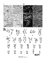
- mutant mouse embryonic stem (mES) cells were used in which both alleles of the NMHCIIA gene are disrupted (NMHCIIA ⁇ /A ⁇ ) ( FIG. 7 a ).
- the cell-cell contacts of mutant cells were severely impaired ( FIG. 7 b ).
- the level of apoptosis was also determined by TUNEL assay demonstrating considerably lower numbers of TUNEL-positive mutant cells as compared to the wild type mES cells ( FIG. 7 e ).
- NMHCIIA transcripts were recognized as being significantly enriched in the differentiated state of hES cells (Table 1), suggesting a potential molecular link between self-renewal/differentiation programs and NMII.
- Self-renewal regulators such as Oct3/4 and Nanog and NMII were examined and are mechanistically connected in hPS cells.
- hiPS or hES cells were grown in either mTeSR which supports the undifferentiated state or non-conditioned human ES medium (hESm) that induces passive differentiation in the presence or absence of blebbistatin at different concentrations for 48 h.
- hES cells treated with blebbistatin demonstrated elevated levels of both transcription factors in a dose dependent manner under both medium conditions as determined by quantitative RT-PCR (QPCR), Western analysis, and immunocytochemistry ( FIG. 8 a - e ). Similar results were obtained from hiPS cells. To determine if this connection is also conserved in mouse, mES cells grown in the presence or absence of leukemia inhibitory factor (LIF) for 2 days were examined. Consistent with the data from hES cells, both of the self-renewal regulators, Oct3/4 and Nanog, were expressed at substantially higher protein levels in NMHCIIA ⁇ /A ⁇ mES cells as compared to the parental cells under each condition ( FIG. 8 f,g ).
- LIF leukemia inhibitory factor
- NMII may function as a causal rather than a consequential factor in the negative regulation of self-renewal in both human and mouse pluripotent stem cells. This connection could be mediated by the interactions between NMII and ⁇ 1 integrin at focal adhesions which, in turn, activate multiple pathways including ERK signaling that has been implicated in differentiation of ES cells. Alternatively, NMII may crosstalk with other self-renewal pathways such as TGF- ⁇ , PI3 kinase, and Wnt signaling. The upregulation of self-renewal regulators by inhibition of NMII may contribute to the enhanced survival of self-renewing pluripotent stem cells.
- the disclosure demonstrates that Y-27632 treatment enables hES cells to self-renew on PDL-coating, on which hES cells do not normally grow.
- PDL is a chemically synthesized substrate unlike Matrigel, a mouse tumor-derived extract
- the disclosure provides a fully defined condition for self-renewal of hES cells using a defined medium.
- blebbistatin accurately replicates all of the effects of Y-27632 on both hES and iPS cells, blebbistatin alone was tested to determine if it can fully replace Y-27632 in the defined culture condition.
- Blebbistatin was used at concentrations between 2.5 and 5 ⁇ M which are the minimum concentrations sufficient for supporting self-renewal but not affecting cell-cell adhesions.
- blebbistatin The required minimum concentration of blebbistatin varies depending on individual hES or hiPS cell lines although most of the lines self-renew at 5 ⁇ M of blebbistatin.
- the maintenance of typical undifferentiated morphology and expression of pluripotency markers was confirmed in each line after 20 passages under this defined condition ( FIG. 9 a - d ).
- hiPS or hES cells were able to self-renew on PDL at a constant growth rate comparable to that of cells treated with Y-27632 or cells grown under regular feeder-free conditions using Matrigel ( FIG. 9 e ).
- the passaged cells were subjected to teratoma assay by subcutaneously injecting them into severe combined immunodeficient (SCID)/beige mice.
- the teratoma samples were examined by histological sections, and determined to contain all three germ-layer derived differentiated tissues such as melanocytes (ectoderm), neuronal tissue (ectoderm), bone (mesoderm), skeletal muscle (mesoderm), gastrointestinal-like mucosal epithelium with goblet cells (endoderm), and glomeruli (endoderm) ( FIG. 9 f ). This was further validated by immunohistochemistry detecting specialized cell-specific marker proteins ( FIG. 9 g ).
- hES or hiPS cells grown under the defined condition with blebbistatin can self-renew for an extended period of time without compromising multi-differentiation capacities, verifying the full reversibility of the effect of blebbistatin.
- the cells were determined to maintain genomic integrity as evaluated by karyotyping (hiPS cells in FIG. 9 h , and hES cells in FIG. 12 ).
- blebbistatin could be advantageous in pinpointing NMII function over that of Y-27632 which is likely to affect numerous effector molecules downstream of ROCK.
- blebbistatin is more cost-effective than Y-27632 for routine use.
- hiPS cells treated with blebbistatin exhibited higher growth and cell survival rates than the cells treated with Y27632.
- the pinpoint inhibition of the principle regulator, myosin II, by blebbistatin have a great advantage over the use of Y27632 in precluding unnecessary outcomes by the inhibition of numerous downstream molecules other than myosin II.
Landscapes
- Health & Medical Sciences (AREA)
- Life Sciences & Earth Sciences (AREA)
- Engineering & Computer Science (AREA)
- Biomedical Technology (AREA)
- Bioinformatics & Cheminformatics (AREA)
- Organic Chemistry (AREA)
- Chemical & Material Sciences (AREA)
- General Health & Medical Sciences (AREA)
- Zoology (AREA)
- Wood Science & Technology (AREA)
- Genetics & Genomics (AREA)
- Biotechnology (AREA)
- Medicinal Chemistry (AREA)
- Developmental Biology & Embryology (AREA)
- Pharmacology & Pharmacy (AREA)
- General Chemical & Material Sciences (AREA)
- Animal Behavior & Ethology (AREA)
- Chemical Kinetics & Catalysis (AREA)
- Public Health (AREA)
- Veterinary Medicine (AREA)
- Nuclear Medicine, Radiotherapy & Molecular Imaging (AREA)
- Biochemistry (AREA)
- General Engineering & Computer Science (AREA)
- Neurology (AREA)
- Cell Biology (AREA)
- Microbiology (AREA)
- Neurosurgery (AREA)
- Gynecology & Obstetrics (AREA)
- Reproductive Health (AREA)
- Diabetes (AREA)
- Transplantation (AREA)
- Psychology (AREA)
- Obesity (AREA)
- Psychiatry (AREA)
- Emergency Medicine (AREA)
- Hospice & Palliative Care (AREA)
- Endocrinology (AREA)
- Hematology (AREA)
- Orthopedic Medicine & Surgery (AREA)
- Physical Education & Sports Medicine (AREA)
Priority Applications (1)
| Application Number | Priority Date | Filing Date | Title |
|---|---|---|---|
| US13/202,324 US8609417B2 (en) | 2009-04-13 | 2010-04-13 | Methods and compositions for stem cell cultures |
Applications Claiming Priority (3)
| Application Number | Priority Date | Filing Date | Title |
|---|---|---|---|
| US16867909P | 2009-04-13 | 2009-04-13 | |
| US13/202,324 US8609417B2 (en) | 2009-04-13 | 2010-04-13 | Methods and compositions for stem cell cultures |
| PCT/US2010/030899 WO2010120785A2 (en) | 2009-04-13 | 2010-04-13 | Methods and compositions for stem cell cultures |
Publications (2)
| Publication Number | Publication Date |
|---|---|
| US20120058561A1 US20120058561A1 (en) | 2012-03-08 |
| US8609417B2 true US8609417B2 (en) | 2013-12-17 |
Family
ID=42983102
Family Applications (1)
| Application Number | Title | Priority Date | Filing Date |
|---|---|---|---|
| US13/202,324 Active 2030-05-22 US8609417B2 (en) | 2009-04-13 | 2010-04-13 | Methods and compositions for stem cell cultures |
Country Status (9)
| Country | Link |
|---|---|
| US (1) | US8609417B2 (de) |
| EP (1) | EP2401361A4 (de) |
| JP (1) | JP2012523240A (de) |
| KR (1) | KR20120017019A (de) |
| CN (1) | CN102395672A (de) |
| AU (1) | AU2010236632A1 (de) |
| CA (1) | CA2758003A1 (de) |
| IL (1) | IL214798A0 (de) |
| WO (1) | WO2010120785A2 (de) |
Cited By (2)
| Publication number | Priority date | Publication date | Assignee | Title |
|---|---|---|---|---|
| US20130071927A1 (en) * | 2010-05-05 | 2013-03-21 | Sydney Ivf Limited | Media and methods for cell culture |
| US9644186B2 (en) | 2010-08-05 | 2017-05-09 | Wisconsin Alumni Research Foundation | Simplified basic media for human pluripotent cell culture |
Families Citing this family (30)
| Publication number | Priority date | Publication date | Assignee | Title |
|---|---|---|---|---|
| EP2490701B9 (de) | 2009-10-19 | 2017-11-15 | Cellular Dynamics International, Inc. | Herstellung von kardiomyozyten |
| US20130295669A1 (en) * | 2010-11-09 | 2013-11-07 | Lonza Cologne Gmbh | Method for controlling binding of cells to a substrate |
| US9518249B2 (en) | 2010-12-16 | 2016-12-13 | General Electric Company | Cell carrier, associated methods for making cell carrier and culturing cells using the same |
| US9534206B2 (en) | 2010-12-16 | 2017-01-03 | General Electric Company | Cell carrier, associated methods for making cell carrier and culturing cells using the same |
| US9926523B2 (en) | 2010-12-16 | 2018-03-27 | General Electric Company | Cell carriers and methods for culturing cells |
| US9453196B2 (en) | 2010-12-16 | 2016-09-27 | General Electric Company | Cell carrier, methods of making and use |
| US9453197B2 (en) | 2010-12-16 | 2016-09-27 | General Electric Company | Methods of making cell carrier |
| KR102482184B1 (ko) * | 2010-12-22 | 2022-12-28 | 페이트 세러퓨틱스, 인코포레이티드 | 단세포 분류 및 iPSC의 증강된 재프로그래밍을 위한 세포 배양 플랫폼 |
| US9463204B2 (en) | 2011-12-27 | 2016-10-11 | Osaka University | Method for inducing differentiation enabling tumorigenesis of IPS cells to be suppressed |
| KR101972766B1 (ko) * | 2012-04-20 | 2019-04-29 | 주식회사 차바이오텍 | 인간 다능성 줄기세포 단일세포의 계대배양 방법, 및 이를 이용한 형질전환 인간 다능성 줄기세포 제조 방법 |
| DK2852671T3 (en) | 2012-05-21 | 2018-12-10 | Univ California | CREATION OF HUMAN IPS CELLS WITH A SYNTHETIC SELF REPLICATING RNA |
| CN102940631B (zh) * | 2012-11-02 | 2015-04-15 | 清华大学 | Blebbistatin在促进干细胞存活和维持干细胞干性中的应用 |
| CN103031270A (zh) * | 2013-01-05 | 2013-04-10 | 绍兴文理学院 | 胆管上皮细胞的高效扩增和培养方法 |
| US10081609B2 (en) | 2013-03-15 | 2018-09-25 | The Broad Institute, Inc. | Compounds for inducing proliferation and differentiation of cells, and methods of use thereof |
| ES2602078T3 (es) * | 2013-03-15 | 2017-02-17 | Lonza Cologne Gmbh | Composiciones y métodos para mejorar el potencial terapéutico de las células madre |
| CN103555661B (zh) * | 2013-10-25 | 2015-07-29 | 中国科学院动物研究所 | 一种无血清、无饲养层的多能干细胞培养方法 |
| US11268069B2 (en) | 2014-03-04 | 2022-03-08 | Fate Therapeutics, Inc. | Reprogramming methods and cell culture platforms |
| JP6940835B2 (ja) * | 2015-01-29 | 2021-09-29 | 国立大学法人 東京大学 | 細胞の培養方法 |
| CN104830754B (zh) * | 2015-05-08 | 2018-07-03 | 兰诺生物技术无锡有限公司 | 一种用于卵母细胞、皮肤成体干细胞培养的组合物及应用 |
| CN106479978A (zh) * | 2015-10-14 | 2017-03-08 | 北京昱龙盛世生物科技有限公司 | 一种神经干细胞的专用培养基及其培养方法 |
| CN108291237B (zh) | 2015-10-16 | 2023-11-21 | 菲特治疗公司 | 用于诱导和维护基态多能性的平台 |
| JP6754966B2 (ja) * | 2016-02-25 | 2020-09-16 | 国立大学法人広島大学 | 始原生殖細胞の培養方法及び始原生殖細胞の培養用培地添加物 |
| JP2019118279A (ja) * | 2017-12-28 | 2019-07-22 | 株式会社カネカ | 細胞凝集促進剤 |
| JP7542258B2 (ja) | 2018-01-29 | 2024-08-30 | 中国科学院動物研究所 | 細胞の機械的恒常性を破壊し、組織器官の再生と修復を促進する方法、およびその使用 |
| WO2019198962A1 (ko) * | 2018-04-09 | 2019-10-17 | 건국대학교 산학협력단 | 소변유래줄기세포로부터 제작된 유도만능줄기세포의 증식 및 분화 촉진 방법 |
| KR102016257B1 (ko) * | 2018-04-09 | 2019-08-29 | 건국대학교 산학협력단 | 소변 유래 줄기세포의 분리 효율 및 자가 증식을 증가시키는 방법 |
| HU231285B1 (hu) | 2018-04-18 | 2022-08-28 | Printnet Kereskedelmi És Szolgáltató Kft. | A miozin-2-izoformákat szelektíven gátló, gyógyászatilag hatásos vegyületek |
| US20230220331A1 (en) * | 2020-03-24 | 2023-07-13 | Jichi Medical University | Temporary treatment medium, treatment kit, embryogenesis arrest inhibitor, embryogenesis arrest inhibitory method, developmental engineering product preparation method, transplantation method, therapeutic method, and developmental engineering product |
| CN114736302B (zh) * | 2022-05-12 | 2022-10-25 | 诺赛联合(北京)生物医学科技有限公司 | 一种干细胞无血清培养基 |
| CN117343901A (zh) * | 2023-10-10 | 2024-01-05 | 广东格林邦卡新材料生物科技有限公司 | 一种培养基、干细胞分泌物及其在治疗心肌纤维化应用 |
Citations (1)
| Publication number | Priority date | Publication date | Assignee | Title |
|---|---|---|---|---|
| US20080241919A1 (en) * | 2003-12-31 | 2008-10-02 | The Burnham Institute Of Medical Research | Defined media for pluripotent stem cell culture |
Family Cites Families (2)
| Publication number | Priority date | Publication date | Assignee | Title |
|---|---|---|---|---|
| NZ553235A (en) * | 2004-09-08 | 2009-11-27 | Wisconsin Alumni Res Found | Culturing human pluripotent stem cells |
| JP6189581B2 (ja) * | 2009-02-20 | 2017-08-30 | セルラー ダイナミクス インターナショナル, インコーポレイテッド | 幹細胞の分化のための方法および組成物 |
-
2010
- 2010-04-13 CN CN2010800165165A patent/CN102395672A/zh active Pending
- 2010-04-13 US US13/202,324 patent/US8609417B2/en active Active
- 2010-04-13 JP JP2012504932A patent/JP2012523240A/ja active Pending
- 2010-04-13 AU AU2010236632A patent/AU2010236632A1/en not_active Abandoned
- 2010-04-13 CA CA2758003A patent/CA2758003A1/en not_active Abandoned
- 2010-04-13 EP EP10765022.8A patent/EP2401361A4/de not_active Withdrawn
- 2010-04-13 KR KR1020117023718A patent/KR20120017019A/ko not_active Withdrawn
- 2010-04-13 WO PCT/US2010/030899 patent/WO2010120785A2/en not_active Ceased
-
2011
- 2011-08-23 IL IL214798A patent/IL214798A0/en unknown
Patent Citations (1)
| Publication number | Priority date | Publication date | Assignee | Title |
|---|---|---|---|---|
| US20080241919A1 (en) * | 2003-12-31 | 2008-10-02 | The Burnham Institute Of Medical Research | Defined media for pluripotent stem cell culture |
Non-Patent Citations (9)
| Title |
|---|
| Engler et al., "Matrxi elasticity irects stem cell lineage specification," Cell, 2006, pp. 677-689, vol. 126. |
| Harb et al., "The Rho-Rock-Myosin signaling axis determines cell-cell integrity of self-renewing pluripotent stem cells," PlOS One, Aug. 2008, pp. 1-13, vol. 3, No. 8. |
| Harb N et al. The Rho-Rock-Myosin Signaling Axis Determines Cell-Cell Integrity of Self-Renewing Pluripotent Stem Cells, PloS One, 3 (8): e3001, 2008. * |
| Hyojin, Lee, International Search Report and Written Opinion, PCT/2010/030899, Korean Intellectual Property Office, Jan. 28, 2011. |
| Kim et al., "Substrate rigidity regulates Ca2+ oscillation via RhoA pathway in stem cells," J. of Cellular Phys., Oct. 2008, pp. 285-293, vol. 218. |
| Mader CC et al. Probing Cell Shape Regulation with Patterned Substratum: Requirement of Myosin II-Mediated Contractility, Soft Matter 3: 357-363, 2007. * |
| Ruiz, et al., "Emergence of patterned stem ceell differentiation within multicellular structures," Stem Cells, 2008, pp. 2921-2927, vol. 26. |
| Shu et al., "Blebbistatin and Blebbistatin-inactivated myosin II inhibit myosin II-independent processes in Dictyostelium," PNAS, 2005, pp. 1472-1477, vol. 102, No. 5. |
| Sigma (DME) downloaded from (http://www.sigmaaldrich.com/life-science/cell-culture/learning-center/media-formulations/dme.html) on Jan. 29, 2013, 4 pages. * |
Cited By (2)
| Publication number | Priority date | Publication date | Assignee | Title |
|---|---|---|---|---|
| US20130071927A1 (en) * | 2010-05-05 | 2013-03-21 | Sydney Ivf Limited | Media and methods for cell culture |
| US9644186B2 (en) | 2010-08-05 | 2017-05-09 | Wisconsin Alumni Research Foundation | Simplified basic media for human pluripotent cell culture |
Also Published As
| Publication number | Publication date |
|---|---|
| CA2758003A1 (en) | 2010-10-21 |
| JP2012523240A (ja) | 2012-10-04 |
| EP2401361A2 (de) | 2012-01-04 |
| IL214798A0 (en) | 2011-11-30 |
| AU2010236632A1 (en) | 2011-09-15 |
| EP2401361A4 (de) | 2013-09-04 |
| CN102395672A (zh) | 2012-03-28 |
| WO2010120785A3 (en) | 2011-03-31 |
| US20120058561A1 (en) | 2012-03-08 |
| WO2010120785A2 (en) | 2010-10-21 |
| KR20120017019A (ko) | 2012-02-27 |
Similar Documents
| Publication | Publication Date | Title |
|---|---|---|
| US8609417B2 (en) | Methods and compositions for stem cell cultures | |
| US12545891B2 (en) | Methods for neural conversion of human embryonic stem cells | |
| Beers et al. | Passaging and colony expansion of human pluripotent stem cells by enzyme-free dissociation in chemically defined culture conditions | |
| US12195757B2 (en) | Methods of producing RPE cells | |
| Pakzad et al. | Presence of a ROCK inhibitor in extracellular matrix supports more undifferentiated growth of feeder-free human embryonic and induced pluripotent stem cells upon passaging | |
| CN102695790A (zh) | 多能干细胞 | |
| US20240384232A1 (en) | Clinical-grade therapeutic progenitors generated from tankyrase/parp-inhibited pluripotent stem cell banks | |
| EP3504324B1 (de) | Differenzierung von pluripotenten stammzellen in hornhautzellen | |
| So et al. | The Rho-associated kinase inhibitor fasudil can replace Y-27632 for use in human pluripotent stem cell research | |
| WO2018225802A1 (ja) | 多能性幹細胞から生殖系列幹細胞様細胞への分化誘導方法 | |
| WO2015125926A1 (ja) | 栄養膜幹細胞の樹立及び維持方法 | |
| Oh et al. | Human embryonic stem cells: technological challenges towards therapy. | |
| Gu et al. | Rapid conversion of human ESCs into mouse ESC-like pluripotent state by optimizing culture conditions | |
| CN108998410B (zh) | 蛋白激酶抑制剂在抑制单倍体细胞二倍化中的用途 | |
| Ono et al. | A single-cell and feeder-free culture system for monkey embryonic stem cells | |
| Liu et al. | Derivation of human embryonic stem cell lines without any exogenous growth factors | |
| Alsolami et al. | DMSO derives Trophectoderm and Clonal Blastoid from Single Human Pluripotent Stem Cell | |
| Fan et al. | Breaking the link between morphology and potency for mESCs | |
| Zandi et al. | Optimization of buffalo (Bubalus bubalis) embryonic stem cell culture system | |
| WO2026058673A1 (ja) | 分化誘導に適したイヌ多能性幹細胞の培養方法 | |
| WO2024243481A1 (en) | Compositions and methods for generation of bovine pluripotent embryonic stem cells | |
| Parris | Isolation and characterisation of muscle satellite cells from differentiating human embryonic stem cells. | |
| Acton | Stem Cells—Advances in Research and Application: 2013 Edition | |
| Crutchley | Automation and scale-up of human induced pluripotent stem cell models of cardiovascular disease for drug screening | |
| Flatt | Developing an automation-compatible quantitative and functional assay for the assessment of pluripotency |
Legal Events
| Date | Code | Title | Description |
|---|---|---|---|
| AS | Assignment |
Owner name: THE REGENTS OF THE UNIVERSITY OF CALIFORNIA, CALIF Free format text: ASSIGNMENT OF ASSIGNORS INTEREST;ASSIGNOR:SATO, NOBORU;REEL/FRAME:024226/0961 Effective date: 20100413 |
|
| AS | Assignment |
Owner name: THE REGENTS OF THE UNIVERSITY OF CALIFORNIA, CALIF Free format text: ASSIGNMENT OF ASSIGNORS INTEREST;ASSIGNOR:SATO, NOBORU;REEL/FRAME:027262/0691 Effective date: 20100413 |
|
| STCF | Information on status: patent grant |
Free format text: PATENTED CASE |
|
| FEPP | Fee payment procedure |
Free format text: PAT HOLDER NO LONGER CLAIMS SMALL ENTITY STATUS, ENTITY STATUS SET TO UNDISCOUNTED (ORIGINAL EVENT CODE: STOL); ENTITY STATUS OF PATENT OWNER: LARGE ENTITY |
|
| FPAY | Fee payment |
Year of fee payment: 4 |
|
| MAFP | Maintenance fee payment |
Free format text: PAYMENT OF MAINTENANCE FEE, 8TH YEAR, LARGE ENTITY (ORIGINAL EVENT CODE: M1552); ENTITY STATUS OF PATENT OWNER: LARGE ENTITY Year of fee payment: 8 |
|
| MAFP | Maintenance fee payment |
Free format text: PAYMENT OF MAINTENANCE FEE, 12TH YEAR, LARGE ENTITY (ORIGINAL EVENT CODE: M1553); ENTITY STATUS OF PATENT OWNER: LARGE ENTITY Year of fee payment: 12 |