US7510706B2 - Pseudotype retroviral vectors containing membrane proteins having hemagglutinin activity - Google Patents
Pseudotype retroviral vectors containing membrane proteins having hemagglutinin activity Download PDFInfo
- Publication number
- US7510706B2 US7510706B2 US10/306,949 US30694902A US7510706B2 US 7510706 B2 US7510706 B2 US 7510706B2 US 30694902 A US30694902 A US 30694902A US 7510706 B2 US7510706 B2 US 7510706B2
- Authority
- US
- United States
- Prior art keywords
- protein
- vector
- cells
- composition according
- virus
- Prior art date
- Legal status (The legal status is an assumption and is not a legal conclusion. Google has not performed a legal analysis and makes no representation as to the accuracy of the status listed.)
- Expired - Lifetime, expires
Links
- 239000013598 vector Substances 0.000 title claims abstract description 494
- 230000001177 retroviral effect Effects 0.000 title claims abstract description 118
- 101710154606 Hemagglutinin Proteins 0.000 title claims abstract description 83
- 101710093908 Outer capsid protein VP4 Proteins 0.000 title claims abstract description 83
- 101710135467 Outer capsid protein sigma-1 Proteins 0.000 title claims abstract description 83
- 101710176177 Protein A56 Proteins 0.000 title claims abstract description 83
- 230000000694 effects Effects 0.000 title claims abstract description 72
- 239000000185 hemagglutinin Substances 0.000 title claims abstract description 70
- 108010052285 Membrane Proteins Proteins 0.000 title claims abstract description 33
- 210000004027 cell Anatomy 0.000 claims abstract description 401
- 108090000623 proteins and genes Proteins 0.000 claims abstract description 328
- 238000012546 transfer Methods 0.000 claims abstract description 154
- 210000003958 hematopoietic stem cell Anatomy 0.000 claims abstract description 36
- 210000002919 epithelial cell Anatomy 0.000 claims abstract description 23
- 102000018697 Membrane Proteins Human genes 0.000 claims abstract description 15
- 101710133291 Hemagglutinin-neuraminidase Proteins 0.000 claims description 146
- 241000711408 Murine respirovirus Species 0.000 claims description 122
- 108010068327 4-hydroxyphenylpyruvate dioxygenase Proteins 0.000 claims description 115
- 241000713311 Simian immunodeficiency virus Species 0.000 claims description 112
- 239000000203 mixture Substances 0.000 claims description 112
- 241000700605 Viruses Species 0.000 claims description 103
- 238000004806 packaging method and process Methods 0.000 claims description 86
- 102000004169 proteins and genes Human genes 0.000 claims description 82
- 230000001086 cytosolic effect Effects 0.000 claims description 76
- 101710091045 Envelope protein Proteins 0.000 claims description 69
- 101710188315 Protein X Proteins 0.000 claims description 69
- 238000000034 method Methods 0.000 claims description 58
- 241000713666 Lentivirus Species 0.000 claims description 53
- 241001430294 unidentified retrovirus Species 0.000 claims description 51
- 108020004414 DNA Proteins 0.000 claims description 49
- 239000001963 growth medium Substances 0.000 claims description 45
- 210000003097 mucus Anatomy 0.000 claims description 23
- 150000001413 amino acids Chemical class 0.000 claims description 22
- 101710125418 Major capsid protein Proteins 0.000 claims description 14
- 210000003677 hemocyte Anatomy 0.000 claims description 14
- 101710177291 Gag polyprotein Proteins 0.000 claims description 10
- 238000004519 manufacturing process Methods 0.000 claims description 10
- 238000000338 in vitro Methods 0.000 claims description 8
- 210000003928 nasal cavity Anatomy 0.000 claims description 8
- 125000000539 amino acid group Chemical group 0.000 claims description 6
- 230000002685 pulmonary effect Effects 0.000 claims description 5
- 230000003362 replicative effect Effects 0.000 claims description 5
- 229940000351 hemocyte Drugs 0.000 claims description 4
- 108010089520 pol Gene Products Proteins 0.000 claims description 3
- 102100021696 Syncytin-1 Human genes 0.000 claims 15
- 125000003275 alpha amino acid group Chemical group 0.000 claims 3
- 239000013603 viral vector Substances 0.000 abstract description 36
- 238000001415 gene therapy Methods 0.000 abstract description 22
- 238000007796 conventional method Methods 0.000 abstract description 7
- 210000000601 blood cell Anatomy 0.000 abstract description 2
- 210000004877 mucosa Anatomy 0.000 abstract 1
- 210000003550 mucous cell Anatomy 0.000 abstract 1
- 239000013613 expression plasmid Substances 0.000 description 73
- 239000000243 solution Substances 0.000 description 72
- 108700001624 vesicular stomatitis virus G Proteins 0.000 description 72
- 239000000277 virosome Substances 0.000 description 69
- 208000015181 infectious disease Diseases 0.000 description 64
- 102100034349 Integrase Human genes 0.000 description 55
- 239000006144 Dulbecco’s modified Eagle's medium Substances 0.000 description 53
- 239000002245 particle Substances 0.000 description 51
- 108010048367 enhanced green fluorescent protein Proteins 0.000 description 49
- 239000012634 fragment Substances 0.000 description 48
- 241000282414 Homo sapiens Species 0.000 description 43
- 239000013612 plasmid Substances 0.000 description 43
- 230000014509 gene expression Effects 0.000 description 42
- 102100031573 Hematopoietic progenitor cell antigen CD34 Human genes 0.000 description 30
- 101000777663 Homo sapiens Hematopoietic progenitor cell antigen CD34 Proteins 0.000 description 30
- 108091003079 Bovine Serum Albumin Proteins 0.000 description 28
- 239000002609 medium Substances 0.000 description 28
- 238000005119 centrifugation Methods 0.000 description 27
- 230000004927 fusion Effects 0.000 description 27
- 229940098773 bovine serum albumin Drugs 0.000 description 26
- 239000003153 chemical reaction reagent Substances 0.000 description 25
- 210000005260 human cell Anatomy 0.000 description 24
- 238000002360 preparation method Methods 0.000 description 24
- 238000001890 transfection Methods 0.000 description 22
- 108010003533 Viral Envelope Proteins Proteins 0.000 description 20
- 239000004033 plastic Substances 0.000 description 19
- 229920003023 plastic Polymers 0.000 description 19
- WSFSSNUMVMOOMR-UHFFFAOYSA-N Formaldehyde Chemical compound O=C WSFSSNUMVMOOMR-UHFFFAOYSA-N 0.000 description 18
- 102000004142 Trypsin Human genes 0.000 description 18
- 108090000631 Trypsin Proteins 0.000 description 18
- 239000012228 culture supernatant Substances 0.000 description 18
- 239000012588 trypsin Substances 0.000 description 18
- 108010005774 beta-Galactosidase Proteins 0.000 description 17
- 238000006243 chemical reaction Methods 0.000 description 17
- 210000000130 stem cell Anatomy 0.000 description 17
- 241000712461 unidentified influenza virus Species 0.000 description 17
- 238000012217 deletion Methods 0.000 description 16
- 230000037430 deletion Effects 0.000 description 16
- 241000725303 Human immunodeficiency virus Species 0.000 description 15
- 238000002474 experimental method Methods 0.000 description 15
- 210000002966 serum Anatomy 0.000 description 15
- 230000000007 visual effect Effects 0.000 description 15
- 101710085938 Matrix protein Proteins 0.000 description 14
- 101710127721 Membrane protein Proteins 0.000 description 14
- 244000309466 calf Species 0.000 description 14
- 102000004889 Interleukin-6 Human genes 0.000 description 13
- 108090001005 Interleukin-6 Proteins 0.000 description 13
- 239000006228 supernatant Substances 0.000 description 13
- 210000001744 T-lymphocyte Anatomy 0.000 description 12
- 210000000349 chromosome Anatomy 0.000 description 12
- 210000003743 erythrocyte Anatomy 0.000 description 12
- 239000013604 expression vector Substances 0.000 description 12
- 102000039446 nucleic acids Human genes 0.000 description 12
- 108020004707 nucleic acids Proteins 0.000 description 12
- 150000007523 nucleic acids Chemical class 0.000 description 12
- 238000006467 substitution reaction Methods 0.000 description 12
- 108091032973 (ribonucleotides)n+m Proteins 0.000 description 11
- 108091034117 Oligonucleotide Proteins 0.000 description 11
- 230000002458 infectious effect Effects 0.000 description 11
- 210000001519 tissue Anatomy 0.000 description 11
- 102000005348 Neuraminidase Human genes 0.000 description 10
- 108010006232 Neuraminidase Proteins 0.000 description 10
- 241000711975 Vesicular stomatitis virus Species 0.000 description 10
- 230000004520 agglutination Effects 0.000 description 10
- 238000010276 construction Methods 0.000 description 10
- 238000007865 diluting Methods 0.000 description 10
- 108700004025 env Genes Proteins 0.000 description 10
- 238000011534 incubation Methods 0.000 description 10
- 230000001404 mediated effect Effects 0.000 description 10
- 239000011259 mixed solution Substances 0.000 description 10
- 238000013518 transcription Methods 0.000 description 10
- 230000035897 transcription Effects 0.000 description 10
- 229920000209 Hexadimethrine bromide Polymers 0.000 description 9
- 102000004388 Interleukin-4 Human genes 0.000 description 9
- 108090000978 Interleukin-4 Proteins 0.000 description 9
- JLCPHMBAVCMARE-UHFFFAOYSA-N [3-[[3-[[3-[[3-[[3-[[3-[[3-[[3-[[3-[[3-[[3-[[5-(2-amino-6-oxo-1H-purin-9-yl)-3-[[3-[[3-[[3-[[3-[[3-[[5-(2-amino-6-oxo-1H-purin-9-yl)-3-[[5-(2-amino-6-oxo-1H-purin-9-yl)-3-hydroxyoxolan-2-yl]methoxy-hydroxyphosphoryl]oxyoxolan-2-yl]methoxy-hydroxyphosphoryl]oxy-5-(5-methyl-2,4-dioxopyrimidin-1-yl)oxolan-2-yl]methoxy-hydroxyphosphoryl]oxy-5-(6-aminopurin-9-yl)oxolan-2-yl]methoxy-hydroxyphosphoryl]oxy-5-(6-aminopurin-9-yl)oxolan-2-yl]methoxy-hydroxyphosphoryl]oxy-5-(6-aminopurin-9-yl)oxolan-2-yl]methoxy-hydroxyphosphoryl]oxy-5-(6-aminopurin-9-yl)oxolan-2-yl]methoxy-hydroxyphosphoryl]oxyoxolan-2-yl]methoxy-hydroxyphosphoryl]oxy-5-(5-methyl-2,4-dioxopyrimidin-1-yl)oxolan-2-yl]methoxy-hydroxyphosphoryl]oxy-5-(4-amino-2-oxopyrimidin-1-yl)oxolan-2-yl]methoxy-hydroxyphosphoryl]oxy-5-(5-methyl-2,4-dioxopyrimidin-1-yl)oxolan-2-yl]methoxy-hydroxyphosphoryl]oxy-5-(5-methyl-2,4-dioxopyrimidin-1-yl)oxolan-2-yl]methoxy-hydroxyphosphoryl]oxy-5-(6-aminopurin-9-yl)oxolan-2-yl]methoxy-hydroxyphosphoryl]oxy-5-(6-aminopurin-9-yl)oxolan-2-yl]methoxy-hydroxyphosphoryl]oxy-5-(4-amino-2-oxopyrimidin-1-yl)oxolan-2-yl]methoxy-hydroxyphosphoryl]oxy-5-(4-amino-2-oxopyrimidin-1-yl)oxolan-2-yl]methoxy-hydroxyphosphoryl]oxy-5-(4-amino-2-oxopyrimidin-1-yl)oxolan-2-yl]methoxy-hydroxyphosphoryl]oxy-5-(6-aminopurin-9-yl)oxolan-2-yl]methoxy-hydroxyphosphoryl]oxy-5-(4-amino-2-oxopyrimidin-1-yl)oxolan-2-yl]methyl [5-(6-aminopurin-9-yl)-2-(hydroxymethyl)oxolan-3-yl] hydrogen phosphate Polymers Cc1cn(C2CC(OP(O)(=O)OCC3OC(CC3OP(O)(=O)OCC3OC(CC3O)n3cnc4c3nc(N)[nH]c4=O)n3cnc4c3nc(N)[nH]c4=O)C(COP(O)(=O)OC3CC(OC3COP(O)(=O)OC3CC(OC3COP(O)(=O)OC3CC(OC3COP(O)(=O)OC3CC(OC3COP(O)(=O)OC3CC(OC3COP(O)(=O)OC3CC(OC3COP(O)(=O)OC3CC(OC3COP(O)(=O)OC3CC(OC3COP(O)(=O)OC3CC(OC3COP(O)(=O)OC3CC(OC3COP(O)(=O)OC3CC(OC3COP(O)(=O)OC3CC(OC3COP(O)(=O)OC3CC(OC3COP(O)(=O)OC3CC(OC3COP(O)(=O)OC3CC(OC3COP(O)(=O)OC3CC(OC3COP(O)(=O)OC3CC(OC3CO)n3cnc4c(N)ncnc34)n3ccc(N)nc3=O)n3cnc4c(N)ncnc34)n3ccc(N)nc3=O)n3ccc(N)nc3=O)n3ccc(N)nc3=O)n3cnc4c(N)ncnc34)n3cnc4c(N)ncnc34)n3cc(C)c(=O)[nH]c3=O)n3cc(C)c(=O)[nH]c3=O)n3ccc(N)nc3=O)n3cc(C)c(=O)[nH]c3=O)n3cnc4c3nc(N)[nH]c4=O)n3cnc4c(N)ncnc34)n3cnc4c(N)ncnc34)n3cnc4c(N)ncnc34)n3cnc4c(N)ncnc34)O2)c(=O)[nH]c1=O JLCPHMBAVCMARE-UHFFFAOYSA-N 0.000 description 9
- WQZGKKKJIJFFOK-FPRJBGLDSA-N beta-D-galactose Chemical compound OC[C@H]1O[C@@H](O)[C@H](O)[C@@H](O)[C@H]1O WQZGKKKJIJFFOK-FPRJBGLDSA-N 0.000 description 9
- 210000002798 bone marrow cell Anatomy 0.000 description 9
- 210000000981 epithelium Anatomy 0.000 description 9
- 230000001605 fetal effect Effects 0.000 description 9
- 238000001727 in vivo Methods 0.000 description 9
- 229940028885 interleukin-4 Drugs 0.000 description 9
- 108020004999 messenger RNA Proteins 0.000 description 9
- 241000282693 Cercopithecidae Species 0.000 description 8
- 241000700159 Rattus Species 0.000 description 8
- 238000003556 assay Methods 0.000 description 8
- 238000004113 cell culture Methods 0.000 description 8
- 235000013601 eggs Nutrition 0.000 description 8
- 239000012528 membrane Substances 0.000 description 8
- 239000008188 pellet Substances 0.000 description 8
- -1 phospho Chemical class 0.000 description 8
- 239000011148 porous material Substances 0.000 description 8
- 230000003612 virological effect Effects 0.000 description 8
- WQZGKKKJIJFFOK-GASJEMHNSA-N Glucose Natural products OC[C@H]1OC(O)[C@H](O)[C@@H](O)[C@@H]1O WQZGKKKJIJFFOK-GASJEMHNSA-N 0.000 description 7
- 241000713772 Human immunodeficiency virus 1 Species 0.000 description 7
- 101710149951 Protein Tat Proteins 0.000 description 7
- 238000004458 analytical method Methods 0.000 description 7
- 239000000427 antigen Substances 0.000 description 7
- 108091007433 antigens Proteins 0.000 description 7
- 102000036639 antigens Human genes 0.000 description 7
- 210000001185 bone marrow Anatomy 0.000 description 7
- 238000000684 flow cytometry Methods 0.000 description 7
- 108700014844 flt3 ligand Proteins 0.000 description 7
- 239000008103 glucose Substances 0.000 description 7
- 210000003292 kidney cell Anatomy 0.000 description 7
- 210000004379 membrane Anatomy 0.000 description 7
- 238000011160 research Methods 0.000 description 7
- 238000010186 staining Methods 0.000 description 7
- 238000004448 titration Methods 0.000 description 7
- 210000003437 trachea Anatomy 0.000 description 7
- OPIFSICVWOWJMJ-AEOCFKNESA-N 5-bromo-4-chloro-3-indolyl beta-D-galactoside Chemical compound O[C@@H]1[C@@H](O)[C@@H](O)[C@@H](CO)O[C@H]1OC1=CNC2=CC=C(Br)C(Cl)=C12 OPIFSICVWOWJMJ-AEOCFKNESA-N 0.000 description 6
- 101710132601 Capsid protein Proteins 0.000 description 6
- 241000287828 Gallus gallus Species 0.000 description 6
- SXRSQZLOMIGNAQ-UHFFFAOYSA-N Glutaraldehyde Chemical compound O=CCCCC=O SXRSQZLOMIGNAQ-UHFFFAOYSA-N 0.000 description 6
- TWRXJAOTZQYOKJ-UHFFFAOYSA-L Magnesium chloride Chemical compound [Mg+2].[Cl-].[Cl-] TWRXJAOTZQYOKJ-UHFFFAOYSA-L 0.000 description 6
- 241000699666 Mus <mouse, genus> Species 0.000 description 6
- 108010067390 Viral Proteins Proteins 0.000 description 6
- 230000015572 biosynthetic process Effects 0.000 description 6
- 201000010099 disease Diseases 0.000 description 6
- 208000037265 diseases, disorders, signs and symptoms Diseases 0.000 description 6
- 239000007758 minimum essential medium Substances 0.000 description 6
- 210000004400 mucous membrane Anatomy 0.000 description 6
- PFNFFQXMRSDOHW-UHFFFAOYSA-N spermine Chemical compound NCCCNCCCCNCCCN PFNFFQXMRSDOHW-UHFFFAOYSA-N 0.000 description 6
- 238000011282 treatment Methods 0.000 description 6
- 241000700199 Cavia porcellus Species 0.000 description 5
- 101710094648 Coat protein Proteins 0.000 description 5
- 102100021181 Golgi phosphoprotein 3 Human genes 0.000 description 5
- 241000282560 Macaca mulatta Species 0.000 description 5
- 241000713869 Moloney murine leukemia virus Species 0.000 description 5
- 206010028980 Neoplasm Diseases 0.000 description 5
- 101710141454 Nucleoprotein Proteins 0.000 description 5
- 241000711504 Paramyxoviridae Species 0.000 description 5
- 101710083689 Probable capsid protein Proteins 0.000 description 5
- 108700008625 Reporter Genes Proteins 0.000 description 5
- 108010093857 Viral Hemagglutinins Proteins 0.000 description 5
- MXWJVTOOROXGIU-UHFFFAOYSA-N atrazine Chemical compound CCNC1=NC(Cl)=NC(NC(C)C)=N1 MXWJVTOOROXGIU-UHFFFAOYSA-N 0.000 description 5
- 201000011510 cancer Diseases 0.000 description 5
- 238000012258 culturing Methods 0.000 description 5
- 239000003599 detergent Substances 0.000 description 5
- 241001493065 dsRNA viruses Species 0.000 description 5
- 238000011156 evaluation Methods 0.000 description 5
- 230000002607 hemopoietic effect Effects 0.000 description 5
- 210000004072 lung Anatomy 0.000 description 5
- 230000007935 neutral effect Effects 0.000 description 5
- 230000007918 pathogenicity Effects 0.000 description 5
- 238000000746 purification Methods 0.000 description 5
- 238000010839 reverse transcription Methods 0.000 description 5
- 239000000523 sample Substances 0.000 description 5
- 230000001629 suppression Effects 0.000 description 5
- 230000002463 transducing effect Effects 0.000 description 5
- 208000030507 AIDS Diseases 0.000 description 4
- 241000272814 Anser sp. Species 0.000 description 4
- 241000713756 Caprine arthritis encephalitis virus Species 0.000 description 4
- 108091026890 Coding region Proteins 0.000 description 4
- 241000713800 Feline immunodeficiency virus Species 0.000 description 4
- 208000031886 HIV Infections Diseases 0.000 description 4
- 206010061598 Immunodeficiency Diseases 0.000 description 4
- 208000029462 Immunodeficiency disease Diseases 0.000 description 4
- 241001529936 Murinae Species 0.000 description 4
- 229930006000 Sucrose Natural products 0.000 description 4
- CZMRCDWAGMRECN-UGDNZRGBSA-N Sucrose Chemical compound O[C@H]1[C@H](O)[C@@H](CO)O[C@@]1(CO)O[C@@H]1[C@H](O)[C@@H](O)[C@H](O)[C@@H](CO)O1 CZMRCDWAGMRECN-UGDNZRGBSA-N 0.000 description 4
- RAHZWNYVWXNFOC-UHFFFAOYSA-N Sulphur dioxide Chemical compound O=S=O RAHZWNYVWXNFOC-UHFFFAOYSA-N 0.000 description 4
- 102100034195 Thrombopoietin Human genes 0.000 description 4
- 229920004890 Triton X-100 Polymers 0.000 description 4
- 239000013504 Triton X-100 Substances 0.000 description 4
- 230000000692 anti-sense effect Effects 0.000 description 4
- VHJLVAABSRFDPM-QWWZWVQMSA-N dithiothreitol Chemical compound SC[C@@H](O)[C@H](O)CS VHJLVAABSRFDPM-QWWZWVQMSA-N 0.000 description 4
- 229940079593 drug Drugs 0.000 description 4
- 239000003814 drug Substances 0.000 description 4
- 230000001747 exhibiting effect Effects 0.000 description 4
- 239000000706 filtrate Substances 0.000 description 4
- 230000007813 immunodeficiency Effects 0.000 description 4
- 102000035118 modified proteins Human genes 0.000 description 4
- 108091005573 modified proteins Proteins 0.000 description 4
- 230000001717 pathogenic effect Effects 0.000 description 4
- 102000005962 receptors Human genes 0.000 description 4
- 108020003175 receptors Proteins 0.000 description 4
- 108091008146 restriction endonucleases Proteins 0.000 description 4
- 239000005720 sucrose Substances 0.000 description 4
- 101710169336 5'-deoxyadenosine deaminase Proteins 0.000 description 3
- 102000055025 Adenosine deaminases Human genes 0.000 description 3
- 229920001342 Bakelite® Polymers 0.000 description 3
- 102100026189 Beta-galactosidase Human genes 0.000 description 3
- 241000282552 Chlorocebus aethiops Species 0.000 description 3
- 108020004705 Codon Proteins 0.000 description 3
- 102000004127 Cytokines Human genes 0.000 description 3
- 230000006820 DNA synthesis Effects 0.000 description 3
- 241000713340 Human immunodeficiency virus 2 Species 0.000 description 3
- 241000699670 Mus sp. Species 0.000 description 3
- 241000713112 Orthobunyavirus Species 0.000 description 3
- 241000288906 Primates Species 0.000 description 3
- 108010076504 Protein Sorting Signals Proteins 0.000 description 3
- 102000004887 Transforming Growth Factor beta Human genes 0.000 description 3
- 108090001012 Transforming Growth Factor beta Proteins 0.000 description 3
- 210000001766 X chromosome Anatomy 0.000 description 3
- 230000002378 acidificating effect Effects 0.000 description 3
- SHGAZHPCJJPHSC-YCNIQYBTSA-N all-trans-retinoic acid Chemical compound OC(=O)\C=C(/C)\C=C\C=C(/C)\C=C\C1=C(C)CCCC1(C)C SHGAZHPCJJPHSC-YCNIQYBTSA-N 0.000 description 3
- 239000004637 bakelite Substances 0.000 description 3
- 230000008901 benefit Effects 0.000 description 3
- SQVRNKJHWKZAKO-UHFFFAOYSA-N beta-N-Acetyl-D-neuraminic acid Natural products CC(=O)NC1C(O)CC(O)(C(O)=O)OC1C(O)C(O)CO SQVRNKJHWKZAKO-UHFFFAOYSA-N 0.000 description 3
- 210000004369 blood Anatomy 0.000 description 3
- 239000008280 blood Substances 0.000 description 3
- 230000004663 cell proliferation Effects 0.000 description 3
- 235000013312 flour Nutrition 0.000 description 3
- 210000001357 hemopoietic progenitor cell Anatomy 0.000 description 3
- 230000006872 improvement Effects 0.000 description 3
- 230000000415 inactivating effect Effects 0.000 description 3
- 150000002632 lipids Chemical class 0.000 description 3
- 229910001629 magnesium chloride Inorganic materials 0.000 description 3
- 239000002773 nucleotide Substances 0.000 description 3
- 125000003729 nucleotide group Chemical group 0.000 description 3
- 239000000047 product Substances 0.000 description 3
- 230000002829 reductive effect Effects 0.000 description 3
- 229930002330 retinoic acid Natural products 0.000 description 3
- SQVRNKJHWKZAKO-OQPLDHBCSA-N sialic acid Chemical compound CC(=O)N[C@@H]1[C@@H](O)C[C@@](O)(C(O)=O)OC1[C@H](O)[C@H](O)CO SQVRNKJHWKZAKO-OQPLDHBCSA-N 0.000 description 3
- 229940063675 spermine Drugs 0.000 description 3
- 239000000725 suspension Substances 0.000 description 3
- ZRKFYGHZFMAOKI-QMGMOQQFSA-N tgfbeta Chemical compound C([C@H](NC(=O)[C@H](C(C)C)NC(=O)CNC(=O)[C@H](CCC(O)=O)NC(=O)[C@H](CCCNC(N)=N)NC(=O)[C@H](CC(N)=O)NC(=O)[C@H](CC(C)C)NC(=O)[C@H]([C@@H](C)O)NC(=O)[C@H](CCC(O)=O)NC(=O)[C@H]([C@@H](C)O)NC(=O)[C@H](CC(C)C)NC(=O)CNC(=O)[C@H](C)NC(=O)[C@H](CO)NC(=O)[C@H](CCC(N)=O)NC(=O)[C@@H](NC(=O)[C@H](C)NC(=O)[C@H](C)NC(=O)[C@@H](NC(=O)[C@H](CC(C)C)NC(=O)[C@@H](N)CCSC)C(C)C)[C@@H](C)CC)C(=O)N[C@@H]([C@@H](C)O)C(=O)N[C@@H](C(C)C)C(=O)N[C@@H](CC=1C=CC=CC=1)C(=O)N[C@@H](C)C(=O)N1[C@@H](CCC1)C(=O)N[C@@H]([C@@H](C)O)C(=O)N[C@@H](CC(N)=O)C(=O)N[C@@H](CCC(O)=O)C(=O)N[C@@H](C)C(=O)N[C@@H](CC=1C=CC=CC=1)C(=O)N[C@@H](CCCNC(N)=N)C(=O)N[C@@H](C)C(=O)N[C@@H](CC(C)C)C(=O)N1[C@@H](CCC1)C(=O)N1[C@@H](CCC1)C(=O)N[C@@H](CCCNC(N)=N)C(=O)N[C@@H](CCC(O)=O)C(=O)N[C@@H](CCCNC(N)=N)C(=O)N[C@@H](CO)C(=O)N[C@@H](CCCNC(N)=N)C(=O)N[C@@H](CC(C)C)C(=O)N[C@@H](CC(C)C)C(O)=O)C1=CC=C(O)C=C1 ZRKFYGHZFMAOKI-QMGMOQQFSA-N 0.000 description 3
- 238000002054 transplantation Methods 0.000 description 3
- 210000002845 virion Anatomy 0.000 description 3
- 208000023275 Autoimmune disease Diseases 0.000 description 2
- 241000711404 Avian avulavirus 1 Species 0.000 description 2
- 101100297347 Caenorhabditis elegans pgl-3 gene Proteins 0.000 description 2
- 102000053642 Catalytic RNA Human genes 0.000 description 2
- 108090000994 Catalytic RNA Proteins 0.000 description 2
- 241000282556 Cercocebus atys Species 0.000 description 2
- 206010053138 Congenital aplastic anaemia Diseases 0.000 description 2
- 108090000695 Cytokines Proteins 0.000 description 2
- 102000004190 Enzymes Human genes 0.000 description 2
- 108090000790 Enzymes Proteins 0.000 description 2
- 241000713730 Equine infectious anemia virus Species 0.000 description 2
- 201000004939 Fanconi anemia Diseases 0.000 description 2
- 241000711950 Filoviridae Species 0.000 description 2
- 108700039691 Genetic Promoter Regions Proteins 0.000 description 2
- 208000037357 HIV infectious disease Diseases 0.000 description 2
- 208000031220 Hemophilia Diseases 0.000 description 2
- 208000009292 Hemophilia A Diseases 0.000 description 2
- NTYJJOPFIAHURM-UHFFFAOYSA-N Histamine Chemical compound NCCC1=CN=CN1 NTYJJOPFIAHURM-UHFFFAOYSA-N 0.000 description 2
- 241000714192 Human spumaretrovirus Species 0.000 description 2
- 206010061218 Inflammation Diseases 0.000 description 2
- 102000000589 Interleukin-1 Human genes 0.000 description 2
- 108010002352 Interleukin-1 Proteins 0.000 description 2
- 102000003816 Interleukin-13 Human genes 0.000 description 2
- 108090000176 Interleukin-13 Proteins 0.000 description 2
- 108010002350 Interleukin-2 Proteins 0.000 description 2
- 102000000588 Interleukin-2 Human genes 0.000 description 2
- 108010002616 Interleukin-5 Proteins 0.000 description 2
- 102000000743 Interleukin-5 Human genes 0.000 description 2
- 108090001007 Interleukin-8 Proteins 0.000 description 2
- 102000004890 Interleukin-8 Human genes 0.000 description 2
- 108010002335 Interleukin-9 Proteins 0.000 description 2
- 102000000585 Interleukin-9 Human genes 0.000 description 2
- 102000015696 Interleukins Human genes 0.000 description 2
- 108010063738 Interleukins Proteins 0.000 description 2
- 102000004058 Leukemia inhibitory factor Human genes 0.000 description 2
- 108090000581 Leukemia inhibitory factor Proteins 0.000 description 2
- 102100024640 Low-density lipoprotein receptor Human genes 0.000 description 2
- 241000282537 Mandrillus sphinx Species 0.000 description 2
- 241000712045 Morbillivirus Species 0.000 description 2
- 101150118742 NP gene Proteins 0.000 description 2
- 108091028043 Nucleic acid sequence Proteins 0.000 description 2
- 239000012124 Opti-MEM Substances 0.000 description 2
- 241000712464 Orthomyxoviridae Species 0.000 description 2
- 208000002606 Paramyxoviridae Infections Diseases 0.000 description 2
- 101710185720 Putative ethidium bromide resistance protein Proteins 0.000 description 2
- 239000012980 RPMI-1640 medium Substances 0.000 description 2
- 241000710799 Rubella virus Species 0.000 description 2
- 241000713675 Spumavirus Species 0.000 description 2
- 108091081024 Start codon Proteins 0.000 description 2
- 108700026226 TATA Box Proteins 0.000 description 2
- 108700009124 Transcription Initiation Site Proteins 0.000 description 2
- 108010000134 Vascular Cell Adhesion Molecule-1 Proteins 0.000 description 2
- 102100023543 Vascular cell adhesion protein 1 Human genes 0.000 description 2
- 108020000999 Viral RNA Proteins 0.000 description 2
- 239000011543 agarose gel Substances 0.000 description 2
- 238000000246 agarose gel electrophoresis Methods 0.000 description 2
- 210000004102 animal cell Anatomy 0.000 description 2
- 210000000612 antigen-presenting cell Anatomy 0.000 description 2
- 208000006673 asthma Diseases 0.000 description 2
- 210000003719 b-lymphocyte Anatomy 0.000 description 2
- 230000004888 barrier function Effects 0.000 description 2
- 239000011324 bead Substances 0.000 description 2
- 239000012888 bovine serum Substances 0.000 description 2
- 239000000872 buffer Substances 0.000 description 2
- 230000034303 cell budding Effects 0.000 description 2
- 238000003776 cleavage reaction Methods 0.000 description 2
- 108010057085 cytokine receptors Proteins 0.000 description 2
- 210000000805 cytoplasm Anatomy 0.000 description 2
- 230000003247 decreasing effect Effects 0.000 description 2
- 230000007812 deficiency Effects 0.000 description 2
- 238000010586 diagram Methods 0.000 description 2
- 230000004069 differentiation Effects 0.000 description 2
- 238000010790 dilution Methods 0.000 description 2
- 239000012895 dilution Substances 0.000 description 2
- 238000005516 engineering process Methods 0.000 description 2
- 229940088598 enzyme Drugs 0.000 description 2
- 210000003979 eosinophil Anatomy 0.000 description 2
- 239000012091 fetal bovine serum Substances 0.000 description 2
- 239000012530 fluid Substances 0.000 description 2
- 230000006870 function Effects 0.000 description 2
- 108020001507 fusion proteins Proteins 0.000 description 2
- 102000037865 fusion proteins Human genes 0.000 description 2
- 108010027225 gag-pol Fusion Proteins Proteins 0.000 description 2
- 239000003102 growth factor Substances 0.000 description 2
- 210000004524 haematopoietic cell Anatomy 0.000 description 2
- 230000002949 hemolytic effect Effects 0.000 description 2
- 208000033519 human immunodeficiency virus infectious disease Diseases 0.000 description 2
- 230000004054 inflammatory process Effects 0.000 description 2
- 230000010354 integration Effects 0.000 description 2
- 230000003993 interaction Effects 0.000 description 2
- 229940047122 interleukins Drugs 0.000 description 2
- 230000003834 intracellular effect Effects 0.000 description 2
- 210000003734 kidney Anatomy 0.000 description 2
- 208000032839 leukemia Diseases 0.000 description 2
- 239000002502 liposome Substances 0.000 description 2
- 238000011068 loading method Methods 0.000 description 2
- 210000004698 lymphocyte Anatomy 0.000 description 2
- 239000003550 marker Substances 0.000 description 2
- 238000012986 modification Methods 0.000 description 2
- 230000004048 modification Effects 0.000 description 2
- 230000035772 mutation Effects 0.000 description 2
- 239000013642 negative control Substances 0.000 description 2
- 210000002569 neuron Anatomy 0.000 description 2
- 230000008506 pathogenesis Effects 0.000 description 2
- 150000003904 phospholipids Chemical class 0.000 description 2
- 239000002504 physiological saline solution Substances 0.000 description 2
- BASFCYQUMIYNBI-UHFFFAOYSA-N platinum Chemical compound [Pt] BASFCYQUMIYNBI-UHFFFAOYSA-N 0.000 description 2
- 238000002264 polyacrylamide gel electrophoresis Methods 0.000 description 2
- 230000035755 proliferation Effects 0.000 description 2
- 239000000018 receptor agonist Substances 0.000 description 2
- 229940044601 receptor agonist Drugs 0.000 description 2
- 108091092562 ribozyme Proteins 0.000 description 2
- 230000007017 scission Effects 0.000 description 2
- FLNVBBPBGKOJHN-KKAOYSRWSA-N sivmac Chemical compound O=C([C@H](C)NC(=O)[C@H](CO)NC(=O)[C@H]([C@@H](C)O)NC(=O)[C@@H]1CCCN1C(=O)[C@H](C)NC(=O)[C@H](C)NC(=O)[C@H]([C@@H](C)O)NC(=O)[C@H]([C@@H](C)O)NC(=O)[C@@H](NC(=O)[C@@H](NC(=O)[C@@H](NC(=O)[C@H](CO)NC(=O)[C@H](CO)NC(=O)[C@H](CCCCN)NC(=O)[C@@H](NC(=O)[C@H](CC(C)C)NC(=O)CNC(=O)[C@H](CC=1C2=CC=CC=C2NC=1)NC(=O)[C@H](CCCNC(N)=N)NC(=O)[C@H](CC(O)=O)NC(=O)[C@@H](NC(=O)[C@H](CCC(N)=O)NC(=O)[C@H](CO)NC(=O)[C@H](CCCCN)NC(=O)[C@H](CC(N)=O)NC(=O)[C@H](CS)NC(=O)[C@@H](N)CCCNC(N)=N)[C@@H](C)O)[C@@H](C)O)[C@@H](C)O)[C@@H](C)O)[C@@H](C)CC)N1CCC[C@H]1C(=O)N[C@@H](C(C)C)C(=O)N[C@@H](CO)C(=O)N[C@@H](CCC(O)=O)C(=O)N[C@@H](CCCCN)C(O)=O FLNVBBPBGKOJHN-KKAOYSRWSA-N 0.000 description 2
- 238000001179 sorption measurement Methods 0.000 description 2
- 241000894007 species Species 0.000 description 2
- ATHGHQPFGPMSJY-UHFFFAOYSA-N spermidine Chemical compound NCCCCNCCCN ATHGHQPFGPMSJY-UHFFFAOYSA-N 0.000 description 2
- 239000000758 substrate Substances 0.000 description 2
- 238000010257 thawing Methods 0.000 description 2
- 238000002560 therapeutic procedure Methods 0.000 description 2
- 229960001727 tretinoin Drugs 0.000 description 2
- 241000701161 unidentified adenovirus Species 0.000 description 2
- XLYOFNOQVPJJNP-UHFFFAOYSA-N water Substances O XLYOFNOQVPJJNP-UHFFFAOYSA-N 0.000 description 2
- MZAGXDHQGXUDDX-JSRXJHBZSA-N (e,2z)-4-ethyl-2-hydroxyimino-5-nitrohex-3-enamide Chemical compound [O-][N+](=O)C(C)C(/CC)=C/C(=N/O)/C(N)=O MZAGXDHQGXUDDX-JSRXJHBZSA-N 0.000 description 1
- TZCPCKNHXULUIY-RGULYWFUSA-N 1,2-distearoyl-sn-glycero-3-phosphoserine Chemical compound CCCCCCCCCCCCCCCCCC(=O)OC[C@H](COP(O)(=O)OC[C@H](N)C(O)=O)OC(=O)CCCCCCCCCCCCCCCCC TZCPCKNHXULUIY-RGULYWFUSA-N 0.000 description 1
- FZIPCQLKPTZZIM-UHFFFAOYSA-N 2-oxidanylpropane-1,2,3-tricarboxylic acid Chemical compound OC(=O)CC(O)(C(O)=O)CC(O)=O.OC(=O)CC(O)(C(O)=O)CC(O)=O FZIPCQLKPTZZIM-UHFFFAOYSA-N 0.000 description 1
- FWMNVWWHGCHHJJ-SKKKGAJSSA-N 4-amino-1-[(2r)-6-amino-2-[[(2r)-2-[[(2r)-2-[[(2r)-2-amino-3-phenylpropanoyl]amino]-3-phenylpropanoyl]amino]-4-methylpentanoyl]amino]hexanoyl]piperidine-4-carboxylic acid Chemical compound C([C@H](C(=O)N[C@H](CC(C)C)C(=O)N[C@H](CCCCN)C(=O)N1CCC(N)(CC1)C(O)=O)NC(=O)[C@H](N)CC=1C=CC=CC=1)C1=CC=CC=C1 FWMNVWWHGCHHJJ-SKKKGAJSSA-N 0.000 description 1
- 229920001817 Agar Polymers 0.000 description 1
- 229920000936 Agarose Polymers 0.000 description 1
- 208000024827 Alzheimer disease Diseases 0.000 description 1
- 241000712892 Arenaviridae Species 0.000 description 1
- 241000712891 Arenavirus Species 0.000 description 1
- 208000002109 Argyria Diseases 0.000 description 1
- 206010003210 Arteriosclerosis Diseases 0.000 description 1
- 102000005427 Asialoglycoprotein Receptor Human genes 0.000 description 1
- 101150035467 BDNF gene Proteins 0.000 description 1
- 241000283690 Bos taurus Species 0.000 description 1
- 201000006474 Brain Ischemia Diseases 0.000 description 1
- 206010006187 Breast cancer Diseases 0.000 description 1
- 206010006482 Bronchospasm Diseases 0.000 description 1
- SVSKNBRCRNRIGT-UHFFFAOYSA-N C.F.N.P[V] Chemical compound C.F.N.P[V] SVSKNBRCRNRIGT-UHFFFAOYSA-N 0.000 description 1
- 238000011740 C57BL/6 mouse Methods 0.000 description 1
- 108010041397 CD4 Antigens Proteins 0.000 description 1
- 101150029409 CFTR gene Proteins 0.000 description 1
- BHPQYMZQTOCNFJ-UHFFFAOYSA-N Calcium cation Chemical compound [Ca+2] BHPQYMZQTOCNFJ-UHFFFAOYSA-N 0.000 description 1
- 101100348617 Candida albicans (strain SC5314 / ATCC MYA-2876) NIK1 gene Proteins 0.000 description 1
- 108010067225 Cell Adhesion Molecules Proteins 0.000 description 1
- 102000016289 Cell Adhesion Molecules Human genes 0.000 description 1
- 241000366305 Cephaloziella rubella Species 0.000 description 1
- 102000009410 Chemokine receptor Human genes 0.000 description 1
- 108050000299 Chemokine receptor Proteins 0.000 description 1
- 241000867607 Chlorocebus sabaeus Species 0.000 description 1
- 208000017667 Chronic Disease Diseases 0.000 description 1
- 229940122644 Chymotrypsin inhibitor Drugs 0.000 description 1
- 208000003322 Coinfection Diseases 0.000 description 1
- 102000012422 Collagen Type I Human genes 0.000 description 1
- 108010022452 Collagen Type I Proteins 0.000 description 1
- 206010010099 Combined immunodeficiency Diseases 0.000 description 1
- 102100031673 Corneodesmosin Human genes 0.000 description 1
- 101710139375 Corneodesmosin Proteins 0.000 description 1
- 241000711573 Coronaviridae Species 0.000 description 1
- 102000003903 Cyclin-dependent kinases Human genes 0.000 description 1
- 108090000266 Cyclin-dependent kinases Proteins 0.000 description 1
- 201000003883 Cystic fibrosis Diseases 0.000 description 1
- 241000701022 Cytomegalovirus Species 0.000 description 1
- 206010012289 Dementia Diseases 0.000 description 1
- 208000000655 Distemper Diseases 0.000 description 1
- 206010059866 Drug resistance Diseases 0.000 description 1
- 206010013801 Duchenne Muscular Dystrophy Diseases 0.000 description 1
- 108010069091 Dystrophin Proteins 0.000 description 1
- 102000001039 Dystrophin Human genes 0.000 description 1
- 238000011891 EIA kit Methods 0.000 description 1
- 241000709661 Enterovirus Species 0.000 description 1
- 101710204837 Envelope small membrane protein Proteins 0.000 description 1
- 241001455610 Ephemerovirus Species 0.000 description 1
- 102000003951 Erythropoietin Human genes 0.000 description 1
- 108090000394 Erythropoietin Proteins 0.000 description 1
- 102000010834 Extracellular Matrix Proteins Human genes 0.000 description 1
- 108010037362 Extracellular Matrix Proteins Proteins 0.000 description 1
- 101150034814 F gene Proteins 0.000 description 1
- 108010076282 Factor IX Proteins 0.000 description 1
- 108010054218 Factor VIII Proteins 0.000 description 1
- 102000001690 Factor VIII Human genes 0.000 description 1
- 108010014173 Factor X Proteins 0.000 description 1
- 101150065330 Fancc gene Proteins 0.000 description 1
- 108090000379 Fibroblast growth factor 2 Proteins 0.000 description 1
- 241000710831 Flavivirus Species 0.000 description 1
- 229940121900 Flt-3 agonist Drugs 0.000 description 1
- 108091006027 G proteins Proteins 0.000 description 1
- 102000030782 GTP binding Human genes 0.000 description 1
- 108091000058 GTP-Binding Proteins 0.000 description 1
- 208000015872 Gaucher disease Diseases 0.000 description 1
- 208000034951 Genetic Translocation Diseases 0.000 description 1
- 206010018364 Glomerulonephritis Diseases 0.000 description 1
- ZWZWYGMENQVNFU-UHFFFAOYSA-N Glycerophosphorylserin Natural products OC(=O)C(N)COP(O)(=O)OCC(O)CO ZWZWYGMENQVNFU-UHFFFAOYSA-N 0.000 description 1
- 102000003886 Glycoproteins Human genes 0.000 description 1
- 108090000288 Glycoproteins Proteins 0.000 description 1
- 108010017213 Granulocyte-Macrophage Colony-Stimulating Factor Proteins 0.000 description 1
- 102100039620 Granulocyte-macrophage colony-stimulating factor Human genes 0.000 description 1
- 101150008820 HN gene Proteins 0.000 description 1
- 241000700739 Hepadnaviridae Species 0.000 description 1
- 241000700721 Hepatitis B virus Species 0.000 description 1
- 241000700586 Herpesviridae Species 0.000 description 1
- 241000282412 Homo Species 0.000 description 1
- 101000899111 Homo sapiens Hemoglobin subunit beta Proteins 0.000 description 1
- 101000664737 Homo sapiens Somatotropin Proteins 0.000 description 1
- 101000611183 Homo sapiens Tumor necrosis factor Proteins 0.000 description 1
- 208000000563 Hyperlipoproteinemia Type II Diseases 0.000 description 1
- 206010020751 Hypersensitivity Diseases 0.000 description 1
- 206010070511 Hypoxic-ischaemic encephalopathy Diseases 0.000 description 1
- 208000026350 Inborn Genetic disease Diseases 0.000 description 1
- 102100034353 Integrase Human genes 0.000 description 1
- 101710125507 Integrase/recombinase Proteins 0.000 description 1
- 108010061833 Integrases Proteins 0.000 description 1
- 102100022339 Integrin alpha-L Human genes 0.000 description 1
- 108010064593 Intercellular Adhesion Molecule-1 Proteins 0.000 description 1
- 102000015271 Intercellular Adhesion Molecule-1 Human genes 0.000 description 1
- 102100037850 Interferon gamma Human genes 0.000 description 1
- 108010074328 Interferon-gamma Proteins 0.000 description 1
- 102000003814 Interleukin-10 Human genes 0.000 description 1
- 108090000174 Interleukin-10 Proteins 0.000 description 1
- 102000003815 Interleukin-11 Human genes 0.000 description 1
- 108090000177 Interleukin-11 Proteins 0.000 description 1
- 102000013462 Interleukin-12 Human genes 0.000 description 1
- 108010065805 Interleukin-12 Proteins 0.000 description 1
- 102000003812 Interleukin-15 Human genes 0.000 description 1
- 108090000172 Interleukin-15 Proteins 0.000 description 1
- 102000010781 Interleukin-6 Receptors Human genes 0.000 description 1
- 108010038501 Interleukin-6 Receptors Proteins 0.000 description 1
- 108010002586 Interleukin-7 Proteins 0.000 description 1
- 102000000704 Interleukin-7 Human genes 0.000 description 1
- 101150062031 L gene Proteins 0.000 description 1
- 108010001831 LDL receptors Proteins 0.000 description 1
- 108010064548 Lymphocyte Function-Associated Antigen-1 Proteins 0.000 description 1
- 101710145006 Lysis protein Proteins 0.000 description 1
- 241000711828 Lyssavirus Species 0.000 description 1
- 241000282553 Macaca Species 0.000 description 1
- 108010046938 Macrophage Colony-Stimulating Factor Proteins 0.000 description 1
- 102000007651 Macrophage Colony-Stimulating Factor Human genes 0.000 description 1
- 201000005505 Measles Diseases 0.000 description 1
- 241000712079 Measles morbillivirus Species 0.000 description 1
- 108010047230 Member 1 Subfamily B ATP Binding Cassette Transporter Proteins 0.000 description 1
- 108010090054 Membrane Glycoproteins Proteins 0.000 description 1
- 102000012750 Membrane Glycoproteins Human genes 0.000 description 1
- 241001465754 Metazoa Species 0.000 description 1
- 241000713862 Moloney murine sarcoma virus Species 0.000 description 1
- 208000034578 Multiple myelomas Diseases 0.000 description 1
- 208000005647 Mumps Diseases 0.000 description 1
- 241000711386 Mumps virus Species 0.000 description 1
- 208000010359 Newcastle Disease Diseases 0.000 description 1
- 108090001074 Nucleocapsid Proteins Proteins 0.000 description 1
- 241000150452 Orthohantavirus Species 0.000 description 1
- 241000150218 Orthonairovirus Species 0.000 description 1
- 101710160107 Outer membrane protein A Proteins 0.000 description 1
- 101150084044 P gene Proteins 0.000 description 1
- 229910019142 PO4 Inorganic materials 0.000 description 1
- 241000282577 Pan troglodytes Species 0.000 description 1
- 206010033661 Pancytopenia Diseases 0.000 description 1
- 208000009608 Papillomavirus Infections Diseases 0.000 description 1
- 208000018737 Parkinson disease Diseases 0.000 description 1
- 241000150350 Peribunyaviridae Species 0.000 description 1
- 108010069013 Phenylalanine Hydroxylase Proteins 0.000 description 1
- 102100038223 Phenylalanine-4-hydroxylase Human genes 0.000 description 1
- 201000011252 Phenylketonuria Diseases 0.000 description 1
- 241000713137 Phlebovirus Species 0.000 description 1
- 206010035226 Plasma cell myeloma Diseases 0.000 description 1
- 241000711902 Pneumovirus Species 0.000 description 1
- 101710150344 Protein Rev Proteins 0.000 description 1
- 206010037742 Rabies Diseases 0.000 description 1
- 108010008281 Recombinant Fusion Proteins Proteins 0.000 description 1
- 102000007056 Recombinant Fusion Proteins Human genes 0.000 description 1
- 241000725643 Respiratory syncytial virus Species 0.000 description 1
- 241001113283 Respirovirus Species 0.000 description 1
- 241000711931 Rhabdoviridae Species 0.000 description 1
- 241000711897 Rinderpest morbillivirus Species 0.000 description 1
- 241000702670 Rotavirus Species 0.000 description 1
- 241001533467 Rubulavirus Species 0.000 description 1
- 238000011579 SCID mouse model Methods 0.000 description 1
- 101100007329 Saccharomyces cerevisiae (strain ATCC 204508 / S288c) COS1 gene Proteins 0.000 description 1
- 101100221606 Saccharomyces cerevisiae (strain ATCC 204508 / S288c) COS7 gene Proteins 0.000 description 1
- 206010039491 Sarcoma Diseases 0.000 description 1
- 101900194079 Sendai virus Matrix protein Proteins 0.000 description 1
- 241000700584 Simplexvirus Species 0.000 description 1
- 208000001203 Smallpox Diseases 0.000 description 1
- 101710172711 Structural protein Proteins 0.000 description 1
- 238000000692 Student's t-test Methods 0.000 description 1
- 101800001271 Surface protein Proteins 0.000 description 1
- 108010008038 Synthetic Vaccines Proteins 0.000 description 1
- 108091008874 T cell receptors Proteins 0.000 description 1
- 102000016266 T-Cell Antigen Receptors Human genes 0.000 description 1
- 239000008049 TAE buffer Substances 0.000 description 1
- 240000001068 Thogoto virus Species 0.000 description 1
- 102000036693 Thrombopoietin Human genes 0.000 description 1
- 108010041111 Thrombopoietin Proteins 0.000 description 1
- 108091023040 Transcription factor Proteins 0.000 description 1
- 102000040945 Transcription factor Human genes 0.000 description 1
- 108700025716 Tumor Suppressor Genes Proteins 0.000 description 1
- 102000044209 Tumor Suppressor Genes Human genes 0.000 description 1
- 102100040247 Tumor necrosis factor Human genes 0.000 description 1
- 206010045261 Type IIa hyperlipidaemia Diseases 0.000 description 1
- 241000700618 Vaccinia virus Species 0.000 description 1
- 206010046865 Vaccinia virus infection Diseases 0.000 description 1
- 241000870995 Variola Species 0.000 description 1
- 102000005789 Vascular Endothelial Growth Factors Human genes 0.000 description 1
- 108010019530 Vascular Endothelial Growth Factors Proteins 0.000 description 1
- 241000711970 Vesiculovirus Species 0.000 description 1
- 108700005077 Viral Genes Proteins 0.000 description 1
- 241000713325 Visna/maedi virus Species 0.000 description 1
- HGEVZDLYZYVYHD-UHFFFAOYSA-N acetic acid;2-amino-2-(hydroxymethyl)propane-1,3-diol;2-[2-[bis(carboxymethyl)amino]ethyl-(carboxymethyl)amino]acetic acid Chemical compound CC(O)=O.OCC(N)(CO)CO.OC(=O)CN(CC(O)=O)CCN(CC(O)=O)CC(O)=O HGEVZDLYZYVYHD-UHFFFAOYSA-N 0.000 description 1
- 230000004913 activation Effects 0.000 description 1
- 201000009628 adenosine deaminase deficiency Diseases 0.000 description 1
- 239000008272 agar Substances 0.000 description 1
- 208000026935 allergic disease Diseases 0.000 description 1
- 230000004075 alteration Effects 0.000 description 1
- 238000000137 annealing Methods 0.000 description 1
- 239000005557 antagonist Substances 0.000 description 1
- 230000001093 anti-cancer Effects 0.000 description 1
- 230000000845 anti-microbial effect Effects 0.000 description 1
- NOFOAYPPHIUXJR-APNQCZIXSA-N aphidicolin Chemical compound C1[C@@]23[C@@]4(C)CC[C@@H](O)[C@@](C)(CO)[C@@H]4CC[C@H]3C[C@H]1[C@](CO)(O)CC2 NOFOAYPPHIUXJR-APNQCZIXSA-N 0.000 description 1
- SEKZNWAQALMJNH-YZUCACDQSA-N aphidicolin Natural products C[C@]1(CO)CC[C@]23C[C@H]1C[C@@H]2CC[C@H]4[C@](C)(CO)[C@H](O)CC[C@]34C SEKZNWAQALMJNH-YZUCACDQSA-N 0.000 description 1
- 239000007864 aqueous solution Substances 0.000 description 1
- 208000011775 arteriosclerosis disease Diseases 0.000 description 1
- 108010006523 asialoglycoprotein receptor Proteins 0.000 description 1
- 230000002238 attenuated effect Effects 0.000 description 1
- 208000036556 autosomal recessive T cell-negative B cell-negative NK cell-negative due to adenosine deaminase deficiency severe combined immunodeficiency Diseases 0.000 description 1
- 239000003855 balanced salt solution Substances 0.000 description 1
- 208000005980 beta thalassemia Diseases 0.000 description 1
- 210000004556 brain Anatomy 0.000 description 1
- 230000007885 bronchoconstriction Effects 0.000 description 1
- 229910001424 calcium ion Inorganic materials 0.000 description 1
- 239000002775 capsule Substances 0.000 description 1
- 230000022131 cell cycle Effects 0.000 description 1
- 230000003915 cell function Effects 0.000 description 1
- 230000010261 cell growth Effects 0.000 description 1
- 239000008004 cell lysis buffer Substances 0.000 description 1
- 210000003679 cervix uteri Anatomy 0.000 description 1
- 238000002512 chemotherapy Methods 0.000 description 1
- 230000001684 chronic effect Effects 0.000 description 1
- 239000003541 chymotrypsin inhibitor Substances 0.000 description 1
- 230000004186 co-expression Effects 0.000 description 1
- 238000012761 co-transfection Methods 0.000 description 1
- 229940096422 collagen type i Drugs 0.000 description 1
- 238000002648 combination therapy Methods 0.000 description 1
- 238000012790 confirmation Methods 0.000 description 1
- 210000004748 cultured cell Anatomy 0.000 description 1
- 102000003675 cytokine receptors Human genes 0.000 description 1
- 208000024389 cytopenia Diseases 0.000 description 1
- 230000006378 damage Effects 0.000 description 1
- 230000002950 deficient Effects 0.000 description 1
- 238000004925 denaturation Methods 0.000 description 1
- 230000036425 denaturation Effects 0.000 description 1
- 230000001419 dependent effect Effects 0.000 description 1
- 238000001514 detection method Methods 0.000 description 1
- 206010012601 diabetes mellitus Diseases 0.000 description 1
- 239000012153 distilled water Substances 0.000 description 1
- 239000002552 dosage form Substances 0.000 description 1
- 239000003937 drug carrier Substances 0.000 description 1
- 210000001671 embryonic stem cell Anatomy 0.000 description 1
- 108010078428 env Gene Products Proteins 0.000 description 1
- 101150030339 env gene Proteins 0.000 description 1
- 229940105423 erythropoietin Drugs 0.000 description 1
- 230000007717 exclusion Effects 0.000 description 1
- 210000002744 extracellular matrix Anatomy 0.000 description 1
- 229940012426 factor x Drugs 0.000 description 1
- 201000001386 familial hypercholesterolemia Diseases 0.000 description 1
- 210000001752 female genitalia Anatomy 0.000 description 1
- 238000002073 fluorescence micrograph Methods 0.000 description 1
- 238000000799 fluorescence microscopy Methods 0.000 description 1
- 238000005194 fractionation Methods 0.000 description 1
- 231100000221 frame shift mutation induction Toxicity 0.000 description 1
- 230000037433 frameshift Effects 0.000 description 1
- 239000000499 gel Substances 0.000 description 1
- 208000016361 genetic disease Diseases 0.000 description 1
- 230000035931 haemagglutination Effects 0.000 description 1
- 230000036541 health Effects 0.000 description 1
- 210000002443 helper t lymphocyte Anatomy 0.000 description 1
- 208000014951 hematologic disease Diseases 0.000 description 1
- 238000004128 high performance liquid chromatography Methods 0.000 description 1
- 229960001340 histamine Drugs 0.000 description 1
- 210000003630 histaminocyte Anatomy 0.000 description 1
- 230000036039 immunity Effects 0.000 description 1
- 230000036046 immunoreaction Effects 0.000 description 1
- 230000001771 impaired effect Effects 0.000 description 1
- 230000006698 induction Effects 0.000 description 1
- 230000002757 inflammatory effect Effects 0.000 description 1
- 206010022000 influenza Diseases 0.000 description 1
- 238000003780 insertion Methods 0.000 description 1
- 230000037431 insertion Effects 0.000 description 1
- 238000001361 intraarterial administration Methods 0.000 description 1
- 238000007912 intraperitoneal administration Methods 0.000 description 1
- 238000001990 intravenous administration Methods 0.000 description 1
- 230000001678 irradiating effect Effects 0.000 description 1
- 101150066555 lacZ gene Proteins 0.000 description 1
- 150000002617 leukotrienes Chemical class 0.000 description 1
- 230000004807 localization Effects 0.000 description 1
- 230000007774 longterm Effects 0.000 description 1
- 210000004962 mammalian cell Anatomy 0.000 description 1
- 239000000463 material Substances 0.000 description 1
- 239000011159 matrix material Substances 0.000 description 1
- 238000005259 measurement Methods 0.000 description 1
- 238000002844 melting Methods 0.000 description 1
- 210000004779 membrane envelope Anatomy 0.000 description 1
- 230000034217 membrane fusion Effects 0.000 description 1
- 229920000609 methyl cellulose Polymers 0.000 description 1
- 239000001923 methylcellulose Substances 0.000 description 1
- 201000006417 multiple sclerosis Diseases 0.000 description 1
- 208000010805 mumps infectious disease Diseases 0.000 description 1
- 208000031225 myocardial ischemia Diseases 0.000 description 1
- 210000003757 neuroblast Anatomy 0.000 description 1
- 230000004770 neurodegeneration Effects 0.000 description 1
- 208000015122 neurodegenerative disease Diseases 0.000 description 1
- 230000005937 nuclear translocation Effects 0.000 description 1
- 210000000056 organ Anatomy 0.000 description 1
- 230000002018 overexpression Effects 0.000 description 1
- 230000002085 persistent effect Effects 0.000 description 1
- 239000008194 pharmaceutical composition Substances 0.000 description 1
- 238000001050 pharmacotherapy Methods 0.000 description 1
- NBIIXXVUZAFLBC-UHFFFAOYSA-K phosphate Chemical compound [O-]P([O-])([O-])=O NBIIXXVUZAFLBC-UHFFFAOYSA-K 0.000 description 1
- 239000010452 phosphate Substances 0.000 description 1
- 229910052697 platinum Inorganic materials 0.000 description 1
- 239000013641 positive control Substances 0.000 description 1
- OXCMYAYHXIHQOA-UHFFFAOYSA-N potassium;[2-butyl-5-chloro-3-[[4-[2-(1,2,4-triaza-3-azanidacyclopenta-1,4-dien-5-yl)phenyl]phenyl]methyl]imidazol-4-yl]methanol Chemical compound [K+].CCCCC1=NC(Cl)=C(CO)N1CC1=CC=C(C=2C(=CC=CC=2)C2=N[N-]N=N2)C=C1 OXCMYAYHXIHQOA-UHFFFAOYSA-N 0.000 description 1
- 238000011533 pre-incubation Methods 0.000 description 1
- 239000002244 precipitate Substances 0.000 description 1
- 238000002203 pretreatment Methods 0.000 description 1
- 108090000765 processed proteins & peptides Proteins 0.000 description 1
- 239000011541 reaction mixture Substances 0.000 description 1
- 229940124551 recombinant vaccine Drugs 0.000 description 1
- 230000006798 recombination Effects 0.000 description 1
- 238000005215 recombination Methods 0.000 description 1
- 230000009467 reduction Effects 0.000 description 1
- 230000000241 respiratory effect Effects 0.000 description 1
- 210000002345 respiratory system Anatomy 0.000 description 1
- 238000004007 reversed phase HPLC Methods 0.000 description 1
- 230000002441 reversible effect Effects 0.000 description 1
- 206010039073 rheumatoid arthritis Diseases 0.000 description 1
- 230000028327 secretion Effects 0.000 description 1
- 210000003491 skin Anatomy 0.000 description 1
- 238000002415 sodium dodecyl sulfate polyacrylamide gel electrophoresis Methods 0.000 description 1
- 229940063673 spermidine Drugs 0.000 description 1
- 239000003381 stabilizer Substances 0.000 description 1
- 239000007858 starting material Substances 0.000 description 1
- 238000007920 subcutaneous administration Methods 0.000 description 1
- 239000000126 substance Substances 0.000 description 1
- 230000002483 superagonistic effect Effects 0.000 description 1
- 239000013589 supplement Substances 0.000 description 1
- 208000011580 syndromic disease Diseases 0.000 description 1
- 239000006188 syrup Substances 0.000 description 1
- 235000020357 syrup Nutrition 0.000 description 1
- 238000012353 t test Methods 0.000 description 1
- 230000005758 transcription activity Effects 0.000 description 1
- 238000010361 transduction Methods 0.000 description 1
- 230000026683 transduction Effects 0.000 description 1
- 108091005703 transmembrane proteins Proteins 0.000 description 1
- 238000009281 ultraviolet germicidal irradiation Methods 0.000 description 1
- 238000011144 upstream manufacturing Methods 0.000 description 1
- 238000002255 vaccination Methods 0.000 description 1
- 229960005486 vaccine Drugs 0.000 description 1
- 208000007089 vaccinia Diseases 0.000 description 1
- 238000001262 western blot Methods 0.000 description 1
Images


Classifications
-
- C—CHEMISTRY; METALLURGY
- C12—BIOCHEMISTRY; BEER; SPIRITS; WINE; VINEGAR; MICROBIOLOGY; ENZYMOLOGY; MUTATION OR GENETIC ENGINEERING
- C12N—MICROORGANISMS OR ENZYMES; COMPOSITIONS THEREOF; PROPAGATING, PRESERVING, OR MAINTAINING MICROORGANISMS; MUTATION OR GENETIC ENGINEERING; CULTURE MEDIA
- C12N15/00—Mutation or genetic engineering; DNA or RNA concerning genetic engineering, vectors, e.g. plasmids, or their isolation, preparation or purification; Use of hosts therefor
- C12N15/09—Recombinant DNA-technology
- C12N15/63—Introduction of foreign genetic material using vectors; Vectors; Use of hosts therefor; Regulation of expression
- C12N15/79—Vectors or expression systems specially adapted for eukaryotic hosts
- C12N15/85—Vectors or expression systems specially adapted for eukaryotic hosts for animal cells
- C12N15/86—Viral vectors
- C12N15/867—Retroviral vectors
-
- C—CHEMISTRY; METALLURGY
- C12—BIOCHEMISTRY; BEER; SPIRITS; WINE; VINEGAR; MICROBIOLOGY; ENZYMOLOGY; MUTATION OR GENETIC ENGINEERING
- C12N—MICROORGANISMS OR ENZYMES; COMPOSITIONS THEREOF; PROPAGATING, PRESERVING, OR MAINTAINING MICROORGANISMS; MUTATION OR GENETIC ENGINEERING; CULTURE MEDIA
- C12N7/00—Viruses; Bacteriophages; Compositions thereof; Preparation or purification thereof
-
- A—HUMAN NECESSITIES
- A61—MEDICAL OR VETERINARY SCIENCE; HYGIENE
- A61P—SPECIFIC THERAPEUTIC ACTIVITY OF CHEMICAL COMPOUNDS OR MEDICINAL PREPARATIONS
- A61P35/00—Antineoplastic agents
-
- A—HUMAN NECESSITIES
- A61—MEDICAL OR VETERINARY SCIENCE; HYGIENE
- A61P—SPECIFIC THERAPEUTIC ACTIVITY OF CHEMICAL COMPOUNDS OR MEDICINAL PREPARATIONS
- A61P37/00—Drugs for immunological or allergic disorders
-
- A—HUMAN NECESSITIES
- A61—MEDICAL OR VETERINARY SCIENCE; HYGIENE
- A61P—SPECIFIC THERAPEUTIC ACTIVITY OF CHEMICAL COMPOUNDS OR MEDICINAL PREPARATIONS
- A61P7/00—Drugs for disorders of the blood or the extracellular fluid
-
- C—CHEMISTRY; METALLURGY
- C12—BIOCHEMISTRY; BEER; SPIRITS; WINE; VINEGAR; MICROBIOLOGY; ENZYMOLOGY; MUTATION OR GENETIC ENGINEERING
- C12N—MICROORGANISMS OR ENZYMES; COMPOSITIONS THEREOF; PROPAGATING, PRESERVING, OR MAINTAINING MICROORGANISMS; MUTATION OR GENETIC ENGINEERING; CULTURE MEDIA
- C12N15/00—Mutation or genetic engineering; DNA or RNA concerning genetic engineering, vectors, e.g. plasmids, or their isolation, preparation or purification; Use of hosts therefor
- C12N15/09—Recombinant DNA-technology
- C12N15/63—Introduction of foreign genetic material using vectors; Vectors; Use of hosts therefor; Regulation of expression
- C12N15/79—Vectors or expression systems specially adapted for eukaryotic hosts
- C12N15/85—Vectors or expression systems specially adapted for eukaryotic hosts for animal cells
- C12N15/86—Viral vectors
-
- A—HUMAN NECESSITIES
- A61—MEDICAL OR VETERINARY SCIENCE; HYGIENE
- A61K—PREPARATIONS FOR MEDICAL, DENTAL OR TOILETRY PURPOSES
- A61K48/00—Medicinal preparations containing genetic material which is inserted into cells of the living body to treat genetic diseases; Gene therapy
-
- C—CHEMISTRY; METALLURGY
- C12—BIOCHEMISTRY; BEER; SPIRITS; WINE; VINEGAR; MICROBIOLOGY; ENZYMOLOGY; MUTATION OR GENETIC ENGINEERING
- C12N—MICROORGANISMS OR ENZYMES; COMPOSITIONS THEREOF; PROPAGATING, PRESERVING, OR MAINTAINING MICROORGANISMS; MUTATION OR GENETIC ENGINEERING; CULTURE MEDIA
- C12N2740/00—Reverse transcribing RNA viruses
- C12N2740/00011—Details
- C12N2740/10011—Retroviridae
- C12N2740/15011—Lentivirus, not HIV, e.g. FIV, SIV
- C12N2740/15041—Use of virus, viral particle or viral elements as a vector
- C12N2740/15043—Use of virus, viral particle or viral elements as a vector viral genome or elements thereof as genetic vector
-
- C—CHEMISTRY; METALLURGY
- C12—BIOCHEMISTRY; BEER; SPIRITS; WINE; VINEGAR; MICROBIOLOGY; ENZYMOLOGY; MUTATION OR GENETIC ENGINEERING
- C12N—MICROORGANISMS OR ENZYMES; COMPOSITIONS THEREOF; PROPAGATING, PRESERVING, OR MAINTAINING MICROORGANISMS; MUTATION OR GENETIC ENGINEERING; CULTURE MEDIA
- C12N2740/00—Reverse transcribing RNA viruses
- C12N2740/00011—Details
- C12N2740/10011—Retroviridae
- C12N2740/15011—Lentivirus, not HIV, e.g. FIV, SIV
- C12N2740/15041—Use of virus, viral particle or viral elements as a vector
- C12N2740/15045—Special targeting system for viral vectors
-
- C—CHEMISTRY; METALLURGY
- C12—BIOCHEMISTRY; BEER; SPIRITS; WINE; VINEGAR; MICROBIOLOGY; ENZYMOLOGY; MUTATION OR GENETIC ENGINEERING
- C12N—MICROORGANISMS OR ENZYMES; COMPOSITIONS THEREOF; PROPAGATING, PRESERVING, OR MAINTAINING MICROORGANISMS; MUTATION OR GENETIC ENGINEERING; CULTURE MEDIA
- C12N2740/00—Reverse transcribing RNA viruses
- C12N2740/00011—Details
- C12N2740/10011—Retroviridae
- C12N2740/15011—Lentivirus, not HIV, e.g. FIV, SIV
- C12N2740/15051—Methods of production or purification of viral material
- C12N2740/15052—Methods of production or purification of viral material relating to complementing cells and packaging systems for producing virus or viral particles
-
- C—CHEMISTRY; METALLURGY
- C12—BIOCHEMISTRY; BEER; SPIRITS; WINE; VINEGAR; MICROBIOLOGY; ENZYMOLOGY; MUTATION OR GENETIC ENGINEERING
- C12N—MICROORGANISMS OR ENZYMES; COMPOSITIONS THEREOF; PROPAGATING, PRESERVING, OR MAINTAINING MICROORGANISMS; MUTATION OR GENETIC ENGINEERING; CULTURE MEDIA
- C12N2810/00—Vectors comprising a targeting moiety
- C12N2810/50—Vectors comprising as targeting moiety peptide derived from defined protein
- C12N2810/60—Vectors comprising as targeting moiety peptide derived from defined protein from viruses
Definitions
- the present invention relates to pseudotype viral vectors containing HN protein of paramyxovirus.
- Retroviral vectors have been used to express foreign genes in target cells for research, gene therapy, etc. Retroviral vectors can be produced with a relatively simple method, and also have some advantages, such as, to introduce foreign genes into the chromosomes of the host. Normally, viral proteins localized in the viral envelope play a crucial role in retroviral vector infection. Much effort has been expended to widen the range of host cells to which the vectors can infect or to develop viral vectors which infect only specific cells by modifying the envelope proteins of retroviral vector.
- VSV-G is a protein expressed on the surface of envelope of vesicular stomatitis virus, which is infectious to a considerably broad range of host cells.
- Sendai virus F protein has been used as an envelope protein by way of experiment.
- An F protein-pseudotyped retrovirus was found to exhibit specific infectivity to asialoglycoprotein receptor-positive cells (M. Spiegel et al., 1998, Hepatology, 28, 1429-1429; M. Spiegel et al., 1998, J. Virology, 72, 5296-5302).
- these conventional pseudotype retroviruses have only insufficient infectivity to various tissues and cells.
- stem cells including hematopoietic stem cell can be important target cells in gene therapy or the like (Y. Hanazono, Molecular Medicine, Vol. 36, No. 7, 1999), but most stem cells are in nondividing state (Abkowitz, J. L. et al., Nat Med, 2 (2), 190-7, 1996).
- the vector system using conventional techniques has failed to introduce genes into extracellular matrix-abundant cells such as lung airway mucosal epithelial cells.
- a method for introducing genes into these types of cells requires physically removing extracellular matrix, such as mucus, by welshing. However, this method is complicated and tissues can be damaged.
- An objective of the present invention is to provide pseudotype retroviral vectors containing membrane proteins having hemagglutinin activity.
- the present inventors selected proteins having hemagglutinin activity as proteins to pseudotype retrovirus.
- Hemagglutinin is a protein that induces hemagglutination (HA; erythrocyte agglutination), and a number of viruses are known to have this activity.
- the present inventors considered that a retroviral vector capable of high efficiency gene transfer to various types of cells and tissues could be constructed based on a retrovirus with the envelope containing membrane proteins having hemagglutinin activity. In order to construct such a retrovirus, the present inventors used envelope proteins from paramyxovirus having a broad host range.
- a viral vector derived from a mouse retrovirus was pseudotyped by SeV F and/or HN protein.
- a retrovirus was constructed to have SeV F and/or HN proteins in addition to the ecotropic envelope protein or amphotropic envelope protein; the resulting vector was tested for the efficiency of gene transfer to human cells (Examples 1 and 2).
- the present inventors also prepared an amphotropic retroviral vector by pseudotyping with HN protein and determined the efficiency of gene transfer into human bone marrow cells including hematopoietic stem cells (Example 3).
- the vector pseudotyped by HN protein was infected to CD34-positive human bone marrow cells, and the cells containing the introduced gene were fractionated by flow cytometry using CD34 as a marker.
- the result demonstrated that the pseudotyping using HN protein markedly improved the efficiency of gene transfer into both CD34-positive and CD34-negative cells ( FIG. 3 ).
- the CD34-positive cell fraction is believed to contain hematopoietic stem cells.
- the HN protein-based pseudotyping is also useful for gene transfer into hemocytes and hematopoietic cells including hematopoietic stem cells.
- lentivirus which is expected to be more suitable as a vector for gene therapy as compared with various other types of retroviruses
- the lentivirus used is simian immunodeficiency virus (SIV) (Example 5), which offers various advantages including a higher degree of safety when compared with human immunodeficiency virus (HIV) commonly used in gene therapy.
- SIV virus particles comprising Sendai virus (SeV) envelope protein were produced by preparing virosome containing the envelope reconstituted from SeV or inactivated Sendai virus and fusing them with SIV pseudotyped by VSV-G protein.
- the virus particles were incubated with human cells to assess the efficiency of vector infection (Examples 6 and 7).
- the result demonstrated that SIV comprising the envelope resulting from fusion with SeV envelope exhibited higher infectivity as compared with SIV pseudotyped by VSV-G protein ( FIG. 8 ).
- the increased infectivity was attributable to the Sendai virus HN protein.
- the present inventors prepared FHN virosome, F virosome, and HN virosome, and prepared vectors by fusing VSV-G-pseudotyped SIV with each virosome. Then, the resulting vectors were assessed based on infection experiments (Example 7). The vector that is fused with FHN virosome showed significantly higher efficiency of gene transfer than that of SIV alone ( FIG. 10 ). With HN virosome, the efficiency of gene transfer also improved. However, unlike FHN virosome, F virosome fusion did not result in any increase in the gene expression level. Thus, HN protein was demonstrated to be important to increase the efficiency of gene transfer.
- the present inventors produced a lentivirus vector which was pseudotyped by SeV envelope protein at the stage of packaging (Example 8).
- the SIV vector pseudotyped by F protein and HN protein in addition to VSV-G protein was produced at a higher titer.
- This viral vector could be further enriched by centrifugation.
- the present inventors tested the vector in in vivo gene transfer assay.
- the above-mentioned pseudotype SIV vector for GFP expression was intranasally given to mice, and the expression of GFP protein was observed on tissue sections of trachea 3 days after administration; GFP fluorescence was detected in epithelial cells of the trachea ( FIG. 11 ). Further, fluorescence signals resulted from GFP expression were also detectable in mucosal epithelium of septonasal mucus membrane and pseudostratified ciliated epithelium of the same individuals ( FIG. 12 ). Thus, it was demonstrated that the viral vector of the present invention could introduce genes into cells having mucus, such as mucosal epithelial cells, without damaging the cells or tissues.
- the present inventors constructed an expression vector for a modified HN protein in which the cytoplasmic domain had been altered. Using this vector, they produced a novel pseudotyped viral vector. Specifically, they constructed vectors to express a protein comprising the cytoplasmic domain of SIV envelope protein linked to SeV HN protein; a protein in which the cytoplasmic domain of SIV envelope protein had been substituted for the cytoplasmic domain of HN protein; and the SeV F protein in which the whole or a portion of the cytoplasmic domain had been deleted or replaced with the cytoplasmic domain of SIV envelope protein; then, they further constructed pseudotyped SIV vectors containing these proteins (Examples 11 and 12).
- HN pseudotyped vectors were found to be capable of transferring genes into human cells even in the absence of any other coexisting envelope proteins such as VSV-G. It was also shown that MSCV-based pseudotype retroviral vectors constructed using these expression plasmids for modified HN proteins were capable of transferring genes into human cells (Example 13).
- the pseudotyped SIV vector containing modified HN protein was prepared on a large scale, enriched, and then allowed to infect human bone marrow cells. According to this result, the vector could transfer genes into CD34 + bone marrow cells with high efficiency (Example 14).
- the present inventors prepared HA-pseudotyped lentivirus vector using the expression vector for the hemagglutinin (HA) protein of influenza virus.
- the resulting virus could introduce genes into human cells and be highly enriched by centrifugation (Example 16).
- the present inventors succeeded in constructing viral vectors ensuring gene transfer at a high efficiency using retroviruses pseudotyped by membrane proteins having hemagglutinin activity. Additionally, they demonstrated that the viral vectors could transfer genes at a high efficiency into hemocytes and hematopoietic cells including hematopoietic stem cells, and cells having mucus such as mucosal epithelial cells, in ex vivo or in vivo administration.
- the present invention relates to:
- a substantially pure pseudotype retroviral vector that comprises a membrane protein having hemagglutinin activity
- the membrane protein having hemagglutinin activity is derived from a protein having hemagglutinin activity which is contained in a single-stranded negative strand RNA virus;
- the membrane protein having hemagglutinin activity is a portion or the whole of a cytoplasmic domain of a protein having hemagglutinin activity which is contained in a single-stranded negative strand RNA virus, and wherein the portion or the whole has been modified by substitution, deletion, and/or addition;
- pseudotype retroviral vector according to any one of (1) to (3), further comprising F protein of paramyxovirus;
- the pseudotype retroviral vector according to any one of (1) to (6), further comprising an envelope protein derived from a virus that is infectious to human cells;
- the pseudotype retroviral vector according to (7) wherein the vector comprises an amphotropic envelope protein derived from a retroviral;
- composition for gene transfer comprising the pseudotype retroviral vector according to any one of (14) to (19);
- composition according to (20), wherein the composition is a pharmaceutical composition
- (22) a method for introducing a foreign gene into cells, the method comprising the step of contacting cells with the pseudotype retroviral vector according to any one of (14) to (19);
- (23) a packaging cell for producing the pseudotype retroviral vector according to any one of (1) to (19), the cell comprising, in an expressible manner, a DNA encoding a protein having hemagglutinin activity;
- (24) a method for producing the pseudotype retroviral vector according to any one of (1) to (19), the method comprising the step of transcribing a retrovirus-derived gene transfer vector DNA in the packaging cell according to (23).
- the retroviral vector of the present invention is a substantially pure pseudotype retroviral vector pseudotyped by a membrane protein having hemagglutinin activity.
- the term “viral vector” refers to a viral particle capable of transferring nucleic acid molecules into a host.
- the term “retroviral vector” refers to a vector comprising the retrovirus backbone.
- the term “having the retrovirus backbone” means that nucleic acid molecules in the viral particle constituting the vector are based on the retrovirus genome.
- a vector in which the nucleic acid molecules in the virus particle contain the packaging signal sequence derived from the retrovirus genome is one of the retroviral vectors of the present invention.
- retroviral vector pseudotyped by membrane proteins having hemagglutinin activity refers to a retroviral vector containing one or more membrane proteins having hemagglutinin activity which are not contained in the natural counterpart.
- substantially pure pseudotype retroviral vector refers to the pseudotype retroviral vector which does not substantially have a replicative virus having viral hemagglutinin activity except retroviral hemagglutinin activity.
- a preferred pseudotype retroviral vector of the present invention is a vector having substantially no replicative virus except retrovirus.
- replicaative means that a virus replicates and produces infectious virus particles in a host cell where the viral vector has been infected.
- the vector does not substantially have replicative virus containing membrane proteins having hemagglutinin activity.
- the membrane protein having hemagglutinin activity may be a naturally occurring protein or an artificial protein; it is preferred that the protein has viral hemagglutinin activity.
- viruses have been reported to have hemagglutinin activity.
- the type of erythrocyte and the optimal reaction temperature to be used for detecting the hemagglutinin activity depends on the specific virus type. It has also been reported that in the case of rubella virus the reaction requires the presence of calcium ion. In the case of arbovirus, the optimal pH of the reaction falls within a very narrow range.
- the viral hemagglutinin is present in the virion itself in enterovirus or rubella virus, or present in smaller particles as well as in virion in arbovirus, adenovirus, etc.
- the poxvirus hemagglutinin is present as a non-virion particle containing lipids.
- the pseudotype retrovirus of the present invention may contain such proteins.
- Type-III adenoviruses partially agglutinate rat erythrocytes, which results in incomplete agglutination; such proteins can also be used as the membrane proteins having hemagglutinin activity.
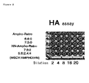
- the hemagglutinin activity (erythrocyte agglutination; HA titer) can be tested by a method known in the art (The society of research associates of The National Institute of Health, Eds., General Experimental Virology, 2nd Ed, pp. 214-225, MARUZEN CO.).
- the erythrocytes include, for example, those from chicken (including chick and fowl), goose, rat, guinea pig, rhesus monkey, green monkey and human.
- the reaction temperature may be 0° C., 4° C., room temperature, 37° C., or the like, depending an the type of protein. Exemplary conditions or erythrocyte agglutination reaction for the respective viruses are shown below:
- Particularly preferred membrane proteins having hemagglutinin activity in the pseudotype retrovirus of the present invention are viral proteins; specifically, such proteins include HN proteins of Paramyxovirus; HA proteins of orthomyxovirus and influenza virus; togaviral E1 protein; A27L, H3L, and D8L proteins of vaccinia virus; M and E proteins of flavivirus; E1 and E2 proteins of coronavirus; G1 protein of bunyavirus, etc.
- proteins from single-stranded negative strand RNA viruses are preferred, and HN protein of paramyxovirus is particularly preferred, as a membrane protein having hemagglutinin activity to be contained in the pseudotype retrovirus of the present invention.
- single-stranded negative strand RNA virus refers to a virus whose genome comprises a single-stranded negative strand (namely, ( ⁇ ) strand) RNA.
- viruses include paramyxovirus (Paramyxoviridae; including the genus Paramyxovirus, the genus Morbillivirus, the genus Rubulavirus, the genus Pneumovirus, and such), rhabdovirus (Rhabdoviridae; including the genus Vesiculovirus, the genus Lyssavirus, the genus Ephemerovirus, and such), filovirus (Filoviridae), orthomyxovirus (Orthomyxoviridae; including influenza viruses A, B, and C, Thogoto-like viruses, and such), bunyavirus (Bunyaviridae; including the genus Bunyavirus, the genus Hantavirus, the genus Nairovirus, the gen
- paramyxovirus refers to a virus belonging to the family of paramyxovirus (Paramyxoviridae).
- the paramyxovirus includes, for example, Sendai virus, Newcastle disease virus, Mumps virus, Measles virus, RS virus (Respiratory syncytial virus), rinderpest virus, distemper virus, monkey parainfluenza virus (SV5), human parainfluenza viruses' type-1, -2, and -3, etc.
- Sendai virus includes wild-type strains, mutant strains, laboratory strains, artificially constructed strains, etc. Incomplete viruses such as DI particles (J. Virol.
- HN protein is a protein of paramyxovirus virus.
- genes encoding paramyxovirus viral proteins include NP, P, M, F, HN, and L genes.
- NP, P, M, F, HN and L genes of viruses belonging to the family Paramyxoviridae correspond to genes encoding nucleocapsid, phospho, matrix, fusion, hemagglutinin-neuraminidase and large proteins.
- NP gene is generally described also as the “N gene”.
- Database accession numbers for nucleotide sequences of genes of the Sendai virus classified into Paramyxovirus of the family Paramyxoviridae are, M29343, M30202, M30203, M30204, M51331, M55565, M69046 and X17218 for NP gene, M30202, M30203, M30204, M55565, M69046, X00583, X17007 and X17008 for P gene, D11446, K02742, M30202, M30203, M30204, M69046, U31956, X00584 and X53056 for M gene, D00152, D11446, D17334, D17335, M30202, M30203, M30204, M69046, X00152 and X02131 for F gene, D26475, M12397, M30202, M30203, M30204, M69046, X00586, X02808 and X56131 for HN gene, and D00053, M30202
- a retroviral vector pseudotyped by paramyxoviral envelope proteins can be produced by preparing inactivated paramyxovirus, virosome having the envelope protein of paramyxovirus, etc. and fusing it with retrovirus.
- the vector can also be produced by expressing an expression vector for the paramyxovirus envelope proteins in retrovirus packaging cells.
- the pseudotype retroviral vector of the present invention can comprise, for example, F protein of paramyxovirus in addition to membrane proteins having hemagglutinin activity such as HN protein of paramyxovirus.
- F protein of paramyxovirus in addition to membrane proteins having hemagglutinin activity such as HN protein of paramyxovirus.
- the present invention demonstrated that the pseudotype retroviral vector containing HN and F proteins exhibited high efficiency of gene transfer.
- Pseudotype retroviral vectors containing the F and HN proteins are within the scope of the present invention.
- the pseudotype retroviral vector of the present invention may further contain M protein of paramyxovirus.
- the pseudotype retroviral vector of the present invention can contain additionally envelope proteins derived from other viruses.
- preferred envelope proteins include those derived from viruses infectious to human cells.
- proteins include, but not limited to, amphotropic envelope proteins of retrovirus, G protein of vesicular stomatitis virus (VSV), etc.
- proteins of viruses belonging to the herpes viridae include, for example, gB, gD, gH and gp85 proteins of herpes simplex virus, gp350 and gp220 proteins of EB virus, etc.
- proteins of viruses belonging to the hepadna viridae include S protein of hepatitis B virus, etc.
- a vector comprising retroviral amphotropic envelope (amphotropic env) protein and HN protein is preferred as a vector of the present invention.
- the vector can contain VSV-G protein that is a surface glycoprotein of vesicular stomatitis virus (VSV).
- VSV-G has been believed to use phospholipid as a receptor, which exists in most animal cells, and thus, by using a vector containing VSV-G protein and HN protein, the variety of cells into which genes can be introduced is dramatically increased and the transfer efficiency is also elevated. Indeed, the vector containing VSV-G protein and HN protein exhibited higher efficiency of gene transfer than the vector containing VSV-G alone.
- the vector containing VSV-G protein and HN protein is a preferred vector of the present invention.
- Such vectors may further contain paramyxovirus F protein.
- both vector containing retroviral amphotropic envelope protein, F protein, and HN protein, and vector containing VSV-G protein, F protein, and HN protein are within the scope of the present invention.
- these vectors may further contain M protein of paramyxovirus.
- both vector containing retroviral amphotropic envelope protein, F protein, HN protein, and M protein, and vector containing VSV-G protein, F protein, HN protein, and M protein are within the scope of the present invention.
- the vector containing paramyxoviral F and HN proteins, and the vector containing F, HN and M proteins, as described above, also transferred genes at a high efficiency into cells having mucus, cell fractions containing hematopoietic stem cells, or the like, into which genes were hardly transferred by the conventional techniques.
- VSV-G protein is a glycoprotein and forms a stable homotrimer on the membrane, the vector particles are scarcely disrupted during purification; thus the particles can be concentrated to a high level by centrifugation (Yang, Y. et al., Hum Gene Ther: September, 6(9), 1203-13. 1995).
- the present inventors confirmed that the SIV vectors pseudotyped by VSV-G and by F and HN could be concentrated by centrifugation.
- the types of paramyxovirus HN, F, and M proteins to be used to prepare pseudotyped vectors are preferred.
- the HN, F, and M proteins of Sendai virus include, for example, those from Z strain.
- the retroviral amphotropic envelope proteins include, for example, the envelope proteins from mouse leukemia virus (MuLV) 4070A strain.
- the MuMLV 10A1-derived envelope proteins can also be used (for example, pCL-10A1 (Imgenex) (Naviaux, R. K. et al., J. Virol. 70: 5701-5705 (1996)).
- the ecotropic envelope proteins include, for example, the envelope proteins from Moloney murine leukemia virus (MoMULV).
- Vesicular stomatitis virus G protein includes, for example, the protein derived from Indiana serotype strain (J. Virology 39: 519-528 (1981)). In addition to these, the proteins may be derived from desired strains.
- HN protein which is a structural protein, has both hemagglutinin (which is erythrocyte agglutination) activity and neuraminidase activity.
- HN protein which is a structural protein
- hemagglutinin which is erythrocyte agglutination
- neuraminidase activity For example, the reduction of the former activity may increase viral stability in blood.
- the efficiency of infection via HN protein can be controlled by modifying the latter activity.
- the fusion efficiency can be controlled by modifying F protein that participates in the membrane fusion.
- the antigen presenting epitopes of F protein and HN protein that can serve as cell-surface antigen molecules are analyzed, and based on the analysis result, pseudotype retroviruses of the present invention can he prepared by using the proteins whose antigen presenting ability has been impaired.
- pseudotype retroviruses of the present invention can he prepared by using the proteins whose antigen presenting ability has been impaired.
- higher efficiency gene transfer vectors can be prepared by pseudotyping the retroviral vector of the present invention by using modified proteins that are the viral envelope proteins having hemagglutinin activity or other envelope proteins whose cytoplasmic domains have been modified by deletion, substitution and/or addition.
- the present invention relates to pseudotype retroviral vectors comprising proteins in which a portion or the whole of a cytoplasmic domain of wild-type membrane proteins having hemagglutinin activity has been modified by substitution, deletion, and/or addition.
- modified HN protein and/or F protein of paramyxovirus in which the cytoplasmic domain has been deleted, or a cytoplasmic domain from another membrane protein (for example, the envelope protein of retrovirus including lentivirus) has been substituted for or added to the original one, can be used preferably to produce a viral vector with high infectivity.
- the present invention provides a membrane protein having hemagglutinin activity in which a portion or the whole of a cytoplasmic domain of the wild type of the membrane protein has been modified by substitution, deletion, and/or addition, and the nucleic acid encoding the protein (DNA, RNA, or the like).
- the present invention provides a modified viral hemagglutinin protein whose cytoplasmic domain has been modified by substitution, deletion, and/or addition, and the nucleic acid encoding the protein.
- a modified HN protein comprising the amino acid sequence of SEQ ID NO: 40 or 41 is preferred.
- modified proteins and the nucleic acids encoding the proteins are useful to produce pseudotype viruses of the present invention.
- foreign genes can be introduced with high efficiency into a wide variety of cells including human cells, by using retrovirus pseudotyped with a modified paramyxovirus HN protein whose cytoplasmic domain has been replaced with the cytoplasmic domain of envelope protein of lentivirus such as SIV, or retrovirus (for example, the protein encoded by pCAGGS-SIVct/HN according to Example 11); or another modified protein in which the cytoplasmic domain of envelope protein of retrovirus such as lentivirus has been added to paramyxovirus HN protein (for example, pCAGGS-SIVct+HN according to Example 11).
- retrovirus pseudotyped with a modified paramyxovirus HN protein whose cytoplasmic domain has been replaced with the cytoplasmic domain of envelope protein of lentivirus such as SIV, or retrovirus (for example, the protein encoded by pCAGGS-SIVct/HN according to Example 11); or another modified protein in which the cytoplasmic domain of envelope protein of retrovirus such
- Such a viral vector can contain additionally a modified F protein of paramyxovirus.
- a modified F protein of paramyxovirus for example, it is possible to use an F protein of paramyxovirus whose cytoplasmic domain has been deleted, or a protein obtained by adding the cytoplasmic domain of envelope protein of lentivirus such as SIV, or retrovirus to Such truncated protein.
- a plasmid in constructed to express an F protein whose cytoplasmic domain amino acids are deleted. Any portions of the domain can be deleted; a portion or the whole of cytoplasmic domain can be deleted.
- Fct4 a truncated Sendai virus F protein
- the protein that contains none or only several amino acid residues of the cytoplasmic domain due to an artificial deletion can be preferred to produce a pseudotype virus of the present invention.
- a protein in which the cytoplasmic domain of F protein has been substituted with a distinct peptide can be produced by attaching a portion or the whole of cytoplasmic domain of envelope protein of another virus (for example, lentiviral envelope protein) to these truncated F proteins.
- such proteins include the protein to which the first 11 amino acids of the cytoplasmic domain of SIV envelope protein from its 5′ end (SIVct11) have been attached.
- the present invention provides a paramyxovirus F protein in which a portion or the whole of cytoplasmic domain of the wild type of the protein has been modified by substitution, deletion, and/or addition, and the nucleic acid encoding the protein.
- the present invention provides an F protein whose cytoplasmic domain has been replaced with a portion of or the whole of a cytoplasmic domain of the envelope protein from a retrovirus including lentivirus, and the nucleic acid encoding the protein.
- a modified F protein comprising the amino acid sequence of SEQ ID NO: 42, 43, 44, 45, 46, or 47 is preferred.
- HA protein of a virus belonging to the Orthomyxoviridae is used as the viral envelope protein having hemagglutinin activity.
- a pseudotype virus produced by using an expression plasmid for influenza virus envelope protein is infectious to a wide variety of mammalian cells including human cells.
- the influenza virus envelope may be derived from a desired strain of isolated influenza virus.
- neuraminidase in responsible for the cleavage of the linkage with sialic acid.
- infectious virus particles pseudotyped by HA can be prepared by the treatment with neuraminidase.
- the linkage with sialic acid can be cleaved automatically by using a viral vector that also encodes a protein having neuraminidase activity.
- a viral envelope protein having neuraminidase activity such as HN protein of paramyxovirus.
- HN protein of paramyxovirus a viral envelope protein having neuraminidase activity
- the retroviral vector of the present invention also includes those derived from oncovirus.
- oncovirus refers to retroviruses belonging to the oncovirus subfamily (Oncovirus).
- the oncovirus includes retroviruses involved in canceration, such as sarcoma virus, leukemia virus, mammary tumor virus, etc.
- MoMLV Moloney murine leukemia virus
- a viral vector prepared by pseudotyping MoMLV with a protein having hemagglutinin activity, such as paramyxovirus HN protein, can be used preferably in the present invention.
- murine stem cell virus (MSCV) used in Example is a preferred gene-transfer vector particularly in transferring genes into hemocytes, hematopoietic cells, embryonic stem cells, etc.
- the retroviral vector of the present invention also includes those derived from lentivirus
- lentivirus refers to a retrovirus belonging to the lentivirus subfamily (Lentivirus).
- the lentivirus includes human immunodeficiency virus (HIV) (e.g., HIV1 or HIV2), simian immunodeficiency virus (SIV), feline immunodeficiency virus (FIV), Maedi-Visna virus, equine infectious anemia virus (EIAV), caprine arthritis-encephalitis virus (CAEV), etc.
- HIV human immunodeficiency virus
- SIV simian immunodeficiency virus
- FIV feline immunodeficiency virus
- Maedi-Visna virus equine infectious anemia virus (EIAV), caprine arthritis-encephalitis virus (CAEV), etc.
- the retroviral vector of the present invention can be derived from a desired strain or subtype.
- HIV-1 includes those of every major (M) subtype (including A to J), N, and outlier (O) (Hu, D. J. et al, JAMA 1996; 275: 210-216; Zhu, T. et al., Nature 1998, 5; 391(6667): 594-7; Simon, F. et al., Nat. Med. 1998, 4-(9): 1032-7).
- Isolated SIV strains include, for example, SIVagm, SIVcpz, SIVmac, SIVmnd, SIVsnm, SIVsyk, etc.
- Lentiviruses are infectious to nondividing cells and the virus genome can be integrated into host cell chromosome.
- the nuclear translocation signals encoded by gag and vpr are believed to be responsible for the integration.
- a viral vector of the present invention is constructed based on a lentivirus, genes can be introduced into nondividing cells in living tissues and cells that hardly divide, such as stem cells in various tissues, which allows long-term gene expression.
- HIV Human immunodeficiency virus
- FMV feline immunodeficiency virus
- CAEV caprine arthritis-encephalitis virus
- Simian immunodeficiency virus was discovered as a monkey-derived HIV-like virus, which, along with HIV, forms the group of primate lentivirus (E. Ido and M. Hayamizu, “Gene, Infection and Pathogenicity of Simian Immunodeficiency Virus”, Protein, Nucleic acid and Enzyme, Vol. 39, No. 8, 1994).
- HIV-1 subgroup containing HIV-1 that is the causative virus for human acquired immune deficiency syndrome (AIDS) and SIVcpz isolated from chimpanzee
- HIV-2 subgroup containing SIVsmm isolated from Sooty Mangabey ( Cercocebus atys ) SIVmac isolated from rhesus monkey ( Macaca mulatta ), and HIV-2 that is less pathogenic in human (Jaffar, S. et al., J. Acquir. Immune Defic. Syndr. Hum.
- Retrovirol. 16(5), 327-32, 1997); (3) SIVagm subgroup represented by SIVagm isolated from African green monkey ( Cercopithecus aethiops ); and (4) SIVmnd subgroup represented by SIVmnd isolated from Mandrill ( Papio sphinx ).
- the retroviral vector of the present invention includes those derived from spumavirus.
- the spumavirus includes, for example, foamyvirus (DE4318387; WO9607749; Virology (1995) 210, 1, 167-178; J. Virol. (1996) 70, 1, 217-22).
- Vectors of the present invention derived from foamyvirus can be utilized for introducing foreign genes into human cells, particularly in gene therapy and administration of recombinant vaccines.
- LTR long terminal repeat
- LTR is a retrovirus-specific sequence, which is present in the virus genome at both ends.
- 5′ LTR serves as a promoter, which enhances the mRNA transcription from the provirus.
- the transcription activity of 5′ LTR is known to be enhanced by viral tat protein, and therefore substitution of a tat protein-independent promoter for 5′ LTR allows the exclusion of tat from the packaging vector.
- the intracellular RNA of virus infected to cells is reverse transcribed and forms a closed circular structure with a linkage between the LTRs at the two ends, and then integrated into the chromosome of cells through the interaction between the linkage site and viral integrase.
- the mRNA transcribed from the provirus corresponds to the region from the transcription initiation site in the 5′ LTR to the 3′ LTR polyA sequence that located downstream; the 5′ LTR promoter portion is not packaged in the virus particle.
- a substitution of 5′ LTR promoter can provide a safer vector with a higher titer.
- a substitution of the promoter at the 5′ end in a gene transfer vector can increase the titer of packagable vectors.
- the safety can be improved with a self inactivating vector (SIN vector) which is prepared by partially eliminating the 3′ LTR sequence to prevent the transcription of full-length vector mRNA in target cells thereof.
- SI vector self inactivating vector
- the provirus for lentivirus which is integrated into the chromosome of target cells, has the U3 portion of 3′ LTR attached to the 5′ end thereof.
- the gene transfer vector contains U3 at the 5′ end and accordingly the transcription of RNA covering the whole gene transfer vector starts there. If there were lentivirus or related proteins in the target cells, the gene transfer vector would be re-packaged and infect other cells.
- SIN vectors can be constructed according to any of methods known in the art or the method as described in Example 5.
- Retroviruses can be produced by transcribing a gene transfer vector DNA containing the packaging signal in host cells, and allowing virus particles formation in the presence of gag and pol proteins, and envelope proteins.
- the gene transfer vector DNA may be a DNA vector such as plasmid, or a DNA that has been integrated in the chromosome of packaging cells. While it is preferable to integrate the packaging signal sequence encoded by the gene transfer vector DNA as long as possible to maintain the structure formed based on the sequence, it is required to minimize the sequence homologous between the packaging signal in the vector DNA and another packaging vector for providing gag and pol proteins to reduce the frequency of wild-type virus formation due to recombination between the types of vectors. Accordingly, it is preferable to construct the gene transfer vector DNA using as short a sequence as possible comprising the sequence required for packaging to meet both criteria of packaging efficiency and safety.
- packaging signal there is no limitation on the type of packaging signal, as long as packaging is achieved in cells where the packaging vector has been introduced; those derived from retrovirus, lentivirus, immunodeficiency virus, and the like can be used depending on the type of packaging vector.
- the type of virus from which the signal to be used is derived is limited to SIV because HIV vectors are not packaged.
- the SIV-derived gene transfer vector is also packagable when an HIV-derived packaging vector is used.
- the frequency of recombinant virus formation can be reduced when the vector particles are formed by combining gene transfer vector and packaging vector, each derived from different type of lentivirus.
- gag protein has been modified so that it is not expressed.
- the viral gag protein can be detected as a foreign substance in the living body, and thus a potential antigen.
- the protein may affect cellular functions.
- frameshift mutations can be introduced for modification by adding or deleting nucleotides downstream of the start codon of gag. It is also preferable to delete portions of the coding region of gag protein. In general, a 5′ portion of the coding region of gag protein is known to be essential for virus packaging. Thus, In a gene transfer vector, it is preferred that the coding region for the gag protein is deleted at the C terminus.
- a viral vector containing the transcription product of gene transfer vector DNA can be produced by introducing the constructed gene transfer vector DNA comprising the packaging signal into appropriate packaging cells. The viral vector particles produced can be recovered from the culture supernatant of packaging cells, or the like.
- packaging cell There is no limitation on the type of packaging cell, as long as the cell line is generally used in viral production.
- a human or monkey-derived cell When used for the purpose of gene therapy in human, a human or monkey-derived cell is suitable.
- Human cell lines to be used as packaging cells include, for example, 293 cell, 293T cell, 293EBNA cell, SW480 cell, u87MG cell, HOS cell, C8166 cell, MT-4 cell, Molt-4 cell, HeLa cell, HT1080 cell, TE671 cell, etc.
- Monkey cell lines include, for example, COS1 cell, COS7 cell, CV-1 cell, BMT10 cell, etc.
- previously established packaging cells can be used.
- Such packaging cells include, for example, Bosc23 cell, PE501 cell, etc.
- foreign gene to be inserted in the vector, which include, for example, nucleic acids which do not encode any protein, such as antisense or ribozyme, as well as protein-encoding nucleic acids.
- the pseudotype retroviral vector of the present invention can transfer genes into CD34-positive cells derived from human bone marrow with high efficiency; such a fraction comprising the CD34-positive cells receives attention as a cell fraction containing hematopoietic stem cells in recent years.
- Previous reports describe that the CD34-positive cells exhibit pluripotency in colony assay using a culture medium containing methylcellulose (Kirshenbaum, A. S. et al., J.
- the present invention relates to a method for introducing genes into hemocytes or hematopoietic cells, in which the method comprises a step of contacting pseudotype retroviral vector containing membrane proteins having hemagglutinin activity with hemocytes or hematopoietic cells, and the use of the pseudotype retroviral vector containing membrane proteins having hemagglutinin activity to introduce genes into hemocytes or hematopoietic cells.
- the retroviral vector of the present invention pseudotyped by the use of proteins having hemagglutinin activity, can transfer genes into hemocytes and hematopoietic cells with high efficiency, and thus is useful in gene therapy whose target is blood cells, e.g., adenosine deaminase (ADA) deficiency (Blaese, R. M., Pediatr. Res., 33 (1 Suppl), S49-53, 1993), hemophilia (Kay, M. A. et al., Proc. Natl. Acad. Sci. USA, 96(18), 9973-5, 1999) and Fanconi anemia, etc.
- the administration can be performed, for example, by an ex viva method.
- Evaluation for the achievement of introduction of foreign genes into hemocytes and hematopoietic cells can be made, for example, by flow cytometry analysis using antibodies against various known surface antigens or by colony assay, or through assessing hemopoietic cell transplantation-based reconstitution of hemopoietic system in mice whose hemopoietic system has been disrupted.
- Gene therapy whose targets are hemopoietic cells, to which the vector of the present invention is applicable, includes, for example, use of the drug resistance gene MDR1 to preserve stem cells in anti-cancer chemotherapy (Licht, T. et al., Gene Ther. (2000) 7, 4, 348-58); introduction of the normal FANCC gene for the treatment of Fanconi anemia (Liu, J. M. et al., Hum. Gene Ther.
- IL-6 superagonist and superantagonist to be used to proliferate hemopoietic progenitor cells
- Factor-X to be used in therapy for blood diseases
- stem cell factor to be used in therapy for blood diseases
- IL-6 to be used in therapy for blood diseases
- stem cell factor to be used in therapy for blood diseases
- IL-6 to be used in therapy for blood diseases
- stem cell factor to be used in therapy for blood diseases
- IL-6 to be used in therapy for blood diseases
- stem cell factor to be used in therapy for blood diseases
- IL-6 stem cell factor
- ribozyme whose targets are RNA viruses, and antisense and/or decoy RNA which are useful to prevent HIV infection and for intracellular immunity
- the HN protein-pseudotyped retroviral vector of the present invention is highly infectious to cells containing mucus, such as mucosal epithelial cells of nasal cavity and epithelial cells of bronchial mucus membrane In the lung.
- the vector of the present invention is useful for high efficiency transfer of foreign genes into cells containing mucus, into which genes are hardly transferred by any of the conventional methods.
- the present invention relates to a method for introducing genes into cells containing mucus, in which the method comprises a step of contacting a pseudotype retroviral vector containing membrane proteins having hemagglutinin activity with cells containing mucus, and the use of the pseudotype retroviral vector containing membrane proteins having hemagglutinin activity to introduce genes into cells containing mucus.
- Such cells containing mucus include mucosal epithelial cell in particular, specifically for example, mucosal epithelial cells of nasal cavity or pulmonary bronchial tubes.
- IL-2 immunoreaction through gene transfer
- APC antigen presenting cells
- WO 9505853 vaccination against rotavirus by oral administration of genes to mucous membrane
- mucosal administration to treat autoimmune diseases WO 9746253
- mucosal administration to prevent infection WO 9621356
- administration of genes to mucous membrane of female genital organs to prevent caner of the cervix uteri caused by sex-transmitted diseases or papilloma virus infection Infect. Immun. (1998) 66, 1, 322-29)
- improvement of administration simplicity and safety by administration via mucous membrane Proc. Am. Assoc. Cancer Res. (1995) 36, 86 Meet., 418).
- the pseudotype retroviral vector of the present invention can be prepared as a composition by appropriately combining with pharmaceutically acceptable carrier or medium.
- the vector can be formulated as a composition appropriately in combination with sterilized water, physiological saline, culture medium, serum, phosphate-buffered physiological saline (PBS), etc.
- the composition may further contain other components including stabilizer, antimicrobial, etc.
- the composition of the present invention can be in a dosage form such as aqueous solution, capsule, suspension, syrup, etc.
- a composition comprising the pseudotype retroviral vector of the present invention is useful as a reagent or a pharmaceutical.
- a composition of the present invention can be used as a reagent for in vitro or in vivo gene transfer into various cells, or as a pharmaceutical for ex vivo or in vivo gene therapy.
- the administration to patients can be achieved, for example, by a method known to those skilled in the art, including intraarterial, intravenous, intraperitoneal, subcutaneous, enteral, oral, or intranasal administration, or ex vivo administration.
- administration to mucous membrane of the nasal cavity or bronchial tubes, and ex vivo administration to hemocytes and hematopoietic cells are suitable.
- the viral vector of the present invention can be used in gene therapy for various other genetic diseases.
- diseases to be treated and the single causative genes thereof include: Gaucher disease, ⁇ -cerebrosidase (chromosome 20); hemophilia, blood coagulation factor VIII (X chromosome) and blood coagulation factor IX (X chromosome) adenosine deaminase deficiency, adenosine deaminase; phenylketonuria, phenylalanine hydroxylase (chromosome 12), Duchenne muscular dystrophy, dystrophin (X chromosome); familial hypercholesterolemia, LDL receptor (chromosome 19), cystic fibrosis, chromosomal translocation of CFTR gene.
- the targeted disease in which other multiple genes are thought to be involved include neurodegenerative diseases such as Alzheimer's disease and Parkinson's disease, ischemic encephalopathy, dementia, and intractable infection such as AIDS.
- a treatment to inactivate the HIV transcription factor may be considered, therein an SIV based vector of this invention is worked in vitro into a hematopoietic stem cell removed from an AIDS patient extracellularly, for increasing the transcription of SIV-derived genome prior to HIV infection, and the transfected cell is returned to the patient's body.
- examples of applications possible for chronic diseases include: suppression of the expression of VEGF and FGF2 genes for ischemic heart disease, and suppression of the expression of cell proliferation related genes, such as cell proliferation factors (PDGF, TGF- ⁇ , etc.) and cyclin-dependent kinase, for gene therapy of arteriosclerosis.
- cell proliferation factors PDGF, TGF- ⁇ , etc.
- cyclin-dependent kinase for gene therapy of arteriosclerosis.
- the BDNF gene may be a candidate.
- this method can be applied to substitution therapy, in which a gene such as a cancer suppressor gene, p53, whose genetic mutation causes canceration, is integrated into the chromosome, and this method enables treatment beyond the limitation of cancer pharmacotherapy by introducing a multiple-drug-resistant gene into bone marrow-derived hematopoietic stem cells in vitro and, then, by returning these cells into patient's blood.
- a gene such as a cancer suppressor gene, p53, whose genetic mutation causes canceration
- T-cell receptors various adhesion factors (for example, ICAM-1, LFA-1, VCAM-1, and LFA-4 etc.), cytokines and cytokine receptor (for example, TNF, IL-8, IL-6, and IL-1 etc.) cell proliferation factors (for example, PDGF, and TGF- ⁇ etc.), and activation factors (for example, MMP etc.) become possible.
- adhesion factors for example, ICAM-1, LFA-1, VCAM-1, and LFA-4 etc.
- cytokines and cytokine receptor for example, TNF, IL-8, IL-6, and IL-1 etc.
- cell proliferation factors for example, PDGF, and TGF- ⁇ etc.
- activation factors for example, MMP etc.
- the elevation of success percentage or a xenotransplant becomes possible by changing the histocompatibility antigen of a non-human animal donor to a human-type. Furthermore, treatment by introducing foreign genes into the chromosome of human ES cells, thus making up the deficient genes at the embryonic stage to supplement deficiencies of systemically circulating enzymes, growth factors, and such may be considered.
- IL-4 promotes differentiation of helper T lymphocyte into Th2 lymphocyte.
- Th2 lymphocyte secretes cytokines, such as IL-4, IL-5, IL-9, and IL-13, which mediate asthmatic inflammation.
- IL-4 is a molecule that induces mucus secretion from lung mucus membrane, which is involved in respiratory disturbance.
- IL-4 regulates the expression of VCAM-1 that is a cell adhesion molecule interacting with VLA 4 molecule present on the surface of eosinophil. The interaction allows eosinophils to migrate from blood to inflammation sites in lung tissues. Since IL-4 increases the number of B cells and induces the production of antigen-specific IgE responsible for allergic reactions.
- the antigen-specific IgE produced induces the release of inflammatory mediators, such as histamine, leukotriene, from mast cells, which results in bronchoconstriction.
- inflammatory mediators such as histamine, leukotriene
- IL-4 expression vectors for soluble interleukin 4 (IL-4) receptor etc. can be used to treat asthma patients.
- FIG. 1 depicts photographic patterns showing the results obtained with 293T cells infected with murine stem cell virus (MSCV) pseudotyped by F, HN, or F and HN proteins from Sendai virus.
- the virus packaging was achieved by using a gene transfer vector expressing EGFP (pMSCV EGFP).
- the labels in the above panels, “F”, “HN”, and “F/HN”, correspond to the results obtained by the infection with the viruses produced by expressing F, HN, and F and HN proteins of Sendai virus in the packaging cell, respectively.
- VSV-G/Null the top panel depicts a positive control pseudotyped by VSV-G; the middle and bottom panels depict non-pseudotyped negative controls without expressing env proteins for pseudotyping, such as F, HN, and VSV-G.
- the label “Null” indicates that retrovirus env was not expressed.
- FIG. 2 depicts a photographic pattern showing results of HA assay of retroviruses having amphotropic env, which were pseudotyped by Sendai virus HN.
- the amounts of pMSCV EGFP, pCl-Ampho, and pCAGGS-HN used in viral production were also indicated (MSCV:AMPHO:HN in this Figure). While erythrocyte agglutination was not detected with the control retrovirus containing amphotropic env (Ampho-Retro in this Figure), the agglutination was observed with the retrovirus pseudotyped by Sendai virus HN (HN-Ampho-Retro in this Figure).
- FIG. 3 is a diagram showing a result of assay which comprises infecting amphotropic env-containing MSCV pseudotyped by HN protein of Sendai virus (HN-ampho in this Figure) to human bone marrow cells and measuring the percentage of infected cells (GFP-expressing cells) by flow cytometry-based fractionation using CD34 as a marker.
- the “ampho” indicated in this Figure refers to a control which is not pseudotyped by HN protein.
- Each column indicates the ratio of GFP-positive cell against CD34-negative (CD34 ⁇ ) cell or CD34-positive (CD34+) cell.
- FIG. 4 depicts a schematic illustration of structures of: (a) the SIVagm-genome plasmid which was used as the backbone for the other constructs, (b) constructed packaging vector, (c) gene transfer vector, and (d) VSV-G-supplying vector.
- FIG. 5 depicts photographs showing a result of SIVagm vector-mediated introduction of the ⁇ -galactosidase gene into 293T cells.
- the top panel shows a result obtained by X-gal staining 48 hours after vector infection into 293T cells using the supernatant of cells transfected with the vector plasmid. The expression of ⁇ -galactosidase is detected in a number of cells.
- the bottom panel shows X-gal staining of untreated control cells.
- FIG. 6 depicts photographs obtained by introducing the EGFP gene into G2-M phase-arrested 293T cells and SH-SY5Y cells differentiated by retinoic acid, in which gene transfer was mediated by the SIVagm SIN vector, and observing the resulting expression using a fluorescent miroscope. The expression was confirmed under a fluorescence microscope.
- the top panel, 293T, magnification ⁇ 100; the bottom panel, SH-SY5Y, magnification ⁇ 200.
- FIG. 7 is a photograph showing a result of Western blotting using FHN virosome and F virosome arranged in approximately the same particle diameter.
- FIG. 8 depicts photographs showing a result obtained by the observations of infected cells (GFP-expressing cells).
- SIVs fused with inactivated Sendai virus (SeV), FHN virosome, and F virosome were added to the culture supernatant of HeLa cells at MOI-10. The mixtures were incubated for 10, 30, and 180 minutes for infection, and then the culture media were changed with fresh ones. The cells were observed 48 hours after the infection.
- the SIV used had been pseudotyped by VSV-G.
- SIV in this Figure refers to a negative control which was not fused with Sendai virus envelope.
- FIG. 9 depicts photographs showing a result obtained by electrophoresing samples of FHN virosome, F virosome, and HN virosome, followed by silver staining.
- Lane 1 SeV (intact); lane 2, DTT-treated SeV; lane 3, trypsin-treated SeV; lane 4, FHN virosome; lane 5, F virosome ; lane 6, HN virosome.
- FIG. 10 depicts a diagram showing a result of assay for relative LacZ activity.
- a LacZ expression SIV vector (pseudotyped by VSV-G) and the same but further fused with virosome were introduced into 293T cells. Asterisk indicates that there was a significant difference for SIV in t-test (p ⁇ 0.05).
- FIG. 11 depicts photographs showing frozen sections of trachea three days after intranasal administration of 100 ⁇ l of SIV-F/HN/M-EGFP 10 8 T.U.
- the arrows in (b) indicate epithelial cells of the trachea, where fluorescence of EGFP can be detected, while only low-level background signals are observed in the sample of the untreated mouse shown in (c).
- the panel (a) shows a pattern of hematoxylin-eosin (H.E.) staining of a serial section.
- FIG. 12 depicts photographs (b) showing observations in septonasal mucus membrane of the same individuals as in FIG. 11 . Arrow indicates pseudostratified ciliated epithelium that exhibits fluorescence of EGFP. (a) depicts photographs of H.E.-stained serial sections.
- FIG. 13 depicts photographs showing a comparison of fluorescence intensities of GFP from the pseudotype retroviral vector in the sections of mouse nasal cavity.
- EGFP fluorescence images of SIV pseudotyped by VSV-G SIV-VSV-EGFP
- MSCV containing amphotropic env pseudotyped by F, HN, and M MSCV-F/HN/M-EGFP
- VSV-G SIV pseudotyped by F, HN, and M SIV-F/HN/M-EGFP
- FIG. 14 shows the amino acid sequence (SEQ ID NO: 40) at the boundary between the SIV cytoplasmic domain and the HN protein transmembrane domain (standard font) in the protein encoded by the HN expression plasmid with a substitution of cytoplasmic domain.
- FIG. 15 shows the amino acid sequence (SEQ ID NO: 41) at the boundary between the cytoplasmic domain (underlined) and the HN protein transmembrane domain (standard font) in the protein encoded by the HN expression plasmid with an addition of SIV cytoplasmic domain.
- FIG. 16 shows the amino acid sequences (SEQ ID NOs: 42 to 44) at the boundary between the F protein transmembrane domain (italic) and F protein cytoplasmic domain (standard font) in the proteins encoded by the F expression plasmids with a deletion of cytoplasmic domain.
- FIG. 17 shows the amino acid sequences (SEQ ID NOs: 45 to 47) at the boundaries between the F protein transmembrane domain (italic without underline), F protein cytoplasmic domain (standard font), and the 11 amino acids of SIV cytoplasmic domain (SIV c11 ) (underline) in the proteins encoded by the F expression plasmids in which the F cytoplasmic domain has been replaced with the SIV cytoplasmic domain.
- FIG. 18 depicts photographs showing gene transfer into 293T cells via the SeV F/HN pseudotype SIV vector.
- FIG. 19 depicts photographs showing gene transfer into BEAS-2B cells via the SeV F/HN pseudotype SIV vector.
- FIG. 20 depicts photographs showing gene transfer into 293T cells via the SeV F/HN pseudotype SIV vector with an addition of SIV c11 .
- FIG. 21 depicts photographs showing the enriched SeV F/HN pseudotype SIV vector.
- FIG. 22 depicts photographs showing gene transfer via the MSCV-based SeV F/HN pseudotype retroviral vector.
- FIG. 23 depicts photographs showing gene transfer via the viral vector pseudotyped by influenza virus envelope.
- FIG. 24 depicts photographs showing the enriched viral vector pseudotyped by influenza virus envelope.
- FIG. 25 depicts photographs showing gene transfer via viral vectors pseudotyped by influenza virus envelope and various HN proteins.
- FIG. 26 depicts photographs showing pseudotyped SIV vectors transduced RTE through apical side.
- the genes for F, HN, and M proteins were obtained by digesting the full-length genome DNA of Sendai virus Z strain pSeV18 + b(+) (Hasan, M. K. et al., 1997, J. General Virology 78: 2813-2820), and inserted into pCAGGS (Niwa, H. et al., Gene: 108, 193-9, 1991) at the XhoI site, to prepare expression vectors for F, HN, and M proteins of Sendai virus (referred to as pCAGGS F, pCAGGS HN, and pCAGGS M, respectively).
- the human fetal kidney-derived cell line, 293T cell (Proc. Natl. Acad. Sci. USA, vol.90, pp.8392-8396, 1993), which was used to produce retrovirus, was cultured in D-MEM (GibcoBRL) containing 10% inactivated fetal bovine serum. Culture was carried out in plastic plates (Sumitomo Bakelite). The vector transfection was carried out using LIPOFECTAMINE PLUS (GibcoBRL) according to the provided instruction. 293T cells were plated in a 6-well plastic plate (Sumitomo Bakelite) at a cell density of 1 ⁇ 10 6 cells/well, and incubated in a CO 2 incubator under 10% CO 2 gas at 37° C. for 48 hours. 30 minutes before transfection, the culture medium was changed with 800 ⁇ l/well of D-MEM (GibcoBRL) containing 1% bovine serum albumin (GibcoBRL), and then culture was continued.
- D-MEM GibcoBRL
- the gene transfer vector used was a murine stem cell virus-based vector (MSCV) (CLONTECH) (R. G. Hawley et al., Proc. Natl. Acad. Sci. USA 93: 10297-10302 (1996); R. G. Hawley et al., Gene Thrapy 1: 136-138 (1994)) containing the EGFP gene at a reporter gene.
- Amounts of DNAs used in the transfection are as follows: 700 ng/well of the gene transfer vector and 300 ng/well of packaging vector (pCL-Eco, pCL-Ampho (MuMLV 4070A) (both were purchased from IMGENEX) (Naviaux, R. K. et al., J. Virol.
- DNA was dissolved in 100 ⁇ l of OptiMEM and then 6 ⁇ l of PLUS reagent (GibcoBRL) was added thereto. The mixture was stirred and allowed to stand still at room temperature for 15 minutes. 4 ⁇ l of LIPOFECTAMINE was diluted with 100 ⁇ l of OptiMEM, and then added To the mixture of DNA and PLUS reagent (GibcoBRL). The resulting mixture was stirred and allowed to stand still at room temperature for 15 minutes.
- PLUS reagent GibcoBRL
- the solution containing the complex of DNA and LIPOFECTAMINE prepared by the above procedure was added dropwise to 293T cells incubated in a 6-well plate. After being gently stirred, the mixture was incubated in a CO 2 incubator under 10% CO 2 gas at 37° C. for 3 hours. After culture, 1 ml/well of D-MEM (GibcoBRL) containing 1% bovine serum albumin (GibcoBRL) and 15 ⁇ g/ml trypsin (GibcoBRL) was added to the culture. The mixture was incubated in a CO 2 incubator under 10% CO 2 gas at 37° C. for 24 hours. Then, the culture supernatant was filtered with a filter with 0.45- ⁇ m diameter pores (DISMIC-25CS filter; ADVANTEC). The resulting solution was used as a vector solution.
- the retroviral vector pseudotyped by Sendai virus envelope protein was prepared by the above-mentioned method and its effect was assessed.
- 293T cells used as a target were plated in a 6-well plastic plate (Sumitomo Bakelite) at a cell density of 1 ⁇ 10 6 cells/well, and then incubated in a CO 2 incubator under 10% CO 2 gas at 37° C. for 48 hours.
- the introduction of the viral vector into the target cells was achieved by overlaying the solution that had been prepared by adding inactivated fetal bovine serum and polybrene (Sigma) at the final concentrations of 10% and 8 ⁇ g/ml, respectively, to a solution containing the viral vector.
- the target cells were fixed using PBS (Invitrogen) containing 2% formaldehyde and 0.2% glutaraldehyde at room temperature for 20 minutes, and then washed once with PBS (Invitrogen). Then, the cells were observed under a fluorescence invert microscope (DMIRB(SLR), Leica) to detect EGFP expression in target cells.
- PBS Invitrogen
- DIRB(SLR) fluorescence invert microscope
- F protein expression plasmid pCAGGS F
- HN protein expression plasmid pCAGGS HN
- Sendai virus envelope proteins Sendai virus envelope proteins
- 293T cells were cultured in Dulbecco's modified Eagle medium (DMEM) containing 10% FCS and 800 ⁇ g/ml G418 according to the conventional method. Using a 10-cm plate, the cells diluted to 8 ⁇ 10 6 cells/plate the day before transfection, and then cultured in DMEM containing 10% FCS.
- DMEM Dulbecco's modified Eagle medium
- the culture medium was removed and the transfection mixture was added dropwise all over to 293T cells in 500 ⁇ l of DMEM containing 1% BSA. The cells were incubated under 5% CO 2 gas at 37° C.
- a control transfection experiment was carried out using 5.6 ⁇ g of pMSCV EGFP and 4.0 ⁇ g of pcl-Ampho.
- the culture medium was replaced with 10 ml of Iscove's modified DMEM (IMDM) containing 10% FCS. On the following day, the medium was again changed with IMDM containing 10% FCS, and the culture was further continued for 24 hours.
- IMDM Iscove's modified DMEM
- the cell culture supernatant was collected 48 hours after transfection.
- the supernatant was filtered with a 0.45- ⁇ m filter and then stored at ⁇ 80° C.
- Retroviruses having amphotropic env pseudotyped by HN were prepared at various ratios of pMSCV EGFP, pCl-Ampho, and pCAGGS-HN in the same manner as described above.
- the viral hemagglutinin activity (erythrocyte agglutination activity; HA activity) was tested.
- the hemagglutinin activity of retrovirus having amphotropic env was also tested as a control.
- Assay for HA activity was carried out according to the conventional method. The result showed that the retrovirus pseudotyped by HN and amphotropic env had HA activity and the virus particles contained proteins exhibiting hemagglutinin activity ( FIG. 2 ).
- Human bone marrow CD34 + cell was purchased from BIO WHITTAKER. After thawing, along with 1 ⁇ 10 5 /ml of the recombinant virus collected in Section 3, the cells were cultured in IMDM containing 50 ng/ml IL-6, 100 ng/ml TPO, 100 ng/ml Stem Cell Factor (GIBCO BRL), 100 ng/ml Flt-3 ligand (Research Diagnostics, Flanders, N.J.) (all the above were human recombinant proteins), and 10% FCS under 5% CO 2 at 37° C. overnight.
- IMDM containing 50 ng/ml IL-6, 100 ng/ml TPO, 100 ng/ml Stem Cell Factor (GIBCO BRL), 100 ng/ml Flt-3 ligand (Research Diagnostics, Flanders, N.J.) (all the above were human recombinant proteins), and 10% FCS under 5% CO 2 at 37° C. overnight.
- the medium was changed with a freshly thawed virus solution, and 50 ng/ml IL-6, 100 ng/ml TPO, 100 ng/ml Stem Cell Factor, and 100 ng/ml Flt-3 ligand were added to the cells. 120 hours after the start of culture, the cells were collected.
- the collected cells were stained with PE-labeled anti-human CD34 antibody (Becton Dickinson), and then analyzed by flow cytometry (EPICS ELITE, Coulter) using two fluorescence probes of GFP and PE. According to the result, the proportions of GFP positive cells in CD34-negative cells (CD34 ⁇ ) and CD34-positive cells (CD34+) infected with the amphotropic retroviral vector (ampho) containing the wild-type envelope were only 0.3% and 3.6%, respectively.
- CD34-negative cells CD34 ⁇
- CD34-positive cells CD34+
- HN-ampho HN-amphotropic retrovirus vector
- Retroviral vectors pseudotyped by various envelope proteins were prepared using the above-described pMSCV EGFP or pLZRNL expressing lacZ under the control of LTR derived from Moloney murine sarcoma virus (Yee, J.-K. et al., Methods In Cell Biology, vol. 43, pp.99-112 (1994); Xu, L. et al., Virology 171, 331-341 (1989)) as a gene transfer vector.
- 293T cells human fetal kidney cell line
- DMEM Dulbecco's Modified Eagle Medium
- Gabco BRL Dulbecco's Modified Eagle Medium
- BIO WHITTAKER 10% inactivated calf serum
- the 293T cells were plated in a 6-well plastic culture plate at a cell density of 5 ⁇ 10 5 cells/well, and then incubated under 10% CO 2 at 37° C. for 48 hours.
- the culture medium was changed with 800 ⁇ l/well of DMEM containing 1% bovine serum albumin. The cells were then used in transfection.
- Env envelope proteins derived from retrovirus (Retro), Sendai virus (SeV), and vesicular stomatitis virus (VSV) used in the production of viruses.
- the gene transfer vector used were pMSCV EGFP or pLZRNL, which were utilized in in vitro and in vivo gene transfer, as shown in this Table.
- the 12 types of vectors indicated in lower (1) to (12), which had been prepared using pLZRNL, were used in the experiment in FIG. 1 .
- VTRF-BglII 5′-GCAGATCTCAACCAGGAGGCGAGGCTGCATTTTGGG-3′ 1R(VTRR-EcoRI): (SEQ ID NO: 2) 5′-GCGAATTCTACTTACTGGTGCTGTAAAGGAGCCAAA-3′ 2F(RREG964E) (SEQ ID NO: 3) 5′-ATCGGAATTCTTTTATTGTAAGATGGATTGGTTTTTAAAT-3′ 2R(RRE-SA + BN): (SEQ ID NO: 4) 5′-CGGGATCCGCGGCCGCGGATATGGATCTGTGGAGATAG AGGAACATAT-3′ 3F(SDFXhoSpe): (SEQ ID NO: 5) 5′-TCGAGACTAGTGACTTGGTGAGTAGGCTT-3′ 3R(SDR-Sal): (SEQ ID NO: 6) 5′-TCGAAAGCCTACTCACCAAGTCACTACTC-3′ 4F: (SEQ ID NO: (SEQ ID NO: 1)
- Plasmids that provide backbone structures for vectors were constructed ( FIG. 4 ). Namely, these plasmid were “packaging vector” providing proteins essential for the formation of vector particles in trans, “gene transfer vector” providing mRNA to be packaged into the vector and delivering and introducing genes of interest into target cells, and the vector providing coat proteins participating in the formation of pseudotype vector particles.
- Expression plasmids in each of which a sequence for gag, pol, tat, rev, vif, or vpr/x is located downstream of the promoter were constructed to provide proteins required for the formation of vector particles. Most parts of the packaging signal ⁇ and env were removed to avoid the generation of wild-type virus particles. An SD sequence was inserted upstream or gag and RRE sequence was inserted downstream of the first exon of tat/rev to allow the expression of all genes in the packaging vector. In addition, the entire nef sequence was excluded because it was predicted to be nonessential for vector packaging ( FIG. 4 b ).
- the LTR sequences from the two ends of the genome, SD, ⁇ , and RRE were inserted into the gene transfer vector providing RNA to be packaged into the vector. Further, a foreign promoter was substituted for the region of 5′ LTR promoter in the gene transfer vector.
- a self inactivating vector (SIN vector), in which a portion of the 3′ LTR sequence was removed to prevent the transcription of full-length vector mRNA in target cells, was prepared; this vector contains as inserts the ⁇ -galactosidase gene as a reporter gene and the CMV promoter for the expression of the reporter gene ( FIG. 4 c ).
- VSV-G The vector used to provide VSV-G was the field-proven pVSV-G in pseudotyping retroviral and HIV vectors ( FIG. 4 d ) (Burns, J. C. (1993) Proc. Natl. Acad. Sci. USA 90: 8033-8037).
- a DNA fragment corresponding to the region (5337 to 5770) containing vif and the first exon of tat/rev was prepared by PCR using a pair or primers 1F and 1R and pSA212 as a template.
- a site for the restriction enzyme EcoRI had been added to one of the PCR primers, and thus the prepared DNA fragment contained an EcoRI site at its 3′ end.
- the PCR fragment was digested with BglII and EcoRI, and purified by using agarose gel electrophoresis and Wizard PCR Preps DNA Purification System (Promega).
- the DNA fragment prepared as described above and another DNA fragment encoding the gag/pol region were ligated into pBluescript KS+ (Stratagene) between the XhoI and EcoRI sites. Then, a Rev responsive element (RRE) and a DNA fragment corresponding to the region containing the second exon (6964 to 7993) of tat/rev were amplified by PCR.
- RRE Rev responsive element
- a NotI site was added to the 3′ end by PCR using the pair of primers 2F and 2R and using pSA212 as a template by the same procedure as used to prepare the above-described PCR fragment.
- the resulting PCR fragment was double-digested with EcoRI and NotI, followed by purification. The fragment was inserted into pBluescript KS+ containing gag-tat/rev between the EcoRI and NotI sites.
- the resulting plasmid was digested with XhoI and NotI.
- the fragment containing SD-gag-RRE-tat/rev was purified.
- a plasmid was prepared by inserting an XhoI/NotI linker (4F and 4R) into pCAGGS (Gene, vol.108, pp.193-200, 1991) at the EcoRI site, and then the above-mentioned SD-gag-RRE-tat/rev fragment was inserted at the XhoI-NotI site.
- the plasmid obtained by the method as described above was used as the packaging vector pCAGGS/SIVagm gag-tat/rev.
- PCR was carried out to amplify the SIVagm TYO1-derived 5′ LTR region (8547 to 9053+1 to 982; containing KpnI site and EcoRI site at the 5′ and 3′ ends, respectively) using a pair of primers 5-1F and 5-1R; the 3′ LTR region (8521 to 9170; containing NotI and BamHI sites at the 5′ end and SacI site at the 3′ end) using a pair of primers 5-3F and 5-3R; and the RRE sequence (7380 to 7993; containing EcoRI and SacII sites at the 5′ and 3′ ends, respectively) using a pair of primers 5-2F and 5-2R.
- the CMV promoter region (1 to 600; containing SacII and NotI sites at the 5′ and 3′ ends, respectively) derived from pEGFPN2 (Clontech) was amplified using a pair of primers 6F and 6R. The DNA fragments were digested at their ends. After purification, the 5′ LTR, RRE, CMV promoter, and 3′ LTR were inserted in this order into pBluescript KS+ at the KpnI-SacI site by ligation. A NotI fragment containing the ⁇ -galactosidase gene derived from pCMV ⁇ (Clontech) was inserted as a reporter gene into the NotI site.
- the resulting plasmid was digested with KpnI and SacI to obtain a DNA fragment containing the region from the 5′ LTR to the 3′ LTR.
- the fragment was inserted into a control vector pGL3 at the KpnI-SacI site.
- the resulting plasmid was used as the gene transfer vector pGL3c/5′ LTR.U3G2/RREc/s/CMV F ⁇ -gal/WT 3′ LTR.
- the CMV promoter region derived from pEGFPC2 (Clontech) and the region encoding EGFP (1 to 1330; containing a SacII site at the 5′ end and NotI, BamHI sites, and translational stop codon at the 3′ end) were amplified by PCR using a pair of primers 6F and 6R and using pEGFPC2 as a template.
- the four types of PCR fragments were digested with restriction enzymes KpnI and EcoRI, EcoRI and SacII, BamHI and SacI, and SacII and BamHI, respectively.
- the fragments of 5′ LTR, RRE, CMV promoter EGFP, and 3′ LTR were inserted in this order into pBluescript KS+ between KpnI and SacI by ligation (pBS/5′LTR.U3G2/RREc/s/CMVFEGFP/WT3′LTR).
- the plasmid pBS/5′LTR.U3G2/RREc/s/CMVFEGFP/WT3′LTR was digested with KpnI and SacI to prepare a DNA fragment containing the region from the 5′LTR to the 3′LTR.
- the fragment was inserted into pGL3 (Promega) as a control vector at the KpnI-SacI site to construct a vector (pGL3C/5′LTR.U3G2/RREc/s/CMVFEGFP/WT3′LTR).
- a fragment containing gag region (9039 to 9170+1 to 982) downstream of the TATA box of 5′ LTR was amplified by PCR using primers 9-1F, 2F, 3F and 9R and using pSA212 as a template.
- the CMV L promoter of cytomegalovirus (derived from pCI (Promega); nucleotides 1 to 721) was amplified by PCR using a pair of primers 10-1F and 10-1R.
- a fragment containing a region downstream of the TATA box of 5′ LTR was combined with the fragment containing the promoter.
- a DNA fragment containing a chimeric promoter of the promoter and the 5′ LTR was prepared by PCR using the mixture as a template and a primer (10-1F) placed on the 5′ side of the promoter and another primer (9R) located on the 3′ side of 5′ LTR.
- the resulting DNA fragment was inserted into the gene transfer vector (pGL3C/5′LTR.U3G2/RREc/s/CMVF ⁇ -gal/WT3′LTR) at the KpnI-EcoRI site to prepare pGL3C/CMVL.U3G2/RREc/s/CMVF ⁇ -gal/WT3′LTR.
- the DNA fragment obtained by the PCR experiment described above was also inserted into the vector pGL3C/5′LTR.U3G2/RREc/s/CMVFEGFP/WT3′LTR at the KpnI-EcoRI site to prepare pGL3C/CMVL.U3G2/RREc/s/CMVFEGFP/WT3′LTR.
- a DNA fragment containing the 27 bp at the 5′ end, 15 bp at the 3′ end, and R region from the U3 region of 3′ LTR was amplified by PCR using a pair or primers 11F and 11R and using pSA212 as a template.
- This fragment was inserted into the SalI-SacI site of the gene transfer vector pGL3C/CMVL.U3G2/RREc/s/CMVF ⁇ -gal/WT3′LTR, which had been prepared to contain the chimeric promoter in the above Section, to prepare pGL3C/CMVL.U3G2/RREc/s/CMVF ⁇ -gal/3′LTR ⁇ U3.
- this fragment was also inserted into pGL3C/CMVL.U3G2/RREc/s/CMVFEGFP/WT3′LTR at the SalI-SacI site to prepare pGL3C/CMVL.U3G2/RREc/s/CMVFEGFP/3LTR ⁇ U3.
- the plasmid was transformed into DH5 ⁇ (Toyobo) by the conventional method, and incubated in agar plates PCR was carried out using the emerging colonies as templates and primers capable of recognizing the DNA sequences of the inserted DNA fragment and a portion corresponding to the insertion site of the plasmid. PCR was carried out using PLATINUM PCR Supermix (Gibco BRL). The presence and the size of amplified products were confirmed by agarose gel electrophoresis to select clones containing desired inserts. Each clone was cultured in 10 ml of LB culture medium, and then the plasmid was purified with a QIAprep miniprep Kit (QIAGEN).
- QIAprep miniprep Kit QIAGEN
- the purified plasmids were treated with restriction enzymes to digest both ends of the inserted DNA fragments and electrophoresed in an agarose gel to confirm the size of the DNA fragments. Clones where the plasmids confirmed to contain an insert fragment of expected size were cultured in 100 ml of LB culture medium. The plasmids were purified with a QIAGEN Plasmid Maxi Kit (QIAGEN). Plasmids in which a PCR amplified fragment had been inserted were purified from 3 or more clones and sequenced, and the determined sequences were compared with those of pSA212 to select clones containing no mutation.
- 293T cells were plated in a 6-well plastic culture plate at a cell density of 5 ⁇ 10 5 cells/well, and incubated under 10% CO 2 at 37° C. for 48 hours. The culture medium was replaced with 800 ⁇ l/well of Opti MEM before transfection. After 300 ng of the above-mentioned gene transfer vector, 600 ng of the above-mentioned packaging vector, and 100 ng of the expression plasmid pVSV-G for VSV-G (Clontech) were dissolved in 100 ⁇ l of Opti MEM in each well, 6 ⁇ l of PLUS Reagent (Gibco BRL) was added thereto. The mixture was stirred, and then allowed to stand still at room temperature for 15 minutes.
- Opti MEM Opti MEM
- 293T cells were plated in a 6-well plastic culture plate at a cell density of 5 ⁇ 10 5 cells/well, and incubated under 10% CO 2 at 37° C. for 48 hours.
- the culture medium was removed, and 1 ml of the solution containing the vector solution to which polybrane (Sigma) had been added at a final concentration of 8 ⁇ g/ml was overlaid onto the cells.
- the cells were incubated under 10% CO 2 at 37° C. for 3 hours to achieve vector transfect. After three hours, 1 ml of the culture medium was added to the cells. On the following day, the culture medium was changed.
- the vector used was the ⁇ -Gal expression vector
- staining was carried out using X-gal as the substrate with a ⁇ -Gal Staining Kit (Invitrogen), and the expression of ⁇ -galactosidase in the target cells was observed under a light microscope.
- the vector used was the EGFP expression vector
- the expression was analyzed under a fluorescence microscope.
- Vector titration was carried out by calculating the number of cells into which a gene was introduced using 1 ml of the vector solution.
- 293T cells were plated in a 6-well plastic culture plate at a cell density of 1 ⁇ 10 6 cells/plate, and incubated for 48 hours.
- infection to the cells was carried out with 1 ml of the vector solution.
- X-gal staining was performed 48 hours after infection.
- the mean value for the number of cells containing the transferred gene in a visual field was determined from three different visual fields under a light microscope with 200-fold magnification, and multiplied by the coefficient 854.865 that had been determined based on the area of the visual field and the area of the plate to determine the titer.
- the unit of titer was defined as Transducing Unit (T.U.)/ml.
- p27 protein in the vector solution was quantified using an SIV core antigen EIA kit (Coulter).
- An SIN SIV vector was prepared based on an SIN gene transfer vector whose 3′ LTR promoter had been deleted.
- An SIV vector was also prepared based on a gene transfer vector containing the original wild-type 3′ LTR.
- the efficiency of transfer into 293T cells was compared between the two types of vectors.
- the amount of p27 protein was also determined to compare the transfer efficiencies normalized with the protein amount.
- the achievement of EGFP gene transfer into cells-cycle arrested 293T cells by irradiation, and SH-SY5Y cells terminally differentiated by retinoic acid were confirmed by observations under a fluorescence microscope.
- the ⁇ -galactosidase gene was introduced into human fetal kidney-derived 293T cells using the SIVagm vector.
- the cells containing the transferred gene were stained using X-gal as the substrate.
- FIG. 5 shows, cells stained blue were detected, which indicates that the ⁇ -galactosidase gene was expressed in the cells.
- the efficiency of gene transfer was 0.5 to 1 ⁇ 10 5 T.U. with 1 ml of vector solution.
- the amount of p27 in the vector solution was 0.5 to 1 ⁇ g/ml, and the transfer efficiency per 100 ng of p27 was 10 5 T.U.
- the transfer efficiency of the SIN vector was compared with that of a conventional vector containing wild-type 3′ LTR prepared under the same conditions. While the titer of conventional vector was 2.4 to 2.8 T.U./ml, that of SIN vector was 2.5 to 2.9 T.U./ml; thus, the transfer efficiency of SIN vector was 105% when the efficiency of conventional vector was taken as 100%. Further, the relationship between the transfer efficiency and the amount of p27 protein was determined by measuring the efficiency of transfer into 293T cells and assaying the amount of p27 by EIA; the efficiency of STN vector was 7 ⁇ 10 5 T.U. per 100 ng of p27.
- the EGFP gene was transferred by the SIN vector into cells-cycle arrested 293T cells and terminally differentiated SH-SY5Y cells.
- 293T cells were cultured in DMEM containing 10% FCS under 10% CO 2 at 37° C.
- the cells cycle was arrested in G1-S phase by adding aphidicolin (Calbiochem-Novabiochem International, Inc.) at a final concentration of 20 ⁇ g/ml (Huberman, J. A. et al., Cell: Vol.23, 647-648, 1981; Byrnes, J. J., Mol. Cell. Biochem.: April, 62(1),:13-24. 1984).
- G2-M phase arrest was achieved by irradiating 4000-rad X ray to 2 ⁇ 10 7 cells (Kastan, M. B. et al., Cell: November 13, 71(4):587-97, 1992). 24 hours after the treatment for cell arrest, the cells were plated in a 6-well culture plate (SUMILON) coated with collagen type I at a cell density of 1 ⁇ 10 6 /well. After 24 hours, gene transfer was carried out.
- human neuroblast cell line SH-SY5Y Kerzawa, S. H.
- the top panel of FIG. 6 shows a pattern of EGFP expression in 293T cells observed with fluorescence microscopy. High efficiency expression or the gene was recognized.
- the bottom panel of FIG. 6 shows EGFP expression in SH-SY5Y. EGFP expression was found in cells having nurites which appeared to have differentiated into neurons.
- the SIVagm vector prepared in this Example was confirmed to mediate gene transfer into culture cells highly efficiently.
- the probability of reconstitution of wild-type virus particles in vector packaging after co-transfection of the three types of independent plasmids is assumed to be very low.
- SIVagm TYO-1 itself, used as a base for the vector has been confirmed to exhibit no pathogenicity in terms of both natural and experiment infection (Ohta, Y. et al., Int. J. Cancer: 41, 115-22, 1988; Miura, T. et al., J. Med. Primatol.: 18(3-4), 255-9, 1989; Honjo, S. et al., J. Med. Primatol. 19(1), 9-20, 1990).
- the vector can be highly safe because generally lentiviruses are highly species-specific and have only weak pathogenicity to animal species other than their original target species (Novieri, F. J. et al., J. Virol.: 71(5), 4086-91, 1997).
- the packaging signal sequence has been removed from the packaging vector construct, and thus the RNA encoding viral proteins is not packaged into particles.
- the binding of rev protein to RRE induces transfer of RNA into the cytoplasm and suppression of splicing, resulting in the expression of viral proteins and packaging of full-length RNA into virus particles. Therefore, the inserted RRE in both packaging vector and gene transfer vector can regulate mRNA splicing of the packaging vector, which thus can allow expression of all genes. Then, mRNA of the gene transfer vector can be transferred into the cytoplasm, and packaged into the vector particles.
- vif, vpr/x have been excluded from HIV-1 vectors (Dull, T. et al., J.
- vpr has been believed to be a factor responsible for the infectivity to nondividing cells (Heinzinger, N. K. et al., Proc. Natl. Acad. Sci. USA: Jul. 19, 91(15), 7311-5, 1994); a report describes that the type of cell, to which HIV-1 vector can transfer genes, varies depending on the presence of vpr (Kim, V. N. et al., J. Virol.: January, 72(1), 811-6, 1998).
- nef which was completely excluded form the packaging vector in the Examples described herein, can be a causative protein of the SIV-mediated immunodeficiency based on the evidence obtained by experiments with infection to monkey (von Gegerfelt, A. S. et al., J. Virol. 73, 6159-65, 1999; Kestler, H. W. 3d., Naidu, Y. N., Kodama, T., King, N. W., Daniel, M. D., Li, Y., Desrosiers, R. C. Use of infectious molecular clones of simian immunodeficiency virus for pathogenesis studies., J. Med. Primatol.
- the lentivirus-based vector can transfer genes into cell-cycle arrested culture cells and neurons because the original lentivirus is infectious to nondividing cells (Naldini, L. et al., Science: 272 263-267, 1996; Sutton, R. E. et al. , J. Virol., 73(5), 3649-60, 1999).
- VSV-G the infectivity of the vector is not limited to the infection to CD4 ⁇ and chemokine receptor-positive cells.
- the receptor of VSV-G is known to be phosphatidylserine, which in one of the phospholipids, whose molecules are present on the surface of various types of cells (Schlegel, R.
- the SIVagm vector which has been pseudotyped by VSV-G has a considerably wider range of infectivity. It is predicted that when viruses that are pseudotyped by membrane proteins having hemagglutinin activity are prepared based on this vector, they are capable of transferring genes to almost all types of animal cells with a high degree of efficiency.
- the seed of wild-type SeV was inoculated to fertile eggs (10-day eggs), and the eggs were cultured at 35.3° C. for three days. Then, chorio-allantoic fluid was collected from the eggs. After centrifugation at 4,000 rpm for 15 minutes, the supernatant was centrifuged at 10,000 rpm for one hour to precipitate virus particles. The pellet was resuspended in BSS, and overlaid on a solution with a sucrose density gradient (30%/50%), followed by centrifugation at 25,000 rpm for one hour. A band of virus particles formed at the boundary between 30% and 50% sucrose was collected, and then diluted with a large volume of BSS. Then, the sample was centrifuged at 9,000 rpm for one hour to precipitate virus particles. The resulting virus particles were resuspended in BSS and stored at ⁇ 80° C. until used.
- F-HN virosome and F virosome were prepared from the Sendai virus obtained according to the method as described in the reference (Bagai et al., 1993, Biochem. Biophys. Acta 1152, 15-25). Specifically, the Sendai virus was solubilized with a detergent, and then insoluble RNP was removed by centrifugation. The particles were reconstituted by removing the detergent from the solution where F and HN proteins, and lipids (envelope lipids) had been solubilized to prepare F-HN virosome. The preparation was carried out under the game conditions as used in Example 7 except that the concentration of Triton X-100 was 1% (v/v) before centrifugation.
- the Sendai virus was pretreated with DTT (dithiothreitol) to reduce HN protein, and solubilized with a detergent by the same procedure as described above.
- DTT dithiothreitol
- the resulting insoluble HN protein along with RNP was removed by centrifugation.
- the particles were reconstituted to prepare F virosome by removing the detergent from the solution by the same procedure as described above.
- the virosome prepared as described above or Sendai virus inactivated by UV irradiation was fused with EGFP-expressing SIV virus, which had been prepared and enriched by the method described in Example 7(3) under conditions at 37° C. for two hours with the composition indicated in the Table below. It has been reported that the efficiency of fusion between VSV-G and liposome is higher at weakly acidic pHs (Yamada, S. et al., 1986, Biochemistry, 25, 3703-3708). Therefore, the fusion was carried out at pH5.5 or neutral pH.
- a control experiment was carried out, where SIV was simply co-infected with Sendai virus.
- Sendai virus was added in 10-times volume of SIV. After the viruses were added to the culture medium, the mixture was incubated for infection for 10, 30, or 180 minutes. Then, the culture medium was changed with fresh one. 48 hours after the start of infection, observation was made to detect GFP fluorescence ( FIG. 8 ).
- F-HN virosome also enhanced infection, thereby significantly improving infection efficiency.
- F virosome also improved the infection efficiently but to a lesser extent as compared with that of F-HN virosome.
- SIV was simply co-infected with Sendai virus, the infection efficiency was only comparable to that of SIV alone.
- the seed of wild-type SeV was inoculated to fertile eggs (10-day eggs), and the eggs were cultured at 35.3° C. for three days. Then, chorio-allantoic fluid was collected from the eggs. After centrifugation at 4,000 rpm for 15 minutes, the supernatant was centrifuged at 10,000 rpm for one hour to precipitate virus particles. The pellet was resuspended in BSS, and overlaid on a solution with a sucrose density gradient (30%/50%), followed by centrifugation at 25,000 rpm for one hour. A band of virus particles formed at the boundary between 30% and 50% sucrose was collected, and then diluted with a large volume of BSS. Then, the sample was centrifuged at 9,000 rpm for one hour to precipitate virus particles. The resulting virus particles were resuspended in BSS and stored at ⁇ 80° C. for later use.
- Triton X-100/BSS 0.2 ml of 20% (V/V) Triton X-100/BSS was added to 1.8 ml of SeV (OD 540 -5, BSS), and the mixture was allowed to stand still at room temperature for 1 hour to solubilize SeV.
- the virosomes exhibited heterogeneous particle diameters.
- the concentration of Triton X-100 was adjusted to 2% (v/v) before centrifugation.
- the insoluble RNP was precipitated by centrifugation (at 100,000 ⁇ g at 4° C. for 1 hour), and then the supernatant where F and HN proteins had been solubilized was collected.
- Bio-bead SM-2 (BIORAD) was added to the supernatant in three steps (0.5 g ⁇ twice, at room temperature for 1 hour; 1 g ⁇ once, at 4° C. for 15 hours plus incubation at room temperature 1 hour) to remove the detergent by adsorption, followed by the removal of the beads.
- 0.5 g ⁇ twice, at room temperature for 1 hour; 1 g ⁇ once, at 4° C. for 15 hours plus incubation at room temperature 1 hour was obtained.
- 0.2 ml of 20% (V/V) Triton X-100/BSS was added to the virus suspension, and the mixture was allowed to stand still at room temperature for one hour to solubilize SeV. Then, insoluble RNP and reduced HN protein were removed by the same procedure as described above. The supernatant in which F protein had been solubilized was collected. Approximately 1.5 ml of F virosome was obtained by the same procedure as used to prepare FHN virosome.
- virosomes The characteristics of the three types of virosomes are shown in the Table below.
- the data of HA activity representing the function of F and HN proteins, hemolytic activity, and electrophoretic patterns showed that virosomes were required ( FIG. 9 ).
- 150 ⁇ g of packaging vector, 300 ⁇ g of gene transfer vector, and 50 ⁇ g of VSV-G plasmid were dissolved in 75 ml of DMEM. 2 ml of Plus reagent (Life Technologies Oriental, Inc.) was added to the solution, and the resulting mixture was allowed to stand still at room temperature for 15 minutes. A suspension prepared by combining 3 ml of LipofectAMINE reagent (Life Technologies Oriental, Inc.) and 75 ml of DMEM was added to the mixture, and the resulting mixture was allowed to stand still at room temperature for 15 minutes to form DNA complex.
- Plus reagent Life Technologies Oriental, Inc.
- 3 ml of the DNA complex solution was added dropwise to 293T cells (which were prepared in 50 dishes containing 10% FCS/DMEM at a cell density of 2.5 ⁇ 10 6 cells/150-mm dish 2 days before transfection). Transfection was carried out under 10% CO 2 at 37° C. for 3 hours. 10 ml of 10% FCS/DMEM was added to the cells, and then the incubation was further continued. After 48 hours, the culture supernatant was collected and filtered with a membrane filter with 0.45- ⁇ m diameter pores. The filtrate was centrifuged at 42,500 ⁇ g for 1.5 hours to precipitate virus particles.
- the precipitates were combined together and resuspended in 15 ml of reverse transcription solution (100 ⁇ M dNTPs, 3 mM Spermine, 0.3 mM Spermidine, 10 mM MgCl 2 /TBS). Revers transcription was carried out at 37° C. for two hours. Then, the virus was centrifuged (at 42,500 ⁇ g for two hours), and the particles were resuspended in 500 ⁇ L of PBS (containing 5% FCS and 2 ⁇ g/mL polybrene) to obtain an SIV-LacZ-enriched solution. The titer of SIV-LacZ was approximately 5 ⁇ 10 8 T.U./mL (293T cells).
- SIV-LacZ was diluted to 1 ⁇ 10 8 T.U./mL with BSS. An equal volume of a virosome/BSS solution was added to this solution. The mixture was allowed to stand still at 4° C. for 30 minutes for adsorption. Then, the fusion was carried out at 37° C. for two hours, and the reaction solution was diluted 10 times with ice-cold PBS (at a final SIV concentration of 5 ⁇ 10 6 T.U./mL). This SIV was used immediately in infection experiments.
- 1 ml of 293T cells in 10% FCS/DMEM (hereinafter referred to as culture medium) was added to each well of 12-well plates at a cell density of 1 ⁇ 10 5 cells/well, and the cells were cultured under 10% CO 2 at 37° C. for 48 hours. Immediately before infection, the culture medium was reduced to 0.5 ml. A fusion vector consisting of SIV and the virosome was added to each well at a concentration of 5 ⁇ 10 5 T.U./ml (100 ⁇ l/well), and the mixture was incubated under 10% CO 2 at 37° C. for 10 minutes for infection. After the wells were washed twice with culture medium, 2 ml of culture medium was added to each well. Then, the plates were further incubated.
- culture medium 1 ml of 293T cells in 10% FCS/DMEM
- the cells were washed twice with PBS, and lysed with 0.25 ml of cell lysis buffer (PicaGene LC- ⁇ ; Toyo Ink). The cell lyates were centrifuged at 12,000 rpm for 3 minutes, and the supernatant was diluted appropriately.
- the expression level of the LacZ gene was determined using Galacto-Light PlusTM (TROPIX, Inc.). The data was presented in relative LacZ activity per 1 ⁇ g of protein from the cells (RLU/ ⁇ g protein), which was obtained as a mean ⁇ standard error from measurements in triplicate.
- LacZ The expression level of LacZ was significantly greater with the FHN-virosome fusion than that with SIV alone (p ⁇ 0.05). Unlike FHN-virosome, F-virosome did not result in increased expression levels of the gene. HN-virosome also had a tendency to elevate the level but to a lesser extent than FHN ( FIG. 10 ).
- SIV VSV-G protein and virosome F protein are responsible for the fusion.
- the optimal pH for the fusion using VSV-G protein falls within a range of weakly acidic pH.
- F protein has been reported to participate in the fusion even at a neutral pH. In this Example, the fusion was achieved at a neutral pH, and thus F protein of SeV was assumed to be responsible for the majority of the fusion. It has been reported that fusion between SeV and liposome is achievable, as long as F protein is functionally active.
- 293T cells human fetal kidney cell line
- DMEM Dulbecco's Modified Eagle Medium
- Gabco BRL Dulbecco's Modified Eagle Medium
- BIO WHITTAKER 10% inactivated calf serum
- 293T cells were plated in a 6-well plastic culture plate at a cell density of 5 ⁇ 10 5 cells/well, and then incubated under 10% CO 2 at 37° C. for 48 hours.
- the culture medium was changed with 800 ⁇ l/well of DMEM containing 1% bovine serum albumin. The cells were then used in transfection.
- 1200 ng of gene transfer vector (pGL3C/CMVL.U3G2/RREc/s/CMVFEGFP/3LTR ⁇ U3 or pGL3C/CMVL.U3G2/RREc/s/CMVF ⁇ -gal/3LTR ⁇ U3), 360 ng of packaging vector (pCAGGS/SIVagm gag-tat/rev), 120 ng of VSV-G expression plasmid pVSV-G(Clontech), 240 ng each of Sendai virus HN, F, and M protein expression plasmids, pCAGGS-HN, pCAGGS-F, and pCAGGS-M, were dissolved in 100 ⁇ l of Opti MEM in each well.
- PV packaging vector pCAGGS/SIVagm gag-tat/rev
- GTV gene transfer vector pGL3C/CMVL.U3G2/RREc/s/CMVFEGFP/3LTR ⁇ U3 or pGL3C/CMVL.U3G2/RREc/s/CMVF ⁇ -gal/3LTR ⁇ U3
- VSV-G VSV-G expression plasmid pVSV-G
- HN HN
- 293T cells were plated in 15-cm plastic dishes at a cell density of 5 ⁇ 10 6 cells/dish, and cultured under 10% CO 2 at 37° C. for 48 hours. The culture medium was changed with 10 ml/dish of DMEM containing 1% bovine serum albumin. The cells were then used in transfection.
- the culture supernatant was collected, and filtered with a filter with 0.45- ⁇ m diameter pores, followed by centrifugation at 42,490 ⁇ g (TOMY SRX-201, TA21BH) at 4° C. for 90 minutes.
- the resulting pellet was dissolved in a 1/100 volume of a reverse transcription solution (TBS, 10 mM MgCl 2 , 3 mM SPERMINE, 0.3 nM SUPERMIDINE, 100 mM dNTPs), and the solution was incubated at 37° C. for two hours. After reverse transcription reaction, the reaction was centrifuged at 42,490 ⁇ g (TOMY SRX-201, TA21BH) at 4° C. for two hours, and the resulting pellet was dissolved in PBS (5% FCS, 2 ⁇ g/ml polybrene). The solution was stored at ⁇ 80° C. until used.
- mRNA is transcribed from the gene transfer vector; the recognition of the ⁇ sequence by a viral protein provided by the packaging vector allowed the RNA packaging into vector particles. Then, vector particles are finally formed via pseudotyping with the envelope protein provided by the coat protein vector.
- the prepared pseudotype virus vector was tested for the ability to achieve gene transfer into cells.
- Vector-mediated gene transfer into 293T cells was carried out by the above-mentioned method.
- the titer was determined based on the number of cells into which genes had been introduced using 1 ml of the vector solution.
- target cells were fixed with PBS (Invitrogen) containing 2% formaldehyde and 0.2% glutaraldehyde at room temperature for 20 minutes, and washed once with PBS; EGFP expression was evaluated based observations under a fluorescence invert microscope (DMIRB(SLR), Leica).
- the three types of plasmids of packaging vector pCAGGS/SIVagm gag-tat/rev
- gene transfer vector pGL3C/CMVL.U3G2/RREc/s/CMVFEGFP/3LTR ⁇ U3
- Sendai virus-derived coat protein vector pCAGGS-F, pCAGGS-HN, and pCAGGS-M
- pCAGGS-F, pCAGGS-HN, and pCAGGS-M Sendai virus-derived coat protein vector
- pseudotype SIVagm vector did not mediate sufficiently high efficiency of gene transfer, but pseudotype viral vectors exhibiting higher infectivity could be produced by the help of coexisting viral envelope proteins, such as VSV-G, having infectivity to human cells.
- the EGFP gene was introduced into 293T cells using the pseudotype SIVagm vector, and the vector titer was determined; the titer of prepared viral vector was 0.5 to 1 ⁇ 10 5 T.U. per 1 ml of the vector solution.
- the titer of viral vector enriched by centrifugation was as high as 1.0 ⁇ 10 7 T.U./ml. This demonstrated that the SIVagm vector pseudotyped by Sendai virus F and HN proteins could be enriched by centrifugation.
- the mucosal cells of tracheal epithelium into which genes are difficult to introduce with a conventional viral vector should be pre-treated to remove physical barriers at the time of introduction.
- VSV-G pseudotype HIV vector When one intends to introduce genes into such cells by the method using the VSV-G pseudotype HIV vector, sufficient effects are ensured only after the cells are damaged with sulfur dioxide, etc. (L. G. Johnson et al., Gene Therapy: 7, 568-574, 2000).
- SIVagm vector pseudotyped by Sendai virus F and HN proteins could transfer genes with high efficiency into mucosal cells of tracheal epithelium without damaging the cells.
- a pseudotype retrovirus containing F and HN proteins was produced using a gene transfer vector expressing EGFP by the same procedure as described above. Specifically, pseudotype MSCV (MSCV-F/HN/M-EGFP) corresponding to (9) of Table 2 and pseudotype SIV (SIV-F/HN/M-EGFP) corresponding to (11) of Table 5 were produced. VSV-G pseudotype SIV (SIV-VSV-EGFP) corresponding to (1) of Table 5 was used as a control.
- the EGFP expression viral vectors were administered to mice intranasally, and sections from nasal mucous membrane tissues were prepared 90 days after the administration to observe EGFP expression.
- every vector used resulted in EGFP expression intense fluorescence signals for EGFP were detected in the cases of administration of MSCV-F/HN/M-EGFP and SIV-F/HN/M-EGFP containing F and HN proteins.
- administration of SIV-F/HN/M-EGFP vector gave a more intense fluorescent signal than that observed with the other vectors.
- the vector was confirmed to have the ability to transfer genes with high efficiency ( FIG. 13 ).
- HN expression plasmid was constructed, where the cytoplasmic domain of HN protein was replaced with the cytoplasmic domain of SIV envelope protein ( FIG. 14 ).
- Three pairs of synthetic oligonucleotides (Xho+Xma/Xma ⁇ Xho, Xma+131/135 ⁇ Xma, 132+Bam/Bam ⁇ 136) were annealed, and then each was inserted into pBluescript KS+ (Stratagene) at the XhoI-BamHI site.
- the plasmid obtained by the method described above was used as the expression plasmid for SIV cytoplasmic domain-substituted HN protein, pCAGGS-SIVct/HN.
- HN expression plasmid was constructed, where the cytoplasmic domain of SIV envelope protein was added to HN protein ( FIG. 15 ).
- a region containing the cytoplasmic domain of SIV envelope protein and a portion of HN protein was amplified by PCR using primers FSIVhn and RhnSIV, and using as a template the above-mentioned expression plasmid for cytoplasmic domain-substituted HN protein.
- the amplified fragment was digested with XhoI and AccI, the three pairs of synthetic oligonucleotides prepared in the above [1] were inserted into pBluescript KS+ (Stratagene) at the XhoI-AccI site to replace with the fragment containing the cytoplasmic domain of SIV envelope.
- the plasmid obtained by the method described above was used as the expression plasmid for SIV cytoplasmic domain-added HN protein, pCAGGS-SIVct+HN.
- F protein expression plasmids were constructed, each of which contained the first 27, 14, or 4 residues from the 5′ end of the cytoplasmic domain amino acids of F protein and thus lacked 15, 28, or 38 amino acid residues, respectively ( FIG. 16 ).
- the amplified fragments were digested with XhoI and NotI, and then each was inserted into the XhoI-NotI site of the plasmid that had been constructed from pCAGGS (Gene, vol.108, pp193-200, 1991) by inserting an XhoI/NotI linker into the EcoRI site to construct plasmids (15 amino acid deletion: pCAGGS-Fct27; 28 amino acid deletion: pCAGGS-Fct14; 38 amino acid deletion: pCAGGS-Fct4).
- Plasmids were constructed ( FIG. 17 ) by adding the first 11 amino acids from 5′ end of the SIV cytoplasmic domain (SIVct11) to cytoplasmic domain-lacking F protein expression plasmids (the numbers of amino acids in the cytoplasmic domain of F protein are the same as those in the plasmids prepared in [3]).
- Fragments corresponding to the above-described three types lacking the amino acids but containing the SIV cytoplasmic domain added were amplified by PCR using the pairs of primers XhFF and SA-F1650, and SA-F1611 and SA-F1581, and using as a template the plasmid pBluescript KS+/SmaI/F, in which the entire region for F protein had been inserted into pBluescript KS+ (Stratagene) at the SmaI site.
- the amplified fragments were digested with XhoI and NotI, and then each was inserted into the XhoI-NotI site of the plasmid that had been constructed from pCAGGS (Gene, vol.108, pp.193-200, 1991) by inserting an XhoI/NotI linker into the EcoRI site to construct plasmids (SIVct11 addition+15 amino acid deletion: pCAGGS-Fct27/SIVct11; SIVct11 addition+28 amino acid deletion: pCAGGS-Fct14/SIVct11; and SIVct11 addition+38 amino acid deletion: pCAGGS-Fct4/SIVct11).
- 293T cells human fetal kidney cell line
- DMEM Dulbecco's Modified Eagle Medium
- Gibco BRL Dulbecco's Modified Eagle Medium
- BIO WHITTAKER 10% inactivated calf serum
- BEAS-2B cells human epithelial cell line derived from the trachea
- MEM Minimum Essential Medium
- RPMI1640 (1:1 ratio) containing 10% inactivated calf serum (BIO WHITTAKER) under 5% CO 2 at 37° C.
- 293T cells were plated in a 6-well plastic culture plate at a cell density of 5 ⁇ 10 5 cells/well, and then incubated under 10% CO 2 at 37° C. for 48 hours.
- the culture medium was changed with 800 ⁇ l/well of DMEM containing 1% bovine serum albumin. The cells were then used in transfection.
- 1200 ng of gene transfer vector (pGL3C/CMVL.U3G2/RREc/s/CMVF EGFP/3LTR ⁇ U3), 360 ng of packaging vector (pCAGGS/SIVagm gag-tat/rev), Sendai virus HN, F protein expression plasmid pCAGGS-SIVct/HN, pCAGGS-SIVct+HN, pCAGGS-Fct4, pCAGGS-Fct14, pCAGGS-Fct27, pCAGGS-Fct4/SIVct11, pCAGGS-Fct14/SIVct11, and pCAGGS-Fct27/SIVct11 were dissolved in 100 ⁇ l of Opti MEM (Gibco BRL) in each well.
- Opti MEM Gabco BRL
- 293T cells and BEAS-2B cells as target cells were plated in 6-well plastic culture plates at a cell density of 1 ⁇ 10 6 cells/well, and incubated under 10% CO 2 at 37° C.; the incubation was carried out for 48 hours for 293T cells or for 24 hours for BEAS-2B cells. Then, the culture medium was removed from the culture plates, and 1 ml of a mixture obtained by adding polybrene (Sigma) to the vector solution at a final concentration of 8 ⁇ g/ml was overlaid on the cells. The plates were incubated at 37° C. for 3 hours to transfect the vector to the cells; the incubation was carried out under 10% CO 2 for 293T cells or under 5% CO 2 for BEAS-2B cells.
- polybrene Sigma
- the titer was determined based on the number of cells into which genes had been introduced using 1 ml of vector solution. Infection was carried out with 1 ml of the vector solution according to the method described above. 48 hours after infection, the cells were fixed with PBS (Invitrogen) containing 2% formaldehyde and 0.2% glutaraldehyde at room temperature for 20 minutes, and washed once with PBS.
- PBS Invitrogen
- the mean value for the number of cells containing the transferred gene in a visual field was determined from three different visual fields under a fluorescence invert microscope (DMIRB(SLR), Leica) with 200-fold magnification, and multiplied by the coefficient 854.865 that had been determined based on the area of the visual field and the area of the plate to determine the titer.
- the unit of titer was defined as Transducing Unit (T.U.)/ml.
- 293T cells were plated in 15-cm plastic dishes at a cell density of 5 ⁇ 10 6 cells/dish, and incubated under 10% CO 2 at 37° C. for 48 hours.
- the culture medium was changed with 10 ml/dish of DMEM containing 1% bovine serum albumin. The cells were used in transfection.
- the culture medium in each dish was changed with 20 ml of DMEM containing 1% bovine serum albumin and 7.5 ⁇ g/ml trypsin (Gibco BRL). After subsequently culturing for 24 hours, culture supernatant was collected. This supernatant was then filtered with a filter having a 0.45- ⁇ m diameter. Then, the filtrate was centrifuged at 16,000 ⁇ g (Beckman J-25I, JA-18) at 4° C. for one hour. The pellet was dissolved in PBS (containing 5% FCS and 2 ⁇ g/ml polybrene). The resulting solution was stored at ⁇ 80° C.
- the gene transfer vector (pGL3C/CMVL.U3G2/RREc/s/CMVF EGFP/3LTR ⁇ U3), packaging vector (pCAGGS/SIVagm gag-tat/rev), Sendai virus HN protein expression plasmid (pCAGGS-SIVct/HN or pCAGGS-SIVct+HN), and F protein expression plasmid (pCAGGS-Fct4, pCAGGS-Fct14, pCAGGS-Fct27, pCAGGS-Fct4/SIVct11, pCAGGS-Fct14/SIVct11, pCAGGS-Fct27/SIVct11) were co-transfected in various combinations into cells.
- genes were successfully introduced into 293T cells and BEAS2B cells ( FIGS. 18 , 19 , and 20 ). Gene transfer was achievable with the modified HN and F protein expression plasmids alone without co-expressing VSV-G. Thus, it was demonstrated that it was possible to provide SIVagm-based pseudotype lentivirus vector modified using Sendai virus F and HN proteins.
- the titer of the pseudotype vector toward 293T cells was approximately 3.6 ⁇ 10 4 T.U./ml.
- the titer was highest when pCAGGS-Fct4 and Fct4/SIVct11 were used in combination as the F protein expression plasmid; among combinations of the two types of F protein expression plasmids with the three types of HN protein expression plasmids, the highest titer was achieved by the combined use of the HN protein expression plasmid pCAGGS-SIVct+HN.
- the synthetic oligonucleotides used except SA-F1611 and SA-F1581, were synthesized and purified with reverse-phase cartridges or PAGE in the Biochemical Research Division of the Nippon Flour Mills Co., Ltd. via the Department of Custom DNA Synthesis, Greiner Japan SA-F1611 and SA-F1581 were synthesized in Sawady Technology Co., and used after being purified by HPLC.
- Xho + Xma (SEQ ID NO: 25) 5′-TCGAGATGTGGTCTGAGTTAAAAATCAGGAGCAACGACGGAGGTGAA GGACCAGACGCCAACGACCC-3′ Xma-Xho: (SEQ ID NO: 26) 5′-CCGGGGGTCGTTGGCGTCTGGTCCTTCACCTCCGTCGTTGCTCCTGA TTTTTAACTCAGACCACATC-3′ Xma + 131: (SEQ ID NO: 27) 5′-CCGGGGAAAGGGGGTGCAACACATCCATATCCAGCCATCTCTACCTG TTTATGGACAGA-3′ 135-Xma: (SEQ ID NO: 28) 5′-ACCCTCTGTCCATAAACAGGTAGAGATGGCTGGATATGGATGTGTTG CACCCCCTTTCC-3′ 132 + Bam: (SEQ ID NO: 29) 5′-GGGTTAGGTGGTTGCTGATTCTCTCATTCACCCAGTGGG-3′ Bam-136: (
- 293T cells human fetal kidney cell line
- DMEM Dulbecco's Modified Eagle Medium
- Gabco BRL Dulbecco's Modified Eagle Medium
- BIO WHITTAKER 10% inactivated calf serum
- 293T cells were plated in a 6-well plastic culture plate at a cell density of 5 ⁇ 10 5 cells/well, and then incubated under 10% CO 2 at 37° C. for 48 hours.
- the culture medium was changed with 800 ⁇ l/well of DMEM containing 1% bovine serum albumin. The cells were then used in transfection.
- 700 ng of gene transfer vector pMSCV EGFP, 300 ng of ecotropic envelope and gag-pol expression plasmid (IMGENEX), 200 ng of Sendai virus F protein expression plasmid pCAGGS-Fct4, 200 ng each of HN protein expression plasmid pCAGGS-HN, pCAGGS-SIVct/HN, and pCAGGS-SIVct+HN were dissolved in 100 ⁇ l of Opti MEM (Gibco BRL) in each well. Then, 6 ⁇ l of PLUS Reagent (Gibco BRL) was added to the well. The mixture was stirred, and then allowed to stand still at room temperature for 15 minutes.
- the culture medium in each well was changed with 2 ml of DMEM containing 1% bovine serum albumin and 7.5 ⁇ g/ml trypsin (Gibco BRL). After the subsequent culture for 24 hours, the culture supernatant was collected, and filtered with a filter with 0.45- ⁇ m diameter pores; the resulting solution was used as a vector solution.
- 293T cells as target cells were plated in 6-well plastic culture plates at a cell density of 1 ⁇ 10 5 cells/well, and incubated under 10% CO 2 at 37° C. for 48 hours. Then, the culture medium was removed from the culture plates, and 1 ml of a mixture obtained by adding polybrene (Sigma) to the vector solution at a final concentration of 8 ⁇ g/ml was overlaid on the cells. The plates were incubated under 10% CO 2 at 37° C. for 3 hours to transfect the vector to the cells. After three hours, 1 ml of a culture medium containing 20% inactivated calf serum (BIO WHITTAKER) was added to the cells, followed by incubation under 10% CO 2 at 37° C. for 48 hours.
- BIO WHITTAKER 20% inactivated calf serum
- the titer was determined based on the number of cells into which genes had been introduced using 1 ml of vector solution. Infection was carried out with 1 ml of the vector solution according to the method described above. 48 hours after infection, the cells were fixed with PBS (Invitrogen) containing 2% formaldehyde and 0.2% glutaraldehyde at room temperature for 20 minutes, and washed once with PBS.
- PBS Invitrogen
- the mean value for the number of cells containing the transferred gene in a visual field was determined from three different visual fields under a fluorescence invert microscope (DMIRB(SLR), Leica) with 200-fold magnification, and multiplied by the coefficient 854.865 that had been determined based on the area of the visual field and the area of the plate to determine the titer.
- the unit of titer was defined as Transducing Unit (T.U.)/ml.
- the gene transfer vector pMSCV EGFP, ecotropic envelope and gag-pol expression plasmid, Sendai virus F protein expression plasmid pCAGGS-Fct4, HN protein expression plasmid pCAGGS-HN, pCAGGS-SIVct/HN, and pCAGGS-SIVct+HN were co-transfected in combination to cells.
- Gene transfer into 293T cells was successfully achieved ( FIG. 22 ).
- the modified HN and F protein expression plasmids were used, genes were successfully introduced into human 293T cells, to which ecotropic envelope-containing viruses have no infectivity.
- VSV-G/HN Pseudotype Lentivirus Vector Production of VSV-G/HN Pseudotype Lentivirus Vector and Comparison with VSV-G Pseudotype Lentivirus Vector for the Efficiency of Gene Transfer into Human Bone Marrow Cells Including Hematopoietic Stem Cells
- 293T cells human fetal kidney cell line
- DMEM Dulbecco's Modified Eagle Medium
- Gibco BRL Dulbecco's Modified Eagle Medium
- BIO WHITTAKER 10% inactivated calf serum
- the titer was determined based on the number of cells into which genes had been introduced using 1 ml of vector solution. Infection was carried out with 1 ml of the vector solution according to the method described above. 48 hours after infection, the cells were fixed with PBS (Invitrogen) containing 2% formaldehyde and 0.2% glutaraldehyde at room temperature for 20 minutes, and washed once with PBS.
- PBS Invitrogen
- the mean value for the number of cells containing the transferred gene in a visual field was determined from three different visual fields under a fluorescence invert microscope (DMIRB(SLR), Leica) with 200-fold magnification, and multiplied by the coefficient 854.865 that had been determined based on the area of the visual field and the area of the plate to determine the titer.
- the unit of titer was defined as Transducing Unit (T.U.)/ml.
- 293 T cells were plated in 15-cm plastic dishes at a cell density of 5 ⁇ 10 6 calls/dish, and incubated under 10% CO 2 at 37° C. for 48 hours.
- the culture medium was changed with 10 ml/dish of DMEM. The cells were used in transfection.
- the culture medium in each dish was changed with 20 ml of DMEM containing inactivated calf serum (BIO WHITTAKER). After subsequently culturing for 24 hours, culture supernatant was collected. This supernatant was then filtered with a filter having a 0.45- ⁇ m diameter. The filtrate was centrifuged at 42,390 ⁇ g (TOMY SRX-201, TA21BH) at 4° C. for 90 minutes.
- the pellet was dissolved in a 1/100 volume of reverse transcription reaction solution (TBS, 10 mM MgCl 2 , 3 mM SPERMINE, 0.3 nM SUPERMIDINE, 100 mM dNTPs). The solution was incubated at 37° C. for two hours. After reverse transcription, the reaction was centrifuged at 42,390 ⁇ g (TOMY SRX-201,TA21BH) at 4° C. for two hours, and the pellet was dissolved in PBS (containing 5% FCS and 2 ⁇ g/ml polybrene). The resulting solution was stored at ⁇ 80° C. until used.
- TBS reverse transcription reaction solution
- Human bone marrow CD34 + cells were purchased from BIO WHITTAKER. After thawing, the cells were cultured in Iscove's modified DMEM (IMDM) containing 50 ng/ml IL-6, 100 ng/ml TPO, 100 ng/ml Stem Cell Factor (GIBCO BRL), 100 ng/ml Flt-3 ligand (Research Diagnostics, Flanders, N.J.) (all human recombinants), and 10% FCS under 5% CO 2 at 37° C. for 48 hours. After incubation, the medium was removed and the viral vector was added to 2 ⁇ 10 5 cells using a virus solution of either 2 ⁇ 10 6 or 10 7 T.U./ml.
- IMDM Iscove's modified DMEM
- IL-6 50 ng/ml IL-6, 100 ng/ml TPO, 100 ng/ml Stem Cell Factor (GIBCO BRL), and 100 ng/ml Flt-3 ligand were added to the cells.
- the cells were harvested 96 hours after the start of incubation.
- the harvested cells were stained using a PE-labeled anti-human CD34 antibody (Becton Dickinson), and then analyzed using two types of fluorescence signals corresponding to GFP and PE by flow cytometry (EPICS ELITE, Coulter).
- the pseudotype vector prepared by co-expressing VSV-G protein and HN protein ensured increased efficiency of gene transfer into human bone marrow cells including hematopoietic stem cells.
- An expression plasmid was constructed, which encodes influenza virus (H1N1)-derived hemagglutinin protein (HA).
- H1N1 influenza virus
- HA hemagglutinin protein
- a fragment was amplified by PCR using primers HAFNot and HARNot and plasmid pDREF HisD (Microbiol. Immunol., 44(8), 677-685, 2000) as a template.
- the amplified fragment was digested with NotI, and then inserted into the NotI site of the vector prepared by adding an XhoI-NotI site to pCAGGS (Gene, vol.108, pp.193-200, 1991).
- the plasmid obtained by the procedure described above was used as the HA protein expression plasmid pCAGGS-HA.
- the synthetic oligonucleotides used were synthesized and purified with reverse-phase cartridges or PAGE in the Biochemical Research Division of the Nippon Flour Mills Co., Ltd. via the Department of Custom DNA Synthesis, Greiner Japan.
- HAFNot (SEQ ID NO: 48) 5′-GAGAGCGGCCGCCCAAAATGAAGGCAAAACTACTG-3′
- HARNot (SEQ ID NO: 49) 5′-GATGCGGCCGCTCAGATGCATATTCTGCAC-3′
- 293T cells human fetal kidney cell line
- DMEM Dulbecco's Modified Eagle Medium
- Gibco BRL Dulbecco's Modified Eagle Medium
- BIO WHITTAKER 10% inactivated calf serum
- 293T cells were plated in a 6-well plastic culture plate at a cell density of 5 ⁇ 10 5 cells/well, and then incubated under 10% CO 2 at 37° C. for 48 hours.
- the culture medium was changed with 800 ⁇ l/well of DMEM containing 1% bovine serum albumin. The cells were then used in transfection.
- the culture medium in each well was changed with 2 ml of DMEM containing 1% bovine serum albumin, 5 ⁇ g/ml trypsin (Gibco BRL), and 50 units of neuraminidase (Roche). After subsequently culturing for 24 hours, culture supernatant was collected. This supernatant was then filtered with a filter having a 0.45- ⁇ m diameter and the resulting solution was used as a vector solution.
- 293T cells as target cells were plated in 6-well plastic culture plates at a cell density of 1 ⁇ 10 6 cells/well, and incubated under 10% CO 2 at 37° C. for 48 hours. Then, the culture medium was removed from the culture plates, and 1 ml of a mixture obtained by adding polybrene (Sigma) to the vector solution at a final concentration of 8 ⁇ g/ml was overlaid on the cells. The plates were incubated under 10% CO 2 at 37° C. for 3 hours to transfect the vector to the cells. After three hours, 1 ml of a culture medium containing 20% inactivated calf serum (BIO WHITTAKER) was added to the cells, and the cells were incubated under 10% CO 2 at 37° C. for 48 hours.
- BIO WHITTAKER 20% inactivated calf serum
- the titer was determined based on the number of cells into which genes had been introduced using 1 ml of vector solution. Infection was carried out with 1 ml of the vector solution according to the method described above. 48 hours after infection, the cells were fixed with PBS (Invitrogen) containing 2% formaldehyde and 0.2% glutaraldehyde at room temperature for 20 minutes, and washed once with PBS.
- PBS Invitrogen
- the mean value for the number of cells containing the transferred gene in a visual field was determined from three different visual fields under a fluorescence invert microscope (DMIRB (SLR) Leica) with 200-fold magnification, and multiplied by the coefficient 854.865 that had been determined based on the area of the visual field and the area of the plate to determine the titer.
- the unit of titer was defined as Transducing Unit (T.U.)/ml.
- 293T cells were plated in 15-cm plastic dishes at a cell density of 5 ⁇ 10 6 calls/dish, and incubated under 10% CO 2 at 37° C. for 48 hours.
- the culture medium was changed with 10 ml/dish of DMEM containing 1% bovine serum albumin. The cells were used in transfection.
- the culture medium in each dish was changed with 20 ml of DMEM containing 1% bovine serum albumin, 5 ⁇ g/ml trypsin (Gibco BRL), and 500 unit of neuraminidase (Roche).
- the culture supernatant was collected, and filtered with a filter with 0.45- ⁇ m diameter pores.
- the filtrate was centrifuged at 16,000 ⁇ g (Beckman J-25I. JA-18) at 4° C. for one hour.
- the pellet was dissolved in PBS (containing 5% FCS and 2 ⁇ g/ml polybrene). The resulting solution was stored at ⁇ 80° C.
- the gene transfer vector (pGL3C/CMVL.U3G2/RREc/s/CMVF EGFP/3LTR ⁇ U3), packaging vector (pCAGGS/SIVagm gag-tat/rev), and HA protein expression plasmid (pCAGGS-HA) were co-transfected in various combinations into cells. Then, genes were successfully introduced into 293T cells ( FIG. 23 ). Gene transfer was achievable without co-expressing VSV-G. Thus, it was demonstrated that it was possible to provide SIVagm-based pseudotype lentivirus vector using influenza virus HA protein. The titer of the pseudotype vector toward 293T cells was 1.3 ⁇ 10 4 T.U./ml. Further, it was tested whether the vector prepared by the procedure described above could be enriched by centrifugation. As a result, the HA pseudotype vector was confirmed to be enriched to a high level by centrifugation ( FIG. 24 ).
- 293T cells human fetal kidney cell line
- DMEM Dulbecco's Modified Eagle Medium
- Gibco BRL Dulbecco's Modified Eagle Medium
- BIO WHITTAKER 10% inactivated calf serum
- 293T cells were plated in a 6-well plastic culture plate at a cell density of 5 ⁇ 10 5 cells/well, and then incubated under 10% CO 2 at 37° C. for 48 hours.
- the culture medium was changed with 800 ⁇ l/well of DMEM containing 1% bovine serum albumin. The cells were then used in transfection.
- the culture medium in each well was changed with 2 ml of DMEM containing 1% bovine serum albumin, 5 ⁇ g/ml trypsin (Gibco BRL), and 50 units of neuraminidase (Roche). After the subsequent culture for 24 hours, the culture supernatant was collected, and filtered with a filter with 0.45- ⁇ m diameter pores; the resulting solution was used as a vector solution.
- the gene transfer vector (pGL3C/CMVL.U3G2/RREc/s/CMVF EGFP/3LTR ⁇ U3), packaging vector (pCAGGS/SIVagm gag-tat/rev), HA protein expression plasmid (pCAGGS-HA), and Sendai virus HN protein expression plasmid (pCAGGS-HN, pCAGGS-SIVct/HN, pCAGGS-SIVct+HN) were co-transfected in various combinations into cells. Then, genes were successfully introduced into 293T cells ( FIG. 25 ).
- the neuraminidase is responsible for the cleavage of the linkage with sialic acid in the budding of influenza virus, and therefore was required for the preparation of the HA pseudotype.
- various HN expression plasmids were tested for the neuraminidase activity of Sendai virus HN protein; coexistence of HN protein led to the production of the desired vector.
- the result demonstrated that it was possible to provide novel HA/HN pseudotype lentivirus vector.
- the prepared HA/HN-pseudotyped vector could be enriched by centrifugation (16,000 ⁇ g),
- F/HN-pseudotyped vector F/HN-pseudotyped vector (Fct4.SIVct+HN) was tested by preparing primary culture of rat tracheal epithelial cells and by examining the efficiency of gene transfer with the vector into the differentiated cells. This system has been often used to assess the efficiency of gene transfer to tissues of respiratory tract.
- the apical surface of RTE cells differentiated on particular permeable support has served as a model of air tract surface.
- RTE cells were isolated and cultured at a cell density of 2 ⁇ 10 5 cells/ml on permeable support. After culture for 5 to 7 days, the transepithelial resistance was measured, indicating the measured value of 1,500 ⁇ or higher. Thus, it was concluded that the isolated cells were in the differentiated state.
- FIG. 26 An EGFP-expression SIV vector that had been pseudotyped by F/HN was loaded to the culture cells from the apical or basolateral side, and the efficiency of gene transfer was assessed three days after infection The result for gene transfer efficiency of each vector is shown in FIG. 26 .
- the cultured cells were infected at 10 4 T.U. with a control vector that had been pseudotyped by VSV-G, no apical infection was detected and the infectivity was also too low in basolateral infection. Even when a viral sample (Conc.) enriched to 10 7 T.U. by centrifugation was used for infection, no infected cell was detected in the case of apical infection.
- the vector pseudotyped by SeV F/HN is infected at 10 4 T.U., high efficient gene transfer was achieved in both apical infection and basolateral infection and the efficiencies were comparable to each other.
- Both EGFP-expression SIV vectors could be enriched by centrifugation (24,500 ⁇ g), and the VSV-G/SeV HN-pseudotyped vector transferred genes into RTE cells with high efficiency.
- the VSV-G-pseudotyped vector used as a control did not transfer genes into RTE cells from the apical side.
- the row F+HN in this Table shows the result obtained with the SIV vector pseudotyped by Fct4 and SIVct+HN prepared as mentioned above.
- the present invention provides retroviral vectors pseudotyped by membrane proteins having hemagglutinin activity.
- the vectors of the present invention can preferably be used for gene therapy, etc.
- the vectors are useful for in vivo administration to the airways and for ex vivo administration to target hematopoietic stem cells,
Landscapes
- Health & Medical Sciences (AREA)
- Life Sciences & Earth Sciences (AREA)
- Genetics & Genomics (AREA)
- Chemical & Material Sciences (AREA)
- Engineering & Computer Science (AREA)
- Organic Chemistry (AREA)
- Bioinformatics & Cheminformatics (AREA)
- Zoology (AREA)
- Wood Science & Technology (AREA)
- General Health & Medical Sciences (AREA)
- General Engineering & Computer Science (AREA)
- Biotechnology (AREA)
- Biomedical Technology (AREA)
- Medicinal Chemistry (AREA)
- Microbiology (AREA)
- Virology (AREA)
- Biochemistry (AREA)
- Nuclear Medicine, Radiotherapy & Molecular Imaging (AREA)
- General Chemical & Material Sciences (AREA)
- Plant Pathology (AREA)
- Veterinary Medicine (AREA)
- Public Health (AREA)
- Animal Behavior & Ethology (AREA)
- Pharmacology & Pharmacy (AREA)
- Molecular Biology (AREA)
- Chemical Kinetics & Catalysis (AREA)
- Immunology (AREA)
- Physics & Mathematics (AREA)
- Biophysics (AREA)
- Hematology (AREA)
- Diabetes (AREA)
- Micro-Organisms Or Cultivation Processes Thereof (AREA)
- Medicines Containing Material From Animals Or Micro-Organisms (AREA)
Applications Claiming Priority (3)
| Application Number | Priority Date | Filing Date | Title |
|---|---|---|---|
| JP2000169090 | 2000-06-01 | ||
| JP2000-169090 | 2000-06-01 | ||
| PCT/JP2001/004659 WO2001092508A1 (fr) | 2000-06-01 | 2001-06-01 | Vecteur de retrovirus de pseudo-type contenant une proteine de membrane possedant une activite d'hemagglutinine |
Related Parent Applications (1)
| Application Number | Title | Priority Date | Filing Date |
|---|---|---|---|
| PCT/JP2001/004659 Continuation-In-Part WO2001092508A1 (fr) | 2000-06-01 | 2001-06-01 | Vecteur de retrovirus de pseudo-type contenant une proteine de membrane possedant une activite d'hemagglutinine |
Publications (2)
| Publication Number | Publication Date |
|---|---|
| US20030203489A1 US20030203489A1 (en) | 2003-10-30 |
| US7510706B2 true US7510706B2 (en) | 2009-03-31 |
Family
ID=18672001
Family Applications (1)
| Application Number | Title | Priority Date | Filing Date |
|---|---|---|---|
| US10/306,949 Expired - Lifetime US7510706B2 (en) | 2000-06-01 | 2002-11-29 | Pseudotype retroviral vectors containing membrane proteins having hemagglutinin activity |
Country Status (10)
| Country | Link |
|---|---|
| US (1) | US7510706B2 (zh) |
| EP (1) | EP1291419B1 (zh) |
| JP (1) | JP4700888B2 (zh) |
| KR (1) | KR100807016B1 (zh) |
| CN (1) | CN100531802C (zh) |
| AU (1) | AU2001262684A1 (zh) |
| CA (1) | CA2413995C (zh) |
| ES (1) | ES2430991T3 (zh) |
| HK (1) | HK1056576A1 (zh) |
| WO (1) | WO2001092508A1 (zh) |
Cited By (3)
| Publication number | Priority date | Publication date | Assignee | Title |
|---|---|---|---|---|
| US20060088934A1 (en) * | 2003-04-04 | 2006-04-27 | Greene Lloyd A | Methods for regulating the growth and/or survival of tumor cells and stem cells by modulating the expression or function of the transcription factor ATF5 |
| US20090162320A1 (en) * | 2005-10-28 | 2009-06-25 | Dnavec Corporation | Gene transfer into airway epithelial stem cell by using lentiviral vector pseudotyped with rna virus or dna virus spike protein |
| US20100189690A1 (en) * | 2006-09-27 | 2010-07-29 | Bundesrepublik Deutschland, Letztvertreten Durch D En Prasidenten Des Paul-Ehrich-Instituts | Pseudotyping of retroviral vectors, methods for production and use thereof for targeted gene transfer and high throughput screening |
Families Citing this family (30)
| Publication number | Priority date | Publication date | Assignee | Title |
|---|---|---|---|---|
| US6490775B1 (en) * | 1999-04-23 | 2002-12-10 | Veri-Tek Inc. | Press operation verification system |
| US7226786B2 (en) * | 1999-05-18 | 2007-06-05 | Dnavec Research Inc. | Envelope gene-deficient Paramyxovirus vector |
| US20030166252A1 (en) * | 1999-05-18 | 2003-09-04 | Kaio Kitazato | Paramyxovirus-derived RNP |
| ES2288866T3 (es) * | 1999-09-06 | 2008-02-01 | Dnavec Research, Inc. | Paramixovirus que contienen una secuencia de inicio de la transcripcion modificada. |
| US7314614B1 (en) | 1999-11-02 | 2008-01-01 | Dnavec Research, Inc. | Recombinant sendai virus vector for introducing exogenous genes to airway epithelia |
| CN1418252A (zh) * | 2000-01-19 | 2003-05-14 | 株式会社载体研究所 | 副粘病毒属载体在血管内基因转移中的用途 |
| JPWO2001072340A1 (ja) | 2000-03-30 | 2004-01-08 | 株式会社ディナベック研究所 | センダイウイルスベクターを用いたaidsウイルスワクチン |
| CA2414077A1 (en) * | 2000-06-27 | 2002-01-03 | Dnavec Research Inc. | Virus vector for introducing gene into renal cells |
| WO2002031138A1 (fr) * | 2000-10-06 | 2002-04-18 | Dnavec Research Inc. | Vecteur de paramyxovirus permettant de transferer un gene etranger dans le muscle squelettique |
| JP2002142770A (ja) * | 2000-11-08 | 2002-05-21 | Dnavec Research Inc | 循環系への遺伝子送達用パラミクソウイルスベクター |
| AU2002224113A1 (en) | 2000-11-27 | 2002-06-03 | Dnavec Research Inc. | Paramyxovirus vector encoding angiogenesis gene and utilization thereof |
| GB0202569D0 (en) * | 2002-02-04 | 2002-03-20 | Oxford Biomedica Ltd | Delivery means |
| JPWO2003092738A1 (ja) * | 2002-04-30 | 2005-09-08 | 株式会社ディナベック研究所 | ヘマグルチニン活性を低下させた薬剤または遺伝子運搬組成物 |
| AU2003261931A1 (en) * | 2002-09-04 | 2004-03-29 | Dnavec Research Inc. | Process for producing virus vector containing membrane protein having sialic acid-binding activitiy in envelope with the use of gram-positive bacterium-origin neuraminidase |
| EP1398041A1 (en) * | 2002-09-13 | 2004-03-17 | Université de Nantes | Recombinant lentiviral vector pseudotyped with the hemagglutinin protein for gene transfer into the retina |
| JPWO2004038029A1 (ja) * | 2002-10-24 | 2006-02-23 | 株式会社ディナベック研究所 | T細胞に遺伝子を導入する方法 |
| EP1454988A1 (en) * | 2003-03-03 | 2004-09-08 | Institut National De La Sante Et De La Recherche Medicale (Inserm) | Infectious flavivirus pseudo-particles containing functional prM-E envelope proteins |
| US20050164384A1 (en) * | 2003-04-04 | 2005-07-28 | Greene Lloyd A. | Methods for regulating differentiation of neural cells and uses thereof |
| KR20060028432A (ko) * | 2003-06-30 | 2006-03-29 | 가부시키가이샤 디나벡크 겐큐쇼 | 고변이영역이 개변된 유전자를 탑재하는 마이너스가닥 rna 바이러스 벡터 |
| JPWO2005071085A1 (ja) * | 2004-01-22 | 2007-09-06 | 株式会社ディナベック研究所 | ウイルスベクターの製造方法 |
| WO2005087936A1 (ja) * | 2004-03-16 | 2005-09-22 | Dnavec Research Inc. | 胚性幹細胞に遺伝子を導入する方法 |
| EP1859813A1 (en) * | 2005-02-23 | 2007-11-28 | DNAVEC Research, Inc. | Remedy for disease associated with apoptotic degeneration in ocular cell tissue with the use of siv-pedf vector |
| CA2541872A1 (en) * | 2006-04-26 | 2007-10-26 | Institut Pasteur | H5 pseudotyped viruses and uses thereof |
| CN108114276A (zh) | 2007-08-03 | 2018-06-05 | 巴斯德研究院 | 慢病毒基因转移载体及其医学应用 |
| WO2011085247A2 (en) * | 2010-01-08 | 2011-07-14 | Immusoft Corporation | Vectors and methods for transducing b cells |
| US20160160234A1 (en) * | 2013-07-12 | 2016-06-09 | Ishihara Sangyo Kaisha, Ltd. | Composition for transferring gene to cell |
| GB2526339A (en) * | 2014-05-21 | 2015-11-25 | Imp Innovations Ltd | Lentiviral vectors |
| KR20200074204A (ko) * | 2017-10-30 | 2020-06-24 | 밀테니 비오텍 비.브이. & 씨오. 케이지 | 표적 세포의 선택적 형질도입을 위한 어댑터-기반 레트로바이러스 벡터 시스템 |
| GB202105276D0 (en) * | 2021-04-13 | 2021-05-26 | Imperial College Innovations Ltd | Delivery of gene therapy vectors |
| GB202105278D0 (en) * | 2021-04-13 | 2021-05-26 | Imperial College Innovations Ltd | Cell therapy |
Citations (18)
| Publication number | Priority date | Publication date | Assignee | Title |
|---|---|---|---|---|
| WO1998027217A1 (fr) | 1996-12-16 | 1998-06-25 | Eisai Co., Ltd. | Procede pour preparer un vecteur de retrovirus pour la therapie genique |
| WO1999013905A1 (en) | 1997-09-18 | 1999-03-25 | The Trustees Of The University Of Pennsylvania | Receptor-binding pocket mutants of influenza a virus hemagglutinin for use in targeted gene delivery |
| US20020002143A1 (en) | 2000-03-30 | 2002-01-03 | Munehide Kano | AIDS virus vaccines using sendai virus vector |
| WO2002038726A2 (en) | 2000-11-08 | 2002-05-16 | Dnavec Research Inc. | Paramyxovirus vector for gene transfer to the cardiovascular system |
| US20020098576A1 (en) | 1995-11-01 | 2002-07-25 | Yoshiyuki Nagai | Recombinant sendai virus |
| WO2003066868A1 (en) | 2002-02-04 | 2003-08-14 | Oxford Biomedica (Uk) Limited | Retroviral vectors pseudotyped with influenza virus hemagglutinin for gene delivery |
| US20030166252A1 (en) | 1999-05-18 | 2003-09-04 | Kaio Kitazato | Paramyxovirus-derived RNP |
| US20030170266A1 (en) | 1999-05-18 | 2003-09-11 | Kaio Kitazato | Envelope gene-deficient paramyxovirus vector |
| US20030170897A1 (en) | 2000-06-27 | 2003-09-11 | Enyu Imai | Virus vector for transferring gene into renal cells |
| US20030170210A1 (en) | 2000-01-19 | 2003-09-11 | Ichiro Masaki | Use of paramyxovirus vector in gene transfer into blood vessel |
| US6645760B2 (en) | 1995-10-31 | 2003-11-11 | Dnavec Research Inc. | Negative strand RNA viral vector having autonomous replication capability |
| US20040005296A1 (en) | 2000-11-27 | 2004-01-08 | Yoshikazu Yonemitsu | Paramyxovirus vector encoding angiogenesis gene and use thereof |
| WO2004022731A1 (ja) | 2002-09-04 | 2004-03-18 | Dnavec Research Inc. | シアル酸結合活性を有する膜蛋白質をエンベロープに含むウイルスベクターをグラム陽性菌由来ノイラミニダーゼを用いて製造する方法 |
| US20040053877A1 (en) | 2000-10-06 | 2004-03-18 | Masayuki Fukumura | Paramyxovirus vector for transfering foreign gene into skeletal muscle |
| US6746860B1 (en) | 2000-05-18 | 2004-06-08 | Dnavec Research, Inc. | Paramyxovirus vectors used for transfer of foreign genes |
| US20040121308A1 (en) | 1999-09-06 | 2004-06-24 | Yoshiyuki Nagai | Paramyxoviruses comprising modified transcription start sequence |
| US6828138B1 (en) | 1998-08-11 | 2004-12-07 | Dnavec Research Inc. | Recombinant sendai virus vector including a gene encoding a chemokine |
| US7314614B1 (en) | 1999-11-02 | 2008-01-01 | Dnavec Research, Inc. | Recombinant sendai virus vector for introducing exogenous genes to airway epithelia |
Family Cites Families (1)
| Publication number | Priority date | Publication date | Assignee | Title |
|---|---|---|---|---|
| US6207455B1 (en) * | 1997-05-01 | 2001-03-27 | Lung-Ji Chang | Lentiviral vectors |
-
2001
- 2001-06-01 KR KR1020027016131A patent/KR100807016B1/ko active IP Right Grant
- 2001-06-01 CN CNB018135854A patent/CN100531802C/zh not_active Expired - Lifetime
- 2001-06-01 JP JP2002500700A patent/JP4700888B2/ja not_active Expired - Lifetime
- 2001-06-01 EP EP01936834.9A patent/EP1291419B1/en not_active Expired - Lifetime
- 2001-06-01 AU AU2001262684A patent/AU2001262684A1/en not_active Abandoned
- 2001-06-01 CA CA2413995A patent/CA2413995C/en not_active Expired - Lifetime
- 2001-06-01 ES ES01936834T patent/ES2430991T3/es not_active Expired - Lifetime
- 2001-06-01 WO PCT/JP2001/004659 patent/WO2001092508A1/ja active Application Filing
-
2002
- 2002-11-29 US US10/306,949 patent/US7510706B2/en not_active Expired - Lifetime
-
2003
- 2003-12-10 HK HK03108959.9A patent/HK1056576A1/xx not_active IP Right Cessation
Patent Citations (21)
| Publication number | Priority date | Publication date | Assignee | Title |
|---|---|---|---|---|
| US6645760B2 (en) | 1995-10-31 | 2003-11-11 | Dnavec Research Inc. | Negative strand RNA viral vector having autonomous replication capability |
| US6723532B2 (en) | 1995-10-31 | 2004-04-20 | Dnavec Research Inc. | Negative strand RNA viral vector having autonomous replication capability |
| US20020098576A1 (en) | 1995-11-01 | 2002-07-25 | Yoshiyuki Nagai | Recombinant sendai virus |
| US6743620B1 (en) | 1996-12-16 | 2004-06-01 | Eisai Co., Ltd. | Method for preparing retrovirus vector for gene therapy |
| WO1998027217A1 (fr) | 1996-12-16 | 1998-06-25 | Eisai Co., Ltd. | Procede pour preparer un vecteur de retrovirus pour la therapie genique |
| WO1999013905A1 (en) | 1997-09-18 | 1999-03-25 | The Trustees Of The University Of Pennsylvania | Receptor-binding pocket mutants of influenza a virus hemagglutinin for use in targeted gene delivery |
| US6828138B1 (en) | 1998-08-11 | 2004-12-07 | Dnavec Research Inc. | Recombinant sendai virus vector including a gene encoding a chemokine |
| US20030166252A1 (en) | 1999-05-18 | 2003-09-04 | Kaio Kitazato | Paramyxovirus-derived RNP |
| US20030170266A1 (en) | 1999-05-18 | 2003-09-11 | Kaio Kitazato | Envelope gene-deficient paramyxovirus vector |
| US20040121308A1 (en) | 1999-09-06 | 2004-06-24 | Yoshiyuki Nagai | Paramyxoviruses comprising modified transcription start sequence |
| US7314614B1 (en) | 1999-11-02 | 2008-01-01 | Dnavec Research, Inc. | Recombinant sendai virus vector for introducing exogenous genes to airway epithelia |
| US20030170210A1 (en) | 2000-01-19 | 2003-09-11 | Ichiro Masaki | Use of paramyxovirus vector in gene transfer into blood vessel |
| US20020002143A1 (en) | 2000-03-30 | 2002-01-03 | Munehide Kano | AIDS virus vaccines using sendai virus vector |
| US6746860B1 (en) | 2000-05-18 | 2004-06-08 | Dnavec Research, Inc. | Paramyxovirus vectors used for transfer of foreign genes |
| US20030170897A1 (en) | 2000-06-27 | 2003-09-11 | Enyu Imai | Virus vector for transferring gene into renal cells |
| US20040053877A1 (en) | 2000-10-06 | 2004-03-18 | Masayuki Fukumura | Paramyxovirus vector for transfering foreign gene into skeletal muscle |
| US20040101965A1 (en) | 2000-11-08 | 2004-05-27 | Uta Griesenbach | Paramyxovirus vector for gene transfer to the cardiovascular system |
| WO2002038726A2 (en) | 2000-11-08 | 2002-05-16 | Dnavec Research Inc. | Paramyxovirus vector for gene transfer to the cardiovascular system |
| US20040005296A1 (en) | 2000-11-27 | 2004-01-08 | Yoshikazu Yonemitsu | Paramyxovirus vector encoding angiogenesis gene and use thereof |
| WO2003066868A1 (en) | 2002-02-04 | 2003-08-14 | Oxford Biomedica (Uk) Limited | Retroviral vectors pseudotyped with influenza virus hemagglutinin for gene delivery |
| WO2004022731A1 (ja) | 2002-09-04 | 2004-03-18 | Dnavec Research Inc. | シアル酸結合活性を有する膜蛋白質をエンベロープに含むウイルスベクターをグラム陽性菌由来ノイラミニダーゼを用いて製造する方法 |
Non-Patent Citations (33)
Cited By (9)
| Publication number | Priority date | Publication date | Assignee | Title |
|---|---|---|---|---|
| US20060088934A1 (en) * | 2003-04-04 | 2006-04-27 | Greene Lloyd A | Methods for regulating the growth and/or survival of tumor cells and stem cells by modulating the expression or function of the transcription factor ATF5 |
| US20090018096A1 (en) * | 2003-04-04 | 2009-01-15 | Greene Lloyd A | Methods For Regulating The Growth And/Or Survival Of Tumor Cells And Stem Cells By Modulating The Expression Or Function Of The Transcription Factor ATF5 |
| US7888326B2 (en) | 2003-04-04 | 2011-02-15 | The Trustees Of Columbia University In The City Of New York | Methods for promoting apoptosis and treating tumor cells inhibiting the expression or function of the transcription factor ATF5 |
| US8158420B2 (en) | 2003-04-04 | 2012-04-17 | The Trustees Of Columbia University In The City Of New York | Methods for inhibiting the differentation of proliferative telencephalic cells in vitro by addition of ATF5 |
| US20090162320A1 (en) * | 2005-10-28 | 2009-06-25 | Dnavec Corporation | Gene transfer into airway epithelial stem cell by using lentiviral vector pseudotyped with rna virus or dna virus spike protein |
| US10017784B2 (en) | 2005-10-28 | 2018-07-10 | Id Pharma Co., Ltd. | Gene transfer into airway epithelial stem cell by using lentiviral vector pseudotyped with RNA virus or DNA virus spike protein |
| US20100189690A1 (en) * | 2006-09-27 | 2010-07-29 | Bundesrepublik Deutschland, Letztvertreten Durch D En Prasidenten Des Paul-Ehrich-Instituts | Pseudotyping of retroviral vectors, methods for production and use thereof for targeted gene transfer and high throughput screening |
| US9862971B2 (en) * | 2006-09-27 | 2018-01-09 | Bundesrepublik Deutschland, Letztvertreten Durch Den Prasidenten Des Paul-Ehrlich-Instituts | Pseudotyping of retroviral vectors, methods for production and use thereof for targeted gene transfer and high throughput screening |
| US10415057B2 (en) | 2006-09-27 | 2019-09-17 | Bundersrepublik Deutschland, Letzvertreten Durch Den Präsidenten Des Paul-Ehrlich-Instituts | Pseudotyping of retroviral vectors, methods for production and use thereof for targeted gene transfer and high throughput screening |
Also Published As
| Publication number | Publication date |
|---|---|
| KR100807016B1 (ko) | 2008-02-25 |
| CN100531802C (zh) | 2009-08-26 |
| CN1444650A (zh) | 2003-09-24 |
| US20030203489A1 (en) | 2003-10-30 |
| EP1291419A1 (en) | 2003-03-12 |
| EP1291419B1 (en) | 2013-09-18 |
| ES2430991T3 (es) | 2013-11-22 |
| HK1056576A1 (en) | 2004-02-20 |
| CA2413995C (en) | 2013-08-13 |
| WO2001092508A1 (fr) | 2001-12-06 |
| EP1291419A4 (en) | 2004-09-08 |
| CA2413995A1 (en) | 2002-12-02 |
| KR20030074115A (ko) | 2003-09-19 |
| JP4700888B2 (ja) | 2011-06-15 |
| AU2001262684A1 (en) | 2001-12-11 |
Similar Documents
| Publication | Publication Date | Title |
|---|---|---|
| US7510706B2 (en) | Pseudotype retroviral vectors containing membrane proteins having hemagglutinin activity | |
| JP2003508017A (ja) | 高いタイターで安全な組換えレンチウイルスベクターの作製方法 | |
| JP5776662B2 (ja) | Rnaウイルスまたはdnaウイルスのスパイクタンパク質でシュードタイプ化したレンチウイルスベクターを用いた気道上皮幹細胞への遺伝子導入 | |
| US20070059291A1 (en) | Methods for producing and using in vivo pseudotyped retroviruses using envelope glycoproteins from lymphocytic choriomeningitis virus (LCMV) | |
| US8278284B2 (en) | Therapeutic agents for diseases associated with apoptotic degeneration in ocular tissue cells that use SIV-PEDF vectors | |
| US7659113B2 (en) | Delivery system | |
| WO2006090697A1 (ja) | Pedfおよびfgf2を含む眼組織細胞におけるアポトーシス変性を伴う疾患の治療薬 | |
| US20060128019A1 (en) | Process for producing virus vector containing membrane protein having sialic acid-binding in envelope with the use of gram-positive bacterium origin nueraminidase | |
| Jung et al. | Lentiviruses inefficiently incorporate human parainfluenza type 3 envelope proteins |
Legal Events
| Date | Code | Title | Description |
|---|---|---|---|
| AS | Assignment |
Owner name: DNAVEC RESEARCH INC., JAPAN Free format text: ASSIGNMENT OF ASSIGNORS INTEREST;ASSIGNORS:YONEMITSU, YOSHIKAZU;NAKAJIMA, TOSHIHIRO;NAKAMARU, KENJI;AND OTHERS;REEL/FRAME:014002/0671;SIGNING DATES FROM 20030203 TO 20030318 |
|
| STCF | Information on status: patent grant |
Free format text: PATENTED CASE |
|
| CC | Certificate of correction | ||
| FEPP | Fee payment procedure |
Free format text: PAYOR NUMBER ASSIGNED (ORIGINAL EVENT CODE: ASPN); ENTITY STATUS OF PATENT OWNER: LARGE ENTITY |
|
| FPAY | Fee payment |
Year of fee payment: 4 |
|
| FPAY | Fee payment |
Year of fee payment: 8 |
|
| FEPP | Fee payment procedure |
Free format text: 11.5 YR SURCHARGE- LATE PMT W/IN 6 MO, LARGE ENTITY (ORIGINAL EVENT CODE: M1556); ENTITY STATUS OF PATENT OWNER: LARGE ENTITY |
|
| MAFP | Maintenance fee payment |
Free format text: PAYMENT OF MAINTENANCE FEE, 12TH YEAR, LARGE ENTITY (ORIGINAL EVENT CODE: M1553); ENTITY STATUS OF PATENT OWNER: LARGE ENTITY Year of fee payment: 12 |