RU2557689C2 - Sealing device and delivery system - Google Patents
Sealing device and delivery system Download PDFInfo
- Publication number
- RU2557689C2 RU2557689C2 RU2012102035/14A RU2012102035A RU2557689C2 RU 2557689 C2 RU2557689 C2 RU 2557689C2 RU 2012102035/14 A RU2012102035/14 A RU 2012102035/14A RU 2012102035 A RU2012102035 A RU 2012102035A RU 2557689 C2 RU2557689 C2 RU 2557689C2
- Authority
- RU
- Russia
- Prior art keywords
- distal
- proximal
- wires
- wire
- loop
- Prior art date
Links
Images
Classifications
-
- A—HUMAN NECESSITIES
- A61—MEDICAL OR VETERINARY SCIENCE; HYGIENE
- A61B—DIAGNOSIS; SURGERY; IDENTIFICATION
- A61B17/00—Surgical instruments, devices or methods, e.g. tourniquets
- A61B17/0057—Implements for plugging an opening in the wall of a hollow or tubular organ, e.g. for sealing a vessel puncture or closing a cardiac septal defect
-
- A—HUMAN NECESSITIES
- A61—MEDICAL OR VETERINARY SCIENCE; HYGIENE
- A61B—DIAGNOSIS; SURGERY; IDENTIFICATION
- A61B17/00—Surgical instruments, devices or methods, e.g. tourniquets
- A61B17/00234—Surgical instruments, devices or methods, e.g. tourniquets for minimally invasive surgery
-
- A—HUMAN NECESSITIES
- A61—MEDICAL OR VETERINARY SCIENCE; HYGIENE
- A61B—DIAGNOSIS; SURGERY; IDENTIFICATION
- A61B18/00—Surgical instruments, devices or methods for transferring non-mechanical forms of energy to or from the body
- A61B18/04—Surgical instruments, devices or methods for transferring non-mechanical forms of energy to or from the body by heating
- A61B18/12—Surgical instruments, devices or methods for transferring non-mechanical forms of energy to or from the body by heating by passing a current through the tissue to be heated, e.g. high-frequency current
- A61B18/14—Probes or electrodes therefor
- A61B18/1492—Probes or electrodes therefor having a flexible, catheter-like structure, e.g. for heart ablation
-
- A—HUMAN NECESSITIES
- A61—MEDICAL OR VETERINARY SCIENCE; HYGIENE
- A61B—DIAGNOSIS; SURGERY; IDENTIFICATION
- A61B90/00—Instruments, implements or accessories specially adapted for surgery or diagnosis and not covered by any of the groups A61B1/00 - A61B50/00, e.g. for luxation treatment or for protecting wound edges
- A61B90/39—Markers, e.g. radio-opaque or breast lesions markers
-
- A—HUMAN NECESSITIES
- A61—MEDICAL OR VETERINARY SCIENCE; HYGIENE
- A61F—FILTERS IMPLANTABLE INTO BLOOD VESSELS; PROSTHESES; DEVICES PROVIDING PATENCY TO, OR PREVENTING COLLAPSING OF, TUBULAR STRUCTURES OF THE BODY, e.g. STENTS; ORTHOPAEDIC, NURSING OR CONTRACEPTIVE DEVICES; FOMENTATION; TREATMENT OR PROTECTION OF EYES OR EARS; BANDAGES, DRESSINGS OR ABSORBENT PADS; FIRST-AID KITS
- A61F2/00—Filters implantable into blood vessels; Prostheses, i.e. artificial substitutes or replacements for parts of the body; Appliances for connecting them with the body; Devices providing patency to, or preventing collapsing of, tubular structures of the body, e.g. stents
- A61F2/95—Instruments specially adapted for placement or removal of stents or stent-grafts
-
- B—PERFORMING OPERATIONS; TRANSPORTING
- B29—WORKING OF PLASTICS; WORKING OF SUBSTANCES IN A PLASTIC STATE IN GENERAL
- B29C—SHAPING OR JOINING OF PLASTICS; SHAPING OF MATERIAL IN A PLASTIC STATE, NOT OTHERWISE PROVIDED FOR; AFTER-TREATMENT OF THE SHAPED PRODUCTS, e.g. REPAIRING
- B29C65/00—Joining or sealing of preformed parts, e.g. welding of plastics materials; Apparatus therefor
- B29C65/02—Joining or sealing of preformed parts, e.g. welding of plastics materials; Apparatus therefor by heating, with or without pressure
-
- B—PERFORMING OPERATIONS; TRANSPORTING
- B29—WORKING OF PLASTICS; WORKING OF SUBSTANCES IN A PLASTIC STATE IN GENERAL
- B29C—SHAPING OR JOINING OF PLASTICS; SHAPING OF MATERIAL IN A PLASTIC STATE, NOT OTHERWISE PROVIDED FOR; AFTER-TREATMENT OF THE SHAPED PRODUCTS, e.g. REPAIRING
- B29C65/00—Joining or sealing of preformed parts, e.g. welding of plastics materials; Apparatus therefor
- B29C65/48—Joining or sealing of preformed parts, e.g. welding of plastics materials; Apparatus therefor using adhesives, i.e. using supplementary joining material; solvent bonding
- B29C65/4805—Joining or sealing of preformed parts, e.g. welding of plastics materials; Apparatus therefor using adhesives, i.e. using supplementary joining material; solvent bonding characterised by the type of adhesives
-
- A—HUMAN NECESSITIES
- A61—MEDICAL OR VETERINARY SCIENCE; HYGIENE
- A61B—DIAGNOSIS; SURGERY; IDENTIFICATION
- A61B17/00—Surgical instruments, devices or methods, e.g. tourniquets
- A61B17/00234—Surgical instruments, devices or methods, e.g. tourniquets for minimally invasive surgery
- A61B2017/00238—Type of minimally invasive operation
- A61B2017/00243—Type of minimally invasive operation cardiac
-
- A—HUMAN NECESSITIES
- A61—MEDICAL OR VETERINARY SCIENCE; HYGIENE
- A61B—DIAGNOSIS; SURGERY; IDENTIFICATION
- A61B17/00—Surgical instruments, devices or methods, e.g. tourniquets
- A61B17/00234—Surgical instruments, devices or methods, e.g. tourniquets for minimally invasive surgery
- A61B2017/00292—Surgical instruments, devices or methods, e.g. tourniquets for minimally invasive surgery mounted on or guided by flexible, e.g. catheter-like, means
-
- A—HUMAN NECESSITIES
- A61—MEDICAL OR VETERINARY SCIENCE; HYGIENE
- A61B—DIAGNOSIS; SURGERY; IDENTIFICATION
- A61B17/00—Surgical instruments, devices or methods, e.g. tourniquets
- A61B2017/00526—Methods of manufacturing
-
- A—HUMAN NECESSITIES
- A61—MEDICAL OR VETERINARY SCIENCE; HYGIENE
- A61B—DIAGNOSIS; SURGERY; IDENTIFICATION
- A61B17/00—Surgical instruments, devices or methods, e.g. tourniquets
- A61B17/0057—Implements for plugging an opening in the wall of a hollow or tubular organ, e.g. for sealing a vessel puncture or closing a cardiac septal defect
- A61B2017/00575—Implements for plugging an opening in the wall of a hollow or tubular organ, e.g. for sealing a vessel puncture or closing a cardiac septal defect for closure at remote site, e.g. closing atrial septum defects
- A61B2017/00592—Elastic or resilient implements
-
- A—HUMAN NECESSITIES
- A61—MEDICAL OR VETERINARY SCIENCE; HYGIENE
- A61B—DIAGNOSIS; SURGERY; IDENTIFICATION
- A61B17/00—Surgical instruments, devices or methods, e.g. tourniquets
- A61B17/0057—Implements for plugging an opening in the wall of a hollow or tubular organ, e.g. for sealing a vessel puncture or closing a cardiac septal defect
- A61B2017/00575—Implements for plugging an opening in the wall of a hollow or tubular organ, e.g. for sealing a vessel puncture or closing a cardiac septal defect for closure at remote site, e.g. closing atrial septum defects
- A61B2017/00597—Implements comprising a membrane
-
- A—HUMAN NECESSITIES
- A61—MEDICAL OR VETERINARY SCIENCE; HYGIENE
- A61B—DIAGNOSIS; SURGERY; IDENTIFICATION
- A61B17/00—Surgical instruments, devices or methods, e.g. tourniquets
- A61B17/0057—Implements for plugging an opening in the wall of a hollow or tubular organ, e.g. for sealing a vessel puncture or closing a cardiac septal defect
- A61B2017/00575—Implements for plugging an opening in the wall of a hollow or tubular organ, e.g. for sealing a vessel puncture or closing a cardiac septal defect for closure at remote site, e.g. closing atrial septum defects
- A61B2017/00606—Implements H-shaped in cross-section, i.e. with occluders on both sides of the opening
-
- A—HUMAN NECESSITIES
- A61—MEDICAL OR VETERINARY SCIENCE; HYGIENE
- A61B—DIAGNOSIS; SURGERY; IDENTIFICATION
- A61B17/00—Surgical instruments, devices or methods, e.g. tourniquets
- A61B17/0057—Implements for plugging an opening in the wall of a hollow or tubular organ, e.g. for sealing a vessel puncture or closing a cardiac septal defect
- A61B2017/00575—Implements for plugging an opening in the wall of a hollow or tubular organ, e.g. for sealing a vessel puncture or closing a cardiac septal defect for closure at remote site, e.g. closing atrial septum defects
- A61B2017/00623—Introducing or retrieving devices therefor
-
- A—HUMAN NECESSITIES
- A61—MEDICAL OR VETERINARY SCIENCE; HYGIENE
- A61B—DIAGNOSIS; SURGERY; IDENTIFICATION
- A61B17/00—Surgical instruments, devices or methods, e.g. tourniquets
- A61B2017/00831—Material properties
- A61B2017/00867—Material properties shape memory effect
-
- A—HUMAN NECESSITIES
- A61—MEDICAL OR VETERINARY SCIENCE; HYGIENE
- A61B—DIAGNOSIS; SURGERY; IDENTIFICATION
- A61B90/00—Instruments, implements or accessories specially adapted for surgery or diagnosis and not covered by any of the groups A61B1/00 - A61B50/00, e.g. for luxation treatment or for protecting wound edges
- A61B90/39—Markers, e.g. radio-opaque or breast lesions markers
- A61B2090/3966—Radiopaque markers visible in an X-ray image
-
- B—PERFORMING OPERATIONS; TRANSPORTING
- B29—WORKING OF PLASTICS; WORKING OF SUBSTANCES IN A PLASTIC STATE IN GENERAL
- B29L—INDEXING SCHEME ASSOCIATED WITH SUBCLASS B29C, RELATING TO PARTICULAR ARTICLES
- B29L2031/00—Other particular articles
- B29L2031/753—Medical equipment; Accessories therefor
- B29L2031/7532—Artificial members, protheses
Landscapes
- Health & Medical Sciences (AREA)
- Life Sciences & Earth Sciences (AREA)
- Surgery (AREA)
- Engineering & Computer Science (AREA)
- Biomedical Technology (AREA)
- Public Health (AREA)
- Heart & Thoracic Surgery (AREA)
- Animal Behavior & Ethology (AREA)
- General Health & Medical Sciences (AREA)
- Veterinary Medicine (AREA)
- Medical Informatics (AREA)
- Molecular Biology (AREA)
- Nuclear Medicine, Radiotherapy & Molecular Imaging (AREA)
- Cardiology (AREA)
- Oral & Maxillofacial Surgery (AREA)
- Mechanical Engineering (AREA)
- Pathology (AREA)
- Otolaryngology (AREA)
- Plasma & Fusion (AREA)
- Transplantation (AREA)
- Vascular Medicine (AREA)
- Physics & Mathematics (AREA)
- Surgical Instruments (AREA)
- Prostheses (AREA)
- Media Introduction/Drainage Providing Device (AREA)
- Materials For Medical Uses (AREA)
Abstract
Description
Область техники, к которой относится изобретениеFIELD OF THE INVENTION
Настоящее изобретение относится к устройству герметизации для репарации порока сердца и заболеваний сосудов при проведении хирургических операций для лечения таких заболеваний, как открытое овальное окно (PFO) или шунт сердца, заболеваний сосудистой системы и т.д. и, в частности, касается обтюратора и системы доставки обтюратора транс-катетера.The present invention relates to a sealing device for the repair of heart disease and vascular diseases during surgical operations for the treatment of diseases such as an open oval window (PFO) or heart bypass, vascular diseases, etc. and, in particular, relates to the obturator and the delivery system of the trans catheter obturator.
Уровень техникиState of the art
Устройства герметизации могут быть использованы для закрытия при осуществлении многих видов хирургических операций по лечению таких заболеваний, как незаращение перегородки сердца, PFO и т.д.Sealing devices can be used to close when performing many types of surgical operations for the treatment of diseases such as non-closure of the septum of the heart, PFO, etc.
При осуществлении хирургических операций на открытом сердце вскрывается грудная клетка. Во избежание травм и осложнений при проведении хирургической операции на открытом сердце используются различные технологии транс-катетерного закрытия. Под данной технологией понимается доставка обтюратора через катетер к месту вскрытия или очагу заболевания. Устройство располагается в очаге заболевания и перманентно развернуто.When performing open-heart surgery, the chest is opened. To avoid injuries and complications during open heart surgery, various trans-catheter closure techniques are used. This technology refers to the delivery of an obturator through a catheter to the autopsy site or focus of the disease. The device is located in the focus of the disease and permanently deployed.
Известно множество устройств доставки транс-катетера. Они включают в себя устройства, которые требуют сборки на месте вскрытия или требуют зашивания или «застегивая на пуговицы» элементов дискретного устройства. Другие устройства включают в себя саморасширяющиеся устройства. Такие саморасширяющиеся устройства, как правило, трудно визуализировать, они громоздки для загрузки, сложны в локализации и репозиционировании на месте вскрытия. Большинство саморасширяющихся устройств не соответствуют анатомии сердца, что приводит к эрозии ткани.A variety of trans catheter delivery devices are known. They include devices that require assembly at the opening or require sewing or “button fastening” of the elements of the discrete device. Other devices include self-expanding devices. Such self-expanding devices are usually difficult to visualize, they are cumbersome to load, difficult to localize and reposition at the opening site. Most self-expanding devices do not match the anatomy of the heart, which leads to tissue erosion.
Примером саморасширяющегося устройства является устройство, которое включает в себя патч, третью трубку, проводник катетера, суперэластичный провод, спусковой механизм, устройство доставки в защитном чехле. Суперэластичный провод прикрепляется к спусковому механизму, и провод, спусковой механизм, патч, проводник катетера и третья трубка вставляются в устройство доставки в защитном чехле для транспортировки к отверстию. После доставки патч размещается на отверстии, и провод развертывается на патче. При необходимости, патч и провод репозиционируются, и спусковой механизм активируется, высвобождая провод.An example of a self-expanding device is a device that includes a patch, a third tube, a catheter guide, a superelastic wire, a trigger, a delivery device in a protective case. The superelastic wire is attached to the trigger, and the wire, trigger, patch, catheter guide and third tube are inserted into the delivery device in a protective case for transport to the hole. After delivery, the patch is placed on the hole, and the wire is deployed on the patch. If necessary, the patch and wire are repositioned, and the trigger is activated, releasing the wire.
Другим примером саморасширяющегося устройства служит устройство, которое включает в себя комплект трубчатых металлических трубок и, возможно, запирающее устройство, выполненное из стекловолокна, включающее в себя полые части устройства. Комплект металлических трубок образует медицинское устройство колоколообразной формы, которое может сломаться при движении через катетер во время развертывания в теле пациента.Another example of a self-expanding device is a device that includes a set of tubular metal tubes and, possibly, a locking device made of fiberglass, including hollow parts of the device. A set of metal tubes forms a bell-shaped medical device that can break when moving through a catheter during deployment in a patient’s body.
Эти и другие саморасширяющиеся устройства, предназначенные для транс-катетерной доставки, требуют сборки либо до начала использования, либо во время работы. Их также сложно репозиционировать или приводить в исходное состояние после первого развертывания, и они недостаточно соответствуют анатомии сердца. По этим причинам желательно усовершенствовать устройство герметизации для использования в транс-катетерной технологии. Такие устройства герметизации будут предпочтительно иметь улучшенную совместимость с анатомией сердца и их будет легче развертывать, репозиционировать и возвращать в исходное состояние на месте вскрытия.These and other self-expanding devices for trans-catheter delivery require assembly either prior to use or during operation. They are also difficult to reposition or reset after the first deployment, and they do not adequately match the anatomy of the heart. For these reasons, it is desirable to improve the sealing device for use in trans-catheter technology. Such sealing devices will preferably have improved compatibility with the anatomy of the heart and it will be easier to deploy, reposition and return to their original state at the opening site.
Транс-катетер саморасширяющихся устройств герметизации может быть доставлен и развернут различными средствами. Для большинства транс-катетерных устройств доставки выбирают одну из двух основных систем развертывания устройства: оттягивание наружного катетера для освобождения устройства или проталкивание устройства, освобождая катетер толкателем. Каждая из данных систем используют рукоятку для активации механизма при развертывании устройства. Такая система включает в себя гибкий толкающий элемент для проталкивания устройства герметизации через катетер и дистанционно расположенное средство управления для проталкивания толкающего элемента. В данном примере средство управления включает в себя имеющий резьбу полый шток, соединенный с толкающим элементом, и вращающийся вручную резьбовой ротор, установленный на штоке. Двигаясь по резьбе по штоку, ведущий ротор своим поворотом на фиксированный угол будет продвигать шток и толкающий элемент на фиксированное расстояние.A trans catheter of self-expanding sealing devices can be delivered and deployed by various means. For most trans-catheter delivery devices, one of two main device deployment systems is chosen: pulling the external catheter to release the device or pushing the device, releasing the catheter with the pusher. Each of these systems uses a handle to activate the mechanism when deploying the device. Such a system includes a flexible pushing element for pushing the sealing device through the catheter and a remotely located control means for pushing the pushing element. In this example, the control means includes a threaded hollow rod connected to the pushing member and a manually rotated threaded rotor mounted on the rod. Moving along the thread along the rod, the driving rotor will advance the rod and the pushing element by a fixed distance by turning it at a fixed angle.
Примером системы, которая использует оттягивание наружного штока или катетера, служит система, включающая в себя рукоятку, которая может выборочно удерживать компоненты системы доставки при любой конфигурации во время развертывания и позиционирования устройства. Наружный катетер такой системы оттягивается, и устройство освобождается за счет срабатывания скользящего рычага и вращения зубчатого кольца на рукоятке системы доставки.An example of a system that utilizes retraction of an external rod or catheter is a system that includes a handle that can selectively hold components of the delivery system for any configuration during deployment and positioning of the device. The external catheter of such a system is pulled out, and the device is released due to the operation of the sliding lever and rotation of the gear ring on the handle of the delivery system.
Несмотря на то, что эти или другие устройства систем доставки предназначены для развертывания транс-катетерного устройства, при их эксплуатации требуется использование туго вращающегося резьбового ротора, или необходимо прилагать значительные усилия для оттяжки наружного катетера для использования всей длины ограниченного устройства. Большинство систем развертывания являются либо нереверсивными, либо очень трудно реверсируются после развертывания. По этим причинам желательно обеспечить усовершенствованную систему доставки устройства герметизации. Такая система доставки предпочтительно имеет рукоятку, управление которой можно выполнять одной рукой и которая может выполнять множественные манипуляции с минимальным усилием или движением руки.Despite the fact that these or other devices of the delivery systems are designed to deploy a trans-catheter device, their operation requires the use of a tightly rotating threaded rotor, or significant efforts must be made to tighten the external catheter to use the entire length of the limited device. Most deployment systems are either non-reversible or very difficult to reverse after deployment. For these reasons, it is desirable to provide an improved delivery system for the sealing device. Such a delivery system preferably has a handle that can be controlled with one hand and which can perform multiple manipulations with minimal effort or hand movement.
Раскрытие изобретения Disclosure of invention
Изобретение касается устройства герметизации, которое содержит: растягивающийся каркас с множеством проволок, каждая из которых проходит от проксимального конца к дистальному концу каркаса;The invention relates to a sealing device, which contains: a stretchable frame with many wires, each of which extends from the proximal end to the distal end of the frame;
первый и второй отрезки каждой из множества проволок, образующих намотанную проксимальную петлю и дистальную петлю соответственно, и множество проволок, образующих проксимальный диск и дистальный диск при развертывании устройства герметизации,the first and second lengths of each of the plurality of wires forming a wound proximal loop and distal loop, respectively, and the plurality of wires forming a proximal disk and a distal disk when deploying the sealing device,
при этом проксимальный диск и дистальный диск расположены между проксимальной и дистальной петлями,wherein the proximal disc and the distal disc are located between the proximal and distal loops,
а каждая проволока из множества проволок образует соответствующий лепесток проксимального диска и соответствующий лепесток дистального диска;and each wire of a plurality of wires forms a corresponding lobe of the proximal disk and a corresponding lobe of the distal disk;
причем соответствующие лепестки образуют зоны перекрытия и неподдерживаемые секции,moreover, the respective petals form overlapping zones and unsupported sections,
и герметизирующий элемент, который, по меньшей мере, частично инкапсулирует растягивающийся проводной каркас.and a sealing member that at least partially encapsulates the stretched wire frame.
Устройство герметизации выполнено самоцентрирующимся при развертывании.The sealing device is self-centering during deployment.
Растягивающийся каркас дополнительно может содержать намотанную промежуточную петлю, образованную множеством проволок и расположенную между проксимальным диском и дистальным диском.The stretch frame may further comprise a wound intermediate loop formed by a plurality of wires and located between the proximal disc and the distal disc.
Целесообразно, чтобы при этом герметизирующий элемент закрывал проксимальный и дистальный диски и проксимальную, дистальную и промежуточную петли.It is advisable that in this case the sealing element covers the proximal and distal discs and the proximal, distal and intermediate loops.
Растягивающийся проволочный каркас может включать, по крайней мере, 5 проволок.The stretch wire frame may include at least 5 wires.
Целесообразно, чтобы герметизирующий элемент содержал материал, отобранный из группы, состоящей из полиэстера, полиэтилена, полипропилена, фторполимера, полиуретана, силикона, нейлона и шелка.It is advisable that the sealing element contains a material selected from the group consisting of polyester, polyethylene, polypropylene, fluoropolymer, polyurethane, silicone, nylon and silk.
При этом фторполимер содержит политетрафторэтилен, который содержит растягивающийся политетрафторэтилен.In this case, the fluoropolymer contains polytetrafluoroethylene, which contains stretchable polytetrafluoroethylene.
Предпочтительно, множество проволок содержит нитинол.Preferably, the plurality of wires contains nitinol.
Герметизирующий элемент прикрепляется к множеству проволок.The sealing member is attached to a plurality of wires.
Устройство дополнительно может содержать дистальный буфер, расположенный дистально относительно дистальной петли.The device may further comprise a distal buffer located distally relative to the distal loop.
Устройство дополнительно содержит фиксирующую петлю.The device further comprises a locking loop.
Множество проволок могут проходить спирально от проксимального конца к дистальному концу.Many wires may extend spirally from the proximal end to the distal end.
Предпочтительно, по меньшей мере, одна из петель образуется в некруглой форме.Preferably, at least one of the loops is formed in a non-circular shape.
Герметизирующий элемент прикрепляют к множеству проволок адгезивом, который можект содержать FEP.The sealing member is attached to the plurality of wires with an adhesive that may contain FEP.
Предпочтительно, нитинол содержит 10% масс, платины. Нитинол является тянутым нитинолом с сердечником.Preferably, nitinol contains 10% by weight of platinum. Nitinol is a drawn core nitinol.
В указанном устройстве части первых лепестков определяют наружный диаметр проксимального диска.In the specified device, the parts of the first lobes determine the outer diameter of the proximal disc.
Части вторых лепестков определяют наружный диаметр дистального диска.Parts of the second lobes determine the outer diameter of the distal disc.
Промежуточная петля может содержать часть каждой проволоки из множества проволок в петельной конфигурации.The intermediate loop may comprise a portion of each of the multiple wires in a loop configuration.
Части соседних лепестков проксимального диска могут перекрывать друг друга.Parts of the adjacent lobes of the proximal disc may overlap.
Части соседних лепестков дистального диска могут перекрывать друг другаParts of adjacent lobes of the distal disc may overlap
Устройство может содержать дистальный буфер, расположенный дистально относительно дистальной петлиThe device may include a distal buffer located distally relative to the distal loop
Устройство может дополнительно содержать фиксирующую петлюThe device may further comprise a locking loop
Понятно, что как вышеуказанное общее описание, так и следующее детальное описание являются примерными и поясняющими и предназначены для дополнительного объяснения изобретения.It is understood that both the foregoing general description and the following detailed description are exemplary and explanatory and are intended to further explain the invention.
Приложенные чертежи обеспечивают лучшее понимание изобретения и составляют часть настоящего описания, поясняют варианты выполнения и совместно с описанием объясняют принципы изобретения.The accompanying drawings provide a better understanding of the invention and form part of the present description, explain embodiments and, together with the description, explain the principles of the invention.
Краткое описание чертежей.A brief description of the drawings.
Фиг. 1 - перспективный вид развернутого устройства герметизации, прикрепленного к дистальному концу системы доставки.FIG. 1 is a perspective view of an expanded sealing device attached to a distal end of a delivery system.
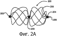
Фиг. 2А - вид расширенного каркаса устройства герметизации.FIG. 2A is a view of an expanded frame of a sealing device.
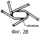
Фиг. 2В - вид спереди петли устройства герметизации.FIG. 2B is a front view of a loop of a sealing device.
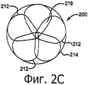
Фиг. 2С - вид спереди каркаса устройства герметизации.FIG. 2C is a front view of the frame of the sealing device.
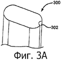
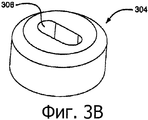
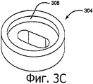
Фиги 3А-В - виды компонентов наматывающего патронаFigs 3A-B are views of the components of the reeling cartridge
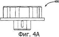
Фиг. 4А - вид сбоку наматывающего патрона.FIG. 4A is a side view of a winding cartridge.
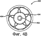
Фиг. 4В - вид сверху наматывающего патрона.FIG. 4B is a plan view of a reeling cartridge.
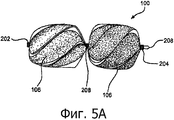
Фиг. 5А - вид сбоку расширенного оплетенного устройства герметизации.FIG. 5A is a side view of an expanded braided sealing device.
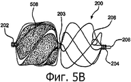
Фиг. 5В - вид сбоку расширенного частично оплетенного устройства герметизации.FIG. 5B is a side view of an expanded partially braided sealing device.
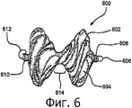
Фиг. 6 - вид сбоку варианта реализации самоцентрирующего устройства герметизации.FIG. 6 is a side view of an embodiment of a self-centering sealing device.
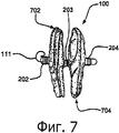
Фиг. 7 - вид сбоку развернутого устройства герметизацииFIG. 7 is a side view of a deployed sealing device
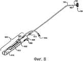
Фиг. 8 - перспективный вид системы доставки, включающей в себя рукоятку развертывания и прикрепленное устройство герметизации.FIG. 8 is a perspective view of a delivery system including a deployment handle and an attached sealing device.
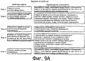
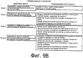
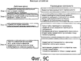
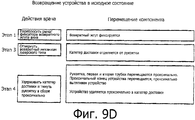
Фиг. 9A-D - блок-схемы алгоритма, описывающие работу системы доставки.FIG. 9A-D are flowcharts describing the operation of a delivery system.
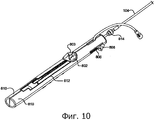
Фиг. 10 - перспективный вид рукоятки развертывания устройства герметизации.FIG. 10 is a perspective view of a deployment handle of a sealing device.
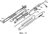
Фиг. 11 - перспективный вид рукоятки развертывания устройства герметизации в сборе.FIG. 11 is a perspective view of a deployment handle of a sealing assembly.
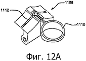
Фиг. 12А - вид сверху вниз варианта реализации первого линейного исполнительного механизма.FIG. 12A is a top-down view of an embodiment of a first linear actuator.
Фиг. 12В - вид сбоку варианта реализации первого линейного исполнительного механизма.FIG. 12B is a side view of an embodiment of a first linear actuator.
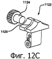
Фиг. 12С - вид сбоку варианта реализации первого линейного исполнительного механизма.FIG. 12C is a side view of an embodiment of a first linear actuator.
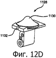
Фиг. 12D - вид сбоку варианта реализации первого линейного исполнительного механизма.FIG. 12D is a side view of an embodiment of a first linear actuator.
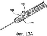
Фиг. 13А - перспективный вид варианта воплощения разблокировочного исполнительного механизма.FIG. 13A is a perspective view of an embodiment of an unlocking actuator.
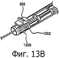
Фиг. 13В - перспективный вид варианта воплощения разблокировочного исполнительного механизма в активированном положении.FIG. 13B is a perspective view of an embodiment of an unlocking actuator in an activated position.
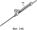
Фиг. 14А - перспективный вид варианта воплощения пружины.FIG. 14A is a perspective view of an embodiment of a spring.
Фиг. 14В - вид спереди варианта реализации первого линейного исполнительного механизма.FIG. 14B is a front view of an embodiment of a first linear actuator.
Фиг. 15 - вид спереди варианта реализации первого линейного исполнительного механизма с литой деталью пружины.FIG. 15 is a front view of an embodiment of a first linear actuator with a molded spring part.
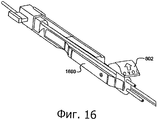
Фиг. 16 - перспективный вид пружинного механизма.FIG. 16 is a perspective view of a spring mechanism.
Осуществление изобретенияThe implementation of the invention
Первый вариант реализации касается устройства герметизации, имеющего расширяющийся каркас, образованный множеством проволок, продолжающихся от проксимального конца к дистальному концу каркаса и образующих проксимальную и дистальную петлю с герметизирующим элементом, по меньшей мере, частично инкапсулируя расширяющийся проволочный каркас.The first embodiment relates to a sealing device having an expandable cage formed by a plurality of wires extending from the proximal end to the distal end of the cage and forming a proximal and distal loop with a sealing element, at least partially encapsulating the expanding wire cage.
На фиг. 1 показан вариант реализации устройства 100 герметизации. Устройство 100 герметизации будет описано подробно ниже. Устройство 100 герметизации может быть размещено в третьей трубке 104. Третья трубка 104 содержит устройство 100 герметизации, первую трубку 102, вторую трубку 108, возвратный жгут 110 и фиксирующую петлю 111. Третья трубка 104 может быть изготовлена из материала Pebax® или из иного материала с соответствующими биологически совместимыми и механическими характеристиками. Может быть выбран непроницаемый для рентгеновского излучения материал. Третья трубка 104 может быть изготовлена с укрепляющей оплеткой или без нее для обеспечения изломоустойчивости и прочности выбранного варианта использования. Третья трубка 104 может также быть изготовлена с ренгенонепроницаемым маркирующим слоем или без него. Конструкция и материалы для изготовления третьей трубки 104 могут быть выбраны с учетом других характеристик, таких как изломоустойчивость, управляемость и снижение травматизма сосудов. Специалисту в данной области техники известно о существовании широкого многообразия материалов, которые могут быть использованы для осуществления данного изобретения. Третья трубка 104 может иметь любые размеры, но предпочтительно использовать трубку 10fr. внутренним диаметром примерно 0,048 мм и наружным диаметром примерно 0,33 мм. Третья трубка 104 может использоваться с проволочным направителем или без него и может включать в себя отверстие 103 быстрой замены. Наконечник первой трубки 104, предпочтительно должен быть изогнут, что помогает в ориентировании и доставки устройства 100 герметизации от места доступа к очагу заболевания с проволочным направителем или без него.In FIG. 1 shows an embodiment of a
На фиг. 1 также показана первая трубка 102. Как сказано выше, первая трубка 102 может быть размещена в третьей трубке 104. Первая трубка 102 может иметь любой наружный диаметр, но, предпочтительно, ее размер должен соответствовать внутренней просвету третьей трубки 104. Первая трубка 102 может быть изготовлена из материала Pebax® или из любого другого материала с соответствующими биологически совместимыми и механическими характеристиками. Первая трубка 102, предпочтительно, представляет собой трехпросветный катетер. Просветы могут иметь любую геометрическую форму, но, предпочтительно, должны иметь круглую или овальную форму или их комбинацию. Первая трубка 102 может быть использована для размещения и облегчения развертывания устройства 100 герметизации. Первая трубка 102 может быть использована во взаимодействии со второй трубкой 108, доставляя устройство 100 герметизации выдавливанием из дистального конца третьей трубки 104, как только устройство 100 герметизации достигло очага заболевания. Первая трубка 102 может также иметь функцию удерживания устройства 100 герметизации в системе доставки до момента окончательного развертывания устройства. Первая трубка 102 имеет отверстие 109 в самом крайнем положении дистального конца, позволяя фиксирующей петле 111 выступать вперед во время развертывания устройства. Отверстие 109 и выступающая фиксирующая петля 111 обеспечивают присоединение к системе доставки устройства. Фиксирующая петля 111 показана в ее расширенном состоянии до удержания ее в заданной форме. Первая трубка 102 может иметь обработанную поверхность или может быть покрыта материалом, повышающим биосовместимость или изменяющим или усиливающим поверхностное трение.In FIG. 1, the
Первая трубка 102 может вмещать вторую трубку 108. Вторая трубка 108, по существу, имеет каналикулярную конструкцию с овальным сечением и может иметь наружный диаметр, подходящий к внутреннему диаметру первой трубки 102. Предпочтительный диапазон наружного диаметра составляет примерно 1,27×0,68 мм с развальцованным дистальным концом. Вторая трубка 108 может быть изготовлена из любого подходящего биосовместимого материала, включая сюда полимеры или металлы. Предпочтительным материалом является РЕЕК (полиэфирэфиркетон). Вторая трубка 108 может быть использована для облегчения доставки и развертывания устройства 100 герметизации в очаге заболевания. Вторая трубка 108 продета через петли устройства 100 герметизации для удержания устройства 100 герметизации в системе доставки и обеспечения устойчивости во время развертывания устройства 100 герметизации. Петли устройства герметизации будут описаны дополнительно.The
Возвратный жгут 110 закольцован через два наименьших внутренних просвета первой трубки 102 и через проксимальную петлю устройства 100 герметизации, обеспечивая присоединение к системе доставки и возвращение в исходное состояние однажды развернутого устройства герметизации. Возвратный жгут 110 продолжается по всей длине первой трубки 102 за концы, которые выводятся на рукоятку, используемую для развертывания устройства 100 герметизации. Возвратный жгут 110 может быть изготовлен из любого биосовместимого материала достаточной прочности и размера. Предпочтительным материалом является ePTFE (расширяющийся политетрафторэтилен).The
Как показано на фиг. 2А, устройство 100 герметизации образовано проволочным каркасом 200. При размещении для доставки проволочный каркас 200 находится в выдвинутом положении на второй трубке 108 и внутри третьей трубки 104. Проволочный каркас 200 может иметь любой размер, подходящий для применения, но предпочтительные конечные наружные диаметры составляют 15, 20, 25 или 30 мм. Проволочный каркас 200 образуется замкнутой проволочной конструкцией. Для создания проволочного каркаса 200 может быть использовано любое количество проволок. Предпочтительно использовать пять проволок. Проволочный каркас 200 может быть образован из проволок, которые имеют эластичные характеристики, позволяющие проволочному каркасу 200 находиться в сжатом состоянии для базовой или торакоскопической доставки катетера и саморасширяться до индуцируемой «памятью» конфигурации, однажды расположенной на месте заболевания. Эластичная проволока может быть пружинной проволокой или проволокой, изготовленным из сплава NiTi (нитинол) с памятью формы или проволокой из суперэластичного сплава NiTi. Эластичная проволока также может быть проволокой типа DFT из сплава NiTi, содержащим различный металл в сердцевине. Предпочтительно, проволочный каркас 200 может быть образован из проволоки типа DFT, изготовленного из сплава NiTi и содержащего непроницаемый для рентгеновского излучения металл в сердцевине. После развертывания, проволочная конструкция приобретает форму развертывания без остаточной деформации.As shown in FIG. 2A, the
Проволочный каркас 200 и иные упомянутые проволочные конструкции образованы из эластичных проволочных материалов, которые имеют наружный диаметр 0,12-0,4 мм. В предпочтительном варианте реализации наружный диаметр составляет примерно 0.3 мм. Образованный проволочный каркас 200 содержит дистальный буфер 208, дистальную петлю 204, фиксирующую петлю 206, возможно, центральную петлю 203 и проксимальную петлю 202. На фиг. 2В показано положение эластичных проволок во время образования петель 202, 203 и 204 проволочного каркаса 200.The
На фиг. 2С показан диск, образованный при развертывании проволочного каркаса 200. Эластичные проволоки, образующие проволочный каркас 200 во время расширения, имеют форму лепестков 212. Заданная конфигурация эластичной проволоки проволочного каркаса 200 позволяет каркасу закручиваться вовремя развертывания. Данное закручивание образует лепестки 212. Развернутые лепестки 212 образуют проволочный каркас 200 с наружным диаметром 214. Развернутые лепестки 212, покрытые герметизирующим элементом 106 и образующие проксимальные и дистальные диски, будут описаны далее. Лепестки 212 оптимально образуют зоны 216 перекрытия для улучшения качества герметизации. Радиус лепестков 212 может быть максимально увеличен для уменьшения изгиба эластичной проволоки и уменьшения размера неподдерживаемых секций лепестков 212, что повысит качество герметизации устройства, уменьшит усталость металла на изгибах проволок и поможет уменьшить силу, вызывающую нагрузку на устройство. Развернутые лепестки 212 образуют диск с обеих сторон центральной петли 203. Развернутая конфигурация будет описана далее.In FIG. 2C shows a disk formed when the
Конструкция проволочного каркаса 200 может быть сформирована множеством средств, включающих в себя механизм наматывания с автоматическим натяжением проволок или ручное наматывание с нагрузочным натяжением каждой проволоки во время формирования. На фиг. 3А-С показаны ключевой центральный штифт 300 и заглушка 304, которые могут быть использованы для содействия в создании проволочного каркаса 200. Специалисту в данной области техники понятно, что существует множество подходящих материалов для использования в производстве или оснащении инструментом. Предпочтительным материалом для использования в формировании центрального штифта 300 является высокопрочная кобальтовая сталь. Предпочтительным материалом для использования в изготовлении заглушки 304 и наматывающего патрона является коррозионно-стойкая инструментальная сталь. Устройство наматывающего патрона будет описано ниже. Показанный на фиг. 3А ключевой центральный штифт 300 может иметь паз 302, который может быть использован для надежного крепления эластичной проволоки во время формирования устройства. Ключевой центральный штифт 300 может быть использован для направления эластичной проволоки через отверстие 306 в заглушке 304, конструктивные элементы которых показаны на фиг. 3В-С. Заглушка 304 предпочтительно образована с углублением 308 в заглушке для надежного крепления в наматывающем патроне. Эластичная проволока удерживается в пазу 302 и вставляется через отверстие 306 в заглушке 304, образуя буфер 208 и фиксирующую петлю 206. Ключевой центральный штифт 300 используется также для образования петель 202, 203 и 204. Во время создания устройства, после формирования буфера 208, эластичные проволоки могут наматываться вокруг ключевого центрального штифта 300, образуя дистальную петлю 202. Другие петли 203 и 204 могут быть образованы подобным образом. Ключевой центральный штифт 300 вставляется в заглушку 304, после чего эластичная проволока может быть вставлена в пазы наматывающего патрона.The design of the
Наматывающий патрон может быть использован для надежного крепления и образования эластичных проволок во время формирования и обработки устройства 100 герметизации. Типовой наматывающий патрон может быть изготовлен, как обычно принято в данной области техники. Материалы, используемые для изготовления такого наматывающего патрона, были указаны ранее. Предпочтительный вариант наматывающего патрона показан на фиг. 4А и 4В. На фиг. 4А показан вид сбоку наматывающего патрона 400. На фиг. 4В показан вид сверху предпочтительного варианта наматывающего патрона 400. Наматывающий патрон 400 содержит отверстие 402, которое имеет форму и размер, необходимые для удержания ключевого центрального штифта 300 и заглушки 304 во время формирования устройства. Пазы 404 в поверхности патрона используются для надежного крепления и формирования эластичных проволок в лепестках 212. Пазы 404 могут иметь любой диаметр, но предпочтительный размер должен вмещать наружный диаметр эластичной проволоки. В варианте воплощения, показанном на фиг. 5А, конструкция наматывающего патрона может быть использована для образования центральной петли 203, лепестка и проксимальной петли 204. Фасонная проволока может быть сжата при сборке наматывающего патрона, нагрета и обработана для придания заранее установленной формы, как широко известно в данной области техники.The winding cartridge can be used to securely fasten and form elastic wires during the formation and processing of the
На фиг. 5А показан вариант реализации устройства 100 герметизации, который представляет собой проволочный каркас 200 и герметизирующий элемент 106 в сборе. Герметизирующий элемент 106 может быть прикреплен к проволочному каркасу 200 клеящим веществом. Проволочный каркас 200 может быть покрыт клеящим веществом, например, фторированным этиленпропиленом (FEP) или иным подходящим клеящим материалом. Клеящий материал может быть нанесен посредством контактного покрытия, порошкового покрытия, нанесения покрытия способом погружения, покрытия, нанесенного методом распыления или любым другим соответствующим способом. Предпочтительным вариантом реализации является клеящий материал FEP, нанесенный методом электростатического порошкового покрытия. Герметизирующий элемент 106 может быть изготовлен из множества материалов, таких как DACRON®, полиэфир, полиэтилен, полипропилен, фторполимеры, полиуретан, вспененная пленка, силикон, нейлон, шелк, тонколистовой суперупругий материал, текстолит, полиэтилентерефталат (PET), коллаген, перикардная ткань или из иного биологически совместимого материала. В варианте воплощения герметизирующий элемент 106 может быть образован из тонкой пористой ePTFE (расширенного политетрафторэтилена) подложки. Герметизирующий элемент 106 предназначен для усиления герметизирующих характеристик устройства 100 герметизации путем обеспечения блокирования очага заболевания и наличием увеличивающейся пористой рабочей среды.In FIG. 5A, an embodiment of a
На фиг. 5А также показаны проксимальные, дистальные и центральные петли (202, 203 и 204), соответственно покрытые герметизирующим элементом 106 и обмотанные пленкой. Петли 202, 203 и 204 могут быть обмотаны пленкой для усиления прилипания герметизирующего элемента 106 к устройству. Пленка, используемая для обмотки петель 202, 203 и 204, может быть изготовлена из любого биологически совместимого тонкослойного материала, но предпочтительный материал должен содержать множество слоев тонкого пористого ePTFE, которые могут быть разделены одним или несколькими слоями непористого FEP.In FIG. 5A also shows the proximal, distal, and central loops (202, 203, and 204) respectively coated with a sealing
На фиг. 5В показан вариант реализации устройства 100 герметизации, которое включает в себя герметизирующий элемент 508, частично покрывающий проволочный каркас 200. Частично покрытое устройство может иметь или дистальный или проксимальный пузырь, покрытый частично или полностью герметизирующим элементом 508.In FIG. 5B shows an embodiment of a
Другим вариантом реализации устройства является самоцентрирующее устройство 600. Как показано на фиг. 6, самоцентрирующее устройство содержит проволочный каркас 602, аналогичный проволочному каркасу 200. Самоцентрирующее устройство 600 представляет собой проволочный каркас 602 и герметизирующий элемент 604 в сборе. Проволочный каркас 602 может быть образован по аналогичным технологиям и из материала как проволочный каркас 200, но не имеет центральной петли. Проволочный каркас 602 содержит дистальный буфер 606, покрытую дистальную петлю 608, покрытую проксимальную петлю 610 и фиксирующую петлю 612. Предварительно заданная конфигурация эластичной проволоки проволочного каркаса 602 позволяет каркасу крутиться при развертывании и создавать центрирующую зону 614 устройства 600 во время развертывания. Во время развертывания, центрирующая зона 614 может центрироваться сама на месте очага заболевания образуя диск, содержащий лепестки по обе стороны центрирующийся зоны 614 и очага заболевания.Another embodiment of the device is a self-centering
На фиг. 7 показано полностью развернутое устройство 100 герметизации. Во время развертывания третья трубка 104 прекращает воздействие на устройство 100 герметизации, и устройство возвращается в его предварительно заданную форму. Во время развертывания и фиксации фиксирующая петля 111 освобождается от давления первой трубки 102 и возвращается в предварительно заданную форму, закручиваясь от проксимальной петли 202. Таким образом, устройство фиксируется в развернутом состоянии. На фиг. 7 также показано положение проксимального и дистального дисков, элементов 702 и 704, относительно проксимальных, центральных и дистальных петель 202, 203 и 204 соответственно.In FIG. 7 shows a fully deployed sealing
На фиг. 8 показан перспективный вид устройства 100 герметизации, прикрепленного к системе доставки и включающего в себя первую трубку 102, третью трубку 104 и рукоятку для развертывания устройства 100 герметизации. На фиг. 8 дополнительно показаны первый линейный исполнительный механизм 802, промывочный порт 804, второй линейный исполнительный механизм 806, разблокировочный исполнительный механизм 808, рама 810 и желоб 812 по длине рамы. Первый линейный исполнительный механизм 802 может иметь различные конфигурации, описание которых будет приведено ниже.In FIG. 8 is a perspective view of a
На фиг. 9A-D показаны блок-схемы алгоритма, которые описывают перемещения различных компонентов системы доставки и прикрепленного устройства 100 герметизации во время использования. Загрузка устройства 100 герметизации в систему доставки до его использования описана на фиг. 9А. Компоненты рукоятки системы доставки показаны на Фигурах 8, 10 и 11. Врач может промывать систему доставки, присоединив шприц или другой подходящий инструмент к промывочному порту 804 и заполнив систему физраствором или любым иным соответствующим промывочным средством. Затем первый линейный исполнительный механизм 802 может быть введен в желоб 812 в раме 810, преодолевая сопротивление пружины 1100. Пружина 1100 может быть изготовлена, как показано, или может действовать как плоская пружина, ступенчатая пружина или может иметь любую иную форму, широко известную в данной области техники. Такое действие обеспечивает вращение сердечника рычага 1000 управления, как показано на Фигуре 11, вокруг скользящего стержня 1102 в сторону рамы 810. Данное движение освобождает первый линейный исполнительный механизм 802 от дистальной выемки 1104 в калибровочной вставке 1103 и препятствует проксимальному и дистальному перемещению второй трубки 108. Калибровочная вставка 1103 может быть изготовлена из любого материала с подходящими механическими характеристиками.In FIG. 9A-D are flowcharts that describe movements of various components of a delivery system and an attached
Типовые рукоятки, компоненты рукоятки, приборы или катетеры, используемые в медицинских устройствах доставки, могут содержать общеизвестные материалы, такие как аморфные термопластические материалы, которые включают в себя полиметилметакрилат (РММА или акриловый полимер), полистирол (PS), акрило-нитрил-бутадиен-стирол (ABS), поливинилхлорид (PVC), модифицированный полиэтилентерефталатгликоль (PETG), ацетобутират целлюлозы (CAB), полукристаллические пластики, которые включают в себя полиэтилен (РЕ), полиэтилен высокой плотности (HDPE), полиэтилен низкой плотности (LDPE или LLDPE), полипропилен (РР), полиметилпентен (РМР); аморфные технические термопласты, которые включают в себя поликарбонат (PC), полифениленоксид (РРО), модифицированный полифениленоксид (ModPPO), полифенилен эфир (РРЕ), модифицированный полифенилен эфир (ModPPE), термопластичный полиуретан (TPU), поликристалический технический термопласт, который включает в себя полиамид (РА или нейлон), полиоксиметилен (РОМ или ацеталь), полиэтилентерефталат (PET, термопластический полиэфир), полибутилентерефталат (РВТ, термопластический полиэфир), сверхвысокомолекулярный полиэтилен (UHMW-PE), термопластики с высокими эксплуатационными характеристиками, которые включают в себя полиимид (PI, имидизированный пластик), полиамидимид (PAI, имидизированный пластик), полибензимидазол (PBI, имидизированный пластик); аморфные термопластики с высокими эксплуатационными характеристиками, которые включают в себя полисульфон (PSU), полиэфиримид (PEI), полиэфирсульфон (PES), полиарилсульфон (PAS), полукристаллические термопластики с высокими эксплуатационными характеристиками, которые включают в себя полифениленсульфид (PPS), полиэфирэфиркетон (РЕЕК); и полукристаллические термопластики с высокими эксплуатационными характеристиками, фторполимеры, которые включают в себя фторированный этиленпропилен (FEP), этилен хлортрифторэтилен (ECTFE), этилен, этилен-тетрафторэтилен (ETFE), полихлортрифторэтилен (PCTFE), политетрафторэтилен (PTFE), поливинилиденфторид (PVDF), перфторалкоксил (PFA). Иные общеизвестные материалы, которые используются в медицине, включают в себя высокоэластичные кремнийорганические полимеры, термопластический эластомер или термопластический сополиэфир (РЕВАХ) и металлы, такие как нержавеющая сталь и никельтитановые сплавы.Typical grips, grip components, devices or catheters used in medical delivery devices may contain well-known materials, such as amorphous thermoplastic materials, which include polymethylmethacrylate (PMMA or acrylic polymer), polystyrene (PS), acrylic nitrile butadiene styrene (ABS), polyvinyl chloride (PVC), modified polyethylene terephthalate glycol (PETG), cellulose acetate butyrate (CAB), semi-crystalline plastics, which include polyethylene (PE), high density polyethylene (HDPE), nor polyethylene viscous density (LDPE or LLDPE), polypropylene (PP), polymethylpentene (PMP); amorphous technical thermoplastics, which include polycarbonate (PC), polyphenylene oxide (PPO), modified polyphenylene oxide (ModPPO), polyphenylene ether (PPE), modified polyphenylene ether (ModPPE), thermoplastic polyurethane (TPU), polycrystalline technical thermoplastic, which includes polyamide (PA or nylon), polyoxymethylene (POM or acetal), polyethylene terephthalate (PET, thermoplastic polyester), polybutylene terephthalate (PBT, thermoplastic polyester), ultra high molecular weight polyethylene (UHMW-PE), high-temperature thermoplastics performance characteristics, which include polyimide (PI, imidized plastic), polyamidimide (PAI, imidized plastic), polybenzimidazole (PBI, imidized plastic); high performance amorphous thermoplastics which include polysulfone (PSU), polyetherimide (PEI), polyethersulfone (PES), polyarylsulfone (PAS), high performance semi-crystalline thermoplastics that include polyphenylene sulfide (PPS), polyether ether ether ); and semi-crystalline thermoplastics with high performance, fluoropolymers, which include fluorinated ethylene propylene (FEP), ethylene chlorotrifluoroethylene (ECTFE), ethylene, ethylene tetrafluoroethylene (ETFE), polychlorotrifluoroethylene (PCTFE), polytetrafluoroethylene (PTFEDefende perfluoroalkoxyl (PFA). Other well-known materials that are used in medicine include highly elastic organosilicon polymers, a thermoplastic elastomer or thermoplastic copolyester (REVAC), and metals such as stainless steel and nickel-titanium alloys.
Дистальная выемка 1104 и проксимальная выемка 1106 в калибровочной вставке 1103 могут быть использованы для помощи в позиционировании первого линейного исполнительного механизма 802 в рамном желобе 812. Расстояние между двумя выемками 1104 и 1106, соответственно, может являться длиной устройства 100 герметизации, когда оно вытягивается по второй трубке 108 до загрузки в систему доставки. Калибровочная вставка 1103 может иметь размер, позволяющий вместить множество длин устройства и предпочтительно должно иметь размер примерно 22,28 см в длину с расстоянием между проксимальным концом дистальной выемки 1104 и проксимальным концом проксимальной выемки 1106 примерно 6,25-13,32 см. Выемки 1104 и 1106 могут иметь любую форму, но предпочтительно прямоугольную.The
Первый линейный исполнительный механизм 802 затем перемещается в среднее положение в желобе 812 в направлении проксимального конца рамы 812. Данное действие вызывает проксимальное перемещение первой трубки 102 и проксимальное перемещение проксимального конца устройства 100 герметизации, таким образом, вытягивая устройство 100 герметизации. Первый линейный исполнительный механизм 802 может иметь любую форму (рычаг, шаровую форму), но предпочтительно должен иметь форму, приспособленную для пальца врача. Первый линейный исполнительный механизм 802 может быть изготовлен из любого материала с соответствующими механическими характеристиками, но предпочтительно использовать материал подобный тому, который используется при изготовлении калибровочной вставки 1103. Свойством первого линейного исполнительного механизма 802 является наличие утопленных зубьев, сформированных в верхней части первого линейного исполнительного механизма 802 для надежного крепления возвратного жгута 110. Данное свойство является предпочтительным, но необязательным. Зубья могут представлять собой любой извилистый тракт или могут иметь любую форму, необходимую для создания сопротивления для возвратного жгута 110 во время загрузки, развертывания или возвращения в исходное состояние устройства 100 герметизации. Соответствующие выступающие зубья (не показаны) могут быть образованы в нижней поверхности фиксатора 803 возвратного жгута. Данные зубья могут заходить в зацепления и надежно удерживать возвратный жгут. Другие способы, общеизвестные в данной области техники и использующиеся для надежного крепления жгута небольшого диаметра, могут также быть использованы и будут описаны в деталях далее.The first
Первый линейный исполнительный механизм 802 затем перемещают далее проксимально до тех пор, пока устройство загружается в третью трубку 104. Во время данного действия пружина 1100 толкает первый линейный исполнительный механизм 802 и сердечник рычага 1000 управления в левую стенку желоба 812 в проксимальную выемку 1106 в калибровочной вставке 1103. Вторая трубка 108 освобождается и перемещается проксимально к устройству 100 герметизации и первой трубке 102. Когда первый линейный исполнительный механизм 802 перемещают проксимально, вторая трубка 108, устройство 100 герметизации и первая трубка 102 скользит или переводится в третью трубку 104. После того, как первый линейный исполнительный механизм 802 будет находиться в наиболее проксимальном положении, система может быть снова промыта физраствором с помощью описанного ранее способа.The first
Альтернативные варианты воплощений первого линейного исполнительного механизма 802 показаны на фиг. 12A-D. На фиг. 12А показан перспективный вид альтернативного первого линейного исполнительного механизма 1108 в фиксированном положении возвратного жгута. Линейный исполнительный механизм 1108 конструктивно аналогичен линейному исполнительному механизму 802, но отличается наличием стопорного кольца 1110 возвратного жгута и паза 1112 возвратного жгута. На фиг. 12В показан альтернативный вариант 1114 воплощения, который выполнен с колесиком 1116 с накаткой, которое регулирует длину линейного исполнительного механизма для облегчения манипулирования. Колесико 1116 с накаткой навинчивается на резьбовой стержень 1118, вокруг которого намотан возвратный жгут. Вариант 1114 воплощения также содержит паз 1120 возвратного жгута, через который возвратный жгут направляется перед надежным креплением вокруг резьбового стержня 1118. На фиг. 12С показан еще один вариант 1122 воплощения, в котором применяется боковая регулировка колесика 1124 с накаткой, вокруг которого намотан возвратный жгут, надежно присоединенный к исполнительному механизму 1122 путем вставки резьбового стержня 1124 в резьбовое отверстие (не показано) в стенке исполнительного механизма 1122. Перед протаскиванием возвратного жгута вокруг резьбового стержня 1124 возвратный жгут вставляется через паз 1126 возвратного жгута. Еще один вариант 1128 воплощения показан на фиг. 12D. Вариант 1128 воплощения показывает линейный исполнительный механизм с литым колесиком 1130 с накаткой. Данное колесико 1130 с накаткой продолжается немного за линейный исполнительный механизм для облегчения управления линейным исполнительным механизмом. Возвратный жгут вставляется через паз 1132 возвратного жгута и обматывается вокруг резьбового стержня (не показано). Литое колесико 1130 с накаткой затем надежно закрепляется на резьбовом стержне и надежно закрепляет возвратный жгут.Alternative embodiments of the first
Развертывание устройства 100 герметизации на месте очага заболевания показано на фиг. 9В. Первый линейный исполнительный механизм 802 перемещается дистально до упора. Данное перемещение вызывает дистальное перемещение первой трубки 102 и второй трубки 108 внутри третьей трубки 104. Линейный исполнительный механизм 802 должен затем быть перемещен вправо в желобе 812, преодолевая сопротивление пружины 1100. Когда линейный исполнительный механизм 802 перемещается вправо, сердечник рычага 1000 управления вращается по скользящему стержню 1102. Данное действие освобождает линейный исполнительный механизм 802 от проксимальной выемки 1106 в калибровочной вставке 1103. После такого действия линейный исполнительный механизм 802 далее перемещается дистально. Это вызывает дистальное перемещение первой трубки 102 и проксимальной петли 202 устройства 100 герметизации. Кроме того, данное действие предотвращает перемещение дистального конца устройства 100 герметизации. Первая трубка 102 направляет устройство из третьей трубки 104 для развертывания устройства на месте заболевания. Перемещаясь дистально к концу желоба 812, линейный исполнительный механизм 802 обеспечивает полное развертывание устройства герметизации. Специалисту в данной области техники понятно, что выполнение описанных выше этапов могло быть остановлено и возобновлено в обратном направлении в определенных точках, позволяя осуществить оптимальное позиционирование устройства 100 герметизации.The deployment of the
Фиксация устройства описана в блок-схеме алгоритма на фиг. 9С. Фиксатор 803 возвратного жгута будет отстегнут от первого исполнительно механизма 802. Врач управляет вторым линейным исполнительным механизмом 806 путем захвата прикрепленного разблокировочного исполнительного механизма 808, подавая его вперед к середине рамы 810. Второй линейный исполнительный механизм 806 может иметь любой размер или форму, но предпочтительный размер должен позволять ему помещаться в желоб 1002 в направлении продольной оси рамы 810. Линейный исполнительный механизм 806 снабжен разблокировочным исполнительным механизмом 808 посредством фитинговой защелки. Любые материалы крепления, такие как клей или наличие литой конструкции деталей, будут обеспечивать надежное крепление разблокирующего исполнительного механизма 808 к линейному исполнительному механизму 806. Материалы, предназначенные для изготовления как второго исполнительного механизма 806, так и разблокировочного исполнительного механизма 808, могут быть любыми материалами с соответствующими механическими свойствами, но, предпочтительно подобными тем материалам, что ранее упомянуты при изготовлении компонентов рукоятки. Разблокировочный исполнительный механизм 808 предназначен для обеспечения надежного захвата устройства пользователем. Захватывание может быть облегчено наличием выступов на боковых сторонах разблокировочного исполнительного механизма 808. Данные выступы могут быть изготовлены из подобного материала, что и разблокировочный исполнительным механизм 808 или могут быть выполнены из материала с высоким коэффициентом трения или из более пригодного материала, чем материал, из которого изготовлен разблокировочный исполнительный механизм 808. Данные выступы могут также быть изготовлены в форме решетки, шероховатой, рельефной или бороздчатой формы в сочетании с материалом, указанным выше, дополнительно способствуя захвату устройства. Данные конструктивные элементы на поверхности разблокировочного исполнительного механизма 808 могут также быть использованы для помощи в захватывании без использования захватывающих выступов и могут быть применены непосредственно на боковой поверхности второго линейного исполнительного механизма 806. Желоб 1002 может быть выполнен с возможностью иметь стопор для удержания второго линейного исполнительного механизма 806 в крайнем дистальном положении, пока устройство герметизации разблокируется. Предпочтительный вариант стопора показан на фиг. 10 и 11 и имеет рифленую конструкцию, но может быть также изготовлен в виде механического стопора. Желоб 1002 может иметь любую длину, но предпочтительная длина должна такой, которая является достаточной для передачи проксимального движения по ширине второго линейного исполнительного механизма 806 плюс примерно 3,18 см. Желоб 1002 может иметь любую форму, которая будет позволять вмещать второй линейный исполнительный механизм 806.The fixing of the device is described in the flowchart of FIG. 9C. The
Альтернативный вариант реализации второго линейного исполнительного механизма 806 показан на фиг. 13А и 13В. Вместо захватывающего разблокировочного исполнительного механизма 808 и активирующего второго линейного исполнительного механизма 806 вращающийся разблокировочный исполнительный механизм 1300 захватывается и поворачивается для разблокировки. Вращающийся разблокировочный исполнительный механизм 1300 может иметь щель 1302, которая будет предотвращать перемещение вперед первого линейного исполнительного механизма 802. При вращении разблокировочный исполнительный механизм 1300 выполняет такие же действия, как разблокировочный исполнительный механизм 806, показанный на Фигуре 10.An alternative embodiment of the second
Как только второй линейный исполнительный механизм 808 будет захвачен, врач может переместить второй линейный исполнительный механизм 806 проксимально. Данное действие приводит к проксимальному перемещению третьей трубки 104, сердечника рычага 1000 управления, калибровочной вставки 1103 и второй трубки 108. Вторая трубка 108 перемещается проксимально между петлями устройства. Альтернативным способом выполнения данного действия является наличие механизма кручения на дистальном конце рукоятки вместо второго линейного исполнительного механизма 806. Механизм кручения будет снабжен желобом, что позволяет осуществить аналогичное перемещение третьей трубки 104, сердечника рычага 1000 управления, калибровочной вставки 1103 и второй трубки 108 подобно действию второго линейного исполнительного механизма 806.Once the second
Как только происходит разблокировка, фиксатор 803 возвратного жгута закручивается и извлекается из первого линейного исполнительного механизма 802 и вытягивается до освобождения возвратного жгута 110 из системы доставки. Возвратный жгут 110 прикрепляется к фиксатору 803 возвратного жгута за один конец. Возвратный жгут 110 может быть изготовлен из любого материала с соответствующими механическими характеристиками, такого как Kevlar®, гибкий металлический провод, полимеры и тому подобные. Предпочтительным материалом для изготовления возвратного жгута 110 является ePTFE волокно. Фиксатор 803 возвратного жгута может быть выполнен в различных формах и размерах. Возможные фиксаторы возвратного жгута могут находиться в желобе, по которому происходит возврат в исходное состояние линейного исполнительного механизма 802. В одной из компоновок возвратный жгут надежно фиксируется через желоб или отверстие в оси колесика с накаткой, расположенного в линейном исполнительном механизме 802 и закрепляемого вращением колесика с накаткой. Альтернативным вариантом конструкции является наличие защелки, которая фиксирует возвратный жгут между фиксатором и линейным исполнительным механизмом 802, используя трение. Предпочтительной конструкцией является наличие надежного крепления возвратного жгута зубцами фиксатора возвратного жгута, как показано на Фигуре 11.As soon as the unlocking occurs, the
Материалы, подходящие для изготовления фиксатора 803 возвратного жгута, подобны тем, которые используются для рамы 810 и других компонентов рукоятки. Как ранее упоминалось, фиксатор 803 возвратного жгута предпочтительно имеет зубья или выступы, которые соответствуют зубцам в линейном исполнительном механизме 802 для захвата возвратного жгута 110. Фиксатор 803 возвратного жгута может быть выполнен во множестве форм для надежной фиксации возвратного жгута 110. Предпочтительная конфигурация включает в себя отверстия через фиксатор 803 возвратного жгута, что позволяет протянуть через него возвратный жгут 110 и затянуть его узлом. После закручивания фиксатора 803 возвратного жгута возвратный жгут 110 удаляется из системы доставки путем его вытягивания.Materials suitable for manufacturing the
Перед выполнением четвертого этапа, описанного на фиг. 9С, устройство 100 герметизации может быть возвращено в исходное состояние, как описано в блок-схеме алгоритма на фиг. 9D. Фиксатор 803 возвратного жгута может защелкиваться на первом линейном исполнительном механизме 802. Это фиксирует на месте возвратный жгут 110. Затем врач перемещает первый исполнительный механизм 802 в правую стенку желоба 812. Первый линейный исполнительный механизм 802 перемещается в желобе 812 вправо, сжимая пружину 1100, в то время как сердечник рычага 1000 управления вращается по скользящему стержню 1102 в правую сторону рукоятки. Скользящий стержень 1102 имеет предпочтительно круглое сечение, но специалистам в данной области техники понятно, что существует множество приемлемых форм сечений (например, прямоугольное или треугольное). Скользящий стержень 1102 также может быть выполнен в форме корончатой пружины 1400, как показано на фиг. 14А и В. Пружина могла быть вставлена в желоб 1402 через линейный исполнительный механизм, позволяя перемещать линейный исполнительный механизм по всей длине. Альтернативный вариант воплощения пружины 1100 может представлять собой литую пружину, которая является неотъемлемой частью 1500 первого линейного исполнительного механизма 802, как показано на фиг. 15. Другой вариант реализации пружины 1100 показан на фиг. 16. В данном варианте реализации, пружина 1600 прикрепляется к раме 810 и толкает первый линейный исполнительный механизм 802 в ключевые позиции. Как сказано выше, специалисту в данной области техники известно, какие соответствующие материалы могут быть использованы как для пружины, так и для литой части. Первый линейный исполнительный механизм 802 освобождается из дистальной выемки 1104, при этом предотвращается перемещение второй трубки 108. Данный первый линейный исполнительный механизм перемещается проксимально врачом, вызывая проксимальное перемещение первой трубки 102. Данное движение перемещает проксимальный конец устройства 100 герметизации, проксимально растягивая устройство 100 и позволяя ему войти в третью трубку 104.Before performing the fourth step described in FIG. 9C, the
ПримерыExamples
Без ограничения объема изобретения ниже приведены примеры, показывающие осуществление и/или использование различных вариантов осуществления.Without limiting the scope of the invention, the following are examples showing the implementation and / or use of various embodiments.
Пример 1Example 1
Устройство герметизации, подобное показанному на фиг. 1, было изготовлено с использованием следующих компонентов и процесса сборки.A sealing device similar to that shown in FIG. 1, was manufactured using the following components and assembly process.
Расширяющийся политетрафторэтилен был получен со следующими характеристиками:Expandable polytetrafluoroethylene was obtained with the following characteristics:
точка начала кипения метанола - 1 фунт/кв. дюймboiling point of methanol is 1 psi. inch
масса/ площадь - 2,2 грамм/кв. мmass / area - 2.2 grams / sq. m
продольная максимальная нагрузка - 1,6 кг/дюймlongitudinal maximum load - 1.6 kg / inch
толщина - 0,0003 дюймаthickness - 0.0003 inches
прочность на растяжение продольной матрицы - 92000 фунт/кв. дюйм.the tensile strength of the longitudinal matrix is 92,000 psi. inch.
Для определения вышеуказанных характеристик были использованы следующие способы испытаний и оборудование: точка начала кипения метанола была измерена изготовленным по заказу механизмом с диаметром основания 1 дюйм, со скоростью отслеживания графика нагрузки 0,2 фунт/кв. дюйм/с, при этом жидкая среда представляла собой метанол. Длина и ширина материала были измерены металлической линейкой. Масса/площадь была измерена с помощью весов (модель GF-400 Top Loader Balance, ANG, San Jose С А.), используя образец 36×5 дюймов. Продольная максимальная нагрузка была измерена с использованием установки для испытаний материалов (Модель 5564, Instron, Grove City, РА), оборудованной датчиком веса 10 кг. Длина испытываемой части образца составляла 1 дюйм, и скорость ползуна составляла 25 мм/мин. Ширина образца составляла 1 дюйм. Измерения при испытаниях на продольное растяжение были выполнены по длине материала. Толщина была измерена с использованием калибромера (Mitutoyo Digital Indicator 547-400) с диаметром основания дюйма. Значения прочности на растяжение (MTS) продольной матрицы были подсчитаны с помощью приведенного ниже уравнения: Плотность была рассчитана по формуле плотность = масса/объемTo determine the above characteristics, the following test methods and equipment were used: the methanol boiling point was measured by a custom-made mechanism with a base diameter of 1 inch, with a load graph tracking rate of 0.2 psi. inch / s, while the liquid medium was methanol. The length and width of the material were measured with a metal ruler. Weight / area was measured using a balance (GF-400 Top Loader Balance, ANG, San Jose C A.) using a 36 x 5 inch sample. The longitudinal maximum load was measured using a material testing facility (Model 5564, Instron, Grove City, RA) equipped with a 10 kg weight sensor. The length of the test portion of the sample was 1 inch and the slide speed was 25 mm / min. The width of the sample was 1 inch. Measurements in longitudinal tensile tests were performed along the length of the material. Thickness was measured using a caliper (Mitutoyo Digital Indicator 547-400) with a base diameter inches. The tensile strength (MTS) values of the longitudinal matrix were calculated using the equation below: Density was calculated using the formula density = mass / volume
Расширяющийся политетрафторэтилен с тонким слоем FEP (фторированный этиленпропилен) был получен со следующими характеристиками: масса/площадь - 36,1 грамм/кв. метр продольная максимальная нагрузка - 12,6 кг/дюйм поперечная максимальная нагрузка - 0,3 кг/дюйм толщина - 0,0012 дюймаExpanded thin-layer polytetrafluoroethylene FEP (fluorinated ethylene propylene) was obtained with the following characteristics: mass / area - 36.1 grams / sq. meter longitudinal maximum load - 12.6 kg / inch transverse maximum load - 0.3 kg / inch thickness - 0.0012 inch
Для определения вышеуказанных свойств были использованы следующие способы испытаний и оборудование: материал был взвешен на точных аналитических весах (модель GF-400 Top Loader Balance, ANG, San Jose CA.) с использованием пробного участка образца 36×1 дюйм. Длина и ширина материала были измерены металлической линейкой. Толщина материала была измерена цифровым калибромером (Mitutoyo Digital Indicator 547-400) с диаметром основания дюйма. Максимальная поперечная нагрузка была измерена с использованием установки для испытаний материалов (модель 5564, Instron, Grove City, РА), оборудованной датчиком веса 10 кг. Ширина образца составляла 1 дюйм, расчетная длина составляла 1 дюйм, и скорость ползуна составляла 25 мм/мин. Максимальная нагрузка была измерена с использованием установки для испытаний материалов (Модель 5564, Instron, Grove City, РА), оборудованной датчиком веса 200 кг. Ширина образца составляла 1 дюйм, расчетная длина составляла 1 дюйм, и скорость ползуна составляла 25 мм/мин. Измерения при испытании на продольное растяжение были выполнены по длине материала, и измерения при испытании на поперечное растяжение были выполнены в направлении, перпендикулярном длине.The following test methods and equipment were used to determine the above properties: the material was weighed on an accurate analytical balance (model GF-400 Top Loader Balance, ANG, San Jose CA.) using a 36 × 1-inch sample sample section. The length and width of the material were measured with a metal ruler. The material thickness was measured with a digital caliper (Mitutoyo Digital Indicator 547-400) with a base diameter inches. The maximum lateral load was measured using a material testing facility (Model 5564, Instron, Grove City, RA) equipped with a 10 kg weight sensor. The width of the sample was 1 inch, the calculated length was 1 inch, and the slide speed was 25 mm / min. The maximum load was measured using a material testing facility (Model 5564, Instron, Grove City, RA) equipped with a 200 kg weight sensor. The width of the sample was 1 inch, the calculated length was 1 inch, and the slide speed was 25 mm / min. Measurements in the longitudinal tensile test were performed along the length of the material, and measurements in the transverse tensile test were performed in a direction perpendicular to the length.
Дистальная петля была образована посредством получения первого отрезка проволоки из тянутой проволоки из нитинола с сердечником с добавлением 10% платины (Fort Wayne Metals, Fort Wayne, IN.) диаметром примерно 0,23 мм. Данная проволока была отмаркирована как «первая проволока». Свободный конец первой проволоки был сдвоен, создавая петлю с открытым концом, и данная петля с открытым концом была вставлена в заглушку. Данная заглушка затем была насажена на ключевой центральный шток. Заглушка имеет отверстие по центру для размещения ключевого центрального штифта и имеет отличительные характеристики, которые позволяют ей оставаться надежно закрепленной на наматывающим патроне. Ключевой центральный штифт (основная ось примерно 0,51 мм, малая ось примерно 0,25 мм и длина около 10,16 мм) был затем вставлен в центр наматывающего патрона. Ключевой центральный штифт был изготовлен из высокопрочной стали (Super Cobalt HSS Tool Bit, MSC#56424278, Seco Fagersta). Данная сталь была подвергнута отпуску согласно инструкциям производителя при температуре 1475°F в течение часа. Наматывающий патрон и заглушка были изготовлены из коррозионно-стойкой инструментальной стали на собственном производстве.A distal loop was formed by producing a first stretched wire of a nitinol drawn wire with a core supplemented with 10% platinum (Fort Wayne Metals, Fort Wayne, IN.) With a diameter of about 0.23 mm. This wire has been marked as “first wire”. The free end of the first wire was doubled, creating a loop with an open end, and this loop with an open end was inserted into the plug. This plug was then mounted on a key central stock. The cap has a hole in the center to accommodate the key center pin and has distinctive characteristics that allow it to remain securely attached to the winding cartridge. A key center pin (main axis of about 0.51 mm, minor axis of about 0.25 mm and length of about 10.16 mm) was then inserted into the center of the winding cartridge. The key center pin was made of high strength steel (Super Cobalt HSS Tool Bit, MSC # 56424278, Seco Fagersta). This steel was tempered according to the manufacturer's instructions at 1475 ° F for an hour. The winding chuck and plug were made of corrosion-resistant tool steel in-house.
Второй отрезок подобной проволоки был получен из тянутой проволоки из нитинола с сердечником и отмаркирован как «пятая проволока». Первая, пятая и три дополнительных проволоки были натянуты грузиками, прикрепленными к концам проволок. Первая проволока и пятая проволока затем были намотаны на свободный конец первой проволоки на один полный оборот.Три дополнительные проволоки были размещены в наматывающем патроне, и все пять проволок были намотаны на свободный конец первой проволоки на высоте примерно 1,98 мм.The second segment of such a wire was obtained from a drawn wire made of nitinol with a core and marked as the "fifth wire". The first, fifth and three additional wires were pulled by weights attached to the ends of the wires. The first wire and the fifth wire were then wound onto the free end of the first wire one full revolution. Three additional wires were placed in the winding cartridge and all five wires were wound on the free end of the first wire at a height of about 1.98 mm.
Дистальный диск затем был образован разделением пяти проволок и надежным их креплением в радиальных пазах вокруг периферийного края наматывающего патрона. Радиус составлял 15 мм. Каждый провод образовывал один лепесток дистального диска. Радиус кривизны лепестков был максимально большим, чтобы свести к минимуму угол резкого изгиба проволоки.The distal disk was then formed by the separation of five wires and their reliable fastening in radial grooves around the peripheral edge of the winding cartridge. The radius was 15 mm. Each wire formed one lobe of the distal disc. The radius of curvature of the petals was as large as possible to minimize the angle of sharp bending of the wire.
Центральная петля было образована путем объединения проволок и их вращением вокруг свободного конца первой проволоки и ключевого центрального штифта на высоте примерно 1,98 мм. Данные проволоки затем были разделены и надежно закреплены в радиальных пазах вокруг периферийного края наматывающего патрона, создавая проксимальный диск радиусом 15 мм.The central loop was formed by combining the wires and rotating them around the free end of the first wire and the key center pin at a height of about 1.98 mm. These wires were then separated and firmly fixed in radial grooves around the peripheral edge of the winding cartridge, creating a proximal disk with a radius of 15 mm.
Проксимальная петля снова была сформирована путем объединения пяти проволок и их наматывания вокруг свободного конца первой проволоки и ключевого центрального штифта на высоте около 1,98 мм. Пять проволок затем были разделены и надежно закреплены металлической пластинкой, изготовленной из нержавеющей стали, размещенной поверх проволок и закрепленной винтами. Свободный конец первой проволоки затем был намотан на один оборот вокруг штифта диаметром примерно 3,18 мм, изготовленного из нержавеющей стали, и надежно закреплен подобно другим пятью проволокми.The proximal loop was again formed by combining the five wires and winding them around the free end of the first wire and the key center pin at a height of about 1.98 mm. The five wires were then separated and securely fastened with a stainless steel plate placed on top of the wires and secured with screws. The free end of the first wire was then wound one revolution around a pin of approximately 3.18 mm diameter made of stainless steel and securely fastened like the other five wires.
Данный патрон с герметизирующим устройством затем был удален из стабилизирующего приспособления и помещен в печь (электрическая конвекционная печь с принудительной подачей воздуха Blue MSPX), и данные проволоки были термически обработаны, как принято в данной области техники. Устройство и патрон затем были закалены в воде. Закрепленные проволоки были отсоединены от крепежной пластинки, и устройство было охлаждено и отсоединено от патрона и ключевого центрального штифта. Устройство затем было помещено на ровный кусок РЕЕК (полиэфирэфиркетон) и выровнено вручную по наружному диаметру дистальной петли. Фиксирующая петля была выровнена вручную более чем на один полный оборот и протянуто через проксимальную и центральную петли.This cartridge with a sealing device was then removed from the stabilizer and placed in a furnace (Blue MSPX electric convection oven with forced air supply), and these wires were heat treated as is conventional in the art. The device and cartridge were then water quenched. The fixed wires were disconnected from the mounting plate, and the device was cooled and disconnected from the cartridge and key center pin. The device was then placed on an even piece of PEEK (polyetheretherketone) and manually aligned with the outer diameter of the distal loop. The locking loop was manually aligned for more than one full revolution and extended through the proximal and central loops.
Устройство было перемещено из РЕЕК оправки на ключевой рабочий сердечник овального сечения из нержавеющей стали. Данный сердечник был изготовлен из гладкого провода, выполненного из нержавеющей стали (Ft. Wayne Metals, Fort Wayne, In.) с овальным поперечным сечением для получения угла 45° закручивания по часовой стрелке между проксимальной петлей и центральной петлей и второго угла 45° закручивания по часовой стрелке между центральной петлей и дистальной петлей.The device was moved from the PEEK mandrel to the oval stainless steel key core. This core was made of smooth stainless steel wire (Ft. Wayne Metals, Fort Wayne, In.) With an oval cross-section to obtain a 45 ° angle of rotation clockwise between the proximal loop and the central loop and a second 45 ° angle of rotation clockwise between the central loop and the distal loop.
Рабочий сердечник и устройство затем были помещены в стабилизирующее приспособление, которое было установлено в FEP установку для нанесения порошкового покрытия FEP (С-30, ElectrostaticTechnology, Inc., Bradford, CN) до получения полного покрытия. Излишек FEP порошка был удален из устройства. FEP был отвакуумирован из фиксирующей петли, рабочего сердечника и буфера. Рабочий сердечник и устройство были удалены из стабилизирующего приспособления, помещены в печь и подвергнуты обжигу для получения FEP покрытия, известного в данной области техники.The working core and device were then placed in a stabilizing device that was installed in the FEP FEP powder coating unit (C-30, Electrostatic Technology, Inc., Bradford, CN) until complete coverage. Excess FEP powder was removed from the device. FEP was evacuated from the retaining loop, the working core and the buffer. The working core and device were removed from the stabilizing device, placed in an oven, and calcined to obtain an FEP coating known in the art.
Был взят полый сердечник с оболочкой (из нержавеющей стали наружным диаметром 35,99 мм и длиной 76,2 см). Расширяющийся политетрафторэтиленовый материал с шириной зазора 22,22 мм был взят и загружен на механизм обвертывания посредством спиральной навивки. Данный механизм был изготовлен на месте для обвертывания PTFE (политетрафторэтилена) под любым необходимым углом, с требуемыми натяжением и скоростью. Сердечник был загружен на механизм обвертывания, и полый сердечник был обвернут данным материалом три раза по длине окружности. Затем сердечник был обвернут данным материалом под углом примерно 8° по длине сердечника. Направление обвертывания было реверсивным, при этом материал обвертывался под тем же углом. Третий и четвертый слои были нанесены тем же способом со смещением стыковых линий. Сердечник был удален из механизма обвертывания, помещен в печь, и подвергнут обжигу при температуре 370°С в течение 45 минут. Обернутый сердечник был извлечен из печи и охлажден до комнатной температуры. Готовая PTFE трубка была удалена из сердечника.A hollow core with a shell (stainless steel with an outer diameter of 35.99 mm and a length of 76.2 cm) was taken. An expanding polytetrafluoroethylene material with a gap width of 22.22 mm was taken and loaded onto the wrapping mechanism by spiral winding. This mechanism was made in place for wrapping PTFE (polytetrafluoroethylene) at any desired angle, with the required tension and speed. The core was loaded onto the wrapping mechanism, and the hollow core was wrapped with this material three times along the circumference. Then the core was wrapped with this material at an angle of about 8 ° along the length of the core. The wrapping direction was reversible, while the material was wrapped at the same angle. The third and fourth layers were applied in the same way with the displacement of the butt lines. The core was removed from the wrapping mechanism, placed in an oven, and calcined at a temperature of 370 ° C. for 45 minutes. The wrapped core was removed from the oven and cooled to room temperature. The finished PTFE tube has been removed from the core.
Данная PTFE трубка затем была отрезана по размеру примерно 140 мм и растянута вручную на требуемую длину 155 мм. PTFE трубка затем была натянута на раму. PTFE трубка затем была обжата на центральной петле и затем обжата на дистальной и проксимальной петлях.This PTFE tube was then cut to approximately 140 mm in size and manually extended to a desired length of 155 mm. The PTFE tube was then pulled onto the frame. The PTFE tube was then crimped on the central loop and then crimped on the distal and proximal loops.
Петли, начиная с центральной петли, были затем обернуты четыре раза расширяющимся политетрафторэтиленом с тонким слоем FEP (фторированный этиленпропилен). Обмотанные петли были припаяны на месте паяльником. PTFE трубка была затем нагрета при температуре 320°С в течение 3 минут и выровнена по наружным точкам проксимальной и дистальной петель. Устройство было удалено из сердечника.The loops, starting with the center loop, were then wrapped four times with expandable thin-layer polytetrafluoroethylene FEP (fluorinated ethylene propylene). The wound loops were soldered in place with a soldering iron. The PTFE tube was then heated at 320 ° C for 3 minutes and aligned with the outside points of the proximal and distal loops. The device has been removed from the core.
Пример 2Example 2
Устройство герметизации, подобно устройству, показанному на фиг. 6, было изготовлено с использованием следующих компонентов и процесса сборки.A sealing device, like the device shown in FIG. 6, was manufactured using the following components and assembly process.
Расширяющийся политетрафторэтилен и расширяющийся политетрафторэтилен с тонким слоем FEP (фторированный этиленпропилен) аналогичны материалам, описанным в Примере 1.Expandable thin-layer polytetrafluoroethylene and expandable polytetrafluoroethylene (FEP) (fluorinated ethylene propylene) are similar to the materials described in Example 1.
Дистальная петля была образована посредством получения первого отрезка проволоки из тянутой проволоки из нитинола с сердечником с добавлением 10% платины (Fort Wayne Metals, Fort Wayne, IN.) диаметром примерно 0,23 мм. Данная проволока была отмаркирована как «первая проволока». Свободный конец первой проволоки был сдвоен, создавая петлю с открытым концом, и данная петля с открытым концом была вставлена в заглушку. Данная заглушка затем была насажена на ключевой центральный шток. Заглушка имеет отверстие по центру для размещения ключевого центрального штифта и имеет отличительные характеристики, которые позволяют ей оставаться надежно закрепленной на наматывающим патроне. Ключевой центральный штифт (основная ось примерно 5,79 мм, малая ось примерно 0,25 мм и длина около 10,16 мм) был затем вставлен в центр наматывающего патрона. Ключевой центральный штифт был изготовлен из высокопрочной стали (Super Cobalt HSS Tool Bit, MSC#56424278, Seco Fagersta). Наматывающий патрон и заглушка были изготовлены из коррозионно-стойкой инструментальной стали на собственном производстве.A distal loop was formed by producing a first stretched wire of a nitinol drawn wire with a core supplemented with 10% platinum (Fort Wayne Metals, Fort Wayne, IN.) With a diameter of about 0.23 mm. This wire has been marked as “first wire”. The free end of the first wire was doubled, creating a loop with an open end, and this loop with an open end was inserted into the plug. This plug was then mounted on a key central stock. The cap has a hole in the center to accommodate the key center pin and has distinctive characteristics that allow it to remain securely attached to the winding cartridge. A key center pin (main axis of about 5.79 mm, minor axis of about 0.25 mm and length of about 10.16 mm) was then inserted into the center of the winding cartridge. The key center pin was made of high strength steel (Super Cobalt HSS Tool Bit, MSC # 56424278, Seco Fagersta). The winding chuck and plug were made of corrosion-resistant tool steel in-house.
Второй отрезок подобной проволоки был получен из тянутой проволоки из нитинола с сердечником и отмаркирован как «пятая проволока». Первая, пятая и три дополнительных проволоки были натянуты грузиками, прикрепленными к концам проволок. Первая проволока и пятая проволока затем были намотаны на свободный конец первой проволоки на один полный оборот. Три дополнительных проволоки были размещены в наматывающем патроне, и все пять проволок были намотаны на свободный конец первой проволоки на высоте примерно 1,98 мм.The second segment of such a wire was obtained from a drawn wire made of nitinol with a core and marked as the "fifth wire". The first, fifth and three additional wires were pulled by weights attached to the ends of the wires. The first wire and fifth wire were then wound onto the free end of the first wire one full revolution. Three additional wires were placed in a winding cartridge, and all five wires were wound on the free end of the first wire at a height of about 1.98 mm.
Устройство затем было образовано разделением пяти проволок и надежным их креплением в радиальных пазах вокруг периферийного края наматывающего патрона. Радиус составлял 15 мм. Каждая проволока была намотана на полный оборот вокруг наматываемого патрона.The device was then formed by separating the five wires and securing them securely in radial grooves around the peripheral edge of the winding cartridge. The radius was 15 mm. Each wire was wound a full revolution around the wound cartridge.
Проксимальная петля снова была сформирована путем объединения пяти проволок и их наматывания вокруг свободного конца первой проволоки и ключевого центрального штифта на высоте около 1,981 мм. Пять проволок затем были разделены и надежно закреплены металлической пластинкой, изготовленной из нержавеющей стали, размещенной поверх проволок и закрепленной винтами. Свободный конец первой проволоки затем был намотан на один оборот вокруг штифта диаметром примерно 3,18 мм, изготовленного из нержавеющей стали, и надежно закреплен подобно другим пяти проволокам.The proximal loop was again formed by combining the five wires and winding them around the free end of the first wire and the key center pin at a height of about 1,981 mm. The five wires were then separated and securely fastened with a stainless steel plate placed on top of the wires and secured with screws. The free end of the first wire was then wound one revolution around a pin with a diameter of about 3.18 mm made of stainless steel and securely fastened like the other five wires.
Данный патрон с герметизирующим устройством затем был удален из стабилизирующего приспособления и помещен в печь (электрическая конвекционная печь с принудительной подачей воздуха Blue MSPX), и данные проволоки были частично термически обработаны, как принято в данной области техники. Устройство и патрон затем были закалены в воде. Закрепленные проволоки были отсоединены от крепежной пластинки, и устройство было охлаждено и отсоединено от патрона и ключевого центрального штифта. Устройство затем было помещено на ровный кусок РЕЕК (полиэфирэфиркетон) и выровнено вручную по наружному диаметру дистальной петли. Фиксирующая петля была выровнена вручную более чем на один полный оборот и протянуто через проксимальную и центральную петли.This cartridge with a sealing device was then removed from the stabilizer and placed in a furnace (Blue MSPX electric convection oven with forced air supply), and these wires were partially heat-treated, as is customary in the art. The device and cartridge were then water quenched. The fixed wires were disconnected from the mounting plate, and the device was cooled and disconnected from the cartridge and key center pin. The device was then placed on an even piece of PEEK (polyetheretherketone) and manually aligned with the outer diameter of the distal loop. The locking loop was manually aligned for more than one full revolution and extended through the proximal and central loops.
Устройство было перемещено из РЕЕК оправки на ключевой сердечник перемещения овального сечения из нержавеющей стали. Данный сердечник был изготовлен из гладкого провода, выполненного из нержавеющей стали (Ft. Wayne Metals, Fort Wayne, In.) с овальным поперечным сечением. Данное устройство было, затем частично удалено из одного конца сердечника перемещения. Удаленный конец устройства был закручен по часовой стрелке приблизительно на 180° и репозиционирован на сердечнике перемещения. Устройство и сердечник перемещения были помещены в печь (электрическая конвекционная печь с принудительной подачей воздуха Blue MSPX), где данные провода были термически обработаны, как известно в данной области техники.The device was moved from the PEEK mandrel to the oval stainless steel key core. This core was made of smooth wire made of stainless steel (Ft. Wayne Metals, Fort Wayne, In.) With an oval cross-section. This device was then partially removed from one end of the displacement core. The remote end of the device was twisted clockwise approximately 180 ° and repositioned on the displacement core. The transfer device and core were placed in an oven (Blue MSPX electric forced-air convection oven), where these wires were thermally processed as is known in the art.
Сердечник перемещения и устройство затем были помещены в стабилизирующее приспособление, которое было установлено в FEP установку для нанесения порошкового покрытия FEP (С-30, ElectrostaticTechnology, Inc., Bradford, CN) до получения полного покрытия. Излишек FEP порошка был удален из устройства. FEP был отвакуумирован из фиксирующей петли, рабочего сердечника и буфера. Сердечник перемещения и устройство были удалены из стабилизирующего приспособления, помещены в печь и подвергнуты обжигу для получения FEP покрытия, известного в данной области техники.The transfer core and the device were then placed in a stabilizing device that was installed in the FEP FEP powder coating unit (C-30, Electrostatic Technology, Inc., Bradford, CN) until complete coverage. Excess FEP powder was removed from the device. FEP was evacuated from the retaining loop, the working core and the buffer. The transfer core and device were removed from the stabilizing device, placed in an oven, and fired to obtain an FEP coating known in the art.
Был взят полый сердечник с оболочкой (из нержавеющей стали наружным диаметром 35,99 мм и длиной 76,2 см). Расширяющийся политетрафторэтиленовый материал с шириной зазора 22,24 мм был взят и загружен на механизм обвертывания посредством спиральной навивки. Данный механизм был изготовлен на месте для обвертывания PTFE (политетрафторэтилена) под любым необходимым углом, с требуемыми натяжением и скоростью. Сердечник был загружен на механизм обвертывания, и полый сердечник был обвернут данным материалом три раза по длине окружности. Затем сердечник был обвернут данным материалом под углом примерно 8° по длине сердечника. Направление обвертывания было реверсивным, при этом материал обвертывался под тем же углом. Третий и четвертый слои были нанесены тем же способом со смещением стыковых линий. Сердечник был удален из механизма обвертывания, помещен в печь, и подвергнут обжигу при температуре 370°С в течение 45 минут.Обернутый сердечник был извлечен из печи и охлажден до комнатной температуры. Готовая ePTFE трубка была удалена из сердечника.A hollow core with a shell (stainless steel with an outer diameter of 35.99 mm and a length of 76.2 cm) was taken. An expanding polytetrafluoroethylene material with a gap width of 22.24 mm was taken and loaded onto the wrapping mechanism by spiral winding. This mechanism was made in place for wrapping PTFE (polytetrafluoroethylene) at any desired angle, with the required tension and speed. The core was loaded onto the wrapping mechanism, and the hollow core was wrapped with this material three times along the circumference. Then the core was wrapped with this material at an angle of about 8 ° along the length of the core. The wrapping direction was reversible, while the material was wrapped at the same angle. The third and fourth layers were applied in the same way with the displacement of the butt lines. The core was removed from the wrapping mechanism, placed in an oven, and calcined at 370 ° C. for 45 minutes. The wrapped core was removed from the oven and cooled to room temperature. The finished ePTFE tube has been removed from the core.
Трубка ePTFE затем была отрезана по размеру примерно 140 мм и растянута вручную на требуемую длину 155 мм. Трубка ePTFE затем была натянута на раму. Трубка ePTFE затем была обжата на дистальной и проксимальной петлях. Петли затем были обвернуты ePTFE с тонким слоем FEP (фторированный этиленпропилен) четыре раза. Обмотанные петли были припаяны на месте паяльником. Данная ePTFE трубка была затем нагрета при температуре 320°С в течение 3 минут и выровнена по наружным точкам проксимальной и дистальной петель. Устройство было удалено из сердечника.The ePTFE tube was then cut to approximately 140 mm in size and manually extended to a desired length of 155 mm. The ePTFE tube was then pulled onto the frame. The ePTFE tube was then crimped on the distal and proximal loops. The loops were then wrapped with ePTFE with a thin layer of FEP (fluorinated ethylene propylene) four times. The wound loops were soldered in place with a soldering iron. This ePTFE tube was then heated at 320 ° C for 3 minutes and aligned with the outer points of the proximal and distal loops. The device has been removed from the core.
Пример 3Example 3
Рукоятка в сборе, подобная рукоятке, показанной на фиг. 8, была изготовлена с использованием следующих компонентов и процесса сборки.The handle assembly, similar to the handle shown in FIG. 8 was manufactured using the following components and assembly process.
Компоненты для сборки рукоятки были произведены литьем под давлением. Части были изготовлены компанией Contour Plastics (Baldwin, WI), используя Lustran® 348. Данный материал был пригодным для применения в медицинских устройствах и имеет заявленную прочность на разрыв 48,2 МПа и модуль упругости при растяжении 2,62 гПа. Девять частей были изготовлены данным способом литья под давлением, используя Lustran® 348. Данные части включали в себя второй линейный исполнительный механизм, фиксатор промывочной прокладки, первый линейный исполнительный механизм, фиксатор возвратного жгута, сердечник рычага управления, левую стенку рамы, калибровочную вставку, правую стенку рамы и разблокировочный исполнительный механизм.The components for assembling the handle were injection molded. Parts were manufactured by Contour Plastics (Baldwin, WI) using Lustran® 348. This material was suitable for use in medical devices and has a stated tensile strength of 48.2 MPa and a tensile modulus of 2.62 hPa. Nine parts were fabricated by this injection molding method using Lustran® 348. These parts included a second linear actuator, a flush gasket retainer, a first linear actuator, a return harness lock, a control lever core, a left frame wall, a calibration insert, a right frame wall and unlocking actuator.
Другие материалы, необходимые для сборки рукоятки, были закуплены. Была заказана трубка катетера, образованная с помощью процесса наложения, известным в данной области техники, (Teleflex Medical, Jaffrey, NH) внутренним диаметром. 0,048 мм и наружным диаметром 0,33 мм и с платиново-иридиевым маркирующим слоем, размещенным около конца дистальной оконечности. Основная часть катетерной трубки была выполнена из трубки Pebax® 7233 с вкладышем из PTFE и оплеткой из нержавеющей стали (65 PPI), и в дистальной наивысшей точке 20,32 мм катетерной трубки содержался 6333 Pebax® (внутренний диаметр 0,027 мм и наружный диаметр 0,033 мм) и дуга в дистальном конце (радиус 39,98 мм). Проволочный направитель катетера, образованный лазером, был помещен в проксимал маркирующего слоя катетерной трубки. Промывочная прокладка или прокладка U-образного сечения, выполненная из силикона (глубина 22,99 мм, конусовидный внутренний диаметр от 2,89 мм до 1,85 мм, конусовидный внутренний диаметр от 6,71 мм до 7,75 мм) была заказана у Apple Rubber of Lancaster, NY. Был взят промывочный порт (Merit Medical, South Jordan, UT), имеющий примерно шестидюймовую гибкую трубку, изготовленную из ПВХ (поливинилхлорид), с охватывающим соединением люэровского типа наружным диаметром 3,18 мм. Цианакрилатный клей мгновенного схватывания был поставлен из собственных запасов. Проксимальные части типа Hypotube, изготовленные из нержавеющей стали, были заказаны у Small Parts, Inc. (наружный диаметр 1,45 мм, внутренний диаметр 1,30 мм, длина 30,48 см). Скользящие стержни (покрытые PTFE проксимальные части типа Hypotube, наружный диаметр 3,18 мм, внутренний диаметр 1,65 мм, длина 33,02 см) были приобретены у Applied Plastics. Пружины управления (покрытые PTFE плоские пружины, изготовленные из нержавеющей стали, толщина 0,10 мм, наименьшая длина фланца 5,33 мм, наибольшая длина фланца 10,11 мм, общая длина 15,88 мм) были заказаны у Incodema of Ithaca, NY.Other materials needed to assemble the handle were purchased. A catheter tube was ordered, formed by an overlay process known in the art (Teleflex Medical, Jaffrey, NH) with an inner diameter. 0.048 mm and an outer diameter of 0.33 mm and with a platinum-iridium marking layer located near the end of the distal extremity. The main part of the catheter tube was made of a Pebax® 7233 tube with a PTFE liner and a stainless steel braid (65 PPI), and at the distal highest point of the 20.32 mm catheter tube contained 6333 Pebax® (inner diameter 0.027 mm and outer diameter 0.033 mm ) and an arc at the distal end (radius 39.98 mm). A laser-guided catheter guidewire was inserted into the proximal marking layer of the catheter tube. A flushing gasket or U-shaped gasket made of silicone (depth 22.99 mm, conical inside diameter from 2.89 mm to 1.85 mm, conical inside diameter from 6.71 mm to 7.75 mm) was ordered from Apple Rubber of Lancaster, NY. A flushing port (Merit Medical, South Jordan, UT) was taken, having an approximately six-inch flexible tube made of PVC (polyvinyl chloride), with a female Luer type connection with an outer diameter of 3.18 mm. Instant setting cyanoacrylate glue was sourced from our own stocks. Hypotube stainless steel proximal parts were ordered from Small Parts, Inc. (outer diameter 1.45 mm, inner diameter 1.30 mm, length 30.48 cm). Sliding rods (PTFE coated proximal Hypotube type, outer diameter 3.18 mm, inner diameter 1.65 mm, length 33.02 cm) were purchased from Applied Plastics. Control springs (PTFE coated flat springs made of stainless steel, 0.10 mm thick, smallest flange length 5.33 mm, longest flange length 10.11 mm, total length 15.88 mm) were ordered from Incodema of Ithaca, NY .
Остальные компоненты были поставлены со своего склада или были изготовлены собственными силами. Все трехпросветные трубки были изготовлены из Pebax® 7233 с 20% сульфатом бария. Обе трехпросветные трубки имели наружный диаметр 0,25 мм. Одна трехпросветная трубка имела просветы круглой формы с двумя внутренними диаметрами 0,035 мм и одним внутренним диаметром 0,15 мм. Одна трехпросветная трубка имела один просвет с овальным сечением с двумя внутренними диаметрами 0,036 мм и одним внутренним диаметром 0,127×0,07 мм. Рабочие сердечники, выполненные из нержавеющей стали и покрытые PTFE (политетрафторэтиленом), были изготовлены на собственном производстве. Один рабочий сердечник имел форму сечения, которая переходила из круглой формы (наружный диаметр 0,16 мм) в овал (наружный диаметр 0,14×0,07 мм). Покрытый PTFE провод, изготовленный из нержавеющей стали наружным диаметром 0,03 мм, был поставлен с собственного склада. Стандартные фитинги люэровского типа были получены с собственного склада. Экструдированная РЕЕК (полиэфирэфиркетон) вторая трубка с овальным сечением наружным диаметром 1,27×0,69 мм была получена с собственного склада.The remaining components were delivered from their warehouse or were manufactured on their own. All three-lumen tubes were made from Pebax® 7233 with 20% barium sulfate. Both three-lumen tubes had an outer diameter of 0.25 mm. One three-lumen tube had round gaps with two inner diameters of 0.035 mm and one inner diameter of 0.15 mm. One three-lumen tube had one lumen with an oval cross-section with two inner diameters of 0.036 mm and one inner diameter of 0.127 × 0.07 mm. Working cores made of stainless steel and coated with PTFE (polytetrafluoroethylene) were made in-house. One working core had a cross-sectional shape, which passed from a round shape (outer diameter 0.16 mm) to an oval (outer diameter 0.14 × 0.07 mm). PTFE-coated wire made of stainless steel with an outside diameter of 0.03 mm was shipped from our own warehouse. The standard Luer-type fittings were obtained from our own warehouse. The extruded PEEK (polyetheretherketone) second tube with an oval cross-section with an outer diameter of 1.27 × 0.69 mm was obtained from our own warehouse.
Первая трубка была изготовлена следующим образом. Одна трехпросветная экструдированная трубка с просветами круглой формы была получена. Другая трехпросветная экструдированная трубка была получена с одним просветом, имеющим овальное сечение. Рабочий сердечник, изготовленный из нержавеющей стали и имеющий сечение, которое переходит из круглого (наружный диаметр 1,52 мм) в овальное (наружный диаметр 1,39×0,81 мм), был также получен. Обе экструдированные трубки были загружены на сердечник, который был вставлен через широкий просвет на обеих трубках. Два небольших провода, изготовленных из нержавеющей стали и покрытые PTFE, были вставлены через небольшие просветы в обеих экстудированных трубках. Сердечник и трубки были вставлены в RF (радиочастотную) головку (внутренний диаметр 2,51 мм, длина 4,45 мм), изготовленную из инструментальной стали D2. Соединение двух катетеров было размещено в центре радиочастотной головки. Радиочастотная головка и сердечник были помещены в середину радиочастотной катушки высокочастотного сварочного аппарата (Hot ShotI, Ameritherm Inc., Scottsville, NY) и приварены, как общеизвестно в данной области техники. Когда компоненты были оплавлены, было приложено давление к каждому концу экструдированных трубок для их соединения. Головка затем была охлаждена сжатым воздухом и была насажена деталь, изготовленная из Pebax®. Экструдированная трубка и головка были извлечены из высокочастотного сварочного аппарата, и экструдированная трубка была удалена из головки. Рабочий сердечник и провода были удалены из просветов экструдированной трубки.The first tube was made as follows. One three-lumen extruded tube with round gaps was obtained. Another three-lumen extruded tube was obtained with one lumen having an oval cross section. A working core made of stainless steel and having a cross section that goes from round (outer diameter 1.52 mm) to oval (outer diameter 1.39 × 0.81 mm) was also obtained. Both extruded tubes were loaded onto a core that was inserted through a wide lumen on both tubes. Two small wires made of stainless steel and coated with PTFE were inserted through small gaps in both extruded tubes. The core and tubes were inserted into an RF (radio frequency) head (inner diameter 2.51 mm, length 4.45 mm) made of tool steel D2. The junction of the two catheters was placed in the center of the RF head. The RF head and core were placed in the middle of the RF coil of a high frequency welding machine (Hot ShotI, Ameritherm Inc., Scottsville, NY) and welded as is well known in the art. When the components were melted, pressure was applied to each end of the extruded tubes to connect them. The head was then cooled by compressed air and a part made of Pebax® was inserted. The extruded tube and head were removed from the high frequency welding machine, and the extruded tube was removed from the head. The working core and wires were removed from the gaps of the extruded tube.
На вторую трубку можно было наносить смазывающее покрытие. Силиконовая антиадгезионная смазка (Nix StixX-9032A, Dwight Products, Inc., Lyndhurst NJ) была распылена примерно на 30 см дистала второй трубки и затем высушена при комнатной температуре под вытяжным устройством.A lubricating coating could be applied to the second tube. Silicone release agent (Nix StixX-9032A, Dwight Products, Inc., Lyndhurst NJ) was sprayed about 30 cm distal to the second tube and then dried at room temperature under a fume hood.
Предварительная сборка третьей трубки была осуществлена следующим образом. Катетерная трубка была разрезана пополам бритвой приблизительно на 6,35 см от проксимального конца катетерной трубки. Охватываемый и охватывающий линейный разъем люэрского типа (Qosina, Edgewood, NY) были взяты и просверлены с внутренним диаметром 3,45 мм. Адгезив ультрафиолетового отверждения (Loctite 3041) был нанесен на концы разделенной пополам катетерной трубки, и были присоединены просверленные фитинги люэрского типа. Адгезив был отвержден согласно инструкциям производителя, и фитинги люэрского типа были соединены винтами.The preliminary assembly of the third tube was carried out as follows. The catheter tube was cut in half with a razor approximately 6.35 cm from the proximal end of the catheter tube. The female and female linear connector of the Luer type (Qosina, Edgewood, NY) were taken and drilled with an inner diameter of 3.45 mm. UV curing adhesive (Loctite 3041) was applied to the ends of a bisected catheter tube, and Luer type drilled fittings were attached. The adhesive was cured according to the manufacturer's instructions, and the Luer type fittings were screwed together.
Предварительная сборка второго линейного исполнительного механизма была осуществлена следующим образом. Были взяты второй линейный исполнительный механизм, промывочный порт, фиксатор промывочной прокладки и силиконовая промывочная прокладка. Промывочная прокладка была вставлена в торец второго исполнительного механизма U-образной частью промывочной прокладки в дистальном направлении. Фиксатор промывочной прокладки был прикреплен на верхнюю часть внутренней стороны второго исполнительного механизма. Цианоакриловый клей был нанесен вокруг фиксатора прокладки для удержания данного фиксатора прокладки на месте. Промывочный порт был размещен в отверстии во втором линейном исполнительном механизме, и адгезив ультрафиолетового отверждения был нанесен и отвержден согласно инструкциям производителя.The preliminary assembly of the second linear actuator was carried out as follows. A second linear actuator, a flushing port, a flushing pad lock, and a silicone flushing pad were taken. The flushing pad was inserted into the end face of the second actuator with a U-shaped portion of the flushing pad in the distal direction. The flush pad lock was attached to the top of the inside of the second actuator. Cyanoacrylic adhesive was applied around the gasket retainer to hold the gasket retainer in place. The flushing port was placed in the hole in the second linear actuator, and the UV curing adhesive was applied and cured according to the manufacturer's instructions.
Была получена первая трубка, и цианоакриловый клей был нанесен на наружную поверхность круглого сечения внутреннего диаметра катетера полоской 2,54 см от конца. Затем в дистальный конец управляющего челнока был вставлен катетер таким образом, чтобы катетер располагался заподлицо с тыльной частью управляющего челнока. Катетер был ориентирован таким образом, чтобы два наименьших просвета были горизонтальными и находились на верхней части круглой полости. Фиксатор возвратного жгута был зафиксирован на управляющем челноке.The first tube was obtained, and cyanoacrylic adhesive was applied to the outer surface of the circular cross section of the inner diameter of the catheter with a strip of 2.54 cm from the end. Then, a catheter was inserted into the distal end of the control shuttle so that the catheter was flush with the back of the control shuttle. The catheter was oriented so that the two smallest lumens were horizontal and located on the top of the circular cavity. The return harness lock was locked to the control hook.
Предварительно сборка второй трубки была выполнена следующим образом. Четырехдюймовый отрезок нитиноловой проволоки диаметром 0,033 мм был вставлен во вторую трубку, изготовленную путем экструзии. Вторая трубка, изготовленная путем экструзии со вставленным проводом, была вставлена в деталь Hypotube. Дистальный конец детали Hypotube был обжат вручную три раза.Pre-assembly of the second tube was performed as follows. A four inch piece of nitinol wire with a diameter of 0.033 mm was inserted into a second extrusion tube. A second tube made by extrusion with an inserted wire was inserted into the Hypotube part. The distal end of the Hypotube part was manually crimped three times.
Дистальный конец первой трубки был протянут через верхнюю часть сердечника рычага управления и через верхнюю часть отверстия на дистальном конце сердечника рычага управления. Дистальный конец второй трубки был протянут в проксимальный конец управляющего катетера. Вторая трубка была вставлена в первую трубку примерно на 4 дюйма детали Hypotube, выступающие из конца управляющего катетера. Цианоакриловый клей был нанесен на проксимальный конец детали Hypotube на отрезок примерно 12,7 мм. Данный отрезок был вставлен в отверстие в проксимальном конце сердечника рычага управления заподлицо с тыльной части сердечника рычага управления. Дистальный конец первой трубки был протянут в проксимальный конец второго линейного исполнительного механизма. Второй линейный исполнительный механизм был перемещен максимально в обратном направлении на управляющем катетере.The distal end of the first tube was extended through the upper part of the core of the control lever and through the upper part of the hole at the distal end of the core of the control lever. The distal end of the second tube was extended into the proximal end of the control catheter. A second tube was inserted into the first tube for approximately 4 inches of Hypotube protruding from the end of the control catheter. Cyanoacrylic adhesive was applied to the proximal end of the Hypotube part over a distance of approximately 12.7 mm. This segment was inserted into the hole in the proximal end of the core of the control lever flush with the back of the core of the control lever. The distal end of the first tube was extended to the proximal end of the second linear actuator. The second linear actuator was moved maximally in the opposite direction on the control catheter.
Затем калибровочная вставка была вставлена в левую стенку корпуса. Калибровочная вставка была ориентирована так, чтобы паз в калибровочной вставке подходил к ориентирующему выступу в левой стенке корпуса. Предварительно собранный катетер был размещен в левой части корпуса таким образом, чтобы сердечник рычага управления уместился в калибровочную вставку, и второй линейный исполнительный механизм вошел в желоб в дистальном конце левой части корпуса. Скользящий стержень был вставлен через отверстия в калибровочной вставке, сердечнике рычага управления, управляющем челноке втором линейном исполнительном механизме. Скользящий стержень был изготовлен таким образом, чтобы он опирался на две опоры в левой части корпуса. Пружина управления была вставлена в правую часть корпуса, так чтобы она подходила к противоположным зубьям. Правая часть корпуса затем была размещена на левой части корпуса и две части были соединены. Два винта (№4-24 × 1/2 дюйма с плоскоконической головкой) были вставлены в существующие отверстия в левой части корпуса и завинчены. Разблокировочный исполнительный механизм был защелкнут на правой лапке второго линейного исполнительного механизма с незначительным количеством цианоакрилового клея для обеспечения надежного крепления.Then the calibration insert was inserted into the left wall of the housing. The calibration insert was oriented so that the groove in the calibration insert approached the orienting protrusion in the left wall of the housing. The pre-assembled catheter was placed on the left side of the body so that the core of the control lever fit into the calibration insert, and the second linear actuator entered the groove at the distal end of the left side of the body. The sliding rod was inserted through the holes in the calibration insert, the core of the control lever, the control hook to the second linear actuator. The sliding rod was made in such a way that it rested on two supports on the left side of the housing. The control spring has been inserted into the right side of the housing so that it fits against the opposing teeth. The right side of the case was then placed on the left side of the case and the two parts were connected. Two screws (№4-24 × 1/2 inch panhead) were inserted into existing openings in the left part of the housing and tightened. The unlocking actuator was snapped onto the right foot of the second linear actuator with a small amount of cyanoacrylic adhesive to ensure a secure fit.
Второй линейный исполнительный механизм, управляющий челнок и сердечник рычага управления были перемещены вперед к их максимально возможным положениям. Второй линейный исполнительный механизм был отведен назад и затем возвращен на его переднее положение. Дистальный конец первой трубки был зачищен вручную лезвием на 1,27 мм от кончика третьей трубки. Калибровочная вставка была продвинута вперед.The second linear actuator, the control hook and the core of the control lever were moved forward to their maximum possible positions. The second linear actuator was retracted and then returned to its forward position. The distal end of the first tube was manually trimmed with a blade 1.27 mm from the tip of the third tube. The calibration insert has been advanced.
Вторая трубка была зачищена вручную с помощью лезвия на длину примерно 0,76 мм от наивысшего дистального конца управляющего катетера. Был взят примерно четырехдюймовый отрезок нитиноволого провода (диаметром 0,30 мм). Цианоакриловый клей был нанесен на кончик второй трубки с использованием продолговатого аппликатора. Нитиноловый провод был вставлен в кончик фиксатора, и другой отрезок нитилового провода длиной примерно 2 мм был вставлен во вторую трубку. Цианоакриловый клей был отвержден.The second tube was manually cleaned with a blade approximately 0.76 mm long from the highest distal end of the control catheter. An approximately four-inch piece of nitin-wire wire (diameter 0.30 mm) was taken. Cyanoacrylic adhesive was applied to the tip of the second tube using an elongated applicator. The nitinol wire was inserted into the tip of the retainer, and another length of the nitrile wire of approximately 2 mm was inserted into the second tube. Cyanoacrylic adhesive was cured.
Второй линейный исполнительный механизм был отведен назад, и управляющий катетер был выдавлен из желоба. Желоб имел ширину, примерно равную ширине малых осей овального просвета катетера. Для разрезания концевой части желоба длиной примерно 19,05 мм использовалось лезвие. Второй линейный исполнительный механизм и калибровочная вставка затем были перемещены в переднее положение.A second linear actuator was retracted and a control catheter was extruded from the gutter. The trough had a width approximately equal to the width of the minor axes of the oval lumen of the catheter. A blade was used to cut the end of the gutter with a length of approximately 19.05 mm. The second linear actuator and calibration insert were then moved to the front position.
Были взяты возвратный жгут длиной приблизительно 3,05 м (PTFE нить наружным диаметром 0,25 мм) и нитиловый провод длиной 1,52 м (наружным диаметром 0,15 мм). Нитиноловый провод был вставлен в один из просветов 0,04 мм в первой трубке и протянут до его появления в рукоятке. Для захвата провода и его вытягивания из желоба в рукоятке был использован микропинцет.Отрезок провода длиной примерно 76,2 мм выступал из дистального конца управляющего катетера. Петля была образована в проводе путем вставления свободного конца в тот же просвет на дистальном конце управляющего катетера. Отрезок возвратного жгута длиной примерно 76,2 мм затем был протянут через готовую петлю. Нитиноловый провод был протянут через катетер до момента появления возвратного жгута в рукоятке.A return harness of approximately 3.05 m long (PTFE yarn with an outer diameter of 0.25 mm) and a nitrile wire 1.52 m long (with an outer diameter of 0.15 mm) were taken. The nitinol wire was inserted into one of the gaps of 0.04 mm in the first tube and extended until it appeared in the handle. A micro-tweezer was used to grip the wire and pull it out of the gutter. A piece of wire of approximately 76.2 mm protruded from the distal end of the control catheter. A loop was formed in the wire by inserting the free end into the same lumen at the distal end of the control catheter. A piece of return harness of approximately 76.2 mm in length was then pulled through the finished loop. The nitinol wire was drawn through the catheter until the return harness appeared in the handle.
Было получено устройство герметизации. Иголка, обычно используемая для шитья, была использована с возвратным жгутом, и иголка была вставлена через пакет PTFE напротив устройства фиксации петли и через просвет проксимальной петли устройства герметизации. Нитиноловый провод затем был протянут через остающееся свободным пространство просвета в первой трубке величиной 0,04 мм с петлевым концом провода, локализованного дистально. Иголка была перемещена из возвратного жгута, и жгут был протянут через петлю на нитиноловом проводе. Возвратный жгут затем был протянут через катетер в порядке, описанном ранее.A sealing device was obtained. The needle commonly used for sewing was used with a return harness, and the needle was inserted through the PTFE bag opposite the loop fixation device and through the lumen of the proximal loop of the sealing device. The nitinol wire was then pulled through the remaining free space of the lumen in the first tube of 0.04 mm in size with the loop end of the wire localized distally. The needle was moved from the return harness, and the harness was pulled through the loop on the nitinol wire. The return harness was then pulled through the catheter in the manner described previously.
Управляющий челнок был перемещен назад приблизительно на 12,7 мм. Вторая трубка затем была протянута через петли устройства. Был использован микропинцет для захвата возвратного жгута и вытягивания его на наружную сторону рукоятки. Петля была образована на участке малого диаметра нитинолового провода. Данная петля была вставлена через отверстие в дистальном участке верхней части управляющего челнока.The control shuttle was moved back approximately 12.7 mm. The second tube was then extended through the loops of the device. Micro forceps were used to grip the return harness and pull it to the outside of the handle. The loop was formed on a small diameter section of a nitinol wire. This loop was inserted through a hole in the distal portion of the upper part of the control hook.
Возвратный жгут был протянут через петлю и вытянут через отверстие в дистальном участке управляющего челнока. Фиксатор возвратного жгута был удален из управляющего челнока, и один свободный конец возвратного жгута был вставлен через отверстие в фиксаторе возвратного жгута из нижней его части. Четыре узла были связаны в жгут. Излишек жгута был обрезан вручную и фиксатор возвратного жгута был возвращен в управляющий челнок.The return harness was pulled through the loop and pulled through the hole in the distal portion of the control hook. The return harness lock was removed from the control hook, and one free end of the return harness was inserted through the hole in the return harness lock from its lower part. Four knots were connected in a harness. The excess harness was manually trimmed and the return harness lock was returned to the control shuttle.
Остающийся свободным возвратный жгут был вытянут до устранения его провисания. Остающийся свободный конец возвратного жгута был вставлен в отверстие в передней верхней части управляющего челнока. Возвратный жгут был вытянут до натяжения, и был зафиксирован фиксатор возвратного жгута. Данный жгут был обрезан вручную до размера примерно 20,32 см.The return harness remaining free was extended until its sagging was removed. The remaining free end of the return harness was inserted into the hole in the front upper part of the control hook. The return harness was stretched to tension, and the lock of the return harness was fixed. This harness was cut manually to a size of approximately 20.32 cm.
Вторая трубка была развальцована путем применения паяльника с заостренным жалом и нагрета примерно до 500°F. Жало паяльника было вставлено во вторую трубку до ее расширения приблизительно до 1,39 мм в диаметре. Фиксирующая петля на устройстве была охлаждена.The second tube was expanded by using a soldering iron with a pointed tip and heated to approximately 500 ° F. The soldering iron tip was inserted into the second tube until it expanded to approximately 1.39 mm in diameter. The locking loop on the device has been cooled.
Claims (25)
растягивающийся каркас, содержащий множество проволок, каждая из которых проходит от проксимального конца к дистальному концу каркаса;
первый и второй отрезки каждой из множества проволок, образующих намотанную проксимальную петлю и дистальную петлю соответственно, и множество проволок, образующих проксимальный диск и дистальный диск при развертывании устройства герметизации,
при этом проксимальный диск и дистальный диск расположены между проксимальной и дистальной петлями,
а каждая проволока из множества проволок образует соответствующий лепесток проксимального диска и соответствующий лепесток дистального диска;
причем соответствующие лепестки образуют зоны перекрытия и неподдерживаемые секции,
и герметизирующий элемент, который, по меньшей мере, частично инкапсулирует растягивающийся проводной каркас.1. The sealing device containing:
a stretch frame containing many wires, each of which extends from the proximal end to the distal end of the frame;
the first and second lengths of each of the plurality of wires forming a wound proximal loop and distal loop, respectively, and the plurality of wires forming a proximal disk and distal disk when deploying the sealing device,
wherein the proximal disc and the distal disc are located between the proximal and distal loops,
and each wire of a plurality of wires forms a corresponding lobe of the proximal disk and a corresponding lobe of the distal disk;
moreover, the respective petals form overlapping zones and unsupported sections,
and a sealing member that at least partially encapsulates the stretched wire frame.
Applications Claiming Priority (5)
| Application Number | Priority Date | Filing Date | Title |
|---|---|---|---|
| US21912009P | 2009-06-22 | 2009-06-22 | |
| US61/219,120 | 2009-06-22 | ||
| US12/498,586 | 2009-07-07 | ||
| US12/498,586 US9636094B2 (en) | 2009-06-22 | 2009-07-07 | Sealing device and delivery system |
| PCT/US2010/039354 WO2010151509A1 (en) | 2009-06-22 | 2010-06-21 | Sealing device and delivery system |
Publications (2)
| Publication Number | Publication Date |
|---|---|
| RU2012102035A RU2012102035A (en) | 2013-07-27 |
| RU2557689C2 true RU2557689C2 (en) | 2015-07-27 |
Family
ID=43354942
Family Applications (2)
| Application Number | Title | Priority Date | Filing Date |
|---|---|---|---|
| RU2012102033/14A RU2543038C2 (en) | 2009-06-22 | 2010-06-21 | Closing device and delivery system |
| RU2012102035/14A RU2557689C2 (en) | 2009-06-22 | 2010-06-21 | Sealing device and delivery system |
Family Applications Before (1)
| Application Number | Title | Priority Date | Filing Date |
|---|---|---|---|
| RU2012102033/14A RU2543038C2 (en) | 2009-06-22 | 2010-06-21 | Closing device and delivery system |
Country Status (11)
| Country | Link |
|---|---|
| US (9) | US8956389B2 (en) |
| EP (4) | EP2445414B1 (en) |
| JP (3) | JP6294001B2 (en) |
| KR (4) | KR101803585B1 (en) |
| CN (3) | CN102802538B (en) |
| AU (2) | AU2010264503B2 (en) |
| BR (2) | BRPI1013304A2 (en) |
| CA (2) | CA2766031C (en) |
| ES (3) | ES2613602T3 (en) |
| RU (2) | RU2543038C2 (en) |
| WO (2) | WO2010151510A1 (en) |
Families Citing this family (73)
| Publication number | Priority date | Publication date | Assignee | Title |
|---|---|---|---|---|
| US9861346B2 (en) | 2003-07-14 | 2018-01-09 | W. L. Gore & Associates, Inc. | Patent foramen ovale (PFO) closure device with linearly elongating petals |
| US9005242B2 (en) | 2007-04-05 | 2015-04-14 | W.L. Gore & Associates, Inc. | Septal closure device with centering mechanism |
| US20130165967A1 (en) | 2008-03-07 | 2013-06-27 | W.L. Gore & Associates, Inc. | Heart occlusion devices |
| US8808345B2 (en) | 2008-12-31 | 2014-08-19 | Medtronic Ardian Luxembourg S.A.R.L. | Handle assemblies for intravascular treatment devices and associated systems and methods |
| US20210161637A1 (en) | 2009-05-04 | 2021-06-03 | V-Wave Ltd. | Shunt for redistributing atrial blood volume |
| WO2010128501A1 (en) | 2009-05-04 | 2010-11-11 | V-Wave Ltd. | Device and method for regulating pressure in a heart chamber |
| US20120029556A1 (en) | 2009-06-22 | 2012-02-02 | Masters Steven J | Sealing device and delivery system |
| US8956389B2 (en) | 2009-06-22 | 2015-02-17 | W. L. Gore & Associates, Inc. | Sealing device and delivery system |
| US9381006B2 (en) * | 2009-06-22 | 2016-07-05 | W. L. Gore & Associates, Inc. | Sealing device and delivery system |
| US9339631B2 (en) * | 2009-09-25 | 2016-05-17 | Boston Scientific Scimed, Inc. | Locking mechanism for a medical device |
| EP2524653A1 (en) | 2011-05-17 | 2012-11-21 | Carag AG | Occluder |
| US9770232B2 (en) | 2011-08-12 | 2017-09-26 | W. L. Gore & Associates, Inc. | Heart occlusion devices |
| US9072519B2 (en) | 2012-03-14 | 2015-07-07 | Gyrus Acmi, Inc. | Anti-retropulsion systems and methods |
| US10959715B2 (en) | 2012-10-31 | 2021-03-30 | W. L. Gore & Associates, Inc. | Devices and methods related to deposited support structures |
| US11744594B2 (en) * | 2012-11-16 | 2023-09-05 | W.L. Gore & Associates, Inc. | Space filling devices |
| US10828019B2 (en) * | 2013-01-18 | 2020-11-10 | W.L. Gore & Associates, Inc. | Sealing device and delivery system |
| WO2014185230A1 (en) * | 2013-05-15 | 2014-11-20 | グンゼ株式会社 | Medical material |
| CN105555204B (en) | 2013-05-21 | 2018-07-10 | V-波有限责任公司 | For delivering the equipment for the device for reducing left atrial pressure |
| US9713475B2 (en) * | 2014-04-18 | 2017-07-25 | Covidien Lp | Embolic medical devices |
| US20150313595A1 (en) | 2014-05-02 | 2015-11-05 | W. L. Gore & Associates, Inc. | Anastomosis Devices |
| US11712230B2 (en) | 2014-05-02 | 2023-08-01 | W. L. Gore & Associates, Inc. | Occluder and anastomosis devices |
| US9808230B2 (en) | 2014-06-06 | 2017-11-07 | W. L. Gore & Associates, Inc. | Sealing device and delivery system |
| ES2690259T3 (en) * | 2014-09-09 | 2018-11-20 | Occlutech Holding Ag | Flow regulation device in the heart |
| EP3190985B1 (en) * | 2014-09-12 | 2020-03-18 | Carag AG | Occluder |
| CN105615991B (en) * | 2014-11-05 | 2021-04-16 | 上海微创电生理医疗科技股份有限公司 | Ablation catheter |
| CN104720854B (en) * | 2015-02-25 | 2017-04-26 | 上海形状记忆合金材料有限公司 | Sectioned degradable plugging device |
| EP3291747B1 (en) | 2015-04-23 | 2021-12-22 | Wierzbicki, Mark A. | Biocompatible biomedical occlusion device |
| US11357488B2 (en) | 2015-04-23 | 2022-06-14 | The Texas A&M University System | Biocompatible biomedical occlusion device |
| US10898202B2 (en) * | 2015-08-25 | 2021-01-26 | University Of Louisville Research Foundation, Inc. | Atrial appendage closure device and related methods |
| CN108430345B (en) | 2015-09-04 | 2021-03-12 | 得克萨斯农业及机械体系综合大学 | Shape memory polymer vaso-occlusive devices |
| CA3003629C (en) * | 2015-10-29 | 2020-09-15 | W.L. Gore & Associates, Inc. | Occluder and anastomosis devices |
| EP3399935B1 (en) | 2016-01-07 | 2024-02-14 | Intuitive Surgical Operations, Inc. | Telescoping cannula arm |
| CN105476686A (en) * | 2016-01-27 | 2016-04-13 | 尚小珂 | Temporary closure device for heart defects |
| WO2017198789A1 (en) * | 2016-05-20 | 2017-11-23 | Vivasure Medical Limited | Vascular closure device |
| US20170340460A1 (en) | 2016-05-31 | 2017-11-30 | V-Wave Ltd. | Systems and methods for making encapsulated hourglass shaped stents |
| US10835394B2 (en) | 2016-05-31 | 2020-11-17 | V-Wave, Ltd. | Systems and methods for making encapsulated hourglass shaped stents |
| WO2018024868A1 (en) | 2016-08-03 | 2018-02-08 | Pi-Harvest Holding Ag | A system and method for non-invasive measurement of pressure inside a body including intravascular blood pressure |
| CN109862832B (en) * | 2016-08-12 | 2021-05-28 | 箭国际有限责任公司 | Vascular occlusion device with locking assembly for obturator |
| WO2019142152A1 (en) | 2018-01-20 | 2019-07-25 | V-Wave Ltd. | Devices and methods for providing passage between heart chambers |
| US11458287B2 (en) | 2018-01-20 | 2022-10-04 | V-Wave Ltd. | Devices with dimensions that can be reduced and increased in vivo, and methods of making and using the same |
| US10898698B1 (en) | 2020-05-04 | 2021-01-26 | V-Wave Ltd. | Devices with dimensions that can be reduced and increased in vivo, and methods of making and using the same |
| WO2019195860A2 (en) | 2018-04-04 | 2019-10-10 | Vdyne, Llc | Devices and methods for anchoring transcatheter heart valve |
| WO2020018697A1 (en) | 2018-07-18 | 2020-01-23 | W. L. Gore & Associates, Inc. | Implantable medical device deployment system |
| US11071627B2 (en) | 2018-10-18 | 2021-07-27 | Vdyne, Inc. | Orthogonally delivered transcatheter heart valve frame for valve in valve prosthesis |
| US11278437B2 (en) | 2018-12-08 | 2022-03-22 | Vdyne, Inc. | Compression capable annular frames for side delivery of transcatheter heart valve replacement |
| US10595994B1 (en) | 2018-09-20 | 2020-03-24 | Vdyne, Llc | Side-delivered transcatheter heart valve replacement |
| US10321995B1 (en) | 2018-09-20 | 2019-06-18 | Vdyne, Llc | Orthogonally delivered transcatheter heart valve replacement |
| US11344413B2 (en) | 2018-09-20 | 2022-05-31 | Vdyne, Inc. | Transcatheter deliverable prosthetic heart valves and methods of delivery |
| EP3856077A1 (en) * | 2018-09-26 | 2021-08-04 | W.L. Gore & Associates Inc. | Cell encapsulation devices with controlled cell bed thickness |
| US11109969B2 (en) | 2018-10-22 | 2021-09-07 | Vdyne, Inc. | Guidewire delivery of transcatheter heart valve |
| US11253359B2 (en) | 2018-12-20 | 2022-02-22 | Vdyne, Inc. | Proximal tab for side-delivered transcatheter heart valves and methods of delivery |
| WO2020132671A1 (en) | 2018-12-21 | 2020-06-25 | W. L. Gore & Associates, Inc. | Implantable medical device with adjustable blood flow |
| EP3914333A1 (en) | 2019-01-21 | 2021-12-01 | W.L. Gore & Associates, Inc. | Systems and methods for endoluminal device treatment |
| US11185409B2 (en) | 2019-01-26 | 2021-11-30 | Vdyne, Inc. | Collapsible inner flow control component for side-delivered transcatheter heart valve prosthesis |
| US11273032B2 (en) | 2019-01-26 | 2022-03-15 | Vdyne, Inc. | Collapsible inner flow control component for side-deliverable transcatheter heart valve prosthesis |
| EP3934583B1 (en) | 2019-03-05 | 2023-12-13 | Vdyne, Inc. | Tricuspid regurgitation control devices for orthogonal transcatheter heart valve prosthesis |
| US11173027B2 (en) | 2019-03-14 | 2021-11-16 | Vdyne, Inc. | Side-deliverable transcatheter prosthetic valves and methods for delivering and anchoring the same |
| US11076956B2 (en) | 2019-03-14 | 2021-08-03 | Vdyne, Inc. | Proximal, distal, and anterior anchoring tabs for side-delivered transcatheter mitral valve prosthesis |
| US11612385B2 (en) * | 2019-04-03 | 2023-03-28 | V-Wave Ltd. | Systems and methods for delivering implantable devices across an atrial septum |
| EP3965701A4 (en) | 2019-05-04 | 2023-02-15 | Vdyne, Inc. | Cinch device and method for deployment of a side-delivered prosthetic heart valve in a native annulus |
| US11865282B2 (en) | 2019-05-20 | 2024-01-09 | V-Wave Ltd. | Systems and methods for creating an interatrial shunt |
| CN110236606A (en) * | 2019-06-13 | 2019-09-17 | 广州启骏生物科技有限公司 | Postinfarction ventricular septal defect plugging device |
| CN110789129A (en) * | 2019-06-28 | 2020-02-14 | 东莞科威医疗器械有限公司 | Enhanced medical cannula and manufacturing method thereof |
| AU2020334080A1 (en) | 2019-08-20 | 2022-03-24 | Vdyne, Inc. | Delivery and retrieval devices and methods for side-deliverable transcatheter prosthetic valves |
| CA3152632A1 (en) | 2019-08-26 | 2021-03-04 | Vdyne, Inc. | Side-deliverable transcatheter prosthetic valves and methods for delivering and anchoring the same |
| CN115279279A (en) * | 2020-01-14 | 2022-11-01 | 斯奎德拉生命科学公司 | Medical implantable atrial septal defect plugging device |
| US11234813B2 (en) | 2020-01-17 | 2022-02-01 | Vdyne, Inc. | Ventricular stability elements for side-deliverable prosthetic heart valves and methods of delivery |
| CN111658026A (en) * | 2020-06-16 | 2020-09-15 | 上海形状记忆合金材料有限公司 | Head end encircling type clamping conveying system |
| CN112472156A (en) * | 2020-12-10 | 2021-03-12 | 上海锦葵医疗器械股份有限公司 | Occluder containing drug coating and preparation method thereof |
| CN112426185A (en) * | 2020-12-10 | 2021-03-02 | 上海锦葵医疗器械股份有限公司 | Plugging device containing expandable coating and preparation method thereof |
| CN114642466B (en) * | 2020-12-17 | 2024-04-02 | 先健科技(深圳)有限公司 | Conveying device and conveying system |
| CN112773418B (en) * | 2020-12-31 | 2021-10-01 | 上海锦葵医疗器械股份有限公司 | Degradable heart foramen ovale closure device and manufacturing method thereof |
| AU2023252664A1 (en) | 2022-04-14 | 2024-10-17 | V-Wave Ltd. | Interatrial shunt with expanded neck region |
Citations (3)
| Publication number | Priority date | Publication date | Assignee | Title |
|---|---|---|---|---|
| US5725552A (en) * | 1994-07-08 | 1998-03-10 | Aga Medical Corporation | Percutaneous catheter directed intravascular occlusion devices |
| US6171329B1 (en) * | 1994-12-19 | 2001-01-09 | Gore Enterprise Holdings, Inc. | Self-expanding defect closure device and method of making and using |
| US6214029B1 (en) * | 2000-04-26 | 2001-04-10 | Microvena Corporation | Septal defect occluder |
Family Cites Families (586)
| Publication number | Priority date | Publication date | Assignee | Title |
|---|---|---|---|---|
| US283653A (en) | 1883-08-21 | Edwabd j | ||
| US3034061A (en) | 1960-09-12 | 1962-05-08 | Sperry Rand Corp | Self-regulating sweep generator |
| DE1445746A1 (en) | 1964-01-04 | 1969-03-20 | Bayer Ag | Process for the production of asymmetrical thiol or thionothiolphosphoric acid esters |
| US3447533A (en) | 1964-09-14 | 1969-06-03 | Richard J Spicer | Closure means for artificial body openings |
| US3324518A (en) | 1965-10-20 | 1967-06-13 | John F Keese | Circular clothespin |
| US3739770A (en) | 1970-10-09 | 1973-06-19 | Olympus Optical Co | Bendable tube of an endoscope |
| US3939849A (en) | 1970-11-18 | 1976-02-24 | Monsanto Chemicals Limited | Filter elements |
| US3784388A (en) | 1971-08-02 | 1974-01-08 | Norton Co | Wear resistant aluminous ceramic articles |
| FR2201911B1 (en) | 1972-07-20 | 1974-12-27 | Rhone Poulenc Sa | |
| US3874388A (en) | 1973-02-12 | 1975-04-01 | Ochsner Med Found Alton | Shunt defect closure system |
| US3875648A (en) | 1973-04-04 | 1975-04-08 | Dennison Mfg Co | Fastener attachment apparatus and method |
| US3824631A (en) | 1973-05-11 | 1974-07-23 | Sampson Corp | Bone joint fusion prosthesis |
| US3924631A (en) | 1973-12-06 | 1975-12-09 | Altair Inc | Magnetic clamp |
| US4006747A (en) | 1975-04-23 | 1977-02-08 | Ethicon, Inc. | Surgical method |
| US4007743A (en) | 1975-10-20 | 1977-02-15 | American Hospital Supply Corporation | Opening mechanism for umbrella-like intravascular shunt defect closure device |
| US4038365A (en) | 1975-12-03 | 1977-07-26 | Basf Wyandotte Corporation | Removal of low level hardness impurities from brine feed to chlorine cells |
| CH598398A5 (en) | 1976-07-21 | 1978-04-28 | Jura Elektroapparate Fab | |
| GB1538810A (en) | 1976-08-10 | 1979-01-24 | Sumitomo Electric Industries | Hydrophilic porous fluorocarbon structures and process for their production |
| US4193138A (en) | 1976-08-20 | 1980-03-18 | Sumitomo Electric Industries, Ltd. | Composite structure vascular prostheses |
| US4766898A (en) | 1980-10-20 | 1988-08-30 | American Cyanamid Company | Anastomotic device |
| US4425908A (en) | 1981-10-22 | 1984-01-17 | Beth Israel Hospital | Blood clot filter |
| IN160503B (en) | 1983-04-12 | 1987-07-18 | Tba Industrial Products Ltd | |
| HU192135B (en) | 1983-04-12 | 1987-05-28 | Forte Fotokemiai Ipar | Process for production light sensitive argent halogenid emulsion with phisycal-chemical and photogrphic characteristics by utilizing soluable in water, substituated by ion groups cyclodextrin polimers |
| US5669936A (en) | 1983-12-09 | 1997-09-23 | Endovascular Technologies, Inc. | Endovascular grafting system and method for use therewith |
| US4525374A (en) | 1984-02-27 | 1985-06-25 | Manresa, Inc. | Treating hydrophobic filters to render them hydrophilic |
| DK151404C (en) | 1984-05-23 | 1988-07-18 | Cook Europ Aps William | FULLY FILTER FOR IMPLANTATION IN A PATIENT'S BLOOD |
| US5124109A (en) | 1984-07-18 | 1992-06-23 | Contech Construction Products Inc. | Method for producing a double wall pipe |
| JPS6171065A (en) | 1984-09-13 | 1986-04-11 | テルモ株式会社 | Catheter introducer |
| US4696300A (en) | 1985-04-11 | 1987-09-29 | Dennison Manufacturing Company | Fastener for joining materials |
| SU1377052A1 (en) | 1985-04-17 | 1988-02-28 | Всесоюзный онкологический научный центр | Arrangement for connecting hollow organs |
| US4738666A (en) | 1985-06-11 | 1988-04-19 | Genus Catheter Technologies, Inc. | Variable diameter catheter |
| US4710181A (en) | 1985-06-11 | 1987-12-01 | Genus Catheter Technologies, Inc. | Variable diameter catheter |
| US4626245A (en) | 1985-08-30 | 1986-12-02 | Cordis Corporation | Hemostatis valve comprising an elastomeric partition having opposed intersecting slits |
| US4710192A (en) | 1985-12-30 | 1987-12-01 | Liotta Domingo S | Diaphragm and method for occlusion of the descending thoracic aorta |
| US4665918A (en) | 1986-01-06 | 1987-05-19 | Garza Gilbert A | Prosthesis system and method |
| US4693249A (en) | 1986-01-10 | 1987-09-15 | Schenck Robert R | Anastomosis device and method |
| EP0253365B1 (en) | 1986-07-16 | 1991-11-27 | Sumitomo Chemical Company, Limited | Rubber composition |
| US4796612A (en) | 1986-08-06 | 1989-01-10 | Reese Hewitt W | Bone clamp and method |
| US4917793A (en) | 1986-12-04 | 1990-04-17 | Pitt Aldo M | Transparent porous membrane having hydrophilic surface and process |
| US5478353A (en) | 1987-05-14 | 1995-12-26 | Yoon; Inbae | Suture tie device system and method for suturing anatomical tissue proximate an opening |
| US5245023A (en) | 1987-06-29 | 1993-09-14 | Massachusetts Institute Of Technology | Method for producing novel polyester biopolymers |
| US5250430A (en) | 1987-06-29 | 1993-10-05 | Massachusetts Institute Of Technology | Polyhydroxyalkanoate polymerase |
| US4836204A (en) | 1987-07-06 | 1989-06-06 | Landymore Roderick W | Method for effecting closure of a perforation in the septum of the heart |
| US4921479A (en) | 1987-10-02 | 1990-05-01 | Joseph Grayzel | Catheter sheath with longitudinal seam |
| US4840623A (en) | 1988-02-01 | 1989-06-20 | Fbk International Corporation | Medical catheter with splined internal wall |
| IT1216042B (en) | 1988-03-09 | 1990-02-22 | Carlo Rebuffat | AUTOMATIC TOOL FOR TOBACCO BAG SUTURES FOR SURGICAL USE. |
| US4832055A (en) | 1988-07-08 | 1989-05-23 | Palestrant Aubrey M | Mechanically locking blood clot filter |
| US4902508A (en) | 1988-07-11 | 1990-02-20 | Purdue Research Foundation | Tissue graft composition |
| US4956178A (en) | 1988-07-11 | 1990-09-11 | Purdue Research Foundation | Tissue graft composition |
| US4917089A (en) | 1988-08-29 | 1990-04-17 | Sideris Eleftherios B | Buttoned device for the transvenous occlusion of intracardiac defects |
| FR2641692A1 (en) | 1989-01-17 | 1990-07-20 | Nippon Zeon Co | Plug for closing an opening for a medical application, and device for the closure plug making use thereof |
| US5245080A (en) | 1989-02-20 | 1993-09-14 | Jouveinal Sa | (+)-1-[(3,4,5-trimethoxy)-benzyloxymethyl]-1-phenyl-N,N-dimethyl-N-propylamine, process for preparing it and its therapeutical use |
| US5620461A (en) | 1989-05-29 | 1997-04-15 | Muijs Van De Moer; Wouter M. | Sealing device |
| US5049131A (en) | 1989-05-31 | 1991-09-17 | Ashridge Ag | Balloon catheter |
| GB8915974D0 (en) | 1989-07-12 | 1989-08-31 | Gore W L & Ass Uk | Hydrophilic semi-permeable ptfe membranes and their manufacture |
| US5571169A (en) | 1993-06-07 | 1996-11-05 | Endovascular Instruments, Inc. | Anti-stenotic method and product for occluded and partially occluded arteries |
| US5622188A (en) | 1989-08-18 | 1997-04-22 | Endovascular Instruments, Inc. | Method of restoring reduced or absent blood flow capacity in an artery |
| US5149327A (en) | 1989-09-05 | 1992-09-22 | Terumo Kabushiki Kaisha | Medical valve, catheter with valve, and catheter assembly |
| US5163131A (en) | 1989-09-08 | 1992-11-10 | Auspex Systems, Inc. | Parallel i/o network file server architecture |
| US5226879A (en) | 1990-03-01 | 1993-07-13 | William D. Ensminger | Implantable access device |
| US5453099A (en) | 1990-03-26 | 1995-09-26 | Becton, Dickinson And Company | Catheter tubing of controlled in vivo softening |
| WO1991015155A1 (en) | 1990-04-02 | 1991-10-17 | Kanji Inoue | Device for closing shunt opening by nonoperative method |
| US5090422A (en) | 1990-04-19 | 1992-02-25 | Cardiac Pacemakers, Inc. | Implantable electrode pouch |
| US5078736A (en) | 1990-05-04 | 1992-01-07 | Interventional Thermodynamics, Inc. | Method and apparatus for maintaining patency in the body passages |
| US5021059A (en) | 1990-05-07 | 1991-06-04 | Kensey Nash Corporation | Plug device with pulley for sealing punctures in tissue and methods of use |
| DE10128917C1 (en) | 2001-06-15 | 2002-10-24 | Aesculap Ag & Co Kg | Surgical implant for fixing bone plates for repair of broken bone has two discs with crenellated edges, interengaging toothed projections and holes for fastening cord |
| US5037433A (en) | 1990-05-17 | 1991-08-06 | Wilk Peter J | Endoscopic suturing device and related method and suture |
| JPH0613686Y2 (en) | 1990-06-08 | 1994-04-13 | 加藤発条株式会社 | Blood vessel obturator |
| FR2663217B1 (en) | 1990-06-15 | 1992-10-16 | Antheor | FILTERING DEVICE FOR THE PREVENTION OF EMBOLIES. |
| US5049275A (en) | 1990-06-15 | 1991-09-17 | Hoechst Celanese Corp. | Modified microporous structures |
| US20020032459A1 (en) | 1990-06-20 | 2002-03-14 | Danforth Biomedical, Inc. | Radially-expandable tubular elements for use in the construction of medical devices |
| US5269809A (en) | 1990-07-02 | 1993-12-14 | American Cyanamid Company | Locking mechanism for use with a slotted suture anchor |
| US5041129A (en) | 1990-07-02 | 1991-08-20 | Acufex Microsurgical, Inc. | Slotted suture anchor and method of anchoring a suture |
| US5171314A (en) * | 1990-07-24 | 1992-12-15 | Andrew Surgical, Inc. | Surgical snare |
| US5098440A (en) | 1990-08-14 | 1992-03-24 | Cordis Corporation | Object retrieval method and apparatus |
| US5152144A (en) | 1990-09-19 | 1992-10-06 | Cummins Engine Company, Inc. | Air to air heat exchanger internal bypass |
| CA2093821A1 (en) | 1990-10-09 | 1992-04-10 | Walter R. Pyka | Device or apparatus for manipulating matter |
| US5443727A (en) | 1990-10-30 | 1995-08-22 | Minnesota Mining And Manufacturing Company | Articles having a polymeric shell and method for preparing same |
| JPH04170966A (en) | 1990-11-01 | 1992-06-18 | Nippon Sherwood Kk | Valvular body for catheter introducer blood stop valve |
| US5108420A (en) | 1991-02-01 | 1992-04-28 | Temple University | Aperture occlusion device |
| US5176659A (en) | 1991-02-28 | 1993-01-05 | Mario Mancini | Expandable intravenous catheter and method of using |
| US5257637A (en) | 1991-03-22 | 1993-11-02 | El Gazayerli Mohamed M | Method for suture knot placement and tying |
| AU649116B2 (en) | 1991-03-27 | 1994-05-12 | Sankyo Company Limited | New compounds, named the "Leustroducins", their preparation and their therapeutic uses |
| CA2078530A1 (en) | 1991-09-23 | 1993-03-24 | Jay Erlebacher | Percutaneous arterial puncture seal device and insertion tool therefore |
| CA2082090C (en) | 1991-11-05 | 2004-04-27 | Jack Fagan | Improved occluder for repair of cardiac and vascular defects |
| DE69229539T2 (en) | 1991-11-05 | 2000-02-17 | Children's Medical Center Corp., Boston | Occlusion device for repairing heart and vascular defects |
| US5282827A (en) | 1991-11-08 | 1994-02-01 | Kensey Nash Corporation | Hemostatic puncture closure system and method of use |
| US5222974A (en) | 1991-11-08 | 1993-06-29 | Kensey Nash Corporation | Hemostatic puncture closure system and method of use |
| DK168419B1 (en) | 1991-11-25 | 1994-03-28 | Cook Inc A Cook Group Company | Abdominal wall support device and apparatus for insertion thereof |
| US5935122A (en) | 1991-12-13 | 1999-08-10 | Endovascular Technologies, Inc. | Dual valve, flexible expandable sheath and method |
| DE69324239T2 (en) | 1992-01-21 | 1999-11-04 | The Regents Of The University Of Minnesota, Minneapolis | SEPTUAL DAMAGE CLOSURE DEVICE |
| US5626599A (en) | 1992-01-22 | 1997-05-06 | C. R. Bard | Method for the percutaneous transluminal front-end loading delivery of a prosthetic occluder |
| US5316262A (en) | 1992-01-31 | 1994-05-31 | Suprex Corporation | Fluid restrictor apparatus and method for making the same |
| US5167363A (en) | 1992-02-10 | 1992-12-01 | Adkinson Steven S | Collapsible storage pen |
| US5282823A (en) | 1992-03-19 | 1994-02-01 | Medtronic, Inc. | Intravascular radially expandable stent |
| WO1993019803A1 (en) | 1992-03-31 | 1993-10-14 | Boston Scientific Corporation | Medical wire |
| US6497709B1 (en) | 1992-03-31 | 2002-12-24 | Boston Scientific Corporation | Metal medical device |
| US5411481A (en) | 1992-04-08 | 1995-05-02 | American Cyanamid Co. | Surgical purse string suturing instrument and method |
| US5236440A (en) | 1992-04-14 | 1993-08-17 | American Cyanamid Company | Surgical fastener |
| US5354308A (en) | 1992-05-01 | 1994-10-11 | Beth Israel Hospital Association | Metal wire stent |
| US5540712A (en) | 1992-05-01 | 1996-07-30 | Nitinol Medical Technologies, Inc. | Stent and method and apparatus for forming and delivering the same |
| DE4215449C1 (en) | 1992-05-11 | 1993-09-02 | Ethicon Gmbh & Co Kg, 2000 Norderstedt, De | |
| JPH0613686A (en) | 1992-06-25 | 1994-01-21 | Matsushita Electric Works Ltd | Laser emitting device |
| US5312341A (en) | 1992-08-14 | 1994-05-17 | Wayne State University | Retaining apparatus and procedure for transseptal catheterization |
| US5342393A (en) | 1992-08-27 | 1994-08-30 | Duke University | Method and device for vascular repair |
| US5437288A (en) | 1992-09-04 | 1995-08-01 | Mayo Foundation For Medical Education And Research | Flexible catheter guidewire |
| US6090072A (en) | 1992-10-15 | 2000-07-18 | Scimed Life Systems, Inc. | Expandable introducer sheath |
| US5304184A (en) | 1992-10-19 | 1994-04-19 | Indiana University Foundation | Apparatus and method for positive closure of an internal tissue membrane opening |
| US6653291B1 (en) | 1992-11-13 | 2003-11-25 | Purdue Research Foundation | Composition and method for production of transformed cells |
| US5275826A (en) | 1992-11-13 | 1994-01-04 | Purdue Research Foundation | Fluidized intestinal submucosa and its use as an injectable tissue graft |
| US5417699A (en) | 1992-12-10 | 1995-05-23 | Perclose Incorporated | Device and method for the percutaneous suturing of a vascular puncture site |
| US5284488A (en) | 1992-12-23 | 1994-02-08 | Sideris Eleftherios B | Adjustable devices for the occlusion of cardiac defects |
| JP3253203B2 (en) | 1993-01-19 | 2002-02-04 | キヤノン株式会社 | Flexible wiring board, inkjet recording head using the same, and method of manufacturing inkjet recording head |
| US5320611A (en) | 1993-02-04 | 1994-06-14 | Peter M. Bonutti | Expandable cannula having longitudinal wire and method of use |
| US6346074B1 (en) | 1993-02-22 | 2002-02-12 | Heartport, Inc. | Devices for less invasive intracardiac interventions |
| US5797960A (en) | 1993-02-22 | 1998-08-25 | Stevens; John H. | Method and apparatus for thoracoscopic intracardiac procedures |
| US5358771A (en) | 1993-03-05 | 1994-10-25 | W. R. Grace & Co.-Conn. | Perforated film with prepunched tube holes |
| US5312435A (en) | 1993-05-17 | 1994-05-17 | Kensey Nash Corporation | Fail predictable, reinforced anchor for hemostatic puncture closure |
| US5350363A (en) | 1993-06-14 | 1994-09-27 | Cordis Corporation | Enhanced sheath valve |
| DE4324218A1 (en) | 1993-07-19 | 1995-01-26 | Bavaria Med Tech | Cuff catheter |
| US6228610B1 (en) | 1993-09-20 | 2001-05-08 | Novartis Corporation | Human metabotropic glutamate receptor subtypes (hmR4, hmR6, hmR7) and related DNA compounds |
| US5591206A (en) | 1993-09-30 | 1997-01-07 | Moufarr+E,Gra E+Ee Ge; Richard | Method and device for closing wounds |
| US5480424A (en) | 1993-11-01 | 1996-01-02 | Cox; James L. | Heart valve replacement using flexible tubes |
| JP3185906B2 (en) | 1993-11-26 | 2001-07-11 | ニプロ株式会社 | Prosthesis for atrial septal defect |
| US5538510A (en) | 1994-01-31 | 1996-07-23 | Cordis Corporation | Catheter having coextruded tubing |
| US6334872B1 (en) | 1994-02-18 | 2002-01-01 | Organogenesis Inc. | Method for treating diseased or damaged organs |
| EP0793457B2 (en) | 1994-04-06 | 2006-04-12 | WILLIAM COOK EUROPE ApS | A medical article for implantation into the vascular system of a patient |
| US5853420A (en) | 1994-04-21 | 1998-12-29 | B. Braun Celsa | Assembly comprising a blood filter for temporary or definitive use and device for implanting it, corresponding filter and method of implanting such a filter |
| US6475232B1 (en) | 1996-12-10 | 2002-11-05 | Purdue Research Foundation | Stent with reduced thrombogenicity |
| ATE310839T1 (en) | 1994-04-29 | 2005-12-15 | Scimed Life Systems Inc | STENT WITH COLLAGEN |
| US5601571A (en) | 1994-05-17 | 1997-02-11 | Moss; Gerald | Surgical fastener implantation device |
| US5453095A (en) | 1994-06-07 | 1995-09-26 | Cordis Corporation | One piece self-aligning, self-lubricating catheter valve |
| ES2340142T3 (en) | 1994-07-08 | 2010-05-31 | Ev3 Inc. | SYSTEM TO CARRY OUT AN INTRAVASCULAR PROCEDURE. |
| US6123715A (en) | 1994-07-08 | 2000-09-26 | Amplatz; Curtis | Method of forming medical devices; intravascular occlusion devices |
| US5575816A (en) | 1994-08-12 | 1996-11-19 | Meadox Medicals, Inc. | High strength and high density intraluminal wire stent |
| US5433727A (en) | 1994-08-16 | 1995-07-18 | Sideris; Eleftherios B. | Centering buttoned device for the occlusion of large defects for occluding |
| DE9413645U1 (en) | 1994-08-24 | 1994-10-27 | Schneidt, Bernhard, Ing.(grad.), 63571 Gelnhausen | Device for closing a duct, in particular the ductus arteriosus |
| DE9413649U1 (en) | 1994-08-24 | 1994-10-20 | Siemens AG, 80333 München | Arrangement for displaying time units by means of a pointer element movable along a scale |
| US5577299A (en) | 1994-08-26 | 1996-11-26 | Thompson; Carl W. | Quick-release mechanical knot apparatus |
| US5618311A (en) | 1994-09-28 | 1997-04-08 | Gryskiewicz; Joseph M. | Surgical subcuticular fastener system |
| US6188756B1 (en) * | 1994-10-11 | 2001-02-13 | Alexander Mashinsky | Efficient communication through networks |
| US5879366A (en) * | 1996-12-20 | 1999-03-09 | W.L. Gore & Associates, Inc. | Self-expanding defect closure device and method of making and using |
| US5702421A (en) | 1995-01-11 | 1997-12-30 | Schneidt; Bernhard | Closure device for closing a vascular opening, such as patent ductus arteriosus |
| CA2207667A1 (en) | 1995-01-27 | 1996-08-01 | Scimed Life Systems, Inc. | Embolizing system |
| US5480353A (en) | 1995-02-02 | 1996-01-02 | Garza, Jr.; Ponciano | Shaker crank for a harvester |
| US5634936A (en) | 1995-02-06 | 1997-06-03 | Scimed Life Systems, Inc. | Device for closing a septal defect |
| US5649959A (en) | 1995-02-10 | 1997-07-22 | Sherwood Medical Company | Assembly for sealing a puncture in a vessel |
| US6124523A (en) | 1995-03-10 | 2000-09-26 | Impra, Inc. | Encapsulated stent |
| US5733337A (en) | 1995-04-07 | 1998-03-31 | Organogenesis, Inc. | Tissue repair fabric |
| US5711969A (en) | 1995-04-07 | 1998-01-27 | Purdue Research Foundation | Large area submucosal tissue graft constructs |
| US5713864A (en) | 1995-04-11 | 1998-02-03 | Sims Level 1, Inc. | Integral conductive polymer resistance heated tubing |
| US5603703A (en) | 1995-04-28 | 1997-02-18 | Medtronic, Inc. | Selectively aspirating stylet |
| US6322548B1 (en) | 1995-05-10 | 2001-11-27 | Eclipse Surgical Technologies | Delivery catheter system for heart chamber |
| AU5561296A (en) | 1995-06-07 | 1996-12-30 | W.L. Gore & Associates, Inc. | Fluid treated transparent polytetrafluoroethylene product |
| US6132438A (en) | 1995-06-07 | 2000-10-17 | Ep Technologies, Inc. | Devices for installing stasis reducing means in body tissue |
| US5713948A (en) | 1995-07-19 | 1998-02-03 | Uflacker; Renan | Adjustable and retrievable graft and graft delivery system for stent-graft system |
| US5769882A (en) | 1995-09-08 | 1998-06-23 | Medtronic, Inc. | Methods and apparatus for conformably sealing prostheses within body lumens |
| WO1997010807A1 (en) | 1995-09-22 | 1997-03-27 | Gore Hybrid Technologies, Inc. | Improved cell encapsulation device |
| EP0954248B1 (en) | 1995-10-13 | 2004-09-15 | Transvascular, Inc. | Apparatus for bypassing arterial obstructions and/or performing other transvascular procedures |
| WO1997016119A1 (en) | 1995-10-30 | 1997-05-09 | Children's Medical Center Corporation | Self-centering umbrella-type septal closure device |
| US5772641A (en) | 1995-12-12 | 1998-06-30 | Medi-Dyne Inc. | Overlapping welds for catheter constructions |
| US5717259A (en) | 1996-01-11 | 1998-02-10 | Schexnayder; J. Rodney | Electromagnetic machine |
| US6168622B1 (en) | 1996-01-24 | 2001-01-02 | Microvena Corporation | Method and apparatus for occluding aneurysms |
| DE19603887C2 (en) | 1996-02-03 | 1998-07-02 | Lerch Karl Dieter | Arrangement for fixing a piece of bone that has been removed from the skull capsule for the purpose of the surgical intervention to the remaining skull leg |
| US6016846A (en) | 1996-02-07 | 2000-01-25 | Morgan Adhesives Company | Pipe insulation sleeve |
| DE19604817C2 (en) | 1996-02-09 | 2003-06-12 | Pfm Prod Fuer Die Med Ag | Device for closing defect openings in the human or animal body |
| DE29724567U1 (en) | 1996-02-14 | 2003-01-16 | Walter Lorenz Surgical, Inc., Jacksonville, Fla. | Bone fixation device and instrument for inserting the bone fixation device |
| CA2197375C (en) | 1996-02-15 | 2003-05-06 | Yasuhiro Okuda | Artificial blood vessel |
| US5957953A (en) | 1996-02-16 | 1999-09-28 | Smith & Nephew, Inc. | Expandable suture anchor |
| CA2197614C (en) | 1996-02-20 | 2002-07-02 | Charles S. Taylor | Surgical instruments and procedures for stabilizing the beating heart during coronary artery bypass graft surgery |
| US5733294A (en) | 1996-02-28 | 1998-03-31 | B. Braun Medical, Inc. | Self expanding cardiovascular occlusion device, method of using and method of making the same |
| US5766220A (en) | 1996-02-29 | 1998-06-16 | Moenning; Stephen P. | Apparatus and method for protecting a port site opening in the wall of a body cavity |
| US5644538A (en) | 1996-03-01 | 1997-07-01 | Micron Technology, Inc. | Circuit and method for controllng the duration of pulses in a control signal from an electronic system |
| US5853422A (en) | 1996-03-22 | 1998-12-29 | Scimed Life Systems, Inc. | Apparatus and method for closing a septal defect |
| US5755791A (en) | 1996-04-05 | 1998-05-26 | Purdue Research Foundation | Perforated submucosal tissue graft constructs |
| AR001590A1 (en) | 1996-04-10 | 1997-11-26 | Jorge Alberto Baccaro | Abnormal vascular communications occluder device and applicator cartridge of said device |
| US6491714B1 (en) | 1996-05-03 | 2002-12-10 | William F. Bennett | Surgical tissue repair and attachment apparatus and method |
| EP1331885B1 (en) | 2000-11-07 | 2009-03-11 | Carag AG | A device for plugging an opening such as in a wall of a hollow or tubular organ |
| US6949116B2 (en) | 1996-05-08 | 2005-09-27 | Carag Ag | Device for plugging an opening such as in a wall of a hollow or tubular organ including biodegradable elements |
| US6488706B1 (en) | 1996-05-08 | 2002-12-03 | Carag Ag | Device for plugging an opening such as in a wall of a hollow or tubular organ |
| AU3186897A (en) | 1996-05-08 | 1997-11-26 | Salviac Limited | An occluder device |
| US5914182A (en) | 1996-06-03 | 1999-06-22 | Gore Hybrid Technologies, Inc. | Materials and methods for the immobilization of bioactive species onto polymeric substrates |
| US6143037A (en) | 1996-06-12 | 2000-11-07 | The Regents Of The University Of Michigan | Compositions and methods for coating medical devices |
| US5893856A (en) | 1996-06-12 | 1999-04-13 | Mitek Surgical Products, Inc. | Apparatus and method for binding a first layer of material to a second layer of material |
| US5755762A (en) | 1996-06-14 | 1998-05-26 | Pacesetter, Inc. | Medical lead and method of making and using |
| US5690674A (en) | 1996-07-02 | 1997-11-25 | Cordis Corporation | Wound closure with plug |
| GB9614950D0 (en) | 1996-07-16 | 1996-09-04 | Anson Medical Ltd | A ductus stent and delivery catheter |
| US5800516A (en) | 1996-08-08 | 1998-09-01 | Cordis Corporation | Deployable and retrievable shape memory stent/tube and method |
| DE59707717D1 (en) | 1996-08-22 | 2002-08-22 | Volkswagen Ag | Occupant protection device for a vehicle |
| WO1998007375A1 (en) | 1996-08-22 | 1998-02-26 | The Trustees Of Columbia University | Endovascular flexible stapling device |
| US5776183A (en) | 1996-08-23 | 1998-07-07 | Kanesaka; Nozomu | Expandable stent |
| US5741297A (en) | 1996-08-28 | 1998-04-21 | Simon; Morris | Daisy occluder and method for septal defect repair |
| US5810884A (en) | 1996-09-09 | 1998-09-22 | Beth Israel Deaconess Medical Center | Apparatus and method for closing a vascular perforation after percutaneous puncture of a blood vessel in a living subject |
| US5755778A (en) | 1996-10-16 | 1998-05-26 | Nitinol Medical Technologies, Inc. | Anastomosis device |
| US5861003A (en) | 1996-10-23 | 1999-01-19 | The Cleveland Clinic Foundation | Apparatus and method for occluding a defect or aperture within body surface |
| GB2318795B (en) | 1996-10-28 | 1999-08-25 | Bardon | Remediation of domestic waste incinerator residue |
| US5944691A (en) | 1996-11-04 | 1999-08-31 | Cordis Corporation | Catheter having an expandable shaft |
| CA2267449C (en) | 1996-11-05 | 2008-10-14 | Purdue Research Foundation | Myocardial graft constructs |
| US6315791B1 (en) | 1996-12-03 | 2001-11-13 | Atrium Medical Corporation | Self-expanding prothesis |
| EP1014895B1 (en) | 1996-12-10 | 2006-03-08 | Purdue Research Foundation | Artificial vascular valves |
| SK71399A3 (en) | 1996-12-10 | 2000-05-16 | Purdue Research Foundation | Tubular submucosal graft constructs |
| WO1998029026A2 (en) | 1996-12-30 | 1998-07-09 | Imagyn Medical Technologies, Inc. | Expandable access device and method of constructing and using same |
| US5776162A (en) | 1997-01-03 | 1998-07-07 | Nitinol Medical Technologies, Inc. | Vessel implantable shape memory appliance with superelastic hinged joint |
| US5967490A (en) | 1997-01-08 | 1999-10-19 | Vadus, Inc. | Catheter hubs having a valve |
| US6074401A (en) | 1997-01-09 | 2000-06-13 | Coalescent Surgical, Inc. | Pinned retainer surgical fasteners, instruments and methods for minimally invasive vascular and endoscopic surgery |
| JP3134287B2 (en) | 1997-01-30 | 2001-02-13 | 株式会社ニッショー | Catheter assembly for endocardial suture surgery |
| JP3134288B2 (en) | 1997-01-30 | 2001-02-13 | 株式会社ニッショー | Endocardial suture surgery tool |
| US5782860A (en) | 1997-02-11 | 1998-07-21 | Biointerventional Corporation | Closure device for percutaneous occlusion of puncture sites and tracts in the human body and method |
| JPH10244611A (en) | 1997-03-04 | 1998-09-14 | Japan Gore Tex Inc | Laminate containing transparentized stretched porous polytetrafluoroethylene film, and adhesion preventing sheet material |
| US5993844A (en) | 1997-05-08 | 1999-11-30 | Organogenesis, Inc. | Chemical treatment, without detergents or enzymes, of tissue to form an acellular, collagenous matrix |
| US6610764B1 (en) | 1997-05-12 | 2003-08-26 | Metabolix, Inc. | Polyhydroxyalkanoate compositions having controlled degradation rates |
| WO1998051812A2 (en) | 1997-05-12 | 1998-11-19 | Metabolix, Inc. | Polyhydroxyalkanoates for in vivo applications |
| US6867248B1 (en) | 1997-05-12 | 2005-03-15 | Metabolix, Inc. | Polyhydroxyalkanoate compositions having controlled degradation rates |
| US6071292A (en) | 1997-06-28 | 2000-06-06 | Transvascular, Inc. | Transluminal methods and devices for closing, forming attachments to, and/or forming anastomotic junctions in, luminal anatomical structures |
| US6030007A (en) | 1997-07-07 | 2000-02-29 | Hughes Electronics Corporation | Continually adjustable nonreturn knot |
| US5928260A (en) | 1997-07-10 | 1999-07-27 | Scimed Life Systems, Inc. | Removable occlusion system for aneurysm neck |
| US7569066B2 (en) * | 1997-07-10 | 2009-08-04 | Boston Scientific Scimed, Inc. | Methods and devices for the treatment of aneurysms |
| US6071998A (en) | 1997-07-22 | 2000-06-06 | Metabolix, Inc. | Polyhydroxyalkanoate molding compositions |
| US6828357B1 (en) | 1997-07-31 | 2004-12-07 | Metabolix, Inc. | Polyhydroxyalkanoate compositions having controlled degradation rates |
| US6174330B1 (en) | 1997-08-01 | 2001-01-16 | Schneider (Usa) Inc | Bioabsorbable marker having radiopaque constituents |
| EP1006890B1 (en) | 1997-08-04 | 2006-09-20 | Boston Scientific Limited | Occlusion system for aneurysm repair |
| US6174322B1 (en) | 1997-08-08 | 2001-01-16 | Cardia, Inc. | Occlusion device for the closure of a physical anomaly such as a vascular aperture or an aperture in a septum |
| US6077880A (en) | 1997-08-08 | 2000-06-20 | Cordis Corporation | Highly radiopaque polyolefins and method for making the same |
| US5902287A (en) | 1997-08-20 | 1999-05-11 | Medtronic, Inc. | Guiding catheter and method of making same |
| DE69838768T2 (en) | 1997-09-19 | 2008-10-30 | Metabolix, Inc., Cambridge | Biological systems for the preparation of polyhydroxyalkanoate polymers which contain 4-hydroxy acid |
| US5902319A (en) | 1997-09-25 | 1999-05-11 | Daley; Robert J. | Bioabsorbable staples |
| US6042606A (en) | 1997-09-29 | 2000-03-28 | Cook Incorporated | Radially expandable non-axially contracting surgical stent |
| US6106913A (en) | 1997-10-10 | 2000-08-22 | Quantum Group, Inc | Fibrous structures containing nanofibrils and other textile fibers |
| WO1999018870A1 (en) | 1997-10-10 | 1999-04-22 | Hearten Medical, Inc. | A balloon catheter for causing thermal trauma to a patent foramen ovale and method of using the balloon catheter |
| AU9693198A (en) | 1997-10-10 | 1999-05-03 | Hearten Medical, Inc. | A catheter device for abrading a patent foramen ovale and method of using the device |
| AU9604498A (en) | 1997-10-10 | 1999-05-03 | Hearten Medical, Inc. | A balloon catheter for abrading a patent foramen ovale and method of using the balloon catheter |
| WO1999018871A1 (en) | 1997-10-10 | 1999-04-22 | Hearten Medical, Inc. | A catheter for causing thermal trauma to a patent foramen ovale and method of using the catheter |
| US5989268A (en) | 1997-10-28 | 1999-11-23 | Boston Scientific Corporation | Endoscopic hemostatic clipping device |
| AU754067B2 (en) | 1997-11-14 | 2002-11-07 | Boston Scientific Limited | Multi-sheath delivery catheter |
| US6443972B1 (en) | 1997-11-19 | 2002-09-03 | Cordis Europa N.V. | Vascular filter |
| GB9725390D0 (en) | 1997-12-02 | 1998-01-28 | Smiths Industries Plc | Catheter assemblies and inner cannulae |
| US5976174A (en) | 1997-12-15 | 1999-11-02 | Ruiz; Carlos E. | Medical hole closure device and methods of use |
| US6036720A (en) | 1997-12-15 | 2000-03-14 | Target Therapeutics, Inc. | Sheet metal aneurysm neck bridge |
| US6027519A (en) | 1997-12-15 | 2000-02-22 | Stanford; Ulf Harry | Catheter with expandable multiband segment |
| US6416063B1 (en) | 1998-01-28 | 2002-07-09 | Scott H. Stillinger | High performance skate |
| US5944738A (en) * | 1998-02-06 | 1999-08-31 | Aga Medical Corporation | Percutaneous catheter directed constricting occlusion device |
| US6051007A (en) | 1998-03-02 | 2000-04-18 | Corvascular, Inc. | Sternal closure device and instruments therefor |
| US5925060A (en) | 1998-03-13 | 1999-07-20 | B. Braun Celsa | Covered self-expanding vascular occlusion device |
| JP3799810B2 (en) | 1998-03-30 | 2006-07-19 | ニプロ株式会社 | Transcatheter surgery closure plug and catheter assembly |
| US6190357B1 (en) | 1998-04-21 | 2001-02-20 | Cardiothoracic Systems, Inc. | Expandable cannula for performing cardiopulmonary bypass and method for using same |
| US5993475A (en) | 1998-04-22 | 1999-11-30 | Bristol-Myers Squibb Co. | Tissue repair device |
| US6113609A (en) | 1998-05-26 | 2000-09-05 | Scimed Life Systems, Inc. | Implantable tissue fastener and system for treating gastroesophageal reflux disease |
| US6265333B1 (en) | 1998-06-02 | 2001-07-24 | Board Of Regents, University Of Nebraska-Lincoln | Delamination resistant composites prepared by small diameter fiber reinforcement at ply interfaces |
| US7452371B2 (en) | 1999-06-02 | 2008-11-18 | Cook Incorporated | Implantable vascular device |
| JP2002518082A (en) | 1998-06-10 | 2002-06-25 | コンバージ メディカル, インコーポレイテッド | Sutureless anastomosis system |
| US5935148A (en) | 1998-06-24 | 1999-08-10 | Target Therapeutics, Inc. | Detachable, varying flexibility, aneurysm neck bridge |
| US6328822B1 (en) | 1998-06-26 | 2001-12-11 | Kiyohito Ishida | Functionally graded alloy, use thereof and method for producing same |
| US6165183A (en) | 1998-07-15 | 2000-12-26 | St. Jude Medical, Inc. | Mitral and tricuspid valve repair |
| US6290689B1 (en) | 1999-10-22 | 2001-09-18 | Corazón Technologies, Inc. | Catheter devices and methods for their use in the treatment of calcified vascular occlusions |
| US6669707B1 (en) | 1998-07-21 | 2003-12-30 | Lee L. Swanstrom | Method and apparatus for attaching or locking an implant to an anatomic vessel or hollow organ wall |
| US6478773B1 (en) | 1998-12-21 | 2002-11-12 | Micrus Corporation | Apparatus for deployment of micro-coil using a catheter |
| AU6250099A (en) | 1998-09-22 | 2000-04-10 | Tietex International, Ltd. | Headliner fabric and process for making same |
| US5919200A (en) | 1998-10-09 | 1999-07-06 | Hearten Medical, Inc. | Balloon catheter for abrading a patent foramen ovale and method of using the balloon catheter |
| US6593504B1 (en) | 1998-10-19 | 2003-07-15 | Uop Llc | Selective aromatics transalkylation |
| US6183496B1 (en) | 1998-11-02 | 2001-02-06 | Datascope Investment Corp. | Collapsible hemostatic plug |
| US7128073B1 (en) | 1998-11-06 | 2006-10-31 | Ev3 Endovascular, Inc. | Method and device for left atrial appendage occlusion |
| US7713282B2 (en) | 1998-11-06 | 2010-05-11 | Atritech, Inc. | Detachable atrial appendage occlusion balloon |
| US7044134B2 (en) | 1999-11-08 | 2006-05-16 | Ev3 Sunnyvale, Inc | Method of implanting a device in the left atrial appendage |
| US6152144A (en) | 1998-11-06 | 2000-11-28 | Appriva Medical, Inc. | Method and device for left atrial appendage occlusion |
| US6080183A (en) | 1998-11-24 | 2000-06-27 | Embol-X, Inc. | Sutureless vessel plug and methods of use |
| JP3906475B2 (en) | 1998-12-22 | 2007-04-18 | ニプロ株式会社 | Transcatheter surgery closure plug and catheter assembly |
| US6280447B1 (en) | 1998-12-23 | 2001-08-28 | Nuvasive, Inc. | Bony tissue resector |
| US6356782B1 (en) | 1998-12-24 | 2002-03-12 | Vivant Medical, Inc. | Subcutaneous cavity marking device and method |
| US6371904B1 (en) | 1998-12-24 | 2002-04-16 | Vivant Medical, Inc. | Subcutaneous cavity marking device and method |
| US6228097B1 (en) | 1999-01-22 | 2001-05-08 | Scion International, Inc. | Surgical instrument for clipping and cutting blood vessels and organic structures |
| US6217590B1 (en) | 1999-01-22 | 2001-04-17 | Scion International, Inc. | Surgical instrument for applying multiple staples and cutting blood vessels and organic structures and method therefor |
| EP1144037B1 (en) | 1999-01-28 | 2009-06-03 | Salviac Limited | Catheter with an expandable end portion |
| AU3722800A (en) | 1999-03-04 | 2000-09-21 | Tepha, Inc. | Bioabsorbable, biocompatible polymers for tissue engineering |
| US6368338B1 (en) | 1999-03-05 | 2002-04-09 | Board Of Regents, The University Of Texas | Occlusion method and apparatus |
| US6632236B2 (en) | 1999-03-12 | 2003-10-14 | Arteria Medical Science, Inc. | Catheter having radially expandable main body |
| US6364903B2 (en) | 1999-03-19 | 2002-04-02 | Meadox Medicals, Inc. | Polymer coated stent |
| EP1867348B1 (en) | 1999-03-25 | 2012-05-16 | Metabolix, Inc. | Medical devices and applications of polyhydroxyalkanoate polymers |
| DK1040843T3 (en) | 1999-03-29 | 2006-01-30 | William Cook Europe As | A guidewire |
| US6277139B1 (en) | 1999-04-01 | 2001-08-21 | Scion Cardio-Vascular, Inc. | Vascular protection and embolic material retriever |
| US6277138B1 (en) | 1999-08-17 | 2001-08-21 | Scion Cardio-Vascular, Inc. | Filter for embolic material mounted on expandable frame |
| US6379342B1 (en) | 1999-04-02 | 2002-04-30 | Scion International, Inc. | Ampoule for dispensing medication and method of use |
| US6752813B2 (en) | 1999-04-09 | 2004-06-22 | Evalve, Inc. | Methods and devices for capturing and fixing leaflets in valve repair |
| JP2000300571A (en) | 1999-04-19 | 2000-10-31 | Nissho Corp | Closure plug for transcatheter operation |
| US6206907B1 (en) | 1999-05-07 | 2001-03-27 | Cardia, Inc. | Occlusion device with stranded wire support arms |
| US6379368B1 (en) | 1999-05-13 | 2002-04-30 | Cardia, Inc. | Occlusion device with non-thrombogenic properties |
| US6712836B1 (en) | 1999-05-13 | 2004-03-30 | St. Jude Medical Atg, Inc. | Apparatus and methods for closing septal defects and occluding blood flow |
| US6656206B2 (en) | 1999-05-13 | 2003-12-02 | Cardia, Inc. | Occlusion device with non-thrombogenic properties |
| US6426145B1 (en) | 1999-05-20 | 2002-07-30 | Scimed Life Systems, Inc. | Radiopaque compositions for visualization of medical devices |
| US6488689B1 (en) | 1999-05-20 | 2002-12-03 | Aaron V. Kaplan | Methods and apparatus for transpericardial left atrial appendage closure |
| US6375668B1 (en) | 1999-06-02 | 2002-04-23 | Hanson S. Gifford | Devices and methods for treating vascular malformations |
| US6165204A (en) | 1999-06-11 | 2000-12-26 | Scion International, Inc. | Shaped suture clip, appliance and method therefor |
| JP2003502097A (en) | 1999-06-18 | 2003-01-21 | ラディ・メディカル・システムズ・アクチェボラーグ | Wound closure device, closure device, device and method |
| US6494888B1 (en) | 1999-06-22 | 2002-12-17 | Ndo Surgical, Inc. | Tissue reconfiguration |
| US6306424B1 (en) | 1999-06-30 | 2001-10-23 | Ethicon, Inc. | Foam composite for the repair or regeneration of tissue |
| US6206895B1 (en) | 1999-07-13 | 2001-03-27 | Scion Cardio-Vascular, Inc. | Suture with toggle and delivery system |
| US6398796B2 (en) | 1999-07-13 | 2002-06-04 | Scion Cardio-Vascular, Inc. | Suture with toggle and delivery system |
| US6245080B1 (en) | 1999-07-13 | 2001-06-12 | Scion Cardio-Vascular, Inc. | Suture with toggle and delivery system |
| US20030150821A1 (en) | 1999-07-16 | 2003-08-14 | Bates Mark C. | Emboli filtration system and methods of use |
| US7892246B2 (en) | 1999-07-28 | 2011-02-22 | Bioconnect Systems, Inc. | Devices and methods for interconnecting conduits and closing openings in tissue |
| US6485507B1 (en) | 1999-07-28 | 2002-11-26 | Scimed Life Systems | Multi-property nitinol by heat treatment |
| US6328689B1 (en) | 2000-03-23 | 2001-12-11 | Spiration, Inc., | Lung constriction apparatus and method |
| US6358238B1 (en) | 1999-09-02 | 2002-03-19 | Scimed Life Systems, Inc. | Expandable micro-catheter |
| EP1210014A1 (en) | 1999-09-07 | 2002-06-05 | Microvena Corporation | Retrievable septal defect closure device |
| US8083766B2 (en) | 1999-09-13 | 2011-12-27 | Rex Medical, Lp | Septal defect closure device |
| AU7373700A (en) | 1999-09-13 | 2001-04-17 | Rex Medical, Lp | Vascular closure |
| CN1236733C (en) | 1999-09-20 | 2006-01-18 | Ev3阳光谷股份有限公司 | Method and apparatus for closing body lumen |
| US6231561B1 (en) | 1999-09-20 | 2001-05-15 | Appriva Medical, Inc. | Method and apparatus for closing a body lumen |
| US6379363B1 (en) | 1999-09-24 | 2002-04-30 | Walter Lorenz Surgical, Inc. | Method and apparatus for reattachment of a cranial flap using a cranial clamp |
| US6374141B1 (en) | 1999-10-08 | 2002-04-16 | Microhelix, Inc. | Multi-lead bioelectrical stimulus cable |
| US6626930B1 (en) | 1999-10-21 | 2003-09-30 | Edwards Lifesciences Corporation | Minimally invasive mitral valve repair method and apparatus |
| US6551303B1 (en) | 1999-10-27 | 2003-04-22 | Atritech, Inc. | Barrier device for ostium of left atrial appendage |
| US6689150B1 (en) | 1999-10-27 | 2004-02-10 | Atritech, Inc. | Filter apparatus for ostium of left atrial appendage |
| DE19952359C1 (en) | 1999-10-30 | 2001-03-22 | Aesculap Ag & Co Kg | Surgical connection has coupling element, two placement elements, bone plates, and holders |
| US6994092B2 (en) | 1999-11-08 | 2006-02-07 | Ev3 Sunnyvale, Inc. | Device for containing embolic material in the LAA having a plurality of tissue retention structures |
| US6387104B1 (en) | 1999-11-12 | 2002-05-14 | Scimed Life Systems, Inc. | Method and apparatus for endoscopic repair of the lower esophageal sphincter |
| US6371971B1 (en) | 1999-11-15 | 2002-04-16 | Scimed Life Systems, Inc. | Guidewire filter and methods of use |
| US7335426B2 (en) | 1999-11-19 | 2008-02-26 | Advanced Bio Prosthetic Surfaces, Ltd. | High strength vacuum deposited nitinol alloy films and method of making same |
| US20010041914A1 (en) | 1999-11-22 | 2001-11-15 | Frazier Andrew G.C. | Tissue patch deployment catheter |
| US6790218B2 (en) | 1999-12-23 | 2004-09-14 | Swaminathan Jayaraman | Occlusive coil manufacture and delivery |
| US20050187564A1 (en) | 1999-12-23 | 2005-08-25 | Swaminathan Jayaraman | Occlusive coil manufacturing and delivery |
| DE10000137A1 (en) | 2000-01-04 | 2001-07-12 | Pfm Prod Fuer Die Med Ag | Implantate for closing defect apertures in human or animal bodies, bearing structure of which can be reversed from secondary to primary form by elastic force |
| US6780197B2 (en) | 2000-01-05 | 2004-08-24 | Integrated Vascular Systems, Inc. | Apparatus and methods for delivering a vascular closure device to a body lumen |
| US20010034567A1 (en) | 2000-01-20 | 2001-10-25 | Allen Marc L. | Remote management of retail petroleum equipment |
| FR2804567B1 (en) | 2000-01-31 | 2002-04-12 | St Microelectronics Sa | VIDEO PREAMPLIFIER |
| US6227139B1 (en) | 2000-03-16 | 2001-05-08 | The United States Of America As Represented By The Secretary Of The Navy | Control tab assisted lift reducing system for underwater hydrofoil surface |
| US6468303B1 (en) | 2000-03-27 | 2002-10-22 | Aga Medical Corporation | Retrievable self expanding shunt |
| US7056294B2 (en) | 2000-04-13 | 2006-06-06 | Ev3 Sunnyvale, Inc | Method and apparatus for accessing the left atrial appendage |
| US6650923B1 (en) | 2000-04-13 | 2003-11-18 | Ev3 Sunnyvale, Inc. | Method for accessing the left atrium of the heart by locating the fossa ovalis |
| US6786915B2 (en) | 2000-04-19 | 2004-09-07 | Radi Medical Systems Ab | Reinforced absorbable medical sealing device |
| JP3844661B2 (en) | 2000-04-19 | 2006-11-15 | ラディ・メディカル・システムズ・アクチェボラーグ | Intra-arterial embolus |
| US6551344B2 (en) | 2000-04-26 | 2003-04-22 | Ev3 Inc. | Septal defect occluder |
| US6352552B1 (en) | 2000-05-02 | 2002-03-05 | Scion Cardio-Vascular, Inc. | Stent |
| DE60138880D1 (en) | 2000-05-03 | 2009-07-16 | Bard Inc C R | DEVICE FOR MULTI-DIMENSIONAL PRESENTATION AND ABLATION IN ELECTROPHYSIOLOGICAL PROCEDURES |
| US6599448B1 (en) | 2000-05-10 | 2003-07-29 | Hydromer, Inc. | Radio-opaque polymeric compositions |
| US6334864B1 (en) | 2000-05-17 | 2002-01-01 | Aga Medical Corp. | Alignment member for delivering a non-symmetric device with a predefined orientation |
| US6652576B1 (en) | 2000-06-07 | 2003-11-25 | Advanced Cardiovascular Systems, Inc. | Variable stiffness stent |
| US6494846B1 (en) | 2000-06-20 | 2002-12-17 | Wayne Margolis Family Partnership, Ltd. | Dual-mode catheter |
| US6695817B1 (en) | 2000-07-11 | 2004-02-24 | Icu Medical, Inc. | Medical valve with positive flow characteristics |
| AU7599601A (en) | 2000-07-21 | 2002-02-05 | Metabolix Inc | Production of polyhydroxyalkanoates from polyols |
| DK1303223T3 (en) | 2000-07-27 | 2005-10-10 | Synthes Ag | Skull clip and instrument for use with this |
| US6440152B1 (en) | 2000-07-28 | 2002-08-27 | Microvena Corporation | Defect occluder release assembly and method |
| US20020022860A1 (en) | 2000-08-18 | 2002-02-21 | Borillo Thomas E. | Expandable implant devices for filtering blood flow from atrial appendages |
| US6867249B2 (en) | 2000-08-18 | 2005-03-15 | Kin Man Amazon Lee | Lightweight and porous construction materials containing rubber |
| EP1313406B1 (en) | 2000-08-29 | 2010-06-16 | KAPLAN, Aaron, V. | Methods and apparatus for transpericardial left atrial appendage closure |
| US6533762B2 (en) | 2000-09-01 | 2003-03-18 | Angiolink Corporation | Advanced wound site management systems and methods |
| AU2001285369A1 (en) | 2000-09-01 | 2002-03-13 | Advanced Vascular Technologies, Llc | Endovascular fastener and grafting apparatus and method |
| US6554849B1 (en) | 2000-09-11 | 2003-04-29 | Cordis Corporation | Intravascular embolization device |
| US6364853B1 (en) | 2000-09-11 | 2002-04-02 | Scion International, Inc. | Irrigation and suction valve and method therefor |
| AU2001296265A1 (en) | 2000-09-15 | 2002-03-26 | Macropore, Inc. | Cranial flap fixation device |
| JP3722682B2 (en) | 2000-09-21 | 2005-11-30 | 富士通株式会社 | Transmission device that automatically changes the type of transmission data within a specific band |
| WO2002024106A2 (en) | 2000-09-21 | 2002-03-28 | Atritech, Inc. | Apparatus for implanting devices in atrial appendages |
| US20020072792A1 (en) | 2000-09-22 | 2002-06-13 | Robert Burgermeister | Stent with optimal strength and radiopacity characteristics |
| NZ525519A (en) | 2000-09-25 | 2005-01-28 | Cohesion Tech Inc | Resorbable anastomosis stents for insertion into an opening in a lumen of a vessel or tissue of a patient |
| US6666861B1 (en) | 2000-10-05 | 2003-12-23 | James R. Grabek | Atrial appendage remodeling device and method |
| US6375625B1 (en) | 2000-10-18 | 2002-04-23 | Scion Valley, Inc. | In-line specimen trap and method therefor |
| US6629901B2 (en) | 2000-11-09 | 2003-10-07 | Ben Huang | Composite grip for golf clubs |
| US6508828B1 (en) | 2000-11-03 | 2003-01-21 | Radi Medical Systems Ab | Sealing device and wound closure device |
| FR2816218B1 (en) | 2000-11-08 | 2003-05-09 | Ela Medical Sa | IMPLANTABLE ACTIVE MEDICAL DEVICE, IN PARTICULAR CARDIAC PACEMAKER, DEFIBRILLATOR, CARDIOVERTER OR MULTI-SITE DEVICE, COMPRISING MEANS FOR DETECTION OF A RISK OF FUSION SITUATION |
| US6746404B2 (en) | 2000-12-18 | 2004-06-08 | Biosense, Inc. | Method for anchoring a medical device between tissue |
| US6585719B2 (en) | 2001-01-04 | 2003-07-01 | Scimed Life Systems, Inc. | Low profile metal/polymer tubes |
| US20020128680A1 (en) | 2001-01-25 | 2002-09-12 | Pavlovic Jennifer L. | Distal protection device with electrospun polymer fiber matrix |
| US6550480B2 (en) | 2001-01-31 | 2003-04-22 | Numed/Tech Llc | Lumen occluders made from thermodynamic materials |
| US6450987B1 (en) | 2001-02-01 | 2002-09-17 | Innercool Therapies, Inc. | Collapsible guidewire lumen |
| JP4097924B2 (en) | 2001-02-05 | 2008-06-11 | オリンパス株式会社 | Biological tissue clip device |
| US20020107531A1 (en) | 2001-02-06 | 2002-08-08 | Schreck Stefan G. | Method and system for tissue repair using dual catheters |
| US6623518B2 (en) | 2001-02-26 | 2003-09-23 | Ev3 Peripheral, Inc. | Implant delivery system with interlock |
| US20030057156A1 (en) | 2001-03-08 | 2003-03-27 | Dean Peterson | Atrial filter implants |
| US6855126B2 (en) | 2001-04-02 | 2005-02-15 | David Flinchbaugh | Conformable balloonless catheter |
| US20050148925A1 (en) | 2001-04-20 | 2005-07-07 | Dan Rottenberg | Device and method for controlling in-vivo pressure |
| US6726696B1 (en) | 2001-04-24 | 2004-04-27 | Advanced Catheter Engineering, Inc. | Patches and collars for medical applications and methods of use |
| EP1444665A4 (en) | 2001-05-04 | 2005-10-05 | Micrometal Technologies Inc | Metalized dielectric substrates for eas tags |
| WO2002089863A1 (en) | 2001-05-04 | 2002-11-14 | Concentric Medical | Bioactive polymer vaso-occlusive device |
| US6921410B2 (en) | 2001-05-29 | 2005-07-26 | Scimed Life Systems, Inc. | Injection molded vaso-occlusive elements |
| US6537300B2 (en) | 2001-05-30 | 2003-03-25 | Scimed Life Systems, Inc. | Implantable obstruction device for septal defects |
| US7338514B2 (en) | 2001-06-01 | 2008-03-04 | St. Jude Medical, Cardiology Division, Inc. | Closure devices, related delivery methods and tools, and related methods of use |
| WO2002098282A2 (en) | 2001-06-04 | 2002-12-12 | Albert Einstein Healthcare Network | Cardiac stimulating apparatus having a blood clot filter and atrial pacer |
| US6623506B2 (en) | 2001-06-18 | 2003-09-23 | Rex Medical, L.P | Vein filter |
| JP2005519644A (en) | 2001-06-18 | 2005-07-07 | レックス メディカル リミテッド パートナーシップ | Vein filter |
| US6585755B2 (en) | 2001-06-29 | 2003-07-01 | Advanced Cardiovascular | Polymeric stent suitable for imaging by MRI and fluoroscopy |
| US7103008B2 (en) | 2001-07-02 | 2006-09-05 | Conexant, Inc. | Communications system using rings architecture |
| WO2003007825A1 (en) | 2001-07-19 | 2003-01-30 | Atritech, Inc. | Individually customized device for covering the ostium of left atrial appendage |
| US7288105B2 (en) | 2001-08-01 | 2007-10-30 | Ev3 Endovascular, Inc. | Tissue opening occluder |
| US20070112358A1 (en) | 2001-09-06 | 2007-05-17 | Ryan Abbott | Systems and Methods for Treating Septal Defects |
| US20060052821A1 (en) | 2001-09-06 | 2006-03-09 | Ovalis, Inc. | Systems and methods for treating septal defects |
| US6702835B2 (en) | 2001-09-07 | 2004-03-09 | Core Medical, Inc. | Needle apparatus for closing septal defects and methods for using such apparatus |
| US20070129755A1 (en) | 2005-12-05 | 2007-06-07 | Ovalis, Inc. | Clip-based systems and methods for treating septal defects |
| US6776784B2 (en) | 2001-09-06 | 2004-08-17 | Core Medical, Inc. | Clip apparatus for closing septal defects and methods of use |
| US20090054912A1 (en) | 2001-09-06 | 2009-02-26 | Heanue Taylor A | Systems and Methods for Treating Septal Defects |
| US20080015633A1 (en) | 2001-09-06 | 2008-01-17 | Ryan Abbott | Systems and Methods for Treating Septal Defects |
| US6596013B2 (en) | 2001-09-20 | 2003-07-22 | Scimed Life Systems, Inc. | Method and apparatus for treating septal defects |
| US6685707B2 (en) | 2001-09-25 | 2004-02-03 | Walter Lorenz Surgical, Inc. | Cranial clamp and method for fixating a bone plate |
| US7094245B2 (en) | 2001-10-05 | 2006-08-22 | Scimed Life Systems, Inc. | Device and method for through the scope endoscopic hemostatic clipping |
| RU2208400C2 (en) | 2001-10-08 | 2003-07-20 | Робак Анатолий Николаевич | Apparatus for application of compression circular anastomoses |
| US6939352B2 (en) | 2001-10-12 | 2005-09-06 | Cordis Corporation | Handle deployment mechanism for medical device and method |
| US6866669B2 (en) | 2001-10-12 | 2005-03-15 | Cordis Corporation | Locking handle deployment mechanism for medical device and method |
| US7270668B2 (en) | 2001-12-03 | 2007-09-18 | Xtent, Inc. | Apparatus and methods for delivering coiled prostheses |
| US7318833B2 (en) | 2001-12-19 | 2008-01-15 | Nmt Medical, Inc. | PFO closure device with flexible thrombogenic joint and improved dislodgement resistance |
| EP1467661A4 (en) | 2001-12-19 | 2008-11-05 | Nmt Medical Inc | Septal occluder and associated methods |
| CN2524710Y (en) | 2001-12-27 | 2002-12-11 | 龚善石 | New cardiac atrial septal defect blocking device |
| US7220265B2 (en) | 2002-01-14 | 2007-05-22 | Nmt Medical, Inc. | Patent foramen ovale (PFO) closure method and device |
| US20030139819A1 (en) | 2002-01-18 | 2003-07-24 | Beer Nicholas De | Method and apparatus for closing septal defects |
| US20030181942A1 (en) | 2002-01-25 | 2003-09-25 | Sutton Gregg S. | Atrial appendage blood filtration systems |
| US20030153901A1 (en) | 2002-02-08 | 2003-08-14 | Atrium Medical Corporation | Drug delivery panel |
| AU2002367772A1 (en) | 2002-03-14 | 2003-09-29 | Jeffrey E. Yeung | Suture anchor and approximating device |
| EP1487353A4 (en) | 2002-03-25 | 2008-04-16 | Nmt Medical Inc | Patent foramen ovale (pfo) closure clips |
| US6960189B2 (en) | 2002-03-29 | 2005-11-01 | Gore Enterprise Holdings | Proximal catheter assembly allowing for natural and suction-assisted aspiration |
| SE523913C2 (en) | 2002-04-04 | 2004-06-01 | Sandvik Ab | Striking drill bit and a pin therefore |
| US20030225439A1 (en) | 2002-05-31 | 2003-12-04 | Cook Alonzo D. | Implantable product with improved aqueous interface characteristics and method for making and using same |
| US20040098042A1 (en) | 2002-06-03 | 2004-05-20 | Devellian Carol A. | Device with biological tissue scaffold for percutaneous closure of an intracardiac defect and methods thereof |
| AU2003240549A1 (en) | 2002-06-05 | 2003-12-22 | Nmt Medical, Inc. | Patent foramen ovale (pfo) closure device with radial and circumferential support |
| US7048738B1 (en) | 2002-07-23 | 2006-05-23 | Bioplate, Inc. | Cranial bone flap fixation |
| JP4425135B2 (en) | 2002-07-31 | 2010-03-03 | アボット ラボラトリーズ バスキュラー エンタープライゼズ リミテッド | Device for sealing vascular and tissue puncture holes |
| US20040044364A1 (en) | 2002-08-29 | 2004-03-04 | Devries Robert | Tissue fasteners and related deployment systems and methods |
| CA2498880A1 (en) | 2002-09-23 | 2004-04-01 | Nmt Medical, Inc. | Loader for prosthetic occluders and methods thereof |
| WO2004026113A2 (en) | 2002-09-23 | 2004-04-01 | Eva Corporation | Apparatus and method for reducing fluid loss during a surgical procedure |
| AU2003276999A1 (en) | 2002-09-26 | 2004-04-19 | Savacor, Inc. | Cardiovascular anchoring device and method of deploying same |
| US20040127855A1 (en) | 2002-10-10 | 2004-07-01 | Nmt Medical, Inc. | Hemostasis valve |
| US7766820B2 (en) | 2002-10-25 | 2010-08-03 | Nmt Medical, Inc. | Expandable sheath tubing |
| EP1562653A1 (en) | 2002-11-06 | 2005-08-17 | NMT Medical, Inc. | Medical devices utilizing modified shape memory alloy |
| EP1560525B1 (en) | 2002-11-07 | 2009-01-14 | NMT Medical, Inc. | Patent foramen ovale (pfo) closure with magnetic force |
| AU2003280278A1 (en) * | 2002-11-27 | 2004-06-18 | Carag Ag | An implant for occluding a passage |
| EP2399526B1 (en) | 2002-12-09 | 2014-11-26 | W.L. Gore & Associates, Inc. | Septal closure devices |
| US20040122349A1 (en) | 2002-12-20 | 2004-06-24 | Lafontaine Daniel M. | Closure device with textured surface |
| DE10302447B4 (en) | 2003-01-21 | 2007-12-06 | pfm Produkte für die Medizin AG | Occlusion device, placement system, set of such a placement system and such occlusion device and method for producing an occlusion device |
| US20040143294A1 (en) | 2003-01-22 | 2004-07-22 | Cardia, Inc. | Septal stabilization device |
| US20040254594A1 (en) | 2003-01-24 | 2004-12-16 | Arthur Alfaro | Cardiac defect occlusion device |
| US8202293B2 (en) | 2003-01-30 | 2012-06-19 | Integrated Vascular Systems, Inc. | Clip applier and methods of use |
| US8252019B2 (en) * | 2003-01-31 | 2012-08-28 | Cordis Corporation | Filter retrieval catheter system, and methods |
| WO2004069055A2 (en) | 2003-02-04 | 2004-08-19 | Ev3 Sunnyvale Inc. | Patent foramen ovale closure system |
| US20040167566A1 (en) | 2003-02-24 | 2004-08-26 | Scimed Life Systems, Inc. | Apparatus for anchoring an intravascular device along a guidewire |
| EP1605865B1 (en) | 2003-03-17 | 2008-12-10 | ev3 Endovascular, Inc. | Stent with thin film composite laminate |
| US8585714B2 (en) * | 2003-03-18 | 2013-11-19 | Depuy Mitek, Llc | Expandable needle suture apparatus and associated handle assembly with rotational suture manipulation system |
| US20040186510A1 (en) | 2003-03-18 | 2004-09-23 | Scimed Life Systems, Inc. | Embolic protection ivc filter |
| US7165552B2 (en) | 2003-03-27 | 2007-01-23 | Cierra, Inc. | Methods and apparatus for treatment of patent foramen ovale |
| US20040267306A1 (en) | 2003-04-11 | 2004-12-30 | Velocimed, L.L.C. | Closure devices, related delivery methods, and related methods of use |
| US7597704B2 (en) | 2003-04-28 | 2009-10-06 | Atritech, Inc. | Left atrial appendage occlusion device with active expansion |
| US6913614B2 (en) | 2003-05-08 | 2005-07-05 | Cardia, Inc. | Delivery system with safety tether |
| CA2429314A1 (en) | 2003-05-22 | 2004-11-22 | Richard A. Dawson | Collapsible shield for smoking animal lure |
| US7625364B2 (en) | 2003-05-27 | 2009-12-01 | Cardia, Inc. | Flexible center connection for occlusion device |
| AU2004257701B2 (en) | 2003-07-08 | 2007-09-13 | Tepha, Inc. | Poly-4-hydroxybutyrate matrices for sustained drug delivery |
| US7678123B2 (en) | 2003-07-14 | 2010-03-16 | Nmt Medical, Inc. | Tubular patent foramen ovale (PFO) closure device with catch system |
| US8480706B2 (en) | 2003-07-14 | 2013-07-09 | W.L. Gore & Associates, Inc. | Tubular patent foramen ovale (PFO) closure device with catch system |
| US9861346B2 (en) | 2003-07-14 | 2018-01-09 | W. L. Gore & Associates, Inc. | Patent foramen ovale (PFO) closure device with linearly elongating petals |
| US9498366B2 (en) | 2003-07-28 | 2016-11-22 | Baronova, Inc. | Devices and methods for pyloric anchoring |
| US7735493B2 (en) | 2003-08-15 | 2010-06-15 | Atritech, Inc. | System and method for delivering a left atrial appendage containment device |
| CA2536368A1 (en) | 2003-08-19 | 2005-03-03 | Nmt Medical, Inc. | Expandable sheath tubing |
| US7481832B1 (en) | 2003-09-09 | 2009-01-27 | Biomet Sports Medicine, Llc | Method and apparatus for use of a self-tapping resorbable screw |
| US7192435B2 (en) | 2003-09-18 | 2007-03-20 | Cardia, Inc. | Self centering closure device for septal occlusion |
| US7658748B2 (en) | 2003-09-23 | 2010-02-09 | Cardia, Inc. | Right retrieval mechanism |
| US20050070935A1 (en) | 2003-09-30 | 2005-03-31 | Ortiz Mark S. | Single lumen access deployable ring for intralumenal anastomosis |
| US20050067523A1 (en) | 2003-09-30 | 2005-03-31 | 3M Innovative Properties Company | Apparatus for deflecting or inverting moving webs |
| US7452363B2 (en) | 2003-09-30 | 2008-11-18 | Ethicon Endo-Surgery, Inc. | Applier for fastener for single lumen access anastomosis |
| US7967829B2 (en) | 2003-10-09 | 2011-06-28 | Boston Scientific Scimed, Inc. | Medical device delivery system |
| US20050192627A1 (en) | 2003-10-10 | 2005-09-01 | Whisenant Brian K. | Patent foramen ovale closure devices, delivery apparatus and related methods and systems |
| US7419498B2 (en) * | 2003-10-21 | 2008-09-02 | Nmt Medical, Inc. | Quick release knot attachment system |
| US20050113868A1 (en) | 2003-11-20 | 2005-05-26 | Devellian Carol A. | Device, with electrospun fabric, for a percutaneous transluminal procedure, and methods thereof |
| US20050273119A1 (en) | 2003-12-09 | 2005-12-08 | Nmt Medical, Inc. | Double spiral patent foramen ovale closure clamp |
| US7824442B2 (en) * | 2003-12-23 | 2010-11-02 | Sadra Medical, Inc. | Methods and apparatus for endovascularly replacing a heart valve |
| US20070156225A1 (en) * | 2003-12-23 | 2007-07-05 | Xtent, Inc. | Automated control mechanisms and methods for custom length stent apparatus |
| EP1547526A1 (en) | 2003-12-23 | 2005-06-29 | UMC Utrecht Holding B.V. | Operation element, operation set and method for use thereof |
| US9526609B2 (en) * | 2003-12-23 | 2016-12-27 | Boston Scientific Scimed, Inc. | Methods and apparatus for endovascularly replacing a patient's heart valve |
| US20060106447A1 (en) * | 2004-01-26 | 2006-05-18 | Nmt Medical, Inc. | Adjustable stiffness medical system |
| US8262694B2 (en) | 2004-01-30 | 2012-09-11 | W.L. Gore & Associates, Inc. | Devices, systems, and methods for closure of cardiac openings |
| CN100413472C (en) | 2004-02-04 | 2008-08-27 | 卡拉格股份公司 | Implant for blocking body passage |
| US20050187568A1 (en) | 2004-02-20 | 2005-08-25 | Klenk Alan R. | Devices and methods for closing a patent foramen ovale with a coil-shaped closure device |
| EP1737349A1 (en) | 2004-03-03 | 2007-01-03 | NMT Medical, Inc. | Delivery/recovery system for septal occluder |
| JP4589643B2 (en) | 2004-03-17 | 2010-12-01 | テルモ株式会社 | In vivo tissue closure device |
| US8398670B2 (en) | 2004-03-19 | 2013-03-19 | Aga Medical Corporation | Multi-layer braided structures for occluding vascular defects and for occluding fluid flow through portions of the vasculature of the body |
| US8777974B2 (en) | 2004-03-19 | 2014-07-15 | Aga Medical Corporation | Multi-layer braided structures for occluding vascular defects |
| US9039724B2 (en) | 2004-03-19 | 2015-05-26 | Aga Medical Corporation | Device for occluding vascular defects |
| US8361110B2 (en) | 2004-04-26 | 2013-01-29 | W.L. Gore & Associates, Inc. | Heart-shaped PFO closure device |
| CA2563426C (en) | 2004-05-05 | 2013-12-24 | Direct Flow Medical, Inc. | Unstented heart valve with formed in place support structure |
| US7842053B2 (en) | 2004-05-06 | 2010-11-30 | Nmt Medical, Inc. | Double coil occluder |
| US8308760B2 (en) | 2004-05-06 | 2012-11-13 | W.L. Gore & Associates, Inc. | Delivery systems and methods for PFO closure device with two anchors |
| US7704268B2 (en) | 2004-05-07 | 2010-04-27 | Nmt Medical, Inc. | Closure device with hinges |
| US8257389B2 (en) * | 2004-05-07 | 2012-09-04 | W.L. Gore & Associates, Inc. | Catching mechanisms for tubular septal occluder |
| US7842069B2 (en) | 2004-05-07 | 2010-11-30 | Nmt Medical, Inc. | Inflatable occluder |
| US20090069885A1 (en) * | 2004-05-14 | 2009-03-12 | Rahdert David A | Devices, systems, and methods for reshaping a heart valve annulus |
| ES2436399T3 (en) | 2004-05-14 | 2013-12-30 | St. Jude Medical, Inc. | Apparatus for holding and supporting an annuloplasty ring |
| US7918872B2 (en) | 2004-07-30 | 2011-04-05 | Codman & Shurtleff, Inc. | Embolic device delivery system with retractable partially coiled-fiber release |
| US8764848B2 (en) | 2004-09-24 | 2014-07-01 | W.L. Gore & Associates, Inc. | Occluder device double securement system for delivery/recovery of such occluder device |
| US7310041B2 (en) | 2004-11-23 | 2007-12-18 | Mp Hollywood Llc | Circuit breaker |
| US7905901B2 (en) | 2004-11-29 | 2011-03-15 | Cardia, Inc. | Self-centering occlusion device |
| US7582104B2 (en) | 2004-12-08 | 2009-09-01 | Cardia, Inc. | Daisy design for occlusion device |
| JP4366306B2 (en) | 2004-12-17 | 2009-11-18 | テルモ株式会社 | In vivo tissue closure device and in vivo tissue closure device |
| US20080147111A1 (en) | 2005-01-03 | 2008-06-19 | Eric Johnson | Endoluminal Filter With Fixation |
| US20060167494A1 (en) | 2005-01-21 | 2006-07-27 | Loubert Suddaby | Aneurysm repair method and apparatus |
| US8100943B2 (en) * | 2005-02-17 | 2012-01-24 | Kyphon Sarl | Percutaneous spinal implants and methods |
| JP4616665B2 (en) | 2005-02-25 | 2011-01-19 | テルモ株式会社 | Patent application for patent foramen ovale and instrument for patent foramen ovale |
| WO2006102213A1 (en) | 2005-03-18 | 2006-09-28 | Nmt Medical, Inc. | Catch member for pfo occluder |
| EP1869424A4 (en) | 2005-04-11 | 2015-01-14 | Terumo Corp | Methods and apparatus to achieve a closure of a layered tissue defect |
| US20070021758A1 (en) | 2005-07-22 | 2007-01-25 | Ethicon Endo-Surgery, Inc. | Anastomotic ring applier for use in colorectal applications |
| US20070185530A1 (en) | 2005-09-01 | 2007-08-09 | Chao Chin-Chen | Patent foramen ovale closure method |
| US20070088388A1 (en) * | 2005-09-19 | 2007-04-19 | Opolski Steven W | Delivery device for implant with dual attachment sites |
| US9265605B2 (en) | 2005-10-14 | 2016-02-23 | Boston Scientific Scimed, Inc. | Bronchoscopic lung volume reduction valve |
| WO2007047851A2 (en) | 2005-10-19 | 2007-04-26 | Pulsar Vascular, Inc. | Methods and systems for endovascularly clipping and repairing lumen and tissue defects |
| US20070118176A1 (en) | 2005-10-24 | 2007-05-24 | Opolski Steven W | Radiopaque bioabsorbable occluder |
| DE502005009987D1 (en) | 2005-11-11 | 2010-09-02 | Occlutech Gmbh | OCCLUSION INSTRUMENT FOR CLOSING A HEART OF EAR |
| US7955354B2 (en) | 2005-11-14 | 2011-06-07 | Occlutech Gmbh | Occlusion device and surgical instrument and method for its implantation/explantation |
| US7665466B2 (en) | 2005-11-14 | 2010-02-23 | Occlutech Gmbh | Self-expanding medical occlusion device |
| WO2007073566A1 (en) | 2005-12-22 | 2007-06-28 | Nmt Medical, Inc. | Catch members for occluder devices |
| JP5184371B2 (en) | 2005-12-29 | 2013-04-17 | ダブリュー.エル.ゴア アンド アソシエイツ,インコーポレイテッド | Syringe-operated valve and method for flushing a catheter |
| US7625392B2 (en) | 2006-02-03 | 2009-12-01 | James Coleman | Wound closure devices and methods |
| US20070208350A1 (en) | 2006-03-06 | 2007-09-06 | Gunderson Richard C | Implantable medical endoprosthesis delivery systems |
| DE102006013770A1 (en) | 2006-03-24 | 2007-09-27 | Occlutech Gmbh | Occlusion instrument and method for its production |
| US8870913B2 (en) | 2006-03-31 | 2014-10-28 | W.L. Gore & Associates, Inc. | Catch system with locking cap for patent foramen ovale (PFO) occluder |
| EP2004066A1 (en) | 2006-03-31 | 2008-12-24 | NMT Medical, Inc. | Adjustable length patent foramen ovale (pfo) occluder and catch system |
| JP2009532125A (en) | 2006-03-31 | 2009-09-10 | エヌエムティー メディカル, インコーポレイティッド | Deformable flap catch mechanism for occluder equipment |
| US8551135B2 (en) | 2006-03-31 | 2013-10-08 | W.L. Gore & Associates, Inc. | Screw catch mechanism for PFO occluder and method of use |
| CN101049269B (en) | 2006-04-03 | 2010-12-29 | 孟坚 | Medical use obstruction appliance |
| DE102006036649A1 (en) | 2006-04-27 | 2007-10-31 | Biophan Europe Gmbh | Occluder for human or animal heart, has intermediate piece eccentrically connected with each closing body in edge area of bodies in closing condition, where occluder or its part forms electrical resonant oscillating circuit |
| WO2007140797A1 (en) | 2006-06-02 | 2007-12-13 | Occlutech Gmbh | Occlusion instrument for closing a cardiac auricle |
| US8062325B2 (en) | 2006-07-31 | 2011-11-22 | Codman & Shurtleff, Inc. | Implantable medical device detachment system and methods of using the same |
| US8439961B2 (en) | 2006-07-31 | 2013-05-14 | Boston Scientific Scimed, Inc. | Stent retaining mechanisms |
| CN200963203Y (en) | 2006-08-08 | 2007-10-24 | 深圳市先健科技股份有限公司 | Blocking device for cardiac septal defect |
| US8167894B2 (en) | 2006-08-09 | 2012-05-01 | Coherex Medical, Inc. | Methods, systems and devices for reducing the size of an internal tissue opening |
| DE102006040415B3 (en) | 2006-08-29 | 2008-01-24 | Peter Osypka Stiftung Stiftung des bürgerlichen Rechts | Closure element for an occlusion instrument for closing openings in a heart comprises a double-walled tubular braiding having a first braided section with a small cross-section and a second braided section surrounding the first section |
| US8894682B2 (en) | 2006-09-11 | 2014-11-25 | Boston Scientific Scimed, Inc. | PFO clip |
| DE102006045545A1 (en) | 2006-09-25 | 2008-04-03 | Peter Osypka Stiftung Stiftung des bürgerlichen Rechts | Medical device |
| US20080077180A1 (en) | 2006-09-26 | 2008-03-27 | Nmt Medical, Inc. | Scaffold for tubular septal occluder device and techniques for attachment |
| WO2008039521A2 (en) | 2006-09-26 | 2008-04-03 | Nmt Medical, Inc. | Method for modifying a medical implant surface for promoting tissue growth |
| US8992545B2 (en) | 2006-09-28 | 2015-03-31 | W.L. Gore & Associates, Inc. | Implant-catheter attachment mechanism using snare and method of use |
| DE102006050385A1 (en) | 2006-10-05 | 2008-04-10 | pfm Produkte für die Medizin AG | Implantable mechanism for use in human and/or animal body for e.g. closing atrium septum defect, has partial piece that is folded back on another partial piece from primary form into secondary form of carrying structure |
| EP1982655B2 (en) | 2007-04-16 | 2022-12-07 | Occlutech Holding AG | Occluder to seal an atrial appendage and method of manufacture thereof |
| CN200980690Y (en) | 2006-11-13 | 2007-11-28 | 王震 | Multifunctional heart partition defect blocking device |
| DE602007009915D1 (en) | 2006-11-20 | 2010-12-02 | Septrx Inc | Device for preventing unwanted flow of emboli from the veins into the arteries |
| US20080208214A1 (en) | 2007-02-26 | 2008-08-28 | Olympus Medical Systems Corp. | Applicator and tissue fastening method through natural orifice |
| US9005242B2 (en) | 2007-04-05 | 2015-04-14 | W.L. Gore & Associates, Inc. | Septal closure device with centering mechanism |
| WO2008153872A2 (en) | 2007-06-08 | 2008-12-18 | St. Jude Medical, Inc. | Devices for transcatheter prosthetic heart valve implantation and access closure |
| US8034061B2 (en) | 2007-07-12 | 2011-10-11 | Aga Medical Corporation | Percutaneous catheter directed intravascular occlusion devices |
| ES2390047T3 (en) | 2007-08-02 | 2012-11-06 | Occlutech Holding Ag | Production procedure of an implantable medical device |
| US8734483B2 (en) | 2007-08-27 | 2014-05-27 | Cook Medical Technologies Llc | Spider PFO closure device |
| US8858576B2 (en) | 2007-09-10 | 2014-10-14 | Olympus Medical Systems Corp. | Tissue fastening tool, stent, applicator for placing the same, and tissue fastening method through natural orifice |
| US8366741B2 (en) | 2007-09-13 | 2013-02-05 | Cardia, Inc. | Occlusion device with centering arm |
| US20090292310A1 (en) | 2007-09-13 | 2009-11-26 | Dara Chin | Medical device for occluding a heart defect and a method of manufacturing the same |
| US20120150218A1 (en) | 2007-09-13 | 2012-06-14 | Robert Tyler Sandgren | Medical device for occluding a heart defect and a method of manufacturing the same |
| US20090082803A1 (en) | 2007-09-26 | 2009-03-26 | Aga Medical Corporation | Braided vascular devices having no end clamps |
| US8715319B2 (en) | 2007-09-28 | 2014-05-06 | W.L. Gore & Associates, Inc. | Catch member for septal occluder with adjustable-length center joint |
| CN201082203Y (en) | 2007-10-26 | 2008-07-09 | 先健科技(深圳)有限公司 | Heart defect occluder device |
| US20090118745A1 (en) | 2007-11-06 | 2009-05-07 | Cook Incorporated | Patent foramen ovale closure apparatus and method |
| US20090171386A1 (en) | 2007-12-28 | 2009-07-02 | Aga Medical Corporation | Percutaneous catheter directed intravascular occlusion devices |
| JP5260681B2 (en) | 2008-01-24 | 2013-08-14 | ボストン サイエンティフィック サイムド,インコーポレイテッド | Stent loading and delivery apparatus and method with improved handle for controlling relative movement of catheter components |
| US20130165967A1 (en) | 2008-03-07 | 2013-06-27 | W.L. Gore & Associates, Inc. | Heart occlusion devices |
| US9138213B2 (en) | 2008-03-07 | 2015-09-22 | W.L. Gore & Associates, Inc. | Heart occlusion devices |
| US9119607B2 (en) | 2008-03-07 | 2015-09-01 | Gore Enterprise Holdings, Inc. | Heart occlusion devices |
| US20110040324A1 (en) | 2008-03-17 | 2011-02-17 | Mccarthy Patrick M | Devices and methods for percutaneous access, hemostasis, and closure |
| DE102008015781B4 (en) | 2008-03-26 | 2011-09-29 | Malte Neuss | Device for sealing defects in the vascular system |
| US20110184439A1 (en) | 2008-05-09 | 2011-07-28 | University Of Pittsburgh-Of The Commonwealth System Of Higher Education | Biological Matrix for Cardiac Repair |
| RU84711U1 (en) | 2008-10-30 | 2009-07-20 | Государственное образовательное учреждение высшего профессионального образования "Белгородский государственный университет" | NOBILE CATHETER-DRAINAGE |
| US8940015B2 (en) | 2008-11-11 | 2015-01-27 | Aga Medical Corporation | Asymmetrical medical devices for treating a target site and associated method |
| WO2010065728A1 (en) | 2008-12-05 | 2010-06-10 | Wilson-Cook Medical, Inc. | Tissue anchors for purse-string closure of perforations |
| US20100160944A1 (en) | 2008-12-24 | 2010-06-24 | Boston Scientific Scimed, Inc. | Thermally detachable embolic assemblies |
| CN101773418B (en) | 2009-01-12 | 2011-12-07 | 赵菁 | Cardiac ventricular septal defect closer |
| US10702275B2 (en) | 2009-02-18 | 2020-07-07 | St. Jude Medical Cardiology Division, Inc. | Medical device with stiffener wire for occluding vascular defects |
| US8029534B2 (en) | 2009-03-16 | 2011-10-04 | Cook Medical Technologies Llc | Closure device with string retractable umbrella |
| CH701269A1 (en) | 2009-06-10 | 2010-12-15 | Carag Ag | Occluder. |
| CN105640606B (en) | 2009-06-17 | 2018-10-09 | 科赫里克斯医疗股份有限公司 | Medical treatment device for correcting left auricle of heart and relevant system and method |
| US9381006B2 (en) | 2009-06-22 | 2016-07-05 | W. L. Gore & Associates, Inc. | Sealing device and delivery system |
| US20120029556A1 (en) | 2009-06-22 | 2012-02-02 | Masters Steven J | Sealing device and delivery system |
| US8956389B2 (en) | 2009-06-22 | 2015-02-17 | W. L. Gore & Associates, Inc. | Sealing device and delivery system |
| US8357179B2 (en) | 2009-07-08 | 2013-01-22 | Concentric Medical, Inc. | Vascular and bodily duct treatment devices and methods |
| US9757107B2 (en) | 2009-09-04 | 2017-09-12 | Corvia Medical, Inc. | Methods and devices for intra-atrial shunts having adjustable sizes |
| WO2011044486A1 (en) | 2009-10-09 | 2011-04-14 | Boston Scientific Scimed, Inc. | Stomach bypass for the treatment of obesity |
| DE102009060770A1 (en) | 2009-12-30 | 2011-07-07 | Bentley Surgical GmbH, 72379 | Medical implant for closing vascular openings |
| US20110301630A1 (en) | 2010-06-02 | 2011-12-08 | Cook Incorporated | Occlusion device |
| WO2011153548A1 (en) | 2010-06-04 | 2011-12-08 | Endoshield, Inc. | Temporary protective gastrointestinal device |
| EP2588026B1 (en) | 2010-07-02 | 2024-05-29 | PFM Medical AG | Left atrial appendage occlusion device |
| AU2011298843A1 (en) | 2010-09-06 | 2013-04-04 | Nonwotecc Medical Gmbh | Device for closing openings or cavities in blood vessels |
| CN201905941U (en) | 2010-12-24 | 2011-07-27 | 王广义 | Atrial septal defect occluder |
| DE202011001366U1 (en) | 2011-01-12 | 2011-03-24 | Osypka, Peter, Dr.-Ing. | Closure of unwanted openings in the heart |
| US8821529B2 (en) | 2011-03-25 | 2014-09-02 | Aga Medical Corporation | Device and method for occluding a septal defect |
| JP2014522263A (en) | 2011-05-11 | 2014-09-04 | マイクロベンション インコーポレイテッド | Device for occluding a lumen |
| EP2524653A1 (en) | 2011-05-17 | 2012-11-21 | Carag AG | Occluder |
| US10813630B2 (en) | 2011-08-09 | 2020-10-27 | Corquest Medical, Inc. | Closure system for atrial wall |
| US9770232B2 (en) | 2011-08-12 | 2017-09-26 | W. L. Gore & Associates, Inc. | Heart occlusion devices |
| EP2744423A4 (en) | 2011-08-19 | 2015-06-24 | Inceptus Medical LLC | Expandable occlusion device and methods |
| US9554806B2 (en) | 2011-09-16 | 2017-01-31 | W. L. Gore & Associates, Inc. | Occlusive devices |
| EP2572644A1 (en) | 2011-09-22 | 2013-03-27 | Occlutech Holding AG | Medical implantable occlusion device |
| ES2655179T3 (en) | 2012-02-29 | 2018-02-19 | Occlutech Holding Ag | Occlusion device of an opening of a body and associated methods |
| US11744594B2 (en) | 2012-11-16 | 2023-09-05 | W.L. Gore & Associates, Inc. | Space filling devices |
| US10828019B2 (en) | 2013-01-18 | 2020-11-10 | W.L. Gore & Associates, Inc. | Sealing device and delivery system |
| US20140309684A1 (en) | 2013-04-10 | 2014-10-16 | Mustafa H. Al-Qbandi | Atrial septal occluder device and method |
| US10123805B2 (en) | 2013-06-26 | 2018-11-13 | W. L. Gore & Associates, Inc. | Space filling devices |
| US9082246B2 (en) | 2013-07-31 | 2015-07-14 | Ncr Corporation | Media item transportation |
| CN106464953B (en) | 2014-04-15 | 2020-03-27 | 克里斯·T·阿纳斯塔斯 | Two-channel audio system and method |
| US9808230B2 (en) | 2014-06-06 | 2017-11-07 | W. L. Gore & Associates, Inc. | Sealing device and delivery system |
-
2009
- 2009-07-07 US US12/498,606 patent/US8956389B2/en active Active
- 2009-07-07 US US12/498,586 patent/US9636094B2/en active Active
-
2010
- 2010-06-21 WO PCT/US2010/039358 patent/WO2010151510A1/en active Application Filing
- 2010-06-21 RU RU2012102033/14A patent/RU2543038C2/en not_active IP Right Cessation
- 2010-06-21 JP JP2012517630A patent/JP6294001B2/en active Active
- 2010-06-21 EP EP10728996.9A patent/EP2445414B1/en active Active
- 2010-06-21 EP EP16193808.9A patent/EP3181058B1/en active Active
- 2010-06-21 ES ES10728996.9T patent/ES2613602T3/en active Active
- 2010-06-21 CN CN201080028771.1A patent/CN102802538B/en active Active
- 2010-06-21 CA CA2766031A patent/CA2766031C/en active Active
- 2010-06-21 EP EP10728997.7A patent/EP2445415B1/en active Active
- 2010-06-21 BR BRPI1013304A patent/BRPI1013304A2/en not_active IP Right Cessation
- 2010-06-21 ES ES16193808T patent/ES2891823T3/en active Active
- 2010-06-21 KR KR1020127001085A patent/KR101803585B1/en active IP Right Grant
- 2010-06-21 CA CA2765680A patent/CA2765680C/en active Active
- 2010-06-21 KR KR1020187017100A patent/KR102205361B1/en active IP Right Grant
- 2010-06-21 AU AU2010264503A patent/AU2010264503B2/en active Active
- 2010-06-21 AU AU2010264504A patent/AU2010264504C1/en active Active
- 2010-06-21 KR KR1020197032763A patent/KR102204852B1/en active IP Right Grant
- 2010-06-21 KR KR1020127001081A patent/KR20120101624A/en active Search and Examination
- 2010-06-21 ES ES10728997T patent/ES2797492T3/en active Active
- 2010-06-21 EP EP20165890.3A patent/EP3730063A1/en active Pending
- 2010-06-21 BR BRPI1013274A patent/BRPI1013274A2/en not_active IP Right Cessation
- 2010-06-21 CN CN201080028772.6A patent/CN102802539B/en active Active
- 2010-06-21 WO PCT/US2010/039354 patent/WO2010151509A1/en active Application Filing
- 2010-06-21 CN CN201610117930.4A patent/CN105686856B/en active Active
- 2010-06-21 RU RU2012102035/14A patent/RU2557689C2/en not_active IP Right Cessation
-
2012
- 2012-02-28 JP JP2012041102A patent/JP6162365B2/en active Active
-
2013
- 2013-03-14 US US13/828,734 patent/US9451939B2/en active Active
-
2015
- 2015-02-12 US US14/621,241 patent/US9468430B2/en active Active
-
2016
- 2016-09-20 US US15/270,968 patent/US10806437B2/en active Active
- 2016-09-20 US US15/270,989 patent/US11564672B2/en active Active
- 2016-10-06 JP JP2016198131A patent/JP6374463B2/en active Active
-
2017
- 2017-04-19 US US15/491,038 patent/US11596391B2/en active Active
-
2023
- 2023-01-27 US US18/102,310 patent/US20230165577A1/en active Pending
- 2023-02-01 US US18/104,579 patent/US20230172599A1/en active Pending
Patent Citations (3)
| Publication number | Priority date | Publication date | Assignee | Title |
|---|---|---|---|---|
| US5725552A (en) * | 1994-07-08 | 1998-03-10 | Aga Medical Corporation | Percutaneous catheter directed intravascular occlusion devices |
| US6171329B1 (en) * | 1994-12-19 | 2001-01-09 | Gore Enterprise Holdings, Inc. | Self-expanding defect closure device and method of making and using |
| US6214029B1 (en) * | 2000-04-26 | 2001-04-10 | Microvena Corporation | Septal defect occluder |
Also Published As
Similar Documents
| Publication | Publication Date | Title |
|---|---|---|
| RU2557689C2 (en) | Sealing device and delivery system | |
| CN107157527B (en) | Sealing device and delivery system | |
| JP6420770B2 (en) | Sealing device and delivery device | |
| JP6470316B2 (en) | Sealing device and delivery system |
Legal Events
| Date | Code | Title | Description |
|---|---|---|---|
| PC41 | Official registration of the transfer of exclusive right |
Effective date: 20160113 |
|
| MM4A | The patent is invalid due to non-payment of fees |
Effective date: 20160622 |

