JP6129474B2 - X線診断装置 - Google Patents
X線診断装置 Download PDFInfo
- Publication number
- JP6129474B2 JP6129474B2 JP2012025848A JP2012025848A JP6129474B2 JP 6129474 B2 JP6129474 B2 JP 6129474B2 JP 2012025848 A JP2012025848 A JP 2012025848A JP 2012025848 A JP2012025848 A JP 2012025848A JP 6129474 B2 JP6129474 B2 JP 6129474B2
- Authority
- JP
- Japan
- Prior art keywords
- ray
- unit
- tube current
- data
- image
- Prior art date
- Legal status (The legal status is an assumption and is not a legal conclusion. Google has not performed a legal analysis and makes no representation as to the accuracy of the status listed.)
- Active
Links
- 238000003384 imaging method Methods 0.000 claims description 34
- 230000005540 biological transmission Effects 0.000 claims description 15
- 238000009826 distribution Methods 0.000 claims description 14
- 238000013480 data collection Methods 0.000 claims description 10
- 238000003860 storage Methods 0.000 description 13
- 238000000034 method Methods 0.000 description 12
- 230000008569 process Effects 0.000 description 9
- 230000008859 change Effects 0.000 description 5
- 238000002405 diagnostic procedure Methods 0.000 description 3
- 238000006243 chemical reaction Methods 0.000 description 2
- 238000002591 computed tomography Methods 0.000 description 2
- 238000001514 detection method Methods 0.000 description 2
- 230000001678 irradiating effect Effects 0.000 description 2
- 238000004519 manufacturing process Methods 0.000 description 2
- 230000003287 optical effect Effects 0.000 description 2
- 230000000903 blocking effect Effects 0.000 description 1
- 230000006870 function Effects 0.000 description 1
- 239000004973 liquid crystal related substance Substances 0.000 description 1
- 230000007246 mechanism Effects 0.000 description 1
- 238000012986 modification Methods 0.000 description 1
- 230000004048 modification Effects 0.000 description 1
- 238000007781 pre-processing Methods 0.000 description 1
- 230000009467 reduction Effects 0.000 description 1
- 230000004044 response Effects 0.000 description 1
- 239000004065 semiconductor Substances 0.000 description 1
- 238000000926 separation method Methods 0.000 description 1
- XLYOFNOQVPJJNP-UHFFFAOYSA-N water Substances O XLYOFNOQVPJJNP-UHFFFAOYSA-N 0.000 description 1
Images
Classifications
-
- A—HUMAN NECESSITIES
- A61—MEDICAL OR VETERINARY SCIENCE; HYGIENE
- A61B—DIAGNOSIS; SURGERY; IDENTIFICATION
- A61B6/00—Apparatus or devices for radiation diagnosis; Apparatus or devices for radiation diagnosis combined with radiation therapy equipment
- A61B6/54—Control of apparatus or devices for radiation diagnosis
-
- A—HUMAN NECESSITIES
- A61—MEDICAL OR VETERINARY SCIENCE; HYGIENE
- A61B—DIAGNOSIS; SURGERY; IDENTIFICATION
- A61B6/00—Apparatus or devices for radiation diagnosis; Apparatus or devices for radiation diagnosis combined with radiation therapy equipment
- A61B6/02—Arrangements for diagnosis sequentially in different planes; Stereoscopic radiation diagnosis
- A61B6/03—Computed tomography [CT]
- A61B6/032—Transmission computed tomography [CT]
-
- A—HUMAN NECESSITIES
- A61—MEDICAL OR VETERINARY SCIENCE; HYGIENE
- A61B—DIAGNOSIS; SURGERY; IDENTIFICATION
- A61B6/00—Apparatus or devices for radiation diagnosis; Apparatus or devices for radiation diagnosis combined with radiation therapy equipment
- A61B6/48—Diagnostic techniques
- A61B6/488—Diagnostic techniques involving pre-scan acquisition
-
- A—HUMAN NECESSITIES
- A61—MEDICAL OR VETERINARY SCIENCE; HYGIENE
- A61B—DIAGNOSIS; SURGERY; IDENTIFICATION
- A61B6/00—Apparatus or devices for radiation diagnosis; Apparatus or devices for radiation diagnosis combined with radiation therapy equipment
- A61B6/54—Control of apparatus or devices for radiation diagnosis
- A61B6/542—Control of apparatus or devices for radiation diagnosis involving control of exposure
-
- A—HUMAN NECESSITIES
- A61—MEDICAL OR VETERINARY SCIENCE; HYGIENE
- A61B—DIAGNOSIS; SURGERY; IDENTIFICATION
- A61B6/00—Apparatus or devices for radiation diagnosis; Apparatus or devices for radiation diagnosis combined with radiation therapy equipment
- A61B6/54—Control of apparatus or devices for radiation diagnosis
- A61B6/545—Control of apparatus or devices for radiation diagnosis involving automatic set-up of acquisition parameters
Landscapes
- Health & Medical Sciences (AREA)
- Life Sciences & Earth Sciences (AREA)
- Engineering & Computer Science (AREA)
- Medical Informatics (AREA)
- Radiology & Medical Imaging (AREA)
- Molecular Biology (AREA)
- Biophysics (AREA)
- Nuclear Medicine, Radiotherapy & Molecular Imaging (AREA)
- Optics & Photonics (AREA)
- Pathology (AREA)
- Physics & Mathematics (AREA)
- Biomedical Technology (AREA)
- Heart & Thoracic Surgery (AREA)
- High Energy & Nuclear Physics (AREA)
- Surgery (AREA)
- Animal Behavior & Ethology (AREA)
- General Health & Medical Sciences (AREA)
- Public Health (AREA)
- Veterinary Medicine (AREA)
- Pulmonology (AREA)
- Theoretical Computer Science (AREA)
- Apparatus For Radiation Diagnosis (AREA)
Description
3c1 X線管
3e X線検出器
3f データ収集部
4b 画像処理部
4e 表示部
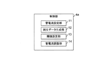
11 管電流設定部
12 純生データ生成部
13 閾値設定部
14 管電流調整部
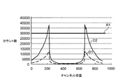
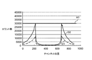
A1 下限値
A2 上限値
D2 純生データ
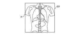
G1 平面画像
G2 側面画像
M1 メッセージ
Claims (6)
- 被検体の位置決め画像を撮像する場合の管電流を設定する管電流設定部と、
前記管電流設定部により設定された前記位置決め画像を撮像する場合の管電流に基づいて前記被検体に対してX線を出射するX線管と、
前記X線管により出射されて前記被検体を透過したX線を検出するX線検出器と、
前記X線検出器により検出された前記X線の透過データを収集するデータ収集部と、
前記データ収集部により収集された前記透過データから位置決め画像を生成する画像処理部と、
前記画像処理部により生成された前記位置決め画像又は前記画像処理部により前記位置決め画像を生成する途中に生じる第1データから、X線の線量分布を示す第2データを生成するデータ生成部と、
前記データ生成部により生成された前記第2データに対して閾値を設定する閾値設定部と、
前記データ生成部により生成された前記第2データ内のX線量と前記閾値設定部により設定された前記閾値との比較に応じ、前記被検体の断層画像を撮像する場合の管電流を調整する管電流調整部と、
を備えることを特徴とするX線診断装置。 - 前記管電流調整部により前記被検体の断層画像を撮像する場合の管電流を調整したことを報知するメッセージを表示する表示部を備えることを特徴とする請求項1記載のX線診断装置。
- 前記閾値設定部は、前記閾値としてアーチファクトが出る下限値を設定し、
前記管電流調整部は、前記データ生成部により生成された前記第2データ内のX線量が、前記閾値設定部により設定された前記下限値より小さくなるように、前記被検体の断層画像を撮像する場合の管電流を調整することを特徴とする請求項1又は2記載のX線診断装置。 - 前記閾値設定部は、前記閾値としてアーチファクトが出る上限値を設定し、
前記管電流調整部は、前記データ生成部により生成された前記第2データ内のX線量が、前記閾値設定部により設定された前記上限値より大きくなるように、前記被検体の断層画像を撮像する場合の管電流を調整することを特徴とする請求項1又は2記載のX線診断装置。 - 前記閾値設定部は、前記閾値として第1のアーチファクトが出る下限値及び第2のアーチファクトが出る上限値を設定し、
前記管電流調整部は、前記データ生成部により生成された前記第2データ内のX線量が、前記閾値設定部により設定された前記下限値より小さく、又は、前記閾値設定部により設定された前記上限値より大きくなるように、前記被検体の断層画像を撮像する場合の管電流を調整することを特徴とする請求項1又は2記載のX線診断装置。 - 前記第2データは、純生データであることを特徴とする請求項1から請求項5のいずれか一項に記載のX線診断装置。
Priority Applications (3)
| Application Number | Priority Date | Filing Date | Title |
|---|---|---|---|
| JP2012025848A JP6129474B2 (ja) | 2012-02-09 | 2012-02-09 | X線診断装置 |
| CN201310047327.XA CN103239252B (zh) | 2012-02-09 | 2013-02-06 | X射线诊断装置以及控制x射线诊断装置的方法 |
| US13/760,539 US9050058B2 (en) | 2012-02-09 | 2013-02-06 | X-ray diagnostic system and X-ray diagnostic method |
Applications Claiming Priority (1)
| Application Number | Priority Date | Filing Date | Title |
|---|---|---|---|
| JP2012025848A JP6129474B2 (ja) | 2012-02-09 | 2012-02-09 | X線診断装置 |
Publications (3)
| Publication Number | Publication Date |
|---|---|
| JP2013158630A JP2013158630A (ja) | 2013-08-19 |
| JP2013158630A5 JP2013158630A5 (ja) | 2015-03-05 |
| JP6129474B2 true JP6129474B2 (ja) | 2017-05-17 |
Family
ID=48919370
Family Applications (1)
| Application Number | Title | Priority Date | Filing Date |
|---|---|---|---|
| JP2012025848A Active JP6129474B2 (ja) | 2012-02-09 | 2012-02-09 | X線診断装置 |
Country Status (3)
| Country | Link |
|---|---|
| US (1) | US9050058B2 (ja) |
| JP (1) | JP6129474B2 (ja) |
| CN (1) | CN103239252B (ja) |
Families Citing this family (14)
| Publication number | Priority date | Publication date | Assignee | Title |
|---|---|---|---|---|
| US9907544B2 (en) | 2011-09-29 | 2018-03-06 | Proa Medical, Inc. | Minimally obstructive retractor for vaginal repairs |
| DE102012216850B3 (de) * | 2012-09-20 | 2014-02-13 | Siemens Aktiengesellschaft | Verfahren zur Planungsunterstützung und Computertomographiegerät |
| WO2015071798A1 (en) * | 2013-11-18 | 2015-05-21 | Koninklijke Philips N.V. | One or more two dimensional (2d) planning projection images based on three dimensional (3d) pre-scan image data |
| JP6381966B2 (ja) * | 2014-05-14 | 2018-08-29 | キヤノンメディカルシステムズ株式会社 | 医用画像診断装置 |
| CN104302081B (zh) * | 2014-09-24 | 2017-06-16 | 沈阳东软医疗系统有限公司 | 一种ct球管中灯丝电流的控制方法和设备 |
| JP6771879B2 (ja) * | 2014-10-31 | 2020-10-21 | キヤノンメディカルシステムズ株式会社 | X線コンピュータ断層撮影装置 |
| KR20160139294A (ko) * | 2015-05-27 | 2016-12-07 | 삼성전자주식회사 | 의료 영상 장치 및 의료 영상 장치를 위한 방법 |
| JP6681689B2 (ja) * | 2015-10-16 | 2020-04-15 | ジーイー・メディカル・システムズ・グローバル・テクノロジー・カンパニー・エルエルシー | 放射線断層撮影装置及びプログラム |
| JP6906905B2 (ja) * | 2016-06-29 | 2021-07-21 | キヤノンメディカルシステムズ株式会社 | X線診断装置 |
| JP6849521B2 (ja) * | 2017-05-01 | 2021-03-24 | キヤノン電子管デバイス株式会社 | X線システムおよびx線管検査方法 |
| JP7258473B2 (ja) | 2018-05-01 | 2023-04-17 | キヤノンメディカルシステムズ株式会社 | X線ct装置及び撮影条件管理装置 |
| JP7144292B2 (ja) * | 2018-11-27 | 2022-09-29 | キヤノンメディカルシステムズ株式会社 | 医用画像処理装置および医用画像処理方法 |
| JP7309988B2 (ja) * | 2018-11-27 | 2023-07-18 | キヤノンメディカルシステムズ株式会社 | 医用画像処理装置および医用画像処理方法 |
| CN113797448B (zh) * | 2020-06-11 | 2024-08-13 | 中硼(厦门)医疗器械有限公司 | 照射参数选取装置及其使用方法 |
Family Cites Families (13)
| Publication number | Priority date | Publication date | Assignee | Title |
|---|---|---|---|---|
| JP4075166B2 (ja) * | 1998-11-30 | 2008-04-16 | 松下電器産業株式会社 | X線基板検査装置 |
| JP3950612B2 (ja) | 2000-02-08 | 2007-08-01 | ジーイー横河メディカルシステム株式会社 | X線ct装置 |
| JP4532005B2 (ja) * | 2001-03-09 | 2010-08-25 | 株式会社日立メディコ | X線ct装置及びその画像表示方法 |
| JP4309631B2 (ja) * | 2001-10-22 | 2009-08-05 | 株式会社東芝 | X線コンピュータトモグラフィ装置 |
| US7054406B2 (en) * | 2002-09-05 | 2006-05-30 | Kabushiki Kaisha Toshiba | X-ray CT apparatus and method of measuring CT values |
| JP2004325183A (ja) | 2003-04-23 | 2004-11-18 | M & C:Kk | 放射線検出方法、放射線検出器、及び、この検出器を搭載した放射線撮像システム |
| US7149276B2 (en) * | 2004-07-14 | 2006-12-12 | Kabushiki Kaisha Toshiba | System, method, and computer program product that corrects measured data |
| US7215733B2 (en) * | 2004-07-23 | 2007-05-08 | Kabushiki Kaisha Toshiba | X-ray computed tomography apparatus |
| EP1731100B9 (en) * | 2005-06-06 | 2013-01-23 | Kabushiki Kaisha Toshiba | Medical image display apparatus and medical image display system |
| JP2009050531A (ja) | 2007-08-28 | 2009-03-12 | Konica Minolta Medical & Graphic Inc | 放射線画像撮影システム |
| JP5675117B2 (ja) * | 2009-02-17 | 2015-02-25 | 株式会社東芝 | X線ct装置及びx線ct装置の制御プログラム |
| JP5514450B2 (ja) * | 2009-02-23 | 2014-06-04 | 株式会社日立メディコ | X線ct装置 |
| JP5537138B2 (ja) | 2009-12-10 | 2014-07-02 | 株式会社東芝 | X線ct装置及びその制御プログラム |
-
2012
- 2012-02-09 JP JP2012025848A patent/JP6129474B2/ja active Active
-
2013
- 2013-02-06 US US13/760,539 patent/US9050058B2/en active Active
- 2013-02-06 CN CN201310047327.XA patent/CN103239252B/zh active Active
Also Published As
| Publication number | Publication date |
|---|---|
| CN103239252B (zh) | 2015-08-12 |
| CN103239252A (zh) | 2013-08-14 |
| US20130208853A1 (en) | 2013-08-15 |
| US9050058B2 (en) | 2015-06-09 |
| JP2013158630A (ja) | 2013-08-19 |
Similar Documents
| Publication | Publication Date | Title |
|---|---|---|
| JP6129474B2 (ja) | X線診断装置 | |
| JP6289223B2 (ja) | X線コンピュータ断層撮影装置 | |
| JP6571313B2 (ja) | 医用画像診断装置及び制御方法 | |
| US20160249868A1 (en) | Radiography device, radiography method, and radiography program | |
| JP5727277B2 (ja) | X線ct装置 | |
| JP2013215392A (ja) | X線診断装置及びx線診断装置の制御方法 | |
| JP5308862B2 (ja) | 医用寝台装置及び医用画像撮影装置 | |
| US8905636B2 (en) | X-ray diagnostic apparatus | |
| JP6125206B2 (ja) | X線画像診断装置 | |
| JP6615439B2 (ja) | X線ct装置 | |
| US9230702B2 (en) | System and method for reducing grid line image artifacts | |
| JP7086756B2 (ja) | 医用画像診断装置 | |
| JP6523451B2 (ja) | 放射線検出器とそれを備えたx線ct装置 | |
| JP2020049059A (ja) | 医用画像処理装置および方法 | |
| JP2013192750A (ja) | X線診断装置及びx線診断装置の制御方法 | |
| JP2024060170A (ja) | Pcct装置とその制御方法 | |
| JP2023035485A (ja) | X線ct装置 | |
| JP6162324B2 (ja) | 放射線画像撮影システム、放射線画像撮影方法、及び放射線画像撮影プログラム | |
| JP7206163B2 (ja) | X線ct装置、医用情報処理装置、及び医用情報処理プログラム | |
| JP2020022579A (ja) | X線コンピュータ断層撮影装置 | |
| JP7305334B2 (ja) | X線診断システム及び再構成処理システム | |
| JP6506583B2 (ja) | 放射線断層撮影装置及び画像生成装置並びにプログラム | |
| JP2024030533A (ja) | 光子計数型のx線画像診断装置及びパイルアップ補正用の較正データの生成方法 | |
| JP2024110270A (ja) | X線装置、x線装置管理システム、方法 | |
| US20190336089A1 (en) | X-ray computed tomography apparatus and imaging condition management apparatus |
Legal Events
| Date | Code | Title | Description |
|---|---|---|---|
| A521 | Request for written amendment filed |
Free format text: JAPANESE INTERMEDIATE CODE: A523 Effective date: 20150120 |
|
| A621 | Written request for application examination |
Free format text: JAPANESE INTERMEDIATE CODE: A621 Effective date: 20150120 |
|
| RD01 | Notification of change of attorney |
Free format text: JAPANESE INTERMEDIATE CODE: A7421 Effective date: 20150703 |
|
| A977 | Report on retrieval |
Free format text: JAPANESE INTERMEDIATE CODE: A971007 Effective date: 20151023 |
|
| A131 | Notification of reasons for refusal |
Free format text: JAPANESE INTERMEDIATE CODE: A131 Effective date: 20151104 |
|
| A521 | Request for written amendment filed |
Free format text: JAPANESE INTERMEDIATE CODE: A523 Effective date: 20151225 |
|
| A131 | Notification of reasons for refusal |
Free format text: JAPANESE INTERMEDIATE CODE: A131 Effective date: 20160517 |
|
| A711 | Notification of change in applicant |
Free format text: JAPANESE INTERMEDIATE CODE: A711 Effective date: 20160527 |
|
| A521 | Request for written amendment filed |
Free format text: JAPANESE INTERMEDIATE CODE: A523 Effective date: 20160715 |
|
| A02 | Decision of refusal |
Free format text: JAPANESE INTERMEDIATE CODE: A02 Effective date: 20161108 |
|
| A521 | Request for written amendment filed |
Free format text: JAPANESE INTERMEDIATE CODE: A523 Effective date: 20170208 |
|
| A911 | Transfer to examiner for re-examination before appeal (zenchi) |
Free format text: JAPANESE INTERMEDIATE CODE: A911 Effective date: 20170215 |
|
| TRDD | Decision of grant or rejection written | ||
| A01 | Written decision to grant a patent or to grant a registration (utility model) |
Free format text: JAPANESE INTERMEDIATE CODE: A01 Effective date: 20170314 |
|
| A61 | First payment of annual fees (during grant procedure) |
Free format text: JAPANESE INTERMEDIATE CODE: A61 Effective date: 20170412 |
|
| R150 | Certificate of patent or registration of utility model |
Ref document number: 6129474 Country of ref document: JP Free format text: JAPANESE INTERMEDIATE CODE: R150 |
|
| S533 | Written request for registration of change of name |
Free format text: JAPANESE INTERMEDIATE CODE: R313533 |
|
| R350 | Written notification of registration of transfer |
Free format text: JAPANESE INTERMEDIATE CODE: R350 |


