EP4579309A2 - Mikroskop zur abbildung einer probe - Google Patents
Mikroskop zur abbildung einer probe Download PDFInfo
- Publication number
- EP4579309A2 EP4579309A2 EP25163069.5A EP25163069A EP4579309A2 EP 4579309 A2 EP4579309 A2 EP 4579309A2 EP 25163069 A EP25163069 A EP 25163069A EP 4579309 A2 EP4579309 A2 EP 4579309A2
- Authority
- EP
- European Patent Office
- Prior art keywords
- illumination
- sample
- light
- along
- waist
- Prior art date
- Legal status (The legal status is an assumption and is not a legal conclusion. Google has not performed a legal analysis and makes no representation as to the accuracy of the status listed.)
- Pending
Links
Images


Classifications
-
- G—PHYSICS
- G02—OPTICS
- G02B—OPTICAL ELEMENTS, SYSTEMS OR APPARATUS
- G02B21/00—Microscopes
- G02B21/0004—Microscopes specially adapted for specific applications
- G02B21/002—Scanning microscopes
- G02B21/0024—Confocal scanning microscopes (CSOMs) or confocal "macroscopes"; Accessories which are not restricted to use with CSOMs, e.g. sample holders
- G02B21/0032—Optical details of illumination, e.g. light-sources, pinholes, beam splitters, slits, fibers
-
- G—PHYSICS
- G02—OPTICS
- G02B—OPTICAL ELEMENTS, SYSTEMS OR APPARATUS
- G02B21/00—Microscopes
- G02B21/0004—Microscopes specially adapted for specific applications
- G02B21/002—Scanning microscopes
- G02B21/0024—Confocal scanning microscopes (CSOMs) or confocal "macroscopes"; Accessories which are not restricted to use with CSOMs, e.g. sample holders
- G02B21/0052—Optical details of the image generation
- G02B21/0076—Optical details of the image generation arrangements using fluorescence or luminescence
-
- G—PHYSICS
- G02—OPTICS
- G02B—OPTICAL ELEMENTS, SYSTEMS OR APPARATUS
- G02B21/00—Microscopes
- G02B21/16—Microscopes adapted for ultraviolet illumination ; Fluorescence microscopes
-
- G—PHYSICS
- G02—OPTICS
- G02B—OPTICAL ELEMENTS, SYSTEMS OR APPARATUS
- G02B21/00—Microscopes
- G02B21/18—Arrangements with more than one light path, e.g. for comparing two specimens
-
- G—PHYSICS
- G02—OPTICS
- G02B—OPTICAL ELEMENTS, SYSTEMS OR APPARATUS
- G02B21/00—Microscopes
- G02B21/36—Microscopes arranged for photographic purposes or projection purposes or digital imaging or video purposes including associated control and data processing arrangements
- G02B21/365—Control or image processing arrangements for digital or video microscopes
- G02B21/367—Control or image processing arrangements for digital or video microscopes providing an output produced by processing a plurality of individual source images, e.g. image tiling, montage, composite images, depth sectioning, image comparison
-
- G—PHYSICS
- G02—OPTICS
- G02B—OPTICAL ELEMENTS, SYSTEMS OR APPARATUS
- G02B26/00—Optical devices or arrangements for the control of light using movable or deformable optical elements
- G02B26/08—Optical devices or arrangements for the control of light using movable or deformable optical elements for controlling the direction of light
- G02B26/0816—Optical devices or arrangements for the control of light using movable or deformable optical elements for controlling the direction of light by means of one or more reflecting elements
- G02B26/0825—Optical devices or arrangements for the control of light using movable or deformable optical elements for controlling the direction of light by means of one or more reflecting elements the reflecting element being a flexible sheet or membrane, e.g. for varying the focus
-
- G—PHYSICS
- G01—MEASURING; TESTING
- G01N—INVESTIGATING OR ANALYSING MATERIALS BY DETERMINING THEIR CHEMICAL OR PHYSICAL PROPERTIES
- G01N21/00—Investigating or analysing materials by the use of optical means, i.e. using sub-millimetre waves, infrared, visible or ultraviolet light
- G01N21/62—Systems in which the material investigated is excited whereby it emits light or causes a change in wavelength of the incident light
- G01N21/63—Systems in which the material investigated is excited whereby it emits light or causes a change in wavelength of the incident light optically excited
- G01N21/64—Fluorescence; Phosphorescence
- G01N21/645—Specially adapted constructive features of fluorimeters
- G01N21/6456—Spatial resolved fluorescence measurements; Imaging
- G01N21/6458—Fluorescence microscopy
Definitions
- the sample holder is arranged to receive a sample. It has a portion which is light transparent.
- the term "light transparent" in this connection particularly relates to transparency to the first illumination light beam, the second illumination light beam and to the detection light.
- Light transparent may relate to being transparent to visible light, UV light, infrared light and/or others.
- the first illumination objective is arranged to eject a first illumination light beam along a first illumination path to illuminate the sample received by the sample holder through the light transparent portion such that light is ejected from the sample.
- the second illumination objective is arranged to eject a second illumination light beam along a second illumination path to illuminate the sample received by the sample holder through the light transparent portion such that light is ejected from the sample.
- the first and/or second illumination light beams can be straight, redirected by suitable optical means or have any other appropriate form, particularly a form of a static light sheet or light sheet generated by scanning.
- the illumination light beam can be centered on a back aperture of the illumination objective in which case the illumination path will be coincident or coaxial with the axis of the illumination objective. Alternatively, it can be de-centered in which case after the objective the illumination path will be angled relative to the axis of the illumination objective.
- the size and position of the illumination beam at the objective back aperture can be adapted to appropriately or sophisticatedly illuminate the sample or changed during an experiment in an automated way.
- the illumination light beam can be a laser light beam having a range of wavelengths adapted to the properties of the sample. In particular, the wavelength of the laser light beam can be suitable for excitation of fluorophores and fluorescence imaging.
- Such a microscope allows for illumination of the sample from dual or plural directions. Particularly, this can be essential for comparably large samples such as biological samples in particular organoids where light from one side does not penetrate through the entire sample.
- illumination allows for reducing shadow effects in or on the sample impairing the quality of the imaging and allows imaging larger sample by bringing light into the sample from multiple directions.
- the first imaging objective is arranged to receive detection light comprising at least a portion of the light ejected from the sample.
- the detection light is propagated along a first detection path angled to the illumination path.
- the angle between the first detection path and the first and second illumination path is about 90°.
- the first imaging objective has a first imaging focal plane.
- the first and/or the second illumination path lie(s) in the first imaging focal plane.
- the second imaging objective is arranged to receive detection light comprising at least a portion of the light ejected from the sample.
- the detection light is propagated along a second detection path angled to the illumination path.
- the angle between the second detection path and the first and second illumination path is about 90°.
- the second imaging objective has a second imaging focal plane.
- the first and/or the second illumination path lie(s) in the second imaging focal plane.
- the light ejected from the sample can particularly comprise emitted fluorescence light or light ejected by the first illumination objective and redirected or reflected by the sample.
- the first and second illumination objectives are arranged such that in relation to the direction of gravity the first and second illumination path are both located below the bottom of the sample holder.
- the bottom of the sample holder may be a very bottom portion of the sample holder or a portion of the sample holder on or at which the sample is placed, if the two are not identical. In such arrangement the sample holder can move to image multiple positions of the sample and multiple samples without the sample holder interfering with the illumination paths. Further, in such arrangement the first and second illumination paths will not be parallel.
- top ends of the first and second illumination objectives are positioned below a bottom of the sample holder.
- the light transparent portion of the sample holder is located in between the imaging objectives.
- images from two opposing directions can be acquired allowing imaging of large samples impossible to image from one side only.
- images are captured using at least one detector which can be a camera.
- the first illumination light beam propagating along a first illumination path is preferably angled between 10 and 60 degrees to a horizontal plane and the second illumination light beam propagating along a second illumination path is preferably angled between 10 and 60 degrees to a horizontal plane.
- no other sample or object inside the sample should be located in the path of the incoming illumination beams because it would create shadows which would decrease image quality.
- different samples or objects within a sample to image may be located horizontally in the bottom part of the sample holder illuminating under an angle close to horizontal may require large spacing between multiple samples or objects within a sample. The smallest possible spacing between adjacent samples or objects within a sample would be achieved for illumination pointing vertically upwards.
- the illumination light beams should enter the sample from opposing sides, for example horizontally.
- An optimal angle range may be between 20 and 50 degrees to horizontal. Below 20 degrees mechanical constraints of illumination objective and/or sample spacing may start to limit performance of the microscope. Above 50 degrees sample may not be optimally illuminated due to illumination close to single sided illumination. Best performance may be achieved around 30 degrees to horizontal.
- the drive system of the holder support is configured to displace the sample holder along X axis so that at least a part of the sample holder is located directly above the first illumination objective and/or second illumination objective along the direction of gravity.
- the location of the first illumination objective and second illumination objective below the sample holder may be advantageous because it allows a long travel range of a sample holder along the X axis.
- Sample holder can also be elongated in X direction to hold many samples for high throughput imaging.
- the sample holder can be displaced by the drive system along X axis so that at least a portion of the sample holder is positioned directly above the first or second illumination objective.
- the travel range and sample holder dimensions will thus not be limited by collision with housing or optical elements of the illumination objectives. Such configuration allows elongation of the light transparent portion of the sample holder which can be 3 cm, preferably 5 cm long or longer.
- the light transparent portion of the sample holder preferably comprises transparent side walls and bottom, wherein the emitted light passes through the side walls of the sample holder and wherein the sample holder has an open top end.
- the open top end allows to place or manipulate the sample(s) or to exchange medium.
- the sample can be held in the sample holder only by gravity without the need for embedding in agarose or other support. Light emitted from the sample may pass through the light transparent side walls of the sample holder to the imaging objectives.
- Light transparent portion of the sample holder to place a sample can be elongated along X direction.
- Such sample holder can be at least 2 centimeter (cm) in length, preferably 3cm or 4cm long to image multiple positions in a sample in parallel for high throughput imaging.
- cm centimeter
- the light transparent portion of the sample holder may be elongated vertically to fit between the imaging objectives and intersect with the detection path and to efficiently collect light ejected by the sample.
- the light transparent portion may be elongated vertically to have at least 2 millimeter (mm) in height or preferably 4 mm to image large samples.
- the light transparent portion of the sample holder preferably contains at least one indentation extending into the direction of gravity.
- illumination light may pass through the sides of the indentations directly to the position being imaged which minimizes the path the illumination light has to travel across the sample. This can be advantageous for samples embedded in gels such as matrigel where imaging through it degrades image quality. It can also minimize probability that an object in the sample is located in the illumination light path which would create shadow artefact.
- the indentation may further be beneficial to lower the sample and position it between the imaging objective when short working distance objective are used.
- the indentation(s) can stabilize free floating objects in the sample(s).
- the sample holder contains one or multiple separation walls. Like this, at least two linearly arranged compartments can be created.
- An equivalent implementation to a sample holder containing one or multiple separation walls may be an array of at least two linearly arranged individual holders, wherein their side walls are forming the separation walls.
- the light transparent portion preferably contains the at least one indentation which bottom end may be at the same level or below a bottom end of the separation walls.
- a static sheet of light can be generated by a cylindrical lens.
- Such sheet of light will have a waist in which optimal optical sectioning and image quality is achieved.
- This waist will be elongated in direction perpendicular to the illumination path.
- opposing illumination axis are not parallel leading to non-parallel axis of light sheet waists elongation which reduces the area where waists from both sides overlap thus reducing the area of optimal image quality.
- These electronically controlled focusing elements can be synchronized with translation of the light sheet along the detection path for fast acquisition of z-stacks of images of the sample.
- the microscope preferably comprises at least one light source arranged to illuminate the sample through the first imaging objective and/or through the second imaging objective, and to collect the transmitted light through the second imaging objective and/or the first imaging objective, respectively, to generate a transmission image of the sample.
- one imaging objective may serve as a condenser for transmitted light and the other imaging objective to acquire transmission image. In this way, high quality transmission image of the sample can be acquired from both sides.
- Fig. 1 shows an embodiment of a microscope according to the invention. It comprises a first illumination objective (1) arranged to eject a first illumination light beam along a first illumination path (2) to illuminate the sample (3), second illumination objective (4) arranged to eject a second illumination light beam along a second illumination path (5) to illuminate the sample (3), first imaging objective (6) arranged to receive detection light propagated along a first detection path (7), second imaging objective (9) arranged to receive detection light propagated along a second detection path (10).
- the first and second imaging objectives have focal planes (8, 11).
- a holder support (14) is arranged to receive the sample holder (12) with sample (3).
- Holder support has a drive system arranged to displace the sample holder at least along two perpendicular axes (X, Y) and preferably along three perpendicular axes (X, Y, Z).
- Imaging objectives are placed horizontally perpendicular to the direction of gravity (13).
- Imaging objectives can be immersion objectives in which case the sample holder and the front lens of the imaging objectives are immersed in an immersion medium.
- Illumination light from the illumination objectives, in case these are not also immersion objectives, is then entering into the immersion medium through a window transparent to the illumination light such a glass window.
- Two separate transparent windows, one for each objective can be used or a single window for both objectives made for example by a meniscus lens cut on the sides facing the illumination objectives or a meniscus cylindrical lens.
- Illumination objectives (1, 4) are arranged such that the first and second illumination path (2, 5) are located below a bottom of the sample holder (12) and illumination paths (2, 5) are angled between 10 and 60 degrees to a horizontal plane (15) which is perpendicular to the direction of gravity (13).
- the sample holder contains an array of pockets (37) opened on top and having light transparent portion (28) at least at the bottom and sides. Pockets (37) are elongated downwards along the direction of gravity (13). Such pockets are used to hold many separated samples in parallel and can be optimally spaced so that when imaging each sample the samples in adjacent pockets do not lie in the illumination light path and do not create shadows.
- Fig. 4 top shows a side view of the illumination path of a microscope according to the invention.
- Illumination beam propagating along illumination path (2) is reflected by a galvanometric scanner mirror (23) to generate a scanned light sheet and passes through scan lens (22) generating a beam focus which is de-magnified by a tube lens (21) and objective (1) on to the sample (3) where it has a waist (18).
- a lens with a tunable focal lens (20) is placed in the light path to translate the beam waist (18) along the illumination axis.
- Elements described above are placed in an identical arrangement along a second illumination axis (5) containing a second illumination objective (4) and generating second beam waist (19).
- first half (26) of an object in the sample (3) using the first waist (18) the light sheet generating scanning of the first illumination beam is synchronized with translation of the first waist (18) along first illumination axis such that the first waist is scanned along axis 24.
- second half (27) of an object in the sample (3) using the second waist (19) the light sheet generating scanning of the second illumination beam is synchronized with translation of the second waist (19) along second illumination axis such that the second waist is scanned along axis 25.
- Axis 24 and 25 are parallel and sample is optimally illuminated with each beam waist covering half of the object in the sample.
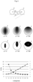
- Sample can be optimally illuminated using a static and scanned light sheet using the following sequence.
- An object in the sample is illuminated by a first light sheet with waist (18) elongated or scanned along axis (29) to illuminating first half of the object (30).
- the waist (18) of the first light sheet is then translated along first illumination axis (2) and second half of the object in the sample (31) is illuminated with first light sheet having waist (18) elongated or scanned along axis (32).
- An object in the sample is then illuminated by a second light sheet with waist (19) elongated or scanned along axis (33) to illuminating first half of the object (34).
- the waist (19) of the second light sheet is then translated along second illumination axis (5) and second half of the object (35) is illuminated with second light sheet having waist (19) located or scanned along axis (36).
- Sample objects can thus be optimally illuminated from both sides and with beam waists covering the sample.
- a separate image using each illumination step above can be acquired. These images are then combined to reconstruct a combined image of the object in the sample (3) where portion of each image illuminated by beam waist is taken into the combined image. Alternatively, some or all the illumination steps can happen within the same camera exposure generating an averaged image of the sample. Furthermore, the application comprises the following embodiments:
Landscapes
- Physics & Mathematics (AREA)
- General Physics & Mathematics (AREA)
- Optics & Photonics (AREA)
- Chemical & Material Sciences (AREA)
- Analytical Chemistry (AREA)
- Engineering & Computer Science (AREA)
- Multimedia (AREA)
- Computer Vision & Pattern Recognition (AREA)
- Microscoopes, Condenser (AREA)
Applications Claiming Priority (3)
| Application Number | Priority Date | Filing Date | Title |
|---|---|---|---|
| CH612021 | 2021-01-22 | ||
| PCT/EP2022/051329 WO2022157300A1 (en) | 2021-01-22 | 2022-01-21 | Microscope for imaging a sample |
| EP22700988.3A EP4281825B1 (de) | 2021-01-22 | 2022-01-21 | Mikroskop zum abbilden einer probe |
Related Parent Applications (1)
| Application Number | Title | Priority Date | Filing Date |
|---|---|---|---|
| EP22700988.3A Division EP4281825B1 (de) | 2021-01-22 | 2022-01-21 | Mikroskop zum abbilden einer probe |
Publications (2)
| Publication Number | Publication Date |
|---|---|
| EP4579309A2 true EP4579309A2 (de) | 2025-07-02 |
| EP4579309A3 EP4579309A3 (de) | 2025-09-17 |
Family
ID=74858157
Family Applications (3)
| Application Number | Title | Priority Date | Filing Date |
|---|---|---|---|
| EP22700988.3A Active EP4281825B1 (de) | 2021-01-22 | 2022-01-21 | Mikroskop zum abbilden einer probe |
| EP22152705.4A Withdrawn EP4036628A1 (de) | 2021-01-22 | 2022-01-21 | Mikroskop zur bildgebung einer probe |
| EP25163069.5A Pending EP4579309A3 (de) | 2021-01-22 | 2022-01-21 | Mikroskop zur abbildung einer probe |
Family Applications Before (2)
| Application Number | Title | Priority Date | Filing Date |
|---|---|---|---|
| EP22700988.3A Active EP4281825B1 (de) | 2021-01-22 | 2022-01-21 | Mikroskop zum abbilden einer probe |
| EP22152705.4A Withdrawn EP4036628A1 (de) | 2021-01-22 | 2022-01-21 | Mikroskop zur bildgebung einer probe |
Country Status (4)
| Country | Link |
|---|---|
| US (2) | US12443021B2 (de) |
| EP (3) | EP4281825B1 (de) |
| CN (1) | CN117043655A (de) |
| WO (1) | WO2022157300A1 (de) |
Citations (3)
| Publication number | Priority date | Publication date | Assignee | Title |
|---|---|---|---|---|
| EP2801855A1 (de) | 2013-05-10 | 2014-11-12 | European Molecular Biology Laboratory (EMBL) | Mikroskopmodul zur Abbildung einer Probe |
| EP3655809A1 (de) | 2017-07-20 | 2020-05-27 | Viventis Microscopy Sàrl | Mikroskop, verfahren zum betrieb eines mikroskops und verfahren zur bildgebung einer probe |
| EP3707544A1 (de) | 2017-11-10 | 2020-09-16 | Viventis Microscopy Sàrl | Mikroskop zur bildgebung einer probe und probenhalter für solch ein mikroskop |
Family Cites Families (38)
| Publication number | Priority date | Publication date | Assignee | Title |
|---|---|---|---|---|
| JP2975476B2 (ja) | 1992-03-30 | 1999-11-10 | 三井金属鉱業株式会社 | 結晶内のフォトルミネッセンス計測方法及び装置 |
| DE10257423A1 (de) | 2002-12-09 | 2004-06-24 | Europäisches Laboratorium für Molekularbiologie (EMBL) | Mikroskop |
| JP2006091723A (ja) | 2004-09-27 | 2006-04-06 | Olympus Corp | 倒立顕微鏡 |
| WO2007124437A2 (en) | 2006-04-20 | 2007-11-01 | Washington University In St. Louis | Objective-coupled selective plane illumination microscopy |
| DE102007047464B4 (de) | 2007-09-28 | 2023-03-02 | Carl Zeiss Microscopy Gmbh | Optische Anordnung zur Photomanipulation |
| US9134521B2 (en) | 2008-07-30 | 2015-09-15 | The Regents Of The University Of California | Multidirectional selective plane illumination microscopy |
| DE102009044986A1 (de) | 2009-09-24 | 2011-03-31 | Carl Zeiss Microimaging Gmbh | Mikroskop |
| US9404869B2 (en) | 2012-10-09 | 2016-08-02 | Howard Hughes Medical Institute | Multiview light-sheet microscopy |
| JP6086366B2 (ja) | 2013-04-05 | 2017-03-01 | 国立研究開発法人理化学研究所 | 顕微鏡、焦準器具、流体保持器具、及び光学ユニット |
| DE202013012338U1 (de) | 2013-07-10 | 2016-04-29 | Carl Zeiss Microscopy Gmbh | Anordnung zur Lichtblattmikroskopie |
| US9581798B2 (en) | 2013-07-22 | 2017-02-28 | Fundacio Institut De Ciencies Fotoniques | Light sheet-based imaging device with extended depth of field |
| DE102013110093B3 (de) | 2013-09-13 | 2015-01-22 | Johann Wolfgang Goethe-Universität | Küvette für eine inverse Fluoreszenz-Untersuchung |
| US10539772B2 (en) | 2013-10-09 | 2020-01-21 | Howard Hughes Medical Institute | Multiview light-sheet microscopy |
| GB2520541A (en) | 2013-11-25 | 2015-05-27 | European Molecular Biology Lab Embl | Optical arrangement for imaging a sample |
| EP3095004A4 (de) | 2014-01-14 | 2017-09-20 | Applied Scientific Instrumentation, Inc. | Lichtfoliengenerator |
| DE102014104977B4 (de) | 2014-04-08 | 2023-11-30 | Carl Zeiss Microscopy Gmbh | Anordnung zur Lichtblattmikroskopie sowie Mikroskopobjektiv für die Lichtblattmikroskopie |
| CN104155274B (zh) | 2014-08-07 | 2016-11-23 | 华中科技大学 | 一种双光束光片照明显微扫描成像方法及显微镜 |
| JP6522361B2 (ja) | 2015-02-18 | 2019-05-29 | オリンパス株式会社 | 蛍光顕微鏡 |
| US10989661B2 (en) | 2015-05-01 | 2021-04-27 | The Board Of Regents Of The University Of Texas System | Uniform and scalable light-sheets generated by extended focusing |
| DE102015209756A1 (de) | 2015-05-28 | 2016-12-01 | Carl Zeiss Microscopy Gmbh | Anordnung und Verfahren zur Lichtblattmikroskopie |
| WO2017075275A1 (en) | 2015-10-29 | 2017-05-04 | The Board Of Trustees Of The Leland Stanford Junior University | Methods and systems for imaging a biological sample |
| WO2017223426A1 (en) * | 2016-06-24 | 2017-12-28 | Howard Hughes Medical Institute | Automated adjustment of light sheet geometry in a microscope |
| JP2018004777A (ja) | 2016-06-28 | 2018-01-11 | オリンパス株式会社 | 光シート顕微鏡、及び、光シート顕微鏡の制御方法 |
| CN106053346B (zh) | 2016-06-29 | 2018-06-08 | 华中科技大学 | 一种光片显微成像转换装置 |
| JP2018017970A (ja) | 2016-07-29 | 2018-02-01 | オリンパス株式会社 | 光シート顕微鏡、及び、光シート顕微鏡の制御方法 |
| JP6685202B2 (ja) | 2016-08-19 | 2020-04-22 | オリンパス株式会社 | シート照明装置 |
| DE102016120683A1 (de) | 2016-10-28 | 2018-05-03 | Carl Zeiss Microscopy Gmbh | Lichtblattmikroskop |
| EP3538941B1 (de) * | 2016-11-10 | 2025-04-23 | The Trustees of Columbia University in the City of New York | Schnelles hochauflösendes bildgebungsverfahren für grosse proben |
| WO2018148309A1 (en) | 2017-02-08 | 2018-08-16 | The Regents Of The University Of California | Selective plane illumination in the conventional inverted microscope geometry by side illumination |
| JP2018169502A (ja) | 2017-03-30 | 2018-11-01 | オリンパス株式会社 | 顕微鏡装置 |
| DE102017119478A1 (de) | 2017-08-25 | 2019-02-28 | Carl Zeiss Microscopy Gmbh | Optische Anordnung zum Scannen von Anregungsstrahlung und/oder Manipulationsstrahlung in einem Laser-Scanning-Mikroskop und Laser-Scanning-Mikroskop |
| DE102017119480A1 (de) | 2017-08-25 | 2019-02-28 | Carl Zeiss Microscopy Gmbh | Optische Anordnung zum Scannen von Anregungsstrahlung und/oder Manipulationsstrahlung in einem Laser-Scanning-Mikroskop und Laser-Scanning-Mikroskop |
| JP7085364B2 (ja) | 2018-02-28 | 2022-06-16 | 浜松ホトニクス株式会社 | ライトシート顕微鏡及び試料観察方法 |
| EP3553583A1 (de) * | 2018-04-10 | 2019-10-16 | Igor Lyuboshenko | Mikroskopie mit beleuchtung selektiver ebenen mit mehreren beleuchtungseinheiten zur synchronen abtastung eines objekts mit einer rollblende einer digitalen kamera |
| CN109596588B (zh) | 2018-12-16 | 2021-10-08 | 华中科技大学 | 一种基于光片照明的高分辨率四维光场显微成像系统 |
| US12411088B2 (en) | 2020-03-13 | 2025-09-09 | University Of Southern California | Optimized photon collection for light-sheet microscopy |
| EP4134724A1 (de) | 2021-08-13 | 2023-02-15 | European Molecular Biology Laboratory | Mikroskop mit invertiertem lichtblatt |
| DE102022200841B3 (de) | 2022-01-26 | 2023-05-04 | Carl Zeiss Microscopy Gmbh | Verfahren, Anordnung und Mikroskop zur dreidimensionalen Bildgebung in der Mikroskopie unter Nutzung einer asymmetrischen Punktbildübertragungsfunktion |
-
2022
- 2022-01-21 EP EP22700988.3A patent/EP4281825B1/de active Active
- 2022-01-21 EP EP22152705.4A patent/EP4036628A1/de not_active Withdrawn
- 2022-01-21 EP EP25163069.5A patent/EP4579309A3/de active Pending
- 2022-01-21 CN CN202280023094.7A patent/CN117043655A/zh active Pending
- 2022-01-21 US US18/262,128 patent/US12443021B2/en active Active
- 2022-01-21 WO PCT/EP2022/051329 patent/WO2022157300A1/en not_active Ceased
-
2025
- 2025-09-26 US US19/341,058 patent/US20260023252A1/en active Pending
Patent Citations (3)
| Publication number | Priority date | Publication date | Assignee | Title |
|---|---|---|---|---|
| EP2801855A1 (de) | 2013-05-10 | 2014-11-12 | European Molecular Biology Laboratory (EMBL) | Mikroskopmodul zur Abbildung einer Probe |
| EP3655809A1 (de) | 2017-07-20 | 2020-05-27 | Viventis Microscopy Sàrl | Mikroskop, verfahren zum betrieb eines mikroskops und verfahren zur bildgebung einer probe |
| EP3707544A1 (de) | 2017-11-10 | 2020-09-16 | Viventis Microscopy Sàrl | Mikroskop zur bildgebung einer probe und probenhalter für solch ein mikroskop |
Also Published As
| Publication number | Publication date |
|---|---|
| EP4036628A1 (de) | 2022-08-03 |
| CN117043655A (zh) | 2023-11-10 |
| US20260023252A1 (en) | 2026-01-22 |
| EP4281825B1 (de) | 2025-03-12 |
| US12443021B2 (en) | 2025-10-14 |
| EP4579309A3 (de) | 2025-09-17 |
| EP4281825A1 (de) | 2023-11-29 |
| WO2022157300A1 (en) | 2022-07-28 |
| US20240094516A1 (en) | 2024-03-21 |
Similar Documents
| Publication | Publication Date | Title |
|---|---|---|
| US20220276479A1 (en) | Microscope module for a microscope arrangement for imaging a sample | |
| JP6685977B2 (ja) | 顕微鏡 | |
| EP3237949B1 (de) | Mikroskopieinstrumente mit beleuchtung auf selektiver ebene | |
| US12147021B2 (en) | Microscope, method of operating a microscope and method of imaging a sample | |
| WO2019092132A1 (en) | Microscope for imaging a sample and sample holder for such a microscope | |
| EP4579309A2 (de) | Mikroskop zur abbildung einer probe | |
| EP3855234B1 (de) | Lichtscheibenmikroskop und verfahren für grosse proben | |
| WO2023017181A1 (en) | Inverted light-sheet microscope | |
| US11187659B2 (en) | Systems for fluorescence light sheet microscopy of large samples in high refractive index solutions | |
| US12320966B2 (en) | Method and system for multi-view episcopic selective plane illumination microscope | |
| HK40023128A (en) | Microscope for selective plane illumination microscopy | |
| EP4616181A1 (de) | Konfokales mikroskopiesystem mit freiraumoptikverbindung | |
| HK40013087B (en) | A microscope module for imaging a sample | |
| HK40013087A (en) | A microscope module for imaging a sample |
Legal Events
| Date | Code | Title | Description |
|---|---|---|---|
| PUAI | Public reference made under article 153(3) epc to a published international application that has entered the european phase |
Free format text: ORIGINAL CODE: 0009012 |
|
| STAA | Information on the status of an ep patent application or granted ep patent |
Free format text: STATUS: THE APPLICATION HAS BEEN PUBLISHED |
|
| AC | Divisional application: reference to earlier application |
Ref document number: 4281825 Country of ref document: EP Kind code of ref document: P |
|
| AK | Designated contracting states |
Kind code of ref document: A2 Designated state(s): AL AT BE BG CH CY CZ DE DK EE ES FI FR GB GR HR HU IE IS IT LI LT LU LV MC MK MT NL NO PL PT RO RS SE SI SK SM TR |
|
| REG | Reference to a national code |
Ref country code: DE Ref legal event code: R079 Free format text: PREVIOUS MAIN CLASS: G02B0021360000 Ipc: G02B0021000000 |
|
| PUAL | Search report despatched |
Free format text: ORIGINAL CODE: 0009013 |
|
| AK | Designated contracting states |
Kind code of ref document: A3 Designated state(s): AL AT BE BG CH CY CZ DE DK EE ES FI FR GB GR HR HU IE IS IT LI LT LU LV MC MK MT NL NO PL PT RO RS SE SI SK SM TR |
|
| RIC1 | Information provided on ipc code assigned before grant |
Ipc: G02B 21/00 20060101AFI20250812BHEP Ipc: G02B 21/36 20060101ALI20250812BHEP Ipc: G01N 21/64 20060101ALI20250812BHEP Ipc: G02B 21/16 20060101ALI20250812BHEP Ipc: G02B 26/08 20060101ALN20250812BHEP Ipc: G02B 21/18 20060101ALN20250812BHEP |