EP4170633A1 - Gallbladder model - Google Patents
Gallbladder model Download PDFInfo
- Publication number
- EP4170633A1 EP4170633A1 EP22214865.2A EP22214865A EP4170633A1 EP 4170633 A1 EP4170633 A1 EP 4170633A1 EP 22214865 A EP22214865 A EP 22214865A EP 4170633 A1 EP4170633 A1 EP 4170633A1
- Authority
- EP
- European Patent Office
- Prior art keywords
- layer
- simulated
- artificial
- gallbladder
- anatomical
- Prior art date
- Legal status (The legal status is an assumption and is not a legal conclusion. Google has not performed a legal analysis and makes no representation as to the accuracy of the status listed.)
- Pending
Links
Images


Classifications
-
- G—PHYSICS
- G09—EDUCATION; CRYPTOGRAPHY; DISPLAY; ADVERTISING; SEALS
- G09B—EDUCATIONAL OR DEMONSTRATION APPLIANCES; APPLIANCES FOR TEACHING, OR COMMUNICATING WITH, THE BLIND, DEAF OR MUTE; MODELS; PLANETARIA; GLOBES; MAPS; DIAGRAMS
- G09B23/00—Models for scientific, medical, or mathematical purposes, e.g. full-sized devices for demonstration purposes
- G09B23/28—Models for scientific, medical, or mathematical purposes, e.g. full-sized devices for demonstration purposes for medicine
- G09B23/30—Anatomical models
- G09B23/34—Anatomical models with removable parts
-
- G—PHYSICS
- G09—EDUCATION; CRYPTOGRAPHY; DISPLAY; ADVERTISING; SEALS
- G09B—EDUCATIONAL OR DEMONSTRATION APPLIANCES; APPLIANCES FOR TEACHING, OR COMMUNICATING WITH, THE BLIND, DEAF OR MUTE; MODELS; PLANETARIA; GLOBES; MAPS; DIAGRAMS
- G09B23/00—Models for scientific, medical, or mathematical purposes, e.g. full-sized devices for demonstration purposes
- G09B23/28—Models for scientific, medical, or mathematical purposes, e.g. full-sized devices for demonstration purposes for medicine
- G09B23/30—Anatomical models
- G09B23/32—Anatomical models with moving parts
Definitions
- This application relates to surgical training tools, and in particular, to simulated tissue structures and models for teaching and practicing surgical procedures involving a gallbladder.
- a common treatment for gallstones and other gallbladder conditions is a cholecystectomy which is the surgical removal of the gallbladder from the liver bed.
- Laparoscopic cholecystectomy is the most common laparoscopic procedure and has replaced open cholecystectomy as the first-choice of treatment for gallstones and inflammation of the gallbladder.
- Laparoscopic cholecystectomy advantageously requires smaller incisions, resulting in less pain, improved cosmetic results, quicker healing, and fewer complications such as infection and adhesions.
- Laparoscopic cholecystectomy requires several small incisions in the abdomen to allow the insertion of trocars or small cylindrical tubes approximately 5 to 10 millimeters in diameter through which surgical instruments and a laparoscope are placed into the abdominal cavity.
- the laparoscope illuminates the surgical field and sends a magnified image from inside the body to a video monitor giving the surgeon a close-up view of the organs and tissues.
- the surgeon watches the live video feed and performs the operation by manipulating the surgical instruments placed through the trocars.
- a patient In a laparoscopic cholecystectomy, a patient is placed in a supine position on the operating table and anesthetized. A scalpel can be used to make a small incision at the umbilicus.
- a trocar the abdominal cavity is entered and enlarged by delivering carbon dioxide gas to insufflate the cavity to create a working space inside the patient's abdominal region.
- the trocar may include an inserted laparoscope for observing the penetration, insertion, and insufflation of the abdominal space. Additional trocars are inserted at a location inferior to the ribs.
- the fundus of the gallbladder which is covered by the peritoneum, is identified, grasped with a surgical grasper extending through one of the trocars, and retracted.
- a second surgical grasper may be used to retract the rest of the gallbladder in a lateral direction to expose Calot's triangle.
- Calot's triangle is that portion of the gallbladder anatomy that is bound by the cystic duct, cystic artery, the hepatic duct and the border of the liver. The surgeon identifies the cystic duct and cystic artery. In this area, the underlying structures are carefully skeletonized from the peritoneum separating the peritoneum from the both the cystic duct and the cystic artery.
- a surgical clip applier is introduced through one of the trocars and clips are applied in two locations to both the cystic duct and the cystic artery.
- the cystic duct and the cystic artery are then divided with surgical scissors between the two locations of clips freeing the gallbladder for removal.
- the gallbladder is dissected from the bed of the liver and removed through one of the trocars.
- laparoscopic cholecystectomy complications may arise due to gallbladder perforation which can occur due to excessive traction during retraction or during dissection of the gallbladder from the liver bed or extraction from the abdomen.
- the outcome of laparoscopic cholecystectomy is greatly influenced by the training, experience and skill of the surgeon performing the procedure. In order for residents and surgeons to learn and practice these surgical techniques, a realistic, functional, and anatomically correct model for use in a laparoscopic training device is needed.
- a gallbladder model is not only useful for training residents and surgeons in laparoscopic cholecystectomy, but also, desirable for training residents and surgeons in laparoscopic common bile duct exploration.
- the common bile duct is a tube that connects the liver, gallbladder and pancreas to the small intestine and delivers fluid to aid in digestion.
- Common bile duct exploration is a procedure used to see if a gallbladder stone or some other obstruction is blocking the flow of bile from the gallbladder or liver to the intestine which can cause jaundice.
- a laparoscopic common bile duct exploration procedure the abdominal cavity is approached as in a cholecystectomy described above.
- the surgeon identifies the common bile duct and a small hemi-circumferential incision is made in the common bile duct.
- a cholangiography catheter is inserted into the insufflated abdominal cavity through one of the trocars and into the incision made in the common bile duct.
- Contrast media or radiopaque fluid is introduced into the cystic and common bile ducts and an X-ray is taken to reveal the location of any gallstones in the common bile duct. If there are gallstones, the obstructions will appear as discontinuities in the flow of contrast media. The gallstones are then surgically extracted.
- surgeons need a way to practice laparoscopic cholecystectomies and common bile duct explorations outside of the operating room.
- the practice model needs to be anatomically correct and include all important landmarks normally seen during surgery in order to give the surgeon or resident the most realistic practice possible.
- an anatomical model for surgical training includes a first layer having an inner surface and an outer surface.
- the first layer has a substantially uniform thickness defined between the inner surface and the outer surface.
- the first layer has a first perimeter and is configured to simulate at least a portion of a first anatomical structure.
- the model includes a second layer having an inner surface and an outer surface.
- the second layer has a thickness between the inner surface and the outer surface.
- the second layer defines a second perimeter and overlays the first layer such that the outer surface of the second layer faces the inner surface of the first layer.
- the model includes at least one second simulated anatomical structure which has a third perimeter around the at least one simulated anatomical structure.
- the at least one simulated anatomical structure is connected to the inner surface of the second layer.
- the outer surface of the second layer is connected to the inner surface of the first layer at least partially around the location of the at least one second simulated anatomical structure.
- an anatomical model for surgical training includes an anatomical portion and a support removably connectable to the anatomical portion.
- the anatomical portion includes at least a first layer having an inner surface and an outer surface interconnected by a top side and a bottom side and a left side and a right side.
- the first layer has a thickness defined between the inner surface and the outer surface.
- the first layer is configured to simulate at least a portion of a liver.
- the top side of the first layer has a peak.
- the model includes a simulated gallbladder positioned in the location of the peak and facing the inner surface of the first layer.
- the model includes a frame connected to at least the first layer.
- the frame has a first end interconnected to a second end by a central portion.
- the first end and the second end of the frame are removably connectable to the support to hold the anatomical portion in a substantially upright position.
- the frame does not extend into the location of the peak such that the first layer in the location of the peak is capable of flexing inwardly and outwardly relative to the frame.
- an anatomical model for surgical training includes an anatomical portion having a first layer.
- the first layer includes an inner surface and an outer surface interconnected by a top side and a bottom side and a left side and a right side.
- the first layer has a thickness defined between the inner surface and the outer surface.
- the first layer is configured to simulate at least one anatomical structure.
- the anatomical portion includes a second layer that includes at least one anatomical structure overlaying the first layer.
- the anatomical portion also includes a frame having a first end interconnected to a second end by a central portion. At least part of the frame is embedded within the first layer with the first end and the second end of the frame extending out from the first layer.
- the model includes a support to which the first end and the second end of the frame are removably connectable to the support to hold the anatomical portion in a substantially upright position with respect to a supporting surface.
- a surgical simulation system includes an anatomical model.
- the model includes an anatomical portion.
- the anatomical portion includes a first layer having an inner surface and an outer surface interconnected by a top side and a bottom side and a left side and a right side.
- the first layer has a substantially uniform thickness defined between the inner surface and the outer surface.
- the first layer is configured to simulate at least one anatomical structure and defines a substantially planar configuration.
- the model includes a second layer having a plurality of anatomical structures connected to and overlaying the inner surface of the first layer.
- a support is connectable to the anatomical portion and configured to hold the anatomical portion in a substantially perpendicular orientation with respect to a supporting surface.
- the system further includes a surgical training device.
- the surgical training device includes a base and a top cover connected to and spaced apart from the base to define a simulated insufflated internal cavity between the top cover and the base.
- the internal cavity is at least partially obstructed from direct observation by a user.
- the top cover includes an aperture or penetrable simulated tissue region.
- the top cover of the surgical training device is angled to form an acute angle with respect to a horizontal plane as measured from inside the cavity.
- the anatomical model is positioned inside the internal cavity a distance opposite the acute angle such that the inner surface of the first layer faces the acute angle and the aperture or penetrable simulated tissue region.
- an anatomical model for surgical training includes an anatomical portion.
- the anatomical portion includes a first layer having an inner surface and an outer surface interconnected by a top side, a bottom side, a left side and a right side.
- the inner surface is substantially planar and flat and the first layer defines a thickness between the inner surface and the outer surface.
- the first layer is configured to simulate at least a portion of a liver.
- the top side of the first layer has a peak.
- the anatomical portion includes a second layer having an inner surface and an outer surface interconnected by a top side, a bottom side, a left side and a right side. The second layer overlays the first layer such that the outer surface of the second layer faces the inner surface of the first layer.
- the outer surface of the second layer is connected to the inner surface of the first layer along at least part of a first perimeter.
- the second layer defines a thickness between the inner surface and the outer surface and the thickness of the second layer is smaller than the thickness of the first layer.
- the anatomical portion includes a third layer having at least one simulated anatomical structure.
- the at least one simulated anatomical structure is connected to the inner surface of the second layer.
- the anatomical portion further includes a fourth layer having an inner surface and an outer surface interconnected by a top side, a bottom side, a left side and a right side. The fourth layer overlays the second layer and the third layer such that the outer surface of the fourth layer faces the inner surface of the second layer and the at least one simulated anatomical structure.
- the outer surface of the fourth layer is connected to the inner surface of the second layer along at least part of a second perimeter.
- the fourth layer defines a thickness between the inner surface and the outer surface and the thickness of the fourth layer is smaller than the thickness of the first layer.
- the anatomical portion further includes a frame at least partially embedded inside the first layer.
- the model includes a support connectable to the frame to hold the anatomical portion in a substantially upright position.
- a gallbladder model allows users to practice open and laparoscopic cholecystectomies and common bile duct explorations.
- the gallbladder model includes an anatomical portion connected to a support.
- the anatomical portion includes a liver layer, a fascia layer, a gallbladder layer, a peritoneum layer, and a frame connected together and held in an upright orientation by the support.
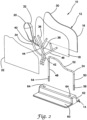
- FIG. 1 there is shown a gallbladder model 10 according to the present invention.
- the gallbladder model 10 includes an anatomical portion 12 removably connected to a support 14.
- the substantially planar anatomical portion 12 is maintained in an upright configuration by the support 14.
- the fundus of the gallbladder is visible and retracted.
- the remainder of the gallbladder underlying the liver toward the posterior of the patient is uncovered and made visible along with the triangle of Calot in the insufflated cavity. This retraction involves lifting part of the lower or inferior portion of the right lobe of the liver.
- the gallbladder model 10 of the present invention is a substantial or partial projection of at least a portion of the retracted liver and gallbladder onto the X-Y plane or transverse plane of a patient.
- the gallbladder model 10 represents a substantial planar projection of a retracted liver and gallbladder in a simulated insufflated cavity.
- the gallbladder model 10 configuration advantageously provides a surgical approach to a simulated gallbladder already in a retracted perpendicular orientation when viewed by the user approaching the gallbladder from the location of the umbilicus.
- the gallbladder model 10 configuration permits practice by the user without requiring a second user to hold portions of the model with graspers in a retracted position and as such, the model 10 is advantageously designed to be used by one person at a time.
- the model 10 only a portion of the liver is simulated, in particular, the right lobe of the liver. Together, with the right lobe, the entirety of the biliary structure including the gallbladder is included in the model.
- FIG. 2 there is shown an exploded view of the gallbladder model 10 comprising an anatomical portion 12 connected to a support 14.
- the anatomical portion 12 includes a liver layer 16, a fascia layer 18, a gallbladder layer 20, a peritoneum layer 22, and a frame 24 connected together.
- Each layer will now be described in greater detail.
- the liver layer or first layer 16 is molded from silicone or thermoplastic elastomer that is dyed with a red color and configured to simulate a retracted portion of a liver.
- the liver layer 16 is shaped to represent a portion of the right lobe of a human liver that is retracted to expose the gallbladder and triangle of Calot.
- the liver layer 16 includes a flat planar inner surface 26 and a convex curved outer surface 28.
- the inner and outer surfaces 26 interconnect along four sides--a curved top side, a straight bottom side, and a left side and right side that interconnect the top and bottom sides.
- the curved top side includes a peak 30 near or at the left side of the model.
- the top side curves downward from the peak 30 to a lower portion that interconnects with the right side. This peaked shape resembles a substantially planar projection of a retracted right lobe of a human liver.
- the peak 30 has a longer length relative to other portions of the liver layer 16.
- the thickest portion of the liver layer 16 is approximately 0.5 inches and located approximately at the middle.
- the frame 24 is molded directly into the liver layer 16 such that at least a portion of the frame 24 resides inside the liver layer 16 and a portion of the frame 24 resides outside of the liver layer 16 as shown in FIG. 3 .
- the frame 24 will be described in greater detail below.
- the fascia layer or second layer 18 is a thin approximately 0.01-0.03 inches thick layer made of a thermoplastic elastomer or silicone that is partially translucent, clear or dyed with a slight yellow color.
- the fascia layer 18 has the same peaked shape as the liver layer 16 and is sized and configured to overlay the liver layer 16.
- the fascia layer 18 has an inner surface and an outer surface with the outer surface overlaying a portion of the inner surface 26 of the liver layer 16.
- the fascia layer 18 is attached to the liver layer 16 with adhesive that is placed at least along the perimeter such that the majority of the middle portion or portions interior from the perimeter of the fascia layer 18 are not attached to the liver layer 16, but instead, are free to remain mobile and separate away from the liver layer 16.
- the gallbladder model 10 of the present invention includes a fascia layer 18 which advantageously simulates the dissection and removal of the gallbladder away from the liver. This advantage will be described in greater detail below.
- the gallbladder layer or third layer 20 includes at least one body component.
- the at least one body component is a plurality of anatomical structures.
- the gallbladder layer 20 includes a gallbladder 32 connected to a cystic duct 34, a common hepatic duct 36 connected to a common bile duct 38, a cystic artery 40, and a common hepatic artery 42 connected to and branching into the right hepatic artery 44 and left hepatic artery 46. All of these anatomical structures are configured to simulate actual human anatomy and arranged within the gallbladder layer 20 in an anatomically correct fashion.
- the gallbladder 32 is a hollow bulbous structure molded out of silicone or other thermoplastic material dyed with a light green or yellow color to simulate bile. In another variation, the gallbladder 32 is a solid and not hollow structure.
- the cystic duct 34, common hepatic duct 36 and common bile duct 38 are also made of silicone or thermoplastic material that is dyed with a light green color.
- the cystic duct 34 is tubular in shape having a tapered end and a diameter of approximately 0.15-0.25 inches. In one variation, the cystic duct 34 has a lumen with a minimum inner diameter of 0.15 inches and a maximum outer diameter of 0.25 inches making it small enough to clip and large enough to permit insertion of catheter.
- the cystic duct 34 includes a lumen having an inner surface that is lubricated with lubricant.
- the cystic duct 34 is larger in outer diameter relative to dimension of a real life cystic duct 34 to facility training and insertion of a catheter into the lumen.
- the common hepatic duct 36 and common bile duct 38 are also tubular in shape having a diameter of approximately 0.15 inches.
- the cystic duct 34, common hepatic duct 36 and common bile duct 38 are hollow and in another variation they are solid.
- the cystic artery 40, the common hepatic artery 42, the right hepatic artery 44 and the left hepatic artery 46 are made from silicone or thermoplastic material that is dyed a red color and molded into a tubular shape having a diameter of approximately 0.15 inches.
- the cystic artery 40, common hepatic artery 42, the right hepatic artery 44 and the left hepatic artery 46 are hollow and in another variation they are solid structures.
- the gallbladder layer 20 is connected to the fascia layer 18 with selectively-placed adhesive.
- the gallbladder layer 20 may be formed from multiple pieces joined together or as a unit with no disconnects.
- the manufacturing process consists of a wax form that is dipped in molten plastic and melted out once the plastic has set.
- the gallbladder model 10 is configured for practicing bile duct exploration.
- the biliary structures of the gallbladder layer 20 are hollow and filled with fluid that resembles bile.
- An exemplary fluid is green-colored dishwashing liquid.
- the inner diameter of the hollow biliary structures is approximately 0.09 inches and the outer diameter is approximately 0.15 inches.
- the gallbladder model 10 that is configured for biliary exploration includes a hollow gallbladder 32 filled with fluid that resembles bile.
- the free ends of the cystic duct 34, common hepatic duct 36, and common bile duct 38 are closed or capped with standard tubing caps, solid connectors or barbed connectors that retain fluid inside the ducts.
- biliary structures made of multiple tubular structures are connected together with connectors.
- the junction between the common hepatic duct 36 and common bile duct 38 is connected with a connector such as a Y-shaped split that permits fluid to flow therebetween.
- the cystic duct 34 and the common bile duct 38 are connected via a connector or molded as a unitary structure such that fluid is allowed to flow between the cystic duct 34 and the common bile duct 38.
- the employ of connectors is advantageous in that after practice scenarios in which the ducts are cut, such as in a cholecystectomy, the severed ducts are replaceable with new ducts that are reconnected at the same locations using the same connectors so that training scenarios can be repeated.
- any one or more of the gallbladder 32, bile duct 34, common hepatic duct 36, and common bile duct 38 may include one or more simulated gallstones (not shown).
- a simulated gallstone is a small bead-like structure made of plastic or other material. The simulated gallstones are placed inside the hollow space of the gallbladder 32 and/or inside the lumen of one or more of the cystic duct 34, common hepatic duct 36, and common bile duct 38.
- simulated gallstones are shaped and configured such that they are not visible to the user when the model is received but become visible when a syringe and/or catheter is used to inject simulated contrast media fluid such as colored water into one or more of the ducts and the continuous flow of contrast media fluid is visibly interrupted or blocked by the gallstones as the simulated contrast media fluid fills the biliary structures.
- a kit is provided that includes a syringe with which the gallbladder 32 is injected with fluid and/or simulated gallstones.
- the gallbladder 32 is not filled with liquid but is filled with air which may be injectable into the open cavity of the gallbladder 32 with a syringe or other similar device.
- the cavity of the gallbladder 32 may be pressurized to a pressure greater than ambient such that when the gallbladder 32 is inadvertently punctured, as if by an improper surgical technique, the gallbladder 32 noticeably deflates and as such provides a visual indication to the trainee.
- the gallbladder 32 has a wall thickness configured to permit observation of deflation of the gallbladder 32.
- the peritoneum layer or fourth layer 22 is a thin layer approximately 0.01-0.03 inches thick made of a thermoplastic elastomer or silicone that is clear or partially translucent and/or dyed with a slightly yellow color.
- the peritoneum layer 22 is nearly identical to the fascia layer 18 and has the same peaked shape as the underlying fascia layer 18 and liver layer 16.
- the peritoneum layer 22 includes an inner surface and an outer surface overlaying the gallbladder layer 20 and overlaying at least a portion of the inner surface of the second layer 18.
- both the fascia layer 18 and the peritoneum layer 22 are each formed by molding liquid silicone on a layer of foam such as packaging foam or other spongiform structure and then peeled off the foam after it has set to impart at least one textured surface to the fascia and peritoneum layers 18, 22.
- the peritoneum layer 22 is sized and configured to overlay the gallbladder layer 20.
- the peritoneum layer 22 is attached to the fascia layer 18 with adhesive that is placed in locations that are capable of direct contact with the fascia layer 18 without interference from the intervening gallbladder layer 20.
- the peritoneum layer 22 is only adhered to the fascia layer 18 and in one variation, the peritoneum layer 22 is only adhered to the fascia layer 18 and not to the gallbladder layer 20. In another variation, portions of the peritoneum layer 22 are adhered to portions of the gallbladder layer 20 as well as the fascia layer 18. In yet in another variation, portions of the peritoneum layer 22 are adhered only to portions of the gallbladder layer 20. The layers are adhered with adhesive or by the inherent tackiness of the material composing the layers. In essence, the peritoneum layer 22 is selectively adhered to one or more of the underlying gallbladder layer 20 and fascia layer 18 with adhesive.
- the anatomical portion 12 includes a frame 24 that is configured to support the entire anatomical portion 12 in a substantially upright orientation with respect to a table top or other substantially flat surface including an organ-receiving tray or other surface inside a laparoscopic training simulator.
- the frame 24 includes a left leg 48 and a right leg 50 interconnected by a central portion 52.
- the central portion 52 is curved and mimics the generally peaked-shape of the other layers 16, 18, 22.
- the frame 24 is sized smaller than the liver, fascia and peritoneum layers 16,18, 22.
- the frame 24 is made of rigid metal, plastic or other polymer or material that is capable and strong enough to support the layers of silicone and plastic comprising the anatomical portion 12 of the model 10 in an upright orientation.
- the left leg 49 is at or adjacent to the peak and is approximately 3.5-4.0 inches long and the shorter right leg 50 is approximately 2.5-3.0 inches long.
- the curved central portion 52 is approximately 4.0-4.5 inches long and follows the curvature of the layers 16, 18, 22.
- the overall height of the gallbladder model 10 is approximately 5-6 inches and the length of the model 10 is approximately 5-6 inches.
- the left leg 48 defines a left prong 54 at its free end and the right leg 50 defines a right prong 56 at the free end of the right leg 50.
- the left and right prongs 54, 56 extend beyond the anatomical portion 12 for insertion into a support 14.
- the cross-section of the frame 24 is substantially circular with a diameter of approximately 0.15 inches with the prongs 54, 56 having a slightly larger diameter.
- Each prong 54, 56 includes a curved, ball-shaped, or spherical-shaped or angled detent 58 as illustrated in FIG. 4 which shows a sectional view of a the left leg 48.
- the prongs 54, 56 have angled distal tips.
- the frame 24 is connected to the anatomical portion 12 such that the prongs 54, 56 protrude out from the layers for connection with the support 14.
- the frame 24 is molded directly into the liver layer 16 and is clear or transparent in color or substantially the same color as the liver layer 16 in which it is embedded so that it is not readily visible to the user.
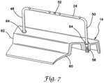
- the frame 24 does not have a peaked portion and is substantially U-shaped. As shown in FIG. 7 , the central portion 52 of the frame 24 is straight and does not follow the peaked-shaped of the other layers 16, 18, 22.
- This variation provides less support to the other layers 16, 18, 22 in the location of the peak 30 advantageously permitting all of these layers to be more flexible and to be more easily pushed distally or proximally relative to areas adjacent to the frame 24 to practice the retraction of the liver 16 from the gallbladder 32 while still providing support to the overall model 10 in the support 14.
- both the right leg 50 and left leg 48 are the same length approximately 2.5-3.0 inches long instead of the left leg 48 in the location of the peak 30 being longer.
- the peak 30 formation in the layers 16, 18, 22 represents only a portion of the liver, in particular, the right lobe of the liver with all of the anatomical structures of the gallbladder layer 20 being presented in the model 10.
- the support 14 is configured to connect with the anatomical portion 12 and hold the anatomical portion 12 in a substantially upright orientation with respect to a table top or other surface.
- the support 14 includes a base 60 interconnected with an upright portion 62.
- the upright portion 62 includes at least two sockets 64 that are sized and configured to receive the prongs 54, 56 of the frame 24.
- the upright portion 62 further includes a spring-biased plunger 66 in communication with each socket 64. To connect the anatomical portion 12 to the support 14, the prongs 54, 56 are inserted into the sockets 64 of the support 14.
- the angled distal tips of the prongs 54, 56 cam against the plungers 66 until they snap into the detents 58 on each prong 54, 56 to securely lock the anatomical portion 12 to the support 14.
- the anatomical portion 12 may be removed from the support 14 by releasing the plungers 66 from each detent 58 or by pulling with force such that the detent 58 cams against the plunger 66 moving it out of the way.
- the anatomical portion 12 can be snapped into the support 14 or into sockets formed as a removable part of a larger anatomical model, organ tray or laparoscopic trainer.
- connection fit is within the scope of the present invention for connecting the anatomical portion 12 to the support 14 including left and right prongs 54, 56 that are split and splay outwardly as shown in FIG. 7 .
- the prongs 54, 56 are further biased outwardly and ramped to flex past and snap behind a detent to secure the anatomical portion 12 to the support 14.
- the slit end of the prongs 54, 56 are squeezed together by a user from underneath the support 14 to permit the prongs 54, 56 to slide past the detent.
- the frame 24 and the anatomical portion 12 are separated from the support 14.
- the gallbladder model 10 can be used to practice open procedures that involve gallbladder anatomy. Also, the gallbladder model 10 is particularly well suited for practicing laparoscopic gallbladder procedures. To practice laparoscopic gallbladder procedures, the model 10 is placed inside a laparoscopic trainer 68 such as the trainer 68 shown in FIG. 6 and described in co-pending U.S. Patent Application Serial No. 13/248,449 entitled "Portable laparoscopic trainer" and filed on September 29, 2011 by Pravong et al. to Applied Medical Resources Corporation and published as U.S. Patent Application Publication No. 2012/0082970 , hereby incorporated by reference in its entirety herein.
- a laparoscopic trainer 68 such as the trainer 68 shown in FIG. 6 and described in co-pending U.S. Patent Application Serial No. 13/248,449 entitled "Portable laparoscopic trainer” and filed on September 29, 2011 by Pravong et al. to Applied Medical Resources Corporation and published as U.S. Patent Application Publication No. 2012/
- the laparoscopic trainer 68 includes a top cover 70 connected to a base 72 by a pair of legs 74 spacing the top cover 70 from the base 72.
- the laparoscopic trainer 68 is configured to mimic the torso of a patient such as the abdominal region.
- the top cover 70 is representative of the anterior surface of the patient and the space between the top cover 70 and the base 72 is representative of an interior of the patient or body cavity where organs reside.
- the laparoscopic trainer 68 is a useful tool for teaching, practicing and demonstrating various surgical procedures and their related instruments in simulation of a patient. Surgical instruments are inserted into the cavity through pre-established apertures 76 in the top cover 48.
- These pre-established apertures 76 may include seals that simulate trocars or may include simulated tissue region(s) that simulates the patient's skin and abdominal wall portions.
- Various tools and techniques may be used to penetrate the top cover 70 to perform mock procedures on model organs placed between the top cover 70 and the base 72 such as the gallbladder model 10.
- the gallbladder model 10 When placed inside the cavity of the trainer 68, the gallbladder model 10 is generally obscured from the perspective of the user who can then practice performing surgical techniques laparoscopically by viewing the surgical site indirectly via a video feed displayed on a video monitor 78.
- the video display monitor 78 is hinged to the top cover 70 and is shown in an open orientation in FIG. 6 .
- the video monitor 78 is connectable to a variety of visual systems for delivering an image to the monitor 78.
- a laparoscope inserted through one of the pre-established apertures 76 or a webcam located in the cavity and used to observe the simulated procedure can be connected to the video monitor 78 and/or a mobile computing device to provide an image to the user.
- the top cover 70 When assembled, the top cover 70 is positioned above the base 72 with the legs 74 located substantially at the periphery and interconnected between the top cover 70 and base 72.
- the top cover 70 and base 72 are substantially the same shape and size and have substantially the same peripheral outline.
- the laparoscopic trainer 68 includes a top cover 48 that angulates with respect to the base 50.
- the legs 52 are configured to permit the angle of the top cover 70 with respect to the base 72 to be adjusted.
- FIG. 6 illustrates the trainer 68 adjusted to an angulation of approximately 30-45 degrees with respect to the base 72.
- the selected angulation of the top cover 70 is locked by tightening thumbscrews provided on the legs 74.
- the angulation of the top cover 70 of the trainer 68 with respect to the base 72 is particularly advantageous with respect to accommodating the gallbladder model 10 of the present invention.
- the gallbladder model 10 is inserted into the cavity of the trainer 68 and positioned between the top cover 70 and base 72.
- the peritoneum layer 20 faces the front of the trainer 68.
- the inner surface of the gallbladder model 10 substantially faces the apertures or tissue simulation region 76.
- the model 10 shares a vertical component with the top cover 70 in the angled orientation.
- the top cover 70 is angled such that the top cover 70 is positioned between the user and the gallbladder model 10. The direction of approach by the user is through the apertures, or simulated tissue region(s) 76 in the top cover 70.
- Instruments are inserted through locations 76 in the top cover 70 to access the gallbladder model 10 for practicing surgical procedures. Also, a scope is inserted into the trainer cavity between the top cover 70 and base 72 via one of the apertures 76 to capture video images of the obscured gallbladder model 10 and display them to the user via the video monitor 78.
- the gallbladder model 10 advantageously portrays a retracted gallbladder, the user is not required to use surgical graspers to retract the simulated liver, nor is it required to have an assistant hold one or more of the graspers to maintain the retracted position. Instead, the gallbladder model 10 is designed to be used by one person.
- the user will practice identifying the triangle of Calot by using an inserted scope to view an image on the monitor 78. After the triangle of Calot is identified, the peritoneum layer 22 is dissected and the cystic duct 34 and cystic artery 40 are approached.
- the cystic duct 34 and cystic artery 40 are easily skeletonized or separated from the peritoneum layer 22.
- portions of the cystic duct 34 and cystic artery 40 and other elements of the gallbladder layer 20 are selectively attached to the underlying layer, they advantageously maintain their anatomical layout and are still relatively mobile as they would be in vivo.
- the mobility of the elements comprising the gallbladder layer 20 relative to the liver layer 16 or one or more adjacent fascia or peritoneum layers 18, 22 is advantageously enhanced not only by the mere existence of such layers 18, 22 in the model 10 and the select adhesion of said gallbladder layer elements to one or more of the fascia layer 18 and peritoneum layer 22, but also, by mobility of the underlying fascia layer 18 which itself is selectively adhered to the underlying liver layer 16.
- the gallbladder 32 is attached to the fascia layer 18 that is located above the liver layer 16. This allows the gallbladder 32 to be removed from the model 10 without damaging the liver layer 16 or only slightly damaging the liver layer 16 either of which is a more realistic outcome to the procedure.
- the liver is a vascular and sensitive structure and removing the gallbladder without taking too much of the liver is key to the success of a cholecystectomy and the model 10 advantageously allows realization of such outcomes in practice.
- the fascia layer 18 does not exist in reality, it aids in the simulation because without the fascia layer 18, adhesive cannot be dissected in the same manner as the real-life connective tissue between the gallbladder and liver.
- the outer surface of the peritoneum layer 22 is adhered to the gallbladder layer 20 with adhesive.
- the peritoneum layer 22 is also adhered to the inner surface of the second layer 18 with adhesive only along at least part of the perimeter.
- the outer surface of the second layer 18 is adhered to the inner surface of the liver layer 16 with adhesive only along at least part of the perimeter.
- the peritoneum layer 22 will result in the pulling of the gallbladder layer 20 along with the peritoneum layer 22 and a resulting tenting of the combined peritoneum layer 22 and gallbladder layer 20 relative to the second layer 18 and the liver layer 16 because the peritoneum layer 22 is attached to the second layer 18 only at the perimeter and the second layer 18 is in turn attached to the liver layer 16 only along at least part of the perimeter allowing for advantageous tenting effect.
- the gallbladder 32 is adhered to the inner surface of the second layer 32.
- a second gap or pocket is formed between the fascia layer 18 and the liver layer 16 as the fascia layer 18 tents with respect to the liver layer 18 as the fascia layer 18 is pulled due to the predetermined and selective adherence of the gallbladder 32 to the second layer 18.
- the second gap or pocket is smaller than the first gap or pocket when the peritoneum layer 22 is pulled away from the liver layer 16.
- the second layer 18 can be made slightly thicker than the peritoneum layer 22.
- the peritoneum layer 22 and the second layer 18 are thicker than the liver layer 16.
- the user Prior to removal of the gallbladder 32, the user will practice introducing a surgical clip applier through one of the apertures 76 of the trainer 68 and applying clips in two locations to both the cystic duct 34 and the cystic artery 40.
- the vasculature and biliary structures are made of materials that allow the simulated tissue structures to function similarly to human anatomy and be pliable, dissectable, and withstand the application of real clips from a surgical clip applier such that when the clips are closed on the structures of the gallbladder layer 20, they do not sever the structures.
- the user then inserts laparoscopic scissors through one of the apertures 76 and cuts the cystic duct 34 and the cystic artery 40 between the two locations of clips.
- the gallbladder 32 is then dissected from the bed of the liver and removed through one of the trocars inserted in one of the apertures 76.
- the gallbladder 32 is advantageously attached to the fascia layer 18 and not directly to the liver layer 16. The presence of a fascia layer 18 makes removal of the gallbladder 32 more realistic as described above providing a situs for incision.
- the gallbladder model 10 is also useful for training residents and surgeons in laparoscopic common bile duct exploration.
- Common bile duct exploration is a procedure used to see if a gallbladder stone or some other obstruction is blocking the flow of bile from the gallbladder or liver to the intestine which can cause jaundice.
- the gallbladder model 10 is placed in the cavity of the laparoscopic trainer 68 and the abdominal cavity is approached as in a cholecystectomy described above with a scope inserted through one of the apertures 76 in the laparoscopic trainer 68 and the resulting live image displayed on the video monitor 78.
- the user identifies the common bile duct 38 on the monitor 78.
- a bladed instrument is introduced into the cavity of the trainer 68 and a small hemicircumferential incision is made in the common bile duct 38.
- a cholangiography catheter (not shown) such as the AEROSTAT ® manufactured by Applied Medical Resources Corporation in California is inserted into the laparoscopic trainer 68 cavity through one of the apertures 76 and into the incision made in the common bile duct 38.
- contrast media or radiopaque fluid colored water is injected with a syringe into the proximal end of the catheter and allowed to flow into the cystic and common bile ducts 34, 38. The colored water will fill the one or more biliary structures allowing the simulated gallstones to be seen.
- the present invention further includes a kit for practicing common bile duct exploration.
- a kit for common bile duct exploration comprises a gallbladder model 10 and a syringe of colored water.
- the kit further comprises a catheter and/or a plurality of simulated gallstones which can be inserted into the biliary structures of the gallbladder layer 20.
- the kit may further include replacement sections of any one or more ducts 34, 36, 38 and arteries 40, 42, 44, 46 and/or connectors.
- the replacement ducts have hollow lumens for practicing common bile duct exploration.
- Other replacement ducts and/or arteries in the kit are solid diameter structures for replacing ducts and/or arteries that have been previously severed in practice of previous procedures.
- the gallbladder model 10 of the present invention is particularly suited for laparoscopic procedures; however, the invention is not so limited and the gallbladder model of the present invention can be used in open surgical procedures equally effectively.
Abstract
Description
- This application relates to surgical training tools, and in particular, to simulated tissue structures and models for teaching and practicing surgical procedures involving a gallbladder.
- A common treatment for gallstones and other gallbladder conditions is a cholecystectomy which is the surgical removal of the gallbladder from the liver bed. Laparoscopic cholecystectomy is the most common laparoscopic procedure and has replaced open cholecystectomy as the first-choice of treatment for gallstones and inflammation of the gallbladder. Laparoscopic cholecystectomy advantageously requires smaller incisions, resulting in less pain, improved cosmetic results, quicker healing, and fewer complications such as infection and adhesions.
- Laparoscopic cholecystectomy requires several small incisions in the abdomen to allow the insertion of trocars or small cylindrical tubes approximately 5 to 10 millimeters in diameter through which surgical instruments and a laparoscope are placed into the abdominal cavity. The laparoscope illuminates the surgical field and sends a magnified image from inside the body to a video monitor giving the surgeon a close-up view of the organs and tissues. The surgeon watches the live video feed and performs the operation by manipulating the surgical instruments placed through the trocars.
- In a laparoscopic cholecystectomy, a patient is placed in a supine position on the operating table and anesthetized. A scalpel can be used to make a small incision at the umbilicus. Using a trocar, the abdominal cavity is entered and enlarged by delivering carbon dioxide gas to insufflate the cavity to create a working space inside the patient's abdominal region. The trocar may include an inserted laparoscope for observing the penetration, insertion, and insufflation of the abdominal space. Additional trocars are inserted at a location inferior to the ribs. Using the laparoscope, the fundus of the gallbladder, which is covered by the peritoneum, is identified, grasped with a surgical grasper extending through one of the trocars, and retracted. A second surgical grasper may be used to retract the rest of the gallbladder in a lateral direction to expose Calot's triangle. Calot's triangle is that portion of the gallbladder anatomy that is bound by the cystic duct, cystic artery, the hepatic duct and the border of the liver. The surgeon identifies the cystic duct and cystic artery. In this area, the underlying structures are carefully skeletonized from the peritoneum separating the peritoneum from the both the cystic duct and the cystic artery. A surgical clip applier is introduced through one of the trocars and clips are applied in two locations to both the cystic duct and the cystic artery. The cystic duct and the cystic artery are then divided with surgical scissors between the two locations of clips freeing the gallbladder for removal. The gallbladder is dissected from the bed of the liver and removed through one of the trocars. During laparoscopic cholecystectomy, complications may arise due to gallbladder perforation which can occur due to excessive traction during retraction or during dissection of the gallbladder from the liver bed or extraction from the abdomen. The outcome of laparoscopic cholecystectomy is greatly influenced by the training, experience and skill of the surgeon performing the procedure. In order for residents and surgeons to learn and practice these surgical techniques, a realistic, functional, and anatomically correct model for use in a laparoscopic training device is needed.
- A gallbladder model is not only useful for training residents and surgeons in laparoscopic cholecystectomy, but also, desirable for training residents and surgeons in laparoscopic common bile duct exploration. The common bile duct is a tube that connects the liver, gallbladder and pancreas to the small intestine and delivers fluid to aid in digestion. Common bile duct exploration is a procedure used to see if a gallbladder stone or some other obstruction is blocking the flow of bile from the gallbladder or liver to the intestine which can cause jaundice. In a laparoscopic common bile duct exploration procedure, the abdominal cavity is approached as in a cholecystectomy described above. The surgeon identifies the common bile duct and a small hemi-circumferential incision is made in the common bile duct. A cholangiography catheter is inserted into the insufflated abdominal cavity through one of the trocars and into the incision made in the common bile duct. Contrast media or radiopaque fluid is introduced into the cystic and common bile ducts and an X-ray is taken to reveal the location of any gallstones in the common bile duct. If there are gallstones, the obstructions will appear as discontinuities in the flow of contrast media. The gallstones are then surgically extracted.
- In order to help patient outcomes and recoveries, surgeons need a way to practice laparoscopic cholecystectomies and common bile duct explorations outside of the operating room. The practice model needs to be anatomically correct and include all important landmarks normally seen during surgery in order to give the surgeon or resident the most realistic practice possible.
- According to one aspect of the invention, an anatomical model for surgical training is provided. The model includes a first layer having an inner surface and an outer surface. The first layer has a substantially uniform thickness defined between the inner surface and the outer surface. The first layer has a first perimeter and is configured to simulate at least a portion of a first anatomical structure. The model includes a second layer having an inner surface and an outer surface. The second layer has a thickness between the inner surface and the outer surface. The second layer defines a second perimeter and overlays the first layer such that the outer surface of the second layer faces the inner surface of the first layer. The model includes at least one second simulated anatomical structure which has a third perimeter around the at least one simulated anatomical structure. The at least one simulated anatomical structure is connected to the inner surface of the second layer. The outer surface of the second layer is connected to the inner surface of the first layer at least partially around the location of the at least one second simulated anatomical structure.
- According to another aspect of the invention, an anatomical model for surgical training is provided. The model includes an anatomical portion and a support removably connectable to the anatomical portion. The anatomical portion includes at least a first layer having an inner surface and an outer surface interconnected by a top side and a bottom side and a left side and a right side. The first layer has a thickness defined between the inner surface and the outer surface. The first layer is configured to simulate at least a portion of a liver. The top side of the first layer has a peak. The model includes a simulated gallbladder positioned in the location of the peak and facing the inner surface of the first layer. The model includes a frame connected to at least the first layer. The frame has a first end interconnected to a second end by a central portion. The first end and the second end of the frame are removably connectable to the support to hold the anatomical portion in a substantially upright position. The frame does not extend into the location of the peak such that the first layer in the location of the peak is capable of flexing inwardly and outwardly relative to the frame.
- According to another aspect of the invention, an anatomical model for surgical training is provided. The model includes an anatomical portion having a first layer. The first layer includes an inner surface and an outer surface interconnected by a top side and a bottom side and a left side and a right side. The first layer has a thickness defined between the inner surface and the outer surface. The first layer is configured to simulate at least one anatomical structure. The anatomical portion includes a second layer that includes at least one anatomical structure overlaying the first layer. The anatomical portion also includes a frame having a first end interconnected to a second end by a central portion. At least part of the frame is embedded within the first layer with the first end and the second end of the frame extending out from the first layer. The model includes a support to which the first end and the second end of the frame are removably connectable to the support to hold the anatomical portion in a substantially upright position with respect to a supporting surface.
- According to another aspect of the invention, a surgical simulation system is provided. The system includes an anatomical model. The model includes an anatomical portion. The anatomical portion includes a first layer having an inner surface and an outer surface interconnected by a top side and a bottom side and a left side and a right side. The first layer has a substantially uniform thickness defined between the inner surface and the outer surface. The first layer is configured to simulate at least one anatomical structure and defines a substantially planar configuration. The model includes a second layer having a plurality of anatomical structures connected to and overlaying the inner surface of the first layer. A support is connectable to the anatomical portion and configured to hold the anatomical portion in a substantially perpendicular orientation with respect to a supporting surface. The system further includes a surgical training device. The surgical training device includes a base and a top cover connected to and spaced apart from the base to define a simulated insufflated internal cavity between the top cover and the base. The internal cavity is at least partially obstructed from direct observation by a user. The top cover includes an aperture or penetrable simulated tissue region. The top cover of the surgical training device is angled to form an acute angle with respect to a horizontal plane as measured from inside the cavity. The anatomical model is positioned inside the internal cavity a distance opposite the acute angle such that the inner surface of the first layer faces the acute angle and the aperture or penetrable simulated tissue region.
- According to another aspect of the invention, an anatomical model for surgical training is provided. The model includes an anatomical portion. The anatomical portion includes a first layer having an inner surface and an outer surface interconnected by a top side, a bottom side, a left side and a right side. The inner surface is substantially planar and flat and the first layer defines a thickness between the inner surface and the outer surface. The first layer is configured to simulate at least a portion of a liver. The top side of the first layer has a peak. The anatomical portion includes a second layer having an inner surface and an outer surface interconnected by a top side, a bottom side, a left side and a right side. The second layer overlays the first layer such that the outer surface of the second layer faces the inner surface of the first layer. The outer surface of the second layer is connected to the inner surface of the first layer along at least part of a first perimeter. The second layer defines a thickness between the inner surface and the outer surface and the thickness of the second layer is smaller than the thickness of the first layer. The anatomical portion includes a third layer having at least one simulated anatomical structure. The at least one simulated anatomical structure is connected to the inner surface of the second layer. The anatomical portion further includes a fourth layer having an inner surface and an outer surface interconnected by a top side, a bottom side, a left side and a right side. The fourth layer overlays the second layer and the third layer such that the outer surface of the fourth layer faces the inner surface of the second layer and the at least one simulated anatomical structure. The outer surface of the fourth layer is connected to the inner surface of the second layer along at least part of a second perimeter. The fourth layer defines a thickness between the inner surface and the outer surface and the thickness of the fourth layer is smaller than the thickness of the first layer. The anatomical portion further includes a frame at least partially embedded inside the first layer. The model includes a support connectable to the frame to hold the anatomical portion in a substantially upright position.
- According to another aspect of the invention, a gallbladder model is provided. The model allows users to practice open and laparoscopic cholecystectomies and common bile duct explorations. The gallbladder model includes an anatomical portion connected to a support. The anatomical portion includes a liver layer, a fascia layer, a gallbladder layer, a peritoneum layer, and a frame connected together and held in an upright orientation by the support.
-
-
FIG. 1 is a top perspective view of an anatomical model according to the present invention. -
FIG. 2 is an exploded, top perspective view of an anatomical model according to the present invention. -
FIG. 3 is a side view of a liver layer of an anatomical portion of the anatomical model according to the present invention. -
FIG. 4 is a partial side view of a prong of a frame of an anatomical portion of the anatomical model according to the present invention. -
FIG. 5 is a side, cross-sectional view of a support for an anatomical portion of an anatomical model according to the present invention. -
FIG. 6 is a top perspective view of a laparoscopic trainer for use with an anatomical model according to the present invention. -
FIG. 7 is a top perspective view of a frame and support of an anatomical model according to the present invention. - Turning now to
FIG. 1 , there is shown agallbladder model 10 according to the present invention. Thegallbladder model 10 includes ananatomical portion 12 removably connected to asupport 14. The substantially planaranatomical portion 12 is maintained in an upright configuration by thesupport 14. In a cholecystectomy, as described above in the background section of this application, the fundus of the gallbladder is visible and retracted. In doing so, the remainder of the gallbladder underlying the liver toward the posterior of the patient is uncovered and made visible along with the triangle of Calot in the insufflated cavity. This retraction involves lifting part of the lower or inferior portion of the right lobe of the liver. With the liver and gallbladder lying substantially in the X-Z plane or frontal plane of the patient, and the retraction lifting the liver and gallbladder substantially into the Y plane or transverse plane of the patient, thegallbladder model 10 of the present invention is a substantial or partial projection of at least a portion of the retracted liver and gallbladder onto the X-Y plane or transverse plane of a patient. Hence, thegallbladder model 10 represents a substantial planar projection of a retracted liver and gallbladder in a simulated insufflated cavity. As such, thegallbladder model 10 configuration advantageously provides a surgical approach to a simulated gallbladder already in a retracted perpendicular orientation when viewed by the user approaching the gallbladder from the location of the umbilicus. Also, thegallbladder model 10 configuration permits practice by the user without requiring a second user to hold portions of the model with graspers in a retracted position and as such, themodel 10 is advantageously designed to be used by one person at a time. Furthermore, in themodel 10, only a portion of the liver is simulated, in particular, the right lobe of the liver. Together, with the right lobe, the entirety of the biliary structure including the gallbladder is included in the model. - Turning now to
FIG. 2 , there is shown an exploded view of thegallbladder model 10 comprising ananatomical portion 12 connected to asupport 14. Theanatomical portion 12 includes aliver layer 16, afascia layer 18, agallbladder layer 20, aperitoneum layer 22, and aframe 24 connected together. Each layer will now be described in greater detail. - Still referencing
FIG. 2 , the liver layer orfirst layer 16 is molded from silicone or thermoplastic elastomer that is dyed with a red color and configured to simulate a retracted portion of a liver. In particular, theliver layer 16 is shaped to represent a portion of the right lobe of a human liver that is retracted to expose the gallbladder and triangle of Calot. Referring toFIG. 3 , theliver layer 16 includes a flat planarinner surface 26 and a convex curvedouter surface 28. The inner andouter surfaces 26 interconnect along four sides--a curved top side, a straight bottom side, and a left side and right side that interconnect the top and bottom sides. The curved top side includes apeak 30 near or at the left side of the model. The top side curves downward from the peak 30 to a lower portion that interconnects with the right side. This peaked shape resembles a substantially planar projection of a retracted right lobe of a human liver. Thepeak 30 has a longer length relative to other portions of theliver layer 16. The thickest portion of theliver layer 16 is approximately 0.5 inches and located approximately at the middle. In one variation, theframe 24 is molded directly into theliver layer 16 such that at least a portion of theframe 24 resides inside theliver layer 16 and a portion of theframe 24 resides outside of theliver layer 16 as shown inFIG. 3 . Theframe 24 will be described in greater detail below. - Still referencing
FIG. 2 , the fascia layer orsecond layer 18 is a thin approximately 0.01-0.03 inches thick layer made of a thermoplastic elastomer or silicone that is partially translucent, clear or dyed with a slight yellow color. Thefascia layer 18 has the same peaked shape as theliver layer 16 and is sized and configured to overlay theliver layer 16. Thefascia layer 18 has an inner surface and an outer surface with the outer surface overlaying a portion of theinner surface 26 of theliver layer 16. Thefascia layer 18 is attached to theliver layer 16 with adhesive that is placed at least along the perimeter such that the majority of the middle portion or portions interior from the perimeter of thefascia layer 18 are not attached to theliver layer 16, but instead, are free to remain mobile and separate away from theliver layer 16. While thisfascia layer 18 does not exist in real life, that is, there is no tissue layer located between the gallbladder and the liver, thegallbladder model 10 of the present invention includes afascia layer 18 which advantageously simulates the dissection and removal of the gallbladder away from the liver. This advantage will be described in greater detail below. - Still referencing
FIG. 2 , the gallbladder layer orthird layer 20 includes at least one body component. InFIG. 2 , the at least one body component is a plurality of anatomical structures. For example, thegallbladder layer 20 includes agallbladder 32 connected to acystic duct 34, a commonhepatic duct 36 connected to acommon bile duct 38, acystic artery 40, and a commonhepatic artery 42 connected to and branching into the righthepatic artery 44 and lefthepatic artery 46. All of these anatomical structures are configured to simulate actual human anatomy and arranged within thegallbladder layer 20 in an anatomically correct fashion. Thegallbladder 32 is a hollow bulbous structure molded out of silicone or other thermoplastic material dyed with a light green or yellow color to simulate bile. In another variation, thegallbladder 32 is a solid and not hollow structure. Thecystic duct 34, commonhepatic duct 36 andcommon bile duct 38 are also made of silicone or thermoplastic material that is dyed with a light green color. Thecystic duct 34 is tubular in shape having a tapered end and a diameter of approximately 0.15-0.25 inches. In one variation, thecystic duct 34 has a lumen with a minimum inner diameter of 0.15 inches and a maximum outer diameter of 0.25 inches making it small enough to clip and large enough to permit insertion of catheter. In yet another variation, thecystic duct 34 includes a lumen having an inner surface that is lubricated with lubricant. In yet another variation, thecystic duct 34 is larger in outer diameter relative to dimension of a real lifecystic duct 34 to facility training and insertion of a catheter into the lumen. The commonhepatic duct 36 andcommon bile duct 38 are also tubular in shape having a diameter of approximately 0.15 inches. In one variation, thecystic duct 34, commonhepatic duct 36 andcommon bile duct 38 are hollow and in another variation they are solid. Thecystic artery 40, the commonhepatic artery 42, the righthepatic artery 44 and the lefthepatic artery 46 are made from silicone or thermoplastic material that is dyed a red color and molded into a tubular shape having a diameter of approximately 0.15 inches. In one variation, thecystic artery 40, commonhepatic artery 42, the righthepatic artery 44 and the lefthepatic artery 46 are hollow and in another variation they are solid structures. Thegallbladder layer 20 is connected to thefascia layer 18 with selectively-placed adhesive. Thegallbladder layer 20 may be formed from multiple pieces joined together or as a unit with no disconnects. To form aunitary gallbladder layer 20, the manufacturing process consists of a wax form that is dipped in molten plastic and melted out once the plastic has set. - In one variation, the
gallbladder model 10 is configured for practicing bile duct exploration. In such a variation, the biliary structures of thegallbladder layer 20 are hollow and filled with fluid that resembles bile. An exemplary fluid is green-colored dishwashing liquid. The inner diameter of the hollow biliary structures is approximately 0.09 inches and the outer diameter is approximately 0.15 inches. Thegallbladder model 10 that is configured for biliary exploration includes ahollow gallbladder 32 filled with fluid that resembles bile. So that the simulated bile fluid is not lost, the free ends of thecystic duct 34, commonhepatic duct 36, andcommon bile duct 38 are closed or capped with standard tubing caps, solid connectors or barbed connectors that retain fluid inside the ducts. If not molded as a single unit, biliary structures made of multiple tubular structures are connected together with connectors. For example, the junction between the commonhepatic duct 36 andcommon bile duct 38 is connected with a connector such as a Y-shaped split that permits fluid to flow therebetween. In one variation, thecystic duct 34 and thecommon bile duct 38 are connected via a connector or molded as a unitary structure such that fluid is allowed to flow between thecystic duct 34 and thecommon bile duct 38. The employ of connectors is advantageous in that after practice scenarios in which the ducts are cut, such as in a cholecystectomy, the severed ducts are replaceable with new ducts that are reconnected at the same locations using the same connectors so that training scenarios can be repeated. In thegallbladder model 10 that is adapted for biliary duct exploration, any one or more of thegallbladder 32,bile duct 34, commonhepatic duct 36, andcommon bile duct 38, may include one or more simulated gallstones (not shown). A simulated gallstone is a small bead-like structure made of plastic or other material. The simulated gallstones are placed inside the hollow space of thegallbladder 32 and/or inside the lumen of one or more of thecystic duct 34, commonhepatic duct 36, andcommon bile duct 38. These simulated gallstones are shaped and configured such that they are not visible to the user when the model is received but become visible when a syringe and/or catheter is used to inject simulated contrast media fluid such as colored water into one or more of the ducts and the continuous flow of contrast media fluid is visibly interrupted or blocked by the gallstones as the simulated contrast media fluid fills the biliary structures. In another variation, a kit is provided that includes a syringe with which thegallbladder 32 is injected with fluid and/or simulated gallstones. In another variation, thegallbladder 32 is not filled with liquid but is filled with air which may be injectable into the open cavity of thegallbladder 32 with a syringe or other similar device. The cavity of thegallbladder 32 may be pressurized to a pressure greater than ambient such that when thegallbladder 32 is inadvertently punctured, as if by an improper surgical technique, thegallbladder 32 noticeably deflates and as such provides a visual indication to the trainee. In such a variation, thegallbladder 32 has a wall thickness configured to permit observation of deflation of thegallbladder 32. - Still referencing
FIG. 2 , the peritoneum layer orfourth layer 22 is a thin layer approximately 0.01-0.03 inches thick made of a thermoplastic elastomer or silicone that is clear or partially translucent and/or dyed with a slightly yellow color. Theperitoneum layer 22 is nearly identical to thefascia layer 18 and has the same peaked shape as theunderlying fascia layer 18 andliver layer 16. Theperitoneum layer 22 includes an inner surface and an outer surface overlaying thegallbladder layer 20 and overlaying at least a portion of the inner surface of thesecond layer 18. In one variation, both thefascia layer 18 and theperitoneum layer 22 are each formed by molding liquid silicone on a layer of foam such as packaging foam or other spongiform structure and then peeled off the foam after it has set to impart at least one textured surface to the fascia and peritoneum layers 18, 22. Theperitoneum layer 22 is sized and configured to overlay thegallbladder layer 20. Theperitoneum layer 22 is attached to thefascia layer 18 with adhesive that is placed in locations that are capable of direct contact with thefascia layer 18 without interference from the interveninggallbladder layer 20. Hence, only portions of theperitoneum layer 22 are adhered to thefascia layer 18 and in one variation, theperitoneum layer 22 is only adhered to thefascia layer 18 and not to thegallbladder layer 20. In another variation, portions of theperitoneum layer 22 are adhered to portions of thegallbladder layer 20 as well as thefascia layer 18. In yet in another variation, portions of theperitoneum layer 22 are adhered only to portions of thegallbladder layer 20. The layers are adhered with adhesive or by the inherent tackiness of the material composing the layers. In essence, theperitoneum layer 22 is selectively adhered to one or more of theunderlying gallbladder layer 20 andfascia layer 18 with adhesive. - Still referencing
FIG. 2 , theanatomical portion 12 includes aframe 24 that is configured to support the entireanatomical portion 12 in a substantially upright orientation with respect to a table top or other substantially flat surface including an organ-receiving tray or other surface inside a laparoscopic training simulator. Theframe 24 includes aleft leg 48 and aright leg 50 interconnected by acentral portion 52. Thecentral portion 52 is curved and mimics the generally peaked-shape of theother layers frame 24 is sized smaller than the liver, fascia and peritoneum layers 16,18, 22. Theframe 24 is made of rigid metal, plastic or other polymer or material that is capable and strong enough to support the layers of silicone and plastic comprising theanatomical portion 12 of themodel 10 in an upright orientation. The left leg 49 is at or adjacent to the peak and is approximately 3.5-4.0 inches long and the shorterright leg 50 is approximately 2.5-3.0 inches long. The curvedcentral portion 52 is approximately 4.0-4.5 inches long and follows the curvature of thelayers gallbladder model 10 is approximately 5-6 inches and the length of themodel 10 is approximately 5-6 inches. Theleft leg 48 defines aleft prong 54 at its free end and theright leg 50 defines aright prong 56 at the free end of theright leg 50. The left andright prongs anatomical portion 12 for insertion into asupport 14. The cross-section of theframe 24 is substantially circular with a diameter of approximately 0.15 inches with theprongs prong detent 58 as illustrated inFIG. 4 which shows a sectional view of a theleft leg 48. Theprongs frame 24 is connected to theanatomical portion 12 such that theprongs support 14. As described above, in one variation, theframe 24 is molded directly into theliver layer 16 and is clear or transparent in color or substantially the same color as theliver layer 16 in which it is embedded so that it is not readily visible to the user. - In another variation, the
frame 24 does not have a peaked portion and is substantially U-shaped. As shown inFIG. 7 , thecentral portion 52 of theframe 24 is straight and does not follow the peaked-shaped of theother layers other layers frame 24 to practice the retraction of theliver 16 from thegallbladder 32 while still providing support to theoverall model 10 in thesupport 14. In this variation, both theright leg 50 andleft leg 48 are the same length approximately 2.5-3.0 inches long instead of theleft leg 48 in the location of the peak 30 being longer. The peak 30 formation in thelayers gallbladder layer 20 being presented in themodel 10. - With additional reference to
FIG. 5 , thesupport 14 is configured to connect with theanatomical portion 12 and hold theanatomical portion 12 in a substantially upright orientation with respect to a table top or other surface. Thesupport 14 includes a base 60 interconnected with anupright portion 62. Theupright portion 62 includes at least twosockets 64 that are sized and configured to receive theprongs frame 24. Theupright portion 62 further includes a spring-biasedplunger 66 in communication with eachsocket 64. To connect theanatomical portion 12 to thesupport 14, theprongs sockets 64 of thesupport 14. The angled distal tips of theprongs plungers 66 until they snap into thedetents 58 on eachprong anatomical portion 12 to thesupport 14. Theanatomical portion 12 may be removed from thesupport 14 by releasing theplungers 66 from eachdetent 58 or by pulling with force such that thedetent 58 cams against theplunger 66 moving it out of the way. Theanatomical portion 12 can be snapped into thesupport 14 or into sockets formed as a removable part of a larger anatomical model, organ tray or laparoscopic trainer. Any type of connection fit is within the scope of the present invention for connecting theanatomical portion 12 to thesupport 14 including left andright prongs FIG. 7 . Theprongs anatomical portion 12 to thesupport 14. To remove theanatomical portion 12, the slit end of theprongs support 14 to permit theprongs frame 24 and theanatomical portion 12 are separated from thesupport 14. - The
gallbladder model 10 can be used to practice open procedures that involve gallbladder anatomy. Also, thegallbladder model 10 is particularly well suited for practicing laparoscopic gallbladder procedures. To practice laparoscopic gallbladder procedures, themodel 10 is placed inside alaparoscopic trainer 68 such as thetrainer 68 shown inFIG. 6 and described in co-pendingU.S. Patent Application Serial No. 13/248,449 entitled "Portable laparoscopic trainer" and filed on September 29, 2011 by Pravong et al. U.S. Patent Application Publication No. 2012/0082970 , hereby incorporated by reference in its entirety herein. - Still referencing
FIG. 6 , thelaparoscopic trainer 68 includes atop cover 70 connected to abase 72 by a pair oflegs 74 spacing thetop cover 70 from thebase 72. Thelaparoscopic trainer 68 is configured to mimic the torso of a patient such as the abdominal region. Thetop cover 70 is representative of the anterior surface of the patient and the space between thetop cover 70 and thebase 72 is representative of an interior of the patient or body cavity where organs reside. Thelaparoscopic trainer 68 is a useful tool for teaching, practicing and demonstrating various surgical procedures and their related instruments in simulation of a patient. Surgical instruments are inserted into the cavity throughpre-established apertures 76 in thetop cover 48. Thesepre-established apertures 76 may include seals that simulate trocars or may include simulated tissue region(s) that simulates the patient's skin and abdominal wall portions. Various tools and techniques may be used to penetrate thetop cover 70 to perform mock procedures on model organs placed between thetop cover 70 and the base 72 such as thegallbladder model 10. When placed inside the cavity of thetrainer 68, thegallbladder model 10 is generally obscured from the perspective of the user who can then practice performing surgical techniques laparoscopically by viewing the surgical site indirectly via a video feed displayed on avideo monitor 78. The video display monitor 78 is hinged to thetop cover 70 and is shown in an open orientation inFIG. 6 . The video monitor 78 is connectable to a variety of visual systems for delivering an image to themonitor 78. For example, a laparoscope inserted through one of thepre-established apertures 76 or a webcam located in the cavity and used to observe the simulated procedure can be connected to thevideo monitor 78 and/or a mobile computing device to provide an image to the user. - When assembled, the
top cover 70 is positioned above the base 72 with thelegs 74 located substantially at the periphery and interconnected between thetop cover 70 andbase 72. Thetop cover 70 andbase 72 are substantially the same shape and size and have substantially the same peripheral outline. Thelaparoscopic trainer 68 includes atop cover 48 that angulates with respect to thebase 50. Thelegs 52 are configured to permit the angle of thetop cover 70 with respect to the base 72 to be adjusted.FIG. 6 illustrates thetrainer 68 adjusted to an angulation of approximately 30-45 degrees with respect to thebase 72. The selected angulation of thetop cover 70 is locked by tightening thumbscrews provided on thelegs 74. The angulation of thetop cover 70 of thetrainer 68 with respect to thebase 72 is particularly advantageous with respect to accommodating thegallbladder model 10 of the present invention. - With the
top cover 70 angled as shown inFIG. 6 , thegallbladder model 10 is inserted into the cavity of thetrainer 68 and positioned between thetop cover 70 andbase 72. With thegallbladder model 10 inserted into thetrainer 68, theperitoneum layer 20 faces the front of thetrainer 68. In particular, the inner surface of thegallbladder model 10 substantially faces the apertures ortissue simulation region 76. Themodel 10 shares a vertical component with thetop cover 70 in the angled orientation. Thetop cover 70 is angled such that thetop cover 70 is positioned between the user and thegallbladder model 10. The direction of approach by the user is through the apertures, or simulated tissue region(s) 76 in thetop cover 70. Instruments are inserted throughlocations 76 in thetop cover 70 to access thegallbladder model 10 for practicing surgical procedures. Also, a scope is inserted into the trainer cavity between thetop cover 70 andbase 72 via one of theapertures 76 to capture video images of the obscuredgallbladder model 10 and display them to the user via thevideo monitor 78. - Users practicing laparoscopic cholecystectomy will pass other instruments in addition to the scope into the cavity of the
laparoscopic trainer 68 to access thegallbladder model 10 inside thetrainer 68. Because themodel 10 advantageously portrays a retracted gallbladder, the user is not required to use surgical graspers to retract the simulated liver, nor is it required to have an assistant hold one or more of the graspers to maintain the retracted position. Instead, thegallbladder model 10 is designed to be used by one person. - In the practice of laparoscopic cholecystectomy, the user will practice identifying the triangle of Calot by using an inserted scope to view an image on the
monitor 78. After the triangle of Calot is identified, theperitoneum layer 22 is dissected and thecystic duct 34 andcystic artery 40 are approached. Advantageously, because only select portions of theperitoneum layer 22 are adhered to theunderlying layer 18 orlayers cystic duct 34 andcystic artery 40 are easily skeletonized or separated from theperitoneum layer 22. Also, because portions of thecystic duct 34 andcystic artery 40 and other elements of thegallbladder layer 20 are selectively attached to the underlying layer, they advantageously maintain their anatomical layout and are still relatively mobile as they would be in vivo. The mobility of the elements comprising thegallbladder layer 20 relative to theliver layer 16 or one or more adjacent fascia or peritoneum layers 18, 22 is advantageously enhanced not only by the mere existence ofsuch layers model 10 and the select adhesion of said gallbladder layer elements to one or more of thefascia layer 18 andperitoneum layer 22, but also, by mobility of theunderlying fascia layer 18 which itself is selectively adhered to theunderlying liver layer 16. Selective adherence of one layer to an adjacent layer typically results from the application of adhesive in pre-selected areas and the avoidance of adhesive in strategic areas of the anatomy that demand greater mobility and/or removal relative to the adjacent layer(s). With regards to thegallbladder 32, thegallbladder 32 is attached to thefascia layer 18 that is located above theliver layer 16. This allows thegallbladder 32 to be removed from themodel 10 without damaging theliver layer 16 or only slightly damaging theliver layer 16 either of which is a more realistic outcome to the procedure. The liver is a vascular and sensitive structure and removing the gallbladder without taking too much of the liver is key to the success of a cholecystectomy and themodel 10 advantageously allows realization of such outcomes in practice. While thefascia layer 18 does not exist in reality, it aids in the simulation because without thefascia layer 18, adhesive cannot be dissected in the same manner as the real-life connective tissue between the gallbladder and liver. In one variation, the outer surface of theperitoneum layer 22 is adhered to thegallbladder layer 20 with adhesive. In the same variation, theperitoneum layer 22 is also adhered to the inner surface of thesecond layer 18 with adhesive only along at least part of the perimeter. Also, in the same variation, the outer surface of thesecond layer 18 is adhered to the inner surface of theliver layer 16 with adhesive only along at least part of the perimeter. As a result of this configuration, pulling of theperitoneum layer 22 will result in the pulling of thegallbladder layer 20 along with theperitoneum layer 22 and a resulting tenting of the combinedperitoneum layer 22 andgallbladder layer 20 relative to thesecond layer 18 and theliver layer 16 because theperitoneum layer 22 is attached to thesecond layer 18 only at the perimeter and thesecond layer 18 is in turn attached to theliver layer 16 only along at least part of the perimeter allowing for advantageous tenting effect. In a version of this variation, thegallbladder 32 is adhered to the inner surface of thesecond layer 32. Therefore, pulling of thegallbladder layer 20 and/or theperitoneum layer 22 and/orgallbladder 32 in a direction substantially perpendicular to thelayers liver layer 16 will result in a further tenting of thesecond layer 18 relative to theliver layer 16 at the location of thegallbladder 32. Because thelayers layers peritoneum layer 22 is pulled in a direction away from the liver layer 16 a first gap or pocket is formed between theperitoneum layer 22 and thefascia layer 18 by the tenting of theperitoneum layer 22 as a result of the predetermined and selective adherence. Also, a second gap or pocket is formed between thefascia layer 18 and theliver layer 16 as thefascia layer 18 tents with respect to theliver layer 18 as thefascia layer 18 is pulled due to the predetermined and selective adherence of thegallbladder 32 to thesecond layer 18. Wherein the second gap or pocket is smaller than the first gap or pocket when theperitoneum layer 22 is pulled away from theliver layer 16. Also, thesecond layer 18 can be made slightly thicker than theperitoneum layer 22. Theperitoneum layer 22 and thesecond layer 18 are thicker than theliver layer 16. - Prior to removal of the
gallbladder 32, the user will practice introducing a surgical clip applier through one of theapertures 76 of thetrainer 68 and applying clips in two locations to both thecystic duct 34 and thecystic artery 40. The vasculature and biliary structures are made of materials that allow the simulated tissue structures to function similarly to human anatomy and be pliable, dissectable, and withstand the application of real clips from a surgical clip applier such that when the clips are closed on the structures of thegallbladder layer 20, they do not sever the structures. The user then inserts laparoscopic scissors through one of theapertures 76 and cuts thecystic duct 34 and thecystic artery 40 between the two locations of clips. Thegallbladder 32 is then dissected from the bed of the liver and removed through one of the trocars inserted in one of theapertures 76. Thegallbladder 32 is advantageously attached to thefascia layer 18 and not directly to theliver layer 16. The presence of afascia layer 18 makes removal of thegallbladder 32 more realistic as described above providing a situs for incision. - The
gallbladder model 10 is also useful for training residents and surgeons in laparoscopic common bile duct exploration. Common bile duct exploration is a procedure used to see if a gallbladder stone or some other obstruction is blocking the flow of bile from the gallbladder or liver to the intestine which can cause jaundice. In the practice of this procedure, thegallbladder model 10 is placed in the cavity of thelaparoscopic trainer 68 and the abdominal cavity is approached as in a cholecystectomy described above with a scope inserted through one of theapertures 76 in thelaparoscopic trainer 68 and the resulting live image displayed on thevideo monitor 78. The user identifies thecommon bile duct 38 on themonitor 78. A bladed instrument is introduced into the cavity of thetrainer 68 and a small hemicircumferential incision is made in thecommon bile duct 38. A cholangiography catheter (not shown) such as the AEROSTAT® manufactured by Applied Medical Resources Corporation in California is inserted into thelaparoscopic trainer 68 cavity through one of theapertures 76 and into the incision made in thecommon bile duct 38. Instead of contrast media or radiopaque fluid, colored water is injected with a syringe into the proximal end of the catheter and allowed to flow into the cystic andcommon bile ducts gallbladder model 10 of the present invention. If there are gallstones, the obstructions will appear as discontinuities in the flow of colored water. The user can then practice locating the simulated gallstones at the location of fluid flow obstruction or color discontinuity. Once the simulated gallstones are located the user practices removing the gallstones from the hollow biliary structures. - The present invention further includes a kit for practicing common bile duct exploration. A kit for common bile duct exploration comprises a
gallbladder model 10 and a syringe of colored water. The kit further comprises a catheter and/or a plurality of simulated gallstones which can be inserted into the biliary structures of thegallbladder layer 20. The kit may further include replacement sections of any one ormore ducts arteries - The
gallbladder model 10 of the present invention is particularly suited for laparoscopic procedures; however, the invention is not so limited and the gallbladder model of the present invention can be used in open surgical procedures equally effectively. - It is understood that various modifications may be made to the embodiments of the
gallbladder model 10 disclosed herein. Therefore, the above description should not be construed as limiting, but merely as exemplifications of preferred embodiments. Those skilled in the art will envision other modifications within the scope and spirit of the present disclosure. - Potentially inventive concepts are recited in the following clauses.
- Clause 1 An anatomical model for surgical training, comprising:
- a first layer having an inner surface and an outer surface; the first layer having a thickness defined between the inner surface and the outer surface; the first layer being configured to simulate at least a portion of a first anatomical structure and having a first perimeter;
- a second layer having an inner surface and an outer surface; the second layer defining a thickness between the inner surface and the outer surface; the second layer having a second perimeter; the second layer overlaying the first layer such that the outer surface of the second layer faces the inner surface of the first layer; and
- at least one second simulated anatomical structure; the at least one second simulated anatomical structure defining a third perimeter around the at least one simulated anatomical structure; the at least one simulated anatomical structure being connected to the inner surface of the second layer; and
- wherein the outer surface of the second layer is connected to the inner surface of the first layer at least partially around the location of the at least one second simulated anatomical structure.
- Clause 2. The anatomical model of clause 1 wherein the second layer is not connected to the first layer inside the third perimeter such that the second layer is permitted to tent with respect to the first layer when the second layer is pulled away from the first layer at a location inside the third perimeter.
- Clause 3. The anatomical model of clause 1 further including a fourth layer having an inner surface and an outer surface; the fourth layer defining a thickness between the inner surface and the outer surface and having a fourth perimeter; the fourth layer overlays the second layer and the at least one second simulated anatomical structure such that the outer surface of the fourth layer faces the inner surface of the second layer and the at least one second simulated anatomical structure; wherein the outer surface of the fourth layer is connected to the inner surface of the second layer.
- Clause 4. The anatomical model of clause 3 wherein the outer surface of the second layer is connected to the inner surface of the first layer at a location outside the third perimeter as projected onto the first layer; and
wherein the outer surface of the fourth layer is connected to the inner surface of the second layer at a location outside the third perimeter as projected onto the second layer. - Clause 5. The anatomical model of clause 3 wherein the outer surface of the second layer is connected to the inner surface of the first layer at a location along at least part of third perimeter as projected onto the first layer; and
wherein the outer surface of the fourth layer is connected to the inner surface of the second layer at a location along at least part of the third perimeter or outside the third perimeter as projected onto the second layer. - Clause 6. The anatomical model of clause 3 wherein the thickness of the second layer and the fourth layer are smaller than the thickness of the first layer.
- Clause 7. The anatomical model of clause 3 wherein the outer surface of the second layer is connected to the inner surface of the first layer along at least a part of the first perimeter and the outer surface of the fourth layer is connected to the inner surface of the second layer along at least a part of the first perimeter or second perimeter.
- Clause 8. The anatomical model of clause 1 wherein the first layer simulates at least a portion of a liver and the at least one second simulated anatomical structure includes a simulated gallbladder.
- Clause 9. The anatomical model of clause 1 wherein the first layer is thicker than the second layer.
-
Clause 10. The anatomical model of clause 1 wherein the inner surface of the first layer is flat. - Clause 11. The anatomical model of clause 1 wherein the outer surface of the second layer is connected to the inner surface of the first layer along at least a part of the first perimeter.
-
Clause 12. The anatomical model of clause 1 wherein the second layer is not connected to the first layer at a location inside the third perimeter as projected onto the overlayed second layer. - Clause 13. The anatomical model of clause 1 wherein the third layer is connected to the second layer at or inside the third perimeter.
-
Clause 14. The anatomical model of clause 1 wherein the outer surface of the second layer is connected to the inner surface of the first layer at a location along at least part of the third perimeter or outside the third perimeter as projected onto the first layer. - Clause 15. An anatomical model for surgical training, comprising:
- a support; and
- an anatomical portion removably connectable to the support; the anatomical portion including:
- at least a first layer having an inner surface and an outer surface interconnected by a top side and a bottom side and a left side and a right side; the first layer having a thickness defined between the inner surface and the outer surface; the first layer being configured to simulate at least a portion of a liver; the top side having a peak; and
- a simulated gallbladder positioned above the location of the peak;
- a frame connected to at least the first layer; the frame having a first end interconnected to a second end by a central portion;
- wherein the first end and the second end of the frame are removably connectable to the support to hold the anatomical portion in a substantially upright position; and
- wherein the frame does not extend into the location of the peak such that the first layer in the location of the peak is capable of flexing relative to the frame.
-
Clause 16. The anatomical model of clause 15 wherein the first layer defines a substantially planar configuration. - Clause 17. The anatomical model of clause 15 wherein the inner surface of the first layer is flat.
-
Clause 18. The anatomical model of clause 15 wherein the frame is at least partially embedded inside the first layer; and the first end and the second end of the frame extend out from the first layer. - Clause 19. The anatomical model of clause 15 wherein the frame includes a first leg substantially parallel to the left side and a second leg substantially parallel to the right side; the central portion being interconnected between the first leg and the second leg.
-
Clause 20. The anatomical model of clause 15 further including a second layer having an outer surface and an inner surface defining a thickness; the second layer overlaying the first layer such that the outer surface of the second layer faces the inner surface of the first layer; the outer surface of the second layer being connected to the inner surface of the first layer at least partially around the location of the simulated gallbladder. - Clause 21. The anatomical model of
clause 20 wherein the simulated gallbladder is adhered to the inner surface of the second layer. -
Clause 22. An anatomical model for surgical training, comprising:
an anatomical portion, including:- a first layer having an inner surface and an outer surface interconnected by a top side and a bottom side and a left side and a right side; the first layer having a thickness defined between the inner surface and the outer surface; the first layer being configured to simulate at least one anatomical structure; and
- a second layer including at least one anatomical structure overlaying the first layer;
- a frame having a first end interconnected to a second end by a central portion; at least part of the frame being embedded within the first layer with the first end and the second end of the frame extending out from the first layer; and
- a support; the first end and the second end of the frame being removably connectable to the support to hold the anatomical portion in a substantially upright position with respect to a supporting surface.
- Clause 23. The anatomical model of
clause 22 wherein the first and second ends extend out of the bottom side of the first layer. -
Clause 24. The anatomical model ofclause 22 wherein the frame includes a first leg connected to the first end and a second leg connected to the second end; the central portion being located between the first leg and the second leg. - Clause 25. The anatomical model of
clause 24 wherein the first leg and the second leg are parallel. -
Clause 26. The anatomical model ofclause 24 wherein the central portion is substantially perpendicular to the first leg and the second leg to define a substantially U-shaped configuration for the frame. - Clause 27. The anatomical model of
clause 24 wherein the first leg is substantially parallel to the left side and the second leg is substantially parallel to the right side. -
Clause 28. The anatomical model ofclause 22 wherein the first layer includes a peak projecting from the top side and the frame does not extend into the projection. - Clause 29. The anatomical model of
clause 22 wherein the first layer simulates at least a portion of a liver and the second layer includes a simulated gallbladder. -
Clause 30. The anatomical model ofclause 22 further including a third layer having an inner surface and an outer surface; the third layer overlaying the first layer such that the outer surface of the third layer faces the inner surface of the first layer; the outer surface of the third layer being connected to the perimeter of the first layer; the second layer being connected to the inner surface of the third layer inside the perimeter of the first layer. - Clause 31. The anatomical model of
clause 30 further including a fourth layer having an inner surface and an outer surface; the fourth layer overlaying the third layer and the second layer such that the outer surface of the fourth layer faces the second layer and the third layer; the fourth layer being connected to the third layer. -
Clause 32. A surgical simulation system, comprising:- an anatomical model including:
an anatomical portion including:- a first layer having an inner surface and an outer surface interconnected by a top side and a bottom side and a left side and a right side; the first layer having a thickness defined between the inner surface and the outer surface; the first layer being configured to simulate at least one anatomical structure and defining a substantially planar configuration;
- a second layer including a plurality of anatomical structures connected to and overlaying the inner surface of the first layer; and
- a support connectable to the anatomical portion and configured to hold the anatomical portion in a substantially perpendicular orientation with respect to a supporting surface; and
- a surgical training device including:
- a base;
- a top cover connected to and spaced apart from the base to define a simulated insufflated internal cavity between the top cover and the base; the internal cavity being at least partially obstructed from direct observation by a user; the top cover including an aperture or penetrable simulated tissue region;
- wherein the top cover of the surgical training device is angled to form an acute angle with respect to a horizontal plane and the anatomical model is positioned inside the internal cavity a distance opposite the acute angle such that the inner surface of the first layer faces the acute angle and the aperture or penetrable simulated tissue region.
- an anatomical model including:
- Clause 33. The surgical simulation system of
clause 32 wherein the first layer simulates at least a portion of a liver and the second layer includes a simulated gallbladder. -
Clause 34. The anatomical model ofclause 32 further including a third layer having an inner surface and an outer surface; the third layer overlaying the first layer such that the outer surface of the third layer faces the inner surface of the first layer; the outer surface of the third layer being connected to the perimeter of the first layer; the second layer being connected to the inner surface of the third layer inside the perimeter of the first layer. - Clause 35. The anatomical model of
clause 34 further including a fourth layer having an inner surface and an outer surface; the fourth layer overlaying the third layer and the second layer such that the outer surface of the fourth layer faces the second layer and the third layer; the fourth layer being connected to the third layer. -
Clause 36. The anatomical model ofclause 32 wherein the anatomical model is substantial planar projection of a retracted liver and gallbladder in the simulated insufflated internal cavity. - Clause 37. An anatomical model for surgical training, comprising:
an anatomical portion comprising:- a first layer having an inner surface and an outer surface interconnected by a top side, a bottom side, a left side and a right side; the inner surface being substantially planar; the first layer having a thickness defined between the inner surface and the outer surface; the first layer being configured to simulate at least a portion of a liver;
- a second layer having an inner surface and an outer surface interconnected by a top side, a bottom side, a left side and a right side; the second layer overlays the first layer such that the outer surface of the second layer faces the inner surface of the first layer; the outer surface of the second layer being connected to the inner surface of the first layer along at least part of a first perimeter; the second layer defining a thickness between the inner surface and the outer surface; wherein the thickness of the second layer is smaller than the thickness of the first layer;
- a third layer including at least one simulated anatomical structure; the at least one simulated anatomical structure being connected to the inner surface of the second layer; and
- a fourth layer having an inner surface and an outer surface interconnected by a top side, a bottom side, a left side and a right side; the fourth layer overlaying the second layer and the third layer such that the outer surface of the fourth layer faces the inner surface of the second layer and the at least one simulated anatomical structure; the outer surface of the fourth layer being connected to the inner surface of the second layer along at least part of a second perimeter; the fourth layer defining a thickness between the inner surface and the outer surface; wherein the thickness of the fourth layer is smaller than the thickness of the first layer.
-
Clause 38. The anatomical model of clause 37 wherein the first perimeter is defined around one or more anatomical structure of the third layer. - Clause 39. The anatomical model of clause 37 wherein the first perimeter is defined around the edge of the first layer.
-
Clause 40. The anatomical model of clause 37 wherein the second layer is connected to the first layer with adhesive. - Clause 41. The anatomical model of clause 37 wherein the at least one anatomical structure is connected to the inner surface of the second layer with adhesive.
-
Clause 42. The anatomical model of clause 37 further including a peak projecting from the top side of the first layer. - Clause 43. The anatomical model of
clause 42 wherein the top side of the second layer includes a peak that substantially matches the shape of the peak of the first layer such that the peak in the second layer conformingly overlays the peak in the first layer. -
Clause 44. The anatomical model ofclause 42 wherein the fourth layer includes a peak that conforms to the peak in the first layer and substantially overlays the peak in the first layer. - Clause 45. The anatomical model of clause 37 further including a frame at least partially embedded inside the first layer; and a support connectable to the frame to hold the anatomical portion in a substantially upright position.
-
Clause 46. The anatomical model of clause 45 wherein the frame includes a left leg and a right leg interconnected by a central portion wherein the frame does not extend into the peak of the first layer leaving it capable of flexing inwardly and outwardly. - Clause 47. The anatomical model of clause 45 wherein the frame is substantially U-shaped and extends along the top side but not inside the peak of the first layer; the frame having a right leg and a left leg interconnected by a central portion; wherein the distal portions of the right leg and left leg protrude outwardly from the first layer.
Claims (15)
- An anatomical model for surgical training, comprising:an artificial liver layer having a flat inner surface and an outer surface;an artificial fascia layer having a flat inner surface and a flat outer surface; the artificial fascia layer attached to the artificial liver layer such that the flat outer surface of the artificial fascia layer overlays the flat inner surface of the artificial liver layer and such that a first pocket is formed between the artificial fascia layer and the artificial liver layer; andan artificial gallbladder attached to the artificial fascia layer in the location of the first pocket.
- The anatomical model of claim 1 further comprising an artificial peritoneum layer having a flat inner surface and a flat outer surface; the artificial peritoneum layer attached to the artificial fascia layer such that the flat outer surface overlays the artificial gallbladder and inner surface of the artificial fascia layer and such that a second pocket is formed between the artificial peritoneum layer and the artificial fascia layer.
- The anatomical model of claim 2 wherein the artificial peritoneum layer is attached to the artificial gallbladder.
- The anatomical model of claim 3 wherein pulling of the artificial peritoneum layer in a direction away from the artificial liver layer results in pulling of the artificial gallbladder along with the artificial peritoneum layer.
- The anatomical model of claim 3 wherein pulling of the artificial peritoneum layer results in tenting of the first pocket and the second pocket.
- The anatomical model of claim 3 wherein pulling of the artificial peritoneum layer results in the artificial peritoneum layer tenting with respect to the artificial fascia layer and the artificial fascia layer tenting with respect to the artificial liver layer.
- The anatomical model of claim 3 wherein the perimeter of the artificial peritoneum layer is attached to the perimeter of the artificial fascia layer and the perimeter of the artificial fascia layer is attached to the perimeter of the artificial liver layer.
- The anatomical model of claim 1 wherein the first pocket is formed such that a majority of a middle portion or portions interior from a perimeter of the artificial fascia layer is not attached to the artificial liver layer and remains mobile to separate away from the artificial liver layer.
- The anatomical model of claim 1 wherein the artificial gallbladder comprises a plurality of simulated anatomical structures arranged and configured to simulate actual human anatomy; the plurality of simulated anatomical structures comprising a simulated gallbladder connected to a simulated cystic duct, a simulated common hepatic duct connected to a simulated common bile duct, a simulated cystic artery, and a simulated common hepatic artery connected to and branching into a simulated right hepatic artery and a simulated left hepatic artery.
- The anatomical model of claim 9 wherein the artificial gallbladder is attached to the flat inner surface of the artificial fascia layer with selectively-placed attachments inside a perimeter around the plurality of simulated anatomical structures.
- The anatomical model of claim 9 wherein the simulated gallbladder comprises a solid or hollow bulbous structure molded from silicone or thermoplastic elastomer; the gallbladder being a light green or yellow in color to simulate bile.
- The anatomical model of claim 9 wherein the simulated cystic duct, simulated common hepatic duct, and simulated common bile duct are made of silicone or thermoplastic elastomer that is dyed a light green color; the simulated cystic duct, simulated common hepatic duct, and simulated common bile duct being solid or hollow tubular structures.
- The anatomical model of claim 9 wherein the simulated cystic artery, simulated common hepatic artery, simulated right hepatic artery, and simulated left hepatic artery are made of silicone or thermoplastic elastomer that is dyed a red color; the simulated cystic artery, simulated common hepatic artery, simulated right hepatic artery, and simulated left hepatic artery being solid or hollow tubular structures.
- The anatomical model of claim 9 wherein one or more of the simulated gallbladder, simulated cystic duct, simulated common hepatic duct, and simulated common bile duct comprise one or more simulated gallstones; the one or more simulated gallstones having a small bead-like structure made of plastic that are placed inside a hollow space of the gallbladder and/or inside a lumen of the one or more of the simulated cystic duct, simulated common hepatic duct, and simulated common bile duct.
- The anatomical model of claim 1 wherein the outer surface of the artificial liver layer is curved convex.
Applications Claiming Priority (4)
| Application Number | Priority Date | Filing Date | Title |
|---|---|---|---|
| US201361836512P | 2013-06-18 | 2013-06-18 | |
| EP17202365.7A EP3301662B1 (en) | 2013-06-18 | 2014-06-18 | Gallbladder model |
| PCT/US2014/042998 WO2014205110A2 (en) | 2013-06-18 | 2014-06-18 | Gallbladder model |
| EP14733949.3A EP3011550B1 (en) | 2013-06-18 | 2014-06-18 | Gallbladder model |
Related Parent Applications (3)
| Application Number | Title | Priority Date | Filing Date |
|---|---|---|---|
| EP17202365.7A Division-Into EP3301662B1 (en) | 2013-06-18 | 2014-06-18 | Gallbladder model |
| EP17202365.7A Division EP3301662B1 (en) | 2013-06-18 | 2014-06-18 | Gallbladder model |
| EP14733949.3A Division EP3011550B1 (en) | 2013-06-18 | 2014-06-18 | Gallbladder model |
Publications (1)
| Publication Number | Publication Date |
|---|---|
| EP4170633A1 true EP4170633A1 (en) | 2023-04-26 |
Family
ID=51023211
Family Applications (3)
| Application Number | Title | Priority Date | Filing Date |
|---|---|---|---|
| EP22214865.2A Pending EP4170633A1 (en) | 2013-06-18 | 2014-06-18 | Gallbladder model |
| EP17202365.7A Active EP3301662B1 (en) | 2013-06-18 | 2014-06-18 | Gallbladder model |
| EP14733949.3A Active EP3011550B1 (en) | 2013-06-18 | 2014-06-18 | Gallbladder model |
Family Applications After (2)
| Application Number | Title | Priority Date | Filing Date |
|---|---|---|---|
| EP17202365.7A Active EP3301662B1 (en) | 2013-06-18 | 2014-06-18 | Gallbladder model |
| EP14733949.3A Active EP3011550B1 (en) | 2013-06-18 | 2014-06-18 | Gallbladder model |
Country Status (8)
| Country | Link |
|---|---|
| US (3) | US9922579B2 (en) |
| EP (3) | EP4170633A1 (en) |
| JP (4) | JP6496717B2 (en) |
| KR (3) | KR102607634B1 (en) |
| AU (2) | AU2014281483B2 (en) |
| CA (3) | CA2914952C (en) |
| ES (2) | ES2946968T3 (en) |
| WO (1) | WO2014205110A2 (en) |
Families Citing this family (39)
| Publication number | Priority date | Publication date | Assignee | Title |
|---|---|---|---|---|
| CA3157649A1 (en) | 2010-10-01 | 2012-04-05 | Applied Medical Resources Corporation | Portable laparoscopic trainer |
| US9218753B2 (en) | 2011-10-21 | 2015-12-22 | Applied Medical Resources Corporation | Simulated tissue structure for surgical training |
| US8961190B2 (en) | 2011-12-20 | 2015-02-24 | Applied Medical Resources Corporation | Advanced surgical simulation |
| EP2880647A1 (en) | 2012-08-03 | 2015-06-10 | Applied Medical Resources Corporation | Simulated stapling and energy based ligation for surgical training |
| EP2907125B1 (en) | 2012-09-26 | 2017-08-02 | Applied Medical Resources Corporation | Surgical training model for laparoscopic procedures |
| EP3483862B1 (en) | 2012-09-27 | 2021-03-03 | Applied Medical Resources Corporation | Surgical training model for laparoscopic procedures |
| EP4276801A3 (en) | 2012-09-27 | 2024-01-03 | Applied Medical Resources Corporation | Surgical training model for laparoscopic procedures |
| US10679520B2 (en) | 2012-09-27 | 2020-06-09 | Applied Medical Resources Corporation | Surgical training model for laparoscopic procedures |
| CA2885314C (en) | 2012-09-28 | 2021-01-19 | Applied Medical Resources Corporation | Surgical training model for transluminal laparoscopic procedures |
| WO2014052868A1 (en) | 2012-09-28 | 2014-04-03 | Applied Medical Resources Corporation | Surgical training model for laparoscopic procedures |
| KR20230078824A (en) | 2013-03-01 | 2023-06-02 | 어플라이드 메디컬 리소시스 코포레이션 | Advanced surgical simulation constructions and methods |
| CA2912069C (en) | 2013-05-15 | 2022-01-11 | Applied Medical Resources Corporation | Hernia model |
| KR102607634B1 (en) | 2013-06-18 | 2023-11-29 | 어플라이드 메디컬 리소시스 코포레이션 | Gallbladder model for teaching and practicing surgical procedures |
| US10198966B2 (en) | 2013-07-24 | 2019-02-05 | Applied Medical Resources Corporation | Advanced first entry model for surgical simulation |
| AU2014293036B2 (en) | 2013-07-24 | 2017-12-21 | Applied Medical Resources Corporation | First entry model |
| ES2891756T3 (en) | 2014-03-26 | 2022-01-31 | Applied Med Resources | Simulated dissectable tissue |
| WO2016077195A1 (en) | 2014-11-13 | 2016-05-19 | Applied Medical Resources Corporation | Simulated tissue models and methods |
| CA2970367A1 (en) | 2015-02-19 | 2016-08-25 | Applied Medical Resources Corporation | Simulated tissue structures and methods |
| EP3476343B1 (en) | 2015-05-14 | 2022-12-07 | Applied Medical Resources Corporation | Synthetic tissue structures for electrosurgical training and simulation |
| CA2988767A1 (en) * | 2015-06-09 | 2016-12-15 | Applied Medical Resources Corporation | Hysterectomy model |
| EP3323122B1 (en) | 2015-07-16 | 2020-09-02 | Applied Medical Resources Corporation | Simulated dissectable tissue |
| EP3326168B1 (en) | 2015-07-22 | 2021-07-21 | Applied Medical Resources Corporation | Appendectomy model |
| KR102649261B1 (en) | 2015-10-02 | 2024-03-20 | 어플라이드 메디컬 리소시스 코포레이션 | Hysterectomy Model |
| AU2016358076A1 (en) | 2015-11-20 | 2018-04-12 | Applied Medical Resources Corporation | Simulated dissectible tissue |
| AU2017291422B2 (en) | 2016-06-27 | 2023-04-06 | Applied Medical Resources Corporation | Simulated abdominal wall |
| US20180012517A1 (en) * | 2016-07-06 | 2018-01-11 | WhiteClouds Inc. | Three dimensional hinged model |
| WO2018079711A1 (en) * | 2016-10-28 | 2018-05-03 | 国立大学法人大阪大学 | Organ model for catheter and/or simulator |
| CA3053498A1 (en) | 2017-02-14 | 2018-08-23 | Applied Medical Resources Corporation | Laparoscopic training system |
| WO2018156544A1 (en) | 2017-02-23 | 2018-08-30 | Applied Medical Resources Corporation | Synthetic tissue structures for electrosurgical training and simulation |
| US10847057B2 (en) | 2017-02-23 | 2020-11-24 | Applied Medical Resources Corporation | Synthetic tissue structures for electrosurgical training and simulation |
| US10706744B2 (en) | 2018-03-16 | 2020-07-07 | Coloplast A/S | Penile prosthesis demonstration tool |
| EP3806068A4 (en) * | 2018-06-08 | 2022-01-19 | Fasotec Co., Ltd. | Multi-purpose fixture tool for manipulation training |
| US11580884B2 (en) | 2019-05-10 | 2023-02-14 | Coloplast A/S | Anatomical teaching model |
| US11107371B2 (en) * | 2019-05-24 | 2021-08-31 | Sharp Medical Products—Chest Tube, LLC. | Rib training assembly |
| GB201912903D0 (en) * | 2019-09-06 | 2019-10-23 | Inovus Ltd | Laparoscopic simulator |
| RU200664U1 (en) * | 2020-04-24 | 2020-11-05 | Лариса Дмитриевна Белоцерковцева | Model for training cervical cerclage |
| CN112116860B (en) * | 2020-10-23 | 2022-06-10 | 北京博医时代教育科技有限公司 | Model for upper abdomen operation training and manufacturing method thereof |
| CN113920808B (en) * | 2021-08-17 | 2023-11-28 | 浙江省人民医院 | Laparoscopic cholecystectomy training device and method based on 3D printing technology |
| FR3131970B1 (en) * | 2022-01-14 | 2024-02-23 | Univ D’Aix Marseille Amu | Device for simulation in abdominal surgery |
Citations (3)
| Publication number | Priority date | Publication date | Assignee | Title |
|---|---|---|---|---|
| EP0990227B1 (en) * | 1997-06-19 | 2002-04-10 | LIMBS & THINGS LIMITED | Clinical and/or surgical training apparatus |
| EP0870292B1 (en) * | 1995-06-09 | 2008-07-30 | Simulab Corporation | Anatomical simulator for videoendoscopic surgical training |
| US20120082970A1 (en) | 2010-10-01 | 2012-04-05 | Applied Medical Resources Corporation | Portable laparoscopic trainer |
Family Cites Families (445)
| Publication number | Priority date | Publication date | Assignee | Title |
|---|---|---|---|---|
| US184573A (en) | 1876-11-21 | Improvement in gas-cocks | ||
| US2127774A (en) | 1936-04-27 | 1938-08-23 | Jacobs Julian Bay | Apparatus for teaching obstetrics |
| US2324702A (en) | 1938-11-30 | 1943-07-20 | Karl F Hoffmann | Surgical simulacra and process of preparing same |
| US2284888A (en) | 1941-04-14 | 1942-06-02 | Arc Diaphragm & Drug Co | Demonstrating device for vaginal diaphragms |
| US2345489A (en) | 1943-04-10 | 1944-03-28 | Frederic P Lord | Anatomical model |
| US2495568A (en) | 1948-12-30 | 1950-01-24 | Holland Rantos Company Inc | Clinical model |
| US3766666A (en) | 1971-10-13 | 1973-10-23 | Robins Co Inc A H | Uterine simulator trainer |
| US3789518A (en) | 1972-04-12 | 1974-02-05 | Weatherby Nasco Inc | Simulated human limb |
| US3775865A (en) | 1972-07-24 | 1973-12-04 | R Rowan | Simulator for teaching suturing techniques |
| US3991490A (en) | 1973-04-09 | 1976-11-16 | Markman H David | Teaching aid for sigmoidoscope and the like |
| US3921311A (en) | 1975-01-06 | 1975-11-25 | Pathfinder Fund | Clinical demonstration model |
| US4001951A (en) | 1975-03-25 | 1977-01-11 | Fasse Wolfgang G | Breast cancer detection training device |
| US4001952A (en) | 1975-10-24 | 1977-01-11 | Kleppinger Trygve M | Hysteroscopy teaching aid |
| US4321047A (en) | 1980-06-05 | 1982-03-23 | Bradley Landis | Simulator and process for teaching surgical knot tying techniques |
| US4323350A (en) | 1980-09-22 | 1982-04-06 | Bowden Jr Robert L | Anatomical model |
| US4371345A (en) | 1980-10-17 | 1983-02-01 | National Research Development Corporation | Multi-dimensional display equipment |
| US4332569A (en) | 1981-03-16 | 1982-06-01 | University Of Kentucky Research Foundation | Instructional device for use of a bronchoscope |
| ES260340Y (en) | 1981-08-31 | 1982-10-16 | LEARNING DEVICE FOR ENDOSCOPES | |
| US4386917A (en) | 1981-09-16 | 1983-06-07 | Forrest Leonard E | Suturing training device and method |
| US4481001A (en) * | 1983-05-26 | 1984-11-06 | Collagen Corporation | Human skin model for intradermal injection demonstration or training |
| US4596528A (en) | 1984-07-02 | 1986-06-24 | Lewis Leonard A | Simulated skin and method |
| US4726772A (en) | 1986-12-01 | 1988-02-23 | Kurt Amplatz | Medical simulator |
| US4737109A (en) | 1987-03-03 | 1988-04-12 | Abramson Daniel J | Breast cancer detection training device |
| US4832978A (en) | 1987-04-24 | 1989-05-23 | Lesser Jary M | Simulated connective tissue for construction of models and prostheses |
| US4789340A (en) | 1987-08-18 | 1988-12-06 | Zikria Bashir A | Surgical student teaching aid |
| AU630859B2 (en) | 1987-08-24 | 1992-11-12 | Zapf Creation Ag | Doll |
| IL84752A (en) | 1987-12-08 | 1991-11-21 | Elscint Ltd | Anatomical models and methods for manufacturing such models |
| US4907973A (en) | 1988-11-14 | 1990-03-13 | Hon David C | Expert system simulator for modeling realistic internal environments and performance |
| US4867686A (en) | 1989-02-09 | 1989-09-19 | Goldstein Mark K | Breast cancer detection model and method for using same |
| US4938696A (en) | 1989-07-25 | 1990-07-03 | Foster-Pickard International, Inc. | Model demonstrating human organ systems |
| US5061187A (en) | 1990-04-12 | 1991-10-29 | Ravinder Jerath | Ultrasound training apparatus |
| US5104328A (en) | 1990-04-18 | 1992-04-14 | Lounsbury Katherine L | Anatomical model |
| US5149270A (en) | 1990-10-29 | 1992-09-22 | Mckeown M J | Apparatus for practicing surgical procedures |
| US5279547A (en) | 1991-01-03 | 1994-01-18 | Alcon Surgical Inc. | Computer controlled smart phacoemulsification method and apparatus |
| DE9102218U1 (en) | 1991-02-14 | 1991-05-16 | Pier, Arnold, Dipl.-Ing. Dr.Med., 5138 Heinsberg, De | |
| DE4105892A1 (en) | 1991-02-14 | 1992-08-27 | Arnold Dipl Ing Dr Med Pier | Simulation training appts. for surgery - comprises sealed housing and cover releasably connected to housing, excess pressure being formable in housing, and having flat insert in cover top side opening |
| DE69230494T2 (en) | 1991-04-05 | 2000-06-08 | Metcal Inc | INSTRUMENT FOR CUTTING, COAGULATING AND REMOVING TISSUE |
| US5403191A (en) | 1991-10-21 | 1995-04-04 | Tuason; Leo B. | Laparoscopic surgery simulator and method of use |
| US5180308A (en) | 1992-01-06 | 1993-01-19 | Garito Jon C | Medical demonstration model |
| US5318448A (en) | 1992-01-06 | 1994-06-07 | Garito Jon C | Demonstration model for gynecological procedure |
| US5775916A (en) | 1992-01-15 | 1998-07-07 | Limbs & Things Limited | Method of making a surgical and/or clinical apparatus |
| FR2691826A1 (en) | 1992-06-01 | 1993-12-03 | Allal Hossein | Coelio-surgery simulator for teaching and training of surgeons - uses semi-rigid envelope in form of human body, with coeloscope connected to video camera and to video screen |
| US5273435B1 (en) | 1992-07-16 | 1995-12-05 | Wisconsin Med College Inc | Tumor localization phantom |
| US5230630A (en) | 1992-07-20 | 1993-07-27 | Richard Burgett | Suture training device |
| US5368487A (en) | 1992-07-31 | 1994-11-29 | Medina; Marelyn | Laparoscopic training device and method of use |
| US5762458A (en) | 1996-02-20 | 1998-06-09 | Computer Motion, Inc. | Method and apparatus for performing minimally invasive cardiac procedures |
| EP0611469B1 (en) | 1992-09-07 | 1999-12-15 | Karl Storz GmbH & Co. | Medical training apparatus |
| DE9320422U1 (en) | 1992-09-07 | 1994-06-30 | Wallwiener Diethelm Dr Med | Medical training device |
| US5310348A (en) | 1992-10-09 | 1994-05-10 | United States Surgical Corporation | Suture demonstration portfolio |
| US5295694A (en) | 1992-10-27 | 1994-03-22 | Levin John M | Laparoscopic surgery simulating game |
| US5769640A (en) | 1992-12-02 | 1998-06-23 | Cybernet Systems Corporation | Method and system for simulating medical procedures including virtual reality and control method and system for use therein |
| US5358408A (en) | 1993-03-09 | 1994-10-25 | Marelyn Medina | Tissue specimen suspension device |
| US5320537A (en) | 1993-03-16 | 1994-06-14 | Triangle Research And Development Corporation | Microsurgical training apparatus |
| US5472345A (en) | 1993-04-14 | 1995-12-05 | Gaumard Scientific Company, Inc. | Gynecological simulator |
| US5425644A (en) | 1993-05-13 | 1995-06-20 | Gerhard Szinicz | Surgical training apparatus and method |
| US5518407A (en) | 1993-11-02 | 1996-05-21 | Greenfield; Cathy L. | Anatomically correct artificial organ replicas for use as teaching aids |
| US5518406A (en) | 1993-11-24 | 1996-05-21 | Waters; Tammie C. | Percutaneous endoscopic gastrostomy teaching device |
| US5380207A (en) | 1993-12-27 | 1995-01-10 | Siepser; Steven B. | Slip suture practice kit |
| DE4414832A1 (en) | 1994-04-28 | 1995-11-02 | Laerdal Asmund S As | Teaching model for practising blood taking or injection of blood vessels |
| US5541304A (en) | 1994-05-02 | 1996-07-30 | Hercules Incorporated | Crosslinked hydrogel compositions with improved mechanical performance |
| US5623582A (en) | 1994-07-14 | 1997-04-22 | Immersion Human Interface Corporation | Computer interface or control input device for laparoscopic surgical instrument and other elongated mechanical objects |
| WO1996010725A1 (en) | 1994-09-30 | 1996-04-11 | Tovarischestvo S Ogranichennoi Otvetstvennostiu 'anter Ltd.' | Target |
| US5720742A (en) | 1994-10-11 | 1998-02-24 | Zacharias; Jaime | Controller and actuating system for surgical instrument |
| US6106524A (en) | 1995-03-03 | 2000-08-22 | Neothermia Corporation | Methods and apparatus for therapeutic cauterization of predetermined volumes of biological tissue |
| US5882206A (en) | 1995-03-29 | 1999-03-16 | Gillio; Robert G. | Virtual surgery system |
| US5649956A (en) | 1995-06-07 | 1997-07-22 | Sri International | System and method for releasably holding a surgical instrument |
| US5814038A (en) | 1995-06-07 | 1998-09-29 | Sri International | Surgical manipulator for a telerobotic system |
| US5803746A (en) | 1996-01-23 | 1998-09-08 | Medisim Corporation | Body part model and method of making same |
| US5855583A (en) | 1996-02-20 | 1999-01-05 | Computer Motion, Inc. | Method and apparatus for performing minimally invasive cardiac procedures |
| US5743730A (en) | 1996-05-07 | 1998-04-28 | Clester; Kenneth E. | Dental porcelain shading guide and method of use therefore |
| US5722836A (en) | 1996-05-21 | 1998-03-03 | Simulab Corporation | Reflected-image videoendoscopic surgical trainer and method of training |
| US5785531A (en) | 1996-06-06 | 1998-07-28 | Wilson-Cook Medical Incorporated | Cuttable papilla and sphincterotomy training apparatus |
| US6929481B1 (en) | 1996-09-04 | 2005-08-16 | Immersion Medical, Inc. | Interface device and method for interfacing instruments to medical procedure simulation systems |
| US5727948A (en) | 1996-09-05 | 1998-03-17 | Jordan; Lynette S. | Syringe injection practice device |
| DE59608936D1 (en) | 1996-12-04 | 2002-04-25 | Erbe Elektromedizin | ARTIFICIAL FABRIC |
| JP3679535B2 (en) | 1997-01-29 | 2005-08-03 | オリンパス株式会社 | Colonoscopy insertion practice device |
| DE19716341C2 (en) | 1997-03-19 | 2000-09-21 | Erbe Elektromedizin | Training model, especially torso |
| US6271278B1 (en) | 1997-05-13 | 2001-08-07 | Purdue Research Foundation | Hydrogel composites and superporous hydrogel composites having fast swelling, high mechanical strength, and superabsorbent properties |
| US5873863A (en) | 1997-08-29 | 1999-02-23 | United States Surgical Corporation | Vascular surgery demonstration/training kit |
| JP2893178B2 (en) | 1997-09-01 | 1999-05-17 | 工業技術院長 | Biological optical phantom and method of manufacturing the same |
| US5873732A (en) | 1997-09-26 | 1999-02-23 | Hasson; Harrith M. | Apparatus for training for the performance of a medical procedure |
| US5947743A (en) | 1997-09-26 | 1999-09-07 | Hasson; Harrith M. | Apparatus for training for the performance of a medical procedure |
| IL123073A0 (en) | 1998-01-26 | 1998-09-24 | Simbionix Ltd | Endoscopic tutorial system |
| EP1103041B1 (en) | 1998-01-28 | 2016-03-23 | Immersion Medical, Inc. | Interface device and method for interfacing instruments to medical procedure simulation system |
| US8016823B2 (en) | 2003-01-18 | 2011-09-13 | Tsunami Medtech, Llc | Medical instrument and method of use |
| US6511325B1 (en) | 1998-05-04 | 2003-01-28 | Advanced Research & Technology Institute | Aortic stent-graft calibration and training model |
| US5908302A (en) | 1998-06-12 | 1999-06-01 | Goldfarb; Michael A. | Inguinal hernia model |
| US6113395A (en) | 1998-08-18 | 2000-09-05 | Hon; David C. | Selectable instruments with homing devices for haptic virtual reality medical simulation |
| GB9827496D0 (en) | 1998-12-14 | 1999-02-10 | Pharmabotics Limited | Simulated body tissue |
| US6169155B1 (en) | 1999-01-14 | 2001-01-02 | Dow Corning Corporation | Silicone gel composition and silicone gel produced therefrom |
| WO2000051485A2 (en) | 1999-03-02 | 2000-09-08 | Peter Yong | Thoracic training model for endoscopic cardiac surgery |
| JP2001005378A (en) | 1999-06-24 | 2001-01-12 | Yasuhisa Koki:Kk | Simulator for training of operation technique |
| US20030031993A1 (en) | 1999-08-30 | 2003-02-13 | Carla Pugh | Medical examination teaching and measurement system |
| US6398557B1 (en) | 1999-09-17 | 2002-06-04 | The University Of Iowa Research Foundation | Devices, methods and kits for training in surgical techniques |
| US6488507B1 (en) | 1999-11-29 | 2002-12-03 | Ethicon, Inc. | Portable surgical trainer |
| US6497902B1 (en) | 1999-12-01 | 2002-12-24 | The Regents Of The University Of Michigan | Ionically crosslinked hydrogels with adjustable gelation time |
| BR9906054A (en) | 1999-12-29 | 2001-09-18 | Thadeu Rezende Provenza | Female human mammary gland simulator device |
| US7803151B2 (en) | 2001-12-04 | 2010-09-28 | Power Medical Interventions, Llc | System and method for calibrating a surgical instrument |
| US7819799B2 (en) | 2000-03-16 | 2010-10-26 | Immersion Medical, Inc. | System and method for controlling force applied to and manipulation of medical instruments |
| US6817973B2 (en) | 2000-03-16 | 2004-11-16 | Immersion Medical, Inc. | Apparatus for controlling force for manipulation of medical instruments |
| US6939138B2 (en) | 2000-04-12 | 2005-09-06 | Simbionix Ltd. | Endoscopic tutorial system for urology |
| CN2421706Y (en) | 2000-04-26 | 2001-02-28 | 佟玉章 | Multifunctional exerciser for operating tying and suturing |
| US6969480B2 (en) | 2000-05-12 | 2005-11-29 | Matregen Corp. | Method of producing structures using centrifugal forces |
| US6887082B2 (en) | 2000-08-23 | 2005-05-03 | The Royal Alexandra Hospital For Children | Laparoscopic trainer |
| US6589057B1 (en) | 2000-09-27 | 2003-07-08 | Becton, Dickinson & Company | Incision trainer for ophthalmological surgery |
| US6780016B1 (en) | 2000-10-23 | 2004-08-24 | Christopher C. Toly | Human surgical trainer and methods for training |
| US7850454B2 (en) | 2000-10-23 | 2010-12-14 | Toly Christopher C | Simulated anatomical structures incorporating an embedded image layer |
| US8556635B2 (en) | 2000-10-23 | 2013-10-15 | Christopher C. Toly | Physiological simulator for use as a brachial plexus nerve block trainer |
| US7857626B2 (en) | 2000-10-23 | 2010-12-28 | Toly Christopher C | Medical physiological simulator including a conductive elastomer layer |
| US6820025B2 (en) | 2000-10-30 | 2004-11-16 | The United States Of America As Represented By The Secretary Of The Navy | Method and apparatus for motion tracking of an articulated rigid body |
| US6517354B1 (en) | 2000-11-17 | 2003-02-11 | David Levy | Medical simulation apparatus and related method |
| US6659776B1 (en) | 2000-12-28 | 2003-12-09 | 3-D Technical Services, Inc. | Portable laparoscopic trainer |
| US7526112B2 (en) | 2001-04-30 | 2009-04-28 | Chase Medical, L.P. | System and method for facilitating cardiac intervention |
| US6485308B1 (en) | 2001-07-09 | 2002-11-26 | Mark K. Goldstein | Training aid for needle biopsy |
| US7056123B2 (en) | 2001-07-16 | 2006-06-06 | Immersion Corporation | Interface apparatus with cable-driven force feedback and grounded actuators |
| NL1018874C2 (en) | 2001-09-03 | 2003-03-05 | Michel Petronella Hub Vleugels | Surgical instrument. |
| DE10148341A1 (en) | 2001-09-29 | 2003-04-24 | Friedhelm Brassel | Process for the production of a model system for vascular malformations |
| GB2380594B (en) | 2001-10-02 | 2004-02-04 | Keymed | Improvements in endoscopy training apparatus |
| US6773263B2 (en) | 2001-10-09 | 2004-08-10 | Robert J. Nicholls | Medical simulator |
| US7464847B2 (en) | 2005-06-03 | 2008-12-16 | Tyco Healthcare Group Lp | Surgical stapler with timer and feedback display |
| FR2838185B1 (en) | 2002-04-05 | 2004-08-06 | Commissariat Energie Atomique | DEVICE FOR CAPTURING ROTATIONAL MOVEMENTS OF A SOLID |
| WO2003089506A1 (en) | 2002-04-22 | 2003-10-30 | Purdue Research Foundation | Hydrogels having enhanced elasticity and mechanical strength properties |
| WO2003090630A2 (en) | 2002-04-25 | 2003-11-06 | Tyco Healthcare Group, Lp | Surgical instruments including micro-electromechanical systems (mems) |
| US7080984B1 (en) | 2002-04-29 | 2006-07-25 | Bonnie Cohen | Simulated disposable foreskin for training surgical procedure of infant circumcision |
| MXPA02004422A (en) | 2002-05-03 | 2003-11-07 | Univ Mexico Nacional Autonoma | Training device for surgery of minimal invasion. |
| IES20030352A2 (en) | 2002-05-10 | 2003-10-15 | Haptica Ltd | A surgical training simulator |
| US6950025B1 (en) | 2002-05-17 | 2005-09-27 | Li Nguyen | Medical surgery safety device |
| US6997719B2 (en) | 2002-06-26 | 2006-02-14 | Ethicon, Inc. | Training model for endoscopic vessel harvesting |
| JP2004049479A (en) * | 2002-07-18 | 2004-02-19 | Olympus Corp | Training device for endoscope |
| JP3092146U (en) | 2002-08-16 | 2003-02-28 | 社団法人日本家族計画協会 | Educational model of female genitalia |
| US7018327B1 (en) | 2002-08-27 | 2006-03-28 | Conti James C | Test apparatus providing pulsatile flow service for test samples |
| EP1550099A1 (en) | 2002-10-07 | 2005-07-06 | Xitact S.A. | Interactive medical training system and method |
| US6854976B1 (en) | 2002-11-02 | 2005-02-15 | John S. Suhr | Breast model teaching aid and method |
| US7997903B2 (en) | 2003-01-22 | 2011-08-16 | Realsim Systems, Llc | Medical training apparatus |
| US20070166682A1 (en) | 2003-01-22 | 2007-07-19 | Realsim Systems, Llc. | Medical training apparatus |
| US6866514B2 (en) | 2003-01-31 | 2005-03-15 | Von Enterprises, Inc. | Gel electrophoresis training aid and training kit |
| US20050142525A1 (en) | 2003-03-10 | 2005-06-30 | Stephane Cotin | Surgical training system for laparoscopic procedures |
| EP1605831A1 (en) | 2003-03-18 | 2005-12-21 | Anke Gasche | Apparatus and method for colonoscopic appendectomy |
| US20050008997A1 (en) | 2003-07-08 | 2005-01-13 | Mayo Foundation For Medical Education And Research | Portable endoscopic training and research device and methods of use |
| US7850456B2 (en) | 2003-07-15 | 2010-12-14 | Simbionix Ltd. | Surgical simulation device, system and method |
| FR2858453B1 (en) | 2003-08-01 | 2006-04-28 | Centre Nat Rech Scient | FUNCTIONAL SIMULATOR AND ANATOMIC DELIVERY |
| US7594815B2 (en) | 2003-09-24 | 2009-09-29 | Toly Christopher C | Laparoscopic and endoscopic trainer including a digital camera |
| US8007281B2 (en) | 2003-09-24 | 2011-08-30 | Toly Christopher C | Laparoscopic and endoscopic trainer including a digital camera with multiple camera angles |
| WO2005038751A1 (en) | 2003-10-16 | 2005-04-28 | Nagoya Industrial Science Research Institute | Three-dimensional model |
| US10041822B2 (en) | 2007-10-05 | 2018-08-07 | Covidien Lp | Methods to shorten calibration times for powered devices |
| US7147650B2 (en) | 2003-10-30 | 2006-12-12 | Woojin Lee | Surgical instrument |
| US7901348B2 (en) | 2003-12-12 | 2011-03-08 | University Of Washington | Catheterscope 3D guidance and interface system |
| WO2005071639A1 (en) | 2004-01-09 | 2005-08-04 | Board Of Regents, The University Of Texas System | Models imitating internal organs and the real anatomy |
| US7802990B2 (en) | 2004-01-23 | 2010-09-28 | Korndorffer James R Jr | Laparoscopic camera navigation trainer |
| WO2005083653A1 (en) | 2004-02-24 | 2005-09-09 | Cedars-Sinai Medical Center | Laparoscopic surgery training device with adjustable instrument placement |
| US20050196739A1 (en) | 2004-03-08 | 2005-09-08 | Olympus Corporation | Endoscopic simulator system and training method for endoscopic manipulation using endoscopic simulator |
| US20050196740A1 (en) | 2004-03-08 | 2005-09-08 | Olympus Corporation | Simulator system and training method for endoscopic manipulation using simulator |
| EP1738343A4 (en) | 2004-03-08 | 2010-03-24 | Univ Johns Hopkins | Device and method for medical training and evaluation |
| US7255565B2 (en) | 2004-03-15 | 2007-08-14 | Brian Keegan | Anthropomorphic phantoms and method |
| US8403674B2 (en) | 2004-03-23 | 2013-03-26 | Laerdal Medical As | Vascular-access simulation system with ergonomic features |
| US20070275359A1 (en) | 2004-06-22 | 2007-11-29 | Rotnes Jan S | Kit, operating element and haptic device for use in surgical simulation systems |
| US7968085B2 (en) | 2004-07-05 | 2011-06-28 | Ascendis Pharma A/S | Hydrogel formulations |
| US8021162B2 (en) | 2004-08-06 | 2011-09-20 | The Chinese University Of Hong Kong | Navigation surgical training model, apparatus having the same and method thereof |
| US7465168B2 (en) | 2004-09-03 | 2008-12-16 | Birth Injury Prevention, Llc | Birthing simulator |
| US8128658B2 (en) | 2004-11-05 | 2012-03-06 | Biomet Sports Medicine, Llc | Method and apparatus for coupling soft tissue to bone |
| CN2751372Y (en) | 2004-12-15 | 2006-01-11 | 武彪 | Laparoscope simulated training table |
| US7467075B2 (en) | 2004-12-23 | 2008-12-16 | Covidien Ag | Three-dimensional finite-element code for electrosurgery and thermal ablation simulations |
| US7325845B2 (en) * | 2005-01-04 | 2008-02-05 | General Motors Corporation | Spring loaded lock and latch with automatic reset capability |
| JP2006187566A (en) | 2005-01-05 | 2006-07-20 | Tamotsu Sato | Table for injection and blood collection |
| US8137110B2 (en) | 2005-02-03 | 2012-03-20 | Christopher Sakezles | Dielectric properties models and methods of using same |
| US7427199B2 (en) | 2005-02-03 | 2008-09-23 | Christopher Sakezles | Models and methods of using same for testing medical devices |
| US7272766B2 (en) | 2005-04-04 | 2007-09-18 | Christopher Sakezles | Method of making tissue simulating analog materials and models made from same |
| US7993140B2 (en) | 2005-02-03 | 2011-08-09 | Christopher Sakezles | Models and methods of using same for testing medical devices |
| US7699615B2 (en) | 2005-02-03 | 2010-04-20 | Christopher Sakezles | Joint replica models and methods of using same for testing medical devices |
| JP5069096B2 (en) | 2005-02-09 | 2012-11-07 | 株式会社高研 | Model equipment for medical training |
| US9427496B2 (en) | 2005-02-18 | 2016-08-30 | Drexel University | Method for creating an internal transport system within tissue scaffolds using computer-aided tissue engineering |
| US8465771B2 (en) | 2005-03-30 | 2013-06-18 | The University Of Western Ontario | Anisotropic hydrogels |
| US8945095B2 (en) | 2005-03-30 | 2015-02-03 | Intuitive Surgical Operations, Inc. | Force and torque sensing for surgical instruments |
| US20110020779A1 (en) | 2005-04-25 | 2011-01-27 | University Of Washington | Skill evaluation using spherical motion mechanism |
| US20060252019A1 (en) | 2005-05-06 | 2006-11-09 | David Burkitt | Knot tying training apparatus |
| US20060275741A1 (en) | 2005-06-02 | 2006-12-07 | Depuy Spine, Inc. | Spine simulator system |
| US7717312B2 (en) | 2005-06-03 | 2010-05-18 | Tyco Healthcare Group Lp | Surgical instruments employing sensors |
| US20070077544A1 (en) | 2005-06-16 | 2007-04-05 | Gottfried Lemperle | Life-like anatomic feature for testing injection of soft tissue fillers |
| US8028885B2 (en) | 2006-05-19 | 2011-10-04 | Ethicon Endo-Surgery, Inc. | Electric surgical instrument with optimized power supply and drive |
| US7544062B1 (en) | 2005-08-02 | 2009-06-09 | Ams Research Corporation | Abdominopelvic region male anatomic model |
| US7775916B1 (en) | 2005-08-05 | 2010-08-17 | Thomas Henry Mahoney | Soccer goal structure |
| SG10201807556TA (en) | 2005-09-09 | 2018-10-30 | Ottawa Hospital Res Inst | Interpenetrating Networks, and Related Methods and Compositions |
| US7648367B1 (en) | 2005-09-23 | 2010-01-19 | Acclarent, Inc. | Anatomical models and methods for training and demonstration of medical procedures |
| CA2520942C (en) | 2005-09-23 | 2013-03-19 | Queen's University At Kingston | Tactile amplification instrument and method of use |
| US20070074584A1 (en) | 2005-10-03 | 2007-04-05 | Joseph Talarico | Gentle touch surgical instrument and method of using same |
| US20070078484A1 (en) | 2005-10-03 | 2007-04-05 | Joseph Talarico | Gentle touch surgical instrument and method of using same |
| EP1964086A1 (en) | 2005-12-13 | 2008-09-03 | Erbe Elektromedizin GmbH | Training model for the endoscopic investigation and treatment of hollow organs |
| US7549866B2 (en) | 2005-12-15 | 2009-06-23 | Kimberly-Clark Worldwide, Inc. | Mannequin with more skin-like properties |
| US8017107B2 (en) | 2005-12-22 | 2011-09-13 | Zimmer, Inc. | Perfluorocyclobutane crosslinked hydrogels |
| DE102006001884A1 (en) | 2006-01-13 | 2007-07-19 | Siemens Ag | Medical instrument`s e.g. intracranial catheter, guidance visual aiding method, involves marking input point and/or target point in spatial representation of hollow organ section of examination object by medical personnel |
| US7866983B2 (en) | 2006-01-13 | 2011-01-11 | East Tennessee State University Research Foundation | Surgical simulator system |
| US20120292367A1 (en) | 2006-01-31 | 2012-11-22 | Ethicon Endo-Surgery, Inc. | Robotically-controlled end effector |
| US8708213B2 (en) | 2006-01-31 | 2014-04-29 | Ethicon Endo-Surgery, Inc. | Surgical instrument having a feedback system |
| US7845537B2 (en) | 2006-01-31 | 2010-12-07 | Ethicon Endo-Surgery, Inc. | Surgical instrument having recording capabilities |
| EP1815949A1 (en) | 2006-02-03 | 2007-08-08 | The European Atomic Energy Community (EURATOM), represented by the European Commission | Medical robotic system with manipulator arm of the cylindrical coordinate type |
| US20070197895A1 (en) | 2006-02-17 | 2007-08-23 | Sdgi Holdings, Inc. | Surgical instrument to assess tissue characteristics |
| CN2909427Y (en) | 2006-03-21 | 2007-06-06 | 南方医科大学珠江医院 | Surgical basic skill training box |
| US20070225734A1 (en) | 2006-03-22 | 2007-09-27 | Minos Medical | Systems and methods for less invasive resolution of maladies of tissue including the appendix, gall bladder, and hemorrhoids |
| US7837473B2 (en) | 2006-04-11 | 2010-11-23 | Koh Charles H | Surgical training device and method |
| EP2012803A4 (en) | 2006-04-20 | 2012-08-01 | Univ Utah Res Found | Polymeric compositions and methods of making and using thereof |
| US7621749B2 (en) | 2006-05-05 | 2009-11-24 | Wallcur, Inc. | Kit, injectable object, aids and a method of using them for practicing hypodermic needle insertion techniques |
| US7553159B1 (en) | 2006-05-12 | 2009-06-30 | Ams Research Corporation | Abdominopelvic region surgical training model |
| US20080076101A1 (en) | 2006-05-12 | 2008-03-27 | Abbott Laboratories | Forming vascular diseases within anatomical models |
| AU2007254173B2 (en) | 2006-05-17 | 2013-07-25 | Nuvasive, Inc. | Surgical trajectory monitoring system and related methods |
| US8403676B2 (en) | 2006-05-19 | 2013-03-26 | Olympus Endo Technology America Inc. | Anatomical model |
| US7854612B2 (en) | 2006-05-19 | 2010-12-21 | Spirus Medical, Inc. | Anatomical model |
| US20080032273A1 (en) | 2006-06-21 | 2008-02-07 | Boston Scientific Scimed, Inc. | Anatomical model |
| US20080097501A1 (en) | 2006-06-22 | 2008-04-24 | Tyco Healthcare Group Lp | Ultrasonic probe deflection sensor |
| CA2686927A1 (en) | 2006-07-06 | 2008-01-06 | Abbott Respiratory Llc | Superporous hydrogels for heavy-duty applications |
| US7575434B2 (en) | 2006-08-01 | 2009-08-18 | Palakodeti Ratna K | Surgery practice kit |
| US20080029573A1 (en) | 2006-08-02 | 2008-02-07 | Shelton Frederick E | Pneumatically powered surgical cutting and fastening instrument with replaceable power sources |
| US7419376B2 (en) | 2006-08-14 | 2008-09-02 | Artahn Laboratories, Inc. | Human tissue phantoms and methods for manufacturing thereof |
| US20080064017A1 (en) | 2006-08-29 | 2008-03-13 | Grundmeyer Ramond Iii | Suture training device |
| ITMI20061726A1 (en) | 2006-09-11 | 2008-03-12 | Fidia Farmaceutici | CROSSLINKATI DERIVATIVES BASED ON HYALURONIC ACID RETICULATED VIA CLICK CHEMISTRY |
| GB2437763B (en) | 2006-10-05 | 2008-05-07 | Hi Tec Medical Services Ltd | Endotrainer |
| US8807414B2 (en) | 2006-10-06 | 2014-08-19 | Covidien Lp | System and method for non-contact electronic articulation sensing |
| US8460002B2 (en) | 2006-10-18 | 2013-06-11 | Shyh-Jen Wang | Laparoscopic trainer and method of training |
| US8116847B2 (en) | 2006-10-19 | 2012-02-14 | Stryker Corporation | System and method for determining an optimal surgical trajectory |
| JP2010506669A (en) | 2006-10-20 | 2010-03-04 | フェムスイート, エルエルシー | Optical surgical device and method of use thereof |
| EP1915963A1 (en) | 2006-10-25 | 2008-04-30 | The European Atomic Energy Community (EURATOM), represented by the European Commission | Force estimation for a minimally invasive robotic surgery system |
| DE602007012983D1 (en) | 2006-11-07 | 2011-04-21 | Arthrex Inc | Shoulder model for shoulder arthroscopy |
| MX2009004930A (en) | 2006-11-10 | 2009-09-22 | Bayer Healthcare Llc | Training aid. |
| CN101568949A (en) | 2006-12-21 | 2009-10-28 | 皇家飞利浦电子股份有限公司 | Anatomically and functionally accurate soft tissue phantoms and method for generating same |
| US8439687B1 (en) | 2006-12-29 | 2013-05-14 | Acclarent, Inc. | Apparatus and method for simulated insertion and positioning of guidewares and other interventional devices |
| US20110174861A1 (en) | 2007-01-10 | 2011-07-21 | Shelton Iv Frederick E | Surgical Instrument With Wireless Communication Between Control Unit and Remote Sensor |
| US8684253B2 (en) | 2007-01-10 | 2014-04-01 | Ethicon Endo-Surgery, Inc. | Surgical instrument with wireless communication between a control unit of a robotic system and remote sensor |
| US8459520B2 (en) | 2007-01-10 | 2013-06-11 | Ethicon Endo-Surgery, Inc. | Surgical instrument with wireless communication between control unit and remote sensor |
| US20080188948A1 (en) | 2007-02-05 | 2008-08-07 | Flatt Terry J | Liner system and liner for prosthetics and method for using and making |
| WO2008103383A1 (en) | 2007-02-20 | 2008-08-28 | Gildenberg Philip L | Videotactic and audiotactic assisted surgical methods and procedures |
| US8893946B2 (en) | 2007-03-28 | 2014-11-25 | Ethicon Endo-Surgery, Inc. | Laparoscopic tissue thickness and clamp load measuring devices |
| US8647125B2 (en) | 2007-03-30 | 2014-02-11 | Emory University | Apparatuses and methods for simulating microlaryngeal surgery |
| US7931471B2 (en) * | 2007-05-24 | 2011-04-26 | Anthony Senagore | Surgical training aid apparatus |
| CN101313842A (en) | 2007-05-29 | 2008-12-03 | 高永东 | Peritoneoscope vermiform appendix ablation lancing retractor |
| US8157145B2 (en) | 2007-05-31 | 2012-04-17 | Ethicon Endo-Surgery, Inc. | Pneumatically powered surgical cutting and fastening instrument with electrical feedback |
| WO2009000939A1 (en) | 2007-06-22 | 2008-12-31 | Gmv, S.A. | Laparoscopic surgical simulator |
| FR2917876B1 (en) | 2007-06-22 | 2010-01-15 | Michel Bams | ANATOMICAL PEDAGOGICAL DEVICE OF THE VERTEBRAL COLUMN TYPE WHICH ENABLES TO DEMONSTRATE AND TEACH THE IMPORTANCE OF THE ROLE OF FLEXIBILITY |
| US20110046659A1 (en) | 2007-07-09 | 2011-02-24 | Immersion Corporation | Minimally Invasive Surgical Tools With Haptic Feedback |
| EP2181441A2 (en) | 2007-07-13 | 2010-05-05 | Koninklijke Philips Electronics N.V. | Phantom for ultrasound guided needle insertion and method for making the phantom |
| US20090026347A1 (en) * | 2007-07-24 | 2009-01-29 | Noon Michael A | Taxidermy mounting tool and method |
| EP2170229A2 (en) | 2007-08-02 | 2010-04-07 | Össur HF | Liner for prosthetic and orthopedic systems |
| JP4866816B2 (en) | 2007-09-06 | 2012-02-01 | 株式会社八光 | Endoscopic surgical training instrument |
| US8469715B2 (en) | 2007-09-26 | 2013-06-25 | Rose Marie Ambrozio | Dynamic human model |
| US8197464B2 (en) | 2007-10-19 | 2012-06-12 | Cordis Corporation | Deflecting guide catheter for use in a minimally invasive medical procedure for the treatment of mitral valve regurgitation |
| US9119572B2 (en) | 2007-10-24 | 2015-09-01 | Josef Gorek | Monitoring trajectory of surgical instrument during the placement of a pedicle screw |
| US8454368B2 (en) | 2007-11-29 | 2013-06-04 | Cedars-Sinai Medical Center | Medical training methods and devices |
| JP4580973B2 (en) | 2007-11-29 | 2010-11-17 | オリンパスメディカルシステムズ株式会社 | Treatment instrument system |
| EP2068294A1 (en) | 2007-12-03 | 2009-06-10 | Endosim Limited | Laparoscopic training apparatus |
| US20110046637A1 (en) | 2008-01-14 | 2011-02-24 | The University Of Western Ontario | Sensorized medical instrument |
| CA2711116C (en) | 2008-01-22 | 2017-08-29 | Applied Medical Resources Corporation | Surgical instrument access device |
| US20090246747A1 (en) | 2008-03-25 | 2009-10-01 | Operative Experience, Inc. | Simulator for major surgical operations |
| JP2009236963A (en) | 2008-03-25 | 2009-10-15 | Panasonic Electric Works Co Ltd | Training device for endoscopic surgery, and skill evaluation method for endoscopic surgery |
| EP2127604A1 (en) | 2008-05-30 | 2009-12-02 | Nederlandse Organisatie voor toegepast- natuurwetenschappelijk onderzoek TNO | An instrument for minimally invasive surgery |
| US8221129B2 (en) | 2008-06-03 | 2012-07-17 | Techline Technologies, Inc. | Wearable wound simulant |
| US9280917B2 (en) | 2008-06-03 | 2016-03-08 | Techline Technologies, Inc. | Simulant with vascular element mechanically responsive to a tourniquet |
| US8491309B2 (en) | 2008-06-03 | 2013-07-23 | Techline Technologies, Inc. | Wearable wound simulant |
| US20090314550A1 (en) | 2008-06-18 | 2009-12-24 | Layton Michael D | Touchpad designed in a planar configuration that can be molded to conform to a non-planar object |
| EP2315193B1 (en) | 2008-07-16 | 2018-07-25 | Waseda University | Mold for producing simulated blood vessel, method of producing simulated blood vessel and simulated blood vessel |
| US8480407B2 (en) | 2008-08-13 | 2013-07-09 | National Research Council Of Canada | Tissue-mimicking phantom for prostate cancer brachytherapy |
| US9017080B1 (en) | 2008-08-29 | 2015-04-28 | Otto J. Placik | System and method for teaching injection techniques of the human head and face |
| US8342851B1 (en) | 2008-09-19 | 2013-01-01 | Devicor Medical Products, Inc. | Tissue model for testing biopsy needles |
| US20100248200A1 (en) | 2008-09-26 | 2010-09-30 | Ladak Hanif M | System, Method and Computer Program for Virtual Reality Simulation for Medical Procedure Skills Training |
| JP2010085512A (en) | 2008-09-30 | 2010-04-15 | Terumo Corp | Endermic technique simulator |
| US20120202180A1 (en) | 2008-10-14 | 2012-08-09 | Pyng Medical Corp. | Training Device For Medical Procedures |
| WO2010046772A1 (en) | 2008-10-21 | 2010-04-29 | Eugenio Agro Felice | Mannequin for medical training |
| US8083691B2 (en) | 2008-11-12 | 2011-12-27 | Hansen Medical, Inc. | Apparatus and method for sensing force |
| CN201364679Y (en) | 2008-12-05 | 2009-12-16 | 天津市天堰医教科技开发有限公司 | Genital cutting demonstration model |
| US8535062B2 (en) | 2008-12-23 | 2013-09-17 | Simskin, Llc | Cutaneous surgical training model of the head, neck and shoulders |
| US20100167250A1 (en) | 2008-12-31 | 2010-07-01 | Haptica Ltd. | Surgical training simulator having multiple tracking systems |
| US20100167253A1 (en) | 2008-12-31 | 2010-07-01 | Haptica Ltd. | Surgical training simulator |
| US20100167249A1 (en) | 2008-12-31 | 2010-07-01 | Haptica Ltd. | Surgical training simulator having augmented reality |
| US20100167248A1 (en) | 2008-12-31 | 2010-07-01 | Haptica Ltd. | Tracking and training system for medical procedures |
| EP2218570A1 (en) | 2009-01-26 | 2010-08-18 | VKR Holding A/S | Roofing components having vacuum-formed thermoset materials and related manufacturing methods |
| FR2941556B1 (en) | 2009-01-27 | 2023-09-15 | Thierry Mousques | EDUCATIONAL DEVICE FOR INCISIONS AND SUTURES |
| US8459094B2 (en) | 2009-01-30 | 2013-06-11 | Research In Motion Limited | Method for calibrating an accelerometer of an electronic device, an accelerometer, and an electronic device having an accelerometer with improved calibration features |
| US8808004B2 (en) | 2009-02-17 | 2014-08-19 | Terumo Kabushiki Kaisha | Biological model for training and production method of biological model for training |
| US8297982B2 (en) * | 2009-02-18 | 2012-10-30 | University Of Maryland, Baltimore | Simulated abdominal wall |
| FR2942392B1 (en) | 2009-02-20 | 2011-04-22 | Commissariat Energie Atomique | DEVICE FOR CONNECTING SUTURE TO TWO HOLLOW BIOLOGICAL TISSUES. |
| US20130066304A1 (en) | 2009-02-27 | 2013-03-14 | Modular Surgical, Inc. | Apparatus and methods for hybrid endoscopic and laparoscopic surgery |
| WO2010108128A2 (en) | 2009-03-20 | 2010-09-23 | The Johns Hopkins University | Method and system for quantifying technical skill |
| US20100273136A1 (en) | 2009-04-24 | 2010-10-28 | Sangampalyam Vedanayagam Kandasami | Svk's real time turp simulator |
| EP2426657B1 (en) | 2009-04-28 | 2016-08-03 | Yuugengaisha Seiwadental | Organ model |
| US8360786B2 (en) | 2009-04-29 | 2013-01-29 | Scott Duryea | Polysomnography training apparatus |
| SG185988A1 (en) | 2009-05-15 | 2012-12-28 | Univ Nanyang Tech | Composition for manufacturing a scaffold for tissue engineering, and a method of making it |
| US8888498B2 (en) | 2009-06-02 | 2014-11-18 | National Research Council Of Canada | Multilayered tissue phantoms, fabrication methods, and use |
| US8205779B2 (en) | 2009-07-23 | 2012-06-26 | Tyco Healthcare Group Lp | Surgical stapler with tactile feedback system |
| US8641423B2 (en) | 2009-08-14 | 2014-02-04 | Covidien Lp | Circumcision testing and training model |
| KR101231565B1 (en) | 2009-09-04 | 2013-02-08 | 한양대학교 산학협력단 | Preparation method of dna-carbon nanotube hydrogel fiber and dna-carbon nanotube hydrogel fiber thereof |
| KR101103423B1 (en) | 2009-09-04 | 2012-01-06 | 아주대학교산학협력단 | In situ forming hydrogel for tissue adhesives and biomedical use thereof |
| WO2011027634A1 (en) * | 2009-09-07 | 2011-03-10 | 学校法人昭和大学 | Exercise model for small intestine endoscope |
| DE102009042438A1 (en) | 2009-09-22 | 2011-03-31 | Erbe Elektromedizin Gmbh | surgical device |
| CA2774995C (en) | 2009-09-22 | 2017-07-18 | The University Of Western Ontario | Surgical training aids and methods of fabrication thereof |
| EP2489028A1 (en) * | 2009-10-15 | 2012-08-22 | Douglas W. Miyazaki | Pelvic surgery training model |
| JP3162161U (en) | 2009-11-26 | 2010-08-26 | 株式会社ワインレッド | Endoscopic surgery / inspection training organ placement device and pulsation device |
| JP5505927B2 (en) | 2009-11-30 | 2014-05-28 | 株式会社 鹿児島Tlo | Endoscopic surgery simulation device |
| US20110200976A1 (en) | 2010-02-12 | 2011-08-18 | Mari Hou | Method and apparatus for in vitro testing for medical devices |
| WO2011103489A2 (en) | 2010-02-19 | 2011-08-25 | Gaumard Scientific Company, Inc. | Ultrasound phantom models, materials, and methods |
| US20110218550A1 (en) | 2010-03-08 | 2011-09-08 | Tyco Healthcare Group Lp | System and method for determining and adjusting positioning and orientation of a surgical device |
| US20110244436A1 (en) | 2010-04-01 | 2011-10-06 | Campo Theresa M | Incision and drainage simulator |
| US9251721B2 (en) | 2010-04-09 | 2016-02-02 | University Of Florida Research Foundation, Inc. | Interactive mixed reality system and uses thereof |
| US8469716B2 (en) | 2010-04-19 | 2013-06-25 | Covidien Lp | Laparoscopic surgery simulator |
| FR2959409B1 (en) | 2010-05-03 | 2012-06-29 | Gen Electric | METHOD FOR DETERMINING A TOOL INSERTION PATH IN A DEFORMABLE TISSUE MATRIX AND ROBOTIC SYSTEM USING THE METHOD |
| HUE034644T2 (en) | 2010-05-12 | 2018-02-28 | Mondi Halle Gmbh | Standing pouch made of a heat sealable plastic film |
| EP2577645B1 (en) | 2010-05-31 | 2018-07-18 | Laerdal Medical AS | Iv training system |
| US9226799B2 (en) | 2010-06-23 | 2016-01-05 | Mako Surgical Corp. | Inertially tracked objects |
| US8613621B2 (en) | 2010-07-15 | 2013-12-24 | Colorado State University Research Foundation | Simulated tissue, body lumens and body wall and methods of making same |
| US9959785B2 (en) | 2010-08-24 | 2018-05-01 | Vti Medical, Inc. | Apparatus and method for laparoscopic skills training |
| US9345534B2 (en) | 2010-10-04 | 2016-05-24 | Covidien Lp | Vessel sealing instrument |
| US9299476B2 (en) | 2010-10-22 | 2016-03-29 | Newsouth Innovations Pty Limited | Polymeric material |
| US9072523B2 (en) | 2010-11-05 | 2015-07-07 | Ethicon Endo-Surgery, Inc. | Medical device with feature for sterile acceptance of non-sterile reusable component |
| US9782214B2 (en) | 2010-11-05 | 2017-10-10 | Ethicon Llc | Surgical instrument with sensor and powered control |
| US20120115118A1 (en) | 2010-11-08 | 2012-05-10 | Marshall M Blair | Suture training device |
| US20120115117A1 (en) | 2010-11-08 | 2012-05-10 | Marshall M Blair | Suture training device |
| US8679279B2 (en) | 2010-11-16 | 2014-03-25 | Allergan, Inc. | Methods for creating foam-like texture |
| CN201955979U (en) | 2010-11-17 | 2011-08-31 | 天津市医学堂科技有限公司 | Abdominal operation opening-suturing model |
| US8480703B2 (en) | 2010-11-19 | 2013-07-09 | Covidien Lp | Surgical device |
| JP5550050B2 (en) | 2010-12-14 | 2014-07-16 | 株式会社ティー・エム・シー | Partial model of human body |
| WO2012080910A1 (en) | 2010-12-14 | 2012-06-21 | Khan, Aslam | Stylus and treatment head for use with a medical device |
| US20120264097A1 (en) | 2010-12-15 | 2012-10-18 | Allergan, Inc. | Anatomical model |
| ES2384853B1 (en) | 2010-12-17 | 2013-05-20 | Jesus HERNANDEZ JUANPERA | AUTONOMOUS PORTABLE DEVICE, SPECIFICALLY APPLICABLE IN SURGERY, MICRO-COMPONENT AND SIMILAR HANDLING |
| GB2488994A (en) | 2011-03-14 | 2012-09-19 | Marek Stefan Cynk | Surgical Training Model |
| US9026247B2 (en) | 2011-03-30 | 2015-05-05 | University of Washington through its Center for Communication | Motion and video capture for tracking and evaluating robotic surgery and associated systems and methods |
| US9183764B2 (en) | 2011-03-31 | 2015-11-10 | National University Corporation Kobe University | Method for manufacturing three-dimensional molded model and support tool for medical treatment, medical training, research, and education |
| US8932063B2 (en) | 2011-04-15 | 2015-01-13 | Ams Research Corporation | BPH laser ablation simulation |
| US10354555B2 (en) | 2011-05-02 | 2019-07-16 | Simbionix Ltd. | System and method for performing a hybrid simulation of a medical procedure |
| WO2012149606A1 (en) | 2011-05-05 | 2012-11-08 | University Of New England | Artificial bowel model |
| EP2704658A4 (en) | 2011-05-05 | 2014-12-03 | Univ Johns Hopkins | Method and system for analyzing a task trajectory |
| EP2706913A4 (en) | 2011-05-12 | 2015-03-18 | Beaumont Hospital William | Catheter placement detection system and method for surgical procedures |
| WO2012168287A1 (en) | 2011-06-06 | 2012-12-13 | Lapskill Medical As | Artificial organs for surgical simulation training and method of producing artificial organs |
| EP2717771B1 (en) | 2011-06-10 | 2018-10-03 | Koninklijke Philips N.V. | Optical fiber sensing for determining real time changes in applicator geometry for interventional therapy |
| GB2492115B (en) | 2011-06-22 | 2014-03-05 | Royal Brompton & Harefield Nhs Foundation Trust | Simulation apparatus |
| US9498231B2 (en) | 2011-06-27 | 2016-11-22 | Board Of Regents Of The University Of Nebraska | On-board tool tracking system and methods of computer assisted surgery |
| CA2850132A1 (en) | 2011-09-26 | 2013-04-04 | Allergan, Inc. | Silicone implant with imprinted texture |
| US20150037773A1 (en) | 2011-10-06 | 2015-02-05 | Cesar Quirarte Catano | Tissue-Simulation Device for Learning and Training in Basic Techniques of Laparoscopic, Endoscopic or Minimally-Invasive Surgery |
| CN103050040A (en) | 2011-10-11 | 2013-04-17 | 天津艾劢奇科技有限公司 | Surgical planar model for use in simulation teaching of laparoscope gynecological tumor surgery |
| US9218753B2 (en) | 2011-10-21 | 2015-12-22 | Applied Medical Resources Corporation | Simulated tissue structure for surgical training |
| KR102115366B1 (en) | 2011-10-26 | 2020-05-26 | 인튜어티브 서지컬 오퍼레이션즈 인코포레이티드 | Cartridge status and presence detection |
| TWI452999B (en) | 2011-10-31 | 2014-09-21 | Iner Aec Executive Yuan | Medical prostheses for medical imaging systems |
| US8801438B2 (en) | 2011-11-23 | 2014-08-12 | Christopher Sakezles | Artificial anatomic model |
| US8911238B2 (en) | 2011-11-28 | 2014-12-16 | BrachyTech LLC | Prostate brachytherapy simulator |
| WO2013085832A1 (en) | 2011-12-06 | 2013-06-13 | Ohio University | Active colonoscopy training model and method of using the same |
| JP5865694B2 (en) | 2011-12-16 | 2016-02-17 | 株式会社 タナック | Simulated organ placement table and surgical training device |
| US8961190B2 (en) | 2011-12-20 | 2015-02-24 | Applied Medical Resources Corporation | Advanced surgical simulation |
| US20130171288A1 (en) | 2011-12-29 | 2013-07-04 | Allergan, Inc. | Device for facilitating molding of breast implant shells |
| WO2013103956A1 (en) | 2012-01-05 | 2013-07-11 | President And Fellows Of Harvard College | Interpenetrating networks with covalent and ionic crosslinks |
| CN202443680U (en) | 2012-01-19 | 2012-09-19 | 德州学院 | Teaching model for abdominal operation |
| US9472123B2 (en) | 2012-01-27 | 2016-10-18 | Gaumard Scientific Company, Inc. | Human tissue models, materials, and methods |
| US9123261B2 (en) | 2012-01-28 | 2015-09-01 | Gaumard Scientific Company, Inc. | Surgical simulation models, materials, and methods |
| US20130218166A1 (en) | 2012-02-21 | 2013-08-22 | Ranell Elmore | Surgical Angulation Measurement Instrument for Orthopedic Instumentation System |
| US9489869B2 (en) | 2012-02-24 | 2016-11-08 | Arizona Board Of Regents, On Behalf Of The University Of Arizona | Portable low cost computer assisted surgical trainer and assessment system |
| CN202694651U (en) | 2012-03-15 | 2013-01-23 | 中国人民解放军第二军医大学 | Laparoscopic surgery puncture operation training device |
| US8740919B2 (en) | 2012-03-16 | 2014-06-03 | Ethicon, Inc. | Devices for dispensing surgical fasteners into tissue while simultaneously generating external marks that mirror the number and location of the dispensed surgical fasteners |
| US20130253480A1 (en) | 2012-03-22 | 2013-09-26 | Cory G. Kimball | Surgical instrument usage data management |
| PT106230A (en) | 2012-03-27 | 2013-09-27 | David Serrano Faustino Angelo | SURGICAL TRAINING PLATFORM |
| USD699297S1 (en) | 2012-03-30 | 2014-02-11 | Ali Nehme Bahsoun | Laparoscopic trainer |
| US20130302771A1 (en) | 2012-04-17 | 2013-11-14 | Suzanne Renee Alderete | Three-dimensional muscle and fascial pieces |
| US9788851B2 (en) | 2012-04-18 | 2017-10-17 | Ethicon Llc | Surgical instrument with tissue density sensing |
| US9972219B2 (en) | 2012-04-30 | 2018-05-15 | Laerdal Global Health As | Postpartum uterus model |
| US9008989B2 (en) | 2012-05-02 | 2015-04-14 | Microsoft Technology Licensing, Llc | Wireless controller |
| WO2013165529A2 (en) | 2012-05-03 | 2013-11-07 | Poniatowski Lauren H | Systems and methods for analyzing surgical techniques |
| CN202563792U (en) | 2012-05-17 | 2012-11-28 | 北京日正华瑞科技发展有限公司 | Base laparoscope simulator |
| CN202601055U (en) | 2012-05-17 | 2012-12-12 | 谢梅芳 | Perineum cutting and suturing simulation teaching model |
| US9572592B2 (en) | 2012-05-31 | 2017-02-21 | Ethicon Endo-Surgery, Llc | Surgical instrument with orientation sensing |
| US9681884B2 (en) | 2012-05-31 | 2017-06-20 | Ethicon Endo-Surgery, Llc | Surgical instrument with stress sensor |
| EP2866649B1 (en) | 2012-06-28 | 2020-01-15 | Koninklijke Philips N.V. | Dedicated user interface for mr-guided interstitial interventions |
| US20140030682A1 (en) * | 2012-07-26 | 2014-01-30 | William Jackson THILENIUS | Training device and method for spaying and/or suturing animals |
| EP2880647A1 (en) | 2012-08-03 | 2015-06-10 | Applied Medical Resources Corporation | Simulated stapling and energy based ligation for surgical training |
| US9827321B2 (en) | 2012-08-14 | 2017-11-28 | The Trustees Of The University Of Pennsylvania | Stabilizing shear-thinning hydrogels |
| US20140051049A1 (en) | 2012-08-17 | 2014-02-20 | Intuitive Surgical Operations, Inc. | Anatomical model and method for surgical training |
| US20140081659A1 (en) | 2012-09-17 | 2014-03-20 | Depuy Orthopaedics, Inc. | Systems and methods for surgical and interventional planning, support, post-operative follow-up, and functional recovery tracking |
| EP2907125B1 (en) | 2012-09-26 | 2017-08-02 | Applied Medical Resources Corporation | Surgical training model for laparoscopic procedures |
| EP4276801A3 (en) | 2012-09-27 | 2024-01-03 | Applied Medical Resources Corporation | Surgical training model for laparoscopic procedures |
| EP3483862B1 (en) | 2012-09-27 | 2021-03-03 | Applied Medical Resources Corporation | Surgical training model for laparoscopic procedures |
| WO2014052868A1 (en) | 2012-09-28 | 2014-04-03 | Applied Medical Resources Corporation | Surgical training model for laparoscopic procedures |
| CA2885314C (en) | 2012-09-28 | 2021-01-19 | Applied Medical Resources Corporation | Surgical training model for transluminal laparoscopic procedures |
| US9713564B2 (en) | 2012-10-05 | 2017-07-25 | Wsm Investment Llc | Model dolls and methods for making the same |
| US20140106328A1 (en) | 2012-10-17 | 2014-04-17 | The Cleveland Clinic Foundation | Surgical training apparatus |
| WO2014070799A1 (en) | 2012-10-30 | 2014-05-08 | Truinject Medical Corp. | System for injection training |
| US9070306B2 (en) | 2012-11-02 | 2015-06-30 | Digital Surgicals Pte. Ltd. | Apparatus, method and system for microsurgical suture training |
| JP5904110B2 (en) | 2012-12-06 | 2016-04-13 | ソニー株式会社 | Manufacturing method of shaped objects |
| CN203038549U (en) | 2012-12-12 | 2013-07-03 | 内蒙古自治区人民医院医学工程处 | Endoscopic surgery operation training device |
| CA2895083A1 (en) | 2012-12-13 | 2014-06-19 | Allergan, Inc. | Device and method for making a variable surface breast implant |
| US20140170623A1 (en) | 2012-12-19 | 2014-06-19 | John S. Jarstad | Cataract surgical trainer |
| WO2014105820A1 (en) | 2012-12-28 | 2014-07-03 | Boston Scientific Scimed, Inc. | Methods, compositions and kits for surgical repair |
| US10265090B2 (en) | 2013-01-16 | 2019-04-23 | Covidien Lp | Hand held electromechanical surgical system including battery compartment diagnostic display |
| CN203013103U (en) | 2013-01-16 | 2013-06-19 | 黄磊 | A uterus operation teaching and training model |
| US20140212861A1 (en) | 2013-01-29 | 2014-07-31 | Peter Joseph Romano | Educational suturing apparatus |
| US20140220527A1 (en) | 2013-02-07 | 2014-08-07 | AZ Board of Regents, a body corporate of the State of AZ, acting for & on behalf of AZ State | Video-Based System for Improving Surgical Training by Providing Corrective Feedback on a Trainee's Movement |
| US9011158B2 (en) | 2013-02-07 | 2015-04-21 | The Johns Hopkins University | Human surrogate neck model |
| KR20230078824A (en) | 2013-03-01 | 2023-06-02 | 어플라이드 메디컬 리소시스 코포레이션 | Advanced surgical simulation constructions and methods |
| US9307986B2 (en) | 2013-03-01 | 2016-04-12 | Ethicon Endo-Surgery, Llc | Surgical instrument soft stop |
| US9675272B2 (en) | 2013-03-13 | 2017-06-13 | DePuy Synthes Products, Inc. | Methods, systems, and devices for guiding surgical instruments using radio frequency technology |
| US9888919B2 (en) | 2013-03-14 | 2018-02-13 | Ethicon Llc | Method and system for operating a surgical instrument |
| US20140272870A1 (en) | 2013-03-14 | 2014-09-18 | 7-Sigma, Inc. | Responsive model with sensors |
| US10105149B2 (en) | 2013-03-15 | 2018-10-23 | Board Of Regents Of The University Of Nebraska | On-board tool tracking system and methods of computer assisted surgery |
| US9117377B2 (en) | 2013-03-15 | 2015-08-25 | SmarTummy, LLC | Dynamically-changeable abdominal simulator system |
| CA2899359C (en) | 2013-03-15 | 2017-01-17 | Synaptive Medical (Barbados) Inc. | Planning, navigation and simulation systems and methods for minimally invasive therapy |
| US9087458B2 (en) | 2013-03-15 | 2015-07-21 | Smartummy Llc | Dynamically-changeable abdominal simulator system |
| GB201304947D0 (en) | 2013-03-18 | 2013-05-01 | Cass Anthony E G | Biomimetic collagen |
| EP2981218A1 (en) | 2013-03-29 | 2016-02-10 | Koninklijke Philips N.V. | Force feedback gripping device with a magnetorheological based actuator |
| US20140303660A1 (en) | 2013-04-04 | 2014-10-09 | Elwha Llc | Active tremor control in surgical instruments |
| KR20140121581A (en) | 2013-04-08 | 2014-10-16 | 삼성전자주식회사 | Surgical robot system |
| CA2912069C (en) | 2013-05-15 | 2022-01-11 | Applied Medical Resources Corporation | Hernia model |
| US11361678B2 (en) | 2013-06-06 | 2022-06-14 | Board Of Regents Of The University Of Nebraska | Portable camera aided simulator (PortCAS) for minimally invasive surgical training |
| EP2811479B1 (en) | 2013-06-07 | 2017-08-02 | Surgical Science Sweden AB | A user interface for a surgical simulation system |
| KR102607634B1 (en) | 2013-06-18 | 2023-11-29 | 어플라이드 메디컬 리소시스 코포레이션 | Gallbladder model for teaching and practicing surgical procedures |
| CN203338651U (en) | 2013-07-09 | 2013-12-11 | 金黑鹰 | Laparoscope exercising machine |
| CN103396562B (en) | 2013-07-09 | 2015-07-08 | 西安交通大学 | Preparation method for sodium alginate-acrylamide-based hydrogel |
| US9666102B2 (en) | 2013-07-18 | 2017-05-30 | Biotras Holdings, Llc | Spinal injection trainer and methods therefor |
| US10198966B2 (en) | 2013-07-24 | 2019-02-05 | Applied Medical Resources Corporation | Advanced first entry model for surgical simulation |
| AU2014293036B2 (en) | 2013-07-24 | 2017-12-21 | Applied Medical Resources Corporation | First entry model |
| CN203397593U (en) | 2013-08-22 | 2014-01-15 | 马常兰 | Obstetric perineum cutting and stitching skill training model |
| EP3049000A4 (en) | 2013-09-25 | 2017-06-21 | Covidien LP | Surgical instrument with magnetic sensor |
| US9817019B2 (en) | 2013-11-13 | 2017-11-14 | Intuitive Surgical Operations, Inc. | Integrated fiber bragg grating accelerometer in a surgical instrument |
| CN203562128U (en) | 2013-11-29 | 2014-04-23 | 刘兰峰 | Porous laparoscope simulation teaching aid |
| CN103845757B (en) | 2013-12-13 | 2015-12-09 | 天津大学 | A kind of artificial articular cartilage material and preparation method thereof |
| US9802033B2 (en) | 2014-01-28 | 2017-10-31 | Ethicon Llc | Surgical devices having controlled tissue cutting and sealing |
| US9801679B2 (en) | 2014-01-28 | 2017-10-31 | Ethicon Llc | Methods and devices for controlling motorized surgical devices |
| WO2015138708A1 (en) | 2014-03-12 | 2015-09-17 | Proximed, Llc | Surgical guidance systems, devices, and methods |
| CN103886797B (en) | 2014-03-13 | 2017-06-20 | 西安交通大学 | A kind of height emulation laparoscopic surgery simulation trainer |
| US20150262511A1 (en) | 2014-03-17 | 2015-09-17 | Henry Lin | Systems and methods for medical device simulator scoring |
| US10028761B2 (en) | 2014-03-26 | 2018-07-24 | Ethicon Llc | Feedback algorithms for manual bailout systems for surgical instruments |
| ES2891756T3 (en) | 2014-03-26 | 2022-01-31 | Applied Med Resources | Simulated dissectable tissue |
| US9826977B2 (en) | 2014-03-26 | 2017-11-28 | Ethicon Llc | Sterilization verification circuit |
| US10420577B2 (en) | 2014-03-31 | 2019-09-24 | Covidien Lp | Apparatus and method for tissue thickness sensing |
| US10653339B2 (en) | 2014-04-29 | 2020-05-19 | Nxp B.V. | Time and frequency domain based activity tracking system |
| US9613545B2 (en) | 2014-05-16 | 2017-04-04 | Kimberly Jean Alexander | Kit for simulated animal spaying |
| US9987095B2 (en) | 2014-06-26 | 2018-06-05 | Covidien Lp | Adapter assemblies for interconnecting electromechanical handle assemblies and surgical loading units |
| US10369045B2 (en) | 2014-07-29 | 2019-08-06 | The Johns Hopkins University | Micromanipulation systems and methods |
| WO2016033337A1 (en) | 2014-08-27 | 2016-03-03 | The Cleveland Clinic Foundation | Biocompatible tissue graft |
| US20160066913A1 (en) | 2014-09-05 | 2016-03-10 | Ethicon Endo-Surgery, Inc. | Local display of tissue parameter stabilization |
| US10820939B2 (en) | 2014-09-15 | 2020-11-03 | Covidien Lp | Vessel-sealing device including force-balance interface and electrosurgical system including same |
| US9830834B2 (en) | 2014-10-09 | 2017-11-28 | Douglas Miyazaki | Pelvic model |
| US20160125762A1 (en) | 2014-11-05 | 2016-05-05 | Illinois Tool Works Inc. | System and method for welding system clamp assembly |
| WO2016077195A1 (en) | 2014-11-13 | 2016-05-19 | Applied Medical Resources Corporation | Simulated tissue models and methods |
| US9734732B2 (en) | 2014-11-18 | 2017-08-15 | Ibrahim Ihsan Jabbour | Collapsible surgical training apparatus and method for laparoscopic procedures |
| CA2970367A1 (en) | 2015-02-19 | 2016-08-25 | Applied Medical Resources Corporation | Simulated tissue structures and methods |
| EP3261626A4 (en) | 2015-02-27 | 2018-10-03 | Wayne State University | Methods and compositions relating to biocompatible implants |
| US10052044B2 (en) | 2015-03-06 | 2018-08-21 | Ethicon Llc | Time dependent evaluation of sensor data to determine stability, creep, and viscoelastic elements of measures |
| EP3476343B1 (en) | 2015-05-14 | 2022-12-07 | Applied Medical Resources Corporation | Synthetic tissue structures for electrosurgical training and simulation |
| CA2988767A1 (en) | 2015-06-09 | 2016-12-15 | Applied Medical Resources Corporation | Hysterectomy model |
| EP3103485A1 (en) | 2015-06-11 | 2016-12-14 | Commissariat A L'energie Atomique Et Aux Energies Alternatives | Material comprising a polymer capable of forming a hydrogel and nanoparticles |
| CN108136070B (en) | 2015-08-19 | 2021-11-23 | 爱荷华大学研究基金会 | Prophylactic treatment of post-traumatic osteoarthritis |
| AU2016320395B2 (en) | 2015-09-09 | 2020-09-17 | ETH Zürich | Injectable macroporous hydrogels |
| CN105194740B (en) | 2015-09-20 | 2018-06-26 | 哈尔滨工业大学 | A kind of post-operation adhesion preventing hydrogel and preparation method thereof |
| CN105504166B (en) | 2016-01-20 | 2018-06-08 | 武汉理工大学 | A kind of sodium alginate-acrylamide composite hydrogel and its preparation method and application |
-
2014
- 2014-06-18 KR KR1020227023416A patent/KR102607634B1/en active IP Right Grant
- 2014-06-18 KR KR1020217008152A patent/KR102420272B1/en active IP Right Grant
- 2014-06-18 ES ES17202365T patent/ES2946968T3/en active Active
- 2014-06-18 EP EP22214865.2A patent/EP4170633A1/en active Pending
- 2014-06-18 US US14/308,385 patent/US9922579B2/en active Active
- 2014-06-18 EP EP17202365.7A patent/EP3301662B1/en active Active
- 2014-06-18 CA CA2914952A patent/CA2914952C/en active Active
- 2014-06-18 JP JP2016521548A patent/JP6496717B2/en active Active
- 2014-06-18 AU AU2014281483A patent/AU2014281483B2/en active Active
- 2014-06-18 CA CA3232626A patent/CA3232626A1/en active Pending
- 2014-06-18 EP EP14733949.3A patent/EP3011550B1/en active Active
- 2014-06-18 KR KR1020157036739A patent/KR102231989B1/en active Application Filing
- 2014-06-18 WO PCT/US2014/042998 patent/WO2014205110A2/en active Application Filing
- 2014-06-18 CA CA3159232A patent/CA3159232A1/en active Granted
- 2014-06-18 ES ES14733949.3T patent/ES2661644T3/en active Active
-
2018
- 2018-02-02 US US15/887,762 patent/US11049418B2/en active Active
- 2018-07-12 AU AU2018205161A patent/AU2018205161B2/en active Active
-
2019
- 2019-03-11 JP JP2019043506A patent/JP6865243B2/en active Active
-
2021
- 2021-04-05 JP JP2021063936A patent/JP7140872B2/en active Active
- 2021-06-29 US US17/362,294 patent/US11735068B2/en active Active
-
2022
- 2022-09-08 JP JP2022143052A patent/JP7470751B2/en active Active
Patent Citations (3)
| Publication number | Priority date | Publication date | Assignee | Title |
|---|---|---|---|---|
| EP0870292B1 (en) * | 1995-06-09 | 2008-07-30 | Simulab Corporation | Anatomical simulator for videoendoscopic surgical training |
| EP0990227B1 (en) * | 1997-06-19 | 2002-04-10 | LIMBS & THINGS LIMITED | Clinical and/or surgical training apparatus |
| US20120082970A1 (en) | 2010-10-01 | 2012-04-05 | Applied Medical Resources Corporation | Portable laparoscopic trainer |
Also Published As
Similar Documents
| Publication | Publication Date | Title |
|---|---|---|
| US11735068B2 (en) | Gallbladder model | |
| US10657845B2 (en) | First entry model | |
| KR102216609B1 (en) | Hernia model | |
| KR102391470B1 (en) | Advanced first entry model for surgical simulation | |
| KR20200102462A (en) | Anterior rectal mesenteric surgery simulator | |
| CA3159232C (en) | Gallbladder model |
Legal Events
| Date | Code | Title | Description |
|---|---|---|---|
| PUAI | Public reference made under article 153(3) epc to a published international application that has entered the european phase |
Free format text: ORIGINAL CODE: 0009012 |
|
| STAA | Information on the status of an ep patent application or granted ep patent |
Free format text: STATUS: REQUEST FOR EXAMINATION WAS MADE |
|
| 17P | Request for examination filed |
Effective date: 20221220 |
|
| AC | Divisional application: reference to earlier application |
Ref document number: 3011550 Country of ref document: EP Kind code of ref document: P Ref document number: 3301662 Country of ref document: EP Kind code of ref document: P |
|
| AK | Designated contracting states |
Kind code of ref document: A1 Designated state(s): AL AT BE BG CH CY CZ DE DK EE ES FI FR GB GR HR HU IE IS IT LI LT LU LV MC MK MT NL NO PL PT RO RS SE SI SK SM TR |