EP4152004B1 - Assay für jc-viren-antikörper - Google Patents
Assay für jc-viren-antikörper Download PDFInfo
- Publication number
- EP4152004B1 EP4152004B1 EP22191259.5A EP22191259A EP4152004B1 EP 4152004 B1 EP4152004 B1 EP 4152004B1 EP 22191259 A EP22191259 A EP 22191259A EP 4152004 B1 EP4152004 B1 EP 4152004B1
- Authority
- EP
- European Patent Office
- Prior art keywords
- jcv
- subject
- sample
- assay
- hpvlps
- Prior art date
- Legal status (The legal status is an assumption and is not a legal conclusion. Google has not performed a legal analysis and makes no representation as to the accuracy of the status listed.)
- Active
Links
Images


Classifications
-
- G—PHYSICS
- G01—MEASURING; TESTING
- G01N—INVESTIGATING OR ANALYSING MATERIALS BY DETERMINING THEIR CHEMICAL OR PHYSICAL PROPERTIES
- G01N33/00—Investigating or analysing materials by specific methods not covered by groups G01N1/00 - G01N31/00
- G01N33/48—Biological material, e.g. blood, urine; Haemocytometers
- G01N33/50—Chemical analysis of biological material, e.g. blood, urine; Testing involving biospecific ligand binding methods; Immunological testing
- G01N33/68—Chemical analysis of biological material, e.g. blood, urine; Testing involving biospecific ligand binding methods; Immunological testing involving proteins, peptides or amino acids
- G01N33/6854—Immunoglobulins
-
- G—PHYSICS
- G01—MEASURING; TESTING
- G01N—INVESTIGATING OR ANALYSING MATERIALS BY DETERMINING THEIR CHEMICAL OR PHYSICAL PROPERTIES
- G01N33/00—Investigating or analysing materials by specific methods not covered by groups G01N1/00 - G01N31/00
- G01N33/48—Biological material, e.g. blood, urine; Haemocytometers
- G01N33/50—Chemical analysis of biological material, e.g. blood, urine; Testing involving biospecific ligand binding methods; Immunological testing
- G01N33/53—Immunoassay; Biospecific binding assay; Materials therefor
- G01N33/569—Immunoassay; Biospecific binding assay; Materials therefor for microorganisms, e.g. protozoa, bacteria, viruses
- G01N33/56983—Viruses
-
- G—PHYSICS
- G01—MEASURING; TESTING
- G01N—INVESTIGATING OR ANALYSING MATERIALS BY DETERMINING THEIR CHEMICAL OR PHYSICAL PROPERTIES
- G01N2333/00—Assays involving biological materials from specific organisms or of a specific nature
- G01N2333/005—Assays involving biological materials from specific organisms or of a specific nature from viruses
- G01N2333/01—DNA viruses
- G01N2333/025—Papovaviridae, e.g. papillomavirus, polyomavirus, SV40, BK virus, JC virus
-
- G—PHYSICS
- G01—MEASURING; TESTING
- G01N—INVESTIGATING OR ANALYSING MATERIALS BY DETERMINING THEIR CHEMICAL OR PHYSICAL PROPERTIES
- G01N2469/00—Immunoassays for the detection of microorganisms
- G01N2469/20—Detection of antibodies in sample from host which are directed against antigens from microorganisms
-
- G—PHYSICS
- G01—MEASURING; TESTING
- G01N—INVESTIGATING OR ANALYSING MATERIALS BY DETERMINING THEIR CHEMICAL OR PHYSICAL PROPERTIES
- G01N2800/00—Detection or diagnosis of diseases
- G01N2800/50—Determining the risk of developing a disease
Definitions
- the invention relates to methods for analyzing samples for the presence of JC virus antibodies.
- PML Progressive Multifocal Leukoencephalopathy
- CNS central nervous system
- JCV JC virus
- JCV JC virus
- PML risk is considered, in a not well-understood way, to be associated with the convergence of multiple viral and host-related factors that cause the virus to become pathogenic (maj, "Progressive Multifocal Leukoencephalopathy in Patients on Immunomodulatory Therapies" Annu. Rev. Med. 61:35-47 (2010) [2009 Aug. 31 , Epub ahead of print]).
- JCV infection in the human population is varied. This information is based on various types of studies including PCR analysis for viral DNA and detection of antibodies to JCV. Despite the prevalence of JCV in the population, infection with JCV rarely results in PML, even in individuals with documented immunosuppression.
- JCV DNA detection suggest the method to be insensitive and of limited use for assessing exposure to JCV because JCV DNA has been rarely and inconsistently detected in the plasma, serum or peripheral blood mononuclear cells of JCV-infected PML patients. Detection of anti-JCV antibodies appears to be a more sensitive marker of JCV infection; however the reported results are variable.
- Padgett and Walker published a study reporting a JCV seroprevalence of 65-84% using a haemagglutination inhibition (HI) assay ( Padgett and Walker, "Prevalence of antibodies in human sera agains JC virus, an isolate from a case of progressive multifocal leukoencephalopathy" J. Infect. Dis. 127:467-70, 1973 ).
- JCV antibodies that can be used, for example, for assessing whether an individual has been exposed to JCV.
- the invention relates to the development of an analytically validated, sensitive assay for detecting the presence of JCV antibodies in a biological fluid, e.g ., serum or plasma.
- a biological fluid e.g ., serum or plasma.
- the present invention is an in vitro method for identifying a subject at risk of developing progressive multifocal leukoencephalopathy (PML) comprising a primary and secondary assay; wherein the primary assay comprises: (a) contacting a biological sample obtained from a subject with highly purified VP1 particles (HPVLPs), consisting predominantly of the VP1 protein of the JCV, immobilized on a solid substrate under conditions suitable for binding a JC virus (JCV) antibody in the sample to an HPVLP; (b) detecting the level of JCV antibody binding in the sample to HPVLPs; and (c) correlating the detected level with a reference level derived from a control sample or set of samples that is processed with the sample from the subject; and wherein the secondary assay comprises: (a) contacting a portion of the biological sample obtained from a subject with highly purified virus-like particles (HPVLPs), consisting predominantly of the VP1 protein of JCV, in solution under conditions suitable for binding of a JCV antibody in the sample to an HP
- contacting the biological sample with HPVLPs in solution is for a period of time selected from 30 minutes, one hour, or overnight at 4°C.
- the specified percentage is 40% and the method uses an HPVLP ELISA.
- the reference level is selected to provide a false negative rate of 3% or less for detection of JCV antibodies in the sample obtained from the subject. Most preferably, the reference level is selected to provide a false negative rate of 1% or less for detection of JCV antibodies in the sample obtained from the subject.
- the biological sample obtained from the subject is classified as negative for JCV antibody when the level of JCV antibody in the pre-incubated biological sample binding to the immobilized HPVLPs is approximately the same as the level of JCV antibody detected in the biological sample obtained from the subject that was pre-incubated in a solution without HPVLPs.
- the HPVLPs contain more than 1, at least 5, 10, 20, 30, 40, 50, 60, 70 or 72 VP1 pentamers.
- an HPVLP further comprises at least one of a JCV VP2, or a JCV VP3; or the VP1 in an HPVLP is a recombinant VP1; or at least one VP1 in the HPVLP is a mutant VP1.
- the biological sample is serum.
- the biological sample is from a subject prescribed an immunomodulator or a subject considering taking an immunomodulator, wherein the immunomodulator is selected from an anti-VLA-4 therapy, and anti-CD20-therapy, an anti-CD11a therapy, or mycophenolate mofetil.
- the immunomodulator is preferably natalizumab.ln some embodiments, the subject prescribed an immunomodulator, or considering taking an immunomodulator, has not previously been administered the immunomodulator. In other embodiments, the subject has previously received one or more doses of the immunomodulator.
- detection of JCV antibody binding to the HPVLPs indicates that the subject: (i) is not a candidate to receive treatment with an immunomodulator if the biological sample is positive for a JCV antibody; or (ii) is a candidate to receive treatment with an immunomodulator and enhanced monitoring for adverse symptoms upon treatment with the immunomodulator, optionally wherein the adverse symptoms indicate the development of PML.
- failure to detect JCV antibody binding to HPVLPs indicates that the subject is a candidate to receive treatment with an immunomodulator.
- the immunomodulator is preferably natalizumab.
- a subject having a biological sample determined not to have JCV antibodies in an initial testing is re-tested at least annually for the presence of JCV antibodies after the initial testing.
- the subject who on a first date has been determined to have JCV antibodies is re-tested at a later date to determine if the subject does not have JCV antibodies.
- the subject has multiple sclerosis (MS) or Crohn's Disease (CD).
- MS multiple sclerosis
- CD Crohn's Disease
- the reference is selected to indicate a false negative rate not greater than 3% and minimal cross reactivity to other components of the sample such as antibodies against other polyoma viruses, e.g ., BK virus (BKV).
- BKV BK virus
- the reference is derived from a control sample or set of samples that is processed with the sample from the subject. In some embodiments, the reference is selected such that the false negative rate of the assay is not greater than 1%.
- At least about 10% of the HPVLPs in a preparation of purified HPVLPs contain more than five VP1 polypeptides per HPVLP. In other instances , at least about 15%, about 20%, about 25%, about 30%, about 40%, about 50%, about 60%, about 65%, about 70%, about 80% or about 90% of the HPVLPs in a preparation of purified HPVLPs contain more than five VP1 polypeptides per HPVLP.
- the HPVLP is immobilized on a solid substrate such as a microtiter plate or slide.
- the HPVLP consists essentially of VP1 viral protein.
- the HPVLP can further include other viral proteins, for example at least one of a JCV VP2 or a VP3.
- the viral protein(s) in the HPVLP can be recombinantly derived ( e.g., a MAD1 strain VP1) or can be a naturally-occurring viral protein ( e.g ., derived from a naturally-occurring source).
- the method can be performed using, for example, a biological sample obtained from a subject currently being treated with an immunomodulatory drug, a subject considering initiating treatment with an immunomodulatory drug, or a subject suspected of having Progressive Multifocal Leukoencephalopathy (PML).
- PML Progressive Multifocal Leukoencephalopathy
- a two-step assay that includes a secondary confirmation assay process that includes contacting a portion of the biological sample from the subject with HPVLP in solution (prior to incubating the sample with the HPVLP attached to a solid substrate), thereby providing a secondary sample; contacting the secondary sample with HPVLP under the same conditions used for the primary assay; detecting the level of JCV antibody binding to HPVLP in the secondary sample; and comparing the detected level of JCV antibody in the secondary sample to the level of JCV antibody in the sample that was not preincubated with soluble HPVLP, such that a decrease in the detected level in the secondary assay sample compared to the sample that was not preincubated indicates the sample is positive for JCV antibody, and a change in the detected level below a specified percentage indicates that there is no JCV-specific antibody present in the sample.
- An assay described herein can be used to assay for the presence of JCV antibodies in a subject who has never received treatment with an immunomodulator; or in a subject who has previously received an immunomodulator, but who is no longer receiving treatment with the immunomodulator; or in subject who is presently undergoing treatment with an immunomodulator.
- Detection of JCV antibodies binding to the HPVLPs in an assay featured in the invention can indicate that a subject is at an increased risk for PML. Detection of JCV antibodies can also indicate that the subject is at an increased risk for adverse symptoms, such as the development of PML, upon administration of certain therapeutic agents, such as certain immunomodulators, and therefore the subject is not a candidate for treatment with these agent. For example, detection of JCV antibodies in a sample from a subject can indicate that the subject is not a candidate for treatment with an anti-VLA-4 therapeutic, such as natalizumab.
- Detection of JCV antibodies in a biological sample may indicate that the subject is a candidate for treatment with an immunomodulator, such as natalizumab, except that the subject will undergo enhanced monitoring during treatment than a subject who does not have detectable JVC antibodies.
- the enhanced monitoring can include observation for adverse symptoms, such as symptoms that may indicate the development of PML.
- Failure to detect JCV antibodies binding to HPVLPs in an assay featured in the invention can indicate that the subject is a candidate to receive treatment with an immunomodulator, such as natalizumab, and in one instance , the subject is further administered the immunomodulator.
- a subject determined not to have JCV antibodies can be re-tested at least annually (e.g., at least every 3 months, every 6 months, every 9 months, or every 12 months) to determine whether the subject has developed JCV antibodies, which may indicate that the subject has been infected with JCV.
- a subject who previously did not have detectable JCV antibodies in a biological sample, and who subsequently develops JCV antibodies in a biological sample can stop receiving treatment with an immunomodulator.
- a subject who was previously identified as having JCV antibodies can be subsequently tested at a later date and determined not to have JCV antibodies. These subjects can be determined to be candidates to receive treatment with an immunomodulator, such as natalizumab.
- an immunomodulator such as natalizumab.
- a subject who previously tested positive for the presence of JCV antibodies and who subsequently tested negative for JCV antibodies can be administered the immunomodulator, and undergo enhanced monitoring as compared to a subject who never tested positive for JCV antibodies, such as to monitor for symptoms that may indicate the development of PML.
- An assay featured in the invention is useful to treat a subject having an immunological disease or disorder, such as multiple sclerosis (MS) or Crohn's Disease
- MS multiple sclerosis
- the claimed assay has been validated for use in MS and CD patients, and is effective to detect JCV antibodies in MS and CD patients in a controlled test environment, such as in a clinical trial.
- the disclosure features a method of identifying a subject at risk of developing PML, such as by obtaining a biological sample from the subject; contacting the biological sample with HPVLPs under conditions suitable for binding of a JCV (JCV) antibody in the sample to an HPVLP; detecting the level of JCV antibody binding in the sample to HPVLPs; and correlating the detected level with a reference set, wherein the subject is at increased risk of PML if JCV antibody binding is detected.
- the reference set is selected to indicate a false negative rate of about 5%, about 3%, about 1% or less.
- the disclosure features a method of identifying PML risk in a subject by determining the level of anti-JCV antibodies in a sample from the subject, such as from a plasma, blood or serum sample; and assigning a risk level to the subject according to the level of anti-JCV antibodies in the sample.
- the subject may be receiving an immunomodulatory therapy, such as an anti-VLA4 treatment, e.g. , natalizumab, or may be a candidate for receiving an immunomodulatory thereapy.
- the subject has been diagnosed with an immunological disease or disorder, such as multiple sclerosis or Crohn's disease.
- the level of anti-JCV antibodies is determined using a two-step assay.
- the two-step assay may include an ELISA assay.
- the claimed method includes determining the level of anti-JCV antibodies in the subject in a sample from a date subsequent to the initial sample; comparing the level of anti-JCV antibodies in the sample from the subsequent date to the level in the sample from the initial sample; and determining whether the subject is at increased risk of PML at the subsequent date compared to the time of the initial sample.
- the claimed method may be used for monitoring PML risk in a subject, the method comprising determining the level of anti-JCV antibodies in a subject using a sample from a first date; assigning a risk of PML (e.g ., high, or moderate or low risk) based on the level of anti-JCV antibodies in the subject on the first date; determining the level of anti-JCV antibodies in the subject using a sample from a second date; and assigning a risk of PML ( e.g., high, or moderate or low risk) based on the level of anti-JCV antibodies in the subject on the second date.
- a risk of PML e.g., high, or moderate or low risk
- an “HPVLP” is a highly purified VLP ("virus-like particle") consisting predominantly of the VP1 protein.
- An “HPVLP” featured in the invention is composed mainly of the major capsid protein "VP1,” which can be a naturally-occurring VP1 or a recombinant VP1, from the polyomavirus, JCV).
- An HPVLP can be composed of, e.g., more than one pentameric subunit, at least 10 pentameric subunits, at least 20 pentameric subunits, at least 30 pentameric subunits, at least 50 pentameric subunits, at least seventy-two pentameric subunits or more of VP1.
- An HPVLP may contain VP1 polypeptides in an undetermined configuration (e.g., the polypeptides may or may not be organized in pentamers), in which case an HPVLP can be composed of more than 5 VP1 polypeptides, at least 50 VP1 polypeptides, at least 150 VP1 polypeptides, at least 360 VP1 polypeptides or more.
- HPVLPs include capsomeres, which contain about 10 to 24 pentamers.
- An HPVLP featured in the invention can bind antibodies against naturally-occurring, intact JC virus.
- an HPVLP includes a second, and optionally a third, type of polypeptide that is a minor capsid protein of JC virus, e.g ., at least one VP2 or VP3 polypeptide.
- the VP2 or VP3 can be recombinant or naturally-occurring or naturally-derived polypeptides.
- Such "highly purified” particles contain more than one VP1 pentamer, e.g ., at least 5, 10, 20, 30, 40, 50, 60, 70, 72 VP1 pentamers, or less than 100 VP1 pentamers.
- Such highly purified particles can be obtained, for example, by a method that involves double filtration.
- a highly purified preparation of VLPs is obtained by purifying the particles at least twice by centrifugation, e.g. , through a sucrose cushion.
- an HPVLP preparation can be identified by its activity in an ELISA assay using defined control samples. In some cases, such control samples are negative controls and/or control samples containing low levels of JCV antibodies.
- a sensitive assay for JCV antibodies that minimizes false negatives and minimizes detection of cross-reacting antibodies is useful for identification of individuals that have been exposed to JCV. Deployment of such a test may be useful in the identification of individuals who have a current JCV infection or have had sufficient past exposure to JCV to develop antibodies against the virus. Such an assay may also provide a tool to assist clinicians with PML clinical vigilance and risk stratification. For example, such a test may be useful for practitioners and patients as part of an evaluation of a patient's risk of developing PML by accurately assessing whether a subject has been exposed to JCV. In some cases, the analysis may include determining JCV antibody levels in a biological sample from the patient.
- an analytically validated assay that uses a highly purified VP1-containing virus-like particle (VLP) has been developed to detect the presence of JCV antibody in a body fluid, such as serum, plasma, urine, CSF, or other body fluid that contains antibodies.
- a body fluid such as serum, plasma, urine, CSF, or other body fluid that contains antibodies.
- HPVLP highly purified viral-like particle
- the assay described herein has a relatively low false negative rate, e.g ., a false negative rate of about 10%, about 8%, about 6%, about 4%, about 3%, about 1% or less for the detection of antibodies to JCV.
- the assay has a false negative rate of only about 3% or less for the detection of antibodies to JCV.
- the new assay can be used to monitor the serconversion rate for JCV.
- the assay has been used to discover an annual seroconversion rate of no more than about 2% in a tested cohort of subjects who were initially negative for JCV antibody. This demonstrates that the assay can be useful for monitoring the JCV exposure status of an individual over time.
- the assay can be used for the detection of JCV antibodies in any human subject, including a subject considering treatment with an immunomodulator, for example an anti-VLA-4 therapy (e.g., natalizumab), an anti-CD20 therapy (e.g., rituximab), an anti-CD11a therapy (e.g ., efalizumab), or mycophenolate mofetil; in a subject currently being treated with an immunomodulator; or a subject that has ceased treatment with an immunomodulator.
- an immunomodulator for example an anti-VLA-4 therapy (e.g., natalizumab), an anti-CD20 therapy (e.g., rituximab), an anti-CD11a therapy (e.g ., efalizumab), or mycophenolate mofetil
- an immunomodulator for example an anti-VLA-4 therapy (e.g., natalizumab), an anti-CD20 therapy (e.g., rituxim
- the assay may be useful to others who may be susceptible to PML, such as individuals having lymphoproliferative disorders, such as multiple myeloma or a lymphoma; individuals infected with human immunodeficiency virus (HIV), or having acquired immune deficiency syndrome (AIDS), hematologic malignancies, or an autoimmune disease such as systemic lupus erythematosus (SLE), an inflammatory bowel disease, such as Crohn's Disease (CD) or ulcerative colitis, multiple sclerosis (MS) or arthritis, e.g ., rheumatoid arthritis (RA).
- the assay may also be useful to subjects receiving immunosuppressive or immunomodulatory therapies, such as transplant patients.
- Exemplary immunosuppressive or immunomodulatory therapies include natalizumab, rituximab, efalizumab, and mycophenolate mofetil.
- the assay can be useful for detection of JCV antibodies in a subject having a disorder, or being treated with a drug, disclosed in Piccinni et al. "Stronger association of drug-induced progressive multifocal leukoencephalopathy (PML) with biological immunomodulating agents" Eur. J. Clin. Pharmacol. 66:199-206, 2010 .
- VP1 for use in producing HPVLPs can be generated using methods known in the art and can be either naturally-occurring VP1 or recombinantly produced VP1, e.g., a VP1 from a JCV virus.
- the VP1 used is VP1 from MAD1 strain of JCV.
- the VP1 used in the assay may comprise VP1 from more than one JCV strain, for example, from one or more of strains 1A, 1B, 2A, 2B, 3, 4, and 7. After preparation of VP1, e.g.
- recombinantly synthesized VP1 the VP1 for use in the assays described herein is then further purified through standard biochemical methods including density-gradient/ultracentrifugation methods, or a series of chemical precipitation steps, concentration/diafiltration and ion-exchange chromatography.
- the purification methods typically include a step to remove smaller proteins including monomer VP1 polypeptides, or pentamer VP1. The removal of these smaller particles can be done in, for example, in one step or in two steps (e.g., a first filtration step to remove VP1 monomers, and then a second filtration step to remove pentamer VP1 particles).
- biochemical purification methods are known to those in the art.
- Examples 1 and 7 provide two different methods of JCV VP1-VLP purification.
- the HPVLP can be prepared from recombinant VP1 or naturally-occurring VP1 ( e.g ., isolated from virus or virus capsid).
- additional JCV components such as one or both of the minor coat proteins from JC virus, e.g ., VP2 or VP3, are included in the HPVLP particle or are associated with the substrate.
- recombinantly expressed VP1 may not assemble into pentamers or HPVLPs that resemble naturally-occurring viral capsids, for example, recombinantly expressed VP1 may assemble into tubes or other non-spherical geometries.
- HPVLPs can be made, for example, by transforming a baculovirus with a vector expressing a VP1 gene, such as a VP1 gene from a JC virus.
- the baculovirus is used to infect a cell culture, such as an insect cell culture ( e.g ., SF9 cells) or a mammalian cell culture, and the cells express the VP1 protein.
- HPVLPs are isolated by lysing the cells, and purifying the particles through a series of centrifugation and ultrafiltration steps. In general, the purification is performed using methods such as sucrose cushion sedimentation, isopycnic ultracentrifugation and extensive ultrafiltration or other methods known to those in the art. The purification may include twice centrifuging the particles through a sucrose cushion.
- cells are lysed, and particles are isolated by a series of precipitation and concentration/diafiltration steps with a final ion-exchange step.
- Purity can be assessed using any suitable techniques known in the art, for example, analytical ultracentrifugation, electron microscopy, PAGE analysis, mass spectrometry, protein concentration, or activity in an ELISA with control sera. Insufficiently purified VLPs result in a high background yielding falsely high JCV antibody levels or calculated exposure rates.
- the HPVLPs contain VP1 as the sole JC virus protein.
- the HPVLPs are heterogeneous particles, and therefore include VP1 protein, and at least one of the minor coat proteins of JC virus, e.g ., VP2 or VP3.
- the HPVLP includes VP1, VP2 and VP3 proteins.
- An HPVLP that includes VP1 and VP2 can be produced using methods known in the art, for example, by transforming a baculovirus with a nucleic acid including a VP1 and a VP2 gene, such as under the control of the same or different promoters. A cell culture is infected with the baculovirus, and the cells express VP1 and VP2, and HPVLPs form which include both types of proteins.
- the VP1 and VP2 genes are-on different DNA molecules, the DNA molecules are transformed into different baculoviruses and the baculoviruses are used to transfect cells in the same culture.
- the cells express the VP1 and VP2 proteins, and HPVLPs form which include both types of protein.
- a heterogeneous HPVLP will include, e.g, one or two VP2 polypeptides for every five VP1 polypeptides.
- an HPVLP will contain more VP1 polypeptides than VP2 polypeptides, as is the case in naturally-occurring JC virus.
- An HPVLP that includes both VP1 and VP3 or both VP1 and VP2 molecules can be produced, for example, by transforming a baculovirus with a nucleic acid including a VP1 and a VP3 gene or a VP1 and VP2 gene, respectively, under the control of the same or different promoters.
- a cell culture is infected with the baculovirus, and the cells express VP1 and VP3 or VP1 and VP2, and HPVLPs form which include both types of proteins.
- the VP1 and VP3 or VP1 and VP2 genes may be on different DNA molecules, the DNA molecules are transformed into different baculoviruses, and the baculoviruses are used to transfect cells in the same culture.
- the cells express the VP1 and VP3 proteins or VP1 and VP2 genes, respectively, and HPVLPs form which include both types of protein.
- HPVLP particles can be isolated from such preparations using methods known in the art such as those used to isolate JCV capsids.
- a VP1 pentamer that is in a heterogeneous HPVLP will include, e.g, five VP1 polypeptides and one VP3 polypeptide and/or one VP2 polypeptide, depending on whether a VP3 gene or VP2 gene was used to make the constructs. There will typically be more VP1 polypeptides than VP3 or VP2 polypeptides in an HPVLP.
- the VP2 or VP3 is from a polyoma virus that is not a JC virus, e.g ., a BK virus polypeptide.
- An HPVLP that includes all three of VP1 and VP2 and VP3 molecules can be produced by transforming a baculovirus with a nucleic acid (e.g., a circular DNA, e.g., ⁇ 5.5 kb) including a VP1, VP2 and VP3 gene, such as under the control of the same or different promoters.
- a cell culture such as a mammalian cell culture, is infected with the baculovirus, and the cells express VP1, VP2 and VP3 proteins.
- HPVLPs consequently form which include all three types of proteins.
- the VP1, and either or both of the VP2 and VP3 genes may be on different DNA molecules, the DNA molecules are transformed into the same or different baculovirus, and the baculovirus are used to infect cells in the same or separate cultures.
- the cells express the VP1, VP2 and VP3 proteins, and HPVLPs form which include both types of protein.
- a heterogeneous HPVLP can include, e.g , five VP1 polypeptides and one each of VP2 and VP3 polypeptides, although the ratios may vary within a preparation. There will typically be more VP1 polypeptides than VP2 and VP3 polypeptides in an HPVLP.
- the HPVLP may be greater in size than a VP1 pentamer.
- greater in size it is meant that the mass of protein contained in an HPVLP particle is greater than a pentamer containing solely VP1.
- an HPVLP preparation suitable for use in an assay will contain at least 20% HPVLPs, at least 25% HPVLPs, at least 40% HPVLPs, at least 60% HPVLPs, at least 65% HPVLPs, at least 70% HPVLPs, at least 80% HPVLPs, at least 85% HPVLPs, at least 90% HPVLPs, at least 95% HPVLPs, or at least 99% HPVPLs compared to non-HLVLP particles (e.g ., by percent of pentamers compared to VP1 monomers and aggregates containing fewer than five VP1 molecules).
- the invention provides methods of analysis that employ "cut points" to reduce false negative and false positive rates.
- the cut points are established based on data from the HPVLP assays (e.g ., to detect JCV antibodies in a biological sample), averaged, for example, between duplicate test samples and multiple replicates (for example, at least two, at least four, or at least eight replicates of control samples).
- results from initial HPVLP screening assays e.g ., ELISA assays
- ELISA assays will cause a test sample to be classified as having or not having JCV-specific antibodies, or, if the sample does not fall under one of these two classifications, then the sample will be subjected to a supplemental confirmation assay.
- samples that produce a result in an HPVLP ELISA assay featured in the invention less than an established level e.g ., an nOD 450 ⁇ 0.1
- samples that provide a result in the ELISA greater than an established level e.g ., an nOD 450 > 0.25
- samples that do not clearly fall into one of these classifications e.g ., 0.1 ⁇ OD450 ⁇ 0.25) can be tested in a confirmatory assay.
- the confirmatory assay requires a pre-incubation step, where the test sample is pre-incubated with buffer (or other suitable solution) control or with HPVLPs (in buffer or other suitable solution) to pre-adsorb JCV-specific antibodies prior to analysis in an HPVLP ELISA, as described in further detail below.
- buffer or other suitable solution
- HPVLPs in buffer or other suitable solution
- the sample is interpreted to be negative for the presence of JCV-specific antibodies. If the results show a ⁇ 40% reduction in reaction compared to buffer control in the primary assay after pre-incubation with HPVLP then the sample is interpreted to contain JCV specific antibodies.
- Example 4 An example of a method for selecting and verifying suitable cut points is provided in Example 4.
- the substrate may be a microtiter plate (e.g., a 96-well plate) a slide, a bead, or a column.
- the substrate can be suitable for chromogenic or chemiluminescent detection methods.
- Assays are conducted by adding a biological sample to a substrate that has been coated with an HPVLP and detected using methods known in the art.
- a solid base platform is used such as a microtiter plate (for example, a 96 well plate); although other formats known in the art can be used.
- the biological sample may be diluted prior to use in an assay.
- the assay format may be an enzyme-linked immunoassay (ELISA).
- the method typically includes coating the substrate with capture antigen such as HPVLP, incubating sample containing binding antibodies directed to capture reagent, washing to remove non-specifically bound species, and detecting the bound immune complexes, e.g ., by a chromogenic or chemiluminescent assay.
- Chromogenic substrates produce a colored end product, which can be detected and measured visually or with the use of a spectrophotometer.
- Chemiluminescent substrates produce light, which can be measured using a luminometer.
- Coating a plate with HPVLP generally includes incubating the solid substrate (such as wells of a microtiter plate) with a solution of HPVLP at a suitable concentration (e.g., 1 ⁇ g/ml), either overnight or for a specified number of hours.
- the HPVLP can include VP1 as the only JCV viral component, or the HPVLP can be a heterologous particle, that contains at least one of VP2 or VP3 per particle or at least one each of VP2 and VP3 per particle.
- the wells of the plate are washed.
- the substrate is then "coated" with a nonspecific protein that is antigenically neutral with regard to the samples to be tested.
- Suitable coating materials are known in the art and include bovine serum albumin (BSA), casein or solutions of milk powder.
- the sample or reference is incubated on the prepared substrate under conditions effective to permit complex formation (HPVLP/JCV antibody), thus forming a bound complex.
- Detection of the bound complex is performed using a labeled antibody that can bind to human antibody.
- the labeled antibody can detect human IgG or human IgG and IgM.
- the assay can be performed using secondary or tertiary detection methods.
- a reference sample can be of the same biological material (e.g. , plasma, serum, urine, or CSF) isolated from an individual known to be infected with JC virus based on the presence of JCV DNA in urine of the individual (uropositive).
- a reference sample is used to establish the assay cut point such that the false negative rate of the assay is not greater than 1%-3%.
- Under conditions effective to permit complex formation generally means conditions in which the reagents have been diluted to reduce background and provide readouts of results that lie within a specified range.
- Diluents can include, in non-limiting examples, solutions that include BSA, phosphate buffered saline (PBS), or PBS containing Tween.
- Suitable conditions also include conditions that are at a temperature and/or for a period of time sufficient to allow effective binding. Incubations are typically from about one to two hours or one to four hours, at temperatures of approximately 25°C to 27°C, or may be overnight at about 4°C. However, those in the art will understand that other conditions may be suitable.
- wash solutions include diluent buffer (e.g ., PBS or PBS/Tween) or borate buffer.
- the detection of antibody bound to HPVLP is performed using methods well known in the art. In general, such methods are based on the detection of a label or marker, such as a radioactive, fluorescent, biological or enzymatic tag.
- a label or marker such as a radioactive, fluorescent, biological or enzymatic tag.
- U.S. patents concerning the use of such labels include, for example, U.S. Pat. Nos. 3,817,837 ; 3,850,752 ; 3,939,350 ; 3,996,345 ; 4,277,437 ; 4,275,149 and 4,366,241 .
- the detection of JCV antibody binding is detected using a secondary antibody that is labeled. In general, the secondary antibody is specific for detecting human IgG. Quantification is achieved by measuring the degree of color generated, e.g ., using a visible spectra spectrophotometer.
- Example 2 illustrates a method of performing the assay and those in the art will understand that suitable modifications can be made.
- the assay may be performed in a medical office, such as by a healthcare provider, e.g ., a doctor, a nurse or a technician, working in a facility where the biological sample is obtained from a patient.
- the biological sample obtained from a patient may be transported to another facility, e.g ., to a third party facility, where the assay is performed.
- the results of the assay can be reported back to the healthcare provider, such as through a form, which can be submitted by mail or electronically ( e.g ., through facsimile or e-mail) or through an on-line database.
- the results of the assay (including the screening assay and, optionally, a confirmatory assay) may be stored in a database and can be accessed by a healthcare provider, such as through the worldwide web.
- a secondary test (also referred to herein as a "confirmatory assay") of the sample is employed.
- two aliquots of a biological sample are used. The first is prepared prior to use in the assay by preincubating the sample in the presence of assay buffer in solution for a period of time (e.g., for 30 minutes, one hour, or longer such as overnight at 4°C). The second aliquot is prepared prior to use in the assay by preincubating the sample in the presence of HPVLP in solution for a period of time (e.g., for 30 minutes, or one hour or longer).
- the two aliquots are then used in the HPVLP assay as described herein, and the assignment of the sample to JCV antibody positive or antibody negative is made. If the assay results for the aliquot incubated with HPVLP in solution is the same as for the first aliquot incubated with buffer in the primary assay ( i.e ., approximately the same OD), then the sample is interpreted to be negative for the presence of JCV-specific antibodies. If the assay results are lower after pre-incubation ( i.e ., in the secondary assay), then the sample is interpreted to contain JCV specific antibodies.
- the assay of the invention is also referred to herein as a "two-step test” or a “two-step assay.”
- the assay includes a read out that can be a level (e.g ., OD) relative to a reference or a read out that is an evaluation of whether the sample is positive, negative, or indeterminate for the presence of JCV antibodies.
- a read out can be a level (e.g ., OD) relative to a reference or a read out that is an evaluation of whether the sample is positive, negative, or indeterminate for the presence of JCV antibodies.
- the HPVLP can be provided in any form, e.g ., liquid, dried, semi-dried, or lyophilized form, or in a form for storage in a frozen condition.
- Prepared HPVLPs may be pelleted and stored in a semi-solid form.
- HPVLPs are provided in a form that is sterile.
- the liquid solution generally is an aqueous solution, e.g ., a sterile aqueous solution.
- reconstitution generally is accomplished by the addition of a suitable solvent.
- the solvent e.g ., sterile buffer, can optionally be provided in the kit.
- a biological sample is provided to an assay provider, e.g ., a service provider (such as a third party facility) or a healthcare provider, who evaluates the sample in an assay and provides a read out.
- an assay provider receives a biological sample from a subject, such as a plasma, blood or serum sample, and evaluates the sample using an assay described herein, and determines that the sample contains JCV antibodies.
- the assay provider e.g ., a service provider or healthcare provider, can then conclude that the subject is at increased risk for PML.
- the assay provider can further determine that the subject is not a candidate to receive treatment with an immunomodulator, such as an anti-VLA therapy, such as natalizumab, or that the subject is a candidate to receive treatment with an immunomodulator, but the candidate will have enhanced monitoring as compared to a subject who is determined not to have JCV antibodies. For example, the candidate will be examined more frequently for the development of adverse symptoms, such as symptoms that may indicate the development of PML.
- an immunomodulator such as an anti-VLA therapy, such as natalizumab
- the assay provider may perform an assay described herein and determines that a subject does not have detectable JCV antibodies. The assay provider further determines that the subject is a candidate to receive treatment with an immunomodulator, such as natalizumab. The assay provider may inform a healthcare provider that the subject is a candidate for treatment with the immunomodulator, and the candidate is administered the immunomodulator.
- an immunomodulator such as natalizumab.
- the assay provider may inform a healthcare provider that the subject is a candidate for treatment with the immunomodulator, and the candidate is administered the immunomodulator.
- the assay provider can provide the results of the evaluation, and optionally, conclusions regarding one or more of diagnosis, prognosis, or appropriate therapy options to, for example, a healthcare provider, or patient, or an insurance company, in any suitable format, such as by mail or electronically, or through an online database.
- the information collected and provided by the assay provider can be stored in a database.
- HPVLPs consisting of JCV or BKV capsid protein VP1 were produced in SF9 insect cells transfected with a recombinant baculovirus.
- recombinant baculovirus was transformed with a nucleic acid expressing VP1 from the Mad-1 strain of JCV.
- the recombinant VLP was harvested prior to cell lysis and was purified by differential ultracentrifugation, detergent washing and ultrafiltration.
- baculovirus infected cells were harvested about three days post infection by centrifugation at 3000 ⁇ G and stored frozen until purification of HPVLPs. Purification was performed using about 100 grams of frozen cell pellets. Thawed cells were lysed in 500 ml of PBS supplemented with 0.1 mM CaCl 2 (PBS-C). The cells were disrupted by passing the cell suspension twice through a Microfluidics Microfluidizer ® . Cell debris was removed by pelleting at 8000 ⁇ G for 15 minutes. The supernatant volume was adjusted to 720 ml with PBS-C and loaded onto 5 ml 40% sucrose cushions.
- HPVLPs were twice pelleted through the sucrose cushions in a SW28 rotor at 100,000 ⁇ G for 5 hours.
- the HPVLP pellets were resuspended in PBS-CaCl 2 and then treated with 0.25% deoxycholate for 1 hour at 37°C followed by the addition of 4 M NaCl supplemented with 0.1 mM CaCl 2 for 1 hour at 4°C.
- Precipitated material was removed by centrifugation at 8000 ⁇ G for 15 minutes.
- the resulting supernatant was concentrated and buffer exchanged by ultrafiltration through a Pelicon-2 500,000 MWCO membrane (Millipore).
- the concentrated VLPs were applied to the center of a 25-40% step gradient of Optiprep TM (Sigma, St.
- VLP bands were collected and then concentrated and buffer exchanged in an Amicon stirred cell (Millipore) with a 300,000 MWCO (molecular weight cut-off) membrane. The concentrated material was filtered through a 0.22 ⁇ PES (polyethersulfone) filter and stored at 4°C. VLPs prepared in this way are termed HPVLPs herein. VLP quality is generally determined by gel electrophoresis and electron microscopy.
- VLPs were evaluated using electron microscopy. VLP samples were placed on carbon grids, briefly washed in water and negatively stained with uranyl acetate and allowed to dry. The grids were viewed and imaged on a Tecnai TM G2 Spirit BioTWIN TEM.
- Example 2 Example of an HPVLP Antibody assay not claimed as the invention
- HPVLP assay A sensitive assay for anti-JCV antibodies was developed using the HPVLPs and can be used in connection with the confirmatory assay that is the claimed method. It is described herein and is referred to herein as an HPVLP assay.
- 96 well microtiter plates were prepared by adding a solution containing HPVLP at a concentration of 1 ⁇ g/ml and incubating the plate overnight at 4°C. The wells were rinsed with diluent buffer and then blocked for one hour at room temperature with Casein Blocking Buffer and rinsed with diluent buffer. The assay controls and serum or plasma samples were diluted 1:200 in assay diluent.
- the diluted samples and controls were added to wells and incubated for one hour at room temperature and washed with diluent buffer. Detection was performed using donkey anti-human-HRP antibody (IgG), which was added to the wells and incubated at room temperature for one hour. Plates were then washed and TMB (3,3',5,5'-tetramethylbenzidine) buffer (Chromagen, Inc., San Diego, CA) was added. After a development for a time suitable to permit color to develop (about 20 minutes), the reaction was stopped with 1 N H 2 SO 4 , and the absorbance at 450 nm was read. Levels of anti-JCV antibody in the samples were expressed as OD units.
- the assay was interpreted as described below using the OD units to determine levels.
- samples with OD values greater than the cut point OD were defined as positive for the presence of JCV antibodies, whereas samples with OD values equal to or less than the cut point OD were defined as negative.
- Controls used in the assay were selected based on target OD and specificity (as determined in the secondary confirmation assay for specificity (described infra ) and included Positive Control 1, which was pooled donor sera with high reactivity in the assay defined as having target OD value of about 1.0 and for specificity, competed with JCV >80%; Positive Control 2, which contained pooled donor sera with lower reactivity in assay defined as having a target OD value of about 0.25 in the assay; and for specificity competed with JCV >80%; and Negative Control, which was pooled donor sera with reactivity similar to buffer control in assay having a target OD value of approximately 0.07 (note that the assay buffer has an O.D. value of approximately 0.045).
- a titration assay was conducted in which positive samples were tested at multiple dilutions, and the highest dilution giving an OD value greater than the cut point OD was defined as the JCV IgG titer.
- the assays have been validated from the perspective of specificity, precision, matrix interference, robustness, and reagent stability.
- a secondary confirmation assay (secondary assay) was carried out in addition to the test described supra.
- Control samples were incubated in assay buffer, and not in the presence of HPVLP.
- the assay was then conducted as described above.
- the sample was interpreted to be negative for the presence of JCV-specific antibodies. If the assay results were lower after pre-incubation with HPVLPs (i.e., in the secondary assay), then the sample was interpreted to contain JCV-specific antibodies.
- the serological test (JCV antibody test) was configured as a two-step assay: a screening ELISA and a supplemental confirmation ELISA (secondary assay).
- sample results were normalized to the optical density (OD 450 ) value of the positive control on the plate and reported as normalized OD 450 as described below.
- False positive and false negative rates were defined as follows.
- the false negative rate is the proportion of true JC virus positive samples that are determined to be antibody negative by the assay.
- the sero-positive rate is the proportion of samples determined to be sero-positive ( i.e ., have JCV antibodies as determined using the anti-JCV screening/confirmation cut point algorithm).
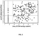
- Urinary JCV DNA levels were determined by a quantitative real-time polymerase chain reaction (q-PCR) assay (ViraCor Laboratories, Lee's Summit, MO) with a limit of quantitation of 500 copies/mL and a limit of detection of 50 copies/mL.
- q-PCR quantitative real-time polymerase chain reaction
- the anti-JCV antibody status of 831 MS patient serum samples which included samples from 204 JCV uropositive patients, was initially evaluated for anti-JCV antibodies in a screening ELISA to determine the distribution of serological responses.
- the assay results by urinary DNA status showed the presence of two overlapping yet distinct populations of JCV IgG reactivity ( FIG. 1 ).
- nOD 450 > 0.60 a stronger correlation was observed with a higher proportion of serum samples from individuals with high JCV DNA copies/mL exhibiting higher nOD 450 values, consistent with literature reports ( e.g. , Egli et al., J. Infect. Dis. 199:837-846, 2009 ). These data suggest that seronegative results are likely due to an absence of JCV infection, rather than to very low viral levels.
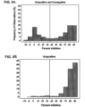
- a competition ELISA was developed using soluble HPVLP (secondary assay). JCV-specific higher affinity antibodies were expected to be more effectively competed by the soluble antigen, whereas lower affinity antibodies may detach from the complexes formed with the JCV antigen in solution and bind to the JCV VLP coated on the ELISA plate.
- the distribution of the serum responses in the confirmation ELISA consisted of two defined peaks, most optimally separated at 40% inhibition ( FIG. 5A ) corresponding approximately to the lower 5th percentile of the response distribution of uropositive samples ( FIG. 5B ). Therefore, the 40% inhibition level was selected as the cut point for the confirmation ELISA.
- This seroprevalence calculation assumed confirmation of anti-JCV antibodies in equal proportions of samples from uropositive and uronegative subjects in the nOD region between 0.10 and 0.25. (percent inhibition >40%); this assumption was supported by a 2-sided Fisher's exact test with a p -value of 0.702.
- Assay validation was performed by Focus Diagnostics, Inc. (Cypress, CA), where performance parameters including inter- and intra-assay precision, specificity, sensitivity and stability of assay reagents and controls were demonstrated. Assay performance parameters including inter- and intra-assay precision, specificity, sensitivity and stability of assay reagents and controls was demonstrated. Precision parameters were evaluated by three independent analysts in both plasma and serum on four different days using independent preparations of assay controls. For demonstration of assay specificity, ten individual serum and plasma samples from healthy volunteers or MS patients (TYSABRI ® (natalizumab) naive) were pre-incubated with either assay buffer or a defined concentration of HPVLP or BKV VLP in solution.
- Plasma and serum samples were obtained from a total of 831 patients from the Safety of TYSABRI Re-dosing And Treatment (STRATA) study.
- STRATA is an open-label, single-arm, multinational study (North America, Europe, Australia, and New Zealand) in which all patients receive natalizumab 300 mg by intravenous infusion every 4 weeks for 48 weeks.
- Urine samples collected according to the STRATA protocol were analyzed for the presence of JCV DNA.
- anti-JCV antibodies were detected in all 11 patients (100%) via the combination of the serological status screening ELISA and the supplemental confirmation ELISA ( FIGs. 6A and 6B ) described above. Using a one-sample Fisher's exact test, this result was significantly different from the expected proportion (53.6%) with a p-value of 0.002.
- This method is an example of an alternative to the density-gradient/ultracentrifugation method described above for the purification of JCV VP1-VLP's from insect cells.
- the general steps in the protocol are lysis, benzonase treatment, deoxycholate precipitation, ammonium sulfate precipitation and concentration/diafiltration, with a final ion-exchange step using TMAE fractogel.
- Sf9 cells infected with JCV-VP1 baculovirus were lysed in PBS, 0.1 mM CaCl 2 by passing twice through a microfluidizer cell disrupter at 5,000 psi. Cell debris was removed by low speed centrifugation and the supernatant treated with 40 units/ml Benzonase (EMD Biosciences 71206-3) for 1 hour at room temperature.
- EMD Biosciences 71206-3 40 units/ml Benzonase
- one tenth volume 2.5 % deoxycholate was added to the lysate (0.25% final deoxycholate), and the lysate was incubated at 37° C for 1 hour with gentle stirring.
- the ammonium sulfate precipitate was removed by low speed centrifugation and the VP1-containing supernatant was filtered using a 0.45 ⁇ m filter and carried on to the next step.
- the solution was concentrated 5 to 10 fold using a 100 kDa NMWL TFF membrane (Pellicon 2 Mini UF Mod Biomax-100 C 0.1m 2 , P2B100C01) and exchanged into assembly buffer (25 mM tris, 150 mM NaCl, 1 mM CaCl 2 , pH 7.5) by diluting 5 fold and concentrating back to the starting volume twice.
- the solution was then diafiltered using a 500 kDa NMWL TFF membrane (Pellicon 2 Mini UF Mod Biomax-500 V, Millipore part # P2B500V01) using 40 volumes TMA chromatography buffer (25 mM tris, 150 mM NaCl, 0.1 mM CaCl 2 , pH 8.0). For the chromatography, approximately 1 ml resin is required per 2 g starting cell mass.
- the protein was loaded onto the appropriately sized TMAE column (Fractogel ® EMD TMAE HiCap (M) - EMD Biosciences cat. 1.10316) and washed with 3 column volumes chromatography buffer.
- VLPs were eluted with 25 mM tris, 600 mM NaCl, 0.1 mM CaCl 2 , pH 8.0. VP1 purity was assessed by SDS-PAGE and mass spectrometry, presence of VLPs was confirmed by electron microscopy, and the percentage of total protein in the form of VLPs was determined by sedimentation velocity analytical ultracentrifugation. This method resulted in HPVLP preparations of about 80% HPVLPs.
Landscapes
- Health & Medical Sciences (AREA)
- Life Sciences & Earth Sciences (AREA)
- Immunology (AREA)
- Engineering & Computer Science (AREA)
- Molecular Biology (AREA)
- Biomedical Technology (AREA)
- Chemical & Material Sciences (AREA)
- Urology & Nephrology (AREA)
- Hematology (AREA)
- Virology (AREA)
- Food Science & Technology (AREA)
- General Health & Medical Sciences (AREA)
- Cell Biology (AREA)
- Biotechnology (AREA)
- Pathology (AREA)
- Medicinal Chemistry (AREA)
- Physics & Mathematics (AREA)
- Analytical Chemistry (AREA)
- Biochemistry (AREA)
- Microbiology (AREA)
- General Physics & Mathematics (AREA)
- Tropical Medicine & Parasitology (AREA)
- Proteomics, Peptides & Aminoacids (AREA)
- Peptides Or Proteins (AREA)
- Measuring Or Testing Involving Enzymes Or Micro-Organisms (AREA)
- Micro-Organisms Or Cultivation Processes Thereof (AREA)
- Medicines Containing Antibodies Or Antigens For Use As Internal Diagnostic Agents (AREA)
- Investigating Or Analysing Biological Materials (AREA)
Claims (18)
- In-vitro-Verfahren zum Identifizieren eines Subjekts mit einem Risiko, eine progressive multifokale Leukoenzephalopathie (PML) zu entwickeln, umfassend einen primären und einen sekundären Assay;wobei der primäre Assay Folgendes umfasst:(a) Inkontaktbringen einer von einem Subjekt erhaltenen biologischen Probe mit hochgereinigten VP1-Partikeln (HPVLPs), die überwiegend aus dem VP1-Protein des JCV bestehen und auf einem festen Substrat unter Bedingungen immobilisiert sind, die für die Bindung eines JC-Virus(JCV)-Antikörpers in der Probe an ein HPVLP geeignet sind;(b) Erfassen des Spiegels der Bindung von JCV-Antikörpern in der Probe an HPVLP; und(c) Korrelieren des erfassten Spiegels mit einem Referenzspiegel, der von einer Kontrollprobe oder einem Satz von Proben abgeleitet wird, die/der mit der Probe des Subjekts verarbeitet wird;und wobei der sekundäre Assay Folgendes umfasst:(a) Inkontaktbringen eines Teils der von einem Subjekt erhaltenen biologischen Probe mit hochgereinigten virusähnlichen Partikeln (HPVLPs), die überwiegend aus dem VP1-Protein des JCV bestehen, in Lösung unter Bedingungen, die für die Bindung eines JCV-Antikörpers in der Probe an ein HPVLP geeignet sind, wodurch eine vorinkubierte Probe bereitgestellt wird;(b) Inkontaktbringen der vorinkubierten Probe mit HPVLPs, die überwiegend aus dem VP1-Protein des JCV bestehen und auf einem festen Substrat unter Bedingungen immobilisiert sind, die für die Bindung eines JCV-Antikörpers in der Probe an ein HPVLP geeignet sind;(c) Erfassen des Spiegels an JCV-Antikörpern in der vorinkubierten Probe, die an die immobilisierten HPVLPs binden;(d) Vergleichen des erfassten Spiegels an JCV-Antikörpern in der vorinkubierten biologischen Probe mit dem erfassten Spiegel an JCV-Antikörpern in einer biologischen Probe, die von dem Subjekt erhalten wurde und die in einer Lösung ohne HPVLPs vorinkubiert wurde und mit HPVLPs, die auf einem festen Substrat unter Bedingungen, die für die Bindung eines JCV-Antikörpers in der Probe an ein HPVLP geeignet sind, immobilisiert wurden, in Kontakt gebracht wurde,wobei die HPVLPs aus mehr als 5, mindestens 50, 150 oder 360 VP1-Polypeptiden zusammengesetzt sind;wobei eine Abnahme des erfassten Spiegels an JCV-Antikörpern in der vorinkubierten biologischen Probe im Vergleich zu der von dem Subjekt erhaltenen biologischen Probe, die in einer Lösung ohne HPVLPs vorinkubiert wurde, angibt, dass die Probe positiv für JCV-Antikörper ist und das Subjekt ein erhöhtes Risiko aufweist, eine PML zu entwickeln,und eine Veränderung des erfassten Spiegels an JCV-Antikörpern unterhalb eines bestimmten Prozentsatzes angibt, dass in der Probe keine JCV-spezifischen Antikörper vorhanden sind.
- Verfahren nach Anspruch 1, wobei das Inkontaktbringen der biologischen Probe mit HPVLPs in Lösung für einen Zeitraum erfolgt, der ausgewählt ist aus 30 Minuten, einer Stunde oder über Nacht bei 4 °C.
- Verfahren nach Anspruch 1, wobei der spezifizierte Prozentsatz 40 % beträgt und das Verfahren einen HPVLP-ELISA verwendet.
- Verfahren nach Anspruch 1, wobei der Referenzspiegel so gewählt ist, dass er eine falsch negative Rate von 3 % oder weniger für das Erfassen von JCV-Antikörpern in der von dem Subjekt erhaltenen Probe bereitstellt.
- Verfahren nach Anspruch 1, wobei der Referenzspiegel so gewählt ist, dass er eine falsch negative Rate von 1 % oder weniger für das Erfassen von JCV-Antikörpern in der von dem Subjekt erhaltenen Probe bereitstellt.
- Verfahren nach Anspruch 1, wobei die von dem Subjekts erhaltene biologische Probe als negativ für JCV-Antikörper klassifiziert wird, wenn der Spiegel an JCV-Antikörpern in der vorinkubierten biologischen Probe, die an die immobilisierten HPVLP binden, ungefähr gleich dem Spiegel an JCV-Antikörpern ist, der in der biologischen Probe erfasst wurde, die von dem Subjekt erhalten wurde und die in einer Lösung ohne HPVLPs vorinkubiert wurde.
- Verfahren nach Anspruch 1, wobei die HPVLPs mehr als 1, mindestens 5, 10, 20, 30, 40, 50, 60, 70 oder 72 VP1-Pentamere enthalten.
- Verfahren nach Anspruch 1, wobei:(i) ein HPVLP ferner mindestens eines von einem JCV-VP2 oder einem JCV-VP3 umfasst; oder(ii) das VP1 in einem HPVLP ein rekombinantes VP1 ist; oder(iii) mindestens ein VP1 im HPVLP ein mutiertes VP1 ist.
- Verfahren nach Anspruch 1, wobei die biologische Probe Serum ist.
- Verfahren nach Anspruch 1, wobei die biologische Probe von einem Subjekt, dem ein Immunmodulator verschrieben wurde, oder einem Subjekt, das die Einnahme eines Immunmodulators in Betracht zieht, stammt, wobei der Immunmodulator ausgewählt ist aus einer Anti-VLA-4-Therapie und einer Anti-CD20-Therapie, einer Anti-CD11a-Therapie oder Mycophenolatmofetil.
- Verfahren nach Anspruch 10, wobei dem Subjekt, dem ein Immunmodulator verschrieben wurde oder das die Einnahme eines Immunmodulators in Betracht zieht, der Immunmodulator zuvor nicht verabreicht wurde.
- Verfahren nach Anspruch 10, wobei das Subjekt zuvor eine oder mehrere Dosen des Immunmodulators erhalten hat.
- Verfahren nach Anspruch 1, wobei das Erfassen von JCV-Antikörpern, die an die HPVLPs binden, angibt, dass das Subjekt:(i) kein Kandidat für eine Behandlung mit einem Immunmodulator ist, wenn die biologische Probe positiv für einen JCV-Antikörper ist; oder(ii) ein Kandidat für eine Behandlung mit einem Immunmodulator und eine verstärkte Überwachung auf unerwünschte Symptome bei der Behandlung mit dem Immunmodulator ist, optional wobei die unerwünschten Symptome die Entwicklung einer PML angeben.
- Verfahren nach Anspruch 1, wobei das fehlende Erfassen einer Bindung von JCV-Antikörpern an HPVLPs angibt, dass das Subjekt ein Kandidat für eine Behandlung mit einem Immunmodulator ist.
- Verfahren nach Anspruch 1, wobei ein Subjekt mit einer biologischen Probe, bei der in einem ersten Test bestimmt wurde, dass sie keine JCV-Antikörper aufweist, nach dem ersten Test mindestens jährlich erneut auf die Anwesenheit von JCV-Antikörpern getestet wird.
- Verfahren nach Anspruch 1, wobei das Subjekt, bei dem an einem ersten Datum durch das Verfahren nach Anspruch 1 bestimmt wurde, dass es JCV-Antikörper aufweist, zu einem späteren Zeitpunkt erneut getestet wird, um zu bestimmen, ob das Subjekt keine JCV-Antikörper aufweist.
- Verfahren nach einem der Ansprüche 10-14, wobei der Immunmodulator Natalizumab ist.
- Verfahren nach Anspruch 1, wobei das Subjekt Multiple Sklerose (MS) oder Morbus Crohn (CD) hat.
Priority Applications (5)
| Application Number | Priority Date | Filing Date | Title |
|---|---|---|---|
| RS20250151A RS66509B1 (sr) | 2010-01-11 | 2011-01-11 | Test za antitela na jc virus |
| EP24213770.1A EP4524571A3 (de) | 2010-01-11 | 2011-01-11 | Assay für jc-viren-antikörper |
| SI201132127T SI4152004T1 (sl) | 2010-01-11 | 2011-01-11 | Preiskava za protitelesa virusa jc |
| HRP20250210TT HRP20250210T1 (hr) | 2010-01-11 | 2011-01-11 | Analiza za antitijela jc virusa |
| SM20250065T SMT202500065T1 (it) | 2010-01-11 | 2011-01-11 | Saggio per anticorpi del virus jc |
Applications Claiming Priority (5)
| Application Number | Priority Date | Filing Date | Title |
|---|---|---|---|
| US29404810P | 2010-01-11 | 2010-01-11 | |
| US31619310P | 2010-03-22 | 2010-03-22 | |
| EP17203178.3A EP3339865B9 (de) | 2010-01-11 | 2011-01-11 | Assay für jc-virus-antikörper |
| PCT/US2011/020832 WO2011085369A1 (en) | 2010-01-11 | 2011-01-11 | Assay for jc virus antibodies |
| EP11732315.4A EP2524060B1 (de) | 2010-01-11 | 2011-01-11 | Assay für jc-viren-antikörper |
Related Parent Applications (3)
| Application Number | Title | Priority Date | Filing Date |
|---|---|---|---|
| EP11732315.4A Division EP2524060B1 (de) | 2010-01-11 | 2011-01-11 | Assay für jc-viren-antikörper |
| EP17203178.3A Division-Into EP3339865B9 (de) | 2010-01-11 | 2011-01-11 | Assay für jc-virus-antikörper |
| EP17203178.3A Division EP3339865B9 (de) | 2010-01-11 | 2011-01-11 | Assay für jc-virus-antikörper |
Related Child Applications (2)
| Application Number | Title | Priority Date | Filing Date |
|---|---|---|---|
| EP24213770.1A Division EP4524571A3 (de) | 2010-01-11 | 2011-01-11 | Assay für jc-viren-antikörper |
| EP24213770.1A Division-Into EP4524571A3 (de) | 2010-01-11 | 2011-01-11 | Assay für jc-viren-antikörper |
Publications (3)
| Publication Number | Publication Date |
|---|---|
| EP4152004A1 EP4152004A1 (de) | 2023-03-22 |
| EP4152004B1 true EP4152004B1 (de) | 2024-11-20 |
| EP4152004B9 EP4152004B9 (de) | 2025-04-09 |
Family
ID=44305841
Family Applications (4)
| Application Number | Title | Priority Date | Filing Date |
|---|---|---|---|
| EP24213770.1A Pending EP4524571A3 (de) | 2010-01-11 | 2011-01-11 | Assay für jc-viren-antikörper |
| EP11732315.4A Revoked EP2524060B1 (de) | 2010-01-11 | 2011-01-11 | Assay für jc-viren-antikörper |
| EP17203178.3A Active EP3339865B9 (de) | 2010-01-11 | 2011-01-11 | Assay für jc-virus-antikörper |
| EP22191259.5A Active EP4152004B9 (de) | 2010-01-11 | 2011-01-11 | Assay für jc-viren-antikörper |
Family Applications Before (3)
| Application Number | Title | Priority Date | Filing Date |
|---|---|---|---|
| EP24213770.1A Pending EP4524571A3 (de) | 2010-01-11 | 2011-01-11 | Assay für jc-viren-antikörper |
| EP11732315.4A Revoked EP2524060B1 (de) | 2010-01-11 | 2011-01-11 | Assay für jc-viren-antikörper |
| EP17203178.3A Active EP3339865B9 (de) | 2010-01-11 | 2011-01-11 | Assay für jc-virus-antikörper |
Country Status (27)
| Country | Link |
|---|---|
| US (2) | US9316641B2 (de) |
| EP (4) | EP4524571A3 (de) |
| JP (2) | JP5946218B2 (de) |
| KR (1) | KR101877576B1 (de) |
| CN (1) | CN102906278A (de) |
| AU (1) | AU2011203815B2 (de) |
| BR (1) | BR112012017014B1 (de) |
| CA (1) | CA2784137A1 (de) |
| CY (2) | CY1119972T1 (de) |
| DK (3) | DK4152004T3 (de) |
| ES (3) | ES2664173T3 (de) |
| FI (2) | FI3339865T5 (de) |
| HR (3) | HRP20221390T1 (de) |
| HU (3) | HUE060312T2 (de) |
| IN (1) | IN2012DN06139A (de) |
| LT (3) | LT3339865T (de) |
| ME (1) | ME03026B (de) |
| MX (1) | MX341991B (de) |
| NO (1) | NO2524060T3 (de) |
| NZ (1) | NZ600681A (de) |
| PL (3) | PL3339865T3 (de) |
| PT (3) | PT2524060T (de) |
| RS (3) | RS63744B1 (de) |
| SG (2) | SG181653A1 (de) |
| SI (3) | SI3339865T1 (de) |
| SM (3) | SMT202500065T1 (de) |
| WO (1) | WO2011085369A1 (de) |
Families Citing this family (17)
| Publication number | Priority date | Publication date | Assignee | Title |
|---|---|---|---|---|
| DK2645106T4 (da) | 2005-04-04 | 2024-12-02 | Biogen Ma Inc | Fremgangsmåder til evaluering af et immunrespons på et terapeutisk middel |
| US20070207141A1 (en) | 2006-02-28 | 2007-09-06 | Ivan Lieberburg | Methods of treating inflammatory and autoimmune diseases with natalizumab |
| MX2008011176A (es) | 2006-03-03 | 2008-09-10 | Elan Pharm Inc | Metodos para tratar padecimientos inflamatorios y autoinmunes con natalizumab. |
| ES2666372T3 (es) | 2009-10-11 | 2018-05-04 | Biogen Ma Inc. | Ensayos relacionados con anti-VLA-4 |
| US11287423B2 (en) * | 2010-01-11 | 2022-03-29 | Biogen Ma Inc. | Assay for JC virus antibodies |
| CN102906278A (zh) | 2010-01-11 | 2013-01-30 | 比奥根艾迪克Ma公司 | 用于jc病毒抗体的测定 |
| DK2715352T3 (da) | 2011-05-31 | 2019-05-20 | Biogen Ma Inc | Fremgangsmåde til vurdering af risiko for pml |
| EP2791162A2 (de) * | 2011-12-12 | 2014-10-22 | Janssen Diagnostics BVBA | Polyomavirus-peptidsequenzen |
| CA2870593A1 (en) * | 2012-04-16 | 2013-10-24 | Board Of Regents, The University Of Texas System | Detection of extracellular jcv micrornas |
| WO2013192100A1 (en) | 2012-06-18 | 2013-12-27 | The United States Of America, As Represented By The Secretary, Department Of Health & Human Services | Methods and compositions for detecting jc virus |
| CN104936980B (zh) * | 2012-12-31 | 2019-06-07 | 生物控股有限公司 | 用于治疗和预防多瘤病毒相关的疾病的重组人抗体 |
| US9949671B2 (en) | 2013-03-13 | 2018-04-24 | Orthoaccel Technologies, Inc. | Diagnostic mouthpieces |
| WO2014193804A1 (en) | 2013-05-28 | 2014-12-04 | Biogen Idec Ma Inc. | Method of assessing risk of pml |
| EP3110976B1 (de) * | 2014-02-27 | 2020-05-13 | Biogen MA Inc. | Verfahren zur beurteilung eines pml-risikos |
| MA40985A (fr) | 2014-11-17 | 2017-09-26 | Biogen Ma Inc | Méthodes de traitement de la sclérose en plaques |
| IT201600083859A1 (it) * | 2016-08-09 | 2018-02-09 | Univ Degli Studi Di Ferrara | Immunosaggio per l’identificazione di anticorpi contro il virus Polioma JC (JCPyV) mediante l’uso di peptidi sintetici. |
| WO2019169317A1 (en) * | 2018-03-02 | 2019-09-06 | Stueve Olaf | Methods and compositions for treating natalizumab-associated progressive multifocal encephalopathy |
Citations (1)
| Publication number | Priority date | Publication date | Assignee | Title |
|---|---|---|---|---|
| WO2007100770A2 (en) | 2006-02-28 | 2007-09-07 | Elan Pharmaceuticals, Inc. | Methods of treating inflammatory and autoimmune diseases with natalizumab |
Family Cites Families (94)
| Publication number | Priority date | Publication date | Assignee | Title |
|---|---|---|---|---|
| NL154598B (nl) | 1970-11-10 | 1977-09-15 | Organon Nv | Werkwijze voor het aantonen en bepalen van laagmoleculire verbindingen en van eiwitten die deze verbindingen specifiek kunnen binden, alsmede testverpakking. |
| US3817837A (en) | 1971-05-14 | 1974-06-18 | Syva Corp | Enzyme amplification assay |
| US3939350A (en) | 1974-04-29 | 1976-02-17 | Board Of Trustees Of The Leland Stanford Junior University | Fluorescent immunoassay employing total reflection for activation |
| US3996345A (en) | 1974-08-12 | 1976-12-07 | Syva Company | Fluorescence quenching with immunological pairs in immunoassays |
| US4277437A (en) | 1978-04-05 | 1981-07-07 | Syva Company | Kit for carrying out chemically induced fluorescence immunoassay |
| US4275149A (en) | 1978-11-24 | 1981-06-23 | Syva Company | Macromolecular environment control in specific receptor assays |
| US4235601A (en) | 1979-01-12 | 1980-11-25 | Thyroid Diagnostics, Inc. | Test device and method for its use |
| US4391904A (en) | 1979-12-26 | 1983-07-05 | Syva Company | Test strip kits in immunoassays and compositions therein |
| US4376110A (en) | 1980-08-04 | 1983-03-08 | Hybritech, Incorporated | Immunometric assays using monoclonal antibodies |
| US4366241A (en) | 1980-08-07 | 1982-12-28 | Syva Company | Concentrating zone method in heterogeneous immunoassays |
| US4517288A (en) | 1981-01-23 | 1985-05-14 | American Hospital Supply Corp. | Solid phase system for ligand assay |
| US4703017C1 (en) | 1984-02-14 | 2001-12-04 | Becton Dickinson Co | Solid phase assay with visual readout |
| CA1291031C (en) | 1985-12-23 | 1991-10-22 | Nikolaas C.J. De Jaeger | Method for the detection of specific binding agents and their correspondingbindable substances |
| US5763262A (en) | 1986-09-18 | 1998-06-09 | Quidel Corporation | Immunodiagnostic device |
| DE3856421T2 (de) | 1987-04-27 | 2000-12-14 | Unilever Nv | Spezifische Bindungstestverfahren |
| US4943522A (en) | 1987-06-01 | 1990-07-24 | Quidel | Lateral flow, non-bibulous membrane assay protocols |
| US5120643A (en) | 1987-07-13 | 1992-06-09 | Abbott Laboratories | Process for immunochromatography with colloidal particles |
| US4818677A (en) | 1987-12-03 | 1989-04-04 | Monoclonal Antibodies, Inc. | Membrane assay using focused sample application |
| AU2684488A (en) | 1988-06-27 | 1990-01-04 | Carter-Wallace, Inc. | Test device and method for colored particle immunoassay |
| US5221616A (en) | 1988-07-15 | 1993-06-22 | Quidel Corporation | Prevention of spontaneous complement activation in mammalian biological fluids |
| US5118630A (en) | 1988-11-04 | 1992-06-02 | Quidel Corporation | Method for determining periodic infertility in females |
| US6352862B1 (en) | 1989-02-17 | 2002-03-05 | Unilever Patent Holdings B.V. | Analytical test device for imuno assays and methods of using same |
| US6033665A (en) | 1989-09-27 | 2000-03-07 | Elan Pharmaceuticals, Inc. | Compositions and methods for modulating leukocyte adhesion to brain endothelial cells |
| US5084828A (en) | 1989-09-29 | 1992-01-28 | Healthtech Services Corp. | Interactive medication delivery system |
| US5252496A (en) | 1989-12-18 | 1993-10-12 | Princeton Biomeditech Corporation | Carbon black immunochemical label |
| US5096837A (en) | 1990-02-08 | 1992-03-17 | Pacific Biotech, Inc. | Immunochromatographic assay and method of using same |
| US5223220A (en) | 1990-03-27 | 1993-06-29 | Pacific Biotech, Inc. | Solid phase immunoassay device and method of making same |
| US5118428A (en) | 1990-11-13 | 1992-06-02 | Quidel | Method to remove red blood cells from whole blood samples |
| DE4139840B4 (de) | 1990-12-04 | 2005-06-02 | Quidel Corp., San Diego | Antigen-Zubereitung zum Nachweis von H. pylori |
| WO1992012428A1 (en) | 1991-01-11 | 1992-07-23 | Quidel Corporation | A one-step lateral flow nonbibulous assay |
| US5213796A (en) | 1991-05-06 | 1993-05-25 | Dana Farber Cancer Institute | Assay for polyomavirus in humans and uses thereof |
| US5225328A (en) | 1991-05-30 | 1993-07-06 | Quidel Corporation | Stable alkaline phosphatase compositions with color enhancement and their use in assays |
| US5686315A (en) | 1991-06-14 | 1997-11-11 | Quidel Corporation | Assay device for one step detection of analyte |
| WO1993015217A1 (en) | 1992-02-04 | 1993-08-05 | Quidel Corporation | Simplified extraction method for bacterial antigens using dried reagents |
| US5541069A (en) | 1992-02-28 | 1996-07-30 | Quidel Corporation | Assay having improved dose response curve |
| JP2948318B2 (ja) | 1992-03-10 | 1999-09-13 | クイデル コーポレイション | 特異的結合アッセイ用の赤血球の分離方法 |
| DE69419721T2 (de) | 1993-01-12 | 2000-04-27 | Biogen, Inc. | Rekombinante anti-vla4 antikörpermoleküle |
| US5415994A (en) | 1993-08-02 | 1995-05-16 | Quidel Corporation | Lateral flow medical diagnostic assay device with sample extraction means |
| US5840299A (en) | 1994-01-25 | 1998-11-24 | Athena Neurosciences, Inc. | Humanized antibodies against leukocyte adhesion molecule VLA-4 |
| US5434057A (en) | 1994-02-02 | 1995-07-18 | Quidel Corporation | Sperm motility assay and devices |
| US5521102A (en) | 1994-08-08 | 1996-05-28 | Quidel Corporation | Controlled sensitivity immunochromatographic assay |
| US5845255A (en) | 1994-10-28 | 1998-12-01 | Advanced Health Med-E-Systems Corporation | Prescription management system |
| US6551593B1 (en) | 1995-02-10 | 2003-04-22 | Millennium Pharmaceuticals, Inc. | Treatment of Inflammatory bowel disease by inhibiting binding and/or signalling through α 4 β 7 and its ligands and madcam |
| US5712172A (en) | 1995-05-18 | 1998-01-27 | Wyntek Diagnostics, Inc. | One step immunochromatographic device and method of use |
| US5804452A (en) | 1995-04-27 | 1998-09-08 | Quidel Corporation | One step urine creatinine assays |
| US5786220A (en) | 1995-04-28 | 1998-07-28 | Quidel Corporation | Assays and devices for distinguishing between normal and abnormal pregnancy |
| US6319676B1 (en) | 1995-05-02 | 2001-11-20 | Carter Wallace, Inc. | Diagnostic detection device and method |
| US5773234A (en) | 1995-08-07 | 1998-06-30 | Quidel Corporation | Method and device for chlamydia detection |
| DE19543553B4 (de) * | 1995-11-22 | 2009-04-09 | Deutsches Primatenzentrum Gmbh | VP-Antigene des JC-Virus |
| US6305377B1 (en) | 1996-12-12 | 2001-10-23 | Michael T. Portwood | System and method for improving compliance of a medical regimen |
| US6229011B1 (en) | 1997-08-22 | 2001-05-08 | Hoffman-La Roche Inc. | N-aroylphenylalanine derivative VCAM-1 inhibitors |
| US6306642B1 (en) | 1997-11-24 | 2001-10-23 | Quidel Corporation | Enzyme substrate delivery and product registration in one step enzyme immunoassays |
| US6623981B2 (en) | 1998-01-27 | 2003-09-23 | Bristol-Myers Squibb Company | Detection of patients at risk for developing integrin antagonist/agonist mediated disease states |
| US6014631A (en) | 1998-04-02 | 2000-01-11 | Merck-Medco Managed Care, Llc | Computer implemented patient medication review system and process for the managed care, health care and/or pharmacy industry |
| US6045501A (en) | 1998-08-28 | 2000-04-04 | Celgene Corporation | Methods for delivering a drug to a patient while preventing the exposure of a foetus or other contraindicated individual to the drug |
| US6407066B1 (en) | 1999-01-26 | 2002-06-18 | Elan Pharmaceuticals, Inc. | Pyroglutamic acid derivatives and related compounds which inhibit leukocyte adhesion mediated by VLA-4 |
| US7171371B2 (en) | 1999-09-03 | 2007-01-30 | Smg Trust | Method and system for providing pre and post operative support and care |
| SI1232392T1 (en) | 1999-10-12 | 2003-10-31 | Connex Gesellschaft Zur Optimierung Von Forschung Und | Improved method for the detection of acid resistant bacteria of the genus helicobacter in stool |
| US6388084B1 (en) | 1999-12-06 | 2002-05-14 | Hoffmann-La Roche Inc. | 4-pyridinyl-n-acyl-l-phenylalanines |
| NZ519447A (en) | 1999-12-16 | 2004-03-26 | Biogen Inc | Methods of treating central nervous system ischemic or hemorrhagic injury using anti alpha4 integrin antagonists |
| WO2001075449A1 (fr) | 2000-03-30 | 2001-10-11 | Nippon Kayaku Kabushiki Kaisha | Technique d'investigation du cancer par epreuve biologique d'un autoanticorps contre mdm2 et reactif |
| US6620626B1 (en) | 2000-08-09 | 2003-09-16 | Mission Research Corp. | Antigen detection device and method |
| MY129000A (en) | 2000-08-31 | 2007-03-30 | Tanabe Seiyaku Co | INHIBITORS OF a4 MEDIATED CELL ADHESION |
| WO2002033417A1 (en) | 2000-10-17 | 2002-04-25 | Besst-Test Aps | Assay for directly detecting a rs virus related biological cell in a body fluid sample |
| US6315720B1 (en) | 2000-10-23 | 2001-11-13 | Celgene Corporation | Methods for delivering a drug to a patient while avoiding the occurrence of an adverse side effect known or suspected of being caused by the drug |
| US6485460B2 (en) | 2001-01-12 | 2002-11-26 | Bracco Diagnostics, Inc. | Tamper evident syringe barrel |
| WO2003016902A1 (en) | 2001-08-20 | 2003-02-27 | Proteome Systems Intellectual Property Pty Ltd | Diagnostic testing process and apparatus |
| US7205159B2 (en) * | 2001-08-20 | 2007-04-17 | Proteome Systems Intellectual Property Pty Ltd. | Diagnostic testing process and apparatus |
| US6605602B1 (en) | 2001-09-28 | 2003-08-12 | University Of Pittsburgh-Of The Commonwealth System Of Higher Education | Method of treating BK virus nephropathy |
| DK1485127T3 (da) | 2002-02-25 | 2011-10-03 | Elan Pharm Inc | Anvendelse af midler til behandling af inflammation |
| TW200307671A (en) | 2002-05-24 | 2003-12-16 | Elan Pharm Inc | Heteroaryl compounds which inhibit leukocyte adhesion mediated by α 4 integrins |
| TWI281470B (en) | 2002-05-24 | 2007-05-21 | Elan Pharm Inc | Heterocyclic compounds which inhibit leukocyte adhesion mediated by alpha4 integrins |
| WO2004001539A2 (en) | 2002-06-21 | 2003-12-31 | Mckesson Information Solutions Llc | Closed loop medication use system and method |
| US20070275481A1 (en) | 2003-11-24 | 2007-11-29 | Biogen Idec Ma Inc. | Methods For Detecting Half-Antibodies Using Chip Based Gel Electophoresis |
| JP4672263B2 (ja) | 2004-01-27 | 2011-04-20 | デンカ生研株式会社 | 簡便な検出法、検出装置及び検出キットとその製法 |
| US7419666B1 (en) | 2004-02-23 | 2008-09-02 | Massachusetts Eye And Ear Infirmary | Treatment of ocular disorders |
| TW201705980A (zh) | 2004-04-09 | 2017-02-16 | 艾伯維生物技術有限責任公司 | 用於治療TNFα相關失調症之多重可變劑量療法 |
| US20050283385A1 (en) | 2004-06-21 | 2005-12-22 | The Permanente Medical Group, Inc. | Individualized healthcare management system |
| US7794700B2 (en) | 2004-07-08 | 2010-09-14 | Elan Pharmaceuticals Inc. | Multimeric VLA-4 antagonists comprising polymer moieties |
| CA2478458A1 (en) | 2004-08-20 | 2006-02-20 | Michael Panzara | Treatment of pediatric multiple sclerosis |
| NZ581497A (en) | 2004-12-03 | 2012-07-27 | Biogen Idec Inc | Delaying or preventing onset of multiple sclerosis by vla-4 ginding antibody |
| EP1863940B1 (de) | 2005-03-03 | 2012-06-06 | Seedlings Life Science Ventures, LLC. | Verfahren zum risikomanagement für mit natalizumab behandelten patienten |
| DK2645106T4 (da) | 2005-04-04 | 2024-12-02 | Biogen Ma Inc | Fremgangsmåder til evaluering af et immunrespons på et terapeutisk middel |
| BRPI0616687A2 (pt) | 2005-09-29 | 2011-06-28 | Elan Pharm Inc | compostos de pirimidinil amida que inibem a adesão leucocitária mediada por vla-4 |
| CA2643838A1 (en) | 2006-02-27 | 2007-09-07 | Elan Pharmaceuticals, Inc. | Pyrimidinyl sulfonamide compounds which inhibit leukocyte adhesion mediated by vla-4 |
| US8410115B2 (en) * | 2006-02-28 | 2013-04-02 | Elan Pharmaceuticals, Inc. | Methods of treating inflammatory and autoimmune diseases with alpha-4 inhibitory compounds |
| MX2008011176A (es) | 2006-03-03 | 2008-09-10 | Elan Pharm Inc | Metodos para tratar padecimientos inflamatorios y autoinmunes con natalizumab. |
| US7838583B2 (en) * | 2006-06-30 | 2010-11-23 | Sumitomo Rubber Industries, Ltd. | Rubber composition for cap tread and pneumatic tire having cap tread using same |
| WO2008061243A2 (en) * | 2006-11-16 | 2008-05-22 | Novavax, Inc. | Respiratory syncytial virus-virus like particle (vlps) |
| EP1933140B1 (de) | 2006-12-11 | 2011-08-17 | AraGen Biotechnology Co. Ltd. | Verfahren zur Detektion von Antikörpern mittels eines durch Oligonukleotide verstärkten kolloidalen Gold Signals |
| CN102369436B (zh) | 2009-02-05 | 2014-10-22 | 生物基因Idecma公司 | 用于检测jc多瘤病毒的方法 |
| US9354233B2 (en) | 2009-02-20 | 2016-05-31 | The Regents Of The University Of California | A+ biomarker assays |
| CN102906278A (zh) | 2010-01-11 | 2013-01-30 | 比奥根艾迪克Ma公司 | 用于jc病毒抗体的测定 |
| DK2715352T3 (da) | 2011-05-31 | 2019-05-20 | Biogen Ma Inc | Fremgangsmåde til vurdering af risiko for pml |
-
2011
- 2011-01-11 CN CN2011800133864A patent/CN102906278A/zh active Pending
- 2011-01-11 BR BR112012017014-7A patent/BR112012017014B1/pt active IP Right Grant
- 2011-01-11 SI SI201132069T patent/SI3339865T1/sl unknown
- 2011-01-11 EP EP24213770.1A patent/EP4524571A3/de active Pending
- 2011-01-11 HR HRP20221390TT patent/HRP20221390T1/hr unknown
- 2011-01-11 ES ES11732315.4T patent/ES2664173T3/es active Active
- 2011-01-11 DK DK22191259.5T patent/DK4152004T3/da active
- 2011-01-11 NO NO11732315A patent/NO2524060T3/no unknown
- 2011-01-11 ES ES22191259T patent/ES3012733T3/es active Active
- 2011-01-11 WO PCT/US2011/020832 patent/WO2011085369A1/en not_active Ceased
- 2011-01-11 PT PT117323154T patent/PT2524060T/pt unknown
- 2011-01-11 SG SG2012043154A patent/SG181653A1/en unknown
- 2011-01-11 LT LTEP17203178.3T patent/LT3339865T/lt unknown
- 2011-01-11 NZ NZ600681A patent/NZ600681A/en unknown
- 2011-01-11 HU HUE17203178A patent/HUE060312T2/hu unknown
- 2011-01-11 EP EP11732315.4A patent/EP2524060B1/de not_active Revoked
- 2011-01-11 SM SM20250065T patent/SMT202500065T1/it unknown
- 2011-01-11 CA CA2784137A patent/CA2784137A1/en active Pending
- 2011-01-11 KR KR1020127019171A patent/KR101877576B1/ko active Active
- 2011-01-11 MX MX2012007660A patent/MX341991B/es active IP Right Grant
- 2011-01-11 LT LTEP11732315.4T patent/LT2524060T/lt unknown
- 2011-01-11 FI FIEP17203178.3T patent/FI3339865T5/fi active
- 2011-01-11 AU AU2011203815A patent/AU2011203815B2/en active Active
- 2011-01-11 DK DK17203178.3T patent/DK3339865T5/da active
- 2011-01-11 IN IN6139DEN2012 patent/IN2012DN06139A/en unknown
- 2011-01-11 PT PT172031783T patent/PT3339865T/pt unknown
- 2011-01-11 FI FIEP22191259.5T patent/FI4152004T3/fi active
- 2011-01-11 RS RS20221053A patent/RS63744B1/sr unknown
- 2011-01-11 HU HUE11732315A patent/HUE036183T2/hu unknown
- 2011-01-11 PL PL17203178.3T patent/PL3339865T3/pl unknown
- 2011-01-11 RS RS20180245A patent/RS56977B1/sr unknown
- 2011-01-11 HU HUE22191259A patent/HUE070903T2/hu unknown
- 2011-01-11 DK DK11732315.4T patent/DK2524060T3/en active
- 2011-01-11 SM SM20220445T patent/SMT202200445T1/it unknown
- 2011-01-11 EP EP17203178.3A patent/EP3339865B9/de active Active
- 2011-01-11 JP JP2012548227A patent/JP5946218B2/ja active Active
- 2011-01-11 SI SI201131438T patent/SI2524060T1/en unknown
- 2011-01-11 SM SM20180148T patent/SMT201800148T1/it unknown
- 2011-01-11 SI SI201132127T patent/SI4152004T1/sl unknown
- 2011-01-11 PL PL22191259.5T patent/PL4152004T3/pl unknown
- 2011-01-11 PL PL11732315T patent/PL2524060T3/pl unknown
- 2011-01-11 RS RS20250151A patent/RS66509B1/sr unknown
- 2011-01-11 ME MEP-2018-60A patent/ME03026B/de unknown
- 2011-01-11 HR HRP20180356TT patent/HRP20180356T1/hr unknown
- 2011-01-11 SG SG10201500079YA patent/SG10201500079YA/en unknown
- 2011-01-11 HR HRP20250210TT patent/HRP20250210T1/hr unknown
- 2011-01-11 ES ES17203178T patent/ES2930469T3/es active Active
- 2011-01-11 LT LTEP22191259.5T patent/LT4152004T/lt unknown
- 2011-01-11 EP EP22191259.5A patent/EP4152004B9/de active Active
- 2011-01-11 US US13/521,311 patent/US9316641B2/en active Active
- 2011-01-11 PT PT221912595T patent/PT4152004T/pt unknown
-
2015
- 2015-12-18 JP JP2015246985A patent/JP2016040557A/ja not_active Withdrawn
-
2016
- 2016-03-08 US US15/063,777 patent/US10444234B2/en active Active
-
2018
- 2018-02-28 CY CY20181100251T patent/CY1119972T1/el unknown
-
2022
- 2022-11-18 CY CY20221100739T patent/CY1126069T1/el unknown
Patent Citations (1)
| Publication number | Priority date | Publication date | Assignee | Title |
|---|---|---|---|---|
| WO2007100770A2 (en) | 2006-02-28 | 2007-09-07 | Elan Pharmaceuticals, Inc. | Methods of treating inflammatory and autoimmune diseases with natalizumab |
Non-Patent Citations (11)
| Title |
|---|
| "Polyomavirus and human diseases Chapter 5", 2 February 2006, article VISCIDI RAPHAEL P., BARBARA CLAYMAN: "Serological Cross Reactivity between Polyomavirus Capsids", pages: 73 - 84, XP093097121 |
| ADRIAN EGLI, LAURA INFANTI, ALEXIS DUMOULIN, ANDREAS BUSER, JACQUELINE SAMARIDIS, CHRISTINE STEBLER, RAINER GOSERT, HANS H. H: "Prevalence of Polyomavirus BK and JC Infection and Replication in 400 Healthy Blood Donors", JOURNAL OF INFECTIOUS DISEASES, OXFORD UNIVERSITY PRESS, vol. 199, no. 6, 15 March 2009 (2009-03-15), pages 837 - 846, XP055491320, ISSN: 0022-1899, DOI: 10.1086/597126 |
| BERGER JOSEPH R., HOUFF SIDNEY A., MAJOR EUGENE O.: "Monoclonal antibodies and progressive multifocal leukoencephalopathy", MABS, LANDES BIOSCIENCE, US, vol. 1, no. 6, 1 November 2009 (2009-11-01), US , pages 583 - 589, XP093130366, ISSN: 1942-0862, DOI: 10.4161/mabs.1.6.9884 |
| BUBB M. O., T. MARIMUTHU, J.D. CONRADIE: "Hepatitis B surface antigen in donated blood — screening and confirmation by enzyme-linked immunosorbent assay", SA MEDIESE TYDSKRIF / SA MEDICAL JOURNAL, vol. 63, 29 January 1983 (1983-01-29), pages 148 - 151, XP093308229 |
| CARSON, K.R. ; FOCOSI, D. ; MAJOR, E.O. ; PETRINI, M. ; RICHEY, E.A. ; WEST, D.P. ; BENNETT, C.L.: "Monoclonal antibody-associated progressive multifocal leucoencephalopathy in patients treated with rituximab, natalizumab, and efalizumab: a Review from the Research on Adverse Drug Events and Reports (RADAR) Project", THE LANCET ONCOLOGY, ELSEVIER, AMSTERDAM, NL, vol. 10, no. 8, 1 August 2009 (2009-08-01), AMSTERDAM, NL , pages 816 - 824, XP026393894 |
| D11 - Expert declaration by Sam Scrimshaw including the Exhibits cited therein |
| DANA E. ROLLISON, ERIC A. ENGELS, NEAL A. HALSEY, KEERTI V. SHAH, RAPHAEL P. VISCIDI, KATHY J. HELZLSOUER: "Prediagnostic circulating antibodies to JC and BK human polyomaviruses and risk of non-Hodgkin lymphoma.", CANCER EPIDEMIOLOGY, BIOMARKERS & PREVENTION, AMERICAN ASSOCIATION FOR CANCER RESEARCH, vol. 15, no. 3, 1 March 2006 (2006-03-01), pages 543 - 550, XP002711818, ISSN: 1055-9965, DOI: 10.1158/1055-9965.EPI-05-0728 |
| LEONID GORELIK, MICHAELA LERNER, SARAH BIXLER, MARY CROSSMAN, BRIAN SCHLAIN, KENNETH SIMON, AMY PACE, ANNE CHEUNG, LING LING CHEN,: "Anti-JC virus antibodies: Implications for PML Risk Stratification", ANNALS OF NEUROLOGY, WILEY-LISS., vol. 68, no. 3, 1 September 2010 (2010-09-01), pages 295 - 303, XP055136721, ISSN: 03645134, DOI: 10.1002/ana.22128 |
| N. KHANNA, M. WOLBERS, N. J. MUELLER, C. GARZONI, R. A. DU PASQUIER, C. A. FUX, P. VERNAZZA, E. BERNASCONI, R. VISCIDI, M. BATTEGA: "JC Virus-Specific Immune Responses in Human Immunodeficiency Virus Type 1 Patients with Progressive Multifocal Leukoencephalopathy", JOURNAL OF VIROLOGY, AMERICAN SOCIETY FOR MICROBIOLOGY., vol. 83, no. 9, 1 May 2009 (2009-05-01), pages 4404 - 4411, XP055133530, ISSN: 0022538X, DOI: 10.1128/JVI.02657-08 |
| R. P. VISCIDI, D. E. M. ROLLISON, E. VISCIDI, B. CLAYMAN, E. RUBALCABA, R. DANIEL, E. O. MAJOR, K. V. SHAH: "Serological Cross-Reactivities between Antibodies to Simian Virus 40, BK Virus, and JC Virus Assessed by Virus-Like-Particle-Based Enzyme Immunoassays", CLINICAL AND VACCINE IMMUNOLOGY, AMERICAN SOCIETY FOR MICROBIOLOGY, vol. 10, no. 2, 1 March 2003 (2003-03-01), pages 278 - 285, XP055162298, ISSN: 15566811, DOI: 10.1128/CDLI.10.2.278-285.2003 |
| WRIGHT P F, NILSSON E, VAN ROOIJ E M A, LELENTA M, JEGGO M H: "Standardisation and validation of enzymelinked immunosorbent assay techniques for the detection of antibody in infectious disease diagnosis", vol. 12, no. 2, 1 June 1993 (1993-06-01), pages 435 - 450, XP093308231, DOI: 10.20506/rst.12.2.691 |
Also Published As
Similar Documents
| Publication | Publication Date | Title |
|---|---|---|
| EP4152004B1 (de) | Assay für jc-viren-antikörper | |
| ES2729945T3 (es) | Procedimiento para valorar el riesgo de LMP | |
| EP2402755B1 (de) | Verfahren und Zusammensetzungen zur Erkennung von Herpes Simplex Virus Typ 2 | |
| US12345703B2 (en) | Assay for JC virus antibodies | |
| HK40088791A (en) | Assay for jc virus antibodies | |
| HK40088791B (en) | Assay for jc virus antibodies | |
| HK40123180A (en) | Assay for jc virus antibodies | |
| HK1255318B (en) | Assay for jc virus antibodies | |
| HK1177234B (en) | Assay for jc virus antibodies | |
| HK1177234A (en) | Assay for jc virus antibodies |
Legal Events
| Date | Code | Title | Description |
|---|---|---|---|
| REG | Reference to a national code |
Ref country code: HR Ref legal event code: TUEP Ref document number: P20250210T Country of ref document: HR |
|
| PUAI | Public reference made under article 153(3) epc to a published international application that has entered the european phase |
Free format text: ORIGINAL CODE: 0009012 |
|
| STAA | Information on the status of an ep patent application or granted ep patent |
Free format text: STATUS: THE APPLICATION HAS BEEN PUBLISHED |
|
| AC | Divisional application: reference to earlier application |
Ref document number: 2524060 Country of ref document: EP Kind code of ref document: P Ref document number: 3339865 Country of ref document: EP Kind code of ref document: P |
|
| AK | Designated contracting states |
Kind code of ref document: A1 Designated state(s): AL AT BE BG CH CY CZ DE DK EE ES FI FR GB GR HR HU IE IS IT LI LT LU LV MC MK MT NL NO PL PT RO RS SE SI SK SM TR |
|
| STAA | Information on the status of an ep patent application or granted ep patent |
Free format text: STATUS: REQUEST FOR EXAMINATION WAS MADE |
|
| REG | Reference to a national code |
Ref country code: HK Ref legal event code: DE Ref document number: 40088791 Country of ref document: HK |
|
| TPAC | Observations filed by third parties |
Free format text: ORIGINAL CODE: EPIDOSNTIPA |
|
| 17P | Request for examination filed |
Effective date: 20230922 |
|
| RAX | Requested extension states of the european patent have changed |
Extension state: ME Payment date: 20230922 Extension state: BA Payment date: 20230922 |
|
| RBV | Designated contracting states (corrected) |
Designated state(s): AL AT BE BG CH CY CZ DE DK EE ES FI FR GB GR HR HU IE IS IT LI LT LU LV MC MK MT NL NO PL PT RO RS SE SI SK SM TR |
|
| STAA | Information on the status of an ep patent application or granted ep patent |
Free format text: STATUS: EXAMINATION IS IN PROGRESS |
|
| 17Q | First examination report despatched |
Effective date: 20231205 |
|
| TPAC | Observations filed by third parties |
Free format text: ORIGINAL CODE: EPIDOSNTIPA |
|
| GRAP | Despatch of communication of intention to grant a patent |
Free format text: ORIGINAL CODE: EPIDOSNIGR1 |
|
| STAA | Information on the status of an ep patent application or granted ep patent |
Free format text: STATUS: GRANT OF PATENT IS INTENDED |
|
| INTG | Intention to grant announced |
Effective date: 20240627 |
|
| GRAS | Grant fee paid |
Free format text: ORIGINAL CODE: EPIDOSNIGR3 |
|
| GRAA | (expected) grant |
Free format text: ORIGINAL CODE: 0009210 |
|
| STAA | Information on the status of an ep patent application or granted ep patent |
Free format text: STATUS: THE PATENT HAS BEEN GRANTED |
|
| AC | Divisional application: reference to earlier application |
Ref document number: 2524060 Country of ref document: EP Kind code of ref document: P Ref document number: 3339865 Country of ref document: EP Kind code of ref document: P |
|
| AK | Designated contracting states |
Kind code of ref document: B1 Designated state(s): AL AT BE BG CH CY CZ DE DK EE ES FI FR GB GR HR HU IE IS IT LI LT LU LV MC MK MT NL NO PL PT RO RS SE SI SK SM TR |
|
| P01 | Opt-out of the competence of the unified patent court (upc) registered |
Free format text: CASE NUMBER: APP_56327/2024 Effective date: 20241015 |
|
| REG | Reference to a national code |
Ref country code: GB Ref legal event code: FG4D |
|
| REG | Reference to a national code |
Ref country code: CH Ref legal event code: EP |
|
| REG | Reference to a national code |
Ref country code: DE Ref legal event code: R096 Ref document number: 602011075122 Country of ref document: DE |
|
| REG | Reference to a national code |
Ref country code: IE Ref legal event code: FG4D |
|
| REG | Reference to a national code |
Ref country code: PT Ref legal event code: SC4A Ref document number: 4152004 Country of ref document: PT Date of ref document: 20250218 Kind code of ref document: T Free format text: AVAILABILITY OF NATIONAL TRANSLATION Effective date: 20250213 |
|
| REG | Reference to a national code |
Ref country code: FI Ref legal event code: FGE |
|
| REG | Reference to a national code |
Ref country code: DK Ref legal event code: T3 Effective date: 20250221 |
|
| REG | Reference to a national code |
Ref country code: NL Ref legal event code: FP |
|
| REG | Reference to a national code |
Ref country code: SE Ref legal event code: TRGR |
|
| REG | Reference to a national code |
Ref country code: CH Ref legal event code: PK Free format text: BERICHTIGUNG B9 |
|
| REG | Reference to a national code |
Ref country code: SK Ref legal event code: T3 Ref document number: E 45908 Country of ref document: SK |
|
| REG | Reference to a national code |
Ref country code: HR Ref legal event code: ODRP Ref document number: P20250210T Country of ref document: HR Payment date: 20250218 Year of fee payment: 15 |
|
| REG | Reference to a national code |
Ref country code: ES Ref legal event code: FG2A Ref document number: 3012733 Country of ref document: ES Kind code of ref document: T3 Effective date: 20250410 |
|
| REG | Reference to a national code |
Ref country code: HR Ref legal event code: T1PR Ref document number: P20250210 Country of ref document: HR |
|
| PGFP | Annual fee paid to national office [announced via postgrant information from national office to epo] |
Ref country code: SI Payment date: 20250131 Year of fee payment: 15 |
|
| REG | Reference to a national code |
Ref country code: GR Ref legal event code: EP Ref document number: 20250400361 Country of ref document: GR Effective date: 20250409 |
|
| PGFP | Annual fee paid to national office [announced via postgrant information from national office to epo] |
Ref country code: AL Payment date: 20250218 Year of fee payment: 15 |
|
| REG | Reference to a national code |
Ref country code: EE Ref legal event code: FG4A Ref document number: E024969 Country of ref document: EE Effective date: 20250217 |
|
| REG | Reference to a national code |
Ref country code: HU Ref legal event code: AG4A Ref document number: E070903 Country of ref document: HU |
|
| REG | Reference to a national code |
Ref country code: DE Ref legal event code: R026 Ref document number: 602011075122 Country of ref document: DE |
|
| PLBI | Opposition filed |
Free format text: ORIGINAL CODE: 0009260 |
|
| PLAX | Notice of opposition and request to file observation + time limit sent |
Free format text: ORIGINAL CODE: EPIDOSNOBS2 |
|
| 26 | Opposition filed |
Opponent name: POLPHARMA BIOLOGICS S.A. Effective date: 20250819 |
|
| PLBB | Reply of patent proprietor to notice(s) of opposition received |
Free format text: ORIGINAL CODE: EPIDOSNOBS3 |
|
| PGFP | Annual fee paid to national office [announced via postgrant information from national office to epo] |
Ref country code: LT Payment date: 20251217 Year of fee payment: 16 Ref country code: GB Payment date: 20251220 Year of fee payment: 16 |
|
| PGFP | Annual fee paid to national office [announced via postgrant information from national office to epo] |
Ref country code: MC Payment date: 20251219 Year of fee payment: 16 Ref country code: NO Payment date: 20251218 Year of fee payment: 16 |
|
| PGFP | Annual fee paid to national office [announced via postgrant information from national office to epo] |
Ref country code: SM Payment date: 20251219 Year of fee payment: 15 Ref country code: MK Payment date: 20251219 Year of fee payment: 16 Ref country code: PT Payment date: 20251218 Year of fee payment: 16 |
|
| PGFP | Annual fee paid to national office [announced via postgrant information from national office to epo] |
Ref country code: FI Payment date: 20251217 Year of fee payment: 16 Ref country code: DK Payment date: 20251217 Year of fee payment: 16 |
|
| PGFP | Annual fee paid to national office [announced via postgrant information from national office to epo] |
Ref country code: LU Payment date: 20251217 Year of fee payment: 16 Ref country code: NL Payment date: 20251217 Year of fee payment: 16 Ref country code: FR Payment date: 20251218 Year of fee payment: 16 |
|
| PGFP | Annual fee paid to national office [announced via postgrant information from national office to epo] |
Ref country code: BE Payment date: 20251217 Year of fee payment: 16 Ref country code: GR Payment date: 20251218 Year of fee payment: 16 |
|
| PGFP | Annual fee paid to national office [announced via postgrant information from national office to epo] |
Ref country code: SE Payment date: 20251217 Year of fee payment: 16 |
|
| PGFP | Annual fee paid to national office [announced via postgrant information from national office to epo] |
Ref country code: IE Payment date: 20251218 Year of fee payment: 16 Ref country code: CZ Payment date: 20251229 Year of fee payment: 16 |
|
| PGFP | Annual fee paid to national office [announced via postgrant information from national office to epo] |
Ref country code: MT Payment date: 20251230 Year of fee payment: 16 Ref country code: LV Payment date: 20251217 Year of fee payment: 16 |
|
| PGFP | Annual fee paid to national office [announced via postgrant information from national office to epo] |
Ref country code: PL Payment date: 20251229 Year of fee payment: 16 |
|
| PGFP | Annual fee paid to national office [announced via postgrant information from national office to epo] |
Ref country code: EE Payment date: 20251222 Year of fee payment: 16 Ref country code: BG Payment date: 20251229 Year of fee payment: 16 |
|
| PGFP | Annual fee paid to national office [announced via postgrant information from national office to epo] |
Ref country code: SK Payment date: 20251223 Year of fee payment: 16 |
|
| PGFP | Annual fee paid to national office [announced via postgrant information from national office to epo] |
Ref country code: RS Payment date: 20251223 Year of fee payment: 16 |
|
| REG | Reference to a national code |
Ref country code: HR Ref legal event code: ODRP Ref document number: P20250210 Country of ref document: HR Payment date: 20260107 Year of fee payment: 16 |
|
| REG | Reference to a national code |
Ref country code: CH Ref legal event code: U11 Free format text: ST27 STATUS EVENT CODE: U-0-0-U10-U11 (AS PROVIDED BY THE NATIONAL OFFICE) Effective date: 20260201 |
|
| PGFP | Annual fee paid to national office [announced via postgrant information from national office to epo] |
Ref country code: HU Payment date: 20260119 Year of fee payment: 16 |
|
| PGFP | Annual fee paid to national office [announced via postgrant information from national office to epo] |
Ref country code: ES Payment date: 20260202 Year of fee payment: 16 |
|
| PGFP | Annual fee paid to national office [announced via postgrant information from national office to epo] |
Ref country code: DE Payment date: 20251217 Year of fee payment: 16 |
|
| PGFP | Annual fee paid to national office [announced via postgrant information from national office to epo] |
Ref country code: AT Payment date: 20251218 Year of fee payment: 16 Ref country code: HR Payment date: 20260107 Year of fee payment: 16 |
|
| PGFP | Annual fee paid to national office [announced via postgrant information from national office to epo] |
Ref country code: RO Payment date: 20260112 Year of fee payment: 16 Ref country code: IT Payment date: 20260107 Year of fee payment: 16 |
|
| REG | Reference to a national code |
Ref country code: AT Ref legal event code: UEP Ref document number: 1744017 Country of ref document: AT Kind code of ref document: T Effective date: 20241120 |
|
| PGFP | Annual fee paid to national office [announced via postgrant information from national office to epo] |
Ref country code: IS Payment date: 20260107 Year of fee payment: 16 |
|
| PGFP | Annual fee paid to national office [announced via postgrant information from national office to epo] |
Ref country code: TR Payment date: 20260106 Year of fee payment: 16 |
|
| PGFP | Annual fee paid to national office [announced via postgrant information from national office to epo] |
Ref country code: CH Payment date: 20260201 Year of fee payment: 16 |
|
| PGFP | Annual fee paid to national office [announced via postgrant information from national office to epo] |
Ref country code: CY Payment date: 20251218 Year of fee payment: 16 |