EP3762706B1 - Verfahren zum analysieren einer biologischen probe, die biologische zellen enthält, und analysevorrichtung zur durchführung des analyseverfahrens - Google Patents
Verfahren zum analysieren einer biologischen probe, die biologische zellen enthält, und analysevorrichtung zur durchführung des analyseverfahrens Download PDFInfo
- Publication number
- EP3762706B1 EP3762706B1 EP19716218.3A EP19716218A EP3762706B1 EP 3762706 B1 EP3762706 B1 EP 3762706B1 EP 19716218 A EP19716218 A EP 19716218A EP 3762706 B1 EP3762706 B1 EP 3762706B1
- Authority
- EP
- European Patent Office
- Prior art keywords
- biological
- cell
- sample
- cluster
- measuring
- Prior art date
- Legal status (The legal status is an assumption and is not a legal conclusion. Google has not performed a legal analysis and makes no representation as to the accuracy of the status listed.)
- Active
Links
Images


Classifications
-
- G—PHYSICS
- G01—MEASURING; TESTING
- G01N—INVESTIGATING OR ANALYSING MATERIALS BY DETERMINING THEIR CHEMICAL OR PHYSICAL PROPERTIES
- G01N15/00—Investigating characteristics of particles; Investigating permeability, pore-volume or surface-area of porous materials
- G01N15/10—Investigating individual particles
- G01N15/14—Optical investigation techniques, e.g. flow cytometry
- G01N15/1429—Signal processing
- G01N15/1433—Signal processing using image recognition
-
- G—PHYSICS
- G01—MEASURING; TESTING
- G01N—INVESTIGATING OR ANALYSING MATERIALS BY DETERMINING THEIR CHEMICAL OR PHYSICAL PROPERTIES
- G01N15/00—Investigating characteristics of particles; Investigating permeability, pore-volume or surface-area of porous materials
- G01N15/01—Investigating characteristics of particles; Investigating permeability, pore-volume or surface-area of porous materials specially adapted for biological cells, e.g. blood cells
-
- G—PHYSICS
- G01—MEASURING; TESTING
- G01N—INVESTIGATING OR ANALYSING MATERIALS BY DETERMINING THEIR CHEMICAL OR PHYSICAL PROPERTIES
- G01N15/00—Investigating characteristics of particles; Investigating permeability, pore-volume or surface-area of porous materials
- G01N15/10—Investigating individual particles
- G01N15/14—Optical investigation techniques, e.g. flow cytometry
- G01N15/1429—Signal processing
-
- G—PHYSICS
- G01—MEASURING; TESTING
- G01N—INVESTIGATING OR ANALYSING MATERIALS BY DETERMINING THEIR CHEMICAL OR PHYSICAL PROPERTIES
- G01N15/00—Investigating characteristics of particles; Investigating permeability, pore-volume or surface-area of porous materials
- G01N15/10—Investigating individual particles
- G01N15/14—Optical investigation techniques, e.g. flow cytometry
- G01N15/1456—Optical investigation techniques, e.g. flow cytometry without spatial resolution of the texture or inner structure of the particle, e.g. processing of pulse signals
- G01N15/1459—Optical investigation techniques, e.g. flow cytometry without spatial resolution of the texture or inner structure of the particle, e.g. processing of pulse signals the analysis being performed on a sample stream
-
- G—PHYSICS
- G01—MEASURING; TESTING
- G01N—INVESTIGATING OR ANALYSING MATERIALS BY DETERMINING THEIR CHEMICAL OR PHYSICAL PROPERTIES
- G01N15/00—Investigating characteristics of particles; Investigating permeability, pore-volume or surface-area of porous materials
- G01N15/10—Investigating individual particles
- G01N2015/1006—Investigating individual particles for cytology
-
- G—PHYSICS
- G01—MEASURING; TESTING
- G01N—INVESTIGATING OR ANALYSING MATERIALS BY DETERMINING THEIR CHEMICAL OR PHYSICAL PROPERTIES
- G01N15/00—Investigating characteristics of particles; Investigating permeability, pore-volume or surface-area of porous materials
- G01N15/10—Investigating individual particles
- G01N2015/1019—Associating Coulter-counter and optical flow cytometer [OFC]
-
- G—PHYSICS
- G01—MEASURING; TESTING
- G01N—INVESTIGATING OR ANALYSING MATERIALS BY DETERMINING THEIR CHEMICAL OR PHYSICAL PROPERTIES
- G01N15/00—Investigating characteristics of particles; Investigating permeability, pore-volume or surface-area of porous materials
- G01N15/10—Investigating individual particles
- G01N15/14—Optical investigation techniques, e.g. flow cytometry
- G01N2015/1477—Multiparameters
-
- G—PHYSICS
- G01—MEASURING; TESTING
- G01N—INVESTIGATING OR ANALYSING MATERIALS BY DETERMINING THEIR CHEMICAL OR PHYSICAL PROPERTIES
- G01N35/00—Automatic analysis not limited to methods or materials provided for in any single one of groups G01N1/00 - G01N33/00; Handling materials therefor
- G01N35/00584—Control arrangements for automatic analysers
- G01N35/00722—Communications; Identification
- G01N2035/00891—Displaying information to the operator
- G01N2035/009—Displaying information to the operator alarms, e.g. audible
Definitions
- the present invention relates to a method for analyzing a biological sample containing biological cells and in particular blood cells, and an analysis apparatus suitable for implementing such an analysis method.
- the cells circulating in the blood include non-nucleated cells such as red blood cells or erythrocytes (about 5 million per mm 3 of normal blood), platelets (about 300,000 per mm 3 ), and include nucleated cells, leukocytes (about 10,000 per mm 3 ).
- the blood may contain other nucleated cells, such as erythroblasts, which are immature red blood cells, or other rarer cells. Each type of cell constitutes what is also called a population.
- biological fluids contain blood cells, such as cerebrospinal fluid or urine. Subsequently, we speak of a biological sample, but this is not limited to a blood sample but to all biological fluids containing blood cells.
- Conventional hematology analyzers whether using flow cytometry or not, are intended to perform the Blood Count and to provide qualitative information when quantitative abnormalities are detected by cell type.
- flow cytometry consists of carrying out at least one hydrodynamic focusing and passing the blood cells one by one through a measuring device which, depending on what is implemented, produces a certain number of physical measurements for each cell.
- the cells For the measurements to be made distinctly, the cells must be separated and pass at speeds that allow the measurements and their acquisition. In addition, the counts must be sufficient to allow correct statistical evaluations of each population. To do this, the blood sample is not analyzed pure but diluted.
- the sample to carry out an analysis that is statistically representative of the leukocyte populations, must pass for a time of several minutes in the cytometer.
- cytometry in immunohematology uses antibodies to characterize cells in a very specific manner and uses expensive products, making it difficult for cytometry to establish itself as a routine method.
- the step of measuring cytometry parameters comprises at least one step of measuring cytometry parameters representative of the morphology and/or the structure of the biological cells of the biological sample to be analyzed.
- the step of measuring cytometry parameters comprises at least one step of measuring, for each biological cell of the biological sample to be analyzed, at least one electrical and/or electromagnetic property of said biological cell.
- the light scattered by each biological cell provides information on the morphology and structure of said biological cell.
- the intensity of a light beam scattered at small angles, for example at angles less than 15°, advantageously equal to 4° and/or 9°, by each biological cell is substantially proportional to the size of said biological cell, while the intensity of a light beam scattered at 90° by each biological cell is proportional to the shape, internal structure and granularity of said biological cell.
- the intensity of a light beam in the optical axis of the incident light beam by each biological cell is proportional to the size and viability of said biological cell.
- Such a measurement of scattering along the optical path of the incident light beam corresponds to a measurement of the intensity of the light absorption of each biological cell.
- the detection step comprises a step of simultaneous detection of at least one light beam diffused by each biological cell passing through the measuring chamber and of at least one fluorescence beam emitted by each biological cell passing through the measuring chamber.
- the analysis method comprises a step of determining the structure and/or the shape of said biological cells.
- the analysis method comprises a step of determining the concentration of biological cells and/or the distribution of biological cells in the respective cell clusters.
- the analysis method comprises, prior to the step of passing the biological cells of the biological sample to be analyzed, a step of preparing the biological sample to be analyzed.
- the preparation step comprises, for example, a step of diluting the biological sample to be analyzed, for example using an isotonic diluent.
- the preparation step may further comprise, in addition to the dilution step, a step of selective lysis of at least some of the biological cells contained in the biological sample to be analyzed, and for example erythrocytes.
- the analysis method comprises a step of integrating the sample cluster file as a reference cluster file.
- Such an integration step is notably carried out after a hematologist has identified the pathology relating to the biological sample to be analyzed and has associated indications relating to such a pathology with the sample cluster file.
- the analysis method comprises a step of comparing the sample cluster file with normal cluster files, each of the normal cluster files being defined from cytometry parameters of a respective normal biological sample.
- sample means “normal” biological, a biological sample that is not pathological and is not abnormal.
- the preparation step comprises the addition, to the biological sample to be analyzed, of one or more reagents containing antibodies specific for receptors found on the membranes of biological cells. These antibodies are conjugated either to fluorescent tracers or to particles which make it possible to generate one or more specific signals on each cell.
- the analysis method according to the invention makes it possible to add, to the basic physical quantities converted digitally into cytometry parameters, immuno-hematological measurements on demand in order to be able to better specify or confirm a diagnosis.
- the analysis device is an analysis device for in vitro diagnosis, such as a hematology device.
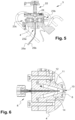
- the measuring cell of the flow cytometer is inclined relative to the horizontal, for example at an angle of approximately 45°.
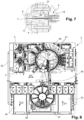
- the flow cytometer 3 comprises a single-piece support 4 which may, for example, be metallic.
- the support 4 is parallelepipedal and delimits an internal receiving housing 5.
- the support 4 comprises in particular six passage openings arranged respectively on the six external faces of the support 4.
- the flow cytometer 3 further comprises a measuring cell 6 (shown more particularly in the Figure 4 ) which at least partly delimits a measuring chamber 7, an injection device 8 arranged to inject a flow of biological cells F into the measuring chamber 7, and an evacuation device 9 arranged to evacuate the flow of biological cells F injected into the measuring chamber 7 outside the flow cytometer 3.
- a measuring cell 6 shown more particularly in the Figure 4
- an injection device 8 arranged to inject a flow of biological cells F into the measuring chamber 7
- an evacuation device 9 arranged to evacuate the flow of biological cells F injected into the measuring chamber 7 outside the flow cytometer 3.
- the injection and evacuation devices 8, 9 are fixed respectively on two opposite external faces of the support 4, and for example on the external faces opposite sides of the support 4. However, the injection and evacuation devices 8, 9 could also be fixed respectively on the two upper and lower external faces of the support 4.
- the injection device 8 further comprises a second supply conduit 16 intended to supply the internal chamber 12 with a sheathing fluid.
- the injection nozzle 11 and the second supply conduit 16 are configured such that the sheathing fluid introduced into the internal chamber 12 via the second supply conduit 16 is capable of hydrodynamically sheathing the biological sample introduced into the internal chamber 12 before the biological sample passes through the injection orifice 13.
Landscapes
- Chemical & Material Sciences (AREA)
- Life Sciences & Earth Sciences (AREA)
- Biochemistry (AREA)
- Dispersion Chemistry (AREA)
- Physics & Mathematics (AREA)
- Health & Medical Sciences (AREA)
- Pathology (AREA)
- Analytical Chemistry (AREA)
- Immunology (AREA)
- General Health & Medical Sciences (AREA)
- General Physics & Mathematics (AREA)
- Engineering & Computer Science (AREA)
- Signal Processing (AREA)
- Investigating Or Analysing Biological Materials (AREA)
- Computer Vision & Pattern Recognition (AREA)
Claims (16)
- Verfahren zur Analyse einer biologischen Probe, die biologische Zellen einschließlich Blutzellen enthält, wobei das Analyseverfahren die folgenden Schritte umfasst:- Durchlauf der biologischen Zellen der zu analysierenden biologischen Probe in eine Messzelle (6) eines Durchflusszytometers (3),- Messung von N Zytometrieparametern für jede biologische Zelle der zu analysierenden biologischen Probe,- Bestimmung, für jede biologische Zelle der zu analysierenden biologischen Probe, eines Punktes in einem N-dimensionalen Raum, dessen Koordinaten in Abhängigkeit von den für die entsprechende biologische Zelle gemessenen Zytometrieparametern definiert sind, wobei N eine Ganzzahl größer oder gleich 3 ist,- automatische Gruppierung der bestimmten Punkte in verschiedene Zellcluster in Abhängigkeit von den gemessenen Zytometrieparametern, um eine Probenclusterdatei zu definieren,- Identifizierung der Zellpopulationen, die durch die verschiedenen Zellcluster der im Gruppierungsschritt definierten Probenclusterdatei definiert sind,- Zählung der Punkte jedes Zellclusters der im Gruppierungsschritt definierten Probenclusterdatei, wobei das Verfahren durch den folgenden Schritt gekennzeichnet ist:- Vergleich der im Gruppierungsschritt definierten Probenclusterdatei mit Referenzclusterdateien, wobei jede der Referenzclusterdateien aus Zytometrieparametern einer jeweiligen pathologischen oder abnormalen biologischen Probe definiert wird.
- Analyseverfahren nach Anspruch 1, das ferner einen Sendeschritt einer Alarmmeldung umfasst, wenn die im Gruppierungsschritt definierte Probenclusterdatei mindestens teilweise identisch oder ähnlich einer Referenzclusterdatei ist.
- Analyseverfahren nach Anspruch 2, wobei die ausgegebene Alarmmeldung Hinweise auf eine Pathologie oder Anomalie enthält, die mit der Referenzclusterdatei verbunden ist, mit der die im Gruppierungsschritt definierte Probenclusterdatei mindestens teilweise identisch oder ähnlich ist.
- Analyseverfahren nach einem der Ansprüche 1 bis 3, das ferner einen Analyseschritt der im Gruppierungsschritt definierten Probenclusterdatei umfasst, um mindestens eine mögliche Anomalie in der Probenclusterdatei zu erfassen.
- Analyseverfahren nach Anspruch 4, wobei der Analyseschritt die folgenden Schritte umfasst:- Analyse, für jeden Zellcluster der im Gruppierungsschritt definierten Probenclusterdatei, mindestens eines morphologischen Parameters des Zellclusters,- Erfassung einer Anomalie, wenn mindestens ein morphologischer Parameter von mindestens einem Zellcluster der im Gruppierungsschritt definierten Probenclusterdatei einen jeweiligen vorbestimmten Schwellenwert überschreitet.
- Analyseverfahren nach Anspruch 4 oder 5, wobei der Analyseschritt die folgenden Schritte umfasst:- Vergleich, für jeden Zellcluster der im Gruppierungsschritt definierten Probenclusterdatei, der Anzahl der in dem Zellcluster gruppierten Punkte mit mindestens einem jeweiligen vorbestimmten Schwellenwert,- Erfassung einer Anomalie, wenn die Anzahl der in mindestens einem der Zellcluster gruppierten Punkte kleiner und/oder größer ist als der mindestens jeweils vorbestimmte Schwellenwert.
- Analyseverfahren nach einem der Ansprüche 4 bis 6, wobei der Analyseschritt die folgenden Schritte umfasst:- Analyse der Verteilung der Punkte in jedem Zellcluster der im Gruppierungsschritt definierten Probenclusterdatei,- Erfassung einer Anomalie, wenn die Verteilung der Punkte in mindestens einem der Zellcluster nicht gaußförmig ist.
- Analyseverfahren nach einem der Ansprüche 4 bis 7, wobei der Analyseschritt die folgenden Schritte umfasst:- Analyse der Positionierung der Zellcluster der im Gruppierungsschritt definierten Probenclusterdatei,- Erfassung einer Anomalie, wenn mindestens zwei Zellcluster der im Gruppierungsschritt definierten Probenclusterdatei mindestens teilweise verwechselt werden.
- Analyseverfahren nach einem der Ansprüche 4 bis 8, wobei der Analyseschritt die folgenden Schritte umfasst:- Analyse der Zellcluster der im Gruppierungsschritt definierten Probenclusterdatei,- Erfassung einer Anomalie, wenn das Vorhandensein oder Fehlen mindestens eines vorbestimmten Zellclusters erfasst wird.
- Analyseverfahren nach einem der Ansprüche 1 bis 9, wobei der Messschritt von Zytometrieparametern mindestens einen Messschritt von Zytometrieparametern umfasst, die repräsentativ für die Morphologie und/oder die Struktur der biologischen Zellen der zu analysierenden biologischen Probe sind.
- Analyseverfahren nach Anspruch 10, wobei der Messschritt von Zytometrieparametern mindestens einen Messschritt, für jede biologische Zelle der zu analysierenden biologischen Probe, von mindestens einer optischen Eigenschaft der biologischen Zelle umfasst.
- Analyseverfahren nach Anspruch 11, wobei der Messschritt von Zytometrieparametern Folgendes umfasst:- einen Messschritt der Intensität eines von jeder biologischen Zelle in kleinen Winkeln gestreuten Lichtstrahls, und/oder- einen Messschritt der Intensität eines von jeder biologischen Zelle um 90° gestreuten Lichtstrahls, und/oder- einen Messschritt der Intensität eines Lichtstrahls, der entlang eines optischen Pfades des auftreffenden Lichtstrahls von jeder biologischen Zelle gestreut wird.
- Analyseverfahren nach Anspruch 11 oder 12, wobei der Messschritt von Zytometrieparametern einen Messschritt der Intensität mindestens eines von jeder biologischen Zelle emittierten Fluoreszenzstrahls, beispielsweise bei 90°, umfasst.
- Analyseverfahren nach einem der Ansprüche 1 bis 13, wobei der Messschritt von Zytometrieparametern die folgenden Schritte umfasst:- Aussendung eines auftreffenden Lichtstrahls in Richtung der die Messkammer durchlaufenden biologischen Zellen, so dass der auftreffende Lichtstrahl den Pfad der biologischen Zellen kreuzt,- Erfassung mindestens eines Lichtstrahls aus jeder biologischen Zelle, die die Messkammer durchlaufen.
- Analyseverfahren nach einem der Ansprüche 1 bis 14, das ferner die folgenden Schritte umfasst:- Durchlauf der biologischen Zellen einer biologischen Referenzprobe in eine Messzelle (6) eines Durchflusszytometers (3),- Messung von N Zytometrieparametern für jede biologische Zelle der biologischen Referenzprobe,- Bestimmung, für jede biologische Zelle der biologischen Referenzprobe, eines Punktes in einem N-dimensionalen Raum, dessen Koordinaten in Abhängigkeit von den für die biologische Zelle der biologischen Referenzprobe gemessenen Zytometrieparametern definiert sind, wobei N eine Ganzzahl größer oder gleich 3 ist,- automatische Gruppierung der bestimmten Punkte in Bezug auf die biologische Referenzprobe in verschiedene Zellcluster in Abhängigkeit von den für jede biologische Zelle der biologischen Referenzprobe gemessenen Zytometrieparametern, um eine Referenzclusterdatei zu definieren,- Wiederholen der Schritte des Durchlaufens, Messens, Bestimmens und Gruppierens für eine Vielzahl von biologischen Referenzproben, um eine Vielzahl von Referenzclusterdateien zu definieren.
- Analysegerät, umfassend:- ein Durchflusszytometer (3), das eine Messzelle (6) umfasst, die für den Durchlauf biologischer Zellen einer zu analysierenden biologischen Probe bestimmt ist, und Messmittel (22, 23a, 23b, 23c), die so eingerichtet sind, dass sie Zytometrieparameter der biologischen Zellen der zu analysierenden biologischen Probe messen, und- Verarbeitungseinheit (32), die für Folgendes eingerichtet ist:- Bestimmen, für jede biologische Zelle der zu analysierenden biologischen Probe, eines Punkts in einem N-dimensionalen Raum, dessen Koordinaten in Abhängigkeit von den für die entsprechende biologische Zelle der zu analysierenden biologischen Probe gemessenen Zytometrieparametern definiert sind, wobei N eine Ganzzahl größer oder gleich 3 ist,- Gruppieren von Punkten in verschiedenen Zellclustern in Abhängigkeit von den für jede biologische Zelle der zu analysierenden biologischen Probe gemessenen Zytometrieparametern, um eine Probenclusterdatei zu definieren, dadurch gekennzeichnet, dass die Verarbeitungseinheit ferner für Folgendes eingerichtet ist:- Vergleichen der im Gruppierungsschritt definierten Probenclusterdatei mit Referenzclusterdateien, wobei jede der Referenzclusterdateien aus Zytometrieparametern einer jeweiligen pathologischen oder abnormalen biologischen Probe definiert wird.
Applications Claiming Priority (2)
| Application Number | Priority Date | Filing Date | Title |
|---|---|---|---|
| FR1851958A FR3078777B1 (fr) | 2018-03-07 | 2018-03-07 | Procede d’analyse d’un echantillon biologique contenant des cellules biologiques, et appareil d’analyse pour la mise en œuvre du procede d’analyse |
| PCT/FR2019/050481 WO2019170993A1 (fr) | 2018-03-07 | 2019-03-04 | PROCÉDÉ D'ANALYSE D'UN ÉCHANTILLON BIOLOGIQUE CONTENANT DES CELLULES BIOLOGIQUES, ET APPAREIL D'ANALYSE POUR LA MISE EN œUVRE DU PROCÉDÉ D'ANALYSE |
Publications (2)
| Publication Number | Publication Date |
|---|---|
| EP3762706A1 EP3762706A1 (de) | 2021-01-13 |
| EP3762706B1 true EP3762706B1 (de) | 2025-07-02 |
Family
ID=62597648
Family Applications (1)
| Application Number | Title | Priority Date | Filing Date |
|---|---|---|---|
| EP19716218.3A Active EP3762706B1 (de) | 2018-03-07 | 2019-03-04 | Verfahren zum analysieren einer biologischen probe, die biologische zellen enthält, und analysevorrichtung zur durchführung des analyseverfahrens |
Country Status (11)
| Country | Link |
|---|---|
| US (1) | US11953421B2 (de) |
| EP (1) | EP3762706B1 (de) |
| JP (1) | JP7319986B2 (de) |
| KR (1) | KR102714409B1 (de) |
| CN (1) | CN111954802B (de) |
| AU (1) | AU2019229702B2 (de) |
| CA (1) | CA3089163A1 (de) |
| ES (1) | ES3046010T3 (de) |
| FR (1) | FR3078777B1 (de) |
| MX (1) | MX2020009270A (de) |
| WO (1) | WO2019170993A1 (de) |
Families Citing this family (2)
| Publication number | Priority date | Publication date | Assignee | Title |
|---|---|---|---|---|
| US12332265B2 (en) | 2019-12-27 | 2025-06-17 | Beckman Coulter, Inc. | Sample preparation instrument |
| US12008825B2 (en) | 2020-11-19 | 2024-06-11 | Sony Group Corporation | Classification workflow for flexible image based particle sorting |
Family Cites Families (17)
| Publication number | Priority date | Publication date | Assignee | Title |
|---|---|---|---|---|
| EP0610774B1 (de) * | 1993-02-09 | 2001-03-28 | Becton, Dickinson and Company | Automatische Bestimmung der Zellinie schwerer Leukämien durch Flusszytometrie |
| JP2002207035A (ja) | 2001-01-10 | 2002-07-26 | Sysmex Corp | 腫瘍化細胞計数方法 |
| FR2873813B1 (fr) | 2004-07-30 | 2006-11-17 | Abx Sa | Procede et dispositif de caracterisation des composants cellulaires d'un liquide biologique |
| US20060192940A1 (en) | 2005-01-20 | 2006-08-31 | Phi-Wilson Janette T | Modular flow cytometry system |
| EP1844426A4 (de) * | 2005-02-01 | 2016-09-07 | Amnis Corp | Blutanalyse mittels durchflussbildgebungszytometer |
| EP1859377B1 (de) * | 2005-02-18 | 2016-09-28 | Hematologics, Inc. | System für den nachweis abnormaler zellen unter verwendung von multidimensionaler analyse |
| CN101226190B (zh) * | 2007-01-17 | 2013-07-03 | 深圳迈瑞生物医疗电子股份有限公司 | 流式细胞术的自动分类方法和装置 |
| US20100204973A1 (en) | 2009-01-15 | 2010-08-12 | Nodality, Inc., A Delaware Corporation | Methods For Diagnosis, Prognosis And Treatment |
| US9103759B2 (en) | 2011-05-04 | 2015-08-11 | Abbott Laboratories | Nucleated red blood cell analysis system and method |
| EP2912433B1 (de) | 2012-10-26 | 2019-03-20 | Fluidigm Canada Inc. | Zellanalyse durch massenzytometrie |
| JP6001425B2 (ja) | 2012-11-26 | 2016-10-05 | シスメックス株式会社 | 血球分析方法、血球分析装置およびプログラム |
| EP2939001B1 (de) * | 2012-12-31 | 2019-12-04 | Beckman Coulter, Inc. | Systeme und verfahren zur blutplättchenzählung mit klumpenanpassung |
| US20160169786A1 (en) | 2014-12-10 | 2016-06-16 | Neogenomics Laboratories, Inc. | Automated flow cytometry analysis method and system |
| CN106687810B (zh) | 2014-12-31 | 2019-10-22 | 深圳迈瑞生物医疗电子股份有限公司 | 一种非诊断目的的有核红细胞报警方法、装置及流式细胞分析仪 |
| CN106018246A (zh) | 2016-06-27 | 2016-10-12 | 上海泽泉科技股份有限公司 | 基于流式细胞术的藻华在线监测方法及监测系统 |
| KR101895760B1 (ko) * | 2016-06-27 | 2018-09-06 | (주)뉴옵틱스 | 혈구 분석 시스템 및 그의 제어방법 |
| CN106548205A (zh) * | 2016-10-21 | 2017-03-29 | 北京信息科技大学 | 一种流式细胞数据快速自动分群及圈门方法 |
-
2018
- 2018-03-07 FR FR1851958A patent/FR3078777B1/fr active Active
-
2019
- 2019-03-04 JP JP2020542396A patent/JP7319986B2/ja active Active
- 2019-03-04 CA CA3089163A patent/CA3089163A1/fr active Pending
- 2019-03-04 MX MX2020009270A patent/MX2020009270A/es unknown
- 2019-03-04 ES ES19716218T patent/ES3046010T3/es active Active
- 2019-03-04 US US16/978,080 patent/US11953421B2/en active Active
- 2019-03-04 AU AU2019229702A patent/AU2019229702B2/en active Active
- 2019-03-04 KR KR1020207027941A patent/KR102714409B1/ko active Active
- 2019-03-04 CN CN201980017554.3A patent/CN111954802B/zh active Active
- 2019-03-04 WO PCT/FR2019/050481 patent/WO2019170993A1/fr not_active Ceased
- 2019-03-04 EP EP19716218.3A patent/EP3762706B1/de active Active
Also Published As
| Publication number | Publication date |
|---|---|
| ES3046010T3 (en) | 2025-12-01 |
| BR112020017931A2 (pt) | 2020-12-22 |
| CN111954802A (zh) | 2020-11-17 |
| CA3089163A1 (fr) | 2019-09-12 |
| FR3078777A1 (fr) | 2019-09-13 |
| KR102714409B1 (ko) | 2024-10-07 |
| AU2019229702B2 (en) | 2024-02-29 |
| JP2021516335A (ja) | 2021-07-01 |
| CN111954802B (zh) | 2024-02-06 |
| RU2020124003A (ru) | 2022-04-07 |
| FR3078777B1 (fr) | 2020-11-13 |
| KR20200126405A (ko) | 2020-11-06 |
| AU2019229702A1 (en) | 2020-08-27 |
| JP7319986B2 (ja) | 2023-08-02 |
| US20200393356A1 (en) | 2020-12-17 |
| WO2019170993A1 (fr) | 2019-09-12 |
| US11953421B2 (en) | 2024-04-09 |
| EP3762706A1 (de) | 2021-01-13 |
| MX2020009270A (es) | 2020-10-01 |
Similar Documents
| Publication | Publication Date | Title |
|---|---|---|
| EP2352984B1 (de) | Durchflusszytometrieverfahren und-vorrichtung ohne hüllflüssigkeit | |
| EP0758400B1 (de) | Verfahren und einrichtung zur zählung von zellen und mikroorganismen, im besonderen in lebensmitteln und biologischen flüssigkeiten | |
| EP1771718B1 (de) | Verfahren zur charakterisierung von zellkomponenten einer biologischen flüssigkeit | |
| US12078597B2 (en) | Framework for image based unsupervised cell clustering and sorting | |
| EP2671063A1 (de) | Vorrichtung und verfahren für multiparametermessungen von mikropartikeln in einer flüssigkeit | |
| EP3762706B1 (de) | Verfahren zum analysieren einer biologischen probe, die biologische zellen enthält, und analysevorrichtung zur durchführung des analyseverfahrens | |
| US20060192940A1 (en) | Modular flow cytometry system | |
| FR2735578A1 (fr) | Reactif de lyse des erythrocytes, et son utilisation dans des procedes d'isolement et de discrimination des leucocytes | |
| EP2318820B1 (de) | Verfahren und vorrichtung zur klassifizierung, präsentation und analyse biologischer daten | |
| EP4508409B1 (de) | Vorrichtung zum streuen oder anfärben und zum bestimmen einer sedimentationsrate | |
| RU2803025C2 (ru) | Способ анализа биологического образца, содержащего биологические клетки, и анализирующее устройство для осуществления способа анализа | |
| US12423810B2 (en) | Image-based unsupervised multi-model cell clustering | |
| EP4588020A1 (de) | Bildbasiertes unüberwachtes multimodell-zellclustering | |
| BR112020017931B1 (pt) | Método de análise para analisar uma amostra biológica contendo células biológicas e aparelho de análise | |
| EP4562399A1 (de) | Vorrichtung zum zählen und unterscheiden von partikeln eines probenstroms | |
| Persaud | Development of a helium-neon laser based flow cytometer for evaluation of particulate matter |
Legal Events
| Date | Code | Title | Description |
|---|---|---|---|
| STAA | Information on the status of an ep patent application or granted ep patent |
Free format text: STATUS: UNKNOWN |
|
| STAA | Information on the status of an ep patent application or granted ep patent |
Free format text: STATUS: THE INTERNATIONAL PUBLICATION HAS BEEN MADE |
|
| PUAI | Public reference made under article 153(3) epc to a published international application that has entered the european phase |
Free format text: ORIGINAL CODE: 0009012 |
|
| STAA | Information on the status of an ep patent application or granted ep patent |
Free format text: STATUS: REQUEST FOR EXAMINATION WAS MADE |
|
| 17P | Request for examination filed |
Effective date: 20200917 |
|
| AK | Designated contracting states |
Kind code of ref document: A1 Designated state(s): AL AT BE BG CH CY CZ DE DK EE ES FI FR GB GR HR HU IE IS IT LI LT LU LV MC MK MT NL NO PL PT RO RS SE SI SK SM TR |
|
| AX | Request for extension of the european patent |
Extension state: BA ME |
|
| DAV | Request for validation of the european patent (deleted) | ||
| DAX | Request for extension of the european patent (deleted) | ||
| STAA | Information on the status of an ep patent application or granted ep patent |
Free format text: STATUS: EXAMINATION IS IN PROGRESS |
|
| 17Q | First examination report despatched |
Effective date: 20230119 |
|
| P01 | Opt-out of the competence of the unified patent court (upc) registered |
Effective date: 20230525 |
|
| GRAP | Despatch of communication of intention to grant a patent |
Free format text: ORIGINAL CODE: EPIDOSNIGR1 |
|
| STAA | Information on the status of an ep patent application or granted ep patent |
Free format text: STATUS: GRANT OF PATENT IS INTENDED |
|
| RIC1 | Information provided on ipc code assigned before grant |
Ipc: G01N 35/00 20060101ALI20250114BHEP Ipc: G01N 15/10 20060101ALI20250114BHEP Ipc: G01N 15/00 20060101ALI20250114BHEP Ipc: G01N 15/1429 20240101ALI20250114BHEP Ipc: G01N 15/14 20060101AFI20250114BHEP |
|
| INTG | Intention to grant announced |
Effective date: 20250205 |
|
| GRAS | Grant fee paid |
Free format text: ORIGINAL CODE: EPIDOSNIGR3 |
|
| GRAA | (expected) grant |
Free format text: ORIGINAL CODE: 0009210 |
|
| STAA | Information on the status of an ep patent application or granted ep patent |
Free format text: STATUS: THE PATENT HAS BEEN GRANTED |
|
| RAP1 | Party data changed (applicant data changed or rights of an application transferred) |
Owner name: ARTEION |
|
| AK | Designated contracting states |
Kind code of ref document: B1 Designated state(s): AL AT BE BG CH CY CZ DE DK EE ES FI FR GB GR HR HU IE IS IT LI LT LU LV MC MK MT NL NO PL PT RO RS SE SI SK SM TR |
|
| REG | Reference to a national code |
Ref country code: GB Ref legal event code: FG4D Free format text: NOT ENGLISH |
|
| REG | Reference to a national code |
Ref country code: CH Ref legal event code: EP |
|
| REG | Reference to a national code |
Ref country code: DE Ref legal event code: R096 Ref document number: 602019071898 Country of ref document: DE |
|
| REG | Reference to a national code |
Ref country code: IE Ref legal event code: FG4D Free format text: LANGUAGE OF EP DOCUMENT: FRENCH |
|
| REG | Reference to a national code |
Ref country code: SE Ref legal event code: TRGR |
|
| REG | Reference to a national code |
Ref country code: NL Ref legal event code: FP |
|
| REG | Reference to a national code |
Ref country code: ES Ref legal event code: FG2A Ref document number: 3046010 Country of ref document: ES Kind code of ref document: T3 Effective date: 20251201 |
|
| PG25 | Lapsed in a contracting state [announced via postgrant information from national office to epo] |
Ref country code: PT Free format text: LAPSE BECAUSE OF FAILURE TO SUBMIT A TRANSLATION OF THE DESCRIPTION OR TO PAY THE FEE WITHIN THE PRESCRIBED TIME-LIMIT Effective date: 20251103 |
|
| REG | Reference to a national code |
Ref country code: AT Ref legal event code: MK05 Ref document number: 1809721 Country of ref document: AT Kind code of ref document: T Effective date: 20250702 |
|
| PG25 | Lapsed in a contracting state [announced via postgrant information from national office to epo] |
Ref country code: IS Free format text: LAPSE BECAUSE OF FAILURE TO SUBMIT A TRANSLATION OF THE DESCRIPTION OR TO PAY THE FEE WITHIN THE PRESCRIBED TIME-LIMIT Effective date: 20251102 |
|
| PG25 | Lapsed in a contracting state [announced via postgrant information from national office to epo] |
Ref country code: NO Free format text: LAPSE BECAUSE OF FAILURE TO SUBMIT A TRANSLATION OF THE DESCRIPTION OR TO PAY THE FEE WITHIN THE PRESCRIBED TIME-LIMIT Effective date: 20251002 |
|
| REG | Reference to a national code |
Ref country code: LT Ref legal event code: MG9D |
|
| PG25 | Lapsed in a contracting state [announced via postgrant information from national office to epo] |
Ref country code: AT Free format text: LAPSE BECAUSE OF FAILURE TO SUBMIT A TRANSLATION OF THE DESCRIPTION OR TO PAY THE FEE WITHIN THE PRESCRIBED TIME-LIMIT Effective date: 20250702 |
|
| PG25 | Lapsed in a contracting state [announced via postgrant information from national office to epo] |
Ref country code: FI Free format text: LAPSE BECAUSE OF FAILURE TO SUBMIT A TRANSLATION OF THE DESCRIPTION OR TO PAY THE FEE WITHIN THE PRESCRIBED TIME-LIMIT Effective date: 20250702 |
|
| PG25 | Lapsed in a contracting state [announced via postgrant information from national office to epo] |
Ref country code: HR Free format text: LAPSE BECAUSE OF FAILURE TO SUBMIT A TRANSLATION OF THE DESCRIPTION OR TO PAY THE FEE WITHIN THE PRESCRIBED TIME-LIMIT Effective date: 20250702 |
|
| PG25 | Lapsed in a contracting state [announced via postgrant information from national office to epo] |
Ref country code: GR Free format text: LAPSE BECAUSE OF FAILURE TO SUBMIT A TRANSLATION OF THE DESCRIPTION OR TO PAY THE FEE WITHIN THE PRESCRIBED TIME-LIMIT Effective date: 20251003 |
|
| PG25 | Lapsed in a contracting state [announced via postgrant information from national office to epo] |
Ref country code: CZ Free format text: LAPSE BECAUSE OF FAILURE TO SUBMIT A TRANSLATION OF THE DESCRIPTION OR TO PAY THE FEE WITHIN THE PRESCRIBED TIME-LIMIT Effective date: 20250702 |
|
| PG25 | Lapsed in a contracting state [announced via postgrant information from national office to epo] |
Ref country code: LV Free format text: LAPSE BECAUSE OF FAILURE TO SUBMIT A TRANSLATION OF THE DESCRIPTION OR TO PAY THE FEE WITHIN THE PRESCRIBED TIME-LIMIT Effective date: 20250702 |
|
| PG25 | Lapsed in a contracting state [announced via postgrant information from national office to epo] |
Ref country code: BG Free format text: LAPSE BECAUSE OF FAILURE TO SUBMIT A TRANSLATION OF THE DESCRIPTION OR TO PAY THE FEE WITHIN THE PRESCRIBED TIME-LIMIT Effective date: 20250702 Ref country code: PL Free format text: LAPSE BECAUSE OF FAILURE TO SUBMIT A TRANSLATION OF THE DESCRIPTION OR TO PAY THE FEE WITHIN THE PRESCRIBED TIME-LIMIT Effective date: 20250702 |
|
| PG25 | Lapsed in a contracting state [announced via postgrant information from national office to epo] |
Ref country code: RS Free format text: LAPSE BECAUSE OF FAILURE TO SUBMIT A TRANSLATION OF THE DESCRIPTION OR TO PAY THE FEE WITHIN THE PRESCRIBED TIME-LIMIT Effective date: 20251002 |
|
| PGFP | Annual fee paid to national office [announced via postgrant information from national office to epo] |
Ref country code: NL Payment date: 20260216 Year of fee payment: 8 |
|
| REG | Reference to a national code |
Ref country code: CH Ref legal event code: U11 Free format text: ST27 STATUS EVENT CODE: U-0-0-U10-U11 (AS PROVIDED BY THE NATIONAL OFFICE) Effective date: 20260401 |
|
| PG25 | Lapsed in a contracting state [announced via postgrant information from national office to epo] |
Ref country code: SM Free format text: LAPSE BECAUSE OF FAILURE TO SUBMIT A TRANSLATION OF THE DESCRIPTION OR TO PAY THE FEE WITHIN THE PRESCRIBED TIME-LIMIT Effective date: 20250702 |
|
| PGFP | Annual fee paid to national office [announced via postgrant information from national office to epo] |
Ref country code: SE Payment date: 20260323 Year of fee payment: 8 |
|
| PGFP | Annual fee paid to national office [announced via postgrant information from national office to epo] |
Ref country code: GB Payment date: 20260213 Year of fee payment: 8 |
|
| PG25 | Lapsed in a contracting state [announced via postgrant information from national office to epo] |
Ref country code: DK Free format text: LAPSE BECAUSE OF FAILURE TO SUBMIT A TRANSLATION OF THE DESCRIPTION OR TO PAY THE FEE WITHIN THE PRESCRIBED TIME-LIMIT Effective date: 20250702 |
|
| PGFP | Annual fee paid to national office [announced via postgrant information from national office to epo] |
Ref country code: DE Payment date: 20260216 Year of fee payment: 8 |
|
| PGFP | Annual fee paid to national office [announced via postgrant information from national office to epo] |
Ref country code: BE Payment date: 20260323 Year of fee payment: 8 Ref country code: IT Payment date: 20260317 Year of fee payment: 8 |
|
| PGFP | Annual fee paid to national office [announced via postgrant information from national office to epo] |
Ref country code: FR Payment date: 20260213 Year of fee payment: 8 |
|
| PGFP | Annual fee paid to national office [announced via postgrant information from national office to epo] |
Ref country code: TR Payment date: 20260216 Year of fee payment: 8 |