EP3690060A1 - Procédé d'identification et de quantification relative d'expression de séquence d'acide nucléique, de variant d'épissage, de translocation, de nombre de copies ou de changements de méthylation en utilisant des réactions combinées de nucléase, de ligature et de polymérase comportant une prévention de transfert - Google Patents
Procédé d'identification et de quantification relative d'expression de séquence d'acide nucléique, de variant d'épissage, de translocation, de nombre de copies ou de changements de méthylation en utilisant des réactions combinées de nucléase, de ligature et de polymérase comportant une prévention de transfert Download PDFInfo
- Publication number
- EP3690060A1 EP3690060A1 EP20150354.7A EP20150354A EP3690060A1 EP 3690060 A1 EP3690060 A1 EP 3690060A1 EP 20150354 A EP20150354 A EP 20150354A EP 3690060 A1 EP3690060 A1 EP 3690060A1
- Authority
- EP
- European Patent Office
- Prior art keywords
- primer
- sequence
- oligonucleotide
- target
- sample
- Prior art date
- Legal status (The legal status is an assumption and is not a legal conclusion. Google has not performed a legal analysis and makes no representation as to the accuracy of the status listed.)
- Granted
Links
- 238000000034 method Methods 0.000 title claims abstract description 171
- 230000005945 translocation Effects 0.000 title claims abstract description 32
- 238000006243 chemical reaction Methods 0.000 title claims description 186
- 150000007523 nucleic acids Chemical group 0.000 title claims description 167
- 108010029485 Protein Isoforms Proteins 0.000 title abstract description 5
- 102000001708 Protein Isoforms Human genes 0.000 title abstract description 5
- 230000002265 prevention Effects 0.000 title description 219
- 230000011987 methylation Effects 0.000 title description 61
- 238000007069 methylation reaction Methods 0.000 title description 61
- 108091028043 Nucleic acid sequence Proteins 0.000 title description 41
- 101710163270 Nuclease Proteins 0.000 title description 20
- 238000011002 quantification Methods 0.000 title description 12
- 230000014509 gene expression Effects 0.000 title description 4
- 125000003729 nucleotide group Chemical group 0.000 claims abstract description 228
- 239000002773 nucleotide Substances 0.000 claims abstract description 211
- 230000035772 mutation Effects 0.000 claims abstract description 125
- 238000003780 insertion Methods 0.000 claims abstract description 55
- 230000037431 insertion Effects 0.000 claims abstract description 55
- 238000012217 deletion Methods 0.000 claims abstract description 37
- 230000037430 deletion Effects 0.000 claims abstract description 37
- 230000008707 rearrangement Effects 0.000 claims abstract description 14
- 239000013615 primer Substances 0.000 claims description 819
- 239000000523 sample Substances 0.000 claims description 550
- 238000003752 polymerase chain reaction Methods 0.000 claims description 296
- 108091034117 Oligonucleotide Proteins 0.000 claims description 277
- 108020005187 Oligonucleotide Probes Proteins 0.000 claims description 276
- 239000002751 oligonucleotide probe Substances 0.000 claims description 276
- 230000000295 complement effect Effects 0.000 claims description 274
- 238000001514 detection method Methods 0.000 claims description 172
- 239000011541 reaction mixture Substances 0.000 claims description 166
- 102000039446 nucleic acids Human genes 0.000 claims description 150
- 108020004707 nucleic acids Proteins 0.000 claims description 150
- 238000011282 treatment Methods 0.000 claims description 132
- AHCYMLUZIRLXAA-SHYZEUOFSA-N Deoxyuridine 5'-triphosphate Chemical compound O1[C@H](COP(O)(=O)OP(O)(=O)OP(O)(O)=O)[C@@H](O)C[C@@H]1N1C(=O)NC(=O)C=C1 AHCYMLUZIRLXAA-SHYZEUOFSA-N 0.000 claims description 88
- 238000009396 hybridization Methods 0.000 claims description 85
- 230000000903 blocking effect Effects 0.000 claims description 78
- 102100034343 Integrase Human genes 0.000 claims description 71
- 239000000203 mixture Substances 0.000 claims description 71
- 102000004190 Enzymes Human genes 0.000 claims description 70
- 108090000790 Enzymes Proteins 0.000 claims description 70
- 239000002299 complementary DNA Substances 0.000 claims description 70
- 229920002477 rna polymer Polymers 0.000 claims description 67
- 102000003960 Ligases Human genes 0.000 claims description 65
- 108090000364 Ligases Proteins 0.000 claims description 65
- JTBBWRKSUYCPFY-UHFFFAOYSA-N 2,3-dihydro-1h-pyrimidin-4-one Chemical compound O=C1NCNC=C1 JTBBWRKSUYCPFY-UHFFFAOYSA-N 0.000 claims description 56
- 239000003155 DNA primer Substances 0.000 claims description 52
- 108010092799 RNA-directed DNA polymerase Proteins 0.000 claims description 42
- 238000004925 denaturation Methods 0.000 claims description 40
- 230000036425 denaturation Effects 0.000 claims description 40
- 238000010839 reverse transcription Methods 0.000 claims description 39
- JLCPHMBAVCMARE-UHFFFAOYSA-N [3-[[3-[[3-[[3-[[3-[[3-[[3-[[3-[[3-[[3-[[3-[[5-(2-amino-6-oxo-1H-purin-9-yl)-3-[[3-[[3-[[3-[[3-[[3-[[5-(2-amino-6-oxo-1H-purin-9-yl)-3-[[5-(2-amino-6-oxo-1H-purin-9-yl)-3-hydroxyoxolan-2-yl]methoxy-hydroxyphosphoryl]oxyoxolan-2-yl]methoxy-hydroxyphosphoryl]oxy-5-(5-methyl-2,4-dioxopyrimidin-1-yl)oxolan-2-yl]methoxy-hydroxyphosphoryl]oxy-5-(6-aminopurin-9-yl)oxolan-2-yl]methoxy-hydroxyphosphoryl]oxy-5-(6-aminopurin-9-yl)oxolan-2-yl]methoxy-hydroxyphosphoryl]oxy-5-(6-aminopurin-9-yl)oxolan-2-yl]methoxy-hydroxyphosphoryl]oxy-5-(6-aminopurin-9-yl)oxolan-2-yl]methoxy-hydroxyphosphoryl]oxyoxolan-2-yl]methoxy-hydroxyphosphoryl]oxy-5-(5-methyl-2,4-dioxopyrimidin-1-yl)oxolan-2-yl]methoxy-hydroxyphosphoryl]oxy-5-(4-amino-2-oxopyrimidin-1-yl)oxolan-2-yl]methoxy-hydroxyphosphoryl]oxy-5-(5-methyl-2,4-dioxopyrimidin-1-yl)oxolan-2-yl]methoxy-hydroxyphosphoryl]oxy-5-(5-methyl-2,4-dioxopyrimidin-1-yl)oxolan-2-yl]methoxy-hydroxyphosphoryl]oxy-5-(6-aminopurin-9-yl)oxolan-2-yl]methoxy-hydroxyphosphoryl]oxy-5-(6-aminopurin-9-yl)oxolan-2-yl]methoxy-hydroxyphosphoryl]oxy-5-(4-amino-2-oxopyrimidin-1-yl)oxolan-2-yl]methoxy-hydroxyphosphoryl]oxy-5-(4-amino-2-oxopyrimidin-1-yl)oxolan-2-yl]methoxy-hydroxyphosphoryl]oxy-5-(4-amino-2-oxopyrimidin-1-yl)oxolan-2-yl]methoxy-hydroxyphosphoryl]oxy-5-(6-aminopurin-9-yl)oxolan-2-yl]methoxy-hydroxyphosphoryl]oxy-5-(4-amino-2-oxopyrimidin-1-yl)oxolan-2-yl]methyl [5-(6-aminopurin-9-yl)-2-(hydroxymethyl)oxolan-3-yl] hydrogen phosphate Polymers Cc1cn(C2CC(OP(O)(=O)OCC3OC(CC3OP(O)(=O)OCC3OC(CC3O)n3cnc4c3nc(N)[nH]c4=O)n3cnc4c3nc(N)[nH]c4=O)C(COP(O)(=O)OC3CC(OC3COP(O)(=O)OC3CC(OC3COP(O)(=O)OC3CC(OC3COP(O)(=O)OC3CC(OC3COP(O)(=O)OC3CC(OC3COP(O)(=O)OC3CC(OC3COP(O)(=O)OC3CC(OC3COP(O)(=O)OC3CC(OC3COP(O)(=O)OC3CC(OC3COP(O)(=O)OC3CC(OC3COP(O)(=O)OC3CC(OC3COP(O)(=O)OC3CC(OC3COP(O)(=O)OC3CC(OC3COP(O)(=O)OC3CC(OC3COP(O)(=O)OC3CC(OC3COP(O)(=O)OC3CC(OC3COP(O)(=O)OC3CC(OC3CO)n3cnc4c(N)ncnc34)n3ccc(N)nc3=O)n3cnc4c(N)ncnc34)n3ccc(N)nc3=O)n3ccc(N)nc3=O)n3ccc(N)nc3=O)n3cnc4c(N)ncnc34)n3cnc4c(N)ncnc34)n3cc(C)c(=O)[nH]c3=O)n3cc(C)c(=O)[nH]c3=O)n3ccc(N)nc3=O)n3cc(C)c(=O)[nH]c3=O)n3cnc4c3nc(N)[nH]c4=O)n3cnc4c(N)ncnc34)n3cnc4c(N)ncnc34)n3cnc4c(N)ncnc34)n3cnc4c(N)ncnc34)O2)c(=O)[nH]c1=O JLCPHMBAVCMARE-UHFFFAOYSA-N 0.000 claims description 35
- 238000002156 mixing Methods 0.000 claims description 32
- 239000007787 solid Substances 0.000 claims description 28
- 108091026890 Coding region Proteins 0.000 claims description 24
- 229910019142 PO4 Inorganic materials 0.000 claims description 18
- 108010014303 DNA-directed DNA polymerase Proteins 0.000 claims description 17
- 102000016928 DNA-directed DNA polymerase Human genes 0.000 claims description 17
- 239000010452 phosphate Substances 0.000 claims description 17
- NBIIXXVUZAFLBC-UHFFFAOYSA-K phosphate Chemical compound [O-]P([O-])([O-])=O NBIIXXVUZAFLBC-UHFFFAOYSA-K 0.000 claims description 15
- 238000007169 ligase reaction Methods 0.000 claims description 8
- 230000003100 immobilizing effect Effects 0.000 claims 1
- 108091070501 miRNA Proteins 0.000 abstract description 3
- 239000002679 microRNA Substances 0.000 abstract description 3
- 239000000047 product Substances 0.000 description 431
- 108020004414 DNA Proteins 0.000 description 159
- 230000003321 amplification Effects 0.000 description 126
- 238000003199 nucleic acid amplification method Methods 0.000 description 126
- 238000011529 RT qPCR Methods 0.000 description 112
- 238000011144 upstream manufacturing Methods 0.000 description 108
- 108700011259 MicroRNAs Proteins 0.000 description 102
- 108091032973 (ribonucleotides)n+m Proteins 0.000 description 88
- LSNNMFCWUKXFEE-UHFFFAOYSA-M Bisulfite Chemical compound OS([O-])=O LSNNMFCWUKXFEE-UHFFFAOYSA-M 0.000 description 84
- 101150040913 DUT gene Proteins 0.000 description 78
- 102000006943 Uracil-DNA Glycosidase Human genes 0.000 description 68
- 108010072685 Uracil-DNA Glycosidase Proteins 0.000 description 68
- YBJHBAHKTGYVGT-ZKWXMUAHSA-N (+)-Biotin Chemical compound N1C(=O)N[C@@H]2[C@H](CCCCC(=O)O)SC[C@@H]21 YBJHBAHKTGYVGT-ZKWXMUAHSA-N 0.000 description 66
- 238000001327 Förster resonance energy transfer Methods 0.000 description 65
- 108020004999 messenger RNA Proteins 0.000 description 62
- 108091093088 Amplicon Proteins 0.000 description 56
- 238000012408 PCR amplification Methods 0.000 description 56
- 206010028980 Neoplasm Diseases 0.000 description 54
- 238000003753 real-time PCR Methods 0.000 description 51
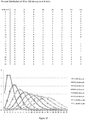
- 238000009826 distribution Methods 0.000 description 40
- 238000001507 sample dispersion Methods 0.000 description 40
- 201000011510 cancer Diseases 0.000 description 35
- 229960002685 biotin Drugs 0.000 description 33
- 235000020958 biotin Nutrition 0.000 description 33
- 239000011616 biotin Substances 0.000 description 33
- 101710203526 Integrase Proteins 0.000 description 29
- 108700024394 Exon Proteins 0.000 description 27
- 125000002496 methyl group Chemical group [H]C([H])([H])* 0.000 description 26
- ISAKRJDGNUQOIC-UHFFFAOYSA-N Uracil Chemical compound O=C1C=CNC(=O)N1 ISAKRJDGNUQOIC-UHFFFAOYSA-N 0.000 description 25
- 125000002887 hydroxy group Chemical group [H]O* 0.000 description 25
- 108091008146 restriction endonucleases Proteins 0.000 description 25
- 210000001808 exosome Anatomy 0.000 description 23
- 125000006850 spacer group Chemical group 0.000 description 22
- 239000000539 dimer Substances 0.000 description 21
- 238000002474 experimental method Methods 0.000 description 20
- 238000003776 cleavage reaction Methods 0.000 description 18
- 208000005443 Circulating Neoplastic Cells Diseases 0.000 description 17
- 238000003556 assay Methods 0.000 description 17
- 238000011049 filling Methods 0.000 description 17
- 238000013459 approach Methods 0.000 description 16
- 230000000694 effects Effects 0.000 description 16
- 210000004369 blood Anatomy 0.000 description 15
- 239000008280 blood Substances 0.000 description 15
- 201000010099 disease Diseases 0.000 description 15
- 208000037265 diseases, disorders, signs and symptoms Diseases 0.000 description 15
- 230000007017 scission Effects 0.000 description 15
- 238000004458 analytical method Methods 0.000 description 14
- 230000015572 biosynthetic process Effects 0.000 description 14
- 210000004027 cell Anatomy 0.000 description 14
- 235000021317 phosphate Nutrition 0.000 description 14
- 102100037111 Uracil-DNA glycosylase Human genes 0.000 description 13
- 238000003757 reverse transcription PCR Methods 0.000 description 13
- 125000002652 ribonucleotide group Chemical group 0.000 description 13
- 102000053602 DNA Human genes 0.000 description 12
- 108091028664 Ribonucleotide Proteins 0.000 description 12
- 239000002336 ribonucleotide Substances 0.000 description 12
- 229940035893 uracil Drugs 0.000 description 12
- 108090000623 proteins and genes Proteins 0.000 description 11
- 108010006785 Taq Polymerase Proteins 0.000 description 10
- 238000007792 addition Methods 0.000 description 10
- 230000008569 process Effects 0.000 description 10
- 108010037497 3'-nucleotidase Proteins 0.000 description 9
- 238000013461 design Methods 0.000 description 9
- 238000006911 enzymatic reaction Methods 0.000 description 9
- 238000004088 simulation Methods 0.000 description 9
- 210000000601 blood cell Anatomy 0.000 description 8
- -1 expression Proteins 0.000 description 8
- 238000009877 rendering Methods 0.000 description 8
- 108090000652 Flap endonucleases Proteins 0.000 description 7
- 102000004150 Flap endonucleases Human genes 0.000 description 7
- 238000010790 dilution Methods 0.000 description 7
- 239000012895 dilution Substances 0.000 description 7
- 238000002866 fluorescence resonance energy transfer Methods 0.000 description 7
- 230000001404 mediated effect Effects 0.000 description 7
- 238000011897 real-time detection Methods 0.000 description 7
- 238000012163 sequencing technique Methods 0.000 description 7
- 108010090804 Streptavidin Proteins 0.000 description 6
- 238000004891 communication Methods 0.000 description 6
- 239000012530 fluid Substances 0.000 description 6
- 239000007788 liquid Substances 0.000 description 6
- 238000012340 reverse transcriptase PCR Methods 0.000 description 6
- 238000012216 screening Methods 0.000 description 6
- 238000012360 testing method Methods 0.000 description 6
- 108010083644 Ribonucleases Proteins 0.000 description 5
- 102000006382 Ribonucleases Human genes 0.000 description 5
- 230000002759 chromosomal effect Effects 0.000 description 5
- 238000007847 digital PCR Methods 0.000 description 5
- 230000004927 fusion Effects 0.000 description 5
- 210000000265 leukocyte Anatomy 0.000 description 5
- 239000003550 marker Substances 0.000 description 5
- 244000052769 pathogen Species 0.000 description 5
- 230000035945 sensitivity Effects 0.000 description 5
- 210000001519 tissue Anatomy 0.000 description 5
- 206010009944 Colon cancer Diseases 0.000 description 4
- 208000001333 Colorectal Neoplasms Diseases 0.000 description 4
- 108010065472 Vimentin Proteins 0.000 description 4
- 230000008901 benefit Effects 0.000 description 4
- 238000002405 diagnostic procedure Methods 0.000 description 4
- 238000006073 displacement reaction Methods 0.000 description 4
- 239000003814 drug Substances 0.000 description 4
- 239000012634 fragment Substances 0.000 description 4
- 238000010348 incorporation Methods 0.000 description 4
- 208000002154 non-small cell lung carcinoma Diseases 0.000 description 4
- 239000002987 primer (paints) Substances 0.000 description 4
- 125000006853 reporter group Chemical group 0.000 description 4
- 102200104166 rs11540652 Human genes 0.000 description 4
- 210000002966 serum Anatomy 0.000 description 4
- 208000029729 tumor suppressor gene on chromosome 11 Diseases 0.000 description 4
- 208000035657 Abasia Diseases 0.000 description 3
- 241000588724 Escherichia coli Species 0.000 description 3
- 102000013127 Vimentin Human genes 0.000 description 3
- 238000007844 allele-specific PCR Methods 0.000 description 3
- 150000001540 azides Chemical class 0.000 description 3
- 239000012472 biological sample Substances 0.000 description 3
- 238000011109 contamination Methods 0.000 description 3
- 230000034994 death Effects 0.000 description 3
- 231100000517 death Toxicity 0.000 description 3
- 229940079593 drug Drugs 0.000 description 3
- 210000003743 erythrocyte Anatomy 0.000 description 3
- 238000012544 monitoring process Methods 0.000 description 3
- 108090000765 processed proteins & peptides Proteins 0.000 description 3
- 102200055464 rs113488022 Human genes 0.000 description 3
- 238000007423 screening assay Methods 0.000 description 3
- 239000000243 solution Substances 0.000 description 3
- 210000005048 vimentin Anatomy 0.000 description 3
- 238000010626 work up procedure Methods 0.000 description 3
- 208000035473 Communicable disease Diseases 0.000 description 2
- 238000001712 DNA sequencing Methods 0.000 description 2
- 108060002716 Exonuclease Proteins 0.000 description 2
- YLQBMQCUIZJEEH-UHFFFAOYSA-N Furan Chemical compound C=1C=COC=1 YLQBMQCUIZJEEH-UHFFFAOYSA-N 0.000 description 2
- 108700039691 Genetic Promoter Regions Proteins 0.000 description 2
- 101000664956 Homo sapiens Single-strand selective monofunctional uracil DNA glycosylase Proteins 0.000 description 2
- 229930010555 Inosine Natural products 0.000 description 2
- UGQMRVRMYYASKQ-KQYNXXCUSA-N Inosine Chemical compound O[C@@H]1[C@H](O)[C@@H](CO)O[C@H]1N1C2=NC=NC(O)=C2N=C1 UGQMRVRMYYASKQ-KQYNXXCUSA-N 0.000 description 2
- 108091092195 Intron Proteins 0.000 description 2
- 206010069755 K-ras gene mutation Diseases 0.000 description 2
- 108700019961 Neoplasm Genes Proteins 0.000 description 2
- 102000048850 Neoplasm Genes Human genes 0.000 description 2
- 230000002159 abnormal effect Effects 0.000 description 2
- 238000000137 annealing Methods 0.000 description 2
- 230000006907 apoptotic process Effects 0.000 description 2
- 210000000481 breast Anatomy 0.000 description 2
- 238000011161 development Methods 0.000 description 2
- 230000018109 developmental process Effects 0.000 description 2
- 238000005516 engineering process Methods 0.000 description 2
- 102000013165 exonuclease Human genes 0.000 description 2
- 102000055291 human SMUG1 Human genes 0.000 description 2
- 208000015181 infectious disease Diseases 0.000 description 2
- 229960003786 inosine Drugs 0.000 description 2
- 230000000670 limiting effect Effects 0.000 description 2
- 238000004519 manufacturing process Methods 0.000 description 2
- 239000002105 nanoparticle Substances 0.000 description 2
- 230000035790 physiological processes and functions Effects 0.000 description 2
- 238000000746 purification Methods 0.000 description 2
- 238000010791 quenching Methods 0.000 description 2
- 230000000171 quenching effect Effects 0.000 description 2
- 230000002829 reductive effect Effects 0.000 description 2
- 102200006531 rs121913529 Human genes 0.000 description 2
- 102200006538 rs121913530 Human genes 0.000 description 2
- 238000012795 verification Methods 0.000 description 2
- 230000003612 virological effect Effects 0.000 description 2
- HCGYMSSYSAKGPK-UHFFFAOYSA-N 2-nitro-1h-indole Chemical compound C1=CC=C2NC([N+](=O)[O-])=CC2=C1 HCGYMSSYSAKGPK-UHFFFAOYSA-N 0.000 description 1
- FTBBGQKRYUTLMP-UHFFFAOYSA-N 2-nitro-1h-pyrrole Chemical compound [O-][N+](=O)C1=CC=CN1 FTBBGQKRYUTLMP-UHFFFAOYSA-N 0.000 description 1
- WEVYNIUIFUYDGI-UHFFFAOYSA-N 3-[6-[4-(trifluoromethoxy)anilino]-4-pyrimidinyl]benzamide Chemical compound NC(=O)C1=CC=CC(C=2N=CN=C(NC=3C=CC(OC(F)(F)F)=CC=3)C=2)=C1 WEVYNIUIFUYDGI-UHFFFAOYSA-N 0.000 description 1
- 208000030507 AIDS Diseases 0.000 description 1
- 206010069754 Acquired gene mutation Diseases 0.000 description 1
- 108091023037 Aptamer Proteins 0.000 description 1
- 208000032791 BCR-ABL1 positive chronic myelogenous leukemia Diseases 0.000 description 1
- 201000009030 Carcinoma Diseases 0.000 description 1
- 208000010833 Chronic myeloid leukaemia Diseases 0.000 description 1
- HMFHBZSHGGEWLO-SOOFDHNKSA-N D-ribofuranose Chemical compound OC[C@H]1OC(O)[C@H](O)[C@@H]1O HMFHBZSHGGEWLO-SOOFDHNKSA-N 0.000 description 1
- 102000012410 DNA Ligases Human genes 0.000 description 1
- 108010061982 DNA Ligases Proteins 0.000 description 1
- 238000000018 DNA microarray Methods 0.000 description 1
- 102000010719 DNA-(Apurinic or Apyrimidinic Site) Lyase Human genes 0.000 description 1
- 108010063362 DNA-(Apurinic or Apyrimidinic Site) Lyase Proteins 0.000 description 1
- 108010036364 Deoxyribonuclease IV (Phage T4-Induced) Proteins 0.000 description 1
- 206010061819 Disease recurrence Diseases 0.000 description 1
- 206010059866 Drug resistance Diseases 0.000 description 1
- 208000037595 EN1-related dorsoventral syndrome Diseases 0.000 description 1
- 241001115402 Ebolavirus Species 0.000 description 1
- 101150039808 Egfr gene Proteins 0.000 description 1
- 102100031780 Endonuclease Human genes 0.000 description 1
- 108010042407 Endonucleases Proteins 0.000 description 1
- 101001107181 Enterobacteria phage T4 Ribonuclease H Proteins 0.000 description 1
- 101000637245 Escherichia coli (strain K12) Endonuclease V Proteins 0.000 description 1
- 208000034454 F12-related hereditary angioedema with normal C1Inh Diseases 0.000 description 1
- 206010061218 Inflammation Diseases 0.000 description 1
- 239000005517 L01XE01 - Imatinib Substances 0.000 description 1
- 238000002944 PCR assay Methods 0.000 description 1
- 241000205160 Pyrococcus Species 0.000 description 1
- 101710086015 RNA ligase Proteins 0.000 description 1
- 208000015634 Rectal Neoplasms Diseases 0.000 description 1
- PYMYPHUHKUWMLA-LMVFSUKVSA-N Ribose Natural products OC[C@@H](O)[C@@H](O)[C@@H](O)C=O PYMYPHUHKUWMLA-LMVFSUKVSA-N 0.000 description 1
- 108020004682 Single-Stranded DNA Proteins 0.000 description 1
- 241000193996 Streptococcus pyogenes Species 0.000 description 1
- 241000589500 Thermus aquaticus Species 0.000 description 1
- 241000589499 Thermus thermophilus Species 0.000 description 1
- 108700009124 Transcription Initiation Site Proteins 0.000 description 1
- 208000037280 Trisomy Diseases 0.000 description 1
- 208000027418 Wounds and injury Diseases 0.000 description 1
- 230000003213 activating effect Effects 0.000 description 1
- 238000001261 affinity purification Methods 0.000 description 1
- 238000003314 affinity selection Methods 0.000 description 1
- 150000001336 alkenes Chemical class 0.000 description 1
- HMFHBZSHGGEWLO-UHFFFAOYSA-N alpha-D-Furanose-Ribose Natural products OCC1OC(O)C(O)C1O HMFHBZSHGGEWLO-UHFFFAOYSA-N 0.000 description 1
- 230000000692 anti-sense effect Effects 0.000 description 1
- QVGXLLKOCUKJST-UHFFFAOYSA-N atomic oxygen Chemical compound [O] QVGXLLKOCUKJST-UHFFFAOYSA-N 0.000 description 1
- 239000011324 bead Substances 0.000 description 1
- 238000001574 biopsy Methods 0.000 description 1
- 230000036765 blood level Effects 0.000 description 1
- 230000036770 blood supply Effects 0.000 description 1
- 238000005251 capillar electrophoresis Methods 0.000 description 1
- 210000004970 cd4 cell Anatomy 0.000 description 1
- 238000012512 characterization method Methods 0.000 description 1
- 239000007795 chemical reaction product Substances 0.000 description 1
- 239000003153 chemical reaction reagent Substances 0.000 description 1
- 210000001072 colon Anatomy 0.000 description 1
- 208000029742 colonic neoplasm Diseases 0.000 description 1
- 238000002052 colonoscopy Methods 0.000 description 1
- 230000001332 colony forming effect Effects 0.000 description 1
- 238000013329 compounding Methods 0.000 description 1
- 239000000356 contaminant Substances 0.000 description 1
- 238000005520 cutting process Methods 0.000 description 1
- 230000001351 cycling effect Effects 0.000 description 1
- OPTASPLRGRRNAP-UHFFFAOYSA-N cytosine Chemical group NC=1C=CNC(=O)N=1 OPTASPLRGRRNAP-UHFFFAOYSA-N 0.000 description 1
- 230000006378 damage Effects 0.000 description 1
- 238000007405 data analysis Methods 0.000 description 1
- 230000003247 decreasing effect Effects 0.000 description 1
- 238000012350 deep sequencing Methods 0.000 description 1
- 230000007812 deficiency Effects 0.000 description 1
- 125000002637 deoxyribonucleotide group Chemical class 0.000 description 1
- 230000001419 dependent effect Effects 0.000 description 1
- 239000010432 diamond Substances 0.000 description 1
- 238000007865 diluting Methods 0.000 description 1
- 238000007599 discharging Methods 0.000 description 1
- 238000001962 electrophoresis Methods 0.000 description 1
- 230000002255 enzymatic effect Effects 0.000 description 1
- 102000052116 epidermal growth factor receptor activity proteins Human genes 0.000 description 1
- 108700015053 epidermal growth factor receptor activity proteins Proteins 0.000 description 1
- 108700021358 erbB-1 Genes Proteins 0.000 description 1
- 230000001605 fetal effect Effects 0.000 description 1
- 231100000221 frame shift mutation induction Toxicity 0.000 description 1
- 230000037433 frameshift Effects 0.000 description 1
- 229940080856 gleevec Drugs 0.000 description 1
- 230000036541 health Effects 0.000 description 1
- 208000016861 hereditary angioedema type 3 Diseases 0.000 description 1
- KTUFNOKKBVMGRW-UHFFFAOYSA-N imatinib Chemical compound C1CN(C)CCN1CC1=CC=C(C(=O)NC=2C=C(NC=3N=C(C=CN=3)C=3C=NC=CC=3)C(C)=CC=2)C=C1 KTUFNOKKBVMGRW-UHFFFAOYSA-N 0.000 description 1
- 230000036039 immunity Effects 0.000 description 1
- 230000006872 improvement Effects 0.000 description 1
- 238000011337 individualized treatment Methods 0.000 description 1
- 230000004054 inflammatory process Effects 0.000 description 1
- 208000014674 injury Diseases 0.000 description 1
- 238000002955 isolation Methods 0.000 description 1
- 210000004185 liver Anatomy 0.000 description 1
- 238000007403 mPCR Methods 0.000 description 1
- 230000004048 modification Effects 0.000 description 1
- 238000012986 modification Methods 0.000 description 1
- 108091005601 modified peptides Chemical class 0.000 description 1
- 239000003607 modifier Substances 0.000 description 1
- 238000004802 monitoring treatment efficacy Methods 0.000 description 1
- YOHYSYJDKVYCJI-UHFFFAOYSA-N n-[3-[[6-[3-(trifluoromethyl)anilino]pyrimidin-4-yl]amino]phenyl]cyclopropanecarboxamide Chemical compound FC(F)(F)C1=CC=CC(NC=2N=CN=C(NC=3C=C(NC(=O)C4CC4)C=CC=3)C=2)=C1 YOHYSYJDKVYCJI-UHFFFAOYSA-N 0.000 description 1
- 238000007826 nucleic acid assay Methods 0.000 description 1
- 235000015097 nutrients Nutrition 0.000 description 1
- 230000008520 organization Effects 0.000 description 1
- 230000002611 ovarian Effects 0.000 description 1
- 238000013021 overheating Methods 0.000 description 1
- 229910052760 oxygen Inorganic materials 0.000 description 1
- 239000001301 oxygen Substances 0.000 description 1
- 230000001717 pathogenic effect Effects 0.000 description 1
- 150000003013 phosphoric acid derivatives Chemical class 0.000 description 1
- 238000009609 prenatal screening Methods 0.000 description 1
- 238000002360 preparation method Methods 0.000 description 1
- 125000002924 primary amino group Chemical group [H]N([H])* 0.000 description 1
- 238000012545 processing Methods 0.000 description 1
- 238000004393 prognosis Methods 0.000 description 1
- 238000003762 quantitative reverse transcription PCR Methods 0.000 description 1
- 206010038038 rectal cancer Diseases 0.000 description 1
- 201000001275 rectum cancer Diseases 0.000 description 1
- 230000003362 replicative effect Effects 0.000 description 1
- 230000004044 response Effects 0.000 description 1
- 102200006537 rs121913529 Human genes 0.000 description 1
- 102200006539 rs121913529 Human genes 0.000 description 1
- 102200006541 rs121913530 Human genes 0.000 description 1
- 230000028327 secretion Effects 0.000 description 1
- 238000000926 separation method Methods 0.000 description 1
- 230000037439 somatic mutation Effects 0.000 description 1
- 238000001228 spectrum Methods 0.000 description 1
- GUKSGXOLJNWRLZ-UHFFFAOYSA-N thymine glycol Chemical compound CC1(O)C(O)NC(=O)NC1=O GUKSGXOLJNWRLZ-UHFFFAOYSA-N 0.000 description 1
- 210000004881 tumor cell Anatomy 0.000 description 1
- 239000000439 tumor marker Substances 0.000 description 1
- 238000002604 ultrasonography Methods 0.000 description 1
- 230000001018 virulence Effects 0.000 description 1
Images


Classifications
-
- C—CHEMISTRY; METALLURGY
- C12—BIOCHEMISTRY; BEER; SPIRITS; WINE; VINEGAR; MICROBIOLOGY; ENZYMOLOGY; MUTATION OR GENETIC ENGINEERING
- C12Q—MEASURING OR TESTING PROCESSES INVOLVING ENZYMES, NUCLEIC ACIDS OR MICROORGANISMS; COMPOSITIONS OR TEST PAPERS THEREFOR; PROCESSES OF PREPARING SUCH COMPOSITIONS; CONDITION-RESPONSIVE CONTROL IN MICROBIOLOGICAL OR ENZYMOLOGICAL PROCESSES
- C12Q1/00—Measuring or testing processes involving enzymes, nucleic acids or microorganisms; Compositions therefor; Processes of preparing such compositions
- C12Q1/68—Measuring or testing processes involving enzymes, nucleic acids or microorganisms; Compositions therefor; Processes of preparing such compositions involving nucleic acids
- C12Q1/6813—Hybridisation assays
- C12Q1/6827—Hybridisation assays for detection of mutation or polymorphism
-
- C—CHEMISTRY; METALLURGY
- C12—BIOCHEMISTRY; BEER; SPIRITS; WINE; VINEGAR; MICROBIOLOGY; ENZYMOLOGY; MUTATION OR GENETIC ENGINEERING
- C12Q—MEASURING OR TESTING PROCESSES INVOLVING ENZYMES, NUCLEIC ACIDS OR MICROORGANISMS; COMPOSITIONS OR TEST PAPERS THEREFOR; PROCESSES OF PREPARING SUCH COMPOSITIONS; CONDITION-RESPONSIVE CONTROL IN MICROBIOLOGICAL OR ENZYMOLOGICAL PROCESSES
- C12Q2521/00—Reaction characterised by the enzymatic activity
- C12Q2521/30—Phosphoric diester hydrolysing, i.e. nuclease
- C12Q2521/331—Methylation site specific nuclease
-
- C—CHEMISTRY; METALLURGY
- C12—BIOCHEMISTRY; BEER; SPIRITS; WINE; VINEGAR; MICROBIOLOGY; ENZYMOLOGY; MUTATION OR GENETIC ENGINEERING
- C12Q—MEASURING OR TESTING PROCESSES INVOLVING ENZYMES, NUCLEIC ACIDS OR MICROORGANISMS; COMPOSITIONS OR TEST PAPERS THEREFOR; PROCESSES OF PREPARING SUCH COMPOSITIONS; CONDITION-RESPONSIVE CONTROL IN MICROBIOLOGICAL OR ENZYMOLOGICAL PROCESSES
- C12Q2600/00—Oligonucleotides characterized by their use
- C12Q2600/118—Prognosis of disease development
-
- C—CHEMISTRY; METALLURGY
- C12—BIOCHEMISTRY; BEER; SPIRITS; WINE; VINEGAR; MICROBIOLOGY; ENZYMOLOGY; MUTATION OR GENETIC ENGINEERING
- C12Q—MEASURING OR TESTING PROCESSES INVOLVING ENZYMES, NUCLEIC ACIDS OR MICROORGANISMS; COMPOSITIONS OR TEST PAPERS THEREFOR; PROCESSES OF PREPARING SUCH COMPOSITIONS; CONDITION-RESPONSIVE CONTROL IN MICROBIOLOGICAL OR ENZYMOLOGICAL PROCESSES
- C12Q2600/00—Oligonucleotides characterized by their use
- C12Q2600/154—Methylation markers
-
- C—CHEMISTRY; METALLURGY
- C12—BIOCHEMISTRY; BEER; SPIRITS; WINE; VINEGAR; MICROBIOLOGY; ENZYMOLOGY; MUTATION OR GENETIC ENGINEERING
- C12Q—MEASURING OR TESTING PROCESSES INVOLVING ENZYMES, NUCLEIC ACIDS OR MICROORGANISMS; COMPOSITIONS OR TEST PAPERS THEREFOR; PROCESSES OF PREPARING SUCH COMPOSITIONS; CONDITION-RESPONSIVE CONTROL IN MICROBIOLOGICAL OR ENZYMOLOGICAL PROCESSES
- C12Q2600/00—Oligonucleotides characterized by their use
- C12Q2600/156—Polymorphic or mutational markers
Definitions
- the present invention relates to methods for identifying and quantifying nucleic acid sequence, expression, splice variant, translocation, copy number, and/or methylation changes using combined nuclease, ligation, and polymerase reactions with carryover prevention.
- Blood carries oxygen, nutrients, and physiological signals to every cell in the body, while simultaneously providing immunity and protection against outside pathogens. Yet the same ability of blood to spread sustenance also allows for dissemination of disease, be it cancer cells metastasizing to the liver, Ebola virus ravaging the capillaries, Streptococcus pyogenes liquefying flesh, or HIV eluding detection within the very CD4 cells that aim to eliminate infections.
- Cancer is the leading cause of death in developed countries and the second leading cause of death in developing countries. Cancer has now become the biggest cause of mortality worldwide, with an estimated 8.2 million deaths from cancer in 2012. Cancer cases worldwide are forecast to rise by 75% and reach close to 25 million over the next two decades. A recent report by the world health organization concludes: "(The) Global battle against cancer won't be won with treatment alone. Effective prevention measures (are) urgently needed to prevent (a) cancer crisis”. Detection of early cancer in the blood is the best means of effective prevention. It will save lives by enabling earlier and better treatment, as well as reduce the cost of cancer care.
- Plasma or serum from a cancer patient contains nucleic acids released from cancers cells undergoing abnormal physiological processes. These nucleic acids have already demonstrated diagnostic utility ( Diaz and Bardelli, J Clin Oncol 32: 579-586 (2014 ); Bettegowda et al., Sci Transl Med 6: 224 (2014 ); Newman et al., Nat Med 20: 548-554 (2014 ); Thierry et al., Nat Med 20: 430-435 (2014 )).
- a further source of nucleic acids is within circulating tumor cells (CTCs), although early stage and a significant fraction of localized tumors send out very few to no CTC's per ml.
- Normal plasma or serum contains nucleic acids released from normal cells undergoing normal physiological processes (i.e . exosome secretion, apoptosis). There may be additional release of nucleic acids under conditions of stress, inflammation, infection, or injury.
- the challenge to develop reliable diagnostic and screening tests is to distinguish those markers emanating from the tumor that are indicative of disease (e.g., early cancer) vs. presence of the same markers emanating from normal tissue (which would lead to a false-positive signal).
- TCGA Cancer Genome Atlas Consortium
- Compounding the biological problem is the need to reliably quantify mutation, promoter methylation, or DNA or RNA copy number from either a very small number of initial cells (i.e. from CTCs), or when the cancer signal is from cell-free DNA (cfDNA) in the blood and diluted by an excess of nucleic acid arising from normal cells, or inadvertently released from normal blood cells during sample processing ( Mateo et al., Genome Biol 15: 448 (2014 )).
- an analogous problem of identifying rare target is encountered when using nucleic-acid-based techniques to detect infectious diseases directly in the blood.
- the pathogen may be present at 1 or less colony forming units (cfu)/ml, and/or there are many potential pathogens and sequence variations responsible for virulence or drug resistance. While these issues are exemplified with cancer, it is recognized that the solutions are equally applicable to infectious diseases.
- a continuum of diagnostic needs require a continuum of diagnostic tests.
- prognostic and predictive genomics e.g., identifying inherited mutations in cancer predisposition genes, such as BrCA1, BrCA2, ( Ford et al.
- cancer marker load analogous to viral load
- DNA sequencing provides the ultimate ability to distinguish all nucleic acid changes associated with disease. However, the process still requires multiple up-front sample and template preparation, and is not always cost-effective. DNA microarrays can provide substantial information about multiple sequence variants, such as SNPs or different RNA expression levels, and are less costly then sequencing; however, they are less suited for obtaining highly quantitative results, nor for detecting low abundance mutations. On the other end of the spectrum is the TaqManTM reaction, which provides real-time quantification of a known gene, but is less suitable for distinguishing multiple sequence variants or low abundance mutations.
- nucleic acid detection Central to the concept of nucleic acid detection is the selective amplification or purification of the desired cancer-specific markers away from the same or closely similar markers from normal cells.
- These approaches include: (i) multiple primer binding regions for orthogonal amplification and detection, (ii) affinity selection of CTC's or exosomes, and (iii) spatial dilution of the sample.
- PCR-LDR which uses 4 primer-binding regions to assure sensitivity and specificity, has previously been demonstrated. Desired regions are amplified using pairs or even tandem pairs of PCR primers, followed by orthogonal nested LDR primer pairs for detection.
- One advantage of using PCR-LDR is the ability to perform proportional PCR amplification of multiple fragments to enrich for low copy targets, and then use quantitative LDR to directly identify cancer-specific mutations.
- Biofire/bioMerieux has developed a similar technology termed "film array"; wherein initial multiplexed PCR reaction products are redistributed into individual wells, and then nested real-time PCR performed with SYBR Green Dye detection.
- the DNA may be amplified via PCR, and then detected via probe hybridization or TaqManTM reaction, giving in essence a 0/1 digital score.
- the approach is currently the most sensitive for finding point mutations in plasma, but it does require prior knowledge of the mutations being scored, as well as a separate digital dilution for each mutation, which would deplete the entire sample to score just a few mutations.
- PCR assays / microfabricated devices have been designed for rapid detection of pathogens and disease-associated translocations and mutations.
- Each assay/hardware combination has particular strengths, but when combined with the real world problem of multidimensional and multiplexed markers required for cancer detection, the flexibility of PCR-LDR with microfluidics provides certain advantages.
- Instrumentation, assay design, and microfluidic architecture need to be seamlessly integrated.
- Some PCR instrumentation use real-time fluorescence or end-point fluorescence to quantify initial template molecules by cycling chambers, wells, or droplets through different temperatures.
- Yet other instrumentation comprises addressable microfluidic plates for real-time PCR detection.
- the high cost of both the instruments and consumables has limited the widespread use of these machines for clinical applications.
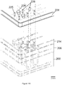
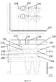
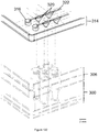
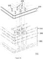
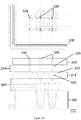
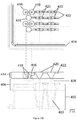
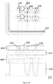
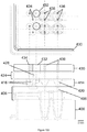
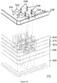
- the reaction mix moves through channels that are neatly arranged in a radiator pattern, and flow over heating elements that are at fixed temperatures.
- This architecture allows the entire amplification reaction to be completed in a few minutes, and is ideal for capillary separation and readout.
- the readout may be achieved by taking advantage of LDR-FRET or electronic detection.
- LDR-FRET one primer has a donor, the other has an acceptor group, and after ligation they form a hairpin. This allows for counting single ligation events to obtain highly quantitative readouts of input DNA copy number.
- the ligation product will contain two nano-particles, and these may be distinguished using electronic readout.
- the process should be separated into modular steps that may initially be optimized on separate instruments.
- the device may be comprised of a first module for purification of DNA from plasma cfDNA as well as RNA from exosomes, a second module for multiplexed reverse transcription and/or limited amplification of various targets, and a third module for generating and detecting ligation products.
- a modular architecture allows for swapping in improved modules that keep pace with technological developments.
- it is critical that products from one module can be moved seamlessly into the next module, without leakage and without worry of crossover contamination.
- the modular design should be amenable to scalable manufacture in high volumes at low cost.
- the manufacturing costs and how primers / reagents / samples are deposited into the device must be taken into consideration.
- the present invention is directed at overcoming these and other deficiencies in the art.
- a first aspect of the present invention is directed to a method for identifying, in a sample, one or more nucleic acid molecules containing a target nucleotide sequence differing from nucleotide sequences in other nucleic acid molecules in the sample, or other samples, by one or more nucleotides, one or more copy numbers, one or more transcript sequences, and/or one or more methylated residues.
- This method involves providing a sample potentially containing one or more nucleic acid molecules containing the target nucleotide sequence differing from the nucleotide sequences in other nucleic acid molecules by one or more nucleotides, one or more copy numbers, one or more transcript sequences, and/or one or more methylated residues, and contacting the sample with one or more enzymes capable of digesting deoxyuracil (dU) containing nucleic acid molecules present in the sample.
- dU deoxyuracil
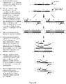
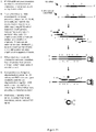
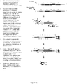
- Each primary oligonucleotide primer set comprising (a) a first primary oligonucleotide primer that comprises a nucleotide sequence that is complementary to a sequence adjacent to the target nucleotide sequence, and (b) a second primary oligonucleotide primer that comprises a nucleotide sequence that is complementary to a portion of an extension product formed from the first primary oligonucleotide primer.
- the contacted sample is blended with the one or more primary oligonucleotide primer sets, a deoxynucleotide mix including dUTP, and a DNA polymerase to form a polymerase chain reaction mixture, and the polymerase chain reaction mixture is subjected to one or more polymerase chain reaction cycles comprising a denaturation treatment, a hybridization treatment, and an extension treatment, thereby forming primary extension products comprising the target nucleotide sequence or a complement thereof.
- the method further involves blending the primary extension products with a ligase and one or more oligonucleotide probe sets to form a ligation reaction mixture.
- Each oligonucleotide probe set comprises (a) a first oligonucleotide probe having a target nucleotide sequence-specific portion, and (b) a second oligonucleotide probe having a target nucleotide sequence-specific portion, and wherein the first and second oligonucleotide probes of a probe set are configured to hybridize, in a base specific manner, on a complementary target nucleotide sequence of a primary extension product.
- the first and second oligonucleotide probes of the one or more oligonucleotide probe sets are ligated together to form ligated product sequences in the ligation reaction mixture, and the ligated product sequences in the sample are detected and distinguished to identify the presence of one or more nucleic acid molecules containing target nucleotide sequences differing from nucleotide sequences in other nucleic acid molecules in the sample by one or more nucleotides, one or more copy numbers, one or more transcript sequences, and/or one or more methylated residues.
- Another aspect of the present invention is directed to a method for identifying, in a sample, one or more nucleic acid molecules containing a target nucleotide sequence differing from nucleotide sequences in other nucleic acid molecules in the sample, or other samples, by one or more nucleotides, one or more copy numbers, one or more transcript sequences, and/or one or more methylated residues.
- This method involves providing a sample containing one or more nucleic acid molecules potentially containing the target nucleotide sequence differing from the nucleotide sequences in other nucleic acid molecules by one or more nucleotides, one or more copy numbers, one or more transcript sequences, and/or one or more methylated residues.
- the method further involves providing one or more enzymes capable of digesting deoxyuracil (dU) containing nucleic acid molecules present in the sample, and providing one or more primary oligonucleotide primer sets, each primary oligonucleotide primer set comprising (a) a first primary oligonucleotide primer that comprises a nucleotide sequence that is complementary to a sequence adjacent to the target nucleotide sequence and (b) a second primary oligonucleotide primer that comprises a nucleotide sequence that is complementary to a portion of an extension product formed from the first primary oligonucleotide primer.
- dU deoxyuracil
- the sample is blended with the one or more primary oligonucleotide primer sets, the one or more enzymes capable of digesting deoxyuracil (dU) containing nucleic acid molecules in the sample, a deoxynucleotide mix including dUTP, and a DNA polymerase to form a polymerase chain reaction mixture.
- the polymerase chain reaction mixture is subjected to conditions suitable for digesting deoxyuracil (dU) containing nucleic acid molecules present in the polymerase chain reaction mixture, and for one or more polymerase chain reaction cycles comprising a denaturation treatment, a hybridization treatment, and an extension treatment, thereby forming primary extension products comprising the target nucleotide sequence or a complement thereof.
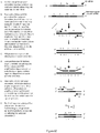
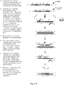
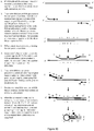
- the method further involves blending the primary extension products with a ligase and one or more oligonucleotide probe sets to form a ligation reaction mixture, wherein each oligonucleotide probe set comprises (a) a first oligonucleotide probe having a 5' primer-specific portion and a 3' target nucleotide sequence-specific portion, and (b) a second oligonucleotide probe having a 5' target nucleotide sequence-specific portion and a 3' primer-specific portion.
- the first and second oligonucleotide probes of a probe set are configured to hybridize, in a base specific manner, on a complementary target nucleotide sequence of a primary extension product.
- the ligation reaction mixture is subjected to one or more ligation reaction cycles whereby the first and second oligonucleotide probes of the one or more oligonucleotide probe sets are ligated together to form ligated product sequences in the ligation reaction mixture where each ligated product sequence comprises the 5' primer-specific portion, the target-specific portions, and the 3' primer-specific portion.
- the method further involves providing one or more secondary oligonucleotide primer sets, each secondary oligonucleotide primer set comprising (a) a first secondary oligonucleotide primer comprising the same nucleotide sequence as the 5' primer-specific portion of the ligated product sequence and (b) a second secondary oligonucleotide primer comprising a nucleotide sequence that is complementary to the 3' primer-specific portion of the ligated product sequence, and blending the ligated product sequences, the one or more secondary oligonucleotide primer sets, the one or more enzymes capable of digesting deoxyuracil (dU) containing nucleic acid molecules, a deoxynucleotide mix including dUTP, and a DNA polymerase to form a second polymerase chain reaction mixture.
- each secondary oligonucleotide primer set comprising (a) a first secondary oligonucleotide primer comprising the same nucleotide sequence as
- the second polymerase chain reaction mixture is subjected to conditions suitable for digesting deoxyuracil (dU) containing nucleic acid molecules present in the second polymerase chain reaction mixture, and one or more polymerase chain reaction cycles comprising a denaturation treatment, a hybridization treatment, and an extension treatment thereby forming secondary extension products.
- the secondary extension products are detected and distinguished in the sample to identify the presence of one or more nucleic acid molecules containing target nucleotide sequences differing from nucleotide sequences in other nucleic acid molecules in the sample by one or more nucleotides, one or more copy numbers, one or more transcript sequences, and/or one or more methylated residues.
- Another aspect of the present invention is directed to a method for identifying, in a sample, one or more nucleic acid molecules containing a target nucleotide sequence differing from nucleotide sequences in other nucleic acid molecules in the sample, or other samples, by one or more nucleotides, one or more copy numbers, one or more transcript sequences, and/or one or more methylated residues.
- This method involves providing a sample containing one or more nucleic acid molecules potentially containing the target nucleotide sequence differing from the nucleotide sequences in other nucleic acid molecules by one or more nucleotides, one or more copy numbers, one or more transcript sequences, and/or one or more methylated residues; providing one or more enzymes capable of digesting deoxyuracil (dU) containing nucleic acid molecules present in the sample; and providing one or more primary oligonucleotide primer sets, each primary oligonucleotide primer set comprising (a) a first primary oligonucleotide primer that comprises a nucleotide sequence that is complementary to a sequence adjacent to the target nucleotide sequence and (b) a second primary oligonucleotide primer that comprises a nucleotide sequence that is complementary to a portion of an extension product formed from the first primary oligonucleotide primer.
- the method further involves blending the sample, the one or more primary oligonucleotide primer sets, the one or more enzymes capable of digesting deoxyuracil (dU) containing nucleic acid molecules in the sample, a deoxynucleotide mix including dUTP, and a DNA polymerase to form a polymerase chain reaction mixture.
- the polymerase chain reaction mixture is subjected to conditions suitable for digesting deoxyuracil (dU) containing nucleic acid molecules present in the polymerase chain reaction mixture, and for one or more polymerase chain reaction cycles comprising a denaturation treatment, a hybridization treatment, and an extension treatment, thereby forming primary extension products comprising the target nucleotide sequence or a complement thereof.
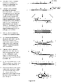
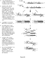
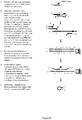
- each oligonucleotide probe set comprises (a) a first oligonucleotide probe having a 5' portion and a 3' target nucleotide sequence-specific portion, and (b) a second oligonucleotide probe having a 5' target nucleotide sequence-specific portion and a 3' portion, where the 5' portion of the first oligonucleotide probe of the probe set is complementary to a portion of the 3' portion of the second oligonucleotide probe, where one probe of the probe set comprises a detectable signal generating moiety, and where the first and second oligonucleotide probes of a probe set are configured to hybridize, in a base specific manner, on a complementary target nucleotide sequence of a primary extension product.
- the method further involves subjecting the ligation reaction mixture to one or more ligation reaction cycles whereby the first and second oligonucleotide probes of the one or more oligonucleotide probe sets are ligated together to form ligated product sequences in the ligation reaction mixture where each ligated product sequence comprises the 5' portion, the target-specific portions, the 3' portion, and the detectable signal generating moiety.
- the 5' portion of the ligated product sequence is hybridized to its complementary 3' portion and signal from the detectable signal generating moiety that is produced upon said hybridizing is detected.
- the ligated product sequences are distinguished in the sample based on said detecting to identify the presence of one or more nucleic acid molecules containing target nucleotide sequences differing from nucleotide sequences in other nucleic acid molecules in the sample by one or more nucleotides, one or more copy numbers, one or more transcript sequences, and/or one or more methylated residues.
- Another aspect of the present invention is directed to a method for identifying, in a sample, one or more nucleic acid molecules containing a target nucleotide sequence differing from nucleotide sequences in other nucleic acid molecules in the sample, or other samples, by one or more methylated residue.
- This method involves providing a sample potentially containing one or more nucleic acid molecules comprising the target nucleotide sequence differing from the nucleotide sequences in other nucleic acid molecules by one or more methylated residues and contacting the sample with one or more enzymes capable of digesting deoxyuracil (dU) containing nucleic acid molecules present in the sample.
- dU deoxyuracil
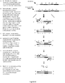
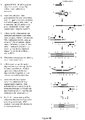
- the method further involves contacting the sample with one or more methylation sensitive enzymes to form a restriction enzyme reaction mixture, wherein the one or more methylation sensitive enzyme cleaves nucleic acid molecules in the sample that contain one or more unmethylated residues within at least one methylation sensitive enzyme recognition sequence.
- Each primary oligonucleotide primer set comprising (a) first primary oligonucleotide primer comprising a nucleotide sequence that is complementary to a region of the target nucleotide sequence that is upstream of the one or more methylated residues and (b) a second primary oligonucleotide primer comprising a nucleotide sequence that is the same as a region of the target nucleotide sequence that is downstream of the one or more methylated residues.
- the restriction enzyme reaction mixture is blended with the one or more primary oligonucleotide primer sets, a deoxynucleotide mix including dUTP, and a DNA polymerase to form a primary polymerase chain reaction mixture.
- the method further involves subjecting the primary polymerase chain reaction mixture to one or more polymerase chain reaction cycles comprising a denaturation treatment, a hybridization treatment, and an extension treatment, thereby forming primary extension products comprising the target nucleotide sequence or a complement thereof.
- One or more secondary oligonucleotide primer sets are provided, each secondary oligonucleotide primer set comprising first and second nested oligonucleotide primers capable of hybridizing to the primary extension products
- the primary extension products are blended with the one or more secondary oligonucleotide primer sets, a deoxynucleotide mix including dUTP, and a DNA polymerase to form a secondary polymerase chain reaction mixture, and the secondary polymerase chain reaction mixture is subjected to one or more polymerase chain reaction cycles comprising a denaturation treatment, a hybridization treatment, and an extension treatment thereby forming secondary extension products.
- the secondary extension products in the sample are detected and distinguished to identify the presence of one or more nucleic acid molecules containing target nucleotide sequences differing from nucleotide sequences in other nucleic acid molecules in the sample by one or more methylated residues.
- Another aspect of the present invention is directed to a method for identifying in a sample, one or more target ribonucleic acid molecules differing in sequence from other ribonucleic acid molecules in the sample due to alternative splicing, alternative transcript, alternative start site, alternative coding sequence, alternative non-coding sequence, exon insertion, exon deletion, intron insertion, translocation, mutation, or other rearrangement at the genome level.
- This method involves providing a sample containing one or more target ribonucleic acid molecules potentially containing a sequence differing from other ribonucleic acid molecules, and contacting the sample with one or more enzymes capable of digesting dU containing nucleic acid molecules potentially present in the sample.
- One or more oligonucleotide primers are provided, each primer being complementary to the one or more target ribonucleic acid molecule.
- the contacted sample is blended with the one or more oligonucleotide primers, and a reverse-transcriptase to form a reverse-transcription mixture, and complementary deoxyribonucleic acid (cDNA) molecules are generated in the reverse transcription mixture.
- Each cDNA molecule comprises a nucleotide sequence that is complementary to the target ribonucleic acid molecule sequence and contains dU.
- the method further involves providing one or more oligonucleotide primer sets, each primer set comprising (a) a first oligonucleotide primer comprising a nucleotide sequence that is complementary to a portion of a cDNA nucleotide sequence adjacent to the target ribonucleic acid molecule sequence complement of the cDNA, and (b) a second oligonucleotide primer comprising a nucleotide sequence that is complementary to a portion of an extension product formed from the first oligonucleotide primer.
- the reverse transcription mixture containing the cDNA molecules is blended with the one or more oligonucleotide primer sets, and a polymerase to form a polymerase reaction mixture, and the polymerase chain reaction mixture is subjected to one or more polymerase chain reaction cycles comprising a denaturation treatment, a hybridization treatment, and an extension treatment thereby forming one or more different primary extension products.
- the method further involves providing one or more oligonucleotide probe sets.
- Each probe set comprises (a) a first oligonucleotide probe having a target sequence-specific portion, and (b) a second oligonucleotide probe having a target sequence-specific portion, wherein the first and second oligonucleotide probes of a probe set are configured to hybridize, in a base specific manner, on a complementary portion of a primary extension product corresponding to the target ribonucleic acid molecule sequence.
- the primary extension products are contacted with a ligase and the one or more oligonucleotide probe sets to form a ligation reaction mixture and the first and second probes of the one or more oligonucleotide probe sets are ligated together to form ligated product sequences in the ligase reaction mixture.
- the ligated product sequences in the sample are detected and distinguished thereby identifying the presence of one or more target ribonucleic acid molecules differing in sequence from other ribonucleic acid molecules in the sample due to alternative splicing, alternative transcript, alternative start site, alternative coding sequence, alternative non-coding sequence, exon insertion, exon deletion, intron insertion, translocation, mutations, or other rearrangement at the genome level.
- Another aspect of the present invention is directed to a method for identifying in a sample, one or more target ribonucleic acid molecules differing in sequence from other ribonucleic acid molecules in the sample due to alternative splicing, alternative transcript, alternative start site, alternative coding sequence, alternative non-coding sequence, exon insertion, exon deletion, intron insertion, translocation, mutation, or other rearrangement at the genome level.
- This method involves providing a sample containing one or more target ribonucleic acid molecules potentially differing in sequence from other ribonucleic acid molecules, and contacting the sample with one or more enzymes capable of digesting dU containing nucleic acid molecules potentially present in the sample.
- One or more oligonucleotide primers is provided, each primer being complementary to the one or more target ribonucleic acid molecules, and the contacted sample is blended with the one or more oligonucleotide primers, a deoxynucleotide mix including dUTP, and a reverse-transcriptase to form a reverse-transcription mixture.
- Complementary deoxyribonucleic acid (cDNA) molecules are generated in the reverse transcription mixture, each cDNA molecule comprising a nucleotide sequence that is complementary to the target ribonucleic acid molecule and contains dU.
- the method further involves providing one or more oligonucleotide primer sets, each primer set comprising (a) a first oligonucleotide primer comprising a nucleotide sequence that is complementary to a portion of a cDNA nucleotide sequence adjacent to the target ribonucleic acid molecule sequence complement of the cDNA, and (b) a second oligonucleotide primer comprising a nucleotide sequence that is complementary to a portion of an extension product formed from the first oligonucleotide primer.
- the reverse transcription mixture containing the cDNA molecules is blended with the one or more oligonucleotide primer sets, a deoxynucleotide mix including dUTP, and a polymerase to form a polymerase reaction mixture, and the polymerase chain reaction mixture is subjected to one or more polymerase chain reaction cycles comprising a denaturation treatment, a hybridization treatment, and an extension treatment thereby forming one or more different primary extension products.
- the method further involves providing one or more oligonucleotide probe sets, each probe set comprising (a) a first oligonucleotide probe having a 5' primer-specific portion and a 3' target sequence-specific portion, and (b) a second oligonucleotide probe having a 5' target sequence-specific portion and a 3' primer-specific portion, where the first and second oligonucleotide probes of a probe set are configured to hybridize, in a base specific manner, on complementary portions of a primary extension product corresponding to the target ribonucleic acid molecule sequence.
- the primary extension products are contacted with a ligase and the one or more oligonucleotide probe sets to form a ligation reaction mixture, and the ligation reaction mixture is subjected to one or more ligation reaction cycles whereby the first and second probes of the one or more oligonucleotide probe sets are ligated together to form ligated product sequences in the ligase reaction mixture, where each ligated product sequence comprises the 5' primer-specific portion, the target-specific portions, and the 3' primer-specific portion.
- the method further involves providing one or more secondary oligonucleotide primer sets, each secondary oligonucleotide primer set comprising (a) a first secondary oligonucleotide primer comprising the same nucleotide sequence as the 5' primer-specific portion of the ligated product sequence and (b) a second secondary oligonucleotide primer comprising a nucleotide sequence that is complementary to the 3' primer-specific portion of the ligated product sequence, and blending the ligated product sequences, the one or more secondary oligonucleotide primer sets with one or more enzymes capable of digesting deoxyuracil (dU) containing nucleic acid molecules, a deoxynucleotide mix including dUTP, and a DNA polymerase to form a second polymerase chain reaction mixture.
- dU deoxyuracil
- the second polymerase chain reaction mixture is subjected to conditions suitable for digesting deoxyuracil (dU) containing nucleic acid molecules present in the second polymerase chain reaction mixture, and one or more polymerase chain reaction cycles comprising a denaturation treatment, a hybridization treatment, and an extension treatment thereby forming secondary extension products.
- the secondary extension products in the sample are detected and distinguished thereby identifying the presence of one or more ribonucleic acid molecules differing in sequence from other ribonucleic acid molecules in the sample due to alternative splicing, alternative transcript, alternative start site, alternative coding sequence, alternative non-coding sequence, exon insertion, exon deletion, intron insertion, translocation, mutation, or other rearrangement at the genome level.
- Another aspect of the present invention is directed to a method for identifying in a sample, one or more target ribonucleic acid molecules differing in sequence from other ribonucleic acid molecules in the sample due to alternative splicing, alternative transcript, alternative start site, alternative coding sequence, alternative non-coding sequence, exon insertion, exon deletion, intron insertion, translocation, mutation, or other rearrangement at the genome level.
- This method involves providing a sample containing one or more target ribonucleic acid molecules potentially differing in sequence from other ribonucleic acid molecules, and contacting the sample with one or more enzymes capable of digesting dU containing nucleic acid molecules potentially present in the sample.
- the method further involves providing one or more oligonucleotide primers, each primer being complementary to the one or more target ribonucleic acid molecules, and blending the contacted sample, the one or more oligonucleotide primers, a deoxynucleotide mix including dUTP, and a reverse-transcriptase to form a reverse-transcription mixture.
- Complementary deoxyribonucleic acid (cDNA) molecules are generated in the reverse transcription mixture, each cDNA molecule comprising a nucleotide sequence that is complementary to the target ribonucleic acid molecule and contains dU.
- the method further involves providing one or more oligonucleotide primer sets, each primer set comprising (a) a first oligonucleotide primer comprising a nucleotide sequence that is complementary to a portion of a cDNA nucleotide sequence adjacent to the target ribonucleic acid molecule sequence complement of the cDNA, and (b) a second oligonucleotide primer comprising a nucleotide sequence that is complementary to a portion of an extension product formed from the first oligonucleotide primer.
- the reverse transcription mixture containing the cDNA molecules is blended with the one or more oligonucleotide primer sets, a deoxynucleotide mix including dUTP, and a polymerase to form a polymerase reaction mixture, and the polymerase chain reaction mixture is subjected to one or more polymerase chain reaction cycles comprising a denaturation treatment, a hybridization treatment, and an extension treatment thereby forming one or more different primary extension products.
- the method further involves providing one or more oligonucleotide probe sets, each probe set comprising (a) a first oligonucleotide probe having a 5' portion and a 3' target nucleotide sequence-specific portion, and (b) a second oligonucleotide probe having a 5' target nucleotide sequence-specific portion and a 3' portion, where the 5' portion of the first oligonucleotide probe of the probe set is complementary to a portion of the 3' portion of the second oligonucleotide probe, where one probe of the probe set comprises a detectable signal generating moiety, and where the first and second oligonucleotide probes of a probe set are configured to hybridize, in a base specific manner, on complementary portions of a primary extension product corresponding to the target ribonucleic acid molecule sequence.
- the primary extension products are contacted with a ligase and the one or more oligonucleotide probe sets to form a ligation reaction mixture, and the ligation reaction mixture is subjected to one or more ligation reaction cycles whereby the first and second probes of the one or more oligonucleotide probe sets are ligated together to form ligated product sequences in the ligase reaction mixture, where each ligated product sequence comprises the 5' portion, the target-specific portions, the 3' portion, and the detectable signal generating moiety.
- the 5' portion of the ligated product sequence is hybridized to its complementary 3' portion, and the signal from the detectable signal generating moiety that is produced upon said hybridizing is detected.
- the ligated product sequences in the sample are detected based on said detecting to identify the presence of one or more ribonucleic acid molecules differing in sequence from other ribonucleic acid molecules in the sample due to alternative splicing, alternative transcript, alternative start site, alternative coding sequence, alternative non-coding sequence, exon insertion, exon deletion, intron insertion, translocation, mutation, or other rearrangement at the genome level.
- Another aspect of the present invention is directed to a method for identifying, in a sample, one or more target micro-ribonucleic acid (miRNA) molecules differing in sequence from other miRNA molecules in the sample by one or more bases.
- This method involves providing a sample containing one or more target miRNA molecules potentially differing in sequence from other miRNA molecules in the sample by one or more bases, and contacting the sample with one or more enzymes capable of digesting dU containing nucleic acid molecules potentially present in the sample.
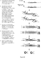
- oligonucleotide primer sets comprising (a) a first oligonucleotide primer having a 5' stem-loop portion, a blocking group, an internal primer-specific portion within the loop region, and a 3' nucleotide sequence portion that is complementary to a 3' portion of the target miRNA molecule sequence, (b) a second oligonucleotide primer having a 3' nucleotide sequence portion that is complementary to a complement of the 5' end of the target miRNA molecule sequence, and a 5' primer-specific portion, (c) a third oligonucleotide primer comprising a nucleotide sequence that is the same as the internal primer-specific portion of the first oligonucleotide primer, and (d) a fourth oligonucleotide primer comprising a nucleotide sequence that is the same as the 5' primer-specific portion of the second oligonucleotide primer.
- the contacted sample is blended with the one or more first oligonucleotide primers of a primer set, a deoxynucleotide mix including dUTP, and a reverse transcriptase to form a reverse transcription reaction mixture.
- the first oligonucleotide primer hybridizes to the target miRNA molecule sequence, if present in the sample, and the reverse transcriptase extends the 3' end of the hybridized first oligonucleotide primer to generate an extended first oligonucleotide primer comprising the complement of the target miRNA molecule sequence.
- the method further involves blending the reverse transcription reaction mixture with the second, third, and fourth oligonucleotide primers of the primer set to form a polymerase reaction mixture under conditions effective for the one or more second oligonucleotide primers of a primer set to hybridize to the region of the extended first oligonucleotide primer comprising the complement of the target miRNA molecule sequence and extend to generate a primary extension product comprising the 5' primer-specific portion, a nucleotide sequence corresponding to the target miRNA molecule sequence, and the complement of the internal primer-specific portion.
- the polymerase chain reaction mixture is subjected to one or more polymerase chain reaction cycles comprising a denaturation treatment, a hybridization treatment, and an extension treatment thereby forming a plurality of primary extension products.
- the method further involves blending the plurality of primary extension products with a ligase and one or more oligonucleotide probe sets to form a ligation reaction mixture.
- Each oligonucleotide probe set comprises (a) a first oligonucleotide probe having a target sequence-specific portion, and (b) a second oligonucleotide probe having a target sequence-specific portion and a portion complementary to a primary extension product, wherein the first and second oligonucleotide probes of a probe set are configured to hybridize, in a base specific manner on complementary portions of a primary extension product corresponding to the target miRNA molecule sequence.
- the first and second oligonucleotide probes of the one or more oligonucleotide probe sets are ligated together to form ligated product sequences in the ligation reaction mixture, and the ligated product sequences in the sample are detected and distinguished thereby identifying one or more target miRNA molecules differing in sequence from other miRNA molecules in the sample by one or more bases.
- Another aspect of the present invention is directed to a method for identifying, in a sample, one or more target micro-ribonucleic acid (miRNA) molecules differing in sequence from other miRNA molecules in the sample by one or more bases.
- This method involves providing a sample containing one or more target miRNA molecules potentially differing in sequence from other miRNA molecules in the sample by one or more bases, and contacting the sample with one or more enzymes capable of digesting dU containing nucleic acid molecules potentially present in the sample.
- the method further involves providing one or more oligonucleotide primer sets, each primer set comprising (a) a first oligonucleotide primer having a 5' stem-loop portion, a blocking group, an internal primer-specific portion within the loop region, and a 3' nucleotide sequence portion that is complementary to a 3' portion of the target miRNA molecule sequence, (b) a second oligonucleotide primer having a 3' nucleotide sequence portion that is complementary to a complement of the 5' end of the target miRNA molecule sequence, and a 5' primer-specific portion, (c) a third oligonucleotide primer comprising a nucleotide sequence that is the same as the internal primer-specific portion of the first oligonucleotide primer, and (d) a fourth oligonucleotide primer comprising a nucleotide sequence that is the same as the 5' primer-specific portion of the second oligonucleotide primer.
- the contacted sample is blended with the one or more first oligonucleotide primers of a primer set, a deoxynucleotide mix including dUTP, and a reverse transcriptase to form a reverse transcription reaction mixture where the first oligonucleotide primer hybridizes to the target miRNA molecule sequence, if present in the sample, and the reverse transcriptase extends the 3' end of the hybridized first oligonucleotide primer to generate an extended first oligonucleotide primer comprising the complement of the target miRNA molecule sequence.
- the reverse transcription reaction mixture is blended with the second, third, and fourth oligonucleotide primers of the primer set to form a polymerase reaction mixture under conditions effective for the one or more second oligonucleotide primers of a primer set to hybridize to the region of the extended first oligonucleotide primer comprising the complement of the target miRNA molecule sequence and extend to generate a primary extension product comprising the 5' primer-specific portion, a nucleotide sequence corresponding to the target miRNA molecule sequence, and the complement of the internal primer-specific portion.
- the polymerase chain reaction mixture is subjected to one or more polymerase chain reaction cycles comprising a denaturation treatment, a hybridization treatment, and an extension treatment thereby forming a plurality of primary extension products.
- the plurality of primary extension products are blended with a ligase and one or more oligonucleotide probe sets to form a ligation reaction mixture, where each oligonucleotide probe set comprises (a) a first oligonucleotide probe having a 5' primer-specific portion and a 3' targetsequence-specific portion, and (b) a second oligonucleotide probe having a 5' target sequence-specific portion, a portion complementary to a primary extension product, and a 3' primer-specific portion, and where the first and second oligonucleotide probes of a probe set are configured to hybridize, in a base specific manner, on complementary portions of a primary extension product corresponding to the target miRNA molecule sequence.
- the ligation reaction mixture is subjected to one or more ligation reaction cycles whereby the first and second oligonucleotide probes of the one or more oligonucleotide probe sets are ligated together to form ligated product sequences in the ligation reaction mixture wherein each ligated product sequence comprises the 5' primer-specific portion, the target-specific portions, and the 3' primer-specific portion.
- the method further involves providing one or more secondary oligonucleotide primer sets, each secondary oligonucleotide primer set comprising (a) a first secondary oligonucleotide primer comprising the same nucleotide sequence as the 5' primer-specific portion of the ligated product sequence and (b) a second secondary oligonucleotide primer comprising a nucleotide sequence that is complementary to the 3' primer-specific portion of the ligated product sequence, and blending the ligated product sequences, the one or more secondary oligonucleotide primer sets, with one or more enzymes capable of digesting deoxyuracil (dU) containing nucleic acid molecules, a deoxynucleotide mix including dUTP, and a DNA polymerase to form a second polymerase chain reaction mixture.
- dU deoxyuracil
- the second polymerase chain reaction mixture is subjected to conditions suitable for digesting deoxyuracil (dU) containing nucleic acid molecules present in the second polymerase chain reaction mixture, and one or more polymerase chain reaction cycles comprising a denaturation treatment, a hybridization treatment, and an extension treatment thereby forming secondary extension product.
- the secondary extension products in the sample are detected and distinguished thereby identifying one or more target miRNA molecules differing in sequence from other miRNA molecules in the sample by one or more bases.
- Another aspect of the present invention is directed to a method for identifying, in a sample, one or more target micro-ribonucleic acid (miRNA) molecules differing in sequence from other miRNA molecules in the sample by one or more bases.
- This method involves providing a sample containing one or more target miRNA molecules potentially differing in sequence from other miRNA molecules in the sample by one or more bases, and contacting the sample with one or more enzymes capable of digesting dU containing nucleic acid molecules potentially present in the sample.
- the method further involves providing one or more oligonucleotide primer sets, each primer set comprising (a) a first oligonucleotide primer having a 5' stem-loop portion, a blocking group, an internal primer-specific portion within the loop region, and a 3' nucleotide sequence portion that is complementary to a 3' portion of the target miRNA molecule sequence, (b) a second oligonucleotide primer having a 3' nucleotide sequence portion that is complementary to a complement of the 5' end of the target miRNA molecule sequence, and a 5' primer-specific portion, (c) a third oligonucleotide primer comprising a nucleotide sequence that is the same as the internal primer-specific portion of the first oligonucleotide primer, and (d) a fourth oligonucleotide primer comprising a nucleotide sequence that is the same as the 5' primer-specific portion of the second oligonucleotide primer.
- the contacted sample is blended with the one or more first oligonucleotide primers of a primer set, a deoxynucleotide mix including dUTP, and a reverse transcriptase to form a reverse transcription reaction mixture wherein the first oligonucleotide primer hybridizes to the target miRNA molecule sequence, if present in the sample, and the reverse transcriptase extends the 3' end of the hybridized first oligonucleotide primer to generate an extended first oligonucleotide primer comprising the complement of the target miRNA molecule sequence.
- the reverse transcription reaction mixture is blended with the second, third, and fourth oligonucleotide primers of the primer set to form a polymerase reaction mixture under conditions effective for the one or more second oligonucleotide primers of a primer set to hybridize to the region of the extended first oligonucleotide primer comprising the complement of the target miRNA molecule sequence and extend to generate a primary extension product comprising the 5' primer-specific portion, a nucleotide sequence corresponding to the target miRNA molecule sequence, and the complement of the internal primer-specific portion.
- the method further involves subjecting the polymerase chain reaction mixture to one or more polymerase chain reaction cycles comprising a denaturation treatment, a hybridization treatment, and an extension treatment thereby forming a plurality of primary extension products.
- the plurality of primary extension products are blended with a ligase and one or more oligonucleotide probe sets to form a ligation reaction mixture, wherein each oligonucleotide probe set comprises (a) a first oligonucleotide probe having a 5' portion and a 3' target nucleotide sequence-specific portion, and (b) a second oligonucleotide probe having a 5' target nucleotide sequence-specific portion and a 3' portion, where the 5' portion of the first oligonucleotide probe of the probe set is complementary to a portion of the 3' portion of the second oligonucleotide probe, where one probe of the probe set comprises a detectable signal generating moiety, and where the first and
- the ligation reaction mixture is subjected to one or more ligation reaction cycles whereby the first and second oligonucleotide probes of the one or more oligonucleotide probe sets are ligated together to form ligated product sequences in the ligation reaction mixture wherein each ligated product sequence comprises the 5' portion, the target-specific portions, the 3' portion, and the detectable signal generating moiety.
- the 5' portion of the ligated product sequence is hybridized to its complementary 3' portion, and signal from the detectable signal generating moiety that is produced upon said hybridizing is detected.
- the ligated product sequences in the sample are distinguished based on said detecting to identify the presence one or more target miRNA molecules differing in sequence from other miRNA molecules in the sample by one or more bases.
- Another aspect of the present invention is directed to a method for identifying, in a sample, one or more target micro-ribonucleic acid (miRNA) molecules differing in sequence from other miRNA molecules in the sample by one or more bases.
- This method involves providing a sample containing one or more target miRNA molecules potentially differing in sequence from other miRNA molecules by one or more base differences, and contacting the sample with one or more enzymes capable of digesting dU containing nucleic acid molecules potentially present in the sample.
- the contacted sample is blended with a ligase and a first oligonucleotide probe comprising a 5' phosphate, a 5' stem-loop portion, an internal primer-specific portion within the loop region, a blocking group, and a 3' nucleotide sequence that is complementary to a 3' portion of the target miRNA molecule sequence to form a ligation reaction.
- the method further involves ligating the target miRNA molecule sequence at its 3' end to the 5' phosphate of the first oligonucleotide probe to generate a chimeric nucleic acid molecule comprising the target miRNA molecule sequence, if present in the sample, appended to the first oligonucleotide probe.
- oligonucleotide primer sets comprising (a) a first oligonucleotide primer comprising a 3' nucleotide sequence that is complementary to a complement of the 5' end of the target miRNA molecule sequence, and a 5' primer-specific portion, (b) a second oligonucleotide primer comprising a nucleotide sequence that is complementary to the internal primer-specific portion of the first oligonucleotide probe, and (c) a third oligonucleotide primer comprising a nucleotide sequence that is the same as the 5' primer-specific portion of the first oligonucleotide primer.
- the chimeric nucleic acid molecule is blended with the one or more second oligonucleotide primers, a deoxynucleotide mix including dUTP, and a reverse transcriptase to form a reverse transcription reaction mixture, wherein the one or more second oligonucleotide primers of a primer set hybridizes to the internal primer specific portion of the chimeric nucleic acid molecule, and extends at its 3' end to generate a complement of the chimeric nucleic acid molecule, if present in the sample.
- the method further involves blending the reverse transcription reaction mixture with the first and third oligonucleotide primers of a primer set to form a polymerase reaction mixture, and subjecting the polymerase chain reaction mixture to one or more polymerase chain reaction cycles comprising a denaturation treatment, a hybridization treatment, and an extension treatment thereby forming primary extension products.
- the primary extension products comprise the 5' primer-specific portion, a nucleotide sequence corresponding to the target miRNA molecule sequence, and the complement of the internal primer-specific portion.
- the primary extension products are blended with a ligase and one or more oligonucleotide probe sets to form a ligation reaction mixture.
- Each oligonucleotide probe set comprises (a) a first oligonucleotide probe having a target sequence-specific portion, and (b) a second oligonucleotide probe having a target sequence-specific portion and a portion complementary to a primary extension product, wherein the first and second oligonucleotide probes of a probe set are configured to hybridize, in a base specific manner, on complementary portions of a primary extension product corresponding to the target miRNA molecule sequence.
- the first and second oligonucleotide probes of the one or more oligonucleotide probe sets are ligated together to form ligated product sequences in the ligation reaction mixture, and the ligated product sequences in the sample are detected and distinguished thereby identifying one or more target miRNA molecules differing in sequence from other miRNA molecules in the sample by one or more bases.
- Another aspect of the present invention method for identifying, in a sample, one or more target micro-ribonucleic acid (miRNA) molecules differing in sequence from other miRNA molecules in the sample by one or more bases involves providing a sample containing one or more miRNA molecules potentially differing in sequence from other miRNA molecules by one or more base differences, and contacting the sample with one or more enzymes capable of digesting dU containing nucleic acid molecules potentially present in the sample.
- miRNA micro-ribonucleic acid
- the contacted sample is blended with a ligase and a first oligonucleotide probe comprising a 5' phosphate, a 5' stem-loop portion, an internal primer-specific portion within the loop region, a blocking group, and a 3' nucleotide sequence that is complementary to a 3' portion of the target miRNA molecule sequence to form a ligation reaction, and the target miRNA molecule sequence at its 3'end is ligated to the 5' phosphate of the first oligonucleotide probe to generate a chimeric nucleic acid molecule comprising the target miRNA molecule sequence, if present in the sample, appended to the first oligonucleotide probe.
- the method further involves providing one or more oligonucleotide primer sets, each primer set comprising (a) a first oligonucleotide primer comprising a 3' nucleotide sequence that is complementary to a complement of the 5' end of the target miRNA molecule sequence, and a 5' primer-specific portion, (b) a second oligonucleotide primer comprising a nucleotide sequence that is complementary to the internal primer-specific portion of the first oligonucleotide probe, and (c) a third oligonucleotide primer comprising a nucleotide sequence that is the same as the 5' primer-specific portion of the first oligonucleotide primer.
- the chimeric nucleic acid molecule is blended with the one or more second oligonucleotide primers, a deoxynucleotide mix including dUTP, and a reverse transcriptase to form a reverse transcription reaction mixture, where the one or more second oligonucleotide primers of a primer set hybridizes to the internal primer specific portion of the chimeric nucleic acid molecule and extends at its 3' end to generate a complement of the chimeric nucleic acid molecule, if present in the sample.
- the reverse transcription reaction mixture is blended with the first and third oligonucleotide primers of a primer set to form a polymerase reaction mixture, and the polymerase chain reaction mixture is subjected to one or more polymerase chain reaction cycles comprising a denaturation treatment, a hybridization treatment, and an extension treatment thereby forming primary extension products comprising the 5' primer-specific portion, a nucleotide sequence corresponding to the target miRNA molecule sequence, and the complement of the internal primer-specific portion.
- the primary extension products are blended with a ligase and one or more oligonucleotide probe sets to form a ligation reaction mixture, wherein each oligonucleotide probe set comprises (a) a first oligonucleotide probe having a 5' primer-specific portion and a 3' target sequence-specific portion, and (b) a second oligonucleotide probe having a 5' target sequence-specific portion, a portion complementary to a primary extension product, and a 3' primer-specific portion, wherein the first and second oligonucleotide probes of a probe set are configured to hybridize, in a base specific manner, on complementary portions of a primary extension product corresponding to the target miRNA molecule sequence.
- the ligation reaction mixture is subjected to one or more ligation reaction cycles whereby the first and second oligonucleotide probes of the one or more oligonucleotide probe sets are ligated together to form ligated product sequences in the ligation reaction mixture, wherein each ligated product sequence comprises the 5' primer-specific portion, the target-specific portions, and the 3' primer-specific portion.
- the method further involves providing one or more secondary oligonucleotide primer sets, each secondary oligonucleotide primer set comprising (a) a first secondary oligonucleotide primer comprising the same nucleotide sequence as the 5' primer-specific portion of the ligated product sequence and (b) a second secondary oligonucleotide primer comprising a nucleotide sequence that is complementary to the 3' primer-specific portion of the ligated product sequence.
- the ligated product sequences are blended with the one or more secondary oligonucleotide primer sets, one or more enzymes capable of digesting deoxyuracil (dU) containing nucleic acid molecules, a deoxynucleotide mix including dUTP, and a DNA polymerase to form a second polymerase chain reaction mixture.
- the second polymerase chain reaction mixture is subjected to conditions suitable for digesting deoxyuracil (dU) containing nucleic acid molecules present in the second polymerase chain reaction mixture, and one or more polymerase chain reaction cycles comprising a denaturation treatment, a hybridization treatment, and an extension treatment thereby forming secondary extension products.
- the secondary extension products in the sample are detected and distinguished thereby identifying one or more target miRNA molecules differing in sequence from other miRNA molecules in the sample by one or more bases.
- Another aspect of the present invention is directed to a method for identifying, in a sample, one or more target micro-ribonucleic acid (miRNA) molecules differing in sequence from other miRNA molecules in the sample by one or more bases.
- This method involves providing a sample containing one or more miRNA molecules potentially differing in sequence from other miRNA molecules by one or more base differences, and contacting the sample with one or more enzymes capable of digesting dU containing nucleic acid molecules potentially present in the sample.
- the contacted sample is blended with a ligase and a first oligonucleotide probe comprising a 5' phosphate, a 5' stem-loop portion, an internal primer-specific portion within the loop region, a blocking group, and a 3' nucleotide sequence that is complementary to a 3' portion of the target miRNA molecule sequence to form a ligation reaction.
- the target miRNA molecule sequence is ligated at its 3'end to the 5' phosphate of the first oligonucleotide probe to generate a chimeric nucleic acid molecule comprising the target miRNA molecule sequence, if present in the sample, appended to the first oligonucleotide probe.
- the method further involves providing one or more oligonucleotide primer sets, each primer set comprising (a) a first oligonucleotide primer comprising a 3' nucleotide sequence that is complementary to a complement of the 5' end of the target miRNA molecule sequence, and a 5' primer-specific portion, (b) a second oligonucleotide primer comprising a nucleotide sequence that is complementary to the internal primer-specific portion of the first oligonucleotide probe, and (c) a third oligonucleotide primer comprising a nucleotide sequence that is the same as the 5' primer-specific portion of the first oligonucleotide primer.
- the chimeric nucleic acid molecule is blended with the one or more second oligonucleotide primers, a deoxynucleotide mix including dUTP, and a reverse transcriptase to form a reverse transcription reaction mixture, wherein the one or more second oligonucleotide primers of a primer set hybridizes to the internal primer specific portion of the chimeric nucleic acid molecule and extends at its 3' end to generate a complement of the chimeric nucleic acid molecule, if present in the sample.
- the reverse transcription reaction mixture is blended with the first and third oligonucleotide primers of a primer set to form a polymerase reaction mixture, and the polymerase chain reaction mixture is subjected to one or more polymerase chain reaction cycles comprising a denaturation treatment, a hybridization treatment, and an extension treatment thereby forming primary extension products comprising the 5' primer-specific portion, a nucleotide sequence corresponding to the target miRNA molecule sequence, and the complement of the internal primer-specific portion.
- each oligonucleotide probe set comprises (a) a first oligonucleotide probe having a 5' portion and a 3' target nucleotide sequence-specific portion, and (b) a second oligonucleotide probe having a 5' target nucleotide sequence-specific portion and a 3' portion, where the 5' portion of the first oligonucleotide probe of the probe set is complementary to a portion of the 3' portion of the second oligonucleotide probe, where one probe of the probe set comprises a detectable signal generating moiety, and where the first and second oligonucleotide probes of a probe set are configured to hybridize, in a base specific manner, on complementary portions of a primary extension product corresponding to the target miRNA molecule sequence.
- the ligation reaction mixture is subjected to one or more ligation reaction cycles whereby the first and second oligonucleotide probes of the one or more oligonucleotide probe sets are ligated together to form ligated product sequences in the ligation reaction mixture wherein each ligated product sequence comprises the 5' portion, the target-specific portions, the 3' portion, and the detectable signal generating moiety.
- each ligated product sequence comprises the 5' portion, the target-specific portions, the 3' portion, and the detectable signal generating moiety.
- the the 5' portion of the ligated product sequence is hybridized to its complementary 3' portion, and signal from the detectable signal generating moiety that is produced upon said hybridizing is detected.
- the ligated product sequences are distinguished in the sample based on said detecting to identify the presence one or more target miRNA molecules differing in sequence from other miRNA molecules in the sample by one or more bases.
- Another aspect of the present invention is directed to a device for simultaneously adding liquids to two or more wells in a row and/or column of a microtiter plate.
- the device has opposed top and bottom surfaces with the top surface having openings leading into the wells and the bottom surface defining closed ends of the wells.
- the device comprises a first layer defined by first and second boundaries with metering chambers extending between the first and second boundaries of said first layer and in fluid communication with one another.
- the first layer is configured to be fitted, in an operative position, proximate to the microtiter plate with the first boundary of the first layer being closest to the top surface of the microtiter plate and each of the metering chambers being in fluid communication with an individual well in a row and/or column of the microtiter plate.
- the first layer further comprises a filling chamber in fluid communication with one or more of the metering chambers.
- the device comprises a second layer defined by first and second boundaries with a filling port extending between the first and second boundaries of the second layer.
- the second layer is configured to be fitted, in an operative position, on the first layer with the first boundary of the second layer adjacent to the second boundary of the first layer and the filling port being aligned with the filling chamber.
- Another aspect of the present invention is directed to a method of adding liquids to two or more wells in a row and/or column of a microtiter plate having opposed top and bottom surfaces with the top surface having openings leading into the wells and the bottom surface defining closed ends of the wells.
- This method involves providing a device comprising a first layer having first and second boundaries with metering chambers extending between the first and second boundaries of the first layer and in fluid communication with one another.
- the first layer of the device is configured to be fitted, in an operative position, proximate to the microtiter plate with the first boundaries of the first layer being closest to the top surface of the microtiter plate and one of the metering chambers being in fluid communication with an individual well in a row and/or column of the microtiter plate.
- the first layer further comprising a filling chamber in fluid communication with one or more of said metering chambers.
- the device comprises a second layer having first and second boundaries with a filling port extending between the first and second boundaries of the second layer.
- the second layer is configured to be fitted, in an operative position, on the first layer with the first boundary of the second layer adjacent to the second boundary of the first layer and the filling port being aligned with the charge chamber.
- liquid entering the device through the filling port will pass through the filling chamber, the metering chambers, and into two or more wells in a row and/or column of said microtiter plate.
- the method further involves filling the device with liquid, and discharging liquid in the device into two or more wells in a row and/or column of said microtiter plate.
- the present invention describes a number of approaches for detecting mutations, expression, splice variant, translocation, copy number, and/or methylation changes in target nucleic acid molecules using nuclease, ligase and polymerase reactions.
- the present invention solves the problems of carry over prevention, as well as allowing for spatial multiplexing to provide relative quantification, similar to digital PCR.
- Such technology may be utilized for non-invasive early detection of cancer, non-invasive prognosis of cancer, and monitoring for cancer recurrence from plasma or serum samples.
- the most cost-effective early disease detection test may combine an initial multiplexed coupled amplification and ligation assay to determine "disease load". For cancer detection this would achieve > 95% sensitivity for all cancers (pan-oncology), at > 97% specificity.
- a flow chart for a cancer tumor load assay is illustrated in Figure 1 .
- An initial multiplexed PCR/LDR screening assay scoring for mutation, methylation, miRNA, mRNA, alternative splicing, and translocations identifies those samples with > 5 of 24-48 markers positive. Presumptive positive samples are then assayed using "pixel" PCR/LDR with additional tissue-specific markers to validate the initial result, and identify tissue of origin. The physician may then order targeted sequencing to further guide treatment decisions for the patient.
- the present invention is directed to a universal diagnostic approach that seeks to combine the best features of digital polymerase chain reaction (PCR), ligation detection reaction (LDR), and quantitative detection of multiple disease markers, e.g., cancer markers.
- PCR digital polymerase chain reaction
- LDR ligation detection reaction
- the family of assay architecture and devices comprises three modules for PCR-LDR quantification of low-abundance disease markers from blood. Each module may be optimized independently, and may be manual, semi-automated, or fully automated. The design enables integrating the modules together, such that any module may be optimized independently to bring improved performance to the entire assay.
- the first family of assay designs is based on an initial multiplexed PCR or RT-PCR amplification followed by multiplexed LDR using LDR probes having unique sequence tags containing primer-specific portions.
- the products are distributed and mixed with tag primer sets with target-directed TaqManTM probes, or alternatively with UniTaq primer sets, and the input target nucleic acids quantified using real-time PCR
- the first module takes an input sample of blood, and separates plasma from red blood cells (RBCs) and white blood cells (WBCs). It separates plasma again to remove any residual cells.
- cell fractions containing all WBCs and circulating tumor cells (CTCs) and some RBCs are separated. Exosomes are separated or affinity captured from plasma.
- the module purifies (i) RNA from the WBC and CTC fraction, (ii) miRNA and RNA from exosomes, and (iii) cell free DNA (cfDNA) from plasma.
- the second module enables distribution of the above components into 24 or 48 chambers or wells for spatial multiplexing with proportional multiplexed PCR or RT-PCR amplification of targeted gene, promoter, miRNA, or mRNA regions.
- These include: (i) specific splice-variant or gene-fusion mRNAs in the CTC containing WBC fraction, (ii) specific miRNAs from exosomes, (iii) specific mRNAs from exosomes, (iv) specific cancer gene DNA regions from cfDNA, and (v) specific (methylated) promoter regions from cfDNA.
- the third module enables spatial distribution of the above products into wells of a microtiter plate, e.g., in a 24 x 16 or 48 x 32 configuration.
- This module enables detection and enumeration of LDR products using real-time PCR to provide quantitative results for each disease marker.
- the first and second modules may be configured to process multiple samples simultaneously for the screening assay mode, where the LDR products containing sequence tags provide relative quantitative results using real-time PCR readout (see Figure 2 ).
- DNA and RNA isolated from various blood fractions from 24 individual samples are subjected to multiplexed PCR-LDR and RT-PCR-LDR, then distributed down a column, e.g., 16 wells in the microtiter plate as illustrated in Figure 3 .
- Tag primer sets with target-directed TaqManTM probes, or alternatively with UniTaq primer sets are added across the rows as shown in Figure 4 , allowing for real-time PCR ( Figure 5 ).
- samples #2 & #15 have strong signal at >5 positions, so are considered potentially positive (pending additional verification as described more below), while sample #8 with 4 weak signals should also get additional workup.
- the two modules may also be configured to process a single sample, with spatial multiplexing enabling "pixel" PCR/LDR, where the LDR products enable enumeration of the original target molecules.
- This is analogous to digital PCR, but at a higher level of multiplexing (see Figure 6 ).
- the DNA and RNA from a single sample are distributed into 24 chambers prior to multiplex PCR-LDR as shown in Figure 7 .
- some chambers have one or no target molecules.
- the LDR products are distributed down a column, e.g., 16 wells in the microtiter plate ( Figure 7 ).
- Tag primer sets with target-directed TaqManTM probes, or alternatively UniTaq primer sets, are added across the rows (see Figure 8 ), allowing for real-time PCR (see Figure 9 ).
- the results are interpreted based on Poisson distribution of Ct value as representing an integral multiple of a single molecule in the original mix, i.e., 0, 1, 2 etc.
- Figures 29 and 30 show a Poisson distribution of 6 to 48 and 12 to 96 molecules in 24 wells, respectively, and
- Figures 31 and 32 show a Poisson distribution of 12 to 96 and 24 to 192 molecules in 48 wells, respectively.
- Figure 9 , row A shows (# addresses: # initial target molecule) of (17:0; 6:1; 1:2), which corresponds to 8 molecules.
- Figure 9 , row K shows (7:0; 10:1; 5:2; 2:4), which corresponds to about 30 molecules.
- a different form of dilution and distribution may be used to enumerate molecules over a wider range, as illustrated for miRNA or mRNA quantification in Figures 10 -13 .
- the initial sample is distributed into 8 chambers, diluted 10-fold and distributed into another 8 chambers, etc. (see Figure 11 ).
- the samples are subjected to multiplexed RT-PCR and LDR, and the LDR products are individually distributed down a column ( Figure 11 ).
- Tag primer sets with target-directed TaqManTM probes, or alternatively UniTaq primer sets, are added across the rows (see Figure 12 ), allowing for real-time PCR and detection (see Figure 13 ).
- Figures 33-37 show Poisson distributions of 1 to 128 molecules in 8 wells.
- the second family of assay designs is based on an initial multiplexed PCR or RT-PCR amplification followed by distribution and capture of PCR amplified targets on the wells of a microtiter plate.
- a single cycle of LDR enables capture of LDR products on the correct targets on the solid support, while mis-ligations are washed away.
- the LDR products are quantified, either through LDR-FRET, real-time PCR, or other reporter systems.
- the first module takes an input sample of blood, and separates CTCs if present, separates plasma from blood cells, and exosomes from plasma, and then purifies (i) DNA and RNA from CTCs if present, (ii) miRNA and RNA from exosomes, and (iii) cfDNA from plasma.
- the second module enables distribution of the above components into 24 or 48 chambers or wells for spatial multiplexing with proportional multiplexed PCR or RT-PCR amplification of targeted gene, promoter, miRNA, or mRNA regions.
- These include: (i) specific chromosomal regions for copy number enumeration from CTC's, (ii) specific splice-variant or gene-fusion mRNAs from CTC's, (iii) specific miRNAs from exosomes, (iv) specific mRNAs from exosomes, (v) specific cancer gene DNA regions from cfDNA, and (vi) specific (methylated) promoter regions from cfDNA.
- the third module enables spatial distribution of the above products down a column, e.g., 16 wells in the microtiter plate, followed by capture of the amplified target on the solid support, e.g., in a 24 x 16 or 48 x 32 configuration.
- This module enables capture of LDR products on the solid support, followed by detection and enumeration of LDR products to provide quantitative results for each marker.
- the first and second modules may be configured to process multiple samples simultaneously for the screening assay mode, wherein the LDR products provide relative quantitative results, analogous to real-time PCR readout (see schematic overview of Figure 14 ).
- LDR probes are added across the rows, and ligation on correct target captures the product while unreacted primer is washed away ( Figure 17 ).
- the two modules may also be configured to process a single sample, with spatial multiplexing enabling "pixel" PCR/LDR, wherein the LDR products enable enumeration of the original target molecules, analogous to digital PCR, but at a higher level of multiplexing ( Figure 19 ).
- the DNA and RNA from a single sample are distributed into 24 chambers prior to multiplex PCR ( Figure 20 ), such that some chambers have one or no target molecules.
- the amplicons are distributed down a column, e.g., 16 wells in the microtiter plate, and captured on the solid support ( Figures 20 and 21 ).
- LDR probe addition and LDR product detection is as above ( Figures 22 and 23 ).
- Figures 29 and 30 show the Poisson distribution of 6-48 and 12-96 molecules in 24 wells, respectively, and Figures 31 and 32 show the Poisson distribution of 12-96 and 24-192 molecules in 48 wells, respectively.
- Figure 23 , row A shows (# addresses: # initial target molecule) of (17:0; 6:1; 1:2), which corresponds to 8 molecules.
- Figure 23 , row K shows (7:0; 10:1; 5:2; 2: 4), which corresponds to about 30 molecules.
- a different form of dilution and distribution may be used to enumerate molecules over a wider range, as illustrated for miRNA or mRNA quantification in Figures 24 - 28 .
- the initial sample is distributed into 8 chambers, diluted 10-fold and distributed into another 8 chambers, etc. ( Figure 25 ).
- the samples are subjected to multiplex RT-PCR and distributed down a column.
- the RT-PCR products are captured on a solid support ( Figure 26 ) and LDR primer sets are added across the rows (see Figure 27 ). For the example of 24 chambers, this can quantify across 3 orders of magnitude, but across 48 chambers it can cover 6 orders of magnitude differences (see Poisson distributions of Figures 33-37 ).
- the first theme is multiplexing. PCR works best when primer concentration is relatively high, from 50nM to 500nM, limiting multiplexing. Further, the more PCR primer pairs added, the chances of amplifying incorrect products or creating primer-dimers increase exponentially. In contrast, for LDR probes, low concentrations on the order of 4nM to 20nM are used, and probe-dimers are limited by the requirement for adjacent hybridization on the target to allow for a ligation event. Use of low concentrations of gene-specific PCR primers or LDR probes containing universal primer sequence "tails" allows for subsequent addition of higher concentrations of universal primers to achieve proportional amplification of the initial PCR or LDR products.
- PCR primers containing a few extra bases and a blocking group which is liberated to form a free 3'OH by cleavage with a nuclease only when hybridized to the target, e.g., a ribonucleotide base as the blocking group and RNase H2 as the cleaving nuclease.
- the second theme is fluctuations in signal due to low input target nucleic acids.
- the target nucleic acid originated from a few cells, either captured as CTCs, or from tumor cells that underwent apoptosis and released their DNA as small fragments (140 - 160 bp) in the serum.
- target-independent signal also known as "No Template Control” (NTC).
- NTC No Template Control
- NTC No Template Control
- the 5' ⁇ 3' nuclease activity of polymerase may be used to liberate the 5' phosphate of the downstream ligation primer (only when hybridized to the target), so it is suitable for ligation.
- Further specificity for distinguishing presence of a low-level mutation may be achieved by: (i) using upstream LDR probes containing a mismatch in the 2 nd or 3 rd position from the 3'OH, (ii) using LDR probes to wild-type sequence that (optionally) ligate but do not undergo additional amplification, and (iii) using upstream LDR probes containing a few extra bases and a blocking group, which is liberated to form a free 3'OH by cleavage with a nuclease only when hybridized to the complementary target (e.g., RNase H2 and a ribonucleotide base).
- the complementary target e.g., RNase H2 and a ribonucleotide base
- the fourth theme is either suppressed (reduced) amplification or incorrect (false) amplification due to unused primers in the reaction.
- One approach to eliminate such unused primers is to capture genomic or target or amplified target DNA on a solid support, allow ligation probes to hybridize and ligate, and then remove probes or products that are not hybridized.
- Alternative solutions include pre-amplification, followed by subsequent nested LDR and/or PCR steps, such that there is a second level of selection in the process.
- Carryover signal may be eliminated by standard uracil incorporation during the universal amplification step, and using UDG (and optionally AP endonuclease) in the pre-amplification workup procedure. Incorporation of carryover prevention is central to the methods of the present invention as described in more detail below.
- the initial PCR amplification is performed using incorporation of uracil.
- the LDR reaction is performed with LDR probes lacking uracil. Thus, when the LDR products are subjected to real-time PCR quantification, addition of UDG destroys the initial PCR products, but not the LDR products.
- LDR is a linear process and the tag primers use sequences absent from the human genome, accidental carryover of LDR products back to the original PCR will not cause template-independent amplification.
- Additional schemes to provide carryover prevention with methylated targets include use of restriction endonucleases before amplification, or after bisulfite treatment if using the latter approach as described infra.
- a first aspect of the present invention is directed to a method for identifying, in a sample, one or more nucleic acid molecules containing a target nucleotide sequence differing from nucleotide sequences in other nucleic acid molecules in the sample, or other samples, by one or more nucleotides, one or more copy numbers, one or more transcript sequences, and/or one or more methylated residues.
- This method involves providing a sample potentially containing one or more nucleic acid molecules containing the target nucleotide sequence differing from the nucleotide sequences in other nucleic acid molecules by one or more nucleotides, one or more copy numbers, one or more transcript sequences, and/or one or more methylated residues, and contacting the sample with one or more enzymes capable of digesting deoxyuracil (dU) containing nucleic acid molecules present in the sample.
- dU deoxyuracil
- Each primary oligonucleotide primer set comprising (a) a first primary oligonucleotide primer that comprises a nucleotide sequence that is complementary to a sequence adjacent to the target nucleotide sequence, and (b) a second primary oligonucleotide primer that comprises a nucleotide sequence that is complementary to a portion of an extension product formed from the first primary oligonucleotide primer.
- the contacted sample is blended with the one or more primary oligonucleotide primer sets, a deoxynucleotide mix including dUTP, and a DNA polymerase to form a polymerase chain reaction mixture, and the polymerase chain reaction mixture is subjected to one or more polymerase chain reaction cycles comprising a denaturation treatment, a hybridization treatment, and an extension treatment, thereby forming primary extension products comprising the target nucleotide sequence or a complement thereof.
- the method further involves blending the primary extension products with a ligase and one or more oligonucleotide probe sets to form a ligation reaction mixture.
- Each oligonucleotide probe set comprises (a) a first oligonucleotide probe having a target nucleotide sequence-specific portion, and (b) a second oligonucleotide probe having a target nucleotide sequence-specific portion, wherein the first and second oligonucleotide probes of a probe set are configured to hybridize, in a base specific manner, adjacent to one another on a complementary target nucleotide sequence of a primary extension product with a junction between them.
- the first and second oligonucleotide probes of the one or more oligonucleotide probe sets are ligated together to form ligated product sequences in the ligation reaction mixture, and the ligated product sequences in the sample are detected and distinguished to identify the presence of one or more nucleic acid molecules containing target nucleotide sequences differing from nucleotide sequences in other nucleic acid molecules in the sample by one or more nucleotides, one or more copy numbers, one or more transcript sequences, and/or one or more methylated residues.
- FIGS 38-44 illustrate various embodiments of this aspect of the present invention.
- Figure 38 illustrates an exemplary PCR-LDR-qPCR carryover prevention reaction to detect mutations from genomic or cfDNA.
- This method starts by isolating genomic DNA or cell free DNA (cfDNA) as shown in step A.
- the DNA sample is treated with an enzyme capable of digesting deoxyuracil (dU) containing nucleic acid molecules that may be present in the sample.
- Suitable enzymes include, without limitation, E. coli uracil DNA glycosylase (UDG), Antarctic Thermolabile UDG, or Human single-strand-selective monofunctional uracil-DNA Glycosylase (hSMUG1).
- the sample is then subject to an amplification reaction, e.g., a polymerase chain reaction (PCR) to amplify mutation containing regions of interest.
- amplification reaction e.g., a polymerase chain reaction (PCR) to amplify mutation containing regions of interest.
- the amplification reaction is carried out using locus specific primers and a deoxynucleotide mix that includes dUTP.
- limited cycle amplification (12-20 cycles) is performed to maintain relative ratios of different amplicons being produced.
- the regions of interest are amplified using 20-40 cycles.
- the amplified products contain dU as shown in Figure 38 , step C, which allows for subsequent treatment with UDG or a similar enzyme for carryover prevention.
- target-specific oligonucleotide probes are hybridized to the amplified products and ligase (filled circle) covalently seals the two oligonucleotides together when hybridized to their complementary sequence.
- the upstream oligonucleotide probe contains a 5' primer-specific portion (Ai) and the downstream oligonucleotide probe contains a 3' primer-specific portion (Ci') that permits subsequent amplification of the ligation product.
- the ligation products are aliquot into separate wells containing one or more tag-specific primer pairs, each pair comprising of matched primers Ai and Ci, treated with UDG or similar enzyme to remove dU containing amplification products or contaminants, PCR amplified, and detected.
- steps E & F detection of the ligation product can be carried out using traditional TaqManTM detection assay (see U.S. Patent No. 6,270,967 to Whitcombe et al. , and U.S. Patent No. 7,601,821 to Anderson et al. , which are hereby incorporated by reference in their entirety).
- an oligonucleotide probe spanning the ligation junction is used in conjunction with primers suitable for hybridization on the primer-specific portions of the ligation products for amplification and detection.
- the TaqManTM probe contains a fluorescent reporter group on one end (F1) and a quencher molecule (Q) on the other end that are in close enough proximity to each other in the intact probe that the quencher molecule quenches fluorescence of the reporter group.
- F1 fluorescent reporter group on one end
- Q quencher molecule
- the TaqManTM probe and upstream primer hybridize to their complementary regions of the ligation product.
- the 5' ⁇ 3' nuclease activity of the polymerase extends the hybridized primer and liberates the fluorescent group of the TaqManTM probe to generate a detectable signal ( Figure 38 , step F).
- Use of dUTP during the amplification reaction generates products containing dU, which can subsequently be destroyed using UDG for carryover prevention.
- Figure 39 illustrates an exemplary PCR-qLDR carryover prevention reaction to detect mutations from genomic or cfDNA.
- This method starts by isolating genomic DNA or cell free DNA (cfDNA) as shown in step A.
- step B the DNA sample is treated with a deoxyuracil (dU) digesting enzyme, such as UDG, to digest dU containing nucleic acid molecules that may be present in the sample, and then subject to an amplification reaction, e.g., a polymerase chain reaction (PCR) to amplify mutation containing regions of interest.
- amplification reaction is carried out using locus specific primers and a deoxynucleotide mix that includes dUTP.
- limited cycle amplification (12-20 cycles) is performed to maintain relative ratios of different amplicons being produced.
- the regions of interest are amplified using 20-40 cycles.
- the locus specific primers also contain 5' primer regions, e.g., universal primer regions, which enables a subsequent universal PCR amplification using biotin labeled primers to append a 5' biotin to the amplification products containing the region of interest ( Figure 39 , step B).
- the amplification products incorporate dU, allowing for carryover prevention, and are captured on a solid support via the appended 5' biotin moiety.
- the mutation of interest is detected using mutation specific ligation probes as illustrated in Figure 39 , step D.
- the first ligation probe contains a 3' target specific region and a 5' tail sequence with a donor or acceptor moiety and the second ligation probe in a probe set contains a 5' target specific region and 3' tail sequence with an acceptor or donor moiety, respectively.
- the 5' and 3' tail sequences of the ligation probes in a probe set are complementary to each other and the acceptor and donor groups are capable of generating a detectable signal via Förster resonance energy transfer (FRET) when brought in close proximity to each other.
- FRET Förster resonance energy transfer
- unligated oligonucleotide probes are washed away, and the ligation product is denatured from the immobilized amplification products.
- the complementary 5' and 3'tail sequences of the ligation products hybridize to each other bringing the donor and acceptor groups in close proximity to each other to generate a detectable FRET signal.
- the upstream probe may contain a fluorescent reporter group on the 5' end followed by the tail sequence portion, a quenching group (e.g., ZEN), and the target-specific portion as shown in Figure 39 , step F.
- a quenching group e.g., ZEN
- the fluorescent group is quenched by the Zen group.
- the complementary 5' and 3' tail portions of the ligation product hybridize to form a short double stranded portion. In this formation the reporter group is no longer quenched by the quenching group and a detectable signal is produced.
- Figure 39 illustrates a biotinylated universal primer-streptavidin coated surface approach for capturing extension products on a solid support. Such capture may occur prior to or subsequent to the ligation step.
- Other approaches for linking the product to the solid support include covalently attaching a portion of or the majority of the universal primer to the solid support prior to PCR amplification.
- the primers can be designed to include a capture sequence on the 5' end, a polymerase extension blocking group, and a universal or target-specific portion on the 3' end.
- the 5' capture sequence portion of the products will be single stranded, and if it is a long and/or GC rich sequence, it may be captured on a complementary sequence under conditions which allow for denaturation of the non-captured strand, or removal by cleavage, e.g., lambda exonuclease cleavage.
- the capture step may be enhanced by use of PNA, LNA, or other nucleotide analogues within the primer, capture probe sequence or both.
- the primer may be covalently attached to the solid surface using Dibenzocyclooctyl (DBCO) for copper-free click chemistry (to an azide); 5-Octadiynyl dU for click chemistry (to an azide); Amino Modifier C6 dT (for peptide linkage); or Azide, for click chemistry to an alkene or DBCO.
- DBCO Dibenzocyclooctyl
- Figure 40 illustrates another exemplary PCR-LDR-qPCR carryover prevention reaction to detect mutations.
- Genomic or cfDNA is isolated ( Figure 40 , step A), and the isolated DNA sample is treated with UDG to digest dU containing nucleic acid molecules that may be present in the sample.
- initial amplification is carried out using locus specific PCR primers that contain a cleavable blocking group at their 3' ends.
- the blocking group prevents non-target specific polymerase extension and amplification.
- a suitable blocking group is an RNA base (r) that is cleaved by RNase-H (star symbol) only upon hybridization of the primer to its complementary sequence (see e.g., Dobosy et. al.
- the region of interest is amplified and the PCR product contains dU allowing for carryover prevention ( Figure 40 , step C).
- Target-specific oligonucleotide probes containing primer tags (Ai and Ci') are then hybridized to the amplified products in a base specific manner, and ligase (filled circle) covalently seals the two oligonucleotides together when hybridized to their complementary sequence ( Figure 40 , step D).
- the ligation products are detected using pairs of matched primers Ai and Ci, and TaqManTM probes that span the ligation junction as described in Figure 38 (see Figure 40 , steps E-F).
- Figure 41 illustrates another exemplary PCR-LDR-qPCR carryover prevention reaction to detect mutations.
- Genomic or cfDNA is isolated ( Figure 41 , step A), and the isolated DNA sample is treated with UDG to digest dU containing nucleic acid molecules that may be present in the sample ( Figure 41 , step B).
- the region of interest is amplified using locus specific primers and a deoxynucleotide mix that includes dUTP.
- limited cycle amplification (12-20 cycles) is performed to maintain relative ratios of different amplicons being produced.
- the regions of interest are amplified using 20-40 cycles.
- the amplified products contain dU as shown in Figure 41 , step C, which allows for subsequent treatment with UDG or a similar enzyme for carryover prevention.
- target-specific oligonucleotide probes are hybridized to the amplified products and ligase (filled circle) covalently seals the two oligonucleotides together when hybridized to their complementary sequence.
- the upstream oligonucleotide probe having a sequence specific for detecting the mutation of interest further contains a 5' primer-specific portion (Ai) to facilitate subsequent detection of the ligation product, while the upstream oligonucleotide probe having a sequence specific for detecting the wildtype (non-mutated) nucleic acid sequence does not contain a 5' primer-specific portion.
- the downstream oligonucleotide probe having a sequence common to both mutant and wildtype sequences contains a 3' primer-specific portion (Ci') that, together with the 5' primer specific portion (Ai) of the upstream probe having a sequence specific for detecting the mutation, permit subsequent amplification and detection of only mutant ligation products.
- a 3' cleavable blocking group Blk 3', e.g. C3 spacer
- r RNA base
- RNase H (star symbol) removes the RNA base to generate a ligation competent 3'OH group ( Figure 41 , step D).
- the ligation products can be detected using pairs of matched primers Ai and Ci, and TaqManTM probes that span the ligation junction as described in Figure 38 (see Figure 41 , steps E-G), or using other suitable means known in the art.
- Figure 42 illustrates another exemplary PCR-qLDR carryover prevention reaction to detect mutations.
- Genomic or cfDNA is isolated ( Figure 42 , step A), and the isolated DNA sample is treated with UDG to digest dU containing nucleic acid molecules that may be present in the sample ( Figure 42 , step B).
- the region of interest is amplified using locus specific primers and a deoxynucleotide mix that includes dUTP.
- the locus specific primers also contain 5' primer regions, e.g., universal primer regions.
- Such sequences enable a subsequent universal PCR amplification using biotin labeled primers to append a 5' biotin to the amplification products containing the region of interest ( Figure 42 , step B).
- the biotinylated PCR products are immobilized to a solid support and the mutation of interest is detected using mutation specific ligation probes as illustrated in Figure 42 , step D.
- the ligation probes of a ligation pair capable of detecting the mutant nucleic acid sequence (but not the wild-type sequence) contain complementary tail sequences and an acceptor or donor group, respectively, capable of generating a detectable signal via FRET when brought in close proximity to each other as described supra for Figure 39 .
- another layer of specificity can be incorporated into the method by including a 3' cleavable blocking group, (e.g. C3-spacer), and an RNA base (r), in the upstream ligation probe.
- RNase H removes the RNA base to generate a ligation competent 3'OH group ( Figure 42 , step D).
- ligation competent 3'OH group Figure 42 , step D
- the complementary 5' and 3' tail ends of the ligation products hybridize to each other bringing their respective donor and acceptor moieties in close proximity to each other to generate a detectable FRET signal ( Figure 42 , step F).
- Figure 43 illustrates another exemplary PCR-qLDR carryover prevention reaction to detect mutations.
- Genomic or cfDNA is isolated ( Figure 43 , step A), and the isolated DNA sample is treated with UDG to digest dU containing nucleic acid molecules that may be present in the sample ( Figure 43 , step B).
- the region of interest is amplified using locus specific primers and a deoxynucleotide mix that includes dUTP.
- the locus specific primers also contain 5' primer regions, e.g., universal primer regions. These regions enable a subsequent universal PCR amplification using biotin labeled primers to append a 5' biotin to the amplification products containing the region of interest ( Figure 43 , step B).
- the biotinylated PCR products are immobilized to a solid support and the mutation of interest is detected using mutation specific ligation probes as illustrated in Figure 43 , step D.
- the oligonucleotide probes of a probe set are designed such that the 3'-most base of the first oligonucleotide probe is overlapped by the immediately flanking 5'-most base of the second oligonucleotide probe that is complementary to the target nucleic acid molecule as shown in Figure 43 , step D.
- the overlapping nucleotide is referred to as a "flap".
- the phosphodiester bond immediately upstream of the flap nucleotide of the second oligonucleotide probe is discriminatingly cleaved by an enzyme having flap endonuclease (FEN) or 5' nuclease activity (e.g. the 5'-3' exonuclease of Taq polymerase).
- FEN flap endonuclease
- 5' nuclease activity e.g. the 5'-3' exonuclease of Taq polymerase
- That specific FEN activity produces a ligation competent 5' phosphate end on the second oligonucleotide probe that is precisely positioned alongside the adjacent 3' OH of the first oligonucleotide probe.
- the oligonucleotide probes for ligation also contain complementary tail sequences and an acceptor or donor group, respectively, capable of generating a detectable signal via FRET when brought in close proximity to each other as described supra for Figure 39 .
- the complementary 5' and 3' tail ends of the ligation products hybridize to each other bringing their respective donor and acceptor moieties in close proximity to each other to generate a detectable FRET signal ( Figure 43 , step F).
- Figure 44 illustrates another PCR-LDR-qPCR carryover prevention reaction to detect mutations.
- Genomic or cfDNA is isolated ( Figure 44 , step A), and the isolated DNA sample is treated with UDG to digest dU containing nucleic acid molecules that may be present in the sample ( Figure 44 , step B).
- the region of interest is amplified using locus specific primers and a deoxynucleotide mix that includes dUTP.
- the ligation probes are designed to contain UniTaq primer and tag sequences to facilitate detections.
- the UniTaq system is fully described in U.S. Patent Application Publication No. 2011/0212846 to Spier , which is hereby incorporated by reference in its entirety.
- the UniTaq system involves the use of three unique "tag" sequences, where at least one of the unique tag sequences (Ai) is present in the first oligonucleotide probe, and the second and third unique tag portions (Bi' and Ci') are in the second oligonucleotide probe sequence as shown in Figure 44 , step D.
- the resulting ligation product Upon ligation of oligonucleotide probes in a probe set, the resulting ligation product will contain the Ai sequence-target specific sequences-Bi' sequence-Ci' sequence.
- the sample Prior to detecting the ligation product, the sample is treated with UDG to destroy original target amplicons allowing only authentic ligation products to be detected.
- the ligation product containing Ai (a first primer-specific portion), Bi' (a UniTaq detection portion), and Ci' (a second primer-specific portion) is primed on both strands using a first oligonucleotide primer having the same nucleotide sequence as Ai, and a second oligonucleotide primer that is complementary to Ci' (i.e., Ci).
- the first oligonucleotide primer also includes a UniTaq detection probe (Bi) that has a detectable label F1 on one end and a quencher molecule (Q) on the other end (F1-Bi-Q-Ai).
- a polymerase-blocking unit e.g., HEG, THF, Sp-18, ZEN, or any other blocker known in the art that is sufficient to stop polymerase extension.
- PCR amplification results in the formation of double stranded products as shown in Figure 44 , step F).
- a polymerase-blocking unit prevents a polymerase from copying the 5' portion (Bi) of the first universal primer, such that the bottom strand of product cannot form a hairpin when it becomes single-stranded. Formation of such a hairpin would result in the 3' end of the stem annealing to the amplicon such that polymerase extension of this 3' end would terminate the PCR reaction.
- the double stranded PCR products are denatured, and when the temperature is subsequently decreased, the upper strand of product forms a hairpin having a stem between the 5' portion (Bi) of the first oligonucleotide primer and portion Bi' at the opposite end of the strand ( Figure 44 , step G).
- the second oligonucleotide primer anneals to the 5'-primer specific portion (Ci') of the hairpinned product.
- 5' nuclease activity of the polymerase cleaves the detectable label D1 or the quencher molecule from the 5' end of the amplicon, thereby increasing the distance between the label and the quencher and permitting detection of the label.
- Figure 145 illustrates another exemplary PCR-LDR-qPCR carryover prevention reaction to detect low-level mutations.
- Genomic or cfDNA is isolated ( Figure 145 , step A), and the isolated DNA sample is treated with UDG to digest dU containing nucleic acid molecules that may be present in the sample ( Figure 145 , step B).
- the region of interest is selectively amplified using mutation-selective upstream primers, locus-specific downstream primers, and a deoxynucleotide mix that includes dUTP.
- another layer of selectivity can be incorporated into the method by including a 3' cleavable blocking group (Blk 3', e.g.
- RNA base C3 spacer
- mr mutation-specific RNA base
- RNase H star symbol
- RNA base to liberate a 3'OH group suitable for polymerase extension
- Figure 145 , step B RNaseH will preferentially cleave the RNA base when it is perfectly matched to mutant DNA, but will be less likely to cleave the RNA base when hybridized to wild-type DNA.
- the polymerase faithfully extends the liberated 3'OH and copies the mutant or wild-type base of the target.
- the PCR primer does not propagate a primer-derived mutation.
- this PCR selectively amplifies mutant target over wild-type target during each cycle of amplification.
- Optional primers with wild-type sequence lack the RNA base and remain blocked, thus further reducing amplification of wild-type sequence.
- Optionally aliquot sample into 24, 48, or 96 wells prior to PCR.
- the amplified products contain dU as shown in Figure 145 , step C, which allows for subsequent treatment with UDG or a similar enzyme for carryover prevention.
- target-specific oligonucleotide probes are hybridized to the amplified products and ligase (filled circle) covalently seals the two oligonucleotides together when hybridized to their complementary sequence.
- the upstream oligonucleotide probe having a sequence specific for detecting the mutation of interest further contains a 5' primer-specific portion (Ai) to facilitate subsequent detection of the ligation product, while the upstream oligonucleotide probe having a sequence specific for detecting the wild-type (non-mutated) nucleic acid sequence does not contain a 5' primer-specific portion.
- the downstream oligonucleotide probe having a sequence common to both mutant and wild-type sequences contains a 3' primer-specific portion (Ci') that, together with the 5' primer specific portion (Ai) of the upstream probe having a sequence specific for detecting the mutation, permit subsequent amplification and detection of only mutant ligation products.
- a 3' cleavable blocking group Blk 3', e.g. C3 spacer
- r RNA base
- RNase H removes the RNA base to generate a ligation competent 3'OH group ( Figure 145 , step D).
- the ligation products can be detected using pairs of matched primers Ai and Ci, and TaqManTM probes that span the ligation junction as described in Figure 38 (see Figure 145 , steps E-G), or using other suitable means known in the art.
- Figure 146 illustrates another exemplary PCR-LDR-qPCR carryover prevention reaction to detect low-level mutations.
- Genomic or cfDNA is isolated ( Figure 146 , step A), and the isolated DNA sample is treated with UDG to digest dU containing nucleic acid molecules that may be present in the sample ( Figure 146 , step B).
- the region of interest is selectively amplified using mutation-selective upstream primers, locus-specific downstream primers, and a deoxynucleotide mix that includes dUTP.
- another layer of selectivity can be incorporated into the method by including a 3' cleavable blocking group (Blk 3', e.g.
- RNA base C3 spacer
- mr mutation-specific RNA base
- RNase H star symbol
- RNA base to liberate a 3'OH group suitable for polymerase extension
- Figure 146 , step B RNaseH will preferentially cleave the RNA base when it is perfectly matched to mutant DNA, but will be less likely to cleave the RNA base when hybridized to wild-type DNA.
- the polymerase faithfully extends the liberated 3'OH and copies the mutant or wild-type base of the target.
- the PCR primer does not propagate a primer-derived mutation.
- this PCR selectively amplifies mutant target over wild-type target during each cycle of amplification.
- Optional primers with wild-type sequence lack the RNA base and remain blocked, thus further reducing amplification of wild-type sequence.
- Optionally aliquot sample into 12, 24, 48, or 96 wells prior to PCR.
- the amplified products contain dU as shown in Figure 146 , step C, which allows for subsequent treatment with UDG or a similar enzyme for carryover prevention.
- target-specific oligonucleotide probes are hybridized to the amplified products and ligase (filled circle) covalently seals the two oligonucleotides together when hybridized to their complementary sequence.
- the upstream oligonucleotide probe having a sequence specific for detecting the mutation of interest further contains a 5' primer-specific portion (Ai) to facilitate subsequent detection of the ligation product, while the upstream oligonucleotide probe having a sequence specific for detecting the wild-type (non-mutated) nucleic acid sequence does not contain a 5' primer-specific portion.
- the downstream oligonucleotide probe having a sequence common to both mutant and wild-type sequences contains a 3' primer-specific portion (Bi'-Ci') that, together with the 5' primer specific portion (Ai) of the upstream probe having a sequence specific for detecting the mutation, permit subsequent amplification and detection of only mutant ligation products.
- a 3' cleavable blocking group Blk 3', e.g. C3 spacer
- r RNA base
- RNase H removes the RNA base to generate a ligation competent 3'OH group ( Figure 146 , step D).
- the ligation products are amplified using UniTaq-specific primers (i.e., F1-Bi-Q-Ai, Ci) and detected as described supra for Figure 44 (see Figure 146 , steps E-H), or using other suitable means known in the art.
- Figure 147 illustrates another exemplary PCR-qLDR carryover prevention reaction to detect low-level mutations.
- Genomic or cfDNA is isolated ( Figure 147 , step A), and the isolated DNA sample is treated with UDG to digest dU containing nucleic acid molecules that may be present in the sample ( Figure 147 , step B).
- the region of interest is selectively amplified using mutation-selective upstream primers, locus-specific downstream primers, and a deoxynucleotide mix that includes dUTP.
- another layer of selectivity can be incorporated into the method by including a 3' cleavable blocking group (Blk 3', e.g.
- RNA base C3 spacer
- mr mutation-specific RNA base
- RNase H star symbol
- RNA base to liberate a 3'OH group suitable for polymerase extension
- Figure 147 , step B RNaseH will preferentially cleave the RNA base when it is perfectly matched to mutant DNA, but will be less likely to cleave the RNA base when hybridized to wild-type DNA.
- the polymerase faithfully extends the liberated 3'OH and copies the mutant or wild-type base of the target.
- the PCR primer does not propagate a primer-derived mutation.
- this PCR selectively amplifies mutant target over wild-type target during each cycle of amplification.
- Optional primers with wild-type sequence lack the RNA base and remain blocked, thus further reducing amplification of wild-type sequence.
- the downstream locus-specific primers also contain 5' primer regions, e.g., universal primer regions, that enables universal PCR amplification using biotin labeled primers to append a 5' biotin to the amplification products containing the region of interest ( Figure 146 , step B).
- 5' primer regions e.g., universal primer regions
- the biotinylated PCR products are immobilized to a solid support and the mutation of interest is detected using mutation specific ligation probes as illustrated in Figure 147 , step D.
- the ligation probes of a ligation pair capable of detecting the mutant nucleic acid sequence (but not the wild-type sequence) contain complementary tail sequences and an acceptor or donor group, respectively, capable of generating a detectable signal via FRET when brought in close proximity to each other as described supra for Figure 39 .
- another layer of specificity can be incorporated into the method by including a 3' cleavable blocking group, (e.g. C3-spacer), and an RNA base (r), in the upstream ligation probe.
- Figure 148 illustrates another exemplary PCR-LDR-qPCR carryover prevention reaction to detect low-level mutations.
- Genomic or cfDNA is isolated ( Figure 148 , step A), and the isolated DNA sample is treated with UDG to digest dU containing nucleic acid molecules that may be present in the sample ( Figure 148 , step B).
- the region of interest is selectively amplified using locus-specific upstream primers, locus-specific downstream primers, a blocking LNA or PNA probe comprising wild-type sequence, and a deoxynucleotide mix that includes dUTP.
- another layer of selectivity can be incorporated into the method by including a 3' cleavable blocking group (Blk 3', e.g.
- RNA base in the upstream primer.
- RNase H star symbol
- a blocking LNA or PNA probe comprising wild-type sequence that partially overlaps with the upstream PCR primer will preferentially compete in binding to wild-type sequence over the upstream primer, but not as much to mutant DNA, and thus suppresses amplification of wild-type DNA during each round of PCR.
- aliquot sample into 12, 24, 48, or 96 wells prior to PCR.
- the amplified products contain dU as shown in Figure 148 , step C, which allows for subsequent treatment with UDG or a similar enzyme for carryover prevention.
- target-specific oligonucleotide probes are hybridized to the amplified products and ligase (filled circle) covalently seals the two oligonucleotides together when hybridized to their complementary sequence.
- the upstream oligonucleotide probe having a sequence specific for detecting the mutation of interest further contains a 5' primer-specific portion (Ai) to facilitate subsequent detection of the ligation product.
- the presence of blocking LNA or PNA probe comprising wild-type sequence suppresses ligation to wild-type target sequence if present after the enrichment of mutant sequence during the PCR amplification step.
- the downstream oligonucleotide probe having a sequence common to both mutant and wild-type sequences contains a 3' primer-specific portion (Ci') that, together with the 5' primer specific portion (Ai) of the upstream probe having a sequence specific for detecting the mutation, permit subsequent amplification and detection of only mutant ligation products.
- a 3' cleavable blocking group Blk 3', e.g. C3 spacer
- r RNA base
- RNase H (star symbol) removes the RNA base to generate a ligation competent 3'OH group ( Figure 148 , step D).
- the ligation products can be detected using pairs of matched primers Ai and Ci, and TaqManTM probes that span the ligation junction as described supra for Figure 38 (see Figure 148 , steps E-G), or using other suitable means known in the art.
- Figure 149 illustrates another exemplary PCR-LDR-qPCR carryover prevention reaction to detect low-level mutations.
- Genomic or cfDNA is isolated ( Figure 149 , step A), and the isolated DNA sample is treated with UDG to digest dU containing nucleic acid molecules that may be present in the sample ( Figure 149 , step B).
- Upstream locus-specific primers are designed a few bases upstream of the mutation, and include a 3' cleavable blocking group (Blk 3', e.g. C3 spacer), and an RNA base (r).
- Blk 3' 3' cleavable blocking group
- r RNA base
- a blocking LNA or PNA probe comprising wild-type sequence that partially overlaps with the upstream PCR primer will preferentially compete in binding to wild-type sequence over the upstream primer, but not as much to mutant DNA, and thus suppresses amplification of wild-type DNA during each round of PCR.
- the amplified products contain dU as shown in Figure 148 , step C, which allows for subsequent treatment with UDG or a similar enzyme for carryover prevention.
- target-specific oligonucleotide probes are hybridized to the amplified products and ligase (filled circle) covalently seals the two oligonucleotides together when hybridized to their complementary sequence.
- the upstream oligonucleotide probe having a sequence specific for detecting the mutation of interest further contains a 5' primer-specific portion (Ai) to facilitate subsequent detection of the ligation product.
- the presence of blocking LNA or PNA probe comprising wild-type sequence suppresses ligation to wild-type target sequence if present after the enrichment of mutant sequence during the PCR amplification step.
- the downstream oligonucleotide probe having a sequence common to both mutant and wild-type sequences contains a 3' primer-specific portion (Bi-Ci') that, together with the 5' primer specific portion (Ai) of the upstream probe having a sequence specific for detecting the mutation, permit subsequent amplification and detection of only mutant ligation products.
- a 3' cleavable blocking group Blk 3', e.g. C3 spacer
- r RNA base
- RNase H removes the RNA base to generate a ligation competent 3'OH group ( Figure 149 , step D).
- the ligation products are amplified using UniTaq-specific primers (i.e., F1-Bi-Q-Ai, Ci) and detected as described supra for Figure 44 (see Figure 149 , steps E-H), or using other suitable means known in the art.
- Figure 150 illustrates another exemplary PCR-qLDR carryover prevention reaction to detect low-level mutations.
- Genomic or cfDNA is isolated ( Figure 150 , step A), and the isolated DNA sample is treated with UDG to digest dU containing nucleic acid molecules that may be present in the sample ( Figure 150 , step B).
- Upstream locus-specific primers are designed a few bases upstream of the mutation, and include a 3' cleavable blocking group (Blk 3', e.g. C3 spacer), and an RNA base (r).
- Blk 3' 3' cleavable blocking group
- r RNA base
- RNase H star symbol
- a blocking LNA or PNA probe comprising wild-type sequence that partially overlaps with the upstream PCR primer will preferentially compete in binding to wild-type sequence over the upstream primer, but not as much to mutant DNA, and thus suppresses amplification of wild-type DNA during each round of PCR.
- the downstream locus-specific primers also contain 5' primer regions, e.g., universal primer regions, that enables universal PCR amplification using biotin labeled primers to append a 5' biotin to the amplification products containing the region of interest ( Figure 150 , step B).
- the biotinylated PCR products are immobilized to a solid support and the mutation of interest is detected using mutation specific ligation probes as illustrated in Figure 150 , step D.
- the presence of blocking LNA or PNA probe comprising wild-type sequence suppresses ligation to wild-type target sequence if present after the enrichment of mutant sequence during the PCR amplification step.
- the ligation probes of a ligation pair capable of detecting the mutant nucleic acid sequence contain complementary tail sequences and an acceptor or donor group, respectively, capable of generating a detectable signal via FRET when brought in close proximity to each other as described supra for Figure 39 .
- another layer of specificity can be incorporated into the method by including a 3' cleavable blocking group, (e.g. C3-spacer), and an RNA base (r), in the upstream ligation probe.
- a 3' cleavable blocking group e.g. C3-spacer
- r RNA base
- RNase H star symbol
- ligation competent 3'OH group Figure 150 , step D
- the complementary 5' and 3' tail ends of the ligation products hybridize to each other bringing their respective donor and acceptor moieties in close proximity to each other to generate a detectable FRET signal ( Figure 150 , step F).
- the first primary oligonucleotide primer of the primary oligonucleotide primer set comprises a 5' portion having a nucleotide sequence that is the same as a nucleotide sequence portion in a wildtype nucleic acid molecule to which the primary oligonucleotide primer hybridizes to, but has one or more nucleotide sequence mismatches to a corresponding nucleotide sequence portion in the target nucleic acid molecule.
- the primary oligonucleotide primer may also contain a cleavable nucleotide or nucleotide analog which is cleaved during the hybridization step of PCR to liberate a free 3'OH end on the oligonucleotide primer prior to said extension treatment.
- the polymerase chain reaction mixture is subjected to one or more additional polymerase chain reaction cycles comprising a denaturation treatment wherein the extension products from the reaction are separated from each other, a hybridization treatment wherein the first primary oligonucleotide primer hybridizes to the extension product arising from the second primary oligonucleotide primer.
- the extension products arising from the second primary oligonucleotide are capable of forming an intramolecular loop-hairpin between the 3' end and the complementary sequence within the extension product, which (i) comprises a mismatch at or near the 3' end that inhibits self-extension if hybridized to mutant target-sequence or (ii) comprises a match at the 3' end that enhances self-extension if self-hybridized to wild-type target-sequence.
- the second primary oligonucleotide primer hybridizes to the extension product arising from the first primary oligonucleotide primer.
- the extension product of the first primary primer forms an intramolecular loop-hairpin between the 5' portion and the complementary sequence within the extension product.
- the first primary oligonucleotide primer (i) preferentially extends on extension product comprising mutant target sequence thereby preferentially forming primary extension products comprising the mutant target nucleotide sequence or a complement thereof, or (ii) is inhibited from forming primary extension products comprising the wild-type target nucleotide sequence or a complement thereof due to prior self-hybridization and self-extension on said target.
- the second primary oligonucleotide primer extends on extension product independent of target sequence, wherein the mutant sequence is preferentially amplified due to the different primary extension products arising from the hybridization of the first primary oligonucleotide primers to the target or copies thereof, resulting in enrichment of the mutant sequence extension product and complements thereof during the primary polymerase chain reaction.
- Figures 154 and 155 illustrate the above described embodiment of this aspect of the present invention.
- genomic or cfDNA is isolated ( Figure 154 , step A), and the isolated DNA sample is treated with UDG to digest dU containing nucleic acid molecules that may be present in the sample ( Figure 154 , step B).
- the region of interest is selectively amplified using locus-specific upstream primers that comprise a 5' portion having a nucleotide sequence that is the same as a nucleotide sequence portion of the wild-type nucleic acid molecule to which the primer hybridizes to such that the extension product is capable of forming a loop hairpin.
- the 5' portion of the upstream primer contains a nucleotide sequence that is the same as a sequence portion within the antisense wild-type DNA strand or complementary to a sequence portion within the sense wild-type DNA strand.
- the amplification reaction also contains locus-specific downstream primers and a deoxynucleotide mix that includes dUTP.
- another layer of selectivity can be incorporated into the method by including a 3' cleavable blocking group (Blk 3', e.g. C3 spacer), and an RNA base (r), in the upstream mutation-specific primer.
- RNase H removes the RNA base to liberate a 3'OH group suitable for polymerase extension ( Figure 154 , step B).
- a 3'OH group suitable for polymerase extension Figure 154 , step B.
- aliquot sample into 12, 24, 48, or 96 wells prior to PCR.
- the amplified products contain dU as shown in Figure 154 , step C, which allows for subsequent treatment with UDG or a similar enzyme for carryover prevention.
- the PCR is performed with a polymerase lacking 5' nuclease, 3' nuclease, and strand-displacement activity.
- step D further illustrates that during subsequent rounds of PCR (i) the denatured wild-type bottom strand forms a loop-hairpin with perfect match at the 3' end, which is extended by polymerase, (ii) the denatured mutant bottom strand forms a loop-hairpin with at least one mismatched base at the 3' end, which generally is not extended by polymerase, (iii) the denatured top strand forms a loop-hairpin on 5' side, which denatures during the extension step of PCR at 72°C.
- step E further illustrates that: (i) after extension of the loop-hairpin on wild-type DNA, extended hairpin sequence does not denature at 72°C and prevents upstream primer from generating full-length top strand. However, the loop-hairpin sequence of mutant DNA (ii) does not extend on account of the 3' mismatched base, and thus denatures at 72°C, enabling upstream primer to generate full-length top strand. Likewise, top strand product (iii) denatures at 72°C, allowing polymerase to generate full-length bottom strand.
- the difference in loop-hairpin extension preference of upstream primers with wild-type (i) and mutant (ii) template results in preferential removal of wild-type products during each cycle of amplification, and thus results in preferential amplification of mutant DNA.
- the differential extension efficiency of the 3' end to extend the loop hairpin when hybridized to mutant vs. wild-type DNA may be further enhanced by designing the 5' portion of the upstream primer to contain a mismatch to wild-type DNA in the 2 nd or 3 rd position from the end.
- the extension product from the bottom primer will generate only 1 mismatch in the 2 nd or 3 rd position from the 3' end when self-hybridizing to wild-type sequence, which will easily extend with polymerase, but will generate 2 mismatches at the 3' end when self-hybridizing to mutant sequence, which will not extend with polymerase.
- target-specific oligonucleotide probes are hybridized to the amplified products and ligase (filled circle) covalently seals the two oligonucleotides together when hybridized to their complementary sequence.
- the upstream oligonucleotide probe having a sequence specific for detecting the mutation of interest further contains a 5' primer-specific portion (Ai) to facilitate subsequent detection of the ligation product, while the upstream oligonucleotide probe having a sequence specific for detecting the wild-type (non-mutated) nucleic acid sequence does not contain a 5' primer-specific portion.
- the downstream oligonucleotide probe having a sequence common to both mutant and wild-type sequences contains a 3' primer-specific portion (Ci') that, together with the 5' primer specific portion (Ai) of the upstream probe having a sequence specific for detecting the mutation, permits subsequent amplification and detection of only mutant ligation products.
- a 3' cleavable blocking group Blk 3', e.g. C3 spacer
- r RNA base
- RNase H (star symbol) removes the RNA base to generate a ligation competent 3'OH group ( Figure 154 , step H).
- the ligation products can be detected using pairs of matched primers Ai and Ci, and TaqManTM probes that span the ligation junction as described in Figure 38 (see Figure 154 , steps H-J), or using other suitable means known in the art.
- Figure 155 illustrates another exemplary PCR-LDR carryover prevention reaction to detect mutations.
- Genomic or cfDNA is isolated ( Figure 155 , step A), and the isolated DNA sample is treated with UDG to digest dU containing nucleic acid molecules that may be present in the sample ( Figure 155 , step B).
- the region of interest is selectively amplified using (i) locus-specific upstream primers that also comprise a 5' sequence portion that is complementary to wild-type sequence of the top strand allowing for formation of loop-hairpins after extension, (ii) locus-specific downstream primers, and (iii) a deoxynucleotide mix that includes dUTP.
- step B of this Figure another layer of selectivity can be incorporated into the method by including a 3' cleavable blocking group (Blk 3', e.g. C3 spacer), and an RNA base (r), in the upstream mutation-specific primer.
- a 3' cleavable blocking group Blk 3', e.g. C3 spacer
- RNA base r
- RNase H star symbol
- RNA base RNA base
- Figure 155 step B
- step B optionally aliquot sample into 12, 24, 48, or 96 wells prior to PCR.
- the amplified products contain dU as shown in Figure 155 , step C, which allows for subsequent treatment with UDG or a similar enzyme for carryover prevention.
- step D further illustrates that during subsequent rounds of PCR: (i) the denatured wild-type bottom strand forms a loop-hairpin with perfect match at the 3' end, which is extended by polymerase, (ii) the denatured mutant bottom strand forms a loop-hairpin with at least one mismatched base at the 3' end, which generally is not extended by polymerase, (iii) the denatured top strand forms a loop-hairpin on 5' side, which denatures during the extension step of PCR at 72°C.
- step E further illustrates that after extension of the loop-hairpin on wild-type DNA (i), extended hairpin sequence does not denature at 72°C and prevents upstream primer from generating full-length top strand.
- the loop-hairpin sequence of mutant DNA (ii) does not extend on account of the 3' mismatched base, and thus denatures at 72°C, enabling upstream primer to generate full-length top strand.
- top strand product (iii) denatures at 72°C, allowing polymerase to generate full-length bottom strand.
- the difference in loop-hairpin extension preference of upstream primers with wild-type (i) and mutant (ii) template results in preferential removal of wild-type products during each cycle of amplification, and thus results in preferential amplification of mutant DNA.
- the downstream locus-specific primers also contain 5' primer regions, e.g., universal primer regions, that enables universal PCR amplification using biotin labeled primers to append a 5' biotin to the amplification products containing the region of interest ( Figure 155 , step B).
- the biotinylated PCR products are immobilized to a solid support and the mutation of interest is detected using mutation specific ligation probes as illustrated in Figure 155 , step G.
- the ligation probes of a ligation pair capable of detecting the mutant nucleic acid sequence contain complementary tail sequences and an acceptor or donor group, respectively, capable of generating a detectable signal via FRET when brought in close proximity to each other as described supra for Figure 39 .
- another layer of specificity can be incorporated into the method by including a 3' cleavable blocking group, (e.g. C3-spacer), and an RNA base (r), in the upstream ligation probe.
- RNase H star symbol
- removes the RNA base to generate a ligation competent 3'OH group Figure 155 , step G).
- the complementary 5' and 3' tail ends of the ligation products hybridize to each other bringing their respective donor and acceptor moieties in close proximity to each other to generate a detectable FRET signal ( Figure 155 , step I).
- Ligases suitable for ligating oligonucleotide probes of a probe set together include, without limitation Thermus aquaticus ligase, E. coli ligase, T4 DNA ligase, T4 RNA ligase, Taq ligase, 9 N° ligase, and Pyrococcus ligase, or any other thermostable ligase known in the art.
- the nuclease-ligation process of the present invention can be carried out by employing an oligonucleotide ligation assay (OLA) reaction (see Landegren, et al., "A Ligase-Mediated Gene Detection Technique," Science 241:1077-80 (1988 ); Landegren, et al., “DNA DiagnosticsMolecular Techniques and Automation,” Science 242:229-37 (1988 ); and U.S. Patent No.
- OVA oligonucleotide ligation assay
- LDR ligation detection reaction
- LCR ligation chain reaction
- the oligonucleotide probes of a probe sets can be in the form of ribonucleotides, deoxynucleotides, modified ribonucleotides, modified deoxyribonucleotides, peptide nucleotide analogues, modified peptide nucleotide analogues, modified phosphate-sugar-backbone oligonucleotides, nucleotide analogs, and mixtures thereof.
- the hybridization step in the ligase detection reaction discriminates between nucleotide sequences based on a distinguishing nucleotide at the ligation junctions.
- the difference between the target nucleotide sequences can be, for example, a single nucleic acid base difference, a nucleic acid deletion, a nucleic acid insertion, or rearrangement. Such sequence differences involving more than one base can also be detected.
- the oligonucleotide probe sets have substantially the same length so that they hybridize to target nucleotide sequences at substantially similar hybridization conditions.
- Ligase discrimination can be further enhanced by employing various probe design features.
- an intentional mismatch or nucleotide analogue e.g., Inosine, Nitroindole, or Nitropyrrole
- nucleotide analogue e.g., Inosine, Nitroindole, or Nitropyrrole
- This design reduces inappropriate misligations when mutant probes hybridize to wild-type target.
- RNA bases that are cleaved by RNases can be incorporated into the oligonucleotide probes to ensure template-dependent product formation.
- RNase H-Dependent PCR rhPCR
- rhPCR Improved Specificity and Single Nucleotide Polymorphism Detection Using Blocked Cleavable Primers
- BMC Biotechnology 11(80): 1011 (2011 ) which is hereby incorporated by reference in its entirety, describes using an RNA-base close to the 3' end of an oligonucleotide probe with 3'-blocked end, and cutting it with RNase H2 generating a PCR-extendable and ligatable 3'-OH.
- This approach can be used to generate either ligation-competent 3'OH or 5'-P, or both, provided a ligase that can ligate 5'-RNA base is utilized.
- abasic sites e.g., internal abasic furan or oxo-G. These abnormal "bases” are removed by specific enzymes to generate ligation-competent 3'-OH or 5'P sites. Endonuclease IV, Tth EndoIV (NEB) will remove abasic residues after the ligation oligonucleotides anneal to the target nucleic acid, but not from a single-stranded DNA. Similarly, one can use oxo-G with Fpg or inosine/uracil with EndoV or Thymine glycol with EndoVIII.
- Ligation discrimination can also be enhanced by using the coupled nuclease-ligase reaction described in WO2013/123220 to Barany et al. , which is hereby incorporated by reference in its entirety.
- the first oligonucleotide probe bears a ligation competent 3' OH group while the second oligonucleotide probe bears a ligation incompetent 5' end ( i.e., an oligonucleotide probe without a 5' phosphate).
- the oligonucleotide probes of a probe set are designed such that the 3'-most base of the first oligonucleotide probe is overlapped by the immediately flanking 5'-most base of the second oligonucleotide probe that is complementary to the target nucleic acid molecule.
- the overlapping nucleotide is referred to as a "flap".
- the phosphodiester bond immediately upstream of the flap nucleotide of the second oligonucleotide probe is discriminatingly cleaved by an enzyme having flap endonuclease (FEN) or 5' nuclease activity.
- FEN flap endonuclease
- flap endonucleases or 5' nucleases that are suitable for cleaving the 5' flap of the second oligonucleotide probe prior to ligation include, without limitation, polymerases the bear 5' nuclease activity such as E.coli DNA polymerase and polymerases from Taq and T. thermophilus, as well as T4 RNase H and Taq Exo.
- incorporation of a matched base or nucleotide analogues e.g., -amino-dA or 5-propynyl-dC
- incorporation of a matched base or nucleotide analogues improves stability and may improve discrimination of such frameshift mutations from wild-type sequences.
- use of one or more thiophosphate-modified nucleotides downstream from the desired scissile phosphate bond of the second oligonucleotide probe will prevent inappropriate cleavage by the 5' nuclease enzyme when the probes are hybridized to wild-type DNA, and thus reduce false-positive ligation on wild-type target.
- thiophosphate-modified nucleotides upstream from the desired scissile phosphate bond of the second oligonucleotide probe will prevent inappropriate cleavage by the 5' nuclease enzyme when the probes are hybridized to wild-type DNA, and thus reduce false-positive ligation on wild-type target.
- the method of the present invention can also be utilized to identify one or more target nucleic acid sequences differing from other nucleic acid sequences in a sample by one or more methylated residues.
- the method further comprises contacting the sample with at least a first methylation sensitive enzyme to form a restriction enzyme reaction mixture prior to forming a polymerase chain reaction mixture.
- the first primary oligonucleotide primer comprises a nucleotide sequence that is complementary to a region of the target nucleotide sequence that is upstream of the one or more methylated residues and the second primary oligonucleotide primer comprises a nucleotide sequence that is the same as a region of the target nucleotide sequence that is downstream of the one or more methylated residues.
- the first methylation sensitive enzyme cleaves nucleic acid molecules in the sample that contain one or more unmethylated residues within at least one methylation sensitive enzyme recognition sequence.
- detecting involves detection of one or more nucleic acid molecules containing the target nucleotide sequence, where the nucleic acid molecule originally contained one or more methylated residues.
- a "methylation sensitive enzyme” is an endonuclease that will not cleave or has reduced cleavage efficiency of its cognate recognition sequence in a nucleic acid molecule when the recognition sequence contains a methylated residue ( i.e., it is sensitive to the presence of a methylated residue within its recognition sequence).
- a "methylation sensitive enzyme recognition sequence” is the cognate recognition sequence for a methylation sensitive enzyme.
- the methylated residue is a 5-methyl-C, within the sequence CpG ( i.e., 5-methyl-CpG).
- methylation sensitive restriction endonuclease enzymes that are suitable for use in the methods of the present invention include, without limitation, AciI, HinP1I, Hpy99I, HpyCH4IV, BstUI, HpaII, HhaI, or any combination thereof.
- the method of the present invention may further comprise subjecting the restriction enzyme reaction mixture to a bisulfite treatment under conditions suitable to convert unmethylated cytosine residues to uracil residues prior to forming a polymerase chain reaction mixture.
- the first primary oligonucleotide primer of the primary oligonucleotide primer set comprises a nucleotide sequence that is complementary to the bisulfite-treated target nucleotide sequence containing the one or more methylated, uncleaved restriction sites and the second primary oligonucleotide primer of the provided primary oligonucleotide primer set comprises a nucleotide sequence that is complementary to a portion of the extension product formed from the first oligonucleotide primer.
- the method of the present invention may further involve providing one or more second methylation sensitive enzymes that cleave nucleic acid molecules containing unmethylated residues within a methylation sensitive enzyme recognition sequence.
- the at least one second methylation sensitive enzyme is blended with the polymerase chain reaction mixture comprising the bisulfite-treated restriction enzyme reaction mixture to form a second restriction enzyme reaction mixture, where the second methylation sensitive enzyme cleaves nucleic acid molecules potentially present in the sample that contain one or more unmethylated residues within at least one methylation sensitive enzyme recognition sequence during said hybridization treatment.
- one or both primary oligonucleotide primers of the primary oligonucleotide primer set have a 3' portion having a cleavable nucleotide or nucleotide analogue and a blocking group, such that the 3' end of said primer or primers is unsuitable for polymerase extension.
- the method further involves cleaving the cleavable nucleotide or nucleotide analog of one or both oligonucleotide primers during the hybridization treatment thereby liberating free 3'OH ends on one or both oligonucleotide primers prior to said extension treatment.
- the method further involves providing one or more blocking oligonucleotides capable of hybridizing to a region of the bisulfite-treated target nucleotide sequence containing unmethylated residues.
- the polymerase chain reaction mixture comprising the bisulfite-treated restriction enzyme reaction mixture is contacted with the one or more blocking oligonucleotides prior to subjecting the mixture to one or more polymerase chain reaction cycles.
- the one or more blocking oligonucleotides hybridize to complementary target nucleic acid sequences during said hybridization treatment and impede primary oligonucleotide primer extension during said extension treatment.
- Figures 45-53 illustrate various embodiments of the method of the present invention for detecting target nucleic acid molecules containing one or more methylated residues.
- the first step of the PCR-LDR-qPCR reaction depicted in Figure 45 involves the isolation of genomic or cfDNA.
- methylated DNA can be enriched using methylation specific antibodies.
- the sample is then treated with methyl-sensitive restriction endonucleases, e.g., Bsh1236I (CG ⁇ CG) and/or HinPlI (G ⁇ CGC), and UNG (37°C, 30-60 minutes) to completely digest unmethylated DNA and prevent carryover ( Figure 45 , step A).
- methylated regions of interest are amplified using PCR in presence of dUTP using locus-specific primers.
- limited cycle amplification (12-20 cycles) is performed to maintain relative ratios of different amplicons being produced.
- the regions of interest are amplified using 20-40 cycles.
- the PCR products incorporate dU, allowing for carryover prevention ( Figure 45 , step C), and the products lack methyl groups, providing additional protection.
- the methyl region-specific ligation oligonucleotide probes contain tag primer-specific portions (Ai, Ci') suitable for subsequent PCR amplification, using pairs of tag primers Ai and Ci' and TaqManTM probes.
- the sample containing the ligation product can be aliquoted into separate wells for detection.
- Treatment with UDG destroys original target amplicons ( Figure 45 , step E), allowing only authentic LDR products to be amplified and detected.
- the ligation products are detected in this embodiment using a traditional TaqManTM detection assay ( Figure 45 , steps E-F) as described supra.
- Figure 46 illustrates another exemplary PCR-LDR-qPCR carryover prevention reaction to detect methylation.
- genomic DNA or cfDNA is isolated and treated with methyl-sensitive restriction endonucleases, e.g., Bsh1236I (CG ⁇ CG) and/or HinP1I (G ⁇ CGC), and UNG (37°C, 30-60 minutes) to completely digest unmethylated DNA and prevent carryover ( Figure 46 , step A).
- methyl-sensitive restriction endonucleases e.g., Bsh1236I (CG ⁇ CG) and/or HinP1I (G ⁇ CGC), and UNG (37°C, 30-60 minutes) to completely digest unmethylated DNA and prevent carryover ( Figure 46 , step A).
- methylated regions of interest are amplified using PCR in presence of dUTP using locus-specific primers.
- limited cycle amplification (12-20 cycles) is performed to maintain relative ratios of different amplicons being produced.
- the regions of interest are amplified using 20
- the oligonucleotide probes utilized in this embodiment are designed to contain the UniTaq primer and tag sequences to facilitate UniTaq detection as described supra. Accordingly, following the ligation reaction and treatment with UDG for carryover prevention, the ligation products are amplified using UniTaq-specific primers (i.e., F1-Bi-Q-Ai, Ci) as shown in Figure 46 , steps E and F, and the amplified products are detected as shown in Figure 46 , step G and described supra.
- UniTaq-specific primers i.e., F1-Bi-Q-Ai, Ci
- Figure 47 illustrates another exemplary PCR-qLDR carryover prevention reaction to detect methylation.
- genomic DNA or cfDNA is isolated and treated with methyl-sensitive restriction endonucleases, e.g., Bsh1236I (CG ⁇ CG) and/or HinPlI (G ⁇ CGC), and UNG (37°C, 30-60 minutes) to completely digest unmethylated DNA and prevent carryover ( Figure 47 , step A).
- methyl-sensitive restriction endonucleases e.g., Bsh1236I (CG ⁇ CG) and/or HinPlI (G ⁇ CGC), and UNG (37°C, 30-60 minutes) to completely digest unmethylated DNA and prevent carryover ( Figure 47 , step A).
- step B methylated regions of interest are amplified using PCR in presence of dUTP using locus-specific primers.
- the locus specific primers also contain 5' primer regions, e.g., universal primer regions, which enable a subsequent universal PCR amplification using biotin labeled primers to append a 5' biotin to the amplification products containing the region of interest ( Figure 47 , step B).
- the biotinylated PCR products are immobilized to a solid support and the mutation of interest is detected using mutation specific ligation probes as illustrated in Figure 47 , step D.
- the oligonucleotide probes for ligation contain complementary tail sequences and an acceptor or donor group, respectively, capable of generating a detectable signal via FRET when brought in close proximity to each other as described supra for Figure 39 .
- the complementary 5' and 3' tail ends of the ligation products hybridize to each other bringing their respective donor and acceptor moieties in close proximity to each other to generate a detectable FRET signal ( Figure 47 , step E).
- Figure 48 illustrates a nuclease-ligation-PCR-qPCR carryover prevention reaction to detect methylation.
- genomic DNA or cfDNA is isolated and treated with HaeIII (GG ⁇ CC), methyl-sensitive restriction endonucleases, e.g., Bsh1236I (CG ⁇ CG) and/or HinPlI (G ⁇ CGC), and UNG (37°C, 30-60 minutes) to completely digest unmethylated DNA and prevent carryover ( Figure 48 , step A).
- a hairpinned oligonucleotide containing tag sequence (Ai') is ligated to the newly liberated phosphate of the digested target DNA in the sample template strand.
- step C the sample is treated with HinPlI and Bsh1236I at 37°C.
- the methyl-sensitive restriction enzymes are then heat-inactivated while activating Taq polymerase for subsequent PCR amplification using locus-specific primers containing Ci tag sequence.
- the primers can contain a cleavable blocking group, (e.g. C3-spacer), and an RNA base (r), that is removed prior to amplification by an RNaseH2 (star symbol) treatment only when the primer is bound to complementary target sequence.
- the unblocked primer is extended with polymerase, and the 5' nuclease activity of the polymerase digests the 5' portion of the ligated hairpin to generate a product complementary to the target containing the Ci and Ai' sequence. Unligated hair-pinned oligonucleotides extend on themselves.
- step D methyl-containing regions of interest are amplified using PCR including dUTP with tag- primers Ai and Ci. Perform limited cycle amplification (12-20) to maintain relative ratios of different amplicons.
- the PCR products incorporate dU, allowing for carryover prevention ( Figure 48 , step E), and the products lack methyl groups, providing additional protection.
- the amplified products are aliquot into separate wells for TaqManTM detection using locus-specific primers and TaqManTM probe as described supra.
- Figure 49 illustrates a nuclease-ligation-PCR-qPCR carryover prevention reaction to detect methylation.
- PCR products containing the originally methylated residues of interest and dU are generated following the steps A-D as illustrated and described above with regard to Figure 48 .
- subsequent amplification using UniTaq primers and tag sequences are used to detect the methylated residues of interest.
- PCR products containing dU for carryover prevention are aliquot into separate wells and amplified using locus-specific primers tailed with Aj, and Bj-Cj, as well as UniTaq-specific primers (F1-Bj-Q-Aj, and Cj).
- step F The resulting double-stranded DNA products are shown in Figure 49 , step F.
- step G after the denaturation step, the temperature is cooled to allow hairpin formation between Bj and Bj'.
- the 5' ⁇ 3' nuclease activity of Taq polymerase (filled diamonds) extends primer Ci and liberates the fluorescent group to generate signal.
- Figure 50 illustrates a PCR-LDR-qPCR carryover prevention reaction to detect methylation.
- genomic DNA or cfDNA is isolated and treated with methyl-sensitive restriction endonucleases, e.g ., Bsh1236I (CG ⁇ CG) and UNG (37°C, 30-60 minutes) to completely digest unmethylated DNA and prevent carryover ( Figure 50 , step A).
- the digested DNA is bisulfite-treated to convert unmethylated dC residues to uracil (dU) thereby rendering the double stranded DNA non-complementary.
- locus-specific primers are hybridized in the presence of BstU1 (CG ⁇ CG) (filled triangles), which will cleave carryover DNA containing unmethylated residues.
- the locus-specific primers contain a cleavable blocker at their 3' end to prevent non-target specific extension.
- the blocker group C3-spacer
- an RNA base is removed with RNaseH2 (star symbol).
- the methyl-containing region of interest is amplified using PCR in the presence of dUTP.
- a blocking oligonucleotide may be used to limit amplification of wild-type DNA.
- the amplified products contain dU and lack methyl groups, allowing for carryover prevention as shown in Figure 50 , step C.
- step D methyl region-specific ligation oligonucleotide probes containing primer-specific sequences (Ai, Ci') suitable for subsequent PCR amplification, are hybridized to the target region of interest.
- Ligase (filled circles) covalently seals the two oligonucleotides together to form ligation products containing upstream and downstream primer specific portions and a portion corresponding to the methylated region of interest.
- the ligation products are aliquot into separate wells for detection using pairs of matched primers Ai and Ci, and TaqManTM probes across the ligation junction as shown in Figure 50 steps E-F.
- the sample mixture is treated with UDG for carryover prevention and removal of original target amplicons ( Figure 50 , step E) so that only authentic LDR products are amplified and detected.
- Figure 51 illustrates another PCR-LDR-qPCR carryover prevention reaction to detect methylation.
- PCR products containing the originally methylated residues of interest and dU are generated following the steps A-C as illustrated and described above with regard to Figure 50 .
- the methyl region-specific ligation oligonucleotides contain UniTaq detection primer-specific sequences (Ai and Ci') and tag sequence (Bi') for subsequent PCR amplification detection.
- the ligation products are amplified and detected using UniTaq-specific primers (F1-Bi-Q-Ai, Ci) as described supra ( Figure 51 , steps E-G).
- Figure 52 illustrates another PCR-qLDR carryover prevention reaction to detect methylation. Similar to the embodiments shown in Figures 50 and 51 , genomic DNA or cfDNA is isolated and treated with methyl-sensitive restriction endonucleases, e.g., Bsh1236I (CG ⁇ CG) and UNG (37°C, 30-60 minutes) to completely digest unmethylated DNA and prevent carryover ( Figure 52 , step A). The digested DNA is bisulfite-treated to convert unmethylated residues to uracil thereby rendering the double stranded DNA non-complementary.
- methyl-sensitive restriction endonucleases e.g., Bsh1236I (CG ⁇ CG) and UNG (37°C, 30-60 minutes
- locus-specific primers containing a 3' cleavable blocking group are hybridized in the presence of BstU1 (CG ⁇ CG) (filled triangles), which will cleave carryover DNA containing unmethylated residues.
- BstU1 CG ⁇ CG
- the blocking group is removed and the methyl-containing region of interest is amplified using PCR in the presence of dUTP.
- the locus-specific primers contain universal tails (with identical 8-11 bases to prevent primer dimers), which enables a subsequent universal primer amplification to append a 5' biotin group.
- a blocking oligonucleotide primer may be used during amplification to limit formation of wild-type amplicon.
- Amplification products are captured on a solid support via the 5' biotin group ( Figure 52 , step C).
- ligation products are formed using methyl region-specific ligation oligonucleotide probes, where the downstream probe contains a 5' acceptor group and sequence tail that is complementary to the 3' sequence tail of the upstream ligation probe.
- the upstream ligation probe also contains a 3' donor group such that upon the formation of a ligation product, the 5' and 3' complementary regions of the product hybridize bringing the acceptor and donor groups in close proximity to each other to generate FRET signal for detection of the methylated residues of interest ( Figure 52 , step E).
- Figure 151 illustrates another exemplary PCR-LDR-qPCR carryover prevention reaction to detect low-level target methylation.
- Genomic or cfDNA is isolated ( Figure 151 , step A), and the isolated DNA sample is treated with methyl-sensitive restriction endonucleases, e.g., Bsh1236I (CG ⁇ CG), and UNG to completely digest unmethylated DNA and prevent carryover ( Figure 151 , step A).
- the digested DNA is bisulfite-treated to convert unmethylated residues to uracil thereby rendering the double stranded DNA non-complementary.
- the region of interest is selectively amplified using locus-specific upstream primers, locus-specific downstream primers, a blocking LNA or PNA probe comprising the bisulfite converted unmethylated sequence or its complement, and a deoxynucleotide mix that includes dUTP.
- a blocking LNA or PNA probe comprising the bisulfite converted unmethylated sequence or its complement
- a deoxynucleotide mix that includes dUTP.
- another layer of selectivity can be incorporated into the method by including a 3' cleavable blocking group (Blk 3', e.g. C3 spacer), and an RNA base (r), in the upstream primer, as well as the downstream primer.
- RNase H removes the RNA base to liberate a 3'OH group of the upstream primer, which is a few bases upstream of the methylation site, and suitable for polymerase extension ( Figure 151 , step B).
- a blocking LNA or PNA probe comprising the bisulfite converted unmethylated sequence (or its complement) that partially overlaps with the upstream PCR primer will preferentially compete for binding to the bisulfite converted unmethylated sequence over the upstream primer and over the bisulfite converted methylated sequence DNA, thus suppressing amplification of bisulfite converted unmethylated sequence DNA during each round of PCR.
- aliquot sample into 12, 24, 48, or 96 wells prior to PCR.
- the amplified products contain dU as shown in Figure 151 , step C, which allows for subsequent treatment with UDG or a similar enzyme for carryover prevention.
- target-specific oligonucleotide probes are hybridized to the amplified products and ligase (filled circle) covalently seals the two oligonucleotides together when hybridized to their complementary sequence.
- the upstream oligonucleotide probe having a sequence specific for detecting the bisulfite converted methylated target sequence of interest further contains a 5' primer-specific portion (Ai) to facilitate subsequent detection of the ligation product.
- the downstream oligonucleotide probe having a sequence common to both bisulfite converted unmethylated and methylated target sequences contains a 3' primer-specific portion (Ci') that, together with the 5' primer specific portion (Ai) of the upstream probe having a sequence specific for detecting the bisulfite converted methylated target sequence.
- Ligation of the upstream and downstream oligonucleotide probes permits subsequent amplification and detection of only bisulfite converted methylated target sequence ligation products.
- another layer of specificity can be incorporated into the method by including a 3' cleavable blocking group (Blk 3', e.g. C3 spacer), and an RNA base (r), in the upstream ligation probe.
- Blk 3' 3' cleavable blocking group
- r RNA base
- the ligation products can be detected using pairs of matched primers Ai and Ci, and TaqManTM probes that span the ligation junction as described supra for Figure 38 (see Figure 151 , steps E-G), or using other suitable means known in the art.
- Figure 152 illustrates another exemplary PCR-LDR-qPCR carryover prevention reaction to detect low-level target methylation.
- Genomic or cfDNA is isolated ( Figure 152 , step A), and the isolated DNA sample is treated with methyl-sensitive restriction endonucleases, e.g., Bsh1236I (CG ⁇ CG), and UNG to completely digest unmethylated DNA and prevent carryover ( Figure 152 , step A).
- the digested DNA is bisulfite-treated to convert unmethylated residues to uracil thereby rendering the double stranded DNA non-complementary.
- Upstream and downstream locus-specific primers are designed to include a 3' cleavable blocking group (Blk 3', e.g.
- RNA base Upon target-specific hybridization, RNase H (star symbol) removes the RNA base to liberate a 3'OH, and is suitable for polymerase extension ( Figure 152 , step B).
- a blocking LNA or PNA probe comprising the bisulfite converted unmethylated sequence (or its complement) that partially overlaps with the upstream PCR primer will preferentially compete for binding to bisulfite converted unmethylated target sequence over the upstream primer and over bisulfite converted methylated target sequence, thus suppressing amplification of bisulfite converted unmethylated target sequence during each round of PCR.
- aliquot sample into 12, 24, 48, or 96 wells prior to PCR.
- the amplified products contain dU as shown in Figure 152 , step C, which allows for subsequent treatment with UDG or a similar enzyme for carryover prevention.
- step D target-specific oligonucleotide probes are hybridized to the amplified products and ligase (filled circle) covalently seals the two oligonucleotides together when hybridized to their complementary sequence.
- ligase filled circle
- the presence of blocking LNA or PNA probe comprising bisulfite converted unmethylated sequence (or its complement) suppresses ligation to bisulfite converted unmethylated target sequence if present after the enrichment of bisulfite converted methylated target sequence during the PCR amplification step.
- the upstream oligonucleotide probe having a sequence specific for detecting the bisulfite converted methylated target sequence of interest further contains a 5' primer-specific portion (Ai) to facilitate subsequent detection of the ligation product.
- the downstream oligonucleotide probe, having a sequence common to both bisulfite converted unmethylated and methylated target sequences contains a 3' primer-specific portion (Bi'-Ci') that, together with the 5' primer specific portion (Ai) of the upstream probe having a sequence specific for detecting the bisulfite converted methylated target sequence, permit subsequent amplification and detection of only bisulfite converted methylated target sequence ligation products.
- step D of this Figure another layer of specificity can be incorporated into the method by including a 3' cleavable blocking group (Blk 3', e.g. C3 spacer), and an RNA base (r), in the upstream ligation probe.
- a 3' cleavable blocking group Blk 3', e.g. C3 spacer
- r RNA base
- RNase H star symbol
- the ligation products are amplified using UniTaq-specific primers (i.e., F1-Bi-Q-Ai, Ci) and detected as described supra for Figure 44 (see Figure 152 , steps E-H), or using other suitable means known in the art.
- Figure 153 illustrates another exemplary PCR-qLDR carryover prevention reaction to detect low-level target methylation.
- Genomic or cfDNA is isolated ( Figure 153 , step A), and the isolated DNA sample is treated with methyl-sensitive restriction endonucleases, e.g., Bsh1236I (CG ⁇ CG), and UNG to completely digest unmethylated DNA and prevent carryover ( Figure 153 , step A).
- the digested DNA is bisulfite-treated to convert unmethylated residues to uracil thereby rendering the double stranded DNA non-complementary.
- Upstream and downstream locus-specific primers are designed to include a 3' cleavable blocking group (Blk 3', e.g.
- RNA base Upon target-specific hybridization, RNase H (star symbol) removes the RNA base to liberate a 3'OH, and is suitable for polymerase extension ( Figure 153 , step B).
- a blocking LNA or PNA probe comprising the bisulfite converted unmethylated sequence (or its complement) that partially overlaps with the upstream PCR primer will preferentially compete for binding to bisulfite converted unmethylated target sequence over the upstream primer and over the bisulfite converted methylated target sequence, thus suppressing amplification of bisulfite converted unmethylated target sequence during each round of PCR.
- the downstream locus-specific primers also contain 5' primer regions, e.g., universal primer regions, that enables universal PCR amplification using biotin labeled primers to append a 5' biotin to the amplification products containing the region of interest ( Figure 153 , step B).
- 5' primer regions e.g., universal primer regions
- biotin labeled primers to append a 5' biotin to the amplification products containing the region of interest
- Figure 153 , step B Optionally aliquot sample into 12, 24, 48, or 96 wells prior to PCR.
- the amplified products contain dU as shown in Figure 153 , step C, which allows for subsequent treatment with UDG or a similar enzyme for carryover prevention.
- the biotinylated PCR products are immobilized to a solid support and the bisulfite converted methylated target sequence of interest is detected using bisulfite converted methylated target sequence specific ligation probes as illustrated in Figure 153 , step D.
- the presence of blocking LNA or PNA probe comprising the bisulfite converted unmethylated sequence (or its complement) suppresses ligation to bisulfite converted unmethylated target sequence if present after the enrichment of bisulfite converted methylated target sequence during the PCR amplification step.
- the ligation probes of a ligation pair capable of detecting the bisulfite converted methylated target sequence nucleic acid sequence contain complementary tail sequences and an acceptor or donor group, respectively, capable of generating a detectable signal via FRET when brought in close proximity to each other as described supra for Figure 39 .
- another layer of specificity can be incorporated into the method by including a 3' cleavable blocking group, (e.g. C3-spacer), and an RNA base (r), in the upstream ligation probe.
- RNase H removes the RNA base to generate a ligation competent 3'OH group ( Figure 153 , step D).
- the complementary 5' and 3' tail ends of the ligation products hybridize to each other bringing their respective donor and acceptor moieties in close proximity to each other to generate a detectable FRET signal ( Figure 153 , step F).
- the first primary oligonucleotide primer of the primary oligonucleotide primer set comprises a 5' portion having a nucleotide sequence that is the same as a nucleotide sequence portion in a bisulfite-treated unmethylated target sequence to which the primary oligonucleotide primer hybridizes to, but has one or more nucleotide sequence mismatches to a corresponding nucleotide sequence portion in the bisulfite-treated methylated target sequence.
- the DNA polymerase is one that lacks 5' nuclease, 3' nuclease, and strand displacing activity.
- the primary oligonucleotide primer also contains a cleavable nucleotide or nucleotide analog that is cleaved during the hybridization step of the polymerase chain reaction to liberate free 3'OH ends on the oligonucleotide primer suitable for extension.
- the polymerase chain reaction mixture is subject to one or more additional polymerase chain reaction cycles comprising a denaturation treatment wherein the extension products from the reaction are separated from each other, and a hybridization treatment wherein the first primary oligonucleotide primer hybridizes to the extension product arising from the second primary oligonucleotide primer.
- the extension product arising from the second primary primer forms an intramolecular loop-hairpin between the 3' end and the complementary sequence within the extension product, which (i) comprises one or more mismatches at or near the 3' end that inhibits self-extension if self-hybridized to bisulfite-treated methylated sequence or (ii) comprises a match at the 3' end that enhances self-extension if self-hybridized to bisulfite-treated unmethylated target-sequence.
- the second primary oligonucleotide primer hybridizes to the extension product arising from the first primary oligonucleotide primer.
- the extension product arising from the first primary oligonucleotide primer forms an intramolecular loop-hairpin between the 5' portion and the complementary sequence within the extension product.
- the first primary oligonucleotide primer (i) preferentially extends on extension product comprising bisulfite-treated methylated target sequence thereby preferentially forming primary extension products comprising the bisulfite-treated methylated target nucleotide sequence or a complement thereof, or (ii) is inhibited from forming primary extension products comprising the bisulfite-treated unmethylated target nucleotide sequence or a complement thereof due to prior self-hybridization and self-extension on said target.
- the second primary oligonucleotide primer (iii) extends on extension product independent of target sequence, wherein the bisulfite-treated methylated sequence is preferentially amplified due to the different primary extension products arising from the hybridization of the first primary oligonucleotide primers to the target or copies thereof, resulting in enrichment of the bisulfite-treated methylated sequence extension product and complements thereof during said the primary polymerase chain reaction
- Figures 156 and 157 illustrate this embodiment of the present invention.
- genomic DNA or cfDNA is isolated and treated with methyl-sensitive restriction endonucleases, e.g ., Bsh1236I (CG ⁇ CG) and UNG (37°C, 30-60 minutes) to completely digest unmethylated DNA and prevent carryover ( Figure 156 , step A).
- the digested DNA is bisulfite-treated to convert unmethylated dC residues to uracil (dU) thereby rendering the double stranded DNA non-complementary.
- the region of interest is selectively amplified using (i) locus-specific upstream primers that also comprise a 5' sequence portion complementary to bisulfite-treated unmethylated sequence of the top strand allowing for formation of loop-hairpins after extension, (ii) locus-specific downstream primers, and (iii) a deoxynucleotide mix that includes dUTP.
- locus-specific upstream primers that also comprise a 5' sequence portion complementary to bisulfite-treated unmethylated sequence of the top strand allowing for formation of loop-hairpins after extension
- locus-specific downstream primers e.g. C3 spacer
- r RNA base
- RNase H removes the RNA base to liberate a 3'OH group suitable for polymerase extension ( Figure 156 , step B).
- a 3'OH group suitable for polymerase extension Figure 156 , step B.
- aliquot digested sample into 12, 24, 48, or 96 wells prior to PCR.
- the amplified products contain dU as shown in Figure 156 , step C, which allows for subsequent treatment with UDG or a similar enzyme for carryover prevention.
- PCR is performed with a polymerase lacking 5' nuclease, 3' nuclease, and strand-displacement activity.
- step D further illustrates that in subsequent rounds of amplification: (i) the denatured bisulfite-treated unmethylated bottom strand forms a loop-hairpin with perfect match at the 3' end, which is extended by polymerase, (ii) the denatured bisulfite-treated methylated bottom strand forms a loop-hairpin with two or more mismatches, which generally is not extended by polymerase, and (iii) the denatured top strand forms a loop-hairpin on its 5' side, which denatures during the extension step of PCR at 72°C.
- step E further illustrates that: after extension of the loop-hairpin on bisulfite-treated unmethylated DNA (i), the extended hairpin sequence does not denature at 72°C and prevents upstream primer from generating full-length top strand.
- the loop-hairpin sequence of bisulfite-treated methylated DNA (ii) does not extend on account of two or more mismatched bases, and thus denatures at 72°C, enabling upstream primer to generate full-length top strand.
- top strand product (iii) denatures at 72°C, allowing polymerase to generate full-length bottom strand.
- step G oligonucleotide probes specific for the bisulfite-treated methylated target sequence are hybridized to the amplified products, and ligase (filled circle) covalently seals the two oligonucleotides together when hybridized to their complementary sequence.
- the upstream oligonucleotide probe having a sequence specific for detecting the bisulfite-treated methylated sequence of interest further contains a 5' primer-specific portion (Ai) to facilitate subsequent detection of the ligation product, while the optional upstream oligonucleotide probe having a sequence specific for detecting the bisulfite-treated unmethylated nucleic acid sequence does not contain a 5' primer-specific portion.
- the downstream oligonucleotide probe having a sequence specific for detecting bisulfite-treated methylated sequences contains a 3' primer-specific portion (Ci') that, together with the 5' primer specific portion (Ai) of the upstream probe having a sequence specific for detecting bisulfite-treated methylated sequence of interest, permit subsequent amplification and detection of only mutant ligation products.
- a 3' cleavable blocking group Blk 3', e.g. C3 spacer
- r RNA base
- RNase H (star symbol) removes the RNA base to generate a ligation competent 3'OH group ( Figure 156 , step H).
- the ligation products can be detected using pairs of matched primers Ai and Ci, and TaqManTM probes that span the ligation junction as described in Figure 38 (see Figure 156 , steps H-J), or using other suitable means known in the art.
- Figure 157 illustrates another PCR-qLDR carryover prevention reaction to detect methylation.
- genomic DNA or cfDNA is isolated and treated with methyl-sensitive restriction endonucleases, e.g ., Bsh1236I (CG ⁇ CG) and UNG (37°C, 30-60 minutes) to completely digest unmethylated DNA and prevent carryover ( Figure 157 , step A).
- the digested DNA is bisulfite-treated to convert unmethylated dC residues to uracil (dU) thereby rendering the double stranded DNA non-complementary.
- the region of interest is selectively amplified using (i) locus-specific upstream primers that also comprise a 5' portion having a sequence complementary to bisulfite-treated unmethylated sequence of the top strand allowing for formation of loop-hairpins after extension, (ii) locus-specific downstream primers, and (iii) a deoxynucleotide mix that includes dUTP.
- locus-specific upstream primers that also comprise a 5' portion having a sequence complementary to bisulfite-treated unmethylated sequence of the top strand allowing for formation of loop-hairpins after extension
- locus-specific downstream primers e.g. C3 spacer
- r RNA base
- RNase H removes the RNA base to liberate a 3'OH group suitable for polymerase extension ( Figure 157 , step B).
- a 3'OH group suitable for polymerase extension Figure 157 , step B.
- aliquot digested sample into 12, 24, 48, or 96 wells prior to PCR.
- the amplified products contain dU as shown in Figure 157 , step C, which allows for subsequent treatment with UDG or a similar enzyme for carryover prevention.
- PCR is performed with a polymerase lacking 5' nuclease, 3' nuclease, and strand-displacement activity.
- step D further illustrates that in subsequent rounds of amplification (i) the denatured bisulfite-treated unmethylated bottom strand forms a loop-hairpin with perfect match at the 3' end, which is extended by polymerase, (ii) the denatured bisulfite-treated methylated bottom strand forms a loop-hairpin with two or more mismatches, which generally is not extended by polymerase, and (iii) the denatured top strand forms a loop-hairpin on 5' side, which denatures during the extension step of PCR at 72°C.
- step E further illustrates that after extension of the loop-hairpin on bisulfite-treated unmethylated DNA (i), extended hairpin sequence does not denature at 72°C and prevents upstream primer from generating full-length top strand.
- the loop-hairpin sequence of bisulfite-treated methylated DNA (ii) does not extend on account of two or more mismatched bases, and thus denatures at 72°C, enabling upstream primer to generate full-length top strand.
- top strand product denatures at 72°C, allowing polymerase to generate full-length bottom strand (iii).
- the downstream locus-specific primers also contain a 5' primer region, e.g., universal primer region, that enables universal PCR amplification using biotin labeled primers to append a 5' biotin to the amplification products containing the region of interest ( Figure 157 , step B).
- the biotinylated PCR products are immobilized to a solid support and the bisulfite-treated methylated sequence of interest is detected using ligation probes specific for the bisulfite-treated methylated target sequence as illustrated in Figure 150 , step G.
- the ligation probes of a ligation pair capable of detecting the bisulfite-treated methylated nucleic acid sequence contain complementary tail sequences and an acceptor or donor group, respectively, capable of generating a detectable signal via FRET when brought in close proximity to each other as described supra for Figure 39 .
- another layer of specificity can be incorporated into the method by including a 3' cleavable blocking group, (e.g. C3-spacer), and an RNA base (r), in the upstream ligation probe.
- RNase H removes the RNA base to generate a ligation competent 3'OH group ( Figure 150 , step G).
- ligation competent 3'OH group Figure 150 , step G
- the complementary 5' and 3' tail ends of the ligation products hybridize to each other bringing their respective donor and acceptor moieties in close proximity to each other to generate a detectable FRET signal ( Figure 157 , step I).
- Another aspect of the present invention is directed to a method for identifying, in a sample, one or more nucleic acid molecules containing a target nucleotide sequence differing from nucleotide sequences in other nucleic acid molecules in the sample, or other samples, by one more methylated residue.
- This method involves providing a sample containing one or more nucleic acid molecules potentially comprising the target nucleotide sequence differing from the nucleotide sequences in other nucleic acid molecules by one or more methylated residues and contacting the sample with one or more enzymes capable of digesting deoxyuracil (dU) containing nucleic acid molecules present in the sample.
- dU deoxyuracil
- the method further involves contacting the sample with one or more methylation sensitive enzymes to form a restriction enzyme reaction mixture, wherein the one or more said methylation sensitive enzyme cleaves nucleic acid molecules in the sample that contain one or more unmethylated residues within at least one methylation sensitive enzyme recognition sequence.
- Each primary oligonucleotide primer set comprising (a) first primary oligonucleotide primer comprising a nucleotide sequence that is complementary to a region of the target nucleotide sequence that is upstream of the one or more methylated residues and (b) a second primary oligonucleotide primer comprising a nucleotide sequence that is the same as a region of the target nucleotide sequence that is downstream of the one or more methylated residues.
- the restriction enzyme reaction mixture is blended with the one or more primary oligonucleotide primer sets, a deoxynucleotide mix including dUTP, and a DNA polymerase to form a primary polymerase chain reaction mixture.
- the method further involves subjecting the primary polymerase chain reaction mixture to one or more polymerase chain reaction cycles comprising a denaturation treatment, a hybridization treatment, and an extension treatment, thereby forming primary extension products comprising the target nucleotide sequence or a complement thereof.
- One or more secondary oligonucleotide primer sets are provided, each secondary oligonucleotide primer set comprising first and second nested oligonucleotide primers capable of hybridizing to the primary extension products
- the primary extension products are blended with the one or more secondary oligonucleotide primer sets, a deoxynucleotide mix including dUTP, and a DNA polymerase to form a secondary polymerase chain reaction mixture, and the secondary polymerase chain reaction mixture is subjected to one or more polymerase chain reaction cycles comprising a denaturation treatment, a hybridization treatment, and an extension treatment thereby forming secondary extension products.
- the secondary extension products in the sample are detected and distinguished to identify the presence of one or more nucleic acid molecules containing target nucleotide sequences differing from nucleotide sequences in other nucleic acid molecules in the sample by one or more methylated residues.
- the secondary polymerase chain reaction mixture may further comprise one or more oligonucleotide detection probes, e.g., a TaqManTM oligonucleotide detection probe.
- the detection probe hybridizes to a target nucleotide sequence within the primary extension product or its complement, and has a quencher molecule and a detectable label that are separated from each other but in close enough proximity to each so that the quencher molecule quenches the detectable label.
- the one or more oligonucleotide detection probes hybridize to complementary portions of the primary extension products and the quencher molecule and the detectable label are subsequently cleaved from the one or more oligonucleotide detection probes during the extension step. Upon cleavage, the detectable label is separated from the quencher so that the detectable label is detected.
- one or both primary oligonucleotide primers of the primary oligonucleotide primer set may optionally have a 3' portion comprising a cleavable nucleotide or nucleotide analogue and a blocking group, such that the 3' end of said primer or primers is unsuitable for polymerase extension until the cleavable nucleotide or nucleotide analog is cleaved. Upon cleavage, a free 3'OH end is liberated on one or both primary oligonucleotide primers prior to allow for primer extension.
- the primary oligonucleotide primers of the primary oligonucleotide primer set comprise an identical or substantially identical 5' nucleotide sequence portion that is between about 6 to 20 bases in length.
- the desired extension products that are generated are of sufficient length such that primary primers preferentially hybridize to them. However, when an undesired primer dimer product forms, it will hairpin on itself via its complementary 5' ends and be unsuitable for continued amplification.
- Figure 158 illustrates an exemplary PCR-PCR carryover prevention reaction to detect methylation in accordance with this aspect of the present invention.
- genomic DNA or cfDNA is isolated and treated with methyl-sensitive restriction endonucleases, e.g., Bsh1236I (CG ⁇ CG) and/or HinPlI (G ⁇ CGC), and UNG to completely digest unmethylated DNA and prevent carryover ( Figure 158 , step A).
- methyl-sensitive restriction endonucleases e.g., Bsh1236I (CG ⁇ CG) and/or HinPlI (G ⁇ CGC)
- UNG methyl-sensitive restriction endonucleases
- the regions of interest are amplified using 20-40 cycles.
- Primers contain identical 8-11 base tails to prevent primer dimers.
- the PCR products contain dU, allowing for carryover prevention ( Figure 158 , step C).
- the sample is aliquoted into 12, 24, 48, or 96 wells prior to PCR.
- the methyl containing regions are amplified using nested or semi-nested locus-specific primers and an internal traditional TaqManTM detection assay. PCR products incorporate dU, allowing for carryover prevention.
- Figure 53 illustrates another PCR-qPCR carryover prevention reaction to detect methylation.
- genomic DNA or cfDNA is isolated and treated with methyl-sensitive restriction endonucleases, e.g ., Bsh1236I (CG ⁇ CG) and UNG (37°C, 30-60 minutes) to completely digest unmethylated DNA and prevent carryover ( Figure 53 , step A).
- the digested DNA is bisulfite-treated to convert unmethylated residues to uracil thereby rendering the double stranded DNA non-complementary.
- step B locus-specific primers containing a 3' cleavable blocking group are hybridized in the presence of BstU1 (CG ⁇ CG) (filled triangles), which will cleave carryover DNA containing unmethylated residues.
- BstU1 CG ⁇ CG
- the blocking group is removed.
- the methyl-containing region of interest is amplified using PCR in the presence of dNTP.
- a blocking oligonucleotide primer is used during amplification to limit formation of wild-type amplicon.
- step C the PCR products are unmethylated providing carryover protection.
- steps D and E the PCR products are aliquot into separate wells for TaqManTM detection using locus-specific primers that are optionally nested or semi-nested to the primary set of locus specific primers, and TaqManTM probe (black bar).
- TaqManTM probe black bar
- a blocking oligonucleotide thin black bar
- the TaqManTM reaction is carried out in the presence of dUTPs, allowing for carryover prevention.
- Another aspect of the present invention is directed to a method for identifying in a sample, one or more ribonucleic acid molecules containing a target ribonucleotide sequence differing from ribonucleotide sequences in other ribonucleic acid molecules in the sample due to alternative splicing, alternative transcript, alternative start site, alternative coding sequence, alternative non-coding sequence, exon insertion, exon deletion, intron insertion, translocation, mutation, or other rearrangement at the genome level.
- This method involves providing a sample containing one or more ribonucleic acid molecules potentially containing a target ribonucleotide sequence differing from ribonucleotide sequences in other ribonucleic acid molecules, and contacting the sample with one or more enzymes capable of digesting dU containing nucleic acid molecules potentially present in the sample.
- One or more oligonucleotide primers are provided, each primer being complementary to the one or more ribonucleic acid molecules containing a target ribonucleotide sequence.
- the contacted sample is blended with the one or more oligonucleotide primers, and a reverse-transcriptase to form a reverse-transcription mixture, and complementary deoxyribonucleic acid (cDNA) molecules are generated in the reverse transcription mixture.
- cDNA complementary deoxyribonucleic acid
- Each cDNA molecule comprises a nucleotide sequence that is complementary to the target ribonucleotide sequence and contains dU.
- the method further involves providing one or more oligonucleotide primer sets, each primer set comprising (a) a first oligonucleotide primer comprising a nucleotide sequence that is complementary to a portion of a cDNA nucleotide sequence adjacent to the target ribonucleotide sequence complement of the cDNA, and (b) a second oligonucleotide primer comprising a nucleotide sequence that is complementary to a portion of an extension product formed from the first oligonucleotide primer.
- the reverse transcription mixture containing the cDNA molecules is blended with the one or more oligonucleotide primer sets, and a polymerase to form a polymerase reaction mixture, and the polymerase chain reaction mixture is subjected to one or more polymerase chain reaction cycles comprising a denaturation treatment, a hybridization treatment, and an extension treatment thereby forming one or more different primary extension products.
- the method further involves providing one or more oligonucleotide probe sets.
- Each probe set comprises (a) a first oligonucleotide probe having a target sequence-specific portion, and (b) a second oligonucleotide probe having a target sequence-specific portion, wherein the first and second oligonucleotide probes of a probe set are configured to hybridize, in a base specific manner, adjacent to one another on a complementary primary extension product with a junction between them.
- the primary extension products are contacted with a ligase and the one or more oligonucleotide probe sets to form a ligation reaction mixture and the first and second probes of the one or more oligonucleotide probe sets are ligated together to form ligated product sequences in the ligase reaction mixture.
- the ligated product sequences in the sample are detected and distinguished thereby identifying the presence of one or more ribonucleic acid molecules containing the target ribonucleotide sequence differing from ribonucleotide sequences in other ribonucleic acid molecules in the sample due to alternative splicing, alternative transcript, alternative start site, alternative coding sequence, alternative non-coding sequence, exon insertion, exon deletion, intron insertion, translocation, mutation, or other rearrangement at the genome level
- FIGS 54-85 illustrate various embodiments of this aspect of the present invention.
- Figure 54 illustrates an overview of RT-PCR-LDR-qPCR carryover prevention reaction to detect translocations at the mRNA level.
- An illustration of translocations between two genes is shown at the DNA level in Figure 54 , step A.
- Examples of the different fusion junctions between exons 1-b (mRNA fusion 1), 2-b (mRNA fusion 2), and 3-b (mRNA fusion 3) in mRNAs are illustrated ( Figure 54 , step B).
- This method involves isolating mRNA from whole blood cells, exosomes, or circulating tumor cells (CTCs) and generating cDNA using reverse transcriptase and a primer complementary to exon b.
- CTCs circulating tumor cells
- the generated cDNA is PCR amplified using forward primers to exons 1, 2, and 3 and the primer to exon b ( Figure 54 , step B). Independent of translocation breakpoint, the primers will amplify the smallest fragment containing the exon junction region.
- the various products formed during PCR amplification are shown in Figure 54 , step C.
- LDR is carried out using exon junction-specific ligation oligonucleotide probes.
- the ligation probes can be designed to contain tag primer specific portions (e.g., Ai, Ci') suitable for subsequent detection using primers (Ai, Ci) and TaqManTM probes ( Figure 54 , steps C-D, left panel).
- the ligation probes can be designed to contain UniTaq primer-specific (Ai, Ci') and tag-specific portions (Bi') ( Figure 54 , steps C-D, right panel).
- the ligation products are PCR amplified and detected ( Figure 54 , step D).
- each TaqManTM probe spans the ligation junction, and can be scored individually.
- UniTaq-specific primers F1-Bi-Q-Ai, Ci
- the same primer set scores for the given translocation independent of the specific exon junction.
- Figure 55 illustrates a RT-PCR-LDR-qPCR carryover prevention reaction to detect translocations at the mRNA level.
- mRNA is isolated ( Figure 55 , step A), and treated with UDG for carryover prevention ( Figure 55 , step B).
- cDNA is generated using 3' transcript-specific primers and reverse-transcriptase in the presence of dUTP.
- Taq polymerase is activated to perform limited cycle PCR amplification (12-20) to maintain relative ratios of different amplicons ( Figure 55 , step B).
- the primers contain identical 8-11 base tails to prevent primer dimers.
- PCR products incorporate dUTP, allowing for carryover prevention ( Figure 55 , step C).
- exon junction-specific ligation oligonucleotide probes containing primer-specific portions (Ai, Ci') suitable for subsequent PCR amplification hybridize to their corresponding target sequence in a base-specific manner.
- Ligase covalently seals the two oligonucleotides together ( Figure 55 , step D), and ligation products are aliquoted into separate wells for detection using tag-primers (Ai, Ci) and TaqManTM probe (F1-Q) which spans the ligation junction ( Figure 55 , step E).
- Treat samples with UDG for carryover prevention, which also destroys original target amplicons Figure 55 , step E). Only authentic LDR products will amplify, when using PCR in presence of dUTP. Neither original PCR primers nor LDR probes amplify LDR products, providing additional carryover protection.
- Figure 56 illustrates another RT-PCR-LDR-qPCR carryover prevention reaction to detect translocations at the mRNA level.
- mRNA is isolated ( Figure 56 , step A), and treated with UDG for carryover prevention ( Figure 56 , step B).
- cDNA is generated using 3' transcript-specific primers and reverse-transcriptase in the presence of dUTP.
- Taq polymerase is activated to perform limited cycle PCR amplification (12-20) to maintain relative ratios of different amplicons ( Figure 56 , step B).
- the primers contain identical 8-11 base tails to prevent primer dimers.
- PCR products incorporate dU, allowing for carryover prevention ( Figure 56 , step C).
- step D exon junction-specific ligation oligonucleotide probes containing UniTaq primer-specific portions (Ai, Ci') and tag portion (Bi') suitable for subsequent PCR amplification and detection, hybridize to nucleic acid sequences corresponding to the target mRNA molecule to be detected ( Figures 56 , step D).
- the sample is UDG treated to remove original target amplicons, allowing selective amplification and detection of the ligation products using UniTaq-specific primers (F1-Bi-Q-Ai, Ci) as described supra ( Figures 56 , steps E-F), thereby facilitating the detection of the mRNA translocation mRNA fusion.
- the amplified ligation products incorporate dUTP to allow for future carry over prevention.
- Figure 57 illustrates an example of a RT-PCR-qLDR carryover prevention reaction to detect translocations at the mRNA level.
- mRNA is isolated ( Figure 57 , step A), and treated with UDG for carryover prevention ( Figure 57 , step B).
- cDNA is generated using 3' transcript-specific primers and reverse-transcriptase in the presence of dUTP.
- Taq polymerase is activated to perform limited cycle PCR amplification (12-20) to maintain relative ratios of different amplicons ( Figure 57 , step B).
- the primers contain identical 8-11 base tails to prevent primer dimers and universal primer-specific portions to enable a subsequent universal PCR amplification using biotin labeled primers to append a 5' biotin to the amplification products containing the region of interest.
- PCR products incorporate dU, allowing for carryover prevention ( Figure 57 , step C).
- the biotinylated PCR products are immobilized to a solid support and the region of interest is detected using exon junction-specific ligation probes as illustrated in Figure 57 , step D.
- the exon junction-specific ligation probes of a ligation pair contain complementary tail sequences and an acceptor or donor group, respectively, capable of generating a detectable signal via FRET when brought in close proximity to each other as described supra. Accordingly, following ligation ( Figure 57 , step D), the complementary 5' and 3' tail ends of the ligation products hybridize to each other bringing their respective donor and acceptor moieties in close proximity to each other to generate a detectable FRET signal ( Figure 57 , step E).
- Figure 58 illustrates an overview of RT- PCR-LDR-qPCR carryover prevention reaction to detect alternative splicing.
- step A is an illustration of a gene with 5 exons shown at the DNA level
- step B shows examples of normal (1-2-3a-4), and alternatively spliced (1-2-3b-4) variant mRNAs.
- This method involves isolating mRNA from whole blood cells, exosomes, or CTCs, and generating cDNA using reverse transcriptase with a primer complementary to exon 4 as shown in Figure 58 , step B.
- the cDNA is PCR amplified using the exon 4 primer and a forward primer to exon 2, to generate amplicons of both normal and alternative splice variants, if present.
- exon junction-specific ligation oligonucleotide probes containing tag-primer sequences (Ai, Ci'; left panel) or UniTaq primer and tag sequences (Ai, Bi'-Ci'; right panel) hybridize to their corresponding target sequence in the PCR products, and ligase covalently seals the two oligonucleotides together if there is perfect complementarity at the junction.
- the ligation products are amplified and detected using tag-specific primers (Ai, Ci) and TaqManTM probes (F1-Q or F2-Q, Figure 58 , step D, left panel) or UniTaq primers (F1-Bi-Q-Ai, F2-Bi-Q-Ai, Ci, Figure 58 , step D, right panel), as described supra.
- tag-specific primers Al, Ci
- TaqManTM probes F1-Q or F2-Q, Figure 58 , step D, left panel
- UniTaq primers F1-Bi-Q-Ai, F2-Bi-Q-Ai, Ci, Figure 58 , step D, right panel
- Figure 59 illustrates a RT-PCR-LDR-qPCR carryover prevention reaction to quantify wild-type and alternatively spliced mRNA transcripts.
- step A illustrates the wildtype transcript containing exon 3a (top) and alternatively spliced transcript containing exon 3b (bottom) to be detected.
- This method involves isolating mRNA and treating with UDG for carryover prevention ( Figure 59 , step B).
- cDNA is generated using 3' transcript-specific primers and reverse-transcriptase in the presence of dUTP.
- Taq polymerase is activated to perform limited cycle PCR amplification (12-20) to maintain relative ratios of different amplicons ( Figure 59 , step B).
- the primers contain identical 8-11 base tails to prevent primer dimers.
- PCR products incorporate dU, allowing for carryover prevention ( Figure 59 , step C).
- exon junction-specific ligation oligonucleotide probes containing tag-primer-specific portions (Ai, Ci') suitable for subsequent PCR amplification hybridize to their corresponding target sequence in a base-specific manner.
- Ligase covalently seals the two oligonucleotides together ( Figure 59 , step D), and ligation products are aliquoted into separate wells for detection using tag-primers (Ai, Ci) and TaqManTM probes (F1-Q and F2-Q) which span the ligation junction ( Figure 59 , step E-F).
- tag-primers Al, Ci
- TaqManTM probes F1-Q and F2-Q
- the wild-type and alternative splice variant are quantified and distinguished using real-time PCR and detecting the differently labeled TaqManTM probes.
- Treat samples with UDG for carryover prevention, which also destroys original target amplicons ( Figure 59 , step E). Only authentic LDR products will amplify, when using PCR in presence of dUTP. Neither original PCR primers nor LDR probes amplify LDR products, providing additional carryover protection.
- Figure 60 illustrates another RT-PCR-LDR-qPCR carryover prevention reaction to quantify wild-type and alternatively spliced mRNA transcripts.
- step A illustrates the wildtype transcript containing exon 3a (top) and alternatively spliced transcript containing exon 3b (bottom) to be detected.
- Steps A-D of this method are essentially the same as that described for Figure 59 , except that the exon junction-specific ligation probes are designed to contain UniTaq primer sequences (Ai, Ci') and a UniTaq tag sequence (Bi').
- the ligation products corresponding to the wild-type and alternative splice variant are subsequently amplified, detected, and quantified using real time PCR with UniTaq-specific primers (F1-Bi-Q-Ai, Ci) as described above and illustrated in Figure 60 , steps E-G.
- Figure 61 illustrates a RT-PCR-qLDR carryover prevention reaction to quantify wild-type and alternatively spliced mRNA transcripts.
- step A illustrates the wild-type transcript containing exon 3a (top) and alternatively spliced transcript containing exon 3b (bottom) to be detected.
- This method involves isolating mRNA and treating with UDG for carryover prevention ( Figure 61 , step B).
- cDNA is generated using 3' transcript-specific primers and reverse-transcriptase in the presence of dUTP.
- the primers contain identical 8-11 base tails to prevent primer dimers and universal primer-specific portions to enable a subsequent universal PCR amplification using biotin labeled primers to append a 5' biotin to the amplification products containing the region of interest.
- PCR products incorporate dU, allowing for carryover prevention ( Figure 61 , step C).
- the biotinylated PCR products are immobilized to a solid support and the region of interest is detected using exon junction-specific ligation probes as illustrated in Figure 61 , step D.
- the exon junction-specific ligation probes of a ligation pair contain complementary tail sequences and an acceptor or donor group, respectively, capable of generating a detectable signal via FRET when brought in close proximity to each other as described supra. Accordingly, following ligation ( Figure 61 , step D), the complementary 5' and 3' tail ends of the ligation products hybridize to each other bringing their respective donor and acceptor moieties in close proximity to each other to generate a detectable FRET signal ( Figure 61 , step E).
- Figure 62 illustrates a RT-PCR-LDR-qPCR carryover prevention reaction to detect low-level alternatively spliced transcripts.
- Figure 62 , step A illustrates the wild-type transcript containing exon 3a (top) and the low level alternatively spliced transcript containing exon 3b (bottom) to be detected.
- This method involves isolating mRNA and treating with UDG for carryover prevention ( Figure 62 , step B).
- cDNA is generated using 3' transcript-specific primers (i.e. to exon 4) and reverse-transcriptase in the presence of dUTP.
- Taq polymerase is activated to perform limited cycle PCR amplification (12-20) to maintain relative ratios of different amplicons ( Figure 62 , step B).
- a primer specific for the alternative splice variant i.e., exon 3b
- the wild-type variant i.e., exon 3a
- PCR products incorporate dUTP, allowing for carryover prevention ( Figure 62 , step C).
- exon junction-specific ligation oligonucleotide probes containing primer-specific portions (Ai, Ci') suitable for subsequent PCR amplification, hybridize to their corresponding target sequence in a base-specific manner.
- Ligase covalently seals the two oligonucleotides together ( Figure 62 , step D), and ligation products are aliquot into separate wells for detection using tag-primers (Ai, Ci) and TaqManTM probe (F1-Q) which span the ligation junction ( Figure 62 , step E-F).
- tag-primers Al, Ci
- TaqManTM probe F1-Q which span the ligation junction
- the alternative splice variant is detected during real-time PCR by the liberation of fluorescent group of the TaqManTM probe.
- Samples are treated with UDG for carryover prevention, which also destroys original target amplicons ( Figure 62 , step E). Only authentic LDR products are amplified, when using PCR in presence of dUTP. Neither original PCR primers nor LDR probes amplify LDR products, providing additional carryover protection.
- Figures 63 and 64 illustrate similar RT-PCR-LDR-qPCR and RT-PCR-qLDR carryover prevention reactions to detect low-level alternatively spliced transcript as described and shown with respect to Figure 62 .
- the exon junction-specific ligation probes are designed to contain UniTaq primer sequences (Ai, Ci') and a UniTaq tag sequence (Bi'). Accordingly, in this embodiment, the ligation products corresponding to the alternative splice variant are subsequently amplified, detected, and quantified using real time PCR with UniTaq-specific primers (F1-Bi-Q-Ai, Ci) as described above and as illustrated in Figure 63 , steps E-G.
- the exon junction-specific ligation probes of a ligation pair contain complementary tail sequences and an acceptor or donor group, respectively, capable of generating a detectable signal via FRET when brought in close proximity to each other as described supra. Accordingly, following ligation ( Figure 64 , step D), the complementary 5' and 3' tail ends of the ligation products hybridize to each other bringing their respective donor and acceptor moieties (D, F2) in close proximity to each other to generate a detectable FRET signal ( Figure 64 , step E).
- Figure 65 illustrates an overview of RT-PCR-LDR-qPCR carryover prevention reaction to detect alternative splicing.
- step A shows an illustration of a gene with 3 exons, and an alternative start site and first exon at the DNA level.
- Figure 65 , step B shows examples of the normal (1-2-3) and the alternative splice variant (1a-2-3) mRNAs. This method involves isolating mRNA from whole blood cells, exosomes, or CTCs, and generating cDNA using reverse transcriptase with a primer complementary to exon 2 as shown in Figure 65 , step B.
- the cDNA is PCR amplified using the exon 2 primer and a forward primer that is complementary to either exon 1 or exon 1a to generate amplicons of both splice variants.
- exon junction-specific ligation oligonucleotide probes containing tag-primer sequences (Ai, Ci'; left panel) or UniTaq primer and tag sequences (Ai, Bi'-Ci'; right panel) hybridize to their corresponding target sequence in the PCR products, and ligase covalently seals the two oligonucleotides together if there is perfect complementarity at the junction.
- the ligation products are amplified and detected using tag-specific primers (Ai, Ci) and TaqManTM probes (F1-Q or F2-Q; Figure 58 , step D, left panel) or UniTaq primers (F1-Bi-Q-Ai, F2-Bi-Q-Ai, and Ci, Figure 58 , step D, right panel), as described supra.
- tag-specific primers Al, Ci
- TaqManTM probes F1-Q or F2-Q; Figure 58 , step D, left panel
- UniTaq primers F1-Bi-Q-Ai, F2-Bi-Q-Ai, and Ci, Figure 58 , step D, right panel
- Figure 66 illustrates a RT-PCR-LDR-qPCR carryover prevention reaction to quantify transcripts containing the wild-type and an alternative transcription start site.
- step A illustrates the wildtype transcript containing exon 1 (top) and the alternative transcript having exon 1a as the start site (bottom).
- This method involves isolating mRNA and treating with UDG for carryover prevention ( Figure 66 , step B).
- cDNA is generated using 3' transcript-specific primers and reverse-transcriptase in the presence of dUTP.
- the cDNA is PCR amplified using the exon 2-specific primer and a forward primer that is complementary to either exon 1 or exon 1a to generate amplicons of both splice variants.
- PCR amplification (12-20 cycles) is performed to maintain relative ratios of different amplicons ( Figure 66 , step B).
- regions of interest are amplified using 20-40 PCR cycles.
- the primers contain identical 8-11 base tails to prevent primer dimers.
- PCR products incorporate dU, allowing for carryover prevention ( Figure 66 , step C).
- exon junction-specific ligation oligonucleotide probes containing tag-primer-specific portions (Ai, Ci') suitable for subsequent TaqManTM PCR amplification, hybridize to their corresponding target sequence in a base-specific manner.
- Ligase covalently seals the two oligonucleotides together ( Figure 66 , step D), and ligation products are aliquot into separate wells for detection using tag-primers (Ai, Ci) and TaqManTM probes (F1-Q and F2-Q) which span the ligation junction ( Figure 66 , step E-F).
- tag-primers Al, Ci
- TaqManTM probes F1-Q and F2-Q
- the wild-type and alternative transcript start site variant are quantified and distinguished using real-time PCR and detecting the differently labeled TaqManTM probes.
- Samples are treated with UDG for carryover prevention, which also destroys original target amplicons ( Figure 66 , step E). Only authentic LDR products amplify, when using PCR in presence of dUTP. Neither original PCR primers nor LDR probes amplify LDR products, providing additional carryover protection.
- Figure 67 illustrates another RT-PCR-LDR-qPCR carryover prevention reaction to quantify wild-type and alternatively spliced mRNA transcripts.
- step A illustrates the wildtype transcript containing exon 1 (top) and the alternative transcript having exon 1a as the start site (bottom).
- Steps A-D of this method are essentially the same as that described for Figure 66 , except that the exon junction-specific ligation probes are designed to contain UniTaq primer sequences (Ai, Ci') and a UniTaq tag sequence (Bi').
- the ligation products corresponding to the wildtype and variant transcripts are subsequently amplified, detected, and quantified using real time PCR with UniTaq-specific primers (F1-Bi-Q-Ai, Ci) as described above and illustrated in Figure 67 , steps E-F.
- Figure 68 illustrates RT-PCR-qLDR carryover prevention reaction to quantify wild-type and alternatively spliced mRNA transcripts.
- Figure 68 , step A illustrates the wildtype transcript containing exon 1 (top) and the alternative transcript having exon 1a as the start site (bottom).
- This method involves isolating mRNA and treating with UDG for carryover prevention ( Figure 68 , step B).
- cDNA is generated using 3' transcript-specific primers and reverse-transcriptase in the presence of dUTP.
- the cDNA is PCR amplified using the exon 2 primer and a forward primer that is complementary to either exon 1 or exon 1a to generate amplicons of both splice variants.
- the primers contain identical 8-11 base tails to prevent primer dimers and universal primer-specific portions to enable a subsequent universal PCR amplification using biotin labeled primers to append a 5' biotin to the amplification products containing the region of interest.
- PCR products incorporate dU, allowing for carryover prevention ( Figure 68 , step C).
- the biotinylated PCR products are immobilized to a solid support and the region of interest is detected using exon junction-specific ligation probes as illustrated in Figure 68 , step D.
- the exon junction-specific ligation probes of a ligation pair contain complementary tail sequences and an acceptor or donor group, respectively, capable of generating a detectable signal via FRET when brought in close proximity to each other as described supra. Accordingly, following ligation ( Figure 68 , step D), the complementary 5' and 3' tail ends of the ligation products hybridize to each other bringing their respective donor and acceptor moieties in close proximity to each other to generate a detectable FRET signal ( Figure 68 , step E).
- Figure 69 illustrates a RT-PCR-LDR-qPCR carryover prevention reaction to detect low-level of the alternative start site transcript.
- Figure 69 , step A illustrates the wildtype transcript containing exon 1 (top) and the alternative transcript having exon 1a as the start site (bottom).
- This method involves isolating mRNA and treating with UDG for carryover prevention ( Figure 69 , step B).
- cDNA is generated using 3' transcript-specific primers and reverse-transcriptase in the presence of dUTP.
- Taq polymerase is activated to perform limited cycle PCR amplification (12-20) to maintain relative ratios of different amplicons ( Figure 69 , step B).
- a primer specific for the alternative transcript i.e., exon la
- the wildtype variant i.e., exon 1
- PCR products incorporate dUTP, allowing for carryover prevention ( Figure 69 , step C).
- exon junction-specific ligation oligonucleotide probes containing primer-specific portions (Ai, Ci') suitable for subsequent PCR amplification, hybridize to their corresponding target sequence in a base-specific manner.
- Ligase covalently seals the two oligonucleotides together ( Figure 69 , step D), and ligation products are aliquot into separate wells for detection using tag-primers (Ai, Ci) and TaqManTM probe (F1-Q) which span the ligation junction ( Figure 69 , step E-F).
- tag-primers Al, Ci
- TaqManTM probe F1-Q
- the alternative transcript is detected during real-time PCR by the liberation of fluorescent group of the TaqManTM probe.
- Samples are treated with UDG for carryover prevention, which also destroys original target amplicons ( Figure 69 , step E). Only authentic LDR products are amplified, when using PCR in presence of dUTP. Neither original PCR primers nor LDR probes amplify LDR products, providing additional carryover protection.
- Figures 70 and 71 illustrate similar RT-PCR-LDR-qPCR and RT-PCR-qLDR carryover prevention reactions to detect low-level alternative start site transcript as described and shown with respect to Figure 69 (steps A-C), where only primers specific for the amplification of the alternative transcript are utilized.
- the exon junction-specific ligation probes are designed to contain UniTaq primer sequences (Ai, Ci') and a UniTaq tag sequence (Bi').
- ligation products corresponding to the alternative start site transcript are subsequently amplified, detected, and quantified using real time PCR with UniTaq-specific primers (F1-Bi-Q-Ai, Ci) as described above and as illustrated in Figure 70 , steps E-G.
- the exon junction-specific ligation probes of a ligation pair contain complementary tail sequences and an acceptor or donor group, respectively, capable of generating a detectable signal via FRET when brought in close proximity to each other as described supra.
- the complementary 5' and 3' tail ends of the ligation products hybridize to each other bringing their respective donor and acceptor moieties (D, F2) in close proximity to each other to generate a detectable FRET signal ( Figure 71 , step E).
- Figure 72 illustrates an overview of RT-PCR-LDR-qPCR carryover prevention reaction to detect exon deletion.
- step A shows an illustration of a gene with 5 exons and 4 introns at the DNA level.
- step B shows examples of the wild-type transcript containing exons 1-5 (top) and the alternative transcript where exon 4 is deleted (bottom, i.e., exons 1-3, and 5).
- This method involves isolating mRNA from whole blood cells, exosomes, or CTCs, and generating cDNA using reverse transcriptase with a primer complementary to exon 5 as shown in Figure 72 , step B.
- the cDNA is PCR amplified using the exon 5 primer in conjunction with forward primers complementary to exon 3 and exon 4 to generate amplicons of both wild-type and the deletion variant.
- exon junction-specific ligation oligonucleotide probes containing tag-primer sequences (Ai, Ci'; left panel) or UniTaq primer and tag sequences (Ai, Bi'-Ci'; right panel) hybridize to their corresponding target sequence in the PCR products, and ligase covalently seals the two oligonucleotides together if there is perfect complementarity at the junction.
- the ligation products are amplified and detected using tag-specific primers (Ai, Ci), and TaqManTM probes (F1-Q or F2-Q; Figure 72 , step D, left panel) or UniTaq primers (F1-Bi-Q-Ai, F2-Bi-Q-Ai, and Ci, Figure 72 , step D, right panel), as described supra.
- Figure 73 illustrates a RT-PCR-LDR-qPCR carryover prevention reaction to quantify wild-type transcripts and transcripts having an exon deletion.
- step A illustrates the wild-type transcript containing exons 1-5 (top) and the alternative transcript having only exons 1-3 and 5, where exon 4 is missing (bottom).
- This method involves isolating mRNA and treating with UDG for carryover prevention ( Figure 73 , step B).
- cDNA is generated using a 3' transcript-specific primer (e.g., exon 5 specific primer) and reverse-transcriptase in the presence of dUTP.
- a 3' transcript-specific primer e.g., exon 5 specific primer
- the cDNA is PCR amplified using the exon 5 primer in conjunction with forward primers complementary to exon 3 and exon 4 to generate amplicons of both wild-type and the deletion variant. Limited PCR amplification (12-20 cycles) is performed to maintain relative ratios of different amplicons ( Figure 73 , step B). In another embodiment, the regions of interest are amplified using 20-40 PCR cycles.
- the primers contain identical 8-11 base tails to prevent primer dimers.
- PCR products incorporate dU, allowing for carryover prevention ( Figure 73 , step C).
- exon junction-specific ligation oligonucleotide probes containing primer-specific portions (Ai, Ci') suitable for subsequent PCR amplification hybridize to their corresponding target sequence in a base-specific manner.
- Ligase covalently seals the two oligonucleotides together ( Figure 73 , step D), and ligation products are aliquot into separate wells for detection using tag-primers (Ai, Ci) and TaqManTM probes (F1-Q and F2-Q) which span the ligation junction ( Figure 73 , step E-F).
- the wild-type and deletion variant are quantified and distinguished using real-time PCR and detecting the differently labeled TaqManTM probes.
- Samples are treated with UDG for carryover prevention, which also destroys original target amplicons ( Figure 73 , step E). Only authentic LDR products amplify, when using PCR in presence of dUTP. Neither original PCR primers nor LDR probes amplify LDR products, providing additional carryover protection.
- Figure 74 illustrates another RT-PCR-LDR-qPCR carryover prevention reaction to quantify wildtype transcripts and transcripts having an exon deletion.
- step A illustrates the wildtype transcript containing exons 1-5 (top) and the alternative transcript having only exons 1-3 and 5, where exon 4 is missing (bottom).
- Steps A-D of this method are essentially the same as that described for Figure 73 , except that the exon junction-specific ligation probes are designed to contain UniTaq primer sequences (Ai, Ci') and a UniTaq tag sequence (Bi').
- the ligation products corresponding to the wildtype and variant transcripts are subsequently amplified, detected, and quantified using real time PCR with UniTaq-specific primers (F1-Bi-Q-Ai, Ci) as described above and illustrated in Figure 74 , steps E-G.
- Figure 75 illustrates RT-PCR-qLDR carryover prevention reaction to quantify wildtype transcripts and transcripts having an exon deletion.
- step A illustrates the wildtype transcript containing exons 1-5 (top) and the alternative transcript having only exons 1-3 and 5, where exon 4 is missing (bottom).
- This method involves isolating mRNA and treating with UDG for carryover prevention ( Figure 75 , step B).
- cDNA is generated using a 3' transcript-specific primer (e.g., exon 5 specific primer) and reverse-transcriptase in the presence of dUTP.
- the cDNA is PCR amplified using the exon 5 primer in conjunction with forward primers complementary to exon 3 and exon 4 to generate amplicons of both wildtype and the deletion variant.
- the primers contain identical 8-11 base tails to prevent primer dimers and universal primer-specific portions to enable a subsequent universal PCR amplification using biotin labeled primers to append a 5' biotin to the amplification products containing the region of interest.
- PCR products incorporate dU, allowing for carryover prevention ( Figure 75 , step C).
- the biotinylated PCR products are immobilized to a solid support and the region of interest is detected using exon junction-specific ligation probes as illustrated in Figure 75 , step D.
- the exon junction-specific ligation probes of a ligation pair contain complementary tail sequences and an acceptor or donor group, respectively, capable of generating a detectable signal via FRET when brought in close proximity to each other as described supra. Accordingly, following ligation ( Figure 75 , step D), the complementary 5' and 3' tail ends of the ligation products hybridize to each other bringing their respective donor and acceptor moieties in close proximity to each other to generate a detectable FRET signal ( Figure 75 , step E).
- Figure 76 illustrates a RT-PCR-LDR-qPCR carryover prevention reaction to detect low-level transcripts having an exon deletion.
- Figure 76 , step A illustrates the wildtype transcript containing exons 1-5 (top) and the alternative transcript having only exons 1-3 and 5, where exon 4 is missing (bottom).
- This method involves isolating mRNA and treating with UDG for carryover prevention ( Figure 76 , step B).
- cDNA is generated using a 3' transcript-specific primer (e.g., an exon 5 specific primer) and reverse-transcriptase in the presence of dUTP.
- the cDNA is PCR amplified using the exon 5 primer in conjunction with a forward primer complementary to exon 3 and a blocking oligonucleotide.
- the blocking oligonucleotide hybridizes to exon 4 in the wild-type transcript, thereby preventing amplification of wild-type transcripts. Accordingly, only amplification products corresponding to the deletion transcript are generated.
- PCR products incorporate dUTP, allowing for carryover prevention ( Figure 76 , step C).
- exon junction-specific ligation oligonucleotide probes containing tag primer-specific portions (Ai, Ci') suitable for subsequent PCR amplification hybridize to their corresponding target sequence in a base-specific manner.
- Ligase covalently seals the two oligonucleotides together ( Figure 76 step D), and ligation products are aliquot into separate wells for detection using tag-primers (Ai, Ci) and TaqManTM probe (F1-Q) which span the ligation junction ( Figure 76 , step E-F).
- tag-primers Al, Ci
- TaqManTM probe F1-Q which span the ligation junction
- the deletion transcript is detected during real-time PCR by the liberation of fluorescent group of the TaqManTM probe.
- Samples are treated with UDG for carryover prevention, which also destroys original target amplicons ( Figure 76 , step E). Only authentic LDR products are amplified, when using PCR in presence of dUTP. Neither original PCR primers nor LDR probes amplify LDR products, providing additional carryover protection.
- Figures 77 and 78 illustrate similar RT-PCR-LDR-qPCR and RT-PCR-qLDR carryover prevention reactions to detect low-level deletion transcripts as described and shown with respect to Figure 76 .
- the exon junction-specific ligation probes are designed to contain UniTaq primer sequences (Ai, Ci') and a UniTaq tag sequence (Bi'). Accordingly, in this embodiment, ligation products corresponding to the deletion transcript are subsequently amplified, detected, and quantified using real time PCR with UniTaq-specific primers (F1-Bi-Q-Ai, Ci) as described above and as illustrated in Figure 77 , steps E-G.
- the exon junction-specific ligation probes of a ligation pair contain complementary tail sequences and an acceptor or donor group, respectively, capable of generating a detectable signal via FRET when brought in close proximity to each other as described supra. Accordingly, following ligation ( Figure 78 , step D), the complementary 5' and 3' tail ends of the ligation products hybridize to each other bringing their respective donor and acceptor moieties (D, F2) in close proximity to each other to generate a detectable FRET signal ( Figure 78 , step E).
- Figure 79 illustrates an overview of RT-PCR-LDR-qPCR carryover prevention reaction to detect alternative splicing with intron insertion.
- step A shows an illustration of a gene with 5 exons and 4 introns at the DNA level.
- step B shows examples of the wildtype transcript containing exons 1-5 (top) and the alternatively spliced transcript containing exons 1-5 with an intron i1 insertion (bottom).
- This method involves isolating mRNA from whole blood cells, exosomes, or CTCs, and generating cDNA using reverse transcriptase with a primer complementary to exon 2 as shown in Figure 79 , step B.
- the cDNA is PCR amplified using the exon 2-specific primer in conjunction with a forward primer to exon 1, to generate amplicons of both wild-type and the intron insertion variant.
- exon junction-specific ligation oligonucleotide probes containing tag-primer sequences (Ai, Ci'; left panel) or UniTaq primer and tag sequences (Ai, Bi'-Ci'; right panel) hybridize to their corresponding target sequence in the PCR products, and ligase covalently seals the two oligonucleotides together if there is perfect complementarity at the junction.
- the ligation products are amplified and detected using tag-specific primers (Ai, Ci), and TaqManTM probes (F1-Q or F2-Q; Figure 79 , step D, left panel) or UniTaq primers (F1-Bi-Q-Ai, F2-Bi-Q-Ai, and Ci, Figure 79 , step D, right panel), as described supra.
- tag-specific primers Al, Ci
- TaqManTM probes F1-Q or F2-Q; Figure 79 , step D, left panel
- UniTaq primers F1-Bi-Q-Ai, F2-Bi-Q-Ai, and Ci, Figure 79 , step D, right panel
- Figure 80 illustrates a RT-PCR-LDR-qPCR carryover prevention reaction to quantify wildtype transcripts and alternatively spliced transcripts containing an intron insertion.
- Figure 80 , step B shows examples of the wildtype transcript containing exons 1-5 (top) and the alternatively spliced transcript containing exons 1-5 with an intron i1 insertion (bottom).
- This method involves isolating mRNA and treating with UDG for carryover prevention ( Figure 80 , step B).
- cDNA is generated using a 3' transcript-specific primer (e.g., exon 2 specific primer) and reverse-transcriptase in the presence of dUTP.
- a 3' transcript-specific primer e.g., exon 2 specific primer
- the cDNA is PCR amplified using the exon 2-specific primer in conjunction with a forward primer to exon 1 to generate amplicons of both wildtype and the intron insertion variant.
- Limited PCR amplification (12-20 cycles) is performed to maintain relative ratios of different amplicons ( Figure 80 , step B).
- the regions of interest are amplified using 20-40 cycles.
- the primers contain identical 8-11 base tails to prevent primer dimers.
- PCR products incorporate dU, allowing for carryover prevention ( Figure 80 , step C).
- exon junction-specific ligation oligonucleotide probes containing tag primer-specific portions (Ai, Ci') suitable for subsequent PCR amplification hybridize to their corresponding target sequence in a base-specific manner.
- Ligase covalently seals the two oligonucleotides together ( Figure 80 , step D), and ligation products are aliquot into separate wells for detection using tag-primers (Ai, Ci) and TaqManTM probes (F1-Q and F2-Q) which span the ligation junction ( Figure 80 , step E-F).
- the wild-type and insertion variant are quantified and distinguished using real-time PCR and detecting the differently labeled TaqManTM probes.
- Samples are treated with UDG for carryover prevention, which also destroys original target amplicons ( Figure 80 , step E). Only authentic LDR products amplify, when using PCR in presence of dUTP. Neither original PCR primers nor LDR probes amplify LDR products, providing additional carryover protection.
- Figure 81 illustrates another RT-PCR-LDR-qPCR carryover prevention reaction to quantify wildtype transcripts and alternatively spliced transcripts containing an intron insertion.
- step B shows examples of the wildtype transcript containing exons 1-5 (top) and the alternatively spliced transcript containing exons 1-5 with an intron i1 insertion (bottom).
- Steps A-D of this method are essentially the same as that described for Figure 80 , except that the exon junction-specific ligation probes are designed to contain UniTaq primer sequences (Ai, Ci') and a UniTaq tag sequence (Bi').
- the ligation products corresponding to the wildtype and variant transcripts are subsequently amplified, detected, and quantified using real time PCR with UniTaq-specific primers (F1-Bi-Q-Ai, Ci) as described above and illustrated in Figure 81 , steps E-G.
- Figure 82 illustrates RT-PCR-qLDR carryover prevention reaction to quantify wildtype transcripts and alternatively spliced transcripts containing an intron insertion.
- Figure 82 , step B shows examples of the wildtype transcript containing exons 1-5 (top) and the alternatively spliced transcript containing exons 1-5 with an intron i1 insertion (bottom).
- This method involves isolating mRNA and treating with UDG for carryover prevention ( Figure 82 , step B).
- cDNA is generated using a 3' transcript-specific primer (e.g., exon 2 specific primer) and reverse-transcriptase in the presence of dUTP.
- a 3' transcript-specific primer e.g., exon 2 specific primer
- the cDNA is PCR amplified using the exon 2-specific primer in conjunction with a forward primer to exon 1 to generate amplicons of both wildtype and the intron insertion variant.
- the primers contain identical 8-11 base tails to prevent primer dimers and universal primer-specific portions to enable a subsequent universal PCR amplification using biotin labeled primers to append a 5' biotin to the amplification products containing the region of interest.
- PCR products incorporate dU, allowing for carryover prevention ( Figure 82 , step C).
- the biotinylated PCR products are immobilized to a solid support and the region of interest is detected using exon junction-specific ligation probes as illustrated in Figure 82 , step D.
- the exon junction-specific ligation probes of a ligation pair contain complementary tail sequences and an acceptor or donor group, respectively, capable of generating a detectable signal via FRET when brought in close proximity to each other as described supra. Accordingly, following ligation ( Figure 82 , step D), the complementary 5' and 3' tail ends of the ligation products hybridize to each other bringing their respective donor and acceptor moieties in close proximity to each other to generate a detectable FRET signal ( Figure 82 , step E).
- Figure 83 illustrates a RT-PCR-LDR-qPCR carryover prevention reaction to detect low-level transcripts containing an intron insertion.
- Figure 83 , step B shows examples of the wildtype transcript containing exons 1-5 (top) and the alternatively spliced transcript containing exons 1-5 with an intron i1 insertion (bottom).
- This method involves isolating mRNA and treating with UDG for carryover prevention ( Figure 83 , step B).
- cDNA is generated using a 3' transcript-specific primer (e.g., an exon 2-specific primer) and reverse-transcriptase in the presence of dUTP.
- the cDNA is PCR amplified using the exon 2 primer in conjunction with an intron specific forward primer.
- the intron specific primer does not amplify the wildtype transcript, thus only transcript containing the intron i1 insertion are amplified.
- PCR products incorporate dU, allowing for carryover prevention ( Figure 83 , step C).
- exon junction-specific ligation oligonucleotide probes containing primer-specific portions (Ai, Ci') suitable for subsequent PCR amplification hybridize to their corresponding target sequence in a base-specific manner.
- Ligase covalently seals the two oligonucleotides together ( Figure 83 , step D), and ligation products are aliquot into separate wells for detection using tag-primers (Ai, Ci) and TaqManTM probe (F1-Q) which span the ligation junction ( Figure 83 , step E-F).
- tag-primers Al, Ci
- TaqManTM probe F1-Q
- the transcript containing the intron insertion is detected during real-time PCR by the liberation of fluorescent group of the TaqManTM probe.
- Samples are treated with UDG for carryover prevention, which also destroys original target amplicons ( Figure 83 , step E). Only authentic LDR products are amplified, when using PCR in presence of dUTP. Neither original PCR primers nor LDR probes amplify LDR products, providing additional carryover protection.
- Figures 84 and 85 illustrate similar RT-PCR-LDR-qPCR and RT-PCR-qLDR carryover prevention reactions to detect low-level intron insertion transcripts as described and shown with respect to Figure 83 .
- the exon junction-specific ligation probes are designed to contain UniTaq primer sequences (Ai, Ci') and a UniTaq tag sequence (Bi'). Accordingly, in this embodiment, ligation products corresponding to the intron insertion transcript are subsequently amplified, detected, and quantified using real time PCR with UniTaq-specific primers (F1-Bi-Q-Ai, Ci) as described above and as illustrated in Figure 84 , steps E-G.
- the exon junction-specific ligation probes of a ligation pair contain complementary tail sequences and an acceptor or donor group, respectively, capable of generating a detectable signal via FRET when brought in close proximity to each other as described supra. Accordingly, following ligation ( Figure 85 , step D), the complementary 5' and 3' tail ends of the ligation products hybridize to each other bringing their respective donor and acceptor moieties (D, F1) in close proximity to each other to generate a detectable FRET signal ( Figure 85 , step E).
- the methods of the present invention are suitable for quantifying or enumerating the amount of the one or more target nucleotide sequences in a sample.
- the methods of the present invention can be utilized to enumerate the relative copy number of one or more target nucleic acid molecules in a sample as illustrated in Figures 86-91 .
- Figure 86 illustrates PCR-LDR carryover prevention reaction to enumerate DNA copy number.
- This method involves isolating DNA from CTCs, tumor-specific exosomes, or another biological sample, and treating with UDG for carryover prevention ( Figure 86 , step B).
- Chromosomal regions of interest are amplified using limited cycle PCR (12-20 cycles) to maintain relative ratios of different amplicons ( Figure 86 , step B).
- the chromosomal regions of interest are amplified using 20-40 cycles.
- the sample is dispersed into 12, 24, 48, or 96 wells prior to PCR amplification.
- the primers contain identical 8-11 base tails to prevent primer dimers.
- PCR products incorporate dU, allowing for carryover prevention ( Figure 86 , step C).
- locus-specific ligation oligonucleotide probes containing primer-specific portions (Ai, Ci') suitable for subsequent PCR amplification hybridize to their corresponding target sequence in a base-specific manner.
- Ligase covalently seals the two oligonucleotides together ( Figure 86 , step D), and ligation products are aliquot into separate wells for detection using tag-primers (Ai, Ci) and TaqManTM probe (F1-Q) which span the ligation junction ( Figure 86 , step E-F).
- the DNA copy number is determined based on the Poisson distribution of signal in the different wells or chambers. Samples are treated with UDG for carryover prevention, which also destroys original target amplicons ( Figure 86 , step E). Only authentic LDR products will amplify, when using PCR in presence of dUTP. Neither original PCR primers nor LDR probes amplify LDR products, providing additional carryover protection.
- Figure 87 illustrates another PCR-LDR-qPCR carryover prevention reaction to enumerate DNA copy number.
- This method involves essentially the same steps (i.e., steps A-D) as the method illustrated in Figure 86 ; however, in this embodiment, the locus specific ligation probes are designed to contain UniTaq primer sequences (Ai, Ci') and a UniTaq tag sequence (Bi'). Accordingly, in this embodiment, the ligation products are subsequently amplified, detected, and quantified using real time PCR with UniTaq-specific primers (F1-Bi-Q-Ai, Ci) as described above and illustrated in Figure 87 , steps E-G. Copy number is determined based on the Poisson distribution of signal in the different wells or chambers.
- Figure 88 also illustrates PCR-qLDR carryover prevention reaction to enumerate DNA copy number.
- This method involves isolating DNA from CTCs, tumor-specific exosomes, or another biological sample, and treating with UDG for carryover prevention ( Figure 88 , step B).
- Chromosomal regions of interest are amplified using limited cycle PCR (12-20 cycles) to maintain relative ratios of different amplicons ( Figure 88 , step B).
- the chromosomal regions of interest are amplified using 20-40 cycles. For accurate enumeration, the sample is dispersed into 12, 24, 48, or 96 wells prior to PCR amplification.
- the primers contain identical 8-11 base tails to prevent primer dimers and universal primer-specific portions to enable a subsequent universal PCR amplification using biotin labeled primers to append a 5' biotin to the amplification products containing the region of interest.
- PCR products incorporate dU, allowing for carryover prevention ( Figure 88 , step C).
- the biotinylated PCR products are immobilized to a solid support and the region of interest is detected using locus-specific ligation probes as illustrated in Figure 88 , step D.
- the exon junction-specific ligation probes of a ligation pair contain complementary tail sequences and an acceptor or donor group, respectively, capable of generating a detectable signal via FRET when brought in close proximity to each other as described supra. Accordingly, following ligation ( Figure 88 , step D), the complementary 5' and 3' tail ends of the ligation products hybridize to each other bringing their respective donor and acceptor moieties in close proximity to each other to generate a detectable FRET signal ( Figure 88 , step E). Copy number is determined based on the Poisson distribution of signal in the different wells or chambers.
- Figure 89 illustrates RT-PCR-LDR-qPCR carryover prevention reaction to enumerate RNA copy number.
- This method involves isolating RNA from whole blood cells, exosomes, CTCs, or another biological sample, and treating with UDG for carryover prevention ( Figure 89 , step B).
- cDNA is generated using 3' transcript-specific primers and reverse-transcriptase in the presence of dUTP.
- Taq polymerase is activated to perform limited cycle PCR amplification (12-20) to maintain relative ratios of different amplicons ( Figure 89 , step B). For accurate enumeration, the sample is dispersed into 12, 24, 48, or 96 wells prior to PCR amplification.
- the primers contain identical 8-11 base tails to prevent primer dimers.
- PCR products incorporate dU, allowing for carryover prevention ( Figure 89 , step C).
- locus-specific ligation oligonucleotide probes containing tag primer-specific portions (Ai, Ci') suitable for subsequent PCR amplification hybridize to their corresponding target sequence in a base-specific manner.
- Ligase covalently seals the two oligonucleotides together ( Figure 89 , step D), and ligation products are aliquot into separate wells for detection using tag-primers (Ai, Ci) and TaqManTM probe (F1-Q) which span the ligation junction ( Figure 89 , step E-F).
- RNA copy number is quantified using real-time PCR based on the Poisson distribution of signal in the different wells or chambers.
- Samples are treated with UDG for carryover prevention, which also destroys original target amplicons ( Figure 89 , step E).
- Only authentic LDR products will amplify when using PCR in presence of dUTP.
- Neither original PCR primers nor LDR probes amplify LDR products, providing additional carryover protection.
- Figure 90 illustrates another RT-PCR-LDR-qPCR carryover prevention reaction to enumerate RNA copy number.
- the sample is dispersed into 12, 24, 48, or 96 wells prior to PCR amplification.
- This method involves essentially the same steps (i.e., steps A-D) as the method illustrated in Figure 89 ; however, in this embodiment, the locus specific ligation probes are designed to contain UniTaq primer sequences (Ai, Ci') and a UniTaq tag sequence (Bi').
- the ligation products are subsequently amplified, detected, and quantified using real time PCR with UniTaq-specific primers (F1-Bi-Q-Ai, Ci) as described above and illustrated in Figure 90 , steps E-G.
- Copy number is determined based on the Poisson distribution of signal in the different wells or chambers..
- Figure 91 illustrates RT-PCR-qLDR carryover prevention reaction to enumerate RNA copy number.
- This method involves isolating RNA from whole blood cells, exosomes, CTCs, or another biological sample, and treating with UDG for carryover prevention ( Figure 91 , step B).
- the sample is dispersed into 12, 24, 48, or 96 wells prior to PCR amplification.
- cDNA is generated using 3' transcript-specific primers and reverse-transcriptase in the presence of dUTP.
- Taq polymerase is activated to perform limited cycle PCR amplification (12-20) to maintain relative ratios of different amplicons ( Figure 91 , step B).
- the primers contain identical 8-11 base tails to prevent primer dimers and universal primer-specific portions to enable a subsequent universal PCR amplification using biotin labeled primers to append a 5' biotin to the amplification products containing the region of interest.
- PCR products incorporate dU, allowing for carryover prevention ( Figure 91 , step C).
- the biotinylated PCR products are immobilized to a solid support and the region of interest is detected using locus-specific ligation probes as illustrated in Figure 91 , step D.
- the exon junction-specific ligation probes of a ligation pair contain complementary tail sequences and an acceptor or donor group, respectively, capable of generating a detectable signal via FRET when brought in close proximity to each other as described supra. Accordingly, following ligation ( Figure 91 , step D), the complementary 5' and 3' tail ends of the ligation products hybridize to each other bringing their respective donor and acceptor moieties in close proximity to each other to generate a detectable FRET signal ( Figure 91 , step E). Copy number is determined based on the Poisson distribution of signal in the different wells or chambers.
- Another aspect of the present invention is directed to a method for identifying, in a sample, one or more micro-ribonucleic acid (miRNA) molecules containing a target micro-ribonucleotide sequence differing from micro-ribonucleotide sequences in other miRNA molecules in the sample by one or more bases.
- This method involves providing a sample containing one or more miRNA molecules potentially containing the target micro-ribonucleotide sequence differing from micro-ribonucleotide sequences in other miRNA molecules in the sample by one or more bases, and contacting the sample with one or more enzymes capable of digesting dU containing nucleic acid molecules potentially present in the sample.
- oligonucleotide primer sets comprising (a) a first oligonucleotide primer having a 5' stem-loop portion, a blocking group, an internal primer-specific portion within the loop region, and a 3' nucleotide sequence portion that is complementary to a 3' portion of the miRNA molecule containing the target micro-ribonucleotide sequence, (b) a second oligonucleotide primer having a 3' nucleotide sequence portion that is complementary to a complement of the 5' end of the miRNA molecule containing the target micro-ribonucleotide sequence, and a 5' primer-specific portion, (c) a third oligonucleotide primer comprising a nucleotide sequence that is the same as the internal primer-specific portion of the first oligonucleotide primer, and (d) a fourth oligonucleotide primer comprising a nucleotide sequence that is the same as
- the contacted sample is blended with the one or more first oligonucleotide primers of a primer set, a deoxynucleotide mix including dUTP, and a reverse transcriptase to form a reverse transcription reaction mixture.
- the first oligonucleotide primer hybridizes to the miRNA molecule containing the target micro-ribonucleotide sequence, if present in the sample, and the reverse transcriptase extends the 3' end of the hybridized first oligonucleotide primer to generate an extended first oligonucleotide primer comprising the complement of the miRNA molecule containing the target micro-ribonucleotide sequence.
- the method further involves blending the reverse transcription reaction mixture with the second, third, and fourth oligonucleotide primers of the primer set to form a polymerase reaction mixture under conditions effective for the one or more second oligonucleotide primers of a primer set to hybridize to the region of the extended first oligonucleotide primer comprising the complement of the miRNA molecule containing the target micro-ribonucleotide sequence and extend to generate a primary extension product comprising the 5' primer-specific portion, a nucleotide sequence corresponding to the target micro-ribonucleotide sequence of the miRNA molecule, and the complement of the internal primer-specific portion.
- the polymerase chain reaction mixture is subjected to one or more polymerase chain reaction cycles comprising a denaturation treatment, a hybridization treatment, and an extension treatment thereby forming a plurality of primary extension products.
- the method further involves blending the plurality of primary extension products with a ligase and one or more oligonucleotide probe sets to form a ligation reaction mixture.
- Each oligonucleotide probe set comprises (a) a first oligonucleotide probe having a target-specific portion, and (b) a second oligonucleotide probe having a target-specific portion and a portion complementary to a primary extension product, wherein the first and second oligonucleotide probes of a probe set are configured to hybridize, in a base specific manner, adjacent to one another on complementary target-specific portions of a primary extension product with a junction between them.
- the first and second oligonucleotide probes of the one or more oligonucleotide probe sets are ligated together to form ligated product sequences in the ligation reaction mixture, and the ligated product sequences in the sample are detected and distinguished thereby identifying one or more miRNA molecules containing a target micro-ribonucleotide sequence differing from micro-ribonucleotide sequences in other miRNA molecules in the sample by one or more bases.
- Figure 92 illustrates PCR-LDR carryover prevention reaction to quantify miRNA.
- This method involves isolating RNA from exosomes or another biological sample, and treating with UDG for carryover prevention ( Figure 92 , step B).
- An oligonucleotide primer having a portion that is complementary to the 3' end of the target miRNA, and containing a stem-loop, tag (Tj), and blocking group (filled circle) is hybridized to the 3' end of the target miRNA.
- the 3' end of the oligonucleotide primer is extended using reverse transcriptase (filled diamond) in the presence of dUTP ( Figure 92 , step B).
- Taq polymerase is activated to perform limited cycle PCR amplification (12-20) using a bridge primer comprising a sequence that is complementary to a portion of the reverse transcribed miRNA and an upstream primer-specific sequence portion (Ti), and tag (Ti, Tj) primers as shown in Figure 92 , step B.
- Primers contain identical 8-11 base tails to prevent primer dimers.
- the sample can be aliquot into 12, 24, 48, or 96 wells prior to PCR.
- PCR products incorporate dUTP, allowing for carryover prevention ( Figure 92 , step C).
- step D miRNA sequence-specific ligation probes containing primer-specific portions (Ai, Ci') suitable for subsequent PCR amplification, hybridize to their corresponding target sequence in the PCR products in a base-specific manner.
- Ligase covalently seals the two oligonucleotides together ( Figure 92 , step D), and ligation products are aliquot into separate wells for detection using tag-primers (Ai, Ci) and TaqManTM probe (F1-Q) which spans the ligation junction ( Figure 92 , step E-F).
- the presence of the miRNA in the sample is quantified using real-time PCR based on the detection of the liberated label of the TaqManTM probe.
- Samples are treated with UDG for carryover prevention, which also destroys original target amplicons ( Figure 92 , step E). Only authentic LDR products will amplify, when using PCR in presence of dUTP. Neither original PCR primers nor LDR probes amplify LDR products, providing additional carryover protection.
- Figure 93 illustrates another RT-PCR-LDR-qPCR carryover prevention reaction to quantify miRNA.
- This method involves essentially the same steps ( i.e., steps A-D) as the method illustrated in Figure 92 ; however, in this embodiment, the miRNA specific ligation probes are designed to contain UniTaq primer sequences (Ai, Ci') and a UniTaq tag sequence (Bi'). Accordingly, in this embodiment, the ligation products are subsequently amplified, detected, and quantified using real time PCR with UniTaq-specific primers (F1-Bi-Q-Ai, Ci) as described above and illustrated in Figure 93 , steps E-G.
- UniTaq-specific primers F1-Bi-Q-Ai, Ci
- Figure 94 also illustrates RT-PCR-qLDR carryover prevention reaction to quantify miRNA.
- This method involves isolating RNA from exosomes or another biological sample, and treating with UDG for carryover prevention ( Figure 94 , step B).
- An oligonucleotide primer having a portion that is complementary to the 3' end of the target miRNA, and containing a stem-loop, tag (Tj), and blocking group (filled circle) is hybridized to the 3' end of the target miRNA.
- the 3' end of the oligonucleotide primer is extended using reverse transcriptase (filled diamond) in the presence of dUTP ( Figure 94 , step B).
- Taq polymerase is activated to perform limited cycle PCR amplification (12-20) using a bridge primer comprising a sequence that is complementary to a portion of the reverse transcribed miRNA and an upstream primer-specific sequence portion (Ti), and tag (Ti, Tj) primers as shown in Figure 94 , step B.
- the primers contain identical 8-11 base tails to prevent primer dimers and universal primer-specific portions to enable a subsequent universal PCR amplification using biotin labeled primers to append a 5' biotin to the amplification products containing the region of interest.
- PCR products incorporate dU, allowing for carryover prevention ( Figure 94 , step C).
- the biotinylated PCR products are immobilized to a solid support and the region of interest is detected using miRNA sequence-specific ligation probes as illustrated in Figure 94 , step D.
- the miRNA sequence-specific ligation probes of a ligation pair contain complementary tail sequences and an acceptor or donor group, respectively, capable of generating a detectable signal via FRET when brought in close proximity to each other as described supra. Accordingly, following ligation ( Figure 94 , step D), the complementary 5' and 3' tail ends of the ligation products hybridize to each other bringing their respective donor and acceptor moieties in close proximity to each other to generate a detectable FRET signal ( Figure 94 , step E).
- Another aspect of the present invention is directed to a method for identifying, in a sample, one or more micro-ribonucleic acid (miRNA) molecules containing a target micro-ribonucleotide sequence differing in sequence from other miRNA molecules in the sample by one or more bases.
- This method involves providing a sample containing one or more miRNA molecules potentially containing a target micro-ribonucleotide sequence differing in sequence from other miRNA molecules by one or more base differences, and contacting the sample with one or more enzymes capable of digesting dU containing nucleic acid molecules potentially present in the sample.
- the contacted sample is blended with a ligase and a first oligonucleotide probe comprising a 5' phosphate, a 5' stem-loop portion, an internal primer-specific portion within the loop region, a blocking group, and a 3' nucleotide sequence that is complementary to a 3' portion of the miRNA molecule containing a target micro-ribonucleotide sequence to form a ligation reaction.
- the method further involves ligating the miRNA molecule containing a target micro-ribonucleotide sequence at its 3' end to the 5' phosphate of the first oligonucleotide probe to generate a chimeric nucleic acid molecule comprising the miRNA molecule containing a target micro-ribonucleotide sequence, if present in the sample, appended to the first oligonucleotide probe.
- oligonucleotide primer sets comprising (a) a first oligonucleotide primer comprising a 3' nucleotide sequence that is complementary to a complement of the 5' end of the miRNA molecule containing a target micro-ribonucleotide sequence, and a 5' primer-specific portion, (b) a second oligonucleotide primer comprising a nucleotide sequence that is complementary to the internal primer-specific portion of the first oligonucleotide probe, and (c) a third oligonucleotide primer comprising a nucleotide sequence that is the same as the 5' primer-specific portion of the first oligonucleotide primer.
- the chimeric nucleic acid molecule is blended with the one or more second oligonucleotide primers, a deoxynucleotide mix including dUTP, and a reverse transcriptase to form a reverse transcription reaction mixture, wherein the one or more second oligonucleotide primers of a primer set hybridizes to the internal primer specific portion of the chimeric nucleic acid molecule, and extends at its 3' end to generate a complement of the chimeric nucleic acid molecule, if present in the sample.
- the method further involves blending the reverse transcription reaction mixture with the first and third oligonucleotide primers of a primer set to form a polymerase reaction mixture, and subjecting the polymerase chain reaction mixture to one or more polymerase chain reaction cycles comprising a denaturation treatment, a hybridization treatment, and an extension treatment thereby forming primary extension products.
- the primary extension products comprise the 5' primer-specific portion, a nucleotide sequence corresponding to the target micro-ribonucleotide sequence of the miRNA molecule, and the internal primer-specific portion or complements thereof.
- the primary extension products are blended with a ligase and one or more oligonucleotide probe sets to form a ligation reaction mixture.
- Each oligonucleotide probe set comprises (a) a first oligonucleotide probe having a target-specific portion, and (b) a second oligonucleotide probe having a target specific portion and a portion complementary to a primary extension product, wherein the first and second oligonucleotide probes of a probe set are configured to hybridize, in a base specific manner, adjacent to one another on complementary target-specific portions of a primary extension product with a junction between them.
- the first and second oligonucleotide probes of the one or more oligonucleotide probe sets are ligated together to form ligated product sequences in the ligation reaction mixture, and the ligated product sequences in the sample are detected and distinguished thereby identifying one or more miRNA molecules containing a target micro-ribonucleotide sequence differing in sequence from other miRNA molecules in the sample by one or more bases.
- Figure 95 illustrates Ligation-RT-PCR-LDR-qPCR carryover prevention reaction to quantify miRNA.
- This method involves isolating RNA from exosomes and treating with UDG for carryover prevention ( Figure 95 , step B).
- An oligonucleotide probe having a portion that is complementary to the 3' end of the target miRNA, and containing a stem-loop, tag (Tj'), and blocking group (filled circle) is ligated at its 5' end to the 3' end of the target miRNA.
- the ligation product comprises the miRNA, Tj' tag, the blocking group, and a sequence complementary to the 3' portion of the miRNA ( Figure 95 , step B).
- cDNA is generated using a primer to Tj' and reverse transcriptase.
- the cDNA is amplified by Taq polymerase in a limited cycle PCR amplification (12-20 cycles) using a bridge primer comprising a sequence that is complementary to a portion of the reverse transcribed miRNA and an upstream primer-specific sequence portion (Ti) and tag (Ti, Tj) primers as shown in Figure 95 , step B.
- the cDNA is amplified using 20-40 PCR cycles.
- Primers contain identical 8-11 base tails to prevent primer dimers.
- the sample can be aliquot into 12, 24, 48, or 96 wells prior to PCR.
- PCR products incorporate dU, allowing for carryover prevention ( Figure 95 , step C).
- step D miRNA sequence-specific ligation probes containing primer-specific portions (Ai, Ci') suitable for subsequent PCR amplification, hybridize to their corresponding target sequence in a base-specific manner. Ligase covalently seals the two oligonucleotides together ( Figure 95 , step D), and ligation products are aliquot into separate wells for detection using tag-primers (Ai, Ci) and TaqManTM probe (F1-Q) which spans the ligation junction ( Figure 95 , step E-F). The presence of the miRNA in the sample is quantified using real-time PCR based on the detection of the liberated label of the TaqManTM probe.
- Figure 96 illustrates another Ligation-RT-PCR-LDR-qPCR carryover prevention reaction to quantify miRNA.
- This method involves essentially the same steps ( i.e., steps A-D) as the method illustrated in Figure 95 ; however, in this embodiment, the miRNA specific ligation probes are designed to contain UniTaq primer sequences (Ai, Ci') and a UniTaq tag sequence (Bi'). Accordingly, in this embodiment, the ligation products are subsequently amplified, detected, and quantified using real time PCR with UniTaq-specific primers (F1-Bi-Q-Ai, Ci) as described above and illustrated in Figure 96 , steps E-G.
- UniTaq-specific primers F1-Bi-Q-Ai, Ci
- Figure 97 illustrates Ligation-RT-PCR-qLDR carryover prevention reaction to quantify miRNA.
- This method involves essentially the same steps, i.e., steps A and B, as described and illustrated in Figure 95 .
- the cDNA that is produced in step B is amplified using at least one biotin labeled primer to append a 5' biotin to the amplification products containing the region of interest.
- PCR products incorporate dU, allowing for carryover prevention ( Figure 97 , step C).
- the biotinylated PCR products are immobilized to a solid support and the region of interest is detected using miRNA sequence-specific ligation probes as illustrated in Figure 97 , step D.
- the miRNA sequence-specific ligation probes of a ligation pair contain complementary tail sequences and an acceptor or donor group, respectively, capable of generating a detectable signal via FRET when brought in close proximity to each other as described supra. Accordingly, following ligation ( Figure 97 , step D), the complementary 5' and 3' tail ends of the ligation products hybridize to each other bringing their respective donor and acceptor moieties in close proximity to each other to generate a detectable FRET signal ( Figure 97 , step E).
- Figure 98 illustrates Ligation-RT-PCR-LDR-qPCR carryover prevention reaction to quantify miRNA.
- This method involves isolating RNA from exosomes or another biological sample, and treating with UDG for carryover prevention ( Figure 98 , step B).
- An oligonucleotide probe having a portion that is complementary to the 3' end of the target miRNA, and containing a stem-loop, tag (Tj'), and blocking group (filled circle) is ligated at its 5' end to the 3' end of the target miRNA.
- the ligation product comprises the miRNA, Tj' tag, the blocking group, and a sequence complementary to the 3' portion of the miRNA ( Figure 98 , step B).
- cDNA is generated using primer Tj and reverse transcriptase.
- the cDNA is amplified by Taq polymerase in a limited cycle PCR amplification (12-20 cycles) using a bridge primer comprising a sequence that is complementary to a portion of the reverse transcribed miRNA and an upstream primer-specific sequence portion (Ti) and tag (Ti, Tj) primers, where the bridge primer contains a cleavable blocking group on its 3' end as shown in Figure 98 , step C.
- the cDNA is amplified using 20-40 PCR cycles.
- C3-spacer is a suitable blocking group
- an RNA base (r) is cleaved using RNaseH (star symbol) only when the primer hybridizes to its complementary target.
- Primers contain identical 8-11 base tails to prevent primer dimers.
- the sample can be aliquot into 24, 48, or 96 wells prior to PCR.
- PCR products incorporate dU, allowing for carryover prevention ( Figure 98 , step D).
- step E miRNA sequence-specific ligation probes containing primer-specific portions (Ai, Ci') suitable for subsequent PCR amplification, hybridize to their corresponding target sequence in a base-specific manner. Ligase covalently seals the two oligonucleotides together ( Figure 98 , step E), and ligation products are aliquot into separate wells for detection using tag-primers (Ai, Ci) and TaqManTM probe (F1-Q) which spans the ligation junction ( Figure 98 , step F-G). The presence of the miRNA in the sample is quantified using real-time PCR based on the detection of the liberated label of the TaqManTM probe.
- Figure 99 illustrates another Ligation-RT-PCR-LDR-qPCR carryover prevention reaction to quantify miRNA.
- This method involves essentially the same steps ( i.e., steps A-E) as the method illustrated in Figure 98 ; however, in this embodiment, the miRNA specific ligation probes are designed to contain UniTaq primer sequences (Ai, Ci') and a UniTaq tag sequence (Bi'). Accordingly, in this embodiment, the ligation products are subsequently amplified, detected, and quantified using real time PCR with UniTaq-specific primers (F1-Bi-Q-Ai, Ci) as described above and illustrated in Figure 99 , steps F-H.
- UniTaq-specific primers F1-Bi-Q-Ai, Ci
- Figure 100 illustrates Ligation-RT-PCR-LDR-qPCR carryover prevention reaction to quantify miRNA.
- This method involves essentially the same steps, i.e., steps A and B, as described and illustrated in Figure 98 .
- the cDNA that is produced in step B is amplified using at least one biotin labeled primer to form biotinylated products containing the region of interest.
- PCR products incorporate dU, allowing for carryover prevention ( Figure 100 , step D).
- the biotinylated PCR products are immobilized to a solid support and the region of interest is detected using miRNA sequence-specific ligation probes as illustrated in Figure 100 , step E.
- the miRNA sequence-specific ligation probes of a ligation pair contain complementary tail sequences and an acceptor or donor group, respectively, capable of generating a detectable signal via FRET when brought in close proximity to each other as described supra. Accordingly, following ligation ( Figure 100 , step E), the complementary 5' and 3' tail ends of the ligation products hybridize to each other bringing their respective donor and acceptor moieties in close proximity to each other to generate a detectable FRET signal ( Figure 100 , step F).
- the method of the present invention are capable of detecting low abundance nucleic acid molecules comprising one or more nucleotide base mutations, insertions, deletions, translocations, splice variants, miRNA variants, alternative transcripts, alternative start sites, alternative coding sequences, alternative non-coding sequences, alternative splicings, exon insertions, exon deletions, intron insertions, other rearrangement at the genome level, and/or methylated nucleotide bases.
- low abundance nucleic acid molecule refers to a target nucleic acid molecule that is present at levels as low as 1% to 0.01% of the sample.
- a low abundance nucleic acid molecule with one or more nucleotide base mutations, insertions, deletions, translocations, splice variants, miRNA variants, alternative transcripts, alternative start sites, alternative coding sequences, alternative non-coding sequences, alternative splicings, exon insertions, exon deletions, intron insertions, other rearrangement at the genome level, and/or methylated nucleotide bases can be distinguished from a 100 to 10,000-fold excess of nucleic acid molecules in the sample (i.e., high abundance nucleic acid molecules) having a similar nucleotide sequence as the low abundance nucleic acid molecules but without the one or more nucleotide base mutations, insertions, deletions, translocations, splice variants, miRNA variants, alternative transcripts
- the copy number of one or more low abundance target nucleotide sequences are quantified relative to the copy number of high abundance nucleic acid molecules in the sample having a similar nucleotide sequence as the low abundance nucleic acid molecules.
- the one or more target nucleotide sequences are quantified relative to other nucleotide sequences in the sample.
- the relative copy number of one or more target nucleotide sequences is quantified. Methods of relative and absolute (i.e., copy number) quantitation are well known in the art.
- the low abundance target nucleic acid molecules to be detected can be present in any biological sample, including, without limitation, tissue, cells, serum, blood, plasma, amniotic fluid, sputum, urine, bodily fluids, bodily secretions, bodily excretions, cell-free circulating nucleic acids, cell-free circulating tumor nucleic acids, cell-free circulating fetal nucleic acids in pregnant woman, circulating tumor cells, tumor, tumor biopsy, and exosomes.
- the methods of the present invention are suitable for diagnosing or prognosing a disease state and/or distinguishing a genotype or disease predisposition.
- the methods of the present invention are suitable for detecting both repeat mutations in known genes (e.g., CRAF, KRAS), and uncommon mutations in known genes (e.g., p53) when present at 1% to 0.01% of the sample.
- the methods of the present invention can also achieve accurate quantification of tumor-specific mRNA isolated from exosomes (e.g. a dozen expression markers that differentiate colon tumor tissue from matched normal mucosa), and tumor-specific miRNA isolated from exosomes or Argonaut proteins (e.g. a dozen microRNA markers that differentiate colon tumor tissue from matched normal mucosa).
- the methods of the present invention also afford accurate quantification of tumor-specific copy changes in DNA isolated from circulating tumor cells (e.g. a dozen copy changes that differentiate colon tumor tissue from matched normal mucosa), and the detection of mutations in DNA isolated from circulating tumor cells. (e.g. KRAS, BRAF, AKT, p53, BRCA1 genes).
- the present invention is also capable of accurately quantifying (i) tumor-specific mRNA isolated from exosomes or circulating tumor cells, (ii) tumor-specific miRNA isolated from exosomes or Argonaut proteins, and (iii) tumor-specific copy changes in DNA isolated from circulating tumor cells that can predict outcome or guide treatment.
- the present invention can also detect mutations in DNA isolated from circulating tumor cells, e.g. KRAS, BRAF, AKT, p53, BRCA1 or other genes that predict outcome or guide treatment.
- the methods of the present invention are capable of detecting aneuploidy through counting copy number (e.g., Trisomy 21), inherited diseases containing common mutations in known genes (e.g. Sickle Cell Anemia, Cystic Fibrosis), inherited diseases containing uncommon mutations in known genes (e.g. familial adenomatous polyposis), inherited diseases arising from known or sporadic copy number loss or gain in known gene (e.g. Duchenne's muscular dystrophy), and paternity testing.
- copy number e.g., Trisomy 21
- inherited diseases containing common mutations in known genes e.g. Sickle Cell Anemia, Cystic Fibrosis
- inherited diseases containing uncommon mutations in known genes e.g. familial adenomatous polyposis
- inherited diseases arising from known or sporadic copy number loss or gain in known gene e.g. Duchenne's muscular dystrophy
- paternity testing e.g., Trisomy 21
- One embodiment of the assays described above requires the dispersal of 24 multiplex PCR-LDR reactions into the 24 columns of a microtiter plate followed by the dispersal of 16 different sets of LDR tag probes across the rows of the plate.
- This assay set-up would require 48 deliveries by an 8 tip pipettor to fill the columns and then 48 deliveries by an 8 tip pipettor to fill all of the rows.
- Plates having 1536 wells have the advantage of reducing the cost of an assay even further but demand automated filling as they are beyond the mechanical abilities of a human operator.
- Another aspect of the present invention is directed to a device for use in combination with a microtiter plate to simultaneously add liquids to two or more wells in a row and/or column of the microtiter plate having opposed top and bottom surfaces with the top surface having openings leading into the wells and the bottom surface defining closed ends of the wells.
- the device comprises a first layer defined by first and second boundaries with metering chambers extending between the first and second boundaries of said first layer and in fluid communication with one another.
- the first layer is configured to be fitted, in an operative position, proximate to the microtiter plate with the first boundary of the first layer being closest to the top surface of the microtiter plate and each of the metering chambers being in fluid communication with an individual well in a row and/or column of the microtiter plate.
- the first layer further comprises a filling chamber in fluid communication with one or more of the metering chambers.
- the device comprises a second layer defined by first and second boundaries with a filling port extending between the first and second boundaries of the second layer.
- the second layer is configured to be fitted, in an operative position, proximate to the first layer with the first boundary of the second layer being closest to the second boundary of the first layer and the filling port being aligned with the filling chamber.
- the device of the present invention further comprises an intermediate layer having first and second boundaries with intermediate layer passages extending between the first and second boundaries of said intermediate layer.
- the intermediate layer is configured to be fitted, in an operative position, between the microtiter plate and the first layer with the first boundary of the intermediate layer adjacent to the top surface of the microtiter plate and the second boundary of the intermediate layer adjacent to the first boundary of the first layer.
- One of the intermediate layer passages is aligned with an individual well in a row and/or column of the microtiter plate, where, when the first layer, the second layer, the intermediate layer, and the microtiter plate are positioned with respect to one another in their operative positions, liquid entering the device through the filling port will pass through the filling chamber, the metering chambers, the intermediate layer passages, and into the wells of the microtiter plate.
- the device of the present invention may further comprise a third layer having first and second boundaries with a filling port connector extending between the first and second boundaries of the third layer.
- the third layer is configured to be fitted, in an operative position, between the first and the second layers with the first boundary of the third layer adjacent to the second boundary of the first layer and the filling port connector being aligned with the filling port.
- Figures 103-112 depict one exemplary embodiment of the device of the invention.
- Figure 103 shows a top and side view of a portion of a typical 384-well microtiter plate 100 that is used in combination with the device of the present invention.
- the microtiter plate is defined by several wells 103, each well having a top, open end 104 that is suitable for receiving sample and/or reaction reagents, and a closed, bottom end 102.
- Figure 104 shows a perspective view of the microtiter plate of Figure 103 .
- Figures 105 and 106 depict top and side views and an exploded perspective view, respectively, of the intermediate layer 106 of the device positioned adjacent to the top surface of the microtiter plate 100.
- the intermediate layer 106 of the device contains intermediate passages 108 that extend through the intermediate layer.
- Each intermediate passage 108 of the intermediate layer 106 aligns with an individual well 103 of a row or column of the microtiter plate 100.
- the intermediate layer passages function as burst valves to control or prevent the flow of liquid from the metering chamber into the wells of the microtiter plate.
- the diameter of the vertical channel of the intermediate layer passages 108 creates surface tension that prevents liquid from flowing out of the metering chamber 120 ( see Figure 107 ) until centrifugal force is applied.
- the vertical walls of the intermediate layer passages 108 are composed of or are coated with a hydrophobic material which increases the resistance of fluid flow out of the metering chamber 120 until centrifugal force is applied.
- Suitable hydrophobic materials include any material with a water contact angle ⁇ 90°, such as, e.g., cyclic olefin copolymer, polyethylene, polypropylene, polydimethylsiloxane, fluorinated ethylene polypropylene, polytetrafluoroethylene.
- the wall of the intermediate layer passages may be composed of a hydrophilic material having a water contact angle ⁇ 90°, but treated with a hydrophobic coatings, e.g., Teflon-carbon black, to create a superhydrophobic surface having water contact angles >150°.
- a hydrophobic coatings e.g., Teflon-carbon black
- the ratio of the resistance of the intermediate layer passages 108 to the volume of the metering chamber 120 is readily calculable by one skilled in the art.
- Other embodiments of passive valves that release the liquid under the influence of an externally applied force can alternatively be employed.
- the intermediate layer 106 also contains an overflow passage 110 that connects the overflow chamber 116 of the first layer with the filling chamber 118 of the first layer ( see Figure 107 ).
- Proper positioning and orientation of the device onto the top surface of a microtiter plate is achieved by keying the device to the perimeter of the top of the microtiter plates as shown in Figures 101 and 102 . Further alignment of each of the metering chambers can be achieved by the use of flanges or skirts 112 in the intermediate layer 106 which interface with the open end 104 of each well 103 of the microtiter plate 100 as shown in the side view of Figure 105 . Other embodiments of positioning can be envisioned by one skilled in the art, for example based on small flanges at the top of the wells of the microtiter plate. While not meant to provide a hermetic seal, the flanges or skirts 112 of the intermediate layer 106 as depicted in Figure 105 provide some measure of cross contamination control between adjacent wells of the microtiter plate 100.
- Figures 107 and 108 depict top and side views and an exploded perspective view, respectively, of the first layer 114 of the device, operatively positioned adjacent to the second boundary of the intermediate layer 106 of the device.
- the first layer 114 of the device contains metering chambers 120 that are in fluid communication with each other via metering chamber channels 122, and with individual wells 103 of the microtiter plate.
- the metering chambers 120 have a fixed volume to control the volume of liquid delivered into each well 103 of the microtiter plate 100.
- the metering chambers 120 receive liquid from the filling chamber 118 by capillary action of the liquid or by mechanical force, e.g., the force of a pipettor pushing liquid into the filling chamber 118.
- the metering chambers 120 of the device all have the same metering volume. In another embodiment, the metering chambers 120 have differing metering volumes per row and/or column.
- the walls of the filling chamber, metering chambers, and the metering chamber channels may be composed of or coated with a hydrophilic material.
- each metering chamber 120 is in fluid communication with the wells 103 of the microtiter plate via the intermediate layer passages 108.
- the intermediate layer passages may function as a burst valve to prevent liquid in the metering chamber 120 from flowing into the wells 103 of the microtiter plate 100 until appropriate force is applied, e.g., centrifugal force.
- the first layer 114 of the device also contains an overflow chamber 116 as depicted in Figures 107 and 108 .
- the overflow chamber 116 is in fluid communication with the filling chamber 118 via the overflow passage 110 of the intermediate layer 106.
- Figures 109 and 110 depict top and side views and an exploded perspective view, respectively, of the third layer 124 of the device operatively positioned adjacent to the second boundary of the first layer 114 of the device.
- the third layer 124 of the device contains a filling port connector 128 that extends through the third layer 124, aligning and connecting the filling port 132 of the second layer 130 (shown in Figure 111 ) with the filling chamber 118 of the first layer 114.
- the third layer 124 also contains air passage connectors 126 that extend through the third layer 124, aligning with and connecting the metering chambers 120 of the first layer 114 with the air passages 136 of the second layer 130 (also shown in Figure 111 ).
- the walls of the air passage connectors 126 of the third layer are composed of or coated with a hydrophobic material.
- the third layer 124 of the device also contains overflow air passage connectors 125 that extend through the third layer, aligning with and connecting the overflow chamber 116 of the first layer 114 to the overflow air passages 1 34 of the second layer 130 (shown in Figure 111 ).
- Figures 111 and 112 depict top and side views and an exploded perspective view, respectively, of the second layer 130 of the device operatively positioned adjacent to the second boundary of the third layer 124 of the device.
- the second layer 130 of the device contains a filling port 132 that extends through the second layer 130, and aligns with the filling port connector 128 of the third layer 124, or in some embodiments, directly with the filling chamber 118 of the first layer 114.
- the second layer 130 of the device also contains air passages 136 that extend through the second layer 130 and align with the metering chambers 120 of the first layer 114.
- the air passages 136 of the second layer connect to the metering chambers 120 of the first layer 114 via the air passage connectors 126 of the third layer 124.
- the second layer 130 of the device also contains an overflow air passage 134 that extends through the second layer 130 and aligns with the overflow chamber 116 of the first layer 114.
- the overflow air passage 134 connects to the overflow chamber 116 via the overflow air passage connectors 125 of the third layer 124.
- the device is described in terms of individual layers, the layers of the device are integral, giving the device a monolithic structure.
- the first layer of the device is provided with a pair of spaced filling chambers on opposite ends of the metering chambers and the second layer is provided with a pair of spaced filling ports, each in fluid communication with one of the pair of spaced filling chambers.
- One of the pair of spaced filling ports provides liquid to one of the pair of spaced filling chambers and half of the metering chambers, while the other one of the pair of spaced filling ports provides liquid to the other of the pair of filling chambers and the other half of the metering chambers.
- the first layer of the device is provided with a pair of spaced filling chambers on opposite ends of the metering chambers and the second layer is provided with a pair of spaced filling ports, each in fluid communication with one of the pair of spaced filling chambers.
- One of the pair of spaced filling ports provides liquid to one of the pair of filling chambers and all of the metering chambers, while the other one of the pair of spaced filling ports provides liquid to the other of the pair of filling chambers and all of the metering chambers.
- the device of the present invention can be configured to fill two or more rows and columns of said microtiter plate with liquid.
- Figures herein illustrate different designs compatible with a 384 well microtiter plate; however the concepts described are likewise applicable to 1536 well plates by one skilled in the art.
- Figures 105 thru 112 described in detail above illustrate a device for the simultaneous filling of all wells in all rows across 24 columns by use of filling ports 132 on the right side of the device-microtiter plate stack. This configuration relies on capillary action to fill each of the individual metering chambers 120 connected by channels 122 as shown in Figure 111 .
- Figures 113 thru 118 are a series of top, side, and exploded perspective drawings that illustrate a second configuration of the device.
- This second configuration of the device relies on mechanical force of the pipettor to drive the liquid into the channels and metering chambers.
- Figures 113-118 depict all of the same device features as shown in Figures 107-112 (numbered correspondingly as 200-236), except that the filling port 232 of the second layer 230 is modified to facilitate mechanical filling of the wells with liquid as shown in Figure 117 .
- the filling port 232 dimensions are tapered, such that a standard disposable tip would fit snugly into the port, allowing for positive pressure to be used to fill the metering chambers.
- the dimensions and spacing of the filling ports are compatible with use of hand-held multichannel pipettes or multichannel robotic workstations.
- Figures 119 thru 126 are a series of top, side, and exploded perspective drawings that show a different portion of the device as depicted in Figures 105 thru 118 .
- the portion of the device depicted in Figures 119-126 represents the exit ends of the row and/or columns shown in Figures 105-118 , i.e., the portion of the device that is opposite the filling ports.
- the device features depicted in Figures 119-126 labeled as 300-336, correspond to the features described and labeled in reference to Figures 105-112 ( i.e., device features 100-136).
- Figures 127 thru 134 are a series of top, side, and exploded perspective drawings that illustrate an alternative configuration of the device of the present invention.
- filling ports 432 see Figure 133
- This configuration can be used to add two different sets of reagents to all wells in each row.
- each side of the fluidic channels can address 12 of the 24 columns from each side of the device thus dividing the plate into two side-by-side regions of 192 wells.
- This configuration retains the ability to independently address each row and each column of the two regions.
- the device features depicted in Figures 127-134 labeled as 400-436, correspond to the features described and labeled in reference to Figures 105-112 ( i.e., device features 100-136).
- Figures 135 thru 144 are a series of top, side, and exploded perspective drawings that illustrate an alternative configuration of the device of the present invention.
- This configuration of the device is suitable for the simultaneous filling of all wells by rows and all wells by columns.
- each side of the fluidic channels can address 12 of the 24 columns from each side of the device and 8 of the 16 rows from each side of the device thus dividing the plate into four different regions of 96 wells.
- This configuration retains the ability to independently address each row and each column of the four regions.
- the device features depicted in Figures 135-144 labeled as 500-536, correspond to the features described and labeled in reference to Figures 105-112 ( i.e., device features 100-136).
- Figures 135 and 136 depict top and side views and an exploded perspective view, respectively, of the intermediate layer 506 of the device positioned adjacent to the top surface of the microtiter plate 500.
- the intermediate layer 506 of the device contains intermediate passages 508 that extend through the intermediate layer.
- each well of the microtiter plate aligns with at least two intermediate passages 508 as shown in the top, side and exploded perspective view of Figures 135 and 136 , respectively.
- the intermediate layer 506 also contains the overflow passage 510.
- Figures 137 thru 140 depict top, side, and an exploded perspective views, of the first layer 514 of the device ( see Figure 139 ), operatively positioned adjacent to the second boundary of the intermediate layer 506 of the device.
- the first layer 514 of the device of the present invention has a first region 513 having the metering chambers 520 in two or more rows (see Figures 137 and 138 ), and a second region 515 having the metering chambers 520 in two or more columns (see Figures 139 and 140 ) with the first and second regions being displaced from each other.
- the metering chambers 520 of the first 513 and second 515 regions of the first layer 514 are in fluid communication with each other via metering chamber channels 522, and with individual wells 503 of the microtiter plate.
- the metering chambers 520 have a fixed volume to control the volume of liquid delivered into each well 503 of the microtiter plate 500.
- the metering chambers 520 receive liquid from the filling chamber 518 by capillary action of the liquid or by mechanical force pushing liquid into the filing chamber 518. Flow of the liquid out of the metering chambers 520 into the wells of the microtiter plate is controlled by, e.g., the hydrophobic forces of the intermediate layer passages 508.
- the metering chambers 520 of the device all have the same metering volume.
- the metering chambers 520 have differing metering volumes per row and/or column.
- the first 513 and second 515 regions of the first layer 514 of the device each contain an overflow chamber 516 as depicted in Figures 137-138 and Figures 139-140 , respectively.
- the overflow chamber 516 is in fluid communication with the filling chamber 518 via the overflow passage 510 of the intermediate layer 506.
- Figures 141 and 142 depict top and side views and an exploded perspective view, respectively, of the third layer 524 of the device operatively positioned adjacent to the second boundary of the second region 515 of the first layer 514 of the device.
- the third layer 524 of the device contains a filling port connector 528 that extends through the third layer 524, aligning and connecting the filling port 532 of the second layer 530 (shown in Figure 143 ) with the filling chamber 518 of the first layer 514.
- the third layer 524 also contains air passage connectors 526 that extend through the third layer 524, aligning with and connecting the metering chambers 520 of the first layer 514 with the air passages 536 of the second layer 530 (also shown in Figure 143 ).
- the third layer 524 of the device also contains overflow air passage connectors 525 that extend through the third layer, aligning with and connecting the overflow chamber 516 of the first layer 514 to the overflow air passages 534 of the second layer 530 (shown in Figure 143 ).
- Figures 143 and 144 depict top and side views and an exploded perspective view, respectively, of the second layer 530 of the device operatively positioned adjacent to the second boundary of the third layer 524 of the device.
- the second layer 530 of the device contains filling ports 532, one for each row and each column, that extend through the second layer 530, and align with the filling port connectors 528 of the third layer 524, or in some embodiments, directly with the filling chambers 518 of the first layer 514.
- the second layer 530 of the device further contains air passages 536 that extend through the second layer 530 and align with the metering chambers 520 of the first layer 514.
- the air passages 536 of the second layer connect to the metering chambers 520 of the first layer 514 via the air passage connectors 526 of the third layer 524.
- the second layer 530 of the device also contains an overflow air passage 534 in each row and column that extends through the second layer 530 and aligns with the overflow chamber 516 of the first layer 514.
- the overflow air passage 534 connects to the overflow chamber 516 via the overflow air passage connectors 525 of the third layer 524.
- Another aspect of the present invention is directed to a method of adding liquids to two or more wells in a row and/or column of a microtiter plate having opposed top and bottom surfaces with the top surface having openings leading into the wells and the bottom surface defining closed ends of the wells.
- This method involves providing the device of the present invention as described supra, and filling the device with liquid. The liquid is discharged into two or more wells in a row and/or column of said microtiter plate of the device.
- the device of the present invention may be configured to permit the wells to be filled by capillary action or by mechanical force.
- the device of the present invention which allows the simultaneous filling of all columns and all rows of a microtiter plate either sequentially or at the same time, is fabricated out of a suitable material (e.g., polystyrene, polycarbonate, etc .) that is compatible with biological reagents and is positioned over wells of the microtiter plate.
- a suitable material e.g., polystyrene, polycarbonate, etc .
- reagents are introduced into the filling ports (e.g., 24 and/or 16 filling ports) and the reagents are automatically dispersed to each of the metering chambers positioned above each of the microtiter plate wells (e.g., 384 metering chambers for 384 wells in a plate).
- the loaded device-microtiter plate stack is placed into a swinging bucket rotor of a standard low speed centrifuge and subjected to a brief spin at a force sufficient to drive the liquid from the individual metering chambers into each of the wells. After the centrifuge has halted, the device-microtiter plate stack is removed from the centrifuge, the stack is separated and the device is disposed of while the plate is then used for the next step in the assay process.
- the dispersed liquid can be any biological liquid, e.g., a biological sample, reaction reagents such as, e.g., primers, probes, enzymes, reaction products, etc..
- a series of dispersion devices are available with a choice of fixed metering volumes, which anticipates particular high volume molecular biology applications such as might be found in a clinical diagnostics laboratory or a pharmaceutical development laboratory.
- custom dispersion devices are made with user specified metering volumes for their specific applications.
- This approach depends on the fidelity of three enzymes: (i) Taq polymerase to faithfully copy low-level copies of DNA in the initial sample, (ii) RNase H2 enzyme removing a blocking group on the upstream LDR primer, and (iii) Ligase in discriminating a match from mismatch on the 3' side of the upstream primer.
- the later is enhanced further by using an intentional mismatch or nucleotide analogue in the 2 nd or 3 rd base from the 3' end that slightly destabilizes hybridization of the 3' end if it is perfectly matched at the 3' end, but significantly destabilizes hybridization of the 3' end if it is mis-matched at the 3' end.
- PCR primers containing universal tails that are partially identical are used at lower concentrations (10 - 50 nM).
- the identical region may vary from 8 to 11 bases or more.
- the tails enhance subsequent binding of PCR primer to the correct amplicons.
- the fidelity of amplification and reduction of false amplicons and primer dimers is achieved by using PCR primers at lower concentrations, but containing an RNA base, 4 additional bases and a blocking group on the 3' end. Only when the primer correctly hybridizes to its intended target will RNaseH2 cleave the RNA base, liberating a free 3'OH on the DNA primer. Even if a primer is accidentally activated on an incorrect target, on the next round, the bases downstream of the primer 3' end will not be perfect matches for the bases to be removed. This significantly lowers the chance of a second cleavage and extension. In short the efficiency of amplification of incorrect targets will not produce significant background. Further, the initial PCR amplification is followed by an LDR step, essentially nesting within the initial PCR product.
- PCR primers should be pooled in two groups when tiling across a gene region (e.g., p53) such that each group amplifies fragments of about 100 bp, which are shifted with respect to each other by about 50 bases, such that one gets "tiling" across a given region.
- gene region e.g., p53
- UNG is added to the reaction prior to polymerase activation, and the initial PCR amplification is performed with dUTP.
- the LDR probes are comprised of the natural bases, thus the LDR product is now resistant to UNG digestion in the second real-time PCR step.
- the LDR products contain sequence tags or UniTaq sequences on their non-ligating ends, which are lacking in the target DNA, thus accidental carryover of LDR products does not result in large-scale amplification.
- an initial LDR product is not a substrate for a second LDR reaction.
- mutant probes where 6 changes on codon 12 and 1 change on codon 13 are all spaced together.
- mismatch between mutant probe and wild-type sequence should at least be C:A for the last base, not G:T.
- more than one mutation may be given the same UniTaq sequence, or fluorescence label with a TaqManTM probe, since the aim is to find a mutation and not necessarily distinguish different mutations from each other.
- 1.1.a. Incubate genomic DNA in the presence of UNG (37°C, 15-30 minutes, to prevent carryover), dUTP, and other dNTP's, AmpliTaq Gold, and gene-specific primers containing universal tails, such that if any target-independent primer dimer formed, the incorrect product will form a hairpin that will inhibit further amplification.
- This initial genomic DNA - PCR reaction mixture is suitable for multiplex PCR amplification in 12, 24, 48, or 96 individual wells (spatial multiplexing), or in a single well.
- PCR primers are designed to have Tm values around 64-66°C, and will hybridize robustly, even when used at concentrations 10 to 50-fold below the norm for uniplex PCR (10 nM to 50 nM each primer).
- the cycles are limited to retain proportional balance of PCR products with respect to each other, while still amplifying low abundant sequences about 100,000 to 1,000,000 - fold.
- Taq polymerase is inactivated (by incubating at 99°C for 30 minutes.)
- thermostable ligase preferably from strain AK16D
- RNaseH2 buffer supplement to optimized ligation conditions
- suitable upstream and downstream LDR probes 10 nM to 20 nM each, downstream probes may be synthesized with 5' phosphate, or kinased in bulk prior to reactions; upstream probes comprise an RNA base after the desired 3' end, 4 additional bases, and a blocking group to prevent target-independent ligation.
- Upstream probes comprise of a 5' tag, such as UniAi followed by target-specific sequence with a C:A or G:T mismatch at the 3 rd or penultimate base, the mutation base at the 3' end, followed by an RNA base and 4 more DNA bases that matches the target, and a C3 spacer to block ligation (or subsequent extension by polymerase).
- the downstream probes comprise a 5' phosphorylated end, followed by target-specific sequence, and a 3' tag, such as UniCi'. Perform 20 cycles of LDR, (94°C, 10 sec., 60°C 4-5 minutes). This will allow for ligation events to occur on the PCR products if mutant DNA is present.
- UNG will destroy the bottom strand of the initial PCR product, such that remaining upstream LDR probe has no target to hybridize to, and thus the 3' end remains blocked and will not extend.
- any residual PCR primer from the initial PCR reaction will be unable to bind to either initial PCR products (destroyed by UNG) or LDR products (no binding sites) and thus the 3' end remains blocked and will not extend.
- the TaqManTM probe now has 2 bases differing from wild-type sequence (the mutation base, and the base in the 3 rd position from the 3' end of the ligation junction), and thus will only hybridize at a temperature below 60°C, but now the upstream PCR primer will have hybridized first, and consequently extended, thus preventing the TaqManTM probe from hybridizing and generating signal from the 5'-3' activity of the Taq polymerase.
- a second assay design is based on an initial multiplexed PCR amplification followed by distribution and capture of PCR amplified targets on the wells of a microtiter plate.
- a single cycle of LDR enables capture of LDR products on the correct targets on the solid support, while mis-ligations are washed away.
- the LDR products are quantified, either through LDR-FRET, real-time PCR, or other reporter systems.
- PCR primers containing universal tails are used.
- the gene-specific primers are used at low concentrations (10 to 50 nM) and contain universal tails that are partially identical. Thus, if there is any target-independent primer dimer formed, the product will form a hairpin that will inhibit further amplification.
- the reaction mixture is distributed into 12, 24, 48, or 96 independent wells. Since a single molecule (with a mutation) can only be distributed into a given well, the process will effectively enrich the mutation-containing molecule compared to the normal wild-type DNA, and thus significantly improve signal-to-noise.
- One approach is to use a two-step amplification, wherein the initial amplification uses gene-specific primers with universal tails, and the second amplification uses universal primers to append a biotin group to a specific product strand.
- the initial amplification (with low PCR primer concentration) will still be quantitative, provided it is limited to about 8-20 cycles.
- the amplification products are then diluted into two new wells for each of the original wells, each containing the two universal primers (at higher concentrations of 0.5 - 1 pmoles), with one or the other biotinylated in the respective well. Amplification is now continued for another 8-29 cycles, for a total of about 15-40 cycles.
- the products may be separated (by electrophoresis or size) from unused primers.
- products may be amplified in a single amplification reaction by adding the universal primers to the initial amplification well, where only one universal primer is biotinylated.
- biotinylated gene-specific primers may be used directly in a single amplification step. Only when it is prudent to design LDR probes against both the top and bottom strand (i.e. to maximize LDR discrimination of mutation) will it be necessary to capture both forward and reverse strand of a given amplicon. This may be achieved by using mixes of each primer at 50% biotinylation in the same reaction, or at 100% biotinylation in separate reactions. As long as the biotinylated products remain separated during the capture on the solid support, they may both be in the same amplification reaction.
- PCR product strands may need to be captured on separate addresses on the solid support. This spatial separation may be needed to assure there is sufficient single-stranded PCR product available for identifying mutations by subsequent LDR detection.
- fragment length is biologically limited to about 160 bp.
- primer sets will be designed to generate overlapping fragments. As such, the primers would be distributed between an "A" and "B" pool, doubling the number of wells mentioned above.
- DNA isolated from CTC's exon size fragments may often be used, thus mitigating the need for two amplicon pools. Some fragments amplify more slowly than others. This problem may be overcome by including an additional multiplexed reaction with a few more amplification cycles.
- Figure 38 illustrates mutation detection on genomic or cfDNA using the basic PCR-LDR-q PCR detection protocol with carryover prevention. Products are detected using TaqManTM probes designed across the ligation junction sequence.
- Figure 39 illustrates a variation of Figure 38 , where the PCR products are distributed (spatial multiplexing), and are captured on a solid support.
- the PCR primers contain universal tails to eliminate primer dimer formation, and to allow for amplification with universal primers, one of which contains a biotin, allowing for capture of products in streptavidin-coated wells.
- Ligation probes are hybridized to target and only form product when there is perfect complementarity at the ligation junction. Unreacted ligation probes, or target-independent ligation products are then washed away.
- LDR probes are designed to contain short complementary sequences that only hybridize to each other when ligated together, generating FRET signal suitable for detection.
- Figure 40 illustrates a variation of Figure 38 , where the initial gene-specific PCR primers contain an RNA base, 4 additional bases and a blocking group on the 3' end. These gene-specific primers are unblocked from the end by RNaseH2 only when hybridized to the target, liberating a 3'OH end suitable for polymerase extension.
- Figure 41 illustrates a variation of Figure 38 , where the initial gene-specific PCR primers contain identical 8-11 base tails to prevent primer dimers.
- the upstream and downstream LDR probes contain UniAi and UniCi' primer specific portions respectively.
- the upstream mutation-specific LDR probes contain the mutation base at the 3' end, followed by an RNA base and 4 more DNA bases that matches the target, and a C3 spacer to block ligation (or subsequent extension by polymerase). Only in the presence of RNaseH2 and when hybridized to the correct target will the upstream blocking group be removed, liberating a 3'OH end suitable for ligation.
- the upstream LDR probe complementary to wild-type DNA also contains a blocking group, but no RNA base or 5' tag. This probe further enhances ligation specificity in discriminating mutant from wild-type target.
- Figure 42 illustrates a variation of Figure 41 , where the PCR products are distributed (spatial multiplexing), and are captured on a solid support.
- the PCR primers contain universal tails to eliminate primer dimer formation, and the LDR probes are designed to contain short complementary sequences that only hybridize to each other when ligated together, generating FRET signal suitable for detection.
- Figure 43 illustrates a variation of Figure 42 , where the 5' side of the downstream LDR probe contains a base the same as the 3' discriminating base on the upstream probe, said base removed by the 5' to 3' nuclease activity of Fen nuclease or Taq polymerase to liberate a 5' phosphate suitable for a subsequent ligation.
- the nuclease should only cleave when the downstream probe is hybridized to mutant target.
- Both the upstream and downstream mutation-specific LDR probes contain short complementary sequences that only hybridize to each other when ligated together, generating FRET signal suitable for detection.
- the upstream LDR probe complementary to wild-type DNA does not contain a complementary sequence, and would not generate a FRET signal even if ligated to a cleaved downstream probe.
- Figure 44 illustrates a variation of Figure 43 , where the initial gene-specific PCR primers contain identical 8-11 base tails to prevent primer dimers.
- the upstream and downstream LDR probes contain UniTaq Ai and UniTaq Bi'-UniTaq Ci' primer specific and sequence tag portions.
- the products are diluted and distributed into wells containing UniTaq-specific primers of the format UniTaq Ci and F1-UniTaq Bi - Q - UniTaq Ai. (where F1 is a fluorescent dye that is quenched by Quencher Q).
- F1 is a fluorescent dye that is quenched by Quencher Q.
- the product strand formed by the fluorescently labeled primer will hairpin, such that the UniTaq Bi sequence pairs with the UniTaq Bi' sequence.
- Figure 145 illustrates a variation of Figure 41 , where the PCR products are selectively amplified using mutation-selective upstream primers and locus-specific downstream primers.
- RNaseH removes the mutant-specific RNA base to liberate a 3'OH group suitable for polymerase extension.
- RNaseH will preferentially cleave the RNA base when it is perfectly matched to mutant DNA, but will be less likely to cleave the RNA base when hybridized to wild-type DNA. This occurs during every cycle of the PCR amplification, thus enriching for amplification of specific mutant targets.
- Optional primers with wild-type sequence lack the RNA base and remain blocked, thus further reducing amplification of wild-type sequence.
- this procedure continues with LDR-q PCR detection protocol with carryover prevention. Products are detected using TaqManTM probes designed across the ligation junction sequence.
- Figure 146 illustrates a variation of Figure 145 , where the upstream and downstream LDR probes contain UniTaq Ai and UniTaq Bi'-UniTaq Ci' primer specific and sequence tag portions. After ligation, the products are diluted and distributed into wells containing UniTaq-specific primers of the format UniTaq Ci and F1-UniTaq Bi - Q - UniTaq Ai, and signal generated in the PCR is detected by a real-time PCR instrument.
- Figure 147 illustrates a variation of Figure 145 , where the PCR products are captured on a solid support.
- the LDR probes are designed to contain short complementary sequences that only hybridize to each other when ligated together, generating FRET signal suitable for detection.
- Figure 148 illustrates a variation of Figure 41 , where the PCR products are selectively amplified using locus-specific upstream and downstream primers.
- RNaseH removes the RNA base to liberate a 3'OH group suitable for polymerase extension.
- a blocking LNA or PNA probe comprising wild-type sequence that partially overlaps with the upstream PCR primer will preferentially compete in binding to wild-type sequence over the upstream primer, but not as much to mutant DNA, and thus suppresses amplification of wild-type DNA during each round of PCR.
- this procedure continues with LDR-qPCR detection protocol with carryover prevention. Products are detected using TaqManTM probes designed across the ligation junction sequence.
- Figure 149 illustrates a variation of Figure 148 , where the upstream and downstream LDR probes contain UniTaq Ai and UniTaq Bi'-UniTaq Ci' primer specific and sequence tag portions. After ligation, the products are diluted and distributed into wells containing UniTaq-specific primers of the format UniTaq Ci and F1-UniTaq Bi - Q - UniTaq Ai, and signal generated in the PCR is detected by a real-time PCR instrument.
- Figure 150 illustrates a variation of Figure 148 , where the PCR products are captured on a solid support.
- the LDR probes are designed to contain short complementary sequences that only hybridize to each other when ligated together, generating FRET signal suitable for detection.
- Figure 154 illustrates another variation where the PCR products are selectively amplified using locus-specific upstream primers that also comprise 5' portion sequences complementary to wild-type sequence of the top strand allowing for formation of loop-hairpins after extension, and locus-specific downstream primers.
- locus-specific upstream primers that also comprise 5' portion sequences complementary to wild-type sequence of the top strand allowing for formation of loop-hairpins after extension, and locus-specific downstream primers.
- RNaseH removes the RNA base to liberate a 3'OH group suitable for polymerase extension.
- PCR is performed with a polymerase lacking 5' nuclease, 3' nuclease, and strand-displacement activity, (i) Denaturation of wild-type bottom strand results in the formation of a loop-hairpin with perfect match at 3' end, which is extended by polymerase to form a longer hairpin region.
- Figure 155 illustrates a variation of Figure 154 , where the PCR products are captured on a solid support.
- the LDR probes are designed to contain short complementary sequences that only hybridize to each other when ligated together, generating FRET signal suitable for detection.
- mutant DNA Assuming a ligation efficiency of 50% on mutant DNA per cycle, times 20 cycles, and for ligation on wild-type DNA, a ligation fidelity of 1,000-fold, then the mutant DNA would yield 9.6 million molecules, while wild-type DNA would yield 1.9 million molecules. Spatial distribution into 48 wells would yield 200,000 and 40,000 molecules of LDR product for mutant and wild-type respectively. After addition of tag primers and TaqManTM probe with real-time PCR, for simplicity in the calculations, the above LDR products convert to Ct values of 10 and 12.5 respectively. For any mutation-derived signal to be scored as positive, it would need to appear in at least 2 or 3 wells, and also easily distinguished from (low-level) signal arising from misligation of probes on wild-type DNA.
- Such mis-ligation to wild-type DNA may be even further suppressed by adding a wild-type upstream LDR probe, which would lack the fluorescent reporter, such that ligation products would be silent with no signal.
- the likely Poisson distribution see e.g., Figure 31 ) shows, the negative sample will have a range of distribution of (wells: molecules) (38:0 ; 10:1 ; 1:2) for 12 molecules
- These numbers are sufficiently different that TaqManTM readout will give quantitative enumeration of signal, allowing us to assign a score of 0, 1, or 2 original molecules per well (represented by Ct values of 12.5, 10, and 9 respectively), for a total of 12 mutant KRAS molecules in the sample.
- the initial number of mutant copies can be enumerated.
- each well will have captured 10,000 mutant and 2 million wild-type KRAS amplicons respectively. Assuming a ligation efficiency of only 50% on mutant template, at least 5,000 LDR products should be captured on the solid support if a single mutant molecule was originally present. If two mutant molecules were in the original well, then approximately 10,000 LDR products should be captured. For the wells with only wild-type product, assuming a ligation fidelity of 1:1,000 (mutant upstream LDR probe misligated on wild-type DNA), only 1,000 LDR products would be captured.
- any mutation-derived signal to be scored as positive it would need to appear in at least 2 or 3 wells, and also easily distinguished from (low-level) signal arising from misligation of probes on wild-type DNA.
- Such mis-ligation to wild-type DNA may be even further suppressed by adding a wild-type upstream LDR probe, which would lack the fluorescent reporter, such that ligation products would be silent with no signal.
- LDR-FRET readout will give quantitative enumeration of signal, allowing assignment of a score of 0, 1, or 2 original molecules per well (represented as LDR-FRET signals of about 1,000, 5,000, and 10,000, respectively), for a total of 12 mutant KRAS molecules in the sample.
- the initial number of mutant copies can be enumerated.
- Upstream probes comprise of a 5' sequence tag, such as UniTaqAi, containing a primer specific portion, followed by target-specific sequence with a C:A or G:T mismatch at the 3 rd or penultimate base, the mutation base at the 3' end, followed by an RNA base and 4 more DNA bases that matches the target, and a C3 spacer-blocking group.
- the downstream primers comprise a 5' phosphorylated end, followed by target-specific sequence, and a 3' sequence tag, such as UniTaq Bi' - UniCi', also containing a primer specific portion.
- the LDR products may be detected using UniTaq-specific primers of the format UniTaq Ci and F1-UniTaq Bi - Q - UniTaq Ai (where F1 is a fluorescent dye that is quenched by Quencher Q). Under these conditions, the following product will form: F1-UniTaq Bi - Q - UniTaq Ai - Upstream Target-Mutation-Downstream Target - UniTaq Bi' - UniTaq Ci'
- This construct will hairpin, such that the UniTaq Bi sequence pairs with the UniTaq Bi' sequence.
- UniTaq Primer Ci binds to the UniTaq Ci' sequence, the 5' ⁇ 3' exonuclease activity of polymerase digests the UniTaq Bi sequence, liberating the F1 fluorescent dye.
- the initial PCR primers or upstream LDR probes may also contain an RNA base, 4 additional bases and a blocking group (e.g. C3-spacer) on the 3' end. RNaseH2 is then added to the reaction. This assures that no template independent products are formed.
- the downstream LDR probes may also be phosphorylated during the ligation reaction using thermophilic phage kinase (derived from bacteriophage RM378 that infects Rhodothermus marinus ). Under these conditions the denaturation step in the LDR should be as short as possible (i.e., 94°C or even lower for 1 second), as the thermophilic kinase is not fully thermostable - or just preincubate at 65°C for 15 minute to achieve full primer phosphorylation.
- thermophilic phage kinase derived from bacteriophage RM378 that infects Rhodothermus marinus
- the 5' side of the downstream LDR probe contains a base the same as the 3' discriminating base on the upstream probe, said base removed by the 5' to 3' nuclease activity of Fen nuclease or Taq polymerase to liberate a 5' phosphate suitable for a subsequent ligation.
- Judiciously chosen PCR primers amplify uncut DNA fragments of about 100 - 130 bp. The fragment should have at least 2-3 methyl sensitive enzyme sites, such that cleavage would cause these fragments to dissipate.
- sites are chosen such that carryover prevention may work at two levels: (i) the sites are still cleavable in DNA containing incorporated dUTP, allowing for use of UNG for carryover prevention and (ii) after amplification, the sites are unmethylated, such that products would readily be recleaved should they carryover to another reaction.
- LDR and UniTaq reactions with carryover protection are performed as described above.
- LDR and TaqManTM, or straight TaqManTM reactions may be performed to identify and quantify relative amounts of methylated DNA in the initial sample.
- 2.1.a Incubate genomic DNA, cfDNA, or methyl enriched DNA in the presence of Bsh1236I (CG ⁇ CG) and HinPlI (G ⁇ CGC), and UNG (37°C, 30-60 minutes) to completely digest unmethylated DNA and prevent carryover.
- Bsh1236I CG ⁇ CG
- HinPlI G ⁇ CGC
- UNG 37°C, 30-60 minutes
- This initial genomic DNA - PCR reaction mixture is suitable for multiplex PCR amplification in 12, 24, 48, or 96 individual wells (spatial multiplexing), or in a single well.
- Denature digested genomic DNA inactivate UNG and restriction endonucleases, and activate AmpliTaq Gold (94°C, 5-10 minute) and multiplex PCR amplify mutation containing fragments for a limited number of cycles (94°C, 10 sec., 60°C 30 sec., 72°C 30 sec. for 16-20 cycles).
- the PCR primers are designed to have Tm values around 64-66°C, and will hybridize robustly, even when used at concentrations 10 to 50-fold below the norm for uniplex PCR (10 nM to 50 nM each primer).
- the cycles are limited to retain relative balance of PCR products with respect to each other, while still amplifying low abundant sequences about 100,000 to 1,000,000 - fold.
- Taq polymerase is inactivated (by incubating at 99°C for 30 minutes.)
- thermostable ligase preferably from strain AK16D
- buffer supplement to optimized ligation conditions
- suitable upstream and downstream LDR probes (10 nM to 20 nM each, downstream primers may be synthesized with 5' phosphate, or kinased in bulk prior to reactions.
- upstream probes comprise of a 5' tag, such as UniAi followed by target-specific sequence.
- the downstream probes comprise a 5' phosphorylated end, followed by target-specific sequence, and a 3' tag, such as UniCi'.
- Two or three fragments in a single promoter region may be interrogated at the same time using the same fluorescent dye.
- the number of methylated fragments per promoter may be determined by total signal for that dye.
- the sample is distributed to 12, 24, 48, or 96 individual wells prior to the 37°C incubation step (but after addition of enzymes). In this manner, methylation across a promoter region of a given molecule of DNA may be distinguished from methylation of three different regions on three different molecules.
- the LDR step may be eliminated, with the initial PCR reaction followed directly by a secondary real-time PCR (e.g., TaqManTM) reaction.
- a secondary real-time PCR e.g., TaqManTM
- the disadvantage of going straight to a secondary PCR is that UNG carryover protection would not be used since the initial PCR reaction products have incorporated dUTP, and thus would be destroyed by UNG.
- One approach to address this problem would be to use standard dNTP's in the initial PCR, and rely solely on the restriction endonucleases to destroy any potential carryover from the initial or subsequent PCR reactions, since these products are now unmethylated.
- a second assay design is based on an initial restriction digestion, then multiplexed PCR amplification followed by distribution and capture of PCR amplified targets on the wells of a microtiter plate.
- a single cycle of LDR enables capture of LDR products on the correct targets on the solid support, while mis-ligations are washed away.
- the LDR products are quantified, either through LDR-FRET (qLDR), real-time PCR (qPCR), or other reporter systems.
- Figure 45 illustrates methylation detection on genomic or cfDNA using the basic restriction digestion, PCR-LDR-q PCR detection protocol with carryover prevention. Products are detected using TaqManTM probes designed across the ligation junction sequence.
- Figure 46 illustrates a variation of Figure 45 , where the initial gene-specific PCR primers contain identical 8-11 base tails to prevent primer dimers.
- the upstream and downstream LDR probes contain UniTaq Ai and UniTaq Bi'-UniTaq Ci' tags respectively.
- the products are diluted and distributed into wells containing UniTaq-specific primers of the format UniTaq Ci and F1 -UniTaq Bi - Q - UniTaq Ai. (where F1 is a fluorescent dye that is quenched by Quencher Q).
- F1 is a fluorescent dye that is quenched by Quencher Q.
- the product strand formed by the fluorescently labeled primer will hairpin, such that the UniTaq Bi sequence pairs with the UniTaq Bi' sequence.
- Figure 47 illustrates a variation of Figure 45 , where the PCR products are distributed (spatial multiplexing), and are captured on a solid support.
- the PCR primers contain universal tails to eliminate primer dimer formation, and to allow for amplification with universal primers, one of which contains a biotin, allowing for capture of products in streptavidin-coated wells.
- Ligation probes are hybridized to target and only form product when there is perfect complementarity at the ligation junction. Unreacted ligation probes, or target-independent ligation products are then washed away.
- LDR probes are designed to contain short complementary sequences that only hybridize to each other when ligated together, generating FRET signal suitable for detection.
- samples with no tumor-specific methylation may have a signal in the range of 12-24 copies, while those with the marker on chromosome 20 methylated in all 4 chromosomes may have a total in the range of 60-72 copies.
- the negative sample will have a range of distribution of (wells: molecules) (38:0 ; 10:1 ; 1:2) for 12 molecules to (29:0 ; 15:1 ; 4:2 ; 1:3) for 24 molecules
- the positive samples will have a range of distribution of (14:0; 17:1 ; 11:2 ; 4:3 ; 1:4) for 60 molecules to (11:0 ; 16:1 ; 12:2 ; 6:3 ; 2:4 ; 1:5) for 72 molecules.
- LDR-TaqManTM a Ct value of 9, or for LDR-FRET detection 10,000 LDR products
- 3 molecules e.g., for LDR-TaqManTM, a Ct value of 8.5, or for LDR-FRET 15,000 molecules
- the approach should distinguish and enumerate methylated signal arising from those individuals with authentic circulating tumor DNA from those with age-related (but not cancerous) methylated signal in normal blood.
- Upstream probes comprise of a 5' tag, such as UniTaqAi followed by target-specific sequence.
- the downstream probes comprise a 5' phosphorylated end, followed by target-specific sequence, and a 3' tag, such as UniTaq Bi' - UniTaq Ci'.
- the LDR products may be detected using UniTaq-specific primers of the format UniTaq Ci and F1-UniTaq Bi - Q - UniTaq Ai. (where F1 is a fluorescent dye that is quenched by Quencher Q). Under these conditions, the following product will form: F1-UniTaq Bi - Q - UniTaq Ai - Upstream Target- Methylation Region -Downstream Target - UniTaq Bi' - UniTaq Ci'
- This construct will hairpin, such that the UniTaq Bi sequence pairs with the UniTaq Bi' sequence.
- UniTaq Primer Ci binds to the UniTaq Ci' sequence, the 5'->3' exonuclease activity of polymerase digests the UniTaq Bi sequence, liberating the F1 fluorescent dye.
- the upstream LDR probes and PCR primers may also contain an RNA base, 4 additional bases and a blocking group on the 3' end. RNaseH2 is then added to the reaction. This assures that no template independent products are formed.
- a methyl sensitive restriction site is downstream of the 3' end of the PCR primer, such that cleavage with the enzyme removes the binding sequence for the 4 additional bases, and cleavage of the RNA base by RNaseH2 is significantly reduced.
- the downstream LDR probes may also be phosphorylated during the ligation reaction using thermophilic phage kinase (derived from bacteriophage RM378 that infects Rhodothermus marinus ) .
- thermophilic phage kinase derived from bacteriophage RM378 that infects Rhodothermus marinus
- the denaturation step in the LDR should be as short as possible (e.g., 94°C or even lower for 1 second), as the thermophilic kinase is not fully thermostable - or just preincubate at 65°C for 15 minute to achieve full primer phosphorylation.
- the 5' side of the downstream probe may contain a base the same as the 3' discriminating base on the upstream probe, said base removed by the 5' to 3' nuclease activity of Fen nuclease or Taq polymerase to liberate a 5' phosphate suitable for a subsequent ligation.
- One strand of the genomic fragment is then hybridized onto an artificial template containing a hairpin, with and upstream region, which is unrelated to genomic DNA, and can ligate to the genomic fragment at the 5' phosphate.
- the single-stranded portion of the hairpin also contains a region complementary to the target containing one or more methyl-sensitive restriction enzyme sites.
- the same methyl sensitive enzymes are then added back in, and if an unmethylated target strand accidentally escaped the initial restriction digestion step, it will be cleaved in this second step.
- a downstream oligonucleotide is added that hybridizes to the genomic fragment, downstream of where it hybridized to the template strand.
- the 5 ⁇ 3' exonuclease activity of polymerase destroys the template portion of the ligated oligo, creating a product containing both upstream and downstream tags and suitable for amplification. Unligated hairpin oligo will extend on itself and not amplify further. Both upstream and downstream oligonucleotides have optional Universal sequences, as well as UniTaq specific sequences, allowing for simultaneous "preamplification” for 12-20 cycles, prior to opening tube, and dividing into the appropriate UniTaq or TaqManTM assays.
- 2.2a Prepare mix containing restriction enzymes, artificial hairpin templates (see below), and thermostable ligase. Cleave isolated genomic DNA, or methyl enriched DNA with a cocktail of methyl sensitive enzymes (e.g. HinPlI, Bsh1236I, AciI, Hpy99I, and HpyCH4IV), as well as by methyl insensitive enzymes (HaeIII and MspI). Generate fragments of approximately 40 bases or more that have a 5' phosphate from a HaeIII or MspI site, and at least 3 methyl sensitive sites (that are not cleaved because they were methylated). Preferably, generate three such fragments per promoter.
- methyl sensitive enzymes e.g. HinPlI, Bsh1236I, AciI, Hpy99I, and HpyCH4IV
- HaeIII and MspI methyl insensitive enzymes
- Heat-kill endonucleases (65°C for 15 minutes) and denature DNA (94°C 1 minute).
- Artificial templates contain upstream primer region (optional 5' Universal Primer U1Pm, followed by UniTaq Ai) as well as a region complementary to UniTaq Ai, and a region complementary to target DNA with Tm of about 72°C, and overlap with at least one methyl-sensitive restriction site).
- Incubate at 60°C to allow for hybridization and ligation of hairpin oligonucleotides to 5' phosphate of target DNA if and only if it was methylated and hybridized to the correct template.
- This initial genomic DNA - ligation product mixture is suitable for multiplex PCR amplification in 12, 24, 48, or 96 individual wells (spatial multiplexing), or in a single well.
- the universal primer tails U1Pm and U2 on the LDR and PCR compound primers are slightly shorter than Universal primers U1 and U2. This allows initial universal amplification at a lower cycling temperature (e.g., 55°C annealing) followed by higher cycling temperature (e.g., 65°C annealing) such that the universal primers U1Pm and U2 bind preferentially to the desired product (compared to composite primers binding to incorrect products).
- the downstream PCR primers contain an RNA base and a blocked 3' end, which is liberated by an RNase-H that cleaves the RNA base when the primer is hybridized to its target. These conditions amplify products of the sequence: Univ.
- This construct will hairpin, such that the UniTaq Bi sequence pairs with the UniTaq Bi' sequence.
- Universal Primer U2 binds to the Univ.Primer U2' sequence
- the 5' ⁇ 3' exonuclease activity of polymerase digests the UniTaq Bi sequence, liberating the F1 fluorescent dye.
- the upstream oligonucleotide that is ligated to the target fragment is a mixture of two oligos: (i) An oligonucleotide present at 1 in 100 with the correct UniTaq specific sequence, and (ii) an oligonucleotide present at 99 in 100 with a sequence that has about 8-10 bases complementary to its 3' end.
- the universal primers are added for PCR amplification.
- the ligation product containing the UniTaq sequences amplifies and will give a signal equivalent to 1 in 100 of the original template.
- the majority ligation product lacks the universal sequence on the 5' end, and does not amplify exponentially.
- Unligated upstream probe will form a hairpin back on itself, and extend its own 3' sequence on itself, taking it out of contention for becoming part of another PCR amplicon.
- the control may use a different ratio of the two oligonucleotides, for example 1:10 or 1:1,000 to allow for accurate comparisons to low-levels of the methylated DNA present at the promoter site of interest.
- An alternative control uses a mixture of two oligos: (i) A hairpin oligonucleotide present at 1 in 100 with the correct UniTaq specific sequence, and (ii) A hairpin oligonucleotide present at 99 in 100 without the UniTaq sequence.
- the universal primers are added for PCR amplification.
- the 5'->3' exonuclease activity of polymerase destroys the template portion of the ligated oligo, creating a product containing both upstream and downstream tags and suitable for amplification. Unligated hairpin oligo will extend on itself and not amplify further.
- the ligation product containing the UniTaq sequences amplifies and will give a signal equivalent to 1 in 100 of the original template.
- the majority ligation product lacks the universal sequence on the 5' end, and does not amplify exponentially.
- Figure 48 illustrates methylation detection on genomic or cfDNA using the restriction digestion, hairpin ligation, locus-specific extension, with PCR amplification protocol with carryover prevention. Products are detected using nested PCR primers with TaqManTM probes designed across the methylated sequence.
- Figure 49 illustrates a variation of Figure 48 , where the initial gene-specific PCR primers contain tags UniAi and UniCi. The products are diluted and distributed into wells for a nested amplification.
- the upstream and downstream nested PCR primers contain UniTaq Aj and UniTaq Bj-UniTaq Cj tags on their 5' ends, respectively.
- UniTaq-specific primers of the format UniTaq Cj and F1-UniTaq Bj - Q - UniTaq Aj are present in the amplification mix at higher concentration, and thus become the dominant primers incorporating into the amplification products.
- the product strand formed by the fluorescently labeled primer will hairpin, such that the UniTaq Bj sequence pairs with the UniTaq Bj' sequence.
- UniTaq Primer Cj binds to the UniTaq Cj' sequence, the 5'->3' exonuclease activity of polymerase digests the UniTaq Bj sequence, liberating the F1 fluorescent dye, and generating signal detected by a real-time PCR instrument.
- the products of the sequence UniTaq Ai - Target DNA - UniTaq Ci' may also be detected using primers nested inside the UniTaq Ci and UniTaq Ai sequence, and a standard TaqManTM probe.
- Two or three fragments in a single promoter region may be interrogated at the same time using the same fluorescent dye.
- the number of methylated fragments per promoter may be determined by total signal for that dye.
- the sample is distributed to 12, 24, 48, or 96 individual wells prior to the 37°C incubation step (but after addition of enzymes). In this manner, methylation across a promoter region of a given molecule of DNA may be distinguished from methylation of three different regions on three different molecules.
- a subsequent LDR step is not necessary, with the initial PCR reaction followed directly by a secondary real-time PCR (e.g., TaqManTM) reaction.
- a secondary real-time PCR e.g., TaqManTM
- the disadvantage of going straight to a secondary PCR is that UNG carryover protection would not be used since the initial PCR reaction products have incorporated dUTP, and thus would be destroyed by UNG.
- One approach to address this problem would be to use standard dNTP's in the initial PCR, and rely solely on the restriction endonucleases to destroy any potential carryover from the initial or subsequent PCR reactions, since these products are now unmethylated.
- the PCR primers may also contain an RNA base, 4 additional bases and a blocking group on the 3' end. RNaseH2 is then added to the reaction. This assures that no template independent products are formed with the UniTaq primer sets.
- a methyl sensitive restriction site is downstream of the 3' end of the PCR primer, such that cleavage with the enzyme removes the binding sequence for the 4 additional bases, and cleavage of the RNA base by RNaseH2 is significantly reduced.
- Unblocked PCR primers then amplify uncut bisulfite-converted DNA fragments of about 100 - 130 bp.
- the fragment should have at least 2 methyl sensitive enzyme sites, such that cleavage would cause these fragments to dissipate. These sites are chosen such that carryover prevention may work at two levels: (i) the sites are still cleavable in DNA containing incorporated dUTP, allowing for use of UNG for carryover prevention and (ii) after amplification, the sites are unmethylated, such that products would readily be recleaved should they carryover to another reaction.
- the fragment should have additional internal methylated CpG pairs, such that a blocking primer would enrich for amplification of initially methylated target, and further the LDR probes would also select for detection of initially methylated target.
- LDR and UniTaq reactions with carryover protection are performed as described above.
- LDR and TaqManTM, or straight TaqManTM reactions may be performed to identify and quantify relative amounts of methylated DNA in the initial sample.
- 2.3.a Incubate genomic DNA, or methyl enriched DNA in the presence of Bsh 1236I (CG ⁇ CG) and UNG (37°C, 30-60 minutes) to completely digest unmethylated DNA and prevent carryover. Treat with bisulfite, which renders the strands non-complementary, and purify DNA strands using a commercially available kit (i.e. from Zymo Research or Qiagen).
- PCR primers are designed to have Tm values around 60°C, but with the 5 extra bases they are closer to 65-68°C, and will hybridize robustly, even when used at concentrations 10 to 50-fold below the norm for uniplex PCR (10 nM to 50 nM each primer).
- a blocking oligonucleotide is used to limit amplification of unmethylated DNA.
- the cycles are limited to retain relative balance of PCR products with respect to each other, while still amplifying low abundant sequences about 100,000 to 1,000,000 - fold.
- Taq polymerase is inactivated (by incubating at 99°C for 30 minutes.)
- thermostable ligase preferably from strain AK16D
- buffer supplement to optimized ligation conditions
- suitable upstream and downstream LDR probes (10 nM to 20 nM each, downstream probes may be synthesized with 5' phosphate, or phosphorylated with kinase in bulk prior to reactions.
- Upstream probes comprise of a 5' tag, such as UniAi followed by target-specific sequence.
- the downstream probes comprise a 5' phosphorylated end, followed by target-specific sequence, and a 3' tag, such as UniCi'.
- Two or three fragments in a single promoter region may be interrogated at the same time using the same fluorescent dye.
- the number of methylated fragments per promoter may be determined by total signal for that dye.
- the sample is distributed to 12, 24, 48, or 96 individual wells prior to the 37°C incubation step (but after addition of enzymes). In this manner, methylation across a promoter region of a given molecule of DNA may be distinguished from methylation of three different regions on three different molecules.
- Figure 50 illustrates methylation detection on genomic or cfDNA using restriction digestion, and bisulfite treatment.
- the initial bisulfite-converted methylated region PCR primers contain an RNA base, 4 additional bases and a blocking group on the 3' end. Hybridization of these primers in the presence of BstU1 will cleave carryover DNA.
- These gene-specific primers are unblocked from the end by RNaseH2 only when hybridized to the target, liberating a 3'OH end suitable for polymerase extension.
- a blocking oligonucleotide is used to limit amplification of bisulfite-converted, unmethylated DNA.
- LDR probes for bisulfite-converted methylated DNA target provide additional specificity. Products are detected using TaqManTM probes designed across the ligation junction sequence.
- Figure 51 illustrates a variation of Figure 50 .
- the upstream and downstream LDR probes contain UniTaq Ai and UniTaq Bi'-UniTaq Ci' tags respectively.
- the products are diluted and distributed into wells containing UniTaq-specific primers of the format UniTaq Ci and F1-UniTaq Bi - Q - UniTaq Ai (where F1 is a fluorescent dye that is quenched by Quencher Q).
- F1 is a fluorescent dye that is quenched by Quencher Q.
- the product strand formed by the fluorescently labeled primer will hairpin, such that the UniTaq Bi sequence pairs with the UniTaq Bi' sequence.
- Figure 52 illustrates a variation of Figure 50 , where the PCR products are distributed (spatial multiplexing), and are captured on a solid support.
- the PCR primers contain universal tails to eliminate primer dimer formation, and to allow for amplification with universal primers, one of which contains a biotin, allowing for capture of products in streptavidin-coated wells.
- Ligation probes are hybridized to target and only form product when there is perfect complementarity at the ligation junction. Unreacted ligation probes, or target-independent ligation products are then washed away.
- LDR probes are designed to contain short complementary sequences that only hybridize to each other when ligated together, generating FRET signal suitable for detection.
- Figure 151 illustrates a variation of Figure 50 , where after initial restriction endonuclease digestion and bisulfite conversion, the PCR products are selectively amplified using locus-specific upstream and downstream primers.
- RNaseH removes the RNA base to liberate a 3'OH group suitable for polymerase extension.
- a blocking LNA or PNA probe comprising bisulfite converted unmethylated sequence (or its complement) that partially overlaps with the upstream PCR primer will preferentially compete in binding to bisulfite converted unmethylated target sequence over the upstream primer, but not as much to bisulfite converted methylated target sequence, and thus suppresses amplification of bisulfite converted unmethylated target sequence during each round of PCR.
- this procedure continues with LDR-qPCR detection protocol with carryover prevention. Products are detected using TaqManTM probes designed across the ligation junction sequence.
- Figure 152 illustrates a variation of Figure 151 , where the upstream and downstream LDR probes contain UniTaq Ai and UniTaq Bi'-UniTaq Ci' primer specific and sequence tag portions. After ligation, the products are diluted and distributed into wells containing UniTaq-specific primers of the format UniTaq Ci and F1-UniTaq Bi - Q - UniTaq Ai, and signal generated in the PCR is detected by a real-time PCR instrument.
- Figure 153 illustrates a variation of Figure 151 , where the PCR products are captured on a solid support.
- the LDR probes are designed to contain short complementary sequences that only hybridize to each other when ligated together, generating FRET signal suitable for detection.
- Figure 156 illustrates a variation of Figure 50 , where the PCR products are selectively amplified using locus-specific upstream primers that also comprise 5' portion sequences complementary to bisulfite-treated unmethylated sequence of the top strand allowing for formation of loop-hairpins after extension, and locus-specific downstream primers.
- locus-specific upstream primers that also comprise 5' portion sequences complementary to bisulfite-treated unmethylated sequence of the top strand allowing for formation of loop-hairpins after extension, and locus-specific downstream primers.
- RNaseH removes the RNA base to liberate a 3'OH group suitable for polymerase extension.
- PCR is performed with a polymerase lacking 5' nuclease, 3' nuclease, and strand-displacement activity.
- Figure illustrates a variation of Figure 148 , where the PCR products are captured on a solid support.
- the LDR probes are designed to contain short complementary sequences that only hybridize to each other when ligated together, generating FRET signal suitable for detection.
- the upstream LDR probes and PCR primers may also contain an RNA base, 4 additional bases and a blocking group on the 3' end. RNaseH2 is then added to the reaction. This assures that no template independent products are formed.
- a methyl sensitive restriction site is downstream of the 3' end of the PCR primer, such that cleavage with the enzyme removes the blocking group.
- the downstream LDR probes may also be phosphorylated during the ligation reaction using thermophilic phage kinase (derived from bacteriophage RM378 that infects Rhodothermus marinus ) .
- thermophilic phage kinase derived from bacteriophage RM378 that infects Rhodothermus marinus
- the denaturation step in the LDR should be as short as possible (e.g., 94°C or even lower for 1 second), as the thermophilic kinase is not fully thermostable - or just preincubate at 65°C for 15 minute to achieve full primer phosphorylation.
- the 5' side of the downstream primer may contain a base the same as the 3' discriminating base on the upstream primer, said base removed by the 5' to 3' nuclease activity of Fen nuclease or Taq polymerase to liberate a 5' phosphate suitable for a subsequent ligation.
- Figure 53 illustrates a variation of Figure 50 , where the PCR products are distributed (spatial multiplexing), and then subjected to a second nested PCR using a TaqManTM probe as well as a blocking oligonucleotide to limit amplification of bisulfite-converted, unmethylated DNA.
- Figure 151 158 illustrates a variation of Figure 45 , where the primary PCR products are detected directly using nested locus-specific primers and internal TaqManTM probes. PCR products incorporate dU, and are unmethylated, allowing for carryover prevention.
- This approach depends on the fidelity of three enzymes: (i) reverse transcriptase and faithfully copy low-level copies of aberrant RNA transcripts in the initial sample, (ii) Taq polymerase to proportionally amplify the cDNA, and (iii) thermostable ligase in discriminating primers hybridized adjacent to each other. Once a ligation event has taken place, those products will be amplified in a subsequent Real-time PCR amplification step, and thus this is the key discriminatory step.
- LDR Low Density Determination
- Tumors may undergo global dysregulation of gene expression through overall hypo-methylation.
- hypomethylation is the degradation of control of transcription start sites in promoter regions, allowing for alternative sequences in the 5' end of transcripts.
- alternatively spliced leader sequences may then be accurately identified and quantified using LDR-based assays.
- a second source of aberrantly spliced transcripts arises from dysregulation of the splicing machinery. Some such transcripts are translated into proteins that facilitate or even drive tumor growth. Again, these alternatively spliced transcripts may then be accurately identified and quantified using LDR-based assays, including providing relative levels of both the aberrant and wild-type transcript in the same LDR reaction.
- UNG is added to the reaction prior to polymerase activation, and the initial PCR amplification is performed with dUTP.
- the LDR probes are comprised of the natural bases, thus the LDR product is now resistant to UNG digestion in the second real-time PCR step.
- the LDR products contain tags or UniTaq sequences on their non-ligating ends, which are lacking in the target DNA, thus accidental carryover of LDR products does not result in large-scale amplification.
- an initial LDR product is not a substrate for a second LDR reaction.
- Figure 54 illustrates an overview of the approach to use PCR-LDR reaction with carryover prevention to detect translocation at the mRNA level. An illustration of a translocation between two genes is shown, with the crossover allowing exons 1, 2, or 3 of the upstream gene to fuse with exon b of the downstream gene. LDR probes are designed to detect all the possible exon junctions (1-b, 2-b, and 3-b).
- Figure 55 illustrates a close up of translocation detection (overview in Figure 54 ) on a fusion gene using the basic RT-PCR-LDR-qPCR detection protocol with carryover prevention.
- the initial gene-specific reverse-transcription and PCR primers contain identical 8-11 base tails to prevent primer dimers. Products are detected using TaqManTM probes designed across the ligation junction sequence.
- Figure 56 illustrates a variation of Figure 55 , where upstream and downstream LDR probes contain UniTaq Ai and UniTaq Bi'-UniTaq Ci' tags respectively, and products are detected using fluorescently labeled UniTaq primers.
- Figure 57 illustrates a variation of Figure 55 , where the PCR products are distributed (spatial multiplexing), and are captured on a solid support.
- the LDR probes are designed to contain short complementary sequences that only hybridize to each other when ligated together, generating FRET signal suitable for detection.
- Figure 58 illustrates an overview of the approach to use PCR-LDR reaction with carryover prevention to detect alternative splicing.
- An illustration of an example of normal (1-2-3a-4) and alternative splice variant (1-2-3b-4) mRNA's are illustrated.
- LDR probes are designed to detect both the normal and/or the alternative splice variant (3a-4, and 3b-4).
- Figure 59 illustrates a close up of alternative splice variant detection (overview in Figure 58 ) using the basic RT- PCR-LDR-qPCR detection protocol with carryover prevention.
- the initial gene-specific reverse-transcription and PCR primers contain identical 8-11 base tails to prevent primer dimers. Products are detected using differently labeled TaqManTM probes designed across the ligation junction sequences, for the normal transcript 3a-4 (F1), and for the alternative splice variant 3b-4 (F2).
- Figure 60 illustrates a variation of Figure 59 , where upstream and downstream LDR probes contain UniTaq Ai or UniTaq Aj and UniTaq Bi'-UniTaq Ci' tags respectively, and products are detected using fluorescently labeled UniTaq primers F1- UniTaq Bi - Q - UniTaq Ai and F2- UniTaq Bi - Q - UniTaq Aj to detect signals F1 and F2, representing normal transcript 3a-4, and for the alternative splice variant 3b-4, respectively.
- Figure 61 illustrates a variation of Figure 59 , where the PCR products are distributed (spatial multiplexing), and are captured on a solid support.
- the LDR probes are designed to contain short complementary sequences that only hybridize to each other when ligated together, generating FRET signal F1 and F2 suitable for detection, representing normal transcript 3a-4, and for the alternative splice variant 3b-4, respectively.
- Figure 62 illustrates a close up of low-level alternative splice variant detection (overview in Figure 58 ) using the basic RT- PCR-LDR-q PCR detection protocol with carryover prevention.
- the initial gene-specific reverse-transcription and PCR primers contain identical 8-11 base tails to prevent primer dimers, and are designed to amplify only the minority transcript containing the 3b-4 junction. Products are detected using TaqManTM probes designed across the ligation junction sequence for the low-abundant alternative splice variant 3b-4.
- Figure 63 illustrates a variation of Figure 62 , where upstream and downstream LDR probes contain UniTaq Ai and UniTaq Bi'-UniTaq Ci' tags respectively, and products are detected using fluorescently labeled UniTaq primer F1- UniTaq Bi - Q - UniTaq Ai to detect signal F1, representing low-abundant alternative splice variant 3b-4.
- Figure 64 illustrates a variation of Figure 62 , where the PCR products are distributed (spatial multiplexing), and are captured on a solid support.
- the LDR probes are designed to contain short complementary sequences that only hybridize to each other when ligated together, generating FRET signal F1 suitable for detection, representing the low-abundant alternative splice variant 3b-4.
- Figure 65 illustrates an overview of the approach to use PCR-LDR reaction with carryover prevention to detect alternative splicing, with an alternative start site for the first exon.
- An illustration of an example of normal (1-2-3) and alternative splice variant (1a-2-3) mRNAs are shown.
- LDR probes are designed to detect both the normal and/or the alternative splice variant (1-2, and 1a-2).
- Figure 66 illustrates a close up of alternative splice variant detection (overview in Figure 65 ) using the basic RT- PCR-LDR-qPCR detection protocol with carryover prevention.
- the initial gene-specific reverse-transcription and PCR primers contain identical 8-11 base tails to prevent primer dimers. Products are detected using differently labeled TaqManTM probes designed across the ligation junction sequences, for the normal transcript 1-2 (F1), and for the alternative start site variant 1a-2 (F2).
- Figure 67 illustrates a variation of Figure 66 , where upstream and downstream LDR probes contain UniTaq Ai or UniTaq Aj and UniTaq Bi'-UniTaq Ci' tags respectively, and products are detected using fluorescently labeled UniTaq primers F1- UniTaq Bi - Q - UniTaq Ai and F2- UniTaq Bi - Q - UniTaq Aj to detect signals F1 and F2, representing normal transcript 1-2, and for the alternative start site variant 1a-2, respectively.
- Figure 68 illustrates a variation of Figure 66 , where the PCR products are distributed (spatial multiplexing), and are captured on a solid support.
- the LDR probes are designed to contain short complementary sequences that only hybridize to each other when ligated together, generating FRET signal F1 and F2 suitable for detection, representing normal transcript 1-2, and for the alternative start site variant 1a-2, respectively.
- Figure 69 illustrates a close up of low-level alternative splice variant detection (overview in Figure 58 ) using the basic RT-PCR-LDR-q PCR detection protocol with carryover prevention.
- the initial gene-specific reverse-transcription and PCR primers contain identical 8-11 base tails to prevent primer dimers, and are designed to amplify only the minority transcript containing the 1a-2 junction. Products are detected using TaqManTM probes designed across the ligation junction sequence for the low-abundant alternative start site variant 1a-2.
- Figure 70 illustrates a variation of Figure 69 , where upstream and downstream LDR probes contain UniTaq Ai and UniTaq Bi'-UniTaq Ci' tags respectively, and products are detected using fluorescently labeled UniTaq primer F1- UniTaq Bi - Q - UniTaq Ai to detect signal F1, representing low-abundant alternative start site variant 1a-2.
- Figure 71 illustrates a variation of Figure 69 , where the PCR products are distributed (spatial multiplexing), and are captured on a solid support.
- the LDR probes are designed to contain short complementary sequences that only hybridize to each other when ligated together, generating FRET signal F1 suitable for detection, representing the low-abundant alternative start site variant 1a-2.
- Figure 72 illustrates an overview of the approach to use PCR-LDR reaction with carryover prevention to detect alternative splicing with exon deletion.
- An illustration of an example of normal (e1-e2-e3-e4-e5) and alternative splice variant (e1-e2-e3-e5) mRNA's are illustrated.
- LDR probes are designed to detect both the normal and/or the alternative splice exon-deletion variant (e4-e5, and e3-e5).
- Figure 73 illustrates a close up of alternative splice variant (exon deletion) detection (overview in Figure 58 ) using the basic RT- PCR-LDR-q PCR detection protocol with carryover prevention.
- the initial gene-specific reverse-transcription and PCR primers contain identical 8-11 base tails to prevent primer dimers. Products are detected using differently labeled TaqManTM probes designed across the ligation junction sequences, for the normal transcript e4-e5 (F1), and for the alternative splice exon-deletion variant e3-e5 (F2).
- Figure 74 illustrates a variation of Figure 73 , where upstream and downstream LDR probes contain UniTaq Ai or UniTaq Aj and UniTaq Bi'-UniTaq Ci' tags respectively, and products are detected using fluorescently labeled UniTaq primers F1- UniTaq Bi - Q - UniTaq Ai and F2- UniTaq Bi - Q - UniTaq Aj to detect signals F1 and F2, representing normal transcript e4-e5, and for the alternative splice exon-deletion variant e3-e5, respectively.
- Figure 75 illustrates a variation of Figure 73 , where the PCR products are distributed (spatial multiplexing), and are captured on a solid support.
- the LDR probes are designed to contain short complementary sequences that only hybridize to each other when ligated together, generating FRET signal F1 and F2 suitable for detection, representing normal transcript e4-e5, and for the alternative splice exon-deletion variant e3-e5, respectively.
- Figure 76 illustrates a close up of low-level alternative splice variant (exon deletion) detection (overview in Figure 58 ) using the basic RT-PCR-LDR-q PCR detection protocol with carryover prevention.
- the initial gene-specific reverse-transcription and PCR primers contain identical 8-11 base tails to prevent primer dimers, and are designed to amplify only the minority transcript containing the e3-e4 junction by using a blocking oligonucleotide with e4 sequence to suppress amplification of wild-type transcript. Products are detected using TaqManTM probes designed across the ligation junction sequence for the low-abundant alternative splice exon-deletion variant e3-e5.
- Figure 77 illustrates a variation of Figure 76 , where upstream and downstream LDR probes contain UniTaq Ai and UniTaq Bi'-UniTaq Ci' tags respectively, and products are detected using fluorescently labeled UniTaq primer F1- UniTaq Bi - Q - UniTaq Ai to detect signal F1, representing low-abundant alternative splice exon-deletion variant e3-e5.
- Figure 78 illustrates a variation of Figure 76 , where the PCR products are distributed (spatial multiplexing), and are captured on a solid support.
- the LDR probes are designed to contain short complementary sequences that only hybridize to each other when ligated together, generating FRET signal F1 suitable for detection, representing the low-abundant alternative splice exon-deletion variant e3-e5.
- Figure 79 illustrates an overview of the approach to use PCR-LDR reaction with carryover prevention to detect alternative splicing with intron insertion.
- An illustration of an example of normal (e1-e2-e3-e4-e5) and alternative splice variant (e1-i1-e2-e3-e4-e5) mRNA's are shown.
- LDR probes are designed to detect both the normal and/or the alternative splice variant with intron insertion (e1-e2, and i1-e2).
- Figure 80 illustrates a close up of alternative splice variant detection (overview in Figure 79 ) using the basic RT- PCR-LDR-real-time PCR detection protocol with carryover prevention.
- the initial gene-specific reverse-transcription and PCR primers contain identical 8-11 base tails to prevent primer dimers. Products are detected using differently labeled TaqManTM probes designed across the ligation junction sequences, for the normal transcript e1-e2 (F1), and for the alternative splice variant with intron insertion i1-e2 (F2).
- Figure 81 illustrates a variation of Figure 80 , where upstream and downstream LDR probes contain UniTaq Ai or UniTaq Aj and UniTaq Bi'-UniTaq Ci' tags respectively, and products are detected using fluorescently labeled UniTaq primers F1- UniTaq Bi - Q - UniTaq Ai and F2- UniTaq Bi - Q - UniTaq Aj to detect signals F1 and F2, representing normal transcript e1-e2, and for the alternative splice variant with intron insertion i1-e2, respectively.
- Figure 82 illustrates a variation of Figure 80 , where the PCR products are distributed (spatial multiplexing), and are captured on a solid support.
- the LDR probes are designed to contain short complementary sequences that only hybridize to each other when ligated together, generating FRET signal F1 and F2 suitable for detection, representing normal transcript e1-e2, and for the alternative splice variant with intron insertion i1-e2, respectively.
- Figure 83 illustrates a close up of low-level alternative splice variant (intron insertion) detection (overview in Figure 79 ) using the basic RT- PCR-LDR-qPCR detection protocol with carryover prevention.
- the initial gene-specific reverse-transcription and PCR primers contain identical 8-11 base tails to prevent primer dimers, and are designed to amplify only the minority transcript containing the i1-e2 junction. This may be achieved by (i) digesting nucleic acids with pancreatic DNase1, to digest all genomic DNA while leaving the mRNA intact, or (ii) using primer sets that span intron i2 as well, e.g., reverse-transcribe from e3, in the presence of blocking primer to i2. Products are detected using TaqManTM probes designed across the ligation junction sequence for the low-abundant alternative splice variant with intron insertion i1-e2.
- Figure 84 illustrates a variation of Figure 83 , where upstream and downstream LDR probes contain UniTaq Ai and UniTaq Bi'-UniTaq Ci' tags respectively, and products are detected using fluorescently labeled UniTaq primer F1- UniTaq Bi - Q - UniTaq Ai to detect signal F1, representing low-abundant alternative splice variant with intron insertion i1-e2.
- Figure 85 illustrates a variation of Figure 83 , where the PCR products are distributed (spatial multiplexing), and are captured on a solid support.
- the LDR probes are designed to contain short complementary sequences that only hybridize to each other when ligated together, generating FRET signal F1 suitable for detection, representing the low-abundant alternative splice variant with intron insertion i1-e2.
- upstream probes comprise of a 5' tag, such as UniTaqAi followed by target-specific sequence.
- the downstream probes comprise a 5' phosphorylated end, followed by target-specific sequence, and a 3' tag, such as UniTaq Bi' - UniTaq Ci'.
- the LDR products may be detected using UniTaq-specific primers of the format UniTaq Ci and F1-UniTaq Bi - Q - UniTaq Ai. (where F1 is a fluorescent dye that is quenched by Quencher Q). Under these conditions, the following product will form: F1-UniTaq Bi - Q - UniTaq Ai - Upstream Exon-Downstream Exon Junction - UniTaq Bi' - UniTaq Ci'
- This construct will hairpin, such that the UniTaq Bi sequence pairs with the UniTaq Bi' sequence.
- UniTaq Primer Ci binds to the UniTaq Ci' sequence, the 5'->3' exonuclease activity of polymerase digests the UniTaq Bi sequence, liberating the F1 fluorescent dye.
- One of the initial PCR primers or upstream LDR probes may also contain an RNA base, 4 additional bases and a blocking group on the 3' end.
- RNaseH2 is added to the reaction after the reverse-transcription step for the PCR, and/or during the LDR reaction. This assures that no template independent products are formed.
- the downstream LDR probes may also be phosphorylated during the ligation reaction using thermophilic phage kinase (derived from bacteriophage RM378 that infects Rhodothermus marinus ). Under these conditions the denaturation step in the LDR should be as short as possible ( e.g., 94°C or even lower for 1 second), as the thermophilic kinase is not fully thermostable - or just preincubate at 65°C for 15 minute to achieve full primer phosphorylation.
- thermophilic phage kinase derived from bacteriophage RM378 that infects Rhodothermus marinus
- the 5' side of the downstream probe may contain a base the same as the 3' discriminating base on the upstream primer, said base removed by the 5' to 3' nuclease activity of Fen nuclease or Taq polymerase to liberate a 5' phosphate suitable for a subsequent ligation.
- UNG is added to the reaction prior to polymerase activation, and the initial PCR amplification is performed with dUTP.
- the LDR probes are comprised of the natural bases, thus the LDR product is now resistant to UNG digestion in the second real-time PCR step.
- the LDR products contain tags or UniTaq sequences on their non-ligating ends, which are lacking in the target DNA, thus accidental carryover of LDR products does not result in large-scale amplification.
- an initial LDR product is not a substrate for a second LDR reaction.
- Figure 86 illustrates DNA copy number enumeration with spatial multiplexing (see Figures 6-9 ), using the basic PCR-LDR-q PCR detection protocol with carryover prevention.
- the initial gene-specific PCR primers contain identical 8-11 base tails to prevent primer dimers. Products are detected using TaqManTM probes designed across the ligation junction sequence for each chromosomal region, and total copy number enumerated.
- Figure 87 illustrates a variation of Figure 86 , where upstream and downstream LDR probes contain UniTaq Ai and UniTaq Bi'-UniTaq Ci' tags respectively, and products are detected using fluorescently labeled UniTaq primer F1- UniTaq Bi - Q - UniTaq Ai to detect signal for each chromosomal region, and total copy number enumerated.
- Figure 88 illustrates a variation of Figure 86 , where the PCR products are distributed (spatial multiplexing), and are captured on a solid support (see Figures 19-23 ).
- the LDR probes are designed to contain short complementary sequences that only hybridize to each other when ligated together, generating FRET signal suitable for detection of each chromosomal region, and total copy number enumerated.
- Figure 89 illustrates mRNA transcript number enumeration with spatial multiplexing (see Figures 10-13 ), using the basic reverse-transcription PCR-LDR-qPCR detection protocol with carryover prevention.
- the initial gene-specific reverse-transcription and PCR primers contain identical 8-11 base tails to prevent primer dimers. Products are detected using TaqManTM probes designed across the ligation junction sequence for each mRNA transcript, to enable accurate enumeration.
- Figure 90 illustrates a variation of Figure 89 , where upstream and downstream LDR probes contain UniTaq Ai and UniTaq Bi'-UniTaq Ci' tags respectively, and products are detected using fluorescently labeled UniTaq primer F1- UniTaq Bi - Q - UniTaq Ai to detect signal for each mRNA transcript, to enable accurate enumeration.
- Figure 91 illustrates a variation of Figure 89 , where the PCR products are distributed (spatial multiplexing), and are captured on a solid support (see Figures 24-28 ).
- the LDR probes are designed to contain short complementary sequences that only hybridize to each other when ligated together, generating FRET signal suitable for detection of each mRNA transcript, to enable accurate enumeration.
- LDR probe sets may be employed to determine copy number across the genome, with additional pairs at focal points known to undergo significant amplification. For this example, the diploid regions of the genome would produce signal from 24 copies (i.e.
- an LOH event at 8p would produce 12 copies, and for example if the Her2 gene was amplified 8-fold, it would produce signal from 96 copies (i.e. 8 x 12).
- the likely Poisson distribution ( Figures 31 & 32 ) show the LOH region will have a distribution of about (wells: molecules) (38:0 ; 10:1 ; 1:2) for 12 molecules, the diploid regions will have a distribution of about (29:0 ; 15:1 ; 4:2 ; 1:3) for 24 molecules, while the amplified region will have a distribution of about (6:0 ; 13:1 ; 13:2 ; 9:3 ; 4:4 ; 2:5 ; 1:6) for 96 molecules.
- the CT or fluorescent signal is variable enough only to distinguish between 0 and 1 or more initial chromosomal molecules, given 24 copies of diploid chromosomes in a minimum of 48 wells, this approach should distinguish and enumerate regions that have undergone LOH, regions that are diploid, and regions that have undergone amplification.
- the LDR signal will be proportionally stronger.
- RNA copy The same approach may be used for quantifying RNA copy, but reverse transcriptase is added in the first step, and the spatial distribution of initial reaction mix may be dilution & distribution as outlined above.
- upstream probes comprise of a 5' tag, such as UniTaqAi followed by target-specific sequence with a C:A or G:T mismatch at the 3 rd or penultimate base, the mutation base at the 3' end, followed by an RNA base and 4 more DNA bases that matches the target, and a C3 spacer-blocking group.
- the downstream probes comprise a 5' phosphorylated end, followed by target-specific sequence, and a 3' tag, such as UniTaq Bi' - UniCi'.
- the LDR products may be detected using UniTaq-specific primers of the format UniTaq Ci and F1-UniTaq Bi - Q - UniTaq Ai. (where F1 is a fluorescent dye that is quenched by Quencher Q). Under these conditions, the following product will form: F1-UniTaq Bi - Q - UniTaq Ai - Chromosomal target region - UniTaq Bi' - UniTaq Ci'
- This construct will hairpin, such that the UniTaq Bi sequence pairs with the UniTaq Bi' sequence.
- UniTaq Primer Ci binds to the UniTaq Ci' sequence, the 5'->3' exonuclease activity of polymerase digests the UniTaq Bi sequence, liberating the F1 fluorescent dye.
- One of the initial PCR primers (with RNA) or both initial PCR primers (with DNA) or upstream LDR probes may also contain an RNA base, 4 additional bases and a blocking group on the 3' end.
- RNaseH2 is added with the PCR reaction, and/or with the LDR reaction.
- RNaseH2 is added to the reaction after the reverse-transcription step with the PCR, and/or with the LDR reaction. This assures that no template independent products are formed.
- the downstream LDR probes may also be phosphorylated during the ligation reaction using thermophilic phage kinase (derived from bacteriophage RM378 that infects Rhodothermus marinus ). Under these conditions the denaturation step in the LDR should be as short as possible (e.g. 94°C or even lower for 1 second), as the thermophilic kinase is not fully thermostable - or just preincubate at 65°C for 15 minute to achieve full primer phosphorylation.
- thermophilic phage kinase derived from bacteriophage RM378 that infects Rhodothermus marinus
- the 5' side of the downstream primer may contain a base the same as the 3' discriminating base on the upstream primer, said base removed by the 5' to 3' nuclease activity of Fen nuclease or Taq polymerase to liberate a 5' phosphate suitable for a subsequent ligation.
- This approach depends on the fidelity of two enzymes: (i) Reverse Transcriptase and Taq polymerase to faithfully copy low-level copies of miRNA in the initial sample, and (ii) the ligase in discriminating probes hybridized adjacent to each other. Once a ligation event has taken place, those products will be amplified in a subsequent Real-time PCR amplification step, and thus this is the key discriminatory step.
- MicroRNA have been identified as potential tissue-specific markers of the presence of tumors, their classification and prognostication. miRNA exist in serum and plasma either as complexes with Ago2 proteins or by encapsulation as exosomes.
- UNG is added to the reaction prior to reverse transcription by MMLV, and the initial PCR amplification is performed with dUTP.
- the LDR probes are comprised of the natural bases, thus the LDR product is now resistant to UNG digestion in the second real-time PCR step.
- the LDR products contain tags or UniTaq sequences on their non-ligating ends, which are lacking in the target miRNA, thus accidental carryover of LDR products does not result in large-scale amplification.
- an initial LDR product is not a substrate for a second LDR reaction.
- a miRNA specific hairpin loop containing a universal reverse primer region and an 6-8 base miRNA specific region is hybridized to the 3' end of the miRNA and extended with MMLV in the presence of dUTP. Under the right conditions, MMLV will add 2-3 C nucleotides past the 5' end of the miRNA template in a template-independent extension reaction.
- a universal reverse primer and universal-tailed forward primer that hybridize to the 2-3 additional C nucleotides and from 12 to 14 specific bases of the miRNA.
- Taq polymerase is used to perform 16-20 cycles of universal amplification; the universal reverse primer is located to eliminate most of the hairpin region during the cDNA generation.
- the hairpin oligonucleotide is ligated to the 3' end of the miRNA in a base-specific manner, appending an artificial loop sequence, which contains a tag-primer binding site (Tj).
- Tj tag-primer binding site
- Figure 92 illustrates miRNA detection, using reverse transcription of a loop primer, followed by tag and bridge primer PCR protocol with carryover prevention. Subsequent LDR-q PCR detection protocol is used with carryover prevention.
- the initial miRNA-specific PCR primers contain identical 8-11 base tails as part of the tag primers to prevent primer dimers. Products are detected using TaqManTM probes designed across the ligation junction sequence for each miRNA detected.
- Figure 93 illustrates a variation of Figure 92 , where upstream and downstream LDR probes contain UniTaq Ai and UniTaq Bi'-UniTaq Ci' tags respectively, and products are detected using fluorescently labeled UniTaq primer F1- UniTaq Bi - Q - UniTaq Ai to detect signal for each miRNA.
- Figure 94 illustrates a variation of Figure 92 , where the PCR products are distributed (spatial multiplexing), and are captured on a solid support.
- the LDR probes are designed to contain short complementary sequences that only hybridize to each other when ligated together, generating FRET signal suitable for detection of each miRNA.
- Figure 95 illustrates miRNA detection, using ligation of a loop primer directly to miRNA, followed reverse transcription using a tag primer complementary to the loop with carryover prevention.
- the procedure uses a tag and bridge primer for PCR-LDR-q PCR detection, with carryover prevention.
- the initial miRNA-specific PCR primers contain identical 8-11 base tails as part of the tag primers to prevent primer dimers. Products are detected using TaqManTM probes designed across the ligation junction sequence for each miRNA detected.
- Figure 96 illustrates a variation of Figure 95 , where upstream and downstream LDR primers contain UniTaq Ai and UniTaq Bi'-UniTaq Ci' tags respectively, and products are detected using fluorescently labeled UniTaq primer F1- UniTaq Bi - Q - UniTaq Ai to detect signal for each miRNA.
- Figure 97 illustrates a variation of Figure 95 , where the PCR products are distributed (spatial multiplexing), and are captured on a solid support.
- the LDR probes are designed to contain short complementary sequences that only hybridize to each other when ligated together, generating FRET signal suitable for detection of each miRNA.
- Figure 98 illustrates a variation of Figure 95 , where the bridge PCR primer contains an RNA base, 4 additional bases and a blocking group on the 3' end.
- RNaseH2 is added to the reaction after the reverse-transcription step to unblock the PCR primer. This assures that no template independent products are formed.
- Figure 99 illustrates a variation of Figure 98 , where upstream and downstream LDR probes contain UniTaq Ai and UniTaq Bi'-UniTaq Ci' tags respectively, and products are detected using fluorescently labeled UniTaq primer F1- UniTaq Bi - Q - UniTaq Ai to detect signal for each miRNA.
- Figure 100 illustrates a variation of Figure 98 , where the PCR products are distributed (spatial multiplexing), and are captured on a solid support.
- the LDR probes are designed to contain short complementary sequences that only hybridize to each other when ligated together, generating FRET signal suitable for detection of each miRNA.
- upstream probes comprise of a 5' tag, such as UniTaqAi followed by target-specific sequence.
- the downstream probes comprise a 5' phosphorylated end, followed by target-specific sequence, and a 3' tag, such as UniTaq Bi' - UniTaq Ci'.
- the LDR products may be detected using UniTaq-specific primers of the format UniTaq Ci and F1-UniTaq Bi - Q - UniTaq Ai. (where F1 is a fluorescent dye that is quenched by Quencher Q). Under these conditions, the following product will form: F1-UniTaq Bi - Q - UniTaq Ai - Upstream miRNA - Downstream miRNA Junction - UniTaq Bi' - UniTaq Ci'
- This construct will hairpin, such that the UniTaq Bi sequence pairs with the UniTaq Bi' sequence.
- UniTaq Primer Ci binds to the UniTaq Ci' sequence, the 5'->3' exonuclease activity of polymerase digests the UniTaq Bi sequence, liberating the F1 fluorescent dye.
- One of the initial PCR primers or upstream LDR probes may also contain an RNA base, 4 additional bases and a blocking group on the 3' end.
- RNaseH2 is added to the reaction after the reverse-transcription step for the PCR, and/or during the LDR reaction. This assures that no template independent products are formed.
- the downstream LDR probes may also be phosphorylated during the ligation reaction using thermophilic phage kinase (derived from bacteriophage RM378 that infects Rhodothermus marinus ). Under these conditions the denaturation step in the LDR should be as short as possible (e.g., 94°C or even lower for 1 second), as the thermophilic kinase is not fully thermostable - or just preincubate at 65°C for 15 minute to achieve full primer phosphorylation.
- thermophilic phage kinase derived from bacteriophage RM378 that infects Rhodothermus marinus
- the 5' side of the downstream probe may contain a base the same as the 3' discriminating base on the upstream probe, said base removed by the 5' to 3' nuclease activity of Fen nuclease or Taq polymerase to liberate a 5' phosphate suitable for a subsequent ligation.
- the cell lines used were: HT-29 colon adenocarcinoma cell line, which harbors the V600E (1799T>A) BRAF heterozygotic mutation; HEC-1 (A) endometrium adenocarcinoma cell line, which harbors the R248Q (743G>A) TP53 heterozygotic mutation and the G12D (35G>A) KRAS heterozygotic mutation; LS123 colon adenocarcinoma cell line, which harbors the G12S (34G>A) KRAS heterozygotic mutation; SW1463 colon adenocarcinoma cell line, which harbors the G12C (34G>T) KRAS homozygotic mutation; SW480 colon adenocarcinoma cell line, which harbors the G12V (35G>T) KRAS homozygotic mutation; and SW1116 colon adenocarcinoma cell line, which harbors the G12A (35G>C) KRAS heterozygotic mutation.
- Human Genomic DNA (0.2 mg/ml) containing high molecular weight (>50 kb) genomic DNA isolated from human blood (buffy coat) (Roche hgDNA) was purchased from Roche (Indianapolis, IN). Its accurate concentration was determined to be 39 ng/ ⁇ l by Quant-iT PicoGreen dsDNA Assay Kit.
- the PCR step was performed in a 10 ⁇ l reaction prepared by adding: 1.58 ⁇ l of nuclease free water (IDT), 2 ⁇ l of Gotaq Flexi buffer 5X without Magnesium (Promega, Madison, WI), 0.8 ⁇ l of MgCl 2 at 25 mM (Promega, Madison, WI), 0.2 ⁇ l of dNTPs (with dATP, dCTP, dGTP and dUTP, 10 mM each) (Promega, Madison, WI), 0.25 ⁇ l of iCDx-328-Braf_PF_WT_blk2 forward primer at 2 ⁇ M, 0.25 ⁇ l of iCDx-284-Br600-PR reverse primer at 2 ⁇ M, 1.25 ⁇ l of iCDx-284-Br600-PR LNA blocking primer at 2 ⁇ M, 0.25 ⁇ l of RNAseH2 (IDT) at 20 mU/ ⁇ l (diluted in RNAse
- Templates were: nuclease free water for the NTC - Non Template Control -, Roche hgDNA at 11.7 ng/ ⁇ l (thus, 35 ng or 10000 Genome Equivalents (GE) in 3 ⁇ l) - wild type -, and HT-29 wcDNA mixed with Roche hgDNA as follows: 1) 0.047 ng/ ⁇ l of wcDNA HT-29 in 11.7 ng/ ⁇ l of Roche hgDNA, thus 0.14 ng of wcDNA HT-29 and 35 ng of Roche hgDNA in 3 ⁇ l, which corresponds to 40 GE HT-29 (only 20 GE are mutated) and 10000 GE of Roche human genomic DNA (i.e.
- PCR reactions were run in BioExcell Clear 96 Well PCR 0.2 ml plates (Worldwide Medical Products, Inc., Bristol, PA) sealed with MicroAmp® clear adhesive film from Applied Biosystems (Applied Biosystems/ThermoFisher; Waltham, Ma), using a Proflex PCR system thermocycler (Applied Biosystems/ThermoFisher; Waltham, Ma) and the following program: 30 min at 37°C, 2 min at 95°C, 40 cycles of (10 sec at 94°C, 30 sec at 60°C and 30 sec at 72°C), 10 min at 99.5°C, and a final hold at 4°C.
- the LDR step was performed in a 10 ⁇ l reaction prepared by adding: 5.82 ⁇ l of nuclease free water (IDT), 1 ⁇ l of 10X AK16D ligase reaction buffer [IX buffer contains 20 mM Tris-HCl pH 8.5 (Bio-Rad, Hercules, CA), 5 mM MgCl 2 (Sigma-Aldrich, St. Louis, MO), 50 mM KCl (Sigma-Aldrich, St. Louis, Mo), 10 mM DTT (Sigma-Aldrich, St. Louis, Mo) and 20 ug/ml of BSA (Sigma-Aldrich, St.
- the qPCR step was performed in a 10 ⁇ l reaction prepared by adding: 1.5 ⁇ l of nuclease free water (IDT), 5 ⁇ l of 2X TaqMan® Fast Universal PCR Master Mix (fast amplitaq, UDG and dUTP) from Applied Biosystems (Applied Biosystems/ThermoFisher; Waltham, Ma), 1 ⁇ l of iCDx-277_A4 forward primer at 2.5 ⁇ M, 1 ⁇ l of iCDx-279_C4 reverse primer at 2.5 ⁇ M, 0.5 ⁇ l of iCDx-281-Br600_(3)_Probe Taqman probe at 5 ⁇ M, and 1 ⁇ l of LDR reaction.
- IDTT nuclease free water
- 2X TaqMan® Fast Universal PCR Master Mix fast amplitaq, UDG and dUTP
- Applied Biosystems Applied Biosystems/ThermoFisher; Waltham, Ma
- qPCR reactions were run in a ViiA7 real-time thermocycler from Applied Biosystems (Applied Biosystems/ThermoFisher; Waltham, Ma), using MicroAmp® Fast-96-Well Reaction 0.1 ml plates sealed with MicroAmpTM Optical adhesive film Applied Biosystems/ThermoFisher; Waltham, Ma, and the following setting: fast block, Standard curve as experiment type, ROX as passive reference, Ct as quantification method (automatic threshold, but adjusted to 0.04 when needed), TAMRA as reporter, and NFQ-MGB as quencher; and using the following program: 2 min at 50°C, and 40 cycles of (1 sec at 95°C, and 20 sec at 60°C). Results are shown in Figure 159 and Table 2.
- Ct values in bold correspond to Cts obtained from “true curves”
- values in normal font type correspond to values obtained from "flat curves,” i.e. non-baseline curves that appear in the late amplification cycles (generally after a Ct value of 36).
- Templates were: nuclease free water for the NTC - Non Template Control -, Roche hgDNA at 2.925 ng/ ⁇ l (thus, 8.75 ng of Roche hgDNA or 2500 Genome Equivalents (GE) in 3 ⁇ l) - wild type -, and HT-29 wcDNA mixed with Roche hgDNA as follows: 1) 0.023 ng/ ⁇ l of wcDNA HT-29 in 2.925 ng/ ⁇ l of Roche hgDNA, thus 0.07 ng of wcDNA HT-29 and 8.75 ng of Roche hgDNA in 3 ⁇ l, which corresponds to 20 GE HT-29 (only 10 GE are mutated) and 2500 GE of Roche hgDNA; 2) 0.0117 ng/ ⁇ l of wcDNA HT-29 in 2.925 ng/ ⁇ l of Roche hgDNA, thus 0.0035 ng of wcDNA HT-29 and 8.75 ng of Roche hgDNA in 3 ⁇
- the 0.0117 ng/ ⁇ l of wcDNA HT-29 in 2.925 ng/ ⁇ l of Roche hgDNA mix is prepared by serial dilution mixing 0.5 volume of the 0.023 ng/ ⁇ l of wcDNA HT-29 in 2.925 ng/ ⁇ l of Roche hgDNA mix (20 GE of mutant, 10 GE mutated, in 2500 GE of Roche hgDNA) plus 0.5 volume of Roche hgDNA at 2.925 ng/ ⁇ l, so the mutant is diluted to 10 GE (5 GE mutated) while Roche hgDNA GE remains undiluted (2500 GE).
- PCR step was performed in a 130 ⁇ l mixture prepared by adding: 46.54 ⁇ l of nuclease free water (IDT), 26 ⁇ l of Gotaq Flexi buffer 5X without Magnesium (Promega, Madison, WI), 10.4 ⁇ l of MgCl 2 at 25 mM (Promega, Madison, WI), 2.6 ⁇ l of dNTPs (with dATP, dCTP, dGTP and dUTP, 10 mM each) (Promega, Madison, WI), 3.25 ⁇ l of iCDx-328-Braf PF_WT_blk2 forward primer at 2 ⁇ M, 3.25 ⁇ l of iCDx-284-Br600-PR reverse primer at 2 ⁇ M, 16.25 ⁇ l of iCDx-284-Br600-PR LNA blocking primer at 2 ⁇ M, 3.25 ⁇ l of RNAseH2 (IDT) at 20 mU/
- Templates were: nuclease free water for the NTC - Non Template Control -, Plasma DNA (prepared as 6.9 ⁇ l nuclease free H2O plus 6.1 ⁇ l of plasma DNA at 0.714 ng/ ⁇ l thus, 4.375 ng of plasma DNA or 1250 Genome Equivalents (GE) in the PCR reaction) - wild type - , and HT-29 wcDNA mixed with Plasma DNA as follows: 1) 4.9 ⁇ l nuclease free H2O, plus 6.1 ⁇ l Plasma DNA at 0.714 ng/ ⁇ l, plus 2 ⁇ 1 of 0.035 ng/ ⁇ l of wcDNA HT-29, thus, 4.375 ng of plasma DNA or 1250 Genome Equivalents (GE) plus 0.07 ng of wcDNA HT-29 which corresponds to 20 GE HT-29 (only 10 GE are mutated) in the PCR reaction; 2) 5.9 ⁇ l nuclease free H2O, plus 6.1 ⁇ l Plasma DNA at
- the mix with 4.375 ng of plasma DNA or 1250 Genome Equivalents (GE) plus 0.035 ng of wcDNA (10 GE of mutant, 5 mutated) is prepared by serial dilution mixing 0.5 volumes of the previous mix with 4.375 ng of plasma DNA or 1250 Genome Equivalents (GE) and 0.07 ng of wcDNA (20 GE of mutant, 10 mutated) with 0.5 volumes of the 4.375 ng of plasma DNA mix.
- the PCR step was performed in a 10 ⁇ l reaction prepared by adding: 1.58 ⁇ l of nuclease free water (IDT), 2 ⁇ l of Gotaq Flexi buffer 5X without Magnesium (Promega, Madison, WI), 0.8 ⁇ l of MgCl 2 at 25 mM (Promega, Madison, WI), 0.2 ⁇ l of dNTPs (with dATP, dCTP, dGTP and dUTP, 10 mM each) (Promega, Madison, WI), 0.25 ⁇ l of iCDx-326-p53-248_PF_WT_blk2 forward primer at 2 ⁇ M, 0.25 ⁇ l of iCDx-248-p53-248_PR reverse primer at 2 ⁇ M, 1.25 ⁇ l of PNA-p53-248-10 PNA blocking primer (or PNA-p53-248-11L in the latest experiments) at 2 ⁇ M, 0.25 ⁇ l of RNAseH2 (IDT)
- Templates were: nuclease free water for the NTC - Non Template Control -, Roche hgDNA at 11.7 ng/ ⁇ l (thus, 35 ng or 10000 Genome Equivalents (GE) in 3 ⁇ l) - wild type -, and HEC-1(A) wcDNA mixed with Roche hgDNA as follows: 1) 0.047 ng/ ⁇ l of wcDNA HEC-1(A) in 11.7 ng/ ⁇ l of Roche hgDNA, thus 0.14 ng of wcDNA HEC-1(A) and 35 ng of Roche hgDNA in 3 ⁇ l, which corresponds to 40 GE HEC-1(A) (only 20 GE are mutated) and 10000 GE of Roche human genomic DNA (i.e.
- PCR reactions were run in BioExcell Clear 96 Well PCR 0.2 ml plates (Worldwide Medical Products, Inc., Bristol, PA) sealed with MicroAmp® clear adhesive film from Applied Biosystems (Applied Biosystems/ThermoFisher; Waltham, Ma), using a Proflex PCR system thermocycler Applied Biosystems/ThermoFisher; Waltham, Ma and the following program: 30 min at 37°C, 2 min at 95°C, 35 cycles of (10 sec at 94°C, 30 sec at 60°C and 30 sec at 72°C), 10 min at 99.5°C, and a final hold at 4°C.
- the LDR step was performed in a 10 ⁇ l reaction prepared by adding: 5.82 ⁇ l of nuclease free water (IDT), 1 ⁇ l of 10X AK16D ligase reaction buffer [IX buffer contains 20 mM Tris-HCl pH 8.5 (Bio-Rad, Hercules, CA), 5 mM MgCl 2 (Sigma-Aldrich, St. Louis, MO), 50 mM KCl (Sigma-Aldrich, St. Louis, Mo), 10 mM DTT (Sigma-Aldrich, St. Louis, Mo) and 20 ug/ml of BSA (Sigma-Aldrich, St.
- the qPCR step was performed in a 10 ⁇ l reaction prepared by adding: 1.5 ⁇ l of nuclease free water (IDT), 5 ⁇ l of 2X TaqMan® Fast Universal PCR Master Mix (fast amplitaq, UDG and dUTP) from Applied Biosystems (Applied Biosystems/ThermoFisher; Waltham, Ma), 1 ⁇ l of iCDx-82_GTT-GCGC_A2 forward primer at 2.5 ⁇ M, 1 ⁇ l of iCDx-244-C2 reverse primer at 2.5 ⁇ M, 0.5 ⁇ l of iCDx-228-p53-248_Probe_s Taqman probe at 5uM, and 1 ⁇ l of LDR reaction.
- IDTT nuclease free water
- 2X TaqMan® Fast Universal PCR Master Mix fast amplitaq, UDG and dUTP
- Applied Biosystems Applied Biosystems/ThermoFisher; Waltham, Ma
- qPCR reactions were run in a ViiA7 real-time thermocycler from Applied Biosystems (Applied Biosystems/ThermoFisher; Waltham, Ma), using MicroAmp® Fast-96-Well Reaction 0.1 ml plates sealed with MicroAmpTM Optical adhesive film Applied Biosystems/ThermoFisher; Waltham, Ma, and the following setting: fast block, Standard curve as experiment type, ROX as passive reference, Ct as quantification method (automatic threshold, but adjusted to 0.04 when needed), FAM as reporter, and NFQ-MGB as quencher; and using the following program: 2 min at 50oC, and 40 cycles of (1 sec at 95°C, and 20 sec at 60°C).
- Results for the experiment using PNA-p53-248-10 blocking primer in the PCR step are shown in Figure 162 and Table 6, and results for the experiment using PNA-p53-248-1 1L blocking primer in the PCR step are shown in Figure 163 and Table 7. Table 6. Results of dilution experiments to detect TP53 R248Q using PNA-p53-248-10 as blocking primer in the PCR step.
- Templates Starting Genome Equivalents (per 10 ⁇ l of PCR) Ct Ct Difference vs 10000 GE of Roche hgDNA HEC-1(A)_1 (TP53 R248Q, het) + Roche hgDNA (WT) 40 (20 mt) + 10,000 wt (i.e.
- HEC-1(A)_1 (TP53 R248Q, het) + Roche hgDNA (WT) 20 (10 mt) + 10,000 wt (i.e. 1 mt / 1,000 wt) 7.6 6.0 HEC-1(A)_1 (TP53 R248Q, het) + Roche hgDNA (WT) 10 (5 mt) + 10,000 wt (i.e. 1 mt / 2,000 wt) 10.6 3.0 HEC-1(A)_1 (TP53 R248Q, het) + Roche hgDNA (WT) 5 (2 or 3 mt) + 10,000 wt (i.e.
- UD Undetermined. Ct values in bold correspond to Cts obtained from “true curves”, and values in normal font type correspond to values obtained from "flat curves," i.e. non-baseline curves that appear in the late amplification cycles (generally after a Ct value of 36).
- PCR step was performed in a 130 ⁇ l mixture prepared by adding: 56.54 ⁇ l of nuclease free water (IDT), 26 ⁇ l of Gotaq Flexi buffer 5X without Magnesium (Promega, Madison, WI), 10.4 p.1 of MgCl 2 at 25 mM (Promega, Madison, WI), 2.6 ⁇ l of dNTPs (with dATP, dCTP, dGTP and dUTP, 10 mM each) (Promega, Madison, WI), 3.25 ⁇ l of iCDx-326-p53-248_PF_WT_blk2 forward primer at 2 ⁇ M, 3.25 ⁇ l of iCDx-248-p53-248_PR reverse primer at 2 ⁇ M, 16.25 ⁇ l of PNA-p53-248-10 PNA blocking primer at 2 ⁇ M, 3.25 ⁇ l of RNAseH2 (IDT) at 20 mU/ ⁇ l (diluted
- Templates were: nuclease free water for the NTC - Non Template Control -, Roche hgDNA at 2.925 ng/ ⁇ l (thus, 8.75 ng of Roche hgDNA or 2500 Genome Equivalents (GE) in 3 ⁇ l - wild type -, and HEC-1(A) wcDNA mixed with Roche hgDNA as follows: 1) 0.023 ng/ ⁇ l of wcDNA HEC-1(A) in 2.925 ng/ ⁇ l of Roche hgDNA, thus 0.07 ng of wcDNA HEC-1(A) and 8.75 ng of Roche hgDNA in 3 ⁇ l, which corresponds to 20 GE HEC-1(A) (only 10 GE are mutated) and 2500 GE of Roche hgDNA; 2) 0.0117 ng/ ⁇ l of wcDNA HEC-1(A) in 2.925 ng/ ⁇ l of Roche hgDNA, thus 0.0035 ng of wcDNA HEC-1(A) and 8.75 ng
- the 0.0117 ng/ ⁇ l of wcDNA HEC-1(A) in 2.925 ng/ ⁇ l of Roche hgDNA mix is prepared by serial dilution mixing 0.5 volume of the 0.023 ng/ ⁇ l of wcDNA HEC-1(A) in 2.925 ng/ ⁇ l of Roche hgDNA mix (20 GE of mutant, 10 GE mutated, in 2500 GE of Roche hgDNA) plus 0.5 volume of Roche hgDNA at 2.925 ng/ ⁇ l, so the mutant is diluted to 10 GE (5 GE mutated) while Roche hgDNA GE remains undiluted (2500 GE).
- WT wild-type TP53.
- Het heterozygote.
- UD Undetermined.
- Columns 1-12 correspond to the Cts obtained from tubes 1-12, respectively, with values in bold corresponding to Cts obtained from “true curves”, and values in normal font type corresponding to values obtained from "flat curves," i.e. non-baseline curves that appear in the late amplification cycles (generally after a Ct value of 36).
- PCR step was performed in a 130 ⁇ l mixture prepared by adding: 46.54 ⁇ l of nuclease free water (IDT), 26 ⁇ l of Gotaq Flexi buffer 5X without Magnesium (Promega, Madison, WI), 10.4 ⁇ l of MgCl 2 at 25 mM (Promega, Madison, WI), 2.6 ⁇ l of dNTPs (with dATP, dCTP, dGTP and dUTP, 10 mM each) (Promega, Madison, WI), 3.25 ⁇ l of iCDx-326-p53-248_PF _WT_blk2 forward primer at 2 ⁇ M, 3.25 ⁇ l of iCDx-248-p53-248_PR reverse primer at 2 ⁇ M, 16.25 ⁇ l of PNA-p53-248-11L PNA blocking primer at 2 ⁇ M, 3.25 ⁇ l of RNAseH2 (IDT) at 20 mU/ ⁇
- Templates were: nuclease free water for the NTC - Non Template Control -; Plasma DNA (prepared as 7.6 ⁇ l nuclease free H2O plus 5.4 ⁇ l of plasma DNA at 0.811 ng/ ⁇ l thus, 4.375 ng of plasma DNA or 1250 Genome Equivalents (GE) in the PCR reaction) - wild type - ; and HEC-1(A) wcDNA mixed with Plasma DNA as follows: 1) 5.6 ⁇ l nuclease free H2O, plus 5.4 ⁇ l Plasma DNA at 0.811 ng/ ⁇ l, plus 2 ⁇ l of 0.035 ng/ ⁇ l of wcDNA HEC-1(A), thus, 4.375 ng of plasma DNA or 1250 Genome Equivalents (GE) plus 0.07 ng of wcDNA HEC-1(A) which corresponds to 20 GE HEC-1(A) (only 10 GE are mutated) in the PCR reaction; 2) 6.6 ⁇ l nuclease free H2O, plus
- the mix with 4.375 ng of plasma DNA or 1250 Genome Equivalents (GE) plus 0.035 ng of wcDNA (10 GE of mutant, 5 mutated) is prepared by serial dilution mixing 0.5 volumes of the previous mix with 4.375 ng of plasma DNA or 1250 Genome Equivalents (GE) and 0.07 ng of wcDNA (20 GE of mutant, 10 mutated) with 0.5 volumes of the 4.375 ng of plasma DNA mix.
- WT wild-type TP53.
- Het heterozygote.
- UD Undetermined.
- Columns 1-12 correspond to the Cts obtained from tubes 1-12, respectively, with values in bold corresponding to Cts obtained from “true curves”, and values in normal font type corresponding to values obtained from "flat curves," i.e. non-baseline curves that appear in the late amplification cycles (generally after a Ct value of 36).
- the PCR step was performed in a 10 ⁇ l reaction prepared by adding: 1.58 ⁇ l of nuclease free water (IDT), 2 ⁇ l of Gotaq Flexi buffer 5X without Magnesium (Promega, Madison, WI), 0.8 ⁇ l of MgCl 2 at 25 mM (Promega, Madison, WI), 0.2 ⁇ l of dNTPs (with dATP, dCTP, dGTP and dUTP, 10 mM each) (Promega, Madison, WI), 0.25 ⁇ l of iCDx-327-Kr_12_2_PF_WT_blk2 forward primer at 2 ⁇ M, 0.25 ⁇ l of iCDx-303-Kr-12_1&2_PR reverse primer at 2 ⁇ M, 1.25 ⁇ l of PNA-Kras 12_2-11L (or PNA-Kras 12_2-11R) PNA blocking primer at 2 ⁇ M, 0.25 ⁇ l of RNAse
- Templates were: nuclease free water for the NTC - Non Template Control -, Roche hgDNA at 11.7 ng/ ⁇ l (thus, 35 ng or 10000 Genome Equivalents (GE) in 3 ⁇ l) - wild type -, and SW1463 (G12C, 34G>T, homozygotic) or LS123 (G12S, 34G>A, heterozygotic) wcDNA mixed with Roche hgDNA as follows: 1) 0.047 ng/ ⁇ l of SW1463 or LS123 wcDNA in 11.7 ng/ ⁇ l of Roche hgDNA, thus 0.14 ng of SW1463 or LS123 wcDNA and 35 ng of Roche hgDNA in 3 ⁇ l, which corresponds to 40 GE of SW1463 or LS123 wcDNA (40 GE are mutated for SW1463, and 20 GE are mutated for LS123) and 10000 GE of Roche human genomic DNA (i.e.
- PCR reactions were run in BioExcell Clear 96 Well PCR 0.2 ml plates (Worldwide Medical Products, Inc., Bristol, PA) sealed with MicroAmp® clear adhesive film from Applied Biosystems (Applied Biosystems/ThermoFisher; Waltham, Ma), using a Proflex PCR system thermocycler Applied Biosystems/ThermoFisher; Waltham, Ma and the following program: 30 min at 37°C, 2 min at 95°C, 50 cycles of (10 sec at 94°C, 30 sec at 60°C and 30 sec at 72°C), 10 min at 99.5°C, and a final hold at 4°C.
- the LDR step was performed in a 10 ⁇ l reaction prepared by adding: 5.82 ⁇ l of nuclease free water (IDT), 1 ⁇ l of 10X AK16D ligase reaction buffer [IX buffer contains 20 mM Tris-HCl pH 8.5 (Bio-Rad, Hercules, CA), 5 mM MgCl 2 (Sigma-Aldrich, St. Louis, MO), 50 mM KCl (Sigma-Aldrich, St. Louis, Mo), 10 mM DTT (Sigma-Aldrich, St. Louis, Mo) and 20 ug/ml of BSA (Sigma-Aldrich, St.
- the qPCR step was performed in a 10 ⁇ l reaction prepared by adding: 1.5 ⁇ l of nuclease free water (IDT), 5 ⁇ l of 2X TaqMan® Fast Universal PCR Master Mix (fast amplitaq, UDG and dUTP) from Applied Biosystems (Applied Biosystems/ThermoFisher; Waltham, Ma), 1 ⁇ l of iCDx-245_A3 forward primer at 2.5 ⁇ M, 1 ⁇ l of iCDx-246-C3 reverse primer at 2.5 ⁇ M, 0.5 ⁇ l of iCDx-259-T-Kr-12-1-Probe Taqman probe at 5 ⁇ M, and 1 ⁇ l of LDR reaction.
- IDTT nuclease free water
- 2X TaqMan® Fast Universal PCR Master Mix fast amplitaq, UDG and dUTP
- Applied Biosystems Applied Biosystems/ThermoFisher; Waltham, Ma
- qPCR reactions were run in a ViiA7 real-time thermocycler from Applied Biosystems (Applied Biosystems/ThermoFisher; Waltham, Ma), using MicroAmp® Fast-96-Well Reaction 0.1 ml plates sealed with MicroAmpTM Optical adhesive film Applied Biosystems/ThermoFisher; Waltham, Ma, and the following setting: fast block, Standard curve as experiment type, ROX as passive reference, Ct as quantification method (automatic threshold, but adjusted to 0.04 when needed), HEX as reporter, and NFQ-MGB as quencher; and using the following program: 2 min at 50oC, and 40 cycles of (1 sec at 95°C, and 20 sec at 60°C).
- Results of the experiment using PNA-Kras 12_2-11L PNA blocking primer in the PCR step are shown in Figure 166 and Table 11.
- Results of the experiment using PNA-Kras 12_2-11R PNA blocking primer in the PCR step are shown in Figure 167 and Table 12.
- PCR step was performed in a 130 ⁇ l mixture prepared by adding: 46.54 ⁇ l of nuclease free water (IDT), 26 ⁇ l of Gotaq Flexi buffer 5X without Magnesium (Promega, Madison, WI), 10.4 ⁇ l of MgCl 2 at 25 mM (Promega, Madison, WI), 2.6 ⁇ l of dNTPs (with dATP, dCTP, dGTP and dUTP, 10 mM each) (Promega, Madison, WI), 3.25 ⁇ l of iCDx-327-Kr_12_2_PF_WT_blk2 forward primer at 2 ⁇ M, 3.25 ⁇ l of iCDx-303-Kr-12_1&2_PR reverse primer at 2 ⁇ M, 16.25 ⁇ l of PNA-Kras 12_2-11L PNA blocking primer at 2 ⁇ M, 3.25 ⁇ l of RNAseH2 (IDT)
- Templates were: nuclease free water for the NTC - Non Template Control -; Plasma DNA (prepared as 8.3 ⁇ l nuclease free H2O plus 4.7 ⁇ l of plasma DNA at 0.743 ngl/ ⁇ l thus, 3.5 ng of plasma DNA or 1000 Genome Equivalents (GE) in the PCR reaction) - wild type -; and SW1463 wcDNA mixed with Plasma DNA as follows: 1) 6.3 ⁇ l nuclease free H2O, plus 4.7 ⁇ l Plasma DNA at 0.743 ng/ ⁇ l, plus 2 ⁇ l of 0.0175 ng/ ⁇ l of wcDNA SW1463, thus, 3.5 ng of plasma DNA or 1000 Genome Equivalents (GE) plus 0.035 ng of wcDNA SW1463 which corresponds to 10 GE SW1463 (all 10 GE are mutated) in the PCR reaction; 2) 7.3 ⁇ l nuclease free H2O, plus 4.7 ⁇ l Plasma DNA at 0.743 ng/
- the mix with 3.5 ng of plasma DNA or 1000 Genome Equivalents (GE) plus 0.0175 ng of wcDNA (5 GE of mutant, 5 mutated) is prepared by serial dilution mixing 0.5 volumes of the previous mix with 3.5 ng of plasma DNA or 1000 Genome Equivalents (GE) and 0.035 ng of wcDNA (10 GE of mutant, 10 mutated) with 0.5 volumes of the 3.5 ng of plasma DNA mix.
- WT wild-type KRAS.
- Hom homozygote .
- UD Undetermined.
- Columns 1-12 correspond to the Cts obtained from tubes 1-12, respectively, with values in bold corresponding to Cts obtained from “true curves”, and values in normal font type corresponding to values obtained from "flat curves," i.e. non-baseline curves that appear in the late amplification cycles (generally after a Ct value of 36).
- the PCR step was performed in a 10 ⁇ l reaction prepared by adding: 1.58 ⁇ l of nuclease free water (IDT), 2 ⁇ l of Gotaq Flexi buffer 5X without Magnesium (Promega, Madison, WI), 0.8 ⁇ l of MgCl 2 at 25 mM (Promega, Madison, WI), 0.2 ⁇ l of dNTPs (with dATP, dCTP, dGTP and dUTP, 10 mM each) (Promega, Madison, WI), 0.25 ⁇ l of iCDx-327-Kr_12_2_PF_WT_blk2 forward primer at 2 ⁇ M, 0.25 ⁇ l of iCDx-303-Kr-12_1&2_PR reverse primer at 2 ⁇ M, 1.25 ⁇ l of PNA-Kras 12_2-11L PNA blocking primer at 2 ⁇ M, 0.25 ⁇ l of RNAseH2 (IDT) at 20 mU/ ⁇ l (IDT) at
- Templates were: nuclease free water for the NTC - Non Template Control -, Roche hgDNA at 11.7 ng/ ⁇ l (thus, 35 ng or 10000 Genome Equivalents (GE) in 3 ⁇ l) - wild type -, HEC-1(A) (G12D, 35G>A, heterozygotic) or SW1116 (G12A, 35G>C, heterozygotic) or SW480 (G12V, 35G>T, homozygotic) wcDNA mixed with Roche hgDNA as follows: 1) 0.047 ng/ ⁇ l of HEC-1(A), SW1116 or SW480 wcDNA in 11.7 ng/ ⁇ l of Roche hgDNA, thus 0.14 ng of HEC-1(A), SW1116 or SW480 and 35 ng of Roche hgDNA in 3 ⁇ l, which corresponds to 40 GE of HEC-1(A), SW1116 or SW480 (20 GE are mutated for HEC-1(A) or SW1116, and 40
- PCR reactions were run in BioExcell Clear 96 Well PCR 0.2 ml plates (Worldwide Medical Products, Inc., Bristol, PA) sealed with MicroAmp® clear adhesive film from Applied Biosystems (Applied Biosystems/ThermoFisher; Waltham, Ma), using a Proflex PCR system thermocycler (Applied Biosystems/ThermoFisher; Waltham, Ma) and the following program: 30 min at 37°C, 2 min at 95°C, 50 cycles of (10 sec at 94°C, 30 sec at 60°C and 30 sec at 72°C), 10 min at 99.5°C, and a final hold at 4°C.
- the LDR step was performed in a 10 ⁇ l reaction prepared by adding: 5.82 ⁇ l of nuclease free water (IDT), 1 ⁇ l of 10X AK16D ligase reaction buffer [IX buffer contains 20 mM Tris-HCl pH 8.5 (Bio-Rad, Hercules, CA), 5 mM MgCl 2 (Sigma-Aldrich, St. Louis, MO), 50 mM KCl (Sigma-Aldrich, St. Louis, Mo), 10 mM DTT (Sigma-Aldrich, St. Louis, Mo) and 20 ug/ml of BSA (Sigma-Aldrich, St.
- the qPCR step was performed in a 10 ⁇ l reaction prepared by adding: 1.5 ⁇ l of nuclease free water (IDT), 5 ⁇ l of 2X TaqMan® Fast Universal PCR Master Mix (fast amplitaq, UDG and dUTP) from Applied Biosystems (Applied Biosystems/ThermoFisher; Waltham, Ma), 1 ⁇ l of iCDx-245_A3 forward primer at 2.5 ⁇ M, 1 ⁇ l of iCDx-246-C3 reverse primer at 2.5 ⁇ M, 0.5 ⁇ l of iCDx-270-Kr-12_2(3)_Probe Taqman probe at 5 ⁇ M, and 1 ⁇ l of LDR reaction.
- IDTT nuclease free water
- 2X TaqMan® Fast Universal PCR Master Mix fast amplitaq, UDG and dUTP
- Applied Biosystems Applied Biosystems/ThermoFisher; Waltham, Ma
- qPCR reactions were run in a ViiA7 real-time thermocycler from Applied Biosystems (Applied Biosystems/ThermoFisher; Waltham, Ma), using MicroAmp® Fast-96-Well Reaction 0.1 ml plates sealed with MicroAmpTM Optical adhesive film Applied Biosystems/ThermoFisher; Waltham, Ma, and the following setting: fast block, Standard curve as experiment type, ROX as passive reference, Ct as quantification method (automatic threshold, but adjusted to 0.04 when needed), HEX as reporter, and NFQ-MGB as quencher; and using the following program: 2 min at 50oC, and 40 cycles of (1 sec at 95°C, and 20 sec at 60°C).
- Results of the experiment to detect KRAS G12D (35G>A) mutation using iCDx-307-Kr-12_2(3)-L up Rm UP LDR primer are shown in Figure 169 and Table 15.
- Results of the experiment to detect KRAS G12A (G>C) mutation using iCDx-307-Kr-12_2(3)-L up Rm UP LDR primer are shown in Figure 170 and Table 16.
- Results of the experiment to detect KRAS G12V (35G>T) mutation using iCDx-307-Kr-12_2(3)-L up Rm UP LDR primer are shown in Figure 171 and Table 17. Table 15.
- HEC-1(A)_1 KRAS G12D, het) + Roche hgDNA (WT) 20 (10 mt) + 10,000 wt (i.e. 1 mt / 1,000 wt) 31.0 3.5 HEC-1(A)_1 (KRAS G12D, het) + Roche hgDNA (WT) 10 (5 mt) + 10,000 wt (i.e. 1 mt / 2,000 wt) 32.2 2.2 HEC-1(A)_1 (KRAS G12D, het) + Roche hgDNA (WT) 5 (2 or 3 mt) + 10,000 wt (i.e.
- PCR step was performed in a 130 ⁇ l mixture prepared by adding: 46.54 ⁇ l of nuclease free water (IDT), 26 ⁇ l of Gotaq Flexi buffer 5X without Magnesium (Promega, Madison, WI), 10.4 ⁇ l of MgCl 2 at 25 mM (Promega, Madison, WI), 2.6 ⁇ l of dNTPs (with dATP, dCTP, dGTP and dUTP, 10 mM each) (Promega, Madison, WI), 3.25 ⁇ l of iCDx-327-Kr_12_2_PF_WT_blk2 forward primer at 2 ⁇ M, 3.25 ⁇ l of iCDx-303-Kr-12_1&2_PR reverse primer at 2 ⁇ M, 16.25 ⁇ l of PNA-Kras 12_2-11L PNA blocking primer at 2 ⁇ M, 3.25 ⁇ l of RNAseH2 (IDT)
- Templates were: nuclease free water for the NTC - Non Template Control -; Plasma DNA (prepared as 8.3 ⁇ l nuclease free H2O plus 4.7 ⁇ l of plasma DNA at 0.743 ng/ ⁇ l thus, 3.5 ng of plasma DNA or 1000 Genome Equivalents (GE) in the PCR reaction) - wild type -; and SW480 wcDNA mixed with Plasma DNA as follows: 1) 6.3 ⁇ l nuclease free H2O, plus 4.7 ⁇ l Plasma DNA at 0.743 ng/ ⁇ l, plus 2 ⁇ l of 0.0175 ng/ ⁇ l of SW480 wcDNA, thus, 3.5 ng of plasma DNA or 1000 Genome Equivalents (GE) plus 0.035 ng of SW480 wcDNA which corresponds to 10 GE of SW480 wcDNA (all 10 GE are mutated) in the PCR reaction; 2) 7.3 ⁇ l nuclease free H2O, plus 4.7 ⁇ l Plasma DNA at 0.743 ng
- the mix with 3.5 ng of plasma DNA or 1000 Genome Equivalents (GE) plus 0.0175 ng of SW480 wcDNA (5 GE of mutant, 5 mutated) is prepared by serial dilution mixing 0.5 volumes of the previous mix with 3.5 ng of plasma DNA or 1000 Genome Equivalents (GE) and 0.035 ng of SW480 wcDNA (10 GE of mutant, 10 mutated) with 0.5 volumes of the 3.5 ng of plasma DNA mix.
- WT wild-type KRAS.
- Hom homozygote .
- UD Undetermined.
- Columns 1-12 correspond to the Cts obtained from tubes 1-12, respectively, with values in bold corresponding to Cts obtained from “true curves”, and values in normal font type corresponding to values obtained from "flat curves," i.e. non-baseline curves that appear in the late amplification cycles (generally after a Ct value of 36).
- the cell lines used were: LS-174T, HCT-15, HT-29, WiDi, SW1116, colon adenocarcinoma cell line. All cell lines were seeded in 60 cm 2 c ⁇ lture dishes in McCoy's 5a medium containing 4.5 g/l glucose, supplemented with 10% fetal calf serum and kept in a humidified atmosphere containing 5% CO 2 . Once cells reached 80-90% confluence they were washed in Phosphate Buffered Saline (x 3) and collected by centrifugation (500 x g). DNA was isolated using the DNeasy Blood & Tissue Kit (Qiagen; Valencia, CA). DNA concentration was determined with Quant-iT Picogreen Assay (Life Technologies/ThermoFisher; Waltham, Ma).
- Human Genomic DNA (0.2 mg/ml) containing high molec ⁇ lar weight (>50 kb) genomic DNA isolated from human blood (buffy coat) (Roche human genomic DNA) was purchased from Roche (Indianapolis, IN). Its concentration was determined to be 39 ng/ ⁇ l by Quant-iT PicoGreen dsDNA Assay Kit.
- Genomic DNA 500ng
- genomic DNA 500ng
- 10 units of the restriction enzyme Bsh1236I in 20 ⁇ l of reaction mixture containing 1xCutSmart buffer (50 mM Potassium Acetate, 20 mM Tris-Acetate, 10 mM Magnesium Acetate, 100 ⁇ g/ml BSA, pH7.9 @ 25°C).
- 1xCutSmart buffer 50 mM Potassium Acetate, 20 mM Tris-Acetate, 10 mM Magnesium Acetate, 100 ⁇ g/ml BSA, pH7.9 @ 25°C.
- the digestion reactions were carried out at 37°C for 1 hour and subsequent enzyme inactivation by heating to 80°C for 20 min.
- Bisulfite conversion of digested genomic DNA was carried out using the EZ DNA Methylation-Lightning kit from Zymo Research Corporation (Irvine, CA). 130 ⁇ l of Lightning Conversion Reagent was added to 20 ⁇ l of the Bsh1236I digested genomic DNA. The conversion reaction was incubated at 98°C for 8 minutes, followed by 54°C for one hour and then cooled down to 4°C. 600 ⁇ l of M-Binding Buffer was added to a Zymo-SpinTM Column followed by column placement into a collection tube.
- the 150 ⁇ l reaction mixture containing digested DNA and lightning conversion reagent was loaded into the Zymo-Spin IC Column containing the 600 ⁇ l of M-Binding Buffer. The cap of the column was sealed, and the solution was mixed by inverting the column several times. The column was centrifuged at full speed ( ⁇ 10,000 x g) for 30 sec with the flow through discarded. 100 ⁇ l of M-washing buffer was added to the column and the column was centrifuged at full speed for 30 seconds and the flow through was discarded. 200 ⁇ l of L-Desulphonation Buffer was added into the column, and the column was allowed to stand at room temperature for 15-20 minutes. After the incubation, the column was centrifuged at full speed for 30 seconds.
- PCR and LDR primers All primers used are listed in Table 19. All primers were purchased from Integrated DNA Technologies Inc. ((IDT), Coralville, IA), except for LNA1 and LNA2, which was purchased from Exiqon Inc. (Woburn, MA), and PNA, which was purchased from PNA Bio (Thousand Oaks, CA). Table 19. Primers for PCR-LDR-qPCR methylation.
- Primer Name Step Primer Sequence iCDx-2031-Vim-S3-FP PCR iCDx-2032A-Vim-S3-RP PCR VIM-S3-LNA1 PCR CA+TAA+CT+AC+AT+CC+AC+CCA (SEQ ID NO: 33) VIM-S3-LNA2 PCR CA+TAA+CT+AC+AT+C+C+AC+CCA (SEQ ID NO: 34) VIM-S3-PNA2 PCR ACATAACTACATCCACCCA (SEQ ID NO: 35) iCDx-2033A-Vim-S3-Up LDR iCDx-2034A-Vim-S3-Dn LDR iCDx-2035A-Vim-S3-RT-Pb qPCR TAACTGCGTCCACCCGCACCTAC (SEQ ID NO: 38) iCDx-2036-Vim-S3-RT-FP qPCR TAGACACGAGCGAGGTCAC (SEQ ID NO: 39
- PCR-LDR-qPCR experiments were performed in a 10 ⁇ l reaction prepared by adding: 2 ⁇ l of Gotaq Flexi buffer 5X without Magnesium (Promega, Madison, WI), 0.8 ⁇ l of MgCl 2 at 25 mM (Promega, Madison, WI), 0.2 ⁇ l of dNTPs (with dATP, dCTP, dGTP and dUTP, 10 mM each) (Promega, Madison, WI), 0.25 ⁇ l of iCDx-2031-Vim-S3-FP forward primer at 2 ⁇ M, 0.25 ⁇ l of iCDx-2032A-Vim-S3-RP reverse primer at 2 ⁇ M, 1.25 ⁇ l of VIM-S3-LNA or VIM-S3-PNA2 blocking primer at 2 ⁇ M, 0.25 ⁇ l of RNAseH2 (IDT) at 20 mU/ ⁇ l (diluted in RNAseH2 dilution buffer from IDT), and
- PCR reactions were run in a ProFlex PCR system thermocycler (Applied Biosystems/ThermoFisher, Waltham, Ma) and run with the following program: 2 min at 95°C, 40 cycles of (10 sec at 94°C, 30 sec at 60°C and 30 sec at 72°C), 10 min at 99.5°C, and a final hold at 4°C.
- the LDR step was performed in a 10 ⁇ l reaction prepared by adding: 5.82 ⁇ l of nuclease free water (IDT), 1 ⁇ l of 10X AK16D ligase reaction buffer [IX buffer contains 20 mM Tris-HCl pH 8.5 (Bio-Rad, Hercules, CA), 5 mM MgCl 2 (Sigma-Aldrich, St. Louis, MO), 50 mM KCl (Sigma-Aldrich, St. Louis, Mo), 10 mM DTT (Sigma-Aldrich, St. Louis, Mo) and 20 ug/ml of BSA (Sigma-Aldrich, St.
- the qPCR step was performed in a 10 ⁇ l reaction mixture prepared by adding: 1.5 ⁇ l of nuclease free water (IDT), 5 ⁇ l of 2X TaqMan® Fast Universal PCR Master Mix (Fast amplitaq, UDG and dUTP) from Applied Biosystems (Applied Biosystems/ThermoFisher; Waltham, Ma), 1 ⁇ l of iCDx-2036-Vim-S3-RT-FP forward primer at 2.5 ⁇ M, 1 ⁇ l of iCDx-2037-Vim-S3-RT-RP reverse primer at 2.5 ⁇ M, 0.5 ⁇ l of iCDx-2035A-Vim-S3-RT-Pb Taqman probe at 5 ⁇ M, and 1 ⁇ l of LDR reaction products.
- IDTT nuclease free water
- 2X TaqMan® Fast Universal PCR Master Mix Fest amplitaq, UDG and dUTP
- Applied Biosystems Applied Biosystems/The
- qPCR reactions were run in a ViiA7 real-time thermo-cycler from Applied Biosystems (Applied Biosystems/ThermoFisher; Waltham, Ma), using MicroAmp® Fast-96-Well Reaction 0.1 ml plates sealed with MicroAmpTM Optical adhesive film (Applied Biosystems/ThermoFisher; Waltham, Ma), and the following setting: fast block, Standard curve as experiment type, ROX as passive reference, Ct as quantification method (automatic threshold, but adjusted to 0.05 when needed), TAMRA as reporter, and NFQ-MGB as quencher; and using the following program: 2 min at 50°C, and 40 cycles of (1 sec at 95°C, and 20 sec at 60°C).
- Results are shown in Figure 173 and Table 20.
- Table 20 Results of experiments to detect methylation in cell line genomic DNA. Templates Starting Genome Equivalents (per 10 ⁇ l of PCR) Ct Ct Difference vs 10000 GE of Roche hgDNA LS-174T 10,000 28.5 10.1 HCT-15 10,000 20.2 18.6 HT-29 10,000 20.4 18.2 WiDr 10,000 20.3 18.3 SW1116 10,000 UD 0 Roche DNA 10,000 38.6 0
- the PCR step was setup in a 130 ⁇ l mixture prepared by adding: 59.14 ⁇ l of nuclease free water (IDT), 26 ⁇ l of Gotaq Flexi buffer 5X without Magnesium (Promega, Madison, WI), 10.4 ⁇ l of MgCl 2 at 25 mM (Promega, Madison, WI), 2.6 ⁇ l of dNTPs (with dATP, dCTP, dGTP and dUTP, 10 mM each) (Promega, Madison, WI), 3.25 ⁇ l of iCDx-2031-VIM-S3-FP forward primer at 2 ⁇ M, 3.25 ⁇ l of iCDx-2032-VIM-S3-PR reverse primer at 2 ⁇ M, 16.25 ⁇ l of iCDx-VIM-S3-LNA2 blocking primer at 2 ⁇ M, 3.25 ⁇ l of RNAseH2 (IDT) at 20 mU/ ⁇ l (diluted in RNAseH2 (ID
- 3 ⁇ l of templates contains: (1) 0.070 ng (20 GE) HT-29 DNA mixed with 9 ng (2500 GE) Roche hgDNA. (2) 0.035 ng (10 GE) HT-29 cell line DNA mixed with 9 ng (2500 GE) Roche hgDNA. (3) 9 ng (2500 GE) Roche DNA, (4) nuclease free water for the Non Template Control (NTC).
- NTC Non Template Control
- PCR mixture was divided into 12 tubes, 10 ⁇ l each, and then the PCR reactions were run in a Proflex PCR system thermo-cycler (Applied Biosystems/ThermoFisher; Waltham, Ma) and the following program: 2 min at 95°C, 40 cycles of (10 sec at 94°C, 30 sec at 60°C and 30 sec at 72°C), 10 min at 99.5°C, and a final hold at 4°C.
- the subsequent LDR and qPCR step were performed as described above. Results are shown in Figure 174 and Table 25.
- FIG. 175 provides the real-time PCR plots for cell-line and wild-type (Roche) DNA using the VIM-S3 top-strand primer design and the Taqman probe version "A" (Table 21). Results show a Ct value of about 10 and 11.5 for WiDr and HT-29 cell line DNA respectively, indicating methylation at the S3 site in the VIM promoter region.
- Figure 177 provides the real-time PCR plots for cell-line and wild-type (Roche) DNA using the WIM-S3 top-strand primer design and the Taqman probe version "B" (Table 23). Results show a Ct value of about 12 and 13.5 for WiDr and HT-29 cell line DNA respectively, indicating methylation at the S3 site in the VIM promoter region.
- Cell lines SW1116, Roche DNA, no-template control (NTC) for the initial PCR step, the following LDR step, and the final Taqman step are all base-line (i.e. flat). The same experiment was repeated using version "B" probes designed for the bottom strand ( Figure 178 and Table 24).
- results show a Ct value of about 11.5 and 9.5 for WiDr and HT-29 cell line DNA respectively, indicating methylation at the S3 site in the VIM promoter region.
- results with cell lines SW1116, Roche DNA, no-template control (NTC) for the initial PCR step, the following LDR step, and the final Taqman step are all base-line (i.e. flat), indicating the new probe design version "B" solved the problem of false signal from NTC or samples without methylation in the VIM-site 3 region.
- Methylation Assay design was accomplished by first performing computerized analysis of the genomic DNA sequence of the Vimentin (VIM) and TMEM90B genes to identify prospective highly methylated CG sites. After the computer analysis and identification of multiple methylation sites, three to four sites within each gene were selected for assay development. To aid in assay development, a computerized bisulfite conversion is performed on the bisulfite converted genomic DNA of the respective genes. This is done for both the top and bottom strand to design oligonucleotides off of the expected bisulfite converted sequences.
- VIP Vimentin
- TMEM90B genes three to four sites within each gene were selected for assay development.
- a computerized bisulfite conversion is performed on the bisulfite converted genomic DNA of the respective genes. This is done for both the top and bottom strand to design oligonucleotides off of the expected bisulfite converted sequences.
- the computerized conversion is performed on the top strand by converting the G bases that are not following an immediate 5'C base into A bases (G-A Conversion for all G's that are not CG) and for the bottom strand converting a C base next to an immediate 3'G base into T bases (C-T conversion for all C bases that are not CG).
- the design criteria for the ligation assay starts by identifying the discriminating base that is either the methylated C or adjacent G (wherein the C of the complementary strand is methylated), as the identification of a number of additional features in the sequence that would aid our design criteria, including avoiding sequences with very high GC content after bisulfite conversion.
- the first step is to design the upstream and downstream Ligase Detection Reaction (LDR) oligonucleotides.
- LDR Ligase Detection Reaction
- FAM and HEXTM fluorescent dyed labeled probes
- the LDR upstream (Up) oligonucleotide probes are designed with a T m of 64-66°C, with the 3' end at the discriminating base of interest either the C or G in the methylated CG.
- a technique of blocking nonspecific extension based off of the IDT (Coralville, la) RNaseH2TM cleavage method, is utilized to add a complementary RNA base immediate to the 3' side of the discriminating base followed by two matched DNA bases at the second and third position and two mismatched DNA bases (preferably either G-T or A-C mismatches) at the fourth and fifth (or the third and fifth) positions followed by the addition of a 3' C3 Spacer.
- the overall five base tail and spacer is used to block non-specific extension off of the upstream LDR probe.
- an additional mismatch preferably either G-T or A-C mismatch
- Additional modifications include an optional 5' C3 Spacer to block lambda exonuclease digestion of ligation products.
- the downstream (Dn) LDR oligonucleotide probe was designed at the base immediately following the 3' side of the discriminating base.
- the oligonucleotide probes were designed with a T m of 70-72°C degrees.
- a selected tag- sequence is added to the 3' end of the sequence. Additional modifications made to the design involve the addition of a mismatch at the fourth position closest to the 5' side and the inclusion of additional mismatched bases to the 3' end of the oligonucleotide right before the real-time tag sequence.
- Gene specific forward (FP) and reverse (RP) oligonucleotide primers were designed immediately upstream of the LDR oligonucleotide probes with significant overlap of the upstream LDR oligonucleotide probe with a total amplicon length between about 100-140 bases.
- the forward and reverse gene-specific oligonucleotide primers were designed with a T m of 62-64°C.
- the reverse primer was designed further downstream and with no overlap of the Dn LDR oligonucleotide probe, to incorporate additional CGCG restriction sites when present for further discrimination of methylated sites.
- one or more Bsh1236I (CGCG) restriction sites are present in the target DNA to allow for an optional cleaving of un-methylated DNA prior to bisulfite conversion.
- the reverse primer has a non-specific 10 base tail added to the 5' end to prevent primer-dimer formation with the other reverse primers. Both the forward and reverse primers also utilize the RNA cleavage trick at the 3' end of the primer, but unlike the LDR oligonucleotide probes the primers only have one mismatch at the 3' end adjacent to the blocking group.
- strand specific real-time probes were designed to cover the ligation product with site-specific 5' FAM- or 5' HEXTM labeled probes.
- Real-time probes were designed to incorporate the third position mismatch that was incorporated into the upstream LDR probe on the 5' side of the discriminating base, and an additional probe was designed to cover the 5' mismatch and the optional fourth position mismatch on the 3' side of the discriminating base in the downstream probe.
- the T m of the probes are designed for 68-70°C.
- the probes were designed to have equal coverage of the 5' and 3' sides of the ligation site, but additional probes were designed with a coverage bias of 1/3 on the (5') side followed by 2/3 on the downstream side (3') of the ligation site, which was in most cases 7 bases upstream and 15 bases downstream of the ligation site for most probes.
- all probes were designed avoiding a G base at its first 5' base so dyes can be interchanged as needed without the need for sequence modification. Additional modifications involve the addition of mismatches immediately following the fluorescent dye.
- the probes were synthesized from IDT and utilize a ZENTM (IDT) quencher nine bases from the 5' side, and Iowa Black® FQ (IDT) fluorescent quencher at the 3' end.
- LNA blocking primers were designed to reduce amplification of bisulfite converted un-methylated target, by having a perfectly matched LNA probe binds to the bisulfite converted un-methylated target and utilizing between 5-10 locked (LNA) bases to prevent amplification.
- the LNA blocking probes are synthesized by Exiqon (Woburn, Ma) with a T m of 72-75°C. Table 26.
- Mutation detection was performed on a number of different cancer oncogenes and tumor suppressors harboring known hot-spot mutations including KRAS (G12A, G12D, G12S, G12C, and G12V), BRAF (V600E, 1799T>A), and Tp53 (R248Q, 743G>A).
- KRAS G12A, G12D, G12S, G12C, and G12V
- BRAF V600E, 1799T>A
- Tp53 R248Q, 743G>A
- the LDR upstream (Up) oligonucleotide probes are designed with a T m of 64-66°C 5' upstream of and ending at the mutation (discriminating base of interest).
- T m 64-66°C 5' upstream of and ending at the mutation (discriminating base of interest).
- the technique of blocking nonspecific extension based off of the IDT (Coralville, la) RNaseH2TM cleavage method is utilized to add a complementary RNA base immediate to the 3' side of the discriminating base followed by two matched DNA bases at the second and third position and two mismatched DNA bases (G-A, C-T) at the fourth and fifth position followed by the addition of a 3' C3 Spacer.
- the overall five base tail and spacer is used to block non-specific extension off of the upstream LDR probe.
- an additional mismatch preferably either G-T or A-C mismatch
- a tag- sequence corresponding to a forward primer for a subsequent real-time PCR experiment is added to the 5' side of the upstream LDR probe sequence.
- the tag sequences are chosen from a list of qPCR oligonucleotide forward and reverse primer pairs that were designed previously.
- Additional upstream LDR probes were designed as described above, with the following modifications: for "A” version LDR probes, the RNA base immediate to the 3' side of the discriminating base followed by three matched DNA bases at the second, third, and fourth position and one mismatched DNA base (G-A, C-T) at the fifth position followed by the addition of a 3' C3 Spacer; for "B” version LDR probes, the RNA base immediate to the 3' side of the discriminating base followed by two matched DNA bases at the second and third position and two mismatched DNA bases (G-A, C-T) at the fourth and fifth position followed by the addition of a 3' C3 Spacer; for "D” versions of the LDR probes, the RNA base immediate to the 3' side of the discriminating base followed by two matched DNA bases at the second and fourth position and two mismatched DNA bases (G-A, C-T) at the third and fifth position followed by the addition of a 3' C3 Spacer.
- the "A”, "B” and “D” version of the upstream LDR probes had a mismatched DNA base (G-A, C-T) at the third position from the ligation junction (i.e. 3' end after cleavage with RNaseH) and the "D” version of the downstream LDR probes had a mismatched DNA base (G-A, C-T) at the fourth position from the ligation junction (i.e. 5' end).
- the downstream (Dn) LDR oligonucleotide probe was designed at the base immediately following the 3' side of the discriminating base.
- the oligonucleotides were designed with a T m of 68°C degrees.
- a tag- sequence corresponding to the reverse complement of the reverse primer for a subsequent real-time PCR experiment is added to the 3' side of the LDR specific sequence.
- these sequences were also selected from a predetermined list of optimal qPCR primer pairs.
- iCDx-276-Br600-L_Dn_P /5Phos/GAAATCTCGATGGAGTGGGTCCCAT ttggt GTGCGGAAACCTATCGTCGA (SEQ ID NO: 5)
- Gene-specific forward (FP) and reverse (RP) primers for the PCR step were designed immediately upstream or downstream, respectively, of the LDR oligonucleotide probes. There is significant overlap between the FP and the upstream LDR oligonucleotide probe, and there is no overlap between the RP and downstream LDR probe.
- the forward and reverse gene-specific oligonucleotide primers were designed with a T m of 64-66°C, and with a total amplicon length of 74-94 bases. Additionally, the reverse primer has a non-specific 10 base tail added to the 5' end to prevent primer-dimer formation of the downstream primers.
- both the forward and reverse primers also utilize the 5-base RNaseH2TM cleavage method at the 3' end of the primer, but unlike the LDR oligonucleotide probes, they had zero or one mismatch at the 3' end (when that 3' end is the mutated base to be discriminated, it matched the wild type sequence and mismatched the mutant sequence).
- iCDx-328-Braf_PF_WT_blk2 (gene-specific FP with 5-base RNaseH2TM cleavage method and mismatch at 3' end): CCTCACAGTAAAAATAGGTGATTTTGGTCTArGCTAt/3SpC3/ (SEQ ID NO: 1)
- iCDx-284-Br600-PR (gene-specific RP with 10-base tail in bold, one extra linker base in italics, and gene-specific sequence in uppercase): ggtgtcgtgg T CAAAATGGATCCAGACAACTGTTCAAAC (SEQ ID NO: 2)
- SEQ ID NO: 2 Example of a gene-specific forward and reverse PCR primers
- strand specific real-time probes labeled with FAM, HEXTM or TAMRA at the 5' end were designed to cover the ligation product across the junction between the upstream and downstream LDR primers.
- Different probes were designed to incorporate either the adjacent, second, or third position mismatch positioned on the 5' side of the discriminating base that was incorporated into the corresponding upstream oligos.
- the probes were designed with a T m of 68°C.
- all probes were designed without using a G base at its first 5' starting base so dyes can be interchanged as needed without the need for sequence modification. Additional modifications involve the addition of mismatches immediately following the fluorescent probe.
- iCDx-277_A4 TAGCGATAGTACCGACAGTCAC (SEQ ID NO: 6)
- iCDx-279_C4 TCGACGATAGGTTTCCGCAC (SEQ ID NO: 7)
- iCDx-281-Br600_(3)_Probe 5'- /56-TAMN/TA CGG AGA AAT CTC GAT GGA GTG GGT /3IAbRQSp/ -3' (SEQ ID NO: 8)
- Locked Nucleic Acid (LNATM) blocking primers were designed to reduce amplification of wild-type target, as well as ligation of upstream and downstream LDR probes on amplified wild-type target.
- LNA Locked Nucleic Acid
- the LNAs also contain a 5' phosphate group and a three prime C3 Spacer to prevent false-positive results arising from probe extension on Wild-type DNA.
- iCDx-315-BRAF_FLW /5Phos/GCTA+C+AG+T+G+AAAT+CTCG/3SpC3/ (SEQ ID NO: 3)
- LNA bases are those preceded by a + sign
- Peptide nucleic acid (PNATM) blocking primers were designed to reduce amplification of wild-type target, as well as ligation of upstream and downstream LDR probes on amplified wild-type target.
- PNA oligos were synthesized by PNA Bio (Thousand Oaks, CA).
- PNA-p53-248-11L TGAACC G GAGG (SEQ ID NO: 12)
- TMEM90B-S3 Top Strand Forward Primer PCR iCDx-2111-TMEM90B-F-S3-FP Reverse Primer PCR iCDx-2112-TMEM90B-F-S3-RP Upstream LDR ver-B iCDx-2113B-TMEM90B-F-S3-Up Upstream LDR ver-D iCDx-2113D-TMEM90B-F-S3-Up Upstream LDR ver-A iCDx-2113A-TMEM90B-F-S3-Up Downstream LDR ver-D iCDx-2114D-TMEM90B-F-S3-Dn Downstream LDR iCDx-2114A-TMEM90B-F-S3-Dn ver-A Real Time probe ver-B iCDx-2115B-TMEM90B-F-S3-RT-Pb Real Time Probe ver-D iCDx-2115D-TMEM90B-F-S3-RT-Pb Real-Time Probe ver-A
Priority Applications (1)
| Application Number | Priority Date | Filing Date | Title |
|---|---|---|---|
| EP21202762.7A EP4026914A1 (fr) | 2014-10-08 | 2015-10-08 | Procédé d'identification et de quantification relative de changements de méthylation de séquence d'acide nucléique au moyen de réactions combinées de nucléase, de ligature et de polymérase comportant une prévention de transfert |
Applications Claiming Priority (4)
| Application Number | Priority Date | Filing Date | Title |
|---|---|---|---|
| US201462061376P | 2014-10-08 | 2014-10-08 | |
| US201562103894P | 2015-01-15 | 2015-01-15 | |
| PCT/US2015/054759 WO2016057832A2 (fr) | 2014-10-08 | 2015-10-08 | Procédé pour l'identification et la détermination quantitative relative de l'expression de séquences d'acide nucléique, variant d'épissage, translocation, nombre de copies ou modifications de méthylation à l'aide de nucléases combinées, ligature, et réactions par polymérase avec prévention de l'entraînement |
| EP15849301.5A EP3204520B1 (fr) | 2014-10-08 | 2015-10-08 | Procédé pour l'identification et la détermination quantitative de l'expression d'acide nucléique, variant d'épissage, translocation, nombre de copies ou modifications de méthylation |
Related Parent Applications (2)
| Application Number | Title | Priority Date | Filing Date |
|---|---|---|---|
| EP15849301.5A Division EP3204520B1 (fr) | 2014-10-08 | 2015-10-08 | Procédé pour l'identification et la détermination quantitative de l'expression d'acide nucléique, variant d'épissage, translocation, nombre de copies ou modifications de méthylation |
| EP15849301.5A Division-Into EP3204520B1 (fr) | 2014-10-08 | 2015-10-08 | Procédé pour l'identification et la détermination quantitative de l'expression d'acide nucléique, variant d'épissage, translocation, nombre de copies ou modifications de méthylation |
Related Child Applications (2)
| Application Number | Title | Priority Date | Filing Date |
|---|---|---|---|
| EP21202762.7A Division-Into EP4026914A1 (fr) | 2014-10-08 | 2015-10-08 | Procédé d'identification et de quantification relative de changements de méthylation de séquence d'acide nucléique au moyen de réactions combinées de nucléase, de ligature et de polymérase comportant une prévention de transfert |
| EP21202762.7A Division EP4026914A1 (fr) | 2014-10-08 | 2015-10-08 | Procédé d'identification et de quantification relative de changements de méthylation de séquence d'acide nucléique au moyen de réactions combinées de nucléase, de ligature et de polymérase comportant une prévention de transfert |
Publications (2)
| Publication Number | Publication Date |
|---|---|
| EP3690060A1 true EP3690060A1 (fr) | 2020-08-05 |
| EP3690060B1 EP3690060B1 (fr) | 2021-12-08 |
Family
ID=55653949
Family Applications (3)
| Application Number | Title | Priority Date | Filing Date |
|---|---|---|---|
| EP21202762.7A Pending EP4026914A1 (fr) | 2014-10-08 | 2015-10-08 | Procédé d'identification et de quantification relative de changements de méthylation de séquence d'acide nucléique au moyen de réactions combinées de nucléase, de ligature et de polymérase comportant une prévention de transfert |
| EP15849301.5A Active EP3204520B1 (fr) | 2014-10-08 | 2015-10-08 | Procédé pour l'identification et la détermination quantitative de l'expression d'acide nucléique, variant d'épissage, translocation, nombre de copies ou modifications de méthylation |
| EP20150354.7A Active EP3690060B1 (fr) | 2014-10-08 | 2015-10-08 | Procédé d'identification et de quantification relative d'expression de séquence d'acide nucléique, de variant d'épissage, de translocation, de nombre de copies ou de changements de méthylation en utilisant des réactions combinées de nucléase, de ligation et de polymérase comportant une prévention de transfert |
Family Applications Before (2)
| Application Number | Title | Priority Date | Filing Date |
|---|---|---|---|
| EP21202762.7A Pending EP4026914A1 (fr) | 2014-10-08 | 2015-10-08 | Procédé d'identification et de quantification relative de changements de méthylation de séquence d'acide nucléique au moyen de réactions combinées de nucléase, de ligature et de polymérase comportant une prévention de transfert |
| EP15849301.5A Active EP3204520B1 (fr) | 2014-10-08 | 2015-10-08 | Procédé pour l'identification et la détermination quantitative de l'expression d'acide nucléique, variant d'épissage, translocation, nombre de copies ou modifications de méthylation |
Country Status (13)
| Country | Link |
|---|---|
| US (2) | US10344321B2 (fr) |
| EP (3) | EP4026914A1 (fr) |
| JP (3) | JP6871160B2 (fr) |
| CN (2) | CN107002145B (fr) |
| AU (2) | AU2015330844B2 (fr) |
| BR (1) | BR112017007033B1 (fr) |
| CA (2) | CA2963687C (fr) |
| DK (2) | DK3690060T3 (fr) |
| ES (2) | ES2784342T3 (fr) |
| IL (3) | IL296204B1 (fr) |
| PT (2) | PT3690060T (fr) |
| SG (2) | SG11201702831XA (fr) |
| WO (1) | WO2016057832A2 (fr) |
Families Citing this family (32)
| Publication number | Priority date | Publication date | Assignee | Title |
|---|---|---|---|---|
| US9424392B2 (en) | 2005-11-26 | 2016-08-23 | Natera, Inc. | System and method for cleaning noisy genetic data from target individuals using genetic data from genetically related individuals |
| US8825412B2 (en) | 2010-05-18 | 2014-09-02 | Natera, Inc. | Methods for non-invasive prenatal ploidy calling |
| US11939634B2 (en) | 2010-05-18 | 2024-03-26 | Natera, Inc. | Methods for simultaneous amplification of target loci |
| US10316362B2 (en) | 2010-05-18 | 2019-06-11 | Natera, Inc. | Methods for simultaneous amplification of target loci |
| US20190010543A1 (en) | 2010-05-18 | 2019-01-10 | Natera, Inc. | Methods for simultaneous amplification of target loci |
| US11326208B2 (en) | 2010-05-18 | 2022-05-10 | Natera, Inc. | Methods for nested PCR amplification of cell-free DNA |
| US11408031B2 (en) | 2010-05-18 | 2022-08-09 | Natera, Inc. | Methods for non-invasive prenatal paternity testing |
| US11332793B2 (en) | 2010-05-18 | 2022-05-17 | Natera, Inc. | Methods for simultaneous amplification of target loci |
| US11332785B2 (en) | 2010-05-18 | 2022-05-17 | Natera, Inc. | Methods for non-invasive prenatal ploidy calling |
| US11339429B2 (en) | 2010-05-18 | 2022-05-24 | Natera, Inc. | Methods for non-invasive prenatal ploidy calling |
| US9677118B2 (en) | 2014-04-21 | 2017-06-13 | Natera, Inc. | Methods for simultaneous amplification of target loci |
| US11322224B2 (en) | 2010-05-18 | 2022-05-03 | Natera, Inc. | Methods for non-invasive prenatal ploidy calling |
| RU2717641C2 (ru) | 2014-04-21 | 2020-03-24 | Натера, Инк. | Обнаружение мутаций и плоидности в хромосомных сегментах |
| US11479812B2 (en) | 2015-05-11 | 2022-10-25 | Natera, Inc. | Methods and compositions for determining ploidy |
| US11485996B2 (en) | 2016-10-04 | 2022-11-01 | Natera, Inc. | Methods for characterizing copy number variation using proximity-litigation sequencing |
| US10011870B2 (en) | 2016-12-07 | 2018-07-03 | Natera, Inc. | Compositions and methods for identifying nucleic acid molecules |
| DK3607086T3 (da) * | 2017-04-03 | 2021-10-04 | Qiagen Gmbh | Fremgangsmåde til analyse af ekspressionen af et eller flere rna-biomarkørmolekyler |
| CN108179176B (zh) * | 2018-02-08 | 2021-08-03 | 斯格特生物 | 一种基于miRNA搭桥对两个互不重叠的扩增引物进行双向延伸检测miRNA的方法 |
| US11639928B2 (en) | 2018-02-22 | 2023-05-02 | 10X Genomics, Inc. | Methods and systems for characterizing analytes from individual cells or cell populations |
| WO2019226734A1 (fr) * | 2018-05-22 | 2019-11-28 | Biochain Institute, Inc. | Enrichissement et séquençage de cibles d'acide nucléique modifié pour la détection du cancer chez l'homme |
| US11525159B2 (en) | 2018-07-03 | 2022-12-13 | Natera, Inc. | Methods for detection of donor-derived cell-free DNA |
| CN109385464A (zh) * | 2018-07-27 | 2019-02-26 | 中山大学附属第六医院 | 一种dna甲基化检测试剂盒及方法 |
| CN109161594A (zh) * | 2018-08-17 | 2019-01-08 | 中山大学达安基因股份有限公司 | 一种检测braf基因突变的方法及其试剂盒 |
| CN108998535B (zh) * | 2018-09-11 | 2021-09-10 | 广州市宝创生物技术有限公司 | 一种kras基因多重突变位点检测试剂盒 |
| SG11202102710SA (en) * | 2018-12-21 | 2021-04-29 | Illumina Inc | Nuclease-based rna depletion |
| US20220205050A1 (en) * | 2019-05-03 | 2022-06-30 | Cornell University | Method and markers for identifying and quantifying of nucleic acid sequence, mutation, copy number, or methylation changes |
| SG11202112246UA (en) * | 2019-05-03 | 2021-12-30 | Univ Cornell | Markers for identifying and quantifying of nucleic acid sequence mutation, expression, splice variant, translocation, copy number, or methylation changes |
| CA3176759A1 (fr) * | 2020-05-01 | 2021-11-04 | Francis Barany | Procede et marqueurs pour l'identification et la quantification relative d'une sequence d'acide nucleique, d'une mutation, d'un nombre de copies ou de changements de methylation a l'aide de combinaisons de nuclease, de ligature, de reparation d'adn et de reactions par polymerase avec prevention de remanenc |
| WO2022149575A1 (fr) * | 2021-01-06 | 2022-07-14 | 東洋鋼鈑株式会社 | Kit de détection de modification de méthylation mlh1 et de mutation braf |
| CN117222750A (zh) * | 2021-02-17 | 2023-12-12 | 行动基因(智财)有限公司 | Dna片段接合检测方法及套组 |
| WO2022182682A1 (fr) | 2021-02-23 | 2022-09-01 | 10X Genomics, Inc. | Analyse à base de sonde d'acides nucléiques et de protéines |
| WO2023183812A2 (fr) * | 2022-03-21 | 2023-09-28 | Billion Toone, Inc. | Numération de molécules d'adn libre circulant méthylé pour une surveillance de traitement |
Citations (10)
| Publication number | Priority date | Publication date | Assignee | Title |
|---|---|---|---|---|
| US4988617A (en) | 1988-03-25 | 1991-01-29 | California Institute Of Technology | Method of detecting a nucleotide change in nucleic acids |
| WO1991017239A1 (fr) | 1990-05-03 | 1991-11-14 | Cornell Research Foundation, Inc. | Systeme d'amplification d'adn induit par ligase thermostable, servant a detecter des maladies genetiques. |
| WO1997045559A1 (fr) * | 1996-05-29 | 1997-12-04 | Cornell Research Foundation, Inc. | Detection de differences dans des sequences d'acides nucleiques utilisant une combinaison de la detection par ligase et de reactions d'amplification en chaine par polymerase |
| US6270967B1 (en) | 1996-05-04 | 2001-08-07 | Zeneca Limited | Method for detecting a nucleic acid base sequence |
| WO2004027082A2 (fr) * | 2002-09-19 | 2004-04-01 | Applera Corporation | Procedes et compositions de detection de cibles |
| WO2005092038A2 (fr) * | 2004-03-22 | 2005-10-06 | The Johns Hopkins University | Procedes de detection de differences d'acides nucleiques |
| US20080299609A1 (en) * | 2007-03-12 | 2008-12-04 | Sungkyunkwan University Foundation For Corporate Collaboration | Uracil-DNA glycosylase of psychrobacter sp. HJ147 and use thereof |
| US7601821B2 (en) | 2004-03-24 | 2009-10-13 | Applied Biosystems, Llc | Encoding and decoding reactions for determining target molecules |
| US20110212846A1 (en) | 2010-02-09 | 2011-09-01 | UniTag Bio | Methods and compositions for universal detection of nucleic acids |
| WO2013123220A1 (fr) | 2012-02-14 | 2013-08-22 | Cornell University | Méthode de quantification relative d'une séquence d'acides nucléiques, d'une expression ou de modifications de copie, en utilisant des réactions combinées de nucléase, ligature, et polymérase |
Family Cites Families (13)
| Publication number | Priority date | Publication date | Assignee | Title |
|---|---|---|---|---|
| US5683896A (en) | 1989-06-01 | 1997-11-04 | Life Technologies, Inc. | Process for controlling contamination of nucleic acid amplification reactions |
| US5035996A (en) | 1989-06-01 | 1991-07-30 | Life Technologies, Inc. | Process for controlling contamination of nucleic acid amplification reactions |
| CA2073298C (fr) | 1991-07-12 | 2007-04-17 | James L. Hartley | Procede de reduction de la contamination lors des reactions d'amplification d'acides nucleiques |
| US6312892B1 (en) | 1996-07-19 | 2001-11-06 | Cornell Research Foundation, Inc. | High fidelity detection of nucleic acid differences by ligase detection reaction |
| US20030119004A1 (en) * | 2001-12-05 | 2003-06-26 | Wenz H. Michael | Methods for quantitating nucleic acids using coupled ligation and amplification |
| US20040219565A1 (en) * | 2002-10-21 | 2004-11-04 | Sakari Kauppinen | Oligonucleotides useful for detecting and analyzing nucleic acids of interest |
| CN1632132A (zh) * | 2003-12-22 | 2005-06-29 | 华东理工大学 | 一种探针及其在关联检测基因多态性位点中的应用 |
| EP2484781B1 (fr) | 2006-08-01 | 2017-08-23 | Applied Biosystems, LLC | Détection d'analytes et d'acides nucléiques |
| WO2008135512A2 (fr) * | 2007-05-02 | 2008-11-13 | Jerzy Paszkowski | Procédé d'amplification de l'adn |
| CN101329250B (zh) * | 2007-06-20 | 2011-11-30 | 上海翼和应用生物技术有限公司 | 一种荧光标记探针的pcr-ldr基因多态性检测测序方法 |
| US20100248228A1 (en) * | 2008-03-27 | 2010-09-30 | The Curators Of The University Of Missouri | Method for detecting DNA methylation in cancer cells |
| US8669061B2 (en) * | 2008-06-26 | 2014-03-11 | Roche Molecular Systems, Inc. | Method for the prevention of carryover contamination in nucleic acid amplification technologies |
| US10011862B2 (en) * | 2013-03-14 | 2018-07-03 | Cornell University | Method for relative quantification of changes in DNA methylation, using combined nuclease, ligation, and polymerase reactions |
-
2015
- 2015-10-08 US US15/517,727 patent/US10344321B2/en active Active
- 2015-10-08 WO PCT/US2015/054759 patent/WO2016057832A2/fr active Application Filing
- 2015-10-08 SG SG11201702831XA patent/SG11201702831XA/en unknown
- 2015-10-08 ES ES15849301T patent/ES2784342T3/es active Active
- 2015-10-08 EP EP21202762.7A patent/EP4026914A1/fr active Pending
- 2015-10-08 AU AU2015330844A patent/AU2015330844B2/en active Active
- 2015-10-08 EP EP15849301.5A patent/EP3204520B1/fr active Active
- 2015-10-08 JP JP2017519282A patent/JP6871160B2/ja active Active
- 2015-10-08 CA CA2963687A patent/CA2963687C/fr active Active
- 2015-10-08 IL IL296204A patent/IL296204B1/en unknown
- 2015-10-08 CA CA3187713A patent/CA3187713A1/fr active Pending
- 2015-10-08 CN CN201580066531.3A patent/CN107002145B/zh active Active
- 2015-10-08 CN CN202110785131.5A patent/CN113481281A/zh active Pending
- 2015-10-08 DK DK20150354.7T patent/DK3690060T3/da active
- 2015-10-08 ES ES20150354T patent/ES2904909T3/es active Active
- 2015-10-08 EP EP20150354.7A patent/EP3690060B1/fr active Active
- 2015-10-08 PT PT201503547T patent/PT3690060T/pt unknown
- 2015-10-08 DK DK15849301.5T patent/DK3204520T3/da active
- 2015-10-08 BR BR112017007033-2A patent/BR112017007033B1/pt active IP Right Grant
- 2015-10-08 PT PT158493015T patent/PT3204520T/pt unknown
- 2015-10-08 SG SG10202008961UA patent/SG10202008961UA/en unknown
-
2017
- 2017-04-02 IL IL251513A patent/IL251513B/en unknown
-
2019
- 2019-06-06 US US16/433,971 patent/US11466311B2/en active Active
-
2021
- 2021-04-15 JP JP2021068936A patent/JP7209762B2/ja active Active
- 2021-09-19 IL IL286496A patent/IL286496A/en unknown
-
2022
- 2022-01-14 AU AU2022200229A patent/AU2022200229A1/en active Pending
-
2023
- 2023-01-10 JP JP2023001623A patent/JP2023040174A/ja active Pending
Patent Citations (10)
| Publication number | Priority date | Publication date | Assignee | Title |
|---|---|---|---|---|
| US4988617A (en) | 1988-03-25 | 1991-01-29 | California Institute Of Technology | Method of detecting a nucleotide change in nucleic acids |
| WO1991017239A1 (fr) | 1990-05-03 | 1991-11-14 | Cornell Research Foundation, Inc. | Systeme d'amplification d'adn induit par ligase thermostable, servant a detecter des maladies genetiques. |
| US6270967B1 (en) | 1996-05-04 | 2001-08-07 | Zeneca Limited | Method for detecting a nucleic acid base sequence |
| WO1997045559A1 (fr) * | 1996-05-29 | 1997-12-04 | Cornell Research Foundation, Inc. | Detection de differences dans des sequences d'acides nucleiques utilisant une combinaison de la detection par ligase et de reactions d'amplification en chaine par polymerase |
| WO2004027082A2 (fr) * | 2002-09-19 | 2004-04-01 | Applera Corporation | Procedes et compositions de detection de cibles |
| WO2005092038A2 (fr) * | 2004-03-22 | 2005-10-06 | The Johns Hopkins University | Procedes de detection de differences d'acides nucleiques |
| US7601821B2 (en) | 2004-03-24 | 2009-10-13 | Applied Biosystems, Llc | Encoding and decoding reactions for determining target molecules |
| US20080299609A1 (en) * | 2007-03-12 | 2008-12-04 | Sungkyunkwan University Foundation For Corporate Collaboration | Uracil-DNA glycosylase of psychrobacter sp. HJ147 and use thereof |
| US20110212846A1 (en) | 2010-02-09 | 2011-09-01 | UniTag Bio | Methods and compositions for universal detection of nucleic acids |
| WO2013123220A1 (fr) | 2012-02-14 | 2013-08-22 | Cornell University | Méthode de quantification relative d'une séquence d'acides nucléiques, d'une expression ou de modifications de copie, en utilisant des réactions combinées de nucléase, ligature, et polymérase |
Non-Patent Citations (28)
| Title |
|---|
| "Comprehensive Molecular Characterization of Human Colon and Rectal Cancer", NATURE, vol. 487, 2014, pages 330 - 337 |
| ADAMS ET AL., JAM CHEM SOC, vol. 130, 2008, pages 8633 - 8641 |
| AMADO ET AL., J CLIN ONCOL, vol. 26, 2008, pages 1626 - 1634 |
| BACOLOD ET AL., CANCER RES, vol. 69, 2009, pages 723 - 727 |
| BENN ET AL., ULTRASOUND OBSTET GYNECOL., vol. 42, no. 1, 2013, pages 15 - 33 |
| BETTEGOWDA ET AL., SCI TRANSL MED, vol. 6, 2014, pages 224 |
| CHIU ET AL., PROC NATL ACAD SCI USA, vol. 105, 2008, pages 20458 - 20463 |
| DHARMASIRI ET AL., ELECTROPHORESIS, vol. 30, 2009, pages 3289 - 3300 |
| DIAZBARDELLI, J CLIN ONCOL, vol. 32, 2014, pages 579 - 586 |
| DOBOSY: "RNase H-Dependent PCR (rhPCR): Improved Specificity and Single Nucleotide Polymorphism Detection Using Blocked Cleavable Primers", BMC BIOTECHNOLOGY, vol. 11, no. 80, 2011, pages 1011 |
| DRESSMAN ET AL., PROC NATL ACAD SCI USA, vol. 100, 2003, pages 8817 - 8822 |
| ESSERMAN ET AL., LANCET ONCOL, vol. 15, 2014, pages e234 - 242 |
| FORD ET AL., AM J HUM GENET, vol. 62, 1998, pages 676 - 689 |
| HILEY ET AL., GENOME BIOL, vol. 15, 2014, pages 452 |
| JABBOUR ET AL., CANCER, vol. 112, 2008, pages 2112 - 2118 |
| JIA ET AL., GENOME RES, vol. 23, 2013, pages 1434 - 1445 |
| JUNEAU ET AL., FETAL DIAGN THER., vol. 36, no. 4, 2014 |
| KANDOTH ET AL., NATURE, vol. 502, 2013, pages 333 - 339 |
| LANDEGREN ET AL.: "A Ligase-Mediated Gene Detection Technique", SCIENCE, vol. 241, 1988, pages 1077 - 80, XP000676556, DOI: 10.1126/science.3413476 |
| LANDEGREN ET AL.: "DNA Diagnostics - Molecular Techniques and Automation", SCIENCE, vol. 242, 1988, pages 229 - 37, XP002016560, DOI: 10.1126/science.3051381 |
| P. A. MARONEY ET AL: "A rapid, quantitative assay for direct detection of microRNAs and other small RNAs using splinted ligation", RNA, vol. 13, no. 6, 1 January 2007 (2007-01-01), pages 930 - 936, XP055047151, ISSN: 1355-8382, DOI: 10.1261/rna.518107 * |
| SCHMITTGEN ET AL: "Real-time PCR quantification of precursor and mature microRNA", METHODS, ACADEMIC PRESS, NL, vol. 44, no. 1, 23 December 2007 (2007-12-23), pages 31 - 38, XP022398577, ISSN: 1046-2023, DOI: 10.1016/J.YMETH.2007.09.006 * |
| SEQUISTLYNCH, ANN REV MED, vol. 59, 2008, pages 429 - 442 |
| SOPER ET AL., BIOSENS BIOELECTRON, vol. 21, 2006, pages 1932 - 1942 |
| THIERRY ET AL., NAT MED, vol. 20, 2014, pages 430 - 435 |
| TSAFRIR ET AL., CANCER RES, vol. 66, 2006, pages 2129 - 2137 |
| VOGELSTEINKINZLER, PROC NATL ACAD SCI USA., vol. 96, no. 16, 1999, pages 9236 - 41 |
| WEINSTEIN ET AL., NAT GENET, vol. 45, 2013, pages 1113 - 1120 |
Also Published As
Similar Documents
| Publication | Publication Date | Title |
|---|---|---|
| US11466311B2 (en) | Method for identification and quantification of nucleic acid expression, splice variant, translocation, copy number, or methylation changes | |
| CA2863257C (fr) | Methode de quantification relative d'une sequence d'acides nucleiques, d'une expression ou de modifications de copie, en utilisant des reactions combinees de nuclease, ligature, e t polymerase | |
| EP2971172B1 (fr) | Procédé pour la quantification relative de modifications de la méthylation de l'adn, utilisant des réactions combinées de nucléase, de ligature et de polymérase | |
| JP4989493B2 (ja) | 分子内プローブによる核酸配列の検出方法 | |
| JP4909413B2 (ja) | 一般化された方法―特異的な配列補完(dsf)により可能になる連続的アダプター連結および増幅を用いたハイスループット突然変異スクリーニング法およびキット | |
| EP3126523B1 (fr) | Détection de la méthylation de l'adn à l'aide de réactions combinées de ligature et nucléasiques | |
| US11555222B2 (en) | PCR controls | |
| Ruiz et al. | Single‐molecule detection of cancer mutations using a novel PCR‐LDR‐qPCR assay | |
| BR122020005324B1 (pt) | Métodos para identificar, em uma amostra, uma ou mais moléculas de ácido ribonucleico alvo e métodos para identificar, em uma amostra, uma ou mais moléculas alvo de micro-rna (mirna) | |
| Meuzelaar | New technologies for genetic analysis in research and diagnostics settings |
Legal Events
| Date | Code | Title | Description |
|---|---|---|---|
| PUAI | Public reference made under article 153(3) epc to a published international application that has entered the european phase |
Free format text: ORIGINAL CODE: 0009012 |
|
| STAA | Information on the status of an ep patent application or granted ep patent |
Free format text: STATUS: REQUEST FOR EXAMINATION WAS MADE |
|
| 17P | Request for examination filed |
Effective date: 20200110 |
|
| AC | Divisional application: reference to earlier application |
Ref document number: 3204520 Country of ref document: EP Kind code of ref document: P |
|
| AK | Designated contracting states |
Kind code of ref document: A1 Designated state(s): AL AT BE BG CH CY CZ DE DK EE ES FI FR GB GR HR HU IE IS IT LI LT LU LV MC MK MT NL NO PL PT RO RS SE SI SK SM TR |
|
| REG | Reference to a national code |
Ref country code: HK Ref legal event code: DE Ref document number: 40033712 Country of ref document: HK |
|
| GRAP | Despatch of communication of intention to grant a patent |
Free format text: ORIGINAL CODE: EPIDOSNIGR1 |
|
| STAA | Information on the status of an ep patent application or granted ep patent |
Free format text: STATUS: GRANT OF PATENT IS INTENDED |
|
| INTG | Intention to grant announced |
Effective date: 20210617 |
|
| GRAS | Grant fee paid |
Free format text: ORIGINAL CODE: EPIDOSNIGR3 |
|
| GRAA | (expected) grant |
Free format text: ORIGINAL CODE: 0009210 |
|
| STAA | Information on the status of an ep patent application or granted ep patent |
Free format text: STATUS: THE PATENT HAS BEEN GRANTED |
|
| AC | Divisional application: reference to earlier application |
Ref document number: 3204520 Country of ref document: EP Kind code of ref document: P |
|
| AK | Designated contracting states |
Kind code of ref document: B1 Designated state(s): AL AT BE BG CH CY CZ DE DK EE ES FI FR GB GR HR HU IE IS IT LI LT LU LV MC MK MT NL NO PL PT RO RS SE SI SK SM TR |
|
| REG | Reference to a national code |
Ref country code: GB Ref legal event code: FG4D |
|
| REG | Reference to a national code |
Ref country code: AT Ref legal event code: REF Ref document number: 1453811 Country of ref document: AT Kind code of ref document: T Effective date: 20211215 Ref country code: CH Ref legal event code: EP |
|
| REG | Reference to a national code |
Ref country code: DE Ref legal event code: R096 Ref document number: 602015075726 Country of ref document: DE |
|
| REG | Reference to a national code |
Ref country code: IE Ref legal event code: FG4D |
|
| REG | Reference to a national code |
Ref country code: DK Ref legal event code: T3 Effective date: 20220113 |
|
| REG | Reference to a national code |
Ref country code: PT Ref legal event code: SC4A Ref document number: 3690060 Country of ref document: PT Date of ref document: 20220127 Kind code of ref document: T Free format text: AVAILABILITY OF NATIONAL TRANSLATION Effective date: 20220120 |
|
| REG | Reference to a national code |
Ref country code: NL Ref legal event code: FP |
|
| REG | Reference to a national code |
Ref country code: FI Ref legal event code: FGE |
|
| REG | Reference to a national code |
Ref country code: ES Ref legal event code: FG2A Ref document number: 2904909 Country of ref document: ES Kind code of ref document: T3 Effective date: 20220406 |
|
| REG | Reference to a national code |
Ref country code: LT Ref legal event code: MG9D |
|
| PG25 | Lapsed in a contracting state [announced via postgrant information from national office to epo] |
Ref country code: RS Free format text: LAPSE BECAUSE OF FAILURE TO SUBMIT A TRANSLATION OF THE DESCRIPTION OR TO PAY THE FEE WITHIN THE PRESCRIBED TIME-LIMIT Effective date: 20211208 Ref country code: LT Free format text: LAPSE BECAUSE OF FAILURE TO SUBMIT A TRANSLATION OF THE DESCRIPTION OR TO PAY THE FEE WITHIN THE PRESCRIBED TIME-LIMIT Effective date: 20211208 Ref country code: BG Free format text: LAPSE BECAUSE OF FAILURE TO SUBMIT A TRANSLATION OF THE DESCRIPTION OR TO PAY THE FEE WITHIN THE PRESCRIBED TIME-LIMIT Effective date: 20220308 |
|
| REG | Reference to a national code |
Ref country code: GR Ref legal event code: EP Ref document number: 20220400496 Country of ref document: GR Effective date: 20220418 |
|
| PG25 | Lapsed in a contracting state [announced via postgrant information from national office to epo] |
Ref country code: SE Free format text: LAPSE BECAUSE OF FAILURE TO SUBMIT A TRANSLATION OF THE DESCRIPTION OR TO PAY THE FEE WITHIN THE PRESCRIBED TIME-LIMIT Effective date: 20211208 Ref country code: NO Free format text: LAPSE BECAUSE OF FAILURE TO SUBMIT A TRANSLATION OF THE DESCRIPTION OR TO PAY THE FEE WITHIN THE PRESCRIBED TIME-LIMIT Effective date: 20220308 Ref country code: LV Free format text: LAPSE BECAUSE OF FAILURE TO SUBMIT A TRANSLATION OF THE DESCRIPTION OR TO PAY THE FEE WITHIN THE PRESCRIBED TIME-LIMIT Effective date: 20211208 Ref country code: HR Free format text: LAPSE BECAUSE OF FAILURE TO SUBMIT A TRANSLATION OF THE DESCRIPTION OR TO PAY THE FEE WITHIN THE PRESCRIBED TIME-LIMIT Effective date: 20211208 |
|
| PG25 | Lapsed in a contracting state [announced via postgrant information from national office to epo] |
Ref country code: SM Free format text: LAPSE BECAUSE OF FAILURE TO SUBMIT A TRANSLATION OF THE DESCRIPTION OR TO PAY THE FEE WITHIN THE PRESCRIBED TIME-LIMIT Effective date: 20211208 Ref country code: SK Free format text: LAPSE BECAUSE OF FAILURE TO SUBMIT A TRANSLATION OF THE DESCRIPTION OR TO PAY THE FEE WITHIN THE PRESCRIBED TIME-LIMIT Effective date: 20211208 Ref country code: RO Free format text: LAPSE BECAUSE OF FAILURE TO SUBMIT A TRANSLATION OF THE DESCRIPTION OR TO PAY THE FEE WITHIN THE PRESCRIBED TIME-LIMIT Effective date: 20211208 Ref country code: EE Free format text: LAPSE BECAUSE OF FAILURE TO SUBMIT A TRANSLATION OF THE DESCRIPTION OR TO PAY THE FEE WITHIN THE PRESCRIBED TIME-LIMIT Effective date: 20211208 Ref country code: CZ Free format text: LAPSE BECAUSE OF FAILURE TO SUBMIT A TRANSLATION OF THE DESCRIPTION OR TO PAY THE FEE WITHIN THE PRESCRIBED TIME-LIMIT Effective date: 20211208 |
|
| PG25 | Lapsed in a contracting state [announced via postgrant information from national office to epo] |
Ref country code: PL Free format text: LAPSE BECAUSE OF FAILURE TO SUBMIT A TRANSLATION OF THE DESCRIPTION OR TO PAY THE FEE WITHIN THE PRESCRIBED TIME-LIMIT Effective date: 20211208 |
|
| REG | Reference to a national code |
Ref country code: DE Ref legal event code: R097 Ref document number: 602015075726 Country of ref document: DE |
|
| PG25 | Lapsed in a contracting state [announced via postgrant information from national office to epo] |
Ref country code: IS Free format text: LAPSE BECAUSE OF FAILURE TO SUBMIT A TRANSLATION OF THE DESCRIPTION OR TO PAY THE FEE WITHIN THE PRESCRIBED TIME-LIMIT Effective date: 20220408 |
|
| PLBE | No opposition filed within time limit |
Free format text: ORIGINAL CODE: 0009261 |
|
| STAA | Information on the status of an ep patent application or granted ep patent |
Free format text: STATUS: NO OPPOSITION FILED WITHIN TIME LIMIT |
|
| PG25 | Lapsed in a contracting state [announced via postgrant information from national office to epo] |
Ref country code: AL Free format text: LAPSE BECAUSE OF FAILURE TO SUBMIT A TRANSLATION OF THE DESCRIPTION OR TO PAY THE FEE WITHIN THE PRESCRIBED TIME-LIMIT Effective date: 20211208 |
|
| 26N | No opposition filed |
Effective date: 20220909 |
|
| PG25 | Lapsed in a contracting state [announced via postgrant information from national office to epo] |
Ref country code: SI Free format text: LAPSE BECAUSE OF FAILURE TO SUBMIT A TRANSLATION OF THE DESCRIPTION OR TO PAY THE FEE WITHIN THE PRESCRIBED TIME-LIMIT Effective date: 20211208 |
|
| PGFP | Annual fee paid to national office [announced via postgrant information from national office to epo] |
Ref country code: BE Payment date: 20221027 Year of fee payment: 8 |
|
| REG | Reference to a national code |
Ref country code: AT Ref legal event code: UEP Ref document number: 1453811 Country of ref document: AT Kind code of ref document: T Effective date: 20211208 |
|
| PGFP | Annual fee paid to national office [announced via postgrant information from national office to epo] |
Ref country code: NL Payment date: 20231026 Year of fee payment: 9 |
|
| PGFP | Annual fee paid to national office [announced via postgrant information from national office to epo] |
Ref country code: LU Payment date: 20231027 Year of fee payment: 9 |
|
| PGFP | Annual fee paid to national office [announced via postgrant information from national office to epo] |
Ref country code: GB Payment date: 20231027 Year of fee payment: 9 Ref country code: GR Payment date: 20231027 Year of fee payment: 9 |
|
| PGFP | Annual fee paid to national office [announced via postgrant information from national office to epo] |
Ref country code: MC Payment date: 20231006 Year of fee payment: 9 |
|
| PGFP | Annual fee paid to national office [announced via postgrant information from national office to epo] |
Ref country code: ES Payment date: 20231102 Year of fee payment: 9 |
|
| PGFP | Annual fee paid to national office [announced via postgrant information from national office to epo] |
Ref country code: PT Payment date: 20231003 Year of fee payment: 9 Ref country code: IT Payment date: 20231023 Year of fee payment: 9 Ref country code: IE Payment date: 20231027 Year of fee payment: 9 Ref country code: FR Payment date: 20231025 Year of fee payment: 9 Ref country code: FI Payment date: 20231025 Year of fee payment: 9 Ref country code: DK Payment date: 20231027 Year of fee payment: 9 Ref country code: DE Payment date: 20231027 Year of fee payment: 9 Ref country code: CH Payment date: 20231102 Year of fee payment: 9 Ref country code: AT Payment date: 20231004 Year of fee payment: 9 |
|
| PGFP | Annual fee paid to national office [announced via postgrant information from national office to epo] |
Ref country code: BE Payment date: 20231027 Year of fee payment: 9 |
|
| PG25 | Lapsed in a contracting state [announced via postgrant information from national office to epo] |
Ref country code: HU Free format text: LAPSE BECAUSE OF FAILURE TO SUBMIT A TRANSLATION OF THE DESCRIPTION OR TO PAY THE FEE WITHIN THE PRESCRIBED TIME-LIMIT; INVALID AB INITIO Effective date: 20151008 |